From sensation to action
14
Research report From sensation to action Ranulfo Romo *, Adria ´n Herna ´ndez, Emilio Salinas, Carlos D. Brody, Antonio Zainos, Luis Lemus, Victor de Lafuente, Rogelio Luna Instituto de Fisiologı ´a Celular, Universidad Nacional Auto ´noma de Me ´xico, 04510 Mexico, DF, Mexico Received in revised form 20 April 2002; accepted 1 May 2002 Abstract Key to understanding somatosensation is the form of how the mechanical stimuli are represented in the evoked neuronal activity of the brain. Here, we focus on studies that address the question of which components of the evoked neuronal activity in the somatosensory system represent the stimulus features. We review experiments that probe whether these neuronal representations are essential to somatosensation. We also discuss recent results that suggest how the somatosensory stimuli are represented in the brain during short-term memory. Finally, we review data that show the neuronal correlates of a decision during somatosensory perception. # 2002 Elsevier Science B.V. All rights reserved. Keywords: Behaving monkeys; Somatosensory cortex; Frontal cortex; Psychophysics; Neural coding; Microstimulation 1. Introduction We focus on the neuronal mechanisms associated with somatosensory perception. To understand this cognitive function, three major questions need to be answered. In the first, we want to know how the mechanical stimuli are encoded from the primary afferents that are linked to mechanoreceptors up to the neuronal ensembles of the cerebral cortex. This section has a rich long tradition probing the peripheral neural code that matches the psychophysical performance [17,62,64,65,68]. In the second, we want to understand whether the cortical neural representations of the stimulus features are meaningful for somatosensory perception [50]. In the third, we want to understand how somatosensory processing is linked to the motor representations during the formation of a decision [50]. This operation requires understanding not only the sensory to motor transfor- mations at each level of the system, but also proving that the correlations between neuronal representations and sensory stimuli are not simple coincidences. These questions can only be addressed in combined psychophysical/neurophysiological experiments. But, beyond this, the objective is not only to understand the functioning of the somatosensory system, but to use it as a model for exploring the higher functions of the brain. In this respect, we identify some elements of the organization of the somatosensory system that may facilitate the study of the construction of a sensation and the formation of a decision. For example, consider the simplicity of the anatomic organization of the somato- sensory system from the periphery to the neocortex, compared with the visual system [10]. We know with some precision the mechanoreceptor organs and the primary afferents that link them with the central nervous system [7]. The pathways from the periphery to the brain are well known, as well as the connectivity between central somatosensory areas with the motor regions of the brain [50]. In addition, the peripheral encoding of simple and complex mechanical stimuli are known and their association with perception as well [17,62,64,65,67,68]. These elements, we believe, may allow investigators to address the question of how the somatosensory stimulus features are dynamically repre- sented in the brain, how these representations are used for sensation and perception and, ultimately, how decisions are expressed through the motor apparatus. Therefore, the study of the somatosensory system is an important enterprise not only for understanding soma- * Corresponding author. Tel.: /52-5-622-5586; fax: /52-5-622- 5607 E-mail address: [email protected] (R. Romo). Behavioural Brain Research 135 (2002) 105 /118 www.elsevier.com/locate/bbr 0166-4328/02/$ - see front matter # 2002 Elsevier Science B.V. All rights reserved. PII:S0166-4328(02)00161-4
-
Upload
independent -
Category
Documents
-
view
1 -
download
0
Transcript of From sensation to action
Research report
From sensation to action
Ranulfo Romo *, Adrian Hernandez, Emilio Salinas, Carlos D. Brody, Antonio Zainos,Luis Lemus, Victor de Lafuente, Rogelio Luna
Instituto de Fisiologıa Celular, Universidad Nacional Autonoma de Mexico, 04510 Mexico, DF, Mexico
Received in revised form 20 April 2002; accepted 1 May 2002
Abstract
Key to understanding somatosensation is the form of how the mechanical stimuli are represented in the evoked neuronal activity
of the brain. Here, we focus on studies that address the question of which components of the evoked neuronal activity in the
somatosensory system represent the stimulus features. We review experiments that probe whether these neuronal representations are
essential to somatosensation. We also discuss recent results that suggest how the somatosensory stimuli are represented in the brain
during short-term memory. Finally, we review data that show the neuronal correlates of a decision during somatosensory
perception. # 2002 Elsevier Science B.V. All rights reserved.
Keywords: Behaving monkeys; Somatosensory cortex; Frontal cortex; Psychophysics; Neural coding; Microstimulation
1. Introduction
We focus on the neuronal mechanisms associated with
somatosensory perception. To understand this cognitive
function, three major questions need to be answered. In
the first, we want to know how the mechanical stimuli
are encoded from the primary afferents that are linked
to mechanoreceptors up to the neuronal ensembles of
the cerebral cortex. This section has a rich long tradition
probing the peripheral neural code that matches the
psychophysical performance [17,62,64,65,68]. In the
second, we want to understand whether the cortical
neural representations of the stimulus features are
meaningful for somatosensory perception [50]. In the
third, we want to understand how somatosensory
processing is linked to the motor representations during
the formation of a decision [50]. This operation requires
understanding not only the sensory to motor transfor-
mations at each level of the system, but also proving that
the correlations between neuronal representations and
sensory stimuli are not simple coincidences.
These questions can only be addressed in combined
psychophysical/neurophysiological experiments. But,
beyond this, the objective is not only to understand
the functioning of the somatosensory system, but to use
it as a model for exploring the higher functions of the
brain. In this respect, we identify some elements of the
organization of the somatosensory system that may
facilitate the study of the construction of a sensation and
the formation of a decision. For example, consider the
simplicity of the anatomic organization of the somato-
sensory system from the periphery to the neocortex,
compared with the visual system [10]. We know with
some precision the mechanoreceptor organs and the
primary afferents that link them with the central
nervous system [7]. The pathways from the periphery
to the brain are well known, as well as the connectivity
between central somatosensory areas with the motor
regions of the brain [50]. In addition, the peripheral
encoding of simple and complex mechanical stimuli are
known and their association with perception as well
[17,62,64,65,67,68]. These elements, we believe, may
allow investigators to address the question of how the
somatosensory stimulus features are dynamically repre-
sented in the brain, how these representations are used
for sensation and perception and, ultimately, how
decisions are expressed through the motor apparatus.
Therefore, the study of the somatosensory system is an
important enterprise not only for understanding soma-
* Corresponding author. Tel.: �/52-5-622-5586; fax: �/52-5-622-
5607
E-mail address: [email protected] (R. Romo).
Behavioural Brain Research 135 (2002) 105�/118
www.elsevier.com/locate/bbr
0166-4328/02/$ - see front matter # 2002 Elsevier Science B.V. All rights reserved.
PII: S 0 1 6 6 - 4 3 2 8 ( 0 2 ) 0 0 1 6 1 - 4
tosensation, but also for understanding the cognitive
functions of the brain.
2. Overview of the somatosensory system
The somatosensory system, together with the visual
system, has served as a model to investigate stimulus
information processing, and some general principles of
the functional organization of the brain. There are some
elements of the organization of the somatosensory
system that are relevant to investigate neural coding of
sensory stimuli both at the periphery and in the brain.For sake of simplicity, we restrict this review on the
cutaneous information-processing channel.
2.1. Cutaneous primary afferents
The human hand contains four types of cutaneous
afferent fibers that transmit information of the mechan-
ical stimulus features to the central nervous system[7,64,65]. Two of these afferent fibers are rapidly-
adapting: one is anatomically linked to the Meissner
receptor organ (QA) and, the other, to the Pacinian
receptor organ (PC). The other two afferent fibers are
slowly-adapting and are linked to Merkel (SA-I) and
Ruffini organs (SA-II), respectively. The monkey hand
possesses these afferent fibers, except the SA-II.
Although all these afferent fibers respond to a cutaneousstimulus, they become specialized to encoding spatio-
temporal features of the stimuli [38,62]. This has been
demonstrated in well-designed experiments aimed at
exploring their capacities. The degree of sensitivity of
these afferent fibers is evidenced by the fact that a
psychophysical observer can detect even a single spike
evoked in one single primary afferent [64,65].
2.2. Neocortical somatosensory areas
After a relay in the dorsal column nuclei and in the
basal complex nuclei of the thalamus, somatosensory
information reaches the primary somatosensory (S1)
cortex. Primate S1 cortex is subdivided in four areas
(area 3a, 3b, 1 and 2), each containing a somatotopic
representation of the body [18,34]. Tactile information isprocessed mainly by areas 3b, 1 and 2, which are
interconnected [58]. To a certain extent, neurons in S1
cortex replicate the functional properties of QA, SA-I
and PC afferent fibers [32,41,62] and are referred as QA,
SA and PC neurons. These subtypes are clustered in
columns [28,41,61].
Information flows from S1 cortex to the posterior
parietal cortex and to the lateral somatosensory areas.As for the visual system, it appears there is also a dorsal
stream and a ventral stream in the cortical organization
of the somatosensory system [27,33]. According to this
organization, the dorsal stream flows through areas 5,
and 7b [6,37,58] and the ventral stream flows through
the lateral somatosensory areas [3,19,39,40]. The dorsal
stream is more likely associated with processing soma-tosensory information that reaches premotor (PM)
cortex [6,12,23,63]. The operations through this dorsal
stream could be important for self-initiated or stimulus-
triggered, voluntary movements involving sensory pro-
cessing. The ventral stream is more likely associated
with fine discrimination and recognition of stimulus
patterns. This processing reaches also the PM [6,12,23]
and the prefrontal cortex [5,42], and might be associatedwith fine discrimination of stimulus objects. Interest-
ingly, both streams reach the M1 cortex [23,63], and
both should drive the motor representations during
sensory tasks that require indication of decision-making.
The functional meaning of these streams, however, need
to be investigated further and what aspects of somato-
sensory perception they contribute.
3. Formation of a sensation via a neural code
Mountcastle and colleagues pioneered this enterprise
almost four decades ago [62,68]. The key conceptual
advance was to combine psychophysics and neurophy-
siology, two experimental disciplines that had been
divorced before in sensory research. Mountcastle and
colleagues used mechanical stimuli applied to thefingertips of humans that changed in one dimension
and measured the subjective estimates quantitatively
[62,66]. Second, they recorded in anesthetized monkeys
the responses of cutaneous afferent fibers using the same
stimuli in the psychophysical experiments [62,68]. Their
goal was to determine the relationship between the
subjective sensation and the evoked peripheral activity
produced by the stimuli. Indeed, they found a closerelationship between the psychophysical performance
and the evoked neural activity by the stimuli [62,68].
These pioneering experiments have been adapted since
then as a tool for exploring the neural codes that
underlie a sensation in the different sensory modalities.
3.1. Peripheral coding of vibrotactile stimuli
A sensory neural code is that activity produced by a
natural stimulus, which correlates with the psychophy-
sical performance. Defining the peripheral coding of a
somatosensory stimulus makes implicit that this ap-
proach might facilitate exploring the central neural
mechanisms of somatosensory perception. Mountcastle
and colleagues pioneered this research area using the
sensory modality of the sense of flutter-vibration [62].They showed that depending on the range of frequency
of the mechanical vibrations applied to the skin of the
hand, two sensations can be elicited: the sensation of
R. Romo et al. / Behavioural Brain Research 135 (2002) 105�/118106
flutter at low frequencies (range of 5�/50 Hz), and the
sensation of vibration at high frequency (range of 60�/
300 Hz). Mountcastle and colleagues first quantified
amplitude detection thresholds in humans, and thenshowed that the sensitivities of QA and PC afferents
account for performance in the low and high frequency
regimes, respectively [62]. This correspondence between
perceptual and anatomic submodalities was later con-
firmed and extended by recording and microstimulating
afferents in attending human subjects [24,36,64,65].
There were two major observations on the nature of
the peripheral neural code underlying flutter-vibrationperception [62]. First, the QA and PC afferents respond
periodically to the periodic structure of the stimulus
frequency. Second, the QA afferents change little the
firing rate between the frequency range of 10�/50 Hz,
while the PC afferents increase their firing rate as a
function of the increasing stimulus frequency (60�/250
Hz). Thus, it was concluded that high frequencies could
be encoded by the total number of PC spikes produced -a rate code [57,60]- but low frequencies could not,
because the number of QA spikes seemed to be constant
in the flutter range; it had to be encoded in the regular,
periodic spikes produced by the flutter stimuli in the QA
afferents*/a temporal code. However, direct microsti-
mulation of QA afferents produced flutter sensations of
frequencies that were perceived to increase with evoked
firing rate [36]. If the frequency of microstimulationcurrent increases between the range of 5 and 100 Hz*/
presumably, producing a proportional increase in QA
firing rate*/humans subjects report gradual increases in
perceived flutter frequency at a constant intensity [36].
The results reviewed above defined the roles that the
different cutaneous afferents play in coding temporal
stimuli. Clearly, the QA and PC systems encode the
temporal features. Interestingly, it has been shown thatthe SA-I afferent system transmits information regard-
ing the spatial properties of the stimulus features [17].
The neural coding of the physical properties of the
stimuli seems to define and limit the capacity of the
psychophysical observer to make detection, recognition
and discrimination of the stimuli. These important
observations have paved the way for investigating
further the cortical processing of somatosensory inputsduring perceptual tasks.
4. Cortical coding of vibrotactile stimuli and the link to
perception
Compared with our knowledge of tactile coding in
afferent fibers, the central mechanisms are less under-
stood. This has been in part to the difficulties inadapting somatosensory tasks in behaving monkeys.
Tracing a neural code from the periphery to the cerebral
cortex has remained as the leading idea for under-
standing somatosensation. Key to approaching this
question is the use of well-designed psychophysical tasks
in behaving monkeys. In this section, we review devel-
opments on this research area.
4.1. Psychophysics
Mountcastle and colleagues [30] adapted to behaving
monkeys the vibrotactile task used initially in human
subjects. They trained monkeys to make amplitude
detection and discrimination of stimulus frequencies in
the range of flutter [21,30]. With intense training,
monkeys showed amplitude detection thresholds of thestimulus frequency almost indistinguishable from those
quantified in humans subjects in identical conditions
[30]. Second, discrimination of two consecutive stimulus
frequencies was almost similar to those measured in
humans in identical conditions [21,31]. These results
indicate that monkeys could be an appropriate model
for exploring the central neural mechanisms associated
to the flutter task. The discrimination flutter task isparticularly rich in that comparison of the second
stimulus frequency is made against the memory trace
left by the first stimulus frequency. To solve this task the
psychophysical observer requires a number of cognitive
processes such as detection, working memory, compar-
ison, and decision-making [13,31,46]. Some other tasks
require that monkeys categorize moving tactile stimuli
[45], detection of roughness [16,59] and/or discrimina-tion of tactual stimulus orientation and form [4,15]. All
these tasks require attention focused on the stimulus and
indication of performance through voluntary move-
ments; that is, from sensation to action. Investigators
using these somatosensory tasks want to unravel the
central mechanisms associated with the different com-
ponents of these psychophysical tasks.
4.2. Coding of vibrotactile stimuli in S1 cortex
If QA afferents encode reliably the periodic structure
of the flutter stimulus frequency, the question is whether
QA neurons of S1 cortex do similarly, or whether there
is another way of coding the stimuli. Shortly after their
work on cutaneous afferent fibers, Mountcastle and
colleagues [32] studied the responses of S1 neurons. Twodecades later, S1 neurons were recorded again, this time
in behaving monkeys trained to detect and discriminate
the frequencies of flutter stimuli [21,31]. The results
support previous findings. First, it was found that QA
neurons of S1, like their afferent fibers, fire periodically,
in phase with mechanical oscillations. Second, their
firing rates seem to change little in the flutter range (this
conclusion was based, however, on data from 17neurons). Third, psychophysical performance matched
inferred performance based on the discriminability of
the periodic inter-spike intervals [31]. It followed that, as
R. Romo et al. / Behavioural Brain Research 135 (2002) 105�/118 107

proposed before, stimulus frequency could not be
encoded by S1 firing rates; stimulus frequency had to
be encoded temporally, in the serial order of evoked
spikes [31,32,62].In support of this proposal, using flutter stimuli,
Merzenich and colleagues compared psychophysical
data from monkeys to S1 recordings in separate experi-
ments from the same animals [43]. The comparison was
consistent with a temporal coding mechanism, and firing
rates were not seen to vary with stimulus frequency
(however, the range of frequencies tested was quite
narrow, and animals were anesthetized). Merzenich andcolleagues made another important observation, that
spike timing associated with the sine wave was much
precise in trained animals compared with untrained
monkeys [43]. Thus, based on these results, a psycho-
physical observer should exploit the periodic spike
timing evoked in the QA neurons of S1 cortex for
sensory discrimination.
Arguments in favor of the proposal reviewed abovecould be strengthened if a large number of neurons were
studied, and if neurons were studied in awake animals
during the flutter discrimination task. Romo and
colleagues trained monkeys to discriminate between
flutter stimulus frequencies [14] and recorded many
neurons with QA properties in areas 3b and 1 of S1
[52]. Each recorded neuron with QA properties was
studied during the discrimination task. There were threemajor results. First, the majority of neurons from S1
were phase-locked to the input stimulus frequency;
however, almost a third of QA neurons modulated their
firing rates as a function of the stimulus frequency [52].
The second important finding was that QA neurons that
modulate their firing rates were affected by the task
condition; that is, they increased their transmitted
information about the stimulus frequency during taskperformance [52]. Third, only those neurons that varied
their firing rates as a function of the stimulus frequency
were affected in error trials [52].
These findings question a unique role of periodic,
spike timing in discrimination of flutter stimuli, and
suggest that a firing rate code cannot be discarded [52].
But, apart of this, what do these findings suggest? They
suggest the presence of two sub-populations of QAneurons in S1 cortex which behave differently in
response to a periodic mechanical stimulus [52]. These
two sub-populations might be arranged in hierarchical
fashion: QA neurons that respond periodically might be
closer to the input stimulus, and those that modulate
their firing might integrate the responses of the periodic
neurons and transform them into a rate code [52]. Such
last order neurons of the QA circuit could distribute theneural representation to those structures anatomically
linked to S1, in order to solve the sensory discrimination
task. However, further studies are needed to see whether
or not this is so.
4.3. Neuronal correlates of vibrotactile discrimination in
S1 cortex
A more direct test for the role of periodicity in flutter
discrimination is measuring the discrimination capabil-
ities of these subtypes of QA neurons associated with the
psychophysical performance (Fig. 1). A second test is to
prove whether the evoked neural activity during dis-
crimination in S1 cortex is sufficient for sensory
performance. Finally, whether the temporal order of
the spikes is important for sensory discrimination. These
are incisive tests to validate the meaning of the neural
encoding of the flutter stimuli in S1 cortex. We review
recent findings on these questions.
The vibrotactile discrimination task requires the
comparison of the second stimulus frequency against
the first [13]. As indicated above, Romo and colleagues
found two types of responses in QA neurons of S1
cortex: one that is periodically entrained by the stimulus
frequency, and a second that, although not periodically
entrained, has average firing rates during the stimulus
Fig. 1. Discrimination task. (a) Sequence of events during discrimina-
tion trials. The mechanical probe is lowered, indenting the glabrous
skin of one digit of the hand (PD); the monkey places his free hand on
an immovable key (KD); the probe oscillates vertically, at the base
frequency; after a delay, a second mechanical vibration is delivered at
the comparison frequency; the monkey releases the key (KU) and
presses one of two push-buttons (PB) to indicate whether the
comparison frequency was higher or lower than the base. (b) Stimulus
set used during recording. Each box indicates a base frequency/
comparison frequency stimulus pair used; the number inside the box
indicates overall percent correct trials for that base/comparison pair.
Adapted from Hernandez et al. [14].
R. Romo et al. / Behavioural Brain Research 135 (2002) 105�/118108
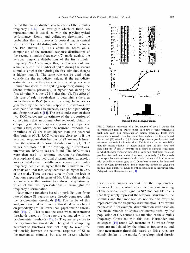
period that are modulated as a function of the stimulus
frequency [14,52]. To investigate which of these two
representations is associated with the psychophysical
performance, Romo and colleagues determined theprobability that an observer (a cortical region central
to S1 cortex) could distinguish the difference between
the two stimuli [14]. This could be based on a
comparison of the neuronal response distributions of
the second stimulus frequency (f2) made against the
neuronal response distributions of the first stimulus
frequency (f1). According to this, the observer could use
a simple rule: if the number of spikes during the secondstimulus is higher than during the first stimulus, then f2
is higher than f1. The same rule can be used when
considering the periodicity values: if the periodicity
(estimated as the frequency with greatest power in a
Fourier transform of the spiking responses) during the
second stimulus period (f2) is higher than during the
first stimulus (f1), then f2 is higher than f1. The effect of
this type of rule is equivalent to determining the areaunder the curve ROC (receiver operating characteristic)
generated by the neuronal response distributions for
each pair of stimulus frequencies, using both periodicity
and firing rate values [14]. The areas under each of these
two ROC curves are an estimate of the proportion of
correct trials that an optimal observer would obtain by
comparing numbers of spikes or periodicity. In pairs of
stimulus frequencies where the neuronal response dis-tributions of f2 are much higher than the neuronal
distributions of f1, ROC values are close to 1; if the
neuronal response distributions of f2 are much lower
than the neuronal response distributions of f1, ROC
values are close to 0; for overlapping distributions,
intermediate ROC values are found. The ROC values
were then used to compute neurometric functions.
Psychophysical and neuronal discrimination thresholdsare calculated as half the difference between the stimulus
frequency identified as higher than the standard in 75%
of trials and that frequency identified as higher in 25%
of the trials. These are read directly from the logistic
functions expressed in terms of Hz. Using this analysis,
we are now in the position to address the question of
which of the two representations is meaningful for
frequency discrimination.Neurometric functions based on periodicity or firing
rate of single S1 neurons were directly compared with
the psychometric thresholds [14]. The results of this
analysis show that neurometric threshold values based
on periodicity are far lower than psychometric thresh-
olds (Fig. 2). This is not the case when neurometric
thresholds based on firing rate are compared with the
psychometric thresholds (Fig. 3). They are very close tothe psychometric thresholds. The goal of computing
neurometric functions was not only to reveal the
relationship between the neuronal responses of S1 to
the mechanical stimulus, but also to discern whether
these neural signals account for the psychometric
behavior. However, what is then the functional meaning
of the periodic neural signal in S1? One possible role is
that they simply represent the temporal structure of the
stimulus and that monkeys do not use this exquisite
representation for frequency discrimination. This would
be the case if, for example, discrimination were based on
the mean number of spikes (or bursts) fired by the
population of QA neurons as a function of the stimulus
frequency. Consistent with this idea, Hernandez and
colleagues [14] found QA neurons in S1 whose firing
rates are modulated by the stimulus frequencies, and
their neurometric thresholds based on firing rates are
closely similar to the monkey’s psychophysical thresh-
Fig. 2. Periodic responses of a QA neuron of area 1 during the
discrimination task. (a) Raster plots. Each row of ticks represents a
trial, and each tick represents an action potential. Trials were
randomly delivered. Grey horizontal lines indicate the first (f1) and
the second (f2) stimulus. (b) Relationship between psychometric and
neurometric discrimination functions. This is plotted as the probability
that the second stimulus is judged higher than the first; data and
sigmoidal fits (x2-test, P B/0.001) for 11 pairs of stimulus frequencies
in which the base frequency was 20 Hz. Grey and black lines represent
psychometric and neurometric functions, respectively. (c) Thresholds
ratios (psychometric/neurometric thresholds) calculated from neurons
with periodic responses (grey bars). Open bars represent the threshold
ratios between psychometric and neurometric thresholds calculated
from a small number of neurons with modulations in their firing rate.
Adapted from Hernandez et al. [14].
R. Romo et al. / Behavioural Brain Research 135 (2002) 105�/118 109
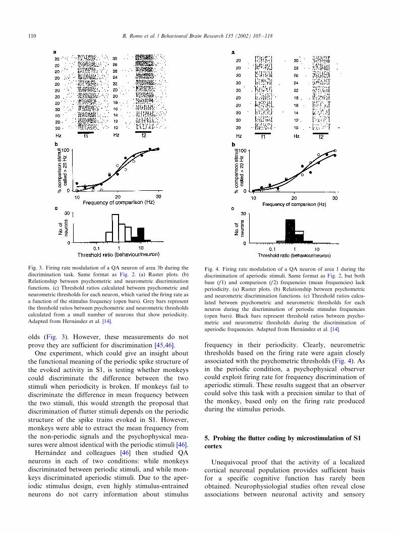
olds (Fig. 3). However, these measurements do not
prove they are sufficient for discrimination [45,46].
One experiment, which could give an insight about
the functional meaning of the periodic spike structure of
the evoked activity in S1, is testing whether monkeys
could discriminate the difference between the two
stimuli when periodicity is broken. If monkeys fail to
discriminate the difference in mean frequency between
the two stimuli, this would strength the proposal that
discrimination of flutter stimuli depends on the periodic
structure of the spike trains evoked in S1. However,
monkeys were able to extract the mean frequency from
the non-periodic signals and the psychophysical mea-
sures were almost identical with the periodic stimuli [46].
Hernandez and colleagues [46] then studied QA
neurons in each of two conditions: while monkeys
discriminated between periodic stimuli, and while mon-
keys discriminated aperiodic stimuli. Due to the aper-
iodic stimulus design, even highly stimulus-entrained
neurons do not carry information about stimulus
frequency in their periodicity. Clearly, neurometricthresholds based on the firing rate were again closely
associated with the psychometric thresholds (Fig. 4). As
in the periodic condition, a psychophysical observer
could exploit firing rate for frequency discrimination of
aperiodic stimuli. These results suggest that an observer
could solve this task with a precision similar to that of
the monkey, based only on the firing rate produced
during the stimulus periods.
5. Probing the flutter coding by microstimulation of S1
cortex
Unequivocal proof that the activity of a localized
cortical neuronal population provides sufficient basisfor a specific cognitive function has rarely been
obtained. Neurophysiologial studies often reveal close
associations between neuronal activity and sensory
Fig. 3. Firing rate modulation of a QA neuron of area 3b during the
discrimination task. Same format as Fig. 2. (a) Raster plots. (b)
Relationship between psychometric and neurometric discrimination
functions. (c) Threshold ratios calculated between psychometric and
neurometric thresholds for each neuron, which varied the firing rate as
a function of the stimulus frequency (open bars). Grey bars represent
the threshold ratios between psychometric and neurometric thresholds
calculated from a small number of neurons that show periodicity.
Adapted from Hernandez et al. [14].
Fig. 4. Firing rate modulation of a QA neuron of area 1 during the
discrimination of aperiodic stimuli. Same format as Fig. 2, but both
base (f1) and comparison (f 2) frequencies (mean frequencies) lack
periodicity. (a) Raster plots. (b) Relationship between psychometric
and neurometric discrimination functions. (c) Threshold ratios calcu-
lated between psychometric and neurometric thresholds for each
neuron during the discrimination of periodic stimulus frequencies
(open bars). Black bars represent threshold ratios between psycho-
metric and neurometric thresholds during the discrimination of
aperiodic frequencies. Adapted from Hernandez et al. [14].
R. Romo et al. / Behavioural Brain Research 135 (2002) 105�/118110
events, but does such activity have an impact on
perception and subsequent behavior? We typically
assume so, but this is hard to verify. Intracortical
microstimulation has provided the most compellingevidence to date of a causal link between the activity
of localized populations of neurons and specific cogni-
tive functions [2,45,46,55]. Electrical microstimulation
directly activates small cluster of neurons, and has been
shown to bias a monkey’s choice during the decision
stage of an ongoing perceptual task [56]. A convenient
model to approach this question is the flutter sensation,
for which humans and monkeys have similar discrimi-nation thresholds [13,31]. During the vibrotactile dis-
crimination task, subjects pay attention to the frequency
of the first (base) stimulus, store a trace of it during the
delay period between the two stimuli and compare the
stored trace to the frequency of the second (comparison)
stimulus. This task, therefore, contains a number of
cognitive processes, such as stimulus detection, working
memory, discrimination between the two stimuli, anddecision-making. These cognitive processes should be
initiated by the evoked neuronal activity in S1 cortex
[50]. As reviewed above, the QA circuit of S1 distributes
the representation of the flutter stimuli to more central
structures anatomically linked to it to solve this task.
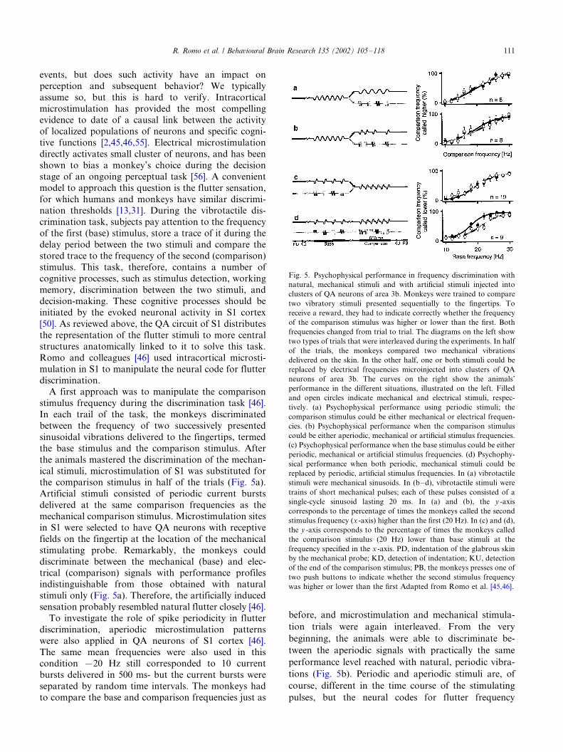
Romo and colleagues [46] used intracortical microsti-
mulation in S1 to manipulate the neural code for flutter
discrimination.A first approach was to manipulate the comparison
stimulus frequency during the discrimination task [46].
In each trail of the task, the monkeys discriminated
between the frequency of two successively presented
sinusoidal vibrations delivered to the fingertips, termed
the base stimulus and the comparison stimulus. After
the animals mastered the discrimination of the mechan-
ical stimuli, microstimulation of S1 was substituted forthe comparison stimulus in half of the trials (Fig. 5a).
Artificial stimuli consisted of periodic current bursts
delivered at the same comparison frequencies as the
mechanical comparison stimulus. Microstimulation sites
in S1 were selected to have QA neurons with receptive
fields on the fingertip at the location of the mechanical
stimulating probe. Remarkably, the monkeys could
discriminate between the mechanical (base) and elec-trical (comparison) signals with performance profiles
indistinguishable from those obtained with natural
stimuli only (Fig. 5a). Therefore, the artificially induced
sensation probably resembled natural flutter closely [46].
To investigate the role of spike periodicity in flutter
discrimination, aperiodic microstimulation patterns
were also applied in QA neurons of S1 cortex [46].
The same mean frequencies were also used in thiscondition �/20 Hz still corresponded to 10 current
bursts delivered in 500 ms- but the current bursts were
separated by random time intervals. The monkeys had
to compare the base and comparison frequencies just as
before, and microstimulation and mechanical stimula-
tion trials were again interleaved. From the very
beginning, the animals were able to discriminate be-
tween the aperiodic signals with practically the same
performance level reached with natural, periodic vibra-
tions (Fig. 5b). Periodic and aperiodic stimuli are, of
course, different in the time course of the stimulating
pulses, but the neural codes for flutter frequency
Fig. 5. Psychophysical performance in frequency discrimination with
natural, mechanical stimuli and with artificial stimuli injected into
clusters of QA neurons of area 3b. Monkeys were trained to compare
two vibratory stimuli presented sequentially to the fingertips. To
receive a reward, they had to indicate correctly whether the frequency
of the comparison stimulus was higher or lower than the first. Both
frequencies changed from trial to trial. The diagrams on the left show
two types of trials that were interleaved during the experiments. In half
of the trials, the monkeys compared two mechanical vibrations
delivered on the skin. In the other half, one or both stimuli could be
replaced by electrical frequencies microinjected into clusters of QA
neurons of area 3b. The curves on the right show the animals’
performance in the different situations, illustrated on the left. Filled
and open circles indicate mechanical and electrical stimuli, respec-
tively. (a) Psychophysical performance using periodic stimuli; the
comparison stimulus could be either mechanical or electrical frequen-
cies. (b) Psychophysical performance when the comparison stimulus
could be either aperiodic, mechanical or artificial stimulus frequencies.
(c) Psychophysical performance when the base stimulus could be either
periodic, mechanical or artificial stimulus frequencies. (d) Psychophy-
sical performance when both periodic, mechanical stimuli could be
replaced by periodic, artificial stimulus frequencies. In (a) vibrotactile
stimuli were mechanical sinusoids. In (b�/d), vibrotactile stimuli were
trains of short mechanical pulses; each of these pulses consisted of a
single-cycle sinusoid lasting 20 ms. In (a) and (b), the y -axis
corresponds to the percentage of times the monkeys called the second
stimulus frequency (x -axis) higher than the first (20 Hz). In (c) and (d),
the y -axis corresponds to the percentage of times the monkeys called
the comparison stimulus (20 Hz) lower than base stimuli at the
frequency specified in the x -axis. PD, indentation of the glabrous skin
by the mechanical probe; KD, detection of indentation; KU, detection
of the end of the comparison stimulus; PB, the monkeys presses one of
two push buttons to indicate whether the second stimulus frequency
was higher or lower than the first Adapted from Romo et al. [45,46].
R. Romo et al. / Behavioural Brain Research 135 (2002) 105�/118 111

underlying the discriminations performed by the mon-
keys might be the same for both. If so, the result might
imply that spike periodicity does not pay a functional
role in our monkey’s performance of the frequency
discrimination task.
Because of the design of this task, comparison of the
second stimulus frequency is made against the memory
trace of the first stimulus. Romo and colleagues [45]
wondered whether, in addition to using artificial stimuli
during the decision stage of the task, monkeys could
store and use a quantitative trace of an electrical
stimulus delivered to QA neurons in S1 cortex in place
of the first mechanical stimulus. They also wondered
whether monkeys could perform the entire task on the
basis of purely artificial stimuli. This would demonstrate
that activation of QA neurons was sufficient to initiate
the entire cognitive process involved in the task.
Again, the mixed mechanical/microstimulation pro-
tocol was used, in which microstimulation trials were
randomly intermixed with standard, purely mechanical
trails [45]. The frequency pairs and event sequence were
the same in both mechanical and microstimulation
trials, except that in microstimulation trials the first or
both mechanical stimuli were substituted by trains of
current pulses injected in S1 and delivered at the
frequency of the mechanical stimulus they were repla-
cing. Design of the stimulus set assured to explore the
working memory component of the task and determine
discrimination thresholds.
Psychophysical performance with electrical microsti-
mulation patterns in S1 cortex at the mechanical base
stimulus frequencies they were replacing was almost
similar to that measured with the mechanical stimulus
(Fig. 5c). These results show that monkeys were able to
memorize the base artificial stimulus frequency and
make quantitative comparisons of the second stimulus
frequency against the trace left by the artificial stimulus.
As for substituting the comparison stimulus with
electrical patterns, monkeys could not reach the usual
level of performance when clusters of SA neurons were
microstimulated. Nor could they discriminate when
microstimulation patterns were made at the border
between QA and SA clusters (45). These control
experiments tell us about the specificity of the QA
circuit of SI cortex in flutter discrimination. Finally, in
most sessions in which the two mechanical stimuli were
replaced by microstimulated patterns, monkeys were
able to reach discrimination levels close to those
measured with mechanical stimuli delivered to the
fingertips (Fig. 5d). This indicates that microstimulation
elicits quantitative memorizable and discriminable per-
cepts, and shows that activation of the QA circuit of S1
cortex is sufficient to initiate the entire subsequent
neural process associated with flutter discrimination
[45].
In flutter discrimination, the first stimulus has to be
detected and memorized. Comparison of the second
stimulus is made against the trace left by the first
stimulus, and a decision is then projected to the motorapparatus to indicate discrimination. Accurate perfor-
mance of the task can be consistent only with induction
of a sensory percept during both stimulus periods. The
above reviewed results indicate that the whole sequence
of events that leads to discrimination could be initiated
by artificial stimulus patterns injected into the QA
circuit of S1 cortex. Thus, the neural activity produced
by either the natural or the artificial stimulus can beused as the basis for sensory discrimination by a
psychophysical observer. The results tell us also that
periodicity does not play a functional role in our
monkey’s performance of the frequency discrimination.
Psychophysical performance with periodic or aperiodic
electrical patterns injected in S1 cortex can be discrimi-
nated similarly as when they are delivered to the
fingertips.
6. Coding of vibrotactile stimuli in cortical areas central
to S1
The results reviewed above are the basis for exploring
the somatosensory network central to S1. This is an
important enterprise, considering that S1 cortex is only
one of many brain structures that participate insomatosensory perception. But, in the flutter task,
what is the neuronal representation of flutter stimuli in
structures central to S1? Assuming that it is periodicity,
do S2 neurons represent flutter stimuli in the same
format? What is the neural correlate for flutter dis-
crimination in central structures to S1? An obvious
candidate to explore these questions is the second
somatosensory (S2) cortex. SI cortex is strongly con-nected with S2 [3,19]. This central somatosensory region
belongs to the ventral stream [27,33].
6.1. Coding of flutter stimuli in S2 cortex
Unlike the majority of S1 neurons, very few S2
neurons are periodically entrained by the flutter stimuli
[52]. There are basically three groups of neuronalresponses during the stimulus periods: the first group
increases the firing rate as a function of the stimulus
frequency; the second decreases the firing rate as a
function of the increasing stimulus frequency; and the
third, which responds but is not modulated as a function
of the stimulus frequency. According to this, there is a
dramatic change in the flutter representation from S1 to
S2. Clearly, the most interesting responses in S2 arethose which modulate their firing rate as a function of
the stimulus frequency. These responses are affected by
the animal’s state. These responses are more prominent
R. Romo et al. / Behavioural Brain Research 135 (2002) 105�/118112

during the discrimination task than when the same
stimuli are delivered in non-working conditions. These
distinct populations are operating simultaneously in S2
and should produce a computation that is useful forfrequency discrimination in an analogous manner to
that reported in central visual areas like the middle
temporal cortex [1]. Finally, an important result ob-
tained in S2 neurons is that many of them retain
information about the base stimulus during the early
component of the delay period between the two stimulus
frequencies. They do similarly as for the stimulation
periods: that is, if the neuron increases its firing rate as afunction of base stimulus frequency, the same represen-
tation is maintained during the early component of the
delay. We consider this as a neural correlate of the
working memory component of the task. This informa-
tion must be translated to structures central to S2 that
contain a network for working memory in this task.
6.2. Parametric encoding of flutter stimuli during
working memory in the prefrontal cortex
As reviewed above, some neurons of S2 cortex retain
the base stimulus frequency during the early component
of the delay period [52]. They do so by retaining the base
stimulus frequency monotonically during the early
memorization component of the task [52]. Where,
then, is this early representation projected and held
during the whole delay period between the two flutterstimuli? Is this associated to the stimulus parameters?
Romo and colleagues [44] recorded in the prefrontal
cortex and sought to determine the neuronal correlate
for the working memory component of this task.
Although there is no clear direct input from S2 or S1
cortex to the prefrontal cortex, in a pilot experiment
Romo and colleagues recorded above and below the
principal sulcus in prefrontal cortex while a monkeyperformed the flutter discrimination task. Recordings in
the first monkey suggested that the inferior convexity of
the prefrontal cortex contained neurons whose activity
varied, during the delay period between the two stimuli,
as a monotonic function of the stimulus frequency [44].
This finding was then further investigated in three more
animals performing the flutter discrimination task.
Some of the delay responses responded most weaklyafter stimulation with the lowest base frequency, and
increased their firing rates steadily for increasing fre-
quencies (positive monotonic; see Fig. 6a, c, e). Others
had discharge rate rates that varied in the opposite
direction (negative monotonic; see Fig. 6b, d, f). These
types of responses in prefrontal cortex are similar to
those recorded in S2 during the early part of the delay
period in the same task [52]; the most importantdifference between S2 and prefrontal cortex was that
many neurons in prefrontal cortex prolonged their
monotonic responses through to the end of the delay
period. Thus, the base stimulus frequency, a scalar
analogue value, appeared to be encoded directly in the
neuron’s firing rate (also a scalar analogue value), most
often in a smoothly grade fashion. These results led
Romo and colleagues [44] to conclude that smooth
monotonic encoding found in prefrontal cortex is
consistent with the existence of a parametric, rather
than categorical, representation of the memorized
stimulus during the working memory component of
this task. In the same vein, these results could suggest
that monotonic encoding might be the basic representa-
tion of sensory magnitude continua during working
memory, in tasks that require ordinal comparisons
between scalar analogue stimuli.
Monotonic encoding of the stimulus frequency in the
prefrontal cortex may be derived from inputs from
cortical somatosensory areas. As described above, in
recordings from S1 and S2 cortices during the same
frequency discrimination task, neurons were found in S2
cortex that responded in a manner similar to ‘early’
neurons, encoding the base stimulus frequency mono-
tonically during the delay period between the two
Fig. 6. Monotonic responses of two neurons of the prefrontal cortex
during the delay period between the two vibrotactile stimuli. (a), (b)
Raters. Each row of ticks represents a trial, and each tick represents an
action potential. Trials were delivered in a random order, but have
been sorted into blocks of equal base stimulus frequency, indicated on
the left. Trials have been further sorted into groups of equal
comparison frequency (indicated in the center), separated by a
horizontal black line. Grey boxes (and short horizontal grey lines in
c and d) indicate base and comparison stimulus periods; thick grey tics
after the comparison stimulus indicates the beginning of the motor
response (KU; see Fig. 1a). Time axes for (a) and (b) are shown in (c)
and (d), respectively. (c), (d) Time-dependent spike densities for each
base frequency stimulus condition. Grey level indicates the base
frequency: the lightest grey line corresponds to 10 Hz and the darkest
line corresponds to 34 Hz. (e, f) Mean firing rates, averaged across the
entire delay period. Small vertical lines are9/S.E.M. Thick grey lines
are soft sigmoid fits to the data Adapted from Romo et al. [44].
R. Romo et al. / Behavioural Brain Research 135 (2002) 105�/118 113

stimuli. Although many neurons of S1 cortex responded
during the two stimulus periods, none did so during the
delay period, in contrast to results obtained in a
different tactile task involving working memory [70].These results constitute a neurophysiological demon-
stration that neurons of the prefrontal cortex can retain
working memory information induced by non-visual
modalities.
7. From somatosensation to action
In the past decade, an important advance has beenmade in sensory physiology; that is, sensory physiolo-
gists not only seek the neuronal correlates of sensory
perception, but also the neuronal mechanisms under-
lying decision-making: from sensation to action. Phy-
siologists in the motor system would refer to this as the
sensory-motor interface. However, the cornerstone to
exploring decision-making is to investigate it in psycho-
physical tasks [35,49]. The investigator controls thestimulus input, to know the encoding of the stimulus
in the early sensory areas, and to have a precise control
of the motor outputs as functions of the stimulus
parameters. Performance is measured with psychophy-
sical techniques and the neuronal responses are quanti-
fied with neurometric techniques; that is, neuronal
responses are evaluated as if it were measured by an
ideal psychophysical observer.
7.1. Defining a somatosensory task for exploring
decision-making mechanisms
Romo and colleagues [48,49] designed a somatosen-
sory task in which monkeys categorized the speed of
moving tactile stimulus. Monkeys had to decide whether
the stimulus speed was high or low. Categorization wasindicated through an arm-hand movement directed to
one or two push buttons. Performance was measured
with psychophysical techniques, and the motor re-
sponses were quantified by measuring the reaction and
motor times. Under these conditions, it is possible to
study in S1 cortex the responses of neurons to the tactile
stimuli and the neuronal activity in motor areas of the
frontal lobe. What do we want to explore in this task?We want to know how a somatosensory signal after
being processed in S1 cortex is transported to the
neuronal ensembles of frontal motor areas during the
decision stage.
The direction of a moving tactile stimulus is repre-
sented in the evoked neuronal activity of S1 cortex and it
can be quantified in the form of a population vector,
whose magnitude is modulated by the speed of thestimulus [51]. This dynamic internal representation
provides the initial substrate for higher order processing
of moving tactile stimuli. If this is so, a salient question
is whether this representation of the moving tactile
stimuli in S1 cortex can be correlated directly with the
sensory performance of the animal. Delivering the
moving tactile stimulus across the cutaneous receptivefield of the recorded neuron while the monkey per-
formed the categorization task gave two salient results
[47]. First, S1 neurons increased their firing rates as a
function of the stimulus speed. Second, these responses
were almost identical when the same stimuli were
delivered passively, in the non-working condition. This
suggests that the neuronal signals associated with the
perception of the tactile stimuli may be occurring inmore central areas anatomically linked to S1 cortex.
However, removing S1 cortex impaired categorization
of the moving tactile stimuli, but not detection [69]. A
similar result was observed in the flutter discrimination
task and suggests that these representations are essential
for somatosensory perception [22].
7.2. Decision signals in premotor cortex
Where to look for the neural decision mechanisms
during somatosensation? This should be investigated in
those somatosensory areas central to S1 cortex. How-
ever, little neurophysiological data is available, except
for the motor areas of the frontal lobe
[29,26,48,49,53,54]. Anatomic studies have shown that
the supplementary motor cortex (SMA) receives direct
inputs from the posterior parietal cortex and the lateralsomatosensory areas (reviewed above). The output of
the SMA is directed to MI and to the spinal cord [8,9].
In addition, the SMA maintains an important one way
connection with the neostriatum and the brainstem (20,
25). Given this prominent connection with motor
centers, neurophysiologists have looked at the role of
SMA in motor behavior. However, it might be possible
that the SMA is involved also in some aspects of thesensory perceptual process. Romo and colleagues [48,49]
have investigated this possibility by recording single
neurons in the SMA contralateral and ipsilateral to the
stimulated hand during the execution of the categoriza-
tion task.
Of particular interest for decision-making was the
recording of neurons in the SMA ipsilateral and
contralateral to the stimulated hand that respondeddifferentially during the categorization task [48,49].
These neural responses occurred mainly during the
stimulus period, during the stimulus and reaction time,
and during the reaction-movement time periods. These
neurons encoded in their activity whether the stimulus
speed was low or high. A number of these neurons were
then tested when the same set of stimuli was delivered
passively. None of them responded in this condition.Another important test is that these differential re-
sponses occurred exclusively during the categorization
task; these neurons did not respond differentially when
R. Romo et al. / Behavioural Brain Research 135 (2002) 105�/118114

the same hand-arm movement was guided by visual cues
in the absence of somatic stimuli. But, interestingly, the
differential responses were considerably attenuated
when a visual cue predicted whether the tactile stimuluswas low or high. Some neurons even showed such
modulatory effects during the waiting periods before
stimulus delivery.
With these elements in hand, we should be able to
inquire whether these differential neural responses are
related to the animal’s decision. Decoding information
from these neural responses show that indeed they are
decision signals clearly correlated with the sensory-motor performance [53]. But, is this a unique property
of the SMA? Probably not, given the large interconnec-
tivity between motor and somatosensory areas.
7.3. Decision signals in neostriatum
The neostriatum receives bilateral inputs from the
SMA [20,25], so is this structure associated to decision-
making during somatosensation? Single neuronal re-cordings were made in the neostriatum ipsilateral and
contralateral to the stimulated hand during the categor-
ization task [26]. Again, a large proportion of neurons in
the bilateral neostriatum showed differential responses.
That is, groups of neurons responded selectively when
the animal indicated that the category of the stimulus
was low and others responded when the animal indi-
cated the category of the stimulus speed was high. As forthe SMA, these responses occurred exclusively during
the categorization task, these neural signals were not
present when the same stimuli were delivered passively.
Clearly, these differential responses were modality
specific because these neurons failed to respond when
the same differential movements were guided by visual
cues in the absence of somatic stimuli. Although
decoding analysis was not carried out as for the SMAneurons [53], it is very likely, according to the neural
response profiles, that they are similarly linked to the
psychophysical performance. This would indicate that
decision neural signals are widely distributed in those
motor areas of the brain that are anatomically linked to
the somatosensory cortex.
7.4. Decision signals in primary motor cortex
If premotor areas of the brain show decision neural
signals, does MI cortex contains a neural apparatus
associated with the categorization process? Given the
prominent role of MI cortex in coding movement
parameters [11], decision signals could simple drive
populations of neurons in MI, for expressing the
decision through a goal directed movement (in thiscase to the push buttons). If this were so, a differential
neuron during the categorization task would respond
similarly when the same hand-arm movement is per-
formed during visual guidance. If not, this would
indicate that some MI neurons make the link between
somatosensory processing that leads to decision and
motor output during the categorization task. To probe
this, Salinas and Romo [54] recorded in the arm area of
MI while monkeys performed in the tactile categoriza-
tion task.
Remarkably, about a fifth of the recorded neurons
discharged differentially during the tactile categoriza-
tion task (Fig. 7a). Salinas and Romo [54] reasoned
these could be neural signals encoding arm direction,
given the prominent role of neuronal populations of MI
neurons in encoding this variable [11]. However, this is
unlikely for two main reasons. First, assuming they are
representing the direction of arm movements, direction
coding has not been proven to occur when targets are
close together (they were separated by an angle of 118.The second and most important argument is that
differential neurons tested while visually guiding the
hand-arm movements towards the push buttons failed
to respond differentially in this condition (Fig. 7b).
Needless to say, none of these motor cortex neurons, as
the reviewed above, responded when the same stimuli
were delivered passively, in the non-working condition.
Fig. 7. Responses related to categorical decisions in primary motor
cortex. Monkeys classified the speed of a probe moving across the
glabrous skin of a fingertip as either low (B/20 mm/s) or high (�/20
mm/s). (a) Raster plot of a neuron that fired more intensely for low
speeds. Each line corresponds to one trial, and each small tick
represents and action potential. Squares represent behavioral events:
onset of probe movement (ON); offset of probe movement (OFF); or
key release (KR) which marked the initiation of the hand-arm
movement to indicate the decision. Stimulus speeds are indicated on
the left. (b) Responses of the same neuron when identical arm
movements, to the medial (M) or lateral (L) button, were triggered
by visual cues, in the absence of tactile stimuli. In each trial, the first
square indicates the visual go signal, and the second square indicates
KR. No differential activity was observed in this condition. (c) Mean
firing rate as a function of the stimulus speed averaged over a
population of 20 neurons that were selective for high speeds, Error
bars indicate9/S.E.M. (d) As in (c) but for a population of 20 neurons
selective for low speeds. Adapted from Salinas and Romo [54].
R. Romo et al. / Behavioural Brain Research 135 (2002) 105�/118 115

Analysis by decoding information from these neural
responses together with the psychophysical performance
provided two main types of information regarding the
identity of these neural signals in the categorization task[53,54]. First, the neurometric profile clearly matched
the psychophysical performance (Fig. 7c, d). Second,
error trials revealed these neurons are neither sensory
nor motor [53]. They are both. Therefore, it appears the
MI cortex possesses a type of neuron, which had not
been identified before, which makes the link between a
sensory signal and a motor signal.
From a teleological perspective, sensory representa-tions are meaningful only if they are relevant for the
construction of sensations, forming memories and
reaching decisions. The results reviewed above clearly
suggest that the large territories of the brain receive a
copy of a sensory decision, which is then expressed
through a voluntary motor action. The fact that the
outputs of premotor cortex and basal ganglia are
directed to MI cortex suggests that the informationfrom multiple processing nodes finally converges in MI
cortex and suggests a strong link between sensory
processing and motor activity. The remarkable spread
of the decision-related representations in the brain may
indicate something fundamental about how decision-
making is implemented by neurons.
8. Concluding comments
Decision-making is present in all sensory tasks. This is
elaborated in the sensory areas of the brain during
processing afferent inputs. A decision is expressed
through voluntary motor actions. However, as shown
here, few examples exist exploring this function in the
somatosensory system. Similarly, exploring the central
neural mechanisms that lead to the construction of asensory percept has also proceeded at a low pace. This
has not been the case for revealing the neural represen-
tation of tactile stimuli in the afferent component of the
somatosensory system. This has produced strong im-
portant concepts, as that afferent fibers represent in
their activity the stimulus features and that they limit
and account for the psychophysical performance. This
idea is currently tested in the brain. In other words, howthese sensory representations account for sensation and
decision-making. Indeed, one of these representations
has been proven to be sufficient for sensory discrimina-
tion [45,46]. A remarkably finding is that working
memory in one-dimensional stimulus discrimination
task has a parametric representation in the brain [44].
This should be similar in other sensory modalities
involving processing of these types of stimuli. Finally,an effort has been made to test the neural signal that
matches decision-making in the somatosensory system.
This encoding is associated with early sensory represen-
tations [26,48,49,53,54]. However, more studies are
needed to have a complete picture of how a sensory
percept is built up in the somatosensory network.
To conclude, we believe that there are two divergentresearch lines when exploring the neuronal correlates of
tactile perception. The first relies on stimulus encoding;
that is, ‘the representational problem’. The second relies
on neuronal changes that occur when animals shift from
one condition to another. While we believe that it is
important to show that the neuronal responses in the
somatosensory system are associated with behavioral
changes, however, these studies may have greater impactif they are in line with the ‘representational problem’;
that is, how the attended stimulus that is discriminated
and memorized is represented in the brain, and whether
these representations are meaningful for somatosensory
perception.
Acknowledgements
Research in R.R. laboratory was supported by an
International Research Scholars award from the Ho-
ward Hughes Medical Institute, the Millennium Science
Initiative, CONACyT and DGAPA.
References
[1] Britten KH, Shadlen MN, Newsome WT, Movshon JA. The
analysis of visual motion: a comparison of neuronal and
psychophysical performance. J Neurosci 1992;12:4745�/65.
[2] Britten KH, Wezel RJ. Electrical microstimulation of cortical
MST biases heading perception in monkeys. Nat Neurosci
1998;1:59�/63.
[3] Burton H, Fabri M, Alloway K. Cortical areas within the lateral
sulcus connected to cutaneous representations in areas 3b and 1: a
revisited interpretation of the second somatosensory area in
macaque monkeys. J Comp Neurol 1995;355:539�/62.
[4] Burton H, Sinclair RJ, Hong SY, Pruett JR, Wang KC. Tactile-
spatial and cross-modal attention effects in the second somato-
sensory and 7b cortical areas of rhesus monkeys. Somat Mot Res
1997;14:237�/67.
[5] Carmichael ST, Price JL. Sensory and premotor connections of
orbital and medial prefrontal cortex of macaque monkeys. J
Comp Neurol 1995;363:642�/64.
[6] Cavada C, Goldman-Rakic PS. The posterior parietal cortex in
rhesus monkeys: I. Parcellation of areas based on distinctive
limbic and sensory corticocortical connections. J Comp Neurol
1989;287:393�/421.
[7] Darian-Smith I. The sense of touch: performance and peripheral
neural processes. In: Brookhart JM, Mountcastle VB, editors.
Handbook of physiology: section I. The nervous system. Sensory
processes, Part 2, vol. III. Bethesda, MD: American Physiological
Society, 1984:739�/88.
[8] Dum RP, Strick PL. The origin of corticospinal projections from
the premotor areas in the frontal lobe. J Neurosci 1991;11:667�/
89.
[9] Dum RP, Strick PL. Spinal cord terminations of the medial wall
motor areas in macaque monkeys. J Neurosci 1996;16:6513�/25.
R. Romo et al. / Behavioural Brain Research 135 (2002) 105�/118116

[10] Felleman D, Van Essen DC. Distributed hierarchical processing
in the primate cerebral cortex. Cereb Cortex 1991;1:1�/47.
[11] Georgopoulos AP, Kettener RE, Schwartz AB. Primate motor
cortex and free arm movements to visual targets in three-
dimensional space. II. Coding of the direction of movement by
a neuronal population. J Neurosci 1988;8:2928�/37.
[12] Godshalk M, Lemon RB, Kuypers HG, Ronday HK. Cortical
afferents and efferents of monkey postarcuate area: an anatomical
and electrophysiological study. Exp Brain Res 1984;56:410�/24.
[13] Hernandez H, Salinas E, Garcia R, Romo R. Discrimination in
the sense of flutter: new psychophysical measurements in mon-
keys. J Neurosci 1997;17:6391�/400.
[14] Hernandez A, Zainos A, Romo R. Neuronal correlates of sensory
discrimination in the somatosensory cortex. Proc Natl Acad Sci
USA 2000;97:6091�/6.
[15] Hsiao SS, Johnson KO, O’Shaughnessy DM. Effects of selective
attention of spatial form processing in monkey primary and
secondary somatosensory cortex. J Neurophysiol 1993;70:444�/7.
[16] Jiang W, Tremblay F, Chapman CE. Neuronal encoding of
texture changes in the primary and in the secondary somatosen-
sory cortical areas of monkeys during passive texture discrimina-
tion. J Neurophysiol 1997;77:1656�/62.
[17] Johnson KO, Hsiao SS. Neural mechanisms of tactual form and
texture perception. Annu Rev Neurosci 1992;15:227�/50.
[18] Kaas JH, Nelson RJ, Sur M, Lin CS, Merzenich MM. Multiple
representations of the body within the primary somatosensory
cortex of primates. Science 1979;204:521�/3.
[19] Krubitzer L, Clarey J, Tweendale R, Elston G, Calford MA. A
redefinition of somatosensory areas in the lateral sulcus of
macaque monkeys. J Neurosci 1995;15:3821�/39.
[20] Kunzle H. Bilateral projections from the precentral motor cortex
to the putamen and other parts of the basal ganglia. An
autoradiographic study in Macaca fascicularis . Brain Res
1995;88:195�/209.
[21] LaMotte RH, Mountcastle VB. Capacities of humans and
monkeys to discriminate between vibratory stimuli of different
frequency and amplitude: correlation between neural events and
psychophysical measurements. J Neurophysiol 1975;38:539�/59.
[22] LaMotte RH, Mountcastle VB. Disorders in somesthesis follow-
ing lesions of parietal lobe. J Neurophysiol 1978;42:400�/19.
[23] Leichnetz GR. Afferent and efferent connections of the dorso-
lateral precentral gyrus (area 4, hand/arm region) in the macaque
monkey, with comparisons to area 8. J Comp Neurol
1989;254:460�/92.
[24] Macefield G, Gandevia SC, Burke D. Perceptual responses to
microstimulation of single afferents innervating joints, muscles
and skin of the human hand. J Physiol 1990;429:113�/29.
[25] McGuire KP, Bates JF, Goldman-Rakic PS. Interhemispheric
integration. II. Symmetry and convergence of the corticostriatal
projections of the left and right principal sulcus (PS) and the left
and the right supplementary motor area (SMA) of the rhesus
monkey. Cereb Cortex 1991;1:409�/17.
[26] Merchant H, Zainos A, Hernandez A, Salinas E, Romo R.
Functional properties of primate putamen neurons during the
categorization of tactile stimuli. J Neurophysiol 1997;77:1132�/54.
[27] Mishkin M. Analogous neural models for tactual and visual
learning. Neurophychologia 1979;17:139�/51.
[28] Mountcastle VB. Modality and topographic properties of single
neurons of cat’s somatic sensory cortex. J Neurophysiol
1957;20:408�/34.
[29] Mountcastle VB, Atluri P, Romo R. Selective output-discrimina-
tive signals in the motor cortex of waking monkeys. Cereb Cortex
1992;2:277�/94.
[30] Mountcastle VB, LaMotte RH, Carli G. Detection thresholds for
stimuli in humans and monkeys: comparison with thresholds
events in mechanoreceptive afferent nerve fibers innervating the
monkey hand. J Neurophysiol 1972;25:122�/36.
[31] Mountcastle VB, Steinmetz MA, Romo R. Frequency discrimina-
tion in the sense of flutter: psychophysical measurements corre-
lated with postcentral events in behaving monkeys. J Neurosci
1990;10:3032�/44.
[32] Mountcastle VB, Talbot WH, Sakata H, Hyvarinen J. Cortical
neuronal mechanisms in flutter-vibration studied in unanesthe-
tized monkeys. Neuronal periodicity and frequency discrimina-
tion. J Neurophysiol 1969;32:452�/84.
[33] Murray EA, Mishkin M. Relative contributions of SII and area 5
to tactile discrimination in monkeys. Behav Brain Res
1984;11:67�/83.
[34] Nelson RJ, Sur M, Felleman DJ, Kaas JH. Representations of the
body surface in the postcentral parietal cortex of Macaca
fascicularis . J Comp Neurol 1980;192:611�/43.
[35] Newsome WT, Britten KH, Movshon JA. Neuronal correlates of
a perceptual decision. Nature 1989;341:52�/4.
[36] Ochoa J, Torebjork E. Sensations evoked by intraneural micro-
stimulation of single mechanoreceptor units innervating the
human hand. J Physiol 1983;42:633�/54.
[37] Pearson RC, Powell TPS. The projection of primary somatic
sensory cortex upon area 5 in the monkey. Brain Res
1985;356:89�/107.
[38] Phillips JR, Johnson KO. Tactile spatial resolution. II. Neural
representations of bars, edges, and gratings in monkey primary
afferents. J Neurophysiol 1981;46:1192�/203.
[39] Pons TP, Garraghty PE, Friedman DP, Mishkin M. Physiological
evidence for serial processing in somatosensory cortex. Science
1987;237:417�/20.
[40] Pons TP, Garraghty PE, Mishkin M. Serial and parallel proces-
sing of tactual information in somatosensory cortex of rhesus
monkeys. J Neurophysiol 1992;68:518�/27.
[41] Powell TPS, Mountcastle VB. Some aspects of the functional
organization of the cortex of the postcentral gyrus of the monkey:
a correlation of findings obtained in a single unit analysis with
cytoarchitecture. Bull Johns Hopkins Hosp 1959;105:133�/62.
[42] Preuss TM, Goldman-Rakic PS. Connections of the ventral
granular frontal cortex of macaques with perisylvian premotor
and somatosensory areas: anatomical evidence for somatic
representation in primate frontal association cortex. J Comp
Neurol 1989;282:293�/316.
[43] Recanzone GH, Merzenich MM, Schreiner CE. Changes in the
distributed temporal response properties of SI cortical neurons
reflect improvements in performance on a temporally based tactile
discrimination task. J Neurophysiol 1992;67:1071�/91.
[44] Romo R, Brody CD, Hernandez A, Lemus L. Neuronal correlates
of parametric working memory in the prefrontal cortex. Nature
1999;339:470�/3.
[45] Romo R, Hernandez A, Zainos A, Brody CD, Lemus L. Sensing
without touching: psychophysical performance based on cortical
microstimulation. Neuron 2000;26:273�/8.
[46] Romo R, Hernandez A, Zainos A, Salinas E. Somatosensory
discrimination based on cortical microstimulation. Nature
1998;392:387�/90.
[47] Romo R, Merchant H, Zainos A, Hernandez A. Categorization of
somaesthetic stimuli: sensorimotor performance and neuronal
activity in primary somatic sensory cortex of awake monkeys.
NeuroReport 1996;7:1273�/9.
[48] Romo R, Merchant H, Zainos A, Hernandez A. Categorical
perception of somesthetic stimuli: psychophysical measurements
correlated with neuronal events in primate medial premotor
cortex. Cereb Cortex 1997;7:317�/26.
[49] Romo R, Ruiz S, Crespo P, Zainos A, Merchant H. Representa-
tion of tactile signals in primate supplementary motor area. J
Neurophysiol 1993;70:2690�/4.
[50] Romo R, Salinas E. Sensing and deciding in the somatosensory
system. Curr Opin Neurobiol 1999;9:487�/93.
R. Romo et al. / Behavioural Brain Research 135 (2002) 105�/118 117

[51] Ruiz S, Crespo P, Romo R. Representation of moving tactile
stimuli in the somatic sensory cortex of awake monkeys. J
Neurophysiol 1995;73:525�/37.
[52] Salinas E, Hernandez A, Zainos A, Romo R. Periodicity and
firing rate as candidate neural codes for the frequency of
vibrotactile stimuli. J Neurosci 2000;20:5503�/15.
[53] Salinas E, Romo R. Neuronal representations in categorization
task: sensory to motor transformation. In: Bower J, editor.
Computational Neuroscience: Trends in Research 98. New
York: Plenum Press, 1998:599�/604.
[54] Salinas E, Romo R. Conversion of sensory signals into motor
commands in primary motor cortex. J Neurosci 1998;18:499�/511.
[55] Salzman D, Britten K, Newsome WT. Cortical microstimulation
influences perceptual judgements of motion direction. Nature
1990;346:174�/7.
[56] Seidemann E, Zohary U, Newsome WT. Temporal gating of
neural signals during performance of a visual discrimination task.
Nature 1999;394:72�/5.
[57] Shadlen MN, Newsome WT. Noise, neural codes and cortical
organization. Curr Opin Neurobiol 1994;4:569�/79.
[58] Shanks MF, Person RC, Powell TPS. The ipsilateral cortico-
cortical connexions between the cytoarchitectonic subdivisions of
the primary somatic sensory cortex in the monkey. Brain Res
1985;356:67�/88.
[59] Sinclair RJ, Burton H. Neuronal activity in the second somato-
sensory cortex of monkeys (Macaca mulatta ) during active touch
of gratings. J Neurophysiol 1993;70:331�/50.
[60] Singer W, Gray CM. Visual feature integration and the temporal
correlation hypothesis. Annu Rev Neurosci 1995;18:555�/86.
[61] Sur M, Wall JT, Kaas JH. Modular distribution of neurons with
slowly adapting and rapidly adapting responses in area 3b of
somatosensory cortex in monkeys. J Neurophysiol 1984;51:724�/
44.
[62] Talbot WH, Darian-Smith I, Kornhuber HH, Mountcastle VB.
The sense of flutter-vibration: comparison of the human capacity
response patterns of mechanoreceptive afferents from the monkey
hand. J Neurophysiol 1968;31:301�/34.
[63] Tokuno H, Tanji J. Input organization of distal and proximal
forelimb areas in the monkey primary motor cortex: retrograde
double labeling study. J Comp Neurol 1993;333:199�/209.
[64] Vallbo AB. Single-afferent neurons and somatic sensation in
humans. In: Gazzaniga M, editor. The cognitive neurosciences.
Cambridge: MIT Press, 1988:237�/52.
[65] Vallbo AB, Johansson RS. Properties of cutaneous mechanor-
eceptors in the human hand related to touch sensations. Hum
Neurobiol 1984;3:3�/14.
[66] Werner G. The study of sensation in physiology. In: Mountcastle
VB, editor. Medical physiology, vol. 1. St. Louis: Mosby,
1980:605�/28.
[67] Werner G, Mountcastle VB. The variability of central neural
sensory system, and its implications for the central reflection of
sensory events. J Neurophysiol 1963;26:958�/77.
[68] Werner G, Mountcastle VB. Neural activity in mechanoreceptive
cutaneous afferents: stimulus-response relations, Weber functions,
and information transmission. J Neurophysiol 1965;28:459�/
97.
[69] Zainos A, Merchant H, Hernandez A, Salinas E, Romo R. Role
of primary somatic sensory cortex in the categorization of tactile
stimuli: effects of lesions. Exp Brain Res 1997;115:357�/60.
[70] Zhou YD, Fuster JM. Mnemonic neuronal activity in somato-
sensory cortex. Proc Natl Acad Sci USA 1996;93:10533�/7.
R. Romo et al. / Behavioural Brain Research 135 (2002) 105�/118118