From etiopathogenic insights to clinical approach in ... - UMF Iasi
184
From etiopathogenic insights to clinical approach in atherosclerosis and pain HABILITATION THESIS MARIA-MAGDALENA LEON-CONSTANTIN MD PhD 2019
-
Upload
khangminh22 -
Category
Documents
-
view
4 -
download
0
Transcript of From etiopathogenic insights to clinical approach in ... - UMF Iasi
From etiopathogenic insights to clinical approach in atherosclerosis and pain
HABILITATION THESIS
MARIA-MAGDALENA LEON-CONSTANTIN MD PhD
2019

HABILITATION THESIS LEON-CONSTANTIN MARIA-MAGDALENA
i
Contents
Contents ................................................................................................................ i REZUMATUL TEZEI .......................................................................................................... 1
SUMMARY OF THE THESIS ............................................................................................. 3
SECTION I. OVERVIEW OF PERSONAL, PROFESSIONAL, ACADEMIC AND SCIENTIFIC ACCOMPLISHMENTS ........................... 4
I.1. Academic activity .................................................................................................................... 4 I.2. Professional progress .............................................................................................................. 5 I.3. Scientific research activity ...................................................................................................... 5
SECTION II. ATHEROSCLEROSIS ............................................................... 7
Chapter 1. RESEARCH DIRECTIONS REGARDING RISK FACTORS RELATED TO ATHEROSCLEROSIS .......................................................................................................... 7
1.1. Epidemiological data .............................................................................................................. 8 1.2. Risk factors .............................................................................................................................. 9 1.3. Material and methods ........................................................................................................... 17 1.4. Results .................................................................................................................................... 17 1.5. Discussions ............................................................................................................................. 22 1.6. Conclusions ........................................................................................................................... 25
Chapter 2. THE MORPHOPHYSIOLOGY OF ATHEROSCLEROSIS ............................ 26 2.1. Theories of pathogenesis....................................................................................................... 26 2.2. Material and methods ........................................................................................................... 30 2.3. Results .................................................................................................................................... 31 2.4. Discussions ............................................................................................................................. 35 2.5. Conclusions ............................................................................................................................ 37
Chapter 3. CLINICAL – BIOLOGICAL EVALUATION ................................................. 38 3.1. Introduction ........................................................................................................................... 38 3.2. Material and methods ........................................................................................................... 43 3.3. Results .................................................................................................................................... 44 3.4. Conclusions ............................................................................................................................ 53
Chapter 4. MANAGEMENT OF ATHEROSCLEROSIS .................................................. 54 4.1. Introduction ........................................................................................................................... 54 4.2. Material and methods ........................................................................................................... 57 4.3. Results .................................................................................................................................... 59 4.4. Discussions ............................................................................................................................. 66 4.5. Conclusions ............................................................................................................................ 70
Chapter 5. QUALITY OF LIFE .......................................................................................... 71 5.1. Introduction ........................................................................................................................... 71 5.2. Material and methods ........................................................................................................... 76 5.3. Results .................................................................................................................................... 78 5.4. Discussions ............................................................................................................................. 85 5.5. Conclusions ............................................................................................................................ 90
SECTION III. PAIN ......................................................................................... 91
Chapter 6. FROM BENCH TO BEDSIDE – FINDING NEW METHODS FOR IMPROVING PAIN MANAGEMENT .............................................................................. 91
6.1. Introduction ........................................................................................................................... 91
HABILITATION THESIS LEON-CONSTANTIN MARIA-MAGDALENA
ii
6.2. Material and methods ........................................................................................................... 95 6.3. Results .................................................................................................................................... 98 6.4. Discussions ........................................................................................................................... 111 6.5. Conclusions .......................................................................................................................... 116
Chapter 7. NEW METHODS AND FORMULATIONS FOR A MORE EFFECTIVE ANALGESIC TREATMENT ........................................................................................... 117
7.1. Introduction ......................................................................................................................... 117 7.2. Material and method .......................................................................................................... 124 7.3. Results .................................................................................................................................. 128 7.4. Discussions ........................................................................................................................... 139 7.5. Conclusions .......................................................................................................................... 143
SECTION IV. FUTURE DIRECTIONS ...................................................... 144
Chapter 8. FUTURE EVOLUTION AND DEVELOPMENT PLANS ............................ 144 8.1. Perspectives in research activity ........................................................................................ 144 8.2. Perspectives in professional activity .................................................................................. 147
SECTION V. REFERENCES ........................................................................ 150

HABILITATION THESIS LEON-CONSTANTIN MARIA-MAGDALENA
1
REZUMATUL TEZEI
Asemenea oricărei profesii, cea de cadru didactic presupune un cumul de cunoştinţe, abilităţi şi competenţe specifice pe care trebuie să le deţină cei care aleg să urmeze acest drum. În opinia mea, viaţa academică depinde în mare măsură de aspiraţiile şi profesionalismul membrilor săi. Sprijinul universităţii este fundamental pentru dezvoltarea atât pe plan profesional cât şi personal, pentru atingerea obiectivelor proprii cât și a celor comunitare.
Prin excelenţă, profesia didactică presupune permanenta formare şi dezvoltare a cadrului didactic, astfel încât acesta să poată oferi celui pe care îl învaţă o perspectivă comprehensivă asupra domeniului pe care îl predă. Cadrul didactic din orice specializare se angajează astfel într-un proces de formare continuă care îi permite să fie mereu informat și să poată răspunde la orice întrebare din partea studenților sau să le prezinte mereu ultimele noutăți în domeniu. În atingerea unor niveluri ridicate de performanţă şi eficienţă, în concordanţă cu standardele moderne ale profesiunii, la profesionalizarea pedagogică a cadrelor didactice, o contribuţie esenţială îi revine tezei de abilitare. Prezenta lucrare reprezintă o descriere a activităților desfășurate, a competenţelor profesionale atinse, obiectivate prin lucrări științifice, granturi de cercetare și direcțiile viitoare de cercetare.
Teza de abilitare este structurată în cinci părți în conformitate cu criteriile recomandate și aprobate de către CNATDCU. Lucrarea este o imagine a preocupărilor mele în domeniul medical, cu punctarea preocupărilor în domeniile: algeziologiei – defășurate în timpul doctoratului în cadrul Disciplinei de Farmacologie-Algeziologie și de asemenea a colaborării cu colegii din CEMEX și colegii de medicină internă, reflectate prin prisma activităților desfășurate în cadrul Disciplinei de Semiologiei Medicale a Universității de Medicină și Farmacie” Grigore T. Popa” Iași după finalizarea doctoratului.
Prima parte este formată din contribuțiile mele profesionale, științifice și academice. Partea a doua studiul procesului aterosclerotic, a fost cea mai importantă direcție
de cercetare pe care am urmat-o în activitatea clinică. Numărul mare de pacienți cu sindrom metabolic (obezitate, hipertensiune arterială, diabet zaharat, sindrom dislipidemic) m-a direcționat către studiul în profunzime al acestui proces ce stă la baza afecțiunilor cardiovasculare. Prin colaborări multidisciplinare a fost posibilă elaborarea de lucrări științifice exhaustive în reviste de specialitate pe tema aterosclerozei.
Partea a treia, de cercetare a durerii reprezintă continuarea temei abordate în cadrul doctoratului. Studiul durerii, un subiect mereu de actualitate, a fost aprofundat, rezultatele cercetării putând fi observate în numeroase lucrări științifice realizate împreună cu colegii din CEMEX, granturilor câștigate.
În partea a patra se detaliază viitoarelor direcții de cercetare:
1. Identificarea factorilor de risc aterogeni, leziunilor timpurii din ateroscleroză, markeri care semnalează afectarea aparatului cardiovascular, precum și mecanismele de producere;
2. Strategii terapeutice în prevenția dezvoltării procesului aterosclerotic.

HABILITATION THESIS LEON-CONSTANTIN MARIA-MAGDALENA
2
3. Investigații referitoare la designul, caracterizarea, toxicitatea acută, biocompatibilitatea in vivo și efectele farmacodinamice ale diferitelor nanoparticule/metale/compuși cu efect analgezic;
4. Studierea experimentală a efectelor nanoparticulelor care încorporează medicamente analgezice asupra sensibilității nociceptive la animalele de laborator;
Partea a cincea cuprinde bibliografia ce însoțește teza de abilitare.
HABILITATION THESIS LEON-CONSTANTIN MARIA-MAGDALENA
3
SUMMARY OF THE THESIS
Comparable to any profession, being a teacher implies an accumulation of specific knowledge, skills and competences that those who choose to follow this path must have. In my opinion, academic life depends largely on the aspirations and professionalism of its members. The support of the university is fundamental for the development both professionally and personally, to reach its own goals as well as the community ones.
By excellence, teaching implies the permanent formation and development of the teacher, so that he can offer the student a comprehensive perspective on the specific field. The teacher from any specialization engages in a process of continuous training that allows him to be always informed and to be able to answer any students question and always to present them the latest news in the field. In reaching high levels of performance and efficiency, in accordance with the modern standards of the profession, in the pedagogical professionalization of the teachers, an essential contribution is the habilitation thesis. This paper is a description of my carried out activities, my achieved professional competences, objectified by scientific papers and research grants and also with future research directions.
The habilitation thesis is structured in five parts according to the criteria recommended and approved by CNATDCU. The paper is an image of my preoccupations in the medical field, as it follows: Algesiology and Internal Medicine, reflected through the activities carried out within the Discipline of Medical Semiology of the University of Medicine and Pharmacy "Grigore T Popa" Iasi after finishing the PhD.
The first part consists of my professional, scientific and academic contributions . The second part, clinical is the most important and included the study of the
atherosclerotic process, this being the first research direction I followed. The large number of patients with metabolic syndrome (obesity, high blood pressure, diabetes mellitus, dyslipidemic syndrome) motivated me to the in-depth study of this process that is the basis of cardiovascular diseases, through multidisciplinary collaborations that have allowed the elaboration of exhaustive scientific papers in specialized journals.
The third part, pain research is a continuation of the PhD study. Pain, a topic that is always to take into consideration, has materialized in the appearance of numerous scientific papers carried out together with colleagues from CEMEX, grants.
The fourth part consists a detailed presentation of the future research directions: 1. Identification of atherogenic risk factors, early lesions of atherosclerosis, markers that
signal the damage of the cardiovascular system, as well as the mechanisms of production; 2. Therapeutic strategies to prevent the development of the atherosclerotic process. 3. Investigations regarding the design, characterization, acute toxicity, in vivo
biocompatibility and pharmacodynamic effects of different nanoparticles / metals / compounds with analgesic effect;
4. Experimental study of the effects of nanoparticles incorporating analgesic drugs on nociceptive sensitivity in laboratory animals;
The fifth part concludes the habilitation thesis by presenting the bibliography that supports all the data presented.

HABILITATION THESIS LEON-CONSTANTIN MARIA-MAGDALENA
4
SECTION I. OVERVIEW OF PERSONAL, PROFESSIONAL, ACADEMIC AND SCIENTIFIC ACCOMPLISHMENTS
Like any profession, being a teacher implies an accumulation of specific knowledge,
skills and competences that those who choose to follow this path must have. In my opinion, academic life depends largely on the aspirations and professionalism of its members. The support of the university is fundamental for the development both professionally and personally, to reach its own goals as well as the community ones.
By excellence, teaching implies the permanent formation and development of the teacher, so that he can offer the student a comprehensive perspective on the specific field. The teacher from any specialization engages in a process of continuous training that allows him to be always informed and to be able to answer any students question and always to present them the latest news in the field.
I.1. Academic activity
If you are thinking about becoming a teacher, you must comprise both standardized and non-standardized skills. In the first category can be included the competences related to the pedagogical analysis of the contents and of the curricular documents, competences regarding the accessibility of the information, the design of the didactic activity, etc., and in the second category the ability to empathize with the student and with the series / group of students, the cognitive interpersonal style, creativity and communication. Professionalization and career development are intrinsically correlated with professional standards and competencies. The continuous training of the teaching staff is based on the model of approach through competences and on the concept of cumulative development of the level of competence of the teaching staff and aims at professionalizing the teaching career, placing the training system in the European context of continuous professional development / learning and lifelong learning and the orientation of the training system towards mobility and career development and professional development. Regarding these directions, I attended in 2008 the Psycho-pedagogy course of the University of Medicine and Pharmacy (UMF) "Grigore T. Popa" Iasi, the Department for the Training of the Teaching Staff, which allowed me a better communication with the students. In the same order, I participated in the Postgraduate Course in Health Management organized by the University of Medicine and Pharmacy "Gr. T. Popa" Iasi.
In reaching high levels of performance and efficiency in accordance with the modern standards of the profession, the pedagogical model supports teacher practice improvement. The trainings for teachers must be done both at the theoretical and practical level. Teachers are responding to extraordinary learning challenges and opportunities facing students. In response to these demands, the teaching profession itself changes continousily: the teachers are spending more time working together; they devote collaborative time to evaluate and to improve their practice and they draw on pedagogical resources to create deeper learning experiences for students. Thus, I went through all the stages, assessing the position of Assistant Professor in the period 01.11.2011-01.10.2014, Lecturer during the period 01.10.2014-01.03.2018 and later Associate Professor in the same discipline. All of these were done concomitant with my training
HABILITATION THESIS LEON-CONSTANTIN MARIA-MAGDALENA
5
as a doctor, initially resident, later specialist and senior specialist. I find it absolutely necessary to reach a certain threshold of knowledge in order to teach in front of students and residents.
Participation in national and international congresses is essential. I find it absolutely necessary to being kept up to date with the latest information, guidelines and results of the research. At these events I participated as a lecturer (invited lecturer, commented posters, oral presentations).
The corroboration of the medical data that I obtained over time allowed me to create interactive courses, pointing out the particularities of the encountered clinical cases, with suggestive images that integrate into the presented course material and also allow the student to memorize better the information. Also, the diversity of patients in the Cardiovascular Rehabilitation Clinique from the Clinical Rehabilitation Hospital from Iasi, where I work, allows the observation of different pathologies (respiratory, cardiovascular, hematological, renal, gastrointestinal) by the third-year students. A doctor's communication and interpersonal skills encompass the ability to gather information in order to facilitate accurate diagnosis, counsel appropriately, give therapeutic instructions, and establish caring relationships with patients. It has been observed that communication skills tend to decline as medical students progress through their medical education, and over time doctors in training tend to lose their focus on holistic patient care. Furthermore, the emotional and physical brutality of medical training, particularly during internship and residency, suppresses empathy, substitutes techniques and procedures for talk, and may even result in derision of patients. In 2014 I gained the Graduate Diploma of Medical Ultrasound at UMF Iasi which gave me the opportunity to collect expert knowledge and professional skills to perform ultrasound examinations. This paraclinical examinations can support, contradict or confirm the supposed diagnosis and may lead to higher-quality outcomes and better satisfaction, lower costs of care, greater patient understanding of health issues, and better adherence to the treatment process.
I.2. Professional progress
In 2012 I completed the residency in the specialty of Internal Medicine and in 2016 the residency in the specialty of Family Medicine. After obtaining the title of specialist doctor I received integration in the Cardiovascular Recovery Clinique of the Clinical Rehabilitation Hospital of Iași. This allowed me to support and obtain the title of senior specialist in the specialty of Internal Medicine, 5 years later, in 2017. At the same time, working with very well qualified cardiologists prompted me to start a new residency, this time in cardiology (currently I am in the third year of residency in cardiology). A good doctor must always be in line with the technology, that is why I obtained the general ultrasonography competence, needed by any internist (completed in 2014). I also performed the echocardiography courses and in November I will take the competence exam. I participated in numerous workshops, trainings, training courses, which allowed me to update the information in the field and maintain a high level of training.
I.3. Scientific research activity
My research activity started with the admission to the PhD under the direction of Mr. Professor Dr. Ostin C. Mungiu, at the Discipline of Pharmacology-Algesiology. The PhD thesis entitled "Physiopharmacological research on modulation of nociception and analgesia by
HABILITATION THESIS LEON-CONSTANTIN MARIA-MAGDALENA
6
peripheral mechanisms at the level of the opioidergic system" was completed in 2009. The use of opioid derivatives in the periphery on an inflammatory pain model was the first in our country. The results have been published extensively in 18 papers in national and international journals or as abstracts.
In addition to the relevant advice received from Prof. Mungiu, I was included in the research group of the Center for the Study and Therapy of Pain (CSTD), and later at CEMEX. Here, I learned to work with the equipment within the centre, to interpret the data, to make my own experiments and finally to lead a team of young researchers, residents, students with whom we collaborate in the development of projects. Also, the special team I worked with taught me how to write grants, to work within them, to aim higher, from national research projects (IDEAS, PARTNERSHIPS, POSTDOC, etc.) to international ones (FP7).
During my PhD I participated in the writing of the grant for equipping the laboratory for the study of high-performance pain, "Platform for Physiopharmacological and Clinical Research on the Mechanisms of Non-oncological and Oncological Pain" that worth 1 million euros. I won an IASP international grant entitled "Innovative education project for cancer pain management in the second largest oncology hospital in Romania (INECAPOR)", carried out between 2013-2014. The grant ended with the publication of a book and an iOS application that allows easy access to pain information. I am also a member of two other IASP educational grants: "Physicians' Education for Pain in NE Romania", acronym: PEPNER, conducted between 2016 and 2017, and Initiative for Improving Pain Education, carried out from 2010-2011, coordinated by UMF "Grigore T. Popa" Iași.
I was a member of an international FP7 grant, "Chemo hyperthermal Delivery - Combined chemo-hyperthermal control of hepatic tumors, based on microwave-activated subendothelial-targeted magnetic nano-assemblies (CheTherDel)", director Prof. univ. Dr. Gabriel Dimofte (2012-2014) who allowed me to study in the laboratory the experimental oncological medication produced in Latvia. With this occasion, I participated in a training in Riga where I learned how to produce substances with oncological effect. I was a member of the group of 7 grants that took place within our university, including internal grants. In 2016 I was included as a member in the grant entitled "Morphoanatomical and Pathophysiological Aspects of Coronary Artery Bypass Grafting in Long-Term Outcome (CABOT)", conducted between 2017-2018, coordinated by UMF "Grigore T. Popa" Iasi, project manager Dr. Cristina Furnică. Due to this collaboration, a book in “Grigore T. Popa” publishing house was issued and also several ISI papers in prestigious journals. Also, I was in charge of a project, partner of UMF Iași in a national grant entitled "Complex formulations based on liposomes and cyclodextrin for transdermal pain therapy (NANODERMA)", carried out between 2014-2017, coordinator being the Institute of Macromolecular Chemistry " P. Poni ". This application focuses on the production of novel drug delivery systems based on polymer matrices to efficiently deliver analgesic compounds transdermal. The study was carried out after receiving the favourable approval from the Ethics Committee of the U.M.F. "Gr. T. Popa” Iasi and after receiving the compound sent by the project coordinator, the Institute of Macromolecular Chemistry “Petru Poni”. The new product entitled CX001 contains lidocaine and opens new perspectives for pain treatment by a local administration, being the first one described in Romania. The grant was completed with the publication of articles in prestigious ISI journals and with the submission of the documents necessary to obtain a patent for the substance in question.
HABILITATION THESIS LEON-CONSTANTIN MARIA-MAGDALENA
7
SECTION II. ATHEROSCLEROSIS
Chapter 1. RESEARCH DIRECTIONS REGARDING RISK FACTORS RELATED TO ATHEROSCLEROSIS
Cardiovascular disease is the leading cause of mortality and morbidity worldwide, with a significant social and economic impact. Statistics show that cardiovascular disease in the United States of America (SUA) is the leading cause of death, but also of high costs in terms of hospitalization, medication or offered medical services (American College of Cardiology (ACC) / American Heart Association (AHA), 2019). Atherosclerosis is an irreversible process that is manifested by a decrease in the lumen of the vessel due to the deposits of fat, cholesterol, calcium, etc. and is the main promoter of cardiovascular disease appearance and development (Skilton et al., 2019). The implementation of optimal preventive strategies according to the recommendations in the guidelines will reduce the number of risk factors that stimulate the onset and development of atherosclerotic disease. This pathology is a systemic one that involves several vascular territories: coronary artery disease, cerebrovascular disease and lower limb artery disease according to the guidelines (European Society of Cardiology (ESC), 2017). It is recommended for all patients to calculate the cardiovascular risk using different scores which are applicable in different parts of the globe. Therefore, the SCORE risk chart is applied in Europe and shows the risk over the next 10 years in developing cardiovascular disease. In the USA, the Framingham score is applied and allows the clinician to identify the patient's negative risk factors (ESC, 2016). The risk of cardiovascular death increases in the presence of several risk factors such as age, male gender, increased cholesterol, presence of hypertension and smoking status. Changing the lifestyle by removing at least one of the risk factors improves significantly life expectancy. In addition to the risk factors mentioned above, there are additional risk factors such as obesity, family history of cardiovascular disease or different markers that influence survival in heart disease (ESC, 2016).
The preventive guidelines divide the risk factors into modifiable and unmodifiable. Among the modifiable ones we mention obesity, diabetes mellitus, hypertension, increased cholesterol and triglyceride levels. Among the unmodifiable ones we mention age, gender, ethnicity, family history of premature cardiovascular disease, socio-economic status. Reducing the number of risk factors by adhering to the rehabilitation programs determines the improvement of the patient's prognosis and increases the quality of life. These results are observed over time, as the number of patients with cardiovascular disease is significantly reduced and the number of risk factors decreases. The atherosclerotic process begins early in life, Bogalusa Heart Study citing the existence of atherosclerotic plaque in the coronary arteries in children aged 2-15 years (Berenson et al., 1998). The atherosclerotic process affects all vessels, the most studied being the coronary arteries. They are the first ones in which these changes can be observed. Studies have shown that atherosclerotic coronary artery disease, precipitated by rupture or erosion of the atheroma plaque, remains the main cause of type 1 myocardial infarction. In patients with type 2 myocardial infarction, coronary angiography highlights in most cases the presence of atherosclerotic process in the coronary arteries. The presence of atherosclerotic plaque is a negative prognostic factor (ESC, 2018).
HABILITATION THESIS LEON-CONSTANTIN MARIA-MAGDALENA
8
Coronary artery disease has the highest incidence of death, followed by cerebrovascular diseases. In Europe, stroke has a major impact on public health, being the leading cause for long-term disability and the third leading cause of mortality. Stroke can be caused by several mechanisms. Thus, ischemic stroke may be due to rupture of the atherosclerotic plaque (being called atherothrombotic stroke) or the atherosclerotic process may stimulate the onset of an embolus (cerebral embolism). Because the atherosclerotic process determines the appearance of the disease, the most affected are the elderly. Stroke can also be hemorrhagic, produced by a rupture of a vessel, but in this case, the atherosclerotic process intervention is minimal, the category most often involved being the young (Banerjee et al., 2017).
The atherosclerotic process develops much frequently in the lower limbs. Peripheral artery disease (PAD) is smoking dependent (smoking being the main risk factor), respectively dose dependent. Of all atherosclerotic diseases, smoking has the greatest impact in PAD. Venous thromboembolism is the third leading cause of cardiovascular disease, including deep vein thrombosis and pulmonary embolism (PE). PE is a major cause of mortality, morbidity and hospitalization in Europe and is most frequently the consequence of deep vein thrombosis (ESC, 2019).
1.1. Epidemiological data
Current epidemiological studies show that atherosclerosis is the leading cause of cardiovascular disease, with an increasing incidence globally. If the USA reports the first cause of death being cardiovascular disease, the Nordic countries (Sweden, Norway) are at the opposite pole, reporting fewer deaths due to the preventive programs they have adopted. Also, in Japan, the incidence of atherosclerotic disease is 5 times lower compared to the USA due to a much lower number of risk factors in this area. Even though, adopting the western lifestyle has made the cardiovascular disease the second cause of death in this country (Shuko et al., 2017). Unfortunately, Romania seats among the leading countries in this issue, with a mortality of 62%, according to studies SEPHAR I and II (Dorobanțu et al., 2018). Epidemiological data regarding the incidence of atherosclerosis can also be reported as the number of deaths on the three basic disorders: coronary artery disease, cerebrovascular disease and peripheral arteries disease (PAD). USA reports show a 25% death rate from myocardial infarction, with predominance in men and the elderly. The same tendency to increase the number of deaths with age, more frequently in males (3 times more frequently) is also observed in statistics from the United Kingdom (UK). In contrast to developed countries, in the countries of South Asia (India, Pakistan, Sri Lanka, Bangladesh and Nepal), the prevalence of myocardial infarction is higher in young people (<45 years) and lower after 60 years (Roger, 2007). Statistical data from the national registers show that the number of deaths due to cardiovascular cause decreased between 1985-2010 by lowering the number of risk factors in central Europe, USA, UK (Herrington et al., 2016). Regarding the incidence of cerebrovascular diseases, the reported statistics show that the maximum number of patients is found in Eastern Europe, followed by central Europe. At the opposite pole, the minimum number is declared to be in Nord America, Australia and Latin America. UK and USA declare an 18% decrease of neurological deaths from 1990 up until now (Bentzon et al., 2014)
HABILITATION THESIS LEON-CONSTANTIN MARIA-MAGDALENA
9
Peripheral artery disease can lead to death in a small percentage of patients (1-2%). However, PAD is a major cause of morbidity, arising from the appearance of intermittent claudication, functional impairment, extreme pain. This disease is also age dependent, the number of patients increasing with age (from 6% between 40-49 years to 12% between 70-79 years) in developed countries (Dégano et al., 2015), affecting more frequently male patients. These significant improvements in the incidence of atherosclerotic disease in different territories can be explained by three directions: 1. Lifestyle changes by quitting smoking, using a mediterranean diet rich in vegetables and fruits and lowering fat, normalizing blood pressure, blood sugar levels and body weight; 2. Improving the treatment for myocardial infarction, stroke, PAD as well as the promotion and inclusion of patients in the rehabilitation programs; 3. Secondary and tertiary prevention for patients with atherosclerotic disease (ESC, 2016; ACC/AHA, 2019).
Risk factors for the onset of atherosclerotic disease identified through prospective studies on well-defined population groups, such as "The Framingham Heart Study", "Multiple Risk Factor Interventional Trial", "Seven Country Study" are smoking, dyslipidemia, diabetes, obesity, high blood pressure, alcohol consumption. However, there are some peripheral arterial determinations, where there is no clear correlation between these risk factors and the development of the disease. In addition, some specific risk factors may be more important for the development of the disease with certain locations, but more comparative studies are needed. In the US Physicians Health study, the ratio of total cholesterol / HDL was strongly correlated with the onset of atherosclerotic disease (ESC, 2017).
In the last years, particular interest has been given to hemostatic, rheological and inflammatory markers, such as serum homocysteine level, plasma fibrinogen and C reactive protein. Few studies have shown their independent association, both with the prevalence and with the incidence of atherosclerotic disease, but it is not yet well established whether this association is primarily a cause or effect of the disease. Genetic factors and many other new biomarkers are currently being evaluated (ESC, 2017).
1.2. Risk factors
The prevalence and severity of atherosclerotic disease differs, depending on the individual, with particularities regarding the race, gender, age or genetic background. If these constitutional risk factors are considered unchangeable, there are added modifiable risk factors, which can diminish or counteract the atherosclerotic process.
1.2.1. Constitutional factors
Age. It has long been thought that atherosclerosis is a disease of the modern man and that it is closely linked to his lifestyle. The HOURS study (Thompson et al., 2013) dismantled this theory by showing that atherosclerosis dates back more than 4,000 years to the ancient pre-industrial population in Egypt and South America. Thus, it is shown that the presence of atherosclerosis is due not only to the lifestyle or diet, but also to the genetic component that each one has.
The process of atherogenesis begins in childhood, so early identification of the atherosclerotic process may help prevent or delay the development of cardiovascular disease.
HABILITATION THESIS LEON-CONSTANTIN MARIA-MAGDALENA
10
The statistics presented in the USA show an increase of the atherosclerotic process with the aging, the maximum number of patients being in the seventh and eighth decade of life (Moran et al., 2014). Atherosclerosis is a silent disease that does not cause symptoms during the onset or condition. It becomes evident with the onset of complications: myocardial infarction, stroke, PAD or during routine paraclinical explorations. The fact that the atherosclerotic process begins in the childhood is reinforced by studies that show the presence of atherosclerotic plaques in the coronary arteries in children aged 2-15 years. The presence of lipid strips and fibrous plaques in the coronary arteries is influenced by the presence of risk factors such as hypertension, smoking status, diabetes mellitus, dyslipidemia, obesity (Oliveira-Santos et al., 2019). The same results were observed in the anatomopathological examination of children who died of different causes.
Myocardial infarction affects all ages and both genders. The sixth decade is the most commonly affected, the number of patients being 5 times higher compared to the number of patients in the fourth decade of life (ESC, 2018). Thus, it is necessary to identify the risk factors at an early age, so that we can prevent premature onset of myocardial infarction. Gender. It is well known that the male gender is a risk factor in the onset of atherosclerotic disease, being more affected 2-5 times than the female. An INTERHEART study showed that women on average have the first acute myocardial infarction 9 years later than men (Anand et al., 2008) Due to hormonal protection, women are initially protected from the cardiovascular disease occurrence. Immediately after the onset of menopause, the incidence of cardiovascular disease increases exponentially, reaching the sixth decade to equal the number of male patients and then exceeding it. Atherosclerotic complications occurring in women during the premenopausal period are unusual and are due to the cumulation of the actions of several risk factors such as hypertension, diabetes mellitus, obesity, smoking, etc. Given these data, the introduction of estrogen replacement hormone therapy during menopause has been considered. Studies have shown a significant reduction in the onset of atherosclerotic disease, an effect observed by modifying certain parameters involved in this process: increasing HDL-cholesterol level, decreasing LDL-cholesterol. The use of steroidal hormonal contraceptives increases the risk for atherosclerosis, especially coronary heart disease, 2-3 times, particularly in women over 35 years, and smokers (Barrett-Connor, 2013). Genetic factors. Most epidemiological studies agree the hypothesis of genetic conditioning in the development of coronary heart disease. The occurrence of cardiovascular diseases is most likely polygenic, but it is due to the action of the risk factors on a predisposing field. Thus, the guidelines highlight that patients who have a family history of prematurely cardiovascular disease (men before 55 years and women before 65 years) have an increased risk of developing the disease. Currently, there are some genetic markers associated with increased risk of this illness, but their use in clinical practice is not recommended. At present, there are certain diseases with familial aggregation, such as familial hypercholesterolemia, polygenic hypercholesterolemia, polygenic hypoalfalipoproteinemia, considered to play an important role in the onset of atherosclerotic disease. For this category of patients, genetic tests are performed, genetic risk scores being calculated (ESC, 2016). There are many genes that could affect the structure and functions of the arterial wall (genes involved in different signaling pathways and in modulation of the extracellular matrix). Their identification is extremely important, offering

HABILITATION THESIS LEON-CONSTANTIN MARIA-MAGDALENA
11
on the one hand new biomarkers useful in assessing arterial compliance, and on the other hand new therapeutic targets to reduce vascular rigidity. Low arterial compliance has a high predictive value for cardiovascular events, so its evaluation has become an important goal in investigating arterial function. Thus, the guideline recommends the quantification of arterial stiffness, which brings us more information in patients with generalized atherosclerosis (Pazoki et al., 2018). Other factors. Recent studies highlight the presence of numerous pro-atherogenic factors. Apolipoprotein B is the most important apolipoprotein with atherogenic properties. Some studies show that the predictive value for cardiovascular disease of the apolipoprotein B is similar to the value of the LDL cholesterol. In patients with hypertriglyceridemia it is recommended to quantify apolipoprotein B in order to reduce the laboratory errors that may occur in the processing of lipemic blood (ESC, 2016). Apolipoprotein E, with its three main variants: E2, E3 and E4 and the 6 genotypes: e22, e23, e24, e33, e34, e44 located on chromosome 19q3.2 is a good example of a genetic polymorphism involved in the atherosclerotic process. It seems that these apolipoproteins are a genetic risk factor for coronary heart disease, dementia and Alzheimer disease. The E4 allele is associated with high levels of LDL-cholesterol and increased cardiovascular mortality in young people. The risk of myocardial infarction is lower in individuals with the epsilon 2-E2 allele than those with the epsilon 4-E4 allele (Mary et al., 2010).
1.2.2. Controlable factors
If the risk factors presented above are difficult to modify, in the cardiovascular disease are also mentioned the involvement of modifiable, controllable factors such as diet, lifestyle and personal habits. They are considered to be potentially reversible.
There are 4 major risk factors that can be controlled: hyperlipidemia, high blood pressure, smoking and diabetes mellitus. Hyperlipidemia. The essential role of dyslipidemia, especially of hypercholesterolemia in cardiovascular disease, is documented by genetic, observational and interventional studies. Morphological studies highlight the presence of cholesterol and cholesterol esters in the atheroma plaque. Experimentally, the rats who received a high fat diet developed a generalized process of atherosclerosis, these being the initial arguments in the involvement of lipids in the production of atherosclerosis. The main cholesterol fraction involved in the atherogenic process is LDL-cholesterol, which plays an essential physiological role in the supply of cholesterol to peripheral tissues. In contrast, HDL-cholesterol has the role of mobilizing cholesterol from the atheroma in formation or from those already formed and transporting it to the liver to be excreted in the bile; hence its name as “good cholesterol”. In addition to the ability to remove cholesterol from the cellular level, HDL-cholesterol has anti-inflammatory, antioxidant and antithrombotic properties, which contribute to an improved endothelial function and inhibition of atherosclerosis (Hirayama et al., 2012). Thus, the higher the level of HDL-cholesterol, the lower the risk of developing atherosclerosis and, therefore, the special interest in diets, drugs and behavioral habits of reducing serum LDL cholesterol and increasing serum HDL cholesterol is understandable (Muramatsu et al., 2019).
Furthermore, prospective studies have shown that patients with high plasma cholesterol levels above 260mg% have an incidence of atherosclerosis 3 or 4 times higher than those with
HABILITATION THESIS LEON-CONSTANTIN MARIA-MAGDALENA
12
levels below 200mg% (Minami et al., 2017). Prevention guidelines recommend for all patients with hypercholesterolemia non-pharmacological and pharmacological treatment. Thus, physical exercise shows once again its benefits by lowering the level of LDL-cholesterol and increasing the HDL-cholesterol. The Mediterranean diet has evidence of decreasing blood lipid levels, being a class I recommendation in patients with dyslipidemia. If the values are very high, then statin medication is associated, with a role both in lowering cholesterol values and also in protecting the onset of cardiovascular disease. Statins reduce circulating cholesterol levels indirectly by inhibiting HMG CoA-reductase, a key enzyme required for cholesterol biosynthesis in the liver. Supplementation with omega 3 fatty acids, produced by synthesis seems to play an important role in lowering cholesterol and triglyceride levels. (ESC, 2016). The involvement of triglycerides in this process is not established. None of the triglyceride fractions - chylomicrons, VLDL-cholesterol does not influence the process of atherosclerosis, but it plays an important role in the occurrence of pancreatitis. Screening by dosing blood cholesterol levels is recommended for all adult patients and especially young people with a family history of premature cardiovascular disease (ESC, 2016). Hypertension. Hypertension is a condition characterized by increased systolic blood pressure values above 140 mmHg, respectively 90 mmHg for diastolic. Increased blood pressure is the main risk factor for death and disability worldwide, according to the World Health Organization and the International Hypertension Society. The 2018 hypertension guideline defines normal-high blood pressure with values between 130-139 / 85-89 mmHg. The patients included in this category require antihypertensive medication if the cardiovascular risk is very high (due to the presence of cardiovascular disease, especially coronary heart disease)(ESC/European Society of Hipertension (ESH), 2018).
The SEPHAR I represents a reference study for our country. Data from the SEPHAR I study, the first research that targeted the prevalence and control of hypertension on a representative sample for the population of Romania, showed an overall prevalence of high blood pressure of 44.92%, higher in men (50.17%) than in women (41,11%), also with a higher prevalence in rural areas (49.47%) compared to urban ones (41.58%). This observational study concluded with the fact that our country falls in the category of high risk countries of cardiovascular disease development (Dorobanțu et al., 2014).
In 2011, a second epidemiological study, SEPHAR II, was initiated for a more accurate estimation of the prevalence of cardiovascular risk factors in the adult population from Romania but not only. In our country, according to the study, the incidence of hypertension is 40.1%, higher in female from the rural area and directly proportional to their age (Dorobanțu et al., 2015). Also, it was found that in Romania there is an important number of patients with treatment-resistant hypertension. The findings of the study warned about the high values of the blood pressure, causing important complications such as stroke or myocardial infarction. Unfortunately, the sedentary lifestyle, stress, a diet high in salt, fat and alcohol, a smoking status causes the risk of cardiovascular disease to increase greatly. The high incidence of hypertension in the rural area, in the less educated population, shows the difficulty of initiating and maintaining non-pharmacological and pharmacological treatment. Cardiovascular rehabilitation programs are still in the beginning, the recommendation to start such a program in patients with hypertension being non-existent (Dorobanțu et al., 2008).
HABILITATION THESIS LEON-CONSTANTIN MARIA-MAGDALENA
13
In 2016, the SEPHAR III study was conducted, which revealed a hypertension prevalence of 45.1% among the adult population. Moreover, only 80.9% of hypertensive adults know that they suffer from this disease, while the remaining 19.1% were diagnosed during the SEPHAR III study (Dorobanțu et al., 2018).
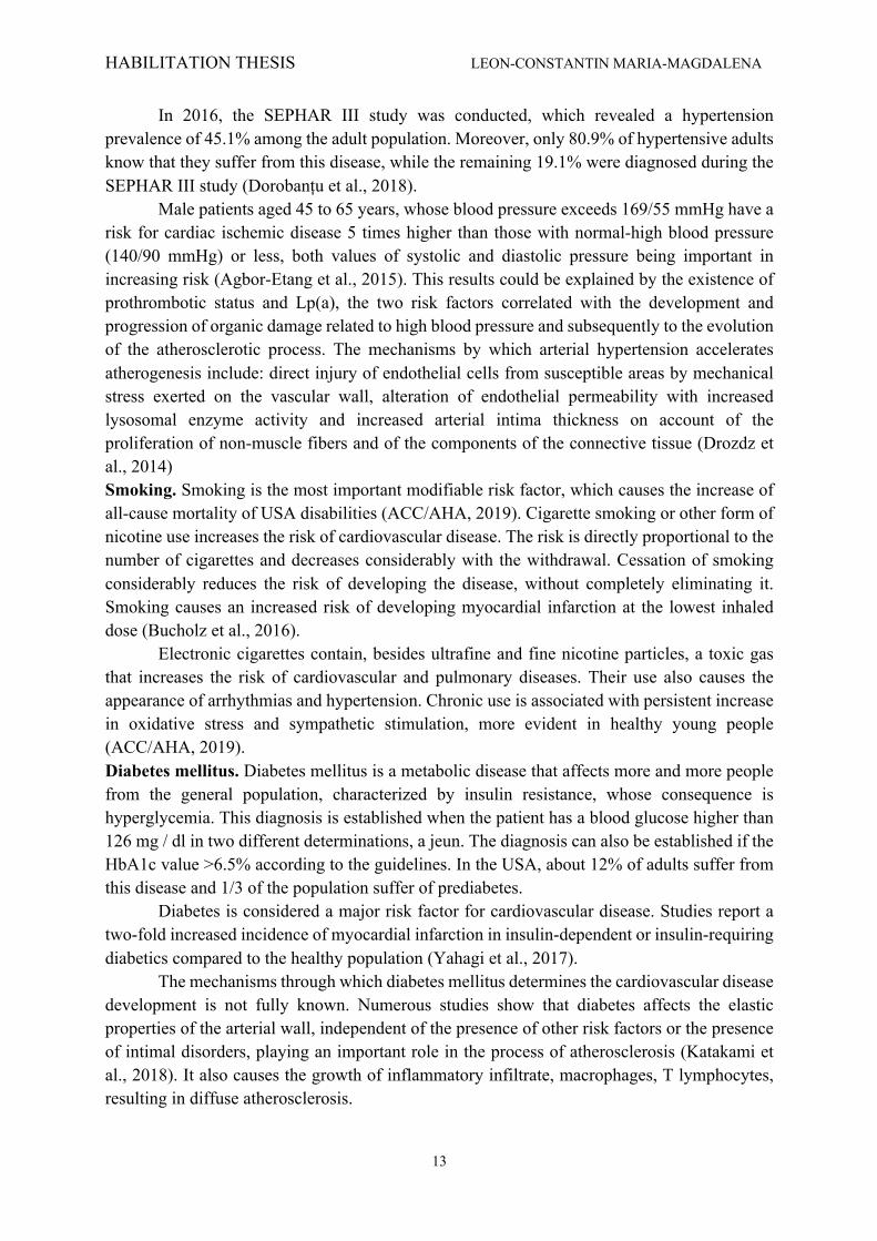
Male patients aged 45 to 65 years, whose blood pressure exceeds 169/55 mmHg have a risk for cardiac ischemic disease 5 times higher than those with normal-high blood pressure (140/90 mmHg) or less, both values of systolic and diastolic pressure being important in increasing risk (Agbor-Etang et al., 2015). This results could be explained by the existence of prothrombotic status and Lp(a), the two risk factors correlated with the development and progression of organic damage related to high blood pressure and subsequently to the evolution of the atherosclerotic process. The mechanisms by which arterial hypertension accelerates atherogenesis include: direct injury of endothelial cells from susceptible areas by mechanical stress exerted on the vascular wall, alteration of endothelial permeability with increased lysosomal enzyme activity and increased arterial intima thickness on account of the proliferation of non-muscle fibers and of the components of the connective tissue (Drozdz et al., 2014) Smoking. Smoking is the most important modifiable risk factor, which causes the increase of all-cause mortality of USA disabilities (ACC/AHA, 2019). Cigarette smoking or other form of nicotine use increases the risk of cardiovascular disease. The risk is directly proportional to the number of cigarettes and decreases considerably with the withdrawal. Cessation of smoking considerably reduces the risk of developing the disease, without completely eliminating it. Smoking causes an increased risk of developing myocardial infarction at the lowest inhaled dose (Bucholz et al., 2016).
Electronic cigarettes contain, besides ultrafine and fine nicotine particles, a toxic gas that increases the risk of cardiovascular and pulmonary diseases. Their use also causes the appearance of arrhythmias and hypertension. Chronic use is associated with persistent increase in oxidative stress and sympathetic stimulation, more evident in healthy young people (ACC/AHA, 2019). Diabetes mellitus. Diabetes mellitus is a metabolic disease that affects more and more people from the general population, characterized by insulin resistance, whose consequence is hyperglycemia. This diagnosis is established when the patient has a blood glucose higher than 126 mg / dl in two different determinations, a jeun. The diagnosis can also be established if the HbA1c value >6.5% according to the guidelines. In the USA, about 12% of adults suffer from this disease and 1/3 of the population suffer of prediabetes. Diabetes is considered a major risk factor for cardiovascular disease. Studies report a two-fold increased incidence of myocardial infarction in insulin-dependent or insulin-requiring diabetics compared to the healthy population (Yahagi et al., 2017). The mechanisms through which diabetes mellitus determines the cardiovascular disease development is not fully known. Numerous studies show that diabetes affects the elastic properties of the arterial wall, independent of the presence of other risk factors or the presence of intimal disorders, playing an important role in the process of atherosclerosis (Katakami et al., 2018). It also causes the growth of inflammatory infiltrate, macrophages, T lymphocytes, resulting in diffuse atherosclerosis.
HABILITATION THESIS LEON-CONSTANTIN MARIA-MAGDALENA
14
Calcifications at the level of the vessels are found in most patients with diabetes, changes being observed on CT by calculating the calcium score. The mechanisms by which these phenomena are achieved include increased oxidative stress, endothelial dysfunction, impaired mineral metabolism due to renal dysfunction and increased inflammatory cytokine production (Blaha et al., 2016). The changes observed in the vessels cause an worsening of the condition of the patients with peripheral artery disease, stimulating about 100 times the occurrence of gangrene (ischemic events) in the diabetic patients, especially if smoking is associated. Age, duration from onset of diabetes and the presence of diabetic neuropathy are important risk factors for atherosclerotic disease of the lower limbs (Thiruvoipati et al., 2015).
Conversely, acute hypoglycemia causes a cascade of important physiological effects and may induce oxidative stress. In the cardiovascular system, mainly as a consequence of sympathetic - adrenergic activation, arrhythmia may occur or even induce sudden death. In healthy adults, cardiovascular effects are transient and have no severe consequences, but they can become pathological in patients with diabetes who already have endothelial dysfunction. Acute hemodynamic and hematological changes may increase the risk of localized tissue ischemia and major vascular events such as myocardial or cerebral ischemia may be precipitated by acute hypoglycemia (Snell-Bergeon et al., 2012).
The association of hypertension with diabetes causes pathophysiological changes both in the large vessels and in the microvascularization level. Increased arterial stiffness, which occurs as a result of calcifications in the intima and media, leads to increased systolic pressure and pulse pressure which determines a decreased coronary perfusion. Reshaping the resistance arteries and the decrease of the capillaries determines the increase of the peripheral resistance, maintaining the high blood pressure and amplifying the negative hemodynamic effects of the reduced arterial compliance. Stiffness is considered an important independent risk factor of cardiovascular disease (Smulyan et al., 2016). Homocysteine is a risk factor for cardiovascular disease, firstly described in 1990, involved in the process of atherosclerosis and hypercoagulability. Clinical and epidemiological studies have shown that there is a link between total serum homocysteine level and coronary artery disease, peripheral vascular disease, stroke and venous thrombosis, in which increased homocysteine level or concentration is associated with the progression of atherosclerosis in patients with these conditions. Increased concentrations of homocysteine endanger the endothelial function, increases oxidative stress, affects the methylation reactions and alters the protein structure (Ganguly et al., 2015).
Hyperhomocysteinemia may be caused by poor absorption of folic acid and B vitamins. As a result, recent data suggest that ingestion of folic acid and vitamin B6, along with a proper diet, would reduce the incidence of cardiovascular disease, but this remains to be evaluated in further studies. An increase of homocysteine concentration is correlated with a risk of coronary heart disease of approximately 10%. Increasing homocysteine by up to 5 micromol / L increases the risk with about 41%, as does the cholesterol increase with 0.52 micromol / L (20 mg / l). Smoking and hypertension have multiple effects on the atherogenic action of increased homocysteine concentrations. The association between hyperhomocysteine and V Leiden factor increases the risk of thrombosis 3-6 times (Nigwekar et al., 2016).
A particular interest is also given to the infectious etiology of atherosclerosis. Infectious agents can contribute to the chronic inflammatory process through both direct and indirect

HABILITATION THESIS LEON-CONSTANTIN MARIA-MAGDALENA
15
mechanism. Bacterial infections with Chlamydia pneumoniae, Mycoplasma pneumoniae, Helicobacter pylori, Enterobacter hormaechei, multiple periodontal organisms (eg. Poryphyromonas gingivalis, Aggregatibacter actinomycetemcomitans, Prevotella intermedia, Tanerella forsreptocytea, Streptococcus) and viruses (cytomegalovirus, hepatitis C virus, human immunodeficiency virus, herpes simplex virus, Epstein-Barr Virus, enteroviruses and parvovirus) may affect endothelial cell function, resulting an increased leukocyte and platelet adhesion in the affected vascular segment (Pothineni et al., 2017).
Numerous studies have shown that atherosclerosis is the consequence of adoptive immunity to microbial HSP-60. Stress factors induce increased heat shock protein (HSP) expression on endothelial cells and also increases the cross-reactivity between antibodies to microbial HSP leading to autoimmune reaction and accelerated atherosclerosis. The association between infectious syndrome and coronary heart disease has been reported by various studies. At the level of the vascular walls, in places where atherosclerosis occurs with predilection, mononuclear cells, CD4 + and CD8 + lymphocytes accumulate. Endothelial cells, macrophages and dendritic cells act as antigen presenting cells. Infectious agents can infect macrophages and persist for longer period of time, causing the secretion of pro-inflammatory cytokines (INF-γ, TNF-α, IL-1, IL-6, IL-8), metalloproteinases and integrins (Hemmat et al., 2018). Stress, the disease of this century, is an independent risk factor for the cardiovascular disease and causes an increase of morbidity and mortality in patients with pre-existing pathology. Psychic or emotional stress and anxiety have been shown to be factors associated with the appearance of ischemic heart disease and sudden death. Chronic stress causes a multitude of nonspecific systemic responses that have as an effect modulation of the onset of atherosclerosis. One possible mechanism would be that stress causes endothelial damage, directly activates macrophages, stimulates adipose cell formation and ultimately generates atherosclerotic plaque (Yao et al., 2019). Sedentarism is an important risk factor in the appearance of cardiovascular disease. The role of physical activity is mentioned in all the guidelines, the main treatment in all cardiovascular pathologies being the nonpharmacological one, that includes movement. Changing the lifestyle, attending the kinetotherapy programs, individualized for each individual (aerobic, fitness, cardio, etc.) are essential in any cardiovascular disease (Lazaros et al., 2019).
Physical activity is associated with a decrease of morbidity and mortality within the cardiovascular disease. Physical training involves morphological, hemodynamic and metabolic changes. The most recommended is submaximal, aerobic physical activity. Studies have shown that maximal or near maximal (anaerobic) physical activity does not bring additional benefits, on the contrary, increases the risk of cardiovascular events and decreases adherence to training. The benefits of physical activity are not just limited to increasing capacity, but also weight loss, lowering blood pressure, cholesterol, LDL-cholesterol, increasing HDL-cholesterol and improving insulin sensitivity (Al-Mamari et al., 2009).
Obesity has an endemic spread worldwide, being an important risk factor for cardiovascular disease. The diagnosis of obesity is established if the BMI is greater than 30 kg / m2, to which is added the abdominal circumference. Abdominal type obesity leads to a higher risk of developing cardiovascular disease. The molecular mechanism by which the pathology occurs in obese individuals is unclear, but disorders of intracellular lipid metabolism are an important aspect in the pathogenesis of metabolic syndrome (Sandfort et al., 2016).

HABILITATION THESIS LEON-CONSTANTIN MARIA-MAGDALENA
16
Fatty tissue can no longer be considered only as a source of fat storage but is also an endocrine and paracrine proinflammatory secretory organ. It is recognized as a rich source of pro-inflammatory mediators, which can contribute to vascular impairment, insulin resistance and atherogenesis. Thus, inflammation of this tissue may be an important step in the occurrence of numerous manifestations related to the pathological features of the metabolic syndrome and may lead to diabetes and atherosclerosis. Defining an obesity phenotype relevant to cardiovascular risk can be done by identifying adipocytokines and biomarkers that quantify the metabolic activity of adipose tissue. Two types of adipocytokines are described, in terms of their effect. Proinflammatory adipocytokines, mediators of endothelial impairment and atherosclerosis, including TNF-α (tumor necrosis factor), IL-6 (interleukin 6), leptin, plasminogen activator inhibitor (PAI-1), angiotensinogen, resistin, and C-reactive protein (PCR) and also anti-atherosclerotic adipocytokines that include nitric oxide (NO) and adiponectin (Lovren et al., 2015). Multiple risk factors accumulate their effects. Thus, the presence of 2 risk factors increases the risk about 4 times and if 3 risk factors are present, the rate of myocardial infarction increases 7 times (Mega et al., 2015). Also, the level of exposure to risk factors causes considerable variations in the evolution of the atherosclerotic process and therefore its early determination by different methods could be extremely useful in the assessment of cardiovascular risk. However, atherosclerosis and its consequences can occur in the apparent absence of any risk factor, even in people who had a prudent life and without an apparent genetic predisposition.
Part of the preoccupations related to atherosclerosis risk factors were synthesized in the following articles: ISI ARTICLES
1. Rezuș E, Leon M, Rezuș C. Correlations in concerning the relation hyperuricemia – metabolic syndrome. Rev. Chim (Bucharest), 2015; 66(8):1015-1018.
2. Mitu F, Ștefanachi E, Leon M. Alcohol and cardiovascular disease - a social impact analysis. Review of Research and Social Intervention, 2013; 40:180-187.
Using the theoretical data mentioned above, the two articles aimed:
• Hyperuricemia and metabolic syndrome
To assess the relationship between cardio metabolic risk factors with asymptomatic hyperuricemia and gout.
• Alcohol and cardiovascular disease
To highlight some data regarding the pathogenesis and natural history of alcohol-related heart disease which still remains obscure as the alcohol has been considered a risk factor for over a century.

HABILITATION THESIS LEON-CONSTANTIN MARIA-MAGDALENA
17
1.3. Material and methods
• Hyperuricemia and metabolic syndrome The first study group included 153 cases diagnosed and evaluated during 01 January
2014 – 31 July 2014 in the Rheumatology Clinic of the Rehabilitation Hospital of Iasi. The selection items included at least three of the five metabolic syndrome (MS) criteria recommended by AHA/National Heart, Lung, and Blood Institute (NHLBI)/ The Obesity Society (TOS) (Table I). The selected patients previously identified with gout or asymptomatic hyperuricemia were classified according to age, gender, gout, metabolic syndrome diagnose, degree of essential hypertension degree (3, 4 or 5 clinical criterias compulsory for MS diagnosis).
• Alcohol and cardiovascular disease
We revised the current literature concerning alcohol determinism in cardiovascular disease etiopathogeny following a relation between excessive regular consumption and the risk of alcoholic cardiomyopathy and congestive heart failure, irrespective of sex. We proposed to highlight echocardiography abnormalities as left ventricular dilation, increased left ventricular mass, reduced or normal left ventricular wall thickness and diminished myocardial contractility and alcohol consumption.
1.4. Results
• Hyperuricemia and metabolic syndrome Gender distribution of the selected group of patients diagnosed with MS reveals 60
females (39,2%) and 93 males (60.8%) (p < 0.01), with an average of 60.01.
Table I. Metabolic syndrome criterias (AHA/ACC/TOS, 2013) Clinical criteria (at least 3) Normal values of the parameters
Obesity ≥30kg/m2 Serum triglycerides level (TG) ≥150 mg/dL or with treatment for high levels of TG High-density lipoprotein cholesterol level (HDL)
<40 mg/dL in men <50 mg/dL in women or with treatment for low levels of HDL↓
Blood pressure (BP) ≥130 mmHg BP sistolic or ≥85 mmHg BP diastolic with treatment for hypertension
Serum glucose level (Glu) ≥100 mg/dL or with treatment for diabetes
21 female patients originate from rural areas comparative with 52 Male patients from the same environment. Statistical analysis demonstrates no significant changes between the rural/urban (t = 0.113; p = 0.910), age or sex distribution in terms of backgrounds distribution (Fig. 1).
HABILITATION THESIS LEON-CONSTANTIN MARIA-MAGDALENA
18
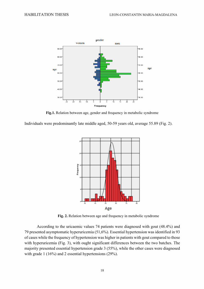
Fig.1. Relation between age, gender and frequency in metabolic syndrome
Individuals were predominantly late middle aged, 50-59 years old, average 55.89 (Fig. 2).
Fig. 2. Relation between age and frequency in metabolic syndrome
According to the uricaemic values 74 patients were diagnosed with gout (48.4%) and
79 presented asymptomatic hyperuricemia (51,6%). Essential hypertension was identified in 93 of cases while the frequency of hypertension was higher in patients with gout compared to those with hyperuricemia (Fig. 3), with ought significant differences between the two batches. The majority presented essential hypertension grade 3 (55%), while the other cases were diagnosed with grade 1 (16%) and 2 essential hypertensions (29%).
HABILITATION THESIS LEON-CONSTANTIN MARIA-MAGDALENA
19
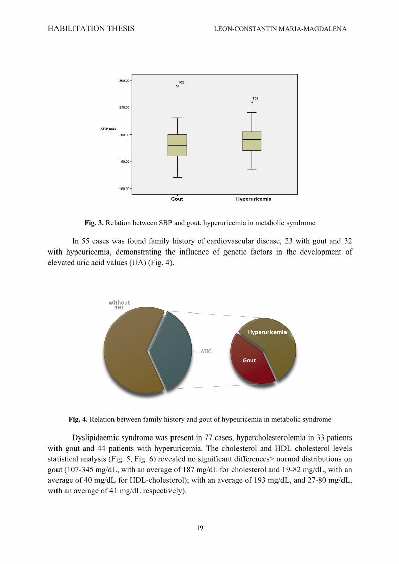
Fig. 3. Relation between SBP and gout, hyperuricemia in metabolic syndrome
In 55 cases was found family history of cardiovascular disease, 23 with gout and 32 with hypeuricemia, demonstrating the influence of genetic factors in the development of elevated uric acid values (UA) (Fig. 4).
Fig. 4. Relation between family history and gout of hypeuricemia in metabolic syndrome
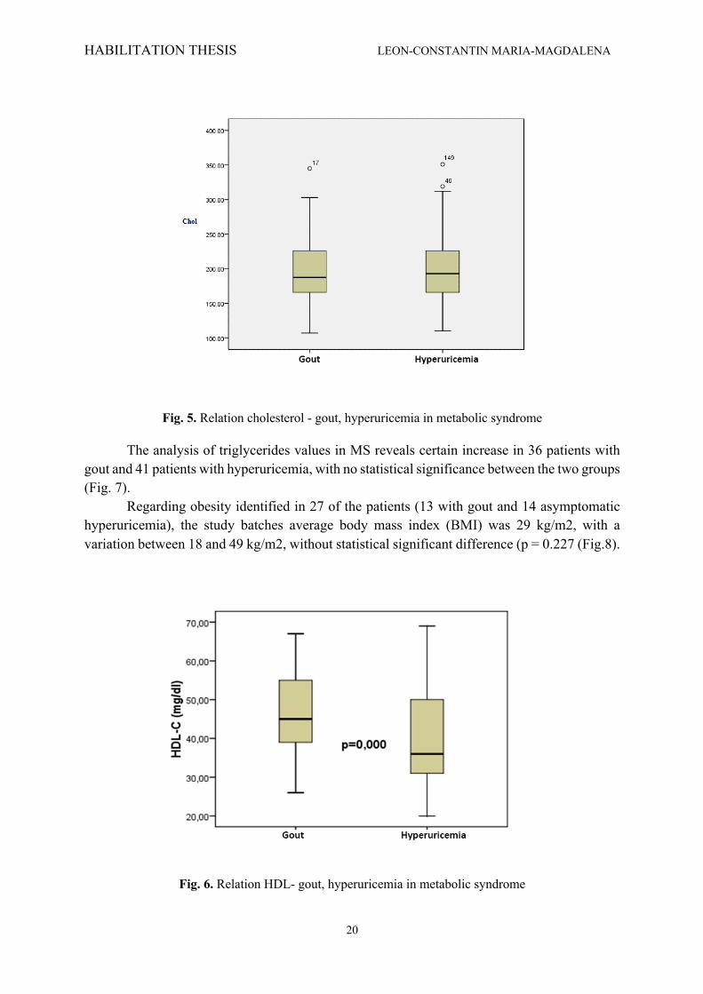
Dyslipidaemic syndrome was present in 77 cases, hypercholesterolemia in 33 patients with gout and 44 patients with hyperuricemia. The cholesterol and HDL cholesterol levels statistical analysis (Fig. 5, Fig. 6) revealed no significant differences> normal distributions on gout (107-345 mg/dL, with an average of 187 mg/dL for cholesterol and 19-82 mg/dL, with an average of 40 mg/dL for HDL-cholesterol); with an average of 193 mg/dL, and 27-80 mg/dL, with an average of 41 mg/dL respectively).
HABILITATION THESIS LEON-CONSTANTIN MARIA-MAGDALENA
20
Fig. 5. Relation cholesterol - gout, hyperuricemia in metabolic syndrome
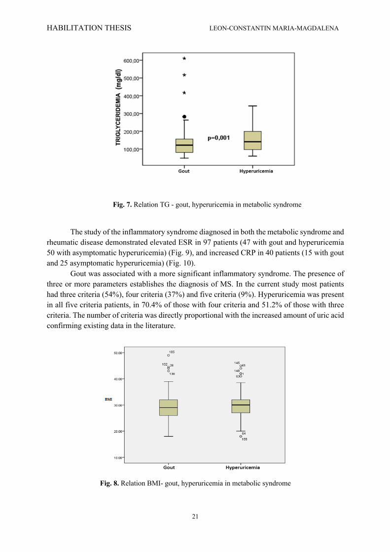
The analysis of triglycerides values in MS reveals certain increase in 36 patients with gout and 41 patients with hyperuricemia, with no statistical significance between the two groups (Fig. 7).
Regarding obesity identified in 27 of the patients (13 with gout and 14 asymptomatic hyperuricemia), the study batches average body mass index (BMI) was 29 kg/m2, with a variation between 18 and 49 kg/m2, without statistical significant difference (p = 0.227 (Fig.8).
Fig. 6. Relation HDL- gout, hyperuricemia in metabolic syndrome
HABILITATION THESIS LEON-CONSTANTIN MARIA-MAGDALENA
21
Fig. 7. Relation TG - gout, hyperuricemia in metabolic syndrome
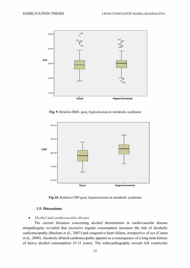
The study of the inflammatory syndrome diagnosed in both the metabolic syndrome and
rheumatic disease demonstrated elevated ESR in 97 patients (47 with gout and hyperuricemia 50 with asymptomatic hyperuricemia) (Fig. 9), and increased CRP in 40 patients (15 with gout and 25 asymptomatic hyperuricemia) (Fig. 10).
Gout was associated with a more significant inflammatory syndrome. The presence of three or more parameters establishes the diagnosis of MS. In the current study most patients had three criteria (54%), four criteria (37%) and five criteria (9%). Hyperuricemia was present in all five criteria patients, in 70.4% of those with four criteria and 51.2% of those with three criteria. The number of criteria was directly proportional with the increased amount of uric acid confirming existing data in the literature.
Fig. 8. Relation BMI- gout, hyperuricemia in metabolic syndrome
HABILITATION THESIS LEON-CONSTANTIN MARIA-MAGDALENA
22
Fig. 9. Relation BMI- gout, hypeuricemia in metabolic syndrome
Fig.10. Relation CRP-gout, hyperuricemia in metabolic syndrome
1.5. Discussions
• Alcohol and cardiovascular disease The current literature concerning alcohol determinism in cardiovascular disease
etiopathogeny revealed that excessive regular consumption increases the risk of alcoholic cardiomyopathy (Beulens et al., 2007) and congestive heart failure, irrespective of sex (Conen et al., 2008). Alcoholic dilated cardiomyopathy appears as a consequence of a long term history of heavy alcohol consumption (5-15 years). The echocardiography reveals left ventricular

HABILITATION THESIS LEON-CONSTANTIN MARIA-MAGDALENA
23
dilation, increased left ventricular mass, reduced or normal left ventricular wall thickness and diminished myocardial contractility. Heart failure is currently a major cause of morbidity and mortality all over the world, thus being justified the importance of its diagnose, while myocardial infarction, hypertension and type 2 diabetes mellitus are considered its high risk predictors. Light-to-moderate drinking has been associated with a lower risk of heart failure while regular excessive consumption of alcohol (heavy drinking) increases it significantly (Djoussé et al., 2008).
The Framingham Heart Study reported a 59% lower risk of heart failure in males with moderate consumption of alcohol versus abstainers, but the results are not confirmed in females. The SOLVD Study (Study of Left Ventricular Dysfunction) demonstrated no association between alcohol consumption and heart failure in ischemic cardiomyopathy patients.
Similar results were obtained in SAVE study (Survival and Ventricular Enlargement), moderate drinking being not associated with hospitalization duration for heart failure secondary to myocardial infarction (Djoussé et al., 2008; Mitu et al., 2011).
Another recent meta-analysis reveals a linear relation between alcohol consumption and risk of atrial fibrillation only in males, mainly due to alcohol that shortens the effective refractory period of right atrium, promotes propagation of a critically timed premature atrial complex, increases thickening and scarring of cardiac connective tissue, alters oxidative stress, induces electrolyte imbalance and negative inotropic effect through calcium-channel inhibition in ventricular cells. If the patient associates diabetes or/and cardiovascular disease the incidence of atrial fibrillation is several times higher than the one registered in the general population (Liang et al., 2012).
The Cardiovascular Health Study identified no relationship between different alcohol consumption levels and atrial fibrillation mainly in elderly, although gender-specific information’s were not provided. No similar effects were demonstrated in women’s, but theoretically there are no effects on atrial fibrillation during consumption of moderate amounts of alcohol, contrarily to excessive amounts of alcohol that might increase the risk of atrial fibrillation (Mukamal et al., 2007).
The HAPIEE study (Health, Alcohol and Psychosocial Factors in Eastern Europe) performed in Lithuania on men and female subjects aged 45–72 years, results demonstrate an association between cardiovascular disease, alcohol intake, older age, lower education level and poorer cognitive performance (Tamosiunas et al., 2012). The cardiovascular risk factors such as cholesterol, hypertension, diabetes mellitus, fibrinogen levels, homocysteine, C-reactive protein and the middle or elderly men and women are the main factors in the cardiovascular diseases determinism and associated psychic problems.
In both sexes cognitive function is conditioned by the quality of life and self-rated health, the alcohol intake being lower in impaired cognitive function in females (Singh-Manoux et al., 2008). Moderate alcohol consumption is traditionally associated with a reduced risk of cardiovascular-related outcomes, such as coronary heart disease, stroke, congestive heart failure, death, but this relation is not available in case of atrial fibrillation. In healthy people, the alcohol intake diminishes vagal modulation decreasing heart frequency (Koskinen et al., 1994). Different observational studies reveal the benefic effects of moderate amount of alcohol, even if this concept is largely debated in the current literature.
HABILITATION THESIS LEON-CONSTANTIN MARIA-MAGDALENA
24
A meta-analyses comparing 84 studies on the relation between alcohol consumption and cardiovascular disease demonstrate that moderate amount of alcohol is highly associated with a 14–25% reduction of the risk of all outcomes compared with alcohol abstaining, but consumption of larger amounts of alcohol is associated with higher risks for stroke incidence and mortality. Another meta-analysis supports the latter association between alcohol and coronary heart disease, with a 25–35% risk reduction for light to moderate drinking in heavier consumptions (Ronksley et al., 2011). Inability to coordinate movements and temporary impairment of memory or mood are considered as a short term effects of alcohol consumption.
Long term effects of alcohol consumption may cause depression. With the increase of blood alcohol concentration, the central nervous system activity diminishes. A state of mild intoxication is determined by a blood alcohol concentration of 50mg/dL, while coma or death will appear at a concentration of 350mg/dL respectively 500mg/dL.
Conversely, moderate alcohol consumption has been demonstrated to have benefic effects on long term, such as increase of HDL cholesterol level, decrease of blood pressure, arterial atherosclerotic lesions appearance or thrombus formation in the coronary arteries protection, decrease of a sudden heart attack risk, increase of the DHEA hormone level (dehydro-epi-androsterone). Meanwhile, alcohol consumption has severe effects on generally speaking wellbeing, or particularly on the cardiovascular system, damaging central nervous system, liver, pancreas, myocardial muscle. A special result of chronic alcohol intake might be hypertension, (Taylor et al., 2009), alcoholic cardiomyopathy, clinically expressed by heart excessive enlargement, considerably reducing of the contractile capacity and blood ejection, with the installing of heart rhythm disorders. Depending on the amount consumed, alcohol increases blood pressure, which increases the risk of hypertension (Higashiyama et al., 2013). Acutely intake of excessive alcohol amounts “binge drinking” has been related to increased risks of myocardial infarction, stroke, hypertension, type 2 diabetes mellitus and atrial fibrillation (Liang et al., 2012).
The current literature establishes a clear evidence between alcohol abuse and the increase risk of hypertension in both sexes, while the effects of low or moderate consumption are still controversial (Sesso et al., 2008). There is a directly proportional relationship between alcohol and the risk of ischemic infarction: increased consumption increases the risk, while a low intake level reduces the risk of infarction. Even in people with no history or previous signs of heart disease, occasionally abusive alcohol consumption increases the risk of arrhythmias and sudden death (Gao et al., 2012).
In both sexes, regular excessive alcohol consumption augments the risk of developing alcoholic cardiomyopathy (directly proportional to the consumed doses) (Beulens et al., 2007) and congestive heart failure (Conen et al., 2008). Secondary, a long-term history of heavy alcohol consumption (5-15 years) might induce a dilated cardiomyopathy. Echography evaluation demonstrate left ventricular dilation, increased left ventricular mass, reduced or normal left ventricular wall thickness and depressed myocardial contractility. In this context, heart failure is a major cause of mortality all over the world, thus it should be correct and in time diagnosed, myocardial infarction, hypertension and type 2 diabetes mellitus being strong predictors. Excessive consumption of alcohol (heavy drinking) increases the risk of heart failure, whereas light-to-moderate drinking has been associated with a lower risk of heart failure (Djoussé et al., 2008).
HABILITATION THESIS LEON-CONSTANTIN MARIA-MAGDALENA
25
• Hyperuricemia and metabolic syndrome
The results of the current study confirmed the previously reported sex-specific differences in the MS and the strong associations between serum UA concentration and age, obesity, hypertension, serum triglycerides levels, serum cholesterol levels (Puig et al., 2008; Juraschek et al., 2013; Chen et al., 2013). Serum UA levels are strongly influenced by many factors, including obesity, body mass index and are positively associated with CRP and ESR. Thus, we could demonstrate the relationship between serum uric acid and the defined markers of systemic inflammation.
From clinical point of view, hyperuricemia is a strong indicator for MS criteria, and contrary, presence of MS is considered indication for investigating the serum UA concentration (Sattui et al., 2014; Zhou et al., 2012; Robinson et al., 2013; Sari et al., 2009). Our results demonstrated a strong relation between asymptomatic hyperuricemia and gout and the cardiovascular risk factors. The diagnose of gout is considered a high risk factor in development of cardiovascular disease. The potential contributions of hyperuricemia associated with the inflammation process characteristic in atherogenesis (Kawada et al., 2006; Johnson et al., 2013; Parsa et al., 2012) and the development of thrombosis in a manner similar to other inflammatory rheumatic diseases are similarly associated with an increased risk of cardiovascular disease (rheumatoid arthritis or lupus) (So et al., 2010; Kirilmaz et al., 2010). In such cases, the design of cardiovascular risk profile is essential for any patient suffering a gout attack (Krishnan et al., 2014), but there are still necessary further studies in order to assess the role of uric acid and to identify the way to reduce significantly the risk of major cardiovascular events.
1.6. Conclusions
• Hyperuricemia and metabolic syndrome The aim of the study were to prove that serum UA concentration is positively correlated
with the number of MS criteria such as obesity, hypertension, serum triglycerides levels, serum cholesterol levels. These criteria are strongly and statistically significant correlated with the association between UA and MS components, serum uric acid and markers of systemic inflammation (ESR, CRP). The obtained results support the association of the cardiovascular risk factors with asymptomatic hyperuricemia and gout disease.
• Alcohol and cardiovascular disease The current results are conjoint with the ones obtained on different international clinical
study groups, alcohol dependency syndrome creating various health, psychological, social or economic problems, deeply impacting the environment. The aims of alcohol addiction treatment remain the improvement of physical and mental health, as well as the relational and social status of the patient. Despite the literature evidence, and the current study results the debate between adherents of abstinence and those of moderate alcohol consumption is still open.
HABILITATION THESIS LEON-CONSTANTIN MARIA-MAGDALENA
26
Chapter 2. THE MORPHOPHYSIOLOGY OF ATHEROSCLEROSIS
2.1. Theories of pathogenesis
The atherosclerosis term was first used to describe the association between the arterial rigidity and lipid degeneration. The term comes from the Greek word 'athere' meaning lipid accumulation and 'sclerosis' (Virmani et al., 2006). This process is characterized by lipid accumulation, infiltration of macrophages, proliferation of the smooth muscle cells, accumulation of fibroblasts and formation of thrombus. Initially, the lesion is abluminal, but it can progress, being influenced by the environmental, but also genetic factors. The speed of proliferation varies from one individual to another, atherosclerosis being described even at birth (Raja et al., 2002). Over time, several theories regarding the pathogenesis of atherosclerosis have been formulated. Of these, only two theories succeeded in summarizing the complexity of the process: the first considers that the starting point of the atherosclerotic process is the "organization" of thrombi at the arterial wall level (Steiner et al., 2008), and the second is linked to an injury of the intima to which cells can adhere from the circulating torrent - macrophages, platelets, lymphocytes, on a field where there is chronic inflammation, all of which lead to degenerative lesions of atherosclerosis (McManus, 1958).
Researchers in the field propose several pathogenic theories to explain the growth and development of atherosclerotic plaque (Aziz et al., 2016).
Trombogenic theory According to this theory, atherosclerosis represents the final stage of repeated episodes
of intramural thrombosis, with the creation of platelet and fibrin deposits, which subsequently causes thrombus formation. The source of lipids in this theory would be represented by the platelet membranes located in the newly formed atheroma; the proliferation of smooth muscle cells is achieved under the influence of platelet-derived growth factors. However, wall thrombosis is the main modification that determines the occlusion of the vessel, especially the coronary arteries, which will subsequently develop atherosclerosis (Golia et al., 2014).
Theory of lipid infiltration This theory has the most supporters, and the basic idea would be that the atherosclerosis
process is due to focal accumulation of lipids from plasmatic lipoproteins in the vessel walls. This is why the recommendations in the preventive guidelines are to lower the level of lipids in the blood. The mechanism by which this lipid accumulation is accomplished is not fully elucidated, but the hypothesis is widely accepted. However, for the formation of the atheroma plaque, it is not enough just the storage of lipids, but also the proliferation of smooth muscle cells, thrombosis, etc. (Geovanini et al., 2018).
Theory of monoclonal proliferation According to it, atherosclerotic plaques show a monoclonal proliferation of smooth muscle cells under the influence of environmental factors, with the abnormal growth of the size of these cells similar to that of the smooth muscle neoplasms (Ross et al., 2001).
Theory of endotelial damage or response to aggression This theory was initially proposed to explain the mechanism of smooth muscle cell accumulation in the atherosclerotic lesion. Proliferation begins with the action of growth factors
HABILITATION THESIS LEON-CONSTANTIN MARIA-MAGDALENA
27
released by endothelial cells, macrophages or even from damaged smooth muscle cells. All these changes have the alteration of the function of the vascular endothelium as central element, under the influence of different factors, causing the appearance of a chronic inflammatory response. The inflammation will persist as long as the incriminating factors exist at the site of the injury (Mizuno et al., 2011). Relatively recent research on the process of atherosclerosis considers that the most plausible theory regarding the process of atherosclerosis is the "hypothesis of injury response". This theory considers that the central element in atherogenesis is a form of endothelial injury, which is expressed by endothelial dysfunction (Reneman et al., 2006). The lesion usually occurs at the bifurcation sites of the arteries, due to the high blood pressure and the shear forces exerted on the endothelial cells at this level are also very high. The process of atherosclerosis begins after the onset of chronic inflammation in the arterial wall lesion. The hemodynamic forces at that level induce the expression of factors promoting atherosclerosis such as: basic fibroblast growth factor (FGF-2), tissue factor (TF), plasminogen activator and endothelin. Moreover, the progression of the lesions is supported by the interaction between the modified lipoproteins, the macrophages derived from the blood monocytes, the T lymphocytes and the normal cellular constituents of the arterial wall. At the level of the susceptible regions of the arterial wall, dysfunctional endothelial cells allow exposure of the subendothelial tissue to different plasma constituents (Libby et al., 2010). In individuals with high cholesterol values, monocytes, along with mitogens derived from cells / plasma and mitogenic promoters, enter the subendothelial space of the intima. Also, blood platelets may come into contact with the exposed intimal collagen, aggregating on the denuded surface of the lesion. They release platelet-derived growth factor (PDGF) which has been shown to be chemotactic for smooth muscle cells, suggesting that at least in some circumstances PDGF may play a major role in inducing smooth muscle cell migration through the internal elastic limiting fenestrae, resulting in focal invasion of the intima. The consequence of the changes presented above is the increase of the thickness of the intima with the reduction of the vascular lumen (thus inducing an even greater risk on the endothelial cells) and the decrease of the quantity of blood that passes through the vessel, with the individual's inability to make effort. Because the diameter is small, the presence of a small thrombus causes complete obstruction of the vessel (Raja et al., 2002). If we speak of myocardium, the infarction manifested by precordial pain, of high intensity, with irradiation in the upper left limb, jaw, parasternal, but never inferior oblique. The pain is so strong that it only gives up on opioids. The intervention in the first 6 hours, with the removal of the thrombus, determines the favorable evolution of the patient, with restitutio ad integrum. If the obstruction occurs at the brain level, the cerebral infarction occurs, known as stroke. Thrombolysis performed in the first hours, as in the heart, makes the evolution very good, without consequences. This theory was reproduced on a computer-created model comprising 32 immunological parameters that stimulate the action of macrophages, monocytes, adipose cells, chemoattractant macrophages, endothelium-stimulating cytokines, modified HDL and LDL lipoproteins. This model considers as the main essential element, only the endothelial injury (Chalmers et al., 2017).
HABILITATION THESIS LEON-CONSTANTIN MARIA-MAGDALENA
28
The processes that develop successively and/or simultaneously in the evolution of atherosclerotic lesions can be systemized in three stages: the first stage of initiation and formation, the second stage - adaptation and the third – clinical symptoms (Steiner et al., 2008). The first two stages are subclinical, so the disease is present but does not manifest (Jarasūniene et al., 2003; Nègre-Salvayre et al., 2017; Jessup et al., 2004; Rajtar et al., 2006). The first stage can be synthesized in this way:
Ø chronic endothelial injury with endothelial dysfunction, which results in increased vascular permeability, increased leukocyte adhesion as a result of expression of adhesion molecules by endothelial cells and thrombotic potential. Risk factors, microorganisms, low oxidative density lipoproteins promote endothelial injury (Jarasūniene et al., 2003);
Ø accumulation of lipoproteins in the subendothelial space predominantly with LDL-cholesterol or hormones (such as insulin) (Nègre-Salvayre et al., 2017);
Ø modification of lipoproteins through oxidation. Oxidative stress not only affects LDL, but also the endothelial cells and macrophages (Jessup et al., 2004);
Ø recruitment of blood monocytes and other leukocytes, their adhesion to the endothelium, subendothelial diapedesis, transformation into macrophages and then into foam cells. Monocytes/macrophages play a central role in stimulation of lipid accumulation, release of growth factors, stimulation of smooth muscle cell accumulation. Monocytes/macrophages synthesize PDGF, FGF, Tumor Necrosis Factor (TNF), interleukine-1 (IL-1), interferon-alpha (IFN-alpha), and TGF-beta, which modulates endothelial cell or muscle cell activity (Rajtar et al., 2006);
Ø adhesion of blood platelets. IL-1 and TNF stimulate endothelial cells to produce PAF (platelet-activating factor), TF (tissue factor) and PAI (plasminogen activator inhibitor) (Rajtar et al., 2006);
Ø release of various factors from activated platelets, macrophages, or vascular cells, which results in smooth muscle cell migration from medium to intima (Decano et al., 2018);
Ø proliferation of smooth muscle cells in the intima and elaboration of extracellular matrix, with the accumulation of collagen, proteoglycans, glycosaminoglycans. They stimulate the accumulation of lipids due to their ability to penetrate the intima. Thus, there is an increase in intracellular and extracellular lipid accumulations (Decano et al., 2018);
Ø possibly the formation of associated thrombi that may increase in size, embolize, or be organized and incorporated into the atheroma plate (Libby et al., 2006).
The second stage – adaptation Because the lumen must preserve its dimensions, and the plaque enlarges, a process of
remodeling the lumen's dimensions takes place. This phenomenon is best observed in coronary arteries. Compensatory remodeling is viable until the plaque is less than half of the lumen size, and then stenosis occurs. Hemodynamic stress is an important regulator of vessel wall remodeling. Matrix metalloproteinases (MMPs) and their inhibitors, tissue inhibitors of metalloproteinases (TIMPs) are also involved in remodeling. As long as the plaque is small, the patient is asymptomatic. The rupture of the plaque may occur at this stage, resulting in patient's death by total obstruction of the vessel and the onset of myocardial infarction (Aziz et al., 2016).
HABILITATION THESIS LEON-CONSTANTIN MARIA-MAGDALENA
29
The third stage – clinical At this stage, the plaque continues to increase in size and obstruct the lumen. The
increase in plaque size may be due to the internal bleeding. Complications that may occur at the level of the plaque are rupture, cracking, ulceration, calcification, hemorrhage in the plaque, thrombosis and the aneurysm. Depending on the location of the vessel, infarcts of larger or smaller dimensions, compatible or not with life, may occur. If the patient arrives at the hospital as soon as the symptomatology is installed and is diagnosed as soon as possible, if does not have significant comorbidities, his condition will improve rapidly and with a very good short and long term prognosis (Golia et al., 2014).
These complex processes are modulated firstly by the risk factors of atherosclerosis, and on the secondly by growth factors or other biological substances produced during the interaction between LDL with cellular elements or extracellular matrix. Risk factors are considered those factors that double the incidence of the disease. Of the most important risk factors we mention diabetes. Patients with diabetes have an increased risk of developing atherosclerosis in many organs, but the mechanism by which these complications occur is not fully known. Metabolic syndrome - high blood pressure, dyslipidemia, diabetes, obesity has become the target for diagnosis in the early stages and also for treatment in order to reduce the atherosclerotic process (Geovanini et al., 2018). The research regarding the process of atherosclerosis has lead through the following articles: 1. Cojocaru E, Trandafirescu M, Leon M, Cotutiu C, Foia L. Immunohistochemical
expression of anti-CD68 antibody in atherosclerotic plaque. RJME. 2012; 53(1): 61-66.
2. Gavril RS, Mitu F, Leon M, Mihalache L, Arhire LI, Grosu C, Gherasim A, Nita O, Ungureanu IO, Oprescu C, Graur M. Biomarkers of inflammation in patients with type 2 diabets mellitus and hepatic steatosis Rev. Chim (Bucharest), 2016;67(9):1828-1832.
Using the theoretical data mentioned above, the mentioned articles aimed:
• CD68 expression in atherosclerosis We performed an immunohistochemical study of the anti-CD68 antibody expression at
the level of the atherosclerotic plaque in order to to highlight the essential role of the vascular wall inflammation in the development of atherosclerosis.
• Inflammation biomarkers in metabolic disorders
The study evaluates the relationship of the inflammatory markers in regard to the lipid profile with the degree of fatty load of the liver and subclinical atherosclerosis in patients with diabetes mellitus type 2 (DM 2). Subclinical atherosclerosis is assessed by measuring carotid intima media thickness (CIMT).
HABILITATION THESIS LEON-CONSTANTIN MARIA-MAGDALENA
30
2.2. Material and methods
• CD68 expression in atherosclerosis A retrospective study was performed on selected data from observation sheets and
pathological results totaling a number of 213 patients, between 33 and 78 years of age, hospitalized in the Cardiovascular Surgery Department of the Institute of Cardiovascular Diseases “Prof. Dr. George I. M. Georgescu”, between 2005 and 2009. The performance of endarterectomies with the subsequent removal of plaques stenosis was affected by the severity of the symptoms at the time of hospitalization and degree of lumen stenosis, due to the presence of lesions. The classic histopathological technique of paraffin embedding, microtome sectioned in 5 µm thick slices and stained with the classical Hematoxylin and Eosin method was used to process the specific tissue samples, which allowed for studying the distribution the extent and the complications of the lesions. Additionally, on the representative blocks, immunohistochemical stains were performed for the study of various factors involved in the pathogenesis of atherosclerosis. The present article refers to the immunohistochemical study of the inflammatory response of atherosclerotic plaque at different stages of development, using the anti-CD68 antibody. Current practice makes use of CD68 antibodies as markers for monocytes/macrophages present in normal or pathological tissue fixed in paraffin.
• Inflammation biomarkers in metabolic disorders
An observational – transversal study was conducted, which included a total of 92 subjects, both in urban and in rural areas investigated within the Clinic of Diabetes, Nutrition and Metabolic Diseases Iasi. For this, the following inclusion criteria were established: patients with DM 2 mellitus treated with diet or oral anti-diabetic agents (OAA) without liver damage of viral nature (hepatitis B, C virus) without toxic - ethanol hepatitis, the patients being selected in the order in which they were admitted in the Diabetes Outpatient Clinic, for a period of three months. Approval of the Ethics Committee was obtained, and all participants signed the informed consent form before the start of the study. CRP and fibrinogen were measured as markers of inflammation. The lipid profile (total cholesterol, LDL-cholesterol, HDL-cholesterol, triglycerides) was determined. Triglycerides (TG) or triacylglycerols represent esters of fatty acid with a high number of carbon atoms with glycerol. TG in excess are deposited mainly in the liver. Excess of TG is mainly responsible for liver steatosis, specifically for the fatty load of liver in varying degrees. HDL-cholesterol, also called antiatherogenic lipoprotein mediates the cellular cholesterol influx. It is the main class of lipoproteins with antiatherogenic effect, being responsible for the reverse transport of cholesterol. Cholesterol released in peripheral tissues, returns to the liver using HDL. Evaluation of the liver function was done by determination of aspartate aminotransferase (AST), alanine aminotransferase (ALT) and gamma-glutamyl transpeptidase (GGT). Transaminases, referred to in the past as glutamic-pyruvic and oxalate-acetic transaminases are enzymes commonly found in the liver. They can be used in assessing liver damage, although they are not specific to liver diseases solely. The GGT enzyme specifically catalyzes the cleavage of the gamma-glutamyl bond of glutathione and gamma glutamyl transformation in water, peptides or amino acids.. Ultrasound with a probe of 3.5MHz was used to evaluate the degree of fatty load of the liver. Five criteria were used to divide liver steatosis into 4 degrees: parenchymal reflectivity, the contrast between the liver and the kidney, deep beam attenuation, viewing the small vessel walls of the liver and
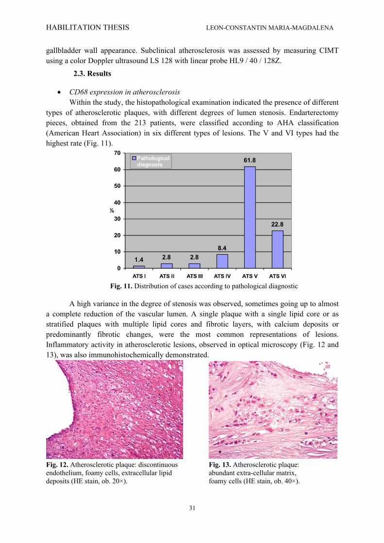
HABILITATION THESIS LEON-CONSTANTIN MARIA-MAGDALENA
31
gallbladder wall appearance. Subclinical atherosclerosis was assessed by measuring CIMT using a color Doppler ultrasound LS 128 with linear probe HL9 / 40 / 128Z.
2.3. Results
• CD68 expression in atherosclerosis Within the study, the histopathological examination indicated the presence of different
types of atherosclerotic plaques, with different degrees of lumen stenosis. Endarterectomy pieces, obtained from the 213 patients, were classified according to AHA classification (American Heart Association) in six different types of lesions. The V and VI types had the highest rate (Fig. 11).
Fig. 11. Distribution of cases according to pathological diagnostic
A high variance in the degree of stenosis was observed, sometimes going up to almost a complete reduction of the vascular lumen. A single plaque with a single lipid core or as stratified plaques with multiple lipid cores and fibrotic layers, with calcium deposits or predominantly fibrotic changes, were the most common representations of lesions. Inflammatory activity in atherosclerotic lesions, observed in optical microscopy (Fig. 12 and 13), was also immunohistochemically demonstrated.
Fig. 12. Atherosclerotic plaque: discontinuous Fig. 13. Atherosclerotic plaque: endothelium, foamy cells, extracellular lipid abundant extra-cellular matrix, deposits (HE stain, ob. 20×). foamy cells (HE stain, ob. 40×).

HABILITATION THESIS LEON-CONSTANTIN MARIA-MAGDALENA
32
The CD68 antigen is a membrane glycoprotein, strongly expressed by blood monocytes and tissue macrophages. A variable degree of infiltration with foamy macrophages was present in the analyzed lesions. Some areas showed a low density of CD68 positive immunoreactivity (Fig. 14 and 15). The fibrous collagen cap revealed the presence of macrophages (CD68 positive), although other inflammatory cells were also present. CD68 was positive in a variable number of macrophages, in the core of the atherosclerotic plaques, depending on the extent of the inflammatory reaction. A chronic inflammatory reaction is indicated by the presence of many macrophages, which is accompanied by changes in the cells and the extracellular matrix of connective tissue.
Fig. 14. Atherosclerotic plaque: positive Fig. 15. Atherosclerotic plaque: positive immuno reactivity for CD68, 40× immuno-reactivity for CD68, 40×
• Inflammation biomarkers in metabolic disorders The study group of 92 subjects included 44 males, where 73% were represented by
patients from urban areas, with an average age of 60.38 and limits between 33 and 86 years of age. The group of subjects was divided into 4 groups depending on the fatty load of the liver: normal liver (9 subjects), intermediate steatosis (24 subjects), moderate steatosis (33 subjects) and severe steatosis (25 subjects). There was no statistically significant differentiation by gender regarding the frequency of the degrees of hepatic steatosis. The proportion of those with fatty liver disease in the studied group exceeded 90%, values comparable to the research literature (Angulo, 2002; Leite et al., 2009). Similarly, average blood glucose was not significantly different according to sex (141.44 ± 143.83 ± 32.85 in men and 34.92 in women) and did not correlate with the degree of hepatic steatosis or with CIMT value, although some literature data showed the relationship between fasting blood sugar and prevalence of NAFLD (Jimba et al., 2005). With regards to the biomarkers of inflammation, the mean value of fibrinogen was slightly higher in men (408.02 compared to 393.4 in women), thus there was no significant difference by gender. No degree of hepatic steatosis or CIMT was correlated with fibrinogen as a marker of inflammation. CRP showed significant direct correlation with the degree of hepatic steatosis. When calculating the Pearson r correlation coefficients between the degree of hepatic steatosis and the parameters included in the study, significant direct correlations with the degree of hepatic steatosis in CRP (r = 0.3673, p = 0.001) were found, in line with the research literature data (Haukeland et al., 2006; Zimmermann et al., 2011). The data presented above are highlighted in Table II.
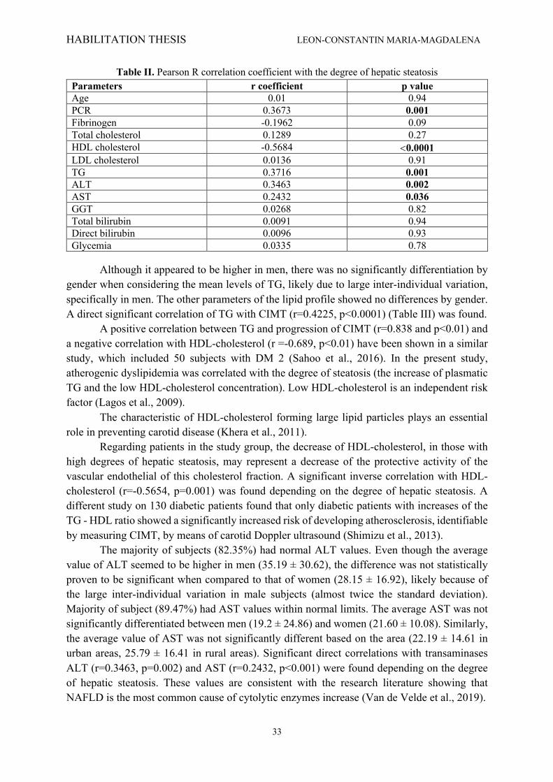
HABILITATION THESIS LEON-CONSTANTIN MARIA-MAGDALENA
33
Table II. Pearson R correlation coefficient with the degree of hepatic steatosis Parameters r coefficient p value Age 0.01 0.94 PCR 0.3673 0.001 Fibrinogen -0.1962 0.09 Total cholesterol 0.1289 0.27 HDL cholesterol -0.5684 <0.0001 LDL cholesterol 0.0136 0.91 TG 0.3716 0.001 ALT 0.3463 0.002 AST 0.2432 0.036 GGT 0.0268 0.82 Total bilirubin 0.0091 0.94 Direct bilirubin 0.0096 0.93 Glycemia 0.0335 0.78
Although it appeared to be higher in men, there was no significantly differentiation by gender when considering the mean levels of TG, likely due to large inter-individual variation, specifically in men. The other parameters of the lipid profile showed no differences by gender. A direct significant correlation of TG with CIMT (r=0.4225, p<0.0001) (Table III) was found.
A positive correlation between TG and progression of CIMT (r=0.838 and p<0.01) and a negative correlation with HDL-cholesterol (r =-0.689, p<0.01) have been shown in a similar study, which included 50 subjects with DM 2 (Sahoo et al., 2016). In the present study, atherogenic dyslipidemia was correlated with the degree of steatosis (the increase of plasmatic TG and the low HDL-cholesterol concentration). Low HDL-cholesterol is an independent risk factor (Lagos et al., 2009).
The characteristic of HDL-cholesterol forming large lipid particles plays an essential role in preventing carotid disease (Khera et al., 2011).
Regarding patients in the study group, the decrease of HDL-cholesterol, in those with high degrees of hepatic steatosis, may represent a decrease of the protective activity of the vascular endothelial of this cholesterol fraction. A significant inverse correlation with HDL-cholesterol (r=-0.5654, p=0.001) was found depending on the degree of hepatic steatosis. A different study on 130 diabetic patients found that only diabetic patients with increases of the TG - HDL ratio showed a significantly increased risk of developing atherosclerosis, identifiable by measuring CIMT, by means of carotid Doppler ultrasound (Shimizu et al., 2013).
The majority of subjects (82.35%) had normal ALT values. Even though the average value of ALT seemed to be higher in men (35.19 ± 30.62), the difference was not statistically proven to be significant when compared to that of women (28.15 ± 16.92), likely because of the large inter-individual variation in male subjects (almost twice the standard deviation). Majority of subject (89.47%) had AST values within normal limits. The average AST was not significantly differentiated between men (19.2 ± 24.86) and women (21.60 ± 10.08). Similarly, the average value of AST was not significantly different based on the area (22.19 ± 14.61 in urban areas, 25.79 ± 16.41 in rural areas). Significant direct correlations with transaminases ALT (r=0.3463, p=0.002) and AST (r=0.2432, p<0.001) were found depending on the degree of hepatic steatosis. These values are consistent with the research literature showing that NAFLD is the most common cause of cytolytic enzymes increase (Van de Velde et al., 2019).
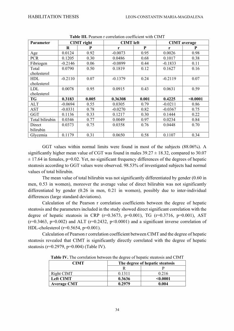
HABILITATION THESIS LEON-CONSTANTIN MARIA-MAGDALENA
34
Table III. Pearson r correlation coefficient with CIMT
Parameter CIMT right CIMT left CIMT average R P r P r P
Age 0.0124 0.92 -0.0073 0.95 0.0026 0.98 PCR 0.1205 0.30 0.0486 0.68 0.1017 0.38 Fibriogen -0.2146 0.06 -0.0899 0.44 -0.1833 0.11 Total cholesterol
0.0790 0.50 0.1819 0.12 0.1627 0.16
HDL cholesterol
-0.2110 0.07 -0.1379 0.24 -0.2119 0.07
LDL cholesterol
0.0078 0.95 0.0915 0.43 0.0631 0.59
TG 0.3183 0.005 0.36308 0.001 0.4225 <0.0001 ALT -0.0694 0.55 0.0305 0.79 -0.0211 0.86 AST -0.0331 0.78 -0.0270 0.82 -0.0367 0.75 GGT 0.1136 0.33 0.1217 0.30 0.1444 0.22 Total bilirubin 0.0346 0.77 0.0049 0.97 0.0234 0.84 Direct bilirubin
0.0373 0.75 0.0358 0.76 0.0448 0.70
Glycemia 0.1179 0.31 0.0650 0.58 0.1107 0.34 GGT values within normal limits were found in most of the subjects (88.06%). A
significantly higher mean value of CGT was found in males 39.27 ± 18.32, compared to 30.07 ± 17.64 in females, p=0.02. Yet, no significant frequency differences of the degrees of hepatic steatosis according to GGT values were observed. 98.53% of investigated subjects had normal values of total bilirubin.
The mean value of total bilirubin was not significantly differentiated by gender (0.60 in men, 0.53 in women), moreover the average value of direct bilirubin was not significantly differentiated by gender (0.26 in men, 0.21 in women), possibly due to inter-individual differences (large standard deviations).
Calculation of the Pearson r correlation coefficients between the degree of hepatic steatosis and the parameters included in the study showed direct significant correlation with the degree of hepatic steatosis in CRP (r=0.3673, p=0.001), TG (r=0.3716, p=0.001), AST (r=0.3463, p=0.002) and ALT (r=0.2432, p<0.0001) and a significant inverse correlation of HDL-cholesterol (r=0.5654, p=0.001).
Calculation of Pearson r correlation coefficient between CIMT and the degree of hepatic steatosis revealed that CIMT is significantly directly correlated with the degree of hepatic steatosis (r=0.2979, p=0.004) (Table IV).
Table IV. The correlation between the degree of hepatic steatosis and CIMT CIMT The degree of hepatic steatosis
R P Right CIMT 0.1311 0.216 Left CIMT 0.3636 <0.0001 Average CMT 0.2979 0.004
HABILITATION THESIS LEON-CONSTANTIN MARIA-MAGDALENA
35
2.4. Discussions
• CD68 expression in atherosclerosis Atherosclerotic lesions are classified into six types, according to the American Heart
Association. The first type is characterized by the appearance of foamy cells as isolated cells, followed by stages of fatty streaks, and fibroatheromatous plaques to more complex lesions (Webb et al., 2007). Lesions type I and II may occur in the first decade of life, though they may be found in adults as well; lesion type III (preatheroma) may occur in adolescence and its structure is in between the lipid streak and the atheroma, while types IV, V and VI are considered advanced lesions. In the V and VI types, vascular stenosis or thrombosis and/or bleeding may be present because of injuries and can be clinically silent or apparent. Lesion progression from I to VI occurs over several decades of life, with different patterns of growth (Kumar et al., 2005).
Many researchers believe that atherosclerosis is a chronic immune-inflammatory disease in which the key events leading to impaired arterial intima are the interactions between blood monocytes and activated endothelium (Bobryshev, 2006; Webb et al., 2007).
In the beginning of atherosclerosis, monocytes migrate into the subendothelial layer differentiating themselves into macrophages or dendritic cells. When atherogenic lipoproteins are present in the subendothelial tissue, most cells become foamy macrophages. The atheroma core can arise by aggregation of the foamy cells; as the process develops, the center of the lesion becomes necrotic, consisting of fat, cholesterol crystals and cell debris (Webb et al., 2007). In our study, the immunohistochemical marker CD68 was used for the assessment of the inflammatory reaction in the plaque. From a physiologic point of view, white line cells do not adhere to normal endothelium, but, early in atherosclerosis, the arterial endothelial cells begin to express on their surface selective adhesion molecules that link various types of leukocytes: ICAM-1, VCAM, ELAM (Boyle, 2005; Ulbrich et al., 2005). Of these molecules, VCAM-1 (vascular cell adhesion molecule-1) precisely binds the types of leukocytes found in human or experimental early atheromas: monocytes and T-lymphocytes. As a consequence, to accession by the endothelial line, monocytes, strongly stimulated by local production of chemokines and receptors of chemokines, migrate through the endothelial cells in order to locate into the intima, where they become macrophages and begin to accumulate lipoproteins (especially oxidized LDL) (Braunersreuther et al., 2007; Schulz et al., 2007).
In the beginning, the recruitment of monocytes and the differentiation in macrophages has a protective role by removing modified lipids, but as the fat load increases, the damage progression occurs (McLaren et al., 2011; Moore et al., 2011; Bui et al., 2009). It was shown previously in many cases that macrophages express multiple metalloproteinases and serine protease that degrade the extracellular matrix, which in turn weaken and predispose to rupture the atherosclerotic plaques. Leukocyte adhesion is further increased by their secretion of many other factors, such as reactive oxygen species, eicosanoids, TNF-α and IL-1, MCP-1 (Loppnow et al., 2008; Holvoet et al., 2008). Even though there are various factors known to lead to the chemotactic migration of monocytes, the MCP-1 (monocyte chemoattractant protein-1) being the most potent and powerful inductor of their migration into atherosclerotic lesions. Essential for macrophage differentiation, proliferation and survival in these lesions is the colony-stimulating factor.
HABILITATION THESIS LEON-CONSTANTIN MARIA-MAGDALENA
36
Studies show that a minor population of macrophages can proliferate even inside the atherosclerotic lesions, especially in the early stage (Matoba et al., 2011; Shi et al., 2011; Wolfs et al., 2011). Macrophages exhibit a variety of receptors, specifically scavenger-receptors, and they uptake various modified lipoproteins. Formation of foamy cells in the developing lesions is a direct result of subsequent accumulation of cholesterol esters in the cytoplasm. Out of the different scavenger-receptors, the class A type I and type II (MSR-A I, II) play the most important role in the uptake of oxidized low-density lipoprotein. Additionally, macrophages and macrophage-derived foamy cells produce ceroid and advanced glycation end products (AGEs) and they accumulate these substances in their cytoplasm. The generated extracellular AGEs are up taken by other macrophages through specific receptors, including MSR-A I, II. While some cells escape from the lesions in the peripheral blood, others die inside the plaques by apoptosis. Macrophages also play multiple roles in inducing plaque rupture, blood coagulation and fibrinolysis by producing various immunohistochemical expression of anti-CD68 antibody in atherosclerotic plaque enzymes, activators, inhibitors and bioactive mediators. Studies show that in the development of atherosclerosis, macrophages interact with vascular endothelial cells, smooth muscle cells of the media and other inflammatory cells, particularly T-cells and dendritic cells (El Khatib et al., 2012; Takahashi et al., 2002).
A vast number of studies regarding the role of immune effectors in atherosclerosis focus on CD4+ T-cells and macrophages, which release proinflammatory cytokines in atherosclerotic plaque (Ikonomidis et al., 2008; Ingersoll et al., 2011). It was demonstrated that CD4+ cells interact with oxidized LDL and it is considered that this process makes them autoantigens, suggesting the possible role of antibodies and autoantigens in early atherosclerosis (Palinski et al., 2000; Hansson et al., 2011) and in this context the participation of immunological factors in early human hypertension it has been reported.
Borderline hypertension shows an increase in anti-HSP-65 and endothelial cell antibodies (AECA) in patients. However, antioxLDL and anti-lysophosphatidylcholine are low in patients with hypertension, suggesting that different autoantigens may have an anti-atherogenic function. CD68 was predominantly positive in the middle of atherosclerotic plaques, depending on the extent of the inflammatory reaction. In stable plaques, either a moderate zonal or diffuse positive immunoreactivity or a high zonal or diffuse immunoreactivity near the lipidonecrotic core with a large number of foamy macrophages were revealed. By contrast, in the fibrous cap itself, only a few macrophages were present. Numerous CD68 positive macrophages were also observed in the cap of complicated plaque. Atherosclerotic lesions in experimental animals and humans contain distinct phenotypes of macrophages, which play various roles in mediating inflammation, clearance of dead cells, and possibly in tissue repair.
The phenotype and biological functions of the macrophages are changed by the presence of lipids in the plaques, which activate specific sets of genes. Activation of the inflammasoms by cholesterol crystals and the interaction with oxidized lipids by specific receptors leads to an M1 inflammatory phenotype of macrophages. A new MOX phenotype develops when the oxidized phospholipids activate the response of stress genes by Nrf2. Other lipid mediators, such as fatty acid derivative nitrosilates and omega-3 fatty acids polarize macrophages of the atherosclerotic plaques with their transformation in anti-inflammatory phenotypes (Adamson et al., 2011).
HABILITATION THESIS LEON-CONSTANTIN MARIA-MAGDALENA
37
• Inflammation biomarkers in metabolic disorders
The lipid profile can be disturbed by TG growth towards atherogenicity, lowering HDL-cholesterol and modifying the LDL particles size, density (LDL small and dense) and their oxidation (Katsiki et al., 2014). Additional data exists, with regards to cardio-metabolic diseases, showing that high levels of GGT, even in the accepted normal interval, are associated with increased risk of cardiovascular events, namely hypertension (HTA) and DM 2 (Lim et al., 2007; Meisinger et al., 2006).
Association between NAFLD and endothelial dysfunction, as well as the increase of the prevalence of carotid disease, have been presented by similar studies in the research literature (Targher et al., 2010).
Results of the present study suggest that among subjects with DM 2, the relationship between NAFLD reflects mainly the atherogenic action of the metabolic syndrome, relying mainly on the atherogenic dyslipidemia, although not all the elements of this syndrome have been assessed. Based on the results, the liver fat load is not only a feature of cardiovascular risk, being possible to play a key role in the development of early atherosclerosis, as shown in other studies (Hardy ET AL., 2016; Al Rifai et al., 2015).
Future perspectives of our study include a follow-up period, in order to monitor cardiovascular events, related to the degree of liver damage and changes in the lipid profile and in the markers of inflammation.
2.5. Conclusions
• CD68 expression in atherosclerosis Identification of differences in location of inflammatory cells in the vascular wall, from
intima to adventitia, was evidenced by the qualitative analysis of the inflammatory component, consisting in the immunohistochemical examination. This can be correlated with developmental stages of plaque formation and organization. In depth understanding of how lipids that accumulate in atherosclerotic plaques impact the phenotype and the functions of macrophages and so the evolution of such lesions, will contribute in the future to develop new therapeutic strategies.
• Inflammation biomarkers in metabolic disorders
Subjects with type 2 diabetes that participated in the study showed an increase incidence of NAFLD, with varying degrees of fat loading, which leads to an increased cardiovascular risk. A positive correlation between the degree of steatosis, hepatic cytolysis enzymes, inflammation (CRP) and atherogenic dyslipidemia (increase in TG and decrease in HDL-cholesterol) has been established. Moreover, the degree of hepatic steatosis has proved to be a predictor of cardiovascular risk through its direct connection with subclinical atherosclerosis assessed by CIMT.

HABILITATION THESIS LEON-CONSTANTIN MARIA-MAGDALENA
38
Chapter 3. CLINICAL – BIOLOGICAL EVALUATION
3.1. Introduction
Cardiovascular diseases are among the leading causes of death in developed countries and also in Romania. Primary prevention aims to extend the range of time from identifying cardiovascular risk factors and cardiovascular disease expression. Dyslipidemia is associated with significant comorbidities, such as obesity, type 2 diabetes mellitus (DM) and essential hypertension (HBP)(Ode et al., 2009). It is well known that serum levels of lipids and lipoproteins vary depending on age, gender and race (Wilson et al., 2015; Dai et al., 2014). The predominant pattern of dyslipidemia is combined; thus patients can associate moderate to severe elevated values of triglycerides (TG), normal to slightly increased values of LDL- cholesterol (LDL-c) and reduced values of HDL-cholesterol (HDL-c). Both patterns of dyslipidemia have been shown to be associated with the onset and progression of atherosclerotic lesions (Expert Panel, 2011).
It has been found that during puberty lipid levels are lower (total and LDL-cholesterol levels can be reduced by 10% to 20% or more), and after this period, they increase again. Also, after the onset of puberty, increased values of total and LDL-cholesterol was found in girls (Wilson et al., 2015).
Generally, there are four classes of dyslipidemia as follows: medication-related dyslipidemia, dyslipidemia caused by lifestyle factors, genetic dyslipidemia and dyslipidemia secondary to other diseases. Also there have been described 3 types of genetic dyslipidemias:
• familial hypercholesterolemia - an autosomal dominant disorder caused by severe elevations in total cholesterol and LDLc.
• familial combined hyperlipidemia caused by high LDL-C and triglycerides (TG) levels. • familial severe hypertriglyceridemia caused by severe hypertriglyceridemia (>885
mg/dL) (Expert Panel, 2011).
The screening of dyslipidemia The screening is based on a standard lipid panel, taken after fasting, that includes total cholesterol, triglycerides, HDL and LDL-cholesterol. If the adult is not fasting, the investigation of non HDL-c (calculated as: Total Cholesterol - HDL-c) is recommended in the initial screening (which is not influenced by food or drink), unlike triglycerides or LDL-c. Calculated LDL-c is based to the Friedewald formula:
LDL-c = Total cholesterol - triglycerides / 5 + HDL-c (which are influenced by food or drink). Significant increased triglycerides (eg. > 400 mg/ dl) exclude LDL-c calculation rule using the Friedewald equation (commonly used by most laboratories). If non-HDL-c is increased (> 145 mg /dl), will have to obtain two lipids profiles a jéun during 3 months, with at least 3 months break between the assessments (Dai et al., 2014). According to the AHA 2017 and ESC 2016 guidelines concerning dyslipidemia, this screening is recommended to all the patients (men more than 40 years old, women more than 50 years old), patients with cardiovascular history, or other comorbidities like obesity, hypertension, stroke, etc (Table V).
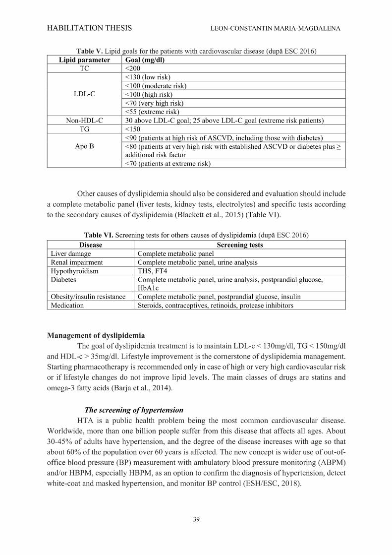
HABILITATION THESIS LEON-CONSTANTIN MARIA-MAGDALENA
39
Table V. Lipid goals for the patients with cardiovascular disease (după ESC 2016) Lipid parameter Goal (mg/dl)
TC <200
LDL-C
<130 (low risk) <100 (moderate risk) <100 (high risk) <70 (very high risk) <55 (extreme risk)
Non-HDL-C 30 above LDL-C goal; 25 above LDL-C goal (extreme risk patients) TG <150
Apo B
<90 (patients at high risk of ASCVD, including those with diabetes) <80 (patients at very high risk with established ASCVD or diabetes plus ≥ additional risk factor <70 (patients at extreme risk)
Other causes of dyslipidemia should also be considered and evaluation should include a complete metabolic panel (liver tests, kidney tests, electrolytes) and specific tests according to the secondary causes of dyslipidemia (Blackett et al., 2015) (Table VI).
Table VI. Screening tests for others causes of dyslipidemia (după ESC 2016) Disease Screening tests
Liver damage Complete metabolic panel Renal impairment Complete metabolic panel, urine analysis Hypothyroidism THS, FT4 Diabetes Complete metabolic panel, urine analysis, postprandial glucose,
HbA1c Obesity/insulin resistance Complete metabolic panel, postprandial glucose, insulin Medication Steroids, contraceptives, retinoids, protease inhibitors
Management of dyslipidemia The goal of dyslipidemia treatment is to maintain LDL-c < 130mg/dl, TG < 150mg/dl and HDL-c > 35mg/dl. Lifestyle improvement is the cornerstone of dyslipidemia management. Starting pharmacotherapy is recommended only in case of high or very high cardiovascular risk or if lifestyle changes do not improve lipid levels. The main classes of drugs are statins and omega-3 fatty acids (Barja et al., 2014).
The screening of hypertension HTA is a public health problem being the most common cardiovascular disease. Worldwide, more than one billion people suffer from this disease that affects all ages. About 30-45% of adults have hypertension, and the degree of the disease increases with age so that about 60% of the population over 60 years is affected. The new concept is wider use of out-of-office blood pressure (BP) measurement with ambulatory blood pressure monitoring (ABPM) and/or HBPM, especially HBPM, as an option to confirm the diagnosis of hypertension, detect white-coat and masked hypertension, and monitor BP control (ESH/ESC, 2018).
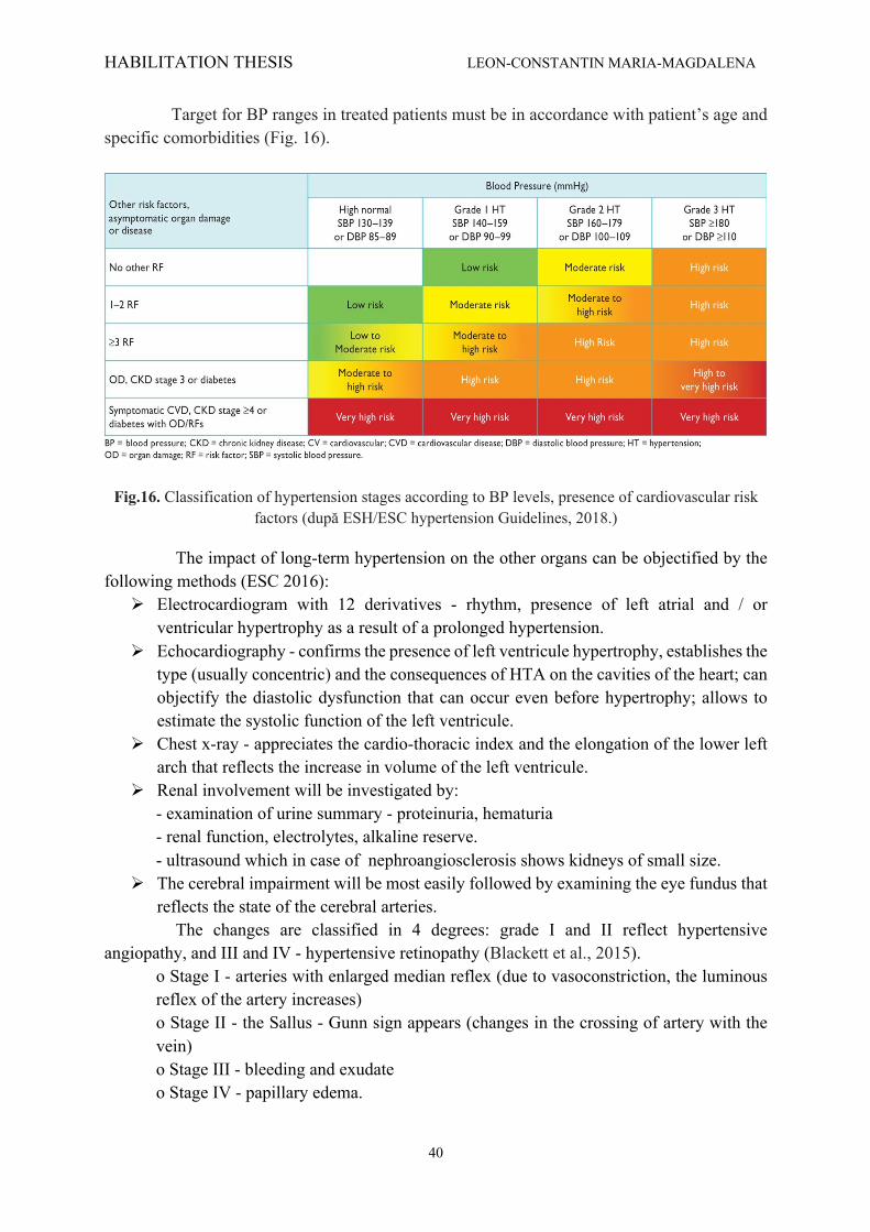
HABILITATION THESIS LEON-CONSTANTIN MARIA-MAGDALENA
40
Target for BP ranges in treated patients must be in accordance with patient’s age and specific comorbidities (Fig. 16).
Fig.16. Classification of hypertension stages according to BP levels, presence of cardiovascular risk factors (după ESH/ESC hypertension Guidelines, 2018.)
The impact of long-term hypertension on the other organs can be objectified by the following methods (ESC 2016):
Ø Electrocardiogram with 12 derivatives - rhythm, presence of left atrial and / or ventricular hypertrophy as a result of a prolonged hypertension.
Ø Echocardiography - confirms the presence of left ventricule hypertrophy, establishes the type (usually concentric) and the consequences of HTA on the cavities of the heart; can objectify the diastolic dysfunction that can occur even before hypertrophy; allows to estimate the systolic function of the left ventricule.
Ø Chest x-ray - appreciates the cardio-thoracic index and the elongation of the lower left arch that reflects the increase in volume of the left ventricule.
Ø Renal involvement will be investigated by: - examination of urine summary - proteinuria, hematuria - renal function, electrolytes, alkaline reserve. - ultrasound which in case of nephroangiosclerosis shows kidneys of small size.
Ø The cerebral impairment will be most easily followed by examining the eye fundus that reflects the state of the cerebral arteries.
The changes are classified in 4 degrees: grade I and II reflect hypertensive angiopathy, and III and IV - hypertensive retinopathy (Blackett et al., 2015).
o Stage I - arteries with enlarged median reflex (due to vasoconstriction, the luminous reflex of the artery increases) o Stage II - the Sallus - Gunn sign appears (changes in the crossing of artery with the vein) o Stage III - bleeding and exudate o Stage IV - papillary edema.
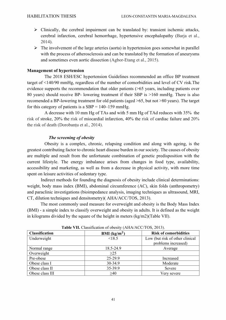
HABILITATION THESIS LEON-CONSTANTIN MARIA-MAGDALENA
41
Ø Clinically, the cerebral impairment can be translated by: transient ischemic attacks, cerebral infarction, cerebral hemorrhage, hypertensive encephalopathy (Barja et al., 2014).
Ø The involvement of the large arteries (aorta) in hypertension goes somewhat in parallel with the process of atherosclerosis and can be translated by the formation of aneurysms and sometimes even aortic dissection (Agbor-Etang et al., 2015).
Management of hypertension The 2018 ESH/ESC hypertension Guidelines recommended an office BP treatment target of <140/90 mmHg, regardless of the number of comorbidities and level of CV risk.The evidence supports the recommendation that older patients (>65 years, including patients over 80 years) should receive BP- lowering treatment if their SBP is >160 mmHg. There is also recomended a BP-lowering treatment for old patients (aged >65, but not >80 years). The target for this category of patients is a SBP = 140–159 mmHg. A decrease with 10 mm Hg of TAs and with 5 mm Hg of TAd reduces with 35% the risk of stroke, 20% the risk of miocardial infarction, 40% the risk of cardiac failure and 20% the risk of death (Dorobanțu et al., 2014).
The screening of obesity Obesity is a complex, chronic, relapsing condition and along with ageing, is the greatest contributing factor to chronic heart disease burden in our society. The causes of obesity are multiple and result from the unfortunate combination of genetic predisposition with the current lifestyle. The energy imbalance arises from changes in food type, availability, accessibility and marketing, as well as from a decrease in physical activity, with more time spent on leisure activities of sedentary type. Indirect methods for founding the diagnosis of obesity include clinical determinations: weight, body mass index (BMI), abdominal circumference (AC), skin folds (anthropometry) and paraclinic investigations (bioimpedance analysis, imaging techniques as ultrasound, MRI, CT, dilution techniques and densitometry)( AHA/ACC/TOS, 2013).
The most commonly used measure for overweight and obesity is the Body Mass Index (BMI) - a simple index to classify overweight and obesity in adults. It is defined as the weight in kilograms divided by the square of the height in meters (kg/m2)(Table VII).
Table VII. Classification of obesity (AHA/ACC/TOS, 2013).
Classification BMI (kg/𝒎𝟐) Risk of comorbidities Underweight <18.5 Low (but risk of other clinical
problems increased) Normal range 18.5-24.9 Average Overweight ≥25 Pre-obese 25-29.9 Increased Obese class I 30-34.9 Moderate Obese class II 35-39.9 Severe Obese class III ≥40 Very severe

HABILITATION THESIS LEON-CONSTANTIN MARIA-MAGDALENA
42
Central weight distribution occurs more commonly in men than women and increases in both men and women with increasing age. The cardiovascular risk is higher to the patients with abdominal obesity (Sandfort et al., 2016) (Table VIII).
Table VIII. Relation between waist circumference and comorbidity risk Waist circumference(cm) Comorbidity risk Women Men Above action level 1 ≥80 ≥94 Above action level 2 ≥88 ≥102
Screening of diabetes
Diabetes is a complex, chronic illness requiring continuous medical care with multifactorial risk-reduction strategies besides glycemic control. Ongoing patient self-management education and support are critical to preventing acute complications and reducing the risk of long-term complications (Johson et al., 2015). Methods and criteria for diagnosing diabetes Diabetes symptoms - e.g. polyuria, polydipsia and unexplained weight loss for type- 1 along with: - a random venous plasma glucose concentration ≥ 11.1 mmol/l or - a fasting plasma glucose concentration ≥ 7.0 mmol/l (whole blood ≥ 6.1 mmol/l) or - 2 hour plasma glucose concentration ≥ 11.1 mmol/l two hours after 75g anhydrous glucose
in an oral glucose tolerance test (OGTT) (Macdonald, 2016). With no symptoms, the diagnosis should not be based on a single glucose determination, requiring confirmatory plasma venous determination. At least one additional glucose test result with a value in the diabetic range in another day is essential. If this value is not pathological, another determination of glycemia is made at 2 hours postprandial (Johson et al., 2015). Diabetes UK welcomes the 2011 decision by the WHO to accept the use of HbA1c testing in diagnosis of diabetes. An HbA1c of 48mmol/mol (6.5%) is recommended as the cut off point for diagnosing diabetes (Johson et al., 2015). The scientific interest related to clinical and biological evaluation were synthesized in the following articles: 1. Mitu O, Mitu F, Leon Constantin M, Roca M, Gherasim A, Graur M. Biochemical changes in asymptomatic adult population with subclinical atherosclerosis. Rev. Chim (Bucharest), 2016; 67(5):953-957. 2. Vasilcu TF, Statescu C, Sascau R, Roca M, Costea CF, Zota M, Bararu I, Leon Constantin M, Mitu F. Cardiopulmonary testing and biochemical profile of coronary patients subject to cardiovascular recovery programs. Rev. Chim (Bucharest). 2018, 69(8):2283-2286. Using the theoretical data mentioned above, the mentioned articles aimed:
• Biochemical changes in subclinical atherosclerosis The present study highlights correlations assessed in a group of asymptomatic memberst
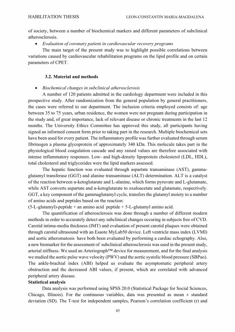
HABILITATION THESIS LEON-CONSTANTIN MARIA-MAGDALENA
43
of society, between a number of biochemical markers and different parameters of subclinical atherosclerosis.
• Evaluation of coronary patient in cardiovascular recovery programs The main target of the present study was to highlight possible correlations between
variations caused by cardiovascular rehabilitation programs on the lipid profile and on certain parameters of CPET.
3.2. Material and methods
• Biochemical changes in subclinical atherosclerosis A number of 120 patients admitted in the cardiology department were included in this
prospective study. After randomization from the general population by general practitioners, the cases were referred to our department. The inclusion criteria employed consists of: age between 35 to 75 years, urban residence, the women were not pregnant during participation in the study and, of great importance, lack of relevant disease or chronic treatments in the last 12 months. The University Ethics Committee has approved this study, all participants having signed an informed consent form prior to taking part in the research. Multiple biochemical sets have been used for every patient. The inflammatory profile was further evaluated through serum fibrinogen a plasma glycoprotein of approximately 340 kDa. This molecule takes part in the physiological blood coagulation cascade and any raised values are therefore associated with intense inflammatory responses. Low- and high-density lipoprotein cholesterol (LDL, HDL), total cholesterol and triglycerides were the lipid markers assessed.
The hepatic function was evaluated through aspartate transaminase (AST), gamma-glutamyl transferase (GGT) and alanine transaminase (ALT) determination. ALT is a catalyst of the reaction between α-ketoglutarate and L-alanine, which forms pyruvate and L-glutamate, while AST converts aspartate and α-ketoglutarate to oxaloacetate and glutamate, respectively. GGT, a key component of the gammaglutamyl cycle, transfers the glutamyl moiety to a number of amino acids and peptides based on the reaction: (5-L-glutamyl)-peptide + an amino acid peptide + 5-L-glutamyl amino acid.
The quantification of atherosclerosis was done through a number of different modern methods in order to accurately detect any subclinical changes occuring in subjects free of CVD. Carotid intima-media thickness (IMT) and evaluation of present carotid plaques were obtained through carotid ultrasound with an Esaote MyLab50 device. Left ventricle mass index (LVMI) and aortic atheromatosis have both been evaluated by performing a cardiac echography. Also, a new biomarker for the assessment of subclinical atherosclerosis was used in the present study, arterial stiffness. We used an Arteriograph™ device for measurement, and for the final analysis we studied the aortic pulse wave velocity (PWV) and the aortic systolic blood pressure (SBPao). The ankle-brachial index (ABI) helped us evaluate the asymptomatic peripheral artery obstruction and the decreased ABI values, if present, which are correlated with advanced peripheral artery disease. Statistical analysis
Data analysis was performed using SPSS 20.0 (Statistical Package for Social Sciences, Chicago, Illinois). For the continuous variables, data was presented as mean ± standard deviation (SD). The T-test for independent samples, Pearson’s correlation coefficient (r) and
HABILITATION THESIS LEON-CONSTANTIN MARIA-MAGDALENA
44
ANOVA were utilised in order to assess any existing relationship between variables. A two sided p value < 0.05 was considered important for all data analysis.
• Evaluation of coronary patient in cardiovascular recovery programs
A prospective study was conducted, which included 60 patients from both urban and rural areas. They were investigated at the Cardiovascular Recovery Clinic, Recovery Hospital of Iasi, Romania, and were clinically evaluated both when initially admitted and 6 months later, at the follow-up. Inclusion criteria consisted of prior chronic myocardial infarction diagnosis, stable angina pectoris or chronic ischemic cardiopathy at least 3 months prior to admission. The Ethics Commission’s approval was further obtained while all patients signed the informed consent form before the study began. Within six months of cardiac rehabilitation, the patients were guided to perform endurance aerobic exercises, at least five days per week. Normally each training session would last between 30 and 60 minutes, mostly depending on the physical condition and comorbidities of each individual. The level of intensity was deemed medium for all subjects. Each patient performed a initial CPET assessment, establishing the type characteristics of the effort that the patients had to sustain (Mezzani et al., 2013).
The significant variables of the physical exercion were frequency, exercise intensity (measured using the Borg scale) and duration of each session (Eagle et al., 2004). The parameters monitored using the CPET were: anaerobic threshold (AT), VO2, effort capacity and cardiac frequency. VO2 is the total quantity of O2 used during the test. At one point during exercise VO2 reaches a maximum level (VO2 max) despite the fact that the continuation of the physical effort (van de Port et al., 2015). The occurrence of metabolic acidosis caused by inefficiency of the aerobic metabolism at muscle level was estimated by anaerobic threshold. AT is obtained during highest effort, making the transition towards anaerobic metabolism and accumulation of lactic acid. An indicator of the patient’s physical condition, AT is used to diagnose limitations toward effort. For monitoring of heart rate response to effort, maximum heart rate is particularly important. Maximum theoretical heart rate was then calculated using the approved formula: 220 - patient age (Choudhary et al., 2008).
By analizing the lipid profile, including triglycerides (TG), total cholesterol levels, LDL-cholesterol and HDL-cholesterol we have defined dyslipidemia as values of total cholesterol > 200 mg/dL and/or LDL-cholesterol > 100 mg/dL and/or HDL-cholesterol < 35 mg/dL and/or use of lipid-lowering drugs (Robinson et al., 2009). The statistical analysis was done by using SPSS program, version 7.0. A importance of p <0.05 was viewed statistically significant. The correlations between variables had been conducted using Pearson r correlation coefficient.
3.3. Results
• Biochemical changes in subclinical atherosclerosis The mean ages of the research group was 52.01 years, with a third being male (forty
participants). Fibrinogen, as marker of inflammatory process, was in regular ranges. About the lipid profile, total cholesterol was above the standard worth while LDL cholesterol, non HDL cholesterol as well as triglycerides had been near the better limit. These results suggest the population totally free of CVD contained uncontrolled lipid values.
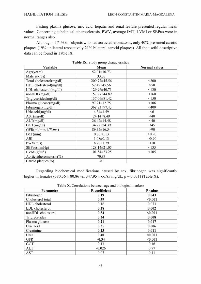
HABILITATION THESIS LEON-CONSTANTIN MARIA-MAGDALENA
45
Fasting plasma glucose, uric acid, hepatic and renal feature presented regular mean values. Concerning subclinical atherosclerosis, PWV, average IMT, LVMI or SBPao were in normal ranges also.
Although of 71% of subjects who had aortic atheromatosis, only 40% presented carotid plaques (19% unilateral respectively 21% bilateral carotid plaques). All the useful descriptive data can be found in Table IX.
Table IX. Study group characteristics Variable Mean Normal values
Age(years) 52.01±10.73 Male sex(%) 33.33 Total cholesterol(mg/dl) 209.77±45.56 <200 HDL cholesterol(mg/dl) 52.49±45.56 >50 LDL cholesterol(mg/dl) 129.96±40.71 <130 nonHDL(mg/dl) 157.27±44.89 <160 Triglycerides(mg/dl) 137.06±81.42 <150 Plasma glucose(mg/dl) 97.21±12.75 <106 Fibrinogen(mg/dl) 368.83±77.43 <400 Uric acid(mg/dl) 4.34±1.59 <6 AST(mg/dl) 24.14±8.49 <40 ALT(mg/dl) 26.42±14.48 <40 GGT(mg/dl) 34.22±24.39 <45 GFR(ml/min/1.73𝑚$) 89.35±16.54 >90 IMT(mm) 0.86±0.13 >0.90 ABI 1.08±0.13 >0.90 PWV(m/s) 8.28±1.79 <10 SBPao(mmHg) 128.14±21.05 <135 LVMI(g/𝑚$) 101.54±23.25 <105 Aortic atheromatosis(%) 70.83 Caroid plaques(%) 40
Regarding biochemical modifications caused by sex, fibrinogen was significantly
higher in females (380.36 ± 80.86 vs. 347.95 ± 66.85 mg/dL, p = 0.031) (Table X).
Table X. Correlations between age and biological markers Parameter R coefficient P value
Fibrinogen 0.19 0.043 Cholesterol total 0.39 <0.001 HDL cholesterol 0.16 0.073 LDL cholesterol 0.28 0.002 nonHDL cholesterol 0.34 <0.001 Triglycerides 0.24 0.008 Plasma glucose 0.21 0.017 Uric acid 0.25 0.006 Creatinine 0.23 0.011 Urea 0.40 <0.001 GFR -0.54 <0.001 GGT 0.13 0.16 ALT -0.026 0.77 AST 0.07 0.41
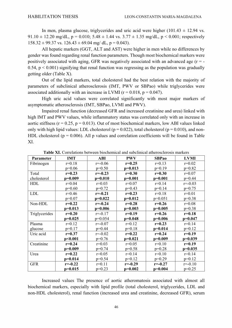
HABILITATION THESIS LEON-CONSTANTIN MARIA-MAGDALENA
46
In men, plasma glucose, triglycerides and uric acid were higher (101.43 ± 12.94 vs. 91.10 ± 12.20 mg/dL, p = 0.010; 5.48 ± 1.44 vs. 3.77 ± 1.35 mg/dL, p < 0.001; respectively 158.32 ± 99.37 vs. 126.43 ± 69.04 mg/ dL, p = 0.043).
All hepatic markers (GGT, ALT and AST) were higher in men while no differences by gender was found regarding renal function parameters. Though most biochemical markers were positively associated with aging, GFR was negatively associated with an advanced age (r = -0.54, p < 0.001) signifying that renal function was regressing as the population was gradually getting older (Table X).
Out of the lipid markers, total cholesterol had the best relation with the majority of parameters of subclinical atherosclerosis (IMT, PWV or SBPao) while triglycerides were associated additionally with an increase in LVMI (r = 0.018, p = 0.047).
High uric acid values were correlated significantly with most major markers of asymptomatic atherosclerosis (IMT, SBPao, LVMI and PWV).
Impaired renal function (decreased GFR and increased creatinine and urea) linked with high IMT and PWV values, while inflammatory status was correlated only with an increase in aortic stiffness (r = 0.25, p = 0.013). Out of most biochemical markers, low ABI values linked only with high lipid values: LDL cholesterol (p = 0.022), total cholesterol (p = 0.010), and non-HDL cholesterol (p = 0.006). All p values and correlation coefficients will be found in Table XI.
Table XI. Correlations between biochemical and subclinical atherosclerosis markers Parameter IMT ABI PWV SBPao LVMI
Fibrinogen r=0.18 p=0.06
r=-0.06 p=0.50
r=0.25 p=0.013
r=0.13 p=0.19
r=0.02 p=0.82
Total cholesterol
r=0.23 p=0.009
r=-0.23 p=0.010
r=0.30 p=0.001
r=0.30 p=0.001
r=0.07 p=0.44
HDL r=0.04 p=0.60
r=0.03 p=0.72
r=0.07 p=0.43
r=0.14 p=0.14
r=-0.03 p=0.75
LDL r=0.46 p=0.07
r=-0.21 p=0.022
r=0.23 p=0.012
r=0.18 p=0.051
r=0.01 p=0.38
Non-HDL r=0.22 p=0.013
r=-0.24 p=0.006
r=0.28 p=0.003
r=0.26 p=0.005
r=0.08 p=0.38
Triglycerides r=0.20 p=0.025
r=-0.17 p=0.054
r=0.19 p=0.048
r=0.26 p=0.006
r=0.18 p=0.047
Plasma glucose
r=0.12 p=0.17
r=-0.07 p=0.44
r=0.12 p=0.18
r=0.23 p=0.014
r=0.14 p=0.12
Uric acid r=0.37 p<0.001
r=-0.02 p=0.76
r=0.22 p=0.021
r=0.24 p=0.009
r=0.19 p=0.039
Creatinine r=0.24 p=0.009
r=0.03 p=0.74
r=0.05 p=0.58
r=0.10 p=0.28
r=0.19 p=0.035
Urea r=0.22 p=0.014
r=0.05 p=0.54
r=0.14 p=0.12
r=0.10 p=0.29
r=0.14 p=0.12
GFR r=-0.22 p=0.015
r=0.11 p=0.23
r=-0.29 p=0.002
r=-0.27 p=0.004
r=-0.10 p=0.25
Increased values The presence of aortic atheromatosis associated with almost all
biochemical markers, especially with lipid profile (total cholesterol, triglycerides, LDL and non-HDL cholesterol), renal function (increased urea and creatinine, decreased GFR), serum
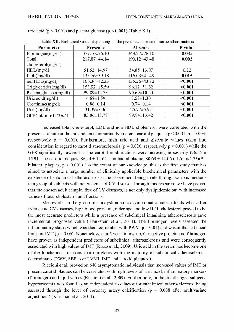
HABILITATION THESIS LEON-CONSTANTIN MARIA-MAGDALENA
47
uric acid (p < 0.001) and plasma glucose (p < 0.001) (Table XII).
Table XII. Biological values depending on the presence/absence of aortic atheromatosis Parameter Presence Absence P value
Fibrinogen(mg/dl) 377.16±76.10 348.27±78.10 0.085 Total cholesterol(mg/dl)
217.87±44.14 190.12±43.48 0.002
HDL(mg/dl) 51.52±14.97 54.85±13.07 0.22 LDL(mg/dl) 135.76±39.18 116.03±41.49 0.015 nonHDL(mg/dl) 166.34±42.33 135.26±43.82 <0.001 Triglycerides(mg/dl) 153.92±85.59 96.12±51.62 <0.001 Plasma glucose(mg/dl) 99.89±12.78 90.69±10.20 <0.001 Uric acid(mg/dl) 4.68±1.59 3.53±1.30 <0.001 Creatinine(mg/dl) 0.86±0.14 0.74±0.14 <0.001 Urea(mg/dl) 31.39±8.36 25.77±5.97 <0.001 GFR(ml/min/1.73𝑚$) 85.06±15.79 99.94±13.42 <0.001
Increased total cholesterol, LDL and non-HDL cholesterol were correlated with the presence of both unilateral and, most importantly bilateral carotid plaques (p < 0.001; p = 0.004; respectively p = 0.001). Furthermore, high uric acid and glycemic values taken into consideration in regard to carotid atherosclerosis (p = 0.020; respectively p < 0.001) while the GFR significantly lowered as the carotid modifications were incresing in severity (96.55 ± 15.91 – no carotid plaques, 86.44 ± 14.62 – unilateral plaque, 80.69 ± 14.06 mL/min/1.73m² – bilateral plaques, p < 0.001). To the extent of our knowledge, this is the first study that has aimed to associate a large number of clinically applicable biochemical parameters with the existence of subclinical atherosclerosis; the asessement being made through various methods in a group of subjects with no evidence of CV disease. Through this research, we have proven that the chosen adult sample, free of CV diseases, is not only dyslipidemic but with increased values of total cholesterol and fractions.
Meanwhile, in the group of nondyslipidemic asymptomatic male patients who suffer from acute CV diseases, high blood pressure, older age and low HDL cholesterol proved to be the most accurate predictors while a presence of subclinical imagining atherosclerosis gave incremental prognostic value (Blankstein et al., 2011). The fibrinogen levels assessed the inflammatory status which was then correlated with PWV (p = 0.01) and was at the statistical limit for IMT (p = 0.06). Nonetheless, at a 5 year follow-up, C-reactive protein and fibrinogen have proven as independent predictors of subclinical atherosclerosis and were consequently associated with high values of IMT (Rizzo et al., 2009). Uric acid in the serum has become one of the biochemical markers that correlates with the majority of subclinical atherosclerosis determinants (PWV, SBPao or LVMI, IMT and carotid plaques,).
Riccioni et al. proved on 640 asymptomatic individuals that increased values of IMT or present carotid plaques can be correlated with high levels of uric acid, inflammatory markers (fibrinogen) and lipid values (Riccioni et al., 2009). Furthermore, in the middle aged subjects, hyperuricemia was found as an independent risk factor for subclinical atherosclerosis, being assessed through the level of coronary artery calcification (p = 0.008 after multivariate adjustment) (Krishnan et al., 2011).
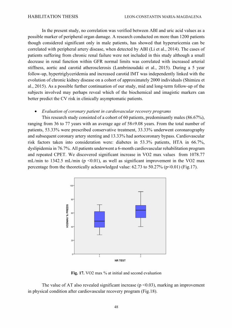
HABILITATION THESIS LEON-CONSTANTIN MARIA-MAGDALENA
48
In the present study, no correlation was verified between ABI and uric acid values as a possible marker of peripheral organ damage. A research conducted on more than 1200 patients though considered significant only in male patients, has showed that hyperuricemia can be correlated with peripheral artery disease, when detected by ABI (Li et al., 2014). The cases of patients suffering from chronic renal failure were not included in this study although a small decrease in renal function within GFR normal limits was correlated with increased arterial stiffness, aortic and carotid atherosclerosis (Lambrinoudaki et al., 2015). During a 5 year follow-up, hypertriglyceridemia and increased carotid IMT was independently linked with the evolution of chronic kidney disease on a cohort of approximately 2000 individuals (Shimizu et al., 2015). As a possible further continuation of our study, mid and long-term follow-up of the subjects involved may perhaps reveal which of the biochemical and imagistic markers can better predict the CV risk in clinically asymptomatic patients.
• Evaluation of coronary patient in cardiovascular recovery programs
This research study consisted of a cohort of 60 patients, predominantly males (86.67%), ranging from 36 to 77 years with an average age of 58±9.08 years. From the total number of patients, 53.33% were prescribed conservative treatment, 33.33% underwent coronarography and subsequent coronary artery stenting and 13.33% had aortocoronary bypass. Cardiovascular risk factors taken into consideration were: diabetes in 53.3% patients, HTA in 66.7%, dyslipidemia in 76.7%. All patients underwent a 6-month cardiovascular rehabilitation program and repeated CPET. We discovered significant increase in VO2 max values from 1078.77 mL/min to 1342.5 mL/min (p <0.01), as well as significant improvement in the VO2 max percentage from the theoretically acknowledged value: 62.73 to 50.27% (p<0.01) (Fig.17).
Fig. 17. VO2 max % at initial and second evaluation The value of AT also revealed significant increase (p <0.03), marking an improvement
in physical condition after cardiovascular recovery program (Fig.18).
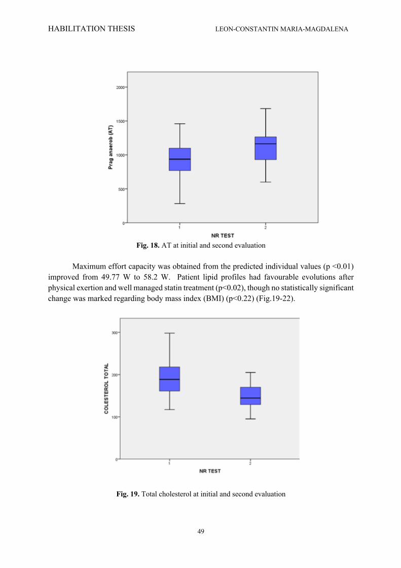
HABILITATION THESIS LEON-CONSTANTIN MARIA-MAGDALENA
49
Fig. 18. AT at initial and second evaluation
Maximum effort capacity was obtained from the predicted individual values (p <0.01)
improved from 49.77 W to 58.2 W. Patient lipid profiles had favourable evolutions after physical exertion and well managed statin treatment (p<0.02), though no statistically significant change was marked regarding body mass index (BMI) (p<0.22) (Fig.19-22).
Fig. 19. Total cholesterol at initial and second evaluation

HABILITATION THESIS LEON-CONSTANTIN MARIA-MAGDALENA
50
Fig. 20. LDL-cholesterol at initial and second evaluation
Fig. 21. HDL-cholesterol at initial and second evaluation
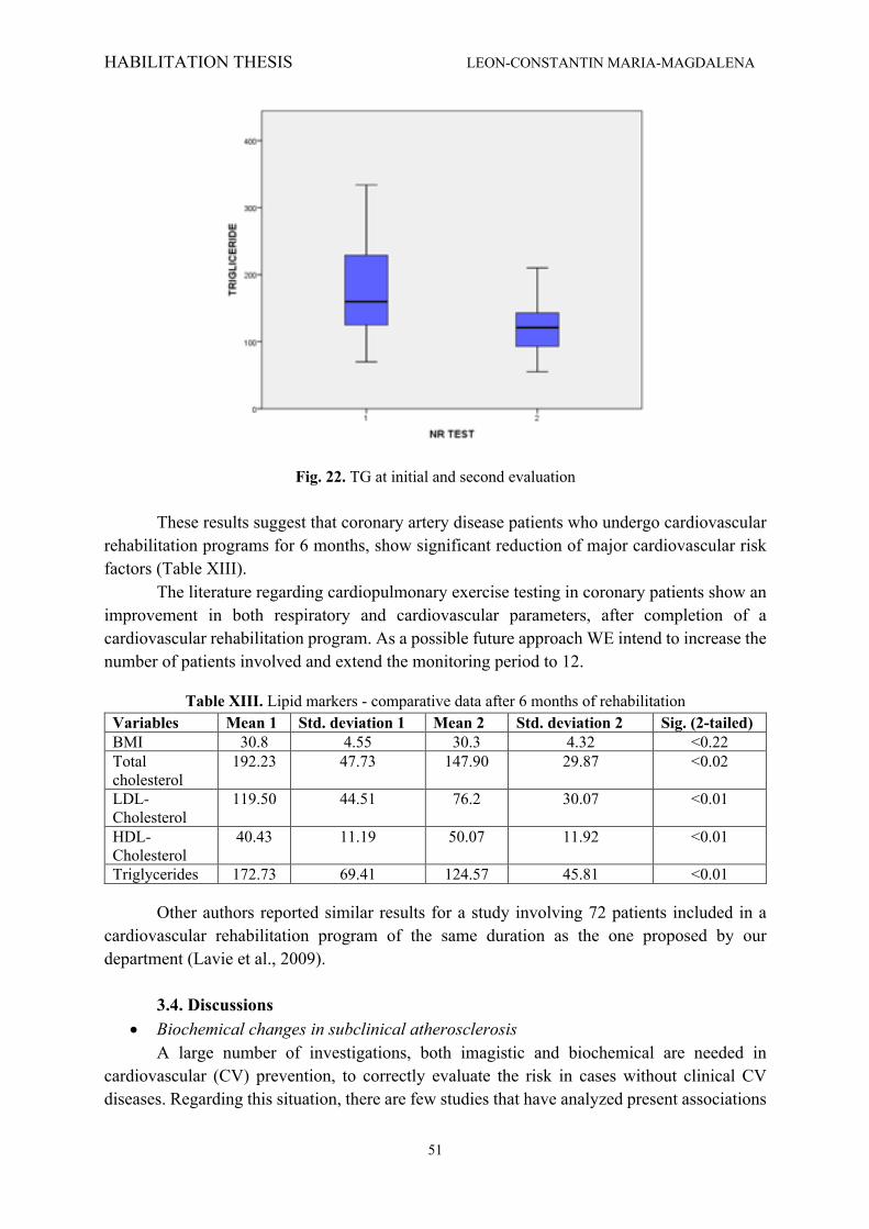
HABILITATION THESIS LEON-CONSTANTIN MARIA-MAGDALENA
51
Fig. 22. TG at initial and second evaluation These results suggest that coronary artery disease patients who undergo cardiovascular
rehabilitation programs for 6 months, show significant reduction of major cardiovascular risk factors (Table XIII).
The literature regarding cardiopulmonary exercise testing in coronary patients show an improvement in both respiratory and cardiovascular parameters, after completion of a cardiovascular rehabilitation program. As a possible future approach WE intend to increase the number of patients involved and extend the monitoring period to 12.
Table XIII. Lipid markers - comparative data after 6 months of rehabilitation Variables Mean 1 Std. deviation 1 Mean 2 Std. deviation 2 Sig. (2-tailed) BMI 30.8 4.55 30.3 4.32 <0.22 Total cholesterol
192.23 47.73 147.90 29.87 <0.02
LDL- Cholesterol
119.50 44.51 76.2 30.07 <0.01
HDL- Cholesterol
40.43 11.19 50.07 11.92 <0.01
Triglycerides 172.73 69.41 124.57 45.81 <0.01
Other authors reported similar results for a study involving 72 patients included in a cardiovascular rehabilitation program of the same duration as the one proposed by our department (Lavie et al., 2009).
3.4. Discussions
• Biochemical changes in subclinical atherosclerosis A large number of investigations, both imagistic and biochemical are needed in
cardiovascular (CV) prevention, to correctly evaluate the risk in cases without clinical CV diseases. Regarding this situation, there are few studies that have analyzed present associations
HABILITATION THESIS LEON-CONSTANTIN MARIA-MAGDALENA
52
between biochemical readings and subclinical atherosclerosis in asymptomatic patients. Through the direct measurement of the atherosclerotic burden, we believe that better measures could be taken in the prevention and risk assessment of CV. The current study aimed to ascertain the correlation between biochemical markers and the presence of subclinical atherosclerosis in adults free of CV diseases.
Cholesterol, or (3β)-cholest-5en-3-ol is an organic molecule biosynthesized by all animal cells, with 256 known stereoisomeres. It is a fundamental component of the cell membrane and a precursor of the steroid hormones, of vitamin D and bile acids. LDL and HDL cholesterol, although having different actions both belong to the five major groups of lipoproteins.
Lipoproteins naturally consist of a neutral lipid.core enclosed by a.20-Ao shell.made of. phospholipids, apolipoproteins and unesterified.cholesterol. LDLs diameter is ten times larger than that of normal cholesterol and has a high cardiovascular risk.by invading and oxidizing the endothelium and thusly promoting the formation of the atherosclerotic plaque. HDL, the smallest and most dense lipoprotein particle, removes the fat molecules from cells and it is considered to play a significant anti-atherogenic role. Furthermore, the non-HDL cholesterol is now considered to be a cause of atheroma and its dosing and measurement has become the better predictor of CV events (Robinson et al., 2009). High levels in triglycerides are associated with atherosclerotic burdens and an increase in the risk of future CV events. Triglycerides, esters derived from the combination of glycerol and three fatty acids (usually RCO2H, R’CO2H and R’’CO2H), are based on the following formula:
HOCH2CH(OH)CH2OH + RCO2H + R’CO2H + R’’CO2H→ →RCO2CH2CH(O2CR’)CH2CO2R’’ + 3H2O
Glucose, a monosaccharide with the molecular formula C6H12O6, exists only as a D-isomer in nature. It has five hydroxyl (OH) groups that are arranged along the six carbon back, making it possible for the biochemical compound to arrange both in straightchain and in ring form. Another one of the assessed biochemical markers was uric acid (7,9-Dihydro-1H-purine-2,6,8(3H)-trione), a diprotic aromatic acid and product of purine nucleotide metabolism.
Hyperuricemia has long been known as a direct factor that leads to both gout and increased CV risks. Urea and serum creatinine were employed to evaluate the renal function. Urea, an organic compound with the chemical formula CH4N2O, consists of a carbonyl (C=O) functional group linked with two –NH2 groups.
Creatinine (or 2-Amino-1-methyl-5H-imidazol-4-one), a cyclic derivative of creatine is a significant indicator of renal function, mainly because it is an easily and quickly determined muscle metabolism byproduct that is excreted unchanged. Furthermore, renal function was determined through calculating the glomerular filtration rate (GFR) according to the CKDEPI (Chronic Kidney Disease Epidemiology Collaboration) formula known for the moment to be the most accurate (Levey et al., 2009):
eGFR = 141 x min(SCr/k,1)α x max(αCr/k,1)-1.209 x 0.993Age x [1.018 if Female] where SCr is serum creatinine (mg/dL), k is 0.7 for females and 0.9 for males, a is -0.329 for females and -0.411 for males, min indicates the minimum of SCr/k or 1, and max indicates the maximum of SCr/k or 1.
Our results are constant with literature data since high values of total cholesterol, LDL and non-HDL cholesterol are traditionally associated with increased CV mortality in
HABILITATION THESIS LEON-CONSTANTIN MARIA-MAGDALENA
53
asymptomatic cases (Paramsothy et al., 2010). Moreover, the lipid parameters correlated with a majority of markers of subclinical atherosclerosis, especially with PWV and IMT, as well as with the presence of carotid and aortic atheromatosis. Orakzai et al. revealed on more than 1600 individuals that high values of LDL, non-HDL cholesterol and triglycerides, are strongly associated with a higher coronary artery calcium score (p < 0.001). This score is a recognised marker of detecting subclinical atherosclerosis (Orakzai et al., 2009). A recently published study has now confirmed that low concentrations of HDL cholesterol and large particles in HDL molecules were associated with increased risk of CV events, especially of stroke (Reina et al., 2015).
• Evaluation of coronary patient in cardiovascular recovery programs For assessing effort capacity, the Cardio-Pulmonary Exercise Test (CPET) has turned
up to be one of the most important clinical investigations. It has value as a tool for both the diagnosis and the prognosis of patients involved. The test evaluates of all the systems involved during effort: pulmonary, haematopoietic, musculoskeletal, cardiovascular, and neuropsychic. It has been concluded that the final result has a greater accuracy than individual measurement of the functions. CPET is these days the gold standard for direct assessment.of exercise intensity.and capacity, in main part due to the ability to analize respiratory gases and measure the VO2 max while determining the threshold (Gibelin et al., 2012; Guazzi et al., 2012). In patients with coronary heart disease, a great importance is attributed to cardiovascular rehabilitation. It is made up of a determined set of activities that have the ability to influence the diseases evolution and to give patients the best physical and mental condition achievable, with the caveat that a long-term effort from both physician and patient is mandatory. This recovery program requires a multidisciplinary team consisting of a cardiologist, a nutritionist, a physiotherapist and a psychologist, who must determine the frequency, intensity and time of the effort in relation to the sequelae of the acute cardiovascular event and the grade of the pathologic process (Parviz et al., 2017; Safdar et al., 2018). Dyslipidemia is a major risk factors that needs optimal control in order to reduce the concerning risk of future ischemic events. Vergès et al. have shown that those patients with a history of coronary disease that were inducted into cardiovascular recovery programs had a significant decrease in lipid fractions under treatment than those who were not part of such programs (Vergès et al., 1998).
3.4. Conclusions
• Biochemical changes in subclinical atherosclerosis The cases free of CV diseases show high values of total cholesterol and its lipid fractions
while various other biochemical markers happen to belong in regular ranges. Most biological values are substantially increased in males and in older subjects. Increased values of non HDL, LDL, total cholesterol, triglycerides, uric acid as well as, respectively, irregular parameters of renal functionality (creatinine, urea and GFR) provide perfect correlations with improved markers of subclinical atherosclerosis.
• Evaluation of coronary patient in cardiovascular recovery programs Enrolling patients with coronary disease in cardiovascular rehabilitation programs has
become of vital importance. The 6-month program improves lipid profile and CPET parameters in subjects with ischemic cardiomyopathy, angina pectoris or myocardial infarction.
HABILITATION THESIS LEON-CONSTANTIN MARIA-MAGDALENA
54
Chapter 4. MANAGEMENT OF ATHEROSCLEROSIS
4.1. Introduction
Cardiovascular disease is the leading cause of death worldwide attributed with more than 31% of global deaths (17.5 million people/year) and an estimated 7.4 million deaths due to coronary artery disease (CAD) alone (Eurostat, 2019). In Romania, cardiovascular disease is responsible for more than 62% of deaths/year (Dorobanțu et al., 2015). Coronary artery bypass grafting (CABG) is nowadays one of the most frequent surgical interventions in Europe (between 18 and 91 per 100.000 inhabitants) (Eurostat, 2019) and according to both European (ESC/EACTS, 2014) and American (Patel et al., 2012) societies guidelines, is associated with an increase in quality of life and survival in patients with unprotected left main (or equivalent) and multi-vessel disease but the ideal grafting technique has not been established. In case of complex, multiple coronary lesions associated with left ventricular ejection fraction < 30% or left ventricular anevrysm, percutaneous revascularization is no longer an option, CABG being mandatory (Eurostat, 2019).
The first successful CABG intervention was performed in 1964 by Michael DeBakey (DeBakey, 1991) who bypassed a coronary stenosis by interposing a saphenous vein graft between the ascending aorta and the diseased coronary artery distal to the lesion. Since then, the technique evolved and nowadays the intervention may be performed off-pump and minimally invasive. Despite all progress, clinical studies (no matter their type) did not manage to identify the ideal grafting technique in terms of grafting configuration, graft type, graft harvesting, graft preparation, anastomoses, and optimal long-term patency (Eurostat, 2019). Early CABG operations were performed almost entirely using aorta-to-coronary saphenous vein grafts (SVG). In the case of veins, patency rates are influenced by the coronary artery size, degree of stenosis, surgical technique and grafted artery (better patency when anastomosed to the left anterior descending artery). Angiographic follow-up studies reveal a late attrition rate of 2 - 5% per year after surgery related to the intrinsic pathologic changes in the grafts (thrombosis, fibro-intimal proliferation)(Patil et al., 2001; Serruys et al., 2001). Despite their anatomical imperfection, venous grafts are easy to harvest and to use, even for the unexperienced surgeon. Compared to veins, internal thoracic arteries (ITA) have and extremely low attrition rate with very good long-term patency rates (96.4% over 15 years)( Tatoulis et al., 2004). The left internal thoracic artery (LITA) is nowadays considered the “gold standard” graft for the left anterior descending artery. A 20-year follow-up study performed by Cameron et al. in 1995 proved that the usage of an ITA graft resulted in a mean survival of 4.4 years longer than that with veins alone (Cameron et al., 1995). Six years later, David Taggard, an advocate of arterial revascularization, added that bilateral ITA usage seems to offer even better survival rates than single grafts (Taggart et al., 2001). Up to date, the only clear facts are that ITA offers better long-term patency and survival rates compared to SVG and that bilateral ITA usage increases survival more than single ITA. Most CAD patients have a multi-vessel disease and need revascularization of more than 2 coronary arteries as the completeness of revascularization is a factor conditioning intermediate and late survival (Yammine et al., 2014).
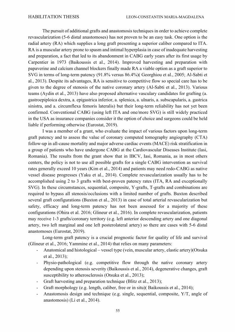
HABILITATION THESIS LEON-CONSTANTIN MARIA-MAGDALENA
55
The pursuit of additional grafts and anastomosis techniques in order to achieve complete revascularization (5-6 distal anastomoses) has not proven to be an easy task. One option is the radial artery (RA) which supplies a long graft presenting a superior caliber compared to ITA. RA is a muscular artery prone to spasm and intimal hyperplasia in case of inadequate harvesting and preparation, a fact that led to its abandonment in CABG early years after its first usage by Carpentier in 1973 (Baikoussis et al., 2014). Improved harvesting and preparation with papaverine and calcium channel blockers finally made RA a viable option as a graft superior to SVG in terms of long-term patency (91.8% versus 86.4%)( Georghiou et al., 2005; Al-Sabti et al., 2013). Despite its advantages, RA is sensitive to competitive flow so special care has to be given to the degree of stenosis of the native coronary artery (Al-Sabti et al., 2013). Various teams (Aydin et al., 2013) have also proposed alternative vasculary candidates for grafting (a. gastroepiploica dextra, a. epigastrica inferior, a. splenica, a. ulnaris, a. subscapularis, a. gastrica sinistra, and a. circumflexa femoris lateralis) but their long-term reliability has not yet been confirmed. Conventional CABG (using left ITA and one/more SVG) is still widely practiced in the USA as insurance companies consider it the option of choice and surgeons could be held liable if performing otherwise (Eurostat, 2019). I was a member of a grant, who evaluate the impact of various factors upon long-term graft patency and to assess the value of coronary computed tomography angiography (CTA) follow-up in all-cause mortality and major adverse cardiac events (MACE) risk stratification in a group of patients who have undergone CABG at the Cardiovascular Diseases Institute (Iasi, Romania). The results from the grant show that in IBCV, Iasi, Romania, as in most others centers, the policy is not to use all possible grafts for a single CABG intervention as survival rates generally exceed 10 years (Kim et al., 2014) and patients may need redo-CABG as native vessel disease progresses (Yaku et al., 2014). Complete revascularization usually has to be accomplished using 2 to 3 grafts with best-proven patency rates (ITA, RA and exceptionally SVG). In these circumstances, sequential, composite, Y-grafts, T-grafts and combinations are required to bypass all stenosis/occlusions with a limited number of grafts. Buxton described several graft configurations (Buxton et al., 2013) in case of total arterial revascularization but safety, efficacy and long-term patency has not been assessed for a majority of these configurations (Ohira et al. 2016; Glineur et al., 2016). In complete revascularization, patients may receive 1-3 grafts/coronary territory (e.g. left anterior descending artery and one diagonal artery, two left marginal and one left posterolateral artery) so there are cases with 5-6 distal anastomoses (Eurostat, 2019). Long-term graft patency is a crucial prognostic factor for quality of life and survival (Glineur et al., 2016; Yammine et al., 2014) that relies on many parameters:
- Anatomical and histological – vessel type (vein, muscular artery, elastic artery)(Otsuka et al., 2013);
- Physio-pathological (e.g. competitive flow through the native coronary artery depending upon stenosis severity (Baikoussis et al., 2014), degenerative changes, graft susceptibility to atherosclerosis (Otsuka et al., 2013);
- Graft harvesting and preparation technique (Blitz et al., 2013); - Graft morphology (e.g. length, caliber, free or in situ)( Baikoussis et al., 2014); - Anastomosis design and technique (e.g. single, sequential, composite, Y/T, angle of
anastomosis) (Li et al., 2014).

HABILITATION THESIS LEON-CONSTANTIN MARIA-MAGDALENA
56
Initially, graft patency has been assessed for individual grafts alone and not for CABG as an entity per se. Three studies (Andreini et al., 2012; Mushtaq et al., 2014) proposed two scores, the number of unprotected coronary territories (UCT) and the coronary artery protection score (CAPS), as being more relevant for long-term prognosis than individual graft’s patency.
Since 2000, more than 2500 patients underwent CABG interventions performed by a single team at the Cardiovascular Diseases Institute (Iasi, Romania) and revascularization technique varied in time according to scientific evidence from all venous grafts to total arterial revascularization and from “one graft-one coronary artery” to composite/Y grafts with 2-3 sequences per graft (Tinica et al., 2018).
The main publications that have derived from our research are listed below.
1. Tinica G, Chistol RO, Enache M, Leon Constantin MM, Ciocoiu M, Furnica C. Long-term graft patency after coronary artery bypass grafting: Effects of morphological and pathophysiological factors. Anatol J Cardiol. 2018; 20(5): 275-282.
2. Furnica C, Chistol RO, Leon Constantin MM, Alexa AI, Tinica G. Calcification of bioprosthetic heart valves biochemical substrate and prevention. Rev. Chim (Bucharest), 2015; 66(10):1716-1719. This was the first study in Romania who analyze the combined effect of several factors
upon graft patency and to integrate graft patency evaluated through medical imaging into CABG prognosis as a whole by taking into account the protection of coronary territories. The final endpoint of the project was identifying the profile of the grafting technique (vessel type, graft harvesting and preparation, anastomosis, grafting configuration and design) that offers the best long term results.
The funding for this research has been partly supported by an internal grant in which I was a member and are listed below:
”Morphoanatomical and Pathophysiological Aspects of Coronary Artery Bypass
Grafting in Terms of Long-Term Outcome (CABOT)” , Director Cristina Furnica (2017-2018). Using the theoretical data mentioned above, the present articles aimed:
• Morphophyisiological factors impact on CABG The purpose of this study was to identify pathophysiological and morphological factors
associated with long-term patency of grafts used in CABG.
• Bioprosthetic heart valves calcification This study analyses modern concepts and evaluates the degeneration and calcification
of explanted BHV in order to summarize the underlying chemical substrate and risk factors.

HABILITATION THESIS LEON-CONSTANTIN MARIA-MAGDALENA
57
4.2. Material and methods
• Morphophyisiological factors impact on CABG Data has been retrospectively collected and introduced into a database since 2000,
outlying patient population and surgical technique Demographic, echocardiographic, clinical, and angiographic information on patients undergoing CABG at our institute. Intraoperative parameters (extracorporeal circulation type and time, CABG technique, aortic cross-clamp time, number and type of grafts, and associated procedures) and postoperative data (complications, intensive care unit parameters, and mortality within 30 days) have also been collected. The surgical technique differed depending on the necessities of each individual intervention, from total arterial to total venous revascularization and from one graft–one anastomosis to composite and sequential grafting.
In 2016, the status of the entire patient cohort (394) with CABG interventions at our institution between 2000 and 2006 was verified through the National Health Insurance House database. The number of survivors was 269 (68.27%). These individuals were subsequently recalled for a coronary computed tomography angiography (CCTA) evaluation of graft patency, through invitation letter or phone call. A total of 127 patients agreed to the examination, presented no contraindications to CCTA graft patency assessment, and had good quality examinations that allowed quantifying all grafts. Patients were evaluated after a mean interval of 139.78±36.64 months after operation. None suffered from myocardial infarction within 30 days, suggestive of early graft failure. Long-term medical treatment for all patients consisted of beta blockers, statins, and enteric-coated aspirin. Patients with RA graft were prescribed a calcium channel blocker (amlodipine) for the first 3 months in order to prevent spasm. Treatment was adjusted individual variations such as: blood pressure, left ventricular ejection fraction, and comorbidities. All CCTA evaluations were performed using a second generation 2*128-slices dual source multidetector CT scanner (Siemens Somatom Definition Flash) with the following scan parameters: 100 or 120 kV tube voltage, 128×0.6 mm collimation, and 280 ms gantry rotation time. Optimal reconstructions were performed (0.75 mm slice thickness) and submitted to the Syngo. via workstation (Siemens Medical Solutions, Germany) for image analysis.
All CT examinations were evaluated twice by the same radiologist for the following parameters: graft type and status (confronted with the operative protocol), target coronary artery (confronted with the operative protocol), graft length, graft caliber, and target vessel caliber. Occluded grafts were traced based on density differences relative to the adjacent mediastinal or epicardial fat translated by a light gray color in the case of occluded grafts and dark gray or black color for mediastinal/epicardial fat (Fig. 23).
Statistical analysis: Continuous variables are expressed as mean±standard deviation. Categorical variables are expressed as percentages. using The chi-square test was used for comparing groups to obtain categorical variables and the Wilcoxon– Mann-Whitney U test and Student’s t-test for continuous variables based on their distribution. Normality was verified using the Kolmogorov–Smirnov and the Shapiro–Wilk W tests. Logistic regression was employed for testing the association between categorical and continuous variables previously identified as affecting graft patency at univariable analysis with a p value <0.05.
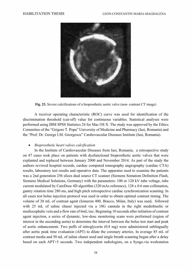
HABILITATION THESIS LEON-CONSTANTIN MARIA-MAGDALENA
58
Fig. 23. Severe calcifications of a bioprosthetic aortic valve (non- contrast CT image) A receiver operating characteristic (ROC) curve was used for identification of the
discrimination threshold (cut-off) value for continuous variables. Statistical analyses were performed using IBM SPSS Statistics 24 for Mac OS X. The study was approved by the Ethics Committee of the “Grigore T. Popa” University of Medicine and Pharmacy (Iasi, Romania) and the “Prof. Dr. George I.M. Georgescu” Cardiovascular Diseases Institute (Iasi, Romania).
• Bioprosthetic heart valves calcification
In the Institute of Cardiovascular Diseases from Iasi, Romania, a retrospective study on 47 cases took place on patients with dysfunctional bioprosthetic aortic valves that were explanted and replaced between January 2000 and November 2014. As part of the study the authors reviwed hospital records, cardiac computed tomography angiography (cardiac CTA) results, laboratory test results and operative data. The apparatus used to examine the patients was a 2nd generation 256 slices dual source CT scanner (Siemens Somatom Definition Flash, Siemens Medical Solutions, Germany) with the parameters: 100 or 120 kV tube voltage, tube current modulated by CareDose 4D algorithm (320 mAs reference), 128 x 0.6 mm collimation, gantry rotation time 280 ms, and high pitch retrospective cardiac synchronization scanning. In all cases test bolus injection protocol was used in order to obtain optimal contrast timing. The volume of 20 mL of contrast agent (Iomeron 400, Bracco, Milan, Italy) was used, followed with 25 mL of saline chaser injected via a 18G cannula in the right mediobasilic or mediocephalic vein and a flow rate of 6mL/sec. Beginning 10 seconds after initiation of contrast agent injection, a series of dynamic, low-dose monitoring scans were performed (region of interest in the ascending aorta) to determine the interval between the bolus test start and peak of aortic enhancement. Two puffs of nitroglycerin (0.8 mg) were administered sublingually after aortic peak time evaluation (APT) to dilate the coronary arteries. In average 85 mL of contrast media and 50 mL of saline chaser used and single breath scanning began after a delay based on each APT+5 seconds. Two independent radiologists, on a Syngo.via workstation
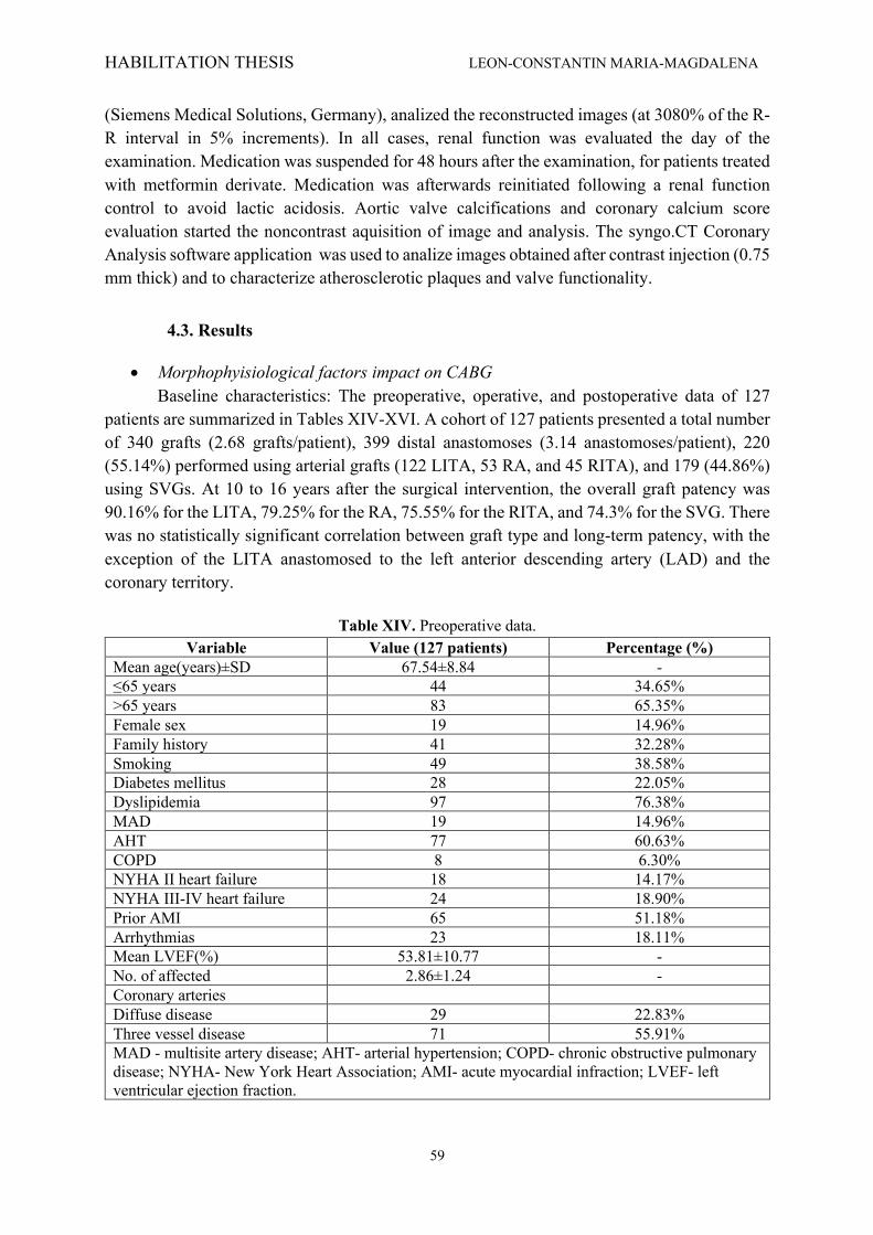
HABILITATION THESIS LEON-CONSTANTIN MARIA-MAGDALENA
59
(Siemens Medical Solutions, Germany), analized the reconstructed images (at 3080% of the R-R interval in 5% increments). In all cases, renal function was evaluated the day of the examination. Medication was suspended for 48 hours after the examination, for patients treated with metformin derivate. Medication was afterwards reinitiated following a renal function control to avoid lactic acidosis. Aortic valve calcifications and coronary calcium score evaluation started the noncontrast aquisition of image and analysis. The syngo.CT Coronary Analysis software application was used to analize images obtained after contrast injection (0.75 mm thick) and to characterize atherosclerotic plaques and valve functionality.
4.3. Results
• Morphophyisiological factors impact on CABG Baseline characteristics: The preoperative, operative, and postoperative data of 127
patients are summarized in Tables XIV-XVI. A cohort of 127 patients presented a total number of 340 grafts (2.68 grafts/patient), 399 distal anastomoses (3.14 anastomoses/patient), 220 (55.14%) performed using arterial grafts (122 LITA, 53 RA, and 45 RITA), and 179 (44.86%) using SVGs. At 10 to 16 years after the surgical intervention, the overall graft patency was 90.16% for the LITA, 79.25% for the RA, 75.55% for the RITA, and 74.3% for the SVG. There was no statistically significant correlation between graft type and long-term patency, with the exception of the LITA anastomosed to the left anterior descending artery (LAD) and the coronary territory.
Table XIV. Preoperative data. Variable Value (127 patients) Percentage (%)
Mean age(years)±SD 67.54±8.84 - ≤65 years 44 34.65% >65 years 83 65.35% Female sex 19 14.96% Family history 41 32.28% Smoking 49 38.58% Diabetes mellitus 28 22.05% Dyslipidemia 97 76.38% MAD 19 14.96% AHT 77 60.63% COPD 8 6.30% NYHA II heart failure 18 14.17% NYHA III-IV heart failure 24 18.90% Prior AMI 65 51.18% Arrhythmias 23 18.11% Mean LVEF(%) 53.81±10.77 - No. of affected 2.86±1.24 - Coronary arteries Diffuse disease 29 22.83% Three vessel disease 71 55.91% MAD - multisite artery disease; AHT- arterial hypertension; COPD- chronic obstructive pulmonary disease; NYHA- New York Heart Association; AMI- acute myocardial infraction; LVEF- left ventricular ejection fraction.
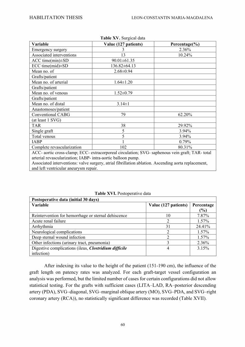
HABILITATION THESIS LEON-CONSTANTIN MARIA-MAGDALENA
60
Table XV. Surgical data
Variable Value (127 patients) Percentage(%) Emergency surgery 3 2.36% Associated interventions 13 10.24% ACC time(min)±SD 90.01±61.35 ECC time(mid)±SD 136.82±64.13 Mean no. of 2.68±0.94 Grafts/patient Mean no. of arterial 1.64±1.20 Grafts/patient Mean no. of venous 1.52±0.79 Grafts/patient Mean no. of distal 3.14±1 Anastomoses/patient Conventional CABG 79 62.20% (at least 1 SVG) TAR 38 29.92% Single graft 5 3.94% Total venous 5 3.94% IABP 1 0.79% Complete revascularization 102 80.31% ACC- aortic cross-clamp; ECC- extracorporeal circulation; SVG- saphenous vein graft; TAR- total arterial revascularization; IABP- intra-aortic balloon pump. Associated interventions: valve surgery, atrial fibrillation ablation. Ascending aorta replacement, and left ventricular aneurysm repair.
Table XVI. Postoperative data Postoperative data (initial 30 days) Variable Value (127 patients) Percentage
(%) Reintervention for hemorrhage or sternal dehiscence 10 7.87% Acute renal failure 2 1.57% Arrhythmia 31 24.41% Neurological complications 2 1.57% Deep sternal wound infection 2 1.57% Other infections (urinary tract, pneumonia) 3 2.36% Digestive complications (ileus, Clostridium difficile infection)
4 3.15%
After indexing its value to the height of the patient (151-190 cm), the influence of the
graft length on patency rates was analyzed. For each graft-target vessel configuration an analysis was performed, but the limited number of cases for certain configurations did not allow statistical testing. For the grafts with sufficient cases (LITA–LAD, RA–posterior descending artery (PDA), SVG–diagonal, SVG–marginal oblique artery (MO), SVG–PDA, and SVG–right coronary artery (RCA)), no statistically significant difference was recorded (Table XVII).
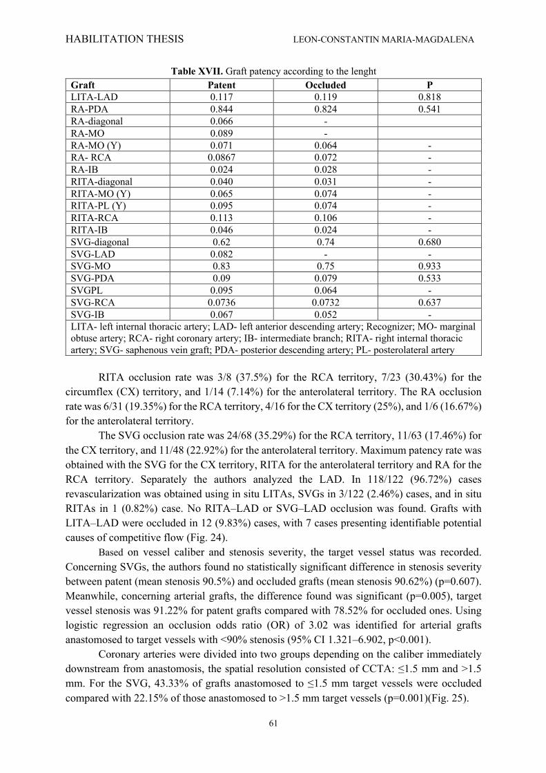
HABILITATION THESIS LEON-CONSTANTIN MARIA-MAGDALENA
61
Table XVII. Graft patency according to the lenght Graft Patent Occluded P LITA-LAD 0.117 0.119 0.818 RA-PDA 0.844 0.824 0.541 RA-diagonal 0.066 - RA-MO 0.089 - RA-MO (Y) 0.071 0.064 - RA- RCA 0.0867 0.072 - RA-IB 0.024 0.028 - RITA-diagonal 0.040 0.031 - RITA-MO (Y) 0.065 0.074 - RITA-PL (Y) 0.095 0.074 - RITA-RCA 0.113 0.106 - RITA-IB 0.046 0.024 - SVG-diagonal 0.62 0.74 0.680 SVG-LAD 0.082 - - SVG-MO 0.83 0.75 0.933 SVG-PDA 0.09 0.079 0.533 SVGPL 0.095 0.064 - SVG-RCA 0.0736 0.0732 0.637 SVG-IB 0.067 0.052 - LITA- left internal thoracic artery; LAD- left anterior descending artery; Recognizer; MO- marginal obtuse artery; RCA- right coronary artery; IB- intermediate branch; RITA- right internal thoracic artery; SVG- saphenous vein graft; PDA- posterior descending artery; PL- posterolateral artery
RITA occlusion rate was 3/8 (37.5%) for the RCA territory, 7/23 (30.43%) for the
circumflex (CX) territory, and 1/14 (7.14%) for the anterolateral territory. The RA occlusion rate was 6/31 (19.35%) for the RCA territory, 4/16 for the CX territory (25%), and 1/6 (16.67%) for the anterolateral territory.
The SVG occlusion rate was 24/68 (35.29%) for the RCA territory, 11/63 (17.46%) for the CX territory, and 11/48 (22.92%) for the anterolateral territory. Maximum patency rate was obtained with the SVG for the CX territory, RITA for the anterolateral territory and RA for the RCA territory. Separately the authors analyzed the LAD. In 118/122 (96.72%) cases revascularization was obtained using in situ LITAs, SVGs in 3/122 (2.46%) cases, and in situ RITAs in 1 (0.82%) case. No RITA–LAD or SVG–LAD occlusion was found. Grafts with LITA–LAD were occluded in 12 (9.83%) cases, with 7 cases presenting identifiable potential causes of competitive flow (Fig. 24).
Based on vessel caliber and stenosis severity, the target vessel status was recorded. Concerning SVGs, the authors found no statistically significant difference in stenosis severity between patent (mean stenosis 90.5%) and occluded grafts (mean stenosis 90.62%) (p=0.607). Meanwhile, concerning arterial grafts, the difference found was significant (p=0.005), target vessel stenosis was 91.22% for patent grafts compared with 78.52% for occluded ones. Using logistic regression an occlusion odds ratio (OR) of 3.02 was identified for arterial grafts anastomosed to target vessels with <90% stenosis (95% CI 1.321–6.902, p<0.001).
Coronary arteries were divided into two groups depending on the caliber immediately downstream from anastomosis, the spatial resolution consisted of CCTA: ≤1.5 mm and >1.5 mm. For the SVG, 43.33% of grafts anastomosed to ≤1.5 mm target vessels were occluded compared with 22.15% of those anastomosed to >1.5 mm target vessels (p=0.001)(Fig. 25).

HABILITATION THESIS LEON-CONSTANTIN MARIA-MAGDALENA
62
Fig. 24. Patent (left) and occluded (right) LITA–LAD graft
Fig. 25. RCA diameter downstream from an SVG–RCA anastomosis
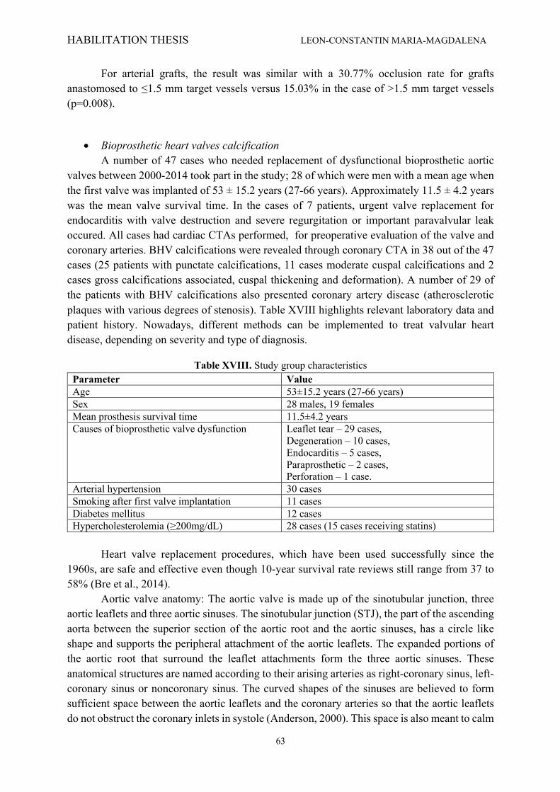
HABILITATION THESIS LEON-CONSTANTIN MARIA-MAGDALENA
63
For arterial grafts, the result was similar with a 30.77% occlusion rate for grafts anastomosed to ≤1.5 mm target vessels versus 15.03% in the case of >1.5 mm target vessels (p=0.008).
• Bioprosthetic heart valves calcification
A number of 47 cases who needed replacement of dysfunctional bioprosthetic aortic valves between 2000-2014 took part in the study; 28 of which were men with a mean age when the first valve was implanted of 53 ± 15.2 years (27-66 years). Approximately 11.5 ± 4.2 years was the mean valve survival time. In the cases of 7 patients, urgent valve replacement for endocarditis with valve destruction and severe regurgitation or important paravalvular leak occured. All cases had cardiac CTAs performed, for preoperative evaluation of the valve and coronary arteries. BHV calcifications were revealed through coronary CTA in 38 out of the 47 cases (25 patients with punctate calcifications, 11 cases moderate cuspal calcifications and 2 cases gross calcifications associated, cuspal thickening and deformation). A number of 29 of the patients with BHV calcifications also presented coronary artery disease (atherosclerotic plaques with various degrees of stenosis). Table XVIII highlights relevant laboratory data and patient history. Nowadays, different methods can be implemented to treat valvular heart disease, depending on severity and type of diagnosis.
Table XVIII. Study group characteristics Parameter Value Age 53±15.2 years (27-66 years) Sex 28 males, 19 females Mean prosthesis survival time 11.5±4.2 years Causes of bioprosthetic valve dysfunction Leaflet tear – 29 cases,
Degeneration – 10 cases, Endocarditis – 5 cases, Paraprosthetic – 2 cases, Perforation – 1 case.
Arterial hypertension 30 cases Smoking after first valve implantation 11 cases Diabetes mellitus 12 cases Hypercholesterolemia (≥200mg/dL) 28 cases (15 cases receiving statins)
Heart valve replacement procedures, which have been used successfully since the
1960s, are safe and effective even though 10-year survival rate reviews still range from 37 to 58% (Bre et al., 2014).
Aortic valve anatomy: The aortic valve is made up of the sinotubular junction, three aortic leaflets and three aortic sinuses. The sinotubular junction (STJ), the part of the ascending aorta between the superior section of the aortic root and the aortic sinuses, has a circle like shape and supports the peripheral attachment of the aortic leaflets. The expanded portions of the aortic root that surround the leaflet attachments form the three aortic sinuses. These anatomical structures are named according to their arising arteries as right-coronary sinus, left-coronary sinus or noncoronary sinus. The curved shapes of the sinuses are believed to form sufficient space between the aortic leaflets and the coronary arteries so that the aortic leaflets do not obstruct the coronary inlets in systole (Anderson, 2000). This space is also meant to calm

HABILITATION THESIS LEON-CONSTANTIN MARIA-MAGDALENA
64
the turbulent current behind the leaflets. The specific shape of the sinuses minimizes the mechanical stress concentration between the aortic sinuses and the leaflets. A study by Gundiah et al. showed that the aortic sinuses and the ascending aorta have different material properties and the aortic sinus is stiffer than the ascending aorta (Gundiah et al., 2008). Through the semilunar attachment, the leaflets are connected to the aortic wall while the commissures, formed from two parts of attachment lines of adjacent leaflets running side by side. Adjacent leaflets come in contact with each other during valve loading and create a seal against backflow into the left ventricle (Ho, 2016). Silver & William revealed that age and heart weight increase sinuse volume, sinotubular junction area and leaflets area (Silver, 1972). The leaflets are thin and elastic and in youths, becoming stiffer and thicker with age (Ho, 2009). The leaflet belly is also thinner in and the coaptation area, and thicker at the attachment and free margin (Anderson, 2000).
Ultrastructure of the aortic leaflets: Water (90%) and connective tissue with unique mechanical properties form the leaflets. The main components of connective tissue are protein collagen types I and III, elastin, GAG’s (glycosaminoglycans - long chain sugars) and a small number of cells. Three layers form a framework of internal collagen: the fibrosa, spongiosa and ventricularis (Weska et al., 2010). The layer towards the aorta is the fibrosa, arranged into a stretch-resisting sheet of tissue from a series of parallel clusters of collagen fibers. The orientation of fiber bundles is circumferential, starting at one commissure, then spreading out near the belly and finally coming together again at the opposite commissure to provide the essential strength of the leaflets. Radial expansion of the leaflets allows them to seal off the aortic orifice together (Gundiah et al., 2008). Facing the left ventricle is the ventricularis, consisting of collagen and elastin sheets. It is more extensible than the fibrosa because of a higher concentration of elastin. The spongiosa is between the fibrosa and ventricularis and is composed of elastin, collagen, proteoglycans and mucopolysaccharides. The specific function of this third layer is not well understood; but it is believed that it facilitates the localized movement and shearing between the fibrosa and the ventricularis during loading and unloading (Ho, 2009).
Treatment of valvular diseases: In this study, the original BHV mean survival time was 11.5 years with leaflet tear and degeneration as the main causes of valvular dysfunction. In 38 of the 47 cases CT revealed valvular calcifications, a mark of tissue degeneration. Severe atherosclerosis was present in most of the patients also, indicated by arterial hypertension, coronary artery disease and hypercholesterolemia which implies stress on the aortic valve. The mechanical type of heart valve is durable but susceptible to blood clots, requiring anticoagulation therapy. Meanwhile xenografts, or BHVs, made of either porcine valves or bovine pericardium, are more biocompatible and less thrombogenic, but they too suffer from tissue degeneration and failure in time because of graft immune response. The latter have a durability range between 5 to 20 years maybe shorter in younger patients with more serious immune responses. Although each type of replacement valves has advantages and disadvantages, they all share calcification as a common weakness (Clark et al., 2004).
Calcium and phosphorous are the most abundant elements in a human body. In the skeleton (bone, teeth) there are about 99% of the total calcium content and 85% of the total phosphorous content in a human. The rest is distributed through intracellular and extracellular fluids and in soft tissue. Extracellular plasma calcium concentration is normally 1 mg/mL

HABILITATION THESIS LEON-CONSTANTIN MARIA-MAGDALENA
65
(approximately 10-3 M) and 1,000 to 10,000 times lower (approximately 10-3 M) in the cytoplasm. However, glutaraldehyde prevents the normal cell mechanism for calcium elimination (Saleeb et al., 2014). Meanwhile, phosphorous is found in cell membranes and other intercellular structures (as phospholipids, especially phosphatidyl serine, and the phosphate backbone of nucleic acids). They can bind with calcium and provide a nucleation site. Further calcification deposits accumulate in these initial nucleation sites and eventually combine and become larger. As a result of this mineralization, the tissue becomes stiff and therefore weak and causes malfunction in bioprosthesis performance (Acharya et al., 2014).
Aortic calcification is a kind of vascular calcification through which calcium deposits build up on the aortic valve leaflets. The calcium deposits thicken and cause narrowing at the opening of the aortic valve. This impairs blood flow through the valve, causing chest pain or a heart attack. In severe cases, patients should undergo surgery and their aortic heart valve should be replaced with mechanical or BHV (Acharya et al., 2014).
Replacement valves, however, are subject to same calcification whether they are made up of tissue, or polymers. This is to make them immunologically inert and improve the tissue durability. It is believed that the glutaraldehyde fixation process leads to calcification of these types of valves. The glutaraldehyde pretreated cells become nonviable and produce the primary sites for calcification. Through this fixation process, crosslinking of antigens happens, which is supposed to make the valves immunologically inert and improve tissue durability (Saleeb et al., 2014). Unlike the old conception of vascular calcification as a passive process, it is currently considered to be an actively regulated one (Acharya et al., 2014).
Recent studies have suggested a similarity between pathologic calcification and physiologic bone mineralization, which is regulated by inductive and inhibitory factors. Many aspects of the pathophysiology of calcification process have been elucidated through in vitro and in vivo pathological analysis on BHVs. Factors such as host metabolism, implant structure and mechanical stresses determine the mineralization in BHVs or other biomaterials. Calcification mineralization is further enhanced at the sites that are under intense mechanical stresses, such as commissures in heart valves (Scherman et al., 2018).
In the cusps of BHVs, the mineralization process is dominantly initiated in connective tissue cells that are no longer viable due to glutaraldehyde pretreatment procedures. These cells have become devitalized but not removed from the structure of the valve. As stated above, dystrophic calcification of the cells happens due to reaction of calcium in extracellular fluid with membrane-associated phosphorus. This is likely because the glutaraldehyde pretreated cells have become nonviable and their calcium ion expulsion has been disrupted. No medical therapy, as of yet, has been demonstrated to be effective in the prevention of the progression of BHV calcification, but several studies demonstrated the similarities between calcific aortic sclerosis in the BHV valve and atherosclerosis (Acharya et al., 2014).
Other authors stipulated that serum cholesterol levels are associated with increased BHV calcification and suggested that statin treatment could be effective in preventing both atherosclerosis and BHV degeneration (Antonini-Canterin et al., 2006). Coronary calcifications are linked to BHV calcifications both in our study and the one performed by Farivar and Cohn. Apart from post implantation treatment, BHV calcification could also be prevented by an adequate pretreatment (Scherman et al., 2018).
HABILITATION THESIS LEON-CONSTANTIN MARIA-MAGDALENA
66
Clark and col. proved that pretreatment with aluminum chloride (AlCl3) (0.1 moles/L) and ethanol (80%, pH 7.4) inhibits calcification of both the glutaraldehyde-fixed porcine aortic BHV cusp and the aortic wall (Clark et al., 2005).
Ethanol prevents mineralization of the cusps by removal of cholesterol and phospholipids and major alterations of collagen intrahelical structural relationships. AlCl3 pre-treatment prevents aortic wall calcification by inhibition of elastin mineralization due to the following mechanisms: binding of Al to elastin resulting in a permanent protein-structural change conferring calcification resistance, inhibition of alkaline phosphatase activity, diminished upregulation of the extracellular matrix protein, tenascin C, and inhibition of matrix metalloproteinase - mediated elastolysis. Compared to other metallic ion pretreatments, aluminum has been shown to be the most effective inhibitor of bioprosthetic aortic wall calcification and alkaline phosphatase activity of BHV tissue (Clark et al., 2005).
4.4. Discussions
• Morphophyisiological factors impact on CABG Coronary artery bypass grafting (CABG) has now become one of the most frequent
surgical interventions undertaken in Europe (18-91 /100,000 inhabitants) (Eurostat, 2019). The surgical manouver is associated with increased survival and quality of life in cases with unprotected left main (or equivalent) and multivessel pathologies, but the optimal grafting technique has not been yet established. According to internationally recognised guidelines, while early CABG interventions were performed almost entirely using aorta-to-coronary saphenous vein grafts (SVGs), the angiographic follow-up studies have revealed a late attrition rate of 2% to 5% per year after surgery related to intrinsic pathological changes occuring in grafts (Patil et al., 2001).
In spite of their anatomical imperfection, venous grafts are more easy to collect and use, even for inexperienced surgeons. Compared to the veins, the internal thoracic arteries [left internal thoracic artery (LITA) and right internal thoracic artery (RITA)] show an extremely low attrition rate with more promising long-term patency rates (96.4% >15 years) (Aydin et al., 2013).
The anatomical imperfection of the venous grrafts and the impossibility to perform complete revascularization using only ITAs has led to a pursuit of better, additional grafts [the most commonly used being a. radialis (RA), followed by a. gastroepiploica dextra, a. splenica, a. epigastrica inferior, a. subscapularis, a. ulnaris, a. gastrica sinistra, and a. circumflexa femoris lateralis] and imagining new operative methods in order to obtain complete revascularization (5-6 distal anastomoses) using limited numbers of grafts (Tatoulis et al., 2004).
The published literature is unanimous in stating that graft patency has a great importance in the conditioning of CABG long-term prognosis but there is no consensus regarding optimal grafting techniques in terms of graft type, harvesting, preparation, configuration, features or anastomoses. In the medical literature, graft patency seem to have mostly been assessed for individual grafts alone and not for CABG as an entity per se; designing an optimal grafting technique in terms of graft type, preparation, harvesting, configuration, features, or anastomoses remains an interesting dilemma despite the increase in worldwide interest.
HABILITATION THESIS LEON-CONSTANTIN MARIA-MAGDALENA
67
We were able to classify the factors influencing graft patency, based on the literature available into morphological (vessel type, graft length, and caliber) (Aydin et al., 2013), pathophysiological (competitive flow through the native coronary artery and graft degenerative changes) (Melby et al., 2016), and surgical (technical expertise, graft harvesting and preparation, grafting design, and anastomosis technique) (Li et al., 2014). Morphological and pathophysiological factors of grafts were analyzed in this single center study.
Graft type: A meta-analysis on 15,962 patients was performed in 2001 by Taggard et al. (Taggard et al., 2001). The study identified a 10-year patency rate of 90%–95% for ITAs while 75% of SVGs presented stenotic or occlusive lesions. Benedetto et al. correlated nine randomized trials including 1620 angiographic controls at 1–7.7 years after CABG and discovered four times higher occlusion risk for the SVG than that for the RITA (Benedetto et al., 2014).
Habib et al. then proved in a study group of 2120 cases >70 years old, an increased survival rate of 5 and 10 years for the ITA–RA association (85.1% and 70.9%, respectively) compared with the ITA–SVG association (70.6% and 50.5%, respectively) (Habib et al., 2012). In our case, the best patency rate was from LITAs (90.16%), followed by RAs (79.25%), RITAs (75.55%), and SVGs (74.3%). The last is particularly sensitive to dysfunction by thrombosis in the first 30 days (due to focal destruction of the venous endothelium while harvesting), intimal hyperplasia between 1 month and 1 year, and atherosclerotic lesions after 1 year.
Necropsic studies found atherosclerotic lesions in the SVG starting from the first postoperative year (Hess et al., 2014). These lesions rarely develop significant stenosis in the first 3 postoperative years, their clinical symptoms appear mostly after 5 years. Arterial grafts show resistance to atherosclerotic lesions, but the RA is a muscular artery prone to spasm when harvesting is inadequate and competitive flow present. Fibro-intimal hyperplasia occurs particularly in ITA grafts, being an isolated process (Otsuka et al., 2013).
Graft length: Graft length-related patency has not yet been analyzed by extensive studies. A short, tensed graft is predisposed to spasm in the case of arteries and to flattening in the case of veins with hypoperfusion while a long graft with excessive length that is predisposed to transection (Voucharas et al., 2011).
The possibility of error, either an imprecise estimation of the cardiac volume or of the graft itself, anatomical features of the graft (high ITA bifurcation), peripheral localization of the target vessel, harvesting or manipulation mistakes (destruction of a graft segment), and pulmonary hyperinflation (emphysema), exists and can lead to an erroneous graft lenght. If such a deficit is discovered during surgery the team can choose one of different paths: elongation with a venous segment, composite anastomosis, right atrial plication (Voucharas et al., 2011), using a skeletonized instead of a pedicled graft, and grafting the CX territory via the transverse sinus (Fig. 26).
A limited number of grafts for each configuration in our study allowed statistical analysis only for LITA–LAD, RA–PDA, SVG–diagonal, SVG– MO, SVG–PDA, and SVG–RCA configurations with no significant difference between patent and occluded grafts.

HABILITATION THESIS LEON-CONSTANTIN MARIA-MAGDALENA
68
Fig. 26. RITA passage through the transverse sinus to reach a marginal obtuse artery Coronary territory: The authors have discovered that the coronary territory influenced
strongly the patency of both arterial and venous grafts. Maximum patency rate was obtained with RITAs for the anterolateral territory, RAs for the RCA territory, and SVGs for the CX territory. Parissis et al. performed a meta-analysis of 44 studies in 2005, reporting graft patencies for the right coronary territory and concluded that both the RA and the RITA patency rates are superior to the SVG patency for the right coronary territory (Parissis et al., 2014). These results proved contrary to ours, which proved the supremacy of the RA (80.65% patency rate) in front of the SVG (64.71%) but not of the in situ RITA (62.5%).
Target vessel status: Based on the physics law elaborated by Poiseuille, vessel resistance is known to be proportional to the length and inversely related to the fourth power of the inner diameter of the conduit. Ding et al., after analizing the impact of competitive flow on wall shear stress, confirmed that native vessel conditions graft flow. Both the volume of myocardial tissue perfused by the grafted artery and the degree of stenosis of the target vessel determine the amplitude of competitive flow (Ding et al., 2012).
This study proves that arterial grafts have a higher susceptibility to competitive flow, developing a mean target vessel stenosis of 91.22% for patent graft, 78.52% for occluded grafts and an occlusion OR of 3.02 for arterial grafts anastomosed to target vessels with <90% stenosis. Target vessel degree of stenosis had no impact on SVGs. Perfused at higher pressures, they are anastomosed directly to the aorta and have a thinner media makes lumen diameter adjustment impossible, which might explain the similarity between the target vessel degree of stenosis in the case of patent SVGs versus occluded SVGs.
The impact of competitive flow an ITA graft (4.6 mm diameter) was analyzed by Ding et al. on a computerized model. The anastomosis, with an angle of 45°, was to the LAD (4.5 mm diameter) (Ding et al., 2012). Simulations for various grades of stenosis (30%, 50%, 75%, and 100%) were performed. Resulting data highlighted that a higher degree of LAD stenosis correlated with a lower the mean velocity in the proximal LAD artery. Maniar et al. proved that competitive flow can occur secondary to residual flow through a non-critical stenosis or due to retrograde flow through coro-coronary auto-anastomoses (collateral circulation). If it is induced by collateral branches, competitive flow diminishes gradually until complete extinction due to the closure of collateral branches when perfusion is efficient (Maniar et al., 2002).
HABILITATION THESIS LEON-CONSTANTIN MARIA-MAGDALENA
69
According to Kawamura et al. a LITA–LAD graft can also occlude secondary to competitive flow generated by a grafted diagonal artery if no significant (≥75%) stenosis is interposed between the origin of the grafted diagonal artery and the LITA–LAD anastomosis (Kawamura et al., 2008). Meanwhile, a well-perfused CX by a grafted MO is not capable to generate competitive flow for the LITA–LAD graft. The length of the interposed segment shows he difference between the two cases, allowing flow attenuation in the second case. The importance of flow conditions in the proximal artery as a determinant of the hemodynamics at the distal end-to-side anastomosis has been demonstrated by a team lead by Kute (Kute et al., 2001).
Target vessel caliber is also a parameter that long-term graft patency. Occlusions seem to occur mostly in target vessels ≤1.5 mm, irreverent of venous or arterial origins. In 1979, Roth was the first to investigate the relationship between graft patency and target vessel caliber. His study identified a 90% patency rate of 1 year for SVGs anastomosed to coronary arteries with a >1.5 mm diameter and a 65% patency rate in the case of anastomosis to ≤1.5 mm coronary arteries (Roth et al., 1979). Goldman et al. then extended the study in 2004 by identifying a 10-year patency rate of 88% for SVGs and 100% for ITAs anastomosed to coronary arteries with a diameter >2 mm compared with 55% for SVGs and 82% for ITAs anastomosed to ≤2 mm target vessels (Goldman et al., 2004). Better runoff and graft flow are therefore linked with increased caliber of the target vessel is associated.
Study limitations: Small sample size was the first major limitant. Also, spatial resolution (0.5–0.625 mm) directly limits CCTA, therefore the measurements may overestimate diameters compared with conventional angiography.
• Bioprosthetic heart valves calcification
Calcification is known to play a major role in the failure of both tissue heart valve substitutes and bioprosthetic heart valves (BHV). The mechanism involves the formation of calcium phosphate mineral deposits through a reaction that involves calcium-containing extracellular fluid and membrane associated phosphorus (Vahanian et al., 2008). This process is accelerated by young age in patients, increased mechanical stress and valve related factors such as glutaraldehyde (CH2(CH2CHO)2) fixation.
Deposition of mineral calcium salts (especially hydroxyapatite – Ca10(PO4)6(OH)2) is physiologically normal in case of bones and teeth and restricted to specific anatomic sites. The calification of biomaterials contained by medical devices is considered a pathological process and is related to tissue degeneration. A certain similarity to bone mineralization has been aknowledged in recent studies, realised by inductive and inhibitory factors (Rutkovskaia et al., 2014). Both types of available heart valves, biomechanical and tissue, have negative side effects because they consist of materials foreign to the body. Mechanical heart valves are durable, but susceptible to thrombosis and thromboembolism and require long-term anticoagulation therapy (Bloomfield, 2002). Meanwhile, although BHVs require little or no anticoagulation they have a limited lifespan because of progressive structural changes such as leaflet wear and calcification (Grunkemeier et al., 2012).

HABILITATION THESIS LEON-CONSTANTIN MARIA-MAGDALENA
70
4.5. Conclusions
• Morphophyisiological factors impact on CABG Severe stenosis on a target vessel, especially those part pf the left coronary territory,
gives good runoff and is mostly associated with higher long-term patency rates. When taking into consideration the right territories, particular morphological features are a cause of lower patency rates. Studies have shown that the radial artery is the best choice of graft when involving the right side.
• Bioprosthetic heart valves calcification
Following our studies, we can conclude that the most promising preventive strategies must include modification of glutaraldehyde fixation, binding of calcification inhibitors to glutaraldehyde fixed tissue, removal or modification of calcifiable components and other linking agents. There are nowadays studies on discovering new tissue treatments and preventing this problem through synergistic and simultaneous employment of multiple anticalcification therapies.

HABILITATION THESIS LEON-CONSTANTIN MARIA-MAGDALENA
71
Chapter 5. QUALITY OF LIFE
5.1. Introduction
Cardiovascular diseases are the leading cause of mortality in the world, as well as in our country, which are responsible for 61% of deaths, placing Romania 3rd in Europe after Bulgaria and Ukraine, compared to 37 % in the European Union (EU) and 53 % in States that have recently joined the EU (Gaziano et al., 2010). People die of cardiovascular disease faster than cancer. Other risk factors to consider in today's society in our country are stress, obesity and unhealthy diets, physical inactivity. Statistics on the risk for cardiovascular disease are alarming.
Heart failure is the inability of the left ventricle to fill or eject enough blood to meet the requirements of the body and might be viewed as a representative syndrome of a variety of cardiac diseases. Nonetheless, coronary artery disease is the underlying pathology in 70% of heart failure patients (Ambrose et al., 2015). Chronic heart failure involves both mechanical failure and autonomic nervous system dysfunction that can lead to sudden cardiac death (SCD). Once admitted in hospital, the diagnosis of HF remains a difficult clinical challenge. Thus, this is based on a complex integration of symptoms (e.g., breathlessness, fatigue, and ankle swelling) and signs (e.g., tachycardia, tachypnea, rales, increased jugular venous pressure, hepatomegaly, and edema), supported by objective evidence of structural anomalies of the heart shown by abnormalities in the electrocardiogram (ECK), echocardiogram or chest X-ray (Ciampi et al., 2007).
Chronic heart failure is progressive and ultimately fatal, making early detection crucial for delaying disease progression. For patients with mild symptoms, the annual death rate is 5%–10%, and this increases sharply to 30%–40% for those with more advanced symptoms (Bui et al., 2011). Although heart failure is generally classified according to the New York Heart Association (NYHA) classification, there are other classification methods. ACC and AHA offer an alternative classification that has several advantages over the NHYA version. The NYHA classification is based on functional limitations of the heart failure patient, progressing from lesser to greater limitation. Unlike the disease itself, the NYHA classification may regress if a patient’s functional status improves. Conversely, the ACC/AHA classification progresses from stage A to D and cannot reverse the order. This is an important discussion because the decision to provide or refuse an Implantable Cardioverter Defibrillator (ICD) in heart failure based on the NYHA classification of the patient may not reflect the patient’s actual disease stage. That is, functional status may improve from NYHA IV to III without an improvement in disease state or, indeed, risk. Stage D in the ACC/AHA classification, on the other hand, provides a consistent classification of disease state and risk (ACC/AHA, 2019).
In elderly persons, heart failure is often accompanied by other concomitant conditions and changes resulting from the ageing process. That is why diagnosing cardiac heart failure (CHF) in old age is more difficult. Owing to the altered metabolism, impairment of the hepatic processes to various degrees and decreased renal excretion of drugs, treatment requires attention, individual choice of drugs and their periodic modification. Appropriate treatment improves and prolongs life. Elderly patients with multiple comorbidities and polypharmacy are also susceptible to poor coordination of care and are also at an increased risk for experiencing
HABILITATION THESIS LEON-CONSTANTIN MARIA-MAGDALENA
72
adverse drug reactions from drug–drug interactions. The association between comorbidity and healthcare costs has also been examined in a Medicare healthcare expenditure study (Park et al., 2017).
Morbidity and mortality in chronic heart failure remain high, despite advances in treatment (Clark et al., 2004). There are several approaches to management of chronic heart failure, and these generally relate to the severity of disease, symptoms, and comorbidity. The goal of therapy is to improve and prolong life, and this is achieved using lifestyle modifications, pharmaceutical therapy, surgery, supportive devices, management programs, and palliative care.
More recently, cardiac resynchronization therapy has emerged as a good option for those patients presenting with class III or IV heart failure (Steffel et al., 2011). The risk of arrhythmia and sudden cardiac death (SCD) is greater in patients with CHD (Hayashi et al., 2015).
The older the patient with cardiovascular disease, irrespective of gender, have an important risk of death and disability. The hazards of premature cardiovascular disease are greater amongst males than females. However, late onset cardiovascular disease and death from cardiovascular disease are now more common amongst females. The mechanism whereby cardiovascular disease events and mortality are reduced through hormone replacement therapy may be largely through the beneficial effects of hormone replacement therapy on lipid levels (reducing total and LDL cholesterol, raising HDL cholesterol and other changes) and reducing fibrinogen levels, with consequent reduction in the progress of atheromatous lesions and the tendency to thrombosis (Wenger et al., 1993). It is further suggested that estrogen reduces the uptake of LDL cholesterol in the arterial wall. It is yet to be defined which patients should be treated with hormone replacement therapy, but current thinking suggests those postmenopausal patients with known cardiovascular disease who have low HDL cholesterol levels or high LDL cholesterol levels should take hormone replacement therapy (Wenger et al., 1993).
Heart failure management In time, it was proven the necessity of other therapies in order to raise the efficiency of
medication already prescribed in patients with heart failure. Later, the accent is on identifying the risk factors, and on individualizing a rehabilitation program.
Patients with heart failure (HF) often have limited exercise capacity because of dyspnea and fatigue. It was previously thought that this exercise limitation was due entirely to cardiac dysfunction, and that treatment with inotropes and vasodilators, such as angiotensin converting enzyme inhibitors and the combination of hydralazine and nitrates, would improve exercise capacity (Wilson et al., 1996).
Although these drugs can improve the cardiac output and, with certain vasodilators, the patient survival, they may not acutely improve exercise tolerance. Thus, other factors in addition to the low cardiac output and reduced skeletal muscle blood flow must contribute to poor exercise tolerance and fatigue.
In the 1970s, exercise training of HF patients was discouraged due to concerns of worsening symptoms and detrimental effect on the disease process itself. Early observations in the 1980s documented improvements in exercise function for patients with HF with a low rate of complications. Early traditional management of patients with heart failure included a period of bed rest, which was sometimes prolonged. This led to a rapid deconditioning - adaptation of
HABILITATION THESIS LEON-CONSTANTIN MARIA-MAGDALENA
73
an organism to less demanding environment, or decrease of physiological adaptation to normal conditions, with muscular wasting and loss, comprising heart muscles also, abnormal body fluids distribution (Taylor et al., 2004), thus contributing to dependency and institutionalization, and probably to early death.
These observations were followed by a series of studies that demonstrated that significant biochemical and functional abnormalities in skeletal muscle are present in patients with HF and play a large role in the exercise intolerance (Gardner, 2011). Inactivity is in part responsible, leading to muscle atrophy. It is not recognized that definite/tangible/real benefits can be achieved through early mobilization and early exercise, both passive and active, which must be increased gradually. Dynamic exercise such as walking together with working the arms is recommended to maintain activities of daily living, as well as resistive training to increase muscular strength (Hornig et al., 1996). Such an exercise program should be gradually introduced and may start in hospital or at any point in the patient’s illness.
It is useful to consider four phases of cardiac rehabilitation, as each represents a different component of the care journey: inpatient care, the early post discharge period, exercise training, and finally the long term follow-up.
Comprehensive cardiac rehabilitation consists of exercise training together with education and psychological support. The studies show that patients who are women, belong to ethnic minorities, are elderly, and have low socioeconomic status have lower participation rates than white men and represent specific high-risk groups to be targeted for referral.
A positive family history of cardiovascular disease is a powerful marker of risk for the development and accelerated progress of cardiovascular disease. It has been clearly demonstrated that those with a positive family history for cardiovascular disease commonly have worse risk factor profiles in terms of lipids, blood pressure, obesity, diabetes and smoking habit, in addition to their non-modifiable genetic background (Myers et al., 1990). Hence, risk factor modification is of greater importance in patients with a positive family history than it is for those with identified modifiable risk factors without a family history.
Exercise Capacity The benefits of exercise training in patients with HF include an improvement in exercise
tolerance as assessed not only by exercise duration but more importantly by peak V̇O2 (Belardinelli, 1999). The exercise training program has varied by such factors as setting (supervised or home training), type of activity (treadmill or bicycle), duration (from 8 weeks to 3 months), and intensity (from low to moderate). One study that used primarily circuit weight training for 8 weeks elicited a modest but significant increase in peak V̇O2 (Maiorana et al., 2000). Changes in peak V̇O2 have ranged from 12% to 31%. Most of the improvement occurs by week 3 but can continue up to 6 months if compliance with the training program continues. Not only is maximal exercise performance improved but also indices of submaximal exercise as measured by the 6-minute walking test or by the ventilatory threshold (Hambrecht et al., 1995).
Systematic reviews of exercise-based cardiac rehabilitation in stable, chronic heart failure have found benefits to exercise capacity and possibly to symptoms. Benefit is probably derived from peripheral adaptations (vasodilation and improved muscle oxidative capacity) rather than improvements in ventricular function (Belardinelli et al., 1999). An overview of 159 randomised trials in Europe that included 134 patients concluded that exercise training
HABILITATION THESIS LEON-CONSTANTIN MARIA-MAGDALENA
74
improved exercise capacity and autonomic indices (for example, heart rate variability), and training could be conducted either in hospital or at home, and 16 weeks training was better than six, and that a combination of cycle ergometry and calisthenics was better than cycle ergometry alone (Belardinelli et al., 1999).
Comprehensive Management In a systematic review of comprehensive disease management for heart failure, there
were fewer hospital attendances, and an improved quality of life, functional capacity, patient satisfaction, and compliance with diet and medications (Rich, 1999). There is limited evidence on the effects of psychological and education only intervention in heart failure. In one recent randomised trial, education in hospital with one home visit was found to increase self-care, but had no impact on hospital attendance rates (Ismail et al., 2014). Patients with chronic heart failure should be considered for comprehensive cardiac rehabilitation if they have limiting symptoms (CLASS A OF RECOMMANDATION) (ACC/AHA, 2019).
Survival Currently published trials are single-center experience of a small number of patients
followed up for a short duration. An exercise regimen of short duration may fail to show any improvement in 1-year survival. Belardinelli and colleagues studied the effects of a long-term (14-month) exercise-training program of moderate intensity in 99 patients with stable chronic HF of various NYHA functional classes. Eighty-five percent of the patients had an ischemic etiology. One group underwent supervised exercise training at 60% of peak V̇O2 initially 3 times a week for 2 months, then twice a week for 12 more months. At 2 months, the percentage of both myocardial perfusion defects with improved thallium in 75% of trained and only 2% of untrained patients at 2 months. This trend was maintained at 14 months. Furthermore, the rate of both hospital readmission for HF and cardiac mortality was significantly lower in trained than control patients (p=0.02). A significant separation of survival curves was observed beyond the first year of follow-up, which confirms the results of previous studies (Belardinelli et al., 1999).
These promising preliminary results should stimulate a wider clinical application of exercise training in patients with stable HF. The results of this trial, however, cannot be considered proof of a mortality reduction because it was not powered to show survival differences. Nonetheless, this positive trend should provide encouragement for investigators to design a large, randomized, prospective mortality trial.
Quality of Life Studies that address quality of life (QOL) in patients with HF participating in an exercise
program are limited. In addition, the QOL measurement tools vary, and results are inconsistent. Tools that focus on symptoms such as dyspnea and fatigue as well as psychosocial status (for example, emotional function and mastery or perceived control over symptoms) are more likely to detect favorable responses to an intervention.
The commonly used “Minnesota Living With Heart Failure Questionnaire” assesses disease-specific health-related QOL by including the patient’s perceptions of the effects of HF and its treatment on his or her daily life; however, two randomized prospective exercise-training studies and one observational study failed to show significant improvement in QOL when utilizing this tool (Gottlieb et al., 1999). One did show a relation between change in total score
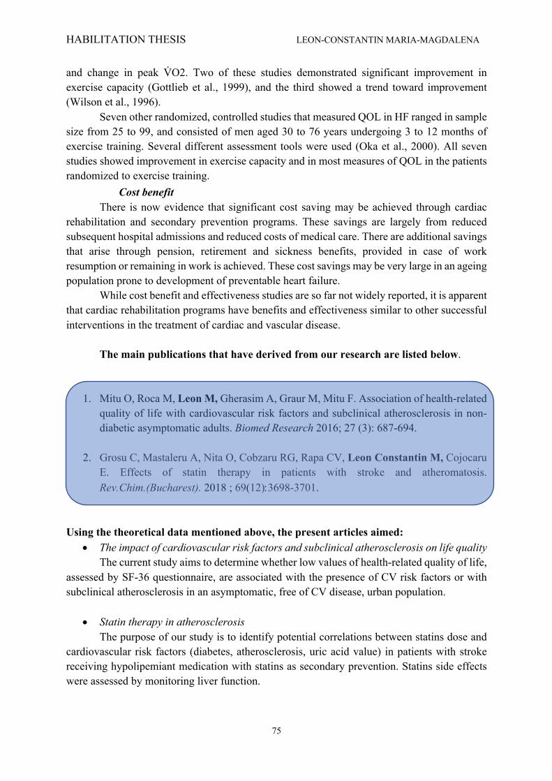
HABILITATION THESIS LEON-CONSTANTIN MARIA-MAGDALENA
75
and change in peak V̇O2. Two of these studies demonstrated significant improvement in exercise capacity (Gottlieb et al., 1999), and the third showed a trend toward improvement (Wilson et al., 1996).
Seven other randomized, controlled studies that measured QOL in HF ranged in sample size from 25 to 99, and consisted of men aged 30 to 76 years undergoing 3 to 12 months of exercise training. Several different assessment tools were used (Oka et al., 2000). All seven studies showed improvement in exercise capacity and in most measures of QOL in the patients randomized to exercise training.
Cost benefit There is now evidence that significant cost saving may be achieved through cardiac
rehabilitation and secondary prevention programs. These savings are largely from reduced subsequent hospital admissions and reduced costs of medical care. There are additional savings that arise through pension, retirement and sickness benefits, provided in case of work resumption or remaining in work is achieved. These cost savings may be very large in an ageing population prone to development of preventable heart failure.
While cost benefit and effectiveness studies are so far not widely reported, it is apparent that cardiac rehabilitation programs have benefits and effectiveness similar to other successful interventions in the treatment of cardiac and vascular disease.
The main publications that have derived from our research are listed below.
1. Mitu O, Roca M, Leon M, Gherasim A, Graur M, Mitu F. Association of health-related quality of life with cardiovascular risk factors and subclinical atherosclerosis in non-diabetic asymptomatic adults. Biomed Research 2016; 27 (3): 687-694.
2. Grosu C, Mastaleru A, Nita O, Cobzaru RG, Rapa CV, Leon Constantin M, Cojocaru E. Effects of statin therapy in patients with stroke and atheromatosis. Rev.Chim.(Bucharest). 2018 ; 69(12):3698-3701.
Using the theoretical data mentioned above, the present articles aimed:
• The impact of cardiovascular risk factors and subclinical atherosclerosis on life quality The current study aims to determine whether low values of health-related quality of life,
assessed by SF-36 questionnaire, are associated with the presence of CV risk factors or with subclinical atherosclerosis in an asymptomatic, free of CV disease, urban population.
• Statin therapy in atherosclerosis
The purpose of our study is to identify potential correlations between statins dose and cardiovascular risk factors (diabetes, atherosclerosis, uric acid value) in patients with stroke receiving hypolipemiant medication with statins as secondary prevention. Statins side effects were assessed by monitoring liver function.
HABILITATION THESIS LEON-CONSTANTIN MARIA-MAGDALENA
76
5.2. Material and methods
• The impact of cardiovascular risk factors and subclinical atherosclerosis on life quality We performed a prospective study on 111 native patients referred by general
practitioners and investigated in our cardiology department. All patients originated from urban area and were randomized when enrolling. Study inclusion criteria included aged 35-75, living in the urban area, absence of pregnancy in women and, most important, no personal history of CV, metabolic, renal, respiratory or cerebral diseases treated in the last 6 months. The study was approved by the University Ethics Committee and written informed consent was obtained in all cases prior to enrolling in the study.
The health-related QoL was assessed by using SF-36 health survey questionnaire with 36 multiple-choice questions each having an assigned score with a value on a scale 0-100 (from the worst to the best possible health status) which are summarized and form eight scales regarding: physical functioning (PF), role physical (RP), bodily pain (BP), general health (GH), vitality (VT), social functioning (SF), role-emotional (RE), mental health (MH). Finally, the eight domains were converted into two major summarizing measures-the physical component summary (PCS) and the mental component summary (MCS). Lower scores were associated with poor health status. SF-36 health survey is a registered trademark of Medical Outcomes Trust, USA, and a non-commercial license agreement was issued for use in the current study. SF-36 has already been tested and validated in the Romanian population, generating general population norms (Mihăilă et al., 2001). Population norms were used to compare our final scores, for all scales the norm being set at 50 as limit of normality (Berger et al., 2010). In our study, the questionnaire was self-administered.
All patients were screened for CV risk factors like age, sex, obesity, systolic and diastolic blood pressure taken at rest and heart rate. Obesity was affirmed according to the World Health Organization classification for BMI (body mass index) ranges for adults (AHA/ACC/TOS, 2013):
- normal weight – 18.5-24.9 kg/m2; - overweight –25-29.9 kg/m2; - obesity class 1– 30-34.9 kg/m2; - obesity class 2–35-39.9 kg/m2; - obesity class 3≥ 40 kg/m2.
We have analyzed the biochemical markers that have clinical CV relevance: lipid profile (total cholesterol, HDL, LDL, nonHDL and triglycerides), fasting plasma glucose, uric acid, total serum proteins, liver function profile (AST, ALT and GGT), inflammatory status (fibrinogen) and renal function (glomerular filtration rate - GFR). Individually, we have applied the SCORE risk chart for determining the 10-year risk of CV mortality in our sample of asymptomatic population. For the risk of developing diabetes mellitus, we applied the most worldwide used questionnaire, the FINDRISC score, which is based on eight very simple multiple-choice questions. If the values in both scores (SCORE, respectively FINDRISC) are high, the risk of developing CV diseases or diabetes in the future is increased.
Since the subjects had no personal history of CV diseases, subclinical atherosclerosis was quantified by multiple methods. Carotid intima media-thickness (IMT) was measured by

HABILITATION THESIS LEON-CONSTANTIN MARIA-MAGDALENA
77
means of carotid ultrasound and interpreted according to the Mannheim criteria (Touboul et al., 2012). We took into consideration the highest value obtained from both sides. To evaluate peripheral artery obstruction, ankle-brachial index (ABI) was calculated. Arterial stiffness was evaluated by using an Arteriograph™ device and we retained for final analysis the aortic pulse wave velocity (PWV), systolic blood pressure (SBPao), pulse pressure (PPao) as well as aortic and brachial augmentation indexes (AIXao, respectively AIXbr). At echocardiography we determined the left ventricle ejection fraction (LVEF), left ventricle mass index (LVMI) and screened for aortic atherosclerosis.
Obtained data was analyzed using SPSS 20.0 (Statistical Package for the Social Sciences, Chicago, Illinois). For continuous variables, data were presented as mean ± standard deviation (SD) and compared using parametric tests (t-test for independent samples). Pearson’s correlation analysis was applied to assess the relationship between variables. A p value inferior to 0.05 was considered significant for all data analyses. As well, the initial SF-36 data processing was performed by using the Health Outcomes Scoring Software 4.0.
• Statin therapy in atherosclerosis
We performed a retrospective study on 58 patients with a history of ischemic stroke, hospitalized between 01.01-30.09.2018 in the Neurology Department of the Clinical Rehabilitation Hospital in Iasi, Romania. Demographic data, BMI and biochemical parameters (hepatic enzymes, uric acid, glycemia, glycosylated hemoglobin and lipid profile) were determined in all cases.
Carotid ultrasound was also performed to screen for atherosclerosis and asses its severy. All examinations were performed by the same sonographer on a Siemens Accuson X300 system using a 7.5 MHz linear probe (Stein et al., 2008). Patients with arterial occlusion or embolic stroke were excluded. Written informed consent was obtained in all cases prior to inclusion.
Statistical analysis was performed with SPSS v.18 considering a p value <0.05 as significant. Continuous variables were presented as mean value ± standard deviation.
A total of 23 females and 35 males with mean age 65.9±13.11 years fulfilled inclusion criteria. Most patients had a history of elevated blood pressure (82.8% vs.17.2%) and also presented with atheroma plaques in the carotid artery at the Doppler examination (53.4% vs.46.5%). A total of 36.2% of the patients were overweight and 25.8% were obese. 53 patients associated type 2 insulin dependent diabetes mellitus. The mean value for AST was 25.5±17.7 mg/dL, with a maximum of 106 mg/dL, for ALT it was 36.7±36.7 mg/dL, with a maximum of 191.6 mg/dL, for uric acid the mean value was 4.5±1.7 mg/dL, with a maximum of 8.16 mg/ dL. As for the lipid profile, mean cholesterol was 145.7±40.4 mg/dL and for triglycerides was 116.7±56.2 mg/dL (Table XIX).
Table XIX. Biological analysis for the study group
AST ALT Uric acid Cholesterol Triglycerides Mean 25.5500 36.7741 4.5569 145.7848 116.7788 Stf. Deviation 17.78476 36.76194 1.73366 40.45919 56.29924 Minimum 11.00 10.60 1.26 85.80 43.40 Maximum 106.60 191.60 8.16 256.00 302.00
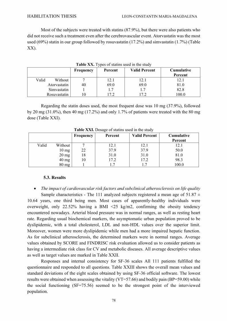
HABILITATION THESIS LEON-CONSTANTIN MARIA-MAGDALENA
78
Most of the subjects were treated with statins (87.9%), but there were also patients who did not receive such a treatment even after the cerebrovascular event. Atorvastatin was the most used (69%) statin in our group followed by rosuvastatin (17.2%) and simvastatin (1.7%) (Table XX).
Table XX. Types of statins used in the study
Frequency Percent Valid Percent Cumulative Percent
Valid Without Atorvastatin Simvastatin
Rosuvastatin
7 40 1 10
12.1 69.0 1.7 17.2
12.1 69.0 1.7 17.2
12.1 81.0 82.8 100.0
Regarding the statin doses used, the most frequent dose was 10 mg (37.9%), followed
by 20 mg (31.0%), then 40 mg (17.2%) and only 1.7% of patients were treated with the 80 mg dose (Table XXI).
Table XXI. Dosage of statins used in the study
Frequency Percent Valid Percent Cumulative Percent
Valid Without 10 mg 20 mg 40 mg 80 mg
7 22 18 10 1
12.1 37.9 31.0 17.2 1.7
12.1 37.9 31.0 17.2 1.7
12.1 50.0 81.0 98.3 100.0
5.3. Results
• The impact of cardiovascular risk factors and subclinical atherosclerosis on life quality Sample characteristics - The 111 analyzed subjects registered a mean age of 51.87 ±
10.64 years, one third being men. Most cases of apparently-healthy individuals were overweight, only 22.52% having a BMI <25 kg/m2, confirming the obesity tendency encountered nowadays. Arterial blood pressure was in normal ranges, as well as resting heart rate. Regarding usual biochemical markers, the asymptomatic urban population proved to be dyslipidemic, with a total cholesterol, LDL and non-HDL values over the superior limit. Moreover, women were more dyslipidemic while men had a more impaired hepatic function. As for subclinical atherosclerosis, the determined markers were in normal ranges. Average values obtained by SCORE and FINDRISC risk evaluation allowed us to consider patients as having a intermediate risk class for CV and metabolic diseases. All average descriptive values as well as target values are marked in Table XXII.
Responses and internal consistency for SF-36 scales All 111 patients fulfilled the questionnaire and responded to all questions. Table XXIII shows the overall mean values and standard deviations of the eight scales obtained by using SF-36 official software. The lowest results were obtained when assessing the vitality (VT=57.66) and bodily pain (BP=59.00) while the social functioning (SF=75.56) seemed to be the strongest point of the interviewed population.
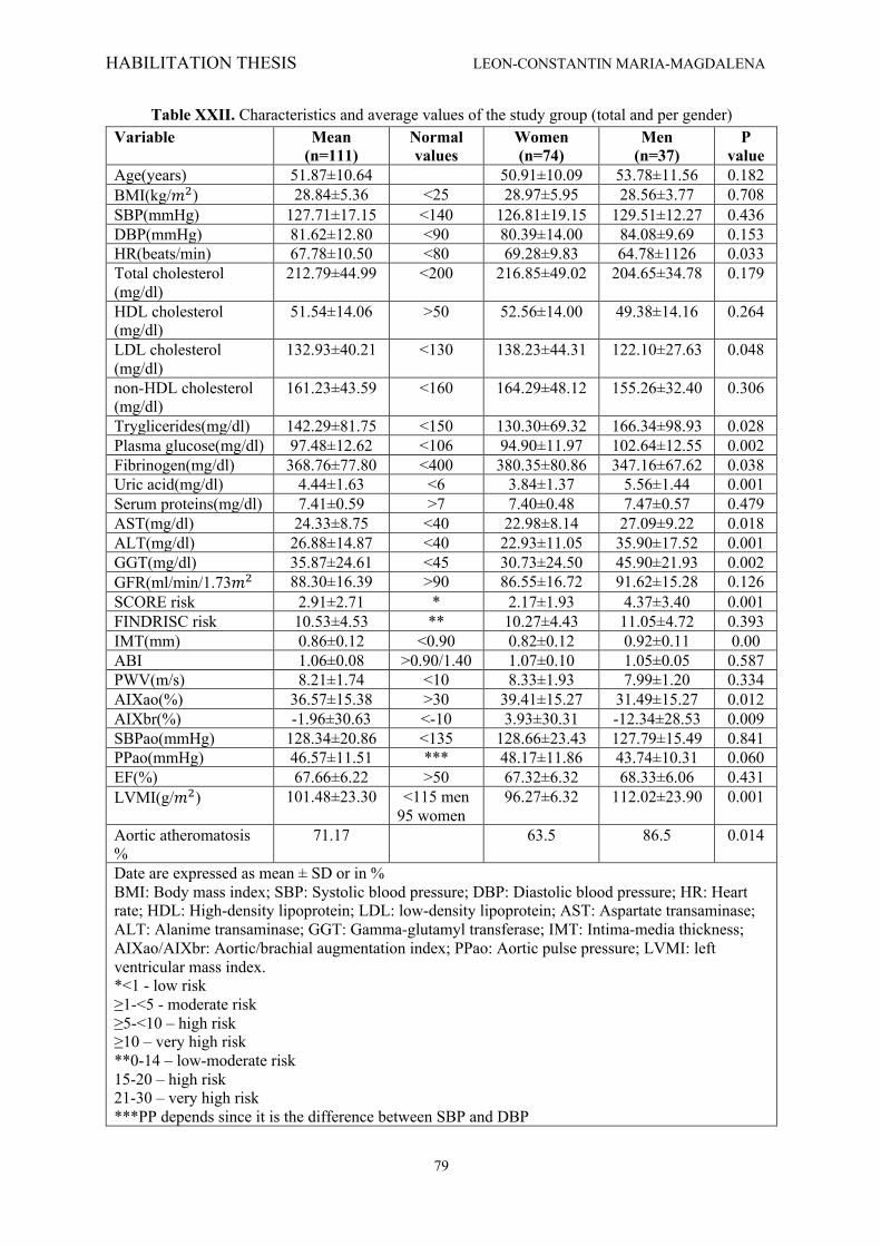
HABILITATION THESIS LEON-CONSTANTIN MARIA-MAGDALENA
79
Table XXII. Characteristics and average values of the study group (total and per gender) Variable Mean
(n=111) Normal values
Women (n=74)
Men (n=37)
P value
Age(years) 51.87±10.64 50.91±10.09 53.78±11.56 0.182 BMI(kg/𝑚$) 28.84±5.36 <25 28.97±5.95 28.56±3.77 0.708 SBP(mmHg) 127.71±17.15 <140 126.81±19.15 129.51±12.27 0.436 DBP(mmHg) 81.62±12.80 <90 80.39±14.00 84.08±9.69 0.153 HR(beats/min) 67.78±10.50 <80 69.28±9.83 64.78±1126 0.033 Total cholesterol (mg/dl)
212.79±44.99 <200 216.85±49.02 204.65±34.78 0.179
HDL cholesterol (mg/dl)
51.54±14.06 >50 52.56±14.00 49.38±14.16 0.264
LDL cholesterol (mg/dl)
132.93±40.21 <130 138.23±44.31 122.10±27.63 0.048
non-HDL cholesterol (mg/dl)
161.23±43.59 <160 164.29±48.12 155.26±32.40 0.306
Tryglicerides(mg/dl) 142.29±81.75 <150 130.30±69.32 166.34±98.93 0.028 Plasma glucose(mg/dl) 97.48±12.62 <106 94.90±11.97 102.64±12.55 0.002 Fibrinogen(mg/dl) 368.76±77.80 <400 380.35±80.86 347.16±67.62 0.038 Uric acid(mg/dl) 4.44±1.63 <6 3.84±1.37 5.56±1.44 0.001 Serum proteins(mg/dl) 7.41±0.59 >7 7.40±0.48 7.47±0.57 0.479 AST(mg/dl) 24.33±8.75 <40 22.98±8.14 27.09±9.22 0.018 ALT(mg/dl) 26.88±14.87 <40 22.93±11.05 35.90±17.52 0.001 GGT(mg/dl) 35.87±24.61 <45 30.73±24.50 45.90±21.93 0.002 GFR(ml/min/1.73𝑚$ 88.30±16.39 >90 86.55±16.72 91.62±15.28 0.126 SCORE risk 2.91±2.71 * 2.17±1.93 4.37±3.40 0.001 FINDRISC risk 10.53±4.53 ** 10.27±4.43 11.05±4.72 0.393 IMT(mm) 0.86±0.12 <0.90 0.82±0.12 0.92±0.11 0.00 ABI 1.06±0.08 >0.90/1.40 1.07±0.10 1.05±0.05 0.587 PWV(m/s) 8.21±1.74 <10 8.33±1.93 7.99±1.20 0.334 AIXao(%) 36.57±15.38 >30 39.41±15.27 31.49±15.27 0.012 AIXbr(%) -1.96±30.63 <-10 3.93±30.31 -12.34±28.53 0.009 SBPao(mmHg) 128.34±20.86 <135 128.66±23.43 127.79±15.49 0.841 PPao(mmHg) 46.57±11.51 *** 48.17±11.86 43.74±10.31 0.060 EF(%) 67.66±6.22 >50 67.32±6.32 68.33±6.06 0.431 LVMI(g/𝑚$) 101.48±23.30 <115 men
95 women 96.27±6.32 112.02±23.90 0.001
Aortic atheromatosis %
71.17 63.5 86.5 0.014
Date are expressed as mean ± SD or in % BMI: Body mass index; SBP: Systolic blood pressure; DBP: Diastolic blood pressure; HR: Heart rate; HDL: High-density lipoprotein; LDL: low-density lipoprotein; AST: Aspartate transaminase; ALT: Alanime transaminase; GGT: Gamma-glutamyl transferase; IMT: Intima-media thickness; AIXao/AIXbr: Aortic/brachial augmentation index; PPao: Aortic pulse pressure; LVMI: left ventricular mass index. *<1 - low risk ≥1-<5 - moderate risk ≥5-<10 – high risk ≥10 – very high risk **0-14 – low-moderate risk 15-20 – high risk 21-30 – very high risk ***PP depends since it is the difference between SBP and DBP
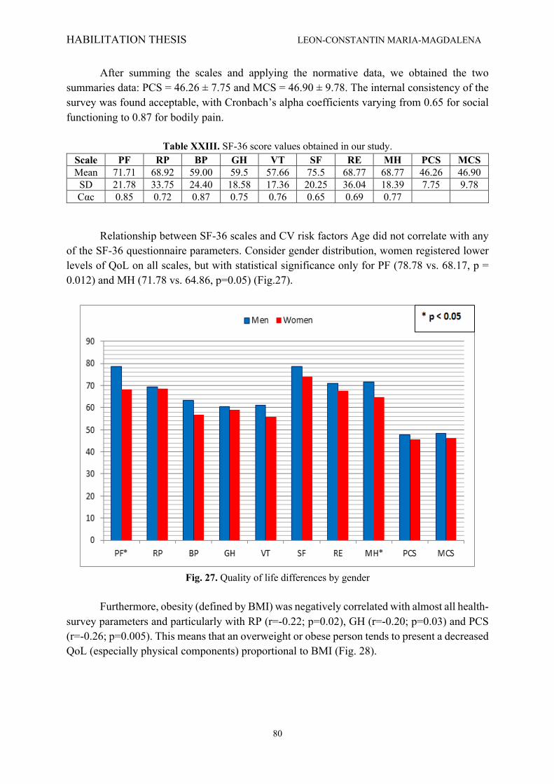
HABILITATION THESIS LEON-CONSTANTIN MARIA-MAGDALENA
80
After summing the scales and applying the normative data, we obtained the two summaries data: PCS = 46.26 ± 7.75 and MCS = 46.90 ± 9.78. The internal consistency of the survey was found acceptable, with Cronbach’s alpha coefficients varying from 0.65 for social functioning to 0.87 for bodily pain.
Table XXIII. SF-36 score values obtained in our study.
Scale PF RP BP GH VT SF RE MH PCS MCS Mean 71.71 68.92 59.00 59.5 57.66 75.5 68.77 68.77 46.26 46.90 SD 21.78 33.75 24.40 18.58 17.36 20.25 36.04 18.39 7.75 9.78 Cαc 0.85 0.72 0.87 0.75 0.76 0.65 0.69 0.77
Relationship between SF-36 scales and CV risk factors Age did not correlate with any of the SF-36 questionnaire parameters. Consider gender distribution, women registered lower levels of QoL on all scales, but with statistical significance only for PF (78.78 vs. 68.17, p = 0.012) and MH (71.78 vs. 64.86, p=0.05) (Fig.27).
Fig. 27. Quality of life differences by gender
Furthermore, obesity (defined by BMI) was negatively correlated with almost all health-
survey parameters and particularly with RP (r=-0.22; p=0.02), GH (r=-0.20; p=0.03) and PCS (r=-0.26; p=0.005). This means that an overweight or obese person tends to present a decreased QoL (especially physical components) proportional to BMI (Fig. 28).
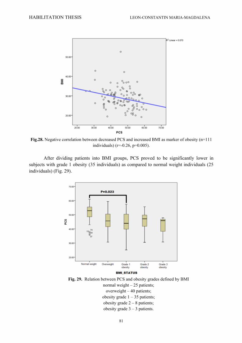
HABILITATION THESIS LEON-CONSTANTIN MARIA-MAGDALENA
81
Fig.28. Negative correlation between decreased PCS and increased BMI as marker of obesity (n=111
individuals) (r=-0.26, p=0.005).
After dividing patients into BMI groups, PCS proved to be significantly lower in subjects with grade 1 obesity (35 individuals) as compared to normal weight individuals (25 individuals) (Fig. 29).
Fig. 29. Relation between PCS and obesity grades defined by BMI
normal weight – 25 patients; overweight – 40 patients;
obesity grade 1 – 35 patients; obesity grade 2 – 8 patients; obesity grade 3 – 3 patients.
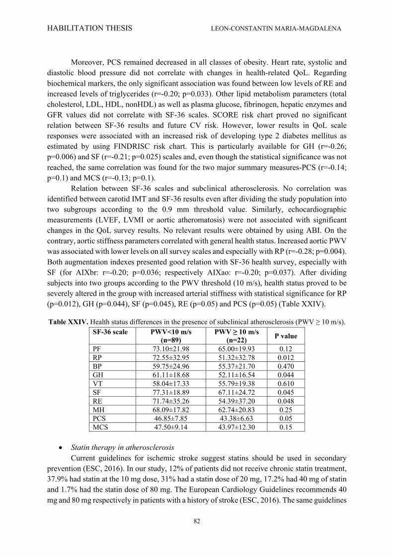
HABILITATION THESIS LEON-CONSTANTIN MARIA-MAGDALENA
82
Moreover, PCS remained decreased in all classes of obesity. Heart rate, systolic and
diastolic blood pressure did not correlate with changes in health-related QoL. Regarding biochemical markers, the only significant association was found between low levels of RE and increased levels of triglycerides (r=-0.20; p=0.033). Other lipid metabolism parameters (total cholesterol, LDL, HDL, nonHDL) as well as plasma glucose, fibrinogen, hepatic enzymes and GFR values did not correlate with SF-36 scales. SCORE risk chart proved no significant relation between SF-36 results and future CV risk. However, lower results in QoL scale responses were associated with an increased risk of developing type 2 diabetes mellitus as estimated by using FINDRISC risk chart. This is particularly available for GH (r=-0.26; p=0.006) and SF (r=-0.21; p=0.025) scales and, even though the statistical significance was not reached, the same correlation was found for the two major summary measures-PCS (r=-0.14; p=0.1) and MCS (r=-0.13; p=0.1).
Relation between SF-36 scales and subclinical atherosclerosis. No correlation was identified between carotid IMT and SF-36 results even after dividing the study population into two subgroups according to the 0.9 mm threshold value. Similarly, echocardiographic measurements (LVEF, LVMI or aortic atheromatosis) were not associated with significant changes in the QoL survey results. No relevant results were obtained by using ABI. On the contrary, aortic stiffness parameters correlated with general health status. Increased aortic PWV was associated with lower levels on all survey scales and especially with RP (r=-0.28; p=0.004). Both augmentation indexes presented good relation with SF-36 health survey, especially with SF (for AIXbr: r=-0.20; p=0.036; respectively AIXao: r=-0.20; p=0.037). After dividing subjects into two groups according to the PWV threshold (10 m/s), health status proved to be severely altered in the group with increased arterial stiffness with statistical significance for RP (p=0.012), GH (p=0.044), SF (p=0.045), RE (p=0.05) and PCS (p=0.05) (Table XXIV).
Table XXIV. Health status differences in the presence of subclinical atherosclerosis (PWV ≥ 10 m/s). SF-36 scale PWV<10 m/s
(n=89) PWV ≥ 10 m/s
(n=22) P value
PF 73.10±21.98 65.00±19.93 0.12 RP 72.55±32.95 51.32±32.78 0.012 BP 59.75±24.96 55.37±21.70 0.470 GH 61.11±18.68 52.11±16.54 0.044 VT 58.04±17.33 55.79±19.38 0.610 SF 77.31±18.89 67.11±24.72 0.045 RE 71.74±35.26 54.39±37.20 0.048 MH 68.09±17.82 62.74±20.83 0.25 PCS 46.85±7.85 43.38±6.63 0.05 MCS 47.50±9.14 43.97±12.30 0.15
• Statin therapy in atherosclerosis
Current guidelines for ischemic stroke suggest statins should be used in secondary prevention (ESC, 2016). In our study, 12% of patients did not receive chronic statin treatment, 37.9% had statin at the 10 mg dose, 31% had a statin dose of 20 mg, 17.2% had 40 mg of statin and 1.7% had the statin dose of 80 mg. The European Cardiology Guidelines recommends 40 mg and 80 mg respectively in patients with a history of stroke (ESC, 2016). The same guidelines

HABILITATION THESIS LEON-CONSTANTIN MARIA-MAGDALENA
83
mention the possibility of decreasing the statin dose when hepatic enzymes increase by 3-5 times the normal value, 3 weeks after treatment initiation. In our study, the altered aspartate aminotransferase (AST) value was found in a low number of cases (1.7%) independent to the statin dose (10 mg, 20 mg, 40 mg or 80 mg). Note that the statistically significant percentage of patients with increased liver enzymes by 3,4% in the absence of hypolipemiant therapy (Fig. 30).
Fig.30. Correlations between dosage of statins and ALT value
Among hepatic enzymes, we found a statistically significant correlation between the
AST value and the statin dose used (p = 0.039). Another risk factor for vascular disease is the uric acid. In our study, we identified a statistically significant difference between the uric acid value and the statin concentration used. Increased uric acid levels were found in 2% of patients receiving 10 mg statin treatment and in similar percentages in patients receiving 20 mg or 40 mg dose. Also, uric acid levels were not altered even in the setting of the maximum dose of 80 mg. Increased uric acid levels were identified in 6% of cases, decreased values in 65.3% of patients and normal in 28.7%. The value of uric acid was taken into account in all cases regardless of urate-lowering therapy, so we can affirm that statins, besides their hypolipemiant role, decrease uric acid levels, a demonstrated cardiovascular risk factor. The statistically significant difference was observed between the statin dose used versus the normal uric acid value (p = 0.046) and the elevated uric acid value (Fig. 31).
Statin therapy is mandatory in patients with a history of stroke, especially if the setting of type 2 diabetes. Only 8.5% of diabetic patients undergoing insulin treatment in our group were also treated with statin and there was a statistically significant difference between statin dose in diabetic patients compared to non-diabetic ones (p = 0.003). Patients with diabetes mellitus on oral medication did not show any statistically significant correlation between doses of statins used. Although the percentage of diabetic patients receiving hypolipemiant therapy is

HABILITATION THESIS LEON-CONSTANTIN MARIA-MAGDALENA
84
low, we noticed that most (3.4%) took the 40 mg dose, statistically significant from the other doses used in our patients. The percentage is similar for the 10 mg and 20 mg dose. Note that the 80 mg dose is found only in patients with stroke and diabetes, unfortunately in a low percentage of 1.7% (Fig. 32).
Fig. 31. Correlations between statin dose and uric acid value
Fig. 32. Correlations between statins concentration and presence of diabetes mellitus
According to the international societies’ guidelines for the secondary prevention of
ischemic stroke patients it is recommended to quantify carotid atherosclerosis by carotid

HABILITATION THESIS LEON-CONSTANTIN MARIA-MAGDALENA
85
Doppler ultrasound. In our study, carotid atherosclerosis was diagnose in 53.4% of patients with ischemic stroke, 8.6% of whom did not have statin treatment. There was a statistically significant difference between the percentage of patients with atherosclerosis and the percentage of patients without atherosclerosis. For the statin dose of 10 mg and 20 mg respectively, the percentage of patients treated with statin but without atherosclerosis was statistically significant compared to atherosclerotic patients (22.4% vs 15.5% at the 10 mg dose, respectively 17.2% vs. 13.8% at the 20 mg dose). The percentages are reversed with a statistically significant difference in patients with stroke, atherosclerosis and 40 mg statin and patients without atherosclerosis. The statin dose of 80 mg, as recommended by the guidelines, was identified in 1.7% of stroke patients with established atherosclerosis (Fig. 33). So, patients with atherosclerosis and stroke are treated optimally at a rate of 15.5%. We did not find any statistically significant difference between cholesterol and triglycerides levels and the statin dose.
Fig. 33. Correlations between statins concentration and atheromatosis
5.4. Discussions
• The impact of cardiovascular risk factors and subclinical atherosclerosis on life quality The evaluation of functional or physical general health status by simple methods has
gained extensive interest lately. The health-related quality of life (QoL) questionnaire is one of the most widely used tools for this purpose, namely the Short Form 36 Health Survey (SF-36) that has been adopted in various domains such as health policy evaluations, research and clinical practice (Ware et al., 1992). SF-36 is a generic measure including multiple health status indicators widely accepted due to easy understanding, completion and interpretation and has become the main health status measure in medical studies (Reed et al., 2000).
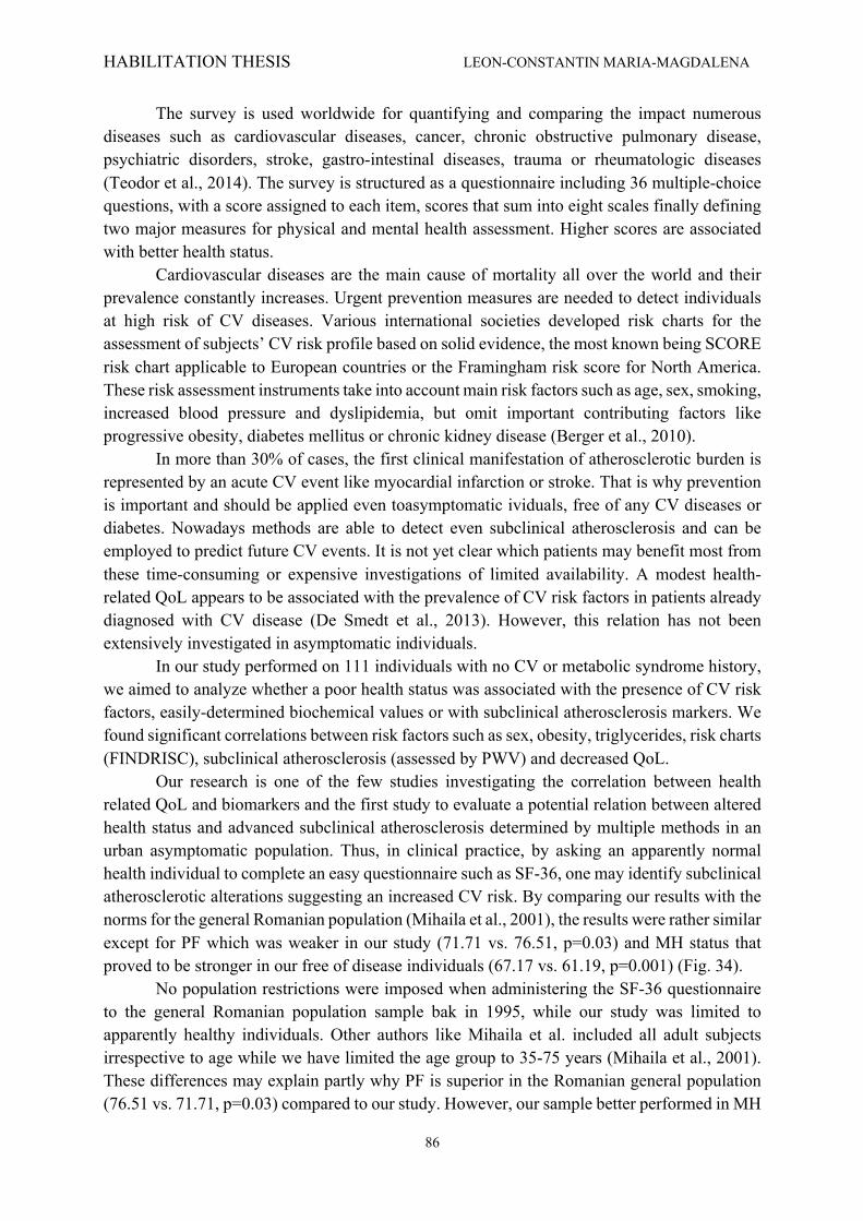
HABILITATION THESIS LEON-CONSTANTIN MARIA-MAGDALENA
86
The survey is used worldwide for quantifying and comparing the impact numerous diseases such as cardiovascular diseases, cancer, chronic obstructive pulmonary disease, psychiatric disorders, stroke, gastro-intestinal diseases, trauma or rheumatologic diseases (Teodor et al., 2014). The survey is structured as a questionnaire including 36 multiple-choice questions, with a score assigned to each item, scores that sum into eight scales finally defining two major measures for physical and mental health assessment. Higher scores are associated with better health status.
Cardiovascular diseases are the main cause of mortality all over the world and their prevalence constantly increases. Urgent prevention measures are needed to detect individuals at high risk of CV diseases. Various international societies developed risk charts for the assessment of subjects’ CV risk profile based on solid evidence, the most known being SCORE risk chart applicable to European countries or the Framingham risk score for North America. These risk assessment instruments take into account main risk factors such as age, sex, smoking, increased blood pressure and dyslipidemia, but omit important contributing factors like progressive obesity, diabetes mellitus or chronic kidney disease (Berger et al., 2010).
In more than 30% of cases, the first clinical manifestation of atherosclerotic burden is represented by an acute CV event like myocardial infarction or stroke. That is why prevention is important and should be applied even toasymptomatic ividuals, free of any CV diseases or diabetes. Nowadays methods are able to detect even subclinical atherosclerosis and can be employed to predict future CV events. It is not yet clear which patients may benefit most from these time-consuming or expensive investigations of limited availability. A modest health-related QoL appears to be associated with the prevalence of CV risk factors in patients already diagnosed with CV disease (De Smedt et al., 2013). However, this relation has not been extensively investigated in asymptomatic individuals.
In our study performed on 111 individuals with no CV or metabolic syndrome history, we aimed to analyze whether a poor health status was associated with the presence of CV risk factors, easily-determined biochemical values or with subclinical atherosclerosis markers. We found significant correlations between risk factors such as sex, obesity, triglycerides, risk charts (FINDRISC), subclinical atherosclerosis (assessed by PWV) and decreased QoL.
Our research is one of the few studies investigating the correlation between health related QoL and biomarkers and the first study to evaluate a potential relation between altered health status and advanced subclinical atherosclerosis determined by multiple methods in an urban asymptomatic population. Thus, in clinical practice, by asking an apparently normal health individual to complete an easy questionnaire such as SF-36, one may identify subclinical atherosclerotic alterations suggesting an increased CV risk. By comparing our results with the norms for the general Romanian population (Mihaila et al., 2001), the results were rather similar except for PF which was weaker in our study (71.71 vs. 76.51, p=0.03) and MH status that proved to be stronger in our free of disease individuals (67.17 vs. 61.19, p=0.001) (Fig. 34).
No population restrictions were imposed when administering the SF-36 questionnaire to the general Romanian population sample bak in 1995, while our study was limited to apparently healthy individuals. Other authors like Mihaila et al. included all adult subjects irrespective to age while we have limited the age group to 35-75 years (Mihaila et al., 2001). These differences may explain partly why PF is superior in the Romanian general population (76.51 vs. 71.71, p=0.03) compared to our study. However, our sample better performed in MH
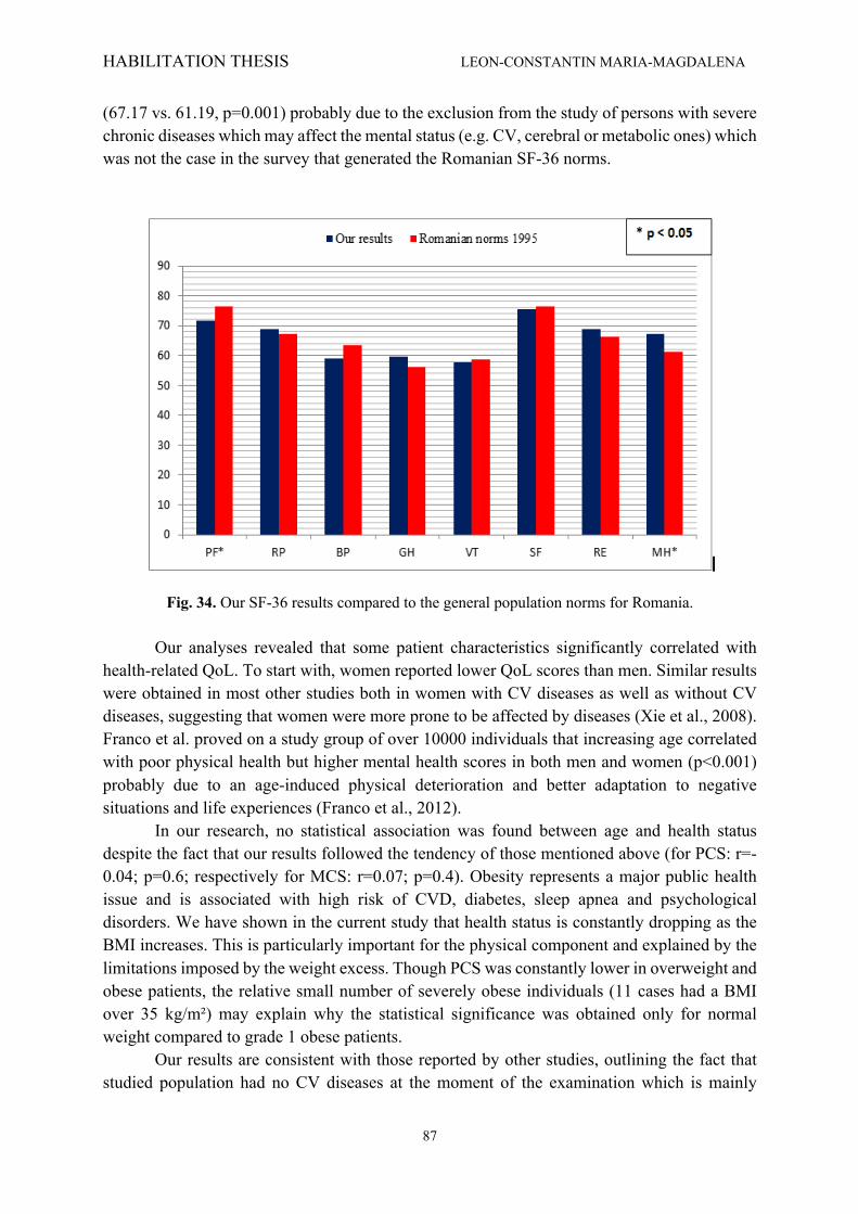
HABILITATION THESIS LEON-CONSTANTIN MARIA-MAGDALENA
87
(67.17 vs. 61.19, p=0.001) probably due to the exclusion from the study of persons with severe chronic diseases which may affect the mental status (e.g. CV, cerebral or metabolic ones) which was not the case in the survey that generated the Romanian SF-36 norms.
Fig. 34. Our SF-36 results compared to the general population norms for Romania. Our analyses revealed that some patient characteristics significantly correlated with
health-related QoL. To start with, women reported lower QoL scores than men. Similar results were obtained in most other studies both in women with CV diseases as well as without CV diseases, suggesting that women were more prone to be affected by diseases (Xie et al., 2008). Franco et al. proved on a study group of over 10000 individuals that increasing age correlated with poor physical health but higher mental health scores in both men and women (p<0.001) probably due to an age-induced physical deterioration and better adaptation to negative situations and life experiences (Franco et al., 2012).
In our research, no statistical association was found between age and health status despite the fact that our results followed the tendency of those mentioned above (for PCS: r=-0.04; p=0.6; respectively for MCS: r=0.07; p=0.4). Obesity represents a major public health issue and is associated with high risk of CVD, diabetes, sleep apnea and psychological disorders. We have shown in the current study that health status is constantly dropping as the BMI increases. This is particularly important for the physical component and explained by the limitations imposed by the weight excess. Though PCS was constantly lower in overweight and obese patients, the relative small number of severely obese individuals (11 cases had a BMI over 35 kg/m²) may explain why the statistical significance was obtained only for normal weight compared to grade 1 obese patients.
Our results are consistent with those reported by other studies, outlining the fact that studied population had no CV diseases at the moment of the examination which is mainly
HABILITATION THESIS LEON-CONSTANTIN MARIA-MAGDALENA
88
favorable for early initiation of preventive measures. Tan et al. performed a study on almost 5000 patients and proved that the decrease in PCS due to obesity was achieved in women, while only men presented an association between obesity and MCS (Tan et al., 2013).
Even though we did not assess it specifically, health related QoL proved to be influenced by other risk and lifestyle factors. Physical activity has a positive effect on both physical and mental component (McAuley et al., 2006). Smoking had no influence over mental health, but altered the physical status mainly in men (Franco et al., 2012). Abnormal sleep duration (less than 6 hours and more than 8 hours per day) is related to poorer self-perceived QoL, besides the general negative effects over health and mortality (Stranges et al., 2008).
Given that we did not include diabetic patients in our research, we found no correlation between SF-36 scores and plasma glucose. However, we revealed important associations between decreased mental and physical statuses and the risk for developing diabetes by using a standardized risk chart (FINDRISC). This result may be of important clinical use since an altered QoL may predict and favor the onset of diabetes in asymptomatic individuals. Previous studies confirmed that fasting glucose does not correlate with lower QoL values compared to glycosylated haemoglobin which was significantly associated with worse health outcomes (Lee et al., 2014). Though lipid values (total cholesterol, HDL, LDL or triglycerides) and inflammatory markers (e.g. fibrinogen) are reported to correlate positively with future CV events, we have only detected a modest association between high triglycerides levels and low values of emotional role (RE) scale. Other studies did not report significant correlations between lipid values and QoL scores (Sevinç et al., 2010).
Regarding subclinical atherosclerosis, we evaluated the association of health status with multiple measures of subclinical atherosclerosis: aortic atheromatosis, LVMI, ABI, carotid IMT and PWV for arterial stiffness. Among all these, only high values of PWV proved to correlate with decreased QoL, especially after dividing PWV into two groups according to its pathological thresold (<10, respectively ≥10 m/s). Literature data on this subject are rather scarce and inconsistent. Other researchers reported positive correlations between mental health status (depressive symptoms) and IMT, especially in older adults (Stewart et al., 2007). Ohira et al. showed that increased IMT positively correlated with anger score, especially in men. However, no correlations were obtained between depressive symptoms and IMT (Ohira et al., 2012). Another marker of subclinical atherosclerosis is coronary artery calcification (CAC) evaluated by Agatston score on multi-slice computer tomography. A recent published cross-sectional study showed that, out of several psychological factors, only anxiety was significantly correlated with CAC (Hernandez et al., 2014).
Roux et al. published in 2006 a study performed on more than 6500 adults with no history of CV diseases in which they evaluated subclinical atherosclerosis by using CAC and physiological factors by using multiple validated scales (Diez Roux et al., 2006). Their conclusion was that health status (chronic stress burden, anxiety, anger or depression) was not associated with coronary atherosclerosis in asymptomatic population (Diez Roux et al., 2006). Like in other studies, ABI showed no to be associated with psychological markers (Hernandez et al., 2014). Thus, by showing a significant correlation between decreased QoL and increased PWV as marker of subclinical atherosclerosis, our results bring new and valuable data regarding CV risk, especially in a sample of asymptomatic adults.
HABILITATION THESIS LEON-CONSTANTIN MARIA-MAGDALENA
89
• Statin therapy in atherosclerosis Stroke is the third cause of morbidity and mortality worldwide after coronary heart
disease and oncologic pathology (Lopez et al., 2006; Wafa et al., 2018). Similar to other diseases, stroke results from the interaction between genetic predisposition and environmental factors. If the genotype cannot be modified, the lifestyle can be improved. Unmodifiable risk factors for stroke are represented by age, family history, race, sex, previous cardiovascular events. Modifiable risk factors include arterial hypertension, diabetes mellitus, atrial fibrillation, asymptomatic cerebrovascular disease, coronary artery disease, hypercholesterolemia, obesity, smoking, sedentarism, poor socio-economic condition, alcohol and drug abuse (Sun et al., 2017).
Secondary prevention involves administration of preventive medication (antiaggregant, hypolipemiant, antihypertensive drugs), depending on etiology and associated diseases (ACC/AHA, 2017; Kernan et al., 2014). Atherosclerosis and dyslipidemia are the main risk factor in the etiology of stroke (both constituted and transient)(Osawa et al., 2018; Kim et al., 2017) and international guidelines recommend the use of statins in patients with a history of stroke as a preventive drug targeting hypercholesterolemia and hypertriglyceridemia in order to stabilize carotid or cerebral atheromatous plaques (Amarenco et al., 2009; Zhong et al., 2017).
According to the 2016 ACC/AHA blood cholesterol management guidelines, the usage of statins in secondary prevention is associated with a 16% reduction in stroke prevalence, 27% for nonfatal myocardial infarction and 20% for mortality from cardiac events (ESC, 2016; Miller et al., 2016). Statins long-term use or abuse induces cytotoxicity, hepatic injury or necrosis, kidney damage and myopathy (Mancini et al., 2016). Liver and kidney function should be monitored periodically in case of statin administration together with muscular enzymes in order to prevent any undesirable effects (Karahalil et al., 2017). Besides the above mentioned side effects, many studies incriminated statins of new-onset of diabetes mellitus, cognitive impairment and hemorrhagic stroke (Mach et al., 2018). However, current literature data and guidelines affirm that benefits of statin therapy outweigh any risks or adverse effects (Bellaosta et al., 2012).
Several published studies linked a high-risk for developing CVD, obesity and abdominal adiposity in patients with high TG, TC, and low HDL cholesterol (Lee et al., 2012), significant predictors factors in the evolution of obesity. WC, and in particular the visceral fat, is an independent risk factor for diabetogenic–atherogenic abnormalities in adolescents (Ramirez-Velez et al., 2018 ; Do et al., 2018).
In our study, we found a high percentage of dyslipidemia in obese patients. The composition of a weight loss diet has a small but significant impact on the lipid level changes. Even in the absence of significant weight loss, dietary therapy can be beneficial and should be encouraged (Lakshman et al., 2012). Duration of exercise also has a significant impact on the total cholesterol levels and improves the cardiovascular risk factors (Mitu et al., 2016). Exercise alone is usually not enough to induce significant weight loss. It is recommended that patients exercise for 150 min or more per week (30 min 5 times per week). The more intensive exercise program is, the greater the effect on weight and lipid levels will be (Lean et al., 2018). The SF-36 questionnaire, a simple QoL health survey, could offer relevant clinical information beyond the evaluation of depressive traits or other physical and mental components.

HABILITATION THESIS LEON-CONSTANTIN MARIA-MAGDALENA
90
5.5. Conclusions
• The impact of cardiovascular risk factors and subclinical atherosclerosis on life quality In our prospective study conducted on individuals without CV and metabolic diseases,
we proved that female sex and obesity are associated with decreased QoL score and age, blood pressure and other biochemical markers are not associated. Decreased QoL increases the risk of developing type 2 diabetes mellitus as assessed by FINDRISC risk chart. Finally, out of multiple methods of determining subclinical atherosclerosis, only high values of PWV are associated with low values in the health status evaluation. To conclude, we recommend the use of easy administered health survey questionnaires, such as SF-36, in asymptomatic individuals in order to obtain additional information concerning CV and metabolic risk.
• Statin therapy in atherosclerosis
In our study, we found a significant link between atherosclerosis and statin treatment, more evident in patients taking a higher dose, and between the statin dose and the decrease of uric acid value, another cardiovascular risk factor. To conclude, the use statins is mandatory as part of cholesterol lowering therapy attempting to stabilize the atheroma plaque in patients with stroke.
HABILITATION THESIS LEON-CONSTANTIN MARIA-MAGDALENA
91
SECTION III. PAIN
Chapter 6. FROM BENCH TO BEDSIDE – FINDING NEW METHODS FOR IMPROVING PAIN MANAGEMENT
6.1. Introduction
Pain, especially chronic pain, affects more than 1.5 billion people worldwide and is recognized as a significant public health problem (Dueñas et al., 2016). Additionally, chronic pain affects not only the patient, but also his family, his social circle and the entire system, being associated with a significant economic and social burden (Breivik et al., 2006). The experience of pain interferes with different aspects of the patient’s life negatively affecting their daily activities, physical and mental health, family and social relationships, and their interactions in the workplace (Dueñas et al., 2016). As such, all physicians have to learn how to treat pain because they will inescapably be faced with patients that experience this unpleasant sensory perception. Currently, there are several types of analgesic drugs, ranging from mild to strong analgesics. Co-analgescics can also help treat complex pain or reduce opioid requirement in some cases. Additionally, different drugs that act on different pain-associated pathways can be combined for a synergic effect (Salvemini, 2010). Last, but not least, non-pharmacologic approaches to pain management have been more and more evaluated in recent years and some strategies have been shown to be effective in some circumstances. In this complex landscape of options, the physician is often faced with a difficult decision between different treatment options. Although there are many effective drugs, some types of pain are still quite difficult to manage. Additionally, new drugs and drug combinations can sometimes be expensive and choosing a long-term treatment should also take this into account in a country where drug reimbursement in not always possible. As such, simple strategies should come first and assessing innovative combinations (such as those between trace elements or heavy metals and analgesics) or using plant extracts (such as those from the Laminacee family) should be investigated in preclinical models with the final aim of translating the results to the bedside. By using these methods, the efficacy of current analgesics could be significantly increased, a result that decreases analgesics use and subsequently the undesirable side-effects associated with opioid treatment.
Another potential strategy for better treating pain is finding new methods for delivering analgesics, a field where nanotechnology and nanoparticles have been more and more researched in recent years. However, nanotechonology-based strategies should be used with caution, as shown by the pro-inflammatory effect that Silver nanoparticles can have on the normal and inflamed tissue.
Improving the efficacy of current analgesic drugs 40% of the US general population (Kawamoto et al., 2013) and more than 65% of
chronically ill patients take nutritional supplements (Eisenberg et al., 1998; Engel et al., 1956). They are immensely popular, often sold as over-the-counter medication and they are less controlled by regulatory agencies when compared with classical drugs. Most nutritional supplements contain different oligo-elements, either trace elements or heavy metals, or a

HABILITATION THESIS LEON-CONSTANTIN MARIA-MAGDALENA
92
combination of both. Trace elements represent a group of essential metals or metaloids necessary for life, present in minute amounts. Most of them (Fe, F, Mg, Si, Zn) are involved in various biochemical enzymatic and metabolic processes (Tamba et al., 2008). Among the trace elements that are found in nutritional supplements, Zinc (Zn), Magnesium (Mg) and Copper (Cu) are almost always present due to the deleterious effect that their deficiency could have on cells. Widespread and widely used in mammals, Zn is a virtually nontoxic trace element. Five to fifteen percent of the cerebral Zn is found in the presynaptic vesicles of glutaminergic nerve terminations, having a possible role in synaptic transmission (Kay et al., 2008; Ketterman et al., 2008; Pan et al., 2011) Mayer et al. (1989) showed that Zn is released simultaneouslywith glutamate, exerting the effect of a noncompetitive N-methyl-D-aspartate (NMDA) antagonist, and Mayer and Vyklicky (Mayer et al., 1989) showed that Zn does not prevent the NMDA binding site affinity or glycine binding on the NMDA receptor. Zn also inhibits AMPA and intrathecal Zn in mice and produces an antinociceptive effect but no change in the thermoalgezic tests (Bresink et al., 1996; Larson et al., 2000). Animal studies also showed that Zn chelators induce hyperalgesia, that Zn ions have antinociceptive roles in neuropathic pain, and that modulation of Zn biological fraction induces analgesia (Larson et al., 2000; Liu et al., 1999; Rodriguez-Munoz et al., 2011). Zn and Cu also reduce pain and inflammation in patients (Honkanen et al., 1991; Lansdown et al., 1996), and Kugelmas (Kugelmas et al., 2000) showed the utility of Zn in muscle cramp pain. Magnesium (Mg), the fourth most abundant cation in the human body, is required for presynaptic delivery of acetylcholine and may induce similar effects with Ca channel blockers. Mg is able to block mechanic hyperalgesia in rats (Lee et al., 2000) and has an antinociceptive effect in neuropathic pain rat models (Xiao et al., 1994). Begon et al. (Begon et al., 2002) proved that intraperitoneal Mg2+ has a partially or fully reversible effect on mechanical hyperalgesia in diabetic rats and increased levels of Mg2+ in the cerebrospinal fluid and other brain regions. Koning et al. (Konig et al., 1998) explain Mg2+ effect by interaction with the NMDA-linked Na/Ca channel, although other authors showed an NMDA-independent antinociceptive effect (Poleszak et al., 2008; Nikolaev et al., 2012). Magnesium’s effects in postoperative analgesia or habitual cephalalgia were investigated, with positive results (Mauskop et al., 1994; Lee et al., 2012). However, whereas Begon et al. (Begon et al., 2002) advocate the Mg2+ passage through the blood–brain barrier (BBB), Takano et al. (Takano et al., 2002) explain the antihyperalgesic effect of intrathecally administered Mg2+ by its not passing the BBB. Brill et al. (Brill et al., 2002) later confirmed, in another clinical study, the effectiveness of i.v. Mg2+ in neuropathic pain. Still the data are at best contradictory, insofar as a previous peripheral neuropathy clinical study by Felsby et al. (Felsby et al., 1995) yielded negative results. Copper (Cu) is an essential trace element that may participate too as a signaling molecule in the nervous system. Extracellular ionic Cu2+ is a powerful inhibitor for K channels (Ma et al., 2008), whereas Cu2+ deficit decreases dopamine levels in rat brain (Yu et al., 2008). Cu2+-based preparations alone proved to be as efficient as morphine or displayed an improved analgesic effect when combined with nonsteroidal anti-inflammatory drugs in rats, possibly through an activation of Cu2+-dependent opioid receptors (Okuyama et al., 1997). Popa and Lerche (Popa et al., 2006) showed that Cu2+ acts as an Na channel blocker but expressed doubts over a possible therapeutic use. Some attempts to make a Cu21-based acetylsalicylic acid
HABILITATION THESIS LEON-CONSTANTIN MARIA-MAGDALENA
93
yielded limited results (Li et al., 2000). Later, Guilarte and Chen (Guilarte et al., 2007) showed that the Cu2+ is an NMDA inhibitor, and Jones et al. (Jones et al., 2007) presented a possible Cu2+–serotonin toxic interaction in neurodegenerative diseases. In 2008, Ma et al (Ma et al., 2008) proved that Cu2+ is a powerful bradykinin and K channel inhibitor, with both playing an important role in pain transmission, and F. Yu et al. (Yu et al., 2008) and Tamba et al. (Tamba et al., 2008) argued for a Cu2+/Zn2+ superoxide dismutase role, linked to neuropathic and inflammatory pain mechanisms. Heavy metal trace elements (HMTE) represent a group of metals or metalloids present in minute amounts in humans and include Cobalt (Co), Nickel (Ni) and Molybdenum (Mo), which are considered essential for human health (Gromiec et al., 2013). Over 50.000 dietary or food supplements are on the market today with many containing Co, Ni and Mo. For years, Co, Ni or Mo were mostly associated with toxic effects and scarcely investigated for other physiological roles, especially in pain generation, transmission or modulation (Gromiec et al., 2013). Co is a constituent of cobalamin, and this was considered its only role. Dietary intake varies between 5 - 50μg/day. Co is a potent inducer of oxidative stress leading to toxicity, carcinogenicity, DNA damage and sister-chromatid exchange (Jomova et al., 2011). Co shows cytotoxic effects on various cell types including neurons (Wang et al., 2006), can induce apoptosis and necrosis (Huk et al., 2004), but also damage mitochondrias (Battaglia et al., 2009). Co cations can also block Ca-mediated neural transmission, following central administration. No direct investigation had been performed at the time of our study regarding its role in pain following peripheral administration (Jomova et al., 2011). Ni plays an important role in animals (Schaumlöffel, 2012) while low levels are reducing growth and severely interfere lipid metabolic pathways in rats (Stangl et al., 1996). Dietary intake can reach 1 mg/day (Wong et al., 2012). Ni role in nociception has been little investigated. Ni is a non-selective Ca channels blocker, inhibiting the delivery of pain mediators (Barritt, 1999; Prado, 2001). Ni may induce direct contact dermatitis. Pain and itching sensory neurons are part of the DRG and trigeminal system and Ni effect in itching induction can be amplified by tissue acidosis (Luebbert et al., 2010). Chronic exposure to Ni can lead to impaired olfaction and anosmia (Jia et al., 2010), neurotoxicity, headaches, lethargy and ataxia. Ni exposure damages the mitochondrial function and impairs cell viability, inhibits the cell proliferation, blocks neuronal Ca channels, including voltage-dependent T-type and R-type (Kang et al., 2007; Kang et al., 2006), ASIC’s (Staruschenko et al., 2007) as well as GABA-activated channels (Fisher et al., 1998). Mo, part of several redox enzymes, is involved in the control mechanisms of purines and fats metabolism. Dietary intake ranges from 0.16–0.2 mg/day. Data on Mo and pain is also very limited. A single pilot clinical study showed a significant pain relieve in the Mo treated group (Holzinger et al., 1998). Last, but not least, an increasingly large body of evidence points to plants and plant extracts as very powerful analgesics and co-analgesics. The medicinal use of plants as analgesic drugs in folk medicine is an ancient tradition, far older than the current sciences of medicine in developing countries (Gul et al., 2019). According to estimations, up to 70,000 plant species are used ethnomedicinally worldwide. Effects of herbal extracts have been studied by different pain tests including writhing test, light tail flick test, tail immersion test, hot-plate test, and
HABILITATION THESIS LEON-CONSTANTIN MARIA-MAGDALENA
94
formalin test (Haq et al., 2012). Plants from the Laminacee family, one of the most important herbal families, incorporates a wide variety of plants with biological and medical applications. The most known members of this family are a variety of aromatic spices like thyme, mint, oregano, basil, sage, savory, rosemary, self-heal, hyssop, lemon balm, and some others with more limited use (Bekut et al., 2018.). A thorough search of the literature revealed that several members of this plant family, such as Betonica officinalis, Glechoma hederacea, Hyptis pectinata, Lavandula genus, Leonurus cardiaca, Lamium genus, Melissa officinalis, Mentha genus, Marrubium vulgare, Origanum genus, Ocimum genus, Rosmarinus officinalis, Salvia genus, Satureja hortensis, Stachys lavandulifolia, Scutellaria lateriflora, Sideritis genus, Teucrium genus, Thymus genus, and Ziziphora tenuior, have potent analgesic and antinociceptive activity. Most of the extracts identified did not present any toxic capabilities or known side effects and were at least as efficient as currently used synthetic drugs. Overall, although promising information evidence the efficacy of Laminaceae genus in the treatment of pain associated disorders, the data are too preliminary and mostly fail to explain the exact cellular and molecular mechanisms of action and the respective active compounds. Therefore, future studies should be focused on investigating mechanisms of actions, realistic dosages, clinical efficacy, and safety of the extracts and active compounds in pain treatment. These data intrigued and challenged our team to try to investigate if any of these supplements/plant extracts can be effective as analgesics or co-analgesics. As such, we assessed the effect of heavy metal trace elements on pain (analgesics) and the effect of trace elements in combination with mild opioids on pain (co-analgesics) in animal models and we performed a systematic review of the literature to decide if plant extracts from the Laminacee family should be investigated in animal models.
The main publications that have derived from our research are listed below.
1. Tamba B, Leon M, Petreus T, Common trace elements alleviate pain in an experimental mouse model, J. Neurosci. Res. 2013;91(4):554–561.
2. Tamba B, Petreus T, Leon MM, Rezus C, Floria M, Rezus E, Heavy metal trace elements induce antinociception in an experimental mouse model. Rev. Chim (Bucharest), 2015; 66 (6): 976-982.
3. Uritu CM, Mihai CT, Stanciu G, Dodi G, Alexa-Stratulat T, Luca A, Leon M, Stefanescu R, Bild V, Melnic S, Tamba B. Medicinal Plants of the Family Lamiaceae in Pain Therapy: A Review. Pain Res. Manag. 2018:1–44.
The funding for this research has been partly supported by two national grants in which I was a member that are listed below:
1. “Experimental studies regarding the relation between pain and mitochondrial
dysfunction; new perspectives in pain therapy”. Grant IDEI, PN-II-ID-PCE-2011-3, Grant Director: Dr. Catalina Roxana Bohotin, 1.500.000 RON (2011-2014).
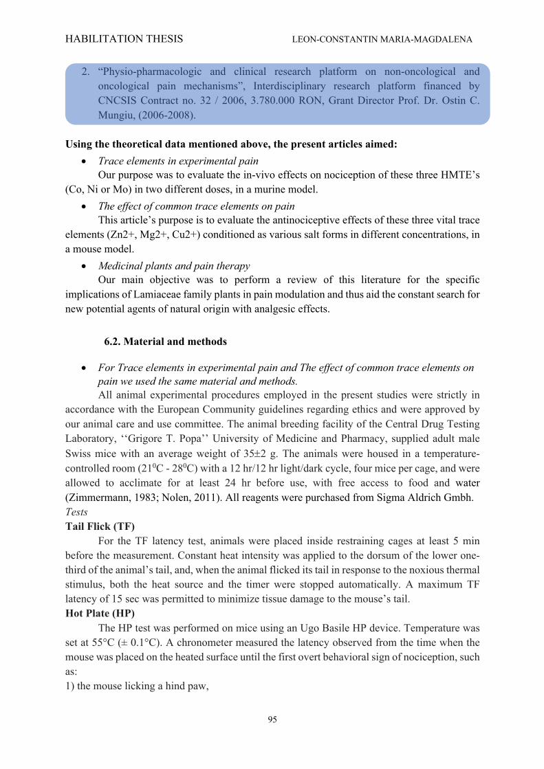
HABILITATION THESIS LEON-CONSTANTIN MARIA-MAGDALENA
95
2. “Physio-pharmacologic and clinical research platform on non-oncological and oncological pain mechanisms”, Interdisciplinary research platform financed by CNCSIS Contract no. 32 / 2006, 3.780.000 RON, Grant Director Prof. Dr. Ostin C. Mungiu, (2006-2008).
Using the theoretical data mentioned above, the present articles aimed:
• Trace elements in experimental pain Our purpose was to evaluate the in-vivo effects on nociception of these three HMTE’s
(Co, Ni or Mo) in two different doses, in a murine model. • The effect of common trace elements on pain
This article’s purpose is to evaluate the antinociceptive effects of these three vital trace elements (Zn2+, Mg2+, Cu2+) conditioned as various salt forms in different concentrations, in a mouse model.
• Medicinal plants and pain therapy Our main objective was to perform a review of this literature for the specific
implications of Lamiaceae family plants in pain modulation and thus aid the constant search for new potential agents of natural origin with analgesic effects.
6.2. Material and methods
• For Trace elements in experimental pain and The effect of common trace elements on pain we used the same material and methods. All animal experimental procedures employed in the present studies were strictly in
accordance with the European Community guidelines regarding ethics and were approved by our animal care and use committee. The animal breeding facility of the Central Drug Testing Laboratory, ‘‘Grigore T. Popa’’ University of Medicine and Pharmacy, supplied adult male Swiss mice with an average weight of 35±2 g. The animals were housed in a temperature-controlled room (210C - 280C) with a 12 hr/12 hr light/dark cycle, four mice per cage, and were allowed to acclimate for at least 24 hr before use, with free access to food and water (Zimmermann, 1983; Nolen, 2011). All reagents were purchased from Sigma Aldrich Gmbh. Tests Tail Flick (TF) For the TF latency test, animals were placed inside restraining cages at least 5 min before the measurement. Constant heat intensity was applied to the dorsum of the lower one-third of the animal’s tail, and, when the animal flicked its tail in response to the noxious thermal stimulus, both the heat source and the timer were stopped automatically. A maximum TF latency of 15 sec was permitted to minimize tissue damage to the mouse’s tail. Hot Plate (HP) The HP test was performed on mice using an Ugo Basile HP device. Temperature was set at 55°C (± 0.1°C). A chronometer measured the latency observed from the time when the mouse was placed on the heated surface until the first overt behavioral sign of nociception, such as: 1) the mouse licking a hind paw,
HABILITATION THESIS LEON-CONSTANTIN MARIA-MAGDALENA
96
2) vocalization, or 3) an escape response. A cutoff time of 30 sec was used for the HP test. Writhing test The abdominal stretch, or writhing assay, was performed by injecting 0.1 ml of 1.0% acetic acid intraperitoneally in manually restrained mice. Immediately after injection, animals were placed in a large glass cylinder. The number of abdominal stretches occurring in successive 5-min intervals was counted beginning 5 min after acetic acid, for a 30-min period after intraperitoneal injection of diluted acetic acid. The tested substances were administered 5 min prior to acetic acid intraperitoneal injection. Treatments that produced a significant decrease in the number of abdominal stretches were considered to be antinociceptive. The mice were kept under observation for 72 hours and then sacrificed. Activity cage (AC) This test is performed to record spontaneous coordinated activity in individual mice and variation of this activity over time. Mice were allowed to acclimate to the testing room the night before testing. Animals were weighed and tested between 9:00 and 11:00 AM. Horizontal and vertical locomotor activity was monitored for 2 min with the Ugo Basile Activity Cage System. Horizontal and vertical activity was defined as the total number of beam interruptions throughout a 2-min observation period. The test was performed 15 min following the administration of the tested substances.
• Trace elements in experimental pain Various groups of 8 mice each were selected and specific formulations were injected intraperitoneally. Control groups received 0.1mL of 0.9% saline while test groups received Co chloride (doses of 3.75 and 7.5 mg/kg b.w respectively), Ni chloride (doses of 0.5 and 2 mg/kg b.w. respectively) and Na-molybdate (doses of 25 and 50 mg/kg b.w respectively). All calculations were made on the chemicals used (not metal component alone). The doses administered were calculated as fractions of known, published intraperitoneal IP LD50 in mice. We did not use ppm data, as standard toxicity tests and data use LD50 and not ppm. Our experimental models used doses 5 to 96 times less that known LD50’s and did not induce any significant toxic effect. The used doses were: - 1/24 and 1/12 for Cobalt chloride of LD50. (LD50 90mg/kg b.w.) (Cromres Safety Data. Available from (www.chromres.com/ AvailableDocuments/ 991042%20%20CRS%20Model%20100,%2 0Drierite.pdf)). - 1/96 and 1/24 for Nickel Chloride (LD50 48 mg/kg b.w.) (Phytotech Safety Data. Available from (www.phytotechlab. com/MSDS/ N478msds.pdf). - And 1/10 and 1/5 for Sodium Molybdate (LD50 257 mg/kg b.w.) (Pestell Safey Data. Available from (www.pestell.com/msds/ Sodium%20Molybdate%20Dihydrate.pdf). The pH of the solutions was slightly acid but still at physiological levels. The solvent of the chemical compound was saline (pH 5.5) (Reddi, 2013), which is also physiological for IP administration and had no effects in control animals. The trace elements however, did not influence significantly the pH of the injected solutions. Thus, there was no need to use a buffer to attenuate for possible acidity. Pain was assessed by means of TF, HP and writhing test. TF assessment was performed at 15’, 30’, 45’ and 1h respectively following exposure to the assay salts or controls. HP was
HABILITATION THESIS LEON-CONSTANTIN MARIA-MAGDALENA
97
performed at onset and at 15’, 30’, 45’ and 1h following salts or saline control intraperitoneal administration. Animals were also assessed by means of the writhing test - evaluated salts were injected at 5 minutes before acetic acid administration. For each group of 8 mice, mean values were reported (±S.E.M.). Antinociceptive response was considered for the treatments that induced a significant decrease in the number of abdominal stretches. Prior to mice sacrifice, their biological evolution was followed for another 72 h. Last, but not least, animals were also evaluated by means of the Activity Cage. Spontaneous behaviour test was performed in blind (the experimenter was not aware of the treatment received by the animals). The activity cage test was a complementary evaluation, which sought to investigate if there are any spontaneous behavioral changes in the test animals for a better interpretation of the pain evaluation tests. The conclusion on the sedative or anxiolytic effects was based on well-established literature data using the activity cage and spontaneous behavior testing.
• The effect of common trace elements on pain Different groups of eight mice were intraperitoneally injected with one of the following formulations: 0.9% saline for the control group (0.3 ml), Zn2+ sulfate (0.5 and 2.0 mg/kg), Zn2+ citrate (0.125 and 0.5 mg/kg), Mg2+ chloride (37.5, 75, and 150 mg/kg), Cu2+ chloride (0.5, 1.0, and 2.0 mg/kg), and Cu2+ sulfate (0.5 and 1.0 mg/kg), all salts solved in 0.3 ml saline/mouse. The antinociceptive effect of the tested substances was evaluated by means of TF and HP tests, behavioral tests that quantify the thermal nociception. The TF test was performed at 15, 30, and 45 min and 1 hour after the administration of substances or saline (control). The HP test was performed at baseline and 15, 30, and 45 min and 1 hour after administration of the drug or 0.9% saline (control group). The antinociceptive effect on visceral pain of the tested substances was also evaluated by a peripheral mechanism (writhing test). Values are reported as the mean (6SEM) for each treatment, with groups composed of eight mice. Animals also underwent locomotion assessment by means of the Activity Cage. Statistical Analysis SPSS 16.0 for Windows from the IBM SPSS Data Collection was used. Data are expressed as the mean 6 SD for each measurement time. Differences between treatment groups were evaluated by one-way ANOVA for comparison at each time point, followed by Bonferroni post hoc tests. P < 0.05 was considered a significant difference for all tests. Pain inhibition with various stimuli (Wu et al., 2003) was also calculated according to the formula:
% Inhibition = [(Tx –T0)/(Tm –T0)] x 100 where T0 is the latency before the drug administration (baseline), Tx is the latency for various consecutive time intervals, and Tm is the maximum time allowed (cutoff time) to avoid any possible lesions to the test animal.
• Medicinal plants and pain therapy The search strategy employed in this review includes internationally accepted databases,
namely, ScienceDirect, Scopus, Web of Science, and PubMed, using specific keywords of both whole plant products and plant extracts, pain, and analgesic and antinociceptive effects. For investigation, a combination of keywords was used [pain; analgesic; antinociceptive; plant extract] + [Betonica officinalis; Glechoma hederacea; Hyptis pectinata; Lavandula; Leonurus cardiaca; Lamium; Melissa officinalis; Mentha; Marrubium vulgare; Origanum; Ocimum; Rosmarinus officinalis; Salvia; Satureja hortensis; Stachys lavandulifolia; Scutellaria lateriflora; Sideritis; Teucrium; Thymus; Ziziphora tenuior] + [Lamiaceae; botanical genus].

HABILITATION THESIS LEON-CONSTANTIN MARIA-MAGDALENA
98
Case reports, case studies, in vivo and in vitrorelevant studies, and comparative studies were included in this search strategy. Additionally, text books and potentially relevant reviews were explored and included in the reference list. The literature search was confined to the period between 2003 and December 2017. Several articles before 2000 were also included in order to point out the universal interest in natural products with potential applicability in therapy. The dynamic character of the field is reflected in the number of recent publications.
6.3. Results
• Trace elements in experimental pain Results obtained following TF test showed that Co chloride is able to extend TF latency at 15, 30, 45 and 60 min from intraperitoneal administration, thus indicating a significant antinociceptive effect. This effect seems to be dose-dependent as TF latency increases. At a dose of 3.75 mg/kg Co chloride, the TF latency is slightly increased at 15 and 30 min, with no statistical significance; later on, at 45 and 60 min TF lowered significantly beneath control values. At a dose of 7.5 mg/kg Co chloride, the TF latency recorded a similar profile, while reaching statistical difference at 15, 30 and 45 min (Fig. 35).
Fig. 35. Latencies for TF test, after intraperitoneal administration of Co chloride in different doses
(3.75 and 7.5 mg/kg respectively)
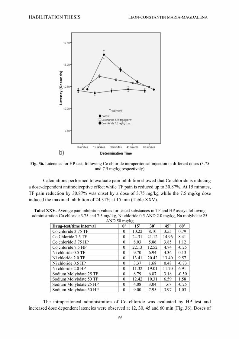
HABILITATION THESIS LEON-CONSTANTIN MARIA-MAGDALENA
99
Fig. 36. Latencies for HP test, following Co chloride intraperitoneal injection in different doses (3.75
and 7.5 mg/kg respectively)
Calculations performed to evaluate pain inhibition showed that Co chloride is inducing a dose-dependent antinociceptive effect while TF pain is reduced up to 30.87%. At 15 minutes, TF pain reduction by 30.87% was onset by a dose of 3.75 mg/kg while the 7.5 mg/kg dose induced the maximal inhibition of 24.31% at 15 min (Table XXV).
Tabel XXV. Average pain inhibition values for tested substances in TF and HP assays following administration Co chloride 3.75 and 7.5 mg/ kg, Ni chloride 0.5 AND 2.0 mg/kg, Na molybdate 25
AND 50 mg/kg Drug-test/time interval 0’ 15’ 30’ 45’ 60’ Co chloride 3.75 TF 0 10.22 8.10 3.55 0.79 Co Chloride 7.5 TF 0 24.31 21.12 14.96 8.41 Co chloride 3.75 HP 0 8.03 5.86 3.85 1.12 Co chloride 7.5 HP 0 22.13 12.52 4.74 -0.25 Ni chloride 0.5 TF 0 9.70 6.94 4.36 0.13 Ni chloride 2.0 TF 0 13.41 20.42 13.40 9.57 Ni chloride 0.5 HP 0 3.37 1.68 0.48 -0.73 Ni chloride 2.0 HP 0 11.32 19.01 11.70 6.91 Sodium Molybdate 25 TF 0 8.79 6.87 3.18 -0.50 Sodium Molybdate 50 TF 0 12.42 10.31 6.59 1.58 Sodium Molybdate 25 HP 0 4.08 3.04 1.68 -0.25 Sodium Molybdate 50 HP 0 9.00 7.95 3.97 1.03
The intraperitoneal administration of Co chloride was evaluated by HP test and increased dose dependent latencies were observed at 12, 30, 45 and 60 min (Fig. 36). Doses of
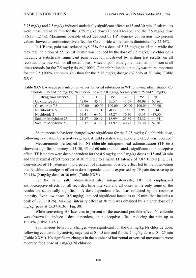
HABILITATION THESIS LEON-CONSTANTIN MARIA-MAGDALENA
100
3.75 mg/kg and 7.5 mg/kg induced statistically significant effects at 15 and 30 min . Peak values were measured at 15 min for the 3.75 mg/kg dose (13.66±0.46 sec) and the 7.5 mg/kg dose (16.13±1.27 s). Maximum possible effect deduced by HP latencies conversion into percent values showed an antinociceptive effect for Co chloride while pain is diminished by 22.46%.
In HP test, pain was reduced by8.03% for a dose of 3.75 mg/kg at 15 min while the maximal inhibition of 22.13% at 15 min was induced by the dose of 7.5 mg/kg. Co chloride is inducing a statistically significant pain reduction illustrated by writing test results, on all recorded time intervals for all tested doses. Visceral pain undergone maximal inhibition at all times records for the 7.5 mg/kg doses (100%). Pain inhibition in our study was more important for the 7.5 (100% continuously) than for the 3.75 mg/kg dosage (67.86% at 30 min) (Table XXV).
Table XXVI. Average pain inhibition values for tested substances in WT following administration Co chloride 3.75 and 7.5 mg/ kg, Ni chloride 0.5 and 2.0 mg/kg, Na molybdate 25 and 50 mg/kg
Drug/time interval 5’ 10’ 15’ 20’ 25’ 30’ Co chloride 3.75 42.06 45.45 36.57 47.69 40.09 67.86 Co chloride 7.5 100.00 100.00 100.00 100.00 100.00 100.00 Ni chloride 0.5 20.33 17.01 -0.12 7.58 2.95 7.14 Ni chloride 2 67.41 64.60 34.27 16.30 -3.04 -37.50 Sodium Molybdate 25 21.37 20.49 18.99 25.89 23.32 44.64 Sodium Molybdate 50 36.37 41.96 41.92 40.71 35.30 62.50
Spontaneous behaviour changes were significant for the 3.75 mg/kg Co chloride dose, following evaluation by activity cage test. A mild sedative and anxiolytic effect was recorded. Measurements performed for Ni chloride intraperitoneal administration (TF test) showed a significant latency at 15, 30, 45 and 60 min and indicated a significant antinociceptive effect. TF latencies were dose-dependent for the 0.5 mg/kg and 2 mg/kg doses at 15 and 30 min and the maximal effect recorded at 30 min led to a mean TF latency of 7.87±0.15 s (Fig. 37). Conversion of TF latencies into a percent of maximum possible effect led to the observation that Ni chloride analgesic effect is dose-dependent and is expressed by TF pain decrease up to 20.41% (2 mg/kg dose, at 30 min) (Table XXV).
For the same salt, administered also intraperitoneally, HP test emphasized antinociceptive effects for all recorded time intervals and all doses while only some of the results are statistically significant. A dose-dependent effect was reflected by the response intensity. Even low doses (0.5 mg/kg) induced significant latencies at 15 min (that includes a peak of 12.77±0.26). Maximal intensity effect at 30 min was obtained by a higher dose of 2 mg/kg (peak at 15.37±0.36) (Fig. 38).
While converting HP latencies in percent of the maximal possible effect, Ni chloride was observed to induce a dose-dependent, antinociceptive effect, reducing the pain up to 19.01% (Table XXV).
Spontaneous behaviour changes were significant for the 0.5 mg/kg Ni chloride dose, following evaluation by activity cage test at 0 - 15 min and for the 2 mg/kg dose at 0 – 25 min (Table XXVI). No significant changes in the number of horizontal or vertical movements were recorded for a dose of 1 mg/kg Ni chloride.

HABILITATION THESIS LEON-CONSTANTIN MARIA-MAGDALENA
101
Fig. 37. Latencies for TF test, following Ni chloride intraperitoneal injection at different doses (0.5 and 2 mg/kg respectively).
Fig. 38. Latencies for HP test, following Ni chloride intraperitoneal injection at different doses (0.5
and 2 mg/kg respectively)
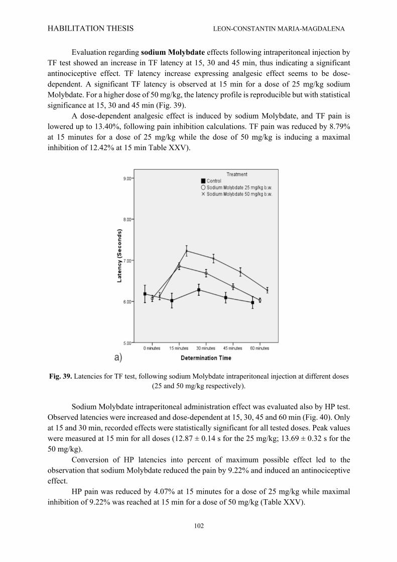
HABILITATION THESIS LEON-CONSTANTIN MARIA-MAGDALENA
102
Evaluation regarding sodium Molybdate effects following intraperitoneal injection by TF test showed an increase in TF latency at 15, 30 and 45 min, thus indicating a significant antinociceptive effect. TF latency increase expressing analgesic effect seems to be dose-dependent. A significant TF latency is observed at 15 min for a dose of 25 mg/kg sodium Molybdate. For a higher dose of 50 mg/kg, the latency profile is reproducible but with statistical significance at 15, 30 and 45 min (Fig. 39).
A dose-dependent analgesic effect is induced by sodium Molybdate, and TF pain is lowered up to 13.40%, following pain inhibition calculations. TF pain was reduced by 8.79% at 15 minutes for a dose of 25 mg/kg while the dose of 50 mg/kg is inducing a maximal inhibition of 12.42% at 15 min Table XXV).
Fig. 39. Latencies for TF test, following sodium Molybdate intraperitoneal injection at different doses
(25 and 50 mg/kg respectively). Sodium Molybdate intraperitoneal administration effect was evaluated also by HP test.
Observed latencies were increased and dose-dependent at 15, 30, 45 and 60 min (Fig. 40). Only at 15 and 30 min, recorded effects were statistically significant for all tested doses. Peak values were measured at 15 min for all doses (12.87 ± 0.14 s for the 25 mg/kg; 13.69 ± 0.32 s for the 50 mg/kg).
Conversion of HP latencies into percent of maximum possible effect led to the observation that sodium Molybdate reduced the pain by 9.22% and induced an antinociceptive effect.
HP pain was reduced by 4.07% at 15 minutes for a dose of 25 mg/kg while maximal inhibition of 9.22% was reached at 15 min for a dose of 50 mg/kg (Table XXV).
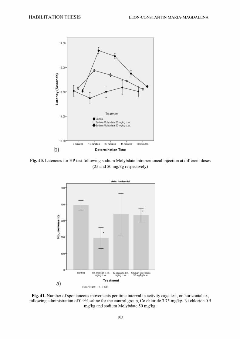
HABILITATION THESIS LEON-CONSTANTIN MARIA-MAGDALENA
103
Fig. 40. Latencies for HP test following sodium Molybdate intraperitoneal injection at different doses
(25 and 50 mg/kg respectively)
Fig. 41. Number of spontaneous movements per time interval in activity cage test, on horizontal ax,
following administration of 0.9% saline for the control group, Co chloride 3.75 mg/kg, Ni chloride 0.5 mg/kg and sodium Molybdate 50 mg/kg.
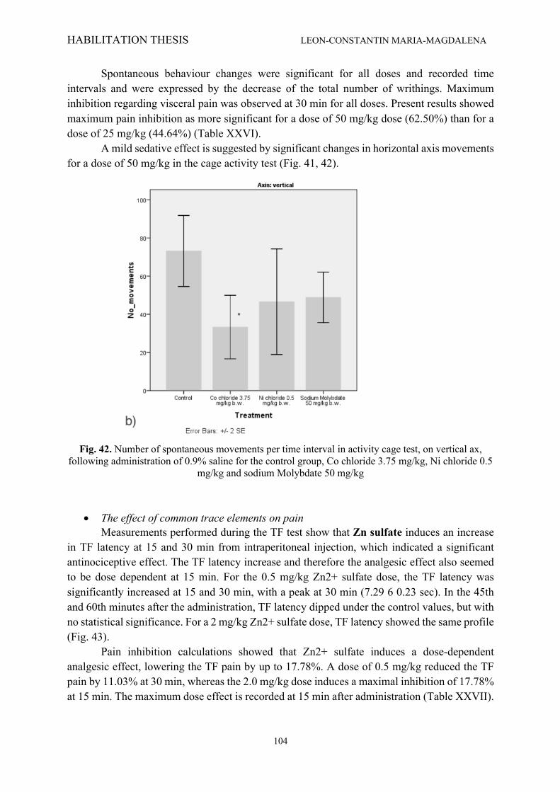
HABILITATION THESIS LEON-CONSTANTIN MARIA-MAGDALENA
104
Spontaneous behaviour changes were significant for all doses and recorded time intervals and were expressed by the decrease of the total number of writhings. Maximum inhibition regarding visceral pain was observed at 30 min for all doses. Present results showed maximum pain inhibition as more significant for a dose of 50 mg/kg dose (62.50%) than for a dose of 25 mg/kg (44.64%) (Table XXVI).
A mild sedative effect is suggested by significant changes in horizontal axis movements for a dose of 50 mg/kg in the cage activity test (Fig. 41, 42).
Fig. 42. Number of spontaneous movements per time interval in activity cage test, on vertical ax,
following administration of 0.9% saline for the control group, Co chloride 3.75 mg/kg, Ni chloride 0.5 mg/kg and sodium Molybdate 50 mg/kg
• The effect of common trace elements on pain Measurements performed during the TF test show that Zn sulfate induces an increase in TF latency at 15 and 30 min from intraperitoneal injection, which indicated a significant antinociceptive effect. The TF latency increase and therefore the analgesic effect also seemed to be dose dependent at 15 min. For the 0.5 mg/kg Zn2+ sulfate dose, the TF latency was significantly increased at 15 and 30 min, with a peak at 30 min (7.29 6 0.23 sec). In the 45th and 60th minutes after the administration, TF latency dipped under the control values, but with no statistical significance. For a 2 mg/kg Zn2+ sulfate dose, TF latency showed the same profile (Fig. 43).
Pain inhibition calculations showed that Zn2+ sulfate induces a dose-dependent analgesic effect, lowering the TF pain by up to 17.78%. A dose of 0.5 mg/kg reduced the TF pain by 11.03% at 30 min, whereas the 2.0 mg/kg dose induces a maximal inhibition of 17.78% at 15 min. The maximum dose effect is recorded at 15 min after administration (Table XXVII).
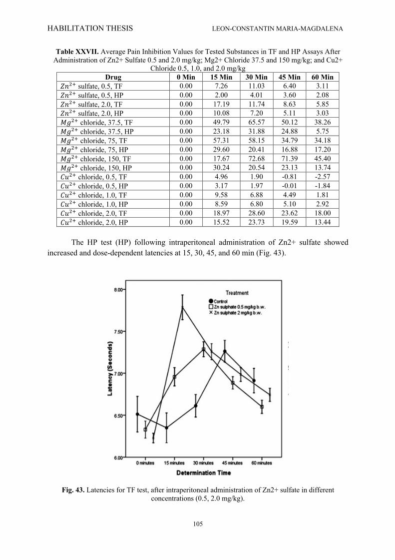
HABILITATION THESIS LEON-CONSTANTIN MARIA-MAGDALENA
105
Table XXVII. Average Pain Inhibition Values for Tested Substances in TF and HP Assays After Administration of Zn2+ Sulfate 0.5 and 2.0 mg/kg; Mg2+ Chloride 37.5 and 150 mg/kg; and Cu2+
Chloride 0.5, 1.0, and 2.0 mg/kg Drug 0 Min 15 Min 30 Min 45 Min 60 Min
𝑍𝑛$' sulfate, 0.5, TF 0.00 7.26 11.03 6.40 3.11 𝑍𝑛$' sulfate, 0.5, HP 0.00 2.00 4.01 3.60 2.08 𝑍𝑛$' sulfate, 2.0, TF 0.00 17.19 11.74 8.63 5.85 𝑍𝑛$' sulfate, 2.0, HP 0.00 10.08 7.20 5.11 3.03 𝑀𝑔$' chloride, 37.5, TF 0.00 49.79 65.57 50.12 38.26 𝑀𝑔$' chloride, 37.5, HP 0.00 23.18 31.88 24.88 5.75 𝑀𝑔$' chloride, 75, TF 0.00 57.31 58.15 34.79 34.18 𝑀𝑔$' chloride, 75, HP 0.00 29.60 20.41 16.88 17.20 𝑀𝑔$' chloride, 150, TF 0.00 17.67 72.68 71.39 45.40 𝑀𝑔$' chloride, 150, HP 0.00 30.24 20.54 23.13 13.74 𝐶𝑢$' chloride, 0.5, TF 0.00 4.96 1.90 -0.81 -2.57 𝐶𝑢$' chloride, 0.5, HP 0.00 3.17 1.97 -0.01 -1.84 𝐶𝑢$' chloride, 1.0, TF 0.00 9.58 6.88 4.49 1.81 𝐶𝑢$' chloride, 1.0, HP 0.00 8.59 6.80 5.10 2.92 𝐶𝑢$' chloride, 2.0, TF 0.00 18.97 28.60 23.62 18.00 𝐶𝑢$' chloride, 2.0, HP 0.00 15.52 23.73 19.59 13.44
The HP test (HP) following intraperitoneal administration of Zn2+ sulfate showed
increased and dose-dependent latencies at 15, 30, 45, and 60 min (Fig. 43).
Fig. 43. Latencies for TF test, after intraperitoneal administration of Zn2+ sulfate in different concentrations (0.5, 2.0 mg/kg).
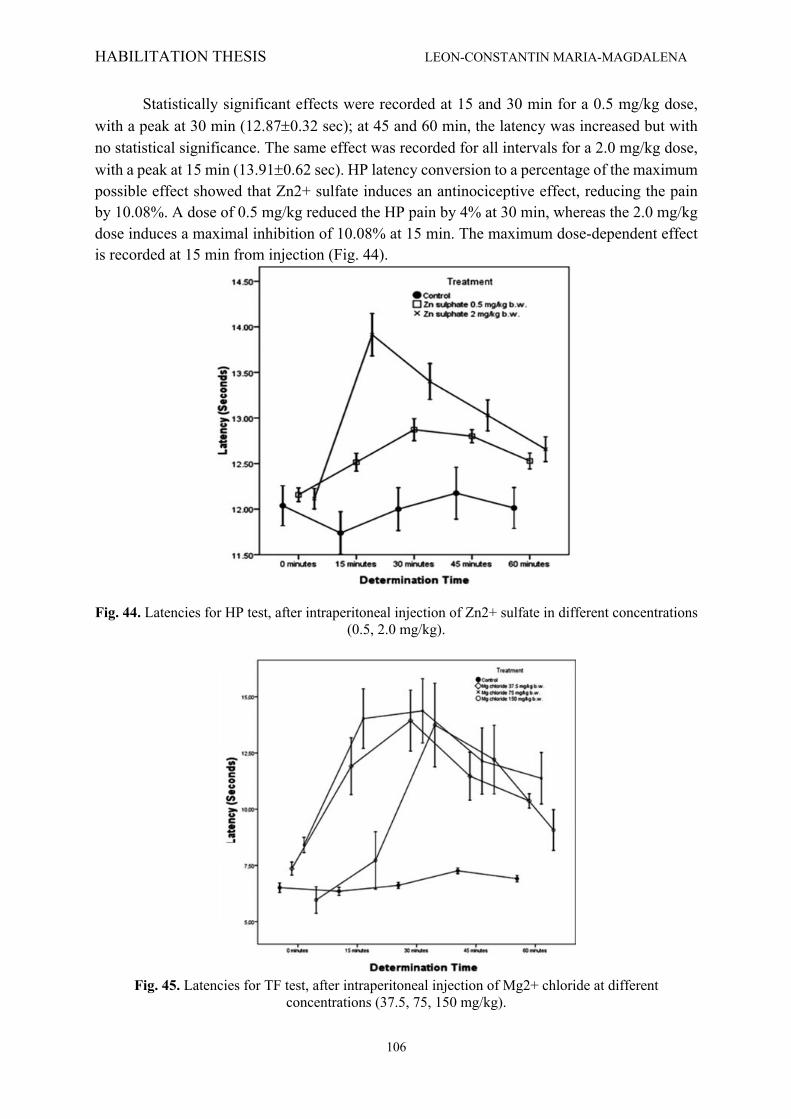
HABILITATION THESIS LEON-CONSTANTIN MARIA-MAGDALENA
106
Statistically significant effects were recorded at 15 and 30 min for a 0.5 mg/kg dose, with a peak at 30 min (12.87±0.32 sec); at 45 and 60 min, the latency was increased but with no statistical significance. The same effect was recorded for all intervals for a 2.0 mg/kg dose, with a peak at 15 min (13.91±0.62 sec). HP latency conversion to a percentage of the maximum possible effect showed that Zn2+ sulfate induces an antinociceptive effect, reducing the pain by 10.08%. A dose of 0.5 mg/kg reduced the HP pain by 4% at 30 min, whereas the 2.0 mg/kg dose induces a maximal inhibition of 10.08% at 15 min. The maximum dose-dependent effect is recorded at 15 min from injection (Fig. 44).
Fig. 44. Latencies for HP test, after intraperitoneal injection of Zn2+ sulfate in different concentrations (0.5, 2.0 mg/kg).
Fig. 45. Latencies for TF test, after intraperitoneal injection of Mg2+ chloride at different
concentrations (37.5, 75, 150 mg/kg).
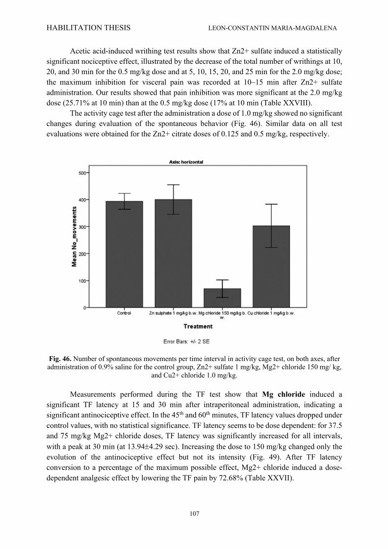
HABILITATION THESIS LEON-CONSTANTIN MARIA-MAGDALENA
107
Acetic acid-induced writhing test results show that Zn2+ sulfate induced a statistically significant nociceptive effect, illustrated by the decrease of the total number of writhings at 10, 20, and 30 min for the 0.5 mg/kg dose and at 5, 10, 15, 20, and 25 min for the 2.0 mg/kg dose; the maximum inhibition for visceral pain was recorded at 10–15 min after Zn2+ sulfate administration. Our results showed that pain inhibition was more significant at the 2.0 mg/kg dose (25.71% at 10 min) than at the 0.5 mg/kg dose (17% at 10 min (Table XXVIII).
The activity cage test after the administration a dose of 1.0 mg/kg showed no significant changes during evaluation of the spontaneous behavior (Fig. 46). Similar data on all test evaluations were obtained for the Zn2+ citrate doses of 0.125 and 0.5 mg/kg, respectively.
Fig. 46. Number of spontaneous movements per time interval in activity cage test, on both axes, after administration of 0.9% saline for the control group, Zn2+ sulfate 1 mg/kg, Mg2+ chloride 150 mg/ kg,
and Cu2+ chloride 1.0 mg/kg. Measurements performed during the TF test show that Mg chloride induced a
significant TF latency at 15 and 30 min after intraperitoneal administration, indicating a significant antinociceptive effect. In the 45th and 60th minutes, TF latency values dropped under control values, with no statistical significance. TF latency seems to be dose dependent: for 37.5 and 75 mg/kg Mg2+ chloride doses, TF latency was significantly increased for all intervals, with a peak at 30 min (at 13.94±4.29 sec). Increasing the dose to 150 mg/kg changed only the evolution of the antinociceptive effect but not its intensity (Fig. 49). After TF latency conversion to a percentage of the maximum possible effect, Mg2+ chloride induced a dose-dependent analgesic effect by lowering the TF pain by 72.68% (Table XXVII).

HABILITATION THESIS LEON-CONSTANTIN MARIA-MAGDALENA
108
Fig. 47. Number of spontaneous movements per time interval in activity cage test, on both axes, after administration of 0.9% saline for the control group, Zn2+ sulfate 1 mg/kg, Mg2+ chloride 150 mg/ kg,
and Cu2+ chloride 1.0 mg/kg. The HP test (HP) after intraperitoneal administration of Mg2+ chloride showed an antinociceptive effect for all doses and all recorded intervals, but only some doses/intervals were statistically significant. The response intensity is not dose dependent. Significant latencies were recorded for the low dose (37.5 mg/kg) at 15 and 30 min (including a peak of 15.75±5.32); for medium and high doses, the effect was faster and the peak latency recorded at 15 min was of 14.16±6.50 for the 75 mg/kg dose and 15.03±5.46 for the 150 mg/kg dose (Fig. 48).
Fig. 48. Latencies for HP test, after intraperitoneal injection of Mg2+ chloride at different concentrations (37.5, 75, 150 mg/kg).
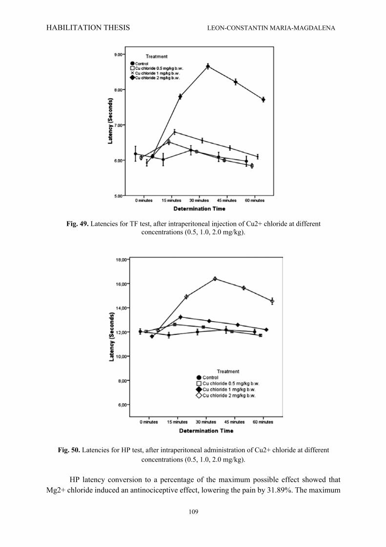
HABILITATION THESIS LEON-CONSTANTIN MARIA-MAGDALENA
109
Fig. 49. Latencies for TF test, after intraperitoneal injection of Cu2+ chloride at different concentrations (0.5, 1.0, 2.0 mg/kg).
Fig. 50. Latencies for HP test, after intraperitoneal administration of Cu2+ chloride at different concentrations (0.5, 1.0, 2.0 mg/kg).
HP latency conversion to a percentage of the maximum possible effect showed that
Mg2+ chloride induced an antinociceptive effect, lowering the pain by 31.89%. The maximum
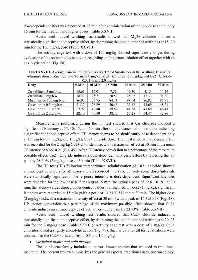
HABILITATION THESIS LEON-CONSTANTIN MARIA-MAGDALENA
110
dose-dependent effect was recorded at 15 min after administration of the low dose and at only 15 min for the medium and higher doses (Table XXVII).
Acetic acid-induced writhing test results showed that Mg2+ chloride induces a statistically significant nociceptive effect, by decreasing the total number of writhings at 15–20 min for the 150 mg/kg dose (Table XXVIII).
The activity cage test with a dose of 150 mg/kg showed significant changes during evaluation of the spontaneous behavior, recording an important sedation effect together with an anxiolytic action (Fig. 58).
Tabel XXVIII. Average Pain Inhibition Values for Tested Substances in the Writhing Test After Administration of Zn2+ Sulfate 0.5 and 2.0 mg/kg; Mg2+ Chloride 150 mg/kg; and Cu2+ Chloride
0.5, 1.0, and 2.0 mg/kg Drug 5 Min 10 Min 15 Min 20 Min 25 Min 30 Min
Zn sulfate 0.5 mg/b.w. 13.61 17.01 7.52 16.30 4.15 14.29 Zn sulfate 2 mg/b.w. 18.27 25.71 20.52 25.02 17.33 0.00 Mg chloride 150 mg/b.w. 86.03 83.75 84.71 89.54 86.82 85.71 Cu chloride 0.5 mg/b.w. 21.37 26.29 30.45 35.48 43.69 48.21 Cu chloride 1 mg/b.w. 22.92 40.80 39.62 41.58 43.69 46.43 Cu chloride 2 mg/b.w. 22.40 40.80 38.10 57.28 54.47 42.86
Measurements performed during the TF test showed that Cu chloride induced a significant TF latency at 15, 30, 45, and 60 min after intraperitoneal administration, indicating a significant antinociceptive effect. TF latency seems to be significantly dose dependent only at 15 min for 0.5 mg/kg and 1 mg/kg Cu2+ chloride dose. The most important analgesic effect was recorded for the 2 mg/kg Cu2+ chloride dose, with a maximum effect at 30 min and a mean TF latency of 8.66±0.21 (Fig. 49). After TF latency conversion to a percentage of the maximum possible effect, Cu2+ chloride induces a dose-dependent analgesic effect by lowering the TF pain by 28.60% (2 mg/kg dose, at 30 min (Table XXVII). The HP test (HP) following intraperitoneal administration of Cu2+ chloride showed antinociceptive effects for all doses and all recorded intervals, but only some doses/intervals were statistically significant. The response intensity is dose dependent. Significant latencies were recorded for the low dose (0.5 mg/kg) at 15 min (including a peak of 12.61±0.19); at 30 min, the latency values dipped under control values. For the medium dose (1 mg/kg), significant latencies were recorded at 15 min (with a peak of 13.23±0.51) and at 30 min. The higher dose (2 mg/kg) induced a maximum intensity effect at 30 min (with a peak of 16.39±0.38 (Fig. 48). HP latency conversion to a percentage of the maximum possible effect showed that Cu2+ chloride induces an antinociceptive effect, lowering the pain by 23.73% (Table XXVII). Acetic acid-induced writhing test results showed that Cu2+ chloride induced a statistically significant nociceptive effect, by decreasing the total number of writhings at 20–25 min for the 2 mg/kg dose (Table XXVIII). Activity cage test with a dose of 1 mg/kg Cu2+ chlorideshowed a slightly anxiolytic action (Fig. 47). Similar data for all test evaluations were obtained for the Cu2+ sulfate doses of 0.5 and 1.0 mg/kg.
• Medicinal plants and pain therapy The Lamiaceae family includes numerous known species that are used as traditional
medicine. The present review summarizes the general aspects, traditional uses, pharmacology,

HABILITATION THESIS LEON-CONSTANTIN MARIA-MAGDALENA
111
and in vitro and in vivo studies of Betonica officinalis, Glechoma hederacea, Hyptis pectinata, Lavandula genus, Leonurus cardiaca, Lamium genus, Melissa officinalis, Mentha genus, Marrubium vulgare, Origanum genus, Ocimum genus, Rosmarinus officinalis, Salvia genus, Satureja hortensis, Stachys lavandulifolia, Scutellaria lateriflora, Sideritis genus, Teucrium genus, Thymus genus, and Ziziphora tenuior, belonging to Lamiaceae botanical genus. The above-referred studies reported that the above mentioned medicinal plants have potent analgesic and antinociceptive activity. The findings of this review are promising, regarding new potential therapeutic agents with possible modulation in pain therapy. Most of the extracts identified did not present any toxic capabilities or known side effects and were at least as efficient as currently used synthetic drugs. Overall, although promising information evidence the efficacy of Lamiaceae genus in the treatment of pain associated disorders, the data are too preliminary and mostly fail to explain the exact cellular and molecular mechanisms of action and the respective active compounds. Therefore, future studies should be focused on investigating mechanisms of actions, realistic dosages, clinical efficacy, and safety of the extracts and active compounds in pain treatment.
6.4. Discussions
• Trace elements in experimental pain A dose-dependent antinociceptive effect of Cobalt
Our results both confirm and add to previous studies, showing for the first time the presence of a dose-dependent in vivo antinociceptive effect of Co chloride. Antinociceptive activity during thermoalgesic tests show an inhibition degree of 27-31% in TF and 18-40% in HP. Antinociceptive effect for Co chloride during the writhing test is more present, ranging between 36 to 100%. The lower dose of Co chloride we used represents 1/24 of LD50, thus limiting the potential toxic effects from interfering with our study. Spontaneous behaviour tests indicate both sedation and an anxiolytic effect in our experimental construct, inferring a possible toxic CNS effect (Knyazeva et al., 2012; Peterson et al., 2017; Barbee et al., 2014; Nieto-Fernandez et al., 2009). However, the difference between the 100% nociceptive protection in visceral pain writhing test and the 31% effect in TF at the same time intervals and dose, dismisses as only possible explanation for our results the CNS depressant effect observed. Co, Ni or Mn ions perfused in a Ca-deficient environment block synaptic transmission (Yin et al., 2003; Knyazeva et al., 2012; Barbee et al., 2014; Nieto-Fernandez et al., 2009; Angstadt et al., 1989) and microinjections of Co chloride in dorsal rat mesencephalon or the periapeductal gray, an area involved in visceral nociception, induce antinociceptive effects in rats (Hamann et al., 1992; Cavun et al., 2004). Co chloride also inhibits synaptic transmission at periapeductal grey level (Keay et al., 1997) and may act as a presynaptic blocker involved in nociception (Allen et al., 1997).
However, it appears that the mechanisms and involvement of this HTME in nociception is far more complex. Engelman et al. (Engelman et al., 1999) used Co chloride to identify Ca permissive AMPA receptors in the dorsal horn of lamina I and lateral area of lamina II. In lamina I, Co-positive NK1 receptors for P-substance are also present, concluding that they might play a role in nociception. Tong and MacDermott (Tong et al., 2006) observed that AMPA receptors contribute to the dorsal horn synaptic transmission and are involved in pain
HABILITATION THESIS LEON-CONSTANTIN MARIA-MAGDALENA
112
signaling pathways, making a Ca channel block through Co replacement a possible explanation for the antinociceptive effect observed in our experiments.
Similar to other metal trace elements (Zn, Ni, Cd, Mn), Co uses specific intracellular transporters (Komeda et al., 1997), becoming cofactor for enzymes involved in the synthesis of neuropeptide and neuromediators but also in the superoxide metabolism. A functional connection between ACDP4 transporter and the ionic chaperonin COX11 suggests specific intracellular sites for these metallic ions (Guo et al., 2005). Valinoid and capsaicinsensitive thermoreceptors were also thoroughly investigated using Co’s ability to use calcium channels (Nicholas et al., 1999; Rosa et al., 2008) investigated the inductive role of cobalt-protoporphyrin on hemoxygenase-1, known as a modulator for inflammatory pain, which might present novel pharmacological targets for analgesic drug development. The data available shows a complex role in pain for Co at various CNS levels, observations in agreement with our previous results (Tamba et al., 2008; D'Amato et al., 2004). Furthermore, previous works lack in-vivo behaviour experimental data regarding Co and thus our results might represent a starting point for the presented subject. Nickel has a dual effect on acetic acid-induced writhing test Acid-sensing ionic channels, present in central and peripheral neurons, with a specific role in nociception, neural hyperexcitability and hyperalgesia, can be inhibited in a dose-dependent manner by Ni ions (Staruschenko et al., 2007; Chen et al., 2002; Nelson et al., 2007). Nelson et al. (Nelson et al., 2007) insists on the Ni role on Ca channels according to their Zn affinity. Our results show that Ni ion plays a moderate but significant and dose-dependent antinociceptive action in thermoalgesic tests, ranged from 9.7 to 20% between 15-30 min and effect persistence over 60 min for 2 mg/kg in HP test. In the writhing test we have seen peculiar results – an initial short but significant antinociceptive action, followed by a hyperalgesic trend in the second part of the test. This biphasic aspect made us consider two different mechanisms; one mechanism involving a fast response while the second mechanism being delayed until the trace element arrives to some specific structures (channels, enzymes) whose impaired activity leads to the observed hyperalgesia. No significant changes during spontaneous behavior AC were observed.
Poor literature data regarding Ni role in nociception allows us just few considerations regarding the mechanism and action place for this rare element in nociception. Prado (Larson et al., 1997) studying Ca2+ involvement in pain and antinociception, reported Ni and other cations (Ce3+, La3+, Cd2+, Co3+, Mg2+, Mn2+) as unselective blockers for the Ca2+ channel pore. It is also well-known that Ca2+ is required to induce nociceptive mediator delivery (Barritt et al., 1999). N and P/Q Ca2+ channels involved in nociception are found in dendrites and neuronal bodies. Some channel subtypes are also found in postsynaptic membranes of the glutamatergic neurons, involved in pain transmission. A more thorough study by Nelson et al. (Nelson, 2001) describes Ni roles on Ca2+ channels and also their affinity for Zn2+ ions, showing that after a peripheral lesion, the nociceptive receptors become hyperexcitable. These receptors are also located in dorsal ganglia where neurons exhibit an increased number of T-type calcium channels. Ni ion was also proved to discriminate between various T-type Ca2+ channels, its affinity for Ca2+ 3.2 channel being 20 times higher compared to other isoforms.
HABILITATION THESIS LEON-CONSTANTIN MARIA-MAGDALENA
113
The antinociceptive effect of Ni could be similar to Zn, although this has a non-specific affinity for these channels. Intrathecal injections of Zn induce antinociceptive effects in mice, while Zn chelating agents lead to hyperalgesia (Kitamura et al., 2006). Though current data may explain Ni antinociceptive action by Ca channels interference in peripheral nerve ends, the hyperalgesic effect observed in the second phase of the writhing test to acetic acid remains to be further investigated. The spontaneous behaviour test was negative for nickel chloride, thus validating and completely eliminating any toxicity related contamination of the interpreted results (Staruschenko et al., 2007). Significant analgesia after Molybdenum administration Mo showed significant antinociceptive effects in all our tests which are dose - independent and appear in a time range of 15 - 30 min from administration. The antinociceptive levels in the thermoalgesic tests were too limited to have clinical significance (inhibition values ranging between 8-13% for TF and 4-9% for HP). In the writhing test, however, Mo administration was associated with an antinociceptive activity of 33% at 15 minutes and 68% at 30 min, effect that is associated with a slight sedation (in AC). Current literature lacks data on in vivo experimental data regarding Mo action on nociception. Toxicity studies in animals did not reveal any significant CNS effects nor motor dysfunctions.
One pilot study (Moss, 1995) presented 14 patients undergoing pain symptoms that received 500μg Mo in oral administration for 4 weeks and compared them to a placebo group. Significant pain relieve was recorded in Mo-treated group, together with general status improvement. Mo induced a growth latency of 50-78%, with visible cortico-dystrophies in a 6 weeks toxicity tests with oral doses of 0.2 mg, 7.5mg or 30 mg/kg/day in rats (Engel et al., 1956) with similar studies investigating Mo toxic effects on weight and hair color. We found no study regarding nociception or possible action mechanisms at cellular and subcellular level in pain. Mo is included in some Mohydroxylases family, enzymes involved in metabolism of some drugs in humans. These enzyme functions are not fully understood (Kitamura et al., 2006) but their effect on free radicals is generally accepted. Recent data seem to open a path toward more complex investigations, as chimeric animals (rats with human hepatic cells) were obtained to study various drug effects on Mo-enzymes in the cell (Kitamura et al., 2006) and also a potential interference of Mo with pro or antinociceptive molecules. Literature data show only a strong inhibiting effect of methadone on Mo-hydroxylases (Robertson et al., 1994). Zinc and its effect on the nervous system The antinociceptive effect of zinc in our evaluation processes was moderate, and we observed an inhibition of 4–17% in thermoalgesic tests and 17–25% in chemoalgesic tests. Our results showed no significant differences between the two Zn2+ salts used. Zn2+ sulfate, when explored by activity cage test, did now show any significant behavioral changes, but Zn2+ citrate had some positive results. Zn2+ ion (in both salts used) showed moderate antinociceptive effects (4–25%), more evident on chemoalgesic test. Significant behavioral changes were recorded also following administration of Zn2+ sulfate and citrate. Our research showed the Zn2+ effect in the periphery, where pain is transmitted by the A delta and C fibers. We hypothesize that, under our experimental conditions, Zn2+ influences glutamatergic receptors both at the peripheral and at the spinal levels (explaining why in most
HABILITATION THESIS LEON-CONSTANTIN MARIA-MAGDALENA
114
our experiments analgesia occurs after a latency of 10–15 min, the time required to cross the BBB and reach the synaptic space). How and where Zn2+ acts in the synaptic space is not precisely known, but several authors have provided additional info, although sometimes contradictory. Takeda et al. (Takeda et al., 2003) argue that zinc increases the presynaptic release of glutamate or GABA, with Smart et al. (Smart et al., 2004) confirming these claims but advancing the hypothesis (not yet confirmed) that zinc acts as a direct neurotransmiter too, by postsynaptic signaling pathway modulation. Li and Clark (Li et al., 2000) presented further data showing a reduction of inflammatory pain (formalin model) from inhibition by zinc (zinc-protoporphyrin intrathecally) of hemoxygenase at the spinal level, but with no effect on peripheral thermal testing or hemoxygenase levels reported. Another possible mechanism of action for zinc ions is interference with the potassium channels. Prost et al. (Prost et al., 2004) showed that the zinc ion has the ability to open the K-ATP channel in pancreatic cells, acting on both sides of the membrane. Zinc also interferes with the nicotinic receptor ion channel, modulating nicotinic receptor function (Hsiao et al., 2008). On the other hand, in a recent study Taly et al. (Taly et al., 2009) showed that Zn2+ acts as an alosteric modulator of the alpha–beta portion of the heteropentameric element (alpha 4–beta 2) of the nicotinic receptor. Of great importance to our study is that Taly et al. argue the modulator role of Zn2+ ions can both stimulate (alpha–alpha interface) and inhibit (beta–alpha interface) nicotinic izoreceptor alpha 4-beta 2 depending on the concentration of zinc used . This could, in our view, explain the sometimes similar and sometimes different results obtained by other authors regarding the involvement of zinc in pain, vs. our data (Taly et al., 2009; Hsiao et al., 2008). In fact, many authors consider the nicotinic receptor as a useful target for therapeutic agents in pain management (Meyer et al., 2006; Rowley et al., 2008). These hypothesis, however, require further exploration. The Zn2+ sulfate dose that we used did not show significant changes in spontaneous behavior when assessed by the activity cage test. A literature review provided comparable data (Nozaki et al., 2011; Matsunami et al., 2011), except for Kroczka et al. (Kroczka et al., 2001), who report an ‘‘antidepressant-like’’ effect in a forced swim test in rats, but without providing a dose that produced this effect or the zinc salt used. Magnesium may exert its antinociceptive effects by penetrating the blood-brain-barrier (BBB) Magnesium salts induced various degrees of antinociceptive action for all performed tests. In comparison with the HP test, which showed no dose-dependent effect (pain inhibition 30%), the TF test showed a pain inhibition of 58–72% at 30 min. The most suggestive values appear in the writhing test, in which the antinociceptive effect reaches 85%. Behavioral effects evaluated by activity cage test denote both a sedative and an anxiolytic action. Our results are consistent with previous studies (Begon et al., 2002), although the administration methods are different. Positive effects on the behavioral parameters argue for an Mg2+ chloride passage through the blood–brain barrier (BBB). There is a documented correlation between the antinociceptive and central nervous system-depressing actions (Fawcett et al., 1999; Durlach et al., 2000). From our results we conclude that Mg2+ chloride exerts an antinociceptive action at a dose of 150 mg/kg, with low results for TF test but with important effects on the HP test (inhibition values of 30% at 15 min, 56% at 30 min, and 22% at 60 min).

HABILITATION THESIS LEON-CONSTANTIN MARIA-MAGDALENA
115
Our view is that one could explain the analgesic effect of Mg2+ by a peripheral mechanism, but the central behavior inhibitory actions of our tests lead to the conclusion that Mg2+ does cross the BBB. Hasanein et al. (Hasanein et al., 2006) confirmed previous results regarding the antihyperalgesic effect of i.p. Mg2+ for mechanoalgezic tests in rats with diabetic neuropathy. However, the author proposes another mechanism for this effect: the normalization of glycemia rather than interference with the Na/Ca channel of the NMDA receptor or the lowering action on pronociceptive free radicals. As for the relationship between the antinociceptive effect and the CNS depressant activity, several authors have investigated this and confirmed it (Fawcett et al., 1999; Durlach et al., 2000). The current study showed a reduction in both horizontal and vertical mobility after the administration of Mg2+ chloride, in agreement with recent data obtained by Bindar et al. (Bindar et al., 2010). The analgesic effect of Copper is dose-dependent Our results regarding copper salts show potential antinociceptive effects during thermoalgesic tests and especially during chemoalgesic tests. It is difficult to explain the analgesic effect, although copper ion is involved in many molecular mechanisms of pain relief. Some research (Yu et al., 2008) has shown that Cu2+ sulfate administration induces dopaminergic neuron lesions with antioxidant defense inhibition and apoptosis induction. Copper chloride induced a dose-dependent antinociceptive effect (5% at minimal dose, 9.58% at medium dose, and 28% at maximal dose) in the TF test and of 3.17–23.7% in the HP test. Behavioral tests showed that Cu2+ chloride exerts an anxiolytic effect. Our results show a potential analgesic effect of Cu2+ salts in both the thermoalgezic and the chemoalgezic tests. Although different from our experimental conditions, the literature data are consistent with our results. However, it seems difficult to explain clearly how Cu2+ ions induce analgesia. As with other bivalent trace elements, Cu2+ also is transported to specific molecules (Cu2+ [1]-ATPase) using guidance proteins. Cu2+ from the CNS cells is coupled in the enzymatic systems involved in the synthesis of catecholamines, opioid peptides, and neuropeptides (Kim et al., 2008). A possible role of Cu2+ in analgesia was described by Jacka et al. (Jacka et al., 1983) from an inflammatory arthritis model in rats, which tested the antinociceptive effects of salicylate after Cu2+ pretreatment (mechanical pain tests). The data presented here and the literature data converge on Cu2+‘s certain involvement in analgesia, but unfortunately, because of various cellular and molecular actions, not always beneficial, high-selectivity pain drugs based on Cu2+ do not yet exist. In fact, we may never attain that, insofar as research by W.R. Yu et al. (Yu et al., 2008) has shown that Cu2+ sulfate administration (striatal nuclei injections in rats) damages the dopaminergic neurons, causing apoptosis.
• Medicinal plants and pain therapy Pain comes in many forms: acute, chronic, visceral, inflammatory, or neuropathic
(Masuda et al., 2017; Tamba et al., 2013). It is not simply a result of tissue damage but also reflects the influence of many psychological variables such as attention, anxiety, stress, suggestion, or previous experiences and may have a significant genetic contribution. Pain accompanies most pathologies present in current medical practice, and 25% percent of Americans, for example, experience pain on a daily basis. Having the numbers on its side,
HABILITATION THESIS LEON-CONSTANTIN MARIA-MAGDALENA
116
pain became a global public health problem and a leading cause of disability all over the world (Gedin et al., 2017).
As life expectancy is rising and chronical pathologies along with it, the prevalence of accompanying pain is expected to increase yearly, with higher prevalence in elderly patients, where the treatment is also more sensitive. Considering the above, new therapeutic agents with increased efficacy, less side effects, and lower costs and leading to an improved quality of life (Iorga et al., 2015) should become one of the primary objectives in modern medical research, together with constant monitoring of the previous mentioned aspects (Iurea et al., 2013).
The medicinal use of plants as analgesic drugs in folk medicine is an ancient tradition, far older than the current sciences of medicine in developing countries (Ayaz et al., 2012). According to estimations, up to 70,000 plant species are used ethnomedicinally worldwide. Effects of herbal extracts have been studied by different pain tests including writhing test, light tail flick test, tail immersion test, hot-plate test, and formalin test (Faq et al., 2012).
The exploration for new analgesic combinations from the enormous arrays of medicinal plant resources is growing. This is because such information holds guarantees for the finding of new therapeutic agents capable of inhibiting, decreasing, or relieving pain (Anilkumar, 2010; Peptu et al., 2015). Plants characterize a vast natural supply of appreciated compounds that might achieve primary importance for the expansion of novel drugs (Bahmani et al., 2014). The survey of the effectiveness of plant-based remedies used in the folk medicine has given great reflections because they are cheap and have reduced side effects.
The data in biomedical literature presenting plants with medicinal capabilities are very similar to the array of publications depicting the modulatory effects certain ones have over pain perception. The Lamiaceae family, one of the most important herbal families, incorporates a wide variety of plants with biological and medical applications. The most known members of this family are a variety of aromatic spices like thyme, mint, oregano, basil, sage, savory, rosemary, self-heal, hyssop, lemon balm, and some others with more limited use (Bekut et al., 2018).
6.5. Conclusions
• Trace elements in experimental pain Heavy metal trace elements have rarely been investigated in the literature. Due to
conflicting and limited in-vitro information about their influence on pain, the authors acknowledge the difficulty in choosing the test doses. All heavy metal trace elements tested induced variable thermoalgesic and chemoalgesic antinociceptive effects after IP administration. These findings support further efforts to understand the molecular level effects of these trace metals.
• The effect of common trace elements on pain Similarly, non-heavy metal trace elements such as Zinc, Magnesium and Copper also
have antinociceptive effects when administered intraperitoneally in an acute setting. These results and corroborate data from cited authors, supporting further efforts to identify molecular-level effects of these investigated salts and making trace elements an important nociception investigation target and possible inexpensive adjuvants for pain therapy.
• Medicinal plants and pain therapy This review covers a useful approach for further identification of new compounds from
various medicinal plants, which may be effective in pain management.
HABILITATION THESIS LEON-CONSTANTIN MARIA-MAGDALENA
117
Chapter 7. NEW METHODS AND FORMULATIONS FOR A MORE EFFECTIVE ANALGESIC TREATMENT
7.1. Introduction
There are several ways to improve available drugs. This is especially relevant for painful conditions where the analgesic requirement is usually high and physicians often have to combine different analgesics and co-analgesics to achieve pain control at the cost of important side-effects. Cystic fibrosis (CF) is one such example. It is a complex, progressive, life-limiting genetic disorder that affects many systems of the human body, namely the respiratory, digestive and reproductive systems, all of which contribute to impaired health-related quality of life (Lee et al., 2016). CF is caused by mutations in the cystic fibrosis transmembrane conductance regulator (CFTR) gene protein, typically found in the apical membrane of most epithelial tissues including lungs, submucosal, salivary, gall bladder glands, gut, kidney, pancreas, cervix and uterus (Kumar et al., 2014). Rarely, CFTR is also expressed in non-epithelial cells and tissues, including ventricular cardiomyocytes and aortic smooth muscle cells, neurones in the brain, corneal and vascular endothelial cells and lymphocytes (Bowen et al., 2015). The studies of Koh et al. (Koh et al., 2005) and Palermo et al. (Palermo et al., 2006) showed that 46% of children with CF reported a painful episode at least once a week, with the intensity associated with common CF-related procedures. The primary reported locations of pain were the abdominal/pelvic region (50%), chest (37%) and head/neck (33%). In contrast, a study by Munck et al. (Munck et al., 2012) found a very low incidence (6%) of recurrent abdominal pain in children with CF. This low percentage was obtained by using a strict definition of recurrent abdominal pain, namely “at least three bouts of pain, severe enough to affect activities, over a period of not less than three months, with attacks in the year preceding the examination” (Apley et al., 1958) using Apley’s criteria, which led to exclusion from the study of many children with less severe pain.
Using daily online diaries, Blackwell and Quittner (Blackwell et al., 2015) found that 76% of adolescents had episodes of pain most frequently in the stomach (49%), followed by the head/neck (42%) and chest (37%), pain associated with treatment burden, respiratory symptoms, higher levels of depression and anxiety.
The study of Lechtzin et al. (Lechtzin et al., 2016) also demonstrated that pain is common in paediatric CF patients and was associated with increased pulmonary distress and decreased quality of life. Pain was most often encountered in the abdomen (42.0%), head/sinuses (31.9%), joints (18.8%), chest (10.1%), back (5.8%) and muscles (2.9%). A recent review argues that a multidisciplinary approach comprised of physicians, psychologists, pharmacists, chemists, material science specialists, engineers and therapists is required to address the optimal care of CF patient (King et al., 2019). According to Leso et al. (Leso et al., 2019), nanotechnology, one of the most innovative tools of the century, will transform the biomedical field by improving prevention, diagnosis and disease therapy. Indirectly, the nano-personalized strategy facilitates a better quality of life and life expectancy for patients with CF. Based on specific small size, large surface area-to-volume ratios,
HABILITATION THESIS LEON-CONSTANTIN MARIA-MAGDALENA
118
improved pharmacokinetics and minimal side effects, nano-sized materials have shown potential as carriers for targeted drug delivery in pharmacotherapy (Yhee et al., 2016).
An important advantage of drug delivery-based nanotechnology is the improved diffusion and degradation characteristics of the encapsulated nanomaterial, allowing the drug to be protected during its transit to the target, while maintaining the biological and chemical properties and allowing it to be released at an appropriate and effective rate. The most commonly employed nonviral nanosized carriers are made of cationic lipids and polymers (D'Angelo et al., 2014). Lipid-based carriers, namely liposomes and solid lipid nanoparticles for drug delivery and cationic lipid particles for gene delivery, are mainly composed of phospholipids and cholesterol. These advanced lipid formulations present remarkable features for the pulmonary delivery of drugs due to their tolerance in the pulmonary tract, reduced absorption, limiting aggregation and adhesion (D'Angelo et al., 2014; Omri et al., 1994). Liposome-encapsulated drugs (mainly antibiotics) are intensively explored for lung targeting due to their improved drug pharmacokinetics and biodistribution, decreased toxicity, ability to overcome bacterial drug resistance and targeted selectivity. Theoretically, nanoparticles hold great promise to improve drug delivery limitations in the lungs, since their surface, structure and composition can be tailored to reach the right target at the right time by providing accurate and controlled drug delivery (Yhee et al., 2016; Da Silva et al., 2013). Still, the success of nanotherapy depends on a number of factors, such as nanoparticle characteristics and toxicity, route of administration and physiological aspects of the lung in the presence of respiratory disease (Weers, 2015). One of the primary objectives of a clinician working with CF patients should be effective pain relief. An analysis of the literature data using “nanotechnology in CF pain” as keywords on the ScienceDirect search engine, found 76 reviews and research articles related to the nanotechnology approach to CF treatment. Although this search may not be entirely representative, to our knowledge there are no data available on the use of nanomaterials in CF pain relief. Instead, a search for “nanotechnology in pain” produced about 7975 publications from 1996.
According to a review article from 2018 by Beiranvand and Sorori (Beiranvand, 2019), the proper treatment of pain is still a major medical challenge; therefore, diferent approaches using various nano-formulated materials have been employed in the last decade. According to Moradkhani et al. (Moradkhani, 2018), analgesic drug delivery nanosystems have been applied in pain therapy due to their remarkable properties: enhanced drug delivery profile, increased drug action and bioavailability, targeted/sustained or prolonged drug release profile, stability in biological fluids, nontoxicity, carried drug protection up to the target cell population, and reduced side effects of the incorporated analgesic drugs (Koning, 2002). Table XXXIV gives a brief overview of several current strategies used in pain therapy with the help of nanotechnology science. Different formulations such as liposomes, nanoparticles, nanoplates, nanocapsules, nanofibers, nanotubes, micelles and dendrimers were developed for delivering analgesics, local anaesthetics, NSAIDs or opioid compounds intended for pain therapies (Raffin et al., 2012).
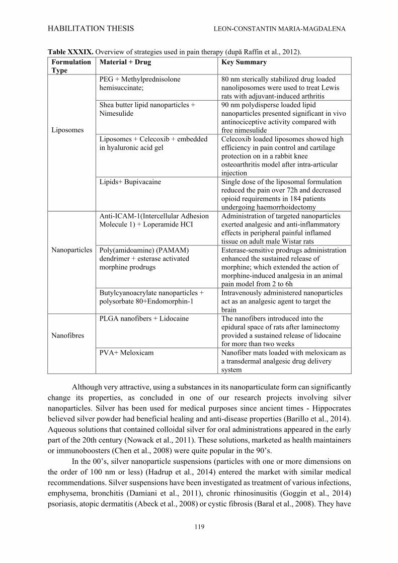
HABILITATION THESIS LEON-CONSTANTIN MARIA-MAGDALENA
119
Table XXXIX. Overview of strategies used in pain therapy (după Raffin et al., 2012). Formulation Type
Material + Drug Key Summary
Liposomes
PEG + Methylprednisolone hemisuccinate;
80 nm sterically stabilized drug loaded nanoliposomes were used to treat Lewis rats with adjuvant-induced arthritis
Shea butter lipid nanoparticles + Nimesulide
90 nm polydisperse loaded lipid nanoparticles presented significant in vivo antinociceptive activity compared with free nimesulide
Liposomes + Celecoxib + embedded in hyaluronic acid gel
Celecoxib loaded liposomes showed high efficiency in pain control and cartilage protection on in a rabbit knee osteoarthritis model after intra-articular injection
Lipids+ Bupivacaine Single dose of the liposomal formulation reduced the pain over 72h and decreased opioid requirements in 184 patients undergoing haemorrhoidectomy
Nanoparticles
Anti-ICAM-1(Intercellular Adhesion Molecule 1) + Loperamide HCI
Administration of targeted nanoparticles exerted analgesic and anti-inflammatory effects in peripheral painful inflamed tissue on adult male Wistar rats
Poly(amidoamine) (PAMAM) dendrimer + esterase activated morphine prodrugs
Esterase-sensitive prodrugs administration enhanced the sustained release of morphine; which extended the action of morphine-induced analgesia in an animal pain model from 2 to 6h
Butylcyanoacrylate nanoparticles + polysorbate 80+Endomorphin-1
Intravenously administered nanoparticles act as an analgesic agent to target the brain
Nanofibres
PLGA nanofibers + Lidocaine The nanofibers introduced into the epidural space of rats after laminectomy provided a sustained release of lidocaine for more than two weeks
PVA+ Meloxicam Nanofiber mats loaded with meloxicam as a transdermal analgesic drug delivery system
Although very attractive, using a substances in its nanoparticulate form can significantly change its properties, as concluded in one of our research projects involving silver nanoparticles. Silver has been used for medical purposes since ancient times - Hippocrates believed silver powder had beneficial healing and anti-disease properties (Barillo et al., 2014). Aqueous solutions that contained colloidal silver for oral administrations appeared in the early part of the 20th century (Nowack et al., 2011). These solutions, marketed as health maintainers or immunoboosters (Chen et al., 2008) were quite popular in the 90’s.
In the 00’s, silver nanoparticle suspensions (particles with one or more dimensions on the order of 100 nm or less) (Hadrup et al., 2014) entered the market with similar medical recommendations. Silver suspensions have been investigated as treatment of various infections, emphysema, bronchitis (Damiani et al., 2011), chronic rhinosinusitis (Goggin et al., 2014) psoriasis, atopic dermatitis (Abeck et al., 2008) or cystic fibrosis (Baral et al., 2008). They have

HABILITATION THESIS LEON-CONSTANTIN MARIA-MAGDALENA
120
also been used as anti-inflammatory agents in cystitis, prostatitis, colitis, gastritis, tonsillitis, appendicitis and sinusitis (Barillo et al., 2014).
However, there are mostly market-available silver suspensions that come from unknown or unreliable sources and there is no actual control of the product’s quality. Some on-line shops also sell generators that produce colloidal silver at home, another important factor that contributes to the lack of control for this product. It is estimated that of all nanomaterials in the medical and healthcare sector, nanosilver has the highest degree of commercialization (Chen et al., 2008), with approximately 320 tones of nanosilver produced worldwide per year (Nowack et al., 2011). Together with the uncontrolled on-line market for oral silver suspensions, this metal is also used in the manufacturing of silver-embedded medical equipments (Sussman et al., 2015) (surgical tools, catheters, bandages, needles and stethoscopes) and in implantology (Devasconcellos et al., 2012).
In 2005, the in vitro toxicity of several nanoparticles was assessed (Braydich-Stolle et al., 2005) and the authors concluded that silver has the highest concentration-dependent toxicity. In 2014, a Scientific Committee employed by the European Commission issued a document regarding nanosilver and its safety. The experts concluded that nanosilver’s toxic effects are still unknown because too little information is available. Some studies suggest silver particles promote inflammation in normal tissue (Carlson et al., 2008), but the effect of the metal on an pre-existing inflammatory environment is unknown. Apart from silver nanoparticles, silica-based nanoparticles (SNPs) are also amongst the substances that have received a lot of research interest for medical applications because of their biocompatibility, versatility, stability, monodispersity, large surface area, high drug loading efficiency, and potential for hybridization with other materials (Argyo et al., 2014). The surface of SNPs is usually negatively charged due to the presence of the hydroxyl group, therefore it is convenient to modify the SNPs’ surface through the silane chemistry. In order to control the physico-chemical, toxicological and pharmacological properties, various reactive functional groups like amine, carboxyl, phosphate or polyethylene glycol could be easily conjugated to hydroxyl SNPs. Alternatively, the surface chemistry of the SNPs can be fine-tuned for a specific biological application, optimizing the dispersion stability and/or cellular uptake, the covalent attachment of imaging agents and targeting ligands the rational control of drug release rate (Mamaeva et al., 2013).
Numerous studies pointed toward their excellent potential as biomarkers, calibration standards in confocal fluorescence microscopy, drug delivery and targeting systems (tumor imaging and therapy in vitro and in vivo) in biomedical science (Legrand et al., 105 2008; Lu et al., 2007; Slowing et al., 2007). The imaging agents such as, fluorescein isothiocyanate, methylene blue, quantum dots, gadolinium chelates, tetramethylrhodamine or the targeting ligands, such as aptamers, antibodies, peptides, and folic acid, can be easily doped into or modified on the surface (Wu et al., 2014). Despite the growing body of papers related to the use of SNPs as therapeutic and imaging tools, today’s challenge remains the biodistribution of silica nanostructures for in vivo diagnostic and therapeutic applications.
Another approach for improving available analgesic treatments is identifying better ways for drug delivery. Minimal invasiveness represents an important requirement and empirical formulations of curative mixtures were developed and perfected through generations as creams, ointments, pastes, powders, liquid extracts of animal or vegetal origins. For example,
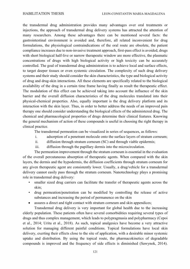
HABILITATION THESIS LEON-CONSTANTIN MARIA-MAGDALENA
121
the transdermal drug administration provides many advantages over oral treatments or injections, the approach of transdermal drug delivery systems has attracted the attention of many researchers. Among these advantages there can be mentioned several facts: the gastrointestinal environment is avoided and, therefore, all related inconvenient for drug formulations, the physiological contraindications of the oral route are obsolete, the patient compliance increases due to non-invasive treatment approach, first-pass effect is avoided, drugs with short biological half-live or narrow therapeutic window are more effective, the plasmatic concentrations of drugs with high biological activity or high toxicity can be accurately controlled. The goal of transdermal drug administration is to achieve local and surface effects, to target deeper tissues or even systemic circulation. The complexity of such drug delivery systems and their study should consider the skin characteristics, the type and biological activity of drug and drug-skin interactions. All these elements are specifically related to the biological availability of the drug in a certain time frame having finally as result the therapeutic effect. The modulation of this effect can be achieved taking into account the influence of the skin barrier and the overall diffusion characteristics of the drug molecules translated into their physical-chemical properties. Also, equally important is the drug delivery platform and its interaction with the skin layer. Thus, in order to better address the needs of an improved pain therapy one should consider understanding the biological effects of the administered drug. The chemical and pharmacological properties of drugs determine their clinical features. Knowing the general mechanism of action of these compounds is useful in choosing the right therapy in clinical practice.
The transdermal permeation can be visualized in series of sequences, as follows: i. adsorption of a penetrant molecule onto the surface layers of stratum corneum; ii. diffusion through stratum corneum (SC) and through viable epidermis; iii. diffusion through the papillary dermis into the microcirculation. The permeation improvement through the stratum corneum is essential in the evaluation
of the overall percutaneous absorption of therapeutic agents. When compared with the skin layers, the dermis and the hypodermis, the diffusion coefficients through stratum corneum for any given therapeutic agent are consistently lower. Usually, a drug/vehicle for a transdermal delivery cannot easily pass through the stratum corneum. Nanotechnology plays a promising role in transdermal drug delivery:
• smaller sized drug carriers can facilitate the transfer of therapeutic agents across the skin;
• drug permeation/penetration can be modified by controlling the release of active substances and increasing the period of permanence on the skin
• assures a direct and tight contact with stratum corneum and skin appendices; Transdermal drug delivery is very important for global health due to the increasing
elderly population. These patients often have several comorbidities requiring several types of drugs and thus complex management, which leads to polypragmasia and polypharmacy (Cepoi et al., 2014; Uritu et al., 2018). As such, topical analgesics have become a very attractive solution for managing different painful conditions. Topical formulations have local skin delivery, exerting their effects close to the site of application, with a desirable minor systemic uptake and distribution. By using the topical route, the pharmacokinetics of degradable compounds is improved and the frequency of side effects is diminished (Sawynok, 2014).
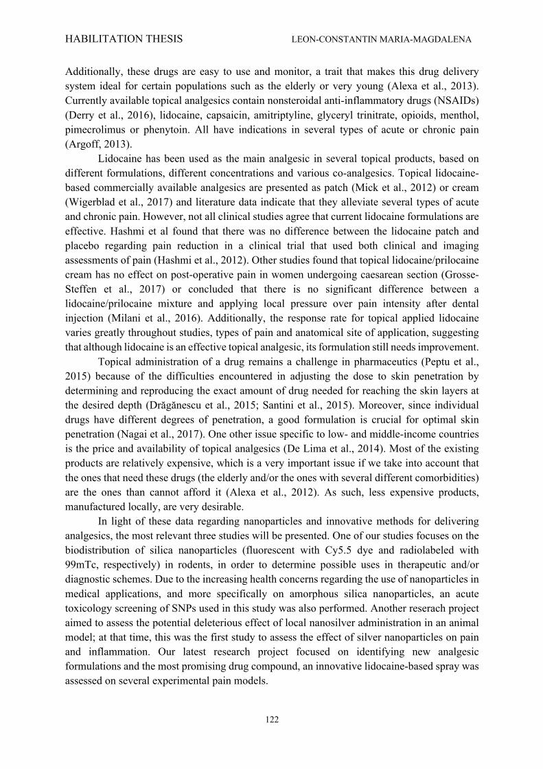
HABILITATION THESIS LEON-CONSTANTIN MARIA-MAGDALENA
122
Additionally, these drugs are easy to use and monitor, a trait that makes this drug delivery system ideal for certain populations such as the elderly or very young (Alexa et al., 2013). Currently available topical analgesics contain nonsteroidal anti-inflammatory drugs (NSAIDs) (Derry et al., 2016), lidocaine, capsaicin, amitriptyline, glyceryl trinitrate, opioids, menthol, pimecrolimus or phenytoin. All have indications in several types of acute or chronic pain (Argoff, 2013).
Lidocaine has been used as the main analgesic in several topical products, based on different formulations, different concentrations and various co-analgesics. Topical lidocaine-based commercially available analgesics are presented as patch (Mick et al., 2012) or cream (Wigerblad et al., 2017) and literature data indicate that they alleviate several types of acute and chronic pain. However, not all clinical studies agree that current lidocaine formulations are effective. Hashmi et al found that there was no difference between the lidocaine patch and placebo regarding pain reduction in a clinical trial that used both clinical and imaging assessments of pain (Hashmi et al., 2012). Other studies found that topical lidocaine/prilocaine cream has no effect on post-operative pain in women undergoing caesarean section (Grosse-Steffen et al., 2017) or concluded that there is no significant difference between a lidocaine/prilocaine mixture and applying local pressure over pain intensity after dental injection (Milani et al., 2016). Additionally, the response rate for topical applied lidocaine varies greatly throughout studies, types of pain and anatomical site of application, suggesting that although lidocaine is an effective topical analgesic, its formulation still needs improvement.
Topical administration of a drug remains a challenge in pharmaceutics (Peptu et al., 2015) because of the difficulties encountered in adjusting the dose to skin penetration by determining and reproducing the exact amount of drug needed for reaching the skin layers at the desired depth (Drăgănescu et al., 2015; Santini et al., 2015). Moreover, since individual drugs have different degrees of penetration, a good formulation is crucial for optimal skin penetration (Nagai et al., 2017). One other issue specific to low- and middle-income countries is the price and availability of topical analgesics (De Lima et al., 2014). Most of the existing products are relatively expensive, which is a very important issue if we take into account that the ones that need these drugs (the elderly and/or the ones with several different comorbidities) are the ones than cannot afford it (Alexa et al., 2012). As such, less expensive products, manufactured locally, are very desirable.
In light of these data regarding nanoparticles and innovative methods for delivering analgesics, the most relevant three studies will be presented. One of our studies focuses on the biodistribution of silica nanoparticles (fluorescent with Cy5.5 dye and radiolabeled with 99mTc, respectively) in rodents, in order to determine possible uses in therapeutic and/or diagnostic schemes. Due to the increasing health concerns regarding the use of nanoparticles in medical applications, and more specifically on amorphous silica nanoparticles, an acute toxicology screening of SNPs used in this study was also performed. Another reserach project aimed to assess the potential deleterious effect of local nanosilver administration in an animal model; at that time, this was the first study to assess the effect of silver nanoparticles on pain and inflammation. Our latest research project focused on identifying new analgesic formulations and the most promising drug compound, an innovative lidocaine-based spray was assessed on several experimental pain models.

HABILITATION THESIS LEON-CONSTANTIN MARIA-MAGDALENA
123
The main publications that have derived from our research are listed below.
1. Tamba BI, Dondas A, Leon M, Neagu AN, Dodi G, Stefanescu C, Tijani A. Silica nanoparticles: Preparation, characterization and in vitro/in vivo biodistribution studies. European Journal of Pharmaceutical Sciences. 2015; 71: 46–55.
2. Alexa AI, Alexa Stratulat T, Leon Constantin M, Alexa ID, Tamba BI. Local Silver Nanoparticles Administration Promotes Inflammation and Hyperalgesia in Rats. Rev. Chim (Bucharest), 2017;68(3):490-496.
3. Luca A, Mihai C, Stanciu G, Cojocaru E, Ancuceanu R, Harabagiu V, Peptu C, Peptu A, Leon–Constantin M, Alexa-Stratulat T. In vivo safety and efficacy evaluation of a novel polymeric based lidocaine formulation for topic analgesia. Farmacia, 2019; 67(1):117-125.
The funding for this research has been partly supported by the NANODERMA grand for
which the University of Medicine and Pharmacy was appointed as partner and I served as project manager from the University:
1. Project coordinator for the PN-II-PT-PCCA-2013-4, “Translational development of novel polymer based transdermal nanodelivery systems for pain therapy (NANODERMA)” (2014), Project manager: CS.I Harabagiu Valeria
Using the theoretical data mentioned above, the present articles aimed:
• Silica nanoparticles synthesis and assessment
This study focuses on the biodistribution of SNPs (fluorescent with Cy5.5 dye and radiolabeled with 99mTc, respectively) in rodents, in order to determine possible uses in therapeutic and/ or diagnostic schemes. Due to the increasing health concerns regarding the use of nanoparticles in medical applications, and more specifically on amorphous silica nanoparticles, we also performed an acute toxicology screening of SNPs used in this study in order to evaluate their safety.
• Nanoparticle silver can have a pro-inflammatory effect
The aim of this study was to assess the potential deleterious effect of local nanosilver administration in an animal model. Literature search indicated that this is the first study to assess the effect of silver nanoparticles on pain and inflammation.
• The novel polymeric based lidocaine formulation for topical analgesia
The aim of this study was to assess the efficacy of an innovative lidocaine-based spray on nociceptive and inflammatory experimental pain.
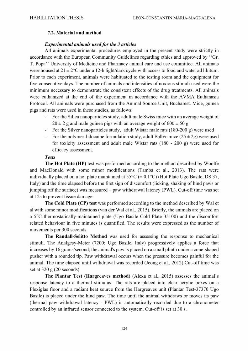
HABILITATION THESIS LEON-CONSTANTIN MARIA-MAGDALENA
124
7.2. Material and method
Experimental animals used for the 3 articles All animals experimental procedures employed in the present study were strictly in
accordance with the European Community Guidelines regarding ethics and approved by ‘‘Gr. T. Popa’’ University of Medicine and Pharmacy animal care and use committee. All animals were housed at 21 ± 2°C under a 12-h light/dark cycle with access to food and water ad libitum. Prior to each experiment, animals were habituated to the testing room and the equipment for five consecutive days. The number of animals and intensities of noxious stimuli used were the minimum necessary to demonstrate the consistent effects of the drug treatments. All animals were euthanized at the end of the experiment in accordance with the AVMA Euthanasia Protocol. All animals were purchased from the Animal Source Unit, Bucharest. Mice, guinea pigs and rats were used in these studies, as follows:
- For the Silica nanoparticles study, adult male Swiss mice with an average weight of 20 ± 2 g and male guinea pigs with an average weight of 600 ± 50 g
- For the Silver nanoparticles study, adult Wistar male rats (180-200 g) were used - For the polymer-lidocaine formulation study, adult Balb/c mice (25 ± 2g) were used
for toxicity assessment and adult male Wistar rats (180 - 200 g) were used for efficacy assessment.
Tests The Hot Plate (HP) test was performed according to the method described by Woolfe
and MacDonald with some minor modifications (Tamba et al., 2013). The rats were individually placed on a hot plate maintained at 55°C (± 0.1°C) (Hot Plate Ugo Basile, DS 37, Italy) and the time elapsed before the first sign of discomfort (licking, shaking of hind paws or jumping off the surface) was measured – paw withdrawal latency (PWL). Cut-off time was set at 12s to prevent tissue damage.
The Cold Plate (CP) test was performed according to the method described by Wal et al with some minor modifications (van der Wal et al., 2015). Briefly, the animals are placed on a 5°C thermostatically-maintained plate (Ugo Basile Cold Plate 35100) and the discomfort related behaviour in five minutes is quantified. The results were expressed as the number of movements per 300 seconds.
The Randall-Selitto Method was used for assessing the response to mechanical stimuli. The Analgesy-Meter (7200; Ugo Basile, Italy) progressively applies a force that increases by 16 grams/second; the animal's paw is placed on a small plinth under a cone-shaped pusher with a rounded tip. Paw withdrawal occurs when the pressure becomes painful for the animal. The time elapsed until withdrawal was recorded (Jeong et al., 2012).Cut-off time was set at 320 g (20 seconds).
The Plantar Test (Hargreaves method) (Alexa et al., 2015) assesses the animal’s response latency to a thermal stimulus. The rats are placed into clear acrylic boxes on a Plexiglas floor and a radiant heat source from the Hargreaves unit (Plantar Test-37370 Ugo Basile) is placed under the hind paw. The time until the animal withdraws or moves its paw (thermal paw withdrawal latency - PWL) is automatically recorded due to a chronometer controlled by an infrared sensor connected to the system. Cut-off is set at 30 s.
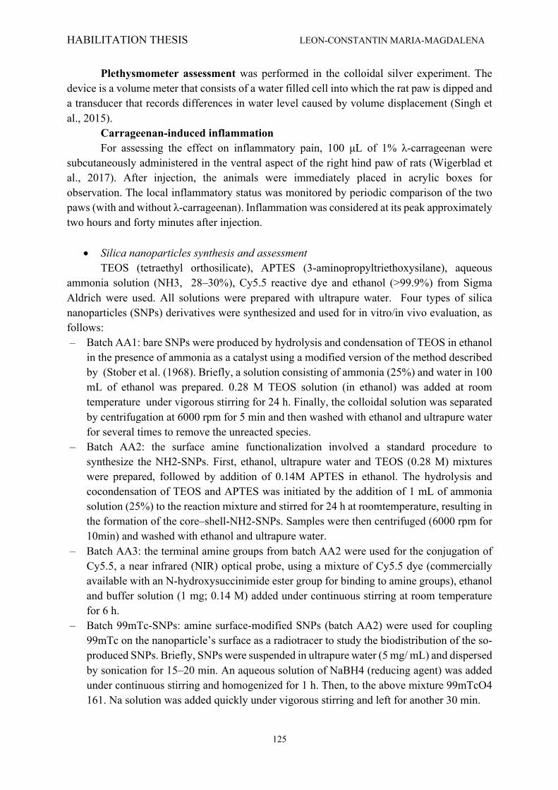
HABILITATION THESIS LEON-CONSTANTIN MARIA-MAGDALENA
125
Plethysmometer assessment was performed in the colloidal silver experiment. The device is a volume meter that consists of a water filled cell into which the rat paw is dipped and a transducer that records differences in water level caused by volume displacement (Singh et al., 2015).
Carrageenan-induced inflammation For assessing the effect on inflammatory pain, 100 μL of 1% λ-carrageenan were
subcutaneously administered in the ventral aspect of the right hind paw of rats (Wigerblad et al., 2017). After injection, the animals were immediately placed in acrylic boxes for observation. The local inflammatory status was monitored by periodic comparison of the two paws (with and without λ-carrageenan). Inflammation was considered at its peak approximately two hours and forty minutes after injection.
• Silica nanoparticles synthesis and assessment TEOS (tetraethyl orthosilicate), APTES (3-aminopropyltriethoxysilane), aqueous
ammonia solution (NH3, 28–30%), Cy5.5 reactive dye and ethanol (>99.9%) from Sigma Aldrich were used. All solutions were prepared with ultrapure water. Four types of silica nanoparticles (SNPs) derivatives were synthesized and used for in vitro/in vivo evaluation, as follows: – Batch AA1: bare SNPs were produced by hydrolysis and condensation of TEOS in ethanol
in the presence of ammonia as a catalyst using a modified version of the method described by (Stober et al. (1968). Briefly, a solution consisting of ammonia (25%) and water in 100 mL of ethanol was prepared. 0.28 M TEOS solution (in ethanol) was added at room temperature under vigorous stirring for 24 h. Finally, the colloidal solution was separated by centrifugation at 6000 rpm for 5 min and then washed with ethanol and ultrapure water for several times to remove the unreacted species.
– Batch AA2: the surface amine functionalization involved a standard procedure to synthesize the NH2-SNPs. First, ethanol, ultrapure water and TEOS (0.28 M) mixtures were prepared, followed by addition of 0.14M APTES in ethanol. The hydrolysis and cocondensation of TEOS and APTES was initiated by the addition of 1 mL of ammonia solution (25%) to the reaction mixture and stirred for 24 h at roomtemperature, resulting in the formation of the core–shell-NH2-SNPs. Samples were then centrifuged (6000 rpm for 10min) and washed with ethanol and ultrapure water.
– Batch AA3: the terminal amine groups from batch AA2 were used for the conjugation of Cy5.5, a near infrared (NIR) optical probe, using a mixture of Cy5.5 dye (commercially available with an N-hydroxysuccinimide ester group for binding to amine groups), ethanol and buffer solution (1 mg; 0.14 M) added under continuous stirring at room temperature for 6 h.
– Batch 99mTc-SNPs: amine surface-modified SNPs (batch AA2) were used for coupling 99mTc on the nanoparticle’s surface as a radiotracer to study the biodistribution of the so-produced SNPs. Briefly, SNPs were suspended in ultrapure water (5 mg/ mL) and dispersed by sonication for 15–20 min. An aqueous solution of NaBH4 (reducing agent) was added under continuous stirring and homogenized for 1 h. Then, to the above mixture 99mTcO4 161. Na solution was added quickly under vigorous stirring and left for another 30 min.

HABILITATION THESIS LEON-CONSTANTIN MARIA-MAGDALENA
126
The obtained product was separated by centrifugation and washed with ultrapure water to remove the uncoupled 99mTc radionuclide. 164
99mTcO4 Na (sodium pertechnetate) was chosen for labelling because it is the most commonly used emitting radioisotope in nuclear medicine having a convenient half-life of approximately 6 h, appropriate energy (140 keV) for imaging on a standard gamma camera and less attenuation by soft tissue. A 12.5 GBq Dry- 170 tec Technetium Generator was used for the production of 99mTcO4 - Na supplied by GE Healthcare. For characterizing the newly-created SNPs, SEM experiments were carried out at an accelerating voltage of 20 kV on a field emission scanning electron microscope (FE-SEM, Zeiss, SUPRA VP 40). Samples suspended in ultrapure water (1 mg/mL) were deposited on freshly cleaved mica surface, dried and gold/palladium coated. Determinations of nanoparticles size, zeta potential and polydispersity index were performed using a Zetasizer (Zetasizer Nano ZS, Malvern Instruments). The samples were dispersed in ultrapure water and measured at a scattering angle of 90 and 25o C. Qualitative chemical composition assessment of the nanoparticles was performed by FTIR analysis (Bomem MB spectrometer). The material was finely grounded and dispersed into KBr powder-pressed pellets using a ratio of approximately 1 mg sample/ mg KBr. IR absorbance data were obtained over a range of wavenumbers from 4000 to 400 cm.
For acute systemic toxicity screening, three groups of mice (10 mice per dose) received intraperitoneal 0.1 mL SNPs buffer solution (batch AA3) in a single dose (25, 50 and 100 mg/kg body weight (bw)) and kept under observation for 5 days after administration. For all experiments, nanoparticles were ultrasonicated for 30 min directly prior to use. The maximum possible dose (20 mg SNPs per 1 mL PBS) administered was defined by preliminary experimental results – maximum tolerated dose (MTD) (Kong et al., 2011). The animals were carefully observed for obvious signs of toxicity, such as convulsions or body weight effects. All mice were necropsied to detect macroscopic evidence of organ and tis- sue damage or dysfunction.
Microscopy biodistribution studies were performed as follows: fluorescent silica nanoparticles (batch AA3) (7.5 mg/1 mL PBS) were administered via two routes in 2 groups of 6 mice: intravenously (0.1 mL /mouse) and orally (0.2 mL/mouse). The control group (6 animals) received a similar volume of PBS. At specific times after intravenous (30 min and 2 h) or oral administration (1, 2, 24, 48 and 72 h) of the labelled nanoparticles (batch AA3), the mice were deeply anesthetized with xylazine and transcardially perfused with 15 mL 0.9% saline solution, followed by fresh 4% paraformaldehyde (PFA) in 75 mL 0.1 M PBS. Key organs (brain, heart, liver, lung, kidneys, spleen, testis and bladder) were extracted and post-fixed overnight in 4% PFA, followed by cryoprotection in 30% sucrose in PBS for 72 h. Sections of the above mentioned organs were cut using a freezing microtome (CM 1850 Leica Microsystems, Germany), were collected, mounted on slides and examined in a Leica Confocal Laser Scanning Microscope (TCS SPE DM 5500Q), using a laser diode (635 nm line), taking into account that the Cy5.5 fluorochrome is optimally excited near 675 nm and fluoresces near 694 nm.
For scintigraphy biodistribution studies, 99mTc radiolabelled NPs were used in order to study the biodistribution (batch 99mTc-SNPs). Five groups of 4 male guinea pigs were injected with 37 MBq/kg bw of 99mTc-coupled amino SNPs. Jugular vein was exposed and
HABILITATION THESIS LEON-CONSTANTIN MARIA-MAGDALENA
127
cannulated for infusion of radiotracer. A control group of 4 male guinea pigs received 37 MBq/kg bw of 99mTcO4 Na, in the same conditions. The guinea pigs were placed under a dual-head gamma camera (SPECT Siemens Gamma camera with LEAP (Low Energy All Purpose) collimators). The body distribution profile was recorded in dynamic mode till 5 min post injection and in static mode every 15 min for 2 h, then every 30 min for a total of 6 h. The animals were sacrificed after 6 h and different organs were extracted and immediately gamma counted.
• Nanoparticle silver can have a pro-inflammatory effect The following drugs were used in the experiment: colloidal solution with 500 parts per
million (ppm) silver nanoparticles purchased from US Research Nanomaterials, Inc, Houston, USA (Silver (Ag) Nanopowder / Nanoparticles (Ag, 99.99%, 30-50 nm, w/~0.2 wt% PVP Coated), colloidal silver 20 ppm (©Nano Silver, 30-50 nm, PVP Coated, Vita Crystal, RO), λ-carrageenan 1% diluted in fresh saline (Sigma-Adrich Germany). Doses were selected according to literature data. In a dermal toxicity study, Korani et al used doses that ranged from 100 ppm to 10000 ppm nanosilver in a subchronic administration regime (Korani et al., 2013). Up to 10 ppm nanosilver were administered daily in pregnant female rats via drinking water in a study to assess the expression of procaspase-3 in newborn rat brain (Ganjuri et al., 2015). Another experiment repeatedly injected 60-2000 ppm nanosilver subcutaneously (intralesional administration) in a mouse model of cutaneous leishmaniasis (Nilforoushzadeh et al., 2012).In the present study, one group received nanosilver 500 ppm and the other treatment group received nanosilver 20 ppm single dose.
Rats were divided in three groups (n=6/group) as follows: Group S1 received 10 microliters colloidal silver 500 ppm, group S2 received 10 microliters colloidal silver 20 ppm and group C received an equivalent volume of saline. All drugs were injected subcutaneously (s.c.) in the intraplantar region of right hind paw. Response latencies were assessed by means of the plantar test and the analgesy-meter; inflammation was assessed by means of the plethysmometer. All animals were evaluated at baseline and 3 and 24 h after colloidal silver/saline administration. After the 24-h assessment, all animals received an s.c. intraplantar injection of 10 microliters λ-carrageenan 1% into the right hind paw. This lead to a localized inflammatory response (acute inflammation). The above-mentioned assessments were again performed 3, 6 and 24 h after λ-carrageenan administration.
• The novel polymeric based lidocaine formulation for topical analgesia The following drugs were used in the experiment: CX001 was synthesized by “Petru
Poni” Institute and AB Pharm Romania. The formula was delivered as a powder and diluted in saline solution in order to create a spray that was applied by means of a commercially-available disperser. CX001 is a matrix-like compound that contains a mixture of innovative polymers and lidocaine. The manner in which lidocaine is included in this compound significantly influences the skin-compound interaction and enhances topical drug diffusion.
CX001 aims to surpass the existing topical lidocaine-based analgesics by increasing lidocaine solubility and skin permeability, improving target control and decreasing side-effects and toxicity. EMLA© cream (lidocaine 2.5% and prilocaine 2.5%) (Astra Zeneca) was purchased and used as a reference drug for the efficacy of CX001. λ-carrageenan (CG) diluted in fresh saline solution (Sigma-Adrich Germany) was administered subcutaneously in order to produce the model of inflammation.
HABILITATION THESIS LEON-CONSTANTIN MARIA-MAGDALENA
128
For the safety assessment, male mice were divided into six groups (n = 6/group). Three of the groups received topical administration of EMLA©, CX or saline solution on the right hind paw. The other three groups first received a subcutaneous injection of λ-carrageenan into the right hind paw and topical application of EMLA©, CX or saline 2 hours and forty minutes after. The mice were sacrificed 15 minutes after the topical applications and the liver and the skin of the hind paw were removed and fixed in a 10% formalin solution for additional tests. Liver samples were stained with the hematoxylin and eosin (HE) coloration. The skin samples were stained with both hematoxylin and eosin (HE) and Szekely tricromic coloration (SZ). All samples were observed microscopically for any histopathological changes.
For the nociceptive pain assessment, rats were divided in three groups (n = 6/group) and each group received as follows:
1. topical administration of a thin layer of EMLA© cream (group En) 2. two puffs of saline solution (group Sn) 3. two puffs of CX001 spray (group CXn). HP and Randall-Selitto assessments were performed at baseline and 5, 15, 30, 45, 60,
120, 180 and respectively 240 minutes after administration. The response to cold stimuli was assessed by means of the CP test that was performed at baseline, 30 minutes after topical administration and hourly after that over a four hours period of time.
For the inflammatory pain assessment, all rats were tested by means of HP, CP and Randall-Selitto method at baseline. Afterwards, each animal received a subcutaneous intraplantar injection of 10 μL 1% λ-carrageenan into the right hind paw. Two hours and forty minutes after intraplantar administration, rats were divided into three groups (n = 6/group) receiving either EMLA© (group Ei), saline solution (group Si) or CX001 (group CXi). HP, CP and the Randall-Selitto assessments were performed with the same frequency as for the nociceptive pain assessment. Statistical analysis
Statistical analysis and graphic design was performed with the aid of GraphPad 3.0 software (GraphPad Software, La Jolla, CA). Descriptive statistics and analysis of variance (Mixed ANOVA with Tukey post-hoc test) were performed. The data obtained was expressed as the mean value ± standard error. For all analyses, the a priori significance level was set at p<0.05. For the silica nanoparticles study, the experiments were replicated three independent times.
7.3. Results
• Silica nanoparticles synthesis and assessment Four types of SNPs were synthesized, as shown in Fig. 51 using a derived method with original modifications for the synthesis of spherical and monodisperse silica nanoparticles. First, SNPs bearing OH groups were prepared from aqueous alcohol solutions of silicon alkoxides in the presence of ammonia as a catalyst by hydrolysis with the formation of silanol groups and condensation reaction for the siloxane bridges development (batch AA1). Since it is supposed that the binding affinity between the nanocarrier and the radioisotope along with the stability of the obtained radiotracer could be modulated by modifying the surface properties of the silica nanocarrier, functionalization via covalent bonding of organic groups was achieved
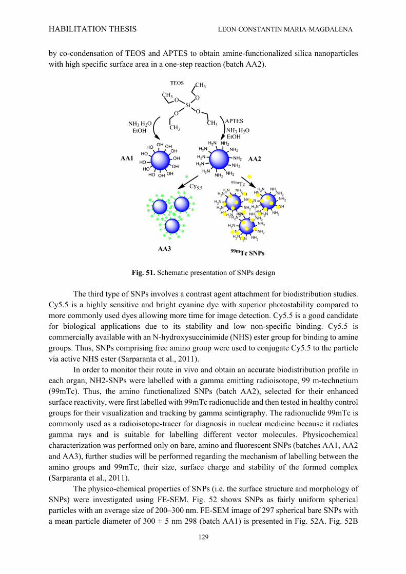
HABILITATION THESIS LEON-CONSTANTIN MARIA-MAGDALENA
129
by co-condensation of TEOS and APTES to obtain amine-functionalized silica nanoparticles with high specific surface area in a one-step reaction (batch AA2).
Fig. 51. Schematic presentation of SNPs design The third type of SNPs involves a contrast agent attachment for biodistribution studies.
Cy5.5 is a highly sensitive and bright cyanine dye with superior photostability compared to more commonly used dyes allowing more time for image detection. Cy5.5 is a good candidate for biological applications due to its stability and low non-specific binding. Cy5.5 is commercially available with an N-hydroxysuccinimide (NHS) ester group for binding to amine groups. Thus, SNPs comprising free amino group were used to conjugate Cy5.5 to the particle via active NHS ester (Sarparanta et al., 2011).
In order to monitor their route in vivo and obtain an accurate biodistribution profile in each organ, NH2-SNPs were labelled with a gamma emitting radioisotope, 99 m-technetium (99mTc). Thus, the amino functionalized SNPs (batch AA2), selected for their enhanced surface reactivity, were first labelled with 99mTc radionuclide and then tested in healthy control groups for their visualization and tracking by gamma scintigraphy. The radionuclide 99mTc is commonly used as a radioisotope-tracer for diagnosis in nuclear medicine because it radiates gamma rays and is suitable for labelling different vector molecules. Physicochemical characterization was performed only on bare, amino and fluorescent SNPs (batches AA1, AA2 and AA3), further studies will be performed regarding the mechanism of labelling between the amino groups and 99mTc, their size, surface charge and stability of the formed complex (Sarparanta et al., 2011). The physico-chemical properties of SNPs (i.e. the surface structure and morphology of SNPs) were investigated using FE-SEM. Fig. 52 shows SNPs as fairly uniform spherical particles with an average size of 200–300 nm. FE-SEM image of 297 spherical bare SNPs with a mean particle diameter of 300 ± 5 nm 298 (batch AA1) is presented in Fig. 52A. Fig. 52B
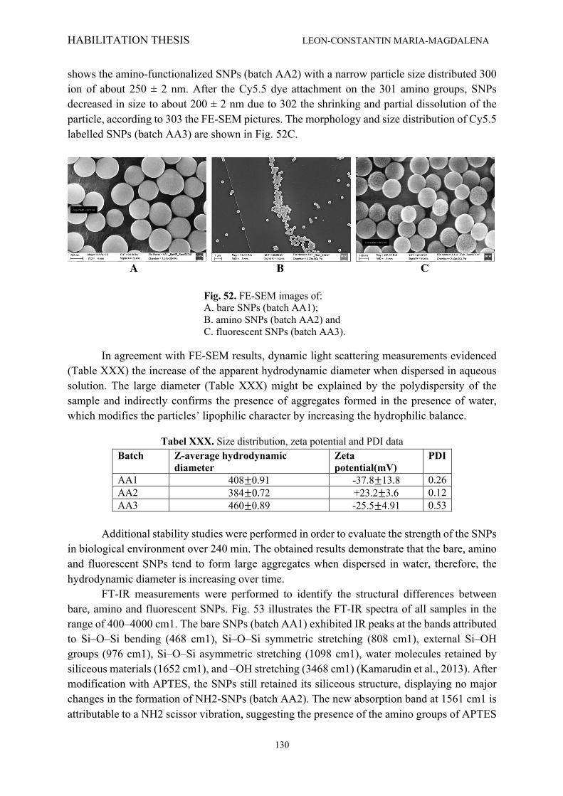
HABILITATION THESIS LEON-CONSTANTIN MARIA-MAGDALENA
130
shows the amino-functionalized SNPs (batch AA2) with a narrow particle size distributed 300 ion of about 250 ± 2 nm. After the Cy5.5 dye attachment on the 301 amino groups, SNPs decreased in size to about 200 ± 2 nm due to 302 the shrinking and partial dissolution of the particle, according to 303 the FE-SEM pictures. The morphology and size distribution of Cy5.5 labelled SNPs (batch AA3) are shown in Fig. 52C.
Fig. 52. FE-SEM images of: A. bare SNPs (batch AA1); B. amino SNPs (batch AA2) and C. fluorescent SNPs (batch AA3).
In agreement with FE-SEM results, dynamic light scattering measurements evidenced (Table XXX) the increase of the apparent hydrodynamic diameter when dispersed in aqueous solution. The large diameter (Table XXX) might be explained by the polydispersity of the sample and indirectly confirms the presence of aggregates formed in the presence of water, which modifies the particles’ lipophilic character by increasing the hydrophilic balance.
Tabel XXX. Size distribution, zeta potential and PDI data Batch Z-average hydrodynamic
diameter Zeta potential(mV)
PDI
AA1 408±0.91 -37.8±13.8 0.26 AA2 384±0.72 +23.2±3.6 0.12 AA3 460±0.89 -25.5±4.91 0.53
Additional stability studies were performed in order to evaluate the strength of the SNPs
in biological environment over 240 min. The obtained results demonstrate that the bare, amino and fluorescent SNPs tend to form large aggregates when dispersed in water, therefore, the hydrodynamic diameter is increasing over time. FT-IR measurements were performed to identify the structural differences between bare, amino and fluorescent SNPs. Fig. 53 illustrates the FT-IR spectra of all samples in the range of 400–4000 cm1. The bare SNPs (batch AA1) exhibited IR peaks at the bands attributed to Si–O–Si bending (468 cm1), Si–O–Si symmetric stretching (808 cm1), external Si–OH groups (976 cm1), Si–O–Si asymmetric stretching (1098 cm1), water molecules retained by siliceous materials (1652 cm1), and –OH stretching (3468 cm1) (Kamarudin et al., 2013). After modification with APTES, the SNPs still retained its siliceous structure, displaying no major changes in the formation of NH2-SNPs (batch AA2). The new absorption band at 1561 cm1 is attributable to a NH2 scissor vibration, suggesting the presence of the amino groups of APTES

HABILITATION THESIS LEON-CONSTANTIN MARIA-MAGDALENA
131
molecules. Also, compared with the bare SNPs, the APTES-modified SNPs show an additional weak band at 2929 cm1, which can be assigned to the alkyl groups [–(CH2)n–] present in APTES.
Fig. 53. FT-IR of bare SNPs (batch AA1), amino SNPs (batch AA2) and fluorescent SNPs (batch AA3).
After cyanine dye attachment, the relative intensity of amine group vibrations became less intense, suggesting that Cy5.5 was successfully loaded onto the surface of SNPs. New bands at 1668 cm1, and 1629 cm1 appeared after the loading process, which might be associated with the aqueous solution of Cy5.5, and N–H group. All these facts suggest that Cy5.5 has been successfully attached to the surface of the NH2-SNPs. The acute toxicity screening allowed identifying the dose of 100 mg/kg bw as MTD for this study, with no significant changes in animal behavior or weight. The histological examination (data not shown) of main organ tissue (liver, kidney, heart, stomach and intestine) that followed the five days observation period identified no histological changes from the normal tissues characteristics. The MTD was several times higher than the doses administered in the bioavailability studies, profiling a good safety profile for future therapeutic associations of the SNPs with drugs. Regarding biodistribution, following the intravenous administration of fluorescent SNPs, batch AA3 (size 200 nm), at specific time points (30 min and 2 h) the SNPs did not penetrate the blood brain barrier and were not present in the myocardium. Bladder tissue sample were also negative. However, SNPs were found in the rest of the investigated organs (liver, kidney, testis, feces and lung (Fig. 54). In order to validate the obtained results, the organs of the control groups were also evaluated after PBS administration. Fig. 54B represents the comparative control liver examination at 2 h in the same conditions as the SNPs. The image evidenced the autofluorescence that was taken into consideration for the evaluation of the SNPs
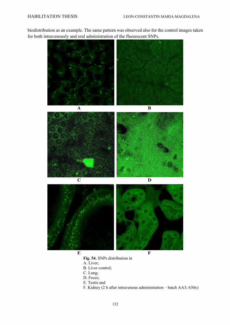
HABILITATION THESIS LEON-CONSTANTIN MARIA-MAGDALENA
132
biodistribution as an example. The same pattern was observed also for the control images taken for both intravenously and oral administration of the fluorescent SNPs.
Fig. 54. SNPs distribution in A. Liver; B. Liver control; C. Lung; D. Feces; E. Testis and F. Kidney (2 h after intravenous administration – batch AA3; 630x)
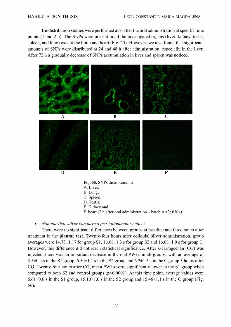
HABILITATION THESIS LEON-CONSTANTIN MARIA-MAGDALENA
133
Biodistribution studies were performed also after the oral administration at specific time points (1 and 2 h). The SNPs were present in all the investigated organs (liver, kidney, testis, spleen, and lung) except the brain and heart (Fig. 55). However, we also found that significant amounts of SNPs were distributed at 24 and 48 h after administration, especially in the liver. After 72 h a gradually decrease of SNPs accumulation in liver and spleen was noticed.
Fig. 55. SNPs distribution in A. Liver; B. Lung; C. Spleen; D. Testis; E. Kidney and F. heart (2 h after oral administration – batch AA3; 630x)
• Nanoparticle silver can have a pro-inflammatory effect
There were no significant differences between groups at baseline and three hours after treatment in the plantar test. Twenty-four hours after colloidal silver administration, group averages were 14.73±1.17 for group S1, 16.68±1.3 s for group S2 and 16.08±1.9 s for group C. However, this difference did not reach statistical significance. After λ-carrageenan (CG) was injected, there was an important decrease in thermal PWLs in all groups, with an average of 3.5±0.4 s in the S1 group, 6.50±1.1 s in the S2 group and 6.2±1.3 s in the C group 3 hours after CG. Twenty-four hours after CG, mean PWLs were significantly lower in the S1 group when compared to both S2 and control groups (p<0.0001). At this time point, average values were 6.61±0.6 s in the S1 group, 15.10±1.0 s in the S2 group and 15.46±1.1 s in the C group (Fig. 56).

HABILITATION THESIS LEON-CONSTANTIN MARIA-MAGDALENA
134
Fig. 56. Average thermal PWLs throughout the experiment. * = p<0.05 (Tukey post-hoc)
There were no significant differences between groups at baseline and three hours after treatment in the Randall-Selito test. However, 24 h after colloidal silver administration both treatment groups had a significantly higher sensibility to mechanical stimuli, with averages of 4.16±0.2 s for group S1, 4.75±0.2 s for group S2 and 5.63±0.4 s for group C (p=0.02 for S1 vs. C and p=0.03 for S2 vs. C) (Fig. 65). After CG injection, in all three groups a decrease in mechanical PWLs was recorded - (1 s latency for all rats three hours after CG). Twenty-four hours after CG, control averages increased to 2 s, whereas averages in the treatment groups were 1.60±0.2 s (S1) and 2.25±0.3 s (S2). After CG administration, there were no statistically significant differences between groups (Fig. 57).
Fig. 57. Average mechanical PWLs throughout the experiment *=p<0.05 (Tukey post-hoc) There were no significant differences between groups at baseline as assessed by plethysmometric measurement. Three hours after colloidal silver injection, paw volume was 1.31±0.03 in group S1, 1.27±0.03 in group S2 and 0.91±0.03 in group C. This difference was

HABILITATION THESIS LEON-CONSTANTIN MARIA-MAGDALENA
135
statistically significant, with p < 0.0001 for S1 vs. C and p = 0.0002 for S2 vs. C (Fig. 58). Colloidal silver’s effect was persistent – 24 h after treatment S1 and S2 groups had an increased paw volume when compared with C group, with p = 0.02 for both S1 vs. C and S2 vs. C. Three hours after CG injection, an increase in paw volume was noted for all groups. However, paw edema was more pronounced in the silver-treated groups, with an average of 2.19±0.1 in group S1, 2.15±0.07 in group S2 and 1.92±0.09 in group C (p < 0.006 for S1 vs. C and p = 0.004 for S2 vs. C) (fig. 58). In the control group, paw edema reached a maximum 6 h after CG injection (mean of 2.24±0.06); at this time point, no statistically significant differences were noted between groups. Paw edema began to decrease afterwards and 24 hours after CG injection averages were 1.76±0.03 in group S1, 1.54±0.09 in group S2 and 1.56±0.08 in group C. Paw volume of group S1 rats was significantly larger than paw volume in group C (p=0.01).
Fig. 58. Paw volume throughout the experiment *=p<0.05 (Tukey post-hoc)
• The novel polymeric based lidocaine formulation for topical analgesia Skin tissue from the mice that had only received EMLA©/CX001/saline topical
application had a normal histological profile. The epidermal, superficial dermal and profound dermal layers were in physiological condition. No other differences were noted between groups. The skin tissue from the mice that had received a subcutaneous carrageenan injection prior to topical drug administration had histological signs of inflammation, with neutrophil infiltration, vascular congestion and oedema (Fig. 59). No differences were noted between EMLA© and CX001-treated groups, although lidocaine in topical administration (in either EMLA© or CX001 formula) was associated with less inflammation. Histological analysis of liver tissue showed that no changes occurred in the hepatic structures.

HABILITATION THESIS LEON-CONSTANTIN MARIA-MAGDALENA
136
A B C Fig. 59. HE coloration – microscopy (x 100) assessment of skin tissue in the inflammation groups. A – EMLA, B – CX001, C – saline.
In the hot plate assessment, for the nociceptive pain group, there were no significant
differences at baseline, with an average PWL of 3.35 ± 0.28 s in the Sn group, 3.48 ± 0.37 s in the En group and 3.48 ± 0.27 s in the CXn group. Five minutes after topical analgesic/saline administration, EMLA© treated animals had an increase in PWL when compared with saline solution (5.01 ± 0.47 s vs. 3.43 ± 0.22 s) or CX001 treated animals (5.01 ± 0.47 s vs. 3.16 ± 0.29 s). PWLs started to increase in the CX001 treated group after this time point and both drugs were superior to saline solution, 15 minutes after administration. The effect remained consistent at 30, 45 and 60 minutes after administration (p < 0.005) (Figure 60). PWLs in all groups were similar at the end of the experiment (after four hours). ANOVA repeated measures identified that there is a significant effect throughout the experiment, with p = 0.0058 (F (2, 10) = 9.022) and significance in time (p = 0.0095).
Fig.60. Hot Plate PWLs in the nociceptive pain group. * = <0.005 vs control group; ** = < 0.005
versus EMLA/CX001group In the inflammatory pain group, there were also no significant differences between
groups at baseline. After CG injection, all animals showed signs of discomfort, avoiding placing the injected hind paw on the ground and licking/scratching the affected area. Five minutes after topical analgesic/saline administration, saline-treated animals had a lower PWL compared to baseline (2.66 ± 0.40 s), whereas CXi and Ei groups showed no such modifications. Additionally, there was a statistically significant difference between the Si and CXi groups (p=0.05), but no significant difference between the Si and Ei groups (Fig. 61). Throughout the
T im e (m in )
PW
L (
se
co
nd
s)
B a s e lin e 5 m in 1 5 m in 3 0 m in 4 5 m in 6 0 m in 2 h o u r s 3 h o u r s 4 h o u r s0
2
4
6
8
S a lin eE M LAC X 0 0 1
*
***
*** **
*
****

HABILITATION THESIS LEON-CONSTANTIN MARIA-MAGDALENA
137
experiment, both CX001 and EMLA© topical administration led to increased PWLs as compared to saline administration. ANOVA repeated measures identified that there is a significant effect throughout the experiment, with p < 0.0001 (F (2, 10) = 26.86) with significance in time (p = 0.0097). There were no significant differences between Ei and CXi groups at any time point.
Fig. 61. Hot Plate PWLs in the inflammatory pain group. * = <0.005 vs control group; ** = < 0.005
versus EMLA/CX001 group Regarding nociceptive response to cold, neither EMLA© nor CX001 induced any
significant changes throughout the experiment in the number of cold-evoked movements in 300 seconds. ANOVA repeated measures indicated there was no significant substance or time effect.
In the inflammatory pain group, there were no significant differences between groups at baseline, with an average number of cold-evoked movements of 8.75 ± 0.47 s in the Si group, 7.75 ± 0.48 s in the Ei group and 7.75 ± 0.47 s in the CXi group. However, thirty minutes after topical analgesic/saline solution administration, saline treated animals expressed more cold/pressure related discomfort, with an average of 36 ± 1.58 s movements in 300 seconds, whereas rats in the Ei and CXi groups had an average of 22 ± 2.27 s respectively 19.75 ± 2.05 s movements (Fig. 62). Throughout the experiment, both CX001 and EMLA© topical administration were associated with fewer discomfort related movements as compared with saline administration (p < 0.005 for CXi and Ei at 30, 60 and 120 minutes). ANOVA repeated measures identified that there is a significant effect throughout the experiment, with p = 0.04 (F (2, 6) = 5.756) and that the effect is significant in time (p = 0.0076). There were no statistically significant differences between Ei and CXi groups at any time point. Both EMLA© and CX001 were more effective in decreasing the number of cold induced discomfort movements in the inflammatory model than in the nociceptive model. Also, we noted a difference between baseline values and post-CG values in the saline group (when compared to the differences noted in other tests).
T im e (m in )
PW
L (
se
co
nd
s)
B a s e lin e 5 m in 1 5 m in 3 0 m in 4 5 m in 6 0 m in 2 h o u r s 3 h o u r s 4 h o u r s0
1
2
3
4
5
C on tro lE M LAC X X
***
***
**
**
*
*
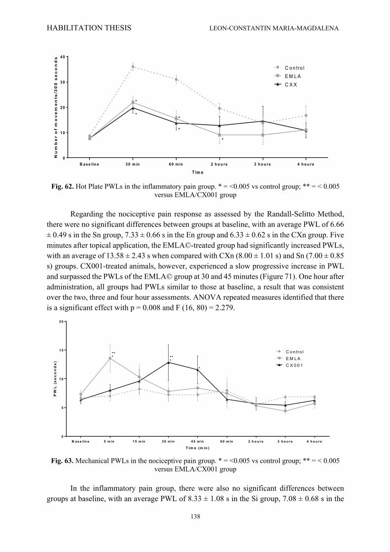
HABILITATION THESIS LEON-CONSTANTIN MARIA-MAGDALENA
138
Fig. 62. Hot Plate PWLs in the inflammatory pain group. * = <0.005 vs control group; ** = < 0.005
versus EMLA/CX001 group Regarding the nociceptive pain response as assessed by the Randall-Selitto Method,
there were no significant differences between groups at baseline, with an average PWL of 6.66 ± 0.49 s in the Sn group, 7.33 ± 0.66 s in the En group and 6.33 ± 0.62 s in the CXn group. Five minutes after topical application, the EMLA©-treated group had significantly increased PWLs, with an average of 13.58 ± 2.43 s when compared with CXn (8.00 ± 1.01 s) and Sn (7.00 ± 0.85 s) groups. CX001-treated animals, however, experienced a slow progressive increase in PWL and surpassed the PWLs of the EMLA© group at 30 and 45 minutes (Figure 71). One hour after administration, all groups had PWLs similar to those at baseline, a result that was consistent over the two, three and four hour assessments. ANOVA repeated measures identified that there is a significant effect with p = 0.008 and F (16, 80) = 2.279.
Fig. 63. Mechanical PWLs in the nociceptive pain group. * = <0.005 vs control group; ** = < 0.005
versus EMLA/CX001 group In the inflammatory pain group, there were also no significant differences between
groups at baseline, with an average PWL of 8.33 ± 1.08 s in the Si group, 7.08 ± 0.68 s in the
T im e
Nu
mb
er
of
mo
ve
me
nts
/30
0 s
ec
on
ds
B a s e lin e 3 0 m in 6 0 m in 2 h o u r s 3 h o u r s 4 h o u r s0
1 0
2 0
3 0
4 0
C on tro lE M LAC X X
*
*
**
*
T im e (m in )
PW
L (
se
co
nd
s)
B a s e lin e 5 m in 1 5 m in 3 0 m in 4 5 m in 6 0 m in 2 h o u r s 3 h o u r s 4 h o u r s0
5
1 0
1 5
2 0
C on tro lE M LAC X 0 0 1
*
*
*****
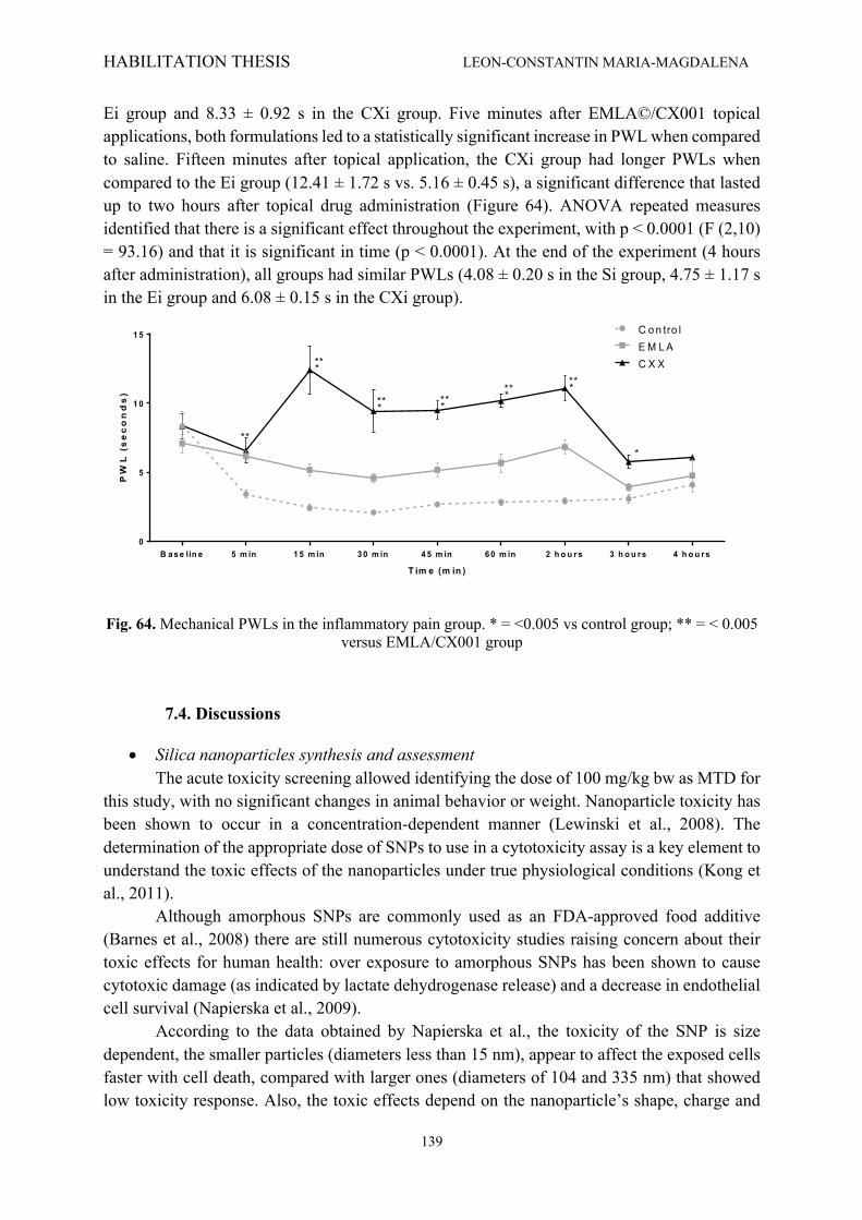
HABILITATION THESIS LEON-CONSTANTIN MARIA-MAGDALENA
139
Ei group and 8.33 ± 0.92 s in the CXi group. Five minutes after EMLA©/CX001 topical applications, both formulations led to a statistically significant increase in PWL when compared to saline. Fifteen minutes after topical application, the CXi group had longer PWLs when compared to the Ei group (12.41 ± 1.72 s vs. 5.16 ± 0.45 s), a significant difference that lasted up to two hours after topical drug administration (Figure 64). ANOVA repeated measures identified that there is a significant effect throughout the experiment, with p < 0.0001 (F (2,10) = 93.16) and that it is significant in time (p < 0.0001). At the end of the experiment (4 hours after administration), all groups had similar PWLs (4.08 ± 0.20 s in the Si group, 4.75 ± 1.17 s in the Ei group and 6.08 ± 0.15 s in the CXi group).
Fig. 64. Mechanical PWLs in the inflammatory pain group. * = <0.005 vs control group; ** = < 0.005
versus EMLA/CX001 group
7.4. Discussions
• Silica nanoparticles synthesis and assessment The acute toxicity screening allowed identifying the dose of 100 mg/kg bw as MTD for this study, with no significant changes in animal behavior or weight. Nanoparticle toxicity has been shown to occur in a concentration-dependent manner (Lewinski et al., 2008). The determination of the appropriate dose of SNPs to use in a cytotoxicity assay is a key element to understand the toxic effects of the nanoparticles under true physiological conditions (Kong et al., 2011). Although amorphous SNPs are commonly used as an FDA-approved food additive (Barnes et al., 2008) there are still numerous cytotoxicity studies raising concern about their toxic effects for human health: over exposure to amorphous SNPs has been shown to cause cytotoxic damage (as indicated by lactate dehydrogenase release) and a decrease in endothelial cell survival (Napierska et al., 2009). According to the data obtained by Napierska et al., the toxicity of the SNP is size dependent, the smaller particles (diameters less than 15 nm), appear to affect the exposed cells faster with cell death, compared with larger ones (diameters of 104 and 335 nm) that showed low toxicity response. Also, the toxic effects depend on the nanoparticle’s shape, charge and
T im e (m in )
PW
L (
se
co
nd
s)
B a s e lin e 5 m in 1 5 m in 3 0 m in 4 5 m in 6 0 m in 2 h o u r s 3 h o u r s 4 h o u r s0
5
1 0
1 5 C on tro lE M LAC X X
***
***
******
******

HABILITATION THESIS LEON-CONSTANTIN MARIA-MAGDALENA
140
density, as well as the viscosity and density of the solution, properties that influence the effective dose. As a result, defining the appropriate dose for in vitro/in vivo study is still a challenging task. In this study, fluorescent SNPs were used for general toxicity assessment. The therapeutic efficiency of a drug carrier does rely not only on its intrinsic activity but also on the bioavailability of the drug at targeted site. In the development of novel therapeutics, the ability to design a suitable pharmaceutical formulation for delivery is of utmost importance. The SNPs tested in the present study were shown to have adequate bioavailability and did not cross the BBB. However, more detailed studies regarding the bioaccumulation of SNPs in the liver, spleen, intestines, kidneys, and bladder is warranted to better clarify the adverse effects arising from such an accumulation. Considering the results obtained in our study, the SNPs showed in vivo similar biodistribution behavior to that demonstrated by others groups on nanostructured systems as silica nanoparticles (obtained by the same method) or chitosan nanoparticle, when the radioactivity was determined by scintigraphy (Banerjee et al., 2005; Xie et al., 2010). Sakai et al. (2012)) analyzed the whole-body distribution of 14C-ADP-labeled silica nanoparticles after intravenous injection into mice and the obtained radioactivity results showed also similar distribution patterns when determined by liquid scintillation counter. Identifying the specific biodistribution areas (compared with free 99mTc) could be the start point for the use of this nanostructure system as vector molecule for a certain radioisotope, to be used for diagnosis in nuclear medicine imaging (using a classical gamma emitter radioisotope, such as 99mTc) and, respectively, therapy (using a suitable corpuscular emitter radioisotope such as 131I) purposes.
• Nanoparticle silver can have a pro-inflammatory effect Despite the many advantages of nanotechnology, the use of metals at such a small scale comes with some changes in an element’s properties. Certain nanomaterials may exhibit significant toxicity to mammalian cells even if they are biochemically inert and biocompatible in bulk size (Shvedova et al., 2005). Upon reaching nanoscale, like other nanomaterials, silver particles exhibit remarkably unusual physicochemical properties and biological activities (Chen et al., 2008), which may lead to unpredictable effects and interactions. In vitro, silver nanoparticles are citotoxic for macrophages and generate free radicals (Schins et al., 2007); also, a recent review emphasizes nanosilver’s effect on cells pointing out that accumulation of nanoparticles of silver within the cell leads to oxidizing stress, genotoxicity, and cytotoxicity through apoptosis (Fontenoy et al., 2011). Carlson et al further underlined that the smallest particles have the most toxic effects (Carlson et al., 2008). In rats, chronic ingestion of silver nanoparticles induced heart and liver dysfunction and promoted systemic oxidation and inflammation (Ebabe Elle et al., 2013). However, scientific papers in the field mainly focus on the effects of inhalation, ingestion or topical use of silver nanoparticles; only few studies explore the effect of direct contact between internal organs/structures and silver nanoparticles and its consequences on local pain and inflammation. In one study performed by Sarhan and Hussein in 2014 (Sarhan et al., 2014) that explored the effects of intraperitoneal (i.p.) silver nanoparticles administration, results indicated marked citopathological changes in both renal and hepatic tissues, with increased white blood cell count. In our study, nanosilver induced hyperalgesia, but only in an inflammatory setting. There were no differences in pain behavior throughout the 24-h assessments performed after
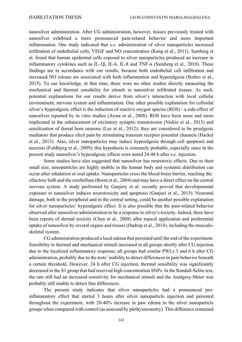
HABILITATION THESIS LEON-CONSTANTIN MARIA-MAGDALENA
141
nanosilver administration. After CG administration, however, tissues previously treated with nanosilver exhibited a more pronounced pain-related behavior and more important inflammation. One study indicated that s.c. administration of silver nanoparticles increased infiltration of endothelial cells, VEGF and NO concentration (Kang et al., 2011). Samberg et al. found that human epidermal cells exposed to silver nanoparticles produced an increase in inflammatory cytokines such as IL-1β, IL-6, IL-8 and TNF-α (Samberg et al., 2010). These findings are in accordance with our results, because both endothelial cell infiltration and increased NO release are associated with both inflammation and hyperalgesia (Srebro et al., 2015). To our knowledge, at that time, there were no other studies directly measuring the mechanical and thermal sensibility for stimuli in nanosilver infiltrated tissues. As such, potential explanations for our results derive from silver’s interaction with local cellular environment, nervous system and inflammation. One other possible explanation for colloidal silver’s hyperalgesic effect is the induction of reactive oxygen species (ROS) - a side-effect of nanosilver reported by in vitro studies (Arora et al., 2008). ROS have been more and more implicated in the enhancement of excitatory synaptic transmission (Nishio et al., 2013) and sensitization of dorsal horn neurons (Lee et al., 2012); they are considered to be proalgesic mediators that produce elicit pain by stimulating transient receptor potential channels (Hackel et al., 2013). Also, silver nanoparticles may induce hyperalgesia through cell apoptosis and necrosis (Foldbjerg et al., 2009); this hypothesis is extremely probable, especially since in the present study nanosilver’s hyperalgesic effects were noted 24-48 h after s.c. injection. Some studies have also suggested that nanosilver has neurotoxic effects. Due to their small size, nanoparticles are highly mobile in the human body and systemic distribution can occur after inhalation or oral uptake. Nanoparticles cross the blood-brain barrier, reaching the olfactory bulb and the cerebellum (Borm et al., 2004) and may have a direct effect on the central nervous system. A study performed by Ganjury et al. recently proved that developmental exposure to nanosilver induces neurotoxicity and apoptosis (Ganjuri et al., 2015). Neuronal damage, both in the peripheral and in the central setting, could be another possible explanation for silver nanoparticles’ hyperalgesic effect. It is also possible that the pain-related behavior observed after nanosilver administration to be a response to silver’s toxicity. Indeed, there have been reports of dermal toxicity (Chen et al., 2008) after topical application and preferential uptake of nanosilver by several organs and tissues (Hadrup et al., 2014), including the musculo-skeletal system. CG administration produced a local edema that persisted until the end of the experiment. Sensibility to thermal and mechanical stimuli increased in all groups shortly after CG injection due to the localized inflammatory response; all groups had similar PWLs 3 and 6 h after CG administration, probably due to the tests’ inability to detect differences in pain behavior beneath a certain threshold. However, 24 h after CG injection, thermal sensibility was significantly decreased in the S1 group that had received high concentration SNPs. In the Randall-Selito test, the rats still had an increased sensitivity for mechanical stimuli and the Analgesy-Meter was probably still unable to detect fine differences. The present study indicates that silver nanoparticles had a pronounced pro-inflammatory effect that started 3 hours after silver nanoparticle injection and persisted throughout the experiment, with 20-40% increase in paw edema in the silver nanoparticle groups when compared with control (as assessed by plethysmometry). This difference remained
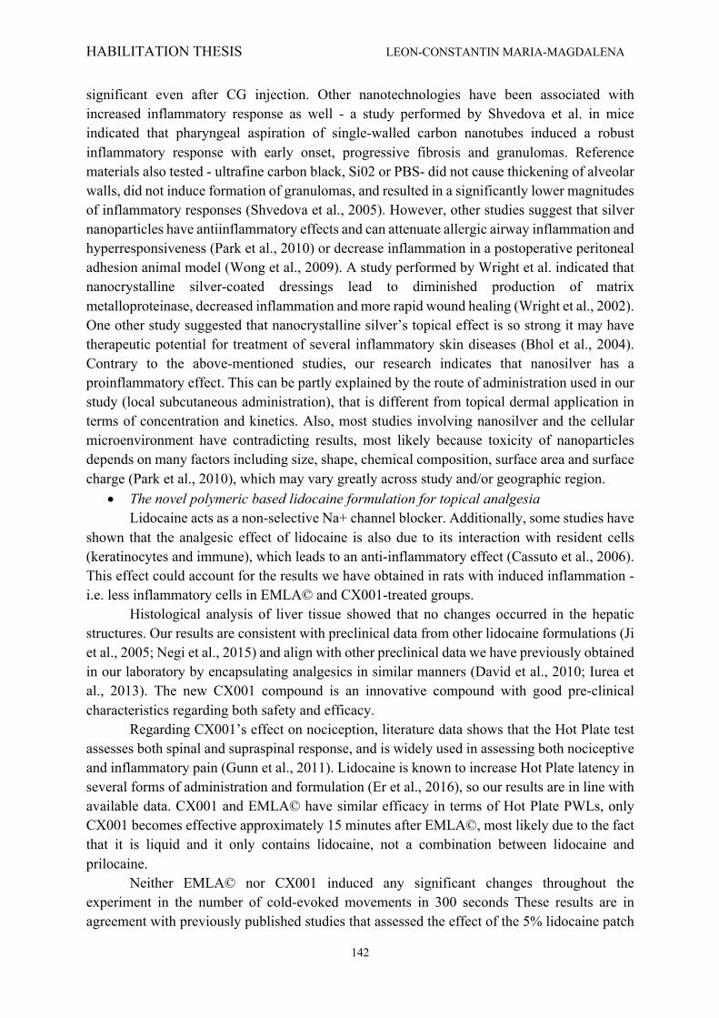
HABILITATION THESIS LEON-CONSTANTIN MARIA-MAGDALENA
142
significant even after CG injection. Other nanotechnologies have been associated with increased inflammatory response as well - a study performed by Shvedova et al. in mice indicated that pharyngeal aspiration of single-walled carbon nanotubes induced a robust inflammatory response with early onset, progressive fibrosis and granulomas. Reference materials also tested - ultrafine carbon black, Si02 or PBS- did not cause thickening of alveolar walls, did not induce formation of granulomas, and resulted in a significantly lower magnitudes of inflammatory responses (Shvedova et al., 2005). However, other studies suggest that silver nanoparticles have antiinflammatory effects and can attenuate allergic airway inflammation and hyperresponsiveness (Park et al., 2010) or decrease inflammation in a postoperative peritoneal adhesion animal model (Wong et al., 2009). A study performed by Wright et al. indicated that nanocrystalline silver-coated dressings lead to diminished production of matrix metalloproteinase, decreased inflammation and more rapid wound healing (Wright et al., 2002). One other study suggested that nanocrystalline silver’s topical effect is so strong it may have therapeutic potential for treatment of several inflammatory skin diseases (Bhol et al., 2004). Contrary to the above-mentioned studies, our research indicates that nanosilver has a proinflammatory effect. This can be partly explained by the route of administration used in our study (local subcutaneous administration), that is different from topical dermal application in terms of concentration and kinetics. Also, most studies involving nanosilver and the cellular microenvironment have contradicting results, most likely because toxicity of nanoparticles depends on many factors including size, shape, chemical composition, surface area and surface charge (Park et al., 2010), which may vary greatly across study and/or geographic region.
• The novel polymeric based lidocaine formulation for topical analgesia Lidocaine acts as a non-selective Na+ channel blocker. Additionally, some studies have
shown that the analgesic effect of lidocaine is also due to its interaction with resident cells (keratinocytes and immune), which leads to an anti-inflammatory effect (Cassuto et al., 2006). This effect could account for the results we have obtained in rats with induced inflammation - i.e. less inflammatory cells in EMLA© and CX001-treated groups.
Histological analysis of liver tissue showed that no changes occurred in the hepatic structures. Our results are consistent with preclinical data from other lidocaine formulations (Ji et al., 2005; Negi et al., 2015) and align with other preclinical data we have previously obtained in our laboratory by encapsulating analgesics in similar manners (David et al., 2010; Iurea et al., 2013). The new CX001 compound is an innovative compound with good pre-clinical characteristics regarding both safety and efficacy.
Regarding CX001’s effect on nociception, literature data shows that the Hot Plate test assesses both spinal and supraspinal response, and is widely used in assessing both nociceptive and inflammatory pain (Gunn et al., 2011). Lidocaine is known to increase Hot Plate latency in several forms of administration and formulation (Er et al., 2016), so our results are in line with available data. CX001 and EMLA© have similar efficacy in terms of Hot Plate PWLs, only CX001 becomes effective approximately 15 minutes after EMLA©, most likely due to the fact that it is liquid and it only contains lidocaine, not a combination between lidocaine and prilocaine.
Neither EMLA© nor CX001 induced any significant changes throughout the experiment in the number of cold-evoked movements in 300 seconds These results are in agreement with previously published studies that assessed the effect of the 5% lidocaine patch

HABILITATION THESIS LEON-CONSTANTIN MARIA-MAGDALENA
143
in healthy human volunteers and found that the threshold for heat and cold-induced pain were not changed by lidocaine when administered on tissue without inflammation (Wehrfritzl et al., 2011).
Also, we noted a difference between baseline values and post-CG values in the saline group (when compared to the differences noted in other tests). A possible explanation for this result is the fact that cold itself can act as a pain inhibitor by directly influencing the epidermal nervous terminations (Barkin, 2013) and by decreasing sensitivity in the inflamed area. Both EMLA© and CX001 were found to be significantly more effective in the inflammatory model, most likely because lidocaine primarily acts on Aδ and C fibers that have abnormal excitation, a situation that occurs in both inflammatory and neuropathic pain (Hashmi et al., 2012).
Our results are in concordance with available literature indicating that EMLA© topical administration is only slightly effective in reducing tactile sensitivity in newborn rats (Strain et al., 2014). A recently published clinical trial showed that EMLA© partially decreases pain during tympanocentesis in less than a third of the patients (Jyväkorpi, 1996). The result of this study is particularly relevant, especially since the perinatal period is considered to be a time where topical analgesics are most effective (Hardman et al., 1998). Due to its improved formula, CX001 is significantly superior to EMLA© as assessed by the Randall-Selitto test, a method that primarily evaluates the response to tactical stimuli.
7.5. Conclusions
• Silica nanoparticles synthesis and assessment Silica nanoparticles have no significant signs of toxicity at a dose of 100 mg/kg bw, proving a good level of safety for the tested nanoparticles. Using confocal microscopy technique, we demonstrated the high uptake of fluorescent SNPs in all the investigated organs (liver, kidney, testis, spleen and lung) for both oral and intravenous administration. Collectively, the obtained radiolabelled SNPs exhibited high labelling efficiency and stability along with their biodistribution.
• Nanoparticle silver can have a pro-inflammatory effect On the other spectrum of safety, silver nanoparticles, when administered
subcutaneously, are associated with an increased inflammatory response and with hyperalgesia, thus showing that researchers should focus their efforts on correctly identifying which substances are beneficial and which are harmful when in a nanoparticulate form.
• The novel polymeric based lidocaine formulation for topical analgesia
Pain therapy using nanotechnologies with transdermal delivery as a penetration enhancement strategy, especially the nanocarriers, macromolecular and cyclodextrin based enhancers has proved to be an alternative to active penetration methods using physical methods to create a transdermal passage for drug diffusion. CX001, a lidocaine-based topical formulation synthetized and tested in our research facility is a novel platform for topical drug delivery. The obtained results indicate that CX001 has the potential to inhibit pain and may be topically applied for pressure-related pain management.
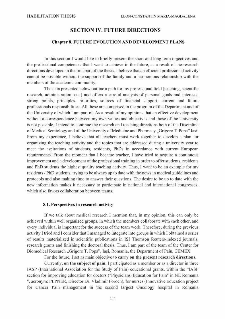
HABILITATION THESIS LEON-CONSTANTIN MARIA-MAGDALENA
144
SECTION IV. FUTURE DIRECTIONS
Chapter 8. FUTURE EVOLUTION AND DEVELOPMENT PLANS
In this section I would like to briefly present the short and long term objectives and the professional competences that I want to achieve in the future, as a result of the research directions developed in the first part of the thesis. I believe that an efficient professional activity cannot be possible without the support of the family and a harmonious relationship with the members of the academic community. The data presented below outline a path for my professional field (teaching, scientific research, administration, etc.) and offers a careful analysis of personal goals and interests, strong points, principles, priorities, sources of financial support, current and future professionals responsibilities. All these are comprised in the program of the Department and of the University of which I am part of. As a result of my opinions that an effective development without a correspondence between my own values and objectives and those of the University is not possible, I intend to continue the research and teaching directions both of the Discipline of Medical Semiology and of the University of Medicine and Pharmacy „Grigore T. Popa” Iasi. From my experience, I believe that all teachers must work together to develop a plan for organizing the teaching activity and the topics that are addressed during a university year to meet the aspirations of students, residents, PhDs in accordance with current European requirements. From the moment that I became teacher, I have tried to acquire a continuous improvement and a development of the professional training in order to offer students, residents and PhD students the highest quality teaching activity. Thus, I want to be an example for my residents / PhD students, trying to be always up to date with the news in medical guidelines and protocols and also making time to answer their questions. The desire to be up to date with the new information makes it necessary to participate in national and international congresses, which also favors collaboration between teams.
8.1. Perspectives in research activity
If we talk about medical research I mention that, in my opinion, this can only be achieved within well organized groups, in which the members collaborate with each other, and every individual is important for the success of the team work. Therefore, during the previous activity I tried and I consider that I managed to integrate into groups in which I obtained a series of results materialized in scientific publications in ISI Thomson Reuters-indexed journals, research grants and finishing the doctoral thesis. Thus, I am part of the team of the Center for Biomedical Research „Grigore T. Popa”, Iași, Romania, the Department of Pain, CEMEX. For the future, I set as main objective to carry on the present research directions. Currently, on the subject of pain, I participated as a member or as a director in three IASP (International Association for the Study of Pain) educational grants, within the “IASP section for improving education for doctors ("Physicians' Education for Pain" in NE Romania ", acronym: PEPNER, Director Dr. Vladimir Poroch), for nurses (Innovative Education project for Cancer Pain management in the second largest Oncology hospital in Romania
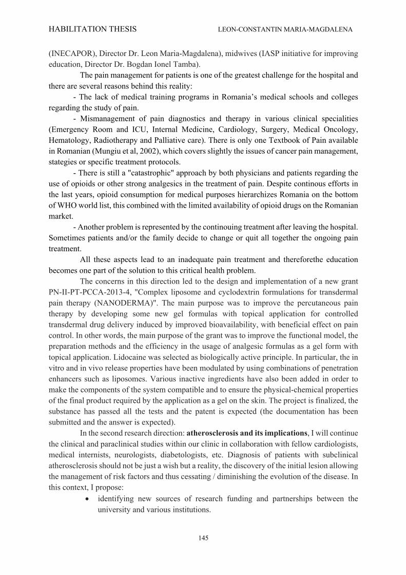
HABILITATION THESIS LEON-CONSTANTIN MARIA-MAGDALENA
145
(INECAPOR), Director Dr. Leon Maria-Magdalena), midwives (IASP initiative for improving education, Director Dr. Bogdan Ionel Tamba). The pain management for patients is one of the greatest challenge for the hospital and there are several reasons behind this reality:
- The lack of medical training programs in Romania’s medical schools and colleges regarding the study of pain.
- Mismanagement of pain diagnostics and therapy in various clinical specialities (Emergency Room and ICU, Internal Medicine, Cardiology, Surgery, Medical Oncology, Hematology, Radiotherapy and Palliative care). There is only one Textbook of Pain available in Romanian (Mungiu et al, 2002), which covers slightly the issues of cancer pain management, stategies or specific treatment protocols.
- There is still a "catastrophic" approach by both physicians and patients regarding the use of opioids or other strong analgesics in the treatment of pain. Despite continous efforts in the last years, opioid consumption for medical purposes hierarchizes Romania on the bottom of WHO world list, this combined with the limited availability of opioid drugs on the Romanian market.
- Another problem is represented by the continouing treatment after leaving the hospital. Sometimes patients and/or the family decide to change or quit all together the ongoing pain treatment. All these aspects lead to an inadequate pain treatment and thereforethe education becomes one part of the solution to this critical health problem. The concerns in this direction led to the design and implementation of a new grant PN-II-PT-PCCA-2013-4, "Complex liposome and cyclodextrin formulations for transdermal pain therapy (NANODERMA)". The main purpose was to improve the percutaneous pain therapy by developing some new gel formulas with topical application for controlled transdermal drug delivery induced by improved bioavailability, with beneficial effect on pain control. In other words, the main purpose of the grant was to improve the functional model, the preparation methods and the efficiency in the usage of analgesic formulas as a gel form with topical application. Lidocaine was selected as biologically active principle. In particular, the in vitro and in vivo release properties have been modulated by using combinations of penetration enhancers such as liposomes. Various inactive ingredients have also been added in order to make the components of the system compatible and to ensure the physical-chemical properties of the final product required by the application as a gel on the skin. The project is finalized, the substance has passed all the tests and the patent is expected (the documentation has been submitted and the answer is expected). In the second research direction: atherosclerosis and its implications, I will continue the clinical and paraclinical studies within our clinic in collaboration with fellow cardiologists, medical internists, neurologists, diabetologists, etc. Diagnosis of patients with subclinical atherosclerosis should not be just a wish but a reality, the discovery of the initial lesion allowing the management of risk factors and thus cessating / diminishing the evolution of the disease. In this context, I propose:
• identifying new sources of research funding and partnerships between the university and various institutions.
HABILITATION THESIS LEON-CONSTANTIN MARIA-MAGDALENA
146
• submission and winning by national/international competition of research grants, as project manager;
• access to the research team of an international grant; • publication of research results of at least two articles in ISI international
journals; • participating in training courses for researchers.
Strategies and means of implementation imagined for achieving the objectives: - continuous collaboration with the members of the research team; - obtaining approvals for conducting research from responsible factors; - providing support for the management of the Discipline, the University of Medicine
and Pharmacy "Grigore T. Popa" Iasi and obtaining the feedback on the activity submitted;
- adapting the research team to the needs of the project; - improvement of professional skills in a dynamic and stable university environment; - a permanent preparation for the professional development requirements in accordance
with the societies to which I am a member. The main ways of evaluating the results obtained in the scientific research activity, within the University of Medicine and Pharmacy "Grigore T. Popa" Iasi are:
- publication of scientific articles in national journals rated at least BDI, and international journals ISI Thomson Reuters-indexed journals;
- scientific oral presentations at national or international events, at least one per year; - member or responsible for new research grants.
During the teaching career, one of the most important aspects is maintaining the motivation for professional development in the teaching career. Unfortunately, more and more teachers are reorienting, and the desire to remain in the university environment, to make a career, to research, to publish news, diminishes. Therefore, maintaining the motivation of the teachers for the profession is a wish, and the group you work in has a role of trigger factor. A model of the teaching profession must include both standardized and non-standardized skills. In the first category can be included the competences related to the pedagogical analysis of the contents and of the curricular documents, competences regarding the accessibility of the information, the projection of the didactic activity etc., and in the second category we include the ability to empathize with the student and with the series / group of students, the interpersonal cognitive style, creativity and communicativity. In this sense, the structural elements of a competence are:
- professional roles or work / learning tasks to be fulfilled, - performance standards, - context, - knowledge, - skills and - personality characteristics / attitudes.
The continuous improvement and career development are intrinsically correlated with professional standards and competencies. The continuous training of the teaching staff is based on the model of approach through competences and on the concept of cumulative development of the level of competence of the teaching staff and aims the professionalizing the teaching
HABILITATION THESIS LEON-CONSTANTIN MARIA-MAGDALENA
147
career, placing the training system in the European context of continuous professional development / learning and lifelong learning and also, on the orientation of the training system towards mobility and career development and professional development.
8.2. Perspectives in professional activity
Another aspect that I consider very important is continuing the medical profession. In this direction, in parallel with the activity of teaching and scientific research, I continued my learning process, currently being primary physician in internal medicine and in the third year cardiology resident. At the same time, I participated in focused training courses in cardiovascular recovery in Switzerland, in Bern, which I want to continue. The exchanges of information between the profile companies is absolutely necessary. I consider that the implementation of the recovery programs already known to have maximum efficiency on long-term in our country it is welcome. A daring decision for the future is to create a national registry in which to enroll patients entering recovery programs. Unfortunately, in our country, cardiovascular recovery is in the shadow, although the long-term results are amazing. An efficient promotion of secondary and tertiary prevention would be useful in attracting the population. I consider that some courses on the benefits of recovery for medical colleagues are necessary. Unfortunately, the reduced funds offered by the Ministry of Health do not allow the continuation of recovery programs in the ambulatory and so many patients give up. The population of our country is aging and it is absolutely necessary to participate in these programs. Moreover, research shows that Romania is one of the countries with a high risk of cardiovascular disease, therefore primary prevention is necessary. As we know, there are patients with a single pathology, but with multiple comorbidities. Collaboration with different colleagues, teamwork (recovery team includes cardiologist, internal medicine, diabetologist, dietitian, physiotherapist, psychologist, healthcare assistant) is the key to success in recovery programs. The most important project I consider, is the creation of a pilot center for the recovery of patients with heart failure. Chronic heart failure is one of the most prevalent diseases, especially in the elderly population with the average age at first diagnosis being 76 years. Heart failure is a progressive, heterogeneous form of cardiovascular disease that requires treatment to be individualized depending on the presenting symptoms. As it is knowing, the most common cause of heart failure in general is coronary artery disease, and many patients had a myocardial infarction in the past (Petersen et al., 2002). Heart failure has been described as the “Cinderella of health issues - hardly registering on the radar of key health care providers, regulators, relevant government bodies and the general public” (Krum et al., 2006). This is in a large part due to a lack of a universally recognized definition of heart failure and because varying degrees of left ventricular systolic dysfunction may be asymptomatic. Indeed, almost half of heart failure patients have preserved left ventricular function (Tendera et al., 2004). Age is a good predictor of the incidence of heart failure. This incidence is 10 per 1,000 people over 65 ys old, and 20% of hospitalizations are in those over 65 ys old (Jessup et al., 2003). For those 50–59 ys old, the prevalence is 8 per 1,000 people for both men and women;
HABILITATION THESIS LEON-CONSTANTIN MARIA-MAGDALENA
148
however, this prevalence sharply increases for those 80–89 ys old, to 66 per 1,000 and 79 per 1,000 for men and women, respectively. Heart failure has a median survival of just 1.7 ys and 3.2 ys for men and women, respectively (Tendera, 2004). It has a 10% mortality by 30 d, which highlights an early high-risk period of disease. This high-risk period is followed by a 5-y mortality rate of 54% and 40% for men and women, respectively (Carrio, 2010). Chronic heart failure is progressive and ultimately fatal, making early detection crucial for delaying disease progression. For patients with mild symptoms, the annual death rate is 5%–10%, and this increases sharply to 30%–40% for those with more advanced symptoms. Morbidity and mortality in chronic heart failure remain high, despite advances in treatment (Clark, 2004). There are several approaches to management of chronic heart failure, and these generally relate to the severity of disease, symptoms, and comorbidity. The goal of therapy is to improve and prolong life, and this is achieved using lifestyle modifications, pharmaceutical therapy, surgery, supportive devices, management programs, and palliative care. A series of studies demonstrate that significant biochemical and functional abnormalities in skeletal muscle are present in patients with HF and play a large role in the exercise intolerance (Kokkinos, 2000). Inactivity is in part responsible, leading to muscle atrophy. It is not recognized that definite/tangible/real benefits can be achieved through early mobilization and early exercise, both passive and active, which must be increased gradually. Dynamic exercise such as walking together with working the arms is recommended to maintain activities of daily living, as well as resistive training to increase muscular strength. Such an exercise program should be gradually introduced and may start in hospital or at any point in the patient’s illness.
It is useful to consider four phases of cardiac rehabilitation, as each represents a different component of the care journey: inpatient care, the early post discharge period, exercise training, and finally the long term follow-up.
Ø Phase 1 occurs during the inpatient stay or after a steep change in a patient with a pre-existing cardiac condition – the diagnosis of heart failure respectively. During this phase, medical evaluation, reassurance and education, correction of cardiac misconceptions, risk factor assessment, mobilisation and discharge planning are the key elements. It is customary to involve family and partners right from this first stage.
Ø Phase 2 is the early post discharge period, a time when many patients feel isolated and insecure. Support can be provided by home visiting, telephone contact, and by supervised use of the rehabilitation information.
Ø Historically, phase 3 has taken the form of a structured exercise programme in a hospital setting with educational and psychological support, and advice on risk factors. It has been increasingly recognised that these components can be undertaken safely and successfully in the community. A menu-based approach recognises the need to tailor the delivery of services to the individual, and it is likely to include specific education to reduce cardiac misconceptions and encourage smoking cessation and weight management; vocational rehabilitation to assist return to work or retirement; and referral to a psychologist, cardiologist, or exercise physiologist.
Ø Phase 4 involves the long-term maintenance of physical activity and lifestyle changes. Available evidence suggests that both must be sustained for cardiac benefits to continue.
HABILITATION THESIS LEON-CONSTANTIN MARIA-MAGDALENA
149
Comprehensive cardiac rehabilitation consists of exercise training together with education and psychological support. The purpose of these interventions is to facilitate a return to normal living and to encourage patients to make lifestyle changes in order to prevent further events. Educational and psychological support is also necessary to deal with psychological distress. The benefits of exercise training in patients with HF include an improvement in exercise tolerance as assessed not only by exercise duration but more importantly by peak V̇O2 (Coats, 1992; Belardinelli, 1999). The exercise training program has varied by such factors as setting (supervised or home training), type of activity (treadmill or bicycle), duration (from 8 weeks to 3 months), and intensity (from low to moderate). One study that used primarily circuit weight training for 8 weeks elicited a modest but significant increase in peak V̇O2 (Maiorana, 2000). Changes in peak V̇O2 have ranged from 12% to 31%. Most of the improvement occurs by week 3 but can continue up to 6 months if compliance with the training program continues (Gottlieb, 1999). Not only is maximal exercise performance improved but also indices of submaximal exercise as measured by the 6-minute walking test or by the ventilatory threshold (Hambrecht, 1995).
Systematic reviews of exercise-based cardiac rehabilitation in stable, chronic heart failure have found benefits to exercise capacity and possibly to symptoms (Thomson, 1998). Benefit is probably derived from peripheral adaptations (vasodilation and improved muscle oxidative capacity) rather than improvements in ventricular function (Dracup, 1994, Belardinelli, 1999).
There is now evidence that significant cost saving may be achieved through cardiac rehabilitation and secondary prevention programs. These savings are largely from reduced subsequent hospital admissions and reduced costs of medical care. There are additional savings that arise through pension, retirement and sickness benefits, provided in case of work resumption or remaining in work is achieved. These cost savings may be very large in an ageing population prone to development of preventable heart failure. While cost benefit and effectiveness studies are so far not widely reported, it is apparent that cardiac rehabilitation programs have benefits and effectiveness similar to other successful interventions in the treatment of cardiac and vascular disease.
Moldova's population is 4,284,062 people residing in the counties of the region, making the region the country with the largest population. The area is 35.806 km ². In Moldova there is only one Cardiovascular Rehabilitation Hospital that serves the population of the area. Unfortunately, Moldova ranks first in the number of poor persons, about 32 % of the total population. Estimated number of Roma in Romania declared and is 1.049.000, and 14-15% live in Moldova. There is poor patient motivation, inadequate third-party reimbursements for services and geographic limitations to accessibility of program sites. In addition, there is a lack of “visibility” and recognition by the public of the importance of cardiac rehabilitation services.

HABILITATION THESIS LEON-CONSTANTIN MARIA-MAGDALENA
150
SECTION V. REFERENCES
Abeck D, Plötz S. Colloidal silver and ozonized olive oil for atopic dermatitis? Med
Monatsschr Pharm. 2008;31:265–266. Aboyans V, Ricco JP, Bartelink ML, Björck M, Brodmann M, Cohnert T, Collet J-
P, Czerny M, De Carlo M, Debus S, Espinola-Klein C, Kahan T, Kownator S, Mazzolai L, Naylor AR, Roffi M, Röther J, Sprynger M, Tendera M, Tepe G, Venermo M, Vlachopoulos C, Desormais I. 2017 ESC Guidelines on the Diagnosis and Treatment of Peripheral Arterial Diseases, in collaboration with the European Society for Vascular Surgery (ESVS): Document covering atherosclerotic disease of extracranial carotid and vertebral, mesenteric, renal, upper and lower extremity arteries. The Task Force for the Diagnosis and Treatment of Peripheral Arterial Diseases of the European Society of Cardiology (ESC) and of the European Society for Vascular Surgery (ESVS). European Heart Journal. 2018;38(9):763-816.
Acharya G, Armstrong M, McFall C, Quinn RW, Hilbert SL, Converse GL, Toth PB, Sherman AK, Lee CH, Lofland GK, Hopkins RA. Calcium and phosphorus concentrations in native and decellularized semilunar valve tissues. J Heart Valve Dis. 2014;23(3):259-270.
Adamson S, Leitinger N, Phenotypic modulation of macrophages in response to plaque lipids, Curr Opin Lipidol. 2011;22(5):335–342.
Agbor-Etang BB, Setaro JF. Management of Hypertension in Patients with Ischemic Heart Disease. Curr Cardiol Rep. 2015;17(12):119.
Al Rifai M, Silverman MG, Nasir K, Budoff M, Blankstein R, Szklo M, Katz R, Blumenthal RS, Blaha MJ. The association of nonalcoholic fatty liver disease, obesity, and metabolic syndrome, with systemic inflammation and subclinical atherosclerosis: the Multi-Ethnic Study of Atherosclerosis (MESA). Atherosclerosis. 2015;239(2):629-633.
Al-Mamari A. Atherosclerosis and Physical Activity. Oman Med J. 2009;24(3):173–178.
Al-Sabti HA, Al Kindi A, Al-Rasadi K, Banerjee Y, Al-Hashmi K, Al-Hinai A. Saphenous vein graft vs. radial artery graft searching for the best second coronary artery bypass graft. J Saudi Heart Assoc. 2013;25(4):247-254.
Alexa ID, Ilie A, Morosanu A, Emmanouil-Stamos P, Raiha I. Self-neglect in elders: a world-wide issue ignored in Romania. Rev Rom Bioet. 2012;10(1):141–148.
Alexa O, Veliceasa B, Malancea R, Alexa ID. Postoperative cognitive disorder has to be included within informed consent of elderly patients undergoing total hip replacement. Rev Rom Bioet. 2013;11(4):38–47.
Allen GV, Pronych SP. Trigeminal autonomic pathways involved in nociception-induced reflex cardiovascular responses. Brain Res.1997;754:269-278.
Amarenco P, Goldstein LB, Messig M, O'Neill BJ, Callahan A 3rd, Sillesen H, Hennerici MG, Zivin JA, Welch KM; SPARCL Investigators. Relative and cumulative effects of lipid and blood pressure control in the stroke prevention by aggressive reduction in cholesterol levels trial. Stroke. 2009;40(7):2486–2492.

HABILITATION THESIS LEON-CONSTANTIN MARIA-MAGDALENA
151
Ambrose JA, Singh M. Pathophysiology of coronary artery disease leading to acute coronary syndromes. F1000Prime Rep. 2015;7:08.
Anand SS, Islam S, Rosengren et al. Risk factors for miocardial infarction in women and men: insights from the INTERHEART study. Eur Heart J 2008; 29(7):932-940.
Anderson RH. Clinical anatomy of the aortic root. Heart. 2000;84(6):670-673. Andreini D, Pontone G, Mushtaq S, Bartorelli AL, Bertella E, Antonioli L, Formenti
A, Cortinovis S, Veglia F, Annoni A, Agostoni P, Montorsi P, Ballerini G, Fiorentini C, Pepi M. A long-term prognostic value of coronary CT angiography in suspected coronary artery disease. J Am Coll Cardiol Img. 2012;5:690-701.
Angstadt JD, Calabrese RL. A hyperpolarization-activated inward current in heart interneurons of the medicinal leech. J Neurosci. 1989;9:2846–2857.
Angulo P. Nonalcoholic fatty liver disease. N Engl J Med 2002;346(16):1221-1231. Anilkumar M. Ethnomedicinal plants as anti-inflammatory and analgesic agents.
Research Signpost. 2010;37661:267–293. Antonini-Canterin F, Zuppiroli A, Baldessin F, Popescu BA, Nicolosi GL. Is there a
role of statins in the prevention of aortic biological prostheses degeneration. Cardiovasc Ultrasound. 2006;4:26.
Apley J, Naish N. Recurrent abdominal pains: A field survey of 1,000 school children. Arch Dis Child. 1958;33:165-170.
Argoff CE. Topical Analgesics in the Management of Acute and Chronic Pain. Mayo Clin Proc. 2013;88(2):195–205.
Argyo C, Weiss V, Bräuchle C, Bein T. Multifunctional mesoporous silica nanoparticles as a universal platform for drug delivery. Chem Mater. 2014;26:435-451.
Arnett DK, Blumenthal RS, Albert MA, Buroker AB, Goldberger ZD, Hahn EJ, Himmelfarb CD, Khera A, Lloyd-Jones D, McEvoy JW, Michos ED, Miedema MD, Muñoz D, Smith Jr SC, Virani SS, Williams Sr KA, Yeboah J, Ziaeian B. 2019 ACC/AHA Guideline on the Primary Prevention of Cardiovascular Disease: A Report of the American College of Cardiology/American Heart Association Task Force on Clinical Practice Guidelines. Circulation. 2019;140:e596–e646.
Arora S, Jain J, Rajwade JM, Paknikar KM. Cellular responses induced by silver nanoparticles: In vitro studies. Toxicol Lett. 2008;179:93–100.
Ayaz M, Junaid M, Ullah F, Sadiq A, Subhan F, Khan MA, Ahmad W, Ali G, Imran M, Ahmad S. Molecularly characterized solvent extracts and saponins from Polygonum hydropiper L. show high anti-angiogenic, anti-tumor, brine shrimp, and fibroblast NIH/3T3 cell line cytotoxicity. Frontiers in Pharmacology. 2016;7:74.
Aydin M, Bulur S, Alemdar R, Yalçin S, Türker Y, Basar C, Aslantas Y, Yazgan Ö, Albayrak S, Özhan H; Melen Investigators. The impact of metabolic syndrome on carotid intima media thickness. Eur Rev Med Pharmacol Sci. 2013;17(17):1-15.
Aydin S, Aydin S, Eren MN, Sahin I, Yilmaz M, Kalayci M, Gungor O. The cardiovascular system and the biochemistry of grafts used in heart surgery. SpringerPlus. 2013;2(1):612.
Aziz M, Yadav KS. Pathogenesis of Atherosclerosis. Med Clin Rev. 2016; 2:22.
HABILITATION THESIS LEON-CONSTANTIN MARIA-MAGDALENA
152
Bahmani M, Shirzad H, Majlesi M, Shahinfard N, Rafieian-Kopaei M. A review study on analgesic applications of Iranian medicinal plants. Asian Pacific Journal of Tropical Medicine. 2014;7:S43–S53.
Baikoussis NG, Papakonstantinou NA, Apostolakis E. Radial artery as graft for coronary artery bypass surgery: Advantages and disadvantages for its usage focused on structural and biological characteristics. J Cardiol. 2014;63(5):321-328.
Banerjee C, Chimowitz MI. Stroke Caused by Atherosclerosis of the Major Intracranial Arteries. Circ Res. 2017;120(3):502-513.
Banerjee T, Singh AK, Sharma RK, Maitra AN. Labeling efficiency and biodistribution of Technetium-99m labeled nanoparticles: interference by colloidal tin oxide particles. Int J Pharm. 2005;289:189–195.
Baral VR, Dewar AL, Connett GJ. Colloidal silver for lung disease in cystic fibrosis. J R Soc Med. 2008;101 Suppl:S51-52.
Barbee NC, Ganio K, Swearer SE. Integrating multiple bioassays to detect and assess impacts of sublethal exposure to metal mixtures in an estuarine fish. Aquat Toxicol. 2014;152:244–255.
Barillo DJ, Marx DE. Silver in medicine: a brief history BC 335 to present. Burns. 2014;40 Suppl 1:S3-8.
Barkin RL. The Pharmacology of Topical Analgesics. Postgrad Med. 2013;125(1):7–18.
Barnes CA, Elsaesser A, Arkusz J, Smok A, Palus J, Lesniak A, Salvati A, Hanrahan JP, Jong WH, Dziubaltowska E, Stepnik M, Rydzynski K, McKerr G, Lynch I, Dawson KA, Howard CV. Reproducible comet assay of amorphous silica nanoparticles detects no genotoxicity. Nano Lett. 2008;8:3069–3074.
Barrett-Connor E. Menopause, atherosclerosis, and coronary artery disease. Curr Opin Pharmacol. 2013;13(2):186–191.
Barritt GJ. Receptor-activated Ca2+ inflow in animal cells: a variety of pathways tailored to meet different intracellular Ca2+ signalling requirements. Biochem J. 1999;337 ( Pt 2):153-169.
Battaglia V, Compagnone A, Bandino A, Bragadin M, Rossi CA, Zanetti F, Colombatto S, Grillo MA, Toninello A. Cobalt induces oxidative stress in isolated liver mitochondria responsible for permeability transition and intrinsic apoptosis in hepatocyte primary cultures. Int J Biochem Cell Biol. 2009;41(3):586-594.
Begon S, Pickering G, Eschalier A, Dubray C. Magnesium increases morphine analgesic effect in different experimental models of pain. Anesthesiology. 2002;96:627–632.
Beiranvand S, Sorori MM. Pain management using nanotechnology approaches. Artif Cells Nanomed Biotechnol. 2019;47:462–468.
Bekut M, Brkić S, Kladar N, Dragović G, Gavarić N, Božin B. Potential of selected Lamiaceae plants in anti(retro)viral therapy. Pharmacol Res. 2018;133:301-314.
Belardinelli R, Georgiou D, Cianci G, Purcaro A. Randomized, controlled trial of long-term moderate exercise training in chronic heart failure: effects on functional capacity, quality of life, and clinical outcome. Circulation. 1999; 99:1173–1182.
Bellaosta S, Corsini A. Statin drug interactions and related adverse reactions. Expert Opin Drug Saf. 2012;12:933–946.
HABILITATION THESIS LEON-CONSTANTIN MARIA-MAGDALENA
153
Benedetto U, Raja SG, Albanese A, Amrani M, Biondi-Zoccai G, Frati G. Searching for the second best graft for coronary artery bypass surgery: a network meta-analysis of randomized controlled trials. Eur J Cardiothorac Surg. 2015;47:59-65.
Benjamin EJ, Blaha MJ, Chiuve SE, Cushman M, Das SR, Deo R, de Ferranti SD, Floyd J, Fornage M, Gillespie C, Isasi CR, Jiménez MC, Jordan LC, Judd SE, Lackland D, Lichtman JH, Lisabeth L, Liu S, Longenecker CT, Mackey RH, Matsushita K, Mozaffarian D, Mussolino ME, Nasir K, Neumar RW, Palaniappan L, Pandey DK, Thiagarajan RR, Reeves MJ, Ritchey M, Rodriguez CJ, Roth GA, Rosamond WD, Sasson C, Towfighi A, Tsao CW, Turner MB, Virani SS, Voeks JH, Willey JZ, Wilkins JT, Wu HY, Alger HM, Wong SS, Muntner P. Heart Disease and Stroke Statistics-2017 Update: a report from the American Heart Association. Circulation. 2017;135(10):e146-e603.
Bentzon JF, Otsuka F, Virmani R, Falk E. Mechanisms of plaque formation and rupture. Circ Res. 2014;114:1852–1866.
Berger JS, Jordan CO, Lloyd-Jones D, Blumenthal RS. Screening for cardiovascular risk in asymptomatic patients. J Am Coll Cardiol. 2010;55:1169-1177.
Beulens JW, Rimm EB, Ascherio A, Spiegelman D, Hendriks HF, Mukamal KJ. Alcohol consumption and risk for coronary heart disease among men with hypertension. Ann Intern Med. 2007;46(1):10-19.
Bhol KC, Alroy J, Schechter PJ. Anti-inflammatory effect of topical nanocrystalline silver cream on allergic contact dermatitis in a guinea pig model. Clin Exp Dermatol. 2004;29:282–287.
Bindar D, Tartu L, Garlea A, Nita L, Merlimg V. Effects of Magnesiun Soft Matter Vesicles Carrier of the Behavioral Manifestations in Mice. Rom J Biophys. 2010;20:23–35.
Blackett PR, Wilson DP, McNeal CJ. Secondary hypertriglyceridemia in children and adolescents. J Clin Lipidol. 2015; 9(5 Suppl):S29-40.
Blackwell LS, Quittner AL. Daily pain in adolescents with CF: Effects on adherence, psychological symptoms, and health-related quality of life. Pediatr Pulmonol. 2015;50:244-251.
Blaha MJ, Cainzos-Achirica M, Greenland P, McEvoy JW, Blankstein R, Budoff MJ, Dardari Z, Sibley CT, Burke GL, Kronmal RA, Szklo M, Blumenthal RS, Nasir K. Role of coronary artery calcium score of zero and other negative risk markers for cardiovascular disease: the multi-ethnic study of atherosclerosis (MESA) Circulation. 2016;133:849–858.
Blankstein R, Budoff MJ, Shaw LJ, Goff DC Jr, Polak JF, Lima J, Blumenthal RS, Nasir K. Predictors of coronary heart disease events among asymptomatic persons with low low-density lipoprotein cholesterol MESA (Multi-Ethnic Study of Atherosclerosis). J Am Coll Cardiol. 2011;58(4):364-374.
Blitz A, Osterday RM, Brodman RF. Harvesting the radial artery. Ann Cardiothorac Surg. 2013;2(4):533-542.
Bloomfield P. Choice of heart valve prosthesis. Heart. 2002;87(6):583-589. Bobryshev YV. Monocyte recruitment and foam cell formation in atherosclerosis,
Micron. 2006, 37(3):208–222. Bonetti PO, Lerman LO, Lerman A. Endothelial dysfunction: a marker of
atherosclerotic risk. Arteriscler Thromb Vasc Biol. 2003;23(2):168-175.

HABILITATION THESIS LEON-CONSTANTIN MARIA-MAGDALENA
154
Borm PJA, Kreyling W. Toxicological hazards of inhaled nanoparticles--potential implications for drug delivery. J Nanosci Nanotechnol. 2004;4:521–531.
Bowen SJ, Hull J. The basic science of cystic fibrosis. Paediatr Child Health. 2015;25:159-164.
Boyle JJ, Macrophage activation in atherosclerosis: pathogenesis and pharmacology of plaque rupture, Curr Vasc Pharmacol, 2005, 3(1):63–68.
Braunersreuther V, Mach F, Steffens S, The specific role of chemokines in atherosclerosis, Thromb Haemost. 2007, 97(5):714–721.
Braydich-Stolle L, Hussain S, Schlager JJ, Hofmann M-C. In vitro cytotoxicity of nanoparticles in mammalian germline stem cells. Toxicol Sci. 2005;88:412–419.
Bre L.P., McCarthy R., Wang W. Prevention of bioprosthetic heart valve calcification: Strategies and outcomes. Curr. Med. Chem. 2014;21:2553–2556.
Breivik H, Collett B, Ventafridda V, Cohen R, Gallacher D. Survey of chronic pain in Europe: prevalence, impact on daily life, and treatment. Eur J Pain. 2006;10(4):287-333.
Bresink I, Ebert B, Parsons CG, Mutschler E. Zinc changes AMPA receptor properties, results of binding studies and patch clamp recordings. Neuropharmacol. 1996;35:503–509.
Brill S, Sadgwick PM, Hamann W, di Vadi PP. Efficacy of intra- venous magnesium in neuropathic pain. Br J Anaesth. 2002;89:711–714.
Bucholz E, Beckman A, Kiefe C, Krumholz H. Smoking status and life expectancy after acute myocardial infarction in the elderly. Heart. 2016;102:87-88.
Bui AL, Horwich TB, Fonarow GC. Epidemiology and risk profile of heart failure. Nat Rev Cardiol. 2011;8(1):30–41.
Buxton BF, Hayward PA. The art of arterial revascularization-total arterial revascularization in patients with triple vessel coronary artery disease. Ann Cardiothorac Surg. 2013;2(4):543-551.
Cameron AA, Green GE, Brogno DA, Thornton J. Internal thoracic artery grafts: 20-year clinical follow-up. J Am Coll Cardiol. 1995;25(1):188-192.
Carlson C, Hussain SM, Schrand AM, Braydich-Stolle LK, Hess KL, Jones RL, Schlager JJ. Unique cellular interaction of silver nanoparticles: size-dependent generation of reactive oxygen species. J Phys Chem B. 2008;112(43):13608–13619.
Carrio´ I, Cowie MR, Yamazaki J, Udelson J, Camici PG. Cardiac sympathetic imaging with mIBG in heart failure. JACC Cardiovasc Imaging. 2010;3:92–100.
Cassuto J, Sinclair R, Bonderovic M. Anti-inflammatory properties of local anesthetics and their present and potential clinical implications. Acta Anaesthesiol Scand. 2006;50(3):265–282.
Cavun S, Goktalay G, Millington WR. The hypotension evoked by visceral nociception is mediated by delta opioid receptors in the periaqueductal gray. Brain Res. 2004;1019:237-245.
Cepoi V, Alexa ID, Ilie A, Alexa O. Ethical dilemmas in treating elderly patients at risk for polypragmasy and polypharmacy. Rev Rom Bioet. 2014;12(3):12–18.
Chalmers AD, Bursill CA, Myerscough MR. Nonlinear dynamics of early atherosclerotic plaque formation may determine the efficacy of high density lipoproteins (HDL) in plaque regression. PLoS ONE. 2017;12:e0187674.
HABILITATION THESIS LEON-CONSTANTIN MARIA-MAGDALENA
155
Chen CC, Zimmer A, Sun WH, Hall J, Brownstein MJ, Zimmer A. A role for ASIC3 in the modulation of high-intensity pain stimuli. Proc Natl Acad Sci U S A. 2002;99(13):8992-8997.
Chen JH, Pan WH, Hsu CC, Yeh WT, Chuang SY, Chen PY, Chen HC, Chang CT, Huang WL. Impact of obesity and hypertriglyceridemia on gout development with or without hyperuricemia: a prospective study. Arthritis Care Res (Hoboken). 2013;65(1):133-140.
Chen X, Schluesener HJ. Nanosilver: a nanoproduct in medical application. Toxicol Lett. 2008;176:1–12.
Choudhary SS, Choudhary S. Exercise testing in assessment and management of patients in clinical practice - present situation. Lung India. 2008; 25(3):111–117.
Ciampi Q, Villari B. Role of echocardiography in diagnosis and risk stratification in heart failure with left ventricular systolic dysfunction. Cardiovasc Ultrasound. 2007; 5:34.
Clark RA, McKenna S, Dawson A,Wilkinson D, Stewart S. Uncovering a hidden epidemic: a study of the current burden of heart failure in Australia. Heart Lung Circ. 2004;13:266–273.
Conen D, Tedrow U, Cook N, Moorthy M, Buring J, Albert J. Alcohol Consumption and Risk of Incident Atrial Fibrillation in Women. JAMA. 2008;300(21):2489–2496.
D'Amato G, Liccardi G, Noschese P, Salzillo A, D'Amato M, Cazzola M. Anti-IgE monoclonal antibody (omalizumab) in the treatment of atopic asthma and allergic respiratory diseases. Curr Drug Targets Inflamm Allergy. 2004 ;3(3):227-229.
D'Angelo I, Conte C, La Rotonda MI, Miro A, Quaglia F, Ungaro F. Improving the efficacy of inhaled drugs in cystic fibrosis: Challenges and emerging drug delivery strategies. Adv Drug Deliv Rev. 2014;75: 92-111.
Da Silva AL, Santos RS, Xisto DG, Alonso Sdel V, Morales MM, Rocco PR. Nanoparticle-based therapy for respiratory diseases. An Acad Bra Cienc. 2013;85:137–146.
Dai S, Yang Q, Yuan K, et al. Non-high-density lipoprotein cholesterol: distribution and prevalence of high serum levels in children and adolescents: United States National Health and Nutrition Examination Surveys, 2005-2010. J Pediatr. 2014;164:247-252.
Damiani V, Di Carlo M, Grappasonni G, Di Domenico R, Dominici P. Efficacy of a new medical device based on colloidal silver and carbossimetyl beta glucan in treatment of upper airways disease in children. Minerva Pediatr. 2011;63(5):347–354.
David G, Jaba IM, Tamba B, Bohotin C, Neamtu A, Patraș X, Simionescu BC, Mungiu OC. Antinociceptive effect of morphiceptin loaded poly(butylcyanoacrylate) nanoparticles. Rev Roum Chim. 2010;55(11–12):923–931.
De Lima L, Pastrana T, Radbruch L, Wenk R. Cross-sectional pilot study to monitor the availability, dispensed prices, and affordability of opioids around the globe. J Pain Symptom Manage. 2014;48(4):649–659.
De Smedt D, Clays E, Annemans L, Doyle F, Kotseva K, Pajak A, Prugger C, Jennings C, Wood D, De Bacquer D. Health related quality of life in coronary patients and its association with their cardiovascular risk profile: results from the EUROASPIRE III survey. Int J Cardiol. 2013;168:898-903.
DeBakey ME. Profiles in cardiology. Clin Cardiol. 1991; 14:1007-1010.

HABILITATION THESIS LEON-CONSTANTIN MARIA-MAGDALENA
156
Decano JL, Aikawa M. Dynamic Macrophages: Understanding Mechanisms of Activation as Guide to Therapy for Atherosclerotic Vascular Disease. Front Cardiovasc Med. 2018;5:97.
Dégano IR, Salomaa V, Veronesi G, Ferriéres J, Kirchberger I, Laks T, Havulinna AS, Ruidavets JB, Ferrario MM, Meisinger C, Elosua R, Marrugat J; Acute Myocardial Infarction Trends in Europe (AMITIE) Study Investigators. Twenty-five-year trends in myocardial infarction attack and mortality rates, and case-fatality, in six European populations. Heart. 2015;101:1413–1421.
Derry S, Conaghan P, Da Silva JAP, Wiffen PJ, Moore RA. Topical NSAIDs for chronic musculoskeletal pain in adults. Cochrane Database of Syst Rev. 2016;4:CD007400.
Devasconcellos P, Bose S, Beyenal H, Bandyopadhyay A, Zirkle LG. Antimicrobial Particulate Silver Coatings on Stainless Steel Implants for Fracture Management. Mater Sci Eng C Mater Biol Appl. 2012;32:1112–1120.
Diez Roux AV, Ranjit N, Powell L, Jackson S, Lewis TT, Shea S, Wu C. Psychosocial factors and coronary calcium in adults without clinical cardiovascular disease. Ann Intern Med. 2006; 144: 822-831.
Ding J, Liu Y, Wang F, Bai F. Impact of Competitive Flow on Hemodynamics in Coronary Surgery: Numerical Study of ITA-LAD Model. Comput Math Methods Med. 2012; 2012: 356187.
Djoussé L, Gaziano JM. Alcohol consumption and heart failure: a systematic review. Curr Atheroscler Rep. 2008;10(2):117-120.
Do K, Brown RE, Wharton S, Ardern CI, Kuk JL. Association between cardiorespiratory fitness and metabolic risk factors in a population with mild to severe obesity. BMC Obes. 2018;5:5.
Dorobanțu M, Bădila E, Ghiorghe S, Darabonț R, Olteanu M, Flondor P. Total cardiovascular risk estimation in Romania. Data from the SEPHAR study. Rom J Intern Med. 2008;46(1):29-37.
Dorobanțu M, Darabonț R, Ghiorghe S, Arsenescu-Georgescu C, Macarie C, Mitu F, Lighezan D, Mușetescu R, Pop C, Ardeleanu E, Craiu E, Tăutu OF. Hypertension prevalence and control in Romania at a seven-year interval. Comparison SEPHAR I and II surveys. J Hypertension. 2014, 32 (1):39-47.
Dorobanțu M, Tăutu OF, Darabonț R, Ghiorghe S, Bădila E, Minca D, Dobreanu M, Baila I, Rutkowski, Zdrojewski. Objectives and methodology of Romanian SEPHAR II Survey. Project for comparing the prevalence and control of cardiovascular risk factors in two East-European countries: Romania and Poland. Arch Med Sci. 2015;11(4):715–723.
Dorobanțu M, Tăutu OF, Dimulescu D, Sinescu C, Gusbeth-Tatomir P, Arsenescu-Georgescu C, Mitu F, Lighezan D, Pop C, Babeș K, Giucă A., Brânză I, Udrescu M, Herdea V, Darabont R. Perspectives on hypertension's prevalence, treatment and control in a high cardiovascular risk East European country: data from the SEPHAR III survey. J Hypertens. 2018;36(3):690-700.
Drăgănescu D, Ibănescu C, Tamba BI, Andrițoiu CV, Dodi G, Popa MI. Flaxseed lignan wound healing formulation: Characterization and in vivo therapeutic evaluation. Int J Biol Macromol. 2015;72:614–623.

HABILITATION THESIS LEON-CONSTANTIN MARIA-MAGDALENA
157
Drozdz D, Kawecka-Jaszcz K. Cardiovascular changes during chronic hypertensive states. Pediatr Nephrol. 2014;29(9):1507–1516.
Dueñas M, Ojeda B, Salazar A, Mico JA, Failde I. A review of chronic pain impact on patients, their social environment and the health care system. J Pain Res. 2016; 9:457–467.
Durlach J, Bac P, Bara M, Guiet-Bara A. Physiopathology of symptomatic and latent forms of central nervous hyperexcitability due to magnesium deficiency, a current general scheme. Magnesium Res. 2000;13:293–302.
Eagle KA, Guyton RA, Davidoff R, Edwards FH, Ewy GA, Gardner TJ, Hart JC, Herrmann HC, Hillis LD, Hutter AM Jr, Lytle BW, Marlow RA, Nugent WC, Orszulak TA. ACC/AHA 2004 guideline update for coronary artery bypass graft surgery: a report of the American College of Cardiology/American Heart Association Task Force on Practice Guidelines (Committee to Update the 1999 Guidelines for Coronary Artery Bypass Graft Surgery). Circulation. 2004;110(14):e340-437.
Ebabe Elle R, Gaillet S, Vidé J, Romain C, Lauret C, Rugani N, Cristol JP, Rouanet JM.. Dietary exposure to silver nanoparticles in Sprague-Dawley rats: effects on oxidative stress and inflammation. Food Chem Toxicol. 2013;60:297–301.
Eisenberg DM, Davis RB, Ettner SL, Appel S, Wilkey S, Van Rompay M, Kessler RC. Trends in alternative medicine use in the United States, 1990-1997: results of a follow-up national survey. JAMA. 1998;280(18):1569-1575.
Eklioglu BS, Atabek ME, Akyürek N, Alp H. Assessment of Cardiovascular Parameters in Obese Children and Adolescents with Non-Alcoholic Fatty Liver Disease. J Clin Res Pediatr Endocrinol. 2015; 7(3):222-227.
El Khatib N, Genieys S, Kazmierczak B, Volpert V, Reaction-diffusion model of atherosclerosis development, J Math Biol. 2012;65(2):349-374.
Engel RW, Miller RF, Price NO. Added dietary inorganic sulfate and its effect upon rats fed molybdenum. J Nutr. 1956;60(4):539-547.
Engelman HS, Allen TB, MacDermott AB. The distribution of neurons expressing calcium-permeable AMPA receptors in the superficial laminae of the spinal cord dorsal horn. J Neurosci. 1999;19(6):2081-2089.
Er BM, Kocamanoglu IS, Bozkurt A, Bilge S, Cetinoglu EC. Evaluation of Postoperative Anti-nociceptive Efficacy of Intrathecal Dexketoprofen in Rats. Balkan Med J. 2016;33(3):245–245.
Fawcett WJ, Haxby EJ, Male DA. Magnesium, physiology and pharmacology. Br J Anaesth. 1999;83:302–320.
Felsby S, Nielsen J, Arendt-Nielsen L, Jensen TS. NMDA receptor blockade in chronic neuropathic pain, a comparison of ketamine and magnesium chloride. Pain. 1995;64:283–291.
Fisher JL, Macdonald RL. The role of an alpha subtype M2-M3 His in regulating inhibition of GABAA receptor current by zinc and other divalent cations. J Neurosci. 1998;18(8):2944-2953.
Foldbjerg R, Olesen P, Hougaard M, Dang DA, Hoffmann HJ, Autrup H. PVP-coated silver nanoparticles and silver ions induce reactive oxygen species, apoptosis and necrosis in THP-1 monocytes. Toxicol Lett. 2009;190:156–162.
HABILITATION THESIS LEON-CONSTANTIN MARIA-MAGDALENA
158
Fontenoy C, Kamel SO. Silver in the medical devices/equipments: Marketing or real clinical interest? Le Pharm Hosp. 2011;46:e1–e11.
Franco OH, Wong YL, Kandala NB, Ferrie JE, Dorn JM, Kivimäki M, Clarke A, Donahue RP, Manoux AS, Freudenheim JL, Trevisan M, Stranges S. Cross-cultural comparison of correlates of quality of life and health status: the Whitehall II Study (UK) and the Western New York Health Study (US). Eur J Epidemiol. 2012; 27: 255-265.
Ganguly P, Alam SF. Role of homocysteine in the development of cardiovascular disease. Nutr J. 2015;14:6.
Ganjuri M, Moshtaghian J, Ghaedi K. Effect of Nanosilver Particles on Procaspase-3 Expression in Newborn Rat Brain. Cell J. 2015;17:489–493.
Gao Y, Li P, Ma LX, Du KX, Wang XH, Tang MJ, He HK, Yu XJ, Zang WJ, Hu H. Effects of acute administration of ethanol on experimental arrhythmia. Chin J Physiol. 2012;55(5):307-313.
Gardner AW, Parker DE, Montgomery PS, Scott KJ, Blevins SM. Efficacy of quantified home-based exercise and supervised exercise in patients with intermittent claudication: a randomized controlled trial. Circulation. 2011;123:491–498.
Gaziano TA, Bitton A, Anand S, Abrahams-Gessel S, Murphy S. Growing Epidemic of Coronary Heart Disease in Low- and Middle-Income Countries. Curr Probl Cardiol. 2010; 35(2):72–115.
Gedin F, Skeppholm M, Burström K, Sparring V, Tessma M, Zethraeus N. Effectiveness, costs and cost-effectiveness of chiropractic care and physiotherapy compared with information and advice in the treatment of non-specific chronic low back pain: study protocol for a randomised controlled trial. Trials. 2017;18(1):613.
Georghiou GP, Vidne BA, Dunning J. Does the radial artery provide better long-term patency than the saphenous vein? Interact Cardiovasc Thorac Surg. 2005;4(4):304-310.
Geovanini GR, Libby P. Atherosclerosis and inflammation: overview and updates. Clin Sci. (Lond) 2018;132(12):1243-1252.
Gibelin P, Aldossari A, Bertora D, Moceri P, Hugues T. New Parameters of Cardiopulmonary Exercise Testing in Patients with Chronic Heart Failure: Practical Applications. IJCM. 2012;3:532-537.
Glineur D, Papadatos S, Grau JB, Shaw RE, Kuschner CE, Aphram G, Mairy Y, Vanbelighen C, Etienne PY. Complete myocardial revascularization using only bilateral internal thoracic arteries provides a low-risk and durable 10-year clinical outcome. Eur J Cardiothorac Surg. 2016. pii: ezw120.
Goggin R, Jardeleza C, Wormald P-J, Vreugde S. Colloidal silver: a novel treatment for Staphylococcus aureus biofilms? Int Forum Allergy Rhinol. 2014;4:171–175.
Goldman S, Zadina K, Moritz T, Ovitt T, Sethi G, Copeland JG. VA Cooperative Study Group #207/297/364. Long-term patency of saphenous vein and left internal mammary artery grafts after coronary artery bypass surgery: results from a Department of Veterans Affairs Cooperative Study. J Am Coll Cardiol. 2004;44:2149-2156.
Golia E, Limongelli G, Natale F, Fimiani F, Maddaloni V, Pariggiano I, Bianchi R, Crisci M, D'Acierno L, Giordano R, Di Palma G, Conte M, Golino P, Russo MG, Calabrò R, Calabrò P. Inflammation and cardiovascular disease: from pathogenesis to therapeutic target. Curr Atheroscler Rep. 2014;16(9):435.
HABILITATION THESIS LEON-CONSTANTIN MARIA-MAGDALENA
159
Gottlieb SS, Fisher ML, Freudenberger R, Robinson S, Zietowski G, Alves L, Krichten C, Vaitkevicus P, McCarter R. Effects of exercise training on peak performance and quality of life in congestive heart failure patients. J Card Fail. 1999;5:188–194.
Gromiec JP, Kupczewska-Dobecka M, Jankowska A, Czerczak S. Predictive models for the assessment of occupational exposure to chemicals: a new challenge for employers. Med Pr. 2013;64(5):699-716.
Grosse-Steffen T, Krämer M, Tuschy B, Weiss C, Sütterlin M, Berlit S. Topic anaesthesia with a eutectic mixture of lidocaine/prilocaine cream after elective caesarean section: a randomised, placebo-controlled trial. Arch Gynecol Obstet. 2017;296(4):771–776.
Grunkemeier GL, Furnary AP, Wu Y, Wang L, Starr A. Durability of pericardial versus porcine bioprosthetic heart valves. J Thorac Cardiovasc Surg. 2012;144(6):1381-1386.
Guazzi M, Adams V, Conraads V, Halle M, Mezzani A, Vanhees L, Arena R, Fletcher GF, Forman DE, Kitzman DW, Lavie CJ, Myers J. EACPR/AHA Scientific Statement. Clinical recommendations for cardiopulmonary exercise testing data assessment in specific patient populations. Circulation. 2012;126(18):2261-2274.
Guilarte TR, Chen MK. Manganese inhibits NMDA receptor channel function, implications to psychiatric and cognitive effects. Neurotoxicology. 2007;28:1147–1152.
Gul S, Ahmad M, Zafar M, Bahadur S, Sultana S, Begum N, Shah SN, Zaman W, Ullah F, Ayaz A, Hanif U. Taxonomic study of subfamily Nepetoideae (Lamiaceae) by polynomorphological approach. Microsc Res Tech. 2019;82(7):1021-1031.
Gundiah N, Matthews PB, Karimi R, Azadani A, Guccione J, Guy TS, Saloner D, Tseng EE. Significant material property differences between the porcine ascending aorta and aortic sinuses. J Heart Valve Dis. 2008;17:606-613.
Gunn A, Bobeck EN, Weber C, Morgan MM. The influence of non-nociceptive factors on hot-plate latency in rats. J Pain. 2011;12(2):222–227.
Guo D, Ling J, Wang MH, She JX, Gu J, Wang CY. Physical interaction and functional coupling between ACDP4 and the intracellular ion chaperone COX11, an implication of the role of ACDP4 in essential metal ion transport and homeostasis. Mol Pain. 2005;1:15.
Habib RH, Schwann TA, Engoren M. Late effects of radial artery ver- sus saphenous vein grafting in patients aged 70 years or older. Ann Thorac Surg. 2012; 94:1478-1484.
Hackel D, Pflücke D, Neumann A, Viebahn J, Mousa S, Wischmeyer E, Roewer N, Brack A, Rittner HL. The connection of monocytes and reactive oxygen species in pain. PLoS One. 2013;8:e63564.
Hadrup N, Lam HR. Oral toxicity of silver ions, silver nanoparticles and colloidal silver—a review. Regul Toxicol Pharmacol. 2014;68:1–7.
Hamann SR, Holtman JR, Martin WR. Analgesic actions of local anesthetics and cobalt chloride in the rat brain stem. Pharmacol Biochem and Behavior. 1992;43(3):925-927.
Hambrecht R, Niebauer J, Fiehn E, Kälberer B, Offner B, Hauer K, Riede U, Schlierf G, Kübler W, Schuler G.. Physical training in patients with stable chronic heart failure: effects on cardiorespiratory fitness and ultrastructural abnormalities of leg muscles. J Am Coll Cardiol. 1995;25:1239–1249.
Hansson GK, Hermansson A, The immune system in atherosclerosis, Nat Immunol. 2011; 12(3):204–212.
HABILITATION THESIS LEON-CONSTANTIN MARIA-MAGDALENA
160
Haq F, Ahmad H, Ullah R, Iqbal Z. Species diversity and ethno botanical classes of the flora of Allai valley District Battagram, Pakistan. Int J Plant Research. 2012;2(4):111–123.
Hardman MJ, Sisi P, Banbury DN, Byrne C. Patterned acquisition of skin barrier function during development. Development. 1998;125(8):1541–1552.
Hardy T, Oakley F, Anstee QM, Day CP, Nonalcoholic Fatty Liver Disease: Pathogenesis and Disease Spectrum. Annu Rev Pathol. 2016;11:451-496.
Hasanein P, Parviz M, Keshavarz M, Javanmardi K, Mansoori M, Soltani N. Oral magnesium administration prevents thermal hyperalgesia induced by diabets in rats. Diabetes Res Clin Pract. 2006;75:17–22.
Hashmi JA, Baliki MN, Huang L, Parks EL, Chanda ML, Schnitzer T, Apkarian AV. Lidocaine patch (5%) is no more potent than placebo in treating chronic back pain when tested in a randomised double blind placebo controlled brain imaging study. Mol Pain. 2012;8:29.
Haukeland JW, Damås JK, Konopski Z, Løberg EM, Haaland T, Goverud I, Torjesen PA, Birkeland K, Bjøro K, Aukrust P. Systemic inflammation in nonalcoholic fatty liver disease is characterized by elevated levels of CCL2. J Hepatol. 2006;44(6):1167-1174.
Hayashi M, Shimizu W, Albert MC. The Spectrum of Epidemiology Underlying Sudden Cardiac Death. Circ Res. 2015;116(12):1887–1906.
Hemmat N, Ebadi A, Badalzadeh R, Memar MY, Baghi HB. Viral infection and atherosclerosis. Eur J Clin Microbiol Infect Dis. 2018;37(12):2225-2233.
Hernandez R, Allen NB, Liu K, Stamler J, Reid KJ, Zee PC, Wu D, Kang J, Garside DB, Daviglus ML. Association of depressive symptoms, trait anxiety, and perceived stress with subclinical atherosclerosis: results from the Chicago Healthy Aging Study (CHAS). Prev Med. 2014;61:54-60.
Herrington W, Lacey B, Sherliker P, Armitage J, Lewington S. Epidemiology of Atherosclerosis and the Potential to Reduce the Global Burden of Atherothrombotic Disease. Circ Res. 2016;118(4):535-546.
Hess CN, Lopes RD, Gibson CM, Hager R, Wojdyla DM, Englum BR, Mack MJ, Kouchoukos NT, Peterson ED, Alexander JH. Saphenous Vein Graft Failure after Coronary Artery Bypass Surgery: Insights from PREVENT IV. Circulation. 2014; 130: 1445-1451.
Higashiyama A, Okamura T, Watanabe M, Kokubo Y, Wakabayashi I, Okayama A, Miyamoto Y. Alcohol consumption and cardiovascular disease incidence in men with and without hypertension: the Suita study. Hypertens Res. 2013;36(1):58-64.
Hirayama S, Miida T. Small dense LDL: An emerging risk factor for cardiovascular disease. Clin Chim Acta. 2012;414:215-224.
Ho SS. Current status of carotid ultrasound in atherosclerosis. Quant Imaging Med Surg. 2016;6(3):285-296.
Holvoet P, Relations between metabolic syndrome, oxidative stress and inflammation and cardiovascular disease, Verh K Acad Geneeskd Belg. 2008,70(3):193–219.
Holzinger S, Anke M, Röhrig B, Gonzalez D. Molybdenum intake of adults in Germany and Mexico. Analyst. 1998;123(3):447-450.
Honkanen V, Konttinen YT, Sorsa T, Hukkanen M, Kemppinen P, Santavirta S, Saari H, Westermarck T. 1991. Serum zinc, copper and selenium in rheumatoid arthritis. J Trace Elem Electrolytes Health Dis.1991;5:261–263.
HABILITATION THESIS LEON-CONSTANTIN MARIA-MAGDALENA
161
Hornig B, Maier V, Drexler H. Physical training improves endothelial function in patients with chronic heart failure. Circulation. 1996;93:210–214.
Hsiao B, Mihalak KB, Magleby KL, Luetje CW. Zinc potentiates neuronal nicotinic receptors by increasing burst duration. J Neurophysiol. 2008;99:999–1007.
http://ec.europa.eu/eurostat/statistics-explained/index.php/Cardiovascular_diseases_statistics, 2019.
Huk OL, Catelas I, Mwale F, Antoniou J, Zukor DJ, Petit A. Induction of apoptosis and necrosis by metal ions in vitro. J Arthroplasty. 2004;19(8 Suppl 3):84-87.
Ikonomidis I, Stamatelopoulos K, Lekakis J, Vamvakou GD, Kremastinos DT, Inflammatory and non-invasive vascular markers: the multimarker approach for risk stratification in coronary artery disease, Atherosclerosis. 2008; 199(1):3-11.
Ingersoll MA, Platt AM, Potteaux S, Randolph GJ, Monocyte trafficking in acute and chronic inflammation, Trends Immunol. 2011;32(10):470–477.
Iorga M, Sztankovszky LZ, Soponaru C, Gardikiotis I. Pharmacists’ attitude and practices about drug dispensing in Romania. Farmacia. 2015;63(4):601–606.
Ismail H, McFarlane JR, Dieberg G, Smart NA. Exercise training program characteristics and magnitude of change in functional capacity of heart failure patients. Int J Cardiol. 2014;171(1):62-65.
Iurea (Rata) DM, Popa M, Chailan J-F, Tamba BI, Tudorancea I, Peptu CA. Ibuprofen-loaded chitosan/poly(maleic anhydride-alt-vinyl acetate) submicronic capsules for pain treatment. J Bioact Compat Polym. 2013;28(4):368–384.
Jacka T, Bernard CC, Singer G. Copper salicylate as an anti-inflammatory and analgesic agent in arthritic rats. Life Sci. 1983;32(9):1023-1030.
Jarasūniene D, Simaitis A. Oxidative stress and endothelial dysfunction. Med. (Kaunas) 2003;39(12):1151-1157.
Jensen MD, Ryan DH, Apovian CM, Ard JD, Comuzzie AG, Donato KA, Hu FB, Hubbard VS, Jakicic JM, Kushner RF, Loria CM, Millen BE, Nonas CA, Pi-Sunyer FX, Stevens J, Stevens VJ, Wadden TA, Wolfe BM, Yanovski SZ. 2013AHA/ACC/TOS Guideline for the management of overweight and obesity in adults. A report of the American College of Cardiology/American Heart Association Task Force on Practice Guidelines and The Obesity Society. Circulation. 2014;129:S102–S138.
Jessup M, Bronzena S. Heart failure. N Engl J Med. 2003;348:2007–2019. Jessup W, Kritharides L, Stocker R. Lipid oxidation in atherogenesis: an overview. Biochem Soc Trans. 2004;32 (Pt 1):134-138.
Ji Y, Guo L, Zhang Y, Liu Y. Comparison of topical anesthesia and subcutaneous infiltration anesthesia with regard to effect of plasma skin regeneration system. J Cosmet Laser Ther. 2005;17(6):330–334.
Jia C, Roman C, Hegg CC. Nickel sulfate induces location-dependent atrophy of mouse olfactory epithelium: protective and proliferative role of purinergic receptor activation. Toxicol Sci. 2010;115(2):547-556.
Jimba S, Nakagami T, Takahashi M, Wakamatsu T, Hirota Y, Iwamoto Y, Wasada T. Prevalence of non-alcoholic fatty liver disease and its association with impaired glucose metabolism in Japanese adults. Diabet Med. 2005;22(9):1141-1145.
HABILITATION THESIS LEON-CONSTANTIN MARIA-MAGDALENA
162
Johnson RJ, Nakagawa T, Sanchez-Lozada G, Shafiu M, Sundaram S, Le M, Ishimoto T, Sautin YY, Lanaspa AM. Sugar, Uric Acid, and the Etiology of Diabetes and Obesity. Diabetes. 2013; 62(10):3307–3315.
Jomova K, Valko M. Advances in metal-induced oxidative stress and human disease. Toxicology. 2011;283(2-3):65-87.
Jones CE, Enderqood CK, Coulson EJ, Taylor PJ. Copper induced oxidation of serotonin, analysis of products and toxicity. J Neurochem. 2007;10:1035–1043.
Jones DL, Rodriguez VJ, Alcaide ML, Barylski N, Cabral D, Rundek T, Weiss SmM Kumar M. Subclinical Atherosclerosis Among Young and Middle-Aged Adults Using Carotid Intima-Media Thickness Measurements. South Med J. 2017;110(11):733-737.
Juraschek SP, Miller ER 3rd, Gelber AC. Body mass index, obesity, and prevalent gout in the United States in 1988-1994 and 2007-2010. Arthritis Care Res (Hoboken). 2013;65(1):127-132.
Jyväkorpi M. A comparison of topical Emla cream with Bonain’s solution for anesthesia of the tympanic membrane during tympanocentesis. Eur Arch Otorhinolaryngol. 1996;253(4–5):234–236.
Kamarudin NHN, Jalil AA, Triwahyono S, Salleh NFM, Karim AH, Mukti RR, Hameed BH, Ahmad A. Role of 3-aminopropyltriethoxysilane in the preparation of mesoporous silica nanoparticles for ibuprofen delivery: effect on physicochemical properties. Micropor Mesopor Mater. 2013;180:235–241.
Kang HW, Moon HJ, Joo SH, Lee JH. Histidine residues in the IS3-IS4 loop are critical for nickel-sensitive inhibition of the Cav2.3 calcium channel. FEBS Lett. 2007;581(30):5774-80.
Kang HW, Park JY, Jeong SW, Kim JA, Moon HJ, Perez-Reyes E, Lee JH. A molecular determinant of nickel inhibition in Cav3.2 T-type calcium channels. J Biol Chem. 2006;281(8):4823-4830.
Kang K, Lim D-H, Choi IH, Kang T, Lee K, Moon EY, Yang Y, Lee MS, Lim JS. Vascular tube formation and angiogenesis induced by polyvinylpyrrolidone-coated silver nanoparticles. Toxicol Lett. 2011;205:227–234.
Karahalil B, Hare E, Koç G, Uslu İ, Şentürk K, Özkan Y. Hepatotoxicity is associated with statins. Arh Hig Rada Toksikol. 2017;68(4):254-260.
Katakami N. Mechanism of Development of Atherosclerosis and Cardiovascular Disease in Diabetes Mellitus. J Atheroscler Thromb. 2018;25(1):27–39.
Katsiki D, Mikhailidis P, Mantzoros CS. Non-alcoholic fatty liver disease and dyslipidemia: an update. Metab. 2016;65(8):1109-1123.
Kawada T, Otsuka T, Katsumata M, Suzuki H. Serum uric acid is a determinant of metabolic syndrome in a population-based study. Am J Hypertens. 2006;19(10):1055-1062.
Kawamoto T, Tsuji M, Tanaka M. Biological monitoring and the occupational physician. J UOEH. 2013;35 Suppl:97-106.
Kawamura M, Nakajima H, Kobayashi J, Funatsu T, Otsuka Y, Yagihara T, Kitamura S. Patency rate of the internal thoracic artery to the left anterior descending artery bypass is reduced by competitive flow from the concomitant saphenous vein graft in the left coronary artery. Eur J Cardiothorac Surg. 2008;34:833-838.
Kay AR, Toth K. Is zinc a neuromodulator? Sci Signal. 2008;1(19):re3.

HABILITATION THESIS LEON-CONSTANTIN MARIA-MAGDALENA
163
Keay KA, Crowfoot LJ, Floyd NS, Henderson LA, Christie MJ, Bandler R. Cardiovascular effects of microinjections of opioid agonists into “depressor region”of the ventrolateral periaqueductal gray region. Brain Res. 1997;762:61-71.
Kernan WN. Guidelines for the prevention of stroke in patients with stroke and transient ischemic attack: A guideline for healthcare professionals from the American Heart Association/American Stroke Association. Stroke. 2014;45:2160–2236.
Ketterman JK, Li YV. Presynaptic evidence for zinc release at the mossy fiber synapse of rat hippocampus. J Neurosci Res. 2008;86(2):422–434.
Khera AV, Cuchel M, De La Llera-Moya M, Rodrigues A, Burke MF, Jafri K. Cholesterol efflux capacity, high-density lipoprotein function, and atherosclerosis. N Engl J Med. 2011;364(2):127-135.
Kim BE, Nevitt T, Thiele DJ. Mechanism for copper acquisition, distribution and regulation. Nat Chem Biol. 2008;4:176–185.
Kim J, Yun S, Lim J, Hwang SK, Jung SH, Song H, Chung CH, Lee JW, Choo SJ. Long-Term Survival Following Coronary Artery Bypass Grafting: Off-Pump Versus On-Pump Strategies. J Am Coll Cardiol. 2014;63(21):2280-2288.
Kim JS, Bang OY. Medical treatment of intracranial atherosclerosis: an update. Stroke. 2017;19:261–270.
King CS, Brown AW, Aryal S, Ahmad K, Donaldson S. Critical Care of the Adult Patient With Cystic Fibrosis. Chest. 2019;155:202-214.
Kirilmaz B, Asgun F, Alioglu E, Ercan E, Tengiz I, Turk U, Saygi S, Ozerkan F. High inflammatory activity related to the number of metabolic syndrome components. J Clin Hypertens (Greenwich). 2010;12(2):136-144.
Kitamura S, Sugihara K, Ohta S. Drug-metabolizing ability of molybdenum hydroxylases. Drug Metab Pharmacokinet. 2006;21(2):83-98.
Knyazeva SI, Loginova NA, Loseva EV. Anxiety Level and Body Weight Changes in Rats Living in Overpopulated Cages. Bull. Exp. Biol. Med. 2012;154(1):3-6.
Koh JL, Harrison D, Palermo TM, Turner H, McGraw T. Assessment of acute and chronic pain symptoms in children with cystic fibrosis. Pediatr Pulmonol. 2005;40:330-335.
Komeda H, Kobayashi M, Shimizu S. A novel transporter involved in cobalt uptake. PNAS. 1997;94(1):36-41.
Kong B, Seog JH, Graham LM, Lee SB. Experimental considerations on the cytotoxicity of nanoparticles. Nanomedicine. 2011;6:929–941.
Konig H, Wallner T, Marhofer P, Andel H, Horauf K, Mayer N. Magnesium sulfate reduses intra- and postoperative analgesic requirements. Anesth Analg. 1998;87:206–210.
Koning GA, Schiffelers RM, Storm G. Endothelial cells at inflammatory sites as target for therapeutic intervention. Endothelium. 2002;9:161–171.
Konstantinides SV, Meyer G, Becattini C, Bueno H, Geersing G-J, Harjola V-P, Huisman MV, Humbert M, Jennings CS, Jiménez D, Kucher N, Lang IM, Lankeit M, Lorusso R, Mazzolai L, Meneveau N, Ní Áinle F, Prandoni P, Pruszczyk P, Righini M, Torbicki A, van Belle E, Zamorano JL. 2019 ESC guidelines for the diagnosis and management of acute pulmonary embolism developed in collaboration with the European Respiratory Society (ERS): The Task Force for the diagnosis and management of acute
HABILITATION THESIS LEON-CONSTANTIN MARIA-MAGDALENA
164
pulmonary embolism of the European Society of Cardiology (ESC). European Heart Journal. ehz405, https://doi.org/10.1093/eurheartj/ehz405.
Korani M, Rezayat SM, Arbabi Bidgoli S. Sub-chronic Dermal Toxicity of Silver Nanoparticles in Guinea Pig: Special Emphasis to Heart, Bone and Kidney Toxicities. IJPR. 2013;12:511–519.
Koskinen P, Virolainen J, Kupari M. Acute alcohol intake decreases short- term heart rate variability in healthy subjects. Cli Sci. 1994; 87: 225–230.
Krishnan E, Pandya BJ, Chung L, Dabbous O. Hyperuricemia and the risk for subclinical coronary atherosclerosis-data from a prospective observational cohort study. Arthritis Res Ther. 2011;13(2):R66.
Krishnan E. Interaction of Inflammation, Hyperuricemia, and the Prevalence of Hypertension Among Adults Free of Metabolic Syndrome: NHANES 2009–2010. J Am Heart Assoc. 2014;3(2):e000157.
Kroczka B, Branski P, Palucha A, Pilc A, Nowak G. Antidepressant-like properties of zinc in rodent forced swim test. Brain Res Bull. 2001;55:297–300.
Krum H, Stewart S. Chronic heart failure: time to recognise this major public health problem. Med J Aust. 2006;184:147–148.
Kuczmarski RJ, Ogden CL, Guo SS, Grummer-Strawn LM, Flegal KM, Mei Z, Wei R, Curtin LR, Roche AF, Johnson CL. 2000 CDC Growth Charts for the United States: methods and development. Vital Health Stat. 2002;11(246):1-190.
Kugelmas M. Preliminary observation: oral zinc sulfate replacement is effective in treating muscle cramps in cirrhotic patients. J Am Coll Nutr. 2000;19:13–15.
Kumar S, Tana A, Shankar A. Cystic fibrosis - What are the prospects for a cure? Eur. J Intern Med. 2014; 25:803-807.
Kumar V, Abbas AK, Fausto N. Robbins and Cotran pathologic basis of disease, 7th edition, Elsevier Saunders, Philadelphia, 2005.
Kute SM, Vorp DA. The effect of proximal artery flow on the hemodynamics at the distal anastomosis of a vascular bypass graft: Computational study. J Biomech Eng. 2001; 123: 277-283.
Lagos KG, Filippatos TD, Tsimihodimos V, Gazi IF, Rizos C, Tselpis AD. Alterations in the high density lipoprotein phenotype and HDL-associated enzymes in subjects with metabolic syndrome. Lipids. 2009 ;44(1):9-16.
Lambrinoudaki I, Tourlakis D, Armeni E, Kaparos G, Rizos D, Augoulea A, Alexandrou A, Kreatsa M, Deligeoroglou E, Stamatelopoulos K. Variations in glomerular filtration rate are associated with subclinical atherosclerosis in healthy postmenopausal women. Menopause. 2015;22(3):317-324.
Lansdown AB. Zinc in the healing wound. Lancet. 1996;347(9003):706-707. Larson AA, Giovengo SL, Shi Q, Velazquez RA, Kovacs KJ. Zinc in the extracellular
area of the central nervous system is necessary for the development of kainic acid-induced persistent hyperalgesia in mice. Pain. 2000;86:177–184.
Larson AA, Kitto KF. Manipulations of zinc in the spinal cord, by intrathecal injection of zinc chloride, disodium-calcium-EDTA, or dipicolinic acid, alter nociceptive activity in mice. J Pharmacol Exp Ther. 1997;282(3):1319-1325.
HABILITATION THESIS LEON-CONSTANTIN MARIA-MAGDALENA
165
Lavie CJ, Thomas RJ, Squires RW, Allison TG, Milani RV. Exercise training and cardiac rehabilitation in primary and secondary prevention of coronary heart disease. Mayo Clin Proc. 2009;84(4):373-383.
Lazaros G, Oikonomou E, Vogiatzi G, Christoforatou E, Tsalamandris S, Goliopoulou A, Tousouli M, Mystakidou V, Chasikidis C, Tousoulis D. The impact of sedentary behavior patterns on carotid atherosclerotic burden: Implications from the Corinthia epidemiological study. Atherosclerosis. 2019;282:154-161.
Lean M, Astrup A, Roberts SB. Making progress on the global crisis of obesity and weight management. BMJ. 2018;361:k2538.
Lechtzin N, Allgood S, Hong G, Riekert K, Haythornthwaite JA, Mogayzel P, Hankinson J, Yaster M. The association between pain and clinical outcomes in adolescents with cystic fibrosis. J Pain Symptom. Manage. 2016;52:681-687.
Lee AL, Rawlings S, Bennett KA, Amstrong D. Pain and its clinical associations in individuals with cystic fibrosis: A systematic review. Chron Respir Dis. 2016;13:102-117.
Lee AR, Yi HW, Chung IS, Ko JS, Ahn HJ, Gwak MS, Choi SJ. Magnesium added to bupivacaine prolongs the duration of analgesia after interscalene nerve block. Can J Anaesth. 2012;59:21–27.
Lee C, Song YK, Jeong HM, Park SN. The effects of magnesium sulfate infiltration on perioperative opioid consumption and opioid- induced hyperalgesia in patients undergoing robot-assisted laparoscopic prostatectomy with remifentanil-based anesthesia. Kor J Anesthesiol. 2000;61:244–250.
Lee DT, Choi KC, Chair SY, Yu DS, Lau ST. Psychological distress mediates the effects of socio-demographic and clinical characteristics on the physical health component of health-related quality of life in patients with coronary heart disease. Eur J Prev Cardiol. 2014;21:107-116.
Lee DZ, Chung JM, Chung K, Kang M-G. Reactive oxygen species (ROS) modulate AMPA receptor phosphorylation and cell-surface localization in concert with pain-related behavior. Pain. 2012;153:1905–1915.
Lee JS, Chang PY, Zhang Y, Kizer JR, Best LG, Howard BV. Triglyceride and HDL-C Dyslipidemia and Risks of Coronary Heart Disease and Ischemic Stroke by Glycemic Dysregulation Status: The Strong Heart Study. Diab Care. 2017;40(4):529-537.
Lee L, Sanders RA. Metabolic syndrome. Pediatr Rev. 2012;33(10):459-466. Legrand S, Catheline A, Kind L, Constable EC, Housecroft CE, Landmann L, Banse P,
Pieles U, Wirth-Heller A. Controlling silica nanoparticle properties for biomedical applications through surface modification. New J Chem. 2008;32:588–593.
Leite NC, Salles GF, Araujo AL, Villela-Nogueira CA, Cardoso CR. Prevalence and associated factors of non-alcoholic fatty liver disease in patients with type-2 diabetes mellitus. Liver Int. 2009;29(1):113-119.
Leso V, Fontana L, Iavicoli I. Biomedical nanotechnology: Occupational views. Nano Today. 2019; 24:10-14.
Levey AS, Stevens LA, Schmid CH, Zhang YL, Castro AF 3rd, Feldman HI, Kusek JW, Eggers P, Van Lente F, Greene T, Coresh J. A new equation to estimate glomerular filtration rate. Ann Intern Med. 2009;150(9):604-612.
Lewinski N, Colvin V, Drezek R. Cytotoxicity of nanoparticles. Small. 2008;4: 26– 49.

HABILITATION THESIS LEON-CONSTANTIN MARIA-MAGDALENA
166
Li H, Xie B, Gu C, Gao M, Zhang F, Wang J, Dai L, Yu Y1. Distal end side-to-side anastomoses of sequential vein graft to small target coronary arteries improve intraoperative graft flow. BMC Cardiovascular Disorders. 2014;14:65.
Li X, Clark JD. The role of heme oxigenase in neuropathic and incisional pain. Anesth Analg. 2000;90:677–682.
Li Y, Lu J, Wu X, Yang C. Serum uric acid concentration and asymptomatic hyperuricemia with subclinical organ damage in general population. Angiology. 2014;65(7):634-640.
Liang Y, Mente A, Yusuf S, Gao P, Sleight P, Zhu J, Fagard R, Lonn E, Teo K. Alcohol consumption and the risk of incident atrial fibrillation among people with cardiovascular disease. CMAJ. 2012;184(16): E857–E866.
Libby P, Okamoto Y, Rocha VZ, Folco E. Inflammation in atherosclerosis: transition from theory to practice. Circ J. 2010;74(2):213-220.
Libby P, Sasiela W. Plaque stabilization: Can we turn theory into evidence? Am J Cardiol. 2006; 98 (11A): 26P-33P. Lim JS, Lee DH, Park JY, Jin SH, Jacobs DR Jr. A strong interaction between serum gamma-glutamyl transferase and obesity on the risk of prevalent type 2 diabetes: results from the Third National Health and Nutrition Examination Survey. Clin Chem. 2007;53(6):1092–1098.
Lindgren A, Lovkvist H, Hallstrom B, Hoglund P, Jonsson AC, Kristoffersson U, Luthman H, Petersen B, Norrving B. Prevalence of stroke and vascular risk factors among first-degree relatives of stroke patients and control subjects. A prospective consecutive study. Cerebrovasc Dis. 2005;20(5):381-387.
Liu T, Walker JS, Tracey DJ. 1999. Zinc alleviates thermal hyperalgesia due to partial nerve injury. Neuroreport. 1999;10:1619–1623.
Lopez AD, Mathers CD, Ezzati M, Jamison DT, Murray CJ. Global and regional burden of disease and risk factors, 2001: systematic analysis of population health data. Lancet. 2006;367:1747-1757.
Loppnow H, Werdan K, Buerke M, Vascular cells contribute to atherosclerosis by cytokine- and innate-immunity-related inflammatory mechanisms, Innate Immun. 2008, 14(2):63– 87.
Lovren F, Teoh H, Verma S. Obesity and atherosclerosis: mechanistic insights. Can J Cardiol. 2015;31(2):177-183.
Lu J, Liong M, Zink JI, Tamanoi F. Mesoporous silica nanoparticles as a delivery system for hydrophobic anticancer drugs. Small. 2007;3:1341–1346.
Luebbert M, Radtke D, Wodarski R, Damann N, Hatt H, Wetzel CH. Direct activation of transient receptor potential V1 by nickel ions. Pflugers Arch. 2010;459(5):737-750.
Ma Z, Wong KY, Horrigan FT. An extracellular Cu21 binding site in the voltage sensor of BK and Shaker potassium channels. J Gen Physiol. 2008;131:483–502.
Macdonald IA. A review of recent evidence relating to sugars, insulin resistance and diabetes. Eur J Nutr. 2016;55(Suppl 2):17-23.
Mach F, Ray KK, Wiklund O, Corsini A, Catapano AL, Bruckert E, De Backer G, Hegele RA, Hovingh GK, Jacobson TA, Krauss RM, Laufs U, Leiter LA, März W, Nordestgaard BG, Raal FJ, Roden M, Santos RD, Stein EA, Stroes ES, Thompson PD,

HABILITATION THESIS LEON-CONSTANTIN MARIA-MAGDALENA
167
Tokgözoglu L, Vladutiu GD, Gencer B, Stock JK, Ginsberg HN, Chapman MJ; European Atherosclerosis Society Consensus Panel. Adverse effects of statin therapy: perception vs. the evidence – focus on glucose homeostasis, cognitive, renal and hepatic function, haemorrhagic stroke and cataract. Eur Heart J. 2018;39(27):2526-2539.
Maiorana A, O’Driscoll G, Cheetham C, Collis J, Goodman C, Rankin S, Taylor R, Green D. Combined aerobic and resistance exercise training improves functional capacity and strength in CHF. J Appl Physiol. 2000; 88: 1565–1570.
Mamaeva V, Sahlgren C, Lindén M. Mesoporous silica nanoparticles in medicine – Recent advances. Adv Drug Deliv Rev. 2013;65:689–702.
Mancini GB, Baker S, Bergeron J, Fitchett D, Frohlich J, Genest J, Gupta M, Hegele RA, Ng D, Pearson GJ, Pope J, Tashakkor AY. Diagnosis, prevention, and management of statin adverse effects and intolerance: Canadian Consensus Working Group Update. Can J Cardiol. 2016;32(7 Suppl):S35–S65.
Maniar HS, Sundt TM, Barner HB, Prasad SM, Peterson L, Absi T, Moustakidis P. Effect of target stenosis and location on radial artery graft patency. J Thorac Cardiovasc Surg. 2002;123:45-52.
Mary N, Elizabeth R. Mayeda. Apolipoprotein E Genotype and Cardiovascular Diseases in the Elderly. Curr Cardiovasc Risk Rep. 2010; 4(5):361–368.
Masuda R, Ajimi J, Murata T. Pharmacotherapy for neuropathic pain in Japan. Journal of Nippon Medical School. 2017;84(6):258–267.
Matoba T, Egashira K, Anti-inflammatory gene therapy for cardiovascular disease, Curr Gene Ther. 2011;11(6):442-446.
Matsunami M, Kirishi S, Okui T, Kawabata A. Chelating luminal zinc mimics hydrogen sulfide-evoked colonic pain in mice, possible involvement of T-type calcium channels. Neuroscience. 2011;181:257–264.
Mauskop A, Altura BT, Cracco RQ, Altura BM. Chronic daily headache—one disease or two? Diagnostic role of serum ionized magnesium. Int J Headache. 1994;14:24–28.
Mayer ML, Vyklicky L Jr. The action of zinc on synaptic transmission and neuronal excitability in cultures of mouse hippocampus. J Physiol. 1989; 415:351–365.
McAuley E, Konopack JF, Motl RW, Morris KS, Doerksen SE, Rosengren KR. Physical activity and quality of life in older adults: influence of health status and self-efficacy. Ann Behav Med. 2006; 31: 99-103.
McLaren JE, Michael DR, Ashlin TG, Ramji DP, Cytokines, macrophage lipid metabolism and foam cells: implications for cardiovascular disease therapy, Prog Lipid Res. 2011;50(4):331–347.
McMANUS JF. Rudolf Virchow in 1858. Lab Invest. 1958;7(6):549-553. Mega JL, Stitziel NO, Smith JG, Chasman DI, Caulfield MJ, Devlin JJ, Nordio F, Hyde
CL, Cannon CP, Sacks FM, Poulter NR, Sever PS, Ridker PM, Braunwald E, Melander O, Kathiresan S, Sabatine MS. Genetic risk, coronary heart disease events, and the clinical benefit of statin therapy: an analysis of primary and secondary prevention trials. Lancet. 2015;385:2264–2271.
Meisinger C, Doring A, Schneider A, Lowel H. Serum gamma-glutamyltransferase is a predictor of incident coronary events in apparently healthy men from the general population. Atherosclerosis. 2006; 189(2):297-302.
HABILITATION THESIS LEON-CONSTANTIN MARIA-MAGDALENA
168
Melby SJ, Saint LL, Balsara K, Itoh A, Lawton JS, Maniar H, Pasque MK, Damiano RJ Jr, Moon MR. Complete Coronary Revascularization Improves Survival in Octogenarians. Ann Thorac Surg. 2016;102:505-511.
Meyer MD. Neuronal nicotinic acetylcholine receptors as a target for the treatment of neuropathic pain. Drug Dev Res. 2006;67:355–359.
Mezzani A, Hamm LF, Jones AM, McBride PE, Moholdt T, Stone JA, Urhausen A, Williams MA. Aerobic exercise intensity assessment and prescription in cardiac rehabilitation: a joint position statement of the European Association for Cardiovascular Prevention and Rehabilitation, the American Association of Cardiovascular and Pulmonary Rehabilitation and the Canadian Association of Cardiac Rehabilitation. Eur J Prev Cardiol. 2013;20(3):442-467.
Mick G, Correa-Illanes G. Topical pain management with the 5% lidocaine medicated plaster-a review. Curr Med Res Opin. 2012;28(6):937–951.
Mihăilă V, Enăchescu D, Bădulescu M. General Population Norms for Romanian using the Short Form 36 Health Survey (SF-36). QoL Newsletter. 2001;26:17-18.
Milani AS, Zand V, Abdollahi AA, Froughreyhani M, Zakeri-Milani P, Jafarabadi MA. Effect of Topical Anesthesia with Lidocaine-prilocaine (EMLA) Cream and Local Pressure on Pain during Infiltration Injection for Maxillary Canines: A Randomized Double-blind clinical trial. J Contemp Dent Pract. 2016;17(7):592–596.
Miller PE, Martin SS. Approach to Statin Use in 2016: an Update, Curr Atheroscler Rep. 2016;18(5):20.
Minami Y, Wang Z, Aguirre AD, Ong DS, Kim CJ, Uemura S, Soeda T, Lee H, Fujimoto J, Jang IK. Clinical Predictors for Lack of Favorable Vascular Response to Statin Therapy in Patients With Coronary Artery Disease: A Serial Optical Coherence Tomography Study. J Am Heart Assoc. 2017; 6(11). pii: e006241.
Mitu F, Turiceanu M, Gaitan A. Heart Transplantation - Ethical Considerations. Rev Rom Bioet. 2011,9(4):109-113.
Mitu O, Roca M, Leon M, Gherasim A, Graur M, Mitu F. Association of health-related quality of life with cardiovascular risk factors and subclinical atherosclerosis in non-diabetic asymptomatic adults. Biomed Res. 2016;27(3):687-694.
Mizuno Y, Jacob RF, Mason RP. Inflammation and the development of atherosclerosis. J Atheroscler Thromb. 2011;18(5):351-358.
Moore KJ, Tabas I, Macrophages in the pathogenesis of atherosclerosis, Cell. 2011, 145(3):341–355.
Moradkhani MR, Karimi A, Negahdari B. Nanotechnology application for pain therapy. Artif Cells Nanomed Biotechnol. 2018;46:368–373.
Moran AE, Forouzanfar MH, Roth GA, Mensah GA, Ezzati M, Flaxman A, Murray CJ, Naghavi M.The global burden of ischemic heart disease in 1990 and 2010: the Global Burden of Disease 2010 study. Circulation. 2014;129:1493–1501.
Moss M. Effects of Molybdenum on Pain and General Health: A Pilot Study. J Nutr Env Med. 1995; 5: 55-61.
Mukamal KJ, Psaty BM, Rautaharju PM. Alcohol consumption and risk and prognosis of atrial fibrillation among older adults: the Cardiovascular Health Study. Am Heart J. 2007;153(2):260-266.

HABILITATION THESIS LEON-CONSTANTIN MARIA-MAGDALENA
169
Munck A, Pesle A, Cunin-Roy C, Gerardin M, Ignace I, Delaisi B, Wood, C. Recurrent abdominal pain in children with cystic fibrosis: A pilot prospective longitudinal evaluation of characteristics and management. J Cyst Fibros. 2012;11:46-48.
Muramatsu Y, Minami Y, Kato A, Katsura A, Sato T, Kakizaki R, Nemoto T, Hashimoto T, Fujiyoshi K, Meguro K, Shimohama T, Ako J. Lipoprotein (a) level is associated with plaque vulnerability in patients with coronary artery disease: An optical coherence tomography study. Int J Cardiol Heart Vasc. 2019; 24: 100382.
Mushtaq S, Andreini D, Pontone G, Bertella E, Bartorelli AL, Conte E, Baggiano A, Annoni A, Formenti A, Trabattoni D, Veglia F, Alamanni F, Fiorentini C, Pepi M. Prognostic value of coronary CTA in coronary bypass patients: a long-term follow-up study. JACC Cardiovasc Imaging. 2014;7(6):580-589.
Myers RH, Kiely DK, Cupples LA, Kannel WB. Parental history is an independent risk factor for coronary artery disease: the Framingham Study. Am Heart J. 1990;120:963–969.
Nagai N, Ogata F, Deguchi S, Ueno A, Kawasaki N, Ito Y. Combination Ointment Containing Solid Tranilast Nanoparticles and Dissolved Sericin Is Efficacious for Treating Skin Wound-Healing Deficits and Redness in Diabetic Rats. Biol Pharm Bull. 2017;40(4):444–450.
Napierska D, Thomassen LC, Rabolli V, Lison D, Gonzalez L, Kirsch-Volders M, Martens JA, Hoet P.H. Size-dependent cytotoxicity of monodisperse 609 silica nanoparticles in human endothelial cells. Small. 2009;5:846–853.
Negi P, Singh B, Sharma G, Beg S, Katare OP. Biocompatible lidocaine and prilocaine loaded-nanoemulsion system for enhanced percutaneous absorption: QbD-based optimisation, dermatokinetics and in vivo evaluation. J Microencapsul. 2015;32(5):419–431.
Nègre-Salvayre A, Augé N, Camaré C, Bacchetti T, Ferretti G, Salvayre R. Dual signaling evoked by oxidized LDLs in vascular cells. Free Radic Biol Med. 2017; 106:118-133.
Nelson MT, Woo J, Kang HW, Vitko I, Barrett PQ, Perez-Reyes E, Lee JH, Shin HS, Todorovic SM. Reducing agents sensitize C-type nociceptors by relieving high-affinity zinc inhibition of T-type calcium channels. J Neurosci. 2007;27(31):8250-8260.
Neumann FJ, Sousa-Uva M, Ahlsson A, Alfonso F, Banning AP, Benedetto U, Byrne RA, Collet JP, Falk V, Head SJ, Jüni P, Kastrati A, Koller A, Kristensen SD, Niebauer J, Richter DJ, Seferović PM, Sibbing D, Stefanini GG, Windecker S, Yadav R, Zembala MO. 2018 ESC/EACTS Guidelines on myocardial revascularisation. European Heart Journal. 2018;40(2):87–165.
Nezu T, Hosomi N, Aoki S, Matsumoto M. Carotid Intima-Media Thickness for Atherosclerosis. J Atheroscler Thromb. 2016;23(1):18-31.
Nicholas RS, Winter J, Wren P, Bergmann R, Woolf CJ. Peripheral inflammation increases the capsaicin sensitivity of dorsal root ganglion neurons in a nerve growth factor-dependent manner. Neuroscience. 1999;91(4):1425-1433.
Nieto-Fernandez F, Andrieux S, Idrees S, Bagnall C, Pryor SC, Sood R. The effect of opioids and their antagonists on the nocifensive response of Caenorhabditis elegans to noxious thermal stimuli. Invert Neurosci. 2009; 9(3-4): 195–200.
HABILITATION THESIS LEON-CONSTANTIN MARIA-MAGDALENA
170
Nigwekar SU, Kang A, Zoungas S, Cass A, Gallagher MP, Kulshrestha S, Navaneethan SD, Perkovic V, Strippoli GF, Jardine MJ. Interventions for lowering plasma homocysteine levels in dialysis patients. Cochrane Database Syst Rev. 2016;(5):CD004683.
Nikolaev MV, Magazanik LG, Tikhonov DB. 2012. Influence of external magnesium ions on the NMDA receptor channel block by different types of organic cations. Neuropharmacology. 2012;62:2078–2085.
Nilforoushzadeh MA, Shirani-Bidabadi LA, Zolfaghari-Baghbaderani A, Jafari R, Heidari-Beni M, Siadat AH, Ghahraman-Tabrizi M. Topical effectiveness of different concentrations of nanosilver solution on Leishmania major lesions in Balb/c mice. J Vector Borne Dis. 2012;49(4):249–253.
Nishio N, Taniguchi W, Sugimura YK, Takiguchi N, Yamanaka M, Kiyoyuki Y, Yamada H, Miyazaki N, Yoshida M, Nakatsuka T. Reactive oxygen species enhance excitatory synaptic transmission in rat spinal dorsal horn neurons by activating TRPA1 and TRPV1 channels. Neuroscience. 2013;247:201–212.
Nowack B, Krug Hf, Height M. 120 years of nanosilver history: implications for policy makers. Environ Sci Technol. 2011;45:1177– 1183.
Nozaki C, Vergnano AM, Filliol D, Ouagazzal AM, Le Goff A, Carvalho S, Kieffer BL. Zinc alleviates pain through high-affinity binding to the NMDA receptor NR2A subunit. Nat Neurosci. 2011;14:1017–1022.
Ohira S, Doi K, Okawa K, Dohi M, Yamamoto T, Kawajiri H, Yaku H. Safety and Efficacy of Sequential Left Internal Thoracic Artery Grafting to Left Circumflex Area. Ann Thorac Surg. 2016;102(3):766-773.
Ohira T, Diez Roux AV, Polak JF, Homma S, Iso H, Wasserman BA. Associations of anger, anxiety, and depressive symptoms with carotid arterial wall thickness: the multi-ethnic study of atherosclerosis. Psychosom Med. 2012;74:517-525.
Oka RK, De Marco T, Haskell WL, Botvinick E, Dae MW, Bolen K, Chatterjee K. Impact of a home-based walking and resistance training program on quality of life in patients with heart failure. Am J Cardiol. 2000;85:365–369.
Okuyama S, Hashimoto S, Aihara H, Willingham WM, Sorenson JR. Copper complexes of non-steroidal antiinflammatory agents, analgesic activity and possible opioid receptor activation. Agents Actions. 1997;21:130–144.
Oliveira-Santos J, Santos R, Moreira C, Abreu S, Lopes L, Agostinis-Sobrinho C, Stratton G, Mota J. Associations between anthropometric indicators in early life and low-grade inflammation, insulin resistance and lipid profile in adolescence. Nutr Metab Cardiovas. 2019;29(8):783-792.
Olmastroni E, Shlyakhto EV, Konradi AO, Rotar OP, Alieva AS, Boyarinova MA, Baragetti A, Grigore L, Pellegatta F, Tragni E, Catapano AL, Casula M.Epidemiology of cardiovascular risk factors in two population-based studies. Atheroscler Suppl. 2018;35:e14- e20.
Omri A, Beaulac C, Bouhajib M, Montplaisir S, Sharkawi M, Lagace J. Pulmonary retention of free and liposome-encapsulated tobramycin after intratracheal administration in uninfected rats and rats infected with Pseudomonas aeruginosa. Antimicrob Agents Chemother. 1994;38(5):1090-1095.
HABILITATION THESIS LEON-CONSTANTIN MARIA-MAGDALENA
171
Orakzai SH, Nasir K, Blaha M, Blumenthal RS, Raggi P. Non-HDL cholesterol is strongly associated with coronary artery calcification in asymptomatic individuals. Atherosclerosis. 2009;202(1):289-295.
Osawa K, Trejo MEP, Nakanishi R, McClelland RL, Blaha MJ, Blankstein R, McEvoy JW, Ceponiene I, Stein JH, Sacco RL, Polak JF, Budoff MJ. Coronary artery calcium and carotid artery intima-media thickness for the prediction of stroke and benefit from statins. Eur J Prev Cardiol. 2018;25(18):1980–1987.
Otsuka F, Yahagi K, Sakakura K, Virmani R. Why is the mammary artery so special and what protects it from atherosclerosis? Ann Cardiothorac Surg. 2013;2(4):519-526.
Palermo T, Harrison D, Koh J. Effect of disease-related pain on the health-related quality of life of children and adolescents with cystic fibrosis. Clin J Pain. 2006;22:532-537.
Palinski W, Witztum JL, Immune responses to oxidative neoepitopes on LDL and phospholipids modulate the development of atherosclerosis, J Intern Med. 2000;247(3): 171–180.
Pan E, Zhang XA, Huang Z, Krezel A, Zhao M, Tinberg CE, McNamara JO. Vesicular zinc promotes presynaptic and inhibits postsynaptic long-term potentiation of mossy fiber-CA3 synapse. Neuron. 2011;71:1116–1126.
Paramsothy P, Knopp RH, Bertoni AG, Blumenthal RS, Wasserman BA, Tsai MY, Rue T, Wong ND, Heckbert SR. Association of combinations of lipid parameters with carotid intima-media thickness and coronary artery calcium in the MESA (Multi-Ethnic Study of Atherosclerosis). J Am Coll Cardiol. 2010;56(13):1034-1041.
Parissis H, Ramesh BC, Al-Alao B. Which is the best graft for the right coronary artery? Asian Cardiovasc Thorac Ann. 2014; 23: 100-113.
Park C, Fang J, Hawkins NA, Wang G. Comorbidity Status and Annual Total Medical Expenditures in U.S. Hypertensive Adults. Am J Prev Med. 2017; 53(6 Suppl 2): S172–S181.
Park HS, Kim KH, Jang S, Park JW, Cha HR, Lee JE, Kim JO, Kim SY, Lee CS, Kim JP, Jung SS. Attenuation of allergic airway inflammation and hyperresponsiveness in a murine model of asthma by silver nanoparticles. Int J Nanomedicine. 2010;5:505–515.
Parsa A, Brown E, Weir MR, et al. Genotype-based changes in serum uric acid affect blood pressure. Kidney Int. 2012;81:502–507.
Parviz Y, Vijayan S, Lavi S. A review of strategies for infarct size reduction during acute myocardial infarction. Cardiovasc Revasc Med. 2017;18(5):374-383.
Patel MR, Dehmer GJ, Hirshfeld JW, Smith PK, Spertus JA; Technical Panel, Masoudi FA, Dehmer GJ, Patel MR, Smith PK, Chambers CE, Ferguson TB Jr, Garcia MJ, Grover FL, Holmes DR Jr, Klein LW, Limacher MC, Mack MJ, Malenka DJ, Park MH, Ragosta M 3rd, Ritchie JL, Rose GA, Rosenberg AB, Russo AM, Shemin RJ, Weintraub WS; Appropriate Use Criteria Task Force, Wolk MJ, Bailey SR, Douglas PS, Hendel RC, Kramer CM, Min JK, Patel MR, Shaw L, Stainback RF, Allen JM. ACCF/SCAI/STS/AATS/AHA/ASNC/HFSA/SCCT 2012 appropriate use criteria for coronary revascularization focused update: a report of the American College of Cardiology Foundation Appropriate Use Criteria Task Force, Society for Cardiovascular Angiography and Interventions, Society of Thoracic Surgeons, American Association for Thoracic Surgery, American Heart Association, American Society of Nuclear Cardiology, and the
HABILITATION THESIS LEON-CONSTANTIN MARIA-MAGDALENA
172
Society of Cardiovascular Computed Tomography. J Thorac Cardiovasc Surg. 2012;143(4):780-803
Patil CV, Nikolsky E, Boulos M. Multivessel coronary artery disease: current revascularization strategies. Eur Heart J. 2001;22:1183-1197.
Pazoki R, Dehghan A, Evangelou E, Warren H, Gao H, Caulfield M, Elliott P, Tzoulaki I. Genetic Predisposition to High Blood Pressure and Lifestyle Factors. Circulation. 2018;137:653–661.
Peptu C, Rotaru R, Ignat L, Humelnicu AC, Harabagiu V, Peptu A, Leon M, Mitu F, Cojocaru E, Boca A, Tamba B . Nanotechnology approaches for pain therapy through transdermal drug delivery. Curr Pharm Des. 2015;21(42):6125–6139.
Petersen JW, Felker GM. Inotropes in the management of acute heart failure. Crit Care Med. 2008;36(1 Suppl):S106-11.
Peterson EK, Buchwalter DB, Kerby JL,. LeFauve MK, Varian-Ramos CW, Swaddle JP. Integrative behavioral ecotoxicology: bringing together fields to establish new insight to behavioral ecology, toxicology, and conservation. Curr Zool. 2017; 63(2): 185–194.
Piepoli MF, Hoes AW, Agewall S, Albus C, Brotons C, Catapano AL, Cooney MT, Corrà U, Cosyns B, Deaton C, Graham I, Hall MS, Hobbs FDR, Løchen ML, Löllgen H, Marques-Vidal P, Perk J, Prescott E, Redon J, Richter DJ, Sattar N, Smulders Y, Tiberi M, van der Worp HB, van Dis I, Verschuren WMM. European Guidelines on cardiovascular disease prevention in clinical practice: The Sixth Joint Task Force of the European Society of Cardiology and Other Societies on Cardiovascular Disease Prevention in Clinical Practice. Eur Heart J. 2016;37(29):2315–2381.
Poleszak E, Wlaz P, Wrobel A, Fidecka S, Nowak G. NMDA/ glutamate mechanism of magnesium-induced anxiolytic-like behavior in mice. Pharmacol Rep. 2008;60:655–663.
Popa M, Lerche H. Cu2+(1,10 phenantroline)3 is an open-channel blocker of the human skeletal muscle sodium channel. Br J Pharmacol. 2006;14:808–814. Pothineni NVK, Subramany S, Kuriakose K, Shirazi LF, Romeo F, Shah PK, Mehta JL. Infections, atherosclerosis, and coronary heart disease. Eur Heart J. 2017;38(43):3195-3201.
Powers WJ, Rabinstein AA, Ackerson T, Adeoye OM, Bambakidis NC, Becker K, Biller J, Brown M, Demaerschalk BM, Hoh B, Jauch EC, Kidwell CS, Leslie-Mazwi TM, Ovbiagele B, Scott PA, Sheth KN, Southerland AM, Summers DM, Tirschwell DL. 2018 Guidelines for the Early Management of Patients With Acute Ischemic Stroke: A Guideline for Healthcare Professionals From the American Heart Association/American Stroke Association. Stroke. 2018;49(3):e46-e110.
Prado WA. Involvement of calcium in pain and antinociception. Braz J Med Biol Res. 2001;34(4):449-461.
Prost AL, Bloc A, Hussy N, Derand R, Vivadiu M. 2004. Zinc is both on intracellular and extracellular regulator of KATP channel function. J Physiol. 2004;559(Pt 1):157-167.
Puig JG , Martínez MA , Mora M , Fraile JM , Montoya F , Torres RJ. Serum urate, metabolic syndrome, and cardiovascular risk factors. A population-based study. Nucleosides Nucleotides Nucleic Acids. 2008;27:620–623.
HABILITATION THESIS LEON-CONSTANTIN MARIA-MAGDALENA
173
Raffin RP, Lima A, Lorenzoni R, Antonow MB, Turra C, Alves MP, Fagan SB. Natural lipid nanoparticles containing nimesulide: Synthesis, characterization and in vivo antiedematogenic and antinociceptive activities. J Biomed Nanotechnol. 2012;8:309–315.
Raja BS, Sushma AM, Yan-Jun X, Amarjit SA, Naranjan SD. Pathogenesis of atherosclerosis: A multifactorial process. Exp Clin Cardiol. 2002 Spring; 7(1): 40–53.
Rajtar R, Kloch M, Bober M, Kolasińska-Kloch W. The current opinion on inflammatory, biochemistry and hemostatic markers and factors in atherosclerosis. Part I. Przegl Lek. 2006; 63 (9): 762-766.
Ramirez-Velez R, Correa-Bautista Je, Carrillo Ha, Gonzalez-Jimenez E, Schmidt-Riovalle J, Correa-Rodriguez M, Garcia-Hermoso A, Gonzalez-Ruiz K. Tri-Ponderal Mass Index vs. Fat Mass/Height3 as a Screening Tool for Metabolic Syndrome Prediction in Colombian Children and Young People. Nutrients. 2018;10(4): E412.
Reddi BA. Why is saline so acidic (and does it really matter?). Int J Med Sci. 2013;10(6):747-750.
Reed PJ, Moore DD. SF-36 as a predictor of health states. Value Health. 2000; 3: 202-207.
Reina SA, Llabre MM, Allison MA, Wilkins JT, Mendez AJ, Arnan MK, Schneiderman N, Sacco RL, Carnethon M, Delaney JA. HDL cholesterol and stroke risk: The Multi-Ethnic Study of Atherosclerosis. Atherosclerosis. 2015;243(1):314-319.
Reneman RS, Arts T, Hoeks AP. Wall shear stress-an important determinant of endothelial cell function and structure-in the arterial system in vivo. Discrepancies with theory. J Vasc Res. 2006;43(3):251-269.
Riccioni G, D'Orazio N, Palumbo N, Bucciarelli V, Ilio Ed, Bazzano LA, Bucciarelli T. Relationship between plasma antioxidant concentrations and carotid intima-media thickness: the Asymptomatic Carotid Atherosclerotic Disease In Manfredonia Study. Eur J Cardiovasc Prev Rehabil. 2009;16(3):351-357.
Rich MW. Heart failure disease management: a critical review. J Card Fail. 1999; 5: 64-75.
Rizzo M, Corrado E, Coppola G, Muratori I, Novo G, Novo S. Markers of inflammation are strong predictors of subclinical and clinical atherosclerosis in women with hypertension. Coron Artery Dis. 2009;20(1):15-20.
Robertson IG, Gamage RS. Methadone: a potent inhibitor of rat liver aldehyde oxidase. Biochem Pharmacol. 1994; 47: 584-587.
Robinson JG, Wang S, Smith BJ, Jacobson TA. Meta-analysis of the relationship between non-high-density lipoprotein cholesterol reduction and coronary heart disease risk. J Am Coll Cardiol. 2009;53(4):316-22.
Robinson PC, Merriman TR, Herbison P, Highton J. Hospital admissions associated with gout and their comorbidities in New Zealand and England 1999-2009. Rheumatology. (Oxford, England) 2013;52:118–126.
Rodriguez-Munoz M, de la Torre-Madrid E, Sanchez-Blazquez P, Garzon J. NO-released zinc supports the simultaneous binding of Raf-1 and PKC gamma cysteine-rich domains to HINT1 protein at the mu-opioid receptor. Antiox Redox Signal. 2011;14:2413–2425.
HABILITATION THESIS LEON-CONSTANTIN MARIA-MAGDALENA
174
Roger VL. Epidemiology of myocardial infarction. Med Clin North Am. 2007; 91(4): 537–539.
Ronksley PE, Brien SE, Turner BJ, Mukamal KJ, Ghali WA. Association of alcohol consumption with selected cardiovascular disease outcomes: a systematic review and meta-analysis. BMJ. 2011;342:d671.
Rosa AO, Egea J, Lorrio S, Rojo AI, Cuadrado A, López MG. Nrf2-mediated haeme oxygenase-1 up-regulation induced by cobalt protoporphyrin has antinociceptive effects against inflammatory pain in the formalin test in mice. Pain. 2008;137(2):332-339.
Ross JS, Stagliano NE, Donovan MJ, Breitbart RE, Ginsburg GS. Atherosclerosis: a cancer of the blood vessels? Am J Clin Pathol. 2001;116 Suppl:S97-107.
Roth JA, Cukingnan RA, Brown BG, Gocka E, Carey JS. Factors influencing patency of saphenous vein grafts. Ann Thorac Surg. 1979; 28:176-183.
Rowley TJ, Payappily J, Lu J, Flood P. The antinociceptive response to nicotinic agonists in a mouse model of postoperative pain. Anesth Analg. 2008;107:1952–1957.
Rutkovskaia NV, Savoct'ianova IuIu, Barbarash OL. Atherosclerotic risk factors in development of bioprosthetic valve failure. Kardiologiia. 2014;54(6):76-80.
Safdar B, Spatz ES, Dreyer RP, Beltrame JF, Lichtman JH, Spertus JA, Reynolds HR, Geda M, Bueno H, Dziura JD, Krumholz HM, D'Onofrio G. Presentation, Clinical Profile, and Prognosis of Young Patients With Myocardial Infarction With Nonobstructive Coronary Arteries (MINOCA): Results From the VIRGO Study. J Am Heart Assoc. 2018;7(13). pii: e009174.
Sahoo S, Emanueli C. Exosomes in Diabetic Cardiomyopathy: The Next-Generation Therapeutic Targets? Diabetes. 2016;65(10):2829-2831.
Sakai N, Takakura M, Imamura H, Sugimoto M, Matsui Y, Miyoshi H, Nakayama A, Yoneda M. Whole-body distribution of 14C-labeled silica nanoparticles and submicron particles after intravenous injection into mice. J Nanopart Res. 2012;14:849–859.
Saleeb SF, Newburger JW, Geva T, Baird CW, Gauvreau K, Padera RF, Del Nido PJ, Borisuk MJ, Sanders SP, Mayer JE. Accelerated degeneration of a bovine pericardial bioprosthetic aortic valve in children and young adults. Circulation. 2014;130(1):51-60.
Salvemini D. Searching for new strategies for the pharmacological treatment of chronic pain. Mo Med. 2010;107(4):262-264.
Samberg ME, Oldenburg SJ, Monteiro-Riviere NA. Evaluation of silver nanoparticle toxicity in skin in vivo and keratinocytes in vitro. Environ Health Perspect. 2010;118:407–413.
Sandfort V, Lai S, Ahlman MA, Mallek M, Liu S, Sibley CT, Turkbey EB, Lima JA, Bluemke DA. Obesity Is Associated With Progression of Atherosclerosis During Statin Treatment. J Am Heart Assoc. 2016; 5(7). pii: e003621.
Santini B, Zanoni I, Marzi R, Cigni C, Bedoni M, Gramatica F, Palugan L, Corsi F, Granucci F, Colombo M. Cream formulation impact on topical administration of engineered colloidal nanoparticles. PLoS One. 2015;10(5):e0126366.
Sarhan OMM, Hussein RM. Effects of intraperitoneally injected silver nanoparticles on histological structures and blood parameters in the albino rat. Int J Nanomedicine. 2014;9:1505–1517.
HABILITATION THESIS LEON-CONSTANTIN MARIA-MAGDALENA
175
Sari I, Akar S, Pakoz B. Hyperuricemia and its related factors in an urban population, Izmir, Turkey. Rheumatol int. 2009;29:869–874.
Sarrafzadegan N, Gharipour M, Sadeghi M, Nezafati P, Talaie M, Oveisgharan S, Nouri F, Khosravi A. Metabolic Syndrome and the Risk of Ischemic Stroke. J Stroke Cerebrovasc Dis. 2017;26(2):286- 294.
Sattui ES, Singh JA, Gaffo AL. Comorbidities in patients with crystal diseases and hyperuricemia. Rheum Dis Clin North Am. 2014; 40(2): 251–278.
Sawynok J. Topical analgesics for neuropathic pain: Preclinical exploration, clinical validation, future development. Eur J Pain. 2014;18(4):465–481.
Schaumlöffel D. Nickel species: analysis and toxic effects. J Trace Elem Med Biol. 2012;26(1):1-6.
Scherman J, Manganyi R, Human P, Pennel T, Brooks A, Brink J, Zilla P. Isolated mechanical aortic valve replacement in rheumatic patients in a low- to middle-income country. J Thorac Cardiovasc Surg. 2018. pii: S0022-5223(18)31836-1.
Schins RPF, Knaapen AM. Genotoxicity of poorly soluble particles. Inhal Toxicol. 2007;19 Suppl 1:189–198.
Schulz C, Schäfer A, Stolla M, Kerstan S, Lorenz M, von Brühl ML, Schiemann M, Bauersachs J, Gloe T, Busch DH, Gawaz M, Massberg S, Chemokine fractalkine mediates leukocyte recruitment to inflammatory endothelial cells in flowing whole blood: a critical role for P-selectin expressed on activated platelets, Circulation. 2007,116(7):764–773.
Schutz DD, Busetto L, Dicker D, Farpour-Lambert N, Pryke R, Toplak H, Widmer D, Yumuk V, Schutz Y. European practical and patient- centred guidelines for adult obesity management in primary care. Obes Facts. 2019;12:40–66.
Serruys PW, Unger F, Eduardo Sousa J. Comparison of coronary-artery bypass surgery and stenting for the treatment of multivessel disease. N Engl J Med. 2001;345:1117-1124.
Sesso HD, Cook NR, Buring JE, Manson JE, Gaziano JM. Alcohol consumption and the risk of hypertension in women and men. Hypertension. 2008; 51(4):1080-1087.
Sevinç S, Akyol AD. Cardiac risk factors and quality of life in patients with coronary artery disease. J Clin Nurs. 2010; 19: 1315-1325.
Shi C, Pamer EG, Monocyte recruitment during infection and inflammation, Nat Rev Immunol. 2011; 11(11):762–774.
Shimizu M, Furusyo N, Mitsumoto F, Takayama K, Ura K, Hiramine S, Ikezaki H, Ihara T, Mukae H, Ogawa E, Toyoda K, Kainuma M, Murata M, Hayashi J. Subclinical carotid atherosclerosis and triglycerides predict the incidence of chronic kidney disease in the Japanese general population: results from the Kyushu and Okinawa Population Study (KOPS). Atherosclerosis. 2015;238(2):207-212.
Shimizu Y, Nakazato M, Sekita T, Kadota K, Yamasaki H, Takamura N, Aoyagi K, Maeda T. Association of arterial stiffness and diabetes with triglycerides-to-HDL cholesterol ratio for Japanese men: the Nagasaki Islands Study. Atherosclerosis. 2013; 228(2):491-495.
Shuko N, Hiroyuki D. Atherosclerotic Cardiovascular Risk in Japan. Jpn Clin Med. 2017; 8: 1179066017712713.
Shvedova AA, Kisin ER, Mercer R, Murray AR, Johnson VJ, Potapovich AI, Tyurina YY, Gorelik O, Arepalli S, Schwegler-Berry D, Hubbs AF, Antonini J, Evans DE, Ku BK, Ramsey D, Maynard A, Kagan VE, Castranova V, Baron P. Unusual inflammator y and
HABILITATION THESIS LEON-CONSTANTIN MARIA-MAGDALENA
176
fibrogenic pulmonar y responses to single-walled carbon nanotubes in mice. Am J Physiol Lung Cell Mol Physiol. 2005;289:L698-708.
Silver W, Steier M, Schwartz O, Zeichner MB: The Holt-Oram syndrome with previously undescribed associated anomalies. Am J Dis Child. 1972;124:911-914.
Singh-Manoux A, Sabia S, Lajnef M, Ferrie JE, Nabi H, Britton AR, Marmot M, Shipley MJ: History of coronary heart disease and cognitive performance in midlife: the Whitehall II study. Eur Heart. 2008; 29: 2100-2107.
Skilton MR, Celermajer DS, Cosmi E, Crispi F, Gidding SS, Raitakari OT, Urbina EM. Natural History of Atherosclerosis and Abdominal Aortic Intima-Media Thickness: Rationale, Evidence, and Best Practice for Detection of Atherosclerosis in the Young. J Clin Med. 2019; 8(8). Pii: E1201.
Slowing II, Trewyn BG, Giri S, Lin VSY. Mesoporous silica nanoparticles for drug delivery and biosensing applications. Adv Funct Mater. 2007; 17:1225-1236.
Smart TG, Hosie AM, Miller PS. Zn21 ions, modulatory of excitatory and inhibitory synaptic activity. Neuroscientist. 2004;10:432–442.
Smulyan H, Lieber A, Safar ME. Hypertension, Diabetes Type II, and Their Association: Role of Arterial Stiffness. Am J Hypertens. 2016;29(1):5-13.
Snell-Bergeon J, Wadwa RP. Hypoglycemia, Diabetes, and Cardiovascular Disease. Diabetes Technol Ther. 2012; 14(Suppl 1): S-51–S-58.
So A, Thorens B. Uric acid transport and disease. J Clin Invest. 2010; 120(6):1791-1799.
Srebro DP, Vučković SM, Savić Vujović KR, Prostran MŠ. TRPA1, NMDA receptors and nitric oxide mediate mechanical hyperalgesia induced by local injection of magnesium sulfate into the rat hind paw. Physiol Behav. 2015;139:267–273.
Stangl GI, Kirchgessner M. Nickel deficiency alters liver lipid metabolism in rats. J Nutr. 1996;126(10):2466-2473.
Staruschenko A, Dorofeeva NA, Bolshakov KV, Stockand JD. Subunit-dependent cadmium and nickel inhibition of acid-sensing ion channels. Dev Neurobiol. 2007;67(1):97-107.
Steffel J, Holzmeister J, Abraham WT. Recent advances in cardiac resynchronization therapy. Postgrad Med. 2011;123:18–26.
Stein JH, Korcarz CE, Hurst RT, Lonn E, Kendall CB, Mohler ER, Najjar SS, Rembold CM, Post WS. ASE Consensus Statement. Use of carotid ultrasound to identify subclinical vascular disease and evaluate cardiovascular disease risk: a consensus statement from the American Society of Echocardiography. J Am Soc Echocardiogr. 2008;21(2):93-111.
Steiner I, Laco J. Rokitansky on atherosclerosis. Cesk Patol. 2008;44(1):23-24. Stewart JC, Janicki DL, Muldoon MF, Sutton-Tyrrell K, Kamarck TW. Negative
emotions and 3-year progression of subclinical atherosclerosis. Arch Gen Psychiatry. 2007; 64: 225-233.
Strain MM, Vineyard MA, Roberto ME, Brumley MR. Effectiveness of topical anesthetics on reducing tactile sensitivity in the paws of newborn rats. Dev Psychobiol. 2014;56(1):126–132.
Stranges S, Dorn JM, Shipley MJ, Kandala NB, Trevisan M, Miller MA, Donahue RP, Hovey KM, Ferrie JE, Marmot MG, Cappuccio FP. Correlates of short and long sleep
HABILITATION THESIS LEON-CONSTANTIN MARIA-MAGDALENA
177
duration: a cross-cultural comparison between the United Kingdom and the United States: the Whitehall II Study and the Western New York Health Study. Am J Epidemiol. 2008; 168: 1353-1364.
Sun Q, Changs S, Lu S, Zhang Y, Chang Y. The efficacy and safety of 3 types of interventions for stroke prevention in patients with cardiovascular and cerebrovascular diseases: a network meta-analysis. Clin Ther. 2017;39(7):1291-1312.
Sussman EM, Jayanti P, Dair BJ, Casey BJ. Assessment of total silver and silver nanoparticle extraction from medical devices. Food Chem Toxicol. 2015;85:10–19.
Taggart DP, D'Amico R, Altman DG. Effect of arterial revascularisation on survival: a systematic review of studies comparing bilateral and single internal mammary arteries. Lancet. 2001;358(9285):870-875.
Takahashi K, Takeya M, Sakashita N, Multifunctional roles of macrophages in the development and progression of atherosclerosis in humans and experimental animals, Med Electron Microsc. 2002; 35(4):179-203.
Takano Y, Soto E, Kaneko T, Sato I. Antihiperalgesic effects of intrathecally administred magnesium sulfate in rats. Pain. 2002;84:175–179.
Takeda A, Minumi A, Seki Y, Oku N. Differential effects of zinc on glutamatergic and GABA-ergic neurotransmiter systems in the hippocampus. J Neurosci Res. 2003;75:225–229.
Taly A, Corringer PJ, Guedin D, Lestage P, Changeux JP. Nicotinic receptors, allosteric transitions and therapeutic targets in the nervous system. Nat Rev Drug Discov. 2009;8:733–750.
Tamba B, Jaba IM, Serban DN, Bohotin C, Mungiu OC. The influence of systematically administered divalent trace elements on the nociceptive process—new experimental data. J Clin Pharmacol. 2008;48:1135.
Tamosiunas A, Baceviciene M, Reklaitiene R, Radisauskas R, Jureniene K, Azaraviciene A, Luksiene D, Malinauskiene V, Daugeliene E, Sapranaviciute- Zabazlajeva L. Cardiovascular risk factors and cognitive function in middle aged and elderly Lithuanian urban population: results from the HAPIEE study. BMC Neurol. 2012; 12:149.
Tan ML, Wee HL, Lee J, Ma S, Heng D, Tai ES, Thumboo J. Association of anthropometric measures with SF-36v2 PCS and MCS in a multi-ethnic Asian population. Qual Life Res. 2013; 22: 801-810.
Targher G, Day C, Bonora E, Risk of cardiovascular disease in patients with nonalcoholic fatty liver disease. N Engl J Med. 2010; ;363(14):1341-1350
Tatoulis J, Buxton BF, Fuller JA. Patencies of 2127 arterial to coronary conduits over 15 years. Ann Thorac Surg. 2004;77(1):93-101.
Taylor B, Irving HM, Baliunas D, Roerecke M, Patra J, Mohapatra S, Rehm J. Alcohol and hypertension: gender differences in dose-response relationships determined through systematic review and meta-analysis. Addiction. 2009;104(12):1981-1990.
Taylor RS, Brown A, Ebrahim S, Jolliffe J, Noorani H, Rees K, Skidmore B, Stone JA, Thompson DR, Oldridge N. Exercise-based rehabilitation for patients with coronary heart disease: systematic review and meta-analysis of randomized trials. Am J Med. 2004;116: 682–697.
HABILITATION THESIS LEON-CONSTANTIN MARIA-MAGDALENA
178
Tendera M. The epidemiology of heart failure. J Rennin-Angiotensin-Aldosterone Syst. 2004;5(suppl):S2–S6.
Teodor D, Juganariu G, Miftode E. Use of Sf-36 Questionnaire in Evaluating the Quality Of Life of Hepatitis C Patients on Antiviral Therapy - Pilot Study. RCIS. 2014;44: 253-265.
Thiruvoipati T, Kielhorn C, Armstrong E. Peripheral artery disease in patients with diabetes: Epidemiology, mechanisms, and outcomes. World J Diabetes. 2015; 6(7):961–969.
Thompson RC, Allam AH, Lombardi GP, Wann LS, Sutherland ML, Sutherland JD, Soliman MA, Frohlich B, Mininberg DT, Monge JM, Vallodolid CM, Cox SL, Abd el-Maksoud G, Badr I, Miyamoto MI, el-Halim Nur el-Din A, Narula J, Finch CE, Thomas GS Atherosclerosis across 4000 years of human history: the Horus study of four ancient populations. Lancet. 2013;381(9873):1211-1222.
Thygesen K, Alpert JS, Jaffe AS, Chaitman BR, Bax JJ, Morrow DA, White HD. Fourth universal definition of myocardial infarction. European Heart Journal. 2019;40(3):237–269.
Tong CK, MacDermott AB. Both Ca2+ -permeable and -impermeable AMPA receptors contribute to primary synaptic drive onto rat dorsal horn neurons. J Physiol. 2006;575(Pt 1):133-144.
Touboul PJ, Hennerici MG, Meairs S, Adams H, Amarenco P, Bornstein N, Csiba L, Desvarieux M, Ebrahim S, Hernandez Hernandez R, Jaff M, Kownator S, Naqvi T, Prati P, Rundek T, Sitzer M, Schminke U, Tardif JC, Taylor A, Vicaut E, Woo KS. Mannheim carotid intima-media thickness and plaque consensus (2004-2006-2011). Cerebrovasc Dis. 2012; 34: 290-296.
Ulbrich H, Eriksson EE, Lindbom L, Leukocyte and endothelial cell adhesion molecules as targets for therapeutic interventions in inflammatory disease, Trends Pharmacol Sci. 2003, 24(12):640–647.
Uritu CM, Mihai CT, Stanciu G, Dodi G, Alexa-Stratulat T, Luca A, Leon M, Stefanescu R, Bild V, Melnic S, Tamba B. Medicinal Plants of the Family Lamiaceae in Pain Therapy: A Review. Pain Res Manag. 2018:1–44.
Vahanian A, Alfieri O, Al-Attar N, Antunes M, Bax J, Cormier B, Cribier A, De Jaegere P, Fournial G, Kappetein AP, Kovac J, Ludgate S, Maisano F, Moat N, Mohr F, Nataf P, Piérard L, Pomar JL, Schofer J, Tornos P, Tuzcu M, van Hout B, Von Segesser LK, Walther T. Transcatheter valve implantation for patients with aortic stenosis: a position statement from the European Association of Cardio-Thoracic Surgery (EACTS) and the European Society of Cardiology (ESC), in collaboration with the European Association of Percutaneous Cardiovascular Interventions (EAPCI). Eur Heart J. 2008;29(11):1463-1470.
Van de Port IG, Kwakkel G, Wittink H. Systematic review of cardiopulmonary exercise testing post stroke: Are we adhering to practice recommendations? J Rehabil Med. 2015;47(10):881-900.
Van de Velde F, Bekaert M, Geerts A, Hoorens A, Batens AH, Samyah S, Ouwens M, Van Nieuwenhove Y, Lapauw B. Insulin resistance associates with hepatic lobular inflammation in subjects with obesity. Endocr Connect. 2019. pii: EC-19-0366.R1.
HABILITATION THESIS LEON-CONSTANTIN MARIA-MAGDALENA
179
Vergès BL, Patois-Vergès B, Cohen M, Casillas JM. Comprehensive cardiac rehabilitation improves the control of dyslipidemia in secondary prevention. J Cardiopulm Rehabil. 1998;18(6):408-415.
Virmani R, Burke AP, Farb A, Kolodgie FD. Pathology of the vulnerable plaque. J Am Coll Cardiol. 2006;47:C13-C18.
Voucharas C, Bisbos A, Zandes N. Plication of the right atrium in or- der to confront a right coronary artery under tension graft. Updates Surg. 2011; 63: 209-211
Wafa HA, Wolfe CDA, Rudd A, Wang Y. Long-term trends in incidence and risk factors for ischemic stroke subtypes: Prospective population study of the South London Stroke Register. PLoS Med. 2018;15(10):e1002669.
Wang S, Gu Y, Wang HL, Li XM, Wang M, Sun LG, Ruan DY. Inhibitory effect of Cd2+ on glycine-induced chloride current in rat hippocampal neurons. Brain Res Bull. 2006;69(6):680-686.
Ware JE Jr, Sherbourne CD. The MOS 36-item short-form health survey (SF-36). Conceptual framework and item selection. Med Care. 1992; 30: 473-483.
Webb NR, Moore KJ, Macrophage-derived foam cells in atherosclerosis: lessons from murine models and implications for therapy. Curr Drug Targets. 2007, 8(12):1249–1263.
Weers J. Inhaled antimicrobial therapy—Barriers to effective treatment. Adv Drug Deliv Rev. 2015;85:24–43.
Wehrfritzl A, Namerl B, Ihmsenl H, Muellerl C, Filitzl J, Koppert W, Leffler A. Differential effects on sensory functions and measures of epidermal nerve fiber density after application of a lidocaine patch (5%) on healthy human skin. Eur J Pain. 2011;15(9):907–912.
Wenger NK, Speroff L, Packard B. Cardiovascular health and disease in women. N Engl J Med. 1993;329:247–255.
Weska RF, Aimoli CG, Nogueira GM, Leirner AA, Maizato MJ, Higa OZ, Polakievicz B, Pitombo RN, Beppu MM. Natural and prosthetic heart valve calcification: morphology and chemical composition characterization. Artif Organs. 2010;34(4):311-318.
Wigerblad G, Huie JR, Yin HZ, Leinders M, Pritchard RA, Koehrn FJ, Xiao WH, Bennett GJ, Huganir RL, Ferguson AR, Weiss JH, Svensson CI, Sorkin LS. Inflammation-induced GluA1 trafficking and membrane insertion of Ca 2+ permeable AMPA receptors in dorsal horn neurons is dependent on spinal tumor necrosis factor, PI3 kinase and protein kinase. A Exp Neurol. 2017;293:144–158.
Williams B, Mancia G, Spiering W, Rosei EA, Azizi M, Burnier M, Clement DL, Coca A, de Simone G, Dominiczak A, Kahan T, Mahfoud F, Redon J, Ruilope L, Zanchetti A, Kerins M, Kjeldsen SE, Kreutz R, Laurent S, Lip GYH, McManus R, Narkiewicz K, Ruschitzka F, Schmieder RE, Shlyakhto E, Tsioufis C, Aboyans V, Desormais I. ESC/ESH Guidelines for the management of arterial hypertension, 2018. European Heart Journal. 2018;39(33):3021–3104.
Wilson JR, Groves J, Rayos G. Circulatory status and response to cardiac rehabilitation in patients with heart failure. Circulation. 1996; 94:1567–1572.
Wolfs IM, Donners MM, de Winther MP, Differentiation factors and cytokines in the atherosclerotic plaque microenvironment as a trigger for macrophage polarization. Thromb Haemost. 2011;106(5):763-771.

HABILITATION THESIS LEON-CONSTANTIN MARIA-MAGDALENA
180
Wong KKY, Cheung SOF, Huang L, Niu J, Tao C, Ho CM, Che CM, Tam PK. Further evidence of the anti-inflammatory effects of silver nanoparticles. Chem Med Chem. 2009;4:1129–1135.
Wong MH, Armour MA, Naidu R, Man M. Persistent toxic substances: sources, fates and effects. Rev Environ Health. 2012;27(4):207-213.
Wright JB, Lam K, Buret AG, Olson ME, Burrell RE. Early healing events in a porcine model of contaminated wounds: effects of nanocrystalline silver on matrix metalloproteinases, cell apoptosis, and healing. Wound Repair Regen. 2002;10:141–151.
Wu X, Wu M, Xiaojun Zhao J. Recent development of silica nanoparticles as delivery vectors for cancer imaging and therapy, a review. Nanomed Nanotech Biol Med. 2014;10:297–312.
www.chromres.com/AvailableDocuments/991042%20%20CRS%20Model%20100,%2 0Drierite.pdf)).
www.pestell.com/msds/ Sodium%20Molybdate%20Dihydrate.pdf www.phytotechlab. com/MSDS/ N478msds.pdf Xiao WH, Bennett GJ. Magnesium suppresses neuropathic pain responses in rats via a
spinal site of action. Brain Res. 1994;666:168–172. Xie G, Sun J, Zhong G, Shi L, Zhang D. Biodistribution and toxicity of 638
intravenously administered silica nanoparticles in mice. Arch Toxicol. 2010;84:183–190. Xie J, Wu EQ, Zheng ZJ, Sullivan PW, Zhan L, Labarthe DR. Patient-reported health
status in coronary heart disease in the United States: age, sex, racial, and ethnic differences. Circulation. 2008; 118: 491-497.
Yahagi K, Kolodgie FD, Lutter C, Mori H, Romero ME, Finn AV, Virmani R. Pathology of human coronary and carotid artery atherosclerosis and vascular calcification in diabetes mellitus. Arterioscler Thromb Vasc Biol. 2017; 37:191–204.
Yaku H, Doi K. Redo coronary artery bypass grafting. Gen Thorac Cardiovasc Surg. 2014;62(8):453-460.
Yammine MB, El-Hage-Sleiman AM, Schwann T, Engoren MC, Bonnell MR, Habib RH. Role of completeness of revascularization in intermediate and late survival after multivessel coronary artery bypass grafting surgery. J Am Coll Cardiol. 2014;63(12_S).
Yhee JY, Im J, Nho RS. Advanced Therapeutic Strategies for Chronic Lung Disease Using Nanoparticle-Based Drug Delivery. J Clin Med. 2016;5:82.
Yin W, Wang TS, Yin FZ, Cai BC. Analgesic and anti-inflammatory properties of brucine and brucine N-oxide extracted from seeds of Strychnos nux-vomica. J. Ethnopharmacol. 2003;88:205.
Yu F, Kamada H, Niizuma K, Endo H, Chan PH. Induction of mmp-9 expression and endothelial injury by oxidative stress after spinal cord injury. J Neurotrauma. 2008;25:184–195.
Zhang Y, Guallar E, Malhotra S, Astor BC, Polak JF, Qiao Y, Gomes AS, Herrington DM, Sharrett AR, Bluemke DA, Wasserman BA. Carotid Artery Wall Thickness and Incident Cardiovascular Events: A Comparison between US and MRI in the Multi-Ethnic Study of Atherosclerosis (MESA). Radiology. 2014;271(2):381-389.
HABILITATION THESIS LEON-CONSTANTIN MARIA-MAGDALENA
181
Zhong P, Wu D, Ye X, Wu Y, Li T, Tong S, Liu X. Secondary prevention of major cerebrovascular events with seven different statins: a multi-treatment meta-analysis. Drug Des Devel Ther. 2017;11:2517–2526.
Zhu Y, Pandya BJ, Choi HK. Comorbidities of gout and hyperuricemia in the US general population: NHANES 2007–2008. Am J Med. 2012;125:679–687. e1.
Zimmermann E, Anty R, Tordjman J, Verrijken A, Gual P, Tran A, Ianelli A, Gugenheim J, Bedossa P, Francque S, Le Marchand-Brustel Y, Clement K, Van Gaal L, Sorensen TI, Jess T. C-reactive protein levels in relation to various features of non-alcoholic fatty liver disease among obese patients. J Hepatol. 2011; 55(3):660-665.