Mitochondrial calcium homeostasis as potential target for mitochondrial medicine
Upload
independentCategory
view
1download
0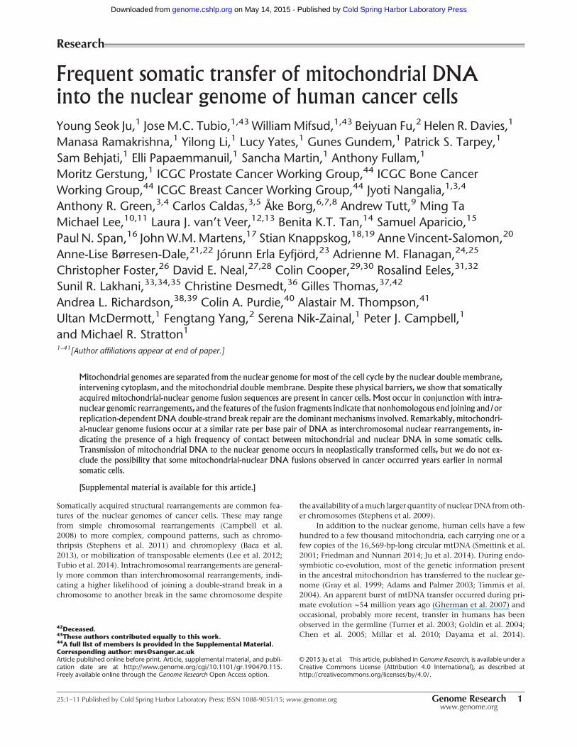
Frequent somatic transfer of mitochondrial DNAinto the nuclear genome of human cancer cells
Young Seok Ju,1 Jose M.C. Tubio,1,43 WilliamMifsud,1,43 Beiyuan Fu,2 Helen R. Davies,1
Manasa Ramakrishna,1 Yilong Li,1 Lucy Yates,1 Gunes Gundem,1 Patrick S. Tarpey,1
Sam Behjati,1 Elli Papaemmanuil,1 Sancha Martin,1 Anthony Fullam,1
Moritz Gerstung,1 ICGC Prostate Cancer Working Group,44 ICGC Bone CancerWorking Group,44 ICGC Breast Cancer Working Group,44 Jyoti Nangalia,1,3,4
Anthony R. Green,3,4 Carlos Caldas,3,5 Åke Borg,6,7,8 Andrew Tutt,9 Ming TaMichael Lee,10,11 Laura J. van’t Veer,12,13 Benita K.T. Tan,14 Samuel Aparicio,15
Paul N. Span,16 JohnW.M.Martens,17 Stian Knappskog,18,19 Anne Vincent-Salomon,20
Anne-Lise Børresen-Dale,21,22 Jórunn Erla Eyfjörd,23 Adrienne M. Flanagan,24,25
Christopher Foster,26 David E. Neal,27,28 Colin Cooper,29,30 Rosalind Eeles,31,32
Sunil R. Lakhani,33,34,35 Christine Desmedt,36 Gilles Thomas,37,42
Andrea L. Richardson,38,39 Colin A. Purdie,40 Alastair M. Thompson,41
Ultan McDermott,1 Fengtang Yang,2 Serena Nik-Zainal,1 Peter J. Campbell,1
and Michael R. Stratton11–41[Author affiliations appear at end of paper.]
Mitochondrial genomes are separated from the nuclear genome for most of the cell cycle by the nuclear double membrane,
intervening cytoplasm, and the mitochondrial double membrane. Despite these physical barriers, we show that somatically
acquired mitochondrial-nuclear genome fusion sequences are present in cancer cells. Most occur in conjunction with intra-
nuclear genomic rearrangements, and the features of the fusion fragments indicate that nonhomologous end joining and/or
replication-dependent DNA double-strand break repair are the dominant mechanisms involved. Remarkably, mitochondri-
al-nuclear genome fusions occur at a similar rate per base pair of DNA as interchromosomal nuclear rearrangements, in-
dicating the presence of a high frequency of contact between mitochondrial and nuclear DNA in some somatic cells.
Transmission of mitochondrial DNA to the nuclear genome occurs in neoplastically transformed cells, but we do not ex-
clude the possibility that some mitochondrial-nuclear DNA fusions observed in cancer occurred years earlier in normal
somatic cells.
[Supplemental material is available for this article.]
Somatically acquired structural rearrangements are common fea-tures of the nuclear genomes of cancer cells. These may rangefrom simple chromosomal rearrangements (Campbell et al.2008) to more complex, compound patterns, such as chromo-thripsis (Stephens et al. 2011) and chromoplexy (Baca et al.2013), or mobilization of transposable elements (Lee et al. 2012;Tubio et al. 2014). Intrachromosomal rearrangements are general-ly more common than interchromosomal rearrangements, indi-cating a higher likelihood of joining a double-strand break in achromosome to another break in the same chromosome despite
the availability of amuch larger quantity of nuclearDNA fromoth-er chromosomes (Stephens et al. 2009).
In addition to the nuclear genome, human cells have a fewhundred to a few thousand mitochondria, each carrying one or afew copies of the 16,569-bp-long circular mtDNA (Smeitink et al.2001; Friedman and Nunnari 2014; Ju et al. 2014). During endo-symbiotic co-evolution, most of the genetic information presentin the ancestral mitochondrion has transferred to the nuclear ge-nome (Gray et al. 1999; Adams and Palmer 2003; Timmis et al.2004). An apparent burst of mtDNA transfer occurred during pri-mate evolution ∼54 million years ago (Gherman et al. 2007) andoccasional, probably more recent, transfer in humans has beenobserved in the germline (Turner et al. 2003; Goldin et al. 2004;Chen et al. 2005; Millar et al. 2010; Dayama et al. 2014).
42Deceased.43These authors contributed equally to this work.44A full list of members is provided in the Supplemental Material.Corresponding author: [email protected] published online before print. Article, supplemental material, and publi-cation date are at http://www.genome.org/cgi/10.1101/gr.190470.115.Freely available online through the Genome Research Open Access option.
© 2015 Ju et al. This article, published in Genome Research, is available under aCreative Commons License (Attribution 4.0 International), as described athttp://creativecommons.org/licenses/by/4.0/.
Research
25:1–11 Published by Cold Spring Harbor Laboratory Press; ISSN 1088-9051/15; www.genome.org Genome Research 1www.genome.org
Cold Spring Harbor Laboratory Press on May 14, 2015 - Published by genome.cshlp.orgDownloaded from
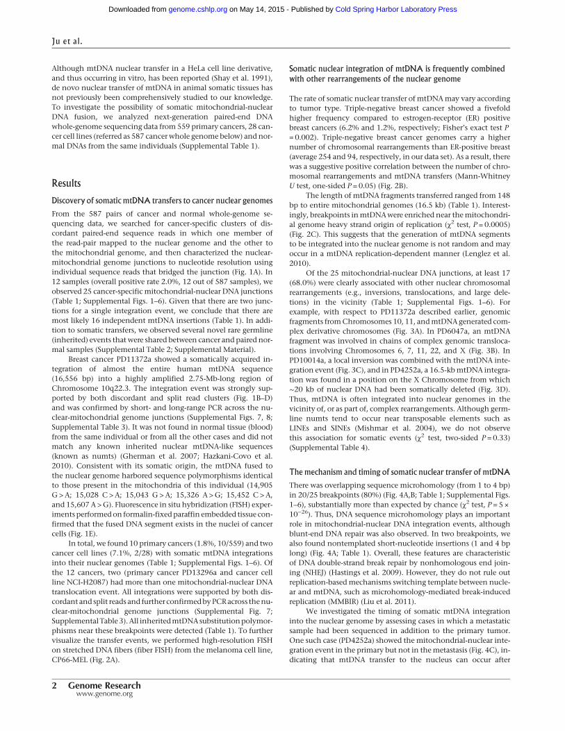
Although mtDNA nuclear transfer in a HeLa cell line derivative,and thus occurring in vitro, has been reported (Shay et al. 1991),de novo nuclear transfer of mtDNA in animal somatic tissues hasnot previously been comprehensively studied to our knowledge.To investigate the possibility of somatic mitochondrial-nuclearDNA fusion, we analyzed next-generation paired-end DNAwhole-genome sequencing data from 559 primary cancers, 28 can-cer cell lines (referred as 587 cancerwhole genome below) and nor-mal DNAs from the same individuals (Supplemental Table 1).
Results
Discovery of somatic mtDNA transfers to cancer nuclear genomes
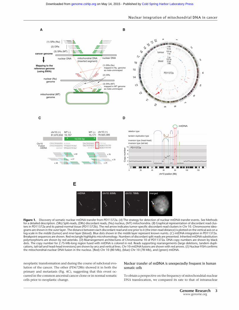
From the 587 pairs of cancer and normal whole-genome se-quencing data, we searched for cancer-specific clusters of dis-cordant paired-end sequence reads in which one member ofthe read-pair mapped to the nuclear genome and the other tothe mitochondrial genome, and then characterized the nuclear-mitochondrial genome junctions to nucleotide resolution usingindividual sequence reads that bridged the junction (Fig. 1A). In12 samples (overall positive rate 2.0%, 12 out of 587 samples), weobserved 25 cancer-specific mitochondrial-nuclear DNA junctions(Table 1; Supplemental Figs. 1–6). Given that there are two junc-tions for a single integration event, we conclude that there aremost likely 16 independent mtDNA insertions (Table 1). In addi-tion to somatic transfers, we observed several novel rare germline(inherited) events that were shared between cancer and paired nor-mal samples (Supplemental Table 2; Supplemental Material).
Breast cancer PD11372a showed a somatically acquired in-tegration of almost the entire human mtDNA sequence(16,556 bp) into a highly amplified 2.75-Mb-long region ofChromosome 10q22.3. The integration event was strongly sup-ported by both discordant and split read clusters (Fig. 1B–D)and was confirmed by short- and long-range PCR across the nu-clear-mitochondrial genome junctions (Supplemental Figs. 7, 8;Supplemental Table 3). It was not found in normal tissue (blood)from the same individual or from all the other cases and did notmatch any known inherited nuclear mtDNA-like sequences(known as numts) (Gherman et al. 2007; Hazkani-Covo et al.2010). Consistent with its somatic origin, the mtDNA fused tothe nuclear genome harbored sequence polymorphisms identicalto those present in the mitochondria of this individual (14,905G >A; 15,028 C > A; 15,043 G > A; 15,326 A >G; 15,452 C > A,and 15,607 A >G). Fluorescence in situ hybridization (FISH) exper-imentsperformedon formalin-fixedparaffin embedded tissue con-firmed that the fused DNA segment exists in the nuclei of cancercells (Fig. 1E).
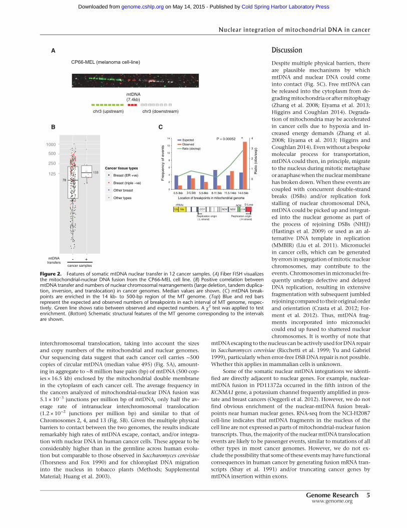
In total, we found 10 primary cancers (1.8%, 10/559) and twocancer cell lines (7.1%, 2/28) with somatic mtDNA integrationsinto their nuclear genomes (Table 1; Supplemental Figs. 1–6). Ofthe 12 cancers, two (primary cancer PD13296a and cancer cellline NCI-H2087) had more than one mitochondrial-nuclear DNAtranslocation event. All integrations were supported by both dis-cordant and split reads and further confirmedbyPCRacross thenu-clear-mitochondrial genome junctions (Supplemental Fig. 7;Supplemental Table3).All inheritedmtDNAsubstitutionpolymor-phisms near these breakpoints were detected (Table 1). To furthervisualize the transfer events, we performed high-resolution FISHon stretched DNA fibers (fiber FISH) from the melanoma cell line,CP66-MEL (Fig. 2A).
Somatic nuclear integration of mtDNA is frequently combined
with other rearrangements of the nuclear genome
The rate of somatic nuclear transfer of mtDNAmay vary accordingto tumor type. Triple-negative breast cancer showed a fivefoldhigher frequency compared to estrogen-receptor (ER) positivebreast cancers (6.2% and 1.2%, respectively; Fisher’s exact test P= 0.002). Triple-negative breast cancer genomes carry a highernumber of chromosomal rearrangements than ER-positive breast(average 254 and 94, respectively, in our data set). As a result, therewas a suggestive positive correlation between the number of chro-mosomal rearrangements and mtDNA transfers (Mann-WhitneyU test, one-sided P = 0.05) (Fig. 2B).
The length of mtDNA fragments transferred ranged from 148bp to entire mitochondrial genomes (16.5 kb) (Table 1). Interest-ingly, breakpoints inmtDNAwere enriched near themitochondri-al genome heavy strand origin of replication (χ2 test, P = 0.0005)(Fig. 2C). This suggests that the generation of mtDNA segmentsto be integrated into the nuclear genome is not random and mayoccur in a mtDNA replication-dependent manner (Lenglez et al.2010).
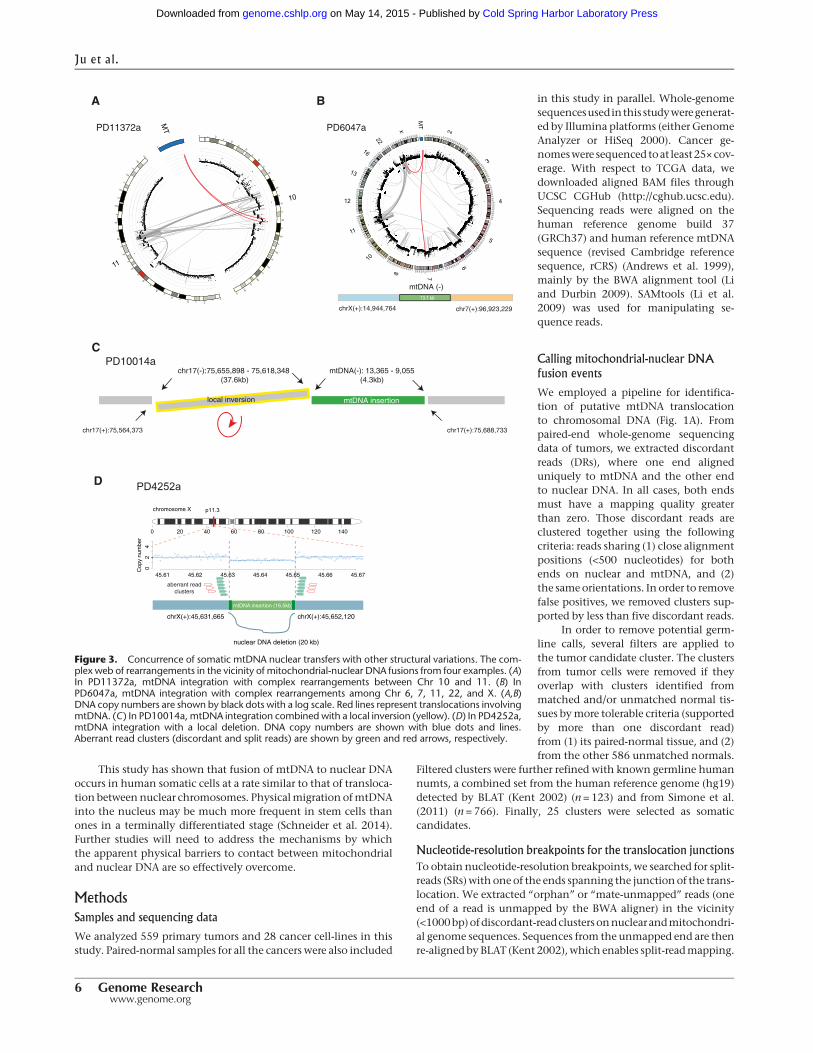
Of the 25 mitochondrial-nuclear DNA junctions, at least 17(68.0%) were clearly associated with other nuclear chromosomalrearrangements (e.g., inversions, translocations, and large dele-tions) in the vicinity (Table 1; Supplemental Figs. 1–6). Forexample, with respect to PD11372a described earlier, genomicfragments fromChromosomes 10, 11, andmtDNAgenerated com-plex derivative chromosomes (Fig. 3A). In PD6047a, an mtDNAfragment was involved in chains of complex genomic transloca-tions involving Chromosomes 6, 7, 11, 22, and X (Fig. 3B). InPD10014a, a local inversion was combined with the mtDNA inte-gration event (Fig. 3C), and in PD4252a, a 16.5-kbmtDNA integra-tion was found in a position on the X Chromosome from which∼20 kb of nuclear DNA had been somatically deleted (Fig. 3D).Thus, mtDNA is often integrated into nuclear genomes in thevicinity of, or as part of, complex rearrangements. Although germ-line numts tend to occur near transposable elements such asLINEs and SINEs (Mishmar et al. 2004), we do not observethis association for somatic events (χ2 test, two-sided P = 0.33)(Supplemental Table 4).
The mechanism and timing of somatic nuclear transfer of mtDNA
There was overlapping sequence microhomology (from 1 to 4 bp)in 20/25 breakpoints (80%) (Fig. 4A,B; Table 1; Supplemental Figs.1–6), substantially more than expected by chance (χ2 test, P = 5 ×10⁻²⁶). Thus, DNA sequence microhomology plays an importantrole in mitochondrial-nuclear DNA integration events, althoughblunt-end DNA repair was also observed. In two breakpoints, wealso found nontemplated short-nucleotide insertions (1 and 4 bplong) (Fig. 4A; Table 1). Overall, these features are characteristicof DNA double-strand break repair by nonhomologous end join-ing (NHEJ) (Hastings et al. 2009). However, they do not rule outreplication-basedmechanisms switching template between nucle-ar and mtDNA, such as microhomology-mediated break-inducedreplication (MMBIR) (Liu et al. 2011).
We investigated the timing of somatic mtDNA integrationinto the nuclear genome by assessing cases in which a metastaticsample had been sequenced in addition to the primary tumor.One such case (PD4252a) showed the mitochondrial-nuclear inte-gration event in the primary but not in themetastasis (Fig. 4C), in-dicating that mtDNA transfer to the nucleus can occur after
Ju et al.
2 Genome Researchwww.genome.org
Cold Spring Harbor Laboratory Press on May 14, 2015 - Published by genome.cshlp.orgDownloaded from
neoplastic transformation and during the course of subclonal evo-lution of the cancer. The other (PD6728b) showed it in both theprimary and metastasis (Fig. 4C), suggesting that this event oc-curred in the common ancestral cancer clone or in normal somaticcells prior to neoplastic change.
Nuclear transfer of mtDNA is unexpectedly frequent in human
somatic cells
To obtain a perspective on the frequency ofmitochondrial-nuclearDNA translocation, we compared its rate to that of intranuclear
1
0 20 40 60 80 100
120
140
160
180
200
220
240
2
020
4060
8010
012
014
0
160
180
200
220
240
3
020
40
60
80100
120
140
160
180
4
0204060
80100120140160180
5
020
4060
80100
120140
160180
6
020
4060
80100120140160
70
20406080100120140
8020406080100
120
140
9
0
20406080100
120
140
10
02040608010012
011
020
4060
80
10012
012 020
4060
80100
120
130
2040
6080
100
140
2040
6080
100
150
2040
6080
100
16020
40
60
80
170
20
40
6080
18020
4060
19
0
2040
200
2040
60
210
2040
220
2040
x0
204060
80100
120140
tumour
blood
1 Mb10 kb
100 bp1 bp
1 bp100 bp10 kb1 Mb
PD11372a
CTCCTGGGTG AGAAACTCCTGGGTG TTGGCCTCAC GATAT TTGGCCTCAC
FusionChr10
mtDNATACTGTGGC CC AGACCTCTT ACACT GC AGACCTCTT
TACTGTGGC CC CTCAG
x 60
x 27 x 25
x 76
chr10 (+)81,670,932
MT (-)15,157
MT (-)15,171
chr10 (+)78,920,385
x 24x 28
16,556 bp
******
**
***
************
* *
chr10 position (Mb)0 20 40 60 80 100 120
*********************************************************
******************************************************************
*********************************************************************************************
***********************************************************************
*******************************************************************
**************************************
**********
*****************************
**
*******
*
************
*
************************
**
**
***
*
*****************************************************
******************************
*
*********
**
*******
************
************************
*************************
**
************************
*
******
*************************************
*******************************
********
*****************
*******************
**************************************
***
***********************************************************************
********************************
***********************************
**************************************************************************************************
******************************************************************************************
*****************************************************
**************************
*********************************************************************
0
2
4
6
8
10
Cop
y nu
mbe
r
************
********
********************************************************************************************************************************** *****
**********
****
************ ****
**
*******
*****
*************
*******
*************
***********
**********
**
****
**
*
**
***
**
***
*****
***********
**********
**
***********
**
**
**
******
**
**********
**
*****
**
*******
******************
*********
************************************************************************************************
**
****
*******
****
mtDNA
PD11372a
deletion type
tandem duplication type
inversion type (head-head)inversion type (tail-tail)
mtDNA chr10: 80Mb chr10: 78Mb merged
E
DC
BA
mitochondrial DNA(inserted segment)
(2) DRs
(1) SRs (Nu)
nuclear DNA nuclear DNA
Mapping to thereference genome
(using BWA)
cancer genome(3) SRs (MT)
nuclear (Nu) genome
mitochondrial (MT) genome
(2) DRs
(1) SRs (Nu) mapped in Nu. genome as mate-unmmaped
(3) SRs (MT) mapped in MT genome as mate-unmmaped
Figure 1. Discovery of somatic nuclear mtDNA transfer from PD11372a. (A) The strategy for detection of nuclear mtDNA transfer events. See Methodsfor a detailed description. (SRs) Split-reads, (DRs) discordant reads, (Nu) nucleus, (MT) mitochondria. (B) Graphical representation of discordant read clus-ters in PD11372a and its paired-normal tissue (PD11372b). The red arrow indicates tumor-specific discordant-read clusters in Chr 10. Chromosome ideo-grams are shown in the outer layer. The distance between each discordant read and one prior to it (the inter-read distance) is plotted on the vertical axis on alog-scale in themiddle (tumor) and inner layer (blood). Blue dots shown in themiddle layer represent known numts. (C ) mtDNA integration in PD11372a.Breakpoint sequences are shown. Red rectangle highlightsmicrohomology. Numbers of discordant split reads are presented. InheritedmtDNA substitutionpolymorphisms are shown by red asterisks. (D) Rearrangement architectures of Chromosome 10 of PD11372a. DNA copy numbers are shown by blackdots. The copy number for 2.75-Mb-long region fused with mtDNA is colored in red. Reads supporting rearrangements (large deletions, tandem dupli-cations, tail-tail and head-head inversions) are shown by arcs and vertical lines. Chr 10-mtDNA fusions are shownwith red arrows. (E) Nuclear FISH confirmsthe mitochondrial-nuclear DNA fusion in the nucleus. (Red) Chr 10 (80 Mb), (blue) Chr 10 (78 Mb), and (green) mtDNA.
Nuclear integration of mitochondrial DNA in cancer
Genome Research 3www.genome.org
Cold Spring Harbor Laboratory Press on May 14, 2015 - Published by genome.cshlp.orgDownloaded from
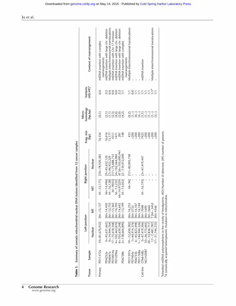
Tab
le1.
Summaryofso
matic
mitoch
ondrial-nuc
lear
DNAfusionsiden
tified
from
12cance
rsamples
Tissue
Sample
Left
junction
Rightjunction
Frag
.size
(bp)
Micro-
homology
(bp,bp)
Variants
(#D/#
P)a
Context
ofrearrangem
ent
Nuc
lear
MT
MT
Nuc
lear
Prim
ary
PD11
372a
10+:81
,670
,932
][M
−:15,15
7M−:15,17
1][10+
:78,92
0,38
516
,556
(0,1)
6/6
mtD
NAinsertionwith
complex
rearrang
emen
tsPD
4252
aX+:45
,631
,665
][M
+:14
,450
M+:14
,496
][X+:45
,652
,120
16,616
(2,1)
2/2
mtD
NAinsertionwith
largech
r.de
letio
nPD
6047
aX+:14
,944
,764
][M
−:12,73
5M−:16,12
8][7+:96
,923
,229
13,177
(1,1)
6/6
Multip
leinterchrom
osom
altran
sloc
ations
PD10
014a
17−:75,61
8,34
8][M
−:13,36
5M−:905
5][17+
:75,68
8,73
343
11(2,3)
0/0
mtD
NAinsertionwith
Chr
17inversion
PD13
296a
4+:102
,463
,870
][M
−:14,70
5M−:13,23
5][4+:10
2,46
4,08
414
71(4,0)
0/0
mtD
NAinsertionwith
largech
r.de
letio
n6+
:103
,639
,248
][M
+:14
,692
M+:14
,972
]TA
AT[6+:10
3,69
0,94
128
1(2,0)
2/2
mtD
NAinsertionwith
largech
r.de
letio
nPD
6728
b2+
:138
,664
,890
][M
−:13,19
9M−:13,05
2][2−:139
,012
,040
148
(4,2)
1/1
mtD
NAinsertionwith
complex
rearrang
emen
tsPD
1139
7a19
−:12,65
0,38
2][M
+:16
,233
M+:96
][17+
:40,00
5,73
843
3(0,2)
1/1
Multip
leinterchrom
osom
altran
sloc
ations
PD74
04a
1+:44,91
4,37
6][M
+:37
32–
–>2
00(1,–)
0/0
–
PD67
33b
6−:45,82
3,49
8][M
+:16
,107
––
>200
(0,–)
1/1
–
PD11
768a
1−:144
,944
,326
][M
+:16
,104
––
>200
(4,–)
1/1
–
Cellline
CP6
6-MEL
3+:47,41
9,50
6][M
−:704
8M−:16,19
3][3+:47
,419
,447
7425
(1,1)
1/1
mtD
NAinsertion
NCI-H
2087
10+:26
,775
,605
][M
+:16
90–
–>2
00(1,–)
1/1
–
20−:33,83
6,71
7][M
−:566
6–
–>2
00(1,–)
1/1
–
17−:7,481
,787
]T[M
−:345
2–
–>2
00(1,–)
1/1b
Multip
leinterchrom
osom
altran
sloc
ations
17−:31,74
4,23
5][M
+:43
46–
–>2
00(3,–)
1/1
–
a Inh
erite
dmtD
NApo
lymorph
ismsin
thevicinity
ofbreakp
oints.(#D)Num
berof
detected
,(#P
)nu
mbe
rof
presen
t.bAsomaticallyacqu
iredhe
teroplasmicmutationin
mito
chon
dria.
Ju et al.
4 Genome Researchwww.genome.org
Cold Spring Harbor Laboratory Press on May 14, 2015 - Published by genome.cshlp.orgDownloaded from
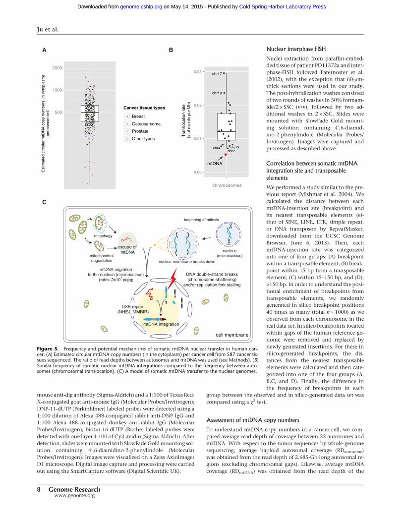
interchromosomal translocation, taking into account the sizesand copy numbers of the mitochondrial and nuclear genomes.Our sequencing data suggest that each cancer cell carries ∼500copies of circular mtDNA (median value 495) (Fig. 5A), amount-ing in aggregate to ∼8 million base pairs (bp) of mtDNA (500 cop-ies × 16.5 kb) enclosed by the mitochondrial double membranein the cytoplasm of each cancer cell. The average frequency inthe cancers analyzed of mitochondrial-nuclear DNA fusion was5.1 × 10−3 junctions per million bp of mtDNA, only half the av-erage rate of intranuclear interchromosomal translocation(1.2 × 10⁻² junctions per million bp) and similar to that ofChromosomes 2, 4, and 13 (Fig. 5B). Given the multiple physicalbarriers to contact between the two genomes, the results indicateremarkably high rates of mtDNA escape, contact, and/or integra-tion with nuclear DNA in human cancer cells. These appear to beconsiderably higher than in the germline across human evolu-tion but comparable to those observed in Saccharomyces cerevisiae(Thorsness and Fox 1990) and for chloroplast DNA migrationinto the nucleus in tobacco plants (Methods; SupplementalMaterial; Huang et al. 2003).
Discussion
Despite multiple physical barriers, thereare plausible mechanisms by whichmtDNA and nuclear DNA could comeinto contact (Fig. 5C). Free mtDNA canbe released into the cytoplasm from de-gradingmitochondria or aftermitophagy(Zhang et al. 2008; Eiyama et al. 2013;Higgins and Coughlan 2014). Degrada-tion of mitochondria may be acceleratedin cancer cells due to hypoxia and in-creased energy demands (Zhang et al.2008; Eiyama et al. 2013; Higgins andCoughlan 2014). Evenwithout a bespokemolecular process for transportation,mtDNA could then, in principle, migrateto the nucleus during mitotic metaphaseor anaphasewhen thenuclearmembranehas broken down.When these events arecoupled with concurrent double-strandbreaks (DSBs) and/or replication forkstalling of nuclear chromosomal DNA,mtDNA could be picked up and integrat-ed into the nuclear genome as part ofthe process of rejoining DSBs (NHEJ)(Hastings et al. 2009) or used as an al-ternative DNA template in replication(MMBIR) (Liu et al. 2011). Micronucleiin cancer cells, which can be generatedbyerrors in segregationofmitoticnuclearchromosomes, may contribute to theevents.Chromosomes inmicronuclei fre-quently undergo defective and delayedDNA replication, resulting in extensivefragmentation with subsequent jumbledrejoiningcompared to theiroriginalorderand orientation (Crasta et al. 2012; For-ment et al. 2012). Thus, mtDNA frag-ments incorporated into micronucleicould end up fused to shattered nuclearchromosomes. It is worthy of note that
mtDNAescaping to thenucleus canbe actively used forDNA repairin Saccharomyces cerevisiae (Ricchetti et al. 1999; Yu and Gabriel1999), particularly when error-free DSB DNA repair is not possible.Whether this applies in mammalian cells is unknown.
Some of the somatic nuclear mtDNA integrations we identi-fied are directly adjacent to nuclear genes. For example, nuclear-mtDNA fusion in PD11372a occurred in the fifth intron of theKCNMA1 gene, a potassium channel frequently amplified in pros-tate and breast cancers (Oeggerli et al. 2012). However, we do notfind obvious enrichment of the nuclear-mtDNA fusion break-points near human nuclear genes. RNA-seq from the NCI-H2087cell-line indicates that mtDNA fragments in the nucleus of thecell line are not expressed as parts of mitochondrial-nuclear fusiontranscripts. Thus, themajority of the nuclearmtDNA translocationevents are likely to be passenger events, similar to mutations of allother types in most cancer genomes. However, we do not ex-clude the possibility that someof these eventsmayhave functionalconsequences in human cancer by generating fusion mRNA tran-scripts (Shay et al. 1991) and/or truncating cancer genes bymtDNA insertion within exons.
chr3 (downstream)chr3 (upstream)
mtDNA(7.4kb)
A
CP66-MEL (melanoma cell-line)
250
500
1000
B
78
133125Cancer tissue types
Breast (ER +ve)
Other breast
Breast (triple −ve)
Other types
mtDNAtransfers
- +cancer samples
0
1
2
3
4
0
2
4
6
8
10
12
14
0.5-3kb 3-5.5kb 5.5-8kb 8-11.5kb 11.5-14kb 14-0.5kb
Ratio
(obs/
exp
)
Fre
quency
of eve
nts
Location of breakpoints in mitochondrial genome
Expected Observed Ratio (obs/exp)
P = 0.00052 *
D-LooprRNAs
16s12s CO1 ND5ND4
ND6
Replication origin ( H strand)
Replication origin ( L strand)
C
CYB
Figure 2. Features of somatic mtDNA nuclear transfer in 12 cancer samples. (A) Fiber FISH visualizesthe mitochondrial-nuclear DNA fusion from the CP66-MEL cell line. (B) Positive correlation betweenmtDNA transfer and numbers of nuclear chromosomal rearrangements (large deletion, tandem duplica-tion, inversion, and translocation) in cancer genomes. Median values are shown. (C) mtDNA break-points are enriched in the 14 kb- to 500-bp region of the MT genome. (Top) Blue and red barsrepresent the expected and observed numbers of breakpoints in each interval of MT genome, respec-tively. Green line shows ratio between observed and expected numbers. A χ2 test was applied to testenrichment. (Bottom) Schematic structural features of the MT genome corresponding to the intervalsare shown.
Nuclear integration of mitochondrial DNA in cancer
Genome Research 5www.genome.org
Cold Spring Harbor Laboratory Press on May 14, 2015 - Published by genome.cshlp.orgDownloaded from
This study has shown that fusion of mtDNA to nuclear DNAoccurs in human somatic cells at a rate similar to that of transloca-tion between nuclear chromosomes. Physicalmigration ofmtDNAinto the nucleus may be much more frequent in stem cells thanones in a terminally differentiated stage (Schneider et al. 2014).Further studies will need to address the mechanisms by whichthe apparent physical barriers to contact between mitochondrialand nuclear DNA are so effectively overcome.
Methods
Samples and sequencing data
We analyzed 559 primary tumors and 28 cancer cell-lines in thisstudy. Paired-normal samples for all the cancers were also included
in this study in parallel. Whole-genomesequencesused inthis studyweregenerat-ed by Illumina platforms (either GenomeAnalyzer or HiSeq 2000). Cancer ge-nomeswere sequenced toat least25×cov-erage. With respect to TCGA data, wedownloaded aligned BAM files throughUCSC CGHub (http://cghub.ucsc.edu).Sequencing reads were aligned on thehuman reference genome build 37(GRCh37) and human reference mtDNAsequence (revised Cambridge referencesequence, rCRS) (Andrews et al. 1999),mainly by the BWA alignment tool (Liand Durbin 2009). SAMtools (Li et al.2009) was used for manipulating se-quence reads.
Calling mitochondrial-nuclear DNA
fusion events
We employed a pipeline for identifica-tion of putative mtDNA translocationto chromosomal DNA (Fig. 1A). Frompaired-end whole-genome sequencingdata of tumors, we extracted discordantreads (DRs), where one end aligneduniquely to mtDNA and the other endto nuclear DNA. In all cases, both endsmust have a mapping quality greaterthan zero. Those discordant reads areclustered together using the followingcriteria: reads sharing (1) close alignmentpositions (<500 nucleotides) for bothends on nuclear and mtDNA, and (2)the sameorientations. In order to removefalse positives, we removed clusters sup-ported by less than five discordant reads.
In order to remove potential germ-line calls, several filters are applied tothe tumor candidate cluster. The clustersfrom tumor cells were removed if theyoverlap with clusters identified frommatched and/or unmatched normal tis-sues bymore tolerable criteria (supportedby more than one discordant read)from (1) its paired-normal tissue, and (2)from the other 586 unmatched normals.
Filtered clusters were further refined with known germline humannumts, a combined set from the human reference genome (hg19)detected by BLAT (Kent 2002) (n = 123) and from Simone et al.(2011) (n = 766). Finally, 25 clusters were selected as somaticcandidates.
Nucleotide-resolution breakpoints for the translocation junctions
To obtain nucleotide-resolution breakpoints, we searched for split-reads (SRs)with one of the ends spanning the junctionof the trans-location. We extracted “orphan” or “mate-unmapped” reads (oneend of a read is unmapped by the BWA aligner) in the vicinity(<1000bp)ofdiscordant-readclustersonnuclearandmitochondri-al genome sequences. Sequences from the unmapped end are thenre-alignedbyBLAT (Kent 2002),which enables split-readmapping.
A
100
5 10
15
20
25
30
35
40
45
50
55
60
65
70
75
80
85
90
95
100
105
110
115
120
125
130
135
11
05
101520
25
30
35
40
45
50
55
60
65
70
75
80
85
90
95
100
105
110
115
120
125
130
135
MT
0
PD11372a
D
B
chrX(+):45,631,665 chrX(+):45,652,120
mtDNA insertion (16.5kb)
nuclear DNA deletion (20 kb)
�
chromosome X
0 20 40 60 80 100 120 140
p11.3
Cop
y nu
mbe
r
45.61 45.62 45.63 45.64 45.65 45.66 45.67
02
4
ooooo
o
oooooooo
oo
ooooo
oooooooooo
o
oo
oo
oo
o
ooo
o
ooooo
ooooo
ooooooooooooo
oo
oooooooooooooooooooo
o
oo
oooooooooooooo
ooooooo
ooo
ooooo
ooooo
ooooo
o
oooo oooooooo ooo oo ooooo o o oo oo oo oooooo ooooo o oo o o ooo
ooo oooooooooooo o o ooo o oooooooooo
08
aberrant readclusters
PD4252a
PD6047a 2
0 10 20 30 40 50 60 70 80 90 100
110
120
130
140
150
160
170
180
190
200
210
220
230
240
3
010
2030
4050
6070
8090
100110120
130140
150160170180190
4
0102030405060708090100110120130140150160170180190
5
010
2030
4050
6070
8090
100110120130140150160170180
6
0102030405060708090100110120130140
150160170
7
0102030405060708090100110
120
130
140
150
8
010203040506070809010011012
013014
010 01020
3040
5060
7080
90100110120130
110
1020
3040
5060
7080
90100110
120130
12
0102030405060708090
100110120130
13
0102030405060708090100
110
16
010
2030
4050
60
7080
90
220
1020
3040
50
x0
1020
3040
5060
7080
90100110
120130140150
MT
0
mtDNA (-)
chr7(+):96,923,229chrX(+):14,944,764
13.1 kb
mtDNA(-): 13,365 - 9,055(4.3kb)
chr17(+):75,688,733
local inversion
C
chr17(+):75,564,373
chr17(-):75,655,898 - 75,618,348 (37.6kb)
mtDNA insertion
PD10014a
Figure 3. Concurrence of somatic mtDNA nuclear transfers with other structural variations. The com-plex web of rearrangements in the vicinity of mitochondrial-nuclear DNA fusions from four examples. (A)In PD11372a, mtDNA integration with complex rearrangements between Chr 10 and 11. (B) InPD6047a, mtDNA integration with complex rearrangements among Chr 6, 7, 11, 22, and X. (A,B)DNA copy numbers are shown by black dots with a log scale. Red lines represent translocations involvingmtDNA. (C) In PD10014a, mtDNA integration combined with a local inversion (yellow). (D) In PD4252a,mtDNA integration with a local deletion. DNA copy numbers are shown with blue dots and lines.Aberrant read clusters (discordant and split reads) are shown by green and red arrows, respectively.
Ju et al.
6 Genome Researchwww.genome.org
Cold Spring Harbor Laboratory Press on May 14, 2015 - Published by genome.cshlp.orgDownloaded from
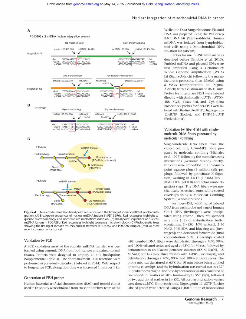
Validation by PCR
A PCR validation assay of the somatic mtDNA transfer was per-formed using genomic DNA from both cancer and paired-normaltissues. Primers were designed to amplify all the breakpoints(Supplemental Table 3). The short-fragment PCR reactions wereperformed as previously described (Tubio et al. 2014).With respectto long-range PCR, elongation time was increased 1 min per 1 kb.
Generation of FISH probes
Human bacterial artificial chromosomes (BAC) and fosmid clonesused in this studywere obtained from the clone archive teamof the
Wellcome Trust Sanger Institute. PlasmidDNA was prepared using the PhasePrepBAC DNA kit (Sigma-Aldrich). HumanmtDNA was isolated from lymphoblas-toid cells using a Mitochondrial DNAIsolation kit (Abcam).
Probes for use in FISH were made asdescribed before (Gribble et al. 2013).Purified mtDNA and plasmid DNA werefirst amplified using a GenomePlexWhole Genome Amplification (WGA)kit (Sigma-Aldrich) following the manu-facturer’s protocols, then labeled usinga WGA reamplification kit (Sigma-Aldrich) with a custom-made dNTP mix.Probes for interphase FISH were labeleddirectly with Aminoallyl-dUTPs - ATTO-488, -Cy3, -Texas Red, and -Cy5 (JenaBioscience); probes for fiber-FISHwere la-beledwithBiotin-16-dUTP,Digoxigenin-11-dUTP (Roche), and DNP-11-dUTP(PerkinElmer).
Validation by fiber-FISH with single-
molecule DNA fibers generated by
molecular combing
Single-molecule DNA fibers from thecancer cell line, CP66-MEL, were pre-pared by molecular combing (Michaletet al. 1997) following the manufacturer’sinstructions (Genomic Vision). Briefly,the cells were embedded in a low-melt-point agarose plug (1 million cells perplug), followed by proteinase K diges-tion, washing in 1 × TE (10 mM Tris, 1mM EDTA, pH 8.0) and beta-agarose di-gestion steps. The DNA fibers were me-chanically stretched onto saline-coatedcoverslips using a Molecular CombingSystem (Genomic Vision).
For fiber-FISH, ∼500 ng of labeledDNA from each probe and 4 μg of humanCot-1 DNA (Invitrogen) were precipi-tated using ethanol, then resuspendedin a mix (1:1) of hybridization buffer(containing 2 × SSC, 10% sarkosyl, 2 MNaCl, 10% SDS, and blocking aid [Invi-trogen]) and deionized formamide (finalconcentration 50%). Coverslips coated
with combed DNA fibers were dehydrated through a 70%, 90%,and 100% ethanol series and aged at 65°C for 30 sec, followed bydenaturation in an alkaline denature solution (0.5 M NaOH, 1.5M NaCl) for 1–3 min, three washes with 1×PBS (Invitrogen), anddehydration through a 70%, 90%, and 100% ethanol series. Theprobe mix was denatured at 65°C for 10 min before being appliedonto the coverslips, and the hybridization was carried out in a 37°C incubator overnight. The post-hybridizationwashes consisted oftwo rounds of washes in 50% formamide/2 × SSC (v/v), followedby two additional washes in 2 × SSC. All post-hybridizationwasheswere done at 25°C, 5min each time.Digoxigenin-11-dUTP (Roche)labeled probes were detected using a 1:100 dilution ofmonoclonal
chr4: GGCGA AACCC CATT TCTACT Fusion: GGCGA AACCC CATT GGTCGT
mtDNA: TTTTT CATAT CATT GGTCGT
TAAT
A
chr4(+)102,463,870 mtDNA(-):14,705
PD13296a (2 mtDNA nuclear integration events)
mtDNA(-):13,235 chr4(+):102,464,084
4bp microhomology blunt-end DNA joining
GCTACGATTT CTTTTGATGT : mtDNA GCTACGATTT AAATAACCAC : FusionGTTATCTTCA AAATAACCAC : chr4
Integration #1
Integration #2
chr6(+):103,639,248 mtDNA(+) 14,692
mtDNA(+) 14,972
chr6(+):103,690,941
chr6: TTGTAAGA AC TAATAGAATG Fusion: TTGTAAGA AC AACCACGACC mtDNA:CACGGACT AC AACCACGACC GTAAATTATG GCTGAATCAT : mtDNA
GTAAATTATG TAAT AAAATATTTG : FusionCTGGGTCCTA AAAATATTTG : chr6
2bp microhomology non-template 4bp insertion
PD6728bB
chr2(+):138,664,890 mtDNA(-) 13,199
mtDNA(-) 13,052
chr2(-):139,012,040
chr2: TCATCT TGCT TGCGTTTTGC Fusion: TCATCT TGCT GCGAACAGAG mtDNA:GCAGAC TGCT GCGAACAGAG
4bp microhomology
GGGGTGGGGC CT TCTATGGC : mtDNA GGGGTGGGGC CT GACTGCAG : Fusion
CTTGGTCTTG CT GACTGCAG : chr2
2bp microhomology
C
PD4252
Fertilized egg
Blood (PD4252b)
MRCA LN metastasis
(PD4252c)
Primary locus(PD4252a; subclonal)
mtDNA transfer
PD6728
Fertilized egg
Blood (PD6728a)
MRCA
LN metastasis(PD6728c; clonal)
Primary locus(PD6728b; clonal)
mtDNA transfer
Transformation
Transformation
Figure 4. Nucleotide-resolution breakpoint sequences and the timing of somatic mtDNA nuclear inte-gration. (A) Breakpoint sequences of nuclear-mtDNA fusions in PD13296a. Red rectangles highlight se-quence microhomology and nontemplate nucleotides insertion. (B) Breakpoint sequences of nuclear-mtDNA fusions in PD6728b. Red rectangles highlight sequence microhomology. (C) Phylogenetic treesshowing the timing of somatic mtDNA nuclear transfers in PD4252 and PD6728 samples. (MRCA) Mostrecent common ancestor cell.
Nuclear integration of mitochondrial DNA in cancer
Genome Research 7www.genome.org
Cold Spring Harbor Laboratory Press on May 14, 2015 - Published by genome.cshlp.orgDownloaded from
mouse anti-dig antibody (Sigma-Aldrich) and a 1:100 of Texas Red-X-conjugated goat anti-mouse IgG (Molecular Probes/Invitrogen);DNP-11-dUTP (PerkinElmer) labeled probes were detected using a1:100 dilution of Alexa 488-conjugated rabbit anti-DNP IgG and1:100 Alexa 488-conjugated donkey anti-rabbit IgG (MolecularProbes/Invitrogen); biotin-16-dUTP (Roche) labeled probes weredetected with one layer 1:100 of Cy3-avidin (Sigma-Aldrich). Afterdetection, slides weremountedwith SlowFade Goldmounting sol-ution containing 4′,6-diamidino-2-phenylindole (MolecularProbes/Invitrogen). Images were visualized on a Zeiss AxioImagerD1 microscope. Digital image capture and processing were carriedout using the SmartCapture software (Digital Scientific UK).
Nuclear interphase FISH
Nuclei extraction from paraffin-embed-ded tissue of patient PD11372a and inter-phase-FISH followed Paternoster et al.(2002), with the exception that 60-μm-thick sections were used in our study.The post-hybridization washes consistedof two rounds of washes in 50% formam-ide/2 × SSC (v/v), followed by two ad-ditional washes in 2 × SSC. Slides weremounted with SlowFade Gold mount-ing solution containing 4′,6-diamid-ino-2-phenylindole (Molecular Probes/Invitrogen). Images were captured andprocessed as described above.
Correlation between somatic mtDNA
integration site and transposable
elements
We performed a study similar to the pre-vious report (Mishmar et al. 2004). Wecalculated the distance between eachmtDNA-insertion site (breakpoint) andits nearest transposable elements (ei-ther of SINE, LINE, LTR, simple repeat,or DNA transposon by RepeatMasker,downloaded from the UCSC GenomeBrowser, June 6, 2013). Then, eachmtDNA-insertion site was categorizedinto one of four groups: (A) breakpointwithin a transposable element; (B) break-point within 15 bp from a transposableelement; (C) within 15–150 bp; and (D),>150 bp. In order to understand the posi-tional enrichment of breakpoints fromtransposable elements, we randomlygenerated in silico breakpoint positions40 times as many (total n = 1000) as weobserved from each chromosome in thereal data set. In silico breakpoints locatedwithin gaps of the human reference ge-nome were removed and replaced bynewly generated insertions. For these insilico-generated breakpoints, the dis-tances from the nearest transposableelements were calculated and then cate-gorized into one of the four groups (A,B,C, and D). Finally, the difference inthe frequency of breakpoints in each
group between the observed and in silico-generated data set wascompared using a χ2 test.
Assessment of mtDNA copy numbers
To understand mtDNA copy numbers in a cancer cell, we com-pared average read depth of coverage between 22 autosomes andmtDNA. With respect to the tumor sequences by whole-genomesequencing, average haploid autosomal coverage (RDautosome)was obtained from the read depth of 2.685-Gb-long autosomal re-gions (excluding chromosomal gaps). Likewise, average mtDNAcoverage (RDmtDNA) was obtained from the read depth of the
500
1000
2000
Cancer tissue types
Breast
Osteosarcoma
Other types
Prostate
AE
stim
ated
circ
ular
mtD
NA
cop
y nu
mbe
rs (
in c
ytop
lasm
)pe
r ca
ncer
cel
l
0.00
0.01
0.02
0.03
chromosomesTr
ansl
ocat
ion
rate
(#
of e
vent
s pe
r Mb)
chr2
chr17
chr19
mtDNA
chr4 chr13
B
mitophagy
mitochondrialdegradation
escape of mtDNA
nuclear membrane breaks down
beginning of mitosis
DNA double-strand breaks(chromosome shattering)
and/or replication fork stalling
mtDNA migration to the nucleus (micronucleus)
(rate> 2x10 pcpg)
cell membrane
-4
nucleus(micronucleus)
mtDNA integration
DSB repair (NHEJ, MMBIR)
C
Figure 5. Frequency and potential mechanisms of somatic mtDNA nuclear transfer in human can-cer. (A) Estimated circular mtDNA copy numbers (in the cytoplasm) per cancer cell from 587 cancer tis-sues sequenced. The ratio of read depths between autosomes and mtDNA was used (see Methods). (B)Similar frequency of somatic nuclear mtDNA integrations compared to the frequency between auto-somes (chromosomal translocation). (C) A model of somatic mtDNA transfer to the nuclear genomes.
Ju et al.
8 Genome Researchwww.genome.org
Cold Spring Harbor Laboratory Press on May 14, 2015 - Published by genome.cshlp.orgDownloaded from
16.5-kbmitochondrial genome. Finally, mtDNA copy number in adiploid cell (Cmt) is calculated as shown below:
Cmt = 2× RDmtDNA
RDautosome.
Assessment of translocation rate for autosomes
and mitochondria
We identified structural variations among nuclear chromosomes(large deletions, tandem duplications, inversions, and interchro-mosomal translocations) using the BRASS II algorithm (Nik-Zainal et al. 2012), which identifies rearrangements by clusteringdiscordant read pairs that point to the same junction and confirmsbreakpoints by local assembly of unmapped reads. The sensitivityand specificity of the BRASS II algorithm is equivalent to those val-ues of the algorithm used for mitochondrial-nuclear DNA fusions(data not shown). We extracted interchromosomal translocationsto calculate the rate of such events. The rate of each haploid auto-some (Rtr,ch) is calculated as shown below:
Rtr, ch(events per megabase) = Ntr,ch/(2× Lch)/Nsam,
whereNtr,ch is the total number of somatic interchromosom-al translocation junctions involving a specific chromosome, Lch isthe lengthof thenonredundant regionof the chromosome inmeg-abases, and Nsam is the total number of samples analyzed. To ob-tain the unique region length (Lch), we excluded redundant (orhighly repetitive) sequence lengths from the ungapped length ofeach chromosome. Genomic regions classified in one or more ofthe three criteria shown below were defined as redundant, wheretranslocation events couldnot be easily detected due to ambiguousread alignment: (1) simple repeats, located by Tandem RepeatsFinder (Benson 1999); (2) segmental duplications with moderateto high sequence similarity (≥95%) (Bailey et al. 2002), or (3) repet-itive sequences including up to 10 different classes of repeats (suchas SINE, LINE, LTR, DNA transposons, andmicrosatellites), locatedby the RepeatMasker program (http://www.repeatmasker.org),with a low divergence level (divergence < 5%). These nonredun-dant sequence regions were downloaded from the UCSC GenomeBrowser (http://genome.ucsc.edu).
Similarly, the rate of mitochondrial-nuclear DNA transloca-tions (Rtr,mt) was calculated as below:
Rtr,mt(events per megabase) = Ntr,mt/(Cmt × LmtDNA)/Nsam,
where Ntr,mt is the total number of junctions of somatic mi-tochondrial-nuclear DNA fusions identified,Cmt is themedian val-ue of mitochondrial genome copy numbers in a diploid cancer cellcalculated above (495 copies), and LmtDNA is the length of the mi-tochondrial genome in megabases (0.016569 Mb).
Assessment of the rates of nuclear mtDNA fusion
and mtDNA escape to the nucleus
Fusion of mtDNA to the nuclear genome requires at least twoevents, each of which could influence the rate of mitochondrial-nuclearDNAfusion. These include escapeofmtDNAto thenucleusand integration to nuclearDNA. According to thismodel, the over-all number of such fusion events can be calculated using the ratesfor these processes (ρescape and ρintegration, respectively):
Ntr = Nsam×Ngen× rescape × rintegration,
where Ntr is the number of total somatic mitochondrial-nu-clear DNA fusion events (n = 12), Nsam is the total number of can-
cer tissues (n = 587), and Ngen is the number of average cellgeneration from the fertilized egg. Using a reasonable assumptionthat Ngen = 1000, we obtain the rate of somatic mtDNA fusion tothe nuclear genome (ρescape × ρintegration) to be 2 × 10⁻⁵ per cell percell generation (pcpg). With one more very conservative assump-tion that ρintegration is 0.1, we obtain ρescape to be 2 × 10⁻⁴ pcpg,or at least one escape event per 5000 cell generations. We hypoth-esize that the real ρintegration value is thought to be much lowerthan 0.1, which results in a higher ρescape. For example, duringthe generation of knockout mice, homologous recombination al-lows one fixation event per 1000–10,000 microinjected DNA cop-ies (Brinster et al. 1985). The integration rate may, however, behigher than the rate in cancer cells with defective homologous re-combination-based repair and increased availability of nucleardouble-strand breaks, which can be joined to by NHEJ or MMBIR.
The mtDNA fusion to the nuclear genome in the germline(the rate of numts insertion) is around 5 × 10⁻⁶ per germ cell perindividual generation in previous phylogenetic studies (Hazkani-Covo et al. 2010). The rate is equivalent to ∼5 × 10⁻⁸ pcpg, giventhat the number of germ cell divisions per human generation is∼100 (401 in males and 31 in females [Drost and Lee 1995]).
Data access
Sequence data for sample pairs with positivemtDNAnuclear trans-fer have been submitted to the European Genome-phenomeArchive (EGA; https://www.ebi.ac.uk/ega/home). The study acces-sion number is EGAS00001001234. Sample accession numbers areavailable in Supplemental Table 1.
List of affiliations1Cancer Genome Project, Wellcome Trust Sanger Institute,Hinxton, Cambridge CB10 1SA, United Kingdom; 2CytogeneticsFacility, Wellcome Trust Sanger Institute, Hinxton, CambridgeCB10 1SA, United Kingdom; 3Cambridge University HospitalsNHS Foundation Trust, Cambridge CB2 0QQ, United Kingdom;4Department of Haematology, University of Cambridge,Cambridge CB2 0XY, United Kingdom; 5Cancer Research UK(CRUK) Cambridge Institute, University of Cambridge,Cambridge CB2 0RE, United Kingdom; 6BioCare, StrategicCancer Research Program, SE-223 81 Lund, Sweden; 7CREATEHealth, Strategic Centre for Translational Cancer Research,SE-221 00 Lund, Sweden; 8Department of Oncology andPathology, Lund University Cancer Center, SE-221 85 Lund,Sweden; 9Breakthrough Breast Cancer Research Unit, ResearchOncology, King’s College London, Guy’s Hospital, London SE19RT, United Kingdom; 10Laboratory for International Alliance onGenomic Research, RIKEN Center for Integrative MedicalSciences, 230-0045 Yokohama, Japan; 11National Center forGenome Medicine, Institute of Biomedical Sciences, AcademiaSinica, Taipei 115, Taiwan; 12Department of LaboratoryMedicine, Helen Diller Family Comprehensive Cancer Center,University of California, San Francisco, California 94158, USA;13Netherlands Cancer Institute, 1066 CX Amsterdam,Netherlands; 14Department of General Surgery, SingaporeGeneral Hospital, Singapore 169608; 15Department of MolecularOncology, British Columbia Cancer Agency, Vancouver V5Z1L3, Canada; 16Department of Radiation Oncology andDepartment of Laboratory Medicine, Radboud UniversityMedical Center, 6525 HP Nijmegen, Netherlands; 17Departmentof Medical Oncology, Erasmus MC Cancer Institute, Erasmus
Nuclear integration of mitochondrial DNA in cancer
Genome Research 9www.genome.org
Cold Spring Harbor Laboratory Press on May 14, 2015 - Published by genome.cshlp.orgDownloaded from
University Medical Center, 3015 CE Rotterdam, Netherlands;18Section of Oncology, Department of Clinical Science,University of Bergen, N-5020 Bergen, Norway; 19Department ofOncology, Haukeland University Hospital, 5021 Bergen, Norway;20Institut Curie, INSERM U934 and Department of TumorBiology, 75248 Paris cédex 05, France; 21Department of Genetics,Institute for Cancer Research, Oslo University Hospital, TheNorwegian Radium Hospital, Montebello, 0310 Oslo, Norway;22The K.G. Jebsen Center for Breast Cancer Research, Institutefor Clinical Medicine, Faculty of Medicine, University of Oslo,0450 Oslo, Norway; 23Cancer Research Laboratory, University ofIceland, 101 Reykjavik, Iceland; 24Royal National OrthopaedicHospital, Middlesex HA7 4LP, United Kingdom; 25UCL CancerInstitute, University College London, London WC1E 6DD,United Kingdom; 26University of Liverpool and HCA PathologyLaboratories, London WC1E 6JA, United Kingdom; 27UrologicalResearch Laboratory, Cancer Research UK Cambridge ResearchInstitute, Cambridge CB2 0RE, United Kingdom; 28Departmentof Surgical Oncology, University of Cambridge, Addenbrooke’sHospital, Cambridge CB2 0QQ, United Kingdom; 29Institute ofCancer Research, Sutton, London SM2 5NG, United Kingdom;30Department of Biological Sciences and School of Medicine,University of East Anglia, Norwich NR4 7TJ, United Kingdom;31Division of Genetics and Epidemiology, The Institute ofCancer Research, Sutton SM2 5NG, United Kingdom; 32RoyalMarsden NHS Foundation Trust, London SW3 6JJ and SuttonSM2 5PT, United Kingdom; 33University of Queensland, Schoolof Medicine, Brisbane, QLD 4006, Australia; 34PathologyQueensland, Royal Brisbane and Women’s Hospital, Brisbane,QLD 4029, Australia; 35University of Queensland, UQ Centre forClinical Research, Brisbane, QLD 4029, Australia; 36Breast CancerTranslational Research Laboratory, Université Libre de Bruxelles,Institut Jules Bordet, 1000 Brussels, Belgium; 37Université Lyon1, Institut National du Cancer (INCa)–Synergie, 69008 Lyon,France; 38Dana-Farber Cancer Institute, Boston, Massachusetts02215, USA; 39Brigham and Women’s Hospital, Harvard MedicalSchool, Boston, Massachusetts 02115, USA; 40Department ofPathology, Ninewells Hospital and Medical School, Dundee DD19SY, United Kingdom; 41Department of Surgical Oncology,University of Texas MD Anderson Cancer Center, Houston,Texas 77030, USA
Acknowledgments
We thank Thomas Bleazard at the Faculty of Medical and HumanSciences, University of Manchester for discussion and assistancewith manuscript preparation. We also thank The Cancer GenomeAtlas (TCGA) Project Team and their specimen donors for pro-viding sequencingdata used in this study. Thisworkwas supportedby theWellcome Trust. Y.S.J is supported by a EuropeanMolecularBiology Organization long-term fellowship (LTF 1203_2012). J.M.C.T. is supported by Marie Curie Fellowship FP7-PEOPLE-2012-IEF (project number 328264). P.J.C. is a Wellcome Trust SeniorClinical Fellow. Support was provided to A.M.F. by the NationalInstitute for Health Research (NIHR) UCLH Biomedical ResearchCentre. The ICGC Breast Cancer Consortium was supported by agrant from the European Union (BASIS) and the Wellcome Trust.The ICGC Prostate Cancer Consortium was funded by CancerResearch UK with a grant from the Dallaglio Foundation (grantnumber C5047/A14835). R.E. is supported by National Institutefor Health Research support to the Biomedical Research Centre atThe Institute of Cancer Research and Royal Marsden NHS
Foundation Trust. We also thank the National Cancer ResearchProstate Cancer Mechanisms of Progression and Treatment(PROMPT) collaborative (grant code G0500966/75466) whichhas funded tissue and urine collections in Cambridge. The authorsalso acknowledge financial support fromtheDepartmentofHealthvia the National Institute for Health Research comprehensiveBiomedical Research Centre award to Guy’s and St. Thomas’ NHSFoundation Trust and Breakthrough Breast Cancer Research(ICGC 08/09 and KCL) (A.T.).
Author contributions: Y.S.J., J.M.C.T., W.M., P.J.C., and M.R.S.conceived the project. Y.S.J. and J.M.C.T. performedmitochondri-al sequence analyses and PCR validation experiments. W.M., B.F.,and F.Y. conducted cytogenetics validation experiments. Y.S.J., J.M.C.T., H.R.D., M.R., Y.L., L.Y., G.G., P.S.T., S.B., E.P., A.F., M.G.,and S.N-Z. performed bioinformatics analyses. J.N., A.R.G., C.C.,A.B., A.T., M.T.M.L., L.J.v.V., B.K.T.T., S.A., P.N.S., J.W.M.M., S.K., A.V-S., A-L.B-D., J.E.E., A.M.F., C.F., D.E.N., C.C., R.E., S.R.L.,C.D., G.T., A.L.R., C.A.P., and A.M.T. provided clinical samplesand commented on the manuscript. U.M. and S.N-Z. providedconceptual advice. Y.S.J and M.R.S wrote the paper with contribu-tions from all authors.
References
Adams KL, Palmer JD. 2003. Evolution of mitochondrial gene con-tent: gene loss and transfer to the nucleus. Mol Phylogenet Evol 29:380–395.
Andrews RM, Kubacka I, Chinnery PF, Lightowlers RN, Turnbull DM,Howell N. 1999. Reanalysis and revision of the Cambridge reference se-quence for human mitochondrial DNA. Nat Genet 23: 147.
Baca SC, Prandi D, Lawrence MS, Mosquera JM, Romanel A, Drier Y, Park K,Kitabayashi N, MacDonald TY, Ghandi M, et al. 2013. Punctuated evo-lution of prostate cancer genomes. Cell 153: 666–677.
Bailey JA, Gu Z, Clark RA, Reinert K, Samonte RV, Schwartz S, Adams MD,Myers EW, Li PW, Eichler EE. 2002. Recent segmental duplications inthe human genome. Science 297: 1003–1007.
Benson G. 1999. Tandem repeats finder: a program to analyze DNA se-quences. Nucleic Acids Res 27: 573–580.
Brinster RL, Chen HY, TrumbauerME, YagleMK, Palmiter RD. 1985. Factorsaffecting the efficiency of introducing foreign DNA into mice by micro-injecting eggs. Proc Natl Acad Sci 82: 4438–4442.
Campbell PJ, Stephens PJ, Pleasance ED, O’Meara S, Li H, Santarius T,Stebbings LA, Leroy C, Edkins S, Hardy C, et al. 2008. Identification ofsomatically acquired rearrangements in cancer using genome-widemas-sively parallel paired-end sequencing. Nat Genet 40: 722–729.
Chen JM, Chuzhanova N, Stenson PD, Ferec C, Cooper DN. 2005. Meta-analysis of gross insertions causing human genetic disease: novel muta-tional mechanisms and the role of replication slippage. Hum Mutat25: 207–221.
Crasta K, Ganem NJ, Dagher R, Lantermann AB, Ivanova EV, Pan Y, Nezi L,Protopopov A, Chowdhury D, Pellman D. 2012. DNA breaks and chro-mosome pulverization from errors in mitosis. Nature 482: 53–58.
Dayama G, Emery SB, Kidd JM, Mills RE. 2014. The genomic landscape ofpolymorphic human nuclear mitochondrial insertions. Nucleic AcidsRes 42: 12640–12649.
Drost JB, LeeWR. 1995. Biological basis of germlinemutation: comparisonsof spontaneous germline mutation rates among Drosophila, mouse, andhuman. Environ Mol Mutagen 25: 48–64.
Eiyama A, Kondo-Okamoto N, Okamoto K. 2013. Mitochondrial degrada-tion during starvation is selective and temporally distinct from bulkautophagy in yeast. FEBS Lett 587: 1787–1792.
Forment JV, Kaidi A, Jackson SP. 2012. Chromothripsis and cancer: causesand consequences of chromosome shattering. Nat Rev Cancer 12:663–670.
Friedman JR, Nunnari J. 2014. Mitochondrial form and function. Nature505: 335–343.
Gherman A, Chen PE, Teslovich TM, Stankiewicz P, Withers M, Kashuk CS,Chakravarti A, Lupski JR, Cutler DJ, Katsanis N. 2007. Population bottle-necks as a potential major shaping force of human genome architecture.PLoS Genet 3: e119.
Goldin E, Stahl S, Cooney AM, Kaneski CR, Gupta S, Brady RO, Ellis JR,Schiffmann R. 2004. Transfer of a mitochondrial DNA fragment toMCOLN1 causes an inherited case of mucolipidosis IV. Hum Mutat24: 460–465.
Ju et al.
10 Genome Researchwww.genome.org
Cold Spring Harbor Laboratory Press on May 14, 2015 - Published by genome.cshlp.orgDownloaded from
Gray MW, Burger G, Lang BF. 1999. Mitochondrial evolution. Science 283:1476–1481.
Gribble SM,Wiseman FK, Clayton S, Prigmore E, Langley E, Yang F,MaguireS, Fu B, Rajan D, Sheppard O, et al. 2013. Massively parallel sequencingreveals the complex structure of an irradiated human chromosome ona mouse background in the Tc1 model of Down syndrome. PLoS One8: e60482.
Hastings PJ, Lupski JR, Rosenberg SM, Ira G. 2009.Mechanisms of change ingene copy number. Nat Rev Genet 10: 551–564.
Hazkani-Covo E, Zeller RM, Martin W. 2010. Molecular poltergeists: mito-chondrial DNA copies (numts) in sequenced nuclear genomes. PLoSGenet 6: e1000834.
Higgins GC, Coughlan MT. 2014. Mitochondrial dysfunction and mitoph-agy: the beginning and end to diabetic nephropathy? Br J Pharmacol171: 1917–1942.
HuangCY, AyliffeMA, Timmis JN. 2003. Directmeasurement of the transferrate of chloroplast DNA into the nucleus. Nature 422: 72–76.
Ju YS, Alexandrov LB, Gerstung M, Martincorena I, Nik-Zainal S,Ramakrishna M, Davies HR, Papaemmanuil E, Gundem G, Shlien A,et al. 2014. Origins and functional consequences of somatic mitochon-drial DNA mutations in human cancer. eLife 3: e02935.
Kent WJ. 2002. BLAT—the BLAST-like alignment tool. Genome Res 12:656–664.
Lee E, Iskow R, Yang L, Gokcumen O, Haseley P, Luquette LJ III, Lohr JG,Harris CC, Ding L, Wilson RK, et al. 2012. Landscape of somatic retro-transposition in human cancers. Science 337: 967–971.
Lenglez S, HermandD, Decottignies A. 2010. Genome-widemapping of nu-clear mitochondrial DNA sequences links DNA replication origins tochromosomal double-strand break formation in Schizosaccharomycespombe. Genome Res 20: 1250–1261.
Li H, Durbin R. 2009. Fast and accurate short read alignment with Burrows-Wheeler transform. Bioinformatics 25: 1754–1760.
Li H, Handsaker B, Wysoker A, Fennell T, Ruan J, Homer N, Marth G,Abecasis G, Durbin R; 1000 Genome Project Data ProcessingSubgroup. 2009. The Sequence Alignment/Map format and SAMtools.Bioinformatics 25: 2078–2079.
Liu P, Erez A, Nagamani SC, Dhar SU, Kolodziejska KE, DharmadhikariAV, Cooper ML, Wiszniewska J, Zhang F, Withers MA, et al. 2011.Chromosome catastrophes involve replication mechanisms generatingcomplex genomic rearrangements. Cell 146: 889–903.
Michalet X, Ekong R, Fougerousse F, Rousseaux S, Schurra C, Hornigold N,van Slegtenhorst M, Wolfe J, Povey S, Beckmann JS, et al. 1997.Dynamic molecular combing: stretching the whole human genomefor high-resolution studies. Science 277: 1518–1523.
Millar DS, Tysoe C, Lazarou LP, Pilz DT, Mohammed S, Anderson K,Chuzhanova N, Cooper DN, Butler R. 2010. An isolated case of lissence-phaly caused by the insertion of a mitochondrial genome-derived DNAsequence into the 5′ untranslated region of the PAFAH1B1 (LIS1) gene.Hum Genomics 4: 384–393.
Mishmar D, Ruiz-Pesini E, Brandon M, Wallace DC. 2004. MitochondrialDNA-like sequences in the nucleus (NUMTs): insights into our Africanorigins and the mechanism of foreign DNA integration. Hum Mutat23: 125–133.
Nik-Zainal S, Alexandrov LB,WedgeDC, VanLoo P, GreenmanCD, Raine K,Jones D, Hinton J, Marshall J, Stebbings LA, et al. 2012. Mutational pro-cesses molding the genomes of 21 breast cancers. Cell 149: 979–993.
Oeggerli M, Tian Y, Ruiz C,Wijker B, Sauter G, Obermann E, Guth U, ZlobecI, SausbierM, Kunzelmann K, et al. 2012. Role of KCNMA1 in breast can-cer. PLoS One 7: e41664.
Paternoster SF, Brockman SR, McClure RF, Remstein ED, Kurtin PJ, DewaldGW. 2002. A newmethod to extract nuclei from paraffin-embedded tis-sue to study lymphomas using interphase fluorescence in situ hybridi-zation. Am J Pathol 160: 1967–1972.
Ricchetti M, Fairhead C, Dujon B. 1999.Mitochondrial DNA repairs double-strand breaks in yeast chromosomes. Nature 402: 96–100.
Schneider JS, Cheng X, Zhao Q, Underbayev C, Gonzalez JP, Raveche ES,FraidenraichD, Ivessa AS. 2014. Reversiblemitochondrial DNA accumu-lation in nuclei of pluripotent stem cells. Stem Cells Dev 23: 2712–2719.
Shay JW, Baba T, Zhan QM, Kamimura N, Cuthbert JA. 1991. HeLaTG cellshave mitochondrial DNA inserted into the c-myc oncogene. Oncogene6: 1869–1874.
Simone D, Calabrese FM, LangM, Gasparre G, Attimonelli M. 2011. The ref-erence human nuclear mitochondrial sequences compilation validatedand implemented on the UCSC genome browser. BMC Genomics 12:517.
Smeitink J, van den Heuvel L, DiMauro S. 2001. The genetics and pathologyof oxidative phosphorylation. Nat Rev Genet 2: 342–352.
Stephens PJ, McBride DJ, Lin ML, Varela I, Pleasance ED, Simpson JT,Stebbings LA, Leroy C, Edkins S, Mudie LJ, et al. 2009. Complex land-scapes of somatic rearrangement in human breast cancer genomes.Nature 462: 1005–1010.
Stephens PJ, Greenman CD, Fu B, Yang F, Bignell GR, Mudie LJ, PleasanceED, Lau KW, Beare D, Stebbings LA, et al. 2011. Massive genomic rear-rangement acquired in a single catastrophic event during cancer devel-opment. Cell 144: 27–40.
Thorsness PE, Fox TD. 1990. Escape of DNA from mitochondria to the nu-cleus in Saccharomyces cerevisiae. Nature 346: 376–379.
Timmis JN, Ayliffe MA, Huang CY, Martin W. 2004. Endosymbiotic genetransfer: organelle genomes forge eukaryotic chromosomes. Nat RevGenet 5: 123–135.
Tubio JM, Li Y, Ju YS, Martincorena I, Cooke SL, Tojo M, Gundem G,Pipinikas CP, Zamora J, Raine K, et al. 2014. Mobile DNA in cancer.Extensive transduction of nonrepetitive DNA mediated by L1 retro-transposition in cancer genomes. Science 345: 1251343.
Turner C, Killoran C, Thomas NS, Rosenberg M, Chuzhanova NA, JohnstonJ, Kemel Y, Cooper DN, Biesecker LG. 2003. Human genetic diseasecaused by de novo mitochondrial-nuclear DNA transfer. Hum Genet112: 303–309.
YuX, Gabriel A. 1999. Patching broken chromosomeswith extranuclear cel-lular DNA. Mol Cell 4: 873–881.
Zhang H, Bosch-Marce M, Shimoda LA, Tan YS, Baek JH, Wesley JB,Gonzalez FJ, Semenza GL. 2008. Mitochondrial autophagy is an HIF-1-dependent adaptive metabolic response to hypoxia. J Biol Chem283: 10892–10903.
Received February 3, 2015; accepted in revised form April 14, 2015.
Nuclear integration of mitochondrial DNA in cancer
Genome Research 11www.genome.org
Cold Spring Harbor Laboratory Press on May 14, 2015 - Published by genome.cshlp.orgDownloaded from
10.1101/gr.190470.115Access the most recent version at doi: published online May 11, 2015Genome Res.
Young Seok Ju, Jose M.C. Tubio, William Mifsud, et al. genome of human cancer cellsFrequent somatic transfer of mitochondrial DNA into the nuclear
Material
Supplemental
http://genome.cshlp.org/content/suppl/2015/04/16/gr.190470.115.DC1.html
P<P
Published online May 11, 2015 in advance of the print journal.
Open Access
Open Access option.Genome ResearchFreely available online through the
License
Commons Creative
.http://creativecommons.org/licenses/by/4.0/
License (Attribution 4.0 International), as described at , is available under a Creative CommonsGenome ResearchThis article, published in
ServiceEmail Alerting
click here.top right corner of the article or
Receive free email alerts when new articles cite this article - sign up in the box at the
http://genome.cshlp.org/subscriptionsgo to: Genome Research To subscribe to
© 2015 Ju et al.; Published by Cold Spring Harbor Laboratory Press
Cold Spring Harbor Laboratory Press on May 14, 2015 - Published by genome.cshlp.orgDownloaded from
Copyright © 2022 FDOKUMEN