Founded in 1959 The Society for Pediatric Radiology 61st ...
298
The Society for Pediatric Radiology introduced its new logo August 15, 2013. The logo communicates both the warmth of the Society community and the strength of the members’ commitment to excellent and thoughtful care of the pediatric patient. The first official logo for the SPR was designed by Tamar Kahane Oestreich of Cincinnati, Ohio in 1985. Thank you, Mrs. Oestreich. Founded in 1959 The Society for Pediatric Radiology 61 st Annual Meeting & Postgraduate Course May 15-19, 2018 The Omni Nashville Hotel Nashville, Tennessee, United States Pediatric Imaging Technologist Program May 17-19, 2018 Jointly provided by the American College of Radiology This supplement was not sponsored by outside commercial interests; it was funded entirely by the Society’s own resources. https://doi.org/10.1007/s00247-018-4130-z Pediatr Radiol (2018) 48 (Suppl 1):S1–S298
-
Upload
khangminh22 -
Category
Documents
-
view
1 -
download
0
Transcript of Founded in 1959 The Society for Pediatric Radiology 61st ...
The Society for Pediatric Radiology introduced its new logo August 15, 2013. The logo communicates both the
warmth of the Society community and the strength of the members’ commitment to excellent and thoughtful care
of the pediatric patient.
The first official logo for the SPR was designed by Tamar Kahane Oestreich of Cincinnati, Ohio in
1985. Thank you, Mrs. Oestreich.
Founded in 1959
The Society for Pediatric Radiology
61st Annual Meeting & Postgraduate Course
May 15-19, 2018
The Omni Nashville Hotel
Nashville, Tennessee, United States
Pediatric Imaging Technologist Program
May 17-19, 2018
Jointly provided by the American College of Radiology
This supplement was not sponsored by outside commercial interests; it was funded entirely by the Society’s own resources.
https://doi.org/10.1007/s00247-018-4130-zPediatr Radiol (2018) 48 (Suppl 1):S1–S298
WELCOME MESSAGE ........................................................................................................................................ 2
DEDICATION ....................................................................................................................................................... 4
SPR 2018 ORGANIZATION ................................................................................................................................. 6
CONTINUING MEDICAL EDUCATION ............................................................................................................ 7
MAINTENANCE OF CERTIFICATION ....................................................................................................... 7 OBJECTIVES ................................................................................................................................................. 7 DISCLOSURES .............................................................................................................................................. 8 ACKNOWLEDGEMENTS .......................................................................................................................... 10
MEETING SPACE ............................................................................................................................................... 11
SPR GENERAL INFORMATION ....................................................................................................................... 12
MISSION STATEMENT .............................................................................................................................. 12 DIVERSITY & INCLUSION STATEMENT ............................................................................................... 12
SPR OFFICERS, DIRECTORS, COMMITTEES ............................................................................................... 12
SPR PAST PRESIDENTS, PREVIOUS & FUTURE MEETINGS, AWARDEES &
EDWARD B. D. NEUHAUSER LECTURE ............................................................................................... 17
SPR 2018 HONOREES ........................................................................................................................................ 27
GOLD MEDALISTS .................................................................................................................................... 27 PIONEER HONOREE .................................................................................................................................. 31 PRESIDENTIAL RECOGNITION AWARDEES ........................................................................................ 32 HONORARY MEMBERS ............................................................................................................................ 34 EDWARD B. SINGLETON-HOOSHANG TAYBI AWARDEE ................................................................ 37 JACK O. HALLER AWARDEES................................................................................................................. 39 HEIDI PATRIQUIN AWARDEE ................................................................................................................. 41
JOHN P. CAFFEY AWARDS ............................................................................................................................. 42
2018 SPR EDWARD B. D. NEUHAUSER LECTURE ...................................................................................... 49
SOCIAL EVENTS ................................................................................................................................................ 50
FULL PROGRAM SCHEDULE .......................................................................................................................... 51
SCIENTIFIC PAPERS ......................................................................................................................................... 77
SCIENTIFIC PAPERS – TECHNOLOGISTS* ................................................................................................. 176
POSTERS ........................................................................................................................................................... 181
CASE REPORT POSTERS......................................................................................................................... 181 EDUCATIONAL POSTERS ...................................................................................................................... 192 SCIENTIFIC POSTERS ............................................................................................................................. 243
POSTERS – TECHNOLOGISTS* ..................................................................................................................... 270
CASE REPORT POSTERS - TECHNOLOGISTS ..................................................................................... 270 EDUCATIONAL POSTERS - TECHNOLOGISTS ................................................................................... 272
AUTHOR INDEX BY ABSTRACT .................................................................................................................. 278
KEYWORD INDEX BY ABSTRACT .............................................................................................................. 290
TABLE OF CONTENTS
* (T) Indicates an Imaging Technologist Program Submission.
Pediatr Radiol (2018) 48 (Suppl 1):S1–S298S2
WELCOME MESSAGE I am extremely pleased to welcome you to the 61
st meeting of The Society for Pediatric Radiology in the
beautiful, vibrant city of Nashville, Tennessee.
We live in exciting, dynamic and challenging times. The world of medicine continues to evolve and advance at
a remarkable and hectic pace. Radiology is at the center of this turmoil and affected in myriad ways. Imaging
technology advances at a pace that is difficult to manage. New machines obtain more numerous and better
images. New techniques allow for imaging of structures and diseases previously thought impossible. Imaging
increasingly guides intervention and therapy. Imaging and imaged-guided interventional procedures are truly
integral to modern pediatric healthcare delivery.
Many challenges persist. We constantly strive to achieve better images more efficiently (time and money),
safely and with compassion. We challenge ourselves to maximize patient throughput, decrease reporting turn-
around time, manage and limit radiation exposure, use contrast judiciously and safely, and address increasing
concern with the use of sedation and anesthesia in children. Our clinical colleagues ask tough questions and want answers. Our researchers
continually push the envelope of discovery.
Many important themes run through this meeting. Everything, however, comes back to the overriding theme of “Value-added Pediatric
Radiology.” Everything that we do in our daily work is aimed at improving the healthcare of the children that we serve. Everything that you
will see and experience at this meeting will aid you to become a better pediatric radiologist or pediatric imaging technologist and take better
care of your patients.
This meeting occurs at a time when our nation, inexplicably, is taking a deep look into issues of race, gender, diversity and inclusion. The
SPR has always prided itself on being a welcoming, diverse, inclusive society. Ill-conceived laws in the states of Tennessee and California
have forced the SPR to directly deal with this issue. As a professional society, we welcome this as a positive opportunity. We have created
an SPR Diversity and Inclusion Committee, co-chaired by Ashok Panigrahy and Stephanie Spottswood. This important committee will help
the SPR to evaluate itself. How are we doing as a society with diversity and inclusion? What can we do better? How can we help members
to promote diversity and inclusion in their home institutions and in the care of our patients and their families? Dr. Spottswood, Associate
Vice Chair for Diversity in the Department of Radiology at Vanderbilt University and Chair of the Office of Inclusion and Health Equity
Advisory Board at Vanderbilt Children’s Hospital, will deliver a keynote address “The Power of Diversity: Meeting Today’s (and
Tomorrow’s) Greatest Healthcare Challenges” to the whole society on Wednesday, immediately prior to the Neuhauser lecture. There will
be a workshop on diversity on Friday. I think that you will find these presentations thought-provoking and enlightening.
In current times, the health and well-being of children is under substantial threat by a small, but growing swell of child abuse denialism. For
those of us that work in children’s healthcare, this challenge is difficult to understand and rationalize. We work very hard to diagnose, to
treat and to protect children and their families. We are extraordinarily careful to get the diagnosis right, be it child abuse or something else.
However, there is much to be learned. We need to be careful, deliberate, regimented, collaborative and compassionate. I am delighted to
have Paul Kleinman deliver this year’s Neuhauser lecture. The lecture will be a capstone of a remarkable and impactful career. Paul’s
lecture, “Curious Bones: Sustaining Discovery in the Face of Doubt”, will be an erudite look at where we have been, where we are and
where we will go in child abuse imaging. A sequence of workshops will also address the important topic of child abuse.
I am indebted to Janet Reid and Jonathan Dillman for organizing and directing a spectacular Postgraduate Course. There will be two
concurrent tracks, one on body imaging and the other on general pediatric radiology. Each topic will be a presented by paired speakers as a
brief didactic followed by a response with cases. The paired speakers have diligently coordinated their presentations. I think that you will
find this novel presentation mode exciting and educational. We are excited to include an updated version of RSNA Diagnosis Live™. We
have dedicated a half day of one track to an update on Image Gently, organized by Don Frush and Keith Strauss, Chair and Vice Chair of
the Image Gently Alliance, respectively.
You will also be excited to find more educational content in the form of workshops – more time slots and more choices. In addition to
traditional sunrise workshops, we have added two “mid-day” (actually late morning) workshops. Ethan Smith and Mahesh Thapa have
constructed a wonderfully diverse array of choices that should please everyone. I am very excited about a non-imaging, leadership
development/practice of radiology series that runs throughout the workshops.
Please plan your stay until Saturday noon!! On Saturday morning, there are five spectacular sessions of focused learning planned –
Education (“The Teaching Portfolio: A Hands-on Workshop on How to Get Promoted as an Educator”), Interventional Radiology (“Young
Practitioners: The Future of Pediatric Interventional Radiology”), Oncology, Hands-on Ultrasound and Neuroradiology. These sessions are
an integral component of the meeting and allow for greater depth of development of the program in each of these topics than can be
achieved elsewhere in the meeting. If you leave early, you will miss out!
George Bisset has carefully recruited two highly qualified, highly competitive teams (again Team E and Team T) to battle it out in a much-
anticipated reprisal of Jeopardy. Thanks to George for making this both fun and highly educational (great cases & pearls).
No professional meeting is successful without great science (papers) and education (posters). A heartfelt thanks goes to all of you who have
invested hard work into your projects, papers and presentations. All papers will be presented in tri-current sessions this year in order to
maximize the schedule and other activities. This reflects the continual growth of our society and our meeting, as well as the need to create
more presentation and participation opportunities. Most of the paper sessions will be preceded by a 20-minute keynote address.
Pediatr Radiol (2018) 48 (Suppl 1):S1–S298 S3
Melissa Hilmes (with her local connections to Vanderbilt) and Rich Heller (with his enthusiasm for the practice of radiology) have been
charged to invite and coach speakers to address the fundamental theme of our meeting – “Value-added Pediatric Radiology.” These talks
will focus on how we add value in each realm of our work.
Imaging technologists are integral to our work. I am very pleased that, for the third year, there will be a Pediatric Imaging Technologists’
Course, renamed from “Radiographers’ Course to reflect its broader content. The course is again organized and directed by Laura Gruber
and Christine Harris with guidance from Steve Simoneaux. The Imaging Technologists’ Course is a great opportunity to learn and network.
Please consider how your radiology department or group can facilitate the attendance of a deserving technologist (or more) who will surely
benefit from this great program!
Nashville is an extraordinary city. It is vibrant and full of life. I urge you to take time to explore what Nashville has to offer. The Omni
Nashville Hotel is conveniently located close to the rich nightlife of downtown Nashville and close to many great restaurants. Be sure to try
the barbecue, Prince’s hot chicken and, if to your liking, local libations. Take time to listen to the music! Many choices abound and not just
limited to country music. Nashville is a very progressive, diverse, inclusive, welcoming and fun city. It is a perfect environment of our
meeting. We thank the people of the city of Nashville and the staff at the truly marvelous Omni Hotel Nashville for their hospitality.
This meeting would not be possible if not for the extraordinary support and work of Angela Davis and Kasey O’Dea. They have been two
or three steps ahead of me throughout the process. Angela and Kasey – thank you so much!!!
Lastly, the SPR meeting is about people. Reacquaint yourself with old friends and make some new friends. Please take time to introduce
yourself to and get to know new people. Share your experiences and your enthusiasm for the work that we do in Pediatric Radiology. We
can all be immensely proud of the work that we do, both individually and collectively as a professional society. Let us take this time to
celebrate those successes, but also to learn and to do even better! I wish you a truly productive and fun week in Nashville!
Peter J. Strouse, MD, FACR
President and Program Director
The Society for Pediatric Radiology
Pediatr Radiol (2018) 48 (Suppl 1):S1–S298S4
DEDICATION
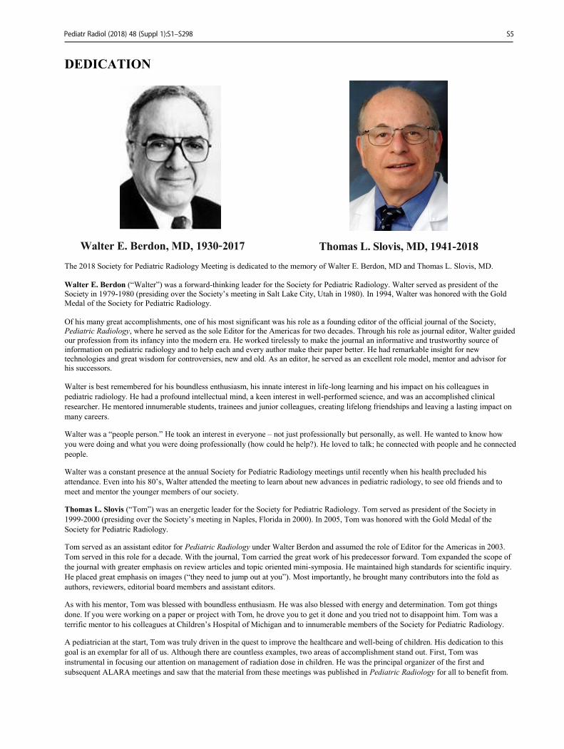
Walter E. Berdon, MD, 1930–2017 Thomas L. Slovis, MD, 1941-2018
The 2018 Society for Pediatric Radiology Meeting is dedicated to the memory of Walter E. Berdon, MD and Thomas L. Slovis, MD.
Walter E. Berdon (“Walter”) was a forward-thinking leader for the Society for Pediatric Radiology. Walter served as president of the
Society in 1979-1980 (presiding over the Society’s meeting in Salt Lake City, Utah in 1980). In 1994, Walter was honored with the Gold
Medal of the Society for Pediatric Radiology.
Of his many great accomplishments, one of his most significant was his role as a founding editor of the official journal of the Society,
Pediatric Radiology, where he served as the sole Editor for the Americas for two decades. Through his role as journal editor, Walter guided
our profession from its infancy into the modern era. He worked tirelessly to make the journal an informative and trustworthy source of
information on pediatric radiology and to help each and every author make their paper better. He had remarkable insight for new
technologies and great wisdom for controversies, new and old. As an editor, he served as an excellent role model, mentor and advisor for
his successors.
Walter is best remembered for his boundless enthusiasm, his innate interest in life-long learning and his impact on his colleagues in
pediatric radiology. He had a profound intellectual mind, a keen interest in well-performed science, and was an accomplished clinical
researcher. He mentored innumerable students, trainees and junior colleagues, creating lifelong friendships and leaving a lasting impact on
many careers.
Walter was a “people person.” He took an interest in everyone – not just professionally but personally, as well. He wanted to know how
you were doing and what you were doing professionally (how could he help?). He loved to talk; he connected with people and he connected
people.
Walter was a constant presence at the annual Society for Pediatric Radiology meetings until recently when his health precluded his
attendance. Even into his 80’s, Walter attended the meeting to learn about new advances in pediatric radiology, to see old friends and to
meet and mentor the younger members of our society.
Thomas L. Slovis (“Tom”) was an energetic leader for the Society for Pediatric Radiology. Tom served as president of the Society in
1999-2000 (presiding over the Society’s meeting in Naples, Florida in 2000). In 2005, Tom was honored with the Gold Medal of the
Society for Pediatric Radiology.
Tom served as an assistant editor for Pediatric Radiology under Walter Berdon and assumed the role of Editor for the Americas in 2003.
Tom served in this role for a decade. With the journal, Tom carried the great work of his predecessor forward. Tom expanded the scope of
the journal with greater emphasis on review articles and topic oriented mini-symposia. He maintained high standards for scientific inquiry.
He placed great emphasis on images (“they need to jump out at you”). Most importantly, he brought many contributors into the fold as
authors, reviewers, editorial board members and assistant editors.
As with his mentor, Tom was blessed with boundless enthusiasm. He was also blessed with energy and determination. Tom got things
done. If you were working on a paper or project with Tom, he drove you to get it done and you tried not to disappoint him. Tom was a
terrific mentor to his colleagues at Children’s Hospital of Michigan and to innumerable members of the Society for Pediatric Radiology.
A pediatrician at the start, Tom was truly driven in the quest to improve the healthcare and well-being of children. His dedication to this
goal is an exemplar for all of us. Although there are countless examples, two areas of accomplishment stand out. First, Tom was
instrumental in focusing our attention on management of radiation dose in children. He was the principal organizer of the first and
subsequent ALARA meetings and saw that the material from these meetings was published in Pediatric Radiology for all to benefit from.
Pediatr Radiol (2018) 48 (Suppl 1):S1–S298 S5
Second, throughout his career, and especially recently, Tom always sought to do what is right and to properly diagnose and protect the
abused child, the most vulnerable of our patients.
Tom was also an instrumental leader of the Society for Pediatric Radiology. Under Tom’s leadership, foundations for the current
administrative structure of the Society were laid. He served on the Society for Pediatric Radiology Board of Directors longer than anyone
else, wearing the hats of member-at-large and secretary before ramping up to president, chairman of the board of directors and past-
president, then adding a few more years as journal editor. Like Walter, Tom was a constant presence at SPR meetings until last year when
his health limited his ability to travel.
As you take in this meeting, please remember Walter and Tom and their many contributions to our society, to the healthcare of
children and to each of us as individuals. The Society for Pediatric Radiology would not be what it is today without Walter and
Tom. It is with immense gratitude that we remember these two great individuals.
Peter J. Strouse, MD, FACR
(Please also refer to: Walter E. Berdon (1930-2017) by Tom Slovis, Terry Levin and Aparna Joshi in the November 2017 issue of Pediatric
Radiology. An obituary for Dr. Slovis will appear in the May 2018 issue of Pediatric Radiology.)
Pediatr Radiol (2018) 48 (Suppl 1):S1–S298S6
SPR 2018 ORGANIZATION
2018 MEETING CURRICULUM COMMITTEE
Peter J. Strouse, MD, FACR (Program Director and Paper Committee Chair)
Adina L. Alazraki, MD, FAAP (Oncology Session)
George S. Bisset, III, MD, FACR (Jeopardy Session)
Leah E. Braswell, MD (Interventional Radiology Session)
Sarah D. Bixby, MD (Poster Committee Vice Chair)
Taylor Chung, MD (Poster Committee Vice Chair)
Kassa Darge, MD, PhD (Hands-On Ultrasound Session)
Jonathan R. Dillman, MD, MSc (Postgraduate Course Director)
Monica Epelman, MD (Hands-On Ultrasound Session)
Donald P. Frush, MD, FACR (Postgraduate Course – Image Gently Organizer)
Michael S. Gee, MD, PhD (Oncology Session)
Laura A. Gruber, MBA, RT(R), RDMS, RVT (Technologist Program Director)
Roger Harned, MD, FACR (Interventional Radiology Session)
Christine Harris, RT (R) (MR) (Technologist Program Director)
Richard E. Heller, III, MD, MBA (Scientific Session Organizer)
Marta Hernanz-Schulman, MD, FACR (Hands-On Ultrasound Session)
Melissa A. Hilmes, MD (Scientific Session Organizer)
Thierry A. G. M. Huisman, MD (Neuroradiology Session)
Sarah S. Milla, MD, FAAP (Poster Committee Chair)
Helen R. Nadel, MD, FRCPC (Oncology Session)
Susan Palasis, MD (Neuroradiology Session)
Manish N. Patel, MD (Interventional Radiology Session)
Janet R. Reid, MD, FRCPC (Postgraduate Course Director and Education Session)
Dennis W. W. Shaw, MD (Neuroradiology Session)
Ethan A. Smith, MD (Workshop Director)
Keith J. Strauss, MSc, FACR (Postgraduate Course – Image Gently Organizer)
Mahesh M. Thapa, MD (Workshop Director and Education Session)
Stephan D. Voss, MD, PhD (Oncology Session)
ABSTRACT REVIEW COMMITTEE – PAPERS
Peter J. Strouse, MD, FACR, Chair
Taylor Chung, MD, Vice Chair
Sarah D. Bixby, MD
David A. Bloom, MD
Lorna P. Browne, MD
Dorothy I. Bulas, MD, FACR, FAAP
Michael J. Callahan, MD
Christopher I. Cassady, MD, FAAP
Teresa Chapman, MD, MA
Nancy A. Chauvin, MD
Brian D. Coley, MD, FACR, FAIUM
Hisham M. Dahmoush, MBBCh, FRCR
Kassa Darge, MD, PhD
Jonathan R. Dillman, MD, MSc
James S. Donaldson, MD, FACR
Josée Dubois, MD
Monica Epelman, MD
Mark R. Ferguson, MD
Donald P. Frush, MD, FACR
Michael S. Gee, MD, PhD
J. Damien Grattan-Smith, MBBS
Roger Harned, MD, FACR
Mark J. Hogan, MD
Nadja Kadom, MD
S. Pinar Karakas, MD
Beth M. Kline-Fath, MD
Bernadette L. Koch, MD
Neha S. Kwatra, MD
John D. MacKenzie, MD
M. Beth McCarville, MD
Arthur B. Meyers, MD
Robert C. Orth, MD, PhD
John M. Racadio, MD
Janet R. Reid, MD, FRCPC
Cynthia K. Rigsby, MD, FACR
Maura E. Ryan, MD
Jonathan Samet, MD
Sabah Servaes, MD
Ethan A. Smith, MD
Keith J. Strauss, MSc, FACR
Alexander J. Towbin, MD
Andrew T. Trout, MD
Jason P. Weinman, MD
Matthew T. Whitehead, MD
Stephan D. Voss, MD, PhD
ABSTRACT REVIEW COMMITTEE – POSTERS
Sarah S. Milla, MD, FAAP, Chair
Sarah D. Bixby, MD, Vice Chair
Anjum N. Bandarkar, MD
Thangamadhan Bosemani, MD
Kiery A. Braithwaite, MD
Maria A. Calvo-Garcia, MD
Gulraiz A. Chaudry, MBChB, MRCP, FRCR
Kassa Darge, MD, PhD
Nilesh Desai, MD
Paula N. Dickson, MD
Steven Don, MD
Eric Eutsler, MD
Anne Gill, MD
Pediatr Radiol (2018) 48 (Suppl 1):S1–S298 S7
ABSTRACT REVIEW COMMITTEE – POSTERS
(continued)
Leslie E. Hirsig, MD
Thierry A. G. M. Huisman, MD
Nadja Kadom, MD
J. Herman Kan, MD
Rajesh Krishnamurthy, MD
Neha S. Kwatra, MD
Maria F. Ladino-Torres, MD
Shailee V. Lala, MD
Jonathan M. Loewen, MD
Adeka D. McIntosh, MD
Craig S. Mitchell, DO, MA
Helen R. Nadel, MD, FRCPC
Srikala Narayanan, MD
Hansel J. Otero, MD
Daniel J. Podberesky, MD
Janet R. Reid, MD, FRCPC
Susan E. Sharp, MD
Manrita K. Sidhu, MD
Bruno P. Soares, MD
Aylin Tekes-Brady, MD
Stephanie B. Theut, DO
Alexander J. Towbin, MD
Jason Tsai, MD
Jennifer Vaughn, MD
Nghia (Jack) Vo, MD
Ewa M. Wasilewska, MD
Arash R. Zandieh, MD
IMAGING TECHNOLOGIST PROGRAM
& ABSTRACT COMMITTEE
Christine Harris, RT (R) (MR) (Technologist Program Director)
Laura A. Gruber, MBA, RT(R), RDMS, RVT (Technologist Program
Director)
Stuart Brice
Brian Fox, MBA
Charles R. Fritz, RT, MBA
Lynne Hamer, MEd., RT
Todd Lehkamp
M. Craig Morriss, MD
Stephen F. Simoneaux, MD, Advisor
R. Daniel Smock, BHS RT(R)(MR)(CT), MRSO (MRSC)
CASES OF THE DAY ORGANIZERS
Neil D. Johnson, MBBS
Christopher Sternal- Johnson
MEETING INFORMATION TECHNOLOGY
Safwan S. Halabi, MD, Meeting IT Co-Director
Alexander J. Towbin, MD, Meeting IT Co-Director
MEETING PHOTOGRAPHERS
Alan E. Schlesinger, MD
Mahesh M. Thapa, MD
CONTINUING MEDICAL EDUCATION
ACCREDITATION STATEMENT:
This activity has been planned and implemented in accordance with the accreditation requirements and policies of the Accreditation
Council for Continuing Medical Education through the joint providership of the American College of Radiology and Society of Pediatric
Radiology. The American College of Radiology is accredited by the ACCME to provide continuing medical education for physicians.
CREDIT DESIGNATION STATEMENT:
The American College of Radiology designates this activity for a maximum of 39. 25 AMA PRA Category 1 Credit(s) ™ Physicians should
claim only the credit commensurate with the extent of their participation in the activity.
TECHNOLOGISTS:
The American College of Radiology is approved by the American Registry of Radiologic Technologists (ARRT) as a Recognized Continuing
Education Evaluation Mechanism (RCEEM) to sponsor and/or review Continuing Medical Educational programs for Radiologic
Technologists and Radiation Therapists. The American College of Radiology designates this educational activity as meeting the criteria for up
to 42.75 Category A credit hours of the ARRT.
MAINTENANCE OF CERTIFICATION
Qualified on January 29, 2018, select sessions from this activity meet the American Board of Radiology criteria for a self-assessment (SAM)
activity and is designated for up to 14.50 SAM credits toward the ABR Maintenance of Certification Program.
OBJECTIVES
The 2018 61st Annual Meeting & Postgraduate Course will provide pediatric and general radiologists with an opportunity to do the following:
1. Summarize the most current information on state-of-the-art pediatric imaging and the practice of pediatric radiology.
2. Describe and apply new technologies and imaging findings for pediatric imaging.
3. Discuss trends in research and education concerning the care and imaging of pediatric patients.
4. Identify common challenges facing pediatric radiologists, and possible solutions.
5. Describe and apply basic principles for implementing quality and safety programs in pediatric radiology
6. Evaluate and apply means of managing radiation exposure and the need for sedation/anesthesia during diagnostic imaging and
image guided therapy.
Pediatr Radiol (2018) 48 (Suppl 1):S1–S298S8
At the conclusion of the experience, participants should have an improved understanding of the technologies discussed, increased
awareness of the benefits and costs of diagnostic imaging in children and of ways to minimize risks, and an improved general
knowledge of pediatric radiology.
DISCLOSURES
In compliance with ACCME requirements and guidelines, the ACR has developed a policy for review and disclosure of potential conflicts of
interest, and a method of resolution if a conflict does exist. The ACR maintains a tradition of scientific integrity and objectivity in its
educational activities. In order to preserve this integrity and objectivity, all individuals participating as planners, presenters, moderators and
evaluators in an ACR educational activity or an activity jointly sponsored by the ACR must appropriately disclose any financial relationship
with a commercial organization that may have an interest in the content of the educational activity.
The following planners, presenters, staff and evaluators have disclosed that neither they nor their spouse/partner have any financial interests,
arrangements or affiliation in the context of this activity:
PRESENTERS
Patricia T. Acharya, MD
Michael R. Acord, MD
Sudha A. Anupindi, MD
Edward Arroyo, RDMS
Lauren W. Averill, MD
Rama S. Ayyala, MD
D. Gregory Bates, MD
David M. Biko, MD
David A. Bloom, MD, FACR
Tim N. Booth, MD
Scott Borinstein, MD
Samuel L. Brady, MS, PhD
Dana Brinson, BHA RT(R)
Brandon P. Brown, MD
Lorna P. Browne, MBBS
Dorothy I. Bulas, MD, FACR, FAAP
Nikki D. Butler, BMSc, RT(R)(QM)
Michael J. Callahan, MD
Christopher I. Cassady, MD, FAAP
Sherwin S. Chan, MD
Nancy A. Chauvin, MD
Govind B. Chavhan, MD, DNB
Lorraine Chisari, RDMS, RVT
Arabinda K. Choudhary, MD, FACHE, MBA
Asim F. Choudhri, MD
Jeanne Chow, MD
Jason Christensen, MD
Matthew Cooper, MD
Jesse Courtier, MD
Eric J. Crotty, MD
Hisham M. Dahmoush, MD
Jonathan Dallas, BS
Matthew Davenport, MD
John W. Dell, BSRT (R) (MR)
Jie Deng, PhD
Sarah Desoky, MD
Paula N. Dickson, MD
Cristina Dodge, MS
Steven Don, MD
James S. Donaldson, MD, FACR
Lane F. Donnelly, MD
Kevin C. Doyle,
Josee Dubois, MD
Jerry R. Dwek, MD
Brandon Edwards, CEO
Jesse M. Ehrenfeld, MD, MPH
Laura Z. Fenton, MD
Mark R. Ferguson, MD
Robert J. Fleck, MD
Judith Gadde, DO
Lacy Gandor, RDMS
Kimberly A. Garver, MD
Michelle Garza, (RTR)
Maryam Ghadimi Mahani, MD
Anne Gill, MD
Ciji N. Gilley, R.T.(R)
Melissa Goehner, BA RT(R)(CT)
Sarah Beth Gray, CCLS
Jared R. Green, MD
Jesse Green, RT(R)
S. Bruce Greenberg, MD
Mary-Louise Greer, MBBS
Richard B. Gunderman, MD, PhD
Matthew R. Hammer, MD
Nicole Hardin, MD
C. Matthew Hawkins, MD
Laura L. Hayes, MD
Gary L. Hedlund, DO
Lamont Hill, RDMS
Mai-Lan Ho, MD
Victor M. Ho-Fung, MD
Jason Hooper, BS, RDMS, RVT
Brian Hopely, MD
Ramesh S. Iyer, MD
Siddarth P. Jadhav, MD
Camilo Jaimes, MD
Diego Jaramillo, MD, MPH
Delma Y. Jarrett, MD
Craig M. Johnson, DO
Marty Jones, MHA, RT (R)(MR)
Aparna Joshi, MD
Bamidele F. Kammen, MD
Summer L. Kaplan, MD, MS
S. Pinar Karakas, MD
Merima Karastanovic, MS, RT(R)(MR)
Boaz Karmazyn, MD
Sue C. Kaste, DO
Geetika Khanna, MD, MS
Jill Kilkelly, MD
Paul K. Kleinman, MD
Beth M. Kline-Fath, MD
Brandi Kozak, RDMS
Rajesh Krishnamurthy, MD
Peter G. Kruk, MD
Jeannie K. Kwon, MD
Maria F. Ladino-Torres, MD
Tal Laor, MD
David B. Larson, MD, MBA
Bernard F. Laya, DO
Edward Y. Lee, MD, MPH
Gabe Linke, BSRT (R)(MR)
Deborah Lowen, MD
Karen Lyons, MB, BCh, BAO
Robert MacDougall, MS
Pediatr Radiol (2018) 48 (Suppl 1):S1–S298 S9
Joseph MacLean, CNMT
Alexis B. R. Maddocks, MD
Ladonna J. Malone, MD
Peter A. Marcovici, MD
Megan B. Marine, MD
Kelley W. Marshall, MD
Jeffrey E. Martus, MD, MS
Lea Matsuoka, MD, FACS
M. Beth McCarville, MD
Sarah McKenney, PhD
Mariana Meyers, MD
Cindy R. Miller, MD
Leslie Mintz, BSN
David M. Mirsky, MD
David A. Mong, MD
Joëlle A. Moreno, JD
Trudy Morgan, RDMS
Dedrick Moulton, MD
Martha M. Munden, MD
Oscar M. Navarro, MD
Elad Nevo, MS, RT(MR)(N)(CT), CNMT
Beverley Newman, MD, FACR
Anh-Vu H. Ngo, MD
Jennifer L. Nicholas, MD
Joshua Nickerson, MD
Anh-Vu M. O'Hara, MD
James A. O'Neill, MD
Seng H. Ong, MBBS
Robert C. Orth, MD, MPH, PhD
Hansel J. Otero, MD
Jeffrey P. Otjen, MD
Randolph K. Otto, MD
Harriet J. Paltiel, MD
Ashok Panigrahy, MD
Marguerite T. Parisi, MD, MS
Zoltan Patay, MD, PhD
Michael D. Payne, RT
Jeannette M. Perez-Rossello, MD
John Pietch, MD
Kara-Lee Pool, MD
Andrada R. Popescu, MD
Laura Poznick, AAS, RDMS
Georgiena E. Prevett, MS, RT(R)(N) (CT)(MR), CNMT (CT)
Sumit Pruthi, MD
John Racadio, MD
Ray Ramoso, ARRT (r) (vi)
Trista Raymer, RT, (R)(CT)(MR)
Cynthia K. Rigsby, MD, FACR
Kimberly Ritze, BS, RT(R), CT, MR
Gina Roberts, ARRT (R)(CT)(MRI)
Nancy K. Rollins, MD
Veronica J. Rooks, MD
Monica A. Rossleigh, MD
Erika Rubesova, MD
Maura E. Ryan, MD
Pallavi Sagar, MD
David Saul, MD
Dawn E. Saunders, MD
Andrew Schapiro, MD
Gary R. Schooler, MD
Daniel M. Schwartz, MD
Erin S. Schwartz, MD
Ramdas Senasi, MD
Suraj D. Serai, PhD
Susan E. Sharp, MD
Karuna V. Shekdar, MD
Sphoorti Shellikeri, MS
Nicholas Shkumat, MSc
Manohar Shroff, MD
Cicero T. Silva, MD
Stephen F. Simoneaux, MD
Maria Smith, BS, RDMS, RVT
Stephanie E. Spottswood, MD, MSPH
Judy H. Squires, MD
Abhay Srinivasan, MD
Lisa J. States, MD
Stacy T. Tanaka, MD, MS
Richard B. Towbin, MD, FACR
Sheryl A. Tulin-Silver, MD
Unmi K. Udayasankar, MD
Seth E. Vatsky, DO
Teresa Victoria, MD, PhD
Nghia "Jack" Vo, MD
Arastoo Vossough, MD, PhD
Jason P. Weinman, MD
Amy S. Whigham, MD
Jason N. Wright, MD
Deborah Zarnow, MD
Andrew M. Zbojniewicz, MD
PLANNING COMMITTEE & PRESENTER
Adina L. Alazraki, MD, FAAP
George S. Bissett, MD, FACR
Sarah D. Bixby, MD
Leah E. Braswell, MD
Kassa Darge, MD, PhD
Ellen M. Chung, MD
Monica Epelman, MD
Donald. P. Frush, MD, FACR
Michael S. Gee, MD, PhD
Laura Gruber, MBA, RT(R), RDMS, RVT
Christine Harris, RT (R) (MR)
Richard E. Heller III, MD, MBA
Marta Hernanz-Schulman, MD, FACR
Melissa A. Hilmes, MD
Thierry A. G. M. Huisman, MD
Sarah S. Milla, MD, FAAP
Helen R. Nadel, MD, FRCPC
Susan Palasis, MD
Manish N. Patel, DO
Janet R. Reid, MD, FRCPC
Dennis W. W. Shaw, MD
Ethan A. Smith, MD
Keith J. Strauss, MSc, FACR
Peter J. Strouse, MD, FACR
Mahesh M. Thapa, MD
Stephan D. Voss, MD, PhD
STAFF & CME REVIEWER(S)
Angela R. Davis, CAE
Kasey O’Dea
Category 1 - Steven Chmielewski, MD
Category A - Brian Monzon, RT
The planners and presenters listed below have disclosed the following relevant financial relationships. Potential conflicts have been
resolved.
Pediatr Radiol (2018) 48 (Suppl 1):S1–S298S10
PRESENTERS WITH DISCLOSURES
Michael Aquino, MD Elsevier - Royalties
Susan J. Back, MD Bracco – Research Grant(s)
Brian D. Coley, MD, FACR Elsevier – Royalties, NewView – Board Member
Philips/Toshiba – Travel Support
R. Paul Guillerman, MD Guerbet LLC – Consultant, Honoraria
Eric Hoggard, MD Gore – Consultant, Honoraria
Jill V. Hunter, MBBS Medico – Consultant, Honoraria; UpToDate - Royalties
Steven J. Kraus, MD, MS, FAAP Elsevier - Royalties
Ruth Lim, MD New England PET Imaging System - Officer
Prakash M. Masand, MD Daiichi Sankyo Inc., Vital Images, Philips MRI – Consultant
Honoraria; Toshiba Medical Systems – Speakers Bureau;
Amirsys - Royalties
Arnold Carl Merrow, MD Elsevier – Royalties, Consultant, Honoraria
Arthur B. Meyers, MD Amirsys – Royalties, Elsevier - Royalties
Eric J. Monroe, MD Biogen – Advisory Board
Daniel J. Podberesky, MD Guerbet/GE/Philips/Siemens – Consultant, Honoraria; Toshiba
of Americas Medical Systems – Speakers Bureau; Amirsys –
Royalties
Caroline D. Robson, MBChB Elsevier – Royalties
Ehsan Samei, PhD GE/Siemens – Research Grant(s); MedInt Holdings – Advisory
Board
Scott E. Snyder, PhD Ground Fluor Pharmaceuticals/ United Therapeutics, EMD
Sorono (Merck, KGaA) – Research Grant(s)
Alexander J. Towbin, MD Siemens/Guerbet/Cystic Fibrosis Foundation – Research
Grant(s), Applied Radiology, IBM Watson Health -
Consultant/Honoraria
Andrew T. Trout, MD Geurbet - Consultant, Honoraria; Elsevier - Royalites; Siemens/
Toshiba - Research Grant(s); Perpectum Diagnostics – Medical
Advisory Board
Shreyas S. Vasanawala, MD, PhD Arterys - Stock Options, GE Healthcare – Research Grant(s),
Stanford - Intellectual Property Rights
Lisa R. Young, MD Boehringer Ingeleheim - Advisory Board, UpToDate – Royalties
PLANNING COMMITTEE & PRESENTER WITH DISCLOSURES
Jonathan R. Dillman, MD, MSc Bracoo Diagnostics/Guerbet/Toshiba US/Siemens Medical Solutions
USA – Research Grant(s)
Keith J. Strauss, MSc, FACR Philips Medical Systems – Consultant/Honoraria
ACKNOWLEDGEMENTS The Society for Pediatric Radiology gratefully acknowledges the support of the of the following companies in presenting the 61st Annual
Meeting & Postgraduate Course.
EDUCATIONAL GRANTS
Bayer
Guerbet LLC
EXHIBITORS – as of April 6th
AGFA HealthCare
Alexion
Aris Radiology
Bayer
Bracco Diagnostics Inc.
Canon Medical Systems USA
ChiRhoClin,Inc.
Dream Think Imagine
Elsevier, Inc.
Fujifilm Medical Systems USA, Inc.
GE Healthcare
Guerbet, LLC
Imorgan
LMT Medical Systems GmbH
Mindray
Philips Healthcare
Planmed, Inc.
Samsung Neurologica
SealCath
Siemens
Softek Illuminate
SREE Medical Systems
Stratasys
Wolters Kluwer
St. Jude Children’s Research Hospital Department
Diagnostic Imaging
Pediatr Radiol (2018) 48 (Suppl 1):S1–S298 S11
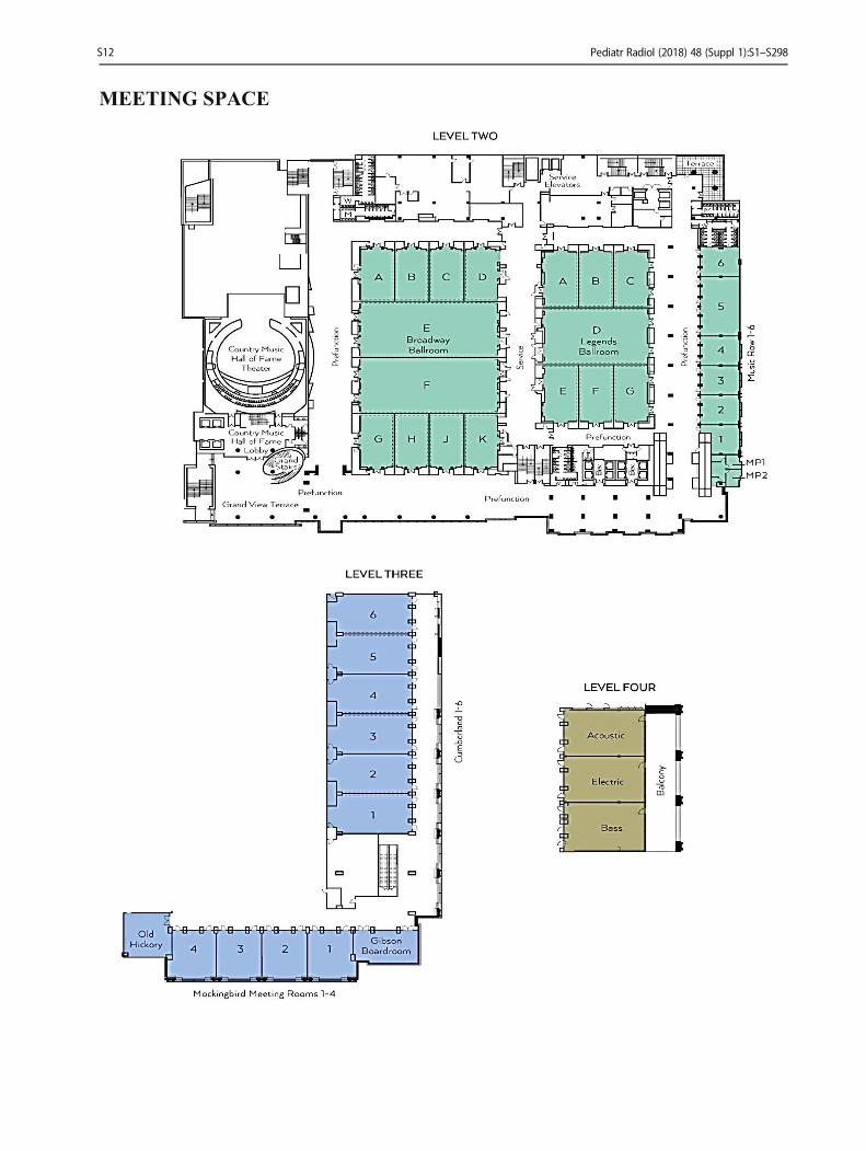
MEETING SPACE
Pediatr Radiol (2018) 48 (Suppl 1):S1–S298S12
SPR GENERAL INFORMATION
MISSION STATEMENT
The Society for Pediatric Radiology is dedicated to fostering excellence in pediatric health care through imaging and image-guided care.
DIVERSITY & INCLUSION STATEMENT
The Society for Pediatric Radiology actively promotes diversity and inclusion at all levels of training, practice and leadership for the
benefit of our patients, our profession and for the Society as a whole.
SPR OFFICERS, DIRECTORS AND COMMITTEES 2017–2018
BOARD OF DIRECTORS
Diego Jaramillo, MD, MPH, Chair
Peter J. Strouse, MD, FACR, President and Editor
Taylor Chung, MD, President-Elect
Christopher I. Cassady, MD, FAAP, 1st Vice-President
J. Damien Grattan-Smith, MBBS, 2nd Vice-President, SPR Research and Education Foundation President
Michael J. Callahan, MD, Secretary
Stephen F. Simoneaux, MD, Treasurer and ‘15-‘17 SCORCH President
Kassa Darge, MD, PhD
Molly E. Dempsey, MD, ‘17-‘19 SCORCH President
Josée Dubois, MD
Laura Z. Fenton, MD
Beth M. Kline-Fath, MD
Susan Palasis, MD
Mary R. Wyers, MD
Richard A. Barth, MD, FACR, FAAP, Past President
Brian D. Coley, MD, FACR, FAIUM, Past President
James S. Donaldson, MD, FACR, Past President
Donald P. Frush, MD, FACR, Image Gently Liaison
Marta Hernanz-Schulman, MD, FACR, ACR Commission Liaison
Benjamin H. Taragin, MD, Web Editor
Maria Gisela Mercado-Dean, MD, ’15-’17 AAP Liaison
Sarah S. Milla, MD, FAAP, ’17-’19 AAP Liaison
M. Ines Boechat, MD, FACR, WFPI Liaison
ABDOMINAL IMAGING COMMITTEE
Jonathan R. Dillman, MD, MSc, Chair
Sudha A. Anupindi, MD
Lauren W. Averill, MD
Govind B. Chavhan, MD, DNB
Ellen M. Chung, MD
Meryle Eklund, MD
Hansel J. Otero, MD
Daniel J. Podberesky, MD
Anil G. Rao, DMRD, DNB
Gary R. Schooler, MD
Ethan A. Smith, MD
Andrew T. Trout, MD
BYLAWS COMMITTEE
Diego Jaramillo, MD, MPH, Chair
Richard A. Barth, MD, FACR, FAAP
Kassa Darge, MD, PhD
Mary R. Wyers, MD
CARDIAC IMAGING COMMITTEE
Lorna P. Browne, MD, Chair
Mark R. Ferguson, MD
Maryam Ghadimi Mahani, MD
Eric Hoggard, MD
Ramkumar Krishnamurthy, PhD
Karen Lyons, MB, BCh, BAO
Ladonna J. Malone, MD
Prakash M. Masand, MD
Mike Seed, MBBS
Ting Y. Tao, MD, PhD
Gauri S. Tilak, MD, PhD, MPH
Smyrna P. Tuburan, MD
Cynthia K. Rigsby MD, FACR
Laureen M. Sena, MD
CHILD ABUSE COMMITTEE
Sabah Servaes, MD, Chair
Arabinda K. Choudhary, MBBS, MRCP, FRCR Vice Chair
David A. Bloom, MD
Karen Blumberg, MD, FACR
Jonathan Chen, MD
Tejaswini K. Deshmukh, MD
Michael F. Fadell, II, MD
Laura L. Hayes, MD
Gary L. Hedlund, DO
Muhammad N. Khan, MBBS, FCPS
Jeannie K. Kwon, MD
Megan B. Marine, MD
Bradley A. Maxfield, MD
Kenneth L. Mendelson, MD
David M. Mirsky, MD
Joëlle A. Moreno, JD
Sandeep Narang, MD
Susan Palasis, MD
Ashishkumar K. Parikh, MD
Jeannette M. Perez-Rossello, MD
Cory M. Pfeifer, MD, FAAP
Pediatr Radiol (2018) 48 (Suppl 1):S1–S298 S13
CHILD ABUSE COMMITTEE
(continued)
Veronica J. Rooks, MD
Michael D. Rubin, MD
Dana S. Schwartz, MD
Daniel M. Schwartz, MD
Victoria M. Silvera, MD
Heba S. Takrouri, MBBS
Chido Vera, MD
Gregory A. Vorona, MD
Matthew A. Zapala, MD, PhD
Stephen D. Brown, MD, Advisory
Richard I. Markowitz, MD, FACR, Advisory
Thomas L. Slovis, MD, Advisory
Peter J. Strouse, MD, FACR, Advisory
CONTRAST-ENHANCED ULTRASOUND
COMMITTEE
M. Beth McCarville, MD, Chair
Susan J. Back, MD, Vice Chair
Patricia T. Acharya, MD
Carol E. Barnewolt, MD
Jamie L. Coleman, MD
Kassa Darge MD, PhD
Jonathan R. Dillman, MD, MSc
Lynn A. Fordham, MD, FACR
Misun Hwang, MD
Annie Lim, DO
Martha M. Munden, MD
Harriet J. Paltiel, MD
CT COMMITTEE
John D. MacKenzie, MD, Chair
Tushar Chandra, MBBS, MD
Kara G. Gill, MD
Prakash M. Masand, MD
Grace S. Phillips, MD
Karuna V. Shekdar, MD
Richard Southard, MD
Jacqueline A. Urbine, MD
Jason P. Weinman, MD
Sjirk J. Westra, MD
Sheila C. Berlin, MD, Advisory
DIVERSITY AND INCLUSION COMMITTEE
Ashok Panigrahy, MD, Co-Chair
Stephanie E. Spottswood, MD, MSPH, Co-Chair
Aparna Annam, DO
Taylor Chung, MD
Gregory L. Compton, MBBS
Sarah Desoky, MD
Melanie B. Levin, MD
Maria-Gisela Mercado-Deane, MD
Cindy R. Miller, MD
Kristi B. Oatis, MD
Tina Young Poussaint, MD, FACR
Peter J. Strouse, MD, FACR
Amit S. Sura, MD
Philip Teitelbaum, MD
Adrienne F. Thompson, MD
Chido Vera, MD
EDUCATION-CURRICULUM COMMITTEE
Christopher I. Cassady, MD, FAAP, Chair
Sarah S. Milla, MD, FAAP, Vice Chair
Michael J. Callahan, MD
Taylor Chung, MD
Peter J. Strouse, MD, FACR, Editor
Monica Epelman, MD
Laura Z. Fenton, MD
Liliane H. Gibbs, MD
Safwan S. Halabi, MD
Angelisa M. Paladin, MD
Daniel J. Podberesky, MD
Sanjay P. Prabhu, MBBS, FRCR
Janet R. Reid, MD, FRCPC
Cynthia K. Rigsby, MD, FACR
Manrita K. Sidhu, MD
Stephen F. Simoneaux, MD
Benjamin H. Taragin, MD
Mahesh M. Thapa, MD
Kevin Wong, DO
EMERGENCY RADIOLOGY & TRAUMA COMMITTEE
Susan D. John MD, FACR, Chair
Michael R Aquino, MD
Tejaswini K Deshmukh, MD
David Dinan, MD
Michael P. George, MD
Ashwin Hegde, MD, FRCPC
Jeanne G. Hill, MD
Victor M. Ho-Fung, MD
Tara L. Holm, MD
Paul J. Iskander, MD
Jennifer H. Johnston, MD
George C. Koberlein, MD
Jonathan M. Loewen, MD
Indu R. Meesa, MD
Michael A. Murati, MD
Michael P. Nasser, MD
FELLOWSHIP PROGRAM DIRECTOR COMMITTEE
Paula N. Dickson, MD, Co-Chair
Sabah Servaes, MD, Co-Chair
FETAL IMAGING COMMITTEE
Teresa Victoria, MD, PhD, Chair
Mariana L. Meyers, MD, Vice Chair
Rama S. Ayyala, MD
Richard A. Barth, MD, FACR, FAAP
Michael A. Breen, MBBCh
Brandon P. Brown, MD
Lucia Carpineta, MD, CM
John A. Cassese, MD
Dorothy I. Bulas, MD, FACR, FAAP
Christopher I. Cassady, MD, FAAP
Patricia Cornejo, MD
Kimberly A. Dannull, MD
Nilesh Desai, MD
Michael S. Gee, MD, PhD
Luis Goncalves, MD
Carolina V. Guimaraes, MD
Camilo J. Cobos, MD
Pamela M. Ketwaroo, MD
Paggie Kim, MD
Beth M. Kline-Fath, MD
Amy B. Kolbe, MD
Pediatr Radiol (2018) 48 (Suppl 1):S1–S298S14
FETAL IMAGING COMMITTEE
(continued)
Leann E. Linam, MD
David M. Mirsky, MD
Edward R. Oliver, MD
Erika Rubesova, MD
Chetan C. Shah, MD
Gayathri Sreedher, MD
FINANCE COMMITTEE
Avrum N. Pollock, MD, FRCPC, Chair
Richard A. Barth, MD, FACR, FAAP
Christopher I. Cassady, MD, FAAP
Taylor Chung, MD
Randheer Shailam, MD
Stephen F. Simoneaux, MD
Peter J. Strouse, MD, FACR
Dayna M. Weinert, MD
HISTORY COMMITTEE
Alan E. Schlesinger, MD, Historian
N.Thorne Griscom, MD, Consultant
HONORS COMMITTEE
Richard A. Barth, MD, FACR, FAAP, Chair
Brian D. Coley, MD, FACR, FAIUM
James S. Donaldson, MD, FACR
INFORMATICS COMMITTEE
Safwan S. Halabi, MD, Chair
Steven L. Blumer, MD
Michael L. Francavilla, MD
C. Matthew Hawkins, MD
Nadja Kadom, MD
Summer L. Kaplan, MD
Jeannie K. Kwon, MD
Neil U. Lall, MD
Morgan McBee, MD
Saad A. Ranginwala, MD
Takashi Sato, MD, PhD
Evan J. Zucker, MD
Alexander J. Towbin, MD
INTERVENTIONAL COMMITTEE
Manish N. Patel, DO, Chair
Leah E. Braswell, MD
Jared R. Green, MD
Craig M. Johnson, DO
Matthew P. Lungren, MD, MPH
Janice D. McDaniel, MD
Radu Nicolaescu, MD
Jeremiah J. Sabado, MD
Timothy R. Singewald, MD
Ranjith Vellody, MD
Fabiola C. Weber, MD
JUDICIARY COMMITTEE
Thomas L. Slovis, MD, Chair
Richard A. Barth, MD, FACR, FAAP
Dorothy I. Bulas, MD, FACR, FAAP
Neil D. Johnson, MBBS
Richard B. Gunderman, MD, PhD, FACR
MR COMMITTEE
Michael S. Gee, MD, PhD, Chair
Sudha A. Anupindi, MD
Sherwin S. Chan, MD
Tushar Chandra, MBBS, MD
Govind B. Chavhan, MD, DNB
Jesse Courtier, MD
Jorge H. Davila-Acosta, MD
Robert J. Fleck, Jr, MD
Shahnaz G. Koureh, MD
Mai-Lan Ho, MD
Geetika Khanna, MD, MS
Hee-Kyung Kim, MD
Amy B. Kolbe, MD
Archana Malik, MD
Michael M. Moore, MD
Thang Ngo, MD
Hansel J. Otero, MD
Anil G. Rao, DMRD, DNB
Gary R. Schooler, MD
Mitchell L. Simon, MD
Gayathri Sreedher, MD
Unni K. Udayasankar, MD
Matthew J. Winfeld, MD
Taylor Chung, MD, Advisory
Shreyas S. Vasanawala, MD, PhD, Advisory
MUSCULOSKELETAL IMAGING COMMITTEE
Jerry R. Dwek, MD, Chair
Arthur B. Meyers, MD, Vice Chair
Lauren W. Averill, MD
Sebastien Benali, MD
Sarah D. Bixby MD
Tushar Chandra, MBBS, MD
Nancy A. Chauvin, MD
Jonathan Chen, MD
Kirsten Ecklund, MD
R. Paul Guillerman, MD
Siddharth P. Jadhav, MD
Shawn E. Kamps, MD
Hee-Kyung Kim, MD
Archana Malik, MD
Tracey R. Mehlman, MD
Arnold Carl Merrow, Jr, MD
Jie C. Nguyen, MD, MS
Allison K. Person, MD
Jonathan Samet, MD
Amisha J. Shah, MD
Mahesh M. Thapa, MD
Sai G. Yarram, MD
NEONATAL IMAGING COMMITTEE
Rama S. Ayyala, MD, Chair
Emily M. Janitz, DO, Vice Chair
Karen Blumberg, MD, FACR
Judy A. Estroff, MD
Tara L. Holm, MD
Shailee V. Lala, MD
Brooke S. Lampl, DO
David W. McDonald, MD
Richard Parad, MD
Pallavi Sagar, MD
Cassandra M. Sams, MD
Mitchell L. Simon, MD
Jennifer L. Williams, MD
Pediatr Radiol (2018) 48 (Suppl 1):S1–S298 S15
NEURORADIOLOGY COMMITTEE
Dennis W. W. Shaw, MD, Chair
Thierry A. G. M. Huisman, MD, Vice Chair
Mariaem M. Andres, MD
Ravi Bhargava, MD
Timothy N. Booth, MD
Thangamadhan Bosemani, MD
S. Srinivas Ganapathy, MD
Carolina V. Guimaraes, MD
Arzu Kovanlikaya, MD
Susan Palasis, MD
Sumit Pruthi, MD
Rupa Radhakrishnan, MBBS
Raghu H. Ramakrishnaiah, MD
Caroline D. Robson, MBChB
Nancy K. Rollins, MD
Gaurav Saigal, MD
Matthew T. Whitehead, MD
Jason N. Wright, MD
Charles R. Fitz, MD, Advisory
NOMINATING COMMITTEE
Diego Jaramillo, MD, MPH, Chair
Monica Epelman, MD
Judy A. Estroff, MD
Tara L. Holm, MD
Alan E. Schlesinger, MD
Sabah Servaes, MD
Stephan D. Voss, MD, PhD
NUCLEAR MEDICINE COMMITTEE
Helen R. Nadel, MD, FRCPC, Chair
Adina L. Alazraki, MD, FAAP
Deepa R. Biyyam, MBBS
Neha S. Kwatra, MD
Maria R. Ponisio, MD
Victor J. Seghers, MD, PhD
Sabah Servaes, MD
Lisa J. States, MD
S. Ted Treves, MD
Stephan D. Voss, MD, PhD
Jennifer L. Williams, MD
ONCOLOGY COMMITTEE
Adina L. Alazraki, MD, FAAP, Chair
Govind B. Chavhan, MD, DNB
Kelly R. Dietz, MD
Meryle Eklund, MD
Sue C. Kaste, DO
Muhammad N. Khan, MBBS, FCPS
Arzu Kovanlikaya, MD
Irit R. Maianski, MD
M. Beth McCarville, MD
Ajaykumar C. Morani, MD
Erika Pace, MD
Marguerite T. Parisi, MD, MS
Edward J. Richer, MD
Susan E. Sharp, MD
Stephan D. Voss, MD, PhD
Sireesha Yedururi, MD
Geetika Khanna, MD, MS, Advisory
PHYSICIAN RESOURCES COMMITTEE
Rebecca L. Hulett-Bowling, MD, Chair
Kristen B. Thomas, MD, Vice Chair
Ellen M. Chung, MD
Meryle Eklund, MD
Shannon G. Farmakis, MD
Summer L. Kaplan, MD
Brooke S. Lampl, DO
Janice D. McDaniel, MD
Maria-Gisela Mercado-Deane, MD
Debbie J. Merinbaum, MD
POST-MORTEM IMAGING COMMITTEE
Mary P. Harty MD, Chair
Pierre J. Schmit, MD
Micheal A. Breen MBBCh
Sharon W. Gould, MD
Jeanne G. Hill, MD
Tatum S. Johnson, MD
Muhammad N. Khan, MBBS, FCPS
Amy R. Mehollin-Ray, MD
Mark E. Sharafinski, Jr, MD
PROFESSIONALISM COMMITTEE
Brandon P. Brown, Chair
Michael A. Breen, MBBCh
Dorothy I. Bulas, MD, FACR, FAAP
Teresa Chapman, MD, MA
Jeanne G. Hill, MD
Anastasia L. Hryhorczuk, MD
Susan D. John, MD, FACR
Craig M. Johnson, DO
Pamela M. Ketwaroo, MD
Sarah S. Milla, MD, FAAP
Tina Y. Poussaint, MD, FACR
Sabah Servaes, MD
Stephen D. Brown, MD, Advisory
PUBLICATIONS COMMITTEE
Ethan A. Smith, MD, Chair
Andrew T. Trout, MD, Vice Chair
Michael J. Callahan, MD
Srikala Narayanan, MD
Ashok Panigrahy, MD
Sumit Pruthi, MD
Pooja D. Thakrar, MD
Teresa Victoria, MD, PhD
Diego Jaramillo, MD, MPH – Ex officio
Peter J. Strouse, MD, FACR, Editor
Brian D. Coley, MD, FACR, FAIUM, Assistant Editor
Geetika Khanna, MD, MS, Assistant Editor
Cynthia K. Rigsby, MD, FACR, Assistant Editor
Pediatr Radiol (2018) 48 (Suppl 1):S1–S298S16
PUBLIC POLICY COMMITTEE
David W. Swenson, MD, Chair
Neil Anand, MD
Aparna Annam, DO
Richard A. Barth, MD, FACR, FAAP
Kate A. Feinstein, MD, FACR
Michael L. Francavilla, MD
Susan D. John, MD, FACR
Michael E. Katz, MD, FACR
Annie Lim, DO
Anil G. Rao, DMRD, DNB
Summit H. Shah, MD
Jonathan Swanson, MD
Richard M. Benator, MD, FACR, Advisory
Marta Hernanz-Schulman, MD, Advisory
QUALITY AND SAFETY COMMITTEE
Ramesh S. Iyer, MD, Chair
Neil Anand, MD
Einat Blumfield, MD
Tushar Chandra, MBBS, MD
Govind B. Chavhan, MD, DNB
Thomas R. Goodman, MBBCh
Muhammad N. Khan, MBBS, FCPS
Michael M. Moore, MD
Thang Ngo, MD
Christina L. Sammet, PhD
Arta-Luana Stanescu, MD
David W. Swenson, MD
Raymond W. Sze, MD
Chido Vera, MD
Thomas L. Slovis, MD, Advisory
SPR REPRESENTATIVES
Richard A. Barth, MD, FACR, FAAP (ARR)
Donald P. Frush, MD, FACR (ABR)
Richard B. Gunderman, MD, PhD, FACR (ACR)
Sarah S. Milla, MD, FAAP (AAP)
Susan D. John, MD, FACR (RRC)
SPR RESEARCH AND EDUCATION FOUNDATION
J. Damien Grattan-Smith, MBBS, President
Peter J. Strouse, MD, FACR, Vice President
Michael J. Callahan, MD, Secretary
Stephen F. Simoneaux, MD, Treasurer
Sudha A. Anupindi, MD
R. Paul Guillerman, MD
Joseph J. Junewick, MD, FACR
William H. McAlister, MD, FACR
John D. Mackenzie, MD
Janet R. Reid, MD, FRCPC
Stuart A. Royal, MS, MD, FACR
THORACIC IMAGING COMMITTEE
Paul G. Thacker, Jr, MD, Chair
David M. Biko, MD
Matthew Cooper, MD
Sarah Desoky, MD
Meryle Eklund, MD
Monica Epelman, MD
Maryam Ghadimi Mahani, MD
Paul J. Iskander, MD
Manisha Jana, MBBS, MD, FRCR
Arzu Kovanlikaya, MD
Gauri S. Tilak, MD, PhD, MPH
Jason P. Weinman, MD
Evan J. Zucker, MD
Alan S. Brody, MD
Beverley Newman MD, FACR, Advisory
ULTRASOUND COMMITTEE
Monica Epelman, MD, Chair
Andrew S. Phelps, MD, Vice Chair
Christian L. Carlson, MD
Tushar Chandra, MBBS, MD
Harris L. Cohen, MD, FACR
Ricardo Faingold, MD
Rachelle Goldfisher, MD
Kerri Highmore, MD
Melanie B. Levin, MD
Harriet J. Paltiel, MD
Michele Retrouvey, MD
Erica L. Riedesel, MD
Henrietta K. Rosenberg, MD, FACR, FAAP
Cicero T. Silva, MD
Judy H. Squires, MD
Neil Vachhani, MD
Dayna M. Weinert, MD
Jonathan R. Wood, MD
Richard D. Bellah, MD, Advisory
WEBSITE EDITORIAL COMMITTEE
Benjamin H. Taragin, MD, Chair and Web Editor
Peter A. Marcovici, MD, Assistant Web Editor -
Unknown Cases
Amy R. Mehollin-Ray, MD, Assistant Web Editor
Anh-Vu H. Ngo, MD, Assistant Web Editor
Mahesh M. Thapa, MD, Assistant Web Editor
Pediatr Radiol (2018) 48 (Suppl 1):S1–S298 S17
SPR PAST PRESIDENTS, PREVIOUS & FUTURE MEETING SITES,
AWARDEES & EDWARD B. D. NEUHAUSER LECTURERS
PAST PRESIDENTS & PREVIOUS MEETING SITES
1958-59 Edward B. Neuhauser, MD Cincinnati, Ohio
1959-60 Frederic N. Silverman, MD Atlantic City, New Jersey
1960-61 John F. Holt, MD Miami Beach, Florida
1961-62 Arthur S. Tucker, MD Washington, D.C.
1962-63 John W. Hope, MD Montreal, Quebec, Canada
1963-64 R. Parker Allen, MD Minneapolis, Minneapolis
1964-65 Edward B. Singleton, MD Washington, D.C.
1965-66 J. Scott Dunbar, MD San Francisco, California
1966-67 Harvey White, MD Washington, D.C.
1967-68 M.H. Wittenborg, MD New Orleans, Louisiana
1968-69 David H. Baker, MD Washington, D.C.
1969-70 John A. Kirkpatrick, Jr., MD Miami Beach, Florida
1970-71 Norman M. Glazer, MD Boston, Massachusetts
1971-72 Bertram R. Girdany, MD Washington, D.C.
1972-73 Donald H. Altman, MD Montreal, Quebec, Canada
1973-74 Hooshang Taybi, MD San Francisco, California
1974-75 John L. Gwinn, MD Atlanta, Georgia
1975-76 Lawrence A. Davis, MD Washington, D.C.
1976-77 Marie A. Capitanio, MD Boston, Massachusetts
1977-78 John P. Dorst, MD Denver, Colorado
1978-79 Bernard J. Reilly, MB, FRCP (C) Toronto, Ontario, Canada
1979-80 Walter E. Berdon, MD Salt Lake City, Utah
1980-81 Andrew K. Poznanski, MD San Francisco, California
1981-82 N. Thorne Griscom, MD New Orleans, Louisiana
1982-83 Virgil R. Condon, MD Atlanta, Georgia
1983-84 Jerald P. Kuhn, MD Las Vegas, Nevada
1984-85 Lionel W. Young, MD Boston, Massachusetts
1985-86 John C. Leonidas, MD Washington, D.C.
1986-87 Derek C. Harwood Nash, MD, DSc & Toronto, Ontario, Canada
Denis Lallemand, MD (ESPR, IPR’87)
1987-88 Beverly P. Wood, MD San Diego, California
1988-89 John F. O’Connor, MD San Antonio, Texas
1989-90 E.A. Franken, Jr., MD Cincinnati, Ohio
1990-91 Donald R. Kirks, MD & Stockholm, Sweden
Hans G. Ringertz, MD, PhD (ESPR, IPR ‘91)
1991-92 William H. McAlister, MD Orlando, Florida
1992-93 M. B. Ozonoff, MD Seattle, Washington
1993-94 Joanna J. Seibert, MD Colorado Springs, Colorado
1994-95 Eric L. Effmann, MD Washington, D.C.
1995-96 Kenneth E. Fellows, MD & Boston, Massachusetts
Paul S. Thomas, MD (ESPR, IPR ‘96)
1996-97 Diane S. Babcock, MD St. Louis, Missouri
1997-98 Charles A. Gooding, MD Tucson, Arizona
1998-99 Robert L. Lebowitz, MD Vancouver, British Columbia, Canada
1999-00 Thomas L. Slovis, MD Naples, Florida
2000-01 Janet L. Strife, MD & Paris, France
Francis Brunelle, MD (ESPR, IPR’01)
2001-02 Bruce R. Parker, MD Philadelphia, Pennsylvania
2002-03 Richard B. Towbin, MD San Francisco, California
2003-04 David C. Kushner, MD Savannah, Georgia
2004-05 Stuart A. Royal, MS, MD New Orleans, Louisiana
2005-06 George A. Taylor, MD & Montreal, Quebec, Canada
Richard Fotter, MD (ESPR, IPR’06)
2006-07 Marilyn J. Goske, MD Miami, Florida
2007-08 Marta Hernanz-Schulman, MD Scottsdale, Arizona
2008-09 M. Ines Boechat, MD Carlsbad, California
2009-10 Neil D. Johnson, MBBS Boston, Massachusetts
2010-11 Dorothy I. Bulas, MD & London, England
Catherine M. Owens, MD (ESPR, IPR’11)
2011-12 Donald P. Frush, MD San Francisco, California
2012-13 Sue C. Kaste, DO San Antonio, Texas
2013-14 Richard A. Barth, MD Washington, D.C.
Pediatr Radiol (2018) 48 (Suppl 1):S1–S298S18
2014-15 Brian D. Coley, MD Bellevue, Washington
2015-16 James S. Donaldson, MD, & Chicago, Illinois
Karen Rosendahl, MD, PhD (ESPR, IPR’ 16)
2016-17 Diego Jaramillo, MD, MPH Vancouver, British Columbia, Canada
FUTURE MEETINGS
2019 April 30 – May 4, 2019 San Francisco, California
2020 May 12 – May 16, 2020 Fajardo, Puerto Rico
2021 June 15 – June 19, 2021 (IPR) Rome, Italy
GOLD MEDALISTS
1988 Frederic N. Silverman, MD
1989 John L. Gwinn, MD
1990 John F. Holt, MD
1991 John A. Kirkpatrick, Jr., MD
1991 Bernard J. Reilly, MB, FRCP
1992 Edward B. Singleton, MD
1993 Hooshang Taybi, MD
1994 Walter E. Berdon, MD
1994 J. Scott Dunbar, MD
1995 Guido Currarino, MD
1995 Derek C. Harwood Nash, MD, DSc
1996 Andrew K. Poznanski, MD
1996 Beverly P. Wood, MD
1997 N. Thorne Griscom, MD
1997 John F. O’Connor, MD
1998 William H. McAlister, MD
1999 E. A. Franken, MD
2000 Eric L. Effmann, MD
2001 Giulio J. D’Angio, MD
2002 David H. Baker, MD
2003 Brinton B. Gay, Jr., MD
2003 William H. Northway, Jr., MD
2004 Diane S. Babcock, MD
2004 Virgil R. Condon, MD
2005 Jerald P. Kuhn, MD
2005 Thomas L. Slovis, MD
2006 Robert L. Lebowitz, MD
2006 John C. Leonidas, MD
2007 Leonard E. Swischuk, MD
2008 Barry D. Fletcher, MD
2009 Charles A. Gooding, MD
2010 Janet L. Strife, MD
2011 Carol M. Rumack, MD
2012 Marilyn J. Goske, MD
2013 Stuart A. Royal, MS, MD
2014 David C. Kushner, MD
2015 George A. Taylor, MD
2016 Jennifer K. Boylan, MA
2017 M. Ines Boechat, MD
2017 Paul K. Kleinman, MD
2018 Dorothy I. Bulas, MD, FACR, FAAP
2018 Neil D. Johnson, MBBS
PIONEER HONOREES
1990 John P. Caffey, MD
1991 M. H. Wittenborg, MD
1992 Edward B. Singleton, MD
1993 Frederic N. Silverman, MD
1994 John P. Dorst, MD
1995 E.B.D. Neuhauser, MD
1996 E. A. Franken, MD
1996 Kazimierz Kozlowski, MD
1996 M. Arnold Lassrich, MD
1997 Arnold Shkolnik, MD
1998 Heidi B. Patriquin, MD
1998 William H. Northway, Jr., MD
Pediatr Radiol (2018) 48 (Suppl 1):S1–S298 S19
2000 Jerald P. Kuhn, MD
2001 Diane S. Babcock, MD
2001 Fred E. Avni, MD, PhD
2003 Walter E. Berdon, MD
2004 G. B. Clifton Harris, MD
2005 Rita L. Teele, MD
2006 Robert L. Lebowitz, MD
2007 Carol M. Rumack, MD
2008 Paul S. Babyn, MD
2009 Kenneth E. Fellows, MD
2010 David K. Yousefzadeh, MD
2011 Massoud Majd, MD
2012 George S. Bisset, III, MD
2013 Barry D. Fletcher, MD
2014 Diego Jaramillo, MD, MPH
2015 William E. Shiels, DO
2016 Mary R. Wyers, MD
2017 H. Theodore Harcke, Jr., MD
2018 Richard B. Towbin, MD, FACR
PRESIDENTIAL RECOGNITION AWARDS
1999 David C. Kushner, MD
2000 Paul K. Kleinman, MD
2001 Neil D. Johnson, MBBS
2001 Christopher Johnson
2002 Jennifer K. Boylan, MA
2002 Thomas L. Slovis, MD
2003 Danielle K.B. Boal, MD
2003 Marta Hernanz-Schulman, MD
2004 Kenneth L. Mendelson, MD
2005 Taylor Chung, MD
2005 J. A. Gordon Culham, MD
2005 Shi-Joon Yoo, MD
2006 L. Christopher Foley, MD
2007 Donald P. Frush, MD
2008 Mary K. Martel, PhD
2008 Connie L. Mitchell, MA, RT(R)(CT)
2008 Harvey L. Neiman, MD
2009 Karen S. Schmitt
2010 Richard A. Barth, MD
2011 Kimberly E. Applegate, MD, MS
2011 Keith Strauss, MS, FACR
2012 David C. Kushner, MD, FACR
2012 Stuart A. Royal, MS, MD
2013 Alan E. Schlesinger, MD
2014 Richard M. Benator, MD
2015 Cynthia K. Rigsby, MD
2016 Vicente Gilsanz, MD, PhD
2017 Tal Laor, MD
2018 Joëlle A. Moreno, JD
2018 Patricia Vario
HONORARY MEMBERS
1985 Jacques Sauvegrain, MD
1987 Bryan J. Cremin, MD
1987 Ole A. Eklof, MD
1987 Clement C. Faure, MD
1987 Andres Giedion, MD
1987 Denis Lallemand, MD
1987 Arnold Lassrich, MD
1987 Ulf G. Rudhe, MD
1998 Frederic N. Silverman, MD
1989 John L. Gwinn, MD
1990 John F. Holt, MD
1990 Richard G. Lester, MD
1991 Gabriel L. Kalifa, MD
1991 Javier Lucaya, MD
1991 John P. Masel, MD
Pediatr Radiol (2018) 48 (Suppl 1):S1–S298S20
1991 Noemi Perlmutter Cremer, MD
1991 Hans G. Ringertz, MD
1991 John A. Kirkpatrick, Jr., MD
1991 Bernard J. Reilly, MB, FRCP(C)
1992 Edward B. Singleton, MD
1992 Donald R. Kirks, MD
1992 Beverly P. Wood, MD
1992 Walter E. Berdon, MD
1993 Hooshang Taybi, MD
1994 Marie A. Capitanio, MD
1994 E. A. Franken, Jr., MD
1994 John C. Leonidas, MD
1994 William H. McAlister, MD
1994 Andrew K. Poznanski, MD
1994 J. Scott Dunbar, MD
1995 David H. Baker, MD
1992 Derek C. Harwood Nash, MD, DSc
1995 N. Thorne Griscom, MD
1995 Guido Currarino, MD
1996 Francis O. Brunelle, MD
1996 Lloyd L. Morris, MD
1996 Heidi B. Patriquin, MD
1997 John F. O’Connor, MD
1997 Theodore E. Keats, MD
1998 Rita L. Teele, MD
1998 H. Ted Harcke, MD
1999 J. Bruce Beckwith, MD
2000 Joseph Volpe, MD
2001 Ulrich V. Willi, MD
2001 Henrique M. Lederman, MD
2001 Mutsuhisa Fujioka, MD
2002 Eric J. Hall, DSc, FACR, FRCR
2002 Walter Huda, PhD
2003 Michael R. Harrison, MD
2004 Lee F. Rogers, MD
2005 Carden Johnston, MD
2006 Alan B. Retik, MD
2007 Robert R. Hattery, MD
2008 Professor Hassen A. Gharbi
2009 Dolores Bustelo, MD
2009 Pedro A. Daltro, MD, PhD
2009 Cristian Garcia, MD
2009 Antônio Soares de Souza, MD
2010 Stephen Chapman, MD
2011 Catherine M. Owens, MBBS
2011 Madan M. Rehani, PhD
2012 Harvey L. Neiman, MD, FACR
2013 Savvas Andronikou, MBBCh, FCRad, FRCR, PhD
2014 Timothy M. Cain, MBBS
2015 In-One Kim, MD
2015 Professor Guy Sebag (posthumously)
2016 Bernard F. Laya, DO
2017 Gloria Soto Giordani, MD
2018 Fred E. Avni, Jr., MD, PhD
2018 Karen Rosendahl, MD, PhD
EDWARD B. SINGLETON-HOOSHANG TAYBI AWARD
2006 Corning Benton, Jr., MD
2007 Michael P. D’Alessandro, MD
2007 Janet R. Reid, MD
2008 Dorothy I. Bulas, MD
2009 Lane F. Donnelly, MD
2010 Wilbur L. Smith, Jr., MD
2011 Ralph S. Lachman, MD, FACR
2012 Alan Daneman, MD
2013 Lisa H. Lowe, MD
2014 Robert H. Cleveland, MD
Pediatr Radiol (2018) 48 (Suppl 1):S1–S298 S21
2015 Stephen F. Simoneaux, MD
2016 Michael A. DiPietro, MD
2017 Shi-Joon Yoo, MD
2018 John D. Strain, MD, FACR
JOHN A. KIRKPATRICK YOUNG INVESTIGATOR AWARD
This award is given to the author of the best paper presented by a resident or fellow at the SPR meeting. Beginning in 1995, the award became
known as the John A. Kirkpatrick Young Investigator Award.
1993 Philipp K. Lang, MD
1993 Stephanie P. Ryan, MD
1994 Sara O’Hara, MD
1995 Philipp K. Lang, MD
1996 Fergus V. Coakley, MB, FRCR
1997 Ronald A. Alberico, MD
1998 Laura J. Varich, MD
1999 A. E. Ensley, BS
1999 R.W. Sze, MD
2000 S. H. Schneider, MD
2001 Valerie L. Ward, MD
2002 Ricardo Faingold, MD
2003 Andrea Doria, MD
2004 Nina M. Menezes, PhD
2005 Lena Naffaa, MD
2006 Courtney A. Coursey, MD
2007 Ashley J. Robinson, MBChB
2008 Hee Kyung Kim, MD
2009 Conor Bogue, MD
2010 Albert Hsiao, MD, PhD
2011 Ethan A. Smith, MD
2012 Saivivek Bogale, MD
2013 Emma Raver, BA
2014 Aarti Luhar, MD
2015 Ashish Parikh, MD
2016 Sila Kurugol, PhD
2017 Ezekiel Maloney, MD
WALTER E. BERDON AND THOMAS L. SLOVIS AWARDS - 2017
The Walter E. Berdon Award recognizes the best clinical research paper submitted to the journal of Pediatric Radiology in the year
preceding the meeting. This award was established to honor Walter E. Berdon who served as the North American Editor of Pediatric
Radiology for 30 years and who stepped down as editor on June 30, 2003.
The Thomas L. Slovis Award recognizes the best basic scientific paper submitted to the journal of Pediatric Radiology in the year
preceding the meeting. This award was established to honor Thomas L. Slovis who served as the North American Editor of Pediatric
Radiology following Dr. Berdon and who stepped down as editor on December 31, 2012.
Prior to 2012, Walter E. Berdon Awards recognized both the best clinical research paper and the best basic scientific paper.
2017 recipients will be announced at the meeting.
2016
Best Clinical Paper (Walter E. Berdon Award): Rothman S, Gonen A, Vodonos A, Novack V, Shelef I (2016) Does preparation of children before MRI reduce the need for anesthesia?
Prospective randomized control trial. Pediatr Radiol 46:1599-1605
Best Basic Science Paper (Thomas L. Slovis Award):
Jarvis K, Schnell S, Barker AJ, Garcia J, Lorenz R, Rose M, Chowdhary V, Carr J, Robinson JD, Rigsby CK, Markl M (2016)
Evaluation of blood flow distribution asymmetry and vascular geometry in patients with Fontan circulation using 4-D flow
MRI. Pediatr Radiol 46:1507-1519
2015
Best Clinical Paper (Walter E. Berdon Award):
Choudhary AK, Bradford R, Dias MS, Thamburaj K, Boal DK (2015) Venous injury in abusive head trauma. Pediatr Radiol 45:1803-
1813
Pediatr Radiol (2018) 48 (Suppl 1):S1–S298S22
Best Basic Science Paper (Thomas L. Slovis Award):
Back SJ, Edgar JC, Canning DA, Darge K (2015) Contrast-enhanced voiding urosonography: In vitro evaluation of a second generation
ultrasound contrast agent for in vivo optimization. Pediatr Radiol 45:1496-1505
2014
Best Clinical Paper (Walter E. Berdon Award):
Tyson ME, Bohl DD, Blickman JG. (2014) A randomized controlled trial: child life services in pediatric imaging. Pediatr
Radiol 44:1426-1432
Best Basic Science Paper (Thomas L. Slovis Award):
Tsai A, McDonald AG, Rosenberg AE, Gupta R, Kleinman PK (2014) High-resolution CT with histopathological correlates of the
classic metaphyseal lesion of infant abuse. Pediatr Radiol 44:124-140
2013
Best Clinical Paper (Walter E. Berdon Award):
Punwani S, Cheung KK, Skipper N, Bell N, Bainbridge A, Taylor SA, Groves AM, Hain SF, Ben-Haim S, Shankar A, Daw S, Halligan
S, Humphries PD (2013) Dynamic contrast enhanced MRI improves accuracy for detecting focal splenic involvement in children and
adolescents with Hodgkin disease. Pediatr Radiol 43:941-949
Best Basic Science Paper (Thomas L. Slovis Award):
Hanquinet S, Rougemont AL, Courvoisier D, Rubbia-Brandt L, McLin V, Tempia M, Anooshiravani M (2013) Acoustic radiation
force impulse (ARFI) elastography for the non-invasive diagnosis of liver fibrosis in children. Pediatr Radiol 43:545-551
2012
Best Clinical Paper (Walter E. Berdon Award): Swanson JO1, Vavilala MS, Wang J, Pruthi S, Fink J, Jaffe KM, Durbin D, Koepsell T, Temkin N, Rivara FP (2012) Association of
initial CT findings with quality-of-life outcomes for traumatic brain injury in children. Pediatr Radiol 42:974-981
Best Basic Science Paper (Thomas L. Slovis Award):
Tkach JA, Hillman NH, Jobe AH, Loew W, Pratt RG, Daniels BR, Kallapur SG, Kline-Fath BM, Merhar SL, Giaquinto RO, Winter
PM, Li Y, Ikegami M, Whitsett JA, Dumoulin CL (2012) An MRI system for imaging neonates in the NICU: initial feasibility
study. Pediatr Radiol 412:1347-1356
2011
Best Basic Science Paper:
Castaneda RT1, Boddington S, Henning TD, Wendland M, Mandrussow L, Liu S, Daldrup-Link H (2011) Labeling human embryonic
stem-cell-derived cardiomyocytes for tracking with MR imaging. Pediatr Radiol 41:1384-1392
Best Clinical Research Paper:
Schachar JL, Zampolin RL, Miller TS, Farinhas JM, Freeman K, Taragin BH (2011) External validation of the New Orleans Criteria
(NOC), the Canadian CT Head Rule (CCHR) and the National Emergency X-Radiography Utilization Study II (NEXUS II) for CT
scanning in pediatric patients with minor head injury in a non-trauma center. Pediatr Radiol 41:971-979
2010
Best Basic Science Paper:
Goo HW (2010) Initial experience of dual energy lung perfusion CT using a dual source CT system in children. Pediatr Radiol
40:1536-1544
Best Clinical Research Paper:
Raissaki M, Perisinakis K, Damilakis J, Gourtsoyiannis N (2010) Eye-lens bismuth shielding in paediatric head CT: artefact evaluation
and reduction. Pediatr Radiol 40:1748-1754
2009
Best Basic Science Paper:
Helm EJ, Silva CT, Roberts HC, Manson D, Seed MT, Amaral JG, Babyn PS (2009) Computer-aided detection for the identification of
pulmonary nodules in pediatric oncology patients: initial experience. Pediatr Radiol 39:685-693
Best Clinical Research Paper:
Ben Saad M, Rohnean A, Sigal-Cinqualbre A, Adler G, Paul JF (2009) Evaluation of image quality and radiation dose of thoracic and
coronary dual-source CT in 110 infant with congenital heart disease. Pediatr Radiol 39:668-676
Pediatr Radiol (2018) 48 (Suppl 1):S1–S298 S23
2008
Best Basic Science Paper:
Wang ZJ, Boddington S, Wendland M, Meier R, Corot C, Daldrup-Link H (2008) MR imaging of ovarian tumors using folate-receptor-targeted contrast agents. Pediatr Radiol 38:529-537
Best Clinical Research Paper:
Hallowell LM, Stewart SE, de Amorim E Silva CT, Ditchfield MR (2008) Reviewing the process of preparing children for MRI. Pediatr Radiol
38:271-279
2007
Best Basic Science Paper:
Maree GJ, Irving BJ, Hering ER (2007) Paediatric dose measurement in a full-body digital radiography unit. Pediatr Radiol 37:990-997
Best Clinical Research Paper:
Silva CT, Daneman A, Navarro OM, Moore AM, Moineddin R, Gerstle JT, Mittal A, Brindle M, Epelman M (2007) Correlation of sonographic
findings and outcome in necrotizing enterocolitis. Pediatr Radiol 37:274-282
2006
Best Basic Science Paper:
Goo HW, Suh DS (2006) The influences of tube voltage and scan direction on combined tube current modulation: a phantom study. Pediatr
Radiol 36:833-840
Best Clinical Research Paper:
Lee T, Tsai IC, Fu YC, Jan SL, Wang CC, Chang Y, Chen MC (2006) Using multidetector-row CT in neonates with complex congenital heart
disease to replace diagnostic cardiac catheterization for anatomical investigation: initial experiences in technical and clinical feasibility. Pediatr Radiol 36:1273-1282
2005
Best Basic Science Paper:
Nield LE, Qi XL, Valsangiacomo ER, Macgowan CK, Wright GA, Hornberger LK, Yoo SJ (2005) In vivo MRI measurement of blood oxygen saturation in children with congenital heart disease. Pediatr Radiol 35:179-185
Best Clinical Paper:
Jones A, Granger S, Brambilla D, Gallagher D, Vichinsky E, Woods G, Berman B, Roach S, Nichols F, Adams RJ (2005) Can peak systolic
velocities be used for prediction of stroke in sickle cell anemia? Pediatr Radiol 35:66-72
2004
Best Basic Science Paper:
Peng SS, Lee WT, Wang YH, Huang KM (2004) Cerebral diffusion tensor images in children with tuberous sclerosis: a preliminary report.
Pediatr Radiol 34:387-392
Best Clinical Paper:
Babyn PS, Chu WC, Tsou IY, Wansaicheong GK, Allen U, Bitnun A, Chee TS, Cheng FW, Chiu MC, Fok TF, Hon EK, Gahunia HK, Kaw GJ,
Khong PL, Leung CW, Li AM, Manson D, Metreweli C, Ng PC, Read S, Stringer DA (2004) Severe acute respiratory syndrome (SARS): chest radiographic features in children. Pediatr Radiol 34:47-58
2003
Best Basic Science Paper:
Xiang J, Holowka S, Sharma R, Hunjan A, Otsubo H, Chuang S (2003) Volumetric localization of somatosensory cortex in children using
synthetic aperture magnetometry. Pediatr Radiol 33:321-327
Best Clinical Paper:
Grattan-Smith JD, Perez-Bayfield MR, Jones RA, Little S, Broecker B, Smith EA, Scherz HC, Kirsch AJ (2003) MR imaging of kidneys:
functional evaluation using F-15 perfusion imaging. Pediatr Radiol 33:293-304
2002
Best Basic Science Paper:
Nield LE, Qi X, Yoo SJ, Valsangiacomo ER, Hornberger LK, Wright GA (2002) MRI-based blood oxygen saturation measurements in infants
and children with congenital heart disease. Pediatr Radiol 32:518-522
Best Clinical Paper:
Lamer S, Dorgeret S, Khairouni A, Mazda K, Brillet PY, Bacheville E, Bloch J, Penneçot GF, Hassan M, Sebag GH (2002) Femoral head
vascularisation in Legg-Calvé-Perthes disease: comparison of dynamic gadolinium-enhanced subtraction MRI with bone scintigraphy. Pediatr Radiol 32:580-585
Pediatr Radiol (2018) 48 (Suppl 1):S1–S298S24
SPR RESEARCH AND EDUCATION FOUNDATION AWARDS
The SPR Research and Education Foundation is dedicated to promoting research and scholarship in pediatric radiology. The SPR Board of
Directors has supported research through grants since 1990. The Foundation was established in 1994 with an initial donation from the
Society’s reserves.
THE JACK O. HALLER AWARD FOR EXCELLENCE IN TEACHING
2005 Alan Daneman, MD
2006 William R. Cranley, MD
2006 John F. O’Connor, MD
2007 Cindy R. Miller, MD
2008 Sara J. Abramson-Squire, MD
2009 Michael A. DiPietro, MD
2010 George A. Taylor, MD
2011 Paul K. Kleinman, MD
2012 Richard I. Markowitz, MD
2013 Gary L. Hedlund, DO
2014 Tal Laor, MD
2014 Carrie B. Ruzal-Shapiro, MD
2015 Laura Z. Fenton, MD
2016 Melvin Senac, MD
2017 Janet R. Reid, MD, FRCPC
2018 Veronica J. Rooks, MD
2018 Yukata Sato, MD, PhD
THE HEIDI PATRIQUIN INTERNATIONAL FELLOWSHIP
2005 Luy Lyda, MD, Angkor Hospital for Children, Siem Reap, Cambodia
2006 Hakima Al-Hashimi, MD Salmaniya Medical Complex, Manama, Bahrain
2006 Pannee Visrutaratna, MD, Chiang Mai University, Chiang Mai, Thailand
2006 Juana Maria Vallejo, MD, Clinica del Country, Bogota, Colombia
2007 Nathan David P. Concepcion, MD, St. Luke’s Medical Center, Quezon City, Philippines
2008 Rolando Reyna Lopez, MD, Hospital Santo Tomas, Panama City, Panama
2009 Ahmed Mussa Jusabani, MD, Kilimanjaro Christian Medical Centre, Moshi Town, Tanzania
2010 Omolola Mojisola Atalabi, MD, College of Medicine, University of Ibadan, Nigeria
2011 Kushaljit Singh Sodhi, MD, Postgraduate Institute of Medical Education and Research (PGIMER),
Chandigarh, India)
2012 Wambani Sidika Jeska, MBchB, Kenyatta National Hospital, Nairobi, Kenya
2012 Yocabel Gorfu, MD, Addis Ababa University, Addis Ababa, Ethiopia
2013 Regina Nava, MD, St. Luke’s Medical Center, Quezon City, Philippines
2013 Olubukola Abeni Omidiji, MBBS, University of Lagos, Lagos, Nigeria
2014 Nneka I. Iloanusi, MBBS, University of Nigeria Teaching Hospital, Enugu, Nigeria
2014 Beatrice Mulama, MBChB, M. Med, Kenyatta National Hospital, Nairobi, Kenya
2015 Nasreen Mahomed, MBBCh, University of the Wiwatersand, Johannesburg, Gauteng
2015 Waseem Akhtar Mirza, MBBS, The Aga Khan University, Karachi, Pakistan
2016 Daniel Zewdneh Solomon, MD, Addis Ababa University, Addis Ababa, Ethiopia
2016 Vikas Yadav, MD, Christian Medical College, Vellore, Tamilnadu, India
2017 Hatíce Ariöz Habíbí, MD, Cerrahpasa Medical, Istanbul University, Turkey
2017 Faizah Mohd Zaki, MD, Universiti Kebangsaan Malaysia Medical Centre, Kuala Lumpur, Malaysia
2018 Bernadette Wambui Muthee, MD, Aga Khan University Hospital, Nairobi, Kenya
SPR RESEARCH AND EDUCATION FOUNDATION GRANTS
The 2017 grant recipients are listed. 2018 grant recipients will be announced at the meeting. For grants from prior years, please see the
SPR website.
EDUCATION PROJECT AWARD
Grant: $10,000
“Facilitating a Pediatric Radiology Curriculum in the Global Health Setting Using Tablet Computers” Jennifer Nicholas, MD,
Mallinckrodt Institute of Radiology
“Pediatric Cardiovascular CT: A Faculty Development Initiative” Lorna Browne, MBBCH, Children's Hospital of Colorado
Pediatr Radiol (2018) 48 (Suppl 1):S1–S298 S25
MULTI-INSTITUTIONAL PILOT AWARD
Grant: $100,000
“Determination of Normal Liver Stiffness by MR Elastography in Children” Andrew Trout, MD, Cincinnati Children's Medical
Center; Kassa Darge, MD, PhD, Children's Hospital of Philadelphia; Michael S. Gee, MD, Phd and Manish Dhyani, MD,
Massachusetts General Hospital; Geetika Khanna, MD, MS, Washington University School of Medicine.
SEED GRANT
Grant: $10,000
“Feasibility of contrast-enhanced transfontanelle ultrasound: comparison with magnetic resonance imaging in the neonate”- Judy H.
Squires, MD, Children's Hospital of Pittsburgh. Co-investigators: Ashok Panigrahy, MD, Toby Yanowitz, MD and Monica Naik, MD
YOUNG INVESTIGATOR
Grant: $30,000
“In vivo Tau Imaging using 18F-AV-1451 PET Radioligand in a Swine Model of Closed Head Injury” - Neha S. Kwatra, MD, Children’s
Hospital, Boston. Mentor: Stephan D. Voss, MD, PhD. Co-investigators: Frederick Fahey DSc and Rebekah Manix, MD, MPH.
PREVIOUS EDWARD B. D. NEUHAUSER LECTURES
1971- John Caffey, MD, Pittsburgh, Pennsylvania: "The Radiologist and Unexplained Injury to Infants: Early History and Current Status"
1972- M. Judah Folkman, MD, Boston, Massachusetts: "Patterns of Discovery Fundamental to Radiology and Biology"
1973- Josef Warkany, MD, Cincinnati, Ohio: "Pediatric Radiology and Syndromology"
1974- Benjamin H. Landing, MD, Los Angeles, California: "Syndromes of Congenital Heart Disease with Tracheobronchial Anomalies"
1975- Frederic N. Silverman, MD, Cincinnati, Ohio: "Viral Diseases of Bone Do They Exist?"
1976- Lynne M. Reid, MD, Boston, Massachusetts: "The Lung Its Growth and Remodeling in Health and Disease"
1977- John F. Holt, MD, Ann Arbor, Michigan: "Neurofibromatosis in Children"
1978- Helen B. Taussig, MD, Baltimore, Maryland: "The Tetralogy of Fallot"
1979- Robert B. Salter, MD, Toronto, Ontario, Canada: "Legg Perthes Disease: The Scientific Basis for the Various Methods of Treatment and
Their Indications"
1980- C. John Hodson, MD, New Haven, Connecticut: "Reflux Nephropathy and the Pediatric Radiologist"
1981- Stanley M. Garn, Ph.D., Ann Arbor, Michigan: "Contributions of the Radiographic Image to the Understanding of Human Growth"
1982- Duncan V. B. Neuhauser, Ph.D., Cleveland, Ohio: "Careful Thinking"
1983- J. Bruce Beckwith, MD, Seattle, Washington: "Renal Tumors of Children Pathologic Considerations Relevant to Diagnostic
Imaging"
1984- J. Scott Dunbar, MD, Cincinnati, Ohio: "The Accessory Nasal Sinuses"
1985- John A. Kirkpatrick, Jr., MD, Boston, Massachusetts: "The Neuhauser Legacy"
1986- Kurt Hirschhorn, MD, New York, New York: "Recent Advances in Prenatal Diagnosis of Genetic and Congenital Disease"
1987- Andres Giedion, MD, Zurich, Switzerland: "Radiological Syntax of Genetic Bone Disease"
1988- Joseph J. Volpe, MD, St. Louis, Missouri: "Brain Injury in the Premature Infant"
1989- David H. Baker, MD, New York, New York: "Personal Reflections on Men and Machines from Red Goggles to Spin Wobbles"
1990- William H. Northway, Jr., MD, Stanford, California: "Bronchopulmonary Dysplasia and Research in Diagnostic Radiology"
1991- Derek C. Harwood Nash, MD, DSc, Toronto, Canada: "Pediatric Neuroimaging: The Evolution and Revolution of a Sub Specialty"
1992- Walter E. Berdon, MD, New York, New York: "Diseases of the Bone Marrow: MRI Observations"
1993- Morrie E. Kricun, MD, Philadelphia, Pennsylvania: "Paleoradiology: A Look into the Past"
1994- Beverly Wood, MD, Los Angeles, California: "Acute Pulmonary Disease in the Compromised Child"
1995- Frances S. Collins, MD, PhD, Washington, DC: "The Human Genome Project and the Future of Medicine"
1996- M. Judah Folkman, MD, Boston, Massachusetts: “Clinical Applications of Angiogenesis Research”
1997- S. Steven Potter, PhD, Cincinnati, Ohio: “Homeobox Genes and Pattern Formation (Master Genes)”
1998- Roy A. Filly, MD, San Francisco, California: “Fetal Thoracic Surgery”
1999- Harold A Richman, PhD, Chicago, Illinois: “Child Abuse: From a Radiologist’s Discovery to a Major Issue of Public Policy. What
Have We Wrought?”
2000-William D. Lyman, PhD, Detroit, Michigan: “Prenatal Molecular Diagnosis and Fetal Therapy”
2001- Jerry R. Dwek, MD, Columbus, Ohio: “Médecins Sans Frontiéres/The Doctors Without Borders Experience – Afghanistan”
2002- Eric J. Hall, DSc, FACR, FRCR, New York, New York: “Lessons We Have Learned From Our Children: Cancer Risks From
Diagnostic Radiology”
2003- Jeffrey A. Towbin, MD, Houston, Texas: “Molecular Cardiology: Laboratory to Bedside”
2004- Bruce R. Rosen, MD, PhD, Boston, Massachusetts: “New Advances in MRI: A Guide for the Practicing Pediatric Radiologist”
2005- Bruce R. Korf, MD, PhD, Birmingham, Alabama: “Pathobiology and Management of NF1 in the ‘Genomic Era’”
2006- Richard M.J. Bohmer, MD, MPH, Boston, Massachusetts: “Evolution, Innovation and the Changing Nature of Healthcare Delivery”
2007- Nogah Haramati, MD, Bronx, New York: “21st Century Radiology: Growth and Development of Our Workflows and Processes”
2008- Emanuel Kanal, MD, FACR, FISMRM, AANG, Pittsburg, Pennsylvania: “MR Technology: Where Are We, Where Are We Going?
2009- Roberta G. Williams, MD, Los Angeles, California: “Cardiology and Radiology: Partners in Producing Healthy Adults with Congenital
Heart Disease”
Pediatr Radiol (2018) 48 (Suppl 1):S1–S298S26

2010- Regina E. Herzlinger, PhD, Boston, Massachusetts: “The Economic Basis of Change in Healthcare”
2011- Sanjiv Gambhir, MD, PhD, Stanford, California: “Molecular Imaging”
2012- William R. Hendee, PhD, Milwaukee, Wisconsin: “Past and Future Patient Benefits of Radiologist/Physicist Collaboration”
2013- James R. Downing, MD, Memphis, Tennessee: “The Pediatric Cancer Genome Project – Implications for Clinical Medicine
2014- Robert Pearl, MD, Oakland, California: “The Future of American Medicine – The Impact of Health Care Reform”
2015- Robert J. Gillies, PhD, Tampa, Florida: “Radiomics and Radiogenomics”
2016- Scott E. Fraser, PhD, Los Angeles, California: "Multimodal Imaging of the Molecular, Cellular and Tissue Events Underlying
Embryonic Development”
2017- James H. Thrall, MD, FACR, Boston, Massachusetts: “Roles for Imaging in the Age of Precision Medicine”
Pediatr Radiol (2018) 48 (Suppl 1):S1–S298 S27
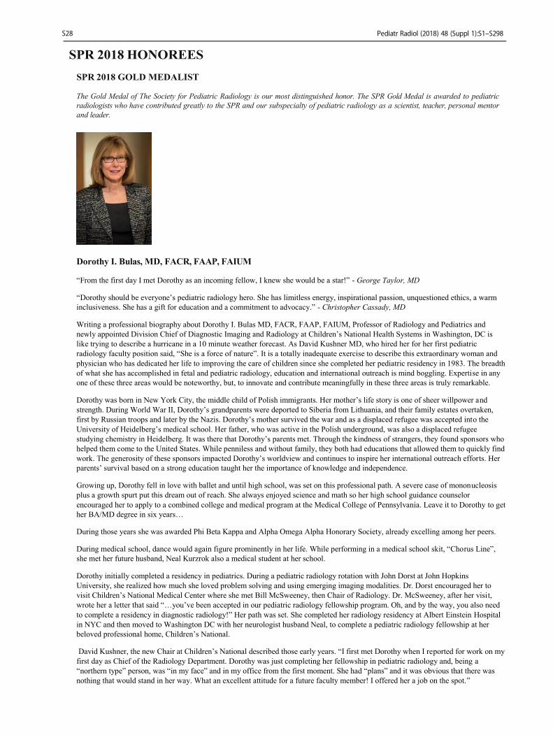
SPR 2018 HONOREES
SPR 2018 GOLD MEDALIST
The Gold Medal of The Society for Pediatric Radiology is our most distinguished honor. The SPR Gold Medal is awarded to pediatric
radiologists who have contributed greatly to the SPR and our subspecialty of pediatric radiology as a scientist, teacher, personal mentor
and leader.
Dorothy I. Bulas, MD, FACR, FAAP, FAIUM
“From the first day I met Dorothy as an incoming fellow, I knew she would be a star!” - George Taylor, MD
“Dorothy should be everyone’s pediatric radiology hero. She has limitless energy, inspirational passion, unquestioned ethics, a warm
inclusiveness. She has a gift for education and a commitment to advocacy.” - Christopher Cassady, MD
Writing a professional biography about Dorothy I. Bulas MD, FACR, FAAP, FAIUM, Professor of Radiology and Pediatrics and
newly appointed Division Chief of Diagnostic Imaging and Radiology at Children’s National Health Systems in Washington, DC is
like trying to describe a hurricane in a 10 minute weather forecast. As David Kushner MD, who hired her for her first pediatric
radiology faculty position said, “She is a force of nature”. It is a totally inadequate exercise to describe this extraordinary woman and
physician who has dedicated her life to improving the care of children since she completed her pediatric residency in 1983. The breadth
of what she has accomplished in fetal and pediatric radiology, education and international outreach is mind boggling. Expertise in any
one of these three areas would be noteworthy, but, to innovate and contribute meaningfully in these three areas is truly remarkable.
Dorothy was born in New York City, the middle child of Polish immigrants. Her mother’s life story is one of sheer willpower and
strength. During World War II, Dorothy’s grandparents were deported to Siberia from Lithuania, and their family estates overtaken,
first by Russian troops and later by the Nazis. Dorothy’s mother survived the war and as a displaced refugee was accepted into the
University of Heidelberg’s medical school. Her father, who was active in the Polish underground, was also a displaced refugee
studying chemistry in Heidelberg. It was there that Dorothy’s parents met. Through the kindness of strangers, they found sponsors who
helped them come to the United States. While penniless and without family, they both had educations that allowed them to quickly find
work. The generosity of these sponsors impacted Dorothy’s worldview and continues to inspire her international outreach efforts. Her
parents’ survival based on a strong education taught her the importance of knowledge and independence.
Growing up, Dorothy fell in love with ballet and until high school, was set on this professional path. A severe case of mononucleosis
plus a growth spurt put this dream out of reach. She always enjoyed science and math so her high school guidance counselor
encouraged her to apply to a combined college and medical program at the Medical College of Pennsylvania. Leave it to Dorothy to get
her BA/MD degree in six years…
During those years she was awarded Phi Beta Kappa and Alpha Omega Alpha Honorary Society, already excelling among her peers.
During medical school, dance would again figure prominently in her life. While performing in a medical school skit, “Chorus Line”,
she met her future husband, Neal Kurzrok also a medical student at her school.
Dorothy initially completed a residency in pediatrics. During a pediatric radiology rotation with John Dorst at John Hopkins
University, she realized how much she loved problem solving and using emerging imaging modalities. Dr. Dorst encouraged her to
visit Children’s National Medical Center where she met Bill McSweeney, then Chair of Radiology. Dr. McSweeney, after her visit,
wrote her a letter that said “…you’ve been accepted in our pediatric radiology fellowship program. Oh, and by the way, you also need
to complete a residency in diagnostic radiology!” Her path was set. She completed her radiology residency at Albert Einstein Hospital
in NYC and then moved to Washington DC with her neurologist husband Neal, to complete a pediatric radiology fellowship at her
beloved professional home, Children’s National.
David Kushner, the new Chair at Children’s National described those early years. “I first met Dorothy when I reported for work on my
first day as Chief of the Radiology Department. Dorothy was just completing her fellowship in pediatric radiology and, being a
“northern type” person, was “in my face” and in my office from the first moment. She had “plans” and it was obvious that there was
nothing that would stand in her way. What an excellent attitude for a future faculty member! I offered her a job on the spot.”
Pediatr Radiol (2018) 48 (Suppl 1):S1–S298S28
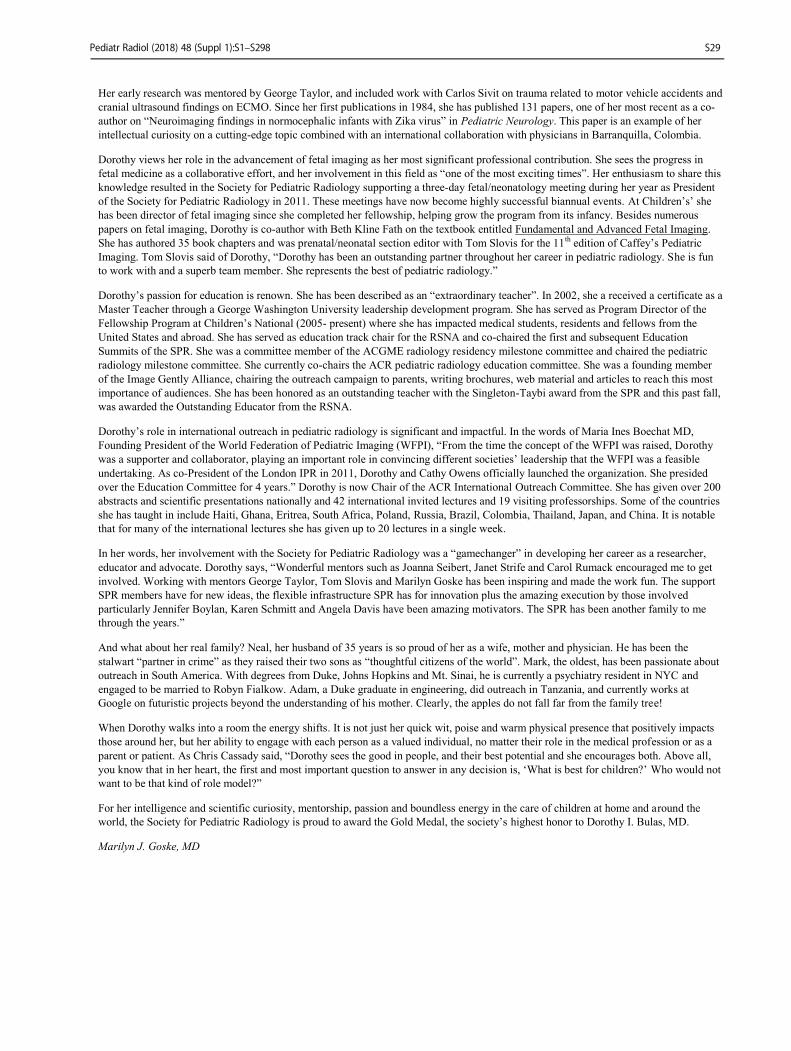
Her early research was mentored by George Taylor, and included work with Carlos Sivit on trauma related to motor vehicle accidents and
cranial ultrasound findings on ECMO. Since her first publications in 1984, she has published 131 papers, one of her most recent as a co-
author on “Neuroimaging findings in normocephalic infants with Zika virus” in Pediatric Neurology. This paper is an example of her
intellectual curiosity on a cutting-edge topic combined with an international collaboration with physicians in Barranquilla, Colombia.
Dorothy views her role in the advancement of fetal imaging as her most significant professional contribution. She sees the progress in
fetal medicine as a collaborative effort, and her involvement in this field as “one of the most exciting times”. Her enthusiasm to share this
knowledge resulted in the Society for Pediatric Radiology supporting a three-day fetal/neonatology meeting during her year as President
of the Society for Pediatric Radiology in 2011. These meetings have now become highly successful biannual events. At Children’s’ she
has been director of fetal imaging since she completed her fellowship, helping grow the program from its infancy. Besides numerous
papers on fetal imaging, Dorothy is co-author with Beth Kline Fath on the textbook entitled Fundamental and Advanced Fetal Imaging.
She has authored 35 book chapters and was prenatal/neonatal section editor with Tom Slovis for the 11th edition of Caffey’s Pediatric
Imaging. Tom Slovis said of Dorothy, “Dorothy has been an outstanding partner throughout her career in pediatric radiology. She is fun
to work with and a superb team member. She represents the best of pediatric radiology.”
Dorothy’s passion for education is renown. She has been described as an “extraordinary teacher”. In 2002, she a received a certificate as a
Master Teacher through a George Washington University leadership development program. She has served as Program Director of the
Fellowship Program at Children’s National (2005- present) where she has impacted medical students, residents and fellows from the
United States and abroad. She has served as education track chair for the RSNA and co-chaired the first and subsequent Education
Summits of the SPR. She was a committee member of the ACGME radiology residency milestone committee and chaired the pediatric
radiology milestone committee. She currently co-chairs the ACR pediatric radiology education committee. She was a founding member
of the Image Gently Alliance, chairing the outreach campaign to parents, writing brochures, web material and articles to reach this most
importance of audiences. She has been honored as an outstanding teacher with the Singleton-Taybi award from the SPR and this past fall,
was awarded the Outstanding Educator from the RSNA.
Dorothy’s role in international outreach in pediatric radiology is significant and impactful. In the words of Maria Ines Boechat MD,
Founding President of the World Federation of Pediatric Imaging (WFPI), “From the time the concept of the WFPI was raised, Dorothy
was a supporter and collaborator, playing an important role in convincing different societies’ leadership that the WFPI was a feasible
undertaking. As co-President of the London IPR in 2011, Dorothy and Cathy Owens officially launched the organization. She presided
over the Education Committee for 4 years.” Dorothy is now Chair of the ACR International Outreach Committee. She has given over 200
abstracts and scientific presentations nationally and 42 international invited lectures and 19 visiting professorships. Some of the countries
she has taught in include Haiti, Ghana, Eritrea, South Africa, Poland, Russia, Brazil, Colombia, Thailand, Japan, and China. It is notable
that for many of the international lectures she has given up to 20 lectures in a single week.
In her words, her involvement with the Society for Pediatric Radiology was a “gamechanger” in developing her career as a researcher,
educator and advocate. Dorothy says, “Wonderful mentors such as Joanna Seibert, Janet Strife and Carol Rumack encouraged me to get
involved. Working with mentors George Taylor, Tom Slovis and Marilyn Goske has been inspiring and made the work fun. The support
SPR members have for new ideas, the flexible infrastructure SPR has for innovation plus the amazing execution by those involved
particularly Jennifer Boylan, Karen Schmitt and Angela Davis have been amazing motivators. The SPR has been another family to me
through the years.”
And what about her real family? Neal, her husband of 35 years is so proud of her as a wife, mother and physician. He has been the
stalwart “partner in crime” as they raised their two sons as “thoughtful citizens of the world”. Mark, the oldest, has been passionate about
outreach in South America. With degrees from Duke, Johns Hopkins and Mt. Sinai, he is currently a psychiatry resident in NYC and
engaged to be married to Robyn Fialkow. Adam, a Duke graduate in engineering, did outreach in Tanzania, and currently works at
Google on futuristic projects beyond the understanding of his mother. Clearly, the apples do not fall far from the family tree!
When Dorothy walks into a room the energy shifts. It is not just her quick wit, poise and warm physical presence that positively impacts
those around her, but her ability to engage with each person as a valued individual, no matter their role in the medical profession or as a
parent or patient. As Chris Cassady said, “Dorothy sees the good in people, and their best potential and she encourages both. Above all,
you know that in her heart, the first and most important question to answer in any decision is, ‘What is best for children?’ Who would not
want to be that kind of role model?”
For her intelligence and scientific curiosity, mentorship, passion and boundless energy in the care of children at home and around the
world, the Society for Pediatric Radiology is proud to award the Gold Medal, the society’s highest honor to Dorothy I. Bulas, MD.
Marilyn J. Goske, MD
Pediatr Radiol (2018) 48 (Suppl 1):S1–S298 S29
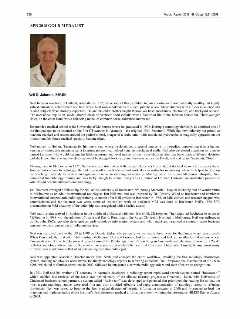
SPR 2018 GOLD MEDALIST
Neil D. Johnson, MBBS
Neil Johnson was born in Ballarat, Australia in 1952, the second of three children to parents who were not materially wealthy but highly
valued education, achievement and hard work. Neil won scholarships to a local private school where students with a focus in science and
related subjects were strongly supported. He and his older brother taught themselves basic mechanics, electronics, and backyard science.
The occasional explosion, model aircraft crash or electrical short circuits were a feature of life in the Johnson household. Their younger
sister, on the other hand, was a balancing model of common sense, tolerance and reason.
He attended medical school at the University of Melbourne where he graduated in 1976. During a neurology clerkship, he admitted one of
the first patients to be scanned on the first CT scanner in Australia – the original “EMI Scanner”. While that revolutionary but primitive
machine clunked and rotated around the patient’s head, images of a brain tumor with associated hydrocephalus magically appeared on the
monitor and his future medical specialty became clear.
Neil moved to Hobart, Tasmania for his intern year where he developed a special interest in orthopedics, approaching it as a human
version of motorcycle maintenance, a longtime passion that helped hone his mechanical skills. Neil also developed a passion for a nurse
named Lorraine, who would become his lifelong partner and loyal mother of their three children. She may have made a different decision
had she known that she and the children would be dragged backwards and forwards across the Pacific and end up in Cincinnati, Ohio!
Moving back to Melbourne in 1977, Neil was a pediatric intern at the Royal Children’s Hospital, but decided to switch his career focus
from pediatrics back to radiology. He took a year off clinical service and worked as an instructor in anatomy where he helped to develop
the teaching materials for a new undergraduate course in radiological anatomy. Moving on to the Royal Melbourne Hospital, Neil
completed his radiology training and was lucky enough to do his final year as a trainee of Dr. Ken Thomson, an Australian pioneer of
what would become interventional radiology.
Dr. Thomson arranged a fellowship for Neil at the University of Rochester, NY, Strong Memorial Hospital intending that he would return
to Melbourne as an adult interventional radiologist. But Neil met and was inspired by Dr. Beverly Wood at Rochester and combined
interventional and pediatric radiology training. A month after Neil arrived in Rochester in 1985, an MRI clinical and research magnet was
commissioned and for the next two years, some of the earliest work on pediatric MRI was done at Rochester. Neil’s 1988 SPR
presentation on MRI anatomy of the infant hip was recognized with a Caffey award.
Neil and Lorraine arrived in Rochester in the middle of a blizzard with their first child, Christopher. They departed Rochester to return to
Melbourne in 1988 with the addition of Luana and David. Returning to the Royal Children’s Hospital in Melbourne, Neil was influenced
by Dr. John DeCampo who developed an early radiology information system and who taught and practiced a common sense business
approach to the organization of radiology services.
Neil was recruited back to the US in 1988 by Donald Kirks, who patiently waited nearly three years for the family to get green cards.
When Don made the first offer while visiting Melbourne, Neil and Lorraine had to rush home and look up an atlas to find out just where
Cincinnati was! So the family packed up and crossed the Pacific again in 1991, settling in Cincinnati and planning to look for a “real”
pediatric radiology job on one of the coasts. Twenty-seven years later he is still at Cincinnati Children’s Hospital, having worn many
different hats in addition to that of an outstanding pediatric radiologist.
Neil was appointed Associate Director under Janet Strife and changed the entire workflow, installing the first radiology information
system, holding radiologists accountable for timely radiology reports to referring clinicians. Neil proposed the installation of PACS in
1998, which led to filmless operations in 2002, followed by integrated electronic radiology orders and soon after, voice recognition.
In 1993, Neil and his brother’s IT company in Australia developed a radiology report rapid word search system named “Radsearch,”
which enabled fast retrieval of the basic data behind many of the clinical research projects at Cincinnati. Later, with University of
Cincinnati business school partners, a system called “Radstream” was developed and patented that prioritized the reading list so that the
most urgent radiology studies were read first and also provided effective and rapid communication of radiology repots to referring
physicians. Neil was asked to become the first medical director of hospital information systems in 2000 and proceeded to lead the
planning and implementation of the hospital’s first electronic medical information system, winning the prestigious HIMSS Davies Award
in 2003.
Pediatr Radiol (2018) 48 (Suppl 1):S1–S298S30
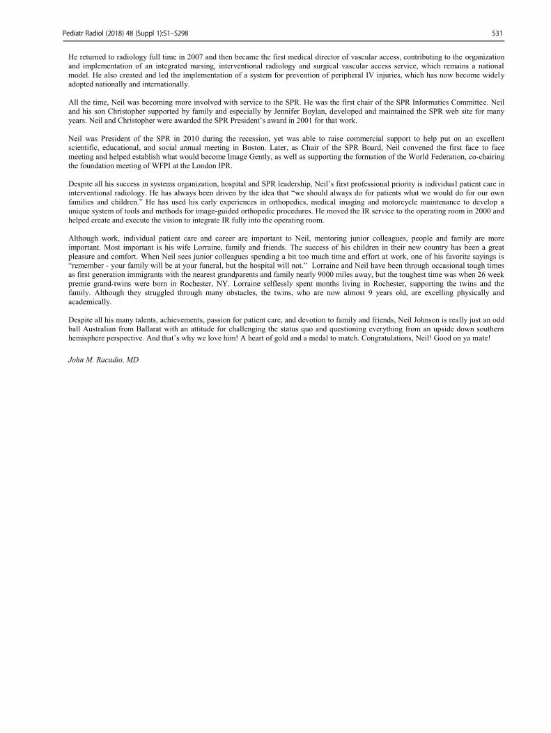
He returned to radiology full time in 2007 and then became the first medical director of vascular access, contributing to the organization
and implementation of an integrated nursing, interventional radiology and surgical vascular access service, which remains a national
model. He also created and led the implementation of a system for prevention of peripheral IV injuries, which has now become widely
adopted nationally and internationally.
All the time, Neil was becoming more involved with service to the SPR. He was the first chair of the SPR Informatics Committee. Neil
and his son Christopher supported by family and especially by Jennifer Boylan, developed and maintained the SPR web site for many
years. Neil and Christopher were awarded the SPR President’s award in 2001 for that work.
Neil was President of the SPR in 2010 during the recession, yet was able to raise commercial support to help put on an excellent
scientific, educational, and social annual meeting in Boston. Later, as Chair of the SPR Board, Neil convened the first face to face
meeting and helped establish what would become Image Gently, as well as supporting the formation of the World Federation, co-chairing
the foundation meeting of WFPI at the London IPR.
Despite all his success in systems organization, hospital and SPR leadership, Neil’s first professional priority is individual patient care in
interventional radiology. He has always been driven by the idea that “we should always do for patients what we would do for our own
families and children.” He has used his early experiences in orthopedics, medical imaging and motorcycle maintenance to develop a
unique system of tools and methods for image-guided orthopedic procedures. He moved the IR service to the operating room in 2000 and
helped create and execute the vision to integrate IR fully into the operating room.
Although work, individual patient care and career are important to Neil, mentoring junior colleagues, people and family are more
important. Most important is his wife Lorraine, family and friends. The success of his children in their new country has been a great
pleasure and comfort. When Neil sees junior colleagues spending a bit too much time and effort at work, one of his favorite sayings is
“remember - your family will be at your funeral, but the hospital will not.” Lorraine and Neil have been through occasional tough times
as first generation immigrants with the nearest grandparents and family nearly 9000 miles away, but the toughest time was when 26 week
premie grand-twins were born in Rochester, NY. Lorraine selflessly spent months living in Rochester, supporting the twins and the
family. Although they struggled through many obstacles, the twins, who are now almost 9 years old, are excelling physically and
academically.
Despite all his many talents, achievements, passion for patient care, and devotion to family and friends, Neil Johnson is really just an odd
ball Australian from Ballarat with an attitude for challenging the status quo and questioning everything from an upside down southern
hemisphere perspective. And that’s why we love him! A heart of gold and a medal to match. Congratulations, Neil! Good on ya mate!
John M. Racadio, MD
Pediatr Radiol (2018) 48 (Suppl 1):S1–S298 S31
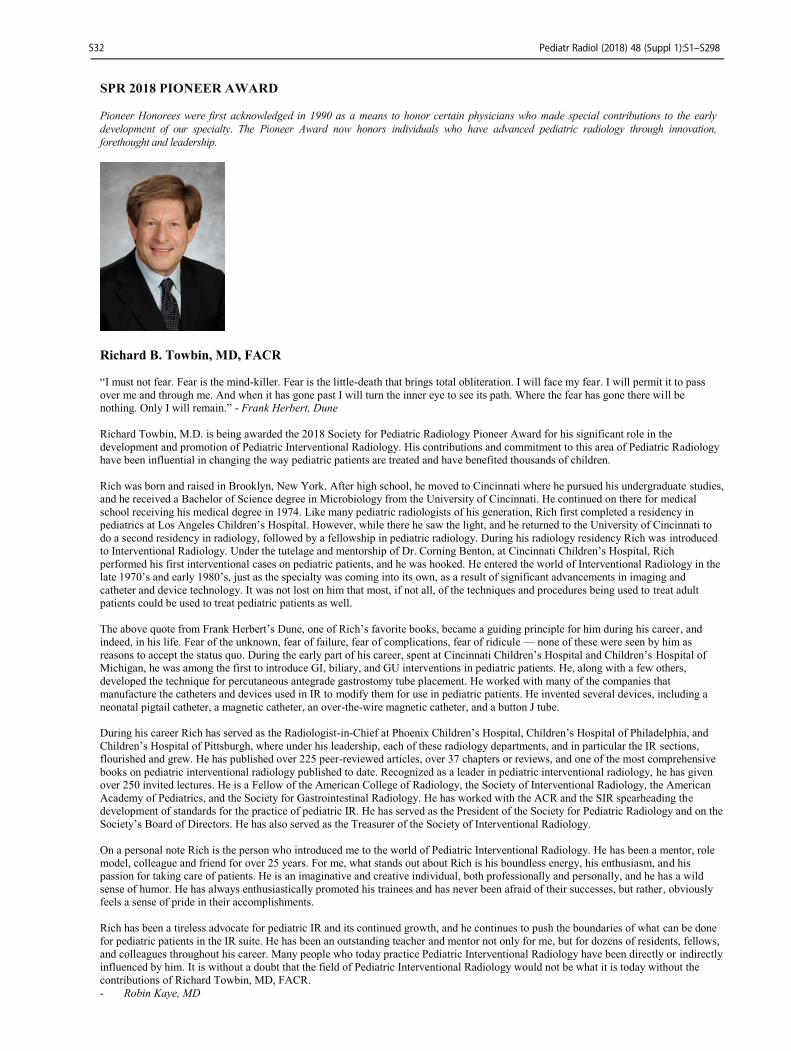
SPR 2018 PIONEER AWARD
Pioneer Honorees were first acknowledged in 1990 as a means to honor certain physicians who made special contributions to the early
development of our specialty. The Pioneer Award now honors individuals who have advanced pediatric radiology through innovation,
forethought and leadership.
Richard B. Towbin, MD, FACR
“I must not fear. Fear is the mind-killer. Fear is the little-death that brings total obliteration. I will face my fear. I will permit it to pass
over me and through me. And when it has gone past I will turn the inner eye to see its path. Where the fear has gone there will be
nothing. Only I will remain.” - Frank Herbert, Dune
Richard Towbin, M.D. is being awarded the 2018 Society for Pediatric Radiology Pioneer Award for his significant role in the
development and promotion of Pediatric Interventional Radiology. His contributions and commitment to this area of Pediatric Radiology
have been influential in changing the way pediatric patients are treated and have benefited thousands of children.
Rich was born and raised in Brooklyn, New York. After high school, he moved to Cincinnati where he pursued his undergraduate studies,
and he received a Bachelor of Science degree in Microbiology from the University of Cincinnati. He continued on there for medical
school receiving his medical degree in 1974. Like many pediatric radiologists of his generation, Rich first completed a residency in
pediatrics at Los Angeles Children’s Hospital. However, while there he saw the light, and he returned to the University of Cincinnati to
do a second residency in radiology, followed by a fellowship in pediatric radiology. During his radiology residency Rich was introduced
to Interventional Radiology. Under the tutelage and mentorship of Dr. Corning Benton, at Cincinnati Children’s Hospital, Rich
performed his first interventional cases on pediatric patients, and he was hooked. He entered the world of Interventional Radiology in the
late 1970’s and early 1980’s, just as the specialty was coming into its own, as a result of significant advancements in imaging and
catheter and device technology. It was not lost on him that most, if not all, of the techniques and procedures being used to treat adult
patients could be used to treat pediatric patients as well.
The above quote from Frank Herbert’s Dune, one of Rich’s favorite books, became a guiding principle for him during his career, and
indeed, in his life. Fear of the unknown, fear of failure, fear of complications, fear of ridicule — none of these were seen by him as
reasons to accept the status quo. During the early part of his career, spent at Cincinnati Children’s Hospital and Children’s Hospital of
Michigan, he was among the first to introduce GI, biliary, and GU interventions in pediatric patients. He, along with a few others,
developed the technique for percutaneous antegrade gastrostomy tube placement. He worked with many of the companies that
manufacture the catheters and devices used in IR to modify them for use in pediatric patients. He invented several devices, including a
neonatal pigtail catheter, a magnetic catheter, an over-the-wire magnetic catheter, and a button J tube.
During his career Rich has served as the Radiologist-in-Chief at Phoenix Children’s Hospital, Children’s Hospital of Philadelphia, and
Children’s Hospital of Pittsburgh, where under his leadership, each of these radiology departments, and in particular the IR sections,
flourished and grew. He has published over 225 peer-reviewed articles, over 37 chapters or reviews, and one of the most comprehensive
books on pediatric interventional radiology published to date. Recognized as a leader in pediatric interventional radiology, he has given
over 250 invited lectures. He is a Fellow of the American College of Radiology, the Society of Interventional Radiology, the American
Academy of Pediatrics, and the Society for Gastrointestinal Radiology. He has worked with the ACR and the SIR spearheading the
development of standards for the practice of pediatric IR. He has served as the President of the Society for Pediatric Radiology and on the
Society’s Board of Directors. He has also served as the Treasurer of the Society of Interventional Radiology.
On a personal note Rich is the person who introduced me to the world of Pediatric Interventional Radiology. He has been a mentor, role
model, colleague and friend for over 25 years. For me, what stands out about Rich is his boundless energy, his enthusiasm, and his
passion for taking care of patients. He is an imaginative and creative individual, both professionally and personally, and he has a wild
sense of humor. He has always enthusiastically promoted his trainees and has never been afraid of their successes, but rather, obviously
feels a sense of pride in their accomplishments.
Rich has been a tireless advocate for pediatric IR and its continued growth, and he continues to push the boundaries of what can be done
for pediatric patients in the IR suite. He has been an outstanding teacher and mentor not only for me, but for dozens of residents, fellows,
and colleagues throughout his career. Many people who today practice Pediatric Interventional Radiology have been directly or indirectly
influenced by him. It is without a doubt that the field of Pediatric Interventional Radiology would not be what it is today without the
contributions of Richard Towbin, MD, FACR. - Robin Kaye, MD
Pediatr Radiol (2018) 48 (Suppl 1):S1–S298S32
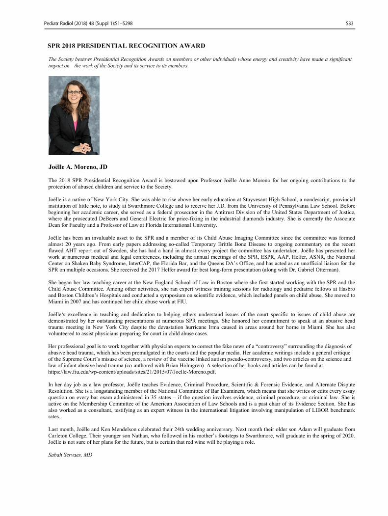
SPR 2018 PRESIDENTIAL RECOGNITION AWARD
The Society bestows Presidential Recognition Awards on members or other individuals whose energy and creativity have made a significant
impact on the work of the Society and its service to its members.
Joëlle A. Moreno, JD
The 2018 SPR Presidential Recognition Award is bestowed upon Professor Joëlle Anne Moreno for her ongoing contributions to the
protection of abused children and service to the Society.
Joëlle is a native of New York City. She was able to rise above her early education at Stuyvesant High School, a nondescript, provincial
institution of little note, to study at Swarthmore College and to receive her J.D. from the University of Pennsylvania Law School. Before
beginning her academic career, she served as a federal prosecutor in the Antitrust Division of the United States Department of Justice,
where she prosecuted DeBeers and General Electric for price-fixing in the industrial diamonds industry. She is currently the Associate
Dean for Faculty and a Professor of Law at Florida International University.
Joëlle has been an invaluable asset to the SPR and a member of its Child Abuse Imaging Committee since the committee was formed
almost 20 years ago. From early papers addressing so-called Temporary Brittle Bone Disease to ongoing commentary on the recent
flawed AHT report out of Sweden, she has had a hand in almost every project the committee has undertaken. Joëlle has presented her
work at numerous medical and legal conferences, including the annual meetings of the SPR, ESPR, AAP, Helfer, ASNR, the National
Center on Shaken Baby Syndrome, InterCAP, the Florida Bar, and the Queens DA’s Office, and has acted as an unofficial liaison for the
SPR on multiple occasions. She received the 2017 Helfer award for best long-form presentation (along with Dr. Gabriel Otterman).
She began her law-teaching career at the New England School of Law in Boston where she first started working with the SPR and the
Child Abuse Committee. Among other activities, she ran expert witness training sessions for radiology and pediatric fellows at Hasbro
and Boston Children’s Hospitals and conducted a symposium on scientific evidence, which included panels on child abuse. She moved to
Miami in 2007 and has continued her child abuse work at FIU.
Joëlle‘s excellence in teaching and dedication to helping others understand issues of the court specific to issues of child abuse are
demonstrated by her outstanding presentations at numerous SPR meetings. She honored her commitment to speak at an abusive head
trauma meeting in New York City despite the devastation hurricane Irma caused in areas around her home in Miami. She has also
volunteered to assist physicians preparing for court in child abuse cases.
Her professional goal is to work together with physician experts to correct the fake news of a “controversy” surrounding the diagnosis of
abusive head trauma, which has been promulgated in the courts and the popular media. Her academic writings include a general critique
of the Supreme Court’s misuse of science, a review of the vaccine linked autism pseudo-controversy, and two articles on the science and
law of infant abusive head trauma (co-authored with Brian Holmgren). A selection of her books and articles can be found at
https://law.fiu.edu/wp-content/uploads/sites/21/2015/07/Joelle-Moreno.pdf.
In her day job as a law professor, Joëlle teaches Evidence, Criminal Procedure, Scientific & Forensic Evidence, and Alternate Dispute
Resolution. She is a longstanding member of the National Committee of Bar Examiners, which means that she writes or edits every essay
question on every bar exam administered in 35 states – if the question involves evidence, criminal procedure, or criminal law. She is
active on the Membership Committee of the American Association of Law Schools and is a past chair of its Evidence Section. She has
also worked as a consultant, testifying as an expert witness in the international litigation involving manipulation of LIBOR benchmark
rates.
Last month, Joëlle and Ken Mendelson celebrated their 24th wedding anniversary. Next month their older son Adam will graduate from
Carleton College. Their younger son Nathan, who followed in his mother’s footsteps to Swarthmore, will graduate in the spring of 2020.
Joëlle is not sure of her plans for the future, but is certain that red wine will be playing a role.
Sabah Servaes, MD
Pediatr Radiol (2018) 48 (Suppl 1):S1–S298 S33
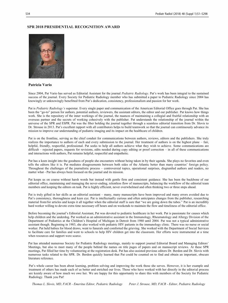
SPR 2018 PRESIDENTIAL RECOGNITION AWARD
Patricia Vario
Since 2004, Pat Vario has served as Editorial Assistant for the journal Pediatric Radiology. Pat’s work has been integral to the sustained
success of the journal. Every Society for Pediatric Radiology member who has submitted a paper to Pediatric Radiology since 2004 has
knowingly or unknowingly benefitted from Pat’s dedication, consistency, professionalism and passion for her work.
Pat is Pediatric Radiology’s superstar. Every single paper and communication of the American Editorial Office goes through Pat. She has
been the “go-to” person for authors, potential authors, reviewers, the assistant editors, the editor and our publisher. Pat knows how things
work. She is the repository of the inner workings of the journal, the nuances of maintaining a collegial and fruitful relationship with an
overseas partner and the secrets of working cohesively with the publisher. Pat understands the relationship of the journal within the
universe of the SPR and ESPR. Pat was the fiber holding the journal together through a seamless editorial transition from Dr. Slovis to
Dr. Strouse in 2013. Pat’s excellent rapport with all contributors helps to build teamwork so that the journal can continuously advance its
mission to improve our understanding of pediatric imaging and its impact on the healthcare of children.
Pat is on the frontline, serving as the chief conduit for communications between authors, reviews, editors and the publishers. She truly
realizes the importance to authors of each and every submission to the journal. Her treatment of authors is on the highest plane – fair,
helpful, friendly, respectful, professional. Pat seeks to help all authors achieve what they wish to achieve. Some communications are
difficult – rejected papers, requests for revisions, edits needed during copy editing or proof correction – in all of these communications
and interactions with authors, Pat remains helpful, respectful and empathetic.
Pat has a keen insight into the goodness of people she encounters without being taken in by their agenda. She plays no favorites and even
tells the editors like it is. Pat mediates disagreements between both sides of the Atlantic better than many countries’ foreign policy.
Throughout the challenges of the journalistic process – controversial topics, operational surprises, disgruntled authors and readers, no
matter what - Pat has always been focused on the journal and its mission.
Pat keeps us on course without harsh words but instead with gentle firm and consistent guidance. She has been the backbone of our
editorial office, maintaining and managing the never-ending, relentless flow of manuscripts, balancing the workflow of the editorial team
members and keeping the editors on task. Pat is highly efficient, never overwhelmed and often thinking two or three steps ahead.
Pat is truly gifted in her skills as an editorial assistant – many, many manuscripts have been improved and many errors avoided due to
Pat’s consistency, thoroughness and keen eye. Pat is intellectually curious and often anticipates changes from the publisher, researching
material from/for articles and keeps it all together when the editorial staff is sure that “we are going down the tubes.” Pat is an incredibly
hard worker willing to devote extra time necessary off hours and on weekends to maintain the flow and timeliness of the editorial office.
Before becoming the journal’s Editorial Assistant, Pat was devoted to pediatric healthcare in her work. Pat is passionate for causes which
help children and the underdog. Pat worked as an administrative assistant in the Immunology, Rheumatology and Allergy Division of the
Department of Pediatrics at the Children’s Hospital of Michigan in Detroit from 1980 until 2004. Pat was not a typical administrative
assistant though. Beginning in 1983, she also worked with pediatric HIV patients in the immunology clinic. There was no nurse or social
worker. Pat held babies for blood draws, went to funerals and comforted the grieving. She worked with the Department of Social Services
to facilitate care for families and went to schools to help HIV children get into the classroom. Her efforts were instrumental at a time
when resources and support were scarce.
Pat has attended numerous Society for Pediatric Radiology meetings, mainly to support journal Editorial Board and Managing Editors’
Meetings, but also to meet many of the people behind the names on title pages of papers and on manuscript reviews. At these SPR
meetings, Pat filled her time by volunteering at the registration desk. Pat has also assisted previous editors Dr. Berdon and Dr. Slovis with
numerous tasks related to the SPR. Dr. Berdon quickly learned that Pat could be counted on to find and obtain an important, obscure
literature reference.
Pat’s whole career has been about learning, problem solving and improving the work those she serves. However, it is her example and
treatment of others has made each of us better and enriched our lives. Those who have worked with her directly in the editorial process
are keenly aware of how much we owe her. We are happy for this opportunity to share this with members of the Society for Pediatric
Radiology. Thank you Pat!
Thomas L. Slovis, MD, FACR - Emeritus Editor, Pediatric Radiology Peter J. Strouse, MD, FACR - Editor, Pediatric Radiology
Pediatr Radiol (2018) 48 (Suppl 1):S1–S298S34
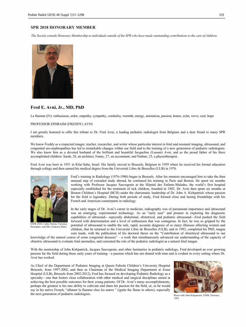
SPR 2018 HONORARY MEMBER
The Society extends Honorary Membership to individuals outside of the SPR who have made outstanding contributions to the care of children.
Fred E. Avni, Jr., MD, PhD
La flamme (Fr): enthusiasm, ardor, empathy, sympathy, cordiality, warmth, energy, animation, passion, honor, eclat, verve, zeal, hope
PROFESSOR EPHRAIM (FREDDY) AVNI
I am greatly honored to offer this tribute to Dr. Fred Avni, a leading pediatric radiologist from Belgium and a dear friend to many SPR
members.
We know Freddy as a respected imager, teacher, researcher, and writer whose particular interest in fetal and neonatal imaging, ultrasound, and
congenital uro-nephropathies has led to remarkable changes within our field and to the training of a new generation of pediatric radiologists.
We also know him as a devoted husband of the brilliant and beautiful Jacqueline (Lussan) Avni, and as the proud father of his three
accomplished children: Sarah, 28, an architect; Fanny, 27, an accountant; and Nathan, 25, a physiotherapist.
Fred Avni was born in 1951 in Kfar-Saba, Israel. His family moved to Brussels, Belgium in 1959 where he received his formal education
through college and then earned his medical degree from the Université Libre de Bruxelles (ULB) in 1976.
Fred’s training in Radiology (1976-1980) began in Brussels. After his mentors encouraged him to take the then
unusual step of extended study abroad, he continued his training in Paris and Boston. He spent six months
working with Professor Jacques Sauvegrain at the Hôpital des Enfants-Malades, the world’s first hospital
especially established for the treatment of sick children, founded in 1802. Dr. Avni then spent six months at
Boston Children’s Hospital (BCH) under the charismatic leadership of Dr. John A. Kirkpatrick whose passion
for the field is legendary. During both periods of study, Fred formed close and lasting friendships with his
French and American counterparts in radiology.
In the early stages of Dr. Avni’s career in medicine, radiography was of paramount importance and ultrasound
was an emerging, experimental technology. As an “early user” and pioneer in exploring the diagnostic
capabilities of ultraound-- especially abdominal, obstetrical, and pediatric ultrasound—Fred pushed the field
forward with determination and a level of enthusiasm that was contagious. In fact, he was so gripped by the
potential of ultrasound to enable the safe, rapid, accurate diagnosis of so many illnesses affecting women and
children, that he returned to the Université Libre de Bruxelles (ULB), and in 1992, completed his PhD, magna
cum laude, with the publication of his doctoral thesis on the “Contribution of obstetrical ultrasound to our
knowledge of the natural course of some congenital diseases” – a work that simultaneously advanced our understanding of the capacity of
obstetric ultrasound to evaluate fetal anomalies, and cemented the role of the pediatric radiologist as a natural fetal imager.
With the mentorship of John Kirkpatrick, Jacques Sauvegrain, and other luminaries in pediatric radiology, Fred developed an ever growing
passion for the field during these early years of training—a passion which has not abated with time and is evident in every setting where Dr.
Avni has worked.
As Chief of the Department of Pediatric Imaging at Queen Fabiola Children’s University Hospital
Brussels, from 1997-2002, and then as Chairman of the Medical Imaging Department at Erase
Hospital (ULB), Brussels from 2002-2012), Fred has focused on developing Pediatric Radiology as a
specialty—one that fosters close collaboration with other medical and surgical disciplines aimed at
achieving the best possible outcomes for their young patients. Of Dr. Avni’s many accomplishments,
perhaps the greatest is his rare ability to cultivate and share his passion for the field, or, as he would
say in his native French, “allumer la flamme chez les autres ” (ignite the flame in others), especially
the next generation of pediatric radiologists.
ESPR 2010, Cathy Owens, Veronica
Donoghue, and Mrs. Francois Diard
Photo with John Kirkpatrick: ESPR, Florence,
1984
Pediatr Radiol (2018) 48 (Suppl 1):S1–S298 S35
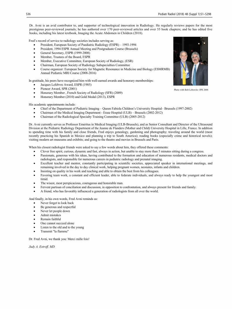
Dr. Avni is an avid contributor to, and supporter of technological innovation in Radiology. He regularly reviews papers for the most
prestigious peer-reviewed journals; he has authored over 170 peer-reviewed articles and over 55 book chapters; and he has edited five
books, including his latest textbook, Imaging the Acute Abdomen in Children (2018).
Fred’s record of service to radiology societies includes serving as:
President, European Society of Paediatric Radiology (ESPR) – 1993-1994
President, 1994 ESPR Annual Meeting and Postgraduate Course (Brussels)
General Secretary, ESPR (1999-2008)
Member, Trustees of the Board, ESPR
Member, Executive Committee, European Society of Radiology, (ESR)
Chairman, European Society of Radiology Subspecialties Committee
Course organizer: European Society for Magnetic Resonance in Medicine and Biology (ESMRMB)
Annual Pediatric MRI Course (2008-2016)
In gratitude, his peers have recognized him with well-earned awards and honorary memberships:
Jacques Lefebvre Award, ESPR (1985)
Pioneer Award, SPR (2001)
Honorary Member , French Society of Radiology (SFR) (2009)
Honorary Member (2010) and Gold Medal (2013), ESPR
His academic appointments include:
Chief of the Department of Pediatric Imaging – Queen Fabiola Children’s University Hospital– Brussels (1997-2002)
Chairman of the Medical Imaging Department - Erase Hospital (ULB) – Brussels (2002-2012)
Chairman of the Radiological Specialty Training Committee (ULB) (2005-2012)
Dr. Avni currently serves as Professor Emeritus in Medical Imaging (ULB-Brussels), and as Senior Consultant and Director of the Ultrasound
Division at the Pediatric Radiology Department of the Jeanne de Flanders (Mother and Child) University Hospital in Lille, France. In addition
to spending time with his family and close friends, Fred enjoys genealogy, gardening and photography; traveling around the world (most
recently practicing his Spanish in Mexico and planning a trip to South America); reading books (especially crime and historical novels);
visiting modern art museums and exhibits; and going to the theatre and movies in Brussels and Paris.
When his closest radiologist friends were asked to say a few words about him, they offered these comments:
Clever free spirit, curious, dynamic and fast, always in action, but unable to stay more than 5 minutes sitting during a congress.
Passionate, generous with his ideas, having contributed to the formation and education of numerous residents, medical doctors and
radiologists, and responsible for numerous careers in pediatric radiology and prenatal imaging.
Excellent teacher and mentor, constantly participating in scientific societies, appreciated speaker in international meetings, and
remaining involved in the day to day clinical work, helping pregnant women, neonates, infants and children.
Insisting on quality in his work and teaching and able to obtain the best from his colleagues.
Favoring team work, a constant and efficient leader, able to federate individuals, and always ready to help the youngest and most
timid.
The wisest, most perspicacious, courageous and honorable man.
Fervent partisan of conciliation and discussion, in opposition to confrontation, and always present for friends and family.
A friend, who has favorably influenced a generation of radiologists from all over the world.
And finally, in his own words, Fred Avni reminds us:
Never forget to look back
Be generous and respectful
Never let people down
Admit mistakes
Remain faithful
One cannot succeed alone
Listen to the old and to the young
Transmit "la flamme"
Dr. Fred Avni, we thank you: Merci mille fois!
Judy A. Estroff, MD
Photo with Bob Lebowitz: IPR 2006
Pediatr Radiol (2018) 48 (Suppl 1):S1–S298S36
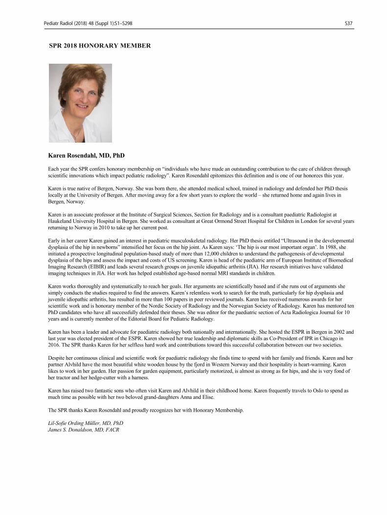
SPR 2018 HONORARY MEMBER
Karen Rosendahl, MD, PhD
Each year the SPR confers honorary membership on “individuals who have made an outstanding contribution to the care of children through
scientific innovations which impact pediatric radiology”. Karen Rosendahl epitomizes this definition and is one of our honorees this year.
Karen is true native of Bergen, Norway. She was born there, she attended medical school, trained in radiology and defended her PhD thesis
locally at the University of Bergen. After moving away for a few short years to explore the world – she returned home and again lives in
Bergen, Norway.
Karen is an associate professor at the Institute of Surgical Sciences, Section for Radiology and is a consultant paediatric Radiologist at
Haukeland University Hospital in Bergen. She worked as consultant at Great Ormond Street Hospital for Children in London for several years
returning to Norway in 2010 to take up her current post.
Early in her career Karen gained an interest in paediatric musculoskeletal radiology. Her PhD thesis entitled “Ultrasound in the developmental
dysplasia of the hip in newborns” intensified her focus on the hip joint. As Karen says: ‘The hip is our most important organ’. In 1988, she
initiated a prospective longitudinal population-based study of more than 12,000 children to understand the pathogenesis of developmental
dysplasia of the hips and assess the impact and costs of US screening. Karen is head of the paediatric arm of European Institute of Biomedical
Imaging Research (EIBIR) and leads several research groups on juvenile idiopathic arthritis (JIA). Her research initiatives have validated
imaging techniques in JIA. Her work has helped established age-based normal MRI standards in children.
Karen works thoroughly and systematically to reach her goals. Her arguments are scientifically based and if she runs out of arguments she
simply conducts the studies required to find the answers. Karen’s relentless work to search for the truth, particularly for hip dysplasia and
juvenile idiopathic arthritis, has resulted in more than 100 papers in peer reviewed journals. Karen has received numerous awards for her
scientific work and is honorary member of the Nordic Society of Radiology and the Norwegian Society of Radiology. Karen has mentored ten
PhD candidates who have all successfully defended their theses. She was editor for the paediatric section of Acta Radiologica Journal for 10
years and is currently member of the Editorial Board for Pediatric Radiology.
Karen has been a leader and advocate for paediatric radiology both nationally and internationally. She hosted the ESPR in Bergen in 2002 and
last year was elected president of the ESPR. Karen showed her true leadership and diplomatic skills as Co-President of IPR in Chicago in
2016. The SPR thanks Karen for her selfless hard work and contributions toward this successful collaboration between our two societies.
Despite her continuous clinical and scientific work for paediatric radiology she finds time to spend with her family and friends. Karen and her
partner Alvhild have the most beautiful white wooden house by the fjord in Western Norway and their hospitality is heart-warming. Karen
likes to work in her garden. Her passion for garden equipment, particularly motorized, is almost as strong as for hips, and she is very fond of
her tractor and her hedge-cutter with a harness.
Karen has raised two fantastic sons who often visit Karen and Alvhild in their childhood home. Karen frequently travels to Oslo to spend as
much time as possible with her two beloved grand-daughters Anna and Elise.
The SPR thanks Karen Rosendahl and proudly recognizes her with Honorary Membership.
Lil-Sofie Ording Müller, MD, PhD
James S. Donaldson, MD, FACR
Pediatr Radiol (2018) 48 (Suppl 1):S1–S298 S37

SPR 2018 EDWARD B. SINGLETON-HOOSHANG TAYBI AWARD
The Edward B. Singleton-Hooshang Taybi Award is given in honor of Edward B. Singleton and Hooshang Taybi, in recognition of their personal
commitment to the educational goals of the SPR. Initiated in 2006, the Award is presented annually to a senior member of the SPR whose
professional lifetime dedication to the education of medical students, residents, fellows, and colleagues has brought honor to him/her and to
the discipline of pediatric radiology.
John D. Strain, MD, FACR
Dr. John D. Strain is a Denver native who grew up in a close family with three siblings, a physician father and a great stay at home mom.
John’s father, Dr. Jim Strain, is an acclaimed pediatrician and former president of the American Academy of Pediatrics.
Growing up, John was a tremendous athlete. He began swimming at age 4, competed in his first out of state swim meet at age 8 and held
every state record in freestyle throughout high school. He was a three-sport athlete, also playing football (all state quarterback) and baseball
(all state pitcher). He even led the nation (NAIA) in strikeouts and ERA on a talented all-star baseball team. The Minnesota twins were even
planning to draft him out of high school, but when his mom received the call, she simply told them not to bother as “Johnny was going to
college!”
Instead of joining the major leagues, John attended the University of Northern Colorado on a full ride baseball scholarship, earning a B.S. in
Chemistry. Upon graduation, he was unsure about medical school but decided to apply to one. He was accepted at the University of Colorado
(CU) School of Medicine and John’s first love was surgery. After completing a surgical internship, he knew this would not be a good fit and
instead, did ED locums for a year. During that time, he honed his golf game, and even competed in the PGA qualifying tournament in
Chicago!
John’s final elective in medical school was radiology and he credits Dr. Paul Siebert, Chair of Radiology at the Denver VA, as the spark to his
career. John discovered he had a keen eye, could identify abnormalities quickly, and remembered patients through their imaging. After a
month rotation during residency at Denver Children's Hospital (now Children's Hospital Colorado, CHCO) under Dr. Jack Campbell, his
career path was clear. Upon completion of pediatric radiology fellowship training at CHCO, his goal was to be chairman. Through the SPR,
he was mentored by the best pediatric radiologists including Drs. John Kirkpatrick, Ed Singleton, Dave Baker and Walter Berdon. John
initially with Drs. Jack Campbell, Chris Foley and Lee Harvey until political challenges at the University led the entire faculty to leave the
practice—except him. A model of resilience, John worked as the sole pediatric radiologist at CHCO for eleven months! Following the strong
recommendation by Dr. Scott Dunbar, former chairman at Cincinnati Children's Hospital, John was promoted to chairman of pediatric
radiology at CHCO and has excelled in this position for 28 years! He spearheaded growth of the faculty from 1 (himself) to the current 23, all
with sub-specialty expertise. John is a dedicated visionary and tireless advocate for the faculty, the hospital and the community.
Throughout his career, John has shown exceptional leadership at the local, state and national levels. At CHCO, after serving on numerous
committees, he quickly chaired them, including the medical board, board of directors, medical staff nominating committee and numerous task
forces. He has been instrumental in organizing, planning and development of hospital informatics. At the state level, he has served as
treasurer, president elect and president of the Colorado Radiological Society. At the national level, John has been active in the children’s
cancer group, the ACR (chairing the imaging appropriateness criteria committee) and especially SCORCH (Society of Chiefs of Radiology in
Children's Hospitals), serving as president in 2002-2003.
John is truly one of the giants in pediatric radiology. He is an outstanding “generalist” in an era of sub-specialization. Many of his
accomplishments are now utilized in daily practice, notably the utility of oblique chest radiographs to improve detection of rib fractures and
the development of indication driven decision support for order entry. John’s tireless work ethic and passion for optimizing pediatric imaging
are summed up by his colleague of 27 years, Dr. Tom Hay: “I learned how to be a good clinical radiologist from John’s guidance and great
example. There is no one better”.
John is a passionate, gifted and dynamic speaker, who has been invited to give more than 100 lectures over his 30+ year career. He connects
with every level of learner from the child to the professional. Our faculty’s children even remember John’s thoughtful practical teaching while
hosting departmental picnics at his home! He sees the big picture, thinks outside the box and offers insight that had often not been considered
previously.
Pediatr Radiol (2018) 48 (Suppl 1):S1–S298S38
As the director of the pediatric radiology fellowship for nearly 20 years, he was a tremendous role model, mentoring over 25 pediatric
radiology fellows and countless residents. John has been my strongest mentor and entrusted me to direct the fellowship since 2010. He also
holds a CAQ in neuroradiology and helped establish our pediatric neuroradiology fellowship program. John supports education through
technology and implemented our Integrated Radiology Imaging System (IRIS) digital teaching file in 2008. The teaching file now has greater
than 23,000 cases and will continue to benefit residents and fellows for generations to come. He listens, supports, guides and inspires others to
create meaningful solutions to our everyday challenges.
John has been awarded many honors for his commitment and leadership, including the medical board award for outstanding service to CHCO
and numerous faculty teaching awards, For 23 consecutive years, John has received Denver’s 5280 “Top Doc” award, emphasizing his
tremendous standing amongst his peers.
Julie and John have been married for 25 years. Julie is the light of John’s life, and the rock of their family. She would often remind him how
lucky he was to be able to help children, even when he had to go in at 3am to reduce an intussusception! They are the proud parents of three
boys: JJ (currently a freshman at Villanova and on the swim team!) and twins James (swimming and playing basketball) and Jonas (playing
basketball and baseball) at Cherry Creek high school.
Dr. John Strain’s lifetime of service and dedication to children, education, leadership, and the SPR make him an obvious choice for the 2018
SPR Edward B. Singleton-Hooshang Taybi Award. Dr. Strain encompasses all the ideals this award embraces. We are fortunate to know and
work with such a brilliant and compassionate pediatric radiologist! Congratulations John!
Laura Z. Fenton, MD
Pediatr Radiol (2018) 48 (Suppl 1):S1–S298 S39
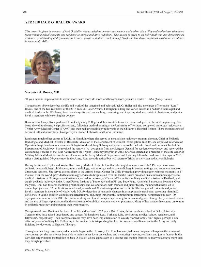
SPR 2018 JACK O. HALLER AWARD
This award is given in memory of Jack O. Haller who excelled as an educator, mentor and author. His ability and enthusiasm stimulated
many young medical students and residents to pursue pediatric radiology. This award is given to an individual who has demonstrated
evidence of outstanding ability to educate trainees (medical student, resident and fellow) who has shown sustained substantial excellence
in mentorship skills.
Veronica J. Rooks, MD
“If your actions inspire others to dream more, learn more, do more, and become more, you are a leader.” - John Quincy Adams
The quotation above describes the life and work of the venerated and beloved Jack O. Haller and also the career of Veronica “Roni”
Rooks, one of the two recipients of the 2018 Jack O. Haller Award. Throughout a long and varied career as a pediatric radiologist and
medical leader in the US Army, Roni has always focused on teaching, mentoring, and inspiring students, resident physicians, and junior
faculty members while serving her country.
Born in New Jersey, Roni graduated from Gettysburg College and then went on to earn a master’s degree in chemical engineering. She
heard the call to the medical profession and, following medical training at the University of Vermont, completed radiology residency at
Tripler Army Medical Center (TAMC) and then pediatric radiology fellowship at the Children’s Hospital Boston. There she met some of
her most influential mentors - George Taylor, Robert Lebowitz, and Carlo Buonomo.
Roni spent much of her career at TAMC in Honolulu where she served as the assistant residency program director, Chief of Pediatric
Radiology, and Medical Director of Research Education of the Department of Clinical Investigation. In 2008, she deployed in service of
Operation Iraqi Freedom as a trauma radiologist to Mosul, Iraq. Subsequently, she rose to the rank of colonel and became Chief of the
Department of Radiology. She received the Army’s “A” designator from the Surgeon General for academic excellence, and received the
Outstanding Teacher of the Year Award from the Tripler Residency program in 2013. She was selected as a member of the elite Order of
Military Medical Merit for excellence of service in the Army Medical Department and fostering fellowship and esprit de corps in 2013.
After a distinguished 24-year career in the Army, Roni recently retired but will return to Tripler as a civilian pediatric radiologist.
During her time at Tripler and Walter Reed Army Medical Center before that, she taught in numerous RSNA Plenary Sessions on
pediatric neuroradiology, child abuse, trauma radiology, teleradiology and remote radiology in austere settings, and countless hands-on
ultrasound sessions. She served as consultant to the Armed Forces Center for Child Protection, providing expert witness testimony in 17
trials all over the world; provided teleradiology services to hospitals all over the Pacific Basin; provided onsite ultrasound expertise to
medical missions in Nicaragua and Guatemala; served as radiology Officer-in-Charge for a military medical mission in Thailand; and
taught pediatric radiology at the Armed Forces Institute of Pathology and in Fiji and Pago Pago, American Samoa, and Rwanda. Over
the years, Roni had fostered mentoring relationships and collaborations with trainees and junior faculty members that have led to
research projects and 32 publications in refereed journals and 39 abstracts/posters and exhibits. She has guided residents and junior
faculty members in the study of whole-body MR for detection of anatomic changes in asymptomatic newborns, screening vitamin D
deficiency in young children without suspected trauma, and, most importantly, demonstrating intracranial hemorrhage in over 100
asymptomatic newborns on MR. She is also working on clinical competency training for ultrasound guided foreign body removal in war
and the use of finger-tip ultrasound in the evaluation of umbilical vascular catheter placement. Many of her trainees have gone on to train
in pediatric radiology and to pursue their own research.
On a personal note, Roni met the love of her life and husband of 27 years, Bob Rooks, during graduate school at Duke University.
Together they have raised three happy and successful daughters, Lexi, Tori, and Liza, born during medical school, residency, and
fellowship, respectively. Their secret to success may have been implementation of weekly “forced family fun” nights, perhaps a side
effect of years of military life. Following in her mother’s footsteps, daughter Lexi is now a second lieutenant in the Army currently
finishing her doctorate in Physical Therapy.
Throughout her long career as a pediatric radiologist in the US Army, Dr. Roni has accepted many unique challenges in the service of
our country, yet she has always been able to maintain her focus on teaching and mentoring students, residents, and junior faculty. In this
way, her career honors the tradition of Jack O. Haller, whose enthusiasm as a teacher and mentor inspired so many to achieve more than
they thought possible.
Ellen M. Chung, MD
Pediatr Radiol (2018) 48 (Suppl 1):S1–S298S40
SPR 2018 JACK O. HALLER AWARD
Yukata Sato, MD, PhD
I am honored to write this biography for Yutaka Sato, MD, PhD, co-recipient of the Society for Pediatric Radiology, Jack O. Haller
Award for Excellence in Teaching.
“All right, you win”, he said as he handed me a $1.00 bill. As a fellow in pediatric radiology at the University of Iowa, it was a great
feeling to have figured out a challenging case before the Chief of Pediatric Radiology. The incentive that this gave me, went far beyond
the value of the dollar bill, and is just one of the many ways that Yutaka Sato makes learning fun in the reading room.
Yutaka asks questions of trainees and engages them by soliciting their opinions. He makes you feel like a peer whose opinion is truly
valued in the reading room. The resulting atmosphere naturally stimulates the medical students, residents, fellows, and junior faculty to
learn more about each case they encounter so that they can make a valuable contribution to the daily discussions.
Intelligent, inquisitive, funny, and humble are just a few words that can be used to describe Yutaka. He has mentored many medical
students, residents, fellows and faculty members. The medical students and residents at the University of Iowa have awarded him
“Faculty Teacher of the Year” on multiple occasions. Of his more than 150 publications, many are co-authored with trainees. Over the
years, many University of Iowa radiology residents have chosen to stay there to do their fellowships in Pediatric Radiology.
Under Yutaka’s mentorship, a very strong academic relationship has been built between the University Of Iowa Department Of
Radiology and the Japanese society of Radiology resulting in a constant stream of visiting faculty members. Several of his former
mentees now hold leadership positions in the Society of Pediatric Radiology, Japanese Society of Pediatric Radiology, and Asian and
Oceanic Society for Pediatric Radiology. Hence, even while practicing and teaching in the small college town of Iowa City, Yutaka has
helped further clinical and academic pediatric radiology in different parts of the world through his numerous mentees.
Finally, apart from all his academic successes, Yutaka is a genuinely warm, generous, and kind-hearted person. He tailgates with his son
during Iowa football season with an open-invitation to everyone in the department: from technologists and film-room staff to residents
and attendings. Yutaka has not only taught us radiology, he has also taught us the more important things in life: cherish time with family
and friends, have a hobby, be a colleague/friend to the technical and administrative staff, and enjoy all that you do at the end of the day.
In summary, Yutaka is an excellent educator, mentor, and author. Through his excellent mentorship skills, he has influenced the careers
of many pediatric radiologists who hope to carry the baton forward.
Geetika Khanna, MD, MS
Pediatr Radiol (2018) 48 (Suppl 1):S1–S298 S41
HEIDI PATRIQUIN AWARD
In recognition of Dr. Patriquin's commitment to international education, this fellowship is designed to subsidize the expenses of one
Pediatric Radiologist per year who practices outside of North America.
Bernadette Wambui Muthee, MD
Aga Khan University Hospital Nairobi, Kenya
Pediatr Radiol (2018) 48 (Suppl 1):S1–S298S42
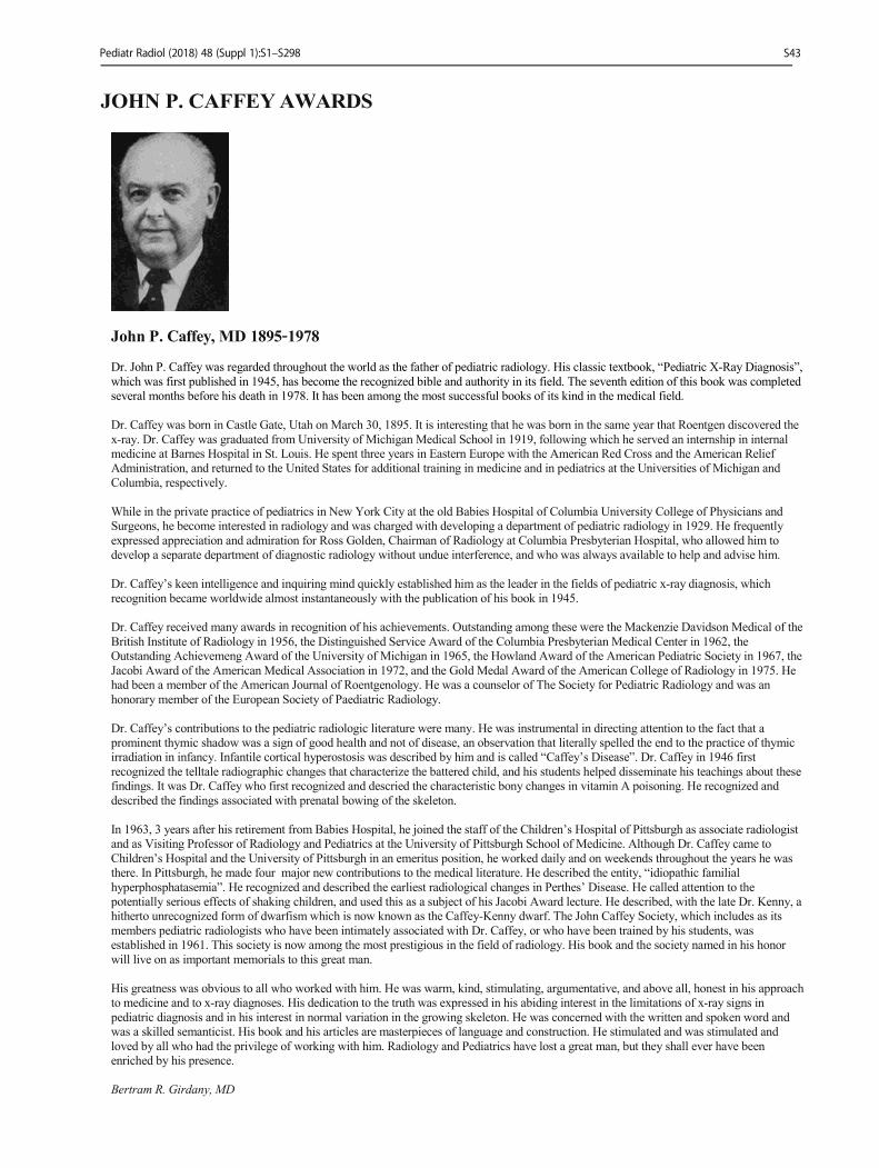
JOHN P. CAFFEY AWARDS
John P. Caffey, MD 1895–1978
Dr. John P. Caffey was regarded throughout the world as the father of pediatric radiology. His classic textbook, “Pediatric X-Ray Diagnosis”,
which was first published in 1945, has become the recognized bible and authority in its field. The seventh edition of this book was completed
several months before his death in 1978. It has been among the most successful books of its kind in the medical field.
Dr. Caffey was born in Castle Gate, Utah on March 30, 1895. It is interesting that he was born in the same year that Roentgen discovered the
x-ray. Dr. Caffey was graduated from University of Michigan Medical School in 1919, following which he served an internship in internal
medicine at Barnes Hospital in St. Louis. He spent three years in Eastern Europe with the American Red Cross and the American Relief
Administration, and returned to the United States for additional training in medicine and in pediatrics at the Universities of Michigan and
Columbia, respectively.
While in the private practice of pediatrics in New York City at the old Babies Hospital of Columbia University College of Physicians and
Surgeons, he become interested in radiology and was charged with developing a department of pediatric radiology in 1929. He frequently
expressed appreciation and admiration for Ross Golden, Chairman of Radiology at Columbia Presbyterian Hospital, who allowed him to
develop a separate department of diagnostic radiology without undue interference, and who was always available to help and advise him.
Dr. Caffey’s keen intelligence and inquiring mind quickly established him as the leader in the fields of pediatric x-ray diagnosis, which
recognition became worldwide almost instantaneously with the publication of his book in 1945.
Dr. Caffey received many awards in recognition of his achievements. Outstanding among these were the Mackenzie Davidson Medical of the
British Institute of Radiology in 1956, the Distinguished Service Award of the Columbia Presbyterian Medical Center in 1962, the
Outstanding Achievemeng Award of the University of Michigan in 1965, the Howland Award of the American Pediatric Society in 1967, the
Jacobi Award of the American Medical Association in 1972, and the Gold Medal Award of the American College of Radiology in 1975. He
had been a member of the American Journal of Roentgenology. He was a counselor of The Society for Pediatric Radiology and was an
honorary member of the European Society of Paediatric Radiology.
Dr. Caffey’s contributions to the pediatric radiologic literature were many. He was instrumental in directing attention to the fact that a
prominent thymic shadow was a sign of good health and not of disease, an observation that literally spelled the end to the practice of thymic
irradiation in infancy. Infantile cortical hyperostosis was described by him and is called “Caffey’s Disease”. Dr. Caffey in 1946 first
recognized the telltale radiographic changes that characterize the battered child, and his students helped disseminate his teachings about these
findings. It was Dr. Caffey who first recognized and descried the characteristic bony changes in vitamin A poisoning. He recognized and
described the findings associated with prenatal bowing of the skeleton.
In 1963, 3 years after his retirement from Babies Hospital, he joined the staff of the Children’s Hospital of Pittsburgh as associate radiologist
and as Visiting Professor of Radiology and Pediatrics at the University of Pittsburgh School of Medicine. Although Dr. Caffey came to
Children’s Hospital and the University of Pittsburgh in an emeritus position, he worked daily and on weekends throughout the years he was
there. In Pittsburgh, he made four major new contributions to the medical literature. He described the entity, “idiopathic familial
hyperphosphatasemia”. He recognized and described the earliest radiological changes in Perthes’ Disease. He called attention to the
potentially serious effects of shaking children, and used this as a subject of his Jacobi Award lecture. He described, with the late Dr. Kenny, a
hitherto unrecognized form of dwarfism which is now known as the Caffey-Kenny dwarf. The John Caffey Society, which includes as its
members pediatric radiologists who have been intimately associated with Dr. Caffey, or who have been trained by his students, was
established in 1961. This society is now among the most prestigious in the field of radiology. His book and the society named in his honor
will live on as important memorials to this great man.
His greatness was obvious to all who worked with him. He was warm, kind, stimulating, argumentative, and above all, honest in his approach
to medicine and to x-ray diagnoses. His dedication to the truth was expressed in his abiding interest in the limitations of x-ray signs in
pediatric diagnosis and in his interest in normal variation in the growing skeleton. He was concerned with the written and spoken word and
was a skilled semanticist. His book and his articles are masterpieces of language and construction. He stimulated and was stimulated and
loved by all who had the privilege of working with him. Radiology and Pediatrics have lost a great man, but they shall ever have been
enriched by his presence.
Bertram R. Girdany, MD
Pediatr Radiol (2018) 48 (Suppl 1):S1–S298 S43
JOHN P. CAFFEY AWARD PAPERS (1969-1998)
1969- Pneumonia of Atypical Measles: Residual Nodular Lesions; Young LW, Smith DI, Glasgow LA.
1970- Plain Skull Roentgenograms in Children with Head Trauma; Roberts F, Shopfner CE.
1971- Vascular Thromboembolism Complicating Umbilical Artery Catheterization; Williams HJ, Jarvis CW, Neal WA, Reynolds JW.
1972- Hydrometrocolpos in Infancy; Reed MH, Griscom NT.
1973- Various Radionuclide Patterns of Cerebral Inflammation in Infants and Children; Gilday DL.
1974- The Tethered Filum; Fitz CR, Harwood-Nash DC.
1975- B-Mode Ultrasound and the Nonvisualizing Kidney in Pediatrics; Shkolnik A.
1976- The Pediatric Tracheostomy: Roentgen Signs of Normal Healing and Complications-The Value of Xerography; Scott JR,
Kramer SS.
1977- A Prospective Study of Intraventricular Hemorrhage in Premature Newborns Using Computed Tomography; Burstein J,
Papile L, Burstein R.
1978- Chemotherapy-Induced Inhibition of Compensatory Renal Growth in the Immature Mouse; Moskowitz PS, Donaldson SS.
1979- Lithiasis Due to Interruption of the Enterohepatic Circulation of Bile Salts; Kirks DR.
1980- Cranial Ultrasound Findings in Patients with Meningomyelocele; Babcock DS, Han BK.
1981- Effect of Contrast Agents in the Lungs of Animals; McAlister WH, Siegel MJ, Shackelford GD, Glasier CH, Askin FB.
1982- Real-Time Ultrasonographic Detection of Vesicoureteral Reflux in Children; Kessler RM, Altman DH.
1983- Ultrasonic Evaluation of Caudal Spine Anomalies in Children; Naidich TP, Fernbach SK, McLone DG, Shkolnik A.
1984- Experimental Neonatal Intraventricular Hemorrhage: Clinical Radiographic and Pathologic Features; Goske MJ, Morin FC,
Eskin TA.
1985- The Metaphyseal Lesion in Abused Infants: A Radiologic? Histopathologic Study; Kleinman PK, Marks SC, Blackbourne BD.
1986- Magnetic Resonance Appearance of Blood and Blood Products; Cohen MD, Smith JA, Cory DA.
1987- Intussusception Reduction by Rectal Insufflation of Air; Gu L, Alton DJ, Daneman A, Stringer DA, Liu P, Wilmot DM, Reilly BJ.
1988- MR Imaging Determination of the Location of the Conus Medullaris in Normal Children and in Children with Tethered Cord
Syndrome; Wilson DA, Prince JR.
1989- Early Avascular Necrosis: MRI and Histological Examination in an Animal Model; Brody AS, Strong M, Babikian G, Seidel FG,
Kuhn JP.
1990- Determination of Functional Residual Capacity from Digital Radiography in an Animal Model of the Neonatal Chest; White KS,
Muelenaer AA, Beam CA, Effmann EL.
1991- Juvenile Colonic Perforation: Experimental Results and Clinical Applications; Shiels II WE, Keller GL, Ryckman FR, Daugherty
CC, Specker BL, Kirks DR, Summa DW.
1992- Pulmonary oxygen toxicity: Experimental assessment of capillary leakiness using contrast-enhanced MRI; Brasch RC, Berthezene
Y, Vexler V, Shames DM, Jerome H, Clément O, Mühler AR, Kuwatsuru R.
1993- High Resolution CT Assessment of Bronchoconstriction: Differential Effects of Methacholine and Histamine; Kramer SS,
Hoffman EA, Amirav I.
1994- Inhibition of Neutrophil Phagocytosis by Barium Sulfate; Hernanz-Schulman M, Hakim RM, Schulman G, Vanholder R.
1995- Evaluation of Perfusion of the Normal and Ischemic Cartilaginous Epiphysis by Using Gadolinium-enhanced MR Imaging;
Jaramillo D, Shapiro F, Villegas OL, Mulkern RV, Doty D, Dwek J.
Pediatr Radiol (2018) 48 (Suppl 1):S1–S298S44
1996- The Detection of Pulmonary Metastases with Pathological Correlation: Effect of Breathing on the Accuracy of Spiral CT. Coakley
FV, Cohen MD, Waters D, Davis MM.
1997- MR Imaging Microvessel Permeability Correlates with Pathologic Tumor Grade; Brasch RC, Daldrup HE, Shames DM, Rosenau
W, Okuhata Y, Wendland, MF.
1998- Imaging Acute Heart and Lung Transplant Rejection in Rats by Using Tc-99m -radiolabeled Annexin V; Blankenberg FG, Vriens,
P, Robbins RC, Ohtusuki K, Tait JF, Strauss HW.
JOHN P. CAFFEY AWARD FOR BEST BASIC SCIENCE RESEARCH PAPER
1999- Changes in Renal Blood Flow Depicted with Contrast-enhanced Harmonic Imaging During Acute Urinary Obstruction; Claudon,
M, Barnewolt, CE, Taylor, GA, Dunning, PS, Gobet, R, MD; Badawy, A.
2000- Detection of Early Atherosclerosis with Radiolabeled Monocyte Chemoattractant Protein-1 in Prediabetic Zucker Rats;
Blankenberg FG, Tait, JF, Strauss, HW, Valentine HA.
2001- Computer-Simulated Radiation Dose Reduction For Pediatric Abdominal Helical CT, Frush, D, Slack,CC, Hollingsworth, CL,
Bisset III,GS, Donnelly, LF, Hsieh,Jl
2002- Understanding the Functional Angiogenic Process in an Antigen-Induced Arthritis Model: Correlative BOLD MR Imaging (fMRI)
of the Stages of Synovitis along the Time Course of the Disease; A. S. Doria, MD , Diagnostic Imaging, Hospital for Sick Children,
Ontario, P.S. Babyn, MD, A. Crawley, PhD, M. Noseworthy, PhD, K. Pritzker, MD, R. B. Salter, MD, et al
2003- A Novel Method for Non-Viral Gene-Therapy: Transcatheter Hydrodynamic Delivery Using Isolated Liver as a Depot Organ In a
Rabbit Model; Kevin Baskin, MD, Children’s Hospital of Philadelphia, PA, Simon J. Eastman, PhD; Ronald K. Scheule, PhD;
Bradley L. Hodges, PhD; Qiuming Chu, MS; Richard B. Towbin, MD
2004- Site-Specific Induction of Lymphatic Malformations in a Rat Model for Image-Guided Therapy; Robert F. Short, MS, Department
of Radiology, Children's Radiological Institute, Children's Hospital, Columbus, OH; William E. Shiels, DO; Thomas J. Sferra, MD;
Katherine Nicol, MD; Minka Schofield, MD; Gregory Wiet, MD
2005- Quantitative Measurement of Microbubble Ultrasound Contrast Agent Flow To Assess the Efficacy of Angiogenesis Inhibitors In
Vivo; Beth McCarville, MD, Dept of Rad Sciences, St. Jude Children’s Research Hosp, Memphis, TN; Christian Streck, MD; Chin-
Shang Li, PhD; Andrew Davidoff, MD
2006- 4Cu-Immuno-PET Imaging of Neuroblastoma with Bioengineered Anti-GD2 Antibodies; Stephan D Voss, MD, PhD, Radiology,
Children’s Hospital Boston, Harvard Medical School, Boston, MA; Suzanne V Smith, PhD; Nadine M Di Bartolo, PhD; Lacey J
McIntosh; Erika M Cyr; Ali A Bonab, PhD, et. al.
2007- MR Imaging of Adenocarcinomas with Folate-Receptor Targeted Contrast Agents; Heike E Daldrup-Link, MD, PhD, Radiology,
University of California San Francisco, San Francisco, CA; Zhen J Wang, MD; Reinhard Meier, MD; Claire Corot, PhD
2008- Evaluation of Quality Assurance Quality Control Phantom for Digital Neonatal Chest Projection Imaging; Steven Don, MD,
Mallinckrodt Institute of Radiology, Washington University School of Medicine
2009- Faster Pediatric MRI Via Compressed Sensing - Shreyas Vasanawala, Stanford University, Marcus Alley, Richard Barth, Brian
Hargreaves, John Pauly, Michael Lustig
2010- Clinical Evaluation of Readout-Segmented-EPI for Diffusion-Weighted Imaging – Roland Bammer, PhD, Stanford University,
Palo Alto, CA, Samantha J Holdsworth, PhD;Stefan Skare, PhD; Kristen Yeom, MD; Patrick D Barnes, MD
2010- High-Resolution Motion-Corrected Diffusion-Tensor Imaging (DTI) in Infants – Stefan T Skare, PhD, Stanford University,
Stanford, CA; Samantha J Holdsworth, PhD; Kirsten Yeom, MD; Patrick D Barnes, MD; Roland Bammer, PhD
2010- 3D SAP-EPI in Motion-Corrected Fast Susceptibility Weighted Imaging (SWI) – Roland Bammer, PhD, Stanford University, Palo
Alto, CA, Samantha J Holdsworth, PhD; Stefan Skare, PhD; Kristen Yeom, MD; Patrick D Barnes, MD
2010- T1-Weighted 3D SAP-EPI for Use in Pediatric Imaging – Roland Bammer, PhD, Stanford University, Palo Alto, CA, Samantha J
Holdsworth, PhD; Stefan Skare, PhD; Kristen Yeom, MD; Patrick D Barnes, MD
2011- An MR System for Imaging Neonates in the NICU, Jean Tkach, Randy Giaquinto, Wolfgang Loew, Ronald Pratt, Barret Daniels,
Blaise Jones, Lane Donnelly, Charles Dumoulin, Cincinnati Children's Hospital Medical Center
2012- Advantages of a Nanoparticle Blood Pool Contrast Agent Over Conventional Intravascular Glomerular-Filtered Contrast Agents
for Pulmonary Vascular Imaging; Ananth Annapragada, Texas Children's Hospital, R. Paul Guillerman, Eric Hoffman, David
Kaczka, Ketan Ghaghada, Cristian Badea
Pediatr Radiol (2018) 48 (Suppl 1):S1–S298 S45
2013- Psychometric Function: A Novel Statistical Analysis Approach to Optimize CT Dose: Steven Don, MD, Mallinckrodt Institute of
Radiology, St. Louis, MO,Bruce Whiting, David Politte, Parinaz Massoumzadeh, Charles Hildebolt
2014- No longer a holiday: Improving the pediatric radiology elective for medical students and pediatric housestaff Eddie Hyatt,
Vanderbilt University, Department of Radiology and Radiological Sciences, Nashville, TN, Cody Penrod, Sudha Singh, Jayne
Seekins, DO, Amy Fleming, Melissa Hilmes, MD
2015- Gonad Shields: Good or Bad for Patient Radiation Exposure?; Summer L. Kaplan, MD, Department of Radiology, The Children's
Hospital of Philadelphia, Philadelphia, PA, Dennise Magill, MS, Marc A. Felice, MS, Sayed Ali, MD, Xiaowei Zhu, MS
2016- In vivo Profiling of Folate Receptor Expression in Rat Placenta Using MR Molecular Imaging; Ketan Ghaghada, PhD, Zbigniew
Starosolski, PhD, Eric Tanifum, PhD, Haijun Gao, PhD, Igor Stupin, MD, PhD, Saakshi Bhayana, BS, Chandresh Patel, BS,
Chandrasekhar Yallampalli, DVM, PhD, Ananth Annapragada, PhD, Texas Children’s Hospital, Houston, TX
2017- Performance of a Deep Neural Network Learning Model in Assessing Skeletal Maturity on Pediatric Hand Radiographs; David B.
Larson, MD, MBA, Matthew C. Chen, Matthew P. Lungren, MD, MPH, Safwan S. Halabi, MD, Nicholas V. Stence, MD, Curtis P.
Langlotz, MD, PhD Radiology, Stanford University, Stanford, CA University of Colorado, Aurora, CO
JOHN P. CAFFEY AWARD FOR BEST CLINICAL RESEARCH OR EDUCATION PAPER
1999 - Triangular Cord Sign in Biliary Atresia: A Gold Standard for the Millennium? Tan Kendrick AP, Phua, KB, Subramaniam, R.
2000 - Cisterna Magna Thrombus and Subsequent Posthemorrhageic Hydrocephalus. Cramer BC, Walsh EA.
2001 - Aneurysmal Bone Cysts In Children: Percutaneous Sclerosing Therapy, An Alternative To Surgery. Dubois J, Garel LA,
Rypens FF, Grimard G, Isler, M, Mercier C
2002 - MR Imaging of Kidneys: Functional Evaluation Using F-15 Perfusion Imaging, Grattan-Smith D, Jones RA; Little S,
Perez M, Kirsch A
2003 - Differential Regurgitation in Branch Pulmonary Arteries after TOF Repair. Yoo SJ, Kang IS, Redington A, Benson LN;
Macgowan CK; Valsangiacomo ER
2004 - Feasibility of a Free-Breathing SSFP Sequence for Dymanic Cardiac Imaging in Pediatric Patients. Krishnamurthy, R, Muthupillai
R, Vick G, Su J, Kovalchin J, Chung T; Diagnostic Imaging, Texas Children’s Hospital, Houston, TX
2005 - Evaluation of High Resolution Cervical Spine CT In 529 Cases of Pediatric Trauma: Value Versus Radiation Exposure. Shiran, D,
Jimenez, R, Altman, D, DuBose, M, Lorenzo, R
2006 - Alterations in Regional O2 Saturation (StO2) and Capillary Blood Volume (HbT) with Brain Injuries and ECMO. P Ellen Grant,
MD, Pediatric Radiology, Massachusetts General Hospital, Boston, MA; George Themelis; Kara Arvin, MD; Sonal Thaker; Kalpathy
K Krishnamoorthy, MD; Maria Angela Franceschini, PhD
2007 - Evaluation of Single Functioning Kidneys Using MR Urography. Damien Grattan-Smith, MBBS, Department of Radiology,
Children’s Healthcare of Atlanta, Atlanta, GA; Richard Jones, PhD; Stephen Little, MD; Andrew Kirsch, MD; Adina Alazraki, MD
2008 - Evaluating the Effects of Childhood Lead Exposure with Proton MR Spectroscopy & Diffusion Tensor Imaging Neuroradiology;
Kim M Cecil PhD, Cincinnati Children's Hospital Medical Center
2009 - Improving Patient Safety: Effects of a Safety Program on Performance and Culture in a Department of Radiology at a Children's
Hospital - Lane Donnelly, Cincinnati Children's Hospital Medical Center, Julie Dickerson, Martha Goodfriend, Stephen Muething
2010 - Juvenile Osteochondritis Dissecans (JOCD): Is It a Growth Disturbance of the Secondary Physis of the Epiphysis? Tal Laor, MD,
Cincinnati Children’s Hospital Medical Center, Cincinnati, OH, Eric J Wall, MD; Andrew M Zbojniewicz, MD
2011 - Quantitative Assessment of Blood Flow with 4D Phase-Contrast MRI and Autocalibrating Parallel Imaging Compressed Sensing,
Albert Hsiao, Stanford University, Micheal Lustig, Marcus Alley, Mark Murphy, Shreyas Vasanawala
2012 - Multidetector CT Pulmonary Angiography in Children with Suspected Pulmonary Embolism: Thromboembolic Risk Factors and
Implications for Appropriate Use; Edward Lee, MD, MPH, Children's Hospital, Boston, Sunny K. Tse, David Zurakowski , Victor
M. Johnson, Tracy A. Donald, Phillip M. Boiselle
2013 - Prospective Comparison of MRI and Ultrasound for the Diagnosis of Pediatric Appendicitis; Robert Orth, MD, PhD, Texas
Children’s Hospital, Houston, TX, R. Paul Guillerman, Prakash Masand, MD, Wei Zhang, George Bisset
2014 - Ultrasound-Derived Shear Wave Speed Correlates with Liver Fibrosis in Children; Jonathan Dillman, M.D., Department of
Radiology, Section of Pediatric Radiology, University of Michigan C.S. Mott Children’s Hospital, Ann Arbor, MI, Ethan Smith,
Amer Heider, Nahid Keshavarzi, Jacob Bilhartz, Jonathan Rubin
Pediatr Radiol (2018) 48 (Suppl 1):S1–S298S46
2015 - Contrast Enhanced Ultrasound in the Assessment of Pediatric Solid Tumor Response to Anti-Angiogenic Therapy; Beth
McCarville, MD, Department of Radiological Sciences, Division of Diagnostic Imaging, St. Jude Children's Research Hospital,
Memphis, TN, Jamie Coleman, MD, Junyu Guo, PhD, Yimei Li, PhD, Xingyu Li, PhD, Fariba Navid, MD
2016 - Intra-operative MRI guided, laparoscopic-assisted anorectoplasty in the treatment for imperforate anus; Damien Grattan-Smith,
MBBS, Children’s Healthcare of Atlanta, Atlanta, GA, George Raschbaum, John Bleacher, Joseph Williams, Edwin Smith, Stephen
Little, Richard Jones
2017 - CXR Reduction Protocol in the Neonatal Intensive Care Unit (NICU) – Lessons Learned; Michelande Ridore, MS, Dorothy Bulas,
MD, William Pastor, MS, MPH, Sarah McKenney, PhD, Lamia Soghier, MD, Billie Lou Short, MD, CNMC, Washington DC
JOHN P. CAFFEY AWARD FOR POSTERS
1994- Wilms Tumor: Unusual Manifestations. Navoy JF, Royal SA, Vaid YN, Mroczek EC.
1995- Evaluation of Suspected Air Trapping with Dynamic CT Densitometry. Johnson JL, Kramer SS, Mahboubi S.
1996- MR Imaging in the Diagnosis of Experimental Pyelonephritis in Piglets. Pennington, Lonergan GJ, Flack CE, Waguespack L,
Jackson CB.
1997- Sensorineural Hearing Loss in Children. Lowe LH, Vezina GL.
1998- Primary Immunodeficiencies: An Immunology Primer for Radiologists. Manson DE, Sikka BS, Cohen S, Reid B, Roifman CM.
1999- Retinoblastoma: US Findings with Pathologic Correlation Kaste, SC, Jenkins, III, JJ, Pratt, CB; Langston, JW, Haik, BG
2001- Mitochondrial Disorders Of Oxidative Phosphorylation In Children: Patterns Of Disease Palasis S, Grattan-Smith JD, Shoffner JM,
Neish AS, Stewart S.
2002- Volumetric Localization of Somatosensory Cortex in Children Using Synthetic Aperture Magnetrometry. Xiang J, MD, PhD, The
Hospital for Sick Children, Toronto, ON, Canada Chuang S., MD; Holowka S; Babyn P, Otsubo H, Sharma R
2003- Assessing the Use of Magnetic Resonance Imaging in Determining the Age of Closure of Growth Plates. Rajwani T, Huang EM,
Secretan C, Bhargava R, Lambert R, Bagnall K
2004- Outstanding Basic Science Research Poster - Imaging of the Diaphragm in Neonates and Young Infants, with Special Emphasis on
Diaphragmatic Motion. Epelman M, Navarro O, Miller S Department of Diagnostic Imaging, Hospital for Sick Children, Toronto,
ON, Canada
2004- Outstanding Clinical Research Poster - The Spectrum of Renal Cystic Disease in Children. Restrepo R, Ranson M, Sookman J,
Jacobson E, Daneman A, Fontalvo L, Department of Radiology, Miami Children's Hospital, Miami, FL
2005- 3D MRI and CT in the Evaluation of Congenital Anomalies of the Aortic Arch. Dehkharghani S, Olson K, Richardson, R
2006- Diffusion Weighted Imaging in Pediatric Neuroradiology: A Primer. Pallavi Sagar, MD, Pediatric Radiology, Massachusetts
General Hospital, Boston, MA; P Ellen Grant, MD
2006- Imaging of Suprarenal Fossa in Children: Radiological Approach and Clinico-Pathological Correlation. Kamlesh Kukreja, MD,
Radiology, Miami Children’s Hospital, Miami, FL; Ricardo Restrepo, MD; Maria D’almeida, MD
2007- Neuroimaging of Nonaccidental Trauma: Pitfalls and Controversies. Lisa H Lowe, MD, Radiology, Children’s Mercy Hospitals
and Clinics and The University of Missouri-Kansas City, Kansas City, MO; Ruby E Obaldo, MD; Kristin A Fickenscher, MD; Irene
Walsh, MD
2008- Estimation of Cumulative Effective Doses from Diagnostic and Interventional Radiological Examinations in Pediatric Oncology
Patients. KE Thomas, BA Ahmed, P Shroff, B Connolly, A Lee Chong, C Gordon, The Hospital for Sick Children – Toronto
2009- Case Report: Multi-Modality Imaging Manifestations of the Meckel’s Diverticulum in Pediatric Patients. Manish K Kotecha, MD,
Richard D Bellah, MD, Andres H Pena, MD, Peter Mattei, MD
2009- Educational: MR Urography: Functional Analysis – Made Simple! Dmitry Khrichenko, BSc, Kassa Darge, MD, PhD
2009- Scientific: MRI Findings in the Term Infant with Neonatal Seizures. An Etiologic Approach - Monica Rebollo Polo,Julie Hurteau-
Miller, Eoghan Laffan, Hazar Tabban, Husein Naser, Khaldoun Koujok
2010- Scientific: Dual Phase Intravenous Contrast Injection in Pediatric Body CT Erika Mann, MD, Hospital for Sick Children,Toronto,
ON, CA, Amin Alzahrani; Nancy Padfield; Liane Farrell; Guila BenDavid; Karen Thomas, MD
Pediatr Radiol (2018) 48 (Suppl 1):S1–S298 S47
2010- Educational: Hemangiomas Revisited: The Useful, the Unusual and the New Ricardo Restrepo, MD, Miami Children’s Hospital,
Miami, FL, Rajaneeshankar Palani, MD; Umamahesh Matapathi, MD; Nolan Altman, MD; Luisa Cervantes, MD; Ana-Margarita
Duarte, MD; Ibrahim Amjad, MD
2010- Case Report: MRI of Congenital Urethroperineal Fistula Maryam Ghadimi Mahani, MD, University of Michigan Health System,
C.S. Mott Children’s Hospital, Ann Arbor, MI, Jonathan R Dillman, MD; Deepa Pai, MD; John M Park, MD; Michael A Dipietro,
MD; Maria F Ladino Torres, MD
2011- Scientific: Updated Estimated Radiation Dose for Pediatric Nuclear Medicine Studies, Frederick Grant, Children's Hospital,
Boston, Laura Drubach, S. Ted Treves, Fred Fahey
2011- Educational: Button Battery Ingestion in Children: What the Radiologist Must Know, Mariam Kappil, Children's Memorial
Hospital, Chicago, Cynthia Rigsby, Martha Saker, Emma Boylan
2011- Case Report: MR Imaging Features of Fetal Mediastinal and Intrapericardial Teratomas, Eva Rubio, Children's National Medical
Center, Washington, DC, Beth Kline-Fath, Maria Calvo-Garcia, Carolina Guimaraes
2012- Case Report: Neuroimaging in Hemiplegic Migraine: Cases and Review of the Literature, Nicholas V. Stence, Children's Hospital
Colorado, Sita Kedia, John A. Maloney, Jennifer Armstrong-Wells, Timothy Bernard
2012- Educational: Primary and Secondary Amenorrhea in Pediatric Patients: From the Beginning to the End, Cesar Cortes, Miami
Children's Hospital, Yanerys Ramos, Ricardo Restrepo, Alejandro Diaz, Lorena Sequeira, Edward Lee
2012- Scientific: Prenatal Evaluation of Limb Body Wall Complex with Emphasis on MRI, Elisa Aguirre-Pascual, Hospital Universitario
de Getafe, Teresa Victoria, Ann Johnson, Nancy Chauvin, Beverly Coleman, Monica Epelman
2013- Case Report: Percutaneous trans-splenic embolization of Roux limb varices in children with chronic portal vein occlusion (PVO)
post orthotopic liver transplant (OLT) Sheena Pimpalwar,MD, Texas Children’s Hospital- Interventional radiology, Houston, TX;
Aparna Annam, Ponraj Chinnadurai, Alberto Hernandez
2013- Educational: MR imaging of coronary arteries in children: Case Based Teaching File, Roy Jacob, MD, Children’s Medical center,
Dallas, TX; Shannon Blalock, Jeanne Dillenbeck
2013- Scientific Exhibit: Phantom Iterative Reconstruction Technique (PIRT)-a quantitative ALARA method to test iterative
reconstructions effect on image quality and dose in the pediatric population Anne McLellan, DO, Medical, Radiology, Phoenix
Children’s Hospital, Phoenix, AZ; James Owen, MS, Robyn Augustyn, BSRT (R)(CT), John Egelhoff, DO, John Curran, MD Jeffrey
Miller, MD, Richard Southard, MD William Pavlicek, PhD, Richard Towbin, MD
2013- Scientific Exhibit: Morbidity associated with delayed treatment of cholelithiasis in pediatric patients with sickle cell disease
Heather Imsande, MD, Boston Medical Center, Boston, MA
2014- Case Report: Contrast-enhanced Ultrasound of Pediatric Abdominal Visceral Trauma: Initial Data; Beatrice Dionigi, Carol
Barnewolt, Jill Zalieckas, David Mooney, Harriet Paltiel, MD, Department of Surgery, Boston Children’s Hospital, Boston, MA
2014- Educational Poster: The Pediatric Breast: What to do with Lumps and Bumps; Natalie Burns, University of Washington Medical
Center, Seattle, WA, Habib Rahbar, Teresa Chapman
2014- Scientific Poster: Towards radiation dose reduction in MDCT with iterative reconstruction for the prenatal diagnosis of skeletal
dysplasia: the minimum radiation dose required to evaluate the normal fetal bones?; Chihiro Tani, Hiroshima University Hospital,
Hiroshima, Japan, Yoshinori Funama, Chikako Fujioka, Kazuo Awai
2015- Case Report: Congenital Portocaval Shunt: A Rare Entity, Arash Zandieh, MD, Georgetown University Hospital, Washington, DC,
Christabel Lee, Frank Volberg
2015- Educational Poster: Pediatric Radiology Economics and Politics in Jeopardy: A Primer, David Swenson, MD, The Alpert Medical
School of Brown University, Providence, RI, Cassandra Sams
2015- Scientific Poster: Infant Bone Age Estimation Based on Fibular Shaft Length: A Validation Study, Andy Tsai, MD, PhD, Boston
Children's Hospital, Boston, MA, Catherine Stamoulis, Sarah Bixby, Michael Breen, Susan Connolly, Paul Kleinman
2016- Case Report: Imaging Appearances of Crayons; Aaron McAllister, MD, MS, Radiology, Cincinnati Children's Hospital,
Cincinnati, OH, Neil Lall, MD, Radiology, Cincinnati Children's Hospital, Cincinnati, OH
2016- Educational Poster: Pediatric Thyroid Cancer: Common Sonographic Appearances and Pitfalls; Claudia Martinez-Rios, MD,
Diagnostic Imaging, The Hospital for Sick Children, Toronto, ON, Canada, Lydia Bajno, Alan Daneman, Rahim Moineddin,
Danielle CM van der Kaay, Jonathan Wasserman
Pediatr Radiol (2018) 48 (Suppl 1):S1–S298S48
2016- Scientific Poster: Ultrasound Diagnosis of Median Arcuate Ligament Syndrome (MALS): A Single Institutional Experience;
Anjum Bandarkar, Children's National Health System, Washington, DC, Hansel Otero, MD
2017- Case Report: Cutaneous Metastases of Infantile Choriocarcinoma can Mimic Infantile Hemangioma both Clinically and
Radiographically; Logan Dance, MD; Patricia Cornejo, MD; Mittun Patel, MD, Phoenix Children's Hospital, Phoenix, AZ
2017- Educational Poster: Sonographic Evaluation of Diaphragmatic Motion: A Practical Guide to Performance and Interpretation;
Benjamin D. Smith, MD, Hansel Otero, MD, Tara Cielma, Anjum Bandarkar, MD, Children’s National Medical Center,
Washington, DC
2017- Educational Poster: Nuts and Bolts: A Radiologist’s Guide to Orthopedic Hardware Utilized in the Lower Extremities of Children;
Hailey Allen, MD, Radiology University of Wisconsin, Madison, WI, Kirkland Davis, MD, Kenneth Noonan, MD, Jie Nguyen, MD
2017- Scientific Poster: Sinusoidal Obstruction Syndrome Causes Increased Liver Stiffness; Naresh Reddivalla, MD, Erin Opfer, DO,
Amie Robinson, BSRT(R)(MR) CCRP, Kimberly J Reid, MS, Mohamed Radhi, MD, & Sherwin Chan, MD, PhD, The Children’s
Mercy Hospital, Kansas City, MO
The top candidates being considered for a Caffey Poster Award from the Scientific and Educational categories will present their work
during one of the scheduled breaks in the exhibit hall.
Pediatr Radiol (2018) 48 (Suppl 1):S1–S298 S49
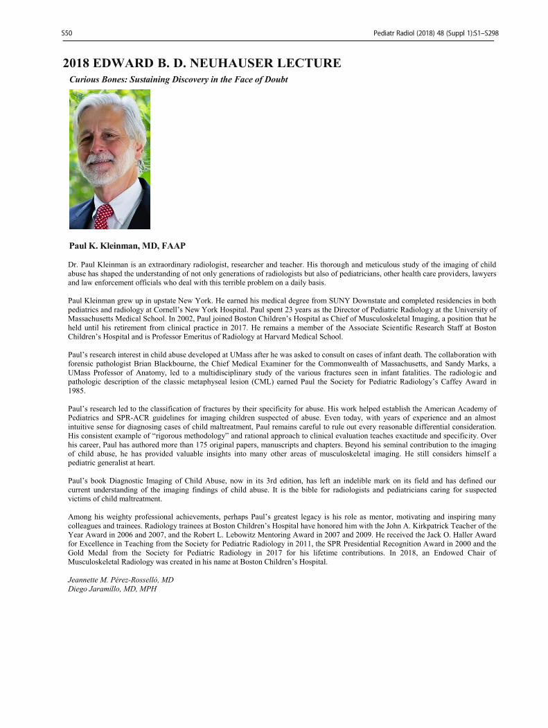
2018 EDWARD B. D. NEUHAUSER LECTURE
Curious Bones: Sustaining Discovery in the Face of Doubt
Paul K. Kleinman, MD, FAAP
Dr. Paul Kleinman is an extraordinary radiologist, researcher and teacher. His thorough and meticulous study of the imaging of child
abuse has shaped the understanding of not only generations of radiologists but also of pediatricians, other health care providers, lawyers
and law enforcement officials who deal with this terrible problem on a daily basis.
Paul Kleinman grew up in upstate New York. He earned his medical degree from SUNY Downstate and completed residencies in both
pediatrics and radiology at Cornell’s New York Hospital. Paul spent 23 years as the Director of Pediatric Radiology at the University of
Massachusetts Medical School. In 2002, Paul joined Boston Children’s Hospital as Chief of Musculoskeletal Imaging, a position that he
held until his retirement from clinical practice in 2017. He remains a member of the Associate Scientific Research Staff at Boston
Children’s Hospital and is Professor Emeritus of Radiology at Harvard Medical School.
Paul’s research interest in child abuse developed at UMass after he was asked to consult on cases of infant death. The collaboration with
forensic pathologist Brian Blackbourne, the Chief Medical Examiner for the Commonwealth of Massachusetts, and Sandy Marks, a
UMass Professor of Anatomy, led to a multidisciplinary study of the various fractures seen in infant fatalities. The radiologic and
pathologic description of the classic metaphyseal lesion (CML) earned Paul the Society for Pediatric Radiology’s Caffey Award in
1985.
Paul’s research led to the classification of fractures by their specificity for abuse. His work helped establish the American Academy of
Pediatrics and SPR-ACR guidelines for imaging children suspected of abuse. Even today, with years of experience and an almost
intuitive sense for diagnosing cases of child maltreatment, Paul remains careful to rule out every reasonable differential consideration.
His consistent example of “rigorous methodology” and rational approach to clinical evaluation teaches exactitude and specificity. Over
his career, Paul has authored more than 175 original papers, manuscripts and chapters. Beyond his seminal contribution to the imaging
of child abuse, he has provided valuable insights into many other areas of musculoskeletal imaging. He still considers himself a
pediatric generalist at heart.
Paul’s book Diagnostic Imaging of Child Abuse, now in its 3rd edition, has left an indelible mark on its field and has defined our
current understanding of the imaging findings of child abuse. It is the bible for radiologists and pediatricians caring for suspected
victims of child maltreatment.
Among his weighty professional achievements, perhaps Paul’s greatest legacy is his role as mentor, motivating and inspiring many
colleagues and trainees. Radiology trainees at Boston Children’s Hospital have honored him with the John A. Kirkpatrick Teacher of the
Year Award in 2006 and 2007, and the Robert L. Lebowitz Mentoring Award in 2007 and 2009. He received the Jack O. Haller Award
for Excellence in Teaching from the Society for Pediatric Radiology in 2011, the SPR Presidential Recognition Award in 2000 and the
Gold Medal from the Society for Pediatric Radiology in 2017 for his lifetime contributions. In 2018, an Endowed Chair of
Musculoskeletal Radiology was created in his name at Boston Children’s Hospital.
Jeannette M. Pérez-Rosselló, MD
Diego Jaramillo, MD, MPH
Pediatr Radiol (2018) 48 (Suppl 1):S1–S298S50
CO
LO
R K
EY
C
OL
OR
KE
Y
2018 SOCIAL EVENTS
SPR RESEARCH AND EDUCATION FOUNDATION FUN RUN
Underwritten by Texas Children’s Hospital
Wednesday, May 16, 2018
6:00 a.m.
Join us for a three-mile run through Downtown Nashville and get your day off to a great start!
Runners and walkers are all welcome. Entrance fee is $25 and includes a Tshirt. Runners/
walkers should meet in the Hotel lobby at 5:45 a.m.
WELCOME RECEPTION
Wednesday, May 16, 2018
7:00-8:15 p.m.
Omni Nashville Hotel
Hors d’oeuvres and Refreshments
Business Casual Attire
RECEPTION AND ANNUAL BANQUET
Friday, May 18, 2018
7:00-10:30 p.m.
Omni Nashville Hotel
Reception, Dinner and Dancing
Business Casual Attire
ACTIVITIES
The Omni Nashville Hotel employs concierge staff who are happy to share their detailed
knowledge of Nashville, Tennessee and the surrounding area. You may contact them by telephone
at (615) 782-5300.
Additional information is included on the SPR website.
Pediatr Radiol (2018) 48 (Suppl 1):S1–S298 S51

CO
LO
R K
EY
2018 61st SPR Annual Meeting & Postgraduate Course
“Value-added Pediatric Radiology”
TUESDAY – MAY 15, 2018
7:00 AM 8:00 AM Continental Breakfast
7:00 AM 5:30 PM Registration
8:00 AM 5:00 PM SPR Online Cases of the Day Activity
8:00 AM 8:20 AM General Postgraduate Course Welcome
8:00 AM 8:20 AM Body Imaging Postgraduate Course Welcome
8:20 AM 10:00 AM General Postgraduate Course: Neuroradiology (SAM Session)
Janet R. Reid, MD, FRCPC, Moderator 8:20-8:45 a.m. Newborn Hypoxic Ischemic Injury
Hisham M. Dahmoush, MBBCh, FRCR
Alexis B. R. Maddocks, MD
8:45-9:05 a.m. Imaging of Sinusitis/Mastoiditis and Complications
Jill V. Hunter, MBBS
Sumit Pruthi, MD
9:05-9:35 a.m. Imaging of Spinal Trauma
Karuna V. Shekdar, MD
Judith Gadde, DO
9:35-10:00 a.m. TORCH and Zika CNS Infections
Dorothy I. Bulas, MD, FACR, FAAP
Jason N. Wright, MD
8:20 AM 10:00 AM Body Imaging Postgraduate Course: Liver Imaging (SAM Session)
Jonathan R. Dillman, MD, MSc, Moderator 8:20-8:45 a.m. Liver Masses – PRETEXT Staging
Alexander J. Towbin, MD
Judy H. Squires, MD
8:45-9:05 a.m. Budd-Chiari/Veno-Occlusive Disease
M. Beth McCarville, MD
Martha M. Munden, MD
9:05-9:35 a.m. Iron and Fat Deposition
Shreyas S. Vasanawala, MD, PhD
Robert C. Orth, MD, PhD
9:35-10:00 a.m. Transplant Complications – US/Doppler (Early and Late)
Rama S. Ayyala, MD
Michael R. Acord, MD
10:00 AM 10:20 AM Break
Pediatr Radiol (2018) 48 (Suppl 1):S1–S298S52

TUESDAY – MAY 15, 2018 (Continued)
10:20 AM 12:00 PM General Postgraduate Course: Musculoskeletal (SAM Session)
Jonathan R. Dillman, MD, MSc, Moderator 10:20-10:45 a.m. Adolescent Hip Imaging
Sarah D. Bixby, MD
Nancy A. Chauvin, MD
10:45-11:10 a.m. Imaging of Knee Cartilage
Matthew R. Hammer, MD
Jerry R. Dwek, MD
11:10-11:35 a.m. Ultrasound of Painful Joints
Andrew M. Zbojniewicz, MD
Mahesh M. Thapa, MD
11:35-12:00 p.m. Throwing Adolescent Athlete
Arthur B. Meyers, MD
Victor M. Ho-Fung, MD
10:20 AM 12:00 PM Body Imaging Postgraduate Course: Pancreatobiliary (SAM Session)
Janet R. Reid, MD, FRCPC, Moderator 10:20-10:45 a.m. Neonatal Cholestasis
Sara M. O’Hara MD
Janet R. Reid, MD, FRCPC
10:45-11:10 a.m. Acute Recurrent and Chronic Pancreatitis
Sudha A. Anupindi, MD
Andrew T. Trout, MD
11:10-11:35 a.m. Pancreatic Trauma
Michael Aquino, MD
Michael J. Callahan, MD
11:35-12:00 p.m. Evaluation of Pediatric CBD Dilation
Sudha A. Anupindi, MD
Ruth Lim, MD
12:00 PM 1:30 PM Lunch On Your Own
12:00 PM 1:30 PM jSPR Luncheon (pre-registration required)
1:30 PM 3:10 PM General Postgraduate Course: Fetal (SAM Session)
Janet R. Reid, MD, FRCPC, Moderator 1:30-1:55 p.m. Myelomeningocele Imaging
Deborah Zarnow, MD
Davis M. Mirsky, MD
1:55-2:20 p.m. Fetal Neck Masses
Beth M. Kline-Fath, MD
Christopher I. Cassady, MD, FAAP
2:20-2:45 p.m. Congenital Diaphragmatic Hernia Update
Teresa Victoria, MD, PhD
Erika Rubesova, MD
2:45-3:10 p.m. Placental Imaging
Brandon P. Brown, MD
Maria F. Ladino-Torres, MD
CO
LO
R K
EY
Pediatr Radiol (2018) 48 (Suppl 1):S1–S298 S53

TUESDAY – MAY 15, 2018 (Continued) 1:30 PM 3:10 PM Body Imaging Postgraduate Course: Vascular (SAM Session)
Jonathan R. Dillman, MD, MSc, Moderator 1:30-1:55 p.m. Vasculitis, including Vessel Wall Imaging
Geetika Khanna, MD, MS
Eric J. Crotty, MD
1:55-2:20 p.m. Imaging of Vasculopathy/Aortopathy
Prakash M. Masand, MD
Eric J. Crotty, MD
2:20-2:45 p.m. Venography in the Post-Ablavar Era
Cynthia K. Rigsby, MD, FACR
Arnold Carl Merrow, MD
2:45-3:10 p.m. Lymphatic MR Imaging
David M. Biko, MD
Rajesh Krishnamurthy, MD
3:10 PM 3:30 PM Break
3:30 PM 5:10 PM General Postgraduate Course: Patient Safety & Technique (SAM Session)
Jonathan R. Dillman, MD, MSc, Moderator 3:30-3:45 p.m. Post-Contrast Acute Kidney Disorder
Matthew S. Davenport, MD
3:45-4:00 p.m. Gadolinium Update
Daniel J. Podberesky, MD
4:00-4:15 p.m. Sedation/Anesthesia Update
Joshua Nickerson, MD
4:15-4:30 p.m. What kVp Should I Use?
Samuel L. Brady, MS, PhD
4:30-4:45 p.m. Contrast Reaction Management
Jonathan R. Dillman, MD, MSc
4:45-5:00 p.m. MRI Device Safety Update
Suraj D. Serai, PhD
5:00-5:10 p.m. Q&A
3:30 PM 5:10 PM Body Imaging Postgraduate Course: Gastrointestinal Radiology (SAM Session)
Janet R. Reid, MD, FRCPC, Moderator 3:30-3:55 p.m. Perineal/Perianal Crohn’s Disease
Govind B. Chavhan, MD Ethan A. Smith, MD
3:55-4:20 p.m. Crohn’s Disease Standardized Nomenclature/Reporting
Kassa Darge, MD, PhD
Michael S. Gee, MD, PhD
4:20-4:45 p.m. Median Arcuate Ligament Syndrome
James S. Donaldson, MD, FACR
Seng H. Ong, MBBS
4:45-5:10 p.m. Small Bowel Tumors Jesse Courtier, MD
Ellen M. Chung, MD
5:10 PM
Adjourn
Pediatr Radiol (2018) 48 (Suppl 1):S1–S298S54

9
WEDNESDAY – MAY 16, 2018
6:00 AM
7:00 AM
7:00 AM
8:00 AM
5:45 PM
SPR REF Fun Run
Continental Breakfast & Exhibits
Registration
8:00 AM 5:00 PM SPR Online Cases of the Day Activity
8:00 AM 9:40 AM General Postgraduate Course: Radiation Dose Monitoring –
Watching Ourselves as We Watch Our Kids Part I
Donald P. Frush, MD, FACR, Moderator 8:00-8:15 a.m. Into Oz: Follow the Yellow Brick Rad
Donald P. Frush, MD, FACR
8:15-8:55 a.m. ABCs of Dose Metrics for Dose Monitoring
Samuel L. Brady, MD, PhD
Keith J. Strauss, MSc, FACR
8:55-9:25 a.m. A Walk Through the Market
Sarah McKenney, PhD, DABR
9:25-9:40 a.m. Dose Metrics in the Patient Report: Myopic or Foresighted?
Beverley Newman, MD, FACR
8:00 AM 9:40 AM Body Imaging Postgraduate Course: Chest (SAM Session)
Jonathan R. Dillman, MD, MSc, Moderator 8:00-8:25 a.m. Congenital Lung Lesions
Andrew Schapiro, MD
Maryam Ghadimi Mahani, MD
8:25-8:50 a.m. Dynamic Airway Imaging (Non-Sleep Apnea)
Edward Y. Lee, MD, MPH
Robert J. Fleck, MD
8:50-9:15 a.m. Ultrasound of the Lung and Pleura
Harriet J. Paltiel, MD
Monica Epelman, MD
9:15-9:40 a.m. Childhood Interstitial Lung Disease
R. Paul Guillerman, MD David A. Mong, MD
9:40 AM 10:10 AM Break & Exhibits
10:10 AM 11:50 AM General Postgraduate Course: Radiation Dose Monitoring –
Watching Ourselves as We Watch Our Kids Part II
Donald P. Frush, MD, FACR, Moderator 10:10-10:50 a.m. Navigating Dose Monitoring: Panel Discussion
Panelists: Donald P. Frush, MD, FACR
R. Paul Guillermann, MD
Ehsan Samei, PhD, DABR, FAAPM, FSPIE
Keith J. Strauss, MSc, FACR
Beverley Newman, MD, FACR
Samuel L. Brady, MS, PhD
Sarah McKenney, PhD, DABR 10:50-11:20 a.m. Into the Mist? Future Opportunities and Challenges
Paul Guillerman, MD
Ehsan Samei, PhD, DABR, FAAPM, FSPIE
11:20-11:50 a.m. Concluding Statements and Audience Q&A
CO
LO
R K
EY
Pediatr Radiol (2018) 48 (Suppl 1):S1–S298 S55

11 9
WEDNESDAY – MAY 16, 2018 (Continued) 10:10 AM 11:50 AM
10:10-10:35 a.m.
Body Imaging Postgraduate Course: Neonatal (SAM Session)
Jonathan R. Dillman, MD, MSc and Janet R. Reid, MD, FRCP, Moderators High Intestinal Obstruction
Boaz Karmazyn, MD
David A. Bloom, MD, FACR
10:35-11:00 a.m. Newborn Low Intestinal Obstruction
Laura Z. Fenton, MD
R. Pinar Karakas, MD
11:00-11:25 a.m. Newborn Lines and Tubes – Normal and Abnormal
Pallavi Sagar, MD
David Saul, MD
11:25-11:50 a.m. Anorectal Malformations
Steven J. Kraus, MD, MS, FAAP D. Gregory Bates, MD
11:50 AM
11:50 AM
1:20 PM
1:20 PM
Lunch on Your Own
3D Read with the Experts (Non-CME Session) Dianna M. E. Bardo, MD, FSCCT, FNASCI and Mark R. Ferguson, MD, Moderators Speaker: Sherwin S. Chan, MD Experts: Eric Hoggard, MD, Luis Goncalves, MD and Prakash M. Masand, MD
11:50 AM 1:20 PM MR Protocol Session (Non-CME Session) Shreyas S. Vasanawala, MD, PhD, Michael S. Gee, MD, PhD and
Taylor Chung, MD, Moderators
Topic 1: Update on last year’s topics
Topic 2: Coils – Scanner vendor options, 3rd party options and beyond
Topic 3: Acoustic Noise – Options and Tradeoffs
ANNUAL MEETING
1:20 PM 1:30 PM Annual Meeting Welcome & Dedication
Peter J. Strouse, MD, FACR
1:30 PM 2:00 PM The Power of Diversity: Meeting Today’s (and Tomorrow’s)
Greatest Healthcare Challenges
Stephanie E. Spottswood, MD, MSPH
2:05 PM 3:05 PM Edward B. Neuhauser Lecture Curious Bones: Sustaining Discovery in the Face of Doubt
Paul K. Kleinman, MD, FAAP
3:15 PM 4:15 PM Pediatric Radiology Jeopardy
George S. Bisset, III, MD, FACR, Host
Team E: Josée Dubois, MD
Tal Laor, MD Cynthia K. Rigsby, MD, FACR Nancy K. Rollins, MD
Team T:
Ashok Panigrahy, MD Daniel J. Podberesky, MD Stephen F. Simoneaux, MD Andrew T. Trout, MD
4:15 PM 4:45 PM Break & Exhibits
CO
LO
R K
EY
CO
LO
R K
EY
Pediatr Radiol (2018) 48 (Suppl 1):S1–S298S56

12 11 9
WEDNESDAY – MAY 16, 2018 (Continued)
4:45 PM 5:45 PM Scientific Session I-A: Gastrointestinal Radiology
Jonathan R. Dillman, MD, MSc and Mary-Louise Greer, MBBS, Moderators
P001 4:45 4:55 Rees, Mitchell Inter-radiologist Agreement Using Society of Abdominal Radiology-Society for Pediatric Radiology-American Gastroenterological Association Consensus Nomenclature for Reporting CT and MR Enterography in Pediatric Small Bowel Crohn Disease
P002 4:55 5:05 Karpewycz, John Accuracy of Magnetic Resonance Enterography in the Assessment of Pediatric Inflammatory Bowel Disease of the Colon in Patients Undergoing Colectomy
P003 5:05 5:15 Gottumukkala, Ravi Comparison of Three Oral Contrast Preparations for Magnetic Resonance Enterography in Pediatric Crohn’s Disease Patients: A Randomized Trial
P004 5:15 5:25 Dillman, Jonathan Breeza vs. VoLumen for Pediatric CT and MR Enterography: a Prospective, Randomized, Blinded Patient Acceptability Study
P005 5:25 5:35 Choi, Jungwhan Use of Small Bowel Ultrasound to Predict Response to Infliximab Induction
P006 5:35 5:45 Gokli, Ami Contrast Enhanced Ultrasound of the Bowel in Children with Suspected Inflammatory Bowel Disease (IBD).
4:45 PM 5:45 PM Scientific Session I-B: Musculoskeletal Imaging Lauren W. Averill, MD and Sarah D. Bixby, MD, Moderators
P007 4:45 4:55 Panwar, Jyoti Inter-reader Reliability Study of a New MRI Scoring System for TMJ Evaluation in JIA with the Use of Measurement Aids: Special Interest Group from the MRI in JIA in OMERACT
P008 4:55 5:05 Ditzler, Matthew Modified Friedman Technique: A New Proposed Method of Measuring Glenoid Version in the Setting of Glenohumeral Dysplasia
P009 5:05 5:15 Milks, Kathryn Advancements in Pediatric Wrist and Thumb Imaging: Sonography in Normal Infants
P010 5:15 5:25 Nicholas, Jennifer Lulun II: Ultrasound Evaluation of Bone Age in Rural Ecuadorian Children
P011 5:25 5:35 Sánchez, Mariana Digital X-ray Metacarpal Radiogrammetry and Dual X-ray Absorptiometry in Mexican Children and Adolescents.
P012 5:35 5:45 Amarneh, Mohammad
Fibro-Adipose Vascular Anomaly (FAVA): A review of imaging features.
4:45 PM 5:45 PM Scientific Session I-C: Interventional Radiology
John M. Racadio, MD and Leah E. Braswell, MD
P013 4:45 4:55 Robinson, Amie The Utility of Percutaneous Image Guided Tumor Ablation in the Treatment of Solid and Vascular Tumors in the Pediatric Population
P014 4:55 5:05 Roebuck, Derek Cryoablation of benign soft tissue lesions in children: initial experience
P015 5:05 5:15 Srinivasan, Abhay
A combined retrospective and prospective single-institution review of image-guided needle biopsy of relapsed or refractory neuroblastoma with a focus on the use of pre-biopsy MIBG imaging to predict tumor characteristics
P016 5:15 5:25 Srinivasan, Abhay
Comparison of MR guided joint injection with a fluoroscopic approach for pediatric MR arthrography
P017 5:25 5:35 Acord, Michael Surveillance Cerebral Angiography in Children with Arteriovenous Malformations: How many vessels should be interrogated?
P018 5:35 5:45 Bhavane, Rohan
A novel approach to deep tissue intra-operative imaging using indocyanine green fluorescence
5:45 PM Adjourn
5:50 PM 7:00 PM Awards Ceremony
7:00 PM 8:15 PM Welcome Reception
CO
LO
R K
EY
C
OL
OR
KE
Y
CO
LO
R K
EY
Pediatr Radiol (2018) 48 (Suppl 1):S1–S298 S57

THURSDAY, MAY 17, 2018
6:30 AM 8:30 AM Continental Breakfast & Exhibits
6:30 AM 5:30 PM Registration
7:00 AM 5:30 PM SPR Online Case of the Day Activity
7:00 AM 8:20 AM Sunrise Workshop – Evidence for Child Abuse Fractures
Jeannette M. Perez-Rossello, MD, Moderator 7:00-7:20 a.m. Evidence Base for Fractures in Child Abuse
Megan B. Marine, MD
7:20-7:40 a.m. Differential Diagnosis for Findings on Skeletal Survey vs. Child Abuse
Jeannie K. Kwon, MD
7:40-7:50 a.m. Entities Provided in Defense Cases (Rickets & Ehlers Danlos)
Daniel M. Schwartz, MD
7:50-8:20 a.m. Using the Literature to Assess What is Child Abuse or Not
Jeannette M. Perez-Rossello, MD
7:00 AM 8:20 AM Sunrise Workshop – Cardiac CT
Prakash M. Masand, MD and Lorna P. Browne, MBBS, Moderators 7:00-7:02 a.m. Introduction & Overview
7:02-7:15 a.m. Cardiac CT Techniques in Neonates
Siddarth P. Jadhav, MD
7:15-7:28 a.m. Common Indications for Cardiac CT in Neonates and Infants
Ladonna J. Malone, MD
7:28-7:41 a.m. Coronary CT in Neonates and Infants: How I Do It?
Prakash M. Masand, MD
7:41-7:54 a.m. Interpreting Heterotaxy in Neonates and Infants
Andrada R. Popescu, MD
7:54-8:07 a.m. Vascular Rings and Slings in Infants
Mark R. Ferguson, MD
8:07-8:20 a.m. Craziest Cases
Lorna P. Browne, MBBS
7:00 AM 8:20 AM Sunrise Workshop – Evolving Practice Models: Future/Present
Richard E. Heller, III, MD, MBA, Moderator 7:00-7:20 a.m. Changing Reimbursement Systems in (Pediatric) Radiology:
Value-Based Payment Reform
Richard E. Heller, III, MD, MBA
7:20-7:40 a.m. Health Care Now…Results of the 2017 Annual Trust Survey
Brandon Edwards, CEO, ReviveHealth
7:40-8:00 a.m. The Children's Hospital as Part of a Large Private Practice
Peter G. Kruk, MD
8:00-8:20 a.m. The Pediatric Radiologist in the General Private Practice
Kimberly A. Garver, MD
Pediatr Radiol (2018) 48 (Suppl 1):S1–S298S58
THURSDAY, MAY 17, 2018 (continued)
TUESDAY 7:00 AM 8:20 AM Sunrise Workshop – MR Techniques for Imaging Children
Michael S. Gee, MD, PhD and Jesse Courtier, MD, Moderators
7:00-7:15 a.m. Non-scanner Related Techniques for MR Imaging of Awake Children
Michael S. Gee, MD, PhD
7:15-7:30 a.m. Essential Neuroradiology Sequences for Evaluating Children
Mai-Lan Ho, MD
7:30-7:45 a.m. Essential Body Sequences for Evaluation of Children
Jesse Courtier, MD
7:45-8:00 a.m. Considerations for Pediatric MSK Imaging
Delma Y. Jarrett, MD
8:00-8:10 a.m. Cardiac and Vascular MR Imaging: Tips and Tricks
Sherwin S. Chan, MD
8:10-8:20 a.m. MR Protocols in the Pediatric ER
Unmi K. Udayasankar, MD
7:00 AM 8:20 AM Sunrise Workshop – Update on Contrast Media:
New Agents, New Concerns and Strategies for Safe Use
R. Paul Guillerman, MD, Moderator 7:00-7:20 a.m. Strategies for Optimizing Enteric Contrast for Fluoroscopy, CT and MRI
Michael J. Callahan, MD
7:20-7:40 a.m. Approach to Imaging Patients with Renal Impairment or
Heighted Risk of Adverse Contrast Reaction
Matthew S. Davenport, MD
7:40-8:00 a.m. Safety Considerations for Ultrasound Contrast Agents
Kassa Darge, MD, PhD
8:00-8:20 a.m. Gadolinium-Based Contrast Agents: Time to Image Gently?
R. Paul Guillerman, MD
7:00 AM 8:20 AM Sunrise Workshop – Pediatric Nuclear Medicine: Learn to Read Like an Expert
Marguerite T. Parisi, MD, MS, Moderator 7:00-7:15 a.m. CNS Imaging in Nuclear Medicine
Adina L. Alazraki, MD, FAAP
7:15-7:30 a.m. Australian Perspectives on Nuclear Renography
Monica A. Rossleigh, MD
7:30-7:45 a.m. Musculoskeletal Scintigraphy
Susan E. Sharp, MD
7:45-8:00 a.m. Gastrointestinal Scintigraphy
Helen R. Nadel, MD, FRCPC
8:00-8:20 a.m. Challenging Cases in NM Oncologic Imaging
Marguerite T. Parisi, MD, MS
Pediatr Radiol (2018) 48 (Suppl 1):S1–S298 S59

THURSDAY, MAY 17, 2018 (continued)8:30 AM 10:10 AM Scientific Session II-A: Neuroradiology
8:30-8:50 a.m. Maura E. Ryan, MD and Ashok Panigrahy, MD, Moderators Keynote Presentation Challenges in Pediatric Obstructive Sleep Apnea Amy Whigham, MD
P019 8:50 9:00 Altinok, Deniz Analysis of diffusion restriction in the corpus callosum in children with abusive head trauma
P020 9:00 9:10 Mata-Mbemba, Daddy
Pediatric pituitary adenoma: imaging features and clinical outcome.
P021 9:10 9:20 Sabin, Noah Potential Imaging and Neurosurgical Correlates of Molecular Subgroups of Posterior Fossa Ependymoma in Children
P022 9:20 9:30 Harreld, Julie Analyzing Misdiagnosed Metastasis on Imaging Studies
P023 9:30 9:40 Alves Rosa, Joao Normal head and neck lymph nodes in the healthy paediatric population
P024 9:40 9:50 Eid, Hadeel Magnetic resonance (MR) assessment of optic nerve thickness and tortuosity for recognition of optic glioma in children with NF1
P025 9:50 10:00 Hsu, Ariel The Unwound Cochlea: a Specific Marker of Branchio-oto-renal Syndrome
P026 10:00 10:10 Whitehead, Matthew
Pseudo-color Perceptual Enhancement for Detection of Cochlear Signal Alterations in Pediatric Patients with Sensorineural Hearing Loss
8:30 AM 10:10 AM Scientific Session II-B: Musculoskeletal Imaging Jeannette M. Perez-Rossello, MD and Aparna Joshi, MD, Moderators
8:30-8:50 a.m. Keynote Presentation Deborah Lowen, MD
P027 8:50 9:00 Pomeranz, Christy
Accuracy of Skeletal Survey in Evaluation of Rib Fractures Compared to CT in the Setting of Accidental and Non Accidental Trauma.
P028 9:00 9:10 Tsai, Andy How Often is Subperiosteal New Bone Formation Noted with the Distal Tibial Classic Metaphyseal Lesion?
P029 9:10 9:20 Kriss, Spencer Radiographic characteristics that delineate abusive from accidental skull fractures, including the significance of fracture extension to sutures.
P030 9:20 9:30 Starosolski, Zbigniew
Gaussian noise simulating reduced X-ray dose does not affect sensitivity of a Neural Network trained to detect tibial fractures in X-ray images
P031 9:30 9:40 Starosolski, Zbigniew
Toddler tibial fractures computer-aided diagnosis by convolutional neural network and transfer learning
P032 9:40 9:50 Alsharief, Alaa Can combined diffusion-weighted imaging/conventional MRI replace post gadolinium-based contrast-enhanced MRI in the assessment of pediatric osseous sarcomas?
P033 9:50 10:00 Ecklund, Kirsten Bone Marrow MR and MRS Evidence of Adrenal and Gonadal Hormone Replacement Therapy Efficacy in girls with Anorexia Nervosa
P034 10:00 10:10 Eutsler, Eric Metal artifact reduction in pediatric dual energy CT using monoenergetic extrapolation
8:30 AM 10:10 AM
8:30-8:50 a.m.
Scientific Session II-C: Thoracic Imaging David M. Biko, MD and Beverly Newman, MD, Moderators Keynote Presentation Lisa Young, MD
P035 8:50 9:00 Ma, Grace Comparison of Prenatal Ultrasound, Prenatal MRI, and Postnatal CTA for Evaluation of Congenital Lung Malformations: Can Postnatal CTA Provide Additional Information?
P036 9:00 9:10 Biko, David Magnetic Resonance Imaging of Central Lymphatics in Noonan’s Syndrome
P037 9:10 9:20 Handly, Brian Initial Clinical Evaluation of Stationary Digital Chest Tomosynthesis in Patients with Cystic Fibrosis
P038 9:20 9:30 Fenlon, Edward Trending Use and Results of Computed Tomographic Angiography for Diagnosing Pulmonary Embolism in the Pediatric Emergency Department: A 13-Year Retrospective Study.
P039 9:30 9:40 Krishnamurthy, Rajesh
Target mode prospective EKG gated volumetric CT reduces the need for sedation for chest CT in young children
Pediatr Radiol (2018) 48 (Suppl 1):S1–S298S60

14
P040 9:40 9:50 Averill, Lauren Tracheal Narrowing in Children and Young Adults with Mucopolysaccharidosis IVA: Evaluation with CT Angiography
P041 9:50 10:00 May, Lauren A Novel Approach Using Volumetric Dynamic Airway CT to Determine Optimal Positive End Expiratory Pressure (PEEP) Settings in Neonates Requiring Long-Term Ventilator Support
P042 10:00 10:10 Rozovsky, Katya Imaging patterns in pediatric pulmonary blastomycosis
8:30 AM 10:10 AM Technologist Opening Session
Laura Gruber, MBA, RT(R), RDMS, RVT, Moderator 8:30-9:00 a.m. Ice Breaker
Laura Gruber, MBA, RT(R), RDMS, RVT
9:00-9:30 a.m. Recruiting Talent – Career Models
Merima Karastanovic, MS, RT(R)(MR)
9:30-10:10 a.m. Generational Differences in the Workplace
Melissa Goehner, BA RT(R)(CT)
10:10 AM 10:40 AM Break & Exhibits
10:40 AM 12:00 PM Midday Workshop – Evidence Base for Abusive Head Trauma
Arabinda K. Choudhary, MBBS, FACHE, MBA, Moderator 10:40-11:00 a.m. Evidence Base for Abusive Head Trauma
Laura L. Hayes, MD
11:00-11:20 a.m. Evidence Base for Spine Trauma
Arabinda K. Choudhary, MBBS, FACHE, MBA
11:20-11:40 a.m. Abusive Head Trauma: Diagnostic Challenges
Gary L. Hedlund, DO
11:40-12:00 p.m. Addressing Issues
Dawn E. Saunders, MD
10:40 AM 12:00 PM Midday Workshop – IR Lite – Unleaded and Photon Free!
Eric J. Monroe, MD, Moderator 10:40-11:00 a.m. Mobile Strike: Lean Lessons Learned from a Sprawling Service
Eric J. Monroe, MD
11:00-11:15 a.m. Ultrasound-guided Tunneled Line Placement
C. Matthew Hawkins, MD
11:15-11:30 a.m. Ultrasound-guided Lumbar Punctures
Ann Gill, MD
11:30-11:45 a.m. Ultrasound-guided Drainage Procedures
Sheryl A. Tulin-Silver, MD
11:45-12:00 p.m. Ultrasound-guided MSK Procedures: Beyond the Big Joints
Jeffrey P. Otjen, MD
10:40 AM 12:00 PM Midday Workshop – Novel Use of Musculoskeletal Ultrasound
10:40-10:50 a.m.
10:50-11:00 a.m.
11:00-11:10 a.m.
11:10-11:20 a.m.
11:20-11:30 a.m.
Monica Epelman, MD and Mahesh M. Thapa, MD, Moderators
Introductions and Overview
Monica Epelman, MD
Role of Ultrasound in the Evaluation of Musculoskeletal Neoplasms
Diego Jaramillo, MD, MPH
Ultrasound Evaluation of Magnetic Growing Roots
Arthur B. Meyers, MD
Ultrasound of the Joints in Pediatric Sport Injuries
Nancy A. Chauvin, MD
Ultrasound Imaging of Vascular Anomalies: Pearls an Pitfalls
Oscar M. Navarro, MD
Ultrasound Imaging of Brachial Plexus Injuries
Ramesh S. Iyer, MD
CO
LO
R K
EY
Pediatr Radiol (2018) 48 (Suppl 1):S1–S298 S61

11:30-11:40 a.m. Stress Sonography of the Ulnar Collateral Ligament in Baseball Pitchers
Lauren W. Averill, MD
11:40-11:50 a.m. Ultrasound Imaging of Carpal Tunnel Syndrome
Mahesh M. Thapa, MD
11:50-12:00 p.m. Panel/Audience Discussion - Question & Answers
10:40 AM 12:00 PM Midday Workshop – Thoracic Imaging
Robert J. Fleck, MD, Moderator 10:40-10:55 a.m.
10:55-11:10 a.m.
11:10-11:25 a.m.
11:25-11:45 a.m.
11:45-12:00 p.m.
Diffuse Lung Disease in the Infant
Jason P. Weinman, MD
Strategies for Optimizing Thoracic CT and MRI in Children
Sarah Desoky, MD
Noninfectious Complications of Stem Cell Transplant
Matthew Cooper, MD
Innovated Imaging of the Pediatric Airway
Robert J. Fleck, MD
Interesting/Challenge Cases in Chest Imaging
Maryam Ghadimi Mahani, MD
10:40 AM 12:00 PM Midday Workshop – Professionalism
Brandon P. Brown, MD, MA, Moderator 10:40-11:00 a.m. The Value of Mentorship in Professionalism
Richard B. Gunderman MD, PhD
11:00-11:20 a.m. Communication and the Pediatric Radiologist
Sarah S. Milla, MD, FAAP
11:20-11:40 a.m. Imaging 3.0: Building a Culture of Professionalism
Brandon P. Brown, MD
11:40-12:00 p.m. Discussion questions and Q/A Panel
10:40 AM 12:00 PM Midday Workshop – World Federation of Pediatric Imaging (WFPI) –
Updates in Outreach
Dorothy I. Bulas, MD, FAAP and Cicero T. Silva, MD, Moderators 10:40-10:45 a.m. Introductions and Overview
10:45-11:00 a.m. World TB Day Summit Update
Bernard F. Laya, DO
11:00-11:15 a.m. Facilitating a Radiology Curriculum in Haiti Using Tablet PCs: Progress
and Challenges
Jennifer L. Nicholas, MD
11:15-11:30 a.m. POCUS Updates - A Key to Radiology Outreach
Kara-Lee Pool, MD
11:30-11:40 a.m. WFPI Project - Pediatric MRI Protocols for Low Resource Settings
Michael S. Gee, MD, PhD
11:40-11:55 a.m. Outreach Experiences - Issues to Consider
Panelists: Veronica J. Rooks, MD & Ramdas Senasi, MBBS 11:55-12:00 p.m. Questions and Answers
12:00 PM 1:30 PM Lunch On Your Own
Pediatr Radiol (2018) 48 (Suppl 1):S1–S298S62

15 16 14
THURSDAY, MAY 17, 2018 (continued)
12:00 PM 1:30 PM CT Protocol Session: State of the Art in CT: Let’s Cut to the Chase! (Non-CME Session) Michael J. Callahan, MD, and Prakash M. Masand, MD, Moderators
12:05-12:13 p.m. “Post-64” Era in CT Scanning with Emphasis on Faster Acquisition Techniques Robert MacDougall, MSc
12:13-12:21 p.m. Fast Pitch Pediatric Head CT: “Mission Impossible?” Christina Dodge, MS
12:21-12:29 p.m. Dynamic CT of the Pediatric Airway: “Take My Breath Away” Abbey Winant, MD
12:29-12:37 p.m. Pediatric CT Angiography: “Injection Perfection” Karen Lyons, MD
12:37-12:45 p.m. Pediatric CTA in the Acute Setting: “Practical Tips for Success” Tushar Chandra, MD
12:45-12:53 p.m. CT Imaging in Pediatric Oncology: Do I Sedate or Radiate? Kara G. Gill, MD
12:53-1:01 p.m. Dose Optimization for High Pitch Imaging: “Image Gently and Rapidly” Grace S. Phillips, MD
1:01-1:30 p.m. Questions and Answers with Panel Discussion
12:00 PM 1:30 PM SPR Research and Education Foundation Symposium (Non-CME Session) 12:00-12:10 p.m. Welcome and Introduction - Peter J. Strouse, MD, FACR and J. Damien Grattan-Smith, MBBS 12:10-12:20 p.m. 2016 Multi-Institute Pilot Award: Single Ventricle Reconstruction III: Brain Connectome
and Neurodevelopmental Outcomes - Part II
Ashok Panigrahy, MD 12:20-12:25 p.m. 2016 Seed Grant: Study for the Detection of Gadolinium Deposition in
Resected Bowel Specimens of Patients with Inflammatory Bowel Disease
Shannon G. Farmakis, MD 12:25-12:35 p.m. 2016 Education Project Grant: MOOC-based Repetitive Image Quizzes for
Learning Concepts in Pediatric Radiology Cara E. Morin, MD, PhD
12:35-12:45 p.m. 2016 Young Investigator: Accurate assessment of kidney function through Motionrobust DCEM
Sila Kurugol, PhD, MS 12:45-1:30 p.m. REF Updates and Announcement of the 2018 Grant Recipients
J. Damien Grattan-Smith, MBBS
1:30 PM 3:30 PM Scientific Session III-A: Genitourinary Radiology Harriet J. Paltiel, MD and Monica Epelman, MD, Moderators
1:30-1:50 p.m. Keynote Presentation
Stacy Tanaka, MD
P043 1:50 2:00 Stanescu, A. Luana
Gadolinium bone tissue retention in pediatric patients after contrast-enhanced MR exams: pathologic confirmation.
P044 2:00 2:10 Tao, Ting Safety and efficacy of gadoterate meglumine in children <2 years of age
P045 2:10 2:20 Zucker, Evan Free-Breathing Ferumoxytol-Enhanced MRI for Preoperative Renal Transplant Vascular Mapping in Infants and Children
P046 2:20 2:30 Kurugol, Sila Feed and Wrap MRU
P047 2:30 2:40 Otero, Hansel
Morphologic and Functional Evaluation of Duplicated Renal Collecting Systems with MR Urography: A Descriptive Analysis
P048 2:40 2:50 Acharya, Patricia
Evaluation of the Urinary Tract Dilation Classification System for postnatal hydronephrosis in the prediction of clinical outcomes
P049 2:50 3:00 Back, Susan Preliminary experience with Contrast Enhanced Ultrasound (CEUS) of the Pediatric Kidney
P050 3:00 3:10 Chow, Jeanne
Contrast enhanced ultrasound for the evaluation of complex anorectal and genitourinary anomalies
P051 3:10 3:20 Pai, Vinay Infant Female Urethral Catheterization: A New Simulator from Special Effects
P052 3:20 3:30 Dao, Kimberly
Pediatric Ovarian Volumes Measured at Ultrasound After Contralateral Unilateral Oophorectomy
CO
LO
R K
EY
C
OL
OR
KE
Y
CO
LO
R K
EY
Pediatr Radiol (2018) 48 (Suppl 1):S1–S298 S63

16 14
THURSDAY, MAY 17, 2018 (continued)
1:30 PM 3:30 PM Scientific Session III-B: Fetal/Neonatal Imaging
Brandon P. Brown, MD and Erika Rubesova, MD, Moderators 1:30-1:50 p.m. Keynote Presentation
Jill Kilkelly, MD
P053 1:50 2:00 Dong, Su-Zhen Feasibility of fetal MRI in the evaluation of fetal cardiovascular anomalies.
P054 2:00 2:10 Dong, Su-Zhen Fetal cardiac MRI in the evaluation of congenital aortic arch anomalies
P055 2:10 2:20 Oliver, Edward Two's Company – Multiple Thoracic Anomalies on Prenatal Ultrasound and MRI
P056 2:20 2:30 Wise, Rachel A Multifactorial Severity Score for Congenital Diaphragmatic Hernia (CDH) using Fetal MRI
P057 2:30 2:40 Smitthimedhin, Anilawan
Normal size of the fetal adrenal gland on prenatal MR Imaging
P058 2:40 2:50 Didier, Ryne Novel Computerized Analytic Technique for Quantification of Amniotic Fluid Voluin Fetal MRI
P059 2:50 3:00 Norris, Carrie Using Quantitative MRI to Identify Abnormal Perfusion in Invasive Placental Disease
P060 3:00 3:10 Tsai, Andy Congenital Skeletal Anomalies with Lower Limb-Length Discrepancy: Does Constant Inhibition Occur In Utero?
P061 3:10 3:20 Victoria, Teresa Ultra-Low Dose Fetal CTL How to Further Decrease Radiation Dose While MainDiagnostic Accuracy
P062 3:20 3:30 Deaver, Pamela Perinatal imaging autopsy: Value and comparison with conventional autopsy
1:30 PM 3:30 PM Scientific Session III-C: Informatics, QI, Education & Healthcare Policy
1:30-1:50 p.m. Richard E. Heller, III, MD, MBA and Ramesh S. Iyer, MD, Moderators Keynote Presentation - Technologist Skill: Moving from Built-in Mediocrity to Built-
in Excellence Using a Coaching Model
David B. Larson, MD, MBA
P063 1:50 2:00 Snyder, Elizabeth
Gaging Potential Risk to Patients in Pediatric Radiology by Review of Over 2,000 Incident Reports
P064 2:00 2:10 Hingsbergen, Elizabeth
Identifying the primary cause of safety events and complaints in the routine operations of a pediatric radiology department
P065 2:10 2:20 Porterfield, Ronna
Analysis of Quick Hits at a Daily Readiness Huddle at a Large Free-Standing Children's Hospital
P066 2:20 2:30 Iyer, Ramesh Survey of Peer Review Programs Amongst Pediatric Radiologists: Report from the SPR Quality and Safety Committee
P067 2:30 2:40 Gates, Erica Potential Risk of Hypothyroidism in Infants Receiving Iodinated Contrast: Are Pediatric Radiology Practitioners Aware of the Food and Drug Administration (FDA) Drug Safety Communication?
P068 2:40 2:50 Frush, Daniel Utilization of computed tomography imaging in the pediatric emergency department
P069 2:50 3:00 Hagedorn, Kelly
Cost comparison of ultrasound versus MRI for adolescent female patients with suspected appendicitis using time-driven activity-based costing
P070 3:00 3:10 Aquino, Michael
Survey of after-hours radiology coverage in children's hospitals.
P071 3:10 3:20 Ayyala, Rama
Evaluation of Stressors that Contribute to Burnout Amongst Pediatric Radiologists: A Survey of the Society for Pediatric Radiology
P072 3:20 3:30 Pfeifer, Cory Assessing the Pediatric Radiology Job Market: Winter is Coming for Employers
CO
LO
R K
EY
C
OL
OR
KE
Y
Pediatr Radiol (2018) 48 (Suppl 1):S1–S298S64

THURSDAY, MAY 17, 2018 (continued)
1:30 PM 3:30 PM Technologist Session
Lorraine Chisari, RDMS, RVT, Moderator 1:30-1:50 p.m. Common Pediatric Fluoroscopy Exams
Ciji N. Gilley, RT(R)
1:50-2:10 p.m. MRE (Enterography)
Gina Roberts, ARRT (R)(CT)(MRI)
2:10-2:30 p.m. Happy Spitter or Surgical Referral?: Sonographic Approach to the Vomiting Infant
Jason Hooper, BS, RDMS, RVT
2:30-2:50 p.m. US Contrast
Trudy Morgan, RDMS
2:50-3:10 p.m. Transplant US Imaging
Lacy Gandor, RDMS
3:10-3:30 p.m. 3D Post Processing and Printing
Gabe Linke, BSRT (R)(MR)
3:30 PM 4:00 PM Break & Exhibits
4:00 PM 5:30 PM Scientific Session IV-A: Gastrointestinal Radiology Govind B. Chavhan, MD and Hansel J. Otero, MD, Moderators
4:00-4:20 p.m. Keynote Presentation
Dedrick Moulton, MD
P073 4:20 4:30 Dillman, Jonathan
Diagnostic Performance of Magnetic Resonance Cholangiopancreatography (MRCP) versus Endoscopic Retrograde Cholangiopancreatography (ERCP) in the Pediatric Population: A Clinical Effectiveness Study
P074 4:30 4:40 Trout, Andrew Normal Pancreatic Secretion in Children Measured by MRI with Secretin P075 4:40 4:50 Dillman,
Jonathan Diagnostic performance of quantitative MRI parameters for predicting radiologic portal hypertension in autoimmune liver diseases
P076 4:50 5:00 Stefek, Heather
Relationship between 2D phase contrast MRI Rex shunt blood flow, Rex shunt diameter, and clinical indicators of portal hypertension
P077 5:00 5:10 Romberg, Erin Validation of LI-RADS in Evaluation of Pediatric Liver Masses
P078 5:10 5:20 Lipsich, Jose Congenital portosystemic shunts (CPSS): clinical outcome after treatment. Experience in a tertiary hospital in Argentina
P079 5:20 5:30 Back, Susan Preliminary experience with Contrast Enhanced Ultrasound (CEUS) of the Pediatric Liver
4:00 PM 5:30 PM Scientific Session IV-B: Cardiovascular Imaging Randolph K. Otto, MD and Prakash M. Masand, MD, Moderators
4:00-4:20 p.m. Keynote Presentation
Jason Christensen, MD
P080 4:20 4:30 Malone, LaDonna
Coronary artery assessment in pediatric patients using ultrafast ECG gated CT: Is this a viable alternative to prospectively gated technique?
P081 4:30 4:40 Barrera, Christian
Depiction of the Native Coronary Arteries during ECG-triggered high-pitch spiral Dual-Source Cardiac CTA in Children: Determinants of Image Quality
P082 4:40 4:50 Masand, Prakash
Myocardial bridging in a cohort of pediatric patients with anomalous aortic origin of coronary artery (AAOCA): “Double whammy”!
P083 4:50 5:00 Ortiz-Neira, Clara L
Coronary artery imaging in Kawasaki disease with Computed Tomography Angiography and Echocardiography, a comparative analysis
P084 5:00 5:10 Markush, Dor Delayed Myocardial Enhancement in Children: Comparison of Conventional Technique to a Free Breathing Single-Shot Technique in Duchenne Muscular Dystrophy
P085 5:10 5:20 Biko, David Association of T2 Lymphatic Imaging in Single Ventricle Patients After Superior Cavopulmonary Connection with Acute Post-Fontan Outcomes
P086 5:20 5:30 Davis, Joseph Aortic Disease in Patients with Mucopolysaccharidosis
CO
LO
R K
EY
Pediatr Radiol (2018) 48 (Suppl 1):S1–S298 S65
14
THURSDAY, MAY 17, 2018 (Continued)/ FRIDAY, MAY 18, 2018
4:00 PM 5:30 PM
4:00-4:20 p.m.
Scientific Session IV-C: ALARA
Steven Don, MD and Robert MacDougall, MSc, Moderators Keynote Presentation
Marta Hernanz-Schulman, MD, FACR
P087 4:20 4:30 Sharma, Priya Assessing Pediatric Fluoroscopy Radiation Dose Awareness amongst Clinicians
P088 4:30 4:40 Kaplan, Summer Reduced-Dose Radiography Protocol for Focused Assessment of Feeding Tube Position
P089 4:40 4:50 Don, Steven Stochastic Noise Tolerance in Pediatric Appendicitis CT
P090 4:50 5:00 Southard, Richard Knowledge-Based Iterative Reconstruction verses Filtered-Back Reconstruction Head CT in Children Referred from the Emergency Department: Dose Reduction, Image Quality, and Image Reconstruction Time
P091 5:00 5:10 Ngo, Jennifer Development and Testing of an Innovative Simulation Paradigm for Subtle Liver Lesion Detection in Pediatric CT
P092 5:10 5:20 Ngo, Jennifer An Innovative Simulation Paradigm for Evaluation of Subtle Liver lesions in Pediatric CT: Performance and Confidence
P093 5:20 5:30 MacDougall, Robert
Development of a tool to aid the radiologic technologist using augmented reality and computer vision
4:00 PM 5:00 PM Technologist Family Feud
Christine Harris, RT (R) (MR), Moderator 4:00-5:00 p.m. Family Feud
Nikki D. Butler, BMSc, RT(R)(QM)
5:00 PM 5:30 PM Technologist Session: Global Health Panel
Christine Harris, RT (R) (MR)
Laura Poznick, AAS, RDMS
Lacy Gandor, RDMS
5:30 PM Adjourn
FRIDAY, MAY 18, 2018
6:30 AM 8:30 AM Continental Breakfast & Exhibits
6:30 AM 6:00 PM Registration
7:00 AM 5:20 PM SPR Online Case of the Day Activity
7:00 AM 8:20 AM Sunrise Workshop – Child Abuse Trial Perspectives
Joëlle A. Moreno, JD, Moderator 7:00-7:20 a.m. 10 Points for the Radiologist to Know About Trials
Joëlle A. Moreno, JD
7:20-7:40 a.m. Testimony: The Do's and Don’t’s
Jerry R. Dwek, MD
7:40-8:00 a.m. Putting It All Together
Superior Court Judge Kevin C. Doyle
8:00-8:20 a.m. Questions and Answers
CO
LO
R K
EY
C
OL
OR
KE
Y
CO
LO
R K
EY
Pediatr Radiol (2018) 48 (Suppl 1):S1–S298S66
FRIDAY, MAY 18, 2018 (continued)
7:00 AM 8:20 AM Sunrise Workshop – Advanced Body Imaging Jonathan R. Dillman, MD, MSc, Moderator
7:00-7:15 a.m. MR Elastography Update Jonathan R. Dillman, MD, MSc
7:15-7:30 a.m. Dixon MRI Applications Andrew T. Trout, MD
7:30-7:45 a.m. Free Breathing Body MRI Michael S. Gee, MD, PhD
7:45-8:00 a.m. Update on Dual-Energy CT in Children Sarah D. Bixby, MD
8:00-8:15 a.m. CT Iterative Reconstruction: Where Do We Stand? Samuel L. Brady, MS, PhD
8:15-8:20 a.m. Questions and Answers
7:00 AM 8:20 AM Sunrise Workshop – Pearls in Fetal Imaging Teresa Victoria, MD, PhD, Moderator
7:00-7:15 a.m. How to Approach Abnormal Placentation Mariana Meyers, MD
7:15-7:30 a.m. Motion Correction in Fetal MRI Camilo Jaimes, MD
7:30-7:45 a.m. Prenatal Evaluation of Lymphatic Disorders with Postnatal Correlation David M. Biko, MD
7:45-8:00 a.m. How to Start a Fetal Program Beth M. Kline-Fath, MD
8:00-8:15 a.m. How the Radiologist Adds Value to Patient Care with Emphasis in Fetal MRI Brandon P. Brown, MD
8:15-8:20 a.m. The Unusual Lung Lesions Teresa Victoria, MD, PhD
7:00 AM 8:20 AM
7:00-7:15 a.m.
7:15-7:30 a.m.
7:30-7:45 a.m.
7:45-8:00 a.m.
8:00-8:20 a.m.
Sunrise Workshop – Physics Robert MacDougall, MS, Moderator Pediatric Digital Radiography: From EI to IQ Robert MacDougall, MS Using Informatics Tools to Drive Quality Improvement Cristina Dodge, MS Contrast Ultrasound Primer Samuel L. Brady, MS, PhD Radiation Management and Dosimetric QA in Interventional Radiology Nicholas Shkumat, MScMRI Safety: Physics and Practice Jie Deng, PhD
7:00 AM 8:20 AM Sunrise Workshop – Diversity Ashok Panigrahy, MD and Stephanie E. Spottswood, MD, MSPH, Moderators
7:00-7:20 a.m. Diversity and Unconscious Bias in Healthcare Stephanie E. Spottswood, MD, MSPH
7:20-7:40 a.m. Supporting LGTBTQ Patients and Families Jesse M. Ehrenfeld, MD, MPH
7:40-8:00 a.m. "Disability - Its Name and Faces" Cindy R. Miller, MD
8:00-8:20 a.m. Diversity, Discrimination, and Mitigation Panel Discussion with: Johnathan Dallas, BS, Jesse M. Ehrenfeld, MD, MPH, Cindy R. Miller, MD and Stephanie E. Spottswood, MD, MSPH
Pediatr Radiol (2018) 48 (Suppl 1):S1–S298 S67

FRIDAY, MAY 18, 2018 (continued)8:30 AM 10:10 AM Technologist Session
David A. Bloom, MD, FACR, Moderator 8:30-8:50 a.m. Child Life Services in Pediatric Radiology
Sarah Beth Gray, CCLS
8:50-9:10 a.m. The Role of Nursing in Preparing Patients for Imaging
Leslie Mintz, BSN
9:10-9:30 a.m. Interactive VR on IV Sticks
Gabe Linke, BSRT (R)(MR) and Nicole Hardin, MS, RT, (R)(M)(CIIP) 9:30-9:50 a.m. PAWS (Patient Awake While Scanning) MRI Program
Sarah Beth Gray, CCLS
9:50-10:10 a.m. “Try Without” Program
Leslie Mintz, BSN
8:30 AM 10:10 AM Scientific Session V-A: Gastrointestinal Radiology Michael S. Gee, MD, PhD and Gary R. Schooler, MD, Moderators
8:30-8:50 a.m. Keynote Presentation
Lea Matsuoka, MD
P094 8:50 9:00 Dillman, Jonathan
Multiparametric Liver MRI in Children and Young Adults with Autoimmune Liver Disease: Correlation between Quantitative Imaging Methods
P095 9:00 9:10 Trout, Andrew Predictive Performance of MR Elastography in Children and Young Adults with Suspected Liver Disease
P096 9:10 9:20 Davis, James Assessment of pediatric liver allografts with shear wave elastography
P097 9:20 9:30 Goette, Matthew
Shear Wave Elastography Ultrasound for Hepatic Veno-occlusive Disease in a Pediatric Population undergoing Hematopoietic Stem Cell Transplantation
P098 9:30 9:40 Diaz, Eric Can MRI Liver Stiffness Predict be used to Predict Focal Liver Lesions in Fontan Patients?
P099 9:40 9:50 Thompson, Benjamin
Magnetic resonance elastography of the liver in children: variations in regional stiffness
P100 9:50 10:00 Robinson, Amie Methotrexate Therapy Increases Hepatic Stiffness in Patients with Inflammatory Bowel Disease
P101 10:00 10:10 Hunte, David Abdominal Fat Distribution and its Relationship with Liver and Pancreatic Fat Fraction, Liver Stiffness and Diabetic Status
8:30 AM 10:10 AM Scientific Session V-B: Fetal/Neonatal Imaging Beth M. Kline-Fath, MD and Marianna Meyers, MD, Moderators
8:30-8:50 a.m. Keynote Presentation - Fetal Diagnosis of Surgical Anomalies: Effect on Pre and Postnatal Therapy
John Pietsch, MD
P102 8:50 9:00 Didier, Ryne Contrast-Enhanced Brain Ultrasound on Extreme Premature Twin Fetal Lambs Maintained by the EXTra-uterine Environment for Neonatal Support (EXTEND): A Promising Imaging Technique
P103 9:00 9:10 Bulas, Dorothy
Fetal and Postnatal Brain Imaging for the Detection of ZIKV Encephalopathy in the Fetus/Newborn
P104 9:10 9:20 Snyder, Elizabeth
Fetal MRI Findings in Diencephalosynapsis
P105 9:20 9:30 Guimaraes, Carolina
Posterior Fossa Findings in Prenatal Diagnosis of Dystroglycanopathy – Extending Beyond Brainstem Kinking
P106 9:30 9:40 Zare, Megan Establishment of normative values for the fetal posterior fossa by MRI.
P107 9:40 9:50 Rubio, Eva Predictive Value of Prenatal Alveolar Cleft Size in Determining the Likelihood of Secondary Palatal Defects
P108 9:50 10:00 Guimaraes, Carolina
The Use of Echo Planar Imaging in the Identification of the Upper Spinal Bony Defect Level During Prenatal Evaluation of Open Neural Tube Defect - A Pilot Study
P109 10:00 10:10 Nagaraj, Usha
Decreased Rectal Meconium Signal on MRI in Fetuses with Open Spinal Dysraphism
Pediatr Radiol (2018) 48 (Suppl 1):S1–S298S68

FRIDAY, MAY 18, 2018 (continued)
8:30 AM 10:10 AM Scientific Session V-C: Neuroradiology Susan Palasis, MD and Hisham Dahmoush, MD, Moderators 8:30-8:50 a.m. Keynote Presentation – Economically Motivated Patient Steerage: The
Pediatric Perspective Richard E. Heller III, MD, MBA
P110 8:50 9:00 Bhargava, Ravi Globus pallidus and Dentate nuclei signal on T1-weighted MRI does not change after multiple exposures to Gadobutrol in pediatric patients
P111 9:00 9:10 Kwong, Yin Yee Commonly Encountered Intracranial Calcifications: Prevalence In Pediatric And Young Adult Patients.
P112 9:10 9:20 Retrouvey, Michele
Sonographic Fronto-Occipital Ratio (FOR) in Evaluation of Hydrocephalus
P113 9:20 9:30 Ndolo, Josephine
Ferumoxytol whole body vascular imaging including the central nervous system in pediatric patients: a single center’s initial experience
P114 9:30 9:40 Chukus, Anjeza Arterial Spin Labeling and Ferumoxytol-based Spoiled Gradient Recalled Acquisition Magnetic Resonance Imaging Versus Digital Subtraction Angiography for Surveillance of Residual Brain Arteriovenous Malformations in Children: A Single-Institution Analysis of Inter-Modality Reliability
P115 9:40 9:50 Starosolski, Zbigniew
Gender-dependent cerebrovascular alterations in Moyamoya mice model correlate with the prevalence in pediatric population
P116 9:50 10:00 Hu, Houchun Motion Insensitive 3D T1-Weighted Post-Contrast Brain MRI Using a Golden Angle Radial Acquisition
10:10 AM 10:40 AM Break & Exhibits
10:40 AM 12:00 PM Midday Workshop – Critical Skills in Pediatric Radiology Lane F. Donnelly, MD, Moderator
10:40-11:00 a.m. Communication - Listening Skills - A Key to Effective Leadership Lane F. Donnelly, MD
11:00-11:20 a.m. Daily Readiness - Managing Today's Work Craig M. Johnson, DO
11:20-11:40 a.m. Improvement - Systems to Promote Problem Solving Accountability David B. Larson, MD, MBA
11:40-12:00 p.m. People - Faculty Development & Engagement Brian D. Coley, MD, FACR, FAIUM
10:40 AM 12:00 PM Midday Workshop – Dynamic Airway and Pulmonary Imaging S. Bruce Greenberg, MD, Moderator
10:40-10:54 a.m. Technical Aspects of Dynamic Airway Imaging Karen Lyons, MB, BCh, BAO
10:54-11:12 a.m. Upper Airway Dynamic Imaging by MR and CT Robert J. Fleck, MD
11:12-11:30 a.m. Anatomic and Functional Evaluation of the Thoracic Trachea, Airways and Lungs Prakash M. Masand, MD
11:30-11:42 a.m. Cardiopulmonary Abnormalities S. Bruce Greenberg, MD
11:42-12:00 p.m. Case Review
10:40 AM 12:00 PM Midday Workshop – Pediatric Contrast Ultrasound – How to Do Session Kassa Darge, MD, PhD, Moderator
10:40-10:50 a.m. Contrast US: How to Start Kassa Darge, MD, PhD
10:50-11:00 a.m. Contrast US: Challenges and Solutions as We Began Patricia T. Acharya, MD
11:00-11:10 a.m. Intravenous Contrast US: How to Do Susan J. Back, MD
11:10-11:20 a.m. Intravesical Contrast US: How to Do Jeanne Chow, MD
Pediatr Radiol (2018) 48 (Suppl 1):S1–S298 S69

11:20-11:30 a.m. Liver Contrast US: How to Interpret
Judy H. Squires, MD
11:30-11:40 a.m. Kidney Contrast US: How to Interpret
Susan J. Back, MD
11:40-11:50 a.m. Interventional Contrast US: Where to Apply
Abhay Srinivasan, MD
11:50-12:00 p.m. Contrast US: Questions and Answers
10:40 AM 12:00 PM Midday Workshop – Balancing Efficiency and Accuracy in Pediatric Neuroimaging
Susan Palasis, MD, Moderator 10:40-11:05 a.m. Minimizing MR Scan Time in Pediatric Neuroimaging
Arastoo Vossough, MD, PhD
11:05-11:30 a.m. Maximizing Diagnostic Yield in Pediatric Neuroimaging
Mai Lan Ho, MD
11:30-11:55 a.m. Targeted Pediatric Neuroimaging
Susan Palasis, MD
11:55-12:00 p.m. Question and Answers
10:40 AM 12:00 PM Midday Workshop – MR Arthography
Mahesh M. Thapa, MD, Moderator 10:40-10:55 a.m. How to Perform Ultrasound Guided Arthrograms of the Shoulder and Hip
Mahesh M. Thapa, MD
10:55-11:10 a.m. Shoulder Arthogram Interpretation
Bamidele F. Kammen, MD
11:10-11:25 a.m. Elbow Arthogram Injection and Interpretation
Jeffrey P. Otjen
11:25-11:40 a.m. Wrist Arthogram Injection and Interpretation
Anh-Vu H. Ngo, MD
11:40-11:55 a.m. Hip Arthogram Interpretation
Arnold Carl Merrow, MD
11:55-12:00 p.m. Questions and Answers
10:40 AM 12:00 PM Midday Workshop – Appendicitis
Andrew T. Trout, MD, Moderator 10:40-10:55 a.m. Appendicitis - Why Are We Still Talking About This?
Andrew T. Trout, MD
10:55-11:05 a.m. Work-up of Appendicitis - The Private Practice Perspective
Kimberly A. Garver, MD
11:05-11:15 a.m. Work-up of Appendicitis - The Academic Perspective
Michael Aquino, MD
11:15-11:20 a.m. Discussion
11:20-11:40 a.m. Appendicitis - What If? Decision Making and Problem Solving
Robert C. Orth, MD, PhD, MPH
11:40-12:00 p.m. Appendicitis - Lessons Learned, Mimics and Other Causes of RLQ Pain Martha M. Munden, MD
12:00 PM 1:30 PM Lunch On Your Own
12:00 PM 1:30 PM SPR Members’ Business Meeting
Pediatr Radiol (2018) 48 (Suppl 1):S1–S298S70

FRIDAY, MAY 18, 2018
1:30 PM 3:20 PM Scientific Session VI-A: Musculoskeletal Imaging Arthur B. Meyers, MD and Delma Y. Jarrett, MD, Moderators
1:30-1:50 p.m. Keynote Presentation Jeffrey Martus, MD
P118 1:50 2:00 Artunduaga, Maddy
MR correlation of spica hip arthrographic sliver sign during closed reduction of developmental hip dysplasia of the hip
P119 2:00 2:10 Artunduaga, Maddy
Correlation of arthrographic inverted limbus sign and spica MRI in developmental dysplasia of the hip
P120 2:10 2:20 Bedoya, Maria
Diffusion-Tensor Imaging of the femoral physes: Can we predict future growth?
P121 2:20 2:30 Delgado, Jorge
Diffusion Tensor Imaging for the Evaluation of Physeal Microstructural Changes in Children Treated with High Dose Cis-Retinoic Acid (Cis-RA) for High Risk Neuroblastoma
P122 2:30 2:40 Rapp, Jordan Morel-Lavellée lesions: MRI characteristics in the pediatric patient
P123 2:40 2:50 Tsai, Andy Children with Unilateral Fibular Hemimelia Have Normal Lower Extremity Growth Trajectory and Skeletal Maturation
P124 2:50 3:00 Thompson, Benjamin
MRI of stress fractures: Relationship between Fredericson classification and time to recovery in children.
P125 3:00 3:10 Ditzler, Matthew
Pediatric Posterior Ankle Impingement: An Underdiagnosed Cause of Posterior Ankle Pain
P126 3:10 3:20 Ditzler, Matthew
MRI Findings of Pediatric Posterior Ankle Impingement and Associated Entities
1:30 PM 3:20 PM Scientific Session VI-B: Oncology/Nuclear Medicine Geetika Khanna, MD, MS and Andrew T. Trout, MD, Moderators
1:30-1:50 p.m. Keynote Presentation – Oncology is a Team Sport: How Radiologists Help Provide Optimal Cancer Care Scott C. Borinstein, MD
P127 1:50 2:00 Cain, Timothy Introduction of a clinical MR PET service into a dedicated children's hospital; lessons learned from the first 550 patients
P128 2:00 2:10 Kaste, Sue The role of routine imaging in childhood melanoma (MM) P129 2:10 2:20 Ranginwala,
Saad MR Imaging Features of Subtypes of Hepatocellular Adenomas in Children
P130 2:20 2:30 Kaste, Sue Long-term Outcomes of Large Femoral Head Osteonecrotic (ON) Lesions in Pediatric Patients Treated for Leukemia or Lymphoma
P131 2:30 2:40 Ghaghada, Ketan
High-Resolution In Vivo Imaging of Tumor Angiogenesis in Orthotopic Xenograft and Transgenic Mouse Models of Neuroblastoma
P132 2:40 2:50 Lim-Dunham, Jennifer
Malignancy risk stratification of pediatric thyroid nodules using ACR Thyroid Imaging, Reporting and Data System (ACR TI-RADS)
P133 2:50 3:00 Low, Samantha
Streamlining primary care referrals for ultrasound assessment of paediatric cervical nodes in a NHS (UK) District General Hospital: Preliminary results from an ongoing study
P134 3:00 3:10 Shah, Summit
Hepatobiliary scintigraphy vs. ultrasound for assessment of gallbladder ejection fraction in pediatric patients with suspected biliary dyskinesia
P135 3:10 3:20 Sharp, Susan Solid gastric emptying: does the 4 hour examination add value in children?
1:30 PM 3:20 PM Scientific Session VI-C: Informatics, QI, Education & Healthcare Policy Paula N. Dickson, MD and Janet R. Reid, MD, FRCPC, Moderators
1:30-1:50 p.m. Keynote Presentation - What does Value-added mean for education and radiology? Melissa A. Hilmes, MD
P136 1:50 2:00 Reid, Janet Building a Comprehensive Learning Management System for Pediatric Radiology: How We Got There
P137 2:00 2:10 Shaikh, Raja Simulation in Pediatric Interventional Radiology Environment: What lessons are we learning?
P138 2:10 2:20 D'Alessandro, Michael
Is Social Media a Growing or Declining Educational Method in Pediatric Radiology?
P139 2:20 2:30 Kaplan, Summer
Feasibility and Impact of Intensive Hands-On Sonography Training for Pediatric Radiology Fellows
P140 2:30 2:40 Hailu, Tigist Body MRI Training During Pediatric Radiology Fellowship: Defining the Needs
Pediatr Radiol (2018) 48 (Suppl 1):S1–S298 S71

P141 2:40 2:50 Miller, Elise Radiology Resident Preparedness in Counseling Patients for MRI During Pregnancy
P142 2:50 3:00 Heller, Richard
Pediatric Radiology Support and Education for Radiologists in a Large, Multicentric Group Practice
P143 3:00 3:10 Francavilla, Michael
The Pediatric Normal Studies Database: First Years’ Experience and Utilization Patterns
P144 3:10 3:20 Buskirk, Tricia
Reducing the sedation rates in MRI for pediatric patients utilizing an MRI simulator preparation session facilitated by a child life specialist.
1:30 PM 3:20 PM Technologist Scientific Session Stephen F. Simoneaux, MD, Moderator
P001 (T) 1:30 1:40 Morales, Gabriel Evaluation of Meso-Rex Bypass with Ferumoxytol Contrast Enhanced MRI
P002 (T) 1:40 1:50 Brondell, Ashley Implementation of a Pediatric MRI Lymphangiography Programs
P003 (T) 1:50 2:00 Stenger, Kristina Conjoined Twins: A Life Together - A Case Report on Prenatal and Postnatal Imaging of Parapagus Dicephalus Twins
P004 (T) 2:00 2:10 Curran, David Paediatric lower limb lymphoscintigraphy: A retrospective review of practice and dose optimization
P005 (T) 2:10 2:20 Patel, Falguni Role of Ultrasound with Atypical Spitz tumor P006 (T) 2:20 2:30 Thorkelson,
Marrit Advanced MRI Scan Techniques and 3D Post-Processing in Pediatric Patients
P007 (T) 2:30 2:40 Augustyn, Robyn CT Iterative Reconstruction Increases Efficiency of 3D Image Post-Processing
P008 (T) 2:40 2:50 Pazienza, Rocco The Diagnosis of Accidental and Non-Accidental Trauma Through Medical Imaging
P009 (T) 2:50 3:00 Hidalgo, Khristina MRI Safety Screening Clearance Process P010 (T) 3:00 3:10 Goehner, Melissa Generational Differences in the Workplace
3:10 3:20 Open Discussion
3:50 PM 5:20 PM Scientific Session VII-A: Gastrointestinal Radiology David A. Bloom, MD, FACR and Summer Kaplan, MD, Moderators
3:50-4:10 p.m. Keynote Presentation - Pediatric Surgery and Radiology: How a Critical Partnership Developed and its Future Goals James A. O’Neill, MD
P145 4:10 4:20 Pomeranz, Christy
Intraabdominal soft tissue injuries on computed tomography (CT) in accidental versus non accidental trauma
P146 4:20 4:30 Blumfield, Einat Perforated appendicitis vs Non-perforated Appendicitis: A Validation Study of a Score System Based on Sonographic, Clinical and Laboratory Findings
P147 4:30 4:40 Naz, Fozia Rapid non-contrast MR protocol for evaluation of abdominal and pelvic abscesses in children
P148 4:40 4:50 Royall, Ivey Does Prolongation of Time Between Intussusception Diagnosis and Therapeutic Enema Result In Increased Patient Morbidity: a Retrospective Review
P149 4:50 5:00 Goldman-Yassen, Adam
Non-emergent outpatient pediatric UGI series: can clinical parameters improve diagnostic yield?
P150 5:00 5:10 Wong, Kevin Does prior CT or MRI obviate the need for Upper GI examination to rule out malrotation?
P151 5:10 5:20 Wong, Kevin Making the Diagnosis of Mid Gut Volvulus: Limited Abdominal Ultrasound has changed our Clinical Practice
3:50 PM 5:20 PM Scientific Session VII-B: Cardiovascular Imaging/Gastrointestinal Radiology
Cynthia K. Rigsby, MD, FACR and Mark R. Ferguson, MD, Moderators 3:50-4:10 p.m. Keynote Presentation
Eric Hoggard, MD
P152 4:10 4:20 Pednekar, Amol
Clinical Evaluation of Free Breathing CArdioREspiratory Synchronized (CARESync) Balanced Steady-State Free Precession (bSSFP) Cine Imaging in Pediatric Population
P153 4:20 4:30 Pednekar, Amol
Numerical Simulations of Geometric Markers for Left Ventricular Hypertrabeculation: Fractal Dimension and Isoperimetic Ratio
P154 4:30 4:40 Pednekar, Amol
Automatic Quantification of Left Ventricular Non-compaction using Fractal Analysis and Perimetric Ratio
P155 4:40 4:50 Li, Arleen Comparison of MR Tissue Phase Mapping and MR Feature Tracking in Children with Hypertrophic Cardiomyopathy
Pediatr Radiol (2018) 48 (Suppl 1):S1–S298S72

P156 4:50 5:00 Krishnamurthy, Ramkumar
Quantitative Regional Fibrosis Burden in Duchenne Muscular Dystrophy Patients by Cardiac Magnetic Resonance Imaging
P157 5:00 5:10 Jones, Richard Demonstration of linear correlation between R2* and liver iron concentration across multiple MR acquisition parameters at 1.5T and 3T.
P158 5:10 5:20 Tipirneni-Sajja, Aaryani
Automated Vessel Exclusion on Free-Breathing Ultrashort Echo Time Imaging For Assessment of Hepatic Iron Overload by R2*-MRI
3:50 PM 5:20 PM
3:50-4:10 p.m.
Scientific Session VII-C: Interventional Radiology Josée Dubois, MD and C. Matthew Hawkins, MD, Moderators Keynote Presentation Richard B. Towbin, MD, FACR
P159 4:10 4:20 Kukreja, Kamlesh Developing an inpatient consult service at a large tertiary Children’s hospital: process and lessons learned
P160 4:20 4:30 Hayatghaibi, Shireen
Reliability of an Updated Classification System for Adverse Events in Pediatric Interventional Radiology
P161 4:30 4:40 Cleveland, Heather
Portal Vein Recanalizations in Pediatric Liver Transplant Patients: Single Center Experience
P162 4:40 4:50 Toso, Seema Diagnostic accuracy of ultrasound, computed tomography and wedge portography in the work-up for mesenterico-rex bypass in children with extrahepatic portal hypertension: a preliminary study
P163 4:50 5:00 Cronan, Julie Endovascular stenting in adolescents with nutcracker syndrome
P164 5:00 5:10 Mayercik, Vera Optimizing Pediatric Gastrojejunostomy Tube Maintenance
P165 5:10 5:20 Variyam, Darshan Internal jugular vein tunneled central lines: To cuff or not to cuff?
3:50 PM 5:20 PM
3:50-4:10 p.m.
4:10-4:30 p.m.
4:30-4:50 p.m.
4:50-5:20 p.m.
Technologist Session
Lorraine Chisari, RDMS, RVT, Moderator Quality Roles in Imaging Nikki D. Butler, BMSc, RT(R)(QM) Creating Educational Videos Dana Brinson, BHA RT(R) and Jesse Green, RT(R) Panel Workflows for Image Quality Review
Michelle Garza, (RTR) and Nikki D. Butler, BMSc, RT(R), (QM) Questions and Answers
5:20 PM Adjourn
7:00 PM 10:30 PM Reception and Annual Banquet (separate registration fee applies)
Pediatr Radiol (2018) 48 (Suppl 1):S1–S298 S73

SATURDAY – MAY 19, 2018 6:45 AM 8:00 AM Continental Breakfast
6:30 AM 12:00 PM Registration
7:00 AM 7:50 AM US Protocol Session (Non-CME Session) Monica Epelman, MD and Cicero T. Silva, MD, Moderators
7:00-7:01 a.m. Introduction & Welcome 7:01-7:30 a.m. Ultrasound of Necrotizing Enterocolitis: Protocol, Pearls and Pitfalls
Alan Daneman, MD 7:30-7:45 a.m. Ultrasound of Vascular Anomalies: What the Interventionalist Want to Know
Craig M. Johnson, DO
7:45-7:50 a.m. US Protocol Session
8:00 AM 12:00 PM Education Session – Creating a Teaching Portfolio: Getting Promoted on
The Teaching Track
Mahesh M. Thapa, MD and Janet R. Reid, MD, FRCPC, Moderators Joe Otjen, MD, Michael Francavilla, MD and Brian R. Hopely, Proctors
8:00-8:10 a.m. Introductions and Overview Mahesh M. Thapa, MD
8:10-8:20 a.m. What is the Teaching Portfolio?
Mahesh M. Thapa, MD
8:20-8:30 a.m. Teaching Portfolio for Trainees
Paula N. Dickson, MD
8:30-9:00 a.m. Hands-on Exercise
9:00-9:20 a.m. What Makes a Good Teaching Philosophy?
Janet R. Reid, MD, FRCPC
9:20-9:50 a.m. Hands-on Exercise
9 50-10:00 a.m. Questions and Answers
10:00-10:10 a.m. Break
10:10-10:20 a.m. What Counts as Teaching Janet R. Reid, MD, FRCPC
10:20-10:50 a.m. Hands-on Exercise
10:50-11:10 a.m. How to Digitally Keep Track of Your Teaching Activities and Productivity
Anh-Vu H. Ngo, MD
11:10-11:40 a.m. Hands-on Exercise
11:40-12:00 p.m. Wrap-up
8:00 AM 12:00 PM Young Practitioners: The Future of Pediatric Interventional Radiology (SAM Session) Leah E. Braswell, MD and Manish N. Patel, DO, Moderators
8:00-8:30 a.m. Transplant Interventions
Jared R. Green, MD 8:30-9:00 a.m. GI Interventions
Anne Gill, MD
9:00-9:30 a.m. GU Interventions
Seth E. Vatsky, DO
9:30-9:45 a.m. Discussion
9:45-10:00 a.m. Break
10:00-10:30 a.m. Aneurysmal Bone Cysts
Leah E. Braswell, MD
10:30-11:00 a.m. Venous Access
Craig M. Johnson, DO
11:00-11:30 a.m. Program Growth and Leadership C. Matthew Hawkins, MD
Nghia “Jack” Vo, MD
11:30-12:00 p.m. Discussion
Pediatr Radiol (2018) 48 (Suppl 1):S1–S298S74
SATURDAY – MAY 19, 2018 (continued) 8:00 AM 12:00 PM Pediatric Neuroradiology Session (SAM Session)
Laura L. Hayes, MD and Judith Gadde, DO, Moderators 8:00-8:25 a.m. Eye(s) Have a Clue
Timothy N. Booth, MD 8:25-8:50 a.m. Imaging Approach of Congenital Ear Anomalies
Caroline D. Robson, MBChB 8:50-9:15 a.m. Current Concepts of Autoimmune Encephalitis
Manohar Shroff, MD 9:15-9:40 a.m. Emergent Neuroimaging for Suspected Acute Pediatric Stroke
Susan Palasis, MD 9:40-10:05 a.m. The Child with Intractable Seizures
Erin S. Schwartz, MD 10:05-10:20 a.m. Break
10:20-10:45 a.m. The New Molecular Classification of Pediatric Brain Tumors: Implications for the Radiologist Zoltan Patay, MD, PhD
10:45-11:10 a.m. Treatment Related Complications in Pediatric Brain Tumors Dennis W. W. Shaw, MD
11:10-11:35 a.m. Neuroimaging Evaluation of Hydrocephalus and Increased Intracranial Pressure Asim F. Choudhri, MD
11:35-12:00 p.m. Update on Posterior Fossa Malformations Thierry A. G. M. Huisman, MD
8:00 AM 12:00 PM
8:00-9:50 a.m. 8:00-8:05 a.m.
8:05-8:25 a.m.
8:25-8:50 a.m.
8:50-9:10 a.m.
9:10-9:35 a.m.
9:35-9:50 a.m.
Oncology Session (SAM Session)
Adina L. Alazraki, MD, FAAP and Stephan D. Voss, MD, PhD, Moderators
General Oncology
Introduction
Adina L. Alazraki, MD, FAAP
Bone Tumor Update
Kelley W. Marshall, MD
Whole Body Imaging in Cancer Predisposition Syndrome
Stephan D. Voss, MD, PhD
CNS Manifestations in Cancer Predisposition Syndromes
Sarah S. Milla, MD, FAAP
Pros and Cons of PET-CT and PET-MRI in Pediatric Oncology
Michael S. Gee, MD, PhD and Lisa J. States, MD
Novel Radiotracers for Oncologic Imaging
Scott E. Snyder, PhD
9:50-10:05 a.m. Break
10:05-12:00 p.m. Rare Tumors
10:05-10:30 a.m. Thyroid Cancer: From Diagnosis to Treatment Marguerite T. Parisi, MD, MS
10:30-10:55 a.m. Neuroendocrine Tumors: What's New in Imaging?
Andrew T. Trout, MD
10:55-11:20 a.m. Zebras in Pediatric Oncology
Sue C. Kaste, DO
11:20-11:40 a.m. Update on RECIST
M. Beth McCarville, MD
11:40-12:00 p.m. Cases in Oncology: Audience Response
Adina L. Alazraki, MD, FAAP
Helen R. Nadel, MD, FRCPC
Pediatr Radiol (2018) 48 (Suppl 1):S1–S298 S75

SATURDAY – MAY 19, 2018 (continued)
8:00 AM 12:00 PM Hands-On Ultrasound Session: How to Do It Right! Kassa Darge, MD, PhD, Monica Epelman, MD and Marta Hernanz-Schulman, MD, FACR, Moderators
8:00-8:30 a.m. Session 1: Ultrasound of the Vomiting Infant 8:00-8:15 a.m. US of the Vomiting Infant
Marta Hernanz-Schulman, MD, FACR
8:15-8:30 a.m. Hands-on Session Station 1: Marta Hernanz-Schulman, MD, FACR Station 2: Monica Epelman, MD & Edward Arroyo, RDMS Station 3: Lamont Hill, RDMS Station 4: Jason Hooper, BS, RDMS, RVT Station 5: Maria Smith, BS, RDMS, RVT
8:30-8:40 a.m. Transition – Intro to Session 2
8:40-9:55 a.m. Session 2: Contrast Ultrasound 8:40-8:55 a.m. Intravenous Contrast Enhanced US
Susan J. Back, MD 8:55-9:10 a.m. Intravesical Contrast Enhanced US
Jeanne Chow, MD 9:10-9:55 a.m. Hands-on Session
Station 1: Preparation of Ultrasound Contrast Agents Laura Poznick, AAS, RDMS and Brandi Kozak, RDMS
Station 2: Imaging of Microbubbles: A Teach Yourself Session Kassa Darge, MD, PhD
Station 3: Imaging of Microbubbles: A Teach Yourself Session Jeanne Chow, MD and Trudy Morgan, RDMS
Station 4: Simulation of Contrast Enhanced Voiding Urosonography Susan J. Back, MD and Sphoorti Shellikeri, MSc
9:55-10:10 a.m. Break
10:10-10:40 a.m. Session 3: Ultrasound Elastography 10:10-10:25 a.m. US Elastography
Jonathan R. Dillman, MD, MSc
10:25-10:40 a.m. Hands-on Session Station 1: Monica Epelman, MD and Edward Arroyo, RDMS Station 2: Jonathan R. Dillman, MD, MSc Station 3: Trudy Morgan, RDMS Station 4: Brandi Kozak, RDMS
Station 5: Maria Smith, RDMS, RVT
10:40-10:50 a.m. Transition – Intro to Session 4
10:50-11:20 a.m. Session 4: Ultrasound of the Lungs
10:50-11:05 a.m. US of the Lung Parenchyma - Technique Monica Epelman, MD
11:05-11:20 a.m. Hands-on Session Station 1: Monica Epelman, MD
Station 2: Maria Smith, RDMS, RVT Station 3: Edward Arroyo, RDMS
Station 4: Brandi Kozak, RDMS
Station 5: Cicero T. Silva, MD
CO
LO
R K
EY
C
OL
OR
KE
Y
CO
LO
R K
EY
Pediatr Radiol (2018) 48 (Suppl 1):S1–S298S76

11:20-11:30 a.m. Transition – Intro to Session 5
11:30-12:00 p.m. Session 5: Ultrasound of the Orbits 11:30-11:45 a.m. US of the Orbits
Cicero T. Silva, MD 11:45-12:00 p.m. Hands-on Session
Station 1: Cicero T. Silva, MD
Station 2: Kassa Darge, MD, PhD
Station 3: Lamont Hill, RDMS
Station 4: Monica Epelman, MD
Station 5: Ricardo Faingold, MD
Station 6: Edward Arroyo, RDMS and Maria Smith, BS, RDMS, RVT
8:00 AM 12:00 PM
8:00-8:30 a.m.
8:30-8:50 a.m.
8:50-9:10 a.m.
9:10-9:30 a.m.
9:30-9:50 a.m.
9:50-10:10 a.m.
10:10-10:20 a.m. 10:20-10:35 a.m.
10:35-10:55 a.m.
10:55-11:15 a.m.
11:15-11:35 a.m.
11:35-11:55 a.m.
11:55-12:00 p.m.
Technologist Session
Laura Gruber, MBA, RT(R), RDMS, RVT, Michelle Garza, (RTR), Moderators
Ultrasound Research
Brandi Kozak, RDMS
MR Fetal
Kimberly Ritze, BS, RT(R), CT, MR
Tiny Transjugular Intrahepatic Portosystemic Shunts (TIPS) for Kids
Ray Ramoso, ARRT (r) (vi)
MR Cardiac
Marty Jones, MHA, RT (R)(MR)
CT to Shield or Not to Shield
Georgiena Prevett, MS, RT(R)(N) (CT)(MR), CNMT (CT)
MR Safety
Trista Raymer, RT (R)(CT)(MR)
Questions and Answers
Break
NM Seizure Imaging
Joseph MacLean, CNMT
PET/MR Brain Imaging
Elad Nevo MS, RT(MR)(N)(CT), CNMT
IR (Boney Ablations)
Michael Payne, RT
Panel of Research in Imaging Overview of the Infrastructure of Research,
Consenting, Protocols, Training
Kimberly Ritze, BS, RT, CT, MR John Dell
Laura Gruber, MBA, RT(R), RDMS, RVT
Questions and Answers
12:00 PM Meeting Adjourn
Pediatr Radiol (2018) 48 (Suppl 1):S1–S298 S77
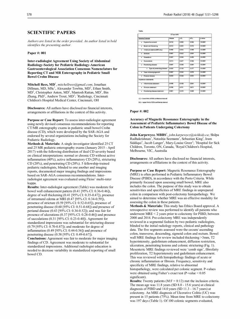
CO
LO
R K
EY
C
OL
OR
KE
Y
SCIENTIFIC PAPERS Authors are listed in the order provided. An author listed in bold identifies the presenting author.
Paper #: 001
Inter-radiologist Agreement Using Society of Abdominal
Radiology-Society for Pediatric Radiology-American
Gastroenterological Association Consensus Nomenclature for
Reporting CT and MR Enterography in Pediatric Small
Bowel Crohn Disease
Mitchell Rees, MD1, [email protected]; Jonathan
Dillman, MD, MSc1, Alexander Towbin, MD
1, Ethan Smith,
MD1, Christopher Anton, MD
1, Mantosh Rattan, MD
1, Bin
Zhang, PhD1, Andrew Trout, MD
1;
1Radiology, Cincinnati
Children's Hospital Medical Center, Cincinnati, OH Disclosures: All authors have disclosed no financial interests,
arrangements or affiliations in the context of this activity. Purpose or Case Report: To assess inter-radiologist agreement using newly devised consensus recommendations for reporting CT/MR enterography exams in pediatric small bowel Crohn disease (CD), which were developed by the SAR-AGA and endorsed by several organizations including the Society for
Pediatric Radiology. Methods & Materials: A single investigator identified 25 CT and 25 MR pediatric enterography exams (January 2015 - April 2017) with the following distribution of ileal CD severity based on clinical interpretations: normal or chronic CD without active inflammation (40%), active inflammatory CD (20%), stricturing
CD (20%), and penetrating CD (20%). 5 fellowship-trained pediatric radiologists, blinded to one another and imaging reports, documented major imaging findings and impressions based on SAR-AGA consensus recommendations. Inter-radiologist agreement was evaluated using Fleiss’ multi-rater kappa. Results: Inter-radiologist agreement (Table) was moderate for
bowel wall enhancement pattern (0.41 [95% CI: 0.36-0.46]), degree of wall thickening (0.41 [95% CI: 0.35-0.47]), presence of intramural edema at MRI (0.47 [95% CI: 0.34-0.59]), presence of stricture (0.59 [95% CI: 0.52-0.65]), presence of penetrating disease (0.60 [95% CI: 0.51-0.68]) and presence of perianal disease (0.43 [95% CI: 0.34-0.52]); and was fair for presence of ulcerations (0.37 [95% CI: 0.28-0.46]) and presence
of sacculations (0.31 [95% CI: 0.23-0.40]). Agreement for standardized impressions was substantial for stricturing disease (0.79 [95% CI: 0.70-0.87]); and moderate for degree of inflammation (0.49 [95% CI: 0.44-0.56]) and presence of penetrating disease (0.58 [95% CI: 0.49-0.67]). Conclusions: Agreement was fair to moderate for major imaging
findings of CD. Agreement was moderate to substantial for standardized impressions. Additional radiologist education is needed to decrease variability in standardized reporting of small bowel CD.
Paper #: 002
Accuracy of Magnetic Resonance Enterography in the
Assessment of Pediatric Inflammatory Bowel Disease of the
Colon in Patients Undergoing Colectomy John Karpewycz. MBBS
1, [email protected]; Shilpa
Radhakrishnan1, Natashia Seemann
1, Sebastian King
2, Iram
Siddiqui1, Jacob Langer
1, Mary-Louise Greer
1;
1Hospital for Sick
Children, Toronto, ON, Canada, 2Royal Children's Hospital,
Melbourne, VIC, Australia Disclosures: All authors have disclosed no financial interests, arrangements or affiliations in the context of this activity.
Purpose or Case Report: Magnetic Resonance Enterography (MRE) is often performed in Pediatric Inflammatory Bowel Disease (PIBD), in accordance with the Porto Criteria. While primarily focused upon assessing small bowel, MRE also includes the colon. The purpose of this study was to obtain sensitivities and specificities of MRE findings in unprepared
colon in comparison with post-colectomy histopathology. We aimed to determine whether MRE was an effective modality for assessing the colon in these patients. Methods & Materials: This study has Ethics Board approval. A retrospective review was performed to identify all patients who underwent MRE < 2 years prior to colectomy for PIBD, between 2000 and 2014. Pre-colectomy MRE was independently
reviewed in a segmental fashion by two pediatric radiologists, blinded to the initial radiology reports, clinical and pathology data. The five segments assessed were the cecum/ ascending colon, transverse, descending, sigmoid colon and rectum. Bowel wall MRE findings for review included thickening >3mm, T2 hyperintensity, gadolinium enhancement, diffusion restriction, ulceration, penetrating lesions and colonic stricturing (Fig. 1).
Mesenteric MRE findings reviewed were ‘comb sign’, fibrofatty proliferation, T2 hyperintensity and gadolinium enhancement. This was reviewed with histopathology findings of acute or chronic inflammation or fibrosis. Frequency, sensitivity and specificity of MRE findings, relative to abnormal histopathology, were calculated per colonic segment. P-values
were obtained using Fisher’s exact test (P-value < 0.05 significant). Results: Twenty patients (M:F = 8:12) met the inclusion criteria. The mean age was 11.8 years (SD 8.0 - 15.6 years) at clinical diagnosis of PIBD and 14.0 years (SD 11.3 - 16.7 years) at colectomy. An MRE diagnosis of Ulcerative Colitis (UC) was present in 15 patients (75%). Mean time from MRE to colectomy
was 197 days (Table 1). Of 100 colonic segments evaluated,
Pediatr Radiol (2018) 48 (Suppl 1):S1–S298S78
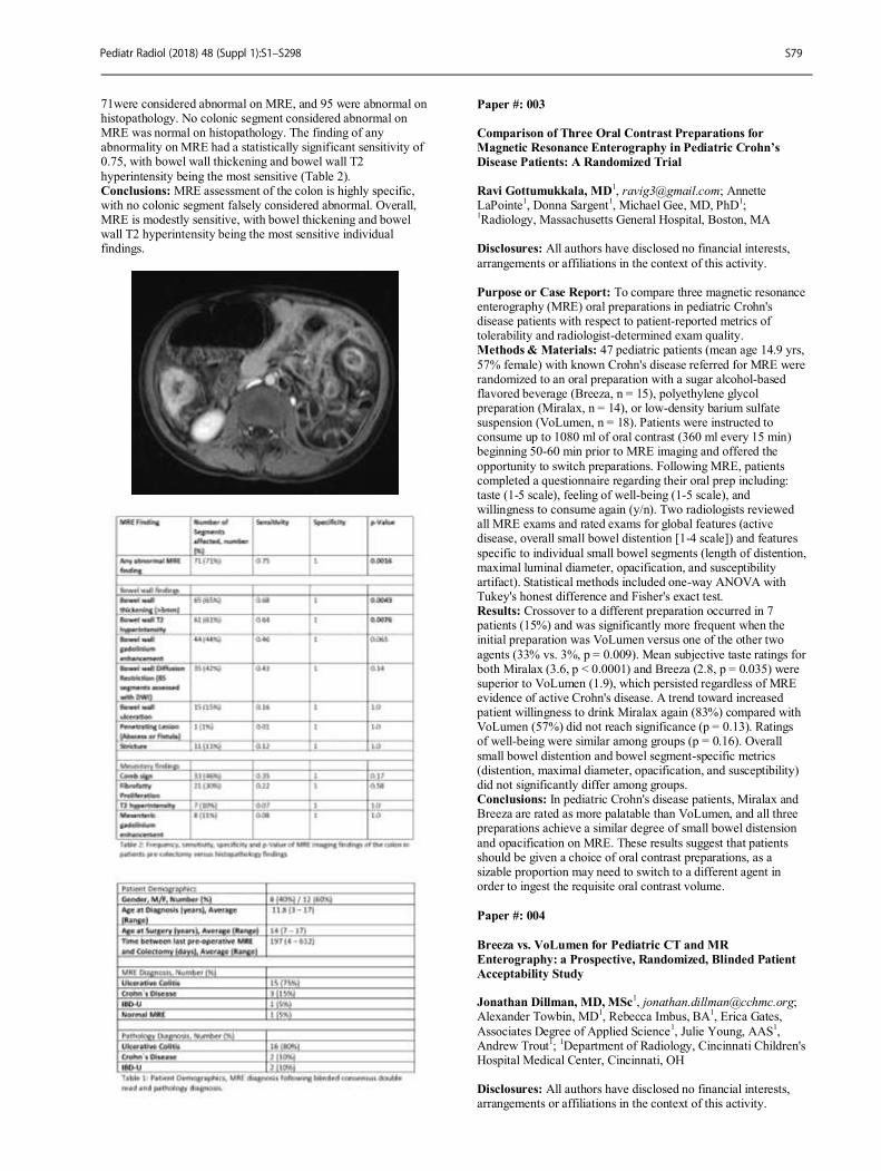
71were considered abnormal on MRE, and 95 were abnormal on histopathology. No colonic segment considered abnormal on MRE was normal on histopathology. The finding of any abnormality on MRE had a statistically significant sensitivity of 0.75, with bowel wall thickening and bowel wall T2
hyperintensity being the most sensitive (Table 2). Conclusions: MRE assessment of the colon is highly specific, with no colonic segment falsely considered abnormal. Overall, MRE is modestly sensitive, with bowel thickening and bowel wall T2 hyperintensity being the most sensitive individual findings.
Paper #: 003
Comparison of Three Oral Contrast Preparations for
Magnetic Resonance Enterography in Pediatric Crohn’s
Disease Patients: A Randomized Trial
Ravi Gottumukkala, MD
1, [email protected]; Annette
LaPointe1, Donna Sargent
1, Michael Gee, MD, PhD
1;
1Radiology, Massachusetts General Hospital, Boston, MA
Disclosures: All authors have disclosed no financial interests,
arrangements or affiliations in the context of this activity. Purpose or Case Report: To compare three magnetic resonance enterography (MRE) oral preparations in pediatric Crohn's disease patients with respect to patient-reported metrics of tolerability and radiologist-determined exam quality. Methods & Materials: 47 pediatric patients (mean age 14.9 yrs,
57% female) with known Crohn's disease referred for MRE were randomized to an oral preparation with a sugar alcohol-based flavored beverage (Breeza, n = 15), polyethylene glycol preparation (Miralax, n = 14), or low-density barium sulfate suspension (VoLumen, n = 18). Patients were instructed to consume up to 1080 ml of oral contrast (360 ml every 15 min) beginning 50-60 min prior to MRE imaging and offered the
opportunity to switch preparations. Following MRE, patients completed a questionnaire regarding their oral prep including: taste (1-5 scale), feeling of well-being (1-5 scale), and willingness to consume again (y/n). Two radiologists reviewed all MRE exams and rated exams for global features (active disease, overall small bowel distention [1-4 scale]) and features
specific to individual small bowel segments (length of distention, maximal luminal diameter, opacification, and susceptibility artifact). Statistical methods included one-way ANOVA with Tukey's honest difference and Fisher's exact test. Results: Crossover to a different preparation occurred in 7 patients (15%) and was significantly more frequent when the initial preparation was VoLumen versus one of the other two
agents (33% vs. 3%, p = 0.009). Mean subjective taste ratings for both Miralax (3.6, p < 0.0001) and Breeza (2.8, p = 0.035) were superior to VoLumen (1.9), which persisted regardless of MRE evidence of active Crohn's disease. A trend toward increased patient willingness to drink Miralax again (83%) compared with VoLumen (57%) did not reach significance (p = 0.13). Ratings of well-being were similar among groups (p = 0.16). Overall
small bowel distention and bowel segment-specific metrics (distention, maximal diameter, opacification, and susceptibility) did not significantly differ among groups. Conclusions: In pediatric Crohn's disease patients, Miralax and Breeza are rated as more palatable than VoLumen, and all three preparations achieve a similar degree of small bowel distension
and opacification on MRE. These results suggest that patients should be given a choice of oral contrast preparations, as a sizable proportion may need to switch to a different agent in order to ingest the requisite oral contrast volume.
Paper #: 004
Breeza vs. VoLumen for Pediatric CT and MR
Enterography: a Prospective, Randomized, Blinded Patient
Acceptability Study Jonathan Dillman, MD, MSc
Alexander Towbin, MD1, Rebecca Imbus, BA
1, Erica Gates,
Associates Degree of Applied Science1, Julie Young, AAS
1,
Andrew Trout1;
1Department of Radiology, Cincinnati Children's
Hospital Medical Center, Cincinnati, OH Disclosures: All authors have disclosed no financial interests, arrangements or affiliations in the context of this activity.
Pediatr Radiol (2018) 48 (Suppl 1):S1–S298 S79
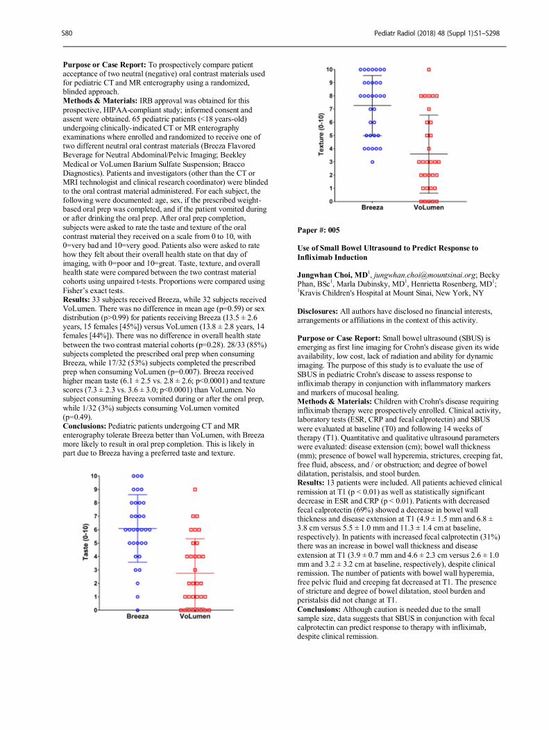
Purpose or Case Report: To prospectively compare patient acceptance of two neutral (negative) oral contrast materials used for pediatric CT and MR enterography using a randomized, blinded approach. Methods & Materials: IRB approval was obtained for this
prospective, HIPAA-compliant study; informed consent and assent were obtained. 65 pediatric patients (<18 years-old) undergoing clinically-indicated CT or MR enterography examinations where enrolled and randomized to receive one of two different neutral oral contrast materials (Breeza Flavored Beverage for Neutral Abdominal/Pelvic Imaging; Beekley
Medical or VoLumen Barium Sulfate Suspension; Bracco Diagnostics). Patients and investigators (other than the CT or MRI technologist and clinical research coordinator) were blinded to the oral contrast material administered. For each subject, the following were documented: age, sex, if the prescribed weight-based oral prep was completed, and if the patient vomited during or after drinking the oral prep. After oral prep completion,
subjects were asked to rate the taste and texture of the oral contrast material they received on a scale from 0 to 10, with 0=very bad and 10=very good. Patients also were asked to rate how they felt about their overall health state on that day of imaging, with 0=poor and 10=great. Taste, texture, and overall health state were compared between the two contrast material cohorts using unpaired t-tests. Proportions were compared using
Fisher’s exact tests. Results: 33 subjects received Breeza, while 32 subjects received VoLumen. There was no difference in mean age (p=0.59) or sex distribution (p>0.99) for patients receiving Breeza (13.5 ± 2.6 years, 15 females [45%]) versus VoLumen (13.8 ± 2.8 years, 14 females [44%]). There was no difference in overall health state
between the two contrast material cohorts (p=0.28). 28/33 (85%) subjects completed the prescribed oral prep when consuming Breeza, while 17/32 (53%) subjects completed the prescribed prep when consuming VoLumen (p=0.007). Breeza received higher mean taste (6.1 ± 2.5 vs. 2.8 ± 2.6; p<0.0001) and texture scores (7.3 ± 2.3 vs. 3.6 ± 3.0; p<0.0001) than VoLumen. No subject consuming Breeza vomited during or after the oral prep,
while 1/32 (3%) subjects consuming VoLumen vomited (p=0.49). Conclusions: Pediatric patients undergoing CT and MR enterography tolerate Breeza better than VoLumen, with Breeza more likely to result in oral prep completion. This is likely in part due to Breeza having a preferred taste and texture.
Paper #: 005
Use of Small Bowel Ultrasound to Predict Response to
Infliximab Induction Jungwhan Choi, MD
1, [email protected]; Becky
Phan, BSc1, Marla Dubinsky, MD
1, Henrietta Rosenberg, MD
1;
1Kravis Children's Hospital at Mount Sinai, New York, NY
Disclosures: All authors have disclosed no financial interests, arrangements or affiliations in the context of this activity.
Purpose or Case Report: Small bowel ultrasound (SBUS) is emerging as first line imaging for Crohn's disease given its wide availability, low cost, lack of radiation and ability for dynamic imaging. The purpose of this study is to evaluate the use of
SBUS in pediatric Crohn's disease to assess response to infliximab therapy in conjunction with inflammatory markers and markers of mucosal healing. Methods & Materials: Children with Crohn's disease requiring infliximab therapy were prospectively enrolled. Clinical activity, laboratory tests (ESR, CRP and fecal calprotectin) and SBUS
were evaluated at baseline (T0) and following 14 weeks of therapy (T1). Quantitative and qualitative ultrasound parameters were evaluated: disease extension (cm); bowel wall thickness (mm); presence of bowel wall hyperemia, strictures, creeping fat, free fluid, abscess, and / or obstruction; and degree of bowel dilatation, peristalsis, and stool burden. Results: 13 patients were included. All patients achieved clinical
remission at T1 (p < 0.01) as well as statistically significant decrease in ESR and CRP (p < 0.01). Patients with decreased fecal calprotectin (69%) showed a decrease in bowel wall thickness and disease extension at T1 (4.9 ± 1.5 mm and 6.8 ± 3.8 cm versus 5.5 ± 1.0 mm and 11.3 ± 1.4 cm at baseline, respectively). In patients with increased fecal calprotectin (31%) there was an increase in bowel wall thickness and disease
extension at T1 (3.9 ± 0.7 mm and 4.6 ± 2.3 cm versus 2.6 ± 1.0 mm and 3.2 ± 3.2 cm at baseline, respectively), despite clinical remission. The number of patients with bowel wall hyperemia, free pelvic fluid and creeping fat decreased at T1. The presence of stricture and degree of bowel dilatation, stool burden and peristalsis did not change at T1.
Conclusions: Although caution is needed due to the small sample size, data suggests that SBUS in conjunction with fecal calprotectin can predict response to therapy with infliximab, despite clinical remission.
Pediatr Radiol (2018) 48 (Suppl 1):S1–S298S80
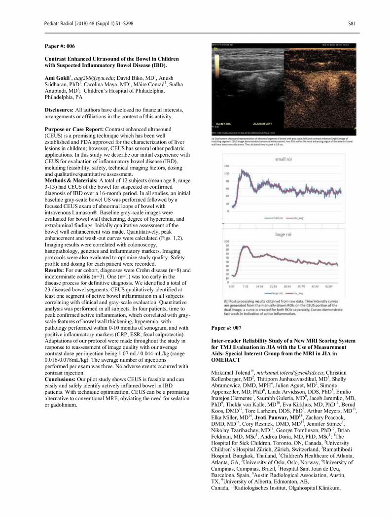
Paper #: 006
Contrast Enhanced Ultrasound of the Bowel in Children
with Suspected Inflammatory Bowel Disease (IBD).
Ami Gokli1, [email protected]; David Biko, MD
1, Anush
Sridharan, PhD1, Carolina Maya, MD
1, Máire Conrad
1, Sudha
Anupindi, MD1;
1Children’s Hospital of Philadelphia,
Philadelphia, PA Disclosures: All authors have disclosed no financial interests,
arrangements or affiliations in the context of this activity. Purpose or Case Report: Contrast enhanced ultrasound (CEUS) is a promising technique which has been well established and FDA approved for the characterization of liver lesions in children; however, CEUS has several other pediatric applications. In this study we describe our initial experience with
CEUS for evaluation of inflammatory bowel disease (IBD), including feasibility, safety, technical imaging factors, dosing and qualitative/quantitative assessment. Methods & Materials: A total of 12 subjects (mean age 8, range 3-13) had CEUS of the bowel for suspected or confirmed diagnosis of IBD over a 16-month period. In all studies, an initial baseline gray-scale bowel US was performed followed by a
focused CEUS exam of abnormal loops of bowel with intravenous Lumason®. Baseline gray-scale images were evaluated for bowel wall thickening, degree of hyperemia, and extraluminal findings. Initially qualitative assessment of the bowel wall enhancement was made. Quantitatively, peak enhancement and wash-out curves were calculated (Figs. 1,2).
Imaging results were correlated with colonoscopy, histopathology, genetics and inflammatory markers. Imaging protocols were also evaluated to optimize study quality. Safety profile and dosing for each patient were recorded. Results: For our cohort, diagnoses were Crohn disease (n=8) and indeterminate colitis (n=3). One (n=1) was too early in the disease process for definitive diagnosis. We identified a total of
23 diseased bowel segments. CEUS qualitatively identified at least one segment of active bowel inflammation in all subjects correlating with clinical and gray-scale evaluation. Quantitative analysis was performed in all subjects. In four patients, time to peak confirmed active inflammation, which correlated with gray-scale features of bowel wall thickening, hyperemia, with pathology performed within 0-10 months of sonogram, and with
positive inflammatory markers (CRP, ESR, fecal calprotectin). Adaptations of our protocol were made throughout the study in response to reassessment of image quality with our average contrast dose per injection being 1.07 mL/ 0.044 mL/kg (range 0.016-0.078mL/kg). The average number of injections performed per exam was three. No adverse events occurred with
contrast injection. Conclusions: Our pilot study shows CEUS is feasible and can easily and safely identify actively inflamed bowel in IBD patients. With technique optimization, CEUS can be a promising alternative to conventional MRE, obviating the need for sedation or gadolinium.
Paper #: 007
Inter-reader Reliability Study of a New MRI Scoring System
for TMJ Evaluation in JIA with the Use of Measurement
Aids: Special Interest Group from the MRI in JIA in
OMERACT
Mirkamal Tolend15
, [email protected]; Christian Kellenberger, MD
2, Thitiporn Junhasavasdikul, MD
3, Shelly
Abramowicz, DMD, MPH4, Julien Aguet, MD
2, Simone
Appenzeller, MD, PhD6, Linda Arvidsson, DDS, PhD
5, Emilio
Inarejos Clemente7, Saurabh Guleria, MD
8, Jacob Jaremko, MD,
PhD9, Thekla von Kalle, MD
10, Eva Kirkhus, MD, PhD
11, Bernd
Koos, DMD12
, Tore Larheim, DDS, PhD5, Arthur Meyers, MD
13,
Elka Miller, MD14
, Jyoti Panwar, MD15
, Zachary Peacock, DMD, MD
16, Cory Resnick, DMD, MD
17, Jennifer Stimec
1,
Nikolay Tzaribachev, MD18
, George Tomlinson, PhD15
, Brian Feldman, MD, MSc
1, Andrea Doria, MD, PhD, MSc
1;
1The
Hospital for Sick Children, Toronto, ON, Canada, 2University
Children’s Hospital Zürich, Zürich, Switzerland, 3Ramathibodi
Hospital, Bangkok, Thailand, 4Children's Healthcare of Atlanta,
Atlanta, GA, 5University of Oslo, Oslo, Norway,
6University of
Campinas, Campinas, Brazil, 7Hospital Sant Joan de Deu,
Barcelona, Spain, 8Austin Radiological Association, Austin,
TX, 9University of Alberta, Edmonton, AB,
Canada, 10
Radiologisches Institut, Olgahospital Klinikum,
Pediatr Radiol (2018) 48 (Suppl 1):S1–S298 S81
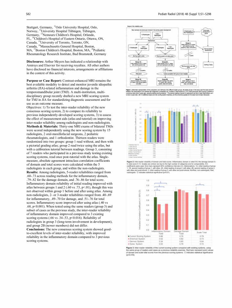
Stuttgart, Germany,
11Oslo University Hospital, Oslo,
Norway, 12
University Hospital Tübingen, Tübingen, Germany,
13Nemours Children's Hospital, Orlando,
FL, 14
Children's Hospital of Eastern Ontario, Ottawa, ON, Canada,
15University of Toronto, Toronto, ON,
Canada, 16
Massachusetts General Hospital, Boston, MA,
17Boston Children's Hospital, Boston, MA,
18Pediatric
Rheumatology Research Institute, Bad Bramstedt, Germany Disclosures: Arthur Meyers has indicated a relationship with Amirsys and Elsevier for receiving royalties. All other authors
have disclosed no financial interests, arrangements or affiliations in the context of this activity. Purpose or Case Report: Contrast-enhanced MRI remains the best available modality to detect and monitor juvenile idiopathic arthritis (JIA)-related inflammation and damage in the temporomandibular joint (TMJ). A multi-institution, multi-
disciplinary group recently drafted a new MRI scoring system for TMJ in JIA for standardizing diagnostic assessment and for use as an outcome measure. Objectives: 1) To test the inter-reader reliability of the new consensus scoring system, 2) to compare its reliability to previous independently-developed scoring systems, 3) to assess the effect of measurement aids (atlas and tutorial) on improving
inter-reader reliability among radiologists and non-radiologists. Methods & Materials: Thirty-one MRI exams of bilateral TMJs were scored independently using the new scoring system by 15 radiologists, 2 oral-maxillofacial surgeons, 2 pediatric rheumatologists, and 1 orthodontist. Thirteen readers were randomized into two groups: group 1 read without, and then with
a pictorial grading atlas; group 2 read twice using the atlas, but with a calibration tutorial between readings. Group 3, consisting of 7 readers who participated in a previous study testing existing scoring systems, read once post-tutorial with the atlas. Single-measure, absolute agreement intraclass correlation coefficients of domain and total scores were calculated within the 5 radiologists in each group, and within the non-radiologists.
Results: Among radiologists, 5-reader reliabilities ranged from .60-.73 across reading methods for the inflammatory domain, .79-.82 for the damage domain, and .76-.86 for total score. Inflammatory domain reliability of initial reading improved with atlas between groups 1 and 2 (.60 vs .73, p<.01), though this was not observed within group 1 before and after using atlas. Among non-radiologists, 2- or 3-reader reliabilities ranged from .40-.69
for inflammatory, .49-.70 for damage, and .51-.76 for total scores. Inflammatory score improved after using atlas (.40 vs .68, p<0.001). When tested using the same readers (group 3) and subset of cases as the previous study, the inter-reader reliability of inflammatory domain improved compared to 3 existing scoring systems (.66 vs .34-.53, p<0.016). Reliability of
radiologists in group 3 (long-term involvement in development), and group 2B (newer members) did not differ. Conclusions: The new consensus scoring system showed good-to-excellent levels of inter-reader reliability, with improved reliability in the inflammatory domain compared to 3 previous scoring systems.
Pediatr Radiol (2018) 48 (Suppl 1):S1–S298S82
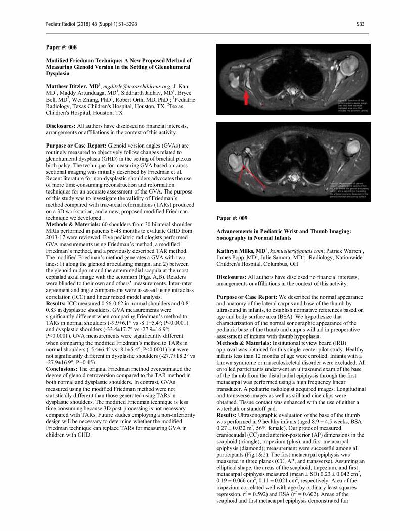
Paper #: 008
Modified Friedman Technique: A New Proposed Method of
Measuring Glenoid Version in the Setting of Glenohumeral
Dysplasia
Matthew Ditzler, MD
1, [email protected]; J. Kan,
MD1, Maddy Artunduaga, MD
1, Siddharth Jadhav, MD
1, Bryce
Bell, MD2, Wei Zhang, PhD
1, Robert Orth, MD, PhD
1;
1Pediatric
Radiology, Texas Children's Hospital, Houston, TX, 2Texas
Children's Hospital, Houston, TX
Disclosures: All authors have disclosed no financial interests, arrangements or affiliations in the context of this activity. Purpose or Case Report: Glenoid version angles (GVAs) are routinely measured to objectively follow changes related to glenohumeral dysplasia (GHD) in the setting of brachial plexus
birth palsy. The technique for measuring GVA based on cross sectional imaging was initially described by Friedman et al. Recent literature for non-dysplastic shoulders advocates the use of more time-consuming reconstruction and reformation techniques for an accurate assessment of the GVA. The purpose of this study was to investigate the validity of Friedman’s method compared with true-axial reformations (TARs) produced
on a 3D workstation, and a new, proposed modified Friedman technique we developed. Methods & Materials: 60 shoulders from 30 bilateral shoulder MRIs performed in patients 6-48 months to evaluate GHD from 2013-17 were reviewed. Five pediatric radiologists performed GVA measurements using Friedman’s method, a modified
Friedman’s method, and a previously described TAR method. The modified Friedman’s method generates a GVA with two lines: 1) along the glenoid articulating margin, and 2) between the glenoid midpoint and the anteromedial scapula at the most cephalad axial image with the acromion (Figs. A,B). Readers were blinded to their own and others’ measurements. Inter-rater agreement and angle comparisons were assessed using intraclass
correlation (ICC) and linear mixed model analysis. Results: ICC measured 0.56-0.62 in normal shoulders and 0.81-0.83 in dysplastic shoulders. GVA measurements were significantly different when comparing Friedman’s method to TARs in normal shoulders (-9.9±6.1° vs -8.1±5.4°; P<0.0001) and dysplastic shoulders (-33.4±17.7° vs -27.9±16.9°; P<0.0001). GVA measurements were significantly different
when comparing the modified Friedman’s method to TARs in normal shoulders (-5.4±6.4° vs -8.1±5.4°; P<0.0001) but were not significantly different in dysplastic shoulders (-27.7±18.2° vs -27.9±16.9°; P=0.45). Conclusions: The original Friedman method overestimated the degree of glenoid retroversion compared to the TAR method in
both normal and dysplastic shoulders. In contrast, GVAs measured using the modified Friedman method were not statistically different than those generated using TARs in dysplastic shoulders. The modified Friedman technique is less time consuming because 3D post-processing is not necessary compared with TARs. Future studies employing a non-inferiority design will be necessary to determine whether the modified
Friedman technique can replace TARs for measuring GVA in children with GHD.
Paper #: 009
Advancements in Pediatric Wrist and Thumb Imaging:
Sonography in Normal Infants Kathryn Milks, MD
1, [email protected]; Patrick Warren
1,
James Popp, MD1, Julie Samora, MD
1;
1Radiology, Nationwide
Children's Hospital, Columbus, OH
Disclosures: All authors have disclosed no financial interests, arrangements or affiliations in the context of this activity. Purpose or Case Report: We described the normal appearance and anatomy of the lateral carpus and base of the thumb by ultrasound in infants, to establish normative references based on
age and body surface area (BSA). We hypothesize that characterization of the normal sonographic appearance of the pediatric base of the thumb and carpus will aid in preoperative assessment of infants with thumb hypoplasia. Methods & Materials: Institutional review board (IRB) approval was obtained for this single-center pilot study. Healthy infants less than 12 months of age were enrolled. Infants with a
known syndrome or musculoskeletal disorder were excluded. All enrolled participants underwent an ultrasound exam of the base of the thumb from the distal radial epiphysis through the first metacarpal was performed using a high frequency linear transducer. A pediatric radiologist acquired images. Longitudinal and transverse images as well as still and cine clips were
obtained. Tissue contact was enhanced with the use of either a waterbath or standoff pad. Results: Ultrasonographic evaluation of the base of the thumb was performed in 9 healthy infants (aged 8.9 ± 4.5 weeks, BSA 0.27 ± 0.032 m
2, 56% female). Our protocol measured
craniocaudal (CC) and anterior-posterior (AP) dimensions in the scaphoid (triangle), trapezium (plus), and first metacarpal
epiphysis (diamond); measurement were successful among all participants (Fig.1&2). The first metacarpal epiphysis was measured in three planes (CC, AP, and transverse). Assuming an elliptical shape, the areas of the scaphoid, trapezium, and first metacarpal epiphysis measured (mean ± SD) 0.23 ± 0.042 cm
2,
0.19 ± 0.066 cm2, 0.11 ± 0.021 cm
2, respectively. Area of the
trapezium correlated well with age (by ordinary least squares
regression, r2 = 0.592) and BSA (r
2 = 0.602). Areas of the
scaphoid and first metacarpal epiphysis demonstrated fair
Pediatr Radiol (2018) 48 (Suppl 1):S1–S298 S83
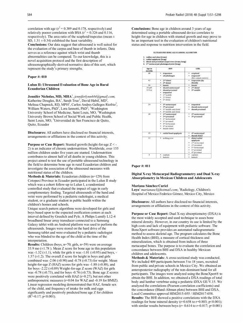
correlation with age (r
2 = 0.389 and 0.178, respectively) and
relatively poorer correlation with BSA (r2 = 0.326 and 0.116,
respectively). The area ratio of the scaphoid/trapezius (mean ± SD, 1.31 ± 0.34) exhibited the least variability. Conclusions: Our data suggest that ultrasound is well suited for
the evaluation of the carpus and base of thumb in infants. Data serves as a reference against which wrist and thumb abnormalities can be compared. To our knowledge, this is a novel acquisition protocol and the first description of ultrasonographically-derived normative data of this sort, which represent the study’s primary strengths.
Paper #: 010
Lulun II: Ultrasound Evaluation of Bone Age in Rural
Ecuadorian Children Jennifer Nicholas, MD, MHA
Katherine Douglas, BA1, Sarah True
2, David Habif, MD
2,
Melissa Chapnick, RD, MPH2, Carlos Andres Gallegos Riofrio
2,
William Waters, PhD3, Lora Iannotti, PhD
2;
1Washington
University School of Medicine, Saint Louis, MO, 2Washington
University Brown School of Social Work and Public Health, Saint Louis, MO,
3Universidad de San Francisco de Quito,
Quito, Ecuador
Disclosures: All authors have disclosed no financial interests, arrangements or affiliations in the context of this activity. Purpose or Case Report: Stunted growth (height-for-age Z < -2) is an indicator of chronic undernutrition. Worldwide, over 155
million children under five years are stunted. Undernutrition contributes to almost half of all deaths in young children. This project aimed to test the use of portable ultrasound technology in the field to determine bone age in rural Ecuadorian children and investigate the association of the ultrasound measures with nutritional status of the children. Methods & Materials: Ecuadorian children (n=129) from
Cotopaxi Province in Ecuador participated in the Lulun II study which was a cohort follow-up to Lulun I, a randomized controlled study that evaluated the impact of eggs in early complementary feeding. Targeted ultrasounds of the hand and wrist were performed by a pediatric radiologist, a medical student, or a graduate student in public health within the children's homes and schools.
Unique search pattern algorithms were developed for girls and boys based upon to the expected ossification centers at each interval defined by Greulich and Pyle. A Philips Lumify L12-4 broadband linear array transducer connected to a Samsung Galaxy tablet with an Android platform was used to perform the ultrasounds. Images were stored on the hard drive of the
Samsung tablet and were evaluated by a pediatric radiologist who was blinded to the age of the child at the time of the interpretation. Results: Children (boys, n=70; girls, n=59) were on average 33.9 mo (±1.78 ). Mean Z score for bone age in this population was -1.32 (±1.1), while for girls it was -1.01 (±0.95) and boys, -1.57 (±1.2). The overall Z score for height in boys and girls
combined was -2.06 (±0.90) and -0.78 (±0.73) for weight. Mean height-for-age Z (HAZ) scores for girls was -1.88 (±0.88), and for boys -2.22 (±0.89) Weight-for-age Z score (WAZ) for girls was -0.78 (±0.73), and for boys -0.76 (±0.73). Bone age Z scores were positively correlated with HAZ (r=0.27), but not other anthropometric measures (r=0.04 for WAZ and -0.19 for BMIZ).
Linear regression modeling demonstrated that HAZ, female sex of the child, and frequency of intake for milk and eggs significantly and positively predicted bone age Z for children (R
2=0.17; p<0.001).
Conclusions: Bone age in children around 3 years of age determined using a portable ultrasound device correlates to height-for-age in children with stunted growth and may prove to be an important tool in the evaluation of children's nutritional status and response to nutrition intervention in the field.
Paper #: 011
Digital X-ray Metacarpal Radiogrammetry and Dual X-ray
Absorptiometry in Mexican Children and Adolescents
Mariana Sánchez Curiel Loyo
1Radiology, Children's
Hospital of Mexico Federico Gómez, Mexico City, Mexico
Disclosures: All authors have disclosed no financial interests, arrangements or affiliations in the context of this activity. Purpose or Case Report: Dual X-ray absorptiometry (DXA) is
the most widely accepted and used technique to asses bone mineral density. However, in our country its use is limited by the high costs and lack of equipment with pediatric software. The BoneXpert software provides an automated radiogrammatic method to assess skeletal age. The program calculates the Bone Health Index (BHI), a measure of cortical thickness and mineralization, which is obtained from indices of three
metacarpal bones. The purpose is to evaluate the correlation and concordance between BHI and DXA in healthy Mexican children and adolescents. Methods & Materials: A cross-sectional study was conducted. We included 409 participants between 5 to 18 years, recruited from public and private schools in Mexico City. We obtained an
anteroposterior radiography of the non-dominant hand for all participants. The images were analyzed using the BoneXpert® to obtain the BHI. In addition, we obtained a DXA readings of total body and lumbar vertebrae using a pediatric iDXA GE/V.15. We analyzed the correlations (Pearson correlation coefficients) and the concordance (Bland-Altman plots) between BHI and DXA. Local Committee approval HIM2015-055 / HIM2017-058.
Results: The BHI showed a positive correlations with the DXA readings for bone mineral density (r=0.658 to r=0.803; p<0.001); with similar results between boys (r= 0.614 to r=0.817; p<0.001)
Pediatr Radiol (2018) 48 (Suppl 1):S1–S298S84
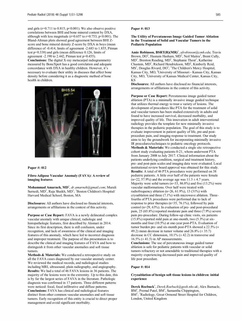
and girls (r=0.711 to 0.815; p<0.001). We also observe positive correlations between BHI and bone mineral content by DXA, although with less magnitude (r=0.657 to r=0.753; p<0.001). The Bland-Altman plots showed good agreement between BHI Z-score and bone mineral density Z-score by DXA in boys (mean
difference of -0.414, limits of agreement -2.683 to 1.855, Pitman test p=0.338) and girls (mean difference 0.126, limits of agreement -2.190 to 2.442, Pitman test p=0.455). Conclusions: The digital X-ray metacarpal radiogrammetry measured by BoneXpert has a good correlation and adequate concordance with DXA in healthy children. However, it’s
necessary to evaluate their utility in diseases that affect bone density before considering it as a diagnostic method of bone health in children.
Paper #: 012
Fibro-Adipose Vascular Anomaly (FAVA): A review of
imaging features Mohammad Amarneh, MD
1, [email protected]; Murali
Surnedi, MD1, Raja Shaikh, MD
1;
1Boston Children's Hospital/
Harvard Medical School, Boston, MA Disclosures: All authors have disclosed no financial interests, arrangements or affiliations in the context of this activity. Purpose or Case Report: FAVA is a newly delineated complex
vascular anomaly with unique clinical, radiologic and histopathologic features, first described by Alomari in 2014. Since its first description, there is still confusion, under recognition, and lack of awareness of the clinical and imaging features of this anomaly, which have led to incorrect diagnosis and improper treatment. The purpose of this presentation is to describe the clinical and imaging features of FAVA and how to
distinguish it from other vascular anomalies and soft tissue tumors. Methods & Materials: We conducted a retrospective study on all the FAVA cases diagnosed by our vascular anomaly center. We reviewed the medical records, and radiological studies including MRI, ultrasound, plain radiography, and angiography.
Results: We had a total of 46 FAVA lesions in 36 patients. The majority of the lesions were in the extremity. Up to this date, this is by far the largest series of FAVA in the literature. Pathologic diagnosis was confirmed in 17 patients. Three different patterns were noticed: focal, focal infiltrative and diffuse patterns. Conclusions: FAVA has clinical and radiological features distinct from other common vascular anomalies and soft tissue
tumors. Early recognition of this entity is crucial to direct proper management and avoid significant morbidity.
Paper #: 013
The Utility of Percutaneous Image Guided Tumor Ablation
in the Treatment of Solid and Vascular Tumors in the
Pediatric Population
Amie Robinson, BSRT(R)(MR)
1, [email protected]; Travis
Brown, DO2, Hasnain Hasham, MD
3, Neil Maitra
2, Brent Cully,
MD1, Brenton Reading, MD
1, Stephanie Theut
1, Katherine
Chastain, MD1, Richard Hendrickson, MD
1, Kimberly Reid,
MS1, Douglas Rivard, DO
1;
1The Children's Mercy Hospital,
Kansas City, MO, 2University of Missouri - Kansas City, Kansas
City, MO, 3University of Kansas Medical Center, Kansas City,
KS Disclosures: All authors have disclosed no financial interests, arrangements or affiliations in the context of this activity. Purpose or Case Report: Percutaneous image guided tumor
ablation (PTA) is a minimally invasive image guided technique that utilizes thermal energy to treat a variety of lesions. The development of procedures like PTA for the treatment of solid and vascular tumors has been studied extensively in adults and found to have increased survival, decreased morbidity, and improved quality of life. This innovation in adult interventional radiology provides the template for new minimally invasive
therapies in the pediatric population. The goal of this study is to evaluate improvement in patient quality of life, pre-and post-procedure pain, and imaging response to treatment. Our study aims to lay the groundwork for incorporating minimally invasive IR procedures/techniques to pediatric oncology protocols. Methods & Materials: We conducted a single site retrospective
cohort study evaluating patients 0-21, whom underwent PTA from January 2008 to July 2017. Clinical information about the patients underlying condition, surgical and treatment history, pre- and post-pain scales and imaging data were evaluated. Local institutional review board approval was obtained for this study. Results: A total of 46 PTA procedures were performed on 38 pediatric patients. A little over half of the patients were female
(n=22, 57.9%) and the average age was 11.3 ± 4.7 years. Majority were solid tumors (n=33, 86.8%) and five (13.2%) were vascular malformations. Over half were treated with radiofrequency ablation (n=26, 61.9%), 13 (31%) with cryoablation and three (7.1%) with microwave ablation. Three-fourths of PTA procedures were performed due to lack of response to prior therapies (n=35, 76.1%), followed by pain
control (n=29, 63%). In evaluation of pre- and post-procedural pain, 25 (65.8%) reported moderate pain and three (7.9%) severe pain pre-procedure. During follow-up clinic visits, six patients (15.8%) reported mild pain at one-month, two (5.3%) at six-months and four (10.5%) at one-year post-PTA. Evaluation of tumor burden pre- and six-month post-PTA showed a 22.5% (±
49.2) mean decrease in tumor volume and 26.8% (± 35.7) decrease in CC dimension, 10.1% (± 42.2) in transverse and 16.5% (± 41.5) in AP measurements. Conclusions: The use of percutaneous image guided tumor ablation is safe for pediatric patients with vascular or solid tumors refractory or not amendable to traditional therapies with a majority experiencing decreased pain and improved quality of
life post procedure.
Paper #: 014
Cryoablation of benign soft tissue lesions in children: initial
experience
Derek Roebuck
1, [email protected]; Alex Barnacle,
BM1, Premal Patel, BM
1, Samantha Chippington,
BM1;
1Radiology, Great Ormond Street Hospital for Children,
London, United Kingdom
Pediatr Radiol (2018) 48 (Suppl 1):S1–S298 S85
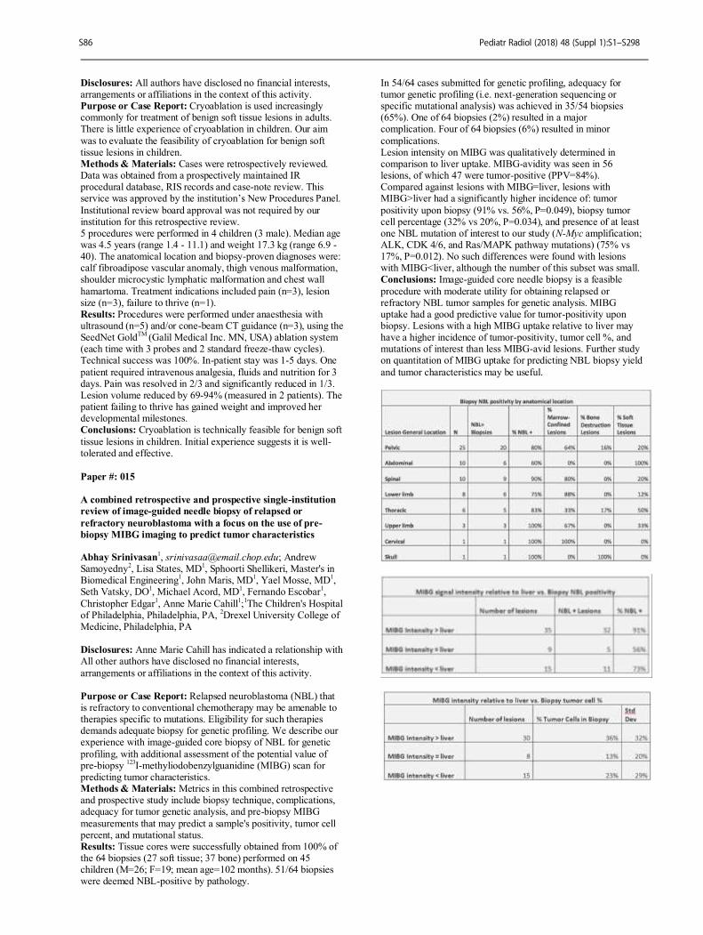
Disclosures: All authors have disclosed no financial interests, arrangements or affiliations in the context of this activity. Purpose or Case Report: Cryoablation is used increasingly commonly for treatment of benign soft tissue lesions in adults. There is little experience of cryoablation in children. Our aim
was to evaluate the feasibility of cryoablation for benign soft tissue lesions in children. Methods & Materials: Cases were retrospectively reviewed. Data was obtained from a prospectively maintained IR procedural database, RIS records and case-note review. This service was approved by the institution’s New Procedures Panel.
Institutional review board approval was not required by our institution for this retrospective review. 5 procedures were performed in 4 children (3 male). Median age was 4.5 years (range 1.4 - 11.1) and weight 17.3 kg (range 6.9 - 40). The anatomical location and biopsy-proven diagnoses were: calf fibroadipose vascular anomaly, thigh venous malformation, shoulder microcystic lymphatic malformation and chest wall
hamartoma. Treatment indications included pain (n=3), lesion size (n=3), failure to thrive (n=1). Results: Procedures were performed under anaesthesia with ultrasound (n=5) and/or cone-beam CT guidance (n=3), using the SeedNet Gold
TM (Galil Medical Inc. MN, USA) ablation system
(each time with 3 probes and 2 standard freeze-thaw cycles). Technical success was 100%. In-patient stay was 1-5 days. One
patient required intravenous analgesia, fluids and nutrition for 3 days. Pain was resolved in 2/3 and significantly reduced in 1/3. Lesion volume reduced by 69-94% (measured in 2 patients). The patient failing to thrive has gained weight and improved her developmental milestones. Conclusions: Cryoablation is technically feasible for benign soft
tissue lesions in children. Initial experience suggests it is well-tolerated and effective.
Paper #: 015
A combined retrospective and prospective single-institution
review of image-guided needle biopsy of relapsed or
refractory neuroblastoma with a focus on the use of pre-
biopsy MIBG imaging to predict tumor characteristics Abhay Srinivasan
1, [email protected]; Andrew
Samoyedny2, Lisa States, MD
1, Sphoorti Shellikeri, Master's in
Biomedical Engineering1, John Maris, MD
1, Yael Mosse, MD
1,
Seth Vatsky, DO1, Michael Acord, MD
1, Fernando Escobar
1,
Christopher Edgar1, Anne Marie Cahill
1;1The Children's Hospital
of Philadelphia, Philadelphia, PA, 2Drexel University College of
Medicine, Philadelphia, PA Disclosures: Anne Marie Cahill has indicated a relationship with All other authors have disclosed no financial interests,
arrangements or affiliations in the context of this activity. Purpose or Case Report: Relapsed neuroblastoma (NBL) that is refractory to conventional chemotherapy may be amenable to therapies specific to mutations. Eligibility for such therapies demands adequate biopsy for genetic profiling. We describe our experience with image-guided core biopsy of NBL for genetic
profiling, with additional assessment of the potential value of pre-biopsy
123I-methyliodobenzylguanidine (MIBG) scan for
predicting tumor characteristics. Methods & Materials: Metrics in this combined retrospective and prospective study include biopsy technique, complications, adequacy for tumor genetic analysis, and pre-biopsy MIBG
measurements that may predict a sample's positivity, tumor cell percent, and mutational status. Results: Tissue cores were successfully obtained from 100% of the 64 biopsies (27 soft tissue; 37 bone) performed on 45 children (M=26; F=19; mean age=102 months). 51/64 biopsies were deemed NBL-positive by pathology.
In 54/64 cases submitted for genetic profiling, adequacy for tumor genetic profiling (i.e. next-generation sequencing or specific mutational analysis) was achieved in 35/54 biopsies (65%). One of 64 biopsies (2%) resulted in a major complication. Four of 64 biopsies (6%) resulted in minor
complications. Lesion intensity on MIBG was qualitatively determined in comparison to liver uptake. MIBG-avidity was seen in 56 lesions, of which 47 were tumor-positive (PPV=84%). Compared against lesions with MIBG=liver, lesions with MIBG>liver had a significantly higher incidence of: tumor
positivity upon biopsy (91% vs. 56%, P=0.049), biopsy tumor cell percentage (32% vs 20%, P=0.034), and presence of at least one NBL mutation of interest to our study (N-Myc amplification; ALK, CDK 4/6, and Ras/MAPK pathway mutations) (75% vs 17%, P=0.012). No such differences were found with lesions with MIBG<liver, although the number of this subset was small. Conclusions: Image-guided core needle biopsy is a feasible
procedure with moderate utility for obtaining relapsed or refractory NBL tumor samples for genetic analysis. MIBG uptake had a good predictive value for tumor-positivity upon biopsy. Lesions with a high MIBG uptake relative to liver may have a higher incidence of tumor-positivity, tumor cell %, and mutations of interest than less MIBG-avid lesions. Further study on quantitation of MIBG uptake for predicting NBL biopsy yield
and tumor characteristics may be useful.
Pediatr Radiol (2018) 48 (Suppl 1):S1–S298S86
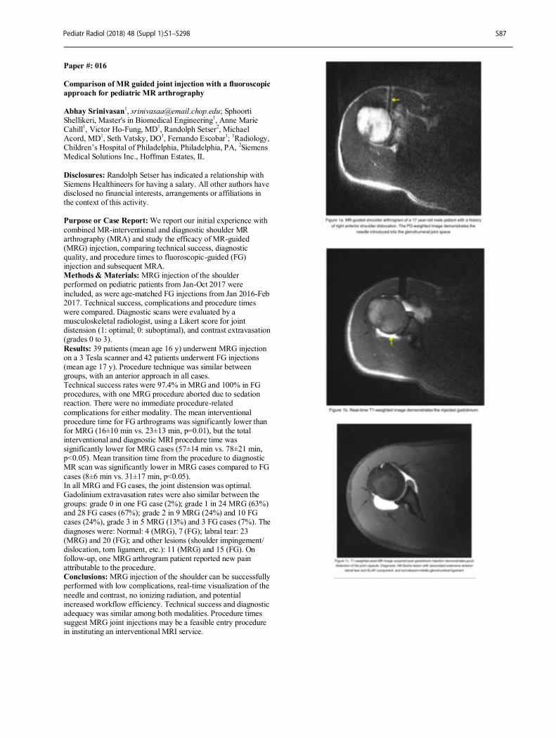
Paper #: 016
Comparison of MR guided joint injection with a fluoroscopic
approach for pediatric MR arthrography
Abhay Srinivasan1, [email protected]; Sphoorti
Shellikeri, Master's in Biomedical Engineering1, Anne Marie
Cahill1, Victor Ho-Fung, MD
1, Randolph Setser
2, Michael
Acord, MD1, Seth Vatsky, DO
1, Fernando Escobar
1;
1Radiology,
Children’s Hospital of Philadelphia, Philadelphia, PA, 2Siemens
Medical Solutions Inc., Hoffman Estates, IL
Disclosures: Randolph Setser has indicated a relationship with Siemens Healthineers for having a salary. All other authors have disclosed no financial interests, arrangements or affiliations in the context of this activity.
Purpose or Case Report: We report our initial experience with
combined MR-interventional and diagnostic shoulder MR arthrography (MRA) and study the efficacy of MR-guided (MRG) injection, comparing technical success, diagnostic quality, and procedure times to fluoroscopic-guided (FG) injection and subsequent MRA. Methods & Materials: MRG injection of the shoulder performed on pediatric patients from Jan-Oct 2017 were
included, as were age-matched FG injections from Jan 2016-Feb 2017. Technical success, complications and procedure times were compared. Diagnostic scans were evaluated by a musculoskeletal radiologist, using a Likert score for joint distension (1: optimal; 0: suboptimal), and contrast extravasation (grades 0 to 3).
Results: 39 patients (mean age 16 y) underwent MRG injection on a 3 Tesla scanner and 42 patients underwent FG injections (mean age 17 y). Procedure technique was similar between groups, with an anterior approach in all cases. Technical success rates were 97.4% in MRG and 100% in FG procedures, with one MRG procedure aborted due to sedation reaction. There were no immediate procedure-related
complications for either modality. The mean interventional procedure time for FG arthrograms was significantly lower than for MRG (16±10 min vs. 23±13 min, p=0.01), but the total interventional and diagnostic MRI procedure time was significantly lower for MRG cases (57±14 min vs. 78±21 min, p<0.05). Mean transition time from the procedure to diagnostic MR scan was significantly lower in MRG cases compared to FG
cases (8±6 min vs. 31±17 min, p<0.05). In all MRG and FG cases, the joint distension was optimal. Gadolinium extravasation rates were also similar between the groups: grade 0 in one FG case (2%); grade 1 in 24 MRG (63%) and 28 FG cases (67%); grade 2 in 9 MRG (24%) and 10 FG cases (24%), grade 3 in 5 MRG (13%) and 3 FG cases (7%). The
diagnoses were: Normal: 4 (MRG), 7 (FG); labral tear: 23 (MRG) and 20 (FG); and other lesions (shoulder impingement/ dislocation, torn ligament, etc.): 11 (MRG) and 15 (FG). On follow-up, one MRG arthrogram patient reported new pain attributable to the procedure. Conclusions: MRG injection of the shoulder can be successfully performed with low complications, real-time visualization of the
needle and contrast, no ionizing radiation, and potential increased workflow efficiency. Technical success and diagnostic adequacy was similar among both modalities. Procedure times suggest MRG joint injections may be a feasible entry procedure in instituting an interventional MRI service.
Pediatr Radiol (2018) 48 (Suppl 1):S1–S298 S87
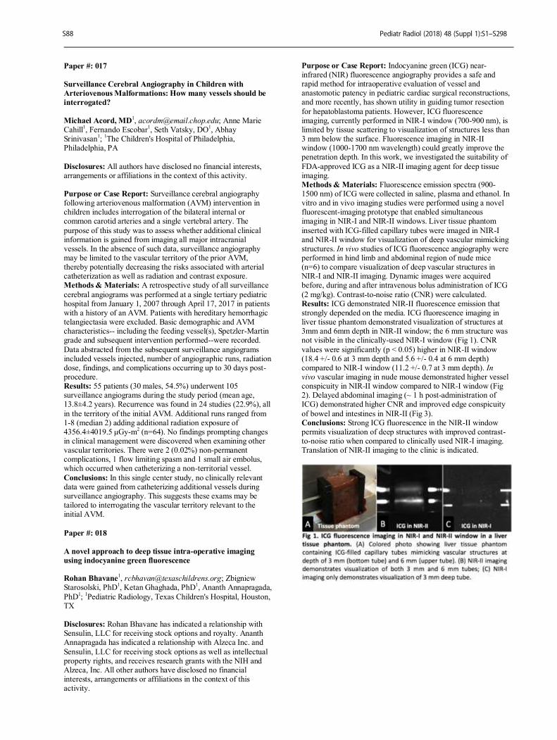
Paper #: 017
Surveillance Cerebral Angiography in Children with
Arteriovenous Malformations: How many vessels should be
interrogated?
Michael Acord, MD
1, [email protected]; Anne Marie
Cahill1, Fernando Escobar
1, Seth Vatsky, DO
1, Abhay
Srinivasan1;
1The Children's Hospital of Philadelphia,
Philadelphia, PA
Disclosures: All authors have disclosed no financial interests, arrangements or affiliations in the context of this activity. Purpose or Case Report: Surveillance cerebral angiography following arteriovenous malformation (AVM) intervention in children includes interrogation of the bilateral internal or common carotid arteries and a single vertebral artery. The
purpose of this study was to assess whether additional clinical information is gained from imaging all major intracranial vessels. In the absence of such data, surveillance angiography may be limited to the vascular territory of the prior AVM, thereby potentially decreasing the risks associated with arterial catheterization as well as radiation and contrast exposure. Methods & Materials: A retrospective study of all surveillance
cerebral angiograms was performed at a single tertiary pediatric hospital from January 1, 2007 through April 17, 2017 in patients with a history of an AVM. Patients with hereditary hemorrhagic telangiectasia were excluded. Basic demographic and AVM characteristics-- including the feeding vessel(s), Spetzler-Martin grade and subsequent intervention performed--were recorded.
Data abstracted from the subsequent surveillance angiograms included vessels injected, number of angiographic runs, radiation dose, findings, and complications occurring up to 30 days post-procedure. Results: 55 patients (30 males, 54.5%) underwent 105 surveillance angiograms during the study period (mean age, 13.8±4.2 years). Recurrence was found in 24 studies (22.9%), all
in the territory of the initial AVM. Additional runs ranged from 1-8 (median 2) adding additional radiation exposure of 4356.4±4019.5 μGy-m
2 (n=64). No findings prompting changes
in clinical management were discovered when examining other vascular territories. There were 2 (0.02%) non-permanent complications, 1 flow limiting spasm and 1 small air embolus, which occurred when catheterizing a non-territorial vessel.
Conclusions: In this single center study, no clinically relevant data were gained from catheterizing additional vessels during surveillance angiography. This suggests these exams may be tailored to interrogating the vascular territory relevant to the initial AVM.
Paper #: 018
A novel approach to deep tissue intra-operative imaging
using indocyanine green fluorescence
Rohan Bhavane
1, [email protected]; Zbigniew
Starosolski, PhD1, Ketan Ghaghada, PhD
1, Ananth Annapragada,
PhD1;
1Pediatric Radiology, Texas Children's Hospital, Houston,
TX
Disclosures: Rohan Bhavane has indicated a relationship with Sensulin, LLC for receiving stock options and royalty. Ananth Annapragada has indicated a relationship with Alzeca Inc. and
Sensulin, LLC for receiving stock options as well as intellectual property rights, and receives research grants with the NIH and Alzeca, Inc. All other authors have disclosed no financial interests, arrangements or affiliations in the context of this activity.
Purpose or Case Report: Indocyanine green (ICG) near-infrared (NIR) fluorescence angiography provides a safe and rapid method for intraoperative evaluation of vessel and anastomotic patency in pediatric cardiac surgical reconstructions, and more recently, has shown utility in guiding tumor resection
for hepatoblastoma patients. However, ICG fluorescence imaging, currently performed in NIR-I window (700-900 nm), is limited by tissue scattering to visualization of structures less than 3 mm below the surface. Fluorescence imaging in NIR-II window (1000-1700 nm wavelength) could greatly improve the penetration depth. In this work, we investigated the suitability of
FDA-approved ICG as a NIR-II imaging agent for deep tissue imaging. Methods & Materials: Fluorescence emission spectra (900-1500 nm) of ICG were collected in saline, plasma and ethanol. In vitro and in vivo imaging studies were performed using a novel fluorescent-imaging prototype that enabled simultaneous imaging in NIR-I and NIR-II windows. Liver tissue phantom
inserted with ICG-filled capillary tubes were imaged in NIR-I and NIR-II window for visualization of deep vascular mimicking structures. In vivo studies of ICG fluorescence angiography were performed in hind limb and abdominal region of nude mice (n=6) to compare visualization of deep vascular structures in NIR-I and NIR-II imaging. Dynamic images were acquired before, during and after intravenous bolus administration of ICG
(2 mg/kg). Contrast-to-noise ratio (CNR) were calculated. Results: ICG demonstrated NIR-II fluorescence emission that strongly depended on the media. ICG fluorescence imaging in liver tissue phantom demonstrated visualization of structures at 3mm and 6mm depth in NIR-II window; the 6 mm structure was not visible in the clinically-used NIR-I window (Fig 1). CNR
values were significantly (p < 0.05) higher in NIR-II window (18.4 +/- 0.6 at 3 mm depth and 5.6 +/- 0.4 at 6 mm depth) compared to NIR-I window (11.2 +/- 0.7 at 3 mm depth). In vivo vascular imaging in nude mouse demonstrated higher vessel conspicuity in NIR-II window compared to NIR-I window (Fig 2). Delayed abdominal imaging (~ 1 h post-administration of ICG) demonstrated higher CNR and improved edge conspicuity
of bowel and intestines in NIR-II (Fig 3). Conclusions: Strong ICG fluorescence in the NIR-II window permits visualization of deep structures with improved contrast-to-noise ratio when compared to clinically used NIR-I imaging. Translation of NIR-II imaging to the clinic is indicated.
Pediatr Radiol (2018) 48 (Suppl 1):S1–S298S88
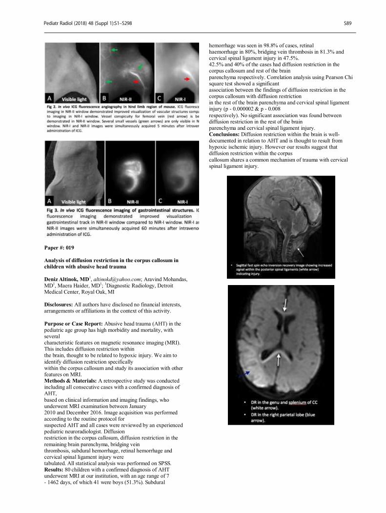
Paper #: 019
Analysis of diffusion restriction in the corpus callosum in
children with abusive head trauma
Deniz Altinok, MD
1, [email protected]; Aravind Mohandas,
MD1, Maera Haider, MD
1;
1Diagnostic Radiology, Detroit
Medical Center, Royal Oak, MI Disclosures: All authors have disclosed no financial interests, arrangements or affiliations in the context of this activity.
Purpose or Case Report: Abusive head trauma (AHT) in the pediatric age group has high morbidity and mortality, with several characteristic features on magnetic resonance imaging (MRI). This includes diffusion restriction within the brain, thought to be related to hypoxic injury. We aim to
identify diffusion restriction specifically within the corpus callosum and study its association with other features on MRI. Methods & Materials: A retrospective study was conducted including all consecutive cases with a confirmed diagnosis of AHT,
based on clinical information and imaging findings, who underwent MRI examination between January 2010 and December 2016. Image acquisition was performed according to the routine protocol for suspected AHT and all cases were reviewed by an experienced pediatric neuroradiologist. Diffusion restriction in the corpus callosum, diffusion restriction in the
remaining brain parenchyma, bridging vein thrombosis, subdural hemorrhage, retinal hemorrhage and cervical spinal ligament injury were tabulated. All statistical analysis was performed on SPSS. Results: 80 children with a confirmed diagnosis of AHT underwent MRI at our institution, with an age range of 7 - 1462 days, of which 41 were boys (51.3%). Subdural
hemorrhage was seen in 98.8% of cases, retinal haemorrhage in 80%, bridging vein thrombosis in 81.3% and cervical spinal ligament injury in 47.5%. 42.5% and 40% of the cases had diffusion restriction in the corpus callosum and rest of the brain
parenchyma respectively. Correlation analysis using Pearson Chi square test showed a significant association between the findings of diffusion restriction in the corpus callosum with diffusion restriction in the rest of the brain parenchyma and cervical spinal ligament injury (p - 0.000002 & p - 0.008
respectively). No significant association was found between diffusion restriction in the rest of the brain parenchyma and cervical spinal ligament injury. Conclusions: Diffusion restriction within the brain is well-documented in relation to AHT and is thought to result from hypoxic ischemic injury. However our results suggest that diffusion restriction within the corpus
callosum shares a common mechanism of trauma with cervical spinal ligament injury.
Pediatr Radiol (2018) 48 (Suppl 1):S1–S298 S89
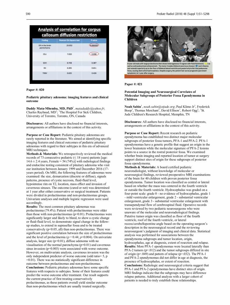
Paper #: 020
Pediatric pituitary adenoma: imaging features and clinical
outcome
Daddy Mata-Mbemba, MD, PhD
Charles Raybaud, MD1;
1The Hospital For Sick Children,
University of Toronto, Toronto, ON, Canada Disclosures: All authors have disclosed no financial interests, arrangements or affiliations in the context of this activity.
Purpose or Case Report: Pediatric pituitary adenomas are rarely reported in the literature. We aimed at identifying specific imaging features and clinical outcomes of pediatric pituitary adenomas with regard to their subtypes in this era of advanced MRI techniques.
Methods & Materials: We retrospectively reviewed the medical records of 73 consecutive pediatric (≤ 18 years) patients [age: 14.6 ± 2.4 years, Female = 54 (74%)] with radiological findings and endocrine testing consistent of pituitary adenoma who visit our institution between January 1999 and December 2016 (17-years period). On MRI, the following features of adenomas were examined: the size, demarcation (discrete or diffuse), signals
patterns, presence of cystic necrosis, fluid-fluid level, hypointense rim on T2, location, mass effect and invasion of cavernous sinuses. The outcome (cured or not) was determined at 1 year after either conservative or surgical treatment. Patients were divided in prolactinomas and non-prolactinomas groups. Univariate analyses and multiple logistic regression were used accordingly.
Results: The most common pituitary adenomas was prolactinoma (79.4%). Patient with prolactinomas were older than those with non-prolactinomas (p=0.01). Prolactinomas were significantly larger and likely to bleed, to show a cystic change or fluid-fluid level, to demonstrate signal change in the follow-up studies, to extend in suprasellar fossa and to be treated
conservatively (p<0.05, all) than non-prolactinomas. There was significant positive correlation between the size of prolactinomas
and the level of prolactinemia (ρ=0.44; p<0.004). On univariate
analysis, larger size (p=0.01), diffuse adenoma with no
visualisation of the normal parenchyma (p=0.01) and cavernous sinus invasion (p=0.003) were associated with worse outcome. However, on multivariate analysis, cavernous invasion was the only independent predictor of worse outcome (odd ratio= 5, p =0.01). There was no statistically significant difference in outcome between prolactinomas and non-prolactinomas. Conclusions: Pediatric pituitary adenomas have some specific
features with respects to subtypes. Some of their features could predict the worse outcome after treatment. Our result supports the current practice of first treating conservatively prolactinomas, as these patients overall yield similar outcome than non-prolactinomas which are usually treated surgically.
Paper #: 021
Potential Imaging and Neurosurgical Correlates of
Molecular Subgroups of Posterior Fossa Ependymoma in
Children Noah Sabin
1, [email protected]; Paul Klimo Jr
1, Frederick
Boop1, Thomas Merchant
1, David Ellison
1, Robert Ogg
1;
1St.
Jude Children's Research Hospital, Memphis, TN Disclosures: All authors have disclosed no financial interests, arrangements or affiliations in the context of this activity. Purpose or Case Report: Recent research on pediatric ependymoma has established two distinct major molecular
subgroups of posterior fossa tumors, PFA-1 and PFA-2. PFA-1 ependymomas have a genetic profile that suggest an origin in the lower brainstem while the molecular signature of PFA-2 lesions points to a source in the rostral posterior fossa. We examined whether brain imaging and reported location of tumor at surgery support distinct sites of origin for these subgroups of posterior
fossa ependymoma. Methods & Materials: A board certified pediatric neuroradiologist, without knowledge of molecular or neurosurgical findings, reviewed preoperative MRI examinations of the brain for 40 children with proven posterior fossa ependymoma. Tumor location was classified as central or lateral based on whether the mass was centered in the fourth ventricle
or outside the fourth ventricle. Hydrocephalus was graded on a four-point scale: grade 0 - no evidence of hydrocephalus, grade 1 - mild ventricular enlargement, grade 2 - substantial ventricular enlargement, grade 3 - substantial ventricular enlargement with transependymal flow of cerebrospinal fluid. Operative records were reviewed by two pediatric neurosurgeons who were unaware of the molecular and neuroradiological findings.
Putative tumor origin was classified as floor of the fourth ventricle, roof of the fourth ventricle, or lateral recess/cerebellopontine angle based on the operating surgeon’s description in the neurosurgical record and the reviewing neurosurgeon’s judgment of imaging and clinical data. Statistical analysis was performed for associations between the
ependymoma subgroups and tumor location, origin, hydrocephalus, age at diagnosis, extent of resection and relapse. Results: More PFA-1 ependymomas were located laterally than PFA-2 tumors (p=.012) and the tumor subgroups differed in site of origin (p=.049) and pattern of relapse (p=.032). The PFA-1 and PFA-2 ependymomas did not differ in age at diagnosis, the presence of hydrocephalus, or extent of resection.
Conclusions: Radiologic and neurosurgical criteria suggest that PFA-1 and PFA-2 ependymomas have distinct sites of origin. MRI findings indicate that the subgroups may have difference relapse patterns. Additional analysis with a larger cohort of patients is needed to truly establish these relationships.
Pediatr Radiol (2018) 48 (Suppl 1):S1–S298S90
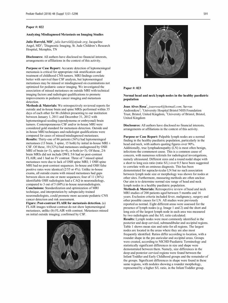
Paper #: 022
Analyzing Misdiagnosed Metastasis on Imaging Studies Julie Harreld, MD
1, [email protected]; Jacqueline
Angel, MD1;
1Diagnostic Imaging, St. Jude Children’s Research
Hospital, Memphis, TN
Disclosures: All authors have disclosed no financial interests, arrangements or affiliations in the context of this activity.
Purpose or Case Report: Accurate detection of leptomeningeal metastasis is critical for appropriate risk stratification and treatment of childhood CNS tumors. MRI findings correlate better with survival than CSF analysis, but leptomeningeal metastases may be missed or misdiagnosed on examinations not optimized for pediatric cancer imaging. We investigated the association of missed metastases on outside MRI with technical
imaging factors and radiologist qualifications to promote improvements in pediatric cancer imaging and metastasis detection. Methods & Materials: We retrospectively reviewed reports for outside and in-house brain and spine MRIs performed within 35 days of each other for 86 children presenting to our institution between January 1, 2011 and December 31, 2012 with
leptomeningeal-seeding (ependymoma or embryonal) brain tumors. Contemporaneous CSF and/or in-house MRI were considered gold standard for metastasis detection. Outside and in-house MRI techniques and radiologist qualifications were compared for cases of missed/misdiagnosed metastases. Results: Thirty-one of 86 patients (36%) had leptomeningeal
metastases (13 brain, 3 spine, 15 both) by initial in-house MRI ± CSF. Of these, 10 (32%) had metastases undiagnosed by OSH MRI of brain (n=3), spine (n=4), or both (n=3). Of these, 2/6 brain MRIs did not include DWI; 5/6 had no post-contrast FLAIR; and 1 had no IV contrast. Three of 7 missed spinal metastases were due to lack of OSH spine MRI; 1 OSH spine MRI had no post-contrast sequences. In-house and OSH false
positive rates were identical (2/55 or 4%). Unlike in-house exams, all outside exams with missed metastases had gaps between slices on one or more sequences; four of 11 (36%) identifiable OSH radiologists had a CAQ in neuroradiology, compared to 3 out of 5 (60%) in-house neuroradiologists. Conclusions: Standardization and optimization of MRI technique, and interpretation by subspecialty-trained
neuroradiologists, could promote more accurate pediatric CNS cancer detection and risk assessment. Figure: Post-contrast FLAIR for metastasis detection. (a) FLAIR images without contrast do not show leptomeningeal metastases, unlike (b) FLAIR with contrast. Metastases missed on initial outside imaging; confirmed by CSF.
Paper #: 023
Normal head and neck lymph nodes in the healthy paediatric
population Joao Alves Rosa
1, [email protected]; Savvas
Andronikou2;
1University Hospital Bristol NHS Foundation
Trust, Bristol, United Kingdom, 2University of Bristol, Bristol,
United Kingdom Disclosures: All authors have disclosed no financial interests, arrangements or affiliations in the context of this activity.
Purpose or Case Report: Palpable lymph nodes are a normal finding in the healthy paediatric population, particularly in the head and neck, with authors quoting figures over 90%. Additionally, true lymphadenopathy (LN) is most often benign, infections the commonest cause. This is a common cause of concern, with numerous referrals for radiological investigations,
namely ultrasound. Different sizes and a round nodal shape with a short to long axis ratio (ratio S/L) over 0.5 have been suggested to correlate with an ominous diagnosis. This has been demonstrated for supraclavicular LN but no such association between lymph node size or morphology was shown for nodes at other sites. Furthermore, measuring methods are often unclear. Our aim is to determine normal size range of head and neck
lymph nodes in a healthy paediatric population. Methods & Materials: Retrospective review of head and neck MRI studies of 200 patients aged between 5 months and 16 years. Exclusion criteria included fever, malignancy, surgery and other possible causes for LN. All studies were previously reported as normal. Eight different areas were assessed for the presence of lymph nodes (e.g. Image 1 and 2) and the short and
long axis of the largest lymph node in each area was measured by two radiologists and the S/L ratio calculated. Results: Lymph nodes were most commonly identified in the posterior and deep cervical, submandibular and occipital regions. Table 1 shows mean size and ratio for all regions. The largest nodes are located in the areas where they are also most
frequently identified. Ratios differ according to location, with a rounder shape in the pre-auricular and occipital areas. Groups were created, according to NICHD Paediatric Terminology and statistically significant differences in size and shape were demonstrated between them. Namely, size differences in the deep and posterior cervical regions were found between the Infant/Toddler and Early Childhood groups and the remainder of
the groups. Significant differences in shape were found in these same regions, with nodules showing a rounder morphology, represented by a higher S/L ratio, in the Infant/Toddler group.
Pediatr Radiol (2018) 48 (Suppl 1):S1–S298 S91
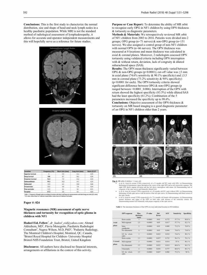
Conclusions: This is the first study to characterize the normal distribution, size and shape of head and neck lymph nodes in a healthy paediatric population. While MRI is not the standard method of radiological assessment of lymphadenopathy, it allows for accurate and operator independent measurements and
this will hopefully serve as a reference for future studies.
Paper #: 024
Magnetic resonance (MR) assessment of optic nerve
thickness and tortuosity for recognition of optic glioma in
children with NF1 Hadeel Eid, Fellow
1, [email protected]; Ahmed
Aldraihem, MD1, Flavia Menegotto, Paediatric Radiologist
Consultant2, Nagwa Wilson, M.D, PhD
1;
1Pediatric Radiology,
The Montreal Children's Hospital, Montreal, QC, Canada, 2Bristol Royal Hospital for Children- University Hospital
Bristol-NHS Foundation Trust, Bristol, United Kingdom Disclosures: All authors have disclosed no financial interests, arrangements or affiliations in the context of this activity.
Purpose or Case Report: To determine the ability of MR orbit to recognize early OPG in NF1 children by using OPN thickness & tortuosity as diagnostic parameters. Methods & Materials: We retrospectively reviewed MR orbit of NF1 children from 2003 to 2016. Patients were divided into 2
groups; OPG group (n=71 nerves) & non-OPG group (n=151 nerves). We also assigned a control group of non-NF1 children with normal OPN (n=66 nerves). The OPN thickness was measured at 8 locations and mean thickness was calculated in axial & coronal planes. Moreover, 2 radiologists assessed OPN tortuosity using validated criteria including OPN interruption
with & without return, deviation, lack of congruity & dilated subarachnoid space (SAS). Results: The OPN mean thickness significantly varied between OPG & non-OPG groups (p<0.0001); cut-off value was ≥3 mm in axial plane (74.6% sensitivity & 90.1% specificity) and ≥3.5 mm in coronal plane (73.2% sensitivity & 94% specificity) (p<0.0001 for each). The OPN tortuosity criteria showed
significant difference between OPG & non-OPG groups (p ranged between <0.0001_0.006). Interruption of the OPN with return showed the highest specificity (83.5%) while dilated SAS had the least specificity (61.2%). Combination of the 5 parameters increased the specificity up to 98.6%. Conclusions: Objective assessment of the OPN thickness & tortuosity on MR based imaging is a good diagnostic parameter
of an OPG in NF1 children older than 2 years.
Pediatr Radiol (2018) 48 (Suppl 1):S1–S298S92
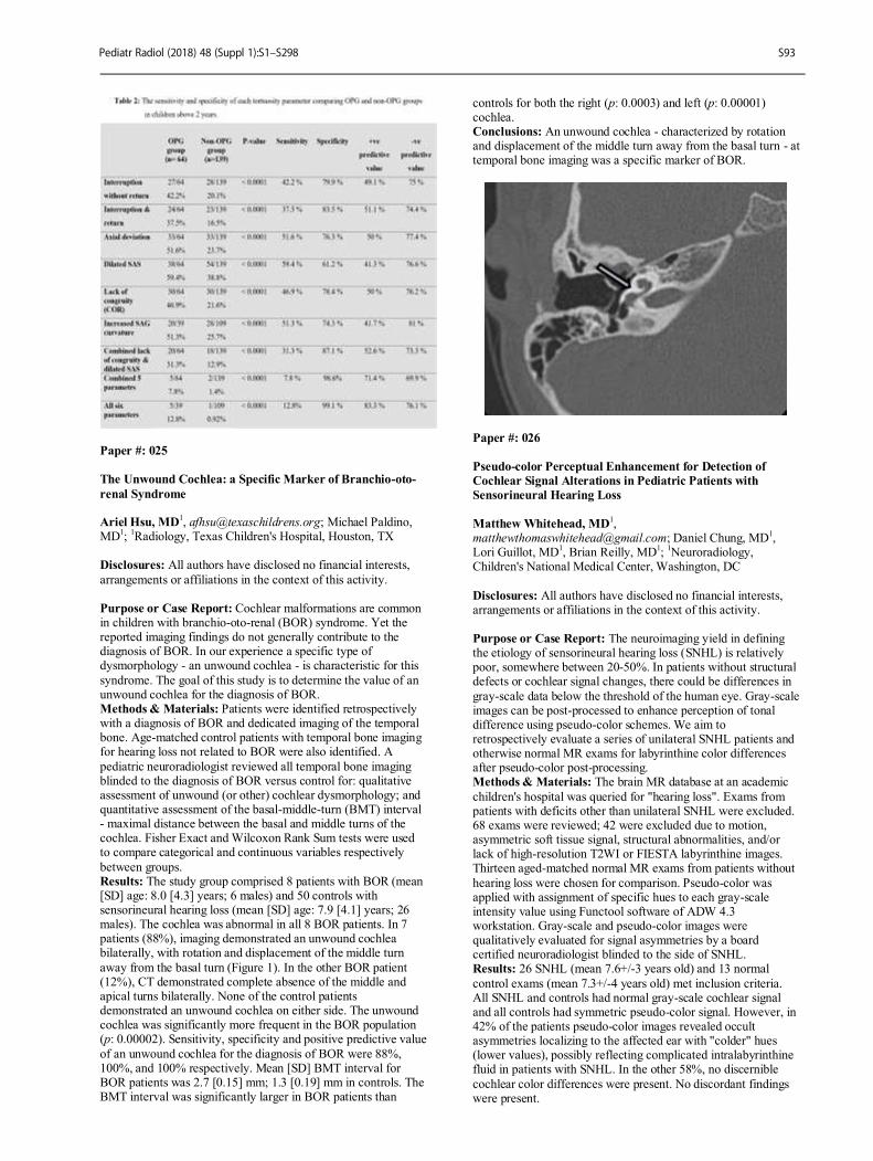
Paper #: 025
The Unwound Cochlea: a Specific Marker of Branchio-oto-
renal Syndrome Ariel Hsu, MD
1, [email protected]; Michael Paldino,
MD1;
1Radiology, Texas Children's Hospital, Houston, TX
Disclosures: All authors have disclosed no financial interests,
arrangements or affiliations in the context of this activity. Purpose or Case Report: Cochlear malformations are common in children with branchio-oto-renal (BOR) syndrome. Yet the reported imaging findings do not generally contribute to the diagnosis of BOR. In our experience a specific type of dysmorphology - an unwound cochlea - is characteristic for this
syndrome. The goal of this study is to determine the value of an unwound cochlea for the diagnosis of BOR. Methods & Materials: Patients were identified retrospectively with a diagnosis of BOR and dedicated imaging of the temporal bone. Age-matched control patients with temporal bone imaging for hearing loss not related to BOR were also identified. A
pediatric neuroradiologist reviewed all temporal bone imaging blinded to the diagnosis of BOR versus control for: qualitative assessment of unwound (or other) cochlear dysmorphology; and quantitative assessment of the basal-middle-turn (BMT) interval - maximal distance between the basal and middle turns of the cochlea. Fisher Exact and Wilcoxon Rank Sum tests were used to compare categorical and continuous variables respectively
between groups. Results: The study group comprised 8 patients with BOR (mean [SD] age: 8.0 [4.3] years; 6 males) and 50 controls with sensorineural hearing loss (mean [SD] age: 7.9 [4.1] years; 26 males). The cochlea was abnormal in all 8 BOR patients. In 7 patients (88%), imaging demonstrated an unwound cochlea bilaterally, with rotation and displacement of the middle turn
away from the basal turn (Figure 1). In the other BOR patient (12%), CT demonstrated complete absence of the middle and apical turns bilaterally. None of the control patients demonstrated an unwound cochlea on either side. The unwound cochlea was significantly more frequent in the BOR population (p: 0.00002). Sensitivity, specificity and positive predictive value
of an unwound cochlea for the diagnosis of BOR were 88%, 100%, and 100% respectively. Mean [SD] BMT interval for BOR patients was 2.7 [0.15] mm; 1.3 [0.19] mm in controls. The BMT interval was significantly larger in BOR patients than
controls for both the right (p: 0.0003) and left (p: 0.00001) cochlea. Conclusions: An unwound cochlea - characterized by rotation and displacement of the middle turn away from the basal turn - at temporal bone imaging was a specific marker of BOR.
Paper #: 026
Pseudo-color Perceptual Enhancement for Detection of
Cochlear Signal Alterations in Pediatric Patients with
Sensorineural Hearing Loss
Matthew Whitehead, MD
1,
[email protected]; Daniel Chung, MD1,
Lori Guillot, MD1, Brian Reilly, MD
1;
1Neuroradiology,
Children's National Medical Center, Washington, DC
Disclosures: All authors have disclosed no financial interests, arrangements or affiliations in the context of this activity. Purpose or Case Report: The neuroimaging yield in defining the etiology of sensorineural hearing loss (SNHL) is relatively poor, somewhere between 20-50%. In patients without structural defects or cochlear signal changes, there could be differences in
gray-scale data below the threshold of the human eye. Gray-scale images can be post-processed to enhance perception of tonal difference using pseudo-color schemes. We aim to retrospectively evaluate a series of unilateral SNHL patients and otherwise normal MR exams for labyrinthine color differences after pseudo-color post-processing. Methods & Materials: The brain MR database at an academic
children's hospital was queried for "hearing loss". Exams from patients with deficits other than unilateral SNHL were excluded. 68 exams were reviewed; 42 were excluded due to motion, asymmetric soft tissue signal, structural abnormalities, and/or lack of high-resolution T2WI or FIESTA labyrinthine images. Thirteen aged-matched normal MR exams from patients without
hearing loss were chosen for comparison. Pseudo-color was applied with assignment of specific hues to each gray-scale intensity value using Functool software of ADW 4.3 workstation. Gray-scale and pseudo-color images were qualitatively evaluated for signal asymmetries by a board certified neuroradiologist blinded to the side of SNHL. Results: 26 SNHL (mean 7.6+/-3 years old) and 13 normal
control exams (mean 7.3+/-4 years old) met inclusion criteria. All SNHL and controls had normal gray-scale cochlear signal and all controls had symmetric pseudo-color signal. However, in 42% of the patients pseudo-color images revealed occult asymmetries localizing to the affected ear with "colder" hues (lower values), possibly reflecting complicated intralabyrinthine fluid in patients with SNHL. In the other 58%, no discernible
cochlear color differences were present. No discordant findings were present.
Pediatr Radiol (2018) 48 (Suppl 1):S1–S298 S93
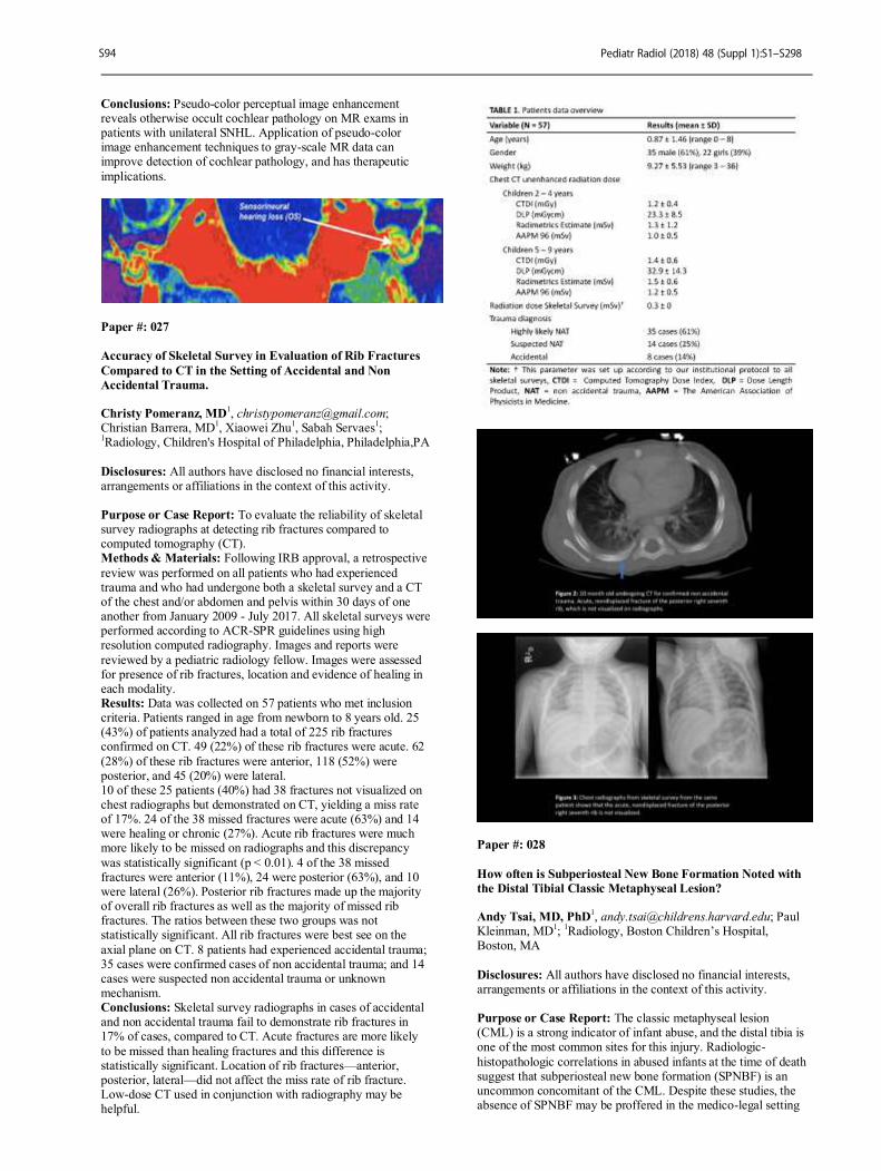
Conclusions: Pseudo-color perceptual image enhancement reveals otherwise occult cochlear pathology on MR exams in patients with unilateral SNHL. Application of pseudo-color image enhancement techniques to gray-scale MR data can improve detection of cochlear pathology, and has therapeutic
implications.
Paper #: 027
Accuracy of Skeletal Survey in Evaluation of Rib Fractures
Compared to CT in the Setting of Accidental and Non
Accidental Trauma. Christy Pomeranz, MD
Christian Barrera, MD1, Xiaowei Zhu
1, Sabah Servaes
1;
1Radiology, Children's Hospital of Philadelphia, Philadelphia,PA
Disclosures: All authors have disclosed no financial interests, arrangements or affiliations in the context of this activity. Purpose or Case Report: To evaluate the reliability of skeletal survey radiographs at detecting rib fractures compared to computed tomography (CT). Methods & Materials: Following IRB approval, a retrospective
review was performed on all patients who had experienced trauma and who had undergone both a skeletal survey and a CT of the chest and/or abdomen and pelvis within 30 days of one another from January 2009 - July 2017. All skeletal surveys were performed according to ACR-SPR guidelines using high resolution computed radiography. Images and reports were
reviewed by a pediatric radiology fellow. Images were assessed for presence of rib fractures, location and evidence of healing in each modality. Results: Data was collected on 57 patients who met inclusion criteria. Patients ranged in age from newborn to 8 years old. 25 (43%) of patients analyzed had a total of 225 rib fractures confirmed on CT. 49 (22%) of these rib fractures were acute. 62
(28%) of these rib fractures were anterior, 118 (52%) were posterior, and 45 (20%) were lateral. 10 of these 25 patients (40%) had 38 fractures not visualized on chest radiographs but demonstrated on CT, yielding a miss rate of 17%. 24 of the 38 missed fractures were acute (63%) and 14 were healing or chronic (27%). Acute rib fractures were much more likely to be missed on radiographs and this discrepancy
was statistically significant (p < 0.01). 4 of the 38 missed fractures were anterior (11%), 24 were posterior (63%), and 10 were lateral (26%). Posterior rib fractures made up the majority of overall rib fractures as well as the majority of missed rib fractures. The ratios between these two groups was not statistically significant. All rib fractures were best see on the
axial plane on CT. 8 patients had experienced accidental trauma; 35 cases were confirmed cases of non accidental trauma; and 14 cases were suspected non accidental trauma or unknown mechanism. Conclusions: Skeletal survey radiographs in cases of accidental and non accidental trauma fail to demonstrate rib fractures in 17% of cases, compared to CT. Acute fractures are more likely
to be missed than healing fractures and this difference is statistically significant. Location of rib fractures—anterior, posterior, lateral—did not affect the miss rate of rib fracture. Low-dose CT used in conjunction with radiography may be helpful.
Paper #: 028
How often is Subperiosteal New Bone Formation Noted with
the Distal Tibial Classic Metaphyseal Lesion? Andy Tsai, MD, PhD
1, [email protected]; Paul
Kleinman, MD1;
1Radiology, Boston Children’s Hospital,
Boston, MA
Disclosures: All authors have disclosed no financial interests, arrangements or affiliations in the context of this activity. Purpose or Case Report: The classic metaphyseal lesion (CML) is a strong indicator of infant abuse, and the distal tibia is one of the most common sites for this injury. Radiologic-
histopathologic correlations in abused infants at the time of death suggest that subperiosteal new bone formation (SPNBF) is an uncommon concomitant of the CML. Despite these studies, the absence of SPNBF may be proffered in the medico-legal setting
Pediatr Radiol (2018) 48 (Suppl 1):S1–S298S94
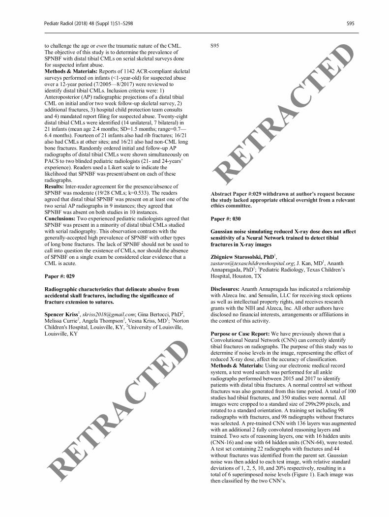
to challenge the age or even the traumatic nature of the CML. The objective of this study is to determine the prevalence of SPNBF with distal tibial CMLs on serial skeletal surveys done for suspected infant abuse. Methods & Materials: Reports of 1142 ACR-compliant skeletal
surveys performed on infants (<1-year-old) for suspected abuse over a 12-year period (7/2005—8/2017) were reviewed to identify distal tibial CMLs. Inclusion criteria were: 1) Anteroposterior (AP) radiographic projections of a distal tibial CML on initial and/or two week follow-up skeletal survey, 2) additional fractures, 3) hospital child protection team consults
and 4) mandated report filing for suspected abuse. Twenty-eight distal tibial CMLs were identified (14 unilateral, 7 bilateral) in 21 infants (mean age 2.4 months; SD=1.5 months; range=0.7—6.4 months). Fourteen of 21 infants also had rib fractures; 16/21 also had CMLs at other sites; and 16/21 also had non-CML long bone fractures. Randomly ordered initial and follow-up AP radiographs of distal tibial CMLs were shown simultaneously on
PACS to two blinded pediatric radiologists (21- and 24-years’ experience). Readers used a Likert scale to indicate the likelihood that SPNBF was present/absent on each of these radiographs. Results: Inter-reader agreement for the presence/absence of SPNBF was moderate (19/28 CMLs; k=0.533). The readers agreed that distal tibial SPNBF was present on at least one of the
two serial AP radiographs in 9 instances; they agreed that SPNBF was absent on both studies in 10 instances. Conclusions: Two experienced pediatric radiologists agreed that SPNBF was present in a minority of distal tibial CMLs studied with serial radiography. This observation contrasts with the generally-accepted high prevalence of SPNBF with other types
of long bone fractures. The lack of SPNBF should not be used to call into question the existence of CMLs, nor should the absence of SPNBF on a single exam be considered clear evidence that a CML is acute.
Paper #: 029
Radiographic characteristics that delineate abusive from
accidental skull fractures, including the significance of
fracture extension to sutures. Spencer Kriss
1, [email protected]; Gina Bertocci, PhD
2,
Melissa Currie2, Angela Thompson
2, Vesna Kriss, MD
1;
1Norton
Children's Hospital, Louisville, KY, 2University of Louisville,
Louisville, KY
Paper #: 030
Gaussian noise simulating reduced X-ray dose does not affect
sensitivity of a Neural Network trained to detect tibial
fractures in X-ray images Zbigniew Starosolski, PhD
1,
[email protected]; J. Kan, MD1, Ananth
Annapragada, PhD1;
1Pediatric Radiology, Texas Children’s
Hospital, Houston, TX
Disclosures: Ananth Annapragada has indicated a relationship with Alzeca Inc. and Sensulin, LLC for receiving stock options as well as intellectual property rights, and receives research grants with the NIH and Alzeca, Inc. All other authors have disclosed no financial interests, arrangements or affiliations in the context of this activity.
Purpose or Case Report: We have previously shown that a Convolutional Neural Network (CNN) can correctly identify tibial fractures on radiographs. The purpose of this study was to determine if noise levels in the image, representing the effect of reduced X-ray dose, affect the accuracy of classification. Methods & Materials: Using our electronic medical record
system, a text word search was performed for all ankle radiographs performed between 2015 and 2017 to identify patients with distal tibia fractures. A normal control set without fractures was also generated from this time period. A total of 100 studies had tibial fractures, and 350 studies were normal. All images were cropped to a standard size of 299x299 pixels, and rotated to a standard orientation. A training set including 98
radiographs with fractures, and 98 radiographs without fractures was selected. A pre-trained CNN with 136 layers was augmented with an additional 2 fully convoluted reasoning layers and trained. Two sets of reasoning layers, one with 16 hidden units (CNN-16) and one with 64 hidden units (CNN-64), were tested. A test set containing 22 radiographs with fractures and 44
without fractures was identified from the parent set. Gaussian noise was then added to each test image, with relative standard deviations of 1, 2, 5, 10, and 20% respectively, resulting in a total of 6 superimposed noise levels (Figure 1). Each image was then classified by the two CNN’s.
Pediatr Radiol (2018) 48 (Suppl 1):S1–S298 S95
RETRACTED S95
RETRACTED
Abstract Paper #:029 withdrawn at author's request becausethe study lacked appropriate ethical oversight from a relevantethics committee.
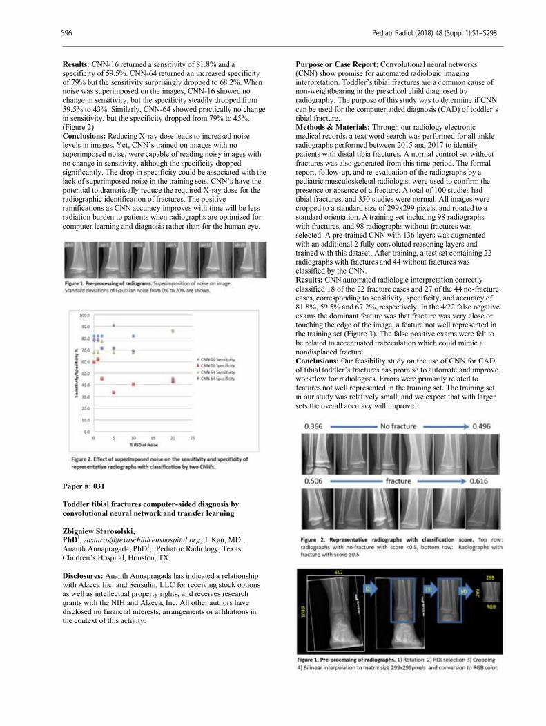
Results: CNN-16 returned a sensitivity of 81.8% and a specificity of 59.5%. CNN-64 returned an increased specificity of 79% but the sensitivity surprisingly dropped to 68.2%. When noise was superimposed on the images, CNN-16 showed no change in sensitivity, but the specificity steadily dropped from
59.5% to 43%. Similarly, CNN-64 showed practically no change in sensitivity, but the specificity dropped from 79% to 45%. (Figure 2) Conclusions: Reducing X-ray dose leads to increased noise levels in images. Yet, CNN’s trained on images with no superimposed noise, were capable of reading noisy images with
no change in sensitivity, although the specificity dropped significantly. The drop in specificity could be associated with the lack of superimposed noise in the training sets. CNN’s have the potential to dramatically reduce the required X-ray dose for the radiographic identification of fractures. The positive ramifications as CNN accuracy improves with time will be less radiation burden to patients when radiographs are optimized for
computer learning and diagnosis rather than for the human eye.
Paper #: 031
Toddler tibial fractures computer-aided diagnosis by
convolutional neural network and transfer learning
Zbigniew Starosolski, PhD
1, [email protected]; J. Kan, MD
1,
Ananth Annapragada, PhD1;
1Pediatric Radiology, Texas
Children’s Hospital, Houston, TX
Disclosures: Ananth Annapragada has indicated a relationship with Alzeca Inc. and Sensulin, LLC for receiving stock options as well as intellectual property rights, and receives research grants with the NIH and Alzeca, Inc. All other authors have disclosed no financial interests, arrangements or affiliations in the context of this activity.
Purpose or Case Report: Convolutional neural networks (CNN) show promise for automated radiologic imaging interpretation. Toddler’s tibial fractures are a common cause of non-weightbearing in the preschool child diagnosed by radiography. The purpose of this study was to determine if CNN
can be used for the computer aided diagnosis (CAD) of toddler’s tibial fracture. Methods & Materials: Through our radiology electronic medical records, a text word search was performed for all ankle radiographs performed between 2015 and 2017 to identify patients with distal tibia fractures. A normal control set without
fractures was also generated from this time period. The formal report, follow-up, and re-evaluation of the radiographs by a pediatric musculoskeletal radiologist were used to confirm the presence or absence of a fracture. A total of 100 studies had tibial fractures, and 350 studies were normal. All images were cropped to a standard size of 299x299 pixels, and rotated to a standard orientation. A training set including 98 radiographs
with fractures, and 98 radiographs without fractures was selected. A pre-trained CNN with 136 layers was augmented with an additional 2 fully convoluted reasoning layers and trained with this dataset. After training, a test set containing 22 radiographs with fractures and 44 without fractures was classified by the CNN. Results: CNN automated radiologic interpretation correctly
classified 18 of the 22 fracture cases and 27 of the 44 no-fracture cases, corresponding to sensitivity, specificity, and accuracy of 81.8%, 59.5% and 67.2%, respectively. In the 4/22 false negative exams the dominant feature was that fracture was very close or touching the edge of the image, a feature not well represented in the training set (Figure 3). The false positive exams were felt to
be related to accentuated trabeculation which could mimic a nondisplaced fracture. Conclusions: Our feasibility study on the use of CNN for CAD of tibial toddler’s fractures has promise to automate and improve workflow for radiologists. Errors were primarily related to features not well represented in the training set. The training set in our study was relatively small, and we expect that with larger
sets the overall accuracy will improve.
Pediatr Radiol (2018) 48 (Suppl 1):S1–S298S96
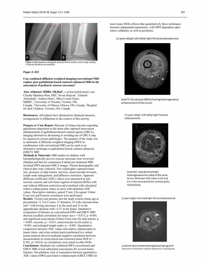
Paper #: 032
Can combined diffusion-weighted imaging/conventional MRI
replace post gadolinium-based contrast-enhanced MRI in the
assessment of pediatric osseous sarcomas? Alaa Alsharief, MBBS, SB-Rad
Claudia Martinez-Rios, MD2, Sevan Hopyan
1, Afsaneh
Amirabadi3, Andrea Doria
1, Mary-Louise Greer,
MBBS1;
1University of Toronto, Toronto, ON,
Canada, 2University of Ottawa, Ottawa, ON, Canada,
3Hospital
for Sick Children, Toronto, ON, Canada Disclosures: All authors have disclosed no financial interests,
arrangements or affiliations in the context of this activity. Purpose or Case Report: Because of rising concerns regarding gadolinium deposition in the brain after repeated intravenous administration of gadolinium-based contrast agents (GBCA), imaging alternatives decreasing or avoiding use of GBCA may
be required in certain pathologies. The purpose of the study was to determine if diffusion-weighted imaging (DWI) in combination with conventional MRI can be used as an alternative technique to gadolinium-based contrast-enhanced (GBCE) MRI Methods & Materials: MRI studies in children with histopathologically-proven osseous sarcomas were reviewed
(blinded and then by consensus) if initial pre-treatment MRI included DWI and post-GBCA images. Patient demographic and clinical data were collected. Two radiologists captured tumor size, presence of skip lesions, necrosis, neurovascular invasion, lymph node enlargement, and diffusion restriction. Apparent diffusion coefficient (ADC) values were measured in non-necrotic osseous and soft tissue regions-of-interest (ROIs) with
and without diffusion restriction and correlated with calculated relative enhancement values in areas with minimum ADC values. Descriptive statistics, paired T-test, Chi-square/ Fisher Exact test and Pearson correlation were performed. Results: Twenty-one patients met the study criteria (mean age at presentation 11.3±4.2 years, 15 females); 14 with osteosarcomas
and 7 with Ewing sarcomas; 6 in the axial and 15 in the appendicular skeleton with 11/21 in the femur. Qualitative comparison of features on conventional/DWI and GBCE MRI showed excellent correlation for tumor size r = 0.873, p <0.001, and significant associations (Fisher Exact test) for skip lesions: p < 0.001; necrosis: p = 0.021; neurovascular involvement: p <0.001; and enlarged lymph nodes: p < 0.001. Quantitative
comparison between ADC values and relative enhancement in tumor (intra- and extra-cortical parts) performed in a subset lesion analysis showed moderate negative correlation between measurements in extracortical non-restricted tumor ROI (r= -0.583, p= 0.014); no correlations were noted in other ROIs. Conclusions: Qualitatively combined DWI-conventional and
GBCE MRI reveal substantial associations for several tumor features. Nevertheless, lack of association between quantitative ADC values (DWI) and relative enhancement (GBCE) MRI for
most tumor ROIs reflects that quantitatively these techniques measure independent parameters, with DWI dependent upon tumor cellularity as well as perfusion.
Pediatr Radiol (2018) 48 (Suppl 1):S1–S298 S97
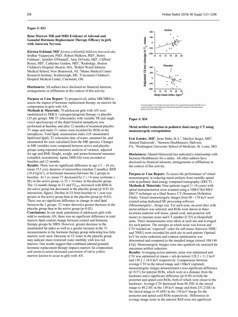
Paper #: 033
Bone Marrow MR and MRS Evidence of Adrenal and
Gonadal Hormone Replacement Therapy Efficacy in girls
with Anorexia Nervosa
Kirsten Ecklund, MD
Sridhar Vajapeyam, PhD1, Robert Mulkern, PhD
1, Henry
Feldman1, Jennifer O'Donnell
2, Amy DiVasta, MD
1, Clifford
Rosen, MD3, Catherine Gordon, MD
4;
1Radiology, Boston
Children's Hospital, Boston, MA, 2Robert Wood Johnson
Medical School, New Brunswick, NJ, 3Maine Medical Center
Research Institute, Scarborough, ME, 4Cincinnati Children's
Hospital Medical Center, Cincinnati, OH Disclosures: All authors have disclosed no financial interests, arrangements or affiliations in the context of this activity.
Purpose or Case Report: To prospectively utilize MR/MRS to assess the impact of hormone replacement therapy on marrow fat composition in girls with AN. Methods & Materials: 70 adolescent girls with AN were randomized to DHEA +estrogen/progestin therapy vs placebo (35 per group). MR T1 relaxometry with variable TR and single voxel spectroscopy of the distal femoral metaphysis was
performed at baseline and after 12 months of treatment/placebo. T1 maps and mean T1 values were recorded for ROIs in the metaphysis. Total lipid, unsaturation index (UI=unsaturated lipid/total lipid), T2 relaxation time of water, saturated fat, and unsaturated fat were calculated from the MR sprectra. Changes in MR variables were compared between active and placebo
groups using repeated-measures analysis of variance, adjusted for age and BMI. Height, weight, and serum hormonal measures (estradiol, testosterone, leptin, DHEAS) were recorded at baseline and 12 months. Results: There was no significant difference in age (11 - 18 yrs, mean 15.5 yrs), duration of amenorrhea (median 5 months), BMI (18.8 kg/m
2), or hormonal measures between the 2 groups at
baseline. At 1 yr, mean T1 decreased by 2 ± 14 msec (estimate ± SE) in the active group, vs 25 ± 14 msec in the placebo group. The 12-month change in T1 and T2unsat increased with BMI in the active group but decreased in the placebo group (p=0.01 for interaction; figure). Decline in UI from baseline to 1 year was greater in the active group than in the placebo group (p=0.02). There was no significant difference in change in total lipid
between the 2 groups. T2 water showed a greater decrease in the placebo group than in the active group (p=0.02). Conclusions: In our study population of adolescent girls with mild to moderate AN, there was no significant difference in total marrow lipid content change between control and hormone therapy groups by MRS. However, greater decrease in the
unsaturated fat index as well as a greater increase in the T1 measurements in the hormone therapy group indicating less fatty marrow were seen. Decrease in T2 water in the placebo group may indicate more restricted water mobility with less red marrow. Our results suggest that combined adrenal/gonadal hormone replacement therapy impacts marrow fat composition and seems to arrest increased conversion of red to yellow
marrow known to occur in girls with AN.
Paper #: 034
Metal artifact reduction in pediatric dual energy CT using
monoenergetic extrapolation
Eric Eutsler, MD2, Irene Duba, B.A.
1, Marilyn Siegel, MD
2,
Ahmed Halaweish1;
1Siemens Healthineers, Malvern,
PA, 2Washington University School of Medicine, St. Louis, MO
Disclosures: Ahmed Halaweish has indicated a relationship with Siemens Healthineers for a salary. All other authors have disclosed no financial interests, arrangements or affiliations in
the context of this activity. Purpose or Case Report: To assess the performance of virtual monoenergetic in reducing metal artifacts from metallic spinal rods in pediatric dual-energy computed tomography (DECT). Methods & Materials: Nine patients (aged 11-18 years) with
spinal instrumentation were scanned using a 100kV/Sn140kV DECT technique on a Dual Source CT (Somatom Definition Flash). Virtual monoenergetic images from 90 - 170 keV were created using dedicated DE processing software (Monoenergetic+, Syngo.via). For each scan, an axial slice with metal artifacts was selected, and ROIs were drawn in three locations (anterior soft tissue, spinal cord, and posterior soft
tissue) to measure noise and CT number (CT#) in Hounsfield units. Three measurements were taken at each site and averaged for each patient. The energies at which noise was lowest and CT# reached an “expected” value for soft tissue (between 30HU and 70HU) were recorded for each site in each patient. Optimal keV for noise reduction and contrast optimization was determined and compared to the standard image (mixed 100/140
kVp). Monoenergetic Images were also qualitatively assessed for maximum artifact reduction. Results: Averaging across patients, noise was minimized and CT# was optimized at (mean ± std deviation) 128.5 ± 11.2 keV and 130.2 ± 14.6 keV respectively. Comparisons between average CT# in the mixed image and 130keV (optimal)
monoenergetic images demonstrated a non-significant difference (p=0.57) for anterior ROIs, which were at a distance from the hardware and a significant difference (p<0.05) in both the posterior and spinal cord ROIs, both of which were closer to the hardware. Average CT# decreased from 88.3HU in the mixed image to 40.2 HU in the 130 keV image and from 251.21HU in the mixed image to 43.6HU in the 130 keV image for the
posterior and spinal cord ROIs respectively. Differences in average image noise in the anterior ROI were not significant
Pediatr Radiol (2018) 48 (Suppl 1):S1–S298S98
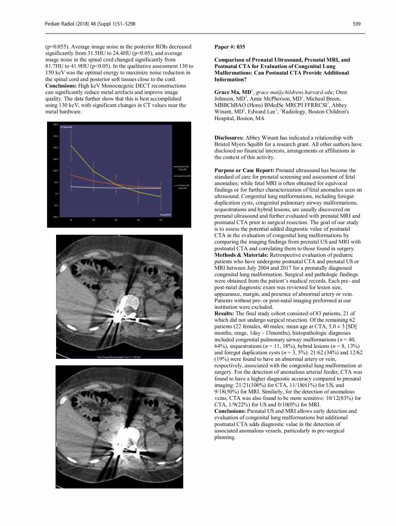
(p=0.055). Average image noise in the posterior ROIs decreased significantly from 31.5HU to 24.4HU (p<0.05), and average image noise in the spinal cord changed significantly from 81.7HU to 41.9HU (p<0.05). In the qualitative assessment 130 to 150 keV was the optimal energy to maximize noise reduction in
the spinal cord and posterior soft tissues close to the cord. Conclusions: High keV Monoenegetic DECT reconstructions can significantly reduce metal artifacts and improve image quality. The data further show that this is best accomplished using 130 keV, with significant changes in CT values near the metal hardware.
Paper #: 035
Comparison of Prenatal Ultrasound, Prenatal MRI, and
Postnatal CTA for Evaluation of Congenital Lung
Malformations: Can Postnatal CTA Provide Additional
Information? Grace Ma, MD
1, [email protected]; Oren
Johnson, MD1, Amie McPherson, MD
1, Micheal Breen,
MBBChBAO (Hons) BMedSc MRCPI FFRRCSI1, Abbey
Winant, MD1, Edward Lee
1;
1Radiology, Boston Children's
Hospital, Boston, MA
Disclosures: Abbey Winant has indicated a relationship with Bristol Myers Squibb for a research grant. All other authors have disclosed no financial interests, arrangements or affiliations in the context of this activity.
Purpose or Case Report: Prenatal ultrasound has become the standard of care for prenatal screening and assessment of fetal anomalies; while fetal MRI is often obtained for equivocal findings or for further characterization of fetal anomalies seen on ultrasound. Congenital lung malformations, including foregut duplication cysts, congenital pulmonary airway malformations,
sequestrations and hybrid lesions, are usually discovered on prenatal ultrasound and further evaluated with prenatal MRI and postnatal CTA prior to surgical resection. The goal of our study is to assess the potential added diagnostic value of postnatal CTA in the evaluation of congenital lung malformations by comparing the imaging findings from prenatal US and MRI with
postnatal CTA and correlating them to those found in surgery. Methods & Materials: Retrospective evaluation of pediatric patients who have undergone postnatal CTA and prenatal US or MRI between July 2004 and 2017 for a prenatally diagnosed congenital lung malformation. Surgical and pathologic findings were obtained from the patient’s medical records. Each pre- and post-natal diagnostic exam was reviewed for lesion size,
appearance, margin, and presence of abnormal artery or vein. Patients without pre- or post-natal imaging preformed at our institution were excluded. Results: The final study cohort consisted of 83 patients, 21 of which did not undergo surgical resection. Of the remaining 62 patients (22 females, 40 males; mean age at CTA, 5.0 ± 3 [SD] months; range, 1day - 13months), histopathologic diagnoses
included congenital pulmonary airway malformations (n = 40, 64%), sequestrations (n = 11, 18%), hybrid lesions (n = 8, 13%) and foregut duplication cysts (n = 3, 5%). 21/62 (34%) and 12/62 (19%) were found to have an abnormal artery or vein, respectively, associated with the congenital lung malformation at surgery. For the detection of anomalous arterial feeder, CTA was
found to have a higher diagnostic accuracy compared to prenatal imaging: 21/21(100%) for CTA, 11/18(61%) for US, and 9/18(50%) for MRI. Similarly, for the detection of anomalous veins, CTA was also found to be more sensitive: 10/12(83%) for CTA, 1/9(22%) for US and 0/10(0%) for MRI. Conclusions: Prenatal US and MRI allows early detection and evaluation of congenital lung malformations but additional
postnatal CTA adds diagnostic value in the detection of associated anomalous vessels, particularly in pre-surgical planning.
Pediatr Radiol (2018) 48 (Suppl 1):S1–S298 S99
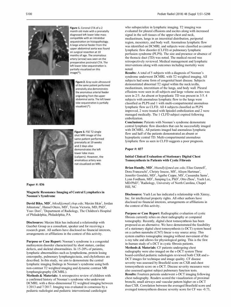
Paper #: 036
Magnetic Resonance Imaging of Central Lymphatics in
Noonan’s Syndrome David Biko, MD
1, [email protected]; Maxim Itkin
1, Jordan
Johnstone1, Hansel Otero, MD
1, Teresa Victoria, MD, PhD
1,
Yoav Dori1;
1Department of Radiology, The Children's Hospital
of Philadelphia, Philadelphia, PA
Disclosures: Maxim Itkin has indicated a relationship with Guerbet Group as a consultant, speaker and for receiving a research grant. All authors have disclosed no financial interests, arrangements or affiliations in the context of this activity.
Purpose or Case Report: Noonan’s syndrome is a congenital multisystem disorder characterized by short stature, cardiac defects, and skeletal abnormalities. In 15-20% of patients, lymphatic abnormalities such as lymphedema, protein losing enteropathy, pulmonary lymphangiectasia, and chylothorax are described. In this study, we aim to demonstrate the central lymphatic imaging findings in Noonan’s syndrome using both
non-contrast T2 weighted imaging and dynamic contrast MR lymphangiography (DCMRL). Methods & Materials: A retrospective review of children with a confirmed history of Noonan’s syndrome who underwent DCMRL with a three-dimensional T2 weighted imaging between 1/2013 and 7/2017. Imaging was evaluated in consensus by a pediatric radiologist and pediatric interventional cardiologist
who subspecialize in lymphatic imaging. T2 imaging was evaluated for pleural effusions and ascites along with increased signal in the soft tissues of the upper chest and neck, mediastinum, lungs in an interstitial distribution, periportal region, mesentery, and body wall. Anomalous lymphatic flow
was identified on DCMRL and subjects were classified as central lymphatic flow disorder (CLFD) or pulmonary lymphatic perfusion syndrome (PLPS). The size and presence or absence of the thoracic duct (TD) was noted. The medical record was retrospectively reviewed. Medical management and lymphatic interventions along with outcomes including mortality were
noted. Results: A total of 5 subjects with a diagnosis of Noonan’s syndrome underwent DCMRL with T2 weighted imaging. All subjects had some form of congenital heart disease. Subjects demonstrated abnormal T2 signal within the neck/axilla, mediastinum, interstitium of the lungs, and body wall. Pleural effusions were seen in all subjects and large volume ascites was
seen in 2/5. An absent or hypoplastic TD was present in 3/5. 4 subjects with anomalous lymphatic flow to the lungs were classified as PLPS and 1 with multi-compartmental anomalous lymphatic flow as CLFD. All 4 subjects classified as PLPS improved, 2 were treated with lipiodol embolization and 2 were managed medically. The 1 CLFD subject expired following embolization.
Conclusions: Patients with Noonan’s syndrome demonstrate central lymphatic flow disorders that can be successfully imaged with DCMRL. All patients imaged had anomalous lymphatic flow and half of the patients demonstrated an absent or hypoplastic central TD. Multi-compartmental anomalous lymphatic flow as seen in CLFD suggests a poor prognosis.
Paper #: 037
Initial Clinical Evaluation of Stationary Digital Chest
Tomosynthesis in Patients with Cystic Fibrosis Brian Handly, MD
1, [email protected]; Elias Gunnell
1,
Dora Franceschi1, Christy Inscoe, MS
1, Alison Hartman
1,
Jennifer Goralski, MD1, Agathe Ceppe, MS
1, Cassandra Sams
1,
Lynn Fordham, MD1, Jianping Lu, PhD
1, Otto Zhou
1, Yueh Lee,
MD/PhD1;
1Radiology, University of North Carolina, Chapel
Hill, NC Disclosures: Yueh Lee has indicated a relationship with Xinray,
Inc. for intellectual property rights. All other authors have disclosed no financial interests, arrangements or affiliations in the context of this activity. Purpose or Case Report: Radiographic evaluation of cystic fibrosis currently relies on chest radiography or computed
tomography. Recently, digital chest tomosynthesis has been proposed as an alternative. We have demonstrated the feasibility of a stationary digital chest tomosynthesis (s-DCT) system based on a carbon nanotube (CNT) linear x-ray source array. This system enables tomographic imaging without movement of the x-ray tube and allows for physiological gating. This is the first in-human study of s-DCT in cystic fibrosis patients.
Methods & Materials: CF patients undergoing chest radiography were also imaged on the s-DCT system Three board-certified pediatric radiologists reviewed both CXR and s-DCT images for technique and image quality. CF disease severity was assessed by Brasfield score on CXR and chest tomosynthesis score on s-DCT. Disease severity measures were
also assessed against subject pulmonary function tests. Results: Fourteen patients underwent s-DCT imaging following chest radiography. Readers scored the visualization of proximal bronchi, small airways and vascular pattern higher on s-DCT than CXR. Correlation between the averaged Brasfield score and averaged tomosynthesis disease severity score for CF was -0.73,
Pediatr Radiol (2018) 48 (Suppl 1):S1–S298S100
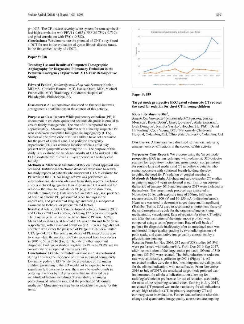
p=.0033. The CF disease severity score system for tomosynthesis had high correlation with FEV1 (-0.685), FEF 25-75% (-0.719), and good correlation with FVC (-0.582). Conclusions: We demonstrate the potential of CNT x-ray based s-DCT for use in the evaluation of cystic fibrosis disease status,
in the first clinical study of s-DCT.
Paper #: 038
Trending Use and Results of Computed Tomographic
Angiography for Diagnosing Pulmonary Embolism in the
Pediatric Emergency Department: A 13-Year Retrospective
Study. Edward Fenlon
1, [email protected]; Summer Kaplan,
MD MS1, Christian Barrera, MD
1, Hansel Otero, MD
1, Michael
Francavilla, MD1;
1Radiology, Children's Hospital of
Philadelphia, Philadelphia, PA
Disclosures: All authors have disclosed no financial interests, arrangements or affiliations in the context of this activity. Purpose or Case Report: While pulmonary embolism (PE) is uncommon in children, quick and accurate diagnosis is crucial to ensure timely management. The rate of PE is reported to be
approximately 16% among children with clinically suspected PE who underwent computed tomographic angiography (CTA). Studies on the prevalence of PE in children have not accounted for the point of clinical care. The pediatric emergency department (ED) is a common location where a child may present with symptoms concerning for PE. The purpose of this
study is to evaluate the trends and results of CTAs ordered in the ED to evaluate for PE over a 13-year period in a tertiary care facility. Methods & Materials: Institutional Review Board approval was obtained. Institutional electronic databases were used to search for study reports of patients who underwent CTA to evaluate for PE while in the ED. No image review was performed; all
information and data was obtained from study reports. Exclusion criteria included age greater than 20 years and CTA ordered for reasons other than to evaluate for PE (e.g., aortic dissection, vascular trauma, etc.). Data recorded included: age, sex, presence of acute or chronic PE, presence of other findings in the impression, and presence of language indicating a suboptimal exam due to technical or patient related factors.
Results: A total of 308 CTAs performed between January 2005 and October 2017 met criteria, including 122 boys and 186 girls. The 13-year positive rate of acute or chronic PE was 16.2%. Mean and median age at time of CTA was 16.04 and 16.83 years respectively, with a standard deviation of 3.17 years. Age did not correlate with either the presence of PE (p=0.3109) or a limited
CTA (p=0.4176). The yearly incidence of PE ranged from zero to seven while the number of CTAs increased from two studies in 2005 to 53 in 2016 (Fig 1). The rate of other important diagnostic findings in studies negative for PE was 39.9% and the overall rate of suboptimal exams was 14%. Conclusions: Despite the tenfold increase in CTAs performed during 13 years, the incidence of PE has remained consistently
low in the pediatric ED. While the prevalence of PE among children presenting to the ED would not be expected to vary significantly from year to year, there may be yearly trends in ordering practices by ED physicians that are affected by a multitude of factors including CT availability, changing perceptions of radiation risk, and the practice of "defensive
medicine." More analysis may better elucidate the cause for this trend.
Paper #: 039
Target mode prospective EKG gated volumetric CT reduces
the need for sedation for chest CT in young children Rajesh Krishnamurthy
1,
[email protected]; Jessica Morrison
1, Kevin Dolan
1, Jarred Lovelace
1, Akila Sankaran
1,
Leah Dameron1, Jennifer Yashko
1, Houchun Hu, PhD
1, David
Hintenlang2, Cody Young, DO
1;
1Nationwide Children's
Hospital, Columbus, OH, 2Ohio State University, Columbus, OH
Disclosures: All authors have disclosed no financial interests,
arrangements or affiliations in the context of this activity. Purpose or Case Report: We propose using the 'target mode’ prospective EKG gating technique with volumetric 320-detector scanner for respiratory motion and gross motion compensation for routine lung and mediastinal CT in pediatric patients who
cannot cooperate with volitional breath-holding, thereby avoiding the need for IV sedation or general anesthesia. Methods & Materials: All chest and cardiovascular CT studies in patients ages 0-4 years performed at our institution between the period of January 2016 and September 2017 were included in the analysis. The target mode protocol was instituted in November 2016, with exposure time of 350ms, half-scan
reconstruction, 80-100 kV and 30-150 mA (indication based). Heart rate was used to determine target phase and ImageExact (Toshiba, Tustin, CA) used to reconstruct a motionless phase of the cardiorespiratory cycle for the organ/s of interest (lung, mediastinum, vasculature). Rate of sedation for chest CT before and after the institution of the target mode protocol was
compared using a test of proportions. Rate of call-back of patients for diagnostic inadequacy after an unsedated scan was monitored. Image quality grading by two radiologists on a 4-point scale, and quantitative image quality assessment by a physicist are pending. Results: From Jan-Nov 2016, 232 out of 358 studies (65.3%) were performed with sedation/GA. From Dec 2016-Sep 2017,
after the institution of the target mode protocol, 109 out of 310 patients (35.2%) were sedated. The 46% reduction in sedation rate was statistically significant (p<0.01) (Figure 1). All unsedated studies were done free-breathing and were diagnostic for the clinical indication, with no callbacks. From November 2016 to July of 2017, the unsedated target mode protocol was implemented for all chest indications, but allowing for
radiologist/clinician preference for use of sedation, accounting for most of the remaining sedated cases. Starting in July 2017, unsedated CT protocol was made mandatory for all indications except high resolution CT, inspiratory-expiratory CT and coronary stenosis evaluation. Further data collection after this change and quantitative image quality assessment are ongoing.
Pediatr Radiol (2018) 48 (Suppl 1):S1–S298 S101
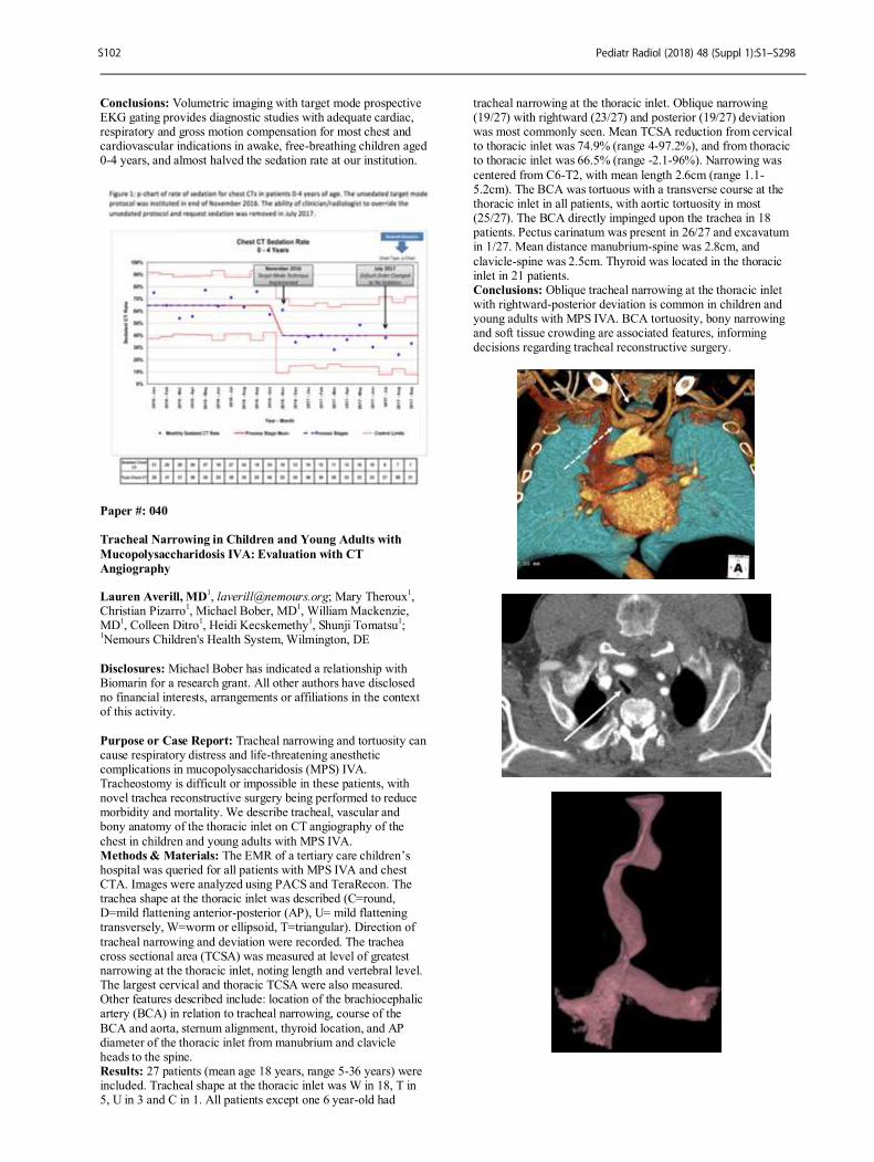
Conclusions: Volumetric imaging with target mode prospective EKG gating provides diagnostic studies with adequate cardiac, respiratory and gross motion compensation for most chest and cardiovascular indications in awake, free-breathing children aged 0-4 years, and almost halved the sedation rate at our institution.
Paper #: 040
Tracheal Narrowing in Children and Young Adults with
Mucopolysaccharidosis IVA: Evaluation with CT
Angiography Lauren Averill, MD
1, [email protected]; Mary Theroux
1,
Christian Pizarro1, Michael Bober, MD
1, William Mackenzie,
MD1, Colleen Ditro
1, Heidi Kecskemethy
1, Shunji Tomatsu
1;
1Nemours Children's Health System, Wilmington, DE
Disclosures: Michael Bober has indicated a relationship with Biomarin for a research grant. All other authors have disclosed no financial interests, arrangements or affiliations in the context of this activity.
Purpose or Case Report: Tracheal narrowing and tortuosity can cause respiratory distress and life-threatening anesthetic complications in mucopolysaccharidosis (MPS) IVA. Tracheostomy is difficult or impossible in these patients, with novel trachea reconstructive surgery being performed to reduce morbidity and mortality. We describe tracheal, vascular and bony anatomy of the thoracic inlet on CT angiography of the
chest in children and young adults with MPS IVA. Methods & Materials: The EMR of a tertiary care children’s hospital was queried for all patients with MPS IVA and chest CTA. Images were analyzed using PACS and TeraRecon. The trachea shape at the thoracic inlet was described (C=round, D=mild flattening anterior-posterior (AP), U= mild flattening transversely, W=worm or ellipsoid, T=triangular). Direction of
tracheal narrowing and deviation were recorded. The trachea cross sectional area (TCSA) was measured at level of greatest narrowing at the thoracic inlet, noting length and vertebral level. The largest cervical and thoracic TCSA were also measured. Other features described include: location of the brachiocephalic artery (BCA) in relation to tracheal narrowing, course of the
BCA and aorta, sternum alignment, thyroid location, and AP diameter of the thoracic inlet from manubrium and clavicle heads to the spine. Results: 27 patients (mean age 18 years, range 5-36 years) were included. Tracheal shape at the thoracic inlet was W in 18, T in 5, U in 3 and C in 1. All patients except one 6 year-old had
tracheal narrowing at the thoracic inlet. Oblique narrowing (19/27) with rightward (23/27) and posterior (19/27) deviation was most commonly seen. Mean TCSA reduction from cervical to thoracic inlet was 74.9% (range 4-97.2%), and from thoracic to thoracic inlet was 66.5% (range -2.1-96%). Narrowing was
centered from C6-T2, with mean length 2.6cm (range 1.1-5.2cm). The BCA was tortuous with a transverse course at the thoracic inlet in all patients, with aortic tortuosity in most (25/27). The BCA directly impinged upon the trachea in 18 patients. Pectus carinatum was present in 26/27 and excavatum in 1/27. Mean distance manubrium-spine was 2.8cm, and
clavicle-spine was 2.5cm. Thyroid was located in the thoracic inlet in 21 patients. Conclusions: Oblique tracheal narrowing at the thoracic inlet with rightward-posterior deviation is common in children and young adults with MPS IVA. BCA tortuosity, bony narrowing and soft tissue crowding are associated features, informing decisions regarding tracheal reconstructive surgery.
Pediatr Radiol (2018) 48 (Suppl 1):S1–S298S102
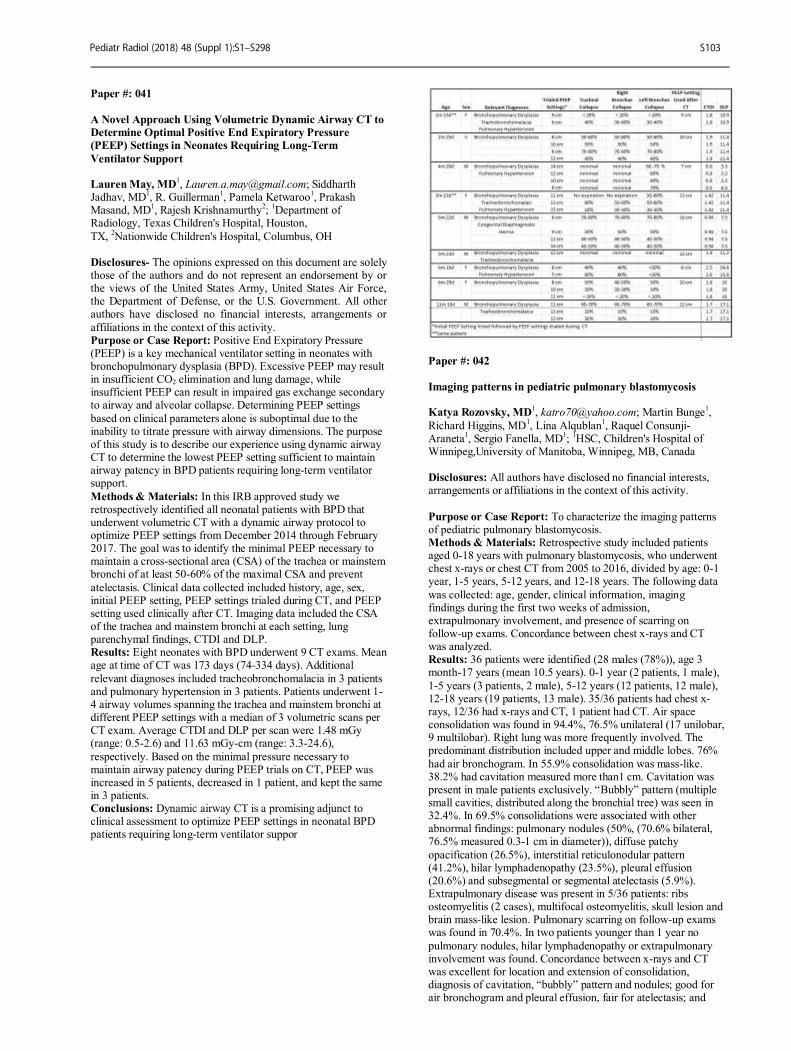
Paper #: 041
A Novel Approach Using Volumetric Dynamic Airway CT to
Determine Optimal Positive End Expiratory Pressure
(PEEP) Settings in Neonates Requiring Long-Term
Ventilator Support Lauren May, MD
1, [email protected]; Siddharth
Jadhav, MD1, R. Guillerman
1, Pamela Ketwaroo
1, Prakash
Masand, MD1, Rajesh Krishnamurthy
2;
1Department of
Radiology, Texas Children's Hospital, Houston,
TX, 2Nationwide Children's Hospital, Columbus, OH
Disclosures- The opinions expressed on this document are solely those of the authors and do not represent an endorsement by or the views of the United States Army, United States Air Force, the Department of Defense, or the U.S. Government. All other authors have disclosed no financial interests, arrangements or
affiliations in the context of this activity. Purpose or Case Report: Positive End Expiratory Pressure (PEEP) is a key mechanical ventilator setting in neonates with bronchopulmonary dysplasia (BPD). Excessive PEEP may result in insufficient CO2 elimination and lung damage, while insufficient PEEP can result in impaired gas exchange secondary to airway and alveolar collapse. Determining PEEP settings
based on clinical parameters alone is suboptimal due to the inability to titrate pressure with airway dimensions. The purpose of this study is to describe our experience using dynamic airway CT to determine the lowest PEEP setting sufficient to maintain airway patency in BPD patients requiring long-term ventilator support.
Methods & Materials: In this IRB approved study we retrospectively identified all neonatal patients with BPD that underwent volumetric CT with a dynamic airway protocol to optimize PEEP settings from December 2014 through February 2017. The goal was to identify the minimal PEEP necessary to maintain a cross-sectional area (CSA) of the trachea or mainstem bronchi of at least 50-60% of the maximal CSA and prevent
atelectasis. Clinical data collected included history, age, sex, initial PEEP setting, PEEP settings trialed during CT, and PEEP setting used clinically after CT. Imaging data included the CSA of the trachea and mainstem bronchi at each setting, lung parenchymal findings, CTDI and DLP. Results: Eight neonates with BPD underwent 9 CT exams. Mean age at time of CT was 173 days (74-334 days). Additional
relevant diagnoses included tracheobronchomalacia in 3 patients and pulmonary hypertension in 3 patients. Patients underwent 1-4 airway volumes spanning the trachea and mainstem bronchi at different PEEP settings with a median of 3 volumetric scans per CT exam. Average CTDI and DLP per scan were 1.48 mGy (range: 0.5-2.6) and 11.63 mGy-cm (range: 3.3-24.6),
respectively. Based on the minimal pressure necessary to maintain airway patency during PEEP trials on CT, PEEP was increased in 5 patients, decreased in 1 patient, and kept the same in 3 patients. Conclusions: Dynamic airway CT is a promising adjunct to clinical assessment to optimize PEEP settings in neonatal BPD patients requiring long-term ventilator suppor
Paper #: 042
Imaging patterns in pediatric pulmonary blastomycosis Katya Rozovsky, MD
1, [email protected]; Martin Bunge
1,
Richard Higgins, MD1, Lina Alqublan
1, Raquel Consunji-
Araneta1, Sergio Fanella, MD
1;
1HSC, Children's Hospital of
Winnipeg,University of Manitoba, Winnipeg, MB, Canada Disclosures: All authors have disclosed no financial interests, arrangements or affiliations in the context of this activity.
Purpose or Case Report: To characterize the imaging patterns of pediatric pulmonary blastomycosis. Methods & Materials: Retrospective study included patients aged 0-18 years with pulmonary blastomycosis, who underwent chest x-rays or chest CT from 2005 to 2016, divided by age: 0-1 year, 1-5 years, 5-12 years, and 12-18 years. The following data
was collected: age, gender, clinical information, imaging findings during the first two weeks of admission, extrapulmonary involvement, and presence of scarring on follow-up exams. Concordance between chest x-rays and CT was analyzed. Results: 36 patients were identified (28 males (78%)), age 3 month-17 years (mean 10.5 years). 0-1 year (2 patients, 1 male),
1-5 years (3 patients, 2 male), 5-12 years (12 patients, 12 male), 12-18 years (19 patients, 13 male). 35/36 patients had chest x-rays, 12/36 had x-rays and CT, 1 patient had CT. Air space consolidation was found in 94.4%, 76.5% unilateral (17 unilobar, 9 multilobar). Right lung was more frequently involved. The predominant distribution included upper and middle lobes. 76%
had air bronchogram. In 55.9% consolidation was mass-like. 38.2% had cavitation measured more than1 cm. Cavitation was present in male patients exclusively. “Bubbly” pattern (multiple small cavities, distributed along the bronchial tree) was seen in 32.4%. In 69.5% consolidations were associated with other abnormal findings: pulmonary nodules (50%, (70.6% bilateral, 76.5% measured 0.3-1 cm in diameter)), diffuse patchy
opacification (26.5%), interstitial reticulonodular pattern (41.2%), hilar lymphadenopathy (23.5%), pleural effusion (20.6%) and subsegmental or segmental atelectasis (5.9%). Extrapulmonary disease was present in 5/36 patients: ribs osteomyelitis (2 cases), multifocal osteomyelitis, skull lesion and brain mass-like lesion. Pulmonary scarring on follow-up exams was found in 70.4%. In two patients younger than 1 year no
pulmonary nodules, hilar lymphadenopathy or extrapulmonary involvement was found. Concordance between x-rays and CT was excellent for location and extension of consolidation, diagnosis of cavitation, “bubbly” pattern and nodules; good for air bronchogram and pleural effusion, fair for atelectasis; and
Pediatr Radiol (2018) 48 (Suppl 1):S1–S298 S103
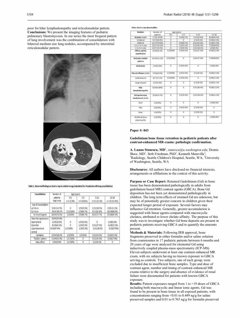
poor for hilar lymphadenopathy and reticulonodular pattern. Conclusions: We present the imaging features of pediatric pulmonary blastomycosis. In our series the most frequent pattern of lung involvement was the combination of consolidation with bilateral medium size lung nodules, accompanied by interstitial
reticulonodular pattern.
Paper #: 043
Gadolinium bone tissue retention in pediatric patients after
contrast-enhanced MR exams: pathologic confirmation. A. Luana Stanescu, MD
1, [email protected]; Dennis
Shaw, MD1, Seth Friedman, PhD
1, Kenneth Maravilla
2;
1Radiology, Seattle Children's Hospital, Seattle, WA,
2University
of Washington, Seattle, WA
Disclosures: All authors have disclosed no financial interests, arrangements or affiliations in the context of this activity.
Purpose or Case Report: Retained Gadolinium (Gd) in bone tissue has been demonstrated pathologically in adults from gadolinium based MRI contrast agents (GBCA). Bone Gd deposition has not been yet demonstrated pathologically in children. The long term effects of retained Gd are unknown, but may be of potentially greater concern in children given their
expected longer period of exposure. Several factors may influence Gd retention. Generally, greater accumularion is suggested with linear agents compared with macrocyclic chelates, attributed to lower chelate affinity. The purpose of this study was to investigate whether Gd bone deposits are present in pediatric patients receiving GBCA and to quantify the amounts present.
Methods & Materials: Following IRB approval, bone fragments preserved in either formalin and/or saline solution from craniotomies in 17 pediatric patients between 6 months and 20 years of age were analyzed for elemental Gd using inductively coupled plasma-mass spectrometry (ICP-MS). Eleven subjects underwent at least one contrast-enhanced MR
exam, with six subjects having no known exposure to GBCA serving as controls. Two subjects, one of each group, were excluded due to insufficient bone samples. Type and dose of contrast agent, number and timing of contrast-enhanced MR exams relative to the surgery and absence of evidence of renal failure were documented for patients with known GBCA exposure.
Results: Patient exposures ranged from 1 to >19 doses of GBCA including both macrocyclic and linear ionic agents. Gd was found to be present in bone tissue in all exposed patients, with concentrations ranging from <0.01 to 0.449 ng/g for saline preserved samples and 0.01 to 0.765 ng/g for formalin preserved
Pediatr Radiol (2018) 48 (Suppl 1):S1–S298S104
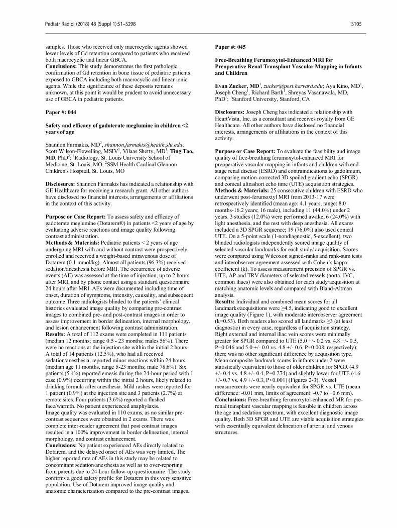
samples. Those who received only macrocyclic agents showed lower levels of Gd retention compared to patients who received both macrocyclic and linear GBCA. Conclusions: This study demonstrates the first pathologic confirmation of Gd retention in bone tissue of pediatric patients
exposed to GBCA including both macrocyclic and linear ionic agents. While the significance of these deposits remains unknown, at this point it would be prudent to avoid unnecessary use of GBCA in pediatric patients.
Paper #: 044
Safety and efficacy of gadoterate meglumine in children <2
years of age Shannon Farmakis, MD
Scott Wilson-Flewelling, MSIV1, Vilaas Shetty, MD
1, Ting Tao,
MD, PhD2;
1Radiology, St. Louis University School of
Medicine, St. Louis, MO, 2SSM Health Cardinal Glennon
Children's Hospital, St. Louis, MO Disclosures: Shannon Farmakis has indicated a relationship with GE Healthcare for receiving a research grant. All other authors have disclosed no financial interests, arrangements or affiliations in the context of this activity.
Purpose or Case Report: To assess safety and efficacy of gadoterate meglumine (Dotarem®) in patients <2 years of age by evaluating adverse reactions and image quality following contrast administration. Methods & Materials: Pediatric patients < 2 years of age
undergoing MRI with and without contrast were prospectively enrolled and received a weight-based intravenous dose of Dotarem (0.1 mmol/kg). Almost all patients (96.3%) received sedation/anesthesia before MRI. The occurrence of adverse events (AE) was assessed at the time of injection, up to 2 hours after MRI, and by phone contact using a standard questionnaire 24 hours after MRI. AEs were documented including time of
onset, duration of symptoms, intensity, causality, and subsequent outcome.Three radiologists blinded to the patients’ clinical histories evaluated image quality by comparing pre-contrast images to combined pre- and post-contrast images in order to assess improvement in border delineation, internal morphology, and lesion enhancement following contrast administration. Results: A total of 112 exams were completed in 111 patients
(median 12 months; range 0.5 - 23 months; males 56%). There were no reactions at the injection site within the initial 2 hours. A total of 14 patients (12.5%), who had all received sedation/anesthesia, reported minor reactions within 24 hours (median age 11 months, range 5-23 months; male 78.6%). Six patients (5.4%) reported emesis during the 24-hour period with 1
case (0.9%) occurring within the initial 2 hours, likely related to drinking formula after anesthesia. Mild rashes were reported for 1 patient (0.9%) at the injection site and 3 patients (2.7%) at remote sites. Four patients (3.6%) reported a flushed face/warmth. No patient experienced anaphylaxis. Image quality was evaluated in 110 exams, as no similar pre-contrast sequences were obtained in 2 exams. There was
complete inter-reader agreement that post contrast images resulted in a 100% improvement in border delineation, internal morphology, and contrast enhancement. Conclusions: No patient experienced AEs directly related to Dotarem, and the delayed onset of AEs was very limited. The higher reported rate of AEs in this study may be related to
concomitant sedation/anesthesia as well as to over-reporting from parents due to 24-hour follow-up questionnaire. The study confirms a good safety profile for Dotarem in this very sensitive population. Use of Dotarem improved image quality and anatomic characterization compared to the pre-contrast images.
Paper #: 045
Free-Breathing Ferumoxytol-Enhanced MRI for
Preoperative Renal Transplant Vascular Mapping in Infants
and Children Evan Zucker, MD
1, [email protected]; Aya Kino, MD
1,
Joseph Cheng1, Richard Barth
1, Shreyas Vasanawala, MD,
PhD1;
1Stanford University, Stanford, CA
Disclosures: Joseph Cheng has indicated a relationship with
HeartVista, Inc. as a consultant and receives royalty from GE Healthcare. All other authors have disclosed no financial interests, arrangements or affiliations in the context of this activity. Purpose or Case Report: To evaluate the feasibility and image quality of free-breathing ferumoxytol-enhanced MRI for
preoperative vascular mapping in infants and children with end-stage renal disease (ESRD) and contraindications to gadolinium, comparing motion-corrected 3D spoiled gradient echo (SPGR) and conical ultrashort echo time (UTE) acquisition strategies. Methods & Materials: 25 consecutive children with ESRD who underwent post-ferumoxtyl MRI from 2013-17 were retrospectively identified (mean age: 4.1 years, range: 8.0
months-16.2 years; 16 male), including 11 (44.0%) under 2 years. 3 studies (12.0%) were performed awake, 6 (24.0%) with light anesthesia, and the rest with deep anesthesia. All exams included a 3D SPGR sequence; 19 (76.0%) also used conical UTE. On a 5-point scale (1-nondiagnostic, 5-excellent), two blinded radiologists independently scored image quality of
selected vascular landmarks for each study/ acquisition. Scores were compared using Wilcoxon signed-ranks and rank-sum tests and interobserver agreement assessed with Cohen’s kappa coefficient (k). To assess measurement precision of SPGR vs. UTE, AP and TRV diameters of selected vessels (aorta, IVC, common iliacs) were also obtained for each study/acquisition at matching anatomic levels and compared with Bland-Altman
analysis. Results: Individual and combined mean scores for all landmarks/acquisitions were ≥4.5, indicating good to excellent image quality (Figure 1), with moderate interobserver agreement (k=0.53). Both readers also scored all landmarks ≥3 (at least diagnostic) in every case, regardless of acquisition strategy. Right external and internal iliac vein scores were minimally
greater for SPGR compared to UTE (5.0 +/- 0.2 vs. 4.8 +/- 0.5, P=0.046 and 5.0 +/- 0.0 vs. 4.8 +/- 0.6, P=0.008, respectively); there was no other significant difference by acquisition type. Mean composite landmark scores in infants under 2 were statistically equivalent to those of older children for SPGR (4.9 +/- 0.4 vs. 4.8 +/- 0.4, P=0.274) and slightly lower for UTE (4.6
+/- 0.7 vs. 4.9 +/- 0.3, P<0.001) (Figures 2-3). Vessel measurements were nearly equivalent for SPGR vs. UTE (mean difference: -0.01 mm, limits of agreement: -0.7 to +0.6 mm). Conclusions: Free-breathing ferumoxytol-enhanced MR for pre-renal transplant vascular mapping is feasible in children across the age and sedation spectrum, with excellent diagnostic image quality. Both 3D SPGR and UTE are viable acquisition strategies
with essentially equivalent delineation of arterial and venous structures.
Pediatr Radiol (2018) 48 (Suppl 1):S1–S298 S105
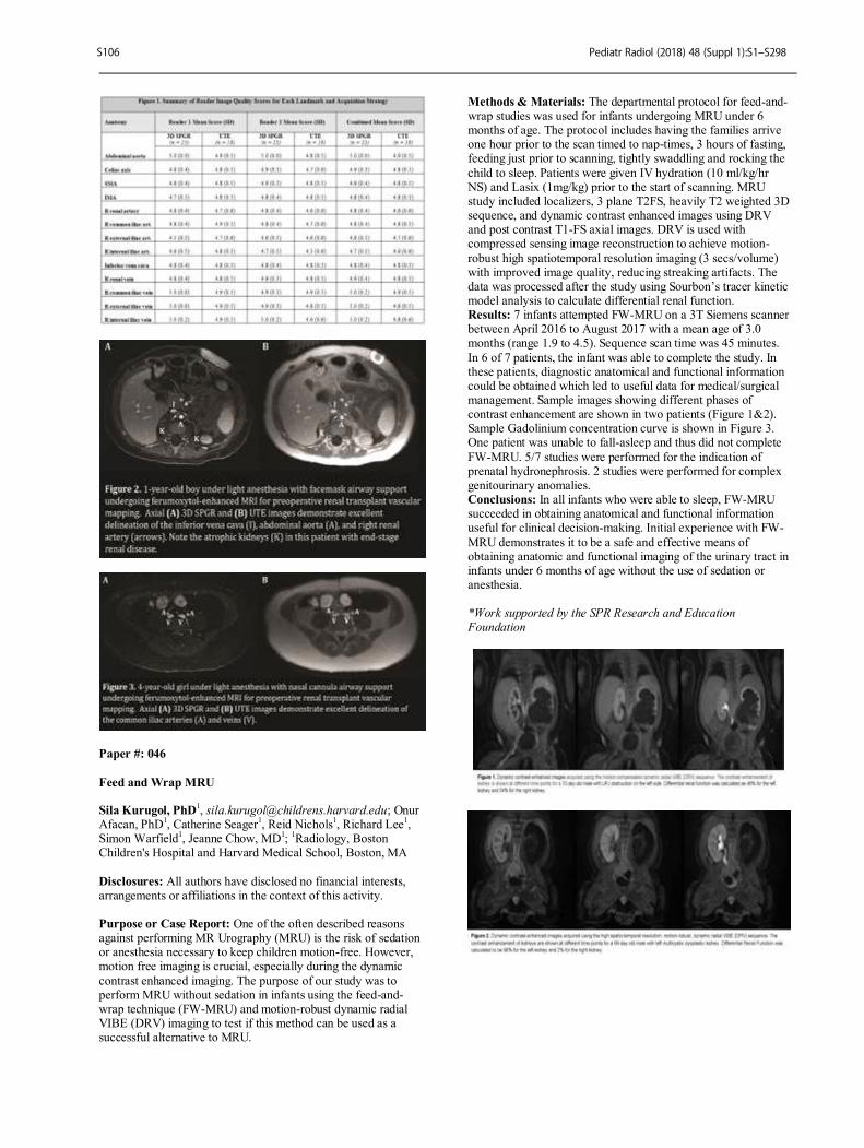
Paper #: 046
Feed and Wrap MRU Sila Kurugol, PhD
1, [email protected]; Onur
Afacan, PhD1, Catherine Seager
1, Reid Nichols
1, Richard Lee
1,
Simon Warfield1, Jeanne Chow, MD
1;
1Radiology, Boston
Children's Hospital and Harvard Medical School, Boston, MA
Disclosures: All authors have disclosed no financial interests, arrangements or affiliations in the context of this activity. Purpose or Case Report: One of the often described reasons against performing MR Urography (MRU) is the risk of sedation or anesthesia necessary to keep children motion-free. However, motion free imaging is crucial, especially during the dynamic
contrast enhanced imaging. The purpose of our study was to perform MRU without sedation in infants using the feed-and-wrap technique (FW-MRU) and motion-robust dynamic radial VIBE (DRV) imaging to test if this method can be used as a successful alternative to MRU.
Methods & Materials: The departmental protocol for feed-and-wrap studies was used for infants undergoing MRU under 6 months of age. The protocol includes having the families arrive one hour prior to the scan timed to nap-times, 3 hours of fasting, feeding just prior to scanning, tightly swaddling and rocking the
child to sleep. Patients were given IV hydration (10 ml/kg/hr NS) and Lasix (1mg/kg) prior to the start of scanning. MRU study included localizers, 3 plane T2FS, heavily T2 weighted 3D sequence, and dynamic contrast enhanced images using DRV and post contrast T1-FS axial images. DRV is used with compressed sensing image reconstruction to achieve motion-
robust high spatiotemporal resolution imaging (3 secs/volume) with improved image quality, reducing streaking artifacts. The data was processed after the study using Sourbon’s tracer kinetic model analysis to calculate differential renal function. Results: 7 infants attempted FW-MRU on a 3T Siemens scanner between April 2016 to August 2017 with a mean age of 3.0 months (range 1.9 to 4.5). Sequence scan time was 45 minutes.
In 6 of 7 patients, the infant was able to complete the study. In these patients, diagnostic anatomical and functional information could be obtained which led to useful data for medical/surgical management. Sample images showing different phases of contrast enhancement are shown in two patients (Figure 1&2). Sample Gadolinium concentration curve is shown in Figure 3. One patient was unable to fall-asleep and thus did not complete
FW-MRU. 5/7 studies were performed for the indication of prenatal hydronephrosis. 2 studies were performed for complex genitourinary anomalies. Conclusions: In all infants who were able to sleep, FW-MRU succeeded in obtaining anatomical and functional information useful for clinical decision-making. Initial experience with FW-
MRU demonstrates it to be a safe and effective means of obtaining anatomic and functional imaging of the urinary tract in infants under 6 months of age without the use of sedation or anesthesia. *Work supported by the SPR Research and Education Foundation
Pediatr Radiol (2018) 48 (Suppl 1):S1–S298S106
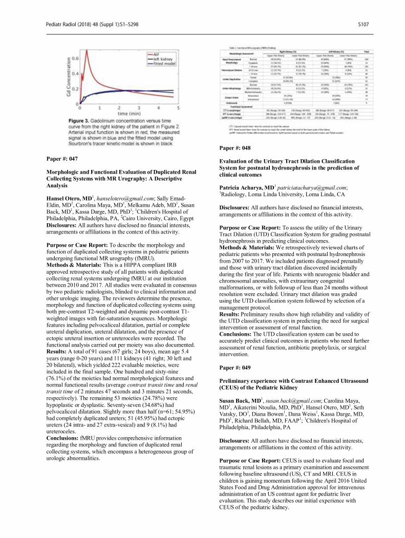
Paper #: 047
Morphologic and Functional Evaluation of Duplicated Renal
Collecting Systems with MR Urography: A Descriptive
Analysis Hansel Otero, MD
1, [email protected]; Sally Emad-
Eldin, MD2, Carolina Maya, MD
1, Melkamu Adeb, MD
1, Susan
Back, MD1, Kassa Darge, MD, PhD
1;
1Children's Hospital of
Philadelphia, Philadelphia, PA, 2Cairo University, Cairo, Egypt
Disclosures: All authors have disclosed no financial interests, arrangements or affiliations in the context of this activity. Purpose or Case Report: To describe the morphology and function of duplicated collecting systems in pediatric patients undergoing functional MR urography (fMRU). Methods & Materials: This is a HIPPA compliant IRB
approved retrospective study of all patients with duplicated collecting renal systems undergoing fMRU at our institution between 2010 and 2017. All studies were evaluated in consensus by two pediatric radiologists, blinded to clinical information and other urologic imaging. The reviewers determine the presence, morphology and function of duplicated collecting systems using both pre-contrast T2-weighted and dynamic post-contrast T1-
weighted images with fat-saturation sequences. Morphologic features including pelvocaliceal dilatation, partial or complete ureteral duplication, ureteral dilatation, and the presence of ectopic ureteral insertion or ureteroceles were recorded. The functional analysis carried out per moiety was also documented. Results: A total of 91 cases (67 girls; 24 boys), mean age 5.4
years (range 0-20 years) and 111 kidneys (41 right; 30 left and 20 bilateral), which yielded 222 evaluable moieties, were included in the final sample. One hundred and sixty-nine (76.1%) of the moieties had normal morphological features and normal functional results (average contrast transit time and renal transit time of 2 minutes 47 seconds and 3 minutes 21 seconds, respectively). The remaining 53 moieties (24.78%) were
hypoplastic or dysplastic. Seventy-seven (34.68%) had pelvocaliceal dilatation. Slightly more than half (n=61; 54.95%) had completely duplicated ureters; 51 (45.95%) had ectopic ureters (24 intra- and 27 extra-vesical) and 9 (8.1%) had ureteroceles. Conclusions: fMRU provides comprehensive information regarding the morphology and function of duplicated renal
collecting systems, which encompass a heterogeneous group of urologic abnormalities.
Paper #: 048
Evaluation of the Urinary Tract Dilation Classification
System for postnatal hydronephrosis in the prediction of
clinical outcomes Patricia Acharya, MD
1Radiology, Loma Linda University, Loma Linda, CA
Disclosures: All authors have disclosed no financial interests,
arrangements or affiliations in the context of this activity. Purpose or Case Report: To assess the utility of the Urinary Tract Dilation (UTD) Classification System for grading postnatal hydronephrosis in predicting clinical outcomes. Methods & Materials: We retrospectively reviewed charts of pediatric patients who presented with postnatal hydronephrosis
from 2007 to 2017. We included patients diagnosed prenatally and those with urinary tract dilation discovered incidentally during the first year of life. Patients with neurogenic bladder and chromosomal anomalies, with extraurinary congenital malformations, or with followup of less than 24 months without resolution were excluded. Urinary tract dilation was graded using the UTD classification system followed by selection of a
management protocol. Results: Preliminary results show high reliability and validity of the UTD classification system in predicting the need for surgical intervention or assessment of renal function. Conclusions: The UTD classification system can be used to accurately predict clinical outcomes in patients who need further
assessment of renal function, antibiotic prophylaxis, or surgical intervention.
Paper #: 049
Preliminary experience with Contrast Enhanced Ultrasound
(CEUS) of the Pediatric Kidney
Susan Back, MD
1, [email protected]; Carolina Maya,
MD1, Aikaterini Ntoulia, MD, PhD
1, Hansel Otero, MD
1, Seth
Vatsky, DO1, Diana Bowen
1, Dana Weiss
1, Kassa Darge, MD,
PhD1, Richard Bellah, MD, FAAP
1;
1Children's Hospital of
Philadelphia, Philadelphia, PA
Disclosures: All authors have disclosed no financial interests, arrangements or affiliations in the context of this activity. Purpose or Case Report: CEUS is used to evaluate focal and traumatic renal lesions as a primary examination and assessment following baseline ultrasound (US), CT and MRI. CEUS in
children is gaining momentum following the April 2016 United States Food and Drug Administration approval for intravenous administration of an US contrast agent for pediatric liver evaluation. This study describes our initial experience with CEUS of the pediatric kidney.
Pediatr Radiol (2018) 48 (Suppl 1):S1–S298 S107
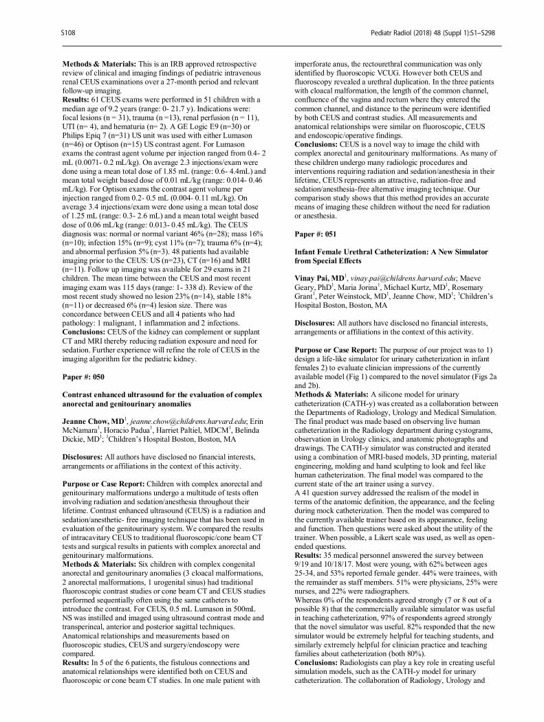
Methods & Materials: This is an IRB approved retrospective review of clinical and imaging findings of pediatric intravenous renal CEUS examinations over a 27-month period and relevant follow-up imaging. Results: 61 CEUS exams were performed in 51 children with a
median age of 9.2 years (range: 0- 21.7 y). Indications were: focal lesions (n = 31), trauma (n =13), renal perfusion (n = 11), UTI (n= 4), and hematuria (n= 2). A GE Logic E9 (n=30) or Philips Epiq 7 (n=31) US unit was used with either Lumason (n=46) or Optison (n=15) US contrast agent. For Lumason exams the contrast agent volume per injection ranged from 0.4- 2
mL (0.0071- 0.2 mL/kg). On average 2.3 injections/exam were done using a mean total dose of 1.85 mL (range: 0.6- 4.4mL) and mean total weight based dose of 0.01 mL/kg (range: 0.014- 0.46 mL/kg). For Optison exams the contrast agent volume per injection ranged from 0.2- 0.5 mL (0.004- 0.11 mL/kg). On average 3.4 injections/exam were done using a mean total dose of 1.25 mL (range: 0.3- 2.6 mL) and a mean total weight based
dose of 0.06 mL/kg (range: 0.013- 0.45 mL/kg). The CEUS diagnosis was: normal or normal variant 46% (n=28); mass 16% (n=10); infection 15% (n=9); cyst 11% (n=7); trauma 6% (n=4); and abnormal perfusion 5% (n=3). 48 patients had available imaging prior to the CEUS: US (n=23), CT (n=16) and MRI (n=11). Follow up imaging was available for 29 exams in 21 children. The mean time between the CEUS and most recent
imaging exam was 115 days (range: 1- 338 d). Review of the most recent study showed no lesion 23% (n=14), stable 18% (n=11) or decreased 6% (n=4) lesion size. There was concordance between CEUS and all 4 patients who had pathology: 1 malignant, 1 inflammation and 2 infections. Conclusions: CEUS of the kidney can complement or supplant
CT and MRI thereby reducing radiation exposure and need for sedation. Further experience will refine the role of CEUS in the imaging algorithm for the pediatric kidney.
Paper #: 050
Contrast enhanced ultrasound for the evaluation of complex
anorectal and genitourinary anomalies Jeanne Chow, MD
1, [email protected]; Erin
McNamara1, Horacio Padua
1, Harriet Paltiel, MDCM
1, Belinda
Dickie, MD1;
1Children’s Hospital Boston, Boston, MA
Disclosures: All authors have disclosed no financial interests,
arrangements or affiliations in the context of this activity. Purpose or Case Report: Children with complex anorectal and genitourinary malformations undergo a multitude of tests often involving radiation and sedation/anesthesia throughout their lifetime. Contrast enhanced ultrasound (CEUS) is a radiation and
sedation/anesthetic- free imaging technique that has been used in evaluation of the genitourinary system. We compared the results of intracavitary CEUS to traditional fluoroscopic/cone beam CT tests and surgical results in patients with complex anorectal and genitourinary malformations. Methods & Materials: Six children with complex congenital anorectal and genitourinary anomalies (3 cloacal malformations,
2 anorectal malformations, 1 urogenital sinus) had traditional fluoroscopic contrast studies or cone beam CT and CEUS studies performed sequentially often using the same catheters to introduce the contrast. For CEUS, 0.5 mL Lumason in 500mL NS was instilled and imaged using ultrasound contrast mode and transperineal, anterior and posterior sagittal techniques.
Anatomical relationships and measurements based on fluoroscopic studies, CEUS and surgery/endoscopy were compared. Results: In 5 of the 6 patients, the fistulous connections and anatomical relationships were identified both on CEUS and fluoroscopic or cone beam CT studies. In one male patient with
imperforate anus, the rectourethral communication was only identified by fluoroscopic VCUG. However both CEUS and fluoroscopy revealed a urethral duplication. In the three patients with cloacal malformation, the length of the common channel, confluence of the vagina and rectum where they entered the
common channel, and distance to the perineum were identified by both CEUS and contrast studies. All measurements and anatomical relationships were similar on fluoroscopic, CEUS and endoscopic/operative findings. Conclusions: CEUS is a novel way to image the child with complex anorectal and genitourinary malformations. As many of
these children undergo many radiologic procedures and interventions requiring radiation and sedation/anesthesia in their lifetime, CEUS represents an attractive, radiation-free and sedation/anesthesia-free alternative imaging technique. Our comparison study shows that this method provides an accurate means of imaging these children without the need for radiation or anesthesia.
Paper #: 051
Infant Female Urethral Catheterization: A New Simulator
from Special Effects Vinay Pai, MD
1, [email protected]; Maeve
Geary, PhD1, Maria Jorina
1, Michael Kurtz, MD
1, Rosemary
Grant1, Peter Weinstock, MD
1, Jeanne Chow, MD
1;
1Children’s
Hospital Boston, Boston, MA Disclosures: All authors have disclosed no financial interests, arrangements or affiliations in the context of this activity.
Purpose or Case Report: The purpose of our project was to 1) design a life-like simulator for urinary catheterization in infant females 2) to evaluate clinician impressions of the currently available model (Fig 1) compared to the novel simulator (Figs 2a and 2b). Methods & Materials: A silicone model for urinary
catheterization (CATH-y) was created as a collaboration between the Departments of Radiology, Urology and Medical Simulation. The final product was made based on observing live human catheterization in the Radiology department during cystograms, observation in Urology clinics, and anatomic photographs and drawings. The CATH-y simulator was constructed and iterated using a combination of MRI-based models, 3D printing, material
engineering, molding and hand sculpting to look and feel like human catheterization. The final model was compared to the current state of the art trainer using a survey. A 41 question survey addressed the realism of the model in terms of the anatomic definition, the appearance, and the feeling during mock catheterization. Then the model was compared to
the currently available trainer based on its appearance, feeling and function. Then questions were asked about the utility of the trainer. When possible, a Likert scale was used, as well as open-ended questions. Results: 35 medical personnel answered the survey between 9/19 and 10/18/17. Most were young, with 62% between ages 25-34, and 53% reported female gender. 44% were trainees, with
the remainder as staff members. 51% were physicians, 25% were nurses, and 22% were radiographers. Whereas 0% of the respondents agreed strongly (7 or 8 out of a possible 8) that the commercially available simulator was useful in teaching catheterization, 97% of respondents agreed strongly that the novel simulator was useful. 82% responded that the new
simulator would be extremely helpful for teaching students, and similarly extremely helpful for clinician practice and teaching families about catheterization (both 80%). Conclusions: Radiologists can play a key role in creating useful simulation models, such as the CATH-y model for urinary catheterization. The collaboration of Radiology, Urology and
Pediatr Radiol (2018) 48 (Suppl 1):S1–S298S108
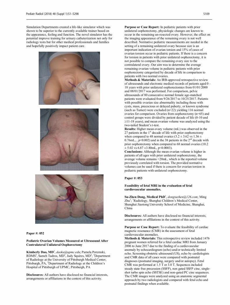
Simulation Departments created a life-like simulator which was shown to be superior to the currently available trainer based on the appearance, feeling and function. The novel simulator has the potential improve training for urinary catheterization not only for radiology tests but for other medical professionals and families
and hopefully positively impact patient care.
Paper #: 052
Pediatric Ovarian Volumes Measured at Ultrasound After
Contralateral Unilateral Oophorectomy Kimberly Dao, MD
1, [email protected]; Pamela Poremski,
RDMS2, Sameh Tadros, MD
2, Judy Squires, MD
2;
1Department
of Radiology at the University of Pittsburgh Medical Center, Pittsburgh, PA,
2Department of Radiology at the Children’s
Hospital of Pittsburgh of UPMC, Pittsburgh, PA
Disclosures: All authors have disclosed no financial interests, arrangements or affiliations in the context of this activity.
Purpose or Case Report: In pediatric patients with prior unilateral oophorectomy, physiologic changes are known to occur in the remaining un-resected ovary. However, the effect on the imaging appearance of the remaining ovary is not well described. Normative pediatric measurements are needed in the
setting of a remaining unilateral ovary because size is an important indication of ovarian torsion and 15% of cases of ovarian torsion occur in pediatric patients. If there is a concern for torsion in patients with prior unilateral oophorectomy, it is not possible to compare the remaining ovary size to the contralateral ovary. Our aim was to determine the average
remaining ovarian volume in pediatric patients with prior oophorectomy categorized by decade of life in comparison to patients with two normal ovaries. Methods & Materials: An IRB-approved retrospective review of ultrasounds and electronic medical records of patients aged 0 -18 years with prior unilateral oophorectomies from 01/01/2000 and 08/01/2017 was performed. For comparison, pelvic
ultrasounds of 80 consecutive normal female age-matched patients were evaluated from 9/26/2017 to 10/25/2017. Patients with possible ovarian size abnormality including those with cysts, mass, precocious or delayed puberty, or known syndrome (such as Turner) were excluded (n=22) yielding 116 normal ovaries for comparison. Ovaries from oophorectomy (n=65) and control groups were divided by patient decade of life (0-10 and
≥11-18 years), and mean ovarian volume was analyzed using the two-tailed Student’s t-test. Results: Higher mean ovary volume (mL) was observed in the 27 patients in the 1
st decade of life with prior oophorectomy
when compared to 48 normal ovaries (3.2 ± 3.62 vs 1.36 ± 0.76mL, , p<0.002) and in the 38 patients in the 2
nd decade with
prior oophorectomy when compared to 68 normal ovaries (10.2 ± 5.02 vs 6.87 ±3.80mL, p<0.0003). Conclusions: Although the mean ovarian volume is higher in patients of all ages with prior unilateral oophorectomy, the average volume remains <20mL, which is the reported volume previously correlated with torsion. The provided normative volumes can be used if there is concern for ovarian torsion in
pediatric patients with unilateral oophorectomy.
Paper #: 053
Feasibility of fetal MRI in the evaluation of fetal
cardiovascular anomalies.
Su-Zhen Dong, Medical PhD1, [email protected]; Ming
Zhu1;
1Radiology, Shanghai Children’s Medical Center,
Shanghai Jiaotong University School of Medicine, Shanghai, China
Disclosures: All authors have disclosed no financial interests,
arrangements or affiliations in the context of this activity. Purpose or Case Report: To evaluate the feasibility of cardiac magnetic resonance (CMR) in the assessment of fetal cardiovascular anomalies. Methods & Materials: This retrospective review included 1476 pregnant women referred for a fetal cardiac MRI from January
2006 to June 2017 due to the finding of a cardiovascular anomaly by echocardiogram (echo) and/or technically limited echo. Screening obstetric ultrasound (US), echo by cardiologists and CMR data of all cases were compared with postnatal diagnoses (postnatal imaging, surgery and/or autopsy). Fetal CMR was performed at 1.5 T or 3.0 T. Sequences included
steady state free precession (SSFP), non gated SSFP cine, single-shot turbo spin echo (SSTSE) and non-gated PC cine sequences. The CMR images were analyzed using an anatomic segmental approach by two radiologists and compared with fetal echo and postnatal findings when available.
Pediatr Radiol (2018) 48 (Suppl 1):S1–S298 S109
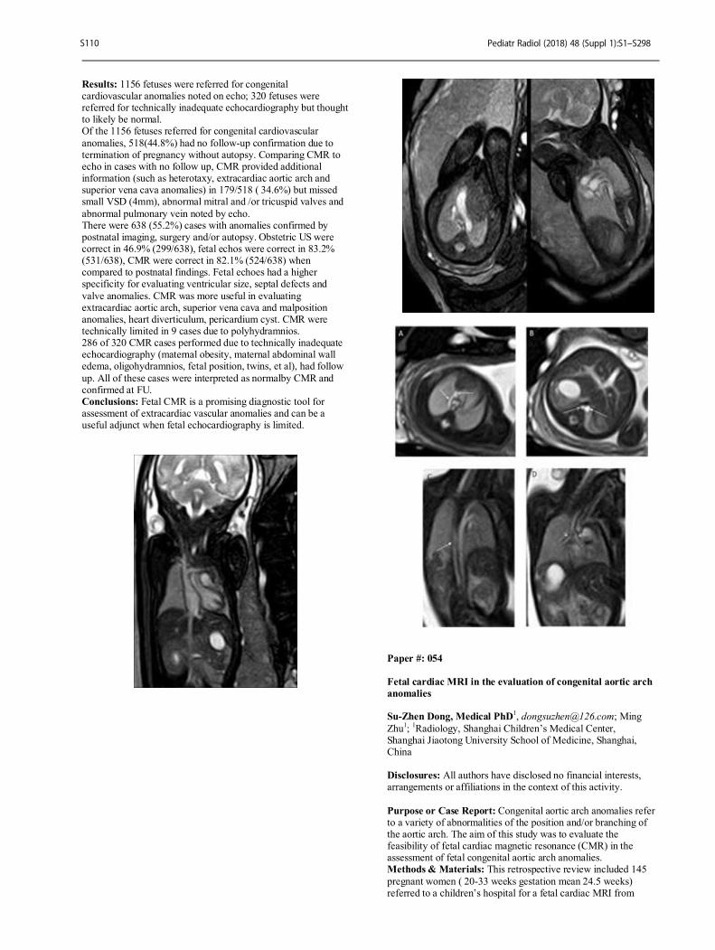
Results: 1156 fetuses were referred for congenital cardiovascular anomalies noted on echo; 320 fetuses were referred for technically inadequate echocardiography but thought to likely be normal. Of the 1156 fetuses referred for congenital cardiovascular
anomalies, 518(44.8%) had no follow-up confirmation due to termination of pregnancy without autopsy. Comparing CMR to echo in cases with no follow up, CMR provided additional information (such as heterotaxy, extracardiac aortic arch and superior vena cava anomalies) in 179/518 ( 34.6%) but missed small VSD (4mm), abnormal mitral and /or tricuspid valves and
abnormal pulmonary vein noted by echo. There were 638 (55.2%) cases with anomalies confirmed by postnatal imaging, surgery and/or autopsy. Obstetric US were correct in 46.9% (299/638), fetal echos were correct in 83.2% (531/638), CMR were correct in 82.1% (524/638) when compared to postnatal findings. Fetal echoes had a higher specificity for evaluating ventricular size, septal defects and
valve anomalies. CMR was more useful in evaluating extracardiac aortic arch, superior vena cava and malposition anomalies, heart diverticulum, pericardium cyst. CMR were technically limited in 9 cases due to polyhydramnios. 286 of 320 CMR cases performed due to technically inadequate echocardiography (maternal obesity, maternal abdominal wall edema, oligohydramnios, fetal position, twins, et al), had follow
up. All of these cases were interpreted as normalby CMR and confirmed at FU. Conclusions: Fetal CMR is a promising diagnostic tool for assessment of extracardiac vascular anomalies and can be a useful adjunct when fetal echocardiography is limited.
Paper #: 054
Fetal cardiac MRI in the evaluation of congenital aortic arch
anomalies Su-Zhen Dong, Medical PhD
1, [email protected]; Ming
Zhu1;
1Radiology, Shanghai Children’s Medical Center,
Shanghai Jiaotong University School of Medicine, Shanghai, China
Disclosures: All authors have disclosed no financial interests, arrangements or affiliations in the context of this activity.
Purpose or Case Report: Congenital aortic arch anomalies refer to a variety of abnormalities of the position and/or branching of the aortic arch. The aim of this study was to evaluate the feasibility of fetal cardiac magnetic resonance (CMR) in the assessment of fetal congenital aortic arch anomalies. Methods & Materials: This retrospective review included 145
pregnant women ( 20-33 weeks gestation mean 24.5 weeks) referred to a children’s hospital for a fetal cardiac MRI from
Pediatr Radiol (2018) 48 (Suppl 1):S1–S298S110
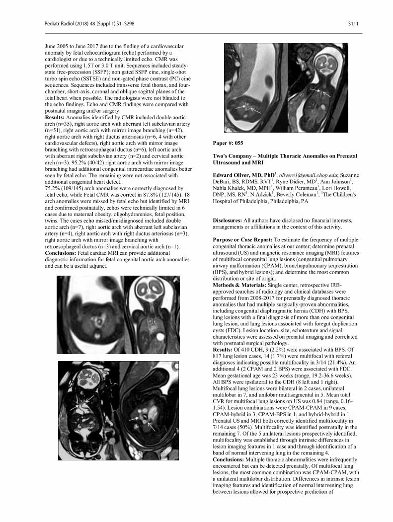
June 2005 to June 2017 due to the finding of a cardiovascular anomaly by fetal echocardiogram (echo) performed by a cardiologist or due to a technically limited echo. CMR was performed using 1.5T or 3.0 T unit. Sequences included steady-state free-precession (SSFP); non gated SSFP cine, single-shot
turbo spin echo (SSTSE) and non-gated phase contrast (PC) cine sequences. Sequences included transverse fetal thorax, and four-chamber, short-axis, coronal and oblique sagittal planes of the fetal heart when possible. The radiologists were not blinded to the echo findings. Echo and CMR findings were compared with postnatal imaging and/or surgery.
Results: Anomalies identified by CMR included double aortic arch (n=35), right aortic arch with aberrant left subclavian artery (n=51), right aortic arch with mirror image branching (n=42), right aortic arch with right ductus arteriosus (n=6, 4 with other cardiovascular defects), right aortic arch with mirror image branching with retroesophageal ductus (n=6), left aortic arch with aberrant right subclavian artery (n=2) and cervical aortic
arch (n=3). 95.2% (40/42) right aortic arch with mirror image branching had additional congenital intracardiac anomalies better seen by fetal echo. The remaining were not associated with additional congenital heart defect. 75.2% (109/145) arch anomalies were correctly diagnosed by fetal echo, while Fetal CMR was correct in 87.8% (127/145). 18 arch anomalies were missed by fetal echo but identified by MRI
and confirmed postnatally, echos were technically limited in 6 cases due to maternal obesity, oligohydramnios, fetal position, twins. The cases echo missed/misdiagnosed included double aortic arch (n=7), right aortic arch with aberrant left subclavian artery (n=4), right aortic arch with right ductus arteriosus (n=3), right aortic arch with mirror image branching with
retroesophageal ductus (n=3) and cervical aortic arch (n=1). Conclusions: Fetal cardiac MRI can provide additional diagnostic information for fetal congenital aortic arch anomalies and can be a useful adjunct.
Paper #: 055
Two's Company – Multiple Thoracic Anomalies on Prenatal
Ultrasound and MRI
Edward Oliver, MD, PhD
1, [email protected]; Suzanne
DeBari, BS, RDMS, RVT1, Ryne Didier, MD
1, Ann Johnson
1,
Nahla Khalek, MD, MPH1, William Peranteau
1, Lori Howell,
DNP, MS, RN1, N Adzick
1, Beverly Coleman
1;
1The Children's
Hospital of Philadelphia, Philadelphia, PA
Disclosures: All authors have disclosed no financial interests, arrangements or affiliations in the context of this activity. Purpose or Case Report: To estimate the frequency of multiple
congenital thoracic anomalies at our center; determine prenatal ultrasound (US) and magnetic resonance imaging (MRI) features of multifocal congenital lung lesions (congenital pulmonary airway malformation (CPAM), bronchopulmonary sequestration (BPS), and hybrid lesions); and determine the most common distribution or site of origin.
Methods & Materials: Single center, retrospective IRB-approved searches of radiology and clinical databases were performed from 2008-2017 for prenatally diagnosed thoracic anomalies that had multiple surgically-proven abnormalities, including congenital diaphragmatic hernia (CDH) with BPS, lung lesions with a final diagnosis of more than one congenital lung lesion, and lung lesions associated with foregut duplication
cysts (FDC). Lesion location, size, echotexture and signal characteristics were assessed on prenatal imaging and correlated with postnatal surgical pathology. Results: Of 410 CDH, 9 (2.2%) were associated with BPS. Of 817 lung lesion cases, 14 (1.7%) were multifocal with referral diagnoses indicating possible multifocality in 3/14 (21.4%). An additional 4 (2 CPAM and 2 BPS) were associated with FDC.
Mean gestational age was 23 weeks (range, 19.2-36.6 weeks). All BPS were ipsilateral to the CDH (8 left and 1 right). Multifocal lung lesions were bilateral in 2 cases, unilateral multilobar in 7, and unilobar multisegmental in 5. Mean total CVR for multifocal lung lesions on US was 0.84 (range, 0.16-1.54). Lesion combinations were CPAM-CPAM in 9 cases,
CPAM-hybrid in 3, CPAM-BPS in 1, and hybrid-hybrid in 1. Prenatal US and MRI both correctly identified multifocality in 7/14 cases (50%). Multifocality was identified postnatally in the remaining 7. Of the 5 unilateral lesions prospectively identified, multifocality was established through intrinsic differences in lesion imaging features in 1 case and through identification of a band of normal intervening lung in the remaining 4.
Conclusions: Multiple thoracic abnormalities were infrequently encountered but can be detected prenatally. Of multifocal lung lesions, the most common combination was CPAM-CPAM, with a unilateral multilobar distribution. Differences in intrinsic lesion imaging features and identification of normal intervening lung between lesions allowed for prospective prediction of
Pediatr Radiol (2018) 48 (Suppl 1):S1–S298 S111
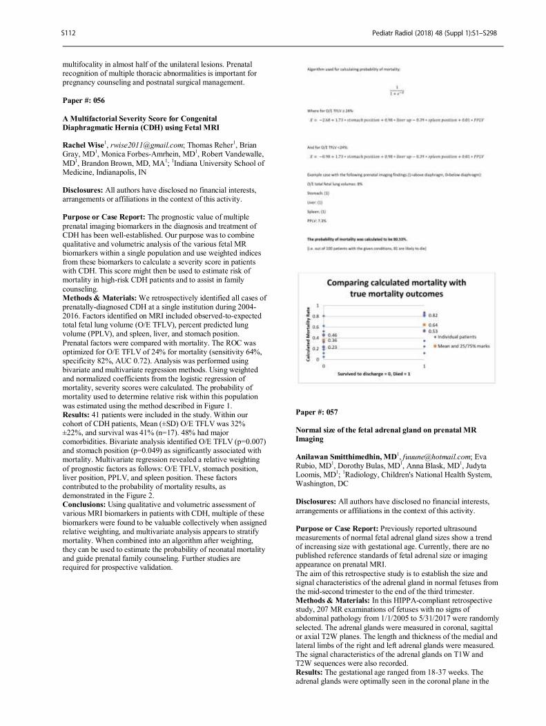
multifocality in almost half of the unilateral lesions. Prenatal recognition of multiple thoracic abnormalities is important for pregnancy counseling and postnatal surgical management.
Paper #: 056
A Multifactorial Severity Score for Congenital
Diaphragmatic Hernia (CDH) using Fetal MRI Rachel Wise
1, [email protected]; Thomas Reher
1, Brian
Gray, MD1, Monica Forbes-Amrhein, MD
1, Robert Vandewalle,
MD1, Brandon Brown, MD, MA
1;
1Indiana University School of
Medicine, Indianapolis, IN Disclosures: All authors have disclosed no financial interests, arrangements or affiliations in the context of this activity. Purpose or Case Report: The prognostic value of multiple
prenatal imaging biomarkers in the diagnosis and treatment of CDH has been well-established. Our purpose was to combine qualitative and volumetric analysis of the various fetal MR biomarkers within a single population and use weighted indices from these biomarkers to calculate a severity score in patients with CDH. This score might then be used to estimate risk of mortality in high-risk CDH patients and to assist in family
counseling. Methods & Materials: We retrospectively identified all cases of prenatally-diagnosed CDH at a single institution during 2004-2016. Factors identified on MRI included observed-to-expected total fetal lung volume (O/E TFLV), percent predicted lung volume (PPLV), and spleen, liver, and stomach position.
Prenatal factors were compared with mortality. The ROC was optimized for O/E TFLV of 24% for mortality (sensitivity 64%, specificity 82%, AUC 0.72). Analysis was performed using bivariate and multivariate regression methods. Using weighted and normalized coefficients from the logistic regression of mortality, severity scores were calculated. The probability of mortality used to determine relative risk within this population
was estimated using the method described in Figure 1. Results: 41 patients were included in the study. Within our cohort of CDH patients, Mean (±SD) O/E TFLV was 32% ±22%, and survival was 41% (n=17). 48% had major comorbidities. Bivariate analysis identified O/E TFLV (p=0.007) and stomach position (p=0.049) as significantly associated with mortality. Multivariate regression revealed a relative weighting
of prognostic factors as follows: O/E TFLV, stomach position, liver position, PPLV, and spleen position. These factors contributed to the probability of mortality results, as demonstrated in the Figure 2. Conclusions: Using qualitative and volumetric assessment of various MRI biomarkers in patients with CDH, multiple of these
biomarkers were found to be valuable collectively when assigned relative weighting, and multivariate analysis appears to stratify mortality. When combined into an algorithm after weighting, they can be used to estimate the probability of neonatal mortality and guide prenatal family counseling. Further studies are required for prospective validation.
Paper #: 057
Normal size of the fetal adrenal gland on prenatal MR
Imaging
Anilawan Smitthimedhin, MD
1, [email protected]; Eva
Rubio, MD1, Dorothy Bulas, MD
1, Anna Blask, MD
1, Judyta
Loomis, MD1;
1Radiology, Children's National Health System,
Washington, DC
Disclosures: All authors have disclosed no financial interests,
arrangements or affiliations in the context of this activity. Purpose or Case Report: Previously reported ultrasound measurements of normal fetal adrenal gland sizes show a trend of increasing size with gestational age. Currently, there are no published reference standards of fetal adrenal size or imaging appearance on prenatal MRI.
The aim of this retrospective study is to establish the size and signal characteristics of the adrenal gland in normal fetuses from the mid-second trimester to the end of the third trimester. Methods & Materials: In this HIPPA-compliant retrospective study, 207 MR examinations of fetuses with no signs of abdominal pathology from 1/1/2005 to 5/31/2017 were randomly
selected. The adrenal glands were measured in coronal, sagittal or axial T2W planes. The length and thickness of the medial and lateral limbs of the right and left adrenal glands were measured. The signal characteristics of the adrenal glands on T1W and T2W sequences were also recorded. Results: The gestational age ranged from 18-37 weeks. The adrenal glands were optimally seen in the coronal plane in the
Pediatr Radiol (2018) 48 (Suppl 1):S1–S298S112
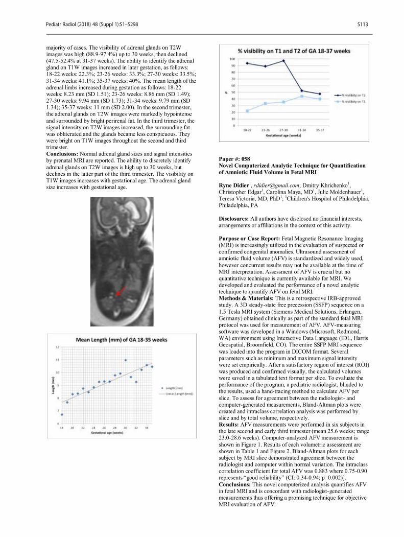
majority of cases. The visibility of adrenal glands on T2W images was high (88.9-97.4%) up to 30 weeks, then declined (47.5-52.4% at 31-37 weeks). The ability to identify the adrenal gland on T1W images increased in later gestation, as follows: 18-22 weeks: 22.3%; 23-26 weeks: 33.3%; 27-30 weeks: 33.5%;
31-34 weeks: 41.1%; 35-37 weeks: 40%. The mean length of the adrenal limbs increased during gestation as follows: 18-22 weeks: 8.23 mm (SD 1.51); 23-26 weeks: 8.86 mm (SD 1.49); 27-30 weeks: 9.94 mm (SD 1.73); 31-34 weeks: 9.79 mm (SD 1.34); 35-37 weeks: 11 mm (SD 2.00). In the second trimester, the adrenal glands on T2W images were markedly hypointense
and surrounded by bright perirenal fat. In the third trimester, the signal intensity on T2W images increased, the surrounding fat was obliterated and the glands became less conspicuous. They were bright on T1W images throughout the second and third trimester. Conclusions: Normal adrenal gland sizes and signal intensities by prenatal MRI are reported. The ability to discretely identify
adrenal glands on T2W images is high up to 30 weeks, but declines in the latter part of the third trimester. The visibility on T1W images increases with gestational age. The adrenal gland size increases with gestational age.
Paper #: 058 Novel Computerized Analytic Technique for Quantification
of Amniotic Fluid Volume in Fetal MRI
Ryne Didier
1, [email protected]; Dmitry Khrichenko
1,
Christopher Edgar1, Carolina Maya, MD
1, Julie Moldenhauer
1,
Teresa Victoria, MD, PhD1;
1Children's Hospital of Philadelphia,
Philadelphia, PA
Disclosures: All authors have disclosed no financial interests, arrangements or affiliations in the context of this activity. Purpose or Case Report: Fetal Magnetic Resonance Imaging (MRI) is increasingly utilized in the evaluation of suspected or confirmed congenital anomalies. Ultrasound assessment of amniotic fluid volume (AFV) is standardized and widely used,
however concurrent results may not be available at the time of MRI interpretation. Assessment of AFV is crucial but no quantitative technique is currently available for MRI. We developed and evaluated the performance of a novel analytic technique to quantify AFV on fetal MRI. Methods & Materials: This is a retrospective IRB-approved study. A 3D steady-state free precession (SSFP) sequence on a
1.5 Tesla MRI system (Siemens Medical Solutions, Erlangen, Germany) obtained clinically as part of the standard fetal MRI protocol was used for measurement of AFV. AFV-measuring software was developed in a Windows (Microsoft, Redmond, WA) environment using Interactive Data Language (IDL, Harris Geospatial, Broomfield, CO). The entire SSFP MRI sequence
was loaded into the program in DICOM format. Several parameters such as minimum and maximum signal intensity were set empirically. After a satisfactory region of interest (ROI) was produced and confirmed visually, the calculated volumes were saved in a tabulated text format per slice. To evaluate the performance of the program, a pediatric radiologist, blinded to the results, used a hand-tracing method to calculate AFV per
slice. To assess for agreement between the radiologist- and computer-generated measurements, Bland-Altman plots were created and intraclass correlation analysis was performed by slice and by total volume, respectively. Results: AFV measurements were performed in six subjects in the late second and early third trimester (mean 25.6 weeks; range 23.0-28.6 weeks). Computer-analyzed AFV measurement is
shown in Figure 1. Results of each volumetric assessment are shown in Table 1 and Figure 2. Bland-Altman plots for each subject by MRI slice demonstrated agreement between the radiologist and computer within normal variation. The intraclass correlation coefficient for total AFV was 0.883 where 0.75-0.90 represents “good reliability” (CI: 0.34-0.94; p=0.002)].
Conclusions: This novel computerized analysis quantifies AFV in fetal MRI and is concordant with radiologist-generated measurements thus offering a promising technique for objective MRI evaluation of AFV.
Pediatr Radiol (2018) 48 (Suppl 1):S1–S298 S113
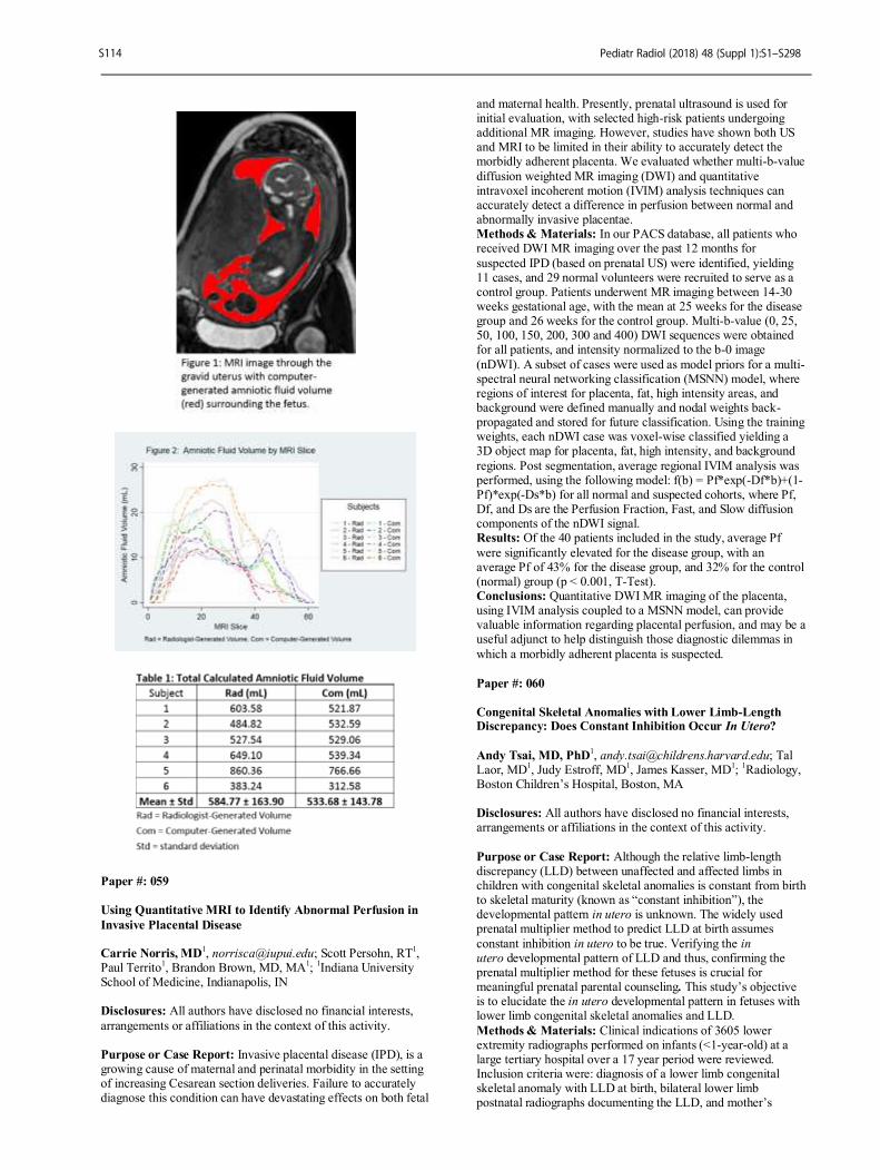
Paper #: 059
Using Quantitative MRI to Identify Abnormal Perfusion in
Invasive Placental Disease Carrie Norris, MD
1, [email protected]; Scott Persohn, RT
1,
Paul Territo1, Brandon Brown, MD, MA
1;
1Indiana University
School of Medicine, Indianapolis, IN Disclosures: All authors have disclosed no financial interests,
arrangements or affiliations in the context of this activity. Purpose or Case Report: Invasive placental disease (IPD), is a growing cause of maternal and perinatal morbidity in the setting of increasing Cesarean section deliveries. Failure to accurately diagnose this condition can have devastating effects on both fetal
and maternal health. Presently, prenatal ultrasound is used for initial evaluation, with selected high-risk patients undergoing additional MR imaging. However, studies have shown both US and MRI to be limited in their ability to accurately detect the morbidly adherent placenta. We evaluated whether multi-b-value
diffusion weighted MR imaging (DWI) and quantitative intravoxel incoherent motion (IVIM) analysis techniques can accurately detect a difference in perfusion between normal and abnormally invasive placentae. Methods & Materials: In our PACS database, all patients who received DWI MR imaging over the past 12 months for
suspected IPD (based on prenatal US) were identified, yielding 11 cases, and 29 normal volunteers were recruited to serve as a control group. Patients underwent MR imaging between 14-30 weeks gestational age, with the mean at 25 weeks for the disease group and 26 weeks for the control group. Multi-b-value (0, 25, 50, 100, 150, 200, 300 and 400) DWI sequences were obtained for all patients, and intensity normalized to the b-0 image
(nDWI). A subset of cases were used as model priors for a multi-spectral neural networking classification (MSNN) model, where regions of interest for placenta, fat, high intensity areas, and background were defined manually and nodal weights back-propagated and stored for future classification. Using the training weights, each nDWI case was voxel-wise classified yielding a 3D object map for placenta, fat, high intensity, and background
regions. Post segmentation, average regional IVIM analysis was performed, using the following model: f(b) = Pf*exp(-Df*b)+(1-Pf)*exp(-Ds*b) for all normal and suspected cohorts, where Pf, Df, and Ds are the Perfusion Fraction, Fast, and Slow diffusion components of the nDWI signal. Results: Of the 40 patients included in the study, average Pf
were significantly elevated for the disease group, with an average Pf of 43% for the disease group, and 32% for the control (normal) group (p < 0.001, T-Test). Conclusions: Quantitative DWI MR imaging of the placenta, using IVIM analysis coupled to a MSNN model, can provide valuable information regarding placental perfusion, and may be a useful adjunct to help distinguish those diagnostic dilemmas in
which a morbidly adherent placenta is suspected.
Paper #: 060
Congenital Skeletal Anomalies with Lower Limb-Length Discrepancy: Does Constant Inhibition Occur In Utero?
Andy Tsai, MD, PhD1, [email protected]; Tal
Laor, MD1, Judy Estroff, MD
1, James Kasser, MD
1;
1Radiology,
Boston Children’s Hospital, Boston, MA Disclosures: All authors have disclosed no financial interests, arrangements or affiliations in the context of this activity.
Purpose or Case Report: Although the relative limb-length discrepancy (LLD) between unaffected and affected limbs in children with congenital skeletal anomalies is constant from birth to skeletal maturity (known as “constant inhibition”), the developmental pattern in utero is unknown. The widely used prenatal multiplier method to predict LLD at birth assumes
constant inhibition in utero to be true. Verifying the in utero developmental pattern of LLD and thus, confirming the prenatal multiplier method for these fetuses is crucial for meaningful prenatal parental counseling. This study’s objective is to elucidate the in utero developmental pattern in fetuses with lower limb congenital skeletal anomalies and LLD.
Methods & Materials: Clinical indications of 3605 lower extremity radiographs performed on infants (<1-year-old) at a large tertiary hospital over a 17 year period were reviewed. Inclusion criteria were: diagnosis of a lower limb congenital skeletal anomaly with LLD at birth, bilateral lower limb postnatal radiographs documenting the LLD, and mother’s
Pediatr Radiol (2018) 48 (Suppl 1):S1–S298S114
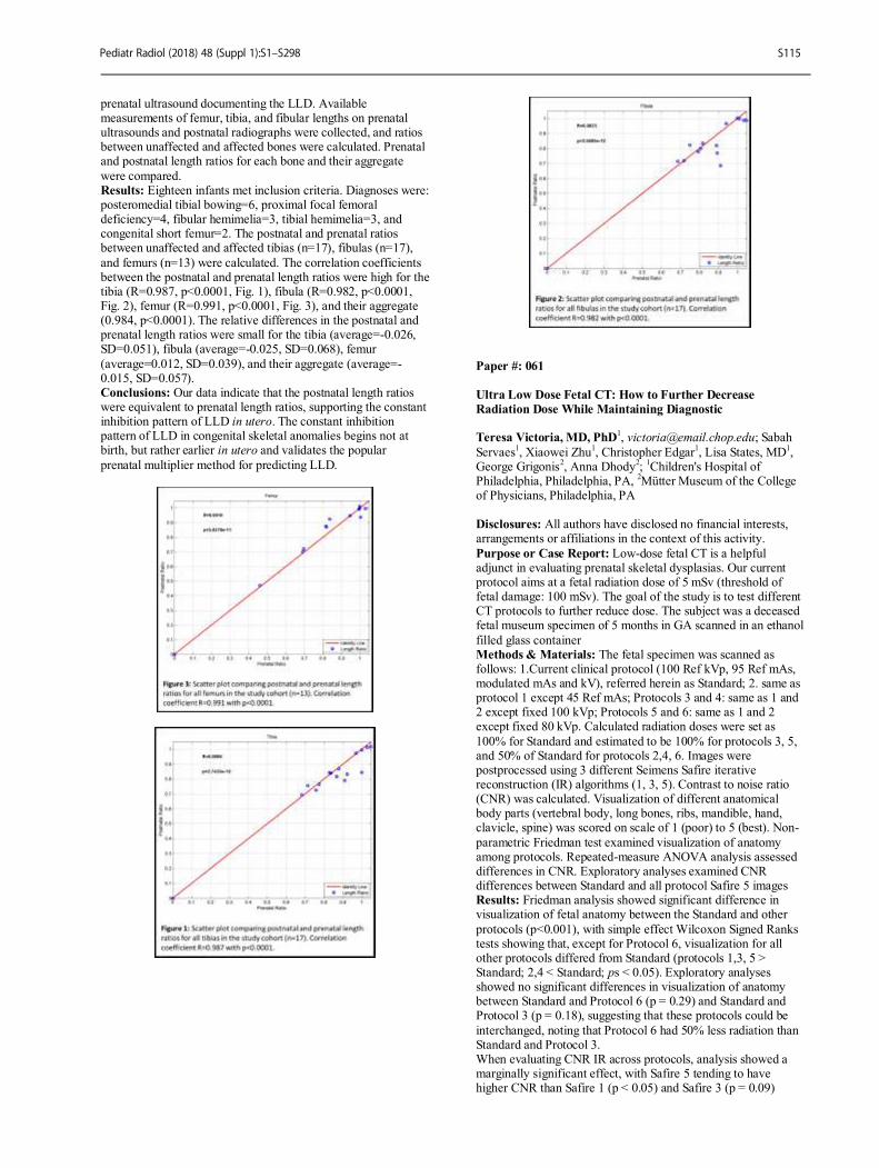
prenatal ultrasound documenting the LLD. Available measurements of femur, tibia, and fibular lengths on prenatal ultrasounds and postnatal radiographs were collected, and ratios between unaffected and affected bones were calculated. Prenatal and postnatal length ratios for each bone and their aggregate
were compared. Results: Eighteen infants met inclusion criteria. Diagnoses were: posteromedial tibial bowing=6, proximal focal femoral deficiency=4, fibular hemimelia=3, tibial hemimelia=3, and congenital short femur=2. The postnatal and prenatal ratios between unaffected and affected tibias (n=17), fibulas (n=17),
and femurs (n=13) were calculated. The correlation coefficients between the postnatal and prenatal length ratios were high for the tibia (R=0.987, p<0.0001, Fig. 1), fibula (R=0.982, p<0.0001, Fig. 2), femur (R=0.991, p<0.0001, Fig. 3), and their aggregate (0.984, p<0.0001). The relative differences in the postnatal and prenatal length ratios were small for the tibia (average=-0.026, SD=0.051), fibula (average=-0.025, SD=0.068), femur
(average=0.012, SD=0.039), and their aggregate (average=-0.015, SD=0.057). Conclusions: Our data indicate that the postnatal length ratios were equivalent to prenatal length ratios, supporting the constant inhibition pattern of LLD in utero. The constant inhibition pattern of LLD in congenital skeletal anomalies begins not at birth, but rather earlier in utero and validates the popular
prenatal multiplier method for predicting LLD.
Paper #: 061
Ultra Low Dose Fetal CT: How to Further Decrease
Radiation Dose While Maintaining Diagnostic Teresa Victoria, MD, PhD
1, [email protected]; Sabah
Servaes1, Xiaowei Zhu
1, Christopher Edgar
1, Lisa States, MD
1,
George Grigonis2, Anna Dhody
2;
1Children's Hospital of
Philadelphia, Philadelphia, PA, 2Mütter Museum of the College
of Physicians, Philadelphia, PA Disclosures: All authors have disclosed no financial interests, arrangements or affiliations in the context of this activity.
Purpose or Case Report: Low-dose fetal CT is a helpful adjunct in evaluating prenatal skeletal dysplasias. Our current protocol aims at a fetal radiation dose of 5 mSv (threshold of fetal damage: 100 mSv). The goal of the study is to test different CT protocols to further reduce dose. The subject was a deceased fetal museum specimen of 5 months in GA scanned in an ethanol
filled glass container Methods & Materials: The fetal specimen was scanned as follows: 1.Current clinical protocol (100 Ref kVp, 95 Ref mAs, modulated mAs and kV), referred herein as Standard; 2. same as protocol 1 except 45 Ref mAs; Protocols 3 and 4: same as 1 and 2 except fixed 100 kVp; Protocols 5 and 6: same as 1 and 2 except fixed 80 kVp. Calculated radiation doses were set as
100% for Standard and estimated to be 100% for protocols 3, 5, and 50% of Standard for protocols 2,4, 6. Images were postprocessed using 3 different Seimens Safire iterative reconstruction (IR) algorithms (1, 3, 5). Contrast to noise ratio (CNR) was calculated. Visualization of different anatomical body parts (vertebral body, long bones, ribs, mandible, hand, clavicle, spine) was scored on scale of 1 (poor) to 5 (best). Non-
parametric Friedman test examined visualization of anatomy among protocols. Repeated-measure ANOVA analysis assessed differences in CNR. Exploratory analyses examined CNR differences between Standard and all protocol Safire 5 images Results: Friedman analysis showed significant difference in visualization of fetal anatomy between the Standard and other
protocols (p<0.001), with simple effect Wilcoxon Signed Ranks tests showing that, except for Protocol 6, visualization for all other protocols differed from Standard (protocols 1,3, 5 > Standard; 2,4 < Standard; ps < 0.05). Exploratory analyses showed no significant differences in visualization of anatomy between Standard and Protocol 6 (p = 0.29) and Standard and Protocol 3 (p = 0.18), suggesting that these protocols could be
interchanged, noting that Protocol 6 had 50% less radiation than Standard and Protocol 3. When evaluating CNR IR across protocols, analysis showed a marginally significant effect, with Safire 5 tending to have higher CNR than Safire 1 (p < 0.05) and Safire 3 (p = 0.09)
Pediatr Radiol (2018) 48 (Suppl 1):S1–S298 S115
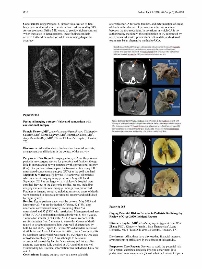
Conclusions: Using Protocol 6, similar visualization of fetal body parts is attained while radiation dose is decreased by 50%. Across protocols, Safire 5 IR tended to provide highest contrast. When translated to actual patients, these findings can help achieve further dose reduction while maintaining diagnostic
accuracy
Paper #: 062
Perinatal imaging autopsy: Value and comparison with
conventional autopsy Pamela Deaver, MD
1, [email protected]; Christopher
Cassady, MD1, Debra Kearney, MD
1, Eumenia Castro, MD
1,
Amy Mehollin-Ray, MD1;
1Texas Children's Hospital, Houston,
TX
Disclosures: All authors have disclosed no financial interests, arrangements or affiliations in the context of this activity. Purpose or Case Report: Imaging autopsy (IA) in the perinatal period is an emerging service for providers and families, though little is known about how it compares with conventional autopsy
(CA). Our purpose is to compare the two modalities using full unrestricted conventional autopsy (UCA) as the gold standard. Methods & Materials: Following IRB approval, all patients who underwent imaging autopsy between May 2013 and September 2017 at our large tertiary children’s hospital were enrolled. Review of the electronic medical record, including imaging and conventional autopsy findings, was performed.
Findings at imaging autopsy, including suspected cause of death, were compared to those at conventional autopsy and subdivided by organ system. Results: Eighty patients underwent IA between May 2013 and September 2017 at our institution. Of those, 62 (78%) also underwent conventional autopsy, including 30 (40%)
unrestricted and 32 (38%) with restrictions. Mean gestational age of the IA/UCA combination cohort at birth was 31.6 ± 4 weeks. Twenty-two infants (73%) with IA/UCA were liveborn, with survival ranging from 5 minutes to 6 months. Causes of death related to structural abnormalities were well characterized by both IA and UCA (Figure 1). Seven (24%) discordant causes of death between IA and UCA were identified, with 6 accounted for
by fulminant sepsis which was occult by IA (Figure 1). One case of hydranencephaly by UCA was thought to be severe acqueductal stenosis by IA. Surface anatomy and intracardiac anatomy were more fully detailed at UCA and often not well visualized by IA. Placental information was included at UCA but not IA. Conclusions: Imaging autopsy may be a more palatable
alternative to CA for some families, and determination of cause of death in the absence of perimortum infection is similar between the two modalities. In occasions in which CA is not authorized by the family, the combination of IA interpreted by an experienced reader, perimortum culture data, and external
exam may be an alternative method to UCA.
Paper #: 063
Gaging Potential Risk to Patients in Pediatric Radiology by
Review of Over 2,000 Incident Reports
Elizabeth Snyder, MD1, [email protected]; Wei
Zhang, PhD1, Kimberly Jasmin
1, Sam Thankachan
1, Lane
Donnelly, MD1;
1Texas Children’s Hospital, Houston, TX
Disclosures: All authors have disclosed no financial interests, arrangements or affiliations in the context of this activity.
Purpose or Case Report: One way to study the potential risk for a patient entering a pediatric imaging department is to perform a common cause analysis of submitted incident reports.
Pediatr Radiol (2018) 48 (Suppl 1):S1–S298S116
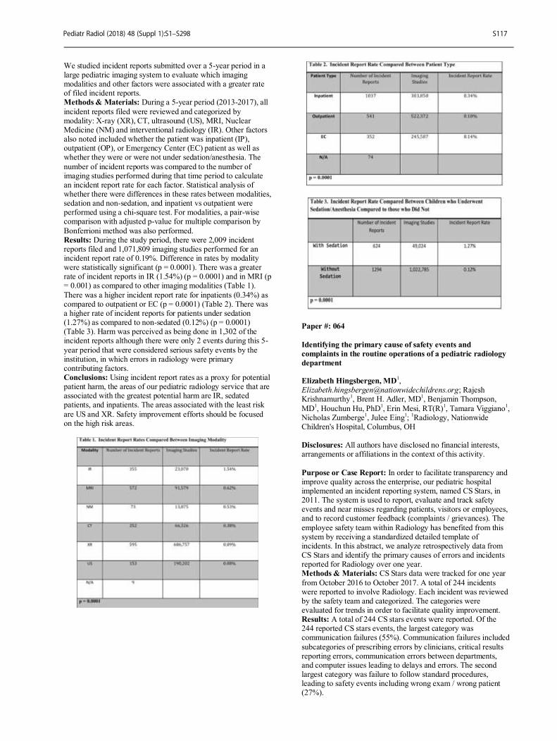
We studied incident reports submitted over a 5-year period in a large pediatric imaging system to evaluate which imaging modalities and other factors were associated with a greater rate of filed incident reports. Methods & Materials: During a 5-year period (2013-2017), all
incident reports filed were reviewed and categorized by modality: X-ray (XR), CT, ultrasound (US), MRI, Nuclear Medicine (NM) and interventional radiology (IR). Other factors also noted included whether the patient was inpatient (IP), outpatient (OP), or Emergency Center (EC) patient as well as whether they were or were not under sedation/anesthesia. The
number of incident reports was compared to the number of imaging studies performed during that time period to calculate an incident report rate for each factor. Statistical analysis of whether there were differences in these rates between modalities, sedation and non-sedation, and inpatient vs outpatient were performed using a chi-square test. For modalities, a pair-wise comparison with adjusted p-value for multiple comparison by
Bonferrioni method was also performed. Results: During the study period, there were 2,009 incident reports filed and 1,071,809 imaging studies performed for an incident report rate of 0.19%. Difference in rates by modality were statistically significant (p = 0.0001). There was a greater rate of incident reports in IR (1.54%) (p = 0.0001) and in MRI (p = 0.001) as compared to other imaging modalities (Table 1).
There was a higher incident report rate for inpatients (0.34%) as compared to outpatient or EC (p = 0.0001) (Table 2). There was a higher rate of incident reports for patients under sedation (1.27%) as compared to non-sedated (0.12%) (p = 0.0001) (Table 3). Harm was perceived as being done in 1,302 of the incident reports although there were only 2 events during this 5-
year period that were considered serious safety events by the institution, in which errors in radiology were primary contributing factors. Conclusions: Using incident report rates as a proxy for potential patient harm, the areas of our pediatric radiology service that are associated with the greatest potential harm are IR, sedated patients, and inpatients. The areas associated with the least risk
are US and XR. Safety improvement efforts should be focused on the high risk areas.
Paper #: 064
Identifying the primary cause of safety events and
complaints in the routine operations of a pediatric radiology
department Elizabeth Hingsbergen, MD
1,
[email protected]; Rajesh Krishnamurthy
1, Brent H. Adler, MD
1, Benjamin Thompson,
MD1, Houchun Hu, PhD
1, Erin Mesi, RT(R)
1, Tamara Viggiano
1,
Nicholas Zumberge1, Julee Eing
1;
1Radiology, Nationwide
Children's Hospital, Columbus, OH
Disclosures: All authors have disclosed no financial interests, arrangements or affiliations in the context of this activity.
Purpose or Case Report: In order to facilitate transparency and improve quality across the enterprise, our pediatric hospital implemented an incident reporting system, named CS Stars, in 2011. The system is used to report, evaluate and track safety events and near misses regarding patients, visitors or employees, and to record customer feedback (complaints / grievances). The
employee safety team within Radiology has benefited from this system by receiving a standardized detailed template of incidents. In this abstract, we analyze retrospectively data from CS Stars and identify the primary causes of errors and incidents reported for Radiology over one year. Methods & Materials: CS Stars data were tracked for one year
from October 2016 to October 2017. A total of 244 incidents were reported to involve Radiology. Each incident was reviewed by the safety team and categorized. The categories were evaluated for trends in order to facilitate quality improvement. Results: A total of 244 CS stars events were reported. Of the 244 reported CS stars events, the largest category was communication failures (55%). Communication failures included
subcategories of prescribing errors by clinicians, critical results reporting errors, communication errors between departments, and computer issues leading to delays and errors. The second largest category was failure to follow standard procedures, leading to safety events including wrong exam / wrong patient (27%).
Pediatr Radiol (2018) 48 (Suppl 1):S1–S298 S117
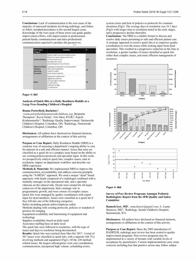
Conclusions: Lack of communication is the root cause of the majority of untoward incidents involving radiology, and failure to follow standard procedures is the second largest cause. Knowledge of the root cause of these errors can guide quality improvement efforts, with improvement in professional-
patient/family communication and inter-professional communication expected to produce the greatest res.
Paper #: 065
Analysis of Quick Hits at a Daily Readiness Huddle at a
Large Free-Standing Children's Hospital Ronna Porterfield, Bachelors
2,
[email protected]; Benjamin Thompson
2, Kevin Dolan
1, Erin Mesi, RT(R)
2, Rajesh
Krishnamurthy2;
1Radiology Quality Improvement, Nationwide
Children's Hospital, Columbus, OH, 2Radiology, Nationwide
Children's Hospital, Columbus, OH Disclosures: All authors have disclosed no financial interests, arrangements or affiliations in the context of this activity.
Purpose or Case Report: Daily Readiness Huddle (DRH) is a common way of assessing a department’s ongoing ability to care for patients in a safe and efficient manner. Issues that arise are classified as a quick hit or a complex issue based on the ability to use existing policies/protocols and specialist input. In this work, we prospectively analyze quick hits, complex issues, time to
resolution, impact on department workflow and describe our DRH experience. Methods & Materials: We implemented DRH to improve the communication, accountability and address concerns promptly using the “S-MESA” approach. We used a unique “dyad” based approach, with dyads composed of a radiologist combined with a modality manager on the operational side, and a specialty
clinician on the clinical side. Dyads were created for all major endeavors of the department, had a strategic role in programmatic growth, and were owners of complex issues. Quick hits were managed by quality coordinators with expert input for dyad members. Issues were classified as a quick hit if they fell into one of the following categories:
Safety-including patient safety/employee safety Methods-dealing with existing protocols, policies or standard of practice for imaging Equipment-availability and functioning of equipment and technology Supplies-availability based on daily need Associates-staffing base on daily need
The quick hits were followed to resolution, with the type of issues and days to resolution being documented. Results: Quick hits were tracked from Mar-Oct 2017. A total of 567 issues were classified as quick hits, with a majority (66.7%) involving methods and equipment (Fig1). Of the 220 method-related issues, the largest subcategories were care coordination, communication, unexpected high volume, scheduling errors,
system errors and lack of policies or protocols for common situations (Fig2). The average days to resolution was 16.1 days (Fig3) with longer time to resolution noted in the early stages, and a progressive decline thereafter. Conclusions: The DRH is a reliable format to discuss and
resolve daily issues pertaining to safe and efficient patient care. A strategic approach to resolve quick hits is to empower quality coordinators to own the issues while seeking input from dyad specialists. This resulted in a progressive reduction in the time to resolution, a greater number of issues classified as quick hits rather than complex issues, and more efficient management of
resources.
Paper #: 066
Survey of Peer Review Programs Amongst Pediatric
Radiologists: Report from the SPR Quality and Safety
Committee
Ramesh Iyer, MD1, [email protected]; A. Luana
Stanescu, MD1;
1Radiology, Seattle Children's Hospital,
Sammamish, WA Disclosures: All authors have disclosed no financial interests, arrangements or affiliations in the context of this activity.
Purpose or Case Report: Since the 2002 introduction of RADPEER, radiology peer review has been central to quality improvement programs. Peer review programs may be implemented in a variety of formats with variable levels of acceptance by practitioners. Current implementations carry some concerns including fear that punitive action may follow subpar
Pediatr Radiol (2018) 48 (Suppl 1):S1–S298S118
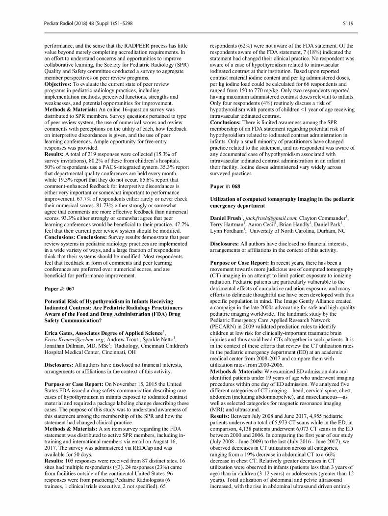
performance, and the sense that the RADPEER process has little value beyond merely completing accreditation requirements. In an effort to understand concerns and opportunities to improve collaborative learning, the Society for Pediatric Radiology (SPR) Quality and Safety committee conducted a survey to aggregate
member perspectives on peer review programs. Objectives: To evaluate the current state of peer review programs in pediatric radiology practices, including implementation methods, perceived functions, strengths and weaknesses, and potential opportunities for improvement. Methods & Materials: An online 16-question survey was
distributed to SPR members. Survey questions pertained to type of peer review system, the use of numerical scores and review comments with perceptions on the utility of each, how feedback on interpretive discordances is given, and the use of peer learning conferences. Ample opportunity for free-entry responses was provided. Results: A total of 219 responses were collected (15.3% of
survey invitations), 80.2% of these from children’s hospitals. 50% of respondents use a PACS-integrated system. 35.3% report that departmental quality conferences are held every month, while 19.3% report that they do not occur. 85.6% report that comment-enhanced feedback for interpretive discordances is either very important or somewhat important to performance improvement. 67.7% of respondents either rarely or never check
their numerical scores. 81.73% either strongly or somewhat agree that comments are more effective feedback than numerical scores. 93.3% either strongly or somewhat agree that peer learning conferences would be beneficial to their practice. 47.7% feel that their current peer review system should be modified. Conclusions: Conclusions: Survey results demonstrate that peer
review systems in pediatric radiology practices are implemented in a wide variety of ways, and a large fraction of respondents think that their systems should be modified. Most respondents feel that feedback in form of comments and peer learning conferences are preferred over numerical scores, and are beneficial for performance improvement.
Paper #: 067
Potential Risk of Hypothyroidism in Infants Receiving
Iodinated Contrast: Are Pediatric Radiology Practitioners
Aware of the Food and Drug Administration (FDA) Drug
Safety Communication?
Erica Gates, Associates Degree of Applied Science1,
[email protected]; Andrew Trout1, Sparkle Netto
1,
Jonathan Dillman, MD, MSc1;
1Radiology, Cincinnati Children's
Hospital Medical Center, Cincinnati, OH Disclosures: All authors have disclosed no financial interests,
arrangements or affiliations in the context of this activity. Purpose or Case Report: On November 15, 2015 the United States FDA issued a drug safety communication describing rare cases of hypothyroidism in infants exposed to iodinated contrast material and required a package labeling change describing these cases. The purpose of this study was to understand awareness of
this statement among the membership of the SPR and how the statement had changed clinical practice. Methods & Materials: A six item survey regarding the FDA statement was distributed to active SPR members, including in-training and international members via email on August 16, 2017. The survey was administered via REDCap and was
available for 50 days. Results: 105 responses were received from 87 distinct sites. 16 sites had multiple respondents (≤3). 24 responses (23%) came from facilities outside of the continental United States. 96 responses were from practicing Pediatric Radiologists (6 trainees, 1 clinical trials executive, 2 not specified). 65
respondents (62%) were not aware of the FDA statement. Of the respondents aware of the FDA statement, 7 (18%) indicated the statement had changed their clinical practice. No respondent was aware of a case of hypothyroidism related to intravascular iodinated contrast at their institution. Based upon reported
contrast material iodine content and per kg administered doses, per kg iodine load could be calculated for 66 respondents and ranged from 150 to 770 mg/kg. Only two respondents reported having maximum administered contrast doses relevant to infants. Only four respondents (4%) routinely discuss a risk of hypothyroidism with parents of children <1 year of age receiving
intravascular iodinated contrast. Conclusions: There is limited awareness among the SPR membership of an FDA statement regarding potential risk of hypothyroidism related to iodinated contrast administration in infants. Only a small minority of practitioners have changed practice related to the statement, and no respondent was aware of any documented case of hypothyroidism associated with
intravascular iodinated contrast administration in an infant at their facility. Iodine doses administered vary widely across surveyed practices.
Paper #: 068
Utilization of computed tomography imaging in the pediatric
emergency department Daniel Frush
1, [email protected]; Clayton Commander
1,
Terry Hartman1, Aaron Cecil
1, Brian Handly
1, Daniel Park
1,
Lynn Fordham1;
1University of North Carolina, Durham, NC
Disclosures: All authors have disclosed no financial interests, arrangements or affiliations in the context of this activity. Purpose or Case Report: In recent years, there has been a movement towards more judicious use of computed tomography (CT) imaging in an attempt to limit patient exposure to ionizing radiation. Pediatric patients are particularly vulnerable to the
detrimental effects of cumulative radiation exposure, and many efforts to delineate thoughtful use have been developed with this specific population in mind. The Image Gently Alliance created a campaign in the late 2000s advocating for safe and high-quality pediatric imaging worldwide. The landmark study by the Pediatric Emergency Care Applied Research Network (PECARN) in 2009 validated prediction rules to identify
children at low risk for clinically-important traumatic brain injuries and thus avoid head CTs altogether in such patients. It is in the context of these efforts that review the CT utilization rates in the pediatric emergency department (ED) at an academic medical center from 2008-2017 and compare them with utilization rates from 2000-2006.
Methods & Materials: We examined ED admission data and identified patients under 19 years of age who underwent imaging procedures within one day of ED admission. We analyzed five different categories of CT imaging—head, cervical spine, chest, abdomen (including abdominopelvic), and miscellaneous—as well as selected categories for magnetic resonance imaging (MRI) and ultrasound.
Results: Between July 2008 and June 2017, 4,955 pediatric patients underwent a total of 5,973 CT scans while in the ED; in comparison, 4,138 patients underwent 6,073 CT scans in the ED between 2000 and 2006. In comparing the first year of our study (July 2008 - June 2009) to the last (July 2016 - June 2017), we observed decreases in CT utilization across all categories,
ranging from a 19% decrease in abdominal CT to a 66% decrease in chest CT. Relatively greater decreases in CT utilization were observed in infants (patients less than 3 years of age) than in children (3-12 years) or adolescents (greater than 12 years). Total utilization of abdominal and pelvic ultrasound increased, with the rise in abdominal ultrasound driven entirely
Pediatr Radiol (2018) 48 (Suppl 1):S1–S298 S119
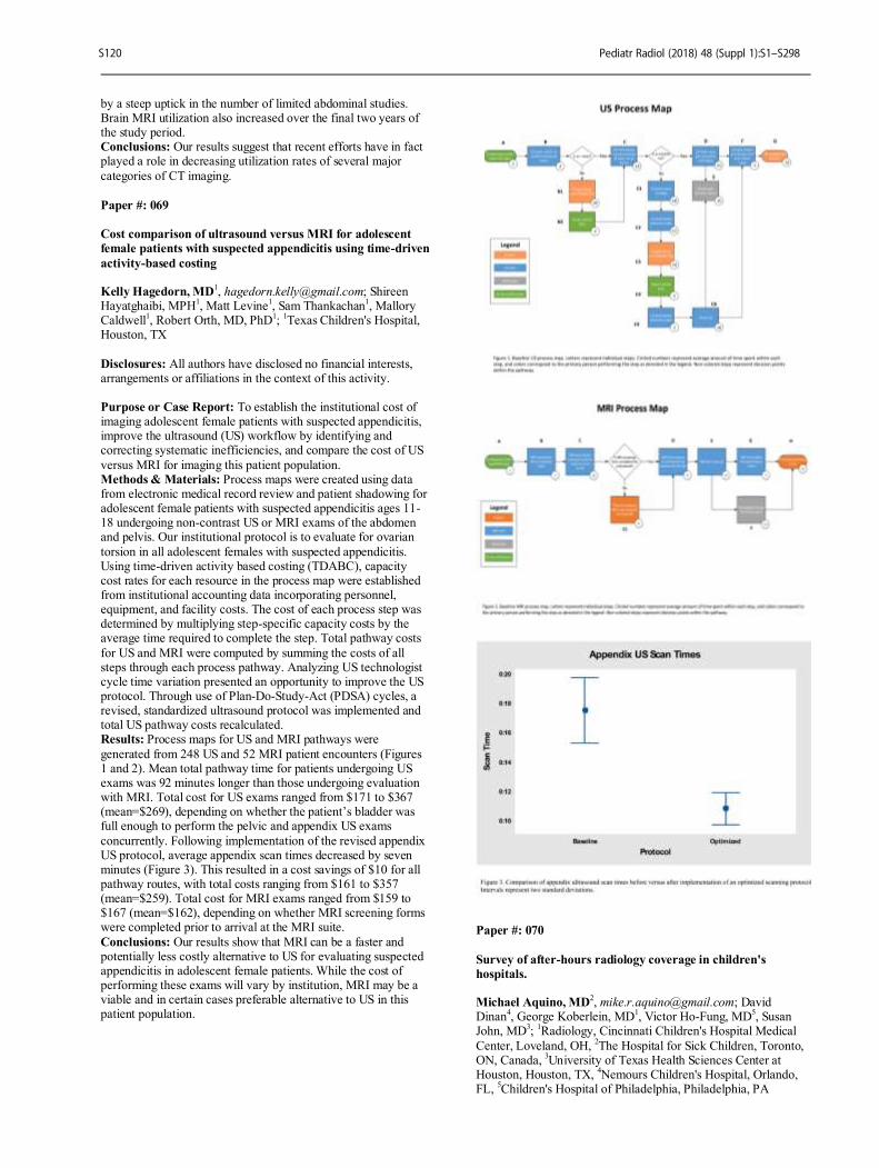
by a steep uptick in the number of limited abdominal studies. Brain MRI utilization also increased over the final two years of the study period. Conclusions: Our results suggest that recent efforts have in fact played a role in decreasing utilization rates of several major
categories of CT imaging.
Paper #: 069
Cost comparison of ultrasound versus MRI for adolescent
female patients with suspected appendicitis using time-driven
activity-based costing Kelly Hagedorn, MD
1, [email protected]; Shireen
Hayatghaibi, MPH1, Matt Levine
1, Sam Thankachan
1, Mallory
Caldwell1, Robert Orth, MD, PhD
1;
1Texas Children's Hospital,
Houston, TX
Disclosures: All authors have disclosed no financial interests, arrangements or affiliations in the context of this activity. Purpose or Case Report: To establish the institutional cost of imaging adolescent female patients with suspected appendicitis, improve the ultrasound (US) workflow by identifying and correcting systematic inefficiencies, and compare the cost of US
versus MRI for imaging this patient population. Methods & Materials: Process maps were created using data from electronic medical record review and patient shadowing for adolescent female patients with suspected appendicitis ages 11-18 undergoing non-contrast US or MRI exams of the abdomen and pelvis. Our institutional protocol is to evaluate for ovarian
torsion in all adolescent females with suspected appendicitis. Using time-driven activity based costing (TDABC), capacity cost rates for each resource in the process map were established from institutional accounting data incorporating personnel, equipment, and facility costs. The cost of each process step was determined by multiplying step-specific capacity costs by the average time required to complete the step. Total pathway costs
for US and MRI were computed by summing the costs of all steps through each process pathway. Analyzing US technologist cycle time variation presented an opportunity to improve the US protocol. Through use of Plan-Do-Study-Act (PDSA) cycles, a revised, standardized ultrasound protocol was implemented and total US pathway costs recalculated. Results: Process maps for US and MRI pathways were
generated from 248 US and 52 MRI patient encounters (Figures 1 and 2). Mean total pathway time for patients undergoing US exams was 92 minutes longer than those undergoing evaluation with MRI. Total cost for US exams ranged from $171 to $367 (mean=$269), depending on whether the patient’s bladder was full enough to perform the pelvic and appendix US exams
concurrently. Following implementation of the revised appendix US protocol, average appendix scan times decreased by seven minutes (Figure 3). This resulted in a cost savings of $10 for all pathway routes, with total costs ranging from $161 to $357 (mean=$259). Total cost for MRI exams ranged from $159 to $167 (mean=$162), depending on whether MRI screening forms were completed prior to arrival at the MRI suite.
Conclusions: Our results show that MRI can be a faster and potentially less costly alternative to US for evaluating suspected appendicitis in adolescent female patients. While the cost of performing these exams will vary by institution, MRI may be a viable and in certain cases preferable alternative to US in this patient population.
Paper #: 070
Survey of after-hours radiology coverage in children's
hospitals. Michael Aquino, MD
2, [email protected]; David
Dinan4, George Koberlein, MD
1, Victor Ho-Fung, MD
5, Susan
John, MD3;
1Radiology, Cincinnati Children's Hospital Medical
Center, Loveland, OH, 2The Hospital for Sick Children, Toronto,
ON, Canada, 3University of Texas Health Sciences Center at
Houston, Houston, TX, 4Nemours Children's Hospital, Orlando,
FL, 5Children's Hospital of Philadelphia, Philadelphia, PA
Pediatr Radiol (2018) 48 (Suppl 1):S1–S298S120
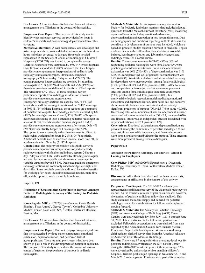
Disclosures: All authors have disclosed no financial interests, arrangements or affiliations in the context of this activity. Purpose or Case Report: The purpose of this study was to identify what radiology services are provided after-hours in
children's hospitals and how radiology departments deliver this coverage. Methods & Materials: A web-based survey was developed and asked respondents to provide detailed information on their after-hours radiology coverage. A delegate from each hospital represented in the Society of Chairs of Radiology at Children's
Hospitals (SCORCH) was invited to complete the survey. Results: Responses were submitted by 59% (47/79) of hospitals. Over 80% of respondents (39/47) indicated that their hospital provides contemporaneous interpretations of pediatric body radiology studies (radiographs, ultrasound, computed tomography) 24 hours a day, 7 days a week ("24/7"). The contemporaneous interpretations are provided by attending
radiologists in 51% (20/39) of hospitals and 95% (19/20) of these interpretations are delivered in the form of final reports. The remaining 49% (19/39) of these hospitals rely on preliminary reports from radiology residents or fellows to provide contemporaneous interpretations. Emergency radiology sections are used by 36% (14/47) of hospitals to staff the overnight duration of the "24/7" coverage.
In 79% (11/14) of these hospitals, these sections are composed of pediatric radiologists. Teleradiology services are used by 8% (4/47) for overnight service. Overall, 55% (26/47) of hospitals described scheduling at least 1 attending pediatric radiologist on a late-shift that extends variable durations beyond 5 PM to provide contemporaneous interpretations after-hours. Only 4%
(2/47) provide strictly beeper-call coverage after 5 PM. The option to work remotely rather than in-house is offered to radiologists working after-hours in 21% (10/47) hospitals. Additional benefits such as increased income, more time off, or academic time is offered by (21/47) hospitals. Conclusions: The majority of children's hospitals surveyed provide contemporaneous interpretations of pediatric body
radiology studies with final or preliminary reports 24 hours a day, 7 days a week. Late-shifts staffed by attending radiologists are used by most surveyed hospitals to extend coverage for variable durations beyond 5 PM. Dedicated pediatric emergency radiology sections are commonly used to bridge late night and day shifts. Some hospitals provide additional incentive benefits for working after-hours including increased income, more time
off, and the option to work remotely from home.
Paper #: 071
Evaluation of Stressors that Contribute to Burnout Amongst
Pediatric Radiologists: A Survey of the Society for Pediatric
Radiology Rama Ayyala, MD
1, [email protected]; Carrie Ruzal-
Shapiro1, Firas Ahmed
1, George Taylor
2;
1Columbia University
Medical Center, New York, NY, 2Boston Children’s Hospital,
Boston, MA
Disclosures: All authors have disclosed no financial interests, arrangements or affiliations in the context of this activity. Purpose or Case Report: Burnout is a psychological syndrome that is characterized by three major components: emotional exhaustion, depersonalization, and perceived lack of
accomplishment. There are multiple stressors that have been shown to play a role in the development of burnout in medicine. The purpose of this study is to evaluate the impact of various causes of stress on the prevalence of burnout in pediatric radiologists.
Methods & Materials: An anonymous survey was sent to Society for Pediatric Radiology members that included adapted questions from the Maslach Burnout Inventory (MBI) measuring aspects of burnout including emotional exhaustion, depersonalization and perception of low accomplishment. Data
on demographics and questions regarding multiple stressors that contribute to development of burnout were included, which are based on previous studies regarding burnout in medicine. Topics evaluated include the call burden, financial stress, work-life balance, healthcare evolution and job market changes, and radiology overall as a career choice.
Results: The response rate was 460/1453 (32%). 50% of responding pediatric radiologists were female and 42% were practicing at academic institutions. Prevalence of emotional exhaustion was 66% (286/435), of depersonalization was 61% (265/433) and perceived lack of personal accomplishment was 15% (67/436). Work-life imbalance and stress related to caring for dependents were more prevalent among female radiologists
(75%, p-value=0.019 and 45%, p-value=0.011). After hours call and competitive radiology job market were more prevalent stressors among female radiologists than male counterparts (53%, p-value=0.002 and 17%, p-value=0.030). In a multivariable logistic regression model predicting emotional exhaustion and depersonalization, after hours call and concerns about work-life balance were consistent and statistically
significant predictors of burnout (ORs=2.3-7.6; p-value <0.001). Decreasing rates of reimbursement was an independent stressor associated with emotional exhaustion (OR=2.7; p-value=0.030) and financial strain was an independent stressor associated with depersonalization (OR=2.3; p-value=0.020). Conclusions: Emotional exhaustion and depersonalization are
prevalent among the community of pediatric radiology. On call responsibilities, work-life imbalance, and financial concerns were strong stressors contributing to the burnout. These stressors were more prevalent among female radiologists.
Paper #: 072
Assessing the Pediatric Radiology Job Market: Winter is
Coming for Employers Cory Pfeifer, MD
1 [email protected]; ;
1Diagnostic
Radiology, University of Texas Southwestern Medical Center, Dallas, TX
Disclosures: All authors have disclosed no financial interests, arrangements or affiliations in the context of this activity. Purpose or Case Report: The 2016-2017 academic year represented a significant recovery of the diagnostic radiology job market. As the available number of jobs has increased, however,
the number of pediatric radiology fellows has declined. This study examines the recent supply and demand for pediatric radiologists as well as implications for fellows and employers moving forward. Methods & Materials: The Society for Pediatric Radiology (SPR) and American College of Radiology (ACR) Career Centers were analyzed each day from July 1, 2016 through June
30, 2017. Job advertisements for fellowship positions were excluded. Fellowship occupation rates were derived from data reported by the Accreditation Council for Graduate Medical Education. Projected fellowship interest was assessed using chief-resident-derived survey data from the American Alliance of Academic Chief Residents in Radiology.
Results: There were 97 unique full-time attending-level jobs for pediatric radiologists advertised on the SPR Career Center during the 2016-2017 academic year. Of these openings, 72% were advertised by universities or free-standing children's hospitals. Distinct peaks in job openings in November 2016 and March 2017 were apparent. Positions were posted for a median
Pediatr Radiol (2018) 48 (Suppl 1):S1–S298 S121
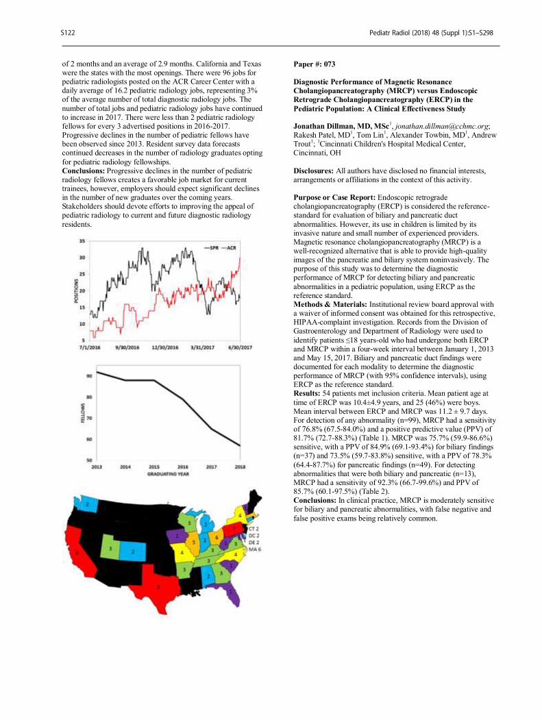
of 2 months and an average of 2.9 months. California and Texas were the states with the most openings. There were 96 jobs for pediatric radiologists posted on the ACR Career Center with a daily average of 16.2 pediatric radiology jobs, representing 3% of the average number of total diagnostic radiology jobs. The
number of total jobs and pediatric radiology jobs have continued to increase in 2017. There were less than 2 pediatric radiology fellows for every 3 advertised positions in 2016-2017. Progressive declines in the number of pediatric fellows have been observed since 2013. Resident survey data forecasts continued decreases in the number of radiology graduates opting
for pediatric radiology fellowships. Conclusions: Progressive declines in the number of pediatric radiology fellows creates a favorable job market for current trainees, however, employers should expect significant declines in the number of new graduates over the coming years. Stakeholders should devote efforts to improving the appeal of pediatric radiology to current and future diagnostic radiology
residents.
Paper #: 073
Diagnostic Performance of Magnetic Resonance
Cholangiopancreatography (MRCP) versus Endoscopic
Retrograde Cholangiopancreatography (ERCP) in the
Pediatric Population: A Clinical Effectiveness Study Jonathan Dillman, MD, MSc
Rakesh Patel, MD1, Tom Lin
1, Alexander Towbin, MD
1, Andrew
Trout1;
1Cincinnati Children's Hospital Medical Center,
Cincinnati, OH
Disclosures: All authors have disclosed no financial interests, arrangements or affiliations in the context of this activity. Purpose or Case Report: Endoscopic retrograde cholangiopancreatography (ERCP) is considered the reference-standard for evaluation of biliary and pancreatic duct
abnormalities. However, its use in children is limited by its invasive nature and small number of experienced providers. Magnetic resonance cholangiopancreatography (MRCP) is a well-recognized alternative that is able to provide high-quality images of the pancreatic and biliary system noninvasively. The purpose of this study was to determine the diagnostic performance of MRCP for detecting biliary and pancreatic
abnormalities in a pediatric population, using ERCP as the reference standard. Methods & Materials: Institutional review board approval with a waiver of informed consent was obtained for this retrospective, HIPAA-complaint investigation. Records from the Division of Gastroenterology and Department of Radiology were used to
identify patients ≤18 years-old who had undergone both ERCP and MRCP within a four-week interval between January 1, 2013 and May 15, 2017. Biliary and pancreatic duct findings were documented for each modality to determine the diagnostic performance of MRCP (with 95% confidence intervals), using ERCP as the reference standard. Results: 54 patients met inclusion criteria. Mean patient age at
time of ERCP was 10.4±4.9 years, and 25 (46%) were boys. Mean interval between ERCP and MRCP was 11.2 ± 9.7 days. For detection of any abnormality (n=99), MRCP had a sensitivity of 76.8% (67.5-84.0%) and a positive predictive value (PPV) of 81.7% (72.7-88.3%) (Table 1). MRCP was 75.7% (59.9-86.6%) sensitive, with a PPV of 84.9% (69.1-93.4%) for biliary findings (n=37) and 73.5% (59.7-83.8%) sensitive, with a PPV of 78.3%
(64.4-87.7%) for pancreatic findings (n=49). For detecting abnormalities that were both biliary and pancreatic (n=13), MRCP had a sensitivity of 92.3% (66.7-99.6%) and PPV of 85.7% (60.1-97.5%) (Table 2). Conclusions: In clinical practice, MRCP is moderately sensitive for biliary and pancreatic abnormalities, with false negative and
false positive exams being relatively common.
Pediatr Radiol (2018) 48 (Suppl 1):S1–S298S122
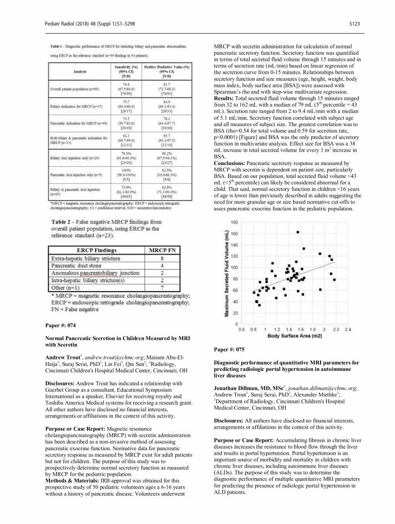
Paper #: 074
Normal Pancreatic Secretion in Children Measured by MRI
with Secretin Andrew Trout
1, [email protected]; Maisam Abu-El-
Haija1, Suraj Serai, PhD
1, Lin Fei
1, Qin Sun
1;
1Radiology,
Cincinnati Children's Hospital Medical Center, Cincinnati, OH Disclosures: Andrew Trout has indicated a relationship with Guerbet Group as a consultant, Educational Symposium International as a speaker, Elsevier for receiving royalty and Toshiba America Medical systems for receiving a research grant.
All other authors have disclosed no financial interests, arrangements or affiliations in the context of this activity. Purpose or Case Report: Magnetic resonance cholangiopancreatography (MRCP) with secretin administration has been described as a non-invasive method of assessing
pancreatic exocrine function. Normative data for pancreatic secretory response as measured by MRCP exist for adult patients but not for children. The purpose of this study was to prospectively determine normal secretory function as measured by MRCP for the pediatric population. Methods & Materials: IRB approval was obtained for this prospective study of 50 pediatric volunteers ages a 6-16 years
without a history of pancreatic disease. Volunteers underwent
MRCP with secretin administration for calculation of normal pancreatic secretory function. Secretory function was quantified in terms of total secreted fluid volume through 15 minutes and in terms of secretion rate (mL/min) based on linear regression of the secretion curve from 0-15 minutes. Relationships between
secretory function and size measures (age, height, weight, body mass index, body surface area [BSA]) were assessed with Spearman’s rho and with step-wise multivariate regression. Results: Total secreted fluid volume through 15 minutes ranged from 32 to 162 mL with a median of 79 mL (5
th percentile = 43
mL). Secretion rate ranged from 2 to 9.4 mL/min with a median
of 5.1 mL/min. Secretory function correlated with subject age and all measures of subject size. The greatest correlation was to BSA (rho=0.54 for total volume and 0.59 for secretion rate, p<0.0001) [Figure] and BSA was the only predictor of secretory function in multivariate analysis. Effect size for BSA was a 38 mL increase in total secreted volume for every 1 m
2 increase in
BSA.
Conclusions: Pancreatic secretory response as measured by MRCP with secretin is dependent on patient size, particularly BSA. Based on our population, total secreted fluid volume <43 mL (<5
th percentile) can likely be considered abnormal for a
child. That said, normal secretory function in children <16 years of age is lower than previously described in adults suggesting the need for more granular age or size based normative cut-offs to
asses pancreatic exocrine function in the pediatric population.
Paper #: 075
Diagnostic performance of quantitative MRI parameters for
predicting radiologic portal hypertension in autoimmune
liver diseases Jonathan Dillman, MD, MSc
Andrew Trout1, Suraj Serai, PhD
1, Alexander Miethke
1;
1Department of Radiology, Cincinnati Children's Hospital
Medical Center, Cincinnati, OH Disclosures: All authors have disclosed no financial interests, arrangements or affiliations in the context of this activity.
Purpose or Case Report: Accumulating fibrosis in chronic liver diseases increases the resistance to blood flow through the liver and results in portal hypertension. Portal hypertension is an important source of morbidity and mortality in children with chronic liver diseases, including autoimmune liver diseases (ALDs). The purpose of this study was to determine the diagnostic performance of multiple quantitative MRI parameters
for predicting the presence of radiologic portal hypertension in ALD patients.
Pediatr Radiol (2018) 48 (Suppl 1):S1–S298 S123
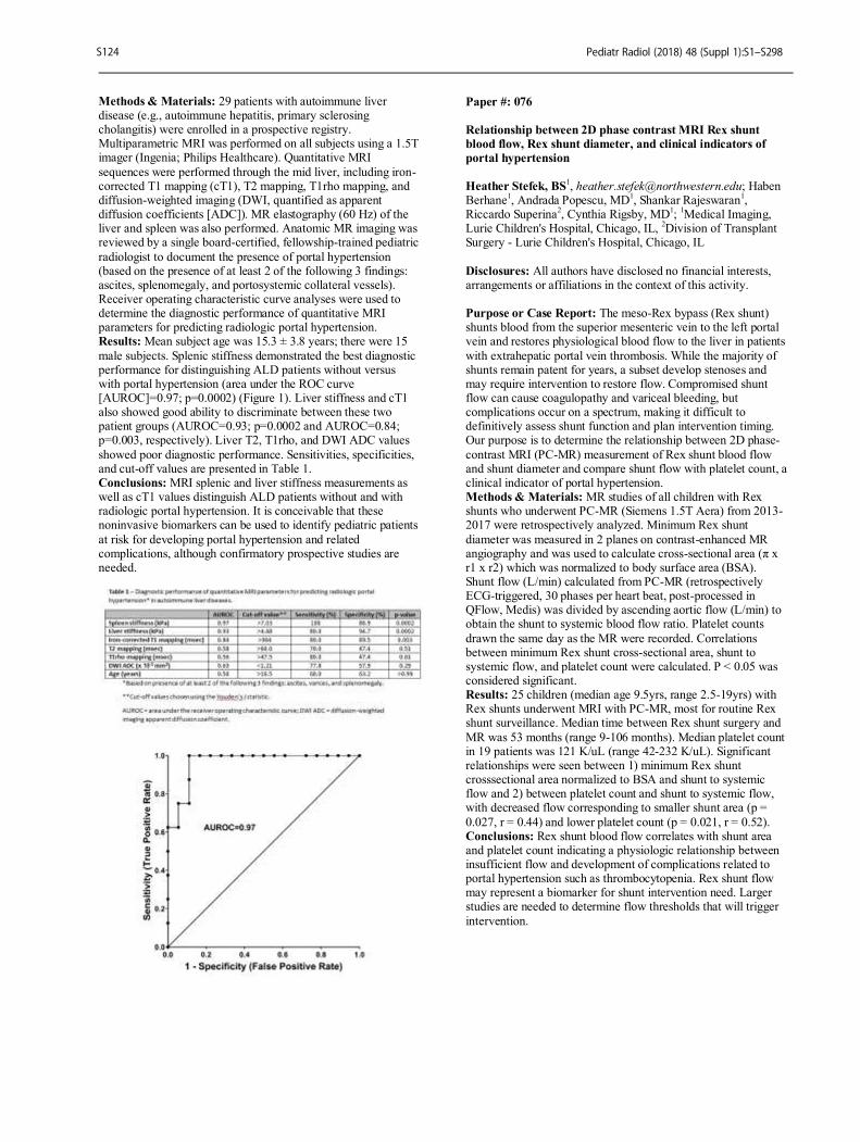
Methods & Materials: 29 patients with autoimmune liver disease (e.g., autoimmune hepatitis, primary sclerosing cholangitis) were enrolled in a prospective registry. Multiparametric MRI was performed on all subjects using a 1.5T imager (Ingenia; Philips Healthcare). Quantitative MRI
sequences were performed through the mid liver, including iron-corrected T1 mapping (cT1), T2 mapping, T1rho mapping, and diffusion-weighted imaging (DWI, quantified as apparent diffusion coefficients [ADC]). MR elastography (60 Hz) of the liver and spleen was also performed. Anatomic MR imaging was reviewed by a single board-certified, fellowship-trained pediatric
radiologist to document the presence of portal hypertension (based on the presence of at least 2 of the following 3 findings: ascites, splenomegaly, and portosystemic collateral vessels). Receiver operating characteristic curve analyses were used to determine the diagnostic performance of quantitative MRI parameters for predicting radiologic portal hypertension. Results: Mean subject age was 15.3 ± 3.8 years; there were 15
male subjects. Splenic stiffness demonstrated the best diagnostic performance for distinguishing ALD patients without versus with portal hypertension (area under the ROC curve [AUROC]=0.97; p=0.0002) (Figure 1). Liver stiffness and cT1 also showed good ability to discriminate between these two patient groups (AUROC=0.93; p=0.0002 and AUROC=0.84; p=0.003, respectively). Liver T2, T1rho, and DWI ADC values
showed poor diagnostic performance. Sensitivities, specificities, and cut-off values are presented in Table 1. Conclusions: MRI splenic and liver stiffness measurements as well as cT1 values distinguish ALD patients without and with radiologic portal hypertension. It is conceivable that these noninvasive biomarkers can be used to identify pediatric patients
at risk for developing portal hypertension and related complications, although confirmatory prospective studies are needed.
Paper #: 076
Relationship between 2D phase contrast MRI Rex shunt
blood flow, Rex shunt diameter, and clinical indicators of
portal hypertension
Heather Stefek, BS
1, [email protected]; Haben
Berhane1, Andrada Popescu, MD
1, Shankar Rajeswaran
1,
Riccardo Superina2, Cynthia Rigsby, MD
1;
1Medical Imaging,
Lurie Children's Hospital, Chicago, IL, 2Division of Transplant
Surgery - Lurie Children's Hospital, Chicago, IL
Disclosures: All authors have disclosed no financial interests, arrangements or affiliations in the context of this activity. Purpose or Case Report: The meso-Rex bypass (Rex shunt) shunts blood from the superior mesenteric vein to the left portal vein and restores physiological blood flow to the liver in patients
with extrahepatic portal vein thrombosis. While the majority of shunts remain patent for years, a subset develop stenoses and may require intervention to restore flow. Compromised shunt flow can cause coagulopathy and variceal bleeding, but complications occur on a spectrum, making it difficult to definitively assess shunt function and plan intervention timing. Our purpose is to determine the relationship between 2D phase-
contrast MRI (PC-MR) measurement of Rex shunt blood flow and shunt diameter and compare shunt flow with platelet count, a clinical indicator of portal hypertension. Methods & Materials: MR studies of all children with Rex shunts who underwent PC-MR (Siemens 1.5T Aera) from 2013-2017 were retrospectively analyzed. Minimum Rex shunt
diameter was measured in 2 planes on contrast-enhanced MR angiography and was used to calculate cross-sectional area (π x r1 x r2) which was normalized to body surface area (BSA). Shunt flow (L/min) calculated from PC-MR (retrospectively ECG-triggered, 30 phases per heart beat, post-processed in QFlow, Medis) was divided by ascending aortic flow (L/min) to obtain the shunt to systemic blood flow ratio. Platelet counts
drawn the same day as the MR were recorded. Correlations between minimum Rex shunt cross-sectional area, shunt to systemic flow, and platelet count were calculated. P < 0.05 was considered significant. Results: 25 children (median age 9.5yrs, range 2.5-19yrs) with Rex shunts underwent MRI with PC-MR, most for routine Rex shunt surveillance. Median time between Rex shunt surgery and
MR was 53 months (range 9-106 months). Median platelet count in 19 patients was 121 K/uL (range 42-232 K/uL). Significant relationships were seen between 1) minimum Rex shunt crosssectional area normalized to BSA and shunt to systemic flow and 2) between platelet count and shunt to systemic flow, with decreased flow corresponding to smaller shunt area (p =
0.027, r = 0.44) and lower platelet count (p = 0.021, r = 0.52). Conclusions: Rex shunt blood flow correlates with shunt area and platelet count indicating a physiologic relationship between insufficient flow and development of complications related to portal hypertension such as thrombocytopenia. Rex shunt flow may represent a biomarker for shunt intervention need. Larger studies are needed to determine flow thresholds that will trigger
intervention.
Pediatr Radiol (2018) 48 (Suppl 1):S1–S298S124
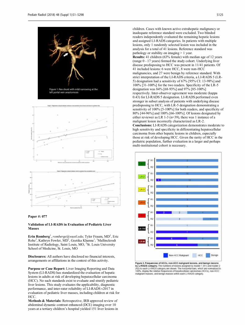
Paper #: 077
Validation of LI-RADS in Evaluation of Pediatric Liver
Masses
Erin Romberg1, [email protected]; Tyler Fraum, MD
1, Eric
Rohe2, Kathryn Fowler, MD
1, Geetika Khanna
1;
1Mallinckrodt
Institute of Radiology, Saint Louis, MO, 2St. Louis University
School of Medicine, St. Louis, MO Disclosures: All authors have disclosed no financial interests, arrangements or affiliations in the context of this activity.
Purpose or Case Report: Liver Imaging Reporting and Data System (LI-RADS) has standardized the evaluation of hepatic lesions in adults at risk of developing hepatocellular carcinoma (HCC). No such standards exist to evaluate and stratify pediatric liver lesions. This study evaluates the applicability, diagnostic performance, and inter-rater reliability of LI-RADS v2017 in
evaluation of pediatric liver masses, including children at risk for HCC. Methods & Materials: Retrospective, IRB approved review of abdominal dynamic contrast enhanced (DCE) imaging over 10 years at a tertiary children’s hospital yielded 151 liver lesions in
children. Cases with known active extrahepatic malignancy or inadequate reference standard were excluded. Two blinded readers independently evaluated the remaining hepatic lesions and assigned LI-RADS categories. In patients with multiple lesions, only 1 randomly selected lesion was included in the
analysis for a total of 41 lesions. Reference standard was pathology or stability on imaging > 1 year. Results: 41 children (63% female) with median age of 12 years (range 0 - 17 years) formed the study cohort. Underlying liver disease predisposing to HCC was present in 11/41 patients. Of 41 included lesions: 6 were HCC, 8 were non-HCC
malignancies, and 27 were benign by reference standard. With strict interpretation of the LI-RADS criteria, a LI-RADS 5 (LR-5) designation had a sensitivity of 67% [95% CI: 13-98%] and 100% [31-100%] for the two readers. Specificity of the LR-5 designation was 84% [68-93%] and 97% [85-100%] respectively. Inter-observer agreement was moderate (kappa 0.43) for LI-RADS 5 designation. LI-RADS performed even
stronger in subset analysis of patients with underlying disease predisposing to HCC, with LR-5 designation demonstrating a sensitivity of 100% [5-100%] for both readers, and specificity of 80% [44-96%] and 100% [66-100%]. Of lesions designated by either reviewer as LR 1-3 (n=39), there was 1 instance of a malignant lesion incorrectly characterized as LR-2. Conclusions: LI-RADS categorization demonstrates moderate to
high sensitivity and specificity in differentiating hepatocellular carcinoma from other hepatic lesions in children, especially those at risk of developing HCC. Given the rarity of HCC in the pediatric population, further evaluation in a larger and perhaps multi-institutional cohort is necessary.
Pediatr Radiol (2018) 48 (Suppl 1):S1–S298 S125
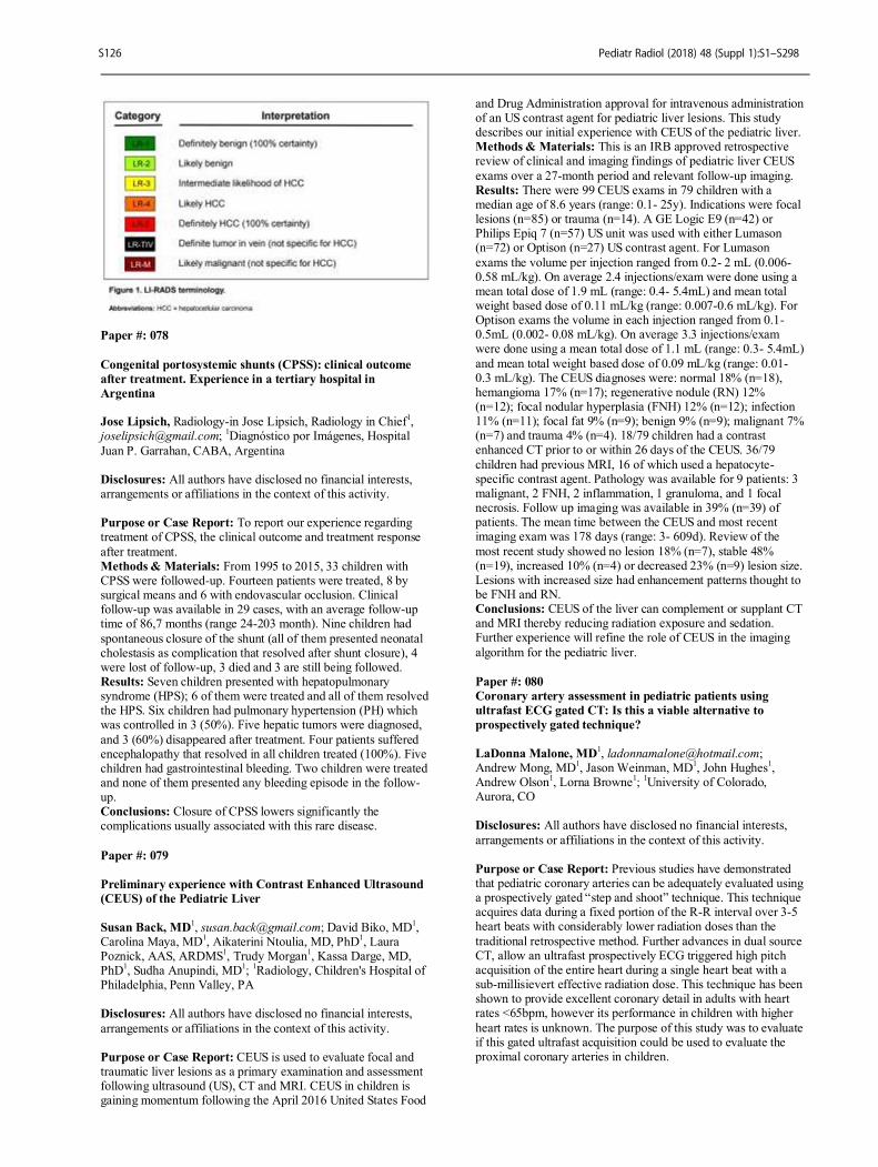
Paper #: 078
Congenital portosystemic shunts (CPSS): clinical outcome
after treatment. Experience in a tertiary hospital in
Argentina Jose Lipsich, Radiology-in Jose Lipsich, Radiology in Chief
1,
[email protected]; 1Diagnóstico por Imágenes, Hospital
Juan P. Garrahan, CABA, Argentina Disclosures: All authors have disclosed no financial interests, arrangements or affiliations in the context of this activity. Purpose or Case Report: To report our experience regarding treatment of CPSS, the clinical outcome and treatment response
after treatment. Methods & Materials: From 1995 to 2015, 33 children with CPSS were followed-up. Fourteen patients were treated, 8 by surgical means and 6 with endovascular occlusion. Clinical follow-up was available in 29 cases, with an average follow-up time of 86,7 months (range 24-203 month). Nine children had
spontaneous closure of the shunt (all of them presented neonatal cholestasis as complication that resolved after shunt closure), 4 were lost of follow-up, 3 died and 3 are still being followed. Results: Seven children presented with hepatopulmonary syndrome (HPS); 6 of them were treated and all of them resolved the HPS. Six children had pulmonary hypertension (PH) which was controlled in 3 (50%). Five hepatic tumors were diagnosed,
and 3 (60%) disappeared after treatment. Four patients suffered encephalopathy that resolved in all children treated (100%). Five children had gastrointestinal bleeding. Two children were treated and none of them presented any bleeding episode in the follow-up. Conclusions: Closure of CPSS lowers significantly the complications usually associated with this rare disease.
Paper #: 079
Preliminary experience with Contrast Enhanced Ultrasound
(CEUS) of the Pediatric Liver
Susan Back, MD1, [email protected]; David Biko, MD
1,
Carolina Maya, MD1, Aikaterini Ntoulia, MD, PhD
1, Laura
Poznick, AAS, ARDMS1, Trudy Morgan
1, Kassa Darge, MD,
PhD1, Sudha Anupindi, MD
1;
1Radiology, Children's Hospital of
Philadelphia, Penn Valley, PA Disclosures: All authors have disclosed no financial interests,
arrangements or affiliations in the context of this activity. Purpose or Case Report: CEUS is used to evaluate focal and traumatic liver lesions as a primary examination and assessment following ultrasound (US), CT and MRI. CEUS in children is gaining momentum following the April 2016 United States Food
and Drug Administration approval for intravenous administration of an US contrast agent for pediatric liver lesions. This study describes our initial experience with CEUS of the pediatric liver. Methods & Materials: This is an IRB approved retrospective review of clinical and imaging findings of pediatric liver CEUS
exams over a 27-month period and relevant follow-up imaging. Results: There were 99 CEUS exams in 79 children with a median age of 8.6 years (range: 0.1- 25y). Indications were focal lesions (n=85) or trauma (n=14). A GE Logic E9 (n=42) or Philips Epiq 7 (n=57) US unit was used with either Lumason (n=72) or Optison (n=27) US contrast agent. For Lumason
exams the volume per injection ranged from 0.2- 2 mL (0.006- 0.58 mL/kg). On average 2.4 injections/exam were done using a mean total dose of 1.9 mL (range: 0.4- 5.4mL) and mean total weight based dose of 0.11 mL/kg (range: 0.007-0.6 mL/kg). For Optison exams the volume in each injection ranged from 0.1- 0.5mL (0.002- 0.08 mL/kg). On average 3.3 injections/exam were done using a mean total dose of 1.1 mL (range: 0.3- 5.4mL)
and mean total weight based dose of 0.09 mL/kg (range: 0.01- 0.3 mL/kg). The CEUS diagnoses were: normal 18% (n=18), hemangioma 17% (n=17); regenerative nodule (RN) 12% (n=12); focal nodular hyperplasia (FNH) 12% (n=12); infection 11% (n=11); focal fat 9% (n=9); benign 9% (n=9); malignant 7% (n=7) and trauma 4% (n=4). 18/79 children had a contrast enhanced CT prior to or within 26 days of the CEUS. 36/79
children had previous MRI, 16 of which used a hepatocyte-specific contrast agent. Pathology was available for 9 patients: 3 malignant, 2 FNH, 2 inflammation, 1 granuloma, and 1 focal necrosis. Follow up imaging was available in 39% (n=39) of patients. The mean time between the CEUS and most recent imaging exam was 178 days (range: 3- 609d). Review of the
most recent study showed no lesion 18% (n=7), stable 48% (n=19), increased 10% (n=4) or decreased 23% (n=9) lesion size. Lesions with increased size had enhancement patterns thought to be FNH and RN. Conclusions: CEUS of the liver can complement or supplant CT and MRI thereby reducing radiation exposure and sedation. Further experience will refine the role of CEUS in the imaging
algorithm for the pediatric liver.
Paper #: 080 Coronary artery assessment in pediatric patients using
ultrafast ECG gated CT: Is this a viable alternative to
prospectively gated technique?
LaDonna Malone, MD1, [email protected];
Andrew Mong, MD1, Jason Weinman, MD
1, John Hughes
1,
Andrew Olson1, Lorna Browne
1;
1University of Colorado,
Aurora, CO Disclosures: All authors have disclosed no financial interests,
arrangements or affiliations in the context of this activity. Purpose or Case Report: Previous studies have demonstrated that pediatric coronary arteries can be adequately evaluated using a prospectively gated “step and shoot” technique. This technique acquires data during a fixed portion of the R-R interval over 3-5 heart beats with considerably lower radiation doses than the
traditional retrospective method. Further advances in dual source CT, allow an ultrafast prospectively ECG triggered high pitch acquisition of the entire heart during a single heart beat with a sub-millisievert effective radiation dose. This technique has been shown to provide excellent coronary detail in adults with heart rates <65bpm, however its performance in children with higher
heart rates is unknown. The purpose of this study was to evaluate if this gated ultrafast acquisition could be used to evaluate the proximal coronary arteries in children.
Pediatr Radiol (2018) 48 (Suppl 1):S1–S298S126
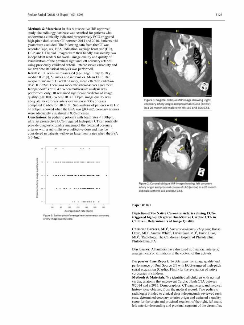
Methods & Materials: In this retrospective IRB approved study, the radiology database was searched for patients who underwent a clinically indicated prospectively ECG-triggered high-pitch dual-source CT between 2014 and 2016. Patients ≥18 years were excluded. The following data from the CT was
recorded: age, sex, BSA, indication, average heart rate (HR), DLP, and CTDI vol. Images were then blindly assessed by two independent readers for overall image quality and quality of visualization of the proximal right and left coronary arteries using previously validated criteria. Interobserver variability and multivariate statistical analysis was performed.
Results: 100 scans were assessed (age range: 1 day to 18 y, median 0.26 y), 58 males and 42 females. Mean DLP: 18.6 mGy-cm, mean CTDIvol:0.61 mGy, mean effective radiation dose: 0.7 mSv. There was moderate interobserver agreement, Krippendorff’s α= 0.49. When multivariate analysis was performed, only HR remained significant predictor of image quality (p<0.001). When HR < 100bpm, image quality was
adequate for coronary artery evaluation in 93% of cases compared to 66% for HR >100. Sub analysis of patients with HR >100bpm, showed when the BSA was ≥0.4 m2, coronary arteries were adequately visualized in 83% of cases. Conclusions: In pediatric patients with heart rates < 100bpm, ultrafast prospective ECG-triggered high-pitch CT can routinely provide diagnostic quality imaging of the proximal coronary
arteries with a sub-millisievert effective dose and may be considered in patients with even faster heart rates when the BSA ≥ 0.4m2.
Paper #: 081
Depiction of the Native Coronary Arteries during ECG-
triggered high-pitch spiral Dual-Source Cardiac CTA in
Children: Determinants of Image Quality Christian Barrera, MD
1, [email protected]; Hansel
Otero, MD1, Ammie White
1, David Saul, MD
1, David Biko,
MD1;
1Radiology, The Children's Hospital of Philadelphia,
Philadelphia, PA
Disclosures: All authors have disclosed no financial interests, arrangements or affiliations in the context of this activity. Purpose or Case Report: To determine the image quality and
performance of Dual Source CT with ECG-triggered high-pitch spiral acquisition (Cardiac Flash) for the evaluation of native coronaries in children. Methods & Materials: We identified all children with normal cardiac anatomy that underwent Cardiac Flash CTA between 8/2014 and 8/2017. Demographics, CT parameters, and medical history were obtained from the medical record. Two pediatric
radiologist blinded to clinical data independently reviewed each case, determined coronary arteries origin and assigned a quality score for the origin and proximal segment of the right, left main, left anterior descending and proximal segment of the circumflex
Pediatr Radiol (2018) 48 (Suppl 1):S1–S298 S127
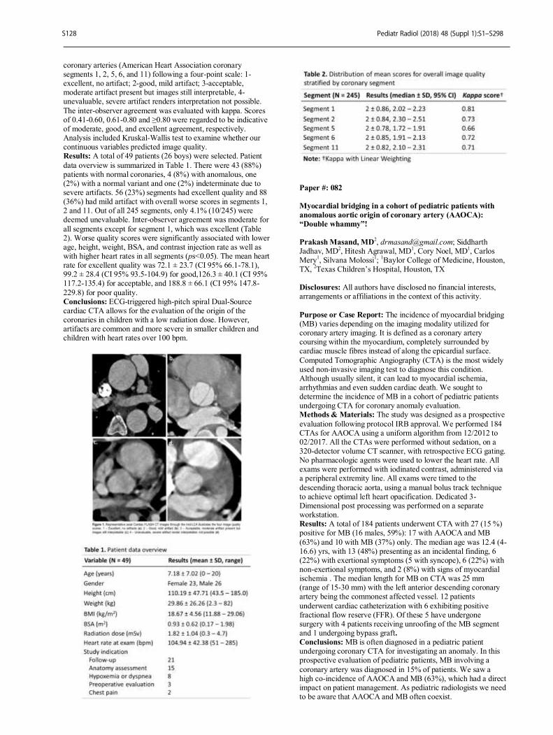
coronary arteries (American Heart Association coronary segments 1, 2, 5, 6, and 11) following a four-point scale: 1-excellent, no artifact; 2-good, mild artifact; 3-acceptable, moderate artifact present but images still interpretable, 4-unevaluable, severe artifact renders interpretation not possible.
The inter-observer agreement was evaluated with kappa. Scores of 0.41-0.60, 0.61-0.80 and ≥0.80 were regarded to be indicative of moderate, good, and excellent agreement, respectively. Analysis included Kruskal-Wallis test to examine whether our continuous variables predicted image quality. Results: A total of 49 patients (26 boys) were selected. Patient
data overview is summarized in Table 1. There were 43 (88%) patients with normal coronaries, 4 (8%) with anomalous, one (2%) with a normal variant and one (2%) indeterminate due to severe artifacts. 56 (23%) segments had excellent quality and 88 (36%) had mild artifact with overall worse scores in segments 1, 2 and 11. Out of all 245 segments, only 4.1% (10/245) were deemed unevaluable. Inter-observer agreement was moderate for
all segments except for segment 1, which was excellent (Table 2). Worse quality scores were significantly associated with lower age, height, weight, BSA, and contrast injection rate as well as with higher heart rates in all segments (ps<0.05). The mean heart rate for excellent quality was 72.1 ± 23.7 (CI 95% 66.1-78.1), 99.2 ± 28.4 (CI 95% 93.5-104.9) for good,126.3 ± 40.1 (CI 95% 117.2-135.4) for acceptable, and 188.8 ± 66.1 (CI 95% 147.8-
229.8) for poor quality. Conclusions: ECG-triggered high-pitch spiral Dual-Source cardiac CTA allows for the evaluation of the origin of the coronaries in children with a low radiation dose. However, artifacts are common and more severe in smaller children and children with heart rates over 100 bpm.
Paper #: 082
Myocardial bridging in a cohort of pediatric patients with
anomalous aortic origin of coronary artery (AAOCA):
“Double whammy”!
Prakash Masand, MD2, [email protected]; Siddharth
Jadhav, MD2, Hitesh Agrawal, MD
1, Cory Noel, MD
1, Carlos
Mery1, Silvana Molossi
1;
1Baylor College of Medicine, Houston,
TX, 2Texas Children’s Hospital, Houston, TX
Disclosures: All authors have disclosed no financial interests, arrangements or affiliations in the context of this activity.
Purpose or Case Report: The incidence of myocardial bridging (MB) varies depending on the imaging modality utilized for coronary artery imaging. It is defined as a coronary artery coursing within the myocardium, completely surrounded by cardiac muscle fibres instead of along the epicardial surface.
Computed Tomographic Angiography (CTA) is the most widely used non-invasive imaging test to diagnose this condition. Although usually silent, it can lead to myocardial ischemia, arrhythmias and even sudden cardiac death. We sought to determine the incidence of MB in a cohort of pediatric patients undergoing CTA for coronary anomaly evaluation. Methods & Materials: The study was designed as a prospective
evaluation following protocol IRB approval. We performed 184 CTAs for AAOCA using a uniform algorithm from 12/2012 to 02/2017. All the CTAs were performed without sedation, on a 320-detector volume CT scanner, with retrospective ECG gating. No pharmacologic agents were used to lower the heart rate. All exams were performed with iodinated contrast, administered via a peripheral extremity line. All exams were timed to the
descending thoracic aorta, using a manual bolus track technique to achieve optimal left heart opacification. Dedicated 3-Dimensional post processing was performed on a separate workstation. Results: A total of 184 patients underwent CTA with 27 (15 %) positive for MB (16 males, 59%): 17 with AAOCA and MB
(63%) and 10 with MB (37%) only. The median age was 12.4 (4-16.6) yrs, with 13 (48%) presenting as an incidental finding, 6 (22%) with exertional symptoms (5 with syncope), 6 (22%) with non-exertional symptoms, and 2 (8%) with signs of myocardial ischemia . The median length for MB on CTA was 25 mm (range of 15-30 mm) with the left anterior descending coronary artery being the commonest affected vessel. 12 patients
underwent cardiac catheterization with 6 exhibiting positive fractional flow reserve (FFR). Of these 5 have undergone surgery with 4 patients receiving unroofing of the MB segment and 1 undergoing bypass graft. Conclusions: MB is often diagnosed in a pediatric patient undergoing coronary CTA for investigating an anomaly. In this prospective evaluation of pediatric patients, MB involving a
coronary artery was diagnosed in 15% of patients. We saw a high co-incidence of AAOCA and MB (63%), which had a direct impact on patient management. As pediatric radiologists we need to be aware that AAOCA and MB often coexist.
Pediatr Radiol (2018) 48 (Suppl 1):S1–S298S128
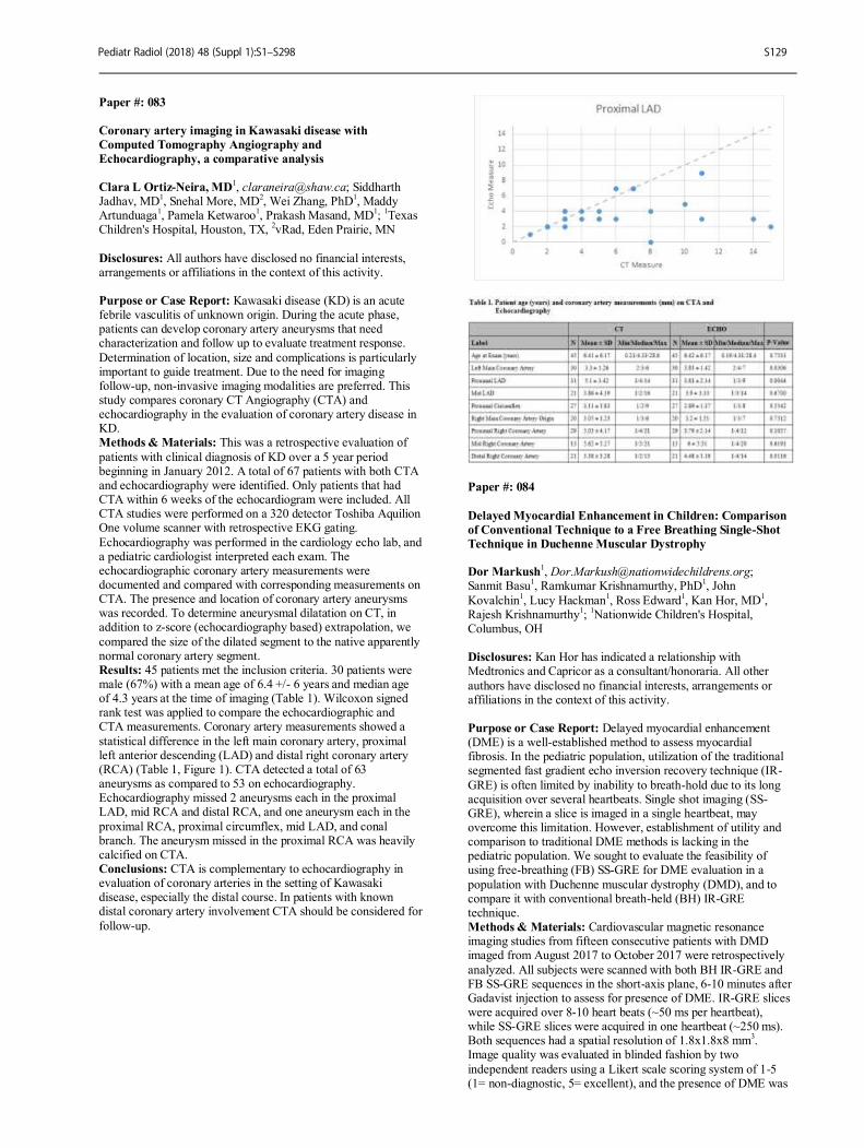
Paper #: 083
Coronary artery imaging in Kawasaki disease with
Computed Tomography Angiography and
Echocardiography, a comparative analysis
Clara L Ortiz-Neira, MD
1, [email protected]; Siddharth
Jadhav, MD1, Snehal More, MD
2, Wei Zhang, PhD
1, Maddy
Artunduaga1, Pamela Ketwaroo
1, Prakash Masand, MD
1;
1Texas
Children's Hospital, Houston, TX, 2vRad, Eden Prairie, MN
Disclosures: All authors have disclosed no financial interests, arrangements or affiliations in the context of this activity. Purpose or Case Report: Kawasaki disease (KD) is an acute febrile vasculitis of unknown origin. During the acute phase, patients can develop coronary artery aneurysms that need characterization and follow up to evaluate treatment response.
Determination of location, size and complications is particularly important to guide treatment. Due to the need for imaging follow-up, non-invasive imaging modalities are preferred. This study compares coronary CT Angiography (CTA) and echocardiography in the evaluation of coronary artery disease in KD. Methods & Materials: This was a retrospective evaluation of
patients with clinical diagnosis of KD over a 5 year period beginning in January 2012. A total of 67 patients with both CTA and echocardiography were identified. Only patients that had CTA within 6 weeks of the echocardiogram were included. All CTA studies were performed on a 320 detector Toshiba Aquilion One volume scanner with retrospective EKG gating.
Echocardiography was performed in the cardiology echo lab, and a pediatric cardiologist interpreted each exam. The echocardiographic coronary artery measurements were documented and compared with corresponding measurements on CTA. The presence and location of coronary artery aneurysms was recorded. To determine aneurysmal dilatation on CT, in addition to z-score (echocardiography based) extrapolation, we
compared the size of the dilated segment to the native apparently normal coronary artery segment. Results: 45 patients met the inclusion criteria. 30 patients were male (67%) with a mean age of 6.4 +/- 6 years and median age of 4.3 years at the time of imaging (Table 1). Wilcoxon signed rank test was applied to compare the echocardiographic and CTA measurements. Coronary artery measurements showed a
statistical difference in the left main coronary artery, proximal left anterior descending (LAD) and distal right coronary artery (RCA) (Table 1, Figure 1). CTA detected a total of 63 aneurysms as compared to 53 on echocardiography. Echocardiography missed 2 aneurysms each in the proximal LAD, mid RCA and distal RCA, and one aneurysm each in the
proximal RCA, proximal circumflex, mid LAD, and conal branch. The aneurysm missed in the proximal RCA was heavily calcified on CTA. Conclusions: CTA is complementary to echocardiography in evaluation of coronary arteries in the setting of Kawasaki disease, especially the distal course. In patients with known distal coronary artery involvement CTA should be considered for
follow-up.
Paper #: 084
Delayed Myocardial Enhancement in Children: Comparison
of Conventional Technique to a Free Breathing Single-Shot
Technique in Duchenne Muscular Dystrophy Dor Markush
Sanmit Basu1, Ramkumar Krishnamurthy, PhD
1, John
Kovalchin1, Lucy Hackman
1, Ross Edward
1, Kan Hor, MD
1,
Rajesh Krishnamurthy1;
1Nationwide Children's Hospital,
Columbus, OH Disclosures: Kan Hor has indicated a relationship with Medtronics and Capricor as a consultant/honoraria. All other
authors have disclosed no financial interests, arrangements or affiliations in the context of this activity. Purpose or Case Report: Delayed myocardial enhancement (DME) is a well-established method to assess myocardial fibrosis. In the pediatric population, utilization of the traditional segmented fast gradient echo inversion recovery technique (IR-
GRE) is often limited by inability to breath-hold due to its long acquisition over several heartbeats. Single shot imaging (SS-GRE), wherein a slice is imaged in a single heartbeat, may overcome this limitation. However, establishment of utility and comparison to traditional DME methods is lacking in the pediatric population. We sought to evaluate the feasibility of using free-breathing (FB) SS-GRE for DME evaluation in a
population with Duchenne muscular dystrophy (DMD), and to compare it with conventional breath-held (BH) IR-GRE technique. Methods & Materials: Cardiovascular magnetic resonance imaging studies from fifteen consecutive patients with DMD imaged from August 2017 to October 2017 were retrospectively
analyzed. All subjects were scanned with both BH IR-GRE and FB SS-GRE sequences in the short-axis plane, 6-10 minutes after Gadavist injection to assess for presence of DME. IR-GRE slices were acquired over 8-10 heart beats (~50 ms per heartbeat), while SS-GRE slices were acquired in one heartbeat (~250 ms). Both sequences had a spatial resolution of 1.8x1.8x8 mm
3.
Image quality was evaluated in blinded fashion by two
independent readers using a Likert scale scoring system of 1-5 (1= non-diagnostic, 5= excellent), and the presence of DME was
Pediatr Radiol (2018) 48 (Suppl 1):S1–S298 S129
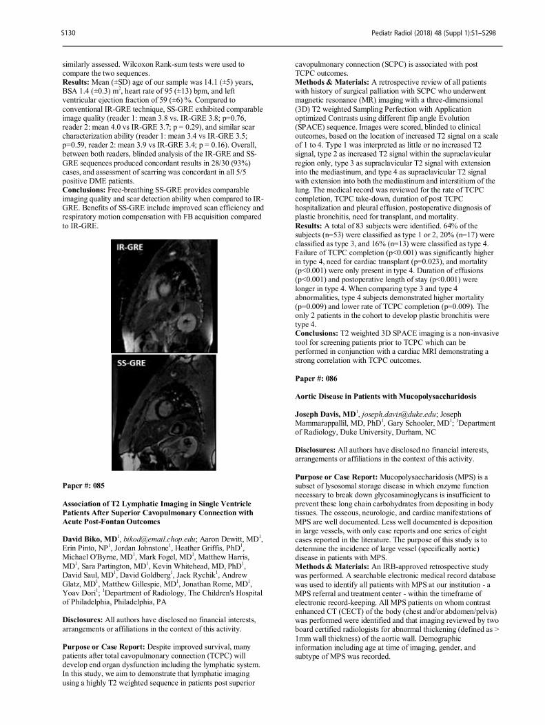
similarly assessed. Wilcoxon Rank-sum tests were used to compare the two sequences. Results: Mean (±SD) age of our sample was 14.1 (±5) years, BSA 1.4 (±0.3) m
2, heart rate of 95 (±13) bpm, and left
ventricular ejection fraction of 59 (±6) %. Compared to
conventional IR-GRE technique, SS-GRE exhibited comparable image quality (reader 1: mean 3.8 vs. IR-GRE 3.8; p=0.76, reader 2: mean 4.0 vs IR-GRE 3.7; p = 0.29), and similar scar characterization ability (reader 1: mean 3.4 vs IR-GRE 3.5; p=0.59, reader 2: mean 3.9 vs IR-GRE 3.4; p = 0.16). Overall, between both readers, blinded analysis of the IR-GRE and SS-
GRE sequences produced concordant results in 28/30 (93%) cases, and assessment of scarring was concordant in all 5/5 positive DME patients. Conclusions: Free-breathing SS-GRE provides comparable imaging quality and scar detection ability when compared to IR-GRE. Benefits of SS-GRE include improved scan efficiency and respiratory motion compensation with FB acquisition compared
to IR-GRE.
Paper #: 085
Association of T2 Lymphatic Imaging in Single Ventricle
Patients After Superior Cavopulmonary Connection with
Acute Post-Fontan Outcomes David Biko, MD
1, [email protected]; Aaron Dewitt, MD
1,
Erin Pinto, NP1, Jordan Johnstone
1, Heather Griffis, PhD
1,
Michael O'Byrne, MD1, Mark Fogel, MD
1, Matthew Harris,
MD1, Sara Partington, MD
1, Kevin Whitehead, MD, PhD
1,
David Saul, MD1, David Goldberg
1, Jack Rychik
1, Andrew
Glatz, MD1, Matthew Gillespie, MD
1, Jonathan Rome, MD
1,
Yoav Dori1;
1Department of Radiology, The Children's Hospital
of Philadelphia, Philadelphia, PA Disclosures: All authors have disclosed no financial interests,
arrangements or affiliations in the context of this activity. Purpose or Case Report: Despite improved survival, many patients after total cavopulmonary connection (TCPC) will develop end organ dysfunction including the lymphatic system. In this study, we aim to demonstrate that lymphatic imaging
using a highly T2 weighted sequence in patients post superior
cavopulmonary connection (SCPC) is associated with post TCPC outcomes. Methods & Materials: A retrospective review of all patients with history of surgical palliation with SCPC who underwent magnetic resonance (MR) imaging with a three-dimensional
(3D) T2 weighted Sampling Perfection with Application optimized Contrasts using different flip angle Evolution (SPACE) sequence. Images were scored, blinded to clinical outcomes, based on the location of increased T2 signal on a scale of 1 to 4. Type 1 was interpreted as little or no increased T2 signal, type 2 as increased T2 signal within the supraclavicular
region only, type 3 as supraclavicular T2 signal with extension into the mediastinum, and type 4 as supraclavicular T2 signal with extension into both the mediastinum and interstitium of the lung. The medical record was reviewed for the rate of TCPC completion, TCPC take-down, duration of post TCPC hospitalization and pleural effusion, postoperative diagnosis of plastic bronchitis, need for transplant, and mortality.
Results: A total of 83 subjects were identified. 64% of the subjects (n=53) were classified as type 1 or 2, 20% (n=17) were classified as type 3, and 16% (n=13) were classified as type 4. Failure of TCPC completion (p<0.001) was significantly higher in type 4, need for cardiac transplant (p=0.023), and mortality (p<0.001) were only present in type 4. Duration of effusions (p<0.001) and postoperative length of stay (p<0.001) were
longer in type 4. When comparing type 3 and type 4 abnormalities, type 4 subjects demonstrated higher mortality (p=0.009) and lower rate of TCPC completion (p=0.009). The only 2 patients in the cohort to develop plastic bronchitis were type 4. Conclusions: T2 weighted 3D SPACE imaging is a non-invasive
tool for screening patients prior to TCPC which can be performed in conjunction with a cardiac MRI demonstrating a strong correlation with TCPC outcomes.
Paper #: 086
Aortic Disease in Patients with Mucopolysaccharidosis
Joseph Davis, MD
1, [email protected]; Joseph
Mammarappallil, MD, PhD1, Gary Schooler, MD
1;
1Department
of Radiology, Duke University, Durham, NC Disclosures: All authors have disclosed no financial interests, arrangements or affiliations in the context of this activity.
Purpose or Case Report: Mucopolysaccharidosis (MPS) is a subset of lysosomal storage disease in which enzyme function necessary to break down glycosaminoglycans is insufficient to prevent these long chain carbohydrates from depositing in body tissues. The osseous, neurologic, and cardiac manifestations of
MPS are well documented. Less well documented is deposition in large vessels, with only case reports and one series of eight cases reported in the literature. The purpose of this study is to determine the incidence of large vessel (specifically aortic) disease in patients with MPS. Methods & Materials: An IRB-approved retrospective study was performed. A searchable electronic medical record database
was used to identify all patients with MPS at our institution - a MPS referral and treatment center - within the timeframe of electronic record-keeping. All MPS patients on whom contrast enhanced CT (CECT) of the body (chest and/or abdomen/pelvis) was performed were identified and that imaging reviewed by two board certified radiologists for abnormal thickening (defined as >
1mm wall thickness) of the aortic wall. Demographic information including age at time of imaging, gender, and subtype of MPS was recorded.
Pediatr Radiol (2018) 48 (Suppl 1):S1–S298S130
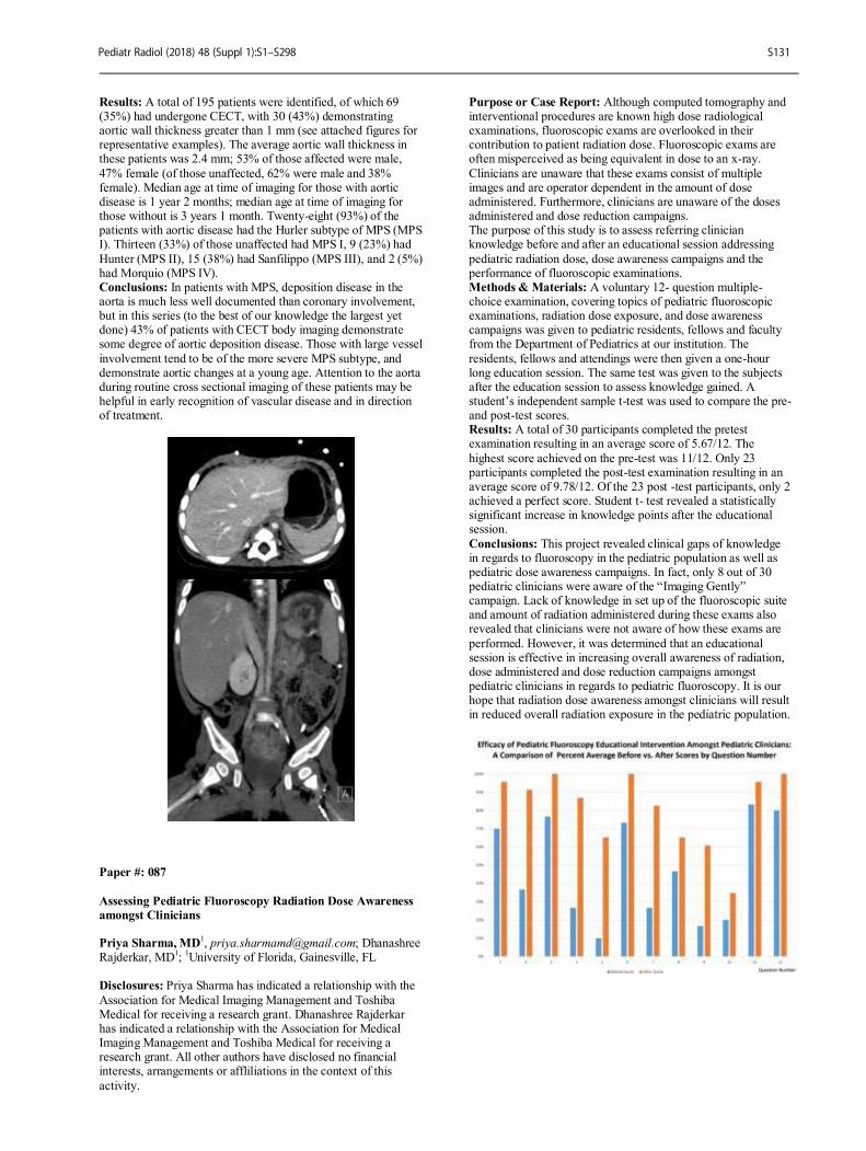
Results: A total of 195 patients were identified, of which 69 (35%) had undergone CECT, with 30 (43%) demonstrating aortic wall thickness greater than 1 mm (see attached figures for representative examples). The average aortic wall thickness in these patients was 2.4 mm; 53% of those affected were male,
47% female (of those unaffected, 62% were male and 38% female). Median age at time of imaging for those with aortic disease is 1 year 2 months; median age at time of imaging for those without is 3 years 1 month. Twenty-eight (93%) of the patients with aortic disease had the Hurler subtype of MPS (MPS I). Thirteen (33%) of those unaffected had MPS I, 9 (23%) had
Hunter (MPS II), 15 (38%) had Sanfilippo (MPS III), and 2 (5%) had Morquio (MPS IV). Conclusions: In patients with MPS, deposition disease in the aorta is much less well documented than coronary involvement, but in this series (to the best of our knowledge the largest yet done) 43% of patients with CECT body imaging demonstrate some degree of aortic deposition disease. Those with large vessel
involvement tend to be of the more severe MPS subtype, and demonstrate aortic changes at a young age. Attention to the aorta during routine cross sectional imaging of these patients may be helpful in early recognition of vascular disease and in direction of treatment.
Paper #: 087
Assessing Pediatric Fluoroscopy Radiation Dose Awareness
amongst Clinicians Priya Sharma, MD
1, [email protected]; Dhanashree
Rajderkar, MD1;
1University of Florida, Gainesville, FL
Disclosures: Priya Sharma has indicated a relationship with the
Association for Medical Imaging Management and Toshiba Medical for receiving a research grant. Dhanashree Rajderkar has indicated a relationship with the Association for Medical Imaging Management and Toshiba Medical for receiving a research grant. All other authors have disclosed no financial interests, arrangements or affliliations in the context of this
activity.
Purpose or Case Report: Although computed tomography and interventional procedures are known high dose radiological examinations, fluoroscopic exams are overlooked in their contribution to patient radiation dose. Fluoroscopic exams are often misperceived as being equivalent in dose to an x-ray.
Clinicians are unaware that these exams consist of multiple images and are operator dependent in the amount of dose administered. Furthermore, clinicians are unaware of the doses administered and dose reduction campaigns. The purpose of this study is to assess referring clinician knowledge before and after an educational session addressing
pediatric radiation dose, dose awareness campaigns and the performance of fluoroscopic examinations. Methods & Materials: A voluntary 12- question multiple- choice examination, covering topics of pediatric fluoroscopic examinations, radiation dose exposure, and dose awareness campaigns was given to pediatric residents, fellows and faculty from the Department of Pediatrics at our institution. The
residents, fellows and attendings were then given a one-hour long education session. The same test was given to the subjects after the education session to assess knowledge gained. A student’s independent sample t-test was used to compare the pre- and post-test scores. Results: A total of 30 participants completed the pretest examination resulting in an average score of 5.67/12. The
highest score achieved on the pre-test was 11/12. Only 23 participants completed the post-test examination resulting in an average score of 9.78/12. Of the 23 post -test participants, only 2 achieved a perfect score. Student t- test revealed a statistically significant increase in knowledge points after the educational session.
Conclusions: This project revealed clinical gaps of knowledge in regards to fluoroscopy in the pediatric population as well as pediatric dose awareness campaigns. In fact, only 8 out of 30 pediatric clinicians were aware of the “Imaging Gently” campaign. Lack of knowledge in set up of the fluoroscopic suite and amount of radiation administered during these exams also revealed that clinicians were not aware of how these exams are
performed. However, it was determined that an educational session is effective in increasing overall awareness of radiation, dose administered and dose reduction campaigns amongst pediatric clinicians in regards to pediatric fluoroscopy. It is our hope that radiation dose awareness amongst clinicians will result in reduced overall radiation exposure in the pediatric population.
Pediatr Radiol (2018) 48 (Suppl 1):S1–S298 S131
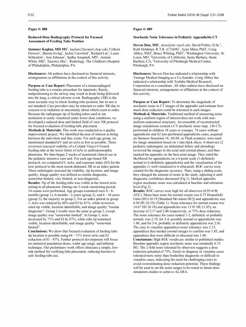
Paper #: 088
Reduced-Dose Radiography Protocol for Focused
Assessment of Feeding Tube Position
Summer Kaplan, MD MS1, [email protected]; Colleen
Flowers1, Sharon Irving
1, Jackie Crawford
1, Richard Lin
1, Laura
Schleelein1, Ann Johnson
1, Sudha Anupindi, MD
1, Ammie
White, MD1, Xiaowei Zhu
1;
1Radiology, The Children's Hospital
of Philadelphia, Philadelphia, PA
Disclosures: All authors have disclosed no financial interests, arrangements or affiliations in the context of this activity. Purpose or Case Report: Placement of a transesophageal feeding tube is a routine procedure for inpatients. Rarely, malpositioning in the airway may result in feeds being delivered into the lung, a critical adverse event. Radiography (XR) is the
most accurate way to check feeding tube position, but its use is not standard. Care providers may be reluctant to order XR due to concern over radiation or uncertainly about which exam to order. Because the radiopaque tip of feeding tubes used at our institution is easily visualized under lower dose conditions, we developed a reduced dose and limited field of view XR protocol for focused evaluation of feeding tube placement.
Methods & Materials: This work was conducted as a quality improvement project. We identified the area of interest as being between the mid-chest and iliac crests. For each age group, we maintained standard kV and set mAs as low as possible. Three reviewers assessed visibility of a Corpak Viasys 6 French feeding tube at the lower limits of mAs in anthropomorphic
phantoms. We then began 2 weeks of monitored clinical use in the pediatric intensive care unit. For each age-based XR protocol, we compared kV, mAs, and exposure index (EI) for the new protocol to the most recent abdomen XR in an index patient. Three radiologists assessed tip visibility, tip location, and image quality. Image quality was defined as routine diagnostic, somewhat limited, very limited, or non-diagnostic.
Results: Tip of the feeding tube was visible at the lowest mAs setting in all phantoms. During our 2-week monitoring period, 14 exams were performed. Age groups examined were 0 - 6-months (group 1), 6-months - 2-years (group 2), and 6 - 10-years (group 3), the majority in group 2. For an index patient in group 1, mAs was reduced by 66% and EI by 81%, while reviewers rated tip visible, location identifiable, and image quality "routine
diagnostic". Group 2 results were the same as group 2, except image quality was "somewhat limited". In Group 3, mAs decreased by 71% and EI by 87%, while tube tip remained visible, location identifiable, and image quality "somewhat limited". Conclusions: We show that focused evaluation of feeding tube
tip location is possible using 66 - 71% lower mAs and EI reduction of 81 - 87%. Further protocol development will focus on statistical population doses, wider age range, and tabletop technique. Our preliminary work offers clinicians a simple, low-risk method for verifying tube placement, reducing barriers to safe feeding tube use.
Paper #: 089
Stochastic Noise Tolerance in Pediatric Appendicitis CT Steven Don, MD
1, [email protected]; David Politte, D.Sc.
1,
Ruth Holdener, R.T.R. (CT)(M)1, Ayan Mitra, PhD
1, Craig
Abbey, PhD2, Bruce Whiting, PhD
3;
1Washington University, St.
Louis, MO, 2University of California, Santa Barbara, Santa
Barbara, CA, 3University of Pittsburgh Medical Center,
Pittsburgh, PA
Disclosures: Steven Don has indicated a relationship with Vantage Medical Imaging as a Co-founder. Craig Abbey has indicated a relationship with Toshiba Medical Research Corporation as a consultant. All other authors have disclosed no financial interests, arrangements or affiliations in the context of this activity.
Purpose or Case Report: To determine the magnitude of stochastic noise in CT images of the appendix and estimate how much dose reduction could be tolerated in such images. Methods & Materials: Traditional method of measuring noise using a uniform region of interest does not work with non-uniform anatomical structures. An ensemble of reconstructed image simulations created a CT stochastic noise map. 136 CTs
performed in children 18 years or younger, 74 cases without appendicitis and 62 non-perforated appendicitis cases, acquired on Siemens Sensation 16, AS 64, or Flash scanners, were used for image simulation based on 1 mm thick slices. 4 observers (2 pediatric radiologists, an abdominal fellow and attending) reviewed the images in the axial and coronal planes, and then
marked the appendix on the best axial image. They rated the likelihood for appendicitis on a 6-point scale (1-definitely normal to 6-definitely appendicitis) and the visualization of the appendix (1-well-visualized to 6-obscured). ROC curves were created for the diagnostic accuracy. Then, using a sliding scale, they changed the amount of noise in the study, adjusting it until diagnostic confidence decreased (Fig 1). Marked appendix
region stochastic noise was calculated at baseline and toleration level (Fig 2). Results: ROC curves were high for all observers (0.95-0.96 AUC). Mean base noise for normal exams was 6.75 Hounsfield Units (HU) (0.19 [Standard Deviation HU]) and appendicitis was 6.94 HU (0.19) (Table 1). Noise tolerance for normal exams was 14.67 HU (0.18) and appendicitis was 13.91 HU (1.05), an
increase of 2.17 and 2.00 respectively, or 75% dose reduction. The noise tolerance for cases ranked 1-2, definitely or probably normal, was 2.19, for 3-4, possibly normal or appendicitis was 1.80, and for 5-6, probably or definitely appendicitis was 2.01. The easy to visualize appendixes noise tolerance was 2.15, appendixes that needed coronal images to confirm was 1.85, and
appendixes that were difficult or obscured was 1.80. Conclusions: High ROC results are similar to published studies. Baseline appendix region stochastic noise was nominally 6.75 HU. The 2-fold noise tolerated by observers suggests a dose reduction potential of 75%. Easier to diagnose or visualize cases tolerated more noise than borderline diagnostic or difficult to visualize cases, indicating the need for challenging cases to
avoid overestimating dose-reduction potential. These findings will be used to set the noise ranges to be tested in future dose simulation studies to achieve ALARA.
Pediatr Radiol (2018) 48 (Suppl 1):S1–S298S132
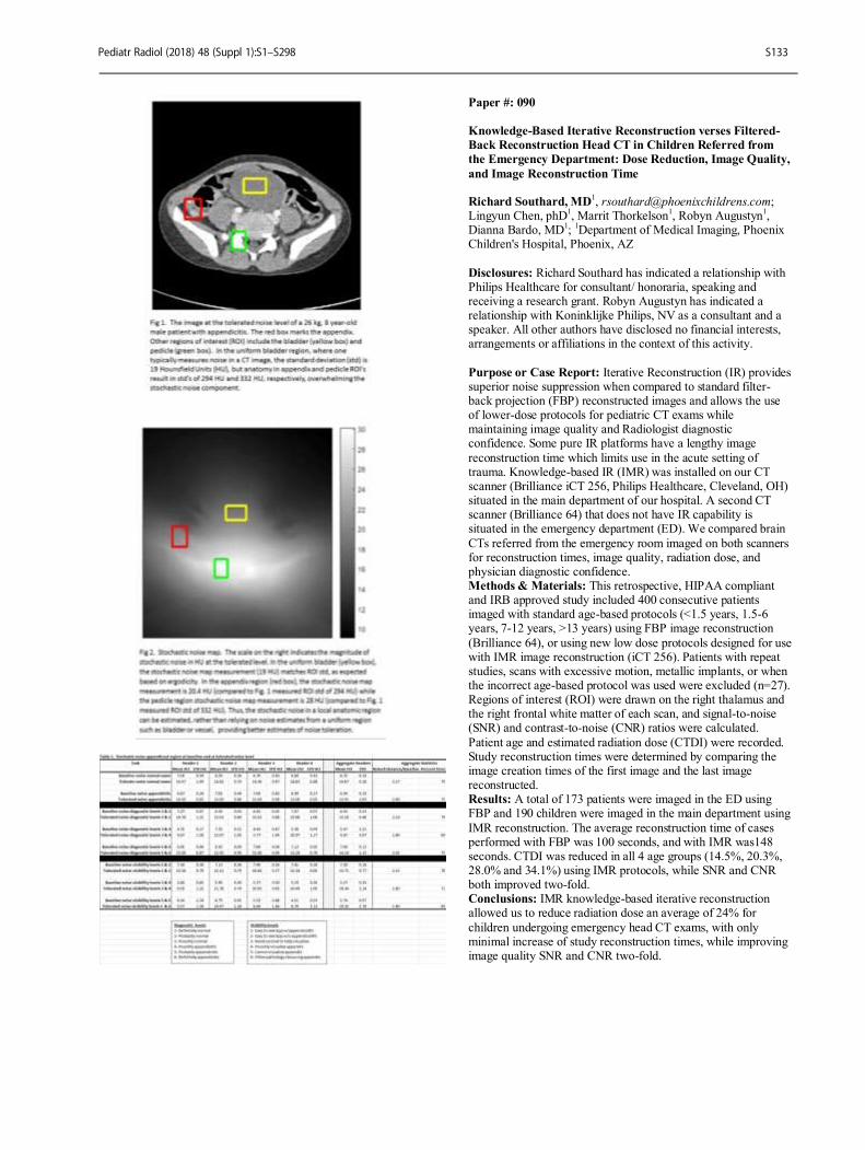
Paper #: 090
Knowledge-Based Iterative Reconstruction verses Filtered-
Back Reconstruction Head CT in Children Referred from
the Emergency Department: Dose Reduction, Image Quality,
and Image Reconstruction Time Richard Southard, MD
Lingyun Chen, phD1, Marrit Thorkelson
1, Robyn Augustyn
1,
Dianna Bardo, MD1;
1Department of Medical Imaging, Phoenix
Children's Hospital, Phoenix, AZ
Disclosures: Richard Southard has indicated a relationship with Philips Healthcare for consultant/ honoraria, speaking and receiving a research grant. Robyn Augustyn has indicated a relationship with Koninklijke Philips, NV as a consultant and a speaker. All other authors have disclosed no financial interests, arrangements or affiliations in the context of this activity.
Purpose or Case Report: Iterative Reconstruction (IR) provides superior noise suppression when compared to standard filter-back projection (FBP) reconstructed images and allows the use of lower-dose protocols for pediatric CT exams while maintaining image quality and Radiologist diagnostic confidence. Some pure IR platforms have a lengthy image
reconstruction time which limits use in the acute setting of trauma. Knowledge-based IR (IMR) was installed on our CT scanner (Brilliance iCT 256, Philips Healthcare, Cleveland, OH) situated in the main department of our hospital. A second CT scanner (Brilliance 64) that does not have IR capability is situated in the emergency department (ED). We compared brain
CTs referred from the emergency room imaged on both scanners for reconstruction times, image quality, radiation dose, and physician diagnostic confidence. Methods & Materials: This retrospective, HIPAA compliant and IRB approved study included 400 consecutive patients imaged with standard age-based protocols (<1.5 years, 1.5-6 years, 7-12 years, >13 years) using FBP image reconstruction
(Brilliance 64), or using new low dose protocols designed for use with IMR image reconstruction (iCT 256). Patients with repeat studies, scans with excessive motion, metallic implants, or when the incorrect age-based protocol was used were excluded (n=27). Regions of interest (ROI) were drawn on the right thalamus and the right frontal white matter of each scan, and signal-to-noise (SNR) and contrast-to-noise (CNR) ratios were calculated.
Patient age and estimated radiation dose (CTDI) were recorded. Study reconstruction times were determined by comparing the image creation times of the first image and the last image reconstructed. Results: A total of 173 patients were imaged in the ED using FBP and 190 children were imaged in the main department using
IMR reconstruction. The average reconstruction time of cases performed with FBP was 100 seconds, and with IMR was148 seconds. CTDI was reduced in all 4 age groups (14.5%, 20.3%, 28.0% and 34.1%) using IMR protocols, while SNR and CNR both improved two-fold. Conclusions: IMR knowledge-based iterative reconstruction allowed us to reduce radiation dose an average of 24% for
children undergoing emergency head CT exams, with only minimal increase of study reconstruction times, while improving image quality SNR and CNR two-fold.
Pediatr Radiol (2018) 48 (Suppl 1):S1–S298 S133
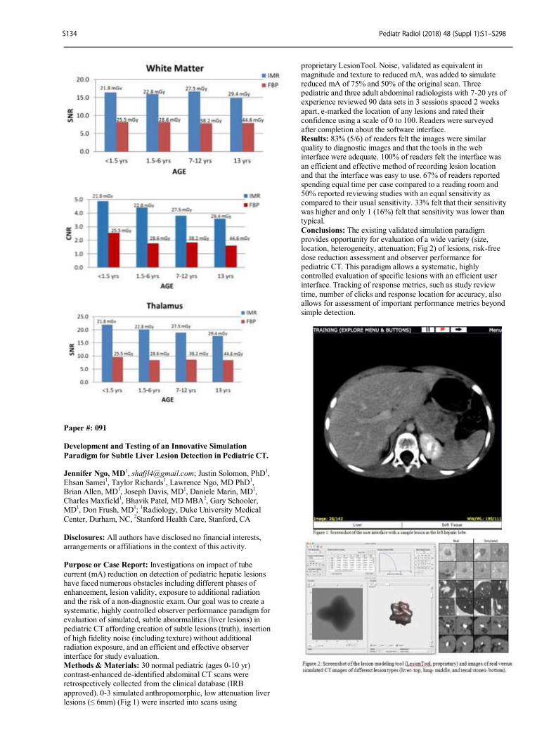
Paper #: 091
Development and Testing of an Innovative Simulation
Paradigm for Subtle Liver Lesion Detection in Pediatric CT. Jennifer Ngo, MD
1, [email protected]; Justin Solomon, PhD
1,
Ehsan Samei1, Taylor Richards
1, Lawrence Ngo, MD PhD
1,
Brian Allen, MD1, Joseph Davis, MD
1, Daniele Marin, MD
1,
Charles Maxfield1, Bhavik Patel, MD MBA
2, Gary Schooler,
MD1, Don Frush, MD
1;
1Radiology, Duke University Medical
Center, Durham, NC, 2Stanford Health Care, Stanford, CA
Disclosures: All authors have disclosed no financial interests, arrangements or affiliations in the context of this activity. Purpose or Case Report: Investigations on impact of tube
current (mA) reduction on detection of pediatric hepatic lesions have faced numerous obstacles including different phases of enhancement, lesion validity, exposure to additional radiation and the risk of a non-diagnostic exam. Our goal was to create a systematic, highly controlled observer performance paradigm for evaluation of simulated, subtle abnormalities (liver lesions) in pediatric CT affording creation of subtle lesions (truth), insertion
of high fidelity noise (including texture) without additional radiation exposure, and an efficient and effective observer interface for study evaluation. Methods & Materials: 30 normal pediatric (ages 0-10 yr) contrast-enhanced de-identified abdominal CT scans were retrospectively collected from the clinical database (IRB
approved). 0-3 simulated anthropomorphic, low attenuation liver lesions (≤ 6mm) (Fig 1) were inserted into scans using
proprietary LesionTool. Noise, validated as equivalent in magnitude and texture to reduced mA, was added to simulate reduced mA of 75% and 50% of the original scan. Three pediatric and three adult abdominal radiologists with 7-20 yrs of experience reviewed 90 data sets in 3 sessions spaced 2 weeks
apart, e-marked the location of any lesions and rated their confidence using a scale of 0 to 100. Readers were surveyed after completion about the software interface. Results: 83% (5/6) of readers felt the images were similar quality to diagnostic images and that the tools in the web interface were adequate. 100% of readers felt the interface was
an efficient and effective method of recording lesion location and that the interface was easy to use. 67% of readers reported spending equal time per case compared to a reading room and 50% reported reviewing studies with an equal sensitivity as compared to their usual sensitivity. 33% felt that their sensitivity was higher and only 1 (16%) felt that sensitivity was lower than typical.
Conclusions: The existing validated simulation paradigm provides opportunity for evaluation of a wide variety (size, location, heterogeneity, attenuation; Fig 2) of lesions, risk-free dose reduction assessment and observer performance for pediatric CT. This paradigm allows a systematic, highly controlled evaluation of specific lesions with an efficient user interface. Tracking of response metrics, such as study review
time, number of clicks and response location for accuracy, also allows for assessment of important performance metrics beyond simple detection.
Pediatr Radiol (2018) 48 (Suppl 1):S1–S298S134
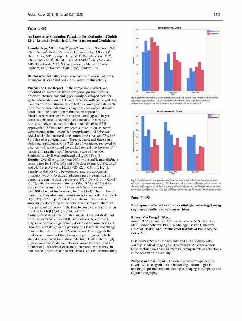
Paper #: 092
An Innovative Simulation Paradigm for Evaluation of Subtle
Liver lesions in Pediatric CT: Performance and Confidence
Jennifer Ngo, MD1, [email protected]; Justin Solomon, PhD
1,
Ehsan Samei1, Taylor Richards
1, Lawrence Ngo, MD PhD
1,
Brian Allen, MD1, Joseph Davis, MD
1, Daniele Marin, MD
1,
Charles Maxfield1, Bhavik Patel, MD MBA
2, Gary Schooler,
MD1, Don Frush, MD
1;
1Duke University Medical Center,
Durham, NC, 2Stanford Health Care, Stanford, CA
Disclosures: All authors have disclosed no financial interests, arrangements or affiliations in the context of this activity. Purpose or Case Report: In the companion abstract, we described an innovative simulation paradigm and effective observer interface combining previously developed tools for
systematic evaluation of CT dose reduction with subtle pediatric liver lesions. Our purpose was to test this paradigm to delineate the effect of dose reduction on diagnostic accuracy and reader confidence, the latter often minimized in importance. Methods & Materials: 30 normal pediatric (ages 0-10 yr) contrast-enhanced de-identified abdominal CT scans were retrospectively collected from the clinical database (IRB
approved). 0-3 simulated low-contrast liver lesions (≤ 6mm) were inserted using LesionTool (proprietary) and noise was added to simulate reduced tube current (mA) that was 75% and 50% that of the original scan. Three pediatric and three adult abdominal radiologists with 7-20 yrs of experience reviewed 90 data sets in 3 sessions and were asked to mark the location of
lesions and rate their confidence on a scale of 0 to 100. Statistical analysis was performed using JMP Pro 13. Results: Overall sensitivity was 38%, with significantly different sensitivities for 100%, 75% and 50% dose exams [52.8%, 35.5% and 24.7% respectively; F(2,15)=26.92, p<0.0001); Fig 1]. Sensitivity did not vary between pediatric and abdominal imagers (p=0.34). Average confidence per case significantly
varied between the three dose levels [F(2,415)=9.51, p=<0.0001; Fig 2], with the mean confidence of the 100% and 75% dose exams varying significantly from the 50% dose exams (p<0.001), but not from one another (p=0.994). The number of clicks per study also varied significantly between the dose levels [F(2,537) = 22.28, p=<0.0001], with the number of clicks surprisingly decreasing as the noise level increased. There was
no significant difference in the time to complete a case between the dose levels [F(2,415) = 2.04, p=0.13]. Conclusions: Academic pediatric and adult specialists did not differ in performance for subtle liver lesions. As expected, diagnostic accuracy significantly decreased as noise increased. However, confidence in the presence of a lesion did not change
between the full dose and 75% dose scans. This suggests that readers are unaware of this decrease in performance, which should be accounted for in dose reduction efforts. Interestingly, higher noise studies did not take any longer to review, but the number of clicks decreased as noise increased, which may, in part, reflect less effort due to perceived decreased discrimination.
Paper #: 093
Development of a tool to aid the radiologic technologist using
augmented reality and computer vision
Robert MacDougall, MSc, [email protected]; Steven Don, MD
2, Benoit Scherrer, PhD
1;
1Radiology, Boston Children's
Hospital, Boston, MA, 2Mallinkrodt Institute of Radiology, St.
Louis, MO Disclosures: Steven Don has indicated a relationship with Vantage Medical Imaging as a Co-founder. All other authors have disclosed no financial interests, arrangements or affiliations in the context of this activity.
Purpose or Case Report: To describe the development of a novel device designed to aid the radiologic technologist in reducing exposure variation and repeat imaging in computed and digital radiography.
Pediatr Radiol (2018) 48 (Suppl 1):S1–S298 S135
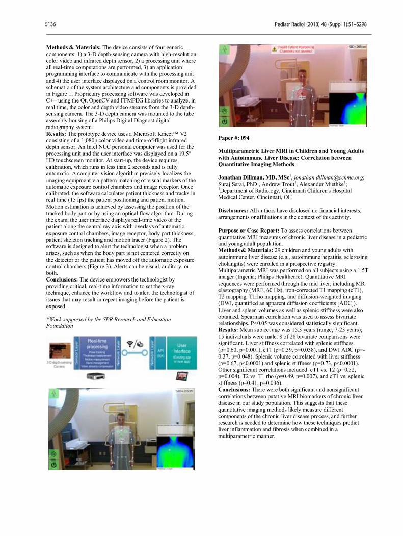
Methods & Materials: The device consists of four generic components: 1) a 3-D depth-sensing camera with high-resolution color video and infrared depth sensor, 2) a processing unit where all real-time computations are performed, 3) an application programming interface to communicate with the processing unit
and 4) the user interface displayed on a control room monitor. A schematic of the system architecture and components is provided in Figure 1. Proprietary processing software was developed in C++ using the Qt, OpenCV and FFMPEG libraries to analyze, in real time, the color and depth video streams from the 3-D depth-sensing camera. The 3-D depth camera was mounted to the tube
assembly housing of a Philips Digital Diagnost digital radiography system. Results: The prototype device uses a Microsoft Kinect™ V2 consisting of a 1,080p color video and time-of-flight infrared depth sensor. An Intel NUC personal computer was used for the processing unit and the user interface was displayed on a 19.5″ HD touchscreen monitor. At start-up, the device requires
calibration, which runs in less than 2 seconds and is fully automatic. A computer vision algorithm precisely localizes the imaging equipment via pattern matching of visual markers of the automatic exposure control chambers and image receptor. Once calibrated, the software calculates patient thickness and tracks in real time (15 fps) the patient positioning and patient motion. Motion estimation is achieved by assessing the position of the
tracked body part or by using an optical flow algorithm. During the exam, the user interface displays real-time video of the patient along the central ray axis with overlays of automatic exposure control chambers, image receptor, body part thickness, patient skeleton tracking and motion tracer (Figure 2). The software is designed to alert the technologist when a problem
arises, such as when the body part is not centered correctly on the detector or the patient has moved off the automatic exposure control chambers (Figure 3). Alerts can be visual, auditory, or both. Conclusions: The device empowers the technologist by providing critical, real-time information to set the x-ray technique, enhance the workflow and to alert the technologist of
issues that may result in repeat imaging before the patient is exposed. *Work supported by the SPR Research and Education Foundation
Paper #: 094
Multiparametric Liver MRI in Children and Young Adults
with Autoimmune Liver Disease: Correlation between
Quantitative Imaging Methods
Jonathan Dillman, MD, MSc1, [email protected];
Suraj Serai, PhD1, Andrew Trout
1, Alexander Miethke
1;
1Department of Radiology, Cincinnati Children's Hospital
Medical Center, Cincinnati, OH Disclosures: All authors have disclosed no financial interests, arrangements or affiliations in the context of this activity.
Purpose or Case Report: To assess correlations between quantitative MRI measures of chronic liver disease in a pediatric and young adult population. Methods & Materials: 29 children and young adults with autoimmune liver disease (e.g., autoimmune hepatitis, sclerosing
cholangitis) were enrolled in a prospective registry. Multiparametric MRI was performed on all subjects using a 1.5T imager (Ingenia; Philips Healthcare). Quantitative MRI sequences were performed through the mid liver, including MR elastography (MRE, 60 Hz), iron-corrected T1 mapping (cT1), T2 mapping, T1rho mapping, and diffusion-weighted imaging (DWI, quantified as apparent diffusion coefficients [ADC]).
Liver and spleen volumes as well as splenic stiffness were also obtained. Spearman correlation was used to assess bivariate relationships. P<0.05 was considered statistically significant. Results: Mean subject age was 15.3 years (range, 7-23 years); 15 individuals were male. 8 of 28 bivariate comparisons were significant. Liver stiffness correlated with splenic stiffness
(ρ=0.60, p=0.001), cT1 (ρ=0.39, p=0.038), and DWI ADC (ρ=-0.37, p=0.048). Splenic volume correlated with liver stiffness (ρ=0.67, p<0.0001) and splenic stiffness (ρ=0.73, p<0.0001). Other significant correlations included: cT1 vs. T2 (ρ=0.52, p=0.004), T2 vs. T1 rho (ρ=0.49, p=0.007), and cT1 vs. splenic stiffness (ρ=0.41, p=0.036). Conclusions: There were both significant and nonsignificant
correlations between putative MRI biomarkers of chronic liver disease in our study population. This suggests that these quantitative imaging methods likely measure different components of the chronic liver disease process, and further research is needed to determine how these techniques predict liver inflammation and fibrosis when combined in a multiparametric manner.
Pediatr Radiol (2018) 48 (Suppl 1):S1–S298S136
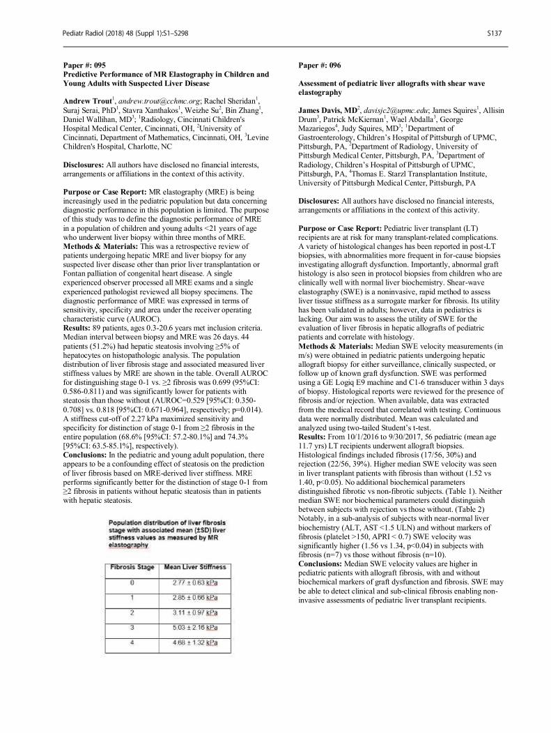
Paper #: 095
Predictive Performance of MR Elastography in Children and
Young Adults with Suspected Liver Disease Andrew Trout
1, [email protected]; Rachel Sheridan
1,
Suraj Serai, PhD1, Stavra Xanthakos
1, Weizhe Su
2, Bin Zhang
1,
Daniel Wallihan, MD3;
1Radiology, Cincinnati Children's
Hospital Medical Center, Cincinnati, OH, 2University of
Cincinnati, Department of Mathematics, Cincinnati, OH, 3Levine
Children's Hospital, Charlotte, NC
Disclosures: All authors have disclosed no financial interests, arrangements or affiliations in the context of this activity. Purpose or Case Report: MR elastography (MRE) is being increasingly used in the pediatric population but data concerning diagnostic performance in this population is limited. The purpose of this study was to define the diagnostic performance of MRE
in a population of children and young adults <21 years of age who underwent liver biopsy within three months of MRE. Methods & Materials: This was a retrospective review of patients undergoing hepatic MRE and liver biopsy for any suspected liver disease other than prior liver transplantation or Fontan palliation of congenital heart disease. A single experienced observer processed all MRE exams and a single
experienced pathologist reviewed all biopsy specimens. The diagnostic performance of MRE was expressed in terms of sensitivity, specificity and area under the receiver operating characteristic curve (AUROC). Results: 89 patients, ages 0.3-20.6 years met inclusion criteria. Median interval between biopsy and MRE was 26 days. 44
patients (51.2%) had hepatic steatosis involving ≥5% of hepatocytes on histopathologic analysis. The population distribution of liver fibrosis stage and associated measured liver stiffness values by MRE are shown in the table. Overall AUROC for distinguishing stage 0-1 vs. ≥2 fibrosis was 0.699 (95%CI: 0.586-0.811) and was significantly lower for patients with steatosis than those without (AUROC=0.529 [95%CI: 0.350-
0.708] vs. 0.818 [95%CI: 0.671-0.964], respectively; p=0.014). A stiffness cut-off of 2.27 kPa maximized sensitivity and specificity for distinction of stage 0-1 from ≥2 fibrosis in the entire population (68.6% [95%CI: 57.2-80.1%] and 74.3% [95%CI: 63.5-85.1%], respectively). Conclusions: In the pediatric and young adult population, there appears to be a confounding effect of steatosis on the prediction
of liver fibrosis based on MRE-derived liver stiffness. MRE performs significantly better for the distinction of stage 0-1 from ≥2 fibrosis in patients without hepatic steatosis than in patients with hepatic steatosis.
Paper #: 096
Assessment of pediatric liver allografts with shear wave
elastography
James Davis, MD2, [email protected]; James Squires
1, Allisin
Drum3, Patrick McKiernan
1, Wael Abdalla
3, George
Mazariegos4, Judy Squires, MD
3;
1Department of
Gastroenterology, Children’s Hospital of Pittsburgh of UPMC, Pittsburgh, PA,
2Department of Radiology, University of
Pittsburgh Medical Center, Pittsburgh, PA, 3Department of
Radiology, Children’s Hospital of Pittsburgh of UPMC, Pittsburgh, PA,
4Thomas E. Starzl Transplantation Institute,
University of Pittsburgh Medical Center, Pittsburgh, PA Disclosures: All authors have disclosed no financial interests, arrangements or affiliations in the context of this activity.
Purpose or Case Report: Pediatric liver transplant (LT) recipients are at risk for many transplant-related complications. A variety of histological changes has been reported in post-LT biopsies, with abnormalities more frequent in for-cause biopsies investigating allograft dysfunction. Importantly, abnormal graft histology is also seen in protocol biopsies from children who are clinically well with normal liver biochemistry. Shear-wave
elastography (SWE) is a noninvasive, rapid method to assess liver tissue stiffness as a surrogate marker for fibrosis. Its utility has been validated in adults; however, data in pediatrics is lacking. Our aim was to assess the utility of SWE for the evaluation of liver fibrosis in hepatic allografts of pediatric patients and correlate with histology.
Methods & Materials: Median SWE velocity measurements (in m/s) were obtained in pediatric patients undergoing hepatic allograft biopsy for either surveillance, clinically suspected, or follow up of known graft dysfunction. SWE was performed using a GE Logiq E9 machine and C1-6 transducer within 3 days of biopsy. Histological reports were reviewed for the presence of fibrosis and/or rejection. When available, data was extracted
from the medical record that correlated with testing. Continuous data were normally distributed. Mean was calculated and analyzed using two-tailed Student’s t-test. Results: From 10/1/2016 to 9/30/2017, 56 pediatric (mean age 11.7 yrs) LT recipients underwent allograft biopsies. Histological findings included fibrosis (17/56, 30%) and rejection (22/56, 39%). Higher median SWE velocity was seen
in liver transplant patients with fibrosis than without (1.52 vs 1.40, p<0.05). No additional biochemical parameters distinguished fibrotic vs non-fibrotic subjects. (Table 1). Neither median SWE nor biochemical parameters could distinguish between subjects with rejection vs those without. (Table 2) Notably, in a sub-analysis of subjects with near-normal liver
biochemistry (ALT, AST <1.5 ULN) and without markers of fibrosis (platelet >150, APRI < 0.7) SWE velocity was significantly higher (1.56 vs 1.34, p<0.04) in subjects with fibrosis (n=7) vs those without fibrosis (n=10). Conclusions: Median SWE velocity values are higher in pediatric patients with allograft fibrosis, with and without biochemical markers of graft dysfunction and fibrosis. SWE may
be able to detect clinical and sub-clinical fibrosis enabling non-invasive assessments of pediatric liver transplant recipients.
Pediatr Radiol (2018) 48 (Suppl 1):S1–S298 S137
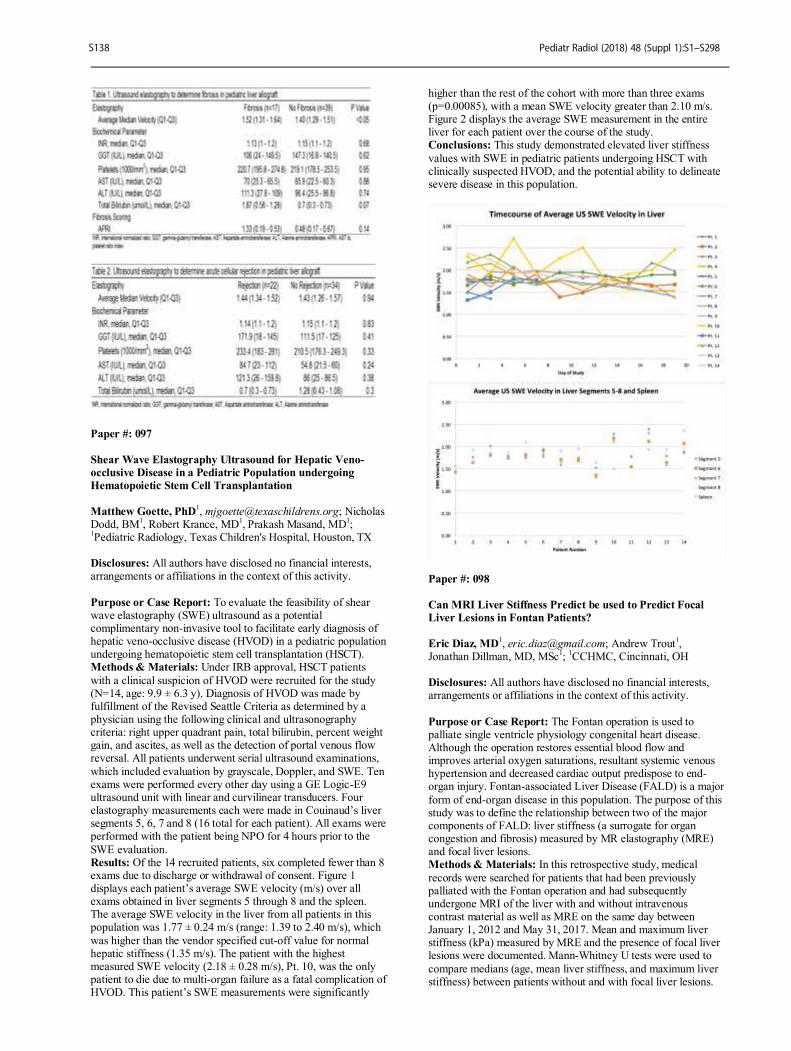
Paper #: 097
Shear Wave Elastography Ultrasound for Hepatic Veno-
occlusive Disease in a Pediatric Population undergoing
Hematopoietic Stem Cell Transplantation
Matthew Goette, PhD1, [email protected]; Nicholas
Dodd, BM1, Robert Krance, MD
1, Prakash Masand, MD
1;
1Pediatric Radiology, Texas Children's Hospital, Houston, TX
Disclosures: All authors have disclosed no financial interests, arrangements or affiliations in the context of this activity.
Purpose or Case Report: To evaluate the feasibility of shear wave elastography (SWE) ultrasound as a potential complimentary non-invasive tool to facilitate early diagnosis of hepatic veno-occlusive disease (HVOD) in a pediatric population undergoing hematopoietic stem cell transplantation (HSCT). Methods & Materials: Under IRB approval, HSCT patients
with a clinical suspicion of HVOD were recruited for the study (N=14, age: 9.9 ± 6.3 y). Diagnosis of HVOD was made by fulfillment of the Revised Seattle Criteria as determined by a physician using the following clinical and ultrasonography criteria: right upper quadrant pain, total bilirubin, percent weight gain, and ascites, as well as the detection of portal venous flow reversal. All patients underwent serial ultrasound examinations,
which included evaluation by grayscale, Doppler, and SWE. Ten exams were performed every other day using a GE Logic-E9 ultrasound unit with linear and curvilinear transducers. Four elastography measurements each were made in Couinaud’s liver segments 5, 6, 7 and 8 (16 total for each patient). All exams were performed with the patient being NPO for 4 hours prior to the
SWE evaluation. Results: Of the 14 recruited patients, six completed fewer than 8 exams due to discharge or withdrawal of consent. Figure 1 displays each patient’s average SWE velocity (m/s) over all exams obtained in liver segments 5 through 8 and the spleen. The average SWE velocity in the liver from all patients in this population was 1.77 ± 0.24 m/s (range: 1.39 to 2.40 m/s), which
was higher than the vendor specified cut-off value for normal hepatic stiffness (1.35 m/s). The patient with the highest measured SWE velocity (2.18 ± 0.28 m/s), Pt. 10, was the only patient to die due to multi-organ failure as a fatal complication of HVOD. This patient’s SWE measurements were significantly
higher than the rest of the cohort with more than three exams (p=0.00085), with a mean SWE velocity greater than 2.10 m/s. Figure 2 displays the average SWE measurement in the entire liver for each patient over the course of the study. Conclusions: This study demonstrated elevated liver stiffness
values with SWE in pediatric patients undergoing HSCT with clinically suspected HVOD, and the potential ability to delineate severe disease in this population.
Paper #: 098
Can MRI Liver Stiffness Predict be used to Predict Focal
Liver Lesions in Fontan Patients?
Eric Diaz, MD
1, [email protected]; Andrew Trout
1,
Jonathan Dillman, MD, MSc1;
1CCHMC, Cincinnati, OH
Disclosures: All authors have disclosed no financial interests, arrangements or affiliations in the context of this activity.
Purpose or Case Report: The Fontan operation is used to palliate single ventricle physiology congenital heart disease. Although the operation restores essential blood flow and improves arterial oxygen saturations, resultant systemic venous hypertension and decreased cardiac output predispose to end-organ injury. Fontan-associated Liver Disease (FALD) is a major
form of end-organ disease in this population. The purpose of this study was to define the relationship between two of the major components of FALD: liver stiffness (a surrogate for organ congestion and fibrosis) measured by MR elastography (MRE) and focal liver lesions. Methods & Materials: In this retrospective study, medical
records were searched for patients that had been previously palliated with the Fontan operation and had subsequently undergone MRI of the liver with and without intravenous contrast material as well as MRE on the same day between January 1, 2012 and May 31, 2017. Mean and maximum liver stiffness (kPa) measured by MRE and the presence of focal liver lesions were documented. Mann-Whitney U tests were used to
compare medians (age, mean liver stiffness, and maximum liver stiffness) between patients without and with focal liver lesions.
Pediatr Radiol (2018) 48 (Suppl 1):S1–S298S138
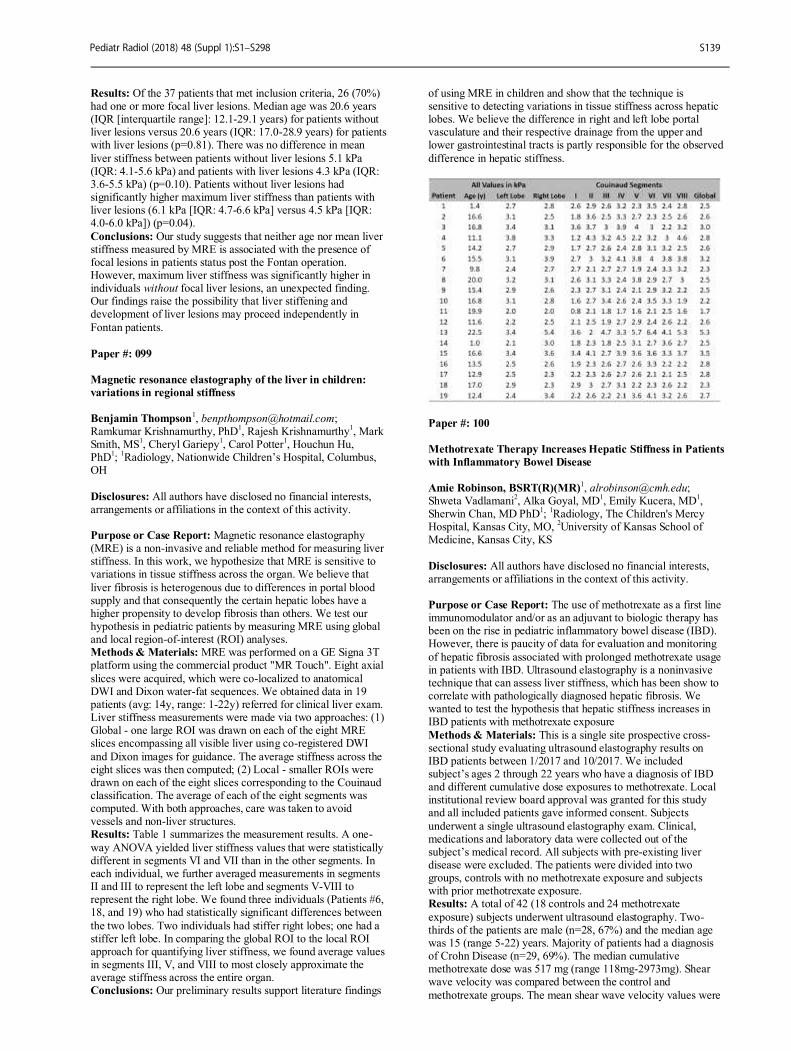
Results: Of the 37 patients that met inclusion criteria, 26 (70%) had one or more focal liver lesions. Median age was 20.6 years (IQR [interquartile range]: 12.1-29.1 years) for patients without liver lesions versus 20.6 years (IQR: 17.0-28.9 years) for patients with liver lesions (p=0.81). There was no difference in mean
liver stiffness between patients without liver lesions 5.1 kPa (IQR: 4.1-5.6 kPa) and patients with liver lesions 4.3 kPa (IQR: 3.6-5.5 kPa) (p=0.10). Patients without liver lesions had significantly higher maximum liver stiffness than patients with liver lesions (6.1 kPa [IQR: 4.7-6.6 kPa] versus 4.5 kPa [IQR: 4.0-6.0 kPa]) (p=0.04).
Conclusions: Our study suggests that neither age nor mean liver stiffness measured by MRE is associated with the presence of focal lesions in patients status post the Fontan operation. However, maximum liver stiffness was significantly higher in individuals without focal liver lesions, an unexpected finding. Our findings raise the possibility that liver stiffening and development of liver lesions may proceed independently in
Fontan patients.
Paper #: 099
Magnetic resonance elastography of the liver in children:
variations in regional stiffness
Benjamin Thompson1, [email protected];
Ramkumar Krishnamurthy, PhD1, Rajesh Krishnamurthy
1, Mark
Smith, MS1, Cheryl Gariepy
1, Carol Potter
1, Houchun Hu,
PhD1;
1Radiology, Nationwide Children’s Hospital, Columbus,
OH
Disclosures: All authors have disclosed no financial interests, arrangements or affiliations in the context of this activity. Purpose or Case Report: Magnetic resonance elastography (MRE) is a non-invasive and reliable method for measuring liver stiffness. In this work, we hypothesize that MRE is sensitive to variations in tissue stiffness across the organ. We believe that
liver fibrosis is heterogenous due to differences in portal blood supply and that consequently the certain hepatic lobes have a higher propensity to develop fibrosis than others. We test our hypothesis in pediatric patients by measuring MRE using global and local region-of-interest (ROI) analyses. Methods & Materials: MRE was performed on a GE Signa 3T platform using the commercial product "MR Touch". Eight axial
slices were acquired, which were co-localized to anatomical DWI and Dixon water-fat sequences. We obtained data in 19 patients (avg: 14y, range: 1-22y) referred for clinical liver exam. Liver stiffness measurements were made via two approaches: (1) Global - one large ROI was drawn on each of the eight MRE slices encompassing all visible liver using co-registered DWI
and Dixon images for guidance. The average stiffness across the eight slices was then computed; (2) Local - smaller ROIs were drawn on each of the eight slices corresponding to the Couinaud classification. The average of each of the eight segments was computed. With both approaches, care was taken to avoid vessels and non-liver structures. Results: Table 1 summarizes the measurement results. A one-
way ANOVA yielded liver stiffness values that were statistically different in segments VI and VII than in the other segments. In each individual, we further averaged measurements in segments II and III to represent the left lobe and segments V-VIII to represent the right lobe. We found three individuals (Patients #6, 18, and 19) who had statistically significant differences between
the two lobes. Two individuals had stiffer right lobes; one had a stiffer left lobe. In comparing the global ROI to the local ROI approach for quantifying liver stiffness, we found average values in segments III, V, and VIII to most closely approximate the average stiffness across the entire organ. Conclusions: Our preliminary results support literature findings
of using MRE in children and show that the technique is sensitive to detecting variations in tissue stiffness across hepatic lobes. We believe the difference in right and left lobe portal vasculature and their respective drainage from the upper and lower gastrointestinal tracts is partly responsible for the observed
difference in hepatic stiffness.
Paper #: 100
Methotrexate Therapy Increases Hepatic Stiffness in Patients
with Inflammatory Bowel Disease
Amie Robinson, BSRT(R)(MR)
Shweta Vadlamani2, Alka Goyal, MD
1, Emily Kucera, MD
1,
Sherwin Chan, MD PhD1;
1Radiology, The Children's Mercy
Hospital, Kansas City, MO, 2University of Kansas School of
Medicine, Kansas City, KS
Disclosures: All authors have disclosed no financial interests, arrangements or affiliations in the context of this activity. Purpose or Case Report: The use of methotrexate as a first line immunomodulator and/or as an adjuvant to biologic therapy has been on the rise in pediatric inflammatory bowel disease (IBD). However, there is paucity of data for evaluation and monitoring
of hepatic fibrosis associated with prolonged methotrexate usage in patients with IBD. Ultrasound elastography is a noninvasive technique that can assess liver stiffness, which has been show to correlate with pathologically diagnosed hepatic fibrosis. We wanted to test the hypothesis that hepatic stiffness increases in IBD patients with methotrexate exposure
Methods & Materials: This is a single site prospective cross-sectional study evaluating ultrasound elastography results on IBD patients between 1/2017 and 10/2017. We included subject’s ages 2 through 22 years who have a diagnosis of IBD and different cumulative dose exposures to methotrexate. Local institutional review board approval was granted for this study and all included patients gave informed consent. Subjects
underwent a single ultrasound elastography exam. Clinical, medications and laboratory data were collected out of the subject’s medical record. All subjects with pre-existing liver disease were excluded. The patients were divided into two groups, controls with no methotrexate exposure and subjects with prior methotrexate exposure. Results: A total of 42 (18 controls and 24 methotrexate
exposure) subjects underwent ultrasound elastography. Two-thirds of the patients are male (n=28, 67%) and the median age was 15 (range 5-22) years. Majority of patients had a diagnosis of Crohn Disease (n=29, 69%). The median cumulative methotrexate dose was 517 mg (range 118mg-2973mg). Shear wave velocity was compared between the control and
methotrexate groups. The mean shear wave velocity values were
Pediatr Radiol (2018) 48 (Suppl 1):S1–S298 S139
significantly higher in patients with methotrexate exposure compared to the controls (1.37 vs. 1.23 m/s. p=0.04). Conclusions: Ultrasound elastography may be useful in detecting early changes in hepatic stiffness in patients receiving methotrexate prior to liver enzyme changes or clinically apparent
liver disease. This technique should be studied further as a monitoring tool for patients on methotrexate.
Paper #: 101
Abdominal Fat Distribution and its Relationship with Liver
and Pancreatic Fat Fraction, Liver Stiffness and Diabetic
Status David Hunte
1, [email protected]; Andrew Trout
1,
Marialena Mouzaki1, Alexander Towbin, MD
1, Stavra
Xanthakos1, Jonathan Dillman, MD, MSc
1;
1Radiology,
Cincinnati Children's Hospital Medical Center, Cincinnati, OH
Disclosures: All authors have disclosed no financial interests, arrangements or affiliations in the context of this activity. Purpose or Case Report: Fatty liver disease is increasingly recognized as a multisystem disease. The purpose of this study was to assess the relationship between measures of anatomic fat
depots, liver fat fraction, liver stiffness, pancreatic fat fraction, and diabetic status in pediatric patients. Methods & Materials: This was a retrospective study of pediatric patients with fatty liver disease who had undergone liver MR elastography (MRE) and multi-point Dixon-based fat quantification between September 2015 and July 2017. A single
observer measured liver and pancreatic proton density fat fraction, abdominal subcutaneous fat thickness, and visceral fat thickness (deep fascia of rectus muscle to anterior aspect of L2). Clinical records were reviewed to abstract demographic data (age, sex, body mass index [BMI]), liver stiffness, and type 2 diabetes status. Bivariate correlations were assessed with Spearman’s rho, and multiple linear and multiple logistic
regression models were used to assess relationships between outcome and predictor variables. Results: 87 patients were included in this study (mean age: 14.3±3.4 years; 27 [31%] females; mean BMI: 36.1±7.8 kg/m
2).
7/87 (8%) patients were diabetic, and 45/87 (52%) were insulin resistant. Mean liver stiffness was 2.5±0.9 kPa, mean liver fat fraction was 18.7±10.3% and mean pancreatic fat fraction was
5.9±5%. Liver fat fraction was significantly correlated only with visceral fat thickness at univariate analysis (ρ=0.31, p=0.004). Based on multivariate modeling, liver stiffness was significantly associated with liver fat fraction (-0.022kPa/+1%, p=0.018) and visceral fat thickness (+0.18kPa/1 cm, p=0.0007). At univariate
analysis, pancreatic fat fraction was significantly correlated with age (ρ=0.28, p=0.012), BMI (ρ=0.34, p=0.002), subcutaneous fat thickness (ρ=0.23, p=0.04), and visceral fat thickness (ρ=0.24, p=0.03), but these relationships did not persist upon multivariate modeling. Diabetic status was not significantly associated with any measure of abdominal fat. Conclusions: Liver and pancreatic fat fraction are both weakly
but significantly correlated with visceral fat thickness suggesting a relationship between visceral, but not subcutaneous, adiposity and liver and pancreatic steatosis in children. Hepatic steatosis appears to have a softening effect on liver stiffness as measured by MRE, and increased visceral fat thickness appears to be associated with increased liver stiffness. None of our imaging
findings could predict diabetic status.
Paper #: 102
Contrast-Enhanced Brain Ultrasound on Extreme
Premature Twin Fetal Lambs Maintained by the EXTra-
uterine Environment for Neonatal Support (EXTEND): A
Promising Imaging Technique Ryne Didier
1, [email protected]; Anush Sridharan
1, Kendall
Lawrence1, Marcus Davey
1, Beverly Coleman
1, Alan Flake
1;
1Children's Hospital of Philadelphia, Philadelphia, PA
Disclosures: All authors have disclosed no financial interests, arrangements or affiliations in the context of this activity. Purpose or Case Report: With recent progress in the development of an artificial environment to support the extreme premature infant, advanced imaging techniques are necessary to evaluate the brain parenchyma. We aimed to evaluate a) the
feasibility of contrast-enhanced ultrasound (CEUS) and b) brain perfusion in fetal lambs maintained on the EXTEND system. Methods & Materials: Following IACUC-approved protocols, twin premature fetal lambs [102 days gestational age (GA), Term = 145 days] were transferred from placental support to the EXTEND system
1. Twin B was immediately subjected to a 12-
hour period of sub-physiologic flow (152mL/kg/min) and
oxygen delivery (15.9mL/kg/min). Twin A maintained physiologic flow (>233mL/kg/min) and oxygen delivery (>22mL/kg/min). Lumason® ultrasound (US) contrast boluses were administered into the pumpless umbilical arterial to umbilical venous oxygenator circuit. Serial CEUS examinations were performed using a GE Logiq E9 US system and C2-9
transducer. Time-to-peak (TTP) measurements were obtained and Parametric maps were created. Hemodynamic parameters and measurements across the oxygenator were recorded. Serum oxygen and lactate values were obtained pre- and post-examination. Post-mortem MRIs were performed. 1Partridge EA et al. An extra-uterine system to physiologically
support the extreme premature lamb. Nat Commun.
2017;8:15112. Results: To account for microbubble destruction within the support equipment, up to 0.59mL/kg contrast was required for optimal visualization. There were no statistically significant changes in measured variables (Table 1). On grayscale images, Twin B demonstrated ventriculomegaly and loss of grey-white differentiation with progressive loss of parenchymal white
matter which was not observed in Twin A. With CEUS, Twin B demonstrated increased TTP enhancement and decreased parenchymal perfusion when compared to Twin A at 108 and 105 days, respectively (Figure 1). Twin B demonstrated progressively increased TTP enhancement within the brain and brainstem at 115 and 129 days GA and post-mortem MRI
confirmed hydranencephaly. Conclusions: CEUS of the brain is feasible in fetal lambs maintained on the EXTEND system and changes in perfusion can be quantified. These findings establish a foundation for further evaluation of this promising technique with potential applications in the setting of fetal hypoxia.
Pediatr Radiol (2018) 48 (Suppl 1):S1–S298S140
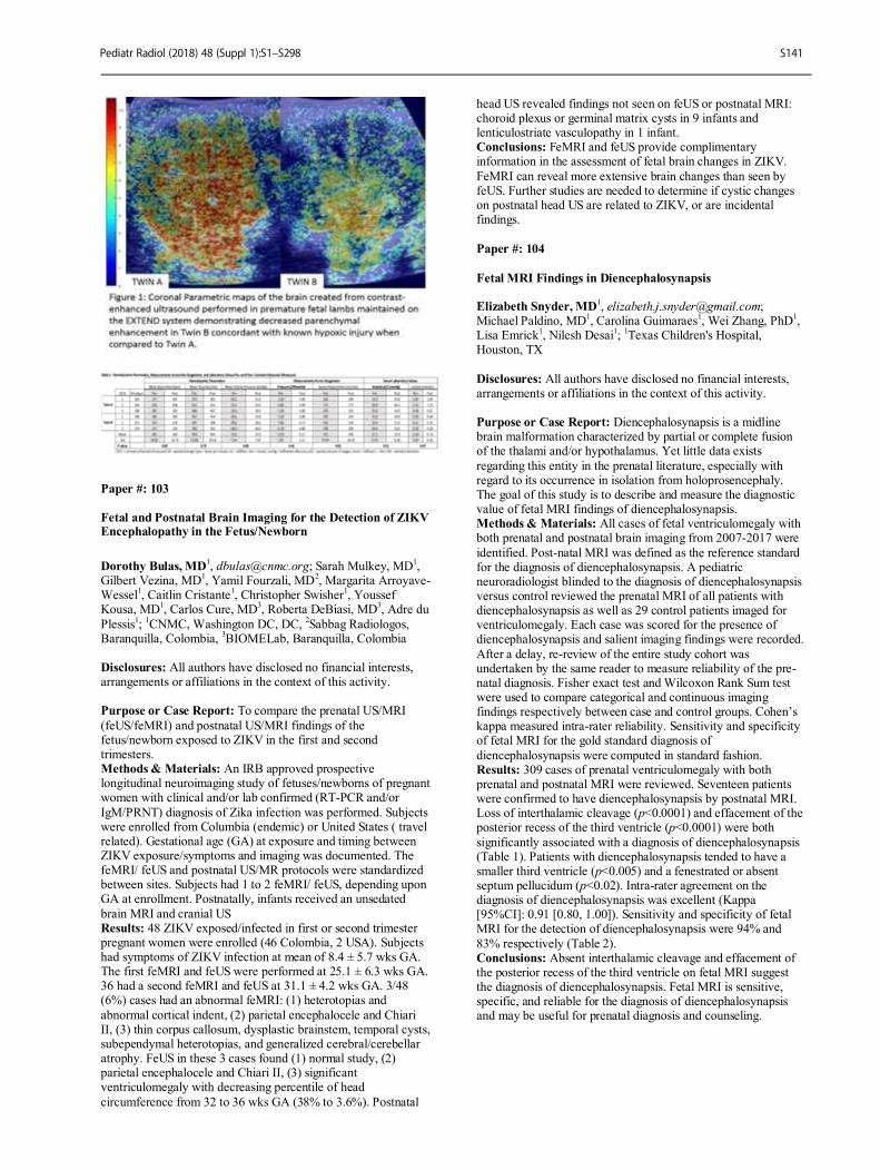
Paper #: 103
Fetal and Postnatal Brain Imaging for the Detection of ZIKV Encephalopathy in the Fetus/Newborn
Dorothy Bulas, MD1, [email protected]; Sarah Mulkey, MD
1,
Gilbert Vezina, MD1, Yamil Fourzali, MD
2, Margarita Arroyave-
Wessel1, Caitlin Cristante
1, Christopher Swisher
1, Youssef
Kousa, MD1, Carlos Cure, MD
3, Roberta DeBiasi, MD
3, Adre du
Plessis1;
1CNMC, Washington DC, DC,
2Sabbag Radiologos,
Baranquilla, Colombia, 3BIOMELab, Baranquilla, Colombia
Disclosures: All authors have disclosed no financial interests, arrangements or affiliations in the context of this activity. Purpose or Case Report: To compare the prenatal US/MRI
(feUS/feMRI) and postnatal US/MRI findings of the fetus/newborn exposed to ZIKV in the first and second trimesters. Methods & Materials: An IRB approved prospective longitudinal neuroimaging study of fetuses/newborns of pregnant women with clinical and/or lab confirmed (RT-PCR and/or
IgM/PRNT) diagnosis of Zika infection was performed. Subjects were enrolled from Columbia (endemic) or United States ( travel related). Gestational age (GA) at exposure and timing between ZIKV exposure/symptoms and imaging was documented. The feMRI/ feUS and postnatal US/MR protocols were standardized between sites. Subjects had 1 to 2 feMRI/ feUS, depending upon GA at enrollment. Postnatally, infants received an unsedated
brain MRI and cranial US Results: 48 ZIKV exposed/infected in first or second trimester pregnant women were enrolled (46 Colombia, 2 USA). Subjects had symptoms of ZIKV infection at mean of 8.4 ± 5.7 wks GA. The first feMRI and feUS were performed at 25.1 ± 6.3 wks GA. 36 had a second feMRI and feUS at 31.1 ± 4.2 wks GA. 3/48 (6%) cases had an abnormal feMRI: (1) heterotopias and
abnormal cortical indent, (2) parietal encephalocele and Chiari II, (3) thin corpus callosum, dysplastic brainstem, temporal cysts, subependymal heterotopias, and generalized cerebral/cerebellar atrophy. FeUS in these 3 cases found (1) normal study, (2) parietal encephalocele and Chiari II, (3) significant ventriculomegaly with decreasing percentile of head
circumference from 32 to 36 wks GA (38% to 3.6%). Postnatal
head US revealed findings not seen on feUS or postnatal MRI: choroid plexus or germinal matrix cysts in 9 infants and lenticulostriate vasculopathy in 1 infant. Conclusions: FeMRI and feUS provide complimentary information in the assessment of fetal brain changes in ZIKV.
FeMRI can reveal more extensive brain changes than seen by feUS. Further studies are needed to determine if cystic changes on postnatal head US are related to ZIKV, or are incidental findings.
Paper #: 104
Fetal MRI Findings in Diencephalosynapsis Elizabeth Snyder, MD
Michael Paldino, MD1, Carolina Guimaraes
1, Wei Zhang, PhD
1,
Lisa Emrick1, Nilesh Desai
1;
1Texas Children's Hospital,
Houston, TX
Disclosures: All authors have disclosed no financial interests, arrangements or affiliations in the context of this activity. Purpose or Case Report: Diencephalosynapsis is a midline brain malformation characterized by partial or complete fusion of the thalami and/or hypothalamus. Yet little data exists
regarding this entity in the prenatal literature, especially with regard to its occurrence in isolation from holoprosencephaly. The goal of this study is to describe and measure the diagnostic value of fetal MRI findings of diencephalosynapsis. Methods & Materials: All cases of fetal ventriculomegaly with both prenatal and postnatal brain imaging from 2007-2017 were
identified. Post-natal MRI was defined as the reference standard for the diagnosis of diencephalosynapsis. A pediatric neuroradiologist blinded to the diagnosis of diencephalosynapsis versus control reviewed the prenatal MRI of all patients with diencephalosynapsis as well as 29 control patients imaged for ventriculomegaly. Each case was scored for the presence of diencephalosynapsis and salient imaging findings were recorded.
After a delay, re-review of the entire study cohort was undertaken by the same reader to measure reliability of the pre-natal diagnosis. Fisher exact test and Wilcoxon Rank Sum test were used to compare categorical and continuous imaging findings respectively between case and control groups. Cohen’s kappa measured intra-rater reliability. Sensitivity and specificity of fetal MRI for the gold standard diagnosis of
diencephalosynapsis were computed in standard fashion. Results: 309 cases of prenatal ventriculomegaly with both prenatal and postnatal MRI were reviewed. Seventeen patients were confirmed to have diencephalosynapsis by postnatal MRI. Loss of interthalamic cleavage (p<0.0001) and effacement of the posterior recess of the third ventricle (p<0.0001) were both
significantly associated with a diagnosis of diencephalosynapsis (Table 1). Patients with diencephalosynapsis tended to have a smaller third ventricle (p<0.005) and a fenestrated or absent septum pellucidum (p<0.02). Intra-rater agreement on the diagnosis of diencephalosynapsis was excellent (Kappa [95%CI]: 0.91 [0.80, 1.00]). Sensitivity and specificity of fetal MRI for the detection of diencephalosynapsis were 94% and
83% respectively (Table 2). Conclusions: Absent interthalamic cleavage and effacement of the posterior recess of the third ventricle on fetal MRI suggest the diagnosis of diencephalosynapsis. Fetal MRI is sensitive, specific, and reliable for the diagnosis of diencephalosynapsis and may be useful for prenatal diagnosis and counseling.
Pediatr Radiol (2018) 48 (Suppl 1):S1–S298 S141
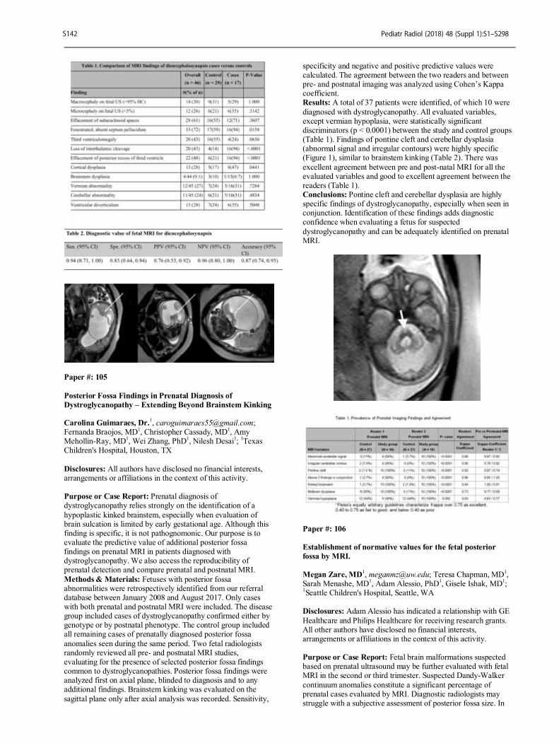
Paper #: 105
Posterior Fossa Findings in Prenatal Diagnosis of
Dystroglycanopathy – Extending Beyond Brainstem Kinking Carolina Guimaraes, Dr.
Fernanda Braojos, MD1, Christopher Cassady, MD
1, Amy
Mehollin-Ray, MD1, Wei Zhang, PhD
1, Nilesh Desai
1;
1Texas
Children's Hospital, Houston, TX
Disclosures: All authors have disclosed no financial interests, arrangements or affiliations in the context of this activity. Purpose or Case Report: Prenatal diagnosis of dystroglycanopathy relies strongly on the identification of a
hypoplastic kinked brainstem, especially when evaluation of brain sulcation is limited by early gestational age. Although this finding is specific, it is not pathognomonic. Our purpose is to evaluate the predictive value of additional posterior fossa findings on prenatal MRI in patients diagnosed with dystroglycanopathy. We also access the reproducibility of prenatal detection and compare prenatal and postnatal MRI.
Methods & Materials: Fetuses with posterior fossa abnormalities were retrospectively identified from our referral database between January 2008 and August 2017. Only cases with both prenatal and postnatal MRI were included. The disease group included cases of dystroglycanopathy confirmed either by genotype or by postnatal phenotype. The control group included all remaining cases of prenatally diagnosed posterior fossa
anomalies seen during the same period. Two fetal radiologists randomly reviewed all pre- and postnatal MRI studies, evaluating for the presence of selected posterior fossa findings common to dystroglycanopathies. Posterior fossa findings were analyzed first on axial plane, blinded to diagnosis and to any additional findings. Brainstem kinking was evaluated on the
sagittal plane only after axial analysis was recorded. Sensitivity,
specificity and negative and positive predictive values were calculated. The agreement between the two readers and between pre- and postnatal imaging was analyzed using Cohen’s Kappa coefficient. Results: A total of 37 patients were identified, of which 10 were
diagnosed with dystroglycanopathy. All evaluated variables, except vermian hypoplasia, were statistically significant discriminators (p < 0.0001) between the study and control groups (Table 1). Findings of pontine cleft and cerebellar dysplasia (abnormal signal and irregular contours) were highly specific (Figure 1), similar to brainstem kinking (Table 2). There was
excellent agreement between pre and post-natal MRI for all the evaluated variables and good to excellent agreement between the readers (Table 1). Conclusions: Pontine cleft and cerebellar dysplasia are highly specific findings of dystroglycanopathy, especially when seen in conjunction. Identification of these findings adds diagnostic confidence when evaluating a fetus for suspected
dystroglycanopathy and can be adequately identified on prenatal MRI.
Paper #: 106
Establishment of normative values for the fetal posterior
fossa by MRI.
Megan Zare, MD
1, [email protected]; Teresa Chapman, MD
1,
Sarah Menashe, MD1, Adam Alessio, PhD
1, Gisele Ishak, MD
1;
1Seattle Children's Hospital, Seattle, WA
Disclosures: Adam Alessio has indicated a relationship with GE
Healthcare and Philips Healthcare for receiving research grants. All other authors have disclosed no financial interests, arrangements or affiliations in the context of this activity. Purpose or Case Report: Fetal brain malformations suspected based on prenatal ultrasound may be further evaluated with fetal MRI in the second or third trimester. Suspected Dandy-Walker
continuum anomalies constitute a significant percentage of prenatal cases evaluated by MRI. Diagnostic radiologists may struggle with a subjective assessment of posterior fossa size. In
Pediatr Radiol (2018) 48 (Suppl 1):S1–S298S142
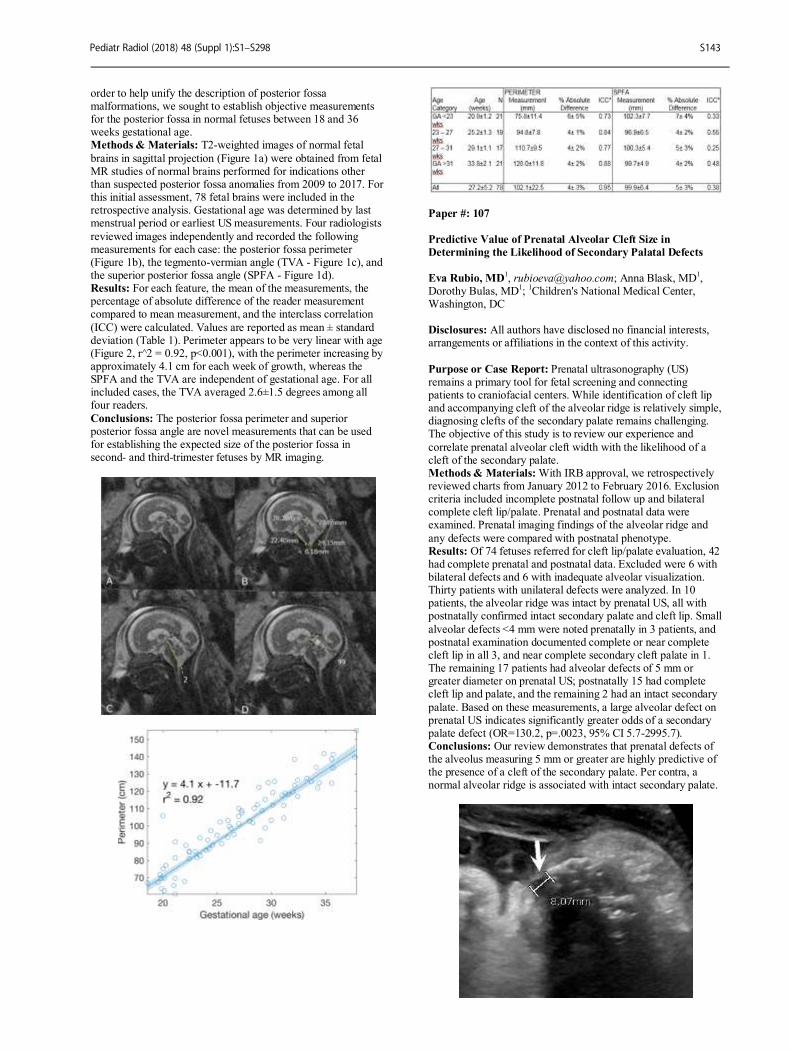
order to help unify the description of posterior fossa malformations, we sought to establish objective measurements for the posterior fossa in normal fetuses between 18 and 36 weeks gestational age. Methods & Materials: T2-weighted images of normal fetal
brains in sagittal projection (Figure 1a) were obtained from fetal MR studies of normal brains performed for indications other than suspected posterior fossa anomalies from 2009 to 2017. For this initial assessment, 78 fetal brains were included in the retrospective analysis. Gestational age was determined by last menstrual period or earliest US measurements. Four radiologists
reviewed images independently and recorded the following measurements for each case: the posterior fossa perimeter (Figure 1b), the tegmento-vermian angle (TVA - Figure 1c), and the superior posterior fossa angle (SPFA - Figure 1d). Results: For each feature, the mean of the measurements, the percentage of absolute difference of the reader measurement compared to mean measurement, and the interclass correlation
(ICC) were calculated. Values are reported as mean ± standard deviation (Table 1). Perimeter appears to be very linear with age (Figure 2, r^2 = 0.92, p<0.001), with the perimeter increasing by approximately 4.1 cm for each week of growth, whereas the SPFA and the TVA are independent of gestational age. For all included cases, the TVA averaged 2.6±1.5 degrees among all four readers.
Conclusions: The posterior fossa perimeter and superior posterior fossa angle are novel measurements that can be used for establishing the expected size of the posterior fossa in second- and third-trimester fetuses by MR imaging.
Paper #: 107
Predictive Value of Prenatal Alveolar Cleft Size in
Determining the Likelihood of Secondary Palatal Defects
Eva Rubio, MD1, [email protected]; Anna Blask, MD
1,
Dorothy Bulas, MD1;
1Children's National Medical Center,
Washington, DC Disclosures: All authors have disclosed no financial interests, arrangements or affiliations in the context of this activity.
Purpose or Case Report: Prenatal ultrasonography (US) remains a primary tool for fetal screening and connecting patients to craniofacial centers. While identification of cleft lip and accompanying cleft of the alveolar ridge is relatively simple, diagnosing clefts of the secondary palate remains challenging. The objective of this study is to review our experience and
correlate prenatal alveolar cleft width with the likelihood of a cleft of the secondary palate. Methods & Materials: With IRB approval, we retrospectively reviewed charts from January 2012 to February 2016. Exclusion criteria included incomplete postnatal follow up and bilateral complete cleft lip/palate. Prenatal and postnatal data were examined. Prenatal imaging findings of the alveolar ridge and
any defects were compared with postnatal phenotype. Results: Of 74 fetuses referred for cleft lip/palate evaluation, 42 had complete prenatal and postnatal data. Excluded were 6 with bilateral defects and 6 with inadequate alveolar visualization. Thirty patients with unilateral defects were analyzed. In 10 patients, the alveolar ridge was intact by prenatal US, all with postnatally confirmed intact secondary palate and cleft lip. Small
alveolar defects <4 mm were noted prenatally in 3 patients, and postnatal examination documented complete or near complete cleft lip in all 3, and near complete secondary cleft palate in 1. The remaining 17 patients had alveolar defects of 5 mm or greater diameter on prenatal US; postnatally 15 had complete cleft lip and palate, and the remaining 2 had an intact secondary
palate. Based on these measurements, a large alveolar defect on prenatal US indicates significantly greater odds of a secondary palate defect (OR=130.2, p=.0023, 95% CI 5.7-2995.7). Conclusions: Our review demonstrates that prenatal defects of the alveolus measuring 5 mm or greater are highly predictive of the presence of a cleft of the secondary palate. Per contra, a normal alveolar ridge is associated with intact secondary palate.
Pediatr Radiol (2018) 48 (Suppl 1):S1–S298 S143
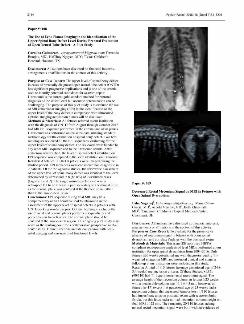
Paper #: 108
The Use of Echo Planar Imaging in the Identification of the
Upper Spinal Bony Defect Level During Prenatal Evaluation
of Open Neural Tube Defect - A Pilot Study
Carolina Guimaraes
1, [email protected]; Fernanda
Braojos, MD1, HaiThuy Nguyen, MD
1;
1Texas Children's
Hospital, Houston, TX Disclosures: All authors have disclosed no financial interests,
arrangements or affiliations in the context of this activity. Purpose or Case Report: The upper level of spinal bony defect in cases of prenatally diagnosed open neural tube defect (ONTD) has significant prognostic implications and is one of the criteria used to identify potential candidates for in-utero repair. Ultrasound is the current gold standard method for prenatal
diagnosis of the defect level but accurate determination can be challenging. The purpose of this pilot study is to evaluate the use of MR echo planar imaging (EPI) in the identification of the upper level of the bony defect in comparison with ultrasound. Optimal imaging acquisition planes will be discussed. Methods & Materials: All fetuses referred to our institution with the diagnosis of ONTD from August through October 2017
had MR EPI sequence performed in the coronal and axial planes. Ultrasound was performed on the same date, utilizing standard methodology for the evaluation of spinal bony defect. Two fetal radiologists reviewed all the EPI sequences, evaluating for the upper level of spinal bony defect. The reviewers were blinded to any other MRI sequence and to the ultrasound results. After
consensus was reached, the level of spinal defect identified on EPI sequence was compared to the level identified on ultrasound. Results: A total of 11 ONTD patients were imaged during the studied period. EPI sequences were considered non-diagnostic in 2 patients. Of the 9 diagnostic studies, the reviewers’ assessment of the upper level of spinal bony defect was identical to the level determined by ultrasound in 8 (88.8%) of 9 evaluated cases
(Figures 1 and 2). The single misinterpreted case was in retrospect felt to be at least in part secondary to a technical error, as the coronal plane was centered at the thoracic spine rather than at the lumbosacral spine. Conclusions: EPI sequence during fetal MRI may be a complimentary or an alternative tool to ultrasound in the assessment of the upper level of spinal defects in patients with
ONTD seeking in-utero repair. Optimal technique includes the use of axial and coronal planes performed sequentially and perpendicular to each other. The coronal plane should be centered at the lumbosacral region. This ongoing pilot study may serve as the starting point for a collaborative prospective multi-center study. Future directions include comparison with post-
natal imaging and assessment of functional levels.
Paper #: 109
Decreased Rectal Meconium Signal on MRI in Fetuses with
Open Spinal Dysraphism Usha Nagaraj
1, [email protected]; Maria Calvo-
Garcia, MD1, Arnold Merrow, MD
1, Beth Kline-Fath,
MD1;
1Cincinnati Children's Hospital Medical Center,
Cincinnati, OH Disclosures: All authors have disclosed no financial interests, arrangements or affiliations in the context of this activity. Purpose or Case Report: To evaluate for the presence or absence of meconium signal in fetuses with open spinal
dysraphism and correlate findings with the postnatal exam. Methods & Materials: This is an IRB approved HIPPA compliant retrospective analysis of fetal MRIs performed at our institution for open spinal dysraphism from 2004-2016. Only fetuses ≥20 weeks gestational age with diagnostic quality T1-weighted images on MRI and postnatal clinical and imaging follow-up at our institution were included in this study.
Results: A total of 110 fetuses (average gestational age of 24 ± 3.4 weeks) met inclusion criteria. Of these fetuses, 81.8% (90/110) had T1 hyperintense rectal meconium signal. The average height of the meconium column in fetuses ≥23 weeks with a measurable column was 11.1 ± 4.3 mm; however, all fetuses (n=17) except 1 at gestational age of 23 weeks had a meconium column that measured 9mm or less . 1/110 fetuses
had imperforate anus on postnatal exam with rectovestibular fistula, but this fetus had a normal meconium column height on fetal MRI of 22 mm. The remaining 20/110 fetuses lacking normal rectal meconium signal were born without evidence of
Pediatr Radiol (2018) 48 (Suppl 1):S1–S298S144
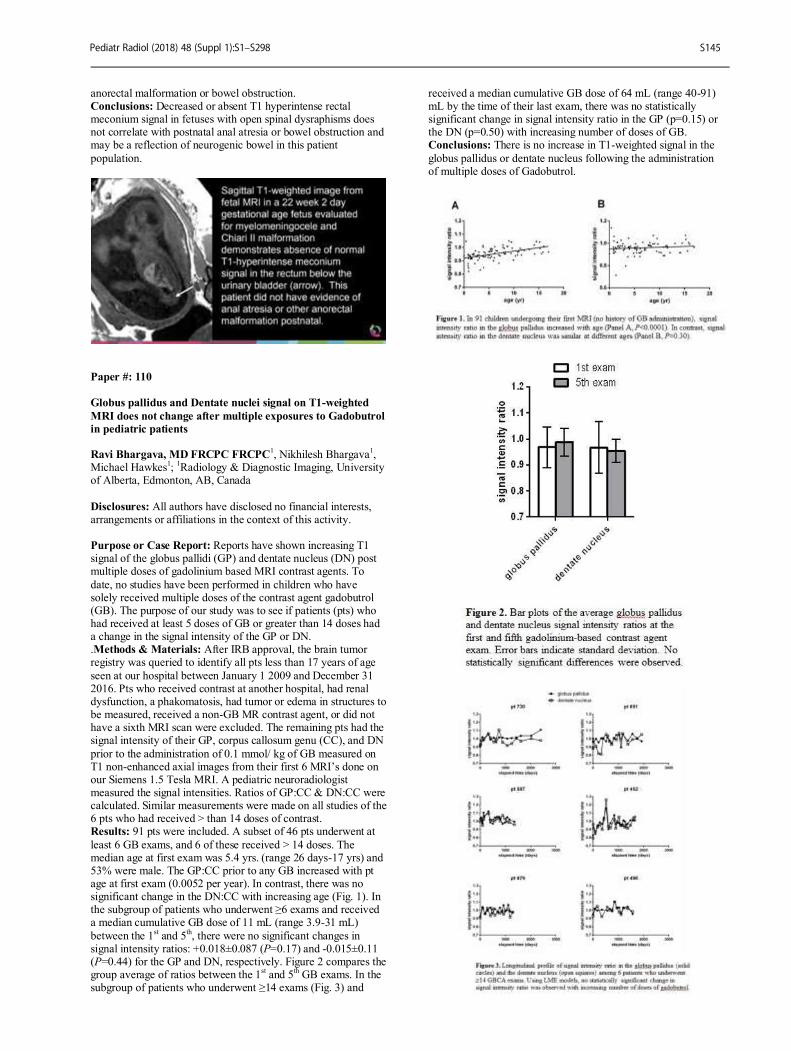
anorectal malformation or bowel obstruction. Conclusions: Decreased or absent T1 hyperintense rectal meconium signal in fetuses with open spinal dysraphisms does not correlate with postnatal anal atresia or bowel obstruction and may be a reflection of neurogenic bowel in this patient
population.
Paper #: 110
Globus pallidus and Dentate nuclei signal on T1-weighted
MRI does not change after multiple exposures to Gadobutrol
in pediatric patients Ravi Bhargava, MD FRCPC FRCPC
1, Nikhilesh Bhargava
1,
Michael Hawkes1;
1Radiology & Diagnostic Imaging, University
of Alberta, Edmonton, AB, Canada
Disclosures: All authors have disclosed no financial interests, arrangements or affiliations in the context of this activity. Purpose or Case Report: Reports have shown increasing T1 signal of the globus pallidi (GP) and dentate nucleus (DN) post multiple doses of gadolinium based MRI contrast agents. To
date, no studies have been performed in children who have solely received multiple doses of the contrast agent gadobutrol (GB). The purpose of our study was to see if patients (pts) who had received at least 5 doses of GB or greater than 14 doses had a change in the signal intensity of the GP or DN. .Methods & Materials: After IRB approval, the brain tumor registry was queried to identify all pts less than 17 years of age
seen at our hospital between January 1 2009 and December 31 2016. Pts who received contrast at another hospital, had renal dysfunction, a phakomatosis, had tumor or edema in structures to be measured, received a non-GB MR contrast agent, or did not have a sixth MRI scan were excluded. The remaining pts had the signal intensity of their GP, corpus callosum genu (CC), and DN
prior to the administration of 0.1 mmol/ kg of GB measured on T1 non-enhanced axial images from their first 6 MRI’s done on our Siemens 1.5 Tesla MRI. A pediatric neuroradiologist measured the signal intensities. Ratios of GP:CC & DN:CC were calculated. Similar measurements were made on all studies of the 6 pts who had received > than 14 doses of contrast. Results: 91 pts were included. A subset of 46 pts underwent at
least 6 GB exams, and 6 of these received > 14 doses. The median age at first exam was 5.4 yrs. (range 26 days-17 yrs) and 53% were male. The GP:CC prior to any GB increased with pt age at first exam (0.0052 per year). In contrast, there was no significant change in the DN:CC with increasing age (Fig. 1). In the subgroup of patients who underwent ≥6 exams and received a median cumulative GB dose of 11 mL (range 3.9-31 mL)
between the 1st and 5
th, there were no significant changes in
signal intensity ratios: +0.018±0.087 (P=0.17) and -0.015±0.11 (P=0.44) for the GP and DN, respectively. Figure 2 compares the group average of ratios between the 1
st and 5
th GB exams. In the
subgroup of patients who underwent ≥14 exams (Fig. 3) and
received a median cumulative GB dose of 64 mL (range 40-91) mL by the time of their last exam, there was no statistically significant change in signal intensity ratio in the GP (p=0.15) or the DN (p=0.50) with increasing number of doses of GB. Conclusions: There is no increase in T1-weighted signal in the
globus pallidus or dentate nucleus following the administration of multiple doses of Gadobutrol.
Pediatr Radiol (2018) 48 (Suppl 1):S1–S298 S145
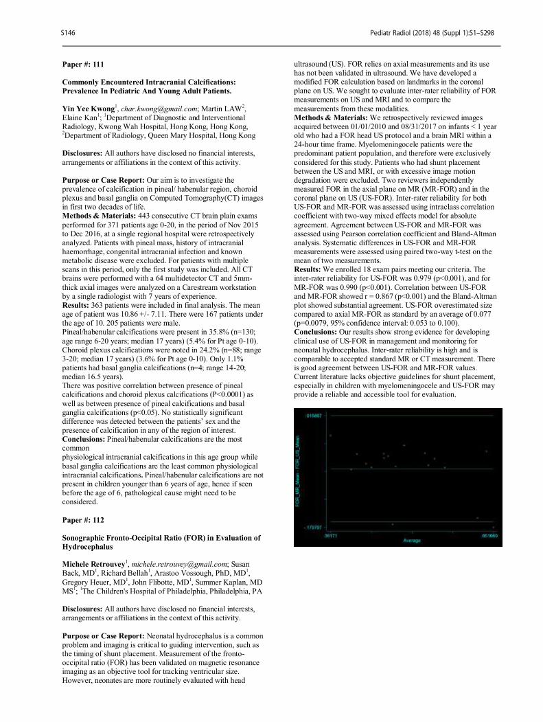
Paper #: 111
Commonly Encountered Intracranial Calcifications:
Prevalence In Pediatric And Young Adult Patients.
Yin Yee Kwong1, [email protected]; Martin LAW
2,
Elaine Kan1;
1Department of Diagnostic and Interventional
Radiology, Kwong Wah Hospital, Hong Kong, Hong Kong, 2Department of Radiology, Queen Mary Hospital, Hong Kong
Disclosures: All authors have disclosed no financial interests,
arrangements or affiliations in the context of this activity. Purpose or Case Report: Our aim is to investigate the prevalence of calcification in pineal/ habenular region, choroid plexus and basal ganglia on Computed Tomography(CT) images in first two decades of life. Methods & Materials: 443 consecutive CT brain plain exams
performed for 371 patients age 0-20, in the period of Nov 2015 to Dec 2016, at a single regional hospital were retrospectively analyzed. Patients with pineal mass, history of intracranial haemorrhage, congenital intracranial infection and known metabolic disease were excluded. For patients with multiple scans in this period, only the first study was included. All CT brains were performed with a 64 multidetector CT and 5mm-
thick axial images were analyzed on a Carestream workstation by a single radiologist with 7 years of experience. Results: 363 patients were included in final analysis. The mean age of patient was 10.86 +/- 7.11. There were 167 patients under the age of 10. 205 patients were male. Pineal/habenular calcifications were present in 35.8% (n=130;
age range 6-20 years; median 17 years) (5.4% for Pt age 0-10). Choroid plexus calcifications were noted in 24.2% (n=88; range 3-20; median 17 years) (3.6% for Pt age 0-10). Only 1.1% patients had basal ganglia calcifications (n=4; range 14-20; median 16.5 years). There was positive correlation between presence of pineal calcifications and choroid plexus calcifications (P<0.0001) as
well as between presence of pineal calcifications and basal ganglia calcifications (p<0.05). No statistically significant difference was detected between the patients’ sex and the presence of calcification in any of the region of interest. Conclusions: Pineal/habenular calcifications are the most common physiological intracranial calcifications in this age group while
basal ganglia calcifications are the least common physiological intracranial calcifications. Pineal/habenular calcifications are not present in children younger than 6 years of age, hence if seen before the age of 6, pathological cause might need to be considered.
Paper #: 112
Sonographic Fronto-Occipital Ratio (FOR) in Evaluation of
Hydrocephalus Michele Retrouvey
1, [email protected]; Susan
Back, MD1, Richard Bellah
1, Arastoo Vossough, PhD, MD
1,
Gregory Heuer, MD1, John Flibotte, MD
1, Summer Kaplan, MD
MS1;
1The Children's Hospital of Philadelphia, Philadelphia, PA
Disclosures: All authors have disclosed no financial interests, arrangements or affiliations in the context of this activity.
Purpose or Case Report: Neonatal hydrocephalus is a common problem and imaging is critical to guiding intervention, such as the timing of shunt placement. Measurement of the fronto-occipital ratio (FOR) has been validated on magnetic resonance imaging as an objective tool for tracking ventricular size. However, neonates are more routinely evaluated with head
ultrasound (US). FOR relies on axial measurements and its use has not been validated in ultrasound. We have developed a modified FOR calculation based on landmarks in the coronal plane on US. We sought to evaluate inter-rater reliability of FOR measurements on US and MRI and to compare the
measurements from these modalities. Methods & Materials: We retrospectively reviewed images acquired between 01/01/2010 and 08/31/2017 on infants < 1 year old who had a FOR head US protocol and a brain MRI within a 24-hour time frame. Myelomeningocele patients were the predominant patient population, and therefore were exclusively
considered for this study. Patients who had shunt placement between the US and MRI, or with excessive image motion degradation were excluded. Two reviewers independently measured FOR in the axial plane on MR (MR-FOR) and in the coronal plane on US (US-FOR). Inter-rater reliability for both US-FOR and MR-FOR was assessed using intraclass correlation coefficient with two-way mixed effects model for absolute
agreement. Agreement between US-FOR and MR-FOR was assessed using Pearson correlation coefficient and Bland-Altman analysis. Systematic differences in US-FOR and MR-FOR measurements were assessed using paired two-way t-test on the mean of two measurements. Results: We enrolled 18 exam pairs meeting our criteria. The inter-rater reliability for US-FOR was 0.979 (p<0.001), and for
MR-FOR was 0.990 (p<0.001). Correlation between US-FOR and MR-FOR showed r = 0.867 (p<0.001) and the Bland-Altman plot showed substantial agreement. US-FOR overestimated size compared to axial MR-FOR as standard by an average of 0.077 (p=0.0079, 95% confidence interval: 0.053 to 0.100). Conclusions: Our results show strong evidence for developing
clinical use of US-FOR in management and monitoring for neonatal hydrocephalus. Inter-rater reliability is high and is comparable to accepted standard MR or CT measurement. There is good agreement between US-FOR and MR-FOR values. Current literature lacks objective guidelines for shunt placement, especially in children with myelomeningocele and US-FOR may provide a reliable and accessible tool for evaluation.
Pediatr Radiol (2018) 48 (Suppl 1):S1–S298S146
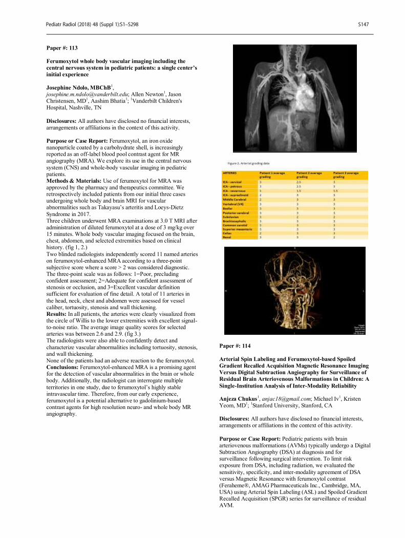
Paper #: 113
Ferumoxytol whole body vascular imaging including the
central nervous system in pediatric patients: a single center’s
initial experience
Josephine Ndolo, MBChB
1,
[email protected]; Allen Newton1, Jason
Christensen, MD1, Aashim Bhatia
1;
1Vanderbilt Children's
Hospital, Nashville, TN
Disclosures: All authors have disclosed no financial interests, arrangements or affiliations in the context of this activity. Purpose or Case Report: Ferumoxytol, an iron oxide nanoparticle coated by a carbohydrate shell, is increasingly reported as an off-label blood pool contrast agent for MR angiography (MRA). We explore its use in the central nervous
system (CNS) and whole-body vascular imaging in pediatric patients. Methods & Materials: Use of ferumoxytol for MRA was approved by the pharmacy and therapeutics committee. We retrospectively included patients from our initial three cases undergoing whole body and brain MRI for vascular abnormalities such as Takayasu’s arteritis and Loeys-Dietz
Syndrome in 2017. Three children underwent MRA examinations at 3.0 T MRI after administration of diluted ferumoxytol at a dose of 3 mg/kg over 15 minutes. Whole body vascular imaging focused on the brain, chest, abdomen, and selected extremities based on clinical history. (fig 1, 2.)
Two blinded radiologists independently scored 11 named arteries on ferumoxytol-enhanced MRA according to a three-point subjective score where a score > 2 was considered diagnostic. The three-point scale was as follows: 1=Poor, precluding confident assessment; 2=Adequate for confident assessment of stenosis or occlusion, and 3=Excellent vascular definition sufficient for evaluation of fine detail. A total of 11 arteries in
the head, neck, chest and abdomen were assessed for vessel caliber, tortuosity, stenosis and wall thickening. Results: In all patients, the arteries were clearly visualized from the circle of Willis to the lower extremities with excellent signal-to-noise ratio. The average image quality scores for selected arteries was between 2.6 and 2.9. (fig 3.) The radiologists were also able to confidently detect and
characterize vascular abnormalities including tortuosity, stenosis, and wall thickening. None of the patients had an adverse reaction to the ferumoxytol. Conclusions: Ferumoxytol-enhanced MRA is a promising agent for the detection of vascular abnormalities in the brain or whole body. Additionally, the radiologist can interrogate multiple
territories in one study, due to ferumoxytol’s highly stable intravascular time. Therefore, from our early experience, ferumoxytol is a potential alternative to gadolinium-based contrast agents for high resolution neuro- and whole body MR angiography.
Paper #: 114
Arterial Spin Labeling and Ferumoxytol-based Spoiled
Gradient Recalled Acquisition Magnetic Resonance Imaging
Versus Digital Subtraction Angiography for Surveillance of
Residual Brain Arteriovenous Malformations in Children: A
Single-Institution Analysis of Inter-Modality Reliability Anjeza Chukus
1, [email protected]; Michael Iv
1, Kristen
Yeom, MD1;
1Stanford University, Stanford, CA
Disclosures: All authors have disclosed no financial interests, arrangements or affiliations in the context of this activity.
Purpose or Case Report: Pediatric patients with brain arteriovenous malformations (AVMs) typically undergo a Digital Subtraction Angiography (DSA) at diagnosis and for surveillance following surgical intervention. To limit risk exposure from DSA, including radiation, we evaluated the
sensitivity, specificity, and inter-modality agreement of DSA versus Magnetic Resonance with ferumoxytol contrast (Feraheme®, AMAG Pharmaceuticals Inc., Cambridge, MA, USA) using Arterial Spin Labeling (ASL) and Spoiled Gradient Recalled Acquisition (SPGR) series for surveillance of residual AVM.
Pediatr Radiol (2018) 48 (Suppl 1):S1–S298 S147
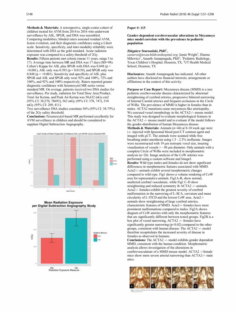
Methods & Materials: A retrospective, single-center cohort of children treated for AVM from 2014 to 2016 who underwent surveillance by ASL, SPGR, and DSA was assembled. Comparing modalities, blinded raters assessed residual AVM, lesion evolution, and their diagnostic confidence using a Likert
scale. Sensitivity, specificity, and inter-modality reliability were determined with DSA as the gold standard. Acute radiation exposure was compared to a safety threshold of 2Gy. Results: Fifteen patients met criteria (mean 11 years, range 3 to 17). Average time between MR and DSA was 17 days (SD=98). Cohen’s Kappa for ASL plus SPGR with DSA was 0.848 (p =
<0.001), ASL only was 0.393 (p = 0.0139), and SPGR only was 0.848 (p = <0.001). Sensitivity and specificity of ASL plus SPGR and ASL and SPGR only were 92% and 100%, 72% and 100%, and 92% and 100% respectively. Raters reported greater diagnostic confidence with ferumoxytol MR series versus standard MR. On average, patients received two DSA studies for surveillance. Per study, radiation for Total Dose Area Product,
Total Air Kerma, and Peak Air Kerma was 58,632 mGy.cm2 (95% CI: 38,570, 78693), 562 mGy (95% CI: 378, 747), 310 mGy (95% CI: 209, 411). Two surveillance DSA studies constitute 56% (95% CI: 38-75%) of the 2Gy safety threshold. Conclusions: Ferumoxytol-based MR performed excellently for AVM surveillance in children and should be considered to
supplant Digital Subtraction Angiography.
Paper #: 115
Gender-dependent cerebrovascular alterations in Moyamoya
mice model correlate with the prevalence in pediatric
population
Zbigniew Starosolski, PhD1,
[email protected]; Jamie Wright2, Dianna
Milewicz2, Ananth Annapragada, PhD
1;
1Pediatric Radiology,
Texas Children’s Hospital, Houston, TX, 2UT Health Medical
School, Houston, TX
Disclosures: Ananth Annapragada has indicated. All other authors have disclosed no financial interests, arrangements or affiliations in the context of this activity. Purpose or Case Report: Moyamoya disease (MMD) is a rare pediatric cerebrovascular disease characterized by abnormal
straightening of cerebral arteries, progressive bilateral narrowing of Internal Carotid arteries and frequent occlusions in the Circle of Willis. The prevalence of MMD is higher in females than in males. ACTA2 mutations cause moyamoya-like arteriopathy. We assessed vessel morphology in the ACTA2-/- mouse model. This study was designed to evaluate morphological features in the ACTA2 -/- mouse model and to evaluate if the model follows
the gender-distribution of human Moyamoya disease. Methods & Materials: Animals (n=44) at 8-10 week age were i.v. injected with liposomal blood pool CT contrast agent and imaged with μCT. The animals were scanned while free breathing under anesthesia using 1.5 - 2.5% isoflurane. Images were reconstructed with 19 μm isotropic voxel size, insuring
visualization of vessels > ~50 μm diameter. Only animals with a complete Circle of Willis were included in morphometric analysis (n=26). Image analysis of the CoW arteries was performed using a custom software and ImageJ. Results: Wild type males and females do not show significant differences in morphometric features associated with MMD. Acta2-/- animals exhibit several morphometric changes
compared to wild type. Fig1 shows a volume rendering of CoW area for representative animals. Fig1A-B, show normal, unaltered cerebral vasculature, while Fig1 C-D show straightening and reduced symmetry ib ACTA2 -/- animals. Acta2-/- females exhibit the greatest severity of cerebral malformation in the narrowing of L-SCA, curvature and mean circularity of L-ITCD and the lowest CoW area. Acta2-/-
animals show straightening of large cerebral arteries, characteristic features of MMD. Acta2-/- females have more prominent malformations compared to males. Fig2A shows diagram of CoW arteries with only the morphometric features that are significantly different between tested groups. Fig2B is a box plot of vessel narrowing, ACTA2 -/- females have
significantly greater narrowing (p<0.02) compared to the other groups, consistent with human disease. The ACTA2 -/- model therefore recapitulates the increased severity of disease in females as observed in humans. Conclusions: The ACTA2 -/- model exhibits gender dependent MMD, consistent with the human condition. Morphometric analysis allows investigation of the alterations in
cerebrovasculature of a MMD mouse model. ACTA2 -/-female mice show more severe arterial narrowing than ACTA2-/- male mice.
Pediatr Radiol (2018) 48 (Suppl 1):S1–S298S148
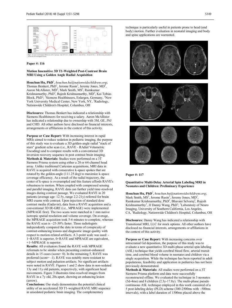
Paper #: 116
Motion Insensitive 3D T1-Weighted Post-Contrast Brain
MRI Using a Golden Angle Radial Acquisition
Houchun Hu, PhD3, [email protected];
Thomas Benkert, PhD1, Jerome Rusin
3, Jeremy Jones, MD
3,
Aaron McAllister, MD3, Mark Smith, MS
3, Ramkumar
Krishnamurthy, PhD3, Rajesh Krishnamurthy, MD
3, Kai Tobias
Block, PhD2;
1Siemens Healthineers, Enlargen, Germany,
2New
York University Medical Center, New York, NY, 3Radiology,
Nationwide Children's Hospital, Columbus, OH
Disclosures: Thomas Benkert has indicated a relationship with Siemens Healthineers for receiving a salary. Aaron McAllister has indicated a relationship due to ownership with 3M, GE, JNJ and CHD. All other authors have disclosed no financial interests, arrangements or affiliations in the context of this activity.
Purpose or Case Report: With increasing interest in rapid MRIs aimed to reduce sedation in pediatric imaging, the purpose of this study was to evaluate a 3D golden-angle radial "stack of stars" gradient echo scan (i.e., RAVE - RAdial Volumetric Encoding) and to compare results with a conventional 3D inversion recovery sequence in post contrast brain imaging.
Methods & Materials: Studies were performed on a 3T Siemens Prisma system using either a 20 or 64-channel head array. Unlike traditional Cartesian acquisitions, MRI data in RAVE is acquired with consecutive k-space spokes that are rotated by the golden-angle (111.25 deg) to maximize k-space coverage efficiency. As a result of the radial trajectory, the center of k-space is oversampled and this feature affords RAVE's
robustness to motion. When coupled with compressed sensing and parallel imaging, RAVE data can further yield time-resolved images during contrast passage. We evaluated RAVE in 20 patients (average age: 12.5y, range 2.2-21y) referred for brain MRI exams with contrast. Upon injection of standard dose contrast media (Gadavist), data from a RAVE acquisition and a conventional 3D IR-GRE (i.e., MPRAGE) were implemented
(MPRAGE first). The two scans were matched in 1 mm native isotropic spatial resolution and volume coverage. On average, the MPRAGE acquisition took 5-6 minutes to complete, whereas the RAVE scan is ~25-30% faster. Three radiologists independently compared the data in terms of conspicuity of contrast-enhancing lesions and diagnostic image quality with
respect to motion-related artifacts. A 3-point scale was used: -1=RAVE is superior, 0=RAVE and MPRAGE are equivalent, +1=MPRAGE is superior. Results: All evaluators found the RAVE with MPRAGE reformats to be similar when presenting contrast-enhanced details in 15 cases (score= 0). In the remaining 5, RAVE was preferred (score= -1). RAVE was notably more resistant to
subject motion and pulsation artifacts. No significant artifacts were noted in RAVE. Figures 1 and 2 show data in non-sedated 13y and 11y old patients, respectively, with significant head movements. Figure 3 illustrates time-resolved images from RAVE in a 7y old, 20s apart, showing an enhancing lesion (arrow). Conclusions: Our study demonstrates the potential clinical
utility of an accelerated 3D T1-weighted RAVE MRI sequence in unsedated pediatric brain imaging. The complementary
technique is particularly useful in patients prone to head (and body) motion. Further evaluation in neonatal imaging and body and spine applications are warranted.
Paper #: 117
Quantitative Multi-Delay Arterial Spin Labeling MRI in
Neonates and Children: Preliminary Experience Houchun Hu, PhD
Mark Smith, MS2, Jerome Rusin
2, Jeremy Jones, MD
2,
Ramkumar Krishnamurthy, PhD2, Bhavani Selvaraj
2, Rajesh
Krishnamurthy2, JJ Danny Wang, PhD
1;
1Laboratory of Neuro
Imaging, University of Southern California, Los Angeles, CA,
2Radiology, Nationwide Children's Hospital, Columbus, OH
Disclosures: Danny Wang has indicated a relationship with
Transitional MRI, LLC for stock options. All other authors have disclosed no financial interests, arrangements or affiliations in the context of this activity. Purpose or Case Report: With increasing concerns over intracranial Gd deposition, the purpose of this study was to evaluate a new quantitative 3D multi-phase arterial spin labeling
(ASL) technique that yields cerebral blood flow, arterial transit time, and cerebral blood volume in neonates and children via a single acquisition. While the technique has been reported in adult populations, feasibility and application in pediatrics has not been previously demonstrated. Methods & Materials: All studies were performed on a 3T
Siemens Prisma platform and data were successfully reconstructed offline. We evaluated the technique in 3 neonates (5d-4mo) and 8 children (2.3y-17.8y). The multi-phase pseudo continuous ASL technique employed in this work consisted of a 5 post-labeling delay (PLD) scheme (300-2500ms with ~500ms intervals), with a label duration of 1500ms placed above the
Pediatr Radiol (2018) 48 (Suppl 1):S1–S298 S149
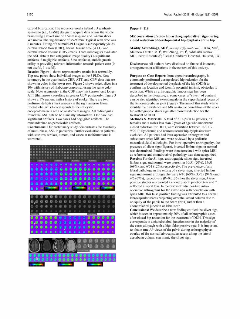
carotid bifurcation. The sequence used a hybrid 3D gradient-spin-echo (i.e., GraSE) design to acquire data across the whole brain using a voxel size of 2.5mm in-plane and 3-4mm slices. We used a labeling distance of 75-90mm. Typical scan time was 6 minutes. Fitting of the multi-PLD signals subsequently yields
cerebral blood flow (CBF), arterial transit time (ATT), and cerebral blood volume (CBV) maps. Three radiologists evaluated the ASL data in two categories: image quality (1-significant artifacts, 2-negligible artifacts, 3-no artifacts), and diagnostic utility in providing relevant information towards patient care (0-not useful, 1-useful).
Results: Figure 1 shows representative results in a normal 2y. Top row panes show individual images at the 5 PLDs. Note symmetry in the quantitative CBF, ATT, and CBV data that are shown in color in the lower row. Figure 2 shows select slices in a 10y with history of rhabdomyosarcoma, using the same color scale. Note asymmetry in the CBF map (thick arrow) and longer ATT (thin arrow), resulting in greater CBV posteriorly. Figure 3
shows a 17y patient with a history of stroke. There are two perfusion deficits (thick arrows) in the right anterior lateral frontal lobe, which corresponds to foci of cystic encephalomalacia seen on anatomical images. All radiologists found the ASL data to be clinically informative. One case had significant artifacts. Two cases had negligible artifacts. The remainder had no perceivable artifacts.
Conclusions: Our preliminary study demonstrates the feasibility of multi-phase ASL in pediatrics. Further evaluation in patients with seizures, strokes, tumors, and vascular malformations is warranted.
Paper #: 118
MR correlation of spica hip arthrographic sliver sign during
closed reduction of developmental hip dysplasia of the hip
Maddy Artunduaga, MD1, [email protected]; J. Kan, MD
1,
Matthew Ditzler, MD1, Wei Zhang, PhD
1, Siddharth Jadhav,
MD1, Scott Rosenfeld
1;
1Texas Children's Hospital, Houston, TX
Disclosures: All authors have disclosed no financial interests, arrangements or affiliations in the context of this activity.
Purpose or Case Report: Intra-operative arthrography is commonly performed during closed hip reduction for the treatment of developmental dysplasia of the hip (DDH) to confirm hip location and identify potential intrinsic obstacles to reduction. While an arthrographic limbus sign has been described in the literature, in some cases, a “sliver” of contrast
can be also identified extending along the superolateral recess of the femoroacetabular joint (figure). The aim of this study was to identify the prevalence and MR anatomic correlation of the spica hip arthrographic sliver sign after closed reduction for the treatment of DDH Methods & Materials: A total of 51 hips in 42 patients, 37 females and 5 males less than 2 years of age who underwent
closed reduction for DDH, were identified between 1/2011-9/2017. Syndromic and neuromuscular hip dysplasias were excluded. All patients had intra-operative arthrogram and subsequent spica MRI and were reviewed by a pediatric musculoskeletal radiologist. For intra-operative arthrography, the presence of sliver sign (figure), inverted limbus sign, or normal
was determined. Findings were then correlated with spica MRI as reference and chondrolabral pathology was then categorized Results: For the 51 hips, arthrographic sliver sign, inverted limbus sign, and normal were present in 10/51 (20%), 35/51 (69%), and 6/51 (12%), respectively. The prevalence of any labral pathology in the setting of a sliver sign, inverted limbus sign and normal arthrography were 6/10 (60%), 33/35 (94%) and
4/6 (67%), respectively (P=0.0136). For the sliver sign, 4 true positive studies represented a chondrolabral junction tear and 2 reflected a labral tear. In re-review of false positive intra-operative arthrograms for the sliver sign with correlation with spica MRI, this false positive finding was attributed to a normal labrocapsular recess projecting over the lateral column due to obliquity of the pelvis to the beam (N=4) rather than a
chondrolabral junction or labral tear Conclusions: We describe a new finding entitled the sliver sign, which is seen in approximately 20% of all arthrographic cases after closed hip reduction for the treatment of DDH. This sign corresponds to a chondrolabral junction tear in the majority of the cases although with a high false positive rate. It is important
to obtain true AP views of the pelvis during arthrography as overlay of the normal labrocapsular recess along the lateral acetabular column can mimic the sliver sign.
Pediatr Radiol (2018) 48 (Suppl 1):S1–S298S150
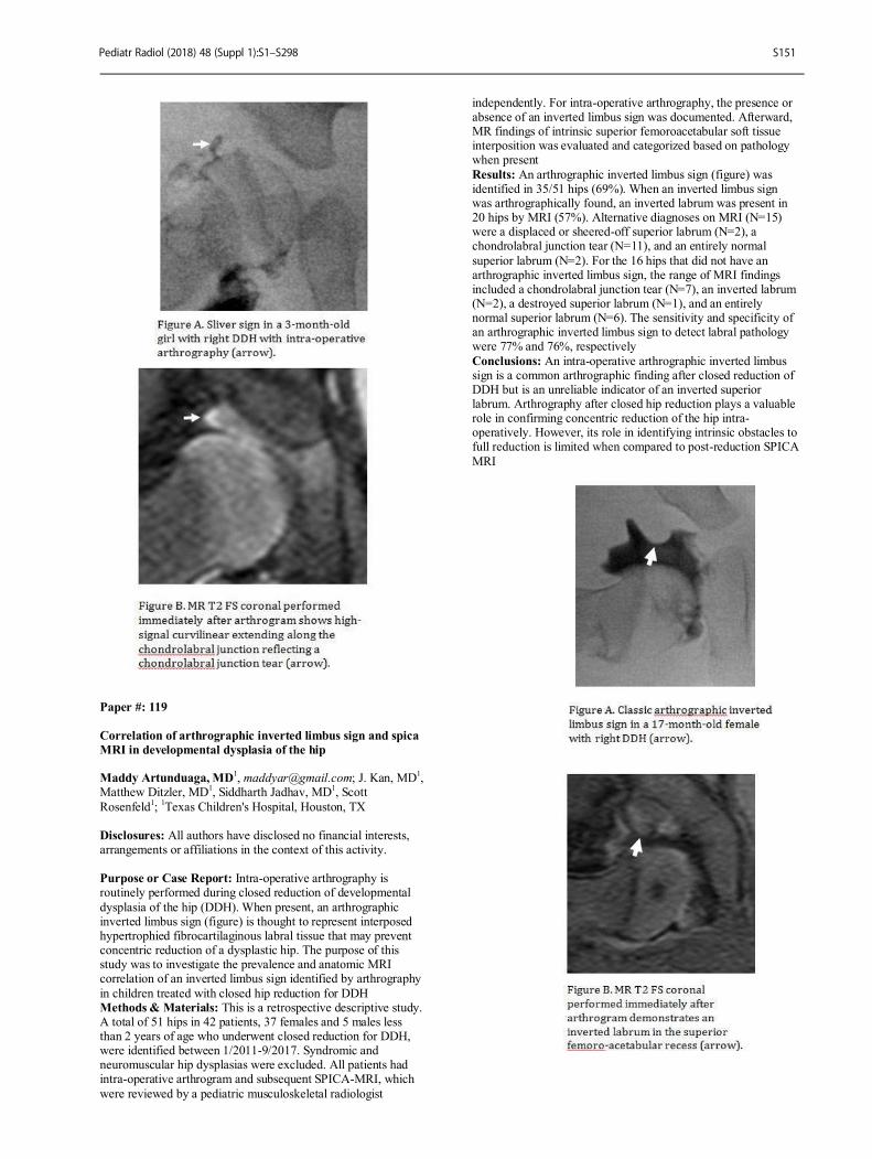
Paper #: 119
Correlation of arthrographic inverted limbus sign and spica
MRI in developmental dysplasia of the hip Maddy Artunduaga, MD
1, [email protected]; J. Kan, MD
1,
Matthew Ditzler, MD1, Siddharth Jadhav, MD
1, Scott
Rosenfeld1;
1Texas Children's Hospital, Houston, TX
Disclosures: All authors have disclosed no financial interests, arrangements or affiliations in the context of this activity. Purpose or Case Report: Intra-operative arthrography is routinely performed during closed reduction of developmental
dysplasia of the hip (DDH). When present, an arthrographic inverted limbus sign (figure) is thought to represent interposed hypertrophied fibrocartilaginous labral tissue that may prevent concentric reduction of a dysplastic hip. The purpose of this study was to investigate the prevalence and anatomic MRI correlation of an inverted limbus sign identified by arthrography
in children treated with closed hip reduction for DDH Methods & Materials: This is a retrospective descriptive study. A total of 51 hips in 42 patients, 37 females and 5 males less than 2 years of age who underwent closed reduction for DDH, were identified between 1/2011-9/2017. Syndromic and neuromuscular hip dysplasias were excluded. All patients had intra-operative arthrogram and subsequent SPICA-MRI, which
were reviewed by a pediatric musculoskeletal radiologist
independently. For intra-operative arthrography, the presence or absence of an inverted limbus sign was documented. Afterward, MR findings of intrinsic superior femoroacetabular soft tissue interposition was evaluated and categorized based on pathology when present
Results: An arthrographic inverted limbus sign (figure) was identified in 35/51 hips (69%). When an inverted limbus sign was arthrographically found, an inverted labrum was present in 20 hips by MRI (57%). Alternative diagnoses on MRI (N=15) were a displaced or sheered-off superior labrum (N=2), a chondrolabral junction tear (N=11), and an entirely normal
superior labrum (N=2). For the 16 hips that did not have an arthrographic inverted limbus sign, the range of MRI findings included a chondrolabral junction tear (N=7), an inverted labrum (N=2), a destroyed superior labrum (N=1), and an entirely normal superior labrum (N=6). The sensitivity and specificity of an arthrographic inverted limbus sign to detect labral pathology were 77% and 76%, respectively
Conclusions: An intra-operative arthrographic inverted limbus sign is a common arthrographic finding after closed reduction of DDH but is an unreliable indicator of an inverted superior labrum. Arthrography after closed hip reduction plays a valuable role in confirming concentric reduction of the hip intra-operatively. However, its role in identifying intrinsic obstacles to full reduction is limited when compared to post-reduction SPICA
MRI
Pediatr Radiol (2018) 48 (Suppl 1):S1–S298 S151
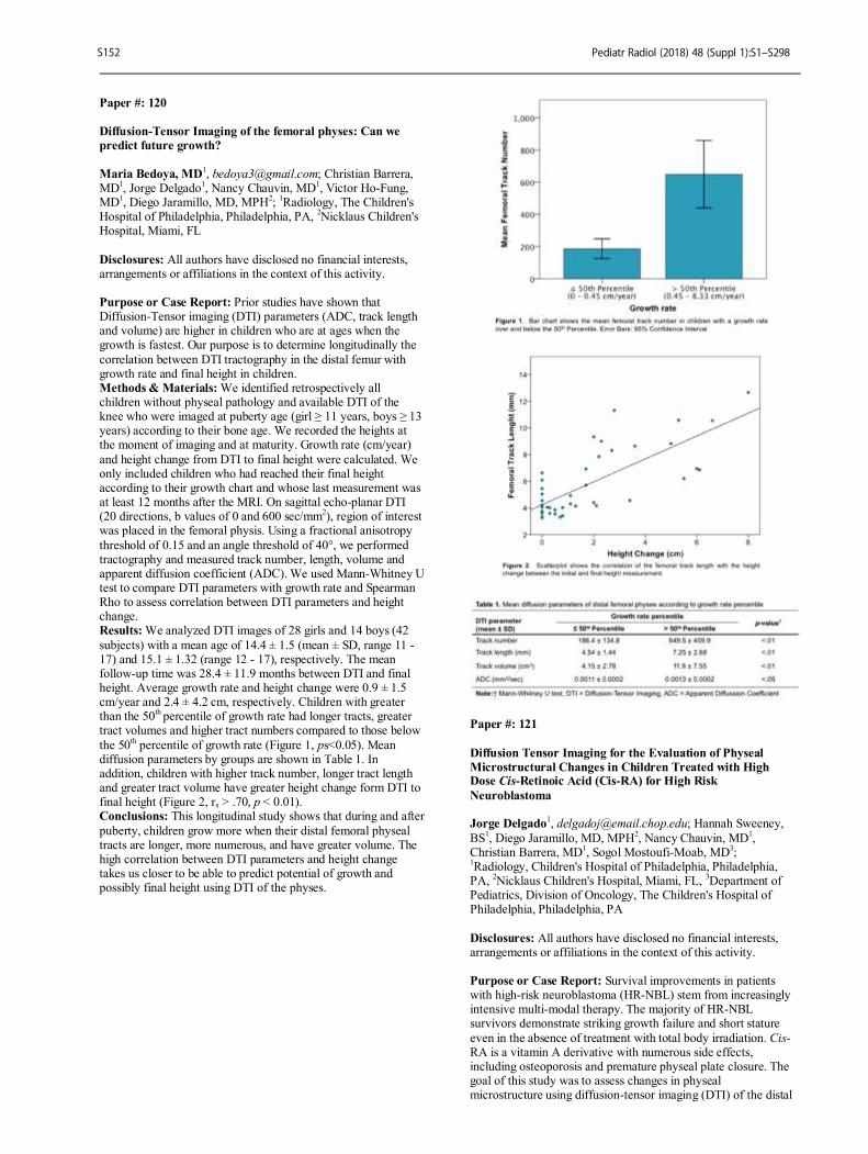
Paper #: 120
Diffusion-Tensor Imaging of the femoral physes: Can we
predict future growth?
Maria Bedoya, MD1, [email protected]; Christian Barrera,
MD1, Jorge Delgado
1, Nancy Chauvin, MD
1, Victor Ho-Fung,
MD1, Diego Jaramillo, MD, MPH
2;
1Radiology, The Children's
Hospital of Philadelphia, Philadelphia, PA, 2Nicklaus Children's
Hospital, Miami, FL
Disclosures: All authors have disclosed no financial interests, arrangements or affiliations in the context of this activity. Purpose or Case Report: Prior studies have shown that Diffusion-Tensor imaging (DTI) parameters (ADC, track length and volume) are higher in children who are at ages when the growth is fastest. Our purpose is to determine longitudinally the
correlation between DTI tractography in the distal femur with growth rate and final height in children. Methods & Materials: We identified retrospectively all children without physeal pathology and available DTI of the knee who were imaged at puberty age (girl ≥ 11 years, boys ≥ 13 years) according to their bone age. We recorded the heights at the moment of imaging and at maturity. Growth rate (cm/year)
and height change from DTI to final height were calculated. We only included children who had reached their final height according to their growth chart and whose last measurement was at least 12 months after the MRI. On sagittal echo-planar DTI (20 directions, b values of 0 and 600 sec/mm
2), region of interest
was placed in the femoral physis. Using a fractional anisotropy
threshold of 0.15 and an angle threshold of 40°, we performed tractography and measured track number, length, volume and apparent diffusion coefficient (ADC). We used Mann-Whitney U test to compare DTI parameters with growth rate and Spearman Rho to assess correlation between DTI parameters and height change. Results: We analyzed DTI images of 28 girls and 14 boys (42
subjects) with a mean age of 14.4 ± 1.5 (mean ± SD, range 11 - 17) and 15.1 ± 1.32 (range 12 - 17), respectively. The mean follow-up time was 28.4 ± 11.9 months between DTI and final height. Average growth rate and height change were 0.9 ± 1.5 cm/year and 2.4 ± 4.2 cm, respectively. Children with greater than the 50
th percentile of growth rate had longer tracts, greater
tract volumes and higher tract numbers compared to those below
the 50th percentile of growth rate (Figure 1, ps<0.05). Mean
diffusion parameters by groups are shown in Table 1. In addition, children with higher track number, longer tract length and greater tract volume have greater height change form DTI to final height (Figure 2, rs > .70, p < 0.01). Conclusions: This longitudinal study shows that during and after
puberty, children grow more when their distal femoral physeal tracts are longer, more numerous, and have greater volume. The high correlation between DTI parameters and height change takes us closer to be able to predict potential of growth and possibly final height using DTI of the physes.
Paper #: 121
Diffusion Tensor Imaging for the Evaluation of Physeal
Microstructural Changes in Children Treated with High Dose Cis-Retinoic Acid (Cis-RA) for High Risk
Neuroblastoma Jorge Delgado
1, [email protected]; Hannah Sweeney,
BS1, Diego Jaramillo, MD, MPH
2, Nancy Chauvin, MD
1,
Christian Barrera, MD1, Sogol Mostoufi-Moab, MD
3;
1Radiology, Children's Hospital of Philadelphia, Philadelphia,
PA, 2Nicklaus Children's Hospital, Miami, FL,
3Department of
Pediatrics, Division of Oncology, The Children's Hospital of Philadelphia, Philadelphia, PA
Disclosures: All authors have disclosed no financial interests, arrangements or affiliations in the context of this activity. Purpose or Case Report: Survival improvements in patients with high-risk neuroblastoma (HR-NBL) stem from increasingly intensive multi-modal therapy. The majority of HR-NBL survivors demonstrate striking growth failure and short stature
even in the absence of treatment with total body irradiation. Cis-RA is a vitamin A derivative with numerous side effects, including osteoporosis and premature physeal plate closure. The goal of this study was to assess changes in physeal microstructure using diffusion-tensor imaging (DTI) of the distal
Pediatr Radiol (2018) 48 (Suppl 1):S1–S298S152
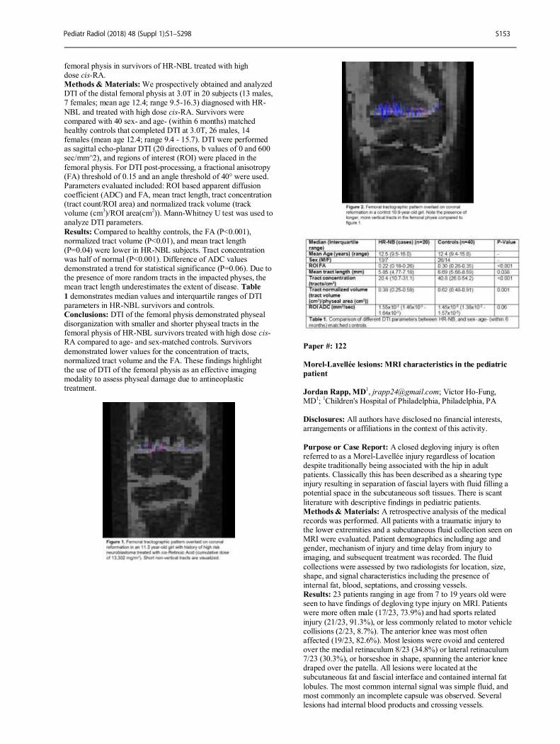
femoral physis in survivors of HR-NBL treated with high dose cis-RA. Methods & Materials: We prospectively obtained and analyzed DTI of the distal femoral physis at 3.0T in 20 subjects (13 males, 7 females; mean age 12.4; range 9.5-16.3) diagnosed with HR-
NBL and treated with high dose cis-RA. Survivors were compared with 40 sex- and age- (within 6 months) matched healthy controls that completed DTI at 3.0T, 26 males, 14 females (mean age 12.4; range 9.4 - 15.7). DTI were performed as sagittal echo-planar DTI (20 directions, b values of 0 and 600 sec/mm^2), and regions of interest (ROI) were placed in the
femoral physis. For DTI post-processing, a fractional anisotropy (FA) threshold of 0.15 and an angle threshold of 40° were used. Parameters evaluated included: ROI based apparent diffusion coefficient (ADC) and FA, mean tract length, tract concentration (tract count/ROI area) and normalized track volume (track volume (cm
3)/ROI area(cm
2)). Mann-Whitney U test was used to
analyze DTI parameters.
Results: Compared to healthy controls, the FA (P<0.001), normalized tract volume (P<0.01), and mean tract length (P=0.04) were lower in HR-NBL subjects. Tract concentration was half of normal (P<0.001). Difference of ADC values demonstrated a trend for statistical significance (P=0.06). Due to the presence of more random tracts in the impacted physes, the mean tract length underestimates the extent of disease. Table
1 demonstrates median values and interquartile ranges of DTI parameters in HR-NBL survivors and controls. Conclusions: DTI of the femoral physis demonstrated physeal disorganization with smaller and shorter physeal tracts in the femoral physis of HR-NBL survivors treated with high dose cis-RA compared to age- and sex-matched controls. Survivors
demonstrated lower values for the concentration of tracts, normalized tract volume and the FA. These findings highlight the use of DTI of the femoral physis as an effective imaging modality to assess physeal damage due to antineoplastic treatment.
Paper #: 122
Morel-Lavellée lesions: MRI characteristics in the pediatric
patient
Jordan Rapp, MD
1, [email protected]; Victor Ho-Fung,
MD1;
1Children's Hospital of Philadelphia, Philadelphia, PA
Disclosures: All authors have disclosed no financial interests, arrangements or affiliations in the context of this activity.
Purpose or Case Report: A closed degloving injury is often referred to as a Morel-Lavellée injury regardless of location despite traditionally being associated with the hip in adult patients. Classically this has been described as a shearing type injury resulting in separation of fascial layers with fluid filling a potential space in the subcutaneous soft tissues. There is scant
literature with descriptive findings in pediatric patients. Methods & Materials: A retrospective analysis of the medical records was performed. All patients with a traumatic injury to the lower extremities and a subcutaneous fluid collection seen on MRI were evaluated. Patient demographics including age and gender, mechanism of injury and time delay from injury to imaging, and subsequent treatment was recorded. The fluid
collections were assessed by two radiologists for location, size, shape, and signal characteristics including the presence of internal fat, blood, septations, and crossing vessels. Results: 23 patients ranging in age from 7 to 19 years old were seen to have findings of degloving type injury on MRI. Patients were more often male (17/23, 73.9%) and had sports related
injury (21/23, 91.3%), or less commonly related to motor vehicle collisions (2/23, 8.7%). The anterior knee was most often affected (19/23, 82.6%). Most lesions were ovoid and centered over the medial retinaculum 8/23 (34.8%) or lateral retinaculum 7/23 (30.3%), or horseshoe in shape, spanning the anterior knee draped over the patella. All lesions were located at the subcutaneous fat and fascial interface and contained internal fat
lobules. The most common internal signal was simple fluid, and most commonly an incomplete capsule was observed. Several lesions had internal blood products and crossing vessels.
Pediatr Radiol (2018) 48 (Suppl 1):S1–S298 S153

Conclusions: Morel-Lavellée injuries most commonly occur in pediatric patients in a location not classically described, the anterior knee, and most commonly occur after a sports related injury. The MRI findings are similar for all patients in that there is an ovoid or horseshoe shaped fluid collection at the fat/fascial
interface containing internal fat droplets. Recognition of this entity with these specific imaging characteristics in a common location after a sports injury can allow for early identification. While most patients recover well with conservative management, patients may need additional intervention with aspiration or even debridement.
Paper #: 123
Children with Unilateral Fibular Hemimelia Have Normal
Lower Extremity Growth Trajectory and Skeletal
Maturation
Andy Tsai, MD, PhD
1, [email protected]; Paul
Kleinman, MD1, Tal Laor, MD
1, James Kasser,
MD1;
1Radiology, Boston Children’s Hospital, Boston, MA
Disclosures: All authors have disclosed no financial interests, arrangements or affiliations in the context of this activity.
Purpose or Case Report: Fibular hemimelia (FH) is the most common congenital lower leg anomaly. It usually results in a limb-length discrepancy and often requires surgical correction. The literature generally ascribes to the concept of constant inhibition, a process by which length ratios between affected and unaffected limbs remain constant throughout growth, but
rigorous scientific data supporting this concept are sparse. Also, some authors have recently suggested that children with FH have earlier skeletal maturation, potentially influencing management. The objective of this study is to elucidate the lower extremity growth patterns and skeletal maturation of children with
unilateral FH. Methods & Materials: Medical records of children with unilateral FH seen at a large pediatric hospital over a 17 year period were reviewed. Inclusion criteria were: at least 2 scanograms, no other confirmed or suspected metabolic/genetic disorders or acquired lower extremity insults. The study cohort’s
affected and unaffected femoral and tibial lengths were collected from scanogram reports, plotted against chronologic age and compared to published growth standards. If hand/wrist radiographs were available during the child’s study interval, the estimated bone age (Greulich and Pyle) was plotted against chronologic age. Results: Twenty-three children were included (15 boys, 8 girls)
with a total of 115 scanograms (average 5, range 2-11, SD=2.7 scanograms). Average age at initial scanogram was 5.6 years (range 1.3-9.2 years). Average age at final scanogram was 10.4 years (range 5.2-18.3 years). At least 1 bone age assessment was performed in 19 children (total of 82 bone ages). All bone growth trajectories (affected and unaffected sides) paralleled normal growth standards for the femur and tibia (Figs. 1 & 2).
Length ratios between affected and unaffected sides remained constant (Fig. 3), adhering to an upward slope pattern of differential limb growth. Individual average percent change in limb length ratio was 0.1-3.3% for the femur and 0.2-3.7% for the tibia. Skeletal maturation was within 2 SD of normal in 72/82 (88%) bone ages.
Conclusions: Lower extremity long bones of children with unilateral FH have a normal growth trajectory in keeping with constant inhibition and most children show normal skeletal maturation. These data contrast with the notion that children with unilateral FH have abnormal growth and earlier skeletal maturation. Recognition of this overall growth pattern can help predict ultimate limb length and guide management.
Pediatr Radiol (2018) 48 (Suppl 1):S1–S298S154
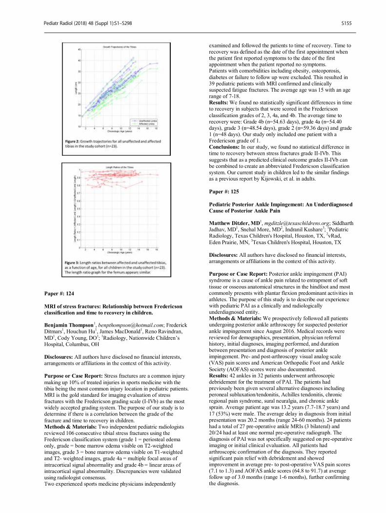
Paper #: 124
MRI of stress fractures: Relationship between Fredericson
classification and time to recovery in children. Benjamin Thompson
1, [email protected]; Frederick
Ditmars1, Houchun Hu
1, James MacDonald
1, Reno Ravindran,
MD1, Cody Young, DO
1;
1Radiology, Nationwide Children’s
Hospital, Columbus, OH Disclosures: All authors have disclosed no financial interests, arrangements or affiliations in the context of this activity. Purpose or Case Report: Stress fractures are a common injury
making up 10% of treated injuries in sports medicine with the tibia being the most common injury location in pediatric patients. MRI is the gold standard for imaging evaluation of stress fractures with the Fredericson grading scale (I-IVb) as the most widely accepted grading system. The purpose of our study is to determine if there is a correlation between the grade of the
fracture and time to recovery in children. Methods & Materials: Two independent pediatric radiologists reviewed 106 consecutive tibial stress fractures using the Fredericson classification system (grade 1 = periosteal edema only, grade = bone marrow edema visible on T2-weighted images, grade 3 = bone marrow edema visible on T1-weighted and T2- weighted images, grade 4a = multiple focal areas of
intracortical signal abnormality and grade 4b = linear areas of intracortical signal abnormality. Discrepancies were validated using radiologist consensus. Two experienced sports medicine physicians independently
examined and followed the patients to time of recovery. Time to recovery was defined as the date of the first appointment when the patient first reported symptoms to the date of the first appointment when the patient reported no symptoms. Patients with comorbidities including obesity, osteoporosis,
diabetes or failure to follow up were excluded. This resulted in 39 pediatric patients with MRI confirmed and clinically suspected fatigue fractures. The average age was 15 with an age range of 7-18. Results: We found no statistically significant differences in time to recovery in subjects that were scored in the Fredericson
classification grades of 2, 3, 4a, and 4b. The average time to recovery were: Grade 4b (n=54.63 days), grade 4a (n=54.40 days), grade 3 (n=48.54 days), grade 2 (n=59.36 days) and grade 1 (n=48 days). Our study only included one patient with a Fredericson grade of 1. Conclusions: In our study, we found no statistical difference in time to recovery between stress fractures grade II-IVb. This
suggests that as a predicted clinical outcome grades II-IVb can be combined to create an abbreviated Fredericson classification system. Our current study in children led to the similar findings as a previous report by Kijowski, et al. in adults.
Paper #: 125
Pediatric Posterior Ankle Impingement: An Underdiagnosed
Cause of Posterior Ankle Pain Matthew Ditzler, MD
1, [email protected]; Siddharth
Jadhav, MD1, Snehal More, MD
2, Indranil Kushare
3;
1Pediatric
Radiology, Texas Children's Hospital, Houston, TX, 2vRad,
Eden Prairie, MN, 3Texas Children's Hospital, Houston, TX
Disclosures: All authors have disclosed no financial interests, arrangements or affiliations in the context of this activity. Purpose or Case Report: Posterior ankle impingement (PAI) syndrome is a cause of ankle pain related to entrapment of soft
tissue or osseous anatomical structures in the hindfoot and most commonly presents with plantar flexion predominant activities in athletes. The purpose of this study is to describe our experience with pediatric PAI as a clinically and radiologically underdiagnosed entity. Methods & Materials: We prospectively followed all patients undergoing posterior ankle arthroscopy for suspected posterior
ankle impingement since August 2016. Medical records were reviewed for demographics, presentation, physician referral history, initial diagnoses, imaging performed, and duration between presentation and diagnosis of posterior ankle impingement. Pre- and post-arthroscopy visual analog scale (VAS) pain scores and American Orthopedic Foot and Ankle
Society (AOFAS) scores were also documented. Results: 42 ankles in 32 patients underwent arthroscopic debridement for the treatment of PAI. The patients had previously been given several alternative diagnoses including peroneal subluxation/tendonitis, Achilles tendonitis, chronic regional pain syndrome, sural neuralgia, and chronic ankle sprain. Average patient age was 13.2 years (7.7-18.7 years) and
17 (53%) were male. The average delay in diagnosis from initial presentation was 20.2 months (range 24-60 months). 24 patients had a total of 27 pre-operative ankle MRIs (3 bilateral) and 20/24 had at least one normal pre-operative radiograph. The diagnosis of PAI was not specifically suggested on pre-operative imaging or initial clinical evaluation. All patients had
arthroscopic confirmation of the diagnosis. They reported significant pain relief with debridement and showed improvement in average pre- to post-operative VAS pain scores (7.1 to 1.3) and AOFAS ankle scores (64.8 to 91.7) at average follow up of 3.0 months (range 1-6 months), further confirming the diagnosis.
Pediatr Radiol (2018) 48 (Suppl 1):S1–S298 S155
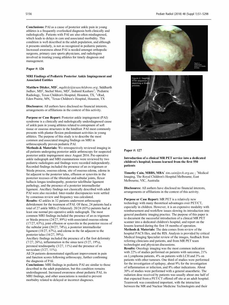
Conclusions: PAI as a cause of posterior ankle pain in young athletes is a frequently overlooked diagnosis both clinically and radiologically. Patients with PAI are also often misdiagnosed, which leads to delays in care and associated morbidity. This condition is well described in the adult population, and although
it presents similarly, is not as recognized in pediatric patients. Increased awareness about PAI is needed amongst orthopedic surgeons, primary care sports physicians, and radiologists involved in treating young athletes for timely diagnosis and management.
Paper #: 126
MRI Findings of Pediatric Posterior Ankle Impingement and
Associated Entities Matthew Ditzler, MD
1, [email protected]; Siddharth
Jadhav, MD1, Snehal More, MD
2, Indranil Kushare
3;
1Pediatric
Radiology, Texas Children's Hospital, Houston, TX, 2vRad,
Eden Prairie, MN, 3Texas Children's Hospital, Houston, TX
Disclosures: All authors have disclosed no financial interests, arrangements or affiliations in the context of this activity. Purpose or Case Report: Posterior ankle impingement (PAI)
syndrome is a clinically and radiologically underdiagnosed cause of ankle pain in young athletes related to entrapment of soft tissue or osseous structures in the hindfoot. PAI most commonly presents with plantar flexion predominant activities in young athletes. The purpose of this study is to describe the most common and associated imaging findings on MRI in
arthroscopically proven pediatric PAI. Methods & Materials: We retrospectively reviewed imaging in all patients undergoing posterior ankle arthroscopy for suspected posterior ankle impingement since August 2016. Pre-operative ankle radiograph and MRI examinations were reviewed by two pediatric radiologists and findings were recorded independently. Recorded findings included the presence of an os trigonum or
Stieda process, osseous edema, site of osseous edema, edema in fat adjacent to the posterior talus, effusion or synovitis in the posterior recesses of the tibiotalar and subtalar joints, flexor hallucis longus tendinopathy, posterior talofibular ligament pathology, and the presence of a posterior intermalleolar ligament. Ancillary findings not classically described with adult PAI were also recorded. Inter-reader discrepancies were settled
by consensus review and frequency was calculated. Results: 42 ankles in 32 patients underwent arthroscopic debridement for the treatment of PAI. Of these, 24 patients had a total of 27 ankle MRIs (3 bilateral). 20/24 (83%) patients had at least one normal pre-operative ankle radiograph. The most common MRI findings included the presence of an os trigonum
or Stieda process (24/27, 89%) with associated osseous edema (17/27, 63%), joint effusion or synovitis in the posterior recess of the subtalar joint (20/27, 74%), a posterior intermalleolar ligament (18/27, 67%), and edema in the fat adjacent to the posterior talus (16/27, 59%). Ancillary findings included the presence of a flat foot deformity (7/27, 26%), inflammation in the sinus tarsi (5/27, 19%),
peroneal tendinopathy (3/27, 11%) and the presence of os naviculare (3/27, 11%). All 24 patients demonstrated significant improvement in pain and function scores following arthroscopy, further confirming the diagnosis of PAI. Conclusions: MRI findings in pediatric PAI are similar to those
described in the adult population, but this condition remains underdiagnosed. Increased awareness about pediatric PAI, its MRI findings, and other associations is needed to prevent morbidity related to delayed or incorrect diagnoses.
Paper #: 127
Introduction of a clinical MR PET service into a dedicated
children's hospital; lessons learned from the first 550
patients Timothy Cain, MBBS, MBA
1 [email protected]; ;
1Medical
Imaging, The Royal Children's Hospital Melbourne, East
Melbourne, VIC, Australia Disclosures: All authors have disclosed no financial interests, arrangements or affiliations in the context of this activity. Purpose or Case Report: MR PET is a relatively new technology with many theoretical advantages over PET/CT,
especially in children. However, it is an expensive modality with reimbursement and workflow issues slowing its introduction into general paediatric imaging practice. The purpose of this paper is to document the successful introduction of a clinical MR PET scanner into a dedicated children's hospital, and report on the lessons learned during the first 18 months of operation. Methods & Materials: The data comes from review of the
hospital PACS files, and the RIS. Analysis is provided by critical Medical Imaging Specialist review of the images, feedback from referring clinicians and patients, and from MR PET team technologist and physician discussions. Results: Oncology imaging was the most common indication with 22% of studies performed on patients with sarcomas, 21%
on Lymphoma patients, 4% on patients with LCH and 3% on patients with other tumours. One third of studies were performed for the investigation of epilepsy, about 9% for the investigation of inflammation or infection, and 8% other indications. About 30% of studies were performed with a general anaesthetic. The radiation dose received by patients was usually about one half of that expected from a PET/CT offered off site at an adult hospital.
Teamwork was considered important, with the interaction between the MR and Nuclear Medicine Technologists and their
Pediatr Radiol (2018) 48 (Suppl 1):S1–S298S156
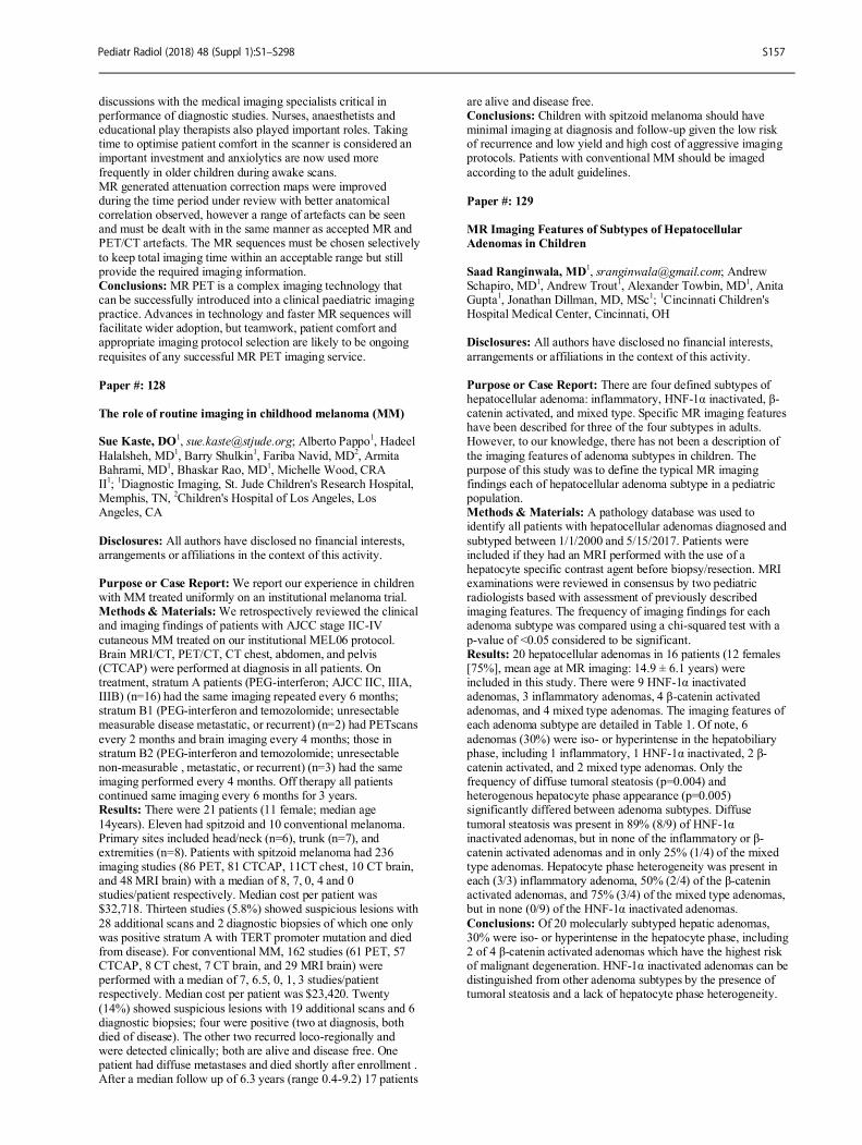
discussions with the medical imaging specialists critical in performance of diagnostic studies. Nurses, anaesthetists and educational play therapists also played important roles. Taking time to optimise patient comfort in the scanner is considered an important investment and anxiolytics are now used more
frequently in older children during awake scans. MR generated attenuation correction maps were improved during the time period under review with better anatomical correlation observed, however a range of artefacts can be seen and must be dealt with in the same manner as accepted MR and PET/CT artefacts. The MR sequences must be chosen selectively
to keep total imaging time within an acceptable range but still provide the required imaging information. Conclusions: MR PET is a complex imaging technology that can be successfully introduced into a clinical paediatric imaging practice. Advances in technology and faster MR sequences will facilitate wider adoption, but teamwork, patient comfort and appropriate imaging protocol selection are likely to be ongoing
requisites of any successful MR PET imaging service.
Paper #: 128
The role of routine imaging in childhood melanoma (MM) Sue Kaste, DO
1, [email protected]; Alberto Pappo
1, Hadeel
Halalsheh, MD1, Barry Shulkin
1, Fariba Navid, MD
2, Armita
Bahrami, MD1, Bhaskar Rao, MD
1, Michelle Wood, CRA
II1;
1Diagnostic Imaging, St. Jude Children's Research Hospital,
Memphis, TN, 2Children's Hospital of Los Angeles, Los
Angeles, CA
Disclosures: All authors have disclosed no financial interests, arrangements or affiliations in the context of this activity. Purpose or Case Report: We report our experience in children with MM treated uniformly on an institutional melanoma trial. Methods & Materials: We retrospectively reviewed the clinical and imaging findings of patients with AJCC stage IIC-IV
cutaneous MM treated on our institutional MEL06 protocol. Brain MRI/CT, PET/CT, CT chest, abdomen, and pelvis (CTCAP) were performed at diagnosis in all patients. On treatment, stratum A patients (PEG-interferon; AJCC IIC, IIIA, IIIB) (n=16) had the same imaging repeated every 6 months; stratum B1 (PEG-interferon and temozolomide; unresectable measurable disease metastatic, or recurrent) (n=2) had PETscans
every 2 months and brain imaging every 4 months; those in stratum B2 (PEG-interferon and temozolomide; unresectable non-measurable , metastatic, or recurrent) (n=3) had the same imaging performed every 4 months. Off therapy all patients continued same imaging every 6 months for 3 years. Results: There were 21 patients (11 female; median age
14years). Eleven had spitzoid and 10 conventional melanoma. Primary sites included head/neck (n=6), trunk (n=7), and extremities (n=8). Patients with spitzoid melanoma had 236 imaging studies (86 PET, 81 CTCAP, 11CT chest, 10 CT brain, and 48 MRI brain) with a median of 8, 7, 0, 4 and 0 studies/patient respectively. Median cost per patient was $32,718. Thirteen studies (5.8%) showed suspicious lesions with
28 additional scans and 2 diagnostic biopsies of which one only was positive stratum A with TERT promoter mutation and died from disease). For conventional MM, 162 studies (61 PET, 57 CTCAP, 8 CT chest, 7 CT brain, and 29 MRI brain) were performed with a median of 7, 6.5, 0, 1, 3 studies/patient respectively. Median cost per patient was $23,420. Twenty
(14%) showed suspicious lesions with 19 additional scans and 6 diagnostic biopsies; four were positive (two at diagnosis, both died of disease). The other two recurred loco-regionally and were detected clinically; both are alive and disease free. One patient had diffuse metastases and died shortly after enrollment . After a median follow up of 6.3 years (range 0.4-9.2) 17 patients
are alive and disease free. Conclusions: Children with spitzoid melanoma should have minimal imaging at diagnosis and follow-up given the low risk of recurrence and low yield and high cost of aggressive imaging protocols. Patients with conventional MM should be imaged
according to the adult guidelines.
Paper #: 129
MR Imaging Features of Subtypes of Hepatocellular
Adenomas in Children
Saad Ranginwala, MD
1, [email protected]; Andrew
Schapiro, MD1, Andrew Trout
1, Alexander Towbin, MD
1, Anita
Gupta1, Jonathan Dillman, MD, MSc
1;
1Cincinnati Children's
Hospital Medical Center, Cincinnati, OH Disclosures: All authors have disclosed no financial interests,
arrangements or affiliations in the context of this activity. Purpose or Case Report: There are four defined subtypes of hepatocellular adenoma: inflammatory, HNF-1α inactivated, β-catenin activated, and mixed type. Specific MR imaging features have been described for three of the four subtypes in adults. However, to our knowledge, there has not been a description of
the imaging features of adenoma subtypes in children. The purpose of this study was to define the typical MR imaging findings each of hepatocellular adenoma subtype in a pediatric population. Methods & Materials: A pathology database was used to identify all patients with hepatocellular adenomas diagnosed and
subtyped between 1/1/2000 and 5/15/2017. Patients were included if they had an MRI performed with the use of a hepatocyte specific contrast agent before biopsy/resection. MRI examinations were reviewed in consensus by two pediatric radiologists based with assessment of previously described imaging features. The frequency of imaging findings for each adenoma subtype was compared using a chi-squared test with a
p-value of <0.05 considered to be significant. Results: 20 hepatocellular adenomas in 16 patients (12 females [75%], mean age at MR imaging: 14.9 ± 6.1 years) were included in this study. There were 9 HNF-1α inactivated adenomas, 3 inflammatory adenomas, 4 β-catenin activated adenomas, and 4 mixed type adenomas. The imaging features of each adenoma subtype are detailed in Table 1. Of note, 6
adenomas (30%) were iso- or hyperintense in the hepatobiliary phase, including 1 inflammatory, 1 HNF-1α inactivated, 2 β-catenin activated, and 2 mixed type adenomas. Only the frequency of diffuse tumoral steatosis (p=0.004) and heterogenous hepatocyte phase appearance (p=0.005) significantly differed between adenoma subtypes. Diffuse
tumoral steatosis was present in 89% (8/9) of HNF-1α inactivated adenomas, but in none of the inflammatory or β-catenin activated adenomas and in only 25% (1/4) of the mixed type adenomas. Hepatocyte phase heterogeneity was present in each (3/3) inflammatory adenoma, 50% (2/4) of the β-catenin activated adenomas, and 75% (3/4) of the mixed type adenomas, but in none (0/9) of the HNF-1α inactivated adenomas.
Conclusions: Of 20 molecularly subtyped hepatic adenomas, 30% were iso- or hyperintense in the hepatocyte phase, including 2 of 4 β-catenin activated adenomas which have the highest risk of malignant degeneration. HNF-1α inactivated adenomas can be distinguished from other adenoma subtypes by the presence of tumoral steatosis and a lack of hepatocyte phase heterogeneity.
Pediatr Radiol (2018) 48 (Suppl 1):S1–S298 S157
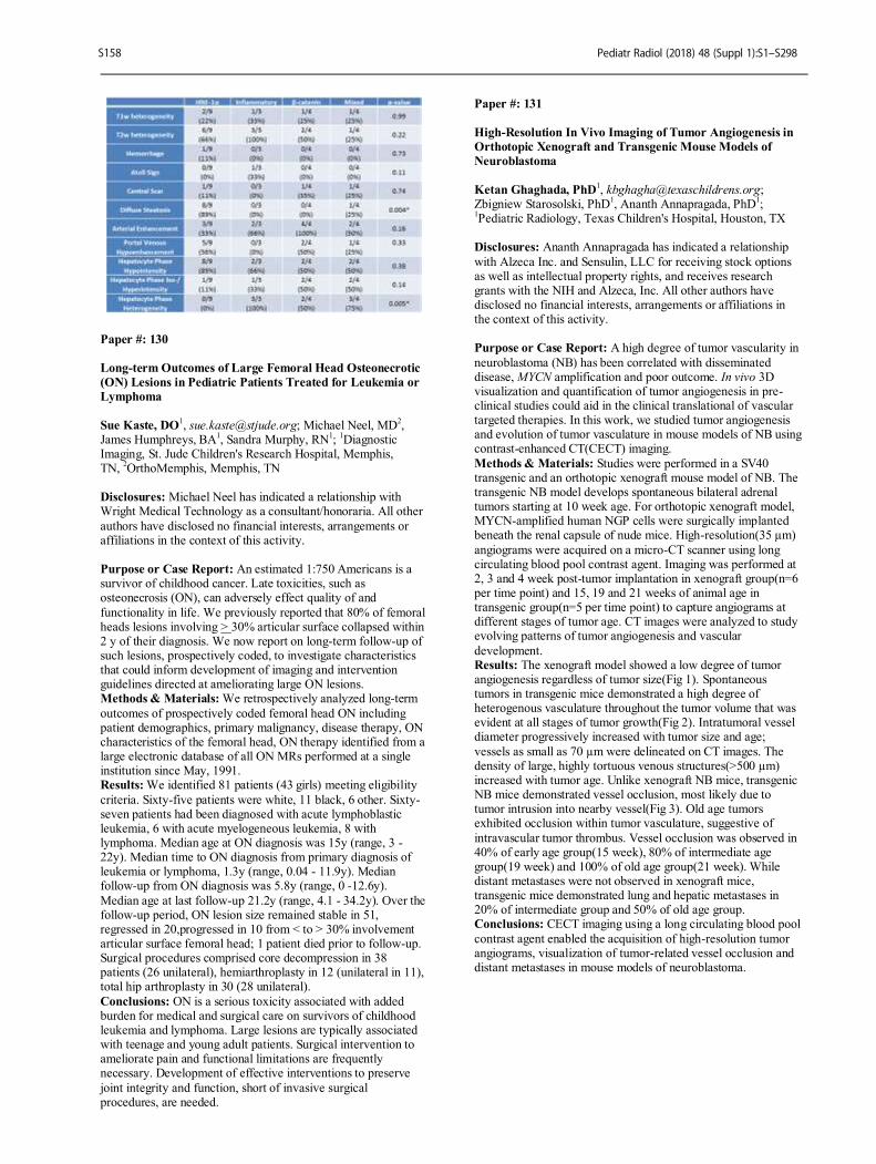
Paper #: 130
Long-term Outcomes of Large Femoral Head Osteonecrotic
(ON) Lesions in Pediatric Patients Treated for Leukemia or
Lymphoma
Sue Kaste, DO1, [email protected]; Michael Neel, MD
2,
James Humphreys, BA1, Sandra Murphy, RN
1;
1Diagnostic
Imaging, St. Jude Children's Research Hospital, Memphis, TN,
2OrthoMemphis, Memphis, TN
Disclosures: Michael Neel has indicated a relationship with Wright Medical Technology as a consultant/honoraria. All other
authors have disclosed no financial interests, arrangements or affiliations in the context of this activity. Purpose or Case Report: An estimated 1:750 Americans is a survivor of childhood cancer. Late toxicities, such as osteonecrosis (ON), can adversely effect quality of and
functionality in life. We previously reported that 80% of femoral heads lesions involving > 30% articular surface collapsed within 2 y of their diagnosis. We now report on long-term follow-up of such lesions, prospectively coded, to investigate characteristics that could inform development of imaging and intervention guidelines directed at ameliorating large ON lesions. Methods & Materials: We retrospectively analyzed long-term
outcomes of prospectively coded femoral head ON including patient demographics, primary malignancy, disease therapy, ON characteristics of the femoral head, ON therapy identified from a large electronic database of all ON MRs performed at a single institution since May, 1991. Results: We identified 81 patients (43 girls) meeting eligibility
criteria. Sixty-five patients were white, 11 black, 6 other. Sixty-seven patients had been diagnosed with acute lymphoblastic leukemia, 6 with acute myelogeneous leukemia, 8 with lymphoma. Median age at ON diagnosis was 15y (range, 3 - 22y). Median time to ON diagnosis from primary diagnosis of leukemia or lymphoma, 1.3y (range, 0.04 - 11.9y). Median follow-up from ON diagnosis was 5.8y (range, 0 -12.6y).
Median age at last follow-up 21.2y (range, 4.1 - 34.2y). Over the follow-up period, ON lesion size remained stable in 51, regressed in 20,progressed in 10 from < to > 30% involvement articular surface femoral head; 1 patient died prior to follow-up. Surgical procedures comprised core decompression in 38 patients (26 unilateral), hemiarthroplasty in 12 (unilateral in 11), total hip arthroplasty in 30 (28 unilateral).
Conclusions: ON is a serious toxicity associated with added burden for medical and surgical care on survivors of childhood leukemia and lymphoma. Large lesions are typically associated with teenage and young adult patients. Surgical intervention to ameliorate pain and functional limitations are frequently necessary. Development of effective interventions to preserve
joint integrity and function, short of invasive surgical procedures, are needed.
Paper #: 131
High-Resolution In Vivo Imaging of Tumor Angiogenesis in
Orthotopic Xenograft and Transgenic Mouse Models of
Neuroblastoma
Ketan Ghaghada, PhD
Zbigniew Starosolski, PhD1, Ananth Annapragada, PhD
1;
1Pediatric Radiology, Texas Children's Hospital, Houston, TX
Disclosures: Ananth Annapragada has indicated a relationship
with Alzeca Inc. and Sensulin, LLC for receiving stock options as well as intellectual property rights, and receives research grants with the NIH and Alzeca, Inc. All other authors have disclosed no financial interests, arrangements or affiliations in the context of this activity. Purpose or Case Report: A high degree of tumor vascularity in
neuroblastoma (NB) has been correlated with disseminated disease, MYCN amplification and poor outcome. In vivo 3D visualization and quantification of tumor angiogenesis in pre-clinical studies could aid in the clinical translational of vascular targeted therapies. In this work, we studied tumor angiogenesis and evolution of tumor vasculature in mouse models of NB using contrast-enhanced CT(CECT) imaging.
Methods & Materials: Studies were performed in a SV40 transgenic and an orthotopic xenograft mouse model of NB. The transgenic NB model develops spontaneous bilateral adrenal tumors starting at 10 week age. For orthotopic xenograft model, MYCN-amplified human NGP cells were surgically implanted beneath the renal capsule of nude mice. High-resolution(35 µm)
angiograms were acquired on a micro-CT scanner using long circulating blood pool contrast agent. Imaging was performed at 2, 3 and 4 week post-tumor implantation in xenograft group(n=6 per time point) and 15, 19 and 21 weeks of animal age in transgenic group(n=5 per time point) to capture angiograms at different stages of tumor age. CT images were analyzed to study evolving patterns of tumor angiogenesis and vascular
development. Results: The xenograft model showed a low degree of tumor angiogenesis regardless of tumor size(Fig 1). Spontaneous tumors in transgenic mice demonstrated a high degree of heterogenous vasculature throughout the tumor volume that was evident at all stages of tumor growth(Fig 2). Intratumoral vessel diameter progressively increased with tumor size and age;
vessels as small as 70 µm were delineated on CT images. The density of large, highly tortuous venous structures(>500 µm) increased with tumor age. Unlike xenograft NB mice, transgenic NB mice demonstrated vessel occlusion, most likely due to tumor intrusion into nearby vessel(Fig 3). Old age tumors exhibited occlusion within tumor vasculature, suggestive of
intravascular tumor thrombus. Vessel occlusion was observed in 40% of early age group(15 week), 80% of intermediate age group(19 week) and 100% of old age group(21 week). While distant metastases were not observed in xenograft mice, transgenic mice demonstrated lung and hepatic metastases in 20% of intermediate group and 50% of old age group. Conclusions: CECT imaging using a long circulating blood pool
contrast agent enabled the acquisition of high-resolution tumor angiograms, visualization of tumor-related vessel occlusion and distant metastases in mouse models of neuroblastoma.
Pediatr Radiol (2018) 48 (Suppl 1):S1–S298S158
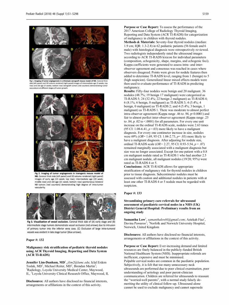
Paper #: 132
Malignancy risk stratification of pediatric thyroid nodules
using ACR Thyroid Imaging, Reporting and Data System
(ACR TI-RADS)
Jennifer Lim-Dunham, MD1, [email protected]; Iclal Erdem
Toslak, MD1, Michael Reiter, MD
1, Brendan Martin
2;
1Radiology, Loyola University Medical Center, Maywood,
IL, 2Loyola University Clinical Research Office, Maywood, IL
Disclosures: All authors have disclosed no financial interests, arrangements or affiliations in the context of this activity.
Purpose or Case Report: To assess the performance of the 2017 American College of Radiology Thyroid Imaging, Reporting and Data System (ACR TI-RADS) for categorization of malignancy in children with thyroid nodules. Methods & Materials: Seventy-four thyroid nodules (median:
1.9 cm; IQR: 1.3-2.8) in 62 pediatric patients (56 female and 6 male) with histological diagnosis were retrospectively reviewed. Two radiologists independently rated the ultrasound images according to ACR TI-RADS lexicon for individual parameters (composition, echogenicity, shape, margins, and echogenic foci). Kappa coefficients were generated to assess intra- and inter-
observer agreement and consensus was reached in cases where observers disagreed. Points were given for nodule features then added to determine TI-RADS level, ranging from 1 (benign) to 5 (high suspicion). Generalized linear mixed effects models were then used to evaluate performance of TI-RADS in predicting malignancy. Results: Fifty-four nodules were benign and 20 malignant. 36
nodules (48.7%; 19 benign 17 malignant) were categorized as TI-RADS 5; 24 (32.4%; 22 benign 2 malignant) as TI-RADS 4; 6 (8.1%; 6 benign, 0 malignant) as TI-RADS 3; 4 (5.4%; 4 benign, 0 malignant) as TI-RADS 2; and 4 (5.4%; 3 benign, 1 malignant) as TI-RADS 1. There was moderate to almost perfect intra-observer agreement (Kappa range .46 to .94, p<0.0001) and fair to almost perfect inter-observer agreement (Kappa range .25
to .84, p .02 to <.0001) for all parameters. For every one unit increase on the ordinal TI-RADS scale, nodules were 2.63 times (95 CI: 1.08-6.41, p =.03) more likely to have a malignant diagnosis. For every one centimeter increase in size, nodules were 69% (OR= 1.69, 95 CI: 1.04-2.73, p= .03) more likely to have a malignant diagnosis. After adjusting for nodule size,
ordinal TI-RADS scale (OR= 2.27, 95 CI: 0.93-5.54, p = .07) remained marginally associated with a malignant diagnosis but size was no longer associated. Except for one patient with a 0.8 cm malignant nodule rated as TI-RADS 1 who had another 2.5 cm malignant nodule, all malignant nodules (19/20; 95%) were rated as TI-RADS 4 or 5. Conclusions: ACR TI-RADS allows for appropriate
stratification of malignancy risk for thyroid nodules in children prior to tissue diagnosis. Subcentimeter nodules must be assessed with caution and additional nodules in patients with at least one other TI-RADS 4 or 5 nodule must be regarded with suspicion.
Paper #: 133
Streamlining primary care referrals for ultrasound
assessment of paediatric cervical nodes in a NHS (UK)
District General Hospital: Preliminary results from an
ongoing study
Samantha Low1, [email protected]; Aziekah Fiaz
1,
Davina Pawaroo1;
1Norfolk and Norwich University Hospital,
Norwich, United Kingdom Disclosures: All authors have disclosed no financial interests, arrangements or affiliations in the context of this activity.
Purpose or Case Report: Ever-increasing demand and limited resources are finely balanced in the publicly-funded British National Healthcare System (NHS). Inappropriate referrals are inefficient, expensive and must be minimised. Palpable cervical nodes are common in the paediatric population. Subjectively, it is felt that too many unnecessary neck
ultrasounds are performed due to poor clinical examination, poor understanding of aetiology and poor parent-clinician communication. Children are referred for ultrasounds to reassure the “worried-well parents”, with a normal study falsely de-meriting the utility of clinical follow-up. Ultrasound alone cannot be used to exclude malignancy and cannot supersede
Pediatr Radiol (2018) 48 (Suppl 1):S1–S298 S159
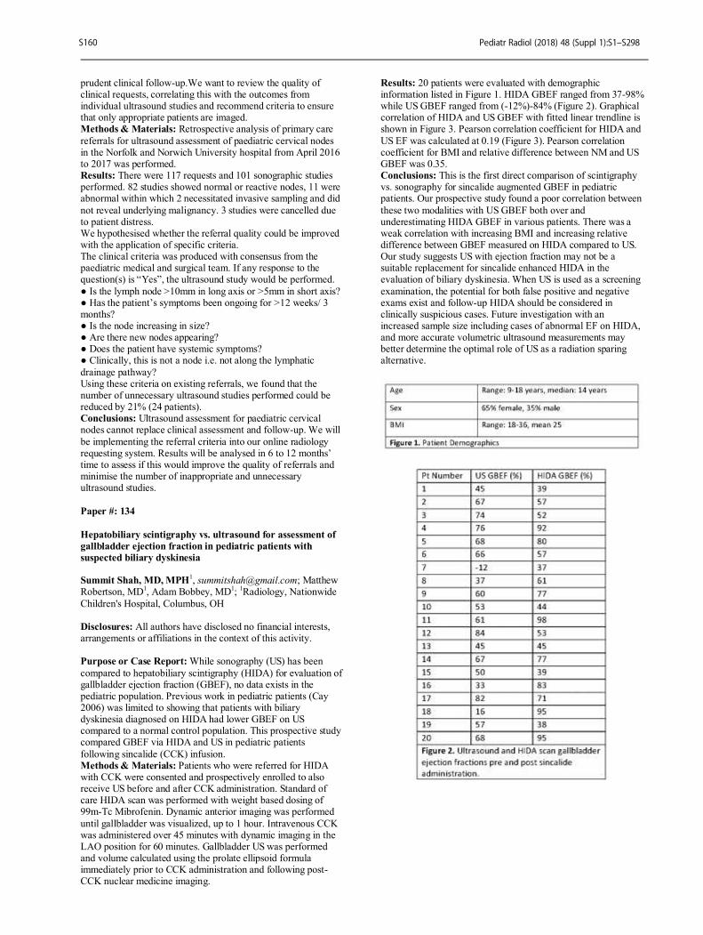
prudent clinical follow-up.We want to review the quality of clinical requests, correlating this with the outcomes from individual ultrasound studies and recommend criteria to ensure that only appropriate patients are imaged. Methods & Materials: Retrospective analysis of primary care
referrals for ultrasound assessment of paediatric cervical nodes in the Norfolk and Norwich University hospital from April 2016 to 2017 was performed. Results: There were 117 requests and 101 sonographic studies performed. 82 studies showed normal or reactive nodes, 11 were abnormal within which 2 necessitated invasive sampling and did
not reveal underlying malignancy. 3 studies were cancelled due to patient distress. We hypothesised whether the referral quality could be improved with the application of specific criteria. The clinical criteria was produced with consensus from the paediatric medical and surgical team. If any response to the question(s) is “Yes”, the ultrasound study would be performed.
● Is the lymph node >10mm in long axis or >5mm in short axis? ● Has the patient’s symptoms been ongoing for >12 weeks/ 3 months? ● Is the node increasing in size? ● Are there new nodes appearing? ● Does the patient have systemic symptoms? ● Clinically, this is not a node i.e. not along the lymphatic
drainage pathway? Using these criteria on existing referrals, we found that the number of unnecessary ultrasound studies performed could be reduced by 21% (24 patients). Conclusions: Ultrasound assessment for paediatric cervical nodes cannot replace clinical assessment and follow-up. We will
be implementing the referral criteria into our online radiology requesting system. Results will be analysed in 6 to 12 months’ time to assess if this would improve the quality of referrals and minimise the number of inappropriate and unnecessary ultrasound studies.
Paper #: 134
Hepatobiliary scintigraphy vs. ultrasound for assessment of
gallbladder ejection fraction in pediatric patients with
suspected biliary dyskinesia Summit Shah, MD, MPH
1, [email protected]; Matthew
Robertson, MD1, Adam Bobbey, MD
1;
1Radiology, Nationwide
Children's Hospital, Columbus, OH Disclosures: All authors have disclosed no financial interests, arrangements or affiliations in the context of this activity. Purpose or Case Report: While sonography (US) has been
compared to hepatobiliary scintigraphy (HIDA) for evaluation of gallbladder ejection fraction (GBEF), no data exists in the pediatric population. Previous work in pediatric patients (Cay 2006) was limited to showing that patients with biliary dyskinesia diagnosed on HIDA had lower GBEF on US compared to a normal control population. This prospective study compared GBEF via HIDA and US in pediatric patients
following sincalide (CCK) infusion. Methods & Materials: Patients who were referred for HIDA with CCK were consented and prospectively enrolled to also receive US before and after CCK administration. Standard of care HIDA scan was performed with weight based dosing of 99m-Tc Mibrofenin. Dynamic anterior imaging was performed
until gallbladder was visualized, up to 1 hour. Intravenous CCK was administered over 45 minutes with dynamic imaging in the LAO position for 60 minutes. Gallbladder US was performed and volume calculated using the prolate ellipsoid formula immediately prior to CCK administration and following post-CCK nuclear medicine imaging.
Results: 20 patients were evaluated with demographic information listed in Figure 1. HIDA GBEF ranged from 37-98% while US GBEF ranged from (-12%)-84% (Figure 2). Graphical correlation of HIDA and US GBEF with fitted linear trendline is shown in Figure 3. Pearson correlation coefficient for HIDA and
US EF was calculated at 0.19 (Figure 3). Pearson correlation coefficient for BMI and relative difference between NM and US GBEF was 0.35. Conclusions: This is the first direct comparison of scintigraphy vs. sonography for sincalide augmented GBEF in pediatric patients. Our prospective study found a poor correlation between
these two modalities with US GBEF both over and underestimating HIDA GBEF in various patients. There was a weak correlation with increasing BMI and increasing relative difference between GBEF measured on HIDA compared to US. Our study suggests US with ejection fraction may not be a suitable replacement for sincalide enhanced HIDA in the evaluation of biliary dyskinesia. When US is used as a screening
examination, the potential for both false positive and negative exams exist and follow-up HIDA should be considered in clinically suspicious cases. Future investigation with an increased sample size including cases of abnormal EF on HIDA, and more accurate volumetric ultrasound measurements may better determine the optimal role of US as a radiation sparing alternative.
Pediatr Radiol (2018) 48 (Suppl 1):S1–S298S160
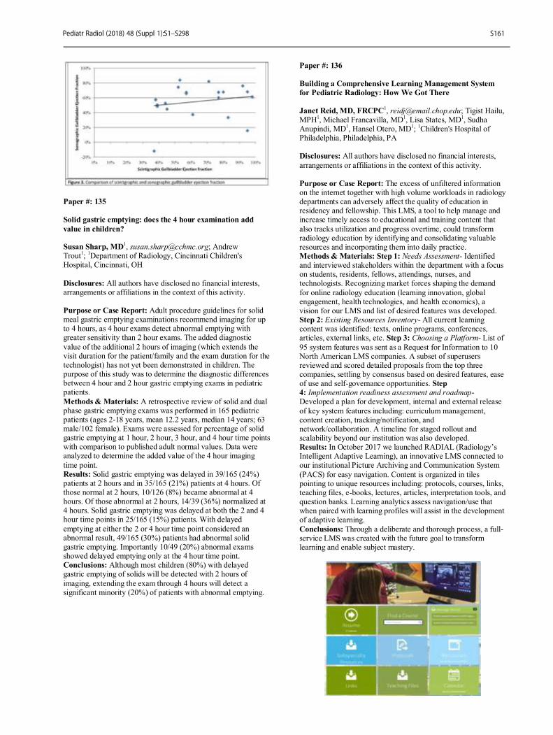
Paper #: 135
Solid gastric emptying: does the 4 hour examination add
value in children?
Susan Sharp, MD
1, [email protected]; Andrew
Trout1;
1Department of Radiology, Cincinnati Children's
Hospital, Cincinnati, OH
Disclosures: All authors have disclosed no financial interests, arrangements or affiliations in the context of this activity. Purpose or Case Report: Adult procedure guidelines for solid meal gastric emptying examinations recommend imaging for up to 4 hours, as 4 hour exams detect abnormal emptying with greater sensitivity than 2 hour exams. The added diagnostic
value of the additional 2 hours of imaging (which extends the visit duration for the patient/family and the exam duration for the technologist) has not yet been demonstrated in children. The purpose of this study was to determine the diagnostic differences between 4 hour and 2 hour gastric emptying exams in pediatric patients.
Methods & Materials: A retrospective review of solid and dual phase gastric emptying exams was performed in 165 pediatric patients (ages 2-18 years, mean 12.2 years, median 14 years; 63 male/102 female). Exams were assessed for percentage of solid gastric emptying at 1 hour, 2 hour, 3 hour, and 4 hour time points with comparison to published adult normal values. Data were analyzed to determine the added value of the 4 hour imaging
time point. Results: Solid gastric emptying was delayed in 39/165 (24%) patients at 2 hours and in 35/165 (21%) patients at 4 hours. Of those normal at 2 hours, 10/126 (8%) became abnormal at 4 hours. Of those abnormal at 2 hours, 14/39 (36%) normalized at 4 hours. Solid gastric emptying was delayed at both the 2 and 4 hour time points in 25/165 (15%) patients. With delayed
emptying at either the 2 or 4 hour time point considered an abnormal result, 49/165 (30%) patients had abnormal solid gastric emptying. Importantly 10/49 (20%) abnormal exams showed delayed emptying only at the 4 hour time point. Conclusions: Although most children (80%) with delayed gastric emptying of solids will be detected with 2 hours of
imaging, extending the exam through 4 hours will detect a significant minority (20%) of patients with abnormal emptying.
Paper #: 136
Building a Comprehensive Learning Management System
for Pediatric Radiology: How We Got There
Janet Reid, MD, FRCPC1, [email protected]; Tigist Hailu,
MPH1, Michael Francavilla, MD
1, Lisa States, MD
1, Sudha
Anupindi, MD1, Hansel Otero, MD
1;
1Children's Hospital of
Philadelphia, Philadelphia, PA Disclosures: All authors have disclosed no financial interests,
arrangements or affiliations in the context of this activity. Purpose or Case Report: The excess of unfiltered information on the internet together with high volume workloads in radiology departments can adversely affect the quality of education in residency and fellowship. This LMS, a tool to help manage and increase timely access to educational and training content that
also tracks utilization and progress overtime, could transform radiology education by identifying and consolidating valuable resources and incorporating them into daily practice. Methods & Materials: Step 1: Needs Assessment- Identified and interviewed stakeholders within the department with a focus on students, residents, fellows, attendings, nurses, and technologists. Recognizing market forces shaping the demand
for online radiology education (learning innovation, global engagement, health technologies, and health economics), a vision for our LMS and list of desired features was developed. Step 2: Existing Resources Inventory- All current learning content was identified: texts, online programs, conferences, articles, external links, etc. Step 3: Choosing a Platform- List of
95 system features was sent as a Request for Information to 10 North American LMS companies. A subset of superusers reviewed and scored detailed proposals from the top three companies, settling by consensus based on desired features, ease of use and self-governance opportunities. Step
4: Implementation readiness assessment and roadmap- Developed a plan for development, internal and external release
of key system features including: curriculum management, content creation, tracking/notification, and network/collaboration. A timeline for staged rollout and scalability beyond our institution was also developed. Results: In October 2017 we launched RADIAL (Radiology’s Intelligent Adaptive Learning), an innovative LMS connected to our institutional Picture Archiving and Communication System
(PACS) for easy navigation. Content is organized in tiles pointing to unique resources including: protocols, courses, links, teaching files, e-books, lectures, articles, interpretation tools, and question banks. Learning analytics assess navigation/use that when paired with learning profiles will assist in the development of adaptive learning.
Conclusions: Through a deliberate and thorough process, a full-service LMS was created with the future goal to transform learning and enable subject mastery.
Pediatr Radiol (2018) 48 (Suppl 1):S1–S298 S161
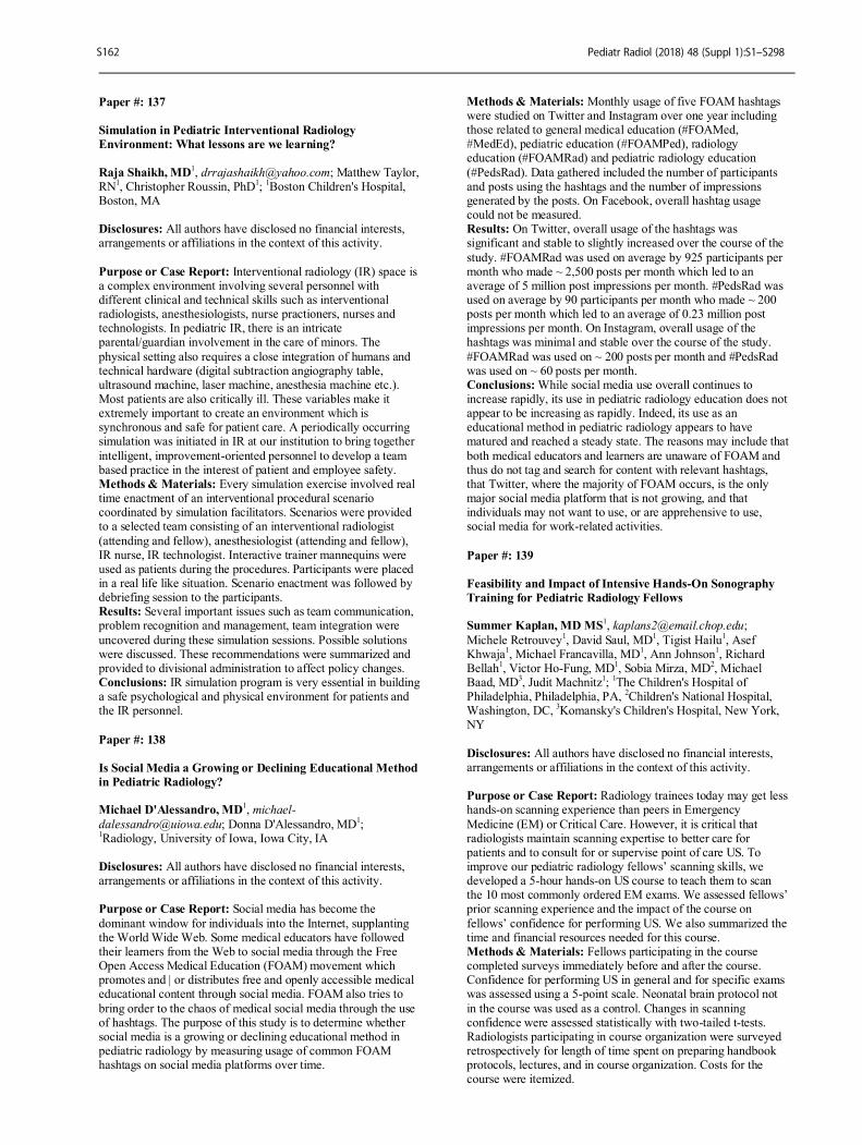
Paper #: 137
Simulation in Pediatric Interventional Radiology
Environment: What lessons are we learning?
Raja Shaikh, MD1, [email protected]; Matthew Taylor,
RN1, Christopher Roussin, PhD
1;
1Boston Children's Hospital,
Boston, MA Disclosures: All authors have disclosed no financial interests, arrangements or affiliations in the context of this activity.
Purpose or Case Report: Interventional radiology (IR) space is a complex environment involving several personnel with different clinical and technical skills such as interventional radiologists, anesthesiologists, nurse practioners, nurses and technologists. In pediatric IR, there is an intricate parental/guardian involvement in the care of minors. The
physical setting also requires a close integration of humans and technical hardware (digital subtraction angiography table, ultrasound machine, laser machine, anesthesia machine etc.). Most patients are also critically ill. These variables make it extremely important to create an environment which is synchronous and safe for patient care. A periodically occurring simulation was initiated in IR at our institution to bring together
intelligent, improvement-oriented personnel to develop a team based practice in the interest of patient and employee safety. Methods & Materials: Every simulation exercise involved real time enactment of an interventional procedural scenario coordinated by simulation facilitators. Scenarios were provided to a selected team consisting of an interventional radiologist
(attending and fellow), anesthesiologist (attending and fellow), IR nurse, IR technologist. Interactive trainer mannequins were used as patients during the procedures. Participants were placed in a real life like situation. Scenario enactment was followed by debriefing session to the participants. Results: Several important issues such as team communication, problem recognition and management, team integration were
uncovered during these simulation sessions. Possible solutions were discussed. These recommendations were summarized and provided to divisional administration to affect policy changes. Conclusions: IR simulation program is very essential in building a safe psychological and physical environment for patients and the IR personnel.
Paper #: 138
Is Social Media a Growing or Declining Educational Method
in Pediatric Radiology? Michael D'Alessandro, MD
1, michael-
[email protected]; Donna D'Alessandro, MD1;
1Radiology, University of Iowa, Iowa City, IA
Disclosures: All authors have disclosed no financial interests, arrangements or affiliations in the context of this activity. Purpose or Case Report: Social media has become the
dominant window for individuals into the Internet, supplanting the World Wide Web. Some medical educators have followed their learners from the Web to social media through the Free Open Access Medical Education (FOAM) movement which promotes and | or distributes free and openly accessible medical educational content through social media. FOAM also tries to
bring order to the chaos of medical social media through the use of hashtags. The purpose of this study is to determine whether social media is a growing or declining educational method in pediatric radiology by measuring usage of common FOAM hashtags on social media platforms over time.
Methods & Materials: Monthly usage of five FOAM hashtags were studied on Twitter and Instagram over one year including those related to general medical education (#FOAMed, #MedEd), pediatric education (#FOAMPed), radiology education (#FOAMRad) and pediatric radiology education
(#PedsRad). Data gathered included the number of participants and posts using the hashtags and the number of impressions generated by the posts. On Facebook, overall hashtag usage could not be measured. Results: On Twitter, overall usage of the hashtags was significant and stable to slightly increased over the course of the
study. #FOAMRad was used on average by 925 participants per month who made ~ 2,500 posts per month which led to an average of 5 million post impressions per month. #PedsRad was used on average by 90 participants per month who made ~ 200 posts per month which led to an average of 0.23 million post impressions per month. On Instagram, overall usage of the hashtags was minimal and stable over the course of the study.
#FOAMRad was used on ~ 200 posts per month and #PedsRad was used on ~ 60 posts per month. Conclusions: While social media use overall continues to increase rapidly, its use in pediatric radiology education does not appear to be increasing as rapidly. Indeed, its use as an educational method in pediatric radiology appears to have matured and reached a steady state. The reasons may include that
both medical educators and learners are unaware of FOAM and thus do not tag and search for content with relevant hashtags, that Twitter, where the majority of FOAM occurs, is the only major social media platform that is not growing, and that individuals may not want to use, or are apprehensive to use, social media for work-related activities.
Paper #: 139
Feasibility and Impact of Intensive Hands-On Sonography
Training for Pediatric Radiology Fellows Summer Kaplan, MD MS
Michele Retrouvey1, David Saul, MD
1, Tigist Hailu
1, Asef
Khwaja1, Michael Francavilla, MD
1, Ann Johnson
1, Richard
Bellah1, Victor Ho-Fung, MD
1, Sobia Mirza, MD
2, Michael
Baad, MD3, Judit Machnitz
1;
1The Children's Hospital of
Philadelphia, Philadelphia, PA, 2Children's National Hospital,
Washington, DC, 3Komansky's Children's Hospital, New York,
NY
Disclosures: All authors have disclosed no financial interests, arrangements or affiliations in the context of this activity. Purpose or Case Report: Radiology trainees today may get less hands-on scanning experience than peers in Emergency
Medicine (EM) or Critical Care. However, it is critical that radiologists maintain scanning expertise to better care for patients and to consult for or supervise point of care US. To improve our pediatric radiology fellows’ scanning skills, we developed a 5-hour hands-on US course to teach them to scan the 10 most commonly ordered EM exams. We assessed fellows’ prior scanning experience and the impact of the course on
fellows’ confidence for performing US. We also summarized the time and financial resources needed for this course. Methods & Materials: Fellows participating in the course completed surveys immediately before and after the course. Confidence for performing US in general and for specific exams was assessed using a 5-point scale. Neonatal brain protocol not
in the course was used as a control. Changes in scanning confidence were assessed statistically with two-tailed t-tests. Radiologists participating in course organization were surveyed retrospectively for length of time spent on preparing handbook protocols, lectures, and in course organization. Costs for the course were itemized.
Pediatr Radiol (2018) 48 (Suppl 1):S1–S298S162
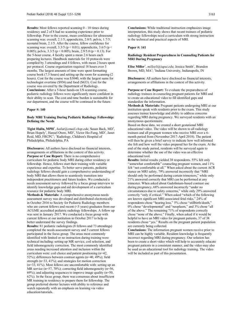
Results: Most fellows reported scanning 0 - 10 times during residency and 2 of 8 had no scanning experience prior to fellowship. Prior to the course, mean confidence for ultrasound scanning was: overall, 2.1/5; appendicitis, 2.0/5; pelvis, 1.7/5; neonatal brain, 2.1/5. After the course, fellow confidence for
scanning was: overall, 3.3/5 (p < 0.01); appendicitis, 3.6/5 (p < 0.005); pelvis, 3.3/5 (p < 0.005); brain, 2.9/5.0 (p = 0.13). For the 5-hour course, 4 faculty spent a mean 2.6 hours each preparing lectures. Handbook materials for 10 protocols were compiled by 3 attendings and 4 fellows, with mean 2 hours spent per protocol. Course organization required 18 hours over 3
months. The largest amounts of time were spent formatting course book (7.5 hours) and setting up the room for scanning (2 hours). Cost for the course was $3040, with the largest sums for technologist overtime ($936) and food ($633). Cost for the course was covered by the Department of Radiology. Conclusions: After a 5-hour hands-on US scanning course, pediatric radiology fellows were significantly more confident in
their ability to scan. The cost and time burden is sustainable for our department, and the course will be continued in the future.
Paper #: 140
Body MRI Training During Pediatric Radiology Fellowship:
Defining the Needs
Tigist Hailu, MPH
1, [email protected]; Susan Back, MD
1,
Brian Hopely1, Hansel Otero, MD
1, Victor Ho-Fung, MD
1, Janet
Reid, MD, FRCPC1;
1Radiology, Children's Hospital of
Philadelphia, Philadelphia, PA
Disclosures: All authors have disclosed no financial interests, arrangements or affiliations in the context of this activity. Purpose or Case Report: There is no comprehensive curriculum for pediatric body MRI during either residency or fellowship. Hence, fellows start their training with variable experience and expertise. To better serve patients, pediatric radiology fellows should gain a comprehensive understanding of
body MRI that allows them to seamlessly transition into independent practitioners and future leaders. We conducted a needs assessment survey followed by a focus group interview to identify knowledge gaps and aid development of a curriculum resource for pediatric body MRI. Methods & Materials: A comprehensive anonymous needs assessment survey was developed and distributed electronically
in October 2016 to Society for Pediatric Radiology members who are current fellows and recent (<5 years) graduates from our ACGME accredited pediatric radiology fellowships. A follow up was sent in January 2017. We conducted a focus group with current fellows at our institution in October 2017 to help us better understand the survey findings.
Results: 81 pediatric radiologists (8 fellows and 73 attendings) completed the needs assessment survey and 5 current fellows participated in the focus group. The areas most commonly identified with limited or no instruction during training were technical including: setting up MR service, coil selection, and field inhomogeneity correction. The most commonly identified areas needing increased attention and inclusion within the
curriculum were: coil choice and patient positioning (n=42, 52%); differences between contrast agents (n=40, 49%); field strength (n=33, 41%); and strategies for motion correction (n=33, 41%). Most fellows are uncomfortable with: setting up an MR service (n=57, 70%); correcting field inhomogeneity (n=56, 69%); and adjusting sequences to improve image quality (n=50,
62%). In the focus group, there was consensus about insufficient MR training in residency to prepare them for fellowship. The group preferred shorter lectures with ability to reference and watch repeatedly with an emphasis on learning via video education/tutorials.
Conclusions: While traditional instruction emphasizes image interpretation, this study shows that recent trainees of pediatric radiology fellowships need a curriculum with strong instruction in the technical and practical aspects of MRI.
Paper #: 141
Radiology Resident Preparedness in Counseling Patients for
MRI During Pregnancy Elise Miller
1, [email protected]; Jessica Smith
1, Brandon
Brown, MD, MA1;
1Indiana University, Indianapolis, IN
Disclosures: All authors have disclosed no financial interests, arrangements or affiliations in the context of this activity. Purpose or Case Report: To evaluate the preparedness of radiology trainees in counseling pregnant patients for MRI and
to create an educational video to fill identified gaps and standardize the information. Methods & Materials: Pregnant patients undergoing MRI at our institution speak with residents prior to the exam. This study assesses trainee knowledge and ability to address concerns regarding MRI during pregnancy. We surveyed residents with an anonymous questionnaire.
Based on these data, we created a short gestational MRI educational video. The video will be shown to all radiology trainees and all pregnant women who receive MRI over a 6-month period from (November 2017-April 2018). The patient will then be given a brief survey to indicate how well informed she felt and how well the video prepared her for the exam. At the
end of the study period, residents will be surveyed again to determine whether the use of the video was an effective educational tool. Results: Initial results yielded 38 respondents. 55% felt only “somewhat comfortable” counseling pregnant women, and 11% felt “not comfortable at all.” When asked about the current ACR stance on MRI safety, 79% answered incorrectly that “MRI
should only be performed during certain trimesters,” while only 21% answered correctly that MRI can be performed at any trimester. When asked about Gadolinium-based contrast use during pregnancy, 68% answered incorrectly “under no circumstances due to safety concerns,” while only 29% answered correctly “only if critical.” When asked “which of the following are known significant MRI-associated fetal risks,” 24% of
respondents chose “hearing loss,” 5% chose “stillbirth/death,” 0% chose “developmental” and “neoplasm,” and 3% chose “all of the above.” The remaining 71% of respondents correctly chose “none of the above.” Finally, when asked if it would be helpful to have an MRI video for pregnant patients, 37 of 38 residents chose “yes.” Results on the pregnant patient population
are currently being collected. Conclusions: The information pregnant women receive prior to MRI can be highly variable. Resident knowledge is frequently incorrect regarding MRI during pregnancy. Our solution has been to create a short video which will help to accurately educate pregnant patients in a consistent manner, and the video may also be used as an educational tool for radiology training. The video
will be included as part of this presentation.
Pediatr Radiol (2018) 48 (Suppl 1):S1–S298 S163
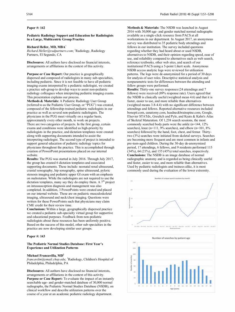
Paper #: 142
Pediatric Radiology Support and Education for Radiologists
in a Large, Multicentric Group Practice
Richard Heller, MD, MBA1
[email protected]; 1Radiology, Radiology
Partners, El Segundo, CA Disclosures: All authors have disclosed no financial interests, arrangements or affiliations in the context of this activity.
Purpose or Case Report: Our practice is geographically dispersed and composed of radiologists in many sub-specialties, including pediatric. Since it is not feasible to have all pediatric imaging exams interpreted by a pediatric radiologist, we created a practice sub-group to develop ways to assist non-pediatric radiology colleagues when interpreting pediatric imaging exams.
This presentation explains our process. Methods & Materials: A Pediatric Radiology User Group (referred to as the Pediatric User Group, or “PUG”) was created, composed of the fellowship-trained pediatric radiologists in our practice as well as several non-pediatric radiologists. The physicians in the PUG meet virtually on a regular basis, approximately every other month, to work on projects.
There are two categories of projects. In the first group, certain exams and conditions were identified as high-priority by radiologists in the practice, and dictation templates were created along with supporting documents intended to assist the interpreting radiologist. The second type of project is intended to support general education of pediatric radiology topics for
physicians throughout the practice. This is accomplished through creation of PowerPoint presentations placed on our internal website. Results: The PUG was started in July 2016. Through July 2017, the group has created 6 dictation templates and associated supporting documents. These include: neonatal renal ultrasound, cranial sonography, hip sonography, spine ultrasound, pyloric
stenosis imaging and pediatric upper GI exam with an emphasis on malrotation. While the radiologists are not required to use the dictation templates, many say they do employ them. A 7
th project
on intussusception diagnosis and management was also completed. In addition, 3 PowerPoints were created and placed on our internal website. These are on pediatric musculoskeletal imaging, ultrasound and neck/chest imaging. Questions were
written for these PowerPoints such that physicians may claim CME credit for their review time. Conclusions: Within a large, geographically dispersed practice we created a pediatric sub-specialty virtual group for supportive and educational purposes. Feedback from non-pediatric radiologists about these resources has been uniformly positive.
Based on the success of this model, other sub-specialties in the practice are now developing similar user groups.
Paper #: 143
The Pediatric Normal Studies Database: First Year’s
Experience and Utilization Patterns
Michael Francavilla, MD1
[email protected]; 1Radiology, Children's Hospital of
Philadelphia, Philadelphia, PA Disclosures: All authors have disclosed no financial interests,
arrangements or affiliations in the context of this activity. Purpose or Case Report: To evaluate the impact of an instantly searchable age- and gender-matched database of 30,000 normal radiographs, the Pediatric Normal Studies Database (NSDB), on clinical workflow and describe utilization patterns over the course of a year at an academic pediatric radiology department.
Methods & Materials: The NSDB was launched in August 2016 with 30,000 age- and gender matched normal radiographs available as a single-click resource from PACS at all workstations in our department. In August 2017, an anonymous survey was distributed to 35 pediatric body attendings and
fellows in our institution. The survey included questions regarding whether they had heard about or used NSDB, alternatives to NSDB, and their opinion regarding speed, ease of use, and reliability compared to alternatives such as web search, reference textbooks, other web sites, and search of our institutional PACS using a 5-point Likert scale. Anonymous
NSDB access analytic logs were reviewed for utilization patterns. The logs were de-anonymized for a period of 30 days for analysis of user roles. Descriptive statistical analysis and nonparametric tests for differences between the attending and fellow groups were performed. Results: Thirty-one survey responses (24 attendings and 7 fellows) were received (89% response rate). Users agreed that
the NSDB is clinically useful (weighted mean 4.6) and that it is faster, easier to use, and more reliable than alternatives (weighted means 3.8-4.4) with no significant difference between attendings and fellows. Reported alternative resources included bonepit.com, eanatomy.com, headneckbrainspine.com, Google, Elsevier STATdx, Greulich and Pyle, and Keats & Kahn's Atlas of Skeletal Maturation. Of 1,238 search sessions, the most
commonly searched body parts were the ankle (n=144, 12% searches), knee (n=111, 9% searches), and elbow (n=101, 8% searches) followed by the hand, foot, chest, and femur. Thirty-two (3%) searches were initiated from skeletal surveys. Searches are becoming more frequent and are most common in infants and pre-teen-aged children. During the 30-day de-anonymized
period, 17 attendings, 6 fellows, and 9 residents performed 115 (34%), 64 (21%), and 155 (45%) total searches, respectively. Conclusions: The NSDB is an image database of normal radiographic anatomy and is regarded as being clinically useful and faster, easier to use, and more reliable than alternatives. Used by pediatric radiologists and trainees alike, it is most commonly used during the evaluation of the lower extremity.
Pediatr Radiol (2018) 48 (Suppl 1):S1–S298S164
Paper #: 144
Reducing the sedation rates in MRI for pediatric patients
utilizing an MRI simulator preparation session facilitated by
a child life specialist.
Tricia Buskirk, Master of Science in Child and Family Studies
1, [email protected]; Whitney
Garrett, Bachelor of Science in Child and Family Studies1,
Arleen Karczewski, Bachelor of Science in Child Life Education
1;1Nationwide Children’s Hospital, Columbus, OH
Disclosures: All authors have disclosed no financial interests, arrangements or affiliations in the context of this activity.
Purpose or Case Report: An MRI simulator was used to reduce the number of patients requiring sedation by allowing them the opportunity to have a realistic experience of an actual MRI prior to their MRI scan. A child life specialist engaged each patient in a simulator session to prepare them and allow them to become familiar with the MRI equipment and process. Methods & Materials: Patients were identified by reviewing the
MRI sedation schedule and the patient’s chart. Simulator candidates were selected by assessing the type of scan, proximity to the hospital, age, and developmental or behavioral barriers. Each patient’s parents were then consulted to make a final assessment of their child’s potential for success in completing a non-sedated MRI. Parents showing interest were given the opportunity to have their child experience a simulator session.
Prior to acquiring the MRI simulator, sedation was standard for patients under the age of 7 and whose chart showed past developmental or behavioral factors. Implementing the MRI simulator program decreased the number of patients requiring sedation. Practice time and education empowered patients to succeed without sedation due to being confident and aware of
what an MRI entails. Results: Since August of 2014, 416 patients have utilized the MRI simulator. Patients who utilized the MRI simulator were between the ages of 5-18. All of the patients chosen had scans that were scheduled to be one hour or less in length. Of the 416 patients that utilized the simulator, 354 of them were able to complete their scans without anesthesia which resulted in an
85% success rate. The 15% that were not able to complete their scans without sedation were unable to hold still or became too anxious during the actual scan. The success rate shows the benefit of allowing patients to practice and become familiar with the MRI before the actual scan. The MRI simulator reduced the number of patients requiring sedation and therefore increased patient safety, created a positive patient experience and reduced
the cost for many families. Based on our current capacity for performing patient training on our MRI simulator and the observed rate of sedation required for these patients, we estimate an annual cost savings of approximately $200,000 for families. Conclusions: Patients have a high success rate in completing their MRIs without sedation when it is paired with an MRI
simulator session facilitated by a child life specialist.
Paper #: 145
Intraabdominal soft tissue injuries on computed tomography
(CT) in accidental versus non accidental trauma
Disclosures: All authors have disclosed no financial interests, arrangements or affiliations in the context of this activity.
Christy Pomeranz, MD
Christian Barrera, MD1, Xiaowei Zhu
1, Sabah Servaes
1;
1Radiology, Children's Hospital of Philadelphia, Philadelphia,
PA Disclosures: All authors have disclosed no financial interests, arrangements or affiliations in the context of this activity. Purpose or Case Report: To evaluate intraabdominal soft tissue injuries on computed tomography (CT) in accidental versus non
accidental trauma (NAT). Methods & Materials: Following IRB approval, a retrospective review was performed on all patients who had experienced trauma and who had undergone both a skeletal survey and a CT of the chest and/or abdomen and pelvis within 30 days of one another from January 2009 - July 2017. All CTs were performed at the Children’s Hospital of Philadelphia with the exception of
one study. Images and reports were reviewed by a pediatric radiology fellow. Results: Data was collected on 57 patients who met inclusion criteria. Patients ranged in age from 0 to 8 years old. 35 cases were cases of NAT confirmed by the Suspected Child Abuse and Neglect Team; 8 cases were confirmed accidental trauma; and 14
cases were unknown mechanism. 53 CTs were performed with intravenous contrast and 4 CTs were performed without intravenous contrast. 30 patients had soft tissue injuries on CT while 27 patients had no evidence of injury on CT. Of the 35 confirmed cases of NAT, 21 patients had soft tissue injuries on CT (60%) and 14 did not. Of the 22 cases of accidental trauma or unknown mechanism, 9
patients had soft tissue injuries on CT (41%) and 13 did not. This difference was statistically significant (p = 0.05) with cases of NAT being more likely to have soft tissue injuries than accidental or unknown mechanisms. Soft tissue injuries included 17 cases of liver laceration, 5 cases of splenic laceration, 6 cases of bowel injury, 3 cases of kidney injury, 3 cases of adrenal hematoma, 2 cases of pancreatic laceration, 1 case of
retroperitoneal hematoma, and 1 case of abdominal wall hematoma. There was no significant difference between NAT and accidental trauma for any of these injury types. Soft tissue injuries were statistically associated with rib fractures (p = 0.02) but not with other fractures, such as long bone fractures. Conclusions: Although skeletal surveys are the mainstay of
imaging in NAT, intraabdominal soft tissue injuries can also occur in NAT. These injuries occur at statistically significant higher rate than that of accidental trauma and may be missed on radiographs. The most common intraabdominal soft tissue injury to occur in both NAT and accidental trauma is liver laceration but a variety of other injuries can also be seen. Furthermore, the type of soft tissue injuries appear to be similar for NAT and
accidental trauma without a difference injury profiles.
Pediatr Radiol (2018) 48 (Suppl 1):S1–S298 S165
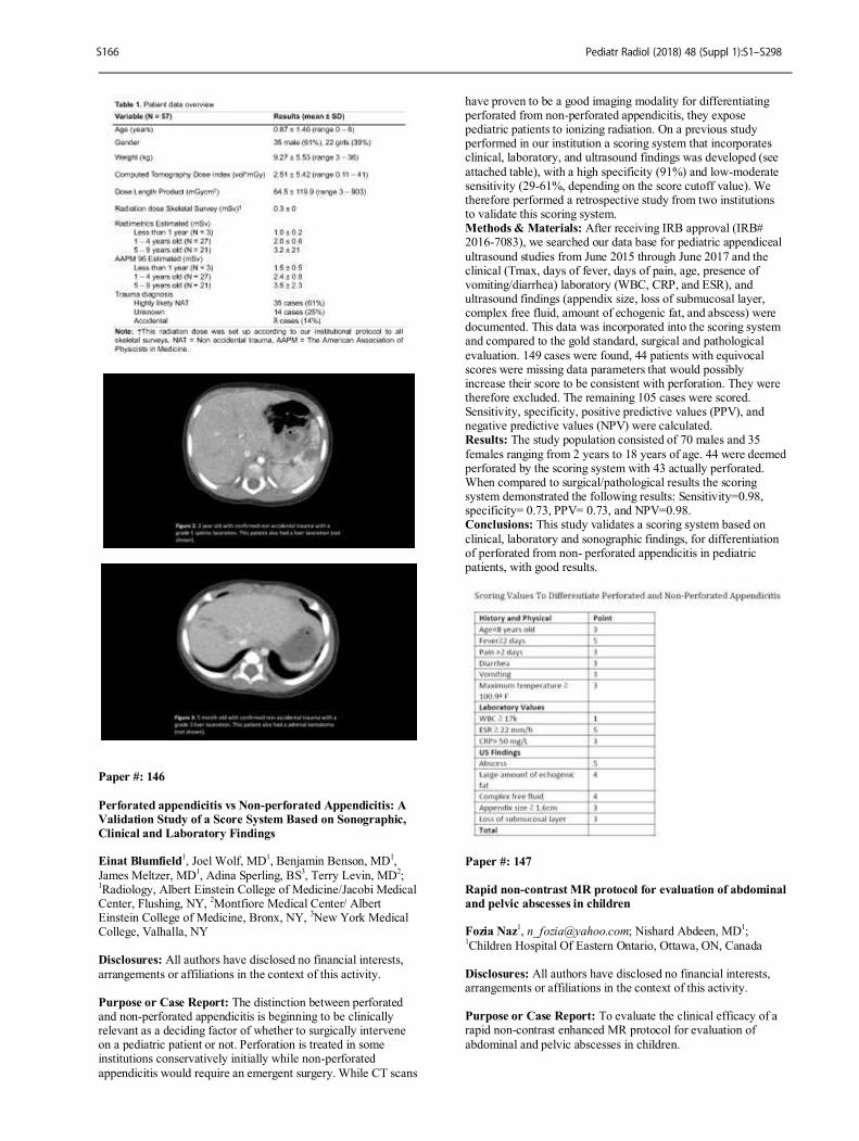
Paper #: 146
Perforated appendicitis vs Non-perforated Appendicitis: A
Validation Study of a Score System Based on Sonographic,
Clinical and Laboratory Findings Einat Blumfield
1, Joel Wolf, MD
1, Benjamin Benson, MD
1,
James Meltzer, MD1, Adina Sperling, BS
3, Terry Levin, MD
2;
1Radiology, Albert Einstein College of Medicine/Jacobi Medical
Center, Flushing, NY, 2Montfiore Medical Center/ Albert
Einstein College of Medicine, Bronx, NY, 3New York Medical
College, Valhalla, NY Disclosures: All authors have disclosed no financial interests,
arrangements or affiliations in the context of this activity. Purpose or Case Report: The distinction between perforated and non-perforated appendicitis is beginning to be clinically relevant as a deciding factor of whether to surgically intervene on a pediatric patient or not. Perforation is treated in some institutions conservatively initially while non-perforated
appendicitis would require an emergent surgery. While CT scans
have proven to be a good imaging modality for differentiating perforated from non-perforated appendicitis, they expose pediatric patients to ionizing radiation. On a previous study performed in our institution a scoring system that incorporates clinical, laboratory, and ultrasound findings was developed (see
attached table), with a high specificity (91%) and low-moderate sensitivity (29-61%, depending on the score cutoff value). We therefore performed a retrospective study from two institutions to validate this scoring system. Methods & Materials: After receiving IRB approval (IRB# 2016-7083), we searched our data base for pediatric appendiceal
ultrasound studies from June 2015 through June 2017 and the clinical (Tmax, days of fever, days of pain, age, presence of vomiting/diarrhea) laboratory (WBC, CRP, and ESR), and ultrasound findings (appendix size, loss of submucosal layer, complex free fluid, amount of echogenic fat, and abscess) were documented. This data was incorporated into the scoring system and compared to the gold standard, surgical and pathological
evaluation. 149 cases were found, 44 patients with equivocal scores were missing data parameters that would possibly increase their score to be consistent with perforation. They were therefore excluded. The remaining 105 cases were scored. Sensitivity, specificity, positive predictive values (PPV), and negative predictive values (NPV) were calculated. Results: The study population consisted of 70 males and 35
females ranging from 2 years to 18 years of age. 44 were deemed perforated by the scoring system with 43 actually perforated. When compared to surgical/pathological results the scoring system demonstrated the following results: Sensitivity=0.98, specificity= 0.73, PPV= 0.73, and NPV=0.98. Conclusions: This study validates a scoring system based on
clinical, laboratory and sonographic findings, for differentiation of perforated from non- perforated appendicitis in pediatric patients, with good results.
Paper #: 147
Rapid non-contrast MR protocol for evaluation of abdominal
and pelvic abscesses in children Fozia Naz
1, [email protected]; Nishard Abdeen, MD
1;
1Children Hospital Of Eastern Ontario, Ottawa, ON, Canada
Disclosures: All authors have disclosed no financial interests, arrangements or affiliations in the context of this activity. Purpose or Case Report: To evaluate the clinical efficacy of a rapid non-contrast enhanced MR protocol for evaluation of
abdominal and pelvic abscesses in children.
Pediatr Radiol (2018) 48 (Suppl 1):S1–S298S166
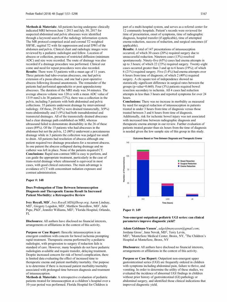
Methods & Materials: All patients having undergone clinically indicated MRI between June 1 2013 and July 30, 2017 for suspected abdominal and pelvic abscesses were identified through a keyword search of the radiology information system. The MR protocol comprised axial and coronal T2 weighted
SSFSE, sagittal T2 with fat suppression and axial DWI of the abdomen and pelvis. Clinical chart and radiologic images were reviewed by a pediatric radiologist and fellow. Location of abscess or collection, presence of restricted diffusion (minimum ADC) and size were recorded. The route of drainage was also recorded if a drainage procedure was performed. Clinical out
come and need for repeat procedures were also recorded. Results: There were 53 patients with a mean age of 11 years. Three patients had tubo-ovarian abscesses, one had pelvic extension of a psoas abscess, and one had a post-operative abscess following ileoanal anastomosis. The remainder of the patients had perforated appendicitis or post appendectomy abscesses. The duration of the MRI study was 34 minutes. The
average abscess volume was 120 cc with a mean ADC value of 1319 mm2/s.In 38 patients (72%), there was a collection in the pelvis, including 5 patients with both abdominal and pelvic collections. 35 patients underwent drainage by interventional radiology. Of these, 29 (82%) were drained transrectally, 3(9%) trans-abdominally and 3 (9%) had both transabdominal and transrectal drainages. All of the transrectally drained abscesses
had a clear drainage path established on MRI, whereas ultrasound failed to demonstrate drainability in the 26 of these cases (89%). Of the 15 patients who had abscesses in the abdomen but not the pelvis, 12 (80%) underwent a percutaneous drainage while in 3 patients the collection was judged too small to drain. All patients had resolution of abscess although one
patient required two drainage procedures for a recurrent abscess. In one patient the abscess collapsed during drainage and no catheter was left in place. None of the patients required CT. Conclusions: Rapid non-contrast MRI is easy to perform, and can guide the appropriate treatment, particularly in the case of trans-rectal drainage where ultrasound is equivocal in most cases, with good clinical outcomes. The main advantage is
avoidance of CT with concomitant radiation exposure and contrast administration.
Paper #: 148
Does Prolongation of Time Between Intussusception
Diagnosis and Therapeutic Enema Result In Increased
Patient Morbidity: a Retrospective Review Ivey Royall, MD
1, [email protected]; Aaron Lindsay,
MD1, Gregory Logsdon, MD
1, Matthew Swerdlow, MD
1, Julie
Pepe, PhD1, Jennifer Williams, MD
1;
1Florida Hospital, Orlando,
FL
Disclosures: All authors have disclosed no financial interests, arrangements or affiliations in the context of this activity. Purpose or Case Report: Ileocolic intussusception is an emergent condition with concern for bowel ischemia prompting rapid treatment. Therapeutic enema performed by a pediatric
radiologist, with progression to surgery if reduction fails is standard of care. However, many hospitals do not have pediatric radiologists available and require transfer, delaying treatment. Despite increased concern for risk of bowel complication, there is limited data evaluating the effect of increased time to therapeutic enema and patient morbidity/mortality. Our purpose
is to determine if there is increased patient morbidity/mortality associated with prolonged time between diagnosis and treatment of intussusception. Methods & Materials: A retrospective evaluation of pediatric patients treated for intussusception at a children’s hospital over a 10-year period was performed. Florida Hospital for Children is
part of a multi-hospital system, and serves as a referral center for 12 community hospitals. Patient’s records were reviewed for time of presentation, onset of symptoms, time of radiographic diagnosis, hospital transfer (if applicable), time of attempted enema reduction, success of reduction, and surgical outcomes (if
applicable). Results: A total of 147 presentations of intussusception occurred, of which 30 cases (20%) required surgery due to unsuccessful reduction. Nineteen cases (13%) resolved spontaneously. Ninety-five (65%) cases had enema attempts in up to 3 hours; of which 22 (23%) required surgery. Twenty-eight
cases occurred greater than 3 and up to 6 hours (19%); of which 6 (21%) required surgery. Five (3.4%) had enema attempts over 6 hours from time of diagnosis; of which 2 (40%) required surgery. A chi square test of independence showed no statistically significant difference in surgical rates between the groups (p-value=0.660). Four (3%) patients required bowel resection secondary to ischemia. All 4 cases had reduction
attempts in less than 3 hours and reported symptoms for over 24 hours. Conclusions: There was no increase in morbidity as measured by need for surgical reduction of intussusception in patients treated in under 3 hours from time of diagnosis versus those treated between 3 and 6 hours from time of diagnosis. Additionally, risk for ischemic bowel injury was not associated
with increased time between radiographic diagnosis and therapeutic enema attempt in our review. Further evaluation of patients treated greater than six hours from the time of diagnosis is needed given the low sample size of this group in this study.
Paper #: 149
Non-emergent outpatient pediatric UGI series: can clinical
parameters improve diagnostic yield? Adam Goldman-Yassen
Jordana Gross1, Inna Novak, MD
2, Terry Levin,
MD1;
1Montefiore Medical Center, Bronx, NY,
2The Children’s
Hospital at Montefiore, Bronx, NY Disclosures: All authors have disclosed no financial interests, arrangements or affiliations in the context of this activity.
Purpose or Case Report: Outpatient non-emergent upper gastrointestinal series (UGI) are frequently ordered in children with symptoms including abdominal pain, failure to thrive, and vomiting. In order to determine the utility of these studies, we evaluated the incidence of abnormal UGI findings in children without prior history of gastrointestinal (GI) pathology or
abdominal surgery, and identified those clinical indications that improved diagnostic yield.
Pediatr Radiol (2018) 48 (Suppl 1):S1–S298 S167
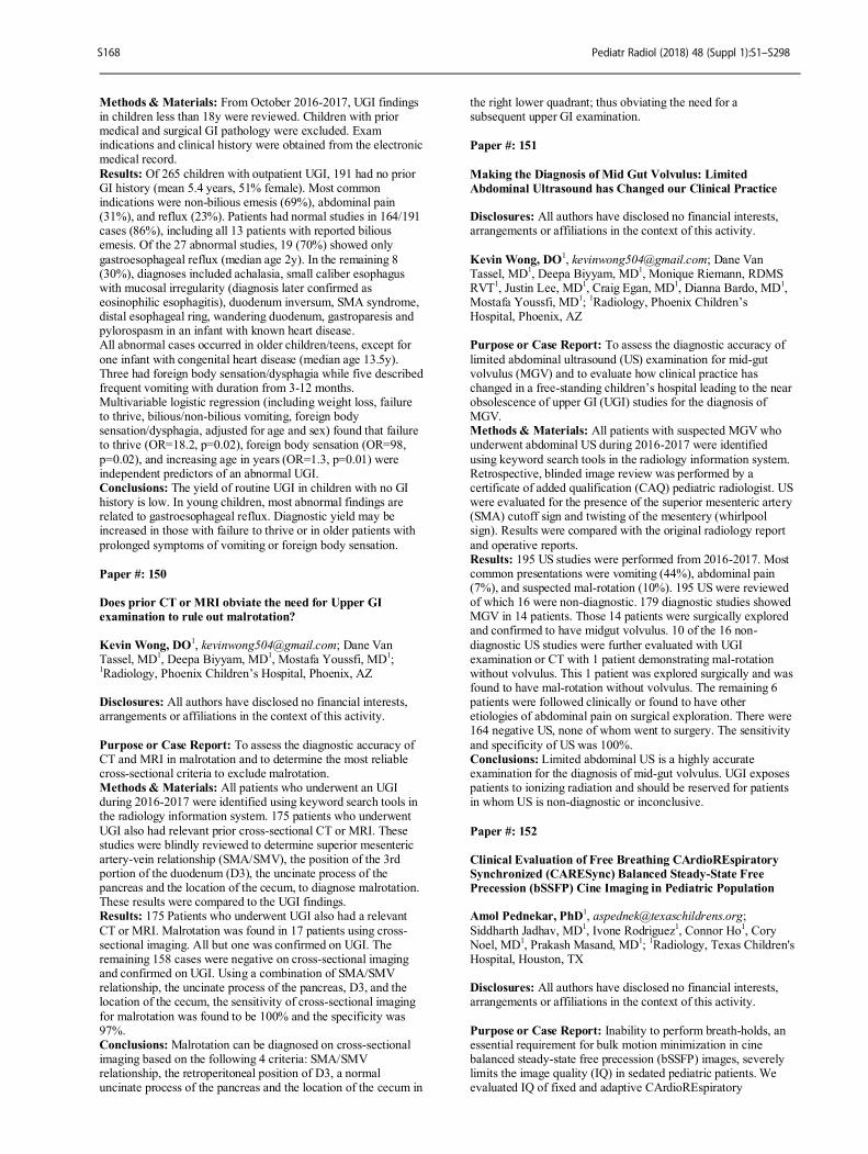
Methods & Materials: From October 2016-2017, UGI findings in children less than 18y were reviewed. Children with prior medical and surgical GI pathology were excluded. Exam indications and clinical history were obtained from the electronic medical record.
Results: Of 265 children with outpatient UGI, 191 had no prior GI history (mean 5.4 years, 51% female). Most common indications were non-bilious emesis (69%), abdominal pain (31%), and reflux (23%). Patients had normal studies in 164/191 cases (86%), including all 13 patients with reported bilious emesis. Of the 27 abnormal studies, 19 (70%) showed only
gastroesophageal reflux (median age 2y). In the remaining 8 (30%), diagnoses included achalasia, small caliber esophagus with mucosal irregularity (diagnosis later confirmed as eosinophilic esophagitis), duodenum inversum, SMA syndrome, distal esophageal ring, wandering duodenum, gastroparesis and pylorospasm in an infant with known heart disease. All abnormal cases occurred in older children/teens, except for
one infant with congenital heart disease (median age 13.5y). Three had foreign body sensation/dysphagia while five described frequent vomiting with duration from 3-12 months. Multivariable logistic regression (including weight loss, failure to thrive, bilious/non-bilious vomiting, foreign body sensation/dysphagia, adjusted for age and sex) found that failure to thrive (OR=18.2, p=0.02), foreign body sensation (OR=98,
p=0.02), and increasing age in years (OR=1.3, p=0.01) were independent predictors of an abnormal UGI. Conclusions: The yield of routine UGI in children with no GI history is low. In young children, most abnormal findings are related to gastroesophageal reflux. Diagnostic yield may be increased in those with failure to thrive or in older patients with
prolonged symptoms of vomiting or foreign body sensation.
Paper #: 150
Does prior CT or MRI obviate the need for Upper GI
examination to rule out malrotation?
Kevin Wong, DO1, [email protected]; Dane Van
Tassel, MD1, Deepa Biyyam, MD
1, Mostafa Youssfi, MD
1;
1Radiology, Phoenix Children’s Hospital, Phoenix, AZ
Disclosures: All authors have disclosed no financial interests, arrangements or affiliations in the context of this activity.
Purpose or Case Report: To assess the diagnostic accuracy of CT and MRI in malrotation and to determine the most reliable cross-sectional criteria to exclude malrotation. Methods & Materials: All patients who underwent an UGI during 2016-2017 were identified using keyword search tools in the radiology information system. 175 patients who underwent
UGI also had relevant prior cross-sectional CT or MRI. These studies were blindly reviewed to determine superior mesenteric artery-vein relationship (SMA/SMV), the position of the 3rd portion of the duodenum (D3), the uncinate process of the pancreas and the location of the cecum, to diagnose malrotation. These results were compared to the UGI findings. Results: 175 Patients who underwent UGI also had a relevant
CT or MRI. Malrotation was found in 17 patients using cross-sectional imaging. All but one was confirmed on UGI. The remaining 158 cases were negative on cross-sectional imaging and confirmed on UGI. Using a combination of SMA/SMV relationship, the uncinate process of the pancreas, D3, and the location of the cecum, the sensitivity of cross-sectional imaging
for malrotation was found to be 100% and the specificity was 97%. Conclusions: Malrotation can be diagnosed on cross-sectional imaging based on the following 4 criteria: SMA/SMV relationship, the retroperitoneal position of D3, a normal uncinate process of the pancreas and the location of the cecum in
the right lower quadrant; thus obviating the need for a subsequent upper GI examination.
Paper #: 151
Making the Diagnosis of Mid Gut Volvulus: Limited
Abdominal Ultrasound has Changed our Clinical Practice Disclosures: All authors have disclosed no financial interests, arrangements or affiliations in the context of this activity.
Kevin Wong, DO1, [email protected]; Dane Van
Tassel, MD1, Deepa Biyyam, MD
1, Monique Riemann, RDMS
RVT1, Justin Lee, MD
1, Craig Egan, MD
1, Dianna Bardo, MD
1,
Mostafa Youssfi, MD1;
1Radiology, Phoenix Children’s
Hospital, Phoenix, AZ Purpose or Case Report: To assess the diagnostic accuracy of
limited abdominal ultrasound (US) examination for mid-gut volvulus (MGV) and to evaluate how clinical practice has changed in a free-standing children’s hospital leading to the near obsolescence of upper GI (UGI) studies for the diagnosis of MGV. Methods & Materials: All patients with suspected MGV who underwent abdominal US during 2016-2017 were identified
using keyword search tools in the radiology information system. Retrospective, blinded image review was performed by a certificate of added qualification (CAQ) pediatric radiologist. US were evaluated for the presence of the superior mesenteric artery (SMA) cutoff sign and twisting of the mesentery (whirlpool sign). Results were compared with the original radiology report
and operative reports. Results: 195 US studies were performed from 2016-2017. Most common presentations were vomiting (44%), abdominal pain (7%), and suspected mal-rotation (10%). 195 US were reviewed of which 16 were non-diagnostic. 179 diagnostic studies showed MGV in 14 patients. Those 14 patients were surgically explored and confirmed to have midgut volvulus. 10 of the 16 non-
diagnostic US studies were further evaluated with UGI examination or CT with 1 patient demonstrating mal-rotation without volvulus. This 1 patient was explored surgically and was found to have mal-rotation without volvulus. The remaining 6 patients were followed clinically or found to have other etiologies of abdominal pain on surgical exploration. There were 164 negative US, none of whom went to surgery. The sensitivity
and specificity of US was 100%. Conclusions: Limited abdominal US is a highly accurate examination for the diagnosis of mid-gut volvulus. UGI exposes patients to ionizing radiation and should be reserved for patients in whom US is non-diagnostic or inconclusive.
Paper #: 152
Clinical Evaluation of Free Breathing CArdioREspiratory
Synchronized (CARESync) Balanced Steady-State Free
Precession (bSSFP) Cine Imaging in Pediatric Population Amol Pednekar, PhD
Siddharth Jadhav, MD1, Ivone Rodriguez
1, Connor Ho
1, Cory
Noel, MD1, Prakash Masand, MD
1;
1Radiology, Texas Children's
Hospital, Houston, TX Disclosures: All authors have disclosed no financial interests, arrangements or affiliations in the context of this activity.
Purpose or Case Report: Inability to perform breath-holds, an essential requirement for bulk motion minimization in cine balanced steady-state free precession (bSSFP) images, severely limits the image quality (IQ) in sedated pediatric patients. We evaluated IQ of fixed and adaptive CArdioREspiratory
Pediatr Radiol (2018) 48 (Suppl 1):S1–S298S168
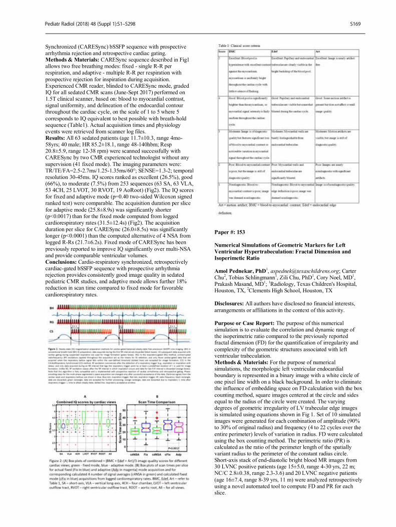
Synchronized (CARESync) bSSFP sequence with prospective arrhythmia rejection and retrospective cardiac gating. Methods & Materials: CARESync sequence described in Fig1 allows two free breathing modes: fixed - single R-R per respiration, and adaptive - multiple R-R per respiration with
prospective rejection for inspiration during acquisition. Experienced CMR reader, blinded to CARESync mode, graded IQ for all sedated CMR scans (June-Sept 2017) performed on 1.5T clinical scanner, based on: blood to myocardial contrast, signal uniformity, and delineation of the endocardial contour throughout the cardiac cycle, on the scale of 1 to 5 where 5
corresponds to IQ equivalent to best possible with breath-hold sequence (Table1). Actual acquisition times and physiology events were retrieved from scanner log files. Results: All 63 sedated patients (age 11.7±10.3, range 4mo-58yrs; 40 male; HR 85.2±18.1, range 48-140hbm; Resp 20.8±5.9, range 12-38 rpm) were scanned successfully with CARESync by two CMR experienced technologist without any
supervision (41 fixed mode). The imaging parameters were: TR/TE/FA=2.5-2.7ms/1.25-1.35ms/60°; SENSE=1.3-2; temporal resolution 30-45ms. IQ scores ranked as excellent (26.5%), good (66%), to moderate (7.5%) from 253 sequences (63 SA, 63 VLA, 53 4CH, 25 LVOT, 30 RVOT, 19 AoRoot) (Fig2). The IQ scores for fixed and adaptive mode (p=0.40 two-sided Wilcoxon signed ranked test) were comparable. The acquisition duration per slice
for adaptive mode (25.8±8.9s) was significantly shorter (p<0.0017) than for the fixed mode computed from logged cardiorespiratory rates (31.5±12.4s) (Fig2). The acquisition duration per slice for CARESync (26.0±8.5s) was significantly longer (p<0.0001) than the computed alternative of 4 NSA from logged R-Rs (21.7±6.2s). Fixed mode of CARESync has been
previously reported to improve IQ significantly over multi-NSA and provide comparable ventricular volumes. Conclusions: Cardio-respiratory synchronized, retrospectively cardiac-gated bSSFP sequence with prospective arrhythmia rejection provides consistently good image quality in sedated pediatric CMR studies, and adaptive mode allows further 18% reduction in scan time compared to fixed mode for favorable
cardiorespiratory rates.
Paper #: 153
Numerical Simulations of Geometric Markers for Left
Ventricular Hypertrabeculation: Fractal Dimension and
Isoperimetic Ratio
Amol Pednekar, PhD
1, [email protected]; Carter
Chu2, Tobias Schlingmann
1, Zili Chu, PhD
1, Cory Noel, MD
1,
Prakash Masand, MD1;
1Radiology, Texas Children's Hospital,
Houston, TX, 2Clements High School, Houston, TX
Disclosures: All authors have disclosed no financial interests,
arrangements or affiliations in the context of this activity. Purpose or Case Report: The purpose of this numerical simulation is to evaluate the correlation and dynamic range of the isoperimetric ratio compared to the previously reported fractal dimension (FD) for the quantification of irregularity and
complexity of the geometric structures associated with left ventricular trabeculation. Methods & Materials: For the purpose of numerical simulations, the morphologic left ventricular endocardial boundary is represented in a binary image with a white circle of one pixel line width on a black background. In order to eliminate the influence of embedding space on FD calculation with the box
counting method, square images centered at the circle and sides equal to the radius of the circle were created. The varying degrees of geometric irregularity of LV trabecular edge images is simulated using equations shown in Fig 1. Set of 10 simulated images were generated for each combination of amplitude (90% to 30% of original radius) and frequency (4 to 22 cycles over the
entire perimeter) levels of variation in radius. FD were calculated using the box counting method. The perimetric ratio (PR) is calculated as the ratio of the perimeter length of the spatially variant radius to the perimeter of the constant radius circle. Short-axis stack of end-diastolic bright blood MR images from 30 LVNC positive patients (age 15±5.0, range 4-30 yrs, 22 m; NC/C 2.8±0.38, range 2.3-3.6) and 20 LVNC negative patients
(age 16±7.4, range 8-39 yrs, 11 m) were analyzed retrospectively using a novel automated tool to compute FD and PR for each slice.
Pediatr Radiol (2018) 48 (Suppl 1):S1–S298 S169
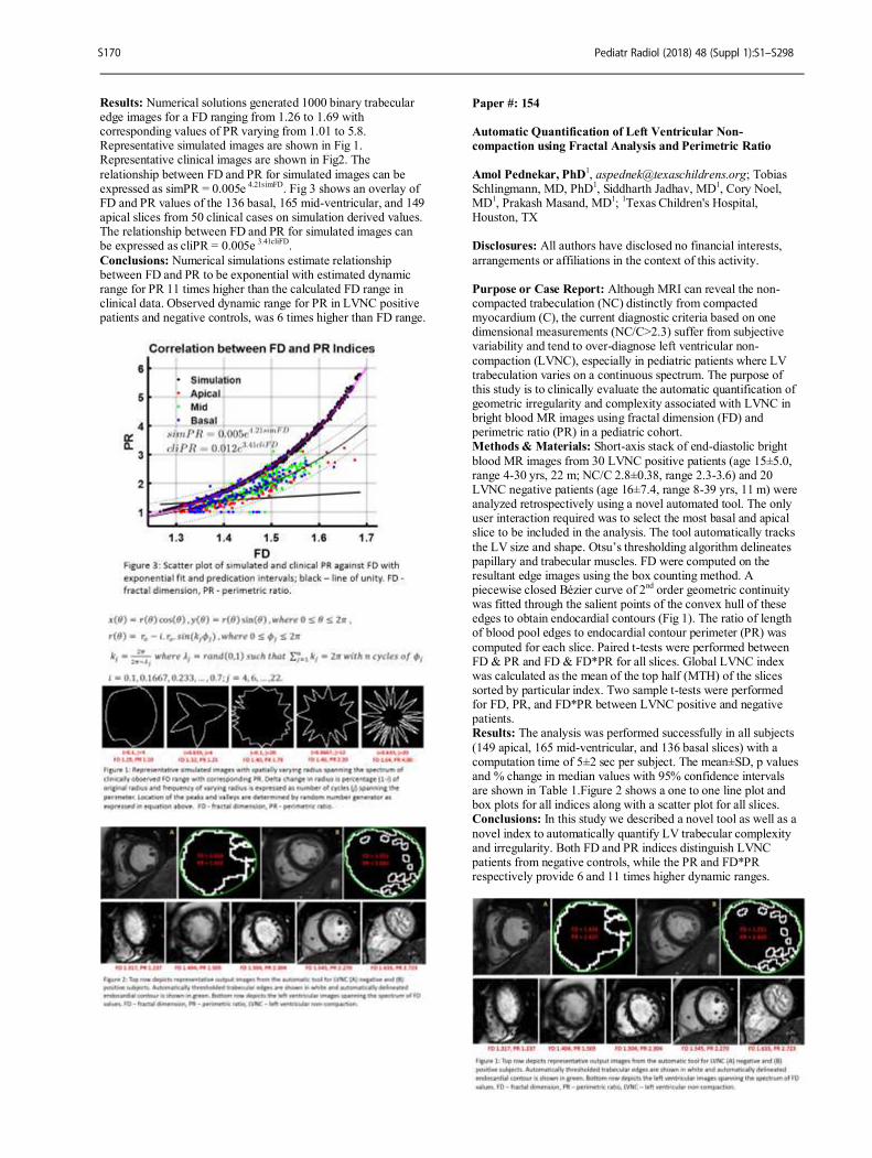
Results: Numerical solutions generated 1000 binary trabecular edge images for a FD ranging from 1.26 to 1.69 with corresponding values of PR varying from 1.01 to 5.8. Representative simulated images are shown in Fig 1. Representative clinical images are shown in Fig2. The
relationship between FD and PR for simulated images can be expressed as simPR = 0.005e
4.21simFD. Fig 3 shows an overlay of
FD and PR values of the 136 basal, 165 mid-ventricular, and 149 apical slices from 50 clinical cases on simulation derived values. The relationship between FD and PR for simulated images can be expressed as cliPR = 0.005e
3.41cliFD.
Conclusions: Numerical simulations estimate relationship between FD and PR to be exponential with estimated dynamic range for PR 11 times higher than the calculated FD range in clinical data. Observed dynamic range for PR in LVNC positive patients and negative controls, was 6 times higher than FD range.
Paper #: 154
Automatic Quantification of Left Ventricular Non-
compaction using Fractal Analysis and Perimetric Ratio
Amol Pednekar, PhD1, [email protected]; Tobias
Schlingmann, MD, PhD1, Siddharth Jadhav, MD
1, Cory Noel,
MD1, Prakash Masand, MD
1;
1Texas Children's Hospital,
Houston, TX Disclosures: All authors have disclosed no financial interests,
arrangements or affiliations in the context of this activity.
Purpose or Case Report: Although MRI can reveal the non-compacted trabeculation (NC) distinctly from compacted myocardium (C), the current diagnostic criteria based on one dimensional measurements (NC/C>2.3) suffer from subjective variability and tend to over-diagnose left ventricular non-
compaction (LVNC), especially in pediatric patients where LV trabeculation varies on a continuous spectrum. The purpose of this study is to clinically evaluate the automatic quantification of geometric irregularity and complexity associated with LVNC in bright blood MR images using fractal dimension (FD) and perimetric ratio (PR) in a pediatric cohort. Methods & Materials: Short-axis stack of end-diastolic bright
blood MR images from 30 LVNC positive patients (age 15±5.0, range 4-30 yrs, 22 m; NC/C 2.8±0.38, range 2.3-3.6) and 20 LVNC negative patients (age 16±7.4, range 8-39 yrs, 11 m) were analyzed retrospectively using a novel automated tool. The only user interaction required was to select the most basal and apical slice to be included in the analysis. The tool automatically tracks
the LV size and shape. Otsu’s thresholding algorithm delineates papillary and trabecular muscles. FD were computed on the resultant edge images using the box counting method. A piecewise closed Bézier curve of 2
nd order geometric continuity
was fitted through the salient points of the convex hull of these edges to obtain endocardial contours (Fig 1). The ratio of length of blood pool edges to endocardial contour perimeter (PR) was
computed for each slice. Paired t-tests were performed between FD & PR and FD & FD*PR for all slices. Global LVNC index was calculated as the mean of the top half (MTH) of the slices sorted by particular index. Two sample t-tests were performed for FD, PR, and FD*PR between LVNC positive and negative patients. Results: The analysis was performed successfully in all subjects
(149 apical, 165 mid-ventricular, and 136 basal slices) with a computation time of 5±2 sec per subject. The mean±SD, p values and % change in median values with 95% confidence intervals are shown in Table 1.Figure 2 shows a one to one line plot and box plots for all indices along with a scatter plot for all slices. Conclusions: In this study we described a novel tool as well as a
novel index to automatically quantify LV trabecular complexity and irregularity. Both FD and PR indices distinguish LVNC patients from negative controls, while the PR and FD*PR respectively provide 6 and 11 times higher dynamic ranges.
Pediatr Radiol (2018) 48 (Suppl 1):S1–S298S170
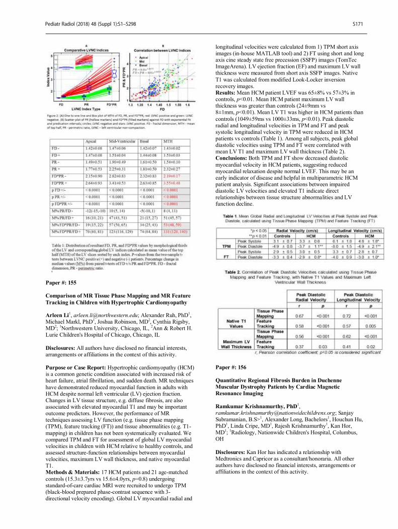
Paper #: 155
Comparison of MR Tissue Phase Mapping and MR Feature
Tracking in Children with Hypertrophic Cardiomyopathy Arleen Li
1, [email protected]; Alexander Ruh, PhD
1,
Michael Markl, PhD1, Joshua Robinson, MD
2, Cynthia Rigsby,
MD2;
1Northwestern University, Chicago, IL,
2Ann & Robert H.
Lurie Children's Hospital of Chicago, Chicago, IL
Disclosures: All authors have disclosed no financial interests, arrangements or affiliations in the context of this activity. Purpose or Case Report: Hypertrophic cardiomyopathy (HCM) is a common genetic condition associated with increased risk of heart failure, atrial fibrillation, and sudden death. MR techniques
have demonstrated reduced myocardial function in adults with HCM despite normal left ventricular (LV) ejection fraction. Changes in LV tissue structure, e.g. diffuse fibrosis, are also associated with elevated myocardial T1 and may be important outcome predictors. However, the performance of MR techniques assessing LV function (e.g. tissue phase mapping (TPM), feature tracking (FT)) and tissue abnormalities (e.g. T1-
mapping) in children has not been systematically evaluated. We compared TPM and FT for assessment of global LV myocardial velocities in children with HCM relative to healthy controls, and assessed structure-function relationships between myocardial velocities, maximum LV wall thickness, and native myocardial T1. Methods & Materials: 17 HCM patients and 21 age-matched
controls (15.3±3.7yrs vs 15.6±4.0yrs, p=0.8) undergoing standard-of-care cardiac MRI were recruited to undergo TPM (black-blood prepared phase-contrast sequence with 3-directional velocity encoding). Global LV myocardial radial and
longitudinal velocities were calculated from 1) TPM short axis images (in-house MATLAB tool) and 2) FT using short and long axis cine steady state free precession (SSFP) images (TomTec ImageArena). LV ejection fraction (EF) and maximum LV wall thickness were measured from short axis SSFP images. Native
T1 was calculated from modified Look-Locker inversion recovery images. Results: Mean HCM patient LVEF was 65±8% vs 57±3% in controls, p<0.01. Mean HCM patient maximum LV wall thickness was greater than controls (24±9mm vs 8±1mm, p<0.01). Mean LV T1 was higher in HCM patients than
controls (1049±59ms vs 1000±33ms, p<0.01). Peak diastolic radial and longitudinal velocities in TPM and FT and peak systolic longitudinal velocity in TPM were reduced in HCM patients vs controls (Table 1). Among all subjects, peak global diastolic velocities using TPM and FT were correlated with mean LV T1 and maximum LV wall thickness (Table 2). Conclusions: Both TPM and FT show decreased diastolic
myocardial velocity in HCM patients, suggesting reduced myocardial relaxation despite normal LVEF. This may be an early indicator of disease and helpful in multiparametric HCM patient analysis. Significant associations between impaired diastolic LV velocities and elevated T1 indicate direct relationships between tissue structure abnormalities and LV function decline.
Paper #: 156
Quantitative Regional Fibrosis Burden in Duchenne
Muscular Dystrophy Patients by Cardiac Magnetic
Resonance Imaging
Ramkumar Krishnamurthy, PhD1,
[email protected]; Sanjay Subramanian, B.S>
1, Alexander Long, Bachelors
1, Houchun Hu,
PhD1, Linda Cripe, MD
1, Rajesh Krishnamurthy
1, Kan Hor,
MD1;
1Radiology, Nationwide Children's Hospital, Columbus,
OH
Disclosures: Kan Hor has indicated a relationship with Medtronics and Capricor as a consultant/honoraria. All other authors have disclosed no financial interests, arrangements or affiliations in the context of this activity.
Pediatr Radiol (2018) 48 (Suppl 1):S1–S298 S171
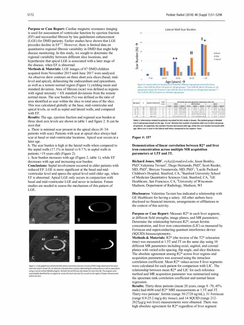
Purpose or Case Report: Cardiac magnetic resonance imaging is used for assessment of ventricular function by ejection fraction (EF) and myocardial fibrosis by late gadolinium enhancement (LGE) for DMD patients. Earlier studies have shown that LGE precedes decline in EF
1-2. However, there is limited data on
quantitative regional fibrosis variability in DMD that might help disease monitoring. In this study, we sought to determine the regional variability between different slice locations, and hypothesize that apical LGE is associated with a later stage of the disease, when EF is abnormal. Methods & Materials: LGE images of 87 DMD children
acquired from November 2015 until June 2017 were analyzed. An observer drew contours on three short axis slices (basal, mid-level and apical), delineating the endocardium and epicardium, as well as a remote normal region (Figure 1) yielding mean and standard deviation. Area of fibrosis (scar) was defined as regions with signal intensity > 6X standard deviations from the remote normal mean. The scar burden (%) was defined as the ratio of
area identified as scar within the slice to total area of the slice. This was calculated globally at the base, mid-ventricular and apical levels, as well as septal and lateral walls, and compared with EF. Results: The age, ejection fraction and regional scar burden at three short axis levels are shown in table 1 and figure 2. It can be seen that
a. There is minimal scar present in the apical slices (8 /34 patients with scar). Patients with scar at apical slice always had scar at basal or mid-ventricular locations. Apical scar occurs at a later age. b. The scar burden is high at the lateral walls when compared to the septal walls (17.1% in lateral vs 0.7 % in septal walls in
patients >19 years old) (Figure 2). c. Scar burden increases with age (Figure 2, table 1), while EF decreases with age and increasing scar burden. Conclusions: Septal involvement occurred in older patients with reduced EF. LGE is more significant at the basal and mid-ventricular level and spares the apical level until older age, when EF is abnormal. Apical LGE only occurs in conjunction with
basal and mid-ventricular LGE and never in isolation. Future studies are needed to assess the mechanism of this pattern of LGE.
Paper #: 157
Demonstration of linear correlation between R2* and liver
iron concentration across multiple MR acquisition
parameters at 1.5T and 3T. Richard Jones, MD
1, [email protected]; Jason Bentley,
PhD2,Valentina Taviani
3, Diego Hernando, PhD
4, Scott Reeder,
MD, PhD4, Shreyas Vasanawala, MD, PhD
1;
1Lucile Packard
Children's Hospital, Stanford, CA, 2Stanford University School
of Medicine Quantitative Sciences Unit, Stanford, CA, 3GE
Healthcare, San Francisco, CA, 4University of Wisconsin-
Madison, Department of Radiology, Madison, WI
Disclosures: Valentina Taviani has indicated a relationship with GE Healthcare for having a salary. All other authors have disclosed no financial interests, arrangements or affiliations in the context of this activity.
Purpose or Case Report: Measure R2* in each liver segment,
at different field strengths, image planes, and MR parameters; Determine the relationship between R2*, serum ferritin concentration, and liver iron concentration (LIC) as measured by Ferriscan and superconducting quantum interference device (SQUID) biosusceptometry. Methods & Materials: R2* (the inverse of the T2* relaxation time) was measured at 1.5T and 3T on the same day using 10
different MR parameters including axial, sagittal, and coronal planes with varied echo spacing, flip angle, and slice thickness. The absolute agreement among R2* across liver regions and acquisition parameters was assessed using the intraclass correlation coefficient. Mean R2* values across 8 liver segments were calculated for each patient for comparison with LIC. The
relationship between mean R2* and LIC for each reference method and MR acquisition parameter was summarized using the spearman rank correlation coefficient and normal linear regression. Results: Thirty-three patients (mean 20 years, range 8 -70, 45% male) had 4696 total R2* MRI measurements at 1.5T and 3T. Thirty-two patients’ ferritin (range 30-2728 ng/mL), 31 Ferriscan
(range 0.9-25.2 mg/g dry tissue), and 14 SQUID (range 212-2625µg/g wet liver) measurements were obtained. There was high absolute agreement for R2* regardless of liver segment
Pediatr Radiol (2018) 48 (Suppl 1):S1–S298S172
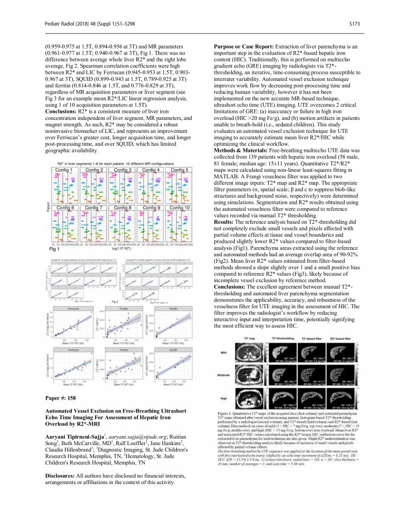
(0.959-0.975 at 1.5T, 0.894-0.956 at 3T) and MR parameters (0.961-0.977 at 1.5T; 0.940-0.967 at 3T), Fig 1. There was no difference between average whole liver R2* and the right lobe average, Fig 2. Spearman correlation coefficients were high between R2* and LIC by Ferriscan (0.945-0.953 at 1.5T, 0.903-
0.967 at 3T), SQUID (0.899-0.943 at 1.5T, 0.789-0.925 at 3T) and ferritin (0.814-0.846 at 1.5T, and 0.776-0.829 at 3T), regardless of MR acquisition parameters or liver segment (see Fig 3 for an example mean R2*/LIC linear regression analysis, using 1 of 10 acquisition parameters at 1.5T). Conclusions: R2* is a consistent measure of liver iron
concentration independent of liver segment, MR parameters, and magnet strength. As such, R2* may be considered a robust noninvasive biomarker of LIC, and represents an improvement over Ferriscan’s greater cost, longer acquisition time, and longer post-processing time, and over SQUID, which has limited geographic availability.
Paper #: 158
Automated Vessel Exclusion on Free-Breathing Ultrashort
Echo Time Imaging For Assessment of Hepatic Iron
Overload by R2*-MRI
Aaryani Tipirneni-Sajja
1, [email protected]; Ruitian
Song1, Beth McCarville, MD
1, Ralf Loeffler
1, Jane Hankins
2,
Claudia Hillenbrand1;
1Diagnostic Imaging, St. Jude Children's
Research Hospital, Memphis, TN, 2Hematology, St. Jude
Children's Research Hospital, Memphis, TN
Disclosures: All authors have disclosed no financial interests, arrangements or affiliations in the context of this activity.
Purpose or Case Report: Extraction of liver parenchyma is an important step in the evaluation of R2*-based hepatic iron content (HIC). Traditionally, this is performed on multiecho gradient echo (GRE) imaging by radiologists via T2*-thresholding, an iterative, time-consuming process susceptible to
interrater variability. Automated vessel exclusion technique improves work flow by decreasing post-processing time and reducing human variability, however it has not been implemented on the new accurate MR-based technique, ultrashort echo time (UTE) imaging. UTE overcomes 2 critical limitations of GRE: (a) inaccuracy or failure in high iron
overload (HIC >20 mg Fe/g), and (b) motion artifacts in patients unable to breath-hold (i.e., sedated children). This study evaluates an automated vessel exclusion technique for UTE imaging to accurately estimate mean liver R2*/HIC while optimizing the clinical workflow. Methods & Materials: Free-breathing multiecho UTE data was collected from 139 patients with hepatic iron overload (58 male,
81 female; median age: 15±11 years). Quantitative T2*/R2* maps were calculated using non-linear least-squares fitting in MATLAB. A Frangi vesselness filter was applied to two different image inputs: T2* map and R2* map. The appropriate filter parameters (σ, spatial scale; β and c to suppress blob-like structures and background noise, respectively) were determined using simulations. Segmentation and R2* results obtained using
the automated vesselness filter were compared to reference values recorded via manual T2* thresholding. Results: The reference analysis based on T2*-thresholding did not completely exclude small vessels and pixels affected with partial volume effects at tissue and vessel boundaries and produced slightly lower R2* values compared to filter-based
analysis (Fig1). Parenchyma areas extracted using the reference and automated methods had an average overlap area of 90-92% (Fig2). Mean liver R2* values estimated from filter-based methods showed a slope slightly over 1 and a small positive bias compared to reference R2* values (Fig3), likely because of incomplete vessel exclusion by reference method. Conclusions: The excellent agreement between manual T2*-
thresholding and automated liver parenchyma segmentation demonstrates the applicability, accuracy, and robustness of the vesselness filter for UTE imaging in the assessment of HIC. The filter improves the radiologist’s workflow by reducing interactive input and interpretation time, potentially signifying the most efficient way to assess HIC.
Pediatr Radiol (2018) 48 (Suppl 1):S1–S298 S173
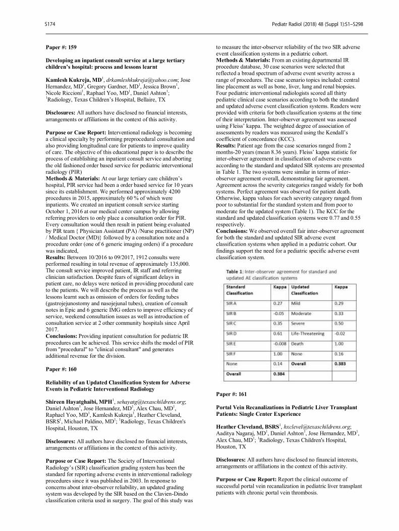
Paper #: 159
Developing an inpatient consult service at a large tertiary
children’s hospital: process and lessons learnt
Kamlesh Kukreja, MD1, [email protected]; Jose
Hernandez, MD1, Gregory Gardner, MD
1, Jessica Brown
1,
Nicole Riccioni1, Raphael Yoo, MD
1, Daniel Ashton
1;
1Radiology, Texas Children’s Hospital, Bellaire, TX
Disclosures: All authors have disclosed no financial interests,
arrangements or affiliations in the context of this activity. Purpose or Case Report: Interventional radiology is becoming a clinical specialty by performing preprocedural consultation and also providing longitudinal care for patients to improve quality of care. The objective of this educational paper is to describe the process of establishing an inpatient consult service and aborting
the old fashioned order based service for pediatric interventional radiology (PIR) Methods & Materials: At our large tertiary care children’s hospital, PIR service had been a order based service for 10 years since its establishment. We performed approximately 4200 procedures in 2015, approximately 60 % of which were inpatients. We created an inpatient consult service starting
October 1, 2016 at our medical center campus by allowing referring providers to only place a consultation order for PIR. Every consultation would then result in patient being evaluated by PIR team { Physician Assistant (PA) /Nurse practitioner (NP) / Medical Doctor (MD)} followed by a consultation note and a procedure order (one of 6 generic imaging orders) if a procedure
was indicated, Results: Between 10/2016 to 09/2017, 1912 consults were performed resulting in total revenue of approximately 135,000. The consult service improved patient, IR staff and referring clinician satisfaction. Despite fears of significant delays in patient care, no delays were noticed in providing procedural care to the patients. We will describe the process as well as the
lessons learnt such as omission of orders for feeding tubes (gastrojejunostomy and nasojejunal tubes), creation of consult notes in Epic and 6 generic IMG orders to improve efficiency of service, weekend consultation issues as well as introduction of consultation service at 2 other community hospitals since April 2017. Conclusions: Providing inpatient consultation for pediatric IR
procedures can be achieved. This service shifts the model of PIR from "procedural" to "clinical consultant" and generates additional revenue for the division.
Paper #: 160
Reliability of an Updated Classification System for Adverse
Events in Pediatric Interventional Radiology Shireen Hayatghaibi, MPH
Daniel Ashton1, Jose Hernandez, MD
1, Alex Chau, MD
1,
Raphael Yoo, MD1, Kamlesh Kukreja
1, Heather Cleveland,
BSRS1, Michael Paldino, MD
1;
1Radiology, Texas Children's
Hospital, Houston, TX Disclosures: All authors have disclosed no financial interests, arrangements or affiliations in the context of this activity. Purpose or Case Report: The Society of Interventional
Radiology’s (SIR) classification grading system has been the standard for reporting adverse events in interventional radiology procedures since it was published in 2003. In response to concerns about inter-observer reliability, an updated grading system was developed by the SIR based on the Clavien-Dindo classification criteria used in surgery. The goal of this study was
to measure the inter-observer reliability of the two SIR adverse event classification systems in a pediatric cohort. Methods & Materials: From an existing departmental IR procedure database, 30 case scenarios were selected that reflected a broad spectrum of adverse event severity across a
range of procedures. The case scenario topics included: central line placement as well as bone, liver, lung and renal biopsies. Four pediatric interventional radiologists scored all thirty pediatric clinical case scenarios according to both the standard and updated adverse event classification systems. Readers were provided with criteria for both classification systems at the time
of their interpretation. Inter-observer agreement was assessed using Fleiss’ kappa. The weighted degree of association of assessments by readers was measured using the Kendall’s coefficient of concordance (KCC). Results: Patient age from the case scenarios ranged from 2 months-20 years (mean 8.36 years). Fleiss’ kappa statistic for inter-observer agreement in classification of adverse events
according to the standard and updated SIR systems are presented in Table 1. The two systems were similar in terms of inter-observer agreement overall, demonstrating fair agreement. Agreement across the severity categories ranged widely for both systems. Perfect agreement was observed for patient death. Otherwise, kappa values for each severity category ranged from poor to substantial for the standard system and from poor to
moderate for the updated system (Table 1). The KCC for the standard and updated classification systems were 0.77 and 0.55 respectively. Conclusions: We observed overall fair inter-observer agreement for both the standard and updated SIR adverse event classification systems when applied in a pediatric cohort. Our
findings support the need for a pediatric specific adverse event classification system.
Paper #: 161
Portal Vein Recanalizations in Pediatric Liver Transplant
Patients: Single Center Experience Heather Cleveland, BSRS
Aaditya Nagaraj, MD1, Daniel Ashton
1, Jose Hernandez, MD
1,
Alex Chau, MD1;
1Radiology, Texas Children's Hospital,
Houston, TX Disclosures: All authors have disclosed no financial interests, arrangements or affiliations in the context of this activity. Purpose or Case Report: Report the clinical outcome of
successful portal vein recanalization in pediatric liver transplant patients with chronic portal vein thrombosis.
Pediatr Radiol (2018) 48 (Suppl 1):S1–S298S174
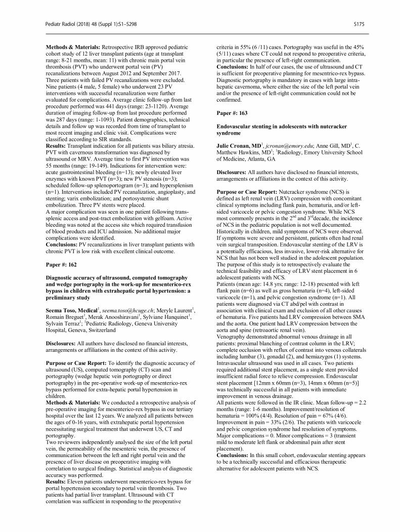
Methods & Materials: Retrospective IRB approved pediatric cohort study of 12 liver transplant patients (age at transplant range: 8-21 months, mean: 11) with chronic main portal vein thrombosis (PVT) who underwent portal vein (PV) recanalizations between August 2012 and September 2017.
Three patients with failed PV recanalizations were excluded. Nine patients (4 male, 5 female) who underwent 23 PV interventions with successful recanalization were further evaluated for complications. Average clinic follow-up from last procedure performed was 441 days (range: 23-1120). Average duration of imaging follow-up from last procedure performed
was 287 days (range: 1-1093). Patient demographics, technical details and follow up was recorded from time of transplant to most recent imaging and clinic visit. Complications were classified according to SIR standards. Results: Transplant indication for all patients was biliary atresia. PVT with cavernous transformation was diagnosed by ultrasound or MRV. Average time to first PV intervention was
55 months (range: 19-149). Indications for intervention were: acute gastrointestinal bleeding (n=13); newly elevated liver enzymes with known PVT (n=3); new PV stenosis (n=3); scheduled follow-up splenoportogram (n=3); and hypersplenism (n=1). Interventions included PV recanalization, angioplasty, and stenting; varix embolization; and portosystemic shunt embolization. Three PV stents were placed.
A major complication was seen in one patient following trans-splenic access and post-tract embolization with gelfoam. Active bleeding was noted at the access site which required transfusion of blood products and ICU admission. No additional major complications were identified. Conclusions: PV recanalizations in liver transplant patients with
chronic PVT is low risk with excellent clinical outcome.
Paper #: 162
Diagnostic accuracy of ultrasound, computed tomography
and wedge portography in the work-up for mesenterico-rex
bypass in children with extrahepatic portal hypertension: a
preliminary study Seema Toso, Medical
1, [email protected]; Meryle Laurent
1,
Romain Breguet1, Merak Anooshiravani
1, Sylviane Hanquinet
1,
Sylvain Terraz1;
1Pediatric Radiology, Geneva University
Hospital, Geneva, Switzerland
Disclosures: All authors have disclosed no financial interests, arrangements or affiliations in the context of this activity. Purpose or Case Report: To identify the diagnostic accuracy of ultrasound (US), computed tomography (CT) scan and portography (wedge hepatic vein portography or direct
portography) in the pre-operative work-up of mesenterico-rex bypass performed for extra-hepatic portal hypertension in children. Methods & Materials: We conducted a retrospective analysis of pre-operative imaging for mesenterico-rex bypass in our tertiary hospital over the last 12 years. We analyzed all patients between the ages of 0-16 years, with extrahepatic portal hypertension
necessitating surgical treatment that underwent US, CT and portography. Two reviewers independently analysed the size of the left portal vein, the permeability of the mesenteric vein, the presence of communication between the left and right portal vein and the presence of liver disease on preoperative imaging with
correlation to surgical findings. Statistical analysis of diagnostic accuracy was performed. Results: Eleven patients underwent mesenterico-rex bypass for portal hypertension secondary to portal vein thrombosis. Two patients had partial liver transplant. Ultrasound with CT correlation was sufficient in responding to the preoperative
criteria in 55% (6 /11) cases. Portography was useful in the 45% (5/11) cases where CT could not respond to preoperative criteria, in particular the presence of left-right communication. Conclusions: In half of our cases, the use of ultrasound and CT is sufficient for preoperative planning for mesentrico-rex bypass.
Diagnostic portography is mandatory in cases with large intra-hepatic cavernoma, where either the size of the left portal vein and/or the presence of left-right communication could not be confirmed.
Paper #: 163
Endovascular stenting in adolescents with nutcracker
syndrome Julie Cronan, MD
1, [email protected]; Anne Gill, MD
1, C.
Matthew Hawkins, MD1;
1Radiology, Emory University School
of Medicine, Atlanta, GA
Disclosures: All authors have disclosed no financial interests, arrangements or affiliations in the context of this activity. Purpose or Case Report: Nutcracker syndrome (NCS) is defined as left renal vein (LRV) compression with concomitant clinical symptoms including flank pain, hematuria, and/or left-
sided varicocele or pelvic congestion syndrome. While NCS most commonly presents in the 2
nd and 3
rddecade, the incidence
of NCS in the pediatric population is not well documented. Historically in children, mild symptoms of NCS were observed. If symptoms were severe and persistent, patients often had renal vein surgical transposition. Endovascular stenting of the LRV is
a potentially efficacious, less invasive, lower-risk alternative for NCS that has not been well studied in the adolescent population. The purpose of this study is to retrospectively evaluate the technical feasibility and efficacy of LRV stent placement in 6 adolescent patients with NCS. Patients (mean age: 14.8 yrs; range: 12-18) presented with left flank pain (n=6) as well as gross hematuria (n=4), left-sided
varicocele (n=1), and pelvic congestion syndrome (n=1). All patients were diagnosed via CT abd/pel with contrast in association with clinical exam and exclusion of all other causes of hematuria. Five patients had LRV compression between SMA and the aorta. One patient had LRV compression between the aorta and spine (retroaortic renal vein). Venography demonstrated abnormal venous drainage in all
patients: proximal blanching of contrast column in the LRV; complete occlusion with reflux of contrast into venous collaterals including lumbar (3), gonadal (2), and hemiazygos (1) systems. Intravascular ultrasound was used in all cases. Two patients required additional stent placement, as a single stent provided insufficient radial force to relieve compression. Endovascular
stent placement [12mm x 60mm (n=3), 14mm x 60mm (n=5)] was technically successful in all patients with immediate improvement in venous drainage. All patients were followed in the IR clinic. Mean follow-up = 2.2 months (range: 1-6 months). Improvement/resolution of hematuria = 100% (4/4). Resolution of pain = 67% (4/6). Improvement in pain = 33% (2/6). The patients with varicocele
and pelvic congestion syndrome had resolution of symptoms. Major complications = 0. Minor complications = 3 (transient mild to moderate left flank or abdominal pain after stent placement). Conclusions: In this small cohort, endovascular stenting appears to be a technically successful and efficacious therapeutic
alternative for adolescent patients with NCS.
Pediatr Radiol (2018) 48 (Suppl 1):S1–S298 S175
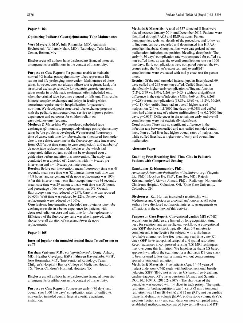
Paper #: 164
Optimizing Pediatric Gastrojejunostomy Tube Maintenance Vera Mayercik, MD
1, Julia Rissmiller, MD
1, Anastasia
Hryhorczuk1, William Mehan, MD
1;
1Radiology, Tufts Medical
Center, Boston, MA Disclosures: All authors have disclosed no financial interests, arrangements or affiliations in the context of this activity.
Purpose or Case Report: For patients unable to maintain normal PO intake, gastrojejunostomy tubes represent a life-saving and life-prolonging intervention. Maintenance of these tubes, however, does not always adhere to a regimen. Lack of a structured exchange schedule for pediatric gastrojejunostomy tubes results in problematic exchanges, often scheduled only when the original tube becomes clogged or falls out. This results
in more complex exchanges and delays in feeding which sometimes require interim hospitalization for parenteral nutrition. We developed a structured tube exchange program with the pediatric gastroenterology service to improve patient experiences and outcomes for children reliant on gastrojejunostomy feedings. Methods & Materials: We introduced scheduled tube
exchanges q3 months to preemptively change gastrojejunostomy tubes before problems developed. We measured fluoroscopy time of cases, wait time for tube exchange (measured from order date to case date), case time in the fluoroscopy suite (measured from KUB/scout time stamp to case completion), and number of de novo tube replacements (defined as a tube which had
completely fallen out and could not be exchanged over a guidewire) before and after this intervention. The study was conducted over a period of 12 months with n = 9 cases pre intervetion and n = 10 cases post intervention. Results: Before our intervention, mean fluoroscopy time was 48 seconds; mean case time was 82 minutes; mean wait time was 44.8 hours; and percentage of de novo replacements was 19%.
After this intervention, mean fluoroscopy time was 34 seconds; mean case time was 29 minutes; mean wait time was 35 hours; and percentage of de novo replacements was 0%. Overall, fluoroscopy time was reduced by 29%. Case time was reduced by 65%. Wait time was reduced by 22%. De novo tube replacements were reduced by 100%. Conclusions: Implementing scheduled gastrojejunostomy tube
exchanges results in a better experience for patients with decreased radiation dose and wait time for tube replacement. Efficiency of the fluoroscopy suite was also improved, with shorter overall duration of cases and fewer de novo replacements.
Paper #: 165
Internal jugular vein tunneled central lines: To cuff or not to
cuff? Darshan Variyam, MD
1, [email protected]; Daniel Ashton,
MD1, Heather Cleveland, BSRS
2, Shireen Hayatghaibi, MPH
2,
Jose Hernandez, MD2;
1Interventional Radiology, Texas
Children’s Hospital / Baylor College of Medicine, Houston, TX,
2Texas Children’s Hospital, Houston, TX
Disclosures: All authors have disclosed no financial interests, arrangements or affiliations in the context of this activity.
Purpose or Case Report: To measure early (<30 days) and overall (per 1000 line days) complication rates for cuffed vs. non-cuffed tunneled central lines at a tertiary academic institution.
Methods & Materials: A total of 337 tunneled IJ lines were placed between January 2014 and December 2015. Patients were identified through PACS and EMR systems. Patient demographics, technical details of the procedure, and follow-up to line removal were recorded and documented in a HIPAA-
compliant database. Complications were categorized as line malfunction, infection, malposition, bleeding, thrombosis. The early (< 30 days) complication rate was compared for cuffed vs. non-cuffed lines, as was the overall complication rate per 1000 line days. Early complications were compared between the two groups using the Fisher’s exact test, and overall[b1]
complications were evaluated with mid-p exact test for person times. Results: Of the total tunneled internal jugular lines placed, 69 were cuffed and 268 were non-cuffed. Cuffed lines had a significantly higher early complication of line malfunction (7.2%, 5/69 vs. 1.9%, 5/268, p= 0.034) without a significant difference in the rate of infection (5.8%, 4/69 vs. 3%, 8/268,
p=0.28) or total complications (18.8%, 13/69 vs. 11.2%, 30/268, p=0.11). Non-cuffed lines had an overall higher rate of malposition (2.4 vs. 1.1/1000 line days, p=0.048) and cuffed lines had a higher rate of catheter malfunction (2 vs 0.7/1000 line days, p=0.014). Differences in the remaining early and overall complications were not statistically significant. Conclusions: There was no significant difference in the
infection rate between cuffed and non-cuffed tunneled central lines. Non-cuffed lines had higher overall rates of malposition, while cuffed lines had a higher rate of early and overall line malfunction.
Alternate Paper
Enabling Free-Breathing Real-Time Cine in Pediatric
Patients with Compressed Sensing Ramkumar Krishnamurthy, PhD
1,
[email protected]; Yingmin Liu, PhD
2, Houchun Hu, PhD
1, Kan Hor, MD
1, Rajesh
Krishnamurthy1, Rizwan Ahmad, PhD
2;
1Radiology, Nationwide
Children's Hospital, Columbus, OH, 2Ohio State University,
Columbus, OH Disclosures: Kan Hor has indicated a relationship with Medtronics and Capricor as a consultant/honoraria. All other authors have disclosed no financial interests, arrangements or
affiliations in the context of this activity. Purpose or Case Report: Conventional cardiac MRI (CMR) acquisitions in children are limited by long acquisition time, need for sedation, and an inefficient workflow. A conventional cine SSFP short-axis stack typically takes 5-7 minutes to
complete and is ineffective for subjects with arrhythmias. Available alternatives like free-breathing, real-time cine (RT-cine) SSFP have suboptimal temporal and spatial resolution. Recent advances in compressed sensing (CS) MRI techniques may overcome this limitation. We hypothesize that a CS-based approach will allow the scan time for a short axis RT-cine stack to be shortened to less than a minute without compromising
spatial or temporal resolution. Methods & Materials: Five patients (Age: 14-44 years ;4 males) underwent CMR study with both conventional breath-held cine SSFP (BH-cine) as well as CS-based free-breathing, cardiac-triggered RT-cine acquisitions (Ahmad and Schniter, DOI: 10.1109/TCI.2015.2485078). The short-axis of the
ventricles was covered with 14 slices in each patient. The spatial resolution for both acquisitions was 1.8x1.8x8 mm
3; temporal
resolution was 32 ms (BH-cine) and 32 ms (RT-cine) per cardiac phase. End-diastolic volume (EDV), end-systolic volume (ESV), ejection fraction (EF), and scan duration were computed using established methods, and compared between BH-cine and RT-
Pediatr Radiol (2018) 48 (Suppl 1):S1–S298S176
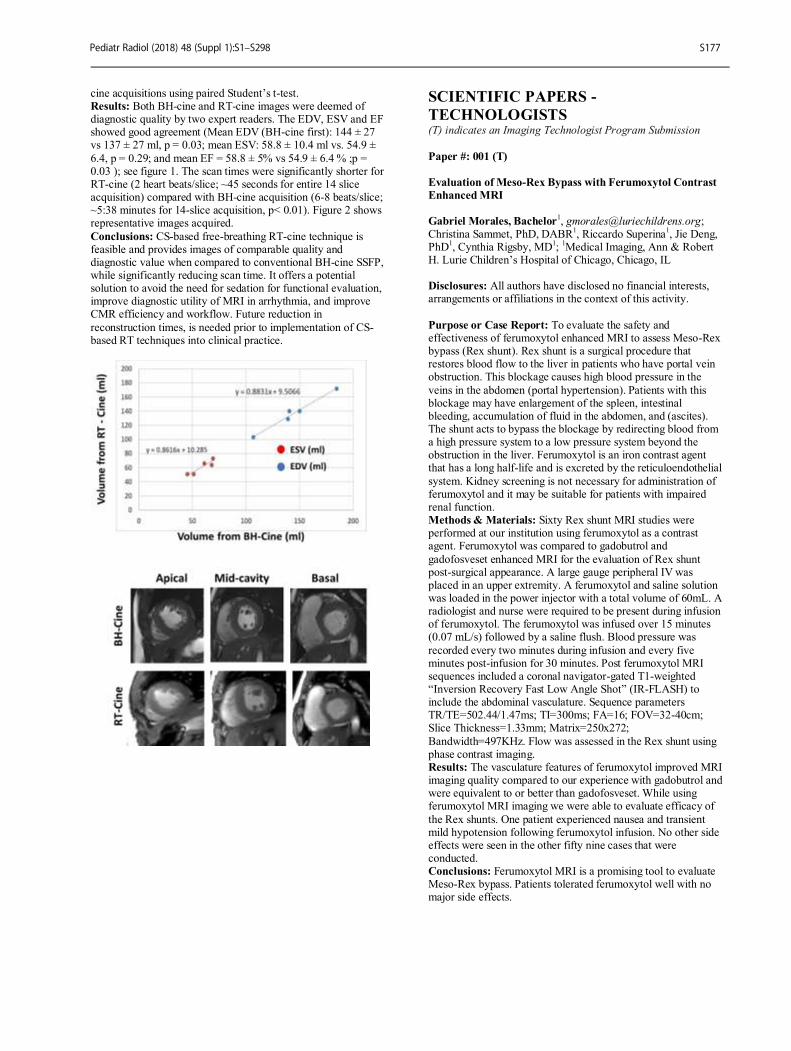
cine acquisitions using paired Student’s t-test. Results: Both BH-cine and RT-cine images were deemed of diagnostic quality by two expert readers. The EDV, ESV and EF showed good agreement (Mean EDV (BH-cine first): 144 ± 27 vs 137 ± 27 ml, p = 0.03; mean ESV: 58.8 ± 10.4 ml vs. 54.9 ±
6.4, p = 0.29; and mean EF = 58.8 ± 5% vs 54.9 ± 6.4 % ;p = 0.03 ); see figure 1. The scan times were significantly shorter for RT-cine (2 heart beats/slice; ~45 seconds for entire 14 slice acquisition) compared with BH-cine acquisition (6-8 beats/slice; ~5:38 minutes for 14-slice acquisition, p< 0.01). Figure 2 shows representative images acquired.
Conclusions: CS-based free-breathing RT-cine technique is feasible and provides images of comparable quality and diagnostic value when compared to conventional BH-cine SSFP, while significantly reducing scan time. It offers a potential solution to avoid the need for sedation for functional evaluation, improve diagnostic utility of MRI in arrhythmia, and improve CMR efficiency and workflow. Future reduction in
reconstruction times, is needed prior to implementation of CS-based RT techniques into clinical practice.
SCIENTIFIC PAPERS -
TECHNOLOGISTS (T) indicates an Imaging Technologist Program Submission
Paper #: 001 (T)
Evaluation of Meso-Rex Bypass with Ferumoxytol Contrast
Enhanced MRI
Gabriel Morales, Bachelor1, [email protected];
Christina Sammet, PhD, DABR1, Riccardo Superina
1, Jie Deng,
PhD1, Cynthia Rigsby, MD
1;
1Medical Imaging, Ann & Robert
H. Lurie Children’s Hospital of Chicago, Chicago, IL Disclosures: All authors have disclosed no financial interests, arrangements or affiliations in the context of this activity.
Purpose or Case Report: To evaluate the safety and effectiveness of ferumoxytol enhanced MRI to assess Meso-Rex bypass (Rex shunt). Rex shunt is a surgical procedure that restores blood flow to the liver in patients who have portal vein obstruction. This blockage causes high blood pressure in the
veins in the abdomen (portal hypertension). Patients with this blockage may have enlargement of the spleen, intestinal bleeding, accumulation of fluid in the abdomen, and (ascites). The shunt acts to bypass the blockage by redirecting blood from a high pressure system to a low pressure system beyond the obstruction in the liver. Ferumoxytol is an iron contrast agent that has a long half-life and is excreted by the reticuloendothelial
system. Kidney screening is not necessary for administration of ferumoxytol and it may be suitable for patients with impaired renal function. Methods & Materials: Sixty Rex shunt MRI studies were performed at our institution using ferumoxytol as a contrast agent. Ferumoxytol was compared to gadobutrol and
gadofosveset enhanced MRI for the evaluation of Rex shunt post-surgical appearance. A large gauge peripheral IV was placed in an upper extremity. A ferumoxytol and saline solution was loaded in the power injector with a total volume of 60mL. A radiologist and nurse were required to be present during infusion of ferumoxytol. The ferumoxytol was infused over 15 minutes (0.07 mL/s) followed by a saline flush. Blood pressure was
recorded every two minutes during infusion and every five minutes post-infusion for 30 minutes. Post ferumoxytol MRI sequences included a coronal navigator-gated T1-weighted “Inversion Recovery Fast Low Angle Shot” (IR-FLASH) to include the abdominal vasculature. Sequence parameters TR/TE=502.44/1.47ms; TI=300ms; FA=16; FOV=32-40cm; Slice Thickness=1.33mm; Matrix=250x272;
Bandwidth=497KHz. Flow was assessed in the Rex shunt using phase contrast imaging. Results: The vasculature features of ferumoxytol improved MRI imaging quality compared to our experience with gadobutrol and were equivalent to or better than gadofosveset. While using ferumoxytol MRI imaging we were able to evaluate efficacy of
the Rex shunts. One patient experienced nausea and transient mild hypotension following ferumoxytol infusion. No other side effects were seen in the other fifty nine cases that were conducted. Conclusions: Ferumoxytol MRI is a promising tool to evaluate Meso-Rex bypass. Patients tolerated ferumoxytol well with no major side effects.
Pediatr Radiol (2018) 48 (Suppl 1):S1–S298 S177
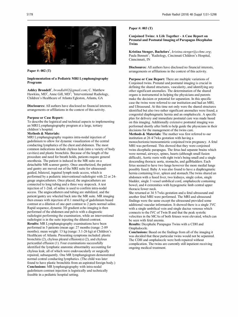
Paper #: 002 (T)
Implementation of a Pediatric MRI Lymphangiography
Programs Ashley Brondell
1, [email protected]; C. Matthew
Hawkins, MD1, Anne Gill, MD
1;
1Interventional Radiology,
Children’s Healthcare of Atlanta Egleston, Atlanta, GA Disclosures: All authors have disclosed no financial interests, arrangements or affiliations in the context of this activity.
Purpose or Case Report: To describe the logistical and technical aspects to implementing an MRI Lymphangiography program at a large, tertiary children’s hospital.
Methods & Materials: MRI Lymphangiography requires intra-nodal injection of gadolinium to allow for dynamic visualization of the central
conducting lymphatics of the chest and abdomen. The most common indications include chylous leak (into a variety of body cavities) and plastic bronchitis. Because of the length of the procedure and need for breath holds, patients require general anesthesia. The patient is induced in the MR suite on a detachable MR scanner gantry. Once anesthetized, the patient and gantry are moved out of the MRI suite for ultrasound-
guided, bilateral, inguinal lymph node access, which is performed by a pediatric interventional radiologist with 22 or 24-gauge angiocatheters. Once placed, the angiocatheters are connected to long tubing and a three way stopcock. A test injection of 1-2mL of saline is used to confirm intra-nodal access. The angiocatheters and tubing are stabilized, and the
patient/gantry are wheeled back into the MR suite. MR imaging then ensues with injection of 0.1 mmol/kg of gadolinium based contrast at a dilution of one-part contrast to 2 parts normal saline. Rapid sequence, dynamic 3D gradient echo imaging is then performed of the abdomen and pelvis with a diagnostic radiologist performing the examination, while an interventional radiologist is in the suite injecting the diluted contrast.
Results: MR Lymphangiography examinations have been performed in 5 patients (mean age: 27 months (range: 2-89 months); mean weight: 13 kg (range: 3.1-26 kg) at Children’s Healthcare of Atlanta. Presenting symptoms included: plastic bronchitis (2), chylous pleural effusion(s) (2), and chylous pericardial effusion (1). Four examinations successfully identified the lymphatic anatomic abnormality accounting for
chylous leak; all of which were endovascularly or surgically repaired, subsequently. One MR lymphangiogram demonstrated normal central conducting lymphatics. (The child was later found to have plastic bronchitis from an aspirated foreign body.) Conclusions: MR lymphangiography with intra-nodal gadolinium contrast injection is logistically and technically
feasible in a pediatric hospital setting.
Paper #: 003 (T)
Conjoined Twins: A Life Together - A Case Report on
Prenatal and Postnatal Imaging of Parapagus Dicephalus
Twins
Kristina Stenger, Bachelors
Paula Bennett1;
1Radiology, Cincinnati Children’s Hospital,
Cinncinnati, IN Disclosures: All authors have disclosed no financial interests,
arrangements or affiliations in the context of this activity. Purpose or Case Report: There are multiple variations of Conjoined twins. Prenatal and postnatal imaging is crucial in defining the shared structures, vascularity, and identifying any other significant anomalies. The determination of the shared organs is instrumental in helping the physicians and parents
make the decision or potential for separation. In this specific case the twins were referred to our institution and had an MRI, and Ultrasound. At this time not only were the shared structures identified but also two rather significant anomalies were found, a congenital diaphragmatic hernia and an omphalocele. A specific plan for delivery and immediate postnatal care was made based on this imaging. Additionally extensive postnatal imaging was
performed shortly after birth to help guide the physicians in their decisions for the management of the twins care. Methods & Materials: The mother was first referred to our institution at 26 4/7wks gestation with having a monochorionic/monoamniotic conjoined twin pregnancy. A fetal MRI was performed. This showed that they were conjoined
twins dicephalic parapagus. The fetus had separate brains which were normal, airways, spines, hearts (although small fusion difficult), Aortic roots with right twin's being small and a single descending thoracic aorta, stomachs, and gallbladders. Each fetus seemed to have two lungs however the more medial lungs possibly fused. Baby A was also found to have a diaphragmatic hernia containing liver, spleen and stomach.The twins shared an
abdomen with a fused liver, two kidneys, single colon, single bladder, single 3 vessel umbilical cord, omphalocele containing bowel, and 4 extremities with hypogenetic limb central upper thoracic/lower neck. She returned at 34 5/7wks gestation and a fetal ultrasound and another fetal MRI were performed. The MRI and ultrasound findings were the same except the ultrasound provided some
additional vascular information. It showed there is a single 3VC with a single umbilical vein and single ductus venosus which connects to the IVC of Twin B and that the peak systolic velocities in the MCAs of both fetuses were elevated, which can be seen with fetal anemia. Results: Dicephalic Parapagus Twins with a CDH and
Omphalocele. Conclusions: Based on the findings from all of the imaging it was decided that these particular twins would not be separated. The CDH and omphalocele were both repaired without complication. The twins are currently still inpatient receiving ongoing medical treatment.
Pediatr Radiol (2018) 48 (Suppl 1):S1–S298S178
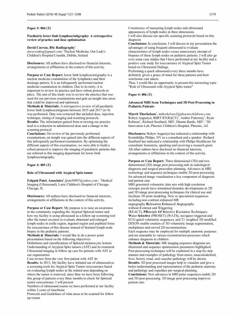
Paper #: 004 (T)
Paediatric lower limb lymphoscintigraphy: A retrospective
review of practice and dose optimisation
David Curran, BSc Radiography1
[email protected]; 1Nuclear Medicine, Our Lady's
Children's Hospital Crumlin, Dublin, Ireland Disclosures: All authors have disclosed no financial interests, arrangements or affiliations in the context of this activity.
Purpose or Case Report: lower limb lymphoscintigraphy is a nuclear medicine examination of the lymphatics and their drainage pattern. It is an infrequently performed nuclear medicine examination in children. Due to its rarity, it is important to review its practice and have robust protocols in place. The aim of this study was to review the practice that was
used for our previous examinations and gain an insight into areas that could be improved and optimised. Methods & Materials: A retrospective review of all paediatric lower limb lymphoscintigrams between 2015 and 2017 (n=8) was performed. Data was extracted that included dose, injection technique, timing of imaging and scanning protocols. Results: The information gained from reviewing our practice
lead to a reduction in administered dose and a change to the scanning protocol. Conclusions: On review of the previously performed examinations, an insight was gained into the different aspects of this infrequently performed examination. By optimising the different aspects of this examination, we were able to build a
robust protocol to improve the imaging of paediatric patients that are referred to this imaging department for lower limb lymphoscintigraphy.
Paper #: 005 (T)
Role of Ultrasound with Atypical Spitz tumor
Falguni Patel, Associates
1Medical
Imaging (Ultrasound), Lurie Children's Hospital of Chicago, Chicago, IL Disclosures: All authors have disclosed no financial interests, arrangements or affiliations in the context of this activity.
Purpose or Case Report: My purpose is to raise an awareness in the community regarding the Atypical Spitzoid tumor and how my facility is using ultrasound as a follow-up screening tool after the tumor excision to evaluate abnormal and enlarged lymph nodes in axilla region, inguinal region and popliteal fossa
for reoccurrence of this disease instead of Sentinel lymph node biopsy in the pediatric patients. Methods & Materials: I would like to do a power point presentation based on the following objectives: Definitions and classifications of Spitzoid melanocytic lesions Understanding of Atypical Spitz tumors (AST) and its treatment Ultrasound imaging in follow up care for patients with AST at
our organization Case review from the very first patient with AST dx Results: In 2015, My facility have initiated use of ultrasound as a screening tools for Atypical Spitz Tumor reoccurrence based on evaluating lymph nodes in the related area depending on where the tumor is removed, since than we have been following
this group of patients every three months to check for Spitzoid tumor reoccurrence. I will present Numbers of ultrasound exams we have performed at our facility within 3 years of timeframe Protocols and Guidelines of what areas to be scanned for follow up exams
Consistency of measuring lymph nodes and ultrasound appearances of lymph nodes in three dimensions I will also discuss our specific scanning protocols based on this diagnosis Conclusions: In conclusion, I will discuss in my presentation the
advantages of using frequent ultrasound to evaluate characteristics of lymph nodes verses unnecessary attempt of biopsies of these lymph nodes on pediatric patients. I will also go over some case studies that I have performed at my facility and a positive case study for reoccurrence of Atypical Spitz Tumor based on Ultrasound findings.
Performing a quick ultrasound every three months have definitely given a peace of mind for these patients and their worrisome care takers. Thus, I would like an opportunity to present this interesting topic "Role of Ultrasound with Atypical Spitz tumor"
Paper #: 006 (T)
Advanced MRI Scan Techniques and 3D Post-Processing in
Pediatric Patients Marrit Thorkelson
Robyn Augustyn, BSRT RT(R)(CT)1, Amber Pokorney
1, Ryan
Robison1, Richard Southard, MD
1, Dianna Bardo, MD
1;
13D
Innovation Lab, Phoenix Children's Hospital, Phoenix, AZ Disclosures: Robyn Augustyn has indicated a relationship with Koninklijke Philips, NV as a consultant and a speaker. Richard Southard has indicated a relationship with Philips Healthcare for consultant/ honoraria, speaking and receiving a research grant.
All other authors have disclosed no financial interests, arrangements or affiliations in the context of this activity.
Purpose or Case Report: Three dimensional (3D) and two dimensional (2D) image post processing aids in radiological diagnosis and surgical procedure planning. Advances in MRI technology and sequence techniques enable 3D post processing
for advanced image visualization a key component of diagnosis and patient care. MRI generated volumetric data sets with high resolution isotropic pixels have stimulated dramatic developments in 2D and 3D image post processing techniques for clinical use and facilitate 3D print modeling. New or specialized sequences including non-contrast enhanced MR
angiography Relaxation Enhanced Angiography without Contrast and Triggering (REACT), PRinciple Of Selective Excitation Techniques-Water Selective (PROSET) (WATS), navigator triggered and ECG-gated volumetric sequences, and T1 weighted 3D modified DIXON enable creation of 3D volumetric image segmentation,
multiplanar and curved 2D reconstructions. Each sequence may be employed for multiple anatomic purposes and are amenable to various reconstruction processes which enhance diagnosis in children. Methods & Materials: MR imaging sequence diagrams are illustrated and sequence optimization parameters highlighted. Post-processing techniques will be explained in a step-by-step
manner and examples of pathology from neuro, musculoskeletal, liver, bowel, renal, and vascular pathology will be shown. Results: 3D post processed images help to visualize and give a better understanding and representation of the pediatric anatomy and pathology and expedites pre-surgical planning. Conclusions: New advances in MRI pulse sequences enable 2D
and 3D post-processing. 3D image post processing improves patient care.
Pediatr Radiol (2018) 48 (Suppl 1):S1–S298 S179
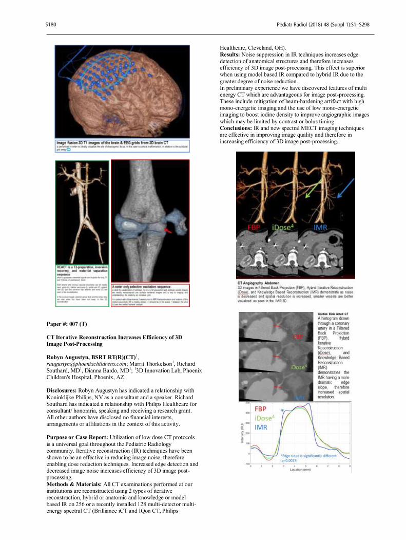
Paper #: 007 (T)
CT Iterative Reconstruction Increases Efficiency of 3D
Image Post-Processing
Robyn Augustyn, BSRT RT(R)(CT)
1,
[email protected]; Marrit Thorkelson1, Richard
Southard, MD1, Dianna Bardo, MD
1;
13D Innovation Lab, Phoenix
Children's Hospital, Phoenix, AZ Disclosures: Robyn Augustyn has indicated a relationship with
Koninklijke Philips, NV as a consultant and a speaker. Richard Southard has indicated a relationship with Philips Healthcare for consultant/ honoraria, speaking and receiving a research grant. All other authors have disclosed no financial interests, arrangements or affiliations in the context of this activity. Purpose or Case Report: Utilization of low dose CT protocols
is a universal goal throughout the Pediatric Radiology community. Iterative reconstruction (IR) techniques have been shown to be an effective in reducing image noise, therefore enabling dose reduction techniques. Increased edge detection and decreased image noise increases efficiency of 3D image post-processing.
Methods & Materials: All CT examinations performed at our institutions are reconstructed using 2 types of iterative reconstruction, hybrid or anatomic and knowledge or model based IR on 256 or a recently installed 128 multi-detector multi-energy spectral CT (Brilliance iCT and IQon CT, Philips
Healthcare, Cleveland, OH). Results: Noise suppression in IR techniques increases edge detection of anatomical structures and therefore increases efficiency of 3D image post-processing. This effect is superior when using model based IR compared to hybrid IR due to the
greater degree of noise reduction. In preliminary experience we have discovered features of multi energy CT which are advantageous for image post-processing. These include mitigation of beam-hardening artifact with high mono-energetic imaging and the use of low mono-energetic imaging to boost iodine density to improve angiographic images
which may be limited by contrast or bolus timing. Conclusions: IR and new spectral MECT imaging techniques are effective in improving image quality and therefore in increasing efficiency of 3D image post-processing.
Pediatr Radiol (2018) 48 (Suppl 1):S1–S298S180
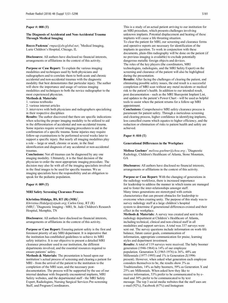
Paper #: 008 (T)
The Diagnosis of Accidental and Non-Accidental Trauma
Through Medical Imaging
Rocco Pazienza1 [email protected];
1Medical Imaging,
Lurie Children’s Hospital, Chicago, IL Disclosures: All authors have disclosed no financial interests, arrangements or affiliations in the context of this activity.
Purpose or Case Report: To explain the various imaging modalities and techniques used by both physicians and radiographers and to correlate them to both acute and chronic accidental and non-accidental traumas with the diagnostic modality that best demonstrates that particular injury. The author will show the importance and usage of various imaging modalities and techniques to both the novice radiographer to the
most experienced physician.
Methods & Materials: 1. various textbooks 2. various internet articles 3. interviews with both physicians and radiographers specializing in their respective disciplines Results: The author discovered that there are specific indications
when selecting the proper imaging modality to be utilized to aid in the differentiation of accidental and non-accidental trauma. Some injuries require several imaging procedures to aid in the confirmation of a specific trauma. Some injuries may require follow-up examinations to be performed several weeks later to support a specific injury. But nearly all imaging modalities play
a role -- large or small, chronic or acute, in the final identification and diagnosis of any accidental or non-accidental traumas. Conclusions: Not all traumas can be diagnosed by any one imaging modality. Ultimately, it is the final decision of the physician to order the most appropriate imaging procedure. The decision may also lie with all of the imaging specialists involved
in the final images to be used for specific traumas. We as imaging specialists have the responsibility and an obligation to speak for the pediatric population.
Paper #: 009 (T)
MRI Safety Screening Clearance Process
Khristina Hidalgo, BS, RT (R) (MR)
1,
[email protected]; Carlos Gray, RT (R) (MR)
1;
1Diagnostic Imaging - MRI, St. Jude Children's Research
Hospital, Memphis, TN
Disclosures: All authors have disclosed no financial interests, arrangements or affiliations in the context of this activity. Purpose or Case Report: Ensuring patient safety is the first and foremost priority of any MRI department. It is imperative that the institution has established guidelines to achieve its MRI safety initiative. It is our objective to present a detailed MRI
clearance procedure used in our institution, the different departments involved, and the essential resources required to ensure patients’ safety. Methods & Materials: The presentation is based upon our institution’s actual process of screening and clearing a patient for MRI - from the arrival of the patient to the institution to the
completion of the MRI scan, and the necessary post-documentation. The process will be supported by the use of our internal database with frequently encountered implants, MRI Safety websites, and the departmental staff - the MRI Safety Expert, Radiologists, Nursing Surgical Services Pre-screening Staff, and Program Coordinators.
This is a study of an actual patient arriving to our institution for an MRI procedure, which presents challenges involving unknown implants. Potential displacement and heating of these implants will cause a life threating situation. To clear the patient for MRI, our screening and clearance forms
and operative reports are necessary for identification of the implants in question. To work in conjunction with these documents, plain-film radiography will be done on the patient (if no previous imaging is available) to exclude potentially dangerous metallic foreign objects and devices. The roles of the key players (the coordinators, MRI
technologists, radiologists, and the MRI Safety Expert) on the screening and clearance of the patient will also be highlighted during the presentation. Results: After facing the challenges of clearing the patient, and eliminating possible safety issues, the end result is a successful completion of MRI scan without any metal incidents or medical risk to the patient’s health. In addition to our intended result,
post documentation - such as the MRI Sharepoint Implant List, and updates to the patient’s Power Chart - will be used as helpful tools to assist when the patient returns for a follow-up MRI appointment. Conclusions: Comprehensive MRI safety clearance process is paramount for patient safety. Through a meticulous screening and clearing process, higher confidence in identifying implants,
less cancelled exams which equates to higher efficiency, and the reduction or elimination of risks to patient health and safety are achieved.
Paper #: 010 (T)
Generational Differences in the Workplace Melissa Goehner
1Diagnostic
Radiology, Children's Healthcare of Atlanta, Stone Mountain, GA Disclosures: All authors have disclosed no financial interests,
arrangements or affiliations in the context of this activity. Purpose or Case Report: With the changing of generations in the radiology workforce, there is increased importance for leadership to address the manner in which teams are managed and to foster the inter-relationships amongst staff. Many times generations are stereotyped with certain
characteristics that can present obstacles for leadership to overcome when creating unity. The purpose of this study was to survey radiology staff at a large children’s hospital system to determine if generational differences existed and their effect in the workplace. Methods & Materials: A survey was created and sent to the
radiology department at Children’s Healthcare of Atlanta, including technical, clinical and non-clinical staff in all modalities and support services. A total was 337 surveys were sent out. The survey questions include information on work-life balance, future career goals, communication of information, appropriate communication for praise, learning styles and department investment.
Results: A total of 119 surveys were received. The baby boomer generation (1946-1964) is 14% of our employee population. Generation X (1965-1976) is 36%. 48% are Millennials (1977-1995) and 1% is Generation Z(1996- present). However, when asked what generation each employee considers themselves to be, the results show 3% as
Traditionalists, 14% as baby boomers, 59% as Generation X and 25% are Millennials. When asked how they like to receive information, 53% prefer to be communicated to by e-mail and 30% prefer to be communicated to by text message. The top 3 social media websites that the staff uses are e-mail (92%), Facebook (67%) and Instagram
Pediatr Radiol (2018) 48 (Suppl 1):S1–S298 S181
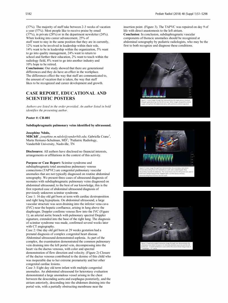
(37%). The majority of staff take between 2-3 weeks of vacation a year (57%). Most people like to receive praise by email (37%), in private (28%) or in the department newsletter (24%). When looking into career advancement, 25% of staff want to stay in the same position that they are in currently,
12% want to be involved in leadership within their role, 14% want to be in leadership within the organization, 5% want to go into quality management, 24% want to return to school and further their education, 2% want to teach within the radiology field, 8% want to go into another industry and 10% hope to be retired.
Conclusions: Our study showed that there are generational differences and they do have an effect in the workplace. The differences effect the way that staff are communicated to, the amount of vacation that is taken, the way that staff likes to be recognized and career development and growth.
CASE REPORT, EDUCATIONAL AND
SCIENTIFIC POSTERS Authors are listed in the order provided. An author listed in bold
identifies the presenting author.
Poster #: CR-001
Subdiaphragmatic pulmonary veins identified by ultrasound.
Josephine Ndolo,
MBChB1, [email protected]; Gabriella Crane
1,
Marta Hernanz-Schulman, MD1;
1Pediatric Radiology,
Vanderbilt University, Nashville, TN Disclosures: All authors have disclosed no financial interests, arrangements or affiliations in the context of this activity.
Purpose or Case Report: Scimitar syndrome and subdiaphragmatic total anomalous pulmonary venous connections (TAPVC) are congenital pulmonary vascular anomalies that are not typically diagnosed on routine abdominal sonography. We present three cases of ultrasound diagnosis of neonates with subdiaphragmatic pulmonary veins diagnosed on
abdominal ultrasound; to the best of our knowledge, this is the first reported case of abdominal ultrasound diagnosis of previously unknown scimitar syndrome. Case 1: 16 day old girl born at term with cardiac dextroposition and right lung hypoplasia. On abdominal ultrasound, a large vascular structure was seen draining into the inferior vena cava (IVC) near the hepatic confluence, arising in lung above the
diaphragm. Doppler confirms venous flow into the IVC (Figure 1); an arterial aortic branch with pulmonary spectral Doppler signature, extended into the base of the right lung. The diagnosis of scimitar syndrome was made, confirmed several weeks later with CT angiography. Case 2: One day old girl born at 29 weeks gestation had a
prenatal diagnosis of complex congenital heart disease. Abdominal ultrasound demonstrated asplenia. As part of the complex, the examination demonstrated the common pulmonary vein draining into the left portal vein, decompressing into the heart via the ductus venosus, with color and spectral demonstration of flow direction and velocity. (Figure 2) Closure of the ductus venosus contributed to the demise of this child who
was inoperable due to her extreme prematurity and her other congenital cardiac lesions. Case 3: Eight day old term infant with multiple congenital anomalies. An abdominal ultrasound for heterotaxy evaluation demonstrated a large anomalous vessel arising in the chest between the descending aorta and esophagus posteriorly, and the atrium anteriorly, descending into the abdomen draining into the
portal vein, with a partially obstructing membrane near the
insertion point. (Figure 3). The TAPVC was repaired on day 9 of life with direct anastomosis to the left atrium. Conclusion: In conclusion, subdiaphragmatic vascular components of thoracic anomalies should be recognized at abdominal sonography by pediatric radiologists, who may be the first to both recognize and diagnose these conditions.
Pediatr Radiol (2018) 48 (Suppl 1):S1–S298S182
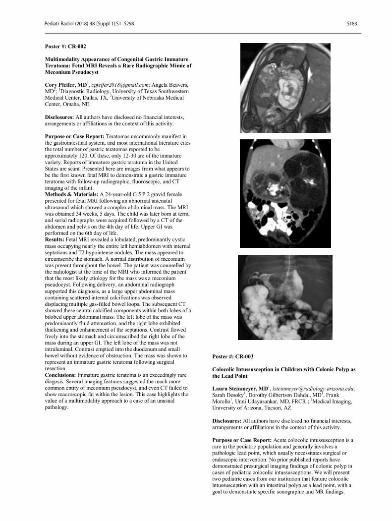
Poster #: CR-002
Multimodality Appearance of Congenital Gastric Immature
Teratoma: Fetal MRI Reveals a Rare Radiographic Mimic of
Meconium Pseudocyst
Cory Pfeifer, MD
1, [email protected]; Angela Beavers,
MD2;
1Diagnostic Radiology, University of Texas Southwestern
Medical Center, Dallas, TX, 2University of Nebraska Medical
Center, Omaha, NE
Disclosures: All authors have disclosed no financial interests, arrangements or affiliations in the context of this activity. Purpose or Case Report: Teratomas uncommonly manifest in the gastrointestinal system, and most international literature cites the total number of gastric teratomas reported to be approximately 120. Of these, only 12-30 are of the immature
variety. Reports of immature gastric teratoma in the United States are scant. Presented here are images from what appears to be the first known fetal MRI to demonstrate a gastric immature teratoma with follow-up radiographic, fluoroscopic, and CT imaging of the infant. Methods & Materials: A 24-year-old G 5 P 2 gravid female presented for fetal MRI following an abnormal antenatal
ultrasound which showed a complex abdominal mass. The MRI was obtained 34 weeks, 5 days. The child was later born at term, and serial radiographs were acquired followed by a CT of the abdomen and pelvis on the 4th day of life. Upper GI was performed on the 6th day of life. Results: Fetal MRI revealed a lobulated, predominantly cystic
mass occupying nearly the entire left hemiabdomen with internal septations and T2 hypointense nodules. The mass appeared to circumscribe the stomach. A normal distribution of meconium was present throughout the bowel. The patient was counselled by the radiologist at the time of the MRI who informed the patient that the most likely etiology for the mass was a meconium pseudocyst. Following delivery, an abdominal radiograph
supported this diagnosis, as a large upper abdominal mass containing scattered internal calcifications was observed displacing multiple gas-filled bowel loops. The subsequent CT showed these central calcified components within both lobes of a bilobed upper abdominal mass. The left lobe of the mass was predominantly fluid attenuation, and the right lobe exhibited thickening and enhancement of the septations. Contrast flowed
freely into the stomach and circumscribed the right lobe of the mass during an upper GI. The left lobe of the mass was not intraluminal. Contrast emptied into the duodenum and small bowel without evidence of obstruction. The mass was shown to represent an immature gastric teratoma following surgical resection.
Conclusions: Immature gastric teratoma is an exceedingly rare diagosis. Several imaging features suggested the much more common entity of meconium pseudocyst, and even CT failed to show macroscopic fat within the lesion. This case highlights the value of a multimodality approach to a case of an unusual pathology.
Poster #: CR-003
Colocolic Intussusception in Children with Colonic Polyp as
the Lead Point Laura Steinmeyer, MD
Sarah Desoky1, Dorothy Gilbertson Dahdal, MD
1, Frank
Morello1, Unni Udayasankar, MD, FRCR
1;
1Medical Imaging,
University of Arizona, Tucson, AZ
Disclosures: All authors have disclosed no financial interests, arrangements or affiliations in the context of this activity. Purpose or Case Report: Acute colocolic intussusception is a rare in the pediatric population and generally involves a pathologic lead point, which usually necessitates surgical or
endoscopic intervention. No prior published reports have demonstrated presurgical imaging findings of colonic polyp in cases of pediatric colocolic intussusceptions. We will present two pediatric cases from our institution that feature colocolic intussusception with an intestinal polyp as a lead point, with a goal to demonstrate specific sonographic and MR findings.
Pediatr Radiol (2018) 48 (Suppl 1):S1–S298 S183
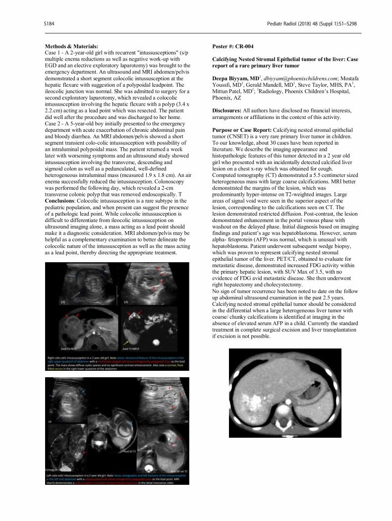
Methods & Materials: Case 1 - A 2-year-old girl with recurrent "intussusceptions" (s/p multiple enema reductions as well as negative work-up with EGD and an elective exploratory laparotomy) was brought to the emergency department. An ultrasound and MRI abdomen/pelvis
demonstrated a short segment colocolic intussusception at the hepatic flexure with suggestion of a polypoidal leadpoint. The ileocolic junction was normal. She was admitted to surgery for a second exploratory laparotomy, which revealed a colocolic intussusception involving the hepatic flexure with a polyp (3.4 x 2.2.cm) acting as a lead point which was resected. The patient
did well after the procedure and was discharged to her home. Case 2 - A 5-year-old boy initially presented to the emergency department with acute exacerbation of chronic abdominal pain and bloody diarrhea. An MRI abdomen/pelvis showed a short segment transient colo-colic intussusception with possibility of an intraluminal polypoidal mass. The patient returned a week later with worsening symptoms and an ultrasound study showed
intussusception involving the transverse, descending and sigmoid colon as well as a pedunculated, well-defined heterogeneous intraluminal mass (measured 1.9 x 1.8 cm). An air enema successfully reduced the intussusception. Colonoscopy was performed the following day, which revealed a 2-cm transverse colonic polyp that was removed endoscopically. T Conclusions: Colocolic intussusception is a rare subtype in the
pediatric population, and when present can suggest the presence of a pathologic lead point. While colocolic intussusception is difficult to differentiate from ileocolic intussusception on ultrasound imaging alone, a mass acting as a lead point should make it a diagnostic consideration. MRI abdomen/pelvis may be helpful as a complementary examination to better delineate the
colocolic nature of the intussusception as well as the mass acting as a lead point, thereby directing the appropriate treatment.
Poster #: CR-004
Calcifying Nested Stromal Epithelial tumor of the liver: Case
report of a rare primary liver tumor
Deepa Biyyam, MD1, [email protected]; Mostafa
Youssfi, MD1, Gerald Mandell, MD
1, Steve Taylor, MHS, PA
1,
Mittun Patel, MD1;
1Radiology, Phoenix Children’s Hospital,
Phoenix, AZ Disclosures: All authors have disclosed no financial interests,
arrangements or affiliations in the context of this activity. Purpose or Case Report: Calcifying nested stromal epithelial tumor (CNSET) is a very rare primary liver tumor in children. To our knowledge, about 30 cases have been reported in literature. We describe the imaging appearance and histopathologic features of this tumor detected in a 2 year old
girl who presented with an incidentally detected calcified liver lesion on a chest x-ray which was obtained for cough. Computed tomography (CT) demonstrated a 5.5 centimeter sized heterogeneous mass with large coarse calcifications. MRI better demonstrated the margins of the lesion, which was predominantly hyper-intense on T2-weighted images. Large areas of signal void were seen in the superior aspect of the
lesion, corresponding to the calcifications seen on CT. The lesion demonstrated restricted diffusion. Post-contrast, the lesion demonstrated enhancement in the portal venous phase with washout on the delayed phase. Initial diagnosis based on imaging findings and patient’s age was hepatoblastoma. However, serum alpha- fetoprotein (AFP) was normal, which is unusual with
hepatoblastoma. Patient underwent subsequent wedge biopsy, which was proven to represent calcifying nested stromal epithelial tumor of the liver. PET/CT, obtained to evaluate for metastatic disease, demonstrated increased FDG activity within the primary hepatic lesion, with SUV Max of 3.5, with no evidence of FDG avid metastatic disease. She then underwent right hepatectomy and cholecystectomy.
No sign of tumor recurrence has been noted to date on the follow up abdominal ultrasound examination in the past 2.5 years. Calcifying nested stromal epithelial tumor should be considered in the differential when a large heterogeneous liver tumor with coarse/ chunky calcifications is identified at imaging in the absence of elevated serum AFP in a child. Currently the standard treatment in complete surgical excision and liver transplantation if excision is not possible.
Pediatr Radiol (2018) 48 (Suppl 1):S1–S298S184
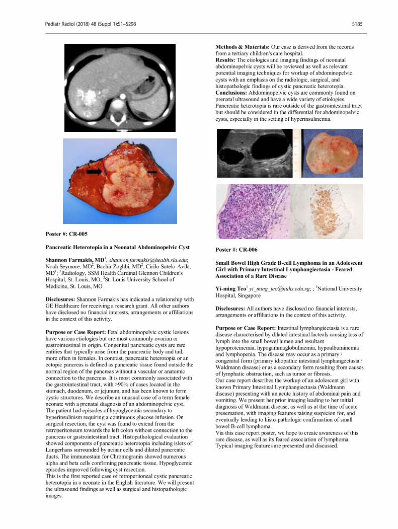
Poster #: CR-005
Pancreatic Heterotopia in a Neonatal Abdominopelvic Cyst Shannon Farmakis, MD
Noah Seymore, MD2, Bachir Zoghbi, MD
2, Cirilo Sotelo-Avila,
MD1;
1Radiology, SSM Health Cardinal Glennon Children's
Hospital, St. Louis, MO, 2St. Louis University School of
Medicine, St. Louis, MO Disclosures: Shannon Farmakis has indicated a relationship with GE Healthcare for receiving a research grant. All other authors have disclosed no financial interests, arrangements or affiliations in the context of this activity.
Purpose or Case Report: Fetal abdominopelvic cystic lesions have various etiologies but are most commonly ovarian or gastrointestinal in origin. Congenital pancreatic cysts are rare entities that typically arise from the pancreatic body and tail, more often in females. In contrast, pancreatic heterotopia or an ectopic pancreas is defined as pancreatic tissue found outside the
normal region of the pancreas without a vascular or anatomic connection to the pancreas. It is most commonly associated with the gastrointestinal tract, with >90% of cases located in the stomach, duodenum, or jejunum, and has been known to form cystic structures. We describe an unusual case of a term female neonate with a prenatal diagnosis of an abdominopelvic cyst. The patient had episodes of hypoglycemia secondary to
hyperinsulinism requiring a continuous glucose infusion. On surgical resection, the cyst was found to extend from the retroperitoneum towards the left colon without connection to the pancreas or gastrointestinal tract. Histopathological evaluation showed components of pancreatic heterotopia including islets of Langerhans surrounded by acinar cells and dilated pancreatic
ducts. The immunostain for Chromogranin showed numerous alpha and beta cells confirming pancreatic tissue. Hypoglycemic episodes improved following cyst resection. This is the first reported case of retroperitoneal cystic pancreatic heterotopia in a neonate in the English literature. We will present the ultrasound findings as well as surgical and histopathologic images.
Methods & Materials: Our case is derived from the records from a tertiary children's care hospital. Results: The etiologies and imaging findings of neonatal abdominopelvic cysts will be reviewed as well as relevant potential imaging techniques for workup of abdominopelvic
cysts with an emphasis on the radiologic, surgical, and histopathologic findings of cystic pancreatic heterotopia. Conclusions: Abdominopelvic cysts are commonly found on prenatal ultrasound and have a wide variety of etiologies. Pancreatic heterotopia is rare outside of the gastrointestinal tract but should be considered in the differential for abdominopelvic
cysts, especially in the setting of hyperinsulinemia.
Poster #: CR-006
Small Bowel High Grade B-cell Lymphoma in an Adolescent
Girl with Primary Intestinal Lymphangiectasia - Feared
Association of a Rare Disease Yi-ming Teo
1 [email protected]; ;
1National University
Hospital, Singapore Disclosures: All authors have disclosed no financial interests, arrangements or affiliations in the context of this activity. Purpose or Case Report: Intestinal lymphangiectasia is a rare disease characterised by dilated intestinal lacteals causing loss of
lymph into the small bowel lumen and resultant hypoproteinemia, hypogammaglobulinemia, hypoalbuminemia and lymphopenia. The disease may occur as a primary / congenital form (primary idiopathic intestinal lymphangectasia / Waldmann disease) or as a secondary form resulting from causes of lymphatic obstruction, such as tumor or fibrosis. Our case report describes the workup of an adolescent girl with
known Primary Intestinal Lymphangiectasia (Waldmann disease) presenting with an acute history of abdominal pain and vomiting. We present her prior imaging leading to her initial diagnosis of Waldmann disease, as well as at the time of acute presentation, with imaging features raising suspicion for, and eventually leading to histo-pathologic confirmation of small
bowel B-cell lymphoma. Via this case report poster, we hope to create awareness of this rare disease, as well as its feared association of lymphoma. Typical imaging features are presented and discussed.
Pediatr Radiol (2018) 48 (Suppl 1):S1–S298 S185
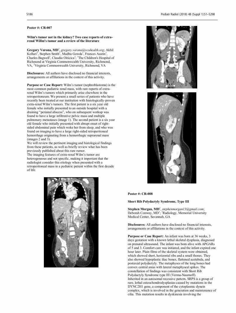
Poster #: CR-007
Wilm's tumor not in the kidney? Two case reports of extra-renal Willm's tumor and a review of the literature
Gregory Vorona, MD1, [email protected]; Akhil
Kolluri2, Stephen Smith
1, Madhu Gowda
1, Frances Austin
1,
Charles Bagwell1, Claudio Oiticica
1;
1The Children's Hospital of
Richmond at Virginia Commonwealth University, Richmond, VA,
2Virginia Commonwealth University, Richmond, VA
Disclosures: All authors have disclosed no financial interests, arrangements or affiliations in the context of this activity.
Purpose or Case Report: Wilm’s tumor (nephroblastoma) is the most common pediatric renal mass, with rare reports of extra-renal Wilm’s tumors which primarily arise elsewhere in the retroperitoneum. We present a small series of patients who have recently been treated at our institution with histologically-proven extra-renal Wilm’s tumors. The first patient is a six year old
female who initially presented to an outside hospital with a draining “perianal abscess”, who on subsequent workup was found to have a large infiltrative pelvic mass and multiple pulmonary metastases (image 1). The second patient is a six year old female who initially presented with abrupt onset of right-sided abdominal pain which woke her from sleep, and who was found on imaging to have a large right-sided retroperitoneal
hemorrhage originating from a hemorrhagic suprarenal mass (images 2 and 3). We will review the pertinent imaging and histological findings from these patients, as well as briefly review what has been previously published about this rare rumor. The imaging features of extra-renal Wilm’s tumor are
heterogeneous and not specific, making it important that the radiologist consider this etiology when presented with a retroperitoneal mass in a pediatric patient within the first decade of life.
Poster #: CR-008
Short Rib Polydactyly Syndrome, Type III Stephen Morgan, MD
Deborah Conway, MD1;
1Radiology, Memorial University
Medical Center, Savannah, GA
Disclosures: All authors have disclosed no financial interests, arrangements or affiliations in the context of this activity. Purpose or Case Report: An infant was born at 36 weeks, 3 days gestation with a known lethal skeletal dysplasia, diagnosed
on prenatal ultrasound. The infant was born alive with APGARs of 5 and 3. Comfort care was initiated, and the infant expired one hour later. Plain films of the skeletal system were obtained, which showed short, horizontal ribs and a small thorax. They also showed hypoplastic iliac bones, flattened acetabula, and postaxial polydactyly. The metaphyses of the long bones had convex central areas with lateral metaphyseal spikes. The
constellation of findings was consistent with Short Rib Polydactyly Syndrome type III (Verma-Naumoff). Inherited in an autosomal recessive pattern, SRPS is a group of rare, lethal osteochondrodysplasias caused by mutations in the DYNC2H1 gene, a component of the cytoplasmic dynein complex, which is involved in the generation and maintenance of cilia. This mutation results in dyskinesia involving the
Pediatr Radiol (2018) 48 (Suppl 1):S1–S298S186
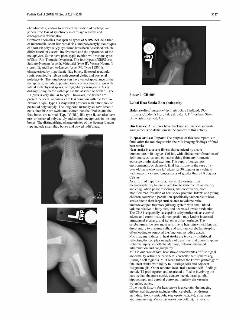
chondrocytes, leading to arrested maturation of cartilage and generalized loss of synchrony in cartilage removal and osteogenic differentiation. Common anomalies that span all types of SRPS include a triad of micromelia, short horizontal ribs, and polydactyly. Four types
of short-rib polydactyly syndrome have been described, which differ based on visceral involvement and the appearance of the metaphyses. Some have phenotypic overlap with various types of Short Rib-Thoracic Dysplasia. The four types of SRPS are: Saldino-Noonan (type I), Majewski (type II), Verma-Naumoff (type III), and Beemer-Langer (type IV). Type I (SN) is
characterized by hypoplastic iliac bones, flattened acetabular roofs, rounded vertebrae with coronal clefts, and postaxial polydactyly. The long bones can have varied appearance of the metaphysis, including: pointed ends, convex central areas with lateral metaphyseal spikes, or ragged-appearing ends. A key distinguishing factor with type I is the absence of fibulae. Type III (VN) is very similar to type I, however, the fibulae are
present. Visceral anomalies are less common with the Verma-Naumoff type. Type II (Majewski) presents with either pre- or postaxial polydactyly. The long bone metaphyses have smooth ends, the tibiae are ovoid and shorter than the fibulae, and the iliac bones are normal. Type IV (BL), like type II, can also have pre- or postaxial polydactyly and smooth metaphyses in the long bones. The distinguishing characteristics of the Beemer-Langer type include small iliac bones and bowed radii/ulnae.
Poster #: CR-009
Lethal Heat Stroke Encephalopathy Hales Skelton
2, [email protected]; Gary Hedlund, DO
1;
1Primary Children's Hospital, Salt Lake, UT,
2Portland State
University, Portland, OR
Disclosures: All authors have disclosed no financial interests, arrangements or affiliations in the context of this activity. Purpose or Case Report: The purpose of this case report is to familiarize the radiologist with the MR imaging findings of fatal heat stroke. Heat stroke is a severe illness characterized by a core
temperature > 40 degrees Celsius, with clinical manifestations of delirium, seizures, and coma, resulting from environmental exposure or physical exertion. This report focuses upon environmental, or classical, fatal heat stroke in the case of a 4 year old male who was left alone for 30 minutes in a vehicle with ambient exterior temperatures of greater than 37.8 degrees
Celsius. As a form of hyperthermia, heat stroke ensues from thermoregulatory failure in addition to systemic inflammatory and coagulation phase responses, and conceivably, from modified manifestation of heat shock proteins. Infants and young children comprise a population specifically vulnerable to heat stroke due to their large surface area to volume ratio,
underdeveloped thermoregulatory system with small blood volume relative to body size, and decreased sweat production. The CNS is especially susceptible to hyperthermia as cerebral edema and cerebrovascular congestion may lead to increased intracranial pressure, and ischemia or hemorrhage. The cerebellum is the area most sensitive to heat injury, with known direct injury to Purkinje cells, and resultant cerebellar atrophy,
often leading to neuronal dysfunction, including ataxia. MR imaging findings in heat stroke are typically multifocal reflecting the complex interplay of direct thermal injury, hypoxic ischemic injury, endothelial damage, cytokine mediated inflammation and coagulopathy. MRI in our case of fatal heat stroke demonstrates diffuse signal
abnormality within the peripheral cerebellar hemispheres (eg. Purkinje cell regions). MRI recapitulates the known pathology of fatal heat stroke with injury to Purkinge cells and adjacent Bergmann glia. Other reported heat stroke related MRI findings include T2 prolongation and restricted diffusion involving the paramedian thalamic nuclei, dentate nuclei, basal ganglia, hippocampii, and cerebral cortex particularly the vascular
watershed zones. If the health history for heat stroke is uncertain, the imaging differential diagnosis includes other cerebellar syndromes including: toxic - metabolic (eg. opiate toxicity), infectious-autoimmune (eg. Varicellar zoster cerebellitis), histiocytic
Pediatr Radiol (2018) 48 (Suppl 1):S1–S298 S187
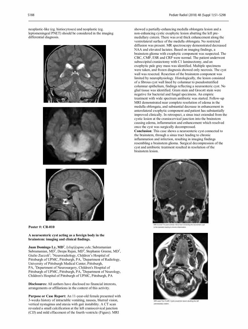
neoplastic-like (eg. histiocytoses) and neoplastic (eg. leptomeningeal PNET) should be considered in the imaging differential diagnosis.
Poster #: CR-010
A neuroenteric cyst acting as a foreign body in the
brainstem: imaging and clinical findings. Juan Domingo Ly, MD
2, [email protected]; Subramanian
Subramanian, MD1, Deepa Rajan, MD
4, Stephanie Greene, MD
3,
Giulio Zuccoli1;
1Neuroradiology, Children’s Hospital of
Pittsburgh of UPMC, Pittsburgh, PA, 2Department of Radiology,
University of Pittsburgh Medical Center, Pittsburgh, PA,
3Department of Neurosurgery, Children's Hospital of
Pittsburgh of UPMC, Pittsburgh, PA, 4Department of Neurology,
Children's Hospital of Pittsburgh of UPMC, Pittsburgh, PA
Disclosures: All authors have disclosed no financial interests, arrangements or affiliations in the context of this activity. Purpose or Case Report: An 11-year-old female presented with 3-weeks history of intractable vomiting, nausea, blurred vision, vertical nystagmus and ataxia with gait instability. A CT scan revealed a small calcification at the left craniocervical junction
(CJJ) and mild effacement of the fourth ventricle (Figure). MRI
showed a partially-enhancing medulla oblongata lesion and a non-enhancing cystic exophytic lesion abutting the left pre-medullary cistern. There was avid thick enhancement along the ventrolateral surface of the medulla oblongata. No restricted diffusion was present. MR spectroscopy demonstrated decreased
NAA and elevated lactates. Based on imaging findings, a brainstem glioma with exophytic component was suspected. The CBC, CMP, ESR and CRP were normal. The patient underwent suboccipital craniectomy with C1 laminectomy, and an exophytic pale gray mass was identified. Multiple specimens were taken, and frozen diagnosis showed only necrosis. The cyst
wall was resected. Resection of the brainstem component was limited by neurophysiology. Histologically, the lesion consisted of a fibrous cyst wall lined by columnar to pseudostratified columnar epithelium, findings reflecting a neuroenteric cyst. No glial tissue was identified. Gram stain and Grocott stain were negative for bacterial and fungal specimens. An empiric treatment with wide spectrum antibiotic was started. Follow-up
MRI demonstrated near complete resolution of edema in the medulla oblongata, and substantial decrease in enhancement in anterolateral exophytic component and patient has substantially improved clinically. In retrospect, a sinus tract extended from the cystic lesion at the craniocervical junction into the brainstem causing edema, inflammation and enhancement which resolved once the cyst was surgically decompressed.
Conclusion: This case shows a neuroenteric cyst connected to the brainstem, through a sinus tract leading to chronic inflammation and infection, resulting in imaging findings resembling a brainstem glioma. Surgical decompression of the cyst and antibiotic treatment resulted in resolution of the brainstem lesion.
Pediatr Radiol (2018) 48 (Suppl 1):S1–S298S188
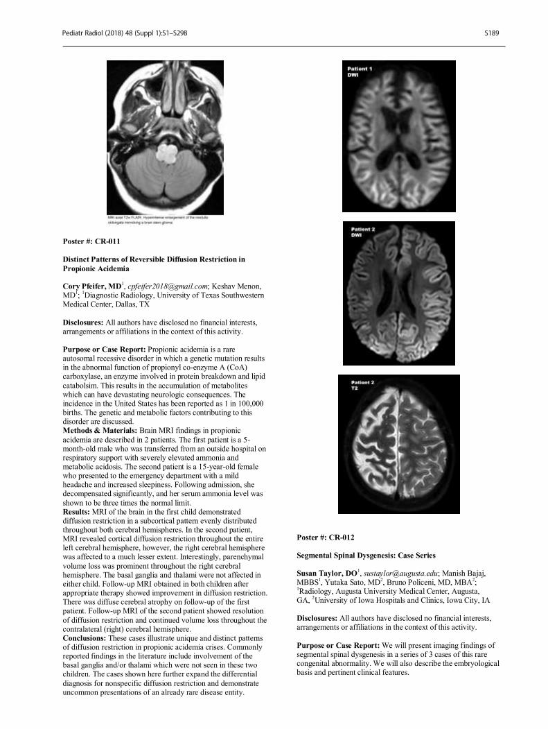
Poster #: CR-011
Distinct Patterns of Reversible Diffusion Restriction in
Propionic Acidemia Cory Pfeifer, MD
1, [email protected]; Keshav Menon,
MD1;
1Diagnostic Radiology, University of Texas Southwestern
Medical Center, Dallas, TX
Disclosures: All authors have disclosed no financial interests, arrangements or affiliations in the context of this activity. Purpose or Case Report: Propionic acidemia is a rare autosomal recessive disorder in which a genetic mutation results in the abnormal function of propionyl co-enzyme A (CoA) carboxylase, an enzyme involved in protein breakdown and lipid
catabolsim. This results in the accumulation of metabolites which can have devastating neurologic consequences. The incidence in the United States has been reported as 1 in 100,000 births. The genetic and metabolic factors contributing to this disorder are discussed. Methods & Materials: Brain MRI findings in propionic
acidemia are described in 2 patients. The first patient is a 5-month-old male who was transferred from an outside hospital on respiratory support with severely elevated ammonia and metabolic acidosis. The second patient is a 15-year-old female who presented to the emergency department with a mild headache and increased sleepiness. Following admission, she decompensated significantly, and her serum ammonia level was
shown to be three times the normal limit. Results: MRI of the brain in the first child demonstrated diffusion restriction in a subcortical pattern evenly distributed throughout both cerebral hemispheres. In the second patient, MRI revealed cortical diffusion restriction throughout the entire left cerebral hemisphere, however, the right cerebral hemisphere was affected to a much lesser extent. Interestingly, parenchymal
volume loss was prominent throughout the right cerebral hemisphere. The basal ganglia and thalami were not affected in either child. Follow-up MRI obtained in both children after appropriate therapy showed improvement in diffusion restriction. There was diffuse cerebral atrophy on follow-up of the first patient. Follow-up MRI of the second patient showed resolution
of diffusion restriction and continued volume loss throughout the contralateral (right) cerebral hemisphere. Conclusions: These cases illustrate unique and distinct patterns of diffusion restriction in propionic acidemia crises. Commonly reported findings in the literature include involvement of the basal ganglia and/or thalami which were not seen in these two children. The cases shown here further expand the differential
diagnosis for nonspecific diffusion restriction and demonstrate uncommon presentations of an already rare disease entity.
Poster #: CR-012
Segmental Spinal Dysgenesis: Case Series
Susan Taylor, DO1, [email protected]; Manish Bajaj,
MBBS1, Yutaka Sato, MD
2, Bruno Policeni, MD, MBA
2;
1Radiology, Augusta University Medical Center, Augusta,
GA, 2University of Iowa Hospitals and Clinics, Iowa City, IA
Disclosures: All authors have disclosed no financial interests, arrangements or affiliations in the context of this activity.
Purpose or Case Report: We will present imaging findings of segmental spinal dysgenesis in a series of 3 cases of this rare congenital abnormality. We will also describe the embryological basis and pertinent clinical features.
Pediatr Radiol (2018) 48 (Suppl 1):S1–S298 S189
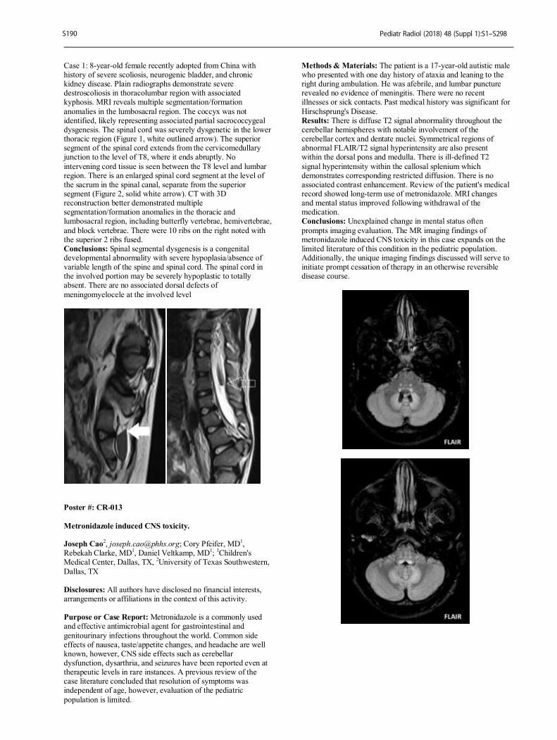
Case 1: 8-year-old female recently adopted from China with history of severe scoliosis, neurogenic bladder, and chronic kidney disease. Plain radiographs demonstrate severe destroscoliosis in thoracolumbar region with associated kyphosis. MRI reveals multiple segmentation/formation
anomalies in the lumbosacral region. The coccyx was not identified, likely representing associated partial sacrococcygeal dysgenesis. The spinal cord was severely dysgenetic in the lower thoracic region (Figure 1, white outlined arrow). The superior segment of the spinal cord extends from the cervicomedullary junction to the level of T8, where it ends abruptly. No
intervening cord tissue is seen between the T8 level and lumbar region. There is an enlarged spinal cord segment at the level of the sacrum in the spinal canal, separate from the superior segment (Figure 2, solid white arrow). CT with 3D reconstruction better demonstrated multiple segmentation/formation anomalies in the thoracic and lumbosacral region, including butterfly vertebrae, hemivertebrae,
and block vertebrae. There were 10 ribs on the right noted with the superior 2 ribs fused. Conclusions: Spinal segmental dysgenesis is a congenital developmental abnormality with severe hypoplasia/absence of variable length of the spine and spinal cord. The spinal cord in the involved portion may be severely hypoplastic to totally absent. There are no associated dorsal defects of
meningomyelocele at the involved level
Poster #: CR-013
Metronidazole induced CNS toxicity. Joseph Cao
2, [email protected]; Cory Pfeifer, MD
1,
Rebekah Clarke, MD1, Daniel Veltkamp, MD
1;
1Children's
Medical Center, Dallas, TX, 2University of Texas Southwestern,
Dallas, TX Disclosures: All authors have disclosed no financial interests, arrangements or affiliations in the context of this activity. Purpose or Case Report: Metronidazole is a commonly used and effective antimicrobial agent for gastrointestinal and
genitourinary infections throughout the world. Common side effects of nausea, taste/appetite changes, and headache are well known, however, CNS side effects such as cerebellar dysfunction, dysarthria, and seizures have been reported even at therapeutic levels in rare instances. A previous review of the case literature concluded that resolution of symptoms was independent of age, however, evaluation of the pediatric
population is limited.
Methods & Materials: The patient is a 17-year-old autistic male who presented with one day history of ataxia and leaning to the right during ambulation. He was afebrile, and lumbar puncture revealed no evidence of meningitis. There were no recent illnesses or sick contacts. Past medical history was significant for
Hirschsprung's Disease. Results: There is diffuse T2 signal abnormality throughout the cerebellar hemispheres with notable involvement of the cerebellar cortex and dentate nuclei. Symmetrical regions of abnormal FLAIR/T2 signal hyperintensity are also present within the dorsal pons and medulla. There is ill-defined T2
signal hyperintensity within the callosal splenium which demonstrates corresponding restricted diffusion. There is no associated contrast enhancement. Review of the patient's medical record showed long-term use of metronidazole. MRI changes and mental status improved following withdrawal of the medication. Conclusions: Unexplained change in mental status often
prompts imaging evaluation. The MR imaging findings of metronidazole induced CNS toxicity in this case expands on the limited literature of this condition in the pediatric population. Additionally, the unique imaging findings discussed will serve to initiate prompt cessation of therapy in an otherwise reversible disease course.
Pediatr Radiol (2018) 48 (Suppl 1):S1–S298S190
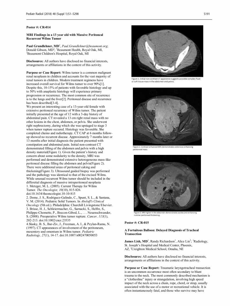
Poster #: CR-014
MRI Findings in a 13 year old with Massive Peritoneal
Recurrent Wilms Tumor
Paul Grundlehner, MD1, [email protected];
Donald Gibson, MD2;
1Beaumont Health, Royal Oak, MI,
2Beaumont Children's Hospital, Royal Oak, MI
Disclosures: All authors have disclosed no financial interests, arrangements or affiliations in the context of this activity.
Purpose or Case Report: Wilms tumor is a common malignant renal neoplasm in children and accounts for the vast majority of renal tumors in children. Modern treatment regimens have increased overall survival for Wilms tumor to over 90%[1]. Despite this, 10-15% of patients with favorable histology and up to 50% with anaplastic histology will experience primary
progression or recurrence. The most common site of recurrence is to the lungs and the liver[2]. Peritoneal disease and recurrence has been described[3-4]. We present an interesting case of a 13-year-old female with extensive peritoneal recurrence of Wilms tumor. The patient initially presented at the age of 12 with a 3-day history of abdominal pain. CT revealed a 13 cm right renal mass with no
other lesions in the chest, abdomen, or pelvis. She underwent right nephrectomy, during which she was upstaged to stage 3 when tumor rupture occured. Histology was favorable. She completed chemo and radiotherapy. CT CAP at 6 months follow-up showed no recurrent disease. Approximately 7 months later or 13 months after initial diagnosis the patient presented with
constipation and abdominal pain. Initial non-contrast CT demonstrated filling of the abdomen and pelvis with a high density material(Figure 1). Given the patient’s history and concern about some nodularity to the density, MRI was performed and demonstrated extensive heterogeneous mass-like peritoneal disease filling the abdomen and pelvis(Figure 2). There were additional areas of peritoneal caking and
thickening(Figure 3). Ultrasound guided biopsy was performed and the pathology was identical to that of the excised Wilms. While unusual recurrent Wilms tumor should be included in the differetial diagnosis of massive intraperitoneal neoplasia 1. Metzger, M. L. (2005). Current Therapy for Wilms Tumor. The Oncologist, 10(10), 815-826. doi:10.1634/theoncologist.10-10-815
2. Dome, J. S., Rodriguez-Galindo, C., Spunt, S. L., & Santana, V. M. (2014). Pediatric Solid Tumors. In Abeloff's Clinical Oncology (5th ed.). Philadelphia: Churchill Livingstone/Elsevier. 3. Brisse, H. J., Schleiermacher, G., Sarnacki, S., Helfre, S., Philippe-Chomette, P., Boccon-Gibod, L., . . . Neuenschwander, S. (2008). Preoperative Wilms tumor rupture. Cancer, 113(1),
202-213. doi:10.1002/cncr.23535 4.Slasky, B. S., Bar-Ziv, J., Freeman, A. I., & Peylan-Ramu, N. (1997). CT appearances of involvement of the peritoneum, mesentery and omentum in Wilms tumor. Pediatric Radiology, 27(1), 14-17. doi:10.1007/s002470050053
Poster #: CR-015
A Fortuitous Balloon: Delayed Diagnosis of Tracheal
Transection James Lish, MD
1, Randy Richardson
1, Alice Lin
2;
1Radiology,
St. Joseph’s Hospital and Medical Center, Phoenix, AZ,
2Creighton Medical School, Omaha, NE
Disclosures: All authors have disclosed no financial interests, arrangements or affiliations in the context of this activity. Purpose or Case Report: Traumatic laryngotracheal transection
is an uncommon occurrence most often secondary to blunt trauma to the neck. The most commonly described mechanism is a “clothesline” injury or strangulation, involving high speed impact of the neck across a chain, rope, chord, or strap, usually associated with the use of a motor or recreational vehicle. It is often instantaneously fatal, and those who survive may have
Pediatr Radiol (2018) 48 (Suppl 1):S1–S298 S191
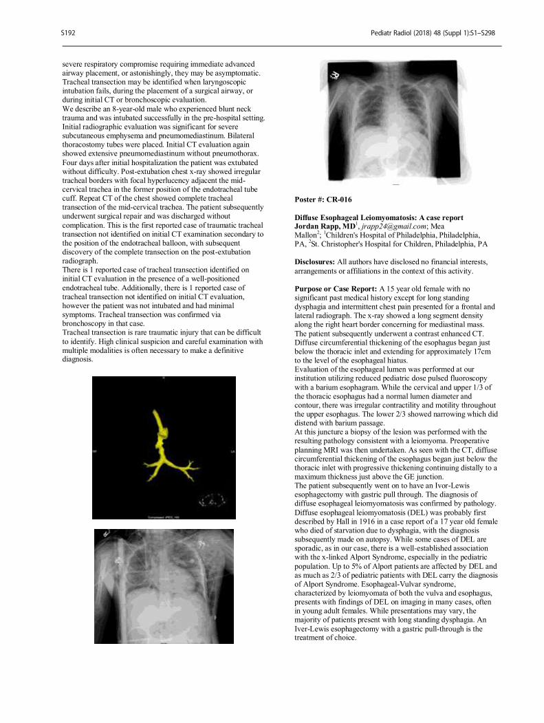
severe respiratory compromise requiring immediate advanced airway placement, or astonishingly, they may be asymptomatic. Tracheal transection may be identified when laryngoscopic intubation fails, during the placement of a surgical airway, or during initial CT or bronchoscopic evaluation.
We describe an 8-year-old male who experienced blunt neck trauma and was intubated successfully in the pre-hospital setting. Initial radiographic evaluation was significant for severe subcutaneous emphysema and pneumomediastinum. Bilateral thoracostomy tubes were placed. Initial CT evaluation again showed extensive pneumomediastinum without pneumothorax.
Four days after initial hospitalization the patient was extubated without difficulty. Post-extubation chest x-ray showed irregular tracheal borders with focal hyperlucency adjacent the mid-cervical trachea in the former position of the endotracheal tube cuff. Repeat CT of the chest showed complete tracheal transection of the mid-cervical trachea. The patient subsequently underwent surgical repair and was discharged without
complication. This is the first reported case of traumatic tracheal transection not identified on initial CT examination secondary to the position of the endotracheal balloon, with subsequent discovery of the complete transection on the post-extubation radiograph. There is 1 reported case of tracheal transection identified on initial CT evaluation in the presence of a well-positioned
endotracheal tube. Additionally, there is 1 reported case of tracheal transection not identified on initial CT evaluation, however the patient was not intubated and had minimal symptoms. Tracheal transection was confirmed via bronchoscopy in that case. Tracheal transection is rare traumatic injury that can be difficult
to identify. High clinical suspicion and careful examination with multiple modalities is often necessary to make a definitive diagnosis.
Poster #: CR-016
Diffuse Esophageal Leiomyomatosis: A case report Jordan Rapp, MD
1, [email protected]; Mea
Mallon2;
1Children's Hospital of Philadelphia, Philadelphia,
PA, 2St. Christopher's Hospital for Children, Philadelphia, PA
Disclosures: All authors have disclosed no financial interests,
arrangements or affiliations in the context of this activity. Purpose or Case Report: A 15 year old female with no significant past medical history except for long standing dysphagia and intermittent chest pain presented for a frontal and lateral radiograph. The x-ray showed a long segment density along the right heart border concerning for mediastinal mass.
The patient subsequently underwent a contrast enhanced CT. Diffuse circumferential thickening of the esophagus began just below the thoracic inlet and extending for approximately 17cm to the level of the esophageal hiatus. Evaluation of the esophageal lumen was performed at our institution utilizing reduced pediatric dose pulsed fluoroscopy
with a barium esophagram. While the cervical and upper 1/3 of the thoracic esophagus had a normal lumen diameter and contour, there was irregular contractility and motility throughout the upper esophagus. The lower 2/3 showed narrowing which did distend with barium passage. At this juncture a biopsy of the lesion was performed with the resulting pathology consistent with a leiomyoma. Preoperative
planning MRI was then undertaken. As seen with the CT, diffuse circumferential thickening of the esophagus began just below the thoracic inlet with progressive thickening continuing distally to a maximum thickness just above the GE junction. The patient subsequently went on to have an Ivor-Lewis esophagectomy with gastric pull through. The diagnosis of diffuse esophageal leiomyomatosis was confirmed by pathology.
Diffuse esophageal leiomyomatosis (DEL) was probably first described by Hall in 1916 in a case report of a 17 year old female who died of starvation due to dysphagia, with the diagnosis subsequently made on autopsy. While some cases of DEL are sporadic, as in our case, there is a well-established association with the x-linked Alport Syndrome, especially in the pediatric
population. Up to 5% of Alport patients are affected by DEL and as much as 2/3 of pediatric patients with DEL carry the diagnosis of Alport Syndrome. Esophageal-Vulvar syndrome, characterized by leiomyomata of both the vulva and esophagus, presents with findings of DEL on imaging in many cases, often in young adult females. While presentations may vary, the majority of patients present with long standing dysphagia. An
Iver-Lewis esophagectomy with a gastric pull-through is the treatment of choice.
Pediatr Radiol (2018) 48 (Suppl 1):S1–S298S192
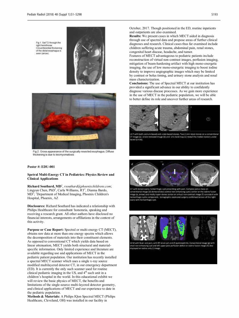
Poster #: EDU-001
Spetral Multi-Energy CT in Pediatrics: Physics Review and
Clinical Applications Richard Southard, MD
Lingyun Chen, PhD1, Carla Williams, RT
1, Dianna Bardo,
MD1;
1Department of Medical Imaging, Phoenix Children's
Hospital, Phoenix, AZ
Disclosures: Richard Southard has indicated a relationship with Philips Healthcare for consultant/ honoraria, speaking and receiving a research grant. All other authors have disclosed no financial interests, arrangements or affiliations in the context of this activity.
Purpose or Case Report: Spectral or multi-energy CT (MECT), obtains raw data at more than one energy spectra which allows the decomposition of materials into their constituent elements. As opposed to conventional CT which yields data based on linear attenuation, MECT yields both structural and material-specific information. Only limited experience and literature are available regarding use and applications of MECT in the
pediatric patient population. Our institution has recently installed a spectral MECT scanner which uses a single x-ray source modified multilayered detector CT, in our emergency department (ED). It is currently the only such scanner used for routine clinical pediatric imaging in the US, and 4
th such unit in a
children’s hospital in the world. In this educational exhibit we
will review the basic physics of MECT, the benefits and limitations of the single-source multi-layered detector geometry, and clinical applications of MECT and our experience to date in the pediatric population. Methods & Materials: A Philips IQon Spectral MECT (Philips Healthcare, Cleveland, OH) was installed in our facility in
October, 2017. Though positioned in the ED, routine inpatients and outpatients are also examined. Results: We present cases in which MECT aided in diagnosis through use of spectral data and propose areas of further clinical diagnoses and research. Clinical cases thus far examined include
children suffering acute trauma, abdominal pain, renal stones, congenital heart disease, headache, and tumor. Features of MECT advantageous to pediatric patients include reconstruction of virtual non-contrast images, perfusion imaging, mitigation of beam-hardening artifact with high mono-energetic imaging, the use of low mono-energetic imaging to boost iodine
density to improve angiographic images which may be limited by contrast or bolus timing, and urinary stone analysis and renal mass characterization. Conclusions: The use of Spectral MECT at our institution has provided a significant advance in our ability to confidently diagnose various disease processes. As we gain more experience in the use of MECT in the pediatric population, we will be able
to better define its role and uncover further areas of research.
Pediatr Radiol (2018) 48 (Suppl 1):S1–S298 S193
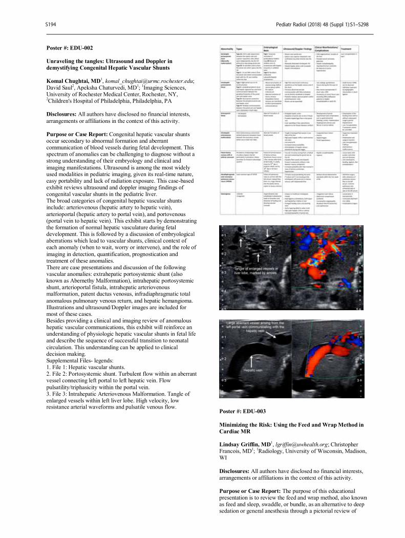
Poster #: EDU-002
Unraveling the tangles: Ultrasound and Doppler in
demystifying Congenital Hepatic Vascular Shunts
Komal Chughtai, MD1, [email protected];
David Saul2, Apeksha Chaturvedi, MD
1;
1Imaging Sciences,
University of Rochester Medical Center, Rochester, NY, 2Children's Hospital of Philadelphia, Philadelphia, PA
Disclosures: All authors have disclosed no financial interests,
arrangements or affiliations in the context of this activity. Purpose or Case Report: Congenital hepatic vascular shunts occur secondary to abnormal formation and aberrant communication of blood vessels during fetal development. This spectrum of anomalies can be challenging to diagnose without a strong understanding of their embryology and clinical and
imaging manifestations. Ultrasound is among the most widely used modalities in pediatric imaging, given its real-time nature, easy portability and lack of radiation exposure. This case-based exhibit reviews ultrasound and doppler imaging findings of congenital vascular shunts in the pediatric liver. The broad categories of congenital hepatic vascular shunts include: arteriovenous (hepatic artery to hepatic vein),
arterioportal (hepatic artery to portal vein), and portovenous (portal vein to hepatic vein). This exhibit starts by demonstrating the formation of normal hepatic vasculature during fetal development. This is followed by a discussion of embryological aberrations which lead to vascular shunts, clinical context of each anomaly (when to wait, worry or intervene), and the role of
imaging in detection, quantification, prognostication and treatment of these anomalies. There are case presentations and discussion of the following vascular anomalies: extrahepatic portosystemic shunt (also known as Abernethy Malformation), intrahepatic portosystemic shunt, arterioportal fistula, intrahepatic arteriovenous malformation, patent ductus venosus, infradiaphragmatic total
anomalous pulmonary venous return, and hepatic hemangioma. Illustrations and ultrasound/Doppler images are included for most of these cases. Besides providing a clinical and imaging review of anomalous hepatic vascular communications, this exhibit will reinforce an understanding of physiologic hepatic vascular shunts in fetal life and describe the sequence of successful transition to neonatal
circulation. This understanding can be applied to clinical decision making. Supplemental Files- legends: 1. File 1: Hepatic vascular shunts. 2. File 2: Portosystemic shunt. Turbulent flow within an aberrant vessel connecting left portal to left hepatic vein. Flow
pulsatility/triphasicity within the portal vein. 3. File 3: Intrahepatic Arteriovenous Malformation. Tangle of enlarged vessels within left liver lobe. High velocity, low resistance arterial waveforms and pulsatile venous flow.
Poster #: EDU-003
Minimizing the Risk: Using the Feed and Wrap Method in
Cardiac MR Lindsay Griffin, MD
1, [email protected]; Christopher
Francois, MD1;
1Radiology, University of Wisconsin, Madison,
WI
Disclosures: All authors have disclosed no financial interests, arrangements or affiliations in the context of this activity. Purpose or Case Report: The purpose of this educational presentation is to review the feed and wrap method, also known as feed and sleep, swaddle, or bundle, as an alternative to deep sedation or general anesthesia through a pictorial review of
Pediatr Radiol (2018) 48 (Suppl 1):S1–S298S194
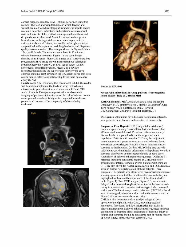
cardiac magnetic resonance (MR) studies performed using this method. The feed and wrap technique in which feeding and warmth are used to induce sleep and swaddling is used to reduce motion is described. Indications and contraindications as well risks and benefits of this method versus general anesthesia and
deep sedation are discussed. Multiple examples of congenital heart disease including atrial and ventricular septal defects, atrioventricular canal defects, and double outlet right ventricle are provided, with sequences used, length of scan, and diagnostic quality also summarized. The example shown in Figures 1-3 is a 12-day-old female. The scan was completed in 12 minutes
without intravenous contrast. Figure 1 is the scout image showing situs inversus. Figure 2 is a gated axial steady state free procession (SSFP) image showing a membranous ventricular septal defect (yellow arrow), an atrial septal defect (yellow arrowhead), and atrial inversion. Figure 3 is a 4D flow reconstruction showing the superior and inferior vena cava entering anatomic right atrium on the left, a right aortic arch with
mirror branch pattern, and relationship to the main pulmonary artery (MPA). Conclusions: After reviewing this educational exhibit, the reader will be able to implement the feed and wrap method as an alternative to general anesthesia or sedation in CT and MRI scans of infants. Examples are provided in cardiovascular imaging, of particular interest because the risk of adverse events
under general anesthesia is higher in congenital heart disease patients and because of the complexity of disease being evaluated.
Poster #: EDU-004
Myocardial infarctions in young patients with congenital
heart disease: Role of Cardiac MRI
Kathryn Henault, MD1, [email protected]; Shailendra
Upadhyay, MD2, Timothy Herbst
1, Michael O'Loughlin
1, Olga
Toro-Salazar, MD2;
1Hartford Hospital, Hartford,
CT, 2Connecticut Children’s Hospital, Hartford, CT
Disclosures: All authors have disclosed no financial interests,
arrangements or affiliations in the context of this activity. Purpose or Case Report: CHD (congenital heart disease) occurs in approximately 1% of all live births with more than 90% survival into adulthood. Prevalence of coronary artery disease has been reported to be similar to general adult population. Patients with complex CHD may be subjected to
non-atherosclerotic premature coronary artery disease due to anomalous coronaries, peri-coronary region interventions, or coronary re-implantation. Cardiac MRI (CMR) may provide valuable myocardium health information with pointers towards a coronary distribution in unsuspected chronic or acute cases. Acquisition of delayed enhancement sequences (LGE) and T1 mapping should be considered routine in CMR studies for
assessment of interval ischemic events. Patients with complex CHD are also at risk for sudden cardiac arrest and LGE data may assist in further risk stratification of these patients. Three complex CHD patients who all suffered myocardial infarctions at a young age as a result of their multifaceted cardiac history are highlighted to illustrate the importance of this (see included
table, Figure 1). Two CMR images (Figures 2,3) demonstrate delayed enhancement throughout the septum at the base to mid-cavity in a patient with truncus arteriosus type 1 who presented with a non-ST-elevation myocardial infarction (NSTEMI). Focal area of low-signal sub-endocaridum within the enhancement on Figure 3 favors microvascular obstruction. CMR is a vital component of surgical planning and post-
operative care of patients with CHD, providing accurate anatomical, functional, and flow information that assists in clinical management. Delayed enhancement sequences and post-gadolinium T1 mapping allow assessment of ischemic injury or infarct, and therefore should be considered part of routine follow up CMR studies in patients with complex CHD.
Pediatr Radiol (2018) 48 (Suppl 1):S1–S298 S195
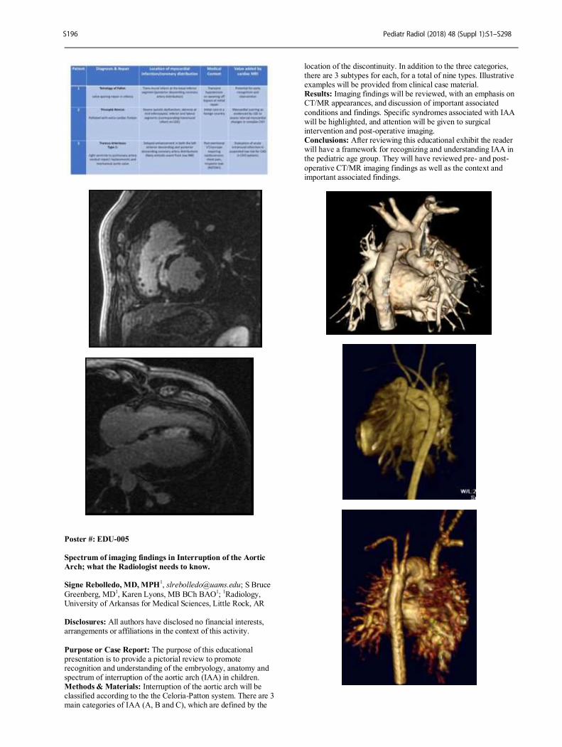
Poster #: EDU-005
Spectrum of imaging findings in Interruption of the Aortic
Arch; what the Radiologist needs to know. Signe Rebolledo, MD, MPH
1, [email protected]; S Bruce
Greenberg, MD1, Karen Lyons, MB BCh BAO
1;
1Radiology,
University of Arkansas for Medical Sciences, Little Rock, AR Disclosures: All authors have disclosed no financial interests, arrangements or affiliations in the context of this activity.
Purpose or Case Report: The purpose of this educational presentation is to provide a pictorial review to promote recognition and understanding of the embryology, anatomy and spectrum of interruption of the aortic arch (IAA) in children. Methods & Materials: Interruption of the aortic arch will be classified according to the the Celoria-Patton system. There are 3 main categories of IAA (A, B and C), which are defined by the
location of the discontinuity. In addition to the three categories, there are 3 subtypes for each, for a total of nine types. Illustrative examples will be provided from clinical case material. Results: Imaging findings will be reviewed, with an emphasis on CT/MR appearances, and discussion of important associated
conditions and findings. Specific syndromes associated with IAA will be highlighted, and attention will be given to surgical intervention and post-operative imaging. Conclusions: After reviewing this educational exhibit the reader will have a framework for recognizing and understanding IAA in the pediatric age group. They will have reviewed pre- and post-
operative CT/MR imaging findings as well as the context and important associated findings.
Pediatr Radiol (2018) 48 (Suppl 1):S1–S298S196
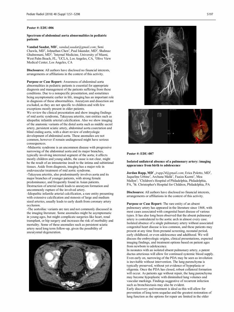
Poster #: EDU-006
Spectrum of abdominal aorta abnormalities in pediatric
patients
Vandad Saadat, MD1, [email protected]; Soni
Chawla, MD3, Johnathan Chen
2, Paul Iskander, MD
2, Shahnaz
Ghahremani, MD2;
1Internal Medicine, University of Miami,
West Palm Beach, FL, 2UCLA, Los Angeles, CA,
3Olive View
Medical Center, Los Angeles, CA
Disclosures: All authors have disclosed no financial interests, arrangements or affiliations in the context of this activity. Purpose or Case Report: Awareness of abdominal aorta abnormalities in pediatric patients is essential for appropriate diagnosis and management of the patients suffering from these conditions. Due to a nonspecific presentation, and sometimes
being asymptomatic earlier in life, imaging has an important role in diagnosis of these abnormalities. Aneurysm and dissection are excluded, as they are not specific to children and with few exceptions mostly present in older patients. We review the clinical presentation and show imaging findings of mid aortic syndrome, Takayusu arteritis, rare entities such as idiopathic infantile arterial calcification. Also we show imaging
of the anatomic variants of the distal aorta such as middle sacral artery, persistent sciatic artery, abdominal aorta coarctation and blind ending aorta, with a short review of embryologic development of abdominal aorta. These anomalies are not common, however if remain undiagnosed might have serious consequences.
-Midaortic syndrome is an uncommon disease with progressive narrowing of the abdominal aorta and its major branches, typically involving interrenal segment of the aorta; it affects mostly children and young adults, the cause is not clear, might be the result of an intrauterine insult to the intima and subintimal tissues. Aside from diagnosis, imaging has a major role in endovascular treatment of mid aortic syndrome.
-Takayusu arteritis, also predominantly involves aorta and its major branches of younger patients, with strong female predominance, and frequently found in Asian patients. Destruction of arterial medi leads to aneurysm formation and uncommonly rupture of the involved artery. -Idiopathic infantile arterial calcification, a rare entity presenting with extensive calcification and stenosis of large and medium
sized arteries, usually leads to early death from coronary artery occlusion. -The aortoiliac variants are rare and not commonly discussed in the imaging literature. Some anomalies might be asymptomatic in young ages, but might complicate surgeries like heart, renal transplant, or hip surgery and increases the risk of morbidity and
mortality. Some of these anomalies such as persistent sciatic artery need long term follow-up, given the possibility of aneurysmal degeneration.
Poster #: EDU-007
Isolated unilateral absence of a pulmonary artery: imaging
appearance from birth to adolescence Jordan Rapp, MD
1, [email protected]; Erica Poletto, MD
2,
Jaqueline Urbine2, Archana Malik
2, Faaiza Kazmi
2, Mea
Mallon2;
1Children's Hospital of Philadelphia, Philadelphia,
PA, 2St. Christopher's Hospital for Children, Philadelphia, PA
Disclosures: All authors have disclosed no financial interests, arrangements or affiliations in the context of this activity.
Purpose or Case Report: The rare entity of an absent
pulmonary artery has appeared in the literature since 1868, with most cases associated with congenital heart disease of various types. It has also long been observed that the absent pulmonary artery is contralateral to the aortic arch in almost every case. Isolated absence of a single pulmonary artery without associated congenital heart disease is less common, and these patients may
present at any time from prenatal screening, neonatal period, early childhood, or even adolescence and adulthood. We will discuss the embryologic origins, clinical presentations, expected imaging findings, and treatment options based on patient ages from newborn to adolescence. In neonates with an isolated absent pulmonary artery, a patent ductus arteriosus will allow for continued systemic blood supply.
Even early on, narrowing of the PDA may be seen as involution is inevitable without intervention. The lung parenchyma is typically preserved, without yet evidence of hypoplasia or oligemia. Once the PDA has closed, robust collateral formation will occur. As patients age without repair, the lung parenchyma may become hypoplastic with diminished lung volumes and vascular markings. Findings suggestive of recurrent infection
such as bronchiectasis may also be evident. Early discovery and treatment is ideal as this will allow for prevention of long term sequelae and the greatest restoration of lung function as the options for repair are limited in the older
Pediatr Radiol (2018) 48 (Suppl 1):S1–S298 S197
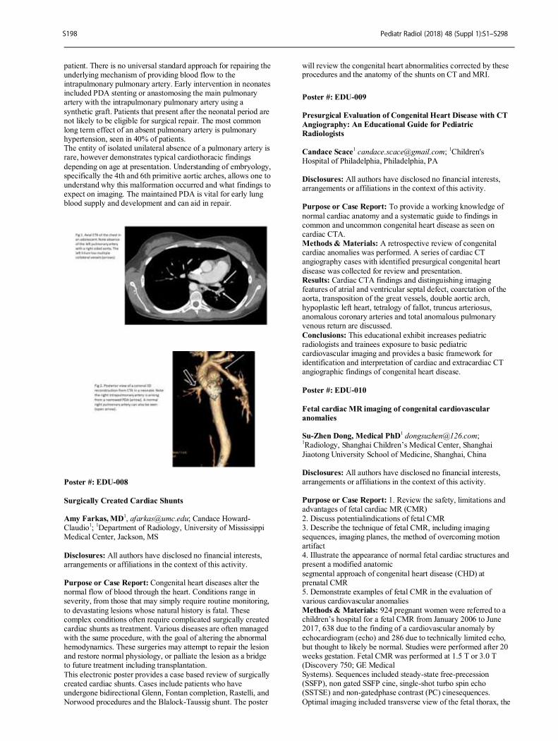
patient. There is no universal standard approach for repairing the underlying mechanism of providing blood flow to the intrapulmonary pulmonary artery. Early intervention in neonates included PDA stenting or anastomosing the main pulmonary artery with the intrapulmonary pulmonary artery using a
synthetic graft. Patients that present after the neonatal period are not likely to be eligible for surgical repair. The most common long term effect of an absent pulmonary artery is pulmonary hypertension, seen in 40% of patients. The entity of isolated unilateral absence of a pulmonary artery is rare, however demonstrates typical cardiothoracic findings
depending on age at presentation. Understanding of embryology, specifically the 4th and 6th primitive aortic arches, allows one to understand why this malformation occurred and what findings to expect on imaging. The maintained PDA is vital for early lung blood supply and development and can aid in repair.
Poster #: EDU-008
Surgically Created Cardiac Shunts Amy Farkas, MD
1, [email protected]; Candace Howard-
Claudio1;
1Department of Radiology, University of Mississippi
Medical Center, Jackson, MS
Disclosures: All authors have disclosed no financial interests, arrangements or affiliations in the context of this activity. Purpose or Case Report: Congenital heart diseases alter the normal flow of blood through the heart. Conditions range in severity, from those that may simply require routine monitoring,
to devastating lesions whose natural history is fatal. These complex conditions often require complicated surgically created cardiac shunts as treatment. Various diseases are often managed with the same procedure, with the goal of altering the abnormal hemodynamics. These surgeries may attempt to repair the lesion and restore normal physiology, or palliate the lesion as a bridge to future treatment including transplantation.
This electronic poster provides a case based review of surgically created cardiac shunts. Cases include patients who have undergone bidirectional Glenn, Fontan completion, Rastelli, and Norwood procedures and the Blalock-Taussig shunt. The poster
will review the congenital heart abnormalities corrected by these procedures and the anatomy of the shunts on CT and MRI.
Poster #: EDU-009
Presurgical Evaluation of Congenital Heart Disease with CT
Angiography: An Educational Guide for Pediatric
Radiologists
Candace Scace1 [email protected];
1Children's
Hospital of Philadelphia, Philadelphia, PA Disclosures: All authors have disclosed no financial interests, arrangements or affiliations in the context of this activity. Purpose or Case Report: To provide a working knowledge of
normal cardiac anatomy and a systematic guide to findings in common and uncommon congenital heart disease as seen on cardiac CTA. Methods & Materials: A retrospective review of congenital cardiac anomalies was performed. A series of cardiac CT angiography cases with identified presurgical congenital heart
disease was collected for review and presentation. Results: Cardiac CTA findings and distinguishing imaging features of atrial and ventricular septal defect, coarctation of the aorta, transposition of the great vessels, double aortic arch, hypoplastic left heart, tetralogy of fallot, truncus arteriosus, anomalous coronary arteries and total anomalous pulmonary venous return are discussed.
Conclusions: This educational exhibit increases pediatric radiologists and trainees exposure to basic pediatric cardiovascular imaging and provides a basic framework for identification and interpretation of cardiac and extracardiac CT angiographic findings of congenital heart disease.
Poster #: EDU-010
Fetal cardiac MR imaging of congenital cardiovascular
anomalies Su-Zhen Dong, Medical PhD
1Radiology, Shanghai Children’s Medical Center, Shanghai
Jiaotong University School of Medicine, Shanghai, China Disclosures: All authors have disclosed no financial interests, arrangements or affiliations in the context of this activity. Purpose or Case Report: 1. Review the safety, limitations and advantages of fetal cardiac MR (CMR)
2. Discuss potentialindications of fetal CMR 3. Describe the technique of fetal CMR, including imaging sequences, imaging planes, the method of overcoming motion artifact 4. Illustrate the appearance of normal fetal cardiac structures and present a modified anatomic
segmental approach of congenital heart disease (CHD) at prenatal CMR 5. Demonstrate examples of fetal CMR in the evaluation of various cardiovascular anomalies Methods & Materials: 924 pregnant women were referred to a children’s hospital for a fetal CMR from January 2006 to June 2017, 638 due to the finding of a cardiovascular anomaly by
echocardiogram (echo) and 286 due to technically limited echo, but thought to likely be normal. Studies were performed after 20 weeks gestation. Fetal CMR was performed at 1.5 T or 3.0 T (Discovery 750; GE Medical Systems). Sequences included steady-state free-precession (SSFP), non gated SSFP cine, single-shot turbo spin echo (SSTSE) and non-gatedphase contrast (PC) cinesequences.
Optimal imaging included transverse view of the fetal thorax, the
Pediatr Radiol (2018) 48 (Suppl 1):S1–S298S198
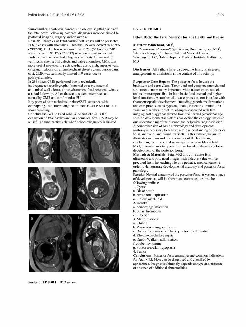
four-chamber, short-axis, coronal and oblique sagittal planes of the fetal heart. Follow up postnatal diagnoses were confirmed by postnatal imaging, surgery and/or autopsy. Results: Examples of Fetal cardiac MRI cases will be presented. In 638 cases with anomalies, Obstetric US were correct in 46.9%
(299/638), fetal echos were correct in 83.2% (531/638), CMR were correct in 82.1% (524/638) when compared to postnatal findings. Fetal echoes had a higher specificity for evaluating ventricular size, septal defects and valve anomalies. CMR was more useful in evaluating extracardiac aortic arch, superior vena cava and malposition anomalies,heart diverticulum, pericardium
cyst. CMR was technically limited in 9 cases due to polyhydramnios. In 286 cases, CMR performed due to technically inadequateechocardiography (maternal obesity, maternal abdominal wall edema, oligohydramnios, fetal position, twins, et al), had follow up. All of these cases were interpreted as normalby CMR and confirmed at FU.
Key point of scan technique includeSSFP sequence with overlapping slice, improving the artifacts is SSFP with radial k-space sampling. Conclusions: While Fetal echo is the first choice in the evaluation of fetal cardiovascular anomalies; fetal CMR may be a useful adjunct particularly when echocardiography is limited.
Poster #: EDU-011 - Withdrawn
Poster #: EDU-012
Below Deck: The Fetal Posterior fossa in Health and Disease Matthew Whitehead, MD
1,
[email protected]; Bonmyong Lee, MD2;
1Neuroradiology, Children's National Medical Center,
Washington, DC, 2Johns Hopkins Medical Institute, Baltimore,
MD Disclosures: All authors have disclosed no financial interests,
arrangements or affiliations in the context of this activity. Purpose or Case Report: The posterior fossa houses the brainstem and cerebellum. These vital and complex parenchymal structures contain many important white matter tracts, nuclei, and neurons responsible for both basic fundamental and higher-level functions. A number of disease processes can interfere with
rhombencephalic development, including genetic malformations and disruption such as hypoxia, toxins, infections, trauma, and vascular disorders. Structural changes associated with fetal imaging pathology that deviate from the normal gestational-age specific developmental patterns can define the etiology, improve our understanding of the disease, and help with prognostication. A comprehension of basic embryology and developmental
anatomy is necessary to achieve a true understanding of posterior fossa anomalies and normal variants. In this exhibit, we aim to illustrate common and rare anomalies of the brainstem, cerebellum, meninges, and meningeal spaces visible on fetal MRI, presented in a temporal manner based on the embryologic development of the posterior fossa.
Methods & Materials: Fetal MRI and correlative fetal ultrasound and post-natal images with didactic value will be procured from the teaching file of a pediatric medical center in order to demonstrate developmental anatomy and posterior fossa pathology. Results: Normal anatomy of the posterior fossa in various stages of developement will be shown and contrasted against the
following entiites: 1. Cysts: a. Blake pouch b. Arachnoid duplication c. Fibrous arachnoid 2. Insutls: a. hemorrhage/infarction
b. Sinus thrombosis c. Infection 3. Malformations: a. Chiari II b. Walker-Warburg syndrome c. Diencephalic-mesencephalic junction malformation
d. Rhombencephalosynapsis e. Dandy-Walker malformation f. Joubert syndrome g. Pontocerebellar hypoplasia 4. Tumor Conclusions: Posterior fossa anomalies are common indications for fetal MRI. Most can be diagnosed and classified by
appearance. Prognosis ultimately depends on type and presence or absence of additional abnormalities.
Pediatr Radiol (2018) 48 (Suppl 1):S1–S298 S199
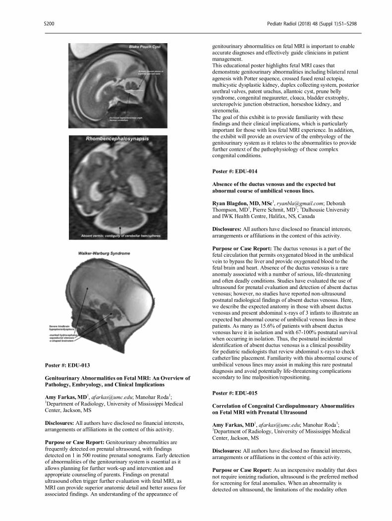
Poster #: EDU-013
Genitourinary Abnormalities on Fetal MRI: An Overview of
Pathology, Embryology, and Clinical Implications Amy Farkas, MD
1, [email protected]; Manohar Roda
1;
1Department of Radiology, University of Mississippi Medical
Center, Jackson, MS Disclosures: All authors have disclosed no financial interests, arrangements or affiliations in the context of this activity. Purpose or Case Report: Genitourinary abnormalities are
frequently detected on prenatal ultrasound, with findings detected on 1 in 500 routine prenatal sonograms. Early detection of abnormalities of the genitourinary system is essential as it allows planning for further work-up and intervention and appropriate counseling of parents. Findings on prenatal ultrasound often trigger further evaluation with fetal MRI, as
MRI can provide superior anatomic detail and better assess for associated findings. An understanding of the appearance of
genitourinary abnormalities on fetal MRI is important to enable accurate diagnoses and effectively guide clinicians in patient management. This educational poster highlights fetal MRI cases that demonstrate genitourinary abnormalities including bilateral renal
agenesis with Potter sequence, crossed fused renal ectopia, multicystic dysplastic kidney, duplex collecting system, posterior urethral valves, patent urachus, allantoic cyst, prune belly syndrome, congenital megaureter, cloaca, bladder exstrophy, ureteropelvic junction obstruction, horseshoe kidney, and sirenomelia.
The goal of this exhibit is to provide familiarity with these findings and their clinical implications, which is particularly important for those with less fetal MRI experience. In addition, the exhibit will provide an overview of the embryology of the genitourinary system as it relates to the abnormalities to provide further context of the pathophysiology of these complex congenital conditions.
Poster #: EDU-014
Absence of the ductus venosus and the expected but
abnormal course of umbilical venous lines. Ryan Blagdon, MD, MSc
1, [email protected]; Deborah
Thompson, MD1, Pierre Schmit, MD
1;
1Dalhousie University
and IWK Health Centre, Halifax, NS, Canada Disclosures: All authors have disclosed no financial interests,
arrangements or affiliations in the context of this activity. Purpose or Case Report: The ductus venosus is a part of the fetal circulation that permits oxygenated blood in the umbilical vein to bypass the liver and provide oxygenated blood to the fetal brain and heart. Absence of the ductus venosus is a rare anomaly associated with a number of serious, life-threatening
and often deadly conditions. Studies have evaluated the use of ultrasound for prenatal evaluation and detection of absent ductus venosus; however, no studies have reported non-ultrasound postnatal radiological findings of absent ductus venosus. Here, we describe the expected anatomy in those with absent ductus venosus and present abdominal x-rays of 3 infants to illustrate an
expected but abnormal course of umbilical venous lines in these patients. As many as 15.6% of patients with absent ductus venosus have it in isolation and with 67-100% postnatal survival when occurring in isolation. Thus, the postnatal incidental identification of absent ductus venosus is a clinical possibility for pediatric radiologists that review abdominal x-rays to check catheter/line placement. Familiarity with this abnormal course of
umbilical venous lines may assist in making this rare postnatal diagnosis and avoid potentially life-threatening complications secondary to line malposition/repositioning.
Poster #: EDU-015
Correlation of Congenital Cardiopulmonary Abnormalities
on Fetal MRI with Prenatal Ultrasound Amy Farkas, MD
1, [email protected]; Manohar Roda
1;
1Department of Radiology, University of Mississippi Medical
Center, Jackson, MS
Disclosures: All authors have disclosed no financial interests, arrangements or affiliations in the context of this activity. Purpose or Case Report: As an inexpensive modality that does
not require ionizing radiation, ultrasound is the preferred method for screening for fetal anomalies. When an abnormality is detected on ultrasound, the limitations of the modality often
Pediatr Radiol (2018) 48 (Suppl 1):S1–S298S200
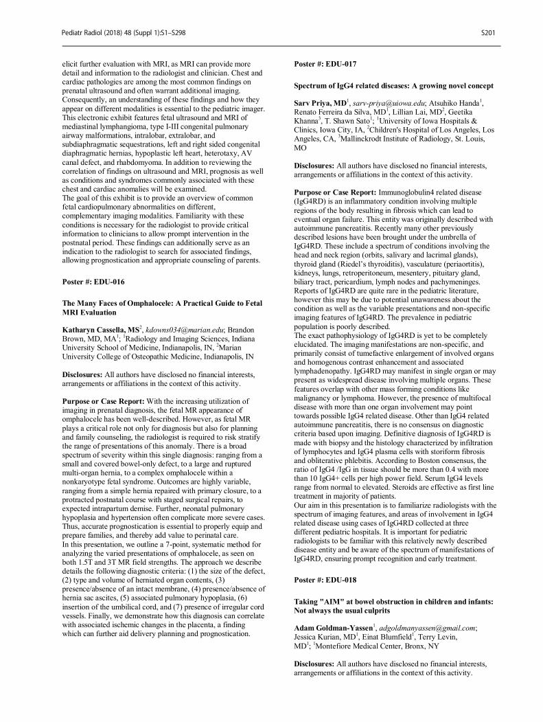
elicit further evaluation with MRI, as MRI can provide more detail and information to the radiologist and clinician. Chest and cardiac pathologies are among the most common findings on prenatal ultrasound and often warrant additional imaging. Consequently, an understanding of these findings and how they
appear on different modalities is essential to the pediatric imager. This electronic exhibit features fetal ultrasound and MRI of mediastinal lymphangioma, type I-III congenital pulmonary airway malformations, intralobar, extralobar, and subdiaphragmatic sequestrations, left and right sided congenital diaphragmatic hernias, hypoplastic left heart, heterotaxy, AV
canal defect, and rhabdomyoma. In addition to reviewing the correlation of findings on ultrasound and MRI, prognosis as well as conditions and syndromes commonly associated with these chest and cardiac anomalies will be examined. The goal of this exhibit is to provide an overview of common fetal cardiopulmonary abnormalities on different, complementary imaging modalities. Familiarity with these
conditions is necessary for the radiologist to provide critical information to clinicians to allow prompt intervention in the postnatal period. These findings can additionally serve as an indication to the radiologist to search for associated findings, allowing prognostication and appropriate counseling of parents.
Poster #: EDU-016
The Many Faces of Omphalocele: A Practical Guide to Fetal
MRI Evaluation Katharyn Cassella, MS
2, [email protected]; Brandon
Brown, MD, MA1;
1Radiology and Imaging Sciences, Indiana
University School of Medicine, Indianapolis, IN, 2Marian
University College of Osteopathic Medicine, Indianapolis, IN
Disclosures: All authors have disclosed no financial interests, arrangements or affiliations in the context of this activity. Purpose or Case Report: With the increasing utilization of imaging in prenatal diagnosis, the fetal MR appearance of omphalocele has been well-described. However, as fetal MR
plays a critical role not only for diagnosis but also for planning and family counseling, the radiologist is required to risk stratify the range of presentations of this anomaly. There is a broad spectrum of severity within this single diagnosis: ranging from a small and covered bowel-only defect, to a large and ruptured multi-organ hernia, to a complex omphalocele within a nonkaryotype fetal syndrome. Outcomes are highly variable,
ranging from a simple hernia repaired with primary closure, to a protracted postnatal course with staged surgical repairs, to expected intrapartum demise. Further, neonatal pulmonary hypoplasia and hypertension often complicate more severe cases. Thus, accurate prognostication is essential to properly equip and prepare families, and thereby add value to perinatal care.
In this presentation, we outline a 7-point, systematic method for analyzing the varied presentations of omphalocele, as seen on both 1.5T and 3T MR field strengths. The approach we describe details the following diagnostic criteria: (1) the size of the defect, (2) type and volume of herniated organ contents, (3) presence/absence of an intact membrane, (4) presence/absence of hernia sac ascites, (5) associated pulmonary hypoplasia, (6)
insertion of the umbilical cord, and (7) presence of irregular cord vessels. Finally, we demonstrate how this diagnosis can correlate with associated ischemic changes in the placenta, a finding which can further aid delivery planning and prognostication.
Poster #: EDU-017
Spectrum of IgG4 related diseases: A growing novel concept Sarv Priya, MD
1, [email protected]; Atsuhiko Handa
1,
Renato Ferreira da Silva, MD1, Lillian Lai, MD
2, Geetika
Khanna3, T. Shawn Sato
1;
1University of Iowa Hospitals &
Clinics, Iowa City, IA, 2Children's Hospital of Los Angeles, Los
Angeles, CA, 3Mallinckrodt Institute of Radiology, St. Louis,
MO Disclosures: All authors have disclosed no financial interests, arrangements or affiliations in the context of this activity. Purpose or Case Report: Immunoglobulin4 related disease (IgG4RD) is an inflammatory condition involving multiple
regions of the body resulting in fibrosis which can lead to eventual organ failure. This entity was originally described with autoimmune pancreatitis. Recently many other previously described lesions have been brought under the umbrella of IgG4RD. These include a spectrum of conditions involving the head and neck region (orbits, salivary and lacrimal glands),
thyroid gland (Riedel’s thyroiditis), vasculature (periaortitis), kidneys, lungs, retroperitoneum, mesentery, pituitary gland, biliary tract, pericardium, lymph nodes and pachymeninges. Reports of IgG4RD are quite rare in the pediatric literature, however this may be due to potential unawareness about the condition as well as the variable presentations and non-specific imaging features of IgG4RD. The prevalence in pediatric
population is poorly described. The exact pathophysiology of IgG4RD is yet to be completely elucidated. The imaging manifestations are non-specific, and primarily consist of tumefactive enlargement of involved organs and homogenous contrast enhancement and associated lymphadenopathy. IgG4RD may manifest in single organ or may present as widespread disease involving multiple organs. These
features overlap with other mass forming conditions like malignancy or lymphoma. However, the presence of multifocal disease with more than one organ involvement may point towards possible IgG4 related disease. Other than IgG4 related autoimmune pancreatitis, there is no consensus on diagnostic criteria based upon imaging. Definitive diagnosis of IgG4RD is
made with biopsy and the histology characterized by infiltration of lymphocytes and IgG4 plasma cells with storiform fibrosis and obliterative phlebitis. According to Boston consensus, the ratio of IgG4 /IgG in tissue should be more than 0.4 with more than 10 IgG4+ cells per high power field. Serum IgG4 levels range from normal to elevated. Steroids are effective as first line treatment in majority of patients.
Our aim in this presentation is to familiarize radiologists with the spectrum of imaging features, and areas of involvement in IgG4 related disease using cases of IgG4RD collected at three different pediatric hospitals. It is important for pediatric radiologists to be familiar with this relatively newly described disease entity and be aware of the spectrum of manifestations of IgG4RD, ensuring prompt recognition and early treatment.
Poster #: EDU-018
Taking "AIM" at bowel obstruction in children and infants:
Not always the usual culprits Adam Goldman-Yassen
Jessica Kurian, MD1, Einat Blumfield
1, Terry Levin,
MD1;
1Montefiore Medical Center, Bronx, NY
Disclosures: All authors have disclosed no financial interests, arrangements or affiliations in the context of this activity.
Pediatr Radiol (2018) 48 (Suppl 1):S1–S298 S201
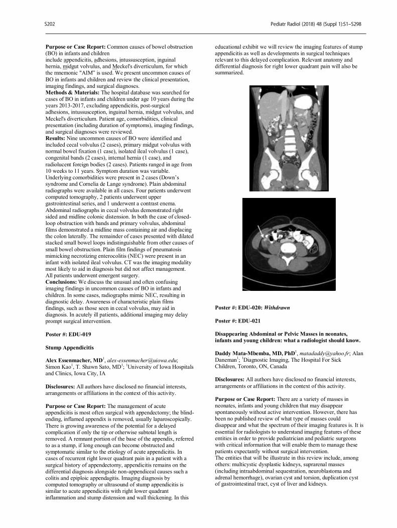
Purpose or Case Report: Common causes of bowel obstruction (BO) in infants and children include appendicitis, adhesions, intussusception, inguinal hernia, midgut volvulus, and Meckel's diverticulum, for which the mnemonic "AIM” is used. We present uncommon causes of
BO in infants and children and review the clinical presentation, imaging findings, and surgical diagnoses. Methods & Materials: The hospital database was searched for cases of BO in infants and children under age 10 years during the years 2013-2017, excluding appendicitis, post-surgical adhesions, intussusception, inguinal hernia, midgut volvulus, and
Meckel's diverticulum. Patient age, comorbidities, clinical presentation (including duration of symptoms), imaging findings, and surgical diagnoses were reviewed. Results: Nine uncommon causes of BO were identified and included cecal volvulus (2 cases), primary midgut volvulus with normal bowel fixation (1 case), isolated ileal volvulus (1 case), congenital bands (2 cases), internal hernia (1 case), and
radiolucent foreign bodies (2 cases). Patients ranged in age from 10 weeks to 11 years. Symptom duration was variable. Underlying comorbidities were present in 2 cases (Down’s syndrome and Cornelia de Lange syndrome). Plain abdominal radiographs were available in all cases. Four patients underwent computed tomography, 2 patients underwent upper gastrointestinal series, and 1 underwent a contrast enema.
Abdominal radiographs in cecal volvulus demonstrated right sided and midline colonic distension. In both the case of closed-loop obstruction with bands and primary volvulus, abdominal films demonstrated a midline mass containing air and displacing the colon laterally. The remainder of cases presented with dilated stacked small bowel loops indistinguishable from other causes of
small bowel obstruction. Plain film findings of pneumatosis mimicking necrotizing enterocolitis (NEC) were present in an infant with isolated ileal volvulus. CT was the imaging modality most likely to aid in diagnosis but did not affect management. All patients underwent emergent surgery. Conclusions: We discuss the unusual and often confusing imaging findings in uncommon causes of BO in infants and
children. In some cases, radiographs mimic NEC, resulting in diagnostic delay. Awareness of characteristic plain films findings, such as those seen in cecal volvulus, may aid in diagnosis. In acutely ill patients, additional imaging may delay prompt surgical intervention.
Poster #: EDU-019
Stump Appendicitis Alex Essenmacher, MD
Simon Kao1, T. Shawn Sato, MD
1;
1University of Iowa Hospitals
and Clinics, Iowa City, IA
Disclosures: All authors have disclosed no financial interests, arrangements or affiliations in the context of this activity. Purpose or Case Report: The management of acute appendicitis is most often surgical with appendectomy; the blind-ending, inflamed appendix is removed, usually laparoscopically.
There is growing awareness of the potential for a delayed complication if only the tip or otherwise subtotal length is removed. A remnant portion of the base of the appendix, referred to as a stump, if long enough can become obstructed and symptomatic similar to the etiology of acute appendicitis. In cases of recurrent right lower quadrant pain in a patient with a
surgical history of appendectomy, appendicitis remains on the differential diagnosis alongside non-appendiceal causes such a colitis and epiploic appendagitis. Imaging diagnosis by computed tomography or ultrasound of stump appendicitis is similar to acute appendicitis with right lower quadrant inflammation and stump distension and wall thickening. In this
educational exhibit we will review the imaging features of stump appendicitis as well as developments in surgical techniques relevant to this delayed complication. Relevant anatomy and differential diagnosis for right lower quadrant pain will also be summarized.
Poster #: EDU-020: Withdrawn
Poster #: EDU-021
Disappearing Abdominal or Pelvic Masses in neonates,
infants and young children: what a radiologist should know.
Daddy Mata-Mbemba, MD, PhD
1, [email protected]; Alan
Daneman1;
1Diagnostic Imaging, The Hospital For Sick
Children, Toronto, ON, Canada Disclosures: All authors have disclosed no financial interests,
arrangements or affiliations in the context of this activity. Purpose or Case Report: There are a variety of masses in neonates, infants and young children that may disappear spontaneously without active intervention. However, there has been no published review of what type of masses could disappear and what the spectrum of their imaging features is. It is
essential for radiologists to understand imaging features of these entities in order to provide pediatrician and pediatric surgeons with critical information that will enable them to manage these patients expectantly without surgical intervention. The entities that will be illustrate in this review include, among others: multicystic dysplastic kidneys, suprarenal masses
(including intraabdominal sequestration, neuroblastoma and adrenal hemorrhage), ovarian cyst and torsion, duplication cyst of gastrointestinal tract, cyst of liver and kidneys.
Pediatr Radiol (2018) 48 (Suppl 1):S1–S298S202
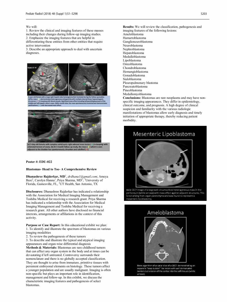
We will: 1. Review the clinical and imaging features of these masses including their changes during follow-up imaging studies. 2. Emphasize the imaging features that are helpful in differentiating these entities from other entities that require
active intervention 3. Describe an appropriate approach to deal with uncertain diagnoses.
Poster #: EDU-022
Blastomas- Head to Toe- A Comprehensive Review Dhanashree Rajderkar, MD
1, [email protected]; Ameya
Baxi2, Carolyn Hanna
1, Priya Sharma, MD
1;
1University of
Florida, Gainesville, FL, 2UT Health, San Antonio, TX
Disclosures: Dhanashree Rajderkar has indicated a relationship with the Association for Medical Imaging Management and Toshiba Medical for receiving a research grant. Priya Sharma has indicated a relationship with the Association for Medical Imaging Management and Toshiba Medical for receiving a
research grant. All other authors have disclosed no financial interests, arrangements or affiliations in the context of this activity. Purpose or Case Report: In this educational exhibit we plan: 1. To identify and illustrate the spectrum of blastomas on various imaging modalities
2. To review the pathogenesis of these tumors 3. To describe and illustrate the typical and atypical imaging appearances and organ-wise differential diagnosis Methods & Materials: Blastomas are rare childhood tumors that can affect any organ system in the body and at times can be devastating if left untreated. Controversy surrounds their nomenclature and there is no globally accepted classification.
They are thought to arise from immature, primitive tissues with persistent embryonal elements on histology. These tumors affect a younger population and are usually malignant. Imaging is often non-specific but plays an important role in identification, management and follow-up. In this exhibit, we discuss the characteristic imaging features and pathogenesis of select
blastomas.
Results: We will review the classification, pathogenesis and imaging features of the following lesions: Ameloblastoma Hamartoblastoma Ganglioneuroblastoma
Neuroblastoma Nephroblastoma Hepatoblastoma Medulloblastoma Lipoblastoma Osteoblastoma
Chondroblastoma Hemangioblastoma Gonadoblastoma Sialoblastoma Pleuropulmonary blastoma Pancreatoblastoma Pineoblastoma
Medullomyoblastoma Conclusions: Blastomas are rare neoplasms and may have non-specific imaging appearances. They differ in epidemiology, clinical outcome, and prognosis. A high degree of clinical suspicion and familiarity with the various radiologic manifestations of blastomas allow early diagnosis and timely initiation of appropriate therapy, thereby reducing patient
morbidity.
Pediatr Radiol (2018) 48 (Suppl 1):S1–S298 S203
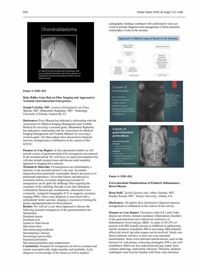
Poster #: EDU-023
Baby Bellies Gone Bad on Film: Imaging and Approach to
Neonatal Gastrointestinal Emergencies. Joseph Uricchio, MD
1, [email protected]; Priya
Sharma, MD1, Dhanashree Rajderkar, MD
1;
1Radiology,
University of Florida, Gainesville, FL Disclosures: Priya Sharma has indicated a relationship with the
Association for Medical Imaging Management and Toshiba Medical for receiving a research grant. Dhanashree Rajderkar has indicated a relationship with the Association for Medical Imaging Management and Toshiba Medical for receiving a research grant. All other authors have disclosed no financial interests, arrangements or affiliations in the context of this
activity. Purpose or Case Report: In this educational exhibit we will present a series of gastrointestinal (GI) emergencies encountered in the neonatal period. We will focus on typical presentations but will also include atypical cases and discuss multi-modality approach to imaging these patients.
Methods & Materials: GI emergencies are unfortunately a mainstay in the neonatal intensive care unit. Accurately diagnosing these potentially catastrophic disease processes is of paramount importance. Given that history and physical is extremely limited, accurately diagnosing neonatal GI emergencies can be quite the challenge often requiring the assistance of the radiology through x-rays and subsequent
confirmatory fluoroscopic examinations, ultrasound or less commonly, computed tomography (CT) or magnetic resonance imaging (MRI). Since early diagnosis is associated with a substantially better outcome, imaging is essential in forming the proper management plan for these patients. Results: We will use a case based approach to discuss the
following neonatal emergencies of the gastrointestinal tract: Malrotation Duodenal atresia Duodenal web Jejunal or ilieal atresia Meconium ileus Meconium plug syndrome
Hirschsprung’s disease Necrotizing enterocolitis Intestinal perforation Meconium peritonitis and complications Conclusions: Neonatal GI emergencies are all too common and remain associated with a high mortality and morbidity. Early diagnosis via knowledge of the classic as well as atypical
radiographic findings combined with confirmatory tests can result in prompt diagnosis and management of these potential catastrophic events in the neonate.
Poster #: EDU-024
Extra-intestinal Manifestations of Pediatric Inflammatory
Bowel Disease Hena Joshi
1, [email protected]; Adina Alazraki, MD
1,
Bradley Rostad, MD1;
1Emory University, Atlanta, GA
Disclosures: All authors have disclosed no financial interests, arrangements or affiliations in the context of this activity. Purpose or Case Report: Ulcerative colitis (UC) and Crohn disease are chronic, immune-mediated, inflammatory disorders
of the gastrointestinal tract collectively referred to as inflammatory bowel disease (IBD). As many of 20-25% of patients with IBD initially present in childhood or adolescence, and the incidence of pediatric IBD is increasing. IBD primarily affects the bowel, but other organs can be involved. Nearly one-third of patients will have at least one extra-intestinal manifestation. Some extra-intestinal manifestations, such as that
between UC and primary sclerosing cholangitis (PSC), are well-established. Others are less understood and may mimic more common pathology, particularly infection. Therefore, pediatric radiologists must become familiar with these extra-intestinal
Pediatr Radiol (2018) 48 (Suppl 1):S1–S298S204
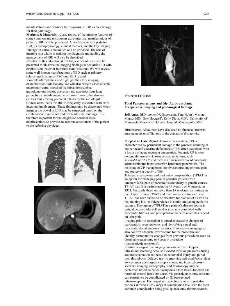
manifestations and consider the diagnosis of IBD as the etiology for their pathology. Methods & Materials: A case review of the imaging features of some common and uncommon extra-intestinal manifestations of pediatric IBD will be presented. A brief overview of pediatric
IBD, its pathophysiology, clinical features, and the key imaging findings on various modalities will be provided. The role of imaging as a whole in making the diagnosis and guiding the management of IBD will also be described. Results: In this educational exhibit, a series of cases will be presented to illustrate the imaging findings in pediatric IBD with
emphasis on the extra-intestinal manifestations. We will review some well-known manifestations of IBD such as primary sclerosing cholangitis (PSC) and IBD related spondyloarthropathies, and highlight their key imaging characteristics. Additionally, we will also present cases of some uncommon extra-intestinal manifestations such as granulomatous hepatic abscesses and non-infectious lung
parenchymal involvement, which may mimic other disease entities thus creating potential pitfalls for the radiologist. Conclusions: Pediatric IBD is frequently associated with extra-intestinal involvement. These findings may be discovered when imaging the bowel or IBD may be suspected based on the combination of intestinal and extra-intestinal findings. It is therefore important for radiologists to consider these
manifestations to provide an accurate assessment of the patient to the referring physician.
Poster #: EDU-025
Total Pancreatectomy and Islet Autotransplant:
Preoperative imaging and post-surgical findings. Jeff Ames, MD
1, [email protected]; Tara Holm
1, Michael
Murati, MD1, Eric Hoggard
1, Kelly Dietz, MD
1;
1University of
Minnesota Masonic Children's Hospital, Minneapolis, MN Disclosures: All authors have disclosed no financial interests, arrangements or affiliations in the context of this activity. Purpose or Case Report: Chronic pancreatitis (CP) is
characterized by permanent damage to the pancreas resulting in endocrine and exocrine deficiencies. CP is often associated with a history of acute recurrent pancreatitis. Pediatric CP is most commonly linked to known genetic mutations, such as PRSS1 or CFTR, and there is an increased risk of pancreatic adenocarcinoma in patients with hereditary pancreatitis. The
mainstay of CP management involves controlling chronic pain and preserving quality of life. Total pancreatectomy and islet auto-transplantation (TPIAT) is an option for managing pain in pediatric patients with uncontrollable pain or pancreatitis secondary to genetic causes. TPIAT was first performed at the University of Minnesota in 1971. Currently there are more than 15 academic institutions in
the US performing TPIAT and that number continues to rise. TPIAT has been shown to be effective for pain relief as well as maintaining insulin independence in adults and young pediatric patients. The timing of TPIAT in a patient’s disease course is critical because islet cell yield is inversely correlated with pancreatic fibrosis, and postoperative diabetes outcomes depend on islet yield.
Imaging prior to transplant is aimed at assessing changes of pancreatitis, vessel patency, and identifying vessel and pancreatic ductal anatomic variants. Preoperative imaging can also confirm adequate liver volume for the procedure and identify postoperative changes from previous procedures such as distal pancreatectomy or Puestow procedure
(pancreaticojejunostomy). Routine postoperative imaging consists of liver Doppler ultrasound screening because elevated infusion pressures during autotransplantation can result in endothelial injury and portal vein thrombosis. Delayed gastric emptying and small bowel ileus are common postsurgical complications, and targeted cross-sectional imaging, radiography, and fluoroscopy may be
performed based on patient symptoms. Once bowel function has returned, enteral feeds are started via gastrojejunostomy tube and can sometimes be complicated by GJ tube-related intussusception. The largest retrospective review in pediatric patients showed a 20% surgical complication rate, with the most common complication being post-splenectomy thrombocytosis.
Pediatr Radiol (2018) 48 (Suppl 1):S1–S298 S205
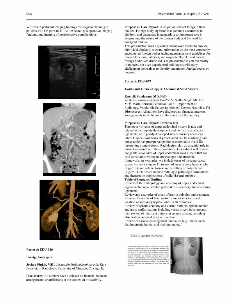
We present pertinent imaging findings for surgical planning in patients with CP prior to TPIAT, expected postoperative imaging findings, and imaging of postoperative complications.
Poster #: EDU-026
Foreign body quiz Joshua Finkle, MD
1, [email protected]; Kate
Feinstein1;
1Radiology, University of Chicago, Chicago, IL
Disclosures: All authors have disclosed no financial interests,
arrangements or affiliations in the context of this activity.
Purpose or Case Report: Kids put all sorts of things in their mouths. Foreign body ingestion is a common occurrence in children, and diagnostic imaging plays an important role in determining the nature of the foreign body and the need for emergent removal.
This presentation uses a question and answer format to provide high-yield clinically relevant information on the most commonly encountered foreign bodies including management guidelines for things like coins, batteries, and magnets. Both GI and airway foreign bodies are discussed. The presentation is catered mostly to trainees, but even experienced radiologists will enjoy
challenging themselves to identify uncommon foreign bodies on imaging.
Poster #: EDU-027
Twists and Turns of Upper Abdominal Solid Viscera Karthik Sundaram, MD, PhD
1,
[email protected]; Sudha Singh, MB BS, MD
1, Marta Hernanz-Schulman, MD
1;
1Department of
Radiology, Vanderbilt University Medical Center, Nashville, TN
Disclosures: All authors have disclosed no financial interests, arrangements or affiliations in the context of this activity.
Purpose or Case Report: Introduction Torsion or volvulus of upper abdominal viscera is rare and related to incomplete development and laxity of suspensory ligaments, or to poorly developed supernumerary accessory
lobes. Clinical symptoms at presentation can be confusing and nonspecific, yet prompt recognition is essential to avoid life-threatening complications. Radiologists play an essential role in prompt recognition of these conditions. Our exhibit will review congenital anomalies of upper abdominal solid viscera that can lead to volvulus within an embryologic and anatomic framework. As examples, we include cases of mesenteroaxial
gastric volvulus (Figure 1), torsion of an accessory hepatic lobe (Figure 2), and splenic torsion in the setting of polysplenia (Figure 3). Our cases include radiologic-pathologic correlations and therapeutic implications of solid visceral torsions.
Table of Contents/Outline: Review of the embryology and anatomy of upper abdominal
organs including a detailed pictorial of suspensory and anchoring ligaments. Review and examples of types of gastric volvulus and treatment. Review of variants of liver anatomy and of incidence and location of accessory hepatic lobes, with examples. Review of splenic anatomy and normal variants, splenic torsion and gross malformations including variants seen in heterotaxy,
with review of treatment options in splenic torsion, including observation, surgical pexy or resection. Review of associated congenital anomalies (e.g. omphalocele, diaphragmatic hernia, and malrotation, etc.)
Pediatr Radiol (2018) 48 (Suppl 1):S1–S298S206
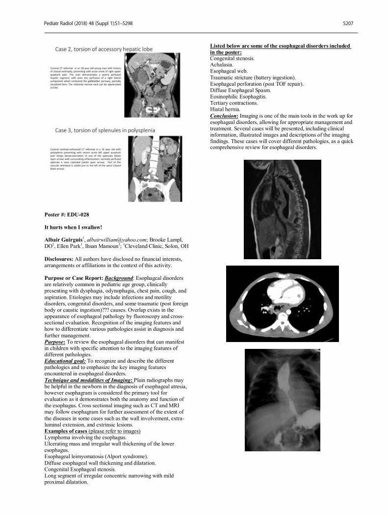
Poster #: EDU-028
It hurts when I swallow! Albair Guirguis
1, [email protected]; Brooke Lampl,
DO1, Ellen Park
1, Ihsan Mamoun
1;
1Cleveland Clinic, Solon, OH
Disclosures: All authors have disclosed no financial interests, arrangements or affiliations in the context of this activity. Purpose or Case Report: Background: Esophageal disorders are relatively common in pediatric age group, clinically presenting with dysphagia, odynophagia, chest pain, cough, and
aspiration. Etiologies may include infections and motility disorders, congenital disorders, and some traumatic (post foreign body or caustic ingestion)??? causes. Overlap exists in the appearance of esophageal pathology by fluoroscopy and cross-sectional evaluation. Recognition of the imaging features and how to differentiate various pathologies assist in diagnosis and
further management. Purpose: To review the esophageal disorders that can manifest in children with specific attention to the imaging features of different pathologies. Educational goal: To recognize and describe the different pathologies and to emphasize the key imaging features encountered in esophageal disorders.
Technique and modalities of Imaging: Plain radiographs may be helpful in the newborn in the diagnosis of esophageal atresia, however esophagram is considered the primary tool for evaluation as it demonstrates both the anatomy and function of the esophagus. Cross sectional imaging such as CT and MRI may follow esophagram for further assessment of the extent of
the diseases in some cases such as the wall involvement, extra-luminal extension, and extrinsic lesions. Examples of cases (please refer to images) Lymphoma involving the esophagus. Ulcerating mass and irregular wall thickening of the lower esophagus. Esophageal leimyomatosis (Alport syndrome).
Diffuse esophageal wall thickening and dilatation. Congenital Esophageal stenosis. Long segment of irregular concentric narrowing with mild proximal dilatation.
Listed below are some of the esophageal disorders included
in the poster: Congenital stenosis. Achalasia. Esophageal web.
Traumatic stricture (battery ingestion). Esophageal perforation (post TOF repair). Diffuse Esophageal Spasm. Eosinophilic Esophagitis. Tertiary contractions. Hiatal hernia.
Conclusion: Imaging is one of the main tools in the work up for esophageal disorders, allowing for appropriate management and treatment. Several cases will be presented, including clinical information, illustrated images and descriptions of the imaging findings. These cases will cover different pathologies, as a quick comprehensive review for esophageal disorders.
Pediatr Radiol (2018) 48 (Suppl 1):S1–S298 S207
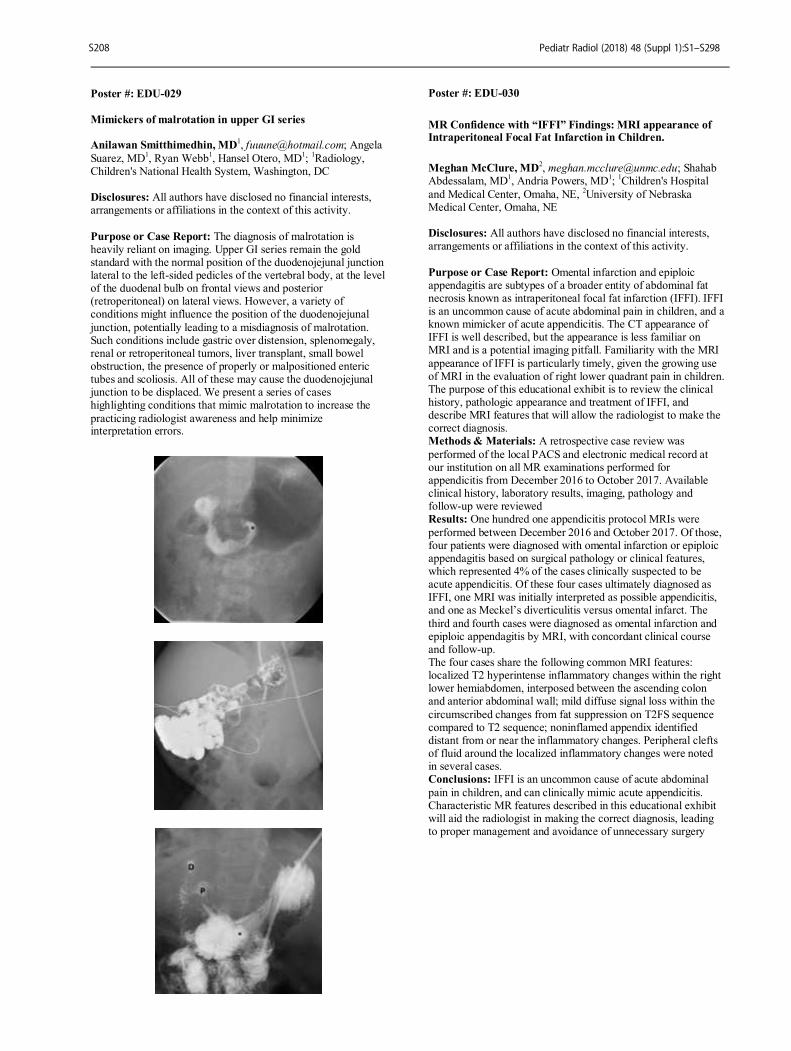
Poster #: EDU-029
Mimickers of malrotation in upper GI series Anilawan Smitthimedhin, MD
1, [email protected]; Angela
Suarez, MD1, Ryan Webb
1, Hansel Otero, MD
1;
1Radiology,
Children's National Health System, Washington, DC Disclosures: All authors have disclosed no financial interests, arrangements or affiliations in the context of this activity.
Purpose or Case Report: The diagnosis of malrotation is heavily reliant on imaging. Upper GI series remain the gold standard with the normal position of the duodenojejunal junction lateral to the left-sided pedicles of the vertebral body, at the level of the duodenal bulb on frontal views and posterior (retroperitoneal) on lateral views. However, a variety of conditions might influence the position of the duodenojejunal
junction, potentially leading to a misdiagnosis of malrotation. Such conditions include gastric over distension, splenomegaly, renal or retroperitoneal tumors, liver transplant, small bowel obstruction, the presence of properly or malpositioned enteric tubes and scoliosis. All of these may cause the duodenojejunal junction to be displaced. We present a series of cases highlighting conditions that mimic malrotation to increase the
practicing radiologist awareness and help minimize interpretation errors.
Poster #: EDU-030
MR Confidence with “IFFI” Findings: MRI appearance of Intraperitoneal Focal Fat Infarction in Children.
Meghan McClure, MD2, [email protected]; Shahab
Abdessalam, MD1, Andria Powers, MD
1;
1Children's Hospital
and Medical Center, Omaha, NE, 2University of Nebraska
Medical Center, Omaha, NE
Disclosures: All authors have disclosed no financial interests, arrangements or affiliations in the context of this activity.
Purpose or Case Report: Omental infarction and epiploic appendagitis are subtypes of a broader entity of abdominal fat necrosis known as intraperitoneal focal fat infarction (IFFI). IFFI is an uncommon cause of acute abdominal pain in children, and a known mimicker of acute appendicitis. The CT appearance of IFFI is well described, but the appearance is less familiar on MRI and is a potential imaging pitfall. Familiarity with the MRI
appearance of IFFI is particularly timely, given the growing use of MRI in the evaluation of right lower quadrant pain in children. The purpose of this educational exhibit is to review the clinical history, pathologic appearance and treatment of IFFI, and describe MRI features that will allow the radiologist to make the correct diagnosis. Methods & Materials: A retrospective case review was
performed of the local PACS and electronic medical record at our institution on all MR examinations performed for appendicitis from December 2016 to October 2017. Available clinical history, laboratory results, imaging, pathology and follow-up were reviewed Results: One hundred one appendicitis protocol MRIs were
performed between December 2016 and October 2017. Of those, four patients were diagnosed with omental infarction or epiploic appendagitis based on surgical pathology or clinical features, which represented 4% of the cases clinically suspected to be acute appendicitis. Of these four cases ultimately diagnosed as IFFI, one MRI was initially interpreted as possible appendicitis, and one as Meckel’s diverticulitis versus omental infarct. The
third and fourth cases were diagnosed as omental infarction and epiploic appendagitis by MRI, with concordant clinical course and follow-up. The four cases share the following common MRI features: localized T2 hyperintense inflammatory changes within the right lower hemiabdomen, interposed between the ascending colon and anterior abdominal wall; mild diffuse signal loss within the
circumscribed changes from fat suppression on T2FS sequence compared to T2 sequence; noninflamed appendix identified distant from or near the inflammatory changes. Peripheral clefts of fluid around the localized inflammatory changes were noted in several cases. Conclusions: IFFI is an uncommon cause of acute abdominal
pain in children, and can clinically mimic acute appendicitis. Characteristic MR features described in this educational exhibit will aid the radiologist in making the correct diagnosis, leading to proper management and avoidance of unnecessary surgery
Pediatr Radiol (2018) 48 (Suppl 1):S1–S298S208
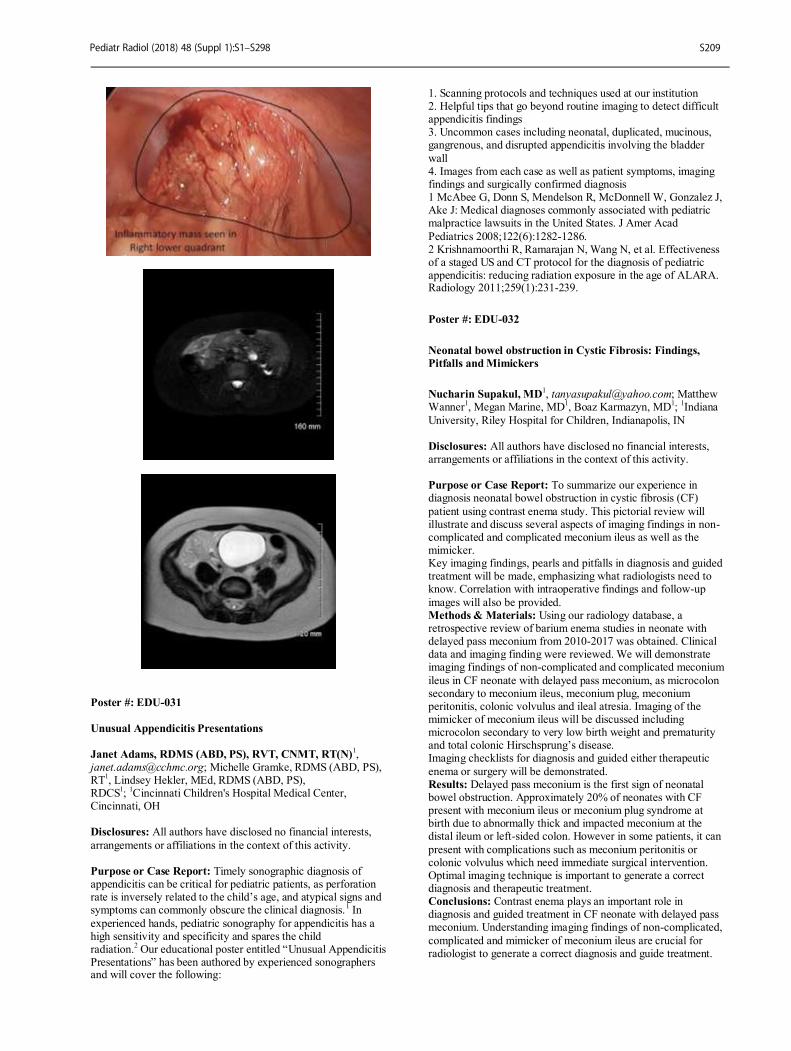
Poster #: EDU-031
Unusual Appendicitis Presentations
Janet Adams, RDMS (ABD, PS), RVT, CNMT, RT(N)1,
[email protected]; Michelle Gramke, RDMS (ABD, PS), RT
1, Lindsey Hekler, MEd, RDMS (ABD, PS),
RDCS1;
1Cincinnati Children's Hospital Medical Center,
Cincinnati, OH
Disclosures: All authors have disclosed no financial interests,
arrangements or affiliations in the context of this activity. Purpose or Case Report: Timely sonographic diagnosis of appendicitis can be critical for pediatric patients, as perforation rate is inversely related to the child’s age, and atypical signs and symptoms can commonly obscure the clinical diagnosis.
1 In
experienced hands, pediatric sonography for appendicitis has a high sensitivity and specificity and spares the child radiation.
2 Our educational poster entitled “Unusual Appendicitis
Presentations” has been authored by experienced sonographers and will cover the following:
1. Scanning protocols and techniques used at our institution 2. Helpful tips that go beyond routine imaging to detect difficult appendicitis findings 3. Uncommon cases including neonatal, duplicated, mucinous, gangrenous, and disrupted appendicitis involving the bladder
wall 4. Images from each case as well as patient symptoms, imaging findings and surgically confirmed diagnosis 1 McAbee G, Donn S, Mendelson R, McDonnell W, Gonzalez J, Ake J: Medical diagnoses commonly associated with pediatric malpractice lawsuits in the United States. J Amer Acad
Pediatrics 2008;122(6):1282-1286. 2 Krishnamoorthi R, Ramarajan N, Wang N, et al. Effectiveness of a staged US and CT protocol for the diagnosis of pediatric appendicitis: reducing radiation exposure in the age of ALARA. Radiology 2011;259(1):231-239.
Poster #: EDU-032
Neonatal bowel obstruction in Cystic Fibrosis: Findings,
Pitfalls and Mimickers
Nucharin Supakul, MD1, [email protected]; Matthew
Wanner1, Megan Marine, MD
1, Boaz Karmazyn, MD
1;
1Indiana
University, Riley Hospital for Children, Indianapolis, IN Disclosures: All authors have disclosed no financial interests, arrangements or affiliations in the context of this activity. Purpose or Case Report: To summarize our experience in diagnosis neonatal bowel obstruction in cystic fibrosis (CF)
patient using contrast enema study. This pictorial review will illustrate and discuss several aspects of imaging findings in non-complicated and complicated meconium ileus as well as the mimicker. Key imaging findings, pearls and pitfalls in diagnosis and guided treatment will be made, emphasizing what radiologists need to know. Correlation with intraoperative findings and follow-up
images will also be provided. Methods & Materials: Using our radiology database, a retrospective review of barium enema studies in neonate with delayed pass meconium from 2010-2017 was obtained. Clinical data and imaging finding were reviewed. We will demonstrate imaging findings of non-complicated and complicated meconium
ileus in CF neonate with delayed pass meconium, as microcolon secondary to meconium ileus, meconium plug, meconium peritonitis, colonic volvulus and ileal atresia. Imaging of the mimicker of meconium ileus will be discussed including microcolon secondary to very low birth weight and prematurity and total colonic Hirschsprung’s disease. Imaging checklists for diagnosis and guided either therapeutic
enema or surgery will be demonstrated. Results: Delayed pass meconium is the first sign of neonatal bowel obstruction. Approximately 20% of neonates with CF present with meconium ileus or meconium plug syndrome at birth due to abnormally thick and impacted meconium at the distal ileum or left-sided colon. However in some patients, it can
present with complications such as meconium peritonitis or colonic volvulus which need immediate surgical intervention. Optimal imaging technique is important to generate a correct diagnosis and therapeutic treatment. Conclusions: Contrast enema plays an important role in diagnosis and guided treatment in CF neonate with delayed pass meconium. Understanding imaging findings of non-complicated,
complicated and mimicker of meconium ileus are crucial for radiologist to generate a correct diagnosis and guide treatment.
Pediatr Radiol (2018) 48 (Suppl 1):S1–S298 S209
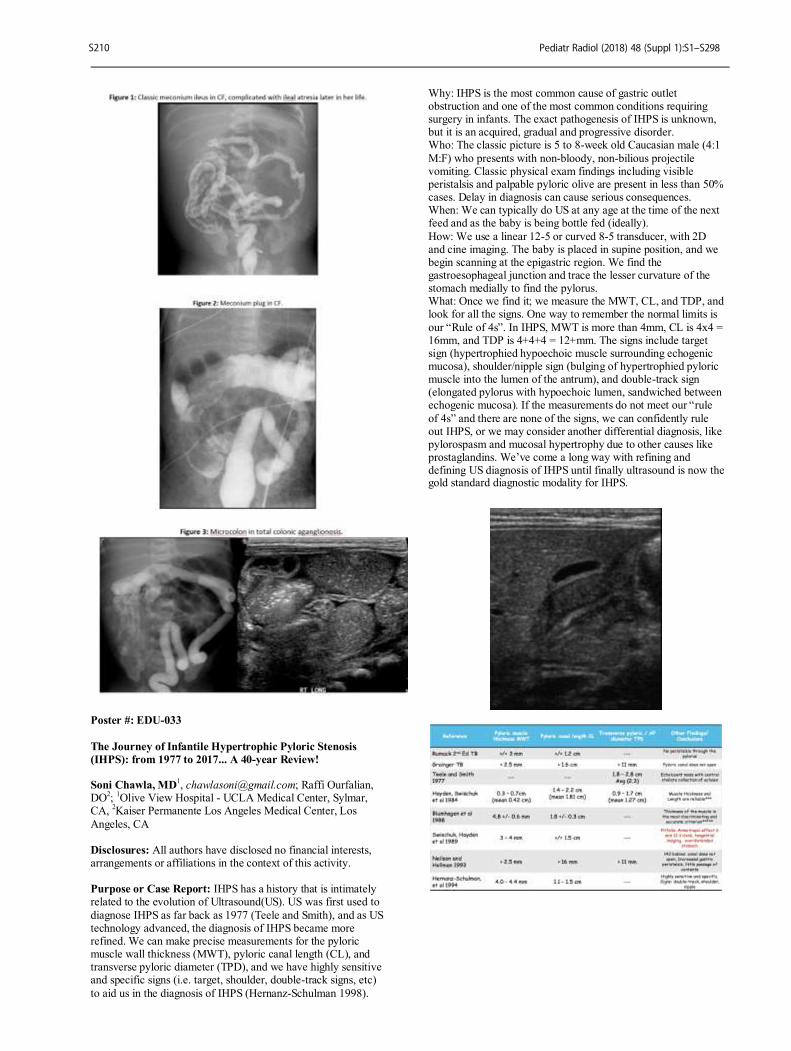
Poster #: EDU-033
The Journey of Infantile Hypertrophic Pyloric Stenosis
(IHPS): from 1977 to 2017... A 40-year Review! Soni Chawla, MD
1, [email protected]; Raffi Ourfalian,
DO2;
1Olive View Hospital - UCLA Medical Center, Sylmar,
CA, 2Kaiser Permanente Los Angeles Medical Center, Los
Angeles, CA Disclosures: All authors have disclosed no financial interests, arrangements or affiliations in the context of this activity. Purpose or Case Report: IHPS has a history that is intimately related to the evolution of Ultrasound(US). US was first used to
diagnose IHPS as far back as 1977 (Teele and Smith), and as US technology advanced, the diagnosis of IHPS became more refined. We can make precise measurements for the pyloric muscle wall thickness (MWT), pyloric canal length (CL), and transverse pyloric diameter (TPD), and we have highly sensitive and specific signs (i.e. target, shoulder, double-track signs, etc)
to aid us in the diagnosis of IHPS (Hernanz-Schulman 1998).
Why: IHPS is the most common cause of gastric outlet obstruction and one of the most common conditions requiring surgery in infants. The exact pathogenesis of IHPS is unknown, but it is an acquired, gradual and progressive disorder. Who: The classic picture is 5 to 8-week old Caucasian male (4:1
M:F) who presents with non-bloody, non-bilious projectile vomiting. Classic physical exam findings including visible peristalsis and palpable pyloric olive are present in less than 50% cases. Delay in diagnosis can cause serious consequences. When: We can typically do US at any age at the time of the next feed and as the baby is being bottle fed (ideally).
How: We use a linear 12-5 or curved 8-5 transducer, with 2D and cine imaging. The baby is placed in supine position, and we begin scanning at the epigastric region. We find the gastroesophageal junction and trace the lesser curvature of the stomach medially to find the pylorus. What: Once we find it; we measure the MWT, CL, and TDP, and look for all the signs. One way to remember the normal limits is
our “Rule of 4s”. In IHPS, MWT is more than 4mm, CL is 4x4 = 16mm, and TDP is 4+4+4 = 12+mm. The signs include target sign (hypertrophied hypoechoic muscle surrounding echogenic mucosa), shoulder/nipple sign (bulging of hypertrophied pyloric muscle into the lumen of the antrum), and double-track sign (elongated pylorus with hypoechoic lumen, sandwiched between echogenic mucosa). If the measurements do not meet our “rule
of 4s” and there are none of the signs, we can confidently rule out IHPS, or we may consider another differential diagnosis, like pylorospasm and mucosal hypertrophy due to other causes like prostaglandins. We’ve come a long way with refining and defining US diagnosis of IHPS until finally ultrasound is now the gold standard diagnostic modality for IHPS.
Pediatr Radiol (2018) 48 (Suppl 1):S1–S298S210
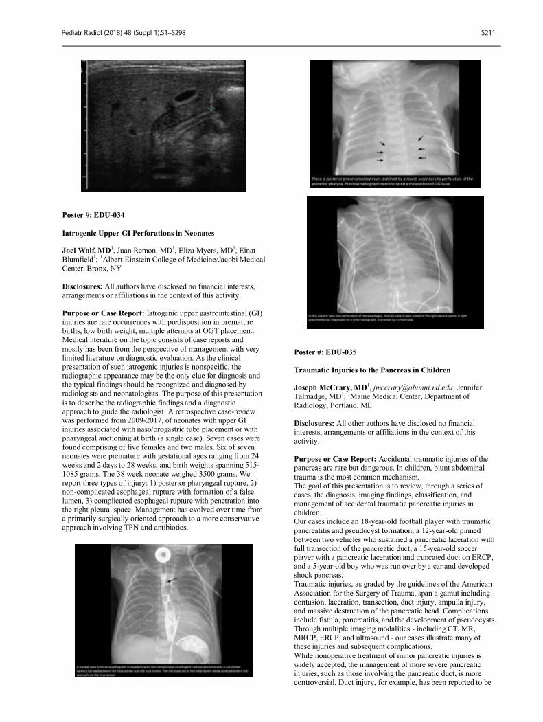
Poster #: EDU-034
Iatrogenic Upper GI Perforations in Neonates Joel Wolf, MD
1, Juan Remon, MD
1, Eliza Myers, MD
1, Einat
Blumfield1;
1Albert Einstein College of Medicine/Jacobi Medical
Center, Bronx, NY
Disclosures: All authors have disclosed no financial interests, arrangements or affiliations in the context of this activity. Purpose or Case Report: Iatrogenic upper gastrointestinal (GI) injuries are rare occurrences with predisposition in premature births, low birth weight, multiple attempts at OGT placement. Medical literature on the topic consists of case reports and
mostly has been from the perspective of management with very limited literature on diagnostic evaluation. As the clinical presentation of such iatrogenic injuries is nonspecific, the radiographic appearance may be the only clue for diagnosis and the typical findings should be recognized and diagnosed by radiologists and neonatologists. The purpose of this presentation
is to describe the radiographic findings and a diagnostic approach to guide the radiologist. A retrospective case-review was performed from 2009-2017, of neonates with upper GI injuries associated with naso/orogastric tube placement or with pharyngeal auctioning at birth (a single case). Seven cases were found comprising of five females and two males. Six of seven neonates were premature with gestational ages ranging from 24
weeks and 2 days to 28 weeks, and birth weights spanning 515-1085 grams. The 38 week neonate weighed 3500 grams. We report three types of injury: 1) posterior pharyngeal rupture, 2) non-complicated esophageal rupture with formation of a false lumen, 3) complicated esophageal rupture with penetration into the right pleural space. Management has evolved over time from a primarily surgically oriented approach to a more conservative approach involving TPN and antibiotics.
Poster #: EDU-035
Traumatic Injuries to the Pancreas in Children Joseph McCrary, MD
1, [email protected]; Jennifer
Talmadge, MD1;
1Maine Medical Center, Department of
Radiology, Portland, ME
Disclosures: All other authors have disclosed no financial interests, arrangements or affiliations in the context of this activity. Purpose or Case Report: Accidental traumatic injuries of the pancreas are rare but dangerous. In children, blunt abdominal
trauma is the most common mechanism. The goal of this presentation is to review, through a series of cases, the diagnosis, imaging findings, classification, and management of accidental traumatic pancreatic injuries in children. Our cases include an 18-year-old football player with traumatic
pancreatitis and pseudocyst formation, a 12-year-old pinned between two vehicles who sustained a pancreatic laceration with full transection of the pancreatic duct, a 15-year-old soccer player with a pancreatic laceration and truncated duct on ERCP, and a 5-year-old boy who was run over by a car and developed shock pancreas. Traumatic injuries, as graded by the guidelines of the American
Association for the Surgery of Trauma, span a gamut including contusion, laceration, transection, duct injury, ampulla injury, and massive destruction of the pancreatic head. Complications include fistula, pancreatitis, and the development of pseudocysts. Through multiple imaging modalities - including CT, MR, MRCP, ERCP, and ultrasound - our cases illustrate many of these injuries and subsequent complications.
While nonoperative treatment of minor pancreatic injuries is widely accepted, the management of more severe pancreatic injuries, such as those involving the pancreatic duct, is more controversial. Duct injury, for example, has been reported to be
Pediatr Radiol (2018) 48 (Suppl 1):S1–S298 S211
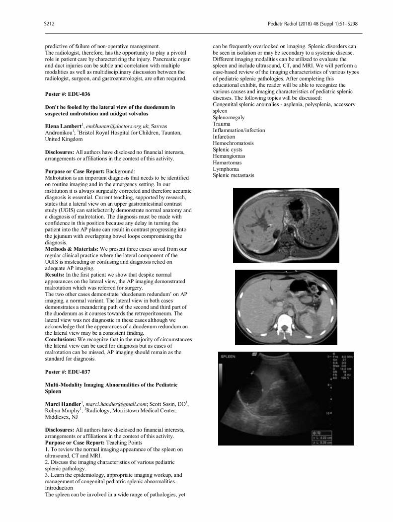
predictive of failure of non-operative management. The radiologist, therefore, has the opportunity to play a pivotal role in patient care by characterizing the injury. Pancreatic organ and duct injuries can be subtle and correlation with multiple modalities as well as multidisciplinary discussion between the radiologist, surgeon, and gastroenterologist, are often required.
Poster #: EDU-036
Don't be fooled by the lateral view of the duodenum in
suspected malrotation and midgut volvulus Elena Lambert
1, [email protected]; Savvas
Andronikou1;
1Bristol Royal Hospital for Children, Taunton,
United Kingdom
Disclosures: All authors have disclosed no financial interests, arrangements or affiliations in the context of this activity. Purpose or Case Report: Background: Malrotation is an important diagnosis that needs to be identified on routine imaging and in the emergency setting. In our
institution it is always surgically corrected and therefore accurate diagnosis is essential. Current teaching, supported by research, states that a lateral view on an upper gastrointestinal contrast study (UGIS) can satisfactorily demonstrate normal anatomy and a diagnosis of malrotation. The diagnosis must be made with confidence in this position because any delay in turning the patient into the AP plane can result in contrast progressing into
the jejunum with overlapping bowel loops compromising the diagnosis. Methods & Materials: We present three cases saved from our regular clinical practice where the lateral component of the UGIS is misleading or confusing and diagnosis relied on adequate AP imaging. Results: In the first patient we show that despite normal
appearances on the lateral view, the AP imaging demonstrated malrotation which was referred for surgery. The two other cases demonstrate ‘duodenum redundum’ on AP imaging, a normal variant. The lateral view in both cases demonstrates a meandering path of the second and third part of the duodenum as it courses towards the retroperitoneum. The
lateral view was not diagnostic in these cases although we acknowledge that the appearances of a duodenum redundum on the lateral view may be a consistent finding. Conclusions: We recognize that in the majority of circumstances the lateral view can be used for diagnosis but as cases of malrotation can be missed, AP imaging should remain as the standard for diagnosis.
Poster #: EDU-037
Multi-Modality Imaging Abnormalities of the Pediatric
Spleen
Marci Handler1, [email protected]; Scott Sosin, DO
1,
Robyn Murphy1;
1Radiology, Morristown Medical Center,
Middlesex, NJ Disclosures: All authors have disclosed no financial interests, arrangements or affiliations in the context of this activity. Purpose or Case Report: Teaching Points
1. To review the normal imaging appearance of the spleen on ultrasound, CT and MRI. 2. Discuss the imaging characteristics of various pediatric splenic pathology. 3. Learn the epidemiology, appropriate imaging workup, and management of congenital pediatric splenic abnormalities. Introduction
The spleen can be involved in a wide range of pathologies, yet
can be frequently overlooked on imaging. Splenic disorders can be seen in isolation or may be secondary to a systemic disease. Different imaging modalities can be utilized to evaluate the spleen and include ultrasound, CT, and MRI. We will perform a case-based review of the imaging characteristics of various types
of pediatric splenic pathologies. After completing this educational exhibit, the reader will be able to recognize the various causes and imaging characteristics of pediatric splenic diseases. The following topics will be discussed: Congenital splenic anomalies - asplenia, polysplenia, accessory spleen
Splenomegaly Trauma Inflammation/infection Infarction Hemochromatosis Splenic cysts Hemangiomas
Hamartomas Lymphoma Splenic metastasis
Pediatr Radiol (2018) 48 (Suppl 1):S1–S298S212
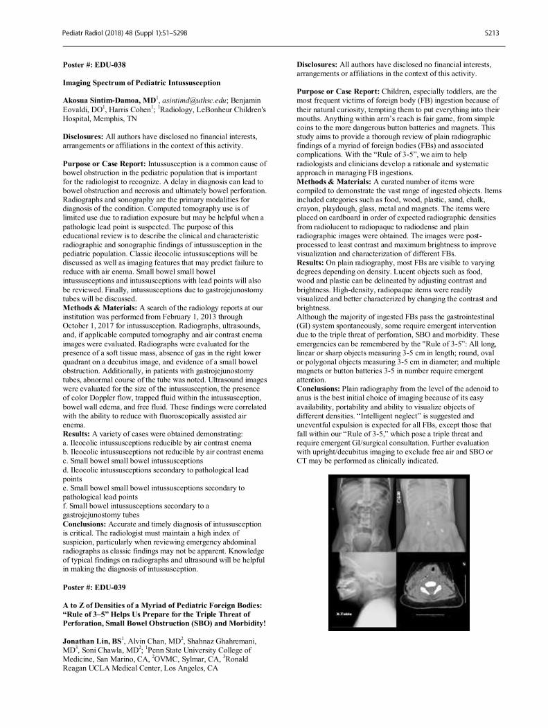
Poster #: EDU-038
Imaging Spectrum of Pediatric Intussusception Akosua Sintim-Damoa, MD
1, [email protected]; Benjamin
Eovaldi, DO1, Harris Cohen
1;
1Radiology, LeBonheur Children's
Hospital, Memphis, TN Disclosures: All authors have disclosed no financial interests, arrangements or affiliations in the context of this activity.
Purpose or Case Report: Intussusception is a common cause of bowel obstruction in the pediatric population that is important for the radiologist to recognize. A delay in diagnosis can lead to bowel obstruction and necrosis and ultimately bowel perforation. Radiographs and sonography are the primary modalities for diagnosis of the condition. Computed tomography use is of limited use due to radiation exposure but may be helpful when a
pathologic lead point is suspected. The purpose of this educational review is to describe the clinical and characteristic radiographic and sonographic findings of intussusception in the pediatric population. Classic ileocolic intussusceptions will be discussed as well as imaging features that may predict failure to reduce with air enema. Small bowel small bowel intussusceptions and intussusceptions with lead points will also
be reviewed. Finally, intussusceptions due to gastrojejunostomy tubes will be discussed. Methods & Materials: A search of the radiology reports at our institution was performed from February 1, 2013 through October 1, 2017 for intussusception. Radiographs, ultrasounds, and, if applicable computed tomography and air contrast enema
images were evaluated. Radiographs were evaluated for the presence of a soft tissue mass, absence of gas in the right lower quadrant on a decubitus image, and evidence of a small bowel obstruction. Additionally, in patients with gastrojejunostomy tubes, abnormal course of the tube was noted. Ultrasound images were evaluated for the size of the intussusception, the presence of color Doppler flow, trapped fluid within the intussusception,
bowel wall edema, and free fluid. These findings were correlated with the ability to reduce with fluoroscopically assisted air enema. Results: A variety of cases were obtained demonstrating: a. Ileocolic intussusceptions reducible by air contrast enema b. Ileocolic intussusceptions not reducible by air contrast enema c. Small bowel small bowel intussusceptions
d. Ileocolic intussusceptions secondary to pathological lead points e. Small bowel small bowel intussusceptions secondary to pathological lead points f. Small bowel intussusceptions secondary to a gastrojejunostomy tubes
Conclusions: Accurate and timely diagnosis of intussusception is critical. The radiologist must maintain a high index of suspicion, particularly when reviewing emergency abdominal radiographs as classic findings may not be apparent. Knowledge of typical findings on radiographs and ultrasound will be helpful in making the diagnosis of intussusception.
Poster #: EDU-039
A to Z of Densities of a Myriad of Pediatric Foreign Bodies:
“Rule of 3–5” Helps Us Prepare for the Triple Threat of
Perforation, Small Bowel Obstruction (SBO) and Morbidity!
Jonathan Lin, BS1, Alvin Chan, MD
2, Shahnaz Ghahremani,
MD3, Soni Chawla, MD
2;
1Penn State University College of
Medicine, San Marino, CA, 2OVMC, Sylmar, CA,
3Ronald
Reagan UCLA Medical Center, Los Angeles, CA
Disclosures: All authors have disclosed no financial interests, arrangements or affiliations in the context of this activity. Purpose or Case Report: Children, especially toddlers, are the most frequent victims of foreign body (FB) ingestion because of
their natural curiosity, tempting them to put everything into their mouths. Anything within arm’s reach is fair game, from simple coins to the more dangerous button batteries and magnets. This study aims to provide a thorough review of plain radiographic findings of a myriad of foreign bodies (FBs) and associated complications. With the “Rule of 3-5”, we aim to help
radiologists and clinicians develop a rationale and systematic approach in managing FB ingestions. Methods & Materials: A curated number of items were compiled to demonstrate the vast range of ingested objects. Items included categories such as food, wood, plastic, sand, chalk, crayon, playdough, glass, metal and magnets. The items were placed on cardboard in order of expected radiographic densities
from radiolucent to radiopaque to radiodense and plain radiographic images were obtained. The images were post-processed to least contrast and maximum brightness to improve visualization and characterization of different FBs. Results: On plain radiography, most FBs are visible to varying degrees depending on density. Lucent objects such as food, wood and plastic can be delineated by adjusting contrast and
brightness. High-density, radiopaque items were readily visualized and better characterized by changing the contrast and brightness. Although the majority of ingested FBs pass the gastrointestinal (GI) system spontaneously, some require emergent intervention due to the triple threat of perforation, SBO and morbidity. These
emergencies can be remembered by the "Rule of 3-5”: All long, linear or sharp objects measuring 3-5 cm in length; round, oval or polygonal objects measuring 3-5 cm in diameter; and multiple magnets or button batteries 3-5 in number require emergent attention. Conclusions: Plain radiography from the level of the adenoid to anus is the best initial choice of imaging because of its easy
availability, portability and ability to visualize objects of different densities. “Intelligent neglect” is suggested and uneventful expulsion is expected for all FBs, except those that fall within our “Rule of 3-5,” which pose a triple threat and require emergent GI/surgical consultation. Further evaluation with upright/decubitus imaging to exclude free air and SBO or CT may be performed as clinically indicated.
Pediatr Radiol (2018) 48 (Suppl 1):S1–S298 S213
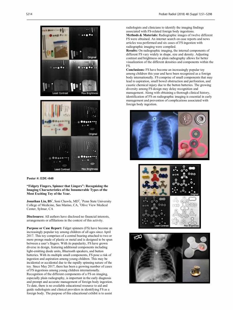
Poster #: EDU-040
“Fidgety Fingers, Spinner that Lingers”: Recognizing the
Imaging Characteristics of the Innumerable Types of the
Most Exciting Toy of the Year.
Jonathan Lin, BS1, Soni Chawla, MD
2;
1Penn State University
College of Medicine, San Marino, CA, 2Olive View Medical
Center, Sylmar, CA Disclosures: All authors have disclosed no financial interests, arrangements or affiliations in the context of this activity.
Purpose or Case Report: Fidget spinners (FS) have become an increasingly popular toy among children of all ages since April 2017. This toy comprises of a central bearing attached to two or more prongs made of plastic or metal and is designed to be spun between a user’s fingers. With its popularity, FS have grown diverse in design, featuring additional components including
light-emitting diode units, Bluetooth speakers, and button batteries. With its multiple small components, FS pose a risk of ingestion and aspiration among young children. This may be incidental or accidental due to the rapidly spinning nature of the toy. Since May 2017, there has been a growing number of cases of FS ingestions among young children internationally. Recognition of the different components of a FS on imaging,
especially plain radiography, is important in the early diagnosis and prompt and accurate management of foreign body ingestion. To date, there is no available educational resource to aid and guide radiologists and clinical providers in identifying FS as a foreign body. The purpose of this educational exhibit is to assist
radiologists and clinicians to identify the imaging findings associated with FS-related foreign body ingestions. Methods & Materials: Radiographic images of twelve different FS were obtained. An internet search on case reports and news articles was performed and six cases of FS ingestion with
radiographic imaging were compiled. Results: On radiographic imaging, the internal components of different FS vary widely in shape, size and density. Adjusting contrast and brightness on plain radiography allows for better visualization of the different densities and components within the FS.
Conclusions: FS have become an increasingly popular toy among children this year and have been recognized as a foreign body internationally. FS comprise of small components that may lead to aspiration, small bowel obstruction and perforation, and caustic chemical injury due to the button batteries. The growing diversity among FS design may delay recognition and management. Along with obtaining a thorough clinical history,
identification of FS on radiographic imaging is essential in early management and prevention of complications associated with foreign body ingestion.
Pediatr Radiol (2018) 48 (Suppl 1):S1–S298S214
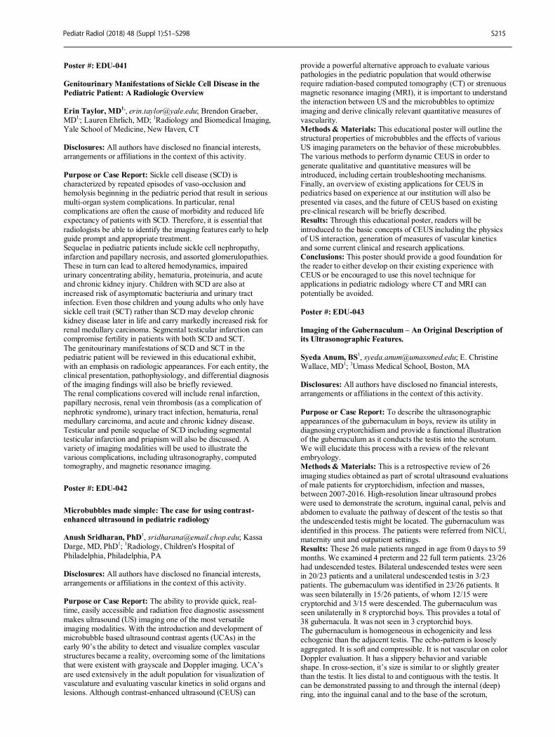
Poster #: EDU-041
Genitourinary Manifestations of Sickle Cell Disease in the
Pediatric Patient: A Radiologic Overview
Erin Taylor, MD1,
, [email protected]; Brendon Graeber, MD
1,; Lauren Ehrlich, MD;
1Radiology and Biomedical Imaging,
Yale School of Medicine, New Haven, CT Disclosures: All authors have disclosed no financial interests, arrangements or affiliations in the context of this activity.
Purpose or Case Report: Sickle cell disease (SCD) is characterized by repeated episodes of vaso-occlusion and hemolysis beginning in the pediatric period that result in serious multi-organ system complications. In particular, renal complications are often the cause of morbidity and reduced life expectancy of patients with SCD. Therefore, it is essential that
radiologists be able to identify the imaging features early to help guide prompt and appropriate treatment. Sequelae in pediatric patients include sickle cell nephropathy, infarction and papillary necrosis, and assorted glomerulopathies. These in turn can lead to altered hemodynamics, impaired urinary concentrating ability, hematuria, proteinuria, and acute and chronic kidney injury. Children with SCD are also at
increased risk of asymptomatic bacteriuria and urinary tract infection. Even those children and young adults who only have sickle cell trait (SCT) rather than SCD may develop chronic kidney disease later in life and carry markedly increased risk for renal medullary carcinoma. Segmental testicular infarction can compromise fertility in patients with both SCD and SCT.
The genitourinary manifestations of SCD and SCT in the pediatric patient will be reviewed in this educational exhibit, with an emphasis on radiologic appearances. For each entity, the clinical presentation, pathophysiology, and differential diagnosis of the imaging findings will also be briefly reviewed. The renal complications covered will include renal infarction, papillary necrosis, renal vein thrombosis (as a complication of
nephrotic syndrome), urinary tract infection, hematuria, renal medullary carcinoma, and acute and chronic kidney disease. Testicular and penile sequelae of SCD including segmental testicular infarction and priapism will also be discussed. A variety of imaging modalities will be used to illustrate the various complications, including ultrasonography, computed tomography, and magnetic resonance imaging.
Poster #: EDU-042
Microbubbles made simple: The case for using contrast-
enhanced ultrasound in pediatric radiology Anush Sridharan, PhD
1, [email protected]; Kassa
Darge, MD, PhD1;
1Radiology, Children's Hospital of
Philadelphia, Philadelphia, PA Disclosures: All authors have disclosed no financial interests, arrangements or affiliations in the context of this activity. Purpose or Case Report: The ability to provide quick, real-time, easily accessible and radiation free diagnostic assessment
makes ultrasound (US) imaging one of the most versatile imaging modalities. With the introduction and development of microbubble based ultrasound contrast agents (UCAs) in the early 90’s the ability to detect and visualize complex vascular structures became a reality, overcoming some of the limitations that were existent with grayscale and Doppler imaging. UCA’s
are used extensively in the adult population for visualization of vasculature and evaluating vascular kinetics in solid organs and lesions. Although contrast-enhanced ultrasound (CEUS) can
provide a powerful alternative approach to evaluate various pathologies in the pediatric population that would otherwise require radiation-based computed tomography (CT) or strenuous magnetic resonance imaging (MRI), it is important to understand the interaction between US and the microbubbles to optimize
imaging and derive clinically relevant quantitative measures of vascularity. Methods & Materials: This educational poster will outline the structural properties of microbubbles and the effects of various US imaging parameters on the behavior of these microbubbles. The various methods to perform dynamic CEUS in order to
generate qualitative and quantitative measures will be introduced, including certain troubleshooting mechanisms. Finally, an overview of existing applications for CEUS in pediatrics based on experience at our institution will also be presented via cases, and the future of CEUS based on existing pre-clinical research will be briefly described. Results: Through this educational poster, readers will be
introduced to the basic concepts of CEUS including the physics of US interaction, generation of measures of vascular kinetics and some current clinical and research applications. Conclusions: This poster should provide a good foundation for the reader to either develop on their existing experience with CEUS or be encouraged to use this novel technique for applications in pediatric radiology where CT and MRI can
potentially be avoided.
Poster #: EDU-043
Imaging of the Gubernaculum – An Original Description of
its Ultrasonographic Features.
Syeda Anum, BS
1, [email protected]; E. Christine
Wallace, MD1;
1Umass Medical School, Boston, MA
Disclosures: All authors have disclosed no financial interests, arrangements or affiliations in the context of this activity.
Purpose or Case Report: To describe the ultrasonographic appearances of the gubernaculum in boys, review its utility in diagnosing cryptorchidism and provide a functional illustration of the gubernaculum as it conducts the testis into the scrotum. We will elucidate this process with a review of the relevant embryology. Methods & Materials: This is a retrospective review of 26
imaging studies obtained as part of scrotal ultrasound evaluations of male patients for cryptorchidism, infection and masses, between 2007-2016. High-resolution linear ultrasound probes were used to demonstrate the scrotum, inguinal canal, pelvis and abdomen to evaluate the pathway of descent of the testis so that the undescended testis might be located. The gubernaculum was
identified in this process. The patients were referred from NICU, maternity unit and outpatient settings. Results: These 26 male patients ranged in age from 0 days to 59 months. We examined 4 preterm and 22 full term patients. 23/26 had undescended testes. Bilateral undescended testes were seen in 20/23 patients and a unilateral undescended testis in 3/23 patients. The gubernaculum was identified in 23/26 patients. It
was seen bilaterally in 15/26 patients, of whom 12/15 were cryptorchid and 3/15 were descended. The gubernaculum was seen unilaterally in 8 cryptorchid boys. This provides a total of 38 gubernacula. It was not seen in 3 cryptorchid boys. The gubernaculum is homogeneous in echogenicity and less echogenic than the adjacent testis. The echo-pattern is loosely
aggregated. It is soft and compressible. It is not vascular on color Doppler evaluation. It has a slippery behavior and variable shape. In cross-section, it’s size is similar to or slightly greater than the testis. It lies distal to and contiguous with the testis. It can be demonstrated passing to and through the internal (deep) ring, into the inguinal canal and to the base of the scrotum,
Pediatr Radiol (2018) 48 (Suppl 1):S1–S298 S215
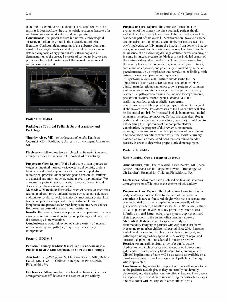
therefore it’s length varies. It should not be confused with the testis as it does not have the characteristic testicular features of a mediastinum testis or strictly ovoid configuration. Conclusions: The gubernaculum is a normal embryological structure not often described in the pediatric radiological
literature. Confident demonstration of the gubernaculum can assist in locating the undescended testis and provides a more detailed diagnosis of cryptorchidism. Ultrasonographic demonstration of the arrested process of testicular descent also provides a beautiful illustration of the normal physiological mechanism of descent.
Poster #: EDU-044
Radiology of Unusual Pediatric Scrotal Anatomy and
Pathology
Timothy Alves, MD1, [email protected]; Kathleen
Gebarski, MD1;
1Radiology, University of Michigan, Ann Arbor,
MI Disclosures: All authors have disclosed no financial interests, arrangements or affiliations in the context of this activity.
Purpose or Case Report: While hydroceles, patent processus vaginalis, inguinal hernias, varicoceles, epididymitis, orchitis, torsion of testes and appendages are common in pediatric radiological practice, other pathology and anatomical variants are unusual and may not be included in every day practice. We composed a pictorial guide of a wide variety of variants and
diseases for education and reference. Methods & Materials: Illustrative cases of ectasia of rete testes, testicular adrenal rests, tunica albuginea cyst, scrotal calcinosis, abdominoscrotal hydrocele, hematocele, meconium periorchitis, testicular epidermoid cyst, calcifying Sertoli cell tumor, lymphoma and paratesticular rhabdomyosarcoma were chosen from over ten years of imaging at our institution.
Results: Reviewing these cases provides an experience of a wide variety of unusual scrotal anatomy and pathology and improves the accuracy of interpretation. Conclusions: A pictorial review of a wide variety of unusual scrotal anatomy and pathology improves the accuracy of interpretation.
Poster #: EDU-045
Pediatric Urinary Bladder Masses and Pseudo-masses: A
Pictorial Review with Emphasis on Ultrasound Findings Ami Gokli
1, [email protected]; Christian Barrera, MD
1, Richard
Bellah, MD, FAAP1;
1Children’s Hospital of Philadelphia,
Philadelphia, PA Disclosures: All authors have disclosed no financial interests, arrangements or affiliations in the context of this activity.
Purpose or Case Report: The complete ultrasound (US) evaluation of the urinary tract in a pediatric patient should include both the urinary bladder and kidneys. Evaluation of the bladder as part of that overall US examination, however, can be deemphasized or incomplete due a number of factors, such as
one’s neglecting to fully image the bladder from dome to bladder neck, suboptimal bladder distension, incomplete distension due to presence of an indwelling drainage catheter or vesicostomy, or in some instances, because the bladder is not included as part of the routine kidney ultrasound exam. True masses arising from the urinary bladder in children are generally rare, and at times,
subtle and non-specific, and potentially mimicked by so-called pseudomasses, so we emphasize that correlation of findings with patient history is of paramount importance. This pictorial review will illustrate and describe the US appearances (along with selective cross-sectional imaging), clinical manifestations, and tumor growth patterns of common and uncommon conditions arising from the pediatric urinary
bladder, i.e. path-proven masses that include leiomyosarcoma, pheochromocytoma, nephrogenic adenoma, vascular malformation, low grade urothelial neoplasms, neurofibromatosis, fibroepithelial polyps, rhabdoid tumor, and rhabdomyosarcoma. Pseudomasses of the bladder that will also be illustrated and briefly discussed include hematomas, urachal remnants, complex ureteroceles, Deflux injection sites, foreign
bodies, and cystitis (viral, eosinophilic, parasitic). In addition to emphasizing the importance of the complete bladder examination, the purpose of this review is to increase radiologist’s awareness of the US appearances of the common and uncommon conditions which afflict the pediatric urinary bladder, as well as those conditions that can mimic bladder
masses, in order to determine proper clinical management.
Poster #: EDU-046
Seeing double: One too many of an organ Anne Misiura, MD
1, Faaiza Kazmi
1, Erica Poletto, MD
1, Mea
Mallon1, Archana Malik
1, Jaqueline Urbine
1;
1Radiology, St.
Christopher's Hospital for Children, Philadelphia, PA Disclosures: All authors have disclosed no financial interests, arrangements or affiliations in the context of this activity. Purpose or Case Report: The duplication of structures in the
body has been a curious topic to the field of medicine for centuries. It is rare to find a radiologist who has not seen at least one duplicated or partially duplicated organ, usually of the genitourinary system, and often incidentally. While implications of GU duplication have been study previously, often due to infertility or renal issues, other organ system duplications and
their implications to the patient often remain a mystery. Methods & Materials: A retrospective analysis of multimodality imaging in patients with duplicated structures presenting to an urban children’s hospital since 2005. Imaging and clinical history are correlated with clinical, surgical, and pathologic findings where applicable. A variety of organ and structural duplications are selected for imaging review.
Results: An enthralling visual array of organ/structure duplication will include cases such as duplicated duodenum, gallbladder, vessels, urinary bladder/genitalia, among others. Clinical implications of each will be discussed as available on a case by case basis, as well as surgical and pathologic findings where applicable.
Conclusions: Organ/structure duplication is a spellbinding topic to the pediatric radiologist, as they are usually incidentally discovered, and the implications are often unknown. Each case is an opportunity for creation of mesmerizing reconstructed images and discussion with colleagues in other clinical areas.
Pediatr Radiol (2018) 48 (Suppl 1):S1–S298S216
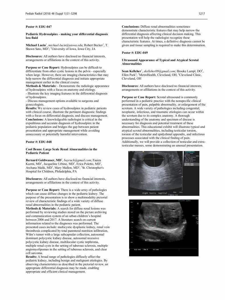
Poster #: EDU-047
Pediatric Hydrosalpinx - making your differential diagnosis
less fluid
Michael Lucin1, [email protected]; Robert Becker
1, T.
Shawn Sato, MD1;
1University of Iowa, Iowa City, IA
Disclosures: All authors have disclosed no financial interests, arrangements or affiliations in the context of this activity.
Purpose or Case Report: Hydrosalpinx can be difficult to differentiate from other cystic lesions in the pelvis - especially when large. However, there are imaging characteristics that may help narrow the differential diagnosis and initiate appropriate management earlier in the clinical course. Methods & Materials: - Demonstrate the radiologic appearance of hydrosalpinx with a focus on anatomy and etiology.
- Illustrate the key imaging features in the differential diagnosis of hydrosalpinx. - Discuss management options available to surgeons and gynecologists. Results: We review cases of hydrosalpinx in pediatric patients with clinical course, illustrate the pertinent diagnostic findings with a focus on differential diagnosis, and discuss management.
Conclusions: A knowledgeable radiologist is critical in the expeditious and accurate diagnosis of hydrosalpinx in the pediatric population and bridges the gap between patient presentation and appropriate management while avoiding unnecessary or potentially harmful intervention.
Poster #: EDU-048
Cool Beans: Large Scale Renal Abnormalities in the
Pediatric Patient Bernard Goldwasser, MD
1, [email protected]; Faaiza
Kazmi, MD1, Jacqueline Urbine, MD
1, Erica Poletto, MD
1,
Archana Malik, MD1, Mary Mallon, MD
1;
1St. Christopher's
Hospital for Children, Philadelphia, PA Disclosures: All authors have disclosed no financial interests, arrangements or affiliations in the context of this activity. Purpose or Case Report: There is a wide array of pathologies
which can cause diffuse changes in the pediatric kidney. The purpose of the presentation is to show a multimodality pictorial review of characteristic findings of a wide variety of diffuse renal abnormalities in the pediatric patient. Methods & Materials: A search for diffuse renal lesions was performed by reviewing studies stored on the picture archiving
and communication system of an urban children’s hospital between 2006 and 2017. A literature search on current information related to the diagnoses was performed. The presented cases include: multicystic dysplastic kidney, renal vein thrombosis complicated by total parenteral nutrition infiltration, Wilm’s tumor with a large subcapsular collection, autosomal dominant polycystic kidney disease, autosomal recessive
polycystic kidney disease, multilocular cystic nephroma, multiple renal cysts in the setting of tuberous sclerosis, multiple angiomyolipomas in the setting of tuberous sclerosis, and clear cell sarcoma. Results: A broad range of pathologies diffusely affect the pediatric kidney, including benign and malignant etiologies. By
observing characteristics as described in the pictorial review, an appropriate differential diagnosis may be made, enabling appropriate and efficient clinical management.
Conclusions: Diffuse renal abnormalities sometimes demonstrate characteristic features that may help narrow the differential diagnosis affecting clinical decision making. This presentation will help the radiologist recognize these characteristic features. At times, a definitive diagnosis cannot be
given and tissue sampling is required to make this determination.
Poster #: EDU-049
Ultrasound Appearance of Typical and Atypical Scrotal
Abnormalities
Sean Kelleher
1, [email protected]; Brooke Lampl, DO
2,
Ellen Park2;
1MetroHealth, Cleveland, OH,
2Cleveland Clinic,
Cleveland, OH Disclosures: All authors have disclosed no financial interests, arrangements or affiliations in the context of this activity.
Purpose or Case Report: Scrotal ultrasound is commonly performed in a pediatric practice with the nonspecific clinical presentation of pain, palpable abnormality, or enlargement of the scrotum. A wide variety of pathologies including congenital, neoplastic, infectious, and traumatic etiologies can occur within the scrotum due to its complex anatomy. A thorough
understanding of the anatomy and spectrum of disease is necessary for diagnosis and potential treatment of these abnormalities. This educational exhibit will illustrate typical and atypical scrotal abnormalities, including testicular torsion, torsion of the testicular and epidydimal appendix, and infectious processes associated with the clinical finding of pain.
Additionally, we will provide a collection of testicular and extra-testicular masses, some demonstrating an unusual presentation.
Pediatr Radiol (2018) 48 (Suppl 1):S1–S298 S217
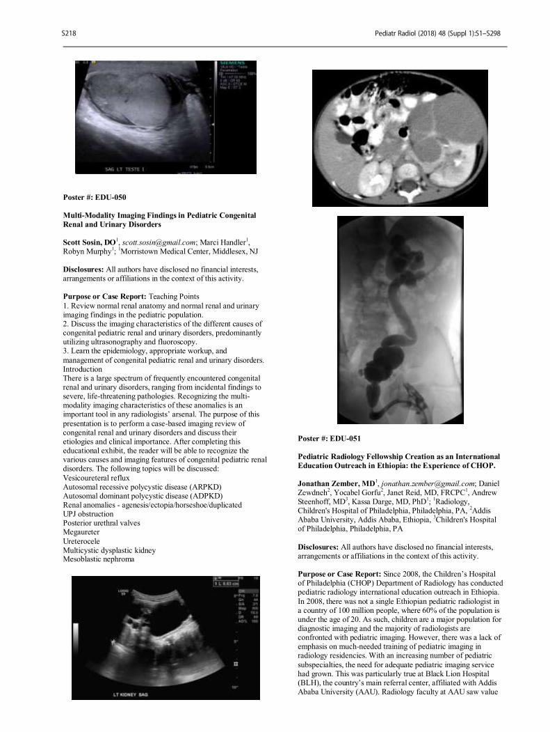
Poster #: EDU-050
Multi-Modality Imaging Findings in Pediatric Congenital
Renal and Urinary Disorders
Scott Sosin, DO1, [email protected]; Marci Handler
1,
Robyn Murphy1;
1Morristown Medical Center, Middlesex, NJ
Disclosures: All authors have disclosed no financial interests, arrangements or affiliations in the context of this activity. Purpose or Case Report: Teaching Points
1. Review normal renal anatomy and normal renal and urinary imaging findings in the pediatric population. 2. Discuss the imaging characteristics of the different causes of congenital pediatric renal and urinary disorders, predominantly utilizing ultrasonography and fluoroscopy. 3. Learn the epidemiology, appropriate workup, and
management of congenital pediatric renal and urinary disorders. Introduction There is a large spectrum of frequently encountered congenital renal and urinary disorders, ranging from incidental findings to severe, life-threatening pathologies. Recognizing the multi-modality imaging characteristics of these anomalies is an important tool in any radiologists’ arsenal. The purpose of this
presentation is to perform a case-based imaging review of congenital renal and urinary disorders and discuss their etiologies and clinical importance. After completing this educational exhibit, the reader will be able to recognize the various causes and imaging features of congenital pediatric renal disorders. The following topics will be discussed:
Vesicoureteral reflux Autosomal recessive polycystic disease (ARPKD) Autosomal dominant polycystic disease (ADPKD) Renal anomalies - agenesis/ectopia/horseshoe/duplicated UPJ obstruction Posterior urethral valves Megaureter
Ureterocele Multicystic dysplastic kidney Mesoblastic nephroma
Poster #: EDU-051
Pediatric Radiology Fellowship Creation as an International
Education Outreach in Ethiopia: the Experience of CHOP.
Jonathan Zember, MD1, [email protected]; Daniel
Zewdneh2, Yocabel Gorfu
2, Janet Reid, MD, FRCPC
1, Andrew
Steenhoff, MD3, Kassa Darge, MD, PhD
1;
1Radiology,
Children's Hospital of Philadelphia, Philadelphia, PA, 2Addis
Ababa University, Addis Ababa, Ethiopia, 3Children's Hospital
of Philadelphia, Philadelphia, PA
Disclosures: All authors have disclosed no financial interests, arrangements or affiliations in the context of this activity. Purpose or Case Report: Since 2008, the Children’s Hospital of Philadelphia (CHOP) Department of Radiology has conducted pediatric radiology international education outreach in Ethiopia.
In 2008, there was not a single Ethiopian pediatric radiologist in a country of 100 million people, where 60% of the population is under the age of 20. As such, children are a major population for diagnostic imaging and the majority of radiologists are confronted with pediatric imaging. However, there was a lack of emphasis on much-needed training of pediatric imaging in radiology residencies. With an increasing number of pediatric
subspecialties, the need for adequate pediatric imaging service had grown. This was particularly true at Black Lion Hospital (BLH), the country’s main referral center, affiliated with Addis Ababa University (AAU). Radiology faculty at AAU saw value
Pediatr Radiol (2018) 48 (Suppl 1):S1–S298S218
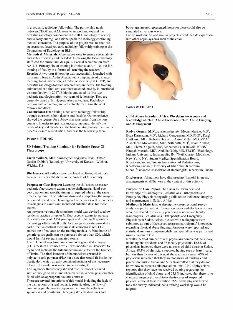
in a pediatric radiology fellowship. The partnership goals between CHOP and AAU were to support and expand the pediatric radiology component in the BLH radiology residency and to carry out regular national pediatric radiology continuing medical education. The purpose of our project was to establish
an accredited local pediatric radiology fellowship training in the Department of Radiology at BLH. Methods & Materials: Core values were to ensure sustainability and self-sufficiency and included: 1- making the local radiology staff lead the curriculum design, 2- Formal accreditation from AAU, 3- Primary site of training in Ethiopia, and, 4- On-the-job-
training of faculty in a format of “teaching the teachers”. Results: A two-year fellowship was successfully launched with its primary base in Addis Ababa, with components of distance learning, local instruction, a limited observership at CHOP, and pediatric radiology-focused research requirements. The training culminated in a final oral examination conducted by international visiting faculty. In 2017, Ethiopia graduated its first two
pediatric radiologists after two years of fellowship. They are currently based at BLH, established a Pediatric Radiology Section with a director, and are actively recruiting the next fellow candidates. Conclusions: Establishing a pediatric radiology fellowship through outreach is both doable and feasible. Our experience showed the request for a fellowship must arise from the host
country. In order to optimize success, one must identify the needs of key stakeholders in the host country, engage them in the process, ensure accreditation, and base the fellowship there.
Poster #: EDU-052
3D Printed Training Simulator for Pediatric Upper GI
Fluoroscopy Jacob Wallace, MD
1, [email protected]; Debbie
Desilet-Dobbs1;
1Radiology, University of Kansas - Wichita,
Wichita, KS
Disclosures: All authors have disclosed no financial interests, arrangements or affiliations in the context of this activity. Purpose or Case Report: Learning the skills used to master pediatric fluoroscopic exams can be challenging. Hand-eye coordination and specific timing is required while at the same time being mindful of radiation dose and interpreting the images
generated in real time. Training on live neonates will often mean less diagnostic exams and increased radiation dose for those exams. An inexpensive reusable simulator model was devised to allow residents practice of upper GI fluoroscopic exams to increase efficiency using ALARA principles and utilizing 3D printing
technology off-the-shelf dolls. Generic gastrografin provided a cost effective contrast medium as its concerns in real UGI studies are of no issue on the training models. A 30ml bottle of generic gastrografin can be purchased for less than $20, which would last for several simulated exams. The 3D model was based on a computer generated imagery (CGI) mesh of a stomach which was modified in Blender™ to
try to best replicate the full duodenum and effect of the ligament of Treitz. The final iteration of the model was printed in polylactic acid polymer (PLA) in a size that would fit inside the plastic doll, which already contained portions of the necessary tubing. The model was sealed to be watertight. Testing under fluoroscopy showed that the model behaved
similar enough to an infant when placed in various positions then filled with an appropriate volume contrast. There are several limitations of this model including the lack of the distractions of a real pediatric patient. Also, the flow of contrast is purely gravity dependent without the effects of sphincters and peristalsis. Overlying skeletal structures and
bowel gas are not represented, however these could also be simulated in various ways. Future work on this and similar projects could include expansion into other organ systems such as the colon
Poster #: EDU-053
Child Abuse in Sudan, Africa: Physician Awareness and
Knowledge of Child Abuse Incidence, Child Abuse Imaging,
and Management Radya Osman, MD
1, [email protected]; Megan Marine, MD
1,
Boaz Karmazyn, MD1, Richard Gunderman, MD, PHD
1, Darel
Heitkamp, MD1, Roberta Hibbard
1, Aaron Miller, MD, MPA
2,
Alaaeldeen Mohammed, MD3, Satti Satti, MD
4, Ilham Ahmed,
MD5, Shima Yagoub, MD
3, Mohamed Salih Bakeit, MBBS
3,
Elrayah Mustafa, MD6, Abdalla Gabir, MD, FRCR
6;
1Radiology,
Indiana University, Indianapolis, IN, 2Weill Cornell Medicine,
New York, NY, 3Sudan Medical Specialization Board,
Khartoum, Sudan, 4Sudan Association of Pediatricians,
Khartoum, Sudan, 5University of Khartoum, Khartoum,
Sudan, 6Sudanese Association of Radiologists, Khartoum, Sudan
Disclosures: All authors have disclosed no financial interests, arrangements or affiliations in the context of this activity.
Purpose or Case Report: To assess the awareness and knowledge of Radiologists, Pediatricians, Orthopedists and Emergency Physicians regarding child abuse incidence, imaging, and management in Sudan, Africa. Methods & Materials: A descriptive cross-sectional survey study was performed. A 16-question paper and electronic survey
were distributed to currently practicing resident and faculty Radiologists, Pediatricians, Orthopedists and Emergency Physicians in Sudan, Africa. 4 cases with radiographs were submitted as part of the survey to assess participants’ knowledge regarding physical abuse findings. Answers were reported and statistical analysis comparing different specialties was performed
using chi-square test. Results: A total number of 400 physicians completed the survey, including 364 residents and 36 faculty physicians. 16.8% of physicians indicated there were no cases of child abuse in Sudan, Africa. 88.3% of physicians reported having seen at least 1 case, but less than 5 cases of physical abuse in their career. 46% of physicians indicated that they are not aware of existing child
protection units in Sudan and 50.5 % admitted that they do not know how to contact child protection units. 77% of physicians reported that they have not received training regarding the identification of child abuse, and 53.8% indicated that there is no standard imaging protocol to evaluate cases of suspected physical abuse at their institution. 99% of the physicians who took the survey indicated that a training workshop would be
helpful.
Pediatr Radiol (2018) 48 (Suppl 1):S1–S298 S219
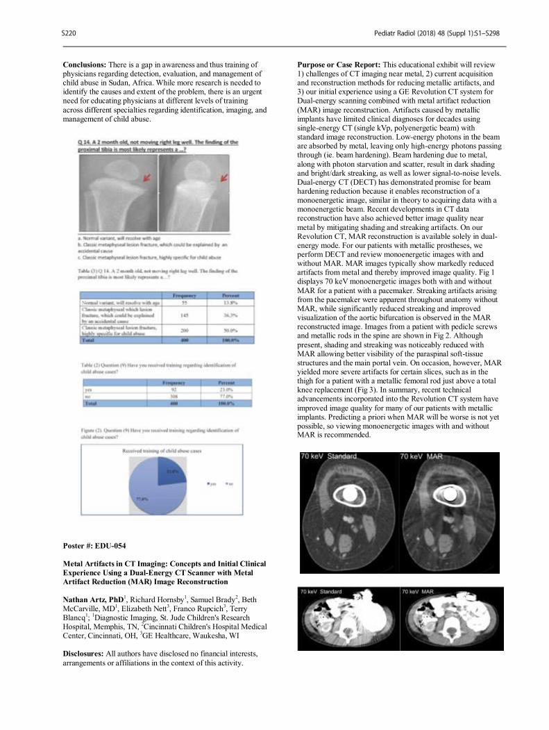
Conclusions: There is a gap in awareness and thus training of physicians regarding detection, evaluation, and management of child abuse in Sudan, Africa. While more research is needed to identify the causes and extent of the problem, there is an urgent need for educating physicians at different levels of training
across different specialties regarding identification, imaging, and management of child abuse.
Poster #: EDU-054
Metal Artifacts in CT Imaging: Concepts and Initial Clinical
Experience Using a Dual-Energy CT Scanner with Metal
Artifact Reduction (MAR) Image Reconstruction
Nathan Artz, PhD1, Richard Hornsby
1, Samuel Brady
2, Beth
McCarville, MD1, Elizabeth Nett
3, Franco Rupcich
3, Terry
Blancq1;
1Diagnostic Imaging, St. Jude Children's Research
Hospital, Memphis, TN, 2Cincinnati Children's Hospital Medical
Center, Cincinnati, OH, 3GE Healthcare, Waukesha, WI
Disclosures: All authors have disclosed no financial interests,
arrangements or affiliations in the context of this activity.
Purpose or Case Report: This educational exhibit will review 1) challenges of CT imaging near metal, 2) current acquisition and reconstruction methods for reducing metallic artifacts, and 3) our initial experience using a GE Revolution CT system for Dual-energy scanning combined with metal artifact reduction
(MAR) image reconstruction. Artifacts caused by metallic implants have limited clinical diagnoses for decades using single-energy CT (single kVp, polyenergetic beam) with standard image reconstruction. Low-energy photons in the beam are absorbed by metal, leaving only high-energy photons passing through (ie. beam hardening). Beam hardening due to metal,
along with photon starvation and scatter, result in dark shading and bright/dark streaking, as well as lower signal-to-noise levels. Dual-energy CT (DECT) has demonstrated promise for beam hardening reduction because it enables reconstruction of a monoenergetic image, similar in theory to acquiring data with a monoenergetic beam. Recent developments in CT data reconstruction have also achieved better image quality near
metal by mitigating shading and streaking artifacts. On our Revolution CT, MAR reconstruction is available solely in dual-energy mode. For our patients with metallic prostheses, we perform DECT and review monoenergetic images with and without MAR. MAR images typically show markedly reduced artifacts from metal and thereby improved image quality. Fig 1 displays 70 keV monoenergetic images both with and without
MAR for a patient with a pacemaker. Streaking artifacts arising from the pacemaker were apparent throughout anatomy without MAR, while significantly reduced streaking and improved visualization of the aortic bifurcation is observed in the MAR reconstructed image. Images from a patient with pedicle screws and metallic rods in the spine are shown in Fig 2. Although
present, shading and streaking was noticeably reduced with MAR allowing better visibility of the paraspinal soft-tissue structures and the main portal vein. On occasion, however, MAR yielded more severe artifacts for certain slices, such as in the thigh for a patient with a metallic femoral rod just above a total knee replacement (Fig 3). In summary, recent technical advancements incorporated into the Revolution CT system have
improved image quality for many of our patients with metallic implants. Predicting a priori when MAR will be worse is not yet possible, so viewing monoenergetic images with and without MAR is recommended.
Pediatr Radiol (2018) 48 (Suppl 1):S1–S298S220
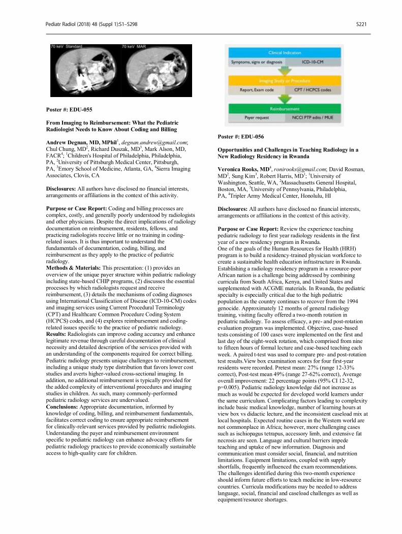
Poster #: EDU-055
From Imaging to Reimbursement: What the Pediatric
Radiologist Needs to Know About Coding and Billing
Andrew Degnan, MD, MPhil1, [email protected];
Chul Chung, MD2, Richard Duszak, MD
3, Mark Alson, MD,
FACR4;
1Children's Hospital of Philadelphia, Philadelphia,
PA, 2University of Pittsburgh Medical Center, Pittsburgh,
PA, 3Emory School of Medicine, Atlanta, GA,
4Sierra Imaging
Associates, Clovis, CA
Disclosures: All authors have disclosed no financial interests, arrangements or affiliations in the context of this activity. Purpose or Case Report: Coding and billing processes are complex, costly, and generally poorly understood by radiologists and other physicians. Despite the direct implications of radiology
documentation on reimbursement, residents, fellows, and practicing radiologists receive little or no training in coding-related issues. It is thus important to understand the fundamentals of documentation, coding, billing, and reimbursement as they apply to the practice of pediatric radiology. Methods & Materials: This presentation: (1) provides an
overview of the unique payer structure within pediatric radiology including state-based CHIP programs, (2) discusses the essential processes by which radiologists request and receive reimbursement, (3) details the mechanisms of coding diagnoses using International Classification of Disease (ICD-10-CM) codes and imaging services using Current Procedural Terminology
(CPT) and Healthcare Common Procedure Coding System (HCPCS) codes, and (4) explores reimbursement and coding-related issues specific to the practice of pediatric radiology. Results: Radiologists can improve coding accuracy and enhance legitimate revenue through careful documentation of clinical necessity and detailed description of the services provided with an understanding of the components required for correct billing.
Pediatric radiology presents unique challenges to reimbursement, including a unique study type distribution that favors lower cost studies and averts higher-valued cross-sectional imaging. In addition, no additional reimbursement is typically provided for the added complexity of interventional procedures and imaging studies in children. As such, many commonly-performed pediatric radiology services are undervalued.
Conclusions: Appropriate documentation, informed by knowledge of coding, billing, and reimbursement fundamentals, facilitates correct coding to ensure appropriate reimbursement for clinically-relevant services provided by pediatric radiologists. Understanding the payer and reimbursement environment specific to pediatric radiology can enhance advocacy efforts for
pediatric radiology practices to provide economically sustainable access to high-quality care for children.
Poster #: EDU-056
Opportunities and Challenges in Teaching Radiology in a
New Radiology Residency in Rwanda Veronica Rooks, MD
4, [email protected]; David Rosman,
MD2, Sung Kim
3, Robert Harris, MD
1;
1University of
Washington, Seattle, WA, 2Massachusetts General Hospital,
Boston, MA, 3University of Pennsylvania, Philadelphia,
PA, 4Tripler Army Medical Center, Honolulu, HI
Disclosures: All authors have disclosed no financial interests, arrangements or affiliations in the context of this activity.
Purpose or Case Report: Review the experience teaching pediatric radiology to first year radiology residents in the first year of a new residency program in Rwanda. One of the goals of the Human Resources for Health (HRH) program is to build a residency-trained physician workforce to create a sustainable health education infrastructure in Rwanda.
Establishing a radiology residency program in a resource-poor African nation is a challenge being addressed by combining curricula from South Africa, Kenya, and United States and supplemented with ACGME materials. In Rwanda, the pediatric specialty is especially critical due to the high pediatric population as the country continues to recover from the 1994
genocide. Approximately 12 months of general radiology training, visiting faculty offered a two-month rotation in pediatric radiology. To assess efficacy, a pre- and post-rotation evaluation program was implemented. Objective, case-based tests consisting of 100 cases were implemented on the first and last day of the eight-week rotation, which comprised from nine to fifteen hours of formal lecture and case-based teaching each
week. A paired t-test was used to compare pre- and post-rotation test results.View box examination scores for four first-year residents were recorded. Pretest mean: 27% (range 12-33% correct), Post-test mean 49% (range 27-62% correct), Average overall improvement: 22 percentage points (95% CI 12-32, p=0.005). Pediatric radiology knowledge did not increase as
much as would be expected for developed world learners under the same curriculum. Complicating factors leading to complexity include basic medical knowledge, number of learning hours at view box vs didactic lecture, and the inconsistent caseload mix at local hospitals. Expected routine cases in the Western world are not commonplace in Africa; however, more challenging cases such as ischiopagus tetrapus, accessory limb, and extensive fat
necrosis are seen. Language and cultural barriers impede teaching and uptake of new information. Diagnosis and communication must consider social, financial, and nutrition limitations. Equipment limitations, coupled with supply shortfalls, frequently influenced the exam recommendations. The challenges identified during this two-month experience should inform future efforts to teach medicine in low-resource
countries. Curricula modifications may be needed to address language, social, financial and caseload challenges as well as equipment/resource shortages.
Pediatr Radiol (2018) 48 (Suppl 1):S1–S298 S221
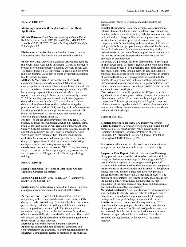
Poster #: EDU-057
Mastering Ultrasound through a Just-In-Time Mobile
Application
Michele Retrouvey1, [email protected]; David
Saul, MD1, Susan Back, MD
1, Richard Bellah, MD, FAAP
1,
Janet Reid, MD, FRCPC1;
1Children’s Hospital of Philadelphia,
Philadelphia, PA Disclosures: All authors have disclosed no financial interests,
arrangements or affiliations in the context of this activity. Purpose or Case Report: It is essential that budding pediatric radiologists have well-honed ultrasound (US) skills in order to provide correct image interpretation and excellent patient care. The lack of hands-on US experience is a critical gap in current radiology training. We sought to create an interactive, portable
tool to remedy this issue. Methods & Materials: A previously published needs assessment survey revealed a dearth of US hands-on skills amongst pediatric radiology fellows, where fewer than 45% had access to hands-on tutorials with sonographers, and only 23% had scanning responsibilities while on call. Most trainees reported their scanning skills has never been formally evaluated.
To fill the learning gap, an electronic application (app) was designed with a user interface (UI) that optimized content delivery, through mobile or stationary devices using the principles of “just-in-time” (JIT) learning. A list of the requisite knowledge and skills for the top 10 emergency pediatric US scans was developed. All key resources and content were
collected and assembled in the UI. Results: The top ten emergency studies include-renal, RUQ, pylorus, intussusception, appendix, pelvis, hip, scrotum, spine, brain. A checklist-based UI was created in our LMS (Absorb, Calgary, Canada) including: protocols, image planes, images of normal and pathology, case log, links to previously created video-based micro-tutorials, “tips” for scan modification,
evaluation by instructors, and transcripts. Through responsive design the app conforms to all mobile devices and desktop configuration and is operating system agnostic. Conclusions: Our interactive mobile POC/JIT app is relatively simple to construct, with an appealing and easy-to-use interface and has potential to fill a gap in US skills during radiology training.
Poster #: EDU-058
Seeing Is Believing: The Value of Ultrasound-Guided
Umbilical Catheter Placement
Michael Collard, MD1, Cory Pfeifer, MD
1;
1Radiology, UT
Southwestern, Coppell, TX Disclosures: All authors have disclosed no financial interests, arrangements or affiliations in the context of this activity. Purpose or Case Report: Umbilical catheters (UC) are
ubiquitously utilized in neonatal intensive care units (NICU) during the early neonatal stage. Traditionally, these catheters are placed blindly, with the external length of the catheter being used to approximate catheter depth. This then requires subsequent radiographs to be obtained to assess for position, often in a sterile field, with considerable dead time. This exhibit
will educate the viewer about the use of ultrasound guidance in the placement of these catheters. Methods & Materials: Recent literature is reviewed. Approaches utilized with both abdominal ultrasound and echocardiography are discussed. Relevant neonatal anatomy is described. Complications related to malposition are reviewed,
and measures related to efficiency and radiation dose are detailed. Results: The traditional use of radiography to assess umbilical catheter placement in the neonatal population involves ionizing radiation and considerable lag time, yet the two-dimensional data
included in this technique still results in only an approximate location for the catheter tip. Atypical vascular anatomy complicates this further, leading to the acquisition of additional radiographs before proper positioning is achieved. Furthermore, the sterile field created for catheter placement is typically maintained during the time of image acquisition and review, and
the time lag in imaging interpretation may increase risk for procedure-related infection. US-guided UC placement has been demonstrated to have equal or often better ability to identify accurate catheter tip positioning, with the added benefit of being performed real-time during the procedure, significantly limiting delays in care and radiation exposure. This has been shown to be particularly true in patients
of increased birthweight. This represents an opportunity for radiologists to provide value in the care of these patients. Now that ultrasound resources are increasing in pediatric tertiary care centers, training and comfort with the procedure remain the only significant barrier to adoption. Conclusions: The use of US guidance for UC placement has significant potential to improve catheter placement accuracy and
clinical turnaround time from procedure beginning to final completion. This is an opportunity for radiologists to improve value via ultrasound-guided umbilical catheter placement while minimizing radiation dose to these young patients as consistent with the ALARA principle.
Poster #: EDU-059
Pediatric Interventional Radiology Biliary Procedures Carlos Llorens, MD
1, [email protected]; Rakesh Sindhi
2,
James Park, MD1, John Crowley, MD
1;
1Department of
Radiology, Children's Hospital of Pittsburgh of UPMC, Pittsburgh, PA,
2Transplant Surgery, Children's Hospital of
Pittsburgh of UPMC, Pittsburgh, PA Disclosures: All authors have disclosed no financial interests, arrangements or affiliations in the context of this activity. Purpose or Case Report: Pediatric Interventional Radiology biliary procedures are mainly performed on patients with liver
transplant. Percutaneous transhepatic cholangiogram (PTC) is very useful for diagnosis of post-surgical development of strictures while at the same time allowing access for therapeutic measures such as biliary dilatation and diversion. Although post-surgical anatomy and non-dilated bile ducts may provide a challenge, biliary procedures have a high rate of success. The
purpose of this exhibit is to review the biliary anatomy, discuss the available biliary procedures, including step-by-step explanation of the more common procedures and discussion of goal and outcomes of these procedures. Methods & Materials: A single institution retrospective review was conducted to identify pediatric patients with prior biliary procedures. Selected patients were correlated with imaging
findings and/or surgical findings, and/or clinical course. Results: Review and discussion of biliary anatomy, PTC procedure with step-by-step explanation, discussion of other biliary procedures as well as discussion of goals and outcomes are presented. Sample cases from our institution are shown to illustrate our approach to biliary procedures. Cased-based
examples are supplemented with a review of the current literature.
Pediatr Radiol (2018) 48 (Suppl 1):S1–S298S222
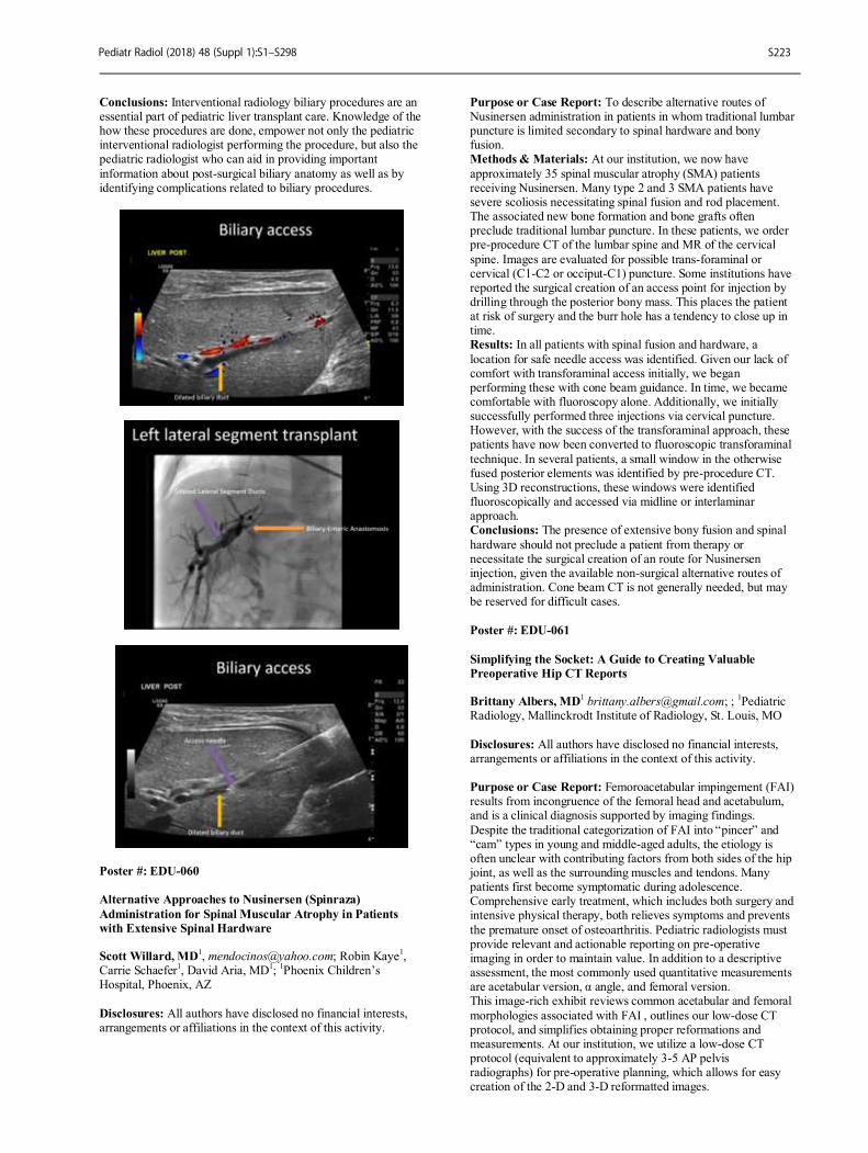
Conclusions: Interventional radiology biliary procedures are an essential part of pediatric liver transplant care. Knowledge of the how these procedures are done, empower not only the pediatric interventional radiologist performing the procedure, but also the pediatric radiologist who can aid in providing important
information about post-surgical biliary anatomy as well as by identifying complications related to biliary procedures.
Poster #: EDU-060
Alternative Approaches to Nusinersen (Spinraza)
Administration for Spinal Muscular Atrophy in Patients
with Extensive Spinal Hardware Scott Willard, MD
1, [email protected]; Robin Kaye
1,
Carrie Schaefer1, David Aria, MD
1;
1Phoenix Children’s
Hospital, Phoenix, AZ
Disclosures: All authors have disclosed no financial interests, arrangements or affiliations in the context of this activity.
Purpose or Case Report: To describe alternative routes of Nusinersen administration in patients in whom traditional lumbar puncture is limited secondary to spinal hardware and bony fusion. Methods & Materials: At our institution, we now have
approximately 35 spinal muscular atrophy (SMA) patients receiving Nusinersen. Many type 2 and 3 SMA patients have severe scoliosis necessitating spinal fusion and rod placement. The associated new bone formation and bone grafts often preclude traditional lumbar puncture. In these patients, we order pre-procedure CT of the lumbar spine and MR of the cervical
spine. Images are evaluated for possible trans-foraminal or cervical (C1-C2 or occiput-C1) puncture. Some institutions have reported the surgical creation of an access point for injection by drilling through the posterior bony mass. This places the patient at risk of surgery and the burr hole has a tendency to close up in time. Results: In all patients with spinal fusion and hardware, a
location for safe needle access was identified. Given our lack of comfort with transforaminal access initially, we began performing these with cone beam guidance. In time, we became comfortable with fluoroscopy alone. Additionally, we initially successfully performed three injections via cervical puncture. However, with the success of the transforaminal approach, these patients have now been converted to fluoroscopic transforaminal
technique. In several patients, a small window in the otherwise fused posterior elements was identified by pre-procedure CT. Using 3D reconstructions, these windows were identified fluoroscopically and accessed via midline or interlaminar approach. Conclusions: The presence of extensive bony fusion and spinal
hardware should not preclude a patient from therapy or necessitate the surgical creation of an route for Nusinersen injection, given the available non-surgical alternative routes of administration. Cone beam CT is not generally needed, but may be reserved for difficult cases.
Poster #: EDU-061
Simplifying the Socket: A Guide to Creating Valuable
Preoperative Hip CT Reports Brittany Albers, MD
1 [email protected]; ;
1Pediatric
Radiology, Mallinckrodt Institute of Radiology, St. Louis, MO
Disclosures: All authors have disclosed no financial interests, arrangements or affiliations in the context of this activity. Purpose or Case Report: Femoroacetabular impingement (FAI) results from incongruence of the femoral head and acetabulum, and is a clinical diagnosis supported by imaging findings.
Despite the traditional categorization of FAI into “pincer” and “cam” types in young and middle-aged adults, the etiology is often unclear with contributing factors from both sides of the hip joint, as well as the surrounding muscles and tendons. Many patients first become symptomatic during adolescence. Comprehensive early treatment, which includes both surgery and intensive physical therapy, both relieves symptoms and prevents
the premature onset of osteoarthritis. Pediatric radiologists must provide relevant and actionable reporting on pre-operative imaging in order to maintain value. In addition to a descriptive assessment, the most commonly used quantitative measurements are acetabular version, α angle, and femoral version. This image-rich exhibit reviews common acetabular and femoral
morphologies associated with FAI , outlines our low-dose CT protocol, and simplifies obtaining proper reformations and measurements. At our institution, we utilize a low-dose CT protocol (equivalent to approximately 3-5 AP pelvis radiographs) for pre-operative planning, which allows for easy creation of the 2-D and 3-D reformatted images.
Pediatr Radiol (2018) 48 (Suppl 1):S1–S298 S223
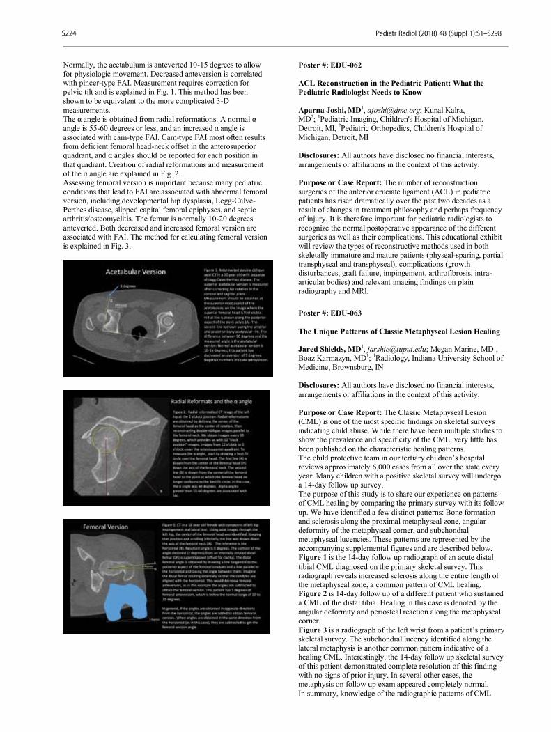
Normally, the acetabulum is anteverted 10-15 degrees to allow for physiologic movement. Decreased anteversion is correlated with pincer-type FAI. Measurement requires correction for pelvic tilt and is explained in Fig. 1. This method has been shown to be equivalent to the more complicated 3-D
measurements. The α angle is obtained from radial reformations. A normal α angle is 55-60 degrees or less, and an increased α angle is associated with cam-type FAI. Cam-type FAI most often results from deficient femoral head-neck offset in the anterosuperior quadrant, and α angles should be reported for each position in
that quadrant. Creation of radial reformations and measurement of the α angle are explained in Fig. 2. Assessing femoral version is important because many pediatric conditions that lead to FAI are associated with abnormal femoral version, including developmental hip dysplasia, Legg-Calve-Perthes disease, slipped capital femoral epiphyses, and septic arthritis/osteomyelitis. The femur is normally 10-20 degrees
anteverted. Both decreased and increased femoral version are associated with FAI. The method for calculating femoral version is explained in Fig. 3.
Poster #: EDU-062
ACL Reconstruction in the Pediatric Patient: What the
Pediatric Radiologist Needs to Know
Aparna Joshi, MD1, [email protected]; Kunal Kalra,
MD2;
1Pediatric Imaging, Children's Hospital of Michigan,
Detroit, MI, 2Pediatric Orthopedics, Children's Hospital of
Michigan, Detroit, MI
Disclosures: All authors have disclosed no financial interests,
arrangements or affiliations in the context of this activity. Purpose or Case Report: The number of reconstruction surgeries of the anterior cruciate ligament (ACL) in pediatric patients has risen dramatically over the past two decades as a result of changes in treatment philosophy and perhaps frequency of injury. It is therefore important for pediatric radiologists to
recognize the normal postoperative appearance of the different surgeries as well as their complications. This educational exhibit will review the types of reconstructive methods used in both skeletally immature and mature patients (physeal-sparing, partial transphyseal and transphyseal), complications (growth disturbances, graft failure, impingement, arthrofibrosis, intra-articular bodies) and relevant imaging findings on plain radiography and MRI.
Poster #: EDU-063
The Unique Patterns of Classic Metaphyseal Lesion Healing Jared Shields, MD
1, [email protected]; Megan Marine, MD
1,
Boaz Karmazyn, MD1;
1Radiology, Indiana University School of
Medicine, Brownsburg, IN Disclosures: All authors have disclosed no financial interests,
arrangements or affiliations in the context of this activity. Purpose or Case Report: The Classic Metaphyseal Lesion (CML) is one of the most specific findings on skeletal surveys indicating child abuse. While there have been multiple studies to show the prevalence and specificity of the CML, very little has
been published on the characteristic healing patterns. The child protective team in our tertiary children’s hospital reviews approximately 6,000 cases from all over the state every year. Many children with a positive skeletal survey will undergo a 14-day follow up survey. The purpose of this study is to share our experience on patterns of CML healing by comparing the primary survey with its follow
up. We have identified a few distinct patterns: Bone formation and sclerosis along the proximal metaphyseal zone, angular deformity of the metaphyseal corner, and subchondral metaphyseal lucencies. These patterns are represented by the accompanying supplemental figures and are described below. Figure 1 is the 14-day follow up radiograph of an acute distal
tibial CML diagnosed on the primary skeletal survey. This radiograph reveals increased sclerosis along the entire length of the metaphyseal zone, a common pattern of CML healing. Figure 2 is 14-day follow up of a different patient who sustained a CML of the distal tibia. Healing in this case is denoted by the angular deformity and periosteal reaction along the metaphyseal corner.
Figure 3 is a radiograph of the left wrist from a patient’s primary skeletal survey. The subchondral lucency identified along the lateral metaphysis is another common pattern indicative of a healing CML. Interestingly, the 14-day follow up skeletal survey of this patient demonstrated complete resolution of this finding with no signs of prior injury. In several other cases, the metaphysis on follow up exam appeared completely normal.
In summary, knowledge of the radiographic patterns of CML
Pediatr Radiol (2018) 48 (Suppl 1):S1–S298S224
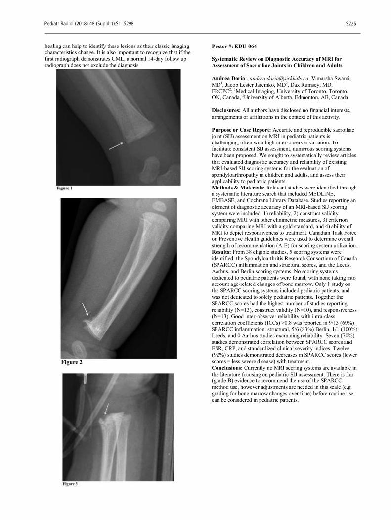
healing can help to identify these lesions as their classic imaging characteristics change. It is also important to recognize that if the first radiograph demonstrates CML, a normal 14-day follow up radiograph does not exclude the diagnosis.
Poster #: EDU-064
Systematic Review on Diagnostic Accuracy of MRI for
Assessment of Sacroiliac Joints in Children and Adults
Andrea Doria1, [email protected]; Vimarsha Swami,
MD1, Jacob Lester Jaremko, MD
2, Dax Rumsey, MD,
FRCPC2;
1Medical Imaging, University of Toronto, Toronto,
ON, Canada, 2University of Alberta, Edmonton, AB, Canada
Disclosures: All authors have disclosed no financial interests,
arrangements or affiliations in the context of this activity. Purpose or Case Report: Accurate and reproducible sacroiliac joint (SIJ) assessment on MRI in pediatric patients is challenging, often with high inter-observer variation. To facilitate consistent SIJ assessment, numerous scoring systems have been proposed. We sought to systematically review articles
that evaluated diagnostic accuracy and reliability of existing MRI-based SIJ scoring systems for the evaluation of spondyloarthropathy in children and adults, and assess their applicability to pediatric patients. Methods & Materials: Relevant studies were identified through a systematic literature search that included MEDLINE, EMBASE, and Cochrane Library Database. Studies reporting an
element of diagnostic accuracy of an MRI-based SIJ scoring system were included: 1) reliability, 2) construct validity comparing MRI with other clinimetric measures, 3) criterion validity comparing MRI with a gold standard, and 4) ability of MRI to depict responsiveness to treatment. Canadian Task Force on Preventive Health guidelines were used to determine overall
strength of recommendation (A-E) for scoring system utilization. Results: From 38 eligible studies, 5 scoring systems were identified: the Spondyloarthritis Research Consortium of Canada (SPARCC) inflammation and structural scores, and the Leeds, Aarhus, and Berlin scoring systems. No scoring systems dedicated to pediatric patients were found, with none taking into account age-related changes of bone marrow. Only 1 study on
the SPARCC scoring systems included pediatric patients, and was not dedicated to solely pediatric patients. Together the SPARCC scores had the highest number of studies reporting reliability (N=13), construct validity (N=10), and responsiveness (N=13). Good inter-observer reliability with intra-class correlation coefficients (ICCs) >0.8 was reported in 9/13 (69%) SPARCC inflammation, structural, 5/6 (83%) Berlin, 1/1 (100%)
Leeds, and 0 Aarhus studies examining reliability. Seven (70%) studies demonstrated correlation between SPARCC scores and ESR, CRP, and standardized clinical severity indices. Twelve (92%) studies demonstrated decreases in SPARCC scores (lower scores = less severe disease) with treatment. Conclusions: Currently no MRI scoring systems are available in
the literature focusing on pediatric SIJ assessment. There is fair (grade B) evidence to recommend the use of the SPARCC method use, however adjustments are needed in this scale (e.g. grading for bone marrow changes over time) before routine use can be considered in pediatric patients.
Pediatr Radiol (2018) 48 (Suppl 1):S1–S298 S225
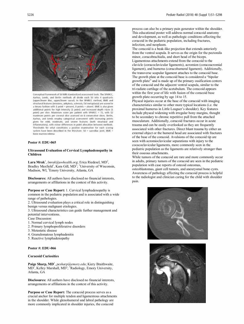
Poster #: EDU-065
Ultrasound Evaluation of Cervical Lymphadenopathy in
Children Lara Mrak
1, [email protected]; Erica Riedesel, MD
2,
Bradley Maxfield1, Kara Gill, MD
1;
1University of Wisconsin,
Madison, WI, 2Emory University, Atlanta, GA
Disclosures: All authors have disclosed no financial interests, arrangements or affiliations in the context of this activity. Purpose or Case Report: 1. Cervical lymphadenopathy is
common in the pediatric population and is associated with a wide range of pathologies. 2. Ultrasound evaluation plays a critical role in distinguishing benign versus malignant etiologies. 3. Ultrasound characteristics can guide further management and potential interventions.
Case Discussion: 1. Normal cervical lymph nodes 2. Primary lymphoproliferative disorders 3. Metastatic disease 4. Granulomatous lymphadenitis 5. Reactive lymphadenopathy
Poster #: EDU-066
Coracoid Curiosities
Paige Sharp, MD1, [email protected]; Kiery Braithwaite,
MD1, Kelley Marshall, MD
1;
1Radiology, Emory University,
Atlanta, GA
Disclosures: All authors have disclosed no financial interests, arrangements or affiliations in the context of this activity.
Purpose or Case Report: The coracoid process serves as a crucial anchor for multiple tendon and ligamentous attachments in the shoulder. While glenohumeral and labral pathology are more commonly implicated in shoulder injuries, the coracoid
process can also be a primary pain generator within the shoulder. This educational poster will address normal coracoid anatomy and development, as well as pathologic conditions affecting the coracoid in the pediatric population, including fractures, infection, and neoplasm.
The coracoid is a beak-like projection that extends anteriorly from the ventral scapula. It serves as the origin for the pectoralis minor, coracobrachialis, and short head of the biceps. Ligamentous attachments extend from the coracoid to the clavicle (coracoclavicular ligaments), acromion (coracoacromial ligament), and humerus (coracohumeral ligament). Additionally,
the transverse scapular ligament attaches to the coracoid base. The growth plate at the coracoid base is considered a “bipolar growth plate” and is made up of the primary ossification centers of the coracoid and the adjacent ventral scapula, similar to the tri-radiate cartilage of the acetabulum. The coracoid appears within the first year of life with fusion of the coracoid base growth plate occurring by age 14 to 15.
Physeal injuries occur at the base of the coracoid with imaging characteristics similar to other more typical locations (i.e. the proximal humerus in Little Leaguer’s shoulder). Key features include physeal widening with irregular bony margins, thought to be secondary to chronic repetitive pull from the attached musculature. Additionally, coracoid fractures occur in acute trauma and can be easily overlooked as they are frequently
associated with other fractures. Direct blunt trauma by either an external object or the humeral head are associated with fractures of the base of the coracoid. Avulsions of the coracoid tip are seen with acromioclavicular separations with injury to the cocacoclavicular ligaments, more commonly seen in the pediatric population as the ligaments are relatively stronger than
their osseous attachments. While tumors of the coracoid are rare and more commonly occur in adults, primary tumors of the coracoid are seen in the pediatric population with case reports of osteoid osteomas, osteoblastomas, giant cell tumors, and aneurysmal bone cysts. Awareness of pathology affecting the coracoid process is helpful to the radiologist and clinician caring for the child with shoulder
pain.
Pediatr Radiol (2018) 48 (Suppl 1):S1–S298S226
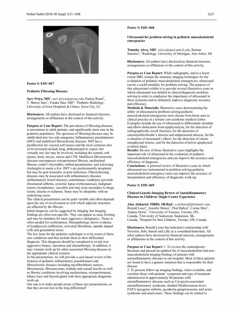
Poster #: EDU-067
Pediatric Fibrosing Diseases Sarv Priya, MD
1, [email protected]; Pankaj Watal
1,
T. Shawn Sato1, Yutaka Sato, MD
1;
1Pediatric Radiology,
University of Iowa Hospitals & Clinics, Iowa City, IA Disclosures: All authors have disclosed no financial interests, arrangements or affiliations in the context of this activity. Purpose or Case Report: The prevalence of fibrosing diseases is uncommon in adult patients, and significantly more rare in the
pediatric population. The spectrum of fibrosing diseases may be subdivided into two sub-categories: Inflammatory pseudotumors (IMT) and multifocal fibrosclerotic diseases. IMT has a predilection for visceral soft tissues and the most common sites of involvement include lung, abdominopelvic region, but virtually any site may be involved, including the somatic soft
tissues, bone, larynx, uterus and CNS. Multifocal fibrosclerotic diseases encompasses retroperitoneal fibrosis, mediastinal fibrosis, reidel’s thyroiditis, orbital pseudotumor, and sclerosing cholangitis to name a few. IMT’s are predominantly neoplastic but may be post-traumatic or post-infectious. Fibrosclerosing diseases may be associated with inflammatory diseases (inflammatory bowel disease), autoimmune conditions (juvenile
rheumatoid arthritis, systemic lupus erythematosus), malignant tumors (lymphoma), vasculitis and may arise secondary to drugs, toxins, trauma or radiation. Some may be idiopathic with no underlying cause. The clinical presentation can be quite variable and often depends upon the site of involvement as well which adjacent structures are affected by the fibrosis.
Initial diagnosis can be suggested by imaging, but imaging findings are often non-specific. They can appear as mass forming and may be mistaken for more aggressive malignancy. Tissue is often needed for confirmation. Histopathology shows evidence of lymphocytic infiltration, activated fibroblasts, spindle shaped cells and granulation tissue.
The key issue for the pediatric radiologist is to be aware of these rare conditions and thus include them in their differential diagnosis. This diagnosis should be considered to avoid over aggressive biopsy, operation and chemotherapy. In addition, it may warrant work up for other associated fibrosing diseases in the appropriate clinical scenario. In this presentation, we will provide a case based review of the
features of pediatric inflammatory pseudotumors and fibrosclerotic diseases including myofibroblastic tumors, fibromatosis, fibrosarcomas, nodular and cranial fascitis as well as fibrotic conditions involving mediastinum, retroperitoneum, biliary tract and thyroid gland with their appropriate diagnostic work-up.
Our aim is to make people aware of these rare presentations, so that they are not lost in the long differential!
Poster #: EDU-068
Ultrasound for problem solving in pediatric musculoskeletal emergencies
Timothy Alves, MD1, [email protected]; Ramon
Sanchez1;
1Radiology, University of Michigan, Ann Arbor, MI
Disclosures: All authors have disclosed no financial interests, arrangements or affiliations in the context of this activity. Purpose or Case Report: While radiographs, and to a lesser extent MRI, remain the mainstay imaging techniques for the
evaluation of pediatric musculoskeletal emergencies, ultrasound can be a useful modality for problem solving. The purpose of this educational exhibit is to provide several illustrative cases in which ultrasound was helpful in clinical/diagnostic problem-solving in order to emphasize the importance of ultrasound in these scenarios and to ultimately improve diagnostic accuracy and efficiency.
Methods & Materials: Illustrative cases demonstrating the utility of ultrasound in problem-solving pediatric musculoskeletal emergencies were chosen from those seen in clinical practice at a tertiary care academic medical center. Examples include the use of ultrasound to differentiate shoulder and elbow dislocation from epiphysiolysis, for the detection of radiographically occult fractures, for the detection of
osteomyelitis/brodie’s abscess and subperiosteal abscess, for the evaluation of nursemaid’s elbow, for the detection of classic metaphyseal lesions, and for the detection of pelvic apophyseal avulsion injury. Results: Review of these illustrative cases highlights the important role of ultrasound in the evaluation of pediatric
musculoskeletal emergencies and can improve the accuracy and efficiency of diagnosis. Conclusions: A pictorial review of illustrative cases in which ultrasound was instrumental in problem-solving pediatric musculoskeletal emergency cases can improve the accuracy of interpretation and efficiency of diagnostic work-up.
Poster #: EDU-069
Clinical-Genetic-Imaging Review of Autoinflammatory
Diseases in Children: Single-Center Experience Alaa Alsharief, MBBS, SB-Rad
Ronald Laxer1, Jennifer Stimec
1, Paul Babyn
2, Carina Man
3,
Andrea Doria1;
1University of Toronto, Toronto, ON,
Canada, 2University of Saskatoon, Saskatoon, SK,
Canada, 3Hospital for Sick Children, Toronto, ON, Canada
Disclosures: Ronald Laxer has indicated a relationship with Novartis, Sobi, Sanofi and Lilly as a consultant/honoraria. All
other authors have disclosed no financial interests, arrangements or affiliations in the context of this activity. Purpose or Case Report: 1. To review the contemporary literature and present an updated list of musculoskeletal and non-musculoskeletal imaging findings of patients with autoinflammatory diseases in our hospital. Most of these patients
are found to have a genetic mutation that is responsible for their disease. 2. To present follow-up imaging findings, when available, and correlate those with patients’ symptoms and type of treatment administered in approximately 40 patients with autoinflammatory diseases such as Cryopyrin-associated
autoinflammatory syndrome, familial Mediterranean fever, PAPA (pyogenic arthritis, pyoderma gangrenousum, and acne) syndrome and much more. These findings can be related to
Pediatr Radiol (2018) 48 (Suppl 1):S1–S298 S227
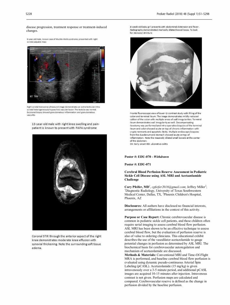
disease progression, treatment response or treatment-induced changes.
Poster #: EDU-070 - Withdrawn
Poster #: EDU-071
Cerebral Blood Perfusion Reserve Assessment in Pediatric
Sickle Cell Disease using ASL MRI and Acetazolamide
Challenge Cory Pfeifer, MD
1, [email protected]; Jeffrey Miller
2;
1Diagnostic Radiology, University of Texas Southwestern
Medical Center, Dallas, TX, 2Phoenix Children's Hospital,
Phoenix, AZ
Disclosures: All authors have disclosed no financial interests, arrangements or affiliations in the context of this activity. Purpose or Case Report: Chronic cerebrovascular disease is common in pediatric sickle cell patients, and these children often require serial imaging to assess cerebral blood flow perfusion.
ASL MRI has been shown to be an effective technique to assess cerebral blood flow, but the evaluation of perfusion reserve is also of value to ordering clinicians. This educational exhibit describes the use of the vasodilator acetazolamide to gauge potential changes in perfusion as determined by ASL MRI. The biochemical basis for cerebrovascular autoregulation and mechanism of acetazolamide are discussed.
Methods & Materials: Conventional MRI and Time-Of-Flight MRA is performed, and baseline cerebral blood flow perfusion is evaluated using dynamic pseudo-continuous Arterial Spin Labeling (pCASL). Acetazolamide (15 mg/kg) is given intravenously over a 3-5 minute period, and additional pCASL images are acquired 10-15 minutes after injection. Intravenous
contrast is not given. Perfusion maps are calculated and compared. Cerebrovascular reserve is defined as the change in perfusion divided by the baseline perfusion.
Pediatr Radiol (2018) 48 (Suppl 1):S1–S298S228
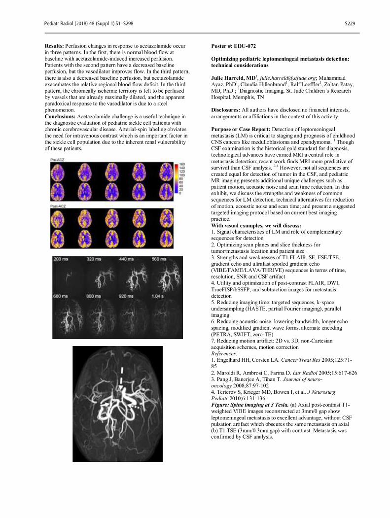
Results: Perfusion changes in response to acetazolamide occur in three patterns. In the first, there is normal blood flow at baseline with acetazolamide-induced increased perfusion. Patients with the second pattern have a decreased baseline perfusion, but the vasodilator improves flow. In the third pattern,
there is also a decreased baseline perfusion, but acetazolamide exacerbates the relative regional blood flow deficit. In the third pattern, the chronically ischemic territory is felt to be perfused by vessels that are already maximally dilated, and the apparent paradoxical response to the vasodilator is due to a steel phenomenon.
Conclusions: Acetazolamide challenge is a useful technique in the diagnostic evaluation of pediatric sickle cell patients with chronic cerebrovascular disease. Arterial-spin labeling obviates the need for intravenous contrast which is an important factor in the sickle cell population due to the inherent renal vulnerability of these patients.
Poster #: EDU-072
Optimizing pediatric leptomeningeal metastasis detection:
technical considerations
Julie Harreld, MD1, [email protected]; Muhammad
Ayaz, PhD1, Claudia Hillenbrand
1, Ralf Loeffler
1, Zoltan Patay,
MD, PhD1;
1Diagnostic Imaging, St. Jude Children’s Research
Hospital, Memphis, TN Disclosures: All authors have disclosed no financial interests,
arrangements or affiliations in the context of this activity. Purpose or Case Report: Detection of leptomeningeal metastasis (LM) is critical to staging and prognosis of childhood CNS cancers like medulloblastoma and ependymoma.
1 Though
CSF examination is the historical gold standard for diagnosis, technological advances have earned MRI a central role in
metastasis detection; recent work finds MRI more predictive of survival than CSF analysis.
2-4 However, not all sequences are
created equal for detection of tumor in the CSF, and pediatric MR imaging presents additional unique challenges such as patient motion, acoustic noise and scan time reduction. In this exhibit, we discuss the strengths and weakness of common sequences for LM detection; technical alternatives for reduction
of motion, acoustic noise and scan time; and present a suggested targeted imaging protocol based on current best imaging practice.
With visual examples, we will discuss: 1. Signal characteristics of LM and role of complementary sequences for detection
2. Optimizing scan planes and slice thickness for tumor/metastasis location and patient size 3. Strengths and weaknesses of T1 FLAIR, SE, FSE/TSE, gradient echo and ultrafast spoiled gradient echo (VIBE/FAME/LAVA/THRIVE) sequences in terms of time, resolution, SNR and CSF artifact 4. Utility and optimization of post-contrast FLAIR, DWI,
TrueFISP/bSSFP, and subtraction images for metastasis detection 5. Reducing imaging time: targeted sequences, k-space undersampling (HASTE, partial Fourier imaging), parallel imaging 6. Reducing acoustic noise: lowering bandwidth, longer echo spacing, modified gradient wave forms, alternate encoding
(PETRA, SWIFT, zero-TE) 7. Reducing motion artifact: 2D vs. 3D, non-Cartesian acquisition schemes, motion correction References: 1. Engelhard HH, Corsten LA. Cancer Treat Res 2005;125:71-85
2. Maroldi R, Ambrosi C, Farina D. Eur Radiol 2005;15:617-626 3. Pang J, Banerjee A, Tihan T. Journal of neuro-oncology 2008;87:97-102 4. Terterov S, Krieger MD, Bowen I, et al. J Neurosurg Pediatr 2010;6:131-136 Figure: Spine imaging at 3 Tesla. (a) Axial post-contrast T1-weighted VIBE images reconstructed at 3mm/0 gap show
leptomeningeal metastasis to excellent advantage, without CSF pulsation artifact which obscures the same metastasis on axial (b) T1 TSE (3mm/0.3mm gap) with contrast. Metastasis was confirmed by CSF analysis.
Pediatr Radiol (2018) 48 (Suppl 1):S1–S298 S229
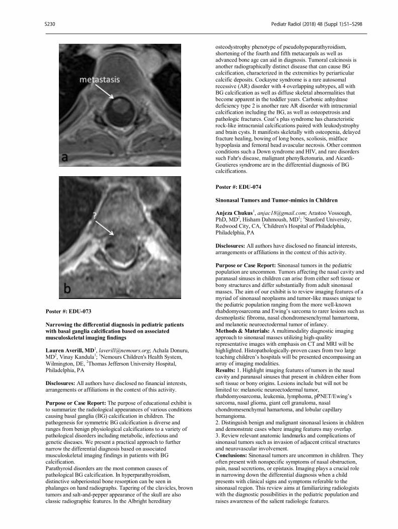
Poster #: EDU-073
Narrowing the differential diagnosis in pediatric patients
with basal ganglia calcification based on associated
musculoskeletal imaging findings Lauren Averill, MD
1, [email protected]; Achala Donuru,
MD2, Vinay Kandula
1;
1Nemours Children's Health System,
Wilmington, DE, 2Thomas Jefferson University Hospital,
Philadelphia, PA Disclosures: All authors have disclosed no financial interests, arrangements or affiliations in the context of this activity.
Purpose or Case Report: The purpose of educational exhibit is to summarize the radiological appearances of various conditions causing basal ganglia (BG) calcification in children. The pathogenesis for symmetric BG calcification is diverse and ranges from benign physiological calcifications to a variety of pathological disorders including metabolic, infectious and genetic diseases. We present a practical approach to further
narrow the differential diagnosis based on associated musculoskeletal imaging findings in patients with BG calcification. Parathyroid disorders are the most common causes of pathological BG calcification. In hyperparathyroidism, distinctive subperiosteal bone resorption can be seen in phalanges on hand radiographs. Tapering of the clavicles, brown
tumors and salt-and-pepper appearance of the skull are also classic radiographic features. In the Albright hereditary
osteodystrophy phenotype of pseudohypoparathyroidism, shortening of the fourth and fifth metacarpals as well as advanced bone age can aid in diagnosis. Tumoral calcinosis is another radiographically distinct disease that can cause BG calcification, characterized in the extremities by periarticular
calcific deposits. Cockayne syndrome is a rare autosomal recessive (AR) disorder with 4 overlapping subtypes, all with BG calcification as well as diffuse skeletal abnormalities that become apparent in the toddler years. Carbonic anhydrase deficiency type 2 is another rare AR disorder with intracranial calcification including the BG, as well as osteopetrosis and
pathologic fractures. Coat’s plus syndrome has characteristic rock-like intracranial calcifications paired with leukodystrophy and brain cysts. It manifests skeletally with osteopenia, delayed fracture healing, bowing of long bones, scoliosis, midface hypoplasia and femoral head avascular necrosis. Other common conditions such a Down syndrome and HIV, and rare disorders such Fahr's disease, malignant phenylketonuria, and Aicardi-
Goutieres syndrome are in the differential diagnosis of BG calcifications.
Poster #: EDU-074
Sinonasal Tumors and Tumor-mimics in Children Anjeza Chukus
1, [email protected]; Arastoo Vossough,
PhD, MD2, Hisham Dahmoush, MD
1;
1Stanford University,
Redwood City, CA, 2Children's Hospital of Philadelphia,
Philadelphia, PA
Disclosures: All authors have disclosed no financial interests, arrangements or affiliations in the context of this activity. Purpose or Case Report: Sinonasal tumors in the pediatric population are uncommon. Tumors affecting the nasal cavity and paranasal sinuses in children can arise from either soft tissue or
bony structures and differ substantially from adult sinonasal masses. The aim of our exhibit is to review imaging features of a myriad of sinonasal neoplasms and tumor-like masses unique to the pediatric population ranging from the more well-known rhabdomyosarcoma and Ewing’s sarcoma to rarer lesions such as desmoplastic fibroma, nasal chondromesenchymal hamartoma,
and melanotic neuroectodermal tumor of infancy. Methods & Materials: A multimodality diagnostic imaging approach to sinonasal masses utilizing high-quality representative images with emphasis on CT and MRI will be highlighted. Histopathologically-proven cases from two large teaching children’s hospitals will be presented encompassing an array of imaging modalities.
Results: 1. Highlight imaging features of tumors in the nasal cavity and paranasal sinuses that present in children either from soft tissue or bony origins. Lesions include but will not be limited to: melanotic neuroectodermal tumor, rhabdomyosarcoma, leukemia, lymphoma, pPNET/Ewing’s sarcoma, nasal glioma, giant cell granuloma, nasal
chondromesenchymal hamartoma, and lobular capillary hemangioma. 2. Distinguish benign and malignant sinonasal lesions in children and demonstrate cases where imaging features may overlap. 3. Review relevant anatomic landmarks and complications of sinonasal tumors such as invasion of adjacent critical structures and neurovascular involvement.
Conclusions: Sinonasal tumors are uncommon in children. They often present with nonspecific symptoms of nasal obstruction, pain, nasal secretions, or epistaxis. Imaging plays a crucial role in narrowing down the differential diagnosis when a child presents with clinical signs and symptoms referable to the sinonasal region. This review aims at familiarizing radiologists with the diagnostic possibilities in the pediatric population and
raises awareness of the salient radiologic features.
Pediatr Radiol (2018) 48 (Suppl 1):S1–S298S230
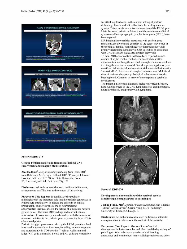
Poster #: EDU-075
Genetic Perforin Defect and Immunopathology: CNS
Involvement and Imaging Manifestations
Alec Hedlund
2, [email protected]; Sara Stern, MD
3,
John Bohnsack, MD3, Gary Hedlund, DO
1;
1Primary Children's
Hospital, Salt Lake, UT, 2Boise State University, Boise,
ID, 3University of Utah, Salt Lake City, UT
Disclosures: All authors have disclosed no financial interests,
arrangements or affiliations in the context of this activity. Purpose or Case Report: To familiarize the pediatric radiologist with the important role that the perforin gene plays in lymphocyte cytotoxicity, to discuss the diversity in clinical presentation, and review the scope of neuroimaging abnormalities that may arise in the setting of a missense perforin
genetic defect. The brain MRI findings and relevant clinical information of two remotely related children with the same novel missense mutation in the perforin gene represent the basis of this educational poster. Perforin is a glycoprotein (encoded by the PRF-1 gene) involved in several human cellular functions, including, immune response
and stored mainly in CD8-positive T-cells as well as natural killer (NK) cells. Normally, T-cells and NK cells are responsible
for attacking dead cells. In the clinical setting of perforin deficiency, T-cells and NK cells attack the healthy immune system. This arises from a missense mutation of the PRF-1 gene. Links between perforin deficiency and the autoimmune clinical syndrome of hemophagocytic lymphohistiocytosis (HLH), have
been reported. MR imaging abnormalities in patients with perforin gene mutations, are diverse and complex as the defect may occur in the setting of familial hemophagocytic lymphohistiocytosis, primary necrotizing lymphocytic CNS vasculitis or associated with CNS infections such as the Epstein Barr virus.
To date, MRI abnormalities that have been reported include mimics of septic cerebral emboli, confluent white matter abnormalities involving the cerebral hemispheres and cerebellum invoking the consideration of diffuse demyelinating disease, and multifocal infratentorial and supratentorial intraaxial lesions with “necrotic-like” character and marginal enhancement. Multifocal sites of perivascular space pathological enhancement has also
been reported. Common to many of these reports is cerebellar involvement. The imaging differential diagnosis includes atypical infection, histocytic disorders of the CNS, lymphomatous granulomatosis, neurosarcoidosis, and primary CNS lymphoma.
Poster #: EDU-076
Developmental abnormalities of the cerebral cortex:
Simplifying a complex group of pathologies
Joshua Finkle, MD
1, [email protected]; Thomas
Tullius1, Ariyan Javadi
1, Carina Yang, MD
1;
1Radiology,
University of Chicago, Chicago, IL Disclosures: All authors have disclosed no financial interests, arrangements or affiliations in the context of this activity.
Purpose or Case Report: Abnormalities of cortical development include a complex and often bewildering variety of pathologies. With substantial overlap in both imaging appearance and terminology, many radiology trainees and other
Pediatr Radiol (2018) 48 (Suppl 1):S1–S298 S231
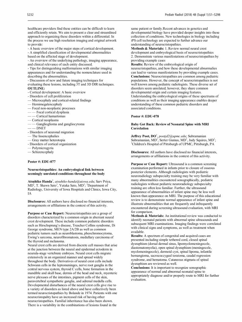
healthcare providers find these entities can be difficult to learn and efficiently retain. We aim to present a clear and streamlined approach to organizing these disorders within a differential. In the process we use high resolution imaging and original artwork to provide:
- A basic overview of the major steps of cortical development. - A simplified classification of developmental abnormalities based on the affected stage of development. - An overview of the underlying pathology, imaging appearance, and clinical relevance of each entity discussed. - Tips for distinguishing malformations with similar imaging
appearances and for understanding the nomenclature used in describing the abnormalities. - Discussion of new and future imaging techniques for evaluating these lesions, including 3T and 3D DIR techniques.
OUTLINE: - Cortical development: A basic overview - Disorders of cell proliferation
— Microcephaly and cortical-related findings — Hemimegalencephaly — Focal non-neoplastic processes — — Focal cortical dysplasia — — Cortical hamartomas — Cortical neoplasms — — Ganglioglioma and gangliocytoma
— — DNET - Disorders of neuronal migration — The lissencephalies — Gray matter heterotopia - Disorders of cortical organization — Polymicrogyria — Schizencephaly
Poster #: EDU-077
Neurocristopathies: An embryological link between
seemingly unrelated conditions throughout the body Atsuhiko Handa
1, [email protected]; Sarv Priya,
MD1, T. Shawn Sato
1, Yutaka Sato, MD
1;
1Department of
Radiology, University of Iowa Hospitals and Clinics, Iowa City, IA
Disclosures: All authors have disclosed no financial interests, arrangements or affiliations in the context of this activity.
Purpose or Case Report: Neurocristopathies are a group of disorders characterized by a common origin in aberrant neural crest development. These include common pediatric disorders
such as Hirschsprung’s disease, Treacher Collins syndrome, Di George syndrome, MEN type 2A/2B as well as common pediatric tumors such as neuroblastoma, pheochromocytoma, Ewing’s sarcoma, neurofibromatosis, medullary carcinoma of the thyroid and melanoma. Neural crest cells are derived from discrete cell masses that arise
at the junction between the neural and epidermal ectoderm in neurula-stage vertebrate embryos. Neural crest cells migrate extensively in an organized manner and spread widely throughout the body. Derivatives of neural crest cells include Schwann cells in the leptomeninges, nerve root ganglia in the central nervous system, thyroid C cells, bone formation in the mandible and skull base, dermis of the head and neck, myenteric
nerve plexuses of the intestines, pigment cells of the skin, paravertebral sympathetic ganglia, and adrenal medulla cells. Developmental disturbances of the neural crest cells give rise to a variety of disorders as listed above and have collectively been termed neurocristopathies by Bolande in 1974. Patients with one neurocristopahty have an increased risk of having other neurocristopathies. Familial inheritance has also been shown.
There is a variability in the combinations of lesions found in the
same patient or family.Recent advances in genetics and developmental biology have provided deeper insights into these collection of conditions. New technologies in biology including iPS cell technology are expected to further advance our understanding of neurocristopathies.
Methods & Materials: 1. Review normal neural crest development and embryological basis of neurocristopathies 2. Demonstrate various manifestations of neurocristopathies by providing example cases Results: Review of the embryological origin of neurocristopathies, and how these developmental abnormalies
can lead to various manifestations by providing example cases. Conclusions: Neurocristopathies are common among pediatric populations. However, the concept of neurocristopathies is not well known among pediatric radiologists. These diverse set of disorders seem unrelated; however, they share common developmental origin and certain imaging features. Understanding the embryological origins of these spectrums of
conditions as well as their imaging appearance enables deeper understanding of these common pediatric disorders and associated conditions.
Poster #: EDU-078
Baby Got Back: Review of Neonatal Spine with MRI
Correlation Jeffrey Poot, DO
1, [email protected]; Subramanian
Subramanian, MD1, Serter Gumus, MD
1, Judy Squires, MD
1;
1Children's Hospital of Pittsburgh of UPMC, Pittsburgh, PA
Disclosures: All authors have disclosed no financial interests, arrangements or affiliations in the context of this activity. Purpose or Case Report: Ultrasound is a common screening examination performed in infants prior to closure of osseous posterior elements. Although radiologists with pediatric neuroradiology subspecialty training may be very familiar with
many abnormalities encountered sonographically, pediatric radiologists without pediatric neuroradiology subspecialty training are often less familiar. Further, the ultrasound appearance of abnormalities of infant spine may be less well known than appearance on MRI. The purpose of this educational review is to demonstrate normal appearance of infant spine and illustrate abnormalities that are frequently and infrequently
encountered during screening ultrasound evaluation, with MRI for comparison. Methods & Materials: An institutional review was conducted to identify neonatal patients with abnormal spine ultrasounds and subsequent MRI examinations. Imaging findings were correlated with clinical signs and symptoms, as well as treatment when
available. Results: A spectrum of congenital and acquired cases are presented including simple tethered cord, closed spinal dysraphism (dorsal dermal sinus, lipomyelomeningocele, diastematomyelia), open spinal dysraphism (meningocele, myelomeningocele), dermoid cyst, spinal lipoma, infantile hemangioma, sacrococcygeal teratoma, caudal regression
syndrome, and hematoma. Cutaneous stigmata of spinal dysraphism are reviewed as well. Conclusions: It is important to recognize sonographic appearance of normal and abnormal neonatal spine to appropriately diagnose and/or properly route to MRI for further evaluation.
Pediatr Radiol (2018) 48 (Suppl 1):S1–S298S232
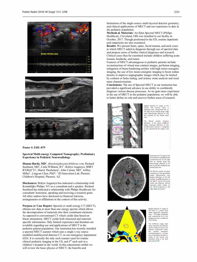
Poster #: EDU-079
Spectral Multi-energy Computed Tomography; Preliminary
Experience in Pediatric Neuroradiology Dianna Bardo, MD
1, [email protected]; Richard
Southard, MD1, Carla Williams, RT
1, Robyn Augustyn, BSRT
RT(R)(CT)1, Marrit Thorkelson
1, John Curran, MD
1, Jeffrey
Miller1, Lingyun Chen, PhD
1;
13D Innovation Lab, Phoenix
Children's Hospital, Phoenix, AZ Disclosures: Robyn Augustyn has indicated a relationship with Koninklijke Philips, NV as a consultant and a speaker. Richard Southard has indicated a relationship with Philips Healthcare for consultant/ honoraria, speaking and receiving a research grant.
All other authors have disclosed no financial interests, arrangements or affiliations in the context of this activity. Purpose or Case Report: Spectral or multi-energy CT (MECT), obtains raw data at more than one energy spectra which allows the decomposition of materials into their constituent elements.
As opposed to conventional CT which yields data based on linear attenuation, MECT yields both structural and material-specific information. Only limited experience and literature are available regarding use and applications of MECT in the pediatric patient population. Our institution has recently installed a spectral MECT scanner which uses a single x-ray source modified multilayered detector CT, in our emergency department
(ED). It is currently the only such scanner used for routine clinical pediatric imaging in the US, and 4
th such unit in a
children’s hospital in the world. In this educational exhibit we will review the basic physics of MECT, the benefits and
limitations of the single-source multi-layered detector geometry, and clinical applications of MECT and our experience to date in the pediatric population. Methods & Materials: An IQon Spectral MECT (Philips Healthcare, Cleveland, OH) was installed in our facility in
October, 2017. Though positioned in the ED, routine inpatients and outpatients are also examined. Results: We present brain, spine, facial trauma, and neck cases in which MECT aided in diagnosis through use of spectral data and propose areas of further clinical diagnoses and research. Clinical cases thus far examined include children suffering acute
trauma, headache, and tumor. Features of MECT advantageous to pediatric patients include reconstruction of virtual non-contrast images, perfusion imaging, mitigation of beam-hardening artifact with high mono-energetic imaging, the use of low mono-energetic imaging to boost iodine density to improve angiographic images which may be limited by contrast or bolus timing, and urinary stone analysis and renal
mass characterization. Conclusions: The use of Spectral MECT at our institution has provided a significant advance in our ability to confidently diagnose various disease processes. As we gain more experience in the use of MECT in the pediatric population, we will be able to better define its role and uncover further areas of research.
Pediatr Radiol (2018) 48 (Suppl 1):S1–S298 S233
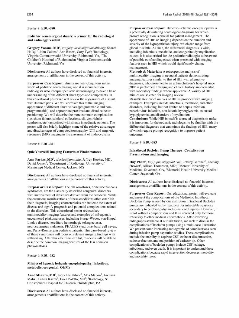
Poster #: EDU-080
Pediatric neurosurgical shunts: a primer for the radiologist
and radiology resident
Gregory Vorona, MD2, [email protected]; Shareh
Hallaji1, John Collins
2, Ann Ritter
2, Gary Tye
2;
1Radiology,
Virginia Commonwealth University, Richmond, VA, 2The
Children's Hospital of Richmond at Virginia Commonwealth University, Richmond, VA
Disclosures: All authors have disclosed no financial interests, arrangements or affiliations in the context of this activity. Purpose or Case Report: Shunts are near-ubiquitous in the world of pediatric neuroimaging, and it is incumbent on radiologists who interpret pediatric neuroimaging to have a basic understanding of the different shunt types and components. In
this educational poster we will review the appearance of a shunt with its three parts. We will correlate this to the imaging appearance of different shunt valves (programmable and non-programmable), and appropriate proximal and distal catheter positioning. We will describe the more common complications (i.e. shunt failure, subdural collections, slit ventriclular syndrome, etc.) associated with shunts in pediatric patients. The
poster will also briefly highlight some of the relative advantages and disadvantages of computed tomography (CT) and magnetic resonance (MR) imaging in the assessment of hydrocephalus.
Poster #: EDU-081
Quiz Yourself! Imaging Features of Phakomatoses Amy Farkas, MD
1, [email protected]; Jeffrey Hooker, MD
1,
David Joyner1;
1Department of Radiology, University of
Mississippi Medical Center, Jackson, MS
Disclosures: All authors have disclosed no financial interests, arrangements or affiliations in the context of this activity. Purpose or Case Report: The phakomatoses, or neurocutaneous syndromes, are the classically described congenital disorders
with involvement of structures derived from the ectoderm. While the cutaneous manifestations of these conditions often establish their diagnosis, imaging characteristics can indicate the extent of disease and signify prognosis and potential complications related to the disorders. This educational poster reviews key multimodality imaging features and examples of infrequently encountered phakomatoses, including Sturge-Weber, von-Hippel
Lindau disease, hereditary hemorrhagic telangiectasia, neurocutaneous melanosis, PHACES syndrome, basal cell nevus, and Parry-Romberg in pediatric patients. This case-based review of these syndromes will focus on relevant imaging findings with self-testing. After this electronic exhibit, residents will be able to describe the common imaging features of the less common phakomatoses.
Poster #: EDU-082
Mimics of hypoxic ischemic encephalopathy: Infectious,
metabolic, congenital, Oh My! Anne Misiura, MD
1, Jaqueline Urbine
1, Mea Mallon
1, Archana
Malik1, Faaiza Kazmi
1, Erica Poletto, MD
1;
1Radiology, St.
Christopher's Hospital for Children, Philadelphia, PA
Disclosures: All authors have disclosed no financial interests, arrangements or affiliations in the context of this activity.
Purpose or Case Report: Hypoxic-ischemic encephalopathy is a potentially devastating neurological diagnosis for which prompt recognition is crucial for patient management. The appearance of HIE on imaging depends on the duration and severity of the hypoperfusion injury, which can range from
global to subtle. As such, the differential diagnosis is wide, including infectious, metabolic, and congenital dysmyelination causes. It is also critical for the pediatric radiologist to be aware of possible confounding cases when presented with imaging features seen in HIE which would significantly change management.
Methods & Materials: A retrospective analysis of multimodality imaging in neonatal patients demonstrating imaging features similar to that of HIE with alternative diagnoses, who presented to an urban children’s hospital since 2005 is performed. Imaging and clinical history are correlated with laboratory findings where applicable. A variety of HIE mimics are selected for imaging review.
Results: Review of mimics of HIE is provided with imaging examples. Examples include infectious, metabolic, and other disorders, including, but not limited to herpes infection, parechovirus infection, non-ketotic hyperglycemia, neonatal hypoglycemia, and disorders of myelination. Conclusions: While HIE in itself is a crucial diagnosis to make, it is important for the pediatric radiologist to be familiar with the
differential diagnoses that can mimic the findings of HIE, many of which require prompt recognition to improve patient outcomes.
Poster #: EDU-083
Intrathecal Baclofen Pump Therapy: Complication
Presentations and Imaging Huy Pham
2, [email protected]; Jeffrey Gardner
1, Zachary
Stewart2, Allison Thompson, MD
2;
1Mercer University of
Medicine, Savannah, GA, 2Memorial Health University Medical
Center, Savannah, GA
Disclosures: All authors have disclosed no financial interests, arrangements or affiliations in the context of this activity. Purpose or Case Report: Our educational poster will evaluate and present the complications of the Medtronic Intrathecal Baclofen Pump as seen by our institution. Intrathecal Baclofen
pumps are indicated as the treatment for intractable spasticity secondary to cerebral palsy and spinal cord injuries. However, it is not without complications and thus, reserved only for those refractory to other medical interventions. After reviewing radiographs available at our institution, we seek to discuss the complications of baclofen pumps using a multi-case illustration.
We present some interesting radiographs of complications seen during infusion pump aspiration studies. These complications include the inability to aspirate CSF, catheter disconnection, catheter fracture, and malposition of catheter tip. Other complications of baclofen pumps include CSF leakage, infections, and even death. It is important to understand these complications because rapid intervention decreases morbidity and mortality rates.
Pediatr Radiol (2018) 48 (Suppl 1):S1–S298S234
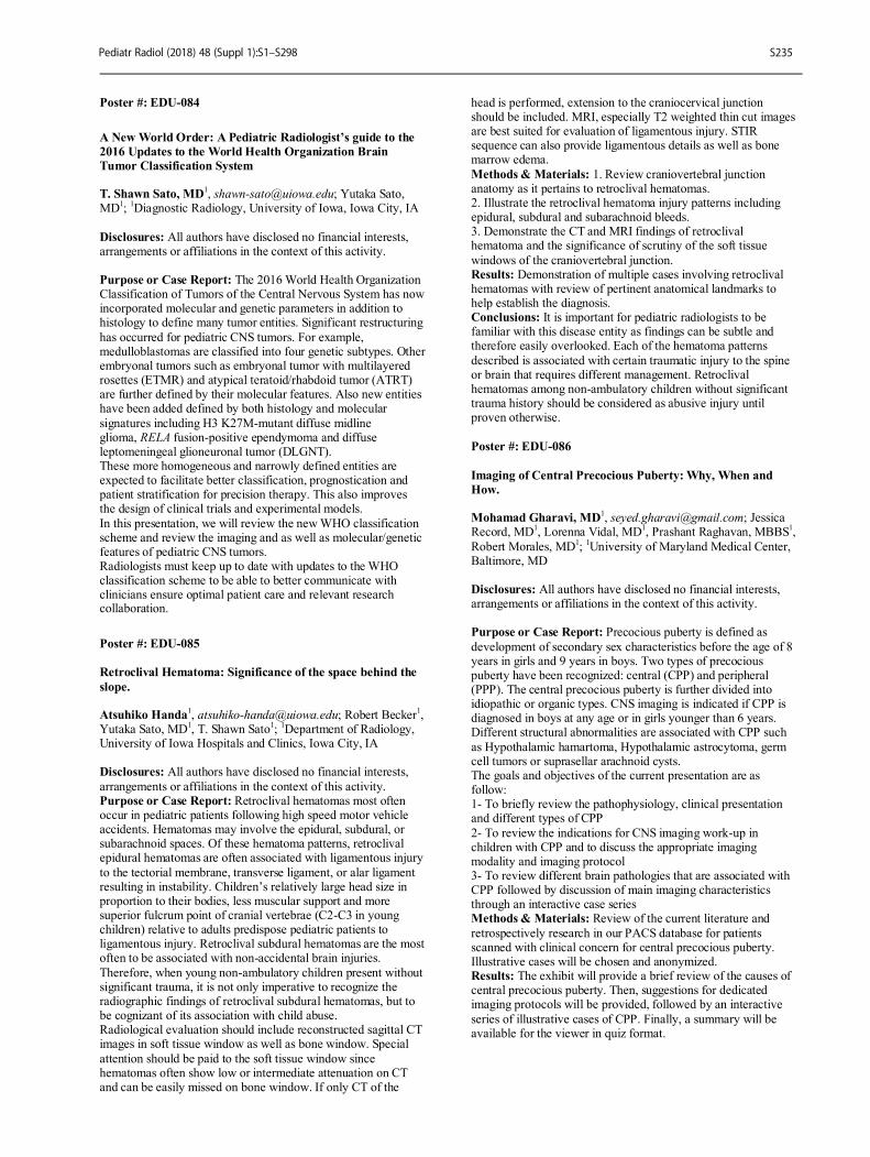
Poster #: EDU-084
A New World Order: A Pediatric Radiologist’s guide to the
2016 Updates to the World Health Organization Brain
Tumor Classification System T. Shawn Sato, MD
1, [email protected]; Yutaka Sato,
MD1;
1Diagnostic Radiology, University of Iowa, Iowa City, IA
Disclosures: All authors have disclosed no financial interests, arrangements or affiliations in the context of this activity. Purpose or Case Report: The 2016 World Health Organization Classification of Tumors of the Central Nervous System has now incorporated molecular and genetic parameters in addition to histology to define many tumor entities. Significant restructuring
has occurred for pediatric CNS tumors. For example, medulloblastomas are classified into four genetic subtypes. Other embryonal tumors such as embryonal tumor with multilayered rosettes (ETMR) and atypical teratoid/rhabdoid tumor (ATRT) are further defined by their molecular features. Also new entities have been added defined by both histology and molecular
signatures including H3 K27M-mutant diffuse midline glioma, RELA fusion-positive ependymoma and diffuse leptomeningeal glioneuronal tumor (DLGNT). These more homogeneous and narrowly defined entities are expected to facilitate better classification, prognostication and patient stratification for precision therapy. This also improves the design of clinical trials and experimental models.
In this presentation, we will review the new WHO classification scheme and review the imaging and as well as molecular/genetic features of pediatric CNS tumors. Radiologists must keep up to date with updates to the WHO classification scheme to be able to better communicate with clinicians ensure optimal patient care and relevant research collaboration.
Poster #: EDU-085
Retroclival Hematoma: Significance of the space behind the
slope. Atsuhiko Handa
1, [email protected]; Robert Becker
1,
Yutaka Sato, MD1, T. Shawn Sato
1;
1Department of Radiology,
University of Iowa Hospitals and Clinics, Iowa City, IA
Disclosures: All authors have disclosed no financial interests,
arrangements or affiliations in the context of this activity. Purpose or Case Report: Retroclival hematomas most often occur in pediatric patients following high speed motor vehicle accidents. Hematomas may involve the epidural, subdural, or subarachnoid spaces. Of these hematoma patterns, retroclival epidural hematomas are often associated with ligamentous injury
to the tectorial membrane, transverse ligament, or alar ligament resulting in instability. Children’s relatively large head size in proportion to their bodies, less muscular support and more superior fulcrum point of cranial vertebrae (C2-C3 in young children) relative to adults predispose pediatric patients to ligamentous injury. Retroclival subdural hematomas are the most often to be associated with non-accidental brain injuries.
Therefore, when young non-ambulatory children present without significant trauma, it is not only imperative to recognize the radiographic findings of retroclival subdural hematomas, but to be cognizant of its association with child abuse. Radiological evaluation should include reconstructed sagittal CT images in soft tissue window as well as bone window. Special
attention should be paid to the soft tissue window since hematomas often show low or intermediate attenuation on CT and can be easily missed on bone window. If only CT of the
head is performed, extension to the craniocervical junction should be included. MRI, especially T2 weighted thin cut images are best suited for evaluation of ligamentous injury. STIR sequence can also provide ligamentous details as well as bone marrow edema.
Methods & Materials: 1. Review craniovertebral junction anatomy as it pertains to retroclival hematomas. 2. Illustrate the retroclival hematoma injury patterns including epidural, subdural and subarachnoid bleeds. 3. Demonstrate the CT and MRI findings of retroclival hematoma and the significance of scrutiny of the soft tissue
windows of the craniovertebral junction. Results: Demonstration of multiple cases involving retroclival hematomas with review of pertinent anatomical landmarks to help establish the diagnosis. Conclusions: It is important for pediatric radiologists to be familiar with this disease entity as findings can be subtle and therefore easily overlooked. Each of the hematoma patterns
described is associated with certain traumatic injury to the spine or brain that requires different management. Retroclival hematomas among non-ambulatory children without significant trauma history should be considered as abusive injury until proven otherwise.
Poster #: EDU-086
Imaging of Central Precocious Puberty: Why, When and
How. Mohamad Gharavi, MD
1, [email protected]; Jessica
Record, MD1, Lorenna Vidal, MD
1, Prashant Raghavan, MBBS
1,
Robert Morales, MD1;
1University of Maryland Medical Center,
Baltimore, MD Disclosures: All authors have disclosed no financial interests, arrangements or affiliations in the context of this activity. Purpose or Case Report: Precocious puberty is defined as
development of secondary sex characteristics before the age of 8 years in girls and 9 years in boys. Two types of precocious puberty have been recognized: central (CPP) and peripheral (PPP). The central precocious puberty is further divided into idiopathic or organic types. CNS imaging is indicated if CPP is diagnosed in boys at any age or in girls younger than 6 years. Different structural abnormalities are associated with CPP such
as Hypothalamic hamartoma, Hypothalamic astrocytoma, germ cell tumors or suprasellar arachnoid cysts. The goals and objectives of the current presentation are as follow: 1- To briefly review the pathophysiology, clinical presentation and different types of CPP
2- To review the indications for CNS imaging work-up in children with CPP and to discuss the appropriate imaging modality and imaging protocol 3- To review different brain pathologies that are associated with CPP followed by discussion of main imaging characteristics through an interactive case series Methods & Materials: Review of the current literature and
retrospectively research in our PACS database for patients scanned with clinical concern for central precocious puberty. Illustrative cases will be chosen and anonymized. Results: The exhibit will provide a brief review of the causes of central precocious puberty. Then, suggestions for dedicated imaging protocols will be provided, followed by an interactive
series of illustrative cases of CPP. Finally, a summary will be available for the viewer in quiz format.
Pediatr Radiol (2018) 48 (Suppl 1):S1–S298 S235
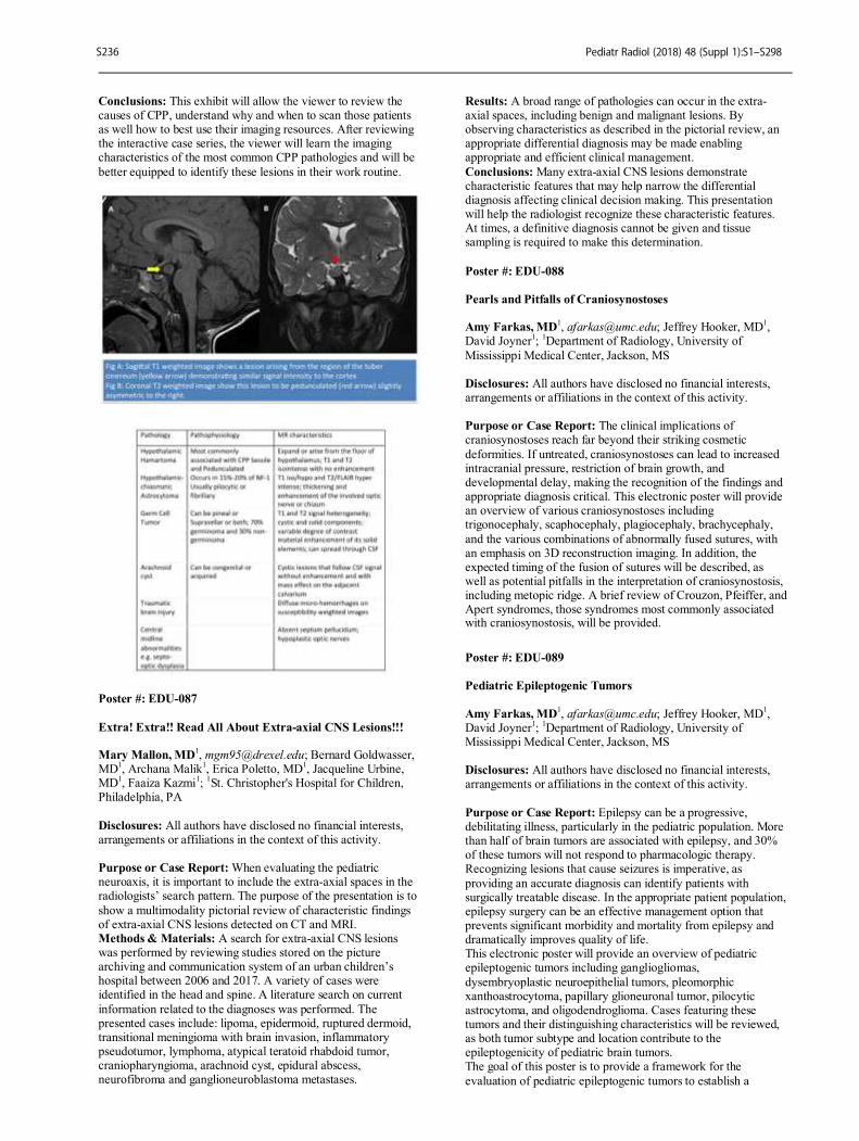
Conclusions: This exhibit will allow the viewer to review the causes of CPP, understand why and when to scan those patients as well how to best use their imaging resources. After reviewing the interactive case series, the viewer will learn the imaging characteristics of the most common CPP pathologies and will be
better equipped to identify these lesions in their work routine.
Poster #: EDU-087
Extra! Extra!! Read All About Extra-axial CNS Lesions!!! Mary Mallon, MD
1, [email protected]; Bernard Goldwasser,
MD1, Archana Malik
1, Erica Poletto, MD
1, Jacqueline Urbine,
MD1, Faaiza Kazmi
1;
1St. Christopher's Hospital for Children,
Philadelphia, PA
Disclosures: All authors have disclosed no financial interests, arrangements or affiliations in the context of this activity. Purpose or Case Report: When evaluating the pediatric neuroaxis, it is important to include the extra-axial spaces in the radiologists’ search pattern. The purpose of the presentation is to
show a multimodality pictorial review of characteristic findings of extra-axial CNS lesions detected on CT and MRI. Methods & Materials: A search for extra-axial CNS lesions was performed by reviewing studies stored on the picture archiving and communication system of an urban children’s hospital between 2006 and 2017. A variety of cases were identified in the head and spine. A literature search on current
information related to the diagnoses was performed. The presented cases include: lipoma, epidermoid, ruptured dermoid, transitional meningioma with brain invasion, inflammatory pseudotumor, lymphoma, atypical teratoid rhabdoid tumor, craniopharyngioma, arachnoid cyst, epidural abscess, neurofibroma and ganglioneuroblastoma metastases.
Results: A broad range of pathologies can occur in the extra-axial spaces, including benign and malignant lesions. By observing characteristics as described in the pictorial review, an appropriate differential diagnosis may be made enabling appropriate and efficient clinical management.
Conclusions: Many extra-axial CNS lesions demonstrate characteristic features that may help narrow the differential diagnosis affecting clinical decision making. This presentation will help the radiologist recognize these characteristic features. At times, a definitive diagnosis cannot be given and tissue sampling is required to make this determination.
Poster #: EDU-088
Pearls and Pitfalls of Craniosynostoses Amy Farkas, MD
1, [email protected]; Jeffrey Hooker, MD
1,
David Joyner1;
1Department of Radiology, University of
Mississippi Medical Center, Jackson, MS Disclosures: All authors have disclosed no financial interests, arrangements or affiliations in the context of this activity. Purpose or Case Report: The clinical implications of craniosynostoses reach far beyond their striking cosmetic
deformities. If untreated, craniosynostoses can lead to increased intracranial pressure, restriction of brain growth, and developmental delay, making the recognition of the findings and appropriate diagnosis critical. This electronic poster will provide an overview of various craniosynostoses including trigonocephaly, scaphocephaly, plagiocephaly, brachycephaly,
and the various combinations of abnormally fused sutures, with an emphasis on 3D reconstruction imaging. In addition, the expected timing of the fusion of sutures will be described, as well as potential pitfalls in the interpretation of craniosynostosis, including metopic ridge. A brief review of Crouzon, Pfeiffer, and Apert syndromes, those syndromes most commonly associated with craniosynostosis, will be provided.
Poster #: EDU-089
Pediatric Epileptogenic Tumors
Amy Farkas, MD
1, [email protected]; Jeffrey Hooker, MD
1,
David Joyner1;
1Department of Radiology, University of
Mississippi Medical Center, Jackson, MS Disclosures: All authors have disclosed no financial interests, arrangements or affiliations in the context of this activity.
Purpose or Case Report: Epilepsy can be a progressive, debilitating illness, particularly in the pediatric population. More than half of brain tumors are associated with epilepsy, and 30% of these tumors will not respond to pharmacologic therapy. Recognizing lesions that cause seizures is imperative, as
providing an accurate diagnosis can identify patients with surgically treatable disease. In the appropriate patient population, epilepsy surgery can be an effective management option that prevents significant morbidity and mortality from epilepsy and dramatically improves quality of life. This electronic poster will provide an overview of pediatric epileptogenic tumors including gangliogliomas,
dysembryoplastic neuroepithelial tumors, pleomorphic xanthoastrocytoma, papillary glioneuronal tumor, pilocytic astrocytoma, and oligodendroglioma. Cases featuring these tumors and their distinguishing characteristics will be reviewed, as both tumor subtype and location contribute to the epileptogenicity of pediatric brain tumors. The goal of this poster is to provide a framework for the
evaluation of pediatric epileptogenic tumors to establish a
Pediatr Radiol (2018) 48 (Suppl 1):S1–S298S236
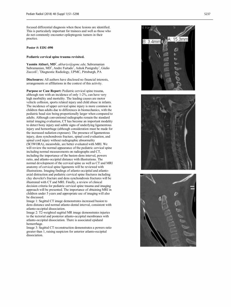
focused differential diagnosis when these lesions are identified. This is particularly important for trainees and well as those who do not commonly encounter epileptogenic tumors in their practice.
Poster #: EDU-090
Pediatric cervical spine trauma revisited. Yasmin Akbari, MD
1, [email protected]; Subramanian
Subramanian, MD1, Andre Furtado
1, Ashok Panigrahy
1, Giulio
Zuccoli1;
1Diagnostic Radiology, UPMC, Pittsburgh, PA
Disclosures: All authors have disclosed no financial interests, arrangements or affiliations in the context of this activity. Purpose or Case Report: Pediatric cervical spine trauma, although rare with an incidence of only 1-2%, can have very high morbidity and mortality. The leading causes are motor vehicle collision, sports related injury and child abuse in infants.
The incidence of upper cervical spine injury is more common in children than adults due to differences in biomechanics, with the pediatric head size being proportionally larger when compared to adults. Although conventional radiographs remain the standard initial imaging evaluation, CT has become an important modality to detect bony injury and subtle signs of underlying ligamentous injury and hemorrhage (although consideration must be made for
the increased radiation exposure). The presence of ligamentous injury, dens synchondrosis fracture, spinal cord evaluation, and spinal cord injury without radiographic abnormality (SCIWORA), meanwhile, are better evaluated with MRI. We will review the normal appearance of the pediatric cervical spine including normal measurements on radiographs and CT,
including the importance of the basion-dens interval, powers ratio, and atlanto-occipital distance with illustrations. The normal development of the cervical spine as well as CT and MRI anatomy of cervical spine ligaments will be reviewed with illustrations. Imaging findings of atlanto-occipital and atlanto-axial distraction and pediatric cervical spine fractures including clay shoveler's fracture and dens synchondrosis fractures will be
illustrated with CT and MRI. Finally, a review of clinical decision criteria for pediatric cervical spine trauma and imaging approach will be presented. The importance of obtaining MRI in children under 5 years and appropriate use of imaging will also be discussed. Image 1: Sagittal CT image demonstrates increased basion to dens distance and normal atlanto-dental interval, consistent with
atlanto-occipital dissociation. Image 2: T2-weighted sagittal MR image demonstrates injuries to the tectorial and posterior atlanto-occipital membranes with atlanto-occipital dissociation. There is associated epidural hemorrhage. Image 3: Sagittal CT reconstruction demonstrates a powers ratio
greater than 1, raising suspicion for anterior atlanto-occipital dissociation.
Pediatr Radiol (2018) 48 (Suppl 1):S1–S298 S237
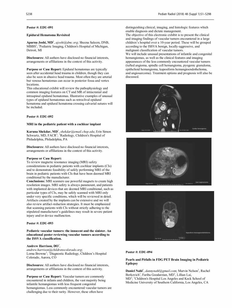
Poster #: EDU-091
Epidural Hematoma Revisited Aparna Joshi, MD
1, [email protected]; Sheena Saleem, DNB,
MBBS1;
1Pediatric Imaging, Children's Hospital of Michigan,
Detroit, MI
Disclosures: All authors have disclosed no financial interests, arrangements or affiliations in the context of this activity.
Purpose or Case Report: Epidural hematomas are typically seen after accidental head trauma in children, though they can also be seen in abusive head trauma. Most often they are arterial but venous hematomas can occur in posterior fossa and vertex locations. This educational exhibit will review the pathophysiology and common imaging features on CT and MR of intracranial and
intraspinal epidural hematomas. Illustrative examples of unusual types of epidural hematomas such as retroclival epidural hematoma and epidural hematoma crossing calvarial sutures will be included.
Poster #: EDU-092
MRI in the pediatric patient with a cochlear implant Karuna Shekdar, MD
1, [email protected]; Erin Simon
Schwartz, MD, FACR1;
1Radiology, Children's Hospital of
Philadelphia, Philadelphia, PA Disclosures: All authors have disclosed no financial interests, arrangements or affiliations in the context of this activity.
Purpose or Case Report: To review magnetic resonance imaging (MRI) safety
considerations in pediatric patients with cochlear implants (CIs) and to demonstrate feasibility of safely performing MRI of the brain in pediatric patients with CIs that have been deemed MRI conditional by the manufacturer. Conclusions: MRI scanners use powerful magnets to create high resolution images. MRI safety is always paramount, and patients
with implanted devices that are deemed MRI conditional, such as particular types of CIs, may be safely scanned with MRI only under very specific conditions, which will be reviewed in detail. Artifacts created by the implants can be extensive and we will also review artifact-reduction strategies. It must be emphasized that scanning patients with CIs without strictly adhering to the stipulated manufacturer’s guidelines may result in severe patient
injury and/or device malfunction.
Poster #: EDU-093
Pediatric vascular tumors: the innocent and the sinister. An
educational poster reviewing vascular tumors according to
the ISSVA classification. Andrew Harrison, DO
1,
[email protected]; Lorna Browne
1;
1Diagnostic Radiology, Children’s Hospital
Colorado, Aurora, CO
Disclosures: All authors have disclosed no financial interests, arrangements or affiliations in the context of this activity. Purpose or Case Report: Vascular tumors are commonly encountered in infants and children, the vast majority being infantile hemangiomas with less frequent congenital hemangiomas. Less commonly encountered vascular tumors are
challenging due to their rarity. However, these often have
distinguishing clinical, imaging, and histologic features which enable diagnosis and dictate management. The objective of this electronic exhibit is to present the clinical and imaging findings of vascular tumors encountered in a large children’s hospital over a 10-year period. These will be grouped
according to the ISSVA benign, locally-aggressive, and malignant classification of vascular tumors. We will include unusual presentations of infantile and congenital hemangiomas, as well as the clinical features and imaging appearances of the less commonly encountered vascular tumors (tufted angioma, spindle cell hemangioma, pyogenic granuloma,
epithelioid hemangioma, kaposiform hemangioendothelioma, and angiosarcoma). Treatment options and prognosis will also be discussed.
Poster #: EDU-094
Pearls and Pitfalls in FDG PET Brain Imaging in Pediatric
Epilepsy Daniel Nahl
1, [email protected]; Marvin Nelson
1, Rachel
Berkovich1, Fariba Goodarzian, MD
1, Lillian Lai,
MD1;
1Children's Hospital Los Angeles and Keck School of
Medicine University of Southern California, Los Angeles, CA
Pediatr Radiol (2018) 48 (Suppl 1):S1–S298S238
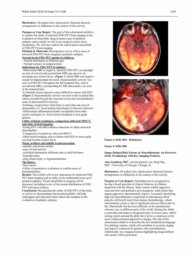
Disclosures: All authors have disclosed no financial interests, arrangements or affiliations in the context of this activity. Purpose or Case Report: The goal of this educational exhibit is to explore the utility of interictal FDG PET brain imaging in the
evaluation of intractable, drug-resistant cases of pediatric epilepsy and to clarify its role in pre-surgical seizure focus localization. We will also explore the salient pearls and pitfalls of FDG PET brain imaging. Methods & Materials: Retrospective review of key cases of interictal FDG PET brain imaging in pediatric epilepsy:
Normal brain FDG PET uptake in children: - Normal distribution at different ages - Normal variance in temporal lobes
Indications for FDG PET in epilepsy: -When initial MRI is negative, interictal FDG PET can spotlight an area of concern and second-look MRI may uncover an inconspicuous seizure focus. (Figure 1. Initial MRI was negative
except for hippocampal inversion. Hypometabolic activity was seen on FDG PET throughout the left temporal lobe, and on second look MRI, corresponding FLAIR abnormality was seen in the temporal lobe. -Evaluation of post-operative areas difficult to assess with EEG (Figure 2. Hypometabolic activity was seen in the occipital lobe, which extended beyond the resection cavity and corresponded to
areas of abnormal EEG activity). -Isolating a target lesion when there is more than one area of abnormality (i.e. focal nodular heterotopia or tuberous sclerosis) -Differentiate inflammation/limbic encephalitis from other seizure etiologies (i.e. focal cortical dysplasia or low grade glioma)
Utility of fusion techniques, comparison with ictal SPECT,
and other hybrid imaging: -Fusing PET with MRI enhances detection of subtle structural abnormalities -Comparison of sensitivity with ictal SPECT -Other hybrid imaging such as fusion with EEG or stereo grids can help localize seizure focus
Major artifacts and pitfalls in interpretation: -metallic and motion artifact -areas of ictal activity -calculated attenuation difference due to skull thickness -hyperglycemia -drug related hypo- or hypermetabolism
The future: -New tracers -Utility of quantitative evaluation to confirm areas of hypometabolism Results: This exhibit will review indications for interictal FDG-PET brain imaging and its utility in the multimodal work-up of pediatric epilepsy. Pearls and pitfalls in imaging will be
discussed, including review of the normal distribution of FDG PET and major artifacts. Conclusions: Recognizing the utility of FDG PET of the brain, as well as its shortcomings and potential pitfalls, will help radiologists and clinicians better utilize this modality in the evaluation of pediatric epilepsy.
Poster #: EDU-095 - Withdrawn
Poster #: EDU-096
Image Defined Risk Factors in Neuroblastoma: An Overview
of the Terminology with Key Imaging Features. Alex Lionberg, MD
1, [email protected]; Seng Ong,
MD1;
1University of Chicago, Chicago, IL
Disclosures: All authors have disclosed no financial interests, arrangements or affiliations in the context of this activity. Purpose or Case Report: Neuroblastoma is recognized as having a broad spectrum of clinical behavior in children diagnosed with the disease. Some tumors exhibit aggressive
characteristics and portend a poor prognosis, while others that appear aggressive spontaneously regress. Accurately identifying high risk neuroblastoma is important in determining which patients will benefit most from intense chemotherapy, which unfortunately carries a risk of significant adverse effects later in life. Historically this has been difficult, as the classification schemes vary in different parts of the world, limiting the ability
to pool data and improve prognostication. In recent years, efforts among experts around the globe have led to a consensus on the most evidenced based approach to staging. The aim of this educational exhibit is to describe the new standardized language for radiology reports, which will contribute to accurate staging and improve treatment for patients with neuroblastoma.
Additionally, key imaging features highlighting image defined risk factors will be presented.
Pediatr Radiol (2018) 48 (Suppl 1):S1–S298 S239
Methods & Materials: The International Neuroblastoma Risk Group Task Force is a group of leading pediatric oncologists, radiologists, researchers and statisticians from around the world. In 2009, the group published a new classification system based on data collected from a large cohort of patients, representing a
consensus among global experts in the field. Results: The INRG Staging System (INRGSS) was introduced to replace the now decades-old International Neuroblastoma Staging System. The new system shifts away from surgicopathologic classification to describe tumors according to radiologic findings. In addition to differentiating local from
metastatic disease, the new system further delineates local disease based on the presence or absence of “image defined risk factors” (IDRF). With this, there is a recommendation for specific terminology to be used in order to accurately characterize an IDRF. These include the words: separation, contact, encasement, compression, infiltration, and invasion. Conclusions: As the care for Neuroblastoma patients evolves,
radiologists must continue to provide value in the multidisciplinary efforts to advance the most up to date approach to staging of the disease. Now more than ever, the use of accurate and uniform language in reports will have a significant impact on the treatment of these patients.
Poster #: EDU-097
Not Your Typical Nashville Blues: An Education Review of
Small Round Blue Cell Tumors." Susan Yoon, DO
1, Stacy White, MD
1, Erica Poletto, MD
2,
Jaqueline Urbine2, Archana Malik
2, Mea Mallon
2, Faaiza
Kazmi2;
1Albert Einstein Medical Center, Philadelphia, PA,
2St.
Christopher's Hospital for Children, Philadelphia, PA Disclosures: All authors have disclosed no financial interests, arrangements or affiliations in the context of this activity. Purpose or Case Report: Small round blue cell tumors
(SRBCT) are a category of malignant tumors that share the characteristic histologic feature of small round undifferentiated cells. These tumors predominantly affect the pediatric population. While SRBCT are similar at the cellular level, their location, imaging characteristics, clinical manifestations, and outcomes are widely variable. The purpose of this educational exhibit is to provide a pictorial review of small round cell blue
tumors and to highlight key imaging findings that will aid the radiologist when faced with such tumors. Methods & Materials: A retrospective review of SRBCT was performed at St. Christopher’s Hospital for Children. A series of cases with pathologically confirmed SRBCT were collected for review and presentation.
Results: A multimodality approach including radiographs, CT, MRI, and nuclear scintigraphy is used to illustrate key imaging features of the various SRBCT. Cases to be presented include: medulloblastoma, primitive neuroectodermal tumor, atypical teratoid rhabdoid tumor, non-Hodgkin lymphoma, pineoblastoma, retinoblastoma, neuroblastoma, Wilm’s tumor, hepatoblastoma, Ewing’s sarcoma, rhabdomyosarcoma, synovial
sarcoma, and desmoplastic small round blue cell tumor. Clinical manifestations, distinguishing imaging characterstics, metastatic patterns, prognosis, and treatment will be discussed for each tumor. Conclusions: Small round blue cell tumors encompass a wide variety of malignant tumors that have a similar histologic
appearance. It is critical for the radiologist to be able to identify the imaging features of these tumors to aid in prompt diagnosis and proper treatment.
Poster #: EDU-098
Imaging defined risk factors in neuroblastoma: A pictorial
review based on MR imaging
Alan Chen1, [email protected]; Andrew Trout
1,
Alexander Towbin, MD1;
1Cincinnati Children's Hospital
Medical Center, Cincinnati, OH Disclosures: All authors have disclosed no financial interests, arrangements or affiliations in the context of this activity.
Purpose or Case Report: Neuroblastoma is the most common extracranial solid malignancy in children. It can have a variety of clinical outcomes, ranging from spontaneous resolution without therapy to fatal outcomes resistant to maximal therapy. Historically, neuroblastoma has been staged using the International Neuroblastoma Staging System (INSS). While this
staging system has been used in clinical trials since its introduction in 1989, its reliance on surgical staging is problematic. Surgical resection can vary between surgeons and between tumors and occurs at an interval from diagnosis. This method complicates the process of standardizing therapy. Additionally, some patients have a disease that spontaneously regresses and does not require surgical management and thus
cannot be staged. To combat these problems, the International Neuroblastoma Risk Group (INRG) created a new staging system for use in clinical trials in 2009. This staging system relies on preoperative imaging for up-front staging. This helps standardize neuroblastoma staging and helps to guide a more standard approach to
management. The INRG staging system is comprised of twenty image-defined risk factors (IDRF), across multiple organ systems, which help predict surgical outcomes and can be combined with clinical data to provide up-front risk stratification. Even though the INRG staging system has been in use since 2009, many pediatric radiologists remain unfamiliar with its
definitions and application. Additionally, MR has now become an essential imaging tool for diagnosis, staging, and follow-up of patients with neuroblastoma. The purpose of this poster is to compare the INSS and INRG staging system, describe the limitations of each system, and illustrate the definitions and IDRFs that comprise the INRG staging system.
Poster #: EDU-099
Dual-Energy CT–Based Perfusion Imaging of the Lungs in
Children: Acquisition Technique, Post-processing, and
Interpretation Jordan Rapp, MD
1, [email protected]; David Saul, MD
1,
Summer Kaplan, MD MS1, Christian Barrera
1, Hansel Otero,
MD1;
1Children's Hospital of Philadelphia, Philadelphia, PA
Disclosures: All authors have disclosed no financial interests, arrangements or affiliations in the context of this activity. Purpose or Case Report: Background: Dual source dual-energy CT scanners (DECT) has allowed for the collection of two data sets with a single scan, opening the potential for functional data acquisition. The technique combines two energy beams at
distinct voltages applied concurrently during a single scanning phase. The source data can be combined to generate a single mixed composite image, or iodine can be subtracted to create a contrast map or a virtual non-contrast image. The result is functional information in the setting of decreased radiation dose when replacing a biphasic scan, or dose neutral when compared to conventional single source CT. Post processing lung perfusion
software allows for imaging display (qualification) and
Pediatr Radiol (2018) 48 (Suppl 1):S1–S298S240

quantification of iodinated contrast volumes in the lungs, a surrogate for lung perfusion. Purpose: This educational exhibit will demonstrate: 1. How to perform DECT in children, 2. How to use postprocessing software, and 3.
How to interpret lung perfusion results through clinical examples of current pediatric clinical indications including pulmonary embolism, lung hypoplasia, pulmonary AV malformation, and pulmonary hypertension. Conclusion: Through this exhibit, readers will gain familiarity with technical
aspects of DECT of the lungs in children, understand the basics of post processing and recognize focal or regional perfusion defects, segmented perfusion analysis, and focal lesion perfusion characteristics as well as identify future applications.
Poster #: EDU-100
Back to the Future: Perfluorochemical Liquid Ventilation
and its Imaging Appearances Andrew Degnan, MD, MPhil
David Biko, MD1, Ammie White
1, Sabah Servaes
1, Hansel
Otero, MD1, William Fox
1, Thomas Shaffer, PhD
2, Huayan
Zhang, MD1, David Saul, MD
1;
1Children's Hospital of
Philadelphia, Philadelphia, PA, 2Nemours Research Lung
Center, Wilmington, DE
Disclosures: All authors have disclosed no financial interests,
arrangements or affiliations in the context of this activity.
Purpose or Case Report: With advances in intensive care, increasing numbers of premature neonates with severe respiratory distress have led to major challenges related to prolonged mechanical ventilation and chronic
bronchopulmonary dysplasia. Acute lung injury and acute respiratory distress syndrome in children still confer significant morbidity and mortality despite advances in ventilation and resuscitative therapies. Much of the damage attributed to mechanical ventilation in critically ill infants and children is due to surface tension and
ventilation of atelectatic lung. First reported clinically in neonates in 1989, partial liquid ventilation involves the endotracheal administration of an inert volatile perfluorochemical liquid. These perfluorocarbons aid in gas exchange due to their large oxygen and carbon dioxide carrying capacity. In addition, these chemicals possess low surface tension that allows for greater alveolar recruitment and improved
lung compliance through clearance of debris and secretions. Early trials in preterm neonates and neonates with congenital diaphragmatic hernia suggested a role for liquid ventilation as salvage therapy for patients not responding to conventional mechanical ventilation and extracorporeal membrane oxygenation. Currently, efforts are underway to reassess its clinical utility in bronchopulmonary dysplasia. With this
renewed clinical interest, it is important for pediatric radiologists at institutions utilizing these perfluorocarbons to be familiar with the clinical use and radiographic appearance of liquid ventilation. Conclusions: This presentation summarizes the imaging evaluation of liquid ventilation including information on the
qualitative assessment of ventilation status and clinical relevance of particular imaging findings. Due to the presence of bromine, these perfluorocarbons demonstrate a strikingly radiopaque appearance on chest radiographs and computed tomography that can be confusing to radiologists unfamiliar with liquid ventilation. Understanding the radiographic distribution and clearance of perfluorocarbons from the lungs can be important in
assessing the effectiveness of this experimental therapy. In addition, radiologists should be aware of the occasional retention of perfluorocarbons in extra-pulmonary tissues following discontinuation of liquid ventilation.
Poster #: EDU-101
Pediatric CXR - The forgotten art. Renato Ferreira da Silva, MD
Prashant Nagpal, MBBS1, Sarv Priya, MD
1, T. Shawn Sato
1,
Yutaka Sato, MD1;
1Department of Radiology, University of
Iowa, Iowa City, IA
Disclosures: All authors have disclosed no financial interests, arrangements or affiliations in the context of this activity. Purpose or Case Report: Interpretation of chest radiographs requires a good understanding of anatomy, the physiology of the lungs and cardiovascular system as well as good pattern
recognition. Additionally, it requires a systematic approach to
Pediatr Radiol (2018) 48 (Suppl 1):S1–S298 S241
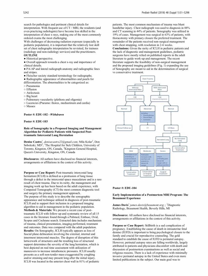
search for pathologies and pertinent clinical details for interpretation. With frequent use of CT / MRI, the residents (and even practicing radiologists) have become less skilled in the interpretation of chest x-rays, making one of the most commonly ordered exams the most challenging.
With challenges of decreasing radiation exposure (especially in pediatric population), it is important that the relatively lost skill set of chest radiographs interpretation be revisited, for trainees (radiology and non-radiology services) and the practitioners.
OUTLINE ● Historical perspective.
● Overall approach towards a chest x-ray and importance of clinical details. ● PA/AP and lateral radiograph anatomy and radiographic lines and stripes. ● Fleischer society standard terminology for radiographs. ● Radiographic appearance of abnormalities and pearls for differentiation. The abnormalities to be categorized as:
○ Pneumonia ○ Effusion ○ Atelectasis ○ Big heart ○ Pulmonary vascularity (plethora and oligemia) ○ Lucencies (Pneumo: thorax, mediastinum and cardia) ○ Masses
Poster #: EDU-102 - Withdrawn
Poster #: EDU-103
Role of Sonography in a Proposed Imaging and Management
Algorithm for Pediatric Patients with Suspected Post-
traumatic Intercostal Lung Herniation Denise Castro
1, [email protected]; Mila Kolar
2, Don
Soboleski, MD2;
1The Hospital for Sick Children, University of
Toronto, Kingston, ON, Canada, 2Kingston General Hospital,
Queen's University, Kingston, ON, Canada Disclosures: All authors have disclosed no financial interests, arrangements or affiliations in the context of this activity.
Purpose or Case Report: Post-traumatic intercostal lung herniation (ICLH) is defined as a protrusion of lung tissue through a defect in the intercostal space musculature and is a rare result of chest trauma. Due to its rarity, the management and imaging work up has been based on the adult experience, with Computed Tomography (CT) the most common diagnostic tool
and surgery the primary management approach. The purpose of this study is to describe the sonographic appearance and technique utilized in diagnosis of post-traumatic ICLH and to support their inclusion in a proposed imaging algorithm to aid in management in the pediatric population. Methods & Materials: We present a recent case of post-
traumatic ICLH with follow-up and systematic review of all 15 cases in the literature found through a Pubmed, Embase, Ovid, Scopus and Cochrane search. Extracted data includes mechanism of trauma, clinical presentation, imaging performed, treatment and outcomes. Data was compared with the adult population. Results: On Sonography, ICLH typically appears as loss of fascial plane delineation involving the external, internal and
innermost intercostal muscles. The degree of disruption of this latticework of structures and the resulting loss of structural support determines the severity of the lung herniation, which is best depicted on real time assessment with utilization of maneuvers to increase intrathoracic pressures. ICLH usually presents as a soft non-tender mass exaggerated by coughing and/or straining and may present long after the initial injury.
ICLH was located in the anterior chest wall in 81% of pediatric
patients. The most common mechanism of trauma was blunt handlebar injury. Chest radiograph was used to diagnosis in 88% and CT scanning in 44% of patients. Sonography was utilized in 19% of cases. Management was surgical in 63% of patients, with thoracotomy with primary closure the preferred treatment. The
remainder of the patients received non-surgical management with chest strapping, with resolution in 2-6 weeks. Conclusions: Given the rarity of ICLH in pediatric patients and the lack of diagnostic and management guidelines, pediatric surgeons have mostly relied on published reports in the adult literature to guide work-up and management. The recent
literature supports the feasibility of non-surgical management and the proposed imaging guidelines (Fig. 1) expanding the use of Sonography are meant to aid in the determination of surgical vs conservative treatment.
Poster #: EDU-104
Early Implementation of a Postmortem MRI Program: The
Beaumont Experience
James Davis
1 [email protected]; ;
1Diagnostic
Radiology, Beaumont Health, Beverly Hills, MI Disclosures: All authors have disclosed no financial interests, arrangements or affiliations in the context of this activity.
Purpose or Case Report: Stillbirth is a sad complication of pregnancy. Establishing the cause of death in intrauterine fetal demise (IUFD) is important to bring psychological closure to the family and crucial for reproductive counseling. The gold standard to establish the cause of IUFD is perinatal autopsy. However, perinatal autopsy rates are falling worldwide, largely attributed to patients and physicians discomfort with death and
discussion of postmortem examinations as well as social and religious reasons. There is a lack of experience with minimally invasive perinatal autopsy in the United States and even more limited publications in the subject. Our main goal was to
Pediatr Radiol (2018) 48 (Suppl 1):S1–S298S242
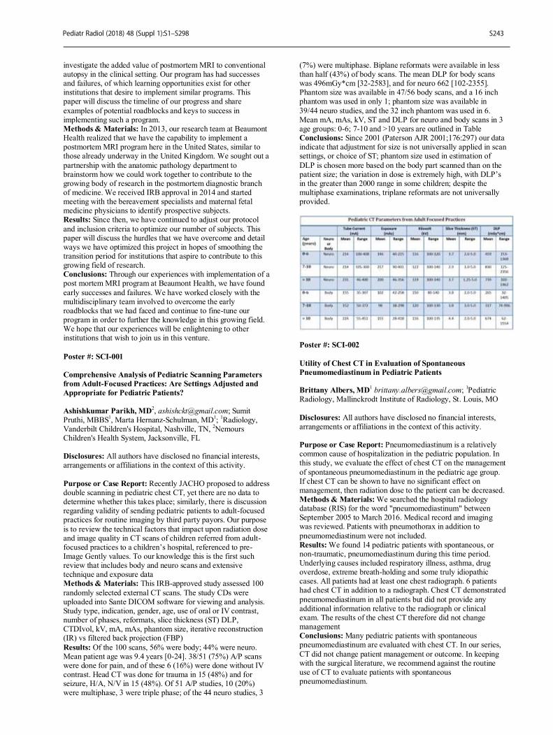
investigate the added value of postmortem MRI to conventional autopsy in the clinical setting. Our program has had successes and failures, of which learning opportunities exist for other institutions that desire to implement similar programs. This paper will discuss the timeline of our progress and share
examples of potential roadblocks and keys to success in implementing such a program. Methods & Materials: In 2013, our research team at Beaumont Health realized that we have the capability to implement a postmortem MRI program here in the United States, similar to those already underway in the United Kingdom. We sought out a
partnership with the anatomic pathology department to brainstorm how we could work together to contribute to the growing body of research in the postmortem diagnostic branch of medicine. We received IRB approval in 2014 and started meeting with the bereavement specialists and maternal fetal medicine physicians to identify prospective subjects. Results: Since then, we have continued to adjust our protocol
and inclusion criteria to optimize our number of subjects. This paper will discuss the hurdles that we have overcome and detail ways we have optimized this project in hopes of smoothing the transition period for institutions that aspire to contribute to this growing field of research. Conclusions: Through our experiences with implementation of a post mortem MRI program at Beaumont Health, we have found
early successes and failures. We have worked closely with the multidisciplinary team involved to overcome the early roadblocks that we had faced and continue to fine-tune our program in order to further the knowledge in this growing field. We hope that our experiences will be enlightening to other institutions that wish to join us in this venture.
Poster #: SCI-001
Comprehensive Analysis of Pediatric Scanning Parameters
from Adult-Focused Practices: Are Settings Adjusted and
Appropriate for Pediatric Patients?
Ashishkumar Parikh, MD2, [email protected]; Sumit
Pruthi, MBBS1, Marta Hernanz-Schulman, MD
1;
1Radiology,
Vanderbilt Children's Hospital, Nashville, TN, 2Nemours
Children's Health System, Jacksonville, FL Disclosures: All authors have disclosed no financial interests, arrangements or affiliations in the context of this activity.
Purpose or Case Report: Recently JACHO proposed to address double scanning in pediatric chest CT, yet there are no data to determine whether this takes place; similarly, there is discussion regarding validity of sending pediatric patients to adult-focused practices for routine imaging by third party payors. Our purpose
is to review the technical factors that impact upon radiation dose and image quality in CT scans of children referred from adult-focused practices to a children’s hospital, referenced to pre-Image Gently values. To our knowledge this is the first such review that includes body and neuro scans and extensive technique and exposure data Methods & Materials: This IRB-approved study assessed 100
randomly selected external CT scans. The study CDs were uploaded into Sante DICOM software for viewing and analysis. Study type, indication, gender, age, use of oral or IV contrast, number of phases, reformats, slice thickness (ST) DLP, CTDIvol, kV, mA, mAs, phantom size, iterative reconstruction (IR) vs filtered back projection (FBP)
Results: Of the 100 scans, 56% were body; 44% were neuro. Mean patient age was 9.4 years [0-24]. 38/51 (75%) A/P scans were done for pain, and of these 6 (16%) were done without IV contrast. Head CT was done for trauma in 15 (48%) and for seizure, H/A, N/V in 15 (48%). Of 51 A/P studies, 10 (20%) were multiphase, 3 were triple phase; of the 44 neuro studies, 3
(7%) were multiphase. Biplane reformats were available in less than half (43%) of body scans. The mean DLP for body scans was 496mGy*cm [32-2583], and for neuro 662 [102-2355]. Phantom size was available in 47/56 body scans, and a 16 inch phantom was used in only 1; phantom size was available in
39/44 neuro studies, and the 32 inch phantom was used in 6. Mean mA, mAs, kV, ST and DLP for neuro and body scans in 3 age groups: 0-6; 7-10 and >10 years are outlined in Table Conclusions: Since 2001 (Paterson AJR 2001;176:297) our data indicate that adjustment for size is not universally applied in scan settings, or choice of ST; phantom size used in estimation of
DLP is chosen more based on the body part scanned than on the patient size; the variation in dose is extremely high, with DLP’s in the greater than 2000 range in some children; despite the multiphase examinations, triplane reformats are not universally provided.
Poster #: SCI-002
Utility of Chest CT in Evaluation of Spontaneous
Pneumomediastinum in Pediatric Patients Brittany Albers, MD
1Pediatric
Radiology, Mallinckrodt Institute of Radiology, St. Louis, MO
Disclosures: All authors have disclosed no financial interests, arrangements or affiliations in the context of this activity. Purpose or Case Report: Pneumomediastinum is a relatively common cause of hospitalization in the pediatric population. In this study, we evaluate the effect of chest CT on the management
of spontaneous pneumomediastinum in the pediatric age group. If chest CT can be shown to have no significant effect on management, then radiation dose to the patient can be decreased. Methods & Materials: We searched the hospital radiology database (RIS) for the word "pneumomediastinum" between September 2005 to March 2016. Medical record and imaging was reviewed. Patients with pneumothorax in addition to
pneumomediastinum were not included. Results: We found 14 pediatric patients with spontaneous, or non-traumatic, pneumomediastinum during this time period. Underlying causes included respiratory illness, asthma, drug overdose, extreme breath-holding and some truly idiopathic cases. All patients had at least one chest radiograph. 6 patients had chest CT in addition to a radiograph. Chest CT demonstrated
pneumomediastinum in all patients but did not provide any additional information relative to the radiograph or clinical exam. The results of the chest CT therefore did not change management Conclusions: Many pediatric patients with spontaneous pneumomediastinum are evaluated with chest CT. In our series,
CT did not change patient management or outcome. In keeping with the surgical literature, we recommend against the routine use of CT to evaluate patients with spontaneous pneumomediastinum.
Pediatr Radiol (2018) 48 (Suppl 1):S1–S298 S243
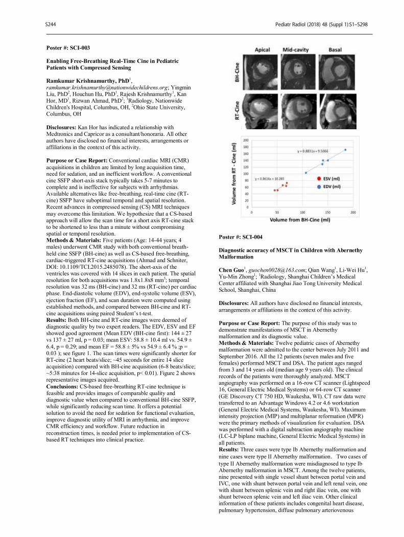
Poster #: SCI-003
Enabling Free-Breathing Real-Time Cine in Pediatric
Patients with Compressed Sensing
Ramkumar Krishnamurthy, PhD1,
[email protected]; Yingmin Liu, PhD
2, Houchun Hu, PhD
1, Rajesh Krishnamurthy
1, Kan
Hor, MD1, Rizwan Ahmad, PhD
2;
1Radiology, Nationwide
Children's Hospital, Columbus, OH, 2Ohio State University,
Columbus, OH
Disclosures: Kan Hor has indicated a relationship with Medtronics and Capricor as a consultant/honoraria. All other authors have disclosed no financial interests, arrangements or affiliations in the context of this activity. Purpose or Case Report: Conventional cardiac MRI (CMR)
acquisitions in children are limited by long acquisition time, need for sedation, and an inefficient workflow. A conventional cine SSFP short-axis stack typically takes 5-7 minutes to complete and is ineffective for subjects with arrhythmias. Available alternatives like free-breathing, real-time cine (RT-cine) SSFP have suboptimal temporal and spatial resolution. Recent advances in compressed sensing (CS) MRI techniques
may overcome this limitation. We hypothesize that a CS-based approach will allow the scan time for a short axis RT-cine stack to be shortened to less than a minute without compromising spatial or temporal resolution. Methods & Materials: Five patients (Age: 14-44 years; 4 males) underwent CMR study with both conventional breath-
held cine SSFP (BH-cine) as well as CS-based free-breathing, cardiac-triggered RT-cine acquisitions (Ahmad and Schniter, DOI: 10.1109/TCI.2015.2485078). The short-axis of the ventricles was covered with 14 slices in each patient. The spatial resolution for both acquisitions was 1.8x1.8x8 mm
3; temporal
resolution was 32 ms (BH-cine) and 32 ms (RT-cine) per cardiac phase. End-diastolic volume (EDV), end-systolic volume (ESV),
ejection fraction (EF), and scan duration were computed using established methods, and compared between BH-cine and RT-cine acquisitions using paired Student’s t-test. Results: Both BH-cine and RT-cine images were deemed of diagnostic quality by two expert readers. The EDV, ESV and EF showed good agreement (Mean EDV (BH-cine first): 144 ± 27 vs 137 ± 27 ml, p = 0.03; mean ESV: 58.8 ± 10.4 ml vs. 54.9 ±
6.4, p = 0.29; and mean EF = 58.8 ± 5% vs 54.9 ± 6.4 % ;p = 0.03 ); see figure 1. The scan times were significantly shorter for RT-cine (2 heart beats/slice; ~45 seconds for entire 14 slice acquisition) compared with BH-cine acquisition (6-8 beats/slice; ~5:38 minutes for 14-slice acquisition, p< 0.01). Figure 2 shows representative images acquired.
Conclusions: CS-based free-breathing RT-cine technique is feasible and provides images of comparable quality and diagnostic value when compared to conventional BH-cine SSFP, while significantly reducing scan time. It offers a potential solution to avoid the need for sedation for functional evaluation, improve diagnostic utility of MRI in arrhythmia, and improve CMR efficiency and workflow. Future reduction in
reconstruction times, is needed prior to implementation of CS-based RT techniques into clinical practice.
Poster #: SCI-004
Diagnostic accuracy of MSCT in Children with Abernethy
Malformation
Chen Guo
1, [email protected]; Qian Wang
1, Li-Wei Hu
1,
Yu-Min Zhong1;
1Radiology, Shanghai Children’s Medical
Center affiliated with Shanghai Jiao Tong University Medical School, Shanghai, China Disclosures: All authors have disclosed no financial interests,
arrangements or affiliations in the context of this activity. Purpose or Case Report: The purpose of this study was to demonstrate manifestations of MSCT in Abernethy malformation and its diagnostic value. Methods & Materials: Twelve pediatric cases of Abernethy malformation were admitted to the center between July 2011 and
September 2016. All the 12 patients (seven males and five females) performed MSCT and DSA. The patient ages ranged from 3 and 14 years old (median age 9 years old). The clinical records of the patients were thoroughly analyzed. MSCT angiography was performed on a 16-row CT scanner (Lightspeed 16, General Electric Medical Systems) or 64-row CT scanner
(GE Discovery CT 750 HD, Waukesha, WI). CT raw data were transferred to an Advantage Windows 4.2 or 4.6 workstation (General Electric Medical Systems, Waukesha, WI). Maximum intensity projection (MIP) and multiplanar reformation (MPR) were the primary methods of visualization for evaluation. DSA was performed with a digital subtraction angiography machine (LC-LP biplane machine, General Electric Medical Systems) in
all patients. Results: Three cases were type Ib Abernethy malformation and
nine cases were type II Abernethy malformation.Two cases of
type II Abernethy malformation were misdiagnosed to type Ib Abernethy malformation in MSCT. Among the twelve patients, nine presented with single vessel shunt between portal vein and IVC, one with shunt between portal vein and left renal vein, one with shunt between splenic vein and right iliac vein, one with shunt between splenic vein and left iliac vein. Other clinical information of these patients includes congenital heart disease,
pulmonary hypertension, diffuse pulmonary arteriovenous
Pediatr Radiol (2018) 48 (Suppl 1):S1–S298S244
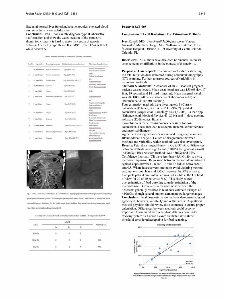
fistula, abnormal liver function, hepatic nodules, elevated blood ammonia, hepatic encephalopathy. Conclusions: MSCT can exactly diagnose type II Abernethy malformation and show the exact location of the portacaval shunt. Sometimes it is hard to make the certain diagnosis
between Abernethy type Ib and II in MSCT, then DSA will help while necessary.
Poster #: SCI-005
Comparison of Fetal Radiation Dose Estimation Methods Ivey Royall, MD
1, [email protected]; Vincent
Grekoski2, Matthew Hough, MS
1, William Sensakovic, PhD
1;
1Florida Hospital, Orlando, FL,
2University of Central Florida,
Orlando, FL Disclosures: All authors have disclosed no financial interests, arrangements or affiliations in the context of this activity.
Purpose or Case Report: To compare methods of estimating the fetal radiation dose delivered during computed tomography (CT) scanning. Further, to assess sources of variability in estimation methods. Methods & Materials: A database of 40 CT scans of pregnant patients was collected. Mean gestational age was 159±63 days (7
first, 19 second, and 14 third trimester). Mean maternal weight was 78±18kg. All patients underwent abdomen (n=10) or abdomen/pelvis (n=30) scanning. Four estimation methods were investigated: 1) Classic calculation (Felmlee, et al. AJR 154:1990), 2) updated calculation (Angel, et al. Radiology 249(1): 2008), 3) iPad app (Sahbaee, et al. Medical Physics 41: 2014), and 4) dose tracking
software (Radimetrics, Bayer). Two observers made measurements necessary for dose calculation. These included fetal depth, maternal circumference, and maternal diameter. Agreement among methods was assessed using regression and Bland-Altman analysis. Causes of disagreement between
methods and variability within methods was also investigated. Results: Fetal dose ranged from <1mGy to 32mGy. Differences between methods were significant (p<0.05), but generally small (<10mGy). Bias between methods was <5mGy and 95% Confidence Intervals (CI) were less than ±15mGy for pairwise method comparison. Regression between methods demonstrated typical slopes between 0.8 and 1.2 and R2 values between 0.3
and 0.8. When datasets were limited to avoid violating method assumptions both bias and 95%CI were cut by 50% or more. Complete patient circumference was not visible in the CT field of view for 30 of 40 patients (75%). This likely causes overestimation of fetal dose due to underestimation of the maternal size. Differences in measurement between the observers generally resulted in fetal dose estimate changes of
<10mGy, though several outliers demonstrated larger changes. Conclusions: Fetal dose estimation methods demonstrated good agreement; however, variability and outliers exist. A qualified medical physicist should review dose estimates to ensure proper calculation. Differences between methods could become important if combined with other dose data in a dose index
tracking system as it could elevate estimated dose above threshold considered acceptable for fetal scanning.
Pediatr Radiol (2018) 48 (Suppl 1):S1–S298 S245
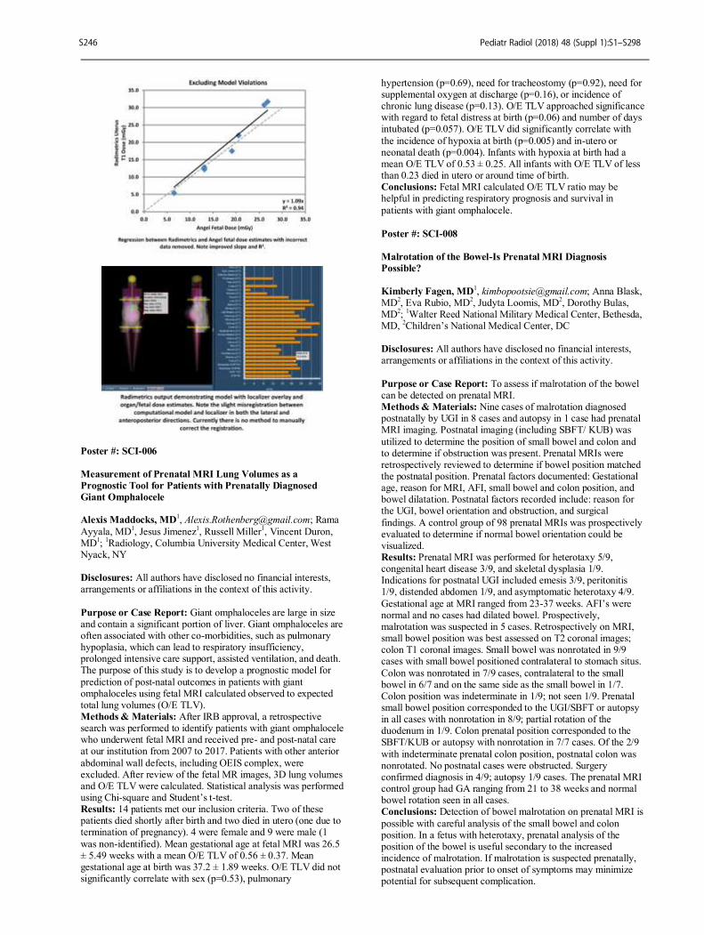
Poster #: SCI-006
Measurement of Prenatal MRI Lung Volumes as a
Prognostic Tool for Patients with Prenatally Diagnosed
Giant Omphalocele Alexis Maddocks, MD
1, [email protected]; Rama
Ayyala, MD1, Jesus Jimenez
1, Russell Miller
1, Vincent Duron,
MD1;
1Radiology, Columbia University Medical Center, West
Nyack, NY Disclosures: All authors have disclosed no financial interests, arrangements or affiliations in the context of this activity.
Purpose or Case Report: Giant omphaloceles are large in size and contain a significant portion of liver. Giant omphaloceles are often associated with other co-morbidities, such as pulmonary hypoplasia, which can lead to respiratory insufficiency, prolonged intensive care support, assisted ventilation, and death. The purpose of this study is to develop a prognostic model for
prediction of post-natal outcomes in patients with giant omphaloceles using fetal MRI calculated observed to expected total lung volumes (O/E TLV). Methods & Materials: After IRB approval, a retrospective search was performed to identify patients with giant omphalocele who underwent fetal MRI and received pre- and post-natal care at our institution from 2007 to 2017. Patients with other anterior
abdominal wall defects, including OEIS complex, were excluded. After review of the fetal MR images, 3D lung volumes and O/E TLV were calculated. Statistical analysis was performed using Chi-square and Student’s t-test. Results: 14 patients met our inclusion criteria. Two of these patients died shortly after birth and two died in utero (one due to termination of pregnancy). 4 were female and 9 were male (1
was non-identified). Mean gestational age at fetal MRI was 26.5 ± 5.49 weeks with a mean O/E TLV of 0.56 ± 0.37. Mean gestational age at birth was 37.2 ± 1.89 weeks. O/E TLV did not significantly correlate with sex (p=0.53), pulmonary
hypertension (p=0.69), need for tracheostomy (p=0.92), need for supplemental oxygen at discharge (p=0.16), or incidence of chronic lung disease (p=0.13). O/E TLV approached significance with regard to fetal distress at birth (p=0.06) and number of days intubated (p=0.057). O/E TLV did significantly correlate with
the incidence of hypoxia at birth (p=0.005) and in-utero or neonatal death (p=0.004). Infants with hypoxia at birth had a mean O/E TLV of 0.53 ± 0.25. All infants with O/E TLV of less than 0.23 died in utero or around time of birth. Conclusions: Fetal MRI calculated O/E TLV ratio may be helpful in predicting respiratory prognosis and survival in
patients with giant omphalocele.
Poster #: SCI-008
Malrotation of the Bowel-Is Prenatal MRI Diagnosis
Possible?
Kimberly Fagen, MD1, [email protected]; Anna Blask,
MD2, Eva Rubio, MD
2, Judyta Loomis, MD
2, Dorothy Bulas,
MD2;
1Walter Reed National Military Medical Center, Bethesda,
MD, 2Children’s National Medical Center, DC
Disclosures: All authors have disclosed no financial interests, arrangements or affiliations in the context of this activity.
Purpose or Case Report: To assess if malrotation of the bowel can be detected on prenatal MRI. Methods & Materials: Nine cases of malrotation diagnosed postnatally by UGI in 8 cases and autopsy in 1 case had prenatal MRI imaging. Postnatal imaging (including SBFT/ KUB) was
utilized to determine the position of small bowel and colon and to determine if obstruction was present. Prenatal MRIs were retrospectively reviewed to determine if bowel position matched the postnatal position. Prenatal factors documented: Gestational age, reason for MRI, AFI, small bowel and colon position, and bowel dilatation. Postnatal factors recorded include: reason for the UGI, bowel orientation and obstruction, and surgical
findings. A control group of 98 prenatal MRIs was prospectively evaluated to determine if normal bowel orientation could be visualized. Results: Prenatal MRI was performed for heterotaxy 5/9, congenital heart disease 3/9, and skeletal dysplasia 1/9. Indications for postnatal UGI included emesis 3/9, peritonitis 1/9, distended abdomen 1/9, and asymptomatic heterotaxy 4/9.
Gestational age at MRI ranged from 23-37 weeks. AFI’s were normal and no cases had dilated bowel. Prospectively, malrotation was suspected in 5 cases. Retrospectively on MRI, small bowel position was best assessed on T2 coronal images; colon T1 coronal images. Small bowel was nonrotated in 9/9 cases with small bowel positioned contralateral to stomach situs.
Colon was nonrotated in 7/9 cases, contralateral to the small bowel in 6/7 and on the same side as the small bowel in 1/7. Colon position was indeterminate in 1/9; not seen 1/9. Prenatal small bowel position corresponded to the UGI/SBFT or autopsy in all cases with nonrotation in 8/9; partial rotation of the duodenum in 1/9. Colon prenatal position corresponded to the SBFT/KUB or autopsy with nonrotation in 7/7 cases. Of the 2/9
with indeterminate prenatal colon position, postnatal colon was nonrotated. No postnatal cases were obstructed. Surgery confirmed diagnosis in 4/9; autopsy 1/9 cases. The prenatal MRI control group had GA ranging from 21 to 38 weeks and normal bowel rotation seen in all cases. Conclusions: Detection of bowel malrotation on prenatal MRI is
possible with careful analysis of the small bowel and colon position. In a fetus with heterotaxy, prenatal analysis of the position of the bowel is useful secondary to the increased incidence of malrotation. If malrotation is suspected prenatally, postnatal evaluation prior to onset of symptoms may minimize potential for subsequent complication.
Pediatr Radiol (2018) 48 (Suppl 1):S1–S298S246
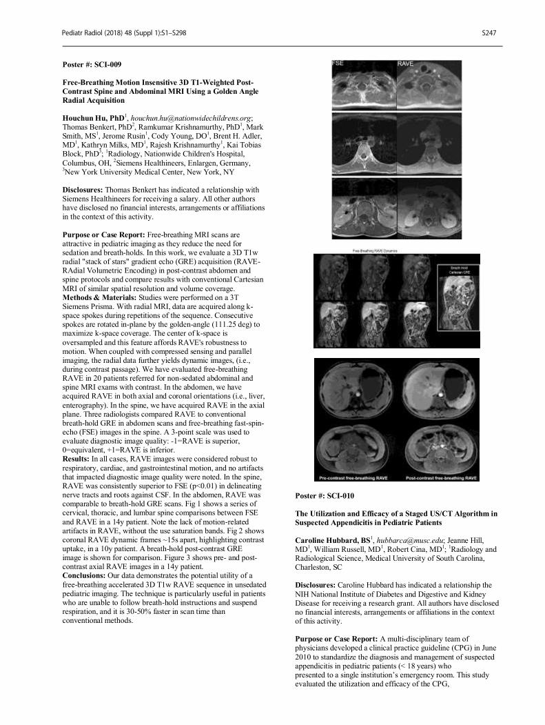
Poster #: SCI-009
Free-Breathing Motion Insensitive 3D T1-Weighted Post-
Contrast Spine and Abdominal MRI Using a Golden Angle
Radial Acquisition
Houchun Hu, PhD
Thomas Benkert, PhD2, Ramkumar Krishnamurthy, PhD
1, Mark
Smith, MS1, Jerome Rusin
1, Cody Young, DO
1, Brent H. Adler,
MD1, Kathryn Milks, MD
1, Rajesh Krishnamurthy
1, Kai Tobias
Block, PhD3;
1Radiology, Nationwide Children's Hospital,
Columbus, OH, 2Siemens Healthineers, Enlargen, Germany,
3New York University Medical Center, New York, NY
Disclosures: Thomas Benkert has indicated a relationship with Siemens Healthineers for receiving a salary. All other authors have disclosed no financial interests, arrangements or affiliations in the context of this activity.
Purpose or Case Report: Free-breathing MRI scans are attractive in pediatric imaging as they reduce the need for sedation and breath-holds. In this work, we evaluate a 3D T1w radial "stack of stars" gradient echo (GRE) acquisition (RAVE-RAdial Volumetric Encoding) in post-contrast abdomen and spine protocols and compare results with conventional Cartesian
MRI of similar spatial resolution and volume coverage. Methods & Materials: Studies were performed on a 3T Siemens Prisma. With radial MRI, data are acquired along k-space spokes during repetitions of the sequence. Consecutive spokes are rotated in-plane by the golden-angle (111.25 deg) to maximize k-space coverage. The center of k-space is
oversampled and this feature affords RAVE's robustness to motion. When coupled with compressed sensing and parallel imaging, the radial data further yields dynamic images, (i.e., during contrast passage). We have evaluated free-breathing RAVE in 20 patients referred for non-sedated abdominal and spine MRI exams with contrast. In the abdomen, we have acquired RAVE in both axial and coronal orientations (i.e., liver,
enterography). In the spine, we have acquired RAVE in the axial plane. Three radiologists compared RAVE to conventional breath-hold GRE in abdomen scans and free-breathing fast-spin-echo (FSE) images in the spine. A 3-point scale was used to evaluate diagnostic image quality: -1=RAVE is superior, 0=equivalent, +1=RAVE is inferior. Results: In all cases, RAVE images were considered robust to
respiratory, cardiac, and gastrointestinal motion, and no artifacts that impacted diagnostic image quality were noted. In the spine, RAVE was consistently superior to FSE (p<0.01) in delineating nerve tracts and roots against CSF. In the abdomen, RAVE was comparable to breath-hold GRE scans. Fig 1 shows a series of cervical, thoracic, and lumbar spine comparisons between FSE
and RAVE in a 14y patient. Note the lack of motion-related artifacts in RAVE, without the use saturation bands. Fig 2 shows coronal RAVE dynamic frames ~15s apart, highlighting contrast uptake, in a 10y patient. A breath-hold post-contrast GRE image is shown for comparison. Figure 3 shows pre- and post-contrast axial RAVE images in a 14y patient. Conclusions: Our data demonstrates the potential utility of a
free-breathing accelerated 3D T1w RAVE sequence in unsedated pediatric imaging. The technique is particularly useful in patients who are unable to follow breath-hold instructions and suspend respiration, and it is 30-50% faster in scan time than conventional methods.
Poster #: SCI-010
The Utilization and Efficacy of a Staged US/CT Algorithm in
Suspected Appendicitis in Pediatric Patients
Caroline Hubbard, BS
1, [email protected]; Jeanne Hill,
MD1, William Russell, MD
1, Robert Cina, MD
1;
1Radiology and
Radiological Science, Medical University of South Carolina, Charleston, SC
Disclosures: Caroline Hubbard has indicated a relationship the
NIH National Institute of Diabetes and Digestive and Kidney Disease for receiving a research grant. All authors have disclosed no financial interests, arrangements or affiliations in the context of this activity. Purpose or Case Report: A multi-disciplinary team of physicians developed a clinical practice guideline (CPG) in June
2010 to standardize the diagnosis and management of suspected appendicitis in pediatric patients (< 18 years) who presented to a single institution’s emergency room. This study evaluated the utilization and efficacy of the CPG,
Pediatr Radiol (2018) 48 (Suppl 1):S1–S298 S247
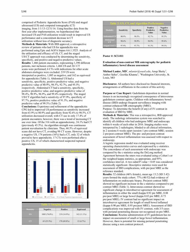
comprised of Pediatric Appendicitis Score (PAS) and staged ultrasound (US) and computed tomography (CT) imaging, from 1/1/13-12/31/16. Using baseline data from the first year after implementation, we hypothesized that increased US and PAS utilization would result in improved US
performance and a concomitant decrease in CT utilization without loss of diagnostic accuracy. Methods & Materials: With IRB approval, a retrospective review of patients who had US for appendicitis was performed using Epic and AGFA Impax 6.6.1.3525. Analysis of the utilization and efficacy of US, CT, and the staged
US/CT approach was conducted by determining the sensitivity, specificity, and positive and negative predictive values. Results: 1,466 patient encounters, representing 1,390 different patients, met inclusion criteria. 1,466 US and 261 CT scans were performed; 6 CTs with indications for other acute abdomen etiologies were excluded. 219 US were interpreted as positive, 1,085 as negative, and 162 as equivocal
for appendicitis (Table 1). Abdominal US had a sensitivity, specificity, positive predictive value, and negative predictive value of 80.9%, 98.5%, 92.7%, and 95.5% respectively. Abdominal CT had a sensitivity, specificity, positive predictive value, and negative predictive value of 98.4%, 98.9%, 96.9%, and 99.4% respectively. The staged US/CT algorithm had a sensitivity of 97.9%, specificity of
97.7%, positive predictive value of 91.3%, and negative predictive value of 99.5% (Table 2). Conclusions: Experience and refinement of the appendicitis CPG led to improved US performance as sensitivity increased from 63.9% to 80.9% and specificity from 92.6% to 98.5%. CT utilization decreased overall, with CT use in only 17.4% of
patient encounters; however, there was a trend of increasing CT use over time. Of the 316 with an appendectomy, 24.1% had CT (previously 45.5%). An algorithm strength was continuous patient reevaluation by clinicians. Thus, 60.5% of equivocal scans did not have CT, avoiding 98 CT scans. However, despite a negative US, 174 patients (16%) had a CT, only 32 of which proved to have appendicitis. 17 CTs were performed after a
positive US, 15 of which characterized suspected ruptured appendicitis.
Poster #: SCI-011
Evaluation of non-contrast MR enterography for pediatric
inflammatory bowel disease assessment Michael Lanier, MD
1, [email protected]; Anup Shetty
1,
Amber Salter1, Geetika Khanna
1;
1Washington University, St.
Louis, MO Disclosures: All authors have disclosed no financial interests, arrangements or affiliations in the context of this activity. Purpose or Case Report: Gadolinium deposition in normal
tissues is an increasingly recognized consequence of intravenous gadolinium contrast agents. Children with inflammatory bowel disease (IBD) undergo frequent surveillance imaging with contrast enhanced MR enterography (MRE). Purpose: To determine the benefit (if any) of IV contrast in evaluation of IBD by MRE.
Methods & Materials: This was a retrospective, IRB approved study. The radiology information system was searched to identify all children who had undergone MRE and endoscopy within 6 weeks of each other in 2016. Imaging studies were interpreted by 2 radiologists, blinded to all clinical information, in 2 sessions 6 weeks apart (session 1 pre-contrast MRE; session 2 pre/post contrast MRE). The pre- and pre/post contrast
assessment of bowel inflammation was assessed with respect to endoscopy. A logistic regression model was evaluated using receiver operating characteristics curves and expressed by c-statistics. The concordance of each assessment with endoscopy was compared by the c-statistics using the DeLong method. Agreement between the raters was evaluated using a Cohen’s or
the weighted kappa statistics, as appropriate, and 95% confidence interval. A two-sided P-value < 0.05 was considered statistically significant. Descriptive statistics were used for assessment of IBD complications, with pre/post MRE as the reference standard. Results: 52 children (46% female), mean age 13.2 (SD 3.42)
years formed the study cohort. 77% (40/52) had evidence of inflammation on endoscopic biopsy. Pre/post contrast MRE showed no significant increase in the c-statistics compared to pre contrast MRE (Table 1). Intravenous contrast showed no significant change in interobserver agreement for assessment of inflammation in either the small (kappa 0.92 pre MRE, 0.88 pre/post MRE) or large bowel (kappa 0.83 pre MRE, 0.73
pre/post MRE). IV contrast had no significant impact on interobserver agreement for length of small bowel inflamed (kappa 0.90 pre MRE, 0.95 pre/post MRE). Assessment of IBD complications was improved with IV contrast, with 3/5 cases with perianal penetrating disease not recognised on pre MRE. Conclusions: Routine administration of IV gadolinium has no
impact on assessment of small or large bowel inflammation. However, there is potential for missing perianal penetrating disease using a non contrast protocol.
Pediatr Radiol (2018) 48 (Suppl 1):S1–S298S248
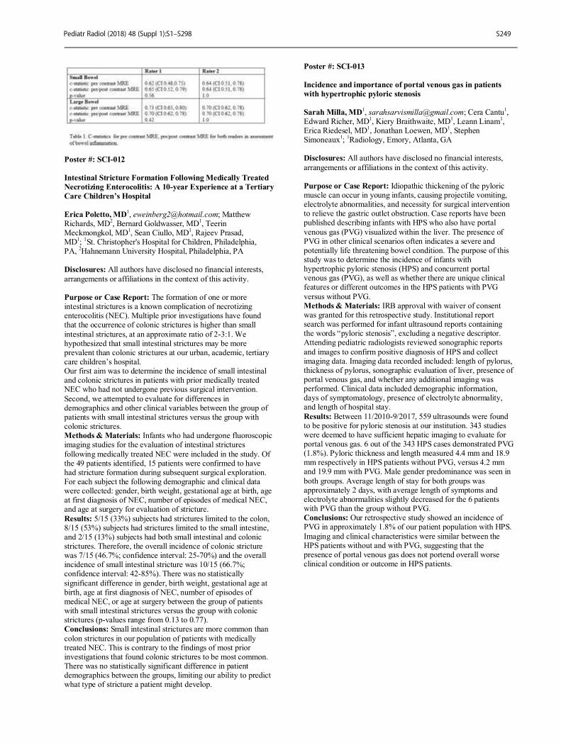
Poster #: SCI-012
Intestinal Stricture Formation Following Medically Treated
Necrotizing Enterocolitis: A 10-year Experience at a Tertiary
Care Children’s Hospital
Erica Poletto, MD1, [email protected]; Matthew
Richards, MD2, Bernard Goldwasser, MD
1, Teerin
Meckmongkol, MD1, Sean Ciullo, MD
1, Rajeev Prasad,
MD1;
1St. Christopher's Hospital for Children, Philadelphia,
PA, 2Hahnemann University Hospital, Philadelphia, PA
Disclosures: All authors have disclosed no financial interests,
arrangements or affiliations in the context of this activity. Purpose or Case Report: The formation of one or more intestinal strictures is a known complication of necrotizing enterocolitis (NEC). Multiple prior investigations have found that the occurrence of colonic strictures is higher than small
intestinal strictures, at an approximate ratio of 2-3:1. We hypothesized that small intestinal strictures may be more prevalent than colonic strictures at our urban, academic, tertiary care children’s hospital. Our first aim was to determine the incidence of small intestinal and colonic strictures in patients with prior medically treated NEC who had not undergone previous surgical intervention.
Second, we attempted to evaluate for differences in demographics and other clinical variables between the group of patients with small intestinal strictures versus the group with colonic strictures. Methods & Materials: Infants who had undergone fluoroscopic imaging studies for the evaluation of intestinal strictures
following medically treated NEC were included in the study. Of the 49 patients identified, 15 patients were confirmed to have had stricture formation during subsequent surgical exploration. For each subject the following demographic and clinical data were collected: gender, birth weight, gestational age at birth, age at first diagnosis of NEC, number of episodes of medical NEC, and age at surgery for evaluation of stricture.
Results: 5/15 (33%) subjects had strictures limited to the colon, 8/15 (53%) subjects had strictures limited to the small intestine, and 2/15 (13%) subjects had both small intestinal and colonic strictures. Therefore, the overall incidence of colonic stricture was 7/15 (46.7%; confidence interval: 25-70%) and the overall incidence of small intestinal stricture was 10/15 (66.7%; confidence interval: 42-85%). There was no statistically
significant difference in gender, birth weight, gestational age at birth, age at first diagnosis of NEC, number of episodes of medical NEC, or age at surgery between the group of patients with small intestinal strictures versus the group with colonic strictures (p-values range from 0.13 to 0.77). Conclusions: Small intestinal strictures are more common than
colon strictures in our population of patients with medically treated NEC. This is contrary to the findings of most prior investigations that found colonic strictures to be most common. There was no statistically significant difference in patient demographics between the groups, limiting our ability to predict what type of stricture a patient might develop.
Poster #: SCI-013
Incidence and importance of portal venous gas in patients
with hypertrophic pyloric stenosis
Sarah Milla, MD1, [email protected]; Cera Cantu
1,
Edward Richer, MD1, Kiery Braithwaite, MD
1, Leann Linam
1,
Erica Riedesel, MD1, Jonathan Loewen, MD
1, Stephen
Simoneaux1;
1Radiology, Emory, Atlanta, GA
Disclosures: All authors have disclosed no financial interests,
arrangements or affiliations in the context of this activity. Purpose or Case Report: Idiopathic thickening of the pyloric muscle can occur in young infants, causing projectile vomiting, electrolyte abnormalities, and necessity for surgical intervention to relieve the gastric outlet obstruction. Case reports have been published describing infants with HPS who also have portal
venous gas (PVG) visualized within the liver. The presence of PVG in other clinical scenarios often indicates a severe and potentially life threatening bowel condition. The purpose of this study was to determine the incidence of infants with hypertrophic pyloric stenosis (HPS) and concurrent portal venous gas (PVG), as well as whether there are unique clinical features or different outcomes in the HPS patients with PVG
versus without PVG. Methods & Materials: IRB approval with waiver of consent was granted for this retrospective study. Institutional report search was performed for infant ultrasound reports containing the words “pyloric stenosis”, excluding a negative descriptor. Attending pediatric radiologists reviewed sonographic reports
and images to confirm positive diagnosis of HPS and collect imaging data. Imaging data recorded included: length of pylorus, thickness of pylorus, sonographic evaluation of liver, presence of portal venous gas, and whether any additional imaging was performed. Clinical data included demographic information, days of symptomatology, presence of electrolyte abnormality, and length of hospital stay.
Results: Between 11/2010-9/2017, 559 ultrasounds were found to be positive for pyloric stenosis at our institution. 343 studies were deemed to have sufficient hepatic imaging to evaluate for portal venous gas. 6 out of the 343 HPS cases demonstrated PVG (1.8%). Pyloric thickness and length measured 4.4 mm and 18.9 mm respectively in HPS patients without PVG, versus 4.2 mm and 19.9 mm with PVG. Male gender predominance was seen in
both groups. Average length of stay for both groups was approximately 2 days, with average length of symptoms and electrolyte abnormalities slightly decreased for the 6 patients with PVG than the group without PVG. Conclusions: Our retrospective study showed an incidence of PVG in approximately 1.8% of our patient population with HPS.
Imaging and clinical characteristics were similar between the HPS patients without and with PVG, suggesting that the presence of portal venous gas does not portend overall worse clinical condition or outcome in HPS patients.
Pediatr Radiol (2018) 48 (Suppl 1):S1–S298 S249
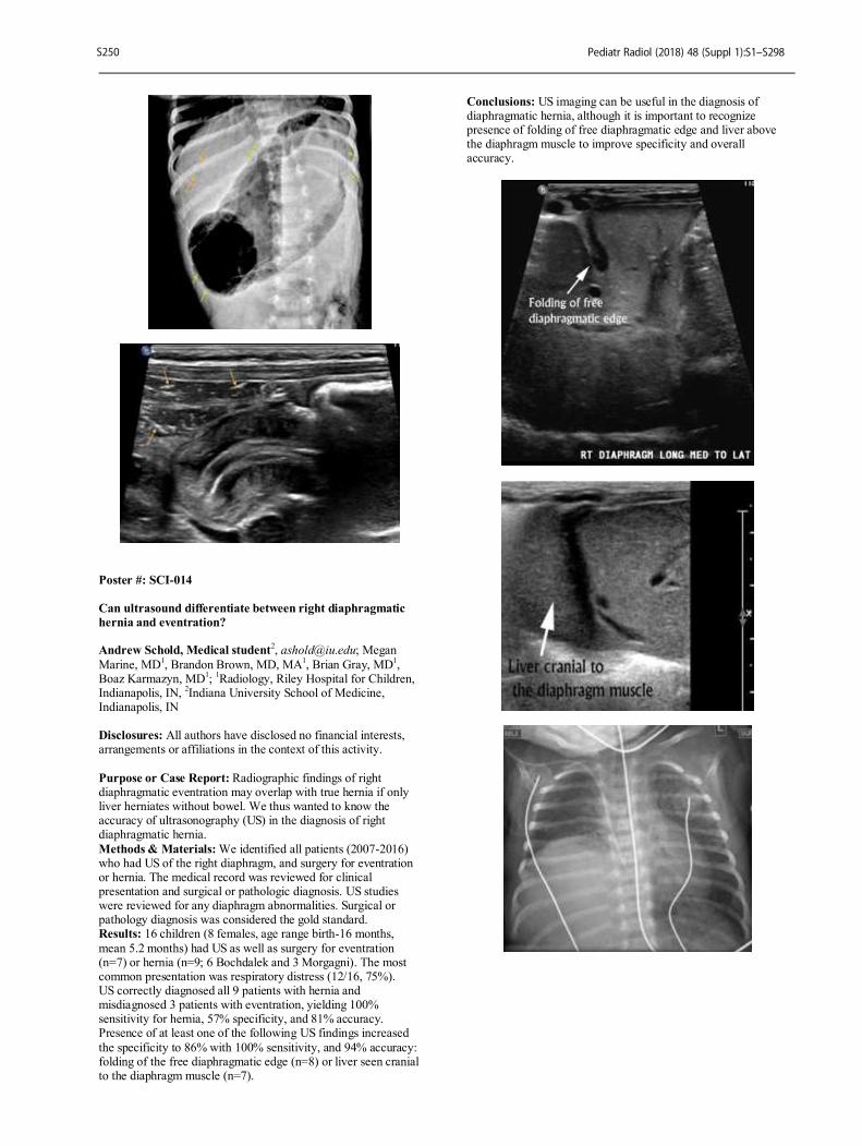
Poster #: SCI-014
Can ultrasound differentiate between right diaphragmatic
hernia and eventration? Andrew Schold, Medical student
2, [email protected]; Megan
Marine, MD1, Brandon Brown, MD, MA
1, Brian Gray, MD
1,
Boaz Karmazyn, MD1;
1Radiology, Riley Hospital for Children,
Indianapolis, IN, 2Indiana University School of Medicine,
Indianapolis, IN Disclosures: All authors have disclosed no financial interests, arrangements or affiliations in the context of this activity.
Purpose or Case Report: Radiographic findings of right diaphragmatic eventration may overlap with true hernia if only liver herniates without bowel. We thus wanted to know the accuracy of ultrasonography (US) in the diagnosis of right diaphragmatic hernia.
Methods & Materials: We identified all patients (2007-2016) who had US of the right diaphragm, and surgery for eventration or hernia. The medical record was reviewed for clinical presentation and surgical or pathologic diagnosis. US studies were reviewed for any diaphragm abnormalities. Surgical or pathology diagnosis was considered the gold standard. Results: 16 children (8 females, age range birth-16 months,
mean 5.2 months) had US as well as surgery for eventration (n=7) or hernia (n=9; 6 Bochdalek and 3 Morgagni). The most common presentation was respiratory distress (12/16, 75%). US correctly diagnosed all 9 patients with hernia and misdiagnosed 3 patients with eventration, yielding 100% sensitivity for hernia, 57% specificity, and 81% accuracy. Presence of at least one of the following US findings increased
the specificity to 86% with 100% sensitivity, and 94% accuracy: folding of the free diaphragmatic edge (n=8) or liver seen cranial to the diaphragm muscle (n=7).
Conclusions: US imaging can be useful in the diagnosis of diaphragmatic hernia, although it is important to recognize presence of folding of free diaphragmatic edge and liver above the diaphragm muscle to improve specificity and overall accuracy.
Pediatr Radiol (2018) 48 (Suppl 1):S1–S298S250
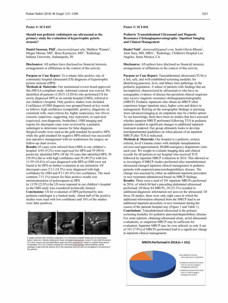
Poster #: SCI-015
Should non pediatric radiologists use ultrasound as the
primary study for evaluation of hypertrophic pyloric
stenosis?
Daniel Sassoon, PhD
1, [email protected]; Matthew Wanner
1,
Megan Marine, MD1, Boaz Karmazyn, MD
1;
1Radiology,
Indiana University, Indianapolis, IN
Disclosures: All authors have disclosed no financial interests,
arrangements or affiliations in the context of this activity. Purpose or Case Report: To evaluate false positive rate of community hospital ultrasound (US) diagnosis of hypertrophic pyloric stenosis (HPS) Methods & Materials: Our institutional review board approved this HIPAA-compliant study; informed consent was waived. We
identified all patients (1/2015-12/2016) who performed US for newly diagnosed HPS at an outside hospital (OSH), referred to our children’s hospital. Only positive studies were included. Confidence of OSH diagnosis was grouped based on key words as follows: high confidence (compatible, positive, diagnostic of, consistent with, meet criteria for, highly suspicious), low (favor, concern, suspicious, suggesting, may represent), or equivocal
(equivocal, non-diagnostic, borderline). OSH imaging and reports for discrepant cases were reviewed by a pediatric radiologist to determine reasons for false diagnosis. Surgical results were used as the gold standard for positive HPS, while the gold standard for negative HPS utilized was successful non operative management with no readmission for surgery on
follow-up chart review. Results: 65 cases were referred from OSHs to our children’s hospital. 6/65 (9.2%) were equivocal for HPS and 59 (90.8) positively identified HPS. Of the 59 cases that identified HPS, 30 (50.8%) did so with high confidence and 29 (49.2%) with low. 11/59 (18.6%) of cases diagnosed with HPS at OSH were not found to be HPS on further evaluation at our institution. Of
discrepant cases 2/11 (18.2%) were diagnosed with high confidence by OSH and 9/11 (81.8%) low confidence. The most common 7/11 (%) reason for false positive results was misinterpretation of pylorospasm as HPS. In 13/59 (22.0%) the US were repeated in our children’s hospital as the OSH study was considered technically limited. Conclusions: US for evaluation of HPS performed by non-
pediatric radiologist is a limited study. About half of the positive studies were read with low confidence and 18% of the studies were false positives.
Poster #: SCI-016
Pediatric Transabdominal Ultrasound and Magnetic
Resonance Cholangiopancreatography: Inpatient Imaging
and Clinical Management
Daniel Nahl
1, [email protected]; Justin Glavis-Bloom
1,
Amit Sura, MD, MBA1;
1Radiology, Children's Hospital Los
Angeles, Santa Monica, CA Disclosures: All authors have disclosed no financial interests,
arrangements or affiliations in the context of this activity. Purpose or Case Report: Transabdominal ultrasound (TUS) is a fast, safe, and well-established screening modality for identifying pancreas, liver, and biliary tract pathology in the pediatric population. A subset of patients with findings that are incompletely characterized by ultrasound or who have no
sonographic evidence of disease but persistent clinical suspicion may receive magnetic resonance cholangiopancreatography (MRCP). Pediatric inpatients who obtain an MRCP often experience longer inpatient stays, higher costs and delays in management. Relying on the sonographic findings or pursuing more advanced imaging as an outpatient may be a better option. To our knowledge, there have been no studies that have assessed
whether inpatient MRCP performed following TUS in pediatric patients resulted in significant changes to additional inpatient treatment rendered. Our group ultimately looks to develop interdepartmental guidelines on when pursuit of an inpatient MRCP after TUS is indicated. Methods & Materials: Our hospital is a pediatric, tertiary
referral, level I trauma center with multiple transplantation services and approximately 80,000 emergency department visits each year. We sought to evaluate imaging data and clinical records for all patients at our hospital who received TUS followed by inpatient MRCP evaluation in 2016. This allowed us to investigate if MRCP studies performed after transabdominal ultrasound changed inpatient clinical management in pediatric
patients with suspected pancreatohepatobiliary disease. The change was assessed by either an additional inpatient procedure or new treatment administered based on MRCP findings. Results: There were a total of 101 inpatient MRCPs performed in 2016, of which 84 had a preceding abdominal ultrasound performed. Of these 84 MRCPs, 28 (33.3%) resulted in additional diagnostic information not seen on the ultrasound. Of
these 28 studies, there were only eight cases in which the additional information obtained from the MRCP lead to an additional inpatient procedure or new treatment during the course of the patients hospital stay (Figure 1 and Table 1). Conclusions: Transabdominal ultrasound is the primary screening modality for pediatric pancreatohepatobiliary disease.
For some patients, obtaining ultrasound alone, serial ultrasound evaluations, or outpatient MRCP may be sufficient for evaluation. Inpatient MRCP may be over utilized, as only 8 out of 101 (7.9%) of MRCPs performed lead to a significant change in inpatient clinical management.
Pediatr Radiol (2018) 48 (Suppl 1):S1–S298 S251
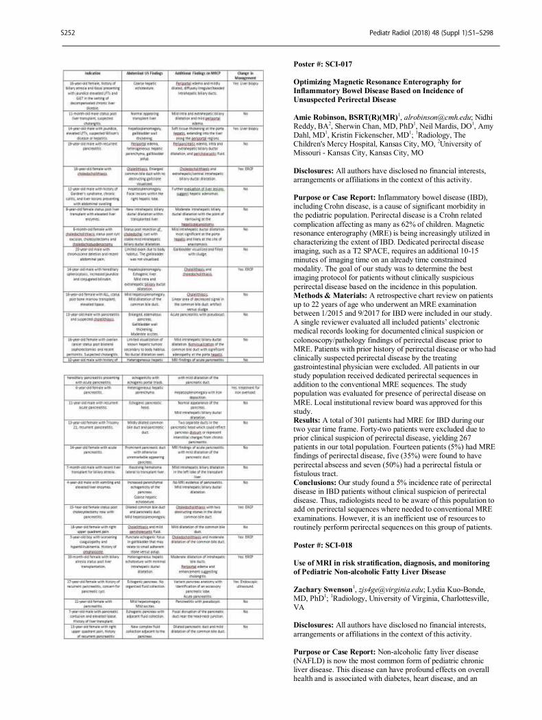
Poster #: SCI-017
Optimizing Magnetic Resonance Enterography for
Inflammatory Bowel Disease Based on Incidence of
Unsuspected Perirectal Disease
Amie Robinson, BSRT(R)(MR)
1, [email protected]; Nidhi
Reddy, BA2, Sherwin Chan, MD, PhD
1, Neil Mardis, DO
1, Amy
Dahl, MD1, Kristin Fickenscher, MD
1;
1Radiology, The
Children's Mercy Hospital, Kansas City, MO, 2University of
Missouri - Kansas City, Kansas City, MO
Disclosures: All authors have disclosed no financial interests, arrangements or affiliations in the context of this activity. Purpose or Case Report: Inflammatory bowel disease (IBD), including Crohn disease, is a cause of significant morbidity in the pediatric population. Perirectal disease is a Crohn related
complication affecting as many as 62% of children. Magnetic resonance enterography (MRE) is being increasingly utilized in characterizing the extent of IBD. Dedicated perirectal disease imaging, such as a T2 SPACE, requires an additional 10-15 minutes of imaging time on an already time constrained modality. The goal of our study was to determine the best imaging protocol for patients without clinically suspicious
perirectal disease based on the incidence in this population. Methods & Materials: A retrospective chart review on patients up to 22 years of age who underwent an MRE examination between 1/2015 and 9/2017 for IBD were included in our study. A single reviewer evaluated all included patients’ electronic medical records looking for documented clinical suspicion or
colonoscopy/pathology findings of perirectal disease prior to MRE. Patients with prior history of perirectal disease or who had clinically suspected perirectal disease by the treating gastrointestinal physician were excluded. All patients in our study population received dedicated perirectal sequences in addition to the conventional MRE sequences. The study population was evaluated for presence of perirectal disease on
MRE. Local institutional review board was approved for this study. Results: A total of 301 patients had MRE for IBD during our two year time frame. Forty-two patients were excluded due to prior clinical suspicion of perirectal disease, yielding 267 patients in our total population. Fourteen patients (5%) had MRE findings of perirectal disease, five (35%) were found to have
perirectal abscess and seven (50%) had a perirectal fistula or fistulous tract. Conclusions: Our study found a 5% incidence rate of perirectal disease in IBD patients without clinical suspicion of perirectal disease. Thus, radiologists need to be aware of this population to add on perirectal sequences where needed to conventional MRE
examinations. However, it is an inefficient use of resources to routinely perform perirectal sequences on this group of patients.
Poster #: SCI-018
Use of MRI in risk stratification, diagnosis, and monitoring
of Pediatric Non-alcoholic Fatty Liver Disease
Zachary Swenson
1, [email protected]; Lydia Kuo-Bonde,
MD, PhD1;
1Radiology, University of Virginia, Charlottesville,
VA Disclosures: All authors have disclosed no financial interests,
arrangements or affiliations in the context of this activity. Purpose or Case Report: Non-alcoholic fatty liver disease (NAFLD) is now the most common form of pediatric chronic liver disease. This disease can have profound effects on overall health and is associated with diabetes, heart disease, and an
Pediatr Radiol (2018) 48 (Suppl 1):S1–S298S252
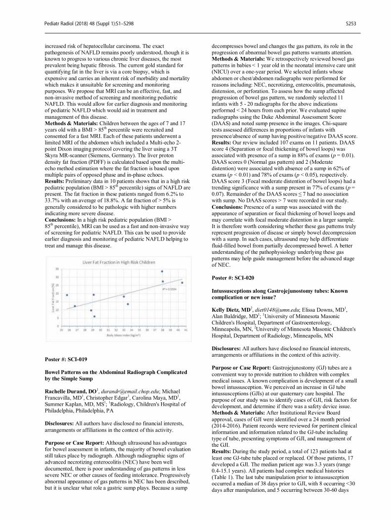
increased risk of hepatocellular carcinoma. The exact pathogenesis of NAFLD remains poorly understood, though it is known to progress to various chronic liver diseases, the most prevalent being hepatic fibrosis. The current gold standard for quantifying fat in the liver is via a core biopsy, which is
expensive and carries an inherent risk of morbidity and mortality which makes it unsuitable for screening and monitoring purposes. We propose that MRI can be an effective, fast, and non-invasive method of screening and monitoring pediatric NAFLD. This would allow for earlier diagnosis and monitoring of pediatric NAFLD which would aid in treatment and
management of this disease. Methods & Materials: Children between the ages of 7 and 17 years old with a BMI > 85
th percentile were recruited and
consented for a fast MRI. Each of these patients underwent a limited MRI of the abdomen which included a Multi-echo 2-point Dixon imaging protocol covering the liver using a 3T Skyra MR-scanner (Siemens, Germany). The liver proton
density fat fraction (PDFF) is calculated based upon the multi-echo method estimation where the fat fraction is based upon multiple pairs of opposed phase and in-phase echoes. Results: Preliminary data in 10 patients shows that in a high risk pediatric population (BMI > 85
th percentile) signs of NAFLD are
present. The fat fraction in these patients ranged from 6.2% to 33.7% with an average of 18.8%. A fat fraction of > 5% is
generally considered to be pathologic with higher numbers indicating more severe disease. Conclusions: In a high risk pediatric population (BMI > 85
th percentile), MRI can be used as a fast and non-invasive way
of screening for pediatric NAFLD. This can be used to provide earlier diagnosis and monitoring of pediatric NAFLD helping to
treat and manage this disease.
Poster #: SCI-019
Bowel Patterns on the Abdominal Radiograph Complicated
by the Simple Sump
Rachelle Durand, DO
1, [email protected]; Michael
Francavilla, MD1, Christopher Edgar
1, Carolina Maya, MD
1,
Summer Kaplan, MD, MS1;
1Radiology, Children's Hospital of
Philadelphia, Philadelphia, PA Disclosures: All authors have disclosed no financial interests,
arrangements or affiliations in the context of this activity. Purpose or Case Report: Although ultrasound has advantages for bowel assessment in infants, the majority of bowel evaluation still takes place by radiograph. Although radiographic signs of advanced necrotizing enterocolitis (NEC) have been well
documented, there is poor understanding of gas patterns in less severe NEC or other causes of feeding intolerance. Progressively abnormal appearance of gas patterns in NEC has been described, but it is unclear what role a gastric sump plays. Because a sump
decompresses bowel and changes the gas pattern, its role in the progression of abnormal bowel gas patterns warrants attention. Methods & Materials: We retrospectively reviewed bowel gas patterns in babies < 1 year old in the neonatal intensive care unit (NICU) over a one-year period. We selected infants whose
abdomen or chest/abdomen radiographs were performed for reasons including: NEC, necrotizing, enterocolitis, pneumatosis, distension, or perforation. To assess how the sump affected progression of bowel gas pattern, we randomly selected 11 infants with 5 - 20 radiographs for the above indications performed < 24 hours from each prior. We evaluated supine
radiographs using the Duke Abdominal Assessment Score (DAAS) and noted sump presence in the images. Chi-square tests assessed differences in proportions of infants with presence/absence of sump having positive/negative DAAS score. Results: Our review included 107 exams on 11 patients. DAAS score 4 (Separation or focal thickening of bowel loops) was associated with presence of a sump in 88% of exams (p = 0.01).
DAAS scores 0 (Normal gas pattern) and 2 (Moderate distention) were associated with absence of a sump in 62% of exams (p < 0.01) and 78% of exams (p < 0.05), respectively. DAAS score 3 (Focal moderate distention of bowel loops) had a trending significance with a sump present in 77% of exams (p = 0.07). Remainder of the DAAS scores ≤ 7 had no association with sump. No DAAS scores > 7 were recorded in our study.
Conclusions: Presence of a sump was associated with the appearance of separation or focal thickening of bowel loops and may correlate with focal moderate distention in a larger sample. It is therefore worth considering whether these gas patterns truly represent progression of disease or simply bowel decompression with a sump. In such cases, ultrasound may help differentiate
fluid-filled bowel from partially decompressed bowel. A better understanding of the pathophysiology underlying these gas patterns may help guide management before the advanced stage of NEC.
Poster #: SCI-020
Intussusceptions along Gastrojejunostomy tubes: Known
complication or new issue? Kelly Dietz, MD
2, [email protected]; Elissa Downs, MD
1,
Alan Baldridge, MD1;
1University of Minnesota Masonic
Children's Hospital, Department of Gastroenterology, Minneapolis, MN,
2University of Minnesota Masonic Children's
Hospital, Department of Radiology, Minneapolis, MN Disclosures: All authors have disclosed no financial interests, arrangements or affiliations in the context of this activity. Purpose or Case Report: Gastrojejunostomy (GJ) tubes are a
convenient way to provide nutrition to children with complex medical issues. A known complication is development of a small bowel intussusception. We perceived an increase in GJ tube intussusceptions (GJIs) at our quaternary care hospital. The purpose of our study was to identify cases of GJI, risk factors for development, and determine if there was a safety device issue. Methods & Materials: After Institutional Review Board
approval, cases of GJI were identified over a 24 month period (2014-2016). Patient records were reviewed for pertinent clinical information and information related to the GJ-tube including type of tube, presenting symptoms of GJI, and management of the GJI. Results: During the study period, a total of 123 patients had at
least one GJ-tube tube placed or replaced. Of those patients, 17 developed a GJI. The median patient age was 3.3 years (range 0.4-15.1 years). All patients had complex medical histories (Table 1). The last tube manipulation prior to intussusception occurred a median of 38 days prior to GJI, with 8 occurring <30 days after manipulation, and 5 occurring between 30-60 days
Pediatr Radiol (2018) 48 (Suppl 1):S1–S298 S253

after manipulation. Imaging was obtained after suspicious signs and symptoms (Table 2). Abdominal ultrasound was ordered and diagnosed 10 cases (56%), CT 6 cases, and MRI 2 cases. There was a wide variety of tubes seen in patients with GJI (Table 3). The initial management plan included GJ-tube removal in 11
patients, removal and replacement of a different tube in 3 patients, and monitoring in 4 patients, 2 of which were asymptomatic. A total of 9 patients either kept or required their GJ-tube be replaced, with replacement an average of 10.8 days after GJI. In 50% of patients, gastrostomy tube feeds were tolerated and replacement of the GJ-tube was not required.
Conclusions: Given intussusceptions were observed across various GJ tubes, a specific device safety issue was not identified. We found a 14% rate of intussusception in our complex patient population, which is similar to previously published GJI rates. An unexpected finding was that 50% of patients went on to tolerate gastrostomy tube feeds, not requiring replacement of their GJ tube. A positive outcome of our research
project has been an increased awareness of this complication throughout the hospital, and after education an increased utilization of abdominal ultrasound in patients with GJ-tubes and any suspicious symptoms.
Poster #: SCI-021 - Withdrawn
Poster #: SCI-022
The Correlation between Serial Ultrasound and Diuretic
Renography in Children with Unilateral High-Grade
Hydronephrosis Carl Flink, MD
1, [email protected]; Deborah Jacobson
2, Earl
Cheng, MD2, Emilie Johnson, MD, MPH
2, Elizabeth Yerkes,
MD2, Max Maizels
2, Bruce Lindgren
2, Dennis Liu
2, Ilina
Rosoklija, MPH2, Edward Gong
2;
1University of Cincinnati,
Cincinnati, OH, 2Lurie Children's Hospital/Northwestern
University, Chicago, IL Disclosures: All authors have disclosed no financial interests, arrangements or affiliations in the context of this activity. Purpose or Case Report: Serial renal ultrasound (RUS) is often used as a surrogate for functional renal imaging among patients
followed for hydronephrosis. However, it is unclear whether a lack of progression on serial RUS safeguards against loss of renal function. This study was performed to better characterize the association between findings on serial RUS and diuretic renography (DR) among children with unilateral high-grade hydronephrosis. Methods & Materials: We retrospectively reviewed imaging
among children <18 years with a history of unilateral Society of Fetal Urology (SFU) 3-4/Urinary Tract Dilation (UTD) P2-3 hydronephrosis. All patients underwent 2 RUS and 2 DR. Patients were excluded for comorbidities associated with bladder-level dysfunction, uninterpretable DR, or diuretic usage during only one RUS. Each RUS was paired with a
contemporaneous DR and compared with a subsequent study pair. All studies were reviewed by an independent, blinded diagnostic radiologist. Change in hydronephrosis was determined by either (1) change in SFU/UTD grade or (2) any change noted by the radiologist. This was compared with change in DR, with a ≥5% change considered significant. Chi-squared and Spearman’s correlation analyses were performed.
Results: 85 patients aged 0-14 years (mean 12 months) were included in the final analysis. Patients were predominantly male (73%) with SFU 3/UTD P2 hydronephrosis (85%). Time between DR ranged from 1-112 months (mean 20). Worsening hydronephrosis was noted in 10/85 patients (12%) by SFU/UTD grade and in 28/85 patients (33%) by radiologist interpretation. Differential renal function decreased by ≥5% in 15/85 patients
(18%). Changes in DR were significantly associated with changes in SFU/UTD grade on RUS among all patients (p=0.004, Table 1) and among patients with SFU 3/UTD P2 hydronephrosis (p=0.005, Table 2). Less substantial changes were not associated with DR findings (p=0.2). 10/70 (13%) patients with stable or improved SFU/UTD grade developed
worsening renal function during the study period. When RUS and DR were directly compared, the Spearman’s correlation was poor (r=0.2, CI [0.03-0.4]). Conclusions: While substantial changes in RUS findings were associated with corollary changes on DR, the overall correlation between imaging modalities was poor. 13% of children with SFU 3-4/UTD P2-3 hydronephrosis experienced a loss of renal
function despite stable longitudinal RUS imaging. These findings are important to consider when counseling conservatively managed patients followed without DR.
Pediatr Radiol (2018) 48 (Suppl 1):S1–S298S254
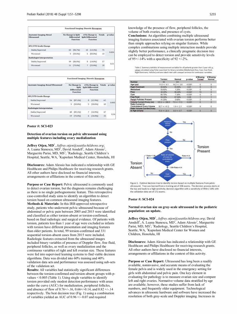
Poster #: SCI-023
Detection of ovarian torsion on pelvic ultrasound using
multiple features including ovary medialization Jeffrey Otjen, MD
A. Luana Stanescu, MD1, David Ansdell
2, Adam Alessio
1,
Marguerite Parisi, MD, MS1;
1Radiology, Seattle Children’s
Hospital, Seattle, WA, 2Kapiolani Medical Center, Honolulu, HI
Disclosures: Adam Alessio has indicated a relationship with GE Healthcare and Philips Healthcare for receiving research grants. All other authors have disclosed no financial interests, arrangements or affiliations in the context of this activity.
Purpose or Case Report: Pelvic ultrasound is commonly used to detect ovarian torsion, but the diagnosis remains challenging as there is no single pathognomonic feature. This retrospective case-controlled study aims to identify an algorithm to detect torsion based on common ultrasound imaging features. Methods & Materials: In this IRB approved retrospective
study, patients who underwent pelvic ultrasound because of abdominal or pelvic pain between 2005 and 2015 were identified and classified as either torsion-absent or torsion-confirmed, based on final radiologic and surgical evidence. Of patients with torsion, patients less than 1 year of age were excluded as infants with torsion have different presentation and imaging features than older patients. In total, 99 torsion-confirmed and 331
sequential torsion-absent cases from 2015 were included. Radiologic features extracted from the ultrasound images included binary variables of presence of Doppler flow, free fluid, peripheral follicles, as well as ovary medialization and the continuous variables of right and left ovarian size. These features were fed into supervised learning systems to find viable decision
algorithms. Data was divided into 60% training and 40% validation data sets and performance was assessed using sub-sets of the validation set. Results: All variables had statistically significant differences between the torsion-confirmed and torsion-absent groups with p-values < 0.005 (Table 1). Using single variables to identify torsion provided only modest detection performance with areas
under the curve (AUC) for medialization, peripheral follicles, and absence of flow of 0.76+/-.16, 0.66+/-0.14, and 0.82 +/-.14 respectively. The best decision tree (Fig. 1) using a combination of variables yielded an AUC of 0.96 +/- 0.07 and required
knowledge of the presence of flow, peripheral follicles, the volume of both ovaries, and presence of cysts. Conclusions: An algorithm combining multiple ultrasound imaging features associated with ovarian torsion performs better than simple approaches relying on singular features. While
complex combinations using multiple interaction models provide slightly better performance, a clinically pragmatic decision tree can be employed to detect torsion and provide sensitivity levels of 95+/-14% with a specificity of 92 +/-2%.
Poster #: SCI-024
Normal ovarian size on gray-scale ultrasound in the pediatric
population: an update. Jeffrey Otjen, MD
1, [email protected]; David
Ansdell2, A. Luana Stanescu, MD
1, Adam Alessio
1, Marguerite
Parisi, MD, MS1;
1Radiology, Seattle Children’s Hospital,
Seattle, WA, 2Kapiolani Medical Center for Women and
Children, Honolulu, HI Disclosures: Adam Alessio has indicated a relationship with GE Healthcare and Philips Healthcare for receiving research grants.
All other authors have disclosed no financial interests, arrangements or affiliations in the context of this activity. Purpose or Case Report: Ultrasound has long been a readily available, noninvasive, and accurate means of evaluating the female pelvis and is widely used in the emergency setting for
girls with abdominal and pelvic pain. One key element in evaluating for pathology is to measure ovarian size and compare left and right ovaries. Normative volume data stratified by age are available; however, these studies suffer from lack of numbers, and frequently older equipment. Technological advances in ultrasonic hardware and software have increased the resolution of both gray-scale and Doppler imaging. Increases in
Pediatr Radiol (2018) 48 (Suppl 1):S1–S298 S255
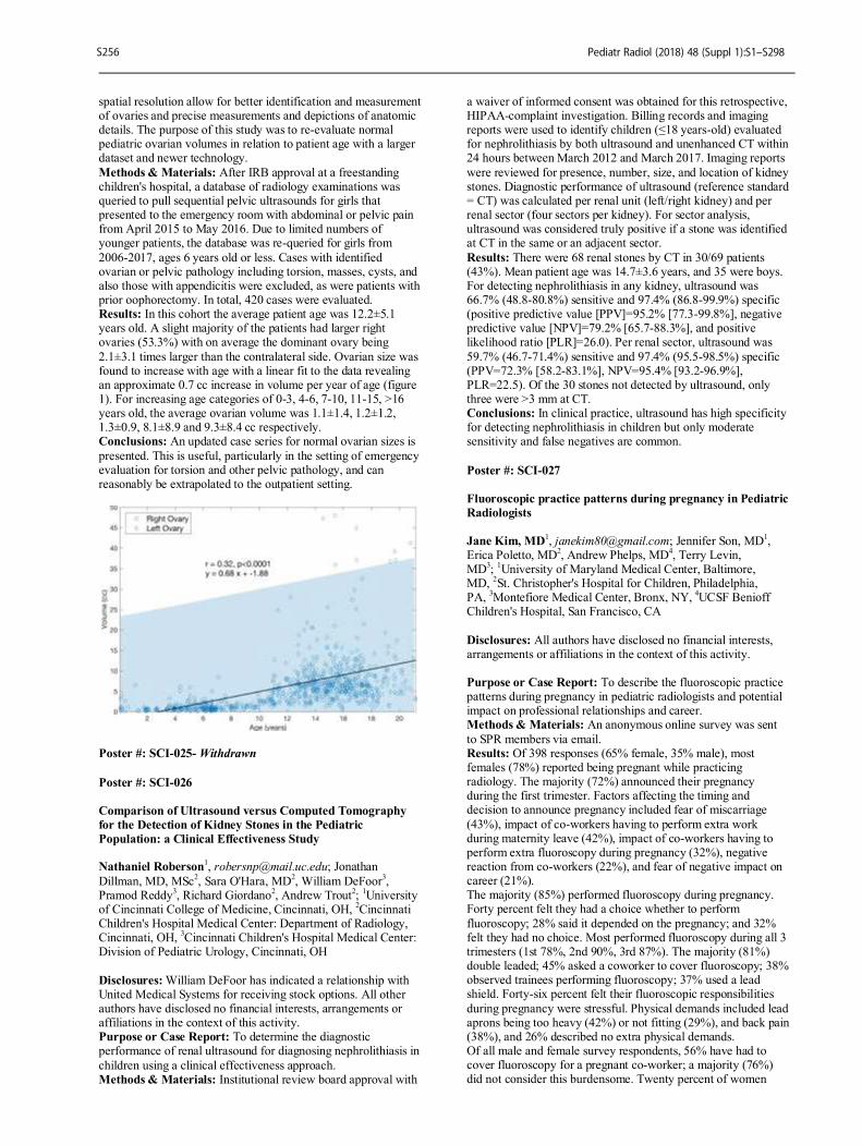
spatial resolution allow for better identification and measurement of ovaries and precise measurements and depictions of anatomic details. The purpose of this study was to re-evaluate normal pediatric ovarian volumes in relation to patient age with a larger dataset and newer technology.
Methods & Materials: After IRB approval at a freestanding children's hospital, a database of radiology examinations was queried to pull sequential pelvic ultrasounds for girls that presented to the emergency room with abdominal or pelvic pain from April 2015 to May 2016. Due to limited numbers of younger patients, the database was re-queried for girls from
2006-2017, ages 6 years old or less. Cases with identified ovarian or pelvic pathology including torsion, masses, cysts, and also those with appendicitis were excluded, as were patients with prior oophorectomy. In total, 420 cases were evaluated. Results: In this cohort the average patient age was 12.2±5.1 years old. A slight majority of the patients had larger right ovaries (53.3%) with on average the dominant ovary being
2.1±3.1 times larger than the contralateral side. Ovarian size was found to increase with age with a linear fit to the data revealing an approximate 0.7 cc increase in volume per year of age (figure 1). For increasing age categories of 0-3, 4-6, 7-10, 11-15, >16 years old, the average ovarian volume was 1.1±1.4, 1.2±1.2, 1.3±0.9, 8.1±8.9 and 9.3±8.4 cc respectively. Conclusions: An updated case series for normal ovarian sizes is
presented. This is useful, particularly in the setting of emergency evaluation for torsion and other pelvic pathology, and can reasonably be extrapolated to the outpatient setting.
Poster #: SCI-025- Withdrawn
Poster #: SCI-026
Comparison of Ultrasound versus Computed Tomography
for the Detection of Kidney Stones in the Pediatric
Population: a Clinical Effectiveness Study Nathaniel Roberson
1, [email protected]; Jonathan
Dillman, MD, MSc2, Sara O'Hara, MD
2, William DeFoor
3,
Pramod Reddy3, Richard Giordano
2, Andrew Trout
2;
1University
of Cincinnati College of Medicine, Cincinnati, OH, 2Cincinnati
Children's Hospital Medical Center: Department of Radiology, Cincinnati, OH,
3Cincinnati Children's Hospital Medical Center:
Division of Pediatric Urology, Cincinnati, OH
Disclosures: William DeFoor has indicated a relationship with United Medical Systems for receiving stock options. All other authors have disclosed no financial interests, arrangements or affiliations in the context of this activity. Purpose or Case Report: To determine the diagnostic performance of renal ultrasound for diagnosing nephrolithiasis in
children using a clinical effectiveness approach. Methods & Materials: Institutional review board approval with
a waiver of informed consent was obtained for this retrospective, HIPAA-complaint investigation. Billing records and imaging reports were used to identify children (≤18 years-old) evaluated for nephrolithiasis by both ultrasound and unenhanced CT within 24 hours between March 2012 and March 2017. Imaging reports
were reviewed for presence, number, size, and location of kidney stones. Diagnostic performance of ultrasound (reference standard = CT) was calculated per renal unit (left/right kidney) and per renal sector (four sectors per kidney). For sector analysis, ultrasound was considered truly positive if a stone was identified at CT in the same or an adjacent sector.
Results: There were 68 renal stones by CT in 30/69 patients (43%). Mean patient age was 14.7±3.6 years, and 35 were boys. For detecting nephrolithiasis in any kidney, ultrasound was 66.7% (48.8-80.8%) sensitive and 97.4% (86.8-99.9%) specific (positive predictive value [PPV]=95.2% [77.3-99.8%], negative predictive value [NPV]=79.2% [65.7-88.3%], and positive likelihood ratio [PLR]=26.0). Per renal sector, ultrasound was
59.7% (46.7-71.4%) sensitive and 97.4% (95.5-98.5%) specific (PPV=72.3% [58.2-83.1%], NPV=95.4% [93.2-96.9%], PLR=22.5). Of the 30 stones not detected by ultrasound, only three were >3 mm at CT. Conclusions: In clinical practice, ultrasound has high specificity for detecting nephrolithiasis in children but only moderate sensitivity and false negatives are common.
Poster #: SCI-027
Fluoroscopic practice patterns during pregnancy in Pediatric
Radiologists
Jane Kim, MD1, [email protected]; Jennifer Son, MD
1,
Erica Poletto, MD2, Andrew Phelps, MD
4, Terry Levin,
MD3;
1University of Maryland Medical Center, Baltimore,
MD, 2St. Christopher's Hospital for Children, Philadelphia,
PA, 3Montefiore Medical Center, Bronx, NY,
4UCSF Benioff
Children's Hospital, San Francisco, CA
Disclosures: All authors have disclosed no financial interests, arrangements or affiliations in the context of this activity. Purpose or Case Report: To describe the fluoroscopic practice patterns during pregnancy in pediatric radiologists and potential impact on professional relationships and career. Methods & Materials: An anonymous online survey was sent
to SPR members via email. Results: Of 398 responses (65% female, 35% male), most females (78%) reported being pregnant while practicing radiology. The majority (72%) announced their pregnancy during the first trimester. Factors affecting the timing and decision to announce pregnancy included fear of miscarriage
(43%), impact of co-workers having to perform extra work during maternity leave (42%), impact of co-workers having to perform extra fluoroscopy during pregnancy (32%), negative reaction from co-workers (22%), and fear of negative impact on career (21%). The majority (85%) performed fluoroscopy during pregnancy. Forty percent felt they had a choice whether to perform
fluoroscopy; 28% said it depended on the pregnancy; and 32% felt they had no choice. Most performed fluoroscopy during all 3 trimesters (1st 78%, 2nd 90%, 3rd 87%). The majority (81%) double leaded; 45% asked a coworker to cover fluoroscopy; 38% observed trainees performing fluoroscopy; 37% used a lead shield. Forty-six percent felt their fluoroscopic responsibilities
during pregnancy were stressful. Physical demands included lead aprons being too heavy (42%) or not fitting (29%), and back pain (38%), and 26% described no extra physical demands. Of all male and female survey respondents, 56% have had to cover fluoroscopy for a pregnant co-worker; a majority (76%) did not consider this burdensome. Twenty percent of women
Pediatr Radiol (2018) 48 (Suppl 1):S1–S298S256
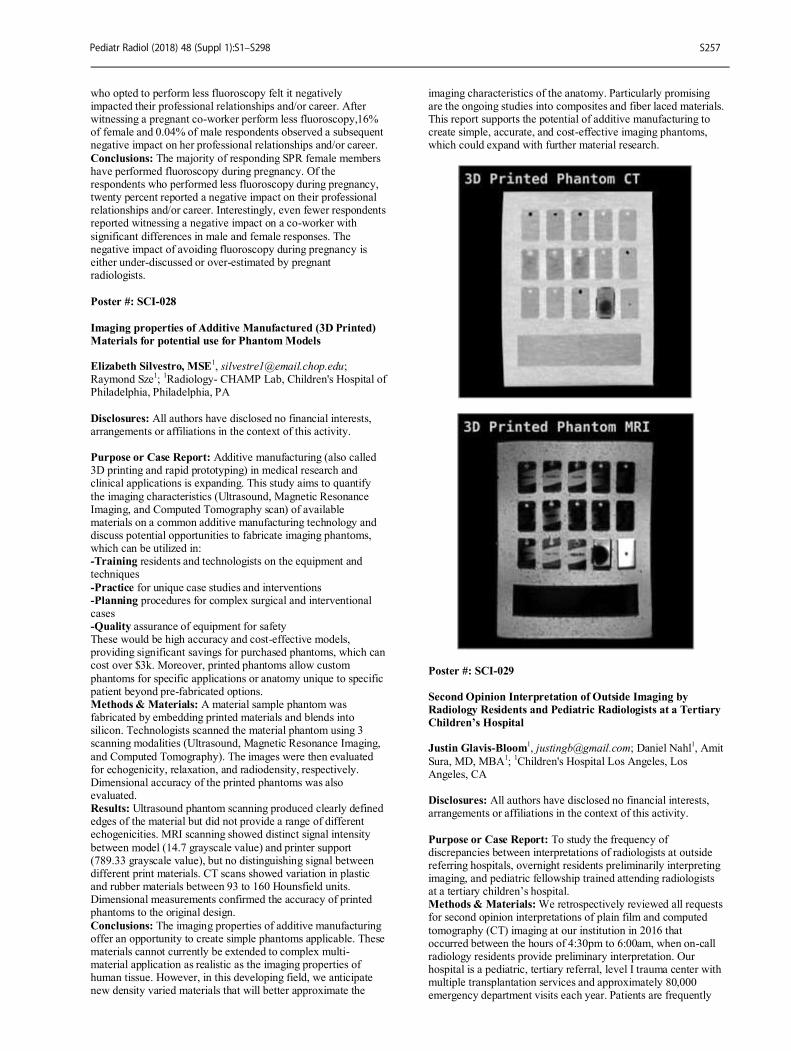
who opted to perform less fluoroscopy felt it negatively impacted their professional relationships and/or career. After witnessing a pregnant co-worker perform less fluoroscopy,16% of female and 0.04% of male respondents observed a subsequent negative impact on her professional relationships and/or career.
Conclusions: The majority of responding SPR female members have performed fluoroscopy during pregnancy. Of the respondents who performed less fluoroscopy during pregnancy, twenty percent reported a negative impact on their professional relationships and/or career. Interestingly, even fewer respondents reported witnessing a negative impact on a co-worker with
significant differences in male and female responses. The negative impact of avoiding fluoroscopy during pregnancy is either under-discussed or over-estimated by pregnant radiologists.
Poster #: SCI-028
Imaging properties of Additive Manufactured (3D Printed)
Materials for potential use for Phantom Models Elizabeth Silvestro, MSE
Raymond Sze1;
1Radiology- CHAMP Lab, Children's Hospital of
Philadelphia, Philadelphia, PA
Disclosures: All authors have disclosed no financial interests, arrangements or affiliations in the context of this activity. Purpose or Case Report: Additive manufacturing (also called 3D printing and rapid prototyping) in medical research and clinical applications is expanding. This study aims to quantify
the imaging characteristics (Ultrasound, Magnetic Resonance Imaging, and Computed Tomography scan) of available materials on a common additive manufacturing technology and discuss potential opportunities to fabricate imaging phantoms, which can be utilized in: -Training residents and technologists on the equipment and techniques
-Practice for unique case studies and interventions -Planning procedures for complex surgical and interventional cases -Quality assurance of equipment for safety These would be high accuracy and cost-effective models, providing significant savings for purchased phantoms, which can cost over $3k. Moreover, printed phantoms allow custom
phantoms for specific applications or anatomy unique to specific patient beyond pre-fabricated options. Methods & Materials: A material sample phantom was fabricated by embedding printed materials and blends into silicon. Technologists scanned the material phantom using 3 scanning modalities (Ultrasound, Magnetic Resonance Imaging,
and Computed Tomography). The images were then evaluated for echogenicity, relaxation, and radiodensity, respectively. Dimensional accuracy of the printed phantoms was also evaluated. Results: Ultrasound phantom scanning produced clearly defined edges of the material but did not provide a range of different echogenicities. MRI scanning showed distinct signal intensity
between model (14.7 grayscale value) and printer support (789.33 grayscale value), but no distinguishing signal between different print materials. CT scans showed variation in plastic and rubber materials between 93 to 160 Hounsfield units. Dimensional measurements confirmed the accuracy of printed phantoms to the original design.
Conclusions: The imaging properties of additive manufacturing offer an opportunity to create simple phantoms applicable. These materials cannot currently be extended to complex multi-material application as realistic as the imaging properties of human tissue. However, in this developing field, we anticipate new density varied materials that will better approximate the
imaging characteristics of the anatomy. Particularly promising are the ongoing studies into composites and fiber laced materials. This report supports the potential of additive manufacturing to create simple, accurate, and cost-effective imaging phantoms, which could expand with further material research.
Poster #: SCI-029
Second Opinion Interpretation of Outside Imaging by
Radiology Residents and Pediatric Radiologists at a Tertiary
Children’s Hospital Justin Glavis-Bloom
1, [email protected]; Daniel Nahl
1, Amit
Sura, MD, MBA1;
1Children's Hospital Los Angeles, Los
Angeles, CA Disclosures: All authors have disclosed no financial interests, arrangements or affiliations in the context of this activity.
Purpose or Case Report: To study the frequency of discrepancies between interpretations of radiologists at outside referring hospitals, overnight residents preliminarily interpreting imaging, and pediatric fellowship trained attending radiologists at a tertiary children’s hospital. Methods & Materials: We retrospectively reviewed all requests for second opinion interpretations of plain film and computed
tomography (CT) imaging at our institution in 2016 that occurred between the hours of 4:30pm to 6:00am, when on-call radiology residents provide preliminary interpretation. Our hospital is a pediatric, tertiary referral, level I trauma center with multiple transplantation services and approximately 80,000 emergency department visits each year. Patients are frequently
Pediatr Radiol (2018) 48 (Suppl 1):S1–S298 S257
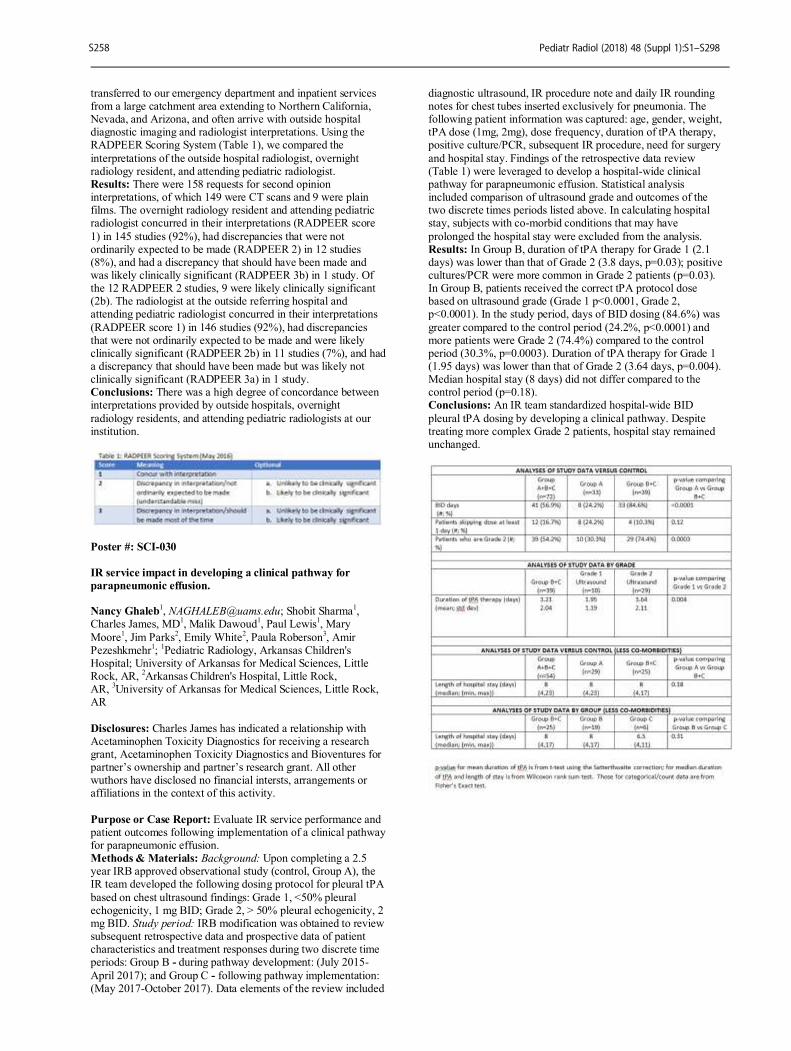
transferred to our emergency department and inpatient services from a large catchment area extending to Northern California, Nevada, and Arizona, and often arrive with outside hospital diagnostic imaging and radiologist interpretations. Using the RADPEER Scoring System (Table 1), we compared the
interpretations of the outside hospital radiologist, overnight radiology resident, and attending pediatric radiologist. Results: There were 158 requests for second opinion interpretations, of which 149 were CT scans and 9 were plain films. The overnight radiology resident and attending pediatric radiologist concurred in their interpretations (RADPEER score
1) in 145 studies (92%), had discrepancies that were not ordinarily expected to be made (RADPEER 2) in 12 studies (8%), and had a discrepancy that should have been made and was likely clinically significant (RADPEER 3b) in 1 study. Of the 12 RADPEER 2 studies, 9 were likely clinically significant (2b). The radiologist at the outside referring hospital and attending pediatric radiologist concurred in their interpretations
(RADPEER score 1) in 146 studies (92%), had discrepancies that were not ordinarily expected to be made and were likely clinically significant (RADPEER 2b) in 11 studies (7%), and had a discrepancy that should have been made but was likely not clinically significant (RADPEER 3a) in 1 study. Conclusions: There was a high degree of concordance between interpretations provided by outside hospitals, overnight
radiology residents, and attending pediatric radiologists at our institution.
Poster #: SCI-030
IR service impact in developing a clinical pathway for
parapneumonic effusion. Nancy Ghaleb
1, [email protected]; Shobit Sharma
1,
Charles James, MD1, Malik Dawoud
1, Paul Lewis
1, Mary
Moore1, Jim Parks
2, Emily White
2, Paula Roberson
3, Amir
Pezeshkmehr1;
1Pediatric Radiology, Arkansas Children's
Hospital; University of Arkansas for Medical Sciences, Little Rock, AR,
2Arkansas Children's Hospital, Little Rock,
AR, 3University of Arkansas for Medical Sciences, Little Rock,
AR
Disclosures: Charles James has indicated a relationship with Acetaminophen Toxicity Diagnostics for receiving a research grant, Acetaminophen Toxicity Diagnostics and Bioventures for partner’s ownership and partner’s research grant. All other wuthors have disclosed no financial intersts, arrangements or affiliations in the context of this activity.
Purpose or Case Report: Evaluate IR service performance and patient outcomes following implementation of a clinical pathway for parapneumonic effusion. Methods & Materials: Background: Upon completing a 2.5 year IRB approved observational study (control, Group A), the IR team developed the following dosing protocol for pleural tPA
based on chest ultrasound findings: Grade 1, <50% pleural echogenicity, 1 mg BID; Grade 2, > 50% pleural echogenicity, 2 mg BID. Study period: IRB modification was obtained to review subsequent retrospective data and prospective data of patient characteristics and treatment responses during two discrete time periods: Group B - during pathway development: (July 2015-
April 2017); and Group C - following pathway implementation: (May 2017-October 2017). Data elements of the review included
diagnostic ultrasound, IR procedure note and daily IR rounding notes for chest tubes inserted exclusively for pneumonia. The following patient information was captured: age, gender, weight, tPA dose (1mg, 2mg), dose frequency, duration of tPA therapy, positive culture/PCR, subsequent IR procedure, need for surgery
and hospital stay. Findings of the retrospective data review (Table 1) were leveraged to develop a hospital-wide clinical pathway for parapneumonic effusion. Statistical analysis included comparison of ultrasound grade and outcomes of the two discrete times periods listed above. In calculating hospital stay, subjects with co-morbid conditions that may have
prolonged the hospital stay were excluded from the analysis. Results: In Group B, duration of tPA therapy for Grade 1 (2.1 days) was lower than that of Grade 2 (3.8 days, p=0.03); positive cultures/PCR were more common in Grade 2 patients (p=0.03). In Group B, patients received the correct tPA protocol dose based on ultrasound grade (Grade 1 p<0.0001, Grade 2, p<0.0001). In the study period, days of BID dosing (84.6%) was
greater compared to the control period (24.2%, p<0.0001) and more patients were Grade 2 (74.4%) compared to the control period (30.3%, p=0.0003). Duration of tPA therapy for Grade 1 (1.95 days) was lower than that of Grade 2 (3.64 days, p=0.004). Median hospital stay (8 days) did not differ compared to the control period (p=0.18). Conclusions: An IR team standardized hospital-wide BID
pleural tPA dosing by developing a clinical pathway. Despite treating more complex Grade 2 patients, hospital stay remained unchanged.
Pediatr Radiol (2018) 48 (Suppl 1):S1–S298S258
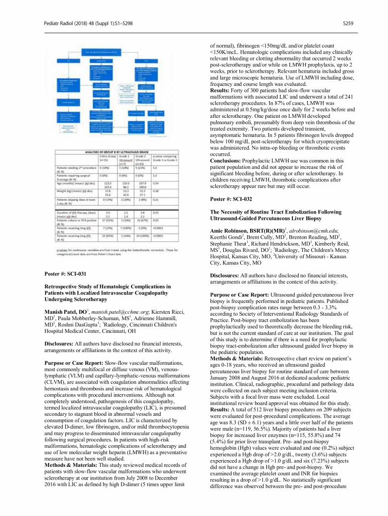
Poster #: SCI-031
Retrospective Study of Hematologic Complications in
Patients with Localized Intravascular Coagulopathy
Undergoing Sclerotherapy
Manish Patel, DO
1, [email protected]; Kiersten Ricci,
MD1, Paula Mobberley-Schuman, MS
1, Adrienne Hammill,
MD1, Roshni DasGupta
1;
1Radiology, Cincinnati Children's
Hospital Medical Center, Cincinnati, OH Disclosures: All authors have disclosed no financial interests,
arrangements or affiliations in the context of this activity. Purpose or Case Report: Slow-flow vascular malformations, most commonly multifocal or diffuse venous (VM), venous-lymphatic (VLM) and capillary-lymphatic-venous malformations (CLVM), are associated with coagulation abnormalities affecting
hemostasis and thrombosis and increase risk of hematological complications with procedural interventions. Although not completely understood, pathogenesis of this coagulopathy, termed localized intravascular coagulopathy (LIC), is presumed secondary to stagnant blood in abnormal vessels and consumption of coagulation factors. LIC is characterized by elevated D-dimer, low fibrinogen, and/or mild thrombocytopenia
and may progress to disseminated intravascular coagulopathy following surgical procedures. In patients with high-risk malformations, hematologic complications of sclerotherapy and use of low molecular weight heparin (LMWH) as a preventative measure have not been well studied. Methods & Materials: This study reviewed medical records of patients with slow-flow vascular malformations who underwent
sclerotherapy at our institution from July 2008 to December 2016 with LIC as defined by high D-dimer (5 times upper limit
of normal), fibrinogen <150mg/dL and/or platelet count <150K/mcL. Hematologic complications included any clinically relevant bleeding or clotting abnormality that occurred 2 weeks post-sclerotherapy and/or while on LMWH prophylaxis, up to 2 weeks, prior to sclerotherapy. Relevant hematuria included gross
and large microscopic hematuria. Use of LMWH including dose, frequency and course length was evaluated. Results: Forty of 300 patients had slow-flow vascular malformations with associated LIC and underwent a total of 241 sclerotherapy procedures. In 87% of cases, LMWH was administered at 0.5mg/kg/dose once daily for 2 weeks before and
after sclerotherapy. One patient on LMWH developed pulmonary emboli, presumably from deep vein thrombosis of the treated extremity. Two patients developed transient, asymptomatic hematuria. In 5 patients fibrinogen levels dropped below 100 mg/dL post-sclerotherapy for which cryoprecipitate was administered. No intra-op bleeding or thrombotic events occurred.
Conclusions: Prophylactic LMWH use was common in this patient population and did not appear to increase the risk of significant bleeding before, during or after sclerotherapy. In children receiving LMWH, thrombotic complications after sclerotherapy appear rare but may still occur.
Poster #: SCI-032
The Necessity of Routine Tract Embolization Following
Ultrasound-Guided Percutaneous Liver Biopsy Amie Robinson, BSRT(R)(MR)
Keerthi Gondi2, Brent Cully, MD
1, Brenton Reading, MD
1,
Stephanie Theut1, Richard Hendrickson, MD
1, Kimberly Reid,
MS1, Douglas Rivard, DO
1;
1Radiology, The Children's Mercy
Hospital, Kansas City, MO, 2University of Missouri - Kansas
City, Kansas City, MO Disclosures: All authors have disclosed no financial interests, arrangements or affiliations in the context of this activity.
Purpose or Case Report: Ultrasound guided percutaneous liver biopsy is frequently performed in pediatric patients. Published post-biopsy complication rates range between 0.3 - 3.3% according to Society of Interventional Radiology Standards of Practice. Post-biopsy tract embolization has been prophylactically used to theoretically decrease the bleeding risk,
but is not the current standard of care at our institution. The goal of this study is to determine if there is a need for prophylactic biopsy tract-embolization after ultrasound guided liver biopsy in the pediatric population. Methods & Materials: Retrospective chart review on patient’s ages 0-18 years, who received an ultrasound guided
percutaneous liver biopsy for routine standard of care between January 2008 and August 2016 at dedicated academic pediatric institution. Clinical, radiographic, procedural and pathology data were collected on each subject meeting inclusion criteria. Subjects with a focal liver mass were excluded. Local institutional review board approval was obtained for this study. Results: A total of 512 liver biopsy procedures on 209 subjects
were evaluated for post-procedural complications. The average age was 8.3 (SD ± 6.1) years and a little over half of the patients were male (n=119, 56.5%). Majority of patients had a liver biopsy for increased liver enzymes (n=115, 55.8%) and 74 (5.4%) for prior liver transplant. Pre- and post-biopsy hemoglobin (Hgb) values were evaluated and one (0.2%) subject
experienced a Hgb drop of >2.0 g/dL, twenty (3.6%) subjects experienced a Hgb drop of >1.0 g/dL and six (7.23%) subjects did not have a change in Hgb pre- and post-biopsy. We examined the average platelet count and INR for biopsies resulting in a drop of >1.0 g/dL. No statistically significant difference was observed between the pre- and post-procedure
Pediatr Radiol (2018) 48 (Suppl 1):S1–S298 S259
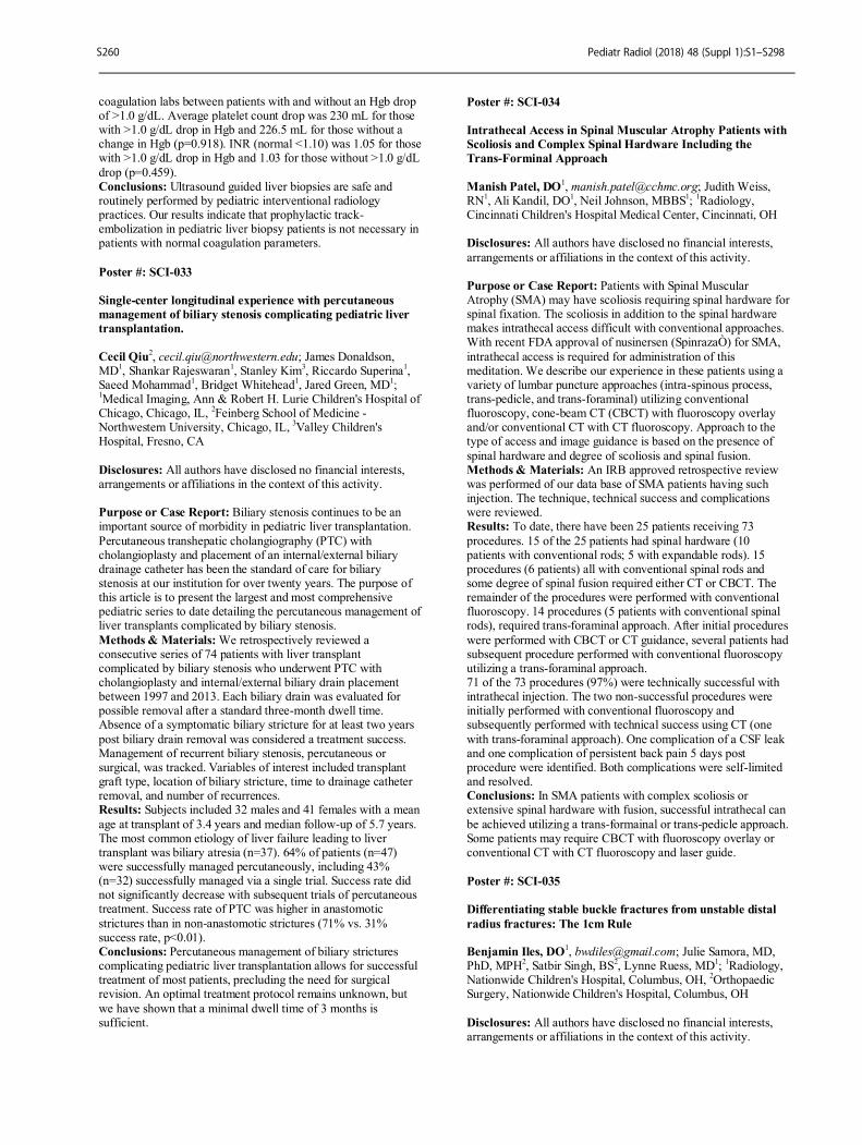
coagulation labs between patients with and without an Hgb drop of >1.0 g/dL. Average platelet count drop was 230 mL for those with >1.0 g/dL drop in Hgb and 226.5 mL for those without a change in Hgb (p=0.918). INR (normal <1.10) was 1.05 for those with >1.0 g/dL drop in Hgb and 1.03 for those without >1.0 g/dL
drop (p=0.459). Conclusions: Ultrasound guided liver biopsies are safe and routinely performed by pediatric interventional radiology practices. Our results indicate that prophylactic track-embolization in pediatric liver biopsy patients is not necessary in patients with normal coagulation parameters.
Poster #: SCI-033
Single-center longitudinal experience with percutaneous
management of biliary stenosis complicating pediatric liver
transplantation.
Cecil Qiu2, [email protected]; James Donaldson,
MD1, Shankar Rajeswaran
1, Stanley Kim
3, Riccardo Superina
1,
Saeed Mohammad1, Bridget Whitehead
1, Jared Green, MD
1;
1Medical Imaging, Ann & Robert H. Lurie Children's Hospital of
Chicago, Chicago, IL, 2Feinberg School of Medicine -
Northwestern University, Chicago, IL, 3Valley Children's
Hospital, Fresno, CA
Disclosures: All authors have disclosed no financial interests, arrangements or affiliations in the context of this activity. Purpose or Case Report: Biliary stenosis continues to be an important source of morbidity in pediatric liver transplantation.
Percutaneous transhepatic cholangiography (PTC) with cholangioplasty and placement of an internal/external biliary drainage catheter has been the standard of care for biliary stenosis at our institution for over twenty years. The purpose of this article is to present the largest and most comprehensive pediatric series to date detailing the percutaneous management of liver transplants complicated by biliary stenosis.
Methods & Materials: We retrospectively reviewed a consecutive series of 74 patients with liver transplant complicated by biliary stenosis who underwent PTC with cholangioplasty and internal/external biliary drain placement between 1997 and 2013. Each biliary drain was evaluated for possible removal after a standard three-month dwell time. Absence of a symptomatic biliary stricture for at least two years
post biliary drain removal was considered a treatment success. Management of recurrent biliary stenosis, percutaneous or surgical, was tracked. Variables of interest included transplant graft type, location of biliary stricture, time to drainage catheter removal, and number of recurrences. Results: Subjects included 32 males and 41 females with a mean
age at transplant of 3.4 years and median follow-up of 5.7 years. The most common etiology of liver failure leading to liver transplant was biliary atresia (n=37). 64% of patients (n=47) were successfully managed percutaneously, including 43% (n=32) successfully managed via a single trial. Success rate did not significantly decrease with subsequent trials of percutaneous treatment. Success rate of PTC was higher in anastomotic
strictures than in non-anastomotic strictures (71% vs. 31% success rate, p<0.01). Conclusions: Percutaneous management of biliary strictures complicating pediatric liver transplantation allows for successful treatment of most patients, precluding the need for surgical revision. An optimal treatment protocol remains unknown, but
we have shown that a minimal dwell time of 3 months is sufficient.
Poster #: SCI-034
Intrathecal Access in Spinal Muscular Atrophy Patients with
Scoliosis and Complex Spinal Hardware Including the
Trans-Forminal Approach
Manish Patel, DO
1, [email protected]; Judith Weiss,
RN1, Ali Kandil, DO
1, Neil Johnson, MBBS
1;
1Radiology,
Cincinnati Children's Hospital Medical Center, Cincinnati, OH Disclosures: All authors have disclosed no financial interests,
arrangements or affiliations in the context of this activity. Purpose or Case Report: Patients with Spinal Muscular Atrophy (SMA) may have scoliosis requiring spinal hardware for spinal fixation. The scoliosis in addition to the spinal hardware makes intrathecal access difficult with conventional approaches. With recent FDA approval of nusinersen (SpinrazaÒ) for SMA,
intrathecal access is required for administration of this meditation. We describe our experience in these patients using a variety of lumbar puncture approaches (intra-spinous process, trans-pedicle, and trans-foraminal) utilizing conventional fluoroscopy, cone-beam CT (CBCT) with fluoroscopy overlay and/or conventional CT with CT fluoroscopy. Approach to the type of access and image guidance is based on the presence of
spinal hardware and degree of scoliosis and spinal fusion. Methods & Materials: An IRB approved retrospective review was performed of our data base of SMA patients having such injection. The technique, technical success and complications were reviewed. Results: To date, there have been 25 patients receiving 73
procedures. 15 of the 25 patients had spinal hardware (10 patients with conventional rods; 5 with expandable rods). 15 procedures (6 patients) all with conventional spinal rods and some degree of spinal fusion required either CT or CBCT. The remainder of the procedures were performed with conventional fluoroscopy. 14 procedures (5 patients with conventional spinal rods), required trans-foraminal approach. After initial procedures
were performed with CBCT or CT guidance, several patients had subsequent procedure performed with conventional fluoroscopy utilizing a trans-foraminal approach. 71 of the 73 procedures (97%) were technically successful with intrathecal injection. The two non-successful procedures were initially performed with conventional fluoroscopy and subsequently performed with technical success using CT (one
with trans-foraminal approach). One complication of a CSF leak and one complication of persistent back pain 5 days post procedure were identified. Both complications were self-limited and resolved. Conclusions: In SMA patients with complex scoliosis or extensive spinal hardware with fusion, successful intrathecal can
be achieved utilizing a trans-formainal or trans-pedicle approach. Some patients may require CBCT with fluoroscopy overlay or conventional CT with CT fluoroscopy and laser guide.
Poster #: SCI-035
Differentiating stable buckle fractures from unstable distal
radius fractures: The 1cm Rule Benjamin Iles, DO
1, [email protected]; Julie Samora, MD,
PhD, MPH2, Satbir Singh, BS
2, Lynne Ruess, MD
1;
1Radiology,
Nationwide Children's Hospital, Columbus, OH, 2Orthopaedic
Surgery, Nationwide Children's Hospital, Columbus, OH
Disclosures: All authors have disclosed no financial interests, arrangements or affiliations in the context of this activity.
Pediatr Radiol (2018) 48 (Suppl 1):S1–S298S260
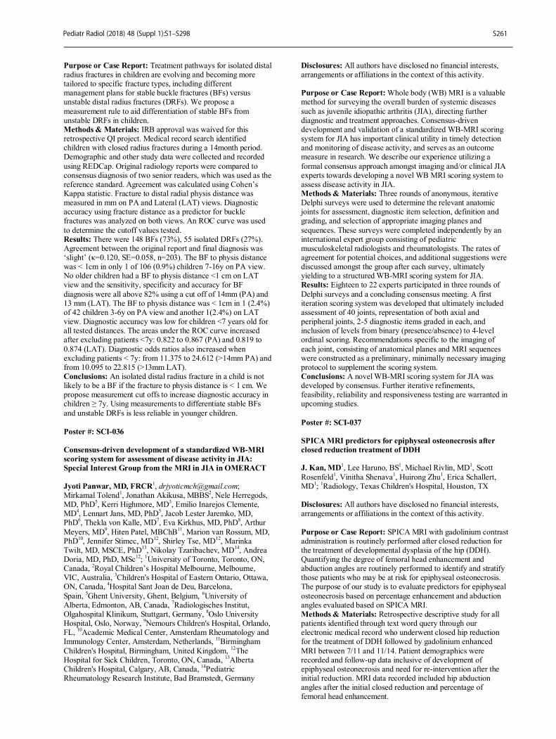
Purpose or Case Report: Treatment pathways for isolated distal radius fractures in children are evolving and becoming more tailored to specific fracture types, including different management plans for stable buckle fractures (BFs) versus unstable distal radius fractures (DRFs). We propose a
measurement rule to aid differentiation of stable BFs from unstable DRFs in children. Methods & Materials: IRB approval was waived for this retrospective QI project. Medical record search identified children with closed radius fractures during a 14month period. Demographic and other study data were collected and recorded
using REDCap. Original radiology reports were compared to consensus diagnosis of two senior readers, which was used as the reference standard. Agreement was calculated using Cohen’s Kappa statistic. Fracture to distal radial physis distance was measured in mm on PA and Lateral (LAT) views. Diagnostic accuracy using fracture distance as a predictor for buckle fractures was analyzed on both views. An ROC curve was used
to determine the cutoff values tested. Results: There were 148 BFs (73%), 55 isolated DRFs (27%). Agreement between the original report and final diagnosis was ‘slight’ (κ=0.120, SE=0.058, n=203). The BF to physis distance was < 1cm in only 1 of 106 (0.9%) children 7-16y on PA view. No older children had a BF to physis distance <1 cm on LAT view and the sensitivity, specificity and accuracy for BF
diagnosis were all above 82% using a cut off of 14mm (PA) and 13 mm (LAT). The BF to physis distance was < 1cm in 1 (2.4%) of 42 children 3-6y on PA view and another 1(2.4%) on LAT view. Diagnostic accuracy was low for children <7 years old for all tested distances. The areas under the ROC curve increased after excluding patients <7y: 0.822 to 0.867 (PA) and 0.819 to
0.874 (LAT). Diagnostic odds ratios also increased when excluding patients < 7y: from 11.375 to 24.612 (>14mm PA) and from 10.095 to 22.815 (>13mm LAT). Conclusions: An isolated distal radius fracture in a child is not likely to be a BF if the fracture to physis distance is < 1 cm. We propose measurement cut offs to increase diagnostic accuracy in children ≥ 7y. Using measurements to differentiate stable BFs
and unstable DRFs is less reliable in younger children.
Poster #: SCI-036
Consensus-driven development of a standardized WB-MRI
scoring system for assessment of disease activity in JIA:
Special Interest Group from the MRI in JIA in OMERACT
Jyoti Panwar, MD, FRCR
Mirkamal Tolend1, Jonathan Akikusa, MBBS
2, Nele Herregods,
MD, PhD5, Kerri Highmore, MD
3, Emilio Inarejos Clemente,
MD4, Lennart Jans, MD, PhD
5, Jacob Lester Jaremko, MD,
PhD6, Thekla von Kalle, MD
7, Eva Kirkhus, MD, PhD
8, Arthur
Meyers, MD9, Hiten Patel, MBChB
11, Marion van Rossum, MD,
PhD10
, Jennifer Stimec, MD12
, Shirley Tse, MD12
, Marinka Twilt, MD, MSCE, PhD
13, Nikolay Tzaribachev, MD
14, Andrea
Doria, MD, PhD, MSc12
; 1University of Toronto, Toronto, ON,
Canada, 2Royal Children’s Hospital Melbourne, Melbourne,
VIC, Australia, 3Children's Hospital of Eastern Ontario, Ottawa,
ON, Canada, 4Hospital Sant Joan de Deu, Barcelona,
Spain, 5Ghent University, Ghent, Belgium,
6University of
Alberta, Edmonton, AB, Canada, 7Radiologisches Institut,
Olgahospital Klinikum, Stuttgart, Germany, 8Oslo University
Hospital, Oslo, Norway, 9Nemours Children's Hospital, Orlando,
FL, 10
Academic Medical Center, Amsterdam Rheumatology and Immunology Center, Amsterdam, Netherlands,
11Birmingham
Children's Hospital, Birmingham, United Kingdom, 12
The Hospital for Sick Children, Toronto, ON, Canada,
13Alberta
Children's Hospital, Calgary, AB, Canada, 14
Pediatric Rheumatology Research Institute, Bad Bramstedt, Germany
Disclosures: All authors have disclosed no financial interests, arrangements or affiliations in the context of this activity. Purpose or Case Report: Whole body (WB) MRI is a valuable method for surveying the overall burden of systemic diseases
such as juvenile idiopathic arthritis (JIA), directing further diagnostic and treatment approaches. Consensus-driven development and validation of a standardized WB-MRI scoring system for JIA has important clinical utility in timely detection and monitoring of disease activity, and serves as an outcome measure in research. We describe our experience utilizing a
formal consensus approach amongst imaging and/or clinical JIA experts towards developing a novel WB MRI scoring system to assess disease activity in JIA. Methods & Materials: Three rounds of anonymous, iterative Delphi surveys were used to determine the relevant anatomic joints for assessment, diagnostic item selection, definition and grading, and selection of appropriate imaging planes and
sequences. These surveys were completed independently by an international expert group consisting of pediatric musculoskeletal radiologists and rheumatologists. The rates of agreement for potential choices, and additional suggestions were discussed amongst the group after each survey, ultimately yielding to a structured WB-MRI scoring system for JIA. Results: Eighteen to 22 experts participated in three rounds of
Delphi surveys and a concluding consensus meeting. A first iteration scoring system was developed that ultimately included assessment of 40 joints, representation of both axial and peripheral joints, 2-5 diagnostic items graded in each, and inclusion of levels from binary (presence/absence) to 4-level ordinal scoring. Recommendations specific to the imaging of
each joint, consisting of anatomical planes and MRI sequences were constructed as a preliminary, minimally necessary imaging protocol to supplement the scoring system. Conclusions: A novel WB-MRI scoring system for JIA was developed by consensus. Further iterative refinements, feasibility, reliability and responsiveness testing are warranted in upcoming studies.
Poster #: SCI-037
SPICA MRI predictors for epiphyseal osteonecrosis after
closed reduction treatment of DDH J. Kan, MD
1, Lee Haruno, BS
1, Michael Rivlin, MD
1, Scott
Rosenfeld1, Vinitha Shenava
1, Huirong Zhu
1, Erica Schallert,
MD1;
1Radiology, Texas Children's Hospital, Houston, TX
Disclosures: All authors have disclosed no financial interests, arrangements or affiliations in the context of this activity.
Purpose or Case Report: SPICA MRI with gadolinium contrast administration is routinely performed after closed reduction for the treatment of developmental dysplasia of the hip (DDH). Quantifying the degree of femoral head enhancement and abduction angles are routinely performed to identify and stratify those patients who may be at risk for epiphyseal osteonecrosis. The purpose of our study is to evaluate predictors for epiphyseal
osteonecrosis based on percentage enhancement and abduction angles evaluated based on SPICA MRI. Methods & Materials: Retrospective descriptive study for all patients identified through text word query through our electronic medical record who underwent closed hip reduction for the treatment of DDH followed by gadolinium enhanced
MRI between 7/11 and 11/14. Patient demographics were recorded and follow-up data inclusive of development of epiphyseal osteonecrosis and need for re-intervention after the initial reduction. MRI data recorded included hip abduction angles after the initial closed reduction and percentage of femoral head enhancement.
Pediatr Radiol (2018) 48 (Suppl 1):S1–S298 S261
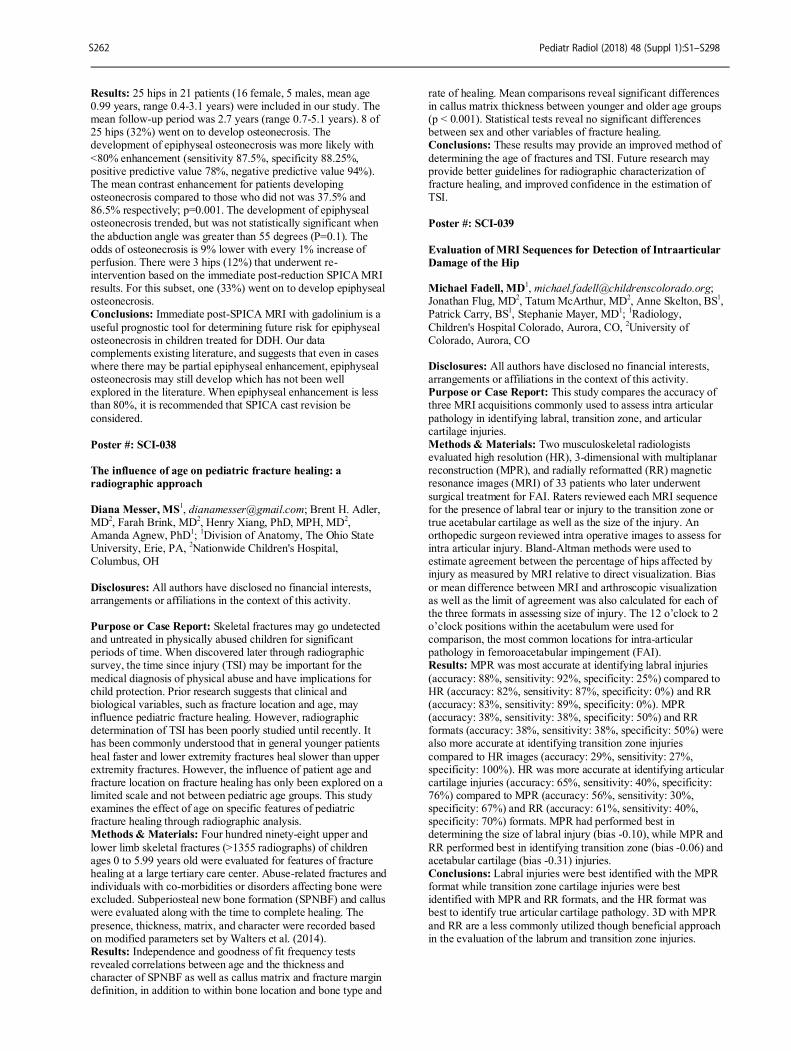
Results: 25 hips in 21 patients (16 female, 5 males, mean age 0.99 years, range 0.4-3.1 years) were included in our study. The mean follow-up period was 2.7 years (range 0.7-5.1 years). 8 of 25 hips (32%) went on to develop osteonecrosis. The development of epiphyseal osteonecrosis was more likely with
<80% enhancement (sensitivity 87.5%, specificity 88.25%, positive predictive value 78%, negative predictive value 94%). The mean contrast enhancement for patients developing osteonecrosis compared to those who did not was 37.5% and 86.5% respectively; p=0.001. The development of epiphyseal osteonecrosis trended, but was not statistically significant when
the abduction angle was greater than 55 degrees (P=0.1). The odds of osteonecrosis is 9% lower with every 1% increase of perfusion. There were 3 hips (12%) that underwent re-intervention based on the immediate post-reduction SPICA MRI results. For this subset, one (33%) went on to develop epiphyseal osteonecrosis. Conclusions: Immediate post-SPICA MRI with gadolinium is a
useful prognostic tool for determining future risk for epiphyseal osteonecrosis in children treated for DDH. Our data complements existing literature, and suggests that even in cases where there may be partial epiphyseal enhancement, epiphyseal osteonecrosis may still develop which has not been well explored in the literature. When epiphyseal enhancement is less than 80%, it is recommended that SPICA cast revision be
considered.
Poster #: SCI-038
The influence of age on pediatric fracture healing: a
radiographic approach
Diana Messer, MS
1, [email protected]; Brent H. Adler,
MD2, Farah Brink, MD
2, Henry Xiang, PhD, MPH, MD
2,
Amanda Agnew, PhD1;
1Division of Anatomy, The Ohio State
University, Erie, PA, 2Nationwide Children's Hospital,
Columbus, OH
Disclosures: All authors have disclosed no financial interests, arrangements or affiliations in the context of this activity. Purpose or Case Report: Skeletal fractures may go undetected and untreated in physically abused children for significant periods of time. When discovered later through radiographic survey, the time since injury (TSI) may be important for the
medical diagnosis of physical abuse and have implications for child protection. Prior research suggests that clinical and biological variables, such as fracture location and age, may influence pediatric fracture healing. However, radiographic determination of TSI has been poorly studied until recently. It has been commonly understood that in general younger patients
heal faster and lower extremity fractures heal slower than upper extremity fractures. However, the influence of patient age and fracture location on fracture healing has only been explored on a limited scale and not between pediatric age groups. This study examines the effect of age on specific features of pediatric fracture healing through radiographic analysis. Methods & Materials: Four hundred ninety-eight upper and
lower limb skeletal fractures (>1355 radiographs) of children ages 0 to 5.99 years old were evaluated for features of fracture healing at a large tertiary care center. Abuse-related fractures and individuals with co-morbidities or disorders affecting bone were excluded. Subperiosteal new bone formation (SPNBF) and callus were evaluated along with the time to complete healing. The
presence, thickness, matrix, and character were recorded based on modified parameters set by Walters et al. (2014). Results: Independence and goodness of fit frequency tests revealed correlations between age and the thickness and character of SPNBF as well as callus matrix and fracture margin definition, in addition to within bone location and bone type and
rate of healing. Mean comparisons reveal significant differences in callus matrix thickness between younger and older age groups (p < 0.001). Statistical tests reveal no significant differences between sex and other variables of fracture healing. Conclusions: These results may provide an improved method of
determining the age of fractures and TSI. Future research may provide better guidelines for radiographic characterization of fracture healing, and improved confidence in the estimation of TSI.
Poster #: SCI-039
Evaluation of MRI Sequences for Detection of Intraarticular
Damage of the Hip Michael Fadell, MD
Jonathan Flug, MD2, Tatum McArthur, MD
2, Anne Skelton, BS
1,
Patrick Carry, BS1, Stephanie Mayer, MD
1;
1Radiology,
Children's Hospital Colorado, Aurora, CO, 2University of
Colorado, Aurora, CO Disclosures: All authors have disclosed no financial interests, arrangements or affiliations in the context of this activity. Purpose or Case Report: This study compares the accuracy of three MRI acquisitions commonly used to assess intra articular
pathology in identifying labral, transition zone, and articular cartilage injuries. Methods & Materials: Two musculoskeletal radiologists evaluated high resolution (HR), 3-dimensional with multiplanar reconstruction (MPR), and radially reformatted (RR) magnetic resonance images (MRI) of 33 patients who later underwent
surgical treatment for FAI. Raters reviewed each MRI sequence for the presence of labral tear or injury to the transition zone or true acetabular cartilage as well as the size of the injury. An orthopedic surgeon reviewed intra operative images to assess for intra articular injury. Bland-Altman methods were used to estimate agreement between the percentage of hips affected by injury as measured by MRI relative to direct visualization. Bias
or mean difference between MRI and arthroscopic visualization as well as the limit of agreement was also calculated for each of the three formats in assessing size of injury. The 12 o’clock to 2 o’clock positions within the acetabulum were used for comparison, the most common locations for intra-articular pathology in femoroacetabular impingement (FAI). Results: MPR was most accurate at identifying labral injuries
(accuracy: 88%, sensitivity: 92%, specificity: 25%) compared to HR (accuracy: 82%, sensitivity: 87%, specificity: 0%) and RR (accuracy: 83%, sensitivity: 89%, specificity: 0%). MPR (accuracy: 38%, sensitivity: 38%, specificity: 50%) and RR formats (accuracy: 38%, sensitivity: 38%, specificity: 50%) were also more accurate at identifying transition zone injuries
compared to HR images (accuracy: 29%, sensitivity: 27%, specificity: 100%). HR was more accurate at identifying articular cartilage injuries (accuracy: 65%, sensitivity: 40%, specificity: 76%) compared to MPR (accuracy: 56%, sensitivity: 30%, specificity: 67%) and RR (accuracy: 61%, sensitivity: 40%, specificity: 70%) formats. MPR had performed best in determining the size of labral injury (bias -0.10), while MPR and
RR performed best in identifying transition zone (bias -0.06) and acetabular cartilage (bias -0.31) injuries. Conclusions: Labral injuries were best identified with the MPR format while transition zone cartilage injuries were best identified with MPR and RR formats, and the HR format was best to identify true articular cartilage pathology. 3D with MPR
and RR are a less commonly utilized though beneficial approach in the evaluation of the labrum and transition zone injuries.
Pediatr Radiol (2018) 48 (Suppl 1):S1–S298S262
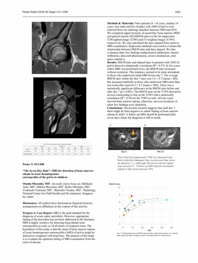
Poster #: SCI-040
“The Seven Day Rule”: MRI for detection of bone marrow
edema in acute hematogenous
osteomyelitis of the pelvis in children
Osamu Miyazaki, MD1, [email protected]; Hidekazu
Aoki, MD1, Mikiko Miyasaka, MD
1, Reiko Okamoto, MD
1,
Yoshiyuki Tsutsumi, MD1, Shunsuke Nosaka, MD
1;
1Radiology,
National Center for Child Health and Development, Setagaya-ku, Japan Disclosures: All authors have disclosed no financial interests,
arrangements or affiliations in the context of this activity. Purpose or Case Report: MRI is the gold standard for the diagnosis of acute septic sacroiliitis. However, appropriate timing of the procedure has not been addressed in the literature. MRI is highly sensitive for detecting long tubular bone osteomyelitis as early as 24-48 hours of symptom onset. The
hypothesis of this study is that the onset of bone marrow edema of acute hematogenous osteomyelitis (AHO) of pelvis might be delayed as compared with long bone. The purpose of this study is to evaluate the optimum timing of MRI examination from the onset of disease.
Methods & Materials: Nine patients (8 - 16 years, median 14 years, four male and five female) with AHO of pelvis were selected from our radiology database between 2004 and 2016. We compared signal intensity of sacral/iliac bone marrow (BM) and gluteal muscle (M) (BM/M ratio) on the fat suppressed
T2Weighted image (T2WI) and T1weighted image (T1WI) respectively. We also calculated the days elapsed from onset to MRI examination. Regression methods were used to evaluate the relationship between BM/M ratio and days elapsed. We also evaluated other five findings (subperiosteal infiltration, muscle infiltration, laba cleft phenomenon, erosive destruction, joint
space enhance). Results: BM/M ratio and elapsed days in patients with AHO of pelvis showed a statistically correlation (R
2= 0.57). In five cases
where MRI was performed twice, the BM/M ratio increased without exception. This tendency seemed to be more prominent in those who underwent initial MRI before day 7. The average BM/M ratio within the first 7 days was 3.6 ± 0.7 (mean ± SD),
but increased markedly in those who underwent MRI more than one week after onset (6.5 ± 0.7 [mean ± SD]). There was a statistically significant difference in the BM/M ratio before and after day 7 (p ≤ 0.001). The BM/M ratio on the T1WI showed an inverse relationship to that on the T2WI with a statistically correlation (R
2= 0.59) on the T2WI as well. All nine cases
showed bone marrow edema, otherwise, un-even incidence of
other five findings were identified. Conclusions: The present research suggests that until day 7 there might be false negative or subtle finding of bone marrow edema in AHO. A follow-up MRI should be performed after seven days where the diagnosis is still in doubt.
Pediatr Radiol (2018) 48 (Suppl 1):S1–S298 S263
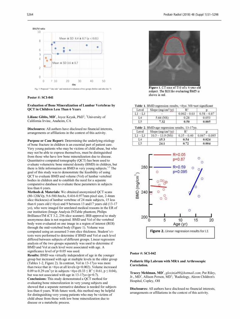
Poster #: SCI-041
Evaluation of Bone Mineralization of Lumbar Vertebrae by
QCT in Children Less Than 6 Years Liliane Gibbs, MD
1, Joyce Keyak, PhD
1;
1University of
California Irvine, Anaheim, CA
Disclosures: All authors have disclosed no financial interests, arrangements or affiliations in the context of this activity. Purpose or Case Report: Determining the underlying etiology of bone fracture in children is an essential part of patient care. Very young patients who may be victims of child abuse, but who
may not be able to express themselves, must be distinguished from those who have low bone mineralization due to disease. Quantitative computed tomography (QCT) has been used to evaluate volumetric bone mineral density (BMD) in children, but there is little information on BMD in very young subjects.
1,2 The
goal of this study was to demonstrate the feasibility of using
QCT to evaluate BMD and volume (Vol) of lumbar vertebral bodies in children and to establish the need for a separate comparative database to evaluate these parameters in subjects less than 6 years. Methods & Materials: We obtained anonymized QCT scans (80-120kVp, 9.6-580.8mAs, 0.416-0.977mm pixel size, 2-4mm slice thickness) of lumbar vertebrae of 24 male subjects, 15 less
than 6 years old (<6yo) and 9 between 13 and17 years old (13-17 yo), who were imaged for unrelated medical reasons in the ER of our institution (Image Analysis INTable phantom; Philips BrillianceTM iCT 3.2, 256 slice scanner). IRB approval to study anonymous data is not required. BMD and Vol of the vertebral body were evaluated on one image in a region of interest (ROI) through the mid-vertebral body (Figure 1). Volume was
computed using an assumed 3-mm slice thickness. Student’s t-tests were performed to determine if BMD and Vol at each level differed between subjects of different groups. Linear regression analysis of the two groups separately was used to determine if BMD and Vol at each level were associated with age. A significance level of p<0.05 was used.
Results: BMD was virtually independent of age in the younger group but increased with age at multiple levels in the older group (Tables 1-2, Figure 2). In contrast, Vol in 13-17yo was more than twice that in <6yo at all levels (p<0.002). Volume increased 0.09 to 0.29 cm
3/yr in subjects <6yo (0.33 ≤ R
2 ≤ 0.61, p ≤ 0.04),
but was not associated with age in 13-17yo (p>0.7). Conclusions: This study demonstrated a QCT method for
evaluating bone mineralization in very young subjects and showed that a separate normative database is needed for subjects less than 6 years. With future work, this method may be helpful for distinguishing very young patients who may be victims of child abuse from those with low bone mineralization due to disease or a metabolic process.
Poster #: SCI-042
Pediatric Hip Labrum with MRA and Arthroscopic
Correlation. Tracey Mehlman, MD
1, [email protected]; Pat Riley,
Jr., MD1, Allison Person, MD
1;
1Radiology, Akron Children's
Hospital, Copley, OH Disclosures: All authors have disclosed no financial interests, arrangements or affiliations in the context of this activity.
Pediatr Radiol (2018) 48 (Suppl 1):S1–S298S264
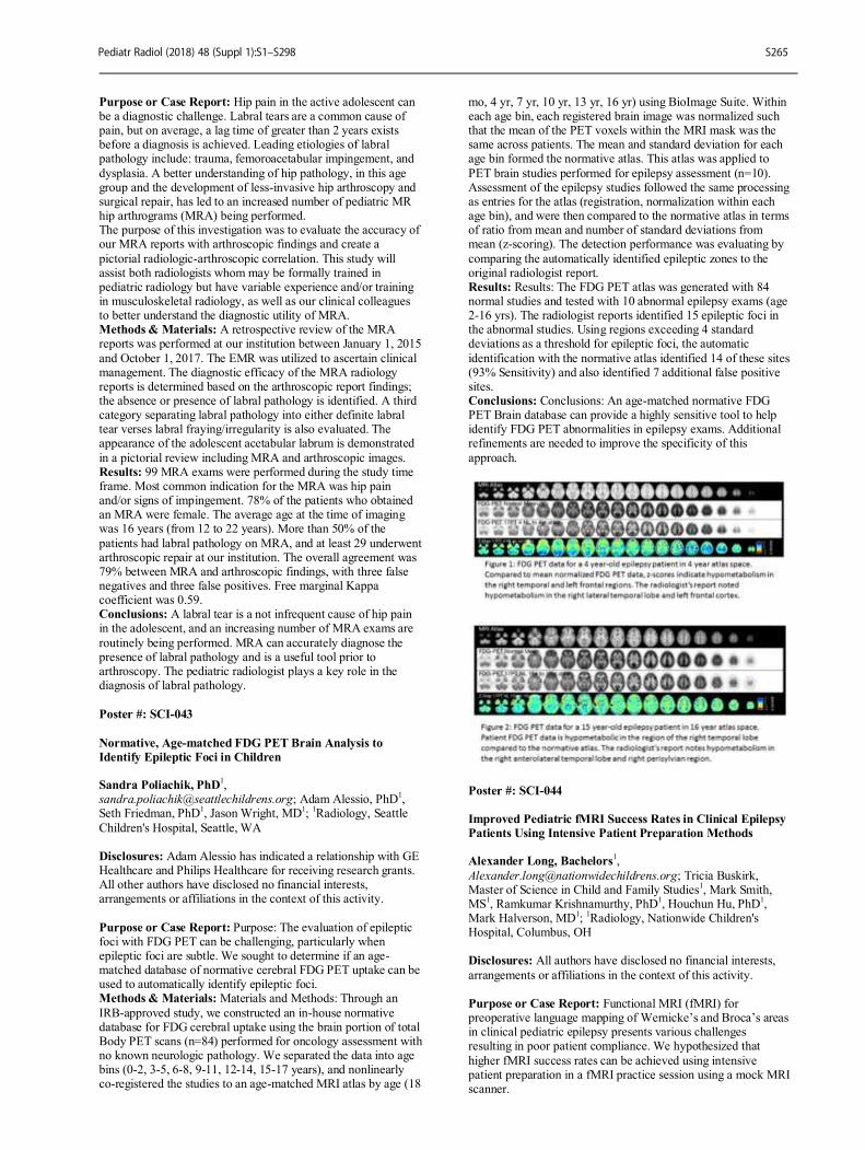
Purpose or Case Report: Hip pain in the active adolescent can be a diagnostic challenge. Labral tears are a common cause of pain, but on average, a lag time of greater than 2 years exists before a diagnosis is achieved. Leading etiologies of labral pathology include: trauma, femoroacetabular impingement, and
dysplasia. A better understanding of hip pathology, in this age group and the development of less-invasive hip arthroscopy and surgical repair, has led to an increased number of pediatric MR hip arthrograms (MRA) being performed. The purpose of this investigation was to evaluate the accuracy of our MRA reports with arthroscopic findings and create a
pictorial radiologic-arthroscopic correlation. This study will assist both radiologists whom may be formally trained in pediatric radiology but have variable experience and/or training in musculoskeletal radiology, as well as our clinical colleagues to better understand the diagnostic utility of MRA. Methods & Materials: A retrospective review of the MRA reports was performed at our institution between January 1, 2015
and October 1, 2017. The EMR was utilized to ascertain clinical management. The diagnostic efficacy of the MRA radiology reports is determined based on the arthroscopic report findings; the absence or presence of labral pathology is identified. A third category separating labral pathology into either definite labral tear verses labral fraying/irregularity is also evaluated. The appearance of the adolescent acetabular labrum is demonstrated
in a pictorial review including MRA and arthroscopic images. Results: 99 MRA exams were performed during the study time frame. Most common indication for the MRA was hip pain and/or signs of impingement. 78% of the patients who obtained an MRA were female. The average age at the time of imaging was 16 years (from 12 to 22 years). More than 50% of the
patients had labral pathology on MRA, and at least 29 underwent arthroscopic repair at our institution. The overall agreement was 79% between MRA and arthroscopic findings, with three false negatives and three false positives. Free marginal Kappa coefficient was 0.59. Conclusions: A labral tear is a not infrequent cause of hip pain in the adolescent, and an increasing number of MRA exams are
routinely being performed. MRA can accurately diagnose the presence of labral pathology and is a useful tool prior to arthroscopy. The pediatric radiologist plays a key role in the diagnosis of labral pathology.
Poster #: SCI-043
Normative, Age-matched FDG PET Brain Analysis to
Identify Epileptic Foci in Children Sandra Poliachik, PhD
1,
[email protected]; Adam Alessio, PhD1,
Seth Friedman, PhD1, Jason Wright, MD
1;
1Radiology, Seattle
Children's Hospital, Seattle, WA Disclosures: Adam Alessio has indicated a relationship with GE Healthcare and Philips Healthcare for receiving research grants. All other authors have disclosed no financial interests, arrangements or affiliations in the context of this activity.
Purpose or Case Report: Purpose: The evaluation of epileptic foci with FDG PET can be challenging, particularly when epileptic foci are subtle. We sought to determine if an age-matched database of normative cerebral FDG PET uptake can be used to automatically identify epileptic foci. Methods & Materials: Materials and Methods: Through an
IRB-approved study, we constructed an in-house normative database for FDG cerebral uptake using the brain portion of total Body PET scans (n=84) performed for oncology assessment with no known neurologic pathology. We separated the data into age bins (0-2, 3-5, 6-8, 9-11, 12-14, 15-17 years), and nonlinearly co-registered the studies to an age-matched MRI atlas by age (18
mo, 4 yr, 7 yr, 10 yr, 13 yr, 16 yr) using BioImage Suite. Within each age bin, each registered brain image was normalized such that the mean of the PET voxels within the MRI mask was the same across patients. The mean and standard deviation for each age bin formed the normative atlas. This atlas was applied to
PET brain studies performed for epilepsy assessment (n=10). Assessment of the epilepsy studies followed the same processing as entries for the atlas (registration, normalization within each age bin), and were then compared to the normative atlas in terms of ratio from mean and number of standard deviations from mean (z-scoring). The detection performance was evaluating by
comparing the automatically identified epileptic zones to the original radiologist report. Results: Results: The FDG PET atlas was generated with 84 normal studies and tested with 10 abnormal epilepsy exams (age 2-16 yrs). The radiologist reports identified 15 epileptic foci in the abnormal studies. Using regions exceeding 4 standard deviations as a threshold for epileptic foci, the automatic
identification with the normative atlas identified 14 of these sites (93% Sensitivity) and also identified 7 additional false positive sites. Conclusions: Conclusions: An age-matched normative FDG PET Brain database can provide a highly sensitive tool to help identify FDG PET abnormalities in epilepsy exams. Additional refinements are needed to improve the specificity of this
approach.
Poster #: SCI-044
Improved Pediatric fMRI Success Rates in Clinical Epilepsy
Patients Using Intensive Patient Preparation Methods Alexander Long, Bachelors
1,
[email protected]; Tricia Buskirk, Master of Science in Child and Family Studies
1, Mark Smith,
MS1, Ramkumar Krishnamurthy, PhD
1, Houchun Hu, PhD
1,
Mark Halverson, MD1;
1Radiology, Nationwide Children's
Hospital, Columbus, OH Disclosures: All authors have disclosed no financial interests,
arrangements or affiliations in the context of this activity. Purpose or Case Report: Functional MRI (fMRI) for preoperative language mapping of Wernicke’s and Broca’s areas in clinical pediatric epilepsy presents various challenges resulting in poor patient compliance. We hypothesized that
higher fMRI success rates can be achieved using intensive patient preparation in a fMRI practice session using a mock MRI scanner.
Pediatr Radiol (2018) 48 (Suppl 1):S1–S298 S265
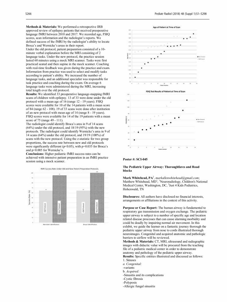
Methods & Materials: We performed a retrospective IRB approved review of epilepsy patients that received preoperative language fMRI between 2010 and 2017. We recorded age, FSIQ scores, scan information and the radiologist’s reports. We defined success of the fMRI by the radiologist’s ability to locate
Broca’s and Wernicke’s areas in their report. Under the old protocol, patient preparation consisted of a 10-minute verbal explanation before the MRI consisting of 2 language tasks. Under the new protocol, the practice session lasted 60 minutes using a mock MRI scanner. Tasks were first practiced seated and then supine in the mock scanner. Coaching
with real-time feedback was given during the practice and exam. Information from practice was used to select and modify tasks according to patient’s ability. We increased the number of language tasks, and an additional specialist was responsible for task practice and coaching during the exam. On average 6 language tasks were administered during the MRI, increasing total length over the old protocol.
Results: We identified 33 preoperative language-mapping fMRI scans of children with epilepsy. 13 of 33 were done under the old protocol with a mean age of 16 (range 12 - 19 years). FSIQ scores were available for 10 of the 14 patients with a mean score of 84 (range 62 - 108). 19 of 33 scans were done after institution of an new protocol with mean age of 14 (range 8 - 19 years). FSIQ scores were available for 14 of the 19 patients with a mean
score of 75 (range 49 - 111). The radiologist could identify Broca’s area in 9 of 14 scans (64%) under the old protocol, and 18/19 (95%) with the new protocols. The radiologist could identify Wernicke’s area in 9 of 14 scans (64%) under the old protocol, and 19/19 (100%) of scans with the new protocol. Using the z-statistic for two group
proportions, the success rate between new and old protocols were significantly different (p<0.05), with p=0.035 for Broca’s and p=0.005 for Wernicke’s. Conclusions: Higher pediatric fMRI success rates can be achieved with intensive patient preparation in an fMRI practice session using a mock scanner.
Poster #: SCI-045
The Pediatric Upper Airway: Thoroughfares and Road
blocks Mark Whitehead, PA
Matthew Whitehead, MD1;
1Neuroradiology, Children's National
Medical Center, Washington, DC, 2Just 4 Kids Pediatrics,
Hohenwald, TN Disclosures: All authors have disclosed no financial interests, arrangements or affiliations in the context of this activity.
Purpose or Case Report: The human airway is fundamental to respiratory gas transmission and oxygen exchange. The pediatric
upper airway is subject to a number of specific age and location related disease processes that can cause alarming morbidity and could be deadly by impairing normal air movement. In this exhibit, we guide the learner on a fantastic journey thorough the pediatric upper airway from nose to cords illustrated thorough neuroimages. Congenital and acquired anatomic and pathologic barriers to airflow will be reviewed.
Methods & Materials: CT, MRI, ultrasound and radiographic images with didactic value will be procured from the teaching file of a pediatric medical center in order to demonstrate anatomy and pathology of the pediatric upper airway. Results: Specific entities illustrated and discussed as follows: 1. Sinuses
a. Congenital: -variants b. Acquired: -Sinusitis and its complications -Cystic fibrosis -Polyposis -Allergic fungal sinusitis
Pediatr Radiol (2018) 48 (Suppl 1):S1–S298S266
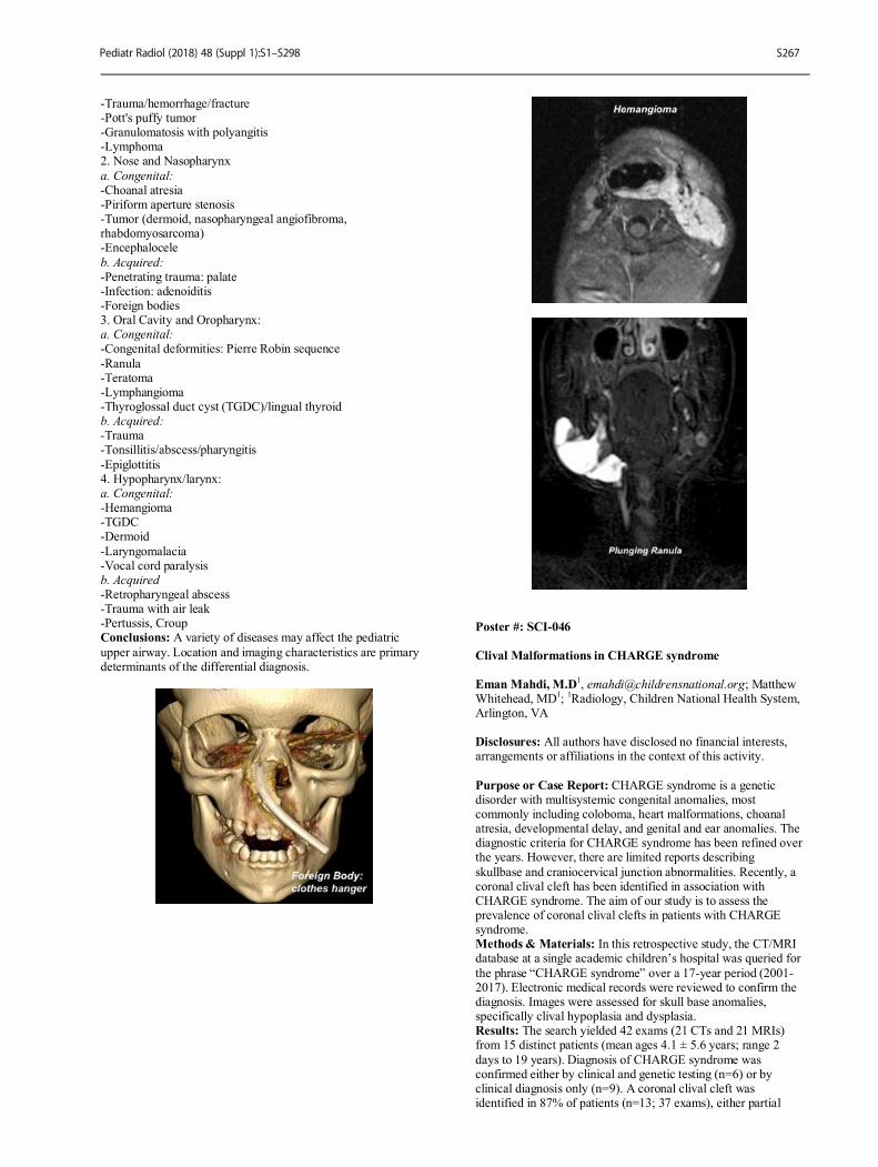
-Trauma/hemorrhage/fracture -Pott's puffy tumor -Granulomatosis with polyangitis -Lymphoma 2. Nose and Nasopharynx
a. Congenital: -Choanal atresia -Piriform aperture stenosis -Tumor (dermoid, nasopharyngeal angiofibroma, rhabdomyosarcoma) -Encephalocele
b. Acquired: -Penetrating trauma: palate -Infection: adenoiditis -Foreign bodies 3. Oral Cavity and Oropharynx: a. Congenital: -Congenital deformities: Pierre Robin sequence
-Ranula -Teratoma -Lymphangioma -Thyroglossal duct cyst (TGDC)/lingual thyroid b. Acquired: -Trauma -Tonsillitis/abscess/pharyngitis
-Epiglottitis 4. Hypopharynx/larynx: a. Congenital: -Hemangioma -TGDC -Dermoid
-Laryngomalacia -Vocal cord paralysis b. Acquired -Retropharyngeal abscess -Trauma with air leak -Pertussis, Croup Conclusions: A variety of diseases may affect the pediatric
upper airway. Location and imaging characteristics are primary determinants of the differential diagnosis.
Poster #: SCI-046
Clival Malformations in CHARGE syndrome
Eman Mahdi, M.D1, [email protected]; Matthew
Whitehead, MD1;
1Radiology, Children National Health System,
Arlington, VA Disclosures: All authors have disclosed no financial interests, arrangements or affiliations in the context of this activity.
Purpose or Case Report: CHARGE syndrome is a genetic disorder with multisystemic congenital anomalies, most commonly including coloboma, heart malformations, choanal atresia, developmental delay, and genital and ear anomalies. The diagnostic criteria for CHARGE syndrome has been refined over the years. However, there are limited reports describing
skullbase and craniocervical junction abnormalities. Recently, a coronal clival cleft has been identified in association with CHARGE syndrome. The aim of our study is to assess the prevalence of coronal clival clefts in patients with CHARGE syndrome. Methods & Materials: In this retrospective study, the CT/MRI database at a single academic children’s hospital was queried for
the phrase “CHARGE syndrome” over a 17-year period (2001-2017). Electronic medical records were reviewed to confirm the diagnosis. Images were assessed for skull base anomalies, specifically clival hypoplasia and dysplasia. Results: The search yielded 42 exams (21 CTs and 21 MRIs) from 15 distinct patients (mean ages 4.1 ± 5.6 years; range 2
days to 19 years). Diagnosis of CHARGE syndrome was confirmed either by clinical and genetic testing (n=6) or by clinical diagnosis only (n=9). A coronal clival cleft was identified in 87% of patients (n=13; 37 exams), either partial
Pediatr Radiol (2018) 48 (Suppl 1):S1–S298 S267
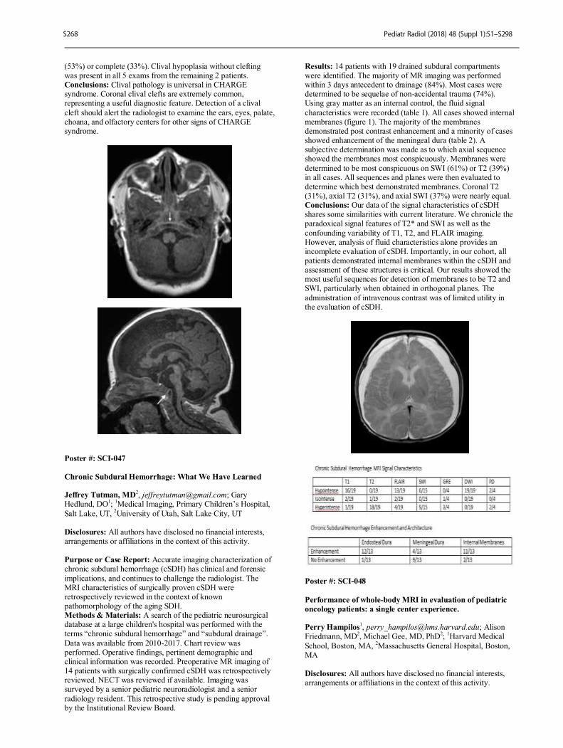
(53%) or complete (33%). Clival hypoplasia without clefting was present in all 5 exams from the remaining 2 patients. Conclusions: Clival pathology is universal in CHARGE syndrome. Coronal clival clefts are extremely common, representing a useful diagnostic feature. Detection of a clival
cleft should alert the radiologist to examine the ears, eyes, palate, choana, and olfactory centers for other signs of CHARGE syndrome.
Poster #: SCI-047
Chronic Subdural Hemorrhage: What We Have Learned Jeffrey Tutman, MD
2, [email protected]; Gary
Hedlund, DO1;
1Medical Imaging, Primary Children’s Hospital,
Salt Lake, UT, 2University of Utah, Salt Lake City, UT
Disclosures: All authors have disclosed no financial interests, arrangements or affiliations in the context of this activity. Purpose or Case Report: Accurate imaging characterization of chronic subdural hemorrhage (cSDH) has clinical and forensic
implications, and continues to challenge the radiologist. The MRI characteristics of surgically proven cSDH were retrospectively reviewed in the context of known pathomorphology of the aging SDH. Methods & Materials: A search of the pediatric neurosurgical database at a large children's hospital was performed with the terms “chronic subdural hemorrhage” and “subdural drainage”.
Data was available from 2010-2017. Chart review was performed. Operative findings, pertinent demographic and clinical information was recorded. Preoperative MR imaging of 14 patients with surgically confirmed cSDH was retrospectively reviewed. NECT was reviewed if available. Imaging was surveyed by a senior pediatric neuroradiologist and a senior
radiology resident. This retrospective study is pending approval by the Institutional Review Board.
Results: 14 patients with 19 drained subdural compartments were identified. The majority of MR imaging was performed within 3 days antecedent to drainage (84%). Most cases were determined to be sequelae of non-accidental trauma (74%). Using gray matter as an internal control, the fluid signal
characteristics were recorded (table 1). All cases showed internal membranes (figure 1). The majority of the membranes demonstrated post contrast enhancement and a minority of cases showed enhancement of the meningeal dura (table 2). A subjective determination was made as to which axial sequence showed the membranes most conspicuously. Membranes were
determined to be most conspicuous on SWI (61%) or T2 (39%) in all cases. All sequences and planes were then evaluated to determine which best demonstrated membranes. Coronal T2 (31%), axial T2 (31%), and axial SWI (37%) were nearly equal. Conclusions: Our data of the signal characteristics of cSDH shares some similarities with current literature. We chronicle the paradoxical signal features of T2* and SWI as well as the
confounding variability of T1, T2, and FLAIR imaging. However, analysis of fluid characteristics alone provides an incomplete evaluation of cSDH. Importantly, in our cohort, all patients demonstrated internal membranes within the cSDH and assessment of these structures is critical. Our results showed the most useful sequences for detection of membranes to be T2 and SWI, particularly when obtained in orthogonal planes. The
administration of intravenous contrast was of limited utility in the evaluation of cSDH.
Poster #: SCI-048
Performance of whole-body MRI in evaluation of pediatric
oncology patients: a single center experience. Perry Hampilos
1, [email protected]; Alison
Friedmann, MD2, Michael Gee, MD, PhD
2;
1Harvard Medical
School, Boston, MA, 2Massachusetts General Hospital, Boston,
MA Disclosures: All authors have disclosed no financial interests, arrangements or affiliations in the context of this activity.
Pediatr Radiol (2018) 48 (Suppl 1):S1–S298S268
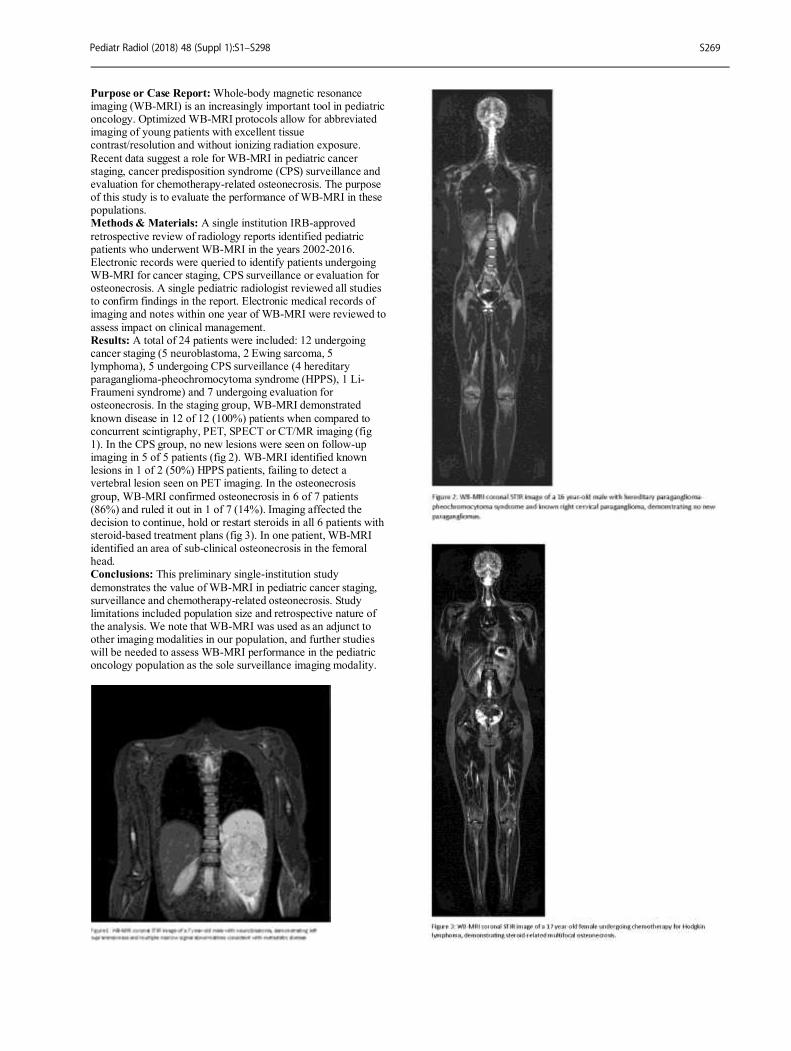
Purpose or Case Report: Whole-body magnetic resonance imaging (WB-MRI) is an increasingly important tool in pediatric oncology. Optimized WB-MRI protocols allow for abbreviated imaging of young patients with excellent tissue contrast/resolution and without ionizing radiation exposure.
Recent data suggest a role for WB-MRI in pediatric cancer staging, cancer predisposition syndrome (CPS) surveillance and evaluation for chemotherapy-related osteonecrosis. The purpose of this study is to evaluate the performance of WB-MRI in these populations. Methods & Materials: A single institution IRB-approved
retrospective review of radiology reports identified pediatric patients who underwent WB-MRI in the years 2002-2016. Electronic records were queried to identify patients undergoing WB-MRI for cancer staging, CPS surveillance or evaluation for osteonecrosis. A single pediatric radiologist reviewed all studies to confirm findings in the report. Electronic medical records of imaging and notes within one year of WB-MRI were reviewed to
assess impact on clinical management. Results: A total of 24 patients were included: 12 undergoing cancer staging (5 neuroblastoma, 2 Ewing sarcoma, 5 lymphoma), 5 undergoing CPS surveillance (4 hereditary paraganglioma-pheochromocytoma syndrome (HPPS), 1 Li-Fraumeni syndrome) and 7 undergoing evaluation for osteonecrosis. In the staging group, WB-MRI demonstrated
known disease in 12 of 12 (100%) patients when compared to concurrent scintigraphy, PET, SPECT or CT/MR imaging (fig 1). In the CPS group, no new lesions were seen on follow-up imaging in 5 of 5 patients (fig 2). WB-MRI identified known lesions in 1 of 2 (50%) HPPS patients, failing to detect a vertebral lesion seen on PET imaging. In the osteonecrosis
group, WB-MRI confirmed osteonecrosis in 6 of 7 patients (86%) and ruled it out in 1 of 7 (14%). Imaging affected the decision to continue, hold or restart steroids in all 6 patients with steroid-based treatment plans (fig 3). In one patient, WB-MRI identified an area of sub-clinical osteonecrosis in the femoral head. Conclusions: This preliminary single-institution study
demonstrates the value of WB-MRI in pediatric cancer staging, surveillance and chemotherapy-related osteonecrosis. Study limitations included population size and retrospective nature of the analysis. We note that WB-MRI was used as an adjunct to other imaging modalities in our population, and further studies will be needed to assess WB-MRI performance in the pediatric oncology population as the sole surveillance imaging modality.
Pediatr Radiol (2018) 48 (Suppl 1):S1–S298 S269
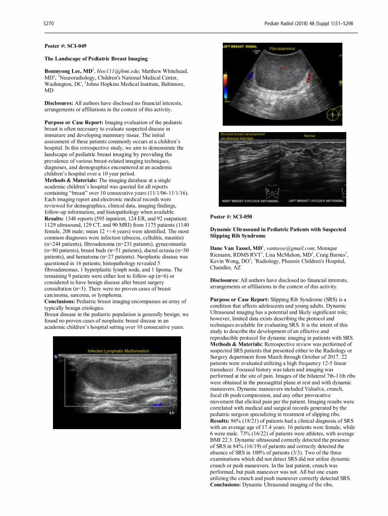
Poster #: SCI-049
The Landscape of Pediatric Breast Imaging Bonmyong Lee, MD
2, [email protected]; Matthew Whitehead,
MD1;
1Neuroradiology, Children's National Medical Center,
Washington, DC, 2Johns Hopkins Medical Institute, Baltimore,
MD Disclosures: All authors have disclosed no financial interests, arrangements or affiliations in the context of this activity.
Purpose or Case Report: Imaging evaluation of the pediatric breast is often necessary to evaluate suspected disease in immature and developing mammary tissue. The initial assessment of these patients commonly occurs at a children’s hospital. In this retrospective study, we aim to demonstrate the
landscape of pediatric breast imaging by providing the prevalence of various breast-related imaging techniques, diagnoses, and demographics encountered at an academic children’s hospital over a 10 year period.
Methods & Materials: The imaging database at a single academic children’s hospital was queried for all reports containing “breast” over 10 consecutive years (11/1/06-11/1/16). Each imaging report and electronic medical records were reviewed for demographics, clinical data, imaging findings, follow-up information, and histopathology when available.
Results: 1348 reports (595 inpatient, 124 ER, and 92 outpatient; 1129 ultrasound, 129 CT, and 90 MRI) from 1175 patients (1140 female, 208 male; mean 12 +/-6 years) were identified. The most common diagnoses were infection (abscess, cellulitis, mastitis) (n=244 patients), fibroadenoma (n=231 patients), gynecomastia (n=80 patients), breast buds (n=51 patients), ductal ectasia (n=30 patients), and hematoma (n=27 patients). Neoplastic disease was
questioned in 16 patients; histopathology revealed 5 fibroadenomas, 1 hyperplastic lymph node, and 1 lipoma. The remaining 9 patients were either lost to follow-up (n=6) or considered to have benign disease after breast surgery consultation (n=3). There were no proven cases of breast carcinoma, sarcoma, or lymphoma. Conclusions: Pediatric breast imaging encompasses an array of
typically benign etiologies. Breast disease in the pediatric population is generally benign; we found no proven cases of neoplastic breast disease in an academic children’s hospital setting over 10 consecutive years.
Poster #: SCI-050
Dynamic Ultrasound in Pediatric Patients with Suspected
Slipping Rib Syndrome Dane Van Tassel, MD
1, [email protected]; Monique
Riemann, RDMS RVT1, Lisa McMahon, MD
1, Craig Barnes
1,
Kevin Wong, DO1;
1Radiology, Phoenix Children's Hospital,
Chandler, AZ Disclosures: All authors have disclosed no financial interests, arrangements or affiliations in the context of this activity.
Purpose or Case Report: Slipping Rib Syndrome (SRS) is a condition that affects adolescents and young adults. Dynamic Ultrasound imaging has a potential and likely significant role; however, limited data exists describing the protocol and techniques available for evaluating SRS. It is the intent of this study to describe the development of an effective and
reproducible protocol for dynamic imaging in patients with SRS. Methods & Materials: Retrospective review was performed of suspected SRS patients that presented either to the Radiology or Surgery department from March through October of 2017. 22 patients were evaluated utilizing a high frequency 12-5 linear transducer. Focused history was taken and imaging was performed at the site of pain. Images of the bilateral 7th-11th ribs
were obtained in the parasagittal plane at rest and with dynamic maneuvers. Dynamic maneuvers included Valsalva, crunch, focal rib push/compression, and any other provocative movement that elicited pain per the patient. Imaging results were correlated with medical and surgical records generated by the pediatric surgeon specializing in treatment of slipping ribs.
Results: 86% (18/21) of patients had a clinical diagnosis of SRS with an average age of 17.4 years. 16 patients were female, while 6 were male. 73% (16/22) of patients were athletes, with average BMI 22.3. Dynamic ultrasound correctly detected the presence of SRS in 84% (16/19) of patients and correctly detected the absence of SRS in 100% of patients (3/3). Two of the three examinations which did not detect SRS did not utilize dynamic
crunch or push maneuvers. In the last patient, crunch was performed, but push maneuver was not. All but one exam utilizing the crunch and push maneuver correctly detected SRS. Conclusions: Dynamic Ultrasound imaging of the ribs,
Pediatr Radiol (2018) 48 (Suppl 1):S1–S298S270
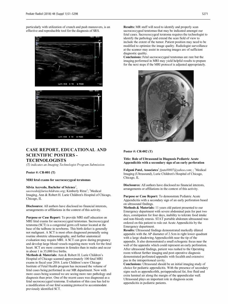
particularly with utilization of crunch and push maneuvers, is an effective and reproducible tool for the diagnosis of SRS.
CASE REPORT, EDUCATIONAL AND
SCIENTIFIC POSTERS -
TECHNOLOGISTS
(T) indicates an Imaging Technologist Program Submission
Poster #: CR-001 (T)
MRI fetal exams for sacrococcygeal teratomas Silvia Acevedo, Bachelor of Science
1,
[email protected]; Kimberly Ritze1;
1Medical
Imaging, Ann & Robert H. Lurie Children's Hospital of Chicago, Chicago, IL Disclosures: All authors have disclosed no financial interests, arrangements or affiliations in the context of this activity.
Purpose or Case Report: To provide MRI staff education on MRI fetal exams for sacrococcygeal teratomas. Sacrococcygeal teratoma (SCT) is a congenital germ cell tumor located at the base of the tailbone in newborns. This birth defect is generally not malignant. A SCT is most often diagnosed prenatally using routine obstetric ultrasonography, and further anatomical evaluation may require MRI. A SCT can grow during pregnancy
and develop large blood vessels requiring more work for the fetal heart. SCT are more common in females than in males and occur in about 1 in 35,000 live births. Methods & Materials: Ann & Robert H. Lurie Children’s Hospital of Chicago scanned approximately 100 fetal MRI exams in fiscal year 2016. Lurie Children’s new Chicago Institute of Fetal Health program has increased the volume of
fetal cases being performed in our MR department. Now with more cases being scanned we are seeing more rare pathology and diagnosis than prior. One of the recent cases was diagnosed as a fetal sacrococcygeal teratoma. Evaluation of this case has led to a modification of our fetal scanning protocol to accommodate previously identified SCT.
Results: MR staff will need to identify and properly scan sacrococcygeal teratomas that may be indicated amongst our fetal cases. Sacrococcygeal teratoma requires the technologist to identify the pathology and extend the scan field of view to include the extent of the tumor. Patient position may need to be
modified to optimize the image quality. Radiologist surveillance at the scanner may assist in ensuring images are of sufficient diagnostic quality. Conclusions: Fetal sacrococcygeal teratomas are rare but the imaging performed in MRI may yield helpful results to prepare for the next steps if the MRI protocol is adjusted appropriately.
Poster #: CR-002 (T)
Title: Role of Ultrasound in Diagnosis Pediatric Acute
Appendicitis with a secondary sign of an early perforation Falguni Patel, Associates
1 [email protected]; ;
1Medical
Imaging (Ultrasound), Lurie Children's Hospital of Chicago, Chicago, IL Disclosures: All authors have disclosed no financial interests, arrangements or affiliations in the context of this activity. Purpose or Case Report: To demonstrate Pediatric Acute
Appendicitis with a secondary sign of an early perforation based on ultrasound findings. Methods & Materials: 11 years old patient presented to our Emergency department with severe abdominal pain for past two days, constipation for four days, inability to tolerate food intake and non-bloody emesis. STAT portable abdomen ultrasound was
ordered on this patient to rule out Acute Appendicitis by the Emergency department. Results: Ultrasound findings demonstrated markedly dilated appendix with the AP diameter of 2.5cm in right lower quadrant with a large shadowing Appendicolith near the tip of the appendix. It also demonstrated a small echogenic focus near the wall of the appendix which could represent an early perforation.
After ultrasound findings, patient was rushed to the Operating room without further imaging and post-operative diagnosis demonstrated perforated appendix with fecalith and extensive pus in the intraperitoneal cavity. Conclusions: Ultrasound should be an initial imaging study of choice for pediatric appendicitis. With the presence of secondary signs such as appendicolith, periappendiceal fat, free fluid and
extra luminal air along the margin of the appendicular wall. Ultrasound plays an important role in diagnosis acute appendicitis in pediatric patients.
Pediatr Radiol (2018) 48 (Suppl 1):S1–S298 S271
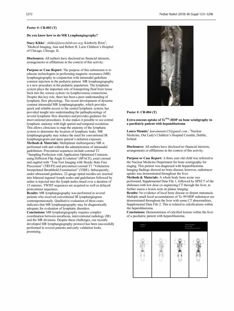
Poster #: CR-003 (T)
Do you know how to do MR Lymphangiography? Stacy Kikke
1, [email protected]; Kimberly Ritze
1;
1Medical Imaging, Ann and Robert H. Lurie Children’s Hospital
of Chicago, Chicago, IL Disclosures: All authors have disclosed no financial interests, arrangements or affiliations in the context of this activity.
Purpose or Case Report: The purpose of this submission is to educate technologists in performing magnetic resonance (MR) lymphangiography in conjunction with intranodal gadolium contrast injection in the pediatric patient. MR lymphangiography is a new procedure in the pediatric population. The lymphatic system plays the important role of transporting fluid from tissue back into the venous system via lymphovenous connections.
Despite this key role, there has been a poor understanding of lymphatic flow physiology. The recent development of dynamic contrast intranodal MR lymphangiography, which provides quick and reliable access to the central lymphatic system, has provided insight into understanding the pathophysiology of several lymphatic flow disorders and provides guidance for interventional procedures. It also makes it possible to see central
lymphatic anatomy with high spatial and temporal resolution. This allows clinicians to map the anatomy of the lymphatic system to determine the location of lymphatic leaks. MR lymphangiography may reduce the need for conventional IR lymphangiogram and spare patient’s radiation exposure. Methods & Materials: Multiplanar multisequence MR is
performed with and without the administration of intranodal gadolinium. Precontrast sequences include coronal T2 “Sampling Perfection with Application Optimized Contrasts using Different Flip Angle Evolution” (SPACE), axial coronal and sagittal with “True Fast Imaging with Steady State Free Precession” (TRUFI) and precontrast coronal T1 “Volumetric Interpolated Breathhold Examination” (VIBE). Subsequently,
under ultrasound guidance, 22-gauge spinal needles are inserted into bilateral inguinal lymph nodes and gadolinium followed by saline is injected into the lymph nodes timed over a duration of 15 minutes. TWIST sequences are acquired as well as delayed postcontrast sequences. Results: MR lymphangiography was performed in several patients who received conventional IR lymphangiogram
contemporaneously. Qualitative evaluation of these cases indicates that MR lymphangiography may be diagnostically adequate for evaluation of lymphatic disorders. Conclusions: MR lymphangiography requires complex coordination between anesthesia, interventional radiology (IR) and the MR divisions. Despite these challenges, our recently
developed MR lymphangiography protocol has been successfully performed in several patients and early validation looks promising.
Poster #: CR-004 (T)
Extra-osseous uptake of Tc99m
-HDP on bone scintigraphy in
a paediatric patient with hepatoblastoma Laura Mannix
1Nuclear
Medicine, Our Lady's Children’s Hospital Crumlin, Dublin, Ireland Disclosures: All authors have disclosed no financial interests, arrangements or affiliations in the context of this activity. Purpose or Case Report: A three year old child was referred to
the Nuclear Medicine Department for bone scintigraphy for staging. This patient was diagnosed with hepatoblastoma. Imaging findings showed no bony disease, however, radiotracer uptake was demonstrated throughout the liver. Methods & Materials: A whole body bone scene was performed, Supplemental Data File 1, followed by SPECT of the abdomen with low dose co-registering CT through the liver, to
further assess a lesion seen on planar imaging. Results: No evidence of local bony disease or distant metastasis. Multiple small focal accumulations of Tc-99 HDP radiotracer are demonstrated throughout the liver with some CT abnormalities, Supplemental Data File 2. This is related to calcifications within the hepatoblastoma.
Conclusions: Demonstration of calcified lesions within the liver of a paediatric patient with hepatoblastoma.
Pediatr Radiol (2018) 48 (Suppl 1):S1–S298S272
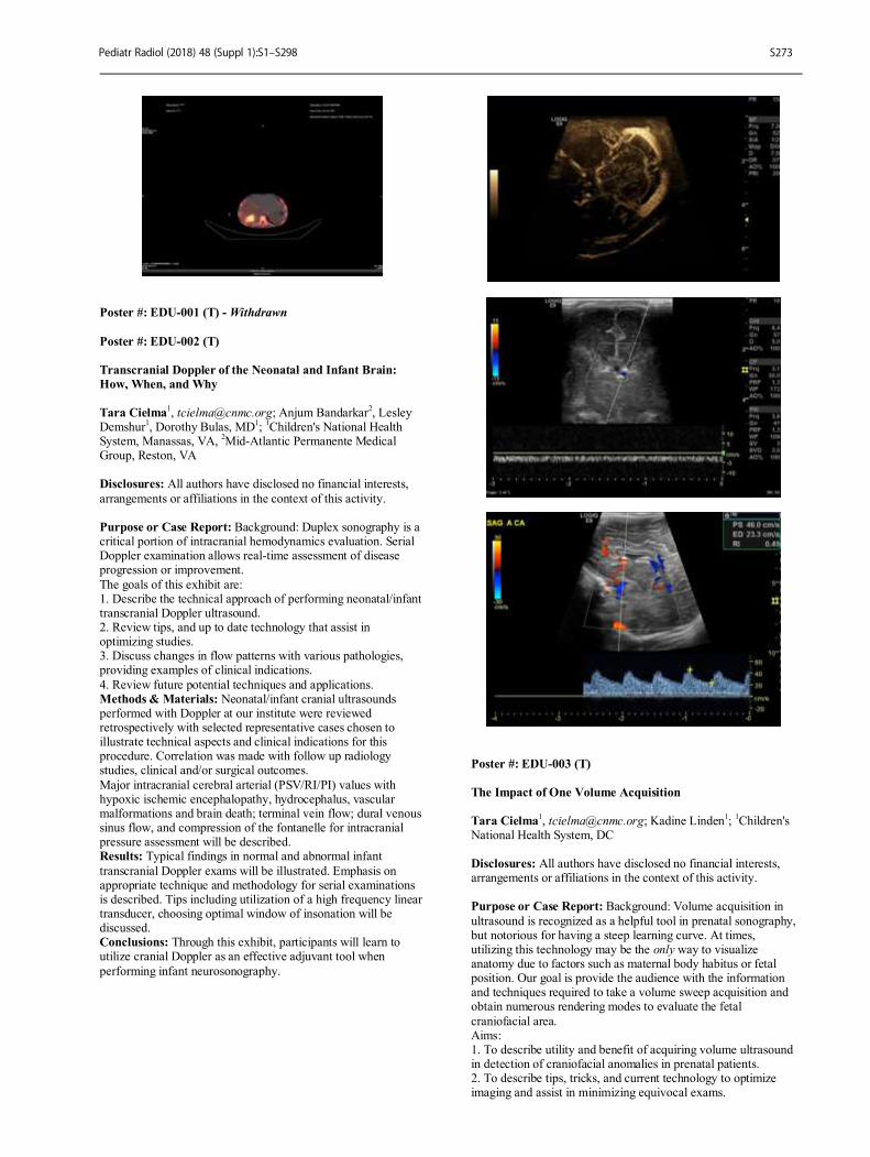
Poster #: EDU-001 (T) - Withdrawn
Poster #: EDU-002 (T)
Transcranial Doppler of the Neonatal and Infant Brain:
How, When, and Why
Tara Cielma
1, [email protected]; Anjum Bandarkar
2, Lesley
Demshur1, Dorothy Bulas, MD
1;
1Children's National Health
System, Manassas, VA, 2Mid-Atlantic Permanente Medical
Group, Reston, VA Disclosures: All authors have disclosed no financial interests,
arrangements or affiliations in the context of this activity. Purpose or Case Report: Background: Duplex sonography is a critical portion of intracranial hemodynamics evaluation. Serial Doppler examination allows real-time assessment of disease progression or improvement.
The goals of this exhibit are: 1. Describe the technical approach of performing neonatal/infant transcranial Doppler ultrasound. 2. Review tips, and up to date technology that assist in optimizing studies. 3. Discuss changes in flow patterns with various pathologies, providing examples of clinical indications.
4. Review future potential techniques and applications. Methods & Materials: Neonatal/infant cranial ultrasounds performed with Doppler at our institute were reviewed retrospectively with selected representative cases chosen to illustrate technical aspects and clinical indications for this procedure. Correlation was made with follow up radiology studies, clinical and/or surgical outcomes.
Major intracranial cerebral arterial (PSV/RI/PI) values with hypoxic ischemic encephalopathy, hydrocephalus, vascular malformations and brain death; terminal vein flow; dural venous sinus flow, and compression of the fontanelle for intracranial pressure assessment will be described. Results: Typical findings in normal and abnormal infant
transcranial Doppler exams will be illustrated. Emphasis on appropriate technique and methodology for serial examinations is described. Tips including utilization of a high frequency linear transducer, choosing optimal window of insonation will be discussed. Conclusions: Through this exhibit, participants will learn to utilize cranial Doppler as an effective adjuvant tool when
performing infant neurosonography.
Poster #: EDU-003 (T)
The Impact of One Volume Acquisition
Tara Cielma1, [email protected]; Kadine Linden
1;
1Children's
National Health System, DC Disclosures: All authors have disclosed no financial interests, arrangements or affiliations in the context of this activity.
Purpose or Case Report: Background: Volume acquisition in
ultrasound is recognized as a helpful tool in prenatal sonography, but notorious for having a steep learning curve. At times, utilizing this technology may be the only way to visualize anatomy due to factors such as maternal body habitus or fetal position. Our goal is provide the audience with the information and techniques required to take a volume sweep acquisition and obtain numerous rendering modes to evaluate the fetal
craniofacial area. Aims: 1. To describe utility and benefit of acquiring volume ultrasound in detection of craniofacial anomalies in prenatal patients. 2. To describe tips, tricks, and current technology to optimize imaging and assist in minimizing equivocal exams.
Pediatr Radiol (2018) 48 (Suppl 1):S1–S298 S273
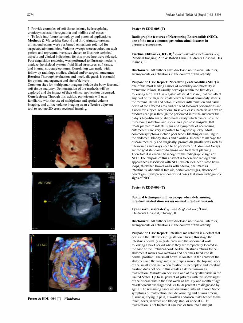
3. Provide examples of soft tissue lesions, hydrocephalus, craniosynostosis, micrognathia and midline cleft cases. 4. To look into future technology and potential applications. Methods & Materials: Second and third trimester prenatal ultrasound exams were performed on patients referred for
suspected abnormalities. Volume sweeps were acquired on each patient and representative cases chosen to illustrate technical aspects and clinical indications for this procedure were selected. Post-acquisition rendering was performed to illustrate modes to analyze the skeletal system, fluid filled structures, soft tissue, and internal structure contours. Correlation was made with
follow up radiology studies, clinical and/or surgical outcomes. Results: Thorough evaluation and timely diagnosis is essential for optimal management and site of delivery. Common sites for multiplanar imaging include the bony face and soft tissue anatomy. Demonstration of the methods will be explored and the impact of their clinical application discussed. Conclusions: Through this exhibit, participants will gain
familiarity with the use of multiplanar and spatial volume imaging, and utilize volume imaging as an effective adjuvant tool to routine 2D cross-sectional imaging.
Poster #: EDU-004 (T) - Withdrawn
Poster #: EDU-005 (T)
Radiographic features of Necrotizing Enterocolitis (NEC),
one of the most common gastrointestinal diseases in premature neonates.
Ewelina Ulikowska, RT (R) 1 [email protected];
1Medical Imaging, Ann & Robert Lurie Children’s Hospital, Des
Plaines, IL
Disclosures: All authors have disclosed no financial interests, arrangements or affiliations in the context of this activity. Purpose or Case Report: Necrotizing enterocolitis (NEC) is one of the most leading causes of morbidity and mortality in premature infants. It usually develops within the first days
following birth. NEC is a gastrointestinal disease, that can affect any part of the large or small bowel but most commonly affects the terminal ileum and colon. It causes inflammation and tissue death of the affected area and can lead to bowel perforations and a need for surgical resections. In severe cases, bacteria and waste products can pass through the perforated intestine and enter the
baby’s bloodstream or abdominal cavity which can cause a life threatening infection and shock. In a pediatric hospital, that treats premature infants, signs and symptoms of necrotizing enterocolitis are very important to diagnose quickly. Most common symptoms include poor feeds, bloating or swelling in the abdomen, bloody stools and diarrhea. In order to manage the disease medically and surgically, prompt diagnostic tests such as
ultrasounds and xrays need to be performed. Abdominal X-rays are the gold standard of diagnosis and treatment planning. Therefore it is crucial, to recognize the radiographic signs of NEC. The purpose of this abstract is to describe radiographic appearances associated with NEC, which include: dilated bowel loops, thickened bowel walls with edema, pneumatosis intestinalis, abdominal free air, portal venous gas, absence of
bowel gas. I will present confirmed cases that show radiographic signs of NEC.
Poster #: EDU-006 (T)
Optimal techniques in fluoroscopy when determining
intestinal malrotation versus normal intestinal variants.
Lynn Gazzi, associates1 [email protected] ;
1Lurie
Children’s Hospital, Chicago, IL Disclosures: All authors have disclosed no financial interests, arrangements or affiliations in the context of this activity. Purpose or Case Report: Intestinal malrotation is a defect that
occurs in the 10th week of gestation. During this stage the intestines normally migrate back into the abdominal wall following a brief period where they are temporarily located in the base of the umbilical cord. As the intestines returns to the abdomen it makes two rotations and becomes fixed into its normal position. The small bowel is located in the center of the
abdomen and the large intestine drapes around the top and sides of the small intestine. When rotation is incomplete and intestinal fixation does not occur, this creates a defect known as malrotation. Malrotation occurs in one of every 500 births in the United States. Up to 40 percent of patients with this show signs of the disease within the first week of life. By one month of age 50-60 percent are diagnosed. 75 to 90 percent are diagnosed by
age 1. The remaining cases are diagnosed into adulthood. Some symptoms of malrotation include vomiting and bilious emesis, fussiness, crying in pain, a swollen abdomen that’s tender to the touch, fever, diarrhea and bloody stool or none at all. If malrotation is not treated, it can lead or turn into a midgut
Pediatr Radiol (2018) 48 (Suppl 1):S1–S298S274
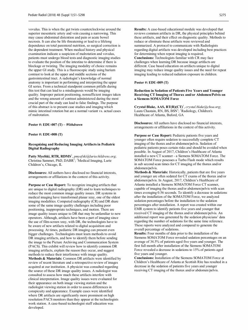
vovulus. This is when the gut twists counterclockwise around the superior mesenteric artery and vein causing a narrowing. This may cause abdominal distention and pain or acute bowel necrosis. It can also be life threatening or lead to a lifelong dependence on total parenteral nutrition, so surgical correction is
the dependent treatment. When medical history and physical examination indicate a suspicion of malrotation and vovulus, patients must undergo blood tests and diagnostic imaging studies to evaluate the position of the intestine to determine if there is blockage or twisting. The imaging modality of choice remains the upper GI study. This is a fluoroscopic study using barium
contrast to look at the upper and middle sections of the gastrointestinal tract. A radiologist’s knowledge of normal anatomy is important in performing and interpreting the upper GI series. From a technical standpoint common pitfalls during this test that can lead to a misdiagnosis would be imaging quality. Improper patient positioning, nonsufficient images taken and the wrong amount of contrast administered during the most
crucial part of the study can lead to false findings. The purpose of this abstract is to present case studies and imaging which mimic intestinal rotation but are a normal variant vs. actual cases of malrotation.
Poster #: EDU-007 (T) – Withdrawn
Poster #: EDU-008 (T)
Recognizing and Reducing Imaging Artifacts in Pediatric
Digital Radiography Patty Myefski, RTR, RDMS
Christina Sammet, PhD, DABR1;
1Medical Imaging, Lurie
Children’s, Chicago, IL Disclosures: All authors have disclosed no financial interests, arrangements or affiliations in the context of this activity.
Purpose or Case Report: To recognize imaging artifacts that
are unique to digital radiography (DR) and to learn techniques to reduce the most common image quality issues. DR systems in medical imaging have transformed planar xray, one of the oldest imaging modalities. Computed radiography (CR) and DR share some of the same image quality challenges including poor positioning, inappropriate techniques, and motion. There are
image quality issues unique to DR that may be unfamiliar to new operators. Although, artifacts have been a part of imaging since the use of film-screen xray, with DR, the technologists have to be aware of new artifacts related to digital acquisition and processing. At times, pediatric DR imaging can present even bigger challenges. Technologists must learn methods to avoid DR imaging artifacts, and how to identify them before sending
the image to the Picture Archiving and Communication System (PACS). This exhibit will review how to identify common DR imaging artifacts, explain the reason they occur, and suggest methods to reduce their interference with image quality. Methods & Materials: Common DR artifacts were identified by review of recent literature and a retrospective review of images
acquired at our institution. A physicist was consulted regarding the source of these DR image quality issues. A radiologist was consulted to assess how much these artifacts interfere with clinical interpretation. Image quality issues were evaluated for their appearance on both image viewing station and the radiologist viewing station in order to assess differences in conspicuity and appearance. Example cases were identified
where DR artifacts are significantly more prominent on high resolution PACS monitors than they appear at the technologists work station. A case-based technologist staff education was developed.
Results: A case-based educational module was developed that reviews common artifacts in DR, the physical principles behind these artifacts, and their effect on diagnostic quality. Methods to reduce or eliminate these artifacts were reviewed and summarized. A protocol to communicate with Radiologists
regarding digital artifacts was developed including best practices for determining when repeat imaging is required. Conclusions: Technologists familiar with CR may face challenges when learning DR because image artifacts are different. Case based education on artifacts unique to digital imaging may reduce image quality issues and the need for repeat
imaging leading to reduced radiation exposure in children.
Poster #: EDU-009 (T)
Reduction in Sedation of Patients Five Years and Younger
Receiving CT Imaging of Thorax and/or Abdomen/Pelvis on
a Siemens SOMATOM Force
Crystal Blake, AAS, RT(R)(CT)
Laura Chastain, RN, BS, BSN1;
1Radiology, Children's
Healthcare of Atlanta, Buford, GA Disclosures: All authors have disclosed no financial interests, arrangements or affiliations in the context of this activity.
Purpose or Case Report: Pediatric patients five years and younger often require sedation to successfully complete CT imaging of the thorax and/or abdomen/pelvis. Sedation of pediatric patients poses certain risks and should be avoided when possible. In August of 2017, Children’s Healthcare of Atlanta
installed a new CT scanner - a Siemens SOMATOM Force. The SOMATOM Force posesses a Turbo Flash mode which results in sub second scan times for CT imaging of the thorax and/or abdomen/pelvis. Methods & Materials: Historically, patients that are five years and younger are often sedated for CT exams of the thorax and/or abdomen/pelvis. In August, 2017, Children’s Healthcare of
Atlanta installed a Siemens SOMATOM Force CT scanner, capable of imaging the thorax and/or abdomen/pelvis with scan times averaging 0.56 seconds. To identify a decrease in sedations after the installation of the SOMATOM Force, we analyzed sedation percentages before the installation to the sedation percentages after installation. A report was created within our EMR system to identify patients five years and younger that
received CT imaging of the thorax and/or abdomen/pelvis. An additional report was generated by the sedation physicians’ data regarding the number of sedations for the same time reference. These reports were analyzed and compared to generate the overall percentage of sedations. Results: Four months of data prior to the installation of the
Siemens SOMATOM Force revealed sedation percentages on an average of 34.5% of patients aged five years and younger. The first full month after installation of the Siemens SOMATOM Force revealed a decrease in sedations to 15% of patients aged five years and younger. Conclusions: Installation of the Siemens SOMATOM Force at Children’s Healthcare of Atlanta at Scottish Rite has resulted in a
decrease in the sedation of patients five years and younger receiving CT imaging of the thorax and/or abdomen/pelvis.
Pediatr Radiol (2018) 48 (Suppl 1):S1–S298 S275
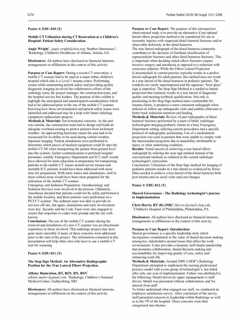
Poster #: EDU-010 (T)
Mobile CT Utilization during CT Renovation at a Children's
Hospital: Patient Safety Consideration
Angie Wright1, [email protected]; Stephen Simoneaux
1;
1Radiology, Children's Healthcare of Atlanta, Atlanta, GA
Disclosures: All authors have disclosed no financial interests, arrangements or affiliations in the context of this activity.
Purpose or Case Report: During a recent CT renovation, a mobile CT scanner had to be used at a major urban children's hospital which also is a Level 1 trauma center. Performing exams while maintaining patient safety and providing quality diagnostic imaging involved the collaborative efforts of the radiology team, the project manager, the construction team, and the hospital service line leaders. The purpose of this exhibit is
highlight the anticipated and unanticipated considerations which had to be addressed prior to the use of the mobile CT scanner. Knowing how these environmental and patient safety issues were identified and addressed may be a help with future radiology equipment replacement projects. Methods & Materials: Environmental concerns: As the unit was outside, the construction team had to design and build an
adequate overhead awning to protect patients from inclement weather. An approaching hurricane meant the area had to be reassessed for its ability to be used during violent weather. Inpatient imaging: Patient simulations were performed to determine which pieces of medical equipment could fit onto the mobile CT lift when transporting the patient from ground level
into the scanner. Earlier communication to pertinent hospital personnel, notably Emergency Department and ICU staff, would have allowed for more education in preparation for transporting patients to the mobile CT. Issues around severe trauma and unstable ICU patients would have been addressed with more time for preparation. With more notice and simulations, staff in these critical areas would have been more prepared for the
utilization of the mobile CT scanner. Emergency and Sedation Preparation: Anesthesiology and Sedation Services were involved in the process. Ultimately, Anesthesia decided that patients could not be safely performed at the mobile location, and these patients were performed on a PET/CT scanner. The sedation team was able to provide its services off-site, but again, simulations and early involvement
were key. Security and the Code Team were also engaged to ensure that responses to codes were prompt and the site well-known. Conclusions: The use of the mobile CT scanner during the removal and installation of a new CT scanner was an educational experience to those involved. This radiology project may have
gone more smoothly if many of these concerns were addressed prior to the start of the project. The information contained in this presentation will help other sites who have to use a mobile CT unit for scanning.
Poster #: EDU-011 (T)
The Stop Sign Method: An Alternative Radiographic
Position for the True Lateral Elbow Projection. Allister Ramrattan, RT, BSN, RN, BSN
1
[email protected]; 1Radiology, Children’s National
Medical Center, Gaithersburg, MD
Disclosures: All authors have disclosed no financial interests, arrangements or affiliations in the context of this activity.
Purpose or Case Report: The purpose of this retrospective observational study is to provide an alternative if not optimal lateral elbow projection method to be considered for use in traumatic injuries with suspected distal humeral fractures and/or observable deformity at the distal humerus.
The true lateral radiograph of the distal humerus commonly contributes to the decision of Gartland classification of supracondylar fractures and other distal humerus fractures. This is important when deciding which elbow fractures require invasive surgery and anesthesia as opposed to a reduction with conscious sedation. While the Elbow Lateral Projection
(Lateromedial) in current practice typically results in a perfect lateral radiograph for adult patients, this method does not result in a true lateral of the distal humerus in pediatric patients. The condyles are rarely superimposed and the signature “hour glass” sign is imperfect. The Stop Sign Method is a medial to lateral projection that routinely results in a true lateral of diagnostic quality and meeting textbook qualification. Not only is
positioning in the Stop Sign method more comfortable for trauma clients, it produces a more consistent radiograph when utilized on follow-up orthopaedic visits and enables clinicians to better track reduction retention and healing. Methods & Materials: Review of past radiographs of distal humeral fractures performed by a team of blind, radiologic technologists imaging pediatric clients in a busy Emergency
Department setting, utilizing current procedures and a specific protocol of radiographic positioning. Use of a mediolateral projection was used in patients that were unable to comply with the lateromedial projection due to immobility attributable to injury or other underlying condition. Results: Noted success in achieving a true lateral elbow
radiograph by utilizing the stop sign method instead of the conventional methods as outlined in the current radiologic technologist's curriculum. Conclusions: Utilization of the Stop Sign method for imaging of pediatric patients results in a true lateral as evidenced by fewer films needed to achieve a true lateral of the distal humerus both post trauma and on serial visits and cast changes.
Poster #: EDU-012 (T)
Shared Governance - The Radiology technologist’s journey
to Implementation Chris Harris, RT (R) (MR)
1Children's Hospital of Philadelphia, Philadelphia, PA
Disclosures: All authors have disclosed no financial interests, arrangements or affiliations in the context of this activity.
Purpose or Case Report: Introduction: Shared governance is a specific leadership style which incorporates commitment to the value of shared decision-making among key stakeholders around issues that affect the work environment. It also provides a dynamic staff-leader partnership that promotes collaboration, shared decision making and accountability for improving quality of care, safety and enhancing work life
Methods & Materials: Around 2009, CHOP’s Radiology Department attempted to implement the nursing professional practice model with a core group of technologist’s, but failed after only one year of implementation. Failure was attributed to the following: Model driven by upper management vs staff driven; Model was presented without collaboration; and No
interest from staff To better understand what engaged our staff, we conducted an employee satisfaction survey. After conclusion of the survey, staff presented concerns to leadership within Radiology as well as to the VP of the hospital. These concerns were then categorized into themes:
Pediatr Radiol (2018) 48 (Suppl 1):S1–S298S276
Recognition/retention, education opportunities for staff, staff support to our patients, having input into decisions and communication within the department. Results: With this knowledge at hand, we relooked at our past shared governance model and design a program by which these
themes could be addressed, while improving patient care and the work environment. We set up eight committees which balanced staff and the following leadership concerns: Recognition and retention, Patient centered care, Policy and procedures, Ancillary partnerships, Education, Quality improvement, Research/Equipment and Imaging protocols. Each
group provided a mission, developed by them. Meetings and, topics are managed by staff. . Committees are supported by Team Leaders in the form of coverage to attend meetings, time to work on projects, dollars for projects, and ensuring that solutions meet hospital standards and policies. Leadership views this as a great opportunity to vet complaints and process and work issues to these groups to look for solutions.
Conclusions: Challenges associated with the introduction of a nurse driven professional practice model into a technologist practice model suggests that greater attention to implementation may be required much more than a top down approach. Understanding what drives your staff, collaboration on these ideals and supporting this practice model makes for a successful program.
Poster #: EDU-013 (T)
Driving the path to easing anxiety for pediatric Radiology
patients
Dana Brinson, BHA, RT(R)(ARRT)1,
[email protected]; Nikki Butler, BMSc, RT(R)(QM)
1;
1Radiology, Children's Healthcare of Atlanta,
Atlanta, GA Disclosures: All authors have disclosed no financial interests, arrangements or affiliations in the context of this activity.
Purpose or Case Report: For pediatric patients, the hallway leading to a radiology imaging room may be just as anxiety-inducing as the exam itself. In an effort to counteract a child’s anxiety, rideable remote control cars were purchased to begin the distraction process at the child’s first step towards the Radiology Department.
Methods & Materials: Through charitable donations, two cars were purchased for $400 each to be used in a trial for patient transport throughout Radiology. Six staff members, across different shifts, were identified as Super Users and trained based on the Operation Manual provided by the manufacturer. Competency was deemed through staff demonstration of
operating the remote control cars safely and practicing all safety measures associated with patient riders. The riders must wear the safety belt, keep all body parts inside the vehicle, be age three years or older and weigh under 60 pounds. There is a switch to deactivate the foot pedals; however there is not a mechanism to deactivate the steering wheel. Therefore, patients are asked to place their hands by their sides. Another consideration was
storage of the cars, as multiple cars require a significant amount of space for parking and electrical outlet needs for recharging the batteries. Storing the cars in a publicly accessible location was not ideal for safety concerns. Ultimately, an alcove by a set of elevators was selected for easy access by staff during the day and the MRI Mock Scanner Room was utilized for recharging at
night. Results: The success of the rideable remote control car program was evident through positive feedback received on customer service surveys. A video posting on Facebook resulted in a local news story highlighting the cars and the fun experience for one patient, thus providing marketing exposure affirmative of the
care being provided. The project returned favorable results, prompting the facility to add the rideable cars to a wish list that is shared with charitable donors. A total of 20 cars are now available for use throughout Radiology and other departments across the system.
Conclusions: While the project initially centered on the reduction of patient anxiety, additional benefits identified were stress alleviation for the parents and entertainment for patient's siblings. The ride from the Radiology Waiting Room to the exam room also allowed the technologists to develop a rapport with the patient that allowed for more cooperation during the imaging
study.
Poster #: EDU-014 (T)
Low Dose Imaging for Feeding Tube Confirmation Colleen Flowers, Associates Degree
Summer Kaplan, MD, MS1, Xiaowei Zhu
1;
1Radiology,
Children’s Hospital of Philadelphia, Philadelphia, PA Disclosures: All authors have disclosed no financial interests, arrangements or affiliations in the context of this activity. Purpose or Case Report: The objective for a low dose imaging
protocol was to eliminate patient risks and to reduce radiation doses. Tube placements are confirmed via imaging thereby avoiding utilization of malpositioned tubes. Reduced technical parameters lessen the absorbed skin dose. A limited field of view which includes distal espohagus and airway allows accurate assessment of feeding tube position. The new region of interest
eliminates unnecessary exposure to hypersensitive organs including, thyroid and reproductive structures. This coned down view requires a lower image quality thereby allowing a lower dose approach. Methods & Materials: A multidisciplinary team discussed current workflows, process improvements. and anticipated obstacles. Workflows were mapped out for all patient visit types,
providers and services. Exam codes were developed with associated CPT charges. Order panels for providers were updated. Macro templates for the Radiologist were developed and loaded at workstations. Lucite block and phantom testing were conducted using equivalent sized infant, toddler and adolescent phantoms on dedicated and portable units. Testing concentrated on portable
units but table bucky techniques were also established. A 40” source to image distance (SID) was used for table (grid) testing and a 36” SID for portable/tabletop. A 6fr feeding tube was used to simulate placement. Established abdomen techniques were selected for corresponding phantom. The KVP factors were unchanged and mAS settings were decreased to lowest outputs
by the units. The lowest mAs 1.25 mAs (table grid) was 0.32 for portable (non-grid). The field of view was mid-chest to iliac crest with proper collimation. Radiologist reviewed images and approved for trial. Exams performed using the new protocol. Image quality remained comparable to phantom testing with same technical factors. Results: The ongoing trial of portable images reflects optimal
for tube confirmation using the lowest mAs setting on unit (0.32 mAs). The abdominal KVP settings remained constant for adequate penetration. Conclusions: The new protocol has a huge benefit to patients. The objective for chest and abdomen imaging is impressively different than a tube check therefore less dose can be utilized.
Full dose images are essential for accurate clinical correlations. Mid chest to crest region reduces potential risk of malpositioned tubes as well as significantly reduces unnecessary dose to hypersensitive organs. This is vital when multiple images are required to confirm tube placements.
Pediatr Radiol (2018) 48 (Suppl 1):S1–S298 S277
Poster #: EDU-015 (T)
Pediatric Shielding Education for Imaging Staff Barbara Karl
1, [email protected]; Christina Sammet,
PhD, DABR1, Caitlin Maher
1;
1Medical Imaging, Ann & Robert
H. Lurie Children’s of Chicago, IL Disclosures: All authors have disclosed no financial interests, arrangements or affiliations in the context of this activity.
Purpose or Case Report: The goal of this study was to develop and share a consistent policy and procedure for the appropriate use of patient shielding in the pediatric environment. Education was developed to increase the level of awareness and skill sets to properly shield, or not shield, pediatric patients. The education was distributed to adult affiliated hospitals and within our institution to ensure consistency of care and safety measures.
Methods & Materials: A comprehensive patient shielding policy was developed by a team of technologists, medical physicists, and managers within our institution. An educational presentation was created to standardize acceptable shielding practices for CT, fluoroscopy and diagnostic radiology. The education was based on a retrospective review of cases from our radiation dose monitoring system and PACS, as well as literature
searches for best practices in patient shielding. We provided examples of proper and improper shielding on diagnostic images and demonstrate the impact of shielding on both image quality and radiation dose. A competency quiz was developed to capture understanding of the material. Results: ● Staff were educated on how to accurately shield
patients for diagnostic imaging. ● Staff have a document on proper patient shielding to reference when needed. ● Staff were better prepared to address shielding concerns with parents and patients. ● Staff have more consistent shielding practices across the enterprise.
Conclusions: The Pediatric Shielding Education improved consistency of shielding practices at our institution. ● Staff are now competent on what constitutes appropriate shielding in diagnostic imaging, CT and fluoroscopy. ● Staff demonstrated an improved understanding of our
shielding practices post education. ● Staff feel more empowered to discuss shielding practices with parents and are aware of their resources for more complex questions. ● Image quality and safety has improved in our pediatric patient population.
Poster #: EDU-016 (T) - Withdrawn
Poster #: EDU-017 (T) – Withdrawn
Poster #: EDU-018 (T)
Intussusception: How to Reduce the Problem Dawn Whitson
1Medical
Imaging - X-ray, Ann & Robert H. Lurie Children's Hospital of Chicago, Chicago, IL Disclosures: All authors have disclosed no financial interests,
arrangements or affiliations in the context of this activity. Purpose or Case Report: The purpose of this education is to describe Therapeutic Enema reduction of Intussusception using Hydrostatic and Pneumatic methods. The benefits of Delayed Repeated Enema (DRE) instead of immediate surgical intervention after a failed reduction will be reviewed.
Pediatr Radiol (2018) 48 (Suppl 1):S1–S298S278

2018 AUTHOR INDEX BY ABSTRACT
A
Abbey, Craig Paper 089 Abdalla, Wael M. Paper 096 Abdeen, Nishard Paper 147
Abdessalam, Shahab Poster EDU-030 Abramowicz, Shelly Paper 007 Abu-El-Haija, Maisam Paper 074 Acevedo, Silvia A. Poster CR-001 (T) Acharya, Patricia T. Paper 048 Acord, Michael Paper 015
Paper 016
Paper 017 Adams, Janet M. Poster EDU-031 Adeb, Melkamu Paper 047 Adler, Brent H. Paper 064
Poster SCI-009 Poster SCI-038
Adzick, N S. Paper 055 Afacan, Onur Paper 046 Agnew, Amanda Poster SCI-038 Agrawal, Hitesh Paper 082 Aguet, Julien Paper 007 Ahmad, Rizwan Poster SCI-003 Ahmed, Firas S. Paper 071
Ahmed, Ilham M. Poster EDU-053 Akbari, Yasmin S. Poster EDU-090 Akikusa, Jonathan Poster SCI-036 Alazraki, Adina Poster EDU-024 Albers, Brittany Poster EDU-061
Poster SCI-002 Aldraihem, Ahmed Paper 024
Alessio, Adam Paper 106 Poster SCI-023 Poster SCI-024 Poster SCI-043
Allen, Brian C. Paper 091 Paper 092
Alqublan, Lina Paper 042 Alsharief, Alaa N. Paper 032
Poster EDU-069 Alson, Mark D. Poster EDU-055 Altinok, Deniz Paper 019 Alves, Timothy Poster EDU-044
Poster EDU-068
Alves Rosa, Joao Paper 023 Amarneh, Mohammad Paper 012 Ames, Jeff Poster EDU-025 Amirabadi, Afsaneh Paper 032 Andronikou, Savvas Paper 023
Poster EDU-036 Angel, Jacqueline Paper 022
Annapragada, Ananth Paper 018 Paper 030 Paper 031 Paper 115 Paper 131
Anooshiravani, Merak Paper 162
Ansdell, David Poster SCI-023 Poster SCI-024
Anton, Christopher Paper 001 Anum, Syeda Poster EDU-043 Anupindi, Sudha A. Paper 006
Paper 079 Paper 088
Paper 136 Aoki, Hidekazu Poster SCI-040
Appenzeller, Simone
Paper 007
Aquino, Michael R. Paper 070 Aria, David J. Poster EDU-060 Arroyave-Wessel, Margarita Paper 103 Artunduaga, Maddy Paper 008
Paper 083
Paper 118 Paper 119
Artz, Nathan S. Poster EDU-054 Arvidsson, Linda Z. Paper 007 Ashton, Daniel Paper 159
Paper 160
Paper 161 Paper 165
Augustyn, Robyn Paper 006 (T) Paper 007 (T) Paper 090 Poster EDU-079
Austin, Frances Poster CR-007
Averill, Lauren W. Paper 040 Poster EDU-073
Ayaz, Muhammad Poster EDU-072 Ayyala, Rama Paper 071
Poster SCI-006
B
Baad, Michael Paper 139 Babyn, Paul Poster EDU-069 Back, Susan J. Paper 047
Paper 049
Paper 079 Paper 112 Paper 140 Poster EDU-057
Bagwell, Charles Poster CR-007 Bahrami, Armita Paper 128
Bajaj, Manish Poster CR-012 Bakeit, Mohamed Salih A. Poster EDU-053 Baldridge, Alan Poster SCI-020 Bandarkar, Anjum Poster EDU-002 (T) Bardo, Dianna Paper 006 (T)
Paper 007 (T) Paper 090
Paper 151 Poster EDU-001 Poster EDU-079
Barnacle, Alex M. Paper 014 Barnes, Craig E. Poster SCI-050 Barrera, Christian A. Paper 027
Paper 038
Paper 081 Paper 120 Paper 121 Paper 145 Poster EDU-045 Poster EDU-099
Barth, Richard Paper 045 Basu, Sanmit K. Paper 084 Baxi, Ameya Poster EDU-022 Beavers, Angela J. Poster CR-002 Becker, Robert Poster EDU-047
Poster EDU-085 Bedoya, Maria A. Paper 120
Bell, Bryce Paper 008
Pediatr Radiol (2018) 48 (Suppl 1):S1–S298 S279

Bellah, Richard Paper 049
Paper 112
Paper 139 Poster EDU-045 Poster EDU-057
Benkert, Thomas Paper 116 Poster SCI-009
Bennett, Paula S. Paper 003 (T) Benson, Benjamin Paper 146
Bentley, Jason Paper 157 Berhane, Haben Paper 076 Berkovich, Rachel Poster EDU-094 Bertocci, Gina Paper 029 Bhargava, Nikhilesh K. Paper 110 Bhargava, Ravi Paper 110
Bhatia, Aashim Paper 113 Bhavane, Rohan Paper 018 Biko, David M. Paper 006
Paper 036 Paper 079 Paper 081 Paper 085
Poster EDU-100 Biyyam, Deepa R. Paper 150
Paper 151 Poster CR-004
Blagdon, Ryan Poster EDU-014 Blake, Crystal Poster EDU-009 (T) Blancq, Terry Poster EDU-054
Blask, Anna R. Paper 057 Paper 107 Poster SCI-008
Block, Kai Tobias Paper 116 Poster SCI-009
Blumfield, Einat Paper 146
Poster EDU-018 Poster EDU-034
Bobbey, Adam Paper 134 Bober, Michael Paper 040 Bohnsack, John F. Poster EDU-075 Boop, Frederick Paper 021 Bowen, Diana K. Paper 049
Brady, Samuel Poster EDU-054 Braithwaite, Kiery Poster EDU-066
Poster SCI-013 Braojos, Fernanda d. Paper 105
Paper 108 Breen, Micheal A. Paper 035 Breguet, Romain Paper 162
Brink, Farah Poster SCI-038 Brinson, Dana M. Poster EDU-013 (T) Brondell, Ashley Paper 002 (T) Brown, Brandon P. Paper 056
Paper 059 Paper 141
Poster EDU-016 Poster SCI-014
Brown, Jessica Paper 159 Brown, Travis Paper 013 Browne, Lorna Paper 080
Poster EDU-093 Bulas, Dorothy Paper 057
Paper 103 Paper 107 Poster EDU-002 (T) Poster SCI-008
Bunge, Martin Paper 042 Buskirk, Tricia Paper 144
Poster SCI-044
Butler, Nikki Poster EDU-013 (T)
C
Cahill, Anne Marie Paper 015 Paper 016 Paper 017
Cain, Timothy Paper 127 Caldwell, Mallory Paper 069
Calvo-Garcia, Maria A. Paper 109 Cantu, Cera Poster SCI-013 Cao, Joseph Poster CR-013 Carry, Patrick Poster SCI-039 Cassady, Christopher Paper 062
Paper 105 Cassella, Katharyn D. Poster EDU-016
Castro, Denise A. Poster EDU-103 Castro, Eumenia Paper 062 Cecil, Aaron Paper 068 Ceppe, Agathe Paper 037 Chan, Alvin P. Poster EDU-039 Chan, Sherwin S. Paper 100
Poster SCI-017 Chapman, Teresa Paper 106 Chapnick, Melissa Paper 010 Chastain, Katherine Paper 013 Chastain, Laura Poster EDU-009 (T) Chaturvedi, Apeksha Poster EDU-002 Chau, Alex Paper 160
Paper 161 Chauvin, Nancy Paper 120
Paper 121 Chawla, Soni C. Poster EDU-006
Poster EDU-033 Poster EDU-039 Poster EDU-040
Chen, Alan M. Poster EDU-098 Chen, Johnathan Poster EDU-006 Chen, Lingyun Paper 090
Poster EDU-001 Poster EDU-079
Cheng, Earl Y. Poster SCI-022
Cheng, Joseph Y. Paper 045 Chippington, Samantha Paper 014 Choi, Jungwhan J. Paper 005 Chow, Jeanne S. Paper 046
Paper 050 Paper 051
Christensen, Jason Paper 113
Chu, Carter Paper 153 Chu, Zili D. Paper 153 Chughtai, Komal Poster EDU-002 Chukus, Anjeza Paper 114
Poster EDU-074 Chung, Chul Y. Poster EDU-055 Chung, Daniel Paper 026
Cielma, Tara Poster EDU-002 (T) Poster EDU-003 (T)
Cina, Robert Poster SCI-010 Ciullo, Sean Poster SCI-012 Clarke, Rebekah Poster CR-013 Cleveland, Heather Paper 160
Paper 161 Paper 165
Cohen, Harris L. Poster EDU-038 Coleman, Beverly G. Paper 055
Paper 102 Collard, Michael Poster EDU-058 Collins, John Poster EDU-080
Commander, Clayton Paper 068 Conrad, Máire A. Paper 006 Consunji-Araneta, Raquel Paper 042 Conway, Deborah Poster CR-008 Crane, Gabriella Poster CR-001
Pediatr Radiol (2018) 48 (Suppl 1):S1–S298S280

Crawford, Jackie Paper 088 Cripe, Linda Paper 156
Cristante, Caitlin Paper 103 Cronan, Julie C. Paper 163 Crowley, John J. Poster EDU-059 Cully, Brent Paper 013
Poster SCI-032 Cure, Carlos Paper 103 Curran, David Paper 004 (T)
Curran, John Poster EDU-079 Currie, Melissa Paper 029
D
D'Alessandro, Donna M. Paper 138 D'Alessandro, Michael P. Paper 138 Dahl, Amy Poster SCI-017 Dahmoush, Hisham M. Poster EDU-074 Dameron, Leah Paper 039 Daneman, Alan Poster EDU-021
Dao, Kimberly Paper 052 Darge, Kassa Paper 047
Paper 049 Paper 079 Poster EDU-042 Poster EDU-051
DasGupta, Roshni Poster SCI-031
Davey, Marcus Paper 102 Davis, James Paper 096
Poster SCI-007 Davis, Joseph Paper 086
Paper 091 Paper 092
Dawoud, Malik Poster SCI-030
Deaver, Pamela Paper 062 DeBari, Suzanne E. Paper 055 DeBiasi, Roberta Paper 103 DeFoor, William Poster SCI-026 Degnan, Andrew J. Poster EDU-055
Poster EDU-100
Delgado, Jorge Paper 120 Paper 121
Demshur, Lesley Poster EDU-002 (T) Deng, Jie Paper 001 (T) Desai, Nilesh Paper 104
Paper 105 Desilet-Dobbs, Debbie Poster EDU-052
Desoky, Sarah Poster CR-003 Dewitt, Aaron G. Paper 085 Dhody, Anna Paper 061 Diaz, Eric Paper 098 Dickie, Belinda Paper 050 Didier, Ryne Paper 055
Paper 058
Paper 102 Dietz, Kelly Poster EDU-025
Poster SCI-020 Dillman, Jonathan R. Paper 001
Paper 004 Paper 067
Paper 073 Paper 075 Paper 094 Paper 098 Paper 101 Paper 129 Poster SCI-026
Dinan, David Paper 070 Ditmars, Frederick Paper 124 Ditro, Colleen Paper 040
Ditzler, Matthew Paper 008 Paper 118
Paper 119 Paper 125 Paper 126
DiVasta, Amy D. Paper 033 Dodd, Nicholas A. Paper 097 Dolan, Kevin Paper 039
Paper 065
Don, Steven Paper 089 Paper 093
Donaldson, James Poster SCI-033 Dong, Su-Zhen Paper 053
Paper 054 Poster EDU-010
Donnelly, Lane F. Paper 063 Donuru, Achala Poster EDU-073 Dori, Yoav Paper 036
Paper 085 Doria, Andrea S. Paper 007
Paper 032 Poster EDU-064
Poster EDU-069 Poster SCI-036
Douglas, Katherine Paper 010 Downs, Elissa Poster SCI-020 Drum, Allisin Paper 096 Duba, Irene Paper 034 Dubinsky, Marla Paper 005
du Plessis, Adre Paper 103 Durand, Rachelle Poster SCI-019 Duron, Vincent Poster SCI-006 Duszak, Richard Poster EDU-055
E
Ecklund, Kirsten Paper 033 Edgar, Christopher J. Paper 015
Paper 058 Paper 061
Poster SCI-019 Edward, Ross Paper 084 Egan, Craig Paper 151 Ehrlich, Lauren Poster EDU-041 Eid, Hadeel A. Paper 024 Eing, Julee Paper 064 Ellison, David W. Paper 021
Emad-Eldin, Sally Paper 047 Emrick, Lisa Paper 104 Eovaldi, Benjamin Poster EDU-038 Erdem Toslak, Iclal Paper 132 Escobar, Fernando Paper 015
Paper 016 Paper 017
Essenmacher, Alex Poster EDU-019 Estroff, Judy A. Paper 060 Eutsler, Eric Paper 034
F
Fadell, Michael Poster SCI-039 Fagen, Kimberly E. Poster SCI-008 Fanella, Sergio Paper 042 Farkas, Amy Poster EDU-008
Poster EDU-013
Poster EDU-015 Poster EDU-081 Poster EDU-088 Poster EDU-089
Farmakis, Shannon G. Paper 044 Poster CR-005
Fei, Lin Paper 074
Feinstein, Kate A. Poster EDU-026
Pediatr Radiol (2018) 48 (Suppl 1):S1–S298 S281

Feldman, Brian M. Paper 007 Feldman, Henry Paper 033
Fenlon, Edward P. Paper 038 Ferreira da Silva, Renato Poster EDU-017
Poster EDU-101 Fiaz, Aziekah Paper 133 Fickenscher, Kristin A. Poster SCI-017 Finkle, Joshua Poster EDU-026
Poster EDU-076
Flake, Alan W. Paper 102 Flibotte, John Paper 112 Flink, Carl Poster SCI-022 Flowers, Colleen Paper 088
Poster EDU-014 (T) Flug, Jonathan Poster SCI-039
Fogel, Mark A. Paper 085 Forbes-Amrhein, Monica Paper 056 Fordham, Lynn Paper 037
Paper 068 Fourzali, Yamil Paper 103 Fowler, Kathryn Paper 077 Fox, William W. Poster EDU-100
Francavilla, Michael L. Paper 038 Paper 136 Paper 139 Paper 143 Poster SCI-019
Franceschi, Dora Paper 037 Francois, Christopher Poster EDU-003
Fraum, Tyler Paper 077 Friedman, Seth Paper 043
Poster SCI-043 Friedmann, Alison M. Poster SCI-048 Frush, Daniel J. Paper 068 Frush, Don Paper 091
Paper 092 Furtado, Andre Poster EDU-090
G
Gabir, Abdalla M. Poster EDU-053 Gallegos Riofrio, Carlos Andres Paper 010 Gardner, Gregory Paper 159 Gardner, Jeffrey T. Poster EDU-083 Gariepy, Cheryl Paper 099 Garrett, Whitney Paper 144 Gates, Erica L. Paper 004
Paper 067 Gazzi, Lynn M. Poster EDU-006 (T) Geary, Maeve Paper 051 Gebarski, Kathleen S. Poster EDU-044 Gee, Michael S. Paper 003
Poster SCI-048 Ghaghada, Ketan B. Paper 018
Paper 131 Ghahremani, Shahnaz Poster EDU-006
Poster EDU-039 Ghaleb, Nancy Poster SCI-030 Gharavi, Mohamad H. Poster EDU-086 Gibbs, Liliane Poster SCI-041
Gibson, Donald P. Poster CR-014 Gilbertson-Dahdal, Dorothy Poster CR-003 Gill, Anne Paper 002 (T)
Paper 163 Gill, Kara G. Poster EDU-065 Gillespie, Matthew J. Paper 085 Giordano, Richard Poster SCI-026
Glatz, Andrew C. Paper 085 Glavis-Bloom, Justin Poster SCI-016
Poster SCI-029 Goehner, Melissa Paper 010 (T) Goette, Matthew Paper 097
Gokli, Ami Paper 006 Poster EDU-045
Goldberg, David J. Paper 085 Goldman-Yassen, Adam Paper 149
Poster EDU-018 Goldwasser, Bernard Poster EDU-048
Poster EDU-087 Poster SCI-012
Gondi, Keerthi Poster SCI-032
Gong, Edward M. Poster SCI-022 Goodarzian, Fariba Poster EDU-094 Goralski, Jennifer Paper 037 Gordon, Catherine M. Paper 033 Gorfu, Yocabel Poster EDU-051 Gottumukkala, Ravi V. Paper 003
Gowda, Madhu Poster CR-007 Goyal, Alka Paper 100 Graeber, Brendon Poster EDU-041 Gramke, Michelle M. Poster EDU-031 Grant, Rosemary Paper 051 Gray, Brian Paper 056
Poster SCI-014
Gray, Carlos Paper 009 (T) Green, Jared Poster SCI-033 Greenberg, S Bruce Poster EDU-005 Greene, Stephanie Poster CR-010 Greer, Mary-Louise Paper 002
Paper 032 Grekoski, Vincent Poster SCI-005
Griffin, Lindsay Poster EDU-003 Griffis, Heather Paper 085 Grigonis, George Paper 061 Gross, Jordana Paper 149 Grundlehner, Paul Poster CR-014 Guillerman, R. P. Paper 041
Guillot, Lori Paper 026 Guimaraes, Carolina Paper 104
Paper 105 Paper 108
Guirguis, Albair Poster EDU-028 Guleria, Saurabh Paper 007 Gumus, Serter Poster EDU-078
Gunderman, Richard Poster EDU-053 Gunnell, Elias T. Paper 037 Guo, Chen Poster SCI-004 Gupta, Anita Paper 129
H
Habif, David Paper 010 Hackman, Lucy Paper 084 Hagedorn, Kelly Paper 069 Haider, Maera Paper 019 Hailu, Tigist Paper 136
Paper 139 Paper 140
Halalsheh, Hadeel Paper 128 Halaweish, Ahmed F. Paper 034 Hallaji, Shareh Poster EDU-080 Halverson, Mark Poster SCI-044
Hammill, Adrienne Poster SCI-031 Hampilos, Perry Poster SCI-048 Handa, Atsuhiko Poster EDU-017
Poster EDU-077 Poster EDU-085
Handler, Marci R. Poster EDU-037 Poster EDU-050
Handly, Brian Paper 037 Paper 068
Hankins, Jane S. Paper 158 Hanquinet, Sylviane Paper 162
Pediatr Radiol (2018) 48 (Suppl 1):S1–S298S282

Harreld, Julie H. Paper 022
Poster EDU-072
Harris, Chris Poster EDU-012 (T) Harris, Matthew A. Paper 085 Harris, Robert D. Poster EDU-056 Harrison, Andrew J. Poster EDU-093 Hartman, Alison Paper 037 Hartman, Terry Paper 068 Haruno, Lee Poster SCI-037
Hasham, Hasnain Paper 013 Hawkes, Michael Paper 110 Hawkins, C. Matthew Paper 002 (T)
Paper 163 Hayatghaibi, Shireen Paper 069
Paper 160
Paper 165 Hedlund, Alec Poster EDU-075 Hedlund, Gary L. Poster CR-009
Poster EDU-075 Poster SCI-047
Heitkamp, Darel E. Poster EDU-053 Hekler, Lindsey M. Poster EDU-031
Heller, Richard E. Paper 142 Henault, Kathryn Poster EDU-004 Hendrickson, Richard Paper 013
Poster SCI-032 Herbst, Timothy Poster EDU-004 Hernandez, Jose A. Paper 159
Paper 160
Paper 161 Paper 165
Hernando, Diego Paper 157 Hernanz-Schulman, Marta Poster CR-001
Poster EDU-027 Poster SCI-001
Herregods, Nele Poster SCI-036 Heuer, Gregory Paper 112 HIbbard, Roberta Poster EDU-053 Hidalgo, Khristina Paper 009 (T) Higgins, Richard Paper 042 Highmore, Kerri Poster SCI-036 Hill, Jeanne Poster SCI-010
Hillenbrand, Claudia M. Paper 158 Poster EDU-072
Hingsbergen, Elizabeth A. Paper 064 Hintenlang, David Paper 039 Ho, Connor Paper 152 Ho-Fung, Victor Paper 016
Paper 070
Paper 120 Paper 122 Paper 139 Paper 140
Hoggard, Eric Poster EDU-025 Holdener, Ruth Paper 089
Holm, Tara Poster EDU-025 Hooker, Jeffrey Poster EDU-081
Poster EDU-088 Poster EDU-089
Hopely, Brian R. Paper 140 Hopyan, Sevan Paper 032 Hor, Kan Alternate Paper
Paper 084 Paper 156 Poster SCI-003
Hornsby, Richard Poster EDU-054 Hough, Matthew Poster SCI-005 Howard-Claudio, Candace Poster EDU-008 Howell, Lori J. Paper 055
Hryhorczuk, Anastasia Paper 164 Hsu, Ariel Paper 025
Hu, Houchun Alternate Paper Paper 039
Paper 064 Paper 099 Paper 116 Paper 117 Paper 124 Paper 156 Poster SCI-003
Poster SCI-009 Poster SCI-044
Hu, Li-Wei Poster SCI-004 Hubbard, Caroline Poster SCI-010 Hughes, John Paper 080 Humphreys, James F. Paper 130
Hunte, David E. Paper 101
I
Iannotti, Lora Paper 010
Iles, Benjamin W. Poster SCI-035 Imbus, Rebecca Paper 004 Inarejos Clemente, Emilio J. Paper 007
Poster SCI-036 Inscoe, Christy R. Paper 037 Irving, Sharon Paper 088 Ishak, Gisele E. Paper 106
Iskander, Paul J. Poster EDU-006 Itkin, Maxim Paper 036 Iv, Michael Paper 114 Iyer, Ramesh Paper 066
J
Jacobson, Deborah L. Poster SCI-022 Jadhav, Siddharth P. Paper 008
Paper 041 Paper 082 Paper 083
Paper 118 Paper 119 Paper 125 Paper 126 Paper 152 Paper 154
James, Charles Poster SCI-030 Jans, Lennart Poster SCI-036 Jaramillo, Diego Paper 120
Paper 121 Jaremko, Jacob Lester Poster EDU-064
Poster SCI-036 Paper 007
Jasmin, Kimberly C. Paper 063 Javadi, Ariyan Poster EDU-076 Jimenez, Jesus Poster SCI-006 John, Susan D. Paper 070 Johnson, Ann Paper 055
Paper 088 Paper 139
Johnson, Emilie K. Poster SCI-022 Johnson, Neil Poster SCI-034 Johnson, Oren Paper 035 Johnstone, Jordan A. Paper 036
Paper 085 Jones, Jeremy Paper 116
Paper 117 Jones, Richard H. Paper 157 Jorina, Maria Paper 051 Joshi, Aparna Poster EDU-062
Poster EDU-091 Joshi, Hena Poster EDU-024
Pediatr Radiol (2018) 48 (Suppl 1):S1–S298 S283

Joyner, David Poster EDU-081
Poster EDU-088
Poster EDU-089 Junhasavasdikul, Thitiporn Paper 007
K
Kalra, Kunal Poster EDU-062 Kan, Elaine Y. Paper 111
Kan, J. H. Paper 008 Paper 030 Paper 031 Paper 118 Paper 119 Poster SCI-037
Kandil, Ali Poster SCI-034
Kandula, Vinay Poster EDU-073 Kao, Simon Poster EDU-019 Kaplan, Summer Paper 038
Paper 088 Paper 112 Paper 139
Poster EDU-014 (T) Poster EDU-099 Poster SCI-019
Karczewski, Arleen Paper 144 Karl, Barbara A. Poster EDU-015 (T) Karmazyn, Boaz Poster EDU-032
Poster EDU-053
Poster EDU-063 Poster SCI-014 Poster SCI-015
Karpewycz, John Paper 002 Kasser, James R. Paper 060
Paper 123 Kaste, Sue C. Paper 128
Paper 130 Kaye, Robin Poster EDU-060 Kazmi, Faaiza Poster EDU-007
Poster EDU-046 Poster EDU-048 Poster EDU-082
Poster EDU-087 Poster EDU-097
Kearney, Debra Paper 062 Kecskemethy, Heidi Paper 040 Kelleher, Sean Poster EDU-049 Kellenberger, Christian J. Paper 007 Ketwaroo, Pamela Paper 041
Paper 083 Keyak, Joyce H. Poster SCI-041 Khalek, Nahla Paper 055 Khanna, Geetika Paper 077
Poster EDU-017 Poster SCI-011
Khrichenko, Dmitry Paper 058
Khwaja, Asef Paper 139 Kikke, Stacy Poster CR-003 (T) Kim, Jane Poster SCI-027 Kim, Stanley Poster SCI-033 Kim, Sung Poster EDU-056 King, Sebastian Paper 002
Kino, Aya Paper 045 Kirkhus, Eva Paper 007
Poster SCI-036 Kleinman, Paul K. Paper 028
Paper 123 Klimo Jr, Paul Paper 021 Kline-Fath, Beth M. Paper 109
Koberlein, George Paper 070 Kolar, Mila Poster EDU-103
Kolluri, Akhil Poster CR-007 Koos, Bernd Paper 007
Kousa, Youssef Paper 103 Kovalchin, John Paper 084 Krance, Robert Paper 097 Krishnamurthy, Rajesh Alternate Paper
Paper 039 Paper 041 Paper 064
Paper 065 Paper 084 Paper 099 Paper 116 Paper 117 Paper 156
Poster SCI-003 Poster SCI-009
Krishnamurthy, Ramkumar Alternate Paper Paper 084 Paper 099 Paper 116 Paper 117
Paper 156 Poster SCI-003 Poster SCI-009 Poster SCI-044
Kriss, Spencer Paper 029 Kriss, Vesna M. Paper 029 Kucera, Emily Paper 100
Kukreja, Kamlesh Paper 159 Paper 160
Kuo-Bonde, Lydia Poster SCI-018 Kurian, Jessica Poster EDU-018 Kurtz, Michael Paper 051 Kurugol, Sila Paper 046
Kushare, Indranil Paper 125 Paper 126
Kwong, Yin Yee Paper 111
L
Lai, Lillian Poster EDU-017 Poster EDU-094
Lambert, Elena Poster EDU-036 Lampl, Brooke S. Poster EDU-028
Poster EDU-049 Langer, Jacob Paper 002
Lanier, Michael Poster SCI-011 Laor, Tal Paper 060
Paper 123 LaPointe, Annette P. Paper 003 Larheim, Tore A. Paper 007 Laurent, Meryle Paper 162 LAW, Martin W. Paper 111
Lawrence, Kendall Paper 102 Laxer, Ronald Poster EDU-069 Lee, Bonmyong Poster EDU-012
Poster SCI-049 Lee, Edward Y. Paper 035 Lee, Justin Paper 151
Lee, Richard Paper 046 Lee, Yueh Z. Paper 037 Levin, Terry L. Paper 146
Paper 149 Poster EDU-018 Poster SCI-027
Levine, Matt Paper 069
Lewis, Paul Poster SCI-030 Li, Arleen Paper 155 Lim-Dunham, Jennifer E. Paper 132 Lin, Alice Poster CR-015
Pediatr Radiol (2018) 48 (Suppl 1):S1–S298S284

Lin, Jonathan Poster EDU-039
Poster EDU-040
Lin, Richard Paper 088 Lin, Tom Paper 073 Linam, Leann Poster SCI-013 Linden, Kadine Poster EDU-003 (T) Lindgren, Bruce W. Poster SCI-022 Lindsay, Aaron Paper 148 Lionberg, Alex Poster EDU-096
Lipsich, Jose E. Paper 078 Lish, James Poster CR-015 Liu, Dennis Poster SCI-022 Liu, Yingmin Alternate Paper
Poster SCI-003 Llorens, Carlos Poster EDU-059
Loeffler, Ralf B. Paper 158 Poster EDU-072
Loewen, Jonathan M. Poster SCI-013 Logsdon, Gregory Paper 148 Long, Alexander Paper 156
Poster SCI-044 Loomis, Judyta Paper 057
Poster SCI-008 Lovelace, Jarred Paper 039 Low, Samantha Paper 133 Lu, Jianping Paper 037 Lucin, Michael Poster EDU-047 Ly, Juan Domingo Poster CR-010 Lyons, Karen Poster EDU-005
M
Ma, Grace M. Paper 035 MacDonald, James Paper 124
MacDougall, Robert D. Paper 093 Machnitz, Judit Paper 139 Mackenzie, William Paper 040 Maddocks, Alexis B. Poster SCI-006 Mahdi, Eman Poster SCI-046 Maher, Caitlin Poster EDU-015 (T)
Maitra, Neil Paper 013 Maizels, Max Poster SCI-022 Malik, Archana Poster EDU-007
Poster EDU-046 Poster EDU-048 Poster EDU-082 Poster EDU-087
Poster EDU-097 Mallon, Mary G. Poster EDU-048
Poster EDU-087 Mallon, Mea Poster CR-016
Poster EDU-007 Poster EDU-046 Poster EDU-082
Poster EDU-097 Malone, LaDonna Paper 080 Mammarappallil, Joseph Paper 086 Mamoun, Ihsan Poster EDU-028 Man, Carina Poster EDU-069 Mandell, Gerald A. Poster CR-004
Mannix, Laura A. Poster CR-004 (T) Maravilla, Kenneth Paper 043 Mardis, Neil J. Poster SCI-017 Marin, Daniele Paper 091
Paper 092 Marine, Megan B. Poster EDU-032
Poster EDU-053
Poster EDU-063 Poster SCI-014 Poster SCI-015
Maris, John Paper 015 Markl, Michael Paper 155
Markush, Dor Paper 084 Marshall, Kelley W. Poster EDU-066
Martin, Brendan Paper 132 Martinez-Rios, Claudia Paper 032 Masand, Prakash M. Paper 041
Paper 082 Paper 083 Paper 097 Paper 152
Paper 153 Paper 154
Mata-Mbemba, Daddy Paper 020 Poster EDU-021
Maxfield, Bradley Poster EDU-065 Maxfield, Charles M. Paper 091
Paper 092 May, Lauren A. Paper 041 Maya, Carolina Paper 006
Paper 047 Paper 049 Paper 058 Paper 079
Poster SCI-019 Mayer, Stephanie Poster SCI-039 Mayercik, Vera Paper 164 Mazariegos, George Paper 096 McAllister, Aaron Paper 116 McArthur, Tatum Poster SCI-039 McCarville, Beth Paper 158
Poster EDU-054 McClure, Meghan Poster EDU-030 McCrary, Joseph Poster EDU-035 McKiernan, Patrick Paper 096 McMahon, Lisa Poster SCI-050 McNamara, Erin Paper 050
McPherson, Amie Paper 035 Meckmongkol, Teerin Poster SCI-012 Mehan, William Paper 164 Mehlman, Tracey R. Poster SCI-042 Mehollin-Ray, Amy R. Paper 062
Paper 105 Meltzer, James Paper 146
Menashe, Sarah J. Paper 106 Menegotto, Flavia Paper 024 Menon, Keshav Poster CR-011 Merchant, Thomas E. Paper 021 Merrow, Arnold C. Paper 109 Mery, Carlos Paper 082 Mesi, Erin L. Paper 064
Paper 065 Messer, Diana L. Poster SCI-038 Meyers, Arthur B. Paper 007
Poster SCI-036 Miethke, Alexander Paper 075
Paper 094
Milewicz, Dianna Paper 115 Milks, Kathryn S. Paper 009
Poster SCI-009 Milla, Sarah S. Poster SCI-013 Miller, Aaron J. Poster EDU-053 Miller, Elise Paper 141 Miller, Elka Paper 007
Miller, Jeffrey Poster EDU-071 Poster EDU-079
Miller, Russell Poster SCI-006 Mirza, Sobia Paper 139 Misiura, Anne K. Poster EDU-046
Poster EDU-082 Mitra, Ayan Paper 089
Miyasaka, Mikiko Poster SCI-040 Miyazaki, Osamu Poster SCI-040
Pediatr Radiol (2018) 48 (Suppl 1):S1–S298 S285

Mobberley-Schuman, Paula Poster SCI-031 Mohammad, Saeed Poster SCI-033
Mohammed, Alaaeldeen M. Poster EDU-053 Mohandas, Aravind N. Paper 019 Moldenhauer, Julie Paper 058 Molossi, Silvana Paper 082 Mong, Andrew Paper 080 Moore, Mary Poster SCI-030 Morales, Gabriel Paper 001 (T)
Morales, Robert Poster EDU-086 More, Snehal R. Paper 083
Paper 125 Paper 126
Morello, Frank Poster CR-003 Morgan, Stephen L. Poster CR-008
Morgan, Trudy Paper 079 Morrison, Jessica Paper 039 Mosse, Yael Paper 015 Mostoufi-Moab, Sogol Paper 121 Mouzaki, Marialena Paper 101 Mrak, Lara Poster EDU-065 Mulkern, Robert Paper 033
Mulkey, Sarah Paper 103 Murati, Michael A. Poster EDU-025 Murphy, Robyn Poster EDU-037
Poster EDU-050 Murphy, Sandra Paper 130 Mustafa, Elrayah M. Poster EDU-053 Myefski, Patty A. Poster EDU-008 (T)
Myers, Eliza Poster EDU-034
N
Nagaraj, Aaditya Paper 161
Nagaraj, Usha D. Paper 109 Nagpal, Prashant Poster EDU-101 Nahl, Daniel Poster EDU-094
Poster SCI-016 Poster SCI-029
Navid, Fariba Paper 128
Naz, Fozia Paper 147 Ndolo, Josephine M. Paper 113
Poster CR-001 Neel, Michael Paper 130 Nelson, Marvin Poster EDU-094 Nett, Elizabeth Poster EDU-054 Netto, Sparkle M. Paper 067
newton, Allen Paper 113 Ngo, Jennifer S. Paper 091
Paper 092 Ngo, Lawrence Paper 091
Paper 092 Nguyen, HaiThuy N. Paper 108 Nicholas, Jennifer L. Paper 010
Nichols, Reid Paper 046 Noel, Cory Paper 082
Paper 152 Paper 153 Paper 154
Norris, Carrie Paper 059
Nosaka, Shunsuke Poster SCI-040 Novak, Inna Paper 149 Ntoulia, Aikaterini Paper 049
Paper 079
O
O'Byrne, Michael L. Paper 085 O'Donnell, Jennifer Paper 033 O'Hara, Sara M. Poster SCI-026 O'Loughlin, Michael Poster EDU-004 Ogg, Robert J. Paper 021
Oiticica, Claudio Poster CR-007
Okamoto, Reiko Poster SCI-040 Oliver, Edward R. Paper 055
Olson, Andrew Paper 080 Ong, Seng Poster EDU-096 Orth, Robert C. Paper 008
Paper 069 Ortiz-Neira, Clara L Paper 083 Osman, Radya G. Poster EDU-053 Otero, Hansel J. Paper 036
Paper 038 Paper 047 Paper 049 Paper 081 Paper 136 Paper 140
Poster EDU-029 Poster EDU-099 Poster EDU-100
Otjen, Jeffrey P. Poster SCI-023 Poster SCI-024
Ourfalian, Raffi M. Poster EDU-033
P
Padua, Horacio Paper 050 Pai, Vinay Paper 051 Paldino, Michael Paper 025
Paper 104 Paper 160
Paltiel, Harriet J. Paper 050 Panigrahy, Ashok Poster EDU-090 Panwar, Jyoti Paper 007
Poster SCI-036 Pappo, Alberto Paper 128
Parikh, Ashishkumar K. Poster SCI-001 Parisi, Marguerite T. Poster SCI-023
Poster SCI-024 Park, Daniel Paper 068 Park, Ellen Poster EDU-028
Poster EDU-049
Park, James K. Poster EDU-059 Parks, Jim Poster SCI-030 Partington, Sara L. Paper 085 Patay, Zoltan Poster EDU-072 Patel, Bhavik N. Paper 091
Paper 092 Patel, Falguni Paper 005 (T)
Poster CR-002 (T) Patel, Hiten Poster SCI-036 Patel, Manish N. Poster SCI-031
Poster SCI-034 Patel, Mittun Poster CR-004 Patel, Premal Paper 014 Patel, Rakesh M. Paper 073
Pawaroo, Davina Paper 133 Pazienza, Rocco J. Paper 008 (T) Peacock, Zachary S. Paper 007 Pednekar, Amol Paper 152
Paper 153 Paper 154
Pepe, Julie Paper 148 Peranteau, William H. Paper 055 Persohn, Scott Paper 059 Person, Allison Poster SCI-042 Pezeshkmehr, Amir Poster SCI-030 Pfeifer, Cory M. Paper 072
Poster CR-002
Poster CR-011 Poster CR-013 Poster EDU-058 Poster EDU-071
Pham, Huy Poster EDU-083
Pediatr Radiol (2018) 48 (Suppl 1):S1–S298S286

Phan, Becky Paper 005 Phelps, Andrew Poster SCI-027
Pinto, Erin M. Paper 085 Pizarro, Christian Paper 040 Pokorney, Amber Paper 006 (T) Poletto, Erica Poster EDU-007
Poster EDU-046 Poster EDU-048 Poster EDU-082
Poster EDU-087 Poster EDU-097 Poster SCI-012 Poster SCI-027
Poliachik, Sandra Poster SCI-043 Policeni, Bruno Poster CR-012
Politte, David Paper 089 Pomeranz, Christy Paper 027
Paper 145 Poot, Jeffrey D. Poster EDU-078 Popescu, Andrada R. Paper 076 Popp, James E. Paper 009 Poremski, Pamela M. Paper 052
Porterfield, Ronna J. Paper 065 Potter, Carol Paper 099 Powers, Andria M. Poster EDU-030 Poznick, Laura Paper 079 Prasad, Rajeev Poster SCI-012 Priya, Sarv Poster EDU-017
Poster EDU-067
Poster EDU-077 Poster EDU-101
Pruthi, Sumit Poster SCI-001
Q
Qiu, Cecil S. Poster SCI-033
R
Radhakrishnan, Shilpa Paper 002 Raghavan, Prashant Poster EDU-086 Rajan, Deepa Poster CR-010 Rajderkar, Dhanashree A. Paper 087
Poster EDU-022 Poster EDU-023
Rajeswaran, Shankar Paper 076 Poster SCI-033
Ramrattan, Allister Poster EDU-011 (T) Ranginwala, Saad A. Paper 129 Rao, Bhaskar Paper 128 Rapp, Jordan B. Paper 122
Poster CR-016
Poster EDU-007 Poster EDU-099
Rattan, Mantosh Paper 001 Ravindran, Reno Paper 124 Raybaud, Charles Paper 020 Reading, Brenton Paper 013
Poster SCI-032
Rebolledo, Signe L. Poster EDU-005 Record, Jessica Poster EDU-086 Reddy, Nidhi Poster SCI-017 Reddy, Pramod Poster SCI-026 Reeder, Scott B. Paper 157 Rees, Mitchell Paper 001
Reher, Thomas Paper 056 Reid, Janet R. Paper 136
Paper 140 Poster EDU-051 Poster EDU-057
Reid, Kimberly J. Paper 013 Poster SCI-032
Reilly, Brian Paper 026
Reiter, Michael P. Paper 132 Remon, Juan Poster EDU-034
Resnick, Cory M. Paper 007 Retrouvey, Michele Paper 112
Paper 139 Poster EDU-057
Ricci, Kiersten Poster SCI-031 Riccioni, Nicole Paper 159 Richards, Matthew Poster SCI-012
Richards, Taylor Paper 091 Paper 092
Richardson, Randy Poster CR-015 Richer, Edward Poster SCI-013 Riedesel, Erica Poster EDU-065
Poster SCI-013
Riemann, Monique Paper 151 Poster SCI-050
Rigsby, Cynthia K. Paper 001 (T) Paper 076 Paper 155
Riley, Jr., Pat Poster SCI-042 Rissmiller, Julia Paper 164
Ritter, Ann Poster EDU-080 Ritze, Kimberly Poster CR-001 (T)
Poster CR-003 (T) Rivard, Douglas Paper 013
Poster SCI-032 Rivlin, Michael J. Poster SCI-037 Roberson, Nathaniel P. Poster SCI-026
Roberson, Paula Poster SCI-030 Robertson, Matthew Paper 134 Robinson, Amie L. Paper 013
Paper 100 Poster SCI-017 Poster SCI-032
Robinson, Joshua D. Paper 155 Robison, Ryan Paper 006 (T) Roda, Manohar Poster EDU-013
Poster EDU-015 Rodriguez, Ivone Paper 152 Roebuck, Derek Paper 014 Rohe, Eric Paper 077
Romberg, Erin Paper 077 Rome, Jonathan J. Paper 085 Rooks, Veronica J. Poster EDU-056 Rosen, Clifford J. Paper 033 Rosenberg, Henrietta K. Paper 005 Rosenfeld, Scott Paper 118
Paper 119
Poster SCI-037 Rosman, David A. Poster EDU-056 Rosoklija, Ilina Poster SCI-022 Rostad, Bradley S. Poster EDU-024 Roussin, Christopher J. Paper 137 Royall, Ivey R. Paper 148
Poster SCI-005 Rozovsky, Katya Paper 042 Rubio, Eva I. Paper 057
Paper 107 Poster SCI-008
Ruess, Lynne Poster SCI-035 Ruh, Alexander Paper 155
Rumsey, Dax Poster EDU-064 Rupcich, Franco Poster EDU-054 Rusin, Jerome A. Paper 116
Paper 117 Poster SCI-009
Russell, William S. Poster SCI-010 Ruzal-Shapiro, Carrie Paper 071
Rychik, Jack Paper 085
Pediatr Radiol (2018) 48 (Suppl 1):S1–S298 S287

S
Saadat, Vandad Poster EDU-006 Sabin, Noah D. Paper 021 Saleem, Sheena Poster EDU-091 Salter, Amber Poster SCI-011 Samei, Ehsan Paper 091
Paper 092 Sammet, Christina L. Paper 001 (T)
Poster EDU-008 (T) Poster EDU-015 (T)
Samora, Julie B. Paper 009 Poster SCI-035
Samoyedny, Andrew Paper 015
Sams, Cassandra Paper 037 Sanchez, Ramon Poster EDU-068 Sánchez Curiel Loyo, Mariana Paper 011 Sankaran, Akila Paper 039 Sargent, Donna M. Paper 003 Sassoon, Daniel Poster SCI-015
Sato, T. Shawn Poster EDU-017 Poster EDU-019 Poster EDU-047 Poster EDU-067 Poster EDU-077 Poster EDU-084 Poster EDU-085
Poster EDU-101 Sato, Yutaka Poster CR-012
Poster EDU-067 Poster EDU-077 Poster EDU-084 Poster EDU-085 Poster EDU-101
Satti, Satti A. Poster EDU-053 Saul, David Paper 081
Paper 085 Paper 139 Poster EDU-002 Poster EDU-057
Poster EDU-099 Poster EDU-100
Scace, Candace Poster EDU-009 Schaefer, Carrie Poster EDU-060 Schallert, Erica K. Poster SCI-037 Schapiro, Andrew Paper 129 Scherrer, Benoit Paper 093
Schleelein, Laura Paper 088 Schlingmann, Tobias Paper 153
Paper 154 Schmit, Pierre Poster EDU-014 Schold, Andrew Poster SCI-014 Schooler, Gary Paper 086
Paper 091
Paper 092 Seager, Catherine Paper 046 Seemann, Natashia Paper 002 Selvaraj, Bhavani Paper 117 Sensakovic, William F. Poster SCI-005 Serai, Suraj D. Paper 074
Paper 075 Paper 094 Paper 095
Servaes, Sabah Paper 027 Paper 061 Paper 145 Poster EDU-100
Setser, Randolph Paper 016 Seymore, Noah M. Poster CR-005 Shaffer, Thomas Poster EDU-100 Shah, Summit H. Paper 134
Shaikh, Raja
Paper 012
Paper 137 Sharma, Priya G. Paper 087
Poster EDU-022 Poster EDU-023
Sharma, Shobit Poster SCI-030 Sharp, Paige Poster EDU-066 Sharp, Susan E. Paper 135
Shaw, Dennis Paper 043 Shekdar, Karuna Poster EDU-092 Shellikeri, Sphoorti Paper 015
Paper 016 Shenava, Vinitha Poster SCI-037 Sheridan, Rachel M. Paper 095
Shetty, Anup Poster SCI-011 Shetty, Vilaas Paper 044 Shields, Jared R. Poster EDU-063 Shulkin, Barry Paper 128 Siddiqui, Iram Paper 002 Siegel, Marilyn J. Paper 034 Silvestro, Elizabeth Poster SCI-028
Simoneaux, Stephen Poster EDU-010 (T) Poster SCI-013
Simon Schwartz, Erin Poster EDU-092 Sindhi, Rakesh Poster EDU-059 Singh, Satbir Poster SCI-035 Singh, Sudha Poster EDU-027 Sintim-Damoa, Akosua Poster EDU-038
Skelton, Anne Poster SCI-039 Skelton, Hales E. Poster CR-009 Smith, Ethan A. Paper 001 Smith, Jessica Paper 141 Smith, Mark Paper 099
Paper 116
Paper 117 Poster SCI-009 Poster SCI-044
Smith, Stephen Poster CR-007 Smitthimedhin, Anilawan Paper 057
Poster EDU-029 Snyder, Elizabeth Paper 063
Paper 104 Soboleski, Don A. Poster EDU-103 Solomon, Justin B. Paper 091
Paper 092 Son, Jennifer Poster SCI-027 Song, Ruitian Paper 158 Sosin, Scott Poster EDU-037
Poster EDU-050 Sotelo-Avila, Cirilo Poster CR-005 Southard, Richard Paper 006 (T)
Paper 007 (T) Paper 090 Poster EDU-001
Poster EDU-079 Sperling, Adina Paper 146 Squires, James Paper 096 Squires, Judy H. Paper 052
Paper 096 Poster EDU-078
Sridharan, Anush Paper 006
Paper 102 Poster EDU-042
Srinivasan, Abhay Paper 015 Paper 016 Paper 017
Stanescu, A. Luana Paper 043 Paper 066
Poster SCI-023 Poster SCI-024
Pediatr Radiol (2018) 48 (Suppl 1):S1–S298S288

Starosolski, Zbigniew A. Paper 018
Paper 030
Paper 031 Paper 115 Paper 131
States, Lisa Paper 015 Paper 061 Paper 136
Steenhoff, Andrew Poster EDU-051
Stefek, Heather Paper 076 Steinmeyer, Laura Poster CR-003 Stenger, Kristina Paper 003 (T) Stern, Sara Poster EDU-075 Stewart, Zachary Poster EDU-083 Stimec, Jennifer Paper 007
Poster EDU-069 Poster SCI-036
Su, Weizhe Paper 095 Suarez, Angela Poster EDU-029 Subramanian, Sanjay Paper 156 Subramanian, Subramanian Poster CR-010
Poster EDU-078
Poster EDU-090 Sun, Qin Paper 074 Sundaram, Karthik M. Poster EDU-027 Supakul, Nucharin Poster EDU-032 Superina, Riccardo Paper 001 (T)
Paper 076 Poster SCI-033
Sura, Amit S. Poster SCI-016 Poster SCI-029
Surnedi, Murali Paper 012 Swami, Vimarsha G. Poster EDU-064 Sweeney, Hannah Paper 121 Swenson, Zachary Poster SCI-018
Swerdlow, Matthew Paper 148 Swisher, Christopher Paper 103 Sze, Raymond
Poster SCI-028
T
Tadros, Sameh Paper 052 Talmadge, Jennifer Poster EDU-035 Tao, Ting Y. Paper 044 Taviani, Valentina Paper 157 Taylor, Erin Poster EDU-041
Taylor, George A. Paper 071 Taylor, Matthew Paper 137 Taylor, Steve Poster CR-004 Taylor, Susan Poster CR-012 Teo, Yi-ming Poster CR-006 Terraz, Sylvain Paper 162 Territo, Paul R. Paper 059
Thankachan, Sam Paper 063 Paper 069
Theroux, Mary Paper 040 Theut, Stephanie B. Paper 013
Poster SCI-032 Thompson, Allison H. Poster EDU-083
Thompson, Angela Paper 029 Thompson, Benjamin Paper 064
Paper 065 Paper 099 Paper 124
Thompson, Deborah Poster EDU-014 Thorkelson, Marrit Paper 006 (T)
Paper 007 (T) Paper 090 Poster EDU-079
Tipirneni-Sajja, Aaryani Paper 158
Tolend, Mirkamal Paper 007 Poster SCI-036
Tomatsu, Shunji Paper 040 Tomlinson, George Paper 007 Toro-Salazar, Olga H. Poster EDU-004 Toso, Seema Paper 162 Towbin, Alexander J. Paper 001
Paper 004 Paper 073
Paper 101 Paper 129 Poster EDU-098
Trout, Andrew T. Paper 001 Paper 004 Paper 067
Paper 073 Paper 074 Paper 075 Paper 094 Paper 095 Paper 098 Paper 101
Paper 129 Paper 135 Poster EDU-098 Poster SCI-026
True, Sarah Paper 010 Tsai, Andy Paper 028
Paper 060
Paper 123 Tse, Shirley M. Poster SCI-036 Tsutsumi, Yoshiyuki Poster SCI-040 Tullius, Thomas Poster EDU-076 Tutman, Jeffrey J. Poster SCI-047 Twilt, Marinka Poster SCI-036
Tye, Gary Poster EDU-080 Tzaribachev, Nikolay Paper 007
Poster SCI-036
U
Udayasankar, Unni K. Poster CR-003 Ulikowska, Ewelina Poster EDU-005 (T) Upadhyay, Shailendra Poster EDU-004 Urbine, Jaqueline Poster EDU-007
Poster EDU-046 Poster EDU-08
Poster EDU-097 Poster EDU-048 Poster EDU-087
Uricchio, Joseph Poster EDU-023
V
Vadlamani, Shweta Paper 100 Vajapeyam, Sridhar Paper 033 Vandewalle, Robert Paper 056 van Rossum, Marion A. Poster SCI-036 Van Tassel, Dane Paper 150
Paper 151 Poster SCI-050
Variyam, Darshan Paper 165 Vasanawala, Shreyas S. Paper 045
Paper 157 Vatsky, Seth Paper 015
Paper 016 Paper 017 Paper 049
Veltkamp, Daniel Poster CR-013 Vezina, Gilbert Paper 103 Victoria, Teresa Paper 036
Paper 058
Paper 061
Pediatr Radiol (2018) 48 (Suppl 1):S1–S298 S289

Vidal, Lorenna L. Poster EDU-086 Viggiano, Tamara Paper 064
von Kalle, Thekla Paper 007 Poster SCI-036
Vorona, Gregory Poster CR-007 Poster EDU-080
Vossough, Arastoo Paper 112 Poster EDU-074
W
Wallace, E. Christine Poster EDU-043 Wallace, Jacob C. Poster EDU-052 Wallihan, Daniel B. Paper 095
Wang, J.J. Danny Paper 117 Wang, Qian Poster SCI-004 Wanner, Matthew R. Poster EDU-032
Poster SCI-015 Warfield, Simon Paper 046 Warren, Patrick S. Paper 009
Watal, Pankaj Poster EDU-067 Waters, William Paper 010 Webb, Ryan L. Poster EDU-029 Weinman, Jason P. Paper 080 Weinstock, Peter Paper 051 Weiss, Dana Paper 049 Weiss, Judith Poster SCI-034
White, Ammie M. Paper 081 Paper 088 Poster EDU-100
White, Emily Poster SCI-030 White, Stacy Poster EDU-097 Whitehead, Bridget Poster SCI-033 Whitehead, Kevin K. Paper 085
Whitehead, Mark A. Poster SCI-045 Whitehead, Matthew Paper 026
Poster EDU-012 Poster SCI-045 Poster SCI-046 Poster SCI-049
Whiting, Bruce Paper 089 Whitson, Dawn Poster EDU-018 (T) Willard, Scott Poster EDU-060 Williams, Carla Poster EDU-001
Poster EDU-079 Williams, Jennifer L. Paper 148 Wilson, Nagwa Paper 024
Wilson-Flewelling, Scott Paper 044 Winant, Abbey Paper 035 Wise, Rachel Paper 056 Wolf, Joel Paper 146
Poster EDU-034 Wong, Kevin Paper 150
Paper 151
Poster SCI-050 Wood, Michelle Paper 128 Wright, Angie Poster EDU-010 (T)
Wright, Jamie Paper 115 Wright, Jason Poster SCI-043
X
Xanthakos, Stavra Paper 095 Paper 101
Xiang, Henry Poster SCI-038
Y Yagoub, Shima N. Poster EDU-053 Yang, Carina Poster EDU-076 Yashko, Jennifer Paper 039
Yeom, Kristen Paper 114 Yerkes, Elizabeth Poster SCI-022 Yoo, Raphael Paper 159
Paper 160 Yoon, Susan Poster EDU-097 Young, Cody Paper 039
Paper 124
Poster SCI-009 Young, Julie Paper 004 Youssfi, Mostafa Paper 150
Paper 151 Poster CR-004
Z Zare, Megan Paper 106 Zember, jonathan Poster EDU-051 Zewdneh, Daniel Poster EDU-051 Zhang, Bin Paper 001
Paper 095 Zhang, Huayan Poster EDU-100 Zhang, Wei Paper 008
Paper 063 Paper 083 Paper 104
Paper 105 Paper 118
Zhong, Yu-Min Poster SCI-004 Zhou, Otto Paper 037 Zhu, Huirong Poster SCI-037 Zhu, Ming Paper 053
Paper 054
Zhu, Xiaowei Paper 027 Paper 061 Paper 088 Paper 145 Poster EDU-014 (T)
Zoghbi, Bachir Poster CR-005
Zuccoli, Giulio Poster CR-010 Poster EDU-090
Zucker, Evan J. Paper 045 Zumberge, Nicholas A. Paper 064
Pediatr Radiol (2018) 48 (Suppl 1):S1–S298S290
2018 KEYWORD INDEX BY ABSTRACT
3
3D Paper 006 (T) Paper 007 (T) Poster EDU-052
3D volume Poster EDU-003 (T)
4
4D volume Poster EDU-003 (T)
A
AAOCA Paper 082 Abscess Paper 147
Abdomen Paper 073 Abdominal Poster CR-003 (T)
Poster EDU-027 Abdominal trauma Paper 145 Abdominoscisis Poster EDU-016 Abernethy malformation Poster SCI-004
Abuse Poster SCI-038 Poster SCI-041
Abusive head trauma Paper 019 Poster SCI-047
Abusive injury Poster EDU-085 Accelerated MRI Poster SCI-009 Acceptability Paper 004
Accuracy Poster SCI-015 Acetazolamide Poster EDU-071 Adaptive learning Paper 136 Adolescent Paper 033 Adrenal gland Paper 057 Africa Poster EDU-053
Poster EDU-056
Airway Poster CR-015 Poster SCI-045
ALARA Poster EDU-052 Poster SCI-001
Alternative Poster EDU-003 Alveolus Paper 107
Amniotic fluid Paper 058 Anesthesia Poster EDU-003 Aneurysm Paper 083 Angiogenesis Paper 131 Angiograph Poster SCI-004 Angiography Paper 018
Paper 131
Angiosarcoma Poster EDU-093 Ankle Pain Paper 126 Anomaly Paper 012 Anorectal malformation Paper 050 Anterior cruciate ligament
reconstruction Poster EDU-062
Aorta Poster EDU-006
Aortic arch anomaly Paper 054 App Poster EDU-057 Appedicolith Poster CR-002 (T) Appendicitis Paper 089
Paper 146 Poster EDU-019
Poster EDU-030 Poster EDU-031 Poster SCI-010
Appendix Poster CR-002 (T) Poster EDU-019
Arterial spin labeling Paper 114 Arterial spin labeling MRI Poster EDU-071
Arterial transit time
Paper 117
Arterioportal fistula
Poster EDU-002
Arteriovenous malformation Poster EDU-002
Arthrography Paper 119 Arthroscopy Paper 125 Artifacts Poster EDU-008 (T) Atlas Paper 143 Atypical spitzoid tumor Paper 005 (T) Augmented reality Paper 093 Autoinflammation Poster EDU-069
Autopsy Paper 062 AVM Paper 017
B
Baclofen pump Poster EDU-083 Basal ganglia Poster EDU-073
Benign Paper 014 Best practices Poster SCI-002 Biliary stenosis Poster SCI-033 Billing Poster EDU-055 Biopsy Paper 015 Bladder Poster EDU-045
Blastomas Poster EDU-022 Blastomycosis Paper 042 BMD Poster SCI-041 Body MRI Paper 043 Bone Paper 043 Bone age Paper 010 Bone health index Paper 011
Bone marrow fat Paper 011 Bone mineral density Paper 011 BOR Paper 025 Bowel Poster SCI-019 Brachial plexus palsy Paper 008 Brain arteriovenous malformation Paper 114 Brain perfusion Paper 102
Brainstem Poster EDU-012 Brain tumor Paper 021
Paper 022 Poster EDU-072 Poster EDU-084 Poster EDU-089
Brasfield Paper 037 Breast Poster SCI-049 Bronchopulmonary dysplasia Paper 041 Burnout Paper 071
C
Calcification Poster EDU-073
Cardiac Paper 152 Paper 153 Paper 154 Paper 156 Poster EDU-003 Poster EDU-005
Cardiac CTA Poster EDU-009 Cardiac magnetic resonance Paper 054
Poster EDU-010 Cardiac MRI Poster EDU-004 Cardiopulmonary Poster EDU-015 Cardiovascular magnetic resonance Paper 053 Catheter Poster EDU-058
Central line Paper 165 Central precocious puberty Poster EDU-086 Cerebellum Poster EDU-012 Cerebral angiography Paper 017 Cerebral blood flow Paper 117 Cerebral blood perfusion Poster EDU-071
Pediatr Radiol (2018) 48 (Suppl 1):S1–S298 S291
Cerebral blood volume Paper 117 Cerebral doppler Poster EDU-002 (T)
Cerebral palsy Poster EDU-083 CEUS Paper 049
Paper 079 Poster EDU-042
CHARGE syndrome Poster SCI-046 Chest Paper 039
Poster EDU-101
Chest radiographs Poster EDU-100 Chiari II Paper 109 Child abuse Paper 008 (T)
Paper 028 Paper 029 Poster EDU-053
Poster EDU-063 Child life specialist Paper 144 Children Poster SCI-035 Cholangiogram Poster EDU-059 Chondral labral junction Poster SCI-039 Choroid plexus Paper 111 Chronic encephalitis Poster EDU-075
Chronic pancreatitis Poster EDU-025 Cine Alternate Paper
Poster SCI-003 Classic metaphyseal lesion Paper 028 Cleft lip Paper 107 Cleft palate Paper 107 Clinical pathway Poster SCI-010
Poster SCI-030 Clival vleft Poster SCI-046 CML Poster EDU-063 Coagulopathy Poster SCI-031 Cochlear implants Poster EDU-092 Coding Poster EDU-055
Colonic Poster CR-003 Comments Paper 066 Complications Paper 160 Compressed sensing Alternate Paper
Poster SCI-003 Computed yomography Paper 027
Paper 145
Computer-aided diagnosis Paper 031 Computer vision Paper 093 Congenital Poster EDU-007
Poster CR-012 Poster EDU-005 Poster EDU-014 Poster EDU-037
Poster EDU-046 Congenital diaphragmatic hernia Paper 055
Paper 056 Congenital heart Poster EDU-008 Congenital heart disease Paper 053
Paper 085
Poster EDU-004 Poster EDU-009 Poster EDU-010
Congenital infection Paper 103 Congenital portosystemic shunts Paper 078 Congenital renal abnormalities Poster EDU-048 Congenital skeletal anomaly Paper 060
Conjoined Paper 003 (T) Consensus Poster SCI-036 Consult Paper 159 Consultant Paper 159 Contrast Paper 003
Paper 006 Paper 110
Contrast material Paper 004 Coronary Paper 081
Coronary artery Paper 080 Coronary CT Paper 082
Cortical development abnormalities Poster EDU-076 Costs Paper 128 Craniosynostosis Poster EDU-088 Crohn Paper 001
Paper 003 Crohn disease Paper 002
Paper 005
Poster EDU-024 Poster SCI-011
Cryoablation Paper 014 Cryptorchidism Poster EDU-043 CT Paper 007 (T)
Paper 067
Paper 068 Paper 08 Paper 089 Paper 091 Paper 092 Paper 115 Poster EDU-010 (T)
Poster EDU-061 CTA Paper 035
Poster EDU-007 CT angiography Paper 080 Customer service Poster EDU-013 (T) Cyst Poster CR-005 Cystic fibrosis Poster EDU-032
D
Daily readiness huddle Paper 065 Dandy-Walker continuum Paper 106 DDH Poster SCI-037 Degloving Paper 122
Delayed myocardial enhancement Paper 084 Delayed pass meconium Poster EDU-032 Deposition Paper 043 Developmental hip dysplasia Paper 118
Paper 119 Diagnostic performance Paper 095 Diaphragm Poster SCI-014
Diencephalon Paper 104 Diffusion Paper 121 Diffusion restriction Paper 019 Diffusion weighted imaging Paper 032 Digital radiography Paper 093 Disappearing mass Poster EDU-021
Discrepancy Poster SCI-029 DMD Paper 156 Documentation Poster EDU-055 Dose Paper 088 Dose awareness Paper 087 Dose reduction Paper 030
Paper 087
Paper 090 Paper 091 Paper 092
DTI Paper 120 Dual energy Paper 034
Poster EDU-099
Dual-energy CT Poster EDU-054 Duchenne muscular dystrophy Paper 084 Ductus venosus Poster EDU-014 Duplex collecting system Paper 047 Duplication Poster EDU-046 Dural Mass Poster EDU-087 DXA Paper 011
Dyads Paper 065 Dynamic airway CT Paper 041 Dynamic brain MRI Paper 116
Pediatr Radiol (2018) 48 (Suppl 1):S1–S298S292
Dynamic ultrasound Poster SCI-050 Dysphagia Poster EDU-028
Dystroglycanopathy Paper 105
E
Education Paper 139 Paper 141 Paper 142
Poster EDU-010 (T) Poster EDU-051 Poster EDU-056
Educational informatics Paper 138 Efficacy Paper 044 Ejection fraction Paper 134 Elastography Paper 095
Paper 097 Paper 099
Elbow Poster EDU-011 (T) Embolization Poster SCI-032 Embryology Poster EDU-077 Emergency Paper 068
Paper 070
Poster EDU-023 Emergency department Paper 038 Employee engagement Poster EDU-012 (T) Encephalopathy Poster CR-009
Poster EDU-082 Enterography Paper 001
Paper 004 Poster SCI-004
Ependymoma Paper 021 Epidural hematoma Poster EDU-091 Epilepsy Poster EDU-089
Poster EDU-094 Poster SCI-043
Poster SCI-044 Epiploic appendagitis Poster EDU-030 Esophagram Poster EDU-028 Esophagus Poster CR-016
Poster EDU-028 Extra-axial masses Poster EDU-087 Extra-renal Poster CR-007
F
Fat Paper 101 Fatal Poster CR-009
FDG PET Poster EDU-094 Poster SCI-043
Feeding tube Paper 088 Feeding tube confirmation Poster EDU-014 (T) Ferumoxytol Paper 001 (T)
Paper 045
Paper 113 Ferumoxytol contrast Paper 114 Fetal Paper 035
Paper 058 Paper 062 Poster SCI-005 Poster SCI-007
Fetal MR Poster SCI-006 Fetal MRI Paper 056
Paper 057 Paper 103 Paper 105 Paper 108 Paper 141
Poster CR-002 Poster EDU-013 Poster EDU-015 Poster SCI-008
Fetal skeletal dysplasia Paper 061
Fetus Paper 053 Paper 054
Poster EDU-010 Fever Poster EDU-069 Fibrosclerosis Poster EDU-067 Fibrosis Poster EDU-017 Fibular hemimelia Paper 123 Fidget spinner Poster EDU-040 Fluorescence Paper 018
Fluoroscopy Paper 087 Paper 149 Paper 164 Poster SCI-027
Fmri Poster SCI-044 Fontan operation Paper 098
Foreign body Poster EDU-026 Poster EDU-039 Poster EDU-040
Fracture Poster EDU-066 Poster SCI-038
Free-breathing MRI Poster SCI-009 Fronto-occipital ratio Paper 112
Functional Poster SCI-022
G
Gadolinium Paper 032 Paper 043 Paper 110
Gadoterate meglumine Paper 044 Gallbladder Paper 134 Gastric emptying Paper 135 Gastric teratoma Poster CR-002 Gastrointestinal Paper 149
Poster EDU-023 Gastrojejunostomy Poster SCI-020
Gastrojejunostomy tube Paper 164 Gastroparesis Paper 135 Generations Paper 010 (T) Genitourinary Poster EDU-013
Poster EDU-041 Glenohumeral dysplasia Paper 008 Glenoid retroversion Paper 008
Glioma Poster CR-010 Glomerular filtration rate Paper 046 Group practice Paper 142 Growth Paper 120
Paper 121 Growth pattern Paper 123
Gubernaculum Poster EDU-043
H
Head and neck Paper 023 Poster EDU-074
Head CT Paper 090
Healing Poster SCI-038 Hearing Paper 026 Hemangioma Poster EDU-093 Hepatic adenoma Paper 129 Hepatic fibrosis Paper 100 Hepatocellular adenoma Paper 129 Heptoblastoma Poster CR-004 (T)
Hernia Poster SCI-014 Heterotaxy Poster SCI-008 HIDA Paper 134 Hip Poster EDU-061
Poster SCI-042 Hips Paper 130 Hurler Paper 086
Hydronephrosis Paper 048 Poster SCI-022
Hydrosalpinx Poster EDU-047
Pediatr Radiol (2018) 48 (Suppl 1):S1–S298 S293
Hyperthermia Poster CR-009 Hypoplastic thumb Paper 009
Hypoxic Poster EDU-082 Hypoxic ischemic encephalopathy Poster EDU-002 (T)
I
Iatrogenic injury Poster EDU-034 IBD Paper 006 IDRF Poster EDU-098 Igg4 Poster EDU-017 IHPS Poster EDU-033 Image database Paper 143 Image findings Poster EDU-006 (T)
Image Gently Poster SCI-001 Imaging Paper 042
Paper 128 Poster EDU-053 Poster EDU-086 Poster SCI-010
Imaging algorithm Poster EDU-103 Imaging features Paper 020 Incident reports Paper 063 Indocyanine green Paper 018 Infant wrist ultrasound Paper 009 Infection Poster EDU-082 Inflammation Poster SCI-011
Inflammatory bowel disease Poster EDU-024 Inflammatory pseudotumors Poster EDU-067 Intercostal lung herniation Poster EDU-103 Inter-observer Paper 160 Interpretation Poster EDU-101 Interventional Paper 137 Intestinal lymphangiectasia Poster CR-006
Intracranial Paper 111 Intraperitoneal Poster CR-014 Intussusception Paper 148
Poster CR-003 Poster EDU-038 Poster EDU-018 (T)
Poster SCI-020 Iodinated contrast Paper 067 Iron Paper 157 Islet autotransplant Poster EDU-025 ISSVA Poster EDU-093 Iterative reconstruction Paper 007 (T)
Paper 090
IVIM Paper 059
J
Joint injection Paper 016 Juvenile idiopathic arthritis Paper 007
Poster SCI-036
K
Kidney Paper 046 Paper 047 Paper 049 Poster EDU-050
Knee Paper 122
L
Labrum Poster SCI-039 Poster SCI-042
Lateral Poster EDU-011 (T) Leadership Paper 010 (T) Learning management system Paper 136 Left renal vein Paper 163 Leiomyomatosis Poster CR-016 LGE Paper 156
LIC Poster SCI-031
Ligamentous injury Poster EDU-085 Limb-length discrepancy Paper 060
Paper 123 Limbus sign Paper 119 Liquid ventilation Poster EDU-100 Lissencephaly Poster EDU-100 Liver Paper 001 (T)
Paper 073 Paper 075
Paper 077 Paper 079 Paper 094 Paper 095 Paper 101 Paper 157
Paper 162 Poster CR-004
Liver biopsy Poster SCI-018 Liver fibrosis Paper 099 Liver iron Paper 158 Liver transplant Paper 096
Paper 161
Poster SCI-033 Low dose fetal CT Paper 061 Low dose protocol Poster EDU-014 (T) Lumbar puncture Poster SCI-034 Lump Paper 133 Lung lesion Paper 055 LV function Paper 155
Lymphangiography Poster CR-003 (T) Lymphatic imaging Paper 085 Lymph nodes Paper 005 (T)
Paper 023 Poster EDU-065
Lymphoma Poster CR-006
Lymphoscintigraphy Paper 004 (T)
M
Machine learning Poster SCI-023 Magnetic resonance imaging Paper 084
Poster CR-010 Poster SCI-018
Malformation Poster CR-010 Malignancy Paper 077 Malrotation Paper 150
Poster EDU-006 (T) Poster EDU-029 Poster EDU-036
Poster SCI-008 Mass Paper 077
Poster EDU-045 Poster EDU-049
Meconium Paper 109 Medical education Paper 138 Melanoma Paper 128
Metabolic crisis Poster CR-011 Metal artifact reduction Poster EDU-054 Metastasis Paper 022
Poster EDU-072 Metronidazole Poster CR-013 MIBG Paper 015
Microbubbles Poster EDU-042 Microcolon Poster EDU-032 Midaortic Poster EDU-006 Mid-gut volvulus Paper 151
Poster EDU-006 (T) Poster EDU-036
Mimickers Poster EDU-029
Mobile Poster EDU-010 (T) Mock scanner Poster SCI-044
Pediatr Radiol (2018) 48 (Suppl 1):S1–S298S294
Model Paper 051
Paper 115
Monoenergetic Paper 034 Morquio syndrome Paper 040 Motion insensitive Paper 116
Poster SCI-009 Moyamoya Paper 115 MRA Poster SCI-042 MRCP Poster SCI-016
MRE Paper 002 Paper 003
MR elastography Paper 098 MR enterography Poster SCI-017 MR-guided arthrography Paper 016 MRI Alternate Paper
Paper 006 (T) Paper 022 Paper 046 Paper 058 Paper 074 Paper 075 Paper 094
Paper 099 Paper 104 Paper 110 Paper 112 Paper 122 Paper 124 Paper 129
Paper 152 Paper 153 Paper 154 Paper 158 Poster CR-001 (T) Poster CR-003 (T)
Poster CR-013 Poster EDU-064 Poster EDU-072 Poster EDU-078 Poster EDU-092 Poster SCI-003 Poster SCI-007
Poster SCI-040 Poster SCI-048
MRI-Guided Intervention Paper 016 MRI lymphangiography Paper 002 (T) MRI safety Paper 009 (T) MRI safety clearance Paper 009 (T) MRI safety screening Paper 009 (T)
MRI simulator Paper 144 MR lymphangiogram Paper 036 MR PET Paper 127 MR spectroscopy Paper 033 MR urography Paper 047 Mucopolysaccharidosis Paper 040
Paper 086 Multi-energy CT Poster EDU-001
Poster EDU-079 Musculoskeletal Poster EDU-068
Poster EDU-073 Myelomeningocele Paper 109 Myocardial bridge Paper 082
Myocardial infarction Poster EDU-004 Myocardial velocity Paper 155
N
Nanotube Paper 037 NEC Poster SCI-019
Neck Paper 133 Neck trauma Poster CR-015
Necrotizing enterocolitis Poster EDU-005 (T) Poster SCI-012
Poster SCI-019 Necrotizing vasculitis Poster EDU-075 Neonatal Paper 062 Neonatal upper GI rupture Poster EDU-034 Neonate Poster CR-005 Neonate, infant and young children Poster EDU-021 Nephroblastoma Poster CR-007
Nephrolithiasis Poster SCI-026 Nested stromal epithelial Poster CR-004 Neural crest cells Poster EDU-077 Neural network Paper 030
Paper 059 Neurenteric Paper 030
Neuroblastoma Paper 015 Paper 121 Paper 131 Poster EDU-096 Poster EDU-098
Neurocristopathy Poster EDU-077 Neurofibromatosis 1 Paper 024
Neuronal network Paper 031 Neurosonography Poster EDU-002 (T) NICU Poster EDU-023 Nodule Paper 132 Nodules Paper 098 Non-accidental head trauma Paper 019 Non accidental trauma Paper 027
Paper 145 Poster SCI-047
Non-accidental trauma Poster SCI-018 Nonalcoholic fatty liver disease Poster SCI-018 Non-perforated Paper 146 Noonan syndrome Paper 036
Normal Paper 023 Paper 143
Normal brain Paper 106 Normative atlas Poster SCI-043 Nuclear medicine Poster CR-004 (T) Numerical simulation Paper 153 Nusinersen Poster EDU-060
Nutcracker syndrome Paper 163
O
Obstruction Poster EDU-018 Omental infarction Poster EDU-030 Omphalocele Poster EDU-016
Poster SCI-006 Oncology Paper 130
Poster EDU-096 Poster SCI-048
Open neural tube defect Paper 108 Optic glioma Paper 024 Organic acidemia Poster CR-011
Organs Poster EDU-046 Osseous sarcoma Paper 032 Osteochondrodysplasia Poster CR-008 Osteoid osteoma Poster EDU-066 Osteomyelitis Poster SCI-040 Osteonecrosis Paper 130
Poster SCI-037 Poster SCI-048
Outcome Paper 020 Outcome measure Paper 007 Outreach Poster EDU-051 Ovary Paper 052
Poster SCI-024
Pediatr Radiol (2018) 48 (Suppl 1):S1–S298 S295
P
Paediatric lymphoscintigraphy Paper 004 (T) Paediatric nuclear medicine Paper 004 (T)
Poster CR-004 (T) Pancreas Paper 073
Paper 074
Poster EDU-035 Pancreatic heterotopia Poster CR-005 Pancreatitis Paper 074 Pancreatohepatobiliary Poster SCI-016 Paranasal sinuses Poster EDU-074 Parapneumonic effusion Poster SCI-030 Patent ductus venosus Poster EDU-002
PDSA Paper 069 Pediatric Paper 001
Paper 068 Paper 070 Paper 081 Paper 137
Paper 162 Poster EDU-015 (T) Poster EDU-062 Poster EDU-090
Pediatric liver biopsy Poster SCI-032 Pediatric MRI Poster SCI-017 Pediatric pituitary adenoma Paper 020
Pediatric radiology fellowships Paper 072 Pediatric radiology jobs Paper 072 Pediatrics Paper 013
Poster SCI-049 Peer learning conferences Paper 066 Peer review Paper 066 Percutaneous image guided tumor
ablation
Paper 013
Perforated Paper 146 Perforin defect Poster EDU-075 Perfusion Poster EDU-099 Perirectal disease imaging Poster SCI-017 Phakomatoses Poster EDU-081
Pharynx Poster SCI-045 Phase contrast MRI Paper 076 Physis Paper 120
Poster EDU-066 Pineal Paper 111 Placenta Paper 059 Pneumatosis Poster SCI-013
Pneumomediastinum Poster SCI-002 Polyjet Poster SCI-028 Polymicrogyria Poster EDU-076 Polyp Poster CR-003 Portal hypertension Paper 075 Portal vein thrombosis Paper 161 Portal venous gas Poster SCI-013
Positive end expiratory pressure Paper 041 Posterior fossa Poster EDU-012 Postmortem Poster SCI-007 Pregnancy Poster SCI-027 Preliminary Poster SCI-029 Prenatal Paper 035
Paper 057 Prenatal ultrasound Paper 060 Presurgical CHD Poster EDU-009 Primitive tissues Poster EDU-022 Professional practice model Poster EDU-012 (T) Prognostication Paper 056 Propionic acidemia Poster CR-011
Pseudocolor Paper 026 PTC Poster EDU-059
Poster SCI-033 Pulmonary Paper 042 Pulmonary artery Poster EDU-007
Pulmonary embolism
Paper 038
Pyloric Poster EDU-033 Poster SCI-015
Pyloric stenosis Poster SCI-013
Q
QA Poster EDU-008 (T)
QCT Poster SCI-041 Quality Paper 064
Paper 065 Paper 069
Quality improvement Paper 038 Paper 142 Paper 164
Poster SCI-035 Quantitative imaging Paper 094
R
R2* Paper 157 Radial Poster SCI-039
Radial MRI Paper 116 Radiation Poster SCI-001 Radiation dose Poster SCI-005 Radiation reduction Poster SCI-002 Radiograph Paper 088
Poster EDU-101 Radiographic findings Poster EDU-005 (T)
Radiography Poster SCI-035 Radiology Poster EDU-062 Radiology education Paper 140 Radiology personnel Paper 064 Rapid prototyping Poster SCI-028 Rare neoplasms Poster EDU-022
Recurrence Poster CR-014 Reduce sedation Paper 144 Renal Paper 045
Poster EDU-041 Renal mass Poster EDU-048 Renal stones Poster EDU-001 Resident Poster EDU-057
Retroclival hematoma Poster EDU-085 Rex shunt Paper 001 (T)
Paper 076 Rheumatology Poster EDU-069 Rib fractures Paper 027 Right lower quadrant Poster CR-002 (T) Risk Paper 063
Rwanda Poster EDU-056
S
Sacrococcygeal Poster CR-001 (T) Sacroiliac joint Poster EDU-064 Safety Paper 044
Paper 063 Poster EDU-092 Poster SCI-020
Safety performance Paper 064 Scanning Paper 139 Scimitar syndrome Poster CR-001 Scoliosis Poster CR-012
Scroiliitis Poster SCI-040 Scrotal Poster EDU-044 Scrotum Poster EDU-049 Second opinion Poster SCI-029 Sedation Paper 039 Sedation reduction Poster EDU-009 (T)
Seizure Poster EDU-094 Sensorineural Paper 026 Sensorineural hearing loss Paper 025
Pediatr Radiol (2018) 48 (Suppl 1):S1–S298S296
Sentinel lymph nodes biopsy Paper 005 (T) Shared goverance Poster EDU-012 (T)
Shear wave elastography Paper 096 Paper 100
Shielding Poster EDU-015 (T) Short rib polydactyly syndrome Poster CR-008 Shunt Paper 162
Poster EDU-080 Sickle Poster EDU-041
Side effect Poster CR-013 Siemens SOMATOM force Poster EDU-009 (T) Simulation Paper 051
Paper 089 Paper 137 Poster EDU-052
Simulation paradigm Paper 091 Paper 092
Single ventricle Paper 085 Sinonasal tumors Poster EDU-074 Sinus Poster SCI-045 SIR Paper 160 Skeletal surveys Paper 028
Skull fracture Paper 029 Slipping rib Poster SCI-050 Sliver sign Paper 118 Small bowel Paper 005 Small round blue cell tumors Poster EDU-097 Social media Paper 138 Soft tissue Paper 014
Sonography Poster EDU-103 Spasticity Poster EDU-083 Spectral CT Poster EDU-001
Poster EDU-079 Spica MRI Paper 118
Poster SCI-037
Spinal muscular atrophy Poster EDU-060 Poster SCI-034
Spine Paper 034 Poster CR-012 Poster EDU-07 Poster EDU-090
Spinraza Poster EDU-060
Poster SCI-034 Splenic Poster EDU-037 Spondyloarthropathy Poster EDU-064 Sports Paper 125
Paper 126 Sports injury Paper 125
Paper 126
Staging Poster EDU-098 Steatosis Paper 101 Stenosis Poster EDU-033 Stent Paper 163 STIC Poster EDU-003 (T) Stress fracture Paper 124
Stress fracture grading system Paper 124 Stressors Paper 071 Stricture Poster SCI-012 Stunting Paper 010 Supracondylar Poster EDU-011 (T) Surgery Poster EDU-019 Surveillance Paper 017
Syndromes affecting the kidney Poster EDU-048
T
Takayusu Poster EDU-006 Teaching Paper 136 Technique Paper 135
Temporomandibular joint Paper 007 Teratomas Poster CR-001 (T)
Testicular Poster EDU-044 Testicular descent Poster EDU-043 Therapeutic enema Paper 148 Thickness tortuosity Paper 024 Thoracic Poster EDU-099 Three dimensional printing Poster SCI-028 Thumb ultrasound Paper 009
Thyroid Paper 067 Thyroid cancer Paper 132 Tibial fractures Paper 030
Paper 031 TI-RADS Paper 132 Tissue plasminogen activator Poster SCI-030
Tomography Poster SCI-004 Torsion Paper 052
Poster EDU-027 Poster SCI-023
Total anomalous pulmonary venous connection (TAPVC)
Poster CR-001
Total pancreatectomy Poster EDU-025
Trachea Paper 040 Tracheal resection Poster CR-015 Training Paper 140 Transabdominal ultrasound Poster SCI-016 Transplant Paper 045 Transplant interventions Paper 161 Trauma Paper 008 (T)
Paper 070 Poster EDU-035 Poster EDU-037 Poster EDU-090
Tumor Poster CR-004 Tumor ablation Paper 013
Tumor staging Poster EDU-096
U
Ulcerative colitis Paper 002 Poster EDU-024
Ultrasound Paper 005 Paper 006
Paper 010 Paper 049 Paper 079 Paper 097 Paper 112 Paper 133
Paper 139 Poster CR-001 Poster EDU-042 Poster EDU-044 Poster EDU-045 Poster EDU-049 Poster EDU-057
Poster EDU-058 Poster EDU-068 Poster EDU-078 Poster SCI-014 Poster SCI-015 Poster SCI-022
Poster SCI-023 Poster SCI-024 Poster SCI-049 Poster SCI-050
Ultrasound contrast Paper 050 Ultrasound elastography Paper 100
Umbilical Poster EDU-058
Pediatr Radiol (2018) 48 (Suppl 1):S1–S298 S297

Upper GI study Poster EDU-029 Ureter Poster EDU-050
Urinary catheterization Paper 051 Urinary tract dilation Paper 048 Urogenital sinus Paper 050 UTD classification Paper 048
V
Value Paper 069 Poster EDU-061
Vascular Paper 012 Paper 113 Poster EDU-005 Poster EDU-014
Vascular access Paper 165
Vasculat malformation Poster SCI-031 Vasculitis Paper 083 Vasculopathy Paper 086 Veno-occlusive disease Paper 097 Ventral hernia Poster EDU-016 Ventriculoperitoneal Poster EDU-080 Verma-Naumoff Poster CR-008
Vessel exclusion Voiding contrast
Paper 158 Poster EDU-007 (T)
Volvulus Poster EDU-018 Poster EDU-027
W
Waldmann disease Poster CR-006
WB-MRI scoring system
WHO Wilms tumor
Poster SCI-036
Poster EDU-084 Poster CR-007 Poster CR-014
Z
Zika virus Paper 103
Pediatr Radiol (2018) 48 (Suppl 1):S1–S298S298