Black and Gifted: Hiding In Plain View - LSU Digital Commons
Upload
cienciayfiguraCategory
view
0download
0
This article appeared in a journal published by Elsevier. The attachedcopy is furnished to the author for internal non-commercial researchand education use, including for instruction at the authors institution
and sharing with colleagues.
Other uses, including reproduction and distribution, or selling orlicensing copies, or posting to personal, institutional or third party
websites are prohibited.
In most cases authors are permitted to post their version of thearticle (e.g. in Word or Tex form) to their personal website orinstitutional repository. Authors requiring further information
regarding Elsevier’s archiving and manuscript policies areencouraged to visit:
http://www.elsevier.com/copyright

Author's personal copy
Mathematically gifted adolescents use more extensive and more bilateral areas of thefronto-parietal network than controls during executive functioning and fluidreasoning tasks
Manuel Desco a,b,c, Francisco J. Navas-Sanchez b,c,⁎, Javier Sanchez-González c,d, Santiago Reig b,c,Olalla Robles e,f, Carolina Franco e, Juan A. Guzmán-De-Villoria b,g, Pedro García-Barreno b,c,h, Celso Arango b,e
a Dept. of Bioengineering and Aerospace Engineering, University Carlos III of Madrid, Spainb Centro de Investigación Biomédica en Red de Salud Mental (CIBERSAM), Madrid, Spainc Dept. of Experimental Surgery and Medicine, Hospital General Universitario Gregorio Marañón, Madrid, Spaind Philips Healthcare, Clinical Science, Madrid, Spaine Adolescent Psychiatry Unit, Dept. of Psychiatry, Hospital General Universitario Gregorio Marañón, Madrid, Spainf Centro de Referencia Estatal de Atención al Daño Cerebral (CEADAC), Madrid, Spaing Diagnostic Radiology Dept, Hospital General Universitario Gregorio Marañón, Madrid, Spainh Spanish Royal Academy of Mathematical, Physical and Natural Sciences Madrid, Spain
a b s t r a c ta r t i c l e i n f o
Article history:Received 13 August 2010Revised 1 March 2011Accepted 22 March 2011Available online 1 April 2011
Keywords:fMRIVisuospatialWorking memoryIntelligenceComplexityRaven matricesTower of London
The main goal of this study was to investigate the neural substrates of fluid reasoning and visuospatialworking memory in adolescents with precocious mathematical ability. The study population comprised twogroups of adolescents: 13 math-gifted adolescents and 14 controls with average mathematical skills. Patternsof activation specific to reasoning tasks in math-gifted subjects were examined using functional magneticresonance images acquired while the subjects were performing Raven's Advanced Progressive Matrices(RAPM) and the Tower of London (TOL) tasks.During the tasks, both groups showed significant activations in the frontoparietal network. In the math-giftedgroup, clusters of activation were always bilateral and more regions were recruited, especially in the righthemisphere. In the TOL task, math-gifted adolescents showed significant hyper-activations relative to controlsin the precuneus, superior occipital lobe (BA 19), and medial temporal lobe (BA 39). The maximumdifferences between the groups were detected during RAPM tasks at the highest level of difficulty, wheremath-gifted subjects showed significant activations relative to controls in the right inferior parietal lobule (BA40), anterior cingulated gyrus (BA 32), and frontal (BA 9, and BA 6) areas. Our results support the hypothesisthat greater ability for complex mathematical reasoning may be related to more bilateral patterns ofactivation and that increased activation in the parietal and frontal regions of math-gifted adolescents isassociated with enhanced skills in visuospatial processing and logical reasoning.
© 2011 Elsevier Inc. All rights reserved.
Introduction
The capacity for fluid reasoning, working memory, and mentalimagery is considered to be fundamental for skilled mathematicalreasoning (Gray et al., 2003; Lee et al., 2006). Mathematical thinking isassociated with comprehension, understanding, and using patterns, aswell as with organizing and systematizing our ideas to discover the rulesthat govern processes involving numbers. Fluid reasoning, workingmemory, and mental imagery are thought to be crucial for mathematical
thinking. Fluid reasoning is the ability to perceive complex relations andengage in working memory, concept formation, reasoning, and abstrac-tion. Working memory makes it possible to retain and manipulateinformation from intermediate steps while solving a problem (Newmanet al., 2003; Prabhakaran et al., 1997). Mental imagery is the ability tovisualizemental images that are structurally similar toperceptionsduringreasoning in order to find new information not explicitly given in thepremises; this ability makes it easier to clarify and understand data andthe relationships between them. These relationships may be tangible(e.g., graphs, maps, or three-dimensional models) or mental (e.g., mentalimages that can be used during reasoning to find new information notexplicitly given in the problem) (Knauff et al., 2002).
Results from functional magnetic resonance imaging (fMRI) andpositron emission tomography (PET) studies suggest that fluid reasoningis associated with activation of a network of frontal and parietal brain
NeuroImage 57 (2011) 281–292
⁎ Corresponding author at: Department of Experimental Surgery and Medicine,Hospital General Universitario Gregorio Marañón, Dr. Esquerdo, 46. E-28007 Madrid,Spain. Fax: +34 91 426 5108.
E-mail address: [email protected] (F.J. Navas-Sanchez).URL: http://www.hggm.es/image (F.J. Navas-Sanchez).
1053-8119/$ – see front matter © 2011 Elsevier Inc. All rights reserved.doi:10.1016/j.neuroimage.2011.03.063
Contents lists available at ScienceDirect
NeuroImage
j ourna l homepage: www.e lsev ie r.com/ locate /yn img

Author's personal copy
regions, specifically the dorsolateral prefrontal cortex (DLPFC) (Brod-mann area [BA] 9, 46) and the superior parietal lobule and intraparietalcortices (BA 7, 40) (Fincham et al., 2002; Lee et al., 2006; Newman et al.,2003; Smith and Jonides, 1999; Todd andMarois, 2004; van den Heuvelet al., 2003). These regions have been related to visuospatial and/orverbal working memory subsystems such as executive functioning,mental imagery (Harris et al., 2000; Just et al., 2001;O'Boyle et al., 2005),and control of spatial attentional shifts (Coull and Frith, 1998). Someresearchers have suggested that fluid reasoning is highly dependent onworking memory capacity (Fincham et al., 2002; Lee et al., 2006;Prabhakaran et al., 1997; vandenHeuvel et al., 2003). Importantly, brainactivation associated with fluid reasoning is dependent on the level oftask difficulty. In other words, increasing task-relatedworkingmemoryload is associated with increased task-related brain activation in theDLPFC (Carpenter et al., 1990) and superior parietal lobules (Klingberget al., 1997; Lee et al., 2006; Perfetti et al., 2009).
Math-gifted subjects show high-level skills in mathematicalreasoning, and it has been suggested that their brain organization isdifferent from that of individuals who are not mathematically gifted(Alexander et al., 1996; Dehaene et al., 2003; Dehaene et al., 1999;O'Boyle et al., 2005; Prescott et al., 2010; Singh and O'Boyle, 2004).Their brains show enhanced development and activation of the righthemisphere (Benbow, 1986; Benbow and Lubinski, 1993; O'Boyleet al., 1991, 2005; Prescott et al., 2010), which in turn confers higherspecialization in visuospatial processing capabilities (Geschwind andGalaburda, 1984; O'Boyle et al., 1991, 1995) and arithmetic calcula-tions (Pesenti et al., 2001). A special formof bilateralism (O'Boyle et al.,2005) and higher connectivity between the left and right hemispheresare also characteristic of math-gifted individuals (Alexander et al.,1996; Singh and O'Boyle, 2004). Activation of both hemispheres andinteraction between them is essential for complex mathematicalreasoning (Dehaene et al., 1999). Inmath-gifted subjects,we can studythe neural substrate underlying high fluid reasoning and visuospatialworking memory and how this overlaps with complex mathematicalthinking. Two studies with math-gifted adolescents reported abilateral frontoparietal network associated with fluid reasoning andvisuospatial working memory (Lee et al., 2006; O'Boyle et al., 2005).Their results suggest that, when performing fluid reasoning (Lee et al.,2006) and mental imagery tasks (O'Boyle et al., 2005), math-giftedsubjects showed a stronger recruitment of task-related brain areas.
The goal of our study was to investigate the neural substrates of fluidreasoning and visuospatial working memory in adolescents with pre-cocious mathematical ability. We used fMRI to scan mathematicallyprecocious adolescents andadolescentswith averagemathematical skills(controls) during Raven's Advanced Progressive Matrices (RAPM) andthe Tower of London (TOL) tasks. These tests are considered goodneuropsychological tests and are used together to assess fluid reasoning,visuospatial working memory, and executive function and planning(Alderton and Larson, 1990; Dagher et al., 1999). Based on previousresearch,wehypothesize that adolescentswithprecociousmathematicalabilitywill showmorebilateral activation andmore areas recruited in theright hemisphere during the RAPM and the TOL tasks. This hypothesiswould stand in contrast towhatwould be expected using an explanationbased on neural efficiency. We believe that these distinctive activationscould be associated with the level of task complexity.
Methods
Subjects
Two groups of adolescents were included in the study:math-giftedsubjects and controls (Table 1).
Math-gifted subjectsThis group was composed of 13 adolescents (8 males and 5 females
aged 12–14 years, mean 13.8 years, SD=0.6) with precocious mathe-
matical ability and an estimated Full-Scale Intelligence Quotient FSIQ of130.7 (SD=10.7) (Table 1). They had a mean of 7.8 years' formaleducation (SD=0.7) and were enrolled in the Stimulus of MathematicalTalent Program (ESTALMAT1—“Programa de Estímulo del Talento Mate-mático”) of the Spanish Royal Academy of Mathematical, Physical andNatural Sciences in Madrid.
ESTALMAT is an extracurricular program aimed at the stimulus ofmathematical thinkingby engaging participants inmathematical gamesoriented and supervised by mathematics professors from the Uni-versidad Autónoma de Madrid. To enter the program, subjects firstunderwent a screening process at school and took a specific ESTALMATtest designed by expertmathematicians. The objective of the testwas todetect the 6 complex mathematical abilities proposed by Kiesswetter(Heller et al., 2000): 1) organizingmaterials; 2) recognizing patterns orrules; 3) changing the representation of the problem and recognizingpatterns and rules in this new area; 4) comprehending and workingwith highly complex structures; 5) reversing and inverting processes;and 6) finding related problems. To pass the test, responses wereexamined bymathematicians taking into account not only whether theresponses were correct, but also the argumentation followed to achievethe solution. In comparison with other standardized measures such asSAT-Math, ESTALMAT emphasizes mathematical problem solving bycreative thinking, rather than using concepts and previous expertisegained at school.
ControlsThe control groupwas recruited from the same schools as themath-
gifted group within the autonomous region of Madrid. This group wascomposed of 14 adolescents (9 males and 5 females). They had theaverage mathematical ability of children of their school grade. Meanestimated FSIQ was 90 (SD=18.1), and they were matched with themath-gifted group for age (mean 13.4 years, SD=0.8), sex, and years ofeducation (mean 7.7 years, SD=0.6) (Table 1).
The selectionprocess for control subjects involved an initial telephoneinterview to confirm suitability (i.e. age, sex, parental consent,availability, and basic exclusion criteria). The inclusion criteria were age12–14 years, right-handedness, Spanish as the first language, and at least5 years' schooling in the Spanish education system. Handedness wasdetermined in all subjects using item#5A, of theNeurological EvaluationScale (Buchanan and Heinrichs, 1989).
Exclusion criteria
For all subjects, the exclusion criteria were illness (medical, neuro-logical, or psychiatric), history of head injurywith loss of consciousness,presence of metallic implants or body tattoos, orthodontic appliances,pervasive developmental disorders, and pregnancy or breast-feeding.To rule out subjects with atypical cognitive functioning and mentalretardation, overall intellectual functioning was estimated using theVocabulary, Information, and Block Design subtests from the Spanish
Table 1Mean and standard deviation (SD) of demographic data for each group.
Controls(n=14)
Math-gifted(n=13)
p*
Mean SD Mean SD
Age 13.4 0.8 13.8 0.6Gender (male/female) 9/5 8/5Education (yr) 7.7 0.6 7.8 0.7Handedness (right/left/mixed) 14/0/0 14/0/0Estimated Full-Scale IQ 90.0 18.1 130.8 10.7 b0.001Verbal IQ 98.1 17.1 125.1 12.1 b0.001Performance IQ 84.3 26.5 128.8 12.1 b0.001
*Student t-test of differences between groups.
1 http://www.uam.es/proyectosinv/estalmat//.
282 M. Desco et al. / NeuroImage 57 (2011) 281–292

Author's personal copy
version of the Wechsler Intelligence Scale for Children — Revised(WISC — R). This short form of the IQ was estimated from these3 subtests following (Ringe et al., 2002; Silverstein, 1985) and showed agood correspondence with FSIQ (Satler, 2001). The cognitive assess-ment,whichwas intended to rule out abnormal intellectual functioning,was performed by a child neuropsychologist and a child psychiatrist,taking into account the performance in the TOL and RAPM tasks andschool records.
Controls were recruited to match math-gifted subjects for age andacademic level, but not explicitly by IQ, since thismay induce a bias, andthe control group would not be any longer representative of themathematically average population. Thus, it is reasonable to expect ahigher dispersion of IQ among controls than in math-gifted subjects, asno low-IQ children are likely to present math abilities (Table 1).
The studywas approvedby theEthics andClinical ResearchBoards ofthe Gregorio Marañón General Hospital. Written informed consent wasobtained from the parents and subjects before the study began.
Experimental paradigms
Executive functioning taskFor the executive functioning and visuospatial working memory and
mental imagery assessment,weused theTower of London (TOL)protocolas in previous studies,where individualswere asked toplan how tomovethree differently colored balls arranged in three boxes of unequal lengthin order to match a target position with the minimum number of moves(Newman et al., 2003). The degree of difficulty was adjusted by selectingtheminimal numberofmoves required to solve aproblem(path length tosolution varied between 2 and 5 moves). Within each activation block,difficult andeasy trialswere randomlypresented, so that all the activationblocks in this task had the same working memory load.
As a control task, we used the same setting, but the start and endconfigurationswere identical; therefore, the number of moveswas zero(Fig. 1). This control task was designed to require similar processing interms of visual stimulus encoding, decisionprocess, andmotor responseexecution, without the need for reasoning.
Fluid reasoning taskFluid and analytical reasoning was assessed using Raven's Advanced
Progressive Matrices (RAPM). During this task, participants were asked
to choose themissing pattern in a series of 3×3matrices of figures withthebottomrightfiguremissing (Raven, 1965).We simplifiedour versionin such a way that the subject had to select themissing figure from fourcandidate options (instead of the standard number of 8) presentedbelow the matrix. Items in the RAPM task were progressively moredifficult and required increasing cognitive capacity to encode andanalyze the task. The control task for this experiment was a 3×3matrixof identicalfigureswith the bottom right figuremissing. Participants didnot have to identify a relationship between the figures, as they were allidentical. Thus, the same processes involved in the test condition wererequired by this task, except for reasoning (Fig. 1).
fMRI imaging procedure
Data were acquired on a Philips Intera 1.5T MRI scanner. For eachparticipant, a high-resolution structural T1 image was initially acquired(gradient-echo; TR=25ms; TE=9.2 ms; matrix size=256×256×175;flip angle=30°; slice thickness=1mm; and voxel size 1×1×1mmm).
The fMRI study used a T2*-weighted echo-planar sequence with thefollowing parameters: TR=3000 ms, TE=50 ms, matrix size=64×64×20; flip angle=90°, voxel size 3.6×3.6 mm; slice thick-ness=5mm; and 20 axial slices. For each task, the fMRI acquisitionlasted 7 min and 30 s (150 functional images, one every 3 s).
fMRI ProtocolIn both tasks, the fMRI paradigm involved the presentation of slides
for each task. Upon display, subjects were carefully instructed andtrained to respond by pressing one of four buttons (corresponding to theright thumb, index, middle, and ring fingers) to indicate which of thefour options represented the correct response (Fig. 1). After pressing abutton, a new task slide was presented. Response timewas the length oftime between appearance of the slide and response. The maximumresponse time allowed was 20 s, after which the next slide appearedautomatically. Inability to complete a slide was recorded as “missingresponse”. In each run, baseline and activation blocks were alternativelypresented in this fashion:9 control slides as thebaseline block, and4 taskslides as the activation block. For the RAPM and TOL exercises, blockswere alternated for 7 min 30 s (fMRI sequence time). Therefore, sincethe course of the paradigm was self-paced, the number of activationblocks and the total number of task slides varied across subjects and
Fig. 1. Examples of activation and baseline trials in the TOL and RAPM tasks. Subjects selected the correct response by pressing one of four available buttons.
283M. Desco et al. / NeuroImage 57 (2011) 281–292

Author's personal copy
tasks due to a faster or slower response time. Full details of thenumber ofslides and time spent on each task in the TOL are provided in Table 2.
In order to compare difficult with easy trials in the RAPM paradigm,the level of complexity was increased progressively from one block tothe next, and alsowithin each block. The first three activation blocks (12slides) were considered “easy blocks”; the subsequent slides wereconsidered “difficult blocks”. Since the trials in the RAPM booklet(Raven, 1965) are presented by increasing complexity, difficult trialswere chosen from those appearing at the end. In the TOL task, difficultylevel effects were not assessed, because in all the activation blocks,difficult and easy trials were randomly presented.
The number of slides presented in the TOL was significantly larger incontrols (P=0.0246). However, no significant differences between thegroups were observed in the accumulated display time (Table 2). Thenumberof baselineandactivationblocks for each taskwas similar inbothgroups. The RAPM task was run first; the TOL was started after a 3-minrest. Number of slides presented in RAPM was significantly larger indifficult than in easy trials in bothmath-gifted subjects (P=0.0012) andcontrols (Pb0.0001) (Table 2). The amount of time spent solving taskslides was also significantly larger in difficult than in easy trials in bothmath-gifted subjects (Pb0.0001) and controls (Pb0.0001) (Table 2).
All participants had a practice session with both the TOL and theRAPM tasks before entering the scanner, and a neuropsychologist madesure they had correctly understood the tasks. These examples were notusedduring theacquisition. Test performancewas also recorded for eachsubject. The variables used to assess performance were response timeand percentage of correct responses.
Data analysis
To study task performance, the number of correct responses andthe reaction time for correct and incorrect responses was recorded.Differences between groups were tested using the t test.
The fMRI data were pre-processed following the standard protocol ofStatistical Parametric Mapping (SPM'5; Wellcome Department ofCognitive Neurology, Institute of Neurology, London, UK). To control fordrift, EPI volumeswere realignedwithin subjects to the first image in theseries using a rigid transformation (6 parameters). Results were visuallychecked and no subject had to be excluded using a criterion of 2 mm ofdisplacement. The images were then normalized to the standard SPM5MNI EPI template. Normalization parameters were estimated using themean image for each run and these were applied to all volumes of thatrun. A linear affine transformation was applied followed by non-lineardeformations using the SPM5 default parameters. Normalized imageswere spatially smoothed with an 8 mm3 full width half maximum(FWHM) Gaussian kernel.
Statistical analysis was based on a general linear model in a block-design analysis. Effects and significance were assessed voxelwise usinglinear contrasts (t test). Contrast images for the individual subjects werefirst analyzed within-subject and within-group (first-level) and thenbetween-group (second-level). Gender-effects were not included in the
modelbecause the sample sizewas too small for testing thegroup*genderinteraction. Nevertheless, the gender ratio was similar in both groups(Table 1). To correct for multiple comparisons, we used the familywiseerror (FWE) criteria, and statistical maps were assessed for voxelwisesignificance using an FWE voxel and cluster threshold of P=0.001. Thebetween-group comparison analyses (math-giftedNcontrol group) werethresholded using an FWE-corrected P value of 0.05 combined with acluster size threshold of 10 voxels.Where indicated, parameter estimateswere extracted from group comparisons and reported for each groupseparately. Differences between groupswere compared using a random-effectsmodel and two-sample t test contrasts formath-gifted vs. controls,and, in the case of RAPM tasks, for easy vs. difficult. For the contrasts ofeasy vs. difficult, we used a less conservative voxel- and cluster-levelthreshold (Pb0.001 uncorrected), because this analysis was madeincluding the effect size images resulting from the first-level analysisinstead of the raw data for each subject. For this reason, the degrees offreedom are reduced in this comparison.
To rule out possible confounding effects derived from the differencesin IQ observed between groups, we conducted three additional analysesperformed separately for TOL and RAPM tasks (see SupplementaryResults). First, a correlation between estimated FSIQ and effect size oftask activation vs. baseline. This analysis shows that only a few clustershad significant correlations with FSIQ (see Supplementary Results). Thesecond analysiswas a repetition of thewithin-group test of activation vs.baseline, but including estimated FSIQ in the model as a covariate. Themodel with covariate yielded similar results to those of the model withno covariate in both controls and math-gifted subjects (see Supplemen-tary Results). The third test was a ROI analysis of specific brain regionsintended to replicate our voxel-wise results with ROI data. This ROIanalysis was performed to test for significant differences in activation vs.baseline using an ANOVA or ANCOVAmodel with FSIQ as covariate. TheROIs selected were the left middle frontal gyrus and the right superioroccipital gyrus. These ROIs were part of the clusters showing groupdifferences andsignificant activations related to the task (Tables5 and6).In both regions, between-group differences were similar in the modelwith or without covariate (see Supplementary Results).
To locate anatomical structures, voxel coordinates of significantclusters were transformed fromMNI space into Talairach space by non-linear transformations based on the “mni2tal” script by MATLAB 7(Mathworks Inc., USA) and the TalDaemon reference (www.talairach.org). Figures showing statisticalmapswere superimposedonanatomicalrenders of SPM5 and on anatomic images using MRIcron software(http://www.cabiatl.com/mricro/mricron/index.html).
Results
Task performance
Overall, both groups showed similar values for behavioral measuresin both tasks.
Table 2Mean and standard deviation (SD) of behavioral data for each group during the TOL and RAPM tasks.
Tower of London Raven Advanced Progressive Matrices
Math-gifted(n=13)
Control(n=14)
P* Math-gifted(n=13)
Control(n=14)
P*
Mean SD Mean SD Mean SD Mean SD
Correct response (%) 75.8 12.4 64.1 17.4 – 66.9 12.6 51.0 13.4 0.0042Reaction time 10.4 2.0 10.7 2.0 – 10.9 2.4 10.3 1.8 –
Reaction time correct response 9.6 1.7 9.4 1.7 – 10.2 2.2 8.9 1.7 –
Reaction time failed response 4.1 2.3 6.2 2.5 0.0363 8.2 2.6 8.9 1.7 –
Activation trials completed 24.2 7.3 26.6 6.3 0.0246 31.2 7.7 34.0 5.7 –
Accumulated display time 239.7 34.0 273.7 39.3 – 328.1 56.8 342.2 29.2 –
Missing responses 2.2 0.8 3.4 1.6 – 2.4 1.6 2.7 0.9 –
* P value, Student t test of differences between groups.
284 M. Desco et al. / NeuroImage 57 (2011) 281–292

Author's personal copy
Group differencesIn the TOL task, only the reaction time of incorrect responses was
significantly shorter in themath-gifted subjects (P=0.0363). In theRAPMtask, only the percentage of correct responses was significantly higher inthemath-gifted subjects (P=0.0042) (Table 2). Regarding task difficulty,groupdifferenceswereonlyobserved indifficult tasks, and thepercentageof correct responseswas significantly higher inmath-gifted subjects thanin controls (P=0.0009).
Differences between complexity levels within groupsReaction time increased with complexity in both the math-gifted
group (P=0.0237) and the control group (P=0.0204), thus confirmingthat subjectswere truly engaged in the test. The reaction time for correctresponses was significantly higher in difficult than in easy tasks in bothmath-gifted subjects (P=0.0028) and controls (P=0.0141). Only thecontrols showeddifferences in taskperformance, namely, thepercentageof correct responseswas significantly lower in difficult tasks than in easyones (P=0.0015) (Table 3).
Tower of London task
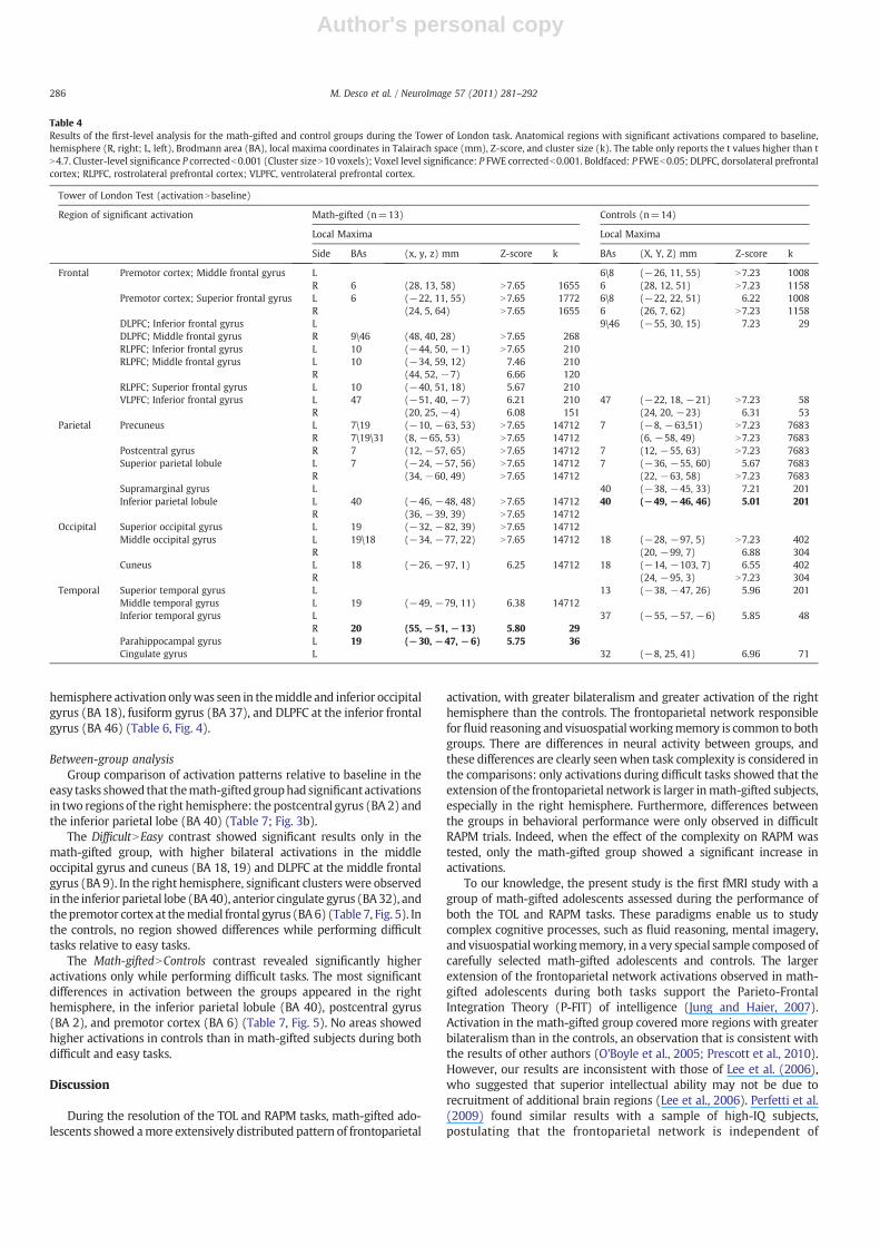
During the TOL task, significant activationwas observed in the frontal,temporal, and parietal lobes. The math-gifted group showed clusters ofactivation (relative to baseline) that were always bilateral andwithmoreregions activated than the control group (Table 4, Fig. 2). Previous fMRIstudies during TOL in normal subjects showed activations in theprefrontal cortex and parietal cortices (Boghi et al., 2006; Fincham et al.,2002; Lazeronet al., 2000;Newmanet al., 2003;Wagner et al., 2006), thussupporting the validity of our TOL paradigm.
Math-gifted subjectsBilateral activationwas seen in the precuneus (BA 7/19 and BA 31 on
the right hemisphere), premotor cortex at the superior frontal gyrus (BA6), superior parietal lobule (BA 7), inferior parietal lobule (BA 40),ventrolateral prefrontal cortex (VLPFC) at the inferior frontal gyrus (BA47), and rostrolateral prefrontal cortex (RLPFC) at the middle frontalgyrus (BA 10). Activation in the left hemisphere onlywas observed in thesuperior occipital gyrus and middle temporal gyrus (BA 19), middleoccipital gyrus (BA 18/19), cuneus (BA 18), and RLPFC at the inferior andsuperior frontal gyri (BA 10). Activation affecting the right hemisphereonlywasobserved in thepostcentral gyrus (BA7), premotor cortex at themiddle frontal gyrus (BA 6), DLPFC at themiddle frontal gyrus (BA 9/46),and inferior temporal gyrus (BA 20) (Table 4, Fig. 2).
ControlsBilateral activation was observed in the precuneus (BA 7), superior
parietal lobule (BA 7), cuneus (BA 18), middle occipital gyrus (BA 18),premotor cortex at the superior andmiddle frontal gyri (BA 6, BA8on theleft hemisphere), and VLPFC at the inferior frontal gyrus (BA 47).
Activation in the left hemisphere onlywas observed in the supramarginalgyrus and inferior parietal lobule (BA 40), superior temporal gyrus (BA13), inferior temporal gyrus (BA 37), DLPFC at the inferior frontal gyrus(BA46\9), andcingulate gyrus (BA32).Activation in the righthemisphereonly was observed in the postcentral gyrus (BA 7) (Table 4, Fig. 2).
Between-group analysisIn the between-group analysis (math-gifted Ncontrols), the math-
gifted adolescents showed activations in the right superior occipitalgyrus (BA 19), right precuneus (BA 19), and bilateral medial temporalgyrus (BA 39) (Table 5, Fig. 3a). Considering that both BA 19 and BA 39correspond to the associative cortex, the cluster showing significantdifferences on the left temporal gyrus (BA39) is anatomically locatedwithin the largest within-group activation cluster observed in math-gifted subjects, labeled as the middle temporal gyrus (BA 19) in Table 4.
Raven's Advanced Progressive Matrices
During the RAPM task, significant activations were observed in thefrontal, parietal, and temporal lobes. The math-gifted group showed amoreextensive andbilateral patternof activations includingmore regionsthan the control group (Table 6, Fig. 4). Previous fMRI studies duringperformance of RAPM for the evaluation of fluid reasoning and workingmemory in normal subjects had revealed activations in the frontal andparietal cortices (Christoff et al., 2001;Grayet al., 2003;Kroger et al., 2002;Masunaga et al., 2008; Perfetti et al., 2009; Prabhakaran et al., 1997;Wendelken et al., 2008). These studies showed that the frontoparietalnetwork is active in both hemispheres during tasks that require workingmemory, such as TOL and RAPM, and their results support the validity ofour RAPM paradigm.
Math-gifted subjectsBilateral activationwas seen in themiddle occipital gyrus (BA 18/19),
precuneus (BA 7, BA 19 on the left hemisphere), superior parietal lobule(BA 7), inferior parietal lobule (BA 40), premotor cortex at the middlefrontal gyrus (BA 6), and DLPFC at the inferior frontal gyrus (BA 46, BA 9on the left hemisphere). Activation on the left hemisphere onlywas seenin the cuneus (BA19), premotor cortexat themiddle frontal gyrus (BA6),DLPFC at the inferior frontal gyrus (BA 9), and RLPFC at the superior,middle and inferior frontal gyri (BA 10). Activation on the righthemisphere only was seen in the superior occipital gyrus (BA 19),fusiform gyrus (BA 37), and VLPFC at the inferior frontal gyrus (BA 44)(Table 6, Fig. 4).
ControlsBilateral activation was seen in the cuneus (BA 18), middle occipital
gyrus (BA 18 plus BA 19 on the left hemisphere), and superior parietallobule (BA 7). Activation in the left hemisphere only was seen in theprecuneus (BA7) andDLPFC at themiddle frontal gyrus (BA46/9). Right
Table 3Mean and standard deviation (SD) for behavioral measures in each group and complexity level during the RAPM task.
Raven's Advanced Progressive Matrices
Math-gifted (n=13) Control (n=14) Groupa Difficultyb
Easy Difficult Easy Difficult
Mean SD Mean SD Mean SD Mean SD Easy Difficult Math-gifted Control
Correct response (%) 69.2 15.2 65.4 13.8 64.9 15.7 43.7 15.9 – 0.0009 – 0.0015Reaction time 9.3 2.9 11.9 2.6 9.1 1.7 11.0 2.2 – – 0.0237 0.0204Reaction time correct response 8.1 2.9 11.4 1.9 7.7 1.9 9.9 2.4 – – 0.0028 0.0141Reaction time failed response 8.5 4.2 8.3 3.3 9.2 2.6 9.3 2.0 – – – –
Activation trials completed 11.7 0.5 19.5 7.7 12.0 0.0 22.0 5.7 – – 0.0012 b0.001Accumulated display time 108.2 32.9 219.9 73.6 109.6 20.6 232.6 36.2 – – b0.0001 b0.001Missing responses 1.5 0.8 1.9 0.8 1.6 1.3 1.9 0.7 – – – –
a P value, student t test between groups for each complexity level.b P value, student t test between complexity levels for each group.
285M. Desco et al. / NeuroImage 57 (2011) 281–292
Author's personal copy
hemisphere activation onlywas seen in themiddle and inferior occipitalgyrus (BA 18), fusiform gyrus (BA 37), and DLPFC at the inferior frontalgyrus (BA 46) (Table 6, Fig. 4).
Between-group analysisGroup comparison of activation patterns relative to baseline in the
easy tasks showed that themath-giftedgrouphad significantactivationsin two regions of the right hemisphere: the postcentral gyrus (BA 2) andthe inferior parietal lobe (BA 40) (Table 7; Fig. 3b).
The DifficultNEasy contrast showed significant results only in themath-gifted group, with higher bilateral activations in the middleoccipital gyrus and cuneus (BA 18, 19) and DLPFC at the middle frontalgyrus (BA 9). In the right hemisphere, significant clusters were observedin the inferior parietal lobe (BA40), anterior cingulate gyrus (BA32), andthepremotor cortex at themedial frontal gyrus (BA6) (Table 7, Fig. 5). Inthe controls, no region showed differences while performing difficulttasks relative to easy tasks.
The Math-giftedNControls contrast revealed significantly higheractivations only while performing difficult tasks. The most significantdifferences in activation between the groups appeared in the righthemisphere, in the inferior parietal lobule (BA 40), postcentral gyrus(BA 2), and premotor cortex (BA 6) (Table 7, Fig. 5). No areas showedhigher activations in controls than in math-gifted subjects during bothdifficult and easy tasks.
Discussion
During the resolution of the TOL and RAPM tasks, math-gifted ado-lescents showed amore extensively distributed pattern of frontoparietal
activation, with greater bilateralism and greater activation of the righthemisphere than the controls. The frontoparietal network responsibleforfluid reasoning and visuospatial workingmemory is common to bothgroups. There are differences in neural activity between groups, andthese differences are clearly seen when task complexity is considered inthe comparisons: only activations during difficult tasks showed that theextension of the frontoparietal network is larger inmath-gifted subjects,especially in the right hemisphere. Furthermore, differences betweenthe groups in behavioral performance were only observed in difficultRAPM trials. Indeed, when the effect of the complexity on RAPM wastested, only the math-gifted group showed a significant increase inactivations.
To our knowledge, the present study is the first fMRI study with agroup of math-gifted adolescents assessed during the performance ofboth the TOL and RAPM tasks. These paradigms enable us to studycomplex cognitive processes, such as fluid reasoning, mental imagery,and visuospatialworkingmemory, in a very special sample composed ofcarefully selected math-gifted adolescents and controls. The largerextension of the frontoparietal network activations observed in math-gifted adolescents during both tasks support the Parieto-FrontalIntegration Theory (P-FIT) of intelligence (Jung and Haier, 2007).Activation in the math-gifted group covered more regions with greaterbilateralism than in the controls, an observation that is consistent withthe results of other authors (O'Boyle et al., 2005; Prescott et al., 2010).However, our results are inconsistent with those of Lee et al. (2006),who suggested that superior intellectual ability may not be due torecruitment of additional brain regions (Lee et al., 2006). Perfetti et al.(2009) found similar results with a sample of high-IQ subjects,postulating that the frontoparietal network is independent of
Table 4Results of the first-level analysis for the math-gifted and control groups during the Tower of London task. Anatomical regions with significant activations compared to baseline,hemisphere (R, right; L, left), Brodmann area (BA), local maxima coordinates in Talairach space (mm), Z-score, and cluster size (k). The table only reports the t values higher than tN4.7. Cluster-level significance P correctedb0.001 (Cluster sizeN10 voxels); Voxel level significance: P FWE correctedb0.001. Boldfaced: P FWEb0.05; DLPFC, dorsolateral prefrontalcortex; RLPFC, rostrolateral prefrontal cortex; VLPFC, ventrolateral prefrontal cortex.
Tower of London Test (activationNbaseline)
Region of significant activation Math-gifted (n=13) Controls (n=14)
Local Maxima Local Maxima
Side BAs (x, y, z) mm Z-score k BAs (X, Y, Z) mm Z-score k
Frontal Premotor cortex; Middle frontal gyrus L 6\8 (−26, 11, 55) N7.23 1008R 6 (28, 13, 58) N7.65 1655 6 (28, 12, 51) N7.23 1158
Premotor cortex; Superior frontal gyrus L 6 (−22, 11, 55) N7.65 1772 6\8 (−22, 22, 51) 6.22 1008R (24, 5, 64) N7.65 1655 6 (26, 7, 62) N7.23 1158
DLPFC; Inferior frontal gyrus L 9\46 (−55, 30, 15) 7.23 29DLPFC; Middle frontal gyrus R 9\46 (48, 40, 28) N7.65 268RLPFC; Inferior frontal gyrus L 10 (−44, 50, −1) N7.65 210RLPFC; Middle frontal gyrus L 10 (−34, 59, 12) 7.46 210
R (44, 52, −7) 6.66 120RLPFC; Superior frontal gyrus L 10 (−40, 51, 18) 5.67 210VLPFC; Inferior frontal gyrus L 47 (−51, 40, −7) 6.21 210 47 (−22, 18, −21) N7.23 58
R (20, 25, −4) 6.08 151 (24, 20, −23) 6.31 53Parietal Precuneus L 7\19 (−10, −63, 53) N7.65 14712 7 (−8, −63,51) N7.23 7683
R 7\19\31 (8, −65, 53) N7.65 14712 (6, −58, 49) N7.23 7683Postcentral gyrus R 7 (12, −57, 65) N7.65 14712 7 (12, −55, 63) N7.23 7683Superior parietal lobule L 7 (−24, −57, 56) N7.65 14712 7 (−36, −55, 60) 5.67 7683
R (34, −60, 49) N7.65 14712 (22, −63, 58) N7.23 7683Supramarginal gyrus L 40 (−38, −45, 33) 7.21 201Inferior parietal lobule L 40 (−46, −48, 48) N7.65 14712 40 (−49, −46, 46) 5.01 201
R (36, −39, 39) N7.65 14712Occipital Superior occipital gyrus L 19 (−32, −82, 39) N7.65 14712
Middle occipital gyrus L 19\18 (−34, −77, 22) N7.65 14712 18 (−28, −97, 5) N7.23 402R (20, −99, 7) 6.88 304
Cuneus L 18 (−26, −97, 1) 6.25 14712 18 (−14, −103, 7) 6.55 402R (24, −95, 3) N7.23 304
Temporal Superior temporal gyrus L 13 (−38, −47, 26) 5.96 201Middle temporal gyrus L 19 (−49, −79, 11) 6.38 14712Inferior temporal gyrus L 37 (−55, −57, −6) 5.85 48
R 20 (55, −51, −13) 5.80 29Parahippocampal gyrus L 19 (−30, −47, −6) 5.75 36Cingulate gyrus L 32 (−8, 25, 41) 6.96 71
286 M. Desco et al. / NeuroImage 57 (2011) 281–292

Author's personal copy
intellectual ability (high or low IQ) (Perfetti et al., 2009). In our study,the extent of activations and recruitmentof newareasdifferedwhen thesubjects were performing difficult trials, which may be interpreted asreflecting the functional specialization of math-giftedness.
In the frontoparietal activations common to both groups, some areasare associated with the type of task. The regions involved in making adecision and responding to a problemare the inferior andmedial frontalgyri (BA 9/46, 44, and 47),which are known to support cognition duringworkingmemory andproblemsolvingof differing types (deductive, andanalogical). The DLPFC (BA 9/46) is the executive center ofmaintenanceand information processing with internal representations duringworking memory, and a key brain region supporting reasoning andnovel problem solving (Baker et al., 1996; Courtney et al., 1998; Chafeeand Goldman-Rakic, 1998; Goldman-Rakic, 1987, 1995; Kroger et al.,2002; Lee et al., 2006; Owen et al., 1996; Perfetti et al., 2009; Todd and
Marois, 2004). Other frontal areas, such as the RLPFC and premotorcortex have been shown to support relational complexity and planning,respectively (BA 10 and 6) (Christoff et al., 2001; Kalbfleisch et al., 2007;van den Heuvel et al., 2003). In the premotor cortex, the role of BA 6 inhigher-level cognition has been investigated in relationship to workingmemory (Owen et al., 1996; Smith and Jonides, 1999), updating spatialinformation (Tanaka et al., 2005), processes of attention, saccadic eyemovements, and visuospatial manipulation processes (Brown et al.,2004; Courtney et al., 1998; Curtis and D'Esposito, 2003; Curtis et al.,2004; Smith and Jonides, 1999).
The posterior parietal cortex areas (BA 7, 19, and 40) store repre-sentations of visual information and are associated with spatial stimuli
Fig. 2. First-level analyses in math-gifted and control groups during the TOL task. The clusters showing significant activations are color-coded for each group. Significant clusters aresuperimposed on the anatomical render (SPM5). The color bar shows the z-score for each group. More activation and bilateralism were observed in the frontal, prefrontal, andparietal cortices in math-gifted adolescents.
Table 5Between-group analysis, math-giftedNcontrols contrast. Anatomical regions withsignificant activation during the Tower of London task, hemisphere (R, right; L, left),Brodmann area (BA), local maxima coordinates in Talairach space (mm), Z-score, andcluster size (k). Significant differences in regions of activation between the two groupsduring TOL task (tN3.20) are shown. Cluster-level significance P correctedb0.01(cluster sizeN10 voxels). Voxel-level significance P uncorrectedb0.001.
Towers of London (Math-giftedNControls)
Region of significantactivation
Local Maxima
Hemisphere BAs (x, y, z) mm Z-score k
Superior occipital gyrus R 19 (32, −76, 26) 4.30 183Precuneus R 19 (34, −72, 33) 3.62 183Middle temporal gyrus L 39 (−32, −69, 24) 4.21 134
R 39 (34, −65, 25) 3.36 183
Fig. 3. Regions with significantly greater activation in math-gifted adolescents duringthe TOL (a) and RAPM (b) tasks than in controls (between-groups analyses; math-giftedNcontrol group contrast) superimposed on the anatomical image; a) is inneurological orientation (left is left); b) is the right hemisphere.
287M. Desco et al. / NeuroImage 57 (2011) 281–292

Author's personal copy
and processing task complexity (Champod and Petrides, 2007; Dehaeneet al., 2003; Knauff et al., 2002; Lee et al., 2006; Newman et al., 2003;Perfetti et al., 2009; Prabhakaran et al., 1997; Todd and Marois, 2004;vandenHeuvel et al., 2003). Both groups showedgreater recruitment ofthe parietal cortices for both tasks. TOL and RAPM are both complexcognitive tasks that require visuospatial and abstract informationprocessing.
Executive functioning
To solve the TOL test, subjects need planning ability, visuospatialworkingmemory, andmental imagery. The level of complexity was notstudied in this paradigm. The design focused on the pattern of activationcompared to baseline, and the neural substrate of visuospatial workingmemory was assessed.
The activation patterns of both groups were similar, although themath-gifted group presented amuchmore extensive activation pattern,especially in frontal regions (premotor cortex, DLPFC, and RLPFC) and inthe intraparietal sulcus, mostly in the right hemisphere.
The intraparietal sulcus plays a key role in processing representationsof objects in space (Brown et al., 2004; Courtney et al., 1998; Curtis et al.,2005) and during numerical processing in adults (Dehaene et al., 2003).In addition, a correlation between visuospatialworkingmemory capacityand intraparietal sulcus activity has been described (Todd and Marois,2004). Thisfinding suggests a greater visuospatial capacity inmath-giftedadolescents. Our sample included males and females in a similar genderratio in both groups. Possible gender effects on visuospatial processing(Boghi et al., 2006) were not assessed due to insufficient sample size.
The prefrontal cortex showed clear group differences in activationpatterns. TheDLPFCwasactivebilaterally in themath-giftedgroup,whilein the control group it was activated only in the left hemisphere duringboth tasks. TheDLPFC is the executive center of informationmaintenanceand processing with internal representations during working memory,and is believed to support reasoning and novel problem solving (Baker
et al., 1996; Chafee and Goldman-Rakic, 1998; Goldman-Rakic, 1987,1995; Lee et al., 2006; Owen et al., 1996; Todd and Marois, 2004).Increased activation in this area is associatedwith degree of difficulty oracts as a response to a higher cognitive load (Braver et al., 1997; Krogeret al., 2002).
Thus, the greater activation of prefrontal areas observed in themath-gifted group may represent a cognitive advantage, greater ease ofprocessing information, creation of a strategy, or planning (Lazeronet al.,2000; Owen et al., 1996; Wagner et al., 2006).
Fluid reasoning
To solve the RAPM task, subjects need logical reasoning and spatialworking memory, because the task requires both greater symbolic andspatial relations to be analyzed (Carpenter et al., 1990). Therefore,activation is associated with both the left and the right frontoparietalcortices and with some left hemispheric dominance. Indeed, the neuralnetwork underlying performance in RAPM overlaps substantially withsymbolic (non-spatial) working memory networks during analyticalreasoning. Therefore, the RAPM task required figural analysis (visuospa-tial) abilities. Themost comprehensive studyof thematrix reasoning test(Raven, 1965) reports right frontal hemispheric activations for matrixreasoning during figural problem solving (visuospatial reasoning)(Prabhakaran et al., 1997). Logical reasoning needs the language systemto perform the task, and this is associated with left frontal regions(Dehaene et al., 2003, 1999; Kroger et al., 2002).
In the present study, the control group had a bilateral activationpattern, with fewer areas activated than the math-gifted group; theyalso had less significant clusters in the right parietal cortex. During thistask, the math-gifted group showed bilateral activations in both thefrontal and parietal cortices, as observed elsewhere (Lee et al., 2006).
Strong activation of the DLPFC bilaterally (mainly the left DLPFCduring difficult trials) and the left RLPFC in themath-gifted groupmay beimportant to perform the task efficiently. Some authors suggest that,
Table 6Results of the first-level analysis for the math-gifted and control groups during the Raven Advanced Progressive Matrices task. Anatomical regions with significant activationscompared to baseline, hemisphere (R, right; L, left), Brodmann area (BA), local maxima coordinates in Talairach space (mm), Z-score, and cluster size (k). Only t-values higher than tN4.7 are shown. Cluster-level significance P correctedb0.001 (cluster sizeN10 voxels); Voxel level significance P FWE correctedb0.001. Boldfaced: P FWEb0.05; DLPFC, dorsolateralprefrontal cortex; RLPFC, rostrolateral prefrontal cortex; VLPFC, ventrolateral prefrontal cortex.
Raven's Advanced Progressive Matrices (activationNbaseline)
Region of significant activation Math-gifted (n=13) Controls (n=14)
Local maxima Local maxima
Side BAs (x, y, z) mm Z-score K BAs (x, y, z) mm Z-score k
Frontal Premotor cortex; Middle frontal gyrus L 6 (−26, 6, 49) N7.76 2345R (30, 9, 57) 7.47 246
DLPFC; Inferior frontal gyrus L 9 (−44, 3, 29) N7.76 2345R 46 (55, 38, 13) 6.62 52
DLPFC; Middle frontal gyrus L 9\46 (−44, 26, 17) N7.76 2345 9\46 (−53, 19, 30) 5.88 207R 46 (53, 36, 22) N7.76 108
RLPFC; Inferior frontal gyrus L 10 (−46, 48, −4) 7.65 96RLPFC; Middle frontal gyrus L 10 (−36, 57, 7) 5.58 48RLPFC; Superior frontal gyrus L 10 (−30, 58, −4) 5.10 48VLPFC; Inferior frontal gyrus R 44 (51, 9, 22) N7.76 213
Parietal Precuneus L 7\19 (−26, −76, 40) N7.76 4048 7 (−22, −70, 40) N7.73 1664R 7 (22, −71, 53) N7.76 4048
Superior parietal lobule L 7 (−28, −50, 41) N7.76 4048 7 (−26, −71, 46) N7.73 1664R (26, −60, 40) N7.76 3644 (28, −71, 44) 6.83 485
Inferior parietal lobule L 40 (−38, −46, 41) N7.76 4048R (42, −42, 48) 7.15 3644
Occipital Superior occipital gyrus L 19 (−32, −80, 26) 6.98 1664R 19 (34, −78, 29) N7.76 3644
Middle occipital gyrus L 18\19 (−34, −91, 2) N7.76 4048 18 (−32, −87, 4) 7.41 1664R (36, −89, −1) N7.76 3644 18\19 (34, −87, 4) N7.73 584
Inferior occipital gyrus R 18 (28, −91, −3) N7.73 584Cuneus L 19 (−30, −84, 24) N7.76 4048 18 (−8, −100, 12) 7.09 180
R (12, −94, 21) 6.08 95Temporal Fusiform gyrus R 37 (49, −57, −9) N7.76 418 37 (−48, −53, −8) 6.18 124
288 M. Desco et al. / NeuroImage 57 (2011) 281–292

Author's personal copy
duringperformanceof theRAPMtask, theRLPFC turns significantly activewhen the task demands integration of information about the shape ofobjects (relational integration) (Christoff et al., 2001; Wendelken et al.,2008).
An importantpoint inour resultsduring theRAPMtaskwas thatmath-gifted subjects showed strong activation in Broca's area, or the left VLPFC(BA 44). This result is consistent with Dehaene's hypothesis that there isinteractionbetween the symbolic systemand the visuospatial andmental
Table 7Between-group analysis, math-gifted adolescentsNcontrol group contrast; between condition analysis math-gifted difficultNeasy contrast; and between-group analysis in thedifficult condition. Anatomical regions showing significant activation during Raven's Advanced Progressive Matrices, the hemisphere (R, right; L, left); the BA, Brodmann Area, thelocal maxima coordinates in Talairach space (mm), the Z-score and the cluster size (k).Significant differences in regions of activation between the two groups during RAPM task (tN3.45). All regions significant at cluster-level P corrected b0.05 and voxel-level P uncorrectedb0.001 (kN10), unless marked otherwise.
Raven's Advanced Progressive Matrices
Region of significant activation Local maxima
Side BAs (x, y, z) mm Z-score k
Math-giftedNControlPostcentral gyrus R 2 (49, −27, 47) 4.33 220Inferior parietal lobule R 40 (65, −27, 38) 3.71 220
Math-gifted (DifficultNEasy)Middle occipital gyrus L** 18 (−22, −103, 7) 5.11 431
R 18 (12, −95, 10) 4.31 157Cuneus L** 19 (−22, −95, 25) 3.97 431
R 18 (20, −100, 14) 3.62 157Inferior parietal lobule R** 40 (44, −38, 52) 3.84 175Anterior cingulate gyrus R** 32 (12, 30, 23) 3.63 132DLPFC; middle frontal gyrus L** 9 (−49, 6, 35) 4.14 132Premotor cortex; medial frontal gyrus R** 6 (0, 16, 47) 3.85 133
Difficult (Math-giftedNControl)Inferior parietal lobule R 40 (65, −27, 40) 3.92 242Postcentral gyrus R 2 (55, −27, 44) 4.00 242Premotor cortex R* 6 (26, −3, 44) 3.97 217
* Cluster p correctedb0.01.** Cluster p correctedb0.001.
Fig. 4. First-level analyses in math-gifted and control groups during the RAPM task. Clusters showing significant activations are color-coded for each group. Significant clusters aresuperimposed on the anatomical render (SPM5). The color bar shows the z-score for each group. More activation and bilateralism were observed in the frontal, prefrontal, andparietal cortices in math-gifted adolescents.
289M. Desco et al. / NeuroImage 57 (2011) 281–292

Author's personal copy
imagery systems (Dehaene et al., 2003, 1999). Broca's area has beenimplicated in deduction problems (Dehaene et al., 1999; Kroger et al.,2002). Another interpretation was proposed by Kroger et al. (2002),who suggested that the neural substrates of logical reasoning andmathematical thinking are not the same, and that high level cognitiondoesnot dependona single global system.Thus, logical reasoningwouldgo beyond linguistic processing to the manipulation of nonlinguisticrepresentations.We found areas that overlapwith logical reasoning andspatial working memory.
The math-gifted group showed activation in BA 6 during both tasks.The control group only showed activation in this area during the TOLtask.Within the context of this study, the left lateral BA6 cortex supportsprocessing related to the complexity of the problem. These activations inBA 6 may suggest that the math-gifted group had greater high-levelcognitive abilities such as visuospatial and attention processes. Activa-tions in BA 6 have been associated with saccadic eye movements,attention, and visuospatial manipulation (Brown et al., 2004; Courtneyet al., 1998; Curtis and D'Esposito, 2003; Curtis et al., 2004; Smith andJonides, 1999).
One of the most relevant findings of our study is that differencesbetween groups appear only when performing the difficult RAPM trials.No differences were observed between the groups in the neural patternof activation during the easy trials; in fact, in the control group, nodifferences between the levels of complexity were observed. Themath-gifted group showed more significantly activated regions when thecomplexity level of the task increased, as if they were using moreresources only when necessary.
Most of the significantly activated regions in the math-gifted groupduring difficult trials are observed in the right hemisphere, suggestingthat high complex reasoning involvesbothan increase in activationof theright hemisphere andmore bilateralism in the pattern of activation of thefrontoparietal network. In addition, increased activation in math-gifted
subjects during complex tasks wasmore evident in frontal regions (BA 9and BA 6) and the anterior cingulate gyrus (BA 32). This was the patternobserved by Lee et al. (2006). Activation also increased with taskcomplexity in the inferior parietal lobule (BA 40) inmath-gifted subjectsand was significantly different between groups during difficult tasks,suggesting that the parietal cortex, especially on the right hemisphere,may play a crucial role in complex reasoning processes (Dehaene et al.,2003; Lee et al., 2006).
Whenmath-giftedsubjectsperformeddifficult trials, higheractivationwas observed in the anterior cingulate gyrus (BA 32). This finding agreeswith those of Gray et al. (2003), who showed that activation of frontallobes and the anterior cingulate correlatewithfluid reasoning (Gray et al.,2003) and alsowith the results of O'Boyle et al. (2005), who showed thatincreased activation of the anterior cingulate gyrus was a sign of highfunctional development of the math-gifted brain (O'Boyle et al., 2005).Other studies have suggested a role for the anterior cingulate when thetask becomesmoredifficult, and this area serves as an on-line regulator ofcognitive resource allocation policies (O'Boyle et al., 2005).
Number of slides completed and total display timewere significantlylower in easy than in difficult tasks, and this may have affected theanalysis of group comparisons within easy or difficult trials. However,both groups showed a similar gap between the number of easy anddifficult slides completed and total display time, thus ruling out anygroup bias due to these differences.
Increased brain activity in the frontal cortex, cingulate gyrus, andparietal regions seems to be associatedwith higher performance influidreasoning and working memory. The largest activations correspond tothe increase in task complexity and to high workingmemory load, bothof which confer an advantage to math-gifted adolescents in complexproblem solving.
During the RAPM task, activations in the frontoparietal network inboth groups could be related tofluid reasoning andworkingmemory. The
Fig. 5. Left. Random-effects analyses between groups when performing difficult tasks of RAPM; Right. Random-effects analyses between “easy” and “difficult” conditions. Only themath-gifted group showed significant differences.
290 M. Desco et al. / NeuroImage 57 (2011) 281–292

Author's personal copy
greater activations of the math-gifted group, especially during difficulttrials, may give this group greater ability to solve problems that needrelational reasoning with symbolic and spatial information.
There are several limitations in our study. The first one concerns thecognitive characterization of our two groups. Selection of math-giftedsubjects was made using only their performance in the ESTALMAT test,which is specific for detecting math abilities. FSIQ was estimated to ruleout atypical cognitive functioning or mental retardation, together withthe overall functioning assessment made by the child psychiatrist andchild psychologist. A more thorough cognitive assessment could haverevealed whether other cognitive differences than mathematicalreasoning and problem solving, exist between the two groups. However,our study is focused on brain mapping of subjects with precociousmathematical abilities, rather than investigating cognitive performanceor overall intelligence. Thus, althoughmath-gifted subjects showedothercognitive skills in addition to math, the only criteria used for selectionwas math-giftedness, which was assessed with a highly specific test. Torule out a statistical effect of IQ on the activation pattern observed, weperformed a correlation and covariate analysis, which suggest that theconfounding effect was minimal (see Methods and SupplementaryResults). Another limitationwas that the paradigm usedwas not specificfor mathematical thinking; such a paradigm would have yielded moreinformation about the brain regions involved when the subject wasengaged in a mathematical task. A paradigm assessing global mathe-matical abilities could have proven useful, by increasing groupdifferences and minimizing the confounding effect of multiple cognitiveabilities in themath-gifted subjects. However, no such specific paradigmhas been reported in the literature; therefore, we chose two tasks whichwe believe provide an adequate context in which to analyze mathemat-ical thinking. Finally, our findings about the brain response to increasedtask complexity only apply to fluid reasoning. Thus, the effect ofcomplexity level on executive function should be analyzed in a specificstudy.
Conclusions
In summary, the differences between subjects with or withoutmathematical ability in complex cognitive processes such as fluidreasoning, executive functioning, and visuospatial working memoryseem to bemediated by the neural activity of the bilateral frontoparietalnetwork. In the math-gifted group, we observed greater bilateralism ofactivation in the frontoparietal network (especially the posteriorparietal cortex, intraparietal sulcus, and DLPFC), which is probablyassociated with the higher capacity of math-gifted subjects to solvecomplex visuospatial and analytical tasks demanding logical thinking.During easy tasks, activation patterns were similar in both groups,whereas major differences between groups appeared only duringdifficult tasks, mainly in the frontal cortex and inferior parietal lobule.Functional specialization of math-giftedness has been described as acombination of greater recruitment of right hemisphere regions andmore bilateralism (O'Boyle et al., 2005; Prescott et al., 2010). In contrastto an explanation based on neural efficiency, our study further supportsthis hypothesis of stronger recruitment of math-gifted children.Therefore, math-gifted subjects are qualitatively different in terms ofthe degree and location of activated brain regions.
Acknowledgments
Partly supported by Fundación Vodafone; Real Academia de CienciasExactas, Físicas y Naturales; Programa de Estímulo del Talento Matemá-tico (ESTALMAT); Ministerio de Sanidad y Consumo, Instituto de SaludCarlos III; CIBER de Salud Mental (CIBERSAM) CB07/09/0031; CDTI(Centro para el Desarrollo Tecnológico e Industrial) under the CENIT(Consorcios Estratégicos Nacionales en Investigación Técnica) Pro-gramme (AMIT Project), Spanish Ministry of Science and Innovation.FJN was supported by a PFIS grant from the Instituto de Salud Carlos III.
We are particularly grateful to Professor Eugenio Hernandez from theESTALMATprogramand to all the childrenwhoparticipated in the study.Wewant to thankMichael O´Boyle for his continuous support during theearly stages of this manuscript.
Appendix A. Supplementary data
Supplementarydata to this article canbe foundonline atdoi:10.1016/j.neuroimage.2011.03.063.
References
Alderton, D.L., Larson, G.E., 1990. Dimensionality of Raven's Advanced ProgressiveMatrices Items. Educ. Psychol. Meas. 50, 887–900.
Alexander, J.E., O'Boyle, M.W., Benbow, C.P., 1996. Developmentally advanced EEGalpha power in gifted male and female adolescents. Int. J. Psychophysiol. 23, 25–31.
Baker, S.C., Rogers, R.D., Owen, A.M., Frith, C.D., Dolan, R.J., Frackowiak, R.S., Robbins, T.W.,1996. Neural systems engaged by planning: a PET study of the Tower of London task.Neuropsychologia 34, 515–526.
Benbow, C.P., 1986. Physiological correlates of extreme intellectual precocity. Neuropsy-chologia 24, 719–725.
Benbow, C.P., Lubinski, D., 1993. Psychological profiles of the mathematically talented:somesexdifferences andevidencesupporting their biological basis. CibaFound. Symp.178, 44–59 (discussion 60–46).
Boghi, A., Rasetti, R., Avidano, F., Manzone, C., Orsi, L., D'Agata, F., Caroppo, P., Bergui, M.,Rocca, P., Pulvirenti, L., Bradac, G.B., Bogetto, F., Mutani, R., Mortara, P., 2006. The effectof gender onplanning: an fMRI study using the Tower of London task. Neuroimage33,999–1010.
Braver, T.S., Cohen, J.D.,Nystrom, L.E., Jonides, J., Smith,E.E.,Noll,D.C., 1997.Aparametric studyof prefrontal cortex involvement in human working memory. Neuroimage 5, 49–62.
Brown,M.R., DeSouza, J.F., Goltz, H.C., Ford, K.,Menon, R.S., Goodale,M.A., Everling, S., 2004.Comparison of memory- and visually guided saccades using event-related fMRI. J.Neurophysiol. 91, 873–889.
Buchanan, R.W., Heinrichs, D.W., 1989. The Neurological Evaluation Scale (NES): astructured instrument for the assessment of neurological signs in schizophrenia.Psychiatry Res. 27, 335–350.
Carpenter, P.A., Just,M.A., Shell, P., 1990.What one intelligence testmeasures: a theoreticalaccount of the processing in the Raven Progressive Matrices Test. Psychol. Rev. 97,404–431.
Chafee,M.V., Goldman-Rakic, P.S., 1998.Matchingpatternsof activity inprimateprefrontalarea 8a and parietal area 7ip neurons during a spatial working memory task. J.Neurophysiol. 79, 2919–2940.
Champod, A.S., Petrides, M., 2007. Dissociable roles of the posterior parietal and theprefrontal cortex inmanipulation andmonitoring processes. Proc. Natl. Acad. Sci. USA104, 14837–14842.
Christoff, K., Prabhakaran,V.,Dorfman, J., Zhao, Z., Kroger, J.K.,Holyoak,K.J., Gabrieli, J.D., 2001.Rostrolateral prefrontal cortex involvement in relational integration during reasoning.Neuroimage 14, 1136–1149.
Coull, J.T., Frith, C.D., 1998. Differential activation of right superior parietal cortex andintraparietal sulcus by spatial and nonspatial attention. Neuroimage 8, 176–187.
Courtney, S.M., Petit, L.,Maisog, J.M., Ungerleider, L.G., Haxby, J.V., 1998.Anarea specializedfor spatial working memory in human frontal cortex. Science 279, 1347–1351.
Curtis, C.E., D'Esposito,M., 2003. Persistent activity in the prefrontal cortexduringworkingmemory. Trends Cogn. Sci. 7, 415–423.
Curtis, C.E., Rao, V.Y., D'Esposito, M., 2004. Maintenance of spatial andmotor codes duringoculomotor delayed response tasks. J. Neurosci. 24, 3944–3952.
Curtis, C.E., Sun, F.T.,Miller, L.M.,D'Esposito,M., 2005. Coherence between fMRI time-seriesdistinguishes two spatial working memory networks. Neuroimage 26, 177–183.
Dagher, A., Owen, A.M., Boecker,H., Brooks,D.J., 1999.Mapping thenetwork forplanning: acorrelational PET activation study with the Tower of London task. Brain 122 (Pt 10),1973–1987.
Dehaene, S., Spelke, E., Pinel, P., Stanescu, R., Tsivkin, S., 1999. Sources of mathematicalthinking: behavioral and brain-imaging evidence. Science 284, 970–974.
Dehaene, S., Piazza, M., Pinel, P., Cohen, L., 2003. Three parietal circuits for numberprocessing. Psychology Press, pp. 487–506.
Fincham, J.M., Carter, C.S., van Veen, V., Stenger, V.A., Anderson, J.R., 2002. Neuralmechanismsof planning: a computational analysisusingevent-related fMRI.Proc.Natl.Acad. Sci. USA 99, 3346–3351.
Geschwind, N., Galaburda, A., 1984. Cerebral dominance: the biological foundations.Harvard University Press, Cambridge, MA.
Goldman-Rakic, P.S., 1987. Development of cortical circuitry and cognitive function.Child Dev. 58, 601–622.
Goldman-Rakic, P.S., 1995. Architecture of the prefrontal cortex and the central executive.Ann. N.Y. Acad. Sci. 769, 71–83.
Gray, J.R., Chabris, C.F., Braver, T.S., 2003. Neural mechanisms of general fluid intelligence.Nat. Neurosci. 6, 316–322.
Harris, I.M., Egan, G.F., Sonkkila, C., Tochon-Danguy, H.J., Paxinos, G., Watson, J.D., 2000.Selective right parietal lobe activationduringmental rotation: a parametric PET study.Brain 123 (Pt 1), 65–73.
Heller, K.A., Mönks, F.J., Sternberg, R.J., Subotnik, R.F., 2000. The international handbook ofgiftedness and talent, 2 ed. Elsevier Science, Oxford.
Jung, R.E., Haier, R.J., 2007. The Parieto-Frontal Integration Theory (P-FIT) of intelligence:converging neuroimaging evidence. Behav. Brain Sci. 30, 135–154 (discussion 154–187).
291M. Desco et al. / NeuroImage 57 (2011) 281–292

Author's personal copy
Just, M.A., Carpenter, P.A., Maguire, M., Diwadkar, V., McMains, S., 2001. Mental rotation ofobjects retrieved from memory: a functional MRI study of spatial processing. J. Exp.Psychol. Gen. 130, 493–504.
Kalbfleisch, M.L., Van Meter, J.W., Zeffiro, T.A., 2007. The influences of task difficulty andresponse correctness on neural systems supporting fluid reasoning. Cogn. Neurodyn.1, 71–84.
Klingberg, T.,O'Sullivan,B.T., Roland, P.E., 1997.Bilateral activationof fronto-parietalnetworksby incrementing demand in a working memory task. Cereb. Cortex 7, 465–471.
Knauff, M., Mulack, T., Kassubek, J., Salih, H.R., Greenlee, M.W., 2002. Spatial imagery indeductive reasoning: a functional MRI study. Brain Res. Cogn. Brain Res. 13,203–212.
Kroger, J.K., Sabb, F.W., Fales, C.L., Bookheimer, S.Y., Cohen, M.S., Holyoak, K.J., 2002.Recruitment of anterior dorsolateral prefrontal cortex in human reasoning: a parametricstudy of relational complexity. Cereb. Cortex 12, 477–485.
Lazeron, R.H., Rombouts, S.A., Machielsen, W.C., Scheltens, P., Witter, M.P., Uylings, H.B.,Barkhof, F., 2000. Visualizing brain activation during planning: the tower of Londontest adapted for functional MR imaging. AJNR. Am. J. Neuroradiol. 21, 1407–1414.
Lee, K.H., Choi, Y.Y., Gray, J.R., Cho, S.H., Chae, J.H., Lee, S., Kim, K., 2006. Neural correlates ofsuperior intelligence: stronger recruitment of posterior parietal cortex. Neuroimage29, 578–586.
Masunaga, H., Kawashima, R., Horn, J.L., Sassa, Y., Sekiguchi, A., 2008. Neural substratesof the Topology Test to measure fluid reasoning: an fMRI study. Intelligence 36,607–615.
Newman, S.D., Carpenter, P.A., Varma, S., Just,M.A., 2003. Frontal and parietal participationin problem solving in the Tower of London: fMRI and computational modeling ofplanning and high-level perception. Neuropsychologia 41, 1668–1682.
O'Boyle,M.W., Alexander, J.E., Benbow,C.P., 1991. Enhanced right hemisphereactivation inthe mathematically precocious: a preliminary EEG investigation. Brain Cogn. 17,138–153.
O'Boyle, M.W., Benbow, C.P., Alexander, J.E., 1995. Sex differences, hemisphericlaterality and associated brain activity in the intellectualy gifted. Dev. Neuropsychol.11, 415–443.
O'Boyle, M.W., Cunnington, R., Silk, T.J., Vaughan, D., Jackson, G., Syngeniotis, A., Egan, G.F.,2005. Mathematically giftedmale adolescents activate a unique brain network duringmental rotation. Brain Res. Cogn. Brain Res. 25, 583–587.
Owen,A.M.,Doyon, J., Petrides,M., Evans,A.C., 1996. Planningandspatialworkingmemory:a positron emission tomography study in humans. Eur. J. Neurosci. 8, 353–364.
Perfetti, B., Saggino, A., Ferretti, A., Caulo, M., Romani, G.L., Onofrj, M., 2009. Differentialpatterns of cortical activation as a function of fluid reasoning complexity. Hum. BrainMapp. 30, 497–510.
Pesenti, M., Zago, L., Crivello, F., Mellet, E., Samson, D., Duroux, B., Seron, X., Mazoyer, B.,Tzourio-Mazoyer, N., 2001. Mental calculation in a prodigy is sustained by rightprefrontal and medial temporal areas. Nat. Neurosci. 4, 103–107.
Prabhakaran,V., Smith, J.A., Desmond, J.E.,Glover,G.H.,Gabrieli, J.D., 1997.Neural substratesoffluid reasoning: an fMRI study of neocortical activation during performance of theRaven's Progressive Matrices Test. Cogn. Psychol. 33, 43–63.
Prescott, J., Gavrilescu, M., Cunnington, R., O'Boyle, M.W., Egan, G.F., 2010. Enhanced brainconnectivity in math-gifted adolescents: an fMRI study using mental rotation. Cogn.Neurosci. 277–288.
Raven, J.C., 1965. Advanced Progressive Matrices: Sets I and II. Lewis, London.Ringe, W.K., Saine, K.C., Lacritz, L.H., Hynan, L.S., Cullum, C.M., 2002. Dyadic short forms of
the Wechsler Adult Intelligence Scale-III. Assessment 9, 254–260.Satler, J., 2001. Assessment of children cognitive applications, Fourth ed. Publisher Inc., San
Diego State University.Silverstein, A.B., 1985. Two- and four-subtest short forms of the WAIS-R: a closer look at
validity and reliability. J. Clin. Psychol. 41, 95–97.Singh, H., O'Boyle,M.W., 2004. Interhemispheric interaction during global–local processing
in mathematically gifted adolescents, average-ability youth, and college students.Neuropsychology 18, 371–377.
Smith, E.E., Jonides, J., 1999. Storage and executive processes in the frontal lobes. Science283, 1657–1661.
Tanaka, S., Honda, M., Sadato, N., 2005. Modality-specific cognitive function of medial andlateral human Brodmann area 6. J. Neurosci. 25, 496–501.
Todd, J.J., Marois, R., 2004. Capacity limit of visual short-termmemory in human posteriorparietal cortex. Nature 428, 751–754.
van denHeuvel, O.A., Groenewegen, H.J., Barkhof, F., Lazeron, R.H., van Dyck, R., Veltman, D.J.,2003. Frontostriatal system in planning complexity: a parametric functional magneticresonance version of Tower of London task. Neuroimage 18, 367–374.
Wagner,G., Koch,K.,Reichenbach, J.R., Sauer,H., Schlosser,R.G., 2006.Thespecial involvementof the rostrolateral prefrontal cortex in planning abilities: an event-related fMRI studywith the Tower of London paradigm. Neuropsychologia 44, 2337–2347.
Wendelken, C., Nakhabenko, D., Donohue, S.E., Carter, C.S., Bunge, S.A., 2008. “Brain isto thought as stomach is to ??”: investigating the role of rostrolateral prefrontal cortexin relational reasoning. J. Cogn. Neurosci. 20, 682–693.
292 M. Desco et al. / NeuroImage 57 (2011) 281–292
Copyright © 2022 FDOKUMEN