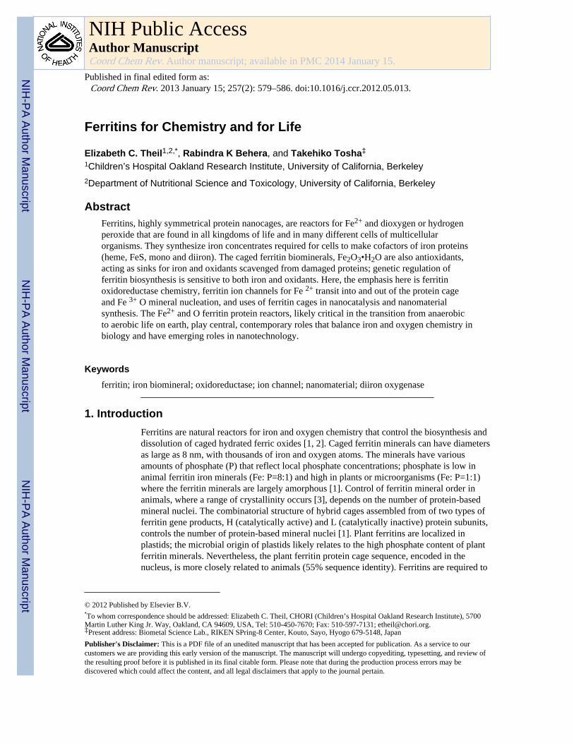
Ferritins for Chemistry and for Life
16
Ferritins for Chemistry and for Life Elizabeth C. Theil 1,2,* , Rabindra K Behera, and Takehiko Tosha ‡ 1 Children’s Hospital Oakland Research Institute, University of California, Berkeley 2 Department of Nutritional Science and Toxicology, University of California, Berkeley Abstract Ferritins, highly symmetrical protein nanocages, are reactors for Fe 2+ and dioxygen or hydrogen peroxide that are found in all kingdoms of life and in many different cells of multicellular organisms. They synthesize iron concentrates required for cells to make cofactors of iron proteins (heme, FeS, mono and diiron). The caged ferritin biominerals, Fe 2 O 3 •H 2 O are also antioxidants, acting as sinks for iron and oxidants scavenged from damaged proteins; genetic regulation of ferritin biosynthesis is sensitive to both iron and oxidants. Here, the emphasis here is ferritin oxidoreductase chemistry, ferritin ion channels for Fe 2+ transit into and out of the protein cage and Fe 3+ O mineral nucleation, and uses of ferritin cages in nanocatalysis and nanomaterial synthesis. The Fe 2+ and O ferritin protein reactors, likely critical in the transition from anaerobic to aerobic life on earth, play central, contemporary roles that balance iron and oxygen chemistry in biology and have emerging roles in nanotechnology. Keywords ferritin; iron biomineral; oxidoreductase; ion channel; nanomaterial; diiron oxygenase 1. Introduction Ferritins are natural reactors for iron and oxygen chemistry that control the biosynthesis and dissolution of caged hydrated ferric oxides [1, 2]. Caged ferritin minerals can have diameters as large as 8 nm, with thousands of iron and oxygen atoms. The minerals have various amounts of phosphate (P) that reflect local phosphate concentrations; phosphate is low in animal ferritin iron minerals (Fe: P=8:1) and high in plants or microorganisms (Fe: P=1:1) where the ferritin minerals are largely amorphous [1]. Control of ferritin mineral order in animals, where a range of crystallinity occurs [3], depends on the number of protein-based mineral nuclei. The combinatorial structure of hybrid cages assembled from of two types of ferritin gene products, H (catalytically active) and L (catalytically inactive) protein subunits, controls the number of protein-based mineral nuclei [1]. Plant ferritins are localized in plastids; the microbial origin of plastids likely relates to the high phosphate content of plant ferritin minerals. Nevertheless, the plant ferritin protein cage sequence, encoded in the nucleus, is more closely related to animals (55% sequence identity). Ferritins are required to © 2012 Published by Elsevier B.V. * To whom correspondence should be addressed: Elizabeth C. Theil, CHORI (Children’s Hospital Oakland Research Institute), 5700 Martin Luther King Jr. Way, Oakland, CA 94609, USA, Tel: 510-450-7670; Fax: 510-597-7131; [email protected]. ‡ Present address: Biometal Science Lab., RIKEN SPring-8 Center, Kouto, Sayo, Hyogo 679-5148, Japan Publisher's Disclaimer: This is a PDF file of an unedited manuscript that has been accepted for publication. As a service to our customers we are providing this early version of the manuscript. The manuscript will undergo copyediting, typesetting, and review of the resulting proof before it is published in its final citable form. Please note that during the production process errors may be discovered which could affect the content, and all legal disclaimers that apply to the journal pertain. NIH Public Access Author Manuscript Coord Chem Rev. Author manuscript; available in PMC 2014 January 15. Published in final edited form as: Coord Chem Rev. 2013 January 15; 257(2): 579–586. doi:10.1016/j.ccr.2012.05.013. NIH-PA Author Manuscript NIH-PA Author Manuscript NIH-PA Author Manuscript
Transcript of Ferritins for Chemistry and for Life
Ferritins for Chemistry and for Life
Elizabeth C. Theil1,2,*, Rabindra K Behera, and Takehiko Tosha‡
1Children’s Hospital Oakland Research Institute, University of California, Berkeley2Department of Nutritional Science and Toxicology, University of California, Berkeley
AbstractFerritins, highly symmetrical protein nanocages, are reactors for Fe2+ and dioxygen or hydrogenperoxide that are found in all kingdoms of life and in many different cells of multicellularorganisms. They synthesize iron concentrates required for cells to make cofactors of iron proteins(heme, FeS, mono and diiron). The caged ferritin biominerals, Fe2O3•H2O are also antioxidants,acting as sinks for iron and oxidants scavenged from damaged proteins; genetic regulation offerritin biosynthesis is sensitive to both iron and oxidants. Here, the emphasis here is ferritinoxidoreductase chemistry, ferritin ion channels for Fe 2+ transit into and out of the protein cageand Fe 3+ O mineral nucleation, and uses of ferritin cages in nanocatalysis and nanomaterialsynthesis. The Fe2+ and O ferritin protein reactors, likely critical in the transition from anaerobicto aerobic life on earth, play central, contemporary roles that balance iron and oxygen chemistry inbiology and have emerging roles in nanotechnology.
Keywordsferritin; iron biomineral; oxidoreductase; ion channel; nanomaterial; diiron oxygenase
1. IntroductionFerritins are natural reactors for iron and oxygen chemistry that control the biosynthesis anddissolution of caged hydrated ferric oxides [1, 2]. Caged ferritin minerals can have diametersas large as 8 nm, with thousands of iron and oxygen atoms. The minerals have variousamounts of phosphate (P) that reflect local phosphate concentrations; phosphate is low inanimal ferritin iron minerals (Fe: P=8:1) and high in plants or microorganisms (Fe: P=1:1)where the ferritin minerals are largely amorphous [1]. Control of ferritin mineral order inanimals, where a range of crystallinity occurs [3], depends on the number of protein-basedmineral nuclei. The combinatorial structure of hybrid cages assembled from of two types offerritin gene products, H (catalytically active) and L (catalytically inactive) protein subunits,controls the number of protein-based mineral nuclei [1]. Plant ferritins are localized inplastids; the microbial origin of plastids likely relates to the high phosphate content of plantferritin minerals. Nevertheless, the plant ferritin protein cage sequence, encoded in thenucleus, is more closely related to animals (55% sequence identity). Ferritins are required to
© 2012 Published by Elsevier B.V.*To whom correspondence should be addressed: Elizabeth C. Theil, CHORI (Children’s Hospital Oakland Research Institute), 5700Martin Luther King Jr. Way, Oakland, CA 94609, USA, Tel: 510-450-7670; Fax: 510-597-7131; [email protected].‡Present address: Biometal Science Lab., RIKEN SPring-8 Center, Kouto, Sayo, Hyogo 679-5148, Japan
Publisher's Disclaimer: This is a PDF file of an unedited manuscript that has been accepted for publication. As a service to ourcustomers we are providing this early version of the manuscript. The manuscript will undergo copyediting, typesetting, and review ofthe resulting proof before it is published in its final citable form. Please note that during the production process errors may bediscovered which could affect the content, and all legal disclaimers that apply to the journal pertain.
NIH Public AccessAuthor ManuscriptCoord Chem Rev. Author manuscript; available in PMC 2014 January 15.
Published in final edited form as:Coord Chem Rev. 2013 January 15; 257(2): 579–586. doi:10.1016/j.ccr.2012.05.013.
NIH
-PA Author Manuscript
NIH
-PA Author Manuscript
NIH
-PA Author Manuscript
concentrate iron to match requirements for synthesis of proteins with iron cofactors (Fe-porphyrin, Fe-S, and Fe); they also are antioxidants and retrieve iron released from proteins,damaged by oxidation, for future cellular use.
Ferritin protein cages self-assemble into highly symmetrical cages, from polypeptides, afterspontaneous folding of each polypeptide into bundles of 4 α helices. Variations are observedin the N-terminal extensions of microbial and eukaryotic ferritins, the location of the activesites, the ferrous oxidants, and the number of ion channels. A central, mineral growth cavityaccounts for ~ 60% of the volume of ferritin protein cages (Figure 1). Animal ferritinscontrast with all other ferritins (Archaeal, bacterial, and plant) in forming cages from tissue -specific combinations of different H and L subunits; plant ferritin cages are hybrids ofdifferent H subunits. Microbes assemble cages that are have identical subunits; the differentferritin gene products are synthesized at different times and assemble into cages containingonly one type of subunit (homopolymers) [4].
The multiple, natural, iron-protein interactions occurring within ferritin protein cagesthroughout the entire length of each subunit: di-Fe2+ sites within each polypeptide whereoxidant (O2 or H2O2) produces di-Fe mineral precursors, Fe2+ entry/exit channels formed bythree subunits at the 3-fold cage symmetry axes, that feed multiple di-iron catalytic sites,and, specific to animal ferritins, nucleation channels that can enhance mineral order and exitat the 4-fold symmetry axes of the cage [1, 5]; the rates may differ widely as in msecdispersal from single channels to multiple catalytic sites or slow (hours) helix-helix changesas ferric oxo nuclei move through nucleation channels. While localized metal-proteininteractions occur throughout individual ferritin polypeptides, cooperative effects on metalion distribution occur throughout the protein cage [6–8]. Here, we focus in section 2 onoxidoreductase chemistry, in section 3 on Fe 2+ transit into and out of the protein cage, Fe 3+
O mineral nucleation and in section 4 on uses of ferritin cages in nanocatalysis andnanomaterial synthesis. The ancient and widespread occurrence of ferritins Fe2+ and Obiological reactors suggests a role in the transition from anaerobic to aerobic life on earth aswell as the critical contemporary roles of balancing iron and oxygen chemistry in heath anddisease and in emerging roles for nano technology.
2. Oxidoreductase chemistryThe ferritins oxidize Fe2+ and reduce either dioxygen or hydrogen peroxide to synthesize theFe2O3• H2O, protein-caged biomineral iron concentrates. Active site ligands among theferritins are diverse and mechanisms vary. The most extensively studied group in the ferritinfamily is the maxi-ferritin of eukaryotes. In the 24-subunit cages, subunits with active sitesuse Fe 2+ and dioxygen as substrates; all subunits are catalytically active except in animalswhere L catalytically inactive subunits, co-assemble with the H active subunits to altermineral nucleation and crystal order; see section 3.2 and [1]. In microorganisms, there areseveral types of ferritins; i) bacterial and archaea maxi-ferritins, which are similar toeukaryotic ferritins in terms of the 3D structure [9–11], albeit with low sequence similarityto eukaryotic ferritins. ii) Bacterioferritins (BFRs), which contain heme and are widespreadin bacteria; BFR have not been detected in archaea. The 24 subunits of the BFR protein cagecontain one b-type heme between dimer pairs [12–14]. iii) Mini-ferritins, called Dps (DNAbinding proteins from starved cells) proteins, self-assembled from 12-subunits [15]. Dpsproteins like many other ferritins protect cells from oxidant damage by removing iron andoxygen, which in mini-ferritins is predominantly hydrogen peroxide rather than diioxygen,from the cytoplasm and converting into iron biomineral. Ferritin proteins are encoded ingenes sensitive to cellular oxidant status. The shared properties of all ferritins are hollow,nanoproteins, of multiple α-helices, containing iron channels and sites for catalytic reactionsbetween two ferrous and oxygen atoms to synthesize caged iron biominerals.
Theil et al. Page 2
Coord Chem Rev. Author manuscript; available in PMC 2014 January 15.
NIH
-PA Author Manuscript
NIH
-PA Author Manuscript
NIH
-PA Author Manuscript

Eukaryotic ferritins catalyze the oxidoreduction reaction for Fe2+ and O2 at the multiplediiron binding sites embedded in the center of a 4-α-helix bundle. Although ferritins utilizeFe2+ as a substrate, the ferritin diiron site shares the structural and functional characteristicswith the diiron cofactor site in diiron enzymes. Such diiron cofactor enzymes includemethane monooxygenase, ribonucleotide reductase, and Δ9-fatty acid desaturase [16–21].The first detectable reaction intermediate for the oxidoreduction reaction is a diferric peroxo(DFP) species, which is also detected in the diiron containing enzymes. DFP formation inferritin has been extensively characterized by kinetics [1, 2] and multiple spectroscopies(UV/vis, resonance Raman, Mössbauer, and EXAFS) [22–24]. DFP decays to Fe3+ oxo orhydroxo dimers or multimers with concomitant production of H2O2; peroxide production isstoichiometric with DFP decay at subsaturating concentrations of Fe2+ [25]. The DFPproducts move to the central cavity and grow into bulk ferric oxide minerals. The ferritinoxidoreduction reaction at the diiron site is shown in Scheme 1.
[Scheme 1]
The mechanism of oxidoreduction reaction in ferritin has been most extensively studied infrog M ferritin, a cage composed of 24 catalytically active (H-type) with favorable kinetics,good yield, and high solubility. The Fe2+ -bound ferritin structure has been elusive becauseof the millisecond time scale of the reaction. Much structural information on ferritin diironsites has been obtained by: studying proteins after insertional and deletional site directedmutagenesis [26, 27]. Physical methods used in recent studies of ferritin diiron sites includevariable temperature and magnetic field magnetic circular dichroism/circular dichroism(VTVH MCD/CD)[6] and X-ray crystallography of co-crystals with Fe2+ analogues [21,28]. The structure of the diiron substrate binding site in eukaryotic ferritin is shown inFigure 2, contrasted with sites in diiron oxygenase enzymes where the iron is a cofactor. TheO-O bridge is preserved during decay of the diferric peroxo intermediate in ferritin, whenthere are fewer than one turnover/site on average based on the appearance of H2O2 with 1–1.5 Fe2+/diiron site [25]. Whether the O-O bond in ferritin diferric peroxo is cleaved whenthere are multiple turnovers, as it is during catalysis by the di-iron oxygenases, is unknown.
One of the iron sites in the ferritin oxidoreductase centers of ferritin, Fe1, has similarcoordination structure to the Fe at each dioxygenase site: E, ExxH. However, a distinctfeature of the ferritin diiron site, except in BFR [4], is the weaker Fe2+ ligands in Fe2 site,compared with the diiron cofactor sites. The Fe2+ ligands for diiron enzymes are Glu, Glu-x-x-His, but the Fe2 site residues in ferritin are Glu103, Gln137-x-x-Asp140. (The residue atposition 140 is not fully conserved in ferritins. For example, frog H and mitochondrialferritins have Ser, and human H ferritin has Ala at that position). It is noteworthy thatanaerobic VTVH MCD/CD revealed cooperative binding of Fe2+ to the ferritin diiron sites,with a Hill coefficient ~3 that was iron independent, and because of the anaerobicconditions, oxygen independent. Rather, the observation reflects cooperative interactionsamong a subset of 24 subunits, indicating that the ferritin function is regulated bycommunication among subunits [6].
The importance of the ferritin-specific Fe2 site residues in the oxidoreduction reactions ofeukaryotic ferritins, was demonstrated by the substitution of the ferritin Fe2 site residueswith the corresponding residues in the diiron enzymes [29]. The substitution of Gln137 withGlu changed the reaction pathway: DFP was absent and a different ferric intermediateformed, yet to be identified. When Fe3+ was measured at a nonspecific absorption band forthe mixture of DFP, DFP decay products and mineral, the oxidation rate of the Gln137Gluferritin was similar to wild-type protein, but only reached an absorbance value 50% of thewild type. The substitution of Asp 140 by His, by contrast, inhibited any Fe2+ oxidation,
Theil et al. Page 3
Coord Chem Rev. Author manuscript; available in PMC 2014 January 15.
NIH
-PA Author Manuscript
NIH
-PA Author Manuscript
NIH
-PA Author Manuscript

even though in natural, active ferritins residue 140 can vary among Asp, Ser or Ala;variation among Asp or Ala or Ser only impacts kinetic parameters, Kapp and Vmax [29].These results indicate that the Fe-2 site, which is ferritin- specific and has weaker ironligands than in diiron oxygenase proteins such as methane monooxygenase, ribonucleotidereductase, and fatty acid desaturase, control the iron mineralization pathway.
The use of Co2+ in protein cocrystals revealed multiple ligand conformations, especiallyaround the Fe2 site, even when crystals were studied at cryogenic temperatures [28]. Co2+,which is not oxidized in ferritin, inhibits the Fe2+/O2 oxidoreduction reaction by ~70%; theCo2+ sites likely trace transient Fe (II) binding sites and outline a reasonable Fe2+ bindingpathway from inner cavity surface to the diiron site. The pivotal role of flexibility at the Fe2site in ferritin diiron sites for Fe 2+ binding and catalysis is illustrated by the effect ofinsertion, at the ferritin-specific Fe2 site, of histidine at position 140 and Glu at position 137,creating the diiron oxygenase enzyme sites. In the variant ferritin all Fe2+/O2 oxidoreductionactivity was eliminated, and flexibility of the diiron site was lost; only one conformation waspresent at the diiron site in Co2+-protein crystals of the variant ferritin [28]. Theseobservations indicate the dynamic site properties conferred by weaker Fe2 ligands in ferritin,which complement the changes in Fe-Fe distance from 2.5 to 3.4 Å and the contribution ofFe-protein site flexibility during ferritin catalysis [24] that contrasts with diiron oxygenaseenzymes.
The location of oxidoreductase sites in microbial ferritins can differ from animal and plantferritins. While metal binding sites in microbial ferritins closely resemble those of plant andanimal ferritins [9–11] in general, and exception is the Fe2 site ligands at one site. Inmicrobial ferritins there is always Glu at the position equivalent to the variable of Asp/Ala/Ser in eukaryotic ferritins (Figure 2) where the variations impact oxidoreductase kinetics ina biologically significant (tissue-specific) manner. In addition, microbial ferritins have athird iron binding site, called Site C, which is located close to the diiron site (Figure 2). Inthe microbial ferritin BFR the diiron site is symmetric and has the same ligands as diironenzyme cofactors (Figure 2): E, ExxH and E, ExxH. The retention of iron at BFR diironsites during turnover and the absence of the DFP intermediate further emphasize thedistinctions between eukaryotic (Scheme 1) and BFR oxidoreductase mechanisms [4, 12,14]. Dps miniferritins oxidize Fe2+ to form bio-mineral, preferentially using H2O2 ratherthan O2 as a second substrate [15]. The Fe2+ oxidation sites are usually between twosubunits on the inner surface of the cage and the order of binding can be different. In severalDps mini-ferritins, only one Fe2+ binds in the absence of H2O2 oxidant, observed by Trpquenching in Listeria Dps [30] or by VTVH MCD in B. anthracis, whether the oxidant wasO2 (Dps 1) or H2O2 (Dps2) [31] (Figure 2). Further mechanistic studies on these microbialferritins will provide more insights into the iron and oxygen chemistry among members ofthe ferritin family.
3. Iron transit-Ion channelsFerritin protein cages move iron around between the multiple Fe-protein interactions sites.Fe2+ entry/exit channels are used for the exchange of iron to and from the caged ferritin ironbiomineral and the cytoplasm. In eukaryotic ferritins, at least, the Fe2+entry channels arerelated to membrane ion channels with cytoplasmic N-terminal gating [32]. Eukaryoticferritins also contain Fe3+ nucleation channels that connect the oxidoreductase sites in eachsubunit to the mineral growth cavity.
3.1 Fe2+ entryThe ferritin function of concentrating iron for synthesizing proteins with iron cofactorrequires transfer of Fe2+ from the cellular cytoplasm to the ferritin oxidoreductase sites in
Theil et al. Page 4
Coord Chem Rev. Author manuscript; available in PMC 2014 January 15.
NIH
-PA Author Manuscript
NIH
-PA Author Manuscript
NIH
-PA Author Manuscript

the protein cage. In addition, after catalysis, Fe3+O catalytic products are transferred throughthe ferritin protein cage to the mineral growth cavity.
Fe2+ enters the ferritin proteins cages through pores in the external surface of the proteinscage. Fe2+ entry channels are ~ 15Å length. The pores on the external cage surface are gatedby extensions of the four-helix bundle subunits. The internal exits of the Fe2+ ion channelsare pores on the internal cage surface, which are about 15–20Å away, from oxidoreductasesite residues. A set of conserved carboxylate residues in the channels create an ion gradientthat directs the Fe2+ ions toward the internal pores [8, 15]; the internal channel exits canaccommodate three metal ions [28]. Fe2+ exiting the ion channels on the inner surface of thecage are destined for one of three different catalytic sites, which are either buried in themiddle of the helix bundles of each subunit (eukaryotic and some microbial ferritins), nearthe inner cage surface (BFR) or on the inner cage surface of microbial miniferritins (Dpsproteins). The channel carboxylates in 24 subunit ferritins selectively control Fe2+ entry [8],contrasting with channels in the 12 subunit mini-ferritins where the same carboxylate groupscontrol both Fe2+ entry and Fe2+ exit [33].
Carboxylate residues in each subunit near the channel exits direct the exiting Fe2+ to eachoxidoreductase site (R.K. Behera and E.C. Theil, to be published). How the distribution ofFe2+ from one channel among the three di-iron sites is determined, remains unknown.However, a protein :protein dependent Hill coefficient of 3 has been observed for Fe2+
binding at the oxidoreductase sites [6] that may relate to cooperative distributions of Fe2+
from the channels to insure filing of all vacant, diiron, oxidoreductase sites.
3.2 Fe3+ nucleationIron, during ferritin mineral formation, moves through several reasonably well characterizedpathways: i.Fe2+ entry into and through the channels, ii. Transit to the oxidoreductase sites.iii. Reaction at the active sites with dioxygen or hydrogen peroxide (12 subunit ferritins) tosynthesize Fe3+O multimers. In the case of eukaryotic ferritins, the first reactionintermediate characterized is a diferric peroxo intermediate followed by diferric oxo product[1]. Variations in microorganisms include retention of diiron as a cofactor and involvementof a third iron site as in E. coli bacterioferritin and other microbial ferritins, and binding ofhydrogen peroxide before binding of the second Fe2+ [4, 12, 15, 34].
A long, 20Å channel connects the oxidoreductase sites to the mineral growth cavity. Thechannel, which was unpredicted from protein crystal structures, was detected using theparamagnetic effects of Fe3+ on 13C NMR spectra of ferritin [7]. As (Fe3+)2O movesthrough the nucleation channels, the diferric oxo products of multiple catalytic turnoversinteract to form tetramers inside the channels [7]. Interestingly, in an older study, diferricperoxo intermediates were observed to decay to diferric oxo and multiferric (N>2) oxospecies in ferritin by Mössbauer spectroscopy [25]. The multimers may relate to thetetrameric Fe3+O species observed by magnetic susceptibility measurements after twoadditions of saturating amounts of Fe (2 Fe/site for the 24 sites) [7].
Ferritin Fe3+O nucleation channels open onto the internal surfaces of ferritin protein cagesat the four fold symmetry axes of the ferritin protein cage. Thus, mineral nuclei of 16 Fe3+Ocan form, oriented by four nucleation channels in close proximity, leading to highly orderedferritin mineral. Such minerals are observed in human tissues like the heart [3]. In plantsferritin mineral order is disrupted by phosphate. In animals, ferritin mineral disorder can beintroduced into ferritin minerals by the coassembly of large numbers of catalytically inactivesubunits (L subunits) only found in animals. The catalytically inactive L subunitscoassemble with catalytically active H subunits. L subunits constitute a large fraction of theferritin cage in liver ferritin, which synthesizes a much more disordered mineral than heart
Theil et al. Page 5
Coord Chem Rev. Author manuscript; available in PMC 2014 January 15.
NIH
-PA Author Manuscript
NIH
-PA Author Manuscript
NIH
-PA Author Manuscript

ferritin [1, 3]; heart ferritin cages contain a large fraction of the H subunits with catalyticsites and nucleation channels.
3.3 Fe2+ exitThe role of the Fe2+ ion channel in iron exit is detected by measuring rates of formation ofFe2+-bipyridyl from ferritin minerals in wild type and variant proteins cages. The Fe2+-bipyridyl is outside the protein cage when measured. Dissolution of the caged, solid iron-oxy ferritin mineral is, as for all minerals, surface limited. The dissolution of the ferritinmineral under aqueous, physiological conditions, requires reductants such as physiologicalNADH/FMN or chemical dithionite. Typical chelators used are bipyridyl, or desferioximine,a therapeutically used chelator in humans; since the reactions are usually run in air it is theFe3+desferioxamiine complex that is measured.
When ferritin mineral dissolution is studied under physiological conditions, many propertiesof the complex reaction remain unknown. Such unknowns include the location of electrontransfer that reduces the ferric mineral (direct or through the protein), how rehydrating waterreaches Fe2+ on the surface of the caged mineral, and where the chelators bind the Fe2+
(outside the protein or in the channels/cage). What is clear, however is that the ferritinprotein pores are mostly closed to minimize uncontrolled reactions between the ferricferritin mineral and external, cytoplasmic reductants. Manipulation of a specific, group ofconserved channel amino acids increases unfolding of the channel or gate structures andselectively increases mineral dissolution and Fe2+ exit with no or small effects on Fe2+entry[32, 35] (Figure 1D).
The ferritin pore region is differentially sensitive to environmental changes. Pore/channelopening occurs at low temperatures far below cage unfolding, at physiological (mM)concentrations of urea [26] and to stoichiometric concentrations of highly selected peptides[26, 36]. The channel amino acid that control folding of ferritin cage pores and channels areboth hydrophobic and hydrophilic residues, based on the higher Fe2+ exit rates/mineraldissolution, both in solution (Figure 1D) and in cultured human cells [37] when the aminoacids are changed. Unfolding is also observed by protein crystallography of the variantproteins [32, 35]. Such observations suggest that Fe2+ exit, and possibly Fe2+ delivery andentry, are regulated by external cellular molecules that change the rate of pore folding inferritin protein cages.
4. Ferritin in Nanochemistry and TechnologyThe hollow, ferritin nanocages, which have 12/8 and 8/5 nm external/internal diametersrespectively[1, 4, 38, 39] are used as templating and restricting devices in nanocatalysis ofpolymers and nanomaterials [40]. Ferritin nanocages are water soluble and at neutral pH, theexternal surface has a net negative charge with inner and outer surfaces that may bemodified for different applications. The symmetrically placed, hydrophilic (3-fold) ionchannels, the hollow nanocavities of different sizes, the sequence variations among theferritin protein cages from different organisms, and the different thermal and chemicalstabilites of ferritin protein cages are all features of ferritin nanocages to be used innanochemistry and technology. During the last several decades ferritin nanocages have beenused in number applications, briefly summarized here; detailed descriptions are in [41–44].The first use of ferritin cages was as templates for nanoparticles/materials synthesis [45–47].Other uses include the synthesis and delivery of magnetic resonance imaging (MRI) contrastagents [48–52], drug delivery and catalysis [44, 53–56].
Ferritin cages from both mesophilic and thermophilic bacteria are stable to relatively harshconditions[57]. In aqueous solution, ferritin cages remain intact at least up to 80°C, 6 M urea
Theil et al. Page 6
Coord Chem Rev. Author manuscript; available in PMC 2014 January 15.
NIH
-PA Author Manuscript
NIH
-PA Author Manuscript
NIH
-PA Author Manuscript

or guanidine (at pH 7.0, room temperature), or 1% sodium dodecyl sulfate (SDS)[38], asneutral values of pH. The ferritin cage can be stabilized in organic solvents, such asdichloromethane, ethyl acetate and toluene, with long chain hydrocarbons (C9, C12, C14)by derivatizing carboxylates on the external surface of the cage [58–63]. The disassembly/assembly of ferritin in aqueous solution at low pH/ high pH has produced different sizednanoparticles (<8nm) entrapped inside ferritin cages [64].
The natural metal binding sites present throughout ferritin protein cages can be used fornanomaterial synthesis and metallocatalysts. In the past many studies used commercially-available ferritin from horse spleen is used [55]. Such ferritins have many fewer functionaliron binding sites because they lack oxidoreductase centers and mineral nucleation channelsthat guide the growth of the natural iron biominerals [7]. Using H ferritins with suchchannels could be exploited in extruding materials for growth in the cavity under controlledconditions. New functional iron-binding sites are being discovered in ferritin [7, 8]; theyremain to be explored as the potential sites of metal binding for catalysis and protein-controlled nanoparticle synthesis.
4.1 Nanoparticle and Nanomaterial SynthesisInside the cell, ferritin nanocages selectively uptake the Fe2+ and forms ferric oxide nanomineral cores inside the nanocavity[38]. However, in vitro, purified ferritin cages canincorporate a variety of metal ions (Fe, Au, Pd, Rh, Pt, Ni, Cr, Cd, Ti, Tb, Co, Cu and Zn),most likely through hydrophilic 3-fold ion channel [28, 43, 47] discussed in section 3. Whenferritin protein cages are used as a template, the uniform nanoparticles are limited by theinterior cavity of the protein cage. Uniform nanoparticle sizes, with diameters < 8 nm, areproduced with maxi-ferritin cages and < 5 nm in diameter with mini-ferritin cages astemplates. Therefore, ferritin provides a platform for constrained size and shapenanomaterial synthesis. Thus in addition to the narrow size distribution conferred, theprotein cage stabilize the nanoparticles for some applications [65].
The caged hydrated ferric oxide minerals synthesized by adding solutions of Fe2+ tosolutions of ferritin protein are similar to the iron oxide core formed naturally in ferritin[66].Use of natural ferritin nanocages as templates has produced CdS (semiconductors) anduniform FeO (MRI) nanoparticles [67, 68]. A series of metallic nanoparticles such as gold,silver, lead, copper and nickel also have been prepared by introducing these metal ionsinside the cavity followed by reducing them with external reductants [43]. The creation ofadditional metal binding sites has depended on protein engineering of ferritin proteincages(Figure 3B), where the introduction of more negatively charged amino acid residues oninternal surfaces enhanced silver and gold nanoparticle templating [69]. To achieve asmaller size nanoparticle, the 12 subunit mini-ferritins (Dps proteins) can be used,exemplified by Listeria innocua Dps protein [43, 70]. The ferritin from thermophilicorganisms has been used for high temperature syntheses exemplified by the production ofmagnetite in ferritin from the archaeon, Pyrococus furious [43]. The variety of differentamino acid sequences among ferritin nanocages, which can differ by 80%, different ferritincage sizes (12 and 24 subunits) and continually emerging natural iron-ferritin interactions allshow that the application of ferritin cages to nanopartcles syntheses has barely scratched thesurface of possibilities.
4.2 Targeted Drug DeliveryThe large cavity (30% of the cage volume) interface provides the potential to differentiallyaccess and exploit the inner and outer surfaces. The use of the internal cavity to trap variouschemicals, such as hydrophilic drugs, imaging agents (MRI and fluorophores) [51, 71, 72](Figure 3) radionuclides or nuclear medicines[44] such as 177Lu3+, 90Y3+, and anti-tumor
Theil et al. Page 7
Coord Chem Rev. Author manuscript; available in PMC 2014 January 15.
NIH
-PA Author Manuscript
NIH
-PA Author Manuscript
NIH
-PA Author Manuscript
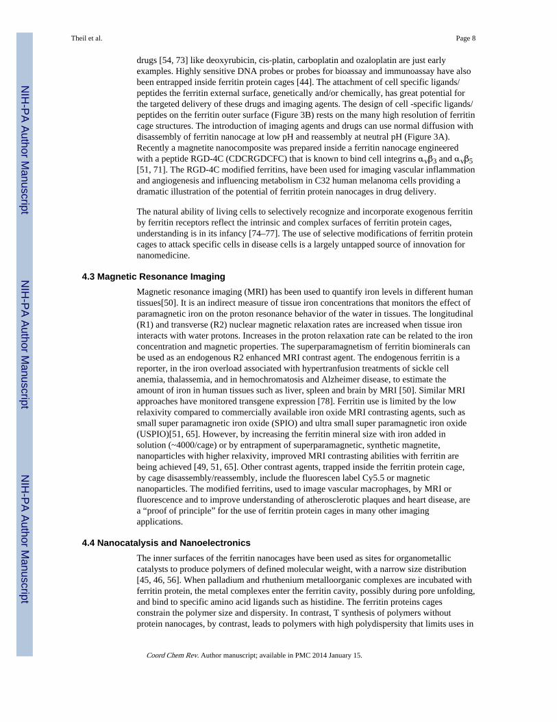
drugs [54, 73] like deoxyrubicin, cis-platin, carboplatin and ozaloplatin are just earlyexamples. Highly sensitive DNA probes or probes for bioassay and immunoassay have alsobeen entrapped inside ferritin protein cages [44]. The attachment of cell specific ligands/peptides the ferritin external surface, genetically and/or chemically, has great potential forthe targeted delivery of these drugs and imaging agents. The design of cell -specific ligands/peptides on the ferritin outer surface (Figure 3B) rests on the many high resolution of ferritincage structures. The introduction of imaging agents and drugs can use normal diffusion withdisassembly of ferritin nanocage at low pH and reassembly at neutral pH (Figure 3A).Recently a magnetite nanocomposite was prepared inside a ferritin nanocage engineeredwith a peptide RGD-4C (CDCRGDCFC) that is known to bind cell integrins αvβ3 and αvβ5[51, 71]. The RGD-4C modified ferritins, have been used for imaging vascular inflammationand angiogenesis and influencing metabolism in C32 human melanoma cells providing adramatic illustration of the potential of ferritin protein nanocages in drug delivery.
The natural ability of living cells to selectively recognize and incorporate exogenous ferritinby ferritin receptors reflect the intrinsic and complex surfaces of ferritin protein cages,understanding is in its infancy [74–77]. The use of selective modifications of ferritin proteincages to attack specific cells in disease cells is a largely untapped source of innovation fornanomedicine.
4.3 Magnetic Resonance ImagingMagnetic resonance imaging (MRI) has been used to quantify iron levels in different humantissues[50]. It is an indirect measure of tissue iron concentrations that monitors the effect ofparamagnetic iron on the proton resonance behavior of the water in tissues. The longitudinal(R1) and transverse (R2) nuclear magnetic relaxation rates are increased when tissue ironinteracts with water protons. Increases in the proton relaxation rate can be related to the ironconcentration and magnetic properties. The superparamagnetism of ferritin biominerals canbe used as an endogenous R2 enhanced MRI contrast agent. The endogenous ferritin is areporter, in the iron overload associated with hypertranfusion treatments of sickle cellanemia, thalassemia, and in hemochromatosis and Alzheimer disease, to estimate theamount of iron in human tissues such as liver, spleen and brain by MRI [50]. Similar MRIapproaches have monitored transgene expression [78]. Ferritin use is limited by the lowrelaxivity compared to commercially available iron oxide MRI contrasting agents, such assmall super paramagnetic iron oxide (SPIO) and ultra small super paramagnetic iron oxide(USPIO)[51, 65]. However, by increasing the ferritin mineral size with iron added insolution (~4000/cage) or by entrapment of superparamagnetic, synthetic magnetite,nanoparticles with higher relaxivity, improved MRI contrasting abilities with ferritin arebeing achieved [49, 51, 65]. Other contrast agents, trapped inside the ferritin protein cage,by cage disassembly/reassembly, include the fluorescen label Cy5.5 or magneticnanoparticles. The modified ferritins, used to image vascular macrophages, by MRI orfluorescence and to improve understanding of atherosclerotic plaques and heart disease, area “proof of principle” for the use of ferritin protein cages in many other imagingapplications.
4.4 Nanocatalysis and NanoelectronicsThe inner surfaces of the ferritin nanocages have been used as sites for organometalliccatalysts to produce polymers of defined molecular weight, with a narrow size distribution[45, 46, 56]. When palladium and rhuthenium metalloorganic complexes are incubated withferritin protein, the metal complexes enter the ferritin cavity, possibly during pore unfolding,and bind to specific amino acid ligands such as histidine. The ferritin proteins cagesconstrain the polymer size and dispersity. In contrast, T synthesis of polymers withoutprotein nanocages, by contrast, leads to polymers with high polydispersity that limits uses in
Theil et al. Page 8
Coord Chem Rev. Author manuscript; available in PMC 2014 January 15.
NIH
-PA Author Manuscript
NIH
-PA Author Manuscript
NIH
-PA Author Manuscript
number of applications. Recently, ferritin bound catalysts has also been shown tohydrogenate smaller olefins size selectively. The catalytic activity per cage can bemodulated by introducing additional metal binding amino acid residues at the inner surfaceof the ferritin nanocage and manipulating the ion channels[43]. Ferritin cages as noveltemplates for iron oxide nanoparticles used in the sustainable productions of olefins usingprocesses such as the Fiescher-Tropsch synthesis [79], remain for future experiments.
Ferritin cages, both 12 and 24 subunit sizes, have been used as templates for metal oxidenanoparticles and metal oxide semiconductors, and also to produce building blocks for thefabrication of electronic devices such as floating nanodot memory device or thin filmtransistor flash memory[42, 43].
5. Conclusions and PerspectiveFerritins, the complex, highly symmetric, cage structures that synthesize ferric oxobiominerals as metabolic iron concentrates and antioxidants, are found in all kingdoms oflife. They share self-assemblying protein cages composed of multiple polypeptides (4-αhelix bundles), with multiple embedded oxidoreductase sites, iron entry/exit channels, and avery large (60% by volume) central mineral growth cavity. Ferritins vary in the number ofsubunits (24 or 12) and size, number of channels, location of the active sites, oxidant (O2 orH2O2), mineral phosphate content and order, and presence or absence of protein-basednucleation channels that guide mineral order.
The enormous progress made in understanding ferritin protein cages has, nevertheless,revealed a number of complex problems that remain to be solved such as: 1. Cytoplamsicdelivery and removal of ferritin iron, i.e. identifying ferritin binding partners. 2.Understanding ferritin pore folding and unfolding rates. 3. Mechanisms of diferric oxorelease from oxidoreductase sites: the role of protein protonation, O-O bond splitting, etc. 4.Electron transfer and hydration paths during ferritin mineral dissolution. 5. Contributions offerritin mineral surface area and order to mineral reduction, dissolution. 6. The transit pathsbetween Fe2+ reduced and dissolved from ferritin minerals and the ion channels. 7. Proteinbased control of mineral crystallinity. 8. Mechanism(s) of movement of Fe3+ O multimersthrough α-helical bundles of ferritin protein cages
What purposes will be served by solving the many remaining problems in ferritin structure /function? First, fundamental understanding of highly evolved protein cage function will beobtained that is also important in virus protein cage formation and supramolecular proteinassembly in general. Second iron chelation therapies will be improved since the iron isferritin iron biominerals; disease targets include iron overload in transfusional treatments forSickle Cell disease and Thalassemia and hereditary hemochromatosis and nervous systemdiseases such as Friedreich’s ataxia. Finally, the ability to manipulate ferritin protein cagesmore completely in nanotechnology such as the use of both inner and outer surfaces of theferritin nanocages as templates in homogeneous nanomaterial synthesis, nanocatalysts forsustainable of hydrocarbons, carrier in targeted drug delivery, synthesis and encapsulation ofimaging agents, nanocatalysis and nanoelectronics, and possibly will be enhanced by theability to admit lager catalytic centers, control nanomaterial order for new applications.
AcknowledgmentsThe work of the authors cited here was supported in part by the NIH-NIDDK (ECT, RKB, and TT), JSPS (TT) andCHORI Partners (ECT, RKB).
Theil et al. Page 9
Coord Chem Rev. Author manuscript; available in PMC 2014 January 15.
NIH
-PA Author Manuscript
NIH
-PA Author Manuscript
NIH
-PA Author Manuscript
ABBREVIATIONS
DFP diferrc peroxo
References1. Theil EC. Current opinion in chemical biology. 2011; 15:304–311. [PubMed: 21296609]
2. Bou-Abdallah F. Biochim Biophys Acta. 2010; 1800:719–731. [PubMed: 20382203]
3. St Pierre T, Tran KC, Webb J, Macey DJ, Heywood BR, Sparks NH, Wade VJ, Mann S, PootrakulP. Biol Met. 1991; 4:162–165. [PubMed: 1931435]
4. Theil, EC.; Le Brun, NE. Comprehensive Inorgannic Chemistry II. Reedijk, J.; Poeppelmeier, K.,editors. Elsevier; 2012.
5. Zhang Y, Orner BP. Int J Mol Sci. 2011; 12:5406–5421. [PubMed: 21954367]
6. Schwartz JK, Liu XS, Tosha T, Theil EC, Solomon EI. Journal of the American Chemical Society.2008; 130:9441–9450. [PubMed: 18576633]
7. Turano P, Lalli D, Felli IC, Theil EC, Bertini I. Proceedings of the National Academy of Sciences ofthe United States of America. 2010; 107:545–550. [PubMed: 20018746]
8. Haldar S, Bevers LE, Tosha T, Theil EC. The Journal of biological chemistry. 2011; 286:25620–25627. [PubMed: 21592958]
9. Stillman TJ, Hempstead PD, Artymiuk PJ, Andrews SC, Hudson AJ, Treffry A, Guest JR, HarrisonPM. Journal of molecular biology. 2001; 307:587–603. [PubMed: 11254384]
10. Johnson E, Cascio D, Sawaya MR, Gingery M, Schroder I. Structure. 2005; 13:637–648. [PubMed:15837202]
11. Tatur J, Hagen WR, Matias PM. J Biol Inorg Chem. 2007; 12:615–630. [PubMed: 17541801]
12. Le Brun NE, Crow A, Murphy ME, Mauk AG, Moore GR. Biochim Biophys Acta. 2010
13. Macedo S, Romao CV, Mitchell E, Matias PM, Liu MY, Xavier AV, LeGall J, Teixeira M,Lindley P, Carrondo MA. Nature structural biology. 2003; 10:285–290.
14. Crow A, Lawson TL, Lewin A, Moore GR, Le Brun NE. Journal of the American ChemicalSociety. 2009; 131:6808–6813. [PubMed: 19391621]
15. Chiancone E, Ceci P. Biochim Biophys Acta. 2010; 1800:798–805. [PubMed: 20138126]
16. Rosenzweig AC, Nordlund P, Takahara PM, Frederick CA, Lippard SJ. Chemistry & biology.1995; 2:409–418.
17. Logan DT, Su XD, Aberg A, Regnstrom K, Hajdu J, Eklund H, Nordlund P. Structure. 1996;4:1053–1064. [PubMed: 8805591]
18. Lindqvist Y, Huang W, Schneider G, Shanklin J. The EMBO journal. 1996; 15:4081–4092.[PubMed: 8861937]
19. Langlois d’Estaintot B, Santambrogio P, Granier T, Gallois B, Chevalier JM, Precigoux G, Levi S,Arosio P. Journal of molecular biology. 2004; 340:277–293. [PubMed: 15201052]
20. Toussaint L, Bertrand L, Hue L, Crichton RR, Declercq JP. Journal of molecular biology. 2007;365:440–452. [PubMed: 17070541]
21. Ha Y, Shi D, Small GW, Theil EC, Allewell NM. J Biol Inorg Chem. 1999; 4:243–256. [PubMed:10439069]
22. Pereira AS, Small W, Krebs C, Tavares P, Edmondson DE, Theil EC, Huynh BH. Biochemistry.1998; 37:9871–9876. [PubMed: 9665690]
23. Moenne-Loccoz P, Krebs C, Herlihy K, Edmondson DE, Theil EC, Huynh BH, Loehr TM.Biochemistry. 1999; 38:5290–5295. [PubMed: 10220314]
24. Hwang J, Krebs C, Huynh BH, Edmondson DE, Theil EC, Penner-Hahn JE. Science (New York,NY. 2000; 287:122–125.
25. Jameson GN, Jin W, Krebs C, Perreira AS, Tavares P, Liu X, Theil EC, Huynh BH. Biochemistry.2002; 41:13435–13443. [PubMed: 12416989]
26. Liu X, Jin W, Theil EC. Proceedings of the National Academy of Sciences of the United States ofAmerica. 2003; 100:3653–3658. [PubMed: 12634425]
Theil et al. Page 10
Coord Chem Rev. Author manuscript; available in PMC 2014 January 15.
NIH
-PA Author Manuscript
NIH
-PA Author Manuscript
NIH
-PA Author Manuscript

27. Arosio P, Ingrassia R, Cavadini P. Biochim Biophys Acta. 2009; 1790:589–599. [PubMed:18929623]
28. Tosha T, Ng HL, Bhattasali O, Alber T, Theil EC. Journal of the American Chemical Society.2010; 132:14562–14569. [PubMed: 20866049]
29. Tosha T, Hasan MR, Theil EC. Proceedings of the National Academy of Sciences of the UnitedStates of America. 2008; 105:18182–18187. [PubMed: 19011101]
30. Ilari A, Latella MC, Ceci P, Ribacchi F, Su M, Giangiacomo L, Stefanini S, Chasteen ND,Chiancone E. Biochemistry. 2005; 44:5579–5587. [PubMed: 15823016]
31. Schwartz JK, Liu XS, Tosha T, Diebold A, Theil EC, Solomon EI. Biochemistry. 2010; 49:10516–10525. [PubMed: 21028901]
32. Tosha T, Behera RK, Ng H-L, Bhattasali O, Alber T, Theil EC. J Biol Chem. 2012 In press.
33. Bellapadrona G, Stefanini S, Zamparelli C, Theil EC, Chiancone E. The Journal of biologicalchemistry. 2009; 284:19101–19109. [PubMed: 19457858]
34. Honarmand Ebrahimi K, Hagedoorn PL, Hagen WR. J Biol Inorg Chem. 2010; 15:1243–1253.[PubMed: 20582559]
35. Takagi H, Shi D, Ha Y, Allewell NM, Theil EC. The Journal of biological chemistry. 1998;273:18685–18688. [PubMed: 9668036]
36. Liu XS, Patterson LD, Miller MJ, Theil EC. The Journal of biological chemistry. 2007;282:31821–31825. [PubMed: 17785467]
37. Hasan MR, Tosha T, Theil EC. The Journal of biological chemistry. 2008; 283:31394–31400.[PubMed: 18805796]
38. Liu X, Theil EC. Accounts of chemical research. 2005; 38:167–175. [PubMed: 15766235]
39. Bevers, LE.; Theil, EC. Progress in Molecular and Subcellualr Biology. Muller, WEG., editor.Springer-Verlag; Berlin-Heidelberg: 2011. p. 18
40. Theil, EC.; Behera, RK. The Chemistry of Nature’s Iron BioMinerals In Ferritin ProteinNanocages. In: Watanabe, Y.; Ueno, T., editors. Coordination Chemistry in Protein Cages:Principles, Design, and Applications. Wiley; 2012.
41. Campan M, Lionetti V, Aquaro GD, Forini F, Matteucci M, Vannucci L, Chiuppesi F, DiCristofano C, Faggioni M, Maioli M, Barile L, Messina E, Lombardi M, Pucci A, Pistello M,Recchia FA. American journal of physiology. 2011; 300:H2238–2250. [PubMed: 21335465]
42. Yamashita I, Iwahori K, Kumagai S. Biochim Biophys Acta. 2010; 1800:846–857. [PubMed:20227466]
43. Uchida M, Kang S, Reichhardt C, Harlen K, Douglas T. Biochim Biophys Acta. 2010; 1800:834–845. [PubMed: 20026386]
44. Maham A, Tang Z, Wu H, Wang J, Lin Y. Small. 2009; 5:1706–1721. [PubMed: 19572330]
45. Wang Z, Takezawa Y, Aoyagi H, Abe S, Hikage T, Watanabe Y, Kitagawa S, Ueno T. Chemicalcommunications (Cambridge, England). 2011; 47:170–172.
46. Takezawa Y, Bockmann P, Sugi N, Wang Z, Abe S, Murakami T, Hikage T, Erker G, WatanabeY, Kitagawa S, Ueno T. Dalton Trans. 2011; 40:2190–2195. [PubMed: 21113534]
47. Iwahori K, Yamashita I. Nanotechnology. 2008; 19:495601. [PubMed: 21730676]
48. Sana B, Poh CL, Lim S. Chemical communications (Cambridge, England). 2012; 48:862–864.
49. Li K, Zhang ZP, Luo M, Yu X, Han Y, Wei HP, Cui ZQ, Zhang XE. Nanoscale. 2012; 4:188–193.[PubMed: 22080281]
50. Wood JC. Hematology / the Education Program of the American Society of Hematology AmericanSociety of Hematology. 2011; 2011:443–450.
51. Terashima M, Uchida M, Kosuge H, Tsao PS, Young MJ, Conolly SM, Douglas T, McConnellMV. Biomaterials. 2011; 32:1430–1437. [PubMed: 21074263]
52. Ziv K, Meir G, Harmelin A, Shimoni E, Klein E, Neeman M. NMR in biomedicine. 2010; 23:523–531. [PubMed: 20175142]
53. Zheng B, Yamashita I, Uenuma M, Iwahori K, Kobayashi M, Uraoka Y. Nanotechnology. 2010;21:045305. [PubMed: 20009209]
Theil et al. Page 11
Coord Chem Rev. Author manuscript; available in PMC 2014 January 15.
NIH
-PA Author Manuscript
NIH
-PA Author Manuscript
NIH
-PA Author Manuscript

54. Xing R, Wang X, Zhang C, Zhang Y, Wang Q, Yang Z, Guo Z. J Inorg Biochem. 2009; 103:1039–1044. [PubMed: 19501911]
55. Mann S. Nat Mater. 2009; 8:781–792. [PubMed: 19734883]
56. Abe S, Hirata K, Ueno T, Morino K, Shimizu N, Yamamoto M, Takata M, Yashima E, WatanabeY. Journal of the American Chemical Society. 2009; 131:6958–6960. [PubMed: 19453195]
57. Ramsay B, Wiedenheft B, Allen M, Gauss GH, Lawrence CM, Young M, Douglas T. J InorgBiochem. 2006; 100:1061–1068. [PubMed: 16412514]
58. Liu G, Wang J, Wu H, Lin Y. Anal Chem. 2006; 78:7417–7423. [PubMed: 17073407]
59. Li M, Viravaidya C, Mann S. Small. 2007; 3:1477–1481. [PubMed: 17768776]
60. Meldrum FC, Heywood BR, Mann S. Science (New York, NY. 1992; 257:522–523.
61. Wade VJ, Levi S, Arosio P, Treffry A, Harrison PM, Mann S. Journal of molecular biology. 1991;221:1443–1452. [PubMed: 1942061]
62. Wade VJ, Treffry A, Laulhere JP, Bauminger ER, Cleton MI, Mann S, Briat JF, Harrison PM.Biochim Biophys Acta. 1993; 1161:91–96. [PubMed: 8422424]
63. Wong KK, Colfen H, Whilton NT, Douglas T, Mann S. J Inorg Biochem. 1999; 76:187–195.[PubMed: 10605836]
64. Lambert EM, Viravaidya C, Li M, Mann S. Angew Chem Int Ed Engl. 2010; 49:4100–4103.[PubMed: 20425877]
65. Uchida M, Terashima M, Cunningham CH, Suzuki Y, Willits DA, Willis AF, Yang PC, Tsao PS,McConnell MV, Young MJ, Douglas T. Magn Reson Med. 2008; 60:1073–1081. [PubMed:18956458]
66. May CA, Grady JK, Laue TM, Poli M, Arosio P, Chasteen ND. Biochim Biophys Acta. 2010;1800:858–870. [PubMed: 20307627]
67. Bulte JW, Douglas T, Mann S, Frankel RB, Moskowitz BM, Brooks RA, Baumgarner CD,Vymazal J, Strub MP, Frank JA. J Magn Reson Imaging. 1994; 4:497–505. [PubMed: 7802866]
68. Hosein HA, Strongin DR, Allen M, Douglas T. Langmuir. 2004; 20:10283–10287. [PubMed:15518526]
69. Butts CA, Swift J, Kang SG, Di Costanzo L, Christianson DW, Saven JG, Dmochowski IJ.Biochemistry. 2008; 47:12729–12739. [PubMed: 18991401]
70. Suci PA, Kang S, Young M, Douglas T. Journal of the American Chemical Society. 2009;131:9164–9165. [PubMed: 19522495]
71. Kitagawa T, Kosuge H, Uchida M, Dua MM, Iida Y, Dalman RL, Douglas T, McConnell MV.Mol Imaging Biol. 2011
72. Flenniken ML, Uchida M, Liepold LO, Kang S, Young MJ, Douglas T. Curr Top MicrobiolImmunol. 2009; 327:71–93. [PubMed: 19198571]
73. Yang Z, Wang X, Diao H, Zhang J, Li H, Sun H, Guo Z. Chemical communications (Cambridge,England). 2007:3453–3455.
74. Zhang L, Fischer W, Pippel E, Hause G, Brandsch M, Knez M. Small. 2011; 7:1538–1541.[PubMed: 21538872]
75. Li JY, Paragas N, Ned RM, Qiu A, Viltard M, Leete T, Drexler IR, Chen X, Sanna-Cherchi S,Mohammed F, Williams D, Lin CS, Schmidt-Ott KM, Andrews NC, Barasch J. Dev Cell. 2009;16:35–46. [PubMed: 19154717]
76. Todorich B, Zhang X, Slagle-Webb B, Seaman WE, Connor JR. Journal of neurochemistry. 2008;107:1495–1505. [PubMed: 19014383]
77. San Martin CD, Garri C, Pizarro F, Walter T, Theil EC, Nunez MT. J Nutr. 2008; 138:659–666.[PubMed: 18356317]
78. Ono K, Fuma K, Tabata K, Sawada M. Biochemical and biophysical research communications.2009; 388:589–594. [PubMed: 19683514]
79. Galvis HMT, Bitter JH, Khare CB, Ruitenbeek M, Dugulan AI, de Jong KP. Science (New York,NY. 2012; 335:834–837.
Theil et al. Page 12
Coord Chem Rev. Author manuscript; available in PMC 2014 January 15.
NIH
-PA Author Manuscript
NIH
-PA Author Manuscript
NIH
-PA Author Manuscript
HIGHLIGHTS
• Ferritins synthesize of caged hydrated ferric oxide nanominerals as metaboliciron concentrates and antioxidants, trapping Fe from damaged proteins O.
• Multiple (24 or 12 polypeptides), folded in 4-α-helix bundles, self -assemble toform ferritin cages.
• Fe2+ enters and exit water soluble ferritin through ion channels, similar tomembrane ion channels,
• Fe2+ and O react at diiron oxidoreductase sites to initiate mineral synthesis withdiferric oxo complexes and in some ferritins larger, protein-based, ferricmultimers.
• Ferrtin proteins cages care are being developed for nanomatierals, nanocatalysts,nanodevices and tissue imaging
Theil et al. Page 13
Coord Chem Rev. Author manuscript; available in PMC 2014 January 15.
NIH
-PA Author Manuscript
NIH
-PA Author Manuscript
NIH
-PA Author Manuscript
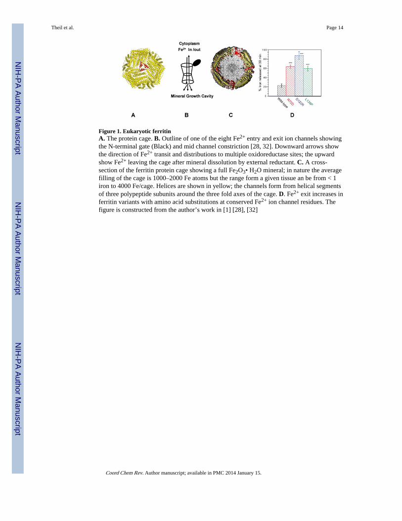
Figure 1. Eukaryotic ferritinA. The protein cage. B. Outline of one of the eight Fe2+ entry and exit ion channels showingthe N-terminal gate (Black) and mid channel constriction [28, 32]. Downward arrows showthe direction of Fe2+ transit and distributions to multiple oxidoreductase sites; the upwardshow Fe2+ leaving the cage after mineral dissolution by external reductant. C. A cross-section of the ferritin protein cage showing a full Fe2O3• H2O mineral; in nature the averagefilling of the cage is 1000–2000 Fe atoms but the range form a given tissue an be from < 1iron to 4000 Fe/cage. Helices are shown in yellow; the channels form from helical segmentsof three polypeptide subunits around the three fold axes of the cage. D. Fe2+ exit increases inferritin variants with amino acid substitutions at conserved Fe2+ ion channel residues. Thefigure is constructed from the author’s work in [1] [28], [32]
Theil et al. Page 14
Coord Chem Rev. Author manuscript; available in PMC 2014 January 15.
NIH
-PA Author Manuscript
NIH
-PA Author Manuscript
NIH
-PA Author Manuscript
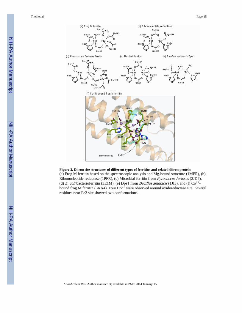
Figure 2. Diiron site structures of different types of ferritins and related diiron protein(a) Frog M ferritin based on the spectroscopic analysis and Mg-bound structure (1MFR), (b)Ribonucleotide reductase (1PFR), (c) Microbial ferritin from Pyrococcus furiosus (2JD7),(d) E. coli bacterioferritin (3E1M), (e) Dps1 from Bacillus anthracis (1JI5), and (f) Co2+-bound frog M ferritin (3KA4). Four Co2+ were observed around oxidoreductase site. Severalresidues near Fe2 site showed two conformations.
Theil et al. Page 15
Coord Chem Rev. Author manuscript; available in PMC 2014 January 15.
NIH
-PA Author Manuscript
NIH
-PA Author Manuscript
NIH
-PA Author Manuscript

Figure 3. Synthesis and incorporation of nanomaterials/nanoparticles inside the multi-subunitferritin nano cavity for catalysis and targeted drug deliveryA. The smaller chemicals, preferably positive charged metal complexes easily diffuse in tothe ferritin nanocavity through the 3-fold ion channels by incubation with the intact ferritincage and nanoparticle synthesis takes place inside the cavity. Bigger nanoparticles (< 8 nm)encapsulated by disassembling/reassembling of multi-subunit ferritin nanocage. B. Bothexternal and internal ferritin cage surfaces modified either by genetically/or chemically touse ferritin nanocage containing nanomaterial for the targeted delivery and to improvecatalysis/synthesis. Purple-ferritin polypeptide subunits.
Theil et al. Page 16
Coord Chem Rev. Author manuscript; available in PMC 2014 January 15.
NIH
-PA Author Manuscript
NIH
-PA Author Manuscript
NIH
-PA Author Manuscript