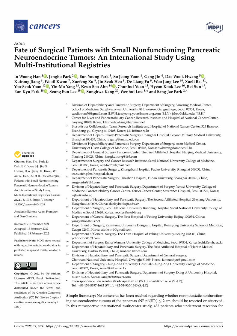
Fate of Surgical Patients with Small Nonfunctioning Pancreatic ...
12
Citation: Han, I.W.; Park, J.; Park, E.Y.; Yoon, S.J.; Jin, G.; Hwang, D.W.; Jiang, K.; Kwon, W.; Xu, X.; Heo, J.S.; et al. Fate of Surgical Patients with Small Nonfunctioning Pancreatic Neuroendocrine Tumors: An International Study Using Multi-Institutional Registries. Cancers 2022, 14, 1038. https://doi.org/ 10.3390/cancers14041038 Academic Editors: Adam Frampton and Dan Granberg Received: 21 December 2021 Accepted: 16 February 2022 Published: 18 February 2022 Publisher’s Note: MDPI stays neutral with regard to jurisdictional claims in published maps and institutional affil- iations. Copyright: © 2022 by the authors. Licensee MDPI, Basel, Switzerland. This article is an open access article distributed under the terms and conditions of the Creative Commons Attribution (CC BY) license (https:// creativecommons.org/licenses/by/ 4.0/). cancers Article Fate of Surgical Patients with Small Nonfunctioning Pancreatic Neuroendocrine Tumors: An International Study Using Multi-Institutional Registries In Woong Han 1 , Jangho Park 2 , Eun Young Park 3 , So Jeong Yoon 1 , Gang Jin 4 , Dae Wook Hwang 5 , Kuirong Jiang 6 , Wooil Kwon 7 , Xuefeng Xu 8 , Jin Seok Heo 1 , De-Liang Fu 9 , Woo Jung Lee 10 , Xueli Bai 11 , Yoo-Seok Yoon 12 , Yin-Mo Yang 13 , Keun Soo Ahn 14 , Chunhui Yuan 15 , Hyeon Kook Lee 16 , Bei Sun 17 , Eun Kyu Park 18 , Seung Eun Lee 19 , Sunghwa Kang 20 , Wenhui Lou 8, * and Sang-Jae Park 2, * 1 Division of Hepatobiliary and Pancreatic Surgery, Department of Surgery, Samsung Medical Center, School of Medicine, Sungkyunkwan University, 81 Irwon-ro, Gangnam-gu, Seoul 06351, Korea; [email protected] (I.W.H.); [email protected] (S.J.Y.); [email protected] (J.S.H.) 2 Center for Liver and Pancreatobiliary Cancer, Research Institute and Hospital of National Cancer Center, Goyang 10408, Korea; [email protected] 3 Biostatistics Collaboration Team, Research Institute and Hospital of National Cancer Center, 323 Ilsan-ro, Ilsandong-gu, Goyang-si 10408, Korea; [email protected] 4 Department of Hepato-Biliary-Pancreatic Surgery, Changhai Hospital, Second Military Medical University, Shanghai 200433, China; [email protected] 5 Division of Hepatobiliary and Pancreatic Surgery, Department of Surgery, Asan Medical Center, University of Ulsan College of Medicine, Seoul 05505, Korea; [email protected] 6 Department of General Surgery, Pancreas Center, The First Affiliated Hospital, Nanjing Medical University, Nanjing 210029, China; [email protected] 7 Department of Surgery and Cancer Research Institute, Seoul National University College of Medicine, Seoul 03080, Korea; [email protected] 8 Department of Pancreatic Surgery, Zhongshan Hospital, Fudan University, Shanghai 200032, China; [email protected] 9 Department of Pancreatic Surgery, Huashan Hospital, Fudan University, Shanghai 200040, China; [email protected] 10 Division of Hepatobiliary and Pancreatic Surgery, Department of Surgery, Yonsei University College of Medicine, Pancreatobiliary Cancer Center, Yonsei Cancer Center, Severance Hospital, Seoul 03722, Korea; [email protected] 11 Department of Hepatobiliary and Pancreatic Surgery, The Second Affiliated Hospital, Zhejiang University, Hangzhou 310009, China; [email protected] 12 Department of Surgery, Seoul National University Bundang Hospital, Seoul National University College of Medicine, Seoul 13620, Korea; [email protected] 13 Department of General Surgery, The First Hospital of Peking University, Beijing 100034, China; [email protected] 14 Department of Surgery, Keimyung University Dongsan Hospital, Keimyung University School of Medicine, Daegu 42601, Korea; [email protected] 15 Department of General Surgery, The Third Hospital of Peking University, Beijing 100083, China; [email protected] 16 Department of Surgery, Ewha Womans University College of Medicine, Seoul 07804, Korea; [email protected] 17 Department of Hepatobiliary and Pancreatic Surgery, The First Affiliated Hospital of Harbin Medical University, Harbin 150001, China; [email protected] 18 Division of Hepatobiliary and Pancreatic Surgery, Department of General Surgery, Chonnam National University Hospital, Gwangju 61469, Korea; [email protected] 19 Department of Surgery, Chung-Ang University Hospital, Chung-Ang University College of Medicine, Seoul 06973, Korea; [email protected] 20 Division of Hepatobiliary and Pancreatic Surgery, Department of Surgery, Dong-A University Hospital, Busan 49201, Korea; [email protected] * Correspondence: [email protected] (W.L.);[email protected] (S.-J.P.); Tel.: +86-136-8197-1683 (W.L.); +82-31-920-1640 (S.-J.P.) Simple Summary: No consensus has been reached regarding whether nonmetastatic nonfunction- ing neuroendocrine tumors of the pancreas (NF-pNETs) ≤ 2 cm should be resected or observed. In this retrospective international multicenter study, 483 patients who underwent resection for Cancers 2022, 14, 1038. https://doi.org/10.3390/cancers14041038 https://www.mdpi.com/journal/cancers
-
Upload
khangminh22 -
Category
Documents
-
view
5 -
download
0
Transcript of Fate of Surgical Patients with Small Nonfunctioning Pancreatic ...
�����������������
Citation: Han, I.W.; Park, J.;
Park, E.Y.; Yoon, S.J.; Jin, G.;
Hwang, D.W.; Jiang, K.; Kwon, W.;
Xu, X.; Heo, J.S.; et al. Fate of Surgical
Patients with Small Nonfunctioning
Pancreatic Neuroendocrine Tumors:
An International Study Using
Multi-Institutional Registries. Cancers
2022, 14, 1038. https://doi.org/
10.3390/cancers14041038
Academic Editors: Adam Frampton
and Dan Granberg
Received: 21 December 2021
Accepted: 16 February 2022
Published: 18 February 2022
Publisher’s Note: MDPI stays neutral
with regard to jurisdictional claims in
published maps and institutional affil-
iations.
Copyright: © 2022 by the authors.
Licensee MDPI, Basel, Switzerland.
This article is an open access article
distributed under the terms and
conditions of the Creative Commons
Attribution (CC BY) license (https://
creativecommons.org/licenses/by/
4.0/).
cancers
Article
Fate of Surgical Patients with Small Nonfunctioning PancreaticNeuroendocrine Tumors: An International Study UsingMulti-Institutional RegistriesIn Woong Han 1 , Jangho Park 2 , Eun Young Park 3, So Jeong Yoon 1, Gang Jin 4, Dae Wook Hwang 5 ,Kuirong Jiang 6, Wooil Kwon 7, Xuefeng Xu 8, Jin Seok Heo 1, De-Liang Fu 9, Woo Jung Lee 10, Xueli Bai 11,Yoo-Seok Yoon 12 , Yin-Mo Yang 13, Keun Soo Ahn 14 , Chunhui Yuan 15, Hyeon Kook Lee 16, Bei Sun 17,Eun Kyu Park 18 , Seung Eun Lee 19 , Sunghwa Kang 20, Wenhui Lou 8,* and Sang-Jae Park 2,*
1 Division of Hepatobiliary and Pancreatic Surgery, Department of Surgery, Samsung Medical Center,School of Medicine, Sungkyunkwan University, 81 Irwon-ro, Gangnam-gu, Seoul 06351, Korea;[email protected] (I.W.H.); [email protected] (S.J.Y.); [email protected] (J.S.H.)
2 Center for Liver and Pancreatobiliary Cancer, Research Institute and Hospital of National Cancer Center,Goyang 10408, Korea; [email protected]
3 Biostatistics Collaboration Team, Research Institute and Hospital of National Cancer Center, 323 Ilsan-ro,Ilsandong-gu, Goyang-si 10408, Korea; [email protected]
4 Department of Hepato-Biliary-Pancreatic Surgery, Changhai Hospital, Second Military Medical University,Shanghai 200433, China; [email protected]
5 Division of Hepatobiliary and Pancreatic Surgery, Department of Surgery, Asan Medical Center,University of Ulsan College of Medicine, Seoul 05505, Korea; [email protected]
6 Department of General Surgery, Pancreas Center, The First Affiliated Hospital, Nanjing Medical University,Nanjing 210029, China; [email protected]
7 Department of Surgery and Cancer Research Institute, Seoul National University College of Medicine,Seoul 03080, Korea; [email protected]
8 Department of Pancreatic Surgery, Zhongshan Hospital, Fudan University, Shanghai 200032, China;[email protected]
9 Department of Pancreatic Surgery, Huashan Hospital, Fudan University, Shanghai 200040, China;[email protected]
10 Division of Hepatobiliary and Pancreatic Surgery, Department of Surgery, Yonsei University College ofMedicine, Pancreatobiliary Cancer Center, Yonsei Cancer Center, Severance Hospital, Seoul 03722, Korea;[email protected]
11 Department of Hepatobiliary and Pancreatic Surgery, The Second Affiliated Hospital, Zhejiang University,Hangzhou 310009, China; [email protected]
12 Department of Surgery, Seoul National University Bundang Hospital, Seoul National University College ofMedicine, Seoul 13620, Korea; [email protected]
13 Department of General Surgery, The First Hospital of Peking University, Beijing 100034, China;[email protected]
14 Department of Surgery, Keimyung University Dongsan Hospital, Keimyung University School of Medicine,Daegu 42601, Korea; [email protected]
15 Department of General Surgery, The Third Hospital of Peking University, Beijing 100083, China;[email protected]
16 Department of Surgery, Ewha Womans University College of Medicine, Seoul 07804, Korea; [email protected] Department of Hepatobiliary and Pancreatic Surgery, The First Affiliated Hospital of Harbin Medical
University, Harbin 150001, China; [email protected] Division of Hepatobiliary and Pancreatic Surgery, Department of General Surgery,
Chonnam National University Hospital, Gwangju 61469, Korea; [email protected] Department of Surgery, Chung-Ang University Hospital, Chung-Ang University College of Medicine,
Seoul 06973, Korea; [email protected] Division of Hepatobiliary and Pancreatic Surgery, Department of Surgery, Dong-A University Hospital,
Busan 49201, Korea; [email protected]* Correspondence: [email protected] (W.L.); [email protected] (S.-J.P.);
Tel.: +86-136-8197-1683 (W.L.); +82-31-920-1640 (S.-J.P.)
Simple Summary: No consensus has been reached regarding whether nonmetastatic nonfunction-ing neuroendocrine tumors of the pancreas (NF-pNETs) ≤ 2 cm should be resected or observed.In this retrospective international multicenter study, 483 patients who underwent resection for
Cancers 2022, 14, 1038. https://doi.org/10.3390/cancers14041038 https://www.mdpi.com/journal/cancers

Cancers 2022, 14, 1038 2 of 12
NF-pNETs ≤ 2 cm in 18 institutions from 2000 to 2017 were enrolled and their medical records werereviewed. Tumor size > 1.5 cm, Ki-67 index ≥ 3%, and nodal metastasis were independent adverseprognostic factors for survival after multivariable analysis. NF-pNET patients with tumors ≤ 1.5 cmcan be observed if the preoperative Ki-67 index is under 3%, and if nodal metastasis is not suspectedin preoperative radiologic studies. These findings support the clinical use to make decisions aboutsmall NF-pNETs.
Abstract: Several treatment guidelines for sporadic, nonmetastatic nonfunctioning neuroendocrinetumors of the pancreas (NF-pNETs) have recommended resection, however, tumors ≤ 2 cm do notnecessarily need surgery. This study aims to establish a surgical treatment plan for NF-pNETs ≤ 2 cm.From 2000 to 2017, 483 patients who underwent resection for NF-pNETs ≤ 2 cm in 18 institutionsfrom Korea and China were enrolled and their medical records were reviewed. The median agewas 56 (range 16–80) years. The 10-year overall survival rate (10Y-OS) and recurrence-free survivalrate (10Y-RFS) were 89.8 and 93.1%, respectively. In multivariable analysis, tumor size (>1.5 cm;HR 4.28, 95% CI 1.80–10.18, p = 0.001) and nodal metastasis (HR 3.32, 95% CI 1.29–8.50, p = 0.013)were independent adverse prognostic factors for OS. Perineural invasion (HR 4.36, 95% CI 1.48–12.87,p = 0.008) and high Ki-67 index (≥3%; HR 9.06, 95% CI 3.01–27.30, p < 0.001) were independentprognostic factors for poor RFS. NF-pNETs ≤ 2 cm showed unfavorable prognosis after resectionwhen the tumor was larger than 1.5 cm, Ki-67 index ≥ 3%, or nodal metastasis was present. NF-pNETpatients with tumors ≤ 1.5 cm can be observed if the preoperative Ki-67 index is under 3%, andif nodal metastasis is not suspected in preoperative radiologic studies. These findings support theclinical use to make decisions about small NF-pNETs.
Keywords: nonfunctioning neuroendocrine tumor of pancreas; prognosis; resection; risk factors
1. Introduction
Pancreatic neuroendocrine tumors (pNETs) account for less than 2% of all pancreaticcancers [1,2] and approximately 10% of tumors arising from the pancreas [3]. Their rareincidence and indolent biologic behavior with variable malignant potential has made it dif-ficult to establish optimal management for pNETs [1,3,4]. Nonfunctioning neuroendocrinetumors of the pancreas (NF-pNET) account for 50–75% of all pNETs, and awareness oftheir incidence and prevalence in recent decades has increased due to the development ofimaging technology and improved pathological diagnosis [1,2,5–7].
Several treatment guidelines for NF-pNETs have recommended resection, however,evidence is lacking for the best way to treat NF-pNETs ≤ 2 cm [8–12]. Some studiessuggest that many small, asymptomatic pNETs are biologically indolent, do not enlarge orprogress over time, show low nodal metastasis, and thus can be safely observed [13,14].However, several reports emphasized that even small tumors can behave aggressivelyand that survival times improved after resection [15–17]. Currently, it is unclear how topreoperatively predict the malignant potential of small NF-pNETs, how to select patientsfor surgery, and how to determine the approach and extent of surgery that should beperformed in patients selected for resection.
The aims of this Korean–Chinese multi-institutional study are to analyze the post-operative outcomes and prognostic factors after resection of sporadic, nonmetastaticNF-pNETs ≤ 2 cm and to suggest surgical indications.
2. Materials and Methods2.1. Patients and Data Collection
Patients with symptoms and biochemical evidence of excess pancreatic hormone are consid-ered to have functioning pNETs, whereas patients with no symptoms, normal serum hormonelevels, and no hereditary syndrome, such as MEN type I, are considered to have sporadic
Cancers 2022, 14, 1038 3 of 12
NF-pNETs [11]. Under Institutional Review Board approval (number: 2019-03-160-001), weretrospectively analyzed the clinicopathological variables of 329 Korean patients from 10 in-stitutes participating in the Korean Tumor Registry System–Biliary Pancreas (KOTUS-BP)and 154 Chinese patients from 8 institutes. All institutions participating in this study aretertiary referral high-volume centers, and all patients were treated based on the guideline orconsensus at the time. All participants underwent resection to treat sporadic, nonmetastaticNF-pNETs smaller than 2 cm, according to final pathologic reports between November2000 and December 2017. When necessary, additional retrospective medical record reviewwas performed.
The variables collected for this work were age at diagnosis, sex, tumor size, location,nodal metastasis, perineural invasion, WHO 2010 tumor grade, mitotic count, Ki-67 index,and duration of follow-up. The OS time was defined as the time from the date of operationto the date of death or last known follow-up. The RFS time was measured from the dateof operation until recurrence. The follow-up was updated in March 2020. In addition,information on surgery and complications was obtained for Korean patients. For thesepatients, postoperative complications were classified using the Clavien–Dindo classifica-tion. Parenchymal sparing resection (PSR) included central pancreatectomy, enucleation,and duodenal preserving pancreatic head resection (DPPHR), and standard resectionsinvolved PD or DP. MIS included laparoscopic or robotic resection of the pancreas. Majorcomplications were defined as Clavien–Dindo grade III or higher. The POPF was definedusing the 2016 International Study Group on Pancreatic Fistula definition [18].
2.2. Statistical Analysis
The baseline characteristics were summarized as median value (range) or frequency(percentage). Comparisons of variables and postoperative complications were performedusing the Chi-squared test. OS and RFS rates were estimated using the Kaplan-Meiermethod, and the survival curves were presented with p of log-rank tests. The clinico-pathological features associated with OS and RFS were analyzed using Cox proportionalhazards models. Statistically significant variables in univariable analysis were includedin multivariable analysis. The final model was determined using the backward selectionmethod with elimination criterion of p > 0.05. Hazard ratios (HRs) and their 95% confidenceintervals (CIs) were presented, and p less than 0.05 indicated statistical significance. Allstatistical analyses were performed using SAS version 9.4 (SAS Institute, Cary, NC, USA)and R project software (version 3.6.2).
3. Results3.1. Clinicopathological Characteristics
Table 1 provides the clinicopathologic details of the 483 patients. The median patientage was 56 (range 16–80) years with a male to female ratio of 1:1.44 (198:285). The tumors of197 patients (40.8%) were in the pancreas head, and those of 286 patients were in the bodyor tail. The median tumor size was 1.4 cm (0.1–2.0 cm) and 24 patients (5.0%) had multipletumors. Using the WHO 2010 grades (G), 364 patients (75.8%) had G1, 105 (21.9%) had G2,and 11 (2.3%) had G3. The numbers of patients with Ki-67 index <3%, 3–20%, and >20%were 388 (82.6%), 73 (15.5%), and 9 (1.9%), respectively. The numbers of patients with mitosiscounts < 2, 2–20, and > 20 were 401 (91.1%), 39 (8.9%), and 0 (0%), respectively. Among243 patients who had lymph node dissection or sampling, 32 patients (13.2%) had lymphnode metastasis (LNM), and these patients accounted for 7.1% of all 483 patients (Table 1).
3.2. Postoperative Complications According to Type of Surgery
Major complications occurred in 28 patients (8.5%), and the Postoperative pancreaticfistula (POPF; grade B or C) rate was 7.6% (n = 25) among the Korean patients. The post-operative outcomes of patients according to PSR or standard resection among the Koreanpatients are shown in Table S1. No differences were observed in major complications,delayed gastric emptying, POPF, or postoperative hemorrhage. In addition, no statistically
Cancers 2022, 14, 1038 4 of 12
significant differences were found in any complication between patients who underwentMIS and those who underwent open resection among the Korean patients (Table S2).
Table 1. Clinical characteristics of the patients.
Characteristics Total (n = 483) Korea (n = 329) China (n = 154)
Sex (male/female) 198/285 (1:1.44) 134/195 (1:1.46) 64/90 (1:1.41)Age (median, range) 56.0 (16–80) 56.0 (17–77) 55.0 (16–80)Tumor size (median, range) (cm) 1.4 (0.1–2.0) 1.4 (0.1–2.0) 1.5 (0.1–2.0)
Tumor size (≤1/1.1–1.5/>1.5–2 cm) (n, %) 146 (30.2%)/188 (38.9%)/149 (30.8%)
95 (28.9%)/131 (39.8%)/103 (31.3%)
51 (33.1%)/57 (37.0%)/46 (29.9%)
Number of tumors (1/>1) (n, %) 459 (95.0%)/24 (5.0%) 316 (96.0%)/13 (4.0%) 143 (92.9%)/11 (7.1%)Tumor location (head/elsewhere) (n, %) 197 (40.8%)/286 (59.2%) 137 (41.6%)/192 (58.4%) 60 (39.0%)/94 (61.0%)
WHO 2010 grade (1/2/3) (n, %) 364 (75.8%)/105 (21.9%)/11 (2%) 259 (78.7%)/66 (20.1%)/4 (1.2%) 105 (68.2%)/39 (25.3%)/7 (4.5%)
Ki-67 index (median, range) (%) 1.0 (0–80) 1.0 (0–80) 2.0 (0–60)Ki-67 index within WHO 2010 grade(G1/G2/G3) (median, range) 1 (0–2.5)/3.7 (0–20)/35 (2–80) 1 (0–2.5)/3.05 (0–10)/
61.685 (2–80) 1 (0–2)/4 (1–20)/30 (3–60)
Ki-67 index (<3/3–20/>20) (n, %) 388 (82.6%)/73 (15.5%)/9 (1.9%) 282 (85.7%)/34 (10.3%)/3(1.0%) 106 (68.8%)/39 (25.3%)/6 (3.9%)Mitotic count (median, range) 1 (0–20) 1 (0–20) 1 (0–20)Mitotic count (<2/≥2) (n, %) 401 (91.1%)/39 (8.9%) 284 (90.5%)/30 (9.6%) 106 (70.2%)/45 (29.8%)Nodal dissection (n, %) 243 (50.3%) 164 (49.8%) 79 (51.3)Nodal metastasis (n, %) 32 (7.1%) 15 (4.6%) 17 (13.6%)Tumor margin (+) (n, %) 27 (5.7%) 24 (7.3%) 3 (2%)Adjacent organ invasion (n, %) 9 (1.9%) 2 (0.6%) 7 (4.5%)Vascular invasion (n, %) 48 (10.3%) 38 (11.6%) 10 (7.4%)Perineural invasion (n, %) 44 (9.6%) 27 (8.2%) 17 (13.1%)
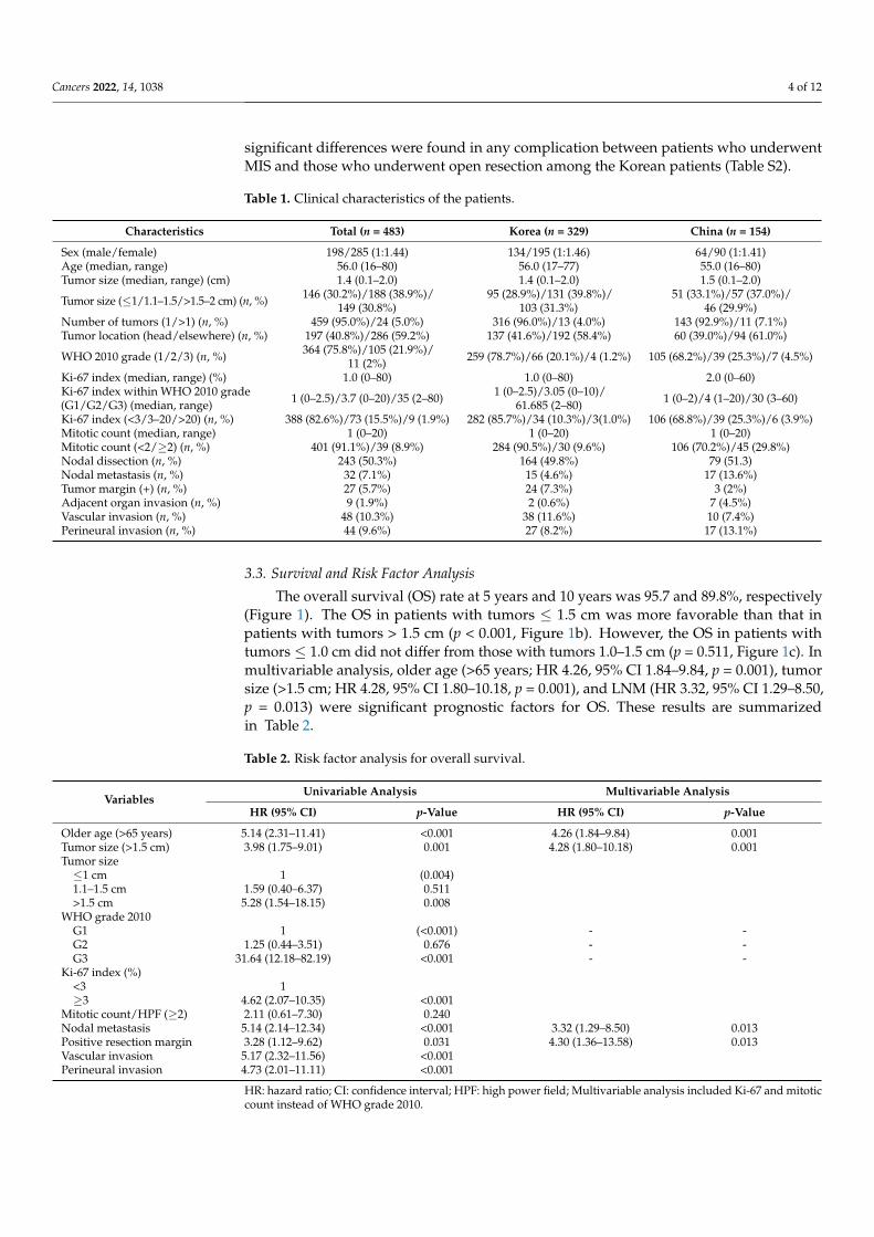
3.3. Survival and Risk Factor Analysis
The overall survival (OS) rate at 5 years and 10 years was 95.7 and 89.8%, respectively(Figure 1). The OS in patients with tumors ≤ 1.5 cm was more favorable than that inpatients with tumors > 1.5 cm (p < 0.001, Figure 1b). However, the OS in patients withtumors ≤ 1.0 cm did not differ from those with tumors 1.0–1.5 cm (p = 0.511, Figure 1c). Inmultivariable analysis, older age (>65 years; HR 4.26, 95% CI 1.84–9.84, p = 0.001), tumorsize (>1.5 cm; HR 4.28, 95% CI 1.80–10.18, p = 0.001), and LNM (HR 3.32, 95% CI 1.29–8.50,p = 0.013) were significant prognostic factors for OS. These results are summarizedin Table 2.
Table 2. Risk factor analysis for overall survival.
VariablesUnivariable Analysis Multivariable Analysis
HR (95% CI) p-Value HR (95% CI) p-Value
Older age (>65 years) 5.14 (2.31–11.41) <0.001 4.26 (1.84–9.84) 0.001Tumor size (>1.5 cm) 3.98 (1.75–9.01) 0.001 4.28 (1.80–10.18) 0.001Tumor size≤1 cm 1 (0.004)1.1–1.5 cm 1.59 (0.40–6.37) 0.511>1.5 cm 5.28 (1.54–18.15) 0.008
WHO grade 2010G1 1 (<0.001) - -G2 1.25 (0.44–3.51) 0.676 - -G3 31.64 (12.18–82.19) <0.001 - -
Ki-67 index (%)<3 1≥3 4.62 (2.07–10.35) <0.001
Mitotic count/HPF (≥2) 2.11 (0.61–7.30) 0.240Nodal metastasis 5.14 (2.14–12.34) <0.001 3.32 (1.29–8.50) 0.013Positive resection margin 3.28 (1.12–9.62) 0.031 4.30 (1.36–13.58) 0.013Vascular invasion 5.17 (2.32–11.56) <0.001Perineural invasion 4.73 (2.01–11.11) <0.001
HR: hazard ratio; CI: confidence interval; HPF: high power field; Multivariable analysis included Ki-67 and mitoticcount instead of WHO grade 2010.
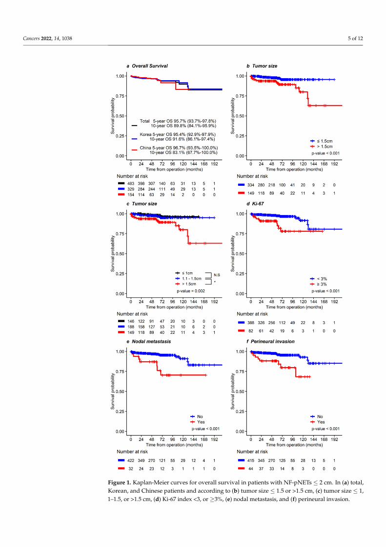
Cancers 2022, 14, 1038 5 of 12
Cancers 2022, 14, x FOR PEER REVIEW 5 of 12
whereas MIS showed favorable prognosis compared to open resection (Figure S1). In ad-dition, RFS did not differ significantly between PSR and standard resection, or between MIS and open resection in Korean patients (Figure S2).
Figure 1. Kaplan-Meier curves for overall survival in patients with NF-pNETs ≤ 2 cm. In (a) total, Korean, and Chinese patients and according to (b) tumor size ≤ 1.5 or >1.5 cm, (c) tumor size ≤ 1, 1–1.5, or >1.5 cm, (d) Ki-67 index <3, or ≥3%, (e) nodal metastasis, and (f) perineural invasion.
Figure 1. Kaplan-Meier curves for overall survival in patients with NF-pNETs ≤ 2 cm. In (a) total,Korean, and Chinese patients and according to (b) tumor size ≤ 1.5 or >1.5 cm, (c) tumor size ≤ 1,1–1.5, or >1.5 cm, (d) Ki-67 index <3, or ≥3%, (e) nodal metastasis, and (f) perineural invasion.
Cancers 2022, 14, 1038 6 of 12
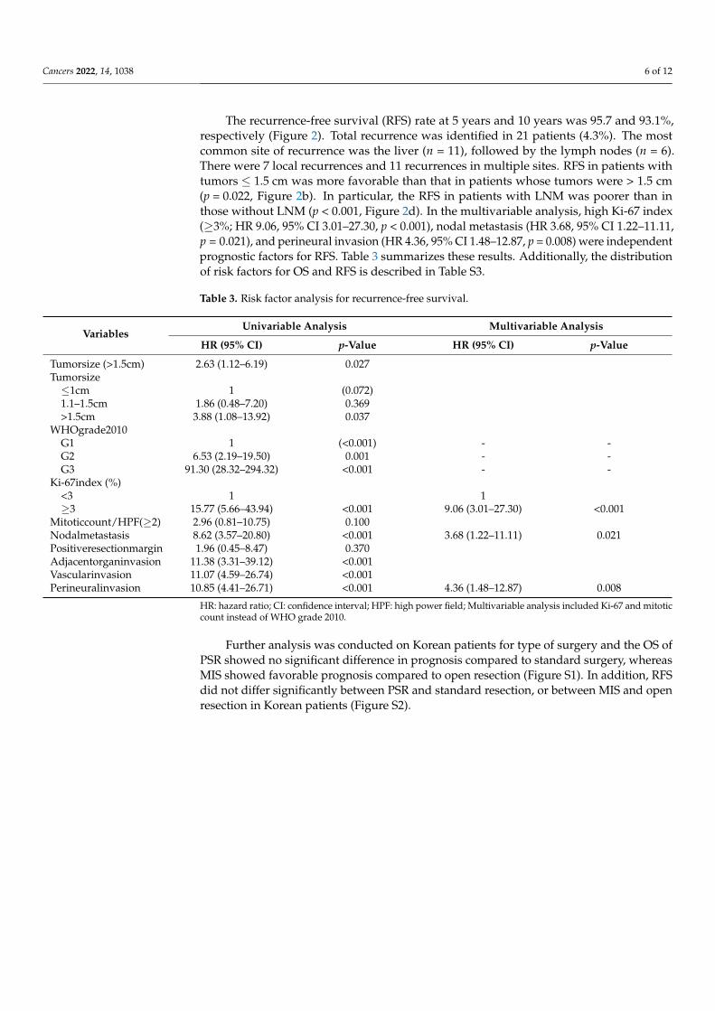
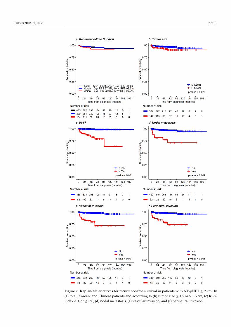
The recurrence-free survival (RFS) rate at 5 years and 10 years was 95.7 and 93.1%,respectively (Figure 2). Total recurrence was identified in 21 patients (4.3%). The mostcommon site of recurrence was the liver (n = 11), followed by the lymph nodes (n = 6).There were 7 local recurrences and 11 recurrences in multiple sites. RFS in patients withtumors ≤ 1.5 cm was more favorable than that in patients whose tumors were > 1.5 cm(p = 0.022, Figure 2b). In particular, the RFS in patients with LNM was poorer than inthose without LNM (p < 0.001, Figure 2d). In the multivariable analysis, high Ki-67 index(≥3%; HR 9.06, 95% CI 3.01–27.30, p < 0.001), nodal metastasis (HR 3.68, 95% CI 1.22–11.11,p = 0.021), and perineural invasion (HR 4.36, 95% CI 1.48–12.87, p = 0.008) were independentprognostic factors for RFS. Table 3 summarizes these results. Additionally, the distributionof risk factors for OS and RFS is described in Table S3.
Table 3. Risk factor analysis for recurrence-free survival.
VariablesUnivariable Analysis Multivariable Analysis
HR (95% CI) p-Value HR (95% CI) p-Value
Tumorsize (>1.5cm) 2.63 (1.12–6.19) 0.027Tumorsize≤1cm 1 (0.072)1.1–1.5cm 1.86 (0.48–7.20) 0.369>1.5cm 3.88 (1.08–13.92) 0.037
WHOgrade2010G1 1 (<0.001) - -G2 6.53 (2.19–19.50) 0.001 - -G3 91.30 (28.32–294.32) <0.001 - -
Ki-67index (%)<3 1 1≥3 15.77 (5.66–43.94) <0.001 9.06 (3.01–27.30) <0.001
Mitoticcount/HPF(≥2) 2.96 (0.81–10.75) 0.100Nodalmetastasis 8.62 (3.57–20.80) <0.001 3.68 (1.22–11.11) 0.021Positiveresectionmargin 1.96 (0.45–8.47) 0.370Adjacentorganinvasion 11.38 (3.31–39.12) <0.001Vascularinvasion 11.07 (4.59–26.74) <0.001Perineuralinvasion 10.85 (4.41–26.71) <0.001 4.36 (1.48–12.87) 0.008
HR: hazard ratio; CI: confidence interval; HPF: high power field; Multivariable analysis included Ki-67 and mitoticcount instead of WHO grade 2010.
Further analysis was conducted on Korean patients for type of surgery and the OS ofPSR showed no significant difference in prognosis compared to standard surgery, whereasMIS showed favorable prognosis compared to open resection (Figure S1). In addition, RFSdid not differ significantly between PSR and standard resection, or between MIS and openresection in Korean patients (Figure S2).
Cancers 2022, 14, 1038 7 of 12Cancers 2022, 14, x FOR PEER REVIEW 6 of 12
Figure 2. Kaplan-Meier curves for recurrence-free survival in patients with NF-pNET ≤ 2 cm. In (a) total, Korean, and Chinese patients and according to (b) tumor size ≤ 1.5 or >1.5 cm, (c) Ki-67 index <3, or ≥3%, (d) nodal metastasis, (e) vascular invasion, and (f) perineural invasion.
Figure 2. Kaplan-Meier curves for recurrence-free survival in patients with NF-pNET ≤ 2 cm. In(a) total, Korean, and Chinese patients and according to (b) tumor size ≤ 1.5 or > 1.5 cm, (c) Ki-67index < 3, or ≥ 3%, (d) nodal metastasis, (e) vascular invasion, and (f) perineural invasion.
Cancers 2022, 14, 1038 8 of 12
4. Discussion
To the best of our knowledge, this study is the largest multi-institution surgical seriesfrom Korea and China (483 patients) to examine sporadic, nonmetastatic NF-pNETs ≤ 2 cm,and thus it provides better external validity than small single-center cohorts. In this study,tumor size (>1.5 cm; HR 4.28, 95% CI 1.80–10.18, p = 0.001) and nodal metastasis (HR 3.32,95% CI 1.29–8.50, p = 0.013) were independent adverse prognostic factors for OS (Table 2).Additionally, perineural invasion (HR 4.36, 95% CI 1.48–12.87, p = 0.008) and high Ki-67index (≥3%; HR 9.06, 95% CI 3.01–27.30, p < 0.001) were independent prognostic factors forpoor RFS (Table 3).
Although surgery used to be the cornerstone of management for small NF-pNETs, thatpractice has been recently challenged. In view of the severe and frequent complicationsfollowing pancreatic surgery and the natural history of sporadic NF-pNET smaller than2 cm, observation without resection has recently been proposed as a possible option.To date, tumor size has been the main determinant when deciding on an operative orobservational policy [7,10–13,17,19]. In several recently published guidelines [10–12], thereis no agreement on whether to perform surgery or initial observation for pNETs ≤ 2 cm.One of the remarkable results of this study is the finding that the size classification criteriashould be changed from 1 or 2 cm to 1.5 cm, as our results indicate that NF-pNETs ≤ 1.5 cmhave a better prognosis than those larger than 1.5 cm (Table 2). NF-pNETs ≤ 1.5 cm canbe observed because their low risk counterbalances the potential for morbidity, mortality,and exocrine and endocrine deficiencies associated with pancreatic resection. Therefore,changing the surgical indication from 1–2 cm to 1.5 cm is reasonable.
Other information to consider when choosing surgical candidates with pNETs smaller than2 cm is the rate of nodal metastases. Nodal metastasis predicts poor prognosis [5,11,14,20–22].In this study, LNM was identified in 32 (7.1%) of all 483 patients and was an independentrisk factor for poor OS and RFS (Tables 2 and 3). In addition, the rate of nodal metastasisin patients with NF-pNETs smaller than 1.5 cm was 5.5% (Table S3). Several previousreports warned that small pNETs, regardless of location, had a risk of LNM from 12.9%to 27.3% [11,14,21,22]. Although we found that the risk of LNM in patients with smallNF-pNETs was lower than in previous studies, we did still find a considerable risk. Thus,standard nodal dissection including suspicious metastatic nodes in preoperative imagingis warranted, and LN sampling can be considered if imaging is negative.
With small NF-pNETs, preoperative endoscopic ultrasound (EUS)-fine needle biopsy(FNB) to determine the Ki-67 level as a predictor of malignancy assists in decision making,because tumor grading has clear implications in terms of prognosis [23,24]. In this study,tumor grading was one of the risk factors for RFS (Table 3). A recent study of data for210 patients from 16 European centers concluded that patients with grade 2 or 3 tumors,which were independent risk factors for poor disease-free survival, should undergo resec-tion, whereas patients with pNETs smaller than 2 cm, could reasonably be managed withsurveillance [7]. Therefore, EUS-FNB should be considered for patients for whom surgicalindications are questionable [11,25].
As in previous studies [23,26], vascular invasion and perineural invasion were adverserisk factors for survival (Tables 2 and 3). Although it is difficult to determine in preoperativeimaging whether vascular or perineural invasions is present, they are clearly factors tobe considered before surgery. An age older than 65 years was an adverse prognosticfactor for OS (Table 2), so it should play a role in selecting patients for surgery versussurveillance. However, older patients also have a higher risk of mortality from surgery, ahigher likelihood of comorbidities, a shorter life expectancy, and shorter surveillance timecompared with younger patients. As a result, caution should be taken in interpreting thesecontradictory results as a uniform endorsement of surgical resection in older patients withsmall pNETs who will potentially benefit from surgical resection.
Patients with NF- pNETs smaller than 2 cm have excellent long-term survival, whichmakes it important to optimize their quality of life in terms of pancreatic function followingsurgical intervention. A pancreaticoduodenectomy (PD) is the gold standard for lesions
Cancers 2022, 14, 1038 9 of 12
of the pancreatic head, whereas a distal pancreatectomy (DP) is used for tumors in thebody and tail. However, these surgical procedures are associated with a substantial lossof functional pancreatic and extrapancreatic tissues. In addition, standard pancreatic re-sections, including multiorgan resection, have considerable postoperative morbidity, asubstantial risk of mortality, and inevitable long-term functional impairments [27,28], andno data supports that an aggressive resection to obtain wide surgical margins is justifiedfor pNETs [11]. PSR, including central pancretectomy, enucleation, and DPPHR, havebeen advocated in select pNET patients to minimize morbidity and maintain pancreaticendocrine and exocrine function [9,27,29]. In this study, all variables regarding compli-cations and survival did not show statistically significant differences between PRS andstandard resection among the Korean patients (Table S1). As mentioned above, accurateassessment of LNM is important to predict prognosis by staging, and PRS generally hasa low lymph node yield rate. In several previous studies, the rate of no lymph nodesampling was higher in patients undergoing PRS than in patients who underwent standardoperations [5,11,30,31]. Patients with low-risk NF-pNETs ≤ 1.5 cm, who are predicted tohave long survival times, and those who develop pNETs at a young age have the most togain from preserved pancreatic function and can thus be considered most appropriate forplanned PRS with selective lymphadenectomy instead of a standard resection. Whether toproceed with PRS or a standard resection is a delicate decision, that should be discussedand preferably made with the patients.
As in this study (Table S2), other studies have reported favorable results from mini-mally invasive surgery (MIS) compared with conventional open pancreatectomy [32–36].Several guidelines have considered different approaches for resecting pNETs, especiallythose in the tail of the pancreas, and laparoscopic or robotic DP has been considered tobe safe and effective with satisfactory postoperative and oncologic outcomes [8,11,35,37].Unlike with DP, results with MIS PD might not be as favorable as those from the con-ventional open approach [38,39]. Therefore, left-sided pancreatectomy should consider alaparoscopic or robotic approach first, but minimally invasive PD should be approachedcarefully considering the surgeon’s experience or the condition of the patient.
The present study has several limitations. First, we only had access to data on pa-tients who underwent resection, and how many patients were under surveillance in theparticipating centers during the same period is unknown. If long-term RFS after curativesurgery is excellent, the results of non-operative management are of prime interest. Second,this study did not have data about the size discrepancy between preoperative CT imagesand pathologic reports. Sallinen et al. [7] reported that significant discrepancies of thosemeasurements should be noted. If possible, this limitation could have been reduced if theimaging and pathology data of Korean and Chinese patients were reviewed once againcentrally. However, due to the nature of the retrospective study, it was not possible todirectly transfer image and pathology data between countries. In addition, we could obtaininformation about the type of surgery or surgical complications only from the Korea multi-center database. If that information could have been included in the multivariable analysisprocess, more sophisticated and reliable statistical analysis would have been possible. Last,selection bias could have affected the retrospective analysis. Patient cohorts of this studywere inconsistent, including surgical indication and inconsistent surgical methods betweeninstitutions, due to the lack of a unified management strategy for small pNETs.
5. Conclusions
NF-pNETs smaller than 2 cm showed considerable recurrence after resection whenthe tumor was larger than 1.5 cm, Ki-67 index was over 3%, or nodal metastasis waspresent. NF-pNET patients with tumors ≤ 1.5 cm can be observed without resection ifthe preoperative Ki-67 index is low, assuming preoperative tissue diagnosis is possible,and nodal metastasis is not suspected in preoperative radiologic studies. Therefore, theproposed surgical indication is expected to help stratify the patient’s prognosis and providecomprehensive clinical decision-making.
Cancers 2022, 14, 1038 10 of 12
Supplementary Materials: The following are available online at https://www.mdpi.com/article/10.3390/cancers14041038/s1, Figure S1: The overall survival on Korean patients for type of surgery. Figure S2:The recurrence-free survival on Korean patients for type of surgery. Table S1: Postoperative compli-cations following parenchymal-sparing resection and standard resection., Table S2: Postoperativecomplications following minimally invasive and open resection., and Table S3: Distribution of riskfactors for survival.
Author Contributions: Conceptualization, I.W.H., W.L. and S.-J.P.; formal analysis, I.W.H., S.J.Y., J.P.,E.Y.P. and S.-J.P.; funding acquisition, I.W.H. and S.-J.P.; investigation, I.W.H., S.J.Y., J.P., E.Y.P., G.J.,D.W.H., K.J., W.K., X.X., J.S.H., D.-L.F., W.J.L., X.B., Y.-S.Y., Y.-M.Y., K.S.A., C.Y., H.K.L., B.S., E.K.P.,S.E.L., S.K., W.L. and S.-J.P.; methodology, I.W.H., W.L. and S.-J.P.; project administration, I.W.H., W.L.and S.-J.P.; resources, I.W.H., S.J.Y., J.P., E.Y.P., G.J., D.W.H., K.J., W.K., X.X., J.S.H., D.-L.F., W.J.L., X.B.,Y.-S.Y., Y.-M.Y., K.S.A., C.Y., H.K.L., B.S., E.K.P., S.E.L., S.K., W.L. and S.-J.P.; software, I.W.H., E.Y.P.and S.-J.P.; supervision, W.L. and S.-J.P.; visualization, I.W.H., S.J.Y., J.P. and E.Y.P.; writing—originaldraft, I.W.H., S.J.Y., E.Y.P.; writing—review and editing, I.W.H., S.J.Y., J.P., E.Y.P., G.J., D.W.H., K.J.,W.K., X.X., J.S.H., D.-L.F., W.J.L., X.B., Y.-S.Y., Y.-M.Y., K.S.A., C.Y., H.K.L., B.S., E.K.P., S.E.L., S.K., W.L.and S.-J.P. All authors have read and agreed to the published version of the manuscript.
Funding: This study was funded by the Research Program of the Korean Association for Hepato-Biliary-Pancreatic Surgery for 2019 (KAHPBS-2019-33).
Institutional Review Board Statement: This study was conducted according to the guidelines of theDeclaration of Helsinki and approved by the Institutional Review Board of Samsung Medical Center(2019-03-160-001).
Informed Consent Statement: Patient consent was waived due to retrospective study after IRB approval.
Data Availability Statement: The data presented in this study are available in this article (andSupplementary Material).
Acknowledgments: We thank the study research nurses Hyemin Kim at Samsung Medical Centerand Gang-Hun Lee at Dongguk University Ilsan Hospital for their work in data acquisition.
Conflicts of Interest: The authors declare no conflict of interest.
References1. Modlin, I.M.; Moss, S.F.; Chung, D.C.; Jensen, R.T.; Snyderwine, E. Priorities for improving the management of gastroenteropan-
creatic neuroendocrine tumors. J. Natl. Cancer Inst. 2008, 100, 1282–1289. [CrossRef]2. Kuo, J.H.; Lee, J.A.; Chabot, J.A. Nonfunctional pancreatic neuroendocrine tumors. Surg. Clin. N. Am. 2014, 94, 689–708.
[CrossRef]3. Panzuto, F.; Boninsegna, L.; Fazio, N.; Campana, D.; Pia Brizzi, M.; Capurso, G.; Scarpa, A.; De Braud, F.; Dogliotti, L.;
Tomassetti, P.; et al. Metastatic and locally advanced pancreatic endocrine carcinomas: Analysis of factors associated with diseaseprogression. J. Clin. Oncol. 2011, 29, 2372–2377. [CrossRef]
4. Metz, D.C.; Jensen, R.T. Gastrointestinal neuroendocrine tumors: Pancreatic endocrine tumors. Gastroenterology 2008, 135,1469–1492. [CrossRef] [PubMed]
5. You, Y.; Jang, J.Y.; Kim, S.C.; Yoon, Y.S.; Park, J.S.; Cho, C.K.; Park, S.J.; Yang, J.D.; Lee, W.J.; Hong, T.H.; et al. Validation of the 8thAJCC Cancer Staging System for Pancreas Neuroendocrine Tumors Using Korean Nationwide Surgery Database. Cancer Res.Treat. 2019, 51, 1639–1652. [CrossRef]
6. Yao, J.C.; Hassan, M.; Phan, A.; Dagohoy, C.; Leary, C.; Mares, J.E.; Abdalla, E.K.; Fleming, J.B.; Vauthey, J.N.; Rashid, A.; et al.One hundred years after "carcinoid": Epidemiology of and prognostic factors for neuroendocrine tumors in 35,825 cases in theUnited States. J. Clin. Oncol. 2008, 26, 3063–3072. [CrossRef] [PubMed]
7. Sallinen, V.J.; Le Large, T.Y.S.; Tieftrunk, E.; Galeev, S.; Kovalenko, Z.; Haugvik, S.P.; Antila, A.; Franklin, O.; Martinez-Moneo, E.;Robinson, S.M.; et al. Prognosis of sporadic resected small (≤2 cm) nonfunctional pancreatic neuroendocrine tumors—Amulti-institutional study. HPB 2018, 20, 251–259. [CrossRef]
8. Falconi, M.; Eriksson, B.; Kaltsas, G.; Bartsch, D.K.; Capdevila, J.; Caplin, M.; Kos-Kudla, B.; Kwekkeboom, D.; Rindi, G.;Klöppel, G.; et al. ENETS Consensus Guidelines Update for the Management of Patients with Functional Pancreatic Neuroen-docrine Tumors and Non-Functional Pancreatic Neuroendocrine Tumors. Neuroendocrinology 2016, 103, 153–171. [CrossRef][PubMed]
9. Singh, S.; Dey, C.; Kennecke, H.; Kocha, W.; Maroun, J.; Metrakos, P.; Mukhtar, T.; Pasieka, J.; Rayson, D.; Rowsell, C.; et al.Consensus Recommendations for the Diagnosis and Management of Pancreatic Neuroendocrine Tumors: Guidelines from aCanadian National Expert Group. Ann. Surg. Oncol. 2015, 22, 2685–2699. [CrossRef] [PubMed]
Cancers 2022, 14, 1038 11 of 12
10. Mansour, J.C.; Chavin, K.; Morris-Stiff, G.; Warner, S.G.; Cardona, K.; Fong, Z.V.; Maker, A.; Libutti, S.K.; Warren, R.;St Hill, C.; et al. Management of asymptomatic, well-differentiated PNETs: Results of the Delphi consensus process of theAmericas Hepato-Pancreato-Biliary Association. HPB 2019, 21, 515–523. [CrossRef]
11. Howe, J.R.; Merchant, N.B.; Conrad, C.; Keutgen, X.M.; Hallet, J.; Drebin, J.A.; Minter, R.M.; Lairmore, T.C.; Tseng, J.F.;Zeh, H.J.; et al. The North American Neuroendocrine Tumor Society Consensus Paper on the Surgical Management of PancreaticNeuroendocrine Tumors. Pancreas 2020, 49, 1–33. [CrossRef]
12. National Comprehensive Cancer Network. Available online: https://www.nccn.org/professionals/physician_gls/default.aspx#neuroendocrine (accessed on 16 February 2022).
13. Kwon, W.; Jang, J.Y.; Song, K.B.; Hwang, D.W.; Kim, S.C.; Heo, J.S.; Choi, D.W.; Hwang, K.K.; Kang, C.M.; Yoon, Y.S.; et al. RiskFactors for Recurrence in Pancreatic Neuroendocrine Tumor and Size as a Surrogate in Determining the Treatment Strategy: AKorean Nationwide Study. Neuroendocrinology 2021, 111, 794–804. [CrossRef] [PubMed]
14. Toste, P.A.; Kadera, B.E.; Tatishchev, S.F.; Dawson, D.W.; Clerkin, B.M.; Muthusamy, R.; Watson, R.; Tomlinson, J.S.; Hines, O.J.;Reber, H.A.; et al. Nonfunctional pancreatic neuroendocrine tumors < 2 cm on preoperative imaging are associated with a lowincidence of nodal metastasis and an excellent overall survival. J. Gastrointest. Surg. 2013, 17, 2105–2113. [PubMed]
15. Haynes, A.B.; Deshpande, V.; Ingkakul, T.; Vagefi, P.A.; Szymonifka, J.; Thayer, S.P.; Ferrone, C.R.; Wargo, J.A.; Warshaw, A.L.;Fernández-del Castillo, C. Implications of incidentally discovered, nonfunctioning pancreatic endocrine tumors: Short-term andlong-term patient outcomes. Arch. Surg. 2011, 146, 534–538. [CrossRef]
16. Sharpe, S.M.; In, H.; Winchester, D.J.; Talamonti, M.S.; Baker, M.S. Surgical resection provides an overall survival benefit forpatients with small pancreatic neuroendocrine tumors. J. Gastrointest. Surg. 2015, 19, 117–123. [CrossRef] [PubMed]
17. Finkelstein, P.; Sharma, R.; Picado, O.; Gadde, R.; Stuart, H.; Ripat, C.; Livingstone, A.S.; Sleeman, D.; Merchant, N.; Yakoub, D.Pancreatic Neuroendocrine Tumors (panNETs): Analysis of Overall Survival of Nonsurgical Management Versus SurgicalResection. J. Gastrointest. Surg. 2017, 21, 855–866. [CrossRef] [PubMed]
18. Bassi, C.; Marchegiani, G.; Dervenis, C.; Sarr, M.; Abu Hilal, M.; Adham, M.; Allen, P.; Andersson, R.; Asbun, H.J.;Besselink, M.G.; et al. The 2016 update of the International Study Group (ISGPS) definition and grading of postoperativepancreatic fistula: 11 Years After. Surgery 2017, 161, 584–591. [CrossRef]
19. Bettini, R.; Boninsegna, L.; Mantovani, W.; Capelli, P.; Bassi, C.; Pederzoli, P.; Delle Fave, G.F.; Panzuto, F.; Scarpa, A.; Falconi, M.Prognostic factors at diagnosis and value of WHO classification in a mono-institutional series of 180 non-functioning pancreaticendocrine tumours. Ann. Oncol. 2008, 19, 903–908. [CrossRef] [PubMed]
20. Hashim, Y.M.; Trinkaus, K.M.; Linehan, D.C.; Strasberg, S.S.; Fields, R.C.; Cao, D.; Hawkins, W.G. Regional lymphadenectomy isindicated in the surgical treatment of pancreatic neuroendocrine tumors (PNETs). Ann. Surg. 2014, 259, 197–203. [CrossRef]
21. Kuo, E.J.; Salem, R.R. Population-level analysis of pancreatic neuroendocrine tumors 2 cm or less in size. Ann. Surg. Oncol. 2013,20, 2815–2821. [CrossRef] [PubMed]
22. Sallinen, V.; Haglund, C.; Seppänen, H. Outcomes of resected nonfunctional pancreatic neuroendocrine tumors: Do size andsymptoms matter? Surgery 2015, 158, 1556–1563. [CrossRef]
23. Landoni, L.; Marchegiani, G.; Pollini, T.; Cingarlini, S.; D’Onofrio, M.; Capelli, P.; De Robertis, R.; Davì, M.V.; Amodio, A.;Impellizzeri, H.; et al. The Evolution of Surgical Strategies for Pancreatic Neuroendocrine Tumors (Pan-NENs): Time-trend andOutcome Analysis from 587 Consecutive Resections at a High-volume Institution. Ann. Surg. 2019, 269, 725–732. [CrossRef][PubMed]
24. Scarpa, A.; Mantovani, W.; Capelli, P.; Beghelli, S.; Boninsegna, L.; Bettini, R.; Panzuto, F.; Pederzoli, P.; delle Fave, G.; Falconi, M.Pancreatic endocrine tumors: Improved TNM staging and histopathological grading permit a clinically efficient prognosticstratification of patients. Mod. Pathol. 2010, 23, 824–833. [CrossRef] [PubMed]
25. Phan, J.; Dawson, D.; Sedarat, A.; Fejleh, M.P.; Marya, N.; Thaker, A.M.; Rogers, M.; Kim, S.; Muthusamy, V.R. ClinicalUtility of Obtaining Endoscopic Ultrasound-Guided Fine-Needle Biopsies for Histologic Analyses of Pancreatic Cystic Lesions.Gastroenterology 2020, 158, 475–477.e1. [CrossRef] [PubMed]
26. Genç, C.G.; Jilesen, A.P.; Partelli, S.; Falconi, M.; Muffatti, F.; van Kemenade, F.J.; van Eeden, S.; Verheij, J.; van Dieren, S.;van Eijck, C.H.J.; et al. A New Scoring System to Predict Recurrent Disease in Grade 1 and 2 Nonfunctional Pancreatic Neuroen-docrine Tumors. Ann. Surg. 2018, 267, 1148–1154. [CrossRef] [PubMed]
27. Beger, H.G.; Mayer, B.; Rau, B.M. Parenchyma-Sparing, Limited Pancreatic Head Resection for Benign Tumors and Low-RiskPeriampullary Cancer—A Systematic Review. J. Gastrointest. Surg. 2016, 20, 206–217. [CrossRef] [PubMed]
28. Ghaferi, A.A.; Birkmeyer, J.D.; Dimick, J.B. Variation in hospital mortality associated with inpatient surgery. N. Engl. J. Med. 2009,361, 1368–1375. [CrossRef] [PubMed]
29. Hüttner, F.J.; Koessler-Ebs, J.; Hackert, T.; Ulrich, A.; Büchler, M.W.; Diener, M.K. Meta-analysis of surgical outcome afterenucleation versus standard resection for pancreatic neoplasms. Br. J. Surg. 2015, 102, 1026–1036. [CrossRef] [PubMed]
30. Parekh, J.R.; Wang, S.C.; Bergsland, E.K.; Venook, A.P.; Warren, R.S.; Kim, G.E.; Nakakura, E.K. Lymph node sampling rates andpredictors of nodal metastasis in pancreatic neuroendocrine tumor resections: The UCSF experience with 149 patients. Pancreas2012, 41, 840–844. [CrossRef]
31. Rindi, G.; Falconi, M.; Klersy, C.; Albarello, L.; Boninsegna, L.; Buchler, M.W.; Capella, C.; Caplin, M.; Couvelard, A.;Doglioni, C.; et al. TNM staging of neoplasms of the endocrine pancreas: Results from a large international cohort study.J. Natl. Cancer Inst. 2012, 104, 764–777. [CrossRef] [PubMed]
Cancers 2022, 14, 1038 12 of 12
32. Drymousis, P.; Raptis, D.A.; Spalding, D.; Fernandez-Cruz, L.; Menon, D.; Breitenstein, S.; Davidson, B.; Frilling, A. Laparoscopicversus open pancreas resection for pancreatic neuroendocrine tumours: A systematic review and meta-analysis. HPB 2014, 16,397–406. [CrossRef]
33. Xourafas, D.; Tavakkoli, A.; Clancy, T.E.; Ashley, S.W. Distal pancreatic resection for neuroendocrine tumors: Is laparoscopicreally better than open? J. Gastrointest. Surg. 2015, 19, 831–840. [CrossRef]
34. Han, S.H.; Han, I.W.; Heo, J.S.; Choi, S.H.; Choi, D.W.; Han, S.; You, Y.H. Laparoscopic versus open distal pancreatectomy fornonfunctioning pancreatic neuroendocrine tumors: A large single-center study. Surg. Endosc. 2018, 32, 443–449. [CrossRef]
35. de Rooij, T.; van Hilst, J.; van Santvoort, H.; Boerma, D.; van den Boezem, P.; Daams, F.; van Dam, R.; Dejong, C.; van Duyn, E.;Dijkgraaf, M.; et al. Minimally Invasive Versus Open Distal Pancreatectomy (LEOPARD): A Multicenter Patient-blindedRandomized Controlled Trial. Ann. Surg. 2019, 269, 2–9. [CrossRef] [PubMed]
36. Zureikat, A.H.; Postlewait, L.M.; Liu, Y.; Gillespie, T.W.; Weber, S.M.; Abbott, D.E.; Ahmad, S.A.; Maithel, S.K.; Hogg, M.E.;Zenati, M.; et al. A Multi-institutional Comparison of Perioperative Outcomes of Robotic and Open Pancreaticoduodenectomy.Ann. Surg. 2016, 264, 640–649. [CrossRef] [PubMed]
37. de Rooij, T.; Klompmaker, S.; Abu Hilal, M.; Kendrick, M.L.; Busch, O.R.; Besselink, M.G. Laparoscopic pancreatic surgery forbenign and malignant disease. Nat. Rev. Gastroenterol. Hepatol. 2016, 13, 227–238. [CrossRef]
38. van Hilst, J.; de Rooij, T.; Bosscha, K.; Brinkman, D.J.; van Dieren, S.; Dijkgraaf, M.G.; Gerhards, M.F.; de Hingh, I.H.; Karsten, T.M.;Lips, D.J.; et al. Laparoscopic versus open pancreatoduodenectomy for pancreatic or periampullary tumours (LEOPARD-2): Amulticentre, patient-blinded, randomised controlled phase 2/3 trial. Lancet Gastroenterol. Hepatol. 2019, 4, 199–207. [CrossRef]
39. Ricci, C.; Casadei, R.; Taffurelli, G.; Pacilio, C.A.; Ricciardiello, M.; Minni, F. Minimally Invasive Pancreaticoduodenectomy: Whatis the Best “Choice”? A Systematic Review and Network Meta-analysis of Non-randomized Comparative Studies. World J. Surg.2018, 42, 788–805. [CrossRef] [PubMed]