Fanconi Anemia repair pathway dysfunction, a potential therapeutic target in lung cancer
33
Fanconi Anemia repair pathway dysfunction, a potential therapeutic target in lung cancer Wenrui Duan, Li Gao, Brittany Aguila, Arjun Kalvala, Gregory A Otterson and Miguel Angel Villalona Journal Name: Frontiers in Oncology ISSN: 2234-943X Article type: Original Research Article Received on: 22 Jul 2014 Accepted on: 04 Dec 2014 Provisional PDF published on: 04 Dec 2014 www.frontiersin.org: www.frontiersin.org Citation: Duan W, Gao L, Aguila B, Kalvala A, Otterson GA and Villalona MA(2014) Fanconi Anemia repair pathway dysfunction, a potential therapeutic target in lung cancer. Front. Oncol. 4:368. doi:10.3389/fonc.2014.00368 Copyright statement: © 2014 Duan, Gao, Aguila, Kalvala, Otterson and Villalona. This is an open-access article distributed under the terms of the Creative Commons Attribution License (CC BY). The use, distribution and reproduction in other forums is permitted, provided the original author(s) or licensor are credited and that the original publication in this journal is cited, in accordance with accepted academic practice. No use, distribution or reproduction is permitted which does not comply with these terms. This Provisional PDF corresponds to the article as it appeared upon acceptance, after rigorous peer-review. Fully formatted PDF and full text (HTML) versions will be made available soon. Thoracic Oncology
Transcript of Fanconi Anemia repair pathway dysfunction, a potential therapeutic target in lung cancer
Fanconi Anemia repair pathway dysfunction, a potential therapeutictarget in lung cancer
Wenrui Duan, Li Gao, Brittany Aguila, Arjun Kalvala, Gregory A Otterson and Miguel Angel Villalona
Journal Name: Frontiers in Oncology
ISSN: 2234-943X
Article type: Original Research Article
Received on: 22 Jul 2014
Accepted on: 04 Dec 2014
Provisional PDF published on: 04 Dec 2014
www.frontiersin.org: www.frontiersin.org
Citation: Duan W, Gao L, Aguila B, Kalvala A, Otterson GA and VillalonaMA(2014) Fanconi Anemia repair pathway dysfunction, a potentialtherapeutic target in lung cancer. Front. Oncol. 4:368.doi:10.3389/fonc.2014.00368
Copyright statement: © 2014 Duan, Gao, Aguila, Kalvala, Otterson and Villalona. This isan open-access article distributed under the terms of theCreative Commons Attribution License (CC BY). The use,distribution and reproduction in other forums is permitted,provided the original author(s) or licensor are credited and thatthe original publication in this journal is cited, in accordance withaccepted academic practice. No use, distribution or reproductionis permitted which does not comply with these terms.
This Provisional PDF corresponds to the article as it appeared upon acceptance, after rigorous
peer-review. Fully formatted PDF and full text (HTML) versions will be made available soon.
Thoracic Oncology
1
Fanconi Anemia repair pathway dysfunction, a potential therapeutic target in
lung cancer
Wenrui Duan1,2 *, Li Gao1, Brittany Aguila1, Arjun Kalvala1, Gregory A. Otterson1, 2, Miguel A Villalona-Calero1, 2, 3 * Comprehensive Cancer Center1, Division of Medical Oncology. Department of Internal Medicine2, and Department of Pharmacology3 at The Ohio State University College of Medicine and Public Health, Columbus, Ohio, 43210 U.S.A
Running Title: FA pathway dysfunction in lung cancer
* Corresponding: Miguel A. Villalona-Calero, A455 Starling-Loving Hall 320 West 10th Avenue, Columbus, OH 43210. Tel. 614-366-5068; Fax, 614-293-7520; E-mail, [email protected] Or Wenrui Duan, 1230 James CHRI, 300 W 10th Avenue, Columbus, OH 43210 Tel. 614-293-3235; Fax, 614-293-6646; E-mail, [email protected]
2
ABSTRACT
The Fanconi Anemia (FA) pathway is a major mechanism of homologous recombination
DNA repair. The functional readout of the pathway is activation through mono-ubiquitination of
FANCD2 leading to nuclear foci of repair. We have recently developed an FA triple staining
immunofluorescence based method (FATSI) to evaluate FANCD2 foci formation in formalin
fixed paraffin embedded (FFPE) tumor samples.
DNA repair deficiencies have been considered of interest in lung cancer prevention,
given the persistence of damage produced by cigarette smoke in this setting, as well as in
treatment, given potential increased efficacy of DNA damaging drugs. We screened 139 non-
small cell lung cancer (NSCLC) FFPE tumors for FANCD2 foci formation by FATSI analysis.
Among 104 evaluable tumors, 23 (22%) were FANCD2 foci negative, thus repair deficient.
To evaluate and compare novel targeted agents in the background of FA deficiency we
utilized RNAi technology to render several lung cancer cell lines FANCD2 deficient. Successful
FANCD2 knockdown was confirmed by reduction in the FANCD2 protein. Subsequently, we
treated the FA defective H1299D2-down and A549D2-down non-small cell lung cancer cells and
their FA competent counterparts (empty vector controls) with the PARP inhibitors veliparib
(ABT-888) (5µM) and BMN673 (0.5µM), as well as the CHK1 inhibitor Arry-575 at a dose of
0.5 µM. We also treated the FA defective small cell lung cancer cell lines H719D2-down and
H792D2-down and their controls with the BCL2/XL inhibitor ABT263 at a dose of 2 µM. The
treated cells were harvested at 24, 48 and 72 hours post treatment. MTT cell viability analysis
showed that each agent was more cytotoxic to the FANCD2 knock-down cells. In all tests, the
FA defective lung cancer cells had less viable cells as comparing to controls 72 hours post
3
treatment. Both MTT and clonogenic analyses comparing the two PARP inhibitors, showed that
BMN673 was more potent compared to veliparib.
Given that FA pathway plays essential roles in response to DNA damage, our results
suggest that a subset of lung cancer patients are likely to be more susceptible to DNA cross-link
based therapy, or to treatments in which additional repair mechanisms are targeted. These
subjects can be identified through FATSI analysis. Clinical trials to evaluate this therapeutic
concept are needed.
4
INTRODUCTION
With more than 159,480 deaths estimated in 2013, lung cancer is the number one cancer
killer in the United States (1). The standard first-line treatment of advanced lung cancer is
platinum-based chemotherapy. However, response rates to chemotherapy vary widely among
patients with the most common type, non-small cell lung cancer (NSCLC), likely due to
heterogeneity in terms of platinum-sensitivity. Great efforts have been made to try to identify
molecular predictive markers of platinum resistance. Inability to repair platinum adducts by the
lack of nucleotide excision repair proteins (ERCC) has received considerable attention, as a
potential predictor of the efficacy of adjuvant platinum based chemotherapy. Results for this
strategy however, are conflicting (2, 3), possibly due to poor discrimination by antibodies of
pertinent proteins isoforms.
Another major mechanism of DNA repair, related to homologous recombination, is
through the Fanconi Anemia (FA) pathway. FA genes collaborate to form foci of DNA repair on
chromatin following DNA damage or during S phase of cell cycle (4). Cells with FA deficiency
are hypersensitive to DNA damage agents such as cisplatin and mitomycin C (MMC) (4), and
tumors from patients with germ line deficiency in some of the genes of this pathway have been
shown to be sensitive to DNA damaging agents, as well as inhibitors of other repair pathways,
such as PARP inhibitors (4-6).
Additional studies have shown disruption of the FA cascade in sporadic cancers (7-9).
These disruptions may involve epigenetic silencing of the FA-core complex, or mutations of one
of several FA genes. The FA pathway contains 16 complementation groups, referred to as FA
subtypes A, B, C, D1/BRCA2, D2, E, F, G, I, J, L, M, N, O, P and Q. Eight of these proteins (A,
5
B, C, E, F, G, L and M) are subunits of FA core complex 1, a nuclear E3 ubiquitin ligase (10-
18).
The FA complex I functions to activate FANCD2 and FANCI by mono-ubiquitinating the
protein following response to DNA damage (12-13). The activated FANCD2 and FANCI
proteins are subsequently transported to subnuclear foci, which are thought to be the sites of
DNA repair and also contain BRCA1, FANCD1/BRCA2, Proliferating Cell Nuclear Antigen
(PCNA) and Rad51 (12, 15, 19).
Given that the FA pathway plays an essential role in response to therapy-induced DNA
interstrand cross-links, it is very plausible that cancers with defective FA pathway are more
sensitive to cross-link based therapy. Since FANCD2 foci formation is critical for cancer cells to
resist MMC and cisplatin, the best way to assess the functionality of this repair pathway as a
whole is by evaluating FANCD2 foci formation. We have developed an FA triple staining
immunofluorescence based method (FATSI) to evaluate FANCD2 foci formation, and have
generated preliminary data showing somatic deficiency of this pathway in tumors across several
organ sites (20).
Herein we report our evaluation of FA deficiency in a series of tumors from patients with
NSCLC and the response of lung cancer cells with reduced FANCD2 expression (FANCD2
knockdown cell) to treatment with inhibitors of PARP, CHK1 and BCL-2/XL.
MATERIALS AND METHODS
FA triple staining immunofluorescence (FATSI) analysis.
Human NSCLC samples were obtained from The Tissue Procurement Shared Resource
of the Ohio State University (OSU) Comprehensive Cancer Center and The Cooperative Human
6
Tissue Network, Midwestern Division at OSU, after Institutional Review Board (IRB) approval.
FFPE tumor tissue was cut at 4 microns, placed on positively charged slides and stained with
hematoxylin and eosin. Additional sections for immunofluorescence staining were placed in a
60oC oven for 1 hour, cooled, deparaffinized and rehydrated through xylenes and graded ethanol
solutions to water in standard fashion. After antigen retrieval, the tissue sections were incubated
with a primary antibody cocktail of rabbit polyclonal FANC-D2 antibody (Novus Biologicals,
Littleton, CO) at a dilution of 1:1000 and a monoclonal anti-Ki67 mouse antibody (Dako,
Carpenteria, CA) at a dilution of 1:150, for 1 hour at room temperature. Sections were then co-
incubated with a secondary antibody (FITC conjugated to anti-rabbit IgG and Alexa fluor 594
donkey anti-mouse IgG, Invitrogen, Carlsbad, CA) at 1:1000 for 1 hour at room temperature. All
rinses were performed on the autostainer with TBST. The sections were mounted on glass slides
using a 4’ 6-diamidino-2-phenylindole (DAPI)–containing embedding medium (Vysis Dapi 1,
Abbott Laboratories, Downers Grove, IL). Formalin fixed paraffin embedded (FFPE) FANCD2
foci negative cells (PD20) and foci positive cells (MCF-7 or FA corrected PD20) were used as
controls on the sample slide during the procedure. The slides were analyzed under a 100 X oil
objective with a Nikon E-400 fluorescence microscope. See prior publication (20).
Generation of FANCD2 knockdown cells
Lung cancer cells A549, H1299 (NSCLC) H719 and H792 (small-cell) were plated 24
hours before transduction. At 60% confluence, cells were transduced with FANCD2-specific
shRNA-expressing and puromycin-resistant lentiviral particles or control shRNA lentiviral
particles (Santa Cruz Biotechnology Inc) according to the manufacturer's protocol. One day after
incubation in medium containing polybrene agent, these transduced cells were transferred to a
dish that contains normal growth medium. The transduced cells were selected in 4 mg/ml
7
puromycin. To create stably transduced cells, 100 to 200 transduced cells were cultured in a 100
mm dish, and medium was replaced with fresh puromycin-containing medium every 3 days, until
resistant colonies were identified. 20 colonies were picked for each cell line, and then the
colonies were expanded. Successful FANCD2 knockdown was confirmed by western blot.
Veliparib, ABT263 and BMN673 were obtained from Selleck Chemicals LLC; Arry 575 was
provided by Array BioPharma.
Cell viability analysis
Five thousand FA defective and control lung cancer cells from each line
(H1299E/H1299D2-down, A549E/A549D2-down, H719E/H719D2-down and H792E/H792D2-
down) were seeded in each well of a 96-well plate 24 h (hours) prior to treatment. Cells were
treated with the single agent at the designated dose (see results section). Dimethylthiazolyl-2-5-
diphenyltetrazolium bromide (MTT) dye solution (Sigma, St. Louis, MO, USA) was added into
the 96-well plate 20 h post treatment. The plate was incubated at 37oC for 4 h, and the treatment
terminated by adding stop solution (isopropanol with 0.04 N HCl). MTT was cleaved by live
cells to a colored formazan product. Absorbance at 560 nm wavelength was recorded using a
Bio-Rad micro plate reader 680 (Bio-Rad Laboratories, Inc., Hercules, CA). Each treatment was
repeated in quadruplicate. An averaged absorbance of blank values (containing all reagents
except cells) was subtracted from all absorbance to yield corrected absorbance. The relative
absorbance of each sample was calculated by comparing the average of corrected absorbance
with an average of corrected untreated control.
Western immunoblot analysis and antibodies
Western immunoblot analysis was performed as described previously (21). Briefly, cells
were digested with lysis buffer which contained 250 mM NaCl; 5 mM EDTA; 1% Igepal; 5 mM
8
dithiothreitol (DTT); 1 mM phenylmethylsulfonyl fluoride (PMSF); and 1% protease inhibitor
cocktail (Sigma, Saint Louis, MO). Protein concentrations were evaluated using the Bradford
reagent (Bio-Rad, Hercules, CA). 100 µg of total protein was loaded onto NuPAGETM 4-12%
Bis-Tris Gel (Invitrogen, Carlsbad, CA). Protein on the gels was electro-transferred onto
nitrocellulose membranes and blocked with blocking buffer (5% of non-fat milk, 500 mM of
NaCl, 20 mM Tris and 0.1% Tween-20). The membranes were incubated with primary
antibodies at 4oC overnight. After washing with TBS-T (blocking buffer without milk) five
times, 10 minutes each, the membranes were incubated with anti-mouse Ig or anti-rabbit Ig
horseradish peroxidase linked to whole secondary antibodies (Amersham Pharmacia Biotech,
Piscataway, NJ) at room temperature for one hour. After washing five times, 10 minutes each, a
chemiluminescent detection system (ECL western blotting detection reagents, GE) was used to
detect the secondary antibody. Finally the membranes were exposed to x-ray films. Antibodies
used were: rabbit polyclonal FANC-D2 antibody (Novus Biologicals, Littleton, CO), anti-tubulin
monoclonal (Sigma, St. Louis, Missouri).
RESULTS
Fanconi Anemia pathway deficiency in non-small cell lung cancer tumor samples.
We used the FATSI method to evaluate FANCD2 foci formation or lack thereof in lung
cancer samples. We screened a total of 139 NSCLC FFPE tumors; 104 were evaluable for
FANCD2 foci status (Fig. 1). Eighty-one of the 104 (78%) evaluable tumors were found
FANCD2 foci positive and 23 (22%) were foci negative.
Forty-nine of the NSCLC samples were of adenocarcinoma histology by morphology
examination, 46 were squamous, five large cell and four of mixed histology. Thirteen (26.5%)
9
adenocarcinomas and seven (15.2%) squamous cell were foci negative. Two of the five large
cell carcinoma and one of four mixed histology were foci negative. The frequencies may suggest
that adenocarcinomas tumors have higher percentage of FANCD2 foci negative tumors as
comparing to squamous cell carcinoma tumors. This observation will need corroboration with
larger sample size and adjustment with other confirmatory tests, such as immunohistochemistry,
not available to us for this dataset.
Generation of FANCD2 knockdown cells and evaluation of sensitivity to PARP inhibitors.
NSCLC cell lines A549, H1299 and small cell H719, H792 were transduced with
FANCD2-specific shRNA-expressing and puromycin-resistant lentiviral particles, or control
shRNA lentiviral particles. To generate stably transduced cells, cells were selected by
puromycin. Successful FANCD2 knockdown colonies were confirmed by western blot
assessment of FANCD2 protein. Figure 2A illustrates four lung cancer cell lines with reduced
FANCD2 protein. We also evaluated the response of the H1299E (H1299 cell transduced with
empty vector) and FANCD2 knockdown (H1299D2-down) to treatment with cisplatin at a dose
of 5ug/ml, 72hours post treatment. We found that FANCD2 silencing resulted in sensitization of
cells to cisplatin (Fig. 2B).
To evaluate the influence of defective FA pathway in regards to cell viability following
exposure to the PARP inhibitors veliparib (ABT-888) and BMN673, we treated the FA defective
NSCLC cell lines H1299D2-down and A549D2-down, as well as their FA competent
counterparts, (H1299E and A549E) (empty vectors) with veliparib at a dose of 5uM or BMN673
at dose of 0.5 µM. MTT assay was used for the cell viability analysis and an averaged
absorbance was recorded 24, 48 and 72 hours post treatment. Cell viability analysis showed that
the FA defective H1299D2-down cells had 80% of viable cells compared to non-treatment
10
controls 72 hours post treatment with veliparib. In contrast there was no influence on viability of
the H1299E cell with the same treatment (Fig. 3A). Both the A549D2-down cell and A549E
cells responded to some degree to the treatment with veliparib at 48 and 72 hours post treatment.
The A549D2-down cells had 68% viable cells compared to 83% viable cells for the A549E cells,
72 hours post treatment (Fig. 3B).
BMN673 is a new class of PARP inhibitor which has in addition strong PARP1-DNA
complex trapping function (22, 23). Cell viability analysis showed that BMN673 was overall a
more potent inhibitor (10 fold difference in active doses) compared to veliparib. H1299
FANCD2 knockdown cancer cells were also more sensitive to BMN673 compared to empty
vectors transfected control cells (25% vs. 62% viable cells, respectively) 72 hours post treatment
(Fig 3C). A549D2-down cell had 29% viable cells and the A549E had 46% viable cells 72 hours
post treatment (Fig. 3D). The IC50 of BMN673 treated A549E cell was 0.64µM and the IC50
of A549D2-down was as low as 0.075 µM 72 hours post treatment. The difference in the IC50
values between H1299E and H1299D2-down cells is smaller with 1.78 µM for the H1299E and
0.74 µM for the H1299D2-down.
To further investigate differential response to treatment with PARP veliparib and
BMN673 between FA defective and FA intact lung cancer cells, we conducted clonogenic
survival analysis. A549D2-down/A549E cells were seeded in a 6-well plate and treated with
veliparib (0.5uM) or BMN673 (0.5uM). Colonies were stained with crystal violet and counted.
Clonogenic survival analysis showed that veliparib was cytotoxic to the FA defective A549D2-
down cells (60% viable cells as compared to non-treatment control), and the A549E had 78%
viable cells (Fig. 4). Following treatment with BMN673 (0.5uM), the FA defective A549D2-
11
down cells were 22% viable as compared to non-treatment control. A549E cells were 43% viable
(Fig. 4).
Effect of Fanconi Anemia repair pathway integrity on response to checkpoint inhibitors.
DNA repair deficient tumor cells have been shown to accumulate high levels of DNA
damage. Therefore the DNA repair deficient cells are dependent on other compensatory DNA
repair pathway, such as the CHK1-kinase pathway. FA defective cells are dependent on this
G2/M checkpoint for viability, since the checkpoint activation allows for the repair of damaged
DNA prior to mitosis. CHK1 is activated by the ATR kinase in response to DNA damage that
stalls replication fork progression (24, 25). Defects in FA pathway have been shown to be
synthetic lethal with CHK1 inhibition or genetic CHK1 depletion in human fibroblast and
ovarian cancer cells (24).
Arry-575 (GDC-0575) is a novel small molecule inhibitor of CHK1, in FA deficient lung
cancer cells. We conducted a dosage test on Arry-575 with H1299 cells, and found the IC50
values were around 1 µM and 0.5 µM for the H1299E and the H1299D2-down cells 72 hours
post treatment. We treated the FA defective lung cancer cell lines H1299D2-down and the
control cell H1299E with Arry-575 at a dose of 0.5 µM. MTT assay was used for cell viability
analysis and an averaged absorbance was recorded 24, 48 and 72 hours post treatment. Cell
viability analysis showed that Arry-575 was more cytotoxic to the H1299D2-down cancer cells.
The FA defective H1299D2-down cells had 38% of viable cells compared to non-treatment
controls 72 hours post treatment. In contrast there were about 60% viable cells in the control cell
line H1299E cells (Fig. 5A).
To evaluate potential synergy for the combination of PARP inhibition and CHK1
inhibition, we treated the H1299D2-down and the control cell H1299E with Arry-575 (0.5 µM)
12
and veliparib (5 µM) alone, or in combination for 72 hours. MTT assay analysis showed a
similar portion of viable cell between the treatment of Arry-575 alone and the combination (Fig.
5B).
Response of FANCD2 defective small cell lung cancer cells to Bcl-2/Bcl-xL inhibition.
Bcl-2 is a central apoptotic inhibitor, and overexpression is associated with tumor
progression and treatment resistance in cancers. Overexpression has been reported in up to 80%
of small cell lung cancers (SCLC). ABT-263 (navitoclax) is a potent and selective inhibitor of
Bcl-2 and Bcl-xL, disrupting their interactions with pro-death proteins leading to the initiation of
apoptosis (26, 27). However a recent phase II study of single-agent navitoclax showed low rate
of response to single agent treatment in advanced and recurrent SCLC (28). Thus, pre-selection
of patients most likely to derive benefit from BCL-2 inhibitors will be needed for further
development of these agents in SCLC.
To evaluate the influence of the FA pathway to treatment with navitoclax, the FA
defective H719D2-down and H792D2-down cells as well as their FA competent counterparts
(H719E and H792E) were treated with navitoclax at a dose of 2µM. The treated cells were then
harvested at 6, 24 and 48 hours post treatment. MTT cell viability analysis showed that
navitoclax was more cytotoxic to the FA deficient H719D2-down compared to its control (51%
and 85% viable cells at 48 hours, respectively) (Fig. 6A). Similarly, the H792D2-down small cell
lung cancer cells had 58% viable cells and the H792E had 86% viable, 48 hours post treatment
(Fig. 6B).
Compensatory activation of alternative DNA repair pathways following exposure to veliparib.
We performed Western immunoblot analysis to evaluate the expression level of PAR,
FancD2 and ERCC1, in the human cancer cell lines H1299 following exposure to veliparib.
13
PAR protein level was reduced at 6 hours post veliparib exposure (5µΜ) and maintained at low
levels through 48 hours in the H1299 cells. However FANCD2 and ERCC1 protein expression
was simultaneously elevated in these cells post treatment with veliparib (Fig. 7).
DISCUSSION
DNA repair is essential for cells to maintain genome stability. There is a growing appreciation
that defects in homologous recombination repair underlie hereditary and sporadic tumorigenesis,
conferring a survival advantage to cancer cells. However, this deficiency may increase
sensitivity of tumors to certain DNA-damaging agents. Homologous recombination deficiency
may therefore prove to be a target of cancer treatment, as long as appropriate biomarkers become
available to identify patients with these tumors (29). Our recently developed FATSI method to
evaluate FANCD2 foci formation, which is capable of evaluating the functionality of the
pathway using FFPE tumor samples (20) could represent such a test. This method is suitable for
large scale screening to select cancer patients most suitable for treatment with DNA-damaging
agents. In addition, the therapeutic window for certain novel molecular targeted agents such as
PARP inhibitors, checkpoint inhibitors and BCL2/xL inhibitors may be larger for the FA-
deficient tumors because the DNA lesions induced cannot be efficiently repaired and will
eventually lead to the cells undergoing apoptosis (29, 30).
We found that 22% of NSCLC tumors examined had functional deficiency in the FA
pathway and that cells with deficient FA pathway were more sensitive to treatment with PARP
inhibitors. Both MTT and clonogenic analyses showed BMN673 was more potent compared to
veliparib. This may be due to veliparib having a much weaker ability to trap PARP–DNA
complexes despite its great activity as a PARP catalytic inhibitor (23). Furthermore, our studies
14
showed that lung cancer cells with deficient FA pathway were more sensitive to treatment with a
CHK1-kinase pathway inhibitor and a BCL2/XL inhibitor.
Of concern are the results showing that veliparib up-regulated FA and nucleotide excision
repair proteins in the H1299 FA wild type cells. Thus treatment with veliparib in repair wild
type cells may plausibly influence resistance to DNA targeting cytotoxic chemotherapy. Cancers
with defective FA or NER pathways, which are incapable of mounting a compensatory response,
may represent a better target for veliparib (or BMN673) alone or in combination with DNA
targeted cytotoxic chemotherapy. It is unclear at this point if targeting two additional repair
mechanisms in the setting of FA dysfunction will be better than one in tumor shrinkage and/or
delaying the appearance of resistance. That is, for example, inhibiting base excision repair
through PARP inhibition/PARP trapping and nucleotide excision repair through ERCC1 in FA
deficient tumors. It is possible that the risks of additional toxicities may outweigh any potential
benefits. However, these experiments are worth conducting but it would be optimal to use best
in class drugs.
The identification of patients with somatic functional deficiency of the FA pathway in
their tumors may also lead to a better understanding of the specific genetic/epigenetic events that
drive the cancer in these patients, by selecting these patients for deep DNA and RNA sequencing
and methylome analysis. The genetic instability caused by the repair deficiency may lead to
additional molecular changes that take over as drivers, a concept that have been named non
oncogenic addiction or induced sustainability (31, 32). Identification and inactivation of these
added drivers may result in an opportunity for synthetic lethality. Our laboratory is pursuing this
approach by performing RNAseq in FA deficient tumor archival material and in fresh biopsies
from patients in clinical trials of PARP inhibition.
15
Resistance to DNA interactive agents and PARP inhibitor may develop in patients with
FA dysfunctional tumors through the recovering of function after a period of treatment. It has
been reported that promoter methylation of several FA genes resulted in deficiency in FA repair
foci formation in human cancers (4, 33-38). The plasticity of epigenetics changes may lead for
example to hypomethylation of these promoters with a resulting recovery of FA function. Other
reported mechanisms of function recovery include post-treatment restorative acquired mutations
in previously dysfunctional repair genes (39, 40). Thus, it is of tantamount importance that any
patient selection for clinical trials evaluating therapeutics in FA deficient tumors is based on
screening of recent tumor material, not separated by intervening treatment. Requiring biopsies
once progression occurs will offer invaluable information regarding the mechanisms mediating
acquired resistance.
In summary, the FA triple-staining immunofluorescence method shows that a proportion
of lung cancer patients have tumors with FA deficiency. We have also demonstrated that lung
cancer cells with defective FA pathway were more sensitive to PARP inhibitors and increase the
therapeutic window of other molecularly targeted agents. Clinical studies are needed to validate
the therapeutic potential of these preclinical findings.
16
Acknowledgements
We thank the Pathology Core Lab and Tissue Procurement facilities of the OSUCCC, and The
Cooperative Human Tissue Network Midwestern Division at The Ohio State University, for their
assistance. This work was supported by NCI R01CA152101 (to M. V); NCI R21CA181291 (to
W.D); American Cancer Society Institutional Seed Grant (IRG-67-003-44 to W.D); NCI grant
P30 CA16058 (to the Ohio State University Comprehensive Cancer Center).
17
REFERENCES
1. Siegel R, Naishadham D, Jemal A. Cancer statistics, 2013. CA Cancer J Clin. (2013)
63(1):11-30.
2. Friboulet L, Olaussen KA, Pignon JP, Shepherd FA, Tsao MS, Graziano S, et al. ERCC1
isoform expression and DNA repair in non-small-cell lung cancer. N Engl J Med (2013)
368(12):1101-10.
3. Olaussen KA, Dunant A, Fouret P, Brambilla E, André F, Haddad V, et al. DNA repair
by ERCC1 in non-small-cell lung cancer and cisplatin-based adjuvant chemotherapy. N
Engl J Med (2006) 355(10):983-91
4. Taniguchi T, Tischkowitz M, Ameziane N, Hodgson SV, Mathew CG, Joenje H, et al.
Disruption of the Fanconi anemia-BRCA pathway in cisplatin-sensitive ovarian tumors.
Nat Med (2003) 9:568-74.
5. Bryant HE, Schultz N, Thomas HD, Parker KM, Flower D, Lopez E, et al. Specific
killing of BRCA2-deficient tumours with inhibitors of poly(ADP-ribose) polymerase.
Nature (2005) 434(7035):913-7.
6. Fong PC, Boss DS, Yap TA, Tutt A, Wu P, Mergui-Roelvink M, et al. Inhibition of
poly(ADP-ribose) polymerase in tumors from BRCA mutation carriers. N Engl J Med
(2009) 361(2):123-34.
7. Lyakhovich A, Surralles J. Disruption of the Fanconi anemia/BRCA pathway in sporadic
cancer. Cancer Lett (2006) 232:99-106.
8. Neveling K, Kalb R, Florl AR, Herterich S, Friedl R, Hoehn H, et al. Disruption of the
FA/BRCA pathway in bladder cancer. Cytogenet Genome Res (2007) 118:166-76.
18
9. Tischkowitz M, Xia B, Sabbaghian N, Reis-Filho JS, Hamel N, Li G, et al. Analysis of
PALB2/FANCN-associated breast cancer families. Proc Natl Acad Sci U S A (2007)
104:6788-93.
10. D'Andrea AD, Grompe M. The Fanconi anaemia/BRCA pathway. Nat Rev Cancer
(2003) 3(1):23-34.
11. Xia B, Sheng Q, Nakanishi K, Ohashi A, Wu J, Christ N, et al. Control of BRCA2
cellular and clinical functions by a nuclear partner, PALB2. Mol Cell (2006) 22(6):719-
29.
12. Smogorzewska A, Matsuoka S, Vinciguerra P, McDonald ER 3rd, Hurov KE, Luo J, et
al. Identification of the FANCI protein, a monoubiquitinated FANCD2 paralog required
for DNA repair. Cell (2007) 129:289-301.
13. Taniguchi T, D'Andrea AD. Molecular pathogenesis of Fanconi anemia: recent progress.
Blood (2006) 107(11):4223-33.
14. Vaz F, Hanenberg H, Schuster B, Barker K, Wiek C, Erven V, et al. Mutation of the
RAD51C gene in a Fanconi anemia-like disorder. Nat Genet (2010) 42:406-9.
15. Somyajit K, Subramanya S, Nagaraju G. RAD51C: a novel cancer susceptibility gene is
linked to Fanconi anemia and breast cancer. Carcinogenesis (2010) 31:2031-8.
16. Kim Y, Lach FP, Desetty R, Hanenberg H, Auerbach AD, Smogorzewska A. Mutations
of the SLX4 gene in Fanconi anemia. Nat Genet (2011) 43:142-6.
17. Stoepker C, Hain K, Schuster B, Hilhorst-Hofstee Y, Rooimans MA, Steltenpool J, et al.
SLX4, a coordinator of structure-specific endonucleases, is mutated in a new Fanconi
anemia subtype. Nat Genet (2011) 43:138-41.
19
18. Bogliolo M, Schuster B, Stoepker C, Derkunt B, Su Y, Raams A, et al. Mutations in
ERCC4, encoding the DNA-repair endonuclease XPF, cause Fanconi anemia. Am J Hum
Genet (2013) 92(5):800-6.
19. Howlett NG, Harney JA, Rego MA, Kolling FW 4th, Glover TW. Functional interaction
between the Fanconi Anemia D2 protein and proliferating cell nuclear antigen (PCNA)
via a conserved putative PCNA interaction motif. J Biol Chem (2009) 284(42):28935-42.
20. Duan W, Gao L, Zhao W, Leon M, Sadee W, Webb A, et al. Assessment of FANCD2
nuclear foci formation in paraffin-embedded tumors: a potential patient-enrichment
strategy for treatment with DNA interstrand crosslinking agents. Transl Res (2013)
161(3):156-64.
21. Duan W, Gao L, Wu X, Wang L, Nana-Sinkam SP, Otterson GA et al. MicroRNA-34a is
an important component of PRIMA-1-induced apoptotic network in human lung cancer
cells. Int J Cancer (2010) 172:313-20.
22. Murai J, Huang SY, Das BB, Renaud A, Zhang Y, Doroshow JH, et al. Trapping of
PARP1 and PARP2 by Clinical PARP Inhibitors. Cancer Res (2012) 72(21):5588-99.
23. Murai J, Huang SY, Renaud A, Zhang Y, Ji J, Takeda S, et al. Stereospecific PARP
trapping by BMN 673 and comparison with olaparib and rucaparib. Mol Cancer Ther
(2014) 13(2):433-43.
24. Chen CC, Kennedy RD, Sidi S, Look AT, D'Andrea A. CHK1 inhibition as a strategy for
targeting Fanconi Anemia (FA) DNA repair pathway deficient tumors. Mol Cancer
(2009) 8:24. doi: 10.1186/1476-4598-8-24.
25. Bartek J, Lukas J, Chk1 and Chk2 kinases in checkpoint control and cancer. Cancer
Cell (2003) 3:421-9.
20
26. Tse C, Shoemaker AR, Adickes J, Anderson MG, Chen J, Jin S, et al. ABT-263: a potent
and orally bioavailable Bcl-2 family inhibitor. Cancer Res (2008) 68(9):3421-8.
27. Shoemaker AR, Mitten MJ, Adickes J, Ackler S, Refici M, Ferguson D, et al. Activity of
the Bcl-2 family inhibitor ABT-263 in a panel of small cell lung cancer xenograft
models. Clin Cancer Res (2008) 14(11):3268-77.
28. Rudin CM, Hann CL, Garon EB, Ribeiro de Oliveira M, Bonomi PD, et al. Phase II study
of single-agent navitoclax (ABT-263) and biomarker correlates in patients with relapsed
small cell lung cancer. Clin Cancer Res (2012)18(11):3163-9.
29. Evers B, Helleday T, Jonkers J. Targeting homologous recombination repair defects in
cancer. Trends Pharmacol Sci (2010) 31(8):372-80.
30. Helleday T, Petermann E, Lundin C, Hodgson B, Sharma RA. DNA repair pathways as
targets for cancer therapy. Nat Rev Cancer (2008) 8(3):193-204.
31. Tischler J, Lehner B, and Frazer AG. Evolutionary plasticity of genetic interaction networks.
Nat Gen (2008) 40: 390-391
32. Luo J, Somilini NL, and Elledge SJ. Principles of cancer therapy: Oncogene and non-oncogene
addiction. Cell (2009) 136: 823-837.
33. Narayan G, Arias-Pulido H, Nandula SV, Basso K, Sugirtharaj DD, Vargas H, et al.
Promoter hypermethylation of FANCF: disruption of Fanconi Anemia-BRCA pathway in
cervical cancer. Cancer Res (2004) 64(9):2994-7.
34. Wang Z, Li M, Lu S, Zhang Y, Wang H. Promoter hypermethylation of FANCF plays an
important role in the occurrence of ovarian cancer through disrupting Fanconi anemia-
BRCA pathway. Cancer Biol Ther (2006) 5(3):256-60.
21
35. Wei M, Xu J, Dignam J, Nanda R, Sveen L, Fackenthal J, et al. Estrogen receptor alpha,
BRCA1, and FANCF promoter methylation occur in distinct subsets of sporadic breast
cancers. Breast Cancer Res Treat (2008) 111(1):113-20.
36. Potapova A, Hoffman AM, Godwin AK, Al-Saleem T, Cairns P. Promoter
hypermethylation of the PALB2 susceptibility gene in inherited and sporadic breast and
ovarian cancer. Cancer Res (2008) 68(4):998-1002.
37. Hess CJ, Ameziane N, Schuurhuis GJ, Errami A, Denkers F, Kaspers GJ, et al.
Hypermethylation of the FANCC and FANCL promoter regions in sporadic acute
leukaemia. Cell Oncol (2008) 30(4):299-306.
38. Dhillon VS, Shahid M, Husain SA. CpG methylation of the FHIT, FANCF, cyclin-D2,
BRCA2 and RUNX3 genes in Granulosa cell tumors (GCTs) of ovarian origin. Mol
Cancer (2004) 3:33.
39. Swisher EM, Sakai W, Karlan BY, Wurz K, Urban N, Taniguchi T. Secondary BRCA1 mutations in
BRCA1-mutated ovarian carcinomas with platinum resistance. Cancer Res (2008) 68(8):2581-6.
40. Bouwman P, Aly A, Escandell JM, Pieterse M, Bartkova J, van der Gulden H, et al. 53BP1 loss rescues
BRCA1 deficiency and is associated with triple-negative and BRCA-mutated breast cancers. Nat Struct
Mol Biol (2010) 17(6):688-95.
22
Figure legends:
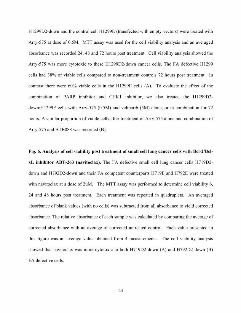
Fig. 1. Detection of FANCD2 foci formation in human lung tumors by the FATSI staining
analysis. The paraffin embedded lung tumor tissues sections were deparaffinized and
rehydrated. The tissue sections were incubated with a primary antibody cocktail of rabbit
polyclonal FANC-D2 antibody (Novus Biologicals, Littleton, CO) at a dilution of 1:1000 and a
monoclonal anti-Ki67 mouse antibody (Dako, Carpenteria, CA) at a dilution of 1:150 for 1 hour
at room temperature. Sections then were incubated with a secondary antibody cocktail
containing FITC conjugated anti-rabbit IgG and Alexafluor 594 donkey anti-mouse secondary
for 1 hour at room temperature. The sections were mounted on glass slides using a 4’ 6-
diamidino-2-phenylindole (DAPI)–containing embedding medium (Vysis Dapi 1, Abbott
Laboratories, Downers Grove, IL). The slides were analyzed under a fluorescence microscope.
(A) FANCD2 foci positive NSCL tumor, and (B) FANCD2 foci negative NSCL tumor.
Magnification: 1000X
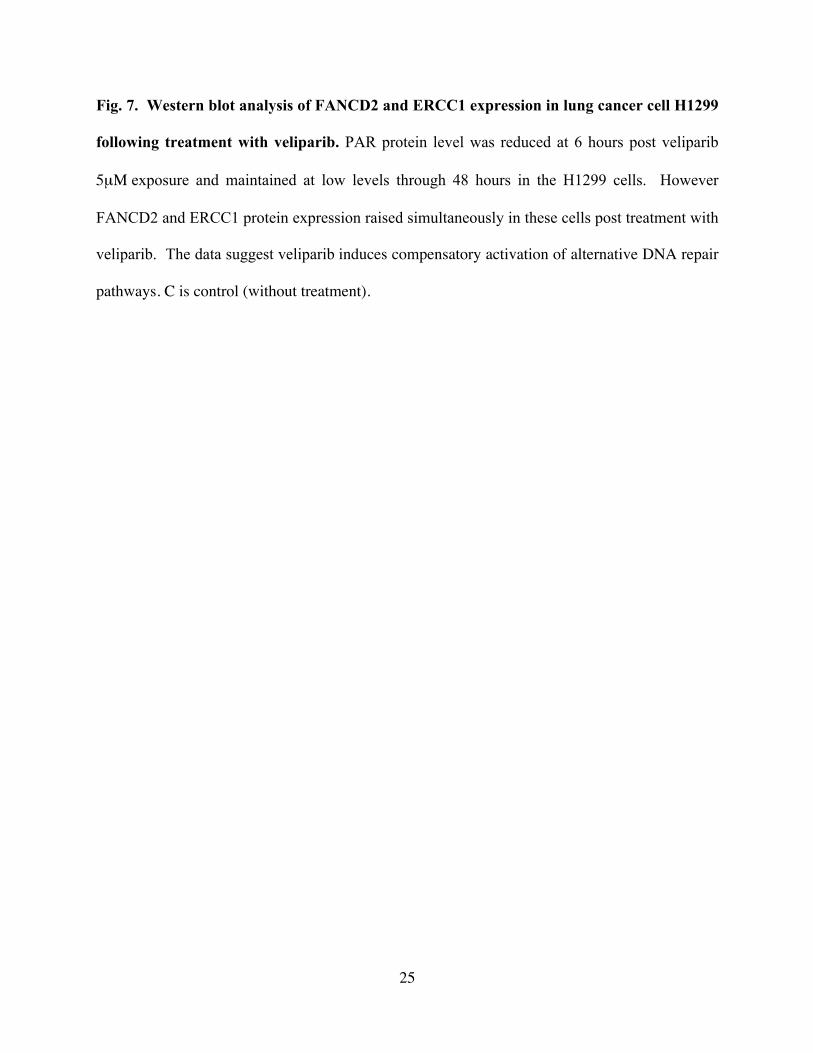
Fig. 2. Creating FANCD2 knockdown cells and evaluating response to cisplatin. (A)
NSCLC cells H1299, A549, and small cell lung cancer cells H719, H792 were plated. At 60%
confluence, cells were transduced with FANCD2-specific shRNA-expressing and puromycin-
resistant lentiviral particles or control shRNA lentiviral particles (Santa Cruz Biotechnology Inc)
according to the manufacturer's protocol. The transduced cells were selected in 4 mg/ml
puromycin to create stably transduced cells with reduced FANCD2 expression. Successful
FANCD2 knockdown was confirmed by western blot detection of the FANCD2 protein. C is a
control cell and D is FANCD2 knockdown cell. (B) The H1299E (H1299 was transfected with
empty vector) and FANCD2 knockdown (H1299D2-down) lung cancer cells were treated with
23
cisplatin (5ug/ml) for 24, 48 and 72 hours. The knockdown cell was more sensitive to the
treatment.
Fig. 3. MTT assay analysis of cell survival in FA deficient and ompetent NSCLC cells post
treatment with the PARP inhibitors veliparib and BMN673. We treated the FA defective
and control lung cancer cell lines H1299D2-down/ H1299E and A549D2-down/A549E with
veliparib (5µΜ) or BMN673 (0.5µΜ). MTT assay was used for the cell viability analysis and an
averaged absorbance was recorded 24, 48 and 72 hours post treatment. Cell viability analysis
showed both control cells H1299E and A549E had no or limited response, and the FA defective
cells H1299D2-down and A549D2-down had a mild response to the treatment of veliparib (2A
and 2B). BMN673 had more cytotoxicity (2C and 2D).
Fig. 4. Clonogenic analysis cell survival for A549E and A549D2-down cells treated with
veliparib (0.5uM) or BMN-673 (0.5uM): A549E and A549D2-down cells were seeded in a 6-
well plate and treated with or without veliparib (0.5uM) or BMN673 (0.5uM) for 8 days.
Colonies were stained with crystal violet and counted. Clonogenic survival analysis showed that
veliparib alone was cytotoxic to A549 cells. The FA defective A549D2-down cells had 60%
viable cells, and the A549E had 78% viable cells as compared to non-treatment control cells.
Post treatment with BMN673 (0.5uM), the FA defective A549D2-down cells has 22% viable
cells, and the A549E had 43% viable cells as compared to non-treatment control.
Fig.5. Cell survival of FA defective lung cancer cells to treatment of CHK1 inhibitor as
single agent or in combination with veliparib. The FA defective lung cancer cell lines
24
H1299D2-down and the control cell H1299E (transfected with empty vectors) were treated with
Arry-575 at dose of 0.5M. MTT assay was used for the cell viability analysis and an averaged
absorbance was recorded 24, 48 and 72 hours post treatment. Cell viability analysis showed the
Arry-575 was more cytotoxic to these H1299D2-down cancer cells. The FA defective H1299
cells had 38% of viable cells compared to non-treatment controls 72 hours post treatment. In
contrast there were 60% viable cells in the H1299E cells (A). To evaluate the effect of the
combination of PARP inhibitor and CHK1 inhibitor, we also treated the H1299D2-
down/H1299E cells with Arry-575 (0.5M) and veliparib (5M) alone, or in combination for 72
hours. A similar proportion of viable cells after treatment of Arry-575 alone and combination of
Arry-575 and ATB888 was recorded (B).
Fig. 6. Analysis of cell viability post treatment of small cell lung cancer cells with Bcl-2/Bcl-
xL inhibitor ABT-263 (navitoclax). The FA defective small cell lung cancer cells H719D2-
down and H792D2-down and their FA competent counterparts H719E and H792E were treated
with navitoclax at a dose of 2uM. The MTT assay was performed to determine cell viability 6,
24 and 48 hours post treatment. Each treatment was repeated in quadruplets. An averaged
absorbance of blank values (with no cells) was subtracted from all absorbance to yield corrected
absorbance. The relative absorbance of each sample was calculated by comparing the average of
corrected absorbance with an average of corrected untreated control. Each value presented in
this figure was an average value obtained from 4 measurements. The cell viability analysis
showed that navitoclax was more cytotoxic to both H719D2-down (A) and H792D2-down (B)
FA defective cells.
25
Fig. 7. Western blot analysis of FANCD2 and ERCC1 expression in lung cancer cell H1299
following treatment with veliparib. PAR protein level was reduced at 6 hours post veliparib
5µΜ exposure and maintained at low levels through 48 hours in the H1299 cells. However
FANCD2 and ERCC1 protein expression raised simultaneously in these cells post treatment with
veliparib. The data suggest veliparib induces compensatory activation of alternative DNA repair
pathways. C is control (without treatment).
Figure 1.JPEG
Figure 2.JPEG
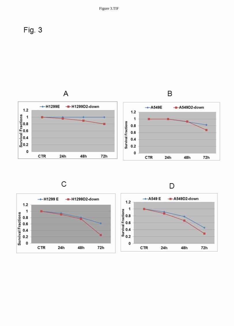
Figure 3.TIF
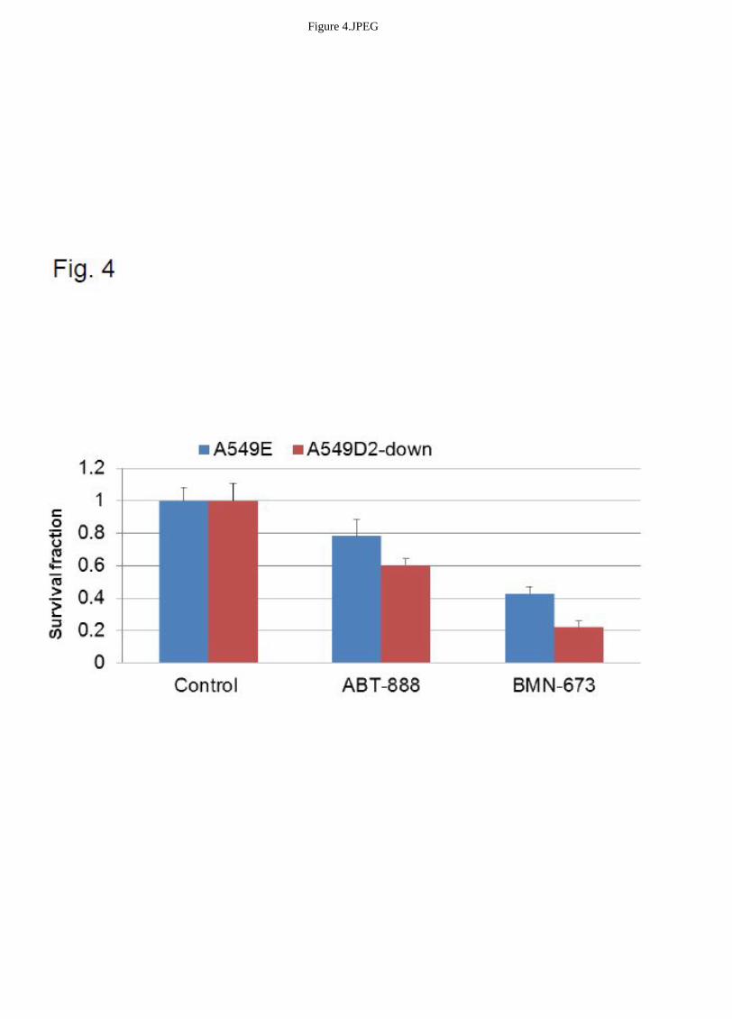
Figure 4.JPEG
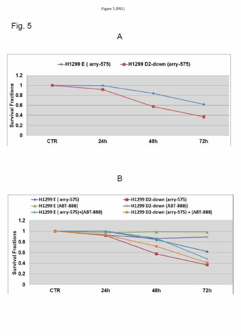
Figure 5.JPEG
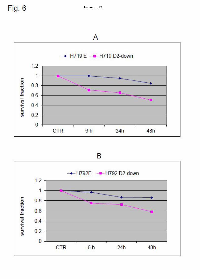
Figure 6.JPEG
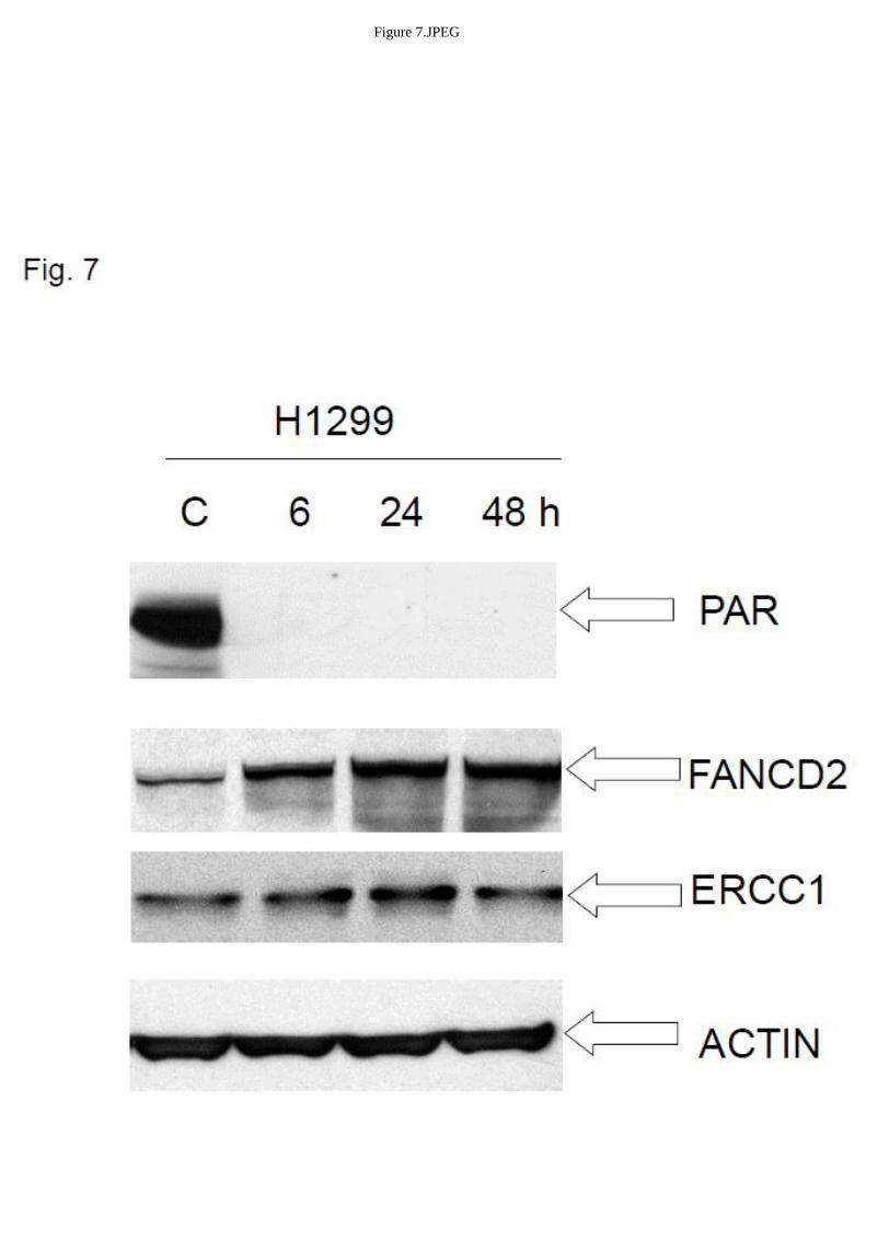
Figure 7.JPEG