FAIR-sharing of multi-scale, multi- species brain models
30
TemplateFlow: FAIR-sharing of multi-scale, multi- species brain models Rastko Ciric Department of Psychology, Stanford University William Thompson Department of Clinical Neuroscience, Karolinska Institutet Romy Lorenz MRC CBU, University of Cambridge Mathias Goncalves Massachusetts Institute of Technology Eilidh MacNicol Department of Neuroimaging, Institute of Psychiatry, Psychology and Neuroscience, King’s College London Christopher Markiewicz Department of Psychology, Stanford University Yaroslav Halchenko Department of Psychological and Brain Sciences, Dartmouth College Satrajit Ghosh Massachusetts Institute of Technology https://orcid.org/0000-0002-5312-6729 Krzysztof Gorgolewski Stanford University https://orcid.org/0000-0003-3321-7583 Russell Poldrack Stanford https://orcid.org/0000-0001-6755-0259 Oscar Esteban ( [email protected] ) CHUV University of Lausanne https://orcid.org/0000-0001-8435-6191 Resource Keywords: TemplateFlow, brain models, atlases, neuroimaging Posted Date: August 26th, 2021 DOI: https://doi.org/10.21203/rs.3.rs-264855/v3 License: This work is licensed under a Creative Commons Attribution 4.0 International License. Read Full License
-
Upload
khangminh22 -
Category
Documents
-
view
3 -
download
0
Transcript of FAIR-sharing of multi-scale, multi- species brain models
TemplateFlow: FAIR-sharing of multi-scale, multi-species brain modelsRastko Ciric
Department of Psychology, Stanford UniversityWilliam Thompson
Department of Clinical Neuroscience, Karolinska InstitutetRomy Lorenz
MRC CBU, University of CambridgeMathias Goncalves
Massachusetts Institute of TechnologyEilidh MacNicol
Department of Neuroimaging, Institute of Psychiatry, Psychology and Neuroscience, King’s CollegeLondonChristopher Markiewicz
Department of Psychology, Stanford UniversityYaroslav Halchenko
Department of Psychological and Brain Sciences, Dartmouth CollegeSatrajit Ghosh
Massachusetts Institute of Technology https://orcid.org/0000-0002-5312-6729Krzysztof Gorgolewski
Stanford University https://orcid.org/0000-0003-3321-7583Russell Poldrack
Stanford https://orcid.org/0000-0001-6755-0259Oscar Esteban ( [email protected] )
CHUV University of Lausanne https://orcid.org/0000-0001-8435-6191
Resource
Keywords: TemplateFlow, brain models, atlases, neuroimaging
Posted Date: August 26th, 2021
DOI: https://doi.org/10.21203/rs.3.rs-264855/v3
License: This work is licensed under a Creative Commons Attribution 4.0 International License. Read Full License

TemplateFlow: FAIR-sharing of multi-scale,
multi-species brain models
Rastko Ciric1,2*, William H. Thompson3, Romy Lorenz1,4,5, Mathias Goncalves1,
Eilidh MacNicol6, Christopher J. Markiewicz1, Yaroslav O. Halchenko7, Satrajit S.
Ghosh8,9, Krzysztof J. Gorgolewski1, Russell A. Poldrack1, Oscar Esteban10*
*For correspondence:
[email protected] (RC);[email protected] (OE)
1Department of Psychology, Stanford University, CA, USA; 2Department ofBioengineering, Stanford University, CA, USA; 3Department of Clinical Neuroscience,Karolinska Institutet, Stockholm, Sweden; 4MRC CBU, University of Cambridge,Cambridge, UK; 5Department of Neurophysics, MPI, Leipzig, Germany; 6Department ofNeuroimaging, Institute of Psychiatry, Psychology and Neuroscience, King’s CollegeLondon, London, UK; 7Department of Psychological and Brain Sciences, DartmouthCollege, Hanover, NH, USA; 8McGovern Institute for Brain Research, MassachusettsInstitute of Technology: MIT, Cambridge, MA, USA; 9Department of Otolaryngology,Harvard Medical School, Boston, MA, USA; 10Department of Radiology, UniversityHospital of Lausanne and University of Lausanne, Lausanne, Switzerland
Abstract Reference anatomies of the brain and corresponding atlases play a central role inexperimental neuroimaging workflows and are the foundation for reporting standardized results.The choice of such references —i.e., templates— and atlases is one relevant source ofmethodological variability across studies, which has recently been brought to attention as animportant challenge to reproducibility in neuroscience. TemplateFlow is a publicly availableframework for human and nonhuman brain models. The framework combines an open databasewith software for access, management, and vetting, allowing scientists to distribute theirresources under FAIR —findable, accessible, interoperable, reusable— principles. TemplateFlowsupports a multifaceted insight into brains across species, and enables multiverse analysestesting whether results generalize across standard references, scales, and in the long term,species, thereby contributing to increasing the reliability of neuroimaging results.
Brains are morphologically variable, exhibiting diversity in such features as overall size (Lüders1
et al., 2002), sulcal curvature (Tosun et al., 2015), and functional topology (Tavor et al., 2016;Mars2
et al., 2018). Morphological variability manifests not only in differences between brains but also3
in the way that a brain changes across its lifespan, as it is remodelled by development, aging, and4
degenerative processes (Courchesne et al., 2000; Good et al., 2001; Sowell et al., 2003). Thesemor-5
phological differences often correspond with the effects of interest in neuroimaging studies and6
hinder direct spatial comparisons between brain maps (Brett et al., 2002). The substantial vari-7
ability within and between individual brains necessitates a means of formalizing population-level8
knowledge about brain anatomy and function. Neuroscientists have answered this need by cre-9
ating brain atlases as references for understanding and contextualizing morphological variability.10
Atlases map landmarks, features, and other knowledge about the brain as annotations that are11
consistent across individual brains.12
The development of atlases in neuroscience has accelerated knowledge discovery and dissem-13
ination. Early endeavors, epitomized by the groundbreaking work of Brodmann (2006, originally14
1 of 28

published in German in 1909) and complemented by Von Economo and Koskinas (2008, originally15
published in German in 1925), leveraged careful scrutiny of microanatomy and cytoarchitectonic16
properties in small numbers of brains. Concurrentmacroanatomical approaches, by contrast, iden-17
tified common features in nuclear boundaries and cortical gyrification. Modern atlases advanced18
on these approaches by incorporating stereotaxy, defining a basis set of coordinate axes over19
the brain and anchoring neural landmarks to coordinates. Talairach’s assiduous postmortem ex-20
amination of a single brain produced a stereotaxic atlas that saw wide use (Talairach et al., 1957).21
Stereotaxywas a fundamental feature to unfold surgical neuronavigation systems. Schurr andMer-22
rington (1978) developed the first sterotaxic apparatus to surgically induce targeted brain lesions23
on cats. This early antecedent of neuronavigation informed early sectional atlases of the cat and24
macaque brains. Since then, neuroscientists have directed great efforts to improve existing (Ta-25
lairach and Tournoux, 1988) and generate new atlases of the neurotypical adult human (Landman26
et al., 2012) and nonhuman (Paxinos and Watson, 1997; Martin and Bowden, 2000) brain; as well27
as developing, aging, and neurologically atypical brains. For instance, new atlases and represen-28
tative stereotaxic maps can be created for diseased (Dickie et al., 2015), infant (Matsuzawa et al.,29
2001; Fonov et al., 2011; Shi et al., 2011), and elderly (Buckner et al., 2004) human populations or30
to capture the rapid postnatal development of nonhuman species (Calabrese et al., 2013; Szulc31
et al., 2015). Advancing beyond the volumetric constraints of stereotaxy, researchers of primate32
neocortex have also devised standard spaces based on geometric reconstructions of the cortical33
surface. This surface-based approach has the advantage of respecting the intrinsic topology of34
cortical folds, a development that has led to further improvements in spatial localization (Coalson35
et al., 2018). On account of its relatively high spatial resolution, its capacity to image the entire36
brain, and its non-invasive acquisition protocols, magnetic resonance imaging (MRI) has revolu-37
tionized neuroscience in general and the atlasing endeavor (Evans et al., 2012) in particular. In38
combination with software instruments’ progress to map homologous features between subjects39
supported by regular grids (Avants et al., 2008) or reconstructed anatomical surfaces (Robinson40
et al., 2014), MRI has enabled researchers to create population-averagemaps of a particular image41
modality and/or particular sample with relative ease. These maps, called “templates”, are typically42
created by averaging features across individuals that are representative of the population of inter-43
est to a study (Dickie et al., 2017). As a result, atlasing endeavours have been made contingent on44
templates, and have largely shifted away from the search for a single universal neuroanatomical45
pattern, instead making use of increasingly large samples with the aim of representing a popula-46
tion average of the distribution of morphological patterns.47
Such resources as atlases and templates, which provide standardized prior knowledge, have be-48
come an indispensable component of modern neuroimaging data workflows for two cardinal rea-49
sons. First, group inference in neuroimaging studies requires that individuals’ features are aligned50
into a common spatial frame of reference where their location can be called standard (Brett et al.,51
2002). Second, templates engender a stereotaxic coordinate system in which atlases can be de-52
lineated or projected. Associating atlases with template coordinates also facilitates the mapping53
of prior population-level knowledge about the brain into images of individual subjects’ brains (for54
instance, to sample and average the functional MRI signal indexed by the regions defined in an55
atlas; Yeo et al. (2011)).56
Because they are integral to analyticworkflows, templates and atlases are frequently distributed57
as part of neuroimaging software libraries. For the most part, the developers of these libraries58
have substantial commitments apart from template aggregation and curation; thus, most libraries59
are practically limited in the subset of templates and atlases that they include. As an unfortunate60
consequence of this distribution model, access to and reuse of templates and atlases has become61
tightly coupled to a user’s choice of software library. As an alternative to this software-bound62
distribution model, some laboratories and institutions maintain repositories where templates and63
atlases can be downloaded. TheMontreal Neurological Institute (MNI) has spearheaded this mode64
of distribution and offers a large portfolio of human and non-human templates and atlases (Evans65
2 of 28
et al., 1993; Mazziotta et al., 1995; Holmes et al., 1998; Collins et al., 1999; Mazziotta et al., 2001;66
Fonov et al., 2011) accessible via a web site (MNI, n.d.). These templates and atlases have evolved67
iteratively (Evans et al., 2012), preserving spatial alignment to the “MNI Average Brain (305 MRI)68
Stereotaxic Registration Model” (“MNI305”; Evans et al., 1993). As a consequence, it is common69
to find references to them in the literature under the umbrella term of “MNI space”. Indeed, the70
default templates distributed with the popular FSL (Jenkinson et al., 2012) and SPM (Friston et al.,71
2006) software packages are generally referred to as being “defined in MNI space,” even though the72
specific templates differ.73
The limitations of the software-bound distribution model underscore three separate problems74
that arise in common practice. First, software-default templates are not generalizable to many use75
cases. When the population targeted by a study substantially deviates from neurotypical human76
adults (e.g., infants, elderly, or nonhuman animals), using an inadequate reference such as the77
defaultMNI space offered by a software library can introduce so-called “template effects” that bias78
morphometric analyses and produce incorrect results (Yoon et al., 2009). There is not yet any stan-79
dard distance function that can objectively determine whether a template choice is phenotypically80
proximal to the study’s sample, and thus whether template effects will be relevant. For example,81
since most MNI templates are created with a sample of adults of European ancestry, a study in-82
volving East Asian adults might require a non-default template. Because of the relative scarcity of83
nonhuman imaging resources, exposure to template effects is even more pressing in the nonhu-84
man context: e.g., is it appropriate to use a mouse template for the spatial standardization of rat85
images? Not only are nonhuman templates and atlases scarce, accommodation of such resources86
in popular software tools is generally limited. For instance AFNI (Cox and Hyde, 1997) includes a87
rat template that can be applied in some contexts, while SPM provides functionality only through88
third-party add-ons (e.g., Sawiak et al., 2009). Second, deviating from software defaults places a89
knowledge burden on the user. Once the researcher has selected a reference standard space that90
is suitable for their study population, if their choice is not included by default with the software91
they plan to use, they must then locate and download the reference template or atlas and inte-92
grate it within their analytic pipeline. This kind of excursion from defaults is far from frictionless93
and will often require expertise in template spaces and pipeline informatics. The required exper-94
tise is greater still when a researcher is workingwith an under-represented population for which no95
suitable template currently exists. In this situation, researchers often develop and make available96
new templates and atlases based on their own data samples, afterward distributing the new data97
assets using institutional websites or data storage systems such as FigShare (RRID:SCR_004328) or98
Dryad (RRID:SCR_005910). The lack of a centralized index for such templates propagates a share of99
the knowledge burden to researchers who stand to benefit from reusing them. Usersmust instead100
be aware not only of the prior existence of a template, but also where to locate it and the methods101
required to access it. Finally, as illustrated by the case ofMNI space, it is not always clear what tem-102
plate a study is using. Since the templates most often used in the literature are software library103
defaults, reporting of spatial standardization is generally implicit (e.g., Carp, 2012b, for functional104
MRI studies). In addition, template and atlas curators do not generally mint universally unique105
identifiers (such as the Research Resource Identifier, RRID; Bandrowski and Martone, 2016) to pre-106
cisely report spatial standardization and analysis. Therefore, deviating from software defaults has107
some potential to endanger reproducibility of studies due to template/atlas accessibility (and conti-108
nuity thereof through time) and the risk of misreporting. Additional concerns regarding the repro-109
ducibility of spatial standardization in research include unlicensed distribution and provenance110
tracking. Errors in template and atlas resources are not common, but have been reported (e.g.,111
Rohlfing, 2013; Halchenko, 2013). Using version control for templates and atlases has traditionally112
been considered too onerous and requires an expertise thatmay exceed the resources of research113
teams.114
Overall, current practices in management and stewardship of group-standardized data (tem-115
plates, atlases, and associated resources) do not follow “Findability, Accessibility, Interoperability,116
3 of 28
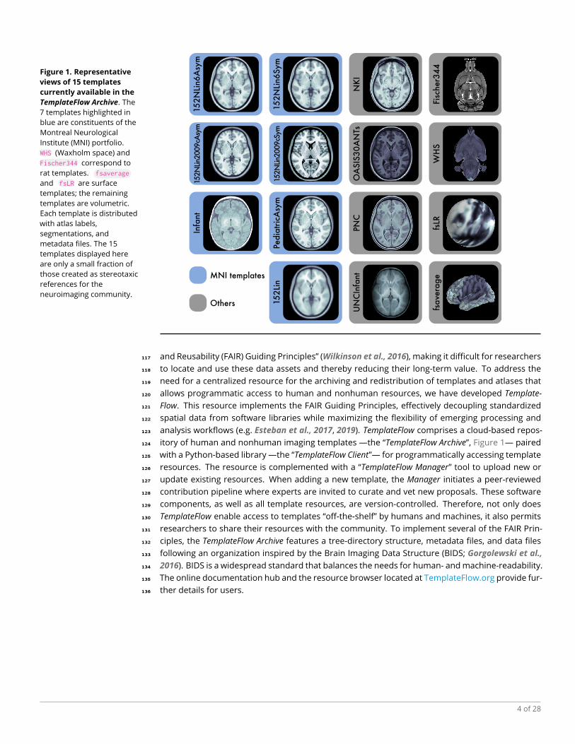
Figure 1. Representative
views of 15 templates
currently available in the
TemplateFlow Archive. The7 templates highlighted inblue are constituents of theMontreal NeurologicalInstitute (MNI) portfolio.WHS (Waxholm space) andFischer344 correspond torat templates. fsaverage
and fsLR are surfacetemplates; the remainingtemplates are volumetric.Each template is distributedwith atlas labels,segmentations, andmetadata files. The 15templates displayed hereare only a small fraction ofthose created as stereotaxicreferences for theneuroimaging community.
152Li
n
152N
Lin2
009cA
sym
152N
Lin6A
sym
152N
Lin6Sym
Infa
nt
Pedia
tric
Asy
m
NK
IO
ASIS
30A
NTs
PNC
Fisc
her3
44
fsLR
fsave
rage
WH
S
UN
CIn
fant
152N
Lin2
009cS
ymMNI templates
Others
and Reusability (FAIR) Guiding Principles” (Wilkinson et al., 2016), making it difficult for researchers117
to locate and use these data assets and thereby reducing their long-term value. To address the118
need for a centralized resource for the archiving and redistribution of templates and atlases that119
allows programmatic access to human and nonhuman resources, we have developed Template-120
Flow. This resource implements the FAIR Guiding Principles, effectively decoupling standardized121
spatial data from software libraries while maximizing the flexibility of emerging processing and122
analysis workflows (e.g. Esteban et al., 2017, 2019). TemplateFlow comprises a cloud-based repos-123
itory of human and nonhuman imaging templates —the “TemplateFlow Archive”, Figure 1— paired124
with a Python-based library —the “TemplateFlow Client”— for programmatically accessing template125
resources. The resource is complemented with a “TemplateFlow Manager” tool to upload new or126
update existing resources. When adding a new template, the Manager initiates a peer-reviewed127
contribution pipeline where experts are invited to curate and vet new proposals. These software128
components, as well as all template resources, are version-controlled. Therefore, not only does129
TemplateFlow enable access to templates “off-the-shelf” by humans and machines, it also permits130
researchers to share their resources with the community. To implement several of the FAIR Prin-131
ciples, the TemplateFlow Archive features a tree-directory structure, metadata files, and data files132
following an organization inspired by the Brain Imaging Data Structure (BIDS; Gorgolewski et al.,133
2016). BIDS is a widespread standard that balances the needs for human- andmachine-readability.134
The online documentation hub and the resource browser located at TemplateFlow.org provide fur-135
ther details for users.136
4 of 28
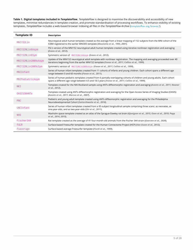
Table 1. Digital templates included in TemplateFlow. TemplateFlow is designed to maximise the discoverability and accessibility of newtemplates, minimise redundancies in template creation, and promote standardisation of processing workflows. To enhance visibility of existingtemplates, TemplateFlow includes a web-based browser indexing all files in the TemplateFlow Archive (templateflow.org/browse/).
Template ID Description
MNI152LinNeurotypical adult human template created as the average from a linear mapping of 152 subjects from the MNI cohort of theICBM registered to the earlier MNI305 template (Mazziotta et al., 1995, 2001).
MNI152NLin6AsymFSL’s version of the MNI152 neurotypical adult human template created using iterative nonlinear registration and averaging(Evans et al., 2012).
MNI152NLin6Sym Symmetric version of MNI152NLin6Asym (Evans et al., 2012).
MNI152NLin2009cAsymUpdate of the MNI152 neurotypical adult template with nonlinear registration. The mapping and averaging proceeded over 40iterations beginning from the earlier MNI152 template (Fonov et al., 2011; Collins et al., 1999).
MNI152NLin2009cSym Symmetric version of MNI152NLin2009cAsym (Fonov et al., 2011; Collins et al., 1999).
MNIInfantSeries of human infant templates created from 11 cohorts of infants and young children. Each cohort spans a different agerange between 0 and 60 months (Fonov et al., 2011).
MNIPediatricAsymSeries of human pediatric templates created from 6 partially overlapping cohorts of children and young adults. Each cohortspans a different age range between 4.5 and 18.5 years (Fonov et al., 2011; Collins et al., 1999).
NKITemplate created for the NKI-Rockland sample using ANTs diffeomorphic registration and averaging (Avants et al., 2011; Nooneret al., 2012).
OASIS30ANTsTemplate created using ANTs diffeomorphic registration and averaging for the Open Access Series of Imaging Studies (OASIS)(Avants et al., 2011;Marcus et al., 2007).
PNCPediatric and young adult template created using ANTs diffeomorphic registration and averaging for the PhiladelphiaNeurodevelopmental Cohort (Satterthwaite et al., 2016).
UNCInfantSeries of human infant templates created from a 95-subject longitudinal sample comprising three scans: as neonates, asone-year-olds, and as two-year-olds (Shi et al., 2011).
WHSWaxholm space template created as an atlas of the Sprague-Dawley rat brain (Kjonigsen et al., 2015; Osen et al., 2019; Pappet al., 2014, 2015).
Fischer344 Rat template created as the average of 41 four-month-old animals from the Fischer 344 strain (Goerzen et al., 2020).
fsLR Surface-based Freesurfer template created for the Human Connectome Project (HCP) (Van Essen et al., 2012).
fsaverage Surface-based average Freesurfer template (Fischl et al., 1999).
5 of 28

Results137
Management and stewardship following the FAIR Guiding Principles138
The specific and measurable FAIR principles of Wilkinson et al. (2016) are reproduced in Sup-139
plementary Box S1. Principles are indexed by their category and numbering, e.g., principle I3 –140
“(meta)data include qualified references to other (meta)data” belongs in the “to be Interoperable” cat-141
egory. We describe how TemplateFlow implements each of these specific principles in the follow-142
ing. Every template and all associatedmetadata, atlases, etc. are assigned a unique and persistent143
identifier (principle F1). BIDS prescribes a file naming scheme comprising a series of key-value pairs144
(called “entities”) that are ordered hierarchically. Following BIDS’ patterns, the template identifier145
is an alphanumeric label that is unique across the Archive, signified with the key tpl- (e.g., tpl-146
MNI152Lin ). TemplateFlow therefore adapts BIDS to the specific domain of templates and atlases,147
affording the tool with a robust implementation of the principles I1-3 (interoperability). Table 1 enu-148
merates several templates currently distributed with the Archive, and their corresponding unique149
identifiers. The unique identifier resolves the issue of inaccurate reporting, as it unambiguously150
designates one specific template. In addition, because the repository is versioned, researchers can151
easily retrieve and report the exact version of the template or atlas that was used in their study.152
Suppl. Table S2 summarizes the available entities and shows a segment of the file organization153
of the Archive. For each template, the TemplateFlow database includes reference volumetric tem-154
plate images (e.g., one T1-weighted and one T2-weighted average map; all must be in register), a155
set of atlas labels and voxelwise annotations defined with reference to the template image, and156
additional files containing the template and atlas metadata. Correspondingly, TemplateFlow allows157
surface-based resources such as average features, geometry files, annotations, or metadata.158
Template resources are described with rich metadata (principles F2 and R1), ensuring that the159
data usage license is clear and accessible (R1.1), data and metadata are associated with detailed160
provenance (R1.2), and data and metadata follow a domain-relevant structure transferred from161
the neuroimaging community standards of BIDS (R1.3). Figure 2 summarizes the data types and162
metadata that can be stored in the Archive. Figure 3 provides an overview of the Archive’s metadata163
specification, showing that metadata clearly and explicitly include the identifier of the data they de-164
scribe (F3). Data and metadata are retrievable using several open, free, standard communications165
protocols without need for authentication (A1) by using DataLad (Halchenko et al., 2021). Cloud166
storage for the Archive is supported by the Open Science Framework (osf.io) and Amazon’s Simple167
Storage Service (S3). Version control, replication, and synchronisation of template resources across168
filesystems is managed with DataLad. Leveraging DataLad, metadata are stored on GitHub, ensur-169
ing accessibility to metadata even when corresponding data are no longer available (A2). DataLad170
is based on Git and Git-Annex, which index all data and metadata (F4). Although DataLad also pro-171
vides searching tools that are applicable to TemplateFlow’s resources, the TemplateFlow framework172
provides a client tool that facilitates searching and querying.173
An indexed resource, searchable with a TemplateFlow “Client”174
TemplateFlow’s Python client provides human users and software tools with reliable and program-175
matic access to the archive. The client can be integrated seamlessly into image processing work-176
flows to handle requests for template resources on the fly. It features an intuitive application pro-177
gramming interface (API) that can query the TemplateFlow Archive for specific files (Figure 5). The178
BIDS-inspired organization enables easy integration of tools and infrastructure designed for BIDS179
(e.g., the Python client uses PyBIDS (Yarkoni et al., 2019) to implement queries like those listed in180
Suppl. Table S2). To query TemplateFlow, a user can submit a list of arguments corresponding to181
the BIDS-like key-value pairs in each entity’s file name (e.g., atlas="Schaefer2018" to return files182
containing voxelwise annotations by Schaefer et al. (2018)).183
To integrate template resources into neuroimaging workflows, traditional approaches required184
deploying an oftentimes voluminous tree of prepackaged data to the filesystem. By contrast, the185
TemplateFlow client implements lazy loading, which permits the base installation to be extremely186
6 of 28

Da
ta
Meta
da
taScr
ipts
NIfTI images include population-average templates and tissue class segmentations.
Masks are binary-valued NIfTI images
indicating whether each voxel is in a region.
Atlases are NIfTI images that assign
anatomical or functional labels to template voxels.
Transformations are HDF5 files containing
maps between template coordinate spaces.
Cohort directories contain template
resources specific to a sub-cohort of participants.
JSON metadata summarise information
about templates, resolutions, and cohorts.
Tabular metadata contain dictionaries
that pair atlas regions with anatomical labels.
A changelog chronicles changes
and updates made to template resources.
License files specify usage rights for
template resources.
Python scripts are used to prepare
template resources.
Figure 2. The TemplateFlow Archive contains template resources. Left, common file formats included in the TemplateFlow Archive. Right, viewof the TemplateFlow Archive’s browser, accessible at TemplateFlow.org, with a single template resource directory expanded. Template data arearchived using a BIDS-like directory structure, with top-level directories for each template. Each directory contains image files, annotations, andmetadata for that template. Following BIDS specifications, volumetric data are stored in NIfTI-1 format. Further surface-based data types aresupported with GIFTI (surfaces) and CIFTI-1 (mixed volumetric-and-surface data).
7 of 28

{
"Authors": [
"Fonov V",
"Evans AC",
"Botteron K",
"Almli CR",
"McKinstry RC",
"Collins DL"
],
"Curators": [
"Esteban O"
],
"Identifier": "MNIPediatricAsym",
"License": "MIT-derived. See LICENSE file",
"Name": "MNI's unbiased standard MRI template for
pediatric data from the 4.5 to 18.5y age
range",
"RRID": "SCR_008796",
"ReferencesAndLinks": [
"https://doi.org/10.1016/j.neuroimage.2010.07.033",
"https://doi.org/10.1016/S1053-8119(09)70884-5",
"http://nist.mni.mcgill.ca/?p=974",
"https://doi.org/10.1007/3-540-48714-X_16"
],
"TemplateFlowVersion": "1.0.0",
"cohort": {
"1": {
"age": [
4.5,
18.5
],
"name": "whole age range",
"units": "yr"
},
"2": {
"age": [
4.5,
8.5
],
"name": "prepuberty",
"units": "yr"
},
. . .
},
"res": {
"1": {
"origin": [
-98.0,
-134.0,
-72.0
],
"shape": [
197,
233,
189
],
"zooms": [
1.0,
1.0,
1.0
]
},
. . .
}
}
Field Type Description
Authors
Curators
Identifier
License
Name
RRID
ReferencesAndLinks
TemplateFlowVersion
Array(String)
Array
String
String
StringString
Array
String
Names of authors who created the template.
Publications to reference when using the template,and salient links for template information.
Research Resource Identifier for the TemplateFlowdataset.
Names of TemplateFlow curators who contributedor manage the dataset.Unique human-readable template identifier withinTemplateFlow.License under which template resources areavailable.Full descriptive name of the template.
Version of TemplateFlow under which the datasetwas distributed.
Field Type Description
1, 2, ...
age
name
units
Object
Array(Number)
StringString
Cohort identifiers. Each has a subdirectory in thetemplate data directory, and each has ametadata object nested in the cohort field.2-tuple array indicating the lower and upperbounds for participant age in the cohort, if thecohorts are stratified by age.Full descriptive name of the cohort..Units for cohort age bounds.
cohort Object Top-level field containing all cohort metadataobjects.
Field Type Description
1, 2, ...
origin
shape
zooms
Object
Array
ArrayArray
Resolution identifiers. Each has a metadataobject nested in the res field. The metadatafor each resolution apply to all images whosename includes res-<identifier>. The identifieritself does not necessarily correspond to the voxelsize.(x, y, z) spatial location of the voxel originrelative to the physical origin in mm.(x, y, z) shape of the image in voxels.(x, y, z) size of each voxel in mm.
res Object Top-level field containing all resolution metadataobjects.
General metadata
Cohort metadata
Resolution metadata
Figure 3. Overview of the metadata specification of the TemplateFlow Archive. TemplateFlow’s metadata are formatted as JavaScript ObjectNotation (JSON) files located within each template set. An example template_description.json metadata file is displayed at the left forMNIPediatricAsym . In addition to general template metadata, datasets can contain cohort-level and resolution-level metadata, which are nestedwithin the main metadata dictionary and apply only to subsets of images in the dataset.
8 of 28

lightweight. Instead of distributing neuroimaging data with the installation, TemplateFlow allows187
the user to dynamically pull from the cloud-based storage only those resources they need, as they188
need them. After a resource has been requested once, it remains cached in the filesystem for189
future utilization.190
We demonstrate benefits of centralizing templates in general, and the validity of the Template-191
Flow framework in particular, via its integration into fMRIPrep (Esteban et al., 2019), a functional192
MRI preprocessing tool. This integration provides fMRIPrep users with flexibility to spatially nor-193
malize their data to any template available in the Archive (see Box 1). This integration has also194
enabled the development of fMRIPrep adaptations, for instance to pediatric populations or rodent195
imaging (MacNicol et al., 2021), using suitable templates from the archive. The uniform interface196
provided by the BIDS-like directory organisation and metadata enables straightforward integra-197
tion of new templates into workflows equipped to use TemplateFlow templates. Further examples198
of tools leveraging TemplateFlow include MRIQC (Esteban et al., 2017) for quality control of MRI;199
PyNets (Pisner and Hammonds, 2020), a package for ensemble learning of functional and struc-200
tural connectomes; ASLPrep (Adebimpe et al., 2021), an ASL pre-processing pipeline that makes201
use of TemplateFlow through sMRIPrep —the spin-off structural pipeline from fMRIPrep; and Net-202
PlotBrain (Thompson and Fanton, 2021), which uses TemplateFlow to display spatially standardized203
brain network data.204
A framework for researchers who generate and share spatially-standardized data205
A centralized repository for neuroimaging templates should also address the needs of template206
creators, enabling peer-reviewed integration of new templates with minimal informatic overhead.207
Inspired by the Conda-forge community repository and the Journal of Open Source Software, the208
GitHub-based “templateflow” organisation is a site for dialogue between members of the neu-209
roimaging community and TemplateFlow Archive curators. GitHub issues offer any communitymem-210
ber the ability to share their needs with developers and Archive curators, for instance by identify-211
ing templates or workflow features for potential inclusion in the project. “Pull requests” provide212
a means for members of the community to directly contribute code or template resources to the213
TemplateFlow Archive.214
This peer-reviewed contribution process is facilitated through the Python-based TemplateFlow215
Manager. The TemplateFlow Manager automates the work of synchronizing data from a local di-216
rectory to cloud storage in OSF. Furthermore, it creates a GitHub repository containing git-annex217
pointers that enable DataLad to download template data from cloud storage to anymachine with a218
copy of the repository. Finally, it opens a new pull request to propose adding the newly contributed219
template repository into the main TemplateFlow Archive (Figure 6). Synchronization of spatial data220
assets to the TemplateFlow Archive affords data producers an immediate way to distribute their221
data according to FAIR principles and thereby increase its reach.222
Unambiguous and precise reporting of spatially standardized processing and analysis.223
To explore the coupling between software libraries and standard spaces, we conducted a topic224
modeling analysis (Blei et al., 2003) of MNI space in the neuroimaging literature. We identified a225
coupling between the reporting of spatial standardization and software libraries across 6,048 arti-226
cles containing the term MNI and published in two leading domain-specific journals (NeuroImage227
and NeuroImage: Clinical). To demonstrate the heterogeneity in the reported standardization to228
MNI space, we sorted topics according to their dominance in articles (i.e., the topic with the high-229
est model score in MNI-related sentences; Figure 4). Out of 15 topics we modeled, two of the230
most dominant topics contained software tool names as well as the names of related scientists.231
As shown in Figure 4, around 500 articles (each term) contained either “SPM” (9% of the docu-232
ments) or “FSL” (8%). Interestingly, the two words do not ever appear together, suggesting that233
researchers stick with one or another in their analyses. Additional topics that seemingly relate to234
the provenance of templates and atlases —beyond the ubiquitous “Montreal”, “Neurological” and235
9 of 28

#1: 1048 documents (17.23%) #2: 735 documents (12.08%) #3: 537 documents (8.83%) #4: 493 documents (8.10%)
#5: 469 documents (7.71%) #6: 397 documents (6.53%) #7: 394 documents (6.48%) #8: 383 documents (6.30%)
Figure 4. The FSL and SPM software tools associate with dominant topics of sentences including the term “MNI” across the literature.We performed topic modeling with latent Dirichlet allocation (LDA; Blei et al., 2003) on text sentences extracted from 6,048 articles thatcontained the word “MNI”. For each topic identified, the 20 words with the highest loadings on that topic are displayed in a word cloud withlarger font size indicating higher loading of the word on the corresponding topic. Word clouds are sorted by descending topic dominance.Ranking and relative dominance are shown above each topic’s cloud. Only the top 8 dominant topics are shown here (full model is reported inSuppl. Figure S1). Two top-dominant topics —#3 and #5— are associated with SPM and FSL respectively.
“Institute” for MNI— are those that ranked #4, #13, #14, which include “SPM” and other terms such236
as “McGill”, “Wellcome”, “UCL”, or the “parametric” in SPM (see Suppl. Figure S1). The remainder237
of topics appears to relate to miscellaneous aspects of spatial standardization, such as “anatom-238
ical”, “smoothness”, “map/mapping”, “standard”, “normalization”, “(re)align/alignment”, etc., with239
no apparent relationship to the actual origin of the resource. These interpretations suggest that240
“MNI space” can refer to any of a family of templates and is not a unique identifier. As a matter of241
fact, studies carried out with SPM96 (Friston et al., 2006) and earlier versions report their results242
in MNI space with reference to the single-subject Colin 27 average template (Holmes et al., 1998).243
However, beginning with SPM99, SPM updated its definition ofMNI space to the template that MNI244
released in 2001: an average of 152 subjects from the ICBM database, aligned by means of lin-245
ear registration. In SPM12 (the latest release at the time of writing), the meaning of MNI template246
varies by submodule: different modules alternately use the Linear MNI152 template (Mazziotta247
et al., 1995) and a new, nonlinear revision from 2009 (Fonov et al., 2011). By contrast, the MNI248
template bundled with the FSL toolbox was developed by Dr. A. Janke in collaboration with MNI249
researchers (Evans et al., 2012). Although it was generated under the guidance of and using the250
techniques of the 2006 release of nonlinear MNI templates, this template is not in fact part of the251
official portfolio distributed by MNI. Nonetheless, our results suggest that the MNI templates bun-252
dled with SPM and FSL have historically gained broader currency as a result of the widespread use253
of these software libraries.254
Discussion255
The use of templates and atlases is ubiquitous in neuroimaging, and the emerging challenges re-256
garding template use accordingly merit immediate attention. In an early perspective, Van Essen257
identified a set of desiderata for brain templates (Van Essen, 2002). Above and beyond anatomical258
10 of 28

fidelity, he called for connecting templates in an aggregation of databases with “powerful and flex-259
ible options for searching, selecting, and visualizing data”. Finally, he stressed the importance of260
resource accessibility. TemplateFlow provides a framework that satisfies all of the aforementioned261
desiderata while following the FAIR principles (Wilkinson et al., 2016). We elaborate that most of262
the issues concerning the reliability of neuroimaging research relating to standardized spatial ref-263
erences stem from the lack of a centralized repository designed to meet FAIR principles. We show264
how we effectively implement such principles with the adoption of a BIDS-like structure for the265
data andmetadata in the resource, and with DataLad to support the core of the data management266
system. We complete the implementation with an easy-to-use client tool.267
When researchers develop a new brain template or atlas for public dissemination, there exists268
no standard channel or format for distributing their work. With no central repository or uniform269
organizational scheme, template creators are often tasked with the responsibilities of maintain-270
ing template resources and managing access on an ad hoc basis and sustaining them over time271
with limited to no support. While the quality of peer reviewed template resources is assessed272
once prior to publication, reviewers often focus on perceived academic merit to the exclusion of273
FAIR principles. This can lead to poor resource adoption and low community value even for high-274
quality resources. Informal vetting of resources is prone to more clerical errors such as missing275
or corrupted files and/or metadata, or unlicensed distribution, which nonetheless may make the276
resource unusable. Conversely, users are confronted with a surfeit of available templates and277
atlases, many with unclear provenance, absent licensing terms, and the attendant challenges of278
accessing them and integrating them into workflows.279
Without a uniform distribution format, integrating a template into software requires a custom280
solution for every new template, increasing the burden on developers. Consistently with the pre-281
vious investigation by Carp (2012b) in the domain of functional MRI, our text mining exploration282
illustrates a strong coupling between software library and the templates and atlases of choice. In-283
deed, Carp (2012b) analysed 241 functional MRI studies of which 90.9% reported normalizing brain284
images to a common template. Of those, 79.0% indicated the target space used for spatial normal-285
ization. Few studies reported critical parameters, and only 50 specified the template image: 26.0%286
used “theMNI152 template”, and 26.0% the “SPM library’s echo-planar imaging template”. Unfortu-287
nately, template selection is seen as a default parameter of the software library, which lends itself288
to assuming that the target normalization space is implicitly reported by identifying the software289
tools of choice. Bowring et al. (2019) wrote comparable pipelines in three software suites (AFNI,290
FSL and SPM) in order to identify challenges to reproducing published studies with openly shared291
raw data. When discussing differences among pipelines, they noted that, “while all packages are292
purportedly using the sameMNI atlas space, an appreciable amount of activation detected by AFNI293
and FSL fell outside of SPM’s analysis mask“.294
This coupling seems to also limit the utilization of templates other than those defaulted by the295
software. Custom templates (i.e., those not included as default option for the software tool) range296
from population-specific templates to ad hoc templates created by averaging images of the study297
at hand. In some settings, the use of default templates risks introducing “template effects” that298
confound the interpretation of results (such as those introduced in pediatric imaging studies by299
using an adult template, Yoon et al., 2009). As the target population moves away from that with300
which a default template was created, “template effects” become more concerning and custom301
templates more necessary. The problem is exacerbated in the case of nonhuman imaging, as the302
scarcity (or absence) of specific templates available within software packages hinders already chal-303
lenging translational endeavors. Further, the consistency across templates and atlases is report-304
edly low (Bohland et al., 2009), and although there has not been any programmatic comparison305
to understand the extent to which this inconsistency alters the spatial interpretation of results, it306
is reasonable that templates and atlases introduce a decision point and therefore are sources of307
some analytical variability.308
One ostensible caveat regarding centralized and FAIR-principled knowledge repositories such309
11 of 28

as TemplateFlow is that, by increasing the findability and reuse potential of data resources, they310
also open the door to increased methodological flexibility. Carp empirically investigated the con-311
sequences of such methodological flexibility in neuroimaging, demonstrating that decision points312
in workflows can lead to substantial variability in analysis outcomes. In a contemporaneous paper,313
Carp (2012b) contextualized these findings vis-à-vis the inflated risk of false positives, underscoring314
that analytical variability degrades the reproducibility of studies only in combination with (intended315
or unintended) selective reporting of methods and results. Selective reporting, in this particular ap-316
plication, would mean that a researcher explores the results with reference to several templates317
or atlases and reports only those that confirm the research’s hypotheses. TemplateFlow’s standard-318
ization preempts the problem of unintended selective reporting: the provenance of all resources is319
tracked, all resources are accessible, and comprehensivemetadata are generated.ß More recently,320
Botvinik-Nezer et al. (2020) advocated for another solution to the problem of analytical variability:321
“multiverse” analyses, whereinmany combinations ofmethodological choices are all thoroughly re-322
ported and cross-compared when presenting results. Applied to the particular choice of template323
and atlas combinations, it would thus be desirable to report neuroimaging results with reference to324
several standard spaces and determine whether the interpretations hold across those references325
and atlases. TemplateFlow’s interoperability empowers users to incorporate this type of analysis326
into their research by easily making template or atlas substitutions for cross-comparison. For in-327
stance, Box 1 shows how TemplateFlow works with fMRIPrep to automate preprocessing of outputs328
in multiple standard spaces. This facilitates assessment of the robustness of a result with respect329
to the template or atlas of choice in accordance with the multiverse approach.330
Limitations331
TemplateFlow affords researchers substantial analytical flexibility in the choice of standard spaces332
of reference. Such flexibility helps researchersminimize “template effects”—by easily inserting the333
most adequate template— but also opens opportunities for incomplete reporting of experiments.334
Using DataLad or the TemplateFlow Client, researchers have at their disposal the necessary tooling335
for precise reporting: unique identifiers, provenance tracking, version tags, and comprehensive336
metadata. Therefore, the effectiveness of TemplateFlow to mitigate selective reporting is bounded337
by the user’s discretion. Similarly, the resource is limited at the time of writing to MRI templates,338
but it readily supports such other modalities as nuclear imagingmaps (e.g., PET/SPECT), and would339
support others with minimal adaptations (e.g., protein expression maps, or 3D reconstructions340
from histology).341
As a research resource, the scope of this manuscript is limited to describing the framework and342
infrastructure of TemplateFlow, highlighting how neuroscientists can leverage this new data archive343
and the tooling around it. Therefore, some fundamental issues related to this workmust be left for344
future investigation: (i) the overarching problems of cross-template and cross-atlas consistency;345
(ii) comparative evaluation of methodological alternatives for producing new templates, atlases346
and related data; (iii) providing neuroimagers with more objective means to determine the most347
appropriate template and atlas choices that apply to their research, aswell as better understanding348
“template effects”; (iv) the adequacy of original (MRI, nuclear imaging, etc.) and derived (regularly349
gridded images, surfaces, etc.) modalities for a specific research application; or (v) the study of the350
validity and reliability of inter-template registration, as well as the evaluation of such a component351
of the TemplateFlow framework.352
In more practical terms, TemplateFlow is limited to the redistribution of resources under per-353
missive licenses, without any access restrictions.354
Conclusion355
We introduce an open framework for the archiving, maintenance and sharing of neuroimaging356
templates and atlases called TemplateFlow that is implemented under FAIR data sharing principles.357
We describe the current need for this resource in the domain of neuroimaging, and further discuss358
the implications of the increased analytical flexibility this tool affords. These two facets of repro-359
12 of 28
ducibility —availability (under FAIR guiding principles) of prior knowledge required by the research360
workflow, and the analytical flexibility such availability affords— are ubiquitous concerns across361
disciplines. TemplateFlow’s approach to addressing both establishes a pattern broadly transfer-362
able beyond neuroimaging. We envision TemplateFlow as a core research tool undergirding multi-363
verse analyses —assessing whether neuroimaging results are robust across population-wide spa-364
tial references— as well as a stepping stone towards the quest of mapping anatomy and function365
across species.366
Acknowledgments367
The development of this resource was supported by the Laura and John Arnold Foundation (RAP368
and KJG), theNIBIB (R01EB020740, SSG; 1P41EB019936-01A1 SSG, YOH), NIMH (RF1MH121867 RAP,369
OE; R24MH114705 and R24MH117179, RAP; 1RF1MH121885 SSG), NINDS (U01NS103780, RAP),370
and NSF (CRCNS 1912266, YOH). RL is funded by the Wellcome Trust (209139/Z/17/Z). EM was371
supported by the UK Medical Research Council (MR/N013700/1) and King’s College London. OE ac-372
knowledges financial support from the SNSF Ambizione project “Uncovering the interplay of struc-373
ture, function, and dynamics of brain connectivity using MRI” (grant number PZ00P2_185872).374
Ethical compliance375
We complied with all relevant ethical regulations. This resource reused publicly available data de-376
rived from studies acquired at many different institutions. Protocols for all of the original studies377
were approved by the corresponding ethical boards.378
Code & data availability statement379
All the software components discussed in this paper are available under the Apache 2.0 license,380
accessible as repositories of https://github.com/templateflow. All templates and associated data381
are available under corresponding open licenses and accessible as described in the manuscript.382
Author contributions383
Conceptualization: RC, CJM, KJG, RAP, OE Data curation: RC, EM, OE Topics analysis: RC, RL, OE384
Funding acquisition: KJG, RAP, OEMethodology: RC, OE Project administration: RAP, OE Resources:385
YOH, SSG, KJG, RAP, OE Software & Documentation: RC, WHT, MG, EM, CJM, YOH, OE Supervision:386
RAP, OE Validation: RC, WHT, EM, OE Visualization: RC, RL. Writing – original draft: RC & OE Writing387
– review & editing: RC, RL, WHT, MG, EM, CJM, YOH, SSG, KJG, RAP, OE.388
References389
AdebimpeA, BertoleroM, Dolui S, CieslakM,Murtha K, Baller EB, Boeve B, Boxer A, Butler ER, Cook P, Colcombe390
S, Covitz S, Davatzikos C, Davila DG, Elliott MA, Flounders MW, Franco AR, Gur RE, Gur RC, Jaber B, et al.391
ASLPrep: A Generalizable Platform for Processing of Arterial Spin LabeledMRI andQuantification of Regional392
Brain Perfusion. bioRxiv. 2021 May; p. 2021.05.20.444998. doi:10.1101/2021.05.20.444998.393
Avants BB, Epstein CL, Grossman M, Gee JC. Symmetric diffeomorphic image registration with cross-394
correlation: Evaluating automated labeling of elderly and neurodegenerative brain. Med Image Anal. 2008;395
12(1):26–41. doi:10.1016/j.media.2007.06.004.396
Avants BB, Tustison NJ, Song G, Cook PA, Klein A, Gee JC. A reproducible evaluation of ANTs397
similarity metric performance in brain image registration. NeuroImage. 2011 Feb; 54(3):2033–44.398
doi:10.1016/j.neuroimage.2010.09.025.399
Bandrowski AE, MartoneME. RRIDs: A Simple Step toward Improving Reproducibility through Rigor and Trans-400
parency of Experimental Methods. Neuron. 2016 May; 90(3):434–436. doi:10.1016/j.neuron.2016.04.030.401
Blei DM, Ng AY, Jordan MI. Latent Dirichlet Allocation. J Mach Learn Res. 2003; 3(Jan):993–1022. https://jmlr.402
org/papers/v3/blei03a.html.403
13 of 28
Bohland JW, Bokil H, Allen CB, Mitra PP. The Brain Atlas Concordance Problem: Quantitative Comparison of404
Anatomical Parcellations. PLoS One. 2009 Sep; 4(9):e7200. doi:10.1371/journal.pone.0007200.405
Botvinik-Nezer R, Holzmeister F, Camerer CF, Dreber A, Huber J, JohannessonM, KirchlerM, Iwanir R,Mumford406
JA, Adcock RA, Avesani P, Baczkowski BM, Bajracharya A, Bakst L, Ball S, Barilari M, Bault N, Beaton D, Beitner407
J, Benoit RG, et al. Variability in the analysis of a single neuroimaging dataset by many teams. Nature. 2020408
Jun; 582(7810):84–88. doi:10.1038/s41586-020-2314-9.409
Bowring A, Maumet C, Nichols TE. Exploring the impact of analysis software on task fMRI results. Human Brain410
Mapping. 2019; 40(11):3362–3384. doi:https://doi.org/10.1002/hbm.24603.411
Brett M, Johnsrude IS, Owen AM. The problem of functional localization in the human brain. Nat Rev Neurosci.412
2002; 3:243–249. doi:10.1038/nrn756.413
Brodmann K. Brodmann’s: Localisation in the Cerebral Cortex. Springer US; 2006. doi:10.1007/b138298.414
Buckner RL, HeadD, Parker J, Fotenos AF,MarcusD,Morris JC, Snyder AZ. A unified approach formorphometric415
and functional data analysis in young, old, and demented adults using automated atlas-based head size nor-416
malization: reliability and validation against manual measurement of total intracranial volume. NeuroImage.417
2004 Oct; 23(2):724–738. doi:10.1016/j.neuroimage.2004.06.018.418
Calabrese E, Badea A, Watson C, Johnson GA. A quantitative magnetic resonance histology atlas of postnatal419
rat brain development with regional estimates of growth and variability. NeuroImage. 2013May; 71:196–206.420
doi:10.1016/j.neuroimage.2013.01.017.421
Carp J. On the Plurality of (Methodological) Worlds: Estimating the Analytic Flexibility of fMRI Experiments.422
Front Neurosci. 2012; 6. doi:10.3389/fnins.2012.00149.423
Carp J. The secret lives of experiments: Methods reporting in the fMRI literature. NeuroImage. 2012 Oct;424
63(1):289–300. doi:10.1016/j.neuroimage.2012.07.004.425
Coalson TS, Van Essen DC, Glasser MF. The impact of traditional neuroimaging methods on the spatial lo-426
calization of cortical areas. Proceedings of the National Academy of Sciences. 2018; 115(27):E6356–E6365.427
doi:10.1073/pnas.1801582115.428
Collins DL, Zijdenbos AP, Baaré WFC, Evans AC. ANIMAL+INSECT: Improved Cortical Structure Segmentation.429
In: Kuba A, Šáamal M, Todd-Pokropek A, editors. Information Processing in Medical Imaging Lecture Notes in430
Computer Science, Berlin, Heidelberg: Springer; 1999. p. 210–223. doi:10.1007/3-540-48714-X_16.431
Courchesne E, Chisum HJ, Townsend J, Cowles A, Covington J, Egaas B, Harwood M, Hinds S, Press GA. Normal432
Brain Development and Aging: Quantitative Analysis at in Vivo MR Imaging in Healthy Volunteers. Radiology.433
2000 Sep; 216(3):672–682. doi:10.1148/radiology.216.3.r00au37672.434
Cox RW, Hyde JS. Software tools for analysis and visualization of fMRI data. NMR Biomed. 1997; 10(4-5):171–435
178. doi:10.1002/(SICI)1099-1492(199706/08)10:4/5<171::AID-NBM453>3.0.CO;2-L.436
Dickie DA, Job DE, Gonzalez DR, Shenkin SD, Wardlaw JM. Use of Brain MRI Atlases to Determine Bound-437
aries of Age-Related Pathology: The Importance of Statistical Method. PLoS One. 2015 May; 10(5):e0127939.438
doi:10.1371/journal.pone.0127939.439
Dickie DA, Shenkin SD, Anblagan D, Lee J, Blesa Cabez M, Rodriguez D, Boardman JP, Waldman A, Job DE,440
Wardlaw JM. Whole Brain Magnetic Resonance Image Atlases: A Systematic Review of Existing Atlases and441
Caveats for Use in Population Imaging. Front Neuroinform. 2017; 11. doi:10.3389/fninf.2017.00001.442
Esteban O, Birman D, Schaer M, Koyejo OO, Poldrack RA, Gorgolewski KJ. MRIQC: Advancing the Au-443
tomatic Prediction of Image Quality in MRI from Unseen Sites. PLoS One. 2017 Aug; 12(9):e0184661.444
doi:10.1371/journal.pone.0184661.445
Esteban O, Markiewicz CJ, Blair RW, Moodie CA, Isik AI, Erramuzpe A, Kent JD, Goncalves M, DuPre E, Snyder M,446
Oya H, Ghosh SS, Wright J, Durnez J, Poldrack RA, Gorgolewski KJ. fMRIPrep: a robust preprocessing pipeline447
for functional MRI. Nat Meth. 2019 Jan; 16(1):111–116. doi:10.1038/s41592-018-0235-4.448
Evans AC, Collins DL, Mills SR, Brown ED, Kelly RL, Peters TM. 3D statistical neuroanatomical models from 305449
MRI volumes. In: IEEE Conference Record Nuclear Science Symposium and Medical Imaging Conference, vol. 3450
San Francisco, CA, USA; 1993. p. 1813–1817. doi:10.1109/NSSMIC.1993.373602.451
14 of 28

Evans AC, Janke AL, Collins DL, Baillet S. Brain templates and atlases. NeuroImage. 2012 Aug; 62(2):911–922.452
doi:10.1016/j.neuroimage.2012.01.024.453
Fischl B, Sereno MI, Dale AM. Cortical surface-based analysis II: Inflation, flattening, and a surface-based coor-454
dinate system. NeuroImage. 1999; 9(2):195–207.455
Fonov V, Evans AC, Botteron K, Almli CR, McKinstry RC, Collins DL. Unbiased average age-appropriate atlases456
for pediatric studies. NeuroImage. 2011 Jan; 54(1):313–327. doi:10.1016/j.neuroimage.2010.07.033.457
Friston KJ, Ashburner J, Kiebel SJ, Nichols TE, Penny WD. Statistical parametric mapping : the analysis of func-458
tional brain images. London: Academic Press; 2006.459
Goerzen D, Fowler C, Devenyi GA, Germann J, Madularu D, Chakravarty MM, Near J. An MRI-Derived Neu-460
roanatomical Atlas of the Fischer 344 Rat Brain. Sci Rep. 2020 Apr; 10(1):6952. doi:10.1038/s41598-020-461
63965-x.462
Good CD, Johnsrude IS, Ashburner J, Henson RN, Friston KJ, Frackowiak RS. A voxel-based morphometric study463
of ageing in 465 normal adult human brains. NeuroImage. 2001; 14(1):21–36. doi:10.1006/nimg.2001.0786.464
Gorgolewski KJ, Auer T, Calhoun VD, Craddock RC, Das S, Duff EP, Flandin G, Ghosh SS, Glatard T, Halchenko465
YO, Handwerker DA, Hanke M, Keator D, Li X, Michael Z, Maumet C, Nichols BN, Nichols TE, Pellman J, Poline466
JB, et al. The brain imaging data structure, a format for organizing and describing outputs of neuroimaging467
experiments. Sci Data. 2016 Jun; 3:160044. doi:10.1038/sdata.2016.44.468
Halchenko YO, FSL usermailing list: Incorrect probabilities in Harvard-Oxford-sub Left hemisphere; 2013. [On-469
line] Available: https://www.jiscmail.ac.uk/cgi-bin/webadmin?A2=FSL;2bb44bee.1301 (Accessed: 2021-07-05).470
Halchenko YO, Meyer K, Poldrack B, Solanky DS, Wagner AS, Gors J, MacFarlane D, Pustina D, Sochat V, Ghosh471
SS,Mönch C,Markiewicz CJ, Waite L, Shlyakhter I, Vega Adl, Hayashi S, Häusler CO, Poline JB, Kadelka T, Skytén472
K, et al. DataLad: distributed system for joint management of code, data, and their relationship. Journal of473
Open Source Software. 2021 Jul; 6(63):3262. doi:10.21105/joss.03262.474
Holmes CJ, Hoge R, Collins L, Woods R, Toga AW, Evans AC. Enhancement of MR Images Using Registration for475
Signal Averaging. J Comput Assist Tomogr. 1998 Mar; 22(2):324–333. https://insights.ovid.com/crossref?an=476
00004728-199803000-00032.477
JenkinsonM, Beckmann CF, Behrens TEJ, Woolrich MW, Smith SM. FSL. NeuroImage. 2012 Aug; 62(2):782–790.478
doi:10.1016/j.neuroimage.2011.09.015.479
Kjonigsen LJ, Lillehaug S, Bjaalie JG,WitterMP, Leergaard TB. WaxholmSpace atlas of the rat brain hippocampal480
region: Three-dimensional delineations based on magnetic resonance and diffusion tensor imaging. Neu-481
roImage. 2015 Mar; 108:441–449. doi:10.1016/j.neuroimage.2014.12.080.482
Landman BA, Ribbens A, Lucas B, Davatzikos C, Avants B, Ledig C, Ma D, Rueckert D, Vandermeulen D, Maes F.483
MICCAI 2012 Workshop on Multi-Atlas Labeling. CreateSpace Independent Publishing Platform; 2012.484
Lüders E, Steinmetz H, Jäncke L. Brain size and grey matter volume in the healthy human brain. NeuroReport.485
2002 Dec; 13(17):2371–2374. https://journals.lww.com/neuroreport/Abstract/2002/12030/Brain_size_and_486
grey_matter_volume_in_the_healthy.40.aspx.487
MacNicol E, Ciric R, Kim E, Censo DD, Cash D, Poldrack R, Esteban O. Atlas-based brain extraction is robust488
across rat MRI studies. In: IEEE 19th International Symposium on Biomedical Imaging (ISBI 2021) Nice, France;489
2021. p. (accepted). doi:10.1109/ISBI48211.2021.9.490
Marcus DS, Wang TH, Parker J, Csernansky JG, Morris JC, Buckner RL. Open Access Series of Imaging Studies491
(OASIS): Cross-sectional MRI Data in Young, Middle Aged, Nondemented, and DementedOlder Adults. J Cogn492
Neurosci. 2007 Aug; 19(9):1498–1507. doi:10.1162/jocn.2007.19.9.1498.493
Mars RB, Passingham RE, Jbabdi S. Connectivity Fingerprints: From Areal Descriptions to Abstract Spaces.494
Trends Cogn Sci. 2018 Nov; 22(11):1026–1037. doi:10.1016/j.tics.2018.08.009.495
Martin RF, Bowden DM. Primate Brain Maps: Structure of the Macaque Brain. Elsevier; 2000.496
Matsuzawa J, Matsui M, Konishi T, Noguchi K, Gur RC, Bilker W, Miyawaki T. Age-related Volumetric Changes497
of Brain Gray and White Matter in Healthy Infants and Children. Cereb Cortex. 2001 Apr; 11(4):335–342.498
doi:10.1093/cercor/11.4.335.499
15 of 28
Mazziotta J, Toga A, Evans A, Fox P, Lancaster J, Zilles K, Woods R, Paus T, Simpson G, Pike B, Holmes C, Collins500
L, Thompson P, MacDonald D, Iacoboni M, Schormann T, Amunts K, Palomero-Gallagher N, Geyer S, Parsons501
L, et al. A Four-Dimensional Probabilistic Atlas of the Human Brain. J Am Med Inform Assoc. 2001 Sep;502
8(5):401–430. doi:10.1136/jamia.2001.0080401.503
Mazziotta JC, Toga AW, Evans A, Fox P, Lancaster J. A Probabilistic Atlas of the Human Brain: Theory and504
Rationale for Its Development: The International Consortium for Brain Mapping (ICBM). NeuroImage. 1995505
Jun; 2(2, Part A):89–101. doi:10.1006/nimg.1995.1012.506
MNI, NeuroImaging & Surgical Technologies Lab (Montreal Neurological Institute) – MNI Atlases; n.d. [Online]507
Available: http://nist.mni.mcgill.ca/category/atlas/ (Accessed: 2021-07-01).508
Nooner KB, Colcombe S, Tobe R, Mennes M, Benedict M, Moreno A, Panek L, Brown S, Zavitz S, Li Q, Sikka S,509
Gutman D, Bangaru S, Schlachter RT, Kamiel S, Anwar A, Hinz C, Kaplan M, Rachlin A, Adelsberg S, et al. The510
NKI-Rockland Sample: A Model for Accelerating the Pace of Discovery Science in Psychiatry. Front Neurosci.511
2012; 6. doi:10.3389/fnins.2012.00152.512
Osen KK, Imad J, Wennberg AE, Papp EA, Leergaard TB. Waxholm Space atlas of the rat brain auditory sys-513
tem: Three-dimensional delineations based on structural and diffusion tensor magnetic resonance imaging.514
NeuroImage. 2019 Oct; 199:38–56. doi:10.1016/j.neuroimage.2019.05.016.515
Papp EA, Leergaard TB, Calabrese E, Allan Johnson G, Bjaalie JG. Addendum to “Waxholm Space atlas of516
the Sprague Dawley rat brain” [NeuroImage 97 (2014) 374-386]. NeuroImage. 2015 Jan; 105:561–562.517
doi:10.1016/j.neuroimage.2014.10.017.518
Papp EA, Leergaard TB, Calabrese E, Johnson GA, Bjaalie JG. Waxholm Space atlas of the Sprague Dawley rat519
brain. NeuroImage. 2014 Aug; 97:374–386. doi:10.1016/j.neuroimage.2014.04.001.520
Paxinos G, Watson C. The rat brain in stereotaxic coordinates. Elsevier Academic Press; 1997.521
PisnerD, Hammonds R. PyNets: A ReproducibleWorkflow for Structural and Functional Connectome Ensemble522
Learning. In: Annual Meeting of the Organization for Human Brain Mapping, vol. 26 Online Event; 2020. https:523
//github.com/dPys/PyNets/.524
Robinson EC, Jbabdi S, Glasser MF, Andersson J, Burgess GC, Harms MP, Smith SM, Van Essen DC, Jenkinson525
M. MSM: A new flexible framework for Multimodal Surface Matching. NeuroImage. 2014; 100:414–426.526
doi:10.1016/j.neuroimage.2014.05.069.527
Rohlfing T. Incorrect ICBM-DTI-81 atlas orientation and white matter labels. Front Neurosci. 2013; 7.528
doi:10.3389/fnins.2013.00004.529
Satterthwaite TD, Connolly JJ, Ruparel K, Calkins ME, Jackson C, Elliott MA, Roalf DR, Hopson R, Prabhakaran530
K, Behr M, Qiu H, Mentch FD, Chiavacci R, Sleiman PMA, Gur RC, Hakonarson H, Gur RE. The Philadelphia531
Neurodevelopmental Cohort: A publicly available resource for the study of normal and abnormal brain de-532
velopment in youth. NeuroImage. 2016 Jan; 124:1115–1119. doi:10.1016/j.neuroimage.2015.03.056.533
Sawiak SJ, Wood NI, Williams GB, Morton AJ, Carpenter TA. SPMMouse: A new toolbox for SPM in the animal534
brain. In: Proc. Intl. Soc. Mag. Reson. Med., vol. 17 Hawaii, USA; 2009. p. 1086.535
Schaefer A, Kong R, Gordon EM, Laumann TO, Zuo XN, Holmes AJ, Eickhoff SB, Yeo BTT. Local-Global Parcel-536
lation of the Human Cerebral Cortex from Intrinsic Functional Connectivity MRI. Cereb Cortex. 2018 Sep;537
28(9):3095–3114. doi:10.1093/cercor/bhx179.538
Schurr PH, Merrington WR. The Horsley–Clarke stereotaxic apparatus. Br J Surg. 1978; 65(1):33–36.539
doi:10.1002/bjs.1800650110.540
Shi F, Yap PT, Wu G, Jia H, Gilmore JH, Lin W, Shen D. Infant Brain Atlases from Neonates to 1- and 2-Year-Olds.541
PLoS One. 2011 Apr; 6(4):1–11. doi:10.1371/journal.pone.0018746.542
Sowell ER, Peterson BS, Thompson PM, Welcome SE, Henkenius AL, Toga AW. Mapping cortical change across543
the human life span. Nat Neurosci. 2003 Mar; 6(3):309–315. doi:10.1038/nn1008.544
Szulc KU, Lerch JP, Nieman BJ, Bartelle BB, Friedel M, Suero-Abreu GA, Watson C, Joyner AL, Turnbull545
DH. 4D MEMRI atlas of neonatal FVB/N mouse brain development. NeuroImage. 2015 Sep; 118:49–62.546
doi:10.1016/j.neuroimage.2015.05.029.547
16 of 28

Talairach J, Tournoux P. Co-planar stereotaxic atlas of the human brain. Stuttgart New York: Georg Thieme548
Verlag/Thieme Medical Publishers; 1988.549
Talairach J, DavidM, Tournoux P, Corredor H, Kvasina J. Atlas d’anatomie stéréotaxique: repérage radiologique550
indirect des noyaux gris centraux des régions mésencéphalo-sous-optique et hypothalamique de l’homme.551
Masson; 1957.552
Tavor I, Jones OP, Mars RB, Smith SM, Behrens TE, Jbabdi S. Task-free MRI predicts individual differences in553
brain activity during task performance. Science. 2016 Apr; 352(6282):216–220. doi:10.1126/science.aad8127.554
Thompson WH, Fanton S, Open Source Software: NetPlotBrain; 2021. [Online] Available: https://zenodo.org/555
record/4593837 (Accessed: 2021-07-08).556
Tosun D, Siddarth P, Levitt J, Caplan R. Cortical thickness and sulcal depth: insights on de-557
velopment and psychopathology in paediatric epilepsy. BJPsych Open. 2015 Oct; 1(2):129–135.558
doi:10.1192/bjpo.bp.115.001719.559
Van Essen DC. Windows on the brain: the emerging role of atlases and databases in neuroscience. Curr Opin560
Neurobiol. 2002 Oct; 12(5):574–579. doi:10.1016/S0959-4388(02)00361-6.561
Van Essen DC, Glasser MF, Dierker DL, Harwell J, Coalson T. Parcellations and Hemispheric Asymmetries of562
Human Cerebral Cortex Analyzed on Surface-Based Atlases. Cerebral Cortex. 2012 Oct; 22(10):2241–2262.563
doi:10.1093/cercor/bhr291.564
Von Economo CF, Koskinas GN. Atlas of cytoarchitectonics of the adult human cerebral cortex, vol. 10. Karger565
Basel; 2008.566
WilkinsonMD, Dumontier M, Aalbersberg IJ, Appleton G, AxtonM, Baak A, Blomberg N, Boiten JW, da Silva San-567
tos LB, Bourne PE, Bouwman J, Brookes AJ, Clark T, Crosas M, Dillo I, Dumon O, Edmunds S, Evelo CT, Finkers568
R, Gonzalez-Beltran A, et al. The FAIR Guiding Principles for scientific data management and stewardship.569
Sci Data. 2016 Mar; 3(1):160018. doi:10.1038/sdata.2016.18.570
Yarkoni T, Markiewicz C, de la Vega A, Gorgolewski K, Salo T, Halchenko Y, McNamara Q, DeStasio K, Po-571
line JB, Petrov D, Hayot-Sasson V, Nielson D, Carlin J, Kiar G, Whitaker K, DuPre E, Wagner A, Tirrell L,572
Jas M, Hanke M, et al. PyBIDS: Python tools for BIDS datasets. J Open Source Softw. 2019 Aug; 4:1294.573
doi:10.21105/joss.01294.574
Yeo BT, Krienen FM, Sepulcre J, Sabuncu MR, Lashkari D, Hollinshead M, Roffman JL, Smoller JW, Zöllei L, Poli-575
meni JR, Fischl B, Liu H, Buckner RL. The organization of the human cerebral cortex estimated by intrinsic576
functional connectivity. J Neurophysiol. 2011 Jun; 106(3):1125–1165. doi:10.1152/jn.00338.2011.577
Yoon U, Fonov VS, Perusse D, Evans AC. The effect of template choice on morphometric analysis of pediatric578
brain data. NeuroImage. 2009 Apr; 45(3):769–777. doi:10.1016/j.neuroimage.2008.12.046.579
17 of 28
Online methods580
Design and architecture581
TemplateFlow comprises four cardinal components: (i) a cloud-based archive, (ii) a Python client for582
programmatically querying the archive, (iii) automated systems for synchronizing and updating583
archive data, and (iv) inter-template registration workflows. Here, we discuss the details of each584
component’s implementation in turn, aswell as themanner inwhich they interact with one another585
to form a cohesive whole.586
The TemplateFlow Archive.587
The archive itself comprises directories of template data in cloud storage. For redundancy, the588
data are stored on both Google Cloud using the Open Science Framework (OSF) and on Amazon’s589
Simple Storage Service (S3). Prior to storage, all template data must be named and organized in590
directories conforming to a data structure adapted from the Brain Imaging Data Structure (BIDS)591
standard (Gorgolewski et al., 2016). The precise implementation of this data structure is a living592
document and is detailed on the TemplateFlow homepage (http://www.templateflow.org). Wedetail593
several critical features here.594
The archive is organized hierarchically, and descriptive metadata follow a principle of inher-595
itance: any metadata that apply to a particular level of the archive also apply to all deeper lev-596
els. At the top level of the hierarchy are directories corresponding to each archived template. If597
applicable, within each template directory are directories corresponding to sub-cohort templates.598
Names of directories and resource files constitute a hierarchically ordered series of key-value pairs599
terminated by a suffix denoting the datatype. For instance, tpl-MNIPediatricAsym_cohort-3_res-600
high_T1w.nii.gz denotes a T1-weighted template image file for resolution “high” of cohort “3” in the601
“MNIPediatricAsym” template (where the definitions of each resolution and cohort are specified in602
the template metadata file, TemplateFlow Archive). The most common TemplateFlow datatypes are603
indexed in Suppl. Table S2; an exhaustive list is available in the most current version of the BIDS604
standard (https://bids.neuroimaging.io/).605
Within each directory, template resources include image data, atlas and template metadata,606
transform files, licenses, and curation scripts. All image data are stored in gzipped NIfTI-1 format607
and are conformed to RAS+ orientation (i.e., left-to-right, posterior-to-anterior, inferior-to-superior,608
with the affine qform and sformmatrices corresponding to a cardinal basis scaled to the resolution609
of the image). Template metadata are stored in a JavaScript Object Notation (JSON) file called tem610
plate_description.json ; an overview ofmetadata specifications is provided in Figure 3 of themain611
manuscript. In brief, templatemetadata files contain general templatemetadata (e.g., authors and612
curators, references), cohort-specific metadata (e.g., ages of subjects included in each cohort), and613
resolution-specific metadata (e.g., dimensions of images associated with each resolution). Atlas614
metadata are often stored in TSV format and specify the region name corresponding to each atlas615
label. Transformfiles are stored inHDF5 format and are generated as a diffeomorphic composition616
of ITK-formatted transforms mapping between each pair of templates.617
The archive has a number of client-facing access points to facilitate browsing of resources. Key618
among these is the archive browser on the TemplateFlow homepage, which indexes all archived619
resources and provides a means for researchers to take inventory of possible templates to use for620
their study.621
The Python client.622
TemplateFlow is distributed with a Python client that can submit queries to the archive and down-623
load any resources as they are requested by a user or program. Valid query options correspond624
approximately to BIDS key-value pairs anddatatypes. A compendiumof commonquery arguments625
is provided in Suppl. Table S2, and comprehensive documentation is available on the TemplateFlow626
homepage.627
When a query is submitted to the TemplateFlow client, the client begins by identifying any files628
18 of 28
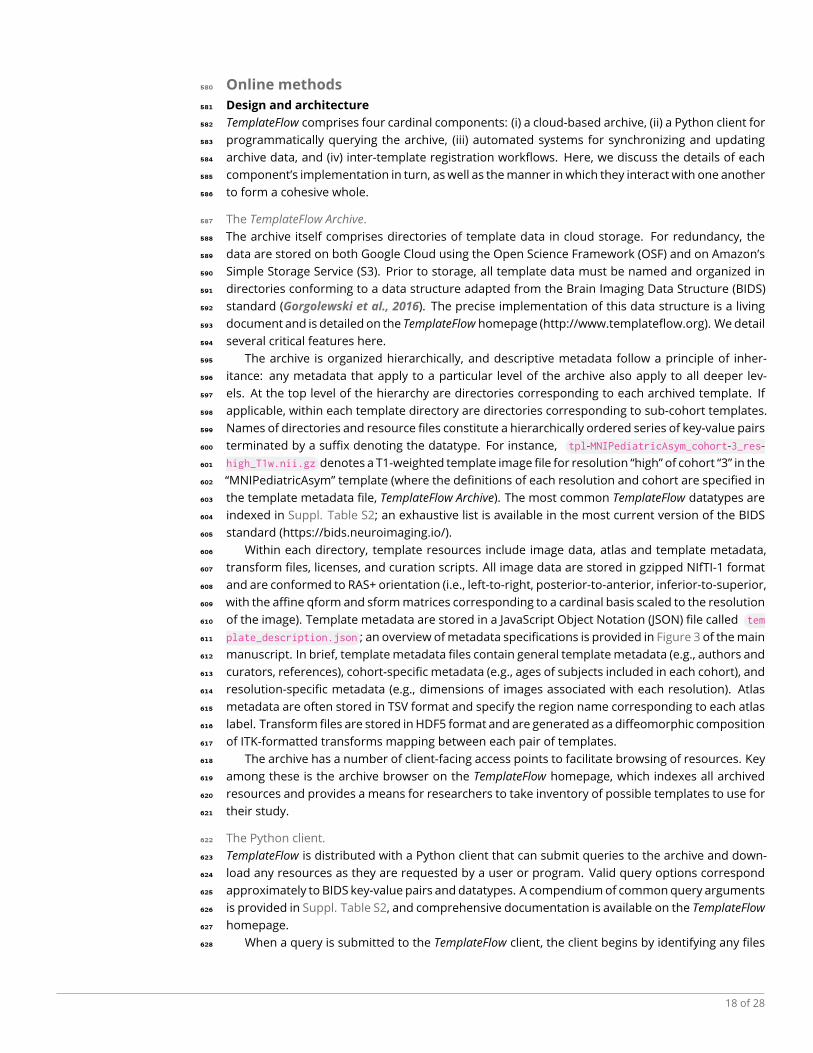
Figure 5. Example usage of
the Python-based
TemplateFlow Client. Afterimporting the API, the usersubmits a query for theT1-weighted FSL version ofthe MNI template at 1 mmresolution. The client firstfilters through the archive,identifies any files thatmatch the query, and findstheir counterparts in cloudstorage. It then downloadsthe requested files andreturns their paths in thelocal TemplateFlowinstallation directory. Futurequeries for the sameresource can be completedwithout any re-downloading.
>>> from templateflow import api as tflow
>>> tflow.get('MNI152NLin6Asym', desc=None, resolution=1,
... suffix='T1w', extension='nii.gz')
PosixPath('/templateflow_home/tpl-MNI152NLin6Asym/tpl-
MNI152NLin6Asym_res-01_T1w.nii.gz')
in the archive that match the query. To do so, it uses PyBIDS (Yarkoni et al., 2019), which exploits629
the BIDS-like architecture of the TemplateFlow Archive to efficiently scan all directories and filter any630
matching files. Next, the client assesses whether queried files exist as data in local storage. When a631
user locally installs TemplateFlow, the local installation initially contains only lightweight pointers to632
files in OSF cloud storage. These pointers are implemented using DataLad (Halchenko et al., 2021),633
a data management tool that extends git and git-annex. TemplateFlow uses DataLad principally to634
synchronize datasets across machines and to perform version control by tracking updates made635
to a dataset.636
If the queried files are not yet synchronized locally (i.e., they exist only as pointers to their637
counterparts in the cloud), the client instructs DataLad to retrieve them from cloud storage. In638
the event that DataLad fails or returns an error, the client falls back on redundancy in storage and639
downloads the file directly from Amazon’s S3. When the client is next queried for the same file, it640
will detect that the file has already been cached in the local filesystem. The use of resource pointers641
with the client thus enables lazy loading of template resources. Finally, the client confirms that the642
file has been downloaded successfully. If the client detects a successful download, it returns the643
result of the query; in the event that it detects a synchronization failure, it displays a warning for644
each queried file that encountered a failure.645
Continued functionality and operability of the client is ensured through an emphasis on maxi-646
mizing code coverage with unit tests. Updating the client requires successful completion of all unit647
tests, which are automatically executed by continuous integration (CI) and continuous delivery (CD)648
services connected to GitHub. CI and CD also keep the web-based archive browser up to date by649
automatically indexing data files.650
Ancillary and managerial systems.651
TemplateFlow includes a number of additional systems andprograms that serve to automate stages652
of the archive update process, for instance addition of a new template or revision of current tem-653
plate resources. To facilitate the update and extension process, TemplateFlow uses GitHub actions654
19 of 28
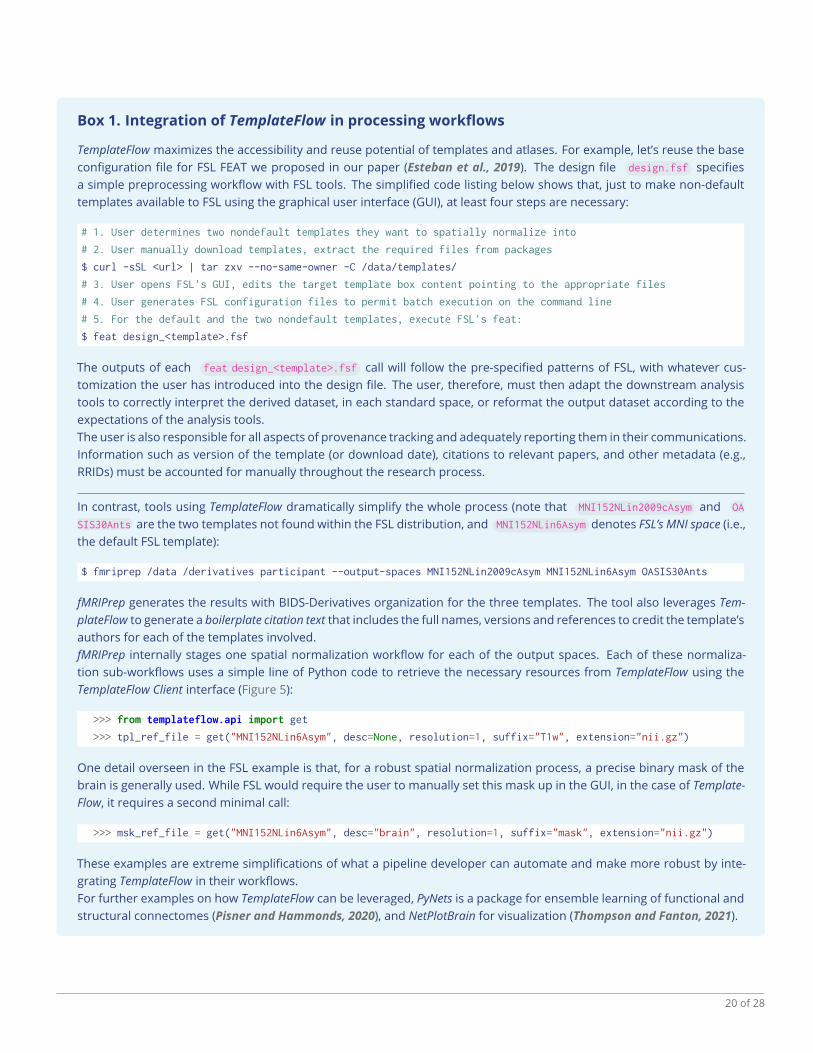
Box 1. Integration of TemplateFlow in processing workflows
TemplateFlow maximizes the accessibility and reuse potential of templates and atlases. For example, let’s reuse the baseconfiguration file for FSL FEAT we proposed in our paper (Esteban et al., 2019). The design file design.fsf specifiesa simple preprocessing workflow with FSL tools. The simplified code listing below shows that, just to make non-defaulttemplates available to FSL using the graphical user interface (GUI), at least four steps are necessary:
# 1. User determines two nondefault templates they want to spatially normalize into
# 2. User manually download templates, extract the required files from packages
$ curl -sSL <url> | tar zxv --no-same-owner -C /data/templates/
# 3. User opens FSL's GUI, edits the target template box content pointing to the appropriate files
# 4. User generates FSL configuration files to permit batch execution on the command line
# 5. For the default and the two nondefault templates, execute FSL's feat:
$ feat design_<template>.fsf
The outputs of each feat design_<template>.fsf call will follow the pre-specified patterns of FSL, with whatever cus-tomization the user has introduced into the design file. The user, therefore, must then adapt the downstream analysistools to correctly interpret the derived dataset, in each standard space, or reformat the output dataset according to theexpectations of the analysis tools.The user is also responsible for all aspects of provenance tracking and adequately reporting them in their communications.Information such as version of the template (or download date), citations to relevant papers, and other metadata (e.g.,RRIDs) must be accounted for manually throughout the research process.
In contrast, tools using TemplateFlow dramatically simplify the whole process (note that MNI152NLin2009cAsym and OA
SIS30Ants are the two templates not found within the FSL distribution, and MNI152NLin6Asym denotes FSL’s MNI space (i.e.,the default FSL template):
$ fmriprep /data /derivatives participant --output-spaces MNI152NLin2009cAsym MNI152NLin6Asym OASIS30Ants
fMRIPrep generates the results with BIDS-Derivatives organization for the three templates. The tool also leverages Tem-plateFlow to generate a boilerplate citation text that includes the full names, versions and references to credit the template’sauthors for each of the templates involved.fMRIPrep internally stages one spatial normalization workflow for each of the output spaces. Each of these normaliza-tion sub-workflows uses a simple line of Python code to retrieve the necessary resources from TemplateFlow using theTemplateFlow Client interface (Figure 5):
>>> from templateflow.api import get
>>> tpl_ref_file = get("MNI152NLin6Asym", desc=None, resolution=1, suffix="T1w", extension="nii.gz")
One detail overseen in the FSL example is that, for a robust spatial normalization process, a precise binary mask of thebrain is generally used. While FSL would require the user to manually set this mask up in the GUI, in the case of Template-Flow, it requires a second minimal call:
>>> msk_ref_file = get("MNI152NLin6Asym", desc="brain", resolution=1, suffix="mask", extension="nii.gz")
These examples are extreme simplifications of what a pipeline developer can automate and make more robust by inte-grating TemplateFlow in their workflows.For further examples on how TemplateFlow can be leveraged, PyNets is a package for ensemble learning of functional andstructural connectomes (Pisner and Hammonds, 2020), and NetPlotBrain for visualization (Thompson and Fanton, 2021).
20 of 28
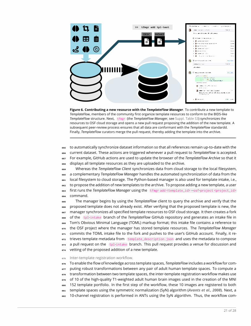
INTAKEDESC
>> tfmgr add tpl-test
Figure 6. Contributing a new resource with the TemplateFlow Manager. To contribute a new template toTemplateFlow, members of the community first organize template resources to conform to the BIDS-likeTemplateFlow structure. Next, tfmgr (the TemplateFlow Manager, see Suppl. Table S3) synchronizes theresources to OSF cloud storage and opens a new pull request proposing the addition of the new template. Asubsequent peer-review process ensures that all data are conformant with the TemplateFlow standardd.Finally, TemplateFlow curators merge the pull request, thereby adding the template into the archive.
to automatically synchronize dataset information so that all references remain up-to-date with the655
current dataset. These actions are triggered whenever a pull request to TemplateFlow is accepted.656
For example, GitHub actions are used to update the browser of the TemplateFlow Archive so that it657
displays all template resources as they are uploaded to the archive.658
Whereas the TemplateFlow Client synchronizes data from cloud storage to the local filesystem,659
a complementary TemplateFlow Manager handles the automated synchronization of data from the660
local filesystem to cloud storage. The Python-based manager is also used for template intake, i.e.,661
to propose the addition of new templates to the archive. To propose adding a new template, a user662
first runs the TemplateFlow Manager using the tfmgr add <template_id> --osf-project <project_id>663
command.664
The manager begins by using the TemplateFlow client to query the archive and verify that the665
proposed template does not already exist. After verifying that the proposed template is new, the666
manager synchronizes all specified template resources to OSF cloud storage. It then creates a fork667
of the tpl-intake branch of the TemplateFlow GitHub repository and generates an intake file in668
Tom’s Obvious Minimal Language (TOML) markup format; this intake file contains a reference to669
the OSF project where the manager has stored template resources. The TemplateFlow Manager670
commits the TOML intake file to the fork and pushes to the user’s GitHub account. Finally, it re-671
trieves template metadata from template_description.json and uses the metadata to compose672
a pull request on the tpl-intake branch. This pull request provides a venue for discussion and673
vetting of the proposed addition of a new template.674
Inter-template registration workflow.675
To enable the flowof knowledge across template spaces, TemplateFlow includes aworkflow for com-676
puting robust transformations between any pair of adult human template spaces. To compute a677
transformation between two template spaces, the inter-template registration workflowmakes use678
of 10 of the high-quality T1-weighted adult human brain images used in the creation of the MNI679
152 template portfolio. In the first step of the workflow, these 10 images are registered to both680
template spaces using the symmetric normalization (SyN) algorithm (Avants et al., 2008). Next, a681
10-channel registration is performed in ANTs using the SyN algorithm. Thus, the workflow com-682
21 of 28

putes a single transformation that simultaneously optimizes the alignment between all 10 images683
in both coordinate spaces.684
MNI space text mining analysis.685
To investigate the use of the term “MNI” in the neuroimaging literature, we conducted an ex-686
ploratory text mining analysis. For this, we used the Elsevier API to download the entire corpus687
of two leading journals of neuroimaging methodology, NeuroImage and NeuroImage: Clinical. In688
this way, we retrieved a total 16,812 full-text articles that were subsequently segmented into lists689
of sentences. A scan of these sentences revealed 14,870 sentences across 6,048 articles that con-690
tained the word “MNI”. Sentences were cleaned (i.e., removing punctuation, single letters, accents,691
numbers) and tokenized into words, which were subsequently lemmatized (i.e., converted to base692
form) using the NLTK wordnet lemmatizer. From the lemmatized words, we filtered out stopwords693
(i.e., NLTK stopwords and a custom list) and included words with a frequency above 10 as part of694
our “dictionary”; this yielded a dictionary size of 2,324 words.695
Next, we computed a sparse dictionary by article count matrix (i.e., 2,324 × 6,048), on which we696
performed topic modelling with latent Dirichlet allocation (LDA; Blei et al., 2003, implementation697
from scikit-learn with the learning decay hyperparameter set to 0.7). The number of topics (k=15)698
was selected by identifying the LDAmodel yielding the lowest perplexity (Blei et al., 2003) sweeping699
the interval [8-16] for the parameter. The 20 words from the dictionary that loaded the highest on700
the 15 topics were visualized using word clouds. Topics were sorted by descending dominance,701
and the dominance fraction (number of articles where the topic is the most loaded with respect702
to the total 6,048 documents) was included above the corresponding topic’s word cloud (Figure 4,703
and Suppl. Figure S1).704
22 of 28
Supp. Box S1. The FAIR Guiding Principles
(Reproduced fromWilkinson et al., 2016, Box 2)
To be Findable:F1. (meta)data are assigned a globally unique and persistent identifierF2. data are described with rich metadata (defined by R1 below)F3. metadata clearly and explicitly include the identifier of the data it describesF4. (meta)data are registered or indexed in a searchable resource
To be Accessible:A1. (meta)data are retrievable by their identifier using a standardized communications protocol
A1.1. the protocol is open, free, and universally implementableA1.2. the protocol allows for an authentication and authorization procedure, where necessary
A2. metadata are accessible, even when the data are no longer available
To be Interoperable:I1. (meta)data use a formal, accessible, shared, and broadly applicable language for knowledge representation.I2. (meta)data use vocabularies that follow FAIR principlesI3. (meta)data include qualified references to other (meta)data
To be Reusable:R1. (meta)data are richly described with a plurality of accurate and relevant attributes
R1.1. (meta)data are released with a clear and accessible data usage licenseR1.2. (meta)data are associated with detailed provenanceR1.3. (meta)data meet domain-relevant community standards
23 of 28
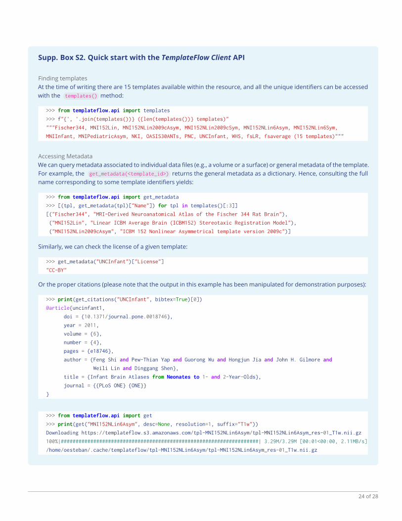
Supp. Box S2. Quick start with the TemplateFlow Client API
Finding templatesAt the time of writing there are 15 templates available within the resource, and all the unique identifiers can be accessedwith the templates() method:
>>> from templateflow.api import templates
>>> f"{', '.join(templates())} ({len(templates())} templates)"
"""Fischer344, MNI152Lin, MNI152NLin2009cAsym, MNI152NLin2009cSym, MNI152NLin6Asym, MNI152NLin6Sym,
MNIInfant, MNIPediatricAsym, NKI, OASIS30ANTs, PNC, UNCInfant, WHS, fsLR, fsaverage (15 templates)"""
Accessing MetadataWe can querymetadata associated to individual data files (e.g., a volume or a surface) or general metadata of the template.For example, the get_metadata(<template_id>) returns the general metadata as a dictionary. Hence, consulting the fullname corresponding to some template identifiers yields:
>>> from templateflow.api import get_metadata
>>> [(tpl, get_metadata(tpl)["Name"]) for tpl in templates()[:3]]
[("Fischer344", "MRI-Derived Neuroanatomical Atlas of the Fischer 344 Rat Brain"),
("MNI152Lin", "Linear ICBM Average Brain (ICBM152) Stereotaxic Registration Model"),
("MNI152NLin2009cAsym", "ICBM 152 Nonlinear Asymmetrical template version 2009c")]
Similarly, we can check the license of a given template:
>>> get_metadata("UNCInfant")["License"]
"CC-BY"
Or the proper citations (please note that the output in this example has been manipulated for demonstration purposes):
>>> print(get_citations("UNCInfant", bibtex=True)[0])
@article{uncinfant1,
doi = {10.1371/journal.pone.0018746},
year = 2011,
volume = {6},
number = {4},
pages = {e18746},
author = {Feng Shi and Pew-Thian Yap and Guorong Wu and Hongjun Jia and John H. Gilmore and
Weili Lin and Dinggang Shen},
title = {Infant Brain Atlases from Neonates to 1- and 2-Year-Olds},
journal = {{PLoS ONE} {ONE}}
}
>>> from templateflow.api import get
>>> print(get("MNI152NLin6Asym", desc=None, resolution=1, suffix="T1w"))
Downloading https://templateflow.s3.amazonaws.com/tpl-MNI152NLin6Asym/tpl-MNI152NLin6Asym_res-01_T1w.nii.gz
100%|###################################################################| 3.29M/3.29M [00:01<00:00, 2.11MB/s]
/home/oesteban/.cache/templateflow/tpl-MNI152NLin6Asym/tpl-MNI152NLin6Asym_res-01_T1w.nii.gz
24 of 28
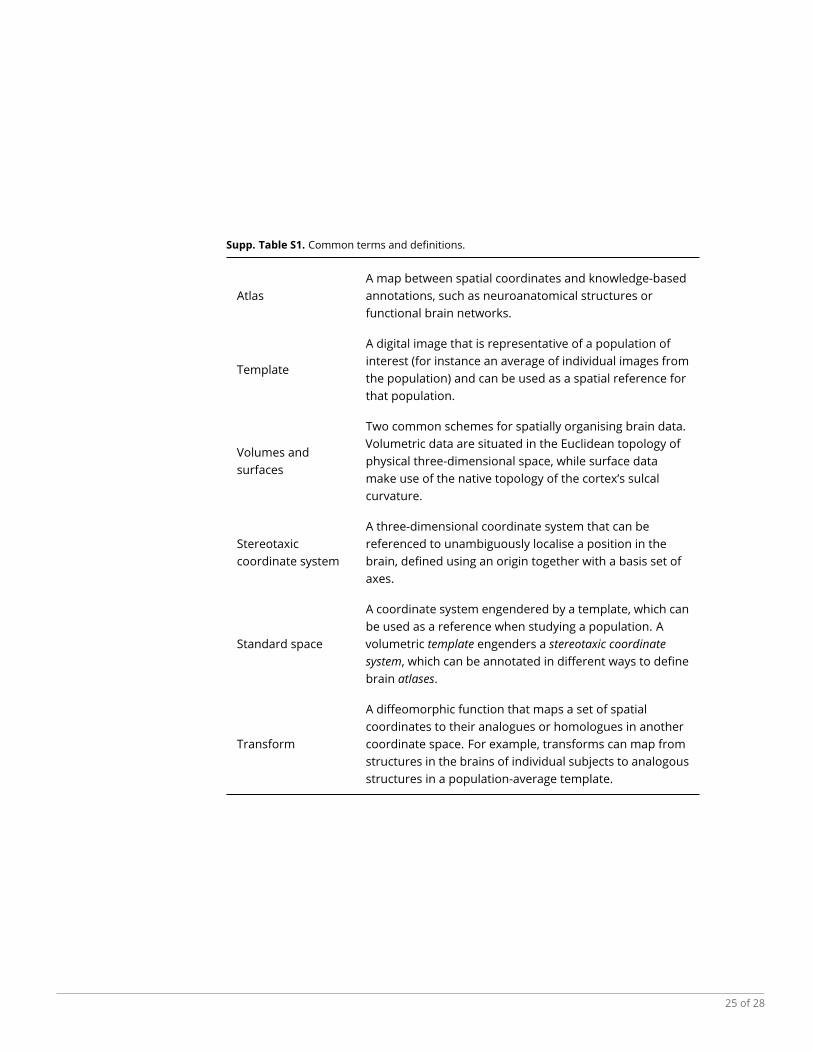
Supp. Table S1. Common terms and definitions.
AtlasA map between spatial coordinates and knowledge-basedannotations, such as neuroanatomical structures orfunctional brain networks.
Template
A digital image that is representative of a population ofinterest (for instance an average of individual images fromthe population) and can be used as a spatial reference forthat population.
Volumes andsurfaces
Two common schemes for spatially organising brain data.Volumetric data are situated in the Euclidean topology ofphysical three-dimensional space, while surface datamake use of the native topology of the cortex’s sulcalcurvature.
Stereotaxiccoordinate system
A three-dimensional coordinate system that can bereferenced to unambiguously localise a position in thebrain, defined using an origin together with a basis set ofaxes.
Standard space
A coordinate system engendered by a template, which canbe used as a reference when studying a population. Avolumetric template engenders a stereotaxic coordinatesystem, which can be annotated in different ways to definebrain atlases.
Transform
A diffeomorphic function that maps a set of spatialcoordinates to their analogues or homologues in anothercoordinate space. For example, transforms can map fromstructures in the brains of individual subjects to analogousstructures in a population-average template.
25 of 28

Supp. Table S2. TemplateFlow data entities.
Data entity API query example Description
Template "MNI152Lin"The template dataset to which an image or other datafile belongs.
Resolution res=1
The image resolution. Each resolution is assigned akey, which is defined in the res field oftemplate_description.json .
Mask desc="brain", suffix="mask"Indicates that the image is a binary-valued annotation,where voxels labelled 1 are part of the mask.
Discrete segmentation desc="malf", suffix="dseg"
Indicates that the image is an integer-valuedannotation. Each segmentation image file ( .nii.gz
format) is paired with a dictionary of segment names (.tsv format).
Probabilistic segmentation label="CSF", suffix="probseg"
Indicates that the image is a probabilistic annotation,wherein the value of each voxel indicates theprobability of that voxel belonging to the specifiedlabel.
Atlas atlas="Schaefer", desc="7Network" The atlas to which a segmentation file belongs.
Transformation from="MNI152Lin", suffix="xfm"
File containing a mapping between 2 stereotaxiccoordinate spaces. The source space is defined in thefrom field, while the target space is defined in the tpl
field.
Image modality suffix="T1w"
For non-annotation brain images, the suffix indicateswhether the image is T1-weighted ( T1w ), T2-weighted (T2w ), proton density-weighted ( PD ), or T2*-weighted (T2star ).
Template cohort cohort=1Subsample of a dataset used to generate an averagetemplate.
26 of 28

Supp. Table S3. Command-line interface for TemplateFlow Manager.
Argument Environment variable Specifications
template_id Identifier of the template. This is the value of the tpl field in all file names.
--osf-project OSF_PROJECT
The OSF project where the template data are to be stored. The project mustbe writable by the user account whose credentials are specified in the--osf-user and --osf-password arguments.
--osf-user OSF_USERNAME Account username or identifier for OSF cloud storage.--osf-password OSF_PASSWORD Account password for OSF cloud storage.
--osf-overwriteFlag that indicates that the OSF client should force the overwrite of anyexisting files in the OSF project that have names conflicting with those ofnew files.
--gh-user GITHUB_USERAccount username for GitHub. The user account whose credentials areprovided must have a fork of the TemplateFlow repo.
--gh-password GITHUB_PASSWORD Account password for GitHub.
--path OSF_PROJECT
Path to a local directory where template resources are located. The pathmust either be a directory whose name is tpl-<template_id> or containsuch a directory.
--nprocsMaximum number of parallel processes to run when uploading to orfetching from OSF.
27 of 28
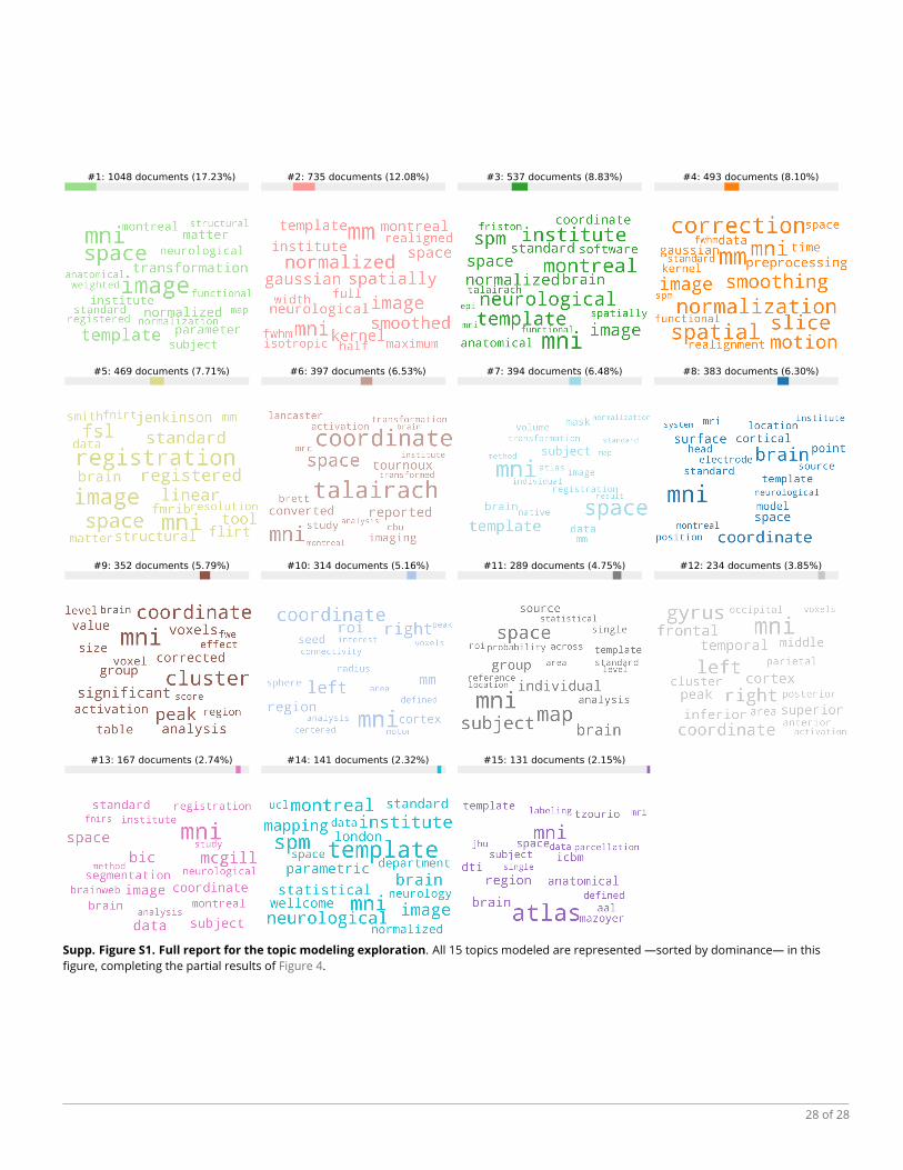
#1: 1048 documents (17.23%) #2: 735 documents (12.08%) #3: 537 documents (8.83%) #4: 493 documents (8.10%)
#5: 469 documents (7.71%) #6: 397 documents (6.53%) #7: 394 documents (6.48%) #8: 383 documents (6.30%)
#9: 352 documents (5.79%) #10: 314 documents (5.16%) #11: 289 documents (4.75%) #12: 234 documents (3.85%)
#13: 167 documents (2.74%) #14: 141 documents (2.32%) #15: 131 documents (2.15%)
Supp. Figure S1. Full report for the topic modeling exploration. All 15 topics modeled are represented —sorted by dominance— in thisfigure, completing the partial results of Figure 4.
28 of 28