Facebow Instructions (2017) - Advanced Dental Designs
11
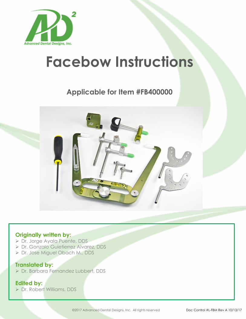
Facebow Instructions Applicable for Item #FB400000 Originally written by: ➢ Dr. Jorge Ayala Puente, DDS ➢ Dr. Gonzalo Guietierrez Alvarez, DDS ➢ Dr. Jose Miguel Obach M., DDS Translated by: ➢ Dr. Barbara Fernandez Lubbert, DDS Edited by: ➢ Dr. Robert Williams, DDS ©2017 Advanced Dental Designs, Inc. All rights reserved Doc Control #L-FBM Rev A 10/13/17
-
Upload
khangminh22 -
Category
Documents
-
view
3 -
download
0
Transcript of Facebow Instructions (2017) - Advanced Dental Designs
Facebow Instructions
Applicable for Item #FB400000
Originally written by:➢ Dr. Jorge Ayala Puente, DDS
➢ Dr. Gonzalo Guietierrez Alvarez, DDS
➢ Dr. Jose Miguel Obach M., DDS
Translated by:➢ Dr. Barbara Fernandez Lubbert, DDS
Edited by:➢ Dr. Robert Williams, DDS
©2017 Advanced Dental Designs, Inc. All rights reserved Doc Control #L-FBM Rev A 10/13/17

AD2 • 800.232.2849 • www.ad2usa.com
Section 1: Facebow BackgroundThe facebow is an indispensable part of the semi or completely adjustable articulator,
because the upper cast is mounted in the same position as the maxilla, with respects to the
cranium. Facebows are classified into two types:
➢ Anatomic - Anatomic (AD2, Panadent, Whip Mix, Dentatus, etc.) facebows position the
upper maxilla based on the axis-orbital plane, which is determined by average values and
will be described later.
➢ Cinematic - Cinematic facebows are sophisticated instruments such as axiographs
and/or pantograghs that help determine the individual values of different parameters
measured in patients. These values provide more information to program the articulator and
can include exact hinge axis, condyle eminence, Bennett angle, and immediate side shift.
For a better understanding of these concepts, we will begin with the basic of the facebow
manufactured by AD2.
©2017 Advanced Dental Designs, Inc. All rights reserved Doc Control #L-FBM Rev A 10/13/17
Section 2: Main Components
Items include:
1. Facebow
2. Bite Fork Stem Assembly
3. Bite Fork
4. Nasion Relator
5. Mounting Table
6. Screw Toggles
7. Hex Screwdriver

AD2 • 800.232.2849 • www.ad2usa.com
AD2 • 800.232.2849 • www.ad2usa.com
Facebow Components
1. Side Arms (2)
2. Central Knob
3. Cross Bar
➢ 3a - Slot for Nasion Relator
➢ 3b - Hole for inserting bite fork stem
4. Ear Piece (2)
5. Transverse Bubble Level
6. Sagittal Bubble Level
7. Orbital Pointer (for third reference point)
Bite Fork Stem Components
The bite fork stem connects the bite fork to the
facebow and replaces the facebow when
mounting the upper cast on the articulator.
The components of the bite fork stem are:
1. Vertical Post
2. Horizontal Arm
3. Vertical Clamp
4. Stem Tip (short)
5. Toggle Clamp
6. Stem Tip (long)
©2017 Advanced Dental Designs, Inc. All rights reserved Doc Control #L-FBM Rev A 10/13/17
The vertical post (1) has two opposing ends.
Each end has a flat surface machined into it
where a thumb screw will contact it. This, in turn,
will prevent the vertical post from rotating once it is locked into place in the facebow or
mounting table.
➢ Stem Tip (long, #6) is designed to fit into the slot on the mounting table.
➢ Stem Tip (short, #4) is denoted by both a green dot on the end of the tip as well as a
green groove on the vertical post. The short Stem Tip is inserted into the facebow crossbar
hole shown by 3a above.
The vertical clamp (3) serves the dual purpose of joining the vertical post and horizontal arm
together as well as allowing the user to lock them together in a specific position, Similarly,
the toggle clamp (5) connects the bite fork to the bite fork stem assembly and lock it into a
specific position. Please note that the toggle clamp should never be tightened without a
bite fork inserted first. Failure to do so may result in the toggle clamp being bent and
rendered unusable.

AD2 • 800.232.2849 • www.ad2usa.com
AD2 • 800.232.2849 • www.ad2usa.com
AD2 • 800.232.2849 • www.ad2usa.com
Bite Fork Components
1. Bite Fork Post
2. Midline
3. Compound Retention Holes
4. Bite Fork Support
To record bite registration, a bite fork will be
into the patient’s mouth (with the bite fork
attached to the facebow via the bite fork
stem assembly). To properly prepare the
bite fork for use, please be sure it has been
sterilized and free of all residue.
©2017 Advanced Dental Designs, Inc. All rights reserved Doc Control #L-FBM Rev A 10/13/17
If bite registration compound (i.e. Kerr green
stick) is used, the compound should be
placed at the midline and in the area of the first molars. Ideally, the surface of the
compound should be smooth, without irregular areas to allow for the most accurate
impression of the upper incisal edges and the cusps of the upper bicuspids and molars.
As an alternative to compound, AD2
recommends the use of its Accu-Bite adhesive
wax discs. Accu-Bites should also be placed
at the same three locations (midline and first
molar) as shown to the right.
While the flat bite fork as shown above is
used more commonly, AD2 provides a
curved bite fork (shown right) as well with
the FB400000 facebow. In cases where the
patient's lower 2 molars are extruded or the
upper 2 molars are not present, the curved
bite fork can provide improved patient
comfort.

AD2 • 800.232.2849 • www.ad2usa.com
AD2 • 800.232.2849 • www.ad2usa.com
AD2 • 800.232.2849 • www.ad2usa.com
AD2 • 800.232.2849 • www.ad2usa.com
Nasion Relator Components
1. Nasion Pad
2. Nasion Bracket
3. Upper Thumb Screw
4. Lower Thumb Screw
5. Nasion Shaft
While we will discuss the procedure for
facebow recording below, the nasion
relator is attached to the facebow via
the slot on the nasion body (2) and then
tightened using the lower thumb screw (4).
©2017 Advanced Dental Designs, Inc. All rights reserved Doc Control #L-FBM Rev A 10/13/17
Section 3: Facebow Use – Basic Concepts
Taking a proper facebow record is necessary to mount the upper cast and reproduce the
three dimensional position of the maxilla. It also provides an estimated mandibular rotating
axis (hinge axis) as well as a reference plane (known as the axis orbital plane).
The facebow uses three reference points: two posterior and one anterior. The posterior
points represent the rotation axis (or hinge) of each condyle, which are arbitrarily recorded
in the patient’s external auditory canal with the ear pieces on the face-bow. The anterior
reference point or orbital point (3rd reference point) is also arbitrary and is determined by a
specific distance from the nasal bridge by the nasion relator. Note that this point does not
necessarily coincide with the cephalometric orbital point.
With these three reference points, we can now establish the axis-orbital plane. When the
facebow record is transferred to the articulator, the upper cast is mounted on the upper
member of the articulator on the axis-orbital plane.
Shown right is the axis-orbital plane recorded
by the facebow. The hinge axis is determined
by the ear pieces and the orbital point by the
nasion relator.

AD2 • 800.232.2849 • www.ad2usa.com
AD2 • 800.232.2849 • www.ad2usa.com
AD2 • 800.232.2849 • www.ad2usa.com
AD2 • 800.232.2849 • www.ad2usa.com
AD2 • 800.232.2849 • www.ad2usa.com
Basic Concepts (cont)
In summary, the use of a semi-adjustable articulator with a facebow will estimate the
position of the maxilla in the skull as well as the relation of the mandible with the cranium,
(the hinge axis). Once the lower cast is mounted on the articulator, the distance
between the hinge axis and the lower teeth is established so that a mandibular closing
arc can be established for each lower tooth. This is one of the reasons why the hinge axis
is so important: the mandibular closing arc of the patient on the articulator shows the
tooth contacts in closure.
©2017 Advanced Dental Designs, Inc. All rights reserved Doc Control #L-FBM Rev A 10/13/17
Pictured right, the mandibular closing arc
where the distance between the hinge axis
and the dental arch are duplicated (or
each individual tooth).
Since the reference points are determined
arbitrarily, this mandibular closing arc is not
exact, but for diagnostic purposes, it is
considered useful system. However, there
are certain therapeutic procedures where
vertical dimension will be changed (i.e.
orthognathic surgery, selective grinding) and
it will be necessary to use a true hinge axis.
In these instances, an axiograph (hinge axis
recorder) will be needed to determine the
true mandibular closing arc.
Facebow Recording Procedure
As discussed earlier, a facebow record can be taken by applying bite registration
compound directly on the bite fork or by using Accu-Bite adhesive wax discs. The
procedure shown below will focus on using Accu-Bite discs for this.
Step 1: Peel off an Accu-Bite strip at printed end
from sheet. Avoid touching the adhesive
underside near the wax disc.

AD2 • 800.232.2849 • www.ad2usa.com
AD2 • 800.232.2849 • www.ad2usa.com
AD2 • 800.232.2849 • www.ad2usa.com
AD2 • 800.232.2849 • www.ad2usa.com
AD2 • 800.232.2849 • www.ad2usa.com
AD2 • 800.232.2849 • www.ad2usa.com
Step 2: Hold Accu-Bite on the sides of the paper
strip and fold back the adhesive paper near the
bottom for easy removal after use.
©2017 Advanced Dental Designs, Inc. All rights reserved Doc Control #L-FBM Rev A 10/13/17
Step 3: Place Accu-Bites at the left molar, right
molar and incisor positions on a clean, dry bite
fork. Avoid covering the midline mark on the bite
fork.
Step 4: Place the bite fork in hot tap water (125°F/40° C) to soften the Accu-Bites (about 60
seconds). For the best Accu-Bite adhesion to the
bite fork, do not place the bite fork in a water
bath.
Step 5: Place the bite fork in the patient’s mouth,
aligning the center mark with the facial mid line.
Lightly press the bite fork upwards so that the teeth
indent the Accu-Bites approximately 1mm. Make
sure that no teeth come in contact with the bite
fork. Remove the bite fork and cool with water or
compressed air.

AD2 • 800.232.2849 • www.ad2usa.com
AD2 • 800.232.2849 • www.ad2usa.com
AD2 • 800.232.2849 • www.ad2usa.com
AD2 • 800.232.2849 • www.ad2usa.com
AD2 • 800.232.2849 • www.ad2usa.com
AD2 • 800.232.2849 • www.ad2usa.com
Step 5 Option: To improve patient comfort with
the bite fork, the doctor can choose to add an
adhesive bite fork stabilizer to the underside of
the bite fork once it has been removed from the
warm water. Simply peel off the contact paper
to reveal the adhesive and press the foam
stabilizer to the bite fork.
©2017 Advanced Dental Designs, Inc. All rights reserved Doc Control #L-FBM Rev A 10/13/17
Step 6: Shave off the excess compound, leaving
imprints about 1mm deep.
Step 7: Install the nasion relator in the slot on the
facebow crossbar as shown.
Step 8: Insert and lock the short end of the bite fork
stem (with the green dot and groove) to the
facebow. The flat surface of this end must face the
thumb screw.

AD2 • 800.232.2849 • www.ad2usa.com
AD2 • 800.232.2849 • www.ad2usa.com
AD2 • 800.232.2849 • www.ad2usa.com
AD2 • 800.232.2849 • www.ad2usa.com
AD2 • 800.232.2849 • www.ad2usa.com
AD2 • 800.232.2849 • www.ad2usa.com
Step 9: With the hex screw driver, loosen the
vertical clamp on the bite fork stem.
©2017 Advanced Dental Designs, Inc. All rights reserved Doc Control #L-FBM Rev A 10/13/17
Step 10: Continue using the hex screw driver
to loosen the toggle clamp on the bite fork
stem.
Observational Note: Steps 6, 7, 8 and 9 are
generally done by a dental assistant before
the facebow recording begins.
Step 11: Loosen the central knob located on
the anterior end of the face-bow with ½ turn
counter clockwise.

AD2 • 800.232.2849 • www.ad2usa.com
AD2 • 800.232.2849 • www.ad2usa.com
AD2 • 800.232.2849 • www.ad2usa.com
AD2 • 800.232.2849 • www.ad2usa.com
AD2 • 800.232.2849 • www.ad2usa.com
AD2 • 800.232.2849 • www.ad2usa.com
Step 12: Instruct the patient to separate the side
arms of the facebow and insert the ear pieces
into the ears (push in and forward). Note that this
can also be done by an assistant. Once
completed, tighten the central knob to lock the
width of the facebow.
©2017 Advanced Dental Designs, Inc. All rights reserved Doc Control #L-FBM Rev A 10/13/17
Step 13: While the patient (or assistant) is still
holding the facebow arms, place the nasion
relator on the patient’s nasion. Use the nasion
relator like a plunger, using gentle pressure to
push the relator against the patient. This will
move the earpieces more forward to
approximate the condyles. Lock the nasion in
place by tightening the upper thumb screw as
shown.
Step 14: Check that the vertical clamp and toggle
clamp on the bite fork stem are loose. Also confirm
that the toggle clamp is facing down and is on the
right hand side of the patient.
Step 15: Slide the bite fork through the hole in the
toggle clamp and put it in the patient’s mouth,
seating the teeth in the indentations in the Accu-
Bites (or registration compound). Make sure the
bite fork seats firmly and there is no movement.

AD2 • 800.232.2849 • www.ad2usa.com
AD2 • 800.232.2849 • www.ad2usa.com
AD2 • 800.232.2849 • www.ad2usa.com
AD2 • 800.232.2849 • www.ad2usa.com
AD2 • 800.232.2849 • www.ad2usa.com
AD2 • 800.232.2849 • www.ad2usa.com
Step 16: Stabilize the bite fork with the index
and middle fingers and tighten both the
vertical clamp and toggle clamp with the hex
screw driver. When completed, double check
the stability of the bite fork.
©2017 Advanced Dental Designs, Inc. All rights reserved Doc Control #L-FBM Rev A 10/13/17
Step 17: Loosen the center knob of the
facebow and ask the patient (or assistant) to
open the side arms and remove it from the
ears. When removing, the facebow should
come down and forward.
Step 18: Loosen the thumb screw that joins the
bite fork assembly to the facebow.
Step 19: Remove the bite fork assembly,
package it carefully and send it to the lab so the
upper cast can be mounted.