extradural diploic and intradural epidermoid - NCBI
41
EXTRADURAL DIPLOIC AND INTRADURAL EPIDERMOID TUMORS (CHOLESTEATOMA) JOSEPH E. J. KING, M.D. NEW YORK, N. Y. EPIDERMOIDS (also known as tumeurs perlees, Perlgschwiulste, cholestea- tome, Epidermoid, and cholesteatomata), arising from the diploe, extradural in position, and those occupying an intradural position are to be considered in this paper. Dermoids, and so-called "cholesteatomata," either primary or secondary, involving the mastoid, arising from the interior of the petrous por- tion of the temporal bone, accessory nasal sinuses and the bones of the skull, other than the cranium, are not under consideration. This lesion was rarely found as late as 1920, when Bailey' reported two cases of Cruveilhier's tumors (tumeurs perlees) successfully operated upon by Doctor Cushing, in I919. These two patients were the first recorded cases to recover following removal of the tumor from the intradural position. In I922, Cushing2 briefly reported abstracts of six cases of the extradural diploic type appearing in the literature since I856, with recovery in all six eases. In his paper he reported a case of his own in complete detail. These two papers stand out as the first two milestones of proper surgical eradica- tion of both intra- and extradural lesions. Since the publication of these two papers, quite a few other cases have been reported from the various neurosurgical clinics of this and other coun- tries. The lesions are still sufficiently rare to permit the author to report six personal cases, and two others from the Neurologic and Neurosurgical Services of Bellevue Hospital, New York. In I829, Cruveilhier3 described three tumors of the intracranial cavity, and on account of their pearl-like appearance gave them the name "tumeurs perlees." One of these cases had previously been reported by Dumeril,4 and so far as is known, it was the first report of a case of the nonhair-con- taining type, although one of the dermoid type had been noted by Verratus,5 in I745. In I838, Johannes Mueller6 gave the name "cholesteatoma" to this kind of lesion, for the reason that cholesterol crystals were found in his two cases. This apparent misnomer has continued to cling in subsequent papers, and has contributed to the confusion in the minds of many regarding the exact origin and nature of the lesion. In i854, von Remak7 suggested that the tumor arose from embryonic epithelial cell rests, and his work afforded the basis for the conception of the term "epidermoid." In I897, Bostroem8 described a definite attachment to the pia mater, and coined the term "haarlosen pialen Epidermoide." Again, on account of the confusion of the terms used to designate this lesion, Horrax,9 in I922, adhered to the general term "cholesteatoma" and 649
-
Upload
khangminh22 -
Category
Documents
-
view
1 -
download
0
Transcript of extradural diploic and intradural epidermoid - NCBI

EXTRADURAL DIPLOIC AND INTRADURAL EPIDERMOIDTUMORS (CHOLESTEATOMA)
JOSEPH E. J. KING, M.D.NEW YORK, N. Y.
EPIDERMOIDS (also known as tumeurs perlees, Perlgschwiulste, cholestea-tome, Epidermoid, and cholesteatomata), arising from the diploe, extraduralin position, and those occupying an intradural position are to be consideredin this paper. Dermoids, and so-called "cholesteatomata," either primary orsecondary, involving the mastoid, arising from the interior of the petrous por-tion of the temporal bone, accessory nasal sinuses and the bones of the skull,other than the cranium, are not under consideration.
This lesion was rarely found as late as 1920, when Bailey' reported twocases of Cruveilhier's tumors (tumeurs perlees) successfully operated uponby Doctor Cushing, in I919. These two patients were the first recordedcases to recover following removal of the tumor from the intradural position.In I922, Cushing2 briefly reported abstracts of six cases of the extraduraldiploic type appearing in the literature since I856, with recovery in all sixeases. In his paper he reported a case of his own in complete detail. Thesetwo papers stand out as the first two milestones of proper surgical eradica-tion of both intra- and extradural lesions.
Since the publication of these two papers, quite a few other cases havebeen reported from the various neurosurgical clinics of this and other coun-tries. The lesions are still sufficiently rare to permit the author to reportsix personal cases, and two others from the Neurologic and NeurosurgicalServices of Bellevue Hospital, New York.
In I829, Cruveilhier3 described three tumors of the intracranial cavity,and on account of their pearl-like appearance gave them the name "tumeursperlees." One of these cases had previously been reported by Dumeril,4and so far as is known, it was the first report of a case of the nonhair-con-taining type, although one of the dermoid type had been noted by Verratus,5in I745.
In I838, Johannes Mueller6 gave the name "cholesteatoma" to this kindof lesion, for the reason that cholesterol crystals were found in his two cases.This apparent misnomer has continued to cling in subsequent papers, andhas contributed to the confusion in the minds of many regarding the exactorigin and nature of the lesion.
In i854, von Remak7 suggested that the tumor arose from embryonicepithelial cell rests, and his work afforded the basis for the conception of theterm "epidermoid." In I897, Bostroem8 described a definite attachment tothe pia mater, and coined the term "haarlosen pialen Epidermoide."
Again, on account of the confusion of the terms used to designate thislesion, Horrax,9 in I922, adhered to the general term "cholesteatoma" and
649
JOSEPH E. J. KING Annals of SurgeryJ ~~~~Ma y, 1 93 9
referred to the intradural lesions as meningeal cholesteatoma, stating that:"There can be no mistaking the kind of growth to which reference is made,as this designation immediately eliminates the group of tumors containingcholesterin crystals which arise from pituitary rests, and also does away withany confusion of association with those of the middle ear, cholesteatomatawhich have not had a primary meningeal attachment."
Critchley and Ferguson,'0 in I928, suggested that these lesions be termed"cerebrospinal epidermoids." This appellation would hardly include thatdiploic type which involves primarily the diploe and the outer table of theskull.
In I922, Cushing used the term "epidermal cholesteatoma" to designatethe extradural lesion reported. In I937, Munro and Wegner" suggestedthe term "primary cranial epidermoid" for the diploic type and "primaryintracranial epidermoid."
Regardless of the name employed, it is now believed that the tumor, forthe most part, is a mass of epithelial debris, which has resulted from slow ac-cumulation of desquamated cells from the epithelial layer of the lining of thelesion. Therefore, it seems that the confusion of terms would be eliminatedif the lesion is designated only as an epidermoid tumor, either diploic extra-dural or intradural. In spite of this apparent clear-cut nomenclature, theauthor has, parenthetically, retained the name "cholesteatoma" in the title ofthis paper, partly for the reason that almost everyone will more promptlygrasp the intent of the subject under discussion, partly due to slowness ingiving up the old, and on account of the bibliography.
Incidence.-Epidermoids are comparatively rare tumors, and, therefore,are infrequently found at operation. Previous to the publication of thepapers by Cushing and Bailey, the majority of cases were found at autopsy.Since then, more epidermoid tumors have been found and removed at opera-tion, probably because there are more neurologists and neurologic surgeons.More cases have been reported in the last two decades than in all the precedingtime. Even so, the percentage of these tumors in neurosurgical clinics is low.
Tooth'2 found but one instance in a series of 258 cases of brain tumor,verified at operation or autopsy, a percentage of 0.4. Frank13 stated thatBernhardt found only one in a series of 487 autopsies on brain tumor cases,or about 0.2 per cent. In I920, Bailey reported that only two cases wereobserved in a series of over 550 verified brain tumors in Doctor Cushing'sclinic, a percentage of 0.37. In I922, Cushing stated that in his series of740 cases of brain tumor, only three epidermoids were found, less than o.5per cent. In I936, Mahoney14 reported that in 2,500 verified intracranialtumors in Cushing's collection from Johns Hopkins and Peter Bent BrighamHospitals, there were I5 epidermoids, o.6 per cent, and in Foerster's group of750 tumors, there were five, o.66 per cent.
It is interesting to note the regularity of the percentage as the numberof cases in the Cushing Clinic increased, amounting throughout to about 0.5per cent.
650
Volume 109Number 5 EPIDERMOID TUMORS
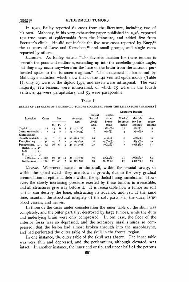
In I920, Bailey reported 62 cases from the literature, including two ofhis own. Mahoney, in his very exhaustive paper published in I936, reported142 true cases of epidermoids from the literature, and added five fromFoerster's clinic. He did not include the five new cases reported by Bucy,15the I I cases of Love and Kernohan,16 and small groups, and single casesreported by others.
Location.-As Bailey stated: "The favorite location for these tumors isbeneath the pons and midbrain, extending up into the cerebello-pontile angle,but they may occur anywhere on the base of the brain from the anterior per-forated space to the foramen magnum." This statement is borne out byMahoney's statistics, which show that of the I42 verified epidermoids (TableI), only 23 were of the diploic type, and seven were intraspinal. The vastmajority, II2 lesions, were intracranial, of which I5 were in the fourthventricle, 44 were parapituitary and 53 were parapontine.
TABLE I
SERIES OF 142 CASES OF EPIDERMOID TUMORS COLLECTED FROM THE LITERATURE (MAHONEY)Operative Results
Clinical Psychi-Location Cases Sex Average Record atric Marked Mortal- Au-
Age Avail- Symp- Improve- ity Per- topsyM. F. ? able toms ment centage Record
Diploic .......... 23 14 6 3 4I (I-7I) 2I 3(I4%) I7 2(I%) 4Intra-arachnoid.. 7 5 2 0 25.4(7-35) 6 o(o%) 3 2(40%) 2(Intraspinal)Fourth ventricle.. I5 7 8 0 38.6(I9-76) I0 4(40%) 2 4(66%) 9Parapituitary.... 44 24 I8 2 32. I(5-65) 26 I5(60%) 7 8(53%) 29Parapontine ...... 53 26 22 5 35.3(I0-78) 32 20(63%) 2 I0(83%) 4IRight... 27Left.. 23?? ........ 3
Totals....... I42 76 56 I0 35 (I-78) 95 42(44%) 31 26(45%) 85Intracranial.... II2 57 48 7 34.5(5-78) 68 39(57%) II 22(67%) 79
Course.-Wherever located-in the skull, within the cranial cavity, orwithin the spinal canal-they are slow in growth, due to the very gradualaccumulation of epithelial debris within the epithelial lining membrane. How-ever, the slowly increasing pressure exerted by these tumors is irresistible,and all structures give way before it. It is remarkable how a tumor as softas this can destroy the bone, obstructing its advance, and yet, at the sametime, maintain the structural integrity of the soft parts, i.e., the dura, largeblood vessels, and nerves.
In three of the cases under consideration the inner table of the skull wascompletely, and the outer partially, destroyed by large tumors, while the duraand underlying brain were only compressed. In one case, the floor of theanterior fossa was so depressed, and the accessory nasal sinuses so com-pressed, that the lesion had almost broken through into the nasopharynx,and had perforated the outer table of the skull in the frontal region.
In one instance, the outer table of the skull was absent. The inner tablewas very thin and depressed, and the pericranium, although elevated, wasintact. In another instance, the inner end or tip, and upper half of the petrous
651

JOSEPH E. . KING Annals of SurgeryKINGMa y, 1 9 3
and the homolateral anterior and posterior clinoid processes were destroyedby pressure from a rather large tumor of the middle fossa, while the gasserianganglion, 5th nerve, and internal carotid artery were still intact. There wasalso a small cranial defect in the squama, which resulted from compressionand erosion. In the single intraspinal case, the soft intradural mass hadgreatly widened the canal in the lumbar region without destroying the com-ponent parts of the cauda equina, and thinning of the dura to any appreciabledegree. This was likewise noted by Naffziger.17 Early recognition and re-moval of these benign lesions is highly desirable on account of the irresistibleand destructive force exerted by them.
Diagnosis.-In the case of the diploic extradural lesion, when the tumordestroys the inner table, it gives the characteristic defect in the skull asshown and described by Cushing, in 1922. In such an instance, the preopera-tive diagnosis should be made roentgenographically, and complete removalcan be accomplished. Cushing stated that so far as he was aware the condi-tion had never been diagnosed except at autopsy or operation. He also stated,however, that "they are capable of recognition roentgenologically owingto the sharp, bony defect, and are capable also of complete surgical removalif approached by a flank, rather than direct attack, in order that the epider-mal membrane" may be completely removed.
In I923, the roentgenograms of the skull of Case i (M. D.) were shownto the author without his having seen the patient. The diagnosis was madeat once due to the fact that Doctor Cushing's paper had been read shortlyafter publication in May, I922, one and one-half years previously. Horraxstated that this was the first time of which he was aware that the preoperativediagnosis had been made. The appearance of the markings about the cranialdefect in the two cases was very similar, almost identical, except for size.Therefore, in this instance, one could hardly fail to make the diagnosis.
In Case 2 (J. R.) the defect was small, located in the frontal bone, andinvolved, for the most part, the outer table, similar to that later shown inBucy's Case i. The diagnosis of subperiosteal dermoid was made on ac-count of having seen previous roentgenograms with somewhat similar erosivedefects from dermoids. The sharply defined border should have enabled oneto make the diagnosis.
In Case 3 (B. M.) the preoperative diagnosis was also made, althoughthe lesion, for the most part, was in the posterior fossa and over the occipitalpole, for which reason the cranial defect had similar but not identicalmarkings.
In Case 4 (V. 0.) the condition, although suspected, was not diagnosed.The skull markings were not typical, and those present were complicatedby remnants of the frontal sinus and ethmoids, which were eroded.
In Case 5 (J. S.), in which an intracranial, intradural lesion was present,one should have made a probable diagnosis instead of that of meningioma, onaccount of the very slow progress of the lesion as determined by the slowand marked erosion of the petrous tip and clinoid processes, as evidenced in
652

Volume 109Number 5 EPIDERMOID TUMORS
the groups of roentgenograms made with a nine-year interval, and the verygradual and progressive involvement of the cranial nerves of the middlefossa.
In I932, Olivecrona'8 ventured a diagnosis of suprasellar cholesteatomain one case, for the reason that a few days before the patient was seen byhim he had operated upon a patient for the same condition, who showed thesame ophthalmic and roentgenographic manifestations as the patient inquestion.
Although in certain instances of intradural intracranial lesions the diag-nosis might be made from roentgenographic and neurologic findings, it isbelieved that in the majority of cases one would be fortunate in making thediagnosis of an operable tumor, and be able to remove it without establish-ing the preoperative diagnosis, desirable though this may be. Furthermore,the exact diagnosis is not so essential, for it is likely that complete removalof the lesion cannot be accomplished, as in the case of the diploic type.
In Case 6 (D. C.), in which the lesion was in the spinal canal, its locationwas established roentgenographically in the plain films, later verified by lipio-dol, but the nature of the lesion was not diagnosed preoperatively. It isknown that other intraspinal lesions can increase the size of the spinal canal.The nature of the lesion was not even suspected; Naffziger's paper had notyet appeared.
In a typical cranial defect produced by the diploic type in which the innertable is more involved than the outer (as observed in Doctor Cushing's notablecase and in Case i), when viewed so that the greatest diameters of thedefect are shown, the defect has a scalloped, dense, clear-cut margin, -show-ing that this bony margin is more compact than the remainder of the skull.One or more bony hiatuses may be observed in the skull. These representareas where the outer table of the skull has been completely destroyed. Theseopenings, if they exist, are more apparent on stereoscopic films. Small piecesof detached islands of bone may be seen. The outstanding and differentiatingfeature of the cranial defect, however, is the sharply defined, dense, white,scalloped margin which is found in no other condition. Any other erodinglesion, regardless of its nature, produces a defect in which the margin is lesssharply defined, more hazy, and may be fuzzy and soft.
If the roentgenogram is taken so that one views the defect "on edge"as though looking at a saucer edgewise, a dense line, about 2 Mm. or morewide, will be seen extending from the upper to the lower limits of the defect.This is due to superimposition of the dense margins of the defect which bringsthe compact bony margins in alignment. The outer table may be so thin thatit may not be visible at all in an under- or overexposed film. The character-istics of the defect are positive and unmistakable.
In cases in which the outer table is destroyed, and the inner table stillremains or partly remains, the roentgenographic appearance of the cranialdefect is not exactly the same pattern as described. Bucy's Case i shows thetypical defect in this type of case. The bony margin on the lateral view
653

JOSEPH E. J. KING A8nnal ofSurgeryMay, 1 9 39
shows it to be composed of very dense bone, but it is smooth and not scal-loped. The marginal, or anteroposterior view, however, shows separation ofthe two tables of the skull. Some degree of scalloped border may be present,as was shown in Case 2 of this series.
Love and Kernohan, in their report of i i cases of epidermoid tumor (noneof which were of the diploic type) and five cases of dermoid tumors, saidthat the "extradural and intradiploic epidermoids often produce a character-istic roentgenogram, and, when they do, a diagnosis is not difficult." Goingfurther, they say that "this diagnosis has been made many times by manyobservers." One can readily agree with the former statement, but the latteris doubtful, unless only a few of the cases observed have been reported. Bucy,in I935, was able to collect only I3 recorded cases of this type of lesion, twoof which were the author's. To this list he added three of his own, whichhad been operated upon by Sargent, making a total of i6. In I936, Ma-honey collected only 23 cases of this type from the literature.
Appearance of Tumor.-These tumors are definitely encapsulated witha friable lining about i Mm. thick. The thickness and color of the membranevary according to the section removed. In one case (M. D.) the outer sur-face of the lining membrane was reddish-pink, not unlike the lining ofthe musculature pulled out of the claw of a boiled lobster. The inner surface,where it came in contact with the caseous mass, was whitish-pink. In thediploic type, where the membrane is ini apposition to the thinned-out table ofthe skull, it can be stripped readily from the bone, or it may come awaywith the tumor mass. On the other hand, where it is in contact with the duraor pericranium, it is intimately adherent to these structures and cannot bestripped away readily.
In the case of an intradural lesion (J. S.), where the limiting membranecovered the free, protruding portion of the lesion, it was grayish-white inappearance, and no more lustrous than the dura. The outer layer of themembrane did not have the pearly-white appearance, as was seen in theCruveilhier type of lesion reported by Bailey and others. Its thickness wasabout equal to that of the overlying dura. Farther down, in the depths, themembrane was intimately adherent to the dura lining the lower, outer andbasilar structures and bony confines of the middle fossa, which were muchthinner and were almost obliterated.
The beautiful, lustrous, silky, pearly-white appearance of the tumor masswas not observed until its membrane was either broken through or strippedoff. The bright mother-of-pearl appearance was more marked in the outerportion of the diploic lesion, where it eroded the bone, than from the innerportions of the tumor, or those which were intradural. Roughly, the outerthird of the diploic type mass consisted of pearly-white silky, lamillated ma-terial which could be readily split into layers. Bailey accurately described theappearance of this material. He stated: "The surface is smooth, silky, withirregular pea-sized or larger elevations, and peels away easily from the sur-roundings. The outer surface layers are tough, with about the malleability
654

Volume 109Nivmnber 5 EPIDERMOID TUMORS
of heavy tinfoil." Mahoney described a similar tumor of the diploic type,except that the tumor mass, including the outer layer, was constructed inconcentric spirals, like the windings or whorl of a snail. Such an arrange-ment has not been observed in any of the cases reported here.
The inner portion of the lesion does not present a beautiful appearance.On the contrary, it is composed of an amorphous, grumous, nonhomogeneousmass of crumbling, soft, caseous material, some of which is murky-white,some of which may be a dingy yellowish-brown, and part of it may bebrownish-green. Some portions crumble and fall apart when compressedbetween the thumb and finger, leaving but little substance adhering to thefinger and thumb, while other portions are greasy, and stick to the fingers.Although bulky, a given mass of this material is not as heavy as a solid tumor.If an attempt is made to preserve the tumor mass in formalin solution, itdisintegrates and falls to pieces and settles to the bottom of the jar as a messy-looking mass of debris. In some instances, a portion of the tumor mass mayconsist of thick, viscid, greenish or yellowish-green, semisolid, opaque material(Case 4), but this is the exception.
Histologic Structutre.-Histologically, the sections of the lining membraneremoved and examined in some of the author's cases were almost identical.There may have been a little difference in the relative thickness of the threelayers, but the sections were so similar that one could believe that all were partof the same specimen.
The wall of the lesion is composed of three distinct layers. The outerlayer consists of relatively acellular connective tissue which supports the innerlayers. It is usually thicker than the second or epithelial layer, but the twomay be of the same thickness, depending upon the portion of the wall ex-amined. Within the connective tissue one may see scattered fusiform andstellate fibroblasts in a mass of intercellular fibers. A few blood vessels maybe seen.
The second layer, which is internal to the fibrous layer, consists of stratifiedsquamous epithelium. The epithelial layer may be four to 20 cells thick. Thecells in the outer portion of the epithelial layer are flattened and are parallelto the surface. These cells have no nuclei. Those composing the inner layerare more cuboidal in shape, and more perpendicular in position. Keratohyalinegranules are present in the cytoplasm of these cells. For the most part, thenuclei are well preserved. Intracellular bridges may also be seen.
The third, and most internal, layer consists of cornified epithelium. Fromthis layer inward, the epithelial debris accumulates as the cells are cast off. Thelining membrane may be calcified so that the tumor is surrounded by aneggshell-like wall. Such an instance was reported by Horrax,19 in whose casethe tumor removed was enormous.
CASE REPORTS
Case i.-U. S. Veterans Hospital, No. 8i; Reg. No. I895; Epidermoid, diploic,extradural, in right temporoparietofrontal region. Preoperative diagnosis. Operation,uwth complete extirpation. Recovery.
655

o .. U
O.M
- y
Q .Eo
4)
0 oa) Wo Z
40
100
.0
C
NO
.; . -
t3
0b.o
.00.
cd.0.0tn
-o
0.-
00
Vd .0S .
NOC}
v.S
-o
.0
.0
C%sQ
656

Volume 109Number 5 EPIDERMOID TUMORS
M. D.,* white, male, age 28, single, was admitted November 27, I923, with the com-plaints of blurred vision, dizziness and inability to walk well.
Family I-fistory. Essentially negative.Past History.-Does not know of any illnesses prior to service in army. No accident
or operations previous to this time. Had had no spells of nervousness prior to his service.Present Illness.-While digging trenches with the army in France, during September,
I9I8, he accidentally fell into a ditch, and on the following day noticed that his right armand left leg were "paralyzed," and that he had loss of sensation up to the left knee. Hehad broken his left ankle and was hospitalized continuously. until his discharge on certifi-cate of disability, June i, I920. He continued to complain of difficulty in using his leftleg, and shortly before his admission he noticed that his vision was becoming impaired,and he complained of dizziness.
Physical Exami)wtion.-He was well developed, not acutely,ill, eyes rather prominent,but no evidence of thyroid disease. Heart, lungs, abdomen and arterial system normal.Mental Status: Open, frank, honest, light-hearted, and never worried or brooded much.Previously he always was content with life and made friends easily among both men andwomen; always got along well with others; sociable, and interested in outdoor sports.Hebrew by faith, but never very religious.
Neurologic Examination.-Pupils reacted well to- light, but less to accommodation. Allocular movements, except convergence, well performed. Dalrymple and von Graefe signspositive. Facial movements equal, and tongue protruded in midline. No ataxia. Deepreflexes within normal limits, but somewhat greater on left. Abdominal and cremastericreflexes equal and active. Walks with a left limp and slight foot-drop. Weakness offlexion of left foot; no clonus or Babinski on either side. Position-sense markedly di-minished in left foot and tactile discrimination diminished over left foot. Generalizedatrophy from disuse of left lower extremity. Light touch equal on both sides. Possiblediminution of pain and temperature sensation over left side of body.
There was a somewhat elevated, visible, and palpable hard swelling in the right tem-poroparietofrontal region, with slight tenderness on percussion and pressure. There wasno pulsation and no doughy feeling. The palpable swelling measured approximately twoinches in-vertical and two and one-half inches in horizontal direction.
Vision 20/20 both eyes; eyegrounds negative; visual fields normal. Ears, noseand throat negative. Uranalysis, blood count and blood Wassermann negative. Bloodpressure normal.
Roentgenologic Examination of Skull.-December I0, 1923: "On lateral view (Fig.Ia) there was seen a definite defect in the skull.on the right side involving the temporo-frontoparietal region. The erosion is clear-cut, and measures in horizontal diameter 7 cm.and in the vertical direction an average of 5Y/2 -cm. The superior and anterior borders ofthe defect are smoother in their definition, while the posterior and inferior margins aremore irregular and - scalloped. -The- entire margin, however, is sharply defined andscalloped. A few areas of decreased density are observed, especially near the middleportion of the defect.
"The anteroposterior view (Fig. ib) also clearly reveals the defect. The outer tableof the skull shows irregular erosion in that the table is not of the same thickness through-out, but shows distinct locules with intervening bony projections directed inward, whilethe external surface of the outer table is smooth, well defined, and free from bony out-growths or bosses. The erosion of the outer table is greatest near the central portionof the lesion, the table being thicker above and below. There is a distinct linear shadowor line about IF/2 or 2 Mm. in thickness, curved slightly inward, running in a verticaldirection, with an average distance of 3 cm. from the external surface of the outer tableof the skull, and measures approximately 8 cm. in a vertical direction. This is believed
* This case was reported before the Section of Neurology and Psychiatry, New YorkAcademy of Medicine, April 8, I924.
657

JOSEPH E. J. KING Annals ofSurgeryMay, 19 3 9
to represent the margins of an eggshell-type of innner table of the skull superimposed onitself.
"Opinion (J. E. J. K.) : From the radiographic plates alone, without examination ofthe patient, it is believed that the lesion present is extradural in origin. It is tnost likelya cholesteatoma (epidermoid), similar to a case described by Dr. Cushing.2 It is notbelieved that the lesion is malignant. The second choice for diagnosis would be an erosiveendothelioma (meningioma). This diagnosis, however, is not likely on account of thepresence of the definite bony margins about the lesion. Operation for removal is advised."
Operation.*-January 9 1924: Crucial incision. Fascia-muscle-pericranial bone flap."Disclosure of large cholesteatoma." "Separation of tumor from dura and skull." "Com-plete eniucleation." Resection of adherent dura. "Repair of dural defect weiith fascia latatransplant." Closure.
The site of the bony defect was outlined on the scalp by actual measurements. Thecenter of the defect was then indicated and a crucial incision was made through the scalpand galea. The four scalp flaps thus marked off-superior, anterior, posterior, and in-ferior-were dissected up from the temporal fascia and held with two angulated self-retaining retractors. The exposure obtained was fairly circular in shape, with a diameterof about five inches. The retractors gave adequate exposure and prevented bleeding fromthe edges of the scalp so that it was unnecessary to apply artery clamps or skin clips.The central point of the bony defect having been indicated in the temporal fascia, measure-ments were made to determine the size of a bone flap sufficiently large to allow of com-plete removal of the tumor. A horseshoe-shaped incision was made in the temporal fascia,muscle and pericranium, with the base directed downward. A bone flap was made allow-ing for about a half inch of normal bone beyond the defect. The two most inferior per-forations were made well below the inferior border of the defect, and the bone across thebase of the flap was cut partly across for a distance of about a half inch from each of thetwo inferior perforations, so that the flap would break well below the site of the tumor,and not through the bony defect; otherwise there would have been likelihood of tearingthrough the tumor and its complete removal might not have been accomplished.
The flap, consisting of temporal fascia, muscle, pericranium and bone, was "tiltedslightly upward, care being taken in this maneuver that it should fracture well down inthe temporal region." "On looking under the slightly raised margin of the flap one couldsee that the dura was pulled up and was adherent to the internal surface of thetumor." It was possible to separate the dura into two layers, leaving one adherent to thetumor, and a very thin layer adjacent to the cortex. This dissection was tedious. Theentire thickness of the dura could have been easily brushed away from the tumor, but onaccount of the possibility of leaving some of the tumor surface, the dura was split asdescribed above, leaving the outer layer attached to the mass. In making this dissection,the layer of dura adjacent to the cortex was button-holed in three places. Finally, afterthe dural dissection, or splitting, had been completed, the flap was turned back, carryingwith it the tumor, completely dissected away from the dura throughout its extent. "Thetumor proved to be of unexpectedly large size and at a late stage of the elevation of thebone flap, the heavy growth began to sag away from its insecure attachment to the bone,so that the whole mass, together with the flap, was held up with a gauze hammock.""The growth was very loosely attached to the irregular, shallow cup it had made foritself in the bone, and was separated without difficulty," except at three small areas.These three areas represented perforations of the outer table of the skull, i.e., the outertable had become completely absorbed at these sites. The largest one was about i cm.;the smallest was about 4 Mm. in diameter. All showed on the roentgenograms. At thesite of these perforations, the lining membrane of the tumor had to be dissected away andcould not be brushed away, as was done with the remaining portion-,of the membrane
*The findings in this case, and the description of the procedure in general were sosimilar to those in Doctor Cushing's case that verbatim expressions and sentences havebeen taken from his report and are indicated by quotation marks.
658

Volume 109Number 5 EPIDERMOID TUMORS
covering the external surface of the tumor. The tumor was not fragmented in its re-moval, but was completely enucleated. "The outer cup in which it lay consisted of asharp ridge of bone which projected a few millimeters on to the side of the tumor in itsentire circumference, accounting for the distinct peripheral shadow in the roentgenogram."In fact, the tumor, as it rested in the defect in the bone, was somewhat similar in appear-ance to a prepared overstuffed deviled crab. "This ridge was subsequently rongeuredaway so as not to leave a projecting edge on the under surface of the skull." "Theunder surface of the bone showed a very much greater degree of absorption than hadbeen anticipated in view of the roentgenologic findings, there being several irregular areaswhere no bone whatsoever remained, but merely a dense membrane." The areas on the
FIG. 2.-Case I: Photograph of extradural diploic epidermoid. (a) Outer surface. Portionof pinkish-red lining membrane (m) is still attached. (b) Lateral view. (c) Under surface. Pieceof skull (x) from scalloped margin of the cranial defect.
under surface of the bone flap, where the tumor was most adherent, were wiped with purecarbolic acid which remained for I5 seconds, followed by alcohol. This precaution wastaken to prevent the possibility of recurrence from any area on the bony surface.
The thin layer of dura which remained after the tumor was dissected away fell backand rested in the markedly concave depression in the cortex, but it was not adherent tothe cortex. This depressed portion of the cortex did not pulsate, nor tend to become ele-vated after the tumor was lifted up. On the contrary, it remained depressed like amashed-in lead pipe. On account of the several perforations in the remaining thin layerof dura, and also on account of the possibility of small pieces of the membrane being leftattached, this portion of the dura was resected, leaving a dural defect about two inchesin diameter. The defect in the dura was repaired with a fascia lata transplant suturedinto the defect with interrupted sutures of chromic catgut No. oo, and a superimposedcontinuous suture of the same material. Just before the suture was completed, the durawith its fascia lata transplant was floated up from the concave depressed surface of thecortex with saline solution to prevent formation of adhesions between the dura and the
659

JOSEPH E. J. KING Annals ofSurgeryMay, 1 9 39
cortex. There was no leakage through the dural suture line. On the contrary,. the duraand its transplant remained in a floated position. On account of the lack of tendency forthe exposed depressed cortex to elevate itself during the operation, it was believed thatthe transplant might have sufficient time to heal in position in the dural defect beforeadhesions formed. The bone flap was replaced and the muscle and fascia margins wereapproximated with interrupted sutures. The scalp flaps were accurately approximatedwith two layers of interrupted sutures of black silk.
(a)
3
J
(b)FIG. 3.-Case I: Photomicrographs of lining membrane of epidermoid show-
ing the three layers; outer (I), middle epithelial (2), on which the growth of thetumor depends, and inner layer (3). (a) Low power. (b) High power. Kerato-hyalin granules are present.
This type of incision and exposure was used for the reason that the operationcould be more rapidly performed, and also for the reason that the closure was believed
* Photographs and a colored lantern slide of the tumor were sent to Doctor Cushingby the author. In his letter of acknowledgment, March 20, i924, he stated: "Howcurious that you should have had one of these cases, and should have diagnosed it afterhaving read my paper! They must be very rare, for I have never seen a similar case,but yours is unquestionably a ringer for it."
660
2 3i
inane it ai it
1O. ...~~~~~~~~~~~~~~~~~~~~~~~~~~~~~~~~~~~~~~~~~~~~~~~~~~~~~~~~~~~~~~~~~~.....I...I. }...P.i e
e.}X*+ ...i
w..' °o '.i;
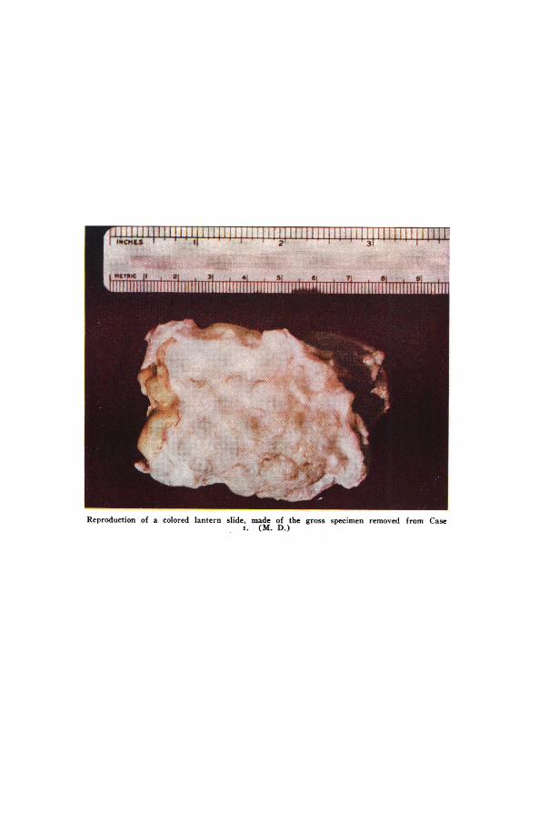
_ }SLwS94Reproduction of a colored lantern slide, made of the gross specimen removed from Case
i. (M. D.)
.m ..i3O.ifj..l,

Volume 109 EPIDERMOID TUMORSNumber 5EPDR ODTM S
to be more effective. After this incision is closed, the broad bases of the flaps lie overthe margins of the boneflap. Therefore, this closure is more effective than when all ofthe lines of sutures are superimposed over the margin of the boneflap, as is the casewith the typical osteoplastic flap.
Pathologic Examination.-Gross: The tumor (Fig. 2, a, b, c) weighed about I IOGm., and measured 7x5x4 cm. "Attached to the under surface is a thin fibrous mem-brane resembling a pseudodura" to which was attached the outer layer of the dura,which was dissected away with the tumor. The outer surface of the tumor and themargins which were in contact with the bone "show the glistening pearly surface of acholesteatoma" after the membrane was removed. The outer surface, as well as theborders, undulated with elevations and depressions corresponding to the counterelevationsand depressions in the bony defect. This picture is most marked on the borders wherethe roentgenograms showed the typical scalloped effect. The small piece of bone shownin Figure 2 was removed from one of these escalloped borders. The inner surface of thetumor was grayish in color, and was softer thanthe firm outer portion of the tumor. No hairwas present in the tumor mass.
Histologic Reexamination.-Dr. F. ChandlerFoot: "The sections show a membrane whichhas been cut across longitudinally, and is seen tobe composed of a heavy layer of connective tissuein which there are some bony spicules and occa-sional nests of giant cells surrounding acicularspaces of lipid crystals. There are also numer-ous fat phagocytes in these nests of the 'foamcell' variety. Overlying this there is a layer ofstratified epidermal epithelium in which there isa rather poorly defined basal layer, several layersof poorly differentiated cells which probably rep-resent prickle cells and a rather heavy layer ofstratum granulosum with deeply pigmented bluegranules. Strangely enough, the keratinizationof this epidermis is not very marked, althoughthere is a narrow zone of keratinized cells repre-senting the innermost layer of the cyst. Over-lying this there are some flakes of desquamated, FIG. 4.-Case I: Photograph of patient twokeratinized epithelium. In some places the months after operation.keratinization is more marked than in others. The wall of this cyst is totally devoidof any of the adnexa of the skin; there are no hairs, sweat or sebaceous glands present,therefore, the cyst is of the epidermal variety. Pathologic Diagnosis: Epidermoid(cholesteatoma)."
Postoperative Course.-Recovery was uneventful. The wound healed without in-fection, and without cerebrospinal leakage. On January 20, II days after operation, thepatient was up in a wheel chair and stated that he was feeling fit. The apparent ex-ophthalmos had become much less. He had no complaints. The dizziness disappeared,and speech was faster than before operation. The slight slowness in thinking haddisappeared. He has remained well since, so far as could be learned. Photograph madetwo months after operation.
COMMENT.-This case has been reported in detail for the reason that it socompletely parallels Doctor Cushing's case. The diagnosis was made fromhis description of the roentgenograms and the lesion. One should encounterbut little difficulty in making the diagnosis in a similar case. Had roentgeno-
661

JOSEPH E. J. KING Annals of SurgeryM a y , 1 9 3 9
grams of the skull been made in I9I8, when some were made of the foot, thecranial defect would have been found, and the tumor could have been removedfive or six years earlier.
It is surprising how such an enormous mass could be present with so fewneurologic findings. It must be due to the fact that the lesion, which obviouslyincreases very slowly in size due to its inherent nature, allows the brain toaccommodate itself to the encroachment of the tumor. This is in markedcontradistinction to the rapid displacement of the brain from an extraduralhemorrhage.
Case 2.-Epidermoid (cholesteatoma); diploic; smiiall. Destruction of outer anddepression of inner table of skull. Palpable milass. No neurologic findings. Completeextirpation. Recovery.
F^rs~~~~~~~~~~~~~~~~~~~~~~~~~~~~~~o(a) (b)
FIG. 5.-Case 2: Roentgenograms showing small cranial defect produced by the smallest epi-dermoid of series. (a) Lateral view shows definite clear-cut border posteriorly (A) where the raysstrike perpendicularly. Anterior margin not so definite. (b) Anteroposterior view. Postero-anteriorexposure would have shown a more sharply defined margin. Outer table was destroyed, inner tabledepressed.
J. R. male, age 27, was admitted to U. S. Veterans Hospital No. 8i, in February,1925. There was a small doughy mass about the size of a hickory nut in the left frontalregion, one inch above the supra-orbital ridge. No signs or symptoms other than thepresence of a small mass which resembled a firm, fixed sebaceous cyst.
Roentgenologic Examination (No. 6882) .-Figure 5, a and b, revealed the presence ofof a small cranial defect in the left frontal region about 2 CM. in diameter, fairly cir-cular in shape except for a notch on the antero-inferior border of the defect. Themargin of the defect presented a narrow, dense line posteriorly, similar to that seenin the typical defect made by an intradiploic epidermoid, with scalloping at only oneplace. The outer table was completely destroyed, while the inner table was intactbut slightly depressed. In retrospect, one should have made the diagnosis before opera-tion. The lesion was so small and its position so near that of the common site of aparacanthus subpericranial dermoid that the diagnosis of a dermoid was made.
Operation.-Under local anesthesia, the lesion was completely shelled out of itsposition in the bony defect. The outer table of the skull was absent, as in Bucy's Casei, while the inner table was thinned-out and depressed. It was not perforated. Themass measured aboUt 2 CM. in diameter, was encapsulated and was fairly circular in
662

Volume 109 EPIDERMOID TUMORSNumber 5
shape. It was doughy to touch and was set in the cranial defect like a bullet, flattenedon the outer side, in half of a bullet mold.
Pathologic Examination.-Gross: Dr. D. S. J. Jessup. "There is a thin-walled sactwo centimeters in diameter, with its contents made up of white flaky masses which arevery soft and crumble. Section taken through the cyst wall is of paper-like thinness.Microscopic.-Section through this limiting membrane shows it to be lined by cuboidalor flattened epithelium attached to which are desquamating cells. The detached material inthe tumor appears to be made up of the same kind of cells rolled up on themselves. Thewhite flaky soft tissue debris shows many flat particles of cholesterol mixed with color-less, round bodies which appear to be desquamated epithelial cells. No hairs or glandswere noted. Pathologic Diagnosis: Cholesteatoma (epidermoid) of frontal bone."
The clinical record could not be obtained from the hospital, so that hospital numberand other data cannot be given. Only a few notes, the roentgenograms, and pathologicreport were in the possession of the author.'
COMMENT.-SO far as is known, this is the smallest epidermoid to bereported. The diagnosis should have been made from the slight doughy feel-ing and from the examination of the roentgenograms.
Case 3.-Bellevue Hospital, Acc. No. 34971-79: Epidermoid (cholesteatoma); ex-tradural; diploic; occipital; left. Complete removal of caseouis mass by morsellation andcurettage. Incomplete removal of lining memnbrane. No infectiont. Recovery.
B. M., white, male, age 26, was admitted to Neurologic and Neurosurgical Service,April 2I, I934, with complaint of constant headache for one year.
Family History.-Irrelevant.Past History.-Negative, except that the patient was told by his mother that, at age
seven, he suffered from "some disease" which made it impossible for him to move hisneck well for about six months.
Present Illness.-The patient complained of constant headache for the past year.These pains radiated to the right occiput and toward the left eye, and became gen-eralized. Stooping intensified the pain. For about a year he had noticed a -'swellingabout the size of a "small walnut" in the left occipital region. No history of headtrauma.
Physical Examination revealed a well developed young man, not acutely ill. Gen-eral examination negative. In the left occipital region there was an irregular, firm,walnut-sized elevation, about 2/2 cm. in diameter, regular in outline and apparently fixedto the skull. It was bony hard, except in its central portion, which was slightly doughy.The scalp was freely movable over it, and it was not tender. T. P. R. normal. Bloodcount and blood pressure normal. N.P.N. 37; blood sugar 6o, blood cholesterol IIO.Lumbar puncture not advised.
Neurologic Examination.-Left pupil slightly larger than right. Both reactedwell to light and accommodation. Fundi showed definite blurring of both disk mar-gins. Visual fields grossly normal. External ocular muscle normal. Other cranialnerves normal. Motor coordination, reflexes and sensory status normal.
Roentgenologic Examination (Fig. 6, a and b).-There was a cranial defect in theoccipital bone on the left side which extended from just behind the posterior margin ofthe mastoid process, without involving it, to the midline, and measured 75/2x8 cm. Nearthe upper limit of the defect there was a narrow bridge of bone of the outer tableseparating the upper from the lower portion, so that the upper portion was an elongatedoval defect separated from the major defect. The bony margins were scalloped, denseand firm, similar to those seen in films of typical cases of extradural diploic epidermoids.This diagnosis was made.
Operation.-May i, I934: Avertin and local anesthesia. A straight, vertical in-cision was made about halfway between the external occipital protuberance and the
663

Annalsof SurgeryJOSEPH E. J. KING May. 1939mastoid process, directly over the central portion of the palpable tumor,- and extendingabove and below it. Counter incisions were made about the midportion to afford betterexposure. -The scalp flaps were dissected up from the fascia and held with self-retainingretractors. Good exposure, practically no bleeding. A horseshoe-shaped incision, withthe base directed downward, was made in the fascia and musculature overlying thelesion. When the pericranium was stripped away, perforations in the bone were seen.A bone flap could not be turned. The defect in the skull was enlarged until it measuredabout two by three inches. On palpation, the tumor offered resistance like that of fairlyfirm rubber or stiff dough. The mass was definitely encapsulated by a membrane ofgrayish color. When a portion of this thin membrane was stripped away, the sur-face of the underlying mass presented the typical pearly-whiteness of the outer layer ofan epidermoid.
(a) (b)FIG. 6(a).-Case 3: Anteroposterior and (b) lateral roentgenograms. Areas of complete absence of
outer table (A) with bridge of bone between. Island of bone (B).
Mechanically, it was impossible to remove the tumor in one mass, as was ac-complished in Cases I and 2; therefore, it was morsellated and scooped out with abrain spoon. The innermost portion of the mass was cheesy in consistency and wasof a dirty grayish-green color. The lesion extended from the lateral sinus on the outerside to the midline and from about three-quarters of an inch superior to the lateralsinus to the posterior margin of the foramen magnum (Fig. 7, a). The exact thicknessof the tumor could not be determined at operation, but on the lateral roentgenologic viewit appeared to be about two inches thick. During removal of the caseous material, assuccessive scoopfuls of the tumor mass were removed, the floor of the excavation rosetoward the skull. After complete removal of the contents of the lining membrane, thedura covering the left cerebellar lobe bulged outward so that it completely filled thespace previously occupied by the tumor. This was quite opposite to the observationmade in Case I in which the dura remained depressed. It was believed that the tendencyto extrude the caseous mass through the defect and the elevation of the dura into thedefect after removal of the tumor were due to release of compression on the aqueductwhich allowed filling of the subarachnoidal space in the posterior fossa. The lateralsinus was seen throughout its course from the mastoid process almost to the torcula.It was about three-sixteenths of an inch wide, markedly compressed, and covered with
664

Nolumbe 509 EPIDERMOID TUMORS
a thin, whitish membrane which lined the dura. Complete removal of the membrane wasnot attempted for fear of injury to the sints.
Bony islands from the outer table of the skull, adherent to the undersurface of thepericranium, were removed and the flaps were returned to position and sutured in layers.The undersurface of the pericranium came into apposition with the cerebellar dura whichpreviously had been depressed about two inches. The morsellated fragments of thetumor weighed iio Gm. Throughout the entire extent of the lesion, on the externalsurface, there was a layer of white, pearly, lamillated material about three-sixteenths ofan inch thick, while the inner surface of the lesion was not covered with this white,pearly material.
Pathologic Examination.-Gross: Dr. A. V. St. George. "Specimen consists of manyirregular pieces of friable, gray-yellow tissue designated as a cholesteatoma. Some arepartially covered by a white, fibrous layer. One piece of reddish, fibrous tissue, desig-nated as 'capsule,' measuring about I.5xO.4 cm., accompanies_ the tumor. Microscopic:
(a) (b) (c)FIo. 7(a).-Case 3: Roentgenogram made after operation. Approximate size of tumor outlined
by broken wbite line. Bony peninsulae over lateral sinus preserved. (b) and (c) Photographs madefour and one-half years after operation.
The above capsule reveals it to be a thin fibrous membrane lined by atrophic squamousepithelium. Several friable, irregular pieces of tissue are composed by amorphous acellu-lar material. No cholesterol crystals seen."~
It is not knowni what part of the caseous cholesteatomatous mass was examined forcholesterol crystals. On reexamination of the slide it was observed that no hair folliclesor glands were present. The lining membrane, part of which was in apposition tothe pericranium, presented the three layers similar to that seen in Case i.
Postoperative Couirse.-Uneventful recovery. On the seventh postoperative day thebilateral papilledema was receding. He showed no signs of a cerebral focal nature. Hewas up and about the ward in two weeks, when he developed a sore throat with eleva-tion of temperature to 1030 F., which, however, subsided rapidly. He was disc-harged,May 21, 1934, 21 days after the operation, with no complaints.O0 Shortly thereafter he resumed his work and has continued to work to date. Hewas examined in November, I938, and the area at the site of operation was sunkenrather than bulging (Fig. 7, b and c). His eyegrounds were found to be normal byDr. Hugh McKeown, but on account of some convergent weakness at near point hewas given glasses. Pupils were equal, reaction to light and accommodation wasnormal, fundi and visual fields were normal.
665

JOSEPH E. J. KING Annalsof SurgeryMay, 1939
COMMENT.-Typical diploic type of epidermoid which destroyed first theinner table of the skull, later the outer table in several areas, and left bonyislands attached to the pericranium. It was sufficiently large to compress theleft cerebellar lobe, lateral sinus and brain stem. His only symptom, headachereferred to the occiput and left eye, was probably due to compression of theaqueduct and left lateral sinus. Morsellation preferred to any attempt toremove in toto for the reason that it was mechanically impossible; it alsoseemed better judgment to subject the patient to a second evacuation of thearea rather than to open the dura or risk damage to the lateral sinus. Completerelief.
Case 4.-Bellevue Hospital, History No. R-i8: Epidermoid (cholesteatoma)frontal region; right. Marked erosion aidt destruiction of orbit; proptosis; enucleationof eye. Operation. Recovery.
V. O., female, Negro, age 46,' was admitted to the Eye Service for third time,January 5, I937, with chief complaint of swelling above the right orbit. Transferredto Neurologic and Neurosurgical Service, January i8, I937.
Past History.-Negative except for mild diabetes, controlled by diet.Present Illness.-In I932, the patient entered the hospital complaining of bulging
of, and pain in, the right eye. Enucleation of the eye was performed for suspected tumor.No tumor was found in the orbit to account for proptosis. Two years later she enteredthe hospital for the purpose of plastic surgery to enable her to wear an artificial eye.She wore the eye until two months before the present admission to the hospital, atwhich time it became dislodged, fell and broke. This was due to the fact that a swell-ing had appeared in the lateral half of the right brow about five months ago. It hadincreased in the last two weeks and extended downward and laterally. The swellingwas accompanied by very little pain and only slight headache. There had been noother symptoms, and no loss in -weight. Sight in the left eye was good.
Physical Examination revealed a well developed colored woman, height about fivefeet six inches, weight about 145 pounds. The right frontal sinus did not transillumi-nate, while the left did. On the right brow and over the outer half of the supra-orbitalridge there was a firm, tense, but slightly compressible tumor, 5x7 cm. in size, whichextended downward into the orbit and outward beneath the temporal muscle. It offeredresistance similar to that of a firm sebaceous cyst. It was not tender. There was erosionof the upper rim of the orbit and the supra-orbital ridge in its outer half. Neurologicexamination was negative in every respect. Blood pressure I40/80. Blood count andurine normal. Wassermann negative. Spinal fluid: Initial pressure 125 Mm. of water,normal dynamics, no cells, no globulin, total protein 40. Visual fields normal in lefteye. Right eye had been enucleated. Left fundus normal. Smell test showed normalreaction to citral, with an increased minimal identifiable odor to coffee as well as anincreased fatigue for coffee on the right side.
Roentgenologic Examination showed marked destruction of right orbit, anteriorethmoids, the lesser wing of the sphenoid, and, to some extent, the greater wing of thesphenoid, with a cyst-like area revealed in the outer wing on the right side. There wasan oval hiatus involving the outer and inner tables of the skull in the right frontal regionextending into the temporal region with destruction of the outer half of the supra-orbitalridge, and upward for a distance of about one and one-quarter inches, to a position higherthan that of the uppermost limit of the frontal sinus on the opposite side. The leftfrontal sinus was clear for the most part, but slightly cloudy toward the midline. Themargins of the defect were not as clear-cut and defined throughout as it is in the usualcase of intradiploic epidermoid. This was probably due to the position of the lesion in-volving and eroding the thinner bones as it did.
666

Volume 109Number 5 EPIDERMOID TUMORS
An attempt was made to inject the remnants of the frontal sinus with lipiodol (Fig.8, a), but only a bead-like mass, about I cm. long, was demonstrated, completely fillingthe remnant of the frontal sinus near the midline. An epidermoid was suspected, butthe preoperative diagnosis was not made.
Operation.-January 22, 1937: Avertin and local anesthesia. An incision abouttwo and one-half inches long was made parallel to and about one and one-half inches abovethe right supra-orbital ridge extending into the temporal region. The flaps consisting ofscalp, galea, muscle and pericranium were dissected up, reflected and held with self-
FIG. 8 (a) .-Case 4: Roentgenogram showing defect (A) produced by erosion of tumor andlipiodol in remnant of frontal sinus (B). (b) Roentgenogram showing postoperative cranial defectafter removal of tumor. A catheter passed through artificial opening made for drainage betweencavity occupied by tumor and the right nasal passage. Cannula passed into remnant of frontal sinus.
retaining retractors. The cranial defect, shown on the right side of the frontal boneroentgenologically, could then be made out. Through this defect protruded a semi-fluctuating, encapsulated mass, with a firm fibrous wall, the mass measuring about oneinch in horizontal diameter, three-quarters of an inch in vertical diameter, and pro-truding through the defect about one-half inch above the outer table of the skull. It didnot pulsate.
When the capsule was nicked, a bead of thick, viscid, opaque, olive-green material,as viscid and stringy as thick rubber cement, exuded very slowly, like stiff tooth-pastefrom a tube. When the opening into the cavity was enlarged, a somewhat globularmass of the olive-green, viscid, odorless material was extruded with a slow creeping,rolling movement like lava down a hillside. Its consistency was that of thick coal tar.As more of the material was evacuated, it was observed that the surface of the sub-stance rose and fell rhythmically, synchronous with the pulsation of the brain. Morebone was removed until the cranial defect was about 5x7 cm. (Fig. 8, b), the longestdiameter directed obliquely backward and upward, extending beneath the temporal
667

JOSEPH E. J. KING Annals of SurgeryM a y, 1 939
muscle. The thick material from the depths was less homogeneous, did not have thesame olive-green color, but was muddy yellowish-green. After this portion of the con-tents was removed, the material in the deeper part of the cavity was more caseous, likethe interior of an intradiploic tumor, and was yellowish-white. The deepest portion ofthe contents of the cavity, in apposition to the dura, was made up of pearly-whitematerial, similar to that found on the exterior of an intradiploic epidermoid. Thislayer was about 8 Mm. to i cm. in thickness and was plastered against the dura, so thatit had to be removed by scraping and scooping it out with a brain spoon.
The dura was markedly depressed backward and upward, was concave on the outersurface like the cupped-palm of one's hand, and pulsated. An oval area of dura about2x252 ins. was exposed, the inner table being absent. Above, and on the outer side,it was held back by a rim of the inner table of the skull about one-half inch in heightwith a definite hard, fairly sharp narrow border, similar to the ridge about the internalmargin of the bony defect found in Case i. Otherwise, the inner table was completelydestroyed, and the outer table of the frontal bone, where it was not completely de-stroyed, was considerably and irregularly thinned-out. Once the major portion of thecavity was emptied, two openings, about i cm. in- diameter, were seen in the antero-inferior wall. These two openings led into several small cavities, incompletely separatedby bony septa, as was observed in Case I of Munro." These smaller cavities werefilled with the same thick, olive-green, viscid material first observed. Nowhere had thecavity broken through into the nasal cavities or antrum, although the anterior ethmoids,the roof and floor of the orbit, the outer half of the supra-orbital ridge, the lesser wingof the sphenoid, a portion of the anterior clinoid process and a portion of the greater wingof the sphenoid had been eroded and destroyed by the slow, increasing compression com-mon to these lesions. The orbital contents could be palpated. The tumor had mechani-cally accomplished the bony removal advised in the Naffziger2' operation for progressiveexophthalmos better than one could have been effected by operation.
The floor of the anterior fossa had given way before the lesion to such an extentthat the lowermost portion of the cavity was delimited by the anterior wall of thesphenoid, later well shown by filling the cavity with lipodol. It was considerably lowerthan the floor of the sella. This portion of the cavity could be identified on the pre-operative lateral films. The lining of the cavity, where it was firmly fixed to the dura,was yellowish-white in color, and apparently avascular, and did not bleed when scrapedfairly vigorously. -The dura showed no tendency to fall forward and obliterate thecavity, which was also observed in Case i. On the contrary, it remained in an archedposition, but pulsated in its central portion.
After removal of the bony septa from the antero-inferior portion of the cavity, sothat all the cavities had been converted into one, saline solution was poured into the onelarge cavity, which required about 75 cc. to fill it. None escaped into the nasal passagesor throat, showing that no communication existed between the cavity and the nose ornasopharynx. No attempt was made to dissect the membrane from the dura for fearof opening the dura and producing meningitis. The entire cavity was filled with iodoformgauze, the end of which was brought out through the central portion of the incision,and the incision on either side was closed with interrupted silk sutures.
Cholesterol crystals were recovered postoperatively from some of the remainingflakes removed from the cavity. An area of the lining showed stratified squamousepithelium three to six layers thick. There were no hair and no glands.
Postoperative Course.-The patient made an uneventful recovery. On January 29,I937, the seventh day after operation, and after all the iodoform gauze was removed, thecavity was filled with lipiodol to the level of the skull, the central portion of the in-cision having been closed with clips to prevent its leakage, and roentgenograms mademerely to show the position and extent of the lesion (Fig. 9, a and b). The posteriorand superior scalloped borders of the defect were thus made more apparent. One couldreadily see that the lowermost limits were below the sella and adjacent to the sphenoid
668

Volume 109 EPIDERMOID TUMORSNumber 5
~~~~~~~~~~~~~A.-
0'
000)
00
00
0
...
*:4:
669

JOSEPH E. J. KING Annals of SurgeryMay, 19 39
sinus which was almost perforated. There was no leakage into either the nose or mouth.The cavity was repeatedly packed through the opening in the central portion of the
incision and could be inspected readily. The membranous lining of the dura wasyellowish-white and did not tend to granulate. The cavity was swabbed out and filledwith 20 per cent silver nitrate solution at successive dressings to destroy the remainingportion of the lining epithelial membrane. The walls later changed in color to pinkish-red, became covered with granulations, but the cavity did not fill in rapidly. Never-theless, later roentgenograms, made after the cavity was filled with lipiodol, showed thatit had decreased in size.
Closure of the opening in the scalp over the cavity was desired, but before doingso drainage into the nasal passages was thought to be advisable. A probe was pushedby Doctor Atkinson through the thin intervening tissue between the cavity and theright nasal passage, somewhere in the neighborhood of the frontonasal duct. The tipof a small catheter was tied to the eyelet of the probe and drawn through into the cavity(Fig. 8, b). This did not allow sufficient draining. Therefore, the opening throughwhich the catheter was passed was enlarged to the size of the diameter of a cigarette,and two small rubber tubes were passed through this opening into the cavity, with theproximal ends protruding through the right external naris and fixed with adhesive,which allowed of irrigation with return flow.
Scalp-plastic under local anesthesia was performed; the incision healed kindly andhas remained closed. Subsequently one tube was substituted for the two and later still,the patient was able to pass a catheter easily and irrigate the cavity without help. Shecontinued to do this for several weeks, then gave it up, inasmuch as the return flow wasalways clear.
A photograph (Fig. 9, c) taken in November, I938, shows the depressed area overthe site of the cranial defect and the loss of the right eye. The orbital contents no longerbulge so that she can now again wear an artificial eye. Recent roentgenologic studiesfollowing instillation of lipiodol have not been undertaken, but the cavity is doubtlessmuch smaller, if not obliterated. She is perfectly well and does her daily work.
COMMENT.-This case again shows the enormous erosive and destructivetendency of the lesion involving bony structures without gross damage to thesoft parts, i.e., dura, brain and orbital contents. It is also noted for the utterlack of neurologic findings. Study of good stereoscopic films, made beforeany operation was performed, should have warranted the diagnosis of anintracranial lesion with earlier removal of the lesion and preservation of theeye. On account of the loss of the right eye, one could make no deductionsregarding the second, third, fourth and sixth cranial nerves, but consideringthe extent of the bony excavation, one was surprised that the olfactory nerveand the first division of the fifth nerve remained intact.
Rather than to have risked the possibility of intradural infection, no attemptwas made to remove the lining membrane completely. Knowing that thecavity was to remain open for quite a while, it was believed that the membranecould be destroyed by a solution of silver nitrate. Should debris collect, itcould be washed out through the opening into the nose.
The semisolid, viscid consistency of the major portion of the lesion wasprobably due to partial "dilution" by secretions of the mucous membrane ofthe eroded ethmoids and frontal sinus.
Case 5.-New York Hospital, No. 2I5378: Epidermoid (cholesteatoma); intra-dural, middle fossa; left. Marked erosion of petrous, middle fossa and homolateral
670

Volume 109 EPIDERMOID TUMORSNumber 5
clinoids. Double itsion for 12 years. Involvement of left cranial nterves III, IV, V,VI, VII, and VIII. Operation. Recovery.
J. S., white, female, single, age 40, was admitted to the hospital, November i, I938,with chief complaint of double vision for 12 years.
Family History.-Father had ulcers of stomach. Mother had tuberculosis. Bothin fair health. One sister well. Two brothers; one had tuberculosis. Family nervousand high strung.
Past History.-At age of six fell and struck head. Not able to "talk straight" fora while. Pneumonia as child. Tonsils removed at i6. Appendicectomy at 20. Other-wise general health excellent until beginning of present illness.
Present Illness.-Began in I926. At time an impacted left wisdom tooth and animpacted incisor removed; noticed numbness left side face and forehead. Numbnessnever improved. Intermittent double vision began about this time, worse during warmweather and under nervous tension and worry.
In I927, right impacted wisdom tooth removed, operation on right antrum, and boneremoved from nose. During I929, double vision became continuous; vision of left eyeblocked by dark covering. A number of eminent ophthalmologists could not find causeor remedy. Glasses prescribed in I932. Operation for double vision considered but notundertaken. May 31, I934, bilateral myringotomy by Dr. Ross Faulkner; profuse dis-charge from both ears, culture showing Streptococcus hemolyticus. Pain and dischargecontinued. June I2, I934, bilateral mastoidectomy performed by Doctor Faulkner.Mastoids filled with pus and granulation. Discharged June 23, 1934. Office dressings.Both wounds healed by July Ii, I934. After operation diplopia disappeared for sixmonths.
In the period between i936 and I938, had several corneal ulcers in the left eye forwhich she was treated by two capable ophthalmologists. One had observed her sinceI934. In I935, he noticed a partial left facial paralysis which was attributed to themastoidectomy. He observed several recurrences of corneal ulceration, the last beingnoted May 5, I938. Although cornea was anesthetic, complained of pain referable toleft eye. Muscular condition unchanged. Interpreted findings as congenital paralysisof superior oblique and external rectus muscles and partial facial paralysis followingmastoidectomy with resultant corneal ulceration.
Another ophthalmologist first examined her September 27, I938. Two infectedcorneal abrasions of left eye. Right pupil reacted normally to light and accommodation,left pupil fixed and somewhat dilated. Corneal sensation normal right eye, left corneaanesthetic. Marked limitation motion of left eye in all directions; third, fourth and sixthnerves involved. Vision 20/I5 right eye, 20/50 left, with correction. Right visual fieldnormal, marked contraction left central field, with enlarged blind spot. Under mydriasisno evidence of increased intracranial pressure, media clear, disk, blood vessels andmacular region left eye normal. From nerve involvement he suspected intracranialpathology.
Physical Examination.-The only positive findings were those referable to left eyeand left cranial nerves. Appendicectomy and bilateral mastoidectomy scars. Patientwell nourished and not ill.
Roentgenologic Examination.-October i8, I938: Dr. F. M. Law. Stereoscopic filmsrevealed marked erosion of left petrous amounting almost to amputation through its mid-portion (Figs. I0, b, and ii, a). The eminential arcuata remained, but the trigeminal im-pression was obliterated. Marked erosion, almost obliteration, of left anterior and pos-terior clinoids. Size of sella unchanged. Floor of the middle fossa was depressed. Nochange in appearance of left optic foramen. The stereovertexmental films (Fig. ii, b)showed erosion of the left side of body of sphenoid and marked erosion of the floor of themiddle fossa. No outline of left foramen ovale. Apparent fracture at posterior borderof left sphenoid. Squama of the left temporal bone thinned-out, especially in lower limits.Reexamination of previous roentgenograms made elsewhere, in ig9 (Fig. I0, a), nine
671

JOSEPH E. J. KING Annals of SurgeryM a y , 1-9 39
years previously, also revealed marked erosion of the left petrous, similar to that shownin the more recent films. In the latter, however, the major extension of the erosion wasshown to have involved principally the lower portion of the petrous. RoentgenologicImpression: Meningioma of the middle fossa.
(a) (b)FIG. io(a).-Case 5: Roentgenogram, made in 1929, showing destruction of petrous tip (A)
by erosion of epidermoid of middle fossa. (b) Destruction of petrous (A) only slightly increasedafter nine years; roentgenogram made in 1938.
(a) (b)FIG. I I (a).-Case 5: Oblique roentgenogram showing erosion of petrous (A) in 1938. (b)
Vertexmental roentgenogram showing erosion and partial destruction of sphenoid bone (A). Leftforamen ovale not seen.
Neurologic Excnnination.-Dr. Foster Kennedy: "Double vision since I926, off andon. Persistent since I934. Occasional headaches. Daily pain in left eye and left face.Pain comes on gradually and recedes gradually. Headache in back of head 'at the baseof skull.' Worse at the end of day. Never wakes her out of sleep. No difficulty in usepf arms or legs. No pains down arms. Hearing in left ear impaired since I927. Lost
672
Volume 109 EPIDERMOID'TUMORSNumber 5
double vision for a few months after operation (mastoidectomy) in i934. Has gainedseven pounds this summer. Now weighs II5 pounds.
"Fundi show congestion; but no frank papilledema. Left 3rd, 5th, 6th and 7th nerveinvolvement with slow progression over I2 years. Left motor 5th nerve with atrophyof temporal and masseter muscles. Marked diminution of left corneal response. Weak-ness of left 7th nerve peripheral. Impaired hearing, almost deafness, left ear. Leftpetrous ridge like a 'bridge blown up in middle.' Otherwise neurologic examination isnegative. Operation advised."
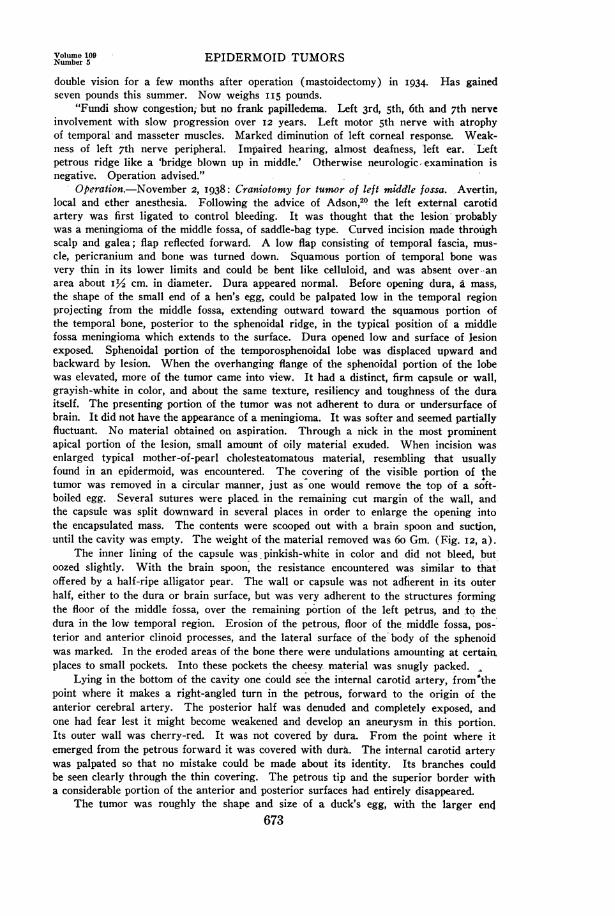
Operation.-November 2, I938: Craniotomy for tumor of left middle fossa. Avertin,local and ether anesthesia. Following the advice of Adson,20 the left external carotidartery was first ligated to control bleeding. It was thought that the lesion~probablywas a meningioma of the middle fossa, of saddle-bag type. Curved incision made throughscalp and galea; flap reflected forward. A low flap consisting of temporal fascia, mus-cle, pericranium and bone was turned down. Squamous portion of temporal bone wasvery thin in its lower limits and could be bent like celluloid, and was absent over -anarea about iY2 cm. in diameter. Dura appeared normal. Before opening dura, a mass,the shape of the small end of a hen's egg, could be palpated low in the temporal regionprojecting from the middle fossa, extending outward toward the squamous portion ofthe temporal bone, posterior to the sphenoidal ridge, in the typical position of a middlefossa meningioma which extends to the surface. Dura opened low and surface of lesionexposed. Sphenoidal portion of the temporosphenoidal lobe was displaced upward andbackward by lesion. When the overhanging flange of the sphenoidal portion of the lobewas elevated, more of the tumor came into view. It had a distinct, firm capsule or wall,grayish-white in color, and about the same texture, resiliency and toughness of the duraitself. The presenting portion of the tumor was not adherent to dura or undersurface ofbrain. It did not have the appearance of a meningioma. It was softer and seemed partiallyfluctuant. No material obtained on aspiration. Through a nick in the most prominentapical portion of the lesion, small amount of oily material exuded. When incision wasenlarged typical mother-of-pearl cholesteatomatous material, resembling that usuallyfound in an epidermoid, was encountered. The covering of the visible portion of thetumor was removed in a circular manner, just as one would remove the top of a soft-boiled egg. Several sutures were placed. in the remaining cut margin of the wall, andthe capsule was split downward in several places in order to enlarge the opening intothe encapsulated mass. The contents were scooped out with a brain spoon and suction,until the cavity was empty. The weight of the material removed was 6o Gm. (Fig. I2, a).
The inner lining of the capsule was, pinkish-white in color and did not bleed, butoozed slightly. With the brain spoon, the resistance encountered was similar to thatoffered by a half-ripe alligator pear. The wall or- capsule was not adherent in -its outerhalf, either to the dura or brain surface, but was very adherent to the structures formingthe floor of the middle fossa, over the remaining portion of the left petrus, and -to thedura in the low temporal region. Erosion of the petrous, floor of the middle fossa, pos-terior and anterior clinoid processes, and the lateral surface of the' body of the sphenoidwas marked. In the eroded areas of the bone there were undulations amounting at certain.places to small pockets. Into these pockets the cheesy material was snugly packed.
Lying in the bottom of the cavity one could see the internal carotid -artery, from"thepoint where it makes a right-angled turn in the petrous, forward to the origin of theanterior cerebral artery. The posterior half was denuded and completely exposed, andone had fear lest it might become weakened and develop an aneurysm in this portion.Its outer wall was cherry-red. It was not covered by dura. From the point where itemerged from the petrous forward it was covered with dura. The internal carotid arterywas palpated so that no mistake could be made about its identity. Its branches couldbe seen clearly through the thin covering. The petrous tip and the superior border witha considerable portion of the anterior and posterior surfaces had entirely disappeared.
The tumor was roughly the shape and size of a duck's egg, with the larger end673

JOSEPH E. J. KING A 19abf
co
_o 0
_ ._
Cd
c q6
,-o 0
CO
cWo
CcLWf*I X
o3
* @T
674
x,. W
.......

Volume 109 EPIDERMOID TUMORSNumber 5
flattened against the structures described and packed in the middle fossa (Fig. I2, b).It was about three inches deep and more than two inches in diameter in its largest por-tion. A considerable part of the capsule or wall was removed-, but no attempt was madeto remove it from the depths, realizing well that there might- be partial, slow refillingof the cavity. Should this occur, the contents could be removed through a short, straightincision, as in the operation for major trigeminal neuralgia.
The fifth, sixth, fourth and third nerves were not identified, although the positionof the gasserian ganglion was seen. It was believed that once the enormous, thoughslow, pressttre was released from these structures -by removal of the contents of theepidermoid, function through these nerves would be restored, providing they were notimpregnated with cholesterol crystals.
The cavity was allowed to collapse as much as it would, the distorted sphenoidallobe was allowed to drop back over it, and the dura was closed except in the lowermost
T~~~~~~
(a) (b)FIG. 13.-Case 5: Photomicrographs of wall of epidermoid showing the three
layers (I, 2 and 3). (a) Low power, showing partial destruction of epithelial layer frompressure (A). (b) High power, showing io to 12 layers of epithelial cells.
portion. A small gauze wick was placed through the dural incision in order to furthercollapse of the cavity, to allow escape of cholesterol containing fluid, and to controlslight oozing. This strip of gauze was brought out through a puncture opening in themiddle of the pedicle of the muscle-fascia flap and through the transverse portion of theincision above the zygoma. The flap was returned to its position, wired in three placeswith rustless steel wire, and the scalp incision was closed in two layers. A transfusionwas given on the table by Doctor Eggston.
Pathologic Examination. Gross: Dr. N. Chandler Foot. "Specimen consists in partof a roughly triangular piece of silvery-white membrane measuring i.5 cm. on the side.This is apparently composed of connective tissue resembling dura, and at one pointthere is a small mass of whitish cheesy material similar to that to be described later,adhering to it. The greater part of this specimen. consists of 47 'Gm. of whitish-yellowsoft material which has the consistency of pot cheese. (Some of the tumor was lost.)The largest fragment measures approximately 3 cm. in diameter, but most of it is brokeninto flaky white curds which contain nothing resembling true tissue. Microscopic examina-tion shows that the bulk of material is nothing more than masses of keratinized, exfoliated
675

JOSEPH E. J. KING Annals of SurgeryM a , 19 39
horny layers from the epidermis. Sections made from the small sheet of tissue (Fig.I3, a and b) show it to consist of a layer of stratified squamous epithelium, which isdivisible into a basal layer, prickle cell layer, stratum granulosum and stratum corneum,which shows marked overproduction of keratinized material. This epidermoid tissueextends along a part of the membrane only and tapers off to be replaced by granulation
FIG. 14.-Case 5: Roentgenogram made after operation showing removal ofthin squama, fixation of bone flap with rustless steel wire, and erosion of left anteriorand posterior clinoids.
FIG. I5.-Photograph 12 days after op-eration, showing position of craniotomy scarin the hair line and the transverse cervicalscar of. incision through which ligation ofexternal carotid artery was accomplished.
tissue in which there are large numbers offoreign body giant cells, many containing acicu-lar crystal spaces. There are very large num-bers of fat phagocytes containing innumerablesmall, clear vacuoles which give them the typical'foam cell' appearance. Underlying this layerof epidermal tissue there are no accessory organssuch as sebaceous glands, sweat glands or hairfollicles, proving this to be epidermoid ratherthan dermoid in nature. Pathologic Diagnosis:Epidermoid (cholesteatoma) of brain."
Postoperative Course.-The patient made anuneventful recovery. There was at first slightdifficulty in word finding (anomia) whichcleared up before her discharge. The gauzewick was partly removed on the first, and com-pletely removed on the second day. This wasaccompanied by a considerable amount of bloody,watery fluid escape with the pulse beat. Both in-cisions healed per priman (Figs. I4 and I5). Onthe second post-operative day she was mentally676

Volume 109 EPIDERMOID TUMORSNumber 6 PDR ODTM R
clear. Out of bed on tenth day. She was discharged, November i8, I938, I5 days afterthe operation.
Neurologic Examination.-Pupils normal except left, slightly irregular; right fundusnormal. Left disk showed an area of pallor without engorgement of veins or swellingof disk. Visual fields grossly normal. There was complete paralysis of left sixth nervewith difficulty in upward lateral gaze to the left. Corneal reflex absent on left. Incom-plete left fifth nerve paralysis. Definite partial paralysis of seventh nerve. Almost totaldeafness on left. Other cranial nerves normal. Sensation nor-mal over rest of body. Noastereognosis or adiodokokenesis. Knee and ankle jerks active and equal. Abdominalreflexes present and equal. Plantar responses bilaterally flexor. No asynergy. No diffi-culty in speech. Motor functions normal. Walks well.
COMMENT.-The slow progress of these lesions is especially exemplifiedin this case. For three years there was no neurologic sign or symptom otherthan intermittent diplopia, the left eye being involved. For about nine yearsdiplopia was constant except for six months' interval following mastoidectomyin I934. Slow involvement of other cranial nerves followed, caused by alesion evidently involving the left middle fossa. Roentgenograms made nineyears before operation showed marked erosion of the petrous tip and otherbony structures forming the walls of the middle fossa. Doubtless, hadroentgenologic examination been made several years prior to I929, theywould also have shown erosion. Over I2 years intervened before the diag-nosis was established. Diagnosis probably should have been made. Givena case with similar history, roentgenographic and neurologic findings, oneshould make the diagnosis, enabling one to remove the major portion of thelesion through a smaller opening in the skull low in the temporal regio'n with-out making an osteoplastic flap. Involvement of the nerves should probablyclear up either partially or wholly unless the nerve trunks have become im-pregnated with cholesterol crystals.
Case 6.-Hospital for the Ruptured and Crippled. Hosp. No. 825-307: Epidermnoid(cholesteatoma); ihtraspinal, fouirth anud fifth luminbar. Wideninitg of canal. Corrobora-tion with lipoidol. Hemilaminectomy. Recovery.
D. C., white, male, age I7, single, first admitted to the hospital, July 8, I932, com-plaining of pain in small of back and both thighs, stating that onset of pain was suddenabout two years previous to admission. At that time the pain was knife-like in char-acter and lasted about two days. It continued for several weeks as a dull ache. It-,oc-curred whether lying, walking or sitting.
Physical Examination disclosed a somewhat awkward gait, without limp. Kyphosisextended from the cervical VII. to lumbar IV; exaggerated deep reflexes, and no sensorychanges except hypersensitiveness to pain, especially in the sole of right foot. Gavehistory of "meningitis" in I926, with apparently good recovery and freedom from symp-toms until onset of pain in I930. Not stated whether lumbar punctures were done dur-ing the course of the meningitis.
Lumbar puncture performed, August I7, I932-only 5 cc. fluid obtained, with greatdifficulty. Pressure on jugular caused slight increase in the flow of fluid. Diagnosis ofchronic adhesive leptomeningitis or adhesive arachnoiditis made. Examination underether revealed definite shortening of the right hamstring muscles. On August 29, I932,the hamstrings were lengthened. Previous to this, with the thigh flexed on the abdomen,the knee could not be extended more than I50 degrees. On September 6, I932, a plasterspica was applied under avertin and ethylene anesthesia, with the right lower extremityat right angles with the trunk.
677

JOSEPH E. J. KING Annalsof SurgervMay, 1939
Neurologic Examination.-October 20, I932: After the spica was removed, there wasright foot-drop, complete loss of motion in foot, some atrophy of calf muscles, and trophicareas on right heel and metatarsal areas and sole of the right foot. Sensory impairmentwas present in the distribution of the internal and external popliteal nerves, most pro-nounced in the external. Diagnosis of pressure neuritis (pressure from spica) was made.Improvement of motion in foot followed removal of spica but no appreciable improve-ment in sensation. Ulcerated areas on sole of foot finally healed, but the ulcer on heelremained. Discharged, November 26, I932, to return to 0. P. D.
Admitted again, February 2, 1934, for ulceration of right heel. Atrophy of rightthigh was noted. Discharged, May 8, I934.
Third admission, September 21, I934. Lumbar puncture performed, October I, 1934,at fourth lumbar space. No fluid obtained. At third lumbar interspace, pressure of fluidwas I0 Mm. of mercury, and on pressure of jugular vein elevation to 23 Mm. of mer-cury. After this admission, he developed retention of urine which was quite disturbing,requiring catheterization.
Neurologic Exam.ination, October 8, I934, showed atrophy of the entire right lowerextremity with swelling of the distal parts, duskiness of the skin; areas of old pressureniecrosis on heel and beneath metatarsal healed. Practically complete paralysis of musclesof right leg and foot. Could neither extend nor flex foot. There was anesthesia indistribution of external popliteal, hyperesthesia in area supplied by internal poplitealand posterior tibial, right knee jerk exaggerated, left normal, no Babinski, marked ex-aggeration of right gluteal reflex, pain on deep pressure over lower lumbar region andurinary retention. Lumbar laminectomy advised.
The author saw the patient in consultation later, and corroborated the above neu-rologic finding that there was complete motor paralysis of the muscles of the right legand foot. The patient could weakly extend and flex the leg. There was definitehypesthesia and anesthesia in the area supplied by the external popliteal nerve. Thearea of hypesthesia extended just above the midpoint of the leg on its outer surface, andanesthesia was present in the lower portion of the leg and dorsum of the foot. Theplantar surface of the right foot was more hyperesthetic than the left. There was nosaddle hypesthesia or anesthesia. Right knee jerk greater than left. Ankle jerk absenton right. No Babinski. Gluteal reflex on right markedly exaggerated. Whole lowerextremity on right showed atrophy. Pressure over lumbar IV and V vertebrae producedpain. Inspection of ordinary roentgenogram showed increase in width of the interspinalspace, especially at L IV and V. It was believed that a lesion involving the caudaequina was present. Lumbar puncture at fourth interspace was advised. Partial blockmight thus be determined or a complete lack of fluid would demonstrate presence of anoccluding lesion. Lipiodol injection then advisable. Lumbar puncture done followingday and 2 CC. lipiodol injected. Roentgenograms showed the lower point of the neckof the lipiodol medial to the body of the fourth lumbar vertebra, and there was a smallstreak extending slightly lower on left side. Twenty-four hours later it was at samelevel. It capped the lesion with its upper limit at the level of the upper border of thebody of the fourth lumbar vertebra. Right hemilaminectomy advised.
Operation.-December 3, I934: Hemilaminectomy (as advised by Dr. Alfred S.Taylor), with removal of the right laminae of L III, IV and V.
The laminae were thinned-out. When the dura was opened, the upper rounded butslightly pointed pole of an encapsulated tumor was seen. Its color was grayish-white andin appearance it was quite similar to the lining membrane of the lesion observed inCase 5. The presenting pole was the shape of the small end of a robin's egg, the largerend being toward the sacral portion of the canal. The lesion was partly covered withthe component parts of the cauda equina. The nerve roots making up the cauda equinawere highly injected and pinkish-red in color. This was thought to be due to the lipiodolwhich had been in the spinal canal since October 9, 1934. The lipiodol had changed incolor and consistency from a clear, homogeneous yellow oil to a yellowish-white, opaque,
678

Volume 109 EPIDERMOID TUMORSNumber 5
oily substance. A number of globules of lipiodol had become encysted, some along theexit of the roots of the cauda equina and some along the roots themselves. The majorportion was free in the subarachnoid space and could be removed. The tumor had theconsistency of a typical epidermoid with a wall or capsule about i Mm. thick. No fluidwas obtained from the lesion on aspiration. The upper pole was removed and a sectionof the limiting membrane preserved for examination. Grayish-white, cheesy, crumblymaterial was removed. It had the typical appearance of epidermoid or cholesteatomatousdebris. The contents of the sac were scooped out with a dull curet. The lesion was oneand one-half inches in length and about three-quarters of an inch in its widest portion.The interior of the sac was pinkish-white. Some of the material adjacent to the innersurface of the limiting membrane had the pearly-white luster observed in the other case.
After the contents of the sac had been removed, the cavity collapsed partially. Al-though the upper pole of the lesion was free and nonadherent, the greater portion of theremainder of the lining membrane was intimately adherent to the component parts ofthe cauda equina and could not possible have been removed without grave damageto the nerve roots. It was believed better judgment would be exercised in leaving mostof the capsule rather than to attempt to dissect it away from the nerve roots, especiallyon account of the encapsulated, encysted bodies of lipiodol already found involving thenerve roots above. Therefore, the lamina of the first sacral overlying the lower portionof the lesion was not removed. Cerebrospinal fluid flowed into the cavity which remainedafter partial removal of the wall. The dura was tightly closed.
Microscopic Examination of a section of the lining membrane revealed that it con-sisted of three layers very similar to the lining membrane in Case I, 2, 3 and 5. Thecontents of the tumor showed many cholesterol crystals. No hair follicle or glands wereseen. Pathologic Diagnosis: Cholesteatoma (epidermoid).
Postoperative Course.-Uneventful recovery. The bladder paralysis soon cleared up,general condition improved, and he began to gain weight. Right foot-drop with paralysisof the entire foot persisted. January II, I935: Calcaneocuboid arthrodesis and posteriorbone block performed by Dr. Marvin Stevens, to stabilize the foot. He was discharged,February 15, I934, and received outpatient treatment in the Orthopedic Clinic. Thetrophic ulcers of the heel and metatarsal region recurred after patient was up and walk-ing, and, on February 8, 1937, he was admitted for the fourth time for further treatmentof the ulcerated areas. Ulcers healed, and he was discharged, March 14, 1934. OnJanuary I9, 1938, he was admitted to the hospital for the fifth time for consideration ofsympathectomy, in an endeavor to heal the ulcerated areas in the right foot. This pro-cedure was abandoned after a study of the skin temperatures. Roentgenologic studiesof the spine after lipiodol injection again revealed definite block in the region of thethird lumbar vertebra. Another exploration of the spinal canal was advised.
Operation.-February 2, I938: Hemnilaminectomy, right.Exposure was made as at the previous operation with widening and extension of the
exposure below. Some bone had formed at the site from which the fifth lamina had beenremoved; otherwise, the dura was covered with scar tissue. The dura was tense and,when excised, a considerable amount of clear fluid escaped with several small choles-teatomatous masses. It was not known whether this debris had accumulated since thelast operation, or was material which had not been removed at that time. At any rate,the amount was small. The original tumor cavity had become sealed over at the upperpole so that it had no communication with the subarachnoid space. The lipiodol recentlyinjected above did not escape through the incision. In order to evacuate it, a probe waspassed upward through the adhesions and this opening was enlarged by stretching.Lipiodol escaped together with a considerable amount of cerebrospinal fluid. The openingwas made larger with the hope that it would remain patent, and the subarachnoid spacewould be continuous with that of the epidermoid sac. The roots of the nerves makingup the cauda equina, although compressed, were not so reddened and irritated as theyhad been when observed at the first laminectomy. Incidentally, it was observed that the
679

JOSEPH E. J. KING Annalsof SurgeryMa y , 19 3 9
lumbar region was quite strong following the original hemilaminectomy, due to retentionof the spinous processes, intraspinous ligament, and laminae of the opposite side. Thearea from which the laminae had been removed was filled with .strong fibrous tissue. Theexposure was quite sufficient. The dura and incision were closed.
Since paralysis of the external popliteal nerve following the previous tenotomy andstretching of the hamstring muscles persisted, and also in view of the fact that there waspractically complete loss of sensation in the foot, it was considered advisable to explorethe external popliteal nerve at the level of the tenotomy incision opposite the knee jointto determine if the nerve had been injured. There was no division either of the internalor external popliteal nerves found, nor was there any marked amount of scar tissue alongthe sciatic nerve at its bifurcation or about the external or internal popliteal nerves.All three nerves, however, appeared to be quite small and atrophic, the atrophy beingmost marked in the external popliteal.
Postoperative Course.-Uneventful recovery; patient sitting up in bed on the four-teenth day with the wound healed. Two days later, up in a wheel chair. The trophiculcers were completely healed. On March I, I938, the area in the metatarsal regionwas excised by Doctor Huber. The great toe was amputated and the skin of the greattoe turned under to cover the defect. The flap healed well. Patient was up and aboutthe ward in 12 days, bearing weight. On April 2, I938, the skin flap, which at first wasin excellent condition, had partly broken down. Weight-bearing withheld and the areahealed. Anesthesia of dorsum of foot persisted. Patient allowed up and walked well.General condition good.
COMMENT.-This is another instance in which thle diagnosis of the intra-spinal epidermoid was delayed for several years after the first operation. Dueto delay in recognition and removal of the tumor, the patient was admitted tothe hospital five times, was given many anesthesias, and underwent six opera-tions. He was in the hospital 429 days, from July 8, 1932, to May 20, I938.He also has a crippled right lower extremity, which doubtless is permanent.It is possible that complete removal of the tumor with its lining membranemight have been accomplished on his first admission.
It is not known whether the paralysis of the external popliteal and partialparalysis of the internal popliteal nerve were due to overstretching underanesthesia, or compression of nerve by the spica or whether this was due tothe intraspinal tumor. It is possible that the roots of the nerves forming thecauda equina may have become infiltrated with cholesterol crystals as was thefifth nerve in the case described by Mahoney.14
On account of the encysted condition of the lipiodol founid at the firstlaminectomy, it is believed laminectomy should be performed followinginjection of lipiodol, as soon as the level of the lesion can be determined.Taylor's hemilaminectomy suffices, and is advisable for removal of tumorsin the region of the cauda equina.
Case 7.-Bellevue Hospital, No. K.-774(26): Epidermoid (cholesteatoma); pos-terior fossa; anterior to pons and medulla, extending through foram-en magnum. Opera-tion: partial removal of tumor. Death.
Y. E., white, female, housewife, age 28, admitted to Neurologic and NeurosurgicalService, September 27, I929.
Family and Past History.-Essentially negative.Present Illness.-Suffered with generalized headaches for six years. Headaches be-
680

Numbe 19 EPIDERMOID TUMORS
came occipital in location two years before admission. Became more severe six months*before admission, associated with double vision and pain in the eyes.
Neutrologic Examination.-Coarse nystagmus on left lateral gaze and fine nystagmuson right lateral gaze. No upward nystagmus. Fundi normal. Visual fields normal.Right lower facial weakness. Diminished sensation over left face with diminished leftcorneal reflex. Palate moved more toward the right side. Hoarseness.of voice probablycaused by left vocal cord paralysis. Atrophy of left sternomastoid and trapezius muscles.Atrophy left side tongue. Pressure tenderness over left suboccipital region. Relativeweakness right arm and leg. Succession movements slightly awkward with right hand.Deep reflexes slightly increased on right. Abdominal reflexes diminished on right.Right plantar equivocal, left plantar flexor.
T.P.R. normal. Serology normal. Roentgenologic examination of skull negative.Spinal tap revealed initial pressure of 240 Mm. of water, with almost complete block.Preoperative Diagnosis: Extramedullary neoplasm, lying to the left of and ventral tothe medulla.
Operation.-September 27, i929: Dr. Alfred S. Taylor. Comstbined suboccipital ex-ploration and high cervical laminectomy.
A midline incision from the external occipital protuberance to the fourth cervicalspinous process was made. Bone from suboccipital region, laminae and spinous processesof C I and II removed. Dura overlying cerebellar lobe, medulla and cord very tense;did not pulsate. No palpable tumor through dura before opening. Posterior horn tappedto reduce intraventricular pressure. When dura was opened, definite pressure collarabout cerebellar lobes and tonsils, evidently caused by jamming into foramen at time oflumbar puncture. Cerebellar lobes otherwise normal. Medulla appeared broadened andon palpation thought to be hard. Hardness found to be due to neoplasm which displacedthe medulla backward and to the right. Tumor glistening, encapsulated, firm, nodularand grayish-white. Tumor lay anterior and to left of medulla, with stretching and atrophyof cranial nerves where they emerged from left side of medulla. Extent of tumor up-ward beneath medulla and pons not determined. Length, therefore, not known. Onlypartly removed. Considerable hemorrhage throughout. Closure. Left table in verypoor condition and died shortly arriving on ward. No autopsy.
Pathologic Examination.-"Cholesteatoma." No further report made, but microscopicexamination showed a stratified squamous epithelial lining with epithelial debris; no hairor glands.
COMMENT.-Slow development of signs and symptoms shown again inthis case. No bony erosion noted. Tumor apparently occupied a positionfrom which it could not be removed. Deatlh probably due, or at least hastened,by preoperative lumbar puncture, with jamming of the conus.
Case 8.-Bellevue Hospital, Hosp. No. M.-242: Epidermoid (cholesteatoma);right frontal lobe. Operation: Osteoplastic flap with tapping of cholesteatomatous cyst.Reelevation of flap and removal of tumor. Recovery.
E. B., white, male, age 33, was admitted to the Psychopathic Division and transferredto the Neurologic and Neurosurgical Service, July i8, I931; the first of three admis-sions, with complaint of nocturnal convulsions.
Family History.-No convulsions in family. One brother mentally deficient. Wifewithout pregnancy.
Past History.-Always healthy, except for scarlet fever. No operations or accidents,except dislocation of shoulder during convulsions.
Present Illness.-Ten years before admission, onset of convulsive seizures; all withexception of two occurred while asleep. At first occurred about three times a year, gradu-ally increased in frequency until three months before admission when convulsions occurredabout twice a week. They lasted from one to three minutes. Characterized by general
681

JOSEPH E. J. KING AnnalsofSurgeryMay, 1939
convulsion, not originating in jacksonian fashion. Did not bite tongue but usually lostvesical sphincter control. Recalled nothing about attacks except that he fell during oneand dislocated his left shoulder. Did not know if he lost consciousness, and there wereno bad after-effects. Two years before admission there was complete loss of libido andpotency, and for one year had progressive impairment of memory.
Physical Examination.-Well developed male, not ill, overcooperative and unusuallypleasant. Some weakness of left shoulder thought to be due to previous dislocation. Gen-eral physical examination negative.
Neurologic Examination.-Essentially normal with exception of bilateral, equallydiminished weak tendon reflexes, but thought to be within physiologic limits. Slight rightfacial weakness. Absent abdominals on right, sluggish on left. Disk margins hazy.Clinical Diagnosis: Idiopathic epilepsy with grand mal seizures. Unlocalized frontallobe tumor to be excluded.
Subsequent Course.-Three encephalograms were made resulting in some, but notsufficient, evidence to warrant operation. Serology normal. Initial spinal fluid pressureat each of the lumbar punctures for the purpose of making encephalograms about 200Mm. of water, with normal dynamics and normal findings in the fluid. Patient discharged,September I5, 193I, as unimproved.
Second admission December i6, I93I, complaining that convulsions had become morefrequent, but still without noticeable after-effects. Neurologic findings showed increasedtendency to anosmia, increased deterioration of memory, loss of libido and potency, fre-quent recurrent convulsions as described, equivocal weakness of right lower face andleft arm. Normal reflexes, except for abdominals, which were absent except in upperleft quadrant. Cremasterics absent on right. Fulness of veins and hazy disk marginson right.
On December 28, 1931, ventriculograms were made by Dr. John E. Scarff. Bilateralbur holes over posterior horns. Right ventricle not located. Thirty cubic centimeters ofcerebrospinal fluid were removed from the posterior horn and replaced with air. Roent-genologic examination showed deformity of anterior horn of right lateral ventricle, anddiagnosis of right frontal lobe tumor was made. The patient was much improved fol-lowing ventricular puncture until, three weeks later, he became violent and was returnedto the psychopathic ward with restraints. Operation for removal of tumor of right frontallobe was further deferred because of an infection of the thigh and septic temperature,which later cleared up.
Operation.-January 27, I932: Dr. Byron Stookey. Frontotemporal craniotomy, withevacuation of cholesteatomatous cyst.
Avertin, local, gas-oxygen and ether anesthesia. The dura was tense. Exploratorypuncture made with cannula through right frontal lobe in direction of ventricle. At adepth of I.5 to 2 cm. a cyst was encountered and go to I00 cc. of colorless fluid wereremoved. This fluid was filled with white flakes which quickly settled when allowed tostand. Flaky particles had gross consistency of soft paraffin or cheese. Microscopicexamination showed cholesterol crystals. Diagnosis of cholesteatoma with cystic forma-tion was made. Transcortical incision in cyst was considered but deferred on accountof patient's poor preoperative general condition. Closure.
Postoperative Course.-Following evacuation of cyst, the patient ran a stormy coursewith bronchopneumonia and evidences of meningitis. Temperature to I050 F., for whichrepeated spinal taps were made with intraspinal administration of antimeningococcicserum. Cerebrospinal fluid was slightly cloudy and xanthochromic, 330 Mm. of waterinitial pressure, final i6o Mm. No block. Cell content, 4,400 cells, mostly polys. Nega-tive culture. Wound healed without infection. After a stormy course, condition improvedsufficiently to allow of elevation of flap and extirpation of tumor.
Second Operation.-April 2I, I932: Dr. Byron Stookey. Reelevation of bone flapwith removal of tumor.
Local anesthesia. Cortex overlying lesion appeared normal. Incised with electric682

Volume 109Number 5 EPIDERMOID TUMORS
cautery. Surface of tumor encountered at depth of I cm. "Typical cholesteatoma, com-posed of irregular flakes of waxy material, of very soft consistency and easily broken upby pressure." About 6o cc. of material removed with large scoop. Capsule of tumorexposed. It appeared tough and well defined. Adjacent brain carefully brushed awayfrom capsule. Thoroughly done except at one point where a small elongation of capsuledipped down into the depths and appeared to have definite attachment. This was ampu-tated low, and cauterized. Elsewhere removal of capsule complete. Practically no bleed-ing. Cavity in brain filled with saline. Dura tightly closed. Replacement of flap.
Pathologic Examination-.Gross: "Specimen consists of a mass of yellowish, friabletissue broken into small pieces. Mass after fixation weighed 65.3 Gm. Microscopic.-Lining membrane: Section showed tissue comprising wall of cyst composed of severalrows of stratified epithelial cells, with a small amount of keratin on the surface. In thewall of the cyst are numbers of fairly large cells with abundant cytoplasm containingsmall nuclei, of epithelial origin. Pathologic Diagnosis: Epithelial lining of cholesteatoma."(Epidermoid.) No hair or glands seen.
Postoperative Course.-Uneventful' recovery and rapid improvement. Discharged,June 7, I932, to return to follow-up clinic.
Third admission, November I3, i933, on account of persistence of convulsions evenunder strenuous luminal therapy. Readmitted for complete check-up. Convulsions be-came less, and duration of each attack shorter. Continued absence of libido and potencywith progressive mental impairment. General condition otherwise good.
Neurologic Examination.-Right anosmia, left central facial weakness, grip strongeron right, slight adiodokokenesis of left arm. Gait normal and no Romberg. Abdominalreflexes, right greater than left. Fair average intelligence to test. No marked personalitydefect. There was an occasional tendency to be overbearing and surly. Spinal tapshowed normal initial pressure with normal dynamics and fluid. Discharged, Novemberi8, I933.
COMMENT.-This case also shows marked slowness in the developmentof the lesion. It is the only case of the series in which the major portion ofthe tumor was within the brain substance. The origin of the tumor was notknown. It was not intraventricular. It probably arose from some of thestructures situated anteriorly adjacent to the skull, or near the midline, andpushed upward into the brain. One might deduce this from the fact that thesmall elongation of the capsule "dipped down deeply and appeared to havea definite attachment." It is possible that the irritative meningitis, withmarked increase of cells, but sterile cultures, might have been produced byleakage of the fluid containing cholesterol, as described by Mahoney.
Of the eight cases reported (Table II) six are personal and two wereoperated upon by other surgeons (Doctors Taylor and Stookey). Five weremale, and three female. The average age was 3I. The youngest was I9 andthe oldest 46. Therefore, the lesion was found more frequently in the thirdand fourth decades. All came to operation. Seven recovered, and one died.Preoperative diagnosis was made in two instances (Cases i and 3), andshould have been made in two others (Cases 2 and 4), and probably inCase 5. The first known preoperative diagnosis was made in Case i.
SUMMARY
(i) A brief review of the literature has been given, including a discussionof the nomenclature. The terms "epidermoid, extradural diploic" and "epi-
683

JOSEPH E. J. KING Annals of SurgeryM ay, 1939
0% 04 0% 0% 0% 0
0 a4.. ~~~~~~~~~~~~~000~~~~~~~~~~40a 0 0~~ ~~ ~~ ~~~~~~~~~~~~~~~0000
C-0 7d, ~ cd
0 0 0 04000 0 0 0E
CO 0
a 4) ~~~~~~~~~~~~~~1 2 242 0 a ..~~~~~~
a0 0 ~~~~~~~~~~~~~~~~.~~0to 0 0 ataCoCO0
U) S- 4-a Cd ~ ~ caa0 0 ce~~0000 00 0
C.)000 0 00 00 4) 22C0 C. - - 0)
C00 20 '0 . CO -ci 0~. 2.
ce 0 4,. 0 0C
a "2.00~~~~~~~~~~~~~~~~~c 0 o0 0 n~
0 9. t ena ' ~0~~~~~~~ ~ ~ ~ ~ ~~~~)C 00 ) 2 0 2 (uo .0(> ..a02..- 1.CO"0 .0a 0~~~~~ ~~~~~~~o~~~ ...co'0 " 00
,00%0'220') 4) 0210) 010
a.. 4) CO ~~~~~~~~~*0>' .400.0 CO~~~~~~~~~~~.0 C 1- aaa.Inr
00 00.~~~~~~~~~<.2~~~~~4-MC,0 rA~~~~~~~~~~~
aO - ...Cd C~.)> raO4021 4
00 -O a a a- U) 4. ~ C
0 00 04'-
o0 0 I..C. - 04-. Co.0-.0a....0 -~~~~~~~~~~~~~~~~~borCO-
CiZ. 0
C4 -.0 00 +--- d( Saa~~~~~~~~~0~~C 0 0a o 0 i20COO
o~~~~~~~~~~~~~~~~~00 4) P. wUt~0o~0)0A , O A -A c
0%0d .,C)C.') - .5 co 0 C
1. 0%~~~~~~~~r)04Jca CO~~~~~~~~~~~~~~~~~~C 40Co 0~~~~~~~~~~~~~~~~~~~4-0
04~~~~~~~~~~~~~~~~~~~~~4a O ., .b0) CM
02. c3~~~~~~~~~~02000 O C
cd ~ ~~ ~ ~ ~ ~ ~ ~ ~ ~ - z
684

Volume 109 EPIDERMOID TUMORSNumber 5
dermoid, intradural" are suggested. These terms designate the origin andlocation of the lesion, and the confusing term "cholesteatoma" is avoided.The only reasons for retention of this term are that it readily indicates thenature of the contents of the lesion and also because of its established placein the literature.
(2) Epidermoid tumors are relatively rare, the intradural type being aboutfour or five times more frequently observed than the extradural type. Morecases have been reported in the last two decades than have ever previouslybeen reported. Prior to I920, the tumor was revealed first at autopsy in themajority of cases.
(3) The living portion of the tumors is the covering membrane, of whichthe epithelial layer only is active. They are slow in growth but relentless intheir eventual destructive force. On account of the benign nature of thelesion, early recognition and removal is indicated.
(4) Points in the roentgenographic findings in these lesions which producebone erosion have been discussed. Most of these cases should be diagnosedand in some instances completely removed.
(5) Eight cases of epidermoid, all of which were operated upon, arereported, Seven recovered and one died.
The author is indebted to Dr. Alfred S. Taylor and Dr. Byron Stookey for permis-sion to report Cases 7 and 8, respectively.
BIBLIOGRAPHY
1Bailey, Percival: Cruveilhier's tumeurs perlees. Surg., Gynec., and Obstet., 31,390, October, I920.
2 Cushing, Harvey: A Large Epidermal Cholesteatoma of the Parieto-temporal RegionDeforming the Left Hemisphere Without Cerebral Symptoms. Surg., Gynec., andObst., 34, 557, May, I922.
3 Cruveilhier, J.: Anatomie pathologique du corps humaine. Paris, J. B. Bailliere, I829,vol. 2, pt. I, plate 6.
4 Dumeril: Bull. Soc. Facultate de med., I, 32, February i9, I807.5 Verratus: De bononiensi scientiarum et artium instituto atque academia commentarii,
2, pt. i, I84, I745.
6 Mueller, Johannes: tUber den Feineren Bau und die Formen der krankhaften Ge-schwiilste. Berlin, G. Reimer, I838.
7 von Remak, R.: Ein Beitrag zur Entwickelungsgeschichte der krebshaften Geschwillste.Deutsch. Klin., 6, 170, April 22, i854.
8 Bostroem, E.: UJber die pialen Epidermoide, Dermoide, und Lipome und duralenDermoide. Centralbl. f. allg. Path. u. path. Anat., 8, i, I897.
9 Horrax, Gilbert: A Consideration of the Dermal Versus the Epidermal CholesteatomasHaving Their Attachment in the Cerebral Envelopes. Arch. Neurol. and Psychiat., 8,265, September, I922.
10 Critchley, M., and Ferguson, F. R.: Cerebro-spinal Epidermoid "Cholesteatoma." Brain,5I, 334, October, I928.
1 Munro, Donald, and Wegner, Walter: Primary Cranial and Intracranial Epidermoidsand Dermoids. New England Jour. Med., 2I6, 273, February i8, I937.
12 Tooth, Howard: Treatment of Tumors of the Brain and the Indications for Operation.Proc. XVII. Internat. Cong. of Med., London, 19I3, Sect. xi, discussion No. 4.
685

JOSEPH-E. J. KING Annas ofSurgeryMay, 1939
13 Frank, C.: Uber einen in der Duerener Irrenanstalt beobachteten Fall von Cholesteatom.Allg. Ztschr. f. Psych., 46, 30, I890.
14 Mahoney, W.: Die Epidermoide des Zentralnervensystems. Ztschr. f. d. ges. Neurol.u. Psychiat., 155, 4I6, I936. (Extensive bibliography.)
15 Bucy, P. C.: Intradiploic Epidermoid (Cholesteatoma) of the Skull. Arch. Surg., 3I,I90, August, I935.
16 Love, J. G., and Kernohan, J. W.: Dermoid and Epidermoid Tumors (Cholesteatomas)of Central Nervous System. J.A.M.A., 107, I876, December 5, I936.
17 Naffziger, Howard: Dermoid Tumors of the Spinal Cord. Arch. Neurol. and Psychiat.,33, 94I, May, 1935.
18 Olivecrona, Herbert: Suprasellar Cholesteatomas. Brain, 55, I22, March, I932.19 Horrax, Gilbert, Yorshis, Morris, and Lavine, G. A.: Calcified Intradural Cholesteatoma
of Unusual Size in a Patient Showing Manic Depressive Symptoms. Arch. Neurol.and Psychiat., 33, I058, May, I935.
20Adson, A. W.: Personal communication.21 Naffziger, Howard C.: Progressive Exophthalmos Associated with Disorders of the
Thyroid Gland. ANNALS OF SURGERY, io8, 529-544, October, I938.
ADDITIONAL REFERENCES
Bailey, Percival: Further Observations on Pearly Tumors. Arch. Surg., 8, 524, I924.Ballance, C.: Some Points in the Surgery of the Brain and Its Meninges. Macmillan,
I908, Fig. 63.Benda, C.: Zwei Faelle von Cholesteatom des Gehirns. Berl. klin. Wchnschr., 44, 8,
I897.Beneke, R.: Uber die Histogenese der pialen Cholesteatoma. Schmidt's Jahr., 256, ii6,
1897.Billroth, C.: Die Allgemeine Chirurgische Pathologie und Therapie. Berlin: George
Reimer, p. 68o, i868.Binder and Schwalbe: Ein Cholesteatom des Gehirns. Verhandl. d. deutsch. path. Ge-
sellsch., p. 325, Jena, I909.Blecher: tYber Cholesteatome (Epidermoide) der Schaedelknochen. Deutsch. Ztschr.
f. Chir., 70, 352, 1903.Blumer, G.: Bilateral Cholesteatomatous Endotheliomata of the Choroid Plexus. Johns
Hopkins Hosp. Rep. (Welch Festschrift), 9, 279, I900.Borchardt, M.: Cholesteatom der hinteren Schaedelgrube. Arch. f. klin. Chir., 77, 892,
I905.Borst, Max: Die Lehre von den Geschwuelsten. Wiesbaden, J. F. Bergmann, I902.Cairns, Hugh: The Ultimate Results of Operations for Intracranial Tumors; Study of a
Series of Cases after a Nine-Year Interval. Yale Jour. Biol. and Med., 8, 421, May,I936.
Esmarch, Fr.: Cholesteatoma in Stirnbein. Arch. f. path. Anat., Berlin, IO, 307, i856.Frank, H.: Ein Fall von Cholesteatom der weichen Hirnhaeute. Inaugural Dissertation,
Marburg, I892.Frick, C.: Ein Fall von Cholesteatom im IV. Ventrikel. Frankf. Ztschr. f. Path., Wies-
baden, 6, 207, I9I0-I9II.Kaufmann, Edward: Pathology for Students and Practitioners. (Translated by Stanley
E. Reimann.) Philadelphia: P. Blakiston's Son and Co., I929.K6rner, 0.: Ein Cholesteatoma verum in der hinteren Schaedelgrube durch eine acute
Mittelohreiterung inficiert und vereitert: Operation, Heilung. Ztschr. f. Ohrenh., 37,352, I900.
Lauer, V.: Four New Kharkov Cases of Cholesteatomata of the Brain. Kharkov Med.Jour., Russia, I9, 404, I915.
Le Prestre, F.: Observations sur des alterations organiques de cerveau; recueilles dans686

Vumbe 519 EPIDERMOID TUMORS
les salles de M. Dominei chirurgien en chef de l'Hotel-Dieu de Caen. Arch. gen. demed., I8, 5, September, I828.
Lillie, H. I., and Stark, W. B.: The Insidious, Symptomless, Destructive Effect ofCholesteatoma. Surg. Clin. N. Amer., 6, I359, October, I926.
Marie, P., and Roussy, G.: Un cas de cholesteatome de la base de l'encephale. Rev.neurol., Paris, I3, II83, 1905.
Meiyer, V. R.: Zur Kasuistik der epidermoidalen Cholesteatome des Gehirns. Arch. f.path. Anat., Berlin, 2II, i6i, I913.
Nehrkorn, A.: Ein Fall von meningealer Perlgeschwulst. Beitr. z. path. Anat. u. allg.Path., 2I, 73, I897.
Pfannmiiller, W.: Uber das meningeale Cholesteatom. Inaug. Dissert., Giessen, C. vonMunchow, I896.
Scholtz, F.: Einige Bemerkungen uiber das meningeale Cholesteatom im Anschluss aneinen Fall von Cholesteatom des III. Ventrikels. Virchows Arch. f. path. Anat., I84,255, 1906.
Schulgin, M.: Two Cases of Cholesteatoma in the Region of the Fourth Ventricle of theBrain. Sovrem. Psikhiat., Moscow, 5, I43, I91I.
Shuh, F.: UCber die Erkenntnis der Pseudoplasmen. Vienna, L. W. Seidel, i85I.Spillman, P., and Schmitt, J.: L'etude des tumeurs du quatrieme ventricule. Arch. gen.
de med., 2, I82, I882.Stenvers, H.: Ein cholesteatom van de schedelbasis voor en na de operatie. Nederl.
Tijdschr. v. Geneesk., Amsterdam, I, I739, I9I8.Strauss, I.: A Case of Cholesteatoma of the Brain. Jour. Nerv. and Ment. Dis., II, 257,
I9I3.
Thomas, J.: Cholesteatomata of the Brain. Jour. Med. Res., I, 220, I90I.Thurman, P.: Cholesteatomatous Tumor of the Brain. Trans. Path. Soc., London, 5, 2I,
I854.Toche, F.: Etude sur deux cas d'endotheliome du cervelet. These de doct., Paris, i888.Verse, M.: Zwei Faelle von Cholesteatom der Hirnbasis. Munchen. med. Wchnschr., 65,
224, I9I8.Weinlechner: Reported at a session of the K. u. K. Gesellschaft der Aertze im Wien.
Wien klin. Wchnschr., 7, I36, February I4, I889.Wotruba: hber ein Cholesteatom im Stirnbein. Wien. klin. Wchnschr., p. 889, Novem-
ber 21, I889.
DISCUSSION.-DR. GILBERT HORRAX (Boston, Mass.): Although DoctorKing has covered the subject exhaustively, there are a few points which Iwould like to discuss concerning these rare and unusual types of intracraniallesions. In an earlier communication, I used the term cholesteatoma. Onemust not misunderstand that. Certain types of cholesteatoma are the collec-tions of debris found in the middle ear, and this term has likewise been usedfor the craniopharyngiomata. These have no relation to the tumors whichDoctor King has been discussing. In addition to the epidermoids, there areintracranial dermoids which evolve from a deeper layer of inclusion. It isparticularly with the dermoid variety that I wish to deal, because DoctorKing has covered the epidermoids so fully. The dermoids represent a verylow percentage of all intracranial tumors, perhaps not more than 0.3 per cent.For some reason they may become infected even before operation, and thenone must deal with them along the same lines as an abscess.
I have had two that became infected. One was in a child who had asuboccipital fistula before operation was undertaken. The other point aboutthese tumors is that they may involve any layer from the scalp to the intra-cranial cavity, subdural or in the diploe. One large intradural cholesteatoma,which I reported some years ago, was in a patient in an insane asylum.
687

JOSEPH E. J. KING Annals of SurgeryMay, 1939
She has made an excellent recovery since the tumor was removed. They areall potentially benign tumors. There is one question which has been raised,and that is as to how much one should do with these tumors, that is, howmuch to take out. Sometimes it is possible to get the whole tumor if it liesfairly superficially, but some of these in the cerebellopontile angle may runfar forward into the middle fossa and, in my opinion, it is almost impossibleto get all the material out. I have one such patient who has been well forten years since operation.
DR. WALTER E. DANDY (Baltimore, Md.): Doctor King's German sta-tistics indicate that almost half of these tumors are located alongside thepons and in the cerebellopontile angle. This, too, has been my experience.This is one very important reason for employing the cerebellar route inoperating for the cure of trigeminal neuralgia. Over 5 per cent of all casesof trigeminal neuralgia are caused by tumors in this angle, and the pearlybody tumors comprise about half of these. By the cerebellar operation, thetumor can be removed in addition to curing the tic pain; thus two birds arekilled with one stone. If the temporal approach is employed, all of themwill be missed. The pearly tumors are not large, are very slow growing,are bloodless and easily removed.
688