Haupt (2015) Assessment of the invasive wasp Vespula germanica in South Africa
Upload
independentCategory
view
0download
0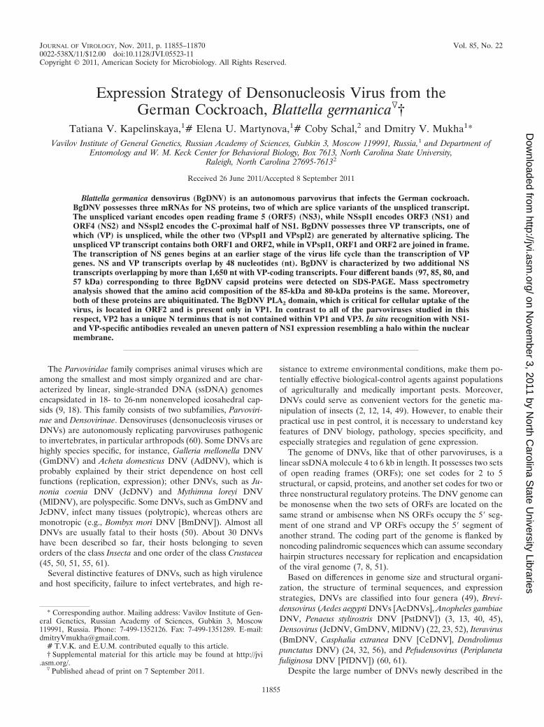
JOURNAL OF VIROLOGY, Nov. 2011, p. 11855–11870 Vol. 85, No. 220022-538X/11/$12.00 doi:10.1128/JVI.05523-11Copyright © 2011, American Society for Microbiology. All Rights Reserved.
Expression Strategy of Densonucleosis Virus from theGerman Cockroach, Blattella germanica�†
Tatiana V. Kapelinskaya,1# Elena U. Martynova,1# Coby Schal,2 and Dmitry V. Mukha1*Vavilov Institute of General Genetics, Russian Academy of Sciences, Gubkin 3, Moscow 119991, Russia,1 and Department of
Entomology and W. M. Keck Center for Behavioral Biology, Box 7613, North Carolina State University,Raleigh, North Carolina 27695-76132
Received 26 June 2011/Accepted 8 September 2011
Blattella germanica densovirus (BgDNV) is an autonomous parvovirus that infects the German cockroach.BgDNV possesses three mRNAs for NS proteins, two of which are splice variants of the unspliced transcript.The unspliced variant encodes open reading frame 5 (ORF5) (NS3), while NSspl1 encodes ORF3 (NS1) andORF4 (NS2) and NSspl2 encodes the C-proximal half of NS1. BgDNV possesses three VP transcripts, one ofwhich (VP) is unspliced, while the other two (VPspl1 and VPspl2) are generated by alternative splicing. Theunspliced VP transcript contains both ORF1 and ORF2, while in VPspl1, ORF1 and ORF2 are joined in frame.The transcription of NS genes begins at an earlier stage of the virus life cycle than the transcription of VPgenes. NS and VP transcripts overlap by 48 nucleotides (nt). BgDNV is characterized by two additional NStranscripts overlapping by more than 1,650 nt with VP-coding transcripts. Four different bands (97, 85, 80, and57 kDa) corresponding to three BgDNV capsid proteins were detected on SDS-PAGE. Mass spectrometryanalysis showed that the amino acid composition of the 85-kDa and 80-kDa proteins is the same. Moreover,both of these proteins are ubiquitinated. The BgDNV PLA2 domain, which is critical for cellular uptake of thevirus, is located in ORF2 and is present only in VP1. In contrast to all of the parvoviruses studied in thisrespect, VP2 has a unique N terminus that is not contained within VP1 and VP3. In situ recognition with NS1-and VP-specific antibodies revealed an uneven pattern of NS1 expression resembling a halo within the nuclearmembrane.
The Parvoviridae family comprises animal viruses which areamong the smallest and most simply organized and are char-acterized by linear, single-stranded DNA (ssDNA) genomesencapsidated in 18- to 26-nm nonenveloped icosahedral cap-sids (9, 18). This family consists of two subfamilies, Parvoviri-nae and Densovirinae. Densoviruses (densonucleosis viruses orDNVs) are autonomously replicating parvoviruses pathogenicto invertebrates, in particular arthropods (60). Some DNVs arehighly species specific, for instance, Galleria mellonella DNV(GmDNV) and Acheta domesticus DNV (AdDNV), which isprobably explained by their strict dependence on host cellfunctions (replication, expression); other DNVs, such as Ju-nonia coenia DNV (JcDNV) and Mythimna loreyi DNV(MlDNV), are polyspecific. Some DNVs, such as GmDNV andJcDNV, infect many tissues (polytropic), whereas others aremonotropic (e.g., Bombyx mori DNV [BmDNV]). Almost allDNVs are usually fatal to their hosts (50). About 30 DNVshave been described so far, their hosts belonging to sevenorders of the class Insecta and one order of the class Crustacea(45, 50, 51, 55, 61).
Several distinctive features of DNVs, such as high virulenceand host specificity, failure to infect vertebrates, and high re-
sistance to extreme environmental conditions, make them po-tentially effective biological-control agents against populationsof agriculturally and medically important pests. Moreover,DNVs could serve as convenient vectors for the genetic ma-nipulation of insects (2, 12, 14, 49). However, to enable theirpractical use in pest control, it is necessary to understand keyfeatures of DNV biology, pathology, species specificity, andespecially strategies and regulation of gene expression.
The genome of DNVs, like that of other parvoviruses, is alinear ssDNA molecule 4 to 6 kb in length. It possesses two setsof open reading frames (ORFs); one set codes for 2 to 5structural, or capsid, proteins, and another set codes for two orthree nonstructural regulatory proteins. The DNV genome canbe monosense when the two sets of ORFs are located on thesame strand or ambisense when NS ORFs occupy the 5� seg-ment of one strand and VP ORFs occupy the 5� segment ofanother strand. The coding part of the genome is flanked bynoncoding palindromic sequences which can assume secondaryhairpin structures necessary for replication and encapsidationof the viral genome (7, 8, 51).
Based on differences in genome size and structural organi-zation, the structure of terminal sequences, and expressionstrategies, DNVs are classified into four genera (49), Brevi-densovirus (Aedes aegypti DNVs [AeDNVs], Anopheles gambiaeDNV, Penaeus stylirostris DNV [PstDNV]) (3, 13, 40, 45),Densovirus (JcDNV, GmDNV, MlDNV) (22, 23, 52), Iteravirus(BmDNV, Casphalia extranea DNV [CeDNV], Dendrolimuspunctatus DNV) (24, 32, 56), and Pefudensovirus (Periplanetafuliginosa DNV [PfDNV]) (60, 61).
Despite the large number of DNVs newly described in the
* Corresponding author. Mailing address: Vavilov Institute of Gen-eral Genetics, Russian Academy of Sciences, Gubkin 3, Moscow119991, Russia. Phone: 7-499-1352126. Fax: 7-499-1351289. E-mail:[email protected].
# T.V.K. and E.U.M. contributed equally to this article.† Supplemental material for this article may be found at http://jvi
.asm.org/.� Published ahead of print on 7 September 2011.
11855
on Novem
ber 3, 2011 by North C
arolina State U
niversity Librarieshttp://jvi.asm
.org/D
ownloaded from
last 2 decades, progress in understanding features of the lifecycles of DNVs, particularly strategies and regulation of ge-nome expression and viral proteins and their functions, israther slow and limited to only certain species: GmDNV (51,52), MlDNV (23), JcDNV (20, 46), PfDNV (60–62), Culexpipiens DNV (CpDNV) (7), PstDNV (19), and AeDNV (57).
The DNV of the German cockroach, Blattella germanica(BgDNV), was discovered in a cockroach colony that origi-nated from a natural population in a pig farm in North Caro-lina and maintained in the laboratory for 5 years. The struc-tural organization of this virus was described in our previouswork (37).
The purposes of this study were (i) to investigate theBgDNV expression strategy and (ii) to determine the intracel-lular distribution of structural and nonstructural virus proteins.The expression strategy was found to differ from that of all ofthe other parvoviruses analyzed thus far. Splicing events areinvolved in the production of both NS and VP transcripts.Splicing in VP mRNAs results in the joining of two ORFs forcapsid proteins into one with an increased coding capacitysufficient to encode the largest VP1 protein. VP2 and VP3, aswell as NS2 proteins, are translated according the leaky-scan-ning mechanism. For the first time, we show monoubiquitina-tion of the DNV capsid proteins and uneven distribution of theNS1 proteins within the nuclei of virus-infected cells.
MATERIALS AND METHODS
Cell culture propagation and virus infection. For the propagation of BgDNV,a culture of B. germanica BGE-2 cells previously isolated from embryonic tissueswas used (30). Cells were grown in 5 ml of antibiotic-free L-15B medium sup-plemented with 5% fetal bovine serum, 5% tryptose phosphate broth, and 0.1%bovine lipoprotein concentrate, pH 7.0 (39). Cell cultures were incubated at25°C, and subcultures were made every 10 to 14 days with an initial seedingdensity of approximately 5 � 105 cells per ml.
To obtain virus-containing extracts, virus-infected cockroaches were lysed incell culture medium through several consecutive freeze-thaw cycles, homoge-nized, freeze-thawed once more, and centrifuged at 13,000 �g for 5 min toremove cellular debris. The resulting supernatant was passed through a 0.22-�mfilter and used to infect BGE-2 cells.
Viral RNA purification and Northern blotting. RNA was extracted fromBgDNV-infected BGE-2 cells 3 h, 1 day, 5 days, 7 days, and 20 days postinfection(p.i.) and late in infection (50 to 100 days p.i., passages V1 to V8), when theproportion of virus DNA and genomic DNA in the total DNA extracts remainedapproximately constant (Fig. 1). Total RNA was extracted using the SV TotalRNA Isolation System kit (Promega) according to the manufacturer’s instruc-
tions. RNA yield and quality were assessed by the absorption value at 260 nmusing NanoDrop2000 (Thermo Scientific).
Northern blotting was done as described by Sambrook et al. (43). Promega’sRNA Markers were used for size estimation of single-stranded RNA.
All of the probes used were obtained by PCR using respective primer pairs. Allof the probes used in our work are listed in Table 1 and depicted in Fig. 2A. Thequality of PCR products was verified using electrophoresis on 1% agarose gels in1� Tris-acetate buffer, and corresponding bands were excised from the gels andpurified using the Wizard PCR Preps DNA Purification system kit (Promega) orthe MinElute Gel Extraction kit (Qiagen) according to the manufacturer’s in-structions.
All of the probes were 32P labeled using the Prime-a-Gene Labeling System(Promega) and [�-32P]dATP as the radioactive precursor according to the sup-plier’s protocols based on the random hexaribonucleotide technique.
RT-PCR. The reverse transcription (RT)-PCR method was used to character-ize the transcripts detected by Northern blotting. First-strand cDNA was syn-thesized using the Im-Prom-II Reverse Transcription System kit (Promega)according to the manufacturer’s instructions using a poly(A) primer. Then re-gions of the viral genome corresponding to bioinformatically predicted ORFswere amplified using corresponding pairs of gene-specific primers. All RNAsamples were tested with negative controls (no RT) to confirm the absence ofcontaminating DNA. All of the resulting bands were gel purified using theWizard PCR Preps DNA purification system kit (Promega), cloned into thepGEM-T Easy vector (Promega), and sequenced according to Sanger et al. (44)with a BigDye Termination kit (Applied Biosystems) on an ABI Prism 310sequencer. All of the primers used throughout this work are listed in Table 2 anddepicted in Fig. 2A.
Identification of 5� and 3� ends of viral transcripts. Transcription start and endpoints were characterized using the SMART RACE cDNA amplification kit(Clontech) according to the manufacturer’s recommendations. To identify 5�ends, first-strand cDNA was synthesized using adaptor primers SMART II AOligonucleotide and 5� CDS primer A, and then 5� ends were amplified using auniversal primer and gene-specific primers. Two primers, VP_R1 (nt 3726 to3757) and VP_R2 (nt 4408 to 4438), for VP transcripts, and one primer, NS_R1(nt 2485 to 2503), for NS transcripts, were utilized. To identify 3� ends, first-strand cDNA was synthesized using only 3� CDS primer A. 3� ends were ampli-fied using the universal primer and gene-specific primers VP_R3 (nt 5067 to5096) for VP transcripts and NS_R2 (nt 241 to 274) for NS transcripts. PCRproducts were separated on 1% agarose gels, all of the visible bands wereexcised, purified using MinElute Gel Extraction Kit (Qiagen), and cloned intothe pGEM-T Easy vector (Promega), and cloned fragments were sequenced.
Production and purification of BgDNV particles. In order to obtain BgDNVparticles, adult B. germanica cockroaches were infected by adding virus-contain-ing lysate to cockroach rearing water. About 100 freshly dead cockroaches werecollected, and virus was purified using a CsCl gradient as follows. Cockroacheswere homogenized in liquid nitrogen, diluted with 50 ml cold phosphate-bufferedsaline (PBS) containing 2 mM phenylmethylsulfonyl fluoride, and then centri-fuged at 10,000 � g for 30 min to remove cell debris. Polyethylene glycol 8000and NaCl were added to the supernatant to final concentrations of 8% and 400mM, respectively. The mixture was stirred for 30 min at 4°C and centrifuged at15,000 �g for 20 min. The pellet containing BgDNV particles was resuspendedin 1 ml of cold PBS. Following mixing on a shaker at room temperature, thesuspension was centrifuged at 9,000 �g to remove insoluble debris. The super-natant was transferred to a tube containing 4 ml of a 20% sucrose cushion in PBSand centrifuged in a 50.2 Ti rotor (Beckman, Fullerton, CA) at 137,000 � governight at 4°C. The resulting pellet was resuspended in 1 ml cold PBS anddissolved overnight at 4°C with intermittent mixing. The resulting suspension wascentrifuged once more through a 20% sucrose cushion, the pellet was resus-pended in 50 mM Tris-HCl (pH 8.0) and loaded onto a CsCl step gradient, and
TABLE 1. Probes used for Northern blot hybridization
Probeno. Description Name Primer
BgDNVsequence
coordinates (nt)
1 NS probe ORF3 pORF3 ORF3/4 st/ORF3 end 960–25052 NS probe ORF5 pNS5 ORF5 st/ORF5 end 346–9013 Whole VP probe pVPwhole P1 st/ORF1 end 2564–50964 VP probe 5� half
of ORF2pVPst P1 st/ORF2 end 4642–5096
5 VP probe 3� endof ORF1
pVPend lNS_2 F/ORF1 end 2564–3466
FIG. 1. Agarose gel electrophoresis of total DNA isolated fromvirus-infected BGE-2 cells at different times p.i. (3 h, 1 day; and 5days). V1 to V13 are the numbers of passages through BGE-2 cells,with approximately one passage per 10 to 14 days. The regular arrowmarks the total genomic DNA, the single-feathered arrow marksBgDNV genomic DNA �5 kb in size, and the double-feathered arrowmarks the replicative form of BgDNV DNA. The DNA size markerson the left are in kilobases.
11856 KAPELINSKAYA ET AL. J. VIROL.
on Novem
ber 3, 2011 by North C
arolina State U
niversity Librarieshttp://jvi.asm
.org/D
ownloaded from
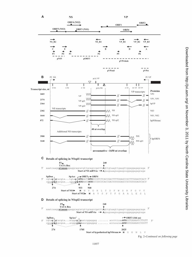
Fig. 2–Continued on following page
11857
on Novem
ber 3, 2011 by North C
arolina State U
niversity Librarieshttp://jvi.asm
.org/D
ownloaded from
11858
on Novem
ber 3, 2011 by North C
arolina State U
niversity Librarieshttp://jvi.asm
.org/D
ownloaded from
the gradient was centrifuged at 152,000 �g for 18 h at 15°C in a Beckman SW41rotor. The gradient was obtained by successively layering 2 ml of 40% CsCl, 4 mlof 35% CsCl, and 2 ml of 30% CsCl in 50 mM Tris-HCl, pH 8.0, which wasoverlaid with 1 M sucrose in the same buffer according to a previously describedtechnique (15). Virus bands were visualized by light scattering, collected, anddialyzed against 50 mM Tris-HCl, pH 8.0.
SDS-PAGE. SDS-PAGE was performed according to the method of Laemmli(31). A suspension of purified virus particles was mixed 1:1 with 2� sample bufferand separated on 7.5% or 10% resolving gels, and protein bands were visualizedby staining with Coomassie brilliant blue R250.
Identification of capsid proteins by mass spectrometry. Bands of interest wereexcised from the gel and digested with 12 �g/ml trypsin, and samples were mixedwith a 2,5-dihydroxybenzoic acid solution on the sample plate. Peptide massspectra were obtained using a tandem matrix-assisted laser desorption ioniza-tion–time of flight-time of flight mass spectrometer Ultraflex II (Bruker) in thepositive-ion reflector mode. Virus proteins were identified by peptide mass
fingerprinting using the Mascot Search program (Matrix Science Inc., Boston,MA) and the NCBI protein database, taking into consideration possible oxida-tion of Met residues and Cys modification by acrylamide.
Western blotting. To obtain total extracts of the virus-infected BGE-2 cells,the cells were harvested and washed three times with cold PBS and the cell pelletwas mixed with an appropriate volume of 2� sample buffer, boiled for 5 min at100°C, and centrifuged at 13,000 �g to remove residual debris. Samples of virusparticles were treated as described above for SDS-PAGE. Proteins were sepa-rated on 7.5% or 10% resolving gels and transferred onto PVDF membranes bysemidry blotting in Tris-glycine-methanol transfer buffer. Blots containingBgDNV protein samples were either stained with Coomassie brilliant blue R250or probed with corresponding antibodies.
Rabbit polyclonal antibodies were generated by Genemed Synthesis (SanAntonio, TX) against the following oligopeptides, corresponding to virus ORFs:ORF1, CTFDRPYFYGKPQRVLNSVEL; ORF2, HYSEAKSDIDIQRADTEAIG; ORF3, CNDPLEFHSGPEVGDIPARPR; ORF4, CRVLELTDAVKDEIKLLLAE; ORF5, DWLKCDEVVLEEFEELNER. The approximate locationsof these oligopeptides are shown in Fig. 2A. To detect ubiquitin, mouse mono-clonal anti-ubiquitin antibody P4G7 (3 to 5 mg/ml, 1:850 dilution; Covance) wasused. Secondary antibodies were goat anti-rabbit or goat anti-mouse IgGs, re-spectively, conjugated with alkaline phosphatase (Promega). For immunoblot-ting, the ProtoBlot II AP System with the Stabilized Substrate kit (Promega) wasused and all procedures were performed according to the manufacturer’srecommendations.
Immunofluorescence assay. Virus-infected and control cells were grown on thesurface of coverslips placed on the bottom of culture dishes. The cell monolayerwas fixed with 4% paraformaldehyde in PBS overnight. Fixed cells were washedthree times with PBS, permeabilized with 0.1% Triton X-100 in PBS for 5 min,and washed three times in PBS. The cells were then blocked by incubation in 3%bovine serum albumin (BSA) in PBS at 4°C overnight, incubated with primaryrabbit antibodies to ORF2 or ORF3 in 3% BSA in PBS at 4°C for 2 h, andwashed three times with PBS. The cells were then incubated with goat anti-rabbitIgG secondary antibodies conjugated with fluorescein isothiocyanate (FITC;1:200; Jackson ImmunoResearch Laboratories, Baltimore, MD) for 2 h in thedark and washed three times with PBS. Nuclei were counterstained with 4�,6-diamidino-2-phenylindole (DAPI), and fluorescence was detected using an LSM510 META confocal fluorescence microscope (Carl Zeiss).
Bioinformatic analysis. The search for homology of BgDNV cDNA nucleotidesequences was made by BLAST (http://blast.ncbi.nlm.nih.gov/Blast.cgi) and pre-diction of ORFs was made by ORF Finder on the NCBI website (http://www.ncbi.nlm.nih.gov/gorf/gorf.html). Promoter region predictions were made by theNeural Network Promoter Prediction program (http://www.fruitfly.org/seq_tools/promoter.html). Search of NLS signals was performed using the Wolf PSORTprogram (http://www.psort.org/).
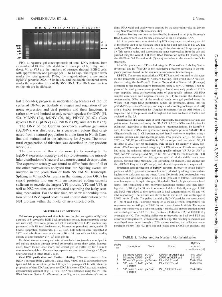
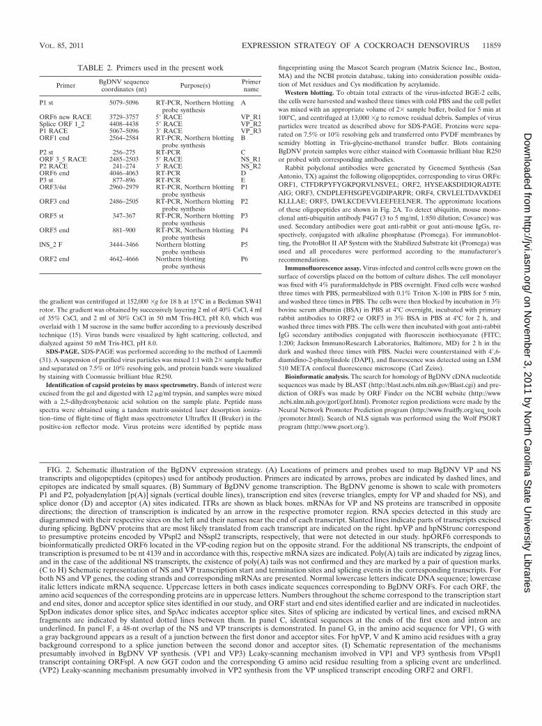
FIG. 2. Schematic illustration of the BgDNV expression strategy. (A) Locations of primers and probes used to map BgDNV VP and NStranscripts and oligopeptides (epitopes) used for antibody production. Primers are indicated by arrows, probes are indicated by dashed lines, andepitopes are indicated by small squares. (B) Summary of BgDNV genome transcription. The BgDNV genome is shown to scale with promotersP1 and P2, polyadenylation [p(A)] signals (vertical double lines), transcription end sites (reverse triangles, empty for VP and shaded for NS), andsplice donor (D) and acceptor (A) sites indicated. ITRs are shown as black boxes. mRNAs for VP and NS proteins are transcribed in oppositedirections; the direction of transcription is indicated by an arrow in the respective promoter region. RNA species detected in this study arediagrammed with their respective sizes on the left and their names near the end of each transcript. Slanted lines indicate parts of transcripts excisedduring splicing. BgDNV proteins that are most likely translated from each transcript are indicated on the right. hpVP and hpNStrunc correspondto presumptive proteins encoded by VPspl2 and NSspl2 transcripts, respectively, that were not detected in our study. hpORF6 corresponds tobioinformatically predicted ORF6 located in the VP-coding region but on the opposite strand. For the additional NS transcripts, the endpoint oftranscription is presumed to be nt 4139 and in accordance with this, respective mRNA sizes are indicated. Poly(A) tails are indicated by zigzag lines,and in the case of the additional NS transcripts, the existence of poly(A) tails was not confirmed and they are marked by a pair of question marks.(C to H) Schematic representation of NS and VP transcription start and termination sites and splicing events in the corresponding transcripts. Forboth NS and VP genes, the coding strands and corresponding mRNAs are presented. Normal lowercase letters indicate DNA sequence; lowercaseitalic letters indicate mRNA sequence. Uppercase letters in both cases indicate sequences corresponding to BgDNV ORFs. For each ORF, theamino acid sequences of the corresponding proteins are in uppercase letters. Numbers throughout the scheme correspond to the transcription startand end sites, donor and acceptor splice sites identified in our study, and ORF start and end sites identified earlier and are indicated in nucleotides.SpDon indicates donor splice sites, and SpAcc indicates acceptor splice sites. Sites of splicing are indicated by vertical lines, and excised mRNAfragments are indicated by slanted dotted lines between them. In panel C, identical sequences at the ends of the first exon and intron areunderlined. In panel F, a 48-nt overlap of the NS and VP transcripts is demonstrated. In panel G, in the amino acid sequence for VP1, G witha gray background appears as a result of a junction between the first donor and acceptor sites. For hpVP, V and K amino acid residues with a graybackground correspond to a splice junction between the second donor and acceptor sites. (I) Schematic representation of the mechanismspresumably involved in BgDNV VP synthesis. (VP1 and VP3) Leaky-scanning mechanism involved in VP1 and VP3 synthesis from VPspl1transcript containing ORFspl. A new GGT codon and the corresponding G amino acid residue resulting from a splicing event are underlined.(VP2) Leaky-scanning mechanism presumably involved in VP2 synthesis from the VP unspliced transcript encoding ORF2 and ORF1.
TABLE 2. Primers used in the present work
Primer BgDNV sequencecoordinates (nt) Purpose(s) Primer
name
P1 st 5079–5096 RT-PCR, Northern blottingprobe synthesis
A
ORF6 new RACE 3729–3757 5� RACE VP_R1Splice ORF 1_2 4408–4438 5� RACE VP_R2P1 RACE 5067–5096 3� RACE VP_R3ORF1 end 2564–2584 RT-PCR, Northern blotting
probe synthesisB
P2 st 256–275 RT-PCR CORF 3_5 RACE 2485–2503 5� RACE NS_R1P2 RACE 241–274 3� RACE NS_R2ORF6 end 4046–4063 RT-PCR DP3 st 877–896 RT-PCR EORF3/4st 2960–2979 RT-PCR, Northern blotting
probe synthesisP1
ORF3 end 2486–2505 RT-PCR, Northern blottingprobe synthesis
P2
ORF5 st 347–367 RT-PCR, Northern blottingprobe synthesis
P3
ORF5 end 881–900 RT-PCR, Northern blottingprobe synthesis
P4
lNS_2 F 3444–3466 Northern blottingprobe synthesis
P5
ORF2 end 4642–4666 Northern blottingprobe synthesis
P6
VOL. 85, 2011 EXPRESSION STRATEGY OF A COCKROACH DENSOVIRUS 11859
on Novem
ber 3, 2011 by North C
arolina State U
niversity Librarieshttp://jvi.asm
.org/D
ownloaded from
Note that, according to the convention of Armentrout et al. (4), genomes ofparvoviruses should be presented as coding, plus strand with NS genes located onthe left. As this principle could not be strictly applied to DNVs possessingambisense genomes, it was decided to define the genome strand encoding NSproteins as plus strand and depict genomes so that NS ORFs are located on theleft while VP ORFs are located on the right (52). In our previous work (37),where the complete sequence of the BgDNV genome was presented, we put it inthe opposite orientation, so that VP ORFs were on the left. In the present work,the BgDNV genome is given in the conventional orientation.
Nucleotide sequence accession number. The GenBank sequence accessionnumber for BgDNV is AY189948.
RESULTS
Northern blot detection of BgDNV transcripts. Total RNAextracted from BgDNV-infected BGE-2 cells late in infection(50 to 100 days) was hybridized with radioactive probes corre-sponding to different ORFs encoding nonstructural regulatory(NS) or structural (VP) proteins.
The NS probe complementary to ORF3 and ORF4 (pORF3,Table 1 and Fig. 2A) revealed two major 2.3- and 1.8-kb tran-scripts and three additional 4.5-, 4.0-, and 1-kb transcripts (Fig.3A, lane 1). In Fig. 3A (lane 1), the 1-kb band indicated by adotted arrow is poorly visible due to a smear in the lower partof the lane. However, it was clearly detected when differentexposure times were used (data not shown). The NS probecomplementary to ORF5 (pNS5, Table 1 and Fig. 2A) revealedtwo strong, clearly defined bands corresponding to 4.5- and2.3-kb transcripts (indicated by arrows in Fig. 3A, lane 2) anda nonspecific smearing in the smaller-molecular-size range.
The VP probe complementary to the whole region encodingcapsid proteins (pVPwhole, Table 1 and Fig. 2A) detected twomajor 2.6- and 2.4-kb transcripts (indicated by arrows in Fig.3B, lane 1) and a number of additional minor transcripts (in-dicated by brackets with asterisks in Fig. 3B, lane 1). Twominor 4.5- and 4.0-kb transcripts (indicated by one asterisk,Fig. 3A, lane 1) were similar in size to transcripts revealed withthe NS probes. Among the smaller-sized minor transcripts, 2.0,1.2, 0.9, and 0.75 kb (indicated by two asterisks), the 2.0-kbtranscript was the most abundant. The VP probe complemen-tary to the 5� segment of ORF2 (pVPst, Table 1 and Fig. 2A)detected the two main 2.6- and 2.4-kb transcripts and all of theminor transcripts except 4.5 and 4.0 kb (Fig. 3B, lane 2),whereas the VP probe complementary to the 3� end of ORF1(pVPend, Table 1 and Fig. 2A) hybridized with the two main2.6- and 2.4-kb transcripts and only one 2.0-kb minor transcript(indicated by two asterisks, Fig. 3B, lane 3). The absence of the1.2-, 0.9-, and 0.75-kb transcripts upon hybridization with thepVPend probe suggests that they are 3�-truncated variants ofthe main 2.4- and 2.6-kb VP mRNAs. The presence of theseminor transcripts in experiments with different probes indi-cates that they are not artifacts and may have some functionalsignificance in BgDNV’s life cycle.
Note that among both the NS mRNAs and VP mRNAs, therelative amounts of transcripts with larger molecular sizes (2.3and 2.6 kb, respectively) were considerably lower than therelative amounts of transcripts with smaller molecular sizes(1.8 and 2.4 kb, respectively) (Fig. 3A and B).
Overall, Northern blot experiments demonstrated thatBgDNV is characterized by a unique transcription pattern,compared with all of the other DNVs described so far.
Temporal dynamics of transcription of the VP and NSgenes. Total RNA was extracted from BgDNV-infected BGE-2cells 3 h, 1 day, 5 days, 7 days, and 20 days p.i. and analyzed byNorthern blot hybridization. The major bands revealed withNS-specific (pORF3) and VP-specific (pVPwhole) probes areshown in Fig. 3C, panels 1 and 2, respectively. To evaluate thecomparative amounts of BgDNV transcripts at different timesafter infection, the relative amount of rRNA revealed byethidium bromide staining in each sample was used as a ref-erence (bottom panels of Fig. 3C). As early as 3 h p.i., clearlydetectable bands corresponding to the NS transcripts werefound, and the amount of mRNA increased appreciably duringthe following 25 days. At the same time, transcripts of struc-tural genes become visible only 5 days p.i., and the amount ofmRNA dramatically increased to 25 days p.i. This result clearlydemonstrates that transcription of regulatory genes begins inearlier stages of the virus life cycle than transcription of struc-tural genes. Thus, BgDNV possesses two groups of genes, early(NS) and late (VP).
Mapping of BgDNV transcripts. 3� and 5� rapid amplifica-tion of cDNA ends (RACE) mapping was used to furtheranalyze the details of BgDNV transcription. Generally, thescheme of the experiments was as follows. Total RNA wasisolated from BGE-2 cells late in infection (50 to 100 days). Todetermine the 3� ends of all possible NS and VP mRNAs, weused gene-specific primers located nearest to the predictedtranscription initiation sites. To determine the 5� ends of NStranscripts, we used primers located most proximally to thepredicted transcription termination site. To determine the 5�ends of VP mRNAs, a nested set of primers located in themiddle of the VP-coding region was used. In all cases, thesecond universal primer (Clontech) was used (see Materialsand Methods). All of the bands resulting from amplificationwere cloned and sequenced. Some bands appeared to be non-specific (indicated by dots in Fig. 3D) and were excluded fromfurther analyses. To verify the results of RACE analyses, RT-PCR using primer pairs listed in Table 2 and depicted in Fig.2A was used.
To ascertain the transcription initiation sites for the NStranscripts, 5� RACE with the NS_R1 primer (Fig. 2A; Table2) and the universal primer was performed. As shown in Fig.3D, panel 1, amplification resulted in one strong band approx-imately 1.6 kb in size (indicated by an arrow with a verticalline), two weak bands approximately 2.0 and 1.1 kb in size(indicated by a simple arrow and an arrow with a vertical line,respectively), and several nonspecific, lower-molecular-massbands (indicated by dots). Additional RT-PCR amplificationsusing the C/P4 primer pair (Fig. 2A; Table 2) confirmed thepresence of 2.0-, 1.6-, and 1.1-kb bands (data not shown).These primers were designed so that primer C was located justdownstream from the predicted start of NS transcription whileprimer P4 was located near the end of predicted ORF3.
Subsequent analysis of the clones revealed that the 2.0-kbband contained cDNA corresponding to the unspliced NStranscript, while alternative splicing generated the 1.6- and1.1-kb NS transcripts (Fig. 2B). In the 1.6-kb transcript, 658nucleotides (nt) are removed between nt 274 (G) (splice donorsite, D NS), which is 7 nt upstream from the AUG start codonof ORF5, and nt 933 (A) (splice acceptor site, Ac NS) justdownstream of the ORF5 stop codon (Fig. 2C). This splicing
11860 KAPELINSKAYA ET AL. J. VIROL.
on Novem
ber 3, 2011 by North C
arolina State U
niversity Librarieshttp://jvi.asm
.org/D
ownloaded from
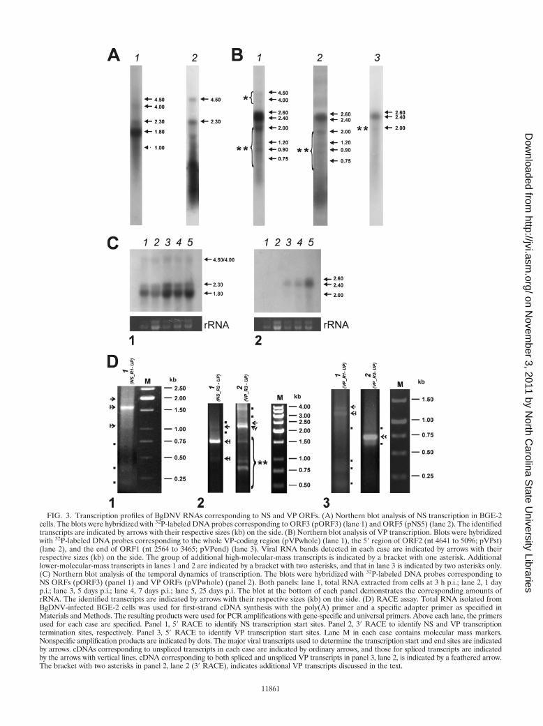
FIG. 3. Transcription profiles of BgDNV RNAs corresponding to NS and VP ORFs. (A) Northern blot analysis of NS transcription in BGE-2cells. The blots were hybridized with 32P-labeled DNA probes corresponding to ORF3 (pORF3) (lane 1) and ORF5 (pNS5) (lane 2). The identifiedtranscripts are indicated by arrows with their respective sizes (kb) on the side. (B) Northern blot analysis of VP transcription. Blots were hybridizedwith 32P-labeled DNA probes corresponding to the whole VP-coding region (pVPwhole) (lane 1), the 5� region of ORF2 (nt 4641 to 5096; pVPst)(lane 2), and the end of ORF1 (nt 2564 to 3465; pVPend) (lane 3). Viral RNA bands detected in each case are indicated by arrows with theirrespective sizes (kb) on the side. The group of additional high-molecular-mass transcripts is indicated by a bracket with one asterisk. Additionallower-molecular-mass transcripts in lanes 1 and 2 are indicated by a bracket with two asterisks, and that in lane 3 is indicated by two asterisks only.(C) Northern blot analysis of the temporal dynamics of transcription. The blots were hybridized with 32P-labeled DNA probes corresponding toNS ORFs (pORF3) (panel 1) and VP ORFs (pVPwhole) (panel 2). Both panels: lane 1, total RNA extracted from cells at 3 h p.i.; lane 2, 1 dayp.i.; lane 3, 5 days p.i.; lane 4, 7 days p.i.; lane 5, 25 days p.i. The blot at the bottom of each panel demonstrates the corresponding amounts ofrRNA. The identified transcripts are indicated by arrows with their respective sizes (kb) on the side. (D) RACE assay. Total RNA isolated fromBgDNV-infected BGE-2 cells was used for first-strand cDNA synthesis with the poly(A) primer and a specific adapter primer as specified inMaterials and Methods. The resulting products were used for PCR amplifications with gene-specific and universal primers. Above each lane, the primersused for each case are specified. Panel 1, 5� RACE to identify NS transcription start sites. Panel 2, 3� RACE to identify NS and VP transcriptiontermination sites, respectively. Panel 3, 5� RACE to identify VP transcription start sites. Lane M in each case contains molecular mass markers.Nonspecific amplification products are indicated by dots. The major viral transcripts used to determine the transcription start and end sites are indicatedby arrows. cDNAs corresponding to unspliced transcripts in each case are indicated by ordinary arrows, and those for spliced transcripts are indicatedby the arrows with vertical lines. cDNA corresponding to both spliced and unspliced VP transcripts in panel 3, lane 2, is indicated by a feathered arrow.The bracket with two asterisks in panel 2, lane 2 (3� RACE), indicates additional VP transcripts discussed in the text.
11861
on Novem
ber 3, 2011 by North C
arolina State U
niversity Librarieshttp://jvi.asm
.org/D
ownloaded from
event results in the complete removal of ORF5 and the gen-eration of mRNA encoding almost fully overlapping ORF3and ORF4 (the ORF3 initiation codon is located only 4 ntupstream of the ORF4 initiation codon, Fig. 2C). In the 1.1-kbNS transcript, the same donor site, but an alternative acceptorsite, is engaged. Splicing between nt 274 (G) (D NS) and nt1705 (G) (splice acceptor site, Ac� NS) results in the removalof a 1,430-nt intron and the generation of mRNA correspond-ing to a small, 501-nt ORF identical to the 3�-proximal half ofORF3 (the AUG start codon maps to nt 2025, and the stopcodon is identical to the ORF3 stop codon) (Fig. 2D).
The transcription start site for all of the NS transcripts wasmapped to position 240, 24 nt downstream of the 5� invertedterminal repeat (ITR), as predicted earlier, provided that tran-scription was driven by the P2 promoter (50) (Fig. 2E). Thesequence context of the transcription start site, TTAGTTG(transcription start site in bold and underlined), correspondedwell to the Inr consensus sequence (5�-Py Py A�1 N T/A PyPy-3�). The 5� untranslated regions for NS mRNAs appearedto be relatively long at 42 nt for unspliced and 35 nt for splicedtranscripts (compare Fig. 2C, D, and E).
Note that RACE analysis did not reveal any transcripts start-ing with nt 823 (A), as would be anticipated if the bioinfor-matically predicted P3 promoter (37) were active. Therefore,the P3 promoter is most likely not active and the transcriptionof all three BgDNV nonstructural genes is controlled by the P2promoter. For PfDNV, a densovirus infecting the cockroach P.fuliginosa, it was demonstrated that the predicted internal P18promoter, presumably driving the transcription of ns1/ns2genes, was nonfunctional, and the transcription of all of the NSgenes was regulated by the P3 promoter, an analogue of theBgDNV P2 promoter. Also, as in BgDNV, in PfDNV, thens3-coding sequence was spliced out of the bigger transcriptencoding the overlapping ns1 and ns2 genes (61). In the samemanner, only one promoter drives the expression of all of theNS genes and splicing is involved in the generation of mRNAsof nonstructural genes in GmDNV (52) and MlDNV (23).
To identify the transcription termination site for NS tran-scripts, we performed 3� RACE with primer NS_R2 and theuniversal primer (Fig. 2A; Table 2, see Materials and Meth-ods). The result of this experiment is shown in Fig. 3D, panel2, lane 1. Amplification resulted in one bright band of approx-imately 1.6 kb (indicated by an arrow with a vertical line), twoweak bands with approximate sizes of 1.0 and 2.1 kb (indicatedby an arrow with a vertical line and a simple arrow, respec-tively), and nonspecific minor bands (indicated by dots). Sub-sequent analysis revealed that the 3� end mapped to nt 2541, 14nt downstream from the predicted canonical polyadenylationsignal (2521 to 2526, AAUAAA) (37) (Fig. 2F).
Data obtained for NS transcripts and their exact sizes aresummarized in Fig. 2B. The sizes, 2,302, 1,644, and 872 nt, arein reasonable agreement with the respective sizes of �2,300,�1,800, and �1,000 nt obtained by Northern blotting (Fig. 3A,lane 1). The fact that hybridization with the ORF5 probe(pNS5) did not reveal �1,800- and �1,000-nt transcripts (Fig.3A, lane 2) confirms that ORF5 is contained only within thelargest unspliced mRNA and excised from the others duringsplicing events.
5� RACE with the VP_R1 primer, located approximately inthe middle of the predicted ORF1 sequence (nt 3729 to 3757)
(Fig. 2A; Table 2), and the universal primer, performed inorder to identify the transcription start sites for VP transcripts,provided two major bands with approximate sizes of 1.4 and 1.2kb (Fig. 3D, panel 3, lane 1, indicated by a simple arrow and anarrow with a vertical line, respectively) and a number of lower-molecular-mass nonspecific bands (indicated by dots in thesame lane). This result was confirmed by RT-PCR amplifica-tions with primers A and B (Fig. 2A; Table 2) that resulted intwo bands with respective sizes of approximately 2.3 and 2.5 kb(data not shown).
Analysis of the 1.2-kb cDNAs revealed that two differentsplicing events were responsible for the generation of VP tran-scripts (Fig. 2B). In one case, 229 nt were excised, resulting inthe junction of ORF2 and ORF1 in frame. This new ORF(ORFspl) is 778 amino acids (aa) in size and has a capacity toencode an 85.3-kDa protein. The donor site (D1 VP) is the Gat nt 4421, and the acceptor site (Ac VP) is the G at nt 4191.D1 VP is located 7 nt upstream of the AUG start codon forORF1 and 16 nt upstream of the TAA stop codon for ORF2,overlapping the beginning of ORF1 by 11 nt (Fig. 2G). Inanother case, an alternative donor site (D2 VP), the G at nt4431, 10 nt upstream of D1 VP (Fig. 2E), was exploited. Thissplicing did not generate any new ORF, but it slightly extendedORF2 (40 aa). Because the cDNAs resulting from these alter-native splicing events differed by only 10 nt, they appearedtogether as one 1.2-kb band.
The larger 1.4-kb cDNA corresponded to an unspliced VPtranscript containing both ORF2 and ORF1.
5� RACE designed to facilitate VP mRNA 5�-end identifi-cation with the universal primer and the VP_R2 primer (Fig.2A; Table 2) nested relative to VP_R1 and located in ORF2,yielded one band of approximately 750 nt (Fig. 3D, panel 3,lane 2, indicated by a feathered arrow) and two nonspecificbands (indicated by dots). Because the VP_R2 primer is lo-cated just upstream of the VP D2 splice donor site, the result-ing single 750-nt band was common to the spliced and un-spliced transcript 5� ends, which was further confirmed bysequencing of the corresponding cDNA.
The transcription start for all of the VP transcripts mappedto nt 5096, 23 nt downstream from the 3� ITR (Fig. 2H), inaccordance with the bioinformatically predicted start point,provided that transcription is driven by the P1 promoter (37).Therefore, in contrast to NS transcripts, the 5� untranslatedregion for VP mRNAs is very short and comprises only 3 nt,exactly the same as for CpDNV (7). A very short 5� leadersequence was also described for GmDNV and MlDNV (23,52). The sequence context of the VP transcription start site,TTAGTATG (transcription start site in bold and underlined),corresponds quite well to the consensus Inr sequence.
To identify transcription stop sites for VP mRNAs, we per-formed 3� RACE using the universal primer and the VP_R3primer, located close to the VP transcription start site (Fig. 2A;Table 2). In addition to the expected two bands correspondingto the unspliced and spliced transcripts (indicated by a simplearrow and an arrow with a vertical line, respectively), amplifi-cation yielded a number of bright bands differing in size (in-dicated by a bracket with two asterisks), as shown in Fig. 3D,panel 2, lane 2. We cloned and sequenced all of the bands;subsequent analysis revealed that the largest two (approxi-mately 2.2 and 2.4 kb, indicated by an arrow with a vertical line
11862 KAPELINSKAYA ET AL. J. VIROL.
on Novem
ber 3, 2011 by North C
arolina State U
niversity Librarieshttp://jvi.asm
.org/D
ownloaded from
and a simple arrow, respectively) indeed corresponded to thefull spliced and unspliced transcripts, respectively, with a tran-scription stop point at nt 2494, 19 nt downstream from thepredicted polyadenylation signal (AAUAAA, nt 2513 to 2518)(37) (Fig. 2F). The exact sizes of VP mRNAs, taking intoaccount two different splicing events, are 2,603, 2,374, and2,364 nt (Fig. 2B) and are in good agreement with the 2,600-and 2,400-nt bands obtained by Northern blotting (Fig. 3B,lane 1). Since the size difference between the two splicedmRNAs is just 10 nt, they appear on the blots as one band. Allof the other 3� RACE bands proved to contain products oftranscription termination at the internal sites: 4,031, 3,989,3,824, and 3,387 nt. The relative sizes of the resulting mRNAs,840, 882, 1,043, and 1,481 nt, matching respective cDNAs (in-dicated by a bracket with two asterisks in Fig. 3D, panel 2, lane2), corresponded to the sizes of the minor transcripts group(2,000, 1,200, 900, and 750 nt) detected by Northern blotting(Fig. 3B, lane 1, indicated by a bracket with two asterisks),suggesting that they are authentic products of viral originrather than products of artifactual PCR amplification. Addi-tional support would come from the results of Northern blothybridization with probe pVPend (Fig. 2A; Table 1), whichoverlapped the 3� end of the longest 1,481-nt additional tran-script; it should not detect shorter transcripts. As describedabove, this was indeed the only transcript detected by thisprobe (Fig. 3B, lane 3, band with asterisk). At the same time,probe pVPst, targeted to the 5� part of the transcripts, was ableto detect all of them, confirming the presence of a sharedcommon transcription start point (Fig. 3B, lane 2). Neverthe-less, the functional role of these additional transcripts is un-clear, as they do not contain any new ORF, so the only differ-ence among them is the size of the N-terminal parts of VPORFs. They might result from some errors in transcriptiontermination or the polyadenylation machinery recognizing A-rich sequences or A stretches, which are highly abundant in theBgDNV genome. However, analysis of sequences surroundingtermination points for these transcripts revealed that theycontain a set of conservative polyadenylation-driving signals[poly(A) signal, A-rich sequences, GT-rich sequences]. More-over, their relative number compared to that of the main tran-scripts is considerable, and although such transcripts are notdetected in NS genes, the NS-coding region is also rich in Astretches. All of these observations suggest that these addi-tional transcripts are not artifactual and that they may playsome role that has yet to be determined. No such transcriptswere described so far for any other DNVs. A summary of theVP transcripts is given in Fig. 2B.
Note that in our previous work (37), an additional poly-adenylation signal for VP genes at positions 4394 to 4399 waspredicted that, if functional, could generate mRNA containingonly ORF2. But in our present work, we did not detect anytranscripts with a transcription stop point corresponding to thispolyadenylation site. The relative amount of unspliced mRNAsfor VP, as well as for NS transcripts, is poorly comparable tothe amount of the spliced ones, as estimated by Northernblotting and RT-PCR. Finally, identification of the 3� ends oftranscripts showed that the NS and VP transcripts overlappedby 48 nt (Fig. 2B). This is characteristic of DNVs possessingambisense genomes, as demonstrated for GmDNV, MlDNV,CpDNV, and others (7, 23, 52).
Identification of long NS transcripts. The sizes of the twolargest additional NS transcripts (about 4,000 and 4,500 nt)detected by Northern blotting (Fig. 3A, lane 1) suggest thatthey may result from transcription that does not terminate atthe identified NS transcription termination point but ratherproceeds farther, so the resulting transcripts may comprisealmost the whole BgDNV genome. The 4,000- and 4,500-ntbands do not result from the hybridization of contaminantBgDNV genome DNA, since hybridization of the same probeswith mRNA samples containing BgDNV genomic DNA dem-onstrated that the band corresponding to the whole genome islarger and is accompanied by two distinct bands of approxi-mately 4,000 and 4500 nt (data not presented). The naturalorigin of the additional transcripts was supported by the factthat the same bands were detected with probes to the wholeVP-coding region, pVPwhole (Fig. 3B, lane 1, indicated by abracket with one asterisk), but not with the probe pVPst, cor-responding to the very beginning of ORF2 (the probe closestto the 5� end of the genome) (Fig. 3B, lane 2). Bioinformaticanalysis of the BgDNV genome revealed the presence of onemore ORF (ORF6, nt 3684 to 4079) located in the VP-codingpart of genome but on the NS-coding strand, with a predictedsize of 396 nt and the coding capacity for a 131-aa (13.6-kDa)protein (Fig. 2A). Comparison with the NCBI databaseshowed no significant homology with any known proteins.
We proposed that ORF6 may be encoded by large additionalNS transcripts, but its functional role is unclear. RT-PCR am-plifications with primer pairs C/D and E/D (Fig. 2A; Table 2)confirmed that transcripts containing ORF6 and extending tothe VP-coding region of the BgDNV genome did exist (datanot shown). We also showed that a portion of these transcriptsis spliced and that splicing occurred between the D NS donorsites and A NS acceptor sites described earlier. This agreedwell with the fact that only the 4,500-nt transcript was revealedby hybridization with probe pNS5 (Fig. 3A, lane 2).
The bioinformatic analysis of the BgDNV genome revealedthe presence of three additional polyadenylation sites for NStranscripts at positions 4209 to 4214, 4533 to 4538, and 4620 to4625 (data not shown). We used 3� RACE to determine the 3�ends of the transcripts, but unfortunately, we encounteredsome difficulties due to a great amount of nonspecific productsresulting from amplifications with different gene-specific prim-ers that made isolation of the sought-for specific amplificationproducts difficult. We found only one polyadenylated tran-script that terminated at nt 4139, which is before the firsthypothesized polyadenylation site at nt 4209 to 4214. We hy-pothesize that a larger amount of transcripts may terminate atsome point(s) closer to the 5� end of the BgDNV genome, afterthe predicted additional polyadenylation signals. Moreover, itis also possible that these long transcripts are, in fact, notpolyadenylated, which hindered the identification of the stoppoint by the RACE method. Features of long NS transcriptsare summarized in Fig. 2B.
Therefore, BgDNV possesses two groups of nonstructuraltranscripts with the same promoter and transcription start sitesbut with different lengths and alternative transcription stoppoints.
To summarize the data obtained, BgDNV possesses threemRNAs for NS proteins, two of which (NSspl1 and NSspl2)are spliced variants of the unspliced transcript (NS) having the
VOL. 85, 2011 EXPRESSION STRATEGY OF A COCKROACH DENSOVIRUS 11863
on Novem
ber 3, 2011 by North C
arolina State U
niversity Librarieshttp://jvi.asm
.org/D
ownloaded from
same donor (D NS) and different acceptor (Ac NS and Ac� NS,respectively) splice sites. The unspliced variant most likelyencodes ORF5 (NS3), while NSspl1 encodes ORF3 (NS1) andORF4 (NS2) and NSspl2 most likely encodes the C-proximalhalf of NS1, designated hpNStrunc. BgDNV is characterizedby two additional NS transcripts (lNS and lNSspl1) with thesame transcription start point and splicing donor (D NS) andacceptor (Ac NS) sites as the main NS transcripts but overlap-ping VP-coding transcripts by more than 1,650 nt.
BgDNV also possesses three VP transcripts. One of them(VP) is unspliced, while the other two are generated by alter-native splicing using different donor sites (D2 VP, VPspl2; D1VP, VPspl1) and the same acceptor site (Ac VP). The VPunspliced transcript contains both ORF1 and ORF2, while inVPspl1, ORF1 and ORF2 are joined in frame to produce newORFspl, and ORF2 in VPspl2 is slightly longer. A summary ofBgDNV transcripts is given in Fig. 2B.
Purification of BgDNV particles and identification of capsidproteins. Centrifugation of BgDNV particles in a CsCl buoyantdensity gradient (see Materials and Methods) yielded threevisible bands. By collecting these bands into separate fractionsand further purification of the viral DNA, we demonstratedthat the upper band contained empty particles while the twolower bands contained full BgDNV particles differing in buoy-ant density. Both kinds of full particles appeared to containplus and minus viral DNA strands in the same proportions.These findings were the same as described previously forGmDNV, viral particle preparations of which also containedtwo types of full particles, DNVI and DNVII. Thorough inves-tigation of this phenomenon supported the idea that the dif-ference between the two types of particles may be related toprotein content and quaternary structure (53).
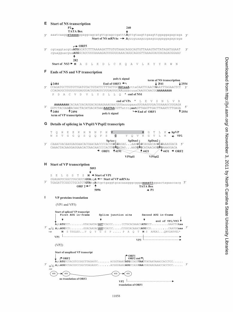
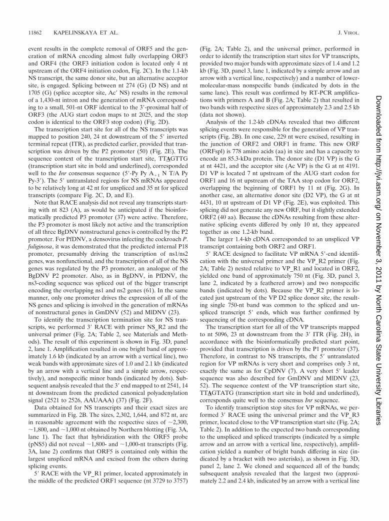
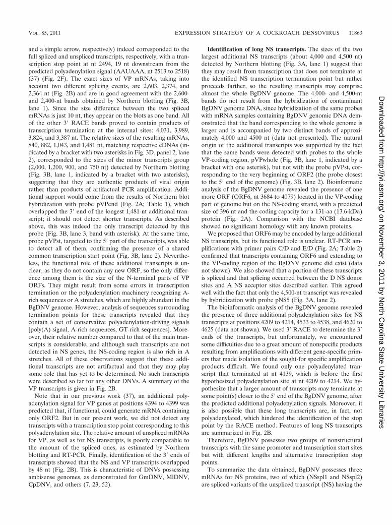
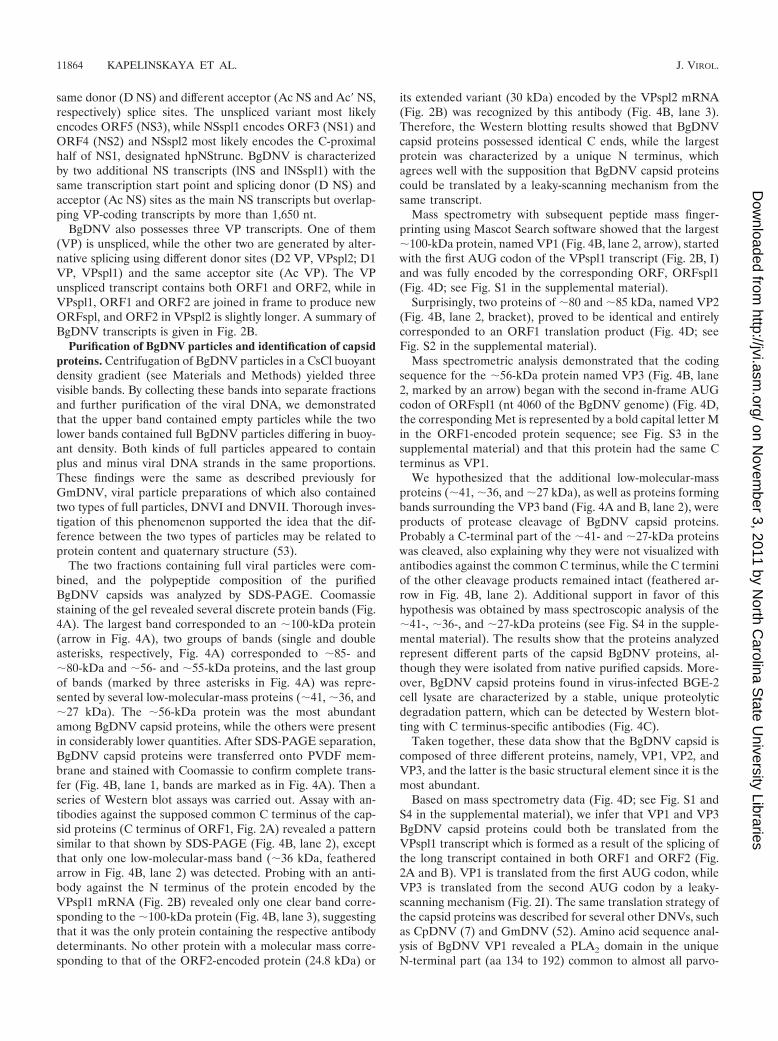
The two fractions containing full viral particles were com-bined, and the polypeptide composition of the purifiedBgDNV capsids was analyzed by SDS-PAGE. Coomassiestaining of the gel revealed several discrete protein bands (Fig.4A). The largest band corresponded to an �100-kDa protein(arrow in Fig. 4A), two groups of bands (single and doubleasterisks, respectively, Fig. 4A) corresponded to �85- and�80-kDa and �56- and �55-kDa proteins, and the last groupof bands (marked by three asterisks in Fig. 4A) was repre-sented by several low-molecular-mass proteins (�41, �36, and�27 kDa). The �56-kDa protein was the most abundantamong BgDNV capsid proteins, while the others were presentin considerably lower quantities. After SDS-PAGE separation,BgDNV capsid proteins were transferred onto PVDF mem-brane and stained with Coomassie to confirm complete trans-fer (Fig. 4B, lane 1, bands are marked as in Fig. 4A). Then aseries of Western blot assays was carried out. Assay with an-tibodies against the supposed common C terminus of the cap-sid proteins (C terminus of ORF1, Fig. 2A) revealed a patternsimilar to that shown by SDS-PAGE (Fig. 4B, lane 2), exceptthat only one low-molecular-mass band (�36 kDa, featheredarrow in Fig. 4B, lane 2) was detected. Probing with an anti-body against the N terminus of the protein encoded by theVPspl1 mRNA (Fig. 2B) revealed only one clear band corre-sponding to the �100-kDa protein (Fig. 4B, lane 3), suggestingthat it was the only protein containing the respective antibodydeterminants. No other protein with a molecular mass corre-sponding to that of the ORF2-encoded protein (24.8 kDa) or
its extended variant (30 kDa) encoded by the VPspl2 mRNA(Fig. 2B) was recognized by this antibody (Fig. 4B, lane 3).Therefore, the Western blotting results showed that BgDNVcapsid proteins possessed identical C ends, while the largestprotein was characterized by a unique N terminus, whichagrees well with the supposition that BgDNV capsid proteinscould be translated by a leaky-scanning mechanism from thesame transcript.
Mass spectrometry with subsequent peptide mass finger-printing using Mascot Search software showed that the largest�100-kDa protein, named VP1 (Fig. 4B, lane 2, arrow), startedwith the first AUG codon of the VPspl1 transcript (Fig. 2B, I)and was fully encoded by the corresponding ORF, ORFspl1(Fig. 4D; see Fig. S1 in the supplemental material).
Surprisingly, two proteins of �80 and �85 kDa, named VP2(Fig. 4B, lane 2, bracket), proved to be identical and entirelycorresponded to an ORF1 translation product (Fig. 4D; seeFig. S2 in the supplemental material).
Mass spectrometric analysis demonstrated that the codingsequence for the �56-kDa protein named VP3 (Fig. 4B, lane2, marked by an arrow) began with the second in-frame AUGcodon of ORFspl1 (nt 4060 of the BgDNV genome) (Fig. 4D,the corresponding Met is represented by a bold capital letter Min the ORF1-encoded protein sequence; see Fig. S3 in thesupplemental material) and that this protein had the same Cterminus as VP1.
We hypothesized that the additional low-molecular-massproteins (�41, �36, and �27 kDa), as well as proteins formingbands surrounding the VP3 band (Fig. 4A and B, lane 2), wereproducts of protease cleavage of BgDNV capsid proteins.Probably a C-terminal part of the �41- and �27-kDa proteinswas cleaved, also explaining why they were not visualized withantibodies against the common C terminus, while the C terminiof the other cleavage products remained intact (feathered ar-row in Fig. 4B, lane 2). Additional support in favor of thishypothesis was obtained by mass spectroscopic analysis of the�41-, �36-, and �27-kDa proteins (see Fig. S4 in the supple-mental material). The results show that the proteins analyzedrepresent different parts of the capsid BgDNV proteins, al-though they were isolated from native purified capsids. More-over, BgDNV capsid proteins found in virus-infected BGE-2cell lysate are characterized by a stable, unique proteolyticdegradation pattern, which can be detected by Western blot-ting with C terminus-specific antibodies (Fig. 4C).
Taken together, these data show that the BgDNV capsid iscomposed of three different proteins, namely, VP1, VP2, andVP3, and the latter is the basic structural element since it is themost abundant.
Based on mass spectrometry data (Fig. 4D; see Fig. S1 andS4 in the supplemental material), we infer that VP1 and VP3BgDNV capsid proteins could both be translated from theVPspl1 transcript which is formed as a result of the splicing ofthe long transcript contained in both ORF1 and ORF2 (Fig.2A and B). VP1 is translated from the first AUG codon, whileVP3 is translated from the second AUG codon by a leaky-scanning mechanism (Fig. 2I). The same translation strategy ofthe capsid proteins was described for several other DNVs, suchas CpDNV (7) and GmDNV (52). Amino acid sequence anal-ysis of BgDNV VP1 revealed a PLA2 domain in the uniqueN-terminal part (aa 134 to 192) common to almost all parvo-
11864 KAPELINSKAYA ET AL. J. VIROL.
on Novem
ber 3, 2011 by North C
arolina State U
niversity Librarieshttp://jvi.asm
.org/D
ownloaded from
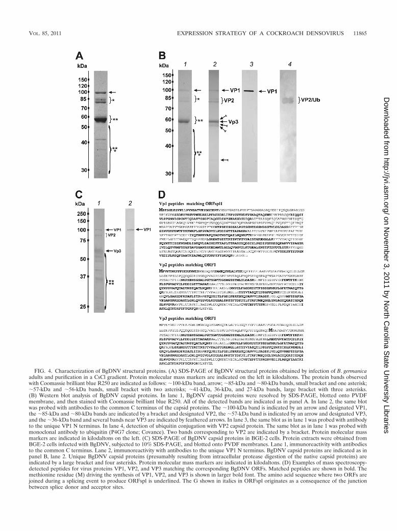
FIG. 4. Characterization of BgDNV structural proteins. (A) SDS-PAGE of BgDNV structural proteins obtained by infection of B. germanicaadults and purification in a CsCl gradient. Protein molecular mass markers are indicated on the left in kilodaltons. The protein bands observedwith Coomassie brilliant blue R250 are indicated as follows: �100-kDa band, arrow; �85-kDa and �80-kDa bands, small bracket and one asterisk;�57-kDa and �56-kDa bands, small bracket with two asterisks; �41-kDa, 36-kDa, and 27-kDa bands, large bracket with three asterisks.(B) Western blot analysis of BgDNV capsid proteins. In lane 1, BgDNV capsid proteins were resolved by SDS-PAGE, blotted onto PVDFmembrane, and then stained with Coomassie brilliant blue R250. All of the detected bands are indicated as in panel A. In lane 2, the same blotwas probed with antibodies to the common C terminus of the capsid proteins. The �100-kDa band is indicated by an arrow and designated VP1,the �85-kDa and �80-kDa bands are indicated by a bracket and designated VP2, the �57-kDa band is indicated by an arrow and designated VP3,and the �36-kDa band and several bands near VP3 are indicated by feathered arrows. In lane 3, the same blot as in lane 1 was probed with antibodyto the unique VP1 N terminus. In lane 4, detection of ubiquitin conjugation with VP2 capsid protein. The same blot as in lane 1 was probed withmonoclonal antibody to ubiquitin (P4G7 clone; Covance). Two bands corresponding to VP2 are indicated by a bracket. Protein molecular massmarkers are indicated in kilodaltons on the left. (C) SDS-PAGE of BgDNV capsid proteins in BGE-2 cells. Protein extracts were obtained fromBGE-2 cells infected with BgDNV, subjected to 10% SDS-PAGE, and blotted onto PVDF membranes. Lane 1, immunoreactivity with antibodiesto the common C terminus. Lane 2, immunoreactivity with antibodies to the unique VP1 N terminus. BgDNV capsid proteins are indicated as inpanel B, lane 2. Unique BgDNV capsid proteins (presumably resulting from intracellular protease digestion of the native capsid proteins) areindicated by a large bracket and four asterisks. Protein molecular mass markers are indicated in kilodaltons. (D) Examples of mass spectroscopy-detected peptides for virus proteins VP1, VP2, and VP3 matching the corresponding BgDNV ORFs. Matched peptides are shown in bold. Themethionine residue (M) driving the synthesis of VP1, VP2, and VP3 is shown in larger bold font. The amino acid sequence where two ORFs arejoined during a splicing event to produce ORFspl is underlined. The G shown in italics in ORFspl originates as a consequence of the junctionbetween splice donor and acceptor sites.
VOL. 85, 2011 EXPRESSION STRATEGY OF A COCKROACH DENSOVIRUS 11865
on Novem
ber 3, 2011 by North C
arolina State U
niversity Librarieshttp://jvi.asm
.org/D
ownloaded from
viruses. The PLA2 domain of BgDNV is located in ORF2 andis therefore present only in VP1.
Mass spectrometry data (Fig. 4D; see Fig. S2 and S3 in thesupplemental material) showed that the amino acid sequenceof the VP2 capsid protein completely corresponded to ORF1.The bioinformatic analysis of the nucleotide sequence of theVP-coding part of the BgDNV genome revealed two addi-tional promoter regions with transcription start points at nt4854 and 4462, respectively, that could produce transcriptsencoding ORF1 only. However, a detailed 5� RACE-assistedsearch failed to detect any such transcripts in sufficientamounts. Besides, the absence of a difference between North-ern blot hybridizations with probes pVPst and pVPwhole (Fig.3B, lanes 1 and 2) further supports the assumption that noseparate ORF1-encoding transcripts exist. Therefore, the VP2protein is most likely translated from the unspliced VP tran-script (Fig. 2B) by the so-called leaky-scanning mechanism. Itis clear that the first AUG codon of ORF2 is functional, as itserves as the start codon for VP1 protein in ORFspl1. But the5� untranslated region in VP transcripts is just 3 nt long, andthe context of the first AUG codon is not very favorable (AGTATGT, where the start codon is in bold and the �3 and �4positions are in bold and underlined) (30), which causes theribosome to pass it through and to start translating the VP2protein from the first AUG codon of ORF1. In other words,the start AUG for ORF2 may, in some cases, be “unseen” bythe ribosome due to its proximity to the 5� end, so the ribosomecan “jump” to the second AUG codon of this transcript, cor-responding to the first ATG codon of ORF1. A hypotheticalscheme for the VP2 translation mechanism is presented in Fig.2I. In contrast to all of the parvoviruses studied in this respect,VP2 has a unique N terminus that is not contained within VP1and VP3.
VP2 protein ubiquitination. According to the BgDNV trans-lation strategy described above, the predicted molecularmasses of the VP1 to VP3 capsid proteins should be 85.3, 69.7,and 56.3 kDa, respectively. Surprisingly, the molecular massesof VP1 and VP2 (but not VP3) estimated by SDS-PAGE didnot correspond to the predicted values and were �100 and�80 to 85 kDa, respectively. Moreover, the VP2 protein wasrepresented by two bands. These two deviances are most likelydue to posttranslational modifications of these proteins, suchas glycosylation, phosphorylation, and/or ubiquitination. Thelast hypothesis was tested experimentally by Western blottingusing BgDNV particles and an anti-ubiquitin antibody possess-ing extensive cross-reactivity from yeasts to human. As shownin Fig. 4B, lane 4, the antiubiquitin antibody recognized twoprotein bands corresponding to VP2 proteins, suggesting thatthey indeed are ubiquitinated, while VP1 and VP3 are not.
To date, monoubiquitination has not been shown for denso-virus capsid proteins. The exact function of ubiquitin conjuga-tion of BgDNV VP2 proteins is unclear. At the same time,based on the proposed roles of capsid ubiquitination in variousother viruses, it is possible that the observed modification ofthe BgDNV capsid proteins provides a means for (i) cell de-fense against BgDNV infection, (ii) viral entry into cockroachcells, and (iii) viral capsid assembly during late phases of in-fection and viral release from the cell (see Discussion).
Western blot assay identification of BgDNV regulatory pro-teins. To identify BgDNV regulatory proteins, we performed
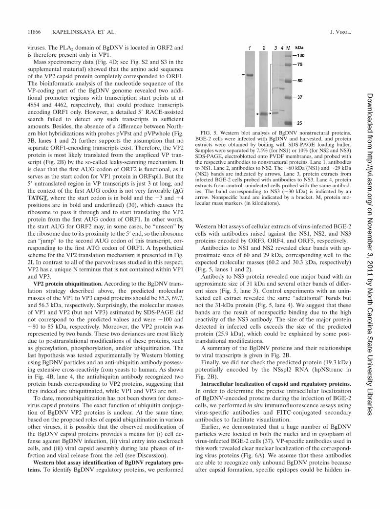
Western blot assays of cellular extracts of virus-infected BGE-2cells with antibodies raised against the NS1, NS2, and NS3proteins encoded by ORF3, ORF4, and ORF5, respectively.
Antibodies to NS1 and NS2 revealed clear bands with ap-proximate sizes of 60 and 29 kDa, corresponding well to theexpected molecular masses (60.2 and 30.3 kDa, respectively)(Fig. 5, lanes 1 and 2).
Antibody to NS3 protein revealed one major band with anapproximate size of 31 kDa and several other bands of differ-ent sizes (Fig. 5, lane 3). Control experiments with an unin-fected cell extract revealed the same “additional” bands butnot the 31-kDa protein (Fig. 5, lane 4). We suggest that thesebands are the result of nonspecific binding due to the highreactivity of the NS3 antibody. The size of the major proteindetected in infected cells exceeds the size of the predictedprotein (25.9 kDa), which could be explained by some post-translational modifications.
A summary of the BgDNV proteins and their relationshipsto viral transcripts is given in Fig. 2B.
Finally, we did not check the predicted protein (19.3 kDa)potentially encoded by the NSspl2 RNA (hpNStrunc inFig. 2B).
Intracellular localization of capsid and regulatory proteins.In order to determine the precise intracellular localizationof BgDNV-encoded proteins during the infection of BGE-2cells, we performed in situ immunofluorescence assays usingvirus-specific antibodies and FITC-conjugated secondaryantibodies to facilitate visualization.
Earlier, we demonstrated that a huge number of BgDNVparticles were located in both the nuclei and in cytoplasm ofvirus-infected BGE-2 cells (37). VP-specific antibodies used inthis work revealed clear nuclear localization of the correspond-ing virus proteins (Fig. 6A). We assume that these antibodiesare able to recognize only unbound BgDNV proteins becauseafter capsid formation, specific epitopes could be hidden in-
FIG. 5. Western blot analysis of BgDNV nonstructural proteins.BGE-2 cells were infected with BgDNV and harvested, and proteinextracts were obtained by boiling with SDS-PAGE loading buffer.Samples were separated by 7.5% (for NS1) or 10% (for NS2 and NS3)SDS-PAGE, electroblotted onto PVDF membranes, and probed withthe respective antibodies to nonstructural proteins. Lane 1, antibodiesto NS1. Lane 2, antibodies to NS2. The �60 kDa (NS1) and �29 kDa(NS2) bands are indicated by arrows. Lane 3, protein extracts frominfected BGE-2 cells probed with antibodies to NS3. Lane 4, proteinextracts from control, uninfected cells probed with the same antibod-ies. The band corresponding to NS3 (�30 kDa) is indicated by anarrow. Nonspecific band are indicated by a bracket. M, protein mo-lecular mass markers (in kilodaltons).
11866 KAPELINSKAYA ET AL. J. VIROL.
on Novem
ber 3, 2011 by North C
arolina State U
niversity Librarieshttp://jvi.asm
.org/D
ownloaded from
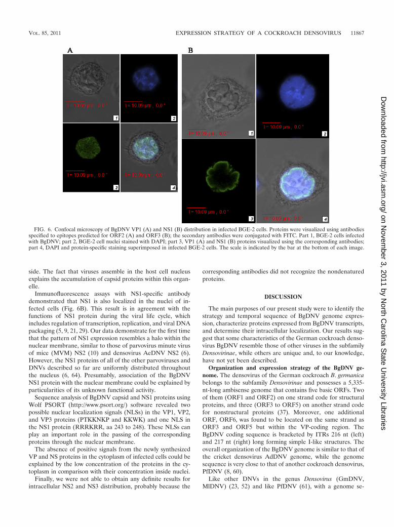
side. The fact that viruses assemble in the host cell nucleusexplains the accumulation of capsid proteins within this organ-elle.
Immunofluorescence assays with NS1-specific antibodydemonstrated that NS1 is also localized in the nuclei of in-fected cells (Fig. 6B). This result is in agreement with thefunctions of NS1 protein during the viral life cycle, whichincludes regulation of transcription, replication, and viral DNApackaging (5, 9, 21, 29). Our data demonstrate for the first timethat the pattern of NS1 expression resembles a halo within thenuclear membrane, similar to those of parvovirus minute virusof mice (MVM) NS2 (10) and densovirus AeDNV NS2 (6).However, the NS1 proteins of all of the other parvoviruses andDNVs described so far are uniformly distributed throughoutthe nucleus (6, 64). Presumably, association of the BgDNVNS1 protein with the nuclear membrane could be explained byparticularities of its unknown functional activity.
Sequence analysis of BgDNV capsid and NS1 proteins usingWolf PSORT (http://www.psort.org/) software revealed twopossible nuclear localization signals (NLSs) in the VP1, VP2,and VP3 proteins (PTKKNKP and KKWK) and one NLS inthe NS1 protein (RRRKRR, aa 243 to 248). These NLSs canplay an important role in the passing of the correspondingproteins through the nuclear membrane.
The absence of positive signals from the newly synthesizedVP and NS proteins in the cytoplasm of infected cells could beexplained by the low concentration of the proteins in the cy-toplasm in comparison with their concentration inside nuclei.
Finally, we were not able to obtain any definite results forintracellular NS2 and NS3 distribution, probably because the
corresponding antibodies did not recognize the nondenaturedproteins.
DISCUSSION
The main purposes of our present study were to identify thestrategy and temporal sequence of BgDNV genome expres-sion, characterize proteins expressed from BgDNV transcripts,and determine their intracellular localization. Our results sug-gest that some characteristics of the German cockroach denso-virus BgDNV resemble those of other viruses in the subfamilyDensovirinae, while others are unique and, to our knowledge,have not yet been described.
Organization and expression strategy of the BgDNV ge-nome. The densovirus of the German cockroach B. germanicabelongs to the subfamily Densovirinae and possesses a 5,335-nt-long ambisense genome that contains five basic ORFs. Twoof them (ORF1 and ORF2) on one strand code for structuralproteins, and three (ORF3 to ORF5) on another strand codefor nonstructural proteins (37). Moreover, one additionalORF, ORF6, was found to be located on the same strand asORF3 and ORF5 but within the VP-coding region. TheBgDNV coding sequence is bracketed by ITRs 216 nt (left)and 217 nt (right) long forming simple I-like structures. Theoverall organization of the BgDNV genome is similar to that ofthe cricket densovirus AdDNV genome, while the genomesequence is very close to that of another cockroach densovirus,PfDNV (8, 60).
Like other DNVs in the genus Densovirus (GmDNV,MlDNV) (23, 52) and like PfDNV (61), with a genome se-
FIG. 6. Confocal microscopy of BgDNV VP1 (A) and NS1 (B) distribution in infected BGE-2 cells. Proteins were visualized using antibodiesspecified to epitopes predicted for ORF2 (A) and ORF3 (B); the secondary antibodies were conjugated with FITC. Part 1, BGE-2 cells infectedwith BgDNV; part 2, BGE-2 cell nuclei stained with DAPI; part 3, VP1 (A) and NS1 (B) proteins visualized using the corresponding antibodies;part 4, DAPI and protein-specific staining superimposed in infected BGE-2 cells. The scale is indicated by the bar at the bottom of each image.
VOL. 85, 2011 EXPRESSION STRATEGY OF A COCKROACH DENSOVIRUS 11867
on Novem
ber 3, 2011 by North C
arolina State U
niversity Librarieshttp://jvi.asm
.org/D
ownloaded from
quence most similar to that of BgDNV, BgDNV utilizes splic-ing to express alternative ORFs for nonstructural proteins. TheNS3 protein (ORF5) can thus be translated from unsplicedmRNA species, while two other nonstructural proteins, NS1(ORF3) and NS2 (ORF4), are both expressed from splicedtranscripts; furthermore, NS2 (ORF4) is translated by an al-ternative translation initiation mechanism. The latter conjec-ture is supported by the fact that after splicing, the AUG startcodon for ORF3, the first AUG in frame in the transcript, is ina less favorable context (ACGUAGAUGA) than the AUGcodon for ORF4 (UGAAUUAUGG) (the �3 and �4 positionsare in bold, and the start codon is in bold and underlined).Therefore, the 40S ribosomal subunit can bypass the first AUGcodon while scanning and initiate translation from the ORF4start codon to synthesize the NS2 protein. BgDNV is charac-terized by a group of additional NS mRNAs transcribed fromthe promoter for nonstructural proteins but extending beyondthe common stop point for NS transcripts into the VP protein-encoding region. To our knowledge, such transcripts have notbeen previously described for any densovirus. Although addi-tional high-molecular-mass mRNAs were detected by North-ern blotting in PstDNV (19), their nature and origin have notbeen investigated. These additional transcripts may encode asmall ORF, ORF6, bioinformatically predicted in the BgDNVgenome, which is located in the VP-coding region, but thefunctional role of ORF6 in BgDNV’s life cycle is unclear, andno similarities to any other known proteins have been found.In a number of DNVs, namely, AeDNV (3), PstDNV (45), andPfDNV (25), small ORFs like BgDNV ORF6 located oppositeto the genome coding strand were predicted. They may havesome functional roles such as, for example, in the case of theErythrovirus 11-kDa and 7.5-kDa proteins (34, 47, 63), butthese roles are still to be elucidated.
It is worth noting that long NS transcripts have no less than1,600 nt of overlap with mRNAs for VP proteins. This overlapmay lead to the formation of a relatively long region of double-stranded RNA (dsRNA) during the virus life cycle, which inturn can induce a cellular antiviral response by means of RNAinterference. In this case, RNA interference can impair thetranscription of VP genes and therefore the expression ofcapsid proteins and subsequent capsid formation, which wouldprevent a productive infection. Involvement of RNA interfer-ence in the antiviral response is common and has been dem-onstrated for plant and animal viruses belonging to differentfamilies (49, 54, 58).
Moreover, VP and NS transcripts overlap by 48 nt at their 3�ends, which may also result in the formation of viral dsRNAsin the host cells and induce a defense response of the infectedcell to the viral infection by the RNA interference mechanism.
It is important to note, however, that overlap of RNA se-quences is required, but not sufficient, for the induction ofRNA interference. The role of RNA interference duringBgDNV infection remains unknown.
Another distinctive feature of BgDNV’s expression strategyis the presence of alternative splicing in nonstructural mRNAsthat leads to the appearance of transcripts encoding only theSF3 helicase domain of the NS1 protein, which is analogous tothe adeno-associated virus type 2 rep40 protein; unfortunately,in the present work, we did not have the reagents to detect thisprotein by Western blotting.
BgDNV possesses two ORFs encoding capsid proteins andutilizes splicing to combine them into one large (85.3 kDa)reading frame that is analogous to the only VP ORF encodedin the genomes of other DNVs. Besides BgDNV, a VP-codingregion split into two or three ORFs has been described for anumber of other DNVs, such as PfDNV (60), Myzus persicaeDNV (MpDNV) (55), Planococcus citri DNV (50), andAdDNV (8). However, the involvement of splicing in the gen-eration of mRNAs for capsid proteins has been described onlyfor PfDNV and MpDNV (55). As in BgDNV, a splicing eventleads two PfDNV VP ORFs (ORF1 and ORF2) to connect inframe, generating a new ORF with a similar coding capacity(86 kDa). In the case of PfDNV, as well as BgDNV, an un-spliced mRNA is also present, but in contrast to BgDNV, anumber of additional splicing donor and acceptor sites thatgenerate mRNAs varying in ORF composition have been de-scribed (60).
As demonstrated in our study, the BgDNV capsid is com-posed of three unique proteins (VP1 to VP3), while the VP2protein is present in two forms presumably differing in someposttranslational modifications. As a result, four differentbands (97, 85, 80, and 57 kDa) corresponding to VP proteinsare detected on SDS-PAGE. It makes BgDNV similar to otherrepresentatives of the genera Densovirus (52) and Iteravirus(24), which also possess four capsid proteins but with a differ-ent molecular mass distribution of VP proteins (i.e., 98, 69, 59,and 49 kDa for Densovirus GmDNV and two doublets with 82and 74 kDa and 54 and 49 kDa for Iteravirus CeDNV andBmDNV-1). Only for PfDNV (28) have capsid proteins withmolecular masses similar to those of BgDNV been found (105,82, 79, 56, and 52 kDa), and like BgDNV, this virus alsopossesses two proteins with very similar molecular masses (82and 79 kDa). It is difficult to further compare the properties ofBgDNV capsid proteins with those of PfDNV because theirfeatures and functions have not been completely determined.
In the present work, some unique features of BgDNV struc-tural protein production were discovered. Mass spectrometricanalysis demonstrated that VP1 and VP3 are most probablytranslated from the first two AUG codons of a large ORFresulting from the joining of ORF1 and ORF2, as in GmDNVand MlDNV. On the other hand, VP2 is fully encoded byORF1 and presumably translated from unspliced VP mRNAthat so far has not been described for DNVs.
Like the majority of DNVs, BgDNV possesses only onepromoter and one termination site for mRNAs for both capsidand nonstructural proteins. BgDNV possesses two groups ofgenes, early (NS) and late (VP). This situation is apparentlycharacteristic of parvoviruses and was described for mamma-lian parvoviruses, for example, MVM (17) and porcine parvo-virus (PPV) (35), as well as for GmDNV (48). This temporalorder of transcription is in agreement with the functions of thecorresponding proteins in the parvoviral life cycle (8, 9). Reg-ulatory proteins, particularly NS1 and NS3 (1), are necessary atearly stages of infection. NS1 acts as the initiator protein ofviral DNA replication that leads to the accumulation of repli-cative forms serving as templates for further transcription andsynthesis of viral genomic ssDNA. NS1 is also capable of reg-ulating and transactivating the VP promoter (21, 33, 41). VPproteins make up the viral capsid, and apparently VP tran-scription and capsid formation begin along with the accumu-
11868 KAPELINSKAYA ET AL. J. VIROL.
on Novem
ber 3, 2011 by North C
arolina State U
niversity Librarieshttp://jvi.asm
.org/D
ownloaded from
lation of replicative forms of the viral genome. Our data onDNA temporal dynamics (Fig. 1) demonstrate that a consid-erable amount of an additional 5-kb DNA corresponding tothe product of annealing of plus and minus strands of theBgDNV genome appears clearly only 20 days p.i., which is laterthan the beginning of VP transcription. This observation isconsistent with the fact that viral ssDNA formation and en-capsulation occurs only in the presence of partially or fullyformed capsids (18).
Ubiquitination. Ubiquitination of the VP2 protein is an-other prominent feature of BgDNV previously undescribed forany DNVs. Ubiquitination could serve to facilitate virus entryinto the cell by assisting endocytosis in some way opposite toretrovirus egress or serve as some sort of viral ligand for cel-lular recognition factors. Or, as suggested by Hingamp et al.(27), it could influence virus entry as a primer for the forma-tion of a polyubiquitin chain that could target now multiubiq-uitinated proteins for degradation, thus participating in virusuncoating. Or ubiquitination may play some roles in viral cap-sid assembly during late phases of infection and virus exit fromthe cell. The latter presumed mechanism is partly supported byresults of coimmunoprecipitation experiments showing thatPPV capsid proteins were ubiquitinated from early-stage in-fection up to 12 h p.i. It was also demonstrated that proteo-some activity was important for a virus-producing infection(11). Furthermore, proteosome function after endosomal es-cape was required for MVM intranuclear penetration, as pro-teosome inhibition led to viral perinuclear accumulation andarrest of replication and capsid disassembly. The same phe-nomenon was shown for canine parvovirus, but no ubiquitina-tion of viral capsid proteins was detected in these cases (42).
It was recently shown that protein VI, an internal capsidprotein of the adenoviruses, is rapidly exposed after cell sur-face attachment and internalization and remains partially as-sociated with the capsid during intracellular transport. ThePPxY motif within protein VI recruits Nedd4 E3 ubiquitinligases to bind and ubiquitylate protein VI. This PPxY motif isinvolved in rapid, microtubule-dependent intracellular move-ment of protein VI. Adenoviruses with a mutated PPxY motifcan efficiently escape endosomes but are defective in microtu-bule-dependent trafficking toward the nucleus. Likewise, de-pletion of Nedd4 ligases attenuates the nuclear accumulationof incoming virus particles and infection (59). This was the firstevidence that virus-encoded PPxY motifs are required duringvirus entry, which may be of significance for several otherpathogens. Interestingly, BgDNV VP2 protein contains aPPxY motif (PPPY, aa 8 to 11) at its N terminus, which mayalso function during entry and interact with novel cellular path-ways for efficient viral transduction.
Ubiquitination may also play a role in the host cell defenseagainst BgDNV infection (16). However, monoubiquitinationdoes not usually lead to proteosomal degradation, as doespolyubiquitination, but rather plays a role in regulation ofprotein activity, structure, or location (e.g., directing endocy-tosis and sorting of transmembrane proteins), regulation of thefunctions of proteins involved in endosome pathway, and his-tone modification (26, 36, 38). Such monoubiquitination couldplay a primer role for the host antiviral defense system. UponBgDNV entry into cells, additional ubiquitin molecules could
be attached to produce polyubiquitin chains and direct therespective proteins to a degradation pathway.
All said, the functional role of ubiquitin conjugation of DNVproteins, as described for the first time in this paper, remainsunknown.
In conclusion, the expression strategy of the BgDNV ge-nome combines features characteristic of representatives ofthe genera Densovirus and Pefudensovirus. As shown by com-parison of sequences of corresponding ORFs, nonstructuralproteins show maximal sequence similarity to those of PfDNV,while capsid proteins demonstrate maximum similarity tostructural proteins of other members of the genus Densovirus.
ACKNOWLEDGMENTS
We thank Timothy J. Kurtti, who kindly provided the B. germanicacell culture. We thank Peter Tijssen for critical review of an earlierdraft of the manuscript and three anonymous reviewers for their con-structive comments.
This project was supported in part by the Presidium of RussianAcademy of Sciences grant Biodiversity (subprogram Gene Pools andBiodiversity) and Foundation for Basic Research grant 11-04-01776-ato D.V.M., the National Institute of Food and Agriculture, U.S. De-partment of Agriculture (award 2009-35302-05303 to C.S.), and theBlanton J. Whitmire endowment at North Carolina State University.
REFERENCES
1. Abd-Alla, A., et al. 2004. NS-3 protein of the Junonia coenia densovirus isessential for viral DNA replication in an Ld 652 cell line and Spodopteralittoralis larvae. J. Virol. 78:790–797.
2. Afanasiev, B., and J. Carlson. 2000. Densovirinae as gene transfer vehicles,p. 33–58. In S. Faisst and J. Rommelaere (ed.), Parvoviruses: from molecularbiology to pathology and therapeutic uses. Karger, Basel, Switzerland.
3. Afanasiev, B. N., E. E. Galyov, L. P. Buchatsky, and Y. V. Kozlov. 1991.Nucleotide sequence and genomic organization of Aedes densonucleosisvirus. Virology 185:323–336.
4. Armentrout, R., et al. 1978. A standardized nomenclature for restrictionendonuclease fragments, p. 523–526. In D. Ward and P. Tattersall (ed.),Replication of mammalian parvoviruses. Cold Spring Harbor Laboratory,Cold Spring Harbor, NY.
5. Astell, C. R., et al. 1996. Minute virus of mice cis-acting sequences requiredfor genome replication and the role of the trans-acting viral protein, NS-1.Prog. Nucleic Acid Res. Mol. Biol. 55:245–285.
6. Azarkh, E., et al. 2008. Mosquito densonucleosis virus non-structural proteinNS2 is necessary for a productive infection. Virology 374:128–137.
7. Baquerizo-Audiot, E., et al. 2009. Structure and expression strategy of thegenome of Culex pipiens densovirus, a mosquito densovirus with an am-bisense organization. J. Virol. 83:6863–6873.
8. Bergoin, M., and P. Tijssen. 2000. Molecular biology of Densovirinae, p.1–11. In S. Faisst and J. Rommelaere (ed.), Parvoviruses: from molecularbiology to pathology and therapeutic uses. Karger, Basel, Switzerland.
9. Berns, K. I. 1990. Parvovirus replication. Microbiol. Rev. 54:316–329.10. Bodendorf, U., C. Cziepluch, J. C. Jauniaux, J. Rommelaere, and N. Salome.
1999. Nuclear export factor CRM1 interacts with nonstructural proteins NS2from parvovirus minute virus of mice. J. Virol. 73:7769–7779.
11. Boisvert, M., S. Fernandes, and P. Tijssen. 2010. Multiple pathways involvedin porcine parvovirus cellular entry and trafficking toward the nucleus. J. Vi-rol. 84:7782–7792.
12. Bossin, H., et al. 2003. Junonia coenia densovirus-based vectors for stabletransgene expression in Sf9 cells: influence of the densovirus sequences ongenomic integration. J. Virol. 77:11060–11071.
13. Boublik, Y., F. X. Jousset, and M. Bergoin. 1994. Complete nucleotidesequence and genomic organization of the Aedes albopictus parvovirus(AaPV) pathogenic for Aedes aegypti larvae. Virology 200:752–763.
14. Carlson, J., E. Suchman, and L. Buchatsky. 2006. Densoviruses for controland genetic manipulation of mosquitoes. Adv. Virus Res. 68:361–392.
15. Chen, K. C., et al. 1986. Complete nucleotide sequence and genome orga-nization of bovine parvovirus. J. Virol. 60:1085–1097.
16. Ciechanover, A. 1998. The ubiquitin-proteasome pathway: on protein deathand cell life. EMBO J. 17:7151–7160.
17. Clemens, K. E., and D. Pintel. 1987. Minute virus of mice (MVM) messen-ger-RNAs predominately polyadenylate at a single site. Virology 160:511–514.
18. Cotmore, S. F., and P. Tattersall. 1996. Parvovirus DNA replication, p.799–813. In M. DePamphilis (ed.), DNA replication in eukaryotic cells. ColdSpring Harbor Laboratory Press, Cold Spring Harbor, NY.
VOL. 85, 2011 EXPRESSION STRATEGY OF A COCKROACH DENSOVIRUS 11869
on Novem
ber 3, 2011 by North C
arolina State U
niversity Librarieshttp://jvi.asm
.org/D
ownloaded from
19. Dhar, A. K., D. K. Lakshman, S. Natarajan, F. C. T. Allnutt, and N. A. M.van Beek. 2007. Functional characterization of putative promoter elementsfrom infectious hypodermal and hematopoietic necrosis virus (IHHNV) inshrimp and in insect and fish cell lines. Virus Res. 127:1–8.
20. Ding, C., M. Urabe, M. Bergoin, and R. M. Kotin. 2002. Biochemical char-acterization of Junonia coenia densovirus nonstructural protein NS-1. J. Vi-rol. 76:338–345.
21. Doerig, C., B. Hirt, P. Beard, and J. P. Antonietti. 1988. Minute virus of micenon-structural protein NS-1 is necessary and sufficient for trans-activation ofthe viral p39-promoter. J. Gen. Virol. 69:2563–2573.
22. Dumas, B., M. Jourdan, A. M. Pascaud, and M. Bergoin. 1992. Completenucleotide sequence of the cloned infectious genome of Junonia coeniadensovirus reveals on organization unique among parvoviruses. Virology191:202–222.
23. Fediere, G., M. El-Far, Y. Li, M. Bergoin, and P. Tijssen. 2004. Expressionstrategy of densonucleosis virus from Mythimna loreyi. Virology 320:181–189.
24. Fediere, G., Y. Li, Z. Zadori, J. Szelei, and P. Tijssen. 2002. Genomeorganization of Casphalia extranea densovirus, a new iteravirus. Virology292:299–308.
25. Guo, H., J. Zhang, and Y. Hu. 2000. Complete sequence and organization ofPeriplaneta fuliginosa densovirus genome. Acta Virol. 44:315–322.
26. Hicke, L. 2001. Protein regulation by monoubiquitin. Nat. Rev. Mol. CellBiol. 2:195–201.
27. Hingamp, P. M., et al. 1995. Characterization of a ubiquitinated proteinwhich is externally located in African swine fever virions. J. Virol. 69:1785–1793.
28. Hu, Y., J. Zheng, T. Iizuka, and H. Bando. 1994. A densovirus newly isolatedfrom the smoky-brown cockroach Periplaneta fuliginosa. Arch. Virol. 138:365–372.
29. Ihalainen, T. O., et al. 2007. Dynamics and interactions of parvoviral NS1protein in the nucleus. Cell. Microbiol. 9:1946–1959.
30. Kozak, M. 1995. Adherence to the first-AUG rule when a second AUGcodon follows closely upon the first. Proc. Natl. Acad. Sci. U. S. A. 92:2662–2666.
31. Laemmli, U. K. 1970. Cleavage of structural proteins during the assembly ofthe head of bacteriophage T4. Nature 227:680–685.
32. Li, Y., et al. 2001. Genome organization of the densovirus from Bombyx mori(BmDNV-1) and enzyme activity of its capsid. J. Gen. Virol. 82:2821–2825.
33. Lorson, C., L. R. Burger, M. Mouw, and D. J. Pintel. 1996. Efficient trans-activation of the minute virus of mice P38 promoter requires upstreambinding of NS1. J. Virol. 70:834–842.
34. Luo, W. X., and C. R. Astell. 1993. A novel protein encoded by small RNAsof parvovirus B19. Virology 195:448–455.
35. Molitor, T. W., H. S. Joo, and M. S. Collett. 1985. Identification and char-acterization of a porcine parvovirus nonstructural polypeptide. J. Virol.55:554–559.
36. Mosesson, Y., and Y. Yarden. 2006. Monoubiquitylation: a recurrent themein membrane protein transport. Isr. Med. Assoc. J. 8:233–237.
37. Mukha, D. V., A. G. Chumachenko, M. J. Dykstra, T. J. Kurtti, and C. Schal.2006. Characterization of a new densovirus infecting the German cockroach,Blattella germanica. J. Gen. Virol. 87:1567–1575.
38. Mukhopadhyay, D., and H. Riezman. 2007. Proteasome-independent func-tions of ubiquitin in endocytosis and signaling. Science 315:201–205.
39. Munderloh, U. G., and T. J. Kurtti. 1989. Formulation of medium for tickcell culture. Exp. Appl. Acarol. 7:219–229.
40. Ren, X. X., E. Hoiczyk, and J. L. Rasgon. 2008. Viral paratransgenesis in themalaria vector Anopheles gambiae. PLoS Pathog. 4(8):e1000135.
41. Rhode, S. L. 1985. trans-activation of parvovirus p38 promoter by 76K non-capsid protein. J. Virol. 55:886–889.
42. Ros, C., and C. Kempf. 2004. The ubiquitin-proteasome machinery is essen-tial for nuclear translocation of incoming minute virus of mice. Virology324:350–360.
43. Sambrook, J., and D. W. Russell. 2001. Molecular cloning: a laboratory
manual, 3rd ed. Cold Spring Harbor Laboratory Press, Cold Spring Har-bor, NY.
44. Sanger, F., S. Nicklen, and A. R. Coulson. 1977. DNA sequencing withchain-terminating inhibitors. Proc. Natl. Acad. Sci. U. S. A. 74:5463–5467.
45. Shike, H., et al. 2000. Infectious hypodermal and hematopoietic necrosisvirus of shrimp is related to mosquito brevidensoviruses. Virology 277:167–177.
46. Shirk, P. D., H. Bossin, R. B. Furlong, and J. L. Gillett. 2007. Regulation ofJunonia coenia densovirus P9 promoter expression. Insect Mol. Biol. 16:623–633.
47. St. Amand, J., and C. R. Astell. 1993. Identification and characterization ofa family of 11-kDa proteins encoded by the human parvovirus B19. Virology192:121–131.
48. Tal, J., and T. Attathom. 1993. Insecticidal potential of the insect parvovirusGmDNV. Arch. Insect Biochem. Physiol. 22:345–356.
49. Tattersall, P., et al. 2006. Family Parvoviridae, p. 353–369. In C. M. Faquet,M. A. Mayo, J. Maniloff, U. Desselberger, and L. A. Ball (ed.), Virustaxonomy: classification and nomenclature of viruses. Eighth report of theInternational Committee on the Taxonomy of Viruses. Elsevier AcademicPress, London, United Kingdom.
50. Thao, M. L., S. Wineriter, G. Buckingham, and P. Baumann. 2001. Geneticcharacterization of a putative densovirus from the mealybug Planococcuscitri. Curr. Microbiol. 43:457–458.
51. Tijssen, P., and M. Bergoin. 1995. Densonucleosis viruses constitute anincreasingly diversified subfamily among the parvoviruses. Semin. Virol.6:347–355.
52. Tijssen, P., et al. 2003. Organization and expression strategy of the am-bisense genome of densonucleosis virus of Galleria mellonella. J. Virol. 77:10357–10365.
53. Tijssen, P., T. Tijssenvanderslikke, and E. Kurstak. 1977. Biochemical,biophysical, and biological properties of densonucleosis virus (Parvovirus).II. Two types of infectious virions. J. Virol. 21:225–231.
54. Vanitharani, R., P. Chellappan, and C. M. Fauquet. 2005. Geminivirusesand RNA silencing. Trends Plant Sci. 10:144–151.
55. van Munster, M., et al. 2003. A new virus infecting Myzus persicae has agenome organization similar to the species of the genus Densovirus. J. Gen.Virol. 84:165–172.
56. Wang, J. P., et al. 2005. Nucleotide sequence and genomic organization of anewly isolated densovirus infecting Dendrolimus punctatus. J. Gen. Virol.86:2169–2173.
57. Ward, T. W., M. W. Kimmick, B. N. Afanasiev, and J. O. Carlson. 2001.Characterization of the structural gene promoter of Aedes aegypti densovirus.J. Virol. 75:1325–1331.
58. Wilkins, C., et al. 2005. RNA interference is an antiviral defence mechanismin Caenorhabditis elegans. Nature 436:1044–1047.
59. Wodrich, H., et al. 2010. A capsid-encoded PPxY-motif facilitates adenovirusentry. PLoS Pathog. 6(3):e1000808.
60. Yamagishi, J., Y. Hu, J. Zheng, and H. Bando. 1999. Genome organizationand mRNA structure of Periplaneta fuliginosa densovirus imply alternativesplicing involvement in viral gene expression. Arch. Virol. 144:2111–2124.
61. Yang, B., et al. 2008. Characterization of the promoter elements and tran-scription profile of Periplaneta fuliginosa densovirus nonstructural genes.Virus Res. 133:149–156.
62. Yang, B., et al. 2006. Biochemical characterization of Periplaneta fuliginosadensovirus non-structural protein NS1. Biochem. Biophys. Res. Commun.342:1188–1196.
63. Zhi, N., et al. 2006. Molecular and functional analyses of a human parvovirusB19 infectious clone demonstrates essential roles for NS1, VP1, and the11-kilodalton protein in virus replication and infectivity. J. Virol. 80:5941–5950.
64. Zhou, W. D., J. M. Zhang, B. Yang, L. Zhou, and Y. Y. Hu. 2009. The nuclearlocalization signal of the NS1 protein is essential for Periplaneta fuliginosadensovirus infection. Virus Res. 145:134–140.
11870 KAPELINSKAYA ET AL. J. VIROL.
on Novem
ber 3, 2011 by North C
arolina State U
niversity Librarieshttp://jvi.asm
.org/D
ownloaded from
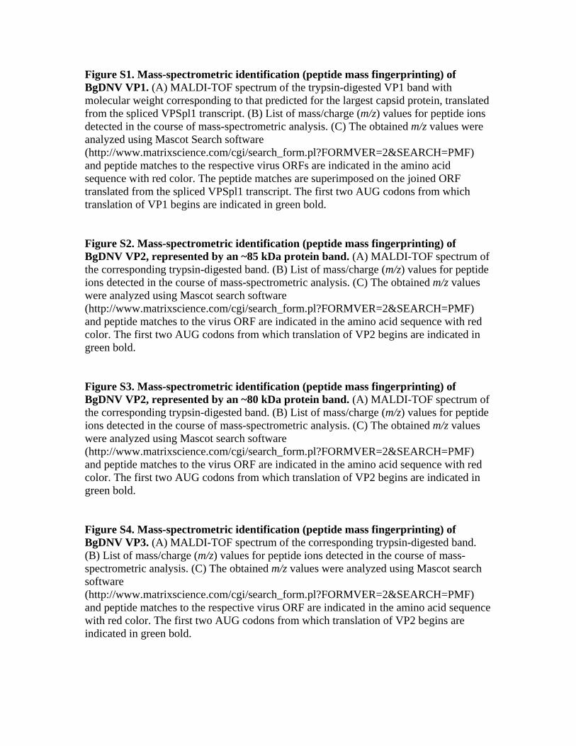
Figure S1. Mass-spectrometric identification (peptide mass fingerprinting) of BgDNV VP1. (A) MALDI-TOF spectrum of the trypsin-digested VP1 band with molecular weight corresponding to that predicted for the largest capsid protein, translated from the spliced VPSpl1 transcript. (B) List of mass/charge (m/z) values for peptide ions detected in the course of mass-spectrometric analysis. (C) The obtained m/z values were analyzed using Mascot Search software (http://www.matrixscience.com/cgi/search_form.pl?FORMVER=2&SEARCH=PMF) and peptide matches to the respective virus ORFs are indicated in the amino acid sequence with red color. The peptide matches are superimposed on the joined ORF translated from the spliced VPSpl1 transcript. The first two AUG codons from which translation of VP1 begins are indicated in green bold. Figure S2. Mass-spectrometric identification (peptide mass fingerprinting) of BgDNV VP2, represented by an ~85 kDa protein band. (A) MALDI-TOF spectrum of the corresponding trypsin-digested band. (B) List of mass/charge (m/z) values for peptide ions detected in the course of mass-spectrometric analysis. (C) The obtained m/z values were analyzed using Mascot search software (http://www.matrixscience.com/cgi/search_form.pl?FORMVER=2&SEARCH=PMF) and peptide matches to the virus ORF are indicated in the amino acid sequence with red color. The first two AUG codons from which translation of VP2 begins are indicated in green bold. Figure S3. Mass-spectrometric identification (peptide mass fingerprinting) of BgDNV VP2, represented by an ~80 kDa protein band. (A) MALDI-TOF spectrum of the corresponding trypsin-digested band. (B) List of mass/charge (m/z) values for peptide ions detected in the course of mass-spectrometric analysis. (C) The obtained m/z values were analyzed using Mascot search software (http://www.matrixscience.com/cgi/search_form.pl?FORMVER=2&SEARCH=PMF) and peptide matches to the virus ORF are indicated in the amino acid sequence with red color. The first two AUG codons from which translation of VP2 begins are indicated in green bold. Figure S4. Mass-spectrometric identification (peptide mass fingerprinting) of BgDNV VP3. (A) MALDI-TOF spectrum of the corresponding trypsin-digested band. (B) List of mass/charge (m/z) values for peptide ions detected in the course of mass-spectrometric analysis. (C) The obtained m/z values were analyzed using Mascot search software (http://www.matrixscience.com/cgi/search_form.pl?FORMVER=2&SEARCH=PMF) and peptide matches to the respective virus ORF are indicated in the amino acid sequence with red color. The first two AUG codons from which translation of VP2 begins are indicated in green bold.
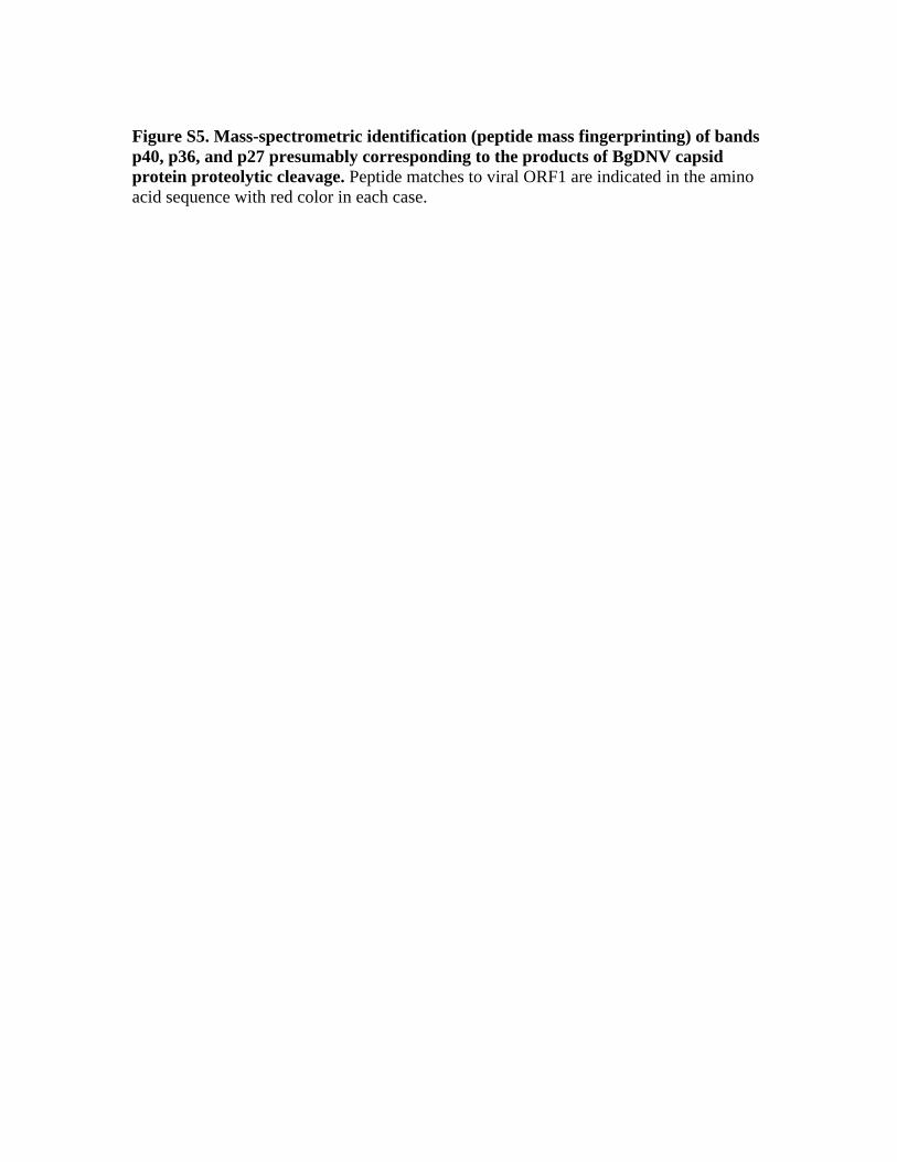
Figure S5. Mass-spectrometric identification (peptide mass fingerprinting) of bands p40, p36, and p27 presumably corresponding to the products of BgDNV capsid protein proteolytic cleavage. Peptide matches to viral ORF1 are indicated in the amino acid sequence with red color in each case.
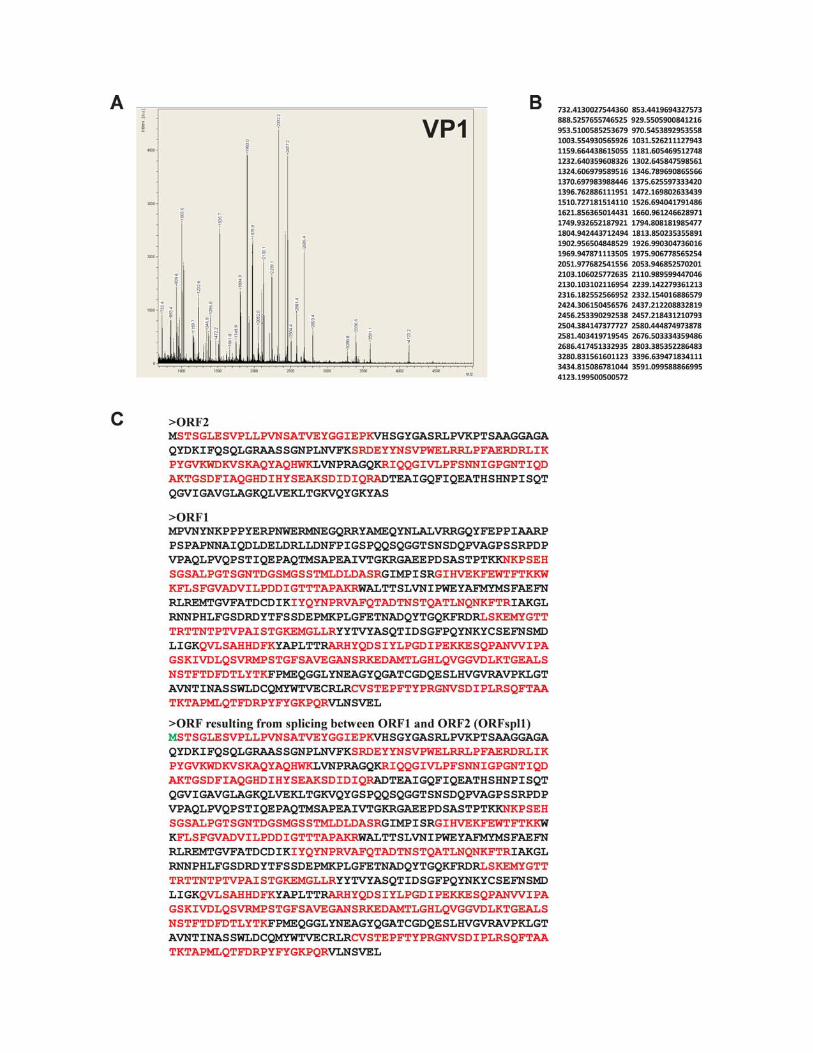
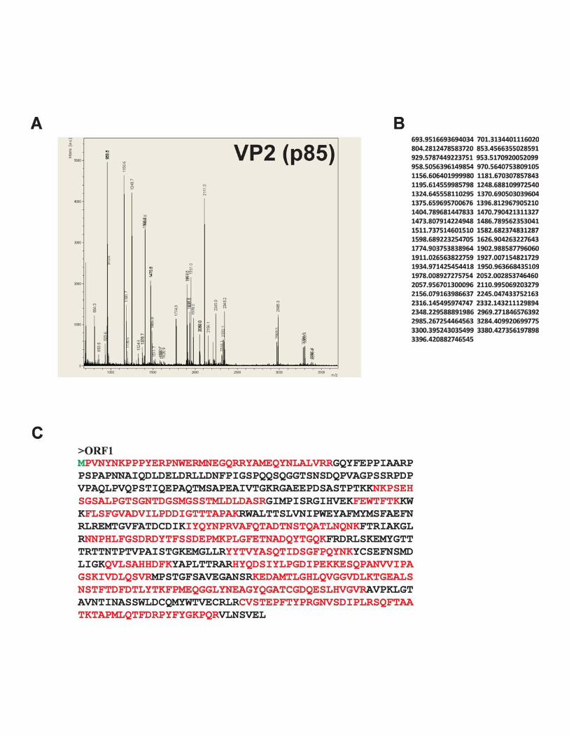
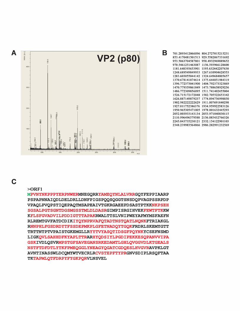
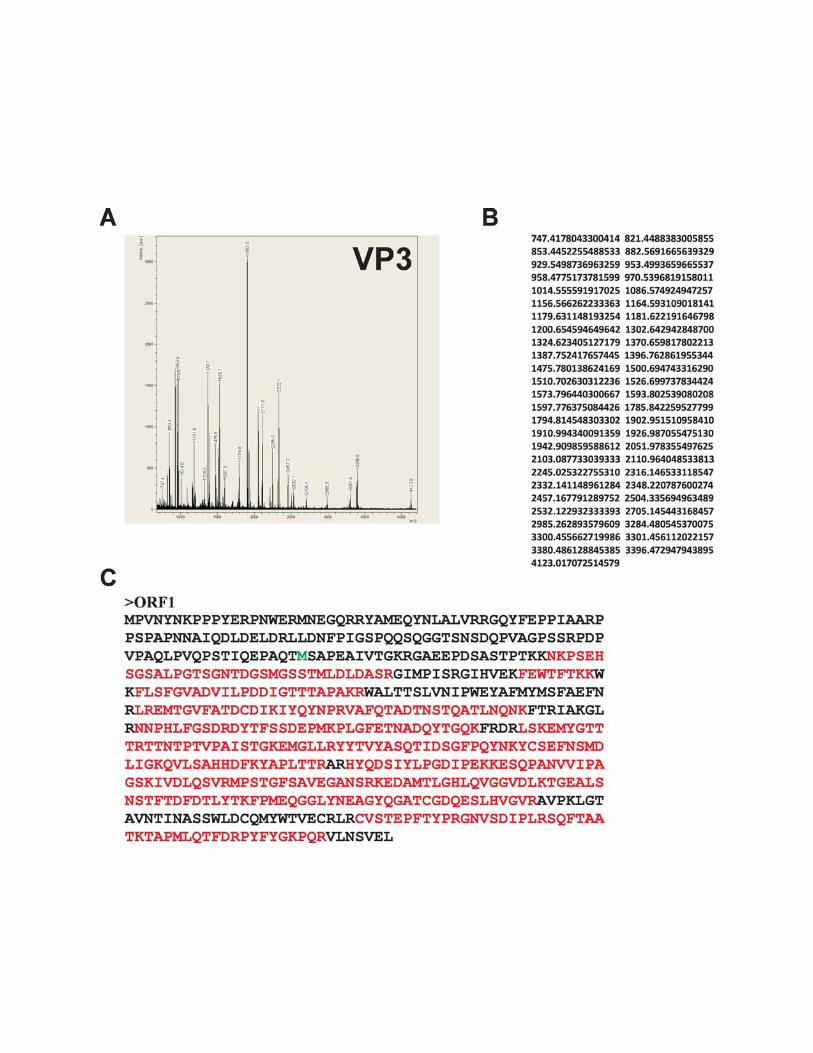
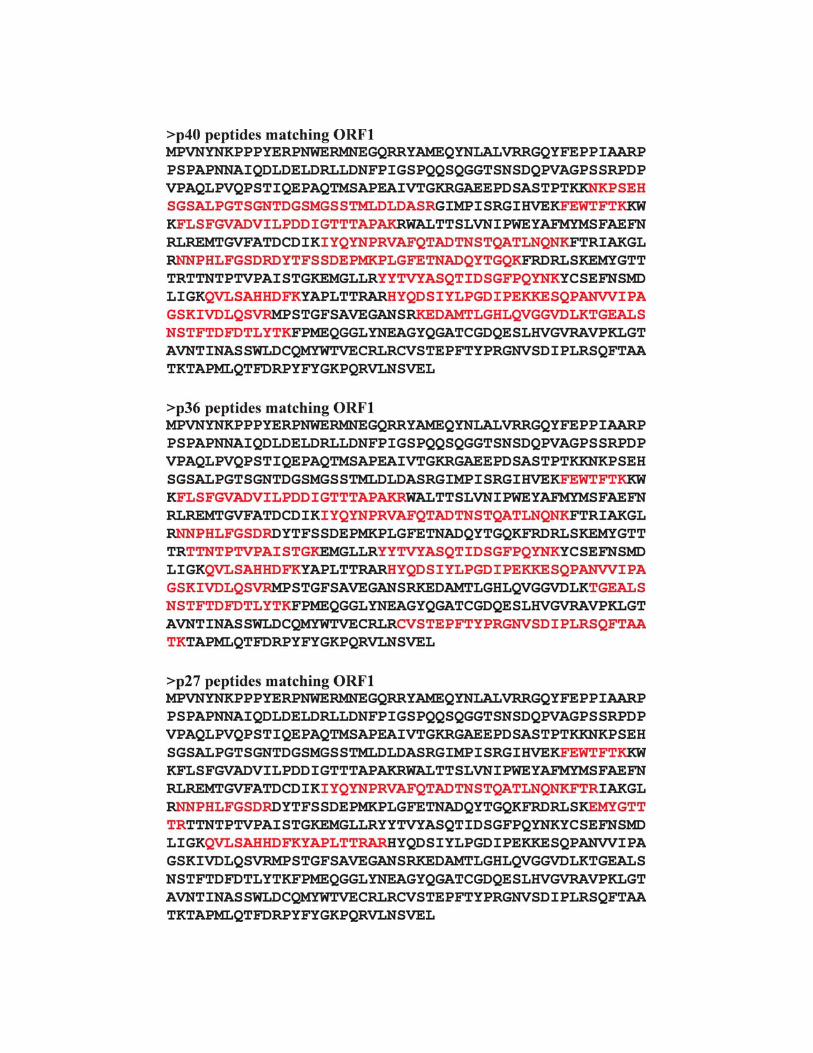
Copyright © 2022 FDOKUMEN