Exploring the surface reactivity of Ag nanoparticles with antimicrobial activity: A DFT study
7
See discussions, stats, and author profiles for this publication at: https://www.researchgate.net/publication/257246796 Exploring the surface reactivity of Ag nanoparticles with antimicrobial activity: A DFT study Article in International Journal of Quantum Chemistry · September 2012 DOI: 10.1002/qua.24207 CITATIONS 0 READS 56 4 authors, including: Some of the authors of this publication are also working on these related projects: Electromagnetic properties and applications of self-assembled nanostructures View project Rubén E. Estrada-Salas Tijuana Institute of Technology 6 PUBLICATIONS 98 CITATIONS SEE PROFILE Hector Barron CSIRO Data61 16 PUBLICATIONS 147 CITATIONS SEE PROFILE Miguel Jose Yacaman University of Texas at San Antonio 485 PUBLICATIONS 12,181 CITATIONS SEE PROFILE All content following this page was uploaded by Hector Barron on 13 August 2014. The user has requested enhancement of the downloaded file. All in-text references underlined in blue are linked to publications on ResearchGate, letting you access and read them immediately.
-
Upload
independent -
Category
Documents
-
view
1 -
download
0
Transcript of Exploring the surface reactivity of Ag nanoparticles with antimicrobial activity: A DFT study
Seediscussions,stats,andauthorprofilesforthispublicationat:https://www.researchgate.net/publication/257246796
ExploringthesurfacereactivityofAgnanoparticleswithantimicrobialactivity:ADFTstudy
ArticleinInternationalJournalofQuantumChemistry·September2012
DOI:10.1002/qua.24207
CITATIONS
0
READS
56
4authors,including:
Someoftheauthorsofthispublicationarealsoworkingontheserelatedprojects:
Electromagneticpropertiesandapplicationsofself-assemblednanostructuresViewproject
RubénE.Estrada-Salas
TijuanaInstituteofTechnology
6PUBLICATIONS98CITATIONS
SEEPROFILE
HectorBarron
CSIROData61
16PUBLICATIONS147CITATIONS
SEEPROFILE
MiguelJoseYacaman
UniversityofTexasatSanAntonio
485PUBLICATIONS12,181CITATIONS
SEEPROFILE
AllcontentfollowingthispagewasuploadedbyHectorBarronon13August2014.
Theuserhasrequestedenhancementofthedownloadedfile.Allin-textreferencesunderlinedinbluearelinkedtopublicationsonResearchGate,lettingyouaccessandreadthemimmediately.
Exploring the Surface Reactivity of Ag Nanoparticles withAntimicrobial Activity: A DFT Study
Rub�en E. Estrada-Salas,[a] Hector Barr�on,[a] Ariel A. Valladares,*[b]
and Miguel Jos�e-Yacam�an[a]
The preferential sites of electrophilic, nucleophilic, and radical
attacks on the surface of highly spherical Ag nanoparticles with a
diameter of � 2 nm are studied via the Fukui functions and the
molecular electrostatic potential; both are calculated using the
density functional (DFT) generalized gradient approximation-
revised version of the Perdew, Burke, and Ernzerhof level of
theory with the double-numerical with polarization functions
(DNP) basis set for the valence electrons, and DFT-based
semicore pseudopotentials for the core electrons. Because the
interaction of Ag nanoparticles with virus and microorganisms
takes place in an aqueous environment, the solvent (water) effect
is also obtained using the Conductor-like Screening Model. Three
typical structures are chosen: cuboctahedral, icosahedral, and
ino-decahedral. All three present an ‘‘amphoteric’’ behavior
against electrophiles, nucleophiles, and radicals. For the
cuboctahedral and decahedral geometries, the highest
susceptibility to attack is on the edges shared by a {111} face and
a {100} face; for the icosahedral geometry, the highest
susceptibility to attack is on the vertices. Ionization potentials,
electron affinities, electronegativities, and chemical hardness are
also reported. Comparison with experiments is presented.VC 2012
Wiley Periodicals, Inc.
DOI: 10.1002/qua.24207
Introduction
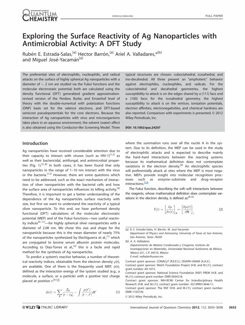
Ag nanoparticles have received considerable attention due to
their capacity to interact with viruses (such as HIV-1)[1,2] as
well as their bactericidal, antifungal, and antimicrobial proper-
ties (Fig. 1).[3–8] In both cases, it has been found that only
nanoparticles in the range of 1–10 nm interact with the virus
or the bacteria.[1,3] However, there are some questions which
need to be addressed, such as the exact mechanism of interac-
tion of silver nanoparticles with the bacterial cells and how
the surface area of nanoparticles influences its killing activity.[4]
Therefore, it is important to get a better understanding of the
dependence of the Ag nanoparticles surface reactivity with
size, but first we want to understand the reactivity of a typical
silver nanoparticle. To this end, we have performed density
functional (DFT) calculations of the molecular electrostatic
potential (MEP) and of the Fukui functions—two useful reactiv-
ity indices[9–11]—for highly spherical silver nanoparticles with a
diameter of 2.08 nm. We chose this size and shape for the
nanoparticle because this is the mean diameter of nearly 75%
of the nanoparticles synthesized by Elechiguerra et al.,[1] which
are conjugated to bovine serum albumin protein molecules.
According to Diaz-Torres et al.,[2] this is a facile and rapid
method for the synthesis of Ag nanoparticles.
To predict a system’s reactive behavior, a number of theoret-
ical reactivity indices, obtainable from the electron density q(r),are available. One of them is the frequently used MEP, /(r),defined as the interaction energy of the system studied (e.g., a
molecule, a surface, or a particle) with a positive test charge
placed at position r:[9,12]
/ rð Þ ¼XA
ZAr � rAj j �
Zq r0ð Þr � r0j j dr
0 (1)
where the summation runs over all the nuclei A in the sys-
tem. Due to its definition, the MEP can be used in the study
of electrophilic attacks and is expected to describe mainly
the hard–hard interactions between the reacting systems
because its mathematical definition does not contemplate
variations in the electron density.[9] An electrophilic species
will preferentially attack at sites where the MEP is most nega-tive. MEPs provide insight into molecular recognition proc-esses such as enzyme–substrate and drug–receptorinteractions.[12]
The Fukui function, describing the soft–soft interactions between
the reagents, whose mathematical definition does contemplate var-
iations in the electron density, is defined as:[9,13]
f rð Þ ¼ dldm rð Þ
� �N
¼ @q rð Þ@N
� �m
(2)
[a] R. E. Estrada-Salas, H. Barr�on, M. Jos�e-Yacam�an
Department of Physics and Astronomy, University of Texas at San Antonio,
San Antonio, Texas 78249
[b] A. A. Valladares
Departamento de Materia Condensada y Criogenia, Instituto de
Investigaciones en Materiales, Universidad Nacional Aut�onoma de M�exico,
M�exico D.F., C.P. 04510, M�exico
E-mail: [email protected]
Contract grant sponsor: CONACyT (R.E.E.S.), DGAPA-UNAM (A.A.V.).
Contract grant sponsor: Welch Foundation Project (H.B. and M.J.Y.); contract
grant number: AX-1615.
Contract grant sponsor; National Science Foundation (NSF) PREM (H.B. and
M.J.Y.); contract grant number: DMR 0934218.
Contract grant sponsor: NIH-RCMI Center for Interdisciplinary Health
Research (H.B. and M.J.Y.); contract grant number: 2G12RR013646-11.
Contract grant sponsor: The NSF (H.B. and M.J.Y.); contract grant number:
DMR-1103730.
VC 2012 Wiley Periodicals, Inc.
International Journal of Quantum Chemistry 2012, 112, 3033–3038 3033
FULL PAPERWWW.Q-CHEM.ORG
Because of the discontinuity in Eq. (2)—the electron number
N can change only by integer values—different physical mean-
ings have been associated with the left and right derivatives
as well as with their average value, corresponding to a reactiv-
ity index for an electrophilic [f �(r)], a nucleophilic [f þ(r)], anda radical attack [f 0(r)], respectively.[9,13]
Other chemical properties suitable for analyzing chemical
reactivity were also used, namely the ionization potential IP,
the electron affinity EA, the electronegativity v, and the chemi-
cal hardness g of each nanoparticle studied.
The IP is defined as the amount of energy necessary to
remove one electron from a chemical species (i.e., atom, mole-
cule, cluster, etc.) in the gas phase:[14]
X0ðgÞ ! Xþ
ðgÞ þ e�;DE ¼ Ip (3)
The IP can be calculated as the difference between the
energy of the positively charged species Eþ and the energy of
the neutral species E0:
IP ¼ Eþ � E0 (4)
The EA is related to the energy of the process of accepting
one electron from a chemical species:[14]
X0ðgÞ þ e� ! X�
ðgÞ; DE ¼ EA (5)
The EA can be calculated as the difference between the
energy of the neutral species E0 and the energy of the nega-
tively charged species E�:
EA ¼ E0 � E� (6)
The v is defined as the tendency of a species to attract electrons.
It can be calculated as the average between the IP and the EA:[15]
v ¼ IP þ EA2
(7)
Finally, the chemical hardness g is also defined in terms of
the IP and the EA[15] by using the following equation:
g ¼ IP � EA2
(8)
The v and g are usually taken as
absolute values and are very useful
properties in the study of the chemical
reactivity of different species.[15]
Calculation details
Full geometry optimizations and total
energy calculations were performed
with the DFT-based program package
DMol[3] at the generalized gradient
approximation level of theory.[16–18] Spe-
cifically, we used the revised version of
the Perdew, Burke, and Ernzerhof (RPBE)
functional[19] with double numerical
atomic orbital basis sets plus polarization functions (DNP)[20]
for the treatment of the valence electrons and DFT-based
semicore pseudopotentials (DSPPs)[21] for the treatment of the
core electrons. DSPPs introduce some degree of relativistic cor-
rection into the core, such relativistic corrections are important
in the study of heavy elements like silver.
We chose this level of theory because the large dimension
of the systems makes the use of higher order theoretical
approaches such as the second-order many-body perturbation
method or the configuration interaction method, unfeasible.
We decided to use the RPBE DFT because it presents essen-
tially the same performance as the well tested PW91[22] and
Perdew, Burke, and Ernzerhof (PBE)[23] functionals, but
improves the calculation of adsorption energies[19] and there-
fore, it is more suitable in studies of surface reactivity.
Due to the fact that the interaction of Ag nanoparticles with
viruses and microorganisms takes place in an aqueous envi-
ronment, and the solvent (water) effect was also taken into
account by using the Conductor-like Screening Model.[24,25] As
it is shown in the Results and Discussion section, clear differ-
ences in the surface reactivity, with and without the effect of
the solvent, were found.
Because of the face-centered-cubic (FCC) crystalline struc-
ture of silver, the atomistic models of the nanoparticles with a
diameter of �2 nm cannot be completely spherical in shape;
therefore, we chose the three typical structures that have
been observed for small particles of materials whose bulk
structure is FCC: cuboctahedral, icosahedral, and decahedral,
Figure 2. Each of these three structures consists of 147 silver
atoms, the magic number corresponding to the size of
�2 nm.[26]
For a clear visualization of the silver nanoparticles surface
reactivity, both the calculated MEP and Fukui functions were
mapped onto an isosurface of the total electron density of
each nanoparticle, showing the sites of high susceptibility to
electrophilic, nucleophilic, and radical attacks on the nanopar-
ticle surface.
The ionization potentials and the electron affinities were cal-
culated taking the energy differences between the total
Figure 1. Electron microscopy images showing the interaction of silver nanoparticles with: a) an HIV-1
virus, and b) an E. coli bacteria (these electron microscopy images were taken at the Dr. M. J. Yaca-
man’s Laboratory of Nanomaterials, Department of Physics and Astronomy, University of Texas at San
Antonio).
FULL PAPER WWW.Q-CHEM.ORG
3034 International Journal of Quantum Chemistry 2012, 112, 3033–3038 WWW.CHEMISTRYVIEWS.ORG
energy of each of the positively charged, the neutral, and the
negatively charged nanoparticles [using Eqs. (4) and (6)]. The
electronegativities were then calculated with Eq. (7).
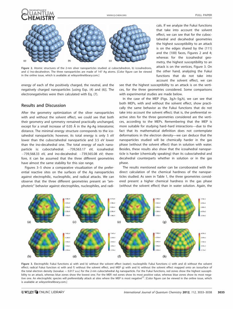
Results and Discussion
After the geometry optimization of the silver nanoparticles
with and without the solvent effect, we could see that both
their geometry and symmetry remained practically unchanged,
except for a small increase of 0.05 A in the Ag-Ag interatomic
distance. The minimal energy structure corresponds to the ico-
sahedral nanoparticle; however, its total energy is only 5 eV
lower than the cuboctahedral nanoparticle and 3.5 eV lower
than the ino-decahedral one. The total energy of each nano-
particle is: cuboctahedral: �739,563.17 eV, icosahedral:
�739,568.33 eV, and ino-decahedral: �739,565.08 eV; there-
fore, it can be assumed that the three different geometries
have almost the same stability for this size range.
Figures 3–5 show a comparative visualization of the prefer-
ential reactive sites on the surfaces of the Ag nanoparticles
against electrophilic, nucleophilic, and radical attacks. We can
observe that the three different geometries present an ‘‘am-
photeric’’ behavior against electrophiles, nucleophiles, and radi-
cals. If we analyze the Fukui functions
that take into account the solvent
effect, we can see that for the cuboc-
tahedral and decahedral geometries
the highest susceptibility to an attack
is on the edges shared by the {111}
and the {100} faces, Figures 2 and 4;
whereas for the icosahedral geo-
metry, the highest susceptibility to an
attack is on the vertices, Figure 3. On
the other hand, analyzing the Fukui
functions that do not take into
account the solvent effect, we can
see that the highest susceptibility to an attack is on the verti-
ces, for the three geometries considered. Some comparisons
with experimental studies are made below.
In the case of the MEP (Figs. 3g,h–5g,h), we can see that
both MEPs, with and without the solvent effect, show practi-
cally the same behavior as the Fukui functions that do not
take into account the solvent effect; that is, the preferential re-
active sites for the three geometries considered are the verti-
ces, according to the MEPs. Remembering that the MEP is
more suitable for studying hard–hard interactions—due to the
fact that its mathematical definition does not contemplate
deformations in the electron density—we can deduce that the
nanoparticles studied will be chemically harder in the gas
phase (without the solvent effect) than in solution with water.
Besides, these results also show that the icosahedral nanopar-
ticle is harder (chemically speaking) than its cuboctahedral and
decahedral counterparts whether in solution or in the gas
phase.
The results mentioned earlier can be corroborated with the
direct calculation of the chemical hardness of the nanopar-
ticles studied. As seen in Table 1, the three geometries consid-
ered present a higher chemical hardness in the gas phase
(without the solvent effect) than in water solution. Again, the
Figure 2. Atomic structures of the 2-nm silver nanoparticles studied: a) cuboctahedron, b) icosahedrons,
and c) Ino-decahedron. The three nanoparticles are made of 147 Ag atoms. [Color figure can be viewed
in the online issue, which is available at wileyonlinelibrary.com.]
Figure 3. Electrophilic Fukui functions a) with and b) without the solvent effect (water); nucleophilic Fukui functions c) with and d) without the solvent
effect; radical Fukui function e) with and f) without the solvent effect, and MEP g) with and h) without the solvent effect mapped onto an isosurface of
the total electron density (isovalue ¼ 0.017 a.u.) for the 2-nm cuboctahedral Ag nanoparticle. For the Fukui functions, red zones show the highest suscepti-
bility to an attack, whereas blue zones show the lowest one. For the MEP, red zones show its most positive value, whereas blue zones show its most nega-
tive one. An electrophilic species will preferentially attack at sites where the MEP is most negative[7]. [Color figure can be viewed in the online issue, which
is available at wileyonlinelibrary.com.]
FULL PAPERWWW.Q-CHEM.ORG
International Journal of Quantum Chemistry 2012, 112, 3033–3038 3035
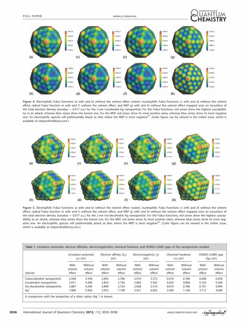
Figure 4. Electrophilic Fukui functions a) with and b) without the solvent effect (water); nucleophilic Fukui functions c) with and d) without the solvent
effect; radical Fukui function e) with and f) without the solvent effect, and MEP g) with and h) without the solvent effect mapped onto an isosurface of
the total electron density (isovalue ¼ 0.017 a.u.) for the 2-nm icosahedral Ag nanoparticle. For the Fukui functions, red zones show the highest susceptibil-
ity to an attack, whereas blue zones show the lowest one. For the MEP, red zones show its most positive value, whereas blue zones show its most negative
one. An electrophilic species will preferentially attack at sites where the MEP is most negative[7]. [Color figure can be viewed in the online issue, which is
available at wileyonlinelibrary.com.]
Figure 5. Electrophilic Fukui functions a) with and b) without the solvent effect (water); nucleophilic Fukui functions c) with and d) without the solvent
effect; radical Fukui function e) with and f) without the solvent effect, and MEP g) with and h) without the solvent effect mapped onto an isosurface of
the total electron density (isovalue ¼ 0.017 a.u.) for the 2-nm Ino-decahedral Ag nanoparticle. For the Fukui functions, red zones show the highest suscep-
tibility to an attack, whereas blue zones show the lowest one. For the MEP, red zones show its most positive value, whereas blue zones show its most neg-
ative one. An electrophilic species will preferentially attack at sites where the MEP is most negative[7]. [Color figure can be viewed in the online issue,
which is available at wileyonlinelibrary.com.]
Table 1. Ionization potentials, electron affinities, electronegativities, chemical hardness, and HOMO–LUMO gaps of the nanoparticles studied.
Species
Ionization potential
(IP) [eV]
Electron affinity (EA)
[eV]
Electronegativity (v)[eV]
Chemical hardness
(g) [eV]HOMO–LUMO gap
(Eg) [eV]
With
solvent
effect
Without
solvent
effect
With
solvent
effect
Without
solvent
effect
With
solvent
effect
Without
solvent
effect
With
solvent
effect
Without
solvent
effect
With
solvent
effect
Without
solvent
effect
Cuboctahedral nanoparticle 2.928 4.358 2.892 2.786 2.910 3.572 0.018 0.786 0.068 0.058
Icosahedral nanoparticle 2.911 4.368 2.854 2.756 2.883 3.562 0.029 0.806 0.162 0.206
Ino-decahedral nanoparticle 2.887 4.296 2.849 2.724 2.868 3.510 0.019 0.786 0.101 0.094
Agþ 7.889 5.456 2.952 7.748 5.421 6.602 2.469 1.146 3.712 4.690
A comparison with the properties of a silver cation (Agþ) is shown.
FULL PAPER WWW.Q-CHEM.ORG
3036 International Journal of Quantum Chemistry 2012, 112, 3033–3038 WWW.CHEMISTRYVIEWS.ORG

icosahedral nanoparticle presents the highest hardness in both
cases, whether or not we include the solvent effect.
The calculated values for the highest occupied molecular or-
bital (HOMO)–lowest unoccupied molecular orbital (LUMO)
gaps of each of the studied nanoparticles also support these
findings (Table 1). It is well known that hard molecules have a
large HOMO–LUMO gap, and soft molecules have a small
HOMO–LUMO gap;[15] therefore, the lower values obtained for
the HOMO–LUMO gaps when taking into account the solvent
effect indicate that the nanoparticles are harder in the gas
phase than in water solution. Once again, the icosahedral
nanoparticle is the hardest of the three nanoparticles studied
since it presents the highest value for the HOMO–LUMO gap.
Morones et al.[3] and Pal et al.[8] proposed that the {111}
faces of the Ag nanoparticles are the most reactive sites on
the nanoparticle surfaces. Our results show that—at least for
the nanoparticle size studied (i.e., �2 nm)—the more reactive sites
are not precisely located on top of the {111} faces but on the edges
shared by a {111} and a {100} face (in the case of the cuboctahedral
and decahedral nanoparticles) as well as on the vertices (in the case
of the icosahedral nanoparticle). It is evident, however, that the
atoms of these edges and vertices are also part of the {111} faces;
therefore, in some sense we can say that parts of the {111} faces are
the most reactive sites for the nanoparticles studied. Nevertheless,
as the size range studied by Morones et al.[3] and Pal et al.[8] includes
nanoparticles from 1 to 100 nm, it can be expected that the most
reactive sites on the surfaces of the nanoparticles will change with
size; therefore, as the nanoparticle increases in size, the most reac-
tive sites might become localized on top of the {111} faces. Further
theoretical studies regarding the size dependence of the reactivity
of Ag nanoparticles are encouraged.
It has been long proposed that the antimicrobial mechanism
of silver nanoparticles is mainly due to the release of silver
ions (Agþ) from the nanoparticle surface; however, experimen-
tal results by Despax et al.[5] indicated that Agþ ions released
from nanosilver structures did not constitute the main (or the
sole) vector of interaction with sulfur (S) or phosphorus (P)
containing compounds, that is, proteins, nucleic acids, and so
forth. They also found the formation of Ag/S clusters inside
the cells when treating the microorganisms with silver ions
(Agþ), and the formation of Ag/S/P clusters when treating the
microorganisms with nanosilver, indicating clear differences in
the reactivity of Agþ and silver nanostructures.
To get insight on the differences in the reactivity of these
two distinct silver species—Agþ and nanosilver—we also per-
formed calculations of the reactivity of a single silver ion (Agþ)for comparison with the silver nanoparticles studied (see Table
1). The fact that the silver ions form only Ag/S clusters inside
the cells, whereas the nanosilver treatment leads to the forma-
tion of Ag/S/P clusters[5] can be correlated with the notably
distinct electronegativity, chemical hardness, and HOMO–
LUMO gap shown by each species. As is evident from Table 1,
a single Agþ ion is considerably more electronegative and
harder than any of the three Ag nanoparticles studied (cubo-,
ico-, and decahedral). This indicates that Agþ prefers to inter-
act with sulfur as it is more electronegative and harder than
phosphorus, which can explain in part why some experi-
ments[5] found that silver ions only form Ag/S clusters without
any significant amount of phosphorus inside the cells. On the
other hand, the presence of Ag/S/P clusters found inside the
microorganisms when treating them with nanosilver[5] can be
explained by the presence of two different silver species,
namely the Ag nanoparticles—which possibly will prefer to
interact with the softer phosphorus atoms rather than with
the harder sulfur ones—and the Agþ ions released by the sil-
ver nanoparticles.
Some recent works assume that the biocide activity of silver
nanoparticles is due to their radical character.[3,5,6] This is in
agreement with our work as the ‘‘amphoteric’’ behavior against
electrophiles and nucleophiles shown by the calculated Fukui
functions of the Ag nanoparticles studied is a typical behavior
of any radical species.
It has long been pointed out that the antimicrobial activity
of silver nanoparticles is critically dependent on particle size,
shape, and surface oxidation.[5–8] For example, Lok et al.[7]
have indicated that partially surface-oxidized nanosilver (�9
nm) exhibits antibacterial activities, whereas zero-valent nano-
silver does not. They also indicated that the smaller silver
nanoparticles had higher antibacterial activity than the bigger
ones. Although our calculations cannot give insight into the
surface oxidation dependence of the nanoparticles reactivity,
they do shed light on some other issues, mainly on those con-
cerning the shape dependence as well as the solvent depend-
ence of the Ag nanoparticles reactivity.
Summary and Conclusions
Our calculations indicate that, for the nanoparticle size studied
(�2 nm), the more reactive sites when taking into account the
solvent effect, are localized on the edges shared by a {111} and
a {100} face in the case of the cuboctahedral and decahedral
nanoparticles, and on the vertices in the case of the icosahedral
nanoparticle. Our calculations also showed that the silver nano-
particles studied present a softer chemical behavior in water so-
lution than in the gas phase. Comparison between the Ag nano-
particles reactivity and the Agþ reactivity gives us a better
understanding of some experimental findings.[5] The radical
character of the silver nanoparticles assumed in some works[3,5,6]
as the origin of their biocide activity is in agreement with our
findings. Further theoretical studies on the dependence of the
Ag nanoparticles reactivity with respect to distinct parameters
such as size, shape, and surface oxidation are in order.
Acknowledgments
M.T. V�azquez and O. Jim�enez have provided the information
requested. Part of this work was carried out on the computers of
DGSCA, UNAM. R.E.E.S., H.B., and M.J.Y. would also like to thank the
International Center for Nanotechnology and Advanced Materials
(ICNAM) at UTSA.
Keywords: silver nanoparticles � surface reactivity � bactericidalproperties � Fukui functions � molecular electrostatic poten-
tial � DFT studies
FULL PAPERWWW.Q-CHEM.ORG
International Journal of Quantum Chemistry 2012, 112, 3033–3038 3037

How to cite this article: R. E. Estrada-Salas, H. Barr�on, A. A.
Valladares, M. Jos�e-Yacam�an, Int. J. Quantum Chem. 2012, 112,
3033–3038. DOI: 10.1002/qua.24207
[1] J. L. Elechiguerra, J. L. Burt, J. R. Morones, A. Camacho-Bragado, X.
Gao, H. H. Lara, M. J. Yacaman, J. Nanobiotechnol. 2005, 3, 6: 1.
[2] L. A. Diaz-Torres, D. Ferrer, M. J. Yacaman, The Third San Antonio Bio-
photonics Symposium, San Antonio, TX. March 28–29, 2008.
[3] J. R. Morones, J. L. Elechiguerra, A. Camacho, K. Holt, J. B. Kouri, J.
Tapia Ramırez, M. J. Yacaman, Nanotechnology 2005, 16, 2346.
[4] M. Rai, A. Yadav, A. Gade, Biotechnol. Adv. 2009, 27, 76.
[5] B. Despax, C. Saulou, P. Raynaud, L. Datas, M. Mercier-Bonin, Nanotech-
nology 2011, 22, 175101: 1.
[6] J. S. Kim, E. Kuk, K. N. Yu, J. H. Kim, S. J. Park, H. J. Lee, S. H. Kim, Y. K.
Park, Y. H. Park, C. Y. Hwang, Y. K. Kim, Y. S. Lee, D. H. Jeong, M. H.
Cho, Nanomedicine 2007, 3, 95.
[7] C. N. Lok, C. M. Ho, R. Chen, Q. Y. He, W. Y. Yu, H. Sun, P. K. H. Tam, J.
F. Chiu, C. M. Che, J. Biol. Inorg. Chem. 2007, 12, 527.
[8] S. Pal, Y. K. Tak, J. M. Song, Appl. Environ. Microbiol. 2007, 73, 1712.
[9] F. De Proft, J. M. L. Martin, P. Geerlings, Chem. Phys. Lett. 1996, 256, 400.
[10] R. Estrada-Salas, A. A. Valladares, J. Mol. Struct. Theochem. 2008, 869, 1.
[11] R. Estrada-Salas, A. A. Valladares, J. Phys. Chem. A 2009, 113, 10299.
[12] I. Levine, Quantum Chemistry, 5th ed.; Prentice Hall: New Jersey, 2000.
[13] R. G. Parr, W. Yang, Density-Functional Theory of Atoms and Molecules;
Oxford University Press: New York, 1989.
[14] A. D. McNaught, A. Wilkinson, IUPAC Compendium of Chemical Termi-
nology–The Gold Book; Blackwell Science: Cambridge, 1997. http://
goldbook.iupac.org. Accessed on June 21, 2012.
[15] R. G. Pearson, Proc. Natl. Acad. Sci. USA 1986, 83, 8440.
[16] B. Delley, J. Chem. Phys. 1990, 92, 508.
[17] B. Delley, J. Chem. Phys. 2000, 113, 7756.
[18] Materials Studio Release 4.4; Accelrys Software Inc.: San Diego, CA,
USA, 2008.
[19] B. Hammer, L. B. Hansen, J. K. Norskov, Phys. Rev. B 1999, 59, 7413.
[20] B. Delley, J. Phys. Chem. A 2006, 110, 13632.
[21] B. Delley, Phys. Rev. B 2002, 66,155125: 1.
[22] J. P. Perdew, J. A. Chevary, S. H. Vosko, K. A. Jackson, M. R. Pederson,
D. J. Singh, C. Fiolhais, Phys. Rev. B 1992, 46, 6671.
[23] J. P. Perdew, K. Burke, M. Ernzerhof, Phys. Rev. Lett. 1996, 77,
3865.
[24] A. Klamt, G. Schuurmann, J. Chem. Soc. Perkins Trans. 1993, 2, 799.
[25] B. Delley, Mol. Simul. 2006, 32, 117.
[26] G. Schmid, Chem. Soc. Rev. 2008, 37, 1909.
Received: 29 February 2012Revised: 29 February 2012Accepted: 24 April 2012Published online on 5 July 2012
FULL PAPER WWW.Q-CHEM.ORG
3038 International Journal of Quantum Chemistry 2012, 112, 3033–3038 WWW.CHEMISTRYVIEWS.ORG