Evaluation of the effectiveness of a home-based inspiratory muscle training programme in patients...
26
1 Title: Evaluation of the effectiveness of a home-based inspiratory muscle training programme in patients with Chronic Obstructive Pulmonary Disease using multiple inspiratory muscle tests. ABSTRACT Purpose: To evaluate the effectiveness of a home-based Inspiratory Muscle Training (IMT) programme using multiple inspiratory muscle tests. Method: Sixty eight patients (37M) with moderate to severe COPD (Mean [SD], FEV 1 36.1 [13.6]%pred.; FEV 1 /FVC 35.7 [11.2]%) were randomised into an experimental or control group and trained with a threshold loading device at intensity >30% maximum inspiratory pressure (PImax) or < 15%PImax respectively for 7 weeks. Thirty nine patients (23M) completed the study. The following measures were assessed pre- and post-IMT: PImax, sniff inspiratory nasal pressure (SNIP), diaphragm contractility (Pdi,tw), incremental shuttle walk test (ISWT), respiratory muscle endurance (RME), chronic respiratory disease questionnaire (CRDQ), the hospital anxiety and depression scale (HADS) and the SF-36. Between group changes were assessed using one-way analysis of variance (ANOVA). Results: PImax and perception of wellbeing improved significantly post-IMT [p=0.04 and <0.05 in four domains respectively]. This was not reflected in SNIP [p=0.7], Pdi,tw [p=0.8], RME [p=0.9] or ISWT [p=0.5]. Conclusions: A seven-week, community-based IMT programme, with realistic use of health- care resources, improves PImax and perception of wellbeing but a different design may be required for improvement in other measures. Multiple tests provide a more comprehensive evaluation of changes in muscle function post-IMT.
-
Upload
healthcare -
Category
Documents
-
view
1 -
download
0
Transcript of Evaluation of the effectiveness of a home-based inspiratory muscle training programme in patients...

1
Title: Evaluation of the effectiveness of a home-based inspiratory muscle training
programme in patients with Chronic Obstructive Pulmonary Disease using multiple
inspiratory muscle tests.
ABSTRACT
Purpose: To evaluate the effectiveness of a home-based Inspiratory Muscle Training (IMT)
programme using multiple inspiratory muscle tests.
Method: Sixty eight patients (37M) with moderate to severe COPD (Mean [SD], FEV1 36.1
[13.6]%pred.; FEV1/FVC 35.7 [11.2]%) were randomised into an experimental or control
group and trained with a threshold loading device at intensity >30% maximum inspiratory
pressure (PImax) or < 15%PImax respectively for 7 weeks. Thirty nine patients (23M)
completed the study. The following measures were assessed pre- and post-IMT: PImax, sniff
inspiratory nasal pressure (SNIP), diaphragm contractility (Pdi,tw), incremental shuttle walk
test (ISWT), respiratory muscle endurance (RME), chronic respiratory disease questionnaire
(CRDQ), the hospital anxiety and depression scale (HADS) and the SF-36. Between group
changes were assessed using one-way analysis of variance (ANOVA).
Results: PImax and perception of wellbeing improved significantly post-IMT [p=0.04 and
<0.05 in four domains respectively]. This was not reflected in SNIP [p=0.7], Pdi,tw [p=0.8],
RME [p=0.9] or ISWT [p=0.5].
Conclusions: A seven-week, community-based IMT programme, with realistic use of health-
care resources, improves PImax and perception of wellbeing but a different design may be
required for improvement in other measures. Multiple tests provide a more comprehensive
evaluation of changes in muscle function post-IMT.

2
Chronic obstructive pulmonary disease (COPD) is a major cause of mortality and morbidity
worldwide, estimated to become the 4th
leading cause of death by 2030 [1]. Rehabilitation
strategies based in community settings, such as early discharge schemes and home-based
exercise programmes, are important in the management of COPD patients as they are
associated with greater patient and carer satisfaction with services [2] and reduced time in
hospital [3]. Despite lacking the element of peer-support and social interaction, a benefit
often derived from participation in group rehabilitation programmes [4], home-based
rehabilitation interventions are ideal for patients who are unable to access centre based
programmes due to work/life commitments, profound breathlessness or comorbidities.
Inspiratory muscle training (IMT) is a rehabilitation intervention that has been investigated in
the context of pulmonary rehabilitation or on its own, with the rationale that an increased
inspiratory muscle capacity will improve perception of breathlessness and exercise tolerance
during activities of daily living [5-9].
IMT has no side effects and is an attractive
rehabilitation intervention for home-based programmes in COPD patients. However, despite
positive results in research studies, a recent update on the NICE guidelines for COPD [10]
highlighted the lack of evidence that would allow IMT to be recommended more widely by
clinicians.
Three main factors contribute to the uncertainty and debate about the clinical benefit of IMT
in COPD patients [11, 12]. First, most clinical studies assess the effects of IMT on inspiratory
muscle strength using the measurement of maximal inspiratory pressure (PImax) [11]. PImax
is a test known to be influenced by patients’ motivation and one that requires practice before
baseline values can be accepted [13, 14]. It is also similar to the respiratory efforts required
for IMT, raising the possibility that patients become better at performing the test rather than
being stronger [11]. Other inspiratory muscle tests that could confirm changes in inspiratory
muscle strength have rarely been used in IMT research [15, 16]. The Sniff inspiratory nasal

3
pressure (SNIP), a simple, non-invasive test that complements PImax [14, 17], requires a
different manoeuvre to IMT and patients are therefore unlikely to learn how to do the test
better during the training period. Although the use of multiple respiratory muscle tests
increases diagnostic accuracy, as reported by Steier and colleagues [18]
and are
recommended in the ATS/ERS guidelines on respiratory muscle testing [19], studies in
COPD have yet to use this approach to assess the effects of IMT programmes.
Another factor contributing to the uncertainty of the clinical benefit of IMT is that studies
offer limited information on effects on the diaphragm. IMT has been shown to lead to rib
cage muscle remodelling [20] but there is no information about remodelling of the diaphragm
in COPD patients. Data from an animal model have shown that IMT increases the thickness
of the rat diaphragm and leads to fast muscle fiber hypertrophy [21-23]. In COPD patients,
Heijdra and colleagues [15] showed increased transdiaphragmatic pressure (Pdi max)
following ten weeks of IMT at 60% training intensity. However, Pdi max was assessed using
PImax manoeuvres rather than a non-volitional test. No study so far has investigated the
effects of IMT on diaphragm contractility in COPD patients using non-volitional methods.
Finally, it is unclear whether the results of laboratory based research studies can be replicated
in clinical services. Many positive home-based studies have a long duration of training,
ranging from 3 to 12 months [24, 25] and close supervision, which would be unrealistic in
community health services. It is unclear whether similar results can be achieved by a more
feasible home-based programme.
The purpose of this study was to evaluate the effectiveness of a 7-week, home-based IMT
programme on inspiratory muscle strength using SNIP as well as PImax. The secondary aims
were to explore changes in diaphragm contractility, using non-volitional bilateral

4
anterolateral magnetic phrenic nerve stimulation (BAMPS), and to study the effects of our
IMT programme on inspiratory muscle endurance, exercise capacity and health status.
METHODS
Design Overview
This was a double blind, randomised controlled trial. The protocol was accepted by the
King’s College London research ethics committee (LREC no 98-211) and participants gave
their informed consent. Sixty-eight COPD patients were allocated into an experimental (PrBr-
IMT) and control group (C-IMT) by an independent investigator using the minimisation
method. Information about the randomisation was then sent to the physiotherapist who was
responsible for setting the IMT device and advising patients about their home training
programme. The physiotherapist’s role as an independent point of contact ensured that the
patients as well as the researcher who performed the assessments and analysed the tests were
blinded. Assessments were performed pre- and post-IMT by one researcher (lead author) who
was blinded to the group the patients belonged to.
Setting and Participants
Sixty-eight COPD patients (37M) with moderate to severe COPD (Mean [SD], FEV1/FVC
35.7 [11.2]%; FEV1 36.1 [13.6]%predicted) volunteered for the study. Patients were recruited
from respiratory outpatient clinics at King’s College Hospital, GP practices and British Lung
Foundation Breathe Easy groups. Patients gave written informed consent and were included
in the study if they had no exacerbation of COPD and had not changed their medication for at
least 4 weeks prior to the initial assessment. Exclusion criteria included: patients with a1-
antitrypsin deficiency, co-existing heart disease, hypertension, cor pulmonale or long term
use of oral corticosteroids and patients with significant thoracic musculoskeletal

5
abnormalities such as kyphosis or scoliosis. Patients unsuitable for magnetic stimulation, for
example those with cardiac pacemakers, were also excluded. Nineteen out of the sixty eight
patients were studied using bilateral anterolateral magnetic phrenic nerve stimulation
(BAMPS) to explore the effects of IMT on diaphragm contractility.
Randomisation and Intervention
Patients were assigned to the experimental (PrBr-IMT) or control group (C-IMT) by an
independent investigator using the minimisation method [26]. The PrBr-IMT and C-IMT
groups were matched for the following variables: a) age (3 categories: 50-60, 60-70 and >70
years old), b) baseline inspiratory muscle strength (PImax and SNIP combined), c) disease
severity (FEV1% predicted) and d) number of participants who performed invasive tests
during baseline assessments (2 categories: Yes or No).
Inspiratory muscle training (IMT) programme
The IMT programme was a 7 week, home-based programme using the POWERbreathe®
inspiratory muscle trainer (HaB International, Southam, Warwickshire, UK). Patients
allocated to the PrBr-IMT started training at 30% of their baseline PImax and increased the
intensity once a week as tolerated. This intensity has been shown to train the respiratory
muscles [27, 28] and is widely accepted as the minimum training intensity in IMT studies
[29, 30]. The threshold load was increased by revolving the load-adjustment valve in the
device’s handle which increases the inspiratory threshold loading pressure. The average
weekly increase in intensity of training was 5% and the mean (SD) intensity at the end of the
programme was 62% (SD:11.7) of the baseline PImax. In the C-IMT group, the device was
set below 15% PImax, an intensity shown not to train the respiratory muscles and often used
in control groups [27-30]. This load remained constant for the duration of the study.

6
The programme consisted of two IMT sessions per day, one in the morning and one in the
afternoon (5 hours apart), six days a week. Each session required patients to take a minimum
of 30 breaths in their device. The majority of patients increased the number of breaths to 40
by the end of the second week. Patients were strongly encouraged to take as many of the
breaths as possible without a break. They were instructed that if a break was required, this
should not exceed 1 min duration and the minimum number of breaths before a break should
be five.
The study physiotherapist called all patients (in the experimental and control group) weekly
to monitor progress and adherence to the programme but in the PrBr-IMT group only, she
encouraged patients to increase the intensity and the number of breaths per session. Patients
recorded this information in a diary booklet which was returned to the physiotherapist at the
end of the programme. Adherence to the programme was assessed and confirmed by
comparing the physiotherapist’s notes against each patient’s diary notes.
Outcome Measures and Follow-up
Measurements were performed at the beginning and end of the IMT programme by one
investigator (lead author) who was blinded throughout. Participants had a run-in period of 3
weeks to familiarise themselves with the volitional PImax and SNIP tests [14] before
performing the following assessments:
Lung function
Lung function testing was performed by senior clinical physiologists at the lung function
department of King’s College Hospital. Measurements were; forced expiratory volume in 1
second (FEV1), forced vital capacity (FVC), total lung capacity (TLC), residual volume (RV)

7
and blood gases. FEV1 and FVC were measured with a dry bellows spirometer (S Model,
Vitalograph, Buckinghamshire, UK) while TLC and RV were measured by constant whole
body plethysmography (Auto-link, Morgan Medical, Gillingham, Kent, UK). Blood gases
were measured from arterialised ear lobe capillary samples (Rapidlab 248, Chiron, MA,
USA).
Respiratory muscle strength
Respiratory muscle strength testing was performed according to ATS/ERS standards [19].
Prior to performing magnetic phrenic nerve stimulation, patients had oesophageal and gastric
balloon catheters inserted pernasally. Local anaesthesia was administered to the nasal mucosa
using 2% lignocaine gel (Biorex Laboratories,London,UK). The catheters were connected to
differential pressure transducers (Validyne MP45, Validyne Corp., Northridge, CA, USA) so
that pressure traces could be viewed on the computer screen. Data from pressure
measurements were acquired on a Macintosh PowerMac computer running Labview 4.1
software (National Instruments, Texas, USA). The following tests were performed:
Maximum inspiratory pressure (PImax)
Maximum inspiratory pressure is the most widely used test for the assessment of inspiratory
muscle strength and its reproducibility, repeatability and normal values have been assessed in
COPD [13, 14, 19]. In this study, PImax was assessed from functional residual capacity
(FRC) using a flanged mouthpiece (PK Morgan Ltd, Rainham, UK) connected to a metal tube
(Fitting et al). The metal tube was connected to a differential pressure transducer and PImax
tracings were recorded in real time. Nose clips were worn and patients performed a minimum
of 10 efforts to minimise the learning effect [14] with rest intervals of 30-60 seconds between
efforts [31, 32]. Visual feedback and strong encouragement were given throughout. A valve
in the tube allowed normal breathing before maximal efforts were made and a small lead

8
incorporated in the tube ensured maintenance of an open glottis. All patients were seated
upright with their belt undone and were encouraged to make maximal inspiratory efforts
sustained for one second.
Sniff nasal inspiratory pressure (SNIP)
The SNIP is a non-invasive test of global inspiratory muscle strength that compliments the
PImax [19] and has good repeatability in COPD patients [14]. It is a more natural manoeuvre
than PImax [17] and it is recommended to be included in the assessment of inspiratory
muscle strength [18]. Sniff nasal manoeuvres were performed from FRC while seated,
without a nose clip [19]. Patients were asked to perform a strong, sharp, maximal sniff. A
minimum of 10 efforts were made with rest intervals of 30secs [19]. Custom-made nasal
plugs were made from dental putty and were hand-fitted around the tip of an 80cm catheter
connected to a pressure transducer (Validyne MP45, Validyne Corp., Northridge, CA, USA).
Pressure tracings were accepted for analysis if they had a smooth upstroke, a sharp peak and
took a maximum of 400ms from baseline to peak.
Twitch transdiaphragmatic pressure (Pdi,tw)
Twitch transdiaphragmatic pressure (Pdi,tw) is a reliable, non-volitional method of assessing
diaphragm contractility [19] that has good within-occasion reproducibility [33]. In our study,
diaphragm contractility was assessed using bilateral anterolateral magnetic phrenic nerve
stimulation (BAMPS) via two 43mm figure-of-eight coils powered by Magstim 200
stimulators (Magstim Company Ltd, Whitland, Dyfed, Wales) at 100% power output. A
minimum of five twitch stimulations were given. A period of 60sec was allowed between
stimulations. The optimal stimulation point was identified and marked. To avoid the
phenomenon of twitch potentiation [34] patients were asked to remain quiet for 20 minutes
before measurements were taken.

9
Respiratory muscle endurance (RME)
RME was assessed using the constant-level, negative threshold loading method as described
by Hart and colleagues [35]. Patients were familiarised with the test in the run-in period. On
the day of the pre-IMT assessment, patients had a minimum of two RME tests and the best
was accepted for analysis. Threshold pressure during the endurance test was set at 50% Poes
max. Task failure was defined as the inability of the subject to achieve the required pressure
to open the one-way valve of the threshold system. Time to task failure (TLim) was recorded
and the average oesophageal load to capacity ratio for the whole test (PTPoes/Poesmax) was
calculated as previously described by Hart and colleagues [35]. This negative threshold
loading device, first described by Chen and colleagues [36], comprised of a main cylindrical
pressure chamber, 41cm in length and 10cm in diameter. The chamber was connected to a
negative pressure generator, a commercial vacuum cleaner (Numatic, Somerset, England,
UK), that drew a constant flow of air out of the cylinder. Air circulated between the room
and the pressure chamber through an adjustable resistance and created a negative pressure in
the chamber. The inlet of the pressure chamber was composed of multiple holes of differing
sizes. The resistance of the inlet to flow, which established the desired negative pressure in
the pressure chamber, was created and adjusted by selectively obstructing different
combinations of holes. A pressure monitor was used to record the pressure in the chamber.
Subjects breathed through a flanged mouthpiece connected to a two-way non-rebreathing
valve (PK Morgan, Rainham, Kent, UK). When the negative mouth pressure achieved by the
subject was more negative than the set value in the cylindrical chamber, the valve opened to
allow flow to occur.
Patients were seated, wore noseclips and were allowed to adopt their own spontaneous
breathing pattern. Standardised encouragement was given every 30 seconds for subjects to
‘keep going for as long as they could’.

10
Exercise capacity
Exercise capacity was assessed with the incremental shuttle walk test (ISWT) according to
the methods of Singh and colleagues [37] Patients performed a minimum of two tests on the
day with at least 20min rest between tests. Heart rate and oxygen saturation were monitored
throughout with a hand held pulse oximeter (Model 3700, Omedha, Louisville, CO, USA).
The baseline heart rate and oxygen saturation values (HRo and SpO2o) and the end-of-test
values (HRe and SpO2e) were documented. Breathlessness and leg fatigue were assessed at
the beginning (Bo and Lo) and at the end (Be and Le) of the ISWT using the 10-point
modified BORG scale.
Health status
A combination of disease-specific and generic questionnaires is recommended for the
assessment of health status in various diseases, including COPD, as this strategy provides a
more comprehensive assessment of co-morbidities that may affect outcomes [38]. In this
study, we used three questionnaires to assess health status: the Hospital Anxiety and
Depression scale (HADS), the Chronic Respiratory Disease Questionnaire (CRQ) and the
short-form 36 (SF-36). The HADS and SF-36 questionnaires are generic while the CRQ is
disease specific. These three questionnaires have been used widely in IMT studies [5,8,9] and
were chosen to allow comparison with other IMT studies.
Data analysis
Outcomes and baseline characteristics were described using means and standard deviations.
Data from primary and secondary outcome measures were analysed under the null hypothesis
that there was no difference in the between-group changes in variables post-IMT. Normality
of data was assessed with the Kolmogorov-Smirnov (K-S) test and visual inspection of

11
histograms. Between-group change in outcome variables was assessed using one-way
analysis of variance (ANOVA), for data that were normally distributed. The Mann-Whitney
test was used if data were not normally distributed. Agreement between PImax and SNIP pre
and post intervention was assessed using Bland-Altman plots. Pearson correlations between
these two measures, at each time point and between changes from baseline, were also
evaluated.
A p value of < 0.05 was taken as statistically significant. Changes in outcomes are presented
as means with 95% confidence intervals (95% CI). Data were analysed using SPSS (17.0)
and graphs were created in Graph Pad Prism (version 3.03) and Stata (version 10).
RESULTS
Forty-one patients completed the study and thirty nine were accepted for analysis. Seventeen
out of nineteen patients who were studied with BAMPS completed the study. The CONSORT
diagram in figure 1 shows the sample sizes throughout the study.
(Insert figure 1 about here)
Baseline characteristics
The two groups were well matched and there was no significant difference between groups
for gender, age, MRC scale, BMI, Pack years, FEV1 and TLC. Table 1 summarises the
characteristics of the study participants.
(Insert table 1 about here)
12
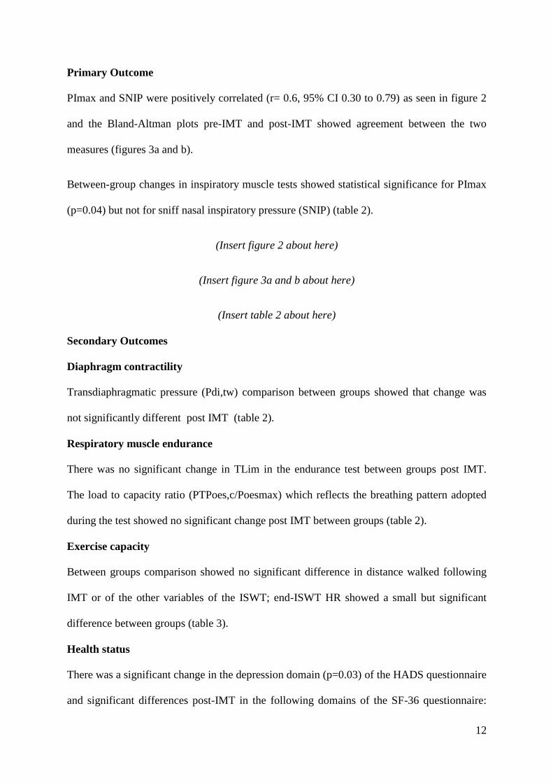
Primary Outcome
PImax and SNIP were positively correlated (r= 0.6, 95% CI 0.30 to 0.79) as seen in figure 2
and the Bland-Altman plots pre-IMT and post-IMT showed agreement between the two
measures (figures 3a and b).
Between-group changes in inspiratory muscle tests showed statistical significance for PImax
(p=0.04) but not for sniff nasal inspiratory pressure (SNIP) (table 2).
(Insert figure 2 about here)
(Insert figure 3a and b about here)
(Insert table 2 about here)
Secondary Outcomes
Diaphragm contractility
Transdiaphragmatic pressure (Pdi,tw) comparison between groups showed that change was
not significantly different post IMT (table 2).
Respiratory muscle endurance
There was no significant change in TLim in the endurance test between groups post IMT.
The load to capacity ratio (PTPoes,c/Poesmax) which reflects the breathing pattern adopted
during the test showed no significant change post IMT between groups (table 2).
Exercise capacity
Between groups comparison showed no significant difference in distance walked following
IMT or of the other variables of the ISWT; end-ISWT HR showed a small but significant
difference between groups (table 3).
Health status
There was a significant change in the depression domain (p=0.03) of the HADS questionnaire
and significant differences post-IMT in the following domains of the SF-36 questionnaire:

13
Role limitation due to emotional problems (RE), mental health (MH) and change in health
(CH). There was a clinically significant improvement in the dyspnoea and fatigue domains of
the CRDQ in the IMT group but between-group comparison showed no statistically
significant changes in any of the CRDQ domains. Health status results are seen in table 4.
Figure 4 shows the distribution of the anxiety and depression domains in the experimental
and control group and figure 5 shows the mean change in the CRDQ domains in the two
groups in relation to the minimal clinical important difference.
(Insert table 3 about here)
(Insert table 4 about here)
(Insert figures 4 and 5 about here)
DISCUSSION
This study investigated the effects of a home based IMT programme on inspiratory muscle
function, exercise capacity and health status, using multiple tests. This is the first study to
use SNIP as well as PImax following IMT in COPD patients and to include the non-volitional
test of bilateral anterolateral magnetic phrenic nerve stimulation (BAMPS). The principal
finding was that although health status and PImax improved following this seven week
programme, other inspiratory muscle tests and exercise capacity did not.
Effects of the IMT programme on inspiratory muscle strength
The SNIP and PImax are both non-invasive tests on inspiratory muscle function that are
available to use in community settings without the need for complex laboratory equipment.
Our Bland-Altman analysis (Figure 3a and b) showed that there was good agreement between
these two measures pre- and post-IMT. However, only PImax improved significantly post-
14
IMT. This discrepancy in results may be a reflection of the two inherent weaknesses of the
PImax test; first, its volitional nature which means that improvement in values may be due to
learning how to do the test and second, its similarity with the IMT breathing technique. We
sought to address the effect of learning the PImax technique, by familiarising our patients
with the manoeuvres in the run-in period prior to baseline assessments [14], as recommended
previously [13]. We are confident that there was limited additional learning of the manoeuvre
post-IMT as our control group changed by only 3.7% from baseline in PImax. However, it is
more difficult to control for the similarity between the pattern of respiratory neuromuscular
activation when breathing through a threshold loading device and that of the PImax
manoeuvre. Both involve a quasi-static (isometric) contraction of the diaphragm and rib cage
muscles at the initial stages when a maximum inspiratory effort is made against a closed
airway [19]. The similarity between the technique using the IMT device and the PImax
manoeuvre could have resulted in increased neuro-muscular coordination for the particular
task. Evidence that training can improve neuromuscular coordination comes from a study that
examined the diaphragmatic response to transcranial magnetic stimulation following
‘diaphragmatic’ training [39] and from an earlier study by the same group [40] which showed
significant increases in cortical excitability of the diaphragm motor area following a short
IMT programme. These studies suggest that the diaphragm and other respiratory muscles are
subject to the same practice-dependent neuro-plasticity as other muscles. The ability of our
COPD patients to generate greater maximal inspiratory pressures at the end of the programme
may have been partly due to an improvement in respiratory muscle co-ordination for the
particular manoeuvre. While improved neuromuscular coordination is a positive outcome, the
clinical value of this improvement is questionable as few activities of daily living require this
particular pattern of muscle activation.

15
Our study supports the previously stated argument about the value of using multiple tests to
assess inspiratory muscle strength [18] and adds to this argument by highlighting the value of
using the non-invasive SNIP for the assessment of inspiratory muscle strength following IMT
programmes, since it is difficult to determine how much of the PImax changes are due to
improvement in neuromuscular coordination.
We also explored changes in diaphragm contractility using the non-volitional BAMPS in a
small number of participants. Our subgroup showed that the non-volitional twitch trans-
diaphragmatic pressure (Pdi,tw) did not change following IMT. This may reflect the
adaptation of the diaphragm in moderate to severe COPD toward enhanced oxidative and
endurance capacity [41, 42] which means there is limited potential for further improvement.
Alternatively, a different training protocol may be required to train the diaphragm in
moderate to severe COPD patients. As this was an invasive test, requiring insertion of
oesophageal and gastric balloon catheters, patients were allowed to opt-out of this
assessment. The number of patients that accepted this invasive test was smaller than the total
of participants in this study, therefore, we cannot come to a firm conclusion about the effects
of 7 weeks of IMT on the diaphragm. However, this study provides the necessary means and
standard deviations to allow us to perform power calculations for future studies.
Effects of the IMT programme on respiratory muscle endurance and exercise capacity
Our study showed no improvements in respiratory muscle endurance, as defined by time to
task failure (TLim) or the oesophageal load to capacity ratio (PTPoes,c/Poesmax), using a
technique which takes into account the breathing strategy when patients breathe against high
inspiratory threshold loads [35]. The findings in COPD patients are in contrast to those of a
similar IMT programme in healthy young subjects [43]. Hart and colleagues [43] showed

16
improvement in inspiratory muscle endurance due to changes toward a more efficient
breathing pattern that served to protect or postpone diaphragm fatigue. Previous studies have
demonstrated the resistance of the COPD diaphragm to fatigue [44, 45] which may partly
explain why there was no change in the PTPoes,c/Poesmax in our patients.
In keeping with the results of inspiratory muscle function, there were no improvements in
exercise tolerance as measured by meters walked or on perception of breathlessness at the
end of the ISWT (BORG scale) between active and control groups. Previous home-based
IMT studies that have demonstrated increases in walking distance had longer durations of
training [24, 25, 46], therefore it is possible that the duration of our IMT programme was a
factor explaining this lack of improvement, although the duration of our programme was
typical of UK practice. We did, however, find that the heart rate at the end of the ISWT in
the PrBr-IMT group was slightly lower post-IMT than in the control group. This suggests an
improvement, even if minor, in aerobic capacity [47, 48]. Although breathlessness at the end
of the ISWT did not improve, the small increase in aerobic capacity may have contributed to
the perceived improvements in health status at the end of the programme.
Effects of the IMT programme on health status
Perception of wellbeing improved in our study. The depression domain of the HADS and
three domains of the SF-36 questionnaire (role limitation due to emotional problems, mental
health and change in health) showed improvements. For CRDQ, the improvement in the
dyspnoea and fatigue domains in the experimental group were at or above the threshold value
of 0.5 which has been identified as a MCID in this questionnaire [49]. Therefore our patients
received symptomatic benefit post-IMT which could perhaps be attributed, to some extent, to
the small improvement in aerobic capacity.

17
Reduction of perception of breathlessness during daily activities and improvement in health
status is one of the most well accepted effects of IMT [6, 49, 50]. This effect was also evident
in our study despite lack of consistent improvement in inspiratory muscle tests or in exercise
capacity. Clinically, this is an important positive result, as the home based programme in this
study did not include other forms of exercise training known to enhance the effects of IMT
[5] and was of shorter duration than some home IMT programmes [24, 25]. Therefore, our
study suggests that community IMT programmes of seven week duration are likely to
improve wellbeing and perception of breathlessness during activities of daily living in
moderate to severe COPD patients even in the absence of improvements in respiratory
muscle function and exercise capacity.
Clinical implications, limitations and further research
The intervention in this study was designed to reflect a realistic treatment approach that could
be readily delivered within a primary care setting given the limited resources available in the
UK and elsewhere. We assessed a heterogeneous COPD group in terms of disease severity
(ranging from moderate to very severe), baseline PImax values or dyspnoea intensity during
every day activities, reflecting the expected group in a community setting. Adherence to the
intervention was good in our study and our group reached a mean pressure of 62% PImax
(11.7) at the end of the programme, well above the 30% PImax minimal intensity required to
achieve a training effect [27] and similar to other home IMT studies [51, 52]. Our study
suggests that a community-based programme, shorter than previously reported in research
[24, 25], could be effective in improving PImax and perception of well-being in community
patients.

18
Home based programmes have been shown to be superior to centre-based ones in terms of
adherence to exercise [53] and are attractive to many people with COPD who are unable to
attend centre-based activities due to work/life commitments or due to limitations to
performing exercise such as additional musculoskeletal impairments or profound
breathlessness. Therefore, it is of clinical significance that participants have derived
important benefits from this home based programme.
However, home-based programmes have limitations when seeking to achieve a training
effect. Hill and colleagues [54] trained COPD patients in a laboratory setting and reached a
training intensity of 101% PImax by week 8. In the current study, patients had a more
individual progression in their IMT training intensity at home, although they were strongly
encouraged by the physiotherapist via phone calls. The training intensity that was tolerated by
our participants may have contributed to the lack of improvement in diaphragm contractility,
respiratory muscle endurance and exercise capacity at the end of the programme. This
potential limitation, however, does not explain the lack of improvement in SNIP which
should have improved in line with PImax at the end of the training programme, as evidenced
by the good correlation between the two measures.
IMT is more likely to be adopted clinically as a rehabilitation option if the design of the
training programme takes into consideration resources and costs associated with the regular
running of such a programme in community settings. Future studies need to explore the
effects of duration or intensity of training on SNIP and diaphragm contractility in community
programmes and identify the minimum level that lead to a training effect in COPD patients.
Larger sample sizes and more homogenous groups may be needed for this purpose.

19
CONCLUSIONS
This study examined the effectiveness of a seven-week, community-based IMT programme
in moderate to severe COPD patients. Following our programme, COPD patients showed
improvements in PImax and perception of wellbeing but not in SNIP or the non-volitional
test of diaphragm function, exercise capacity or respiratory muscle endurance. This study
suggests that some of the symptomatic benefits of IMT may be independent of changes in
inspiratory muscle function. It also highlights that multiple tests provide a more
comprehensive assessment of muscle function when seeking to demonstrate changes
following IMT programmes.
Acknowledgments
The authors would like to thank Ms Jo Gearing for her contribution to patient monitoring
during the study.
Declaration of Interest
The authors have no conflicts of interest. All authors have contributed to the research and
have been part of writing up the findings for publication.

20
REFERENCES
1. Global Initiative for Chronic Obstructive Lung Disease (GOLD). Global Strategy for
the diagnosis, management and prevention of Chronic Obstructive Pulmonary
Disease. NHLBI/WHO Workshop report (Updated 2013). Available from:
http://www.goldcopd.com -last accessed: 23rd
August 2014.
2. British Thoracic Society guideline development group. Intermediate care- Hospital-at-
home in chronic obstructive pulmonary disease; British Thoracic Society guideline.
Thorax 2007;62:200-210.
3. Toy EL, Gallagher KF, Stanley EF, et al. The economic impact of exacerbations of
chronic obstructive pulmonary disease and exacerbation definition: a review. COPD
2010;7(3):214-218.
4. Halding A-G, Wahl A, Heggdal K. ‘Belonging’. Patients’ experiences of social
relationships during pulmonary rehabilitation. Disability and Rehabilitation 2010;32
(15):1272-1280.
5. Gosselink R, De Vos J, van den Heuvel SP, et al. Impact of inspiratory muscle
training in patients with COPD: what is the evidence? Eur Resp J 2011;37:416-525.
6. Bott J, Blumenthal S, Buxton M, et al. Guidelines for the physiotherapy management
of the adult, medical, spontaneously breathing patient. Thorax 2009;64:i1-i52.
7. Crowe J, Reid WD, Geddes EL, et al. Inspiratory muscle training compared with other
rehabilitation interventions in adults with chronic obstructive pulmonary disease: a
systematic literature review and meta-analysis. COPD 2005;2 (3):319-329.

21
8. Geddes LE, Reid DW, Crowe J, et al. Inspiratory muscle training in adults with
chronic obstructive pulmonary disease: a systematic review. Respiratory Medicine
2005;99:1440-1458.
9. Lotters F, van Tol B, Kwakkel G, et al. Effects of controlled inspiratory muscle
training in patients with COPD: a meta-analysis. Eur Resp J 2002;20(3):570-576.
10. NHS Evidence. Chronic obstructive pulmonary disease: Evidence Update 5- February
2012 relevant to NICE Clinical Guideline 101 ‘Management of chronic obstructive
pulmonary disease in adults in primary and secondary care’. Available from:
http://www.evidence.nhs.uk – last accessed: 23rd
August 2014.
11. Polkey MI, Moxham J, Green M. The case against inspiratory muscle training in
COPD. Eur Resp J 2011;37:236-237.
12. Ambrosino N. The case for inspiratory muscle training in COPD. Eur Resp J 2011;
37:233-235.
13. Larson JL, Covey MK, Vitalo CA, et al. Maximal inspiratory pressure; learning effect
and test-retest reliability in patients with chronic obstructive pulmonary disease. Chest
1993;104:448-453.
14. Nikoletou D, Rafferty G, Man W D-C, et al. Sniff nasal inspiratory pressure in
patients with moderate to severe chronic obstructive pulmonary disease; learning
effect and short-term between session repeatability. Respiration 2014;88(5):365-370.
15. Heijdra YF, Dekhuijzen RPN, van Herwaarden CLA, et al. Nocturnal saturation
improves by target-flow inspiratory muscle training in patients with COPD. Am J
Resp Crit Care Med 1996;153:260-265.

22
16. Wanke Th, Formanek D, Lahramann H, et al. Effect of combined inspiratory muscle
and cycle ergometer training on exercise performance in patients with COPD. Eur
Resp J 1994;7:2205-2211.
17. Heritier F, Rahm F, Pasche P, et al. Sniff nasal inspiratory pressure; a noninvasive
assessment of inspiratory muscle strength. Am J Resp Crit Care Med 1994;150:1678-
1683.
18. Steier J, Kaul S, Seymour J, et al. The value of multiple tests of respiratory muscle
strength. Thorax 2007:62:975-980.
19. American Thoracic Society/ European respiratory society. ATS/ERS statement on
respiratory muscle testing. Am J Resp Crit Care Med 2002;166:518-624.
20. Ramirez-Sarmiento A, Orozco-Levi M, Guell R, et al. Inspiratory muscle training in
patients with chronic obstructive pulmonary disease. Am J Respir Crit Care Med
2002;166:1491–1497.
21. Bisschop A, Gayan-Ramirez G, Rollier H, et al. Intermittent inspiratory muscle
training induces fiber hypertrophy in rat diaphragm. Am J Respir Crit Care Med 1997;
155:1583–1589.
22. Rollier H, Bisschop A, Gayan-Ramirez G, et al. Low load inspiratory muscle training
increases diaphragmatic fiber dimensions in rats. Am J Respir Crit Care Med 1998;
157:833-839.
23. Gayan-Ramirez G, Rollier H, Vanderhoydonc F, et al. Nandrolone decanoate does not
enhance training effects but increases IGF-I mRNA in rat diaphragm. J Appl Physiol
2000;88:26-34.

23
24. Battanglia E, Fulgenzi A, Bernucci S, et al. Home respiratory muscle training in
patients with chronic obstructive pulmonary disease. Respirology 2006;11:799- 804.
25. Beckerman M, Magadle R, Weiner M, et al. The effects of 1 year of specific
inspiratory muscle training in COPD. Chest 2005;128:3177-3182.
26. Taves DR. Minimization: a new method of assigning patients to treatment and control
groups. Clin Pharmacolog Ther 1974;15(5):443-453.
27. Larson JL, Kim MJ, Sharp JT, et al. Inspiratory muscle training with a pressure
threshold breathing device in patients with chronic obstructive pulmonary disease.
Am Rev Respir Dis 1988;138(3):689-696.
28. Pardy RL and Rochester DL. Respiratory muscle training. Semin Resp Med
1992;13:53-62.
29. Gosselink R, J De Vos, SP van den Heuvel, et al. Impact of inspiratory muscle
training in patients with COPD: what is the evidence? Eur Resp J 2011;37:416-425.
30. Hill K, Cecins NM, Eastwood P, et al. Inspiratory muscle training for patients with
Chronic Obstructive Pulmonary Disease: A practical guide for clinicians. Arch Phys
Med and Rehab 2010;91(9):1466-1470.
31. Wen SA, Woo MS and Keens TG. How many maneuvers are required to measure
maximal inspiratory pressure accurately? Chest 1997;111:802-807.
32. Terzi N, Corne F, Mouadil A, et al. Mouth and Nasal inspiratory pressure; learning
effect and reproducibility in healthy adults. Respiration 2010;80:379-386.

24
33. Mills GH, Kyroussis D, Hamnegard C-H, et al. Bilateral magnetic stimulation of the
phrenic nerves from an anterolateral approach. Am J Resp Crit Care Med 1996; 154:
1099-1105.
34. Wragg S, Hamnegård C, Road J, et al. Potentiation of diaphragmatic twitch after
voluntary contraction in normal subjects. Thorax 1994; 49:1234-1237.
35. Hart N, Hawkins P, Hamnegård C-H, et al. A novel clinical test of respiratory muscle
endurance. Eur Resp J 2002; 19: 232-239.
36. Chen RC, Que CL and Yan S. Introduction to a new inspiratory threshold loading
device. Eur Resp J 1998; 12: 208-211.
37. Singh SJ, Morgan MD, Scott S, et al. Development of a shuttle walking test of
disability in patients with chronic airways obstruction. Thorax 1992; 47: 1019-1024.
38. Harper R, Brazier JE, Waterhouse JC, et al. Comparison of outcome measures for
patients with chronic obstructive pulmonary disease (COPD) in an outpatient setting.
Thorax 1997; 52:879-887.
39. Demoule A, Verin E, Tezenas du Montcel S and Similowski T. Short-term training-
dependent plasticity of the corticospinal diaphragm control in normal humans. Respir
Physiol Neurob 2007; 160 (2): 172-180.
40. Demoule A, Verin E, Derenne J-P, Similowski T. Plasticity of the human motor
cortical representation of the diaphragm. Am J Resp Crit Care Med 163:A46.
41. Levine S, Nguyen T, Kaiser LR, et al. Human diaphragm remodelling associated with
chronic obstructive pulmonary disease: clinical implications. Am J Resp Crit Care
Med 2003;168:706-713.
25
42. Stubbings AK, Moore AJ, Dusmet M, et al. Physiological properties of human
diaphragm muscle fibres and the effect of chronic obstructive pulmonary disease. J
Physiol 2008; 586 (10): 2637-2650.
43. Hart N, Sylvester K, Ward S, et al. Evaluation of the Powerbreathe inspiratory muscle
trainer in healthy humans; pilot study and sample size calculation. Chest 2001; 95 (6):
526-531.
44. Polkey MI., Kyroussis D, Keilty SE, et al. Exhaustive treadmill exercise does not reduce
twitch transdiaphragmatic pressure in patients with COPD. Am J Respir Crit Care
Med 1995;152: 959-964.
45. Polkey MI., Kyroussis D, Hamnegard CH, et al. Diaphragm performance during
maximal voluntary ventilation in Chronic Obstructive Pulmonary Disease. Am J
Respir Crit Care Med 1997; 155: 642-648.
46. Lisboa C, Munoz V, Beroiza T, et al. Inspiratory muscle training in chronic airflow
limitation: comparison of two different training loads with a threshold device. Eur
Respir J 1994; 7: 1266-1274.
47. Almeida MB, Araújo CGS. Effects of aerobic training on heart rate. Rev Bras Med
Esporte 2003; 9 (2): 104 – 112.
48. Tulppo MP, Hautala AJ, Mäkikallio TH., Laukkanen RT, Nissilä S, Hughson RL,
Huikuri HV. Effects of aerobic training on heart rate dynamics in sedentary subjects. J
Appl Physiol 2003; 95: 364 – 372.
49. Jones PW. Interpreting thresholds for a clinically significant change in health status in
asthma and COPD. Eur Respir J 2002; 19: 398-404.

26
50. Thomas M.J., Simpson J., Riley R, et al. The impact of home-based physiotherapy
interventions on breathlessness during activities of daily living in severe COPD: a
systematic review. Physiotherapy 2010; 96: 108-119.
51. Covey MK, Larson LJ, Wirtz SE, et al. High-intensity inspiratory muscle training in
patients with chronic obstructive pulmonary disease and severely reduced function. J
Cardiopulm Reh 2001; 21: 231-240.
52. Hsiao SF, Wu YT, Wu HD, et al. Comparison of effectiveness of pressure threshold
and targeted resistance devices for inspiratory muscle training in patients with chronic
obstructive pulmonary disease. J Formos Med Assoc 2003; 102 (4): 240-245.
53. Ward D, Severs M, Dean T, et al. Care home versus hospital and own home
environments for rehabilitation of older people. The Cochrane Library 2008; 3: 1-13.
54. Hill K, Jenkins SC, Philippe DL, et al. High-intensity inspiratory muscle training in
COPD. Eur Resp J 2006; 27: 1119-1128.