Evaluation of the Clinical Significance of homB, a Novel Candidate Marker of Helicobacter pylori...
12
Evaluation of the Clinical Significance of homB, a Novel Candidate Marker of Helicobacter pylori Strains Associated with Peptic Ulcer Disease Mónica Oleastro, 1,4 Rita Cordeiro, 1 Jonathan Ferrand, 4,5 Baltazar Nunes, 3 Philippe Lehours, 4,5 Isabel Carvalho-Oliveira, 2 Ana I. Mendes, 2 Deborah Penque, 2 Lurdes Monteiro, 1 Francis Mégraud, 4,5 and Armelle Ménard 4,5 Departamento de 1 Doenças Infecciosas and 2 Genética and 3 Departamento de Epidemiologia, Instituto Nacional Saúde Dr Ricardo Jorge, Lisbon, Portugal; 4 INSERM U853 and 5 Laboratoire de Bactériologie, Université Victor Segalen Bordeaux 2, Bordeaux, France Background. homB codes for a putative Helicobacter pylori outer membrane protein and has previously been associated with peptic ulcer disease (PUD) in children. Methods. A total of 190 H. pylori strains isolated from children and adults were studied to evaluate the clinical importance of the homB gene. In vitro experiments were performed to identify HomB mechanisms of bacterial pathogenicity. Results. Characterization of the isolates demonstrated that homB was significantly associated with PUD in 86 children (odds ratio [OR], 7.64 [95% confidence interval {CI}, 2.65–22.05]) and in 32 adults 40 years of age (OR, 11.25 [95% CI, 1.86 – 68.13]). homB was correlated with the presence of cagA, babA2, vacAs1, hopQI, and oipA “on” genotype (P .001). The HomB protein was found to be expressed in the H. pylori outer membrane and was noted to be antigenic in humans. H. pylori homB knockout mutant strains presented reduced ability to induce interleukin-8 secretion from human gastric epithelial cells, as well as reduced capacity to bind to these cells. Both of these functions correlated with the number of homB copies present in a strain. Conclusion. homB can be considered a comarker of H. pylori strains associated with PUD. Moreover, results strongly suggest that HomB is involved in the inflammatory response and in H. pylori adherence, constituting a novel putative virulence factor. Helicobacter pylori is the major causative agent of peptic ulcer disease (PUD), chronic active gastritis, gastric car- cinoma, and gastric mucosa-associated lymphoid tissue (MALT) lymphoma [1]. Progression to more-severe disease occurs only in some infected individuals and de- pends on a number of factors, including host genetic susceptibility, environmental factors, and differences in the virulence of H. pylori strains. To date, the best rec- ognized bacterial marker of H. pylori pathogenicity is cagA. The cagA gene is a marker for the cag pathogenicity island (cagPAI), which, when intact, encodes the com- ponents of a type IV secretion system responsible for delivering the CagA protein into the cytoplasm of the host cell, where it becomes tyrosine phosphorylated and initiates changes in cell signalling, coupled with induc- tion of the proinflammatory interleukin (IL)– 8 [2]. The onset of H. pylori infection occurs primarily dur- ing childhood [3], but severe gastroduodenal diseases appear primarily in adulthood, after a long-term infec- tion [4, 5]. In young children, the development of a pep- tic ulcer occurs soon after H. pylori infection, suggesting that the implicated strain may be potentially more pathogenic. A putative ulcer-associated outer mem- brane protein (OMP), jhp0870, was recently identified, by means of subtractive hybridization, in an H. pylori strain isolated from a young child presenting with a du- odenal ulcer [6]. The jhp0870 open-reading frame (ORF), homB, is 90% identical to another member of the H. pylori OMPs, Received 12 January 2008; accepted 14 May 2008; electronically published 23 September 2008. Potential conflicts of interest: none reported. Presented in part: XXth International Workshop on Helicobacter and Related Bacteria in Chronic Digestive Inflammation, Istanbul, Turkey, September 2007 (abstract W3.05) (oral communication). Financial support: Sociedade Portuguesa de Gastrenterologia; Programme de Cooperation Scientifique et Technique Franco-Portugais, sponsored by the French Embassy in Portugal. Reprints or correspondence: Dr. Francis Mégraud, INSERM U853, Université Victor Segalen Bordeaux 2, Laboratoire de Bactériologie, Bat. 2B RDC Zone Nord, 33076 Bordeaux cedex, France ([email protected]). The Journal of Infectious Diseases 2008; 198:1379 – 87 © 2008 by the Infectious Diseases Society of America. All rights reserved. 0022-1899/2008/19809-0019$15.00 DOI: 10.1086/592166 MAJOR ARTICLE homB in Helicobacter pylori Virulence ● JID 2008:198 (1 November) ● 1379 by guest on June 3, 2016 http://jid.oxfordjournals.org/ Downloaded from
-
Upload
independent -
Category
Documents
-
view
0 -
download
0
Transcript of Evaluation of the Clinical Significance of homB, a Novel Candidate Marker of Helicobacter pylori...
Evaluation of the Clinical Significance of homB,a Novel Candidate Marker of Helicobacter pyloriStrains Associated with Peptic Ulcer Disease
Mónica Oleastro,1,4 Rita Cordeiro,1 Jonathan Ferrand,4,5 Baltazar Nunes,3 Philippe Lehours,4,5 Isabel Carvalho-Oliveira,2
Ana I. Mendes,2 Deborah Penque,2 Lurdes Monteiro,1 Francis Mégraud,4,5 and Armelle Ménard4,5
Departamento de 1Doenças Infecciosas and 2Genética and 3Departamento de Epidemiologia, Instituto Nacional Saúde Dr Ricardo Jorge, Lisbon,Portugal; 4INSERM U853 and 5Laboratoire de Bactériologie, Université Victor Segalen Bordeaux 2, Bordeaux, France
Background. homB codes for a putative Helicobacter pylori outer membrane protein and has previously beenassociated with peptic ulcer disease (PUD) in children.
Methods. A total of 190 H. pylori strains isolated from children and adults were studied to evaluate the clinicalimportance of the homB gene. In vitro experiments were performed to identify HomB mechanisms of bacterialpathogenicity.
Results. Characterization of the isolates demonstrated that homB was significantly associated with PUD in 86children (odds ratio [OR], 7.64 [95% confidence interval {CI}, 2.65–22.05]) and in 32 adults �40 years of age (OR,11.25 [95% CI, 1.86 – 68.13]). homB was correlated with the presence of cagA, babA2, vacAs1, hopQI, and oipA “on”genotype (P � .001). The HomB protein was found to be expressed in the H. pylori outer membrane and was notedto be antigenic in humans. H. pylori homB knockout mutant strains presented reduced ability to induce interleukin-8secretion from human gastric epithelial cells, as well as reduced capacity to bind to these cells. Both of these functionscorrelated with the number of homB copies present in a strain.
Conclusion. homB can be considered a comarker of H. pylori strains associated with PUD. Moreover, resultsstrongly suggest that HomB is involved in the inflammatory response and in H. pylori adherence, constituting a novelputative virulence factor.
Helicobacter pylori is the major causative agent of peptic
ulcer disease (PUD), chronic active gastritis, gastric car-
cinoma, and gastric mucosa-associated lymphoid tissue
(MALT) lymphoma [1]. Progression to more-severe
disease occurs only in some infected individuals and de-
pends on a number of factors, including host genetic
susceptibility, environmental factors, and differences in
the virulence of H. pylori strains. To date, the best rec-
ognized bacterial marker of H. pylori pathogenicity is
cagA. The cagA gene is a marker for the cag pathogenicity
island (cagPAI), which, when intact, encodes the com-
ponents of a type IV secretion system responsible for
delivering the CagA protein into the cytoplasm of the
host cell, where it becomes tyrosine phosphorylated and
initiates changes in cell signalling, coupled with induc-
tion of the proinflammatory interleukin (IL)– 8 [2].
The onset of H. pylori infection occurs primarily dur-
ing childhood [3], but severe gastroduodenal diseases
appear primarily in adulthood, after a long-term infec-
tion [4, 5]. In young children, the development of a pep-
tic ulcer occurs soon after H. pylori infection, suggesting
that the implicated strain may be potentially more
pathogenic. A putative ulcer-associated outer mem-
brane protein (OMP), jhp0870, was recently identified,
by means of subtractive hybridization, in an H. pylori
strain isolated from a young child presenting with a du-
odenal ulcer [6].
The jhp0870 open-reading frame (ORF), homB, is
90% identical to another member of the H. pylori OMPs,
Received 12 January 2008; accepted 14 May 2008; electronically published 23September 2008.
Potential conflicts of interest: none reported.Presented in part: XXth International Workshop on Helicobacter and Related
Bacteria in Chronic Digestive Inflammation, Istanbul, Turkey, September 2007(abstract W3.05) (oral communication).
Financial support: Sociedade Portuguesa de Gastrenterologia; Programme deCooperation Scientifique et Technique Franco-Portugais, sponsored by the FrenchEmbassy in Portugal.
Reprints or correspondence: Dr. Francis Mégraud, INSERM U853, UniversitéVictor Segalen Bordeaux 2, Laboratoire de Bactériologie, Bat. 2B RDC Zone Nord,33076 Bordeaux cedex, France ([email protected]).
The Journal of Infectious Diseases 2008; 198:1379 – 87© 2008 by the Infectious Diseases Society of America. All rights reserved.0022-1899/2008/19809-0019$15.00DOI: 10.1086/592166
M A J O R A R T I C L E
homB in Helicobacter pylori Virulence ● JID 2008:198 (1 November) ● 1379
by guest on June 3, 2016http://jid.oxfordjournals.org/
Dow
nloaded from
the jhp0649 ORF homA, with the differences between the two
confined to the middle region of these ORFs. According to the
sequenced H. pylori strains [7–9], both genes occupy well-
conserved loci, with the homA locus occupied either by homA or
homB, and with the homB locus either occupied by one of these
ORFs or remaining empty with a segment displaying 96.8% sim-
ilarity with homB. This finding suggests that homB can exchange
positions with homA and, also, that the presence of homB may
have resulted from gene duplication [10].
The clinical importance of homB was evaluated by analyzing
its distribution and diversity in a collection of 190 H. pylori
strains isolated from patients presenting only with PUD or gas-
tritis, as well as its association with the previously described H.
pylori virulence-associated genes. The exposed components,
usually OMPs, can contribute to the colonization and persis-
tence of H. pylori, by allowing the bacteria to adhere to gastric
cells [11, 12]. The OMPs can also influence the pathogenesis of
the disease process, by stimulation of the inflammatory response
of the host [13]. Considering that the product of homB, HomB,
is a virulence-associated OMP candidate, we also investigated its
surface membrane localization and the immunological response
of the patient. The contribution of HomB to the inflammatory
response of H. pylori and to the bacterial adherence properties
was also investigated in vitro.
MATERIAL AND METHODS
Bacterial strains. A total of 190 H. pylori clinical strains were
cultured from antral biopsy specimens [14] (table 1). Two H.
pylori sequenced strains, 26695 (ATCC 700392), positive for
homA and negative for homB, and J99 (ATCC 700824), positive
for both homA and homB, were used as reference strains [7, 8].
Genomic DNA was extracted as described elsewhere [6].
Polymerase chain reaction (PCR)– based genotyping of vir-
ulence factors. Genotyping of the virulence factors was per-
formed by PCR for cagA [15], cagE, cagG, cagM [16], and the
“cag empty site” [17], vacAs1 [18], babA2 [19], iceA [20], hopQ
[21], homB, and homA [6]. The frame status of oipA, sabA, hopZ,
and dupA was determined by sequencing [16, 22–24]. The se-
quence diversity of homB and homA was evaluated by sequenc-
ing entire genes [6].
Construction of homB knockout mutant strains. The
pILL570Hp710�TnKm suicide plasmid containing a homB ho-
mologue disrupted by a kanamycin resistance gene cassette
(supplied by I. Boneca, Microbiology Department of Institut
Pasteur, Paris) was used for natural transformation of H. pylori
strains. The correct and unique insertion of the kanamycin cas-
sette in the target region was confirmed by PCR, sequencing, and
Southern blot analysis. For further discussion of this procedure,
see the Appendix (which is available only in the online version of
the Journal).
Two-dimensional gel electrophoresis (2DE) and analysis of
protein spots. A total of 200 �g of protein membrane pellets
(i.e., sarcosine-insoluble outer membrane fractions) [25] of the
H. pylori strain were separated by 2DE, by means of isoelectric
focusing performed with the use of an immobilized pH gradient
strip with a nonlinear pH of 3–10 (GE Healthcare), followed by
SDS-PAGE performed in 10% w/v polyacrylamide gels with the
use of SE 600 Ruby equipment (GE Healthcare). Gels were either
stained with Coomassie blue [26] or used for Western blot anal-
ysis.
Selected protein spots were excised from the gel and digested
with trypsin. Peptides were analyzed by means of capillary liquid
chromatography tandem mass spectrometry (LC-MS/MS). For
further discussion of this procedure, see the Appendix (which is
available only in the online version of the Journal).
Preparation of recombinant HomB and immunoproteomics.
A recombinant HomB protein (rHpHomB) with the homB ORF
of the J99 strain cloned (jhp0870, GenBank accession no.
NC_000921) was prepared using a glutathione S-transferase
(GST) gene fusion vector pGEX-4T-3 (GE Healthcare). The pu-
rified protein was probed with an anti-GST antibody (1:1000)
(Chemicon Australia), to confirm its expression.
The 2DE or SDS-PAGE (purified rHpHomB) gels were trans-
ferred onto nitrocellulose or polyvinylidene difluoride (PVDF)
membranes and incubated with a pool of 10 serum samples ob-
tained from patients (64.7% of whom were male; mean age �
SD, 51.2 � 6.8 years) who had IgG antibodies against H. pylori.
A pool of 5 serum samples obtained from H. pylori– uninfected
individuals (53.3% of whom were male; mean age � SD,
42.7 � 5.4 years) who had an anti–H. pylori IgG antibody level
below the threshold denoting positivity was used as negative
control. The level of IgG antibodies against H. pylori was quan-
tified using the serological test Pyloriset EIA-G III (Orion Diag-
nostica). For further discussion of this procedure, see the Ap-
pendix (which is available only in the online version of the
Journal).
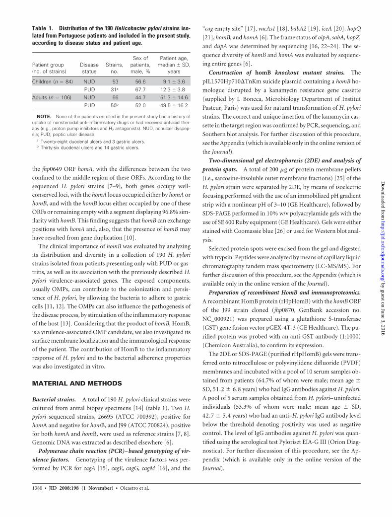
Table 1. Distribution of the 190 Helicobacter pylori strains iso-lated from Portuguese patients and included in the present study,according to disease status and patient age.
Patient group(no. of strains)
Diseasestatus
Strains,no.
Sex ofpatients,male, %
Patient age,median � SD,
years
Children (n � 84) NUD 53 56.6 9.1 � 3.6PUD 31a 67.7 12.3 � 3.8
Adults (n � 106) NUD 56 44.7 51.3 � 14.6PUD 50b 52.0 49.5 � 16.2
NOTE. None of the patients enrolled in the present study had a history ofuptake of nonsteroidal anti-inflammatory drugs or had received antiacid ther-apy (e.g., proton pump inhibitors and H2 antagonists). NUD, nonulcer dyspep-sia; PUD, peptic ulcer disease.
a Twenty-eight duodenal ulcers and 3 gastric ulcers.b Thirty-six duodenal ulcers and 14 gastric ulcers.
1380 ● JID 2008:198 (1 November) ● Oleastro et al.
by guest on June 3, 2016http://jid.oxfordjournals.org/
Dow
nloaded from
IL-8 secretion and adherence assays. The induction of IL-8
secretion and adherence in vitro was evaluated after 18 h of co-
culture of H. pylori with human gastric adenocarcinoma epithe-
lial AGS cells (ATCC CRL-173). IL-8 was quantified by ELISA,
by use of the Quantikine Human IL-8 Immunoassay (R & D
Systems Europe). For the adherence assay, the bacterial suspen-
sions were labelled with a PKH2 green fluorescent linker kit
(Sigma) [27]. Fluorescence was measured using the FACSCali-
bur flow cytometer (Becton Dickinson).
Statistical analysis. Statistical analysis was performed us-
ing the statistical software package SPSS (version 14.0; SPSS).
The level of significance was set at 5%, with the null hypothesis
rejected when P � .05.
RESULTS
Prevalence of homB, homA, and other virulence genes in
relation to clinical outcome. A total of 190 H. pylori clinical
strains (table 1) isolated from Portuguese children (n � 84) and
adults (n � 106) were examined for the presence of the homB,
homA, cagPAI, vacAs1, babA2, iceA, hopQ, oipA, sabA, hopZ, and
dupA genes.
Overall, with regard to the cagPAI markers of these 190 pa-
tients, a concordance between the presence of the cagA, cagE,
cagG, and cagM genes and the simultaneous absence of a cag
empty site product— or vice versa (i.e., absence of the genes and
presence of the cag empty site)—was observed in 135 (71.0%) of
the 190 strains, suggesting that mixed infections with or without
cagPAI were not frequently observed in this group of strains. For
28 (50.9%) of the remaining 55 strains, a discrepancy was ob-
served between the presence of cagA, cagE, cagG, and cagM
genes, suggesting the presence of an incomplete island or mixed
infections, of which only 5 were cagA positive (17.9%). Accord-
ing to these results, the cagA gene was considered to be represen-
tative of an intact cagPAI in this study. The prevalence and the
univariate statistical significance of the association between the
H. pylori virulence genotypes, the homB and homA genotypes,
and the clinical outcome are presented in table 2.
In strains isolated from children (31 with PUD and 53 with
NUD), 5 genotypes were associated with PUD: cagA (P � .001;
odds ratio [OR], 14.06), vacAs1 (P � .001; OR, 14.13), oipA
“on” genotype (P � .001; OR, 14.06), hopQI (P � .004; OR,
5.67), and homB (P � .001; OR. 7.64). Two genotypes were
strongly associated with NUD: the sabA “on” genotype
(P � .028; OR, 0.298) and homA (P � .006; OR, 0.263). Con-
cerning dupA, a PCR fragment corresponding to jhp0917–
jhp0918 was detected in all tested strains, and the presence of an
entire gene was confirmed by sequencing.
In strains isolated from adults (50 with PUD and 56 with
NUD), only cagA was significantly associated with PUD
(P � .05; OR, 2.28), indicating that cagPAI is the major inde-
pendent predictor of disease in the study population. Similarly
to strains isolated in children, all isolates recovered from adults
had an entire dupA gene.
It is likely that environmental factors play a greater role in the
development of PUD in older adult populations than in younger
populations, as has been demonstrated for smoking and nonste-
roidal inflammatory drug (NSAID) consumption [28]. Accord-
ingly, the adult population was stratified by age, and 2 groups
were formed: adults �40 years of age and those �40 years of age.
In the group of patients �40 years of age (n � 32; 17 of whom
had PUD [64.7% of whom were male; mean age � SD,
33.5 � 6.5 years] and 15 of whom had NUD [58.3% of whom
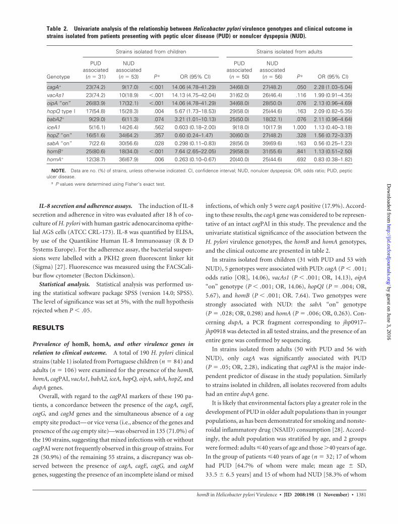
Table 2. Univariate analysis of the relationship between Helicobacter pylori virulence genotypes and clinical outcome instrains isolated from patients presenting with peptic ulcer disease (PUD) or nonulcer dyspepsia (NUD).
Genotype
Strains isolated from children Strains isolated from adults
PUDassociated(n � 31)
NUDassociated(n � 53) P a OR (95% CI)
PUDassociated(n � 50)
NUDassociated(n � 56) P a OR (95% CI)
cagA� 23(74.2) 9(17.0) �.001 14.06 (4.78–41.29) 34(68.0) 27(48.2) .050 2.28 (1.03–5.04)vacAs1 23(74.2) 10(18.9) �.001 14.13 (4.75–42.04) 31(62.0) 26(46.4) .116 1.99 (0.91–4.35)oipA “on” 26(83.9) 17(32.1) �.001 14.06 (4.78–41.29) 34(68.0) 28(50.0) .076 2.13 (0.96–4.69)hopQ type I 17(54.8) 15(28.3) .004 5.67 (1.73–18.53) 29(58.0) 25(44.6) .163 2.09 (0.82–5.35)babA2� 9(29.0) 6(11.3) .074 3.21 (1.01–10.13) 25(50.0) 18(32.1) .076 2.11 (0.96–4.64)iceA1 5(16.1) 14(26.4) .562 0.603 (0.18–2.00) 9(18.0) 10(17.9) 1.000 1.13 (0.40–3.18)hopZ “on” 16(51.6) 34(64.2) .357 0.60 (0.24–1.47) 30(60.0) 27(48.2) .328 1.56 (0.72–3.37)sabA “on” 7(22.6) 30(56.6) .028 0.298 (0.11–0.83) 28(56.0) 39(69.6) .163 0.56 (0.25–1.23)homB� 25(80.6) 18(34.0) �.001 7.64 (2.65–22.05) 29(58.0) 31(55.6) .841 1.13 (0.51–2.50)homA� 12(38.7) 36(67.9) .006 0.263 (0.10–0.67) 20(40.0) 25(44.6) .692 0.83 (0.38–1.82)
NOTE. Data are no. (%) of strains, unless otherwise indicated. CI, confidence interval; NUD, nonulcer dyspepsia; OR, odds ratio; PUD, pepticulcer disease.
a P values were determined using Fisher’s exact test.
homB in Helicobacter pylori Virulence ● JID 2008:198 (1 November) ● 1381
by guest on June 3, 2016http://jid.oxfordjournals.org/
Dow
nloaded from
were male; mean age � SD, 34.7 � 5.4 years), homB was asso-
ciated with PUD (88.2% vs. 40.0% [P � .008]; OR, 11.25 [95%
CI, 1.86 – 68.13]). Compared with strains isolated from patients
with NUD, strains isolated from patients with PUD had a higher
prevalence of cagA (82.4% vs. 53.3%), vacAs1 (82.4% vs. 53.3%),
oipA “on” genotype (88.2% vs. 60.0%), and hopZ “on” genotype
(70.6% vs. 40.0%), although the differences were not statistically
significant. homA was a marker for NUD (60.0% vs. 17.6%
[P � .027]; OR, 7.0 [95% CI, 1.11–50.44]). In the group of
adults �40 years of age, no genotype was significantly associated
with clinical outcome (not shown).
Association of homB and homA with other virulence
genes. Globally, the presence of homB was associated with
cagA (P � .001), vacAs1 (P � .001), babA2 (P � .001), hopQI
(P � .001), and oipA “on” genotype (P � .001), whereas homA
was strongly associated with cagA� (P � .01), vacAs2
(P � .001), babA2� (P � .001), hopQII (P � .001), and oipA
“off” (P � .001). The presence of homB was also significantly
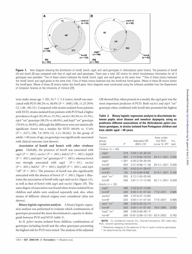
associated with the absence of homA (P � .001). Figure 1 illus-
trates the association of homB with cagA and vacAs1 (figure 1A),
as well as that of homA with cagA and vacAs1 (figure 1B). The
same degree of association was found when strains isolated from
children and adults were analyzed separately and, also, when
strains of different clinical origins were considered (data not
shown).
Binary logistic regression analysis. A binary logistic regres-
sion analysis was performed to evaluate which combinations of
genotypes presented the most discriminatory capacity to distin-
guish between PUD and NUD (table 3).
In H. pylori strains isolated from children, combinations of
genotypes including homB and the other genotypes presenting
the highest risk for PUD were tested. The analysis of the adjusted
OR showed that, when present in a model, the cagA gene was the
most important predictor of PUD. Both vacAs1 and oipA “on”
genotype when combined with homB also presented the highest
Figure 1. Venn diagram showing the distribution of homB, homA, cagA, and vacA genotypes in Helicobacter pylori strains. The presence of homB(A) and homA (B) was compared with that of cagA and vacA genotypes. There was a total 182 strains for which simultaneous information for all 4genotypes was available. *Ten of these strains harbored the homB, homA, cagA, and vacA genes at the same time. **One of these strains harboredthe homB, homA, and cagA genes at the same time. †Two of these strains harbored only the homB and homA genes. ‡None of these 58 strains harborthe homB gene. §None of these 26 strains harbor the homA gene. Venn diagrams were constructed using the software available from the Departmentof Computer Science at the University of Victoria [29].
Table 3. Binary logistic regression analysis to discriminate be-tween peptic ulcer disease and nonulcer dyspepsia, using aspredictors different associations of the Helicobacter pylori viru-lence genotypes, in strains isolated from Portuguese children andfrom adults aged �40 years.
Patientgroup,model P
Adjusted OR(95% CI)a
Area underthe ROC
curve % (P) bH-Ltest
Children (n � 84)cagA� .019 5.94 (1.34–26.23)vacAs1 .062 4.12 (0.93–18.22) 83.4 (�.001) 0.056cagA� �.001 8.26 (2.58–26.44)homB� .054 3.31 (0.98–11.18) 83.4 (�.001) 0.005vacAs1 .001 8.42 (2.41–29.47)homB� .182 2.43 (0.66–8.98) 81.6 (�.001) 0.438oipA “on” .003 6.12 (1.83–20.40)homB� .026 3.81 (1.17–12.36) 82.1 (�.001) 0.209
Adults (n � 32)cagA� .366 2.33 (0.37–14.65)homB� .020 9.00 (1.41–57.44) 77.8 (.007) 0.885vacAs1 .366 2.33 (0.37–14.65)homB� .020 9.00 (1.41–57.44) 77.8 (.007) 0.885oipA “on” .366 3.00 (0.40–22.77)homB� .020 9.00 (1.41–57.44) 78.6 (.006) 0.553hopZ “on” .066 2.06 (0.89–36.17)homB� .008 15.81 (2.06–121.61) 82.0 (.002) 0.762
NOTE. CI, confidence interval; H-L, Hosmer-Lemeshow; OR, odds ratio;ROC, receiver operating characteristic.
a Reference category is the absence of the H. pylori virulence genotypes.b As determined by the Wald test.
1382 ● JID 2008:198 (1 November) ● Oleastro et al.
by guest on June 3, 2016http://jid.oxfordjournals.org/
Dow
nloaded from

and most significant adjusted OR when combined with homB.
Analysis of the area under the receiver operating characteristic
curves showed that all models proposed presented a high and
similar discriminatory capacity to distinguish between PUD and
gastritis, whereas the goodness of fit of each model to the data, as
evaluated by the Hosmer-Lemeshow test, showed that only the
models vacAs1 with homB and oipA “on” with homB showed a
good adjustment (H-L test, �0.05).
In strains isolated from adults �40 years the age, homB was
the most important predictor of PUD, showing the highest value
for the adjusted OR in every model considered and, therefore,
corroborating the univariate analysis. Overall, the best model for
discriminating between PUD and NUD in young adults was the
combination of homB with hopZ “on genotype.”
Diversity in homB and homA genes. The presence of homB
and homA and their genomic positions were analyzed in 190 H.
pylori clinical strains. At least 1 of these 2 genes was always
present in the genome of the clinical strains, with different com-
binations observed: a single-copy genotype (homA or homB) or a
2-copy genotype (either 2 copies of homA or homB, or 1 copy of
each gene). The entire nucleotide sequence of both the homB
and homA genes was determined in 58 clinical strains, which
were randomly chosen, presenting different homB/homA com-
binations: 13 strains had 2 copies of homB, 6 had 2 copies of
homA, 16 had 1 copy of homB, 10 had 1 copy of homA, and 13
had 1 copy of each gene. Both genes presented a variable length:
from 1974 to 2025 bp for homB and from 1941 to 1998 bp for
homA. In cases where only 1 copy of homB or homA was present,
the gene was present in the jhp0649 locus, whereas a conserved
intergenic space was observed at the jhp0870 locus, according to
the numbering of the J99 strain [10]. When 2 copies were found
in the same strain, genes were present indifferently in one of
these loci. Whenever duplicates of the same gene were present in
a single strain, their identity varied between 98% and 99%.
The presence of homB and/or homA in a single- or 2-copy
genotype varied significantly among PUD- and NUD-associated
strains isolated from children and young adults. The single-copy
genotype (homB or homA) was more frequently associated with
NUD (79.4% vs. 37.5%), whereas the 2-copy genotype (homB/
homA, homB/homB, or homA/homA) was more frequently asso-
ciated with PUD (62.5% vs. 20.6%). More specifically, a single-
copy of homA was the genotype most frequently associated with
NUD (61.8% vs. 10.4% [P � .001]; OR, 13.89 [95% CI, 4.49 –
45.99]), whereas 2 copies of homB was the genotype most
strongly associated with PUD (39.9% vs. 11.8% [P � .001]; OR,
4.91 [95% CI, 1.77–14.02]).
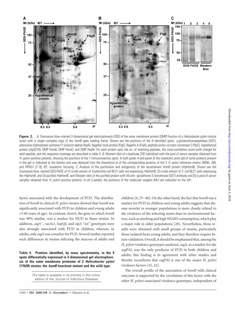
Evaluation of HomB surface expression and antigenicity.
Nine spots were identified as being expressed differentially
among the 2DE OMP fractions of the wild-type (wt) strain
1776/05 and the corresponding homB mutant (figure 2A). In 2
spots, the H. pylori putative OMPs HomD and HopN, which
have not yet been described in H. pylori proteomic studies, were
identified by LC-MS/MS analysis. The other 6 spots corre-
sponded to previously described surface proteins, validating the
preparation of the OMP extracts [30 –33] (table 4) (for further
discussion, see the Appendix, which is available only in the on-
line version of the Journal). The remaining spot, which had an
isoelectric point and a molecular weight similar to those de-
duced for HomB, was identified in the 2DE of the wt strain, and
the LC-MS/MS analysis confirmed that this spot was the HomB
protein. This spot was absent in the homB knockout mutant on
2DE (not shown).
The immunoblot showed that HomB was immunoreactive to
serum samples obtained from H. pylori–positive patients (figure
2B). This result was confirmed by the immunoreaction with the
rHpHomB (figure 2C). Neither the 2DE HomB spot nor the
rHpHomB showed reactivity with the pool of serum samples
obtained from H. pylori–negative patients (data not shown). The
Western blot of the 2DE also showed that HomD and HopN
were immunoreactive (figure 2B).
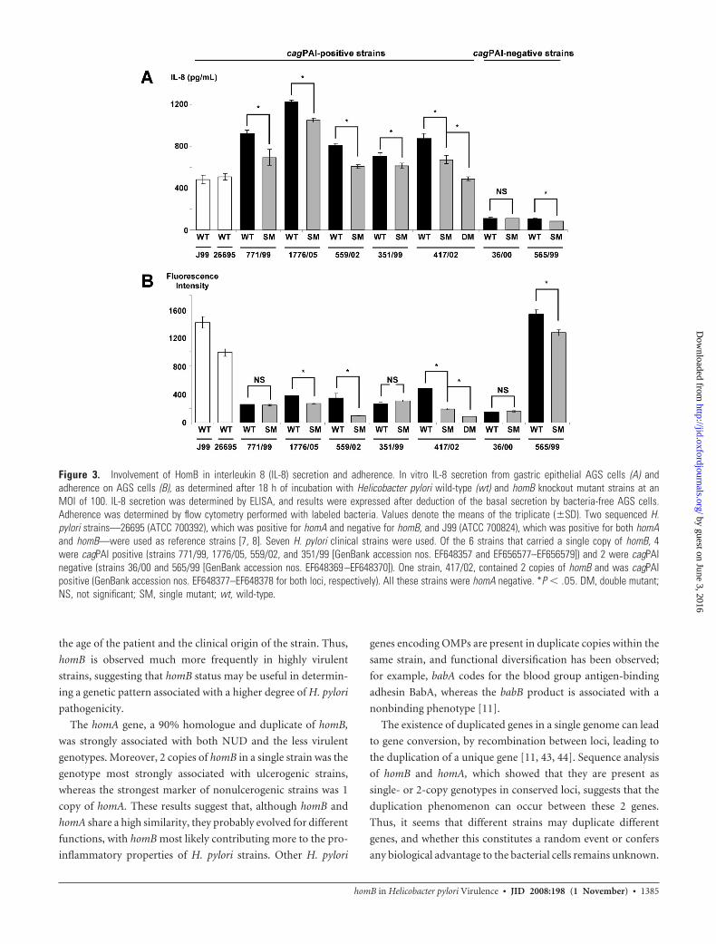
In vitro cellular IL-8 secretion, according to the homB
status of the strains. The level of IL-8 secretion was evaluated
for the 2 H. pylori reference strains (26695 and J99) and the 7
pairs of wt and corresponding homB mutants (figure 3A). IL-8
secretion by epithelial cells is dependent on the presence of a
functional cagPAI [35, 36]. As expected, the mean amount of
IL-8 secretion was significantly higher in cagPAI-positive strains
(mean � SD, 772.1 � 215.7 pg/mL) than in cagPAI-negative
strains (mean � SD, 102.5 � 12 pg/mL). Concerning the 5
cagPAI-positive strains, IL-8 levels obtained with the homB sin-
gle mutants were significantly lower than those obtained with
the parent strains (figure 3A). For strain 417/02, which carried 2
copies of homB, the IL-8 level obtained for the double mutant
was significantly lower than that observed for the corresponding
single mutant. A significant reduction in IL-8 secretion was also
observed for the strain 565/99, 1 of the 2 cagPAI-negative homB
single mutants.
Adherence according to the homB status of the strains. All
of the H. pylori strains tested were able to adhere to the human
cells (figure 3B). The homB disruption of strains 771/99 and
36/00 did not lead to a significant modification in adherence
properties. Concerning strains 1776/05, 559/02, 351/99, and
565/99, the inactivation of the single genomic copy of homB led
to a significant decrease in adherence. For strain 417/02, which
carried 2 copies of homB, the disruption of 1 copy of homB led to
a significant decrease in adherence, compared with the wt strain,
and this decrease was more important when the 2 copies were
disrupted, compared with the single mutant strain (figure 3B).
DISCUSSION
The present study attempts to bring insight to the clinical signif-
icance of a novel H. pylori ulcer marker candidate, the homB
gene, coding for an OMP, aiming to clarify its role as a virulence
homB in Helicobacter pylori Virulence ● JID 2008:198 (1 November) ● 1383
by guest on June 3, 2016http://jid.oxfordjournals.org/
Dow
nloaded from
factor associated with the development of PUD. The distribu-
tion of homB in clinical H. pylori strains showed that homB was
significantly associated with PUD in children and young adults
(�40 years of age). In contrast, homA, the gene to which homB
was 90% similar, was a marker for NUD in those strains. In
addition, cagA�, vacAs1, hopQI, and oipA “on” genotypes were
also strongly associated with PUD in children, whereas, in
adults, only cagA was a marker for PUD. Several studies reported
such differences in strains infecting the mucosa of adults and
children [6, 37– 40]. On the other hand, the fact that homB was a
marker for PUD in children and young adults suggests that dis-
ease severity in younger populations is more closely related to
the virulence of the infecting strain than to environmental fac-
tors, such as smoking and high NSAID consumption, which play
a major role in older populations [28]. Nevertheless, these re-
sults were obtained with small groups of strains, particularly
those isolated from young adults, and they therefore require fu-
ture validation. Overall, it should be emphasized that, among the
H. pylori virulence genotypes analyzed, cagA, as a marker for the
cagPAI, was the only predictor of PUD in both children and
adults; this finding is in agreement with other studies and
thereby reconfirms that cagPAI is one of the major H. pylori
virulence factors [41, 42].
The overall profile of the association of homB with clinical
outcome is supported by the correlation of this factor with the
other H. pylori–associated virulence genotypes, independent of
Figure 2. A, Coomassie blue–stained 2-dimensional gel electrophoresis (2DE) of the outer membrane protein (OMP) fraction of a Helicobacter pylori clinicalstrain with a single complete copy of the homB open reading frame. Shown are the positions of the 9 identified spots: �-glutamyltranspeptidase (GGT),adenosine triphosphate synthase F1 (subunit alpha) (AtpA), flagellar hook protein (FlgE), flagellin A (FlaA), peptidyl-prolyl cis-trans isomerase C (PpiC), hypotheticalprotein (jhp0216), OMP HomB, OMP HomD, and OMP HopN. For each protein spot, the no. of matching peptides, the cross-correlation score (with charge) foreach peptide, and the sequence coverage are described in table 4. B, Western blot of a duplicate 2DE hybridized with the pool of serum samples obtained fromH. pylori–positive patients, showing the positions of the 7 immunoreactive spots. In both panel A and panel B, the isoelectric point (pI) of some proteins presentin the gel is indicated at the bottom and was deduced from the theoretical pI of the corresponding proteins of the 3 H. pylori reference strains 26695, J99,and HPAG1 [7–9]. IEF, isoelectric focusing. C, Analysis of the purification and antigenicity of the recombinant HomB protein (rHpHomB). Shown are theCoomassie blue–stained SDS-PAGE of (1) crude extract of Escherichia coli BL21 cells not expressing rHpHomB, (2) crude extract of E. coli BL21 cells expressingthe rHpHomB, and (3) purified rHpHomB, and Western blot of the purified protein with (4) anti–glutathione S-transferase (GST) antibody and (5) a pool of serumsamples obtained from H. pylori–positive patients. In all 3 panels, the positions of the molecular weights (Mr ) are indicated on the left.
Table 4. Proteins identified, by mass spectrometry, in the 9spots differentially expressed in 2-dimensional gel electrophore-sis of the outer membrane proteome of 2 Helicobacter pylori1776/05 strains: the homB knockout mutant and the wild type.
The table is available in its entirety in the onlineedition of the Journal of Infectious Diseases.
1384 ● JID 2008:198 (1 November) ● Oleastro et al.
by guest on June 3, 2016http://jid.oxfordjournals.org/
Dow
nloaded from
the age of the patient and the clinical origin of the strain. Thus,
homB is observed much more frequently in highly virulent
strains, suggesting that homB status may be useful in determin-
ing a genetic pattern associated with a higher degree of H. pylori
pathogenicity.
The homA gene, a 90% homologue and duplicate of homB,
was strongly associated with both NUD and the less virulent
genotypes. Moreover, 2 copies of homB in a single strain was the
genotype most strongly associated with ulcerogenic strains,
whereas the strongest marker of nonulcerogenic strains was 1
copy of homA. These results suggest that, although homB and
homA share a high similarity, they probably evolved for different
functions, with homB most likely contributing more to the pro-
inflammatory properties of H. pylori strains. Other H. pylori
genes encoding OMPs are present in duplicate copies within the
same strain, and functional diversification has been observed;
for example, babA codes for the blood group antigen-binding
adhesin BabA, whereas the babB product is associated with a
nonbinding phenotype [11].
The existence of duplicated genes in a single genome can lead
to gene conversion, by recombination between loci, leading to
the duplication of a unique gene [11, 43, 44]. Sequence analysis
of homB and homA, which showed that they are present as
single- or 2-copy genotypes in conserved loci, suggests that the
duplication phenomenon can occur between these 2 genes.
Thus, it seems that different strains may duplicate different
genes, and whether this constitutes a random event or confers
any biological advantage to the bacterial cells remains unknown.
Figure 3. Involvement of HomB in interleukin 8 (IL-8) secretion and adherence. In vitro IL-8 secretion from gastric epithelial AGS cells (A) andadherence on AGS cells (B), as determined after 18 h of incubation with Helicobacter pylori wild-type (wt) and homB knockout mutant strains at anMOI of 100. IL-8 secretion was determined by ELISA, and results were expressed after deduction of the basal secretion by bacteria-free AGS cells.Adherence was determined by flow cytometry performed with labeled bacteria. Values denote the means of the triplicate (�SD). Two sequenced H.pylori strains—26695 (ATCC 700392), which was positive for homA and negative for homB, and J99 (ATCC 700824), which was positive for both homAand homB—were used as reference strains [7, 8]. Seven H. pylori clinical strains were used. Of the 6 strains that carried a single copy of homB, 4were cagPAI positive (strains 771/99, 1776/05, 559/02, and 351/99 [GenBank accession nos. EF648357 and EF656577–EF656579]) and 2 were cagPAInegative (strains 36/00 and 565/99 [GenBank accession nos. EF648369 –EF648370]). One strain, 417/02, contained 2 copies of homB and was cagPAIpositive (GenBank accession nos. EF648377–EF648378 for both loci, respectively). All these strains were homA negative. *P � .05. DM, double mutant;NS, not significant; SM, single mutant; wt, wild-type.
homB in Helicobacter pylori Virulence ● JID 2008:198 (1 November) ● 1385
by guest on June 3, 2016http://jid.oxfordjournals.org/
Dow
nloaded from

The observation that the 2-copy genotype was more prevalent in
strains associated with PUD than in strains associated with NUD
points to a nonrandom event of duplication, suggesting a bio-
logical advantage, either by facilitating elevated expression of a
particular protein or by facilitating adaptation to an individual
host.
HomB was shown to be expressed at the H. pylori outer mem-
brane and recognized by human serum samples, indicating that
it possesses surface-exposed regions. The inactivation of homB
caused a significant reduction in IL-8 production, a phenome-
non observed in all of the cagPAI-positive strains and in 1 of the
2 cagPAI-negative strains tested. The double knockout of homB
had a stronger reduction effect, suggesting that both HomB pro-
teins are active and share the same function. This is in accor-
dance with the finding that the presence of 2 copies of homB in a
single strain was the genotype most frequently found in ulcero-
genic strains. It should, however, be emphasized that the level of
reduction in IL-8 secretion obtained with the homB mutants was
low compared with what was previously observed with cagPAI
mutants [35, 36]. These results suggest that, in the presence of
cagPAI, HomB is able to promote an in vitro proinflammatory
response by gastric epithelial cells. This hypothesis is supported
by the finding that homB is strongly associated with cagA, as well
as with other H. pylori virulence factors, because it has been
described for other H. pylori OMPs [19, 45], and it suggests a
shared selective pressure favoring the expression of multiple vir-
ulence determinants.
Helicobacter pylori–related IL-8 induction requires attach-
ment of live bacteria to epithelial cells [46, 47], a prerequisite
required for CagA translocation and consequent induction of
IL-8 secretion [48]. Accordingly, it is likely that the mechanism
underlying the involvement of HomB in inflammation is bacte-
rial adherence. Indeed, it was demonstrated that H. pylori wt
strains adhered to human gastric epithelial cells, whereas the
corresponding homB knockout mutant strains showed signifi-
cantly reduced binding. However, the fact that the disruption of
homB did not completely abolish adherence suggests that HomB
is not the major OMP involved in this mechanism. Indeed, ad-
herence of H. pylori to the human gastric epithelium is mediated
by the BabA major adhesin to the fucosylated Lewis b blood
group antigen [11]. Because the AGS cells express variable levels
of Lewis b blood group antigen [49], it would be interesting to
test other cell lines—for example, the gastric epithelial Kato III
cells deficient in Lewis b epitope—for which H. pylori adherence
is independent of the BabA–Lewis b binding [49].
In conclusion, homB can be considered a comarker of H. py-
lori strains associated with PUD. HomB is immunogenic and is
likely involved in the inflammatory response and in adherence,
suggesting its contribution to the development of a more severe
clinical outcome. Overall, results suggest that homB is a novel H.
pylori putative virulence factor.
Acknowledgments
We thank Paul Jenö and his team from the Mass Spectrometry Biozen-trum, University of Basel (Basel, Switzerland), for performing the LC-MS/MS analysis of the protein spots. We also thank Ivo Boneca (Unité dePathogénie Bactérienne des Muqueuses, Département de Microbiologie, In-stitut Pasteur, Paris, France) for kindly providing the pILL570Hp710�TnKmplasmid. Finally, we thank the Sociedade Portuguesa de Gastrenterologiaand the Programme de Cooperation Scientifique et Technique Franco-Portugais, sponsored by the French Embassy in Portugal, for supporting theproject. We are grateful to Paulo Matos for technical assistance.
References
1. Suerbaum S, Michetti P. Helicobacter pylori infection. N Engl J Med2002; 347:1175– 86.
2. Odenbreit S, Püls J, Sedlmaier B, Gerland E, Fischer W, Haas R. Trans-location of Helicobacter pylori CagA into gastric epithelial cells by type IVsecretion. Science 2000; 287:1497–500.
3. Goodman KJ, Correa P. The transmission of Helicobacter pylori: a criti-cal review of the evidence. Int J Epidemiol 1995; 24:875– 87.
4. Gillen D, McColl KE. Gastroduodenal disease, Helicobacter pylori, andgenetic polymorphisms. Clin Gastroenterol Hepatol 2005; 3:1180 – 6.
5. Peek RM Jr, Crabtree JE. Helicobacter infection and gastric neoplasia.J Pathol 2006; 208:233– 48.
6. Oleastro M, Monteiro L, Lehours P, Mégraud F, Menard A. Identifica-tion of markers for Helicobacter pylori strains isolated from children withpeptic ulcer disease by suppressive subtractive hybridization. Infect Im-mun 2006; 74:4064 –74.
7. Tomb JF, White O, Kerlavage AR, et al. The complete genome sequenceof the gastric pathogen Helicobacter pylori. Nature 1997; 388:539 – 47.
8. Alm RA, Ling LSL, Moir DT, et al. Genomic-sequence comparison oftwo unrelated isolates of the human gastric pathogen Helicobacter pylori.Nature 1999; 397:176 – 80.
9. Oh JD, Kling-Bäckhed H, Giannakis M, et al. The complete genomesequence of a chronic atrophic gastritis Helicobacter pyloristrain: evo-lution during disease progression. Proc Natl Acad Sci USA 2006; 103:9999 –10004.
10. Alm RA, Bina J, Andrews BM, Doig P, Hancock REW, Trust TJ. Com-parative genomics of Helicobacter pylori: analysis of the outer membraneprotein families. Infect Immun 2000; 68:4155– 68.
11. Ilver D, Arnqvist A, Ögren J, et al. Helicobacter pylori adhesin bindingfucosylated histo-blood group antigens revealed by retagging. Science1998; 279:373–7.
12. Mahdavi J, Sondén B, Hurtig M, et al. Helicobacter pylori SabA adhesin inpersistent infection and chronic inflammation. Science 2002; 297:573–8.
13. Yamaoka Y, Ojo O, Fujimoto S, et al. Helicobacter pylori outer mem-brane proteins and gastroduodenal disease. Gut 2006; 55:775– 81.
14. Mégraud F, Lehn N, Lind T, et al. Antimicrobial susceptibility testing ofHelicobacter pylori in a large multicenter trial: the MACH 2 Study. An-timicrob Agents Chemother 1999; 43:2747–52.
15. Tummuru MKR, Cover TL, Blaser MJ. Cloning and expression of ahigh-molecular-mass major antigen of Helicobacter pylori: evidence oflinkage to cytotoxin production. Infect Immun 1993; 61:1799 –1809.
16. Yamaoka Y, Kikuchi S, El-Zimaity HMT, Gutierrez O, Osato MS, Gra-ham DY. Importance of Helicobacter pylori oipA in clinical presentation,gastric inflammation, and mucosal interleukin 8 production. Gastroen-terology 2002; 123:414 –24.
17. Akopyants NS, Clifton SW, Kersulyte D, et al. Analyses of the cag patho-genicity island of Helicobacter pylori. Mol Microbiol 1998; 28:37–53.
18. Atherton JC, Cao P, Peek RM Jr, Tummuru MKR, Blaser MJ, Cover TL.Mosaicism in vacuolating cytotoxin alleles of Helicobacter pylori. Asso-ciation of specific vacA types with cytotoxin production and peptic ul-ceration. J Biol Chem 1995; 270:17771–7.
19. Gerhard M, Lehn N, Neumayer N, et al. Clinical relevance of the Heli-cobacter pylori gene for blood-group antigen-binding adhesin. Proc NatlAcad Sci USA 1999; 96:12778 – 83.
1386 ● JID 2008:198 (1 November) ● Oleastro et al.
by guest on June 3, 2016http://jid.oxfordjournals.org/
Dow
nloaded from
20. van Doorn LJ, Figueiredo C, Sanna R, et al. Clinical relevance of thecagA, vacA, and iceA status of Helicobacter pylori. Gastroenterology1998; 115:58 – 66.
21. Cao P, Cover TL. Two different families of hopQ alleles in Helicobacterpylori. J Clin Microbiol 2002; 40:4504 –11.
22. Lehours P, Menard A, Dupouy S, et al. Evaluation of the association ofnine Helicobacter pylori virulence factors with strains involved in low-grade gastric mucosa-associated lymphoid tissue lymphoma. Infect Im-mun 2004; 72:880 – 8.
23. Peck B, Ortkamp M, Diehl KD, Hundt E, Knapp B. Conservation, local-ization and expression of HopZ, a protein involved in adhesion ofHelicobacter pylori. Nucleic Acids Res 1999; 27:3325–33.
24. Gomes LI, Rocha GA, Rocha AM, et al. Lack of association betweenHelicobacter pylori infection with dupA-positive strains and gastroduo-denal diseases in Brazilian patients. Int J Med Microbiol 2008; 298:223–30.
25. Baik SC, Kim KM, Song SM, et al. Proteomic analysis of the sarcosine-insoluble outer membrane fraction of Helicobacter pylori strain 26695. JBacteriol 2004; 186:949 –55.
26. Candiano G, Bruschi M, Musante L, et al. Blue silver: a very sensitivecolloidal Coomassie G-250 staining for proteome analysis. Electropho-resis 2004; 25:1327–33.
27. Sheu SM, Sheu BS, Yang HB, Lei HY, Wu JJ. Anti-Lewis X antibodypromotes Helicobacter pylori adhesion to gastric epithelial cells. InfectImmun 2007; 75:2661–7.
28. Salles N, Megraud F. Current management of Helicobacter pylori infec-tions in the elderly. Expert Rev Anti Infect Ther 2007; 5:845–56.
29. Ruskey F, Weston M. A survey of Venn diagrams. Electronic Journal ofCombinatorics. Department of Computer Science, University of Victoria,Canada. Available at: http://theory.cs.uvic.ca/venn/EulerianCircles/. Ac-cessed 27 August 2008.
30. Bumann D, Aksu S, Wendland M, et al. Proteome analysis of secretedproteins of the gastric pathogen Helicobacter pylori. Infect Immun 2002;70:3396 – 403.
31. Krah A, Miehlke S, Pleissner KP, et al. Identification of candidate anti-gens for serologic detection of Helicobacter pylori-infected patients withgastric carcinoma. Int J Cancer 2004; 108:456 – 63.
32. Kimmel B, Bosserhoff A, Frank R, Gross R, Goebel W, Beier D. Identi-fication of immunodominant antigens from Helicobacter pyloriand eval-uation of their reactivities with sera from patients with different gas-troduodenal pathologies. Infect Immun 2000; 68:915–20.
33. Sabarth N, Lamer S, Zimny-Arndt U, Jungblut PR, Meyer TF, BumannD. Identification of surface proteins of Helicobacter pylori by selectivebiotinylation, affinity purification, and two-dimensional gel electropho-resis. J Biol Chem 2002; 277:27896 –902.
34. Institut Pasteur. PyloriGene data release R1.6 (Aug 30, 2002). Availableat: http://genolist.pasteur.fr/PyloriGene/genome.cgi. Accessed 12 Au-gust 2008.
35. Yamaoka Y, Kwon DH, Graham DY. A Mr 34,000 proinflammatoryouter membrane protein (OipA) of Helicobacter pylori. Proc Natl AcadSci USA 2000; 97:11133.
36. Fischer W, Püls J, Buhrdorf R, Gebert B, Odenbreit S, Haas R. Systematicmutagenesis of the Helicobacter pylori cag pathogenicity island: essentialgenes for CagA translocation in host cells and induction ofinterleukin-8. Mol Microbiol 2001; 42:1337– 48.
37. Alarcón T, Domingo D, Martinez MJ, López-Brea M. cagA gene andvacA alleles in Spanish Helicobacter pylori clinical isolates from patientsof different ages. FEMS Immunol Med Microbiol 1999; 24:215–9.
38. González-Valencia G, Atherton JC, Muñoz O, Dehesa M, Madrazo de laGarza A, Torres J. Helicobacter pylori vacA and cagA genotypes in Mex-ican adults and children. J Infect Dis 2000; 182:1450 – 4.
39. Oleastro M, Gerhard M, Lopes AI, et al. Helicobacter pylori virulencegenotypes in Portuguese children and adults with gastroduodenal pa-thology. Eur J Clin Microbiol Infect Dis 2003; 22:85–91.
40. Queiroz DM, Bittencourt P, Guerra JB, Rocha AM, Rocha GA, CarvalhoAS. IL1RN polymorphism and cagA-positive Helicobacter pylori strainsincrease the risk of duodenal ulcer in children. Pediatr Res 2005; 58:892– 6.
41. Crabtree JE, Taylor JD, Wyatt JL, et al. Mucosal IgA recognition ofHelicobacter pylori 120 kDa protein, peptic ulceration, and gastric pa-thology. Lancet 1991; 338:332–5.
42. Nomura AMY, Pérez-Pérez GI, Lee J, Stemmermann G, Blaser MJ. Re-lation between Helicobacter pylori cagA status and risk of peptic ulcerdisease. Am J Epidemiol 2002; 155:1054 –9.
43. Pride DT, Blaser MJ. Concerted evolution between duplicated geneticelements in Helicobacter pylori. J Mol Biol 2002; 316:629 – 42.
44. Solnick JV, Hansen LM, Salama NR, Boonjakuakul JK, Syvanen M.Modification of Helicobacter pylori outer membrane protein expressionduring experimental infection of rhesus macaques. Proc Natl Acad SciUSA 2004; 101:2106 –11.
45. Dossumbekova A, Prinz C, Mages J, et al. Helicobacter pylori HopH(OipA) and bacterial pathogenicity: genetic and functional genomicanalysis of hopH gene polymorphisms. J Infect Dis 2006; 194:1346 –55.
46. Aihara M, Tsuchimoto D, Takizawa H, et al. Mechanisms involved inHelicobacter pylori-induced interleukin-8 production by a gastric cancercell line, MKN45. Infect Immun 1997; 65:3218 –24.
47. Keates S, Keates AC, Warny M, Peek RM, Murray PG, Kelly CP. Differ-ential activation of mitogen-activated protein kinases in AGS gastricepithelial cells by cag� and cag� Helicobacter pylori. J Immunol 1999;163:5552–9.
48. Segal ED, Cha J, Lo J, Falkow S, Tompkins LS. Altered states: involve-ment of phosphorylated CagA in the induction of host cellular growthchanges by Helicobacter pylori. Proc Natl Acad Sci USA 1999; 96:14559 –64.
49. Su B, Hellström PM, Rubio C, Celik J, Granström M, Normark S. TypeI Helicobacter pylori shows Lewisb-independent adherence to gastric cellsrequiring de novo protein synthesis in both host and bacteria. J InfectDis 1998; 178:1379 –90.
homB in Helicobacter pylori Virulence ● JID 2008:198 (1 November) ● 1387
by guest on June 3, 2016http://jid.oxfordjournals.org/
Dow
nloaded from
APPENDIX
SUPPLEMENTARY INFORMATION REGARDINGMATERIALS AND METHODS
Construction of homB knockout mutant strains. The homB
knockout mutant strains were constructed by insertion of a kan-
amycin cassette by double homologous recombination. A sui-
cide plasmid containing the targeted gene (HP0710, a homB ho-
mologue) disrupted by a kanamycin resistance gene cassette
(pILL570Hp710�TnKm plasmid) was used for natural transfor-
mation of Helicobacter pylori. Transformation was performed on
an agar surface. In brief, 24 h after inoculation, bacteria were
harvested from an agar plate, suspended in 200 �L of Brucella
broth, spread on an agar base supplemented with 10% horse
blood, and incubated at 37°C for 5 h. Approximately 50 ng of
plasmid DNA was spotted directly onto the inoculated agar, and
incubation was continued for 10 h. The incubated mixture then
was spread on a nonselective medium for 18 h (Columbia agar
supplemented with 10% horse blood). Bacteria then were sus-
pended in 300 �L of Brucella broth, spread on selective plates of
Columbia agar supplemented with 10% horse blood and 20
�g/mL kanamycin, and incubated at 37°C in a microaerobic at-
mosphere for 5 days.
For construction of the double mutant, a second transforma-
tion experiment was performed on a single mutant, with the
homB gene localized at the previously inactivated jhp0649 locus.
Double-mutant colonies were selected on Columbia agar sup-
plemented with 10% horse blood and 250 �g/mL kanamycin.
The correct insertion of the kanamycin cassette in the target
region was confirmed by polymerase chain reaction (PCR) per-
formed with external primers located on the flanking genes and,
subsequently, by sequencing. Southern blot hybridization was
also performed to verify that there was no illegitimate insertion
of the kanamycin cassette elsewhere in the H. pylori genome. A
172-bp probe was prepared from the pILL570Hp710�TnKm plas-
mid with the primers F1-clone (ACTAATGCTTGAAACCCAG)/
R1-clone (CTCCAATTCACTGTTCCTTGC) and was used to hy-
bridize to the genomic DNA digested with HindIII (Promega). The
signal was detected using the DIG Luminescent Detection Kit
(Roche Diagnostics), according to the manufacturer’s instructions.
A single fragment of 1700 bp was detected for both the single and
double mutants (data not shown). In addition, PCR performed
with the internal primers F1-/R1-jhp0870/jhp0649 confirmed that
neither homB nor homA was present in the transformed strain (data
not shown).
Membrane protein preparation. Sarcosine-insoluble outer
membrane fractions, from the wild-type 1776/05 strain and the
corresponding homB knockout mutant strain, were prepared as
described elsewhere [25]. Protein quantification was done ac-
cording to the Bradford method, by use of the Bio-Rad Protein
Assay Kit (Bio-Rad Laboratories).
Two-dimensional gel electrophoresis (2DE). A total of 200
�g of protein membrane pellets of each H. pylori strain was pre-
cipitated with 20% tricloroacetic acid, washed in ice-cold ace-
tone, and solubilized in isoelectric focusing (IEF) buffer—7
mol/L urea, 2 mol/L thiourea, 4% w/v 3-[(3-cholamidopropyl)
dimethylammonio]-1-propanesulphonate, 60 mmol/L 1,4-
dithioerythriol, 0.75% w/v ampholines (pH 3.5–10.0), 0.25%
w/v ampholines pH 4 – 6)—at 37°C for 30 min. The samples
were clarified by centrifugation performed at 12,000 g for 5 min,
and the supernatants containing the membrane protein extracts
were loaded onto 7-cm immobilized pH gradient (IPG) strips
(Immobiline DryStrips) with a nonlinear pH of 3–10 (GE
Healthcare). IEF was performed via stepwise voltage increments
from 100 V to 6500 V, to a total of 29,000 V/h in Protein IEF Cell
equipment (Bio-Rad Laboratories), after an active rehydration
at 50 V for 12 h. For the second dimension, IPG strips were
equilibrated in equilibration solution (50 mmol/L Tris-HCl [pH
8.8], 6 mol/L urea, 30% w/v glycerol, 2% w/v SDS, and traces of
bromophenol blue) with 2% w/v 1,4-dithioerythriol for 15 min
and with 2.8% w/v iodoacetamide for another 15 min. The IPG
strips were sealed with agarose (0.5% in running buffer) in 10%
w/v polyacrylamide gels (1-mm thick) and were run at 50 V for
16 h, by use of SE 600 Ruby equipment (GE Healthcare).
Tryptic digestion of 2DE gel spots and mass spectrometric
analysis. After gel analysis, selected protein spots were excised
from the gel for mass spectrometry analysis and were kept in
microtubes at �20°C until they were analyzed. The protein spots
were cut with a razor blade into small cubes and were washed 5
times with 40% w/v 1-propanol, followed by 5 washes with 50%
w/v acetonitrile and 0.1 mol/L NH4HCO3. The gel pieces were
shrunk with 50 �L of 100% w/v acetonitrile and were dried com-
pletely in a SpeedVac. The dried gel pieces were rehydrated with
10 �L of 50 mmol/L NH4HCO3 containing 125 ng of trypsin
(Sequencing Grade; Promega). Once the gel pieces were impreg-
nated with the trypsin solution, an additional 50 mmol/L
NH4HCO3 was added until the gel pieces were completely im-
mersed in liquid. Digestion was performed at 37°C overnight.
After digestion was completed, the supernatant was transferred
into a new tube, and the gel pieces were extracted with 50 �L of
50% w/v acetonitrile and 0.1% w/v formic acid, and the extract
was combined with the digest. The digest was dried in a Speed-
Vac and redissolved in 25 �L of 0.1% w/v formic acid containing
2% w/v acetonitrile. The peptides were analyzed by capillary liq-
uid chromatography on a Magic C18 100-�m 10-cm reverse-
phase column (Thermo Fisher Scientific) that was connected
to an LTQ-Orbitrap instrument (Thermo Fisher Scientific). A
linear gradient from 5% to 75% w/v solvent B (80% w/v aceto-
nitrile containing 0.1% w/v formic acid) was delivered with a
Rheos 2200 high-performance liquid chromatography system
(Thermo Fisher Scientific) at 50 mL/min. A precolumn flow
by guest on June 3, 2016http://jid.oxfordjournals.org/
Dow
nloaded from
splitter reduced the flow through the column to 500 nL/min.
The eluting peptides were ionized by electrospray ionization,
and the peptides were automatically selected and fragmented in
the ion trap.
Immunoproteomics. 2DE gels were transferred to nitrocel-
lulose membranes by use of a TE 62 Transfer Unit (GE Health-
care) apparatus at 400 mA for 90 min. Membranes were blocked
with 5% skim milk in 0.1% w/v PBS-Tween (PBS-T) at room
temperature for 1 h, and they were incubated with patients’ se-
rum samples (1:400 in PBS-T) at 4°C for 16 h. Incubation with
the secondary antibody anti– human IgG horseradish peroxi-
dase from sheep (GE Healthcare) (1:1500 in PBS-T) was per-
formed at room temperature for 1 h, and development was done
using the ECL Western-blot kit (GE Healthcare).
Data analysis. Individual tandem mass spectrometry spec-
tra, recorded by Xcalibur software (version 2.0; ThermoFinni-
gan), were compared, by use of TurboSequest software (Bio-
Works version 3.1; Thermo Electron), with the protein data
bank and a subset of the National Center for Biotechnology
Information database consisting of protein sequences from
H. pylori. The search parameters were as follows: minimum
cross correlation coefficients of 1.5, 2.0, and 2.5 for singly-,
doubly-, and triply-charged precursor ions, respectively, with
�Cn � 0.1.
by guest on June 3, 2016http://jid.oxfordjournals.org/
Dow
nloaded from
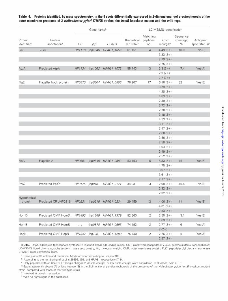
Table 4. Proteins identified, by mass spectrometry, in the 9 spots differentially expressed in 2-dimensional gel electrophoresis of theouter membrane proteome of 2 Helicobacter pylori 1776/05 strains: the homB knockout mutant and the wild type.
Proteinidentifieda
Proteinannotationa
Gene nameb
TheoreticalMr (kDa)a
LC-MS/MS identification
Antigenicspot (status)dHP jhp HPAG1
Matchingpeptides,
no.Xcorr
(charge)c
Sequencecoverage,
%
GGT �-GGT HP1118 jhp1046 HPAG1_1056 61.151 4 4.49 (3�) 10.0 No(B)3.33 (2�)2.79 (2�)2.75 (2�)
AtpA Predicted AtpA HP1134 jhp1062 HPAG1_1072 55.143 3 3.3 (2�) 7.4 Yes(A)2.9 (2�)2.7 (2�)
FlgE Flagellar hook protein HP0870 jhp0804 HPAG1_0853 76.207 17 6.16 (3�) 32 Yes(B)3.29 (2�)4.20 (2�)4.83 (2�)2.39 (2�)3.72 (2�)2.70 (2�)3.18 (2�)4.53 (2�)3.11 (2�)3.47 (2�)2.60 (2�)3.56 (2�)2.58 (2�)1.93 (2�)3.49 (2�)2.52 (2�)
FlaA Flagellin A HP0601 jhp0548 HPAG1_0582 53.153 5 5.33 (2�) 15 Yes(B)4.75 (2�)3.97 (2�)3.61 (2�)2.17 (2�)
PpiC Predicted PpiCe HP0175 jhp0161 HPAG1_0171 34.031 3 2.98 (2�) 15.5 No(B)2.32 (2�)2.32 (2�)
Hypotheticalprotein Predicted CR JHP0216f HP0231 jhp0216 HPAG1_0234 29.459 3 4.06 (2�) 11 Yes(B)
4.01 (2�)2.53 (2�)
HomD Predicted OMP HomD HP1453 jhp1346 HPAG1_1379 82.360 2 2.55 (2�) 3.1 Yes(B)1.89 (2�)
HomB Predicted OMP HomB . . . jhp0870 HPAG1_0695 74.192 2 2.77 (2�) 6 Yes(A)2 (2�)
HopN Predicted OMP HopN HP1342 jhp1261 HPAG1_1289 75.740 2 2.76 (3�) 5 Yes(A)2.57 (2�)
NOTE. AtpA, adenosine triphosphate synthase F1 (subunit alpha); CR, coding region; GGT, glutamyltranspeptidase; �-GGT, gamma-glutamyltranspeptidase;LC-MS/MS, liquid chromatography tandem mass spectrometry; Mr, molecular weight; OMP, outer membrane protein; PpiC, peptidyl-prolyl cis-trans isomeraseC; Xcorr, cross-correlation score.
a Gene product/function and theoretical Mr determined according to Boneca [34].b According to the numbering of strains 26695, J99, and HPAG1, respectively [7–9].c Only peptides with an Xcorr �1.5 (single charge), 2 (double charge), or 2.5 (triple charge) were considered. In all cases, �Cn � 0.1.d Spots apparently absent (A) or less intense (B) in the 2-dimensional gel electrophoresis of the proteome of the Helicobacter pylori homB knockout mutant
strain, compared with those of the wild-type strain.e Involved in protein maturation.f With no homologue in the databases.
by guest on June 3, 2016http://jid.oxfordjournals.org/
Dow
nloaded from