Evaluation of Male-Mediated Reproductive Toxicity Induced by Polyethylene Glycol in Mice
10
World Applied Sciences Journal 31 (8): 1478-1487, 2014 ISSN 1818-4952 © IDOSI Publications, 2014 DOI: 10.5829/idosi.wasj.2014.31.08.83144 Corresponding Author: Aida I El makawy, Cell Biology Department, National Research Centre, EL Tahrir Street, 12622 Dokki, Giza, Egypt. 1478 Evaluation of Male-Mediated Reproductive Toxicity Induced by Polyethylene Glycol in Mice Aida I Elmakawy, Omaima M Abd-Elmoneim, Kawthar AE Diab and Hafiza A Sharaf 1 1 2 3 Cell Biology Department, National Research Centre, EL Tahrir Street, 12622 Dokki, Giza, Egypt 1 Genetics and Cytology Department, National Research Centre, EL Tahrir Street, 12622 Dokki, Giza, Egypt 2 Pathology Department, National Research Centre, EL Tahrir Street, 12622 Dokki, Giza, Egypt 3 Submitted: Jan 05, 2014; Accepted: Mar 20, 2014; Published: Apr 27, 2014 Abstract: In the present work polyethylene glycol (PEG6000) was given orally by gavage to male mice at three different doses 50, 100 and 200 mg/kg/day for consecutive 5 days per week up to six successive weeks. 24hr of treatment, half number of the mice from all experimental groups was sacrificed. Epididymis and testes were collected to evaluate the reproductive toxicity of PEG6000. The other half of treated male mice was mated with untreated virgin female mice to evaluate male-mediated genotoxicity of PEG6000 on fetuses. The results illustrated that PEG6000 decreased sperm motility and count in a dose-dependent manner. However, PEG6000 resulted in an increased fraction of sperm with abnormal morphology or abnormal chromatin integrity measured by toluidine blue and acridine orange. Besides, PEG6000 exhibited an increase in the occurrence of chromosomal aberrations in spermatocytes of treated male and in liver cells of their fetuses. Furthermore, testicular alternation and reduction of DNA nuclear content in spermatogonia and spermatocytes were observed in PEG6000 treated mice. In conclusion, chronic exposure of PEG6000 induced male reproductive impairment which correlated with genetic damage of fetuses generated from mating treated paternal mice with untreated female mice. Key words: Polyethylene Glycol Mouse Sperm Morphology Sperm Chromatin Integrity Histopathology INTRODUCTION Millions of tons of PEGs are involved in industrial process Polyethylene glycol is a polyether compound with discharged as waste water after industrial utilization [4]. many applications from industrial manufacturing to PEG6000 is permitted as food additives in various foods medicine. The structure of PEG is also known as according to the European Parliament and Council polyethylene oxide or polyoxyethylene, depending on its Directive No. 95/2/EC of 20 February1995. Our previous molecular weight [1]. PEGs of different molecular weights experiments exhibited that PEG6000 has been induced (200-10000) are widely used in many cosmetic and DNA damage in mouse bone marrow cells using comet pharmaceutical preparations. In the pharmaceutical assay and micronucleus assay [5]. There is copious industry, they are used as vehicles for drugs and as evidence of male mediated developmental toxicity in ointment bases, capsules, tablet and pill binders, experimental models and some evidence showing a suppositories liquid prescriptions and in veterinary drugs, sensitivity of the male germ line to these transmissible including topical, ophthalmic, oral and rectal preparations effects. Putative mechanism have been proposed [2]. PEGs are utilized as a laxative agent for treatment of including direct action such as contamination through the constipation [3]. Further applications include use as seminal fluid and both genetic and epigenetic pathways ingredients in soaps and detergents, in the textile and including germ cell mutation, induction of germ–line leather industry, in plastics and resins, in the paper genomic instability, suppression of germ cell apoptosis or industry, in printing, in the ceramics and glass industry, interference with genomic imprinting [6] The body of in the rubber, petroleum, mining and metal industries, for knowledge regarding PEG6000 reproductive toxicity has wood preservation and as chemical intermediates [1]. been limited and based largely upon knowledge of similar worldwide every year which of most of them have to be
Transcript of Evaluation of Male-Mediated Reproductive Toxicity Induced by Polyethylene Glycol in Mice

World Applied Sciences Journal 31 (8): 1478-1487, 2014ISSN 1818-4952© IDOSI Publications, 2014DOI: 10.5829/idosi.wasj.2014.31.08.83144
Corresponding Author: Aida I El makawy, Cell Biology Department, National Research Centre,EL Tahrir Street, 12622 Dokki, Giza, Egypt.
1478
Evaluation of Male-Mediated Reproductive Toxicity Induced by Polyethylene Glycol in Mice
Aida I Elmakawy, Omaima M Abd-Elmoneim, Kawthar AE Diab and Hafiza A Sharaf1 1 2 3
Cell Biology Department, National Research Centre, EL Tahrir Street, 12622 Dokki, Giza, Egypt1
Genetics and Cytology Department, National Research Centre, EL Tahrir Street, 12622 Dokki, Giza, Egypt2
Pathology Department, National Research Centre, EL Tahrir Street, 12622 Dokki, Giza, Egypt3
Submitted: Jan 05, 2014; Accepted: Mar 20, 2014; Published: Apr 27, 2014Abstract: In the present work polyethylene glycol (PEG6000) was given orally by gavage to male mice at threedifferent doses 50, 100 and 200 mg/kg/day for consecutive 5 days per week up to six successive weeks. 24hrof treatment, half number of the mice from all experimental groups was sacrificed. Epididymis and testes werecollected to evaluate the reproductive toxicity of PEG6000. The other half of treated male mice was mated withuntreated virgin female mice to evaluate male-mediated genotoxicity of PEG6000 on fetuses. The resultsillustrated that PEG6000 decreased sperm motility and count in a dose-dependent manner. However,PEG6000 resulted in an increased fraction of sperm with abnormal morphology or abnormal chromatin integritymeasured by toluidine blue and acridine orange. Besides, PEG6000 exhibited an increase in the occurrence ofchromosomal aberrations in spermatocytes of treated male and in liver cells of their fetuses. Furthermore,testicular alternation and reduction of DNA nuclear content in spermatogonia and spermatocytes wereobserved in PEG6000 treated mice. In conclusion, chronic exposure of PEG6000 induced male reproductiveimpairment which correlated with genetic damage of fetuses generated from mating treated paternal mice withuntreated female mice.
Key words: Polyethylene Glycol Mouse Sperm Morphology Sperm Chromatin Integrity Histopathology
INTRODUCTION Millions of tons of PEGs are involved in industrial process
Polyethylene glycol is a polyether compound with discharged as waste water after industrial utilization [4].many applications from industrial manufacturing to PEG6000 is permitted as food additives in various foodsmedicine. The structure of PEG is also known as according to the European Parliament and Councilpolyethylene oxide or polyoxyethylene, depending on its Directive No. 95/2/EC of 20 February1995. Our previousmolecular weight [1]. PEGs of different molecular weights experiments exhibited that PEG6000 has been induced(200-10000) are widely used in many cosmetic and DNA damage in mouse bone marrow cells using cometpharmaceutical preparations. In the pharmaceutical assay and micronucleus assay [5]. There is copiousindustry, they are used as vehicles for drugs and as evidence of male mediated developmental toxicity inointment bases, capsules, tablet and pill binders, experimental models and some evidence showing asuppositories liquid prescriptions and in veterinary drugs, sensitivity of the male germ line to these transmissibleincluding topical, ophthalmic, oral and rectal preparations effects. Putative mechanism have been proposed[2]. PEGs are utilized as a laxative agent for treatment of including direct action such as contamination through theconstipation [3]. Further applications include use as seminal fluid and both genetic and epigenetic pathwaysingredients in soaps and detergents, in the textile and including germ cell mutation, induction of germ–lineleather industry, in plastics and resins, in the paper genomic instability, suppression of germ cell apoptosis orindustry, in printing, in the ceramics and glass industry, interference with genomic imprinting [6] The body ofin the rubber, petroleum, mining and metal industries, for knowledge regarding PEG6000 reproductive toxicity haswood preservation and as chemical intermediates [1]. been limited and based largely upon knowledge of similar
worldwide every year which of most of them have to be

World Appl. Sci. J., 31 (8): 1478-1487, 2014
1479
compounds. Therefore, the objective of this study is to Reproductive Toxicity Evaluation Epididymal Spermprovide information regarding reproductive toxicity of Motility, Count and Morphology: The cauda epididymidesPEG6000 in mice. were cut in a pre-warmed saline solution at 37°C and
MATERIALS AND METHODS 5 min. To determine sperm motility, one drop of sperm
Chemicals: PEG6000 was purchased from Al-Gomhoria covered with cover-slip and observed under a standardpharmaceutical Industry, Cairo, Egypt. Cyclophosphamide optical microscope at 400X magnification. A minimum ofmonohydrate (CP) was manufactured by Baxter oncology five microscopic fields were assessed to determine theGmbH, Frankfurt-Germany. PEG6000 and CP were percentage of motile sperm on at least 200 sperm for eachdissolved in distilled water. animal. To count sperm, 5 µl of sperm suspension was
Animals: Adult BALB/c mice of both sexes (Mus was transferred into hemocytometer and sperm wereMusculus), 6–8 weeks old and weighing approximately counted under light microscope and data were expressed25–30 g, were obtained from Animal House of National as the number of sperm per ml. For analysis of spermResearch Center, Dokki, Egypt. The animals were morphology, a drop of sperm suspension was smearedacclimated for a period of two weeks before the beginning onto cleaned slides, allowed to air dry and stained withof the experiments. Mice were housed in stainless-steel 1% aqueous solution of eosin Y. At least 1000 sperm percages, maintained in a room designed for control of each mouse were examined under light microscope attemperature, humidity and light cycle. Mice were fed 1000X magnification. Sperm abnormalities were recordedstandard granulated diet with tap water supplied with ad according to Wyrobek and Bruce [7].libitum. The study protocol was approved by the EthicsCommittee for Animal Care of National Research Center, Sperm Chromatin Integrity AssayDokki, Egypt. Toluidine Blue Assay: The assay was performed
Treatment Schedule: Male mice were randomly assigned et al. [8]. Briefly, air-dried sperm smears were fixed in 96%to five experimental groups of ten mice per group. Control ethanol-acetone (1:1) at 4°C for 30 min. The slides weregroup: mice were given distilled water. Positive control staining with 0.05% TB in 50% Mcilvaine citrategroup: mice were given intraperitoneally CP at single dose phosphate buffer for 10 min. Following staining, the slidesof 20 mg/kg. PEG6000 groups: Three groups of mice were were gently rinsed in a stream of distilled water and airadministrated orally PEG6000 at three doses 50, 100 or 200 dried again. A total 1000 spermatozoa per animal wasmg/kg/day for consecutive 5 days per weeks up to six examined under light microscope at 1000X magnification.successive weeks. Chromatin quality of sperm was assessed in following
Experimental Design: At the end of treatment schedule (abnormal chromatin).half number of the mice from each experimental group wassacrificed by dislocation of neck vertebra. Testes and Acridine Orange Assay: The assay was performedepididymis were collected for different assays. Fifty according to the standard protocol described byuntreated virgin female mice (10 female/group) were Martins et al. [9]. Briefly, air-dried smears were fixed forcohabited with other half of male mice from all groups for 1 hr in Carnoy’s solution at 4°C and allowed to dry. Thetwo weeks. Each treated male mouse was housed in a smears were incubated in 1 N HCl at 75°C for 15 min thenseparate cage with two virgin untreated females of the stained with AO dye (0.2 mg/ml) for 10 min. Slides weresame strain. The day of sperm positive vaginal smear was washed with distilled water and air-dried. At least 1000considered as day‘0’ of gestation. Pregnant female was spermatozoa per mouse were analyzed under fluorescenceremoved from the male’s cage and placed in her own microscope (490/530 nm excitation/barrier filter) at 1000Xindividual cage. The dams were killed by cervical magnification. Two types of staining patterns weredislocation on day 15-17 of gestation and fetuses' livers considered in sperm head; green spermatozoa (double-were collected for mitotic chromosome aberrations stranded DNA), yellow or orange spermatozoa (single-analysis. stranded DNA).
minced with scissors and placed in 37°C incubator for
suspension was placed on warmed microscope slide,
diluted with 95µl saline solution. The diluted suspension
according to standard protocol described by Erenpreisa
criteria: light blue (good chromatin) and dark blue
Abnormal chromatin int egrity 100Good abnormal chromatin int egrity
× +
World Appl. Sci. J., 31 (8): 1478-1487, 2014
1480
Meiotic Chromosome Aberrations Analysis: followed by Duncan's multiple range test for multipleChromosomes from primary spermatocytes were prepared comparisons. Statistical significance was set at p 0.05.according to technique of Evans et al. [10]. Briefly, the Pearson’s correlation coefficient (r) was calculated intunica of the testis was removed and the tubules were order to evaluate correlations between the differenttransferred to a small Petri dish containing isotonic parameters analyzed. The percentage of abnormalsolution (2.2% sodium citrate) and teased out with curved chromatin integrity was calculated according toforceps on a piece of mesh. The cell suspension was Erenpreisa et al. [8] using the following formulatransferred into a conical tube and centrifuged at 1000 rpmfor 10 min at room temperature. The supernatant wasremoved and the pellets were re-suspended in hypotonicsolution (1.1% trisodium citrate) for 20 min at 37°C andfixed in cold Carnoy's fixative. The cells were centrifuged RESULTSand re-suspended in a small volume of Carnoy's fixative.To prepare slides, 3–5 drops of the fixed cell suspension Routine Sperm Analysis: Sperm analysis showed thatwere dropped on a clean slide and air-dried. The slides PEG6000 exhibited significant dose dependent decreasewere stained in 10% Giemsa solution in phosphate buffer (p= 0.01) in the percentage of sperm motility and count(pH 6.8) for 5 min. A total 100 well spread diakinesis- when compared to negative control group (Table 1).metaphase I cells per mice were examined under light However, such reduction in motility and number of spermmicroscope at 1000X magnification. was more in the positive control value. As shown in
Fetal Chromosome Aberrations Analysis: significant increase in the frequencies of abnormal spermChromosomes from fetal liver were prepared according to morphology in both head and tail regions when comparedRomagnano et al. [11]. Fetal liver tissue was placed in a with negative control. The increase in sperm abnormalitiesconical centrifuge tube with 2ml RPMI-1640 medium and in PEG6000-treated mice was dose-dependent manner butdissociated manually using Pasteur pipette. The liver lower than the positive control value. Sperm withsample was incubated with 0.02ml 0.05% colchicine at amorphous head shape and coiled tail represent a major37°C for 90 min. The samples were centrifuged for 5 min part of sperm abnormalities induced after oral repeatedand then incubated in 2 ml of a prewarmed hypotonic KCL treatment with different doses of PEG6000. They reachedsolution (0.56%) for 15 min at 37° C. Cells were fixed twice their maximum percentages 9.20% and 22.20% respectivelyCarnoy's fixative and slides were prepared and air- after treatment with the highest tested dose of PEG6000.dried, stained with 10% Giemsa in phosphate buffer Sperm with small and big head were observed only in(pH 6.8). Good-quality metaphases (n = 100) per each positive control.fetus were examined under light microscope at 2000Xmagnification. Sperm Chromatin Integrity: As shown in Table 1, the
Histological and Histochemical Study: Right testis from integrity) was significantly increased after repeated oraleach mouse were excised and fixed in 10% formal saline treatment with all doses of PEG6000 when compared withfollowed by dehydration in ascending grades of alcohol, negative and positive control groups. It reached itsclearing in xylene and embedding in paraffin wax. Paraffin maximum 16.36±0.14 at the highest tested dose of PEG6000sections (5 ìm thicknesses) were stained with hematoxylin compared with 2.58±0.15 for the negative control. Suchand eosin (H&E) for the histological examination and percentages represent 6.34 fold increases compared withFeulgen method for demonstration DNA content [12]. The control group. These data suggested that PEG6000 hasDNA contents were elucidated from Feulgen stained prominent impact on chromatin proteins inhibitionsections subjected to measurement of optical density especially protamine.using Image Pro Plus image analysis software (Media Staining treated sperm with AO exhibited thatCybernetics Inc, 2002). PEG6000 induced significant increase in the denaturation
Statistical Analysis: Statistical analysis was performed relationship. The proportion of red spermatozoa reachedusing SPSS for Windows (Version 11). Data were 8.06 ±0.21, 10.90±0.23 and 16.36±0.14 at the three differentcompared by one-way analysis of variance (ANOVA) doses 50, 100 and 200 mg/kg, respectively compared with
Table (2), the three tested doses of PEG6000 induced
percentage of dark blue sperm (abnormal chromatin
DNA strand (red spermatozoa) with dose-dependent

World Appl. Sci. J., 31 (8): 1478-1487, 2014
1481
Table 1: Effect of treatment with PEG6000 on mouse sperm motility, sperm count and chromatin integrity
Sperm with abnormal chromatin integritySperm motility Total sperm count × 10 (Mean% ± SE)6
------------------------------- ------------------------------------- -----------------------------------------------------Treatment groups Mean ± SE % Mean ± SE % AO orange sperm TB dark sperm
I-Negative control 82.00 ±1.23 82 549.60 ±13.96 78.5 2.58±0.15 2.66 ±0.051 a a e e
II-Positive control 52.00 ±1.23 52 196.40 ±4.29 28.05 25.90±0.19 27.6±0.93 e e a a
III-PEG-600050 72.00 ±1.23 72 432.20 ±8.49 61.74 7.44±0.35 8.06 ±0.21 b b d d
100 68.00 ±1.23 68 354.60 ±21.39 50.65 10.62±0.30 11.90±0.23 c c c c
200 64.00 ±1.87 64 270.20 ±12.59 38.6 16.36±0.14 18.46±0.28 d d b b
5000 sperm were examined for chromatin integrity in 5 mice per each experimental groupMean values followed with different superscript letters within the same column are significantly different from one another (P= 0.05).
Table 2: Effect of treatment with PEG6000 on the mouse sperm plasma membrane integrity
Type of head abnormalities ---------------------------------------------------------------------------------------------------------------------------
Treatment groups Amorphous Hookless Banana Forked Small Big Coiled tail Total
I-Negative control 0.60±0.25 2.20±0.20 0.00±0.00 0.00±0.00 0.00 ±0.00 0.00 ±0.00 2.60±0.25 5.40±0.25 e c b b b b e e
II-Positive control 12.80 ±1.07 9.40±0.87 1.20 ±0.37 2.80 ±0.49 1.80±0.49 1.00±1.03 27.00±1.76 56.00±3.98 a a a a a a a a
III-PEG-600050 5.00±0.32 4.60 ±0.25 0.40±0.25 0 .40±0.25 0.00±0.00 0.00±0.00 10.20±0.86 20.60±0.51 d c ab b b b d d
100 7.20±0.37 7.40±0.40 0.60±0.25 0.40±0.25 0.00±0.00 0.00±0.00 15.20±0.66 30.80±0.86 c b ab b b b c c
200 9.20±0.37 8.20±0.37 1.20±0.37 0.80±0.37 0.00±0.00 0.00 ±0.00 22.20±.860 41.40±1.21 b b a b b b b b
5000 sperm were examined in 5 mice per each experimental group Data were expressed as mean% ± S.E. Mean values followed with different superscript letters within the same column are significantly different from one another(P 0.05)
Table 3: Effect of treatment with PEG6000 on mouse meiotic chromosomal aberrations
Structural aberrations Numerical aberrations--------------------------------------------------------------------------------------------------------- -------------------------------------
Treatment groups X-Y Uni. Auto.Uni X-Y Break Fragment Chain Aneuploidy Polyploidy Total
I-Negative control 3.00±0.45 1.80±0.20 0.20±0.20 0.40±0.25 0.00±0.00 0.00±0.00 0.00±0.00 5.4± 0.250 c d a a b c b e
II-Positive control 6.60±1.03 9.00±0.84 0.80±0.20 2.00±1.05 0.60±0.25 2.60±0.40 1.20±0.37 22.80±0.58 ab a a b a a a a
III-PEG600050 5.80 ±0.80 3.00 ±0.55 0.40±0.25 0.40±0.24 0.00+0.00 0.00+0.00 0.00±0.00 9.60±0.98b cd a a b c b d
100 7.00 ±1.04 4.20 ±0.66 0.60±0.25 0.60 ±0.40 0.20±0.20 0.80±0.37 0.20±0.20 13.60±0.93 ab bc a a a ab bc b c
200 8.60±0.68 5.60 ±0.51 0.80±0.37 0.80±0.37 0.40 ±0.25 1.20±0.49 0.40±0.20 17.80±0.58 a b a a ab b b b
500 metaphases were examined in 5 mice per each experimental group. Auto = Autosomal; Uni= UnivalentData were expressed as mean % ± S.E. Values within the same column followed by different letters are significantly different form one another (p 0.05).
Table 4: Frequencies of different types of chromosomal aberrations induced in fetal liver generated from paternal mice treated with PEG6000
Metaphases with different types of chromosomal aberrations--------------------------------------------------------------------------------------------------------------------------------------------- Abnormal metaphases Structural aberrations Numerical aberrations -------------------------------------------------------------------------------------------------------------------------------- ---------------------------------------------- Including Excluding
Treatment groups Ch. Gap Chro. gap Ch. Break C.T Del. &Fr. Total Prediploidy Polyploidy Total gaps gaps
I-Negative control 0.12±0.07 0.00±0.00 0.12±0.07 0.12±0.07 0.00±0.00 0.36±0.11 0.16±0.08 0.00±0.00 0.16±0.08 0.48±0.12 0.40±0.10 d d e d c e d d d e e
II-Positive control 5.04±0.23 0.80±0.41 4.72±0.16 4.12±0.19 1.60±0.21 16.28±0.40 1.84±0.14 1.64±0.10 3.48±0.15 19.72±0.38 13.88±0.40 a a a a a a a a a a a
III-PEG600050 0.72±0.09 0.20±0.08 0.56±0.10 1.24±0.15 0.36±0.10 3.28±0.24 0.56±0.10 0.44±0.10 1.00±0.15 4.28±0.30 3.16±0.21 c cd c c c d c c c d d
100 1.64±0.10 0.40±0.10 1.00±0.12 1.60±0.10 0.92±0.13 5.60±0.22 1.32±0.14 1.04±0.14 2.32±0.19 7.92±0.24 5.88±0.23 b bc b c b c b b b c c
200 1.80±0.08 0.56±0.10 1.20±0.14 2.08±0.11 1.24±0.13 6.88±0.27 1.84±0.08 1.48±0.10 3.32±0.11 10.16±0.29 7.84 ±0.24 b b d b ab b a a a b b
Data were expressed as mean% ± S.E. Values within the same column follow by different superscript letters are significantly different from one another (P 0.05). Ch=chromatid;Chro=chromosomal; C.T= Centromeric attenuation; Del=Deletion; Fr=Fragment 25 fetus were analysis for chromosomal aberrations per each experimental group. 100 metaphases wereexamined per each fetus

World Appl. Sci. J., 31 (8): 1478-1487, 2014
1482
Table 5: Effect of treatment with PEG6000 on DNA content in testes of male mice
Control 50mg/kg PEG 100mg/kg PEG 200mg/kg PEG
Spermatogonial DNA 0.432+0.004 d 0.193+0.007 a 0.275+0.003 b 0.300+0.003 cSpermatocytes DNA 0.341+0.003 d 0.180+0.005 a 0.254+0.002 b 0.273+0.003 c
Data were expressed as mean% ± S.E. Values within the same column follow by different superscript letters are significantly different from one another(P= 0.05).
2.66±0.051 for control group (Table 1). Such percentages PEG6000. However, metaphases with fragments and X-Yrepresent 3.03, 4.09 and 6.15 fold increases at three doses breaks were insignificantly increased with all three dosesof PEG6000 when compared with the value of control of PEG6000. Estimation of aneuploidy depends on numbergroup, respectively. This result implies that PEG6000 has of hyperhaploid metaphases, while hypohaploids wereeffect on disturbance of oxidation process of thiols of completely excluded because an artificial loss of a singlecyteines in protamine molecules leading to abnormal chromosome may be occur during preparation. The twosperm with poor disulphide bonds. highest doses of PEG6000 were induced a statistically
The proportion of spermatozoa with abnormal significant and insignificant increase in metaphases withchromatin conformation detected by AO was strongly hyperhaploid and polyploidy respectively compared withcorrelated with the proportion of spermatozoa detected by controls group. It is noteworthy that, some types ofTB (r=0.972, p 0.0001). Pearson's correlation analysis aberrations including chain, aneuploidy, polyploidy wereshowed positive significant correlations between not recorded at low dose of PEG6000 and control group.abnormal sperm morphology and abnormal chromatin Pearson's correlation analysis illustrated high significantintegrity detected by TB and AO. TB assay was strongly positive correlation between meiotic CAs and totalcorrelated with abnormal sperm morphology in head abnormal sperm morphology induced in PEG6000 (r=0.877,(r= 0.911, p 0.0001) and tail (0.941, p 0.0001) regions. p 0.001).Furthermore, AO was highly correlated with abnormalsperm morphology in head (r= 0.941, p 0.0001) and tail Fetal Chromosome Aberrations (CAs) Analysis:(0.954, p 0.0001) regions. Analysis of mitotic CAs from fetuses generated from
Meiotic Chromosome Aberrations (CAs) Analysis: All with PEG6000 showed that PEG6000 causedthe tested doses of PEG6000 induced significant increase precocious increase in the occurrence of CAs with dose-in the incidence of CAs in mouse spermatocytes with dependent relationship even after excluding gaps (Tabledose dependent relationship (Table 3). Such proportions 4). All types of numerical and structural CAs werewere arisen from dramatically increase in frequency of X-Y significantly increased with all three doses of PEG6000and autosomal univalents with all doses of PEG6000. when compared with negative control group. However,Metaphases with translocations in the form of chain IV such increase was lower than the value of the positivewere significant increase by two highest doses of control group.
mating untreated female mice and paternal mice treated
Fig. 1: Section of control testis of mice showing normal architecture of testis, seminiferous tubules (S) show a clearlumen and all cell types are represented including sertoli cells (arrow head), spermatogonia, primaryspermatocytes and spermatids (Hx& E A, ×200; B, ×500).

World Appl. Sci. J., 31 (8): 1478-1487, 2014
1483
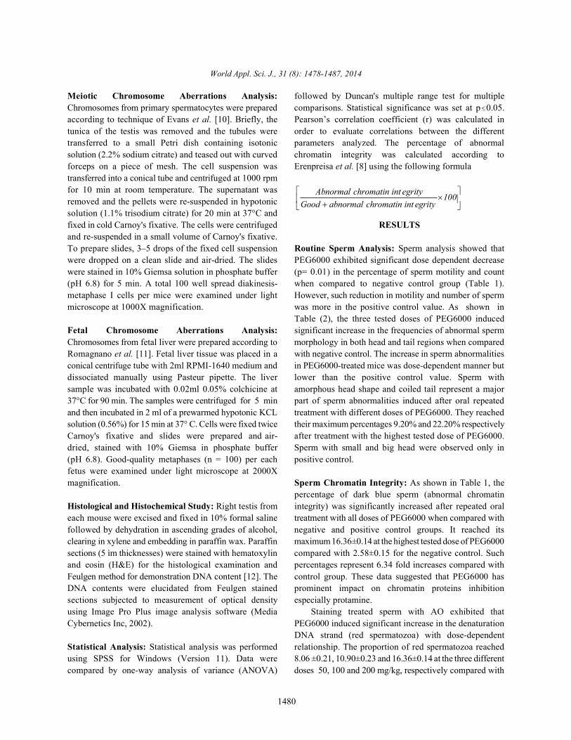
Fig. 2: Section of testis treated with 50 ml/Kg PEG6000 showing (A):degeneration in some seminiferous tubules,detachment of spermatogenic cells and vacuolation areas (area lacking of spermatogenic activity; arrow head).B: pyknosis in spermatogenic cells (arrow), depletion of germ cells layers (A-B Hx & E. x200).
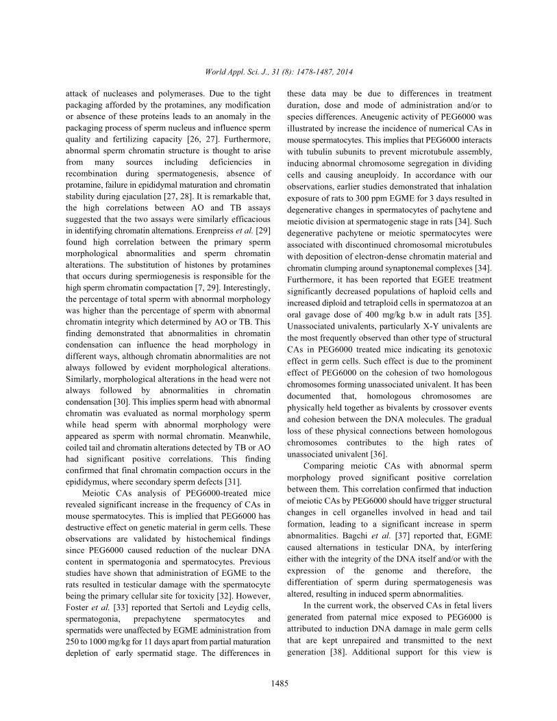
Fig. 3: Section of testis treated with 100 ml/Kg PEG600 showing (A): marked degeneration, necrosis of germ cells(arrows), sloughing of germ cells into tubular lumen. (B): higher magnification of another section showingdepletion of long spermatids ( weave arrow) and the presence of gaps (arrow head) inbetween the spermatogeniccells (Hx. & E. X 200 & 400).
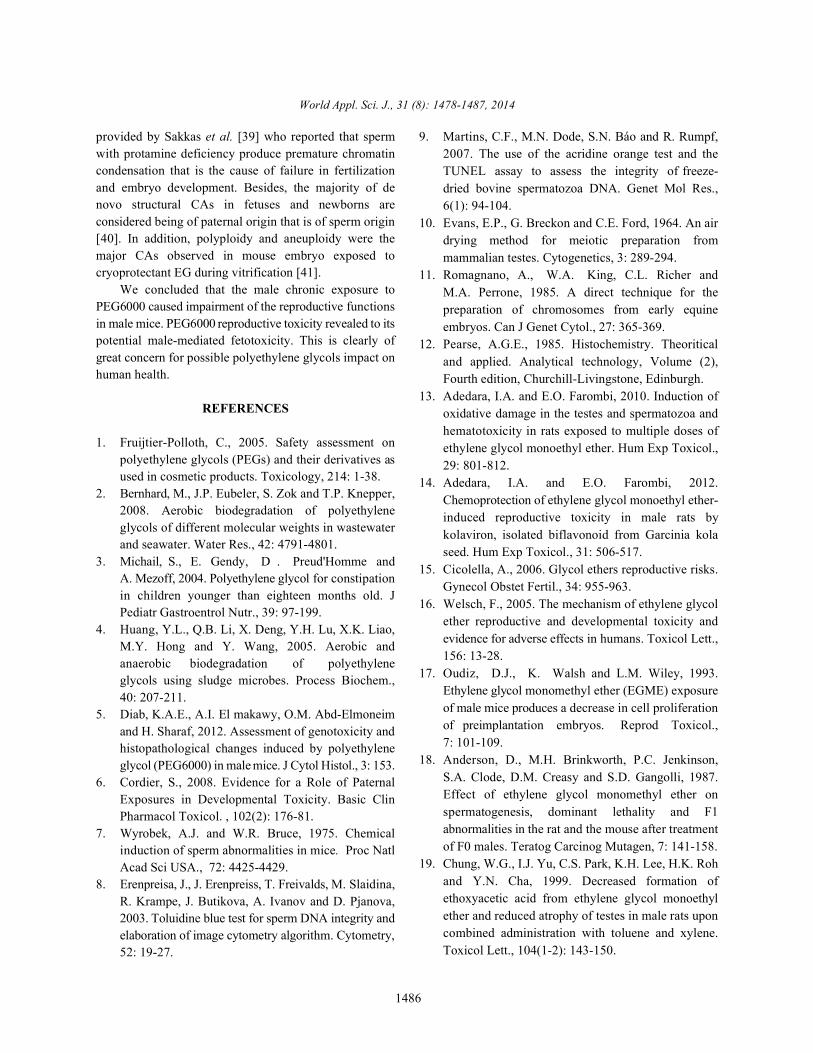
Fig. 4: Section of testis mice treated with 200 mg/kg PEG6000 showing (A): extensive exfoliation of germ cells into tubularlumen (arrow), while the germ cells degeneration, vacuolation, disorganization were generally seen. B-highermagnification of the same section showing depletion of most germ cells layers and long spermatids (Hx & E x 200& 400).
Histological and Histochemical Study: Histological detachment of spermatogenic cells and cytoplasmicexamination of testes from control mice revealed normal vacuolation, pyknosis in spermatogenic cell nuclei andspermatogenesis, with nearly all tubules showing sings of depletion of germ cells layers (Figure 2). Theevidence of endogenous spermatogenesis (Figure 1). On testis of mice treated with 100mg/kg bw appeared markedthe other hand, testicular degeneration and atrophy were degeneration, necrosis of germ cells, sloughing ofobserved in the testis of groups treated with PEG600 with spermatogenic cells into tubular lumen, depletion of longa dose dependent manner. Testis of dose of 50mg/k PEG spermatids and the presence of gaps between the6000 revealed degeneration in some seminiferous tubules, spermatogenic cells (Figure 3). Also,200mg/kg of PEG600

World Appl. Sci. J., 31 (8): 1478-1487, 2014
1484
treated mice showed extensive exfoliation of germ cells late spermatocytes and spermatids, decreased numbers ofinto tubular lumen, while disorganization of sperm and increased numbers of immature germ cells,spermatogenic layers, vacuolation and germ cells decreased sperm motility, peritubular membrane damagedegeneration were still present (Figure 4). and germinal epithelial distortion of the seminiferous
The mean value of DNA per nucleus was measured epithelium [18].by image analyzer and the results was expressed as In the current report, the reduction in sperm countoptical density values (Table 5). The mean value for may be reflected impact of PEG6000 on spermatogenesisnuclear DNA content of spermatogonial nuclei was causing severe erosion and necrosis of the germinalsignificantly lower in all PEG600 treated groups epithelium of the testes [13]. Testicular atrophy and(0.300 ±0.003,0.275 ±0.003 and 0.193±0.00715, respectively) degeneration of germinal epithelium causing oligo orthan control (0.432±0.004). Also the mean value for azoospermia has been recognized consequence ofnuclear DNA content of spermatocytes nuclei were treatment male rat with Ethylene glycol monomethyl ether,significantly lower in all treated groups compared to EGME [19]. Additionally, occupational exposure for malecontrol group. workers to EGs derivative caused an increased prevalence
DISCUSSION ratio for a lower sperm count per ejaculate [20, 21]. The
The present study clearly illustrated that repeated occur as a consequence of endogenous factors like theoral administration of PEG6000 for six weeks caused machinery for motility (flagellum) and alterations in themarked reduction in number and motility of sperm and available energy and oxidative damages. Similar findingobvious increase in occurrence of sperm with abnormal were reported that induction of free radical by EG may bemorphology and abnormal chromatin integrity. In affected on sperm motility by peroxidation ofaddition, dramatically increase in the incidence of CAs in polyunsaturated fatty acid in sperm or by destruction ofmouse spermatocytes and fetal liver generated from sperm mitochondria which is responsible for the energymating untreated female mice with treated paternal mice production necessary to maintain spermatozoa movementwith PEG6000. Histological examination revealed that in some species [22, 23]. Recently, oral administration ofPEG6000 caused testicular alteration that characterized by EGME at 600 mg/kg/daily for 5 weeks significantlyseminiferous tubules degeneration, detachment of decreased total and progressive motility of ratspermatogenic cells, cytoplasmic vacuolation, necrosis spermatozoa [24]. and pyknosis of germ cells, sloughing of spermatogenic Cytochemical experiments were undertaken tocells into tubular lumen, depletion of long spermatids and validate impact of PEG6000 on sperm plasma membraneextensive exfoliation of germ cells into tubular lumen. and chromatin integrity. Sperm analysis revealed that theThese findings may be attributed to accumulation of proportion of sperm with abnormal morphology orPEG6000 in testis that causes detrimental effects to the abnormal chromatin integrity detected by TB and AOmale reproductive performance [13-15]. The observed were dramatically increased in PEG6000 treated mice.effects are attributed to the mode of action of PEG6000. These finding suggested that PEG6000 has prominentThe precise mechanisms of PEG6000 remain unknown. effect on deficiency of protamines and disturbance theHowever, the reproductive toxicity of PEG6000 is probably formation of disulphide bonds which responsible for tightdue to mode of action of one or more of its metabolites packaging of sperm chromatin. This view is explained byrather than to the parent compound. This view supported the fact that sperm chromatin condensation during thewith the earlier studies reported that ethylene gylycols final steps of spermatogenesis in mammals is a multistep(EGs) are biologically activated by enzyme alcohol process that includes the sequential replacement of thedehydrogenase into alkoxyacetic acid metabolites that chromatin proteins (especially histones) by protamines,interfered with the formation of nucleic acids [16]. Thus, allowing a different structural organization to take place intoxicity may be more apparent in tissues undergoing rapid the sperm nucleus [25]. Protamine is rich in cysteine andcell proliferation, such as during spermatogenesis, also other basic amino acids. During the epididymalembryogenesis and fetal development [17]. Similar results passage, the thiols of cysteines in protamine are oxidizedwere obtained when the animals exposed to ethylene to disulfide bonds (S-S). This type of organizationglycol monoethyl ether (EGME) showed testicular provides sperm with a highly condensed nucleus (rich inatrophy, abnormal sperm morphology, degeneration of S-S) and also protects sperm DNA against the enzymatic
of oligospermia and azzospermia and an increased odds
reduction in sperm motility in PEG6000-treated mice might
World Appl. Sci. J., 31 (8): 1478-1487, 2014
1485
attack of nucleases and polymerases. Due to the tight these data may be due to differences in treatmentpackaging afforded by the protamines, any modificationor absence of these proteins leads to an anomaly in thepackaging process of sperm nucleus and influence spermquality and fertilizing capacity [26, 27]. Furthermore,abnormal sperm chromatin structure is thought to arisefrom many sources including deficiencies inrecombination during spermatogenesis, absence ofprotamine, failure in epididymal maturation and chromatinstability during ejaculation [27, 28]. It is remarkable that,the high correlations between AO and TB assayssuggested that the two assays were similarly efficaciousin identifying chromatin alternations. Erenpreiss et al. [29]found high correlation between the primary spermmorphological abnormalities and sperm chromatinalterations. The substitution of histones by protaminesthat occurs during spermiogenesis is responsible for thehigh sperm chromatin compactation [7, 29]. Interestingly,the percentage of total sperm with abnormal morphologywas higher than the percentage of sperm with abnormalchromatin integrity which determined by AO or TB. Thisfinding demonstrated that abnormalities in chromatincondensation can influence the head morphology indifferent ways, although chromatin abnormalities are notalways followed by evident morphological alterations.Similarly, morphological alterations in the head were notalways followed by abnormalities in chromatincondensation [30]. This implies sperm head with abnormalchromatin was evaluated as normal morphology spermwhile head sperm with abnormal morphology wereappeared as sperm with normal chromatin. Meanwhile,coiled tail and chromatin alterations detected by TB or AOhad significant positive correlations. This findingconfirmed that final chromatin compaction occurs in theepididymus, where secondary sperm defects [31].
Meiotic CAs analysis of PEG6000-treated micerevealed significant increase in the frequency of CAs inmouse spermatocytes. This is implied that PEG6000 hasdestructive effect on genetic material in germ cells. Theseobservations are validated by histochemical findingssince PEG6000 caused reduction of the nuclear DNAcontent in spermatogonia and spermatocytes. Previousstudies have shown that administration of EGME to therats resulted in testicular damage with the spermatocytebeing the primary cellular site for toxicity [32]. However,Foster et al. [33] reported that Sertoli and Leydig cells,spermatogonia, prepachytene spermatocytes andspermatids were unaffected by EGME administration from250 to 1000 mg/kg for 11 days apart from partial maturationdepletion of early spermatid stage. The differences in
duration, dose and mode of administration and/or tospecies differences. Aneugenic activity of PEG6000 wasillustrated by increase the incidence of numerical CAs inmouse spermatocytes. This implies that PEG6000 interactswith tubulin subunits to prevent microtubule assembly,inducing abnormal chromosome segregation in dividingcells and causing aneuploidy. In accordance with ourobservations, earlier studies demonstrated that inhalationexposure of rats to 300 ppm EGME for 3 days resulted indegenerative changes in spermatocytes of pachytene andmeiotic division at spermatogenic stage in rats [34]. Suchdegenerative pachytene or meiotic spermatocytes wereassociated with discontinued chromosomal microtubuleswith deposition of electron-dense chromatin material andchromatin clumping around synaptonemal complexes [34].Furthermore, it has been reported that EGEE treatmentsignificantly decreased populations of haploid cells andincreased diploid and tetraploid cells in spermatozoa at anoral gavage dose of 400 mg/kg b.w in adult rats [35].Unassociated univalents, particularly X-Y univalents arethe most frequently observed than other type of structuralCAs in PEG6000 treated mice indicating its genotoxiceffect in germ cells. Such effect is due to the prominenteffect of PEG6000 on the cohesion of two homologouschromosomes forming unassociated univalent. It has beendocumented that, homologous chromosomes arephysically held together as bivalents by crossover eventsand cohesion between the DNA molecules. The gradualloss of these physical connections between homologouschromosomes contributes to the high rates ofunassociated univalent [36].
Comparing meiotic CAs with abnormal spermmorphology proved significant positive correlationbetween them. This correlation confirmed that inductionof meiotic CAs by PEG6000 should have trigger structuralchanges in cell organelles involved in head and tailformation, leading to a significant increase in spermabnormalities. Bagchi et al. [37] reported that, EGMEcaused alternations in testicular DNA, by interferingeither with the integrity of the DNA itself and/or with theexpression of the genome and therefore, thedifferentiation of sperm during spermatogenesis wasaltered, resulting in induced sperm abnormalities.
In the current work, the observed CAs in fetal liversgenerated from paternal mice exposed to PEG6000 isattributed to induction DNA damage in male germ cellsthat are kept unrepaired and transmitted to the nextgeneration [38]. Additional support for this view is
World Appl. Sci. J., 31 (8): 1478-1487, 2014
1486
provided by Sakkas et al. [39] who reported that sperm 9. Martins, C.F., M.N. Dode, S.N. Báo and R. Rumpf,with protamine deficiency produce premature chromatincondensation that is the cause of failure in fertilizationand embryo development. Besides, the majority of denovo structural CAs in fetuses and newborns areconsidered being of paternal origin that is of sperm origin[40]. In addition, polyploidy and aneuploidy were themajor CAs observed in mouse embryo exposed tocryoprotectant EG during vitrification [41].
We concluded that the male chronic exposure toPEG6000 caused impairment of the reproductive functionsin male mice. PEG6000 reproductive toxicity revealed to itspotential male-mediated fetotoxicity. This is clearly ofgreat concern for possible polyethylene glycols impact onhuman health.
REFERENCES
1. Fruijtier-Polloth, C., 2005. Safety assessment onpolyethylene glycols (PEGs) and their derivatives asused in cosmetic products. Toxicology, 214: 1-38.
2. Bernhard, M., J.P. Eubeler, S. Zok and T.P. Knepper,2008. Aerobic biodegradation of polyethyleneglycols of different molecular weights in wastewaterand seawater. Water Res., 42: 4791-4801.
3. Michail, S., E. Gendy, D . Preud'Homme andA. Mezoff, 2004. Polyethylene glycol for constipationin children younger than eighteen months old. JPediatr Gastroentrol Nutr., 39: 97-199.
4. Huang, Y.L., Q.B. Li, X. Deng, Y.H. Lu, X.K. Liao,M.Y. Hong and Y. Wang, 2005. Aerobic andanaerobic biodegradation of polyethyleneglycols using sludge microbes. Process Biochem.,40: 207-211.
5. Diab, K.A.E., A.I. El makawy, O.M. Abd-Elmoneimand H. Sharaf, 2012. Assessment of genotoxicity andhistopathological changes induced by polyethyleneglycol (PEG6000) in male mice. J Cytol Histol., 3: 153.
6. Cordier, S., 2008. Evidence for a Role of PaternalExposures in Developmental Toxicity. Basic ClinPharmacol Toxicol. , 102(2): 176-81.
7. Wyrobek, A.J. and W.R. Bruce, 1975. Chemicalinduction of sperm abnormalities in mice. Proc NatlAcad Sci USA., 72: 4425-4429.
8. Erenpreisa, J., J. Erenpreiss, T. Freivalds, M. Slaidina,R. Krampe, J. Butikova, A. Ivanov and D. Pjanova,2003. Toluidine blue test for sperm DNA integrity andelaboration of image cytometry algorithm. Cytometry,52: 19-27.
2007. The use of the acridine orange test and theTUNEL assay to assess the integrity of freeze-dried bovine spermatozoa DNA. Genet Mol Res.,6(1): 94-104.
10. Evans, E.P., G. Breckon and C.E. Ford, 1964. An airdrying method for meiotic preparation frommammalian testes. Cytogenetics, 3: 289-294.
11. Romagnano, A., W.A. King, C.L. Richer andM.A. Perrone, 1985. A direct technique for thepreparation of chromosomes from early equineembryos. Can J Genet Cytol., 27: 365-369.
12. Pearse, A.G.E., 1985. Histochemistry. Theoriticaland applied. Analytical technology, Volume (2),Fourth edition, Churchill-Livingstone, Edinburgh.
13. Adedara, I.A. and E.O. Farombi, 2010. Induction ofoxidative damage in the testes and spermatozoa andhematotoxicity in rats exposed to multiple doses ofethylene glycol monoethyl ether. Hum Exp Toxicol.,29: 801-812.
14. Adedara, I.A. and E.O. Farombi, 2012.Chemoprotection of ethylene glycol monoethyl ether-induced reproductive toxicity in male rats bykolaviron, isolated biflavonoid from Garcinia kolaseed. Hum Exp Toxicol., 31: 506-517.
15. Cicolella, A., 2006. Glycol ethers reproductive risks.Gynecol Obstet Fertil., 34: 955-963.
16. Welsch, F., 2005. The mechanism of ethylene glycolether reproductive and developmental toxicity andevidence for adverse effects in humans. Toxicol Lett.,156: 13-28.
17. Oudiz, D.J., K. Walsh and L.M. Wiley, 1993.Ethylene glycol monomethyl ether (EGME) exposureof male mice produces a decrease in cell proliferationof preimplantation embryos. Reprod Toxicol.,7: 101-109.
18. Anderson, D., M.H. Brinkworth, P.C. Jenkinson,S.A. Clode, D.M. Creasy and S.D. Gangolli, 1987.Effect of ethylene glycol monomethyl ether onspermatogenesis, dominant lethality and F1abnormalities in the rat and the mouse after treatmentof F0 males. Teratog Carcinog Mutagen, 7: 141-158.
19. Chung, W.G., I.J. Yu, C.S. Park, K.H. Lee, H.K. Rohand Y.N. Cha, 1999. Decreased formation ofethoxyacetic acid from ethylene glycol monoethylether and reduced atrophy of testes in male rats uponcombined administration with toluene and xylene.Toxicol Lett., 104(1-2): 143-150.
World Appl. Sci. J., 31 (8): 1478-1487, 2014
1487
20. Welch, L.S., S.M. Schrader, T.W. Turner and 31. Chapman, J.C. and S.D. Michael, 2003. ProposedM.R. Cullen, 1988. Effects of exposure to ethylene mechanism for sperm chromatin condensationglycol ethers on shipyard painters: II. Male /decondensation in the male rat. Reprod BiolReproduction. Am J Ind Med., 14: 509-526. Endocrinol., 1: 20.
21. Cherry, N., H. Moore, R. McNamee, A. Pacey, 32. Creasy, D.M., L.M. Beech, T.J. Gray and W.H. Butler,G. Burgess, J.A. Clyma, M. Dippnall, H. Baillie and 1986. An ultrastructural study of ethylene glycolA. Povey, 2008. Occupation and male infertility: monomethyl ether-induced spermatocyte injury inglycol ethers and other exposures. Occup Environ the rat. Exp Mol Pathol., 45: 311-322.Med., 65: 708-714. 33. Foster, P.M., D.M. Creasy, J.R. Foster, K.J. Kim,
22. Celik, I. and H. Suzek, 2007. Effects of subacute J.H. Han, G.Y. Cha, W.G. Chung, Y.N. Cha, J.D. Park,treatment of ethylene glycol on serum marker Y.M. Lee and Y.H. Moon, 1984. Testicular toxicityenzymes and erythrocyte and tissue antioxidant produced by ethylene glycol monomethyl anddefense systems and lipid peroxidation in rats. Chem monoethyl ethers in the rat. Environ Health Perspect.,Biol Interact., 167: 145-152. 57: 207-217.
23. Silva, E.C., J.F. Cajueiro, S.V. Silva, A.H. Vidal, 34. Lee, K.P. and L.A. Kinney, 1989. The ultrastructureP.C. Soares and M.M. Guerra, 2012.In vitro evaluation and reversibility of testicular atrophy induced byof ram sperm frozen with glycerol, ethylene glycol or ethylene glycol monomethyl ether (EGME) in the rat.acetamide. Anim Reprod Sci., 132: 155-158. Toxicol Pathol., 17: 759-773.
24. Wang, R.S., K. Ohtani, M. Suda T. Nikajima, 2006. 35. Yoon, C.Y., C.M. Hong, J.Y. Song, K.S. Choi, B.J. LeeInhibitory effect of ethylene glycol monoethyl ether and C.K. Kim, 2001. Effect of ethylene monoethylon rat spermatozoa motion. Ind Health, 44: 665-668. ether on the spermatogenesis in pubertal and adult
25. Brewer, L., M. Corzett and R. Balhorn, 2002. rats. J Vet Sci., 2: 47-51.Condensation of DNA by spermatid basic nuclear 36. Kocer, K., J. Reichmann, D. Best and I.R. Adams,proteins. J Biol Chem., 277: 38895-38900. 2009. Germ cell sex determination in mammals. Mol.
26. McPherson, S. and F.J. Longo, 1993. Chromatin Hum Reprod., 15: 205-213.structure-function alterations during mammalian 37. Bagchi, G., J. David and D.J. Waxman, 2008. Toxicityspermatogenesis: DNA nicking and repair in of ethylene glycol monomethyl ether: impact onelongating spermatids. Eur J Histochem., 37: 109-128. testicular gene expression. Int J Androl., 31: 269-274.
27. Leduc, F., G.B. Nkoma and G. Boissonneault, 2008. 38. Agarwal, A. and T.M. Said, 2003. Role of spermSpermiogenesis and DNA repair: a possible etiology chromatin abnormalities and DNA damage in maleof human infertility and genetic disorders. Syst Biol infertility. Hum Reprod Update, 9: 331-345. Reprod Med., 54: 3-10. 39. Sakkas, D., F. Umer, D. Bizzaro, G. Manicardi,
28. Erenpreiss, J., M. Spano, J. Erenpreisa, M. Bungum P. Biaanchi, Y. Shoukir and A. Campana, 1998.and A. Giwercman, 2006. Sperm chromatin structure Sperm nuclear DNA damage and altered chromatinand male infertility biological and clinical aspects. structure: effect on fertilization and embryoAsian J Androl., 8: 11-29. development. Hum Reprod., 12: 11-19.
29. Erenpreiss, J., K. Jepson, A. Giwercman, I. Tsarev, J. 40. Olson, S.B. and R.E. Magenis, 1988. PreferentialErenpreisa and M. Spano, 2004. Toluidine blue paternal origin of de novo structural chromosomecytometry test for sperm DNA conformation: rearrangements, in: Daniel A (Ed). The Cytogeneticscomparison with the flow cytometric sperm of Mammalian Autosomal Rearrangements, Newchromatin structure and TUNEL assays. Hum York, Alan R. Liss, 8: 585-599. Reprod., 19: 2277–2282. 41. Mozdarani, H. and S.Z. Moradi, 2007. Effect of
30. Beletti, M.E. and M.L.S. Mello, 2004. Comparison vitrification on viability and chromosomebetween the toluidine blue stain and the Feulgen abnormalities in 8-cell mouse embryos at variousreaction for evaluation of rabbit sperm chromatin storage durations. Biol Res., 40: 299-306.condensation and their relationship with spermmorphology. Theriogenology, 62: 398-402.