Essential Oil from Melaleuca leucadendra - Semantic Scholar
13
molecules Article Essential Oil from Melaleuca leucadendra: Antimicrobial, Antikinetoplastid, Antiproliferative and Cytotoxic Assessment Lianet Monzote 1, * , † , Alexander M. Scherbakov 2 , Ramón Scull 3 , Prabodh Satyal 4 , Paul Cos 5 , Andrey E. Shchekotikhin 6 , Lars Gille 7 and William N. Setzer 4,8, * , † 1 Parasitology Department, Institute of Tropical Medicine “Pedro Kouri”, 10400 Havana, Cuba 2 Department of Experimental Tumor Biology, Blokhin N.N. National Medical Research Center of Oncology, 24 Kashirskoye sh., Moscow 115522, Russia; [email protected] 3 Department of Pharmacy, Institute of Pharmacy and Food, Havana University, La Coronela, La Lisa, 13600 Havana, Cuba; [email protected] 4 Aromatic Plant Research Center, 230 N 1200 E, Suite 100, Lehi, UT 84043, USA; [email protected] 5 Laboratory for Microbiology, Parasitology and Hygiene (LMPH), Faculty of Pharmaceutical, Biomedical and Veterinary Sciences, University of Antwerp, 2610 Antwerp, Belgium; [email protected] 6 Laboratory of Chemical Transformations of Antibiotics, Gause Institute of New Antibiotics, 11 B. Pirogovskaya St., Moscow 119021, Russia; [email protected] 7 Department of Biomedical Sciences, Institute of Pharmacology and Toxicology, University of Veterinary Medicine, Veterinärplatz 1, 1210 Vienna, Austria; [email protected] 8 Department of Chemistry, University of Alabama in Huntsville, Huntsville, AL 35899, USA * Correspondence: [email protected] (L.M.); [email protected] (W.N.S.); Tel.: +53-7-255-3612 (L.M.); +1-256-824-6519 (W.N.S.) † Research Network Natural Products against Neglected Diseases (ResNetNPND). Academic Editor: Daniela Rigano Received: 2 November 2020; Accepted: 23 November 2020; Published: 25 November 2020 Abstract: Essential oils (EOs) are known for their use in cosmetics, food industries, and traditional medicine. This study presents the chemical composition and therapeutic properties against kinetoplastid and eukaryotic cells of the EO from Melaleuca leucadendra (L.) L. (Myrtaceae). Forty-five compounds were identified in the oil by GC-MS, containing a major component the 1,8-cineole (61%). The EO inhibits the growth of Leishmania amazonensis and Trypanosoma brucei at IC 50 values <10 μg/mL. However, 1,8 cineole was not the main compound responsible for the activity. Against malignant (22Rv1, MCF-7, EFO-21, including resistant sublines MCF-7/Rap and MCF-7/4OHTAMO) and non-malignant (MCF-10A, J774A.1 and peritoneal macrophage) cells, IC 50 values from 55 to 98 μg/mL and from 94 to 144 μg/mL were obtained, respectively. However, no activity was observed on Staphylococcus aureus, Enterococcus faecalis, Escherichia coli, Pseudomonas aeruginosa, Aspergillus niger, Candida parapsilosis, Microsporum canis, or Trypanosoma cruzi. The EO was able to control the lesion size and parasite burden in the model of cutaneous leishmaniasis in BALB/c mice caused by L. amazonensis compared to untreated animals (p < 0.05) and similar with those treated with Glucantime ® (p > 0.05). This work constitutes the first evidence of antiproliferative potentialities of EO from M. leucadendra growing in Cuba and could promote further preclinical investigations to confirm the medical value of this plant, in particular for leishmaniasis treatment. Keywords: Melaleuca leucadendra; essential oil; protozoa; Leishmania; cancer cells; cytotoxicity; BALB/c Molecules 2020, 25, 5514; doi:10.3390/molecules25235514 www.mdpi.com/journal/molecules
-
Upload
khangminh22 -
Category
Documents
-
view
2 -
download
0
Transcript of Essential Oil from Melaleuca leucadendra - Semantic Scholar

molecules
Article
Essential Oil from Melaleuca leucadendra:Antimicrobial, Antikinetoplastid, Antiproliferativeand Cytotoxic Assessment
Lianet Monzote 1,*,†, Alexander M. Scherbakov 2 , Ramón Scull 3, Prabodh Satyal 4, Paul Cos 5 ,Andrey E. Shchekotikhin 6 , Lars Gille 7 and William N. Setzer 4,8,*,†
1 Parasitology Department, Institute of Tropical Medicine “Pedro Kouri”, 10400 Havana, Cuba2 Department of Experimental Tumor Biology, Blokhin N.N. National Medical Research Center of Oncology,
24 Kashirskoye sh., Moscow 115522, Russia; [email protected] Department of Pharmacy, Institute of Pharmacy and Food, Havana University, La Coronela, La Lisa,
13600 Havana, Cuba; [email protected] Aromatic Plant Research Center, 230 N 1200 E, Suite 100, Lehi, UT 84043, USA; [email protected] Laboratory for Microbiology, Parasitology and Hygiene (LMPH), Faculty of Pharmaceutical, Biomedical and
Veterinary Sciences, University of Antwerp, 2610 Antwerp, Belgium; [email protected] Laboratory of Chemical Transformations of Antibiotics, Gause Institute of New Antibiotics, 11 B.
Pirogovskaya St., Moscow 119021, Russia; [email protected] Department of Biomedical Sciences, Institute of Pharmacology and Toxicology, University of Veterinary
Medicine, Veterinärplatz 1, 1210 Vienna, Austria; [email protected] Department of Chemistry, University of Alabama in Huntsville, Huntsville, AL 35899, USA* Correspondence: [email protected] (L.M.); [email protected] (W.N.S.);
Tel.: +53-7-255-3612 (L.M.); +1-256-824-6519 (W.N.S.)† Research Network Natural Products against Neglected Diseases (ResNetNPND).
Academic Editor: Daniela RiganoReceived: 2 November 2020; Accepted: 23 November 2020; Published: 25 November 2020
�����������������
Abstract: Essential oils (EOs) are known for their use in cosmetics, food industries, and traditionalmedicine. This study presents the chemical composition and therapeutic properties againstkinetoplastid and eukaryotic cells of the EO from Melaleuca leucadendra (L.) L. (Myrtaceae). Forty-fivecompounds were identified in the oil by GC-MS, containing a major component the 1,8-cineole (61%).The EO inhibits the growth of Leishmania amazonensis and Trypanosoma brucei at IC50 values <10 µg/mL.However, 1,8 cineole was not the main compound responsible for the activity. Against malignant(22Rv1, MCF-7, EFO-21, including resistant sublines MCF-7/Rap and MCF-7/4OHTAMO) andnon-malignant (MCF-10A, J774A.1 and peritoneal macrophage) cells, IC50 values from 55 to 98 µg/mLand from 94 to 144 µg/mL were obtained, respectively. However, no activity was observed onStaphylococcus aureus, Enterococcus faecalis, Escherichia coli, Pseudomonas aeruginosa, Aspergillus niger,Candida parapsilosis, Microsporum canis, or Trypanosoma cruzi. The EO was able to control the lesion sizeand parasite burden in the model of cutaneous leishmaniasis in BALB/c mice caused by L. amazonensiscompared to untreated animals (p < 0.05) and similar with those treated with Glucantime® (p > 0.05).This work constitutes the first evidence of antiproliferative potentialities of EO from M. leucadendragrowing in Cuba and could promote further preclinical investigations to confirm the medical value ofthis plant, in particular for leishmaniasis treatment.
Keywords: Melaleuca leucadendra; essential oil; protozoa; Leishmania; cancer cells; cytotoxicity; BALB/c
Molecules 2020, 25, 5514; doi:10.3390/molecules25235514 www.mdpi.com/journal/molecules

Molecules 2020, 25, 5514 2 of 13
1. Introduction
Plant-derived compounds as an alternative therapy for microbial infections have been establishedto be one of the most auspicious sources to develop new therapeutic alternatives. During the lastdecades, an accumulating interest in the screening of plant-derived products has been appreciatedconsidering their availability and safety when compared with synthetic compounds [1]. Among these,essential oils (EOs) constitute an important source of biologically active compounds, which arecomplex mixtures mostly constituted of secondary metabolites. More than 3000 EOs have beenidentified or commercialized, due to their frequent use in cosmetics and flavors, as well as in thefood industries, like spices, or to prepare beverages. They are also widely known for their use intraditional medicine as antibacterial, insecticidal, fungicidal, nematicidal, herbicidal, antioxidant,and anti-inflammatory agents [2,3].
In particular, antiproliferative potentialities of EOs have been demonstrated, including actionsagainst parasitic protozoans [4] and malignant cell lines [5,6]. To continue our search for newbioactive natural products from Cuban plants, in this study, we have focused on the EO fromMelaleuca leucadendra (L.) L. (EO-ML) of the Myrtaceae.
Melaleuca species are tall shrubs and small trees having a height of up to 7 m with a bushy crown andpapery bark. Leaves are usually hairless, 10–35 mm long and about 1 mm wide, while the phyllotaxisof leaves is scattered to whorled. The leaves have prominent glands enriched with aromatic oil [7].In general, a long history of the medicinal use of this genus is known, mainly because of theirbroad-spectrum antimicrobial activity [8]. In particular, M. leucadendra (Figure 1) has been widely grownin various parts of the world and shows different biological properties. For example, the bark and leavesare used in folk medicine as tranquilizing, sedating, evil-dispelling, and pain-relieving agents [9,10].Other pharmacological effects have been reported, including antioxidant, anti-inflammatory [11],and antimicrobial [12,13] activities. In addition, in vitro antimicrobial profiling of an ethanol extract fromM. leucadendra grown in Cuba was performed, exhibiting inhibitory activity against Microsporum canis,Staphylococcus aureus, Plasmodium falciparum, Trypanosoma cruzi, T. brucei, Leishmania infantum [14],and L. amazonensis [15].
Molecules 2020, 25, x FOR PEER REVIEW 2 of 13
1. Introduction
Plant-derived compounds as an alternative therapy for microbial infections have been
established to be one of the most auspicious sources to develop new therapeutic alternatives. During
the last decades, an accumulating interest in the screening of plant-derived products has been
appreciated considering their availability and safety when compared with synthetic compounds [1].
Among these, essential oils (EOs) constitute an important source of biologically active compounds,
which are complex mixtures mostly constituted of secondary metabolites. More than 3000 EOs have
been identified or commercialized, due to their frequent use in cosmetics and flavors, as well as in
the food industries, like spices, or to prepare beverages. They are also widely known for their use in
traditional medicine as antibacterial, insecticidal, fungicidal, nematicidal, herbicidal, antioxidant, and
anti-inflammatory agents [2,3].
In particular, antiproliferative potentialities of EOs have been demonstrated, including actions
against parasitic protozoans [4] and malignant cell lines [5,6]. To continue our search for new
bioactive natural products from Cuban plants, in this study, we have focused on the EO from
Melaleuca leucadendra (L.) L. (EO-ML) of the Myrtaceae.
Melaleuca species are tall shrubs and small trees having a height of up to 7 m with a bushy crown
and papery bark. Leaves are usually hairless, 10–35 mm long and about 1 mm wide, while the
phyllotaxis of leaves is scattered to whorled. The leaves have prominent glands enriched with
aromatic oil [7]. In general, a long history of the medicinal use of this genus is known, mainly because
of their broad-spectrum antimicrobial activity [8]. In particular, M. leucadendra (Figure 1) has been
widely grown in various parts of the world and shows different biological properties. For example,
the bark and leaves are used in folk medicine as tranquilizing, sedating, evil-dispelling, and pain-
relieving agents [9,10]. Other pharmacological effects have been reported, including antioxidant, anti-
inflammatory [11], and antimicrobial [12,13] activities. In addition, in vitro antimicrobial profiling of
an ethanol extract from M. leucadendra grown in Cuba was performed, exhibiting inhibitory activity
against Microsporum canis, Staphylococcus aureus, Plasmodium falciparum, Trypanosoma cruzi, T. brucei,
Leishmania infantum [14], and L. amazonensis [15].
Figure 1. Photographs of Melaleuca leucadendra plant cultivated in National Botanic Garden, Havana,
Cuba (Picture taken by the authors during the collection of the plant).
Figure 1. Photographs of Melaleuca leucadendra plant cultivated in National Botanic Garden, Havana,Cuba (Picture taken by the authors during the collection of the plant).

Molecules 2020, 25, 5514 3 of 13
Based on previous information, this work presents the studies of EO-ML growing in Cuba:(i) analysis of the chemical composition by gas-chromatography coupled with a mass spectrometricdetector (GC-MS); (ii) a general antiproliferative in vitro assessment against kinetoplastid parasites(L. amazonensis, T. cruzi, and T. brucei), malignant cell lines: Human prostate carcinoma cell line (22Rv1)and human breast cancer (MCF-7), and non-malignant murine macrophages: Continuous cultureusing the cell line J774A.1 and primary non-growing macrophage from the peritoneum of BALB/cmice (PMM); (iii) in vitro evaluation of the main compound of EO-ML on more susceptible cultures;and (iv) effects of EO-ML on experimental cutaneous leishmaniasis (CL) in BALB/c mice caused byL. amazonensis.
2. Results and Discussion
The studied EO-ML presented 45 compounds (Table 1), which constituted 99.9% of the compositionand were represented by monoterpene hydrocarbons (10.5%), oxygenated monoterpenoids (79.2%),sesquiterpene hydrocarbons (0.2%), oxygenated sesquiterpenoids (9.6%), benzenoids (0.3%) andothers (0.1%). The main components were 1,8-cineole or eucalyptol with 61%, which is a characteristicchemotype of M. leucadendra as demonstrated Brophy et al. [16] and An et al. [17]. In addition,other compounds at lower concentrations were identified, such asα-terpineol (15.6%), viridiflorol (7.9%),limonene (4.8%) α- and β-pinene (2.7 and 1.2%, respectively) and terpinen-4-ol (1.2%), which have alsobeen previously reported for EO-ML [15,16].
Table 1. Peak assignment for gas chromatography-mass spectrometry profiles of the essentialoil extracted by hydrodistillation from Melaleuca leucadendra collected in National Botanic Garden,Havana, Cuba.
RI Compound % RI Compound %
858 (3Z)-Hexen-1-ol 0.1 1167 Borneol 0.1928 α-Thujene tr 1169 δ-Terpineol 0.3934 α-Pinene 2.7 1174 Ethyl benzoate tr947 Camphene 0.1 1179 Terpinen-4-ol 1.2957 Benzaldehyde 0.1 1194 α-Terpineol 15.6973 β-Pinene 1.2 1200 Methyl chavicol (=Estragole) 0.1989 Myrcene 0.5 1230 Citronellol 0.11001 α-Phellandrene 0.1 1235 Ascaridole tr1007 δ-3-Carene 0.1 1277 Safrole tr1014 α-Terpinene tr 1280 Unidentified 0.11023 p-Cymene 0.7 1420 β-Caryophyllene 0.21027 Limonene 4.8 1451 α-Humulene tr1031 1,8-Cineole 61.0 1458 allo-Aromadendrene tr1049 (E)-β-Ocimene tr 1492 Viridiflorene (=Ledene) tr1059 γ-Terpinene 0.2 1563 Palustrol 0.11090 Terpinolene 0.1 1589 Viridiflorol 7.91096 Methyl benzoate 0.1 1597 Guaiol 0.21103 Linalool 0.2 1600 Ledol 0.81114 endo-Fenchol 0.1 1606 Humulene epoxide II 0.11147 neo-Isopulegol 0.4 1642 τ-Cadinol 0.11149 Camphene hydrate tr 1650 β-Eudesmol 0.11158 iso-Isopulegol 0.1 1653 α-Eudesmol 0.1
RI: Retention Index (determined with respect to a homologous series of n-alkanes on a ZB-5 column). Tr: Trace(concentration < 0.05%). Major components (>1%) are highlighted in bold.
A review of the literature revealed variation in qualitative and quantitative chemical compositionsof EO-ML from plants collected in Cuba, depending on their location. In this sense, Pino et al. reportedthat the analysis of the aerial parts from M. leucadendra collected in Matanzas Province showedalso 1,8-cineole (43.0%) as the main compound [18]; while the major component was viridiflorol(28.2%) in samples from Pinar del Rio Province [19]. In parallel, other reports showed the 1,8-cineole
Molecules 2020, 25, 5514 4 of 13
chemotype from plants growing in Brazil [20] and Egypt [13] with 48.7% and 64.3% 1,8-cineole,respectively. Nevertheless, other chemotypes have also been documented based on phenylpropanoids(namely eugenol methyl ether and (E)-iso-eugenol methyl ether) or nerolidol chemotypes [16,17].
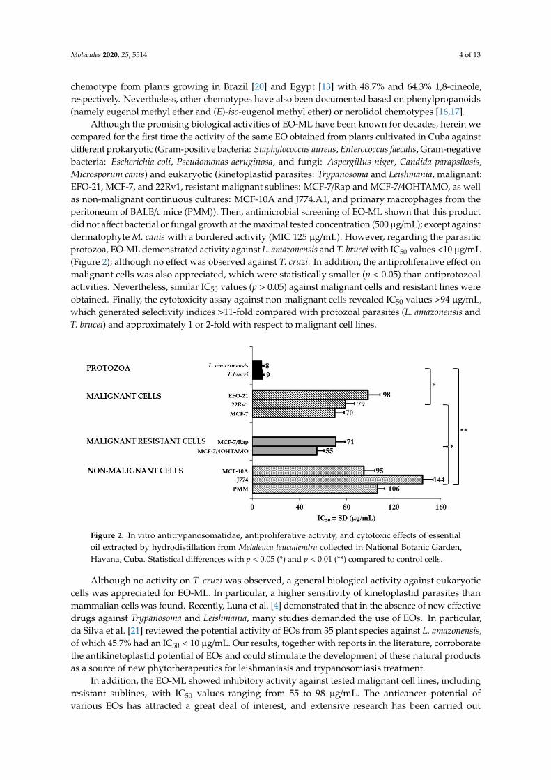
Although the promising biological activities of EO-ML have been known for decades, herein wecompared for the first time the activity of the same EO obtained from plants cultivated in Cuba againstdifferent prokaryotic (Gram-positive bacteria: Staphylococcus aureus, Enterococcus faecalis, Gram-negativebacteria: Escherichia coli, Pseudomonas aeruginosa, and fungi: Aspergillus niger, Candida parapsilosis,Microsporum canis) and eukaryotic (kinetoplastid parasites: Trypanosoma and Leishmania, malignant:EFO-21, MCF-7, and 22Rv1, resistant malignant sublines: MCF-7/Rap and MCF-7/4OHTAMO, as wellas non-malignant continuous cultures: MCF-10A and J774.A1, and primary macrophages from theperitoneum of BALB/c mice (PMM)). Then, antimicrobial screening of EO-ML shown that this productdid not affect bacterial or fungal growth at the maximal tested concentration (500 µg/mL); except againstdermatophyte M. canis with a bordered activity (MIC 125 µg/mL). However, regarding the parasiticprotozoa, EO-ML demonstrated activity against L. amazonensis and T. brucei with IC50 values <10 µg/mL(Figure 2); although no effect was observed against T. cruzi. In addition, the antiproliferative effect onmalignant cells was also appreciated, which were statistically smaller (p < 0.05) than antiprotozoalactivities. Nevertheless, similar IC50 values (p > 0.05) against malignant cells and resistant lines wereobtained. Finally, the cytotoxicity assay against non-malignant cells revealed IC50 values >94 µg/mL,which generated selectivity indices >11-fold compared with protozoal parasites (L. amazonensis andT. brucei) and approximately 1 or 2-fold with respect to malignant cell lines.
Molecules 2020, 25, x FOR PEER REVIEW 4 of 13
Province showed also 1,8-cineole (43.0%) as the main compound [18]; while the major component
was viridiflorol (28.2%) in samples from Pinar del Rio Province [19]. In parallel, other reports showed
the 1,8-cineole chemotype from plants growing in Brazil [20] and Egypt [13] with 48.7% and 64.3%
1,8-cineole, respectively. Nevertheless, other chemotypes have also been documented based on
phenylpropanoids (namely eugenol methyl ether and (E)-iso-eugenol methyl ether) or nerolidol
chemotypes [16,17].
Although the promising biological activities of EO-ML have been known for decades, herein we
compared for the first time the activity of the same EO obtained from plants cultivated in Cuba
against different prokaryotic (Gram-positive bacteria: Staphylococcus aureus, Enterococcus faecalis,
Gram-negative bacteria: Escherichia coli, Pseudomonas aeruginosa, and fungi: Aspergillus niger, Candida
parapsilosis, Microsporum canis) and eukaryotic (kinetoplastid parasites: Trypanosoma and Leishmania,
malignant: EFO-21, MCF-7, and 22Rv1, resistant malignant sublines: MCF-7/Rap and MCF-
7/4OHTAMO, as well as non-malignant continuous cultures: MCF-10A and J774.A1, and primary
macrophages from the peritoneum of BALB/c mice (PMM)). Then, antimicrobial screening of EO-ML
shown that this product did not affect bacterial or fungal growth at the maximal tested concentration
(500 μg/mL); except against dermatophyte M. canis with a bordered activity (MIC 125 μg/mL).
However, regarding the parasitic protozoa, EO-ML demonstrated activity against L. amazonensis and
T. brucei with IC50 values < 10 μg/mL (Figure 2); although no effect was observed against T. cruzi. In
addition, the antiproliferative effect on malignant cells was also appreciated, which were statistically
smaller (p < 0.05) than antiprotozoal activities. Nevertheless, similar IC50 values (p > 0.05) against
malignant cells and resistant lines were obtained. Finally, the cytotoxicity assay against non-
malignant cells revealed IC50 values > 94 μg/mL, which generated selectivity indices >11-fold
compared with protozoal parasites (L. amazonensis and T. brucei) and approximately 1 or 2-fold with
respect to malignant cell lines.
Figure 2. In vitro antitrypanosomatidae, antiproliferative activity, and cytotoxic effects of essential oil
extracted by hydrodistillation from Melaleuca leucadendra collected in National Botanic Garden,
Havana, Cuba. Statistical differences with p < 0.05 (*) and p < 0.01 (**) compared to control cells.
Although no activity on T. cruzi was observed, a general biological activity against eukaryotic
cells was appreciated for EO-ML. In particular, a higher sensitivity of kinetoplastid parasites than
mammalian cells was found. Recently, Luna et al. [4] demonstrated that in the absence of new
effective drugs against Trypanosoma and Leishmania, many studies demanded the use of EOs. In
particular, da Silva et al. [21] reviewed the potential activity of EOs from 35 plant species against L.
amazonensis, of which 45.7% had an IC50 < 10 μg/mL. Our results, together with reports in the
literature, corroborate the antikinetoplastid potential of EOs and could stimulate the development of
Figure 2. In vitro antitrypanosomatidae, antiproliferative activity, and cytotoxic effects of essentialoil extracted by hydrodistillation from Melaleuca leucadendra collected in National Botanic Garden,Havana, Cuba. Statistical differences with p < 0.05 (*) and p < 0.01 (**) compared to control cells.
Although no activity on T. cruzi was observed, a general biological activity against eukaryoticcells was appreciated for EO-ML. In particular, a higher sensitivity of kinetoplastid parasites thanmammalian cells was found. Recently, Luna et al. [4] demonstrated that in the absence of new effectivedrugs against Trypanosoma and Leishmania, many studies demanded the use of EOs. In particular,da Silva et al. [21] reviewed the potential activity of EOs from 35 plant species against L. amazonensis,of which 45.7% had an IC50 < 10 µg/mL. Our results, together with reports in the literature, corroboratethe antikinetoplastid potential of EOs and could stimulate the development of these natural productsas a source of new phytotherapeutics for leishmaniasis and trypanosomiasis treatment.
In addition, the EO-ML showed inhibitory activity against tested malignant cell lines, includingresistant sublines, with IC50 values ranging from 55 to 98 µg/mL. The anticancer potential ofvarious EOs has attracted a great deal of interest, and extensive research has been carried out
Molecules 2020, 25, 5514 5 of 13
to characterize the anticancer activity, the molecular mechanisms, the chemopreventive potential,and the chemotherapeutic application of these products [5]. Although the American National CancerInstitute considers natural products, including EOs, to be active with IC50 values below 30 µg/mL [22],the antiproliferative property of the EO-ML could be in consideration, particularly against resistantsublines. Currently, either intrinsic or acquired resistance during the course of treatment is a limitingfactor in successful cancer chemotherapy and constitutes one of the major challenges for cancerchemotherapy [23]. In this regard, the most interesting result of the studied EO was its capability tolimit the growth of resistant sublines, as well as other malignant susceptible cell lines, which maylead to further studies to prevent or attack proliferation of resistant malignant cells as a neededtherapeutic strategy.
Follow-up studies were carried out on kinetoplastid parasites that showed higher susceptibilityto EO-ML. In this case, the evaluation of 1,8 cineole against L. amazonensis and T. brucei parasiteswas performed. It is an important strategy for its applicability and can facilitate the search for thebiological mechanism by which the oil may act [24]. In addition, although the oils are complexmixtures, a major compound may have a greater influence on the observed biological activity [24].In this study, however, 1,8 cineole showed IC50 values of 68.3 ± 3.4 µg/mL (0.44 ± 0.02 mM) and30.3 ± 1.5 µg/mL (0.19 ± 0.01 mM) against L. amazonensis and T. brucei, respectively. Furthermore,no cytotoxicity was observed against non-malignant macrophages at 200 µg/mL. Thus, 1,8 cineole isnot the main agent responsible for the antiprotozoal activity of EO-ML. Nibret and Wink reported anIC50 of 83.1 µg/mL for this compound against T. brucei [25]. On the other hand, Machado et al. [26]documented that this pure compound did not display an important inhibitory activity on L. infantum,which represented 58.6% of the Thymus capitellatus Hoffmanns & Link EO; while Santana et al. [27]and Camargos et al. [28] showed that 1,8-cineole was able to inhibit the growth of L. amazonensis withIC50 values of 48.4 µg/mL (0.3 mM) and 724 µg/mL (4.7 mM) against amastigotes and promastigotes,respectively. Therefore, the activity of EO-ML could result from the complex interactions betweentheir constituents. In some cases, these interactions may lead to antagonistic or synergistic effects thatcontribute to the biological activity of EOs, and even minor components of EOs can play a criticalrole in these effects. There have been a few studies concerning antikinetoplastid actions of the minorEO-ML constituents. For example, some authors showed IC50 values of: 4.2 µg/mL for limonene,1.0 µg/mL for α-pinene [29], 47.4 µg/mL for β-pinene [30] and 0.02 µg/mL for terpinen-4-ol [31] againstT. brucei; as well as 37.9 µg/mL for limonene [32] 105 µg/mL for α-terpineol [28] and 37 µg/mL forα-pinene [27] against L. amazonensis. Although the concentrations of these compounds were lessthan 1,8-cineole, the presence of known components with antikinetoplastid activity in the oil could,therefore, account for the inhibitory effect found for EO-ML. Nevertheless, further studies with minorcomponents or compound combinations are needed to identify the compound(s) responsible for theeffects of EO-ML.
Note that the previous report of this plant against bacteria, fungi, and parasites was conductedwith an aqueous ethanolic extract [14,15]. Although the chemical composition of this extract was notreported, the different procedures for solvent extraction and EO hydrodistillation will result in verydifferent chemical compositions, marked mainly in relatively polar, non-volatile in the ethanol extract,and volatile non-polar compounds in the EO [33]. Nevertheless, antiparasitic activities were displayedby both the ethanol extract and the EO, which strongly suggests the potentialities of M. leucadendra.
It is concluded, therefore, that the oil exhibited comparable susceptibilities against bothL. amazonensis and T. brucei, and presented higher in-vitro activity and selectivity than the pure maincomponent. However, Caridha et al. [34] highlighted the need for new treatments for CL, due to currenttreatments showing poor justification through clinical trials and sub-optimal effectiveness [35,36].In particular, against L. amazonensis, the EO-ML displayed an IC50 of 8µg/mL and a selectivity index = 18(with respect to J774) and 13 (with respect to PMM), which are in concordance with international criteriarelated to the development of natural products for cutaneous species, i.e., (i) classification as highlyactive with IC50 < 10 µg/mL [37] and (ii) a selectivity index >5 [34]. In the Neotropics, L. amazonensis is
Molecules 2020, 25, 5514 6 of 13
considered to be one of the most important species that causes cutaneous leishmaniasis (CL). In addition,nearly 1% of all CL cases can develop an anergic diffuse CL infection, which is characterized bymassive dermal infiltrates and presents clinical, immunological, parasitological, anatomopathological,and therapeutic responses different from other CL forms. This clinical presentation is chronic withfrequent relapses due to non-response to conventional treatment [26,38]. Thus, the effect of EO-MLwas evaluated in the model of experimental CL caused by L. amazonensis in BALB/c mice.
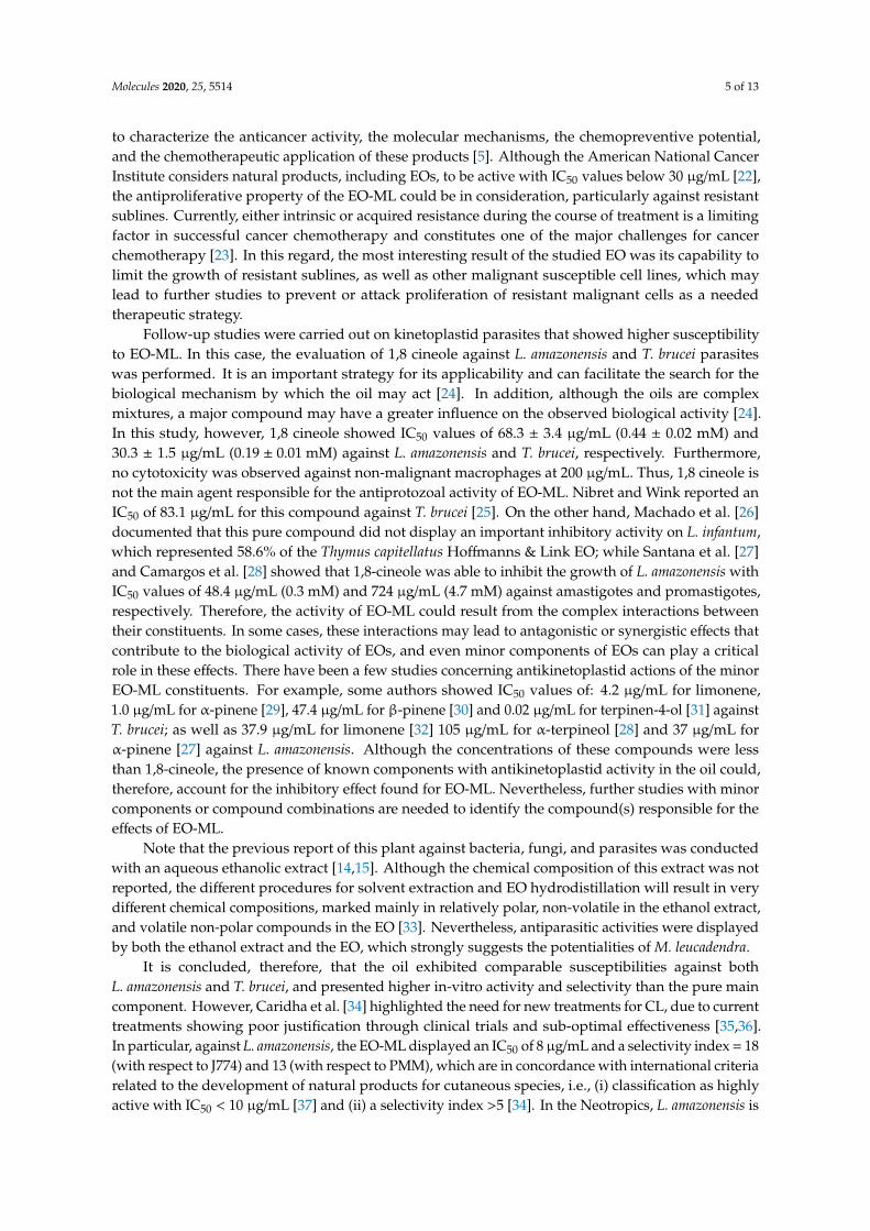
In the in-vivo model, the studied oil was able to control the disease progression, which was shownin the statistically significant smaller (p < 0.05) lesion size (Figure 3A) and parasite burden (Figure 3B)with respect to untreated animals. Compared with Glucantime® (GTM), a similar efficacy (p > 0.05)was found for EO-ML, as shown for lesion size (Figure 3A) and parasite burden (Figure 3B). Figure 3Cshows the differences among cutaneous lesions for each group, which was represented by one animalselected at random.
Molecules 2020, 25, x FOR PEER REVIEW 6 of 13
[35,36]. In particular, against L. amazonensis, the EO-ML displayed an IC50 of 8 μg/mL and a selectivity
index = 18 (with respect to J774) and 13 (with respect to PMM), which are in concordance with
international criteria related to the development of natural products for cutaneous species, i.e., (i)
classification as highly active with IC50 < 10 μg/mL [37] and (ii) a selectivity index > 5 [34]. In the
Neotropics, L. amazonensis is considered to be one of the most important species that causes cutaneous
leishmaniasis (CL). In addition, nearly 1% of all CL cases can develop an anergic diffuse CL infection,
which is characterized by massive dermal infiltrates and presents clinical, immunological,
parasitological, anatomopathological, and therapeutic responses different from other CL forms. This
clinical presentation is chronic with frequent relapses due to non-response to conventional treatment
[26,38]. Thus, the effect of EO-ML was evaluated in the model of experimental CL caused by L.
amazonensis in BALB/c mice.
In the in-vivo model, the studied oil was able to control the disease progression, which was
shown in the statistically significant smaller (p < 0.05) lesion size (Figure 3A) and parasite burden
(Figure 3B) with respect to untreated animals. Compared with Glucantime® (GTM), a similar efficacy
(p > 0.05) was found for EO-ML, as shown for lesion size (Figure 3A) and parasite burden (Figure 3B).
Figure 3C shows the differences among cutaneous lesions for each group, which was represented by
one animal selected at random.
Figure 3. Antileishmanial effect of the essential oil from Melaleuca leucadendra collected in National
Botanic Garden, Havana, Cuba, on BALB/c mice infected with 5 × 10⁶ promastigotes of L.
amazonensis/animal. Treatment started four weeks post-infection with essential oil from Melaleuca
leucadendra or Glucantime® with five doses by the intralesional route at 30 mg/kg every four days. (A):
Lesion size; (B): Parasite burden; (C): Pictures at 12 weeks post-infection of infected animals in the
footpad with L. amazonensis and treated. EO-ML: essential oil from M. leucadendra; GTM: Glucantime
used as reference drug; Control: Untreated animals. *: Displays statistical differences (p < 0.05)
compared to control untreated animals.
Here, we report the in vivo efficacy of EO-ML against L. amazonensis infection by using 30 mg/kg
for 15 days, with a four-day interval by the subcutaneous route, for a total of five doses. In other
studies, however, higher numbers of doses and concentrations of antileishmanial products were
usually employed in the animals, aiming to enhance their efficacy. For example, oil from the trunk of
Figure 3. Antileishmanial effect of the essential oil from Melaleuca leucadendra collected inNational Botanic Garden, Havana, Cuba, on BALB/c mice infected with 5 × 10⁶ promastigotesof L. amazonensis/animal. Treatment started four weeks post-infection with essential oil fromMelaleuca leucadendra or Glucantime® with five doses by the intralesional route at 30 mg/kg everyfour days. (A): Lesion size; (B): Parasite burden; (C): Pictures at 12 weeks post-infection of infectedanimals in the footpad with L. amazonensis and treated. EO-ML: essential oil from M. leucadendra;GTM: Glucantime® used as reference drug; Control: Untreated animals. *: Displays statisticaldifferences (p < 0.05) compared to control untreated animals.
Here, we report the in vivo efficacy of EO-ML against L. amazonensis infection by using 30 mg/kg for15 days, with a four-day interval by the subcutaneous route, for a total of five doses. In other studies,however, higher numbers of doses and concentrations of antileishmanial products were usuallyemployed in the animals, aiming to enhance their efficacy. For example, oil from the trunk ofCopaifera martii Hayne was administered at 100 mg/kg/day for 30 days [39]; while the treatment withthe EO from Carapa guianensis Aubl. Was performed for eight weeks using 100–160 mg/kg/day [40].
Our data show the efficacy of EO-ML to be comparable with animals treated with GTM,a first-line drug, taking into account the evaluated parameters (dosing, lesion size, and parasite burden).

Molecules 2020, 25, 5514 7 of 13
The significant effect of EO-ML on the reduction of BALB/c mice infection caused by L. amazonensis isindicative of a reduced pathology; a complete cure was not observed, however.
The positive effect observed in animals treated with EO-ML, could be explained by the directactivity of the oil on the growth of the parasite, as shown in the in-vitro assays. Nevertheless,the efficacy exhibited in the murine model could also be associated with the ability of oil constituentsto induce indirect effects that can contribute to controlling a Leishmania infection in the treated animals.Recent studies have indicated that terpinen-4-ol and limonene present anti-inflammatory activity,β-pinene has shown antioxidant effect [41], and α-pinene modulated macrophage activation by thestimulation of NO production and increasing the phagocytic and lysosomal activities [42]. All thesepharmacological properties could contribute to controlling a leishmaniasis infection.
3. Materials and Methods
3.1. Plant and Essential Oils
Aerial parts (leaves and stems) of M. leucadendra plant were collected during early hours of themorning in March 2015 at National Botany Garden (NBG), Havana, Cuba, and a specimen was depositedat the Herbarium of Cuban Flora of the NBG and authenticated by M.Sc. Eldys Bécquer (vouchernumber: 8501918). Leaves of M. leucadendra were manually selected, rinsed with abundant water,and crushed into small pieces. Immediately, the fresh vegetal material was conventionally hydrodistilledusing a Clevenger-type apparatus for 5 h, and the EO-ML was obtained, yielding 0.8%. The essentialoil was stored in a sealed amber vial at 4 ◦C until analysis and screening.
A sample of 100 µL was used to carry out the chemical characterization by GC-MS using aShimadzu GCMS-QP2010 Ultra (Shimadzu Scientific Instruments, Columbia, MD, USA). A 5% w/vsolution of the sample in CH2Cl2 was prepared and 0.1 µL was injected with a splitting mode (30:1).In this case, a ZB-5 fused silica capillary column with (5% phenyl)-polymethylsiloxane as stationaryphase (film thickness of 0.25 µm, a length of 30 m, and an internal diameter of 0.25 mm (Phenomenex,Torrance, CA, USA) and the carrier gas was helium (column head pressure of 552 kPa, flow rate of1.37 mL/min, injector temperature of 250 ◦C and an ion source temperature of 200 ◦C) were used.Equipment operated in the electron impact (EI) mode (electron energy = 70 eV), scan range = 40–400atomic mass units, scan rate = 3.0 scans/s and GC-MS solution software. The GC oven temperatureprogram was set to 50 ◦C as the initial temperature, which increased at 2 ◦C/min until 260 ◦C.Finally, identification of the oil components was based on their retention indices (RI) determined byreference to a homologous series of n-alkanes, and by comparison of their mass spectral fragmentationpatterns with those reported in the literature [43], and stored in our in-house Sat-Set library [44].
Another aliquot of EO-ML and 1,8-cineole (purity 99%; Sigma-Aldrich, St. Louis, MO, USA) wereused to carry out the biological assays. The EO-MS and 1,8-cineole were dissolved in dimethylsulfoxide(DMSO; BDH, Poole, England) at 20 mg/mL and 40 mM, respectively.
3.2. Antimicrobial Assays
The screening of antimicrobial activity of EO was performed on a panel of reference strains andclinical isolates (obtained from the Collections of State Scientific Center of Antibiotics for antimicrobialactivity): S. aureus ATCC 29213, E. faecalis ATCC 29212, E. coli ATCC 25922, P. aeruginosa ATCC 27853,C. parapsilosis ATCC, A. niger 37a and M. canis B-200. The MIC values were determined by the brothmicro-dilution method using Mueller Hinton broth and National Committee for Clinical LaboratoryStandards procedures.
3.3. Antikinetoplastid Assays
For antitrypanosomal activity, two species were included: T. brucei (Squib-427) and T. cruzi(Tulahuen CL2). For T. brucei, 1.5 × 10⁴ trypomastigotes cultured in Hirumi-9 medium supplementedwith 10% inactivated fetal calf serum (FCSi; Invitrogen, Belgium) and tested products were added in a

Molecules 2020, 25, 5514 8 of 13
96-well plate and incubated at 37 ◦C and 5% CO2 for 72 h [45]. Then, parasite growth was assessedfluorimetrically using 20 µL of resazurin (Sigma-Aldrich, St. Louis, MO, USA) at 50 µg/mL. The platewas incubated for an additional 24 h under the same conditions and read at 530 nm excitation and590 nm emission in a Tecan GENios Multifunction Fluorimeter (Tecan Group, Maennedorf, Switzerland).On the other hand, EO at different concentrations was added to 4 × 10⁴ amastigotes in 4 × 10³ MRC-5cells using minimal essential medium (MEM; Life Technologies, Carlsbad, CA, USA) supplementedwith 20 mM l-glutamine, 16.5 mM sodium bicarbonate, and 5% of FCSi in a plate of 96-wells andincubated for seven days at 37 ◦C and 5% CO2. In this case, parasite viability was determinedcolorimetrically by adding the β-galactosidase substrate chlorophenol red β-d-galactopyranoside(Sigma Aldrich, St. Louis, MO, USA). Finally, the absorbance was read at 540 nm after 4 h of incubationat 37 ◦C [46].
For antileishmanial activity, the intracellular amastigote model of L. amazonensis (MHOM/77BR/
LTB0016) was used. PMM were obtained by peritoneal washing with RPMI medium (Sigma, St. Louis,MO, USA) supplemented with antibiotics from healthy BALB/c mice and plated at 106/mL in a24-well plate. After incubation at 37 ◦C and 5% CO2 for 2 h, non-adherent cells were removed,and stationary-phase promastigotes were added at a 4:1 parasite/macrophage ratio in the mediumsupplemented with heat-inactivated fetal bovine serum (HFBS; Sigma-Aldrich, St. Louis, MO, USA).The plate was incubated at the same condition for 4 h, and free parasites were also removed.Subsequently, products were added, and four serial dilutions were carried out. The plate was incubatedat the same conditions for 48 h as described above [47]. After that, the supernatant was discarded,cells were fixed with methanol, stained with 10% Giemsa and microscopically examined (Motic, Japan)under immersion oil at 1000×. The total parasite burden was determined according to the numberof infected macrophages and the number of amastigotes inside the macrophages after counting of100 macrophages.
3.4. Antiproliferative and Cytotoxicity Screening on Malignant and Non-Malignant Cells
To determine the activity of the EO on malignant cells, the following cancer cell lines were used:(i) 22Rv1 (human prostate carcinoma, ATCC®CRL-2505TM) cultivated in RPMI-1640 medium (Gibco-LifeTechnologies, Paisley, UK) supplemented with RPMI-1640 Vitamins (PanEco, Moscow, Russia), (ii) MCF-7(human breast cancer, ATCC®HTB-22) cells, as well (iii) MCF-7/Rap and (iv) MCF-7/4OHTAMO resistantsublines (The rapamycin-resistant MCF-7/Rap and 4OH-tamoxifen-resistant MCF-7/4OHTAMOsublines were established from the parent MCF-7 cells by long-term rapamycin or 4OH-tamoxifentreatment, respectively), and (v) EFO-21 (ovary cystadenocarcinoma, DSMZ ACC 235) cultured instandard 4.5 g/L glucose DMEM medium (Gibco-Life Technologies, Paisley, UK). In all cases, culturewas supplemented with 10% FCSi, antibiotics (50 µg of streptomycin/mL and 50 U of penicillin/mL) and0.1 mg/mL sodium pyruvate (Santa Cruz Biotechnology, Dallas, TX, USA) and maintained in a NuAirincubator (NuAir, Plymouth, MN, USA) at 37 ◦C, 5% CO2 and 80–85% humidity. Then, 100 × 103 22Rv1cells/well, 40 × 103 MCF-7, MCF-7/Rap or MCF-7/4OHTAMO cells/well, and 110 × 103 EFO-21cells/well were seeded into 24-well plates in 900 µL of the medium, and the plates were incubatedfor 24 h at 37 ◦C and 5% CO2. Follow, different concentrations of EO were added and the plates wereincubated for 72 h under the same conditions, which the cellular viability was assessed using the3-[4,5-dimethylthiazol-2-yl]-2,5-diphenyltetrazolium bromide (MTT; AppliChem GmbH, Darmstadt,Germany) at 0.2 mg/mL per well [48,49]. After additional incubation of 2 h, the supernatant wasdiscarded, the MTT formazan purple crystals were dissolved in DMSO (350 µL per well), and theabsorbance was measured at 571 nm and 630 nm as a reference in a MultiScan reader (ThermoFisher,Waltham, MA, USA) after the plates were gently shaken.
Cytotoxicity on non-malignant cells was also studied using three models: (i) MCF-10A (ATCC®
CRL-10317) normal breast cells cultured in DMEM/F12 (Gibco) supplemented with 5% donor horseserum (BioSera), 20 ng/mL EGF (PanEco), 0.5 µg/mL hydrocortisone (ChemCruz), and 10 µg/mL insulin(PanEco) at 37 ◦C, 5% CO2 and 80–85% humidity), (ii) J774A.1 (murine macrophage cell line, ATCC®,
Molecules 2020, 25, 5514 9 of 13
TIB-67™) cultivated in Dulbecco’s modified eagle medium (DMEM; Thermo Fisher Scientific, Waltham,MA, USA) supplemented at 10% of HFBS, antibiotics and maintained on a roller culture apparatus at5 rpm and (iii) PMM isolated with RPMI and antibiotics at the moment of use. Briefly, 50× 103 MCF-10Acells were seeded into 24-well plates in 900µL of the medium, and then, the activity of EO-ML was testedas described above for the case of MCF-7 breast cancer cells. In the case of J774A.1 cells, 200 µL with105 cells/mL were distributed in the wells of the 96-well microplates, incubated at 37 ◦C, 5% CO2 for24 h to allow attachment, and non-adherent cells were eliminated after washing. Then, fresh mediumand different concentrations of EO were added, and the plate was incubated for an additional 24 h atthe same condition. Finally, cellular viability was determined fluorometrically by the resazurin method,as previously described [50]. In the case of PMM, macrophages were obtained by peritoneal washingin RPMI medium and antibiotics as mentioned and seeded at 3 × 10⁵ cells/mL. The plate was incubatedat 37 ◦C, and 5% CO2 for 2 h, and non-attached cells were removed. Then, fresh medium with HFBSand EO at different concentrations were added to additional incubation for 48 h under the sameconditions. Cellular viability was then measured by the MTT method as described above.
3.5. In Vivo Evaluation on Cutaneous Leishmaniasis Caused by L. amazonensis
Female healthy BALB/c mice were infected in the right hind footpad with 5 × 10⁶ stationary-phasepromastigotes of L. amazonensis by the subcutaneous route, which was identified as day 0.Then, four weeks p.i., the animals were randomly distributed into three groups of eight mice each,and two groups of mice were treated with EO-ML or GTM at 30 mg/kg. Products were applied everyfour days to a total of five doses by the intralesional route. The third group of animals was consideredto be the control group that received no treatment. In parallel, from four weeks p.i. until 12 weeks p.i,the animals were daily observed, body weight and the lesion size were weekly supervised using atechnical bascule (SCALTEC, Göttingen, Germany) and a caliper to measure footpad swelling and lesiondiameter, respectively. In these cases, a variation of body weight, as well as an average of lesion size(mean of the differences between infected and uninfected footpads), was calculated with respect toweek 4 p.i. for each group. In addition, the parasite burden was determined on weeks 6 and 12 p.i.through the culture microtitration method in 96-well plates [51]. Briefly, three animals selectedat random from each group were killed by cervical dislocation, a sample of subcutaneous tissuesfrom the infected area was excised, weighed, and homogenized in 4 mL of Schneider’s medium.Then, a four-fold serial dilution was carried out in a 96-well plates in duplicate under sterile conditionsand incubated at 26 ◦C. After seven days of incubation, the plates were examined under an invertedmicroscope (Olympus, Tokyo, Japan) at 400× to select the last dilution that contained at least one mobileparasite, which was defined as the final titer. The parasite burden was calculated as the geometricmean of reciprocal titers/weight of tissue sample multiplied by 400. All of the experimental proceduresinvolving animals were conducted in accordance with the Guide for the Care and Use of LaboratoryAnimals, Eighth Edition, which was approved by the Ethics Committee (CEI-IPK 14-12), Havana, Cuba.
3.6. Statistical Analysis
For in vitro assays, the IC50 for each product on each system was obtained from dose-responsecurves, results were expressed as mean ± SD of three replicates, and comparisons among values wereperformed using Mann-Whitney test with Statistica for Windows Program (Version 10, StatSoft, Inc.,Tulsa, OK, USA). For in vivo experiments, lesion evolution and parasite load were processed by theVariance Analysis Test (ANOVA), followed by a Post Hoc Test (LDS test or planned comparison). In allcases, statistically significant differences were identified for p < 0.05.
4. Conclusions
To the best of our knowledge, this is the first report on antiproliferative potentialities of EO-MLgrowing in Cuba, confirming the medical value of the plant. However, the inability of 1,8-cineole todemonstrate a strong antikinetoplastid activity reveals that other ingredients or a possible synergism

Molecules 2020, 25, 5514 10 of 13
are involved in the activity of the EO. In particular, the leishmanicidal effect of EO-ML was confirmedin vivo using a murine model of CL, which the tested product was able to significantly reduce thelesion size and parasite burden in infected tissues similar to the reference drug.
In addition, our results open new perspectives to further investigations to clarify: (i) Therapeuticvalue of EO-ML for African trypanosomiasis on relevant T. brucei rhodesiense and T. brucei gambienseinfectious agents through in vitro and in vivo models, (ii) the possibility of the application of this oil asalternative to traditional treatment in cancer-resistant cells, (iii) potentialities of a combination of oilconstituents, and (iv) mechanism of action of EO-ML, as well as the pure compounds. In conclusion,the results reported here represent an advancement in studies on EOs as drug candidates and mightpromote preclinical investigations on oil standardization.
Author Contributions: Conceptualization, L.M. and A.M.S.; methodology, L.M., A.M.S., R.S., P.S.; formal analysis,P.C., A.E.S., L.G. and W.N.S.; investigation, all; resources, L.M., A.M.S., P.C., L.G. and W.N.S.; data curation,P.C., L.G., A.E.S. and W.N.S.; writing—original draft preparation, L.M., A.M.S. and W.N.S.; writing—review andediting, all; project administration, L.M. and A.E.S. All authors have read and agreed to the published version ofthe manuscript.
Funding: This research was supported by Project No. 18-53-34005 from the Russian Foundation for Basic Researchand Ministry of Science, Technology and Environment of the Republic of Cuba.
Acknowledgments: Thanks to Ernst Mach scholarship award to Lianet Monzote by the Austrian Exchange Office(OEAD) and support of laboratory of Lars Gille through project from the Austrian Science Fund (FWF-P 27814-B22).In addition, the present work is a collaboration of members of the Research Network Natural Products againstNeglected Diseases (ResNetNPND, http://www.resnetnpnd.org/Start/) and contribution to the project AromaticPlant Research Center (APRC, https://aromaticplant.org/). The authors thank Ekaterina Mikhaevich for assistancein antiproliferative assays.
Conflicts of Interest: The authors declare no conflict of interest.
References
1. Mickymaray, S. Efficacy and mechanism of traditional medicinal plants and bioactive compounds againstclinically important pathogens. Antibiotics 2019, 8, 257. [CrossRef] [PubMed]
2. Shaaban, H.A.E.; El-Ghorab, A.H.; Shibamoto, T. Bioactivity of essential oils and their volatile aromacomponents: Review. J. Essent. Oil Res. 2012, 24, 203–212. [CrossRef]
3. Turek, C.; Stintzing, F.C. Stability of essential oils: A review. Compr. Rev. Food Sci. Food Saf. 2013, 12, 40–53.[CrossRef]
4. Luna, E.C.; Luna, I.S.; Scotti, L.; Monteiro, A.F.M.; Scotti, M.T.; de Moura, R.O.; de Araújo, R.S.A.;Monteiro, K.L.C.; de Aquino, T.M.; Ribeiro, F.F.; et al. Active essential oils and their components in useagainst neglected diseases and arboviruses. Oxid. Med. Cell. Longev. 2019, 2019, 6587150. [CrossRef][PubMed]
5. Blowman, K.; Magalhães, M.; Lemos, M.F.L.; Cabral, C.; Pires, I.M. Anticancer properties of essential oilsand other natural products. Evidence-Based Complement. Altern. Med. 2018, 2018, 3149362. [CrossRef]
6. Pavithra, P.S.; Mehta, A.; Verma, R.S. Essential oils: From prevention to treatment of skin cancer.Drug Discov. Today 2019, 24, 644–655. [CrossRef] [PubMed]
7. Johns, M.R.; Johns, J.E.; Rudolph, V. Steam distillation of tea tree (Melaleuca alternifolia) oil. J. Sci. Food Agric.1992, 58, 49–53. [CrossRef]
8. Sharifi-Rad, J.; Salehi, B.; Varoni, E.M.; Sharopov, F.; Yousaf, Z.; Ayatollahi, S.A.; Kobarfard, F.; Sharifi-Rad, M.;Afdjei, M.H.; Sharifi-Rad, M.; et al. Plants of the Melaleuca genus as antimicrobial agents: From farm topharmacy. Phyther. Res. 2017, 31, 1475–1494. [CrossRef]
9. Gan, W.S. Manual of Medicinal Plants in Taiwan, Volume 3; National Research Institute of Chinese Medicine:Taipei City, Taiwan, 1965.
10. Tsuruga, T.; Chun, Y.-T.; Ebizuka, Y.; Sankawa, U. Biologically active constituents of Melaleuca leucadendron:Inhibitors of induced histamine release from rat mast cells. Chem. Pharm. Bull. 1991, 39, 3276–3278. [CrossRef]
11. Surh, J.; Yun, J.-M. Antioxidant and anti-inflammatory activities of butanol extract of Melaleuca leucadendron L.Prev. Nutr. Food Sci. 2012, 17, 22–28. [CrossRef]

Molecules 2020, 25, 5514 11 of 13
12. Lohakachornpan, P.; Rangsipanuratn, W. Chemical compositions and antimicrobial activities of essential oilfrom Melaleuca leucadendron var. minor.. Thai J. Pharm. Sci. 2001, 25, 133–139.
13. Farag, R.S.; Shalaby, A.S.; El-Baroty, G.A.; Ibrahim, N.A.; Ali, M.A.; Hassan, E.M. Chemical and biologicalevaluation of the essential oils of different Melaleuca species. Phyther. Res. 2004, 18, 30–35. [CrossRef] [PubMed]
14. Fernández-Calienes Valdés, A.; Mendiola Martínez, J.; Scull Lizama, R.; Vermeersch, M.; Cos, P.; Maes, L.In vitro anti-microbial activity of the Cuban medicinal plants Simarouba glauca DC, Melaleuca leucadendron Land Artemisia absinthium L. Mem. Inst. Oswaldo Cruz 2008, 103, 615–618. [CrossRef] [PubMed]
15. García, M.; Monzote, L.; Scull, R.; Herrera, P. Activity of Cuban plants extracts against Leishmania amazonensis.ISRN Pharmacol. 2012, 2012, 104540. [CrossRef]
16. Brophy, J.J. Potentially commercial Melaleucas. In The Genus Melaleuca; Southwell, I., Lowe, R., Eds.;Harwood: Amsterdam, The Netherlands, 1999; pp. 247–274.
17. An, N.T.G.; Huong, L.T.; Satyal, P.; Tai, T.A.; Dai, D.N.; Hung, N.H.; Ngoc, N.T.B.; Setzer, W.N. Mosquitolarvicidal activity, antimicrobial activity, and chemical compositions of essential oils from four species ofMyrtaceae from central Vietnam. Plants 2020, 9, 544. [CrossRef]
18. Pino, J.A.; Regalado, E.L.; Rodríguez, J.L.; Fernández, M.D. Phytochemical analysis and in vitrofree-radical-scavenging activities of the essential oils from leaf and fruit of Melaleuca leucadendra L.Chem. Biodivers. 2010, 7, 2281–2288. [CrossRef]
19. Pino, J.; Bello, A.; Urquiola, A.; Aguero, J.; Marbot, R. Chemical composition of cajuput oil (Melaleucaleucadendra L.) from Cuba. J. Essent. Oil Res. 2002, 14, 10–11. [CrossRef]
20. Siani, A.C.; Nakamura, M.J.; das Neves, G.P.; da Monteiro, S.S.; Ramos, M.F.S. Leaf essential oil from threeexotic Mytaceae species growing in the botanical garden of Rio de Janeiro, Brazil. Am. J. Plant Sci. 2016, 7,834–840. [CrossRef]
21. De da Silva, C.E.L.; Oyama, J.; Ferreira, F.B.P.; de Lalucci-Silva, M.P.P.; Lordani, T.V.A.; de da Silva, R.C.L.;de Monich, M.S.T.; Teixeira, J.J.V.; Lonardoni, M.V.C. Effect of essential oils on Leishmania amazonensis:A systematic review. Parasitology 2020. first view. [CrossRef]
22. Suffness, M.; Pezzuto, J.M. Assay related to cancer drug discovery. In Methods in Plant Biochemistry: Assays forBioactivity, Volume 6; Hostettmann, K., Ed.; Academic Press: London, UK, 1991; pp. 71–133.
23. Jones, V.S.; Huang, R.-Y.; Chen, L.-P.; Chen, Z.-S.; Fu, L.; Huang, R.-P. Cytokines in cancer drug resistance:Cues to new therapeutic strategies. Biochim. Biophys. Acta 2016, 1865, 255–265. [CrossRef]
24. Pereira, P.S.; Maia, A.J.; Duarte, A.E.; Oliveira-tintino, C.D.M.; Tintino, S.R.; Barros, L.M.; Vega-Gomez, M.C.;Rolón, M.; Coronel, C.; Coutinho, H.D.M.; et al. Cytotoxic and anti-kinetoplastid potential of the essential oilof Alpinia speciosa K. Schum. Food Chem. Toxicol. 2018, 119, 387–391. [CrossRef]
25. Nibret, E.; Wink, M. Trypanocidal and antileukaemic effects of the essential oils of Hagenia abyssinica, Leonotisocymifolia, Moringa stenopetala, and their main individual constituents. Phytomedicine 2010, 17, 911–920.[CrossRef] [PubMed]
26. Machado, G.U.; Prates, F.V.; Machado, P.R.L. Disseminated leishmaniasis: Clinical, pathogenic, and therapeuticaspects. An. Bras. Dermatol. 2019, 94, 9–16. [CrossRef] [PubMed]
27. Santana, R.C.; dos Rosa, A.S.; da Mateus, M.H.S.; Soares, D.C.; Atella, G.; Guimarães, A.C.; Siani, A.C.;Ramos, M.F.S.; Saraiva, E.M.; Pinto-da-Silva, L.H. In vitro leishmanicidal activity of monoterpenes presentin two species of Protium (Burseraceae) on Leishmania amazonensis. J. Ethnopharmacol. 2020, 259, 112981.[CrossRef] [PubMed]
28. Camargos, H.S.; Moreira, R.A.; Mendanha, S.A.; Fernandes, K.S.; Dorta, M.L.; Alonso, A. Terpenes increasethe lipid dynamics in the Leishmania plasma membrane at concentrations similar to their IC50 values.PLoS ONE 2014, 9, e104429. [CrossRef] [PubMed]
29. Kamte, S.L.N.; Ranjbarian, F.; Cianfaglione, K.; Sut, S.; Dall’Acqua, S.; Bruno, M.; Afshar, F.H.; Iannarelli, R.;Benelli, G.; Cappellacci, L.; et al. Identification of highly effective antitrypanosomal compounds in essentialoils from the Apiaceae family. Ecotoxicol. Environ. Saf. 2018, 156, 154–165. [CrossRef] [PubMed]
30. Kpoviessi, S.; Bero, J.; Agbani, P.; Gbaguidi, F.; Kpadonou-Kpoviessi, B.; Sinsin, B.; Accrombessi, G.;Frédérich, M.; Moudachirou, M.; Quetin-Leclercq, J. Chemical composition, cytotoxicity and in vitroantitrypanosomal and antiplasmodial activity of the essential oils of four Cymbopogon species from Benin.J. Ethnopharmacol. 2014, 151, 652–659. [CrossRef]
31. Mikus, J.; Harkenthal, M.; Steverding, D.; Reichling, J. In vitro effect of essential oils and isolated mono- andsesquiterpenes on Leishmania major and Trypanosoma brucei. Planta Med. 2000, 66, 366–368. [CrossRef]

Molecules 2020, 25, 5514 12 of 13
32. Do Carmo, D.F.M.; Amaral, A.C.F.F.; Machado, G.M.C.; Leon, L.L.; Rocha, J.; de Silva, J.R.A. Chemical andbiological analyses of the essential oils and main constituents of Piper species. Molecules 2012, 17, 1819–1829.[CrossRef]
33. Cseke, L.J.; Setzer, W.N.; Vogler, B.; Kirakosyan, A.; Kaufman, P.B. Traditional, analytical and preparativeseparations of natural products. In Natural Products from Plants; Cseke, L.J., Kirakosyan, A., Kaufman, P.B.,Warber, S.L., Duke, J.A., Brielmann, H.L., Eds.; CRC Press: Boca Raton, FL, USA, 2006; pp. 263–317.ISBN 0-8493-2976-0.
34. Caridha, D.; Vesely, B.; van Bocxlaer, K.; Arana, B.; Mowbray, C.E.; Rafati, S.; Uliana, S.; Reguera, R.;Kreishman-Deitrick, M.; Sciotti, R.; et al. Route map for the discovery and pre-clinical development of newdrugs and treatments for cutaneous leishmaniasis. IJP Drugs Drug Resist. 2019, 11, 106–117. [CrossRef]
35. Pinart, M.; Rueda, J.-R.; Romero, G.A.S.; Pinzón-Flórez, C.E.; Osorio-Arango, K.; Silveira Maia-Elkhoury, A.N.;Reveiz, L.; Elias, V.M.; Tweed, J.A. Interventions for American cutaneous and mucocutaneous leishmaniasis.Cochrane Database Syst. Rev. 2020. [CrossRef]
36. Heras-Mosteiro, J.; Monge-Maillo, B.; Pinart, M.; Lopez Pereira, P.; Garcia-Carrasco, E.; CampuzanoCuadrado, P.; Royuela, A.; Mendez Roman, I.; López-Vélez, R. Interventions for Old World cutaneousleishmaniasis. Cochrane Database Syst. Rev. 2017. [CrossRef]
37. De Lima, J.P.S.; Pinheiro, M.L.B.; Santos, A.M.G.; Pereira, J.L.S.; Santos, D.M.F.; Silva-Jardim, I.; Costa, E.V.In vitro antileishmanial and cytotoxic activities of Annona mucosa (Annonaceae). Rev. Virtual Quim. 2012, 4,692–702.
38. Costa, J.M.L.; Uthant, A.A.; CostaML, E.A.; Bezerril, A.C.; Barral, A. Diffuse cutaneous leishmaniasis (DCL)in Brazil after 60 years of your first description. Gaz. Médica Bahia 2009, 79, 19–24.
39. Dos Santos, A.O.; Costa, M.A.; Ueda-Nakamura, T.; Dias-Filho, B.P.; da Veiga-Júnior, V.F.; de Souza Lima, M.M.;Nakamura, C.V. Leishmania amazonensis: Effects of oral treatment with copaiba oil in mice. Exp. Parasitol.2011, 129, 145–151. [CrossRef] [PubMed]
40. De Moraes, A.R.D.P.; Tavares, G.D.; Rocha, F.J.S.; de Paula, E.; Giorgio, S. Effects of nanoemulsions preparedwith essential oils of copaiba- and andiroba against Leishmania infantum and Leishmania amazonensis infections.Exp. Parasitol. 2018, 187, 12–21. [CrossRef] [PubMed]
41. Spisni, E.; Petrocelli, G.; Imbesi, V.; Spigarelli, R.; Azzinnari, D.; Sarti, M.D.; Campieri, M.; Valerii, M.C.Microbial-modulating activities of essential oils: Implications in colonic pathophysiology. Int. J. Mol. Sci.2020, 21, 4152. [CrossRef] [PubMed]
42. Da Rodrigues, K.A.F.; Amorim, L.V.; Dias, C.N.; Moraes, D.F.C.; Carneiro, S.M.P.; de Carvalho, F.A.A.Syzygium cumini (L.) Skeels essential oil and its major constituent α-pinene exhibit anti-Leishmania activitythrough immunomodulation in vitro. J. Ethnopharmacol. 2015, 160, 32–40. [CrossRef] [PubMed]
43. Adams, R.P. Identification of Essential Oil Components by Gas Chromatography/Mass Spectrometry, 4th ed.;Allured Publishing: Carol Stream, IL, USA, 2007; ISBN 978-1-932633-21-4.
44. Satyal, P. Development of GC-MS Database of Essential Oil Components by the Analysis of Natural EssentialOils and Synthetic Compounds and Discovery of Biologically Active Novel Chemotypes in Essential Oils.Ph.D. Thesis, University of Alabama in Huntsville, Huntsville, AL, USA, 2015.
45. Hirumi, H.; Hirumi, K. Continuous cultivation of Trypanosoma brucei blood stream forms in a mediumcontaining a low concentration of serum protein without feeder cell layers. J. Parasitol. 1989, 75, 985–989.[CrossRef]
46. Buckner, F.S.; Verlinde, C.L.M.J.; La Flamme, A.C.; van Voorhis, W.C. Efficient technique for screeningdrugs for activity against Trypanosoma cruzi using parasites expressing β-galactosidase. Antimicrob. AgentsChemother. 1996, 40, 2592–2597. [CrossRef]
47. Torres-Santos, E.C.; Moreira, D.L.; Kaplan, M.A.; Meirelles, M.N.; Rossi-Bergmann, B. Selective effect of2′,6′-dihydroxy-4′-methoxychalcone isolated from Piper aduncum on Leishmania amazonensis. Antimicrob.Agents Chemother. 1999, 43, 1234–1241. [CrossRef] [PubMed]
48. Cole, S.P.C. Rapid chemosensitivity testing of human lung tumor cells using the MTT assay. Cancer Chemother.Pharmacol. 1986, 17, 259–263. [CrossRef] [PubMed]
49. Komendantova, A.S.; Scherbakov, A.M.; Komkov, A.V.; Chertkova, V.V.; Gudovanniy, A.O.; Chernoburova, E.I.;Sorokin, D.V.; Dzichenka, Y.U.; Shirinian, V.Z.; Volkova, Y.A.; et al. Novel steroidal 1,3,4-thiadiazines: Synthesisand biological evaluation in androgen receptor-positive prostate cancer 22Rv1 cells. Bioorg. Chem. 2019,91, 103142. [CrossRef] [PubMed]
Molecules 2020, 25, 5514 13 of 13
50. Geroldinger, G.; Tonner, M.; Quirgst, J.; Walter, M.; De Sarkar, S.; Machín, L.; Monzote, L.; Stolze, K.;Duvigneau, J.C.; Staniek, K.; et al. Activation of artemisinin and heme degradation in Leishmania tarentolaepromastigotes: A possible link. Biochem. Pharmacol. 2020, 173, 113737. [CrossRef] [PubMed]
51. Buffet, P.A.; Sulahian, A.; Garin, Y.J.F.; Nassar, N.; Derouin, F. Culture microtitration: A sensitive method forquantifying Leishmania infantum in tissues of infected mice. Antimicrob. Agents Chemother. 1995, 39, 2167–2168.[CrossRef]
Sample Availability: Samples of the compounds are available from the authors.
Publisher’s Note: MDPI stays neutral with regard to jurisdictional claims in published maps and institutionalaffiliations.
© 2020 by the authors. Licensee MDPI, Basel, Switzerland. This article is an open accessarticle distributed under the terms and conditions of the Creative Commons Attribution(CC BY) license (http://creativecommons.org/licenses/by/4.0/).