Enzyme inhibitory and antioxidant activities of traditional medicinal plants: Potential application...
7
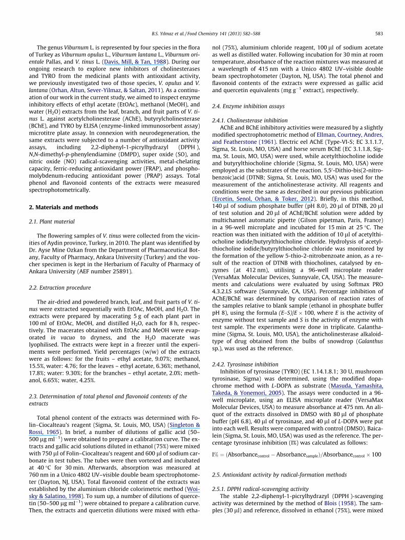
Enzyme inhibitory and antioxidant activities of Viburnum tinus L. relevant to its neuroprotective potential Betül Sever Yılmaz a , Mehmet Levent Altun a , Ilkay Erdogan Orhan b,c,⇑ , Burcin Ergene a , Gülcin Saltan Citoglu a a Department of Pharmacognosy, Faculty of Pharmacy, Ankara University, 06100 Ankara, Turkey b Department of Pharmacognosy, Faculty of Pharmacy, Gazi University, 06330 Ankara, Turkey c Pharmacognosy and Pharmaceutical Botany Unit, Faculty of Pharmacy, Eastern Mediterranean University, Gazimagosa, Cyprus article info Article history: Received 19 November 2012 Received in revised form 22 January 2013 Accepted 6 March 2013 Available online 13 March 2013 Keywords: Viburnum tinus Enzyme inhibition Antioxidant activity Neuroprotection Radical-scavenging abstract In vitro neuroprotective activity of the extracts of Viburnum tinus L. was investigated via inhibition of ace- tylcholinesterase (AChE) and butyrylcholinesterase (BChE), and tyrosinase (TYRO) by microtitre plate assays. Their antioxidant activity was tested using 2,2-diphenyl-1-picrylhydrazyl (DPPH ), N,N- dimethyl-p-phenylendiamine (DMPD), super oxide (SO), and nitric oxide (NO) radical-scavenging activ- ities, ferric ion-chelation capacity, ferric- (FRAP), and phosphomolybdenum-reducing antioxidant power (PRAP) assays. Total phenol and flavonoid content of the extracts was determined spectrophotometri- cally. The branch–ethyl acetate and fruit–methanol extracts exerted potent anticholinesterase effects (66.4 ± 0.65% to 97.7 ± 0.47%), while the fruit–methanol extract had the highest TYRO inhibition (47.0 ± 0.68%). The methanol extracts showed higher activities in most of the antioxidant tests. All the extracts displayed notable NO-scavenging effects (47.5 ± 5.03% to 74.5 ± 1.80%). Only the fruit–ethyl ace- tate extract quenched SO radical (38.4 ± 1.01%) at 500 lg ml 1 . Our data indicate that the fruit and branch extracts of V. tinus may provide potential neuroprotection. Ó 2013 Elsevier Ltd. All rights reserved. 1. Introduction The genus Viburnum L. (Caprifoliaceae) comprises over 230 spe- cies distributed throughout the temperate northern hemisphere, with a few species extending into tropical regions in South America and southeast Asia (Lobstein, Haan-Archipoff, Englert, Kuhry, & An- ton, 1999). Many species of Viburnum have become popular as gar- den or landscape plants because of their showy flowers, berries and fragrance, while some of its species have edible fruits and can be eaten either raw or used for making jam. Among them, Viburnum tinus L., known as ‘‘laurustinus, spring bouquet’’ in English and ‘‘kartopu, defne yapraklı kartopu’’ in Turkish, is a shrub or small tree native to the Mediterranean basin. The plant has fragrant, white, tiny flowers and drupe-type fruits with a metallic steely black–dark blue colour. Its common name ‘‘laurustinus’’ is thought to refer to the leaves which resemble those of the bay tree (Laurus nobilis). A limited number of studies have demonstrated the pres- ence of various biochemical components, including iridoids, cou- marins, saponins, and flavonoids, in V. tinus (Cometa, Mazzanti, & Tomassini, 1998; Mohamed, Marzouk, Moharram, El-Sayed, & Baiuomy, 2005). Neurodegenerative diseases have become a major age-associ- ated health problem, especially in industrialised countries, in which the proportion of the elderly population is rising (Uc & Riz- zo, 2008). Among them, prevalence of Alzheimer’s disease (AD), as the most common form of dementia, as well as Parkinson’s disease (PD), is on the increase and dementia has also been identified in the patients suffering from PD (Metzler-Baddeley, 2007). The cho- linergic hypothesis is the most accepted theory to explain patho- genesis of AD, and therefore the most prescribed drugs for the treatment of AD are the cholinesterase inhibitors (Orhan, Orhan, & Sener, 2006). On the other hand, tyrosinase (TYRO) has been demonstrated to play a role in neuromelanin formation in the hu- man brain and could be central to dopamine neurotoxicity, as well as contributing to the neurodegeneration associated with PD (Khan, 2007). Consequently, TYRO inhibitors have become an attractive target for the treatment of PD. It is also well-known that oxidative damage has been reported to be a triggering factor induc- ing neurodegeneration (Melo et al., 2011). However, current drugs are not able to stop AD and PD and can only slow the progress of these diseases by symptomatic treatment. Consequently, there is still a need to discover new drug candidates and plants are attrac- tive sources for drug research and discovery. 0308-8146/$ - see front matter Ó 2013 Elsevier Ltd. All rights reserved. http://dx.doi.org/10.1016/j.foodchem.2013.03.020 ⇑ Corresponding author. at: Department of Pharmacognosy, Faculty of Pharmacy, Gazi University, 06330 Ankara, Turkey. Tel.: +90 312 202 3186; fax: +90 312 223 5018. E-mail addresses: [email protected], [email protected] (I.E. Orhan). Food Chemistry 141 (2013) 582–588 Contents lists available at SciVerse ScienceDirect Food Chemistry journal homepage: www.elsevier.com/locate/foodchem
-
Upload
independent -
Category
Documents
-
view
1 -
download
0
Transcript of Enzyme inhibitory and antioxidant activities of traditional medicinal plants: Potential application...

Food Chemistry 141 (2013) 582–588
Contents lists available at SciVerse ScienceDirect
Food Chemistry
journal homepage: www.elsevier .com/locate / foodchem
Enzyme inhibitory and antioxidant activities of Viburnum tinusL. relevant to its neuroprotective potential
0308-8146/$ - see front matter � 2013 Elsevier Ltd. All rights reserved.http://dx.doi.org/10.1016/j.foodchem.2013.03.020
⇑ Corresponding author. at: Department of Pharmacognosy, Faculty of Pharmacy,Gazi University, 06330 Ankara, Turkey. Tel.: +90 312 202 3186; fax: +90 312 2235018.
E-mail addresses: [email protected], [email protected] (I.E. Orhan).
Betül Sever Yılmaz a, Mehmet Levent Altun a, Ilkay Erdogan Orhan b,c,⇑, Burcin Ergene a,Gülcin Saltan Citoglu a
a Department of Pharmacognosy, Faculty of Pharmacy, Ankara University, 06100 Ankara, Turkeyb Department of Pharmacognosy, Faculty of Pharmacy, Gazi University, 06330 Ankara, Turkeyc Pharmacognosy and Pharmaceutical Botany Unit, Faculty of Pharmacy, Eastern Mediterranean University, Gazimagosa, Cyprus
a r t i c l e i n f o
Article history:Received 19 November 2012Received in revised form 22 January 2013Accepted 6 March 2013Available online 13 March 2013
Keywords:Viburnum tinusEnzyme inhibitionAntioxidant activityNeuroprotectionRadical-scavenging
a b s t r a c t
In vitro neuroprotective activity of the extracts of Viburnum tinus L. was investigated via inhibition of ace-tylcholinesterase (AChE) and butyrylcholinesterase (BChE), and tyrosinase (TYRO) by microtitre plateassays. Their antioxidant activity was tested using 2,2-diphenyl-1-picrylhydrazyl (DPPH�), N,N-dimethyl-p-phenylendiamine (DMPD), super oxide (SO), and nitric oxide (NO) radical-scavenging activ-ities, ferric ion-chelation capacity, ferric- (FRAP), and phosphomolybdenum-reducing antioxidant power(PRAP) assays. Total phenol and flavonoid content of the extracts was determined spectrophotometri-cally. The branch–ethyl acetate and fruit–methanol extracts exerted potent anticholinesterase effects(66.4 ± 0.65% to 97.7 ± 0.47%), while the fruit–methanol extract had the highest TYRO inhibition(47.0 ± 0.68%). The methanol extracts showed higher activities in most of the antioxidant tests. All theextracts displayed notable NO-scavenging effects (47.5 ± 5.03% to 74.5 ± 1.80%). Only the fruit–ethyl ace-tate extract quenched SO radical (38.4 ± 1.01%) at 500 lg ml�1. Our data indicate that the fruit and branchextracts of V. tinus may provide potential neuroprotection.
� 2013 Elsevier Ltd. All rights reserved.
1. Introduction
The genus Viburnum L. (Caprifoliaceae) comprises over 230 spe-cies distributed throughout the temperate northern hemisphere,with a few species extending into tropical regions in South Americaand southeast Asia (Lobstein, Haan-Archipoff, Englert, Kuhry, & An-ton, 1999). Many species of Viburnum have become popular as gar-den or landscape plants because of their showy flowers, berries andfragrance, while some of its species have edible fruits and can beeaten either raw or used for making jam. Among them, Viburnumtinus L., known as ‘‘laurustinus, spring bouquet’’ in English and‘‘kartopu, defne yapraklı kartopu’’ in Turkish, is a shrub or smalltree native to the Mediterranean basin. The plant has fragrant,white, tiny flowers and drupe-type fruits with a metallic steelyblack–dark blue colour. Its common name ‘‘laurustinus’’ is thoughtto refer to the leaves which resemble those of the bay tree (Laurusnobilis). A limited number of studies have demonstrated the pres-ence of various biochemical components, including iridoids, cou-marins, saponins, and flavonoids, in V. tinus (Cometa, Mazzanti, &
Tomassini, 1998; Mohamed, Marzouk, Moharram, El-Sayed, &Baiuomy, 2005).
Neurodegenerative diseases have become a major age-associ-ated health problem, especially in industrialised countries, inwhich the proportion of the elderly population is rising (Uc & Riz-zo, 2008). Among them, prevalence of Alzheimer’s disease (AD), asthe most common form of dementia, as well as Parkinson’s disease(PD), is on the increase and dementia has also been identified inthe patients suffering from PD (Metzler-Baddeley, 2007). The cho-linergic hypothesis is the most accepted theory to explain patho-genesis of AD, and therefore the most prescribed drugs for thetreatment of AD are the cholinesterase inhibitors (Orhan, Orhan,& Sener, 2006). On the other hand, tyrosinase (TYRO) has beendemonstrated to play a role in neuromelanin formation in the hu-man brain and could be central to dopamine neurotoxicity, as wellas contributing to the neurodegeneration associated with PD(Khan, 2007). Consequently, TYRO inhibitors have become anattractive target for the treatment of PD. It is also well-known thatoxidative damage has been reported to be a triggering factor induc-ing neurodegeneration (Melo et al., 2011). However, current drugsare not able to stop AD and PD and can only slow the progress ofthese diseases by symptomatic treatment. Consequently, there isstill a need to discover new drug candidates and plants are attrac-tive sources for drug research and discovery.
B.S. Yılmaz et al. / Food Chemistry 141 (2013) 582–588 583
The genus Viburnum L. is represented by four species in the floraof Turkey as Viburnum opulus L., Viburnum lantana L., Viburnum ori-entale Pallas, and V. tinus L. (Davis, Mill, & Tan, 1988). During ourongoing research to explore new inhibitors of cholinesterasesand TYRO from the medicinal plants with antioxidant activity,we previously investigated two of those species, V. opulus and V.lantana (Orhan, Altun, Sever-Yilmaz, & Saltan, 2011). As a continu-ation of our work in the current study, we aimed to inspect enzymeinhibitory effects of ethyl acetate (EtOAc), methanol (MeOH), andwater (H2O) extracts from the leaf, branch, and fruit parts of V. ti-nus L. against acetylcholinesterase (AChE), butyrylcholinesterase(BChE), and TYRO by ELISA (enzyme-linked immunosorbent assay)microtitre plate assay. In connexion with neurodegeneration, thesame extracts were subjected to a number of antioxidant activityassays, including 2,2-diphenyl-1-picrylhydrazyl (DPPH�),N,N-dimethyl-p-phenylendiamine (DMPD), super oxide (SO), andnitric oxide (NO) radical-scavenging activities, metal-chelatingcapacity, ferric-reducing antioxidant power (FRAP), and phospho-molybdenum-reducing antioxidant power (PRAP) assays. Totalphenol and flavonoid contents of the extracts were measuredspectrophotometrically.
2. Materials and methods
2.1. Plant material
The flowering samples of V. tinus were collected from the vicin-ities of Aydin province, Turkey, in 2010. The plant was identified byDr. Ayse Mine Ozkan from the Department of Pharmaceutical Bot-any, Faculty of Pharmacy, Ankara University (Turkey) and the vou-cher specimen is kept in the Herbarium of Faculty of Pharmacy ofAnkara University (AEF number 25891).
2.2. Extraction procedure
The air-dried and powdered branch, leaf, and fruit parts of V. ti-nus were extracted sequentially with EtOAc, MeOH, and H2O. Theextracts were prepared by macerating 5 g of each plant part in100 ml of EtOAc, MeOH, and distilled H2O, each for 8 h, respec-tively. The macerates obtained with EtOAc and MeOH were evap-orated in vacuo to dryness, and the H2O macerate waslyophilised. The extracts were kept in a freezer until the experi-ments were performed. Yield percentages (w/w) of the extractswere as follows: for the fruits – ethyl acetate, 9.07%; methanol,15.5%, water: 4.76; for the leaves – ethyl acetate, 6.36%; methanol,17.8%; water: 9.30%; for the branches – ethyl acetate, 2.0%; meth-anol, 6.65%; water, 4.25%.
2.3. Determination of total phenol and flavonoid contents of theextracts
Total phenol content of the extracts was determined with Fo-lin–Ciocalteau’s reagent (Sigma, St. Louis, MO, USA) (Singleton &Rossi, 1965). In brief, a number of dilutions of gallic acid (50–500 lg ml�1) were obtained to prepare a calibration curve. The ex-tracts and gallic acid solutions diluted in ethanol (75%) were mixedwith 750 ll of Folin–Ciocalteau’s reagent and 600 ll of sodium car-bonate in test tubes. The tubes were then vortexed and incubatedat 40 �C for 30 min. Afterwards, absorption was measured at760 nm in a Unico 4802 UV–visible double beam spectrophotome-ter (Dayton, NJ, USA). Total flavonoid content of the extracts wasestablished by the aluminium chloride colorimetric method (Woi-sky & Salatino, 1998). To sum up, a number of dilutions of querce-tin (50–500 lg ml�1) were obtained to prepare a calibration curve.Then, the extracts and quercetin dilutions were mixed with etha-
nol (75%), aluminium chloride reagent, 100 ll of sodium acetateas well as distilled water. Following incubation for 30 min at roomtemperature, absorbance of the reaction mixtures was measured ata wavelength of 415 nm with a Unico 4802 UV–visible doublebeam spectrophotometer (Dayton, NJ, USA). The total phenol andflavonoid contents of the extracts were expressed as gallic acidand quercetin equivalents (mg g�1 extract), respectively.
2.4. Enzyme inhibition assays
2.4.1. Cholinesterase inhibitionAChE and BChE inhibitory activities were measured by a slightly
modified spectrophotometric method of Ellman, Courtney, Andres,and Featherstone (1961). Electric eel AChE (Type-VI-S; EC 3.1.1.7,Sigma, St. Louis, MO, USA) and horse serum BChE (EC 3.1.1.8, Sig-ma, St. Louis, MO, USA) were used, while acetylthiocholine iodideand butyrylthiocholine chloride (Sigma, St. Louis, MO, USA) wereemployed as the substrates of the reaction. 5,50-Dithio-bis(2-nitro-benzoic)acid (DTNB; Sigma, St. Louis, MO, USA) was used for themeasurement of the anticholinesterase activity. All reagents andconditions were the same as described in our previous publication(Ercetin, Senol, Orhan, & Toker, 2012). Briefly, in this method,140 ll of sodium phosphate buffer (pH 8.0), 20 ll of DTNB, 20 llof test solution and 20 ll of AChE/BChE solution were added bymultichannel automatic pipette (Gilson pipetman, Paris, France)in a 96-well microplate and incubated for 15 min at 25 �C. Thereaction was then initiated with the addition of 10 ll of acetylthi-ocholine iodide/butyrylthiocholine chloride. Hydrolysis of acetyl-thiocholine iodide/butyrylthiocholine chloride was monitored bythe formation of the yellow 5-thio-2-nitrobenzoate anion, as a re-sult of the reaction of DTNB with thiocholines, catalysed by en-zymes (at 412 nm), utilising a 96-well microplate reader(VersaMax Molecular Devices, Sunnyvale, CA, USA). The measure-ments and calculations were evaluated by using Softmax PRO4.3.2.LS software (Sunnyvale, CA, USA). Percentage inhibition ofAChE/BChE was determined by comparison of reaction rates ofthe samples relative to blank sample (ethanol in phosphate bufferpH 8), using the formula (E–S)/E � 100, where E is the activity ofenzyme without test sample and S is the activity of enzyme withtest sample. The experiments were done in triplicate. Galantha-mine (Sigma, St. Louis, MO, USA), the anticholinesterase alkaloid-type of drug obtained from the bulbs of snowdrop (Galanthussp.), was used as the reference.
2.4.2. Tyrosinase inhibitionInhibition of tyrosinase (TYRO) (EC 1.14.1.8.1; 30 U, mushroom
tyrosinase, Sigma) was determined, using the modified dopa-chrome method with L-DOPA as substrate (Masuda, Yamashita,Takeda, & Yonemori, 2005). The assays were conducted in a 96-well microplate, using an ELISA microplate reader (VersaMaxMolecular Devices, USA) to measure absorbance at 475 nm. An ali-quot of the extracts dissolved in DMSO with 80 ll of phosphatebuffer (pH 6.8), 40 ll of tyrosinase, and 40 ll of L-DOPA were putinto each well. Results were compared with control (DMSO). Baica-lein (Sigma, St. Louis, MO, USA) was used as the reference. The per-centage tyrosinase inhibition (I%) was calculated as follows:
I% ¼ ðAbsorbancecontrol � AbsorbancesampleÞ=Absorbancecontrol � 100
2.5. Antioxidant activity by radical-formation methods
2.5.1. DPPH radical-scavenging activityThe stable 2,2-diphenyl-1-picrylhydrazyl (DPPH�)-scavenging
activity was determined by the method of Blois (1958). The sam-ples (30 ll) and reference, dissolved in ethanol (75%), were mixed

584 B.S. Yılmaz et al. / Food Chemistry 141 (2013) 582–588
with 2700 ll of DPPH� solution (1.5 � 10�4 M). Remaining DPPH�amount was measured at 520 nm, using a Unico 4802 UV–visibledouble beam spectrophotometer (Dayton, NJ, USA). Gallic acid(Sigma, St. Louis, MO, USA) was employed as the reference. Inhibi-tion of DPPH� in percent (I%) was calculated as given below:
I% = [(Ablank � Asample)/Ablank] � 100, where Ablank is the absor-bance of the control reaction (containing all reagents except thetest sample), and Asample is the absorbance of the extracts/refer-ence. Analyses were run in triplicate and the results were ex-pressed as average values with SEM.
2.5.2. DMPD radical-scavenging activityThe assay is based on reduction of the purple-coloured radical
DMPD+ (N,N-dimethyl-p-phenylendiamine). According to themethod (Schlesier, Harvat, Bohm, & Bitsch, 2002), a reagent com-prising 100 mM DMPD, 0.1 M acetate buffer (pH 5.25), and0.05 M ferric chloride solution, which led to formation of DMPDradical, was freshly prepared and the reagent was equilibrated toan absorbance of 0.900 ± 0.100 at 505 nm. Then, the reagent wasmixed with 50 ll of the extract dilutions and absorbance was takenat 505 nm, using a Unico 4802 UV–visible double beam spectro-photometer (Dayton, NJ, USA). Quercetin was employed as the ref-erence and the experiments were done in triplicate. The resultswere calculated according to the same formula as given for theDPPH radical-scavenging test and expressed as average values withSEM.
2.5.3. SO radical-scavenging activitySO radical-scavenging activity was determined by the method
of Nishimiki, Rao, Appaji, and Yagi (1972). Nitro blue tetrazoliumsolution (NBT, 156 lM) and 468 lM b-nicotinamide adenine dinu-cleotide (NADH) dissolved in 100 mM phosphate buffer (pH 7.4)were mixed with 100 ll of the extracts dissolved in ethanol(75%) at various dilutions. The reaction was initiated by additionof 60 lM phenazine methosulphate (PMS) prepared in 100 mMphosphate buffer (pH 7.4). The reaction mixture was incubated at25 �C for 5 min and then, absorbance was measured against controlsamples, which contained all reagents except the extracts, using aUnico 4802 UV–visible double beam spectrophotometer (Dayton,NJ, USA). Inhibition of superoxide radical in percent (I%) was calcu-lated as given below:
I% = [(Ablank � Asample)/Ablank] � 100, where Ablank is the absor-bance of the control reaction (containing all reagents except thetest sample), and Asample is the absorbance of the extracts. Analyseswere run in triplicate and the results were expressed as averagevalues with SEM.
Table 1Acetylcholinesterase (AChE) and butyrylcholinesterase (BChE) inhibitory activities (percen
Plant part Extract type Inhibitory activity against AChE (Percentage ± S
50 lg ml�1 100 lg ml�1 20
Leaf EtOAc –b – –MeOH 6.12 ± 1.19**** 17.2 ± 1.44**** 40H2O – – 19
Branch EtOAc 12.8 ± 3.29**** 49.6 ± 1.73**** 81MeOH 7.12 ± 0.12**** 30.9 ± 1.23**** 45H2O – – –
Fruit EtOAc – – –MeOH 52.3 ± 3.21**** 64.7 ± 0.52**** 66H2O – – –
Galanthamine (Reference) 99.0 ± 0.24 at 100 lg ml�1
⁄p < 0.05; ⁄⁄⁄p < 0.001.a Standard error mean (n = 6).b No activity.
** p < 0.01.**** p < 0.0001.
2.5.4. Nitric oxide (NO) radical-scavenging activityThe scavenging activity of the extracts against NO was assessed
by the method of Marcocci, Marguire, Droy-Lefaiz, and Packer(1994). Briefly, the extract dilutions were mixed with 5 mM so-dium nitroprusside and left to incubate for 2 h at 29 �C. An aliquotof the solution was removed and diluted with Griess reagent (1%sulfanilamide in 5% H3PO4 and 0.1% naphthylethylenediaminedihydrochloride). The absorbance of the chromophore was mea-sured at 550 nm, using a Unico 4802 UV–visible double beam spec-trophotometer (USA). Inhibition of NO radical in percent (I%) wascalculated as given below:
I% = [(Ablank � Asample)/Ablank] � 100, where Ablank is the absor-bance of the control reaction (containing all reagents except thetest sample), and Asample is the absorbance of the extracts. Analyseswere run in triplicates and the results were expressed as averagevalues with SEM (Standard error of the mean). Quercetin was thereference in this test.
2.6. Antioxidant activity by metal-related methods
2.6.1. Metal-chelating capacityThe metal-chelating capacity of the extracts through ferrous ion
was estimated by the method of Chua, Tung, and Chang (2008).Briefly, dilutions of the extracts were incubated with 2 mM FeCl2
solution. The reaction was initiated by the addition of 5 mM ferro-zine to the mixture and left standing at ambient temperature for10 min. The absorbance of the reaction mixture was measured at562 nm, using a Unico 4802 UV–visible double beam spectropho-tometer (Dayton, NJ, USA). The ratio of inhibition of ferrozine–Fe2+ complex formation was calculated as follows:
I% = [(Ablank � Asample)/Ablank] � 100, where Ablank is the absor-bance of the control reaction (containing only FeCl2 and ferrozine),and Asample is the absorbance of the extracts/reference. Analyseswere run in triplicate and the results were expressed as averagevalues with SEM. The reference employed was ethylenediaminetetraacetic acid (EDTA) in this assay.
2.6.2. Ferric-reducing antioxidant power assay (FRAP)FRAP of the samples was tested, using the assay of Oyaizu
(1986). Different concentrations of the extracts were mixed with2500 ll of phosphate buffer (pH 6.6) and 2500 ll of potassium fer-ricyanide. Later, the mixture was incubated at 50 �C for 20 min and,then, trichloroacetic acid (10%) was added. After the mixture wasshaken vigorously, this solution was mixed with distilled waterand ferric chloride (0.1%). After 30 min of incubation, absorbancewas read at 700 nm, using a Unico 4802 UV–visible double beam
tage ± SEM) of the extracts of Viburnum tinus.
EMa) Inhibitory activity against BChE (Percentage ± SEM)
0 lg ml�1 50 lg ml�1 100 lg ml�1 200 lg ml�1
– – 47.2 ± 5.91****
.0 ± 1.60**** – 16.6 ± 1.35**** 35.8 ± 1.68****
.0 ± 4.09**** – – –
.7 ± 1.60** 22.8 ± 1.31**** 46.2 ± 1.21**** 81.3 ± 1.27****
.9 ± 1.32**** 44.6 ± 0.96**** 59.5 ± 0.19**** 69.7 ± 0.35⁄⁄⁄⁄
– – 4.84 ± 0.26****
– – 7.25 ± 1.15****
.4 ± 0.65**** 75.8 ± 1.73**** 88.8 ± 0.74 97.7 ± 0.47**
– – –90.0 ± 0.87 at 100 lg ml�1

0
10
20
30
40
50
60
70
Fig. 1. The extracts of Viburnum tinus with tyrosinase (TYRO) inhibitory activity(% ± SEM).
B.S. Yılmaz et al. / Food Chemistry 141 (2013) 582–588 585
spectrophotometer (Dayton, NJ, USA). Analyses were achieved intriplicate. Increased absorbance of the reaction meant increasedreducing power and was compared to that of chlorogenic acid (Sig-ma, St. Louis, MO, USA) as the reference.
2.6.3. Phosphomolybdenum-reducing antioxidant power (PRAP) assayIn order to perform PRAP assays on the extracts, each dilution
was mixed with 10% phosphomolybdic acid solution in ethanol(w/v) (Falcioni et al., 2002). The solution was subsequently sub-jected to incubation at 80 �C for 30 min and the absorbance wasread at 600 nm, using a Unico 4802 UV–visible double beamspectrophotometer (Dayton, NJ, USA). Analyses were run in trip-
Table 2Total phenol and flavonoid contents and antiradical activity of the extracts of Viburnum ti
Plant part Extracttype
Total phenolcontenta
Totalflavonoidcontentc
Inhibition% ±
(lg g�1
extract ± SEMb)(lg g�1
extract ± SEM)500 lg ml�1
Leaf EtOAc –d 39.2 ± 2.13 10.7 ± 1.55***
MeOH 328 ± 3.67 53.8 ± 3.09 60.3 ± 2.93***
H2O 193 ± 4.51 63.6 ± 2.04 49.9 ± 0.48***
Branch EtOAc – 86.7 ± 5.29 17.4 ± 2.15***
MeOH 585 ± 3.44 23.6 ± 1.87 45.1 ± 1.61***
H2O 70.1 ± 5.03 12.8 ± 1.99 23.3 ± 0.54***
Fruit EtOAc – 8.65 ± 1.26 9.19 ± 1.67***
MeOH 368 ± 6.95 13.5 ± 3.05 89.5 ± 0.01H2O 48.6 ± 2.33 14.5 ± 0.92 15.9 ± 0.60***
Gallic acid (Reference for DPPHscavenging activity)Quercetin (Reference forDMPD scavenging activity)
91.6 ± 0.06
Plant part Extracttype
Inhibition% ±against SO ra500 lg ml�1
Fruit EtOAc –f
Allopurinol (reference for SOscavenging activity
70.5 ± 0.93 at1000 lg ml�1
⁄p < 0.05.a Gallic acid equivalent.b Standard error mean (n = 3).c Quercetin equivalent.d Not detected.e Only the EtOAc extract of the fruits of Viburnum tinus was found to be active againsf No activity.
** p < 0.01.*** p < 0.001.**** p < 0.0001.
licate. Increased absorbance of the reaction meant increasedreducing power and was compared to that of quercetin as thereference.
2.7. Statistical analysis of data
Data obtained from in vitro enzyme inhibition and antioxidantexperiments were expressed as the means ± standard error(±SEM). Statistical differences between the reference and the sam-ple groups were evaluated by ANOVA (one way). Dunnett’s multi-ple comparison tests were used as post hoc tests. p < 0.05 wasconsidered to be significant [⁄p < 0.05; ⁄⁄p < 0.01; ⁄⁄⁄p < 0.001,⁄⁄⁄⁄p < 0.0001].
3. Results
3.1. Enzyme inhibition results
All the extracts evaluated showed a dose-dependent inhibitionagainst AChE and BChE, as tabulated in Table 1. In general, we ob-served that the extracts displayed higher inhibitory activity at thehighest extract concentration (200 lg ml�1). According to the re-sults obtained, the branch–EtOAc extract showed the highest inhi-bition (81.7 ± 1.60%) at 200 lg ml�1, while the best inhibitionagainst BChE was caused by the fruit–MeOH extract(97.7 ± 0.47%) at the highest concentration tested, followed bythe branch–EtOAc extract (81.3 ± 1.27%). Among the tested ex-tracts, the fruit–MeOH extract was the most effective one in theTYRO inhibition assay (47.0 ± 0.68%) (Fig. 1).
nus against DPPH, DMPD, and SO radicals.
SEM against DPPH radical Inhibition% ± SEM against DMPD radical
1000 lg ml�1 2000 lg ml�1 500 lg ml�1 1000 lg ml�1 2000 lg ml�1
* 15.0 ± 0.24**** 25.6 ± 0.90**** 3.17 ± 0.11**** 4.45 ± 0.27**** 18.1 ± 0.11****
* 89.0 ± 0.30 89.8 ± 0.18*** 31.8 ± 1.79**** 43.8 ± 1.19 61.6 ± 0.11***
* 78.1 ± 2.63**** 89.5 ± 0.36**** 32.2 ± 1.44**** 41.4 ± 1.59 50.6 ± 1.53****
* 22.2 ± 0.12**** 34.4 ± 0.42**** 9.08 ± 0.11**** 9.83 ± 0.03**** 12.0 ± 0.66****
* 68.3 ± 0.30**** 89.3 ± 0.07**** 40.9 ± 0.88**** 50.9 ± 1.19** 56.5 ± 0.82****
* 34.9 ± 0.60**** 69.9 ± 0.30**** 28.0 ± 0.88**** 30.7 ± 0.92*** 42.8 ± 0.77****
* 7.40 ± 0.48**** 9.43 ± 0.12**** 6.18 ± 0.18**** 10.3 ± 0.45**** 15.1 ± 1.53****
90.5 ± 0.29 91.7 ± 0.06 46.0 ± 1.29**** 49.5 ± 1.61** 67.1 ± 0.33* 22.4 ± 2.09**** 30.1 ± 0.54**** 23.3 ± 0.88** 44.9 ± 3.45 62.0 ± 1.13***
92.6 ± 0.10 93.1 ± 0.27 18.0 ± 0.52 41.5 ± 0.71 68.3 ± 0.67
SEMdicale
1000 lg ml�1 2000 lg ml�1
– 38.4 ± 1.01****
t SO radical.
0 20 40 60 80 100
Leaf-EtOAc
Leaf-MeOH
Leaf-H2O
Branch-EtOAc
Branch-MeOH
Branch-H2O
Fruit-EtOAc
Fruit-MeOH
Fruit-H2O
Gallic acid (Ref)
NO inhibition (% S.E.M.) at 2000 µg ml-1
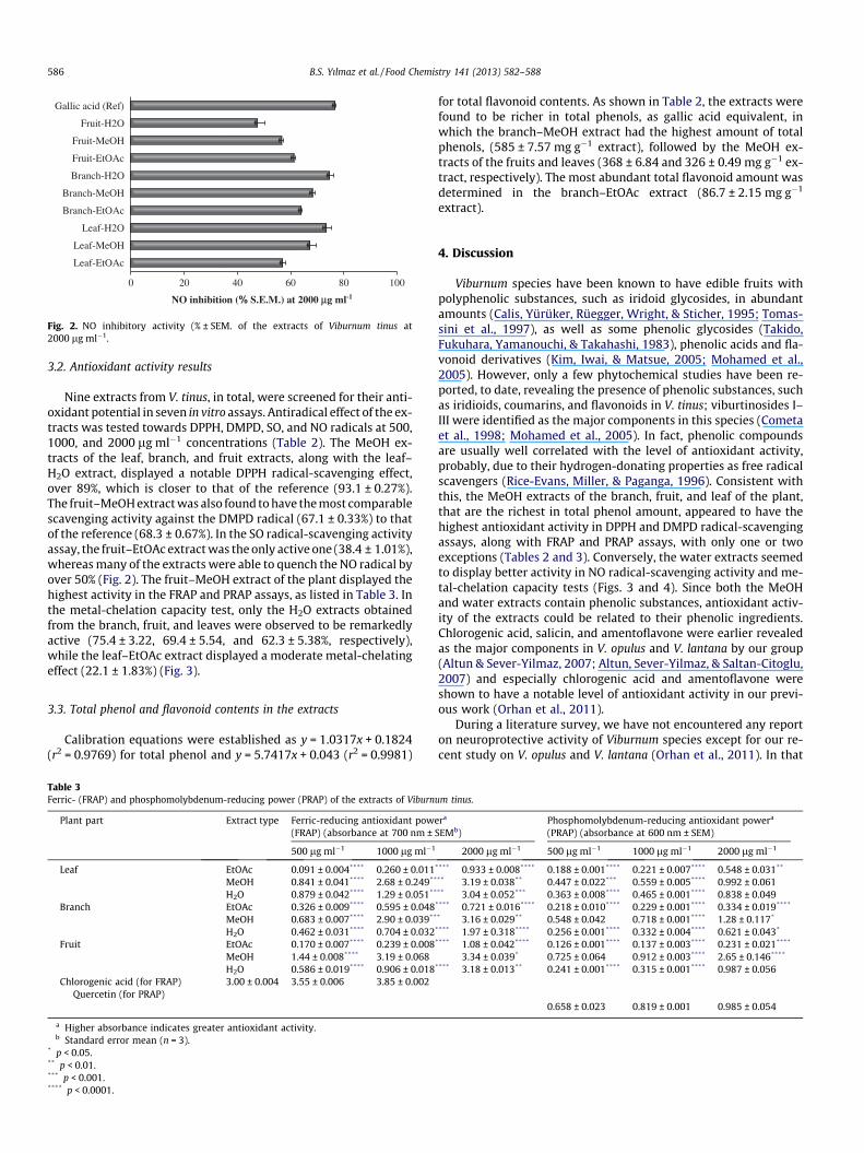
Fig. 2. NO inhibitory activity (% ± SEM. of the extracts of Viburnum tinus at2000 lg ml�1.
586 B.S. Yılmaz et al. / Food Chemistry 141 (2013) 582–588
3.2. Antioxidant activity results
Nine extracts from V. tinus, in total, were screened for their anti-oxidant potential in seven in vitro assays. Antiradical effect of the ex-tracts was tested towards DPPH, DMPD, SO, and NO radicals at 500,1000, and 2000 lg ml�1 concentrations (Table 2). The MeOH ex-tracts of the leaf, branch, and fruit extracts, along with the leaf–H2O extract, displayed a notable DPPH radical-scavenging effect,over 89%, which is closer to that of the reference (93.1 ± 0.27%).The fruit–MeOH extract was also found to have the most comparablescavenging activity against the DMPD radical (67.1 ± 0.33%) to thatof the reference (68.3 ± 0.67%). In the SO radical-scavenging activityassay, the fruit–EtOAc extract was the only active one (38.4 ± 1.01%),whereas many of the extracts were able to quench the NO radical byover 50% (Fig. 2). The fruit–MeOH extract of the plant displayed thehighest activity in the FRAP and PRAP assays, as listed in Table 3. Inthe metal-chelation capacity test, only the H2O extracts obtainedfrom the branch, fruit, and leaves were observed to be remarkedlyactive (75.4 ± 3.22, 69.4 ± 5.54, and 62.3 ± 5.38%, respectively),while the leaf–EtOAc extract displayed a moderate metal-chelatingeffect (22.1 ± 1.83%) (Fig. 3).
3.3. Total phenol and flavonoid contents in the extracts
Calibration equations were established as y = 1.0317x + 0.1824(r2 = 0.9769) for total phenol and y = 5.7417x + 0.043 (r2 = 0.9981)
Table 3Ferric- (FRAP) and phosphomolybdenum-reducing power (PRAP) of the extracts of Viburn
Plant part Extract type Ferric-reducing antioxidant powe(FRAP) (absorbance at 700 nm ±
500 lg ml�1 1000 lg ml�1
Leaf EtOAc 0.091 ± 0.004**** 0.260 ± 0.011MeOH 0.841 ± 0.041**** 2.68 ± 0.249**
H2O 0.879 ± 0.042**** 1.29 ± 0.051**
Branch EtOAc 0.326 ± 0.009**** 0.595 ± 0.048MeOH 0.683 ± 0.007**** 2.90 ± 0.039**
H2O 0.462 ± 0.031**** 0.704 ± 0.032Fruit EtOAc 0.170 ± 0.007**** 0.239 ± 0.008
MeOH 1.44 ± 0.008**** 3.19 ± 0.068H2O 0.586 ± 0.019**** 0.906 ± 0.018
Chlorogenic acid (for FRAP)Quercetin (for PRAP)
3.00 ± 0.004 3.55 ± 0.006 3.85 ± 0.002
a Higher absorbance indicates greater antioxidant activity.b Standard error mean (n = 3).
* p < 0.05.** p < 0.01.*** p < 0.001.**** p < 0.0001.
for total flavonoid contents. As shown in Table 2, the extracts werefound to be richer in total phenols, as gallic acid equivalent, inwhich the branch–MeOH extract had the highest amount of totalphenols, (585 ± 7.57 mg g�1 extract), followed by the MeOH ex-tracts of the fruits and leaves (368 ± 6.84 and 326 ± 0.49 mg g�1 ex-tract, respectively). The most abundant total flavonoid amount wasdetermined in the branch–EtOAc extract (86.7 ± 2.15 mg g�1
extract).
4. Discussion
Viburnum species have been known to have edible fruits withpolyphenolic substances, such as iridoid glycosides, in abundantamounts (Calis, Yürüker, Rüegger, Wright, & Sticher, 1995; Tomas-sini et al., 1997), as well as some phenolic glycosides (Takido,Fukuhara, Yamanouchi, & Takahashi, 1983), phenolic acids and fla-vonoid derivatives (Kim, Iwai, & Matsue, 2005; Mohamed et al.,2005). However, only a few phytochemical studies have been re-ported, to date, revealing the presence of phenolic substances, suchas iridioids, coumarins, and flavonoids in V. tinus; viburtinosides I–III were identified as the major components in this species (Cometaet al., 1998; Mohamed et al., 2005). In fact, phenolic compoundsare usually well correlated with the level of antioxidant activity,probably, due to their hydrogen-donating properties as free radicalscavengers (Rice-Evans, Miller, & Paganga, 1996). Consistent withthis, the MeOH extracts of the branch, fruit, and leaf of the plant,that are the richest in total phenol amount, appeared to have thehighest antioxidant activity in DPPH and DMPD radical-scavengingassays, along with FRAP and PRAP assays, with only one or twoexceptions (Tables 2 and 3). Conversely, the water extracts seemedto display better activity in NO radical-scavenging activity and me-tal-chelation capacity tests (Figs. 3 and 4). Since both the MeOHand water extracts contain phenolic substances, antioxidant activ-ity of the extracts could be related to their phenolic ingredients.Chlorogenic acid, salicin, and amentoflavone were earlier revealedas the major components in V. opulus and V. lantana by our group(Altun & Sever-Yilmaz, 2007; Altun, Sever-Yilmaz, & Saltan-Citoglu,2007) and especially chlorogenic acid and amentoflavone wereshown to have a notable level of antioxidant activity in our previ-ous work (Orhan et al., 2011).
During a literature survey, we have not encountered any reporton neuroprotective activity of Viburnum species except for our re-cent study on V. opulus and V. lantana (Orhan et al., 2011). In that
um tinus.
ra Phosphomolybdenum-reducing antioxidant powera
SEMb) (PRAP) (absorbance at 600 nm ± SEM)
2000 lg ml�1 500 lg ml�1 1000 lg ml�1 2000 lg ml�1
**** 0.933 ± 0.008**** 0.188 ± 0.001**** 0.221 ± 0.007**** 0.548 ± 0.031**
** 3.19 ± 0.038** 0.447 ± 0.022*** 0.559 ± 0.005**** 0.992 ± 0.061** 3.04 ± 0.052*** 0.363 ± 0.008**** 0.465 ± 0.001**** 0.838 ± 0.049**** 0.721 ± 0.016**** 0.218 ± 0.010**** 0.229 ± 0.001**** 0.334 ± 0.019****
* 3.16 ± 0.029** 0.548 ± 0.042 0.718 ± 0.001**** 1.28 ± 0.117*
**** 1.97 ± 0.318**** 0.256 ± 0.001**** 0.332 ± 0.004**** 0.621 ± 0.043*
**** 1.08 ± 0.042**** 0.126 ± 0.001**** 0.137 ± 0.003**** 0.231 ± 0.021****
3.34 ± 0.039* 0.725 ± 0.064 0.912 ± 0.003**** 2.65 ± 0.146****
**** 3.18 ± 0.013** 0.241 ± 0.001**** 0.315 ± 0.001**** 0.987 ± 0.056
0.658 ± 0.023 0.819 ± 0.001 0.985 ± 0.054

0 20 40 60 80 100
Leaf-EtOAc
Leaf-H2O
Branch-H2O
Fruit-H2O
EDTA (Ref)
Metal chelation capacity (% S.E.M.) at 2000 µg ml-1
Fig. 3. Metal-chelation capacity (% ± SEM) of the extracts of Viburnum tinus at2000 lg ml�1.
B.S. Yılmaz et al. / Food Chemistry 141 (2013) 582–588 587
study, the leaf–EtOH extract of V. opulus was found to have93.2 ± 0.87% of inhibition against AChE. Its major components;amentoflavone and chlorogenic acid, did not have ability to inhibitAChE, whereas salicin showed a moderate inhibition against thisenzyme (34.54 ± 1.24%). Therefore, salicin, presumably found inthis plant, might be considered to contribute in part to its AChEinhibitory activity. On the other hand, the high inhibitory effectof this extract may be the result of the existence of some othertypes of compounds in V. tinus. The genus Viburnum has beenfound to contain compounds from various chemical classes, suchas iridoids, terpenes, flavonoids, lignans, coumarins, and alkaloids(Wang, Shi, & Li, 2010). For instance, iridoid derivatives, whichhave been reported to be present in large quantities in many Vibur-num species, are demonstrated to have notable cholinesteraseinhibitory effects. Seven iridoids (barlerinoside, shanzhisidemethyl ester, 6-O-trans-p-coumaroyl-8-O-acetylshanzhisidemethyl ester, barlerin, acetylbarlerin, 7-methoxydiderroside, andlupulinoside) isolated from the aerial parts of Barleria prionitis dis-played different levels of AChE inhibitory and free radical-scaveng-ing activities (Ata, Kalhari, & Samarasekera, 2009). The markedAChE inhibitory effect of Leucosidea sericea was also reported tobe associated with high iridoid content of this plant (Aremu, Amoo,Ndhlala, Finnie, & Van Staden, 2011). Besides, flavonoid derivativeshave been known to possess cholinesterase-inhibiting properties(Khan et al., 2009; Kupeli Akkol, Orhan, & Yesilada, 2012). Conse-quently, it could be suggested that cholinesterase-inhibiting ex-tracts of V. tinus, found in this study, could be correlated withpossible presence of iridoid, flavonoid, or other compounds. Be-sides, since TYRO inhibitors from natural sources are simple phen-olics, such as alpha-kojic acid, arbutin (Khan, 2007), TYRO-inhibiting extracts of this species might be associated with its phe-nolic substances.
5. Conclusion
The findings obtained in this study revealed that the branch–EtOAc and fruit–MeOH extracts of V. tinus possess a significant cho-linesterase inhibitory effect, while the fruit–MeOH extract showedthe greatest TYRO inhibition. Occurrence of notable antioxidantactivity was observed in most of the extracts. The possibility thatneuroprotective properties of V. tinus may involve further mecha-nisms of action remains an interesting question for future study inview of a wider use of this medicinal plant. In our opinion, this isthe first evidence shedding light on the in vitro neuroprotective ef-fect of V. tinus through inhibition of neurodegeneration-related en-zymes and antioxidant effects, indicating a new interestingapproach to pharmacological studies on this species. The isolationof specific bioactive compounds through bioassay-guided fraction-
ation and their characterisation, as well as studies evaluating theirsafety, might be indispensable in the exploration of V. tinus for po-tential new neuroprotective drug leads, which is in progress in ourlaboratory.
References
Altun, M. L., Sever-Yilmaz, B., & Saltan-Citoglu, G. (2007). HPLC analysis ofamentoflavone in Viburnum opulus L. and Viburnum lantana L. Journal ofFaculty of Pharmacy of Ankara University, 36, 161–169.
Altun, M. L., & Sever-Yilmaz, B. (2007). HPLC method for the analysis of salicin andchlorogenic acid from Viburnum opulus and V. lantana. Chemistry of NaturalCompounds, 43, 205–207.
Aremu, A. O., Amoo, S. O., Ndhlala, A. R., Finnie, J. F., & Van Staden, J. (2011).Antioxidant activity, acetylcholinesterase inhibition, iridoid content andmutagenic evaluation of Leucosidea sericea. Food and Chemical Toxicology, 49,1122–1128.
Ata, A., Kalhari, K. S., & Samarasekera, R. (2009). Chemical constituents of Barleriaprionitis and their enzyme inhibitory and free radical scavenging activities.Phytochemistry Letters, 2, 37–40.
Blois, M. S. (1958). Antioxidant determinations by the use of a stable free radical.Nature, 181, 1199–1200.
Calis, I., Yürüker, A., Rüegger, H., Wright, A. D., & Sticher, O. (1995). Lantanoside, amonocyclic C10 iridoid glucoside from Viburnum lantana. Phytochemistry, 38,163–165.
Chua, M. T., Tung, Y. T., & Chang, S. T. (2008). Antioxidant activities of ethanolicextracts from the twigs of Cinnamomum osmophleum. Bioresource Technology,99, 1918–1925.
Cometa, M. F., Mazzanti, G., & Tomassini, L. (1998). Sedative and spasmolytic effectsof Viburnum tinus L. and its major pure compounds. Phytotherapy Research, 12,S89–S91.
Davis, P. H., Mill, R. R., & Tan, K. (Eds.). (1988). Flora of Turkey and East AegeanIslands: Vol. 10 (Suppl.). Edinburgh: Edinburgh University Press, p. 154.
Ellman, G. L., Courtney, K. D., Andres, V., & Featherstone, R. M. (1961). A new andrapid colorimetric determination of acetylcholinesterase activity. BiochemicalPharmacology, 7, 88–95.
Ercetin, T., Senol, F., Orhan, I. E., & Toker, G. (2012). Comparative assessment ofantioxidant and cholinesterase inhibitory properties of the marigold extractsfrom Calendula arvensis L. and Calendula officinalis L. Industrial Crops & Products,26, 203–208.
Falcioni, G., Fedeli, D., Tiano, L., Calzuola, I., Mancinelli, L., Marsili, V., et al. (2002).Antioxidant activity of wheat sprouts extract in vitro: Inhibition of DNAoxidative damage. Journal of Food Science, 67, 2918–2922.
Khan, M. T. H. (2007). Molecular design of tyrosinase inhibitors: A critical review ofpromising novel inhibitors from synthetic origins. Pure & Applied Chemistry, 79,2277–2295.
Khan, M. T. H., Orhan, I., Senol, F. S., Kartal, M., S�ener, B., Dvorska, M., et al. (2009).Cholinesterase inhibitory activities of some flavonoid derivatives and chosenxanthone and their molecular docking studies. Chemicobiological Interactions,181, 383–389.
Kim, M. Y., Iwai, K., & Matsue, H. (2005). Phenolic compositions of Viburnumdilatatum Thunb. fruits and their antiradical properties. Journal of FoodComposition and Analysis, 18, 789–802.
Kupeli Akkol, E., Orhan, I. E., & Yesilada, E. (2012). Anticholinesterase andantioxidant effects of the ethanol extract, ethanol fractions and isolatedflavonoids from Cistus laurifolius L. leaves. Food Chemistry, 131, 626–631.
Lobstein, A., Haan-Archipoff, G., Englert, J., Kuhry, J. G., & Anton, R. (1999).Chemotaxonomical investigation in the genus Viburnum. Phytochemistry, 50,1175–1180.
Marcocci, I., Marguire, J. J., Droy-Lefaiz, M. T., & Packer, L. (1994). The nitric oxidescavenging properties of Ginkgo biloba extract. Biochemical and BiophysicalResearch Communications, 201, 748–755.
Masuda, T., Yamashita, D., Takeda, Y., & Yonemori, S. (2005). Screening fortyrosinase inhibitors among extracts of seashore plants and identification ofpotent inhibitors from Garcinia subelliptica. Bioscience Biotechnology andBiochemistry, 69, 197–201.
Melo, A., Monteiro, L., Lima, R.M., Oliveira, D.M., Cerqueira, M.D., & El-Bachá, R.S.(2011). Oxidative stress in neurodegenerative diseases: mechanisms andtherapeutic perspectives. Oxidative Medicine and Cellular Longevity, Article ID:467180.
Metzler-Baddeley, C. (2007). A review of cognitive impairments in dementia withLewy bodies relative to Alzheimer’s disease and Parkinson’s disease withdementia. Cortex, 43, 583–600.
Mohamed, M. A., Marzouk, M. S. A., Moharram, F. A., El-Sayed, M. M., & Baiuomy, A.R. (2005). Phytochemical constituents and hepatoprotective activity ofViburnum tinus. Phytochemistry, 66, 2780–2786.
Nishimiki, M., Rao, N. A., Appaji, N., & Yagi, K. (1972). The occurrence of superoxideanion in the reaction of reduced phenazine methosulfate and molecular oxygen.Biochemical and Biophysical Research Communications, 46, 849–854.
Orhan, G., Orhan, I., & Sener, B. (2006). Recent developments in natural andsynthetic drug research for Alzheimer’s Disease. Letters in Drug and DesignDiscovery, 3, 268–274.
Orhan, I. E., Altun, M. L., Sever-Yilmaz, B., & Saltan, G. (2011). Anti-acetylcholinesterase and antioxidant assets of the major components (salicin,

588 B.S. Yılmaz et al. / Food Chemistry 141 (2013) 582–588
amentoflavone, and chlorogenic acid) and the extracts of Viburnum opulus andViburnum lantana and their total phenol and flavonoid contents. Journal ofMedicinal Food, 14, 434–440.
Oyaizu, M. (1986). Studies on products of browning reactions-antioxidativeactivities of products of browning reaction prepared from glucosamine.Japanese Journal of Nutrition, 44, 307–315.
Rice-Evans, C. A., Miller, N. J., & Paganga, G. (1996). Structure-antioxidant activityrelationships of flavonoids and phenolic acids. Free Radical Biology and Medicine,20, 933–956.
Schlesier, K., Harvat, M., Bohm, V., & Bitsch, R. (2002). Assessment of antioxidantactivity by using different in vitro methods. Free Radical Research, 36, 177–187.
Singleton, V. L., & Rossi, J. A. Jr., (1965). Colorimetry of total phenolics withphosphomolibdic–phosphotungtic acid reagents. American Journal of Enologyand Viticulture, 16, 144–158.
Takido, M., Fukuhara, K., Yamanouchi, S., & Takahashi, S. (1983). Phlebotrichin, aphenolic compound from the fresh leaves of Viburnum phlebotrichum.Phytochemistry, 22, 223–225.
Tomassini, L., Foddai, S., Nicoletti, M., Cometa, M. F., Palazzino, G., & Galeffi, C.(1997). Iridoid glucosides from Viburnum ayavacense. Phytochemistry, 46,901–905.
Uc, E. Y., & Rizzo, M. (2008). Driving and neurodegenerative diseases. CurrentNeurology and Neuroscience Report, 8, 377–383.
Wang, X. Y., Shi, H. M., & Li, X. B. (2010). Chemical constituents of plants from thegenus Viburnum. Chemistry and Biodiversity, 7, 567–593.
Woisky, R., & Salatino, A. (1998). Analysis of propolis: Some parameters andprocedures for chemical quality control. Journal of Apicology Research, 37,99–105.