Environmental assessment and exposure control of dust mites
106
Environmental assessment and exposure control of dust mites: a practice parameter Jay Portnoy, MD, Jeffrey D. Miller, MD, P. Brock Williams, PhD, Ginger L. Chew, ScD * , J. David Miller, PhD, Fares Zaitoun, MD, Wanda Phipatanakul, MD, MS, Kevin Kennedy, MPH, Charles Barnes, PhD, Carl Grimes, CIEC, Désirée Larenas-Linnemann, MD, James Sublett, MD, David Bernstein, MD, Joann Blessing-Moore, MD, David Khan, MD, David Lang, MD, Richard Nicklas, MD, John Oppenheimer, MD, Christopher Randolph, MD, Diane Schuller, MD, Sheldon Spector, MD, Stephen A. Tilles, MD, and Dana Wallace, MD This parameter was developed by the Joint Task Force on Practice Parameters, representing the American Academy of Allergy, Asthma and Immunology, the American College of Allergy, Asthma and Immunology, and the Joint Council of Allergy, Asthma and Immunology Classification of recommendations and evidence There may be a separation between the strength of recommendation and the quality of evidence. Recommendation rating scale Statement Definition Implication Strong recommendation A strong recommendation means the benefits of the recommended approach clearly exceed the harms (or that the harms clearly exceed the benefits in the case of a strong negative recommendation) and that the quality of the supporting evidence is excellent (grade A or B). In some Clinicians should follow a strong recommendation unless a clear and compelling rationale for an alternative approach is present. * The findings and conclusions in this article are those of the authors and do not necessarily represent the official position of the Centers for Disease Control and Prevention. Members of the Joint Taskforce on Practice Parameters: David Bernstein, MD; Joann Blessing-Moore, MD; David Khan, MD; David Lang, MD; Richard Nicklas, MD; John Oppenheimer, MD; Jay Portnoy, MD; Christopher Randolph, MD; Diane Schuller, MD; Sheldon Spector, MD; Stephen A. Tilles, MD; Dana Wallace, MD Reprints: Joint Council of Allergy, Asthma, and Immunology, 50 N Brockway Street, #3-3, Palatine, IL 60067. Practice Parameter Workgroup: James Sublett, MD, co-chair; Kevin Kennedy, MPH, co-chair; Charles Barnes, PhD; Ginger Chew, ScD*; Carl Grimes, CIEC; Désirée Larenas-Linnemann, MD; Jeffrey D. Miller, MD; J. David Miller, PhD; Wanda Phipatanakul, MD, MS; P. Brock Williams, PhD; Fares Zaitoun, MD Disclaimer: The American Academy of Allergy, Asthma and Immunology (AAAAI) and the American College of Allergy, Asthma and Immunology (ACAAI) have jointly accepted responsibility for establishing “Environmental Assessment and Exposure Control of Dust Mites: A Practice Parameter.” This is a complete and comprehensive document at the current time. The medical environment is a changing environment, and not all recommendations will be appropriate for all patients. Because this document incorporated the efforts of many participants, no single individual, including those who served on the Joint Task force, is authorized to provide an official AAAAI or ACAAI interpretation of these practice parameters. Any request for information about or an interpretation of these practice parameters by the AAAAI or ACAAI should be directed to the Executive Offices of the AAAAI, the ACAAI, and the Joint HHS Public Access Author manuscript Ann Allergy Asthma Immunol. Author manuscript; available in PMC 2016 December 14. Published in final edited form as: Ann Allergy Asthma Immunol. 2013 December ; 111(6): 465–507. doi:10.1016/j.anai.2013.09.018. Author Manuscript Author Manuscript Author Manuscript Author Manuscript
-
Upload
khangminh22 -
Category
Documents
-
view
1 -
download
0
Transcript of Environmental assessment and exposure control of dust mites

Environmental assessment and exposure control of dust mites: a practice parameter
Jay Portnoy, MD, Jeffrey D. Miller, MD, P. Brock Williams, PhD, Ginger L. Chew, ScD*, J. David Miller, PhD, Fares Zaitoun, MD, Wanda Phipatanakul, MD, MS, Kevin Kennedy, MPH, Charles Barnes, PhD, Carl Grimes, CIEC, Désirée Larenas-Linnemann, MD, James Sublett, MD, David Bernstein, MD, Joann Blessing-Moore, MD, David Khan, MD, David Lang, MD, Richard Nicklas, MD, John Oppenheimer, MD, Christopher Randolph, MD, Diane Schuller, MD, Sheldon Spector, MD, Stephen A. Tilles, MD, and Dana Wallace, MDThis parameter was developed by the Joint Task Force on Practice Parameters, representing the American Academy of Allergy, Asthma and Immunology, the American College of Allergy, Asthma and Immunology, and the Joint Council of Allergy, Asthma and Immunology
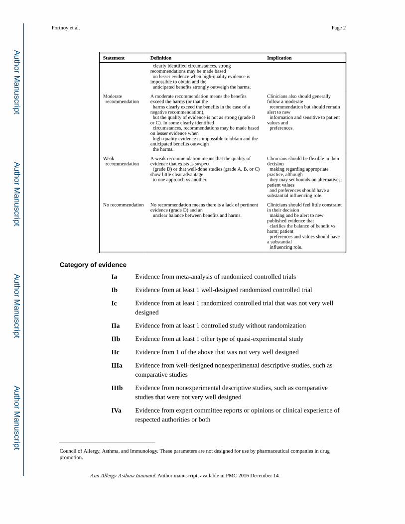
Classification of recommendations and evidence
There may be a separation between the strength of recommendation and the quality of
evidence.
Recommendation rating scale
Statement Definition Implication
Strong recommendation
A strong recommendation means the benefits of the recommended approach clearly exceed the harms (or that the harms clearly exceed the benefits in the case of a strong negative recommendation) and that the quality of the supporting evidence is excellent (grade A or B). In some
Clinicians should follow a strong recommendation unless a clear and compelling rationale for an alternative approach is present.
*The findings and conclusions in this article are those of the authors and do not necessarily represent the official position of the Centers for Disease Control and Prevention.
Members of the Joint Taskforce on Practice Parameters: David Bernstein, MD; Joann Blessing-Moore, MD; David Khan, MD; David Lang, MD; Richard Nicklas, MD; John Oppenheimer, MD; Jay Portnoy, MD; Christopher Randolph, MD; Diane Schuller, MD; Sheldon Spector, MD; Stephen A. Tilles, MD; Dana Wallace, MDReprints: Joint Council of Allergy, Asthma, and Immunology, 50 N Brockway Street, #3-3, Palatine, IL 60067.Practice Parameter Workgroup: James Sublett, MD, co-chair; Kevin Kennedy, MPH, co-chair; Charles Barnes, PhD; Ginger Chew, ScD*; Carl Grimes, CIEC; Désirée Larenas-Linnemann, MD; Jeffrey D. Miller, MD; J. David Miller, PhD; Wanda Phipatanakul, MD, MS; P. Brock Williams, PhD; Fares Zaitoun, MD
Disclaimer: The American Academy of Allergy, Asthma and Immunology (AAAAI) and the American College of Allergy, Asthma and Immunology (ACAAI) have jointly accepted responsibility for establishing “Environmental Assessment and Exposure Control of Dust Mites: A Practice Parameter.” This is a complete and comprehensive document at the current time. The medical environment is a changing environment, and not all recommendations will be appropriate for all patients. Because this document incorporated the efforts of many participants, no single individual, including those who served on the Joint Task force, is authorized to provide an official AAAAI or ACAAI interpretation of these practice parameters. Any request for information about or an interpretation of these practice parameters by the AAAAI or ACAAI should be directed to the Executive Offices of the AAAAI, the ACAAI, and the Joint
HHS Public AccessAuthor manuscriptAnn Allergy Asthma Immunol. Author manuscript; available in PMC 2016 December 14.
Published in final edited form as:Ann Allergy Asthma Immunol. 2013 December ; 111(6): 465–507. doi:10.1016/j.anai.2013.09.018.
Author M
anuscriptA
uthor Manuscript
Author M
anuscriptA
uthor Manuscript
Statement Definition Implication
clearly identified circumstances, strong recommendations may be made based on lesser evidence when high-quality evidence is impossible to obtain and the anticipated benefits strongly outweigh the harms.
Moderate recommendation
A moderate recommendation means the benefits exceed the harms (or that the harms clearly exceed the benefits in the case of a negative recommendation), but the quality of evidence is not as strong (grade B or C). In some clearly identified circumstances, recommendations may be made based on lesser evidence when high-quality evidence is impossible to obtain and the anticipated benefits outweigh the harms.
Clinicians also should generally follow a moderate recommendation but should remain alert to new information and sensitive to patient values and preferences.
Weak recommendation
A weak recommendation means that the quality of evidence that exists is suspect (grade D) or that well-done studies (grade A, B, or C) show little clear advantage to one approach vs another.
Clinicians should be flexible in their decision making regarding appropriate practice, although they may set bounds on alternatives; patient values and preferences should have a substantial influencing role.
No recommendation No recommendation means there is a lack of pertinent evidence (grade D) and an unclear balance between benefits and harms.
Clinicians should feel little constraint in their decision making and be alert to new published evidence that clarifies the balance of benefit vs harm; patient preferences and values should have a substantial influencing role.
Category of evidence
Ia Evidence from meta-analysis of randomized controlled trials
Ib Evidence from at least 1 well-designed randomized controlled trial
Ic Evidence from at least 1 randomized controlled trial that was not very well
designed
IIa Evidence from at least 1 controlled study without randomization
IIb Evidence from at least 1 other type of quasi-experimental study
IIc Evidence from 1 of the above that was not very well designed
IIIa Evidence from well-designed nonexperimental descriptive studies, such as
comparative studies
IIIb Evidence from nonexperimental descriptive studies, such as comparative
studies that were not very well designed
IVa Evidence from expert committee reports or opinions or clinical experience of
respected authorities or both
Council of Allergy, Asthma, and Immunology. These parameters are not designed for use by pharmaceutical companies in drug promotion.
Portnoy et al. Page 2
Ann Allergy Asthma Immunol. Author manuscript; available in PMC 2016 December 14.
Author M
anuscriptA
uthor Manuscript
Author M
anuscriptA
uthor Manuscript
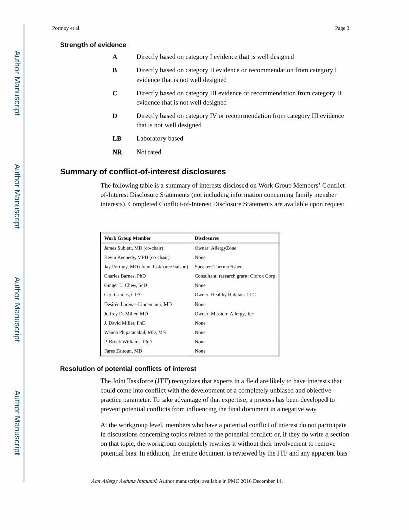
Strength of evidence
A Directly based on category I evidence that is well designed
B Directly based on category II evidence or recommendation from category I
evidence that is not well designed
C Directly based on category III evidence or recommendation from category II
evidence that is not well designed
D Directly based on category IV or recommendation from category III evidence
that is not well designed
LB Laboratory based
NR Not rated
Summary of conflict-of-interest disclosures
The following table is a summary of interests disclosed on Work Group Members’ Conflict-
of-Interest Disclosure Statements (not including information concerning family member
interests). Completed Conflict-of-Interest Disclosure Statements are available upon request.
Work Group Member Disclosures
James Sublett, MD (co-chair) Owner: AllergyZone
Kevin Kennedy, MPH (co-chair) None
Jay Portnoy, MD (Joint Taskforce liaison) Speaker: ThermoFisher
Charles Barnes, PhD Consultant, research grant: Clorox Corp
Ginger L. Chew, ScD None
Carl Grimes, CIEC Owner: Healthy Habitats LLC
Désirée Larenas-Linnemann, MD None
Jeffrey D. Miller, MD Owner: Mission: Allergy, Inc
J. David Miller, PhD None
Wanda Phipatanakul, MD, MS None
P. Brock Williams, PhD None
Fares Zaitoun, MD None
Resolution of potential conflicts of interest
The Joint Taskforce (JTF) recognizes that experts in a field are likely to have interests that
could come into conflict with the development of a completely unbiased and objective
practice parameter. To take advantage of that expertise, a process has been developed to
prevent potential conflicts from influencing the final document in a negative way.
At the workgroup level, members who have a potential conflict of interest do not participate
in discussions concerning topics related to the potential conflict; or, if they do write a section
on that topic, the workgroup completely rewrites it without their involvement to remove
potential bias. In addition, the entire document is reviewed by the JTF and any apparent bias
Portnoy et al. Page 3
Ann Allergy Asthma Immunol. Author manuscript; available in PMC 2016 December 14.
Author M
anuscriptA
uthor Manuscript
Author M
anuscriptA
uthor Manuscript
is removed at that level. The practice parameter is sent for review by invited reviewers and
by anyone with an interest in the topic by posting the document on the Web sites of the
American College of Allergy, Asthma, and Immunology (ACAAI) and the American
Academy of Allergy, Asthma, and Immunology (AAAAI).
For example, Jeffrey D. Miller, MD, owns a company that sells a product discussed in this
practice parameter. Dr Miller wrote a section on mattress encasings. This section was
rewritten by other members of the workgroup without his participation. He did not provide
subsequent input into that section.
How this practice parameter was developed
The JTF on Practice Parameters
The JTF on Practice Parameters is a 13-member taskforce consisting of 6 representatives of
the AAAAI, 6 of the ACAAI, and 1 of the Joint Council of Allergy and Immunology. This
taskforce oversees the development of practice parameters; selects the workgroup chair(s);
and reviews drafts of the parameters for accuracy, practicality, clarity, and broad utility of
the recommendations for clinical practice.
The Environment Practice Parameter Workgroup
The Environment Practice Parameter Workgroup was commissioned by the JTF to develop
practice parameters that address environmental assessment and remediation. The co-chairs
(James Sublett, MD, and Kevin Kennedy, MPH) invited workgroup members to participate
in the parameter development who are considered experts in the field of environmental
assessment and contaminant reduction. Workgroup members have been vetted for financial
conflicts of interest by the JTF and their conflicts of interest have been listed in this
document and are posted on the JTF Web site (http://www.allergyparameters.org). Where a
potential conflict of interest is present, the potentially conflicted workgroup member was
excluded from discussing relevant issues.
The charge to the workgroup was to use a systematic literature review, in conjunction with
consensus expert opinion and workgroup-identified supplementary documents, to develop
practice parameters that provide a comprehensive approach for identifying and managing
environmental exposures and their health effects based on the current state of the science.
Protocol for finding evidence for this practice parameter
A search of the medical literature was performed for different terms that were considered
relevant to this practice parameter. Literature searches were performed on PubMed and the
Cochrane Database of Systematic Reviews. Figure 1 shows the number of references from
1960 to the present for the terms dust mite, Dermatophagoides, pteronyssinus, or farinae (designated as mite in the figure). The search was narrowed by adding the terms allergy or
asthma, designated as Combo in the figure. This document includes references from 1970
through early 2013. All reference types were included in the results. References identified as
being relevant were searched for additional references and these also were searched for
citable references. In addition, members of the workgroup were asked for references that
Portnoy et al. Page 4
Ann Allergy Asthma Immunol. Author manuscript; available in PMC 2016 December 14.
Author M
anuscriptA
uthor Manuscript
Author M
anuscriptA
uthor Manuscript
were missed by this initial search. Although the ideal type of reference would consist of a
randomized, double-blinded, placebo-controlled study, the topic of this practice parameter is
represented by very few such studies. In consequence, it also was necessary to include
observational studies, basic laboratory reports, and regulatory requirements to develop a
document that addresses most of the issues discussed in this practice parameter.
Glossary
Condensation: The conversion of water vapor to liquid phase when cooled below its dew
point.
Dew point: The temperature below which water vapor in a volume of humid air at a constant
barometric pressure will condense into liquid water. Condensed water is called dew when it
forms on a solid surface.
Hygroscopic: A substance that is prone to absorbing moisture in damp environments, such
as salt or sugar.
Hygrometer: A device that is used to measure RH in an environment.
Relative humidity: The ratio of the partial pressure of water vapor in an air–water mixture to
the saturated vapor pressure of water at a prescribed temperature.
Summary statements
1. Advise patients to minimize exposure of susceptible children to dust mite
allergens to decrease their risk of developing mite-specific IgE. Because
intermittent exposure to mite allergens can lead to sensitization, primary
prevention might not be possible to achieve in regions where mite
exposure is prevalent. (Strength of recommendation: strong, A evidence)
2. Advise patients to minimize exposure of dust mite–sensitized children to
dust mite allergens to decrease their risk of developing asthma and
possibly rhinitis. (Strength of recommendation: strong, A evidence)
3. Advise dust mite–sensitized patients with asthma or rhinitis to minimize
exposure to dust mite allergens in addition to avoiding other relevant
allergens to which they are sensitized and avoiding irritants, to decrease
their risk of developing symptoms. (Strength of recommendation: strong,
B evidence for asthma; strength of recommendation: strong, C evidence
for rhinitis)
4. Advise patients to minimize exposure of dust mite–sensitized children
with atopic dermatitis to dust mite allergens, to decrease the symptoms of
atopic dermatitis. (Strength of recommendation: moderate, C evidence)
5. Although 5% to 15% of patients who are highly sensitized to dust mite
also are sensitized to crustaceans, the clinical significance of this is
unknown. For that reason, no recommendation can be made regarding the
Portnoy et al. Page 5
Ann Allergy Asthma Immunol. Author manuscript; available in PMC 2016 December 14.
Author M
anuscriptA
uthor Manuscript
Author M
anuscriptA
uthor Manuscript

need to advise crustacean-naive patients about their risk of ingestion.
(Strength of recommendation: none, D evidence)
6. Evaluate patients who complain of oral symptoms or symptoms consistent
with an IgE-mediated reaction after ingestion of grain flour for dust mite
sensitization regardless of whether they have wheat-specific IgE. (Strength
of recommendation: moderate, C evidence)
7. Test patients with suspected dust mite allergy for the presence of dust
mite–specific IgE using a skin prick test or in vitro test for specific IgE.
(Strength of recommendation: strong, B evidence)
8. Currently there is no evidence supporting routine measurement of specific
IgE to dust mite components, although such measurements may be
considered when necessary, such as for patients with potential Der p 10
(tropomyosin as found in cockroach and crustaceans) sensitivity. (Strength
of recommendation: weak, D evidence)
9. Encourage dust mite–allergic patients to obtain and use a hygrometer to
measure humidity in their home. (Strength of recommendation: strong, D
evidence)
10. Advise patients that relative humidity in the home should be kept at 35%
to 50% to decrease the growth of dust mites. (Strength of
recommendation: strong, B evidence)
11. Do not recommend the use of acaricides to eliminate mite populations
because of their limited efficacy at lowering allergen levels and concerns
about the use of chemical agents in the home. (Strength of
recommendation: moderate, B evidence)
12. Tell patients that the use of physical measures to kill mites, such as
heating, freezing, and desiccation, theoretically should be effective;
however, controlled trials have not been performed to demonstrate clinical
benefit when they are used. (Strength of recommendation: weak, D
evidence)
13. Advise patients that bedding should be washed weekly to decrease dust
mite numbers and mite allergen levels, and that high temperature is not
necessary. Home hot water should be kept below the temperature (120°F)
that causes a scalding risk to occupants. (Strength of recommendation:
strong, B evidence)
14. Suggest postintervention measurement of mite allergens in settled dust for
homes in which mite-sensitive people live if symptoms persist despite
reasonable efforts to decrease mite exposure. (Strength of
recommendation: weak, D evidence)
15. Measurement of airborne mite allergens offers no benefit over their
measurement in settled dust and therefore should not be recommended.
(Strength of recommendation: moderate, C evidence)
Portnoy et al. Page 6
Ann Allergy Asthma Immunol. Author manuscript; available in PMC 2016 December 14.
Author M
anuscriptA
uthor Manuscript
Author M
anuscriptA
uthor Manuscript

16. Recommend regular vacuuming using cleaners that have high-efficiency
particulate air (HEPA) filtration or with a central vacuum with adequate
filtration or that vents to the outside to decrease exposure to dust mite
allergen-containing particles. (Strength of recommendation: strong, B
evidence)
17. Recommend that patients should use mite allergen–proof mattress, box
spring, and pillow encasings to decrease exposure to mite allergens.
(Strength of recommendation: strong, B evidence)
18. Discourage members of families with an atopic background from sleeping
in bunk beds. If bunk sleeping is necessary, the sensitized person ideally
should sleep in the top bed and the top and bottom mattresses (and any
fabric-covered “bunky-boards”) should be enclosed in allergen-
impermeable encasings. (Strength of recommendation: moderate, B
evidence)
19. Do not recommend tannic acid for decreasing mite allergens in carpet dust
because it is only marginally effective. (Strength of recommendation:
moderate, C evidence)
20. HEPA filtration alone is of uncertain benefit for patients with mite allergy,
although it can decrease local exposure to airborne mite allergens and to
some irritants. If used, recommend that HEPA cleaners should be placed in
areas of mite contamination where air disturbance is likely to suspend
particles so that they are available for removal. (Strength of
recommendation: weak, C evidence)
21. Recommend a multifaceted approach for dust mite avoidance using a
combination of techniques that includes repetitive and sequential
interventions shown to decrease mite exposure, as described earlier, for
patients with dust mite allergy who are at risk of mite exposure. (Strength
of recommendation: moderate, A evidence)
22. Offer subcutaneous immunotherapy to dust mite–allergic patients with
rhinitis or mild to moderate asthma if they meet the general criteria for
receiving allergen immunotherapy (Strength of recommendation: strong, A
evidence for asthma; strength of recommendation: moderate, B evidence
for rhinitis)
23. Consider subcutaneous immunotherapy for dust mite–allergic patients
with atopic dermatitis if they meet the general criteria for receiving
allergen immunotherapy; however, possible exacerbation of the disease
during the initial phase of immunotherapy should be discussed with the
patient (Strength of recommendation: moderate, A evidence)
24. Patients receiving immunotherapy for dust mite ideally should receive a
dose that delivers approximately 7 µg of Der p 1 per injection or 500 to
2,000 AU per injection to obtain an optimal balance between efficacy and
safety. (Strength of recommendation: strong, A evidence)
Portnoy et al. Page 7
Ann Allergy Asthma Immunol. Author manuscript; available in PMC 2016 December 14.
Author M
anuscriptA
uthor Manuscript
Author M
anuscriptA
uthor Manuscript
25. US dust mite extracts can be mixed with pollen extracts, including grass
and animal dander extracts. Also at maintenance immunotherapy
concentration, US dust mite extracts can be mixed with fungal or
cockroach extracts when glycerin content is kept at 10%. (Strength of
recommendation: moderate, LB evidence)
26. Recommend 3 to 5 years of immunotherapy to obtain the maximum
benefit from immunotherapy for dust mite–induced asthma and rhinitis.
(Strength of recommendation: moderate, A evidence)
27. Certain protocols and dosages of sublingual immunotherapy have been
shown to be safe and effective for dust mite–allergic patients with rhinitis,
mild to moderate asthma, and/or atopic dermatitis; however, because there
currently is no Food and Drug Administration–approved product available
in the United States, its use should not be recommended until such a
product becomes available. (Strength of recommendation: moderate, A
evidence)
Executive summary
Dust mites are 8-legged arthropods that live in the house dust of homes located in regions
where they are prevalent. They have been recognized as the major source of allergens in
house dust since 1967. The most common species found in homes in temperate regions of
the United States are Dermatophagoides farinae and Dermatophagoides pteronyssinus. In
addition, others, such as Blomia tropicalis, can be found in homes in tropical and subtropical
regions.
Dust mites feed on organic materials, including skin scales, fungi, yeasts, and bacteria.
Because they are composed of approximately 75% water by weight, they maintain their
water balance through uptake of water vapor when RH is at least approximately 65%. They
are susceptible to water loss when humidity decreases below 65% and have decreased
survival and reproduction with an RH below 50%.
Mites produce and excrete numerous allergens into the environment, including cysteine
proteases such as Der p 1 and Der f 1, serine proteases including Der p 3, 6 and 9, and
proteases that can activate protease-activated receptor-2, which are proinflammatory in
humans through a non–IgE-dependent mechanism. Mites also produce glycosidases and
carbohydrate-binding proteins and muscle, cytoskeleton, and calcium-binding proteins.
There is cross-reactivity among various mite species and between mites and other related
families, such as crustaceans and cockroaches.
Tests for measurement of mite allergens from environmental samples are commercially
available. Such tests have included measurement of guanine as a proxy for fecal material and
of specific allergens using polyclonal and monoclonal antibodies. The most commonly used
assays are for the measurement of Der p 1 and Der f 1. Assays for Der p 2, Der f 2, and Blo t
5 also are available. Recently, a new set of international standards for dust mite allergens
that have been standardized using molecular techniques has led to a revision of the
Portnoy et al. Page 8
Ann Allergy Asthma Immunol. Author manuscript; available in PMC 2016 December 14.
Author M
anuscriptA
uthor Manuscript
Author M
anuscriptA
uthor Manuscript

concentrations reported in earlier studies of mite exposure. This may require a reassessment
of exposure thresholds associated with the development of sensitization, disease, and
morbidity.
Although homes in arid regions of the world are virtually free of dust mites, it is estimated
that 84% of US homes have detectable dust mite allergen and that half have concentrations
of at least 2 µg/g of dust. In Canada, the percentage of homes overall with house dust mite
allergen concentrations higher than 2 µg/g is somewhat smaller but similar in highly
populated areas in central and eastern Canada and in British Columbia. Factors leading to
increased mite concentrations include older, single-family homes with lower household
income. Increased population density, the presence of carpeting, and lack of air conditioning
also lead to increased dust mite exposure. The presence of moisture, cockroaches, and mold
also is associated with increased mite populations. Homes in warm damp regions of the
country, such as New Orleans and Florida, tend to have a more diverse population of dust
mites.
There is up to a 20-fold variation in mite exposure in regions that have significant seasonal
variation in temperature. Dust mite allergen levels tend to increase during the summer when
humidity is high and remain elevated through the winter before decreasing during the late
winter and spring.
Dust mite allergens are associated with particles that tend to have a large aerodynamic
behavior, with most settling within 15 minutes of disturbance. Very little mite allergen can
be found in the air of undisturbed rooms. Mite allergens are found in settled dust in
carpeting, bedding, and upholstered furniture but not on hard surfaces. Clothing also appears
to be an important source of mite allergen exposure, particularly if the clothing is washed
infrequently.
Primary prevention of IgE sensitization to mite allergens in susceptible children requires
strict, continuous avoidance of exposure for long periods. Prevention of sensitization has
been observed in arid regions where mites are absent; however, it is difficult to completely
eliminate mite exposure in homes located in mite-prevalent regions. Even when exposure in
a particular home is avoided, intermittent exposure to mite allergens when one travels to
other indoor environments often leads to sensitization. In consequence, most attempts at
primary prevention have been unsuccessful. Even so, there is a correlation between the
amount of exposure and the degree of sensitization. For that reason, exposure to mite
allergens should be minimized in susceptible children as much as is feasible.
The goal of secondary prevention is to decrease the risk of developing asthma and rhinitis in
already mite-sensitized children, usually during the first year of life. Several prospective
studies have found that mite avoidance lowers the risk of developing asthma in a dose-
dependent manner. Specific thresholds for exposure have been proposed in several of these
studies; however, such cutpoints are not used in this practice parameter because there does
not appear to be a level of exposure that does not offer at least some risk of developing
asthma or rhinitis. In addition, the relation between allergen exposure and disease
Portnoy et al. Page 9
Ann Allergy Asthma Immunol. Author manuscript; available in PMC 2016 December 14.
Author M
anuscriptA
uthor Manuscript
Author M
anuscriptA
uthor Manuscript
development appears to be complicated by other factors, including exposure to other
allergens and to irritants and pollutants.
The advisability of decreasing exposure to mite allergens in already sensitized individuals
who have asthma or rhinitis has been accepted conventional wisdom since mite allergens
were identified. Many controlled studies have shown the importance of allergen avoidance;
however, to be most effective, other relevant allergens and irritants should be avoided.
Avoidance of allergens can lead to decreased bronchial hyper-responsiveness, decreased
morbidity, and decreased need for medications. This appears to be true even for patients with
asthma who are not mite allergic because mite emanations have proinflammatory properties
that do not necessarily act through an IgE mechanism.
Atopic dermatitis can be triggered by exposure to dust mites in sensitized individuals. Live
mites have even been found on the skin of up to 35% of children with atopic dermatitis and
on their clothing and bedding. Interventions leading to decreased mite exposure have been
shown to lead to improvement in moderate to severe atopic dermatitis.
Because mites are members of the arthropod family, they contain tropomyosin (Der p 10),
which cross-reacts with other arthropods, including crustaceans and cockroaches. As many
as 5% to 15% of mite-sensitized individuals also are sensitized to crustaceans. A presumably
small, but currently unknown, percentage of dust mite–allergic individuals may be at risk of
a reaction after the ingestion of crustaceans. Because the extent of this risk is unknown, no
recommendation is made regarding the need to advise crustacean-naive patients about their
risk of ingestion.
Dust mites can contaminate grain flour. Systemic reactions have been reported in dust mite–
allergic individuals after the ingestion of grain flours, including beignets, wheat, pancakes,
polenta, okonomi-yaki, and grits. Symptoms have ranged from erythema and urticaria to
wheezing with dyspnea and even to anaphylaxis with loss of consciousness. Cooking
apparently is not sufficient to completely denature mite allergens. Therefore, it is important
to store such flour in sealed dust mite–impermeable bags, ideally in the freezer or
refrigerator. Individuals with symptoms consistent with an allergic reaction to grain flour
should be tested for sensitivity to dust mites.
The clinical evaluation of patients with suspected mite allergy begins with inquiries about a
history of atopy, including a history of increased symptoms upon disturbance of dust as
would occur with vacuuming and dusting. Because this type of history is neither sensitive
nor specific, individuals who live in regions where mites are prevalent should undergo tests
for mite-specific IgE, such as skin prick tests and/or in vitro tests. The performance
characteristics of these types of tests have recently been evaluated and are similar to each
other. These provide high sensitivity and specificity when appropriate criteria for a positive
test result are used.
Extracts used for tests of dust mite sensitization have been standardized in the United States
for total biologic potency; however, substantial differences are present between extracts in
terms of individual constituents. In consequence, extracts from different sources are not
considered interchangeable regardless of their total biologic activity. European extracts also
Portnoy et al. Page 10
Ann Allergy Asthma Immunol. Author manuscript; available in PMC 2016 December 14.
Author M
anuscriptA
uthor Manuscript
Author M
anuscriptA
uthor Manuscript
can differ substantially, in potency and constituents, from those produced in the United
States, making it difficult to compare the results of studies using the different extracts.
Although tests for specific IgE to mite components are available, such tests are not
recommended for routine use because their clinical value is not known.
Decreasing exposure to dust mites requires a multi-intervention approach that addresses
facilitative factors, sources, reservoirs, and pathways to occupants. Although the most
effective intervention is to live in a region where mites are not present or to duplicate such
conditions in a home if it is located in a region in which they are prevalent, such complete
control is often impractical. Ideally, indoor humidity must be kept low year-round regardless
of outdoor conditions. Mattresses, pillows, and bedding must be made free of mite allergen
emanations, and carpeting and other potential reservoirs should be removed completely.
The most important facilitative factor for mite growth is RH. Mites require RH higher than
65% to prevent water loss and to thrive. Once humidity decreases below 50%, mite
proliferation decreases and survival is decreased. Depending on how dry the environment is
kept, mites can survive for weeks before they die. If the humidity increases for as little as 1.5
hours per day, as could occur during cooking or bathing, the mites can survive. An elevated
RH for as briefly as 3 hours per day permits mites to produce eggs. To determine the RH,
patients are advised to obtain an inexpensive hygrometer, available from many outlets in the
United States.
The difficulty of maintaining low RH in regions where moisture is increased has been
demonstrated by numerous attempts to decrease mite exposure and improve health by using
indoor dehumidification. The series of studies performed in Manchester, England used
increasingly intense measures to remove moisture from homes, ranging from free-standing
dehumidifiers (ineffective) to whole-house dehumidification (effective at decreasing mite
allergens but not clinical symptoms). Studies in the United States that used even more
extensive dehumidification measures were able to demonstrate significantly decreased live
mites and mite allergen exposure. The lesson is that homes in which RH can be kept at 35%
to 50% continuously will have lower concentrations of mite allergen than in homes in which
RH is permitted to fluctuate.
Elimination of mites, the source of mite allergen, should lead to decreased exposure.
Because there is a strong relation between mite allergen concentrations in dust and the
number of live mites in an environment, it is not necessary to enumerate live mites to
determine the mite load of an environment. Techniques to kill mites have included chemical
acaricides; physical measures such as heating, freezing, and desiccation; and washing of
bedding and clothing.
Acaricides can kill mites under laboratory conditions. They also kill surface mites when
applied to carpeting and bedding; however, the duration of the benefit is short term so the
application must be repeated every 1 to 3 months. In addition, the decrease of mite allergen
exposure is modest at best and is not likely to be clinically useful. In addition, there is a
concern about the application of chemicals in the home and particularly on mattresses and
Portnoy et al. Page 11
Ann Allergy Asthma Immunol. Author manuscript; available in PMC 2016 December 14.
Author M
anuscriptA
uthor Manuscript
Author M
anuscriptA
uthor Manuscript
furniture where contact with occupants is likely to occur. For these reasons, the use of
acaricides is not recommended for killing mites.
Physical measures, such as freezing, heating, and desiccation, theoretically should be
effective; however, there are no clinical trials that have demonstrated benefit from such
interventions. Therefore, their use is considered optional. Regular washing of bedding and
clothing has been shown to effectively remove mite allergens and to kill mites. Most mites
that are killed in the washing process die by drowning. Although higher temperature kills
slightly more mites, this comes with an increased risk of scalding if home hot water is kept
at 130°F or higher. For this reason, it is recommended that home water temperatures be no
higher than120°F and that washing be performed at weekly intervals.
The most effective way to manage reservoirs of mite allergens is to remove them completely
from the environment. That means removing carpets, drapes, and upholstered furniture and
sealing mattresses, box springs, and pillows in mite-impermeable covers. Because many
home occupants are unlikely to comply with such measures, partial interventions may be an
appropriate beginning. If symptoms persist after dust mite decreasing interventions, it may
help to determine whether such persistence is due to failure of the intervention to decrease
exposure or to the presence of other exposures that have not been removed. For that reason,
it may help to collect a preintervention dust sample so that it can be compared with a sample
collected after the intervention. Many analytic laboratories can measure mite allergens to
determine whether the intervention is successful. Such measurements should be performed
on dust samples because they are more reliable than air samples and they provide the same
type of information.
Methods for removing mite allergens from reservoirs include regular vacuuming with a
high-efficiency vacuum and the use of mite-impermeable mattress, box spring, and pillow
encasings. Use of tannic acid as a mite allergen denaturant is not effective and therefore not
recommended. Regular (at least weekly) vacuuming is essential for preventing buildup of
mite allergens in homes with carpets. To be effective, a vacuum needs to capture particles
that carry mite allergens to prevent their dispersal. Although vacuuming does not remove all
live mites, mite allergens in the form of fecal particles can be removed. Over time, the
amount of exposure to mite allergens has been shown to decrease sufficiently for health
benefits to be possible. Bedding and furniture also can be vacuumed to decrease mite
allergen exposure from those reservoirs.
Mattresses, box springs, and pillows are major sources of mites and mite allergens. The most
effective way to prevent mite colonization is to encase a mattress, box spring, or pillow in a
mite allergen–impermeable encasing. Existing mattresses, box springs, and pillows can be
kept if they are encased in allergen-impermeable covers to entrap already-present mites and
mite allergens. There are several different types of mattress encasings, including woven
microfiber encasings, which prevent mite allergen escape yet allow air and water vapor to
pass freely through the fabric. Woven microfiber fabrics with a mean pore size smaller than
10 µ can effectively block passage of Der p 1, whereas a mean pore size smaller than 6 µ is
necessary to block cat allergen Fel d 1. Nonwoven encasings are not recommended because
they trap mite allergens and are not washable, leading to allergen accumulation. Although
Portnoy et al. Page 12
Ann Allergy Asthma Immunol. Author manuscript; available in PMC 2016 December 14.
Author M
anuscriptA
uthor Manuscript
Author M
anuscriptA
uthor Manuscript

encasings effectively contain mite allergens, in many cases mite covers alone are unlikely to
achieve a clinical benefit unless they are used as part of a more comprehensive multifaceted
avoidance plan.
HEPA filtration is of uncertain benefit, although it can decrease local exposure to airborne
mite allergens and to some irritants. If used, HEPA cleaners should be placed in areas of
mite contamination where air disturbance is likely to suspend particles so that they are
available for removal. Laminar flow cleaners that remove particles from the breathing space
of beds have been demonstrated to be of some benefit, although they may not be practical
for routine use.
Overall, there is evidence that a multifaceted approach using a combination of techniques for
dust mite avoidance that includes repetitive and sequential interventions can decrease mite
exposure. Such interventions should be recommended for patients with dust mite allergy
who are at risk of mite exposure. Therefore, combinations of interventions for mite
avoidance should address facilitative factors, sources, and reservoirs. The most effective
combination includes maintaining humidity at 35% to 50%, regular washing of bedding to
remove mites and mite allergens, regular vacuuming with a high-efficiency vacuum, use of
mattress and pillow encasings, and HEPA filtration if deemed necessary.
Allergen immunotherapy (subcutaneous and sublingual) with dust mite extract has been
shown to be effective for treating asthma and rhinitis in mite-allergic individuals. In
addition, there is some evidence that patients with atopic dermatitis may benefit from dust
mite immunotherapy. To be useful for SCIT, an effective dose of mite allergen needs to be
given (7 µg of Der p 1 per dose for European extracts and 500–2,000 AU per dose for US
extracts). For SLIT, 4,200 AU containing approximately 70 µg of Der f 1 given daily has
been shown to be effective. The frequency of administration in studies showing efficacy
have ranged from weekly to monthly once maintenance is reached for SCIT and daily to 3
times per week for SLIT once a maintenance dose is achieved. There is no evidence to
support giving lower doses more frequently or higher doses less frequently to obtain similar
efficacy. Dust mite extracts are compatible with pollen and animal dander extracts and can
be mixed with fungal and cockroach extracts provided they are kept in glycerin at a
concentration of at least 10%. In general, 3 to 5 years of immunotherapy is sufficient to
obtain maximum benefit from immunotherapy for dust mite–induced asthma and rhinitis.
Overview of dust mites
Dust mite taxonomy
Mites and ticks are 8-legged arthropods called arachnids that belong to the taxonomic order
of Acari, which comprises tens of thousands of species grouped under several suborders,
families, and genera. Most of these mites live freely in various biologic habitats, are very
diverse in form and behavior, and function in the biologic recycling process as scavengers or
saprophagous mites. Other mite species are plant parasites and major pests for crops, and
still others can transmit diseases to humans (chiggers, ticks). However, relatively few species
of mites, which belong to a particular taxon (Astigmata), have clearly been shown to
produce allergens that induce IgE-mediated allergic reactions in susceptible individuals.
Portnoy et al. Page 13
Ann Allergy Asthma Immunol. Author manuscript; available in PMC 2016 December 14.
Author M
anuscriptA
uthor Manuscript
Author M
anuscriptA
uthor Manuscript
House dust mites were recognized as the major source of the allergens in house dust in 1967
when Voorhorst et al1 in the Netherlands and Miyamoto et al2 in Japan reported the
identification of D pteronyssinus as a major dust mite in house dust. House dust mites
belong to the phylum Arthropoda (ie, animals with external skeletons and jointed limbs),
subphylum Chelicerata, class Arachnida, order Acari, and suborder Astigmata (Fig 2).3,4
The term house dust mites has traditionally been used for members of the Pyroglyphidae
family that live permanently and almost exclusively in house dust, although dust mites from
other families have been found in house dust. The term domestic mites includes house dust
mites of the Pyroglyphidae family and other Astigmatid mites traditionally referred to as
storage mites or stored-products mites, which belong to different taxonomic families
(Acaridae, Glycyphagidae, Echymyopodidae, and Chortoglyphidae; Fig 3).3 Several species
of storage mites are a potent source of allergens and can be found in house dust. Some other
mite species of different taxonomic classes, which may be found in house dust, are predatory
mites (Cheyletidae), parasitic mites of plants such as spider mites (Tetranychidae), and
glistening mites (Tarsonemidae). Although their clinical importance is minor, several species
of mites besides those found in house dust can induce allergic reactions, such as the citrus
red mite (pest in apple orchards)5 and the mite Hemisarcoptes cooremani (pests in orchards
and gardens).6
House dust mites are named according to a scientific system consisting of the genus name,
such as Dermatophagoides, and a species name, such as farinae. This binomial name is
always written in italics. The family Pyroglyphidae is composed of about 16 genera and 46
species,7,8 and at least 13 species have been found in house dust and recorded from locations
throughout the world and across all continents.3 However, 3 species, D pteronyssinus, D farinae, and Euroglyphus maynei, are most common, comprising 80% to 90% of house dust
mite fauna.9 Blomia tropicalis, a storage dust mite, is a common dust mite found in homes in
tropical and subtropical regions.
Biology and physiology
Adult house dust mites have an oval shape and creamy to translucent white bodies that
measure 0.2 to 0.4 mm and are barely visible to the naked eye.10 Although electron
microscopic images are widely available, such images may give the false impression that
dust mites are so small as to require an electron microscope for visualization; in reality, they
are easily seen under low power microscopy at ×20 to ×80 magnification (Fig 4). Dust mites
feed mainly on organic detritus that accumulates in house dust, including desquamated
human or pet skin scales, which are colonized by fungi, yeasts, and bacteria.11,12
There are several aspects of the biology and physiology of dust mites that are relevant to
allergy, including food and water requirements, heat requirements, habitat, size, life cycle,
and gastrointestinal allergen production. Water balance is critical to house dust mite survival.
House dust mites are about 75% water by weight and do not drink or urinate. They obtain
and maintain their water balance through uptake of water vapor when the RH is at least
approximately 65%, and they experience water loss by evaporation when the surrounding
RH decreases below approximately 55%.13 The critical lowest humidity is temperature
dependent and ranges from 55% to 75% RH over the temperature range of 15°C to
Portnoy et al. Page 14
Ann Allergy Asthma Immunol. Author manuscript; available in PMC 2016 December 14.
Author M
anuscriptA
uthor Manuscript
Author M
anuscriptA
uthor Manuscript
35°C20,21 with D pteronyssinus and D farinae appearing to thrive best at 75% to 80% RH
and 25°C to 30°C (77–86°F).10 Although lacking eyes, dust mites are light sensitive and
photophobic, and thus live deep within soft substrates, such as pillows, mattresses, and
carpets, where moisture is retained and humidity fluctuations are minimized. Because they
move away from light, dust mites do not live on hard exposed surfaces, although some
temporarily migrate to the top of carpeting during the dark of night. It is not uncommon to
find thousands of mites in a single gram of house dust.14
Dust mites are equipped with many biophysical mechanisms, including timely excretion of
feces, which allow them to survive prolonged periods of drought.15 They maintain internal
water homeostasis by specialized organs, the supracoxal glands, located at the base of the
first pair of legs. These glands concentrate sodium and potassium chloride, which act to
osmotically absorb water vapor from the environment. However, these glands can maintain a
positive water balance only at an ambient RH of at least 50%. This dependence on
environmental factors of temperature and RH is reflected in seasonal fluctuations in dust
mite numbers and allergen levels in different parts of the world.16
Dust mites have a well-developed digestive tract, including an elaborate system of mouth
parts (chelicerae and pedipalps), salivary glands, and a duct consisting of esophagus, midgut
(food absorption), hindgut (water resorption), and slit-formed anus.17 When a mite has
eaten, cells from its gut containing digestive enzymes form a peritrophic membrane that
adheres to the surface of the ingested food. In the posterior midgut, the peritrophic
membrane-wrapped food balls coalesce to be excreted later as fecal pellets.18 Fecal pellets
are produced by house dust mites at a rate of 20 pellets per day, vary in size from 20 to 40
µm, and are considered a rich source of digestive enzyme-derived allergens.19,20 The density
of fecal pellets allows them to become airborne and easily inhaled when the substrate in
which they were deposited is disturbed (eg, by making a bed, walking on a carpet, or moving
on a pillow), followed by settling within 20 to 30 minutes.
In contrast to the gastrointestinal system, house dust mites have no organized respiratory
structure or external openings for ventilation. They are aerobic and exchange oxygen and
carbon dioxide by passive diffusion across their cuticle.
Reproduction
House dust mites reproduce sexually and adult male and female mites have complete and
elaborate sexual organs, which are often helpful as identification characteristics.21 Because
temperatures and RH are not uniform in the various areas where house dust mites are found,
the rate of reproduction, development, and mite population growth vary.8 For example, mite
populations in carpets over slab floors that remain cool develop more slowly than
populations inhabiting mattresses or sofas.
The life cycle of the most commonly studied house dust mites, D farinae, D pteronyssinus,
and E maynei, consists of 5 stages: an egg, a 6-legged larva, two 8-legged nymphal stages
(protonymph and tritonymph), and male or female adult.22 Six-legged larvae hatch from the
eggs and remain active for some time before shedding their integument and becoming 8-
legged resting protonymphs. The protonymphs in turn shed their integument and become
Portnoy et al. Page 15
Ann Allergy Asthma Immunol. Author manuscript; available in PMC 2016 December 14.
Author M
anuscriptA
uthor Manuscript
Author M
anuscriptA
uthor Manuscript

larger active tritonymphs. The tritonymphs undergo another shedding of skin, developing
into active adult mites.23 The shed integuments and exoskeletons are an important secondary
source of mite allergens and immunomodulators, including chitin. The duration of this
developmental stage varies from 19 to 33 days at favorable conditions of temperatures from
22°C to 32°C and approximately 75% humidity.22 After reaching the adult stage, dust mites
can live for about 4 to 6 weeks and mate 1 to 3 times, with the female mite laying about 1 to
2 eggs per day, for a total of 50 to 80 eggs in its lifetime.24,25
In total, the average life cycle of a house dust mite, starting from the hatched egg stage,
ranges from approximately 60 to 120 days, depending on ambient RH and temperature.22,26
Clinical assessment
Algorithm (Fig 5)
Annotations
1. Patient with possible dust mite-related illness: Patients generally present for evaluation
if they have an illness such as eczema, rhinitis, or asthma. Rhinitis and asthma are
respiratory illnesses that can be exacerbated by inhalation of dust mite allergen; eczema can
be exacerbated by skin contact, given sensitization and sensitivity. Because exposure to dust
mites also can trigger symptoms in nonsensitized individuals, sensitization per se is not the
only criterion for possible morbidity from exposure.
This algorithm can be used to evaluate a patient’s risk for morbidity from dust mite exposure
regardless of his or her sensitization status. The purpose of this first algorithm is to
determine which patients would most likely benefit from a more complete evaluation of their
home environment for possible dust mite exposure. As such, this section should be used as a
screening procedure. The 2 factors that determine whether further dust mite assessment is
indicated include patient factors and environmental factors. The next 2 questions address
each of these issues in turn.
2. Increased risk for dust mite morbidity?: Patients who are not sensitized to dust mites
but who are at increased risk to become sensitized ideally should be identified before the
sensitization takes place and therefore deserve a greater degree of evaluation for dust mite
exposure. Patients are at increased risk of dust mite sensitization if they have an elevated
total IgE; if they are sensitized to other allergens (increased specific IgE or positive skin test
reaction); if they have asthma, eczema, or allergic rhinitis; or if there is a strong family
history of atopy. The latter criteria are particularly important in very young children because
they might not yet have developed evidence of atopy.
There are some basic questions that can be used to assess the likelihood that a patient will
experience morbidity from dust mite exposure:
• Does the patient have eczema, asthma, or rhinitis?
• Is there a positive family history for atopy?
Portnoy et al. Page 16
Ann Allergy Asthma Immunol. Author manuscript; available in PMC 2016 December 14.
Author M
anuscriptA
uthor Manuscript
Author M
anuscriptA
uthor Manuscript

• Does the patient have atopy? This could be manifested as an elevated total
IgE or the presence of specific IgE antibodies.
3. Increased risk for dust mite exposure?: This question can be used to determine whether
a patient is at increased risk of exposure to elevated levels of dust mite allergens. Dust mites
tend to be found in locations where there is warmth and moisture. They can survive in cold,
dry climates by occupying human residences that are artificially heated. Home and location
factors associated with increased dust mite exposure are discussed in the following sections.
Facilitative factors—For dust mites, the most important facilitative factor is moisture, so
questions to ask relate to this factor. In Appendix A, there is a detailed discussion of
moisture and humidity. In Appendix B, there is a 3-step guide for clinicians use to assess
whether their patients might be at increased risk for dust mite exposure. In summary, the
following questions address facilitative factors.
• Does the patient live in a location with a warm, humid or damp climate?
The Köppen climate classification (http://webmap.ornl.gov/wcsdown/
wcsdown.jsp?dg_id=10012_1) has maps with climate zones related to
temperature and humidity for the United States that can be used to
determine the answer to this question for a particular location.
Microclimate also is important; general climatic information must be
interpreted in the context of the patient’s residence and work locations.
• What is the RH in the patient’s home? In general, an RH greater than 50%
facilitates mite growth, whereas air that is too dry (<25% RH) can serve as
a respiratory irritant. Patients should be encouraged to obtain a hygrometer
to measure indoor RH and to make indoor climate adjustments as
necessary to keep the RH at 35% to 50%. Because dust mite habitats, such
as mattresses, upholstered furniture, and settled dust, are sensitive to
changes in ambient RH, this should be sufficient to control mite
populations.
• Does the patient’s residence have microenvironments in which dust mites
might thrive? Some building materials are more likely to absorb water than
others, so it is important to understand what materials are in a patient’s
home and the mean humidity in house. Absorption of moisture is faster
than desorption, so materials that bind water, such as house dust, tend to
buffer the humidity. For that reason, moisture control must be consistent.
Reservoirs—
• How old is the building in which the patient lives? Older buildings have
had more time to become contaminated by dust mites and their allergens.
Regardless of a building’s age, low levels of humidity will lead to
decreased mite contamination over time.
• How old are the pieces of upholstered furniture, mattresses, and carpeting?
Older furniture and mattresses are likely to have larger numbers of dust
Portnoy et al. Page 17
Ann Allergy Asthma Immunol. Author manuscript; available in PMC 2016 December 14.
Author M
anuscriptA
uthor Manuscript
Author M
anuscriptA
uthor Manuscript
mite and to have accumulated increased concentrations of dust mite
allergen over time. If the furniture is imported from a different location
where mite growth is supported, there could be mite allergen
contamination although the current environment does not support mite
growth.
• How frequently is bedding changed and how is it washed? What type of
bedding does the patient sleep on? Bedding should be washed weekly to
remove mite allergens and to decrease the mite population.
• If there is carpeting, how frequently is the home vacuumed? Ideally the
carpeting should be vacuumed at least weekly or more frequently
depending on traffic and use. Does the carpeting sit on a concrete slab that
would tend to provide moisture through intrusion or condensation? The
carpet backing can become damp, promoting mite growth, even if the pile
remains dry.
Depending on the answers to these questions, it may be of value to offer the option of
surveying a patient’s home with a simple or advanced screening method. This could involve
collecting a sample of dust from the home environment and testing the sample for dust mite
allergen. Mite allergen measurement can be performed using dust from a used vacuum bag;
however, dust collection by a trained technician is ideal and can help to pinpoint the main
sources of exposure within a home. If a used vacuum bag from the resident’s home is used,
one should realize that it is the accumulation of many different locations within the home
and represents a period that may or may not reflect current exposure in that home. The 2
dust mite allergens for which standardized measurements are available are Der p 1 and Der f
1. A rapid test is also available for use in the home to quickly identify dust mite products.
4. Done: Because the patient is not at increased risk for dust mite morbidity or exposure, it
is not necessary to perform additional procedures. However, exposure and associated risk
factors can change over time. Periodic re-evaluation of the risk for dust mite allergen
exposure should occur depending on the clinical history.
5. Provide mitigation education and consider home assessment for dust mite analysis and decreasing exposure: Based on the environmental history gathered in answer to
questions 1, 2, and 3, the clinician can offer specific education regarding the mitigation of
facilitative factors and abatement of reservoirs. These activities often can be carried out by
patients and/or their families and result in a decrease of live mites and mite allergen. There
are often other instances when homes with elevated dust mite allergen levels in settled dust
should be followed up with a more complete assessment by a professional service. Based on
the physician’s understanding of the patient’s motivation and ability, the physician should
recommend appropriate steps that are likely to lead to decreased exposure. Because this is
often an iterative process, a combination of these 2 interventions may be appropriate for
long-term decrease. When a professional is recommended, suggestions for selecting such a
service are provided in Appendix A of the Rodent Practice Parameter.27
Portnoy et al. Page 18
Ann Allergy Asthma Immunol. Author manuscript; available in PMC 2016 December 14.
Author M
anuscriptA
uthor Manuscript
Author M
anuscriptA
uthor Manuscript
Environmental assessment, mitigation, and abatement
Algorithm (Fig 6)
Annotations
1. Home with suspected dust mites: Mite assessment and decreasing exposure are
indicated when a building’s occupants are at increased risk of morbidity from mite exposure
(atopy, mite-specific IgE, family history) and the home has an increased likelihood of mite
contamination (increased humidity/moisture, older building, upholstered furniture,
carpeting, etc). If dust mite allergens have been measured in dust, increased concentrations
of Der p 1 or Der f 1 also indicate a need for an environmental intervention.
2. Are facilitative factors for dust mite present?: Dust mites require moisture, warmth,
and a source of food to survive. There can be seasonal variations in these factors that should
be taken into account. For example, summer is warm and damp in some locations so mite
populations expand, whereas winter is cold and dry so populations tend to decrease. If
facilitative factors are present, then mites are likely to thrive and allergen removal alone is
unlikely to succeed.
The tools necessary to identify facilitative factors are listed below.
1. Hygrometer and thermometer to determine RH and dew point. This can be
used by the occupant to determine whether decreasing humidity is needed.
2. Moisture meter to measure available water within a material (usually
performed by a professional when excessive moisture is suspected). This
can be used to identify sources of moisture when they are not apparent.
3. Mitigation: remove facilitative factors: Once excessive moisture is identified, it is
important to remove it. Condensation can be decreased by keeping the RH below 50% using
a dehumidifier and/or air conditioning. If used, the dehumidifier needs to be emptied
regularly or set to drain continuously, and it should be located in areas where dampness is
likely to occur. Air conditioners need to run long enough to remove sufficient moisture from
the air to decrease RH. If the air cools too quickly, as could occur with an oversized unit,
adequate dehumidification might not be achieved.
Sources of intrusion or leakage should be identified, repaired, and/or sealed. Surfaces on
which condensation can occur should be appropriately insulated and sealed with special
attention to proper placement of vapor barriers. Cold water pipes may need to be insulated to
prevent condensation.
4. Are live mites present in the home?: Once facilitative factors have been removed, or if
they are not present, it is unlikely that live mites can continue to live in the house and it may
not be necessary to test for their presence. Homes with a history of RH above 50% or
microenvironments in which mites can grow are likely to have live mites present. For mites
to survive, the RH in a house needs only to exceed 50% for 1 hour per day, and 2 to 3 hours
per day is necessary for mites to reproduce.
Portnoy et al. Page 19
Ann Allergy Asthma Immunol. Author manuscript; available in PMC 2016 December 14.
Author M
anuscriptA
uthor Manuscript
Author M
anuscriptA
uthor Manuscript
Although moisture is a limiting facilitative factor, food generally is plentiful and does not
limit mite survival or growth. Mites also prefer cool, dark locations as is found in a box
spring or carpet pad, although it is not usually feasible to remove these factors short of
removing carpeting completely. Although it is possible to identify live mites in dust samples
microscopically, it is easier to simply assume that mites are present if facilitative factors for
their growth are present.
5. Source control: get rid of the mites: The presence of live, allergen-producing dust mites
continuously replenishes mite allergens in the environment. Ideally, mite populations should
be eliminated or at least significantly decreased or else it is unlikely that exposure can be
decreased sufficiently to improve health. The most effective method to eliminate mites is to
decrease their access to moisture by maintaining the indoor RH below 50% for sustained
periods. Mattress, box spring, and pillow encasings also may be used to separate live mites
and their allergens from building occupants. Owing to their lack of effectiveness, the use of
acaricides is not recommended.
6. Are dust mite allergen reservoirs present?: Dust mite reservoirs include carpeting,
upholstered furniture, mattresses, bedding, and settled dust. The presence of these reservoirs
generally is obvious by history and visual inspection. The presence of mite allergens can be
confirmed by measuring Der p 1 and/or Der f 1 in dust samples. Because the 2 species are
not always correlated, measurement of allergens from these species (or a cross-reactive
allergen) is ideal if allergen measurement is performed.
7. Abatement: remove or clean reservoirs?: Abatement, or removal and cleaning, of dust
mite reservoirs is necessary because mite allergens are highly stable for long periods.28 This
means that even if the mites are killed, occupants will continue to have exposure to mite
allergens and other immunomodulators such as chitin from their exoskeletons. The most
effective way to remove reservoirs is to eliminate carpeting, furniture, and mattresses from
the home. In many cases, this is impractical. For that reason, regular vacuuming with a
HEPA or cyclonic vacuum is necessary because it removes dust mite–containing particles
from carpeting and furniture. Mattress, box spring, and pillow encasings can serve as a
barrier between sleepers and mite allergens contained in those substrates. Bedding should be
washed regularly as discussed in the washing section of this parameter. Owing to the
intermittent nature of airborne mite exposure, HEPA filters have not been shown to be
effective for decreasing mite allergen exposure, although they may be useful under specific
circumstances.
8. Intervention is done: Once facilitative factors are removed, the dust mites are killed, and
reservoirs are cleaned, the intervention is completed. It is still desirable to maintain an
ongoing program of humidity control, mite allergen containment, and reservoir cleaning, but
otherwise the occupant is no longer at increased risk of morbidity from dust mite exposure.
Portnoy et al. Page 20
Ann Allergy Asthma Immunol. Author manuscript; available in PMC 2016 December 14.
Author M
anuscriptA
uthor Manuscript
Author M
anuscriptA
uthor Manuscript

Functional overview of mite allergens
General considerations
Many mite allergens from D pteronyssinus and D farinae show significant homology. A
discussion of such allergens with a brief description of their known properties is presented in
Appendix C. A list of mite allergens from various mites is presented in Table 1. The
functional effects ascribed to most of these proteins involve, in one way or another, the
activation of innate immune mechanisms, many of which seem to favor T-helper cell type 2
(TH2) responses. Although any of these proteins might induce IgE responses, collectively
they seem to complement each other in this respect through bystander immunologic effects.
Allergens from mites include commonly encountered functions of allergens from a wide
variety of sources, such as proteases (Der p 1, 3, 6, 9, 20), lipid-binding proteins (Der p 2, 7,
13, 14), contractile proteins (Der p 10, 11, 16, 17, 24), glycosidases and carbohydrate-
binding proteins (Der f 4, 12, 15, 18, 23), and glutathione S-transferase (Der p 8). Other
functions of mite allergens include heat shock protein-70. In addition, many allergens,
including Der p 5, 19, 21, and 22, are unidentified as to function.
Proteases
Der p 1 and Der f 1 are glycoproteins of the papain family with cysteine protease activity
similar to that of some plant allergens (kiwi Act d 1, actinidin, pineapple Ana c 2, bromelain,
fig ficin, papaya Car p 1, papain, soybean Gly m Bd30K, and mammalian enzymes such as
cathepsins H and B). These mite allergens originate from the intestinal tract of the mite. Der
p 1 can cleave the CD23 IgE receptor from human B-cell membranes, thus ablating the
feedback inhibitory mechanism that normally limits IgE synthesis.29 Der p 1 also can cleave
the CD25 subunit of the T-cell interluekin-2 receptor, which can promote TH2 responses. In
addition, Der p 1, 3, 6, 9, and 20 can proteolytically degrade tight junctions in lung
epithelium and cause the release of proinflammatory cytokines from bronchial epithelial
cells, mast cells, eosinophils, and basophils. These synergistic effects can promote IgE
synthesis and have direct inflammatory effects on lung epithelium, which in turn could
explain why mite allergens are closely associated with asthma. More than 50% of allergic
patients and up to 80% of children with asthma are sensitized to Der p 1. Der p 1 appears to
be sufficient to diagnose up to 97% of dust mite–allergic patients.30
Der p 3, 6, and 9 are serine proteases. Der p 3 is a trypsin-like enzyme and is a major
constituent of mite feces. It is quite similar to the cockroach Bla g 10. Der p 6 is
chymotryptic and with Der p 9 exhibits collagenase activity. Trypsin-like enzymes also are
found in insect venoms. Trypsin can trigger protease-activated receptor-2, whose cleavage
results in the initiation of multiple G-protein–coupled signaling cascades. These cascades
result in many events that promote TH2 skewing and inflammation, such as the production of
thymic stromal lymphopoietin and interleukins 4, 5,13, 21, 25, and 31.
House dust mites have proteases that can activate protease-activated receptor-2. Exposure to
dust mites has been shown to increase the secretion rate and number of responding glands in
patients with allergic rhinitis even if they are not mite sensitive, suggesting a nonspecific
proinflammatory mechanism that is not dependent on specific IgE.31
Portnoy et al. Page 21
Ann Allergy Asthma Immunol. Author manuscript; available in PMC 2016 December 14.
Author M
anuscriptA
uthor Manuscript
Author M
anuscriptA
uthor Manuscript

Glycosidases and carbohydrate-binding proteins
Der p 4, 15, 18, and 23 are proteins that interact with carbohydrate moieties. Der p 4 is an α-
amylase and Der p 15 and 18 are chitinases. Der p 20 is an arginine kinase that also binds
chitin. These proteins are widely distributed throughout nature and for unknown reasons can
be potent allergens from many different sources. Alpha-amylases from the storage mite
(Acarus siro) and fungal amylases in flours and some grasses are responsible for some types
of occupational asthma. The amylase activity of dust samples correlates with counts of live
mites and with concentrations of Der p 1. Der p 4 and Eur m 4 sequences are 90% identical
and 50% identical, respectively, to other insect and mammalian α-amylases.32 Der p 15 and
18 are chitinases related to pathogen resistance (fungal, worm, and other arthropods). The
chitinases seem to be very important in dog allergic reactions but somewhat less so for
humans. From a functional point of view, sensitization to chitinases from other sources has
been identified as being responsible for the latex fruit syndrome. In addition, chitin
fragments are immunomodulatory and the chitinases may facilitate their production from
plants, fungi, and insects, helping to induce TH2 responses.
Muscle, cytoskeleton, and Ca2+-binding proteins
Der f 10, 11, 16, 17, and 24 are tropomyosin, paramyosin, gelsolin, Ca2+-binding protein,
and troponin C, respectively. These proteins are involved in the structural aspects of cells, in
addition to cytoskeleton organization, membrane trafficking, and lipid signaling, such as the
regulation of diacylglycerol and phosphatidylinositol 4,5-bisphosphate signaling pathways.
Der f 10 tropomyosin is a highly conserved protein throughout insects, shell fish, and
parasites and as such represents a cross-reactive and possible cross-sensitizing allergen. Two
studies have indicated that 5.6% to 15.2% of dust mite–allergic patients have IgE to Der p
10.33,34
Lipid-binding proteins
Der p 2, 7, 13, and 14 are lipid-transfer or lipid-carrying proteins. These also are commonly
found as allergens from different sources, including plants. Der p 2 is closely related to
lymphocyte antigen 96 (MD-2 protein) that allows toll-like receptor 4 to bind to endotoxin.
Thus, Der p 2 seems to be related to the activation of innate immunologic mechanisms,
many of which seem to favor TH2 immunologic responses. Lipid-binding proteins
participate in signaling pathways that affect the distribution and activity of lipid-
metabolizing enzymes and protein kinases that regulate the activity of many of these
enzymes. Lipid-transfer proteins from different sources seem to be potent allergens.
Others
Cross-reactivity has been observed between ascaris and dust mites species B tropicalis, D pteronyssinus, and D farinae. Among allergic subjects, 70% exhibited ascaris-specific IgE,
whereas 20% to 28% of ascaris-allergic subjects showed dust mite–specific positive IgE.
Ascaris antigens inhibited up to 92% of dust mite–specific IgE in mite allergic subjects and
up to 54% of ascaris-specific IgE was inhibited by dust mite allergens.35
In 1 study, commercial dust mite extracts were analyzed for endotoxin levels, protease and
chitinase activities, and effects on transepithelial resistance, junctional proteins, and
Portnoy et al. Page 22
Ann Allergy Asthma Immunol. Author manuscript; available in PMC 2016 December 14.
Author M
anuscriptA
uthor Manuscript
Author M
anuscriptA
uthor Manuscript
proinflammatory cytokine release in human bronchial cells. These extracts varied
extensively in serine protease activity, including the ability to induce dust mite–specific IgE,
goblet cell hyperplasia, eosinophilic inflammation, and airway hyper-reactivity independent
of protease activity.36
Measurement of dust mite allergens
Efforts to measure the number of mites and the allergenic protein products they produce in
house dust have been ongoing since dust mites were recognized as major allergenic
species.2,37 Early mite enumeration involved microscopic examination of house dust and
vacuum samples of bedding, including counts of observable mite bodies.38 Because house
dust mites excrete guanine, the measurement of guanine in house dust has been used to
estimate mite presence.39,40 The first commercial test detected guanine in the dust based on
the assumption that most guanine in dust comes from mite fecal pellets. The test used a
dipstick that was inserted into a suspension of dust, leading to a color change that could be
compared with an included color chart card to provide an interpretation. This test correlated
well with dust mite allergen, although this test is no longer available.41
The earliest reported immunologically based assay for dust mite antigens found in a PubMed
search of dust mite and allergy was in 1979.42 This assay used polyclonal antibodies raised
in rabbit as part of a counterimmunoelectrophoresis method. These investigators reported
that mite antigens were detected in 64% of 105 dust samples tested and there was good
correlation with microscopic visual observation methods. In 1981, a 24-kDa protein isolated
from dust mite feces was identified that bound a large percentage of dust mite–directed
human IgE. An inhibition radioimmunoassay method for quantification of this protein also
was described.19 The development of monoclonal antibodies to mite allergen protein and the
identification of individual epitopes43 accompanied the development of a specific assay for
D pteronyssinus antigen P1 (Der p 1) in 1984. Subsequently, 4 IgE-targeted proteins were
identified from D farinae,44 and the field of quantification of mite-specific allergenic
material in house dust was developed. These early identified allergens eventually were
renamed Der f 1, Der p 2, etc; subsequently, through the efforts of many researchers, the
groups of mite-related proteins listed above were identified.
Because an assay theoretically can be developed for each mite allergenic protein, a large
number of assays could be available. Practically, assays for whole dust mite, Der f 1, Der p
1, Der f 2, and Der p 2, are most frequently used and reported in the literature.45 These
assays are constructed from 2 monoclonal antibodies, each binding to a different site on the
protein molecule; from 1 monoclonal antibody and 1 polyclonal antibody; or from
polyclonal antibodies usually in an inhibition format. Assays based on these configurations
have been reported that use radioimmunoassay, enzyme-linked immunoassay, and
fluorescent immunoassay methods.
The most commonly available mite assays are available in a monoclonal antibody
immunoassay format for Der f 1, Der p 1, Der f 2, mite group 2, and Blo t 5 (http://
inbio.com/US/Products/). Assays for dust mite allergen proteins also are available in a
multiplex format in combination with allergenic proteins from up to 5 other allergenic
Portnoy et al. Page 23
Ann Allergy Asthma Immunol. Author manuscript; available in PMC 2016 December 14.
Author M
anuscriptA
uthor Manuscript
Author M
anuscriptA
uthor Manuscript
species,46 in a chip format for general mite species present in food,47 and in a screening
format (http://inbio.com/US/Products/Rapid-Test-and-Dust-Collection), although this
method has some limitations in that it measures proteins in mites and not just dust mites,
including Aleuroglyphus ovatus, A siro, Lepidoglypus destructor, Tyrophagus putrescentiae,
and Tribolium castaneum. In addition, the test is in the research phase and currently not
commercially available. At least 2 “in-home” assays are available through the Internet. One
is in a dipstick format (Aclotest; Lofarma, Milan, Italy) and the other is a diffusion/
migration format kit (Ventia Rapid Allergen Test; Indoor Biotechnologies, Charlottesville,
Virginia). In-home dust collection kits also are available, with the dust sample then sent to a
laboratory for allergen immunoassay (MARIA Multiplex Test for Indoor Allergens; Indoor
Biotechnologies; and Dust Mite Screen Check, Environmental Diagnostics Laboratory,
Clearwater, Florida).
The performance of individual dust mite assays depends on the format, the test, and the
experience of the laboratory performing the test. Published performance for the polyclonal
whole mite assay indicates the lower limit of detection to be 100 ng/g of dust, with a
sensitivity of 50 ng/g of dust.48 For the monoclonal Der f 1, Der p 1, and Der p 2 assays, the
lower limit of detection is reported at 2.0 ng/mL, with a sensitivity of 0.5 ng/mL; for the
mite group 2 assay, the lower limit of detection is reported at 0.8 ng/mL, with a sensitivity of
0.2 ng/mL; and for the Blo t 5 assay, the lower limit of detection is reported at 2.0 ng/mL,
with a sensitivity of 0.5 ng/mL (http://www.inbio.com/UK/Products/ELISA-Kits/House-
Dust-Mite). The monoclonal assays have been shown to have good agreement with mite
counts in house dust collections.49 In addition to the laboratory-based immunoassays, 2 low
complexity tests for the evaluation of mite presence in house dust or bed vacuum samples
are available. These tests are qualitative in nature with typically 3 levels of detection (low,
medium, and high). The amount of dust used for the assay contributes to the variability of
test results. Therefore, it is important that sampling methodologies be standardized so that
sample sizes are consistent.
The results of tests for mite allergen concentrations are highly dependent on the standards
used to calibrate them. Enzyme-linked immunosorbent assay (ELISA) standards used for
dust mite measurements in studies that established thresholds of exposure were recently
compared with World Health Organization/International Union of Immunological Societies
standards that were subjected to extensive analysis to determine their actual concentration.50
Then, dose–response curves were compared with previous individual ELISA standards and
allergen measurements of house dust extracts to obtain correction factors. For dust mite
allergens, conversion factors were 1.7 for Der p 1 and 12.7 for Der f 1. This means that a
concentration of 2 µg/g of dust measured with the newer universal standard would be
equivalent to 3.4 µg/g for Der p 1 and 25.4 µg/g for Der f 1 using the older standard for
ELISAs. Threshold values for the old and new standards are listed in Table 2. In other
words, even if patients can decrease dust mite allergen levels to 2 µg/g, this is still a very high level of Der f 1. Patients should try to obtain more extensive allergen avoidance and not
stop there.
Portnoy et al. Page 24
Ann Allergy Asthma Immunol. Author manuscript; available in PMC 2016 December 14.
Author M
anuscriptA
uthor Manuscript
Author M
anuscriptA
uthor Manuscript

Exposure to dust mite allergens
Dust mites are found in geographic areas and climates with sufficient humidity to elevate
moisture inside buildings and in buildings where humidity levels are raised artificially. Arid
areas and high elevations generally do not support dust mite colonies indoors. It is estimated
that 84% of US homes have detectable dust mite allergen. Higher concentrations of dust
mite allergen tend to be found in older, single-family homes with lower household income,
musty or mildew odor, and higher bedroom humidity.51 Air and dust concentrations of Der p
1 and Der f 1 also are related to the size of the family living in the home.52 The
concentration of mite and cockroach allergens in dust are associated with lower
socioeconomic factors and increased population density.53 Der p 1 allergen concentrations
tend to be higher in homes with carpets.54 Other factors that are associated with increased
dust mite allergen exposure include absence of air conditioning and the presence of mold or
mildew.55
In a survey of dust mites over a 5-year-period in 8 geographic areas of the United States, the
most common dust mites were D farinae, D pteronyssinus, E maynei, and B tropicalis. All
homes contained Dermatophagoides species mites, and most were coinhabitated by D farinae and D pteronyssinus. Euroglyphus maynei was found in in New Orleans, Memphis,
Galveston, Delray Beach, and San Diego. Blomia tropicalis also was found in these same
cities. Most homes had mite densities of at least 500 mites per gram of dust.14
Very few mites can grow if the indoor RH is lower than 45% with an indoor temperature of
22°C.56 The concentration of dust mite allergens in air and dust varies within and between
homes. In the United States, peak concentrations typically are found in the autumn. Dust
mite allergens can display as much as a 20-fold seasonal variation that is not restricted to the
houses of allergic patients. Dust from certain reservoirs such as sofas tend to remain
consistently high, showing less seasonal variation than dust from other sites. Allergen levels
tend to increase in July and remain elevated through December. Before this increase, mite
numbers increase in June and July and decrease in September, when humidity decreases.57
In a survey of 158 houses, mean concentrations of Der p 1 were 1.9 µg/g in dust from living
rooms, 1.7 µg/g from bedrooms, and 2.0 µg/g from mattresses. Der p 1 concentrations higher
than 10 µg/g were found in 25% of living rooms and mattresses and in more than 30% of
bedrooms. Bed and floor dust contained a wide range of Der p 1, ranging from 0.1 to 100
µg/g of dust, and this concentration correlated well with the number of mite bodies.58 Der p
1 levels also were associated with window condensation, open fires, vacuum cleaner type,
smokers in the house, the age of house, the use of blankets, and the temperature at which
bedding is washed.59
There is still some uncertainty regarding the aerodynamic behavior of particles carrying dust
mite allergens, their aerosolization, and removal from surfaces.60 For example, very little
dust mite allergen can be detected in the air of an undisturbed room. During domestic
activities that disturb dust, 1 to 30 ng can be detected.58 Allergens from dust mites tend to
become airborne during disturbance and then fall rapidly, mainly because many of them are
carried on fecal pellets. Dust mites produce about 20 pellets per day, each measuring 10 to
Portnoy et al. Page 25
Ann Allergy Asthma Immunol. Author manuscript; available in PMC 2016 December 14.
Author M
anuscriptA
uthor Manuscript
Author M
anuscriptA
uthor Manuscript

24 µ in diameter. Der p 2 and Der f 2 have molecular weights of 14,000 kDa and are
associated with mite bodies. Other smaller allergens become airborne with disturbance, most
of them settle within 15 minutes because of their size and weight,61 and they are carried on
particles that are distinct from fecal pellets. Even so, more than 95% of the allergen
accumulating in mite cultures is associated with fecal particles.19 In samples with more than
10 mites per 100 mg of dust, Der p 1:Der f 1 concentrations closely correlated with the
number of mites counted by microscopy.49
Mite allergens generally are not found on hard surfaces.62 Dermatophagoides farinae allergens can be detected in settled dust samples of most homes; however, it can be detected
in only 20% of samples obtained by wiping walls.63 Dust mites rarely survive in forced-air
systems and the fecal pellets are generally too heavy to stay airborne even if expelled
through a supply vent. A study of airborne and surface dust mite exposures in hospitals
found low levels of mite allergen that were unlikely to be of clinical significance to mite-
sensitive patients with asthma.64
Nasal air sampling has been used to measure personal Der p 1 and Der p 2 exposure in
volunteers who wore nasal samplers to bed. In 1 study, Der p 1 and/or Der p particle
numbers correlated significantly with mattress allergen concentrations.65
In another study from Brazil, 240 dust samples collected from 60 houses during March and
July found D pteronyssinus as the most frequent species followed by D farinae and E maynei. Blomia tropicalis was found less frequently. The highest levels of Der f 1 and Der p
1 were found in bedding, with Der f 1 levels significantly higher than Der p 1 levels. There
was a significant correlation between the number of mites and the corresponding allergen
levels.66
Dust mites and their fecal pellets tend to be found in microenvironments where there is a
food source such as human skin cells and micro-organisms plus sufficient dampness. This
includes surfaces where humans sit or lie for extended periods, such as bedding and
upholstered furniture. Microenvironments with those characteristics include bedding and
furniture with porous surfaces and carpets. Higher dust mite allergen levels are associated
with wool bedding and inner-spring mattresses.67 Although mattresses are major reservoirs
of mite allergen, studies have shown significantly higher levels of mite allergen68,69 and mite
bodies70,71 in mattress bases (box springs) than in mattresses.
Personal clothing appears to be an important source of mite allergen exposure. This is
particularly true of clothing that is washed less frequently. Such items tend to carry more
allergen than regularly washed items and this corresponds to the amount of allergen
inhaled.72
Health effects
The health effects that can occur from exposure to dust mites can be divided into
sensitization; development of a disease, such as asthma, rhinitis, or atopic dermatitis; and
induction of symptoms in sensitized individuals who have developed a disease. Each of
these is considered separately.
Portnoy et al. Page 26
Ann Allergy Asthma Immunol. Author manuscript; available in PMC 2016 December 14.
Author M
anuscriptA
uthor Manuscript
Author M
anuscriptA
uthor Manuscript

Sensitization to dust mite
1. Advise patients to minimize exposure of susceptible children to dust mite allergens to decrease their risk of developing mite-specific IgE. Because intermittent exposure to mite allergens can lead to sensitization, primary prevention may not be possible to achieve in regions where mite exposure is prevalent. (Strength of recommendation: strong, A evidence)—Prevention of dust
mite sensitization is important given the abundant evidence that sensitization is a risk factor
for developing asthma. This evidence includes a prospective birth cohort study in which
whole-body plethysmography was used to show that children of atopic parents and those
with personal atopy have impaired lung function in early life.73 The importance of dust
mite–specific sensitization was demonstrated in a study of school children in central
Virginia. The investigators used multiple regression analysis of exposure and sensitization to
several aeroallergens to identify dust mite sensitization as the only factor independently
associated with developing asthma. Although a relation between dust concentration in the
child’s home and development of asthma could not be identified, most houses were noted to
contain high concentrations of dust mite allergen, so that sensitization became the dominant
risk factor for asthma.74 Similar results were found in a study of children living in New
Zealand in which IgE to dust mite was associated with a greater than 5-fold increase in the
odds of wheezing.75 In another study, the amount of specific IgE to dust mite was shown to
be associated with increased risk of decreased lung function.76
Early sensitization to dust mite does seem to predict later development of asthma. In 1 study,
a positive skin prick test reaction to dust mite at 1 or 2 years of age predicted wheeze at 12
years of age. In addition, mite-sensitized children with eczema were even more likely to
develop asthma.77 These findings were confirmed in another study that found a significant
association between a positive skin prick test reaction for dust mites during the preschool
years and persistence of asthma after 4 to 9 years.78
The evidence for a relation between sensitization and dust mite exposure is based largely on
epidemiologic observations that people who live in intrinsically low-dust mite environments,
such as cold or dry locations, tend to not become sensitized to dust mites. This is most likely
due to lack of exposure to dust mites. Individuals who have been exposed to dust mites and
who already have become sensitized may benefit by moving to a dust mite–free
environment, although prevention of sensitization is no longer a goal.4 For example, despite
moving to New York City homes with extremely low dust mite exposure, 31% of Puerto
Rican women who were born in Puerto Rico were sensitized to dust mites.79 In fact, almost
35% of children from 8 inner-city areas in the United States had skin reactive to dust mite
allergen, and this percentage did not vary much depending on living in low vs high dust mite
level areas.80 What is known about sensitization is that susceptible children are those who
have parents with a history of atopy. Furthermore, the assessment of biologically relevant
timing of exposure required for sensitization is complicated.
The relation between exposure to dust mites and sensitivity has been evaluated by
comparing children living in the Alps with low exposure against those living at sea level
where exposure is higher. Subjects enrolled in the study underwent skin testing for mite
Portnoy et al. Page 27
Ann Allergy Asthma Immunol. Author manuscript; available in PMC 2016 December 14.
Author M
anuscriptA
uthor Manuscript
Author M
anuscriptA
uthor Manuscript
sensitization. In addition, mite levels from mattress dust samples were measured. As
expected, dust mite levels were significantly lower in mattresses from the Alps than in those
from sea level. In addition, the prevalence of positive skin test reactions to dust mites was
significantly lower in mountain schoolchildren than in those living at sea level, confirming a
relation between exposure and sensitization to dust mites.81
In another similar study, 3 case–control studies of asthma in 332 children (157 with
asthmatic symptoms and 175 controls) attending schools in Los Alamos, New Mexico and
central Virginia were combined. Skin prick tests, histamine bronchial hyper-reactivity, and
concentrations of dust mite, cat, and cockroach allergens were measured. The prevalence
and degree of sensitization to dust mite and to cockroach was strongly associated with the
amount of exposure to the respective allergen.82
A study of 567 children attending a Los Alamos middle school compared results of skin
testing and specific IgE for dust mite, cat, and dog. Concentrations of mite allergen were
very low (mean 0.18 µg/g of Der p 1), and rates of mite sensitization were equally low
despite a high rate of sensitization to cat. This indicates that the children tend not to become
sensitized to allergens to which they are not exposed.83
There is preliminary evidence that sensitization to dust mites may begin prenatally
depending on maternal allergen exposure during pregnancy. In the Asthma Coalition on
Community, Environment, and Social Stress (ACCESS) project, prenatal dust mite exposure
to higher than 0.2 µg/g was associated with a 29% increase in cord blood total IgE and a
significant nonlinear increase in mite-specific IgE.84
An important and to date unanswered question is whether a dust mite–laden environment
that undergoes interventions to decrease exposure would have the same salutary effect on
sensitization as an environment that is intrinsically free of dust mite allergen. One study that
compared levels of mite-specific IgE with exposure to mite allergen in mattresses found a
highly significant correlation between the 2 variables, with higher exposures being
associated with higher levels of specific IgE.85
Another study in 6 large random samples of children in different regions of New South
Wales, Australia found that more children were sensitized to house dust mites in regions
where Der p 1 levels were high.86 In another study, 1,812 children underwent 3 skin prick
tests at 12-month intervals for D pteronyssinus and 6 other allergens and had dust mite
allergen from their mattresses measured. Der p 1 exposures were correlated with rates of
sensitization starting at 2 µg/g. The investigators suggested that this is a minimal avoidance
level for primary prevention in children with sensitization to other allergens.87
In a systematic review, the Institute of Medicine noted that in areas where most houses have
higher than 2 µg of mite allergen per gram of dust, sensitization has consistently been found
in a large proportion of children with asthma. The report also emphasized that such a
threshold is not absolute in that highly sensitive individuals may become sensitized at lower
concentrations of exposure, whereas nonatopic individuals are unlikely to become sensitized
even at substantially higher exposures.88 In addition, it is important to remember that the
Portnoy et al. Page 28
Ann Allergy Asthma Immunol. Author manuscript; available in PMC 2016 December 14.
Author M
anuscriptA
uthor Manuscript
Author M
anuscriptA
uthor Manuscript

cutpoints mentioned here refer to the old standards for dust mite allergens. A comparison of
these older values with values obtained using the new standard is presented in Table 2.
In another attempt to determine whether early exposure to dust mite allergens causes
primary sensitization, the Manchester Asthma and Allergy Study was a prospective study
that recruited subjects in utero by screening parents using skin prick testing and an allergy
questionnaire.89 Then, subjects were randomly allocated to full mite allergen avoidance or to
a normal regimen. The interventions included mattress encasings, use of HEPA vacuum
cleaners, vinyl flooring in the infant’s bedroom, a new crib and mattresses encased in mite-
proof material, application of benzyl benzoate on carpets and soft furniture, and weekly
washing of bedding and washable soft toys. Using this combination of interventions, Der p 1
from mattresses was decreased by 97% during the second and third trimesters of pregnancy
and for 12 months after birth in the active but not in the control group. Der p 1 levels from
the crib mattress and nursery floor in the active group also remained extremely low in the
active vs control group.90,91
The difference between this study and the observational studies is that these subjects lived in
an area that is intrinsically high in dust mites and an intervention was used to decrease
exposure. Two thirds of homes contained Der p 1 levels higher than 2 µg/g, and 40% had
Der p 1 levels higher than 10 µg/g. In addition, dampness and condensation were common
findings in these homes. It is likely that subjects were exposed to elevated dust mite levels
when visiting other homes. Failure to prevent sensitization with extensive dust mite
avoidance in this study therefore may have been due to exposures to outside sources of dust
mite allergens.92
There is evidence that even a single bronchial allergen challenge with dust mite allergen can
lead to increased production of mite-specific IgE that is detectable 5 weeks after the
challenge.93 Upholstered seats in public buildings and public transport, for example, can
serve as reservoirs that could compromise the beneficial effects of allergen-avoidance
interventions used at home. In 1 study, dust samples from 5 schools, 6 hotels, 4 cinemas, 6
pubs, 3 buses, 2 trains, and 12 domestic households were assayed for Der p 1. Mite allergen
levels were higher in private homes than in public places except for cinema seats. High
levels of Der p 1were found in 30% of upholstered seats, with 9% having a concentration
higher than 10 µg/g.94
Development of asthma and rhinitis
2. Advise patients to minimize exposure of dust mite–sensitized children to dust mite allergens to decrease their risk of developing asthma and possibly rhinitis. (Strength of recommendation: strong, A evidence)—The relation between
dust mite exposure and the risk of developing asthma has been evaluated extensively.95 The
effect of environmental modification in the first 12 months of life on the prevalence of
asthma in high-risk individuals was evaluated by providing children with a low allergen diet
and decreased dust mite exposure or standard care. By 18 years of age, there was a
significantly lower prevalence of asthma in the prevention group compared with the control
group. This effect occurred early and persisted into adulthood.96
Portnoy et al. Page 29
Ann Allergy Asthma Immunol. Author manuscript; available in PMC 2016 December 14.
Author M
anuscriptA
uthor Manuscript
Author M
anuscriptA
uthor Manuscript
Dust mite exposure and the risk of developing asthma by 6 to 7 years of age also has been
evaluated in some prospective studies.97,98 In 1 study, exposure to more than 2 µg of dust
mite allergen per gram of dust increased the risk for the development of IgE and asthma in
susceptible children.99 In another study in the United Kingdom, exposure in early childhood
to house dust mite allergens was found to increase the risk for the subsequent development
of asthma.100 In addition, the higher the level of dust mite exposure at 1 year, the sooner the
first episode of wheezing occurred. The relative risk of asthma was almost 5 times greater in
the subjects who were exposed to high levels of dust mite allergen (>10 µg/g) than in those
exposed to lower levels. These findings were confirmed in a Boston study of 440 children
with a parental history of atopy in which early exposure to house dust mite was associated
with an increased risk of asthma and late-onset wheezing.101 Children exposed to high levels
of dust mite allergen in their bed at 2 to 3 months old had a 3-fold increase in the odds of
asthma at 7 years old compared with those exposed to low levels of dust mite allergen.
Although thresholds of exposure, such as 2 µg/g, have been mentioned in many studies, one
would expect that if dust mite exposure were a cause of asthma, then there would be a direct
relation between the amount of exposure and the prevalence of asthma. This was evaluated
in children living in 6 different regions of New South Wales, Australia. After adjusting for
sensitization to other allergens, the investigators found that the risk of developing asthma in
house dust mite–sensitized children was linearly related to Der p 1 exposure.86 Because
there was no specific cutoff or threshold for mite exposure, this study result suggests that to
decrease the risk of developing asthma in sensitized children, exposure should be minimized
as much as possible rather than pursuing a specific cutoff value.
Obviously, the relation between allergen exposure and disease development can be
complicated by other factors, including exposure to other allergens and to irritants and
pollutants.102 For example, in the National Asthma Campaign Manchester Asthma and
Allergy Study, the odds of developing asthma increased with the number of positive skin test
reactions to common aeroallergens, including dust mites.103
A review of 6 primary prevention studies using environmental modifications to prevent the
development of asthma in sensitized children consistently identified decreases in asthma and
in some cases rhinitis and atopic dermatitis in response to decreased exposure to dust
mites.104 In the Isle of Wight study using mite and food avoidance for 9 months, researchers
found that at 8 years of age the active group had less wheeze, less bronchial hyper-
responsiveness, and less atopy. The Canadian Primary Prevention Study used inhalant and
food avoidance, which led to a decrease in asthma and rhinitis in the active group at 1 year
of age. The Study on the Prevention of Allergy in Children in Europe also used inhalant and
food avoidance and found less atopy and sensitization to mites in the active group. The
Childhood Asthma Prevention Study used mite avoidance and supplemental ω-3 fatty acids
and found that eczema was more common in children with mite avoidance and ω-3
supplementation, although wheezing at 18 months of age was decreased in that group. The
Prevention and Incidence of Asthma and Mite Allergy Study found a small decrease in
nocturnal cough in the mite-avoidance group. The Manchester Asthma and Allergy Study
found that prescribed medication for wheeze and wheeze with shortness of breath were less
common in the active group.
Portnoy et al. Page 30
Ann Allergy Asthma Immunol. Author manuscript; available in PMC 2016 December 14.
Author M
anuscriptA
uthor Manuscript
Author M
anuscriptA
uthor Manuscript

Morbidity from exposure
3. Advise dust mite–sensitized patients with asthma or rhinitis to minimize exposure to dust mite allergens in addition to avoiding other relevant allergens to which they are sensitized and avoiding irritants, to decrease their risk of developing symptoms. (Strength of recommendation: strong, B evidence for asthma; strength of recommendation: strong, C evidence for rhinitis)—The conventional wisdom is that individuals with asthma who are exposed to an
allergen to which they are sensitized are more likely to develop symptoms than if they are
not exposed. This can be supported with different approaches, including a demonstration
that sensitized patients with asthma and greater exposure are more likely to develop
symptoms, and that decrease of the exposure leads to fewer symptoms. In addition,
bronchial challenges with dust mite extract have been performed in sensitized individuals
demonstrating objectively that mite exposure can cause asthma symptoms.105
In 1 study of adult patients with asthma sensitized to dust mite, Der p 1 exposure was greater
in patients with severe asthma than in those with mild asthma, supporting an association
between the degree of allergen exposure and asthma severity.106 Total Der p 1 plus Der f 1
exposure also correlates with the amount of β2 agonists, long-term treatment, and the
number of asthma attacks in patients with dust mite allergy and asthma.107
Bronchial hyper-responsiveness can be increased by allergen exposure and there is a relation
between immediate hypersensitivity to dust mites and asthma. In addition, natural exposure
to dust mite allergens is different from bronchial provocation. Indoor allergens play a major
role in causing bronchial inflammation, with consequent bronchial reactivity, and this is
usually not apparent to the patient.108 In allergic patients with asthma, airway hyper-
responsiveness increases during autumn, depending on sensitization to dust mite and an
increase of exposure to dust mite allergen.109
Another study of nonsmoking adults with asthma found that mite-sensitive patients who
reacted to methacholine also were exposed to significantly higher concentrations of Der p 1
in their beds than nonreactors. In addition, Der p 1 and Der p 2 in beds significantly
correlated with bronchial hyper-responsiveness.110 A similar association between
sensitization and exposure to dust mite in the home was found for pulmonary function,
exhaled nitric oxide, and airway hyper-responsiveness in another study of patients with
asthma. Subjects who were sensitized and exposed to high levels of dust mite had lower
forced expiration in 1 second, higher forced exhaled nitric oxide, and more severe airway
hyper-responsiveness than subjects who were not sensitized and exposed to dust mites.111
Although dust mite exposure is an important trigger of asthma, its contribution to rhinitis
symptoms is less well studied. One study of adults with perennial rhinitis sensitized only to
dust mites showed evidence of persistent inflammation even when rhinitis symptoms were
not present. In addition, expression of intercellular adhesion molecule-1 (CD54) was
increased on conjunctival and nasal epithelial tissue in those patients compared with
nonallergic adults who had the same mite allergen exposure.112
Portnoy et al. Page 31
Ann Allergy Asthma Immunol. Author manuscript; available in PMC 2016 December 14.
Author M
anuscriptA
uthor Manuscript
Author M
anuscriptA
uthor Manuscript

The long-term effects of dust mite exposure were demonstrated in a 4-year prospective
cohort of persons with asthma in which exposure to high levels of dust mite allergens at
baseline was associated with a subsequent increase in bronchial hyper-responsiveness.
Bronchial hyper-responsiveness increased in those who were exposed to high mite allergen
levels and not in those who were not exposed.113
Similar results have been found in children. A study of children living in Vancouver or
Winnipeg found that in children with positive skin test reactions to dust mite, allergen
exposure correlated with mean daily asthma symptom scores and negatively with daily peak
expiratory flow rate.114 Furthermore, in a study of 82 children admitted to the hospital for
asthma, 75% had been exposed to more than 10 µg/g of Der p 1 before admission, 82% were
sensitive to house dust mite, and 60% were exposed and sensitive as opposed to 23% of
controls.115
Asthma exacerbations are even more common in children who are sensitized and exposed to
dust mite and who have a concurrent viral infection116 and in adults in whom the
combination of sensitization, high exposure to dust mites, and viral infection increased the
risk of being admitted for asthma.117
There is evidence that exposure alone is associated with asthma symptoms regardless of
sensitization. This was shown in a study of patients with atopy and asthma who were not
sensitized to dust mite but who were exposed to high levels of mite allergen. Significantly
more severe bronchial hyper-responsiveness was identified in those with high exposure than
in subjects not exposed to high levels of dust mite.118 Although early exposure to high levels
of dust mite allergen may be associated with the development of asthma; this may be
augmented by exposure to endotoxin at the same time. This suggests that early endotoxin
exposure and dust mite allergy exposure may increase the risk for development of
asthma.101
Exposure to 10 µg of Der p 1 and Der f 1 per gram of house dust exposure has been
suggested as an exposure threshold for the development of asthma symptoms in already
sensitized children.56,119 Another study, using data from the Childhood Asthma
Management Program, evaluated home dust allergen exposure and the number of other
positive allergy skin test responses. Positive allergy skin test responses to dust mites were
more likely in those exposed to mite levels higher than 10.0 µg/g of dust.120 The same
caveat applies as before because the relation between exposure and development of
symptoms does not support a single level below which symptoms do not occur. In addition,
the levels listed here need to be converted from the old standards to the new ones to compare
results from previous studies with those of more current studies (Table 2 presents conversion
information).
Dust mite sensitization appears to increase the risk of wheezing regardless of exposure. In 1
study, children with elevated dust mite–specific IgE were found to have an increased risk of
wheezing when they were infected with rhinovirus. This was true regardless of exposure to
mite allergens.121 Because exposure to dust mites can trigger symptoms in nonsensitized
individuals,122 sensitization per se is not the only criterion for possible morbidity from
Portnoy et al. Page 32
Ann Allergy Asthma Immunol. Author manuscript; available in PMC 2016 December 14.
Author M
anuscriptA
uthor Manuscript
Author M
anuscriptA
uthor Manuscript
exposure. Proposed mechanisms for this effect include inhibition of cyclo-oxygenase–1, an
interaction between glycan-dectin and bone marrow mast cells,123 stimulation of toll-like
receptor 4 by Der p 2 and Der p 7, and protease (Der p 1, 3, 6, 9) activation of eosinophils
and dendritic cells. Other proposed mechanisms include epigenetic changes through
miRNA16 to miRNA21 and miRNA126, which can inhibit GATA-3 (Trans-acting T-cell-
specific transcription factor) and induce TH2 responses and stimulation of epithelial cells to
produce vascular endothelial growth factor secretion. In addition, Der p extracts can induce
apoptosis in A549 cells. It is also possible that chitin could cause problems in individuals
with acidic mammalian chitinase deficiency.
Atopic dermatitis
4. Advise patients to minimize exposure of dust mite–sensitized children with atopic dermatitis to dust mite allergens, to decrease the symptoms of atopic dermatitis. (Strength of recommendation: moderate, C evidence)—The
likelihood of developing symptoms of atopic dermatitis can be increased as a result of
exposure to dust mite allergen. In 1 study, a significant increase in transepidermal water loss
was observed after exposure to volatile organic compounds in patients who also had prior
exposure to Der p 1. Such patients also developed an increase in dermal blood flow and
increased atopy patch test reactions to dust mite allergen.124
Children with atopic dermatitis have a higher prevalence of mites on their skin than healthy
children. In addition, such children often are sensitized to dust mite allergens. In 1 study,
dust mites were found on the skin of 35% of children with atopic dermatitis as opposed to
7.9% of healthy controls. No correlation was found between the number of mites on the skin
and on clothes and the bedding of those same patients.72
Molecules from dust mites have been shown to induce the release of proinflammatory
cytokines and chemokines from epidermal keratinocytes and dermal fibroblasts in vitro. This
suggests a mechanism for dust mite–induced atopic dermatitis and suggests that avoiding
skin contact with house dust mites may decrease mite-induced inflammation.125
The importance of dust mite exposure in atopic dermatitis was further demonstrated in
patients who showed improvement when dust mite exposure was decreased. In 1 study, the
homes of patients with eczema and dust mite sensitivity received allergen-impermeable
bedcovers or cotton covers, benzyl tannate spray or water, and a high-filtration vacuum
cleaner or a conventional domestic vacuum cleaner. The severity of eczema decreased in the
2 groups, but the active group showed significantly greater improvements in severity score.
Most of this was due to a decrease in mattress dust and carpet levels of Der p 1.126
Moderate to severe atopic dermatitis is strongly associated with sensitization to dust mite,
suggesting that dust mites contribute to the severity of disease and that mite avoidance may
be beneficial for the treatment of these patients.127
Dust mites and food
5. Although 5% to 15% of patients who are highly sensitized to dust mite also are sensitized to crustaceans, the clinical significance of this is unknown. For
Portnoy et al. Page 33
Ann Allergy Asthma Immunol. Author manuscript; available in PMC 2016 December 14.
Author M
anuscriptA
uthor Manuscript
Author M
anuscriptA
uthor Manuscript
that reason, no recommendation can be made regarding the need to advise crustacean-naive patients about their risk of ingestion. (Strength of recommendation: none, D evidence)—Although 5% to 15% of patients who are
highly sensitized to dust mite also are sensitized to crustaceans, the clinical significance of
this is unknown.33,34 Cross-reactive IgE-binding epitopes have been described between
shrimp, cockroach, and house dust mite tropomyosin. Inhibition tests have demonstrated that
mite allergen cross-reacts with shrimp, crab, and cockroach allergen.128 This is believed to
account for the presence of detectable IgE to crustaceans such as shrimp in patients with
cockroach and dust mite allergies who may not have had prior seafood exposure. Analysis of
504 serum samples from the National Cooperative Inner-City Asthma Study found a strong
correlation among shrimp, cockroach, and dust mite IgE levels. In particular, high exposure
to cockroach correlated with the development of shrimp and cockroach IgE. In contrast,
exposure to dust mite alone was highly correlated with IgE to D farinae but not with
shrimp.129
Allergic reactions to this Der p 10 cross-reactive binding have been reported for different
crustaceans, including limpets,130 snails,131 shrimp, and crab.128 Lobster tropomyosin has
the greatest and cockroach the least amino acid sequence similarity with shrimp.132
6. Evaluate patients who complain of oral symptoms or symptoms consistent with an IgE-mediated reaction after ingestion of grain flour for dust mite sensitization regardless of whether they have wheat-specific IgE. (Strength of recommendation: moderate, C evidence)—Although mite allergens generally cause
symptoms by inhalation, more than 100 cases have been reported of generalized allergic
symptoms after the ingestion of mites as contaminants of food products. Mite-infested foods
that have provoked symptoms include beignets,133 wheat flour,134–136 pancakes,137–139
polenta (corn flour),140 okonomi-yaki (flour-covered scallops, bonito, and mackerel),141 and
grits.142
Symptoms have ranged from erythema and urticaria to wheezing with dyspnea to
anaphylaxis with loss of consciousness and have occurred in children143 and adults. One
patient developed food-related exercise-induced anaphylaxis.138 All reported patients had a
history of atopic disease. For reasons that are not clear, many patients have coexisting
aspirin sensitivity,135,140,144 with a recent review counting 59 of the 135 (43.7%) of the total
reported patients as being sensitive to nonsteroidal anti-inflammatory drugs.145 Skin and in
vitro testing have shown reactivity to mites and to the contaminated food substance, but not
to wheat or to the contents of uncontaminated packages of the same food.
Microscopic examination of the food products in question generally showed obvious mites,
and immunoassays showed high levels of mite allergen. Given its name (farinae = “wheat”),
it is perhaps not surprising that the most commonly implicated species has been D farinae.133,135,139,141,144 Other responsible mite species have included the domestic mites D pteronyssinus140 and B tropicalis146 and the storage mites Suidasia medinensis,136,138
Tyrophagus putrescientiae,134,141,140 Tyrophagus entomophagous,135,147 and Blomia freeman.137 Because symptoms were produced by cooked foods, it is likely that the heat-
Portnoy et al. Page 34
Ann Allergy Asthma Immunol. Author manuscript; available in PMC 2016 December 14.
Author M
anuscriptA
uthor Manuscript
Author M
anuscriptA
uthor Manuscript

stable group 2 allergens, rather than the heat-labile group 1 allergens, are the primary
inducers of the reactions.
In most cases, the infestation of the food product has apparently occurred in the home, with
contamination found in products that had been left open for periods before the provoking
use.139,148 Examination of unopened food packages have generally not showed
contamination,133,140,141 although a survey of grain stores in Greece showed the frequent
presence of storage mites.149 Cases are more common in tropical or semitropical areas,
where the high humidity supports mite growth,150 but cases have occurred throughout the
world, including the northeastern United States.145,139
Freezing kills dust mites and refrigeration makes them immotile, preventing their
reproduction. Therefore, it is prudent for mite-allergic patients to store opened pancake mix,
flour, and other similar food products in the freezer or refrigerator.
Cooking may decrease the amount of biologically active dust mite allergen in grain flour by
denaturing it. It is known that dry heat can effectively denature mite allergens. Der p 1
denatures within 30 minutes at 120°C, whereas Der p 2 is more heat stable, requiring 140°C
for 30 to 60 minutes to denature.151 Thus, although cooking does expose mite allergens to
moist heat for a sufficient time to bake food, the center of the food may be exposed to lower
temperatures for much of that baking time. Therefore, full denaturation of mite allergens by
baking might not occur reliably.
To prevent contamination of food products, sealable plastic bags can be used. In 1 study,
dust and dry pet food stored in paper bags, sealable plastic bags, and sealable plastic boxes
were analyzed for 90 days using tests for guanine as an indirect indicator of mite levels
(Axarex; Dyn'R, Aix en Provence, France), a Der p 1 ELISA, and mite flotation to count the
number of live mites present. Guanine test results were negative in all food samples but
positive in all house dust samples. The Der p 1 levels and mite numbers significantly
increased in food from paper bags but not from plastic bags or boxes. In addition, mite
numbers and Der p 1 levels were 10 to 1,000 times higher in house dust than in the
corresponding food samples.152
Clinical evaluation
It has been suggested that a positive history of house dust or house dust mite allergy in
patients with asthma is one in which respiratory symptoms become worse during activity
that disturbs house dust, such as vacuuming, dusting, sweeping, making the bed, or shaking
out blankets, or in which symptoms are alleviated when going outdoors. Seasonal variation
and other features of the history are of little value in distinguishing mite-sensitive patients
with asthma. In addition, although often asked about, worsening asthma at night in bed or in
the morning was not predictive of dust mite sensitization.95
One study evaluated the ability of history and physical examination alone to determine
allergists’ ability to predict sensitization to 7 common allergens in 152 children at 2 different
allergy centers. Diagnosis of dust mite sensitivity based on history correlated poorly with
skin prick testing and levels of mite-specific IgE. Allergists tended to overdiagnose dust mite
Portnoy et al. Page 35
Ann Allergy Asthma Immunol. Author manuscript; available in PMC 2016 December 14.
Author M
anuscriptA
uthor Manuscript
Author M
anuscriptA
uthor Manuscript
allergy in that 22% of patients with a positive history reacted negatively to dust mite and
76% of indeterminate results were negative. This suggests that the diagnosis of dust mite
allergy by history alone is not consistent and that discrepancies are dependent on the
allergen and on the allergist.153
Because a clinical history of dust mite allergy is an unreliable predictor of sensitization,
patients should be suspected of having dust mite allergy if they live in a location where dust
mites are prevalent, if there is a family or personal history of atopy, or if there is a personal
history of asthma, rhinitis, or atopic dermatitis.
Tests for dust mite sensitization
7. Test patients with suspected dust mite allergy for the presence of dust mite–specific IgE using a skin prick test or in vitro test for specific IgE. (Strength of recommendation: strong, B evidence)—Patients with suspected dust
mite allergy ideally should be tested for sensitivity to dust mite allergens. The gold standard
for this type of evaluation is with nasal, ocular, or bronchoprovocation with extracts
containing relevant dust mite allergens.154,155 Although such tests can demonstrate
sensitivity to dust mite allergens, they do not confirm that any observed reaction is mediated
by the presence of specific IgE antibodies. In addition, these tests are inconvenient,
expensive, and not widely available and there is a risk of adverse effects from the test,
including anaphylaxis. For that reason, diagnostic tests are generally used as proxies for
these gold standard tests. Such tests include an appropriate history followed by percutaneous
(prick) and/or intracutaneous tests and in vitro blood tests for the presence of dust mite–
specific IgE antibodies.156 The goal of diagnostic testing is to determine a patient’s
sensitization status and minimize unnecessary testing and medications. This can allow a
patient to avoid the allergen and to determine whether he or she is a candidate for allergen
immunotherapy.
Dust mite extracts
Tests for dust mite sensitivity are performed using extracts that are commercially available
from different sources. Dust mite extracts consist of complex heterogeneous mixtures of
allergenic and nonallergenic proteins, glycoproteins, and polysaccharides. They are derived
from cultures of dust mites.
Commercially available dust mite extracts have been standardized relative to reference
preparations and have potencies that are expressed as allergy units (AU) per milliliter. The
goal of allergen standardization is to produce well-characterized extracts of known biologic
potency and composition.157 The World Health Organization established an international
standard for D pteronyssinus extract with an assigned unitage of 100,000 IU per ampule.
These units refer to the total allergenic activity of the ampule but also take into account
individual major allergens, including Der p 1.158
It is important to remember that standardization of dust mite extracts is based on total
biologic potency, whereas the individual components of standardized extracts may vary. This
was demonstrated in 1 study in which absolute and relative quantities of Der p 1 and Der p 2
Portnoy et al. Page 36
Ann Allergy Asthma Immunol. Author manuscript; available in PMC 2016 December 14.
Author M
anuscriptA
uthor Manuscript
Author M
anuscriptA
uthor Manuscript
were compared in 6 different commercial standardized extracts of D pteronyssinus. Ratios of
Der p 1 to Der p 2 ranged from 1.1:1 to 6:1. This variation in the proportion of Der p 1 and
Der p 2 among different D pteronyssinus extracts may influence their biological
effectiveness. Patients with reactivity against only Der p 1 or Der p 2, who were found to
comprise approximately one third of the mite-allergic population, may not respond
optimally to extracts containing relatively low levels of the allergen to which they are
sensitive.159
Extracts currently available in the United States include D farinae, D pteronyssinus, and mite
mix with equal parts of D farinae and D pteronyssinus. Dust mite extracts come in
concentrations of 3,000, 5,000, 10,000, and 30,000 AU/mL.160 Although the use of the mite
mix in selected individuals may decrease the number of tests by a nominal amount, there is
no information about how this would affect the performance characteristics of the tester or
the efficacy of treatment if immunotherapy were given.
Allergenic extracts for the diagnosis and treatment of dust mite are produced from cultures
of dust mites. The growth phase at which the extract is produced can affect its contents. In 1
study, 3 different growth phases were evaluated: the latency phase (F1), the growth phase
(F2), and the death phase (F3). Extracts produced from the growth phase yielded in vitro and
in vivo results that were 3 times more potent than those from the other phases, suggesting
that the maximum growth phase (F2) is the best for producing extracts.161 This was
confirmed in a follow-up study in which extracts produced from the growth phase had 6
times more relative allergenic activity in in vivo studies than extracts from the latency and
death phases.162
To determine how extracts from different countries compare, total protein, specific IgE
binding, and major allergen content of diagnostic extracts from Europe, the United States,
and Mexico were compared with the Food and Drug Administration’s Center for Biologics
Evaluation and Research reference extracts for D pteronyssinus. The total protein content of
US reference extracts was higher than all other extracts. European dust mite extracts had
3,300 to 4,400 AU/mL compared with 10,000 AU/mL in US extracts. This suggests that
European and Mexican extracts have a relative potency less than 50% that of US extracts.163
In addition, although extracts produced in Europe, South America, and Australia appeared to
provide similar skin test reactivity when tested in a group of mite-sensitive adults, IgE
inhibition found that 2 of the extracts were very similar, whereas the third differed
quantitatively and qualitatively when subjected to western blot analysis.164 This means that
extracts from different countries should not be interchanged even if they are labeled with the
same potency in allergy units per milliliter.
The importance of high-quality extracts was illustrated in 1 study in which a high frequency
of positive skin prick test reactions to dog dander was found in patients who did not have
detectable dog-specific IgE by in vitro test. The dog extract turned out to be contaminated
with the major allergens (Der p 1 and Der p 2) of the dust mite, causing false-positive
responses in patients sensitized to dust mite.165 The stability of diluted D farinae extracts
also was evaluated in various diluents, including phenol–saline with and without human
Portnoy et al. Page 37
Ann Allergy Asthma Immunol. Author manuscript; available in PMC 2016 December 14.
Author M
anuscriptA
uthor Manuscript
Author M
anuscriptA
uthor Manuscript
serum albumin. The phenol–saline extract lost 90% of its activity within 1 week, whereas
extracts with serum albumin were stable for at least 8 months after reconstitution.166
Which mite to test for?
Although D pteronyssinus and D farinae have some species-specific allergens, cross-
reactivity between homologous allergens from Dermatophagoides species is high and
ELISA cross-inhibition studies have shown D farinae to be a strong inhibitor of D pteronyssinus IgE binding.167,168 Moreover, other mites with less Dermatophagoides species
cross-reactivity might be of importance in areas where there is clear dominance of other
genera.169 Therefore, in areas where D pteronyssinus and D farinae are predominant, it is
reasonable to test with a mixture of the 2. In other regions, it might make sense to test for E maynei or B tropicalis in addition to Dermatophagoides species.
Percutaneous (skin prick) and in vitro tests for mite-specific IgE
The performance characteristics found in 4 studies of percutaneous tests and in vitro tests for
dust mite–specific IgE are listed in Table 3. In 1 study of D pteronyssinus extracts evaluated
relative to more than 1,000 nasal challenges in patients with allergic rhinitis and suspected
dust mite sensitivity, the sensitivity of skin and radioallergosorbent tests were comparable,
although the specificity was low for the blood test.170 Another study that compared
challenges with ImmunoCAP (Thermo Fisher Scientific Inc, Kalamazoo, Michigan) found a
positive likelihood ratio of 6.33 and a negative likelihood ratio of 0.84, for a sensitivity of 19
and specificity of 97.171
A study of 43 adults with asthma examined percutaneous tests and D farinae–specific IgE
with bronchoprovocation. The sensitivity of the skin test was 81% and that of the IgE test
was 67%. The specificity of the skin test was 52% and that of the IgE test was 71%. In this
study, the skin test was believed to be more sensitive, whereas the IgE test was more
specific.172 It is important to recognize that the studies are from 25 and 21 years ago and
that the third Korean study used European extracts.
More recently, a study using American extracts evaluated the ability of percutaneous skin
testing and measurement of D pteronyssinus–specific IgE to predict a nasal challenge in 20
younger and 28 older adults. Neither test predicted positive challenge results in adults older
than 60 years, whereas the 2 tests had excellent performance characteristics in younger
adults. Skin test reactions with a wheal larger than 5 mm yielded 100% sensitivity and 90%
specificity, whereas IgE higher than 0.35 yielded sensitivity and specificity of 100%.173
Atopy patch test
The atopy patch test has been proposed for the diagnosis of atopic dermatitis. In 1 study, the
atopy patch test reaction was more frequently positive in patients with atopic dermatitis than
in the control group, whereas skin prick test and specific IgE results were more frequently
positive in a control group with rhinitis or asthma but not atopic dermatitis.174 Although
these results are promising, they have not been adopted into standard allergy practice at this
time.
Portnoy et al. Page 38
Ann Allergy Asthma Immunol. Author manuscript; available in PMC 2016 December 14.
Author M
anuscriptA
uthor Manuscript
Author M
anuscriptA
uthor Manuscript
Specific IgE for mite components
8. Currently there is no evidence supporting routine measurement of specific IgE to dust mite components, although such measurements may be considered when necessary, such as for patients with potential Der p 10 (tropomyosin as found in cockroach and crustaceans) sensitivity. (Strength of recommendation: weak, D evidence)—Commercially available tests for dust mite
component–specific IgE include Der p 1, Der p 2, and Der p 10. The prevalences of serum
IgE to commercial components in 1 study were 93% for Der p 1, 77% for Der p 2, and 28%
for Der p 10. Total D pteronyssinus–specific IgE strongly correlated with specific IgE to Der
p 1 and Der p 2 but not to Der p 10. No clinical implication for the prevalence, levels, or
molecular IgE reactivity profile to house dust mite components has been determined. Der p
10 prevalence suggests different patterns in food and mite-related tropomyosin
sensitization.175 Similar results were found in another study of diagnostic tests for dust mite
sensitization in Chinese patients with allergic rhinitis.176
Exposure assessment and decrease
The efficacy of allergen avoidance in mite-sensitive patients has been demonstrated by the
decrease in bronchial hyper-reactivity and other indices of airway inflammation after
moving to mite-free environments, such as high altitudes177,178 and hospital rooms.179 At
high altitudes, the RH is generally too low to support dust mite growth; in hospital rooms,
where the environment has been created for infection control purposes, all surfaces are
washable, the mattress and pillows are covered in plastic, and all bedding is washed in hot
water—all of which prevent mite growth or mite allergen accumulation.
Although the clinical benefits of living in a completely dust-mite-free environment are clear,
the benefits of mite-allergen avoidance for homes in regions where mites are prevalent are
less clear. The model of this series of practice parameters is to consider facilitative factors
(ie, factors that contribute to growth of sources), sources that produce the allergens (in this
case, mites), and reservoirs (ie, places where mite allergens can accumulate and expose
occupants to allergens long after sources are gone). The best way to reproduce the situation
of a home in a dust-mite-free region is to completely remove these 3 sources of exposure.
Therefore, ideal interventions would ensure that indoor humidity remains low year-round
regardless of outdoor conditions; mattresses, box springs, and pillows are impermeable to
mites and allergens; bedding is washed regularly and thoroughly dried; and all carpeting,
upholstered furniture, and draperies are removed to eliminate reservoirs. Although these
interventions may be ideal, the practical aspects of environmental control are that patients
are unlikely to do these things. For that reason, a more detailed discussion of interventions
that are feasible follows.
Facilitative factors
Facilitative factors consist of environmental conditions that enable the growth of the source
of a contaminant. The source of dust mite allergens is the dust mites. Dust mites can grow
and reproduce only when they have access to environmental conditions compatible with
their survival. Facilitative factors for dust mites include adequate moisture, moderate
Portnoy et al. Page 39
Ann Allergy Asthma Immunol. Author manuscript; available in PMC 2016 December 14.
Author M
anuscriptA
uthor Manuscript
Author M
anuscriptA
uthor Manuscript
temperatures, and a source of food that usually consists of skin cells, fungi, and other micro-
organisms. Assessment of a home for the presence of these factors and their removal, when
present, is the topic of this section.
Moisture
Assessment
9. Encourage dust mite–allergic patients to obtain and use a hygrometer to measure humidity in their home. (Strength of recommendation: strong, D evidence): The most
important facilitative factor for dust mites is moisture. An assessment of the amount of
moisture available to dust mites in a home can be performed using an inexpensive device
called a hygrometer.
A hygrometer is an instrument that is used for measuring the RH of the air. RH is the
amount of water vapor in the air, expressed as a percentage of the maximum possible water
vapor that can be held by air at that temperature. The dew point is the temperature below
which the water vapor in a volume of air at a constant barometric pressure will condense
into liquid water. Condensed water is called dew when it forms on a solid surface. Water
activity (aw) is defined as the vapor pressure of water in a material divided by that of pure
water at the same temperature. Appendix A presents a more detailed discussion of humidity
and its measurement.
Because dust mites can survive only if there is sufficient water on the substrate in which they
live, it is important for the homeowner to measure indoor humidity. In general, an RH lower
than 50% is associated with water activity in upholstered furniture and bedding that is below
that which dust mites require for survival. If the humidity is higher than 50%, abatement
should be performed to decrease the amount of water that is available to dust mites.
In addition to measuring indoor humidity, it may help to ask questions related to moisture
(Appendix B presents details on exposure assessment). For example, does the patient’s
residence have microenvironments in which dust mites might thrive? This depends on the
water activity in walls, floors, carpeting, and so on. Some building materials are more likely
to absorb water than others, so it is important to understand what materials are in a patient’s
home and how they absorb moisture. In general, the moisture content of materials that are
not in contact with an outdoor surface is determined by the mean humidity in the house. For
some materials that bind water, such as dust, absorption of moisture is faster than desorption.
As a result, such materials tend to buffer the humidity.
Solid surfaces such as sheetrock and concrete slabs may have greater moisture content
owing to condensation on cool surfaces and therefore could support mite growth in a home
with lower RH.
Abatement
10. Advise patients that relative humidity in the home should be kept between 35% and 50% to decrease the growth of dust mites. (Strength of recommendation: strong, B evidence): Mite bodies contain 70% to 75% water by weight, which they maintain to
Portnoy et al. Page 40
Ann Allergy Asthma Immunol. Author manuscript; available in PMC 2016 December 14.
Author M
anuscriptA
uthor Manuscript
Author M
anuscriptA
uthor Manuscript

reproduce. They can extract water vapor directly from unsaturated air. Mites survive
extended dry periods by forming a desiccation-resistant protonymph stage.180
Dust mites absorb moisture from the air, but only if the RH is sufficiently high. The
minimum RH that is required to survive has been called the critical equilibrium humidity.
Mites slowly lose water when the RH is less than the critical equilibrium humidity, although
they may survive for weeks before they die.181 A dehydrated mite can regain water within a
few hours if the RH increases above the critical equilibrium humidity, even if only for a
short time. Houses which are kept dry most of the time can still harbor living dust mites if
the RH increases for as few as 1.5 hours per day, as would occur during cooking. As few as
3 hours per day of elevated humidity enables dust mites to produce eggs. Dermatophagoides farinae is particularly resistant to desiccation and can survive for many months in low
humidity conditions.182
In theory, humidity control should be an effective means of controlling mite populations.
Because arid locations intrinsically have low mite populations, it makes sense that homes
that can duplicate that low humidity should have equally low mite populations. Because
homes in temperate climates have low humidity during the winter months, decreasing
humidity is more important during the summer months owing to increased RH.183 Even so,
studies of humidity control using portable and central dehumidifiers have failed to decrease
mite exposure, in part because of the difficulty in obtaining adequate humidity decreases and
because even brief periods of elevated humidity are sufficient to sustain mite populations.
Because mites feed more, multiply faster, and produce more fecal matter at higher humidity
levels,180 it would seem reasonable that even moderate decreases in humidity should at least
slow production of mite allergens. In 1 study, when daily humidity was maintained below
50%, even with brief increases above 50% for 2 to 8 hours daily, mite growth and the
production of allergen were decreased. To completely prevent the growth of D farinae,
however, the RH had to be maintained below 35% for at least 22 hours per day.184 This was
confirmed in another study in which D farinae was able to complete development when
given short periods of moist air daily, but the rate of development was slower than when the
RH was consistently elevated.185
In 1 study, portable dehumidifiers were placed in 6 homes in northwest England, and 6 other
homes served as controls. Unfortunately, humidity did not decrease enough to retard mite
growth. As a result, there was no difference in mite counts and Der p 1 levels measured
before and at 1, 2, and 3 months after the installment of dehumidifiers.186
Because a single portable dehumidifier placed centrally in a house was not able to remove
enough moisture to control dust mite populations, another study was performed to evaluate
whole-house dehumidification using mechanical ventilation heat-recovery units. For this
study, mite counts and Der p 1 levels were measured at 3-month intervals over a period of 1
year in 18 houses. No differences in Der p 1 concentrations or mite counts were found in any
of the sampling sites. The whole-house unit also did not decrease indoor humidity to levels
capable of retarding mite growth.187
Portnoy et al. Page 41
Ann Allergy Asthma Immunol. Author manuscript; available in PMC 2016 December 14.
Author M
anuscriptA
uthor Manuscript
Author M
anuscriptA
uthor Manuscript

Another study by the same group used enhanced central dehumidification in 10 houses and
10 control houses and demonstrated a winter humidity decrease from 50% to 37%. The
humidity remained below the study target humidity of 45%, although there were transient
increases in humidity. As a result, this system also failed to decrease mite exposure despite
apparently adequate humidity control.188
In contrast to the studies performed in England, another study also performed in a humid
temperate climate showed that it is practical to maintain an indoor humidity lower than 51%
during the humid summer season, and that this resulted in significant decreases in mite and
allergen levels. One group of homes used high-efficiency dehumidifiers and air conditioning,
a second group used air conditioning alone, and a third group controlled climate by opening
windows and had humidity higher than 51%. The low humidity homes started with 401 live
mites and 17 µg of Der 1 per gram of dust. These values decreased to 8 live mites per gram
and 4 µg of Der 1 per gram of dust after 17 months of maintaining humidity lower than 51%.
The control homes did not show a decrease in live mites or allergen concentrations.189
Although this study showed the validity of dehumidification for dust mite control, the
difficulty in using dehumidification alone in damp environments to decrease dust mite
antigen exposure was described in a recent Cochrane review.190
Because beds are a major site of mites and mite allergen exposure, the influence of overnight
occupation of beds on the humidity in the mattress must be considered because elevated
humidity might permit mites to survive despite adequate home dehumidification.
Investigators in 1 study demonstrated that humidity inside beds did not increase when the
beds were occupied because the temperature increased at the same time. Therefore, whole-
house humidity control also may be effective to control dust mites in beds.191
Although it has been difficult to demonstrate decreased mite exposure with
dehumidification, increased mite exposure with humidification has been observed. In 1
study, Der f 1 concentrations were measured to determine whether highly insulated windows
and central heating systems would encourage mite growth. Temperature and absolute
humidity increased and Der f 1 concentrations increased.192 In another study of evaporative
(swamp) coolers, which are used to cool homes in arid environments, significantly more
positive skin test reactions to dust mites were found in children who lived in such homes.193
Sources
The source of dust mite allergen is, of course, the dust mites. Dust mites can be found in
virtually all homes that are in locations not too arid for their growth. If the mites are
eliminated, further production of mite allergens will cease, leaving only allergen reservoirs
as a source of continued exposure.
Assessment—There is a direct correlation between the number of mites and the amount
of mite allergen exposure. In 1 study of 31 dust samples, allergen concentrations measured
by ELISA correlated well with the number of mite bodies counted by microscopy.194 In
another study, Der p 1 levels in bed and floor dust samples correlated with the number of
mite bodies.58 In a survey of apartments in Moscow, 73% of children with asthma who were
sensitized to D pteronyssinus allergens had apartments that were infested with D
Portnoy et al. Page 42
Ann Allergy Asthma Immunol. Author manuscript; available in PMC 2016 December 14.
Author M
anuscriptA
uthor Manuscript
Author M
anuscriptA
uthor Manuscript
pteronyssinus and D farinae. The number of mites varied from 0 to 162 mites per gram of
dust for the 2 species. A strong correlation was found between the number of mites and the
concentrations of Der p 1 and Der f 1.195
This relation between allergen exposure and the number of mites present is illustrated by the
observation of a seasonal pattern to temperature and RH in the indoor environment. In 1
study, winters were associated with lower indoor temperatures in apartments and houses,
with lower temperatures occurring in houses. RH also was lower during the winter, with
apartments having the lowest humidity levels. Der f 1 and Der p 1 levels increased from
August to peak in September and October and then decreased through June, when
temperature and humidity were at their lowest. Adjusting for correlations within homes, dust
mite allergen levels in beds were 1.8 to 2.2 times higher in the fall than in the spring and a
similar correlation was found for floor dust. Dust mite allergen concentrations in beds were
19 times higher in houses than in apartments.196
To determine whether live mites are present in dust, it is possible to directly view them
under a microscope. The procedure is of uncertain value, however, because mites may not be
present in the sample that is obtained (they migrate away from sources of light and heat) and
they may not survive long enough to be viewed under a microscope as live mites. In
addition, it is difficult to enumerate the number mites per unit of dust. For all these reasons
and the fact that allergen concentrations correlate well with the number of live mites,
measurement of dust mite allergens in dust has become a de facto standard surrogate
measurement for the number of mites.
Source control—Source control for dust mites involves killing the mites. Mites most
commonly live in upholstered furniture, carpeted floors, and bedrooms, although clothing
also is an important niche for mites. There is no correlation between mite abundance and
frequency or thoroughness of cleaning, the amount of dust on surfaces, and the age of
furnishings. In addition, frequent vacuuming does not significantly decrease mite
abundance.197 Because of this, elimination of mites requires the use of chemical agents or
physical means such as the use of heat to scald them, desiccation to dry them out, water to
drown them, or low temperatures to freeze them. The intended result is that mite populations
are eliminated or decreased and the production of mite allergens ceases or is substantially
attenuated.
Acaricides
11. Do not recommend the use of acaricides to eliminate mite populations owing to their limited efficacy at decreasing allergen levels and concerns about the use of chemical agents in the home. (Strength of recommendation: moderate, B evidence): Acaricides such as benzyl benzoate (eg, Acarosan; Bissell, Grand Rapids,
Michigan) are chemical agents that kill mites. These agents usually are applied to materials
in which mites reside, such as carpeting, upholstered furniture, and bedding. The latter
applications should be performed sparingly because of the risk of human exposure in such
locations.
Portnoy et al. Page 43
Ann Allergy Asthma Immunol. Author manuscript; available in PMC 2016 December 14.
Author M
anuscriptA
uthor Manuscript
Author M
anuscriptA
uthor Manuscript

Benzyl benzoate has been evaluated for use as a moist powder and as foam. The active
powder kills 90% of mites in culture within 12 hours and 100% in 24 hours.198 The effects
do not last for long and therefore it needs to be reapplied at 2- to 3-month intervals.199
Although benzyl benzoate will kill dust mites, it is not clear whether this leads to alleviation
of allergy symptoms. In 1 study, carpet treatment with benzyl benzoate was shown to
decrease airborne and carpet dust mite allergen concentrations by more than 64%, although
this degree of decrease was not shown to be clinically beneficial.
In a randomized, double-blinded, placebo-controlled study, the effect of benzyl benzoate
was compared with baking soda (a control) in 12 adult patients with asthma at 0, 3, 6, 9, and
12 months. There were no significant differences in mean allergen levels between the 2
groups and no significant changes in lung function or medication use for either group.200
Disodium octaborate tetrahydrate (2 cups per 2 gallons of H2O per 100 square feet) applied
to carpets with a carpet-cleaning machine decreased survival and population growth of D farinae and D pteronyssinus by 98% compared with water-cleaned and uncleaned carpets at
8 week after cleaning.201
Another acaricide, tri-n-butyl tin maleate, is applied industrially on samples of carpets,
mattress foam, and fabrics. A laboratory test showed that after 1 day of incubation at 25°C
and 75% RH, the acaricide killed all the mites.202 Its use in residential settings has not been
evaluated.
Another acaricide, pirimiphos methyl, also has been shown to decrease the levels of D pteronyssinus allergens in homes after a single application on upholstered furniture. Serial
sampling showed a decrease of Der p 1 by greater than 90% compared with control furniture
that lasted for 6 weeks.203
It has been suggested that application of an acaricide on mattresses and on textile floor
coverings in living rooms and bedrooms can contribute to improvement in lung function and
airway hyper-responsiveness; however, Der p 1 levels decrease more if mattresses are
encased than if they are treated with acaracide.204 Therefore, acaricides are not
recommended for use on mattresses, particularly given the likelihood of exposure to the
chemical when it is used in that location.
Another strategy to decrease mite numbers involves the use of plant-derived acaricides such
as Asarum heterotropoides (Asarum sieboldii Miquel), which is a mixture of essential oils.
One study evaluated 10 constituents. After 2.5 hours of exposure in a vapor phase mortality
bioassay, methyl eugenol and A sieboldii Miquel essential oil resulted in 100% mortality in
closed containers but only 4% to 8% mortality in open containers, suggesting that this
approach is unlikely to be effective for home use.205
Physical measures
12. Tell patients that the use of physical measures to kill mites, such as heating, freezing, and desiccation, theoretically should be effective; however, controlled trials have not been performed to demonstrate clinical benefit when they are used. (Strength
Portnoy et al. Page 44
Ann Allergy Asthma Immunol. Author manuscript; available in PMC 2016 December 14.
Author M
anuscriptA
uthor Manuscript
Author M
anuscriptA
uthor Manuscript
of recommendation: weak, D evidence): In addition to chemical acaricides, physical
measures have been used to kill mites. Because mites are composed largely of water, they
are susceptible to heating and to freezing. Dust mite eggs are harder to kill than live mites.
The effect of temperature and humidity on hatching of D pteronyssinus eggs was
investigated in 1 study. At 40°C, approximately 80% of eggs survived, whereas exposure to
direct sunlight and dry heat at 50°C caused death after 3 to 5 hours. Mite eggs exposed to
60°C died instantaneously. For cold conditions, only the deep freezer at −70°C was effective
in preventing hatching, suggesting that mite eggs are highly resistant to cold.206
Because dust mites are sensitive to heat, it seems reasonable that combined steam and heat
treatment of home furnishings would decrease dust mite exposure, leading to decreased
asthma symptoms. In 1 study, active heat and steam treatment of homes led to a sustained
decrease of Der p 1 and Der p 2 for up to 12 months and alleviated the bronchial hyper-
responsiveness of patients with asthma living there.207
In 1 study, used rugs were vacuumed, “wet cleaned,” “shampooed,” or heated in an
autoclave to determine which intervention would be most effective for eliminating live mites
and their allergens. Autoclaving was most effective for killing mites and for eliminating
allergens. The other cleaning methods did not kill the mites, although they did remove mite
allergens.208
A decrease in house dust mite populations in mattresses can be achieved with regular use of
electric blankets when the beds are not being slept in. In addition, house dust mites in the
heated portions of the mattress tend to migrate deeper inside the mattress.209 In 1 study, the
temperature on mattress surfaces increased by 26°C and the RH decreased by 24% within 3
hours when an electric blanket was left on while the bed was not slept in. This led to a
decreased concentration of house dust mites on mattress surfaces.210
Another way to kill mites is by dehydrating them. This can be done by raising the
temperature and decreasing airborne moisture to keep the humidity below the critical
equilibrium humidity, which is the level required for mite growth and reproduction. This
leads to loss of body water and eventual dust mite death. One study found that 16 houses
with subfloor heating had fewer live mites than 21 homes without subfloor heating. In
addition to mites in settled dust, mite numbers were smaller in upholstered furniture.
Because moisture levels are lower during the winter, decreasing humidity is more effective if
done during the summer.211
To summarize:
□ Freezing mites kills them but does not get rid of the allergen.
□ Dry heat to 60°C kills mites and their eggs.
□ Dehydration with elevated temperature and low humidity can kill mites.
Washing
13. Advise patients that bedding should be washed weekly to decrease dust mite numbers and mite allergen levels, and that high temperature is not necessary. Home
Portnoy et al. Page 45
Ann Allergy Asthma Immunol. Author manuscript; available in PMC 2016 December 14.
Author M
anuscriptA
uthor Manuscript
Author M
anuscriptA
uthor Manuscript

hot water should be kept below the temperature (120°F) that causes a scalding risk to occupants. (Strength of recommendation: strong, B evidence): Clothing is an important
and probably under-rated source of mite allergen exposure.212 Furthermore, although proper
encasings prevent the escape of mite allergen from pillows, mattresses, and box springs, the
blankets, sheets, and pillowcases remain a potential source of exposure. When considering
techniques for the washing and drying of blankets, clothing, or other substrates that harbor
dust mites, it is important to keep in mind the distinction between removing mites and
removing their allergens. In addition, it is important to weigh the benefits of higher
temperatures against the risk of scalding should someone accidently become immersed in
the hot water.
Regarding mites, the frequent recommendation to wash items in 55°C (130°F) water to kill
mites is based not on washing blankets or clothing but on immersing mites placed in
permeable capsules into water of different temperatures to determine the temperature needed
to scald the mites to death.213 However, when washing an item, as long as the mites are
removed, it is immaterial whether the mites are scalded to death, drowned, or simply washed
down the drain still alive, and there is evidence that temperatures lower than 130°F are
adequate. Mite cultures in dialysis bags subjected to a 17-minute simulated wash cycle had
96% to 100% mortality at 50°C (122°F), 88% to 96% mortality at 40°C (104°F), and 90% to
98% mortality in cold water, indicating that most mites died by drowning.214 A study of
mixed laundry loads showed approximately 80% mite removal by cold water washing
followed by line drying,215 and a study of mite-inoculated blanket sections put through a
normal wash cycle and then dried in a clothes dryer showed removal of 93% of mites with
cold water washing.216
The dry heat of a clothes dryer also can be used to kill dust mites. All mites seeded into
blankets were killed when the blankets were dried in a clothes dryer for 10 minutes217; 99%
of mites naturally present in used duvets (“comforters”) were killed after 1 hour in a clothes
dryer.218
Regarding mite allergen, 1 study showed that warm (37°C = 99°F) water washing removed a
mean of 84% of the allergen in household laundry items (range 46% to 100% for specific
items), with or without detergent, and removed 99% of allergen with the addition of
bleach.215 Another study showed that washing in warm (104°F) water removed 98% of mite
allergen from sheets,219 and yet another study showed that cold water washing with
detergent removed 95% of mite allergen from duvets, blankets, and sleeping bags.213 There
is no indication that particular detergents vary in their ability to extract mite allergen,
because a study (which did not do actual laundering, but rather placed samples of extracted
dust in 11 different detergents) did not find any differences among detergents, with all
extracting all the mite allergen in 5 minutes in cold water.220
The effects of dry cleaning with perchloroethylene on mite allergen are less clear. Although
1 study found no decrease in mite allergen concentration,213 another study, of wool blankets
with high levels of mite allergen, found a 78% decrease in group 1 mite allergen
concentration.221 However, because the total amount of dust (as distinct from allergen) was
Portnoy et al. Page 46
Ann Allergy Asthma Immunol. Author manuscript; available in PMC 2016 December 14.
Author M
anuscriptA
uthor Manuscript
Author M
anuscriptA
uthor Manuscript
greatly decreased by dry cleaning, group 1 allergen decreased by 98% when expressed as
allergen per square meter of blanket.
The role of additives in washing is unclear. Immersing mites for 4 hours at 35°C (95°F) in
the recommended 0.35% concentration of bleach killed 100% of D farinae but only 32% of
D pteronyssinus, whereas 10% bleach killed 100% of the 2 species.222 However, studies of
children223 and adults224 have suggested that the regular use of household bleach, although
associated with less atopy and asthma, is associated with more bronchitis and lower
respiratory symptoms, although in neither case was the type of use specified. Short-term use
of a dilute bleach solution on surfaces was associated with decreased allergic respiratory
symptoms,225 but it is unlikely that such dilute solutions would be effective in laundry.
Plants have evolved chemical defenses against mites, and many plant derivatives are
effective miticides. Agents that have shown activity in laundry include benzyl benzoate,
citronella, tea tree, eucalyptus, wintergreen (methyl salicylate), and spearmint oils.226–228
However, despite being “natural,” these oils are not without potential toxicity from
transcutaneous absorption or accidental or intentional ingestion.229,230
In addition to the effectiveness of killing dust mites and removing allergens, it is important
to consider the risk of scalding with elevated water temperatures. The risk of developing a
second-or third-degree thermal burn depends on the water temperature and duration of
immersion. Exposure to the usual recommended upper limit for residential water
temperature of 120°F (49°C) will lead to a second-degree burn within 8 minutes and a third-
degree burn after 10 minutes. Exposure to water temperature of 131°F will lead to a second-
degree burn within 17 seconds and a third-degree burn in 30 seconds. For that reason, the US
Consumer Product Safety Commission has recommended that domestic hot water be set at
120°F. As an alternative to raising the temperature of the hot water for the entire home,
several companies manufacture washing machines that boost the temperature of the water
within the machine to at least 131°F for 3 minutes to kill mites (NSF Protocol P351—
Allergen Reduction Performance of Residential and Commercial, Family-sized Clothes
Washers; NSF International, Ann Arbor, Michigan). Such machines are another option for
families with dust mite–sensitive members if they plan to purchase a new washing machine.
Another consideration, in addition to avoiding the scalding risk from hot water, comes from
a White House report on harmonizing messages across government agencies. This report
concluded that hot water washing is in conflict with the interagency climate change
adaptation task force because it is wasteful of energy.231
Because hot water in the home presents a scalding danger to children,232 and because
allergen removed from blankets by washing generally reappears within 1 month,233 a
reasonable approach to controlling dust mite exposure would be to wash bedding weekly in
warm water, without aiming for a specific and potentially dangerous temperature. This will
remove most, not all, mites and mite allergens. Clothing also should be washed after use.
With clothing and bedding, the usual amounts of bleach should be added to white loads, but
the appropriateness of other additives is doubtful. For items such as blankets, a reasonable
step would be to dry them in a clothes dryer for at least 10 minutes before washing.
Portnoy et al. Page 47
Ann Allergy Asthma Immunol. Author manuscript; available in PMC 2016 December 14.
Author M
anuscriptA
uthor Manuscript
Author M
anuscriptA
uthor Manuscript
Comforters require 1 hour in a dryer to kill all mites; an alternative would be to encase the
comforter or replace it with a comforter made with a barrier fabric. Dry cleaned items, such
as woolen sweaters, should be kept in separate plastic bags after cleaning to prevent
recontamination with mites from other items of clothing.
Reservoirs
Once the facilitative factors that enable mites to grow have been abated and the live mites
have been killed, residual dust mite allergens can lead to intermittent or even continuous
exposure, causing health effects. Mite allergens can be extremely stable under normal indoor
conditions. In 1 study, the speed of decay in 9 combinations of temperature (15°C, 20°C,
and 25°C) and RH (33%, 55%, and 75%) was determined over 6, 12, or 18 months. No
significant changes in the levels of Der p 1 and Der p 2 plus Der f 2 were detected for any of
these combinations, even after 18 months at a high temperature and humidity.28 Therefore,
to completely eliminate this exposure, it is necessary to identify reservoirs and to clean or
mitigate them.
Assessment—The effectiveness of allergen removal from a contaminated environment
can be determined by the measurement of dust mite allergens with appropriate
immunoassays in house dust samples before and after allergen elimination.234
Measurement of mite allergens in settled dust
14. Suggest postintervention measurement of mite allergens in settled dust for homes in which mite-sensitive people live if symptoms persist despite reasonable efforts to decrease mite exposure. (Strength of recommendation: weak, D evidence): The methods
for measuring mite allergens (Der p 1, Der p 2, Der f 1, and Der f 2) are well defined and
readily available in analytical laboratories across the country. Measurement of mite allergens
has been useful in comparing extracts for skin testing and immunotherapy235 and has been
extremely valuable as a research tool.236 A discussion of the accreditation of laboratories
that perform environmental assays can be found in the Practice Parameter on Cockroach
Allergy.237 Methods for collecting dust samples for assay were described in the Practice
Parameter on Rodent Allergy27 and elsewhere.38 The actual health benefits from decreasing
mites are covered elsewhere in this practice parameter.
In theory, allergen level monitoring in patients’ houses should improve their understanding
of the role of allergens in their asthma and improve compliance with avoidance measures.238
Measurement of mite allergens can help convince sensitized persons about the need for
allergen removal efforts and environmental control to decrease mite populations.59 Earlier
recommendations to minimize mite allergen exposure to decrease the risk of sensitization
and development of disease can be guided by actual measurements of mite allergen
exposure. Even so, there have not been any controlled trials proving that such measurements
in clinical practice are beneficial.
Measurement of mite allergens also can serve as a guide to the success of allergen removal.
The earlier recommendation that dust mite allergen exposure be minimized to decrease
morbidity is well established. Should symptoms persist despite appropriate interventions, it
Portnoy et al. Page 48
Ann Allergy Asthma Immunol. Author manuscript; available in PMC 2016 December 14.
Author M
anuscriptA
uthor Manuscript
Author M
anuscriptA
uthor Manuscript
is reasonable to determine whether it is due to inadequately decreased exposure or to some
other factor. Ultimately, there is no value in measuring mite allergens unless such
measurements change the implementation of avoidance measures.
For homeowners to collect samples for measurement of dust mite allergen levels, simple and
inexpensive methods of sampling are needed. It appears that a single sample from a vacuum
cleaner is sufficient to represent overall exposure to mite allergens in the home.239 In 1
study, 4 different devices were used to collect dust samples from carpets. Depending on how
the results were expressed, 3 of the devices yielded results consistent with the reference
method but not with each other.240
Measurement of airborne mite allergens
15. Measurement of airborne mite allergens offers no benefit over their measurement in settled dust and therefore should not be recommended. (Strength of recommendation: moderate, C evidence): There is little correlation between air and dust
concentrations of mite allergens.241 Most studies of dust samples and air samples taken from
the same room have found no measurable airborne Der p 1 even when reservoir dust levels
were as high as 127 µg/g.242 The reason airborne measurements are low is that airborne mite
allergen concentrations are absent without vigorous disturbance. One study using a
multistage cascade impactor found that almost 80% of airborne Der p 2 was carried on
particles larger than 4.7 µ in diameter. In contrast, 20% was associated with particles 1.1 to
4.7 µ.243
Although most detectable dust mite allergens in air samples are associated with large
particles, smaller fragments also are present. In 1 study at an animal facility, the number or
size of particles carrying airborne mite allergens was measured in allergens collected with a
filter or on the stages of a cascade impactor. During the disturbance of rat litter,
approximately 46% of particles had a mean size of approximately 7 µ in diameter. After 15
to 35 minutes, 16% of these medium-sized particles were still airborne. Disturbance of
house dust was associated with about 80% of Der p 1–carrying particles, with a diameter
larger than 10 µ and very few remaining airborne after 15 to 35 minutes.244 These results
were further confirmed in another study in which neither Der p 1 nor Der f 1 was detected
unless the air was disturbed.245
Assessment of personal exposure to dust mite has always been difficult, usually relying on
proxy measurements such as air or settled dust concentrations. The reason to measure
airborne dust mite allergen levels is to obtain an estimate of personal exposure that takes
place in a patient’s breathing zone. One study found similar Der p 1 and Der p 2
concentrations in air collected with nasal samplers and in mattress dust, suggesting that the
dust measurements are a good proxy for personal exposure.65 Another study found that nasal
air samplers offered no advantage over settled dust for measuring personal dust mite
exposure.246
Although most detectable dust mite allergens in air samples are associated with large
particles, this is not to say that exposure to smaller fragments does not occur. To date, it is
not known whether multiple hits to the respiratory system (ie, many small particles with
Portnoy et al. Page 49
Ann Allergy Asthma Immunol. Author manuscript; available in PMC 2016 December 14.
Author M
anuscriptA
uthor Manuscript
Author M
anuscriptA
uthor Manuscript
allergens and/or irritants) or merely a few large allergen-laden particles are the main drivers
in allergy (sensitization or exacerbation) or asthma (development or exacerbation). Long-
term air sampling in homes that might lead to successful detection of dust mite allergens
(and chitin fragments) in the smaller fractions is not feasible with conventional air sampling
methods, which are cumbersome and loud, thus being unacceptable to residents. Passive
collection using electrostatic cloths (ie, electret) has shown some promise in this area of
long-term air sampling, but it is not selective for size. However, if the main issue is to
determine whether dust mite allergen is present in the home and to plan a course of practical
allergen avoidance strategies, then dust sampling is sufficient and relatively easy to obtain.
Mitigation—Mitigation involves the removal of dust mite allergens and other potentially
proinflammatory and irritating dust mite emanations from reservoirs. The goal is to decrease
exposure to these contaminants to levels that are low enough to avoid adverse health effects.
Because mite allergens are highly stable over time, active removal is necessary to achieve
the benefits of environmental control in a reasonable amount of time.
Vacuuming
16. Recommend regular vacuuming using cleaners that have HEPA filtration or using a central vacuum with adequate filtration or that vents to the outside to decrease exposure to dust mite allergen-containing particles. (Strength of recommendation: strong, B evidence): Regular, thorough vacuuming can help remove dust mite allergens,
although it is not capable of removing live mites. Vacuum cleaners should be equipped with
a HEPA filtration system to retain dust mite fecal pellets within the vacuum bag. Emphasis
should be placed on bedrooms, mattresses, and other locations where dust mites are likely to
live. Ideally, allergic individuals should not do the vacuuming because mite allergen
exposure is increased during vacuuming and when the dust compartments are emptied.247
In 1 study, daily vacuum cleaning of mattresses over time significantly decreased house dust
mite allergens from a median of 4 to 0.4 µg after 8 weeks. In addition, endotoxin decreased
from 13.6 to 3.4 EU and β-glucan decreased from 94.4 to 19.7 µg. These decreases
correlated with a decrease in total dust retrieved from those same mattresses.248
Der p 1 levels can vary substantially in different areas within a room. In 1 study, the
coefficient of variation of samples obtained in different locations of the same room was as
high as 80%. This means that the entire room needs to be cleaned as opposed to only areas
near the bed or in high traffic areas.249
Carpet type needs to be considered for allergen avoidance. In 1 study, 26 types of carpet that
differed in fiber density, cross-sectional shape, presence of fluorocarbon treatment, carpet
style, pile height, and pile density were doped with an allergen-containing reference dust.
Fluorocarbon treatment of fibers, square–hollow fiber shape, high-density fiber, low-pile
height in cut-pile carpets, and low-pile density in loop carpets were associated with
increased release and recovery of allergen.250
Portnoy et al. Page 50
Ann Allergy Asthma Immunol. Author manuscript; available in PMC 2016 December 14.
Author M
anuscriptA
uthor Manuscript
Author M
anuscriptA
uthor Manuscript

Beds
17. Recommend that patients should use mite allergen–proof mattress, box spring, and pillow encasings to decrease exposure to mite allergens. (Strength of recommendation: strong, B evidence): Mattresses and bedding are a major source of dust mites and mite
allergens. This is particularly problematic because most people spend substantial amounts of
time in proximity to these allergens. Box springs (mattress bases) generally contain even
more mite allergen68,69 than mattresses, although the relative contribution of these substrates
to actual inhalational exposure is unclear. When new, mattresses generally are not
contaminated by mites, although they can become contaminated and a significant source of
mite exposure in as few as 4 months.251 For that reason, mattress encasings have been used
to prevent or contain dust mites. Theoretically, when a new mattress is encased,
contamination can be prevented.
There are 4 basic types of allergen-barrier encasings: vinyl, laminates, woven microfiber
fabric, and nonwoven microfiber fabric (Fig 7). The first 2 block all allergens, including Fel
d 1, but are not permeable to air or water vapor and therefore are uncomfortable. They are
used infrequently, having been supplanted largely by “breathable” microfiber encasings.
Microfiber encasings are of 2 types: woven and nonwoven. Woven microfiber encasings are
made from fabrics in which (as with all woven fabrics) the long warp and weft yarns have
been alternately woven above and below each other on a loom. A microfiber woven fabric
can be distinguished from other woven fabrics by the fact that each yarn used comprises 100
to 200 ultrathin microfilaments. The tightly woven microfiber fabric acts as a filter that
prevents allergen escape yet allows air and water vapor to pass freely through the fabric.
Commercially available woven barrier fabrics vary considerably in the tightness of the
weave, based on the number of yarns per inch, the number of filaments per yarn, and the
diameter of each of those filaments. In general, woven microfiber fabrics with a mean pore
size smaller than 10 µ block Der p 1, but only those with a mean pore size smaller than 6 µ
block Fel d 1. The mean pore size is only a surrogate measurement of allergen
impermeability, because the mean does not indicate the total number of pores, the
distribution of pore sizes around that mean, or the “tortuosity” of the pores. Although pore
size can be a useful rule of thumb, in the final analysis the issue is simply whether a
particular allergen, measurable with an ELISA, can be suctioned through a given fabric.
Many commercially available woven microfiber encasings block Der p 1 but not Fel d 1.252
In contrast to woven barrier fabrics, nonwoven fabrics are manufactured by fusing amass of
overlain short filaments to each other with heat, glue, and pressure. (Although not a
microfiber, felt is an example of a nonwoven fabric.) Nonwoven microfiber fabrics are
somewhat similar in appearance to a paper towel and can be recognized by an embossed
pattern on their surface. Although pore size measurements apply only to woven and not to
nonwoven fabrics, nonwoven microfiber fabrics do block allergen passage. However, recent
information has indicated that the depth of the interstices between the randomly
crisscrossing fibers of nonwoven encasings is deep enough to accumulate allergens—
including Der p 1, Der f 1, and Fel d 1—over time, so that the patient is eventually sleeping
on a layer of allergen on the surface of the non-woven encasing. This is not the case with the
Portnoy et al. Page 51
Ann Allergy Asthma Immunol. Author manuscript; available in PMC 2016 December 14.
Author M
anuscriptA
uthor Manuscript
Author M
anuscriptA
uthor Manuscript

smooth surface of woven encasings. Ironically, in contrast to woven encasings, (which
although washable do not need to be routinely washed because they do not accumulate
surface allergen), non-woven encasings, which do collect surface allergen, are not washable.
These findings suggest that nonwoven microfibers do not succeed in decreasing allergen
exposure and should not be used for allergen avoidance.253
Mite-proof bedding covers, as part of a structured allergen-control program, decrease the
level of exposure to mite allergens. Even so, this single avoidance measure does not lead to a
significant alleviation of clinical symptoms in patients with allergic rhinitis.254 A
combination of education and mattress encasement to decrease mite allergen exposure can
be used to decrease sensitization to mite allergens.255 Nonallergenic pillows are not a
substitute for covering them with allergy-proof encasements. Foam pillows are not less
prone to dust mite allergens than are feather pillows.
Double-blinded, randomized, placebo-controlled studies of allergen-impermeable bed covers
have demonstrated an ability to decrease exposure to Der p 1 and Der f 1 in mattress dust;
however, there have been inconsistencies in demonstrating that mite-sensitive individuals
show improvement as a result for asthma or for allergic rhinitis. In a prospective trial of 60
children with dust mite allergy and asthma, pillow and mattress encasings or sham encasings
were used for 1 year. There was a significant decrease in the amount of dust mite allergen
and in inhaled steroid use in the treatment group. Dust mite allergens (Der f 1 and Der p 1)
were decreased to below detectable limits by fabrics with a pore size smaller than 10 µ.256
Other studies have shown that dust mite covers alone, without a comprehensive avoidance
plan, may not achieve a clinical benefit in patients with asthma.257,258
In another 1-year study, significant decreases in dust mite allergen concentrations were
associated with a decrease in the dose of inhaled steroids for patients in the active treatment
group.256 Another controlled study of dust mite avoidance involved using zippered vinyl
pillows, mattresses, and box spring covers. After 1 month, asthma signs and symptoms
decreased in the treatment group.259
The use of a feather quilt and pillow and a mattress encasing alone was not effective in
decreasing asthma symptoms in mite-allergic children after 1 year compared with
nonallergic controls. This study did not actually measure the exposure, so it is hard to tell
why the intervention failed to be effective.260
18. Discourage members of families with an atopic background from sleeping in bunk beds. If bunk sleeping is necessary, the sensitized person ideally should sleep in the top bed and the top and bottom mattresses (and any fabric-covered “bunky-boards”) should be enclosed in allergen-impermeable encasings. (Strength of recommendation: moderate, B evidence): In a study of bunk beds, sleeping in bunks was found to increase
the risk of developing asthma primarily for subjects sleeping in the bottom bed.261 Concerns
about sleeping in bunk beds arose because the top mattress on a bunk bed generally was
supported by a wire-like mesh or sometimes by a few slats. As a result, a person sleeping
supine on the lower mattress could look up and see much of the lower surface of the upper
mattress. This created a situation in which the lower occupant was “sandwiched” between 2
Portnoy et al. Page 52
Ann Allergy Asthma Immunol. Author manuscript; available in PMC 2016 December 14.
Author M
anuscriptA
uthor Manuscript
Author M
anuscriptA
uthor Manuscript

mattresses and could thus, be exposed to mite allergen both falling from above and rising
from the mattress below.
Most current bunk-beds have a “bunky-board” beneath the upper mattress. This is basically a
bed board that is covered in fabric. However, the fabric above can be a source of mite
allergen to the sleeper below, so if such a board is present, it too should be encased (in
special encasings made for that purpose). If top and bottom mattresses are encased (in
addition to any fabric-covered bunky-board, if present), then an allergic person sleeping on
the bottom bunk most likely would not experience more mite exposure than the person
sleeping on the top bunk. If the allergic person sleeps on the top bunk, it is still
recommended that all mattresses (and pillows for that matter) in the patient’s bedroom be
encased. This is because anyone moving on a nonencased mattress or pillow can create a
plume of allergen that could potentially affect others in the room.
Denaturants
19. Do not recommend tannic acid for decreasing mite allergens in carpet dust because it is only marginally effective. (Strength of recommendation: moderate, C evidence): Tannic acid is a protein-denaturing agent. It has been reported to decrease
allergen levels in dust and is available commercially as 1% and 3% solutions. Initial studies
suggested that tannic acid was effective at decreasing mite allergen levels in carpet dust. On
further evaluation, tannic acid was found to elute from the dust along with the dust mite
allergens. When mite allergen levels were assayed using ELISA, concentrations of tannic
acid as low as 0.1% were found to inhibit the assays. As a result, although the apparent
decreases in Der p 1 and Der f 1 levels were 89% and 96% with tannic acid initially, the
product was less effective when the assays were rerun taking this inhibition into account. In
an extreme case in which a carpet had been repeatedly treated with tannic acid, the apparent
concentration of Der p 1 was lower than 0.05 µg/g, whereas the actual concentration was 8.4
µg/g.262 This finding was confirmed in another study that found that tannic acid can
decrease mite allergen levels in carpet dust but that the effects were not maintained for very
long.199
Pathways to occupants
Air filtration
20. HEPA filtration alone is of uncertain benefit for patients with mite allergy, although it can decrease local exposure to airborne mite allergens and to some irritants. If used, recommend that HEPA cleaners should be placed in areas of mite contamination where air disturbance is likely to suspend particles so that they are available for removal. (Strength of recommendation: weak, C evidence): The effectiveness of air filtration as a
means to decrease exposure to mite allergens depends on the extent to which mite allergens
are contained on particulate material that is amenable to filtration and the efficiency of
particle removal by the filtration device. Exposure to allergen-containing particles and the
ability to filter them from air depends on the aerodynamic diameter and settling rate of the
airborne particles and their concentrations. Analysis of the aerodynamic size of vacuumed
dust samples showed that mite allergens were associated with relatively large particles, with
a mean aerodynamic diameter of 28 µ, although smaller fragments also were present.263
Portnoy et al. Page 53
Ann Allergy Asthma Immunol. Author manuscript; available in PMC 2016 December 14.
Author M
anuscriptA
uthor Manuscript
Author M
anuscriptA
uthor Manuscript

Particles of this size usually reside in settled dust and become airborne to varying degrees
only when disturbed.
The efficiency of an air filter is usually described as a filter’s MERV rating. MERV is an
acronym for “minimum efficiency reporting value” and is assigned to filters based on the
ASHRAE standard testing method (52.2, 2007).264 Filter ratings typically range from
MERV1 to MERV12, with higher ratings indicating better efficiency. Filters with a MERV12
rating are at least 80% efficient at removing particles in the 1- to 3-µ range and at least 90%
in the 3- to 10-µ range. However, the efficiency of the filter is only 1 factor in determining
overall effectiveness of an air filtration device, the other being the amount of air moved
through the filter material per unit of time. These 2 factors determine the Clean Air Delivery
Rate. Clean Air Delivery Rates of portable room air cleaners and devices are rated in
accordance with the Association of Home Appliance Manufacturers Standard Test Method
for the Performance of Portable Household Electric Room Air Cleaners.265
Air-cleaning devices for residential buildings include whole-house filters that are installed
on the central HVAC system and free-standing portable room air cleaners. With whole-house
filtration, indoor air is transported to the equipment HVAC, where it is cleaned before being
sent back into the occupied space. Disposable filters range from inexpensive fiberglass filters
to pleated materials with a large filtration area. Inexpensive coarse filters provide very little
small-particle filtration and might even worsen the problem by capturing and then dumping
particles downstream. They generally have no or a very low MERV rating, MERV1 to
MERV2. Their low cost makes them popular in apartments or low-income housing.
Multipleat extended surface filters are the most common type of panel furnace with higher
efficiency and can reach a rating of MERV11 or MERV12. The pleating decreases the
pressure necessary to push air through the filter. Filter change intervals are recommended
every 3 months for normal residential use. Contamination of air ducts has been shown to
decrease with improved whole-house air filtration. A recent standard published by ASHRAE
established that “mechanical systems that supply air to occupied space through ductwork
exceeding 10 ft (3 m) in length should have a filter with a designated minimum efficiency of
MERV6, or better, when tested in accordance with standard ASHRAE Standard 52.2.”266
Portable room air cleaners include ionizer cleaners, HEPA cleaners, and non-HEPA cleaners.
The ionizer cleaners are not recommended because of their tendency to produce ozone,
which can trigger asthma symptoms, and the non-HEPA cleaners do not remove enough
particles to provide clinical benefit. For that reason, HEPA devices have been the subject of
clinical trials. In a systematic review of HEPA devices,267 2 studies failed to show decreased
symptoms in subjects with dust mite allergy; the other studies were small, had inadequate
blinding, lack of measured airborne allergen concentrations, and varied in air–velocity rates
relative to room size, location, and occupants. Another review of 10 randomized trials found
HEPA filtration to be associated with symptom decreases.268 A 2-year controlled study of
inner-city children with atopy and asthma showed decreases in asthma symptoms and
bedroom dust mite and cockroach allergen levels in the environmental intervention group,
which included bedroom HEPA filters.
Portnoy et al. Page 54
Ann Allergy Asthma Immunol. Author manuscript; available in PMC 2016 December 14.
Author M
anuscriptA
uthor Manuscript
Author M
anuscriptA
uthor Manuscript
An alternative to cleaning the air of an entire room is to clean a smaller area of air
surrounding the patient, particularly during sleep. This requires a low, nonturbulent (ie,
laminar) airflow out of the device. Three studies of this laminar flow HEPA filtration of the
“breathing zone” showed clinical benefit.269
Two other pediatric asthma studies using room cleaning reported decreased medication
requirements.270,271 In another study, the use of active air cleaners in living rooms and
bedrooms with or even without allergen-impermeable mattress covers decreased allergen
exposure and alleviated airway hyper-responsiveness in patients with asthma.272
Although there is evidence that air filtration decreases levels of particles associated with dust
mite allergens, filtration alone is unlikely to provide sufficiently decreased exposure to
improve health. Air filtration therefore contributes to clinical improvement when used as a
component in a more comprehensive program of decreasing exposure. Portable room air
cleaners with HEPA filters, particularly if they filter the breathing zone during sleep, appear
to be the most beneficial type of filtration.
Overall benefit
Effectiveness of interventions
21. Recommend a multifaceted approach for dust mite avoidance using a combination of techniques that includes repetitive and sequential interventions shown to decrease mite exposure, as described earlier, for patients with dust mite allergy who are at risk of mite exposure. (Strength of recommendation: moderate, A evidence): Although many
studies of dust mite avoidance have reported decreases in exposure, the challenge has been
to show that the observed decreased exposure leads to health benefits in a cost–effective
way. To maximize the likelihood of decreasing mite exposure sufficiently for there to be
measurable health benefits, most studies have used a combination of interventions that
address facilitative factors, sources, and reservoirs. For that reason, it is difficult to
determine which single intervention or combination of interventions causes whatever health
benefit is observed and which interventions are ineffective and therefore unnecessary. The
latter should be avoided to decrease the overall cost of dust mite avoidance because, at least
in the United States, most health plans do not cover the cost of environmental interventions.
To identify the most effective way to decrease mite exposure, some systematic reviews of
dust mite avoidance studies have been performed to better understand the effect of these
interventions. Unfortunately, a great deal of controversy surrounding several of the reviews
and meta-analyses occurred from criteria used to include or exclude studies. A published
series of 4 meta-analyses on this subject from the Cochrane Library suggested that decisions
about which trials to include can have a major effect on the outcome.273
Some of this controversy has been the result of the Cochrane meta-analysis of 2008,274
which included 54 studies on the clinical effect of mite-decreasing measures in mite-
sensitive patients with asthma. Not only did the investigators conclude that “there were no
statistically significant differences either in number of patients improved, asthma symptom
scores, or in medication usage” and that “chemical and physical methods aimed at reducing
exposure to house dust mite allergens cannot be recommended,” but they went so far as to
Portnoy et al. Page 55
Ann Allergy Asthma Immunol. Author manuscript; available in PMC 2016 December 14.
Author M
anuscriptA
uthor Manuscript
Author M
anuscriptA
uthor Manuscript
state that “it is doubtful whether further studies, similar to the ones in our review, are
worthwhile.” These conclusions have not gone unchallenged, however,25,273 it having been
noted that two thirds of the studies included in the Cochrane meta-analysis used measures
that actually failed to lower allergen levels, and that measures that fail to lower allergens can
hardly be expected to improve clinical outcomes. A reanalysis of those studies, separating
the studies that used measures that lowered mite allergen from those that failed to do so,
showed that there was indeed an improvement in clinical parameters in the former group.25
Similar caution must be applied to the interpretation of another well-publicized study of
1,122 adult patients with asthma, which concluded “the use of allergen-impermeable bed
covers as a single intervention for the avoidance of mite allergen seems clinically ineffective
for the routine management of asthma in primary care.”257 A reading of the study, however,
shows that 23% of patients were active smokers and an additional 22% were former
smokers; patients were excluded if they did not require daily albuterol (most required an
average of 3 puffs per day and 1–2 puffs per night, in addition to inhaled steroids); and 55%
of patients owned a cat or dog.
The obvious message is that clinicians must deduce, from the medical and environmental
histories and physical examination, what the patient’s relevant environmental exposures are
and then take steps that have been shown to decrease exposure to those relevant allergens.
When this was done in a multicenter study of children with asthma, allergen avoidance had
an “effect similar to that described in placebo controlled studies of inhaled
corticosteroids.”275
A demonstration of the value of complete dust mite avoidance was shown in 9 dust mite–
allergic patients with asthma who lived in hospital rooms for at least 2 months. All their
symptoms and peak expiratory flow rates improved. In addition, 7 were able to decrease
their medications and had decreased bronchial hyper-responsiveness.179 Unfortunately, such
a profound and prolonged decrease of exposure is rarely achieved in regions where mites are
prevalent.
Under more realistic circumstances, decreasing mite exposure is unlikely to be clinically
useful unless the decrease is sufficient and persistent. This was demonstrated in a double-
blinded, randomized trial comparing asthma progression over 1 year in children whose
homes received standard environmental control intervention with those whose homes
received aggressive intervention for dust mite elimination. Symptom scores and quality-of-
life scores were similar in the 2 groups, but exposure and bronchial hyper-responsiveness
improved in the aggressive group.276
A meta-analysis of 23 randomized trials that investigated the effects of chemical and/or
physical measures to control mites vs untreated control groups concluded that current
chemical and physical methods aimed at decreasing exposure to mite allergens were
ineffective.277
One systematic review of 9 randomized controlled trials of dust mite control measures in
patients with rhinitis triggered by dust mite exposure evaluated mattress encasings,
acaricides, HEPA filters, and acaricide and mattress encasing in isolation and in
Portnoy et al. Page 56
Ann Allergy Asthma Immunol. Author manuscript; available in PMC 2016 December 14.
Author M
anuscriptA
uthor Manuscript
Author M
anuscriptA
uthor Manuscript

combination. Seven of the 9 trials resulted in significant decreases in dust mite
exposure.278,279 Another Cochrane review looked at 54 trials involving patients with asthma
using mattress encasings, chemical methods, and a combination of chemical and physical
methods to decrease mite exposure. This review found no benefit for any of these
interventions.280
A review of 2 studies of children with asthma using mattress encasings alone found
improved peak expiratory flow rates, but no other improvements over 12 months. Another
study found improved peak expiratory flow rates after 9 weeks and decreased use of
medications after 6 months.281
In 1 study, 23 children with asthma living in Atlanta were randomly assigned to active or
placebo groups. Active treatment included encasing mattresses, box springs, and pillows in
allergen-impermeable covers; weekly hot water wash of bed linens; replacement of bedroom
carpet with polished flooring; and 3% tannic acid spray to living room carpets. Placebo
treatment included permeable encasing for bedding, cold water wash, and water spray for
carpet. Allergen levels decreased in the active and placebo homes. Increases in peak
expiratory flow rate were recorded in children in the active treatment group and in sensitized
patients whose dust mite allergens decreased regardless of treatment group. The results were
complicated by exacerbations triggered by respiratory tract infections.282
In another Atlanta study, 104 children with asthma were randomized to an active or placebo
avoidance group. Avoidance included bed and pillow covers and hot washing of bedding.
There was a difference between the active and placebo homes for asthma visits and dust mite
concentrations.283
Thus, although there is little evidence for the clinical benefits of single avoidance,
multifaceted interventions in carefully selected patients appear to be helpful. Such
interventions should be tailored to the patient’s sensitization and allergen exposure; the
interventions should be multifaceted, addressing facilitative factors, sources, and reservoirs;
and to be of most benefit, especially for children, the interventions should be implemented
as early as possible.284
Allergen avoidance is an evolving science. Future goals include the determination of the
contribution of the various sources of mite allergen to inhalational exposure at different
times of the day; a search for markers that might predict which patents are most likely to
benefit from allergen avoidance measures; and a better understanding of the effect of
allergen avoidance on other exposures, eg, endotoxin, which, depending on the stage of the
patient’s atopy, might decrease or increase sensitization and/or symptoms.60
Cost–effectiveness—Home-based, multitrigger, multicomponent interventions with an
environmental focus have been shown to be effective in improving overall quality of life and
productivity in children and adolescents with asthma. What is not clear is the extent to which
the various interventions contribute to this improvement. One systematic review of 20
studies in which environmental interventions were performed in homes of children and
adolescents found that asthma symptoms were decreased by 21 symptom-days per year;
Portnoy et al. Page 57
Ann Allergy Asthma Immunol. Author manuscript; available in PMC 2016 December 14.
Author M
anuscriptA
uthor Manuscript
Author M
anuscriptA
uthor Manuscript

schooldays missed were decreased by 12.3 days per year and the number of asthma acute
care visits were decreased by 0.57 visits per year.285
Although environmental interventions appear to result in clinical benefit, the cost–
effectiveness of these interventions needs to be considered. In a systematic review of 13
studies, intervention costs were evaluated with respect to the intensity of the interventions
(minor, moderate, or major). Benefit/cost ratios ranged from 5.3 to 14.0, which means that
for every dollar spent on the intervention, medical and productivity savings ranged from
$5.30 to $14.00. In addition, the net cost ranged from $12 to $57 for each additional
symptom-free day.286 Because these studies decreased exposure to multiple allergens, it is
impossible to determine the contribution of dust mite avoidance to these results.
Combinations of dust-mite-specific interventions, including dust-mite-impermeable mattress
and pillow encasings, improved cleaning practices, HEPA vacuum cleaners, mechanical
ventilation, and parental education, also are associated with decreased exposure and
improved health outcomes for children with asthma. These combinations of interventions
have proved to be cost–effective in the studies that have used them.287 Ideally, a patient
should implement interventions and consider their cost when prioritizing them. To be
effective, facilitative factors, sources, reservoirs, and pathways to occupants need to be
addressed.
Table 4 presents the range of costs for various interventions that have been shown to be
effective. Interventions for facilitative factors consist primarily of obtaining a hygrometer,
and keeping the RH below 50% using a dehumidifier and/or central air conditioner. The cost
of operating the latter depends on many factors, making it difficult to estimate the cost. The
only recommended intervention for elimination of dust mites is regular washing of bedding
and clothing; a cost estimate is difficult to make. Other interventions are more expensive and
there is no evidence to support them or they are not recommended.
Reservoirs can be managed with HEPA vacuuming of carpets, installation of mattress, box
spring, and pillow encasings, and probably with the use of dust-decreasing methods,
although the latter have no evidence to support them. Tests for mite allergens are considered
optional. Pathways to occupants can be blocked with portable air filters or central filters. The
use of an N95 mask during dust-producing activities seems reasonable but there is no
evidence to make a recommendation.
Thus, the overall cost of these interventions can range anywhere from $100 to $2,300,
depending on which items are used. Because health plans generally do not pay for these
interventions, it is up to the homeowner to determine how many interventions to implement.
The most important interventions include control of humidity, regular washing, regular
vacuuming, and installation of mattress, box spring, and pillow encasings.
Immunotherapy for dust mite allergy
Extensive research has been conducted in past decades to determine whether immunotherapy
(subcutaneous and sublingual) with dust mite extract is effective and safe for the treatment
of rhinitis, asthma, and atopic dermatitis.
Portnoy et al. Page 58
Ann Allergy Asthma Immunol. Author manuscript; available in PMC 2016 December 14.
Author M
anuscriptA
uthor Manuscript
Author M
anuscriptA
uthor Manuscript
Subcutaneous immunotherapy
22. Offer subcutaneous immunotherapy to dust mite–allergic patients with rhinitis or mild to moderate asthma if they meet the general criteria for receiving allergen immunotherapy. (Strength of recommendation: strong, A evidence for asthma; strength of recommendation: moderate, B evidence for rhinitis)—There exist very few data on SCIT with dust mites in patients with allergic
rhinitis, because most trials have been performed in patients with asthma. No randomized
trials have been conducted with US extracts, but 1 English trial and several trials in Asia
have shown efficacy of dust mite SCIT in patients with perennial allergic rhinitis.288 A
recent review of SCIT in pediatric patients concluded there was low-quality evidence for
alleviation of rhinitis symptoms and decrease in medication scores.289 The allergen
specificity of SCIT was confirmed in an elegantly designed, randomized, blinded trial of
dual dust mite and grass pollen in patients with perennial rhinitis receiving dust mite or grass
pollen SCIT for 3 years.290
Most SCIT studies of dust mite have been carried out in patients with asthma. For ethical
reasons, all trials allow for maintenance and rescue asthma treatment in addition to
immunotherapy, which makes symptom improvement hard to demonstrate.291 Thus, the
prime efficacy outcome shown by most trials is a decrease in medication while asthma
symptom control is maintained. Also, specific challenge testing generally shows a more
pronounced improvement than nonspecific methacholine bronchial challenge tests.292 In a
controlled study of standardized dust mite SCIT given for 3 years, the active group had a
1.6-fold increase in the amount of mite allergen required to provoke a 20% drop in FEV1, a
60-fold increase in skin test histamine-equivalent dust mite allergen concentrations, and
decreased immediate- and decreased or abolished late-phase skin reactions. In those patients
with moderate persistent asthma, there was a decrease in the use of inhaled corticosteroids in
the 2 groups, but the decrease was statistically significantly larger for those treated with
SCIT compared with placebo after year 2 and year 3. Also, there was an initial increase in
dust mite–specific IgE followed by a decrease to baseline.293
A review by the Cochrane Airways Group of randomized controlled trials using various
forms of injection allergen immunotherapy for asthma found 42 trials of immunotherapy for
house dust mite allergy, 39 of which used a house dust mite extract. Overall, it would have
been necessary to treat 3 patients with immunotherapy to avoid 1 deterioration in asthma
symptoms (number needed to treat = 3) and 4 patients to avoid 1 requiring increased
medication (number needed to treat = 4). The number needed to harm for a local adverse
reaction was 16, and for a systemic reaction the number needed to harm was 9, probably
indicating the subreporting of local adverse reactions. In the house dust mite (HDM) SCIT
subgroup analysis of double-blinded, placebo-controlled trials, there was a medium effect
size for a decrease in asthma symptom score and a large effect size for a decrease in
medication score. The effect size for improvement in specific bronchial hyper-reactivity after
immunotherapy with dust mite was large.292 In pediatric patients with asthma, there exists
high-quality evidence for improvement in symptom and medication scores with European
and Asian extracts.289 Immunotherapy has been attempted with a recombinant Der p 1 and
Der p 2 combination vaccine. In a preclinical study, immunization of rabbits induced
Portnoy et al. Page 59
Ann Allergy Asthma Immunol. Author manuscript; available in PMC 2016 December 14.
Author M
anuscriptA
uthor Manuscript
Author M
anuscriptA
uthor Manuscript

production of specific IgG that was capable of blocking binding of IgE from dust mite–
sensitized humans.294
23. Consider subcutaneous immunotherapy for dust mite–allergic patients with atopic dermatitis if they meet the general criteria for receiving allergen immunotherapy; however, possible exacerbation of the disease during the initial phase of immunotherapy should be discussed with the patient (Strength of recommendation: moderate, A evidence)—The first controlled trials
with dust mite SCIT for atopic dermatitis reported a statistically significant dose-related
decrease in Scoring Atopic Dermatitis (SCORAD). However, many patients experienced a
flare of their symptoms and only 51 of 89 completed the trial.295 Similar findings were
published with accelerated immunotherapy schedules in dust mite–sensitized patients with
atopic dermatitis using a 3-day or 1-day protocol. The 1-day protocol was associated with a
29% rate of systemic reactions, whereas 22% of patients using the 3-day protocol had a
systemic reaction. Reactions occurred within 4 hours after the maximum dose was
administered.296 Hypoallergenic extracts might produce better results, as was found in a
controlled trial of 168 patients with atopic dermatitis using SCIT with a depigmented
hypoallergenic polymerized mite extract. A statistically significant decrease of the median
total SCORAD by 18%was shown in the subgroup of patients with severe atopic dermatitis
(SCORAD>50), without a difference in adverse events between the active and placebo
groups.297
24. Patients receiving immunotherapy for dust mite ideally should receive a dose that delivers approximately 7 µg of Der p 1 per injection or 500 to 2,000 AU per injection to obtain an optimal balance between efficacy and safety. (Strength of recommendation: strong, A evidence)—A 24-month dose–response
study was conducted in 1993 with a European dust mite extract adsorbed to alum for
immunotherapy, with doses of 0.7, 7, and 21 µg of Der p 1 to 74 patients with asthma who
were allergic to dust mite. A direct dose–response relation was demonstrated for systemic
reactions and a dose-dependent increase in efficacy. An optimal dose providing the greatest
improvement with the lowest rate of systemic reactions was identified as 7 µg of Der p 1.298
However, the manufacturing process of US dust mite extracts is dissimilar. House dust mite
extracts from US manufacturers are derived from 99% pure mite bodies, whereas in Europe
extracts are derived mainly from spent cultures. As a result, not only the concentration but
also the composition is different from the European extracts.163 US dust mite extracts have a
Der p 1-to-Der p 2 content close to 1:1, as opposed to the European extracts in which the
relation is closer to 10:1.235 Because these major allergens, Der p 1 and Der p 2, are
important for the total potency of the extract, a US extract with the same content of Der p 1
can be expected to have greater total potency. Moreover, US extracts lack the depot effect of
the European extracts, because no alum is added. In consequence, their application
frequency is higher than with European extracts.299 Thus, the exact significance of these
findings for treatment with glycerinated US extracts is not clear.
The dosing interval of a probably effective dose as recommended by the Practice Parameters
on Immunotherapy, Third Update, on HDMs is 500 to 2,000 AU weekly until a maintenance
Portnoy et al. Page 60
Ann Allergy Asthma Immunol. Author manuscript; available in PMC 2016 December 14.
Author M
anuscriptA
uthor Manuscript
Author M
anuscriptA
uthor Manuscript
dose is reached and then monthly. There is no evidence to support administering lower doses
more frequently or higher doses less frequently to obtain similar efficacy. US allergists tend
to dose usually near the lower limit, as was shown independently by 2 investigators. To
determine how much mite is administered in a typical course of immunotherapy, a study was
performed to determine the doses of standardized allergen extracts commonly used by 500
randomly selected board-certified allergists in the United States. Median doses of house dust
mites were only slightly lower than those that have proved effective, suggesting that for the
most part allergists are delivering an effective dose of dust mite allergen to their patients
who receive SCIT.300 A recent survey among AAAAI members confirmed this finding.289
In another study, 200 mite-sensitized patients with rhinitis or asthma were given SCIT using
a cluster schedule in which an optimal dose was reached after 4 visits. In total 6 systemic
reactions were observed in 6 patients (0.3% of administered doses), which is comparable to
or lower than with traditional weekly SCIT.301 Immunotherapy with different modified
extracts of D pteronyssinus and D farinae have been studied and have shown efficacy,
including polymerized extracts in asthma302 and glutaraldehyde-modified extracts for
rhinitis and asthma,303 although neither of these extracts is currently available in the United
States.
25. US dust mite extracts can be mixed with pollen extracts, including grass and animal dander extracts. Also, at maintenance immunotherapy concentration US dust mite extracts can be mixed with fungal or cockroach extracts when glycerin content is kept at 10%. (Strength of recommendation: moderate, LB evidence)—As stated earlier, HDM extracts from US manufacturers are
almost exclusively derived from pure mite bodies and thus have a relatively low
concentration of proteases. No detectable loss of allergen reactivity was observed after
mixing timothy grass pollen with the various US manufacturers’ mite extracts at
concentrations equivalent to practice parameter recommendations for immunotherapy
maintenance treatment concentrations.304 The same holds true for tested cat hair and short
ragweed pollen. When diluting mixes 1:100 and 1:1,000, some stabilizer (eg, human serum
albumin, glycerin 10%) is needed to avoid potency loss.
The stability of extracts when mixed with dust mite extract has been studied extensively. In 1
study, mixtures were prepared using individual products from multiple sources at varied
glycerin concentrations and were analyzed after storage for up to 1 year at 2°C to 8°C. Grass
allergens were found compatible with dust mite extracts; however, recoveries of the grass
allergens varied considerably when mixed with mold extracts, whereas cockroach extracts
decreased dust mite allergen potencies. In all cases, glycerin improved the stability of mixed
extracts, and in glycerin at 10% or higher, the protease activity was almost annulled.305
26. Recommend 3 to 5 years of immunotherapy to obtain maximum benefit from immunotherapy for dust mite–induced asthma and rhinitis. (Strength of recommendation: moderate, A evidence)—To determine how long SCIT for dust
mite allergy should be given, SCIT was administered to mite-allergic asthmatic children for
3 or 5 years. Following SCIT discontinuation, annual follow-up visits were performed for 3
more years. Various measurements of effectiveness were used, including the need for
Portnoy et al. Page 61
Ann Allergy Asthma Immunol. Author manuscript; available in PMC 2016 December 14.
Author M
anuscriptA
uthor Manuscript
Author M
anuscriptA
uthor Manuscript

inhaled corticosteroids, forced expiration in 1 second, and asthma symptoms. The 2 active
groups did better than the control group. No differences were found between the 3- and 5-
year groups, suggesting that 3 years of SCIT is an adequate duration for the treatment of
childhood asthma for dust mite allergy and that 2 additional years add no clinical benefit.306
Another 5-year prospective, controlled clinical trial of SCIT with D pteronyssinus found
decreases in rhinitis and asthma symptoms and quality of life by 3 years, although there was
continued improvement in symptoms of rhinitis up to 5 years.307
It is important to note that these 2 trials were conducted in Europe using depot extracts.
There are no prospective trial data on duration of immunotherapy with US extracts. There
are retrospective data from a survey among the AAAAI membership showing
immunotherapy in the United States is generally continued for 5 years (median), clearly
longer than the median duration in Europe, which is 3 years.289
Sublingual immunotherapy
27. Certain protocols and dosages of sublingual immunotherapy have been shown to be safe and effective for dust mite–allergic patients with rhinitis, mild to moderate asthma, and/or atopic dermatitis; however, because currently there is no Food and Drug Administration–approved product available in the United States, its use should not be recommended until such a product becomes available. (Strength of recommendation: moderate, A evidence)—A 2013 review of evidence for SLIT in pediatric patients showed moderate-
quality evidence for improvement of allergic rhinitis symptoms and medication scores and
high-quality evidence for a decrease in specific nasal provocation testing. For asthma, the
quality of evidence for a decrease in medication score (inhaled corticosteroids and/or rescue
medication) was high, but—as explained earlier—the quality of evidence for symptom
decrease was very low and there was no improvement in methacholine bronchial challenge
testing.308
Early studies of dust mite–containing sublingual tablets have shown decreased inhaled
corticosteroid requirements in adult patients with asthma compared with control groups,
with minimal adverse effects consisting primarily of oral pruritis.309
In the United States, SLIT phase 1 trials were conducted with glycerinated US extracts,
including HDM, showing acceptable tolerance profiles.310 In a small double-blinded,
placebo-controlled dose-finding study with a US allergen extract, high-dose (4,200 AU,
containing approximately 70 µg of Der f 1; Greer Laboratories, Inc, Lenoir, North Carolina)
vs low-dose (60 AU, 1 µg of Der f 1) D farinae SLIT daily for 12 to 18 months, after a 1-
month up-dosing phase, showed no severe systemic reactions and no differences in allergic
rhinitis symptom medication scores between the active and placebo groups. High-dose SLIT
did increase the bronchial threshold to allergen challenge and increased D farinae–specific
IgG4, whereas low-dose SLIT and placebo had no effect.311
A question is whether monoallergen HDM SLIT for dust mite–allergic patients would work
as well for monosensitized individuals as for polysensitized individuals. In a 1-year
Portnoy et al. Page 62
Ann Allergy Asthma Immunol. Author manuscript; available in PMC 2016 December 14.
Author M
anuscriptA
uthor Manuscript
Author M
anuscriptA
uthor Manuscript
observational trial conducted in Korea, dust mite SLIT was administered to monosensitized
and polysensitized individuals. The 2 groups showed improvement in nasal symptom scores
and medication requirements, although the polysensitized group received only SLIT to dust
mite.312
Another question is whether dual allergen SLIT with a combined HDM–grass pollen extract
would work. One study assessed 12 months of treatment with a US glycerinated solution of
dual-SLIT in children with allergic rhinitis in a controlled trial. The investigators reported a
statistically significant improvement in the rhino-conjunctivitis symptom score, medication
score, and combined score at 12 and 24 months (12 months after treatment
discontinuation).313
The efficacy and safety of SLIT for allergic rhinitis in adults and children was evaluated by
the Cochrane ENT Group. A meta-analysis of 49 randomized, double-blinded, placebo-
controlled trials of SLIT in adults or children found a significant decrease in symptoms and
medication requirements in participants receiving SLIT compared with placebo. None of the
included trials reported a severe systemic reaction or anaphylaxis. The conclusion was that
SLIT is effective for allergic rhinitis and has been proved a safe route of administration. No
subgroup analysis for dust mite SLIT was conducted.314
For the dosing of dust mite SLIT, maintenance solutions of D pteronyssinus from European
manufacturers were compared with the concentrated glycerinated extracts from US
manufacturers. The quantity of dust mite allergen, as currently recommended for SLIT,
varied more than 10-fold among European manufacturers. Compared with US concentrates,
the relative potency was 10 times higher for US extracts than for European SLIT
maintenance solutions of D pteronyssinus. In addition, European mite extracts contained a
very low quantity of Der p 2 compared with US mite extracts.235
A study of SLIT in children with atopic dermatitis showed a significant difference from
baseline in SCORAD between the active and placebo groups starting from month 9. There
was a significant decrease in the use of medications in the active group.315
Acknowledgments
Disclosures: Dr Barnes received research grants from the National Institutes of Health and the Department of Housing and Urban Development. Dr Larenas-Linnemann received travel grants from UCB, MSD, Senosian, Almirall, and Hollister Stier and speaker’s fees from MSD, AstraZeneca, and GlaxoSmithKline; served on the advisory board of Almirall and Hollister Stier; received grants from Greer Laboratories and ALK Abbott; and led immunotherapy committees for the AAAI and CMICA. Dr Blessing-Moore served on committees of the AAAI, ATS, and ACCP and consulted for BJB Pharmaceuticals, Pfizer, and Stanford University Hospital. Dr Tilles consulted for Istra, Hycor, and Sunonon and received from grants from TEVA, Genetech, Aventis, Boehringer, the National Institutes of Health, Novartis, Amphistar, and Astellos.
The Joint Task Force has made a concerted effort to acknowledge all contributors to this parameter. If any contributors have been excluded inadvertently, the Task Force will ensure that appropriate recognition of such contributions is made subsequently.
Workgroup Co-Chairs
James Sublett, MD, Family Allergy and Asthma, Louisville, Kentucky; Kevin Kennedy,
MPH, Center for Environmental Health, Children’s Mercy Hospitals and Clinics, Kansas
Portnoy et al. Page 63
Ann Allergy Asthma Immunol. Author manuscript; available in PMC 2016 December 14.
Author M
anuscriptA
uthor Manuscript
Author M
anuscriptA
uthor Manuscript

City, Missouri; Joint Taskforce Liaison: Jay M. Portnoy, MD, Section of Allergy, Asthma
& Immunology, The Children’s Mercy Hospitals & Clinics, University of Missouri–Kansas
City School of Medicine, Kansas City, Missouri; Joint Task Force Members: David I.
Bernstein, MD, Division of Allergy/Immunology, University of Cincinnati College of
Medicine, Cincinnati, Ohio; Joann Blessing-Moore, MD, Department of Immunology,
Stanford University Medical Center, Palo Alto, California; David A. Khan, MD, University
of Texas Southwestern Medical Center, Dallas, Texas; David M. Lang, MD (methodologist),
Allergy/Immunology Section, Division of Medicine, Cleveland Clinic Foundation,
Cleveland, Ohio; Richard A. Nicklas, MD, George Washington Medical Center, Washington,
DC; John Oppenheimer, MD, Department of Internal Medicine, New Jersey Medical School,
Pulmonary and Allergy Associates, Morristown, New Jersey; Jay M. Portnoy, MD, Section
of Allergy, Asthma, and Immunology, The Children’s Mercy Hospitals and Clinics,
University of Missouri–Kansas City School of Medicine, Kansas City, Missouri; Christopher
C. Randolph, MD, Yale Affiliated Hospitals, Center for Allergy, Asthma, and Immunology,
Waterbury, Connecticut; Diane E. Schuller, MD, Pennsylvania State University Milton S.
Hershey Medical College, Hershey, Pennsylvania; Sheldon L. Spector, MD, University of
California–Los Angeles School of Medicine, Los Angeles, California; Stephen A. Tilles,
MD, University of Washington School of Medicine, Redmond, Washington; Dana Wallace
MD, Nova Southeastern University College of Osteopathic Medicine, Davie, Florida;
Parameter Workgroup Members: Charles Barnes, PhD (laboratory research), Center for
Environmental Health, The Children’s Mercy Hospitals and Clinics, Kansas City, Missouri;
Ginger L. Chew, ScD (public health, epidemiology), Centers for Disease Control and
Prevention, National Center for Environmental Health, Air Pollution and Respiratory Health
Branch, Atlanta, Georgia; Carl Grimes, CIEC (past president, IAQA), Healthy Habitats
LLC, Denver, Colorado; Désirée Larenas-Linnemann, MD (allergist), Faculty Allergy,
Hospital Medica Sur, Mexico City, Mexico; Jeffrey D. Miller, MD (environment control),
New York Medical College, Valhalla, New York; J. David Miller, PhD (housing & health,
building science), NSERC Industrial Research Chair, Carleton, University, Ottawa, Ontario,
Canada; Wanda Phipatanakul, MD, MS (clinical investigator), Division of Allergy and
Immunology, Harvard Medical School, Children’s Hospital, Boston, Massachusetts; P.
Brock Williams, PhD (molecular allergy), Allergy/Immunology Adjunct Faculty, University
of Missouri–Kansas City School of Medicine and The Children’s Mercy Hospitals and
Clinics, Kansas City, Missouri; Fares Zaitoun, MD (allergist), Allergy/Immunology Clinical
Faculty, American University Hospital Medical Center, Beirut, Lebanon; Invited Reviewers: Janna Tuck, MD, Cape Girardeau, Missouri; Rosa Codina, PhD, Lenoir, North
Carolina; Sachin Baxi, MD, Boston, Massachusetts. The authors also recognize the valuable
review performed by Tom Platts-Mills, MD, Charlottesville, Virginia.
Appendix A: moisture and humidity
Many difficulties with exposure to allergens in homes are caused, or made worse, by
moisture. Floods from rain, broken pipes, or other leaks are usually visible. However,
sources of moisture that are often overlooked are those generated by the occupants. These
include activities such as cooking, showers, drying clothes, and cleaning and the number of
house plants. Most homes are ventilated by a mixture of air leakage and exhaust fans
Portnoy et al. Page 64
Ann Allergy Asthma Immunol. Author manuscript; available in PMC 2016 December 14.
Author M
anuscriptA
uthor Manuscript
Author M
anuscriptA
uthor Manuscript
typically in bathrooms and over the kitchen stove. If the amount of moisture exceeds the
amount removed by ventilation, then the absolute humidity inside the house increases.
Humidity can be measured with a hygrometer, a simple and inexpensive instrument available
in many hardware and discount stores. The dial or digital readout will range from near 0% to
100% in a unit called relative humidity. It is “relative” because it changes with temperature.
If air is warmed, then the RH reading will decrease. Likewise, as air is cooled, the RH
reading will increase. If the RH approaches 100%, then the airborne moisture condenses into
water droplets on cold surfaces. This can often be seen on single-pane windows or on the
outside of a glass of ice water where water condenses. Dust and lint that gather in carpets,
mattresses, and pillows are hydroscopic, tending to absorb moisture in damp environments.
If the RH increases above 65%, then these materials gain enough moisture to support the
growth of some fungi and mites. A simple and effective action to mitigate this problem is to
decrease the dust burdens in carpets and soft furniture by thorough cleaning with a vacuum
cleaner equipped with a HEPA filter. A relatively recent study conducted in Canadian homes
has shown 4 to 6 methodical cleanings might be necessary to decrease the fine dust in
carpeting, but this has many benefits other than just decreasing potential exposure to dust
mite allergens.316
Monitoring RH is not as simple as placing a hygrometer in the middle of a room. RH in one
part of the room can differ from RH in another part, in different locations in a house, over
time, and with different seasons or climates. Moisture in the air will migrate from wet areas
to dry areas and warmer areas of a home toward cooler areas. A key to understanding where
moisture can accumulate is to understand where the colder spots are in a house. For
example, the air near a window in winter can sometimes be cold enough to be below the dew
point, resulting in condensation on the cold window surface. Other cold surfaces, such as
exterior walls on the north side of the house, also can condense water (Fig 8).
The most dramatic short-term changes in RH occur when cooking meals for a family
gathering, showering, or washing the floors. Indoor humidity can build up in areas where
there is not enough air movement, such as behind furniture and cabinets and inside closets.
Surfaces can remain cooler than surrounding areas, which can lead to condensation.
Furniture should be moved slightly away from walls so that air can freely pass behind them.
Air should be allowed to circulate between rooms and, depending on the region of the
country, the house should be regularly ventilated to remove humid air.
To lower indoor humidity during warm, humid weather, air conditioners and/or
dehumidifiers should be used. In chronically damp areas, such as basements or crawlspaces,
it is often recommended that dehumidifiers be used to maintain humidity levels below 60%.
For example, the image on the right in Figure 8 shows how the RH can vary from 30% to
50%, in the same house, at the same time. Similar differences can occur at different heights.
RH near the cooler floor is typically higher than RH near the warmer ceilings.
When the RH is in the “red range” of at least approximately 80%, measures need to be taken
to decrease it. Warming the air can help, but in humid climates a dehumidifier may be
necessary. RH in the “yellow range” of about 60% to near 80% should be monitored to
Portnoy et al. Page 65
Ann Allergy Asthma Immunol. Author manuscript; available in PMC 2016 December 14.
Author M
anuscriptA
uthor Manuscript
Author M
anuscriptA
uthor Manuscript
prevent an increase lasting longer than a few hours. RH in the “green range,” approximately
60% to 40%, is considered comfortable by most people.
The relation among temperature, RH, and surfaces can be quite complex and not always
easily understood. For example, the air in a room can be quite comfortable and not
conducive to moisture condensation or accumulation. Conversely, the air near the ceiling
surface next to the air conditioning vent can be so cold that it chills the surface temperature
of the ceiling below the dew point, making the ceiling tile damp enough for long enough that
mold has germinated and begun spreading (Fig 9).
Despite the difficulty in understanding these technical parameters of the multiple relations
among moisture, temperature, humidity, and dew point in a house, there are some simple
situations that can guide anyone toward what to look for as a starting point. Houses are
designed to be dry, but not all locations remain that way. As mentioned earlier, there are
activities such as showers in bathrooms and cooking in kitchens where there will be extra
moisture; however, there are more remote parts of houses that can have dampness.
Basements and crawlspaces can easily have increased dampness because the structures are
below the surface of the ground and susceptible to water leaks. Open soil in crawlspaces is
inherently damp, with slight amounts of moisture slowing evaporation through the surface
but being trapped beneath items stored on the surface. Attics, especially in humid climates,
can accumulate moisture as the humidity outside moves inside.
Another key to understanding where moisture can accumulate is to understand where the
colder spots are in a house. As the image described earlier demonstrated, the air near a
window in winter can sometimes be cold enough to be below the dew point, resulting in
condensation on the cold window surface. However, other cold surfaces, such as exterior
walls on the north side of the house, also can condense water.
Portnoy et al. Page 66
Ann Allergy Asthma Immunol. Author manuscript; available in PMC 2016 December 14.
Author M
anuscriptA
uthor Manuscript
Author M
anuscriptA
uthor Manuscript
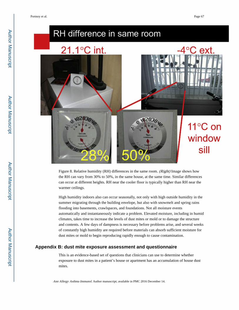
Figure 8. Relative humidity (RH) differences in the same room. (Right) Image shows how
the RH can vary from 30% to 50%, in the same house, at the same time. Similar differences
can occur at different heights. RH near the cooler floor is typically higher than RH near the
warmer ceilings.
High humidity indoors also can occur seasonally, not only with high outside humidity in the
summer migrating through the building envelope, but also with snowmelt and spring rains
flooding into basements, crawlspaces, and foundations. Not all moisture events
automatically and instantaneously indicate a problem. Elevated moisture, including in humid
climates, takes time to increase the levels of dust mites or mold or to damage the structure
and contents. A few days of dampness is necessary before problems arise, and several weeks
of constantly high humidity are required before materials can absorb sufficient moisture for
dust mites or mold to begin reproducing rapidly enough to cause contamination.
Appendix B: dust mite exposure assessment and questionnaire
This is an evidence-based set of questions that clinicians can use to determine whether
exposure to dust mites in a patient’s house or apartment has an accumulation of house dust
mites.
Portnoy et al. Page 67
Ann Allergy Asthma Immunol. Author manuscript; available in PMC 2016 December 14.
Author M
anuscriptA
uthor Manuscript
Author M
anuscriptA
uthor Manuscript
Dust mite allergens have become ubiquitous across most of the continental United States and
Canada. However, in most regions, there are building-related factors that can be modified to
decrease dust mite exposure. If all regional and building-related factors point to an
inhospitable climate for dust mites, then an optional series of questions usually are not
necessary. However, if any regional and building-related factors point to a favorable climate
for dust mites, then a final series of questions are asked to determine potentially modifiable
risk factors within the home. Then, the patient would be led to information about how to
modify those risk factors to decrease exposure to dust mite allergens.
Step 1: Determine the region of the country and if housing type is
conducive to dampness
1. Region of the country: The map presented in Figure 10 shows several
different climates that are overlaid onto the map of the United States and
Canada. Depending on this climate map, colors represent different levels
of temperature and humidity. For example, a home could be warm and dry
(eg, Tucson) or warm and humid (eg, Tampa). For more detailed
delineation of the climate zones, please refer to the Köppen climate
classification (http://webmap.ornl.gov/wcsdown/wcsdown.jsp?
dg_id=10012_1, accessed 8-14-2013).
a. If patient lives in a consistently dry arid area (eg, Tucson),
then one can assume, based on climate, that levels should
be low. Then, the patient would be directed to step 2 to
confirm that the building factors are still inhospitable to
dust mites.
b. If patient lives in a consistently humid area (eg, Tampa),
then one can assume, based on climate, that levels should
be high. Then, the patient would be directed to step 3 to
identify potentially modifiable risk factors within the
home. The patient would be led to information about how
to modify those risk factors to decrease exposure to dust
mite allergens.
2. Building type: The age of a building has been used as a proxy for heating
and ventilation in homes in the northern Unites States and Canada. The
authors believe that they most likely capture forced air ventilation. In
addition, underground living spaces are a risk factor for dampness and thus
dust mites.
3. Dampness: Although studies in the northern United States and Canada
have used mold odor as a proxy for dampness, this has not been tested in
the southern United States. Moldy odor might be a proxy for dampness,
but the conservative assumption has been made in step 1 that all homes in
humid southern states have dust mites.
Portnoy et al. Page 68
Ann Allergy Asthma Immunol. Author manuscript; available in PMC 2016 December 14.
Author M
anuscriptA
uthor Manuscript
Author M
anuscriptA
uthor Manuscript
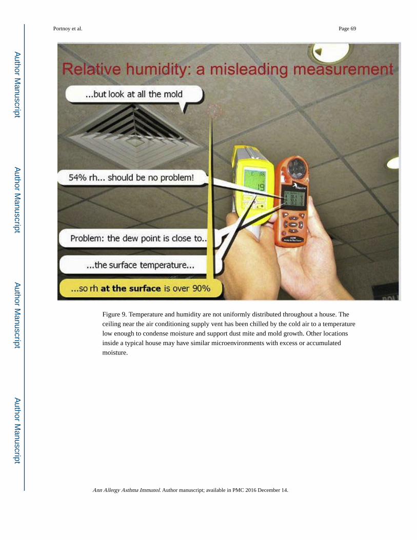
Figure 9. Temperature and humidity are not uniformly distributed throughout a house. The
ceiling near the air conditioning supply vent has been chilled by the cold air to a temperature
low enough to condense moisture and support dust mite and mold growth. Other locations
inside a typical house may have similar microenvironments with excess or accumulated
moisture.
Portnoy et al. Page 69
Ann Allergy Asthma Immunol. Author manuscript; available in PMC 2016 December 14.
Author M
anuscriptA
uthor Manuscript
Author M
anuscriptA
uthor Manuscript

Figure 10. Map to determine whether housing type is conducive to dampness given the
region of the country.
Region of the country and housing type (Fig 10)
Step 2: Determine major dust reservoir and more factors related to dampness
1. Carpeting: For most regions, carpeting is a major risk factor for dust mites.
Carpeting not only serves as a reservoir for dust, but also provides a
protective microclimate to the dust mites. Hardwood floors can easily be
swept, but dust mites can burrow deep into the carpet and often are
protected from effective removal by traditional vacuum cleaners. Even if
HEPA vacuum cleaners remove allergens, the dust mites can remain.
2. Hygrometer measurements: One recommendation is to give a patient a
hygrometer and place it over time throughout out the home to monitor
humidity. This should occur in several different seasons.
Portnoy et al. Page 70
Ann Allergy Asthma Immunol. Author manuscript; available in PMC 2016 December 14.
Author M
anuscriptA
uthor Manuscript
Author M
anuscriptA
uthor Manuscript
Major dust reservoirs and more factors related to dampness (Fig 11)
If the hygrometer level consistently shows an RH below 50% and if responses to all
questions in steps 1 and 2 are yes, then the patient’s home probably has low levels of live
dust mites.
Nonetheless, it would be useful to proceed to step 3 to inquire about possible risk factors
that might still contribute to dust mite allergen exposure.
Step 3: If it has been established that levels in the home are probably not low, then inquire about some of these levels to see if patient can decrease levels as low as possible
The questions were selected as follows. Several studies have measured dust mite allergens,
but not many have focused on assessing the relations between housing characteristics and
dust mite allergen concentrations. The authors chose a 2-step approach to determine if
conditions were conducive to dust mites: (1) Was there a study in the region of the country
that showed associations between housing characteristics and elevated dust mite allergen
concentration? (2) Would the questions found to be associated with elevated dust mite
allergen concentration be generalizable to that region?
Portnoy et al. Page 71
Ann Allergy Asthma Immunol. Author manuscript; available in PMC 2016 December 14.
Author M
anuscriptA
uthor Manuscript
Author M
anuscriptA
uthor Manuscript

Figure 11. Major dust reservoirs and more factors related to dampness.
Several studies often examined housing stock that was unique to their city or metropolitan
area,55,236,317–320 so questions such as “Do you live in a home built after 1951?” might be
relevant for that city but not for others. To illustrate, “pre-war” apartment buildings in New
York City represented by this pre-1951 question are brick-and-mortar, high-rise, multifamily
buildings compared with “pre-war” single-family detached homes built of wood in the early
1900s in Richmond, Virginia. In this case, the pre-1951 variable is most likely a proxy for a
combination of factors related to heating and ventilation of the home. One could debate
whether asking “When was your building built?”would be a good question in those cities
that had the studies with dust mite allergen and housing characteristics. However, many
residents do not know when their building was built and a building date does not take into
account potential retrofits and modifications that would affect the HVAC system.
The questions in this section have been found in studies not only in the United States and
Canada but also in other locations around the world.
Portnoy et al. Page 72
Ann Allergy Asthma Immunol. Author manuscript; available in PMC 2016 December 14.
Author M
anuscriptA
uthor Manuscript
Author M
anuscriptA
uthor Manuscript

Home assessment for dust mite allergens (supplemental questions)
Housing characteristics
Building
Are all your windows sealed shut or don’topen?
□ yes □ no
How long have you lived in this home? ___ years
If <1 year, did you move from a region of the country that might have high levels of dust mites? (see climate maps)
□ yes □ no
If YES, did you bring furniture from your previous home?
□ yes □ no
Is any part of your living area below ground level?
□ yes □ no
If YES, does this area ever get wet or stay wet for long periods (>1 week)?
□ yes □ no
Heating, ventilation, and cooling
Do you use a dehumidifier in your home? □ yes □ no □ N/A
During winter, are some outside walls cold? □ yes □ no □ don’tknow
Does your home sometimes smell “ stuffy,” “ stale,” or “musty”?
□ yes □ no
Does your air conditioner ever leak water onto walls or carpeting?
□ yes □ no □ N/A(no A/C)
Bedroom characteristics
Do you have upholstered furniture in your child’s bedroom?
□ yes □ no
Do you allow your child to have stuffed animals/ toys in the room?
□ yes □ no
Dust reservoirs (overall home)
Do you have cloth sofa or chairs? □ yes □ no
Do you have cloth curtains? □ yes □ no
Can you see dust or dirt on your furniture, walls, ceiling, and curtains?
□ yes □ no
Do you have wall-to-wall carpeting in more than half the rooms in your home?
□ yes □ no
Do you have wall-to-wall carpeting in your kitchen or bathrooms?
□ yes □ no
Do you not own a vacuum cleaner? □ yes □ no
Do you vacuum less than once a week? □ yes □ no
Dampness
In the past 12 months, have you noticed condensation on windows in your home?
□ yes □ no □ don’tknow
If YES, does moisture regularly build up on your windows/walls?
□ yes □ no
In the past 12 months, have you had any water leaks?
□ yes □ no □ don’tknow
Abbreviations: A/C, air conditioning; N/A, not applicable.
This questionnaire can be given to the patient. Affirmative (ie, YES) answers indicate potential dust mite allergen exposure.
Portnoy et al. Page 73
Ann Allergy Asthma Immunol. Author manuscript; available in PMC 2016 December 14.
Author M
anuscriptA
uthor Manuscript
Author M
anuscriptA
uthor Manuscript
References to Appendix B
1. Chew GL, Burge HA, Dockery DW, Muilenberg ML, Weiss ST, Gold DR. Limitations of a home characteristics questionnaire as a predictor of indoor allergen levels. Am J Respir Crit Care Med. 1998; 157:1536–1541. [PubMed: 9603135]
2. Cho SH, Reponen T, Bernstein DI, et al. The effect of home characteristics on dust antigen concentrations and loads in homes. Sci Total Environ. 2006; 371:31–43. [PubMed: 17049968]
3. Dales RE, Miller JD. Residential fungal contamination and health: microbial cohabitants as covariates. Environ Health Perspect. 1999; 107(suppl 3):481–483.
4. Kovesi T, Creery D, Gilbert NL, et al. Indoor air quality risk factors for severe lower respiratory tract infections in Inuit infants in Baffin Region, Nunavut: a pilot study. Indoor Air. 2006; 16:266–275. [PubMed: 16842607]
5. Lawton MD, Dales RE, White J. The influence of house characteristics in a Canadian community on microbiological contamination. Indoor Air. 1998; 8:2–11.
6. Loo CK, Foty RG, Wheeler AJ, et al. Do questions reflecting indoor air pollutant exposure from a questionnaire predict direct measure of exposure in owner-occupied houses? Int J Environ Res Public Health. 2010; 7:3270–3297. [PubMed: 20948960]
7. Miller JD, Dugandzic R, Frescura A-M, Salares V. Indoor and outdoor-derived contaminants in urban and rural homes in Ottawa, Canada. J Air Waste Manag Assoc. 2007; 57:297–302. [PubMed: 17385595]
8. Perry TT, Wood RA, Matsui EC, Curtin-Brosnan J, Rand C, Eggleston PA. Room-specific characteristics of suburban homes as predictors of indoor allergen concentrations. Ann Allergy Asthma Immunol. 2006; 97:628–635. [PubMed: 17165271]
9. van Strien RT, Gehring U, Belanger K, et al. The influence of air conditioning, humidity, temperature and other household characteristics on mite allergen concentrations in the northeastern United States. Allergy. 2004; 59:645–652. [PubMed: 15147450]
10. Wilson J, et al. Housing and allergens: a pooled analysis of nine US studies. Environ Res. 2010; 110:189–198. [PubMed: 19939359]
Appendix C: mite allergens
Dermatophagoides pteronyssinus
Der p 1 (Cysteine protease)
Der p 1 belongs to group 1 mite allergens and is a protein of 25 to 27 kDa. Der p 1 is a
cysteine protease.321–323 Der p 1 has protease activity that can induce a significant IgE
response. A proposed mechanism for its high allergenicity is that it cleaves the low-affinity
IgE receptor (CD23) from the surface of human B lymphocytes, enhancing IgE immune
responses by ablating the feedback inhibitory mechanism that normally limits IgE
synthesis.29 Der p 1 also can activate eosinophils to release proinflammatory mediators324
and it can prolong their survival.325 It has 80% homology with Der f 1. The crystal
structures of natural Der p 1 and Der f 1 in complex with a monoclonal antibody, 4C1, was
shown to bind to a unique cross-reactive epitope on the 2 allergens that is associated with
IgE recognition. This epitope is formed by almost identical amino acid sequences and
contact residues. Mutations of these common residues decrease IgE antibody binding.326
More than 50% of allergic patients and up to 80% of children with asthma are sensitized to
Der p 1. Der p 1 appears to be sufficient to diagnose more than 97% of dust mite–allergic
patients.30
Portnoy et al. Page 74
Ann Allergy Asthma Immunol. Author manuscript; available in PMC 2016 December 14.
Author M
anuscriptA
uthor Manuscript
Author M
anuscriptA
uthor Manuscript
Der p 2 (lipid-binding molecule)
A recombinant Der p 2 has been developed that reacts with IgE from most patients who are
sensitized to native Der p 2.327,328 Removal of either or both disulfide links decreases IgE
binding up to 10-fold, suggesting that these bonds play a critical role in stabilizing the
antigenic structure of this mite allergen.329 Der p 2 is a homolog of MD-2, a protein
involved in the binding of lipopolysaccharide and activation of toll-like receptor 4, which
promotes TH2-mediated inflammation.330,331 Der p 2 peptides were found to induce
multiple responses that were restricted through HLA-DPB1* 0401 and HLA-DRB1*01.332
Der p 3 (trypsin-like serine protease)
Der p 3 is encoded by a single gene. Most cDNA clones of this allergen show only minor
sequence variation similar to that observed for group 1 and 2 house dust mite allergens.333
This allergen contains a trypsin-like enzyme that has been shown to bind to human IgE. The
protein isa31-kDaprotein that is enzymatically similar to invertebrate and vertebrate trypsins
and shows homology with crayfish trypsin. All sera from a panel of mite-allergic individuals
showed IgE reactivity to trypsin, suggesting that mite trypsin is a major allergen.322
Der p 4 (α-amylase)
Der p 4 is a 57- to 60-kDa protein with amylase activity. It can be found in extracts of whole
mite and spent growth medium but not in unused growth medium. It has been detected in
extracts of dust obtained from mattresses and lounge room carpets. The enzyme activity
correlates with counts of live mites and with concentrations of Der p 1. In 1 study, sera from
25% of mite-allergic children and 46% of mite-allergic adults contained specific IgE to this
allergen and directly correlated with concentrations of total mite-specific IgE.334,335
Complement DNA clones of Der p 4 and Eur m 4 were sequenced and were found to code
for 496 amino acid mature proteins with highly conserved residues that are important for the
function of α-amylase. Der p 4 and Eur m 4 were 90% identical and were 50% identical to
insect and mammalian α-amylases.32
Der p 5 (function is unknown)
Der p 5 is a 14-kDa protein that has been isolated and its DNA sequence determined. The
deduced amino acid sequence was not homologous to any known protein sequences and it
contains no cysteine or tryptophan. Sera from 21 of 38 mite-allergic subjects recognized
recombinant Der p 5, which correlates with IgE binding to the native molecule. This protein
has homology to Der p 25; however, the function is not known for either allergen.336
Der p 6 (chymotrypsin, a serine protease)
An ELISA has been developed for the measurement of Der p 6.337
Der p 7 (lipid-binding molecule)
Der p 7 has 198 residues and a predicted molecular weight of 22 kDa. Sera from 14 of 38
dust mite–allergic children reacted strongly with this clone. Skin tests showed reactivity in
16 of 30 allergic patients (53%) and none of the controls.338
Portnoy et al. Page 75
Ann Allergy Asthma Immunol. Author manuscript; available in PMC 2016 December 14.
Author M
anuscriptA
uthor Manuscript
Author M
anuscriptA
uthor Manuscript

Der p 8 (glutathione S-transferase)
Der p 8 is a 26-kDa polypeptide. Nucleotide sequencing showed a 219–amino acid protein.
The molecule has a strong homology with glutathione S-transferases, containing all but 1 of
the 19 conserved amino acid residues found in glutathione transferase.339
Der p 9 (collagenase, a serine protease)
Der p 9 is a 23.7-kDa protein that is enzymatically similar to chymotrypsin and cathepsin
G–like enzymes and it has been shown to cleave collagen. It has homology with Der p 3 and
with Der p 6, although inhibition studies have shown cross-reactivity between Der p 9 and
Der p 3 but not Der p 6. Up to 92% of dust mite–allergic patients have specific IgE to Der p
9.340
Der p 10 (tropomyosin)
Der p 10 is a tropomyosin that shares more than 65% of residues with other invertebrate
tropomyosins. The recombinant allergen cross-reacts with shrimp tropomyosin. In 1 study,
5.6% of sera from mite-allergic patients had IgE reactivity to Der p 10.33 Another study
found that up to 15.2% of dust mite–allergic patients had IgE to Der p 10.34 In that same
study, Der p 10–negative patients were sensitized primarily to Der p 1 and/or Der p 2.
Tropomyosin is believed to be responsible for clinical cross-reactivity between dust mites
and seafood. In a study of dust mite–allergic patients from southern Bavaria, IgE antibodies
to Der p 10 were found in 4 of 93 sera (4.3%). Two of these patients had oral symptoms
accompanied by bronchospasm after consumption of shrimp. Thus, although there is some
cross-reactivity, the low frequency of IgE to Der p 10 in dust mite–allergic patients and the
low frequency of clinical reactions in these patients suggest that most shrimp reactions are
due to other allergens.341
Other dust mite allergens
There is a large number of additional dust mite allergens (listed in Table 1). These include
Der p 11 (paramyosin), Der p 13 (lipid-binding protein), Der p 14 (apolipophorin), Der p 15
(chitinase), Der p 16 (gelsolin/villin), Der p 17 (Ca2+-binding protein), Der p 18 (chitinase),
Der p 19 (antimicrobial protein), Der p 20 (arginine kinase), Der p 21 (similar to Der p 5),
and Der p 24 (troponin C). The clinical relevance of sensitization to these allergens is not
well understood.
Dermatophagoides farinae (very similar to D pteronyssinus)
Der f 1 (cysteine protease)
Der f 1, a major allergen from the house dust mite, is a 223-residue protein with a derived
molecular weight of 25,191 kDa. It has significant homology to other cysteine proteases.
Sequence alignment of Der f 1 and Der p 1 has shown a high degree of homology (81%).342
Portnoy et al. Page 76
Ann Allergy Asthma Immunol. Author manuscript; available in PMC 2016 December 14.
Author M
anuscriptA
uthor Manuscript
Author M
anuscriptA
uthor Manuscript

Der f 2 (lipid-binding molecule)
Der f 2 encodes a 129-residue protein with a calculated molecular weight of 14 kDa and an
expected homology with Der p 2 of 88%. The 2 molecules also display a high degree of
antibody cross-reactivity.343
Der f 5
Der f 5 has a molecular weight of 13.6 kDa and amino acid homologies with Der p 5, Blo t
5, Sui m 5, and Lep d 5. Der f 5 and Der p 5 are more similar to each other than to Blo t 5
and Ale o 5, most likely because they belong to different mite families (Echimyopodidae vs
Acaridae).344
Der f 6 (chymotrypsin-like serine protease)
Der f 6 cDNA is 840 nucleotides long. In 20 patients with asthma, 45% had specific IgE to
rDer f 6. Substantial homology has been shown between Der f 6 and Blo t 6, Sui m 6, Der f
3, and Der f 9.345
Der f 7 (lipid-binding protein)
Der f 7 is a 25-kDa protein with 31- and 30-kDa components that are glycosylation products
of the 25-kDa form and an 18-kDa band consistent with a degradation product.346
Immediate hypersensitivity skin test reactions to Der f 7 have been found in 52% of mite-
sensitive allergic patients.347
Der f 10 (tropomyosin)
Native Der f 10 reacted with specific IgE in the 31 sera tested at a high frequency (80.6%),
comparable to that of Der f 1 (90.3%) and Der f 2 (74.2%).348
Der f 11 (paramyosin)
Der f 11 is a 98-kDa mite allergen. The sequence identity of Der f 11 with other known
paramyosins is 34% to 60%.349 Der f 11 cDNA has 2,625 base pairs encoding a 103-kDa
protein with 875 amino acids with significant homology with the paramyosin of other
invertebrates. It has greater than 89% identity with Blo t 1. IgE binding was found in 78% of
mite-allergic patients. IgE cross-inhibition between rDer p 11 and rDer f 11 was up to 73%
to 80%.350
Der f 18 (chitinase)
Der f 18 is a 60-kDa protein. In 1 study, the purified protein bound IgE in 54% of the sera
from mite-allergic patients. Its cDNA encodes a protein with 462 amino acids with
homology to multiple chitinases. Chitinase is found abundantly in the mite digestive system,
but fecal pellets did not stain positively for this allergen.351
Blo t 21
In B tropicalis, the most prevalent and allergenic allergens are, in descending order, Blo t 21,
Blo t 5, and Blo t 7. Blo t 21 has 40% sequence identity to and small to moderate
immunologic cross-reactivity with Blo t 5.352
Portnoy et al. Page 77
Ann Allergy Asthma Immunol. Author manuscript; available in PMC 2016 December 14.
Author M
anuscriptA
uthor Manuscript
Author M
anuscriptA
uthor Manuscript

References
1. Voorhorst R, Spieksma FT, Varekamp H, Leupen M, Lyklema A. The house-dust mite (Dermatophagoides pteronyssinus) and the allergens it produces. Identity with the house-dust allergen. J Allergy. 1967; 39:325–339. (III).
2. Miyamoto T, Oshima S, Ishizaki T, Sato SH. Allergenic identity between the common floor mite (Dermatophagoides farinae Hughes, 1961) and house dust as a causative antigen in bronchial asthma. J Allergy. 1968; 42:14–28. (NR). [PubMed: 5243795]
3. Colloff MJ. Taxonomy and identification of dust mites. Allergy. 1998; 53(suppl):7–12. (NR).
4. Arlian LG, Platts-Mills TA. The biology of dust mites and the remediation of mite allergens in allergic disease. J Allergy Clin Immunol. 2001; 107(suppl):S406–S413. (IV). [PubMed: 11242601]
5. Kim YK, Kim YY. Spider-mite allergy and asthma in fruit growers. Curr Opin Allergy Clin Immunol. 2002; 2:103–107. (III). [PubMed: 11964757]
6. Arlian LG, Morgan MS, Houck MA. Allergenicity of the mite Hemisarcoptes cooremani. Ann Allergy Asthma Immunol. 1999; 83:529–532. (LB). [PubMed: 10619344]
7. O’Connor, B. Acari: Astigmata. Parker, S., editor. New York: McGraw-Hill; 1982.
8. Hart BJ. Life cycle and reproduction of house-dust mites: environmental factors influencing mite populations. Allergy. 1998; 53(suppl):13–17. (III). [PubMed: 10096801]
9. Blythe ME, Williams JD, Smith JM. Distribution of pyroglyphid mites in Birmingham with particular reference to Euroglyphus maynei. Clin Allergy. 1974; 4:25–33. (III). [PubMed: 4824570]
10. Wharton GW. House dust mites. J Med Entomol. 1976; 12:577–621. (NR). [PubMed: 772212]
11. Platts-Mills TA, de Weck AL, Aalberse RC, et al. Dust mite allergens and asthma—a worldwide problem. J Allergy Clin Immunol. 1989; 83:416–427. (IV). [PubMed: 2645343]
12. Colloff MJ, Ayres J, Carswell F, et al. The control of allergens of dust mites and domestic pets: a position paper. Clin Exp Allergy. 1992; 22(suppl 2):1–28. (IV).
13. Arlian L, Veselica M. Relationship between transpiration rate and temperature in the mite Dermatophagoides farinae. Physiol Zool. 1981; 55:344–354. (LB).
14. Arlian LG, Bernstein D, Bernstein IL, et al. Prevalence of dust mites in the homes of people with asthma living in eight different geographic areas of the United States. J Allergy Clin Immunol. 1992; 90:292–300. (III). [PubMed: 1527314]
15. de Boer R, Kuller K. Winter survival of house dust mites (Dermatophagoides spp.) on the ground floor of Dutch houses. Proc Exp Appl Entomol. 1995; 6:47–51. (III).
16. Lintner TJ, Brame KA. The effects of season, climate, and air-conditioning on the prevalence of Dermatophagoides mite allergens in household dust. J Allergy Clin Immunol. 1993; 91:862–867. (III). [PubMed: 8473674]
17. Mumcuoglu Y. Biologie und Oekologie der Hausstaubmilben. Allergologie. 1988; 11:223–228. (NR).
18. Wharton GW, Brody AR. The peritrophic membrane of the mite, Dermatophagoides farinae: Acariformes. J Parasitol. 1972; 58:801–804. (NR). [PubMed: 5057235]
19. Tovey ER, Chapman MD, Platts-Mills TA. Mite faeces are a major source of house dust allergens. Nature. 1981; 289:592–593. (III). [PubMed: 7464922]
20. Stewart GA, Bird CH, Krska KD, Colloff MJ, Thompson PJ. A comparative study of allergenic and potentially allergenic enzymes from Dermatophagoides pteronyssinus, D. farinae and Euroglyphus maynei. Exp Appl Acarol. 1992; 16:165–180. (LB). [PubMed: 1283568]
21. Walz M. Ultrastructure of the reproductive system of the house dust mites Dermatophagoides farinae and D. pteronyssinus with remarks on spermatogenesis and oogenesis. Exp Appl Acarol. 1992; 16:85–116. (NR). [PubMed: 1493749]
22. Arlian L, Rapp C, Ahmed S. Development of Dermatophagoides pteronyssinus (Acari: Pyroglyphidae). J Med Entomol. 1990; 27:1035–1040. (NR). [PubMed: 2280388]
23. Hallas TE. The biology of mites. Allergy. 1991; 46(suppl 11):6–9. (NR). [PubMed: 1897701]
24. Colloff M. Age structure and dynamics of house dust mite populations. Exp Appl Acarol. 1992; 16:49–74. (NR).
25. Colloff, M. Dust Mites. Collingwood, Victoria, Australia: CSIRO; 2009.
Portnoy et al. Page 78
Ann Allergy Asthma Immunol. Author manuscript; available in PMC 2016 December 14.
Author M
anuscriptA
uthor Manuscript
Author M
anuscriptA
uthor Manuscript

26. Arlian LG, Dippold JS. Development and fecundity of Dermatophagoides farinae (Acari: Pyroglyphidae). J Med Entomol. 1996; 33:257–260. (NR). [PubMed: 8742531]
27. Phipatanakul W, Matsui E, Portnoy J, et al. Environmental assessment and exposure reduction of rodents: a practice parameter. Ann Allergy Asthma Immunol. 2012; 109:375–387. (Ia). [PubMed: 23176873]
28. de Boer R, van der Hoeven WA, Stapel SO. The decay of house dust mite allergens, Der p I and Der p II, under natural conditions. Clin Exp Allergy. 1995; 25:765–770. [PubMed: 7584689]
29. Hewitt CR, Brown AP, Hart BJ, Pritchard DI. A major house dust mite allergen disrupts the immunoglobulin E network by selectively cleaving CD23: innate protection by antiproteases. J Exp Med. 1995; 182:1537–1544. (LB). [PubMed: 7595223]
30. Weghofer M, Thomas WR, Kronqvist M, et al. Variability of IgE reactivity profiles among European mite allergic patients. Eur J Clin Invest. 2008; 38:959–965. (III). [PubMed: 19021722]
31. Cho HJ, Lee HJ, Kim SC, et al. Protease-activated receptor 2–dependent fluid secretion from airway submucosal glands by house dust mite extract. J Allergy Clin Immunol. 2012; 129:529–535. 35, e1–e5. [PubMed: 22196772]
32. Mills KL, Hart BJ, Lynch NR, Thomas WR, Smith W. Molecular characterization of the group 4 house dust mite allergen from Dermatophagoides pteronyssinus and its amylase homologue from Euroglyphus maynei. Int Arch Allergy Immunol. 1999; 120:100–107. (LB).
33. Asturias JA, Arilla MC, Gomez-Bayon N, Martinez A, Martinez J, Palacios R. Sequencing and high level expression in Escherichia coli of the tropomyosin allergen (Der p 10) from Dermatophagoides pteronyssinus. Biochim Biophys Acta. 1998; 1397:27–30. (LB). [PubMed: 9545522]
34. Resch Y, Weghofer M, Seiberler S, et al. Molecular characterization of Der p 10: a diagnostic marker for broad sensitization in house dust mite allergy. Clin Exp Allergy. 2011; 41:1468–1477. (LB). [PubMed: 21711470]
35. Valmonte GR, Cauyan GA, Ramos JD. IgE cross-reactivity between house dust mite allergens and Ascaris lumbricoides antigens. Asia Pac Allergy. 2012; 2:35–44. [PubMed: 22348205]
36. Post S, Nawijn MC, Hackett TL, et al. The composition of house dust mite is critical for mucosal barrier dysfunction and allergic sensitisation. Thorax. 2012; 67:488–495. [PubMed: 22167364]
37. Voorhorst R, Spieksma-Boezeman MI, Spieksma FT. Is a mite (Dermatophagoides sp.) the producer of the house-dust allergen? Allergie Asthma. 1964; 10:329–334. (III). [PubMed: 14307280]
38. Dreborg S. Mite allergens. Collection, determination, expression of results, and risk levels for sensitization and symptom induction. Allergy. 1998; 53(suppl):88–91. (IIa). [PubMed: 10096816]
39. Yokoyama H, Matsuki H, Takaoka M, Sugiuchi M, Kasuga H. Epidemiological study of mites and guanine in house dust. Tokai J Exp Clin Med. 1990; 15:485–491. (III). [PubMed: 2131654]
40. Van Bronswijk JE, Bischoff E, Schirmacher W, Kniest FM. Evaluating mite (Acari) allergenicity of house dust by guanine quantification. J Med Entomol. 1989; 26:55–59. (III). [PubMed: 2926776]
41. van der Brempt X, Haddi E, Michel-Nguyen A, et al. Comparison of the ACAREX test with monoclonal antibodies for the quantification of mite allergens. J Allergy Clin Immunol. 1991; 87:130–132. (LB). [PubMed: 1991917]
42. Lind P, Korsgaard J, Lowenstein H. Detection and quantitation of Dermatophagoides antigens in house dust by immunochemical techniques. Allergy. 1979; 34:319–326. (III). [PubMed: 94774]
43. Chapman MD, Sutherland WM, Platts-Mills TA. Recognition of two Dermatophagoides pteronyssinus-specific epitopes on antigen P1 by using monoclonal antibodies: binding to each epitope can be inhibited by serum from dust mite-allergic patients. J Immunol. 1984; 133:2488–2495. (LB). [PubMed: 6207232]
44. Haida M, Okudaira H, Ogita T, et al. Allergens of the house dust mite Dermatophagoides farinae—immunochemical studies of four allergenic fractions. J Allergy Clin Immunol. 1985; 75:686–692. (LB). [PubMed: 4008797]
45. Maldonado AM, Solano MO. [Relation between Der p2 and Der f2 antigen levels in house dust and the degree of sensitization in asthmatic patients]. Rev Alerg Mex. 2005; 52:67–71. [PubMed: 16158778]
Portnoy et al. Page 79
Ann Allergy Asthma Immunol. Author manuscript; available in PMC 2016 December 14.
Author M
anuscriptA
uthor Manuscript
Author M
anuscriptA
uthor Manuscript

46. Exposure to secondhand smoke among students aged 13–15 years—worldwide, 2000–2007. MMWR Morb Mortal Wkly Rep. 2007; 56:497–500. (III). [PubMed: 17522587]
47. Poltronieri P, Cimaglia F, Santino A, et al. Protein chips for detection of mite allergens using Kunitz-type protease inhibitors. Biotechnol J. 2010; 5:582–587. [PubMed: 20518061]
48. Barnes C, Tuck J, Simon S, Pacheco F, Hu F, Portnoy J. Allergenic materials in the house dust of allergy clinic patients. Ann Allergy Asthma Immunol. 2001; 86:517–523. (III). [PubMed: 11379802]
49. Chapman MD, Heymann PW, Wilkins SR, Brown MJ, Platts-Mills TA. Monoclonal immunoassays for major dust mite (Dermatophagoides) allergens, Der p I and Der f I, quantitative analysis of the allergen content of mite and house dust extracts. J Allergy Clin Immunol. 1987; 80:184–194. (LB). [PubMed: 3611539]
50. Filep S, Tsay A, Vailes L, et al. A multi-allergen standard for the calibration of immunoassays: CREATE principles applied to eight purified allergens. Allergy. 2012; 67:235–241. (LB). [PubMed: 22092159]
51. Arbes SJ Jr, Cohn RD, Yin M, et al. House dust mite allergen in US beds: results from the First National Survey of Lead and Allergens in Housing. J Allergy Clin Immunol. 2003; 111:408–414. (III). [PubMed: 12589364]
52. Peterson EL, Ownby DR, Johnson CC. The relationship of housing and household characteristics to the indoor concentrations of Der f 1, Der p 1, and Fel d 1 measured in dust and air samples. Ann Allergy Asthma Immunol. 2003; 90:564–571. (III). [PubMed: 12775140]
53. Leaderer BP, Belanger K, Triche E, et al. Dust mite, cockroach, cat, dog allergen concentrations in homes of asthmatic children in the northeastern United States: impact of socioeconomic factors and population density. Environ Health Perspect. 2002; 110:419–425. (IIb). [PubMed: 11940461]
54. Mihrshahi S, Marks G, Vanlaar C, Tovey E, Peat J. Predictors of high house dust mite allergen concentrations in residential homes in Sydney. Allergy. 2002; 57:137–142. (III). [PubMed: 11929416]
55. van Strien RT, Gehring U, Belanger K, et al. The influence of air conditioning, humidity, temperature and other household characteristics on mite allergen concentrations in the northeastern United States. Allergy. 2004; 59:645–652. (IIb). [PubMed: 15147450]
56. Vervloet D, Pradal M, Porri F, Charpin D. [The epidemiology of allergy to house dust mites]. Rev Mal Respir. 1991; 8:59–65. (III). [PubMed: 2034857]
57. Platts-Mills TA, Hayden ML, Chapman MD, Wilkins SR. Seasonal variation in dust mite and grass-pollen allergens in dust from the houses of patients with asthma. J Allergy Clin Immunol. 1987; 79:781–791. (III). [PubMed: 3571770]
58. Tovey ER, Chapman MD, Wells CW, Platts-Mills TA. The distribution of dust mite allergen in the houses of patients with asthma. Am Rev Respir Dis. 1981; 124:630–635. (III). [PubMed: 7305119]
59. Luczynska C, Sterne J, Bond J, Azima H, Burney P. Indoor factors associated with concentrations of house dust mite allergen, Der p 1, in a random sample of houses in Norwich, UK. Clin Exp Allergy. 1998; 28:1201–1209. (III). [PubMed: 9824386]
60. Tovey E, Ferro A. Time for new methods for avoidance of house dust mite and other allergens. Curr Allergy Asthma Rep. 2012; 12:465–477. (IV). [PubMed: 22833251]
61. de Blay F, Heymann PW, Chapman MD, Platts-Mills TA. Airborne dust mite allergens: comparison of group II allergens with group I mite allergen and cat-allergen Fel d I. J Allergy Clin Immunol. 1991; 88:919–926. (LB). [PubMed: 1744363]
62. Arlian LG, Neal JS, Morgan MS, Rapp CM, Clobes AL. Distribution and removal of cat, dog and mite allergens on smooth surfaces in homes with and without pets. Ann Allergy Asthma Immunol. 2001; 87:296–302. (III). [PubMed: 11686421]
63. Wood RA, Mudd KE, Eggleston PA. The distribution of cat and dust mite allergens on wall surfaces. J Allergy Clin Immunol. 1992; 89:126–130. (III). [PubMed: 1730832]
64. Custovic A, Fletcher A, Pickering CA, et al. Domestic allergens in public places III: house dust mite, cat, dog and cockroach allergens in British hospitals. Clin Exp Allergy. 1998; 28:53–59. (III).
Portnoy et al. Page 80
Ann Allergy Asthma Immunol. Author manuscript; available in PMC 2016 December 14.
Author M
anuscriptA
uthor Manuscript
Author M
anuscriptA
uthor Manuscript
65. Gore RB, Hadi EA, Craven M, et al. Personal exposure to house dust mite allergen in bed: nasal air sampling and reservoir allergen levels. Clin Exp Allergy. 2002; 32:856–859. (III). [PubMed: 12047431]
66. Terra SA, Silva DA, Sopelete MC, Mendes J, Sung SJ, Taketomi EA. Mite allergen levels and acarologic analysis in house dust samples in Uberaba, Brazil. J Investig Allergol Clin Immunol. 2004; 14:232–237. (III).
67. Garrett MH, Hooper BM, Hooper MA. Indoor environmental factors associated with house-dust-mite allergen (Der p 1) levels in south-eastern Australian houses. Allergy. 1998; 53:1060–1065. (III). [PubMed: 9860238]
68. Pauli G, de Blay F, Bessot JC, Ott M, Gries P. The role of mattress bases in the mite infestation of dwellings. J Allergy Clin Immunol. 1997; 99:261–263. (III). [PubMed: 9042056]
69. de Blay F, Fourgaut G, Hedelin G, et al. Medical Indoor Environment Counselor (MIEC): role in compliance with advice on mite allergen avoidance and on mite allergen exposure. Allergy. 2003; 58:27–33. (Ib). [PubMed: 12580803]
70. de Oliveira CH, Binotti RS, Muniz JR, dos Santos JC, do Prado AP, de Pinho AJ Jr. Comparison of house dust mites found on different mattress surfaces. Ann Allergy Asthma Immunol. 2003; 91:559–562. (III). [PubMed: 14700440]
71. da Silva DR, Binotti RS, da Silva CM, de Oliveira CH, Condino-Neto A, de Capitani EM. Mites in dust samples from mattress surfaces from single beds or cribs in the south Brazilian city of Londrina. Pediatr Allergy Immunol. 2005; 16:132–136. (III). [PubMed: 15787870]
72. Teplitsky V, Mumcuoglu KY, Babai I, Dalal I, Cohen R, Tanay A. House dust mites on skin, clothes, and bedding of atopic dermatitis patients. Int J Dermatol. 2008; 47:790–795. (III). [PubMed: 18717857]
73. Lowe L, Murray CS, Custovic A, et al. Specific airway resistance in 3-year-old children: a prospective cohort study. Lancet. 2002; 359:1904–1908. (IIb). [PubMed: 12057553]
74. Squillace SP, Sporik RB, Rakes G, et al. Sensitization to dust mites as a dominant risk factor for asthma among adolescents living in central Virginia. Multiple regression analysis of a population-based study. Am J Respir Crit Care Med. 1997; 156:1760–1764. (IIa). [PubMed: 9412552]
75. Erwin EA, Wickens K, Custis NJ, et al. Cat and dust mite sensitivity and tolerance in relation to wheezing among children raised with high exposure to both allergens. J Allergy Clin Immunol. 2005; 115:74–79. (III). [PubMed: 15637550]
76. Simpson A, Soderstrom L, Ahlstedt S, Murray CS, Woodcock A, Custovic A. IgE antibody quantification and the probability of wheeze in preschool children. J Allergy Clin Immunol. 2005; 116:744–749. (IIb). [PubMed: 16210045]
77. Lodge CJ, Lowe AJ, Gurrin LC, et al. House dust mite sensitization in toddlers predicts current wheeze at age 12 years. J Allergy Clin Immunol. 2011; 128:782.e9–788.e9. [PubMed: 21820717]
78. Llanora GV, Ming LJ, Wei LM, van Bever HP. House dust mite sensitization in toddlers predict persistent wheeze in children between eight to fourteen years old. Asia Pac Allergy. 2012; 2:181–186. [PubMed: 22872820]
79. Chew GL, Reardon AM, Correa JC, et al. Mite sensitization among Latina women in New York, where dust-mite allergen levels are typically low. Indoor Air. 2009; 19:193–197. (III). [PubMed: 19220508]
80. Rosenstreich DL, Eggleston P, Kattan M, et al. The role of cockroach allergy and exposure to cockroach allergen in causing morbidity among inner-city children with asthma. N Engl J Med. 1997; 336:1356–1363. (Ib). [PubMed: 9134876]
81. Charpin D, Birnbaum J, Haddi E, et al. Altitude and allergy to house-dust mites. A paradigm of the influence of environmental exposure on allergic sensitization. Am Rev Respir Dis. 1991; 143:983–986. (III). [PubMed: 2024854]
82. Sporik R, Squillace SP, Ingram JM, Rakes G, Honsinger RW, Platts-Mills TA. Mite, cat, cockroach exposure, allergen sensitisation, and asthma in children: a case–control study of three schools. Thorax. 1999; 54:675–680. (IIa). [PubMed: 10413718]
83. Sporik R, Ingram JM, Price W, Sussman JH, Honsinger RW, Platts-Mills TA. Association of asthma with serum IgE and skin test reactivity to allergens among children living at high altitude.
Portnoy et al. Page 81
Ann Allergy Asthma Immunol. Author manuscript; available in PMC 2016 December 14.
Author M
anuscriptA
uthor Manuscript
Author M
anuscriptA
uthor Manuscript
Tickling the dragon’s breath. Am J Respir Crit Care Med. 1995; 151:1388–1392. (IIb). [PubMed: 7735590]
84. Peters JL, Suglia SF, Platts-Mills TA, Hosen J, Gold DR, Wright RJ. Relationships among prenatal aeroallergen exposure and maternal and cord blood IgE: project ACCESS. J Allergy Clin Immunol. 2009; 123:1041–1046. (IIb). [PubMed: 19361844]
85. De Lovinfosse S, Charpin D, Dornelas A, Birnbaum J, Vervloet D. Can mite-specific IgE be used as a surrogate for mite exposure? Allergy. 1994; 49:64–66. (IIa). [PubMed: 8198243]
86. Peat JK, Tovey E, Toelle BG, et al. House dust mite allergens. A major risk factor for childhood asthma in Australia. Am J Respir Crit Care Med. 1996; 153:141–146. (IIb). [PubMed: 8542107]
87. Kuehr J, Frischer T, Meinert R, et al. Mite allergen exposure is a risk for the incidence of specific sensitization. J Allergy Clin Immunol. 1994; 94:44–52. (IIb). [PubMed: 8027498]
88. Clearing the Air: Asthma and Indoor Air Exposures. Washington, DC: National Academies Press; 2000. Committee on the Assessment of Asthma and Indoor Air.
89. Custovic A, Simpson BM, Murray CS, Lowe L, Woodcock A. The National Asthma Campaign Manchester Asthma and Allergy Study. Pediatr Allergy Immunol. 2002; 13(suppl 15):32–37. (IIa). [PubMed: 12688622]
90. Custovic A, Simpson BM, Simpson A, et al. Manchester Asthma and Allergy Study: low-allergen environment can be achieved and maintained during pregnancy and in early life. J Allergy Clin Immunol. 2000; 105:252–258. (IIa). [PubMed: 10669844]
91. Woodcock A, Lowe LA, Murray CS, et al. Early life environmental control: effect on symptoms, sensitization, and lung function at age 3 years. Am J Respir Crit Care Med. 2004; 170:433–439. (IIb). [PubMed: 15142868]
92. Simpson A, Simpson B, Custovic A, Cain G, Craven M, Woodcock A. Household characteristics and mite allergen levels in Manchester, UK. Clin Exp Allergy. 2002; 32:1413–1419. (III). [PubMed: 12372118]
93. van de Pol MA, Lutter R, van Ree R, van der Zee JS. Increase in allergen-specific IgE and ex vivo Th2 responses after a single bronchial challenge with house dust mite in allergic asthmatics. Allergy. 2012; 67:67–73. [PubMed: 21958117]
94. Custovic A, Taggart SC, Woodcock A. House dust mite and cat allergen in different indoor environments. Clin Exp Allergy. 1994; 24:1164–1168. (III). [PubMed: 7889431]
95. Murray AB, Ferguson AC, Morrison BJ. Diagnosis of house dust mite allergy in asthmatic children: what constitutes a positive history? J Allergy Clin Immunol. 1983; 71:21–28. (III). [PubMed: 6822690]
96. Scott M, Roberts G, Kurukulaaratchy RJ, Matthews S, Nove A, Arshad SH. Multifaceted allergen avoidance during infancy reduces asthma during childhood with the effect persisting until age 18 years. Thorax. 2012; 67:1046–1051. [PubMed: 22858926]
97. Sears MR, Herbison GP, Holdaway MD, Hewitt CJ, Flannery EM, Silva PA. The relative risks of sensitivity to grass pollen, house dust mite and cat dander in the development of childhood asthma. Clin Exp Allergy. 1989; 19:419–424. (III). [PubMed: 2758355]
98. Bjornsson E, Norback D, Janson C, et al. Asthmatic symptoms and indoor levels of micro-organisms and house dust mites. Clin Exp Allergy. 1995; 25:423–431. (IIb). [PubMed: 7553245]
99. Custovic A, Chapman M. Risk levels for mite allergens. Are they meaningful? Allergy. 1998; 53(suppl):71–76. (IV). [PubMed: 10096813]
100. Sporik R, Holgate ST, Platts-Mills TA, Cogswell JJ. Exposure to house-dust mite allergen (Der p I) and the development of asthma in childhood. A prospective study. N Engl J Med. 1990; 323:502–507. (IIa). [PubMed: 2377175]
101. Celedon JC, Milton DK, Ramsey CD, et al. Exposure to dust mite allergen and endotoxin in early life and asthma and atopy in childhood. J Allergy Clin Immunol. 2007; 120:144–149. (IIb). [PubMed: 17507083]
102. Custovic A, Woodcock A. Exposure and sensitization in infants and children. Curr Opin Allergy Clin Immunol. 2001; 1:133–138. (IV). [PubMed: 11964680]
103. Simpson BM, Custovic A, Simpson A, et al. NAC Manchester Asthma and Allergy Study (NACMAAS): risk factors for asthma and allergic disorders in adults. Clin Exp Allergy. 2001; 31:391–399. (III). [PubMed: 11260150]
Portnoy et al. Page 82
Ann Allergy Asthma Immunol. Author manuscript; available in PMC 2016 December 14.
Author M
anuscriptA
uthor Manuscript
Author M
anuscriptA
uthor Manuscript
104. Simpson A, Custovic A. Allergen avoidance in the primary prevention of asthma. Curr Opin Allergy Clin Immunol. 2004; 4:45–51. (IV). [PubMed: 15090919]
105. Bennett WD, Herbst M, Alexis NE, et al. Effect of inhaled dust mite allergen on regional particle deposition and mucociliary clearance in allergic asthmatics. Clin Exp Allergy. 2011; 41:1719–1728. (IIa). [PubMed: 21729182]
106. Tunnicliffe WS, Fletcher TJ, Hammond K, et al. Sensitivity and exposure to indoor allergens in adults with differing asthma severity. Eur Respir J. 1999; 13:654–659. (III). [PubMed: 10232442]
107. Vervloet D, Charpin D, Haddi E, et al. Medication requirements and house dust mite exposure in mite-sensitive asthmatics. Allergy. 1991; 46:554–558. (IIa). [PubMed: 1796781]
108. Platts-Mills TA, Chapman MD, Pollart S, Luczynska CM, Ward GW Jr. Specific allergens evoking immune reactions in the lung: relationship to asthma. Eur Respir J Suppl. 1991; 13:68s–77s. (LB). [PubMed: 1953920]
109. van der Heide S, De Monchy JG, De Vries K, Dubois AE, Kauffman HF. Seasonal differences in airway hyperresponsiveness in asthmatic patients: relationship with allergen exposure and sensitization to house dust mites. Clin Exp Allergy. 1997; 27:627–633. (III). [PubMed: 9208182]
110. Custovic A, Taggart SC, Francis HC, Chapman MD, Woodcock A. Exposure to house dust mite allergens and the clinical activity of asthma. J Allergy Clin Immunol. 1996; 98:64–72. (IIb). [PubMed: 8765819]
111. Langley SJ, Goldthorpe S, Craven M, Morris J, Woodcock A, Custovic A. Exposure and sensitization to indoor allergens: association with lung function, bronchial reactivity, and exhaled nitric oxide measures in asthma. J Allergy Clin Immunol. 2003; 112:362–368. (IIb). [PubMed: 12897743]
112. Ciprandi G, Buscaglia S, Pesce G, et al. Minimal persistent inflammation is present at mucosal level in patients with asymptomatic rhinitis and mite allergy. J Allergy Clin Immunol. 1995; 96:971–979. (III). [PubMed: 8543756]
113. Fowler SJ, Langley SJ, Truman NJ, Woodcock A, Simpson A, Custovic A. Long-term effects of allergen sensitization and exposure in adult asthma: a prospective study. World Allergy Organ J. 2009; 2:83–90. (IIa). [PubMed: 23283015]
114. Chan-Yeung M, Manfreda J, Dimich-Ward H, et al. Mite and cat allergen levels in homes and severity of asthma. Am J Respir Crit Care Med. 1995; 152:1805–1811. (III). [PubMed: 8520740]
115. Sporik R, Platts-Mills TA, Cogswell JJ. Exposure to house dust mite allergen of children admitted to hospital with asthma. Clin Exp Allergy. 1993; 23:740–746. (III). [PubMed: 10779304]
116. Murray CS, Poletti G, Kebadze T, et al. Study of modifiable risk factors for asthma exacerbations: virus infection and allergen exposure increase the risk of asthma hospital admissions in children. Thorax. 2006; 61:376–382. (IIa). [PubMed: 16384881]
117. Green RM, Custovic A, Sanderson G, Hunter J, Johnston SL, Woodcock A. Synergism between allergens and viruses and risk of hospital admission with asthma: case–control study. BMJ. 2002; 324:763. (IIa). [PubMed: 11923159]
118. Langley SJ, Goldthorpe S, Craven M, Woodcock A, Custovic A. Relationship between exposure to domestic allergens and bronchial hyperresponsiveness in non-sensitised, atopic asthmatic subjects. Thorax. 2005; 60:17–21. (IIa). [PubMed: 15618577]
119. Nitschke M, Pilotto LS, Attewell RG, et al. A cohort study of indoor nitrogen dioxide and house dust mite exposure in asthmatic children. J Occup Environ Med. 2006; 48:462–469. [PubMed: 16688002]
120. Huss K, Adkinson NF Jr, Eggleston PA, Dawson C, Van Natta ML, Hamilton RG. House dust mite and cockroach exposure are strong risk factors for positive allergy skin test responses in the Childhood Asthma Management Program. J Allergy Clin Immunol. 2001; 107:48–54. (IIa). [PubMed: 11149990]
121. Soto-Quiros M, Avila L, Platts-Mills TA, et al. High titers of IgE antibody to dust mite allergen and risk for wheezing among asthmatic children infected with rhinovirus. J Allergy Clin Immunol. 2012; 129:1499.e5–1505.e5. (IIa). [PubMed: 22560151]
122. Rabito FA, Carlson J, Holt EW, Iqbal S, James MA. Cockroach exposure independent of sensitization status and association with hospitalizations for asthma in inner-city children. Ann Allergy Asthma Immunol. 2011; 106:103–109. (III). [PubMed: 21277511]
Portnoy et al. Page 83
Ann Allergy Asthma Immunol. Author manuscript; available in PMC 2016 December 14.
Author M
anuscriptA
uthor Manuscript
Author M
anuscriptA
uthor Manuscript

123. Barrett NA, Maekawa A, Rahman OM, Austen KF, Kanaoka Y. Dectin-2 recognition of house dust mite triggers cysteinyl leukotriene generation by dendritic cells. J Immunol. 2009; 182:1119–1128. (LB). [PubMed: 19124755]
124. Huss-Marp J, Eberlein-Konig B, Breuer K, et al. Influence of short-term exposure to airborne Der p 1 and volatile organic compounds on skin barrier function and dermal blood flow in patients with atopic eczema and healthy individuals. Clin Exp Allergy. 2006; 36:338–345. (IIb). [PubMed: 16499645]
125. Arlian LG, Morgan MS. Immunomodulation of skin cytokine secretion by house dust mite extracts. Int Arch Allergy Immunol. 2011; 156:171–178. (LB). [PubMed: 21576987]
126. Tan BB, Weald D, Strickland I, Friedmann PS. Double-blind controlled trial of effect of house dust-mite allergen avoidance on atopic dermatitis. Lancet. 1996; 347:15–18. (Ia). [PubMed: 8531541]
127. Scalabrin DM, Bavbek S, Perzanowski MS, Wilson BB, Platts-Mills TA, Wheatley LM. Use of specific IgE in assessing the relevance of fungal and dust mite allergens to atopic dermatitis: a comparison with asthmatic and nonasthmatic control subjects. J Allergy Clin Immunol. 1999; 104:1273–1279. (IIa). [PubMed: 10589012]
128. Purohit A, Shao J, Degreef JM, et al. Role of tropomyosin as a cross-reacting allergen in sensitization to cockroach in patients from Martinique (French Caribbean island) with a respiratory allergy to mite and a food allergy to crab and shrimp. Eur Ann Allergy Clin Immunol. 2007; 39:85–88. (III). [PubMed: 17465280]
129. Wang J, Calatroni A, Visness CM, Sampson HA. Correlation of specific IgE to shrimp with cockroach and dust mite exposure and sensitization in an inner-city population. J Allergy Clin Immunol. 2011; 128:834–837. (IIb). [PubMed: 21872304]
130. Azofra J, Lombardero M. Limpet anaphylaxis: cross-reactivity between limpet and house-dust mite Dermatophagoides pteronyssinus. Allergy. 2003; 58:146–149. (III). [PubMed: 12622746]
131. Peroni DG, Piacentini GL, Bodini A, Boner AL. Snail anaphylaxis during house dust mite immunotherapy. Pediatr Allergy Immunol. 2000; 11:260–261. (III). [PubMed: 11110583]
132. Ayuso R, Reese G, Leong-Kee S, Plante M, Lehrer SB. Molecular basis of arthropod cross-reactivity: IgE-binding cross-reactive epitopes of shrimp, house dust mite and cockroach tropomyosins. Int Arch Allergy Immunol. 2002; 129:38–48. (LB). [PubMed: 12372997]
133. Erben AM, Rodriguez JL, McCullough J, Ownby DR. Anaphylaxis after ingestion of beignets contaminated with Dermatophagoides farinae. J Allergy Clin Immunol. 1993; 92:846–849. (III). [PubMed: 8258619]
134. Matsumoto T, Hisano T, Hamaguchi M, Miike T. Systemic anaphylaxis after eating storage-mite–contaminated food. Int Arch Allergy Immunol. 1996; 109:197–200. (III). [PubMed: 8563496]
135. Blanco C, Quiralte J, Castillo R, et al. Anaphylaxis after ingestion of wheat flour contaminated with mites. J Allergy Clin Immunol. 1997; 99:308–313. (III). [PubMed: 9058685]
136. Sanchez-Borges M, Capriles-Hulett A, Fernandez-Caldas E, et al. Mite-contaminated foods as a cause of anaphylaxis. J Allergy Clin Immunol. 1997; 99:738–743. (III). [PubMed: 9215239]
137. Wen DC, Shyur SD, Ho CM, et al. Systemic anaphylaxis after the ingestion of pancake contaminated with the storage mite Blomia freemani. Ann Allergy Asthma Immunol. 2005; 95:612–614. (III). [PubMed: 16400904]
138. Sanchez-Borges M, Iraola V, Fernandez-Caldas E, Capriles-Hulett A, Caballero-Fonseca F. Dust mite ingestion-associated, exercise-induced anaphylaxis. J Allergy Clin Immunol. 2007; 120:714–716. (III). [PubMed: 17531300]
139. Hannaway PJ, Miller JD. The pancake syndrome (oral mite anaphylaxis) by ingestion and inhalation in a 52-year-old woman in the northeastern United States. Ann Allergy Asthma Immunol. 2008; 100:397–398. (III). [PubMed: 18450130]
140. Guerra Bernd LA, Arruda LK, Barros Antunes HB. Oral anaphylaxis to mites. Allergy. 2001; 56:83–84. (III).
141. Matsumoto T, Goto Y, Mike T. Anaphylaxis to mite-contaminated flour. Allergy. 2001; 56:247. [PubMed: 11251406]
142. Posthumus J, Borish LA. 71-year-old man with anaphylaxis after eating grits. Allergy Asthma Proc. 2012; 33:110–113. (III). [PubMed: 22370536]
Portnoy et al. Page 84
Ann Allergy Asthma Immunol. Author manuscript; available in PMC 2016 December 14.
Author M
anuscriptA
uthor Manuscript
Author M
anuscriptA
uthor Manuscript
143. Sanchez-Borges M, Capriles-Hulett A, Caballero-Fonesca F. Oral mite anaphylaxis (pancake syndrome) also observed in children. Ann Allergy Asthma Immunol. 2006; 96:755–756. (III). [PubMed: 16729794]
144. Tay SY, Tham E, Yeo CT, et al. Anaphylaxis following the ingestion of flour contaminated by house dust mites—a report of two cases from Singapore. Asian Pac J Allergy Immunol. 2008; 26:165–170. (III). [PubMed: 19054935]
145. Sanchez-Borges M, Suarez Chacon R, Capriles-Hulett A, Caballero-Fonseca F, Fernandez-Caldas E. Anaphylaxis from ingestion of mites: pancake anaphylaxis. J Allergy Clin Immunol. 2013; 131:31–35. (III). [PubMed: 23154081]
146. Barrera, O.; Murgas, I.; Miranda, R., editors. First report of anaphylactic shock caused by the ingestion of mite infested flour in Panama. Presented at the XXII World Allergy Congress World Allergy Organization; December 4–8, 2011; Cancun, Mexico.
147. Iglesias-Souto J, Sanchez-Machin I, Iraola V, Poza P, Gonzalez R, Matheu V. Oral mite anaphylaxis by Thyreophagus entomophagus in a child: a case report. Clin Mol Allergy. 2009; 7:10. (III). [PubMed: 19939244]
148. Matsumoto T, Satoh A. The occurrence of mite-containing wheat flour. Pediatr Allergy Immunol. 2004; 15:469–471. (III). [PubMed: 15482524]
149. Palyvos NE, Emmanouel NG, Saitanis CJ. Mites associated with stored products in Greece. Exp Appl Acarol. 2008; 44:213–226. (III). [PubMed: 18379887]
150. Yi FC, Chen JY, Chee KK, Chua KY, Lee BW. Dust mite infestation of flour samples. Allergy. 2009; 64:1788–1789. (LB). [PubMed: 19895625]
151. Cain G, Elderfield AJ, Green R, et al. The effect of dry heat on mite, cat, and dog allergens. Allergy. 1998; 53:1213–1215. (LB). [PubMed: 9930600]
152. Gill C, McEwan N, McGarry J, Nuttall T. House dust and storage mite contamination of dry dog food stored in open bags and sealed boxes in 10 domestic households. Vet Dermatol. 2011; 22:162–172. [PubMed: 21106038]
153. Williams PB, Ahlstedt S, Barnes JH, Soderstrom L, Portnoy J. Are our impressions of allergy test performances correct? Ann Allergy Asthma Immunol. 2003; 91:26–33. (IIb). [PubMed: 12877445]
154. Riechelmann H, Epple B, Gropper G. Comparison of conjunctival and nasal provocation test in allergic rhinitis to house dust mite. Int Arch Allergy Immunol. 2003; 130:51–59. (IIa). [PubMed: 12576735]
155. Chusakul S, Phannaso C, Sangsarsri S, Aeumjaturapat S, Snidvongs K. House-dust mite nasal provocation: a diagnostic tool in perennial rhinitis. Am J Rhinol Allergy. 2010; 24:133–136. (IIb). [PubMed: 20338112]
156. Gendo K, Larson EB. Evidence-based diagnostic strategies for evaluating suspected allergic rhinitis. Ann Intern Med. 2004; 140:278–289. (Ib). [PubMed: 14970151]
157. Cox L. Standardized allergen extracts: past, present and future. Expert Rev Clin Immunol. 2005; 1:579–588. (IV). [PubMed: 20477599]
158. Ford A, Seagroatt V, Platts-Mills TA, Lowenstein H. A collaborative study on the first international standard of Dermatophagoides pteronyssinus (house dust mite) extract. J Allergy Clin Immunol. 1985; 75:676–686. (LB). [PubMed: 4008796]
159. Meyer CH, Bond JF, Chen MS, Kasaian MT. Comparison of the levels of the major allergens Der p I and Der p II in standardized extracts of the house dust mite, Dermatophagoides pteronyssinus. Clin Exp Allergy. 1994; 24:1041–1048. [PubMed: 7874602]
160. Joint Task Force on Practice Parameter, American Academy of Allergy, Asthma & Immunology, American College of Allergy, Asthma & Immunology, Joint Council of Allergy, Asthma & Immunology. Allergen immunotherapy: a practice parameter second update. J Allergy Clin Immunol. 2007; 120(suppl):S25–S85. (Ia). [PubMed: 17765078]
161. Eraso E, Martinez J, Martinez A, Palacios R, Guisantes JA. Quality parameters for the production of mite extracts. Allergol Immunopathol (Madr). 1997; 25:113–117. (LB). [PubMed: 9208047]
162. Eraso E, Martinez J, Garcia-Ortega P, et al. Influence of mite growth culture phases on the biological standardization of allergenic extracts. J Investig Allergol Clin Immunol. 1998; 8:201–206. (LB).
Portnoy et al. Page 85
Ann Allergy Asthma Immunol. Author manuscript; available in PMC 2016 December 14.
Author M
anuscriptA
uthor Manuscript
Author M
anuscriptA
uthor Manuscript
163. Larenas-Linnemann D, Esch RE, Guidos-Fogelbach G, Rodriguez-Perez N. A comparison of in vitro potency between European and Mexican allergen extracts and US (CBER/FDA) reference extracts. Allergol Immunopathol (Madr). 2010; 38:170–173. (LB). [PubMed: 20338683]
164. Burazer L, Milovanovic K, Milovanovic M, Vuckovic O, Velickovic TC, Gavrovic-Jankulovic M. Impact of Dermatophagoides pteronyssinus mite body raw material on house dust mite allergy diagnosis in a Serbian population. Med Vet Entomol. 2011; 25:77–83. [PubMed: 20819152]
165. van der Veen MJ, Mulder M, Witteman AM, et al. False-positive skin prick test responses to commercially available dog dander extracts caused by contamination with house dust mite (Dermatophagoides pteronyssinus) allergens. J Allergy Clin Immunol. 1996; 98:1028–1034. (III). [PubMed: 8977501]
166. Naerdal A, Vilsvik JS. Stabilization of a diluted aqueous mite allergen preparation by addition of human serum albumin. An intracutaneous test study. Clin Allergy. 1983; 13:149–153. (LB). [PubMed: 6839441]
167. Mary CL, Lopez-Malpica F, Diaz AM. Analysis of cross-reactivity between group 1 allergens from mites. P R Health Sci J. 2008; 27:163–170. [PubMed: 18616045]
168. Ferrandiz R, Casas R, Dreborg S, Einarsson R, Fernandez B. Cross-reactivity between Dermatophagoides siboney and other house dust mite allergens in sensitized asthmatic patients. Clin Exp Allergy. 1995; 25:929–934. [PubMed: 8556563]
169. Saridomichelakis MN, Marsella R, Lee KW, Esch RE, Farmaki R, Koutinas AF. Assessment of cross-reactivity among five species of house dust and storage mites. Vet Dermatol. 2008; 19:67–76. [PubMed: 18336423]
170. Clarke PS. The diagnosis of perennial rhinitis due to house dust mite (Dermatophagoides pteronyssinus) demonstrated by nasal provocation tests. Ann Allergy. 1987; 59:25–28. (IIb).
171. Williams PB, Dolen WK, Koepke JW, Selner JC. Comparison of skin testing and three in vitro assays for specific IgE in the clinical evaluation of immediate hypersensitivity. Ann Allergy. 1992; 68:35–45. (IIb). [PubMed: 1736718]
172. Choi IS, Koh YI, Koh JS, Lee MG. Sensitivity of the skin prick test and specificity of the serum-specific IgE test for airway responsiveness to house dust mites in asthma. J Asthma. 2005; 42:197–202. (IIb). [PubMed: 15962877]
173. King MJ, Tamulis T, Lockey RF. Prick puncture skin tests and serum specific IgE as predictors of nasal challenge response to Dermatophagoides pteronyssinus in older adults. Ann Allergy Asthma Immunol. 2008; 101:12–17. (IIb). [PubMed: 18681078]
174. Fuiano N, Fusilli S, Incorvaia C. House dust mite-related allergic diseases: role of skin prick test, atopy patch test, and RAST in the diagnosis of different manifestations of allergy. Eur J Pediatr. 2010; 169:819–824. (IIb). [PubMed: 19997746]
175. Bronnert M, Mancini J, Birnbaum J, et al. Component-resolved diagnosis with commercially available d. Pteronyssinus Der p 1, Der p 2, and Der p 10: relevant markers for house dust mite allergy. Clin Exp Allergy. 2012; 42:1406–1415. (IIb). [PubMed: 22747483]
176. Zhang Y, Liu C, Han D, Zhang L. Correlation of routine examinations for the diagnosis of house dust mite allergic rhinitis. ORL J Otorhinolaryngol Relat Spec. 2011; 73:182–188. [PubMed: 21625194]
177. Vervloet D, Penaud A, Razzouk H, et al. Altitude and house dust mites. J Allergy Clin Immunol. 1982; 69:290–296. (IIb). [PubMed: 7061770]
178. Piacentini GL, Vicentini L, Mazzi P, Chilosi M, Martinati L, Boner AL. Mite-antigen avoidance can reduce bronchial epithelial shedding in allergic asthmatic children. Clin Exp Allergy. 1998; 28:561–567. (IIb). [PubMed: 9645592]
179. Platts-Mills TA, Tovey ER, Mitchell EB, Moszoro H, Nock P, Wilkins SR. Reduction of bronchial hyperreactivity during prolonged allergen avoidance. Lancet. 1982; 2:675–678. (IIb). [PubMed: 6126624]
180. Arlian LG. Water balance and humidity requirements of house dust mites. Exp Appl Acarol. 1992; 16:15–35. (LB). [PubMed: 1493744]
181. Gaede K, Knulle W. Water vapour uptake from the atmosphere and critical equilibrium humidity of a feather mite. Exp Appl Acarol. 1987; 3:45–52. (LB). [PubMed: 3453332]
Portnoy et al. Page 86
Ann Allergy Asthma Immunol. Author manuscript; available in PMC 2016 December 14.
Author M
anuscriptA
uthor Manuscript
Author M
anuscriptA
uthor Manuscript
182. De Boer R, Kuller K, Kahl O. Water balance of Dermatophagoides pteronyssinus (Acari: Pyroglyphidae) maintained by brief daily spells of elevated air humidity. J Med Entomol. 1998; 35:905–910. (IIb). [PubMed: 9835678]
183. de Boer R. Reflections on the control of mites and mite allergens. Allergy. 1998; 53(suppl):41–46. (IV).
184. Arlian LG, Neal JS, Vyszenski-Moher DL. Reducing relative humidity to control the house dust mite Dermatophagoides farinae. J Allergy Clin Immunol. 1999; 104:852–856. (LB). [PubMed: 10518832]
185. Arlian LG, Neal JS, Vyszenski-Moher DL. Fluctuating hydrating and dehydrating relative humidities effects on the life cycle of Dermatophagoides farinae (Acari: Pyroglyphidae). J Med Entomol. 1999; 36:457–461. (LB). [PubMed: 10467773]
186. Custovic A, Taggart SC, Kennaugh JH, Woodcock A. Portable dehumidifiers in the control of house dust mites and mite allergens. Clin Exp Allergy. 1995; 25:312–316. (IIa). [PubMed: 7600376]
187. Fletcher AM, Pickering CA, Custovic A, Simpson J, Kennaugh J, Woodcock A. Reduction in humidity as a method of controlling mites and mite allergens: the use of mechanical ventilation in British domestic dwellings. Clin Exp Allergy. 1996; 26:1051–1056. (IIa). [PubMed: 8889260]
188. Niven R, Fletcher AM, Pickering AC, et al. Attempting to control mite allergens with mechanical ventilation and dehumidification in British houses. J Allergy Clin Immunol. 1999; 103:756–762. (IIa). [PubMed: 10329806]
189. Arlian LG, Neal JS, Morgan MS, Vyszenski-Moher DL, Rapp CM, Alexander AK. Reducing relative humidity is a practical way to control dust mites and their allergens in homes in temperate climates. J Allergy Clin Immunol. 2001; 107:99–104. (IIa). [PubMed: 11149998]
190. Singh M, Jaiswal N. Dehumidifiers for chronic asthma. Cochrane Database Syst Rev. 2013; 6:CD003563. (Ia).
191. de Boer R, Kuller K. Mattresses as a winter refuge for house-dust mite populations. Allergy. 1997; 52:299–305. (III). [PubMed: 9140520]
192. Hirsch T, Hering M, Burkner K, et al. House-dust-mite allergen concentrations (Der f 1) and mold spores in apartment bedrooms before and after installation of insulated windows and central heating systems. Allergy. 2000; 55:79–83. (IIa). [PubMed: 10696861]
193. Prasad C, Hogan MB, Peele K, Wilson NW. Effect of evaporative coolers on skin test reactivity to dust mites and molds in a desert environment. Allergy Asthma Proc. 2009; 30:624–627. (III). [PubMed: 20031008]
194. Lind P. Enzyme-linked immunosorbent assay for determination of major excrement allergens of house dust mite species D. pteronyssinus, D. farinae and D. microceras. Allergy. 1986; 41:442–451. (III). [PubMed: 3789329]
195. Zheltikova TM, Ovsyannikova IG, Gervazieva VB, et al. Comparative detection of mite allergens in house dust of homes in Moscow by enzyme-linked immunosorbent assay and acarologic analysis. Allergy. 1994; 49:816–819. (III). [PubMed: 7709990]
196. Chew GL, Higgins KM, Gold DR, Muilenberg ML, Burge HA. Monthly measurements of indoor allergens and the influence of housing type in a northeastern US city. Allergy. 1999; 54:1058–1066. (III). [PubMed: 10536884]
197. Arlian LG, Bernstein IL, Gallagher JS. The prevalence of house dust mites, Dermatophagoides spp, and associated environmental conditions in homes in Ohio. J Allergy Clin Immunol. 1982; 69:527–532. (III). [PubMed: 7076994]
198. Hayden ML, Rose G, Diduch KB, et al. Benzyl benzoate moist powder: investigation of acaricidal [correction of acarical] activity in cultures and reduction of dust mite allergens in carpets. J Allergy Clin Immunol. 1992; 89:536–545. (IIb). [PubMed: 1740584]
199. Woodfolk JA, Hayden ML, Couture N, Platts-Mills TA. Chemical treatment of carpets to reduce allergen: comparison of the effects of tannic acid and other treatments on proteins derived from dust mites and cats. J Allergy Clin Immunol. 1995; 96:325–333. (IIb). [PubMed: 7560634]
200. Huss RW, Huss K, Squire EN Jr, et al. Mite allergen control with acaricide fails. J Allergy Clin Immunol. 1994; 94:27–32. (IIb). [PubMed: 8027495]
Portnoy et al. Page 87
Ann Allergy Asthma Immunol. Author manuscript; available in PMC 2016 December 14.
Author M
anuscriptA
uthor Manuscript
Author M
anuscriptA
uthor Manuscript
201. Vyszenski-Moher DL, Arlian LG. Effects of wet cleaning with disodium octaborate tetrahydrate on dust mites (Acari: Pyroglyphidae) in carpet. J Med Entomol. 2003; 40:508–511. (LB). [PubMed: 14680118]
202. Uehara S, Franzolin MR, Chiesa S, Moreira D, Gambale W, Paula CR. Effectiveness of house dust mite acaricide tri-n-butyl tin maleate on carpets, fabrics and mattress foam: a standardization of methodology. Rev Inst Med Trop Sao Paulo. 2006; 48:171–174. (IIb). [PubMed: 16847508]
203. Mitchell EB, Wilkins S, Deighton JM, Platts-Mills TA. Reduction of house dust mite allergen levels in the home: use of the acaricide, pirimiphos methyl. Clin Allergy. 1985; 15:235–240. (IIb). [PubMed: 4006173]
204. van der Heide S, Kauffman HF, Dubois AE, de Monchy JG. Allergen-avoidance measures in homes of house-dust-mite–allergic asthmatic patients: effects of acaricides and mattress encasings. Allergy. 1997; 52:921–927. (Ib). [PubMed: 9298177]
205. Wu H, Li J, Zhang F, Li L, Liu Z, He Z. Essential oil components from Asarum sieboldii Miquel are toxic to the house dust mite Dermatophagoides farinae. Parasitol Res. 2012; 111:1895–1899. [PubMed: 22833176]
206. Mahakittikun V, Boitano JJ, Ninsanit P, Wangapai T, Ralukruedej K. Effects of high and low temperatures on development time and mortality of house dust mite eggs. Exp Appl Acarol. 2011; 55:339–347. [PubMed: 21751035]
207. Htut T, Higenbottam TW, Gill GW, Darwin R, Anderson PB, Syed N. Eradication of house dust mite from homes of atopic asthmatic subjects: a double-blind trial. J Allergy Clin Immunol. 2001; 107:55–60. (Ib). [PubMed: 11149991]
208. de Boer R. The control of house dust mite allergens in rugs. J Allergy Clin Immunol. 1990; 86:808–814. (IIb). [PubMed: 2229844]
209. de Boer R, van der Geest LP. House-dust mite (Pyroglyphidae) populations in mattresses, and their control by electric blankets. Exp Appl Acarol. 1990; 9:113–122. (IIb). [PubMed: 2226069]
210. Mosbech H, Korsgaard J, Lind P. Control of house dust mites by electrical heating blankets. J Allergy Clin Immunol. 1988; 81:706–710. (IIa). [PubMed: 3356849]
211. de Boer R. The effect of sub-floor heating on house-dust-mite populations on floors and in furniture. Exp Appl Acarol. 2003; 29:315–330. (IIb). [PubMed: 14635817]
212. Tovey ER, Mahmic A, McDonald LG. Clothing—an important source of mite allergen exposure. J Allergy Clin Immunol. 1995; 96:999–1001. (III). [PubMed: 8543760]
213. McDonald LG, Tovey E. The role of water temperature and laundry procedures in reducing house dust mite populations and allergen content of bedding. J Allergy Clin Immunol. 1992; 90:599–608. (IIb). [PubMed: 1401643]
214. Andersen A, Roesen J. House dust mite, Dermatophagoides pteronyssinus, and its allergens: effects of washing. Allergy. 1989; 44:396–400. (LB). [PubMed: 2802113]
215. Arlian LG, Vyszenski-Moher DL, Morgan MS. Mite and mite allergen removal during machine washing of laundry. J Allergy Clin Immunol. 2003; 111:1269–1273. (IIb). [PubMed: 12789228]
216. Miller J, Miller A. Effect of washing and drying on mites in blankets. J Allergy Clin Immunol. 1993; 91:251.
217. de Boer R. Effect of heat treatments on the house-dust mites Dermatophagoides pteronyssinus and D. farinae (Acari: Pyroglyphidae) in a mattress-like polyurethane foam block. Exp Appl Acarol. 1990; 9:131–136. (IIb). [PubMed: 2226071]
218. Mason K, Riley G, Siebers R, Crane J, Fitzharris P. Hot tumble drying and mite survival in duvets. J Allergy Clin Immunol. 1999; 104:499–500. (IIb). [PubMed: 10452778]
219. Choi SY, Lee IY, Sohn JH, et al. Optimal conditions for the removal of house dust mite, dog dander, and pollen allergens using mechanical laundry. Ann Allergy Asthma Immunol. 2008; 100:583–588. (IIb). [PubMed: 18592823]
220. Tovey ER, Taylor DJ, Mitakakis TZ, De Lucca SD. Effectiveness of laundry washing agents and conditions in the removal of cat and dust mite allergen from bedding dust. J Allergy Clin Immunol. 2001; 108:369–374. (IIb). [PubMed: 11544455]
221. Vandenhove T, Soler M, Birnbaum J, Charpin D, Vervloet D. Effect of dry cleaning on mite allergen levels in blankets. Allergy. 1993; 48:264–266. (LB). [PubMed: 8328661]
Portnoy et al. Page 88
Ann Allergy Asthma Immunol. Author manuscript; available in PMC 2016 December 14.
Author M
anuscriptA
uthor Manuscript
Author M
anuscriptA
uthor Manuscript
222. Vyszenski-Moher DL, Arlian LG, Neal JS. Effects of laundry detergents on Dermatophagoides farinae, Dermatophagoides pteronyssinus, and Euroglyphus maynei. Ann Allergy Asthma Immunol. 2002; 88:578–583. (LB). [PubMed: 12086365]
223. Nickmilder M, Carbonnelle S, Bernard A. House cleaning with chlorine bleach and the risks of allergic and respiratory diseases in children. Pediatr Allergy Immunol. 2007; 18:27–35. (IIa). [PubMed: 17295796]
224. Zock JP, Plana E, Anto JM, et al. Domestic use of hypochlorite bleach, atopic sensitization, and respiratory symptoms in adults. J Allergy Clin Immunol. 2009; 124:731.e1–738.e1. (III). [PubMed: 19665775]
225. Barnes CS, Kennedy K, Johnson L, et al. Use of dilute sodium hypochlorite spray and home cleaning to reduce indoor allergen levels and improve asthma health parameters. Ann Allergy Asthma Immunol. 2008; 101:551–552. (IIb). [PubMed: 19055212]
226. McDonald LG, Tovey E. The effectiveness of benzyl benzoate and some essential plant oils as laundry additives for killing house dust mites. J Allergy Clin Immunol. 1993; 92:771–772. (IIa). [PubMed: 8227869]
227. Tovey ER, McDonald LG. A simple washing procedure with eucalyptus oil for controlling house dust mites and their allergens in clothing and bedding. J Allergy Clin Immunol. 1997; 100:464–466. (IIb). [PubMed: 9338538]
228. Bischoff ER, Fischer A, Liebenberg B, Kniest FM. Mite control with low temperature washing—II. Elimination of living mites on clothing. Clin Exp Allergy. 1998; 28:60–65. (LB). [PubMed: 9537781]
229. Henley DV, Lipson N, Korach KS, Bloch CA. Prepubertal gynecomastia linked to lavender and tea tree oils. N Engl J Med. 2007; 356:479–485. (III). [PubMed: 17267908]
230. Chan TY. Potential dangers from topical preparations containing methyl salicylate. Hum Exp Toxicol. 1996; 15:747–750. (III). [PubMed: 8880210]
231. U.S. Global Change Research Program. Progress Report of the Interagency Climate Change Adaptation Task Force: Recommended Actions in Support of a National Climate Change Adaptation Strategy 2010. Washington DC: Interagency Climate Change Adaptation Task Force; 2010 Oct 5.
232. Cohen LR, Runyan CW, Downs SM, Bowling JM. Pediatric injury prevention counseling priorities. Pediatrics. 1997; 99:704–710. (IV). [PubMed: 9113947]
233. Miller A, Kaminsky K, Miller J, Hamilton R. Dust mite allergen in blankets, and the effect of hot water washing. J Allergy Clin Immunol. 1992; 89:256.
234. Sookrung N, Kamlanghan T, Indrawattana N, et al. Quantification of Der f 1 in houses of patients allergic to house dust mite, Dermatophagoides farinae, using a locally produced detection reagents. Asian Pac J Allergy Immunol. 2011; 29:78–85. (III). [PubMed: 21560492]
235. Larenas-Linnemann D, Esch R, Plunkett G, et al. Maintenance dosing for sublingual immunotherapy by prominent European allergen manufacturers expressed in bioequivalent allergy units. Ann Allergy Asthma Immunol. 2011; 107:448.e3–458.e3. (IIa). [PubMed: 22018618]
236. Perry TT, Wood RA, Matsui EC, Curtin-Brosnan J, Rand C, Eggleston PA. Room-specific characteristics of suburban homes as predictors of indoor allergen concentrations. Ann Allergy Asthma Immunol. 2006; 97:628–635. (III). [PubMed: 17165271]
237. Portnoy J, Chew GL, Phipatanakul W, et al. Environmental assessment and exposure reduction of cockroaches: A practice parameter. J Allergy Clin Immunol. 2013; 132:802–808. [PubMed: 23938214]
238. Chapman MD, Heymann PW, Sporik RB, Platts-Mills TA. Monitoring allergen exposure in asthma: new treatment strategies. Allergy. 1995; 50(suppl):29–33. (IV). [PubMed: 7677231]
239. Barnes C, Portnoy JM, Ciaccio CE, Pacheco F. A comparison of subject room dust with home vacuum dust for evaluation of dust-borne aeroallergens. Ann Allergy Asthma Immunol. 2013; 110:375–379. (IIb). [PubMed: 23622010]
240. Sercombe JK, Liu-Brennan D, Garcia ML, Tovey ER. Evaluation of home allergen sampling devices. Allergy. 2005; 60:515–520. (LB). [PubMed: 15727586]
Portnoy et al. Page 89
Ann Allergy Asthma Immunol. Author manuscript; available in PMC 2016 December 14.
Author M
anuscriptA
uthor Manuscript
Author M
anuscriptA
uthor Manuscript
241. Peterson EL, Ownby DR, Kallenbach L, Johnson CC. Evaluation of air and dust sampling schemes for Fel d 1, Der f 1, and Der p 1 allergens in homes in the Detroit area. J Allergy Clin Immunol. 1999; 104:348–355. (IIb). [PubMed: 10452756]
242. Custovic A, Simpson B, Simpson A, Hallam C, Craven M, Woodcock A. Relationship between mite, cat, and dog allergens in reservoir dust and ambient air. Allergy. 1999; 54:612–616. (III). [PubMed: 10435476]
243. Custovic A, Woodcock H, Craven M, et al. Dust mite allergens are carried on not only large particles. Pediatr Allergy Immunol. 1999; 10:258–260. (LB). [PubMed: 10678722]
244. Platts-Mills TA, Heymann PW, Longbottom JL, Wilkins SR. Airborne allergens associated with asthma: particle sizes carrying dust mite and rat allergens measured with a cascade impactor. J Allergy Clin Immunol. 1986; 77:850–857. (LB). [PubMed: 3711552]
245. Custis NJ, Woodfolk JA, Vaughan JW, Platts-Mills TA. Quantitative measurement of airborne allergens from dust mites, dogs, and cats using an ion-charging device. Clin Exp Allergy. 2003; 33:986–991. (III). [PubMed: 12859457]
246. Gore RB, Curbishley L, Truman N, et al. Intranasal air sampling in homes: relationships among reservoir allergen concentrations and asthma severity. J Allergy Clin Immunol. 2006; 117:649–655. (IIb). [PubMed: 16522466]
247. Gore RB, Durrell B, Bishop S, Curbishley L, Woodcock A, Custovic A. High-efficiency vacuum cleaners increase personal mite allergen exposure, but only slightly. Allergy. 2006; 61:119–123. (IIb). [PubMed: 16364166]
248. Wu FF, Wu MW, Pierse N, Crane J, Siebers R. Daily vacuuming of mattresses significantly reduces house dust mite allergens, bacterial endotoxin, and fungal beta-glucan. J Asthma. 2012; 49:139–143. (III). [PubMed: 22316179]
249. Simpson A, Hassall R, Custovic A, Woodcock A. Variability of house-dust-mite allergen levels within carpets. Allergy. 1998; 53:602–607. (III). [PubMed: 9689342]
250. Lewis RD, Breysse PN, Lees PS, Diener-West M, Hamilton RG, Eggleston P. Factors affecting the retention of dust mite allergen on carpet. Am Ind Hyg Assoc J. 1998; 59:606–613. (IIb). [PubMed: 9778820]
251. Custovic A, Green R, Smith A, Chapman MD, Woodcock A. New mattresses: how fast do they become a significant source of exposure to house dust mite allergens? Clin Exp Allergy. 1996; 26:1243–1245. (III). [PubMed: 8955572]
252. Vaughan JW, McLaughlin TE, Perzanowski MS, Platts-Mills TA. Evaluation of materials used for bedding encasement: effect of pore size in blocking cat and dust mite allergen. J Allergy Clin Immunol. 1999; 103:227–231. (LB). [PubMed: 9949312]
253. Miller JD, Naccara L, Satinover S, Platts-Mills TA. Nonwoven in contrast to woven mattress encasings accumulate mite and cat allergen. J Allergy Clin Immunol. 2007; 120:977–979. (IV). [PubMed: 17854879]
254. Terreehorst I, Hak E, Oosting AJ, et al. Evaluation of impermeable covers for bedding in patients with allergic rhinitis. N Engl J Med. 2003; 349:237–246. (Ib). [PubMed: 12867607]
255. Tsitoura S, Nestoridou K, Botis P, et al. Randomized trial to prevent sensitization to mite allergens in toddlers and preschoolers by allergen reduction and education: one-year results. Arch Pediatr Adolesc Med. 2002; 156:1021–1027. (Ib). [PubMed: 12361449]
256. Halken S, Host A, Niklassen U, et al. Effect of mattress and pillow encasings on children with asthma and house dust mite allergy. J Allergy Clin Immunol. 2003; 111:169–176. (Ib). [PubMed: 12532114]
257. Woodcock A, Forster L, Matthews E, et al. Control of exposure to mite allergen and allergen-impermeable bed covers for adults with asthma. N Engl J Med. 2003; 349:225–236. (Ib). [PubMed: 12867606]
258. Luczynska C, Tredwell E, Smeeton N, Burney P. A randomized controlled trial of mite allergen-impermeable bed covers in adult mite-sensitized asthmatics. Clin Exp Allergy. 2003; 33:1648–1653. (Ib). [PubMed: 14656350]
259. Murray AB, Ferguson AC. Dust-free bedrooms in the treatment of asthmatic children with house dust or house dust mite allergy: a controlled trial. Pediatrics. 1983; 71:418–422. (IIa). [PubMed: 6338475]
Portnoy et al. Page 90
Ann Allergy Asthma Immunol. Author manuscript; available in PMC 2016 December 14.
Author M
anuscriptA
uthor Manuscript
Author M
anuscriptA
uthor Manuscript
260. Glasgow NJ, Ponsonby AL, Kemp A, et al. Feather bedding and childhood asthma associated with house dust mite sensitisation: a randomised controlled trial. Arch Dis Child. 2011; 96:541–547. (Ib). [PubMed: 21451166]
261. Gaig P, Enrique E, Garcia-Ortega P, Olona M, del Mar San Miguel M, Richart C. Asthma, mite sensitization, and sleeping in bunks. Ann Allergy Asthma Immunol. 1999; 82:531–533. (III). [PubMed: 10400479]
262. Woodfolk JA, Hayden ML, Miller JD, Rose G, Chapman MD, Platts-Mills TA. Chemical treatment of carpets to reduce allergen: a detailed study of the effects of tannic acid on indoor allergens. J Allergy Clin Immunol. 1994; 94:19–26. (LB). [PubMed: 8027494]
263. Williams P. Aerodynamic size distribution of endotoxin, cat, dog, and mite allergen in house dust. Ann Allergy Asthma Immunol. 1999; 82:115. (LB).
264. American Society of Heating, Refrigerating, and Air-Conditioning Engineers, ANSI/ASHRAE. Method of Testing General Ventilation Air-Cleaning Devices for Removal Efficiency by Particle Size. Atlanta: American Society of Heating, Refrigerating, and Air-Conditioning Engineers; 2008.
265. Test Method for Performance of Portable Household Electric Room Air Cleaners. Washington, DC: Association of Home Appliance Manufacturers; 2006.
266. American Society of Heating, Refrigerating, and Air-Conditioning Engineers. Ventilation and Acceptable Indoor Air Quality in Lowrise Residential Building. Atlanta: American Society of Heating, Refrigerating, and Air-Conditioning Engineers; 2007.
267. Wood RA. Air filtration devices in the control of indoor allergens. Curr Allergy Asthma Rep. 2002; 2:397–400. (IV). [PubMed: 12165206]
268. McDonald E, Cook D, Newman T, Griffith L, Cox G, Guyatt G. Effect of air filtration systems on asthma: a systematic review of randomized trials. Chest. 2002; 122:1535–1542. (Ib). [PubMed: 12426250]
269. Boyle RJ, Pedroletti C, Wickman M, et al. Nocturnal temperature controlled laminar airflow for treating atopic asthma: a randomised controlled trial. Thorax. 2012; 67:215–221. (Ia). [PubMed: 22131290]
270. Zwemer RJ, Karibo J. Use of laminar control device as adjunct to standard environmental control measures in symptomatic asthmatic children. Ann Allergy. 1973; 31:284–290. (Ib). [PubMed: 4710748]
271. Verrall B, Muir DC, Wilson WM, Milner R, Johnston M, Dolovich J. Laminar flow air cleaner bed attachment: a controlled trial. Ann Allergy. 1988; 61:117–122. (Ia). [PubMed: 3041876]
272. van der Heide S, Kauffman HF, Dubois AE, de Monchy JG. Allergen reduction measures in houses of allergic asthmatic patients: effects of air-cleaners and allergen-impermeable mattress covers. Eur Respir J. 1997; 10:1217–1223. (Ib). [PubMed: 9192919]
273. Platts-Mills TA. Allergen avoidance in the treatment of asthma: problems with the meta-analyses. J Allergy Clin Immunol. 2008; 122:694–696. (IV). [PubMed: 19014759]
274. Gotzsche PC, Johansen HK. House dust mite control measures for asthma: systematic review. Allergy. 2008; 63:646–659. (Ia). [PubMed: 18445182]
275. Morgan WJ, Crain EF, Gruchalla RS, et al. Results of a home-based environmental intervention among urban children with asthma. N Engl J Med. 2004; 351:1068–1080. (IIa). [PubMed: 15356304]
276. Shapiro GG, Wighton TG, Chinn T, et al. House dust mite avoidance for children with asthma in homes of low-income families. J Allergy Clin Immunol. 1999; 103:1069–1074. (Ib). [PubMed: 10359888]
277. Gotzsche PC, Hammarquist C, Burr M. House dust mite control measures in the management of asthma: meta-analysis. BMJ. 1998; 317:1105–1110. discussion 1110. (Ia). [PubMed: 9784442]
278. Nurmatov U, van Schayck CP, Hurwitz B, Sheikh A. House dust mite avoidance measures for perennial allergic rhinitis: an updated Cochrane Systematic Review. Allergy. 2012; 67:158–165. (Ia). [PubMed: 22103686]
279. Sheikh A, Hurwitz B, Nurmatov U, van Schayck CP. House dust mite avoidance measures for perennial allergic rhinitis. Cochrane Database Syst Rev. 2010:CD001563. (Ia). [PubMed: 20614426]
Portnoy et al. Page 91
Ann Allergy Asthma Immunol. Author manuscript; available in PMC 2016 December 14.
Author M
anuscriptA
uthor Manuscript
Author M
anuscriptA
uthor Manuscript

280. Gotzsche PC, Johansen HK. House dust mite control measures for asthma. Cochrane Database Syst Rev. 2008:CD001187. [PubMed: 18425868]
281. Simpson A, Custovic A. The role of allergen avoidance in the secondary prevention of atopic disorders. Curr Opin Allergy Clin Immunol. 2005; 5:223–227. (IV). [PubMed: 15864079]
282. Hayden ML, Perzanowski M, Matheson L, Scott P, Call RS, Platts-Mills TA. Dust mite allergen avoidance in the treatment of hospitalized children with asthma. Ann Allergy Asthma Immunol. 1997; 79:437–442. (Ib). [PubMed: 9396978]
283. Carter MC, Perzanowski MS, Raymond A, Platts-Mills TA. Home intervention in the treatment of asthma among inner-city children. J Allergy Clin Immunol. 2001; 108:732–737. (Ia). [PubMed: 11692097]
284. Custovic A, Simpson A. The role of inhalant allergens in allergic airways disease. J Investig Allergol Clin Immunol. 2012; 22:393–401. quiz follow (IV).
285. Crocker DD, Kinyota S, Dumitru GG, et al. Effectiveness of home-based, multi-trigger, multicomponent interventions with an environmental focus for reducing asthma morbidity: a community guide systematic review. Am J Prev Med. 2011; 41(suppl 1):S5–S32. (Ia). [PubMed: 21767736]
286. Nurmagambetov TA, Barnett SB, Jacob V, et al. Economic value of home-based, multi-trigger, multicomponent interventions with an environmental focus for reducing asthma morbidity a community guide systematic review. Am J Prev Med. 2011; 41(suppl 1):S33–S47. (Ia). [PubMed: 21767734]
287. Wu F, Takaro TK. Childhood asthma and environmental interventions. Environ Health Perspect. 2007; 115:971–975. (IV). [PubMed: 17589609]
288. Li L, Guan GM, Zhu DD, et al. Efficacy and safety analysis with standardized mite allergen subcutaneous immunotherapy in 90 patients with allergic rhinitis. Zhonghua Er Bi Yan Hou Tou Jing Wai Ke Za Zhi. 2012; 47:445–448. (IIb). [PubMed: 22932133]
289. Larenas-Linnemann DE, Gupta P, Mithani S, Ponda P. Survey on immunotherapy practice patterns: dose, dose adjustments, and duration. Ann Allergy Asthma Immunol. 2012; 108:373.e3–378.e3. (III). [PubMed: 22541411]
290. Dreborg S, Lee TH, Kay AB, Durham SR. Immunotherapy is allergen-specific: a double-blind trial of mite or timothy extract in mite and grass dual-allergic patients. Int Arch Allergy Immunol. 2012; 158:63–70. (Ia). [PubMed: 22212720]
291. Maestrelli P, Zanolla L, Pozzan M, Fabbri LM. Effect of specific immunotherapy added to pharmacologic treatment and allergen avoidance in asthmatic patients allergic to house dust mite. J Allergy Clin Immunol. 2004; 113:643–649. (Ia). [PubMed: 15100667]
292. Abramson MJ, Puy RM, Weiner JM. Injection allergen immunotherapy for asthma. Cochrane Database Syst Rev. 2010:CD001186. (Ia). [PubMed: 20687065]
293. Blumberga G, Groes L, Dahl R. SQ-standardized house dust mite immunotherapy as an immunomodulatory treatment in patients with asthma. Allergy. 2011; 66:178–185. (Ib). [PubMed: 20883456]
294. Chen KW, Blatt K, Thomas WR, et al. Hypoallergenic Der p 1/Der p 2 combination vaccines for immunotherapy of house dust mite allergy. J Allergy Clin Immunol. 2012; 130:435.e4–443.e4. (LB). [PubMed: 22789398]
295. Werfel T, Breuer K, Rueff F, et al. Usefulness of specific immunotherapy in patients with atopic dermatitis and allergic sensitization to house dust mites: a multi-centre, randomized, dose–response study. Allergy. 2006; 61:202–205. (Ib). [PubMed: 16409197]
296. Kim ME, Kim JE, Sung JM, Lee JW, Choi GS, Nahm DH. Safety of accelerated schedules of subcutaneous allergen immunotherapy with house dust mite extract in patients with atopic dermatitis. J Korean Med Sci. 2011; 26:1159–1164. (IIb). [PubMed: 21935270]
297. Novak N, Bieber T, Hoffmann M, et al. Efficacy and safety of subcutaneous allergen-specific immunotherapy with depigmented polymerized mite extract in atopic dermatitis. J Allergy Clin Immunol. 2012; 130:925.e4–931.e4. (Ia). [PubMed: 22947344]
298. Haugaard L, Dahl R, Jacobsen L. A controlled dose–response study of immunotherapy with standardized, partially purified extract of house dust mite: clinical efficacy and side effects. J Allergy Clin Immunol. 1993; 91:709–722. [PubMed: 8454793]
Portnoy et al. Page 92
Ann Allergy Asthma Immunol. Author manuscript; available in PMC 2016 December 14.
Author M
anuscriptA
uthor Manuscript
Author M
anuscriptA
uthor Manuscript

299. Cox L, Jacobsen L. Comparison of allergen immunotherapy practice patterns in the United States and Europe. Ann Allergy Asthma Immunol. 2009; 103:451–459. quiz 9–61, 95. (III). [PubMed: 20084837]
300. Nelson HS. The use of standardized extracts in allergen immunotherapy. J Allergy Clin Immunol. 2000; 106:41–45. (IIa). [PubMed: 10887303]
301. Tabar AI, Fernandez-Tavora L, Alonso R, et al. Tolerance of a cluster schedule with a house dust mite extract quantified in mass units: multicentre study. J Investig Allergol Clin Immunol. 2004; 14:193–197. (IIa).
302. Garcia-Robaina JC, Sanchez I, de la Torre F, Fernandez-Caldas E, Casanovas M. Successful management of mite-allergic asthma with modified extracts of Dermatophagoides pteronyssinus and Dermatophagoides farinae in a double-blind, placebo-controlled study. J Allergy Clin Immunol. 2006; 118:1026–1032. (Ia). [PubMed: 17088125]
303. Echechipia S, Tabar AI, Lobera T, et al. Immunotherapy with a standardized Dermatophagoides pteronyssinus glutaraldehyde-modified extract against an unmodified extract: a comparative study of efficacy, tolerance and in vivo and in vitro modification of parameters. J Investig Allergol Clin Immunol. 1995; 5:325–332. (Ib).
304. Esch RE. Allergen immunotherapy: what can and cannot be mixed? J Allergy Clin Immunol. 2008; 122:659–660. (LB). [PubMed: 18774400]
305. Grier TJ, LeFevre DM, Duncan EA, Esch RE. Stability of standardized grass, dust mite, cat, and short ragweed allergens after mixing with mold or cockroach extracts. Ann Allergy Asthma Immunol. 2007; 99:151–160. (LB). [PubMed: 17718103]
306. Stelmach I, Sobocinska A, Majak P, Smejda K, Jerzynska J, Stelmach W. Comparison of the long-termefficacy of 3- and 5-year house dustmite allergen immunotherapy. Ann Allergy Asthma Immunol. 2012; 109:274–278. (Ia). [PubMed: 23010234]
307. Tabar AI, Arroabarren E, Echechipia S, Garcia BE, Martin S, Alvarez-Puebla MJ. Three years of specific immunotherapy may be sufficient in house dust mite respiratory allergy. J Allergy Clin Immunol. 2011; 127:57–63. e1–e3. [PubMed: 21211641]
308. Larenas-Linnemann D, Blaiss M, Baena-Cagnani C, Van Bever H, Compalati E. Sublingual immunotherapy in children: updated documentation of scientific evidence. Ann Allergy Asthma Immunol. 2013; 110:402–415. [PubMed: 23706708]
309. Mosbech H. Tolerability and efficacy of house dust mite AIT. Allergy. 2011; 66(suppl 95):55–56. (IV). [PubMed: 21668857]
310. Esch RE, Bush RK, Peden D, Lockey RF. Sublingual-oral administration of standardized allergenic extracts: phase 1 safety and dosing results. Ann Allergy Asthma Immunol. 2008; 100:475–481. (IIa). [PubMed: 18517081]
311. Bush RK, Swenson C, Fahlberg B, et al. House dust mite sublingual immunotherapy: results of a US trial. J Allergy Clin Immunol. 2011; 127:974–981. e1–e7. (Ia). [PubMed: 21333346]
312. Lee JE, Choi YS, Kim MS, et al. Efficacy of sublingual immunotherapy with house dust mite extract in polyallergen sensitized patients with allergic rhinitis. Ann Allergy Asthma Immunol. 2011; 107:79–84. [PubMed: 21704889]
313. Swamy RS, Reshamwala N, Hunter T, et al. Epigenetic modifications and improved regulatory T-cell function in subjects undergoing dual sublingual immunotherapy. J Allergy Clin Immunol. 2012; 130:215.e7–224.e7. (Ib). [PubMed: 22677046]
314. Radulovic S, Calderon MA, Wilson D, Durham S. Sublingual immunotherapy for allergic rhinitis. Cochrane Database Syst Rev. 2010:CD002893. (Ia). [PubMed: 21154351]
315. Pajno GB, Caminiti L, Vita D, et al. Sublingual immunotherapy in mite-sensitized children with atopic dermatitis: a randomized, double-blind, placebo-controlled study. J Allergy Clin Immunol. 2007; 120:164–170. (Ia). [PubMed: 17543376]
316. Salares V, Hinde C, Miller J. Analysis of settled dust in homes and fungal glucan in air particulate collected during HEPA vacuuming. Indoor Built Environ. 2009; 18:485–491. (III).
317. Wilson J, Dixon SL, Breysse P, et al. Housing and allergens: a pooled analysis of nine US studies. Environ Res. 2010; 110:189–198. (IIa). [PubMed: 19939359]
Portnoy et al. Page 93
Ann Allergy Asthma Immunol. Author manuscript; available in PMC 2016 December 14.
Author M
anuscriptA
uthor Manuscript
Author M
anuscriptA
uthor Manuscript
318. Cho SH, Reponen T, Bernstein DI, et al. The effect of home characteristics on dust antigen concentrations and loads in homes. Sci Total Environ. 2006; 371:31–43. (III). [PubMed: 17049968]
319. Chew GL, Burge HA, Dockery DW, Muilenberg ML, Weiss ST, Gold DR. Limitations of a home characteristics questionnaire as a predictor of indoor allergen levels. Am J Respir Crit Care Med. 1998; 157:1536–1541. (IIb). [PubMed: 9603135]
320. Loo CK, Foty RG, Wheeler AJ, et al. Do questions reflecting indoor air pollutant exposure from a questionnaire predict direct measure of exposure in owner-occupied houses? Int J Environ Res Public Health. 2010; 7:3270–3297. (IIa). [PubMed: 20948960]
321. Chua KY, Stewart GA, Thomas WR, et al. Sequence analysis of cDNA coding for a major house dust mite allergen, Der p 1. Homology with cysteine proteases. J Exp Med. 1988; 167:175–182. (LB). [PubMed: 3335830]
322. Stewart GA, Ward LD, Simpson RJ, Thompson PJ. The group III allergen from the house dust mite Dermatophagoides pteronyssinus is a trypsin-like enzyme. Immunology. 1992; 75:29–35. (LB). [PubMed: 1537598]
323. Kent NA, Hill MR, Keen JN, Holland PW, Hart BJ. Molecular characterisation of group I allergen Eur m I from house dust mite Euroglyphus maynei. Int Arch Allergy Immunol. 1992; 99:150–152. (LB). [PubMed: 1483062]
324. Miike S, Kita H. Human eosinophils are activated by cysteine proteases and release inflammatory mediators. J Allergy Clin Immunol. 2003; 111:704–713. (LB). [PubMed: 12704347]
325. Gu X, Jiang D, Wang Y, Du Q, Cai J. Effects of astragaloside IV on eosinophil activation induced by house dust mite allergen. Mol Med Rep. 2012; 6:115–120. (LB). [PubMed: 22505212]
326. Chruszcz M, Pomes A, Glesner J, et al. Molecular determinants for antibody binding on group 1 house dust mite allergens. J Biol Chem. 2012; 287:7388–7398. (LB). [PubMed: 22210776]
327. Chua KY, Dilworth RJ, Thomas WR. Expression of Dermatophagoides pteronyssinus allergen, Der p II, in Escherichia coli and the binding studies with human IgE. Int Arch Allergy Appl Immunol. 1990; 91:124–129. (LB). [PubMed: 2187817]
328. Chua KY, Doyle CR, Simpson RJ, Turner KJ, Stewart GA, Thomas WR. Isolation of cDNA coding for the major mite allergen Der p II by IgE plaque immunoassay. Int Arch Allergy Appl Immunol. 1990; 91:118–123. (LB). [PubMed: 2341191]
329. Smith AM, Chapman MD. Reduction in IgE binding to allergen variants generated by site-directed mutagenesis: contribution of disulfide bonds to the antigenic structure of the major house dust mite allergen Der p 2. Mol Immunol. 1996; 33:399–405. (LB). [PubMed: 8676891]
330. Trompette A, Divanovic S, Visintin A, et al. Allergenicity resulting from functional mimicry of a Toll-like receptor complex protein. Nature. 2009; 457:585–588. (LB). [PubMed: 19060881]
331. Wills-Karp M, Nathan A, Page K, Karp CL. New insights into innate immune mechanisms underlying allergenicity. Mucosal Immunol. 2010; 3:104–110. (IV). [PubMed: 20032970]
332. Crack LR, Chan HW, McPherson T, Ogg GS. Identification of an immunodominant region of the major house dust mite allergen Der p 2 presented by common human leucocyte antigen alleles. Clin Exp Dermatol. 2012; 37:266–276. [PubMed: 22188309]
333. Smith WA, Thomas WR. Sequence polymorphisms of the Der p 3 house dust mite allergen. Clin Exp Allergy. 1996; 26:571–579. (LB). [PubMed: 8735870]
334. Lake FR, Ward LD, Simpson RJ, Thompson PJ, Stewart GA. House dust mite-derived amylase: allergenicity and physicochemical characterization. J Allergy Clin Immunol. 1991; 87:1035–1042. (LB). [PubMed: 1710630]
335. Lake FR, Ward LD, Simpson RJ, Thompson PJ, Stewart GA. Allergenicity and physicochemical characterization of house dust mite derived amylase. Int Arch Allergy Appl Immunol. 1991; 94:357–358. (LB). [PubMed: 1718897]
336. Tovey ER, Johnson MC, Roche AL, Cobon GS, Baldo BA. Cloning and sequencing of a cDNA expressing a recombinant house dust mite protein that binds human IgE and corresponds to an important low molecular weight allergen. J Exp Med. 1989; 170:1457–1462. (LB). [PubMed: 2794865]
Portnoy et al. Page 94
Ann Allergy Asthma Immunol. Author manuscript; available in PMC 2016 December 14.
Author M
anuscriptA
uthor Manuscript
Author M
anuscriptA
uthor Manuscript
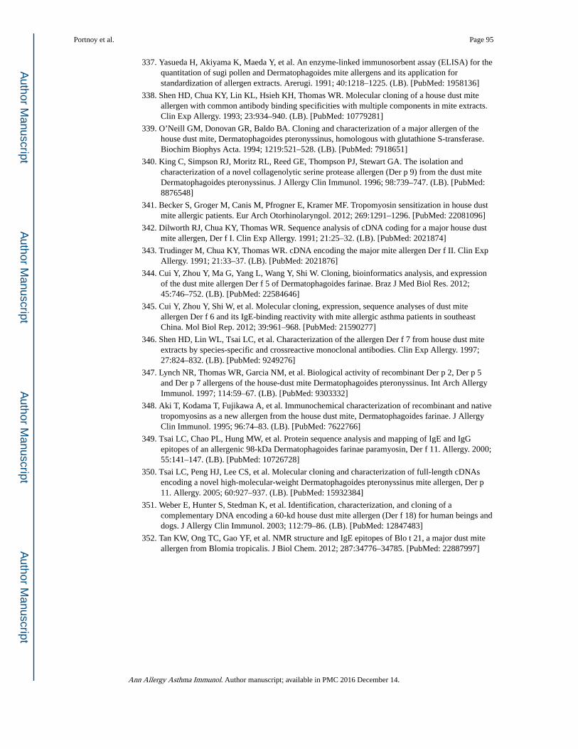
337. Yasueda H, Akiyama K, Maeda Y, et al. An enzyme-linked immunosorbent assay (ELISA) for the quantitation of sugi pollen and Dermatophagoides mite allergens and its application for standardization of allergen extracts. Arerugi. 1991; 40:1218–1225. (LB). [PubMed: 1958136]
338. Shen HD, Chua KY, Lin KL, Hsieh KH, Thomas WR. Molecular cloning of a house dust mite allergen with common antibody binding specificities with multiple components in mite extracts. Clin Exp Allergy. 1993; 23:934–940. (LB). [PubMed: 10779281]
339. O’Neill GM, Donovan GR, Baldo BA. Cloning and characterization of a major allergen of the house dust mite, Dermatophagoides pteronyssinus, homologous with glutathione S-transferase. Biochim Biophys Acta. 1994; 1219:521–528. (LB). [PubMed: 7918651]
340. King C, Simpson RJ, Moritz RL, Reed GE, Thompson PJ, Stewart GA. The isolation and characterization of a novel collagenolytic serine protease allergen (Der p 9) from the dust mite Dermatophagoides pteronyssinus. J Allergy Clin Immunol. 1996; 98:739–747. (LB). [PubMed: 8876548]
341. Becker S, Groger M, Canis M, Pfrogner E, Kramer MF. Tropomyosin sensitization in house dust mite allergic patients. Eur Arch Otorhinolaryngol. 2012; 269:1291–1296. [PubMed: 22081096]
342. Dilworth RJ, Chua KY, Thomas WR. Sequence analysis of cDNA coding for a major house dust mite allergen, Der f I. Clin Exp Allergy. 1991; 21:25–32. (LB). [PubMed: 2021874]
343. Trudinger M, Chua KY, Thomas WR. cDNA encoding the major mite allergen Der f II. Clin Exp Allergy. 1991; 21:33–37. (LB). [PubMed: 2021876]
344. Cui Y, Zhou Y, Ma G, Yang L, Wang Y, Shi W. Cloning, bioinformatics analysis, and expression of the dust mite allergen Der f 5 of Dermatophagoides farinae. Braz J Med Biol Res. 2012; 45:746–752. (LB). [PubMed: 22584646]
345. Cui Y, Zhou Y, Shi W, et al. Molecular cloning, expression, sequence analyses of dust mite allergen Der f 6 and its IgE-binding reactivity with mite allergic asthma patients in southeast China. Mol Biol Rep. 2012; 39:961–968. [PubMed: 21590277]
346. Shen HD, Lin WL, Tsai LC, et al. Characterization of the allergen Der f 7 from house dust mite extracts by species-specific and crossreactive monoclonal antibodies. Clin Exp Allergy. 1997; 27:824–832. (LB). [PubMed: 9249276]
347. Lynch NR, Thomas WR, Garcia NM, et al. Biological activity of recombinant Der p 2, Der p 5 and Der p 7 allergens of the house-dust mite Dermatophagoides pteronyssinus. Int Arch Allergy Immunol. 1997; 114:59–67. (LB). [PubMed: 9303332]
348. Aki T, Kodama T, Fujikawa A, et al. Immunochemical characterization of recombinant and native tropomyosins as a new allergen from the house dust mite, Dermatophagoides farinae. J Allergy Clin Immunol. 1995; 96:74–83. (LB). [PubMed: 7622766]
349. Tsai LC, Chao PL, Hung MW, et al. Protein sequence analysis and mapping of IgE and IgG epitopes of an allergenic 98-kDa Dermatophagoides farinae paramyosin, Der f 11. Allergy. 2000; 55:141–147. (LB). [PubMed: 10726728]
350. Tsai LC, Peng HJ, Lee CS, et al. Molecular cloning and characterization of full-length cDNAs encoding a novel high-molecular-weight Dermatophagoides pteronyssinus mite allergen, Der p 11. Allergy. 2005; 60:927–937. (LB). [PubMed: 15932384]
351. Weber E, Hunter S, Stedman K, et al. Identification, characterization, and cloning of a complementary DNA encoding a 60-kd house dust mite allergen (Der f 18) for human beings and dogs. J Allergy Clin Immunol. 2003; 112:79–86. (LB). [PubMed: 12847483]
352. Tan KW, Ong TC, Gao YF, et al. NMR structure and IgE epitopes of Blo t 21, a major dust mite allergen from Blomia tropicalis. J Biol Chem. 2012; 287:34776–34785. [PubMed: 22887997]
Portnoy et al. Page 95
Ann Allergy Asthma Immunol. Author manuscript; available in PMC 2016 December 14.
Author M
anuscriptA
uthor Manuscript
Author M
anuscriptA
uthor Manuscript
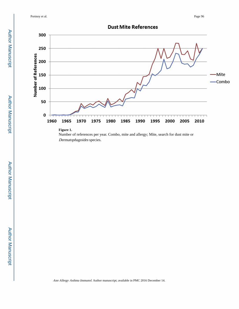
Figure 1. Number of references per year. Combo, mite and allergy; Mite, search for dust mite or
Dermatophagoides species.
Portnoy et al. Page 96
Ann Allergy Asthma Immunol. Author manuscript; available in PMC 2016 December 14.
Author M
anuscriptA
uthor Manuscript
Author M
anuscriptA
uthor Manuscript
Figure 2. Taxonomy of dust mites.
Portnoy et al. Page 97
Ann Allergy Asthma Immunol. Author manuscript; available in PMC 2016 December 14.
Author M
anuscriptA
uthor Manuscript
Author M
anuscriptA
uthor Manuscript

Figure 3. Taxonomy of dust mite species.
Portnoy et al. Page 98
Ann Allergy Asthma Immunol. Author manuscript; available in PMC 2016 December 14.
Author M
anuscriptA
uthor Manuscript
Author M
anuscriptA
uthor Manuscript

Figure 4. Images of dust mites, eggs, and feces. Copyright © Mission: Allergy, Inc. Used with
permission.
Portnoy et al. Page 99
Ann Allergy Asthma Immunol. Author manuscript; available in PMC 2016 December 14.
Author M
anuscriptA
uthor Manuscript
Author M
anuscriptA
uthor Manuscript
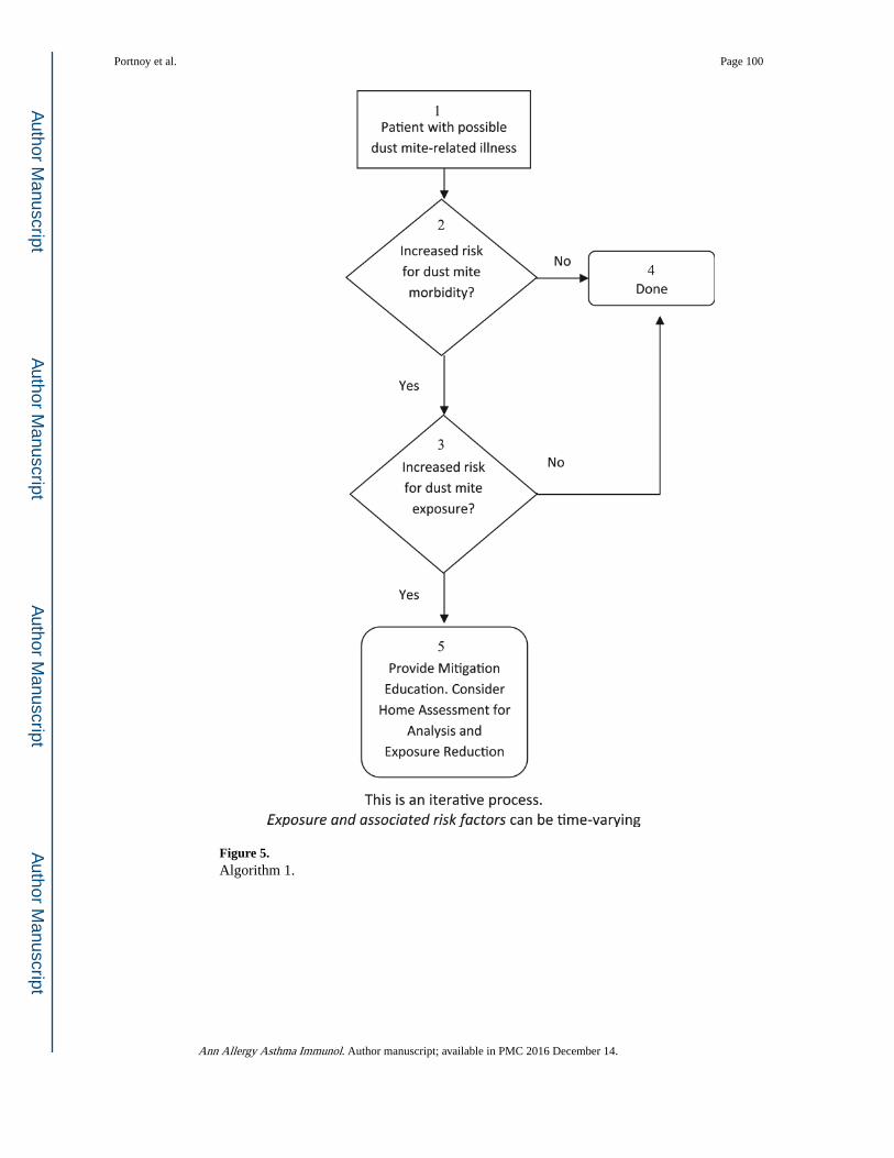
Figure 5. Algorithm 1.
Portnoy et al. Page 100
Ann Allergy Asthma Immunol. Author manuscript; available in PMC 2016 December 14.
Author M
anuscriptA
uthor Manuscript
Author M
anuscriptA
uthor Manuscript
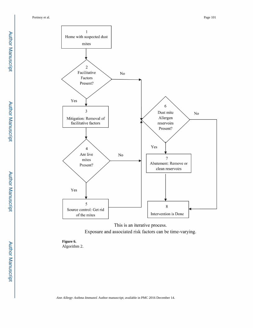
Figure 6. Algorithm 2.
Portnoy et al. Page 101
Ann Allergy Asthma Immunol. Author manuscript; available in PMC 2016 December 14.
Author M
anuscriptA
uthor Manuscript
Author M
anuscriptA
uthor Manuscript
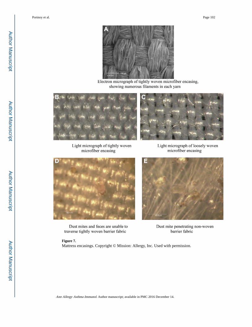
Figure 7. Mattress encasings. Copyright © Mission: Allergy, Inc. Used with permission.
Portnoy et al. Page 102
Ann Allergy Asthma Immunol. Author manuscript; available in PMC 2016 December 14.
Author M
anuscriptA
uthor Manuscript
Author M
anuscriptA
uthor Manuscript
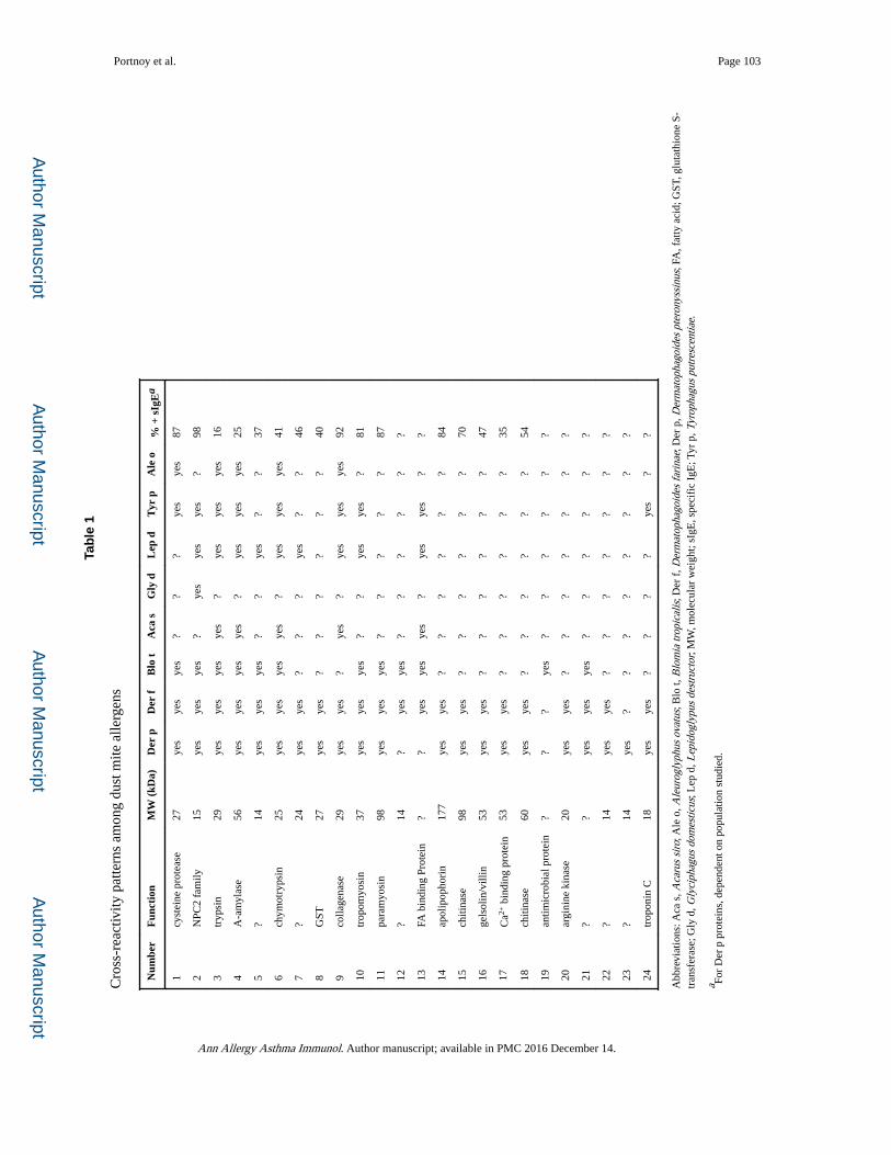
Author M
anuscriptA
uthor Manuscript
Author M
anuscriptA
uthor Manuscript
Portnoy et al. Page 103
Tab
le 1
Cro
ss-r
eact
ivity
pat
tern
s am
ong
dust
mite
alle
rgen
s
Num
ber
Fun
ctio
nM
W (
kDa)
Der
pD
er f
Blo
tA
ca s
Gly
dL
ep d
Tyr
pA
le o
% +
sIg
Ea
1cy
stei
ne p
rote
ase
27ye
sye
sye
s?
??
yes
yes
87
2N
PC2
fam
ily15
yes
yes
yes
?ye
sye
sye
s?
98
3tr
ypsi
n29
yes
yes
yes
yes
?ye
sye
sye
s16
4A
-am
ylas
e56
yes
yes
yes
yes
?ye
sye
sye
s25
5?
14ye
sye
sye
s?
?ye
s?
?37
6ch
ymot
ryps
in25
yes
yes
yes
yes
?ye
sye
sye
s41
7?
24ye
sye
s?
??
yes
??
46
8G
ST27
yes
yes
??
??
??
40
9co
llage
nase
29ye
sye
s?
yes
?ye
sye
sye
s92
10tr
opom
yosi
n37
yes
yes
yes
??
yes
yes
?81
11pa
ram
yosi
n98
yes
yes
yes
??
??
?87
12?
14?
yes
yes
??
??
??
13FA
bin
ding
Pro
tein
??
yes
yes
yes
?ye
sye
s?
?
14ap
olip
opho
rin
177
yes
yes
??
??
??
84
15ch
itina
se98
yes
yes
??
??
??
70
16ge
lsol
in/v
illin
53ye
sye
s?
??
??
?47
17C
a2+ b
indi
ng p
rote
in53
yes
yes
??
??
??
35
18ch
itina
se60
yes
yes
??
??
??
54
19an
timic
robi
al p
rote
in?
??
yes
??
??
??
20ar
gini
ne k
inas
e20
yes
yes
??
??
??
?
21?
?ye
sye
sye
s?
??
??
?
22?
14ye
sye
s?
??
??
??
23?
14ye
s?
??
??
??
?
24tr
opon
in C
18ye
sye
s?
??
?ye
s?
?
Abb
revi
atio
ns: A
ca s
, Aca
rus
siro
; Ale
o, A
leur
ogly
phus
ova
tus;
Blo
t, B
lom
ia tr
opic
alis
; Der
f, D
erm
atop
hago
ides
fari
nae;
Der
p, D
erm
atop
hago
ides
pte
rony
ssin
us; F
A, f
atty
aci
d; G
ST, g
luta
thio
ne S
-tr
ansf
eras
e; G
ly d
, Gly
ciph
agus
dom
estic
us; L
ep d
, Lep
idog
lypu
s de
stru
ctor
; MW
, mol
ecul
ar w
eigh
t; sI
gE, s
peci
fic
IgE
; Tyr
p, T
yrop
hagu
s pu
tres
cent
iae.
a For
Der
p p
rote
ins,
dep
ende
nt o
n po
pula
tion
stud
ied.
Ann Allergy Asthma Immunol. Author manuscript; available in PMC 2016 December 14.
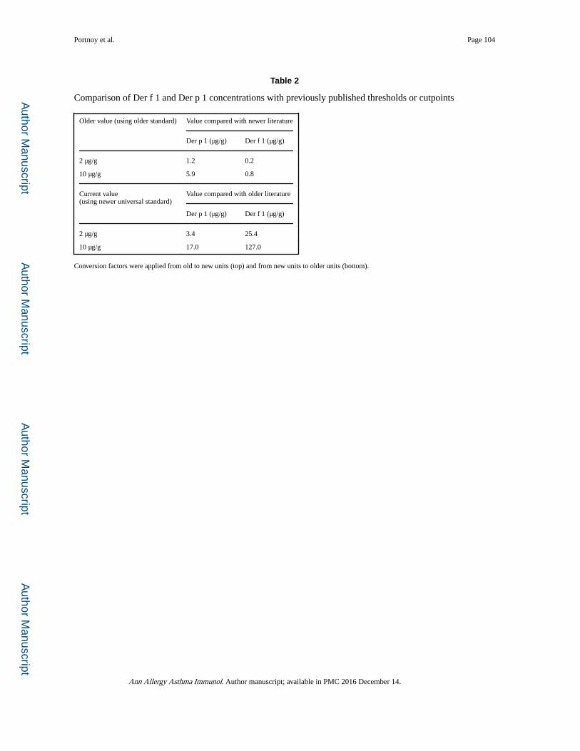
Author M
anuscriptA
uthor Manuscript
Author M
anuscriptA
uthor Manuscript
Portnoy et al. Page 104
Table 2
Comparison of Der f 1 and Der p 1 concentrations with previously published thresholds or cutpoints
Older value (using older standard) Value compared with newer literature
Der p 1 (µg/g) Der f 1 (µg/g)
2 µg/g 1.2 0.2
10 µg/g 5.9 0.8
Current value(using newer universal standard)
Value compared with older literature
Der p 1 (µg/g) Der f 1 (µg/g)
2 µg/g 3.4 25.4
10 µg/g 17.0 127.0
Conversion factors were applied from old to new units (top) and from new units to older units (bottom).
Ann Allergy Asthma Immunol. Author manuscript; available in PMC 2016 December 14.
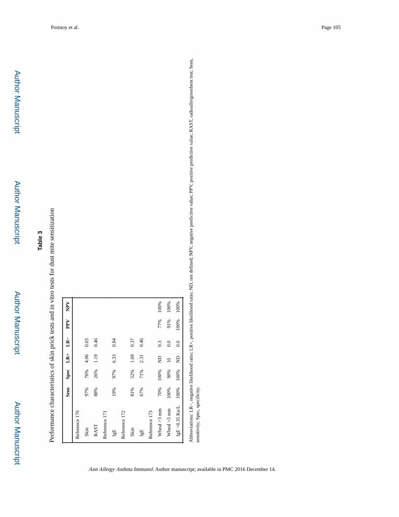
Author M
anuscriptA
uthor Manuscript
Author M
anuscriptA
uthor Manuscript
Portnoy et al. Page 105
Tab
le 3
Perf
orm
ance
cha
ract
eris
tics
of s
kin
pric
k te
sts
and
in v
itro
test
s fo
r du
st m
ite s
ensi
tizat
ion
Sens
Spec
LR
+L
R−
PP
VN
PV
Ref
eren
ce 1
70
Ski
n97
%76
%4.
060.
03
RA
ST88
%26
%1.
190.
46
Ref
eren
ce 1
71
IgE
19%
97%
6.33
0.84
Ref
eren
ce 1
72
Ski
n81
%52
%1.
690.
37
IgE
67%
71%
2.31
0.46
Ref
eren
ce 1
73
Whe
al >
3 m
m70
%10
0%N
D0.
377
%10
0%
Whe
al >
5 m
m10
0%90
%10
0.0
91%
100%
IgE
>0.
35 K
u/L
100%
100%
ND
0.0
100%
100%
Abb
revi
atio
ns: L
R−
, neg
ativ
e lik
elih
ood
ratio
; LR
+, p
ositi
ve li
kelih
ood
ratio
; ND
, not
def
ined
; NPV
, neg
ativ
e pr
edic
tive
valu
e; P
PV, p
ositi
ve p
redi
ctiv
e va
lue;
RA
ST, r
adio
alle
rgos
orbe
nt te
st; S
ens,
se
nsiti
vity
; Spe
c, s
peci
fici
ty.
Ann Allergy Asthma Immunol. Author manuscript; available in PMC 2016 December 14.
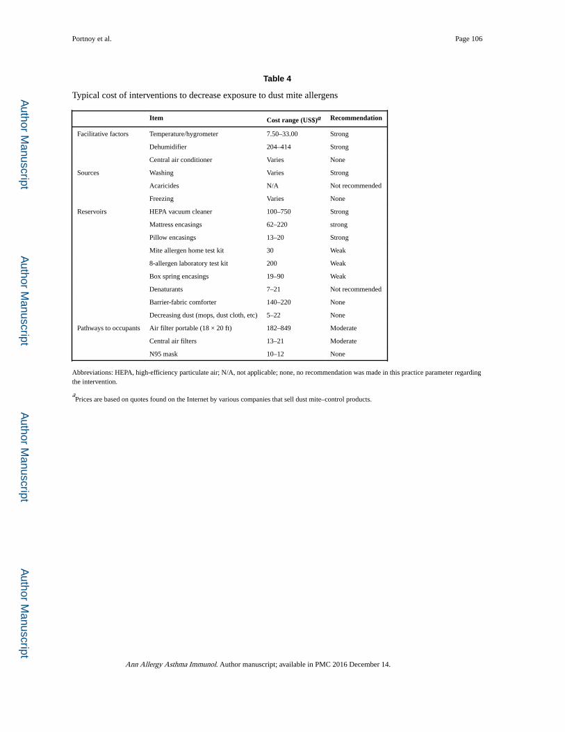
Author M
anuscriptA
uthor Manuscript
Author M
anuscriptA
uthor Manuscript
Portnoy et al. Page 106
Table 4
Typical cost of interventions to decrease exposure to dust mite allergens
Item Cost range (US$)a Recommendation
Facilitative factors Temperature/hygrometer 7.50–33.00 Strong
Dehumidifier 204–414 Strong
Central air conditioner Varies None
Sources Washing Varies Strong
Acaricides N/A Not recommended
Freezing Varies None
Reservoirs HEPA vacuum cleaner 100–750 Strong
Mattress encasings 62–220 strong
Pillow encasings 13–20 Strong
Mite allergen home test kit 30 Weak
8-allergen laboratory test kit 200 Weak
Box spring encasings 19–90 Weak
Denaturants 7–21 Not recommended
Barrier-fabric comforter 140–220 None
Decreasing dust (mops, dust cloth, etc) 5–22 None
Pathways to occupants Air filter portable (18 × 20 ft) 182–849 Moderate
Central air filters 13–21 Moderate
N95 mask 10–12 None
Abbreviations: HEPA, high-efficiency particulate air; N/A, not applicable; none, no recommendation was made in this practice parameter regarding the intervention.
aPrices are based on quotes found on the Internet by various companies that sell dust mite–control products.
Ann Allergy Asthma Immunol. Author manuscript; available in PMC 2016 December 14.