Enrolment in a Middle Ordovician agnostoid trilobite
8
Enrolment in a Middle Ordovician agnostoid trilobite DAVID L. BRUTON and HANS ARNE NAKREM Bruton, D.L. and Nakrem, H.A. 2005. Enrolment in a Middle Ordovician agnostoid trilobite. Acta Palaeontologica Polonica 50 (3): 441–448. Study of silicified material of Trinodus elspethi, from the Edinburg Formation, Virginia, USA, shows there is no gliding of distal parts of segments but an articulating furrow and opposing flange with prongs, acting as apodemes for muscle at− tachment, allowed cephalon and thorax to move as a single unit. Articulation between thorax and pygidium was more rigid with prongs from the thorax articulating in sockets on the pygidium. Support is given to the view that agnostoids lived partially enrolled with cephalon and pygidium gaping. They are unique in lacking an articulating half−ring between cephalon and thorax and the hinge joint is modified medially to provide an opening for what is interpreted as an exhalatory organ through which water passed during feeding and swimming. Peculiarities of the thorax are connected with the presence of only two thoracic segments articulating as a unit with distal tips directed anteriorly rather than poste− riorly. Otherwise articulating half rings are present in the rest of the thorax and pygidium and lack of articulating facets is not unique. It is concluded that agnostoids can be shown to be trilobites. Key words: Trilobita, Agnostida, life habit, preservation, Edinburg Formation, Ordovician, Virginia. David L. Bruton [[email protected]] and Hans Arne Nakrem [[email protected]], Universitetet i Oslo, Naturhistorisk museum – geologi, Boks 1172 Blindern, N−0318 Oslo, Norway. Introduction Silicified specimens of the agnostoid trilobite Trinodus els− pethi (Raymond, 1925), were used by Allan S. Hunt to illus− trate features of a cephalic ventral plate (Hunt 1966) and the nature of size increase, variation and instar development (Hunt 1967). In the latter paper complete, enrolled specimens were figured together with isolated specimens of the thoracic seg− ments but attention was not directed to them or their articula− tion. Hunt’s material was from various localities in the Middle Ordovician Edinburg Limestone of Virginia (Whittington 1959: 378–383), including locality 4 from which the present senior author made collections together with Harry B. Whit− tington in 1969. This is labelled “Locality W12, Edinburg For− mation 50 feet above the base”, W12 being equivalent to lo− cality 4, the Botetourt Member, lower part of the Edinburg Limestone in upper part of field northeast of Virginia State Highway 639, at a point 0.25 miles from its junction with U.S. Highway 11 (Harry B. Whittington, personal communication 11.10.2002). This material was etched in Oslo, mainly for teaching purposes. Using dilute hydrochloric acid, limestone blocks yielded a wealth of trilobite forms including Trinodus elspethi, represented by more than 50 cephala and pygidia, two completely enrolled exoskeletons and 10 isolated first thoracic segments. No second segments were found and the il− lustrated specimen figured herein was found in February 2004 among specimens in the Whittington Collection housed at the Sedgwick Museum, University of Cambridge, England. All this material affords a detailed study of the enrolment mecha− nism which is the object of this work. The subject of enrolment in agnostoids has been discussed by Robison (1972) and more recently has been reviewed by Whittington (1997: 56–57, fig. 48; and references) whose terminology is followed here. Additional terminology as applied to the Agnostina is from Shergold et al. (1990). The present material was photographed by the junior au− thor using a Jeol JSM−5200 scanning electron microscope and specimens were mounted on stubs and coated with gold palladium before being photographed. Institutional abbreviations.—All figured and reference ma− terial used in this study is housed in the collections of the for− mer Palaeontological Museum (abbreviated PMO with refer− ence numbers) now incorporated in the Geological Museum, University of Oslo. Description of material Dorsal and ventral views of the cephalon (Fig. 1A, B) are of an holaspid and show the characteristic notch in the postero− lateral border which first appears at the holaspid instar 1 stage (Hunt 1967: 208). Other holaspid features present are the anterior tapering of the glabella, the presence of a faint median preglabellar furrow and the short, adaxially directed posterolateral spines. In ventral view the doublure is widest antero−laterally, but narrows medially and towards the notch. Behind the latter it becomes ridge−like but broadens and slopes inwards at the base of the posterolateral spine. The latter is hollow. The in− ner margin of the doublure forms a sub−marginal suture which, when present, separates the doublure from the horse− shoe−shaped ventral plate which is missing here but is de− http://app.pan.pl/acta50/app50−441.pdf Acta Palaeontol. Pol. 50 (3): 441–448, 2005
-
Upload
independent -
Category
Documents
-
view
0 -
download
0
Transcript of Enrolment in a Middle Ordovician agnostoid trilobite
Enrolment in a Middle Ordovician agnostoid trilobite
DAVID L. BRUTON and HANS ARNE NAKREM
Bruton, D.L. and Nakrem, H.A. 2005. Enrolment in a Middle Ordovician agnostoid trilobite. Acta PalaeontologicaPolonica 50 (3): 441–448.
Study of silicified material of Trinodus elspethi, from the Edinburg Formation, Virginia, USA, shows there is no glidingof distal parts of segments but an articulating furrow and opposing flange with prongs, acting as apodemes for muscle at−tachment, allowed cephalon and thorax to move as a single unit. Articulation between thorax and pygidium was morerigid with prongs from the thorax articulating in sockets on the pygidium. Support is given to the view that agnostoidslived partially enrolled with cephalon and pygidium gaping. They are unique in lacking an articulating half−ring betweencephalon and thorax and the hinge joint is modified medially to provide an opening for what is interpreted as anexhalatory organ through which water passed during feeding and swimming. Peculiarities of the thorax are connectedwith the presence of only two thoracic segments articulating as a unit with distal tips directed anteriorly rather than poste−riorly. Otherwise articulating half rings are present in the rest of the thorax and pygidium and lack of articulating facets isnot unique. It is concluded that agnostoids can be shown to be trilobites.
Key words: Trilobita, Agnostida, life habit, preservation, Edinburg Formation, Ordovician, Virginia.
David L. Bruton [[email protected]] and Hans Arne Nakrem [[email protected]], Universitetet i Oslo,Naturhistorisk museum – geologi, Boks 1172 Blindern, N−0318 Oslo, Norway.
Introduction
Silicified specimens of the agnostoid trilobite Trinodus els−pethi (Raymond, 1925), were used by Allan S. Hunt to illus−trate features of a cephalic ventral plate (Hunt 1966) and thenature of size increase, variation and instar development (Hunt1967). In the latter paper complete, enrolled specimens werefigured together with isolated specimens of the thoracic seg−ments but attention was not directed to them or their articula−tion. Hunt’s material was from various localities in the MiddleOrdovician Edinburg Limestone of Virginia (Whittington1959: 378–383), including locality 4 from which the presentsenior author made collections together with Harry B. Whit−tington in 1969. This is labelled “Locality W12, Edinburg For−mation 50 feet above the base”, W12 being equivalent to lo−cality 4, the Botetourt Member, lower part of the EdinburgLimestone in upper part of field northeast of Virginia StateHighway 639, at a point 0.25 miles from its junction with U.S.Highway 11 (Harry B. Whittington, personal communication11.10.2002). This material was etched in Oslo, mainly forteaching purposes. Using dilute hydrochloric acid, limestoneblocks yielded a wealth of trilobite forms including Trinoduselspethi, represented by more than 50 cephala and pygidia,two completely enrolled exoskeletons and 10 isolated firstthoracic segments. No second segments were found and the il−lustrated specimen figured herein was found in February 2004among specimens in the Whittington Collection housed at theSedgwick Museum, University of Cambridge, England. Allthis material affords a detailed study of the enrolment mecha−nism which is the object of this work. The subject of enrolmentin agnostoids has been discussed by Robison (1972) and more
recently has been reviewed by Whittington (1997: 56–57, fig.48; and references) whose terminology is followed here.Additional terminology as applied to the Agnostina is fromShergold et al. (1990).
The present material was photographed by the junior au−thor using a Jeol JSM−5200 scanning electron microscopeand specimens were mounted on stubs and coated with goldpalladium before being photographed.
Institutional abbreviations.—All figured and reference ma−terial used in this study is housed in the collections of the for−mer Palaeontological Museum (abbreviated PMO with refer−ence numbers) now incorporated in the Geological Museum,University of Oslo.
Description of materialDorsal and ventral views of the cephalon (Fig. 1A, B) are ofan holaspid and show the characteristic notch in the postero−lateral border which first appears at the holaspid instar 1stage (Hunt 1967: 208). Other holaspid features present arethe anterior tapering of the glabella, the presence of a faintmedian preglabellar furrow and the short, adaxially directedposterolateral spines.
In ventral view the doublure is widest antero−laterally, butnarrows medially and towards the notch. Behind the latter itbecomes ridge−like but broadens and slopes inwards at thebase of the posterolateral spine. The latter is hollow. The in−ner margin of the doublure forms a sub−marginal suturewhich, when present, separates the doublure from the horse−shoe−shaped ventral plate which is missing here but is de−
http://app.pan.pl/acta50/app50−441.pdfActa Palaeontol. Pol. 50 (3): 441–448, 2005
scribed in detail by Hunt (1966: 1238, fig. 1). In lateral viewthe occipital band slopes steeply downwards (Fig. 2A1) and
in dorsal view the posterior margin of the cephalon is straightto weakly curved, medially. In ventral view, the inner edge is
442 ACTA PALAEONTOLOGICA POLONICA 50 (3), 2005
postero-lateralnotch
thoracic recessanteriorprong
posterior prong
anterior flange
muscle scar
articulating socket
doublure flange
muscle scar
anterior prong
1 mm
anterior prong
1 mm
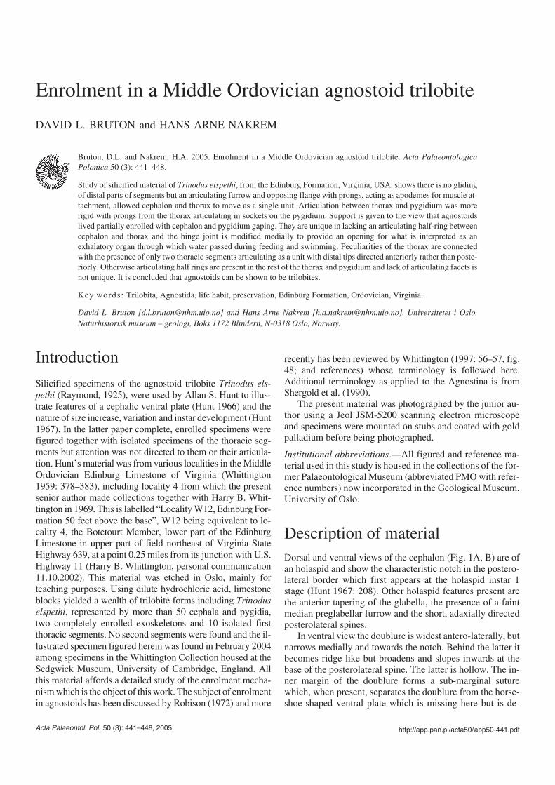
Fig. 1. Trinodus elspethi (Raymond, 1925), silicified specimens from Edinburg Formation, Virginia, USA. A. PMO 206.303/1, dorsal view of holaspidcephalon. B. PMO 206.303/2, ventral view of holaspid cephalon; the posterolateral notch accommodates the anterior border of the pygidium during enrol−ment. C. PMO 206.304/1, dorsal view of first thoracic segment. D. PMO 206.304/2, ventral view of first thoracic segment. E. PMO 206.305, dorsal view ofsecond thoracic segment. F. Ventral view of second thoracic segment; note articulating half−ring. G. PMO 206.306/1, dorsal view of pygidium. H. PMO206.306/2, ventral view of pygidium with socket for posterior prong of second thoracic segment.
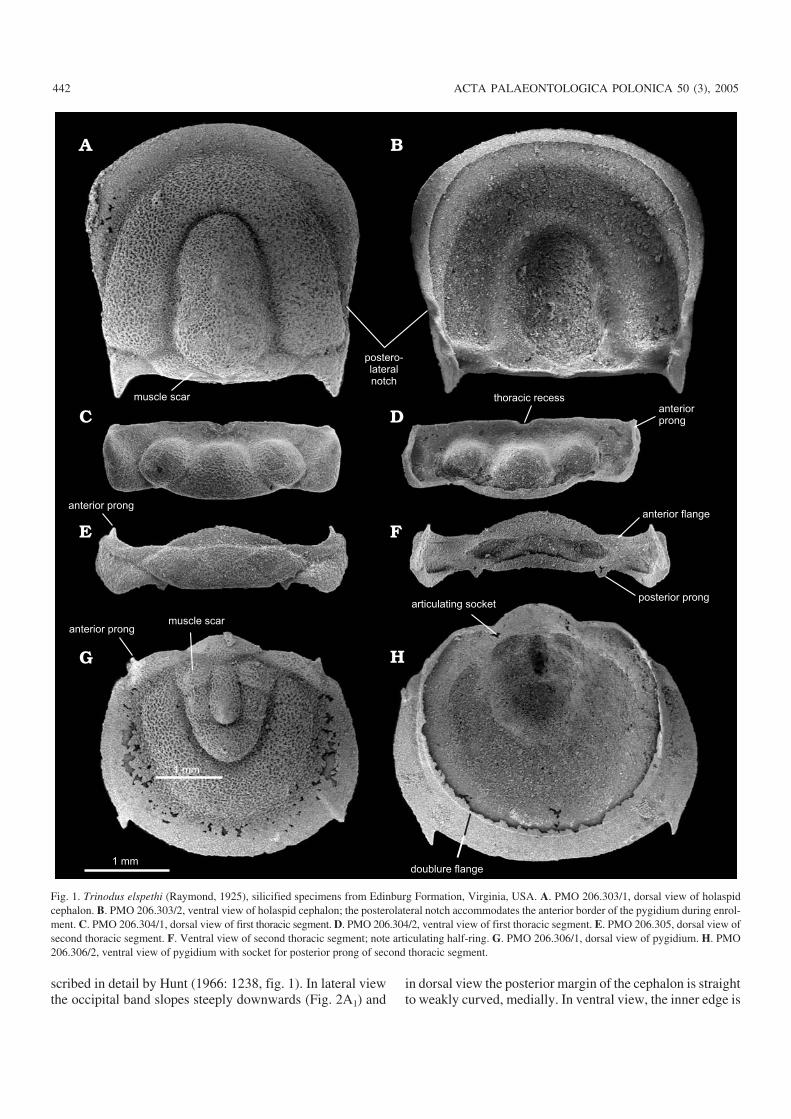
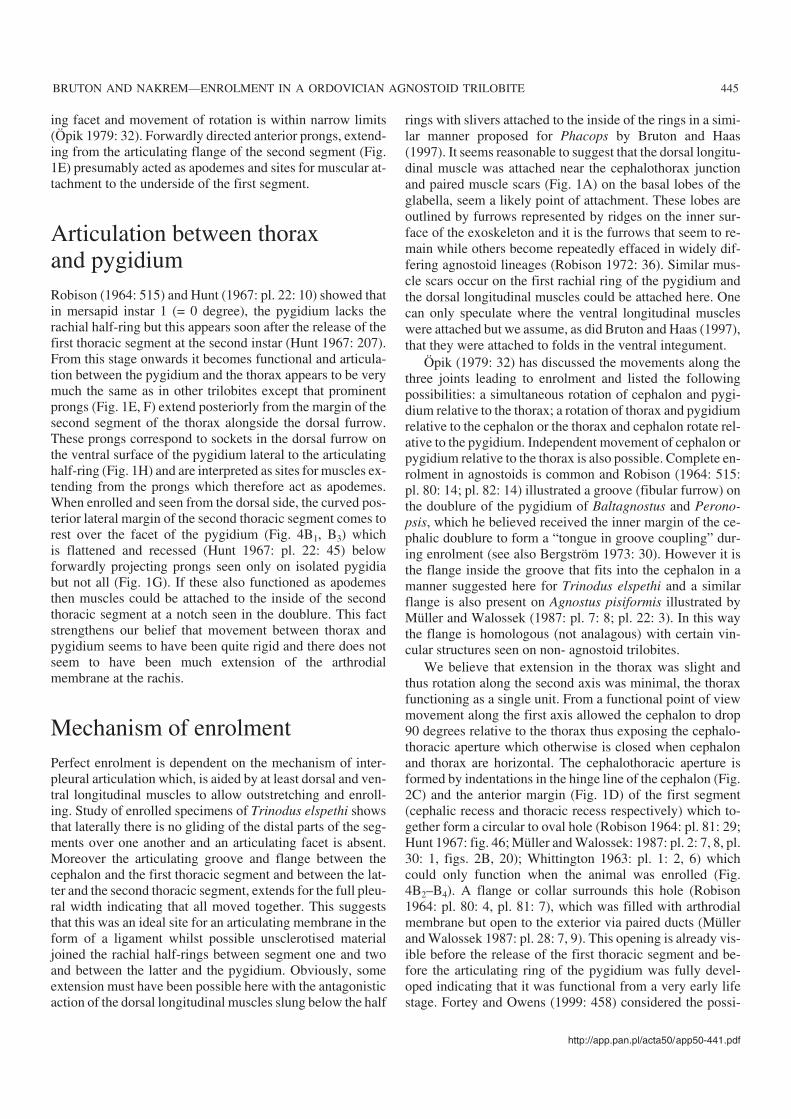
rounded (Fig. 2C) and a semicircular notch (cephalic recess)occurs medially. The rolled edge fits into a corresponding ar−ticulating groove in the anterior margin of the first thoracicsegment (Figs. 2B2, 3C ) and when segment and cephalon arejoined and the thorax is enrolled, the juxtaposed notchesform a circular opening, the cephalothoracic aperture (Fig.4B2–B4). Note here, there is no articulating half−ring on thefirst thoracic segment and thus no corresponding occipitalring on the cephalon.
In dorsal view the first thoracic segment is rectangular inoutline, with a trapezoid−shaped median lobe and roundedlateral lobes (Fig. 1C). There is no clear differentiation intoanterior and posterior bands but there is an antero−lateralchange of slope where the tip of the pleura is inflated andcurved steeply downwards (Fig. 3B) extending inwards onthe ventral side as a narrow outwardly sloping doublure withanterior prong (Fig. 1D). The doublure continues poste−rior−laterally, becoming broader from the dorsal furrow to−wards the mid−line. During enrolment the distal end of thesegment tucks inside the genal angle with a flange (Fig. 3B)and prong sliding into the cavity at the base of the postero−lateral spine.
The second thoracic segment (Fig. 1E, F) differs fromthat of the first in possessing a broad articulating half−ringwhich fits under the arched rachis of the preceding first seg−ment (Fig. 2B1). Laterally, along the anterior margin there isan articulating flange (Fig. 2D) which fits into a correspond−ing groove along the posterolateral edge of the first segment(Figs. 2B1, 3A).
The posterior band is flap−like and separated by a weakpleural furrow from a narrow anterior band (Fig. 1E). Themedian and lateral lobes are less inflated than on the first seg−ment. The posterior band is inflated dorsally but a change ofslope separates this from a thinner distal area which slopessteeply downwards ventrally (Figs. 1F, 2D). This area ex−tends forward as a prong at the distal end of the articulatingflange and rests in a depression at the base of the postero−lateral spine while the ventral side, nestles in the concavearea lateral to the spine (Figs. 1B, 2C).
From the posterior margin of the second thoracic segment,at each side of the rachis, are posteriorly−directed spines, orprongs (Fig. 1E, F). These, slot into small depressions at thedistal end of the articulating furrow of the pygidium (Fig. 1H).Prongs can also project forwards from the facet of the
http://app.pan.pl/acta50/app50−441.pdf
BRUTON AND NAKREM—ENROLMENT IN A ORDOVICIAN AGNOSTOID TRILOBITE 443
cephalic recess
anterior flange
anterior flange
articulating groove
Fig. 2. Trinodus elspethi (Raymond, 1925), silicified specimens from Edinburg Formation, Virginia, USA. A. PMO 206.307. Lateral (A1) and oblique lat−eral (A2) views of completely enrolled specimen. Note attached fragment of a bryozoan on the pygidium. B. PMO 206.304/1. B1. Posterior view of first tho−racic segment showing articulating groove, which receives the articulating flange on the anterior margin of the second segment (Fig. 2D). B2. Anterior viewof first thoracic segment showing articulating groove along anterior margin. This groove receives the rolled posterior margin of the cephalon (Fig. 2C) in theabsence of an articulating half−ring. C. PMO 206.303/2. Detail of rolled posterior margin of the cephalon with cephalic recess. D. PMO 206.305. Anteriorview of second thoracic segment with articulating flange. E. Ventral view of second thoracic segment showing anterior margin with articulating half−ringand prongs from posterior margin. Scale bars 1 mm.
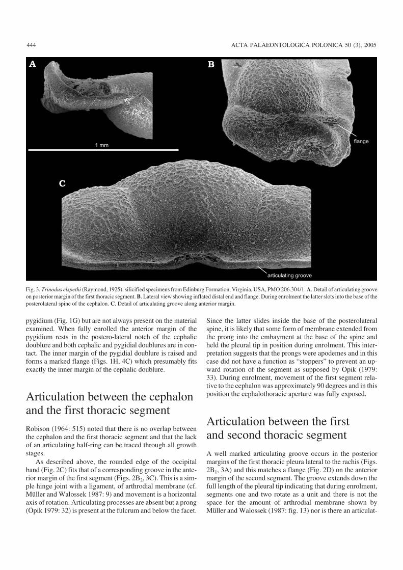
pygidium (Fig. 1G) but are not always present on the materialexamined. When fully enrolled the anterior margin of thepygidium rests in the postero−lateral notch of the cephalicdoublure and both cephalic and pygidial doublures are in con−tact. The inner margin of the pygidial doublure is raised andforms a marked flange (Figs. 1H, 4C) which presumably fitsexactly the inner margin of the cephalic doublure.
Articulation between the cephalonand the first thoracic segmentRobison (1964: 515) noted that there is no overlap betweenthe cephalon and the first thoracic segment and that the lackof an articulating half−ring can be traced through all growthstages.
As described above, the rounded edge of the occipitalband (Fig. 2C) fits that of a corresponding groove in the ante−rior margin of the first segment (Figs. 2B2, 3C). This is a sim−ple hinge joint with a ligament, of arthrodial membrane (cf.Müller and Walossek 1987: 9) and movement is a horizontalaxis of rotation. Articulating processes are absent but a prong(Öpik 1979: 32) is present at the fulcrum and below the facet.
Since the latter slides inside the base of the posterolateralspine, it is likely that some form of membrane extended fromthe prong into the embayment at the base of the spine andheld the pleural tip in position during enrolment. This inter−pretation suggests that the prongs were apodemes and in thiscase did not have a function as “stoppers” to prevent an up−ward rotation of the segment as supposed by Öpik (1979:33). During enrolment, movement of the first segment rela−tive to the cephalon was approximately 90 degrees and in thisposition the cephalothoracic aperture was fully exposed.
Articulation between the firstand second thoracic segment
A well marked articulating groove occurs in the posteriormargins of the first thoracic pleura lateral to the rachis (Figs.2B1, 3A) and this matches a flange (Fig. 2D) on the anteriormargin of the second segment. The groove extends down thefull length of the pleural tip indicating that during enrolment,segments one and two rotate as a unit and there is not thespace for the amount of arthrodial membrane shown byMüller and Walossek (1987: fig. 13) nor is there an articulat−
444 ACTA PALAEONTOLOGICA POLONICA 50 (3), 2005
flange
articulating groove
1 mm
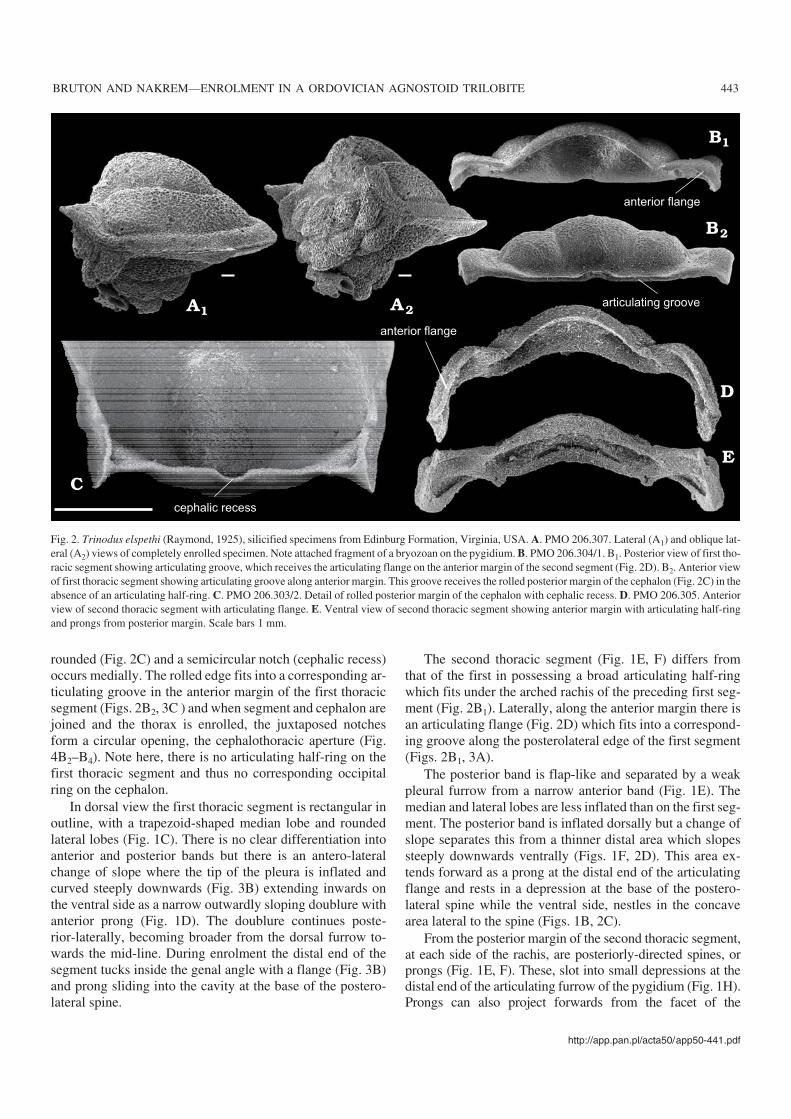
Fig. 3. Trinodus elspethi (Raymond, 1925), silicified specimens from Edinburg Formation, Virginia, USA, PMO 206.304/1. A. Detail of articulating grooveon posterior margin of the first thoracic segment. B. Lateral view showing inflated distal end and flange. During enrolment the latter slots into the base of theposterolateral spine of the cephalon. C. Detail of articulating groove along anterior margin.
ing facet and movement of rotation is within narrow limits(Öpik 1979: 32). Forwardly directed anterior prongs, extend−ing from the articulating flange of the second segment (Fig.1E) presumably acted as apodemes and sites for muscular at−tachment to the underside of the first segment.
Articulation between thoraxand pygidiumRobison (1964: 515) and Hunt (1967: pl. 22: 10) showed thatin mersapid instar 1 (= 0 degree), the pygidium lacks therachial half−ring but this appears soon after the release of thefirst thoracic segment at the second instar (Hunt 1967: 207).From this stage onwards it becomes functional and articula−tion between the pygidium and the thorax appears to be verymuch the same as in other trilobites except that prominentprongs (Fig. 1E, F) extend posteriorly from the margin of thesecond segment of the thorax alongside the dorsal furrow.These prongs correspond to sockets in the dorsal furrow onthe ventral surface of the pygidium lateral to the articulatinghalf−ring (Fig. 1H) and are interpreted as sites for muscles ex−tending from the prongs which therefore act as apodemes.When enrolled and seen from the dorsal side, the curved pos−terior lateral margin of the second thoracic segment comes torest over the facet of the pygidium (Fig. 4B1, B3) whichis flattened and recessed (Hunt 1967: pl. 22: 45) belowforwardly projecting prongs seen only on isolated pygidiabut not all (Fig. 1G). If these also functioned as apodemesthen muscles could be attached to the inside of the secondthoracic segment at a notch seen in the doublure. This factstrengthens our belief that movement between thorax andpygidium seems to have been quite rigid and there does notseem to have been much extension of the arthrodialmembrane at the rachis.
Mechanism of enrolmentPerfect enrolment is dependent on the mechanism of inter−pleural articulation which, is aided by at least dorsal and ven−tral longitudinal muscles to allow outstretching and enroll−ing. Study of enrolled specimens of Trinodus elspethi showsthat laterally there is no gliding of the distal parts of the seg−ments over one another and an articulating facet is absent.Moreover the articulating groove and flange between thecephalon and the first thoracic segment and between the lat−ter and the second thoracic segment, extends for the full pleu−ral width indicating that all moved together. This suggeststhat this was an ideal site for an articulating membrane in theform of a ligament whilst possible unsclerotised materialjoined the rachial half−rings between segment one and twoand between the latter and the pygidium. Obviously, someextension must have been possible here with the antagonisticaction of the dorsal longitudinal muscles slung below the half
rings with slivers attached to the inside of the rings in a simi−lar manner proposed for Phacops by Bruton and Haas(1997). It seems reasonable to suggest that the dorsal longitu−dinal muscle was attached near the cephalothorax junctionand paired muscle scars (Fig. 1A) on the basal lobes of theglabella, seem a likely point of attachment. These lobes areoutlined by furrows represented by ridges on the inner sur−face of the exoskeleton and it is the furrows that seem to re−main while others become repeatedly effaced in widely dif−fering agnostoid lineages (Robison 1972: 36). Similar mus−cle scars occur on the first rachial ring of the pygidium andthe dorsal longitudinal muscles could be attached here. Onecan only speculate where the ventral longitudinal muscleswere attached but we assume, as did Bruton and Haas (1997),that they were attached to folds in the ventral integument.
Öpik (1979: 32) has discussed the movements along thethree joints leading to enrolment and listed the followingpossibilities: a simultaneous rotation of cephalon and pygi−dium relative to the thorax; a rotation of thorax and pygidiumrelative to the cephalon or the thorax and cephalon rotate rel−ative to the pygidium. Independent movement of cephalon orpygidium relative to the thorax is also possible. Complete en−rolment in agnostoids is common and Robison (1964: 515:pl. 80: 14; pl. 82: 14) illustrated a groove (fibular furrow) onthe doublure of the pygidium of Baltagnostus and Perono−psis, which he believed received the inner margin of the ce−phalic doublure to form a “tongue in groove coupling” dur−ing enrolment (see also Bergström 1973: 30). However it isthe flange inside the groove that fits into the cephalon in amanner suggested here for Trinodus elspethi and a similarflange is also present on Agnostus pisiformis illustrated byMüller and Walossek (1987: pl. 7: 8; pl. 22: 3). In this waythe flange is homologous (not analagous) with certain vin−cular structures seen on non− agnostoid trilobites.
We believe that extension in the thorax was slight andthus rotation along the second axis was minimal, the thoraxfunctioning as a single unit. From a functional point of viewmovement along the first axis allowed the cephalon to drop90 degrees relative to the thorax thus exposing the cephalo−thoracic aperture which otherwise is closed when cephalonand thorax are horizontal. The cephalothoracic aperture isformed by indentations in the hinge line of the cephalon (Fig.2C) and the anterior margin (Fig. 1D) of the first segment(cephalic recess and thoracic recess respectively) which to−gether form a circular to oval hole (Robison 1964: pl. 81: 29;Hunt 1967: fig. 46; Müller and Walossek: 1987: pl. 2: 7, 8, pl.30: 1, figs. 2B, 20); Whittington 1963: pl. 1: 2, 6) whichcould only function when the animal was enrolled (Fig.4B2–B4). A flange or collar surrounds this hole (Robison1964: pl. 80: 4, pl. 81: 7), which was filled with arthrodialmembrane but open to the exterior via paired ducts (Müllerand Walossek 1987: pl. 28: 7, 9). This opening is already vis−ible before the release of the first thoracic segment and be−fore the articulating ring of the pygidium was fully devel−oped indicating that it was functional from a very early lifestage. Fortey and Owens (1999: 458) considered the possi−
http://app.pan.pl/acta50/app50−441.pdf
BRUTON AND NAKREM—ENROLMENT IN A ORDOVICIAN AGNOSTOID TRILOBITE 445
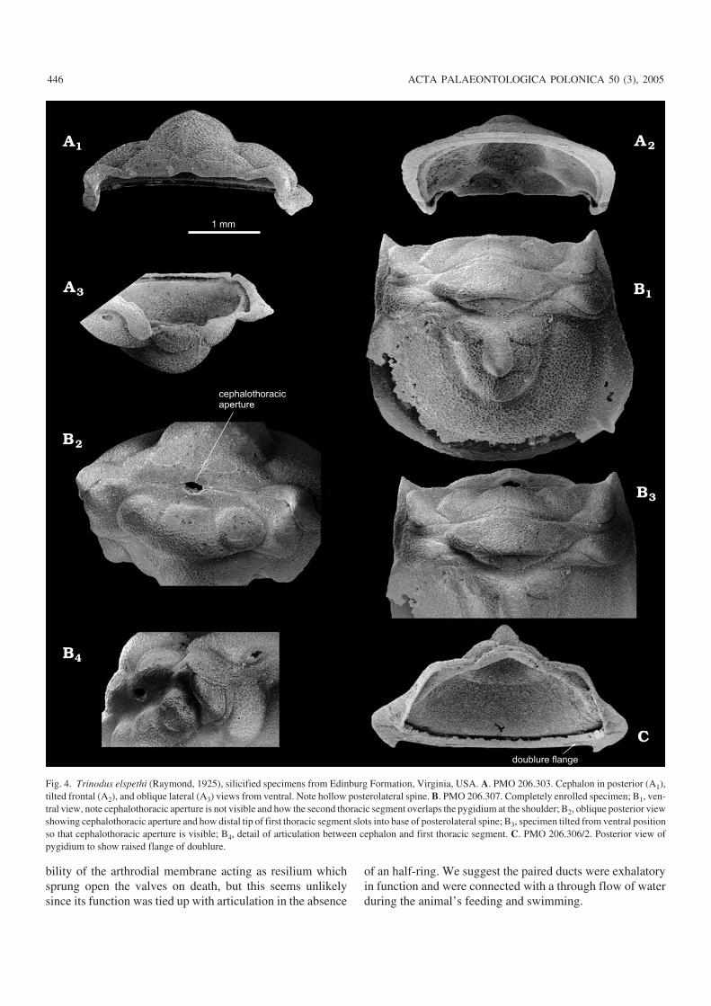
bility of the arthrodial membrane acting as resilium whichsprung open the valves on death, but this seems unlikelysince its function was tied up with articulation in the absence
of an half−ring. We suggest the paired ducts were exhalatoryin function and were connected with a through flow of waterduring the animal’s feeding and swimming.
446 ACTA PALAEONTOLOGICA POLONICA 50 (3), 2005
doublure flange
cephalothoracicaperture
1 mm
Fig. 4. Trinodus elspethi (Raymond, 1925), silicified specimens from Edinburg Formation, Virginia, USA. A. PMO 206.303. Cephalon in posterior (A1),tilted frontal (A2), and oblique lateral (A3) views from ventral. Note hollow posterolateral spine. B. PMO 206.307. Completely enrolled specimen; B1, ven−tral view, note cephalothoracic aperture is not visible and how the second thoracic segment overlaps the pygidium at the shoulder; B2, oblique posterior viewshowing cephalothoracic aperture and how distal tip of first thoracic segment slots into base of posterolateral spine; B3, specimen tilted from ventral positionso that cephalothoracic aperture is visible; B4, detail of articulation between cephalon and first thoracic segment. C. PMO 206.306/2. Posterior view ofpygidium to show raised flange of doublure.

Movement along the third axis was more than 90 degreesto allow contact between the doublures of cephalon andpygidium and to bring the anterior margin of the pygidium tolock into the cephalic notch. Rotation and/or extension alongthe second axis was slight and the distal tip of the secondpleural segment filled exactly any remaining space betweencephalon and pygidium (Fig. 5).
Enrolment in Trinodus elspethi appears to have beenfunctionally well developed with the described coaptativefeatures of the exoskeleton allowing for complete and exactfit of juxtaposed parts. Articulating notches and grooves onthe cephalic doublure to receive structures of the plural tipsand pygidia, together with prongs functioning as apodemesfor adductor muscles, are features preserved because the ma−terial studied is finely silicified. It is a challenge to see if suchfeatures can be identified on non−silicified, more primitive,Cambrian agnostoids for only then can one attempt a com−parison of primitive and derived structures.
Life habits of agnostoidsFortey and Owens (1999) provide an excellent summary ofthe various views regarding the mode of life of agnostoidtrilobites which they claim is controversial and difficult to in−terpret. They concluded that while the evidence is still equiv−ocal it seems likely that a range of life habits best accountsfor the diverse morphologies these small arthropods display.There is no denying that the group exhibits great taxonomicvariety, even within a single horizon, but common to all is asimilar mode of enrolment and the presence of cephalo−thoracic aperture which can only function when the animal isenrolled and becomes closed when it is outstretched. Thuswe support the view of Robison (1972) and Müller andWalossek (1987) that agnostoids spent much of their life par−tially enrolled and fed ostracod−like with the cephalon andpygidium gaping. Fortey and Owens (1999: 458) argue
against this form of life orientation and produce interestingfield evidence including the study of agnostoid rich horizonsand apparent lack of enrolled specimens which one wouldnot expect if this was the normal life style. We too are accus−tomed to studying agnostid coquinas, albeit from the LateCambrian of Scandinavia, and are aware of the paucity of en−rolled specimens here. Otherwise the abundance of freecephala and pygidia in the Scandinavian coquinas are currentswept parts from sites used during periods of moulting andthey occur almost to the exclusion of thoracic segments andenrolled individuals. When found, the latter are tightlyclosed and could represent individuals that died in this posi−tion. We have examined material from several localitieswhere ‘complete,’ outstretched, agnostoids are preserved, in−cluding a small collection of Triplagnostus from the MiddleCambrian Burgess Shale, Canada and uncompressed speci−mens of Ptychagnostus and Lejopyge from the Middle Cam−brian of the Oslo Region. In all these specimens, both tho−racic segments are conjoined and attached to the pygidium,but are separated from the cephalon. This separation is hardlynoticeable and is no more than to be expected if, after death,the thorax is outstretched. The observed separation suggeststhat the outstretched position is incompatible with the formof articulation between the cephalon and the thorax and thatcomplete enrolment or partial enrolment is the natural form.
That complete enrolment was important to the agnostoidis evident from the presence of the vincular structures onboth cephalon and pygidium allowing these to shut tightlysupposedly in response to changes in the environment suchas an abrution event or increase in bottom currents whichproduced the agnostoid coquinas.
We agree with Müller and Walossek (1987) and Forteyand Owens (1999) that agnostoids must have fed on organicparticles concomitant with their small size and that such foodcould be obtained in a benthic flocculent zone rich in detritalmatter (Muller and Walossek 1987: 42). Here they could befully enrolled while resting or in times of sediment distur−
http://app.pan.pl/acta50/app50−441.pdf
BRUTON AND NAKREM—ENROLMENT IN A ORDOVICIAN AGNOSTOID TRILOBITE 447
Fig. 5. Reconstruction of Trinodus elspethi (Raymond, 1925), Edinburg Formation, Virginia, U.S.A. A. Oblique posterior lateral view of completely en−rolled individual. B. Partial pull−apart from anterior, to show articulation between cephalon and thorax. Drawn by Bogdan Bocianowski.

bance but, when feeding, we suggest the anterior margin wasgaping and a stream of water brought food to the mouth aidedby movement of the appendages and a suction effect throughthe cephalothoracic aperture. Swimming within the watercolumn could have been affected by both leg and gill activitytogether with forward movement caused by the exhalent wa−ter through the cephalothoracic aperture. In this way we sup−port Robison (1972; 1975) that agnostoids also enjoyed anactive planktic life within the water column and this is sup−ported by their widespread geographical distribution.
ConclusionsAgnostoids− trilobites or not? Fortey ( 2001: 1143) posed thisquestion after these small, blind arthropods with two bodysegments had been accepted as trilobites in the revised Trea−tise on Invertebrate Paleontology (Whittington et al. 1997).Resser (1938) questioned whether they really were trilobitesand since the discovery by Müller and Walossek (1987) ofappendages in immature specimens of Agnostus pisiformisfrom the Upper Cambrian, which seem to differ from thoseknown for trilobites, these authors and others (Ramsköld andEdgecombe 1991; Maas et al. 2003; Stein et al. 2005) havesuggested that the Agnostida cannot be in−group trilobitesbut are derivatives of the early evolutionary lineage towardsthe Crustacea. We agree with Fortey (2001) that the questionof the limbs, as interpreted from immature and miniscule ma−terial, is doubtful all the time we do not know what theymight have been like in an adult specimen, nor do we havelimbs from older Cambrian agnostoids. When we considerthe exoskeleton, agnostoids are unique in that they do notpossess an articulating half ring between cephalon and firstsegment and the hinge joint here is modified medially to pro−vide an opening for what we here interpret as an exhalatoryorgan. Perhaps the absence of the half− ring is connected withthis since normal articulating half−rings are functional be−tween the first and second thoracic segments and between thelatter and the pygidium. A lack of an articulating facet is notunique and any differences in articulation within the thoraxare connected with the fact that only two segments are pres−ent to fill the space between cephalon and pygidium when theanimal enrolls. These differences include the fact that the ar−ticulated segments move as a unit and their distal tips are di−rected anteriorly rather than posteriorly, which is the case inthe majority of polymeroid trilobites. There is no doubt thatthe agnostoids are very special but, together with the eodis−coids, it is possible to combine the superfamilies Agnostinaand Eodiscina in an analysis which shows them to be trilo−bites (Fortey and Theron 1994; Fortey 2001).
AcknowledgementsBruton thanks Harry B. Whittington (Cambridge, England) for pastguidance in the field and for recently helping to find additional material
in his collections of silicified specimens, access to which was madepossible by Mike Dorling (Sedgwick Museum, University of Cam−bridge, England). Whittington and Winfried Haas (University of Bonn)are thanked for their advice while this work was being written and theformer suggested amendments to the manuscript. We are most gratefulto Richard A. Robison and Per Ahlberg for their referee comments,which very much helped to improve this presentation.
ReferencesBergström, J. 1973. Organization, life, and systematics of trilobites. Fossils
& Strata 2: 1–69.Bruton, D.L. and Haas, W. 1997. Functional morphology of Phacopinae
(Trilobita) and the mechanics of enrolment Palaeontographica A 245:1–43.
Fortey, R.A. 2001. Trilobite systematics: the last 75 years. Journal of Pale−ontology 75: 1141–1151.
Fortey, R.A and Owens, R.M. 1999. Feeding habits in trilobites. Palaeon−tology 42: 429–465.
Fortey, R.A. and Theron, J. 1994. A new Ordovician arthropod Soomaspisand the agnostid problem. Palaeontology 37: 841–861.
Hunt, A.S. 1966. Submarginal suture and ventral plate of an agnostid trilo−bite. Journal of Paleontology 40: 1238–1240.
Hunt, A.S. 1967. Growth, variation and instar development of an agnostidtrilobite. Journal of Paleontology 41: 203–208.
Maas, A., Waloszek, D., and Müller, K.J. 2003. Morphology, ontogeny andphylogeny of the Phosphatocopina (Crustacea) from the Upper Cam−brian “Orsten” of Sweden. Fossils & Strata 49: 1–238.
Müller, K.J. and Walossek, D. 1987. Morphology, ontogeny and life habit ofAgnostus pisiformis from the Upper Cambrian of Sweden. Fossils &Strata 19: 1–124.
Öpik. A.A. 1979. Middle Cambrian agnostids: Systematics and biostrati−graphy. Bureau of Mineral Resources Geology and Geophysics Austra−lia. Bulletin 172: 1–188 (2 volumes).
Ramsköld, L. and Edgecombe, G.D. 1991. Trilobite morphology revisited.Historical Biology 4: 267–283.
Resser, C.E. 1938. Cambrian System (restricted) of the southern Appala−chians. Geological Society of America, Special Paper 15: 1–140.
Robison, R.A. 1964. Late Middle Cambrian faunas from western Utah.Journal of Paleontology 38: 510–566.
Robison, R.A. 1972. Mode of life of agnostid trilobites. 24th. InternationalGeological Congress, Montreal, Canada, Section 7 (Paleontology):33–40.
Robison, R.A. 1975. Species diversity among agnostid trilobites. Fossils &Strata 4: 219–226.
Shergold, J.H., Laurie, J.R., and Sun, X. 1990. Classification of the trilobiteorder Agnostida Salter, 1864. An Australian perspective. Common−wealth of Australia, Bureau of Mineral Resources, Geology and Geo−physics, Report 296: iv + 1–92.
Stein, M., Waloszek, D., and Maas, A. 2005. Oelandocaris oelandica and itscontribution to the stemlineage of Crustacea. In: S. Koenemann and R.Vonck (eds.), Crustacea and Arthropod Relationships. Crustacean Is−sues 16: 55–71.
Whittington, H.B. 1959. Silicified Middle Ordovician trilobites: Remo−pleurida, Trinucleidae, Raphiophoridae, Endymioniidae. Bulletin of theMuseum of Comparative Zoology 121 (8): 371–496.
Whittington, H.B. 1963. Middle Ordovician trilobites from Lower Head,western Newfoundland. Bulletin of the Museum of Comparative Zool−ogy 129 (1): 1–118.
Whittington, H.B. 1997. Morphology of the exoskeleton. In: R.L. Kaesler(ed.), Treatise on Invertebrate Paleontology, Part O. Arthropoda 1Trilobita, revised volume 1: Introduction, Order Agnostida, OrderRedlichiida, 1–85. Geological Society of America, Boulder and Univer−sity of Kansas, Lawrence, Kansas.
448 ACTA PALAEONTOLOGICA POLONICA 50 (3), 2005