Enhancing Surface Coverage and Growth in Layer-by-Layer Assembly of Protein Nanoparticles
6
Enhancing Surface Coverage and Growth in Layer-by-Layer Assembly of Protein Nanoparticles Vaishakhi Mohanta and Satish Patil* Solid State and Structural Chemistry Unit, Indian Institute of Science, Bangalore-560012, India * S Supporting Information ABSTRACT: Thin films of bovine serum albumin (BSA) nanoparticles are fabricated via layer-by-layer assembly. The surface of BSA nanoparticles have two oppositely acting functional groups on the surface: amine (NH 2 ) and carboxylate (COO - ). The protonation and deprotonation of these functional groups at different pH vary the charge density on the particle surface, and entirely different growth can be observed by varying the nature of the complementary polymer and the pH of the particles. The complementary polymers used in this study are poly(dimethyldiallylammonium chloride) (PDDAC) and poly(acrylic acid) (PAA). The assembly of BSA nanoparticles based on electrostatic interaction with PDDAC suffers from the poor loading of the nanoparticles. The assembly with PAA aided by a hydrogen bonding interaction shows tremendous improvement in the growth of the assembly over PDDAC. Moreover, the pH of the BSA nanoparticles was observed to affect the loading of nanoparticles in the LbL assembly with PAA significantly. ■ INTRODUCTION Layer-by-layer (LbL) assembly is a simple and versatile approach by which various functional materials (e.g., polymers, nanoparticles) can be coated onto planar as well as nonplanar surfaces. 1-5 The growth mechanism of LbL-assembled thin films of polymers varying over certain parameters, mainly, ionic strength and pH, has been extensively studied by many researchers over the past decade. 6-8 The ionic strength and pH remarkably affect the charge density on polymers, leading to either strengthening or weakening of the interaction between polymers and a change in the polymeric conformation. These two factors more or less decide the fate of the LbL assembly of polymers via electrostatic interaction or hydrogen bonding. However, involving a 3D particle as a component of LbL assembly requires an additional parameter to be considered, that is, the interparticle interactions. 9,10 The interactions between nanoparticles and polymer/protein in dispersion media have been extensively investigated in the literature; 11,12 however, the situation differs when the same is deposited on a surface because the interaction with the underlying substrate becomes important. Mainly, two forces control the deposition of nanoparticles on a substrate: the interaction of the nanoparticles with the underlying substrate, which favors higher deposition and interparticle repulsion, effectively limiting the surface coverage of the particles. The interparticle interactions in the nanoparticle dispersions are retained when the nanoparticles are deposited on the substrate by the dipping method; as a result lateral growth becomes an important factor in addition to vertical growth in the LbL assembly of nanoparticles. Unlike the conventional LbL assembly of polymers where a deposition step leads to the formation of a uniform monolayer, complete surface coverage is never achieved for a single deposition step of nanoparticles. The multiple depositions of nanoparticles leads to the lateral expansion of the particles on the surface, the nature of which may vary depending on the type of particles, more precisely, the functional groups present on the particles. The functional groups govern the interaction among the particles as well as that with the polymer deposited in a subsequent step. Although various reports exist on incorporating inorganic nanoparticles into an LbL assembly, very few reports exist on the introduction of polymeric or hydrophilic nanoparticles that are larger in magnitude and are biologically relevant. 13-16 Lyon et al. have shown the applicability of a thin film constructed from the LbL assembly of 3D microgels for the controlled pulsatile release of insulin. 17 The electrostatic interaction was mainly involved in the growth of the assembly; however, other weak interactions such as hydrogen bonding and hydrophobic interaction were not considered but have been extensively employed in the LbL assembly of polymers. 18,19 The surface Received: May 7, 2013 Revised: August 1, 2013 Published: August 1, 2013 Letter pubs.acs.org/Langmuir © 2013 American Chemical Society 13123 dx.doi.org/10.1021/la401731a | Langmuir 2013, 29, 13123-13128
Transcript of Enhancing Surface Coverage and Growth in Layer-by-Layer Assembly of Protein Nanoparticles

Enhancing Surface Coverage and Growth in Layer-by-LayerAssembly of Protein NanoparticlesVaishakhi Mohanta and Satish Patil*
Solid State and Structural Chemistry Unit, Indian Institute of Science, Bangalore-560012, India
*S Supporting Information
ABSTRACT: Thin films of bovine serum albumin (BSA) nanoparticles are fabricated via layer-by-layer assembly. The surface ofBSA nanoparticles have two oppositely acting functional groups on the surface: amine (NH2) and carboxylate (COO−). Theprotonation and deprotonation of these functional groups at different pH vary the charge density on the particle surface, andentirely different growth can be observed by varying the nature of the complementary polymer and the pH of the particles. Thecomplementary polymers used in this study are poly(dimethyldiallylammonium chloride) (PDDAC) and poly(acrylic acid)(PAA). The assembly of BSA nanoparticles based on electrostatic interaction with PDDAC suffers from the poor loading of thenanoparticles. The assembly with PAA aided by a hydrogen bonding interaction shows tremendous improvement in the growthof the assembly over PDDAC. Moreover, the pH of the BSA nanoparticles was observed to affect the loading of nanoparticles inthe LbL assembly with PAA significantly.
■ INTRODUCTION
Layer-by-layer (LbL) assembly is a simple and versatileapproach by which various functional materials (e.g., polymers,nanoparticles) can be coated onto planar as well as nonplanarsurfaces.1−5 The growth mechanism of LbL-assembled thinfilms of polymers varying over certain parameters, mainly, ionicstrength and pH, has been extensively studied by manyresearchers over the past decade.6−8 The ionic strength and pHremarkably affect the charge density on polymers, leading toeither strengthening or weakening of the interaction betweenpolymers and a change in the polymeric conformation. Thesetwo factors more or less decide the fate of the LbL assembly ofpolymers via electrostatic interaction or hydrogen bonding.However, involving a 3D particle as a component of LbLassembly requires an additional parameter to be considered,that is, the interparticle interactions.9,10 The interactionsbetween nanoparticles and polymer/protein in dispersionmedia have been extensively investigated in the literature;11,12
however, the situation differs when the same is deposited on asurface because the interaction with the underlying substratebecomes important. Mainly, two forces control the depositionof nanoparticles on a substrate: the interaction of thenanoparticles with the underlying substrate, which favorshigher deposition and interparticle repulsion, effectivelylimiting the surface coverage of the particles. The interparticleinteractions in the nanoparticle dispersions are retained whenthe nanoparticles are deposited on the substrate by the dipping
method; as a result lateral growth becomes an important factorin addition to vertical growth in the LbL assembly ofnanoparticles. Unlike the conventional LbL assembly ofpolymers where a deposition step leads to the formation of auniform monolayer, complete surface coverage is neverachieved for a single deposition step of nanoparticles. Themultiple depositions of nanoparticles leads to the lateralexpansion of the particles on the surface, the nature of whichmay vary depending on the type of particles, more precisely, thefunctional groups present on the particles. The functionalgroups govern the interaction among the particles as well asthat with the polymer deposited in a subsequent step. Althoughvarious reports exist on incorporating inorganic nanoparticlesinto an LbL assembly, very few reports exist on theintroduction of polymeric or hydrophilic nanoparticles thatare larger in magnitude and are biologically relevant.13−16 Lyonet al. have shown the applicability of a thin film constructedfrom the LbL assembly of 3D microgels for the controlledpulsatile release of insulin.17 The electrostatic interaction wasmainly involved in the growth of the assembly; however, otherweak interactions such as hydrogen bonding and hydrophobicinteraction were not considered but have been extensivelyemployed in the LbL assembly of polymers.18,19 The surface
Received: May 7, 2013Revised: August 1, 2013Published: August 1, 2013
Letter
pubs.acs.org/Langmuir
© 2013 American Chemical Society 13123 dx.doi.org/10.1021/la401731a | Langmuir 2013, 29, 13123−13128
coverage of the nanoparticles in electrostatic-interaction-drivenLbL assembly is limited because of the interparticle repulsion asa result of the high zeta potential of polymeric nanoparticles. Abetter growth of the LbL assembly of nanoparticles can beachieved by optimizing the growth parameters to minimize thisinterparticle repulsion. Thus, understanding the growthmechanism of the LbL assembly of nanoparticles can givecontrol over the surface coverage and thickness, which affectthe properties of the thin films. In this report, we have tried totailor the growth of the LbL assembly of bovine serum albumin(BSA) nanoparticles and maximize the surface coverage andthickness of the films.BSA nanoparticles (BNPs) are biocompatible in nature and
have biological importance.20 BNPs have two oppositely actingfunctional groups on the surface: amine (NH2) and carboxylate(COO−). The protonation and deprotonation of bothfunctional groups control the charge density on the particlesurface, and entirely different growth can be observed byvarying the nature of complementary polymers and the pH ofthe particles. LbL-assembled thin films of BSA NPs have beenfabr i ca ted us ing two d ifferent po lymers : po ly -(dimethyldiallylammonium chloride) (PDDAC) and poly-(acrylic acid) (PAA). The assembly formation with PDDACis carried out at pH 8.2 for BNPs, where most of the carboxylicgroups are present as carboxylate anions. The assembly is basedpurely on the electrostatic interaction between the carboxylateanions and ammonium groups on PDDAC. The assembly withPAA is fabricated at a much lower pH, and the interactionsinvolved are mainly hydrogen bonding between the carboxylicgroups of PAA and BSA; however, one cannot rule out thepossibility of the involvement of hydrophobic interactions aswell. The advantage of this assembly is that we obtain films withbetter surface coverage and greater thickness, which was notachieved with PDDAC. This is the first report whereinpolymer/protein nanoparticles are assembled via hydrogenbonding using a layer-by-layer approach.
■ EXPERIMENTAL SECTIONMaterials. Bovine serum albumin (BSA, fraction V) and
glutaraldehyde solution (25%) were obtained from S. D. FineChemicals Ltd. Ethanol was obtained from Brampton (Ontario,Canada). Acetic acid was obtained from Qualigens (India). Poly-(acrylic acid) (PAA) and poly(dimethyldiallylammonium chloride)(PDDAC) were procured from Sigma -Aldrich.Preparation of BSA Nanoparticles. BSA nanoparticles were
prepared by a desolvation technique as reported earlier. Briefly, 8 mLof ethanol was added dropwise using a dropping funnel to a BSAsolution in Milli-Q water (100 mg in 2 mL) under constant stirring atroom temperature. The addition of ethanol resulted in thespontaneous formation of an opalescent suspension. Thereafter, 100μL of 8% glutaraldehyde was added to this colloidal suspension andkept for 18 h to induce cross-linking. The resulting nanoparticles werepurified by 4-fold centrifugation (12 000 rpm, 15 min) and redispersedin Milli-Q water.Layer-by-Layer Self-Assembly of BSA Nanoparticles. Thin
films of BSA nanoparticles (BNPs) were fabricated via LbL self-assembly using two polymers: PDDAC and PAA. Quartz plates andglass coverslips were used as substrates for the thin film assembly. Thesubstrates were first cleaned by treatment with piranha solution(3H2SO4:1.6H2O2) overnight. (Caution! Piranha solution should behandled with care.) It was then washed several times with Milli-Qwater, followed by drying under a flow of nitrogen. The treatment withpiranha also serves the purpose of inducing a more negative potentialon the substrate. The first layer was deposited electrostatically byimmersing the substrate in PDDAC solution (1 mg/mL) overnight.
The substrate was then washed with Milli-Q water to remove theunadsorbed polymer and then dried gently under a flow of nitrogen.The next layer was deposited by immersing the substrate in a BSAnanoparticle dispersion (0.7 mg/mL) for 6 h, followed by a washingand drying step. Subsequent layers of PDDAC and BNPs arealternately deposited by repeating the aforementioned steps with adipping time of 1 h for each layer. Thin films of BNP/PAA werefabricated in a similar manner using PAA instead of PDDAC. TheBNP/PAA assembly was fabricated at two different pH values ofBNPs, pH 3.5 and pH 2, adjusted using HCl. The pH of the PAAsolution is 2 in both the cases.
Characterization. Quartz Crystal Microbalance. The growth ofthe assembly was followed by QCM. An AT-cut quartz crystal with agold electrode coated on both sides was used. The gold surface wasfirst modified with mercaptopropionic acid (MPA) to render anegative charge to the surface by incubating overnight in an ethanolicsolution of MPA. The surface of the electrode was rinsed several timeswith ethanol and dried under a gentle flow of nitrogen, followed byvacuum drying to ensure the complete removal of solvent. First, a layerof positively charged polymer PDDAC was deposited by incubating inPDDAC solution (1 mg/mL), followed by rinsing with Milli-Q waterand drying in a similar fashion. The subsequent layers of polymer(PAA or PDDAC) and BNPs were deposited alternately in a similarfashion. The QCM resonance frequency was measured after eachdeposition with an EQCM oscillator (CH Instruments). The changein the frequency of oscillation with each deposition was plotted.
Scanning Electron Microscopy (SEM). Samples were driedovernight under vacuum to ensure the complete removal of moistureand were sputtered with gold prior to imaging. The images wererecorded on an ULTRA 55 field-emission scanning electronmicroscope (Karl Zeiss).
Zeta Potential Measurement. Electrophoretic mobilities weredetermined from a PALS Zeta Potential Analyzer, ver 3.54(Brookhaven Instrument Corp) and were converted to zeta potentials(ζ) using Huckel’s model. All experiments were performed at 25 °C.The instrument was operated at 4.00 V with a field frequency of 2.00Hz. The results were averaged over 3 runs, with each consisting of 30cycles.
■ RESULTS AND DISCUSSION
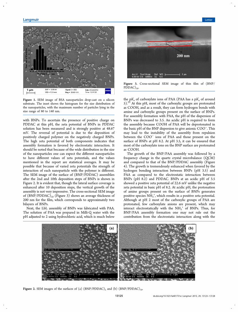
BSA nanoparticles were synthesized by a desolvationapproach.21 The scanning electron microscope (SEM) imageof the drop-cast particles is provided in Figure 1. Thenanoparticles have a spherical morphology and a sizedistribution of 115 ± 33 nm with the maximum number ofnanoparticles lying in the size range of 80 to 140 nm.LbL-assembled thin films of BNPs have been fabricated by
using two different polymers, poly(dimethyldiallylammoniumchloride) (PDDAC) and poly(acrylic acid) (PAA). The LbLassemblies are represented as (PDDAC/BNP)n and (PAA/BNP)n, where n denotes the number of BNP deposition steps.It should be noted that n does not correspond to the number oflayers, unlike the situation in conventional LbL, because asingle deposition step does not lead to the formation of auniform monolayer in the case of nanoparticles. The solution ofBNPs in Milli-Q water has a pH of 8.2. At this basic pH, mostof the amine groups can be expected to be present as neutralspecies, NH2, whereas carboxylic groups are deprotonated,yielding negatively charged carboxylate anions (COO−). Thepresence of excess COO− ions is responsible for the highnegative zeta potential (−44 mV) of BNPs at this pH. Thenegative zeta potential favors the LbL assembly of these BNPswith positively charged polymer, PDDAC. PDDAC has allhydrogens on N replaced by methyl groups (structure providedin Figure S1), and as a result, there is no possibility of hydrogenbonding and this polymer can interact only electrostatically
Langmuir Letter
dx.doi.org/10.1021/la401731a | Langmuir 2013, 29, 13123−1312813124
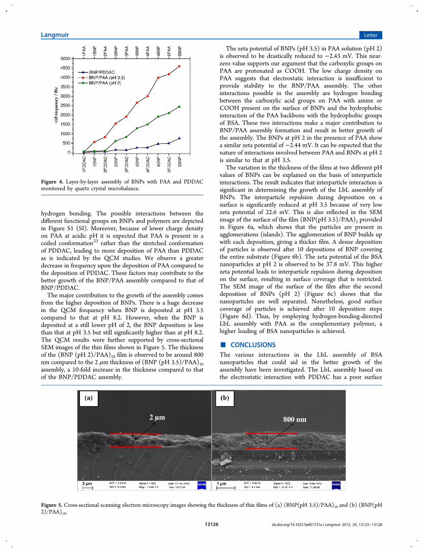
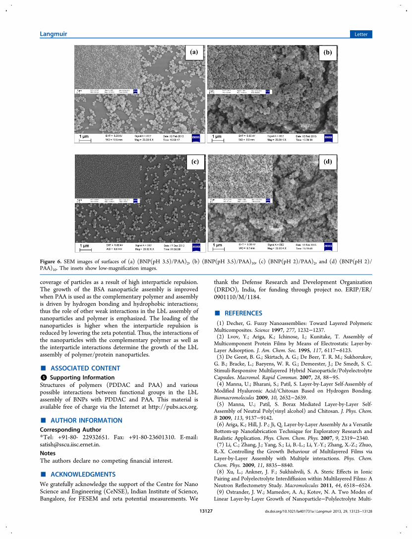
with BNPs. To ascertain the presence of positive charge onPDDAC at this pH, the zeta potential of BNPs in PDDACsolution has been measured and is strongly positive at 48.67mV. The reversal of potential is due to the deposition ofpositively charged polymer on the negatively charged BNPs.The high zeta potential of both components indicates thatassembly formation is favored by electrostatic interaction. Itshould be noted that because of the wide distribution in the sizeof the nanoparticles one can expect the different nanoparticlesto have different values of zeta potentials, and the valuesmentioned in the report are statistical averages. It may bepossible that because of varied zeta potentials the strength ofinteraction of each nanoparticle with the polymer is different.The SEM image of the surface of (BNP/PDDAC) assembliesafter the 2nd and 10th deposition steps of BNPs is shown inFigure 2. It is evident that, though the lateral surface coverage isenhanced after 10 deposition steps, the vertical growth of theassembly is not very impressive. The cross-sectional SEM imageof (BNP/PDDAC)10 (Figure 3) shows an average thickness of200 nm for the film, which corresponds to approximately twobilayers of BNPs.Next, the LbL assembly of BNPs was fabricated with PAA.
The solution of PAA was prepared in Milli-Q water with thepH adjusted to 2 using hydrochloric acid, which is much below
the pKa of carboxylate ions of PAA (PAA has a pKa of around5).22 At this pH, most of the carboxylic groups are protonatedas COOH, and as a result, they can form hydrogen bonds withamine and carboxylic groups present on the surface of BNPs.For assembly formation with PAA, the pH of the dispersion ofBNPs was decreased to 3.5. An acidic pH is required to formthe assembly because COOH of PAA will be deprotonated inthe basic pH of the BNP dispersion to give anionic COO−. Thismay lead to the instability of the assembly from repulsionbetween the COO− ions of PAA and those present on thesurface of BNPs at pH 8.2. At pH 3.5, it can be ensured thatmost of the carboxylate ions on the BNP surface are protonatedas COOH.The growth of the BNP/PAA assembly was followed by a
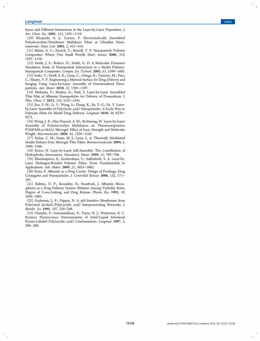
frequency change in the quartz crystal microbalance (QCM)and compared to that of the BNP/PDDAC assembly (Figure4). The growth is tremendously enhanced when favored by thehydrogen bonding interaction between BNPs (pH 3.5) andPAA as compared to the electrostatic interaction betweenBNPs (pH 8.2) and PDDAC. BNPs at an acidic pH of 3.5showed a positive zeta potential of 22.6 mV unlike the negativezeta potential in basic pH of 8.2. At acidic pH, the protonationof amine groups present on the surface of BNPs generatespositive species NH3
+, which results in a positive zeta potential.Although at pH 2 most of the carboxylic groups of PAA areprotonated, few carboxylate anions are present, which mayinteract electrostatically with the NH3
+ of BNPs. Thus, forBNP/PAA assembly formation one may not rule out thecontribution from the electrostatic interaction along with the
Figure 1. SEM image of BSA nanoparticles drop-cast on a siliconsubstrate. The inset shows the histogram for the size distribution ofthe nanoparticles, with the maximum number of particles lying in thesize range of 80 to 140 nm.
Figure 2. SEM images of the surfaces of (a) (BNP/PDDAC)2 and (b) (BNP/PDDAC)10.
Figure 3. Cross-sectional SEM image of thin film of (BNP/PDDAC)10.
Langmuir Letter
dx.doi.org/10.1021/la401731a | Langmuir 2013, 29, 13123−1312813125
hydrogen bonding. The possible interactions between thedifferent functional groups on BNPs and polymers are depictedin Figure S1 (SI). Moreover, because of lower charge densityon PAA at acidic pH it is expected that PAA is present in acoiled conformation23 rather than the stretched conformationof PDDAC, leading to more deposition of PAA than PDDACas is indicated by the QCM studies. We observe a greaterdecrease in frequency upon the deposition of PAA compared tothe deposition of PDDAC. These factors may contribute to thebetter growth of the BNP/PAA assembly compared to that ofBNP/PDDAC.The major contribution to the growth of the assembly comes
from the higher deposition of BNPs. There is a huge decreasein the QCM frequency when BNP is deposited at pH 3.5compared to that at pH 8.2. However, when the BNP isdeposited at a still lower pH of 2, the BNP deposition is lessthan that at pH 3.5 but still significantly higher than at pH 8.2.The QCM results were further supported by cross-sectionalSEM images of the thin films shown in Figure 5. The thicknessof the (BNP (pH 2)/PAA)10 film is observed to be around 800nm compared to the 2 μm thickness of (BNP (pH 3.5)/PAA)10assembly, a 10-fold increase in the thickness compared to thatof the BNP/PDDAC assembly.
The zeta potential of BNPs (pH 3.5) in PAA solution (pH 2)is observed to be drastically reduced to −2.43 mV. This near-zero value supports our argument that the carboxylic groups onPAA are protonated as COOH. The low charge density onPAA suggests that electrostatic interaction is insufficient toprovide stability to the BNP/PAA assembly. The otherinteractions possible in the assembly are hydrogen bondingbetween the carboxylic acid groups on PAA with amine orCOOH present on the surface of BNPs and the hydrophobicinteraction of the PAA backbone with the hydrophobic groupsof BSA. These two interactions make a major contribution toBNP/PAA assembly formation and result in better growth ofthe assembly. The BNPs at pH 2 in the presence of PAA showa similar zeta potential of −2.44 mV. It can be expected that thenature of interactions involved between PAA and BNPs at pH 2is similar to that at pH 3.5.The variation in the thickness of the films at two different pH
values of BNPs can be explained on the basis of interparticleinteractions. The result indicates that interparticle interaction issignificant in determining the growth of the LbL assembly ofBNPs. The interparticle repulsion during deposition on asurface is significantly reduced at pH 3.5 because of very lowzeta potential of 22.6 mV. This is also reflected in the SEMimage of the surface of the film (BNP(pH 3.5)/PAA)2 providedin Figure 6a, which shows that the particles are present inagglomerations (islands). The agglomeration of BNP builds upwith each deposition, giving a thicker film. A dense depositionof particles is observed after 10 depositions of BNP coveringthe entire substrate (Figure 6b). The zeta potential of the BSAnanoparticles at pH 2 is observed to be 37.8 mV. This higherzeta potential leads to interparticle repulsion during depositionon the surface, resulting in surface coverage that is restricted.The SEM image of the surface of the film after the seconddeposition of BNPs (pH 2) (Figure 6c) shows that thenanoparticles are well separated. Nonetheless, good surfacecoverage of particles is achieved after 10 deposition steps(Figure 6d). Thus, by employing hydrogen-bonding-directedLbL assembly with PAA as the complementary polymer, ahigher loading of BSA nanoparticles is achieved.
■ CONCLUSIONSThe various interactions in the LbL assembly of BSAnanoparticles that could aid in the better growth of theassembly have been investigated. The LbL assembly based onthe electrostatic interaction with PDDAC has a poor surface
Figure 4. Layer-by-layer assembly of BNPs with PAA and PDDACmonitored by quartz crystal microbalance.
Figure 5. Cross-sectional scanning electron microscopy images showing the thickness of thin films of (a) (BNP(pH 3.5)/PAA)10 and (b) (BNP(pH2)/PAA)10.
Langmuir Letter
dx.doi.org/10.1021/la401731a | Langmuir 2013, 29, 13123−1312813126
coverage of particles as a result of high interparticle repulsion.The growth of the BSA nanoparticle assembly is improvedwhen PAA is used as the complementary polymer and assemblyis driven by hydrogen bonding and hydrophobic interactions;thus the role of other weak interactions in the LbL assembly ofnanoparticles and polymer is emphasized. The loading of thenanoparticles is higher when the interparticle repulsion isreduced by lowering the zeta potential. Thus, the interactions ofthe nanoparticles with the complementary polymer as well asthe interparticle interactions determine the growth of the LbLassembly of polymer/protein nanoparticles.
■ ASSOCIATED CONTENT*S Supporting InformationStructures of polymers (PDDAC and PAA) and variouspossible interactions between functional groups in the LbLassembly of BNPs with PDDAC and PAA. This material isavailable free of charge via the Internet at http://pubs.acs.org.
■ AUTHOR INFORMATIONCorresponding Author*Tel: +91-80- 22932651. Fax: +91-80-23601310. E-mail:[email protected] authors declare no competing financial interest.
■ ACKNOWLEDGMENTSWe gratefully acknowledge the support of the Centre for NanoScience and Engineering (CeNSE), Indian Institute of Science,Bangalore, for FESEM and zeta potential measurements. We
thank the Defense Research and Development Organization(DRDO), India, for funding through project no. ERIP/ER/0901110/M/1184.
■ REFERENCES(1) Decher, G. Fuzzy Nanoassemblies: Toward Layered PolymericMulticomposites. Science 1997, 277, 1232−1237.(2) Lvov, Y.; Ariga, K.; Ichinose, I.; Kunitake, T. Assembly ofMulticomponent Protein Films by Means of Electrostatic Layer-by-Layer Adsorption. J. Am. Chem. Soc. 1995, 117, 6117−6123.(3) De Geest, B. G.; Skirtach, A. G.; De Beer, T. R. M.; Sukhorukov,G. B.; Bracke, L.; Baeyens, W. R. G.; Demeester, J.; De Smedt, S. C.Stimuli-Responsive Multilayered Hybrid Nanoparticle/PolyelectrolyteCapsules. Macromol. Rapid Commun. 2007, 28, 88−95.(4) Manna, U.; Bharani, S.; Patil, S. Layer-by-Layer Self-Assembly ofModified Hyaluronic Acid/Chitosan Based on Hydrogen Bonding.Biomacromolecules 2009, 10, 2632−2639.(5) Manna, U.; Patil, S. Borax Mediated Layer-by-Layer Self-Assembly of Neutral Poly(vinyl alcohol) and Chitosan. J. Phys. Chem.B 2009, 113, 9137−9142.(6) Ariga, K.; Hill, J. P.; Ji, Q. Layer-by-Layer Assembly As a VersatileBottom-up Nanofabrication Technique for Exploratory Research andRealistic Application. Phys. Chem. Chem. Phys. 2007, 9, 2319−2340.(7) Li, C.; Zhang, J.; Yang, S.; Li, B.-L.; Li, Y.-Y.; Zhang, X.-Z.; Zhuo,R.-X. Controlling the Growth Behaviour of Multilayered Films viaLayer-by-Layer Assembly with Multiple interactions. Phys. Chem.Chem. Phys. 2009, 11, 8835−8840.(8) Xu, L.; Ankner, J. F.; Sukhishvili, S. A. Steric Effects in IonicPairing and Polyelectrolyte Interdiffusion within Multilayered Films: ANeutron Reflectometry Study. Macromolecules 2011, 44, 6518−6524.(9) Ostrander, J. W.; Mamedov, A. A.; Kotov, N. A. Two Modes ofLinear Layer-by-Layer Growth of Nanoparticle−Polylectrolyte Multi-
Figure 6. SEM images of surfaces of (a) (BNP(pH 3.5)/PAA)2, (b) (BNP(pH 3.5)/PAA)10, (c) (BNP(pH 2)/PAA)2, and (d) (BNP(pH 2)/PAA)10. The insets show low-magnification images.
Langmuir Letter
dx.doi.org/10.1021/la401731a | Langmuir 2013, 29, 13123−1312813127
layers and Different Interactions in the Layer-by-Layer Deposition. J.Am. Chem. Soc. 2001, 123, 1101−1110.(10) Khopade, A. J.; Caruso, F. Electrostatically AssembledPolyelectrolyte/Dendrimer Multilayer Films as Ultrathin Nano-reservoirs. Nano Lett. 2002, 2, 415−418.(11) Balazs, A. C.; Emrick, T.; Russell, T. P. Nanoparticle PolymerComposites: Where Two Small Worlds Meet. Science 2006, 314,1107−1110.(12) Smith, J. S.; Bedrov, D.; Smith, G. D. A Molecular DynamicsSimulation Study of Nanoparticle Interactions in a Model Polymer-Nanoparticle Composite. Compos. Sci. Technol. 2003, 63, 1599−1605.(13) Soike, T.; Streff, A. K.; Guan, C.; Ortega, R.; Tantawy, M.; Pino,C.; Shastri, V. P. Engineering a Material Surface for Drug Delivery andImaging Using Layer-by-Layer Assembly of Functionalized Nano-particles. Adv. Mater. 2010, 22, 1392−1397.(14) Mohanta, V.; Madras, G.; Patil, S. Layer-by-Layer AssembledThin Film of Albumin Nanoparticles for Delivery of Doxorubicin. J.Phys. Chem. C 2012, 116, 5333−5341.(15) Jiao, Y.-H.; Li, Y.; Wang, S.; Zhang, K.; Jia, Y.-G.; Fu, Y. Layer-by-Layer Assembly of Poly(lactic acid) Nanoparticles: A Facile Way toFabricate Films for Model Drug Delivery. Langmuir 2010, 26, 8270−8273.(16) Wong, J. E.; Díez-Pascual, A. M.; Richtering, W. Layer-by-LayerAssembly of Polyelectrolyte Multilayers on ThermoresponsiveP(NiPAM-co-MAA) Microgel: Effect of Ionic Strength and MolecularWeight. Macromolecules 2008, 42, 1229−1238.(17) Nolan, C. M.; Serpe, M. J.; Lyon, L. A. Thermally ModulatedInsulin Release from Microgel Thin Films. Biomacromolecules 2004, 5,1940−1946.(18) Kotov, N. Layer-by-Layer Self-Assembly: The Contribution ofHydrophobic Interactions. Nanostruct. Mater. 1999, 12, 789−796.(19) Kharlampieva, E.; Kozlovskaya, V.; Sukhishvili, S. A. Layer-by-Layer Hydrogen-Bonded Polymer Films: From Fundamentals toApplications. Adv. Mater. 2009, 21, 3053−3065.(20) Kratz, F. Albumin as a Drug Carrier: Design of Prodrugs, DrugConjugates and Nanoparticles. J. Controlled Release 2008, 132, 171−183.(21) Rubino, O. P.; Kowalsky, R.; Swarbrick, J. Albumin Micro-spheres as a Drug Delivery System: Relation Among Turbidity Ratio,Degree of Cross-Linking, and Drug Release. Pharm. Res. 1993, 10,1059−1065.(22) Gudeman, L. F.; Peppas, N. A. pH-Sensitive Membranes fromPoly(vinyl alcohol)/Poly(acrylic acid) Interpenetrating Networks. J.Membr. Sci. 1995, 107, 239−248.(23) Chandar, P.; Somasundaran, P.; Turro, N. J.; Waterman, K. C.Excimer Fluorescence Determination of Solid-Liquid InterfacialPyrene-Labeled Poly(acrylic acid) Conformations. Langmuir 1987, 3,298−300.
Langmuir Letter
dx.doi.org/10.1021/la401731a | Langmuir 2013, 29, 13123−1312813128