Enhancement of Antigen-specific Functional Responses by Neutrophils from Allergic Patients
9
Enhancement of Antigen-specific Functional Responses by Neutrophils from Allergic Patients By Javier Monteseirln,* Maria Jesus Camacho,* Rocio Montafio,~ Estrella Llamas,* Manuel Conde,* Modesto Carballo,* Pedro Guardia,* Jos~ Conde,* and Francisco Sobrino** From the *Departamento de Medicina, Servicio de Inmunologla y Alergia, Hospital Universitario Virgen Macarena, Universidad de Sevilla; and ~tDepartamento de Bioqulmica M~dica y Biologla Molecular, Universidad de &villa, 41009 Seville, Spain Summary It has been demonstrated that neutrophils from healthy donors or from patients with inflamma- tory disorders can bind immunoglobulin (Ig) E proteins through binding to Mac-2/ebp. Func- tional responses to allergens were assessed by measuring the respiratory burst and intracellular Ca 2+ levels, and binding of allergens to neutrophils was assessed by flow cytometry analysis and fluorescence microscopy. In this article, we demonstrate that neutrophils sensitized to specific allergens (from allergic patients), but not from healthy donors, are sensitive to allergens of the same type as those that produce clinical allergic symptoms. The activation of neutrophils was analyzed by the induction of a respiratory burst that was detected with luminol-dependent chemiluminescence. Intracellular Ca 2§ levels increased parallel to those of the inducing aller- gens. In addition, the specific binding of allergens on the cell surface was revealed by flow cy- tometry and aUergen-FITC-labeled staining analyses. The present data suggest a restricted rec- ognition of allergen by sensitive neutrophils, probably associated with the specific binding of the allergen to its corresponding IgE molecule, which is bound to the Mac-2/~bp structure. These findings demonstrate a functional role of allergen-associated neutrophils during the aller- gic state. T he development of an immediate hypersensitivity re- action involves various biochemical events leading to the release of histamine, arachidonic acid metabolites, an- ion superoxide, and other effector molecules (1, 2). All in- flammatory cells (macrophages, lymphocytes, mast cells, eosinophils, neutrophils, and platelets) have been shown to be involved in asthma, but the relative role of each cell is not completely understood (1). Because of the chronic na- ture of allergic diseases, leukocytes that persist in the lung tissue, especially eosinophils and lymphocytes, have be- come more strongly associated with the disease. For this reason, neutrophils have often been neglected in studies on allergic diseases. However, there is increasing evidence of neutrophil participation in asthma and the allergic process. For instance, plasma chemotactic activity ofneutrophils in- creases, and peripheral-blood neutrophils show markers of activation, during active asthma (3), after exercise-induced asthma (4), and during early and late asthmatic reactions in- duced by allergens (5) and toluene diisocyanate (6). In sub- jects with pollen-induced asthma, seasonal antigenic exposure is associated with an increase in the number of neutrophils shown in bronchial biopsies (7). In other atopic asthmatics, local endobronchial allergen challenge leads to increased neutrophil infiltration of the airway mucosa (8). Further- more, activated neutrophils can induce airway hyper- responsiveness (9). Most of the cells, such as eosinophils, epidermal Langerhans cells, macrophages/monocytes, and bronchial epithelial cells, selectively express two types of IgE receptors, namely, receptors of high affinity (Fc~RI) 1 and of low affinity (FceRII) (10-15). Although neutrophils express neither Fc~RI nor Fc~RII receptors for IgE (10, 16), evidence has been presented that neutrophils express a new family of IgE-binding receptors, which belong to the S-type lectins. Among them, Mac-2/ebp is the best charac- terized, and its presence in neutrophils has been demon- strated (17, 18). The presence of this new family oflgE re- ceptors has been previously demonstrated in other cells (19-21). Stimulation of human neutrophils induces several phenomena known collectively as respiratory burst, charac- terized by an increase in the production of superoxide an- ion (22, 23). Although the generated inflammatory media- tors are critical to the host defense role of the neutrophils, they can cause severe tissue damage when excessively or inappropriately produced (24). 1Abbreviations used in this paper: CL, chemiluminescence;FceRI and FceRIl, high and low affinity IgE Fc receptors,respectively. 2571 j. Exp. Med. 9 The Rockefeller University Press 9 0022-1007/96/06/2571/09 $2.00 Volume 183 June 1996 2571-2579 on August 1, 2014 jem.rupress.org Downloaded from Published June 1, 1996
-
Upload
independent -
Category
Documents
-
view
1 -
download
0
Transcript of Enhancement of Antigen-specific Functional Responses by Neutrophils from Allergic Patients
Enhancement of Antigen-specific Functional Responses by Neutrophils from Allergic Patients By Javier Montesei r ln ,* Mar ia Jesus Camacho,* R o c i o Montafio,~ Estrella Llamas,* M a n u e l Conde,* M o d e s t o Carballo,* Pedro Guardia,* Jos~ Conde ,* and Francisco Sobrino**
From the *Departamento de Medicina, Servicio de Inmunologla y Alergia, Hospital Universitario Virgen Macarena, Universidad de Sevilla; and ~tDepartamento de Bioqulmica M~dica y Biologla Molecular, Universidad de &villa, 41009 Seville, Spain
Summary It has been demonstrated that neutrophils from healthy donors or from patients with inflamma- tory disorders can bind immunoglobulin (Ig) E proteins through binding to Mac-2/ebp. Func- tional responses to allergens were assessed by measuring the respiratory burst and intracellular Ca 2+ levels, and binding of allergens to neutrophils was assessed by flow cytometry analysis and fluorescence microscopy. In this article, we demonstrate that neutrophils sensitized to specific allergens (from allergic patients), but not from healthy donors, are sensitive to allergens of the same type as those that produce clinical allergic symptoms. The activation of neutrophils was analyzed by the induction of a respiratory burst that was detected with luminol-dependent chemiluminescence. Intracellular Ca 2§ levels increased parallel to those of the inducing aller- gens. In addition, the specific binding of allergens on the cell surface was revealed by flow cy- tometry and aUergen-FITC-labeled staining analyses. The present data suggest a restricted rec- ognition of allergen by sensitive neutrophils, probably associated with the specific binding of the allergen to its corresponding IgE molecule, which is bound to the Mac-2/~bp structure. These findings demonstrate a functional role of allergen-associated neutrophils during the aller- gic state.
T he development of an immediate hypersensitivity re- action involves various biochemical events leading to
the release of histamine, arachidonic acid metabolites, an- ion superoxide, and other effector molecules (1, 2). All in- flammatory cells (macrophages, lymphocytes, mast cells, eosinophils, neutrophils, and platelets) have been shown to be involved in asthma, but the relative role of each cell is not completely understood (1). Because of the chronic na- ture of allergic diseases, leukocytes that persist in the lung tissue, especially eosinophils and lymphocytes, have be- come more strongly associated with the disease. For this reason, neutrophils have often been neglected in studies on allergic diseases. However, there is increasing evidence of neutrophil participation in asthma and the allergic process. For instance, plasma chemotactic activity ofneutrophils in- creases, and peripheral-blood neutrophils show markers of activation, during active asthma (3), after exercise-induced asthma (4), and during early and late asthmatic reactions in- duced by allergens (5) and toluene diisocyanate (6). In sub- jects with pollen-induced asthma, seasonal antigenic exposure is associated with an increase in the number of neutrophils shown in bronchial biopsies (7). In other atopic asthmatics, local endobronchial allergen challenge leads to increased neutrophil infiltration of the airway mucosa (8). Further-
more, activated neutrophils can induce airway hyper- responsiveness (9). Most of the cells, such as eosinophils, epidermal Langerhans cells, macrophages/monocytes, and bronchial epithelial cells, selectively express two types of IgE receptors, namely, receptors of high affinity (Fc~RI) 1 and of low affinity (FceRII) (10-15). Although neutrophils express neither Fc~RI nor Fc~RII receptors for IgE (10, 16), evidence has been presented that neutrophils express a new family of IgE-binding receptors, which belong to the S-type lectins. Among them, Mac-2/ebp is the best charac- terized, and its presence in neutrophils has been demon- strated (17, 18). The presence of this new family oflgE re- ceptors has been previously demonstrated in other cells (19-21). Stimulation of human neutrophils induces several phenomena known collectively as respiratory burst, charac- terized by an increase in the production of superoxide an- ion (22, 23). Although the generated inflammatory media- tors are critical to the host defense role of the neutrophils, they can cause severe tissue damage when excessively or inappropriately produced (24).
1Abbreviations used in this paper: CL, chemiluminescence; FceRI and FceRIl, high and low affinity IgE Fc receptors, respectively.
2571 j. Exp. Med. �9 The Rockefeller University Press �9 0022-1007/96/06/2571/09 $2.00 Volume 183 June 1996 2571-2579
on August 1, 2014
jem.rupress.org
Dow
nloaded from
Published June 1, 1996
Despite this evidence, it is not known whether neutro- phils can directly interact with the allergens, and i f so, what the functional response is. The present work was under- taken to analyze these questions. The rationale is that neu- trophils from allergic patients possess specific IgE pre- attached to the surface o f these cells. Challenging with an allergen could trigger the activation o f neutrophils through the IgE-~bp complex in the membrane. Here we present evidence that specific allergens were able to activate respi- ratory burst from asthmatic patients sensitized to those al- lergens, and we have found a relationship be tween the two parameters (that is, the respiratory burst is closely related to sensitized neutrophils). Moreover , we found that increased respiratory burst was associated with enhanced antigen- dependent Ca 2+ elevation by neutrophils from allergic patients. In addition, sensitized neutrophils from allergic patients specifically bound FITC-labe led allergens, as mea- sured by fluorescence microscopy and flow cytometry.
Materials and Methods
Materials. The allergens studied included D 1 (Dermatophagoides pteronyssinus), G 3 (Dactylisglomerata), T 9 (Olea europea), and W6 (Ar- temisia vulgaris). They were purchased from Ifidesa-Aristegui (Bil- bao, Spain). Mezerein was from Sigma-Aldrich Co. Ltd. (Poole, Dorset, England). Other biochemicals were from Sigma or Merck (Barcelona, Spain).
Patients and Controls. The group studied included adult atopic patients with bronchial asthma and healthy adult nonatopic vol- unteer controls. The asthmatic patients had positive skin prick test (Ifidesa-Arlstegui) and specific IgE (CAP, Pharmacia Ib6rica, Bar- celona, Spain) to at least one common allergen (house dust mites and pollens). The subjects received neither treatment nor specific hyposensitization. None of them had experienced episodes of asthma for at least 3 too, and none had had respiratory tract infec- tions for 4 wk before blood sampling. The healthy controls had no history of allergy or bronchial symptoms and had negative skin prick test (Ifidesa-Aristegui) and specific IgE to a battery of inhalant allergens (house dust mites, pollens, molds, and animal danders).
Preparation of Polymorphonuclear Leukocytes. Human neutrophils were purified (25) from freshly drawn heparinized (10 U/ml) venous blood in two steps: first, the blood (20 nil) was mixed with 1.5 ml of 10% Dextran T 500 (final concentration of 0.7%), dissolved in PBS. The supernatant fraction was centrifuged through a Ficoll-Hypaque. The neutrophil-rich pellet was then removed by plastic pipette and red cells were eliminated by one hypotonic lysis in water. Neutrophils were resuspended to "~107 cells/ml in PBS and 10 mM of glucose and were kept at room temperature.
Measurement of lntracellular Ca 2+. Freshly prepared neutrophils from allergic or healthy donors were incubated in a medium (Ca 2+ medium) composed of 137 mM NaC1, 2.7 mM KC1, 1 mM MgC1, 1 mM CaC12, 20 mM Hepes at pH 7.4, at a concen- tration of 107 cells/ml at 37~ for 45 rain in the presence of 2 btM Fura-2 acetoxymethyl ester (26). The excess of the dye was then washed away, and the neutrophils were kept in the same medium. Batches of 107 cells were resuspended in 0.6 ml of Ca 2§ medium at 37~ and the allergens to be studied were added to the cellular suspension. Fura-2 fluorescence (k~x ~ at 340 nm and ke,,~ at 505 nm) was recorded on a Perkin-Elmer (Norwalk, CT) LS-5 spectrofluorimeter. Ca 2+ was calibrated with a value of 224 for the K d of Fura-2.
Measurement of Chemiluminescence. Luminol-amplified chemi- luminescence (CL) was measured in a Berthold luminometer at 37~ basically as described in reference 27. Briefly, neutrophils (106 cells/ml) were incubated in 1 ml of PBS buffer supple- mented with 10 mM glucose, 500 }~M CaC12, 5 I~M luminol, 100 I~M sodium azide for 5 rain. Then allergens as stimulants at the indicated doses were added, to give a final volume of 1 ml. The CL response was measured every 1 rain in each tube. Data were expressed as the number of cpm recorded each minute over a 15-rain period.
Coupling of FITC to Allergen Protein. Coupling was carried out with a conjugation kit in accordance with the instructions of the manufacturer (Quicktag FITC Conjugation Kit; Boehringer Mann- helm Corp., Indianapohs, IN).
Coupling of Allergen Labeled with FITC to Neutrophils. This cou- phng was carried out as previously described (28), with minor mod- ifications. In brief, after fixing in 3% paraformaldehyde, 7 • 10 ~' cells in PBS with 10 mM glucose were incubated with allergen labeled with FITC (100 ~g/ml) for 16-18 h at 4~ The cells were spun (500 rpm, 4~ 5 rain), and were decanted, 10 ml of PBS, 1% HSA was added, and the cells were resuspended, and spun again. This washing was repeated three more times. Finally the cells were resuspended in 500 bd of PBS, 10 mM glucose.
Fluorescence Staining for Microscopy Analysis. The method pre- viously described (29), with minor modifications, was followed. Neutrophils were fixed for 20 rain in 3% paraformaldehyde in PBS buffer, pH 7.4. After washing, cells (1.5 • 10 s cells per 40 l-d) were suspended in PBS with 10 mM glucose and were at- tached to polylysine coated slides for 30 min at 25~ Cells were incubated with specific allergen, labeled with FITC, for 16-18 h at 4~ in the dark and in a humid chamber. The slides were rinsed with PBS with l% HSA once and rinsed again with PBS alone. Shdes were processed for epifluorescence microscopy using 10% PBS, 90% glycerol as mounting medium.
Flow Cytometry Analysis. Flow cytometry was performed on an Epics Elite flow cytometer (Coulter Corp., Hialeah, FL) and calibrated by standard techniques. For each stained sample, 2 • 104 cells were analyzed for FITC fluorescence intensity. The flu- orescent distribution for both control (cell autofluorescence) and test cells with allergen labeled with FITC was analyzed with a Coulter Elite software workstation.
Results
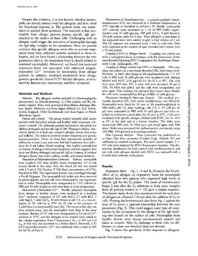
Respiratory Burst. Fig. 1, A and B, illustrates the kinetic effect o f G 3 allergen on respiratory burst for neutrophils obtained from two patients who expressed high levels of specific IgE for the G3 pollen. The onset o f luminescence began 2 rain after the G3 additions in both cases. Samples from all patients studied (n = 12) gave a similar response. The kinetic study shows that max imum levels for each dose o f allergen are obtained 7 -8 rain after the addit ion o f G3 to cells. Plott ing the luminescence data from Fig. 1 against the dose o f G3 shows a sigrnoid relationship between the two parameters (Fig. 2). This could suggest some type o f coop- eration in the occupation by the allergen o f the IgE-bind- ing sites located on the surface o f cells. Neutrophi ls from healthy donors were always simultaneously assayed and taken as controls. After G3 challenge in these cells, no dif- ference in values was detected (data not shown).
Fig. 3 shows the specificity o f the response to allergens.
2572 Enhancement of Antigen--specific Functional Responses
on August 1, 2014
jem.rupress.org
Dow
nloaded from
Published June 1, 1996
A B
- - o - 1 pg/ml --O-- 1 pg/ml ~ , 40 | - - 0 - 2pg/ml ~ = | I--O-- 2pg/ml 30
/ ~ 3 pg/ml F ~ I / ~ 3 pg/ml ~ =o x l ~ 5pg/ml / ~ ^ ^ ] I ~ 5pglml / . . / / / - ' " = r J ~ 7 pg/ml / ~ I I - O - 7 pglml a / J~
I I - o - lo pg/ml 7" / "~ 3~ - c - 10pg/ml / d / / - e - 15pg/ml / / 20
.a 0 0 ~
I I I I I I
0 5 10 0 5 10
Time (minutes) Time (minutes)
F i g u r e 1. Activation of neutrophils by a spe- cific allergen (G3). Neutrophils isolated from two allergic patients (each represented sepa- rately in A and B) were suspended (106 cells/ ml) in 1 ml of PBS buffer supplemented with 10 m M glucose, 500 ~ M CaC12, 100 p~M so- dium azide and the addition of different con- centrations of G3, at the following final concen- trations: 1 (O), 2 ([]), 3 (A), 5 (V), 7 (O), 10 (�9 and 15 I~g/ml (0). At the same time, 5 ~M luminol was added. The suspension was in- cubated at 37~ and the chemiluminescence response was recorded for 10 min. Similar re- sults were obtained in at least eight other exper- iments using neutrophils from different patients with clinical allergies to G 3 allergen.
Neutrophils from a patient who was specifically allergic to G 3 show a clear response when cells were incubated with this pollen. However, when cells were incubated with D 1
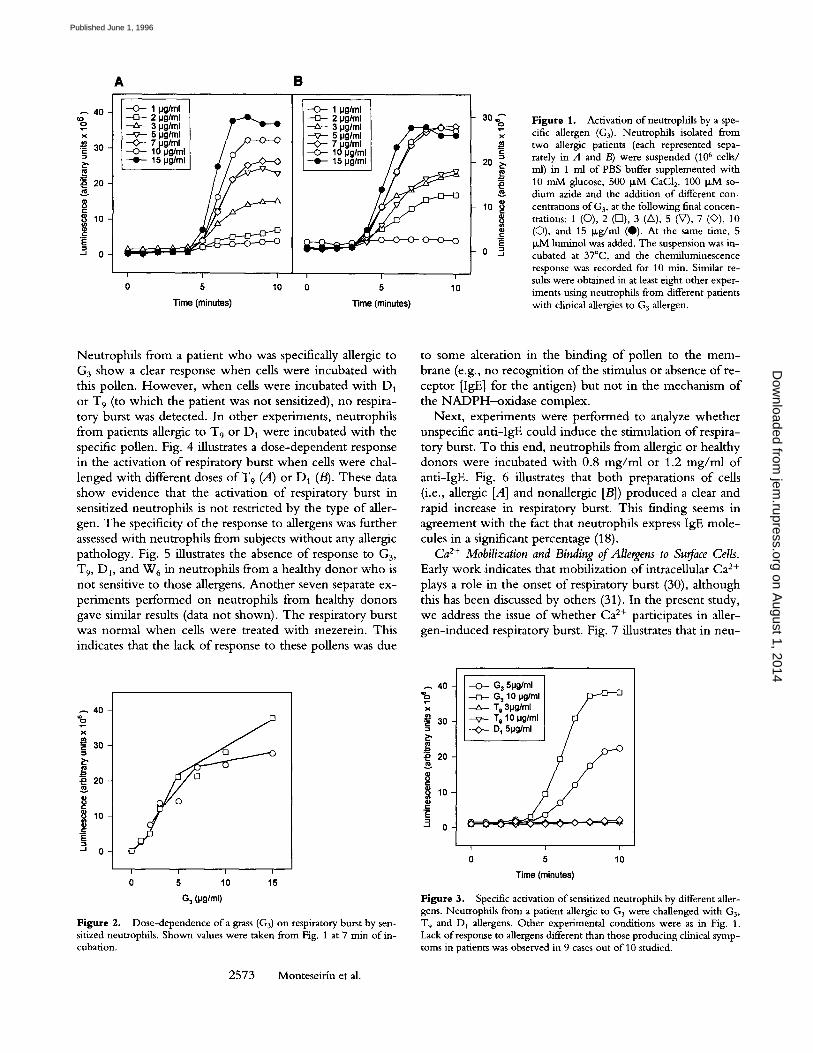
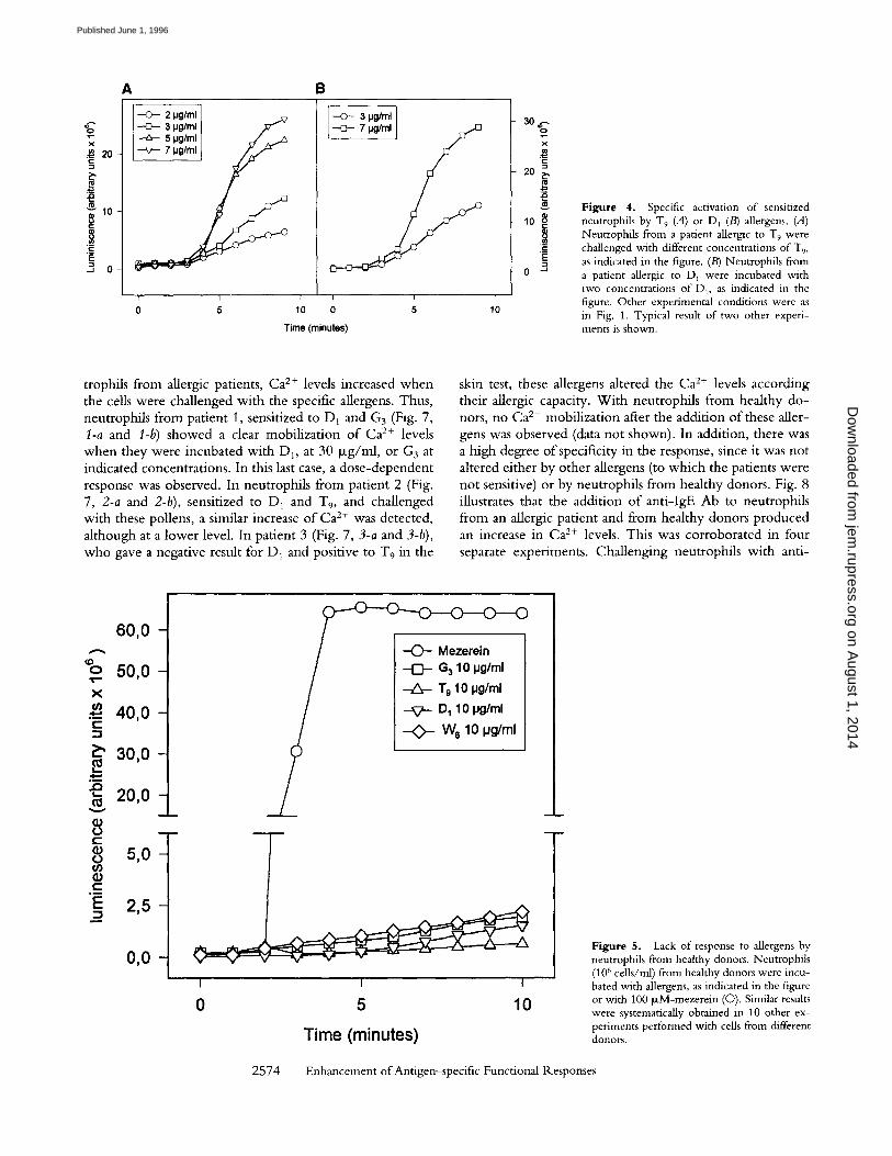
or T9 (to which the patient was not sensitized), no respira- tory burst was detected. In other experiments, neutrophils from patients allergic t o T 9 or D 1 were incubated with the specific pollen. Fig. 4 illustrates a dose-dependent response in the activation of respiratory burst when cells were chal- lenged with different doses o f T 9 (A) o r D 1 (B). These data show evidence that the activation of respiratory burst in sensitized neutrophils is not restricted by the type of aller- gen. The specificity of the response to allergens was further assessed with neutrophils from subjects without any allergic pathology. Fig. 5 illustrates the absence of response to G3,
T9, D1, and W6 in neutrophils from a healthy donor who is not sensitive to those allergens. Another seven separate ex- periments performed on neutrophils from healthy donors gave similar results (data not shown). The respiratory burst was normal when cells were treated with mezerein. This indicates that the lack of response to these pollens was due
40 =o
X
�9 ~ 30
-~ 20
! ' ~
~ 0
I I I I
0 5 10 15
G 3 (pg/ml)
Figure 2. Dose-dependence of a grass (G3) on respiratory burst by sen- sitized neutrophils. Shown values were taken from Fig. 1 at 7 min of in- cubation.
2573 Monteseirin et al.
to some alteration in the binding of pollen to the mem- brane (e.g., no recognition of the stimulus or absence of re- ceptor [IgE] for the antigen) but not in the mechanism of the NADPH-oxidase complex.
Next, experiments were performed to analyze whether unspecific anti-IgE could induce the stimulation of respira- tory burst. To this end, neutrophils from allergic or healthy donors were incubated with 0.8 mg/rnl or 1.2 mg/ml of anti-IgE. Fig. 6 illustrates that both preparations of cells (i.e., allergic [A] and nonallergic [B]) produced a clear and rapid increase in respiratory burst. This finding seems in agreement with the fact that neutrophils express IgE mole- cules in a significant percentage (18).
Ca 2+ Mobilization and Binding of Allergens to SuoCace Cells. Early work indicates that mobilization of intracellular Ca 2+ plays a role in the onset of respiratory burst (30), although this has been discussed by others (31). In the present study, we address the issue of whether Ca 2+ participates in aller- gen-induced respiratory burst. Fig. 7 illustrates that in neu-
~ 4 0 - %
X
�9 ~ 3 0 -
~ 2 0 -
8 ~ 1 0 -
~ 0
- O - - G 3 5pg/ml G 3 10 pg/ml Tg 3pg/ml / T 9 10 IJg/ml D 1 5pg/ml / /
I I I
0 5 10
Time (minutes)
Figure 3. Specific activation of sensitized neutrophils by different aller- gens. Neutrophils from a patient allergic to G 3 were challenged with G3, T 9 and D 1 allergens. Other experimental conditions were as in Fig. 1. Lack of response to allergens different than those producing clinical symp- toms in patients was observed in 9 cases out of 10 studied.
on August 1, 2014
jem.rupress.org
Dow
nloaded from
Published June 1, 1996
A B
='go X
20
g to
.! ~ 0
--O--- 2 pglml ] -C,-- 3 pg/ml [ f " - -A- - 5 pg/mf [ v / /
r i
o 5 lO
---o- 3 pg/ml ]
o 5
Time (minutes)
30 4.- O
X
2O ~.,
10 8
E= 0 "~
f
10
Figure 4. Specific activation of sensitized neutrophils by T 9 (A) or D] (B) allergens. (A) Neutrophils from a patient allergic to T9 were challenged with different concentrations of T,, as indicated in the figure. (B) Neutrophils from a patient allergic to D~ were incubated with two concentrations of D,, as indicated in the figure. Other experimental conditions were as in Fig. 1. Typical result of two other experi- ments is shown.
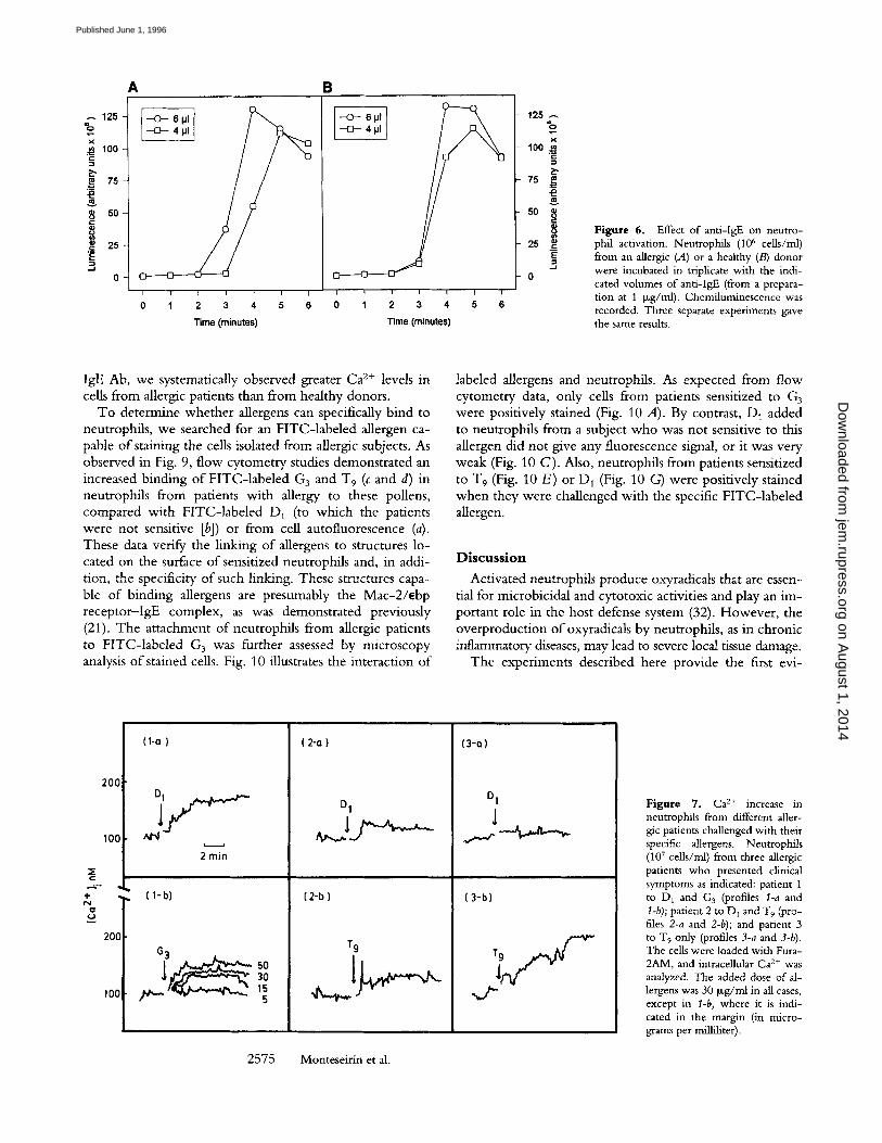
trophils f rom allergic patients, Ca 2+ levels increased w h e n the cells were chal lenged wi th the specific allergens. Thus, neutrophi ls f rom pat ient 1, sensitized to D1 and G3 (Fig. 7, 1-a and I-b) showed a clear mobi l iza t ion o f Ca 2+ levels w h e n they were incuba ted wi th Di , at 30 Izg/ml, or G3 at indicated concentrat ions. In this last case, a dose -dependen t response was observed. In neutrophi ls f rom pat ient 2 (Fig. 7, 2-a and 2-b), sensitized to D 1 and T9, and challenged wi th these pollens, a similar increase o f Ca 2+ was detected, a l though at a lower level. In pat ient 3 (Fig. 7, 3-a and 3-b), who gave a negative result for D I and positive to W 9 in the
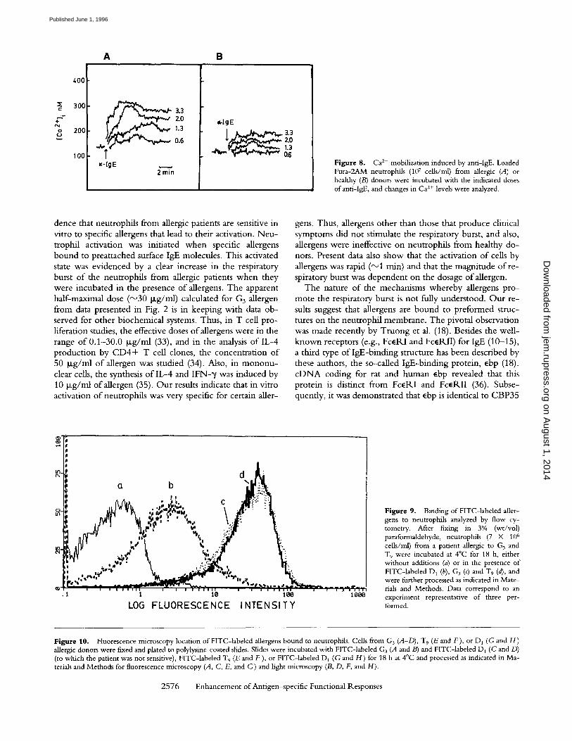
skin test, these allergens altered the Ca 2+ levels according their allergic capacity. W i t h neutrophi ls f rom healthy do- nors, no Ca 2+ mobi l iza t ion after the addi t ion o f these aller- gens was observed (data no t shown). In addit ion, there was a high degree o f specificity in the response, since it was no t altered either by o ther allergens (to which the patients were no t sensitive) or by neutrophi ls f rom healthy donors. Fig. 8 illustrates that the addi t ion o f ant i - lgE Ab to neutrophi ls f rom an allergic pat ient and from healthy donors p roduced an increase in Ca 2+ levels. This was corroborated in four separate experiments . Chal lenging neutrophi ls wi th ant i -
Z" 0
X r r
E',
I , . ,
h .
V
8 t - (9 0 r (9 r
~
E
6 0 , 0 -
5 0 , 0 -
4 0 , 0 -
3 0 , 0 -
2 0 , 0 - m
5 , 0 -
2 , 5 -
0 , 0 -
C)~-O 0 ' 0 ' 0
- C ) - - Mezerein G 3 10 pg/ml
T 9 10 pg/ml
D 1 10 pg/ml
W e 10 p g / m l
I I
5 1 0
T i m e ( m i n u t e s )
m
F i g u r e 5. Lack of response to allergens by neutrophils from healthy donors. Neutrophils (106 cells/ml) from healthy donors were incu- bated with allergens, as indicated in the figure or with 100 ~M-mezerein (O). Similar results were systematically obtained in 10 other ex- periments performed with cells from different donors.
2574 Enhancement of Antigen-specific Functional Responses
on August 1, 2014
jem.rupress.org
Dow
nloaded from
Published June 1, 1996
A B
125 %
100 5 ~ 75
.~ 25
. - f
o I I I I
0 1 2 3 4
Time (minutes)
r i i
5 6 I I I I I I I
0 1 2 3 4 5 6
Time (minutes)
125 %
X 100
7e ~ g
2s ~
- - 1
o
Figure 6. Effect of anti-IgE on neutro- phil activation. Neutrophfls (106 cells/nil) from an allergic (A) or a healthy (B) donor were incubated in triplicate with the indi- cated volumes of anti-IgE (from a prepara- tion at 1 bLg/ml). Chemiluminescence was recorded. Three separate experiments gave the same results.
IgE Ab, we systematically observed greater Ca 2+ levels in cells from allergic patients than from healthy donors.
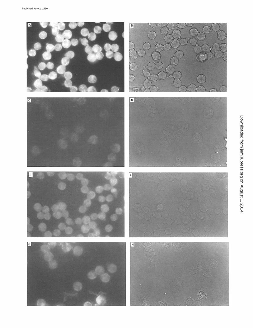
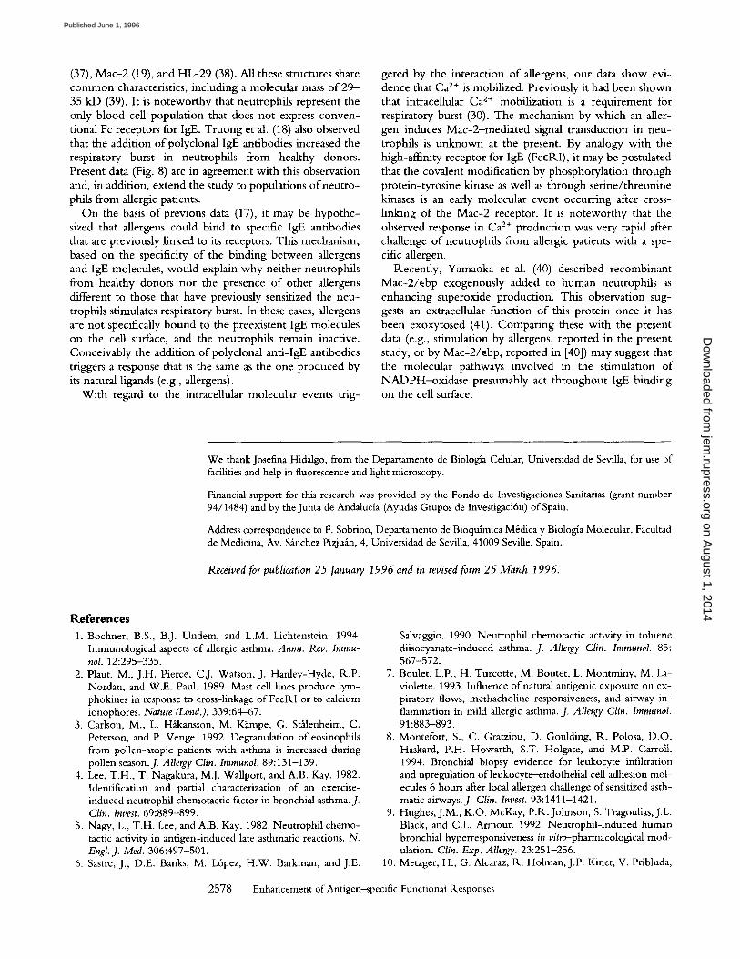
To determine whether allergens can specifically bind to neutrophils, we searched for an FITC- labe led allergen ca- pable o f staining the cells isolated from allergic subjects. As observed in Fig. 9, flow cytometry studies demonstrated an increased binding o f FITC- labe led G3 and T 9 (c and d) in neutrophils from patients with allergy to these pollens, compared with FITC- labe led D1 (to which the patients were not sensitive [b]) or from cell autofluorescence (a). These data verify the l inking o f allergens to structures lo- cated on the surface o f sensitized neutrophils and, in addi- tion, the specificity o f such linking. These structures capa- ble o f binding allergens are presumably the Mac-2 / ebp receptor - IgE complex, as was demonstrated previously (21). The attachment o f neutrophils from allergic patients to F I T C - l a b d e d G3 was further assessed by microscopy analysis o f stained cells. Fig. 10 illustrates the interaction o f
labeled allergens and neutrophils. As expected from flow cytometry data, only cells from patients sensitized to G 3 were positively stained (Fig. 10 A). By contrast, D 1 added to neutrophils from a subject who was not sensitive to this allergen did not give any fluorescence signal, or it was very weak (Fig. 10 C) . Also, neutrophils from patients sensitized to T 9 (Fig. 10 E) or D 1 (Fig. 10 G) were positively stained when they were challenged with the specific FITC- labe led allergen.
D i s c u s s i o n
Activated neutrophils produce oxyradicals that are essen- tial for microbicidal and cytotoxic activities and play an im- portant role in the host defense system (32). However , the overproduct ion o f oxyradicals by neutrophils, as in chronic inflammatory diseases, may lead to severe local tissue damage.
The experiments described here provide the first evi-
200
100
4-
0 r
20q
100
(1-a)
| t
2 rain
(2-o)
(2 -b ) : c l-b)
3O
D I
T 9
(3-0)
D I
(3-b)
2575 Monteseirin et al.
Figure 7. Ca 2+ increase in neutrophils from different aller- gic patients challenged with their specific allergens. Neutrophils (107 cells/ml) from three allergic patients who presented clinical symptoms as indicated: patient 1 to D 1 and G 3 (profiles l-a and t-b); patient 2 to DI and T 9 (pro- files 2-a and 2-b); and patient 3 tO T 9 only (profiles 3-a and 3-b). The cells were loaded with Fura- 2AM, and intracellnlar Ca 2+ was analyzed. The added dose of al- lergens was 30 Ixg/ml in all cases, except in l-b, where it is indi- cated in the margin (in micro- grams per milliliter).
on August 1, 2014
jem.rupress.org
Dow
nloaded from
Published June 1, 1996
A B
400
z 3 0 0
§
o 2 0 0 K.J
1 O0
~ 3.3
2.0 1.3
0.6
=~-lgE 2rain
l{-I g E
Figure 8. Ca 2+ mobilization induced by anti-IgE. Loaded Fura-2AM neutrophils (107 cells/ml) from allergic (A) or healthy (B) donors were incubated with the indicated doses of anti-IgE, and changes in Ca z+ levels were analyzed.
dence that neutrophils from allergic patients are sensitive in vitro to specific allergens that lead to their activation. Neu- trophil activation was initiated when specific allergens bound to preattached surface IgE molecules. This activated state was evidenced by a clear increase in the respiratory burst of the neutrophils from allergic patients when they were incubated in the presence of allergens. The apparent half-maximal dose (~30 p,g/ml) calculated for G 3 allergen from data presented in Fig. 2 is in keeping with data ob- served for other biochemical systems. Thus, in T cell pro- liferation studies, the effective doses of allergens were in the range of 0.1-30.0 p,g/ml (33), and in the analysis of IL-4 production by CD4+ T cell clones, the concentration of 50 p,g/ml of allergen was studied (34). Also, in mononu- clear cells, the synthesis of IL-4 and IFN-~/was induced by 10 p,g/ml of allergen (35). Our results indicate that in vitro activation of neutrophils was very specific for certain aller-
gens. Thus, allergens other than those that produce clinical symptoms did not stimulate the respiratory burst, and also, allergens were ineffective on neutrophils from healthy do- nors. Present data also show that the activation of cells by allergens was rapid ('~1 rain) and that the magnitude of re- spiratory burst was dependent on the dosage of allergen.
The nature of the mechanisms whereby allergens pro- mote the respiratory burst is not fully understood. Our re- sults suggest that allergens are bound to preformed struc- tures on the neutrophil membrane. The pivotal observation was made recently by Truong et al. (18). Besides the well- known receptors (e.g., FceRI and FcelklI) for IgE (10-15), a third type of IgE-binding structure has been described by these authors, the so-called IgE-binding protein, ebp (18). cDNA coding for rat and human ebp revealed that this protein is distinct from FceRI and FcelKII (36). Subse- quently, it was demonstrated that ebp is identical to CBP35
g_
.i
d , :
~ , ." _ E~, .? !
~ L J �9 . :.', . d~'e ,%. -~ \
. t..21"
~ i I I U i I p l -U I ~ i I I l i I ! i n u I i U , n u I I "'~ I U i .U I I i I i
1 10 leo 10e8
LOG FLUORESCENCE INTENSITY
Figure 9. Binding of FITC-labeled aller- gens to neutrophils analyzed by flow cy- tometry. After fixing in 3% (wt/vol) paraformaldehyde, neutrophils (7 • 10 6 cells/ml) from a patient allergic to G 3 and T 9 were incubated at 4~ for 18 h, either without additions (a) or in the presence of FITC-labeled D 1 (b), G 3 (c) and T9 (d), and were further processed as indicated in Mate- rials and Methods. Data correspond to an experiment representative of three per- formed.
Figure 10. Fluorescence microscopy location of FITC-labeled allergens bound to neutrophils. Cells from G 3 (A-D), T 9 (E and F), or D 1 (G and H) allergic donors were fixed and plated to polylysine-coated slides. Slides were incubated with FITC-labeled G 3 (A and B) and FITC-labeled D1 (C and D) (to which the patient was not sensitive), FITC-labeled T 9 (E and F ), or FITC-labeled D1 (G and H ) for 18 h at 4~ and processed as indicated in Ma- terials and Methods for fluorescence microscopy (A, C, E, and G) and light microscopy (B, D, F, and H).
2 5 7 6 E n h a n c e m e n t o f Ant igen~pec i f i c Funct ional Responses
on August 1, 2014
jem.rupress.org
Dow
nloaded from
Published June 1, 1996
(37), Mac-2 (19), and HL-29 (38). All these structures share common characteristics, including a molecular mass of 29- 35 kD (39). It is noteworthy that neutrophils represent the only blood cell population that does not express conven- tional Fc receptors for IgE. Truong et al. (18) also observed that the addition of polyclonal IgE antibodies increased the respiratory burst in neutrophils from healthy donors. Present data (Fig. 8) are in agreement with this observation and, in addition, extend the study to populations ofneutro- phils from allergic patients.
On the basis of previous data (17), it may be hypothe- sized that allergens could bind to specific IgE antibodies that are previously linked to its receptors. This mechanism, based on the specificity of the binding between allergens and IgE molecules, would explain why neither neutrophils from healthy donors nor the presence of other allergens different to those that have previously sensitized the neu- trophils stimulates respiratory burst. In these cases, allergens are not specifically bound to the preexistent IgE molecules on the cell surface, and the neutrophils remain inactive. Conceivably the addition ofpolyclonal anti-IgE antibodies triggers a response that is the same as the one produced by its natural ligands (e.g., allergens).
With regard to the intracellular molecular events trig-
gered by the interaction of allergens, our data show evi- dence that Ca 2+ is mobilized. Previously it had been shown that intracellular Ca > mobilization is a requirement for respiratory burst (30). The mechanism by which an aller- gen induces Mac-2-mediated signal transduction in neu- trophils is unknown at the present. By analogy with the high-affinity receptor for IgE (Fc~RI), it may be postulated that the covalent modification by phosphorylation through protein-tyrosine kinase as well as through serine/threonine kinases is an early molecular event occurring after cross- linking of the Mac-2 receptor. It is noteworthy that the observed response in Ca 2+ production was very rapid after challenge of neutrophils from allergic patients with a spe- cific allergen.
Recently, Yamaoka et al. (40) described recombinant Mac-2/ebp exogenously added to human neutrophils as enhancing superoxide production. This observation sug- gests an extracellular function of this protein once it has been exoxytosed (41). Comparing these with the present data (e.g., stimulation by allergens, reported in the present study, or by Mac-2/~bp, reported in [40]) may suggest that the molecular pathways involved in the stimulation of NADPH--oxidase presumably act throughout IgE binding on the cell surface.
We thankJosefina Hidalgo, from the Departamento de Biologla Celular, Universidad de Sevilla, for use of facilities and help in fluorescence and light microscopy.
Financial support for this research was provided by the Fondo de Investigaciones Sanitarias (grant number 94/1484) and by the Junta de Andalucia (Ayudas Grupos de Investigaci6n) of Spain.
Address correspondence to F. Sobrino, Departamento de Bioquimica M~dica y Biologla Molecular, Facultad de Medicina, Av. S~inchez Pizju~n, 4, Universidad de Sevilla, 41009 Seville, Spain.
Received for publication 25January 1996 and in revised form 25 March 1996.
References 1. Bochner, B.S., B.J. Undem, and L.M. Lichtenstein. 1994.
Immunological aspects of allergic asthma. Annu. Rev. Immu- nol. 12:295-335.
2. Plant, M., J.H. Pierce, C.J. Watson, J. Hanley-Hyde, R.P. Nordan, and W.E. Paul. 1989. Mast cell lines produce lym- phokines in response to cross-linkage of FceRI or to calcium ionophores. Nature (Lond.). 339:64-67.
3. Carlson, M., L. Hikansson, M. K~impe, G. Stfilenheim, C. Peterson, and P. Venge. 1992. Degranulation of eosinophils from pollen-atopic patients with asthma is increased during pollen season.J. Allergy Clin. Immunol. 89:131-139.
4. Lee, T.H., T. Nagakura, M.J. Wallport, and A.B. Kay. 1982. Identification and partial characterization of an exercise- induced neutrophil chemotactic factor in bronchial asthma.J. Clin. Invest. 69:889-899.
5. Nagy, L., T.H. Lee, and A.B. Kay. 1982. Neutrophil chemo- tactic activity in antigenqnduced late asthmatic reactions. N. Engl. J. Med. 306:497-501.
6. Sastre, J., D.E. Banks, M. L6pez, H.W. Barkman, and J.E. 10.
Enhancement of Antigen~specific
Salvaggio. 1990. Neutrophil chemotactic activity in toluene diisocyanate-induced asthma. J. Allergy Clin. Immunol. 85: 567-572.
7. Boulet, L.P., H. Turcotte, M. Boutet, L. Montminy, M. La- violette. 1993. Influence of natural antigenic exposure on ex- piratory flows, methachotine responsiveness, and airway in- flammation in mild allergic asthma. J. Allergy Clin. Immunol. 91:883-893.
8. Montefort, S., C. Gratziou, D. Goulding, R. Polosa, D.O. Haskard, P.H. Howarth, S.T. Holgate, and M.P. Carroll. 1994. Bronchial biopsy evidence for leukocyte infiltration and upregulation of leukocyte-endothelial cell adhesion mol- ecules 6 hours after local allergen challenge of sensitized asth- matic airways. J. Clin. Invest. 93:1411-1421.
9. Hughes, J.M., K.O. McKay, P.R. Johnson, S. Tragoulias, J.L. Black, and C.L. Armour. 1992. Neutrophil-induced human bronchial hyperresponsiveness in vitro-pharmacological mod- ulation. Clin. Exp, Allergy. 23:251-256. Metzger, H., G. Alcaraz, R. Holman, J.P. Kinet, V. Pribluda,
2578 Functional Responses
on August 1, 2014
jem.rupress.org
Dow
nloaded from
Published June 1, 1996
and R. Quarto. 1986. The receptor with high affinity for im- munoglobuhn E. Annu. Rev. Immunol. 4:419-470.
11. Gounni, A.S., B. Lamkhioued, K. Ochial, Y. Tanaka, E. De- laporte, A. Capron, J.P. Kinet, and M. Capron. 1994. High- affinity IgE receptor on eosinophils is involved in defence against parasites. Nature (Land.). 367:183-186.
12. Bieber, T., H. de la Salle, A. Wollenberg, J. Hakimi, R. Chizzonite, J. Ring, D. Hanau, and C. de la Salle. 1992. Hu- man epidermal Langerhans cells express the high affinity re- ceptor for immunoglobulin E (FceRI). J. Exp. Med. 175: 1285-1290.
13. Maurer, D., E. Fiebiger, B, Reininger, B. Wolff-Winiski, M.H. Jouvin, O. Kilgus, J.P. Kinet, and G. Stingl. 1994. Ex- pression of functional high affinity immunoglobulin E recep- tors (FceRI) on monocytes ofatopic individuals.J. Exp. Med. 179:745-750.
14. Capron, A., J.P. Dessaint, M. Joseph, J.M. Rousseaux, M. Capron, and H. Bazin. 1977. Interaction between IgE com- plexes and macrophages in the rat: a new mechanism ofmac- rophage activation. Eur.J. Immunol. 7:315-320.
15. Campbell, A.M., A.M. Vignola, P. Chanez, P. Godard, andJ. Bousquet. 1994. Low-affinity receptor for IgE on human bronchial epithelial cells in asthma. Immunology. 82:506-508.
16. Capron, A., J.P. Desaint, M. Capron, J.C. Joseph, J.C. Ameisen, and A.B. Tonnel. 1986. From parasite to allergy: the second receptor for IgE (FceR2). Immunol Today. 7:15- 18.
17. Liu, F.-T. 1993. S-type mammalian lectins in allergic inflam- mation. Immunol. Today. 14:486-490.
18. Tmong, M.J., V. Gmart, J.P. Kusnierz, J.P. Papin, S. Loisean, A. Capron, and M. Capron. 1993. Human neutrophils ex- press immunoglobulin E (IgE)-binding proteins (Mac-2/{bp) of the S-type lectin family: role in lgE-dependent activation. J. Exp. IVied. 177:243-248.
19. Ho, M.K., and T.A. Springer. 1982. Mac-2, a novel 32.000 Mr mouse macrophage subpopulation--specific antigen de- fined by monoclonal antibodies.J. Immunol. 128:1221-1228.
20. Cherayil, B.J., S.J. Weiner, and S. Pillai. 1989. The Mac-2 antigen is a galactose-specific lectin that binds IgE. J. Exp. Meal. 170:t959-1972.
21. Liu, F.T., K. Albrandt, E. Mendel, A. Kulczycki, Jr., and N.K. Orida. 1985. Identification of an IgE-binding protein by molecular cloning. Proc. Natl. Acad. Sci. USA. 82:4100- 4104.
22. Thelen, M., B. Dewald, and M. Baggiolini. 1993. Neutrophil signal transduction and activation of the respiratory burst. Physiol. Rev. 73:797-821.
23. Edwards, S.W. 1995. Cell signalling by integrins and immu- noglobulin receptors in primed neutrophils. Trends Biol. Sci. 20:362-367.
24. Te Velde, A.A. 1994. Interaction between cytokines and monocytes/macrophages. In Immunopharmacology of Mac- rophage and Other Antigen-presenting Cells. C.A.F.M. Bruijnzeel-Kromen and E.C.M. Hoetsmit, editors. Academic Press, Inc., London. 7-34.
25. Btyum, A. 1986. Isolation ofmononuclear cells and granulo- cytes from blood. Scand.J. Clin. Lab. Invest. 97:77-89.
26. Martin, F., A. Gualberto, F. Sobrino, and E. Pintado. 1991. Thymerosal induces calcium mobilization, fructose 2,6 his- phosphate synthesis and cytoplasmic alkalinization in rat thy-
mus lymphocytes. Biochim. Biophys. Acta. 191:110-114. 27. Wang, J.F., P. Komarov, and H. de Groot. 1993. Luminol
chemiluminescence in rat macrophages and granulocytes: the role of NO, O2-/H202 and HOC1. Arch. Biochem. Biophys. 304:189-196.
28. Unanue, E.R., and J. Braun. 1986. Immunofluorescence methods to study cell surface and cytoplasmic proteins, and their dynamics. In Handbook of Experimental Immunology, D.M. Weir, L.A. Herzenberg, C. Blackwell and L.A. Herzen- berg, editors. Blackwell Scientific Publications, Oxford, UK. 23.1-23.3.
29. Hidalgo, J., M. Mufiiz, and A. Velasco. 1995. Trimeric G proteins regulate the cytosoMnduced redistribution of Golgi enzymes into the endoplasmic reticulum. J. Celt Sci. 108: 1805-1815.
30. Lew, P.D., C.B. Wollheim, F.A. Waldvogel, and T. Pozzan. 1984. Modulation of cytosolic-free calcium transients by changes in intracellular calcium-buffering capacity: correla- tion with exocytosis and 02- production in human neutro- phils.J. Cell Biol. 99:1212-1220.
31. Grinstein, S., and W. Furuya. 1988. Receptor-mediated acti- vation of electropermeabilized neutrophils. J. Biol. Chem. 263:1779-1783.
32. Weiss, S.J. 1989. Tissue destruction by neutrophils. N. Engl. J. Med. 320:365-376.
33. O'Brien, R.M., W.R. Thomas, and A.M. Wootton. 1992. T cell response to the purified major allergens from house dust mite Dermatophagoides pteronyssinus. J. Allergy Clin. Immunol. 89:1021-1031.
34. Secrist, H., C.J. Chelen, Y. Wen, J.D. Marshall, and D.T. Umetsu. 1993. Allergen immunotherapy decreases II,-4 pro- duction in CD4 + T cells from allergic individuals. J. Exp. Med. 178:2123-2130.
35. Gagnon, R., A. Akoum, andJ. Hebert. 1993. Lol p I-induced IL-4 and IFN-y production by peripheral blood mononu- clear cells ofatopic and nonatopic subjects during and out of the pollen season.J. Allergy Clin. Immunol. 91:950-956.
36. Robertson, M.W., K. Albrandt, D. Keller, and F.T. Liu. 1990. Human IgE-binding: a soluble lectin exhibiting a highly conserved interspecies sequence and differential recognition oflgE glycoforms. Biochemistry. 29:8093-8100.
37. Laing, J.G., M.W. Robertson, C.A. Gritzmacher, J.L. Wang, and F.T. Liu. 1989. Biochemical and immunological com- parisons of carbohydrate-binding protein 35 and an IgE-bind- ing protein._/. Biol. Chem. 264:1907-1910.
38. Leffier, H., and S.H. Barondes. 1987. Specificity of binding of three soluble rat lung lectins to substituted and unsubsti- tuted mammalian [3-galactosidase. J. Biol. Chem. 261:10119- 10126.
39. Liu, F.T. 1990. Molecular biology of IgE-binding protein, IgE-binding-factors, and IgE-receptors. Crit. Rev. Immunol. 10:289-302.
40. Yamaoka, A., Y. Kuwabara, L.G. Frigeri, and F.T. Liu. 1993. A human lectin, Galcetin-3 ({bp/Mac-2), stimulates superox- ide production by neutrophils. J. Immunol. 154:3479-3487.
41. Wollenberg, A., H. de la Salle, D. Hanau, F.T. Liu, and T. Bieber. 1993. Human keratinocytes release the endogenous [3-galactoside binding soluble ebp (IgE-binding protein) which binds to Langerhans cells where it modulates their binding capacity for IgE glycoform.J. Exp. Med. 178:777-785.
2579 Monteseirin et al.
on August 1, 2014
jem.rupress.org
Dow
nloaded from
Published June 1, 1996