Enantioselective microbial reduction of substituted acetophenones
12
Enantioselective microbial reduction of substituted acetophenones Ramesh N. Patel, * Animesh Goswami, Linda Chu, Mary Jo Donovan, Venkata Nanduri, Steven Goldberg, Robert Johnston, Prasad J. Siva, Brent Nielsen, Junying Fan, WeiXuan He, Zhongping Shi, Kwok Y. Wang, Ronald Eiring, Dana Cazzulino, Ambarish Singh and Richard Mueller Process Research & Development, Bristol-Myers Squibb Pharmaceutical Research Institute, PO Box 191, New Brunswick, NJ 08903, USA Received 13 November 2003; revised 1 December 2003; accepted 6 February 2004 Abstract—The chiral intermediate (S)-1-(2 0 -bromo-4 0 -fluoro phenyl)ethanol 2 was prepared by the enantioselective microbial reduction of 2-bromo-4-fluoro acetophenone 1. Organisms from genus Candida, Hansenula, Pichia, Rhodotorula, Saccharomyces, Sphingomonas and Baker’s yeast reduced 1 to 2 in >90% yield and 99% enantiomeric excess (ee). In an alternative approach, the enantioselective microbial reductions of methyl, ethyl, and tert-butyl 4-(2 0 -acetyl-5 0 -fluorophenyl) butanoates 3, 5, and 7, respec- tively, were demonstrated using strains of Candida and Pichia. Reaction yields of 40–53% and ee’s of 90–99% were obtained for the corresponding (S)-hydroxy esters 4, 6, and 8. The reductase, which catalyzed the enantioselective reduction of ketoesters was purified to homogeneity from cell extracts of Pichia methanolica SC 13825. It was cloned and expressed in Escherichia coli with recombinant cultures used for the enantioselective reduction of keto methyl ester 3 to the corresponding (S)-hydroxy methyl ester 4. On a preparative scale, a reaction yield of 98% and an ee of 99% was obtained. Ó 2004 Elsevier Ltd. All rights reserved. 1. Introduction Recently, much attention has been focused on the interaction of small molecules with biological macro- molecules. Selective enzyme inhibitors and receptor agonists/antagonists are keys for target-oriented research in the pharmaceutical industry. 1 Chirality is a key factor in the efficiency of many drug products, and as a result the production of single enantiomers of molecules has become increasingly important in the pharmaceutical industry. 2 Single enantiomers can be produced by chemical or chemo-enzymatic synthesis. The advantages of biocatalysis over chemical synthesis are that enzyme-catalyzed reactions are often highly enantioselective and regioselective. They can be carried out at ambient temperature and atmospheric pressure, thus avoiding the use of more extreme conditions, which could cause problems due to isomerization, racemiza- tion, epimerization, or rearrangement. A number of review articles 3–10 have been published on the use of enzymes in organic synthesis. Herein we report the enzymatic synthesis of (S)-1-(2 0 - bromo-4 0 -fluorophenyl)-ethanol 2 and (S)-hydroxy esters 4, 6, and 8 (Fig. 1A and B), as potentially useful intermediates in the synthesis of pharmaceutical prod- ucts. 11–16 Ketone 1 (S)-Alcohol 2 O Br F OH Br F Microorganisms O F CO 2 R OH F CO 2 R Microorganisms Ketoester R= Methyl 3, Ethyl 5, t-Butyl 7 (S)-Hydroxyester R= Methyl 4, Ethyl 6, t-Butyl 8 Figure 1. * Corresponding author. Tel.: +1-732-227-6213; fax: +1-732-227-3994; e-mail: [email protected] 0957-4166/$ - see front matter Ó 2004 Elsevier Ltd. All rights reserved. doi:10.1016/j.tetasy.2004.02.024 Tetrahedron: Asymmetry 15 (2004) 1247–1258 Tetrahedron: Asymmetry
Transcript of Enantioselective microbial reduction of substituted acetophenones
K (S) H dK (S) H d
Tetrahedron:
Tetrahedron: Asymmetry 15 (2004) 1247–1258
Asymmetry
Enantioselective microbial reduction of substitutedacetophenones
Ramesh N. Patel,* Animesh Goswami, Linda Chu, Mary Jo Donovan, Venkata Nanduri,Steven Goldberg, Robert Johnston, Prasad J. Siva, Brent Nielsen, Junying Fan,
WeiXuan He, Zhongping Shi, Kwok Y. Wang, Ronald Eiring,Dana Cazzulino, Ambarish Singh and Richard Mueller
Process Research & Development, Bristol-Myers Squibb Pharmaceutical Research Institute,
PO Box 191, New Brunswick, NJ 08903, USA
Received 13 November 2003; revised 1 December 2003; accepted 6 February 2004
Abstract—The chiral intermediate (S)-1-(20-bromo-40-fluoro phenyl)ethanol 2 was prepared by the enantioselective microbialreduction of 2-bromo-4-fluoro acetophenone 1. Organisms from genus Candida, Hansenula, Pichia, Rhodotorula, Saccharomyces,Sphingomonas and Baker’s yeast reduced 1 to 2 in >90% yield and 99% enantiomeric excess (ee). In an alternative approach, theenantioselective microbial reductions of methyl, ethyl, and tert-butyl 4-(20-acetyl-50-fluorophenyl) butanoates 3, 5, and 7, respec-tively, were demonstrated using strains of Candida and Pichia. Reaction yields of 40–53% and ee’s of 90–99% were obtained for thecorresponding (S)-hydroxy esters 4, 6, and 8. The reductase, which catalyzed the enantioselective reduction of ketoesters waspurified to homogeneity from cell extracts of Pichia methanolica SC 13825. It was cloned and expressed in Escherichia coli withrecombinant cultures used for the enantioselective reduction of keto methyl ester 3 to the corresponding (S)-hydroxy methyl ester 4.On a preparative scale, a reaction yield of 98% and an ee of 99% was obtained.� 2004 Elsevier Ltd. All rights reserved.
Ketone 1 (S)-Alcohol 2
O
BrF
OH
BrFMicroorganisms
FCO2R
FCO2RMicroorganisms
1. Introduction
Recently, much attention has been focused on theinteraction of small molecules with biological macro-molecules. Selective enzyme inhibitors and receptoragonists/antagonists are keys for target-orientedresearch in the pharmaceutical industry.1 Chirality is akey factor in the efficiency of many drug products, andas a result the production of single enantiomers ofmolecules has become increasingly important in thepharmaceutical industry.2 Single enantiomers can beproduced by chemical or chemo-enzymatic synthesis.The advantages of biocatalysis over chemical synthesisare that enzyme-catalyzed reactions are often highlyenantioselective and regioselective. They can be carriedout at ambient temperature and atmospheric pressure,thus avoiding the use of more extreme conditions, whichcould cause problems due to isomerization, racemiza-tion, epimerization, or rearrangement. A number of
* Corresponding author. Tel.: +1-732-227-6213; fax: +1-732-227-3994;
e-mail: [email protected]
0957-4166/$ - see front matter � 2004 Elsevier Ltd. All rights reserved.
doi:10.1016/j.tetasy.2004.02.024
review articles3–10 have been published on the use ofenzymes in organic synthesis.
Herein we report the enzymatic synthesis of (S)-1-(20-bromo-40-fluorophenyl)-ethanol 2 and (S)-hydroxyesters 4, 6, and 8 (Fig. 1A and B), as potentially usefulintermediates in the synthesis of pharmaceutical prod-ucts.11–16
O OH
KetoesterR= Methyl 3, Ethyl 5, t-Butyl 7
(S)-HydroxyesterR= Methyl 4, Ethyl 6, t-Butyl 8
Figure 1.
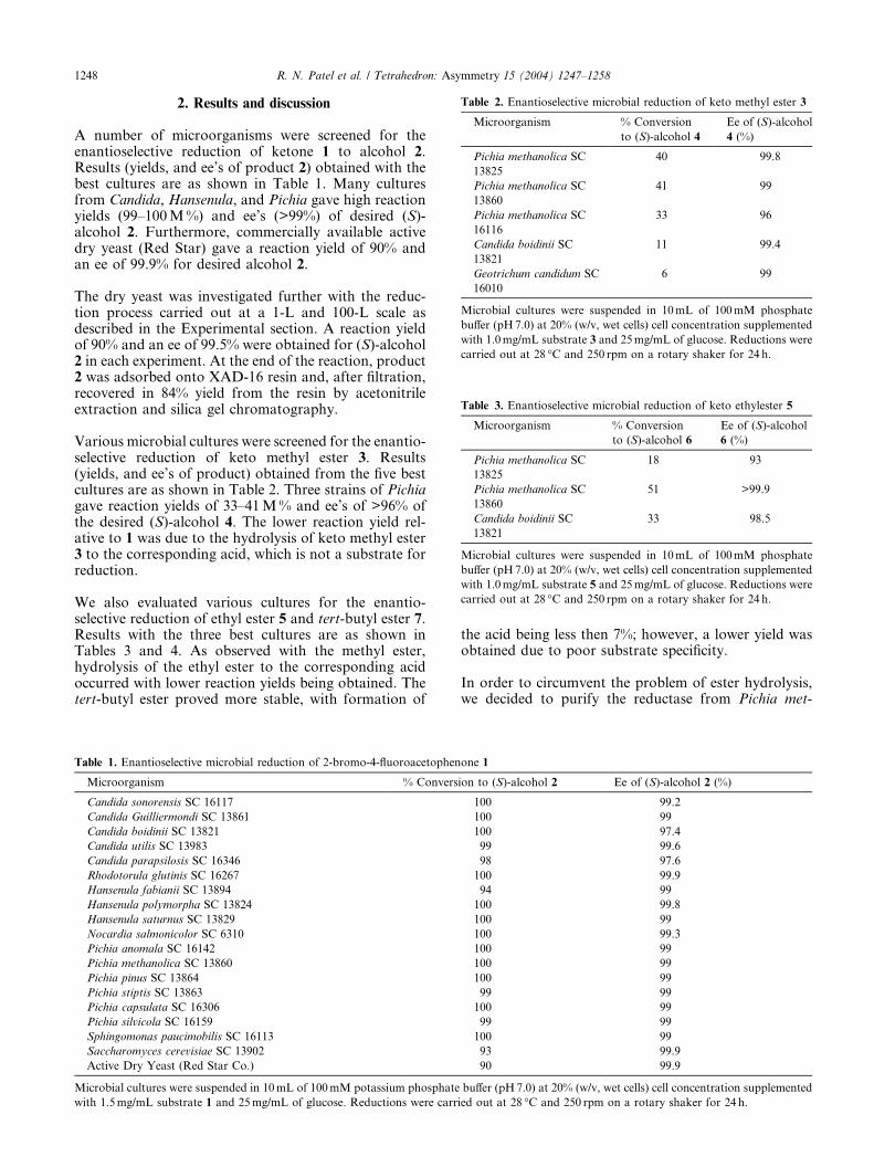
Table 2. Enantioselective microbial reduction of keto methyl ester 3
Microorganism % Conversion
to (S)-alcohol 4
Ee of (S)-alcohol
4 (%)
Pichia methanolica SC
13825
40 99.8
Pichia methanolica SC
13860
41 99
Pichia methanolica SC
16116
33 96
Candida boidinii SC
13821
11 99.4
Geotrichum candidum SC
16010
6 99
Microbial cultures were suspended in 10mL of 100mM phosphate
buffer (pH7.0) at 20% (w/v, wet cells) cell concentration supplemented
with 1.0mg/mL substrate 3 and 25mg/mL of glucose. Reductions were
carried out at 28 �C and 250 rpm on a rotary shaker for 24 h.
Table 3. Enantioselective microbial reduction of keto ethylester 5
Microorganism % Conversion
to (S)-alcohol 6
Ee of (S)-alcohol
6 (%)
Pichia methanolica SC
13825
18 93
Pichia methanolica SC
13860
51 >99.9
Candida boidinii SC
13821
33 98.5
Microbial cultures were suspended in 10mL of 100mM phosphate
buffer (pH7.0) at 20% (w/v, wet cells) cell concentration supplemented
with 1.0mg/mL substrate 5 and 25mg/mL of glucose. Reductions were
carried out at 28 �C and 250 rpm on a rotary shaker for 24 h.
1248 R. N. Patel et al. / Tetrahedron: Asymmetry 15 (2004) 1247–1258
2. Results and discussion
A number of microorganisms were screened for theenantioselective reduction of ketone 1 to alcohol 2.Results (yields, and ee’s of product 2) obtained with thebest cultures are as shown in Table 1. Many culturesfrom Candida, Hansenula, and Pichia gave high reactionyields (99–100M%) and ee’s (>99%) of desired (S)-alcohol 2. Furthermore, commercially available activedry yeast (Red Star) gave a reaction yield of 90% andan ee of 99.9% for desired alcohol 2.
The dry yeast was investigated further with the reduc-tion process carried out at a 1-L and 100-L scale asdescribed in the Experimental section. A reaction yieldof 90% and an ee of 99.5% were obtained for (S)-alcohol2 in each experiment. At the end of the reaction, product2 was adsorbed onto XAD-16 resin and, after filtration,recovered in 84% yield from the resin by acetonitrileextraction and silica gel chromatography.
Various microbial cultures were screened for the enantio-selective reduction of keto methyl ester 3. Results(yields, and ee’s of product) obtained from the five bestcultures are as shown in Table 2. Three strains of Pichiagave reaction yields of 33–41M% and ee’s of >96% ofthe desired (S)-alcohol 4. The lower reaction yield rel-ative to 1 was due to the hydrolysis of keto methyl ester3 to the corresponding acid, which is not a substrate forreduction.
We also evaluated various cultures for the enantio-selective reduction of ethyl ester 5 and tert-butyl ester 7.Results with the three best cultures are as shown inTables 3 and 4. As observed with the methyl ester,hydrolysis of the ethyl ester to the corresponding acidoccurred with lower reaction yields being obtained. Thetert-butyl ester proved more stable, with formation of
Table 1. Enantioselective microbial reduction of 2-bromo-4-fluoroacetophen
Microorganism % Convers
Candida sonorensis SC 16117
Candida Guilliermondi SC 13861
Candida boidinii SC 13821
Candida utilis SC 13983
Candida parapsilosis SC 16346
Rhodotorula glutinis SC 16267
Hansenula fabianii SC 13894
Hansenula polymorpha SC 13824
Hansenula saturnus SC 13829
Nocardia salmonicolor SC 6310
Pichia anomala SC 16142
Pichia methanolica SC 13860
Pichia pinus SC 13864
Pichia stiptis SC 13863
Pichia capsulata SC 16306
Pichia silvicola SC 16159
Sphingomonas paucimobilis SC 16113
Saccharomyces cerevisiae SC 13902
Active Dry Yeast (Red Star Co.)
Microbial cultures were suspended in 10mL of 100mM potassium phosphate
with 1.5mg/mL substrate 1 and 25mg/mL of glucose. Reductions were carr
the acid being less then 7%; however, a lower yield wasobtained due to poor substrate specificity.
In order to circumvent the problem of ester hydrolysis,we decided to purify the reductase from Pichia met-
one 1
ion to (S)-alcohol 2 Ee of (S)-alcohol 2 (%)
100 99.2
100 99
100 97.4
99 99.6
98 97.6
100 99.9
94 99
100 99.8
100 99
100 99.3
100 99
100 99
100 99
99 99
100 99
99 99
100 99
93 99.9
90 99.9
buffer (pH7.0) at 20% (w/v, wet cells) cell concentration supplemented
ied out at 28 �C and 250 rpm on a rotary shaker for 24 h.
Table 4. Enantioselective microbial reduction of keto tert-butyl ester 7
Microorganism % Conversion
to (S)-alcohol 8
Ee of (S)-alcohol
8 (%)
Mucor rouxii SC
13920
53 >99
Mucor hiemalis SC
13974
43 93
Pichia methanolica SC
16116
12 92.6
Microbial cultures were suspended in 10mL of 100mM phosphate
buffer (pH7.0) at 20% (w/v, wet cells) cell concentration supplemented
with 1.0mg/mL substrate 7 and 25mg/mL of glucose. Reductions were
carried out at 28 �C and 250 rpm on a rotary shaker for 24 h.
0
20
40
60
80
100
120
0 10 20 30 40 50Bioconversion time (Hrs)
Yiel
d (M
%)
Substrate 2 g/LSubstrate 4 g/LSubstrate 6 g/L
Figure 3.
igure 2.
R. N. Patel et al. / Tetrahedron: Asymmetry 15 (2004) 1247–1258 1249
hanolica SC 13825 and clone and overexpress thisenzyme in a suitable host. The enzyme was purified 247-fold from the cell extract using Hi-Trap Blue affinitycolumn chromatography (Table 5). The purified proteingave a single band on SDS-PAGE with a molecularweight of 40,000Da. The molecular weight of the puri-fied protein, as determined by size-exclusion columnchromatography, was 38,000–40,000, indicating that thereductase was a monomeric protein.
The purified protein required NADPH or NADH as acofactor for the reduction of keto methyl ester 3.Reaction yields of 96% and 89% were obtained forproduct 4 with 0.1mM NADP or NAD at 1 g/L sub-strate input, respectively, using glucose dehydrogenaseas the cofactor recycling enzyme. The N-terminal andinternal peptide sequences (generated by Lys-peptidasetreatment) of the purified reductase were determined toallow the synthesis of oligonucleotide probes for clon-ing. The reductase was expressed as Escherichia colistrain SC 16445. The complete sequence of the keto-reductase is covered in Ref. 17.
The production of cloned ketoreductase in a 250-L fer-mentor was conducted as described in the Experimentalsection. Growth was completed in 48 h and about 50–52 g/L cells were obtained from the fermentation broth(Fig. 2). Cells harvested from the fermentor were used toconduct the bioreduction of keto methyl ester 3.
The effect of substrate concentration was evaluated inflasks using 10% (w/v, wet cells) cell concentration ofE. coli strain SC 16445. Their reaction went to com-pletion in 24–30 h at 2–4 g/L substrate input. At 6 g/Lsubstrate input, 48 h were required to complete thereaction (Fig. 3).
The biotransformation process was scaled up to 1-L and500-L fermentors. Cells were suspended in 1L of 50mM
Table 5. Purification of ketoreductase from Pichia methanolica SC 13825
Step Volume (mL) Total p
(mg)
Cell extracts 265 2170
HiTrap affinity column (pH gradient) 51 19.5
HiTrap affinity column (NADP gradient) 2 0.06
The reaction mixture for the enzyme assay contained 1mg substrate 3, 4mMN
out at 30 �C for 5 h, and the concentration of product 4 determined by HPL
F
phosphate buffer pH7.0 at 10% (w/v) cell concentration.Cell suspensions were supplemented with nicotinamideadenine dinucleotide phosphate (NADP), glucose, glu-cose dehydrogenase, and 4.5 g/L substrate. Biotrans-formations were carried out at 500 rpm and 28 �C asdescribed in the Experimental section. The reaction wentto completion in 20 h with 95% reaction yield and 99.9%ee of product 4. Significant hydrolysis of substrate 3 wasnot observed, with <2% of keto acid 5 being formed.This process was scaled-up to 500L to prepare 2.0 kg ofproduct 4.
The dehydrogenases from yeast,18–21 horse liver,22 andT. brockii23 transfer the pro-R hydride to the re-face ofthe carbonyl to give (S)-alcohols, a process described byPrelog’s rule.24 In contrast, dehydrogenases from Lac-tobacillus kefir25 and two Pseudomonas sp.26 exhibit anti-Prelog specificity, transferring the pro-R hydride to form(R)-alcohols. Previously we have used various dehy-drogenases to catalyze the enantioselective reductionand reductive amination of ketones and a-ketoacids to
rotein Total units Sp. activity
(units/mg protein)
Purification (fold)
9.4 0.0043 1
1.2087 0.0619 14.4
0.0638 1.063 247
ADP, 4 units glucose dehydrogenase in 1mL. The reaction was carried
C. One unit is defined as 1lmol of 4 formed/h.
1250 R. N. Patel et al. / Tetrahedron: Asymmetry 15 (2004) 1247–1258
prepare alcohols and chiral amines required for thesynthesis of antihypertensive, anticholesterol, antican-cer, and antiviral drugs.27–30
3. Conclusion
Herein, we have described the enantioselective reductionof 2-bromo-4-fluoro-acetophenone 1 to the corre-sponding (S)-2-bromo-4-fluoro-phenyl ethanol 2 byBaker’s yeast. We have also demonstrated the enantio-selective microbial reduction of ketoesters 3, 5, and 7 tothe corresponding (S)-hydroxy esters 4,6, and 8. Theketo reductase from P. methanolica SC 13825 wascloned and expressed in E. coli. Recombinant E. coliexpressing the keto reductase was used to catalyze theenantioselective reduction of keto methyl ester 3 to thecorresponding (S)-hydroxy methyl ester 4 on pre-parative scale.
4. Experimental
4.1. Materials
The 2-bromo-4-fluoroacetophenone was obtained fromLancaster Chemicals. Starting substrates 3, 5, 7 andreference compounds 2, 4, 6, 8 were synthesized bycolleagues in the Process Research and DevelopmentDepartment, Bristol-Myers Squibb PharmaceuticalResearch Institute. The physico-chemical properties,including spectral characteristics (1H NMR, 13C NMR,mass spectra), were in full accord for all compounds.
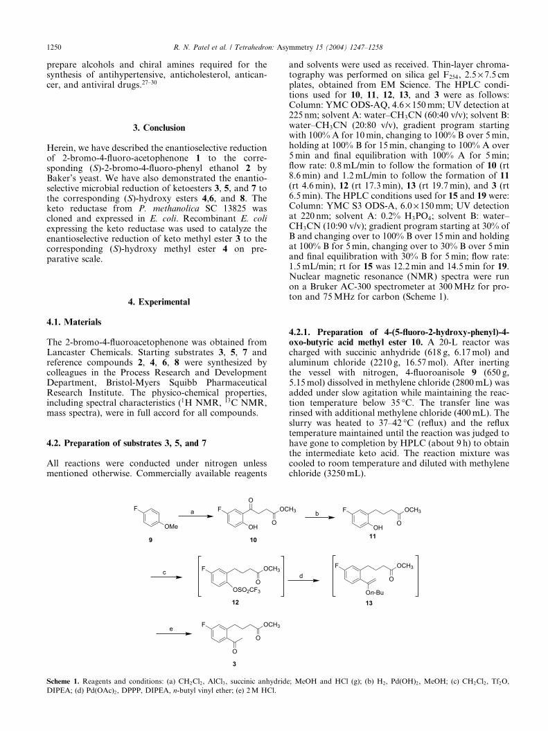
4.2. Preparation of substrates 3, 5, and 7
All reactions were conducted under nitrogen unlessmentioned otherwise. Commercially available reagents
F OC
OOH
OF
OMe
F OCH3
OOSO2CF3
a
c
F OCH3
O
O
e
9 10
12
3
Scheme 1. Reagents and conditions: (a) CH2Cl2, AlCl3, succinic anhydride
DIPEA; (d) Pd(OAc)2, DPPP, DIPEA, n-butyl vinyl ether; (e) 2M HCl.
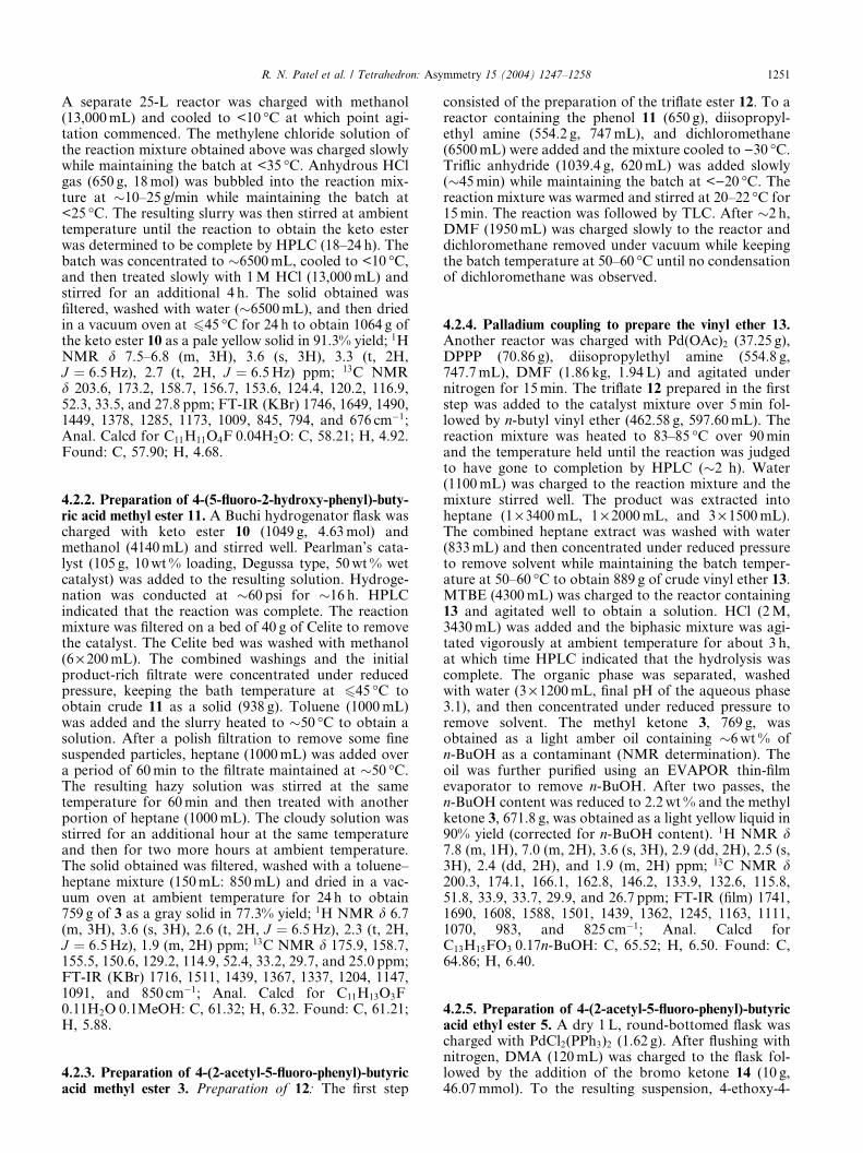
and solvents were used as received. Thin-layer chroma-tography was performed on silica gel F254, 2.5 · 7.5 cmplates, obtained from EM Science. The HPLC condi-tions used for 10, 11, 12, 13, and 3 were as follows:Column: YMC ODS-AQ, 4.6 · 150mm; UV detection at225 nm; solvent A: water–CH3CN (60:40 v/v); solvent B:water–CH3CN (20:80 v/v), gradient program startingwith 100% A for 10min, changing to 100% B over 5min,holding at 100% B for 15min, changing to 100% A over5min and final equilibration with 100% A for 5min;flow rate: 0.8mL/min to follow the formation of 10 (rt8.6min) and 1.2mL/min to follow the formation of 11(rt 4.6min), 12 (rt 17.3min), 13 (rt 19.7min), and 3 (rt6.5min). The HPLC conditions used for 15 and 19 were:Column: YMC S3 ODS-A, 6.0 · 150mm; UV detectionat 220 nm; solvent A: 0.2% H3PO4; solvent B: water–CH3CN (10:90 v/v); gradient program starting at 30% ofB and changing over to 100% B over 15min and holdingat 100% B for 5min, changing over to 30% B over 5minand final equilibration with 30% B for 5min; flow rate:1.5mL/min; rt for 15 was 12.2min and 14.5min for 19.Nuclear magnetic resonance (NMR) spectra were runon a Bruker AC-300 spectrometer at 300MHz for pro-ton and 75MHz for carbon (Scheme 1).
4.2.1. Preparation of 4-(5-fluoro-2-hydroxy-phenyl)-4-oxo-butyric acid methyl ester 10. A 20-L reactor wascharged with succinic anhydride (618 g, 6.17mol) andaluminum chloride (2210 g, 16.57mol). After inertingthe vessel with nitrogen, 4-fluoroanisole 9 (650 g,5.15mol) dissolved in methylene chloride (2800mL) wasadded under slow agitation while maintaining the reac-tion temperature below 35 �C. The transfer line wasrinsed with additional methylene chloride (400mL). Theslurry was heated to 37–42 �C (reflux) and the refluxtemperature maintained until the reaction was judged tohave gone to completion by HPLC (about 9 h) to obtainthe intermediate keto acid. The reaction mixture wascooled to room temperature and diluted with methylenechloride (3250mL).
H3 F OCH3
OOH
F OCH3
O
b
d
11
13On-Bu
; MeOH and HCl (g); (b) H2, Pd(OH)2, MeOH; (c) CH2Cl2, Tf2O,
R. N. Patel et al. / Tetrahedron: Asymmetry 15 (2004) 1247–1258 1251
A separate 25-L reactor was charged with methanol(13,000mL) and cooled to <10 �C at which point agi-tation commenced. The methylene chloride solution ofthe reaction mixture obtained above was charged slowlywhile maintaining the batch at <35 �C. Anhydrous HClgas (650 g, 18mol) was bubbled into the reaction mix-ture at �10–25 g/min while maintaining the batch at<25 �C. The resulting slurry was then stirred at ambienttemperature until the reaction to obtain the keto esterwas determined to be complete by HPLC (18–24 h). Thebatch was concentrated to �6500mL, cooled to <10 �C,and then treated slowly with 1M HCl (13,000mL) andstirred for an additional 4 h. The solid obtained wasfiltered, washed with water (�6500mL), and then driedin a vacuum oven at 645 �C for 24 h to obtain 1064 g ofthe keto ester 10 as a pale yellow solid in 91.3% yield; 1HNMR d 7.5–6.8 (m, 3H), 3.6 (s, 3H), 3.3 (t, 2H,J ¼ 6:5Hz), 2.7 (t, 2H, J ¼ 6:5Hz) ppm; 13C NMRd 203.6, 173.2, 158.7, 156.7, 153.6, 124.4, 120.2, 116.9,52.3, 33.5, and 27.8 ppm; FT-IR (KBr) 1746, 1649, 1490,1449, 1378, 1285, 1173, 1009, 845, 794, and 676 cm�1;Anal. Calcd for C11H11O4FÆ0.04H2O: C, 58.21; H, 4.92.Found: C, 57.90; H, 4.68.
4.2.2. Preparation of 4-(5-fluoro-2-hydroxy-phenyl)-buty-ric acid methyl ester 11. A Buchi hydrogenator flask wascharged with keto ester 10 (1049 g, 4.63mol) andmethanol (4140mL) and stirred well. Pearlman’s cata-lyst (105 g, 10wt% loading, Degussa type, 50wt% wetcatalyst) was added to the resulting solution. Hydroge-nation was conducted at �60 psi for �16 h. HPLCindicated that the reaction was complete. The reactionmixture was filtered on a bed of 40 g of Celite to removethe catalyst. The Celite bed was washed with methanol(6 · 200mL). The combined washings and the initialproduct-rich filtrate were concentrated under reducedpressure, keeping the bath temperature at 645 �C toobtain crude 11 as a solid (938 g). Toluene (1000mL)was added and the slurry heated to �50 �C to obtain asolution. After a polish filtration to remove some finesuspended particles, heptane (1000mL) was added overa period of 60min to the filtrate maintained at �50 �C.The resulting hazy solution was stirred at the sametemperature for 60min and then treated with anotherportion of heptane (1000mL). The cloudy solution wasstirred for an additional hour at the same temperatureand then for two more hours at ambient temperature.The solid obtained was filtered, washed with a toluene–heptane mixture (150mL: 850mL) and dried in a vac-uum oven at ambient temperature for 24 h to obtain759 g of 3 as a gray solid in 77.3% yield; 1H NMR d 6.7(m, 3H), 3.6 (s, 3H), 2.6 (t, 2H, J ¼ 6:5Hz), 2.3 (t, 2H,J ¼ 6:5Hz), 1.9 (m, 2H) ppm; 13C NMR d 175.9, 158.7,155.5, 150.6, 129.2, 114.9, 52.4, 33.2, 29.7, and 25.0 ppm;FT-IR (KBr) 1716, 1511, 1439, 1367, 1337, 1204, 1147,1091, and 850 cm�1; Anal. Calcd for C11H13O3FÆ0.11H2OÆ0.1MeOH: C, 61.32; H, 6.32. Found: C, 61.21;H, 5.88.
4.2.3. Preparation of 4-(2-acetyl-5-fluoro-phenyl)-butyricacid methyl ester 3. Preparation of 12: The first step
consisted of the preparation of the triflate ester 12. To areactor containing the phenol 11 (650 g), diisopropyl-ethyl amine (554.2 g, 747mL), and dichloromethane(6500mL) were added and the mixture cooled to )30 �C.Triflic anhydride (1039.4 g, 620mL) was added slowly(�45min) while maintaining the batch at <)20 �C. Thereaction mixture was warmed and stirred at 20–22 �C for15min. The reaction was followed by TLC. After �2 h,DMF (1950mL) was charged slowly to the reactor anddichloromethane removed under vacuum while keepingthe batch temperature at 50–60 �C until no condensationof dichloromethane was observed.
4.2.4. Palladium coupling to prepare the vinyl ether 13.Another reactor was charged with Pd(OAc)2 (37.25 g),DPPP (70.86 g), diisopropylethyl amine (554.8 g,747.7mL), DMF (1.86 kg, 1.94 L) and agitated undernitrogen for 15min. The triflate 12 prepared in the firststep was added to the catalyst mixture over 5min fol-lowed by n-butyl vinyl ether (462.58 g, 597.60mL). Thereaction mixture was heated to 83–85 �C over 90minand the temperature held until the reaction was judgedto have gone to completion by HPLC (�2 h). Water(1100mL) was charged to the reaction mixture and themixture stirred well. The product was extracted intoheptane (1 · 3400mL, 1 · 2000mL, and 3 · 1500mL).The combined heptane extract was washed with water(833mL) and then concentrated under reduced pressureto remove solvent while maintaining the batch temper-ature at 50–60 �C to obtain 889 g of crude vinyl ether 13.MTBE (4300mL) was charged to the reactor containing13 and agitated well to obtain a solution. HCl (2M,3430mL) was added and the biphasic mixture was agi-tated vigorously at ambient temperature for about 3 h,at which time HPLC indicated that the hydrolysis wascomplete. The organic phase was separated, washedwith water (3 · 1200mL, final pH of the aqueous phase3.1), and then concentrated under reduced pressure toremove solvent. The methyl ketone 3, 769 g, wasobtained as a light amber oil containing �6wt% ofn-BuOH as a contaminant (NMR determination). Theoil was further purified using an EVAPOR thin-filmevaporator to remove n-BuOH. After two passes, then-BuOH content was reduced to 2.2wt% and the methylketone 3, 671.8 g, was obtained as a light yellow liquid in90% yield (corrected for n-BuOH content). 1H NMR d7.8 (m, 1H), 7.0 (m, 2H), 3.6 (s, 3H), 2.9 (dd, 2H), 2.5 (s,3H), 2.4 (dd, 2H), and 1.9 (m, 2H) ppm; 13C NMR d200.3, 174.1, 166.1, 162.8, 146.2, 133.9, 132.6, 115.8,51.8, 33.9, 33.7, 29.9, and 26.7 ppm; FT-IR (film) 1741,1690, 1608, 1588, 1501, 1439, 1362, 1245, 1163, 1111,1070, 983, and 825 cm�1; Anal. Calcd forC13H15FO3Æ0.17n-BuOH: C, 65.52; H, 6.50. Found: C,64.86; H, 6.40.
4.2.5. Preparation of 4-(2-acetyl-5-fluoro-phenyl)-butyricacid ethyl ester 5. A dry 1L, round-bottomed flask wascharged with PdCl2(PPh3)2 (1.62 g). After flushing withnitrogen, DMA (120mL) was charged to the flask fol-lowed by the addition of the bromo ketone 14 (10 g,46.07mmol). To the resulting suspension, 4-ethoxy-4-
1252 R. N. Patel et al. / Tetrahedron: Asymmetry 15 (2004) 1247–1258
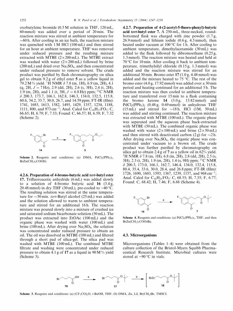
oxobutylzinc bromide (0.5M solution in THF, 120mL,60mmol) was added over a period of 20min. Thereaction mixture was stirred at ambient temperature for�60 h. After cooling in an ice bath, the reaction mixturewas quenched with 1M HCl (100mL) and then stirredfor an hour at ambient temperature. THF was removedunder reduced pressure and the resulting mixtureextracted with MTBE (2 · 200mL). The MTBE extractwas washed with water (2 · 200mL) followed by brine(200mL) and dried over Na2SO4, and then concentratedunder reduced pressure to remove solvent. The crudeproduct was purified by flash chromatography on silicagel to obtain 9.2 g of ethyl ester 5 as a yellow liquid in79.2M% yield. 1H NMR d 7.8 (m, 1H), 6.9 (m, 2H), 4.1(q, 2H, J ¼ 7Hz), 2.9 (dd, 2H), 2.6 (s, 3H), 2.4 (t, 2H),1.9 (m, 2H), and 1.1 (t, 3H, J ¼ 6:8Hz) ppm; 13C NMRd 200.3, 173.7, 166.1, 162.8, 146.3, 134.0, 132.5, 115.8,60.6, 34.2, 33.7, 30.0, 26.7, and 14.59 ppm; FT-IR (film):1741, 1685, 1613, 1582, 1495, 1429, 1357, 1234, 1188,1111, 800, and 973 cm�1; Anal. Calcd for C14H17FO3: C,66.65; H, 6.79; F, 7.53; Found: C, 66.57; H, 6.59; F, 7.32(Scheme 2).
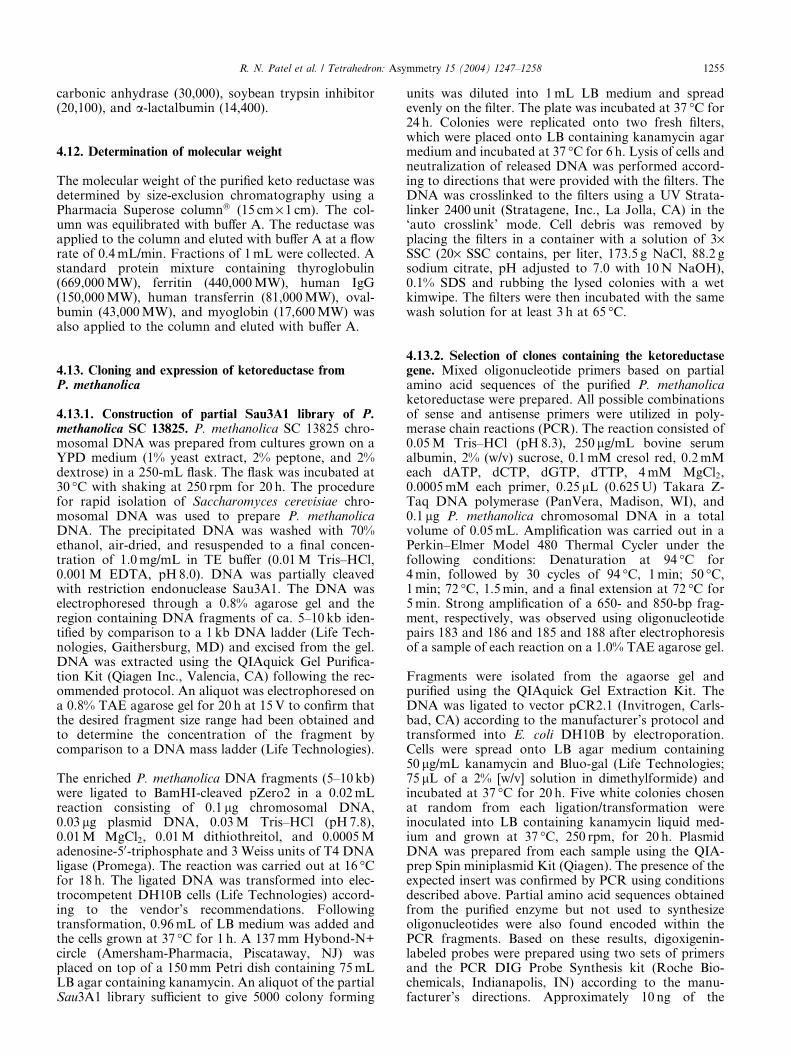
F Br
O
F O
O
O
a
14 7
Scheme 4. Reagents and conditions: (a) PdCl2(PPh3)2, THF, and then
BrZn(CH2)3COOtBu.
F Br
O
F O
O
O
a
14 5
Scheme 2. Reagents and conditions: (a) DMA, PdCl2(PPh3)2,
BrZn(CH2)3COOEt.
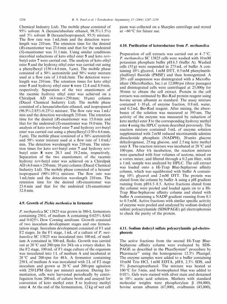
4.2.6. Preparation of 4-bromo-butyric acid tert-butyl ester17. Trifluoroacetic anhydride (6mL) was added slowlyto a solution of 4-bromo butyric acid 16 (3.4 g,20.48mmol) in dry THF (30mL), pre-cooled to )40 �C.The resulting solution was stirred at the same tempera-ture for �30min. tert-Butyl alcohol (25mL) was addedand the solution allowed to warm to ambient tempera-ture and stirred for an additional 16 h. The reactionmixture was poured slowly into a mixture of crushed iceand saturated sodium bicarbonate solution (50mL). Theproduct was extracted into EtOAc (100mL) and theorganic phase was washed with water (100mL) andbrine (100mL). After drying over Na2SO4, the solutionwas concentrated under reduced pressure to obtain anoil. The oil was dissolved in MTBE (100mL) and filteredthrough a short pad of silica-gel. The silica pad waswashed with MTBE (100mL). The combined MTBEfiltrate and washing were concentrated under reducedpressure to obtain 4.1 g of 17 as a liquid in 90M% yield(Scheme 3).
BrOH
O
BrO
O
a
16 17
Scheme 3. Reagents and conditions: (a) (CF3CO)2O, t-BuOH, THF; (b) DM
4.2.7. Preparation of 4-(2-acetyl-5-fluoro-phenyl)-butyricacid tert-butyl ester 7. A 250-mL, three-necked, round-bottomed flask was charged with zinc powder (1.7 g,26.9mmol) and lithium iodide (0.6 g, 4.5mmol), thenheated under vacuum at 100 �C for 1 h. After cooling toambient temperature, dimethylacetamide (30mL) wasadded to the flask followed by dibromoethane (0.25 g,1.3mmol). The reaction mixture was heated and held at70 �C for 10min. After cooling it back to ambient tem-perature, trimethylsilyl chloride (0.15 g, 1.3mmol) wasadded and the reaction mixture was stirred for anadditional 30min. Bromo ester 17 (1.0 g, 4.48mmol) wasadded and the mixture heated to 75 �C. The rest of thebromo ester (4.0 g, 17.92mmol) was added over a 30minperiod and heating continued for an additional 5 h. Thereaction mixture was then cooled to ambient tempera-ture and transferred over 15min to a flask containingthe bromo ketone 14 (3.0 g, 13.82mmol) andPdCl2(PPh3)2 (0.48 g, 0.69mmol) in anhydrous THF(15mL) and stirred for �18 h. HCl (1M, 50mL)was added and stirring continued. The reaction mixturewas extracted with MTBE (100mL). The organic phasewas separated and the aqueous phase back-extractedwith MTBE (50mL). The combined organic phase waswashed with water (2 · 100mL) and brine (2 · 50mL)and then stirred with deactivated carbon (2 g) for �2 h.After drying over Na2SO4, the organic phase was con-centrated under vacuum to a brown oil. The crudeproduct was further purified by chromatography onsilica gel to obtain 2.4 g of 7 as a yellow oil in 62% yield.1H NMR d 7.8 (m, 1H), 6.8 (m, 2H), 2.8 (dd, 2H), 2.5 (s,3H), 2.3 (t, 2H), 1.9 (m, 2H), 1.4 (s, 9H) ppm; 13C NMRd 200.3, 173.0, 166.1, 162.7, 146.4, 134.0, 132.4, 115.8,80.4, 35.4, 33.6, 30.0, 28.4, and 26.8 ppm; FT-IR (film):1726, 1690, 1603, 1593, 1367, 1239, 1157, and 968 cm�1;Anal. Calcd for C16H21FO3: C, 68.55; H, 7.55; F, 6.77;Found: C, 68.42; H, 7.46; F, 6.68 (Scheme 4).
4.3. Microorganisms
Microorganisms (Tables 1–4) were obtained from theculture collection of the Bristol-Myers Squibb Pharma-ceutical Research Institute. Microbial cultures werestored at )90 �C in vials.
BrZnO
O
b
18
A, Zn, Lil, Br(CH2)Br, TMSCI.

R. N. Patel et al. / Tetrahedron: Asymmetry 15 (2004) 1247–1258 1253
4.4. Growth of microorganisms
For screening purposes, one vial (1mL) of each culturewas used to inoculate 100mL of medium A (1% maltextract, 1% yeast extract, 2% glucose, and 0.3% pep-tone). The medium was adjusted to pH6.8 before steri-lization. Cultures were grown at 28 �C and 200 rpm for48 h. Cultures were harvested by centrifugation at18,000g for 15min, washed with 100mM potassiumphosphate buffer (pH 7.0), and suspended in 10mLof the same buffer, and then used for reductionstudies.
4.5. Reduction of 2-bromo-4-fluoro acetophenone 1
Cells of various microorganisms were suspended sepa-rately in 100mM potassium phosphate buffer (pH 7.0) at20% (w/v, wet cells) cell concentration and supple-mented with 1.5mg/mL of acetophenone 1 and 25mg/mL of glucose. Reduction was conducted at 28 �C and150 rpm. Periodically, samples (1mL) were taken andextracted with 4mL of acetonitrile. The sample was fil-tered through a 0.2 lm LID/X filter and analyzed usinga Hewlett Packard 1070 high pressure liquid chro-matograph (HPLC) to determine the substrate 1 andproduct 2 concentration. The remaining solution wasevaporated to dryness under a stream of nitrogen andthe residue taken up in 1mL of ethanol, filtered andanalyzed by HPLC to determine the enantiomeric excessof product 2. A phenylhexyl (150 · 4.6mm, 5 lm Phe-nomenex) column was used. The mobile phase consistedof 1:1 acetonitrile and water, and was used at a flow rateof 1.0mL/min. The detection wavelength was 210 nmand the column temperature 50 �C. The retention timesfor substrate 1 and product 2 were 6.3 and 5.4min,respectively. Separation of the two enantiomers of theracemic product 2 was achieved on a Chiralpak AD(4.6mm · 250mm, 10 lm, Diacel Chemical IndustryLtd.) column. The mobile phase consisted of 0.249%absolute ethanol in hexane. The flow rate was 1mL/minand the detection wavelength 210 nm. The retentiontimes for the desired (R)-enantiomer was 48min andthat for the undesired (S)-enantiomer 54min.
4.6. Reduction of 2-bromo-4-fluoro acetophenone 1 usingBaker’s yeast
A 3-L bioreactor (Braun Biostat B) was equipped with apH electrode and the impeller speed and temperature setat 500 rpm and 28 �C, respectively. Phosphate buffer(800mL of 10mM, pH6.0) was added to the bioreactorand 150 g of Baker’s yeast (from Red Star) added slowlyover a 30min period. The contents of the bioreactorwere stirred at 500 rpm, with the temperature main-tained at 28 �C, and the pH maintained at 6.0. Aceto-phenone 1 (5 g) and 192mL of glucose solution (25%)were added to the reactor at the rate of 8mL/h over aperiod of 24 h. Foaming was controlled by addition of0.5mL of SAG antifoam as required. Samples (1mL)were taken at intervals and analyzed for substrate 1 andproduct 2 concentrations and ee of product 2. Under
similar conditions, this process was scaled-up to 200-Lscale to reduce 1 kg of ketone 1.
4.7. Recovery of product 2
At the end of the biotransformation (20 h), 50 g ofXAD-16 resin (previously washed with 500mL of watercontaining 50% methanol and then with 2 · 500mL ofwater) was added to 1L of the reaction broth. Themixture was stirred at room temperature at 300 rpm for3 h and then filtered through 40 mesh stainless steelsieve. The collected resin was washed with 50mL ofwater containing 20% methanol and filtered through astainless steel sieve. The product-rich resin was treatedwith 50mL of methyl tert-butyl ether (MTBE) to exudethe desired alcohol 2. The solution was dried overanhydrous sodium sulfate, filtered, and the solventremoved in vacuo to provide 4.25 g of an oil in 85%overall yield. HPLC analysis showed 98.9% HI forproduct 2 with an ee of 99.8%. The same procedure wasused to recover alcohol 2 from a 200-L biotransform-ation batch. After adsorption of the product on XAD-16 and subsequent desorption of the product from therich resin by methyl tert-butyl ether extraction andevaporation, about 1.03 kg of oil was recovered. Thecrude product was purified by silica gel chromatographyto provide 707.4 g of product 2 in 99.6% purity with anee of 99.9%.
4.8. Reduction of methyl-, ethyl, and tert-butyl 4-(20-acetyl-50-fluorophenyl) butanoate
Various microbial cultures (1mL) were inoculated into100mL of medium A or medium B (1% malt extract, 1%yeast extract, 0.3% peptone, and 2% glycerol adjusted topH7) in a 500-mL flask and incubated at 28 �C and200 rpm on a shaker for 48 h. Cells were harvested bycentrifugation and suspended in 10mL of 100mMpotassium phosphate buffer (pH7.0) at 20% (w/v, wetcells) concentration. Glucose (25mg/mL) and ketomethyl ester 3 (1mg/mL) were added to the cell sus-pensions. The reduction was carried out at 28 �C and200 rpm on a shaker for 24–48 h. At predeterminedtimes, the reaction mixtures were quenched with fourvolumes of acetone, mixed on a vortex mixer, filteredthrough a 0.2 lm filter, and collected, with a 1mLsample analyzed by HPLC to determine the keto methylester 3 and hydroxy methyl ester 4 concentrations. Theremaining solution was evaporated to dryness under astream of nitrogen and the residue taken up in 1mL ofethanol, filtered, and analyzed by chiral HPLC todetermine the ee of product 4. A YMC ODS-A(150 · 4.6mm, 5 lm) column was used. The mobilephase consisted of a 40% acetonitrile and 60% H3PO4
(0.3%) mixture used at a flow rate of 1.0mL/min. Thedetection wavelength was 210 nm and the column tem-perature 50 �C. The retention times for keto methyl ester3 and hydroxy methyl ester 4 were 11.1 and 6.7min,respectively. Separation of the two enantiomers of theracemic hydroxy methyl ester was achieved on a Chir-alpak AD (4.6mm · 250mm; 10 lm) column (Diacel
1254 R. N. Patel et al. / Tetrahedron: Asymmetry 15 (2004) 1247–1258
Chemical Industry Ltd). The mobile phase consisted of95% solvent A (hexane/absolute ethanol, 98.5%:1.5%)and 5% solvent B (hexane/isopropanol, 95:5) mixture.The flow rate was 1mL/min and the detection wave-length was 210 nm. The retention time for the desired(R)-enantiomer was 23.6min and that for the undesired(S)-enantiomer was 31.1min. Using similar conditionsmicrobial reductions of keto ethyl ester 5 and keto tert-butyl ester 7 were carried out. The analysis of keto ethylester 5 and the hydroxy ethyl ester was carried out usinga phenylhexyl (150 · 4.6mm, 5 lm). The mobile phaseconsisted of a 50% acetonitrile and 50% water mixtureused at a flow rate of 1.0mL/min. The detection wave-length was 210 nm. The retention times for keto ethylester 5 and hydroxy ethyl ester 6 were 12.4 and 8.9min,respectively. Separation of the two enantiomers ofthe racemic hydroxy ethyl ester was achieved on aChiralpak AD (4.6mm · 250mm; 10 lm) column(Diacel Chemical Industry Ltd). The mobile phaseconsisted of a hexane/absolute ethanol, and isopropanol(96.9%:2.85%:0.25%) mixture. The flow rate was 1mL/min and the detection wavelength 210 nm. The retentiontime for the desired (R)-enantiomer was 15.0min andthat for the undesired (S)-enantiomer was 19.0min. Theanalysis of keto tert-butyl ester 7 and hydroxy tert-butylester was carried out using a phenylhexyl (150 · 4.6mm,5 lm). The mobile phase consisted of a 50% acetonitrileand 50% water mixture used at a flow rate of 1.0mL/min. The detection wavelength was 210 nm. The reten-tion times for keto tert-butyl ester 7 and hydroxy tert-butyl ester 8 were 28.3 and 19.2min, respectively.Separation of the two enantiomers of the racemichydroxy tert-butyl ester was achieved on a ChiralpakAD (4.6mm · 250mm; 10 lm) column (Diacel ChemicalIndustry Ltd). The mobile phase consisted of a hexane/isopropanol (90%:10%) mixture. The flow rate was1mL/min and the detection wavelength 210 nm. Theretention time for the desired (R)-enantiomer was23.6min and that for the undesired (S)-enantiomer32.8min.
4.9. Growth of Pichia methonica in fermentor
P. methanolica SC 13825 was grown in 380-L fermentorscontaining 250L of medium A containing 0.025% SAGand 0.025% Dow Corning antifoam. Growth consistedof two inoculum development stages and one fermen-tation stage. Inoculum development consisted of F1 andF2 stages. In the F1 stage, 1mL of a culture of P. met-hanolica SC 13825 was inoculated into 100mL of med-ium A contained in 500-mL flasks. Growth was carriedout at 28 �C and 200 rpm for 24 h on a rotary shaker. Inthe F2 stage, 100mL of F1 stage culture of the organismwas inoculated into 1L of medium A and incubated at28 �C and 200 rpm for 48 h. A fermentor containing250L of medium A was inoculated with 2L of F2 stageinoculum and grown at 28 �C and 200 rpm agitationwith 250LPM (liter per minute) aeration. During fer-mentation, cells were harvested periodically by centri-fugation from 200mL of culture broth and assayed forconversion of keto methyl ester 3 to hydroxy methylester 4. At the end of the fermentation, 12 kg of wet cell
paste was collected on a Sharples centrifuge and storedat )60 �C for future use.
4.10. Purification of ketoreductase from P. methanolica
Preparation of cell extracts was carried out at 4–7 �C.P. methanolica SC 13825 cells were washed with 10mMpotassium phosphate buffer pH6.5 (buffer A). Washedcells (55 g) were suspended in 275mL of buffer A con-taining 10% glycerol, 1mM DTT, 0.5mM phenylmeth-ylsulfonyl fluoride (PMSF) and than homogenized. A20% cell suspension was disintegrated with a Microflu-idizer (Microfluidics, Inc.) at 12,000 psi (three passages)and disintegrated cells were centrifuged at 25,000g for30min to obtain the cell extract. Protein in the cellextracts was estimated by Bio-Rad protein reagent usingbovine serum albumin as standard. The assay mixturecontained 1–10 lL of enzyme fraction, 0.8mL water,and 0.2mL Bio-Rad reagent. After mixing, the absor-bance of the solution was measured at 595 nm. Theactivity of the enzyme was measured by reduction ofketo methyl ester 3 to the corresponding hydroxy methylester 4 using the HPLC system described previously. Thereaction mixture contained 5mL of enzyme solutionsupplemented with 2mM reduced nicotinamide adeninedinucleotide phosphate (NADPH), 20 units glucosedehydrogenase, 25mg glucose, and 2.5mg keto methylester 3. The reaction mixture was incubated at 28 �C and100 rpm. After 6 h incubation, the reaction mixtureswere quenched with four volumes of acetone, mixed ona vortex mixer, and filtered through a 0.2 lm filter, witha 1mL sample was analyzed by HPLC. The cell extractwas loaded onto a Hi-Trap Blue-Sepharose affinitycolumn, which was equilibrated with buffer A contain-ing 10% glycerol and 2mM DTT. The protein waseluted from the column by buffer A using a pH gradientrunning from pH6.5–8.5. Active fractions eluted fromthe column were pooled and loaded again on to a Hi-Trap Blue-Sepharose affinity column and eluted withbuffer A containing a NADP gradient running from 0.1to 0.5mM. Active fractions with similar specific activityof enzyme were pooled and analyzed by sodium dodecylsulfate polyacrylamide (SDS/PAGE) gel electrophoresisto check the purity of the protein.
4.11. Sodium dodecyl sulfate polyacrylamide gel-electro-phoresis
The active fractions from the second Hi-Trap Blue-Sepharose affinity column were evaluated by SDS-PAGE as described in the PhastSystem� procedure byPharmacia31 using the homogeneous 12.5% Phastgel.The enzyme samples were added to a buffer containing10mM Tris–HCl, 1mM EDTA, pH8, 2.5% SDS, and5% b-mercaptoethanol. The mixture was heated at100 �C for 5min, and bromophenol blue was added to0.01%. Gels were stained with silver stain and destainedin 10% acetic acid solution. Markers with standardmolecular weights were phosphorylase b (94,000),bovine serum albumin (67,000), ovalbumin (43,000),
R. N. Patel et al. / Tetrahedron: Asymmetry 15 (2004) 1247–1258 1255
carbonic anhydrase (30,000), soybean trypsin inhibitor(20,100), and a-lactalbumin (14,400).
4.12. Determination of molecular weight
The molecular weight of the purified keto reductase wasdetermined by size-exclusion chromatography using aPharmacia Superose column� (15 cm · 1 cm). The col-umn was equilibrated with buffer A. The reductase wasapplied to the column and eluted with buffer A at a flowrate of 0.4mL/min. Fractions of 1mL were collected. Astandard protein mixture containing thyroglobulin(669,000MW), ferritin (440,000MW), human IgG(150,000MW), human transferrin (81,000MW), oval-bumin (43,000MW), and myoglobin (17,600MW) wasalso applied to the column and eluted with buffer A.
4.13. Cloning and expression of ketoreductase fromP. methanolica
4.13.1. Construction of partial Sau3A1 library of P.methanolica SC 13825. P. methanolica SC 13825 chro-mosomal DNA was prepared from cultures grown on aYPD medium (1% yeast extract, 2% peptone, and 2%dextrose) in a 250-mL flask. The flask was incubated at30 �C with shaking at 250 rpm for 20 h. The procedurefor rapid isolation of Saccharomyces cerevisiae chro-mosomal DNA was used to prepare P. methanolicaDNA. The precipitated DNA was washed with 70%ethanol, air-dried, and resuspended to a final concen-tration of 1.0mg/mL in TE buffer (0.01M Tris–HCl,0.001M EDTA, pH8.0). DNA was partially cleavedwith restriction endonuclease Sau3A1. The DNA waselectrophoresed through a 0.8% agarose gel and theregion containing DNA fragments of ca. 5–10 kb iden-tified by comparison to a 1 kb DNA ladder (Life Tech-nologies, Gaithersburg, MD) and excised from the gel.DNA was extracted using the QIAquick Gel Purifica-tion Kit (Qiagen Inc., Valencia, CA) following the rec-ommended protocol. An aliquot was electrophoresed ona 0.8% TAE agarose gel for 20 h at 15V to confirm thatthe desired fragment size range had been obtained andto determine the concentration of the fragment bycomparison to a DNA mass ladder (Life Technologies).
The enriched P. methanolica DNA fragments (5–10 kb)were ligated to BamHI-cleaved pZero2 in a 0.02mLreaction consisting of 0.1 lg chromosomal DNA,0.03 lg plasmid DNA, 0.03M Tris–HCl (pH7.8),0.01M MgCl2, 0.01M dithiothreitol, and 0.0005Madenosine-50-triphosphate and 3 Weiss units of T4 DNAligase (Promega). The reaction was carried out at 16 �Cfor 18 h. The ligated DNA was transformed into elec-trocompetent DH10B cells (Life Technologies) accord-ing to the vendor’s recommendations. Followingtransformation, 0.96mL of LB medium was added andthe cells grown at 37 �C for 1 h. A 137mm Hybond-N+circle (Amersham-Pharmacia, Piscataway, NJ) wasplaced on top of a 150mm Petri dish containing 75mLLB agar containing kanamycin. An aliquot of the partialSau3A1 library sufficient to give 5000 colony forming
units was diluted into 1mL LB medium and spreadevenly on the filter. The plate was incubated at 37 �C for24 h. Colonies were replicated onto two fresh filters,which were placed onto LB containing kanamycin agarmedium and incubated at 37 �C for 6 h. Lysis of cells andneutralization of released DNA was performed accord-ing to directions that were provided with the filters. TheDNA was crosslinked to the filters using a UV Strata-linker 2400 unit (Stratagene, Inc., La Jolla, CA) in the‘auto crosslink’ mode. Cell debris was removed byplacing the filters in a container with a solution of 3·SSC (20· SSC contains, per liter, 173.5 g NaCl, 88.2 gsodium citrate, pH adjusted to 7.0 with 10N NaOH),0.1% SDS and rubbing the lysed colonies with a wetkimwipe. The filters were then incubated with the samewash solution for at least 3 h at 65 �C.
4.13.2. Selection of clones containing the ketoreductasegene. Mixed oligonucleotide primers based on partialamino acid sequences of the purified P. methanolicaketoreductase were prepared. All possible combinationsof sense and antisense primers were utilized in poly-merase chain reactions (PCR). The reaction consisted of0.05M Tris–HCl (pH8.3), 250 lg/mL bovine serumalbumin, 2% (w/v) sucrose, 0.1mM cresol red, 0.2mMeach dATP, dCTP, dGTP, dTTP, 4mM MgCl2,0.0005mM each primer, 0.25 lL (0.625U) Takara Z-Taq DNA polymerase (PanVera, Madison, WI), and0.1 lg P. methanolica chromosomal DNA in a totalvolume of 0.05mL. Amplification was carried out in aPerkin–Elmer Model 480 Thermal Cycler under thefollowing conditions: Denaturation at 94 �C for4min, followed by 30 cycles of 94 �C, 1min; 50 �C,1min; 72 �C, 1.5min, and a final extension at 72 �C for5min. Strong amplification of a 650- and 850-bp frag-ment, respectively, was observed using oligonucleotidepairs 183 and 186 and 185 and 188 after electrophoresisof a sample of each reaction on a 1.0% TAE agarose gel.
Fragments were isolated from the agaorse gel andpurified using the QIAquick Gel Extraction Kit. TheDNA was ligated to vector pCR2.1 (Invitrogen, Carls-bad, CA) according to the manufacturer’s protocol andtransformed into E. coli DH10B by electroporation.Cells were spread onto LB agar medium containing50 lg/mL kanamycin and Bluo-gal (Life Technologies;75 lL of a 2% [w/v] solution in dimethylformide) andincubated at 37 �C for 20 h. Five white colonies chosenat random from each ligation/transformation wereinoculated into LB containing kanamycin liquid med-ium and grown at 37 �C, 250 rpm, for 20 h. PlasmidDNA was prepared from each sample using the QIA-prep Spin miniplasmid Kit (Qiagen). The presence of theexpected insert was confirmed by PCR using conditionsdescribed above. Partial amino acid sequences obtainedfrom the purified enzyme but not used to synthesizeoligonucleotides were also found encoded within thePCR fragments. Based on these results, digoxigenin-labeled probes were prepared using two sets of primersand the PCR DIG Probe Synthesis kit (Roche Bio-chemicals, Indianapolis, IN) according to the manu-facturer’s directions. Approximately 10 ng of the

1256 R. N. Patel et al. / Tetrahedron: Asymmetry 15 (2004) 1247–1258
isolated PCR fragment described above was included astemplate DNA. Amplification conditions were: Dena-turation at 94 �C for 4min, followed by 30 cycles of94 �C, 1min; 50 �C, 1min, 72 �C, 1min. Incorporation ofthe digoxigenin-dUTP nucleotide could be verified by asignificant increase in the molecular weight of thelabeled fragment. Superior incorporation was obtainedusing oligonucleotides 183 and 186.Duplicate filterscontaining lysed and denatured DNA from the P. met-hanolica Sau3A1 library were incubated with 10mL ofDIG EasyHyb solution (Roche) and 5 lL of the dena-tured, labeled PCR fragment in a roller bottle.Hybridization proceeded at 42 �C for 18 h. Filters werewashed twice with 2· SSC (prepared from a 20· solu-tion; 20· SSC contains, per liter, 173.5 g NaCl, 88.2 gsodium citrate, pH adjusted to 7.0 with 10N NaOH),0.1% sodium dodecyl sulfate (SDS) at room temperaturefor 5min, then twice at 68 �C with 0.5% SSC, 0.1% SDSfor 15min. Anti-digoxigenin antibody binding, washing,and detection were performed using the DIG Labelingand Detection Kit reagents and protocols (Roche). Themembranes were placed on Whatman 3mm paper toremove excess liquid, covered with Saran Wrap, andexposed to autoradiography film (Kodak X-OMATLS). A single hybridizing colony was picked from themaster filter and streaked onto LB agar medium con-taining kanamycin and incubated at 37 �C for 24 h. Thecolony was grown LB liquid medium containing kana-mycin for plasmid isolation using the QIAfilter PlasmidMidi Kit (Qiagen). Restriction mapping indicated aninsert of ca. 5.0 kb was present in the recombinantplasmid. Isolated DNA was tested for its ability tosupport amplification using oligos 183 and 186 and 185and 188, which was confirmed. Oligonucleotide primersbased on the DNA sequence of the isolated PCR frag-ments were used for the analysis of the insert in pKR5.0. An open reading frame of 1059 bp that encodes aprotein of 353 amino acids with a molecular weight of39,800Da was found. This is in near agreement with thesize of the isolated ketoreductase (40,000Da by gel fil-tration).
4.13.3. Subcloning of the ketoreductase gene and expres-sion in E. coli. The polymerase chain reaction was uti-lized to amplify the complete ketoreductase genecontaining restriction sites suitable for cloning intoexpression vector pBMS2000. The primers used are gi-ven below:
BspHI
50 TGCTCATGAATTGGGAAAAAGTTCCACAAG 30
Nucleotides in bold indicate the recognition sequencefor the restriction enzyme BspHI;
BamHI
50 CTCGGATCCTTATAAAATTACAGAATATAAG 30
Nucleotides in bold indicate the recognition sequencefor the restriction enzyme BamHI; PCR conditions were
identical to those described previously, except pKR 5.0DNA was used as a template.
The BspHI/BamHI-digested PCR fragment was ligatedto BspHI/BamHI-cleaved pBMS2000. The ligated DNAwas transformed into electrocompetent DH10B cells(Life Technologies, Inc.) according to the vendor’s rec-ommendations. Following transformation, 0.96mL ofLB medium were added and the cells grown at 37 �C for1 h. An aliquot of the cells was spread onto the LB agarmedium containing 10 lg/mL neomycin sulfate (Sigma)and the plate incubated at 37 �C for 20 h. The presenceof the correct insert was established by PCR usingconditions described earlier except a portion of 16 ran-domly chosen colonies was the source of template DNA.Fourteen out of the 16 colonies amplified a fragment ofthe correct size. The plasmid from one of these isolateswas named pBMS2000-PMKR (for ketoreductase).pBMS2000-PMKR was transformed into competentE. coli strain BL21-CodonPlus(DE3)-RIL cells (Strata-gene) according the manufacturer’s protocol. Cells werespread onto LB agar medium containing 30 lg/mLchloramphenicol and 10 lg/mL neomycin. Four colonieswere randomly chosen and used as a source of templateDNA for PCR using the conditions described earlier.All four reactions amplified a DNA fragment of thecorrect size, demonstrating that the BL21-Codon-Plus(DE3)-RIL had been successfully transformed. Oneof these isolates was selected as the expression strain andused for all further experiments. Vial lots of theexpression strain were prepared.
Expression of the ketoreductase gene was controlled byIPTG (isopropylthio-b-DD galactoside) induction of theptac promoter that originated on plasmid pBMS2000.The expression strain was grown in 25mL of MT3/neo/chlor in a 250-mL flask at 15 �C, 225 rpm until it hadreached OD600nm �0.7. At this point, IPTG (LifeTechnologies) was added to a final concentration of0.5mM. The cultures were allowed to grow overnight(�16 h) to allow complete induction of the ketoreduc-tase gene and production of the ketoreductase protein.Following overnight induction with IPTG, samples wereanalyzed on protein gels. The results indicated thestrain’s ability to overexpress the heterologous protein.
4.14. Reduction of keto methyl ester by recombinantE. coli
Cells of E. coli SC 16445 (expressing ketoreductase)were grown in a 250-L fermentor. Growth consisted ofinoculum development stage, germinator stage and fer-mentation stage. In inoculum development stage, 1mLculture of E. coli SC 16445 was inoculated into 1L ofmedium B (1.0% NZ Amine A, 2.0% Yeastamin, 2.0%glycerol, 0.6% Na2HPO4, 0.3% KH2PO4, 0.125%(NH4)2SO4, 0.0246% MgSO47H2O) containing a filteredsterilized solution (10mg in ethanol) of neomycin sul-fate, and a filtered sterilized solution (33mg in water) ofchloramphenicol. Growth was carried out in 4-L flasksat 15 �C and 150 rpm for 72 h. In a germinator stage, a20-L fermentor containing 15L medium B containing
R. N. Patel et al. / Tetrahedron: Asymmetry 15 (2004) 1247–1258 1257
neomycin sulfate and chloramphenicol (150mg neomy-cin sulfate in 100mL water and 495mg chloramphenicolin 50mL ethanol were separately filter sterilized using a0.2 lm cellulose nitrate filter and added to the germi-nator after sterilizing the medium) was inoculated with1L inoculum. Cells were grown at 15 �C, 20LPM (literper min) aeration and 1000 rpm agitation for 24 h. A380-L fermentor containing 250L of medium B con-taining neomycin sulfate and chloramphenicol chlor-amphenicol (2.5 g neomycin sulfate in 600mL water and8.25 g chloramphenicol in 600mL ethanol were sepa-rately filter sterilized using a 0.2 lm cellulose nitratefilter and added to the fermentor after sterilizing themedium) was inoculated with 15L germinator growninoculum. The growth was carried out at 15 �C,250LPM aeration, and 200 rpm agitation. Induction ofthe ketoreductase was carried out when the optical celldensity (OD at 600 nm) of the culture reached close to3.0 by the addition of a filter-sterilized solution (14.9 g in1L of water) of isopropyl-b-thiogalactoside (IPTG).Eight hours after addition of IPTG, the addition ofnutrient feed (20L of feed containing 1% NZ Amine A,1% yeastamin, 1% glycerol, and 0.02% antifoamUCON) was initiated at 1 L/h over 20 h. During fer-mentation, cells were periodically harvested by centri-fugation from 200mL of culture broth and assayed forconversion of keto methyl ester 3 to hydroxy methylester 4. Cells were harvested approximately 30 h afterinduction when the highest ketoreductase activity wasobserved. At the end of fermentation, the fermentor wascooled to 6–8 �C and cells collected in a Sharples cen-trifuge. About 10–11 kg of cell paste was collected andstored at )70 �C until further used in the biotransfor-mation process.
Cells were used to catalyze the bioconversion of ketomethyl ester 3. Cells were suspended in 1L of 50mMpH7.0 phosphate buffer at 10% (w/v, wet cells) cellconcentration. The suspensions were supplemented with100 lM nicotinamide adenine dinucleotide phosphate(NADP), 1mM phenylmethanesulfonyl fluoride(PMSF), 20 g glucose, 34,000 units glucose dehydroge-nase, and 4.5 g substrate 3. Biotransformation was car-ried out at 500 rpm and 28 �C. The pH was maintainedat 6.8–7.0. Glucose feed (solution containing 20% glu-cose, 1.92% K2HPO4, and 1.2% KH2PO4) was startedafter 1 h at rate of 6mL/h/L of reaction mixture. Thereaction was completed in 20 h with a 95% reaction yieldof 4. An ee of 99.9% was obtained for 4. The biotrans-formation process was scaled up to 500L to reduce 3 kgof substrate 3.
4.15. Recovery of hydroxy methyl ester 4
At the end of the biotransformation, 40 g of XAD-16resin (previously washed with 500mL of a 1:1 (v/v)water and methanol mixture, and then washed with2 · 500mL of water) was added to the 1L of reactionbroth. The mixture was stirred at room temperature at300 rpm for 8 h and finally filtered through a 40-meshstainless steel sieve. The collected resin was washed with50mL of a water and methanol mixture (80:20 by vol)
and filtered through a stainless steel sieve. The product-rich resin was treated with 50mL of methyl tert-butylether (MTBE) to desorb the alcohol 4. The solution wasdried over anhydrous sodium sulfate and filtered and thesolvent was removed on the rotary evaporator to pro-vide 3.9 g of an oil in 86% overall yield. HPLC analysisshowed 98.9 area percent for product 4 with an ee of99.9%. The same procedure was used to recover alcohol4 from a 500-L biotransformation batch.
Acknowledgements
We would like to acknowledge Dr. David Kronenthalfor reviewing this manuscript and providing valuablesuggestions.
References and notes
1. Patel, R. N. Enzyme Microb. Technol. 2002, 31, 804–826.2. Food & Drug Administration. Chirality 1992, 4, 338–340.3. Buckland, B. C.; Robinson, D. K.; Chartrain, M. Metab.
Eng. 2000, 2, 42–48.4. Pesti, J. A.; Dicosimo, R. Curr. Opin. Discov. Dev. 2000, 3,
764–782.5. Gotor, V. Biocat. Biotrans. 2000, 18, 87–103.6. Stewart, D. Curr. Opin. Chem. Biol. 2001, 5, 120–129.7. Zak, A. Curr. Opin. Chem. Biol. 2001, 5, 130–136.8. Patel, R. N. In Stereoselective Biocatalysis; Patel, R. N.,
Ed.; Marcel Dekker: New York, 2000; pp 87–130.9. Schnell, B.; Faber, K.; Kroutil, W. Adv. Synth. Catal.
2003, 345, 653–666.10. Patel, R. N. Adv. Synth. Catal. 2001, 343, 527–546.11. Churcher, I.; Nadin, A. J.; Owens, A. P. PCT Int. Appl.
2002; 56 pp. CAN 136:325575, AN 2002:293634.12. Schenk, D.; Games, D.; Seubert, P. J. Mol. Neurosci. 2001,
17, 259–265.13. Yang, M. G.; Liu, H. PCT Int. Appl. 2001; 170 pp. CAN
135:288707, AN 2001:747759.14. Olson, R. E. PCT Int. Appl. 2001; 254 pp. CAN
136:20092, AN 2001:886081.15. Felsenstein, K.; Smith, D. W.; Poss, M. A.; Chaturvedula,
P. C.; Sloan, C. P.; Charles, P. Eur. Pat. Appl. 1997; 30 pp.CAN 127:109192, AN 1997:470004.
16. Rotella, D. P. Chemtracts 2000, 13, 626–629.17. Patel, R. R. N.; Goswami, A.; Chu, L.; Nanduri, V.;
Goldberg, S.; Johnston, R.; Donovan, M.-J ; Mirfakhrae,K. D. Patent Appl. WO 2002-US6783 20020306. CAN137:277933. AN 2002:754608.
18. Ward, O. P.; Young, C. S. Enzyme Microb. Technol. 1990,12, 482–493.
19. Csuk, R.; Glanzer, B. I. Chem. Rev. 1991, 91, 49–97.20. Sih, C. J.; Chen, C. S. Angew. Chem. 1984, 96, 556–566.21. Kula, M.-R.; Kragl, U. In Stereoselective Biocatalysis;
Patel, R. N., Ed.; Marcel Dekker: New York, 2000; pp839–866.
22. Jones, J. B.; Back, J. F. In Applications of BiochemicalSystems in Organic Synthesis; Jones, J. B., Jones, C. J.,Perlman, S. D., Eds.; John Wiley & Sons: NY, 1976; pp248–376.
23. Keinan, E.; Hafeli, E. K.; Seth, K.; Lamed, R. J. Am.Chem. Soc. 1986, 108, 162–168.
24. Prelog, V. Pure Appl. Chem. 1964, 9, 119–123.

1258 R. N. Patel et al. / Tetrahedron: Asymmetry 15 (2004) 1247–1258
25. Bradshaw, C. W.; Hummel, W.; Wong, C.-H. J. Org.Chem. 1992, 57, 1532–1536.
26. Bradshaw, C. W.; Fu, H.; Shen, G.-J.; Wong, C.-H.J. Org. Chem. 1992, 57, 1526–1532.
27. Patel, R. N.; Banerjee, A.; McNamee, C. G.; Szarka, L.J. Appl. Microbiol. Biotechnol. 1993, 40, 241–245.
28. Patel, R. N.; Banerjee, A.; Howell, J. M.; McNamee, C.G.; Brzozowski, D.; Mirfakhrae, K. D.; Nanduri, V.;
Thottathil, J. K.; Szarka, L. J. Tetrahedron: Asymmetry1993, 4, 2069–2084.
29. Patel, R. N.; Banerjee, A.; Liu, M.; Hanson, R. L.; Ko, R.Y.; Howell, J. M.; Szarka, L. J. Biotechnol. Appl. Biochem.1993, 17, 139–153.
30. Patel, R. N.; Banerjee, A.; McNamee, C.; Brzozowski, D.;Szarka, L. J. Tetrahedron: Asymmetry 1997, 8, 2547–2552.
31. Heukeshoven, J.; Dernick, R. Electrophoresis 1985, 6, 103–112.