Efficacy of Four-Channel Functional Electrical Stimulation on ...
12
Citation: Schick, T.; Kolm, D.; Leitner, A.; Schober, S.; Steinmetz, M.; Fheodoroff, K. Efficacy of Four-Channel Functional Electrical Stimulation on Moderate Arm Paresis in Subacute Stroke Patients—Results from a Randomized Controlled Trial. Healthcare 2022, 10, 704. https://doi.org/10.3390/ healthcare10040704 Academic Editors: Teen-Hang Meen, Chun-Yen Chang, Charles Tijus and Po-Lei Lee Received: 22 February 2022 Accepted: 7 April 2022 Published: 10 April 2022 Publisher’s Note: MDPI stays neutral with regard to jurisdictional claims in published maps and institutional affil- iations. Copyright: © 2022 by the authors. Licensee MDPI, Basel, Switzerland. This article is an open access article distributed under the terms and conditions of the Creative Commons Attribution (CC BY) license (https:// creativecommons.org/licenses/by/ 4.0/). healthcare Article Efficacy of Four-Channel Functional Electrical Stimulation on Moderate Arm Paresis in Subacute Stroke Patients—Results from a Randomized Controlled Trial Thomas Schick 1, * , Daniela Kolm 2 , Andreas Leitner 2 , Sandra Schober 2 , Maria Steinmetz 3 and Klemens Fheodoroff 2 1 MED-EL, Medical Electronics, Business Unit Neurorehabilitation STIWELL, Fürstenweg 77a, 6020 Innsbruck, Austria 2 KABEG Gailtal-Klinik, Neurorehabilitation, Radnigerstrasse 12, 9620 Hermagor, Austria; [email protected] (D.K.); [email protected] (A.L.); [email protected] (S.S.); [email protected] (K.F.) 3 Therapy Center Riedhart, Occupational Therapy, Innsbrucker Str. 9, 6300 Wörgl, Austria; [email protected] * Correspondence: [email protected]; Tel.: +43-577881299 Abstract: This preliminary randomized clinical trial explores the efficacy of task-oriented electromyo- graphy (EMG)-triggered multichannel functional electrical stimulation (EMG-MES) compared to single-channel cyclic neuromuscular electrical stimulation (cNMES) on regaining control of voluntary movements (CVM) and the ability to execute arm-hand-activities in subacute stroke patients with moderate arm paresis. Twelve ischemic stroke patients (Fugl-Meyer Assessment Arm Section (FMA- AS) score: 19–47) with comparable demographics were block-randomized to receive 15 sessions of cNMES or EMG-MES over three weeks additionally to a conventional neurorehabilitation program including task-oriented arm training. FMA-AS, Box-and-Block Test (BBT), and Stroke-Impact-Scale (SIS) were recorded at baseline and follow-up. All participants demonstrated significant improvement in FMA-AS and BBT. Participants treated with EMG-MES had a higher mean gain in FMA-AS than those treated with cNMES. In the SIS daily activities domain, both groups improved non-significantly; participants in the EMG-MES group had higher improvement in arm-hand use and stroke recov- ery. EMG-MES treatment demonstrated a higher gain of CVM and self-reported daily activities, arm-hand use, and stroke recovery compared to cNMES treatment of the wrist only. The protocol of this proof-of-concept study seems robust enough to be used in a larger trial to confirm these preliminary findings. Keywords: moderate arm paresis; subacute stroke; motor recovery; control of voluntary movements; EMG-triggered multichannel electrical stimulation; functional electrical stimulation 1. Introduction Stroke is the main reason for disability in adults [1]. Neurorehabilitation focuses on regaining control of voluntary movements (CVM) to optimize task performance in activities of daily living, thus improving quality of life. Houwink et al. [2] found that 78% of stroke patients with an initial CVM in the shoulder and elbow at admission to first in-patient rehabilitation achieved advanced arm and hand capacity after discharge. Recovery of CVM in paretic arm and hand (with or without synkinesis) can best be quantified with the Fugl-Meyer Assessment Arm Section (FMA-AS; score 0–66) [3,4]. In recent years, electrical stimulation (ES) of an upper extremity is demonstrated to be a promising intervention to improve arm and hand function in neurorehabilitation [5,6]. Data have been published on the effects of both cyclical and electromyography (EMG)- triggered single-channel ES [6–8]. The research exhibits the positive effects of Functional Electrical Stimulation (FES) to improve the ability to perform activities [6,8]. Healthcare 2022, 10, 704. https://doi.org/10.3390/healthcare10040704 https://www.mdpi.com/journal/healthcare
-
Upload
khangminh22 -
Category
Documents
-
view
1 -
download
0
Transcript of Efficacy of Four-Channel Functional Electrical Stimulation on ...
�����������������
Citation: Schick, T.; Kolm, D.;
Leitner, A.; Schober, S.; Steinmetz, M.;
Fheodoroff, K. Efficacy of
Four-Channel Functional Electrical
Stimulation on Moderate Arm Paresis
in Subacute Stroke Patients—Results
from a Randomized Controlled Trial.
Healthcare 2022, 10, 704.
https://doi.org/10.3390/
healthcare10040704
Academic Editors: Teen-Hang Meen,
Chun-Yen Chang, Charles Tijus
and Po-Lei Lee
Received: 22 February 2022
Accepted: 7 April 2022
Published: 10 April 2022
Publisher’s Note: MDPI stays neutral
with regard to jurisdictional claims in
published maps and institutional affil-
iations.
Copyright: © 2022 by the authors.
Licensee MDPI, Basel, Switzerland.
This article is an open access article
distributed under the terms and
conditions of the Creative Commons
Attribution (CC BY) license (https://
creativecommons.org/licenses/by/
4.0/).
healthcare
Article
Efficacy of Four-Channel Functional Electrical Stimulation onModerate Arm Paresis in Subacute Stroke Patients—Resultsfrom a Randomized Controlled TrialThomas Schick 1,* , Daniela Kolm 2, Andreas Leitner 2, Sandra Schober 2, Maria Steinmetz 3 andKlemens Fheodoroff 2
1 MED-EL, Medical Electronics, Business Unit Neurorehabilitation STIWELL, Fürstenweg 77a,6020 Innsbruck, Austria
2 KABEG Gailtal-Klinik, Neurorehabilitation, Radnigerstrasse 12, 9620 Hermagor, Austria;[email protected] (D.K.); [email protected] (A.L.); [email protected] (S.S.);[email protected] (K.F.)
3 Therapy Center Riedhart, Occupational Therapy, Innsbrucker Str. 9, 6300 Wörgl, Austria;[email protected]
* Correspondence: [email protected]; Tel.: +43-577881299
Abstract: This preliminary randomized clinical trial explores the efficacy of task-oriented electromyo-graphy (EMG)-triggered multichannel functional electrical stimulation (EMG-MES) compared tosingle-channel cyclic neuromuscular electrical stimulation (cNMES) on regaining control of voluntarymovements (CVM) and the ability to execute arm-hand-activities in subacute stroke patients withmoderate arm paresis. Twelve ischemic stroke patients (Fugl-Meyer Assessment Arm Section (FMA-AS) score: 19–47) with comparable demographics were block-randomized to receive 15 sessions ofcNMES or EMG-MES over three weeks additionally to a conventional neurorehabilitation programincluding task-oriented arm training. FMA-AS, Box-and-Block Test (BBT), and Stroke-Impact-Scale(SIS) were recorded at baseline and follow-up. All participants demonstrated significant improvementin FMA-AS and BBT. Participants treated with EMG-MES had a higher mean gain in FMA-AS thanthose treated with cNMES. In the SIS daily activities domain, both groups improved non-significantly;participants in the EMG-MES group had higher improvement in arm-hand use and stroke recov-ery. EMG-MES treatment demonstrated a higher gain of CVM and self-reported daily activities,arm-hand use, and stroke recovery compared to cNMES treatment of the wrist only. The protocolof this proof-of-concept study seems robust enough to be used in a larger trial to confirm thesepreliminary findings.
Keywords: moderate arm paresis; subacute stroke; motor recovery; control of voluntary movements;EMG-triggered multichannel electrical stimulation; functional electrical stimulation
1. Introduction
Stroke is the main reason for disability in adults [1]. Neurorehabilitation focuses onregaining control of voluntary movements (CVM) to optimize task performance in activitiesof daily living, thus improving quality of life. Houwink et al. [2] found that 78% of strokepatients with an initial CVM in the shoulder and elbow at admission to first in-patientrehabilitation achieved advanced arm and hand capacity after discharge.
Recovery of CVM in paretic arm and hand (with or without synkinesis) can best bequantified with the Fugl-Meyer Assessment Arm Section (FMA-AS; score 0–66) [3,4].
In recent years, electrical stimulation (ES) of an upper extremity is demonstrated to bea promising intervention to improve arm and hand function in neurorehabilitation [5,6].Data have been published on the effects of both cyclical and electromyography (EMG)-triggered single-channel ES [6–8]. The research exhibits the positive effects of FunctionalElectrical Stimulation (FES) to improve the ability to perform activities [6,8].
Healthcare 2022, 10, 704. https://doi.org/10.3390/healthcare10040704 https://www.mdpi.com/journal/healthcare
Healthcare 2022, 10, 704 2 of 12
Interventions that support voluntary movements through an ES triggered by an EMGsignal, which is induced by a target movement performed from the affected individual,are increasingly gaining acceptance. This type of intervention was described as even morereinforcing than cyclic stimulation due to the proprioceptive feedback and voluntary com-ponent involved. Motor improvements in hand function following a stroke have beenobserved [9–12]. A meta-analysis describes the comparison of EMG-triggered ES to con-ventional therapy and demonstrates improved upper limb impairment in those more than3-month post stroke. The benefits within the first month and the comparison of the effectsof different EMG-stimulation protocols are not clear yet. These authors suggested thatEMG-ES leads to greater gains in post stroke upper extremity impairment [7]. In a studyon stroke patients, EMG-ES was compared with classical transcutaneous nerve stimulation(TENS) below the motor threshold. EMG-triggered stimulation leads to better short-termeffects in the intervention group [13]. In contrast to a pure repetition of movement in onejoint, functional multi-joint arm-and-hand activities can be elicited by EMG-triggered mul-tichannel stimulation patterns (EMG-MES) [11,14,15]. Patients with moderate arm paresiscan use their residual CVM to trigger voluntary movements [7,16]. A higher activity levelof the patient has been proposed as an additional advantage for this type of intervention.
EMG-MES has also been studied as part of motor re-learning programs and for arm-and-hand use in stroke patients [11,12,14,15]. As functional electrical stimulation (FES)is defined as a process of pairing stimulation simultaneously or intermittently with afunctional task [17], EMG-MES is a variation of FES through its functional and targetoriented approach [12].
Although the effects of ES suggest improvement in arm function in stroke patients,it is not yet clear which procedure and parameter choice of different forms of electricalstimulation (sensory afferent stimulation; cyclic electrical stimulation; EMG-triggeredelectrical stimulation) is superior [6,8].
Currently, it is unclear which type of stimulation (and which movements) shouldbe used for different severity levels of impaired CVM. There are few data indicating thesuperiority of EMG-MES compared with cNMES [16,18]. The study presented in this articleshould provide additional insight on this topic.
2. Materials and Methods2.1. Study Design and Participants
This was a randomized, controlled, single-assessor blinded study conducted in astroke in-patient neurorehabilitation center in Austria.
The study was approved by the Ethics Committee of the State of Carinthia, Austria(vote A06/18—26 April 2018), and registered at the German Registry for clinical trials(DRKS) under the national clinical trial registration ID number DRKS00014358 and con-forms to the ICMJE guidelines.
Relevant inclusion and exclusion criteria were:
2.2. Inclusion Criteria
• First ischemic stroke with moderate arm paresis (19 ≤ FMA-AS ≤ 47) [19];• Existing ADL capability before the event;• Early and late post-acute phase 1–6 months [20];• Age between 18 and 80 years;• Capacity for written informed consent.
2.3. Exclusion Criteria
• Implanted defibrillators, brain stimulators, pacemakers, and drug pumps;• Severe contractures in the treatment area;• Wounds in the stimulation area;• Pregnancy.

Healthcare 2022, 10, 704 3 of 12
Participants’ independent execution of movements in activities of daily living (ADL)and full arm-hand activities prior to the stroke were determined by asking participants todescribe their history of ADL and arm-hand activities before entering the study. Writteninformed consent was acquired from all participants or their legal custodians’ previousstudy entry. Strict data security was ensured during and after the study. The entire studydocumentation was anonymized and transferred to the study investigator for safekeeping.Personal data remained at the responsible rehabilitation center and will continue to be savedon-site during regular archiving. Video recordings were stored on a password-protectedonline database and made available to the external assessor. Videos were deleted fromthat database after evaluation and saved to a secure external hard drive by the investigator.Subjects were informed about the two test groups but not about the study hypothesis.
2.4. Randomization
Participants were block-randomized to the intervention group (IG) or the controlgroup (CG) in blocks of six participants for each group. In each block, three participantswere allocated to the IG and the CG. Before study initiation, the study manager providedtwo master envelopes to the person responsible for study execution. Each master envelopecontained six numbered and sealed envelopes. Each of the sealed envelopes included one ofthe random numbers generated by Microsoft Excel® (Version 2016) (Santa Rosa, CA, USA),the resultant randomized allocation to the IG or CG, and all tests, procedures, and relevantdocuments that were needed to carry out the study. The centers were not allowed to openthe second master envelope until all envelopes of the first master envelope were used (blockrandomization) [21,22]. All envelopes were numbered and drawn by a blinded personwho was not directly involved in the study. With this process, a number and a group wereassigned to each participant.
2.5. Intervention
All study participants underwent intensive arm-hand training as part of their in-patient stroke rehabilitation program (training of repetitive (single and complex) move-ments and task-oriented training within given capacity and muscle endurance, 5 × 30 minper week). During the three-week study period, the participants received 15 training sessionswith either EMG-MES or with cNMES (5 × 30 min per week) in addition to their conven-tional rehabilitation program. The study duration was 3 weeks with 5 interventions per week(30 min each) of the respective additional treatment options, with participants seated on achair in front of a height-adjustable desk that was adapted to their individual heights.
ES in both groups was performed with the STIWELL® med4 device (CE 0297; P/N9001015) developed by MED−EL Elektromedizinische Geräte Gesellschaft m.b.H., Inns-bruck, Austria). This electrical stimulation device has four muscle stimulation channelsand up to two channels for EMG signal uptake. For all study participants, the same typeof surface electrodes (PALS® Neurostimulation electrodes, oval 4 cm × 6.4 cm, AxelgaardManufacturing Co., Ltd., Lystrup, Denmark, CE-certified, REF 896230) were used.
In both groups, rectangular biphasic pulses with a pulse width of 300 µs were used. Apulse intensity (mA) to elicit a sufficient and possibly visible grip motion of the affectedlimb was selected according to the participants’ level of tolerance. The standard currentfrequency was 30 Hz.
Since the differences in the procedures of the two therapy options were quite apparent(EMG-MES versus cNMES), therapy sessions that were blinded for both the therapistsand the participants were not possible. A single-blinded study design with blinded videoassessments (as described below) was chosen for this reason.
2.6. EMG-MES
The therapy sessions of the IG consisted of task-oriented unilateral arm-and-handmovements on the affected side with four-channel EMG-triggered pulses for reaching,gripping, and lateral lifting of cylindrical objects to different heights (0 cm, 6 cm, 12 cm,
Healthcare 2022, 10, 704 4 of 12
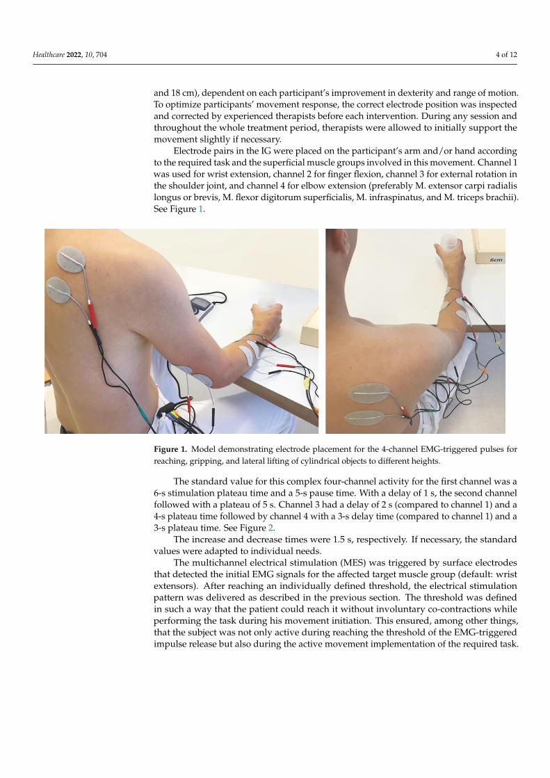
and 18 cm), dependent on each participant’s improvement in dexterity and range of motion.To optimize participants’ movement response, the correct electrode position was inspectedand corrected by experienced therapists before each intervention. During any session andthroughout the whole treatment period, therapists were allowed to initially support themovement slightly if necessary.
Electrode pairs in the IG were placed on the participant’s arm and/or hand accordingto the required task and the superficial muscle groups involved in this movement. Channel 1was used for wrist extension, channel 2 for finger flexion, channel 3 for external rotation inthe shoulder joint, and channel 4 for elbow extension (preferably M. extensor carpi radialislongus or brevis, M. flexor digitorum superficialis, M. infraspinatus, and M. triceps brachii).See Figure 1.
Healthcare 2022, 10, x FOR PEER REVIEW 4 of 13
and the participants were not possible. A single-blinded study design with blinded video
assessments (as described below) was chosen for this reason.
2.6. EMG-MES
The therapy sessions of the IG consisted of task-oriented unilateral arm-and-hand
movements on the affected side with four-channel EMG-triggered pulses for reaching,
gripping, and lateral lifting of cylindrical objects to different heights (0 cm, 6 cm, 12 cm,
and 18 cm), dependent on each participant’s improvement in dexterity and range of mo-
tion. To optimize participants’ movement response, the correct electrode position was in-
spected and corrected by experienced therapists before each intervention. During any ses-
sion and throughout the whole treatment period, therapists were allowed to initially sup-
port the movement slightly if necessary.
Electrode pairs in the IG were placed on the participant’s arm and/or hand according
to the required task and the superficial muscle groups involved in this movement. Chan-
nel 1 was used for wrist extension, channel 2 for finger flexion, channel 3 for external ro-
tation in the shoulder joint, and channel 4 for elbow extension (preferably M. extensor
carpi radialis longus or brevis, M. flexor digitorum superficialis, M. infraspinatus, and M.
triceps brachii). See Figure 1.
Figure 1. Model demonstrating electrode placement for the 4-channel EMG-triggered pulses for
reaching, gripping, and lateral lifting of cylindrical objects to different heights.
The standard value for this complex four-channel activity for the first channel was a
6-s stimulation plateau time and a 5-s pause time. With a delay of 1 s, the second channel
followed with a plateau of 5 s. Channel 3 had a delay of 2 s (compared to channel 1) and
a 4-s plateau time followed by channel 4 with a 3-s delay time (compared to channel 1)
and a 3-s plateau time. See Figure 2.
Figure 1. Model demonstrating electrode placement for the 4-channel EMG-triggered pulses forreaching, gripping, and lateral lifting of cylindrical objects to different heights.
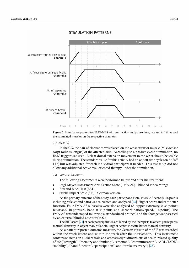
The standard value for this complex four-channel activity for the first channel was a6-s stimulation plateau time and a 5-s pause time. With a delay of 1 s, the second channelfollowed with a plateau of 5 s. Channel 3 had a delay of 2 s (compared to channel 1) and a4-s plateau time followed by channel 4 with a 3-s delay time (compared to channel 1) and a3-s plateau time. See Figure 2.
The increase and decrease times were 1.5 s, respectively. If necessary, the standardvalues were adapted to individual needs.
The multichannel electrical stimulation (MES) was triggered by surface electrodesthat detected the initial EMG signals for the affected target muscle group (default: wristextensors). After reaching an individually defined threshold, the electrical stimulationpattern was delivered as described in the previous section. The threshold was definedin such a way that the patient could reach it without involuntary co-contractions whileperforming the task during his movement initiation. This ensured, among other things,that the subject was not only active during reaching the threshold of the EMG-triggeredimpulse release but also during the active movement implementation of the required task.
Healthcare 2022, 10, 704 5 of 12
Healthcare 2022, 10, x FOR PEER REVIEW 5 of 13
Figure 2. Stimulation pattern for EMG-MES with contraction and pause time, rise and fall time, and
the stimulated muscles on the respective channels.
The increase and decrease times were 1.5 s, respectively. If necessary, the standard
values were adapted to individual needs.
The multichannel electrical stimulation (MES) was triggered by surface electrodes
that detected the initial EMG signals for the affected target muscle group (default: wrist
extensors). After reaching an individually defined threshold, the electrical stimulation
pattern was delivered as described in the previous section. The threshold was defined in
such a way that the patient could reach it without involuntary co-contractions while per-
forming the task during his movement initiation. This ensured, among other things, that
the subject was not only active during reaching the threshold of the EMG-triggered im-
pulse release but also during the active movement implementation of the required task.
2.7. cNMES
In the CG, the pair of electrodes was placed on the wrist extensor muscle (M. extensor
carpi radialis longus) of the affected side. According to a passive cyclic stimulation, no
EMG trigger was used. A clear dorsal extension movement in the wrist should be visible
during stimulation. The standard value for this activity had an on/off time cycle (on 6 s/off
14 s) but was adjusted for each individual participant if needed. This test setup did not
allow any additional active task-oriented therapy under the stimulation.
2.8. Outcome Measures
The following assessments were performed before and after the treatment:
• Fugl-Meyer Assessment Arm Section Score (FMA-AS)—blinded video rating;
• Box and Block Test (BBT);
• Stroke Impact Scale (SIS)—German version.
As the primary outcome of the study, each participant’s total FMA-AS score (0–66
points including reflexes and pain) was calculated and analyzed [23]. Higher scores
Figure 2. Stimulation pattern for EMG-MES with contraction and pause time, rise and fall time, andthe stimulated muscles on the respective channels.
2.7. cNMES
In the CG, the pair of electrodes was placed on the wrist extensor muscle (M. extensorcarpi radialis longus) of the affected side. According to a passive cyclic stimulation, noEMG trigger was used. A clear dorsal extension movement in the wrist should be visibleduring stimulation. The standard value for this activity had an on/off time cycle (on 6 s/off14 s) but was adjusted for each individual participant if needed. This test setup did notallow any additional active task-oriented therapy under the stimulation.
2.8. Outcome Measures
The following assessments were performed before and after the treatment:
• Fugl-Meyer Assessment Arm Section Score (FMA-AS)—blinded video rating;• Box and Block Test (BBT);• Stroke Impact Scale (SIS)—German version.
As the primary outcome of the study, each participant’s total FMA-AS score (0–66 pointsincluding reflexes and pain) was calculated and analyzed [23]. Higher scores indicate betterfunction. Four FMA-AS subscales were also analyzed (A: upper extremity, 0–36 points;B: wrist, 0–10 points; C: hand, 0–14 points; and D: coordination/speed, 0–6 points). TheFMA-AS was videotaped following a standardized protocol and the footage was assessedby an external blinded assessor (M.S.)
The BBT score [24] of each participant was collected by the therapists to assess participants’manual dexterity in object manipulation. Higher scores indicate better manual dexterity.
As a patient-reported outcome measure, the German version of the SIS was recordedwithin the week before and within the week after the intervention. This instrumentcontains 64 items on a Likert scale and assesses eight dimensions of health-related qualityof life (“strength”, “memory and thinking”, “emotion”, “communication”, “ADL/IADL”,“mobility”, “hand function”, “participation”, and “stroke recovery”) [25].
Healthcare 2022, 10, 704 6 of 12
2.9. Data Analysis
Demographic characteristics and the distribution of the study outcome measures pre-and post-intervention were reported as the mean and standard deviation (SD) and/or themedian, as well as the frequency distribution.
The Wilcoxon signed-rank test was applied to examine an improvement from pre- topost-intervention for the primary and secondary outcome measures in both groups. Groupcomparisons were assessed with the Mann–Whitney U-test. The Kolmogorov–Smirnov testand the Shapiro–Wilk test were used to check the data distribution.
A p-value of less than 0.05 was deemed statistically significant. The problem ofmultiplicity (to avoid the Type I error) that results from multiple comparisons, such asfor the FMA and the SIS scales, was solved with the Bonferroni correction method. Forthis reason, when interpreting the p-values, p ≤ 0.01 instead of p ≤ 0.05 was consideredstatistically significant.
IBM SPSS-statistics® (IBM Armonk, New York, NY, USA) for Windows version 24 andMicrosoft Office Excel (https://www.microsoft.com (accessed on 3 November 2020)) wereused to analyze the data.
2.10. A-Posteriori Power Calculation
An a-posteriori power calculation was performed with G*Power 3.1 (Duesseldorf,Germany) [26], applying the Wilcoxon signed-rank test to matched pairs. Based on the dataof the pre-intervention FMA-AS total score (mean = 33.5 ± 11.9) and the post-interventionFMA-AS total score (mean = 40.7 ± 10.9) in the IG, with a correlation between groups(r) = 0.984, n = 5, an alpha level of 0.05, and two-sided testing, a power of 99% was reached.
3. Results3.1. Participant Characteristics
Participants were recruited between 8 August 2018 and 9 January 2020. The lastparticipant completed the study on 29 January 2020.
Eleven of the 16 patients screened met all inclusion criteria and were included in thestudy. With respect to the proof-of-concept design, one patient who exceeded the definedinclusion criteria of falling into the early or late post-acute phase of 1–6 months by 539 dayspost stroke was included to evaluate the possibility of improvement even in the case of achronic patient. Six participants were randomly assigned to the intervention group (IG)and the other six participants were assigned to the control group (CG). All 12 participantscompleted the study per protocol.
Participant demographics in the two groups were comparable except for the meannumber of days since the stroke (“time since stroke”) before study entry; time since thestroke was observed to be longer in the CG due to the inclusion of one chronic patient; seeTable 1. The time since the stroke without this chronic case was comparable (mean = 69(±SD 14) for the CG).
The net intervention time was comparable (IG: mean = 23.5 ± 2 min;CG: mean = 24 ± 1 min). The pulse intensity (mA) was required to elicit a sufficientand possibly visible grip motion of the affected limb ranged 7–59 mA (mean = 16 ± 30 mA).

Healthcare 2022, 10, 704 7 of 12
Table 1. Demographic data and characteristics of the intervention and control groups.
Mean (±SD)IG CG Total
SexMale 4 4 8
Female 2 2 4Age 68 (7) 62 (9)
Type of stroke Ischemic 6 6 12
Hemorrhagic 0 0 0Left 3 4 7
Lesion Right 3 2 5
Affected handDominant 3 4 7
Not dominant 3 2 5Severe tomoderate 2 4 6
Degree of arm paresisModerate tomild 4 2 6
Localization ofinfarction
Cortical 5 4 9
Subcortical 1 2 3Time since stroke Days 63 (15) 147 (192) *Net intervention time Min 23.5 (2) 24 (1)
Abbreviations: IG—Intervention Group; CG—Control Group; ±SD—Standard Deviation. * Difference due to theinclusion of one chronic patient. Time since stroke without the chronic case was comparable (mean = 69 (±SD 14)for the CG).
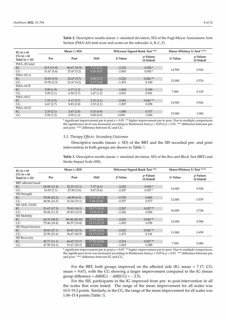
3.2. Therapy Effects: Primary Outcomes
In the IG, all participants improved pre- to post-intervention in the FMA-AS totalscore and its subgroups. This improvement can especially be observed for the FMA-AStotal score with a mean difference of +7.17; for the FMA-AS A (mean difference of +4.00),and for the FMA-AS C (mean difference of +2.33). In the CG, the improvement pre- to post-intervention was most apparent for the FMA-AS total score (average difference of +6.33)and for the subgroup score FMA-AS C (average difference of +2.33); see Table 2.
In the group comparison, participants in the IG who all received the EMG-MESadd-on to treatment, acquired greater apparent gain in the FMA-AS total score andthe FMA-AS A subscore. The apparent gain is the difference between the group meandifferences = diff(IG) − diff(CG) in Table 2. For the IG, the results were as follows: FMA-AStotal score (apparent gain = +0.84) and the FMA-AS A subscore (apparent gain = +1.83).
Healthcare 2022, 10, 704 8 of 12
Table 2. Descriptive results (mean ± standard deviation, SD) of the Fugl-Meyer Assessment ArmSection (FMA-AS) total score and scores on the subscales A, B, C, D.
IG (n = 6)CG (n = 6)Total (n = 12)
Mean (±SD) Wilcoxon Signed-Rank Test *** Mann–Whitney U-Test ****
Pre Post Diff Z-Value p-Values(2-Sided) U-Value p-Values
(2-Sided)FMA-AS totalIG 33.5 (11.9) 40.67 (10.9) 7.17 (2.2) −2.232 0.026 *
14.500 0.926CG 31.67 (5.4) 37.67 (5.2) 6.33 (4.5) −2.003 0.045 *FMA-AS AIG 18.83 (5.9) 22.67 (5.7) 4.00 (2.2) −2.226 0.026 **
12.000 0.576CG 19.50 (2.3) 21.67 (4.2) 2.17 (3.4) −1.476 0.140FMA-AS BIG 5.00 (1.9) 6.17 (1.3) 1.17 (1.6) −1.604 0.109
7.000 0.129CG 5.00 (2.1) 6.50 (2.7) 1.67 (1.2) −2.041 0.041FMA-AS CIG 7.33 (3.5) 9.17 (3.7) 2.33 (2.1) −2.041 0.041 **
14.500 0.926CG 6.67 (2.7) 8.83 (2.8) 2.33 (2.1) −1.897 0.058FMA-AS DIG 2.33 (2.1) 2.67 (2.5) 0.33 (0.8) −1.000 0.317
15.000 1.000CG 0.50 (1.2) 0.50 (1.2) 0.00 (0.0) 0.000 1.000
* significant improvement pre to post p = 0.05. ** higher improvement pre to post. Due to multiple comparisonsthe significance level was increased according to Bonferroni from p = 0.05 to p = 0.01. *** difference between preand post. **** difference between IG and CG.
3.3. Therapy Effects: Secondary Outcomes
Descriptive results (mean ± SD) of the BBT and the SIS recorded pre- and post-intervention in both groups are shown in Table 3.
Table 3. Descriptive results (mean ± standard deviation, SD) of the Box and Block Test (BBT) andStroke Impact Scale (SIS).
IG (n = 6)CG (n = 6)Total (n = 12)
Mean (±SD) Wilcoxon Signed-Rank Test *** Mann-Whitney U-Test ****
Pre Post Diff Z-Value p-Values(2-Sided) U-Value p-Values
(2-Sided)BBT affected handIG 26.00 (11.4) 32.33 (13.1) 7.17 (4.1) −2.023 0.043 *
14.000 0.520CG 16.83 (7.1) 27.00 (3.6) 9.67 (5.6) −2.207 0.027 *SIS StrengthIG 55.00 (22.3) 64.58 (6.5) 10.00 (25.2) −0.730 0.465
12.000 0.579CG 48.96 (16.5) 51.04 (19.1) 1.04 (12.8) −0.557 0.577SIS ADL/IADLIG 51.67 (17.3) 70.83 (18.1) 19.17 (14.4) −2.207 0.027 **
16.000 0.744CG 50.42 (11.5) 65.83 (12.5) 15.42 (5.3) −2.226 0.026SIS MobilityIG 64.33 (38.2) 80.08 (26.02) 15.75 (18.9) −2.023 0.043 **
12.000 0.584CG 75.46 (18.4) 86.57 (10.8) 11.11 (11.5) −1.892 0.058SIS Hand functionIG 30.83 (27.1) 45.83 (23.5) 15.00 (14.8) −2.023 0.042 **
11.000 0.459CG 22.50 (21.6) 36.67 (40.5) 14.17 (19.9) −1.473 0.141SIS RecoveryIG 49.17 (11.1) 66.67 (13.7) 17.50 (10.8) −2.214 0.027 **
7.500 0.086CG 47.50 (18.1) 51.67 (20.2) 4.17 (9.1) −1.063 0.288
* significant improvement pre to post p = 0.05. ** higher improvement pre to post. Due to multiple comparisons,the significance level was increased according to Bonferroni from p = 0.05 to p = 0.01. *** difference between preand post. **** difference between IG and CG.
For the BBT, both groups improved on the affected side (IG: mean = 7.17; CG:mean = 9.67), with the CG showing a larger improvement compared to the IG (meangroup difference = diff(IG) − diff(CG) = −2.5).
For the SIS, participants in the IG improved from pre- to post-intervention in allthe scales that were tested. The range of the mean improvement for all scales was10.0–19.2 points. Similarly, in the CG, the range of the mean improvement for all scales was1.04–15.4 points (Table 3).

Healthcare 2022, 10, 704 9 of 12
For group comparison, the same calculation was carried out for Table 3. The par-ticipants in the IG group who all received the EMG-MES add-on to treatment showed ahigher improvement in SIS Strength (mean group difference = diff(IG) − diff(CG) = +8.96),SIS ADL (mean group difference = +3.75), SIS Mobility (mean group difference = +4.64),SIS Hand function (mean group difference = +0.83), and SIS Stroke recovery (mean groupdifference = +13.33) compared to the CG.
Two of the six participants in the IG reached the first target height (6 cm), two partici-pants reached the second target height (12 cm), and two participants reached the third targetheight (18 cm), without any assistance from the therapist, by the end of the intervention.Not only hand and elbow but also shoulder activity improved.
3.4. Adverse Events
No adverse events or negative effects were reported in this study. Several participantsin both study groups reported mild shoulder pain from the beginning of study procedures.The pain did not increase but even decreased during the intervention in some cases.
4. Discussion
Functional multichannel neuromuscular electrostimulation can be considered to in-duce grasp-release or finger-hand extension and shoulder-elbow function with the trainingof selective movements and activities of daily living. It can also enhance the recovery of se-lective movements in arm paresis after stroke [27]. Some authors describe a better outcomein intensive task-oriented single channel EMG-FES compared to classical single-channelelectrical stimulation or voluntary movements in chronic stroke patients [10].
The data from a preliminary trial are reported here to investigate the feasibility andeffects of two different types of ES on the recovery of CVM in selected subacute strokepatients. Our main interest was to test if the complex interventions and assessment proce-dures associated with cNMES and EMG-MES can be implemented into a daily routine forin-patient stroke rehabilitation.
In previous studies, daily ES treatment times of 45–60 min have been described asnecessary to achieve certain beneficial effects, but negative side effects such as muscularfatigue were reported to occur after 10–15 repetitions on average for each movementpattern [11,12]. In this preliminary trial, it was also possible to demonstrate that EMG-triggered multichannel electrical stimulation with a net intervention time of 23.5 min waswell tolerated by the study participants and well-integrated into the setting of an in-patientstroke rehabilitation clinic.
As expected, both groups improved in arm and hand CVM as measured with theFMA-AS. A noticeably higher gain for the IG group (the EMG-MES group) was found in thepre-/post-intervention comparison but due to the small number of participants and due tomultiple testing, there were no significant changes. For the BBT, where individual task per-formance was measured, both groups improved significantly but there was no significantdifference between the mean BBT scores for the IG and the CG in the group comparison.
Higher activity of daily living (ADL) in patients was found for the EMG-MES in-tervention, as quantified by the SIS. This is attributed to active patient involvement andtask-oriented training, which seem to influence CVM recovery both in a subacute andchronic state of stroke recovery [7].
In this study, a sham control group was deliberately omitted because even subsensitivesham stimulation can elicit afferent sensory effects in the somatomotor cortex [28,29]. Forthis reason, a control group with established cNMES was chosen. The use of two establishedtherapeutic ES methods (EMG-MES and cNMES) with a stimulation paradigm above themotor threshold was tested in this proof-of-concept study, focusing on the combination ofmulti-joint activities and the task-oriented problem-solving approach of the interventiongroup. This article should provide additional insight into this topic.
It should also be taken into account that cNMES, applied in the control group, is anintervention that is based on substantial amounts of data reported in the literature [5,30–32]
Healthcare 2022, 10, 704 10 of 12
and thus, also makes therapeutic effects likely, as can be observed in the FMA-AS subscaleB: wrist (Table 2).
4.1. Limitation of the Study
In this preliminary trial on feasibility, there was no placebo or sham stimulation groupin which participants receive a treatment option with a mesh glove or TENS stimulationtechnique. These techniques are known to have effects on sensory representation only, butno direct effect on the development of CVM or muscle endurance. Introducing a placebo orsham stimulation group would allow to the distinction of spontaneous recovery of CVMfrom the additional benefits of cNMES and EMG-MES, respectively. Since spontaneousrecovery is to be expected in this phase of stroke rehabilitation, a control group should beplanned for any subsequent trial; the control group would receive the mesh glove or TENSstimulation technique in addition to either cNMES or EMG-MES.
The better performance of the intervention group may be due to the different treat-ment setup between the two groups by various confounding variables, such as the use offour pairs of electrodes, the arbitrary EMG triggering, or the active task-oriented therapyapproach. As a more passive treatment approach, cNMES can consequently not be directlyused for comparison. For this reason, both groups completed active task-oriented trainingin addition to the interventions. Although standard therapy was equal in both groups, wedid not explicitly monitor whether all patients adhered to self-managed exercise programand resting periods in the same way.
No specific assessment of pain/fatigue was part of the protocol. An assessment ofpain/discomfort (such as the Spasticity-associated Arm pain scale, SAAPS; Ref. [33]) andan increase in muscle endurance functions should be integral parts of the subsequentstudy design.
In this feasibility study, there were no long-term follow-up assessments integratedto check the stability of the effects achieved with the interventions. Whether repetitivecNMES or task-oriented EMG-MES contributes to greater improvements in CVM and taskperformance remains unclear.
The small number of cases does not allow a generally valid statement on the effective-ness of the interventions. These preliminary results should be confirmed by another studywith a larger number of participants.
4.2. Directions for Future Work
Future studies might incorporate some of the study design components mentioned inthe limitations of our study. Comparisons of shorter and longer intervention periods anddurations of treatment sessions [12] and their effect on CVM, task performance, feasibility,and tolerability are of interest. Finally, follow-up assessments at 4- and 12-weeks post-intervention should be included in future study designs to assess the effects of differenttherapeutic methods.
In this preliminary RCT in carefully selected subacute stroke patients with moderatearm paresis, a three-week EMG-MES add-on to conventional treatment demonstrated ahigher gain in CVM and self-reported daily activities, arm-hand use, and stroke recov-ery compared to a three-week add-on to conventional treatment of cNMES to the wristonly. EMG-MES appears to be an effective intervention that can be applied in routineclinical practice.
EMG-MES was well tolerated and did not lead to any side effects. EMG-MES mightsupport the recovery of CVM in subacute stroke patients even better than cNMES.
The study design seems to be suitable for implementation in a larger trial to confirmthese preliminary findings.
Author Contributions: Conceptualization, T.S. and K.F.; methodology, T.S. and K.F.; validation, T.S.and K.F.; formal analysis, T.S. and K.F.; investigation, D.K., S.S. and A.L.; resources, D.K., S.S. andA.L.; data curation, D.K., S.S. and A.L.; writing—original draft preparation, T.S.; writing—review
Healthcare 2022, 10, 704 11 of 12
and editing, T.S., D.K., A.L., S.S., M.S. and K.F.; visualization, T.S.; supervision, T.S. and K.F.; projectadministration, K.F. All authors have read and agreed to the published version of the manuscript.
Funding: MED-EL covered the expenses for the ethical approval, the manuscript publication, andthe reimbursement of the external assessor.
Institutional Review Board Statement: The study was conducted in accordance with the Declarationof Helsinki, and approved by the Ethics Committee of the State of Carinthia, Austria (vote A06/18—26 April 2018).
Informed Consent Statement: Informed consent was obtained from all subjects involved in the study.
Data Availability Statement: Data available from the corresponding author upon reasonable requestdue to privacy restrictions.
Acknowledgments: Many thanks to all participants of the study, Maria Steinmetz as the blindedassessor, the therapists Sandra Schober, Simone Puggl, Daniela Kolm, and Andreas Leitner, who wereresponsible for patient recruitment, treatments, and assessments. The authors thank Wilnelia Adamsand Laura Sturm (MED-EL, Innsbruck, Austria) for editing a version of this article and Edda Amann(MED-EL, Innsbruck, Austria) for performing the statistics.
Conflicts of Interest: TS is employed as clinical researcher at MED-EL in the BU Unit Neurorehabili-tation STIWELL, one of the distributors and developers of the stimulation device used in this study.He also gives lectures and seminars on functional electrical stimulation and is also a book editor. Allother authors declare no potential conflict of interest with respect to research, authorship, and/orpublication of this article.
References1. Feigin, V.L.; Forouzanfar, M.H.; Krishnamurthi, R.; Mensah, G.A.; Connor, M.; Bennett, D.A.; Moran, A.E.; Sacco, R.L.; Anderson,
L.; Truelsen, T.; et al. Global and regional burden of stroke during 1990–2010: Findings from the Global Burden of DiseaseStudy 2010. Lancet 2014, 383, 245–255. [CrossRef]
2. Houwink, A.; Nijland, R.H.; Geurts, A.C.; Kwakkel, G. Functional Recovery of the Paretic Upper Limb After Stroke: Who RegainsHand Capacity? Arch. Phys. Med. Rehabil. 2013, 94, 839–844. [CrossRef]
3. Sullivan, K.J.; Tilson, J.K.; Cen, S.Y.; Rose, D.K.; Hershberg, J.; Correa, A.; Gallichio, J.; McLeod, M.; Moore, C.; Wu, S.S.;et al. Fugl-Meyer assessment of sensorimotor function after stroke: Standardized training procedure for clinical practice andclinical trials. Stroke 2011, 42, 427–432. [CrossRef] [PubMed]
4. Fugl-Meyer, A.R.; Jääskö, L.; Leyman, I.; Olsson, S.; Steglind, S. The post-stroke hemiplegic patient. 1. a method for evaluation ofphysical performance. Scand. J. Rehabil. Med. 1975, 7, 13–31. [PubMed]
5. Eraifej, J.; Clark, W.; France, B.; Desando, S.; Moore, D. Effectiveness of upper limb functional electrical stimulation after strokefor the improvement of activities of daily living and motor function: A systematic review and meta-analysis. Syst. Rev. 2017, 6,1–21. [CrossRef]
6. Yang, J.-D.; Liao, C.-D.; Huang, S.-W.; Tam, K.-W.; Liou, T.-H.; Lee, Y.-H.; Lin, C.-Y.; Chen, H.-C. Effectiveness of electricalstimulation therapy in improving arm function after stroke: A systematic review and a meta-analysis of randomised controlledtrials. Clin. Rehabil. 2019, 33, 1286–1297. [CrossRef]
7. Silva, K.M.; Piscitelli, D.; Norouzi-Gheidari, N.; Batalla, M.A.P.; Archambault, P.S.; Levin, M.F. Electromyogram-RelatedNeuromuscular Electrical Stimulation for Restoring Wrist and Hand Movement in Poststroke Hemiplegia: A Systematic Reviewand Meta-Analysis. Neurorehabilit. Neural Repair 2019, 33, 96–111. [CrossRef] [PubMed]
8. Howlett, O.A.; Lannin, N.; Ada, L.; McKinstry, C. Functional Electrical Stimulation Improves Activity After Stroke: A SystematicReview with Meta-Analysis. Arch. Phys. Med. Rehabil. 2015, 96, 934–943. [CrossRef]
9. Cauraugh, J.; Light, K.; Kim, S.; Thigpen, M.; Behrman, A. Chronic motor dysfunction after stroke: Recovering wrist and fingerextension by electromyography-triggered neuromuscular stimulation. Stroke 2000, 31, 1360–1364. [CrossRef]
10. Hara, Y.; Obayashi, S.; Tsujiuchi, K.; Muraoka, Y. The effects of electromyography-controlled functional electrical stimulation onupper extremity function and cortical perfusion in stroke patients. Clin. Neurophysiol. 2013, 124, 2008–2015. [CrossRef]
11. Thrasher, T.A.; Zivanovic, V.; McIlroy, W.; Popovic, M.R. Rehabilitation of Reaching and Grasping Function in Severe HemiplegicPatients Using Functional Electrical Stimulation Therapy. Neurorehabilit. Neural Repair 2008, 22, 706–714. [CrossRef] [PubMed]
12. Kapadia, N.; Moineau, B.; Popovic, M.R. Functional Electrical Stimulation Therapy for Retraining Reaching and Grasping AfterSpinal Cord Injury and Stroke. Front. Neurosci. 2020, 14, 718. [CrossRef] [PubMed]
13. Chuang, L.-L.; Chen, Y.-L.; Chen, C.-C.; Li-Ling, C.; Wong, A.M.-K.; Hsu, A.-L.; Chang, Y.-J. Effect of EMG-triggered neuromuscu-lar electrical stimulation with bilateral arm training on hemiplegic shoulder pain and arm function after stroke: A randomizedcontrolled trial. J. Neuroeng. Rehabil. 2017, 14, 122. [CrossRef] [PubMed]
14. von Lewinski, F.; Hofer, S.; Kaus, J.; Merboldt, K.D.; Rothkegel, H.; Schweizer, R.; Liebetanz, D.; Frahm, J.; Paulus, W. Efficacy ofEMG-triggered electrical arm stimulation in chronic hemiparetic stroke patients. Restor. Neurol. Neurosci. 2009, 27, 189–197. [CrossRef]
Healthcare 2022, 10, 704 12 of 12
15. Schick, T.; Schlake, H.-P.; Kallusky, J.; Hohlfeld, G.; Steinmetz, M.; Tripp, F.; Krakow, K.; Pinter, M.; Dohle, C. Synergy effects ofcombined multichannel EMG-triggered electrical stimulation and mirror therapy in subacute stroke patients with severe or verysevere arm/hand paresis. Restor. Neurol. Neurosci. 2017, 35, 319–332. [CrossRef] [PubMed]
16. Jonsdottir, J.; Thorsen, R.; Aprile, I.; Galeri, S.; Spannocchi, G.; Beghi, E.; Bianchi, E.; Montesano, A.; Ferrarin, M. Arm rehabilitationin post stroke subjects: A randomized controlled trial on the efficacy of myoelectrically driven FES applied in a task-orientedapproach. PLoS ONE 2017, 12, e0188642. [CrossRef]
17. Doucet, B.M.; Lam, A.; Griffin, L. Neuromuscular electrical stimulation for skeletal muscle function. Yale J. Biol. Med. 2012, 85, 201–215.18. Wilson, R.D.; Page, S.J.; Delahanty, M.; Knutson, J.S.; Gunzler, D.D.; Sheffler, L.R.; Chae, J. Upper-Limb Recovery After Stroke: A
Randomized Controlled Trial Comparing EMG-Triggered, Cyclic, and Sensory Electrical Stimulation. Neurorehabil. Neural Repair2016, 30, 978–987. [CrossRef]
19. Woodbury, M.L.; Velozo, C.A.; Richards, L.G.; Duncan, P. Rasch Analysis Staging Methodology to Classify Upper ExtremityMovement Impairment After Stroke. Arch. Phys. Med. Rehabil. 2013, 94, 1527–1533. [CrossRef]
20. Bernhardt, J.; Hayward, K.; Kwakkel, G.; Ward, N.; Wolf, S.L.; Borschmann, K.; Krakauer, J.W.; Boyd, L.A.; Carmichael, S.T.;Corbett, D.; et al. Agreed definitions and a shared vision for new standards in stroke recovery research: The Stroke Recovery andRehabilitation Roundtable taskforce. Int. J. Stroke 2017, 12, 444–450. [CrossRef]
21. Schulz, K.F.; Grimes, D.A. Unequal group sizes in randomised trials: Guarding against guessing. Lancet 2002, 359, 966–970. [CrossRef]22. Rosenberger, W.F.; Lachin, J.M. Randomization in Clinical Trials: Theory and Practice; John Wiley & Sons: Hoboken, NJ, USA, 2015.23. Platz, T.; Pinkowski, C.; Van Wijck, F.; Kim, I.-H.; Di Bella, P.; Johnson, G. Reliability and validity of arm function assessment
with standardized guidelines for the Fugl-Meyer Test, Action Research Arm Test and Box and Block Test: A multicentre study.Clin. Rehabil. 2005, 19, 404–411. [CrossRef] [PubMed]
24. Wirz, M.; Köhler, B.; Marks, D.; Kool, J.; Sattelmayer, M.; Oesch, P.; Hilfiker, R.; Rogan, S.; Schädler, S.; Verra, M.; et al. Assessmentsin der Rehabilitation, 3rd ed.; Neurologie; Verlag Hans Huber: Bern, Switzerland, 2012.
25. Petersen, C.; Morfeld, M.; Bullinger, M. Testing and validation of the German version of the Stroke Impact Scale. Fortschr. Neurol Psychiatr.2001, 69, 284–290. [CrossRef] [PubMed]
26. Faul, F.; Erdfelder, E.; Lang, A.-G.; Buchner, A. G*Power 3: A flexible statistical power analysis program for the social, behavioral,and biomedical sciences. Behav. Res. Methods 2007, 39, 175–191. [CrossRef]
27. Platz, T.; Fheodoroff, K.; Jea, M. S3-Leitlinie Rehabilitative Therapie bei Armparese nach Schlaganfall. 2020. Available online: https://www.awmf.org/uploads/tx_szleitlinien/080-001l_S3_Rehabilitative_Therapie_bei_Armparese_nach_Schlaganfall_2020-07.pdf(accessed on 2 November 2020).
28. Chipchase, L.; Schabrun, S.; Hodges, P. Peripheral electrical stimulation to induce cortical plasticity: A systematic review ofstimulus parameters. Clin. Neurophysiol. 2011, 122, 456–463. [CrossRef]
29. Golaszewski, S.M.; Bergmann, J.; Christova, M.; Kunz, A.B.; Kronbichler, M.; Rafolt, D.; Gallasch, E.; Staffen, W.; Trinka, E.; Nar-done, R. Modulation of motor cortex excitability by different levels of whole-hand afferent electrical stimulation. Clin. Neurophysiol.2012, 123, 193–199. [CrossRef]
30. Alon, G. Functional electrical stimulation (FES): The science is strong, the clinical practice not yet—A review of evidence.Ann. Phys. Rehabil. Med. 2016, 59, e26–e27. [CrossRef]
31. Alon, G.; Levitt, A.F.; McCarthy, P.A. Functional Electrical Stimulation Enhancement of Upper Extremity Functional RecoveryDuring Stroke Rehabilitation: A Pilot Study. Neurorehabilit. Neural Repair 2007, 21, 207–215. [CrossRef]
32. Vafadar, A.K.; Côté, J.N.; Archambault, P.S. Effectiveness of Functional Electrical Stimulation in Improving Clinical Outcomes inthe Upper Arm following Stroke: A Systematic Review and Meta-Analysis. BioMed Res. Int. 2015, 2015, 1–14. [CrossRef]
33. Fheodoroff, K.; Kossmehl, P.; Wissel, J. Validity and Reliability of the Spasticity-Associated Arm Pain Scale. J. Pain Manag. Med.2017, 3, 127–133. [CrossRef]