Efficacy and safety of the use of autologous plasma rich in platelets for tissue regeneration: a...
13
ORIGINAL ARTICLE Efficacy and safety of the use of autologous plasma rich in platelets for tissue regeneration: a systematic review M a José Martínez-Zapata, Arturo Martí-Carvajal, Ivan Solà, Ignasi Bolibar, José Ángel Expósito, Luciano Rodriguez, and Joan García BACKGROUND: Autologous plasma rich in platelets (PRP) is a derived blood product whose application in clinical practice is growing. A systematic review was conducted to evaluate its efficacy and safety. STUDY DESIGN AND METHODS: A search was per- formed in electronic databases. Randomized controlled clinical trials (RCTs) in adult patients were included and assessed for methodologic quality. The main outcomes were “tissue regeneration” and “safety.” Relative risks (RRs) and standardized mean differences (SMDs) were calculated to show pooled estimates for these out- comes. When the results heterogeneity was more than 50 percent, a sensitivity analysis was performed. RESULTS: Twenty RCTs were included (11 of oral and maxillofacial surgery, 7 of chronic skin ulcers, and 2 of surgery wounds). Four RCTs evaluated the depth reduction in gingival recession in chronic periodontitis; the SMD was 0.54 (95% confidence interval [CI], 0.16 to 0.92)mm, favorable to PRP. Three RCTs evaluated the clinical attachment level in chronic periodontitis; the SMD was 0.33 (95% CI, -0.71 to 1.37) mm. Six RCTs assessed the complete skin epithelialization in wound ulcers; the RR was 1.40 (95% CI, 0.85 to 2.31). Only 6 RCTs reported adverse effects without differences between groups. CONCLUSIONS: PRP improves the gingival recession but not the clinical attachment level in chronic periodon- titis. In the complete healing process of chronic skin ulcers, the results are inconclusive. There are little data about PRP safety. There are several methodologic limi- tations and, consequently, future research should focus on strong and well-designed RCTs that assess the effi- cacy and safety of PRP. A utologous plasma rich in platelets (PRP) is a product derived from fresh whole blood that contains a high concentration of platelets (PLTs) with healing anti-inflammatory and pro- regenerative properties that permit the body to heal tissue wounds faster and more efficiently. It is obtained from the patient’s body through plasmapheresis technique. Although few investigators have quantified the concentra- tion of PLTs obtained, it can be 338 percent more than the normal total blood PLT count. 1 In recent years, the application of plasmapheresis has been widely extended in diverse medical and surgical pro- cedures, 2 especially in the fields of orthopedic surgery, 3 periodontic surgery, 4 maxillofacial surgery, 1 plastic surgery, 5 thoracic surgery, 6 vascular surgery, 7-9 and ophthalmology. 10 ABBREVIATIONS: GF(s) = growth factor(s); PRGF = plasma rich in growth factors; PRP = plasma rich in platelets; RCT(s) = randomized controlled trial(s); RR(s) = relative risk(s); SMD(s) = standardized mean difference(s). From the Iberoamerican Cochrane Center, Epidemiology and Public Health Service, Hospital de la Santa Creu i Sant Pau, Barcelona; CIBER Epidemiologia y Salud Pública (CIBERESP); Universidad “Arturo Michelena” and Iberoamerican Cochrane Collaboration Network, Valencia (Venezuela); and the Universi- tat Autònoma de Barcelona, Banc de Sang i Teixits de Catalu- nya, Barcelona, Spain. Address reprint requests to: M a José Martínez Zapata, Iberoamerican Cochrane Center, Hospital de la Santa Creu i Sant Pau, C/Sant Antoni Maria Claret 171, 08041 Barcelona, Spain; e-mail: [email protected]. This study was supported by the AETS, Instituto Carlos III, Spain (Grant PI05/90173). This study was presented in part at the 10th LatinCLEN Congress, Barcelona, Spain, June 13-16, 2007. Received for publication June 19, 2008; revision received August 11, 2008; and accepted August 12, 2008. doi: 10.1111/j.1537-2995.2008.01945.x TRANSFUSION **;**:**-**. Volume **, ** ** TRANSFUSION 1
-
Upload
independent -
Category
Documents
-
view
0 -
download
0
Transcript of Efficacy and safety of the use of autologous plasma rich in platelets for tissue regeneration: a...
O R I G I N A L A R T I C L E
Efficacy and safety of the use of autologous plasma rich inplatelets for tissue regeneration: a systematic review
Ma José Martínez-Zapata, Arturo Martí-Carvajal, Ivan Solà, Ignasi Bolibar, José Ángel Expósito,
Luciano Rodriguez, and Joan García
BACKGROUND: Autologous plasma rich in platelets(PRP) is a derived blood product whose application inclinical practice is growing. A systematic review wasconducted to evaluate its efficacy and safety.STUDY DESIGN AND METHODS: A search was per-formed in electronic databases. Randomized controlledclinical trials (RCTs) in adult patients were included andassessed for methodologic quality. The main outcomeswere “tissue regeneration” and “safety.” Relative risks(RRs) and standardized mean differences (SMDs) werecalculated to show pooled estimates for these out-comes. When the results heterogeneity was more than50 percent, a sensitivity analysis was performed.RESULTS: Twenty RCTs were included (11 of oral andmaxillofacial surgery, 7 of chronic skin ulcers, and 2 ofsurgery wounds). Four RCTs evaluated the depthreduction in gingival recession in chronic periodontitis;the SMD was 0.54 (95% confidence interval [CI], 0.16to 0.92) mm, favorable to PRP. Three RCTs evaluatedthe clinical attachment level in chronic periodontitis; theSMD was 0.33 (95% CI, -0.71 to 1.37) mm. Six RCTsassessed the complete skin epithelialization in woundulcers; the RR was 1.40 (95% CI, 0.85 to 2.31). Only 6RCTs reported adverse effects without differencesbetween groups.CONCLUSIONS: PRP improves the gingival recessionbut not the clinical attachment level in chronic periodon-titis. In the complete healing process of chronic skinulcers, the results are inconclusive. There are little dataabout PRP safety. There are several methodologic limi-tations and, consequently, future research should focuson strong and well-designed RCTs that assess the effi-cacy and safety of PRP.
Autologous plasma rich in platelets (PRP) is aproduct derived from fresh whole blood thatcontains a high concentration of platelets(PLTs) with healing anti-inflammatory and pro-
regenerative properties that permit the body to heal tissuewounds faster and more efficiently. It is obtained fromthe patient’s body through plasmapheresis technique.Although few investigators have quantified the concentra-tion of PLTs obtained, it can be 338 percent more than thenormal total blood PLT count.1
In recent years, the application of plasmapheresis hasbeen widely extended in diverse medical and surgical pro-cedures,2 especially in the fields of orthopedic surgery,3
periodontic surgery,4 maxillofacial surgery,1 plasticsurgery,5 thoracic surgery,6 vascular surgery,7-9 andophthalmology.10
ABBREVIATIONS: GF(s) = growth factor(s); PRGF = plasma rich
in growth factors; PRP = plasma rich in platelets;
RCT(s) = randomized controlled trial(s); RR(s) = relative risk(s);
SMD(s) = standardized mean difference(s).
From the Iberoamerican Cochrane Center, Epidemiology and
Public Health Service, Hospital de la Santa Creu i Sant Pau,
Barcelona; CIBER Epidemiologia y Salud Pública (CIBERESP);
Universidad “Arturo Michelena” and Iberoamerican Cochrane
Collaboration Network, Valencia (Venezuela); and the Universi-
tat Autònoma de Barcelona, Banc de Sang i Teixits de Catalu-
nya, Barcelona, Spain.
Address reprint requests to: Ma José Martínez Zapata,
Iberoamerican Cochrane Center, Hospital de la Santa Creu i
Sant Pau, C/Sant Antoni Maria Claret 171, 08041 Barcelona,
Spain; e-mail: [email protected].
This study was supported by the AETS, Instituto Carlos III,
Spain (Grant PI05/90173).
This study was presented in part at the 10th LatinCLEN
Congress, Barcelona, Spain, June 13-16, 2007.
Received for publication June 19, 2008; revision received
August 11, 2008; and accepted August 12, 2008.
doi: 10.1111/j.1537-2995.2008.01945.x
TRANSFUSION **;**:**-**.
Volume **, ** ** TRANSFUSION 1
Clinical evidence suggests that PRP could have ben-eficial therapeutic effects on hard and soft tissue healing,due to the contents of growth factors (GFs) stored in thePLTs. When these GFs are released from the PLTs, theytrigger a tissue regeneration process.7,8 Additionally, PRPcontains other intra- and extra-PLT components thatalso contribute to regeneration. One example is fibri-nogen, which creates the fibrin network necessaryfor cellular implantation and posterior cellularmultiplication.11
Several commercially available methods to obtainPRP concentrate are currently used in the clinical settingand there are many types of kits, centrifuges, and vialsavailable. The usual methods consist of obtaining lessthan 100 mL of fresh whole blood from the patient, andseparating and concentrating the PLTs by centrifugation.Subsequently, before reinsertion into the patient, the PRPis activated to produce PLT degranulation and clotformation.12 The substances used in activation are bovinethrombin (which may cause immunologic problems andFactor V deficiencies)13 or recombinant thrombin orcalcium, which have been proven to be safer proactiveoptions.2
Despite several in vitro studies published, there arevery few clinical studies available on the efficacy of PRP.Although the processing of autologous PRP is highly vari-able, it is extensively used in different clinical settings. Thissuggests that there is a need to synthesize current evi-dence on the subject. Thus, we performed a systematicreview to evaluate the efficacy and safety of PRP in tissueregeneration.
MATERIALS AND METHODS
Inclusion and exclusion criteriaWe included all randomized controlled trials (RCTs) thatassess the efficacy and/or safety of PRP for healing andregeneration of hard and soft tissues in any and allmedical and surgical procedures. Split-mouth-designRCTs were included if they were randomized. In thisdesign each patient receives, simultaneously, the activeand control treatment in the right or in the left side of thebody (for example, of the mouth or the face).14 Any RCTcrossover studies were also included when results regard-ing the first treatment period were reported.
We considered as relevant outcomes those referringto any kind of “tissue regeneration” after PRP applicationand/or “safety” related to PRP. Overall, the concept oftissue regeneration referred to an improvement of thefunctionality or complete tissue healing as specificallydefined in the original studies. Different outcomes wereassessed, depending on the pathology (i.e., wound healingin chronic skin ulcers, clinical attachment in chronicperiodontitis).
Literature searchWe performed several computerized literature searchesfor trials using the following search terms and combiningthem: autologous plasma, autologous platelet, rich growthfactor, plasma rich in growth factors (PRGF), wound,tissue, bone, osseous, heal, and repair implant. Thissearch was filtered with the Cochrane methodologic filterfor clinical trials15 in the following databases: TheCochrane Central Register of Controlled Trials (CENTRAL;The Cochrane Library 2006, issue 1), MEDLINE (accessedvia PubMed; 1966-2006), EMBASE (via OVID; 1974-2006),and Science Citation Index (1945-2006). No languagerestrictions were applied.
Reference lists of the most relevant studies andengines search (http://www.google.com) were checkedfor possible additional studies. Authors of the identifiedstudies were contacted in some cases.16
Data extractionPapers from the bibliographic search that met the inclu-sion criteria were reviewed independently by two review-ers and relevant data were extracted using a standardizedform.
Data was extracted regarding patient and interven-tion characteristics, methodological quality and results foreach group of participants. The quality of the papers wasappraised using the Jadad scale.17 This scale evaluateswhether trials are randomized or double blinded andwhether they include a description of dropouts, using ascore system. An additional point is given based onwhether the method of randomization and double-blinding is appropriate or inappropriate. Based on theresultant score, the studies were classified as “highquality” (score of 4 or 5), “moderate quality” (score of 3), or“low quality” (score < 3). A consensus between reviewerswas reached about the methodologic quality of eachstudy.
Statistical analysisWhen possible, we pooled data from the included trials aspresented in the original publications in a meta-analysisusing a random-effects statistical model.18 We calculatedrisk ratios for binary outcomes and standardized meandifferences (SMDs) for continuous outcomes and their 95percent confidence intervals (95% CIs).
An analysis of heterogeneity was conducted by an I2
test considering values of 25, 50, and 75 percent as a signof low, moderate, and high heterogeneity, respectively.19
When I2 values were 50 percent or greater, we conducted asensitivity analysis to explore the cause of heterogeneity.We anticipated heterogeneity induced by quality of thestudies, disease severity, and the PRP processing method.
MARTÍNEZ-ZAPATA ET AL.
2 TRANSFUSION Volume **, ** **
When an acceptable homogeneity was observed,(I2 < 50%), the reviewers also estimated measures of clini-cal effect such as what patients needed to be treated (NNT)to observe a clinical benefit or the number of patients to betreated to detect an adverse event (NNH). The numberneeded to treat is the estimated number of patients whoneed to be treated with the new treatment rather than thestandard treatment for one additional patient to benefit(NNT) or to harm (NNH). All analyses were undertakenusing computer software (Review Manager [RevMan] 4.2,The Nordic Cochrane Centre, Copenhagen).
RESULTS
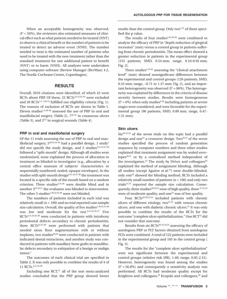
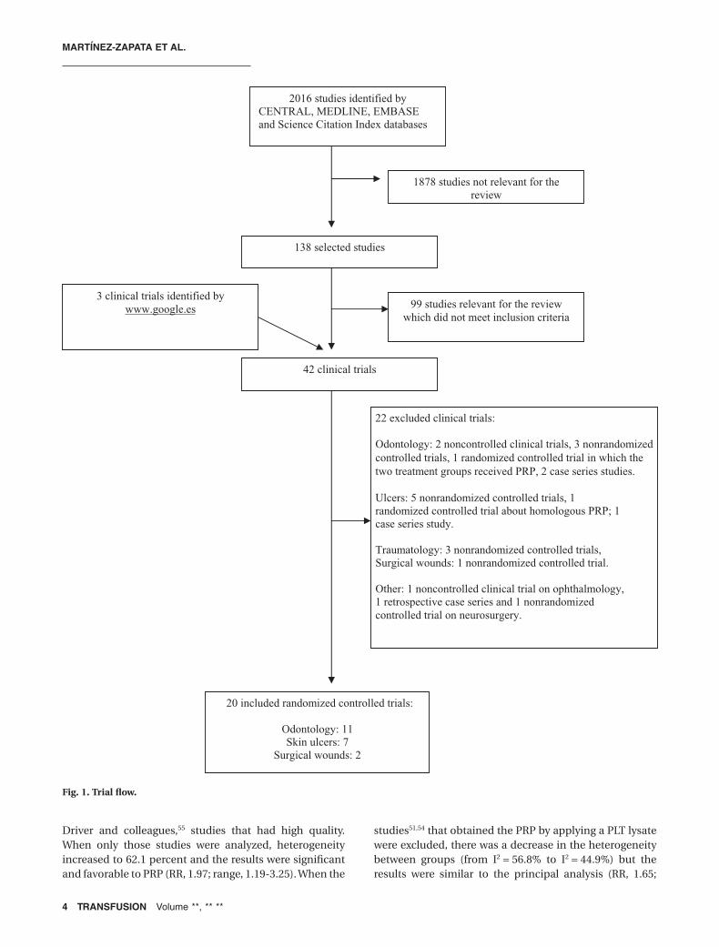
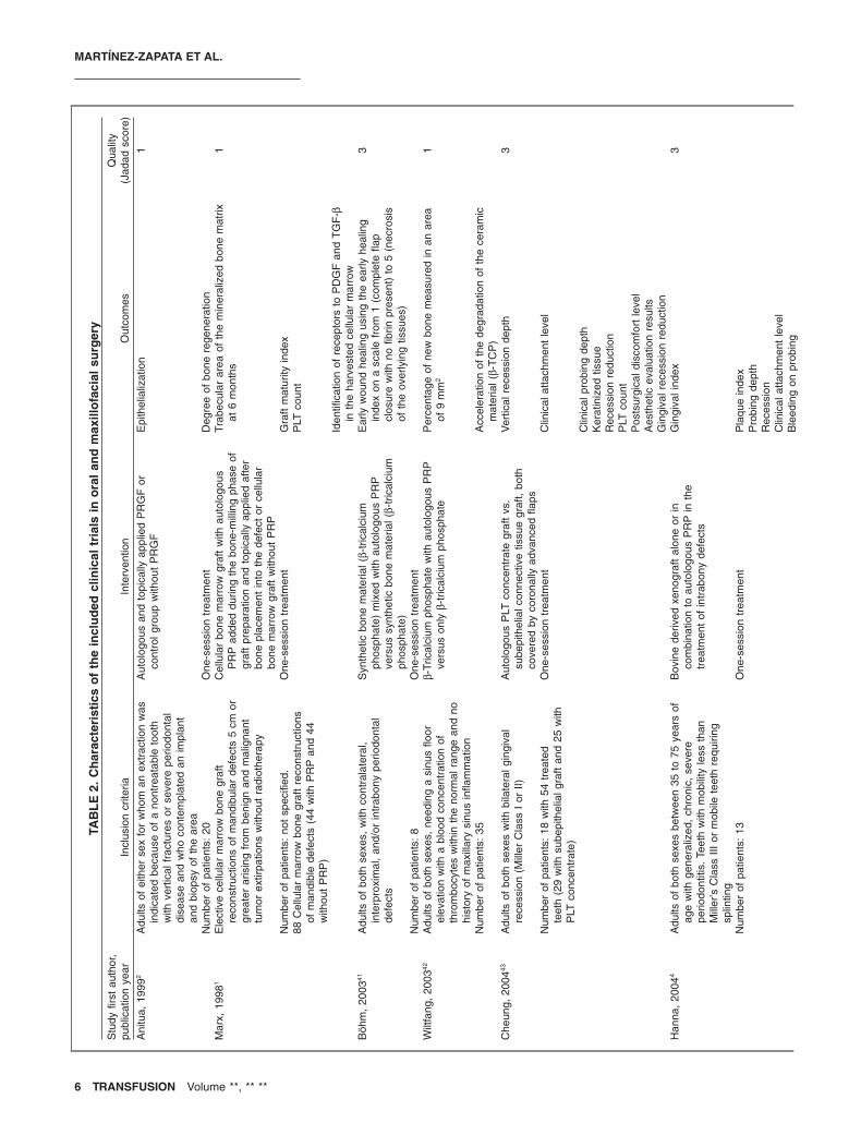
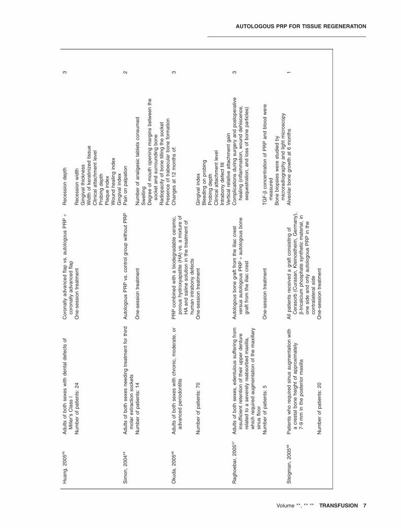
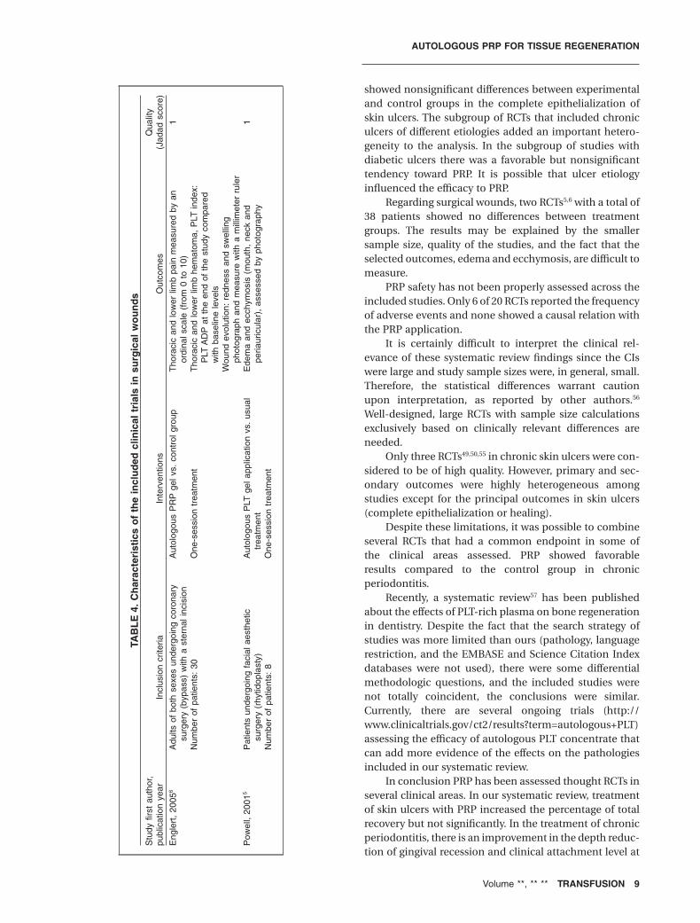
Overall, 2016 citations were identified, of which 42 wereRCTs about PRP. Of these, 22 RCTs10,16,20-39 were excludedand 20 RCTs1,4-7,40-55 fulfilled our eligibility criteria (Fig. 1).The reasons of exclusion of RCTs are shown in Table 1.Eleven studies1,4,40-48 assessed the use of PRP in oral andmaxillofacial surgery (Table 2), 749-55 in cutaneous ulcers(Table 3), and 25,6 in surgical wounds (Table 4).
PRP in oral and maxillofacial surgeryOf the 11 trials assessing the use of PRP in oral and max-illofacial surgery, 540,42,44-46 had a parallel design, 1 study1
did not specify the study design, and 5 studies4,41,43,47,48
followed a “split-mouth” design. Although all studies wererandomized, none explained the process of allocation totreatment as blinded to investigator (e.g., allocation by acentral office unaware of subjects’ characteristics orsequentially numbered, sealed, opaque envelopes). In thestudies with split-mouth design4,41,43,47,48 the treatment waslocated in a specific side of the mouth based on a randomcriterion. Three studies4,41,46 were double blind and inanother 343,45,47 the evaluator was blinded to intervention.The other 5 studies1,40,42,44,48 were not blinded.
The numbers of patients included in each trial wasrelatively small (n < 100) and no trial reported case samplesize calculation. Overall, the quality of five studies1,40,42,44,48
was low and moderate for the rest.4,41,43,45-47 FiveRCTs4,41,43,45,46 were conducted in patients with intrabonyperiodontal defects secondary to chronic periodontitis,three RCTs42,47,48 were performed with patients thatneeded sinus floor augmentations with or withoutimplants, two studies40,44 were conducted in patients withindicated dental extractions, and another study was con-ducted in patients with maxillary bone grafts in mandibu-lar defects secondary to extirpation of a benign or maligntumor.1
The outcomes of each clinical trial are specified inTable 2. It was only possible to combine the results of 4 of11 RCTs.4,41,45,46
Excluding one RCT,47 all of the non meta-analyzedstudies concluded that the PRP group showed better
results than the control group. Only two1,41 of them speci-fied the p value.
The results of four studies4,43,45,46 were combined toanalyze the efficacy of PRP in “depth reduction of gingivalrecession” (mm) versus a control group in patients suffer-ing from chronic periodontitis. The mean effect showed agreater reduction in patients in the experimental group(153 patients; SMD, 0.54 mm; range, 0.16-0.92 mm;Fig. 2).
Three studies4,43,46 assessing the “clinical attachmentlevel” (mm) showed nonsignificant differences betweenthe experimental and control groups (126 patients; SMD,0.33 mm; range, -0.71 to 1.37 mm; Fig. 2), and an impor-tant heterogeneity was observed (I2 = 86%). The heteroge-neity was explained by differences in the criteria of diseaseseverity between studies. Results were homogeneous(I2 = 0%) when only studies4,46 including patients at severestages were considered, and were favorable for the experi-mental group (96 patients; SMD, 0.89 mm; range, 0.47-1.31 mm).
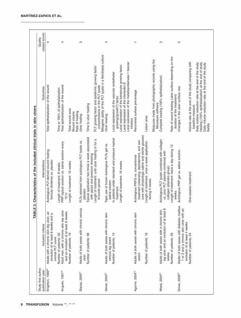
Skin ulcersSix49-53,55 of the seven trials on this topic had a paralleldesign and one54 a crossover design. Two50,55 of the sevenstudies specified the process of random generationsequence by computer numbers and three other studiesexplained that treatment assignment was by sealed enve-lopes49,51 or by a centralized method independent ofthe investigators.55 The study by Driver and colleagues55
explained the method of assignment blinding. Althoughall studies (except Aguirre et al.53) were double-blinded,only one55 showed the blinding method. RCTs included arelatively small number of patients (n < 100) and only twotrials51,55 reported the sample size calculation. Conse-quently, three studies49,50,55 were of high quality, three 51,52,54
were of moderate quality, and one53 was of low quality.Four RCTs49,50,53,54 included patients with chronic
ulcers of different etiology, two51,52 with venous chroniculcers, and one with diabetic chronic ulcers.55 It was onlypossible to combine the results of the RCTs for theoutcome “complete ulcer epithelialization.” One RCT53 didnot consider that outcome.
Results from six RCTs49-52,54,55 assessing the efficacy ofautologous PRP or PLT factors obtained from autologousPLTs were combined. A total of 122 patients were includedin the experimental group and 105 in the control group (Fig. 3).
The results for the “complete ulcer epithelialization”were not significant between the experimental andcontrol groups (relative risk [RR], 1.40; range, 0.85-2.31).However, heterogeneity was found among the studies(I2 = 56.8%) and consequently a sensitivity analysis wasperformed. All RCTs had moderate quality except forKnighton and colleagues,49 Krupski and colleagues,50 and
AUTOLOGOUS PRP FOR TISSUE REGENERATION
Volume **, ** ** TRANSFUSION 3
Driver and colleagues,55 studies that had high quality.When only those studies were analyzed, heterogeneityincreased to 62.1 percent and the results were significantand favorable to PRP (RR, 1.97; range, 1.19-3.25). When the
studies51,54 that obtained the PRP by applying a PLT lysatewere excluded, there was a decrease in the heterogeneitybetween groups (from I2 = 56.8% to I2 = 44.9%) but theresults were similar to the principal analysis (RR, 1.65;
2016 studies identified by CENTRAL, MEDLINE, EMBASE and Science Citation Index databases
138 selected studies
1878 studies not relevant for the review
42 clinical trials
99 studies relevant for the review which did not meet inclusion criteria
20 included randomized controlled trials:
Odontology: 11 Skin ulcers: 7
Surgical wounds: 2
3 clinical trials identified by www.google.es
22 excluded clinical trials:
Odontology: 2 noncontrolled clinical trials, 3 nonrandomizedcontrolled trials, 1 randomized controlled trial in which thetwo treatment groups received PRP, 2 case series studies.
Ulcers: 5 nonrandomized controlled trials, 1 randomized controlled trial about homologous PRP; 1 case series study.
Traumatology: 3 nonrandomized controlled trials, Surgical wounds: 1 nonrandomized controlled trial.
Other: 1 noncontrolled clinical trial on ophthalmology, 1 retrospective case series and 1 nonrandomized controlled trial on neurosurgery.
Fig. 1. Trial flow.
MARTÍNEZ-ZAPATA ET AL.
4 TRANSFUSION Volume **, ** **
range, 0.73-3.70). When the analysis of the RCTs thatincluded ulcers of different etiologies was excluded, theheterogeneity decreased to 10.6 percent and the resultswere again similar to the principal analysis (RR, 1.23;range, 0.90-1.41).
Surgery woundsTwo RCTs5,6 assessed the use of PRP in surgery wounds.The number of included patients was smaller (30 patientsor less) and none of the studies reported a priori samplesize calculation. Both studies were of low quality.
The study by Englert and coworkers6 used a paralleldesign and assessed the autologous PRP gel compared witha control group in patients who underwent bypass surgeryand had an external incision in the inferior extremity. Theexperimental group showed better results in chest pain,redness, and swelling. The pain was measured by anordinal scale that scored from 0 (no pain) to 10 (worst pain).On Day 1, the score was 1.47 � 0.83 and 4.47 � 2, for theexperimental and control groups, respectively. Similarly,the experimental group had favorable results in compari-son with the control group in the outcome of low-extremitypain (1.33 � 0.72 vs. 3.06 � 1.62, for the experimental and
control groups, respectively). However,neither of the two outcome resultsshowed a significance.
The study by Powell and colleagues5
used a split-face design. Each patientreceived both experimental and controltreatments in different sides of the face,simultaneously. The autologous PRP gelwas compared in patients that under-went face-lifts. This RCT included eightpatients. The experimental groupshowed better results in the outcomes ofecchymosis and edema compared to thecontrol group, although results were notsignificantly different.
Safety of the treatmentsOnly 6 of 20 RCTs4,42,47,50,52,55 reported thetreatment-related adverse events.Overall, all studies agreed that therewere not treatment-related complica-tions. Two RCTs included the type ofadverse event. Driver and coworkers55
showed an increase in the blood ureanitrogen in the control group, and anincrease in either the thrombin time orthe activated partial thromboplastintime was observed in both treatmentgroups (PRP and control). In the studyby Senet and colleagues,52 two patients
reported dermatitis (one in each treatment group), onepatient developed an infection in an existing ulcer, andone had thrombophlebitis (both in the PRP group).
DISCUSSION
The aim of this systematic review was to evaluate the effi-cacy and safety of PRP for tissue regeneration. After anexhaustive literature search, 20 RCTs that assessed the effi-cacy of autologous PRP in oral and maxillofacial applica-tions, chronic skin ulcer, and wound healing after surgerywere included.
In oral and maxillofacial surgery, four studies4,43,45,46
with 153 patients suffering from chronic periodontitiswere meta-analyzed. The results of “depth reduction ofgingival recession” showed a significant improvement inthe group treated with PRP. In the subgroup of patientswith more severe stage of the disease the outcome of“clinical attachment level” showed better results than inthe subgroup of patients with an incipient illness. Theseresults suggest that patients with more severe chronicperiodontitis could benefit from PRP.
In chronic skin ulcers, six studies49-52,54,55 with 227patients were included in the meta-analysis. Results
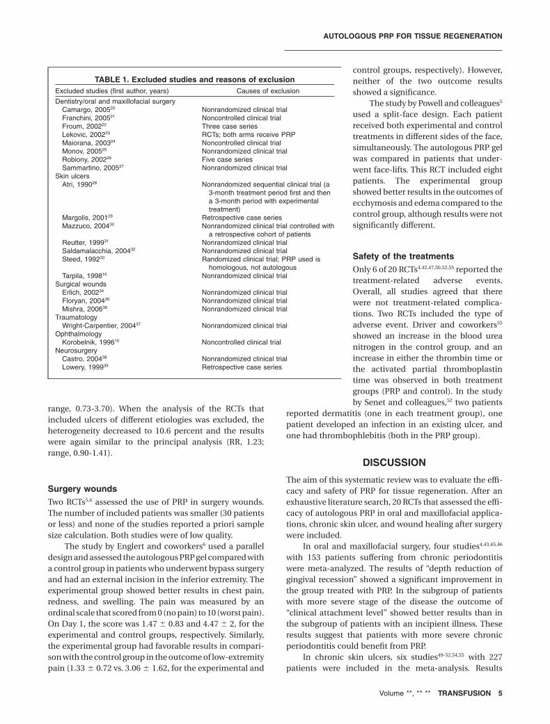
TABLE 1. Excluded studies and reasons of exclusionExcluded studies (first author, years) Causes of exclusion
Dentistry/oral and maxillofacial surgeryCamargo, 200520 Nonrandomized clinical trialFranchini, 200521 Noncontrolled clinical trialFroum, 200222 Three case seriesLekovic, 200223 RCTs; both arms receive PRPMaiorana, 200324 Noncontrolled clinical trialMonov, 200525 Nonrandomized clinical trialRobiony, 200226 Five case seriesSammartino, 200527 Nonrandomized clinical trial
Skin ulcersAtri, 199028 Nonrandomized sequential clinical trial (a
3-month treatment period first and thena 3-month period with experimentaltreatment)
Margolis, 200129 Retrospective case seriesMazzuco, 200430 Nonrandomized clinical trial controlled with
a retrospective cohort of patientsReutter, 199931 Nonrandomized clinical trialSaldamalacchia, 200432 Nonrandomized clinical trialSteed, 199233 Randomized clinical trial; PRP used is
homologous, not autologousTarpila, 199816 Nonrandomized clinical trial
Surgical woundsErlich, 200234 Nonrandomized clinical trialFloryan, 200435 Nonrandomized clinical trialMishra, 200636 Nonrandomized clinical trial
TraumatologyWright-Carpentier, 200437 Nonrandomized clinical trial
OphthalmologyKorobelnik, 199610 Noncontrolled clinical trial
NeurosurgeryCastro, 200438 Nonrandomized clinical trialLowery, 199939 Retrospective case series
AUTOLOGOUS PRP FOR TISSUE REGENERATION
Volume **, ** ** TRANSFUSION 5
TAB
LE
2.C
har
acte
rist
ics
of
the
incl
ud
edcl
inic
altr
ials
ino
ral
and
max
illo
faci
alsu
rger
yS
tudy
first
auth
or,
publ
icat
ion
year
Incl
usio
ncr
iteria
Inte
rven
tion
Out
com
esQ
ualit
y(J
adad
scor
e)
Ani
tua,
1999
2A
dults
ofei
ther
sex
for
who
man
extr
actio
nw
asin
dica
ted
beca
use
ofa
nont
reat
able
toot
hw
ithve
rtic
alfr
actu
res
orse
vere
perio
dont
aldi
seas
ean
dw
hoco
ntem
plat
edan
impl
ant
and
biop
syof
the
area
Aut
olog
ous
and
topi
cally
appl
ied
PR
GF
orco
ntro
lgro
upw
ithou
tP
RG
FE
pith
elia
lizat
ion
1
Num
ber
ofpa
tient
s:20
One
-ses
sion
trea
tmen
tD
egre
eof
bone
rege
nera
tion
Mar
x,19
981
Ele
ctiv
ece
llula
rm
arro
wbo
negr
aft
reco
nstr
uctio
nsof
man
dibu
lar
defe
cts
5cm
orgr
eate
rar
isin
gfr
ombe
nign
and
mal
igna
nttu
mor
extir
patio
nsw
ithou
tra
diot
hera
py
Cel
lula
rbo
nem
arro
wgr
aft
with
auto
logo
usP
RP
adde
ddu
ring
the
bone
-mill
ing
phas
eof
graf
tpr
epar
atio
nan
dto
pica
llyap
plie
daf
ter
bone
plac
emen
tin
toth
ede
fect
orce
llula
rbo
nem
arro
wgr
aft
with
out
PR
P
Trab
ecul
arar
eaof
the
min
eral
ized
bone
mat
rixat
6m
onth
s1
Num
ber
ofpa
tient
s:no
tsp
ecifi
ed.
One
-ses
sion
trea
tmen
tG
raft
mat
urity
inde
x88
Cel
lula
rm
arro
wbo
negr
aft
reco
nstr
uctio
nsof
man
dibl
ede
fect
s(4
4w
ithP
RP
and
44w
ithou
tP
RP
)
PLT
coun
t
Iden
tifica
tion
ofre
cept
ors
toP
DG
Fan
dT
GF
-bin
the
harv
este
dce
llula
rm
arro
wB
öhm
,20
0341
Adu
ltsof
both
sexe
s,w
ithco
ntra
late
ral,
inte
rpro
xim
al,
and/
orin
trab
ony
perio
dont
alde
fect
s
Syn
thet
icbo
nem
ater
ial(
b-tr
ical
cium
phos
phat
e)m
ixed
with
auto
logo
usP
RP
vers
ussy
nthe
ticbo
nem
ater
ial(
b-tr
ical
cium
phos
phat
e)
Ear
lyw
ound
heal
ing
usin
gth
eea
rlyhe
alin
gin
dex
ona
scal
efr
om1
(com
plet
efla
pcl
osur
ew
ithno
fibrin
pres
ent)
to5
(nec
rosi
sof
the
over
lyin
gtis
sues
)
3
Num
ber
ofpa
tient
s:8
One
-ses
sion
trea
tmen
tW
iltfa
ng,
2003
42A
dults
ofbo
thse
xes,
need
ing
asi
nus
floor
elev
atio
nw
itha
bloo
dco
ncen
trat
ion
ofth
rom
bocy
tes
with
inth
eno
rmal
rang
ean
dno
hist
ory
ofm
axill
ary
sinu
sin
flam
mat
ion
b-Tr
ical
cium
phos
phat
ew
ithau
tolo
gous
PR
Pve
rsus
only
b-tr
ical
cium
phos
phat
eP
erce
ntag
eof
new
bone
mea
sure
din
anar
eaof
9m
m2
1
Num
ber
ofpa
tient
s:35
Acc
eler
atio
nof
the
degr
adat
ion
ofth
ece
ram
icm
ater
ial(
b-T
CP
)C
heun
g,20
0443
Adu
ltsof
both
sexe
sw
ithbi
late
ralg
ingi
val
rece
ssio
n(M
iller
Cla
ssI
orII)
Aut
olog
ous
PLT
conc
entr
ate
graf
tvs
.su
bepi
thel
ialc
onne
ctiv
etis
sue
graf
t,bo
thco
vere
dby
coro
nally
adva
nced
flaps
Ver
tical
rece
ssio
nde
pth
3
Num
ber
ofpa
tient
s:18
with
54tr
eate
dte
eth
(29
with
sube
pith
elia
lgra
ftan
d25
with
PLT
conc
entr
ate)
One
-ses
sion
trea
tmen
tC
linic
alat
tach
men
tle
vel
Clin
ical
prob
ing
dept
hK
erat
iniz
edtis
sue
Rec
essi
onre
duct
ion
PLT
coun
tP
osts
urgi
cald
isco
mfo
rtle
vel
Aes
thet
icev
alua
tion
resu
ltsG
ingi
valr
eces
sion
redu
ctio
nH
anna
,20
044
Adu
ltsof
both
sexe
sbe
twee
n35
to75
year
sof
age
with
gene
raliz
ed,
chro
nic,
seve
repe
riodo
ntiti
s.Te
eth
with
mob
ility
less
than
Mill
er’s
Cla
ssIII
orm
obile
teet
hre
quiri
ngsp
lintin
g
Bov
ine
deriv
edxe
nogr
aft
alon
eor
inco
mbi
natio
nto
auto
logo
usP
RP
inth
etr
eatm
ent
ofin
trab
ony
defe
cts
Gin
giva
lind
ex3
Num
ber
ofpa
tient
s:13
One
-ses
sion
trea
tmen
tP
laqu
ein
dex
Pro
bing
dept
hR
eces
sion
Clin
ical
atta
chm
ent
leve
lB
leed
ing
onpr
obin
g
MARTÍNEZ-ZAPATA ET AL.
6 TRANSFUSION Volume **, ** **
Hua
ng,
2005
45A
dults
ofbo
thse
xes
with
dent
alde
fect
sof
Mill
er’s
Cla
ssI
Cor
onal
lyad
vanc
edfla
pvs
.au
tolo
gous
PR
P+
coro
nally
adva
nced
flap
Rec
essi
onde
pth
3
Num
ber
ofpa
tient
s:24
One
-ses
sion
trea
tmen
tR
eces
sion
wid
thG
ingi
valt
hick
ness
Wid
thof
kera
tiniz
edtis
sue
Clin
ical
atta
chm
ent
leve
lP
robi
ngde
pth
Pla
que
inde
xW
ound
heal
ing
inde
xG
ingi
vali
ndex
Sim
on,
2004
44A
dults
ofbo
thse
xes
need
ing
trea
tmen
tfo
rth
irdm
olar
extr
actio
nso
cket
sA
utol
ogou
sP
RP
vs.
cont
rolg
roup
with
out
PR
PP
ain
onpa
lpat
ion
2
Num
ber
ofpa
tient
s:14
One
-ses
sion
trea
tmen
tN
umbe
rof
anal
gesi
cta
blet
sco
nsum
edS
wel
ling
Deg
ree
ofm
outh
open
ing
mar
gins
betw
een
the
sock
etan
dsu
rrou
ndin
gbo
neR
adio
paci
tyof
bone
tillin
gth
eso
cket
Pre
senc
eof
trab
ecul
arbo
nefo
rmat
ion
Oku
da,
2005
46A
dults
ofbo
thse
xes
with
chro
nic,
mod
erat
e,or
adva
nced
perio
dont
itis
PR
Pco
mbi
ned
with
abi
odeg
rada
ble
cera
mic
,po
rous
hydr
oxya
patit
e(H
A)
vs.
am
ixtu
reof
HA
and
salin
eso
lutio
nin
the
trea
tmen
tof
hum
anin
trab
ony
defe
cts
Cha
nges
at12
mon
ths
of:
3
Num
ber
ofpa
tient
s:70
One
-ses
sion
trea
tmen
tG
ingi
vali
ndex
Ble
edin
gon
prob
ing
Pro
bing
dept
hC
linic
alat
tach
men
tle
vel
Intr
abon
yde
fect
fill
Ver
tical
rela
tive
atta
chm
ent
gain
Rag
hoeb
ar,
2005
47A
dults
ofbo
thse
xes,
eden
tulo
ussu
fferin
gfr
omin
suffi
cien
tre
tent
ion
ofth
eir
uppe
rde
ntur
ere
late
dto
ase
vere
lyre
abso
rbed
max
illa,
whi
chre
quire
dau
gmen
tatio
nof
the
max
illar
ysi
nus
floor
Aut
olog
ous
bone
graf
tfr
omth
eili
accr
est
vers
usau
tolo
gous
PR
P+
auto
logo
usbo
negr
aft
from
the
iliac
cres
t
Com
plic
atio
nsdu
ring
surg
ery
and
post
oper
ativ
ehe
alin
g(in
flam
mat
ion,
wou
ndde
hisc
ence
,se
ques
trat
ion,
and
loss
ofbo
nepa
rtic
les)
3
Num
ber
ofpa
tient
s:5
One
-ses
sion
trea
tmen
tT
GF
-bco
ncen
trat
ion
ofP
RP
and
bloo
dw
ere
mea
sure
dB
one
biop
sies
wer
est
udie
dby
mic
rora
diog
raph
yan
dlig
htm
icro
scop
yS
teig
man
,20
0548
Pat
ient
sw
hore
quire
dsi
nus
augm
enta
tion
with
acr
esta
lbon
ehe
ight
ofap
prox
imat
ely
7-9
mm
inth
epo
ster
ior
max
illa
All
patie
nts
rece
ived
agr
aft
cons
istin
gof
Cer
asor
b(C
uras
an,
Kle
inos
thei
m,
Ger
man
y),
b-tr
ical
cium
phos
phat
esy
nthe
ticm
ater
ial,
inon
esi
dean
don
lyau
tolo
gous
PR
Pin
the
cont
rala
tera
lsid
e
Alv
eola
rbo
negr
owth
at6
mon
ths
1
Num
ber
ofpa
tient
s:20
One
-ses
sion
trea
tmen
t
AUTOLOGOUS PRP FOR TISSUE REGENERATION
Volume **, ** ** TRANSFUSION 7
TAB
LE
3.C
har
acte
rist
ics
of
the
incl
ud
edcl
inic
altr
ials
insk
inu
lcer
sS
tudy
first
auth
or,
publ
icat
ion
year
Incl
usio
ncr
iteria
Inte
rven
tions
Out
com
esQ
ualit
y(J
adad
scor
e)
Kni
ghto
n,19
9049
Adu
ltsw
itha
chro
nic
skin
leg
ulce
r,an
evol
utio
nof
atle
ast
8w
eeks
and
ano
rmal
PLT
coun
t
Aut
olog
ous
PLT
-der
ived
wou
ndhe
alin
gfo
rmul
a(A
vite
ne)
vs.
plac
ebo
Tota
lepi
thel
ializ
atio
nof
the
wou
nd4
Num
ber
ofpa
tient
s:32
Leng
thof
trea
tmen
t:8
wee
ksTi
me
to10
0%of
epith
elia
lizat
ion
Kru
pski
,19
9150
Adu
ltm
en,
with
ach
roni
csk
inle
gul
cer
and
anev
olut
ion
ofat
leas
t8
wee
ksP
RP
topi
cals
olut
ion
vs.
salin
eso
lutio
nev
ery
12hr
Tota
lepi
thel
ializ
atio
nof
the
wou
nd4
Num
ber
ofpa
tient
s:18
Leng
thof
trea
tmen
t:12
wee
ksTo
talw
ound
area
Wou
ndvo
lum
eR
ate
ofhe
alin
gS
tace
y,20
0051
Adu
ltsof
both
sexe
sw
ithch
roni
cve
nous
ulce
rP
LTs
obta
ined
from
auto
logo
usP
LTly
sate
vs.
plac
ebo
Ulc
erhe
alin
g3
Num
ber
ofpa
tient
s:96
Topi
cala
pplic
atio
ntw
otim
esa
wee
kas
soci
ated
with
gauz
ean
dpr
essu
redr
essi
ngTi
me
toul
cer
heal
ing
Leng
thof
trea
tmen
t:un
tilul
cer
heal
ing
orfo
ra
9-m
onth
perio
dP
LTgr
owin
gfa
ctor
and
epid
erm
icgr
owin
gfa
ctor
conc
entr
atio
nsin
the
PLT
lysa
teM
itoge
nic
abili
tyof
the
PLT
lysa
tein
afib
robl
asts
cultu
reS
enet
,20
0352
Adu
ltsof
both
sexe
sw
ithch
roni
csk
inve
nous
leg
ulce
rsTo
pic
use
offr
ozen
auto
logo
usP
LTs
gelv
s.sa
line
solu
tion
Ulc
erhe
alin
g3
Num
ber
ofpa
tient
s:15
Inpa
tient
sun
der
stan
dard
and
pres
sure
topi
cal
trea
tmen
tLo
cale
xpre
ssio
nof
the
vasc
ular
endo
thel
ial
grow
ing/
(gro
wth
?)fa
ctor
Leng
thof
trea
tmen
t:16
wee
ksLo
cale
xpre
ssio
nof
the
kera
tocy
tegr
owin
gfa
ctor
Loca
lexp
ress
ion
ofth
ein
terle
ukin
-8Lo
cale
xpre
ssio
nof
the
met
allo
prot
eina
se-1
tissu
lar
inhi
bito
rA
guirr
e,20
0453
Adu
ltsof
both
sexe
sw
ithch
roni
csk
inul
cers
Aut
olog
ous
PR
FG
vs.
conv
entio
nal
trea
tmen
t(c
lean
sing
,de
brid
emen
t,an
dw
etcu
rew
ithph
ysio
logi
csa
line
and
ster
ilega
uzes
)
Rec
over
edsu
rfac
epe
rcen
tage
1
Num
ber
ofpa
tient
s:16
Leng
thof
trea
tmen
t:on
cea
wee
kap
plic
atio
ndu
ring
4w
eeks
.Le
sion
area
Mea
sure
sm
ade
from
phot
ogra
phic
reco
rds
usin
gth
eM
ouse
yes
softw
are
Wee
d,20
0454
Adu
ltsof
both
sexe
sw
itha
chro
nic
skin
leg
ulce
ran
dan
evol
utio
nof
atle
ast
8w
eeks
Aut
olog
ous
PLT
lysa
teco
mbi
ned
with
colla
gen
vs.
poor
PLT
plas
ma
com
bine
dw
ithco
llage
n(p
lace
bogr
oup)
Com
plet
ehe
alin
g(1
00%
epith
elia
lizat
ion)
3
Num
ber
ofpa
tient
s:26
Leng
thof
trea
tmen
t:tw
ice
ada
ydu
ring
12w
eeks
.R
ate
ofw
ound
heal
ing
(ulc
ersu
rfac
ede
pend
ing
onth
edu
ratio
nof
the
trea
tmen
t)D
river
,20
0655
Adu
ltsof
both
sexe
sw
ithdi
abet
esm
ellit
us1
or2
and
ach
roni
csk
inul
cer
with
anev
olut
ion
ofat
leas
t4
wee
ks
Aut
olog
ous
PR
Pge
lvs.
salin
eso
lutio
n.C
hang
esin
the
ulce
rsu
rfac
era
te5
Num
ber
ofpa
tient
s:54
One
-ses
sion
trea
tmen
tV
olum
era
teat
the
end
ofth
est
udy
com
parin
gw
ithba
selin
em
easu
reD
aily
surf
ace
redu
ctio
nra
teat
the
end
ofth
est
udy
Hea
ling
(100
%ep
ithel
ializ
atio
n)at
the
end
ofst
udy
Dai
lyvo
lum
ere
duct
ion
rate
atth
een
dof
the
stud
yS
afet
y
MARTÍNEZ-ZAPATA ET AL.
8 TRANSFUSION Volume **, ** **
showed nonsignificant differences between experimentaland control groups in the complete epithelialization ofskin ulcers. The subgroup of RCTs that included chroniculcers of different etiologies added an important hetero-geneity to the analysis. In the subgroup of studies withdiabetic ulcers there was a favorable but nonsignificanttendency toward PRP. It is possible that ulcer etiologyinfluenced the efficacy to PRP.
Regarding surgical wounds, two RCTs5,6 with a total of38 patients showed no differences between treatmentgroups. The results may be explained by the smallersample size, quality of the studies, and the fact that theselected outcomes, edema and ecchymosis, are difficult tomeasure.
PRP safety has not been properly assessed across theincluded studies. Only 6 of 20 RCTs reported the frequencyof adverse events and none showed a causal relation withthe PRP application.
It is certainly difficult to interpret the clinical rel-evance of these systematic review findings since the CIswere large and study sample sizes were, in general, small.Therefore, the statistical differences warrant cautionupon interpretation, as reported by other authors.56
Well-designed, large RCTs with sample size calculationsexclusively based on clinically relevant differences areneeded.
Only three RCTs49,50,55 in chronic skin ulcers were con-sidered to be of high quality. However, primary and sec-ondary outcomes were highly heterogeneous amongstudies except for the principal outcomes in skin ulcers(complete epithelialization or healing).
Despite these limitations, it was possible to combineseveral RCTs that had a common endpoint in some ofthe clinical areas assessed. PRP showed favorableresults compared to the control group in chronicperiodontitis.
Recently, a systematic review57 has been publishedabout the effects of PLT-rich plasma on bone regenerationin dentistry. Despite the fact that the search strategy ofstudies was more limited than ours (pathology, languagerestriction, and the EMBASE and Science Citation Indexdatabases were not used), there were some differentialmethodologic questions, and the included studies werenot totally coincident, the conclusions were similar.Currently, there are several ongoing trials (http://www.clinicaltrials.gov/ct2/results?term=autologous+PLT)assessing the efficacy of autologous PLT concentrate thatcan add more evidence of the effects on the pathologiesincluded in our systematic review.
In conclusion PRP has been assessed thought RCTs inseveral clinical areas. In our systematic review, treatmentof skin ulcers with PRP increased the percentage of totalrecovery but not significantly. In the treatment of chronicperiodontitis, there is an improvement in the depth reduc-tion of gingival recession and clinical attachment level at
TAB
LE
4.C
har
acte
rist
ics
of
the
incl
ud
edcl
inic
altr
ials
insu
rgic
alw
ou
nd
sS
tudy
first
auth
or,
publ
icat
ion
year
Incl
usio
ncr
iteria
Inte
rven
tions
Out
com
esQ
ualit
y(J
adad
scor
e)
Eng
lert
,20
056
Adu
ltsof
both
sexe
sun
derg
oing
coro
nary
surg
ery
(byp
ass)
with
ast
erna
linc
isio
nA
utol
ogou
sP
RP
gelv
s.co
ntro
lgro
upT
hora
cic
and
low
erlim
bpa
inm
easu
red
byan
ordi
nals
cale
(fro
m0
to10
)1
Num
ber
ofpa
tient
s:30
One
-ses
sion
trea
tmen
tT
hora
cic
and
low
erlim
bhe
mat
oma,
PLT
inde
x:P
LTA
DP
atth
een
dof
the
stud
yco
mpa
red
with
base
line
leve
lsW
ound
evol
utio
n:re
dnes
san
dsw
ellin
gph
otog
raph
and
mea
sure
with
am
illim
eter
rule
rP
owel
l,20
015
Pat
ient
sun
derg
oing
faci
alae
sthe
ticsu
rger
y(r
hytid
opla
sty)
Aut
olog
ous
PLT
gela
pplic
atio
nvs
.us
ual
trea
tmen
tE
dem
aan
dec
chym
osis
(mou
th,
neck
and
peria
uric
ular
),as
sess
edby
phot
ogra
phy
1
Num
ber
ofpa
tient
s:8
One
-ses
sion
trea
tmen
t
AUTOLOGOUS PRP FOR TISSUE REGENERATION
Volume **, ** ** TRANSFUSION 9

severe stages. In relation to the PRP for the treatment ofsurgery wounds, there were not significant differenceswhen compared with a control group. Due to the method-ologic limitations of the RCT included in this systematicreview, there is a need for further RCTs to determine withcertainty the role of PRP for the tissue regeneration.
ACKNOWLEDGMENTS
We thank Eva Arnaiz and Mitsi Ito for the manuscript review and
editing.
REFERENCES
1. Marx RE, Carlson ER, Eichstaedt RM, Schimmele SR,
Strauss JE, Georgeff KR. Platelet-rich plasma: growth factor
enhancement for bone grafts. Oral Surg Oral Med Oral
Pathol Oral Radiol Endod 1998;85:638-46.
2. Anitua E, Andia I, Ardanza B, Nurden P, Nurden AT.
Autologous platelets as a source of proteins for healing and
tissue regeneration. Thromb Haemost 2004;92:1-12.
3. Savarino L, Cenni E, Tarabusi C, Dallari D, Stagni C, Cenac-
chi A, Fornasari PM, Giunti A, Baldini N. Evaluation of
PRP ydutS Control SMD (random) Weight SMD (random)
or sub-category N Mean (SD) N Mean (SD) 95% CI % 95% CI
01 Depth reduction in the gingival recession (mm)
Huang 2005 11 0.50(0.70) 12 0.50(0.60) 12.46 0.00 [-0.82, 0.82] Hanna 2004 13 3.54(1.20) 13 2.53(0.96) 12.52 0.90 [0.09, 1.71] Cheung 2004 17 2.21(0.69) 17 1.98(0.83) 14.43 0.29 [-0.38, 0.97] Okuda 2004 35 4.70(1.60) 35 3.27(2.00) 17.27 0.78 [0.29, 1.27]
Subtotal (95% CI) 76 77 56.69 0.54 [0.16, 0.92]Test for heterogeneity: Chi² = 3.85, df = 3 (P = 0.28), I² = 22.2%
Test for overall effect: Z = 2.80 (P = 0.005)
02 Clinical attachment level (mm)
Hanna 2004 13 3.15(0.99) 13 2.31(1.18) 12.70 0.75 [-0.05, 1.55] Cheung 2004 15 1.53(0.39) 15 1.97(0.70) 13.46 -0.76 [-1.50, - .01] Okuda 2004 35 3.40(1.70) 35 2.00(1.20) 17.15 0.94 [0.45, 1.44]
Subtotal (95% CI) 63 63 43.31 0.33 [-0.71, 1.37]Test for heterogeneity: Chi² = 14.33, df = 2 (P = 0.0008), I² = 86.0%
Test for overall effect: Z = 0.62 (P = 0.54)
-10 -5 0 5 10
Favors control Favors PRP
Fig. 2. Efficacy of autologous PRP in chronic periodontitis.
PRP Control RR (random)
Weight RR (random)
IC %59 N/n N/n Study or sub-category % 95% CI
01 Chronic Venous Ulcers Senet 2003 1/8 1/7 3.39 0.88 [0.07, 11.54] Stacey 2000 33/42 34/44 33.78 1.02 [0.81, 1.27] Subtotal (95% CI) 50 51 37.18 1.02 [0.81, 1.27]Total events: 34 (PRP), 35 (Control)Test for heterogeneity: Chi² = 0.01, df = 1 (P = 0.91), I² = 0%Test for overall effect: Z = 0.14 (P = 0.89)
02 Chronic Diabetic Ulcers Driver 2006 13/19 9/21 24.33 1.60 [0.89, 2.85] Subtotal (95% CI) 19 21 24.33 1.60 [0.89, 2.85]Total events: 13 (PRP), 9 (Control)Test for heterogeneity: not applicableTest for overall effect: Z = 1.58 (P = 0.11)
03 Chronic Ulcers of different etiology Knighton 1990 17/21 2/13 10.58 5.26 [1.45, 19.15] Krupski 1991 4/17 3/9 10.95 0.71 [0.20, 2.49] Weed 2004 9/15 4/11 16.96 1.65 [0.68, 3.99] Subtotal (95% CI) 53 33 38.49 1.80 [0.63, 5.15]Total events: 30 (PRP), 9 (Control)Test for heterogeneity: Chi² = 5.14, df = 2 (P = 0.08), I² = 61.1%Test for overall effect: Z = 1.10 (P = 0.27)
Total (95% CI) 122 105 100.00 1.40 [0.85, 2.31]Total events: 77 (PRP), 53 (Control)Test for heterogeneity: Chi² = 11.57, df = 5 (P = 0.04), I² = 56.8%Test for overall effect: Z = 1.32 (P = 0.19)
0.01 0.1 1 10 100
Favors control Favors PRP
Fig. 3. Efficacy of autologous PRP in chronic skin ulcers.
MARTÍNEZ-ZAPATA ET AL.
10 TRANSFUSION Volume **, ** **

bone healing enhancement by lyophilized bone grafts
supplemented with platelet gel: a standardized methodol-
ogy in patients with tibial osteotomy for genu varus.
J Biomed Mater Res B Appl Biomater 2006;76:364-72.
4. Hanna R, Trejo PM, Weltman RL. Treatment of intrabony
defects with bovine-derived xenograft alone and in combi-
nation with platelet-rich plasma: a randomized clinical
trial. J Periodontol 2004;75:1668-77.
5. Powell DM, Chang E, Farrior EH. Recovery from deep-
plane rhytidectomy following unilateral wound treatment
with autologous platelet gel: a pilot study. Arch Facial Plast
Surg 2001;3:245-50.
6. Englert SJ, Estep TH, Ellis-Stoll CC. Autologous platelet gel
applications during cardiovascular surgery: effect on
wound healing. J Extra Corpor Technol 2005;37:148-
52.
7. Knighton DR, Doucette M, Fiegel VD, Ciresi K, Butler E,
Austin L. The use of platelet derived wound healing
formula in human clinical trials. Prog Clin Biol Res 1988;
266:319-29.
8. Robinson CJ. Growth factors: therapeutic advances in
wound healing. Ann Med 1993;25:535-8.
9. Crovetti G, Martinelli G, Issi M, Barone M, Guizzardi M,
Campanati B, Moroni M, Englert SJ, Estep TH, Ellis-Stoll
CC. Autologous platelet gel applications during cardiovas-
cular surgery: effect on wound healing. J Extra Corpor
Technol 2005;37:148-52.
10. Korobelnik JF, Hannouche D, Belayachi N, Branger M,
Guez JE, Hoang-Xuan T. Autologous platelet concentrate as
an adjunct in macular hole healing: a pilot study. Ophthal-
mology 1996;103:590-4.
11. Munirah S, Samsudin OC, Chen HC, Sharifah Salmah SH,
Aminuddin BS, Ruszymah BH, MD. Articular cartilage res-
toration in load-bearing osteochondral defects by implan-
tation of autologous chondrocyte-fibrin constructs. J Bone
Joint Surg Br 2007; 89-B:1099-109.
12. Sanchez AR, Sheridan PJ, Kupp LI. Is platelet-rich plasma
the perfect enhancement factor? A current review. Int J
Oral Maxillofac Implants 2003;18:93-103.
13. Whitman DH, Berry RL, Green DM. Platelet gel: an autolo-
gous alternative to fibrin glue applications in oral and
maxillofacial surgery. J Oral Maxillofac Surg 1997;55:
1294-9.
14. Imrey PB, Chilton NW. Design and analytic concepts for
periodontal clinical trials. J Periodontol 1992; 63 12 Suppl:
1124-40.
15. Robinson KA, Dickersin K. Development of a highly sensi-
tive search strategy for the retrieval of reports of controlled
trials using PubMed. Source Int J Epidemiol 2002;31:
150-3.
16. Tarpila E, Jergovic D, Sjoberg F, Bjornsson A, Abildgard L,
Rosseau A. Autologous platelet-derived wound healing
factors in the treatment of leg ulcers with split-skin grafts.
New Approaches to the Management of Chronic Wounds
1998;81-5.
17. Jadad AR, Moore RA, Carroll D, Jenkinson C, Reynolds DJ,
Gavaghan DJ, McQuay HJ. Assessing the quality of reports
of randomized clinical trials: is blinding necessary? Control
Clin Trials 1996;17:1-12.
18. DerSimonian R, Laird N. Meta-analysis in clinical trials.
Control Clin Trials 1986;7:177-88.
19. Higgins JP, Thompson SG, Deeks JJ, Altman DG. Measur-
ing inconsistency in meta-analyses. BMJ 2003;327:557-60.
20. Camargo PM, Lekovic V, Weinlaender M, Vasilic N, Madza-
revic M, Kenney EB. A reentry study on the use of bovine
porous bone mineral, GTR, and platelet-rich plasma in the
regenerative treatment of intrabony defects in humans. Int
J Periodontics Restorative Dent 2005;25:49-59.
21. Franchini M, Dupplicato P, Ferro I, De Gironcoli M, Alde-
gheri R. Efficacy of platelet gel in reconstructive bone
surgery. Orthopedics 2005;28:161-3.
22. Froum SJ, Wallace SS, Tarnow DP, Cho SC. Effect of
platelet-rich plasma on bone growth and osseointegration
in human maxillary sinus grafts: three bilateral case
reports. Int J Periodontics Restorative Dent 2002;22:
45-53.
23. Lekovic V, Camargo PM, Weinlaender M, Vasilic N, Kenney
EB. Comparison of platelet-rich plasma, bovine porous
bone mineral, and guided tissue regeneration versus
platelet-rich plasma and bovine porous bone mineral in
the treatment of intrabony defects: a reentry study.
J Periodontol 2002;73:198-205.
24. Maiorana C, Sommariva L, Brivio P, Sigurta D, Santoro F.
Maxillary sinus augmentation with anorganic bovine bone
(Bio-Oss) and autologous platelet-rich plasma: preliminary
clinical and histologic evaluations. Int J Periodontics
Restorative Dent 2003;23:227-35.
25. Monov G, Fuerst G, Tepper G, Watzak G, Zechner W,
Watzek G. The effect of platelet-rich plasma upon implant
stability measured by resonance frequency analysis in the
lower anterior mandibles. Clin Oral Implants Res 2005;16:
461-5.
26. Robiony M, Polini F, Costa F, Politi M. Osteogenesis dis-
traction and platelet-rich plasma for bone restoration of
the severely atrophic mandible: preliminary results. J Oral
Maxillofac Surg 2002;60:630-5.
27. Sammartino G, Tia M, Marenzi G, di Lauro AE, D’Agostino
E, Claudio PP. Use of autologous platelet-rich plasma
(PRP) in periodontal defect treatment after extraction of
impacted mandibular third molars. J Oral Maxillofac Surg
2005;63:766-70.
28. Atri SC, Misra J, Bisht D, Misra K. Use of homologous
platelet factors in achieving total healing of recalcitrant
skin ulcers. Surgery 1990;108:508-12.
29. Margolis DJ, Kantor J, Santanna J, Strom BL, Berlin JA.
Effectiveness of platelet releasate for the treatment of
diabetic neuropathic foot ulcers. Diabetes Care 2001;24:
483-8.
30. Mazzucco L, Medici D, Serra M, Panizza R, Rivara G,
Orecchia S, Libener R, Cattana E, Levis A, Betta PG,
AUTOLOGOUS PRP FOR TISSUE REGENERATION
Volume **, ** ** TRANSFUSION 11
Borzini P. The use of autologous platelet gel to treat
difficult-to-heal wounds: a pilot study. Transfusion 2004;
44:1013-8.
31. Reutter H, Bort S, Jung MF, Klyscz T, Schippert W, Zuder
D, Junger M. Questionable effectiveness of autologous
platelet growth factors (PDHWF) in the treatment of
venous leg ulcers. Hautarzt 1999;50:859-65.
32. Saldalamacchia G, Lapice E, Cuomo V, De Feo E,
D’Agostino E, Rivellese AA, Vaccaro O. A controlled study
of the use of autologous platelet gel for the treatment of
diabetic foot ulcers. Nutr Metab Cardiovasc Dis 2004;14:
395-6.
33. Steed DL, Goslen JB, Holloway GA, Malone JM, Bunt TJ,
Webster MW. Randomized prospective double-blind trial
in healing chronic diabetic foot ulcers. CT-102 activated
platelet supernatant, topical versus placebo. Diabetes Care
1992;15:1598-604.
34. Ehrlich HP, Freedman BM. Topical platelet-derived growth
factor in patients enhances wound closure in the absence
of wound contraction. Cytokines Cell Mol Ther 2002;7:85-
90.
35. Floryan KM, Berghoff WJ. Intraoperative use of autologous
platelet-rich and platelet-poor plasma for orthopedic
surgery patients. AORN J 2004;80:668-74; quiz 675-8.
36. Mishra A, Pavelko T. Treatment of chronic elbow tendino-
sis with buffered platelet-rich plasma. Am J Sports Med
2006;34:1774-78.
37. Wright-Carpentier T, Klein P, Schäferhoff P, Apell HJ, Mir
LM, Wehling P. Treatment of muscle injures by local
administration of autologous conditioned serum: a pilot
study on sportsmen with muscle strains. Int J Sport Med
2004; 25:588-93.
38. Castro FP Jr. Role of activated growth factors in lumbar
spinal fusions. J Spinal Disord Tech 2004;17:380-4.
39. Lowery GL, Kulkarni S, Pennisi AE. Use of autologous
growth factors in lumbar spinal fusion. Bone 1999;25 Suppl
1:47S-50S.
40. Anitua E. Plasma rich in growth factors: preliminary results
of use in the preparation of future sites for implants. Int J
Oral Maxillofacial Implants 1999;14:529-35.
41. Böhm S, Kurtulus I, Gonzales J, Meyle J. Effect of platelet-
rich protein on early wound healing in intrabony defects.
Annual Meeting of the American Academy of Peridontol-
ogy. San Francisco. J Periodontol 2003. Abstract. Pg.
1705.
42. Wiltfang J, Schlegel KA, Schultze-Mosgau S, Nkenke E,
Zimmermann R, Kessler P. Sinus floor augmentation with
beta-tricalciumphosphate (beta-TCP): does platelet-rich
plasma promote its osseous integration and degradation?
Clin Oral Implants Res 2003;14:213-8.
43. Cheung WS, Griffin TJ. A comparative study of root cover-
age with connective tissue and platelet concentrate grafts:
8-month results. J Periodontol 2004;75:1678-87.
44. Simon D, Manuel S, Geetha V, Naik BR. Potential for
osseous regeneration of platelet-rich plasma—a
comparative study in mandibular third molar sockets.
Indian J Dent Res 2004;15:133-6.
45. Huang LH, Neiva RE, Soehren SE, Giannobile WV, Wang
HL. The effect of platelet-rich plasma on the coronally
advanced flap root coverage procedure: a pilot human
trial. J Periodontol 2005;76:1768-77.
46. Okuda K, Tai H, Tanabe K, Suzuki H, Sato T, Kawase T,
Saito Y, Wolff LF, Yoshiex H. Platelet-rich plasma combined
with a porous hydroxyapatite graft for the treatment of
intrabony periodontal defects in humans: a comparative
controlled clinical study. J Periodontol 2005;76:890-8.
47. Raghoebar GM, Schortinghuis J, Liem RS, Ruben JL, van
der Wal JE, Vissink A. Does platelet-rich plasma promote
remodeling of autologous bone grafts used for augmenta-
tion of the maxillary sinus floor? Clin Oral Implants Res
2005;16:349-56.
48. Steigmann M, Garg AK. A comparative study of bilateral
sinus lifts performed with platelet-rich plasma alone
versus alloplastic graft material reconstituted with blood.
Implant Dent 2005;14:261-6.
49. Knighton DR, Ciresi K, Fiegel VD, Schumerth S, Butler E,
Cerra F. Stimulation of repair in chronic, nonhealing, cuta-
neous ulcers using platelet-derived wound healing
formula. Surg Gynecol Obstet 1990;170:56-60.
50. Krupski WC, Reilly LM, Perez S, Moss KM, Crombleholme
PA, Rapp JH. A prospective randomized trial of autologous
platelet-derived wound healing factors for treatment of
chronic nonhealing wounds: a preliminary report. J Vasc
Surg 1991;14:526-32; discussion 532-6.
51. Stacey MC, Mata SD, Trengove NJ, Mather CA. Randomised
double-blind placebo controlled trial of topical autologous
platelet lysate in venous ulcer healing. Eur J Vasc Endovasc
Surg 2000;20:296-301.
52. Senet P, Bon FX, Benbunan M, Bussel A, Traineau R, Calvo
F, Dubertret L, Dosquet C. Randomized trial and local bio-
logical effect of autologous platelets used as adjuvant
therapy for chronic venous leg ulcers. J Vasc Surg 2003;38:
1342-8.
53. Aguirre JJ, Francisco S, Cabezas A, Calleja A, Ayerdi E, Pena
MA, Algorta J. Ensayo clínico piloto, aleatorizado, en
grupos paralelos, ciego y controlado con tratamiento con-
vencional para evaluar la eficacia de plasma rico en fac-
tores de crecimiento en el tratamiento de las úlceras
cutáneas: resultados a las 4 semanas de seguimiento. XIX
Congreso de la Sociedad Española de Farmacología
Clínica. Santander 2004; 28-30 Octubre. Abstract 19:12.
54. Weed B, Davis MD, Felty CL, Liedl DA, Pineda AA, Moore
SB, Rooke TW. Autologous platelet lysate product versus
placebo in patients with chronic leg ulcerations: a pilot
study using a randomized, double-blind, placebo-
controlled trial. Wounds-A Compend Clin Res Pract 2004;
16:273-82.
55. Driver VR, Hanft J, Fylling CP, Beriou JM, and Autologel
Diabetic Foot Ulcer Study Group. A prospective, random-
ized, controlled trial of autologous platelet-rich plasma gel
MARTÍNEZ-ZAPATA ET AL.
12 TRANSFUSION Volume **, ** **
for the treatment of diabetic foot ulcers. Ostomy Wound
Manage 2006;52:68-87.
56. Gunsolley J, Elswick RK, Davenport JM. Equivalence and
superiority testing in regeneration clinical trials.
J Periodontol 1998;69:521-7.
57. Plachokova AS, Nikolidakis D, Mulder J, Jansen JA,
Creugers NH. Effect of platelet-rich plasma on bone regen-
eration in dentistry: a systematic review. Clin Oral
Implants Res 2008;19:539-45.
AUTOLOGOUS PRP FOR TISSUE REGENERATION
Volume **, ** ** TRANSFUSION 13