Effects of temperature and salinity on survival of young-of-the-year Hudson River striped bass (...
29
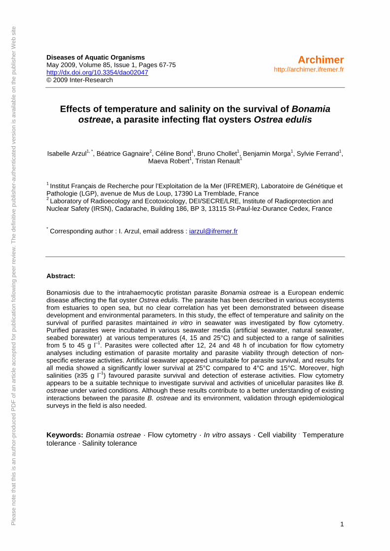
Please note that this is an author-produced PDF of an article accepted for publication following peer review. The definitive publisher-authenticated version is available on the publisher Web site 1 Diseases of Aquatic Organisms May 2009, Volume 85, Issue 1, Pages 67-75 http://dx.doi.org/10.3354/dao02047 © 2009 Inter-Research Archimer http://archimer.ifremer.fr Effects of temperature and salinity on the survival of Bonamia ostreae, a parasite infecting flat oysters Ostrea edulis Isabelle Arzul 1, * , Béatrice Gagnaire 2 , Céline Bond 1 , Bruno Chollet 1 , Benjamin Morga 1 , Sylvie Ferrand 1 , Maeva Robert 1 , Tristan Renault 1 1 Institut Français de Recherche pour l’Exploitation de la Mer (IFREMER), Laboratoire de Génétique et Pathologie (LGP), avenue de Mus de Loup, 17390 La Tremblade, France 2 Laboratory of Radioecology and Ecotoxicology, DEI/SECRE/LRE, Institute of Radioprotection and Nuclear Safety (IRSN), Cadarache, Building 186, BP 3, 13115 St-Paul-lez-Durance Cedex, France * Corresponding author : I. Arzul, email address : [email protected] Abstract: Bonamiosis due to the intrahaemocytic protistan parasite Bonamia ostreae is a European endemic disease affecting the flat oyster Ostrea edulis. The parasite has been described in various ecosystems from estuaries to open sea, but no clear correlation has yet been demonstrated between disease development and environmental parameters. In this study, the effect of temperature and salinity on the survival of purified parasites maintained in vitro in seawater was investigated by flow cytometry. Purified parasites were incubated in various seawater media (artificial seawater, natural seawater, seabed borewater) at various temperatures (4, 15 and 25°C) and subjected to a range of salinities from 5 to 45 g l –1 . Parasites were collected after 12, 24 and 48 h of incubation for flow cytometry analyses including estimation of parasite mortality and parasite viability through detection of non- specific esterase activities. Artificial seawater appeared unsuitable for parasite survival, and results for all media showed a significantly lower survival at 25°C compared to 4°C and 15°C. Moreover, high salinities (≥35 g l –1 ) favoured parasite survival and detection of esterase activities. Flow cytometry appears to be a suitable technique to investigate survival and activities of unicellular parasites like B. ostreae under varied conditions. Although these results contribute to a better understanding of existing interactions between the parasite B. ostreae and its environment, validation through epidemiological surveys in the field is also needed. Keywords: Bonamia ostreae · Flow cytometry · In vitro assays · Cell viability . Temperature tolerance · Salinity tolerance
Transcript of Effects of temperature and salinity on survival of young-of-the-year Hudson River striped bass (...
Ple
ase
note
that
this
is a
n au
thor
-pro
duce
d P
DF
of a
n ar
ticle
acc
ept
ed fo
r pu
blic
atio
n fo
llow
ing
peer
rev
iew
. The
def
initi
ve p
ub
lish
er-a
uthe
ntic
ated
ve
rsio
n is
ava
ilab
le o
n th
e pu
blis
her
Web
site
1
Diseases of Aquatic Organisms May 2009, Volume 85, Issue 1, Pages 67-75 http://dx.doi.org/10.3354/dao02047 © 2009 Inter-Research
Archimerhttp://archimer.ifremer.fr
Effects of temperature and salinity on the survival of Bonamia ostreae, a parasite infecting flat oysters Ostrea edulis
Isabelle Arzul1, *, Béatrice Gagnaire2, Céline Bond1, Bruno Chollet1, Benjamin Morga1, Sylvie Ferrand1, Maeva Robert1, Tristan Renault1
1 Institut Français de Recherche pour l’Exploitation de la Mer (IFREMER), Laboratoire de Génétique et Pathologie (LGP), avenue de Mus de Loup, 17390 La Tremblade, France 2 Laboratory of Radioecology and Ecotoxicology, DEI/SECRE/LRE, Institute of Radioprotection and Nuclear Safety (IRSN), Cadarache, Building 186, BP 3, 13115 St-Paul-lez-Durance Cedex, France * Corresponding author : I. Arzul, email address : [email protected] Abstract:
Bonamiosis due to the intrahaemocytic protistan parasite Bonamia ostreae is a European endemic disease affecting the flat oyster Ostrea edulis. The parasite has been described in various ecosystems from estuaries to open sea, but no clear correlation has yet been demonstrated between disease development and environmental parameters. In this study, the effect of temperature and salinity on the survival of purified parasites maintained in vitro in seawater was investigated by flow cytometry. Purified parasites were incubated in various seawater media (artificial seawater, natural seawater, seabed borewater) at various temperatures (4, 15 and 25°C) and subjected to a range of salinities from 5 to 45 g l–1. Parasites were collected after 12, 24 and 48 h of incubation for flow cytometry analyses including estimation of parasite mortality and parasite viability through detection of non-specific esterase activities. Artificial seawater appeared unsuitable for parasite survival, and results for all media showed a significantly lower survival at 25°C compared to 4°C and 15°C. Moreover, high salinities (≥35 g l–1) favoured parasite survival and detection of esterase activities. Flow cytometry appears to be a suitable technique to investigate survival and activities of unicellular parasites like B. ostreae under varied conditions. Although these results contribute to a better understanding of existing interactions between the parasite B. ostreae and its environment, validation through epidemiological surveys in the field is also needed.
Keywords: Bonamia ostreae · Flow cytometry · In vitro assays · Cell viability . Temperature tolerance · Salinity tolerance
50
51
52
53
54
55
56
57
58
59
60
61
62
63
64
65
66
67
68
69
70
71
72
73
74
75
INTRODUCTION
Bonamia ostreae is a protistan parasite belonging to the phylum Haplosporidia (Sprague 1979). It
is an intracellular parasite, 2-5 µm in diameter, that infects haemocytes. It can also be observed
extracellularly between epithelial or interstitial cells in gills and stomach or in necrotic connective
tissue areas. The parasite can be detected in spat (Lynch et al. 2005), however, mortalities mainly
affect oysters which are more than 2 year old (Culloty & Mulcahy 1996). At a tissue level, the
infection is usually associated with intense haemocyte infiltration of the connective tissue of the
gills, mantle and digestive gland. The life cycle is unknown but the disease can be directly
transmitted between oysters in a population or experimentally by cohabitation or inoculation
(Elston et al. 1986, Hervio et al. 1995) suggesting that intermediate host is not required for parasite
cycle accomplishment. Observation of parasites free in gill epithelia potentially associated with
gill lesions supports the hypothesis of a parasite release through these organs (Montes et al. 1994).
However, the infective form and ways of entry and release remain undetermined. Most of Bonamia
ostreae might be released in the water column after oyster death through tissue lysis.
This intrahaemocytic parasite has been described in oysters collected from different ecosystems
from estuaries and intertidal zones to deep coastal waters or lagoon and is presently reported in
Europe, North America and Morocco. Northern European waters (e.g. Norwegian waters) seem to
be free of bonamiosis probably because of the lack of introduction of infected animals. Flat oysters
from the Mediterranean Basin are infected by Bonamia ostreae, however reported prevalences are
low. No clear correlations have been demonstrated between development of the disease and
environmental parameters including temperature and salinity. Previous work suggested an impact
of temperature on the parasite and / or on the defence capacity of oysters. Although the disease
occurs and can be transmitted throughout the year (Tigé & Grizel, 1984), there is a seasonal
variation in infection with Bonamia ostreae. Prevalence of infection presents peaks in late winter
3
76
77
78
79
80
81
82
83
84
85
86
87
88
89
90
91
92
93
94
95
96
97
98
99
100
101
and in autumn (Grizel 1985, Montes 1990, Van Banning 1991, Culloty & Mulcahy 1996, Arzul et
al. 2006). A study of bonamiosis prevalence as well as haemocyte activities according to
temperature showed that prevalence was higher at low temperature (10°C) compared to higher
temperature (20°C) suggesting that low temperatures may affect defence capacities of the oyster
and/or the ability of the parasite to infect healthy oysters (Cochennec & Auffret 2002).
The lack of suitable tissue culture systems and mollusc cell lines for the culture of the parasite led
to the development of a purification protocol (Miahle et al. 1988). The availability of purified
Bonamia ostreae suspensions allowed experimental infections based on parasite injection (Hervio et
al. 1995) and investigations on in vitro interactions between parasites and haemocytes (Chagot et al.
1992, Mourton et al. 1992). Despite possible survival of purified parasites in filtered sea water (2
weeks) as assessed by success of experimental infection (Grizel 1985), purified parasite suspensions
have not yet been used to study parasite physiology or its behaviour related to environmental
conditions.
In aquatic ecology, flow cytometry is classically used to determine abundance, viability and activity
of microorganisms including viruses, bacteria, microalgae and planktonic protozoan parasites
(Wong & Whiteley 1996, Lindström et al. 2002, Parrow & Burkholder 2002, Binet & Stauber 2006,
Hammes et al. 2008). Recent developments aimed at addressing some questions in environmental
microbiology including studying microbial physiology under environmentally relevant conditions
(Czechowska et al. 2008). Flow cytometry was successfully used to measure cell viability of
cultured Perkinsus marinus, a parasitic protozoan of the Eastern oyster, Crassostrea virginica
(Soudant et al. 2005). This tool allows multi parametric analyses on a large number of cells in a
very short time and thus presents advantages over microscopic approaches.
The objectives of the present study were to test survival of purified Bonamia ostreae in different sea
water media (artificial, natural and underground salty water) in order to identify the most suitable
medium for parasite preservation and to investigate effects of temperature and salinity on the
survival of purified parasites by flow cytometry. Purified B. ostreae were suspended and maintained
4
in vitro in the three different media at three different temperatures and then subjected to a range of
salinities in the optimal medium previously defined. Parasite mortality was measured by flow
cytometry using propidium iodide staining and parasite viability was estimated by measuring
esterase activities using FDA (Fluorescein Diacetate). Esterases are enzymes belonging to the group
of hydrolases and are classically measured to estimate global level of viable cell activities
(Gagnaire et al. 2006b, Berney et al. 2008, Rault et al. 2008).
102
103
104
105
106
107
108
109
110
111
112
113
114
115
116
117
118
119
120
121
122
123
124
125
126
In vitro exposure of purified parasites to ranges of temperature and salinity may improve our
knowledge of the disease epidemiology and may provide guidance for oyster farmers for stock
management.
MATERIAL AND METHODS
Bonamia ostreae purification
Purification of parasites was performed following the protocol of Miahle et al. (1988) using flat
oysters originated from Quiberon Bay (France), an infected area regarding Bonamia ostreae.
Oysters were maintained 30 days in raceways of 120 litres receiving a constant flow of external
seawater at a temperature of 12-15°C and enriched in phytoplankton (Skeletonema costatum,
Isochrysis galbana, Chaetoceros gracilis and Tetraselmis suecica). Some highly infected flat
oysters Ostrea edulis were selected by examination of heart tissue imprints under light microscope.
Two to three highly infected oysters were used per purification. All organs were homogenized
except the adductor muscle. Parasites were concentrated by differential centrifugation on sucrose
gradients and then purified by isopycnic centrifugation on a Percoll gradient. Centrifugations were
performed at 8°C. Lastly, purified parasites were resuspended in 1 ml of 0.22 µm filtered sea water
before being counted using a Malassez-cell haemocytometer. Parasite suspensions were then
maintained at 4°C. Salinity of filtered sea water fluctuated between 30 and 34 g l-1.
5
Experiment design 127
128
129
130
131
132
133
134
135
136
137
138
139
140
141
142
143
144
145
146
147
148
149
150
151
152
Two sets of experiments were performed three times.
In a first set of experiments, purified parasites were suspended in three different media and 10 ml of
each parasite suspension were distributed in equivalent numbers (5.106 cells ml-1) in nine 15 ml-
polypropylene tubes per medium. The three media were (1) 0.22 µm filtered prepared artificial sea
water with a salinity of 23.4 g l-1 and a pH of 6.5 (ASW: 23.4 g NaCl, 1.5 g KCl, 1.2 g MgSO4 . 4
H2O, 0.2 g CaCl2 . 2 H2O, H2O q.s. 1 L) (2) 0.22 µm filtered underground salty water showing a
constant salinity of 32 g l-1 and a pH of 7.06 (USW, collected at – 110 meters at IFREMER facilities
in La Tremblade, France) (3) 0.22 µm filtered natural sea water (NSW) from « La Seudre » estuary,
Charente Maritime (France) with a salinity of 30-34 g l-1and a pH of 8.06. Parasites maintained in
the three different media were subjected to three different temperatures 4°C, 15°C and 25°C (three
tubes per condition). The different parasite suspensions were tested by flow cytometry after 12 h, 24
h and 48 h of incubation.
Regarding parasite survival according to previously tested medium and temperature, the second set
of experiments, aiming at testing effects of salinity on Bonamia ostreae viability, was performed in
USW (stable composition in the time compared to natural sea water) and at 15°C (which reflects
better natural conditions than 4°C).
More precisely, purified parasites were diluted in 0.22 µm filtered USW and distributed in
equivalent numbers (5.106 cells ml-1) in 15 ml-polypropylene tubes. Distilled water or natural salt
from Guérande (Pays de la Loire, France) was added in order to obtain a range of salinities: 5 g l-1,
15 g l-1, 20 g l-1, 25 g l-1, 30 g l-1, 35 g l-1, 40 g l-1, 45 g l-1. Parasite suspensions (3 tubes per salinity
condition) were incubated at 15°C and samples were analysed at 12 h and 48 h by flow cytometry.
Analysis of viability of Bonamia ostreae by flow cytometry
Flow cytometry protocols used in this study were adapted from protocols previously described for
6
Crassostrea gigas haemocytes (Gagnaire et al. 2006a). Each measure was carried out three times.
For each sample, 5000 events were counted using an EPICS XL 4 (Beckman Coulter). Results were
depicted under biparametric representations (density plots) showing parasite cells according to the
Forward SCatter (FSC) in abscissa and Side SCatter (SSC) in ordinate and the fluorescence channel
corresponding to the marker used. FSC and SSC values, which correspond to diffracted light on the
small and right angles, are proportional to cell size and cell complexity, respectively. Recorded
fluorescence depended on the monitored parameters: non specific esterase activities were measured
using green fluorescence (Fluorescence detector FL1) while cell mortality was measured using red
fluorescence (Fluorescence detector FL3).
153
154
155
156
157
158
159
160
161
162
163
164
165
166
167
168
169
170
171
172
173
174
175
176
177
178
Parasite mortality was estimated after incubating 200 µl of parasite suspensions at 5.105 cells ml-1 in
the dark for 30 min at 4°C with 10 µl of the nucleic acid fluorescent dye propidium iodide (PI, 1.0
mg l-1, Interchim). Non specific esterase activities were evaluated by incubating 200 µl of parasite
suspensions at 5.105 cells ml-1 in the dark for 30 min at ambient temperature with 1 µl of the
liposoluble substrate fluoresceine diacetate (FDA, 400 µM in DMSO, Molecular Probes,
Invitrogen).
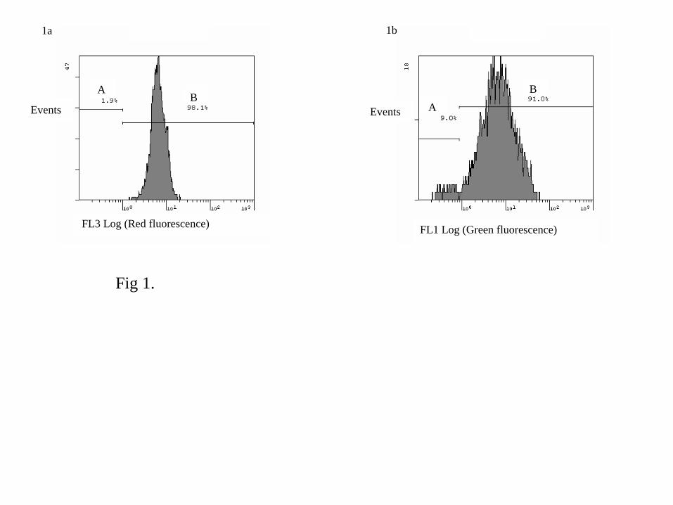
Dead parasites, prepared by boiling cells for 15 min, were used to control efficacy of PI for
mortality measurement. The FL3 fluorescence histogram showed 98.1% of PI-stained cells (red
fluorescence above 1) considered dead (Fig. 1a). Suspension of live parasites was used to control
efficacy of FDA for esterase activities measurement (Fig. 1b). The FL1 fluorescence histogram
showed 91% of fluorescent cells after incubation with FDA (green fluorescence above 1)
considered alive and presenting esterase activities (Fig. 1b).
Statistical analysis
Data were analyzed statistically using the software Statgraphics® Plus version 5.1. Results were
expressed as percentages of positive cells. Mean and standard deviation were calculated for each
7
179
180
181
182
183
184
185
186
187
188
189
190
191
192
193
194
195
196
197
198
199
200
201
202
203
204
triplicate. Effect of tested conditions was evaluated performing one-way, two-ways and three-ways
ANOVA. Values were converted into r angular arcsinus √(% of positive cells) before analysis to
ensure respect of a priori assumptions of normality and homogeneity. In the case of rejection of H0,
an a posteriori Least Significant Difference Test was used to compare differences between means
and to obtain hierarchy between studied factors. Significance was concluded at p ≤ 0.05.
RESULTS
Size and complexity of parasites
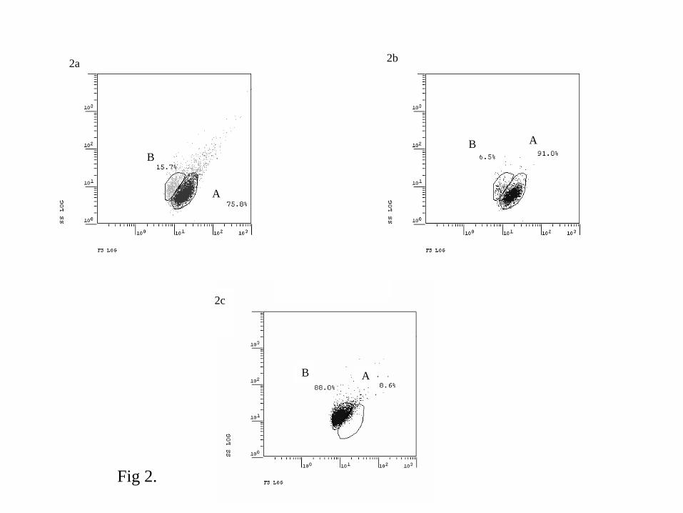
Size and in a lesser concern complexity of parasites varied according to their status: dead or alive.
Irrespective of the tested conditions, it was possible to identify two populations of parasite cells
(Fig. 2a): a population A consisting in 53 ± 24% of live cells and a population B, smaller in size
consisting in a majority of dead cells (mean of 74 ± 23%). Some parasite cells were not included in
population A or B and corresponded generally to dead cells showing higher size and higher
complexity than cells included in populations A and B. For parasites maintained in NSW at 4°C 12
hours after purification, population A and population B included 75.8% and 15.7% of total cells,
respectively (Fig. 2a). When only considering non PI stained parasites for the same experimental
conditions, population A and population B included 91% and 6.5% of live cells, respectively (Fig.
2b). For parasites after boiling, when only considering PI stained cells, population A and population
B included 8.6% and 88% of dead cells, respectively (Fig. 2c).
Population A included more live cells (77.6 ± 6.2%) when mortality rates were below 50%
compared to mortality rates above 70% (37.3 ± 24.7%) (Table 1). On the contrary, population B
included more dead cells when mortality rates were high (91.7 ± 7.4% for mortality rates above
70%) (Table 1).
8
Impact of medium on cell viability 205
206
207
208
209
210
211
212
213
214
215
216
217
218
219
220
221
222
223
224
225
226
227
228
229
230
In the three experiments testing simultaneously the effect of medium, temperature and time of
incubation on parasites (three-ways ANOVA), the medium appeared as the most important factor on
parasite survival (p = 0) and influences more cell mortality and esterase activities than temperature
and time of incubation (Table 2).
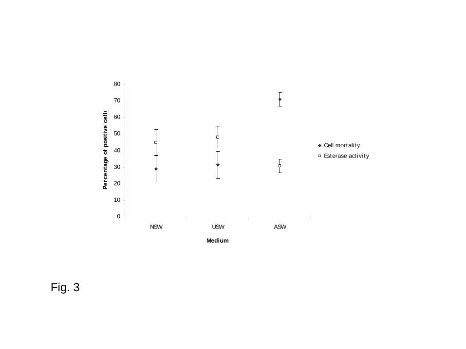
Irrespective of time and temperature of incubation, parasites showed significant better survival in
NSW and in USW than in ASW (Fig. 3 and Table 2). However, there was no significant difference
between mortality and esterase activity percentages in NSW and USW. Parasite mortality means
were 29.1%, 31.4% and 71.1% in NSW, USW and ASW, respectively. The percentage of positive
parasites for esterase activities was 44.9 ± 7.8% in NSW, 48.2 ± 6.5% in USW and 30.6 ± 4.1% in
ASW, respectively.
Impact of temperature on cell viability
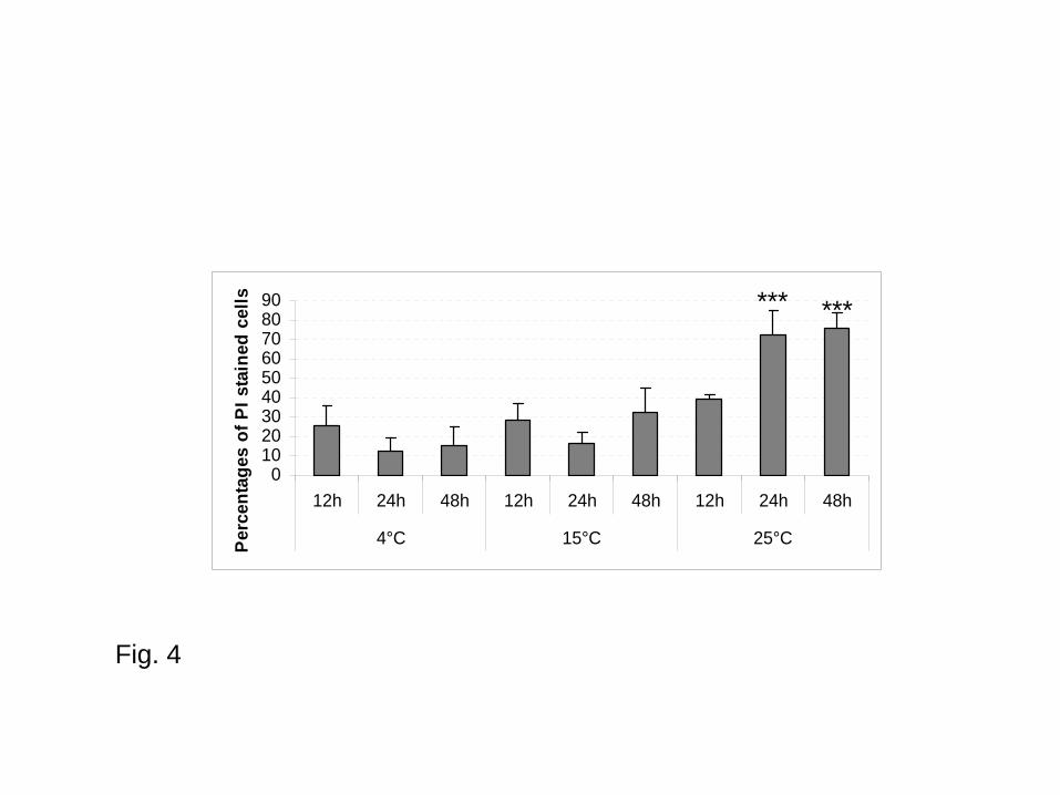
Parasite viability fluctuated according to the tested temperature. Irrespective of medium and time of
incubation, mortality appeared significantly higher at 25°C compared to 15°C and 4°C and
percentages of cells presenting esterase activities were higher at 4°C compared to 15°C and 25°C
(three-ways ANOVA, Table 2).
In NSW, irrespective of incubation time, mortality percentages ranged from 11.92 to 25.59% at 4°C,
from 16.2 to 31.83% at 15°C and from 39.26 to 75.55 at 25°C (Fig. 4). Cell mortality was thus
higher at 25°C compared to 4°C and 15°C especially after 24h and 48h of incubation (p < 0.0001)
(Fig. 4).
In USW, irrespective of time of incubation, the percentage of positive parasites for esterase
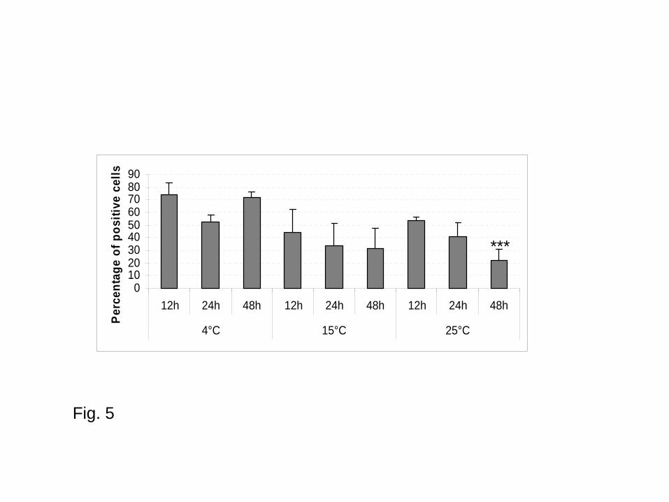
activities ranged from 52.49 to 73.67% at 4°C, from 31.27 to 43.73% at 15°C and from 21.89 to
53.28% at 25°C (Fig. 5).
Impact of incubation time on cell viability
9
231
232
233
234
235
236
237
238
239
240
241
242
243
244
245
246
247
248
249
250
251
252
Irrespective of medium and temperature, incubation time did not have significant impact on parasite
mortality. However, a difference of percentages of cells presenting esterase activities is noted
between 12 and 24 hours of incubation times (p = 0.003; three-ways ANOVA, Table 2).
In NSW and in USW, parasite survival and parasites presenting esterase activities were higher at
4°C and 15°C than at 25°C especially after 48h of incubation (Figs. 4 and 5). In NSW, after 48
hours of incubation, parasite mortality mean was 75.55%, 31.83% and 14.73% at 25°C, 15°C and
4°C respectively (Fig. 4). At 25°C, mortality was significantly higher after 24 h and 48 h of
incubation compared to 12 h (p < 0.0001).
Similarly, percentage of parasites presenting esterase activities significantly decreased at 25°C after
48 h of incubation. In USW at 25°C the percentage of positive cells was 47.23 ± 13.31% after 24 h
and 23.54 ± 8.65% after 48 h (p = 0.0004) (Fig. 5).
Impact of salinity on cell viability
Incubation length had no significant effect on cell mortality (two-ways ANOVA: F = 1.81, p =
0.186). Therefore, data obtained on independent samples after 12 and 48 hours of incubation were
pooled. A posteriori tests showed that salinities of 5, 15 and 20 g l-1 were associated with highest
percentages of mortality whereas salinities of 35, 40 and 45 g l-1 allowed better parasite survival
(Fig. 6). Higher percentages of positive cells for esterase activities were reported for higher
salinities (35 to 45 g l-1) (Fig. 7). Moreover, time of incubation presented a significant impact on
parasite esterase activities (two-ways ANOVA, F = 15.3, p = 0). There was a significant decrease of
percentages of positive parasites between 12h and 48h irrespective of tested salinities except at 25 g
l-1 (Fig. 7).
10
DISCUSSION 253
254
255
256
257
258
259
260
261
262
263
264
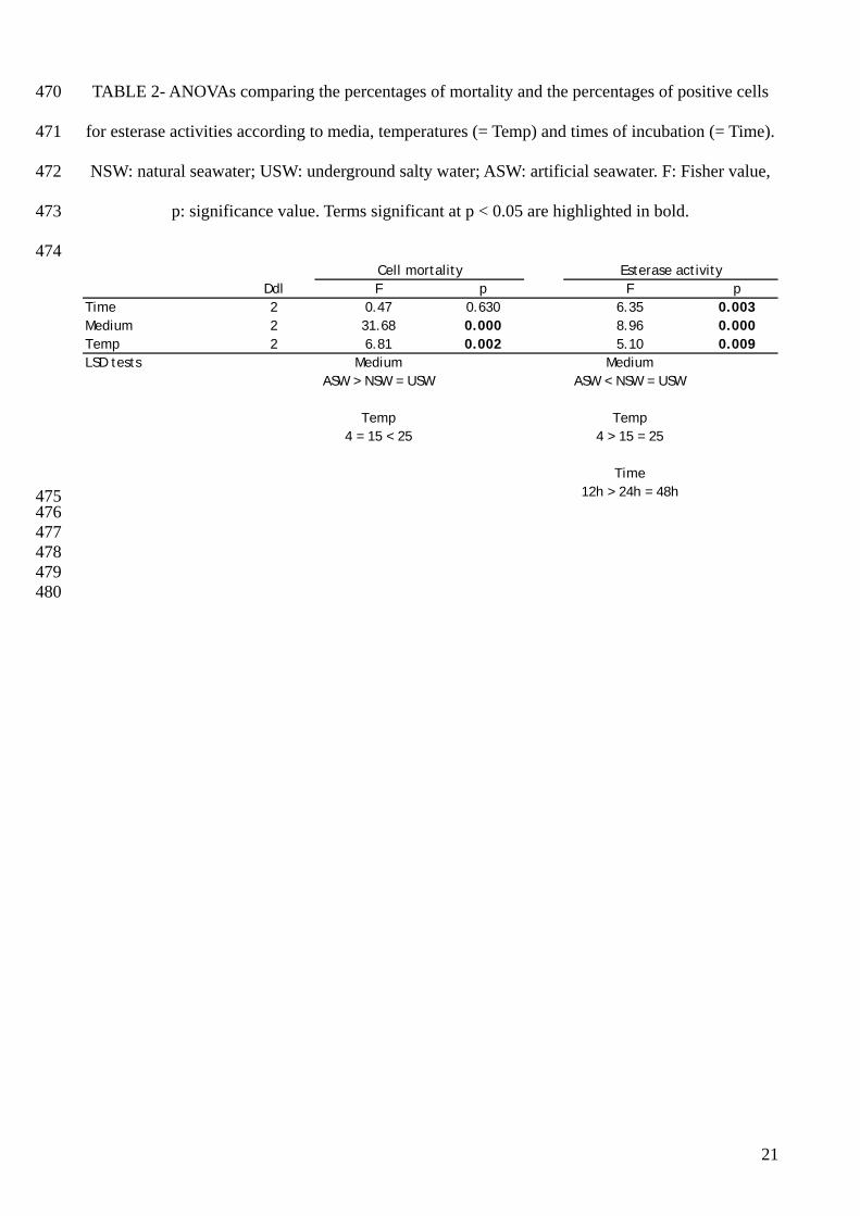
265
266
267
268
269
270
271
272
273
274
275
276
277
278
Despite 25 years of research on the protozoan Bonamia ostreae, its life cycle is poorly known.
Whatever is the date at which naïve oysters are placed in an infected area, first known stages of the
parasite are observed 3 to 5 months after exposition to the parasite (Tigé & Grizel 1984, Montes
1991). Moreover, the infection seems to remain present in areas that have been cleaned and which
ceased to produce oysters for several years (Van Banning 1988). Lagtime before infection and
persistence of the disease in cleaned areas motivated some authors to investigate potential
involvement of macroinvertebrate and zooplankton species in Bonamia ostreae life cycle (Lynch et
al. 2006). Nevertheless, considering the correlation between density of oysters and prevalence of
bonamiosis (Grizel 1985, Hudson & Hill 1991), the parasite mainly depends for its survival and
spread on flat oysters Ostrea edulis themselves and other aquatic organisms might not be involved
as important carriers or transmitters (Van Banning 1988). Transmission of B. ostreae between
oysters probably occurs through the water column. Water characteristics can have an impact on the
survival of the parasites released outside the host and these characteristics can influence the
infective capacity of B. ostreae as well as the number of oysters newly infected.
In that context, the impact of two environmental parameters, the salinity and the temperature, on
the parasite viability was investigated at different times of incubation: 12 h, 24 h and 48 h. Trials
were stopped at 48 h because some preliminary results were not reproducible beyond this
incubation time. In addition, the suitability of three different sea water media for parasite
preservation was tested: 0.22 µm filtered natural sea water; 0.22 µm filtered underground salty
water (with a constant composition) and 0.22 µm filtered artificial sea water (which is easy to
acquire and with a constant composition).
Size and complexity of Bonamia ostreae were generally homogeneous but depended on the status
of the parasite cell i.e. if they were live or dead. Two populations were distinguished: a
homogeneous population of small parasites corresponding mainly to dead cells and increasing
11
279
280
281
282
283
284
285
286
287
288
289
290
291
292
293
294
295
296
297
298
299
300
301
302
303
304
proportionally to recorded mortality, and another population less homogeneous, larger in size
increasing proportionally to survival rates. These results suggest that when dying B. ostreae
becomes smaller. Such phenomenon is described in apoptotic cells (Cotter et al. 1992, Samali &
Cotter 1999, Nasirudeen et al. 2001).
Results showed a better survival of purified Bonamia ostreae (60 to 80%) in natural filtered sea
water and in underground filtered salty water than in artificial filtered sea water (less than 40%)
whatever were temperature and time of incubation. This result could be explained by a difference
of pH. Indeed, pH of ASW used in this study was more acid (6.5) than NSW (8.06) and USW
(7.06). Effect of pH on parasite viability has not been investigated in the present study. Moreover,
a difference of salinity was also noticed between tested media: between 30 and 34 g l-1 for NSW;
32 g l-1 for USW and 23.4 g l-1 for ASW. Therefore, in addition to be more acid, ASW had a lower
salinity than other tested media which could explain the poor conservation of parasites.
Although no significant difference of mortality and non specific esterase activities were observed
between parasites maintained at 4°C and 15°C in NSW or USW, 25°C did not appear suitable for
parasite preservation. In natural conditions, the disease is reported in areas where temperature of
sea water rarely reaches 25°C except in Mediterranean Sea where Bonamia ostreae is reported but
with low prevalence (0.9 ± 1.4%, data from REPAMO, French network for the surveillance of
mollusc diseases). In Quiberon bay (Morbihan, France) where prevalence of bonamiosis is
estimated at 12.4% ± 6.5 (data from REPAMO), summer water temperatures fluctuated between
16.9 and 19.6°C between 1989 and 2003 with a mean estimated at 18.5°C (data from REPHY,
French network for the surveillance of phytoplankton and phycotoxins). Some analyses revealed a
negative correlation between high summer water temperature and number of oysters detected
infected during the following winter (I. Arzul, unpublished data) suggesting that higher
temperatures do not favour infection of oysters. A study carried out on Crassostrea gigas
haemocytes showed that an increase of temperature and a decrease of salinity induced an increase
of cell mortality (Gagnaire et al. 2006b) suggesting that these environmental parameters had also
12
305
306
307
308
309
310
311
312
313
314
315
316
317
318
319
320
321
322
323
324
325
326
327
328
329
330
an effect on oyster defence capacities. Similarly, previous works demonstrated an impact of
temperature on flat oyster Ostrea edulis defence mechanisms (Cochennec & Auffret 2002): lower
temperatures were associated with increased bonamiosis prevalence. However in the same study,
haemocyte activities were tested by flow cytometry and revealed that low temperature (10°C) or a
decrease of temperature (from 20°C to 10°C) induced a decrease of enzymatic activities including
ROS production involved in defence mechanisms. Several authors described a seasonal variation of
infection with B. ostreae; prevalence of infection presenting peaks in late winter and in autumn
which suggests an involvement of environmental parameters including temperature in the
development of the disease (Grizel 1985, Montes 1990, Van Banning 1991, Culloty & Mulcahy
1996). Studies carried out on Bonamia sp. infecting the Asian oyster Crassostrea ariakensis in
Atlantic coastal waters of U.S.A showed a strong influence of temperature on seasonal parasite
cycling (Carnegie et al. 2008). Interestingly, temperatures around 25°C when oysters were placed
in infected area were associated with higher prevalence than temperatures below 20°C.
Experimental studies support these results showing that warm temperatures (>20°C) seem to
increase Bonamia sp. pathogenicity (Audemard et al. 2008a). Epidemiological data available for
Bonamia (= Mikrocytos) roughleyi show that the disease expressed under winter oyster mortalities
is associated with low temperatures (Wolf 1967). However all these studies consider the parasite
inside its host and thus investigate effects of temperature on host-parasite relationships and not
directly on parasite survival.
Purified Bonamia ostreae seems to show a preference for hyper saline media compared to hypo
saline media. Three ranges of salinities could be identified from these results: from 5 to 20 g l-1,
survival and esterase activity measures were very low but a mean of 10% of live cells can still be
detected suggesting that the parasite can still be transmitted in these conditions; between 25 and 30
g l-1 survival was intermediate (estimated at 35% after 12 hours of incubation); between 35 and 45 g
l-1 survival was higher and estimated at 50% after 12 hours of incubation.
As previously mentioned measures of parasite survival in the three tested media (NSW, USW and
13
ASW) supported these results. Indeed, parasite mortality was higher in ASW (salinity of 23.4 g l-1)
compared to NSW (salinity of 32-34 g l-1) and USW (salinity of 32 g l-1).
331
332
333
334
335
336
337
338
339
340
341
342
343
344
345
346
347
348
349
350
351
352
353
354
355
356
These results are also concordant with a previous study realised on Bonamia exiotiosa in New
Zealand in which a salinity of 40 g l-1 was associated with highest disease prevalences (Hine 2002).
Similarly, infection with B. roughleyi seems to be favoured by high salinities (30-35 g l-1) (Farley et
al. 1988). Our results are also in concordance with data obtained during a recent study in which
salinity below 30 g l-1 was associated with lower host mortality and appeared detrimental to
Bonamia sp. in Crassostrea ariakensis (Audemard et al. 2008b).
Time of incubation showed an impact on the parasite preservation especially by increasing effect of
high temperatures on mortality (higher mortality) and esterase activities (lower percentages of
positive cells). Moreover, whatever was the tested salinity, percentages of cells producing esterase
activities were lower after 48h compared to 12h. It would be interesting to complete these results by
testing a wider range of incubation times in order to evaluate the persistence capacity of Bonamia
ostreae in natural sea water collected from different infected areas. However, mortality and esterase
activity measured by flow cytometry are instantaneous and do not allow to follow cumulative
mortality.
In the present context of global change, data allowing forecasting of disease evolution are
requested. Oysters are submitted to environmental changing and parasites as well. Description of
the influence of temperature and salinity on Bonamia ostreae viability should allow modelling
parasite transmission. Moreover these results should contribute to define risky and non risky
geographic areas regarding transmission of the disease. These data might also be of interest for
oyster farmers. Indeed, by monitoring temperature and salinity parameters, oysters might be moved
or sold before suitable conditions for parasites survival are reached.
Acknowledgements
The authors thank R. Brizard and his team for technical assistance for the maintenance of oysters in
14
357
358
359
raceways in Ifremer La Tremblade quarantine. A. Langlade is also acknowledged for supplying flat
oysters from Quiberon Bay, France.
15
360 361
362
363
364
365
366
367
368
369
370
371
372
373
374
375
376
377
378
379
380
381
382
383
384
385
REFERENCES
Arzul I, Miossec L, Blanchet E, Garcia C, François C, Joly J-P (2006) Bonamia ostreae and
Ostrea edulis : a stable host-parasite system in France ? Proceedings of the XIth ISVEE conference,
Cairns, Australia
Audemard C, Carnegie RB, Bishop M, Peterson CH, Burreson E (2008a) Interacting effects of
temperature and salinity on Bonamia sp. parasitism in the Asian oyster Crassostrea ariakensis. J
Invertebr Pathol 98:344-350
Audemard C, Carnegie RB, Stokes NA, Bishop M, Peterson CH, Burreson E (2008b) Effects of
salinity on Bonamia sp. survival in the Asian oyster Crassostrea ariakensis. J Shellfish Res 27:535-
540
Berney M, Vital M, Hülshoff I, Weilenmann HU, Egli T, Hammes F (2008) Rapid, cultivation-
independent assessment of microbial viability in drinking water. Water Res 42:4010-4018
Binet MT, Stauber JL (2006) Rapid flow cytometric method for the assessment of toxic flagellate
cyst viability. Mar Environ Res 62:247-260
Carnegie RB, Stokes NA, Audemard C, Bishop M, Wilbur AE, Alphin TD, Posey MH, Peterson
CH, Burreson E (2008) Strong seasonality of Bonamia sp. infection and induced Crassostrea
ariakensis mortality in Bogue and Masonboro Sounds, North Carolina, USA. J Invertebr Pathol
98:335-343
Chagot D, Boulo V, Hervio D, Mialhe E, Bachere E, Mourton C, Grizel H (1992) Interactions
between Bonamia ostreae (Protozoa: Ascetospora) and hemocytes of Ostrea edulis and Crassostrea
gigas (Mollusca: Bivalvia): Entry mechanisms. J Invertebr Pathol 59:241-249
Cochennec N, Auffret M (2002) European project FAIR-CT98-4120 “Environmental Factors and
Shellfish Diseases” 15/05/2002 Final Report.
Cotter TG, Lennon SV, Glynn JM, Green DR (1992) Microfilament-disrupting agents prevent
formation of apoptotic bodies in tumour cells undergoing apoptosis. Canc Res 25:997-1005
16
386
387
388
389
390
391
392
393
394
395
396
397
398
399
400
401
402
403
404
405
406
407
408
409
410
411
Culloty SC, Mulcahy MF (1996) Season-, age-, and sex-related variation in the prevalence of
bonamiosis in flat oysters (Ostrea edulis L.) on the South coast of Ireland. Aquaculture 144:3–63
Czechowska K, Johnson DR, Roelof van der Meer J (2008) Use of flow cytometric methods for
single-cell analysis in environmental microbiology. Curr Opin Microbiol 11:205-212
Elston RA, Farley CA, Kent ML (1986) Occurrence and significance of bonamiasis in European
flat oysters Ostrea edulis in North America. Dis Aquat Org 2:49–54
Farley CA, Wolf PH, Elston RA (1988) A long-term study of "microcell" disease in oysters with
a description of a new genus, Mikrocytos (g.n.) and two new species Mikrocytos mackini (sp.n.) and
Mikrocytos roughleyi (sp.n.). Fish Bull 86:581-593
Gagnaire B, Thomas-Guyon H, Burgeot T, Renault T (2006a) Pollutant effects on Pacific oyster,
Crassostrea gigas (Thunberg), hemocytes: screening of 23 molecules using flow cytometry. Cell
Biol Toxicol 22:1–14
Gagnaire B, Frouin H, Moreau K, Thomas-Guyon H, Renault T (2006b) Effects of temperature
and salinity on haemocyte activities of the Pacific oyster, Crassostrea gigas (Thunberg). Fish
Shellfish Immunol 20:536-547
Grizel H (1985) Etudes des récentes épizooties de l’huître plate Ostrea edulis L. et de leur impact
sur l’ostréiculture bretonne. PhD dissertation, Université des Sciences et Techniques de Languedoc,
Montpellier, France
Hammes F, Berney M, Wang Y, Vital M, Köster O Egli T (2008) Flow cytometric total bacterial
cell counts as a descriptive microbiological parameter for drinking water treatment processes. Water
Res 42:269-277
Hervio D, Bachere E, Boulo V, Cochennec N, Vuillemin V, Le Coguic Y, Cailletaux G, Mazurie J,
Mialhe E (1995) Establishment of an experimental infection protocol for the flat oyster Ostrea
edulis with the intrahaemocytic protozoan parasite Bonamia ostreae: application in the selection of
parasite-resistant oyster. Aquaculture 132:183–194
Hine PM (2002) Severe apicomplexan infection in the oyster Ostrea chilensis: a possible
17
412
413
414
415
416
417
418
419
420
421
422
423
424
425
426
427
428
429
430
431
432
433
434
435
predisposing factor in bonamiosis Dis Aquat Org 51:49-60
Hudson EB, Hill BJ (1991) Impact and spread of bonamiasis in the UK. Aquaculture 93:279-285
Lindström ES, Weisse T, Stadler P (2002) Enumeration of small ciliates in culture by flow
cytometry and nucleic acid staining. J Microbiol Meth 49:173-182
Lynch SA, Armitage D V, Wylde S, Mulcahy MF, Culloty S C (2005) The susceptibility of young
prespawning oysters, Ostrea edulis, to Bonamia ostreae. J Shellfish Res 24:1019-1025
Lynch SA, Armitage DV, Coughlan J, Mulcahy MF, Culloty SC (2006) Investigating the possible
role of benthic macroinvertebrates and zooplankton in the life cycle of the haplosporidian Bonamia
ostreae. Exp Parasitol 115:359-368
Mialhe E, Bachère E, Chagot D, Grizel H (1988) Isolation and purification of the protozoan
Bonamia ostreae (Pichot et al. 1980), a parasite affecting the flat oyster Ostrea edulis L.
Aquaculture 71:293-299
Montes J (1990) Development of Bonamia ostreae parasitosis of flat oyster, Ostrea edulis, from
Galicia, northwest Spain. In: Perkins FO, Cheng TC (eds) Pathology in Marine Aquaculture.
Academic Press, New York, NY, p 223-227
Montes J (1991). Lag time for the infestation of flat oyster (Ostrea edulis L.) by Bonamia ostreae
in estuaries of Galicia (N.W. Spain). Aquaculture 93:235–239
Montes J, Anadon R, Azevedo C (1994) A possible life cycle for Bonamia ostreae on the basis of
electron microscopy studies. J Invertebr Pathol 63 :1–6
Mourton C, Boulo V, Chagot D, Hervio D, Bachère E, Mialhe E, Grizel H (1992) Interactions
between Bonamia ostreae (Protozoa: Ascetospora) and hemocytes of Ostrea edulis and Crassostrea
gigas (Mollusca: Bivalvia): In vitro system establishment. J Invertebr Pathol 59:235-240
Nasirudeen AMA, Tan KSW, Singh M, Yap EH (2001) Programmed cell death in a human
intestinal parasite, Blastocystis hominis. Parasitology 123:235-246
18
436
437
438
439
440
441
442
443
444
445
446
447
448
449
450
451
452
453
454
455
456
457
458
459
460
Parrow MW, Burkholder JM (2002) Flow cytometric determination of zoospore DNA content
and population DNA distribution in cultured Pfiesteria spp. (Pyrrhophyta). J Exp Mar Biol Ecol
267:35-51
Rault A, Bouix M, Béal C (2008) Dynamic analysis of Lactobacillus delbrueckii subsp.
bulgaricus CFL1 physiological characteristics during fermentation. Appl Microbiol Biotechnol
81:559-570
Samali A, Cotter TG (1999) Measurement of Cell Death in Culture. In : Jenkins N (ed) Animal
Cell Biotechnology: Methods and Protocols. Series: Methods in Biotechnology. Volume 8. Humana
Press Inc, Totowa, NJ, p 155-164
Soudant P, Chu FLE, Lund ED (2005) Assessment of the cell viability of cultured Perkinsus
marinus (Perkinsea), a parasitic protozoan of the eastern oyster, Crassostrea virginica, using
SYBRgreen-Propidium Iodide double staining and flow cytometry. J Euk Microbiol 52:492-499
Sprague V (1979) Classification of the Haplosporidia. Mar Fish Rev 41: 40-44
Tigé G, Grizel H (1984) Essai de contamination d’Ostrea edulis Linne par Bonamia ostreae
(Pichot et al., 1979) en rivière de Crach (Morbihan). Rev Trav Inst Pêches Marit 46:307–314
Van Banning P (1988) Management strategies to control diseases in the Dutch culture of edible
oysters. In: Fisher WS (ed), Disease Processes in Marine Bivalve Molluscs. American fisheries
Society, Spec Publ 18:243-245
Van Banning P (1991) Observations on bonamiasis in the stock of the European flat oyster,
Ostrea edulis, in the Netherlands, with special reference to the recent developments in Lake
Grevelingen. Aquaculture 93:205–211
Wolf P (1967) Winter mortality hits state's oyster output. The Fisherman 2:20-22
Wong JTY, Whiteley A (1996) An improved method of cell cycle synchronisation for the
heterotrophic dinoflagellate Crypthecodinium cohnii Biecheler analyzed by flow cytometry. J Exp
Mar Biol Ecol 197:91-99
19

TABLE 461
462
463
464
465
466
TABLE 1- Distribution of Bonamia ostreae cells in percentages (means (± standard deviation)) in
Population A and Population B (as shown on Figures 2 for example) and composition in live and
dead cells of these two populations according to the level of mortality rates.
Mortality rates Population A Population A
alive
Population B Population B
dead
< 50% 56,68 (± 18,11) 77,58 (± 6,21) 15,66 (± 5,16) 53,54 (± 22,03)
≥ 50 % and < 70% 50,29 (± 20,78) 56,35 (± 9,16) 22,74 (± 10,65) 74,99 (± 13,79)
≥ 70% 25,64 (± 17,06) 37,33 (± 24,56) 49,51 (±14,44) 91,67 (± 7,43)
467 468
469
20
470
471
472
473
474
TABLE 2- ANOVAs comparing the percentages of mortality and the percentages of positive cells
for esterase activities according to media, temperatures (= Temp) and times of incubation (= Time).
NSW: natural seawater; USW: underground salty water; ASW: artificial seawater. F: Fisher value,
p: significance value. Terms significant at p < 0.05 are highlighted in bold.
Ddl F p F pTime 2 0.47 0.630 6.35 0.003Medium 2 31.68 0.000 8.96 0.000Temp 2 6.81 0.002 5.10 0.009LSD tests Medium Medium
ASW > NSW = USW ASW < NSW = USW
Temp Temp4 = 15 < 25 4 > 15 = 25
Time12h > 24h = 48h
Cell mortality Esterase activity
475 476 477 478 479 480
21
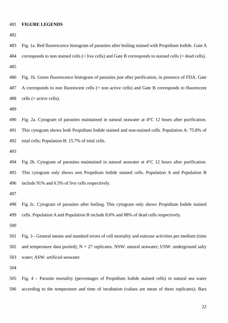
FIGURE LEGENDS 481
482
483
484
485
486
487
488
489
490
491
492
493
494
495
496
497
498
499
500
501
502
503
504
505
506
Fig. 1a. Red fluorescence histogram of parasites after boiling stained with Propidium Iodide. Gate A
corresponds to non stained cells (= live cells) and Gate B corresponds to stained cells (= dead cells).
Fig. 1b. Green fluorescence histogram of parasites just after purification, in presence of FDA. Gate
A corresponds to non fluorescent cells (= non active cells) and Gate B corresponds to fluorescent
cells (= active cells).
Fig. 2a. Cytogram of parasites maintained in natural seawater at 4°C 12 hours after purification.
This cytogram shows both Propidium Iodide stained and non-stained cells. Population A: 75.8% of
total cells; Population B: 15.7% of total cells.
Fig 2b. Cytogram of parasites maintained in natural seawater at 4°C 12 hours after purification.
This cytogram only shows non Propidium Iodide stained cells. Population A and Population B
include 91% and 6.5% of live cells respectively.
Fig 2c. Cytogram of parasites after boiling. This cytogram only shows Propidium Iodide stained
cells. Population A and Population B include 8.6% and 88% of dead cells respectively.
Fig. 3 - General means and standard errors of cell mortality and esterase activities per medium (time
and temperature data pooled); N = 27 replicates. NSW: natural seawater; USW: underground salty
water; ASW: artificial seawater.
Fig. 4 – Parasite mortality (percentages of Propidium Iodide stained cells) in natural sea water
according to the temperature and time of incubation (values are mean of three replicates); Bars
22
23
507
508
509
510
511
512
513
514
515
516
517
518
519
520
521
522 523
represent standard errors; *** p < 0.0001 compared to data at 25°C and 12 h after incubation and
compared to data at 15°C and 4°C
Fig. 5 - Parasite esterase activities (percentages of positive cells) in underground salty water
according to the temperature and time of incubation (Values are mean of three replicates; Bars
represent standard errors; *** p = 0.0004 compared to data at 25°C 24 h after incubation
Fig. 6 - Parasite mortality (Percentages of Propidium Iodide stained cells) according to the salinity
of underground salty water (time of both data pooled). Values are means ± standard error. N = 6
replicates.
Fig. 7 - Parasite esterase activities (Percentages of positive cells) in underground salty water
according to the salinity and time of incubation (Values are mean of three replicates; Bars represent
standard errors); *** p < 0.001 compared to data 12 h after incubation
Ev ents
FL3 Log (Red fluorescence)
BA
1a
Ev ents
FL1 Log (Green fluorescence)
AB
1b
Fig 1.
B
A
2a
B
2b
AB
AB
2c
2b
Fig 2.
0
10
20
30
40
50
60
70
80
NSW USW ASW
Medium
Perc
enta
ge o
f po
siti
ve c
ells
Cell mortality
Esterase activity
Fig. 3
0102030405060708090
12h 24h 48h 12h 24h 48h 12h 24h 48h
4°C 15°C 25°CPerc
enta
ges
of P
I sta
ined
cel
ls *** ***
Fig. 4
0102030405060708090
12h 24h 48h 12h 24h 48h 12h 24h 48h
4°C 15°C 25°C
Perc
enta
ge o
f pos
itive
cel
ls
***
Fig. 5
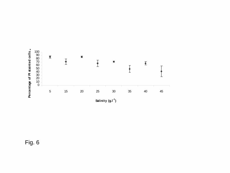
Fig. 6
0102030405060708090
100
5 15 20 25 30 35 40 45
Salinity (g.l-1)
Perc
enta
ge o
f PI
sta
inte
d ce
lls
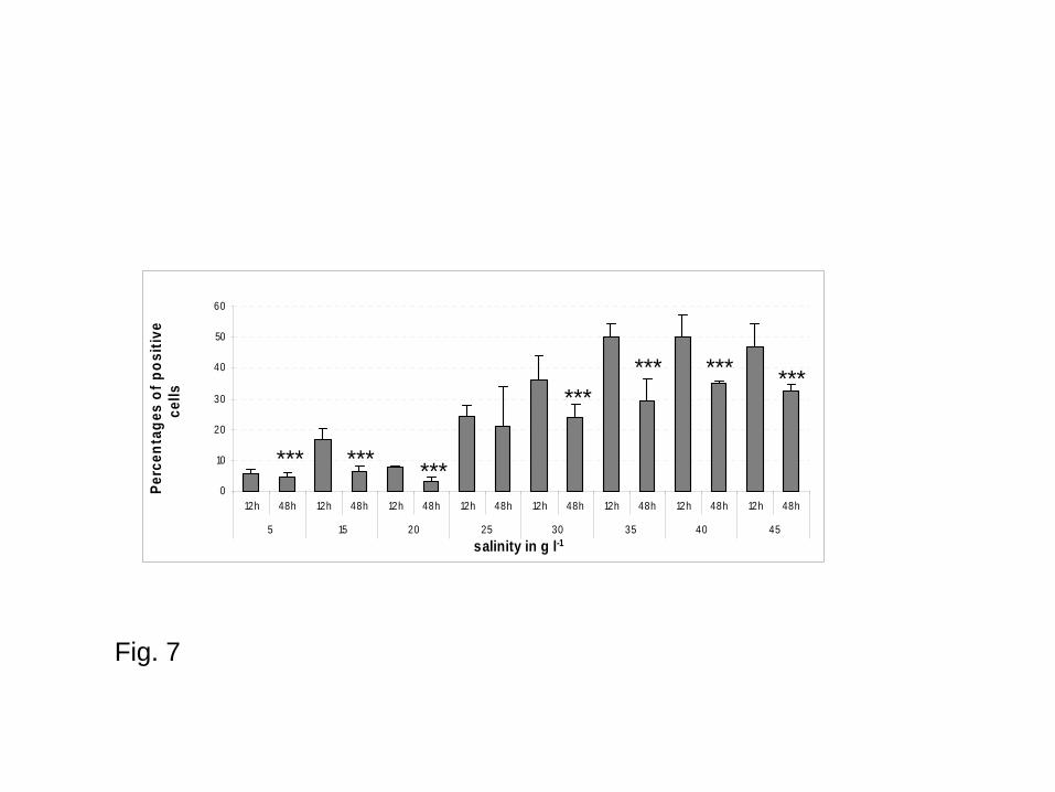
Fig. 7
0
10
20
30
40
50
60
12h 48h 12h 48h 12h 48h 12h 48h 12h 48h 12h 48h 12h 48h 12h 48h
5 15 20 25 30 35 40 45
salinity in g l-1
Perc
enta
ges
of p
ositi
ve
cells
************
*********