Effect of Species Variation and Processing on Phenolic Composition and In Vitro Antioxidant Activity...
13
[Journal Home Page] [Search the Journals] [Table of Contents] [PDF version of this article] [Download to Citation Manager] J. Agric. Food Chem., 56 (3), 954–963, 2008. 10.1021/jf072904a Web Release Date: January 17, 2008 Copyright © 2008 American Chemical Society Effect of Species Variation and Processing on Phenolic Composition and In Vitro Antioxidant Activity of Aqueous Extracts of Cyclopia spp. (Honeybush Tea) Elizabeth Joubert, * † ‡ E. Siân Richards, ‡ § J. Debora Van der Merwe, ‡ Dalene De Beer, † Marena Manley, ‡ and Wentzel c.a. Gelderblom ⊥ ¶ ARC Infruitec-Nietvoorbij, Private Bag X5026, Stellenbosch, 7599, South Africa, Departments of Food Science and Biochemistry, Stellenbosch University, Private Bag X1, Matieland (Stellenbosch), 7602, South Africa, Nampak Research & Development, PO Box 247, Howard Place, 7450, South Africa, and PROMEC Unit, Medical Research Council, PO Box 19070, Tygerberg, 7505, South Africa Received October 1, 2007 Abstract: The in vitro antioxidant activity of aqueous extracts prepared from four Cyclopia spp. (unfermented and fermented) was assessed using radical (ABTS •+ ) scavenging, ferric ion reduction, and inhibition of Fe 2+ -induced microsomal lipid peroxidation as criteria. Aqueous extracts of unfermented and fermented Aspalathus linearis (rooibos) and Camellia sinensis teas (green, oolong, and black) were included as reference samples. Qualitative and quantitative differences in phenolic composition were demonstrated for the Cyclopia spp. The xanthone glycoside, a.k.a. mangiferin, was the major monomeric polyphenol present in the Cyclopia extracts, with both unfermented and fermented C. genistoides extracts containing the highest quantities. Fermentation resulted in a significant reduction in extract yields and their total polyphenolic and individual polyphenol contents. Unfermented plant material should preferentially be used for preparation of extracts, as fermentation significantly (P < 0.05) lowered antioxidant activity of all species, except in the case of C. genistoides, where the ability to inhibit lipid peroxidation was not affected. Unfermented plant material also retained the highest concentration of mangiferin. Overall, extracts of unfermented Cyclopia were either of similar or lower antioxidant activity as compared to the other teas. However, the presence of high levels of mangiferin merits the use of Cyclopia spp. and, in particular, C. genistoides, as an alternative herbal tea and potential dietary supplement. Keywords: Mangiferin; hesperidin; herbal tea; rooibos; processing; lipid peroxidation. Introduction Recently, the focus has fallen onto xanthones as natural antioxidants and cancer chemopreventive agents (1–3 ). Natural sources of xanthones are, most notably, Garcinia mangostana (mangosteen) (3 ), containing a number of prenylated xanthones, and Mangifera indica (mango), which contains mangiferin (4 ). Plant extracts containing high levels of these compounds are commercially available (5 ), such as a standardized aqueous extract from mango stem bark with mangiferin and total polyphenols comprising 13% and 34% (as gallic acid equivalents) of the dry weight, respectively (4 ). Mango fruit waste is considered another source, with the dry peels containing 0.17% mangiferin (6 ). Mangiferin, a C-glycoside, was shown to have antioxidant properties in various in vitro systems (7–11 ). Its ability to protect cultured human peripheral blood lymphocytes against radiation damage is linked to its free radical scavenging abilities (12 ). Cyclopia species, endemic to the Cape Fynbos biome, a unique ecosystem in South Africa and one of six global floral kingdoms, contain xanthones in relatively large quantities (13 ). Dried C. genistoides plant material could contain as much as 7.2% mangiferin (14 ) and 0.54% of the C8-glucoside, isomangiferin (13 ). The flavanone, hesperidin, is another major compound, comprising between 0.3 and 1.8% of the dry plant material, depending on the species (13 ). Investigation of the phenolic composition of C. intermedia and C. subternata showed the presence of other flavanones
-
Upload
independent -
Category
Documents
-
view
5 -
download
0
Transcript of Effect of Species Variation and Processing on Phenolic Composition and In Vitro Antioxidant Activity...

[Journal Home Page] [Search the Journals] [Table of Contents] [PDF version of this article] [Download to Citation Manager]
J. Agric. Food Chem., 56 (3), 954–963, 2008. 10.1021/jf072904a Web Release Date: January 17, 2008
Copyright © 2008 American Chemical Society
Effect of Species Variation and Processing on Phenolic Composition and In Vitro Antioxidant Activity of Aqueous Extracts of Cyclopia spp. (Honeybush Tea)Elizabeth Joubert,*†‡ E. Siân Richards,‡§ J. Debora Van der Merwe,‡ Dalene De Beer,† Marena Manley,‡ and Wentzel c.a. Gelderblom⊥¶
ARC Infruitec-Nietvoorbij, Private Bag X5026, Stellenbosch, 7599, South Africa, Departments of Food Science and Biochemistry, Stellenbosch University, Private Bag X1, Matieland (Stellenbosch), 7602, South Africa, Nampak Research & Development, PO Box 247, Howard Place, 7450, South Africa, and PROMEC Unit, Medical Research Council, PO Box 19070, Tygerberg, 7505, South Africa
Received October 1, 2007
Abstract:
The in vitro antioxidant activity of aqueous extracts prepared from four Cyclopia spp. (unfermented and fermented) was assessed using radical (ABTS•+) scavenging, ferric ion reduction, and inhibition of Fe2+-induced microsomal lipid peroxidation as criteria. Aqueous extracts of unfermented and fermented Aspalathus linearis (rooibos) and Camellia sinensis teas (green, oolong, and black) were included as reference samples. Qualitative and quantitative differences in phenolic composition were demonstrated for the Cyclopia spp. The xanthone glycoside, a.k.a. mangiferin, was the major monomeric polyphenol present in the Cyclopia extracts, with both unfermented and fermented C. genistoidesextracts containing the highest quantities. Fermentation resulted in a significant reduction in extract yields and their total polyphenolic and individual polyphenol contents. Unfermented plant material should preferentially be used for preparation of extracts, as fermentation significantly (P < 0.05) lowered antioxidant activity of all species, except in the case of C. genistoides, where the ability to inhibit lipid peroxidation was not affected. Unfermented plant material also retained the highest concentration of mangiferin. Overall, extracts of unfermented Cyclopia were either of similar or lower antioxidant activity as compared to the other teas. However, the presence of high levels of mangiferin merits the use of Cyclopia spp. and, in particular, C. genistoides, as an alternative herbal tea and potential dietary supplement.
Keywords: Mangiferin; hesperidin; herbal tea; rooibos; processing; lipid peroxidation.
Introduction
Recently, the focus has fallen onto xanthones as natural antioxidants and cancer chemopreventive agents (1–3). Natural sources of xanthones are, most notably, Garcinia mangostana (mangosteen) (3), containing a number of prenylated xanthones, and Mangifera indica (mango), which contains mangiferin (4). Plant extracts containing high levels of these compounds are commercially available (5), such as a standardized aqueous extract from mango stem bark with mangiferin and total polyphenols comprising 13% and 34% (as gallic acid equivalents) of the dry weight, respectively (4). Mango fruit waste is considered another source, with the dry peels containing 0.17% mangiferin (6). Mangiferin, a C-glycoside, was shown to have antioxidant properties in various in vitro systems (7–11). Its ability to protect cultured human peripheral blood lymphocytes against radiation damage is linked to its free radical scavenging abilities (12).
Cyclopia species, endemic to the Cape Fynbos biome, a unique ecosystem in South Africa and one of six global floral kingdoms, contain xanthones in relatively large quantities (13). Dried C. genistoides plant material could contain as much as 7.2% mangiferin (14) and 0.54% of the C8-glucoside, isomangiferin (13). The flavanone, hesperidin, is another major compound, comprising between 0.3 and 1.8% of the dry plant material, depending on the species (13). Investigation of the phenolic composition of C. intermedia and C. subternata showed the presence of other flavanones
in both species, as well as isoflavones and coumestans in C. intermedia(15–17).
Historical use of Cyclopia spp., either as medicinal plant or as herbal tea, predates the 1800s (18). To be used as aherbal tea it requires “fermentation”, an oxidative process whereby the plant material is subjected to high temperatures(>60 °C). This is essential for development of the characteristic honey-like flavor and dark-brown leaf color.Fermentation is, however, detrimental to the soluble total polyphenol content (19) and superoxide anion radical scavenging activity (20) of aqueous extracts of the plant material. Fermented honeybush plant material with its sweet flavor is used to prepare aqueous extracts for the food industry. Unfermented plant material only recently entered the market for herbal tea preparation. Its higher levels of total polyphenols suggest its preferential use for preparation of antioxidant extracts for the nutraceutical and cosmetic markets. Because of their initial prominence on the market, C. subternata (cultivated) and C. intermedia (wild-harvested) are preferentially used by extract manufacturers. However, C. genistoides is now also cultivated and, together with C. subternata, constitutes the vast majority of cultivated honeybush plants. After initial trials with cultivation of C. sessiliflora, which showed poor growth, a limited quantity is harvested from the wild for herbal tea preparation.
In the present study, the antioxidant activity of aqueous extracts of Cyclopia spp. was determined. Differences in their phenolic composition and antioxidant activity, as well as the antioxidant activity of the major polyphenols, mangiferin and hesperidin, and other selected Cyclopia polyphenols were determined. The role of fermentation in relation to phenolic composition and antioxidant activity was also assessed. The ultimate goal was to identify the species with the greatest commercial potential, based on antioxidant activity and concentration of valuable polyphenols. This species could then be used as antioxidant supplement, and the information on the polyphenols could be used to direct future research toward condition-specific beneficial properties associated with antioxidants. The antioxidant activity of Cyclopia extracts was compared to unfermented and fermented rooibos (Aspalathus linearis), a well-known South African herbal tea, and Camellia sinensis (black, oolong, and green tea) extracts, all known for their antioxidant properties.
Materials and Methods
Chemicals and Water Purification. Mangiferin, hesperetin, hesperidin, and (-)-epigallocatechin gallate were suppliedby Sigma-Aldrich Chemical Co. (Cape Town, South Africa), and naringenin, narirutin, eriocitrin, eriodictyol,formononetin, and luteolin were supplied by Extrasynthèse (Genay Cedex, France). An isolate of C. intermedia, containing isomangiferin as major compound, was supplied by Prof. Daneel Ferreira of the University of the Free State, South Africa, for determination of its UV/vis spectrum and HPLC retention time. Sigma-Aldrich Chemical Co. also supplied gallic acid, butylated hydroxytoluene (BHT), 2,4,5-tri(2-pyridyl)-S-triazine (TPTZ), Trolox(6-hydroxy-2,5,7,8-tetramethylchroman-2-carboxylic acid), thiobarbituric acid (TBA), and HPLC-grade acetonitrile.2,2′-Azinobis(3-ethyl-benzothiazoline-6-sulfonate) (ABTS•+) were obtained from Boehringer Mannheim (Mannheim, Germany), and other general reagents were from Merck (Cape Town, South Africa). Deionized water was prepared using a Modulab Water Purification System (Separations, Cape Town, South Africa). For HPLC eluant and sample preparation, deionized water was further purified by means of a Milli-Q 185 Académic Plus water purification system(Millipore, Bedford, MA).
Plant Material. Six batches (2–5 kg) of each Cyclopia spp. (honeybush) were harvested in the Western Cape Province of South Africa (21). Cyclopia intermedia was harvested from a natural stand (identified by Dr. Hannes de Lange, National Botanical Institute, Kirstenbosch, Cape Town, South Africa), whereas C. genistoides (West Coast type), C. sessiliflora, and C. subternata were harvested from commercial plantations. A batch comprised shoots from several bushes, and shoots from a particular bush were not present in more than one batch. Where bushes were harvested in a plantation, consecutive bushes were harvested to form a batch. Six samples of each of the green, oolong, and black Camellia sinensis teas and unfermented and fermented Aspalathus linearis (rooibos) teas (21) were included in the study as references. Green, oolong, and black teas were not prepared from the same plant material.
Processing of Cyclopia Plant Material. The harvested shoots were processed according to a standardized processing procedure (19) into fermented and unfermented counterparts, as previously described (21). Processing entailed thefollowing steps; each batch was shredded into <4 mm pieces, using a mechanized fodder cutter, and subdivided forimmediate artificial drying (preparation of unfermented plant material) and for fermentation. The plant material was thinlyspread on fine mesh drying trays for rapid drying under controlled conditions to minimize oxidative changes. The finalmoisture content was <10%. Drying took place at 40 °C for 12 h in a drying tunnel (Decon Humidifier, Continental FanWorks CC., Cape Town, South Africa). Air movement was in a cross-flow direction at 2 m/s. The remaining plantmaterial was moistened to ca. 60% moisture content with deionized water, “fermented” in a laboratory oven (70 °C for60 h), and then dried similarly to its unfermented counterpart. The dried tea was sieved using an Endecott test sieve (2mm) (London, England), the pieces of leaf and stem smaller than 2 mm were pulverized in a Retsch rotary mill (1 mmsieve) and stored in airtight plastic containers in the dark until analysis.
Aqueous Extract (AE) Preparation. The AEs of Cyclopia spp., rooibos and Camellia sinensis teas prepared for a previous antimutagenicity study (21) were used in this study. Extract preparation consisted of steeping (5 min) the ground plant material (100 g) in freshly boiled deionized water (1000 mL), filtering using a Buchner filter with a 125 µm

mesh cloth (Polymer PES D25/35, Swiss Silk Bolting Cloth Mfg. Co. Ltd., Zurich, Switzerland), followed by filtration through Whatman No. 4 filter paper (Whatman International Ltd., Maidstone, England), and freeze-drying at 40 °C shelftemperature in an Atlas pilot-scale freeze-drier (Denmark model, Copenhagen, Denmark). The dried aqueous extracts (AEs) were stored at room temperature in sealed clear glass vials in desiccators in the dark.
Gravimetric Determination of AE Yield. Duplicate aliquots (20 mL) of the filtrate were evaporated to dryness in preweighed nickel moisture dishes on a steam bath, followed by drying under vacuum (70 °C for 16 h). The moisturedishes were weighed after evaporation and the yield of AE was calculated as a percentage of the plant material.
Determination of Total Polyphenol (TP) Content. A stock solution of each AE was reconstituted in deionized water(ca. 1.2 mg/mL) and further diluted as required. The Folin-Ciocalteu method (22) was used to determine the total polyphenol (TP) content of AE in triplicate. Gallic acid was used as standard, and results were expressed as grams of gallic acid equivalents (GAE) per 100 g of AE.
HPLC Determination of Individual Phenolic Compounds. Quantification of phenolic compounds in the AE was done by reversed-phase HPLC on a 150 × 4.5 mm i.d., 4 µm Phenomenex Synergy MAX-RP C12, 80 Å, column with TMSendcapping (Separations, Johannesburg, South Africa) at 30 °C, using 2% acetic acid in water and acetonitrile aseluants (13) and a LaChrom HPLC system (Merck/Hitachi supplied by Merck, Cape Town, South Africa) (14). Peaks were identified using the retention time and UV/vis spectra of authentic compounds, except for isomangiferin. Stock solutions of the AE of fermented (10 mg/mL) and unfermented (6 mg/mL) Cyclopia spp., prepared in purified water, were sonicated and filtered through 25 mm 0.45 µm Millipore Millex-HV Hydrophilic PVDF syringe filters directly intoHPLC sample vials for duplicate injection (10 µL). Quantification of the compounds at 280 nm was based on peak area,obtained with valley-to-valley integration, using external standards. A dilution series of each pure compound was prepared for quantification. Isomangiferin was quantified using mangiferin as standard.
LC-MS Analysis for Further Confirmation of Peak Identity. LC-MS analysis was performed, using a Waters API Quattro Micro apparatus with a Waters 2690 quaternary HPLC pump and a 996 photodiode array detector (Waters, Milford, Massachusetts). Electrospray ionization in the negative mode was used with the following parameters: desolvation temperature, 350 °C; nitrogen flow rate, 500 L/h; source temperature, 120 °C; capillary voltage, 3500 V;and cone voltage, 25 V. All other parameters were as for the HPLC analysis, except that 2% acetic acid was replaced by 0.1% formic acid as mobile phase. One sample each of the fermented and unfermented Cyclopia spp. was analyzed using LC-MS. The identity of peaks as observed in the HPLC chromatograms was further confirmed by comparing their mass spectra with that of pure standard compounds.
Measurement of Total Antioxidant Capacity. The ABTS•+ decolorization assay was used to determine the total antioxidant capacity of the AE according to the procedure of Re et al. (23). Modifications made to the method for practical purposes involved the increase of the reaction temperature from 30 to 37 °C, the sample volume added to 3mL of ABTS•+ solution was increased from 30 to 150 µL, and the reaction time was increased from 1 to 4 min. Astandard curve for Trolox (dissolved in ethanol) with a concentration range of 5–20 µM in the final reaction mixture wasused. Controls consisted of ethanol and deionized water for the standard curve and samples, respectively. Preparationof the stock solution of AE was the same as for TP determination. The stock solution was diluted to give between 20and 80% scavenging of ABTS•+. The concentration of ABTS•+ remaining in the reaction volume at 734 nm was calculated, using the extinction coefficient of ABTS•+ in ethanol (1.6 × 10⁴/mol/cm) (23). The concentration of scavenged ABTS•+ was calculated as the difference between the remaining ABTS•+ and that of the control. This value was plotted against the Trolox concentration (µmol) in the reaction mixture and the linear equation was used tocalculate the total antioxidant capacity expressed as µmol Trolox equivalents/mg AE. The pure compounds weredissolved in DMSO and their TEAC values (µM of Trolox required to give the same inhibition as 1 µM of compound)were determined.
Determination of Ferric Ion Reducing Capacity. The ferric reducing antioxidant power assay (FRAP) (24) wasperformed, using Trolox (concentration range of 3.2–19.4 µM) as standard. The stock solution of AE, prepared as forTP determination, was diluted to give a reaction mixture with an absorbance of between 0.2 and 0.8 at 593 nm. Thepure compounds were dissolved in DMSO. The reaction was carried out at 37 °C for 4 min. The FRAP value of the AEwas expressed as µM Trolox/mg AE and the pure compounds as µM of Trolox required to give the same absorbance as1 µM of compound.
Inhibition of Fe2+-Induced Microsomal Lipid Peroxidation. The microsomes were prepared according to the method of Van Acker et al. (25) Rat livers were excised from male Fischer rats, weighing approximately 200 g. The livers were washed in 0.15 M KCl, and approximately 1 g of liver was homogenized in 3 mL of 0.15 M KCl with a Polytron at 37 000 rpm for ca. 30 s. The homogenate was filtered through a double layer of cheesecloth before homogenizing first in a loose and then in a tight Dounce. The homogenate was centrifuged in a Sorvall C2-B ultracentrifuge (Separations Scientific, Johannesburg, South Africa) at 9 000g for 10 min at 4 °C. Subsequently, the supernatant was centrifuged at100 000g for 60 min at 4 °C in a Beckmann C8–70 M Ultracentrifuge (Beckman Instruments, California). Thesupernatant was discarded, and the microsomal pellet was washed with 50 mM phosphate buffer (pH 7.4) andcentrifuged again at 100 000g for 60 min at 4 °C. The microsomal pellet was suspended in phosphate buffer and stored

in 1 mL Eppendorf tubes at –80 °C. The protein concentration of the microsomal preparation was determined (26).
The inhibitory ability of the AE against microsomal lipid peroxidation was determined according to a modified version of the method by Yen and Hsieh (27). Modifications included the use of rat liver microsomes at a concentration of 1 mg/mL protein instead of 0.5 mg/mL in the reaction volume and the omission of hydrogen peroxide.
The AE (0.1 mL) and 2.5 mM FeCl2 (0.2 mL) as initiator were added to the microsomes (1 mg/mL) in 13 mm x 120 mm disposable glass tubes (soaked in a 5% EDTA solution for at least 24 h to remove traces of iron, rinsed with deionized water, and dried before use) to give a final reaction volume of 1 mL containing 0.02 mg AE. A positive control, containing the microsomal mixture and FeCl2, and a sample blank containing the extract, FeCl2, and buffer were included. The reaction mixtures were incubated at 37 °C for 1 h. At the end of incubation, 2 mL of a chilled mixture ofEDTA (1 mM) and BHT (0.01%) in 10% trichloroacetic acid were added to stop the reaction. The mixtures were centrifuged at 3600 rpm (1448g) in a Hettich Universal 16 centrifuge (Centrotec, Cape Town, South Africa) for 25 min,and the supernatant (2 mL) was decanted into borosilicate screw-top test tubes. Two milliliters of 0.67% TBA wasadded to the decanted supernatant before incubation at 90 °C for 20 min. The reaction mixture was allowed to cool inice–water for 5 min and to stand at room temperature for a further 10 min. Absorption was read at 532 nm, theconcentration of malonaldehyde (MDA) was calculated (extinction coefficient = 153 000/M/cm) (27), and the percentage of inhibition was calculated from the percentage of malonaldehyde (MDA) formed in the presence of the extract, relative to that formed in the positive control. The inhibition of lipid peroxidation by the pure compounds was established at a concentration of 300 µM in the reaction mixture.
All spectrophotometric measurements were carried out in a quartz cuvette (1 cm path length), using a GBC 911A UV/Visible spectrophotometer (GBC Scientific Equipment (Pty) Ltd., Dandenong, Victoria, Australia).
Statistical Analysis. One-way analysis of variance, followed by Student’s t-LSD (SAS Release version 6.12), wasperformed on the means to determine significant differences between species (unfermented and fermented) at asignificance level of 0.05. The means of total phenol and antioxidant measurements were correlated, and Pearson’scorrelation coefficient was obtained (SAS Release version 6.12).
Results
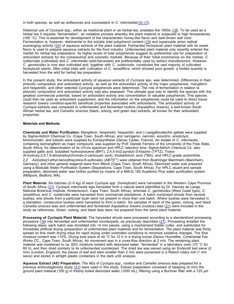
Yield of Aqueous Extract and Total Polyphenol Content. Among the unfermented Cyclopia spp., C. genistoides and C. subternata gave the highest yield of AE (P < 0.05), followed by C. sessiliflora and C. intermedia (Table 1). The total polyphenol content of the AE of unfermented C. subternata was higher than that of the other species (P < 0.05), with no significant difference between the latter. Fermentation reduced both the yield of AE and its TP content of all Cyclopia spp. significantly (P < 0.05). Cyclopia genistoides was the least affected by fermentation, with its yield of AE and total polyphenol content reduced by 8.4% and 23.4%, respectively. C. subternata had the greatest reduction in the yield of AE (41.3%) and total polyphenols (46.0%) as a result of fermentation. When compared with rooibos and Camellia sinensis teas, unfermented C. subternata and C. genistoides yielded quantities of AE similar to green tea. Only the TP content of the AE of unfermented C. subternata was similar to that of green tea, but less (P < 0.05) than that of unfermented rooibos. The TP content of the AE of unfermented Cyclopia spp. was higher than that of oolong tea. Both the AE of fermented rooibos and of black tea had higher TP contents than the AE of fermented Cyclopia spp. (P < 0.05).
Phenolic Composition of AEs. Mangiferin was the major compound present in the AE of the unfermented Cyclopia spp. (P < 0.05), with C. genistoides containing as much as 10.04% (Table 2). However, substantially lower quantities were present in the AE of unfermented C. intermedia, C. subternata, and C. sessiliflora. Isomangiferin was detected in the AE of unfermented C. genistoides, C. sessiliflora, and C. subternata. The highest concentration of hesperidin (1.12%) and eriocitrin (0.47%) occurred in the AE of unfermented C. intermedia and C. subternata, respectively. Eriocitrin and narirutin were absent in the AE of unfermented C. genistoides. Luteolin was not detected in the AE of unfermented C. intermedia but was present in very low quantities in the unfermented AE of the other species. Naringenin, eriodictyol, and formononetin were either not detected in AEs of unfermented Cyclopia spp. or were present in trace quantities. The presence of narirutin in fermented C. genistoides, as opposed to unfermented C. genistoides, was unexpected. Its quantification was problematic, due to very low quantities observed and an unsteady baseline, especially for fermented C. genistoides. LC-MS analysis confirmed its presence in fermented C. genistoides. However, overestimation on HPLC-DAD cannot be ruled out.
A number of unknown peaks of relatively substantial quantity was present in some of the unfermented extracts (Figure1). Their retention times and mass spectra did not agree with any of the authentic standards. An unidentified peak at aretention time of 4.2 min (unknown 1; λmax 289) was detected in the AE of unfermented C. subternata, C. genistoides, and C. sessiliflora. In addition, the AE of unfermented C. subternata and C. intermedia had relatively largeunidentifiable peaks at retention times of ca. 12.4 min (unknown 2; λmax 350) and 19.6 min (unknown 3; λmax 287), respectively.

Figure 1. Typical reversed-phase HPLC phenolic profile of aqueous extract of unfermented and fermented C. intermedia, C. subternata, C. genistoides, and C. sessiliflora (1 = mangiferin; 2 = isomangiferin; 3 = eriocitrin; 4 = narirutin; 5 = hesperidin; 6 = hesperetin; 7 = luteolin). The major unkown compounds are indicated.
Click to Enlarge
The relative changes in the phenolic profile that occur with fermentation are depicted in typical chromatograms of the AE of unfermented and fermented Cyclopia spp. in Figure 1. Fermentation reduced the concentrations of the known compounds in most species (Table 2). Even after fermentation, the quantity of mangiferin (4.29%) present in the AE of C. genistoides was higher than that of unfermented C. intermedia and C. subternata. In a number of cases, compounds present in the unfermented plant material were undetectable after fermentation, that is, isomangiferin in C. subternataand C. sessiliflora, eriocitrin in C. intermedia, and narirutin and luteolin in C. sessiliflora. On the other hand, narirutin and hesperetin were present in the AE of fermented C. genistoides and C. intermedia, respectively, while absent in their unfermented counterparts. The hesperidin content of C. sessiliflora extract was not significantly affected by fermentation (P > 0.05).
Antioxidant Activity of AEs. According to the ABTS•+ assay, no significant difference existed between the AEs of unfermented Cyclopia spp. (Table 3), but in the FRAP assay, unfermented C. genistoides had significantly (P < 0.05) lower activity than the other species, which exhibited similar activity. Fermentation reduced both the radical scavenging and the ferric ion reducing abilities of the AEs of the species significantly (P < 0.05) (Table 3). Fermented C. genistoides had the highest activity in both assays (P < 0.05), with no significant difference between the other species. None of the Cyclopia spp., irrespective of their fermentation state, had comparable activity to unfermented rooibos and Camellia sinensis teas in the ABTS•+ assay, but unfermented Cyclopia spp. were more active than fermented rooibos in this assay (Table 3). Green tea had the highest radical scavenging and ferric ion reducing abilities overall. Unfermented C. intermedia had a higher ferric ion reducing ability (P < 0.05) than fermented rooibos, oolong, and black teas. Unfermented C. subternata and C. sessiliflora were comparable to black tea, and unfermented C. genistoides was comparable to fermented rooibos, black, and oolong teas (Table 3).
Considering the ability of the AEs of Cyclopia spp. to inhibit lipid peroxidation, unfermented C. sessiliflora was the most effective, followed by C. subternata and C. intermedia, with equal efficacy, and C. genistoides, the least effective. However, fermentation had no effect on the ability of C. genistoides to inhibit lipid peroxidation (ca. 33%), whereas that of the other species was reduced (P < 0.05), with all having the same inhibitory effect (P > 0.05) (Table 3). None of the Cyclopia extracts was as effective as rooibos and Camellia sinensis teas, except for unfermented C. sessiliflora, with comparable inhibitory activity to fermented rooibos, which had the lowest inhibitory activity of the rooibos and Camellia sinensis extracts. Green tea gave the highest inhibition of lipid peroxidation overall, followed by unfermented rooibos, oolong, and black teas, which had similar inhibitory activity.
Correlation of TP with Antioxidant Capacity and Inhibition of Fe2+-Induced Microsomal Lipid Peroxidation. The TP content of the AEs correlated well with their antioxidant capacity measured in the ABTS•+ (r = 0.98; P < 0.0001) and FRAP (r = 0.98; P < 0.0001) assays (Figure 2). A very good correlation coefficient (r = 0.98; P < 0.0001) was also obtained for the antioxidant capacity measured as ferric ion reducing ability versus the ABTS•+ scavenging ability (data not shown). Although highly significant (P < 0.0001), a moderate correlation was obtained between inhibition of lipid peroxidation and total polyphenol content (r = 0.74) (Figure 2).
Figure 2. Correlation of (A) ABTS•+ scavenging activity, (B) ferric ion reducing ability, and (C) inhibition of Fe2+-induced lipid peroxidation of dried aqueous extract (AE) of Cyclopia spp. with theirtotal polyphenol content as determined using the Folin-Ciocalteu method.
Click to Enlarge
Antioxidant Activity of Compounds. The relative order of activity of the compounds differed in the different assays (Table 4). The most effective scavenger of ABTS•+ was mangiferin, followed by eriodictyol, eriocitrin, and luteolin,

which were of similar activity. Hesperidin, narirutin, and naringenin had very little activity in the ABTS•+ assay. In the FRAP assay, mangiferin was substantially less active than eriodictyol, eriocitrin, and luteolin. It also offered less protection against lipid peroxidation than eriodictyol, luteolin, and hesperetin (eriocitrin and narirutin were not tested) but had a higher inhibitory activity than hesperidin and naringenin.
Discussion
Antioxidant extracts are marketed on the strength of their in vitro antioxidant activity, with a high concentration of a specific compound or group of compounds supporting product differentiation in the marketplace (28). Plants with high levels of novel or less common antioxidant compounds are sought-after, especially if they have a long history of regular use that attest to their safety, such as a herbal tea. Improvement of the plant material may be achieved by plant breeding and selection whereby the levels of bioactives can be increased to consistently high levels. Presently, large variation exists in the mangiferin content of C. genistoides (14), requiring screening of available plant material for selection and propagation. However, the present study demonstrated that mangiferin content alone does not explain the observed antioxidant activity of aqueous extract from Cyclopia spp. In addition to the mangiferin content, which can be screened by NIRS (14), screening of antioxidant activity would also be required as a selection criterion. The large number of samples generated by a selection and plant improvement program would require rapid screening of antioxidant activity. This can be achieved with chemical methods suitable to high throughput using a microplate format. Both the ABTS•+ and FRAP assays have been adapted for use in microplates (29). The significant correlation between the total polyphenol content and the respective antioxidant capacities of the AEs of Cyclopia spp. as determined with the ABTS•+ and FRAP assays, as well as the significant correlation between these two assays, suggest that either of the methods could be used for rapid screening of the antioxidant activity of Cyclopia plant material.
Because of the link between antioxidants and antimutagenicity/anticancer properties of flavonoids (30–32), the present study was undertaken as a follow-up to a recent investigation of the antimutagenic properties of aqueous extracts of Cyclopia spp. (21). With the exception of unfermented C. genistoides, the extracts of all species, irrespective of processing state, demonstrated protection against both 2-acetylaminofluorene (2-AAF) and aflatoxin B1 in the Salmonella mutagenicity assay in the presence of metabolic activation. Unfermented C. genistoides, depending on the concentration, either protected against or enhanced mutagenesis caused by 2-AAF. It did not, however, exhibit any mutagenic response in the absence or presence of metabolic activation when using tester strain TA98 (21).
A consideration of the study was the evaluation of aqueous extracts in preference to an organic solvent extract, even though aqueous extracts are not as effective in recovery of flavonoids from plant material. Aqueous extracts of fermented Cyclopia spp. have the advantage of a long history of use (33), bearing in mind the regulatory environment for new botanical products (34). Unfermented Cyclopia spp. are, however, recent entries on the global market and require investigation of the extent or lack of beneficial properties, especially in relation to their fermented counterparts. Fermentation does decrease their ability to scavenge the superoxide anion radical (20).
Tea consumption has been linked to the protection against cancer development in humans, mainly due to the antioxidant properties of their flavonoid constituents (35). Protection of the integrity of biological membranes through inhibition of lipid peroxidation and oxidative damage is thus an important underlying defense against a variety of pathological conditions such as cancer and even aging (36). The rat liver microsomal preparation used in the present study represents a model membrane system with accompanying complexities, giving it relevance to an in vivo situation. The inhibition of lipid peroxidation afforded by the AE of Cyclopia spp. would not only depend on the radical scavenging ability of the polyphenols, but also on their distribution between the hydrophobic and hydrophilic phases and the interface. Polyhydroxylated compounds partition into membranes with some affinity, and the partitioning is apparently stabilized by the interfacial hydrogen bonding network present at the water-membrane interface (37). Molecular configuration is also important. The planar configuration as found in flavones as a result of the C2-C3 double bond, in contrast to the tilted configuration of flavanones, would aid intercalation into the organized structures of the phospholipid bilayer of membranes (38), affecting membrane fluidity (39). In the present study, however, eriodictyoland luteolin differed very little in inhibition of membrane lipid peroxidation. This indicates that distribution of theflavonoid at the lipid–water interface of the membranes, where it protects the bilayer against radical attack, as well asa resulting lower effect on membrane fluidity, in contrast to when the flavonoid is present in the bilayer, are also ofimportance.
Mangiferin, a glucoside, was shown to be less effective in inhibiting lipid peroxidation than the flavonoid aglycones, hesperetin, eriodictyol, and luteolin, in spite of being more effective in quenching ABTS•+. High hydrophilicity (36), due to the presence of the sugar residue, possibly resulted in the exclusion of mangiferin from the hydrophobic core of the membrane or at least had altered its distribution to be biased toward the polar headgroup of the bilayer domain (40).
The concomitant capability of polyphenols to chelate iron will also affect the extent of inhibition of peroxidation of the microsomal membrane lipids. Flavonoid compounds with (1) a catechol moiety in the B ring, (2) the 4-oxo-5-hydroxyl arrangement, and/or (3) the 3-hydroxyl-4-oxo arrangement in the heterocyclic ring will chelate trace metals 41. Luteolin, eriodictyol, and eriocitrin fulfill structural requirements one and two, and hesperidin, hesperetin, narirutin, and

naringenin fulfill requirement two. The catechol group of mangiferin and the keto group with neighboring OH groups would also be able to chelate trace metals. Pardo Andreu et al. (10) related the iron-chelating property of mangiferin, in addition to the ability to scavenge free radicals, to its in vitro protection of rat liver mitochondria against Fe2+-citrateinduced lipid peroxidation.
The absence or low concentration of the most potent compounds, the aglycones luteolin, eriodictyol, and hesperetin in the AE, suggests mangiferin to be one of the major contributors to the inhibition of lipid peroxidation by Cyclopiaextracts. With this in mind, it was thus unexpected that the AE of unfermented C. genistoides, containing the highest mangiferin content, that is, 2.5 times more than in the AE of the other species, was not the most effective inhibitor of lipid peroxidation. A possible reason could be attributed to a prooxidative effect due to the high concentration, likely counteracting the antioxidant effect to some extent. Prooxidant activity of flavonoids in the presence of a transition metal was shown to increase with flavonoid concentration (42). The fact that the protective effect was not significantly different after fermentation to that before fermentation, even though it was accompanied by a substantial decrease in mangiferin content, would support this hypothesis. Further investigation of this aspect is required. Pretreatment of human peripheral blood lymphocytes with mangiferin before exposure to H2O2 reduced lipid peroxidation in a concentration-dependent manner with maximum inhibition at 50 mg mangiferin and similar activity at 100 mg (12). Further investigation regarding the possible prooxidant properties of mangiferin is required.
When comparing AEs of similar xanthone content, that is, fermented C. genistoides and unfermented C. sessiliflora, the latter extract was more effective in protecting microsomes against lipid peroxidation. Of the known compounds quantified, eriocitrin was present in a relative substantial quantity in the AE of unfermented C. sessiliflora while absent in C. genistoides. This glycoside could be expected to be less effective than its aglycone, eriodictyol, as demonstrated in liposomes (43) but substantially more potent than hesperetin, as demonstrated in a linoleic acid autoxidation model (43). These findings and the relative order of potency of eriodictyol and hesperidin in the Fe2+-induced lipid peroxidation model used in the present study suggest eriocitrin to be more or at least as effective as mangiferin. It could therefore make a substantial contribution to the potency of the AE of unfermented C. sessiliflora. It is, however, clear that the composition of the extracts in terms of the quantified compounds alone does not explain the inhibitory effect of Cyclopia extracts. Synergism, the contribution of unknown compounds toward radical scavenging and their interference, as related to membrane affinity and distribution, can not be ruled out.
Processing of Cyclopia for manufacture of traditional fermented honeybush tea was detrimental to all parameters measured, except for C. genistoides and the effect on inhibition of lipid peroxidation. The decrease in the yield of AE with fermentation is attributed in part to a decrease in its TP content. The decrease in the levels of the monomeric compounds only partially contributes to the decrease in TP with fermentation. Degradation of unknown compounds and polymerization of compounds susceptible to oxidation, accompanied with lower solubility, are possible contributors to this decrease in TP. The unidentified peaks in C. subternata and C. intermedia, both with relatively high absorbance, suggesting them to be major compounds, decreased substantially with fermentation. A previous study on C. intermediaand C. maculata showed similar trends for the yield of aqueous soluble solids and TP content with fermentation (19). To date, no studies have been undertaken to explain these changes in the aqueous extract with fermentation in terms of the chemical reactions involved.
Based solely on the antioxidant assays used in this study, none of the AEs of Cyclopia spp. were as effective as those of unfermented rooibos and Camellia sinensis teas in vitro. A comparative in vivo study showed C. intermedia to enhance the GSH/GSSG ratio in the liver of rats to a similar extent as unfermented and fermented rooibos extracts (44). An organic solvent extract of C. intermedia was found to suppress mouse skin tumorigenesis better than rooibos (45). These positive in vivo findings support further investigation of the in vivo antioxidant properties of other Cyclopiaspp., especially C. genistoides. Fermented C. genistoides would provide both the characteristic “fermented” flavor andcolor required for a herbal tea, as well as a high mangiferin content. However, when mangiferin content is the soleconsideration, the unfermented plant material should preferentially be used for extract manufacture. Cyclopia genistoides has the further advantage that it is an easily cultivated, renewable source of mangiferin.
Acknowledgment
Analytical HPLC equipment was provided by Raps Foundation, Germany. Christie Malherbe assisted in HPLC analysis of extracts. Dr Marietjie Stander and Désirée Prevoo of the Central Analytical Facility of Stellenbosch Universityperformed LC-MS analyses.
* To whom correspondence should be addressed. Telephone: + 27 21 809 3444 ; fax: +27 21 809 3430; e-mail: [email protected].
† ARC Infruitec-Nietvoorbij.
‡ Department of Food Science, Stellenbosch University.
§ Nampak Research & Development (current address).

⊥ Department of Biochemistry, Stellenbosch University.
¶ PROMEC Unit, Medical Research Council.
1. Yoshimi, N.; Matsunaga, K.; Katayama, M.; Yamada, Y.; Kuno, T.; Qiao, Z.; Hara, A.; Yamahara, J.; Mori, H. The inhibitory effects of mangiferin, a naturally occurring glucosylxanthone, in bowel carcinogenesis of male F344 rats.
Cancer Lett. 2001, 163, 163–170. [ChemPort] [Medline] [CrossRef]
2. Pauletti, P. M.; Castro-Gamboa, I.; Silva, D. H. S.; Young, M. C. M.; Tomazela, D. M.; Eberlin, M. N.; Bolzani, V. S. New antioxidant C-glucosylxanthones from the stems of Arrabidaea samydoides. J. Nat. Prod. 2003, 66, 1384–1387.
[Full Text - ACS] [ChemPort] [Medline] [CrossRef]
3. Jung, H.-A.; Su, B.-N.; Keller, W. J.; Mehta, R. G.; Kinghorn, A. D. Antioxidant xanthones from the pericarp of Garcinia mangostana (mangosteen). J. Agric. Food Chem. 2006, 54, 2077–2082. [Full Text - ACS] [ChemPort] [Medline]
[CrossRef]
4. Núnez Sellés, A. J.; Vélez Castro, H. T.; Agüero-Agüero, J.; González-González, J.; Naddeo, F.; De Simone, F.;Rastrelli, L. Isolation and quantitative analysis of phenolic antioxidants, free sugars, and polyols from mango (Mangifera indica L.) stem bark aqueous decoction used in Cuba as a natural supplement. J. Agric. Food Chem. 2002,
50, 762–766. [Full Text - ACS] [Medline] [CrossRef]
5. Antioxidant directory. Nutraceuticals World 2005, 8(3), 58–66.
6. Berardini, N.; Knödler, M.; Schieber, A.; Carle, R. Utilization of mango peels as source of pectin and polyphenolics.
Innovat. Food Sci. Emerg. Tech. 2005, 6, 442–452. [ChemPort] [CrossRef]
7. Sato, T.; Kawamoto, A.; Tamura, A.; Tatsumi, Y.; Fuji, T. Mechanism of antioxidant action of pueria glycoside (PG)-1
(isoflavonoid) and mangiferin (a xanthonoid). Chem. Pharm. Bull. (Tokyo) 1992, 40, 721–724. [ChemPort] [Medline]
8. Leiro, J. M.; Álvarez, E.; Arranz, J. A.; Siso, I. G.; Orallo, F. In vitro effects of mangiferin on superoxideconcentrations and expression of the inducible nitric oxide synthase, tumor necrosis factor-α and transforming growth
factor-β genes. Biochem. Pharmacol. 2003, 65, 1361–1371. [ChemPort] [Medline] [CrossRef]
9. Tang, S. Y.; Whiteman, M.; Peng, Z. F.; Jenner, A.; Yong, E. L.; Halliwell, B. Characterization of antioxidant and antiglycation properties and isolation of active ingredients from traditional Chinese medicines. Free Radical Biol. Med.
2004, 36, 1575–1587. [ChemPort] [CrossRef]
10. Pardo Andreu, G.; Delgado, R.; Velho, J. A.; Curti, C.; Vercesi, A. E. Iron complexing activity of mangiferin, a naturally occurring glucosylxanthone, inhibits mitochondrial lipid perioxidation induced by Fe2+-citrate. Eur. J.
Pharmacol. 2005, 513, 47–55. [Medline] [CrossRef]
11. Rodríguez, J.; Di Pierro, D.; Cioia, M.; Monaco, S.; Delgado, R.; Coletta, M.; Marini, S. Effects of a natural extractfrom Mangifera indica L, and its active compound, mangiferin, on energy state and lipid peroxidation of red blood cells.
Biochim. Biophys. Acta 2006, 1760, 1333–1342. [Medline]
12. Jagetia, G. A.; Venkatesha, V. A. Effect of mangiferin on radiation-induced micronucleus formation in cultured
human peripheral blood lymphocytes. Environ. Mol. Mutagen. 2005, 46, 12–21. [ChemPort] [Medline] [CrossRef]
13. Joubert, E.; Otto, F.; Grüner, S.; Weinreich, B. Reversed-phase HPLC determination of mangiferin, isomangiferinand hesperidin in Cyclopia and the effect of harvesting date on the phenolic composition of C. genistoides. Eur. Food
Res. Technol. 2003, 216, 270–273. [ChemPort]
14. Joubert, E.; Manley, M.; Botha, M. The use of NIRS for quantification of mangiferin and hesperidin contents of dried, green honeybush (Cyclopia genistoides) plant material. J. Agric. Food Chem. 2006, 54, 5279–5283. [Full Text -
ACS] [ChemPort] [Medline] [CrossRef]
15. Ferreira, D.; Kamara, B. I.; Brandt, E. V.; Joubert, E. Phenolic compounds from Cyclopia intermedia (honeybush

tea). J. Agric. Food Chem. 1998, 46, 3406–3410. [Full Text - ACS] [ChemPort] [CrossRef]
16. Kamara, B. I.; Brand, D. J.; Brandt, E. V.; Joubert, E. Phenolic metabolites from honeybush tea (Cyclopia
subternata). J. Agric. Food Chem. 2004, 52, 5391–5395. [Full Text - ACS] [ChemPort] [Medline] [CrossRef]
17. Kamara, B. I.; Brandt, E. V.; Ferreira, D.; Joubert, E. Polyphenols from honeybush tea (Cyclopia intermedia). J.
Agric. Food Chem. 2003, 51, 3874–3879. [Full Text - ACS] [ChemPort] [Medline] [CrossRef]
18. Du Toit, J.; Joubert, E.; Britz, T. J. Honeybush tea - a rediscovered South African herbal tea. J. Sustainable Agric.
1998, 12, 67–84. [CrossRef]
19. Du Toit, J.; Joubert, E. Optimization of the fermentation parameters of honeybush tea (Cyclopia). J. Food Qual.
1999, 22, 241–256. [CrossRef]
20. Hubbe, M. H.; Joubert, E. In vitro superoxide anion radical scavenging ability of honeybush tea (Cyclopia) . In Dietary Anticarcinogens and Antimutagens. Chemical and Biological Aspects. Proceedings of the Food and CancerPrevention III Meeting, 5–8 September 1999; Johnson, I. T., Fenwick, G. R., Eds.; Royal Chemistry Society United Kingdom, 2000; pp 242–244.
21. Van der Merwe, J. D.; Joubert, E.; Richards, E. S.; Manley, M.; Snijman, P. W.; Marnewick, J. L.; Gelderblom, W. C. A. A comparative study on the antimutagenic properties of aqueous extracts of Aspalathus linearis (rooibos), different Cyclopia spp. (honeybush) and Camellia sinensis teas. Mutat. Res. 2006, 611, 42–53. [ChemPort] [Medline]
22. Singleton, V. L.; Rossi, J. A. Colorimetry of total phenolics with phosphotungstic acid reagents. Am. J. Enol.
Viticul. 1965, 16, 144–158. [ChemPort]
23. Re, R.; Pellegrini, N.; Proteggente, A.; Pannala, A.; Yang, M.; Rice-Evans, C. Antioxidant activity applying an improved ABTS radical cation decolorization assay. Free Radical Biol. Med. 1999, 26, 1231–1237. [ChemPort]
[CrossRef]
24. Benzie, I. F. F.; Strain, J. J. The ferric reducing ability of plasma (FRAP) as a measure of “antioxidant power”: the
FRAP assay. Anal. Biochem. 1996, 239, 70–76. [ChemPort] [Medline] [CrossRef]
25. Van Acker, S. A. B. E.; Van den Berg, D.-J.; Tromp, M. N. J. L.; Griffioen, D. H.; Van Bennekom, W. P.; Van der Vijgh, W. J. F.; Bast, A. Structure aspects of antioxidant activity of flavonoids. Free Radical Biol. Med. 1996, 20,
331–342. [ChemPort] [CrossRef]
26. Bradford, M. M. A rapid and sensitive method for the quantification of microgram quantities of protein utilizing the
principle of protein-dye binding. Anal. Biochem. 1976, 72, 248–254. [ChemPort] [Medline] [CrossRef]
27. Yen, G.-C.; Hsieh, C.-L. Antioxidant activity of extracts from du-zhong (Eucommia ulmoides) towards various lipid
peroxidation models in vitro. J. Agric. Food Chem. 1998, 46, 3952–3957. [Full Text - ACS] [ChemPort] [CrossRef]
28. Wright, R. Antioxidant avalanche. Nutraceuticals World 2005, 8 (3), 46–57.
29. Tachakittirungrod, S.; Okonogi, S.; Chowwanapoonphon, S. Study on antioxidant activity of certain plants in Thailand: mechanism of antioxidant action of guava leaf extract. Food Chem. 2007, 103, 381–388. [ChemPort]
[CrossRef]
30. Edenharder, R.; Von Petersdorff, I.; Rauscher, R. Antimutagenic effects of flavonoids, chalcones and structurally related compounds on the activity of 2-amino-3-methylimidazo[4,5-f]quinoline (IQ) and other heterocyclic amine
mutagens from cooked food. Mutat. Res. 1993, 287, 261–274. [ChemPort] [Medline]
31. Das, A.; Wang, J. H.; Lien, E. J. Carcinogenicity, mutagenicity and cancer preventing activities of flavonoids: a
structure-system-activity relationship (SSAR) analysis. Prog. Drug. Res. 1994, 42, 133–166. [ChemPort] [Medline]

32. Duarte Silva, I.; Gaspar, J.; Gomes da Costa, G.; Rodrigues, A. S.; Laires, A.; Rueff, J. Chemical features of flavonols affecting their genotoxicity. Potential complications in their use as therapeutic agents. Chem. Biol. Interact.
2000, 124, 29–51. [ChemPort] [Medline] [CrossRef]
33. Watt, J. M.; Breyer-Brandwijk. M. G. The Medicinal and Poisonous Plants of Southern Africa, E & S Livingstone, Edinburgh, 1932, p 70.
34. Bast, A.; Chandler, R. F.; Choy, P. C.; Delmulle, L. M.; Gruenwald, J.; Halkes, S. B. A.; Keller, K.; Koeman, J. H.; Peters, P.; Przyrembel, H.; De Ree, E. M.; Renwick, A. G.; Vermeer, I. T. M. Botanical health products, positioning and requirements for effective and safe use. Environ. Toxicol. Pharmacol. 2002, 12, 195–211. [ChemPort] [CrossRef]
35. Weisburger, J. H.; Hara, Y.; Dolan, L.; Luo, F.; Pittman, B.; Zanh, E. Tea polyphenols as inhibitors of mutagenicity
of major classes of carcinogens. Mutat. Res. 1996, 371, 57–63. [Medline] [CrossRef]
36. Saija, A.; Scalese, M.; Lanza, M.; Marzullo, D.; Bonina, F.; Castrelli, F. Flavonoids as antioxidant agents: importance of their interaction with biomembranes. Free Radical Biol. Med. 1995, 19, 481–486. [ChemPort] [CrossRef]
37. Tammela, P.; Laitinen, L.; Galkin, A.; Wennberg, T.; Heczko, R.; Vuorela, H.; Slotte, J. P.; Vuorela, P. Permeability characteristics and membrane affinity of flavonoids and alkyl gallates in Caco-2 cells and in phospholipid vesicles.
Arch. Biochem. Biophys. 2004, 425, 193–199. [ChemPort] [Medline] [CrossRef]
38. Van Dijk, C.; Driessen, A. J. M.; Recourt, K. The uncoupling efficiency and affinity of flavonoids for vesicles.
Biochem. Pharmacol. 2000, 60, 1593–1600. [ChemPort] [Medline] [CrossRef]
39. Oteiza, P. I.; Erlejman, A. G.; Verstraeten, S. V.; Keen, K. L.; Fraga, C. G. Flavonoid-membrane interactions: a protective role of flavonoids at the membrane surface. Clin. Dev. Immunol. 2005, 12, 19–25. [ChemPort] [Medline]
[CrossRef]
40. Scheidt, H. A.; Pampel, A.; Nissler, L.; Gebhardt, R.; Huster, D. Investigation of the membrane localization and distribution of flavonoids by high-resolution magic angle spinning NMR spectroscopy. Biochim. Biophys. Acta 2004,
1663, 97–107. [ChemPort] [Medline] [CrossRef]
41. Pietta, P. Flavonoids as antioxidants. J. Nat. Prod. 2000, 63, 1035–1042. [Full Text - ACS] [ChemPort] [Medline]
[CrossRef]
42. Cao, G.; Sofic, E.; Prior, R. L. Antioxidant and prooxidant behavior of flavonoids: structure-activity relationships.
Free Radical Biol. Med. 1997, 22, 749–760. [ChemPort] [CrossRef]
43. Miyake, Y.; Yamamoto, K.; Morimitsu, Y.; Osawa, T. Isolation of C-glucosylflavone from lemon peel and antioxidative activity of flavonoid compounds in lemon fruit. J. Agric. Food Chem. 1997, 45, 4619–4623. [Full Text -
ACS] [ChemPort] [CrossRef]
44. Marnewick, J. L.; Joubert, E.; Swart, P.; Van der Westhuizen, F.; Gelderblom, W. C. A. Modulation of hepatic drug metabolizing enzymes and oxidative status by rooibos (Aspalathus linearis) and honeybush (Cyclopia intermedia), green and black (Camellia sinensis) teas in rats. J. Agric. Food Chem. 2003, 51, 8113–8119. [Full Text - ACS]
[ChemPort] [Medline] [CrossRef]
45. Marnewick, J. L.; Joubert, E.; Joseph, S.; Swanevelder, S.; Swart, P.; Gelderblom, W. C. A. Inhibition of tumour promotion in mouse skin by extracts of rooibos (Aspalathus linearis) and honeybush (Cyclopia intermedia), unique
South African herbal teas. Cancer Lett. 2005, 224, 193–202. [ChemPort] [Medline] [CrossRef]
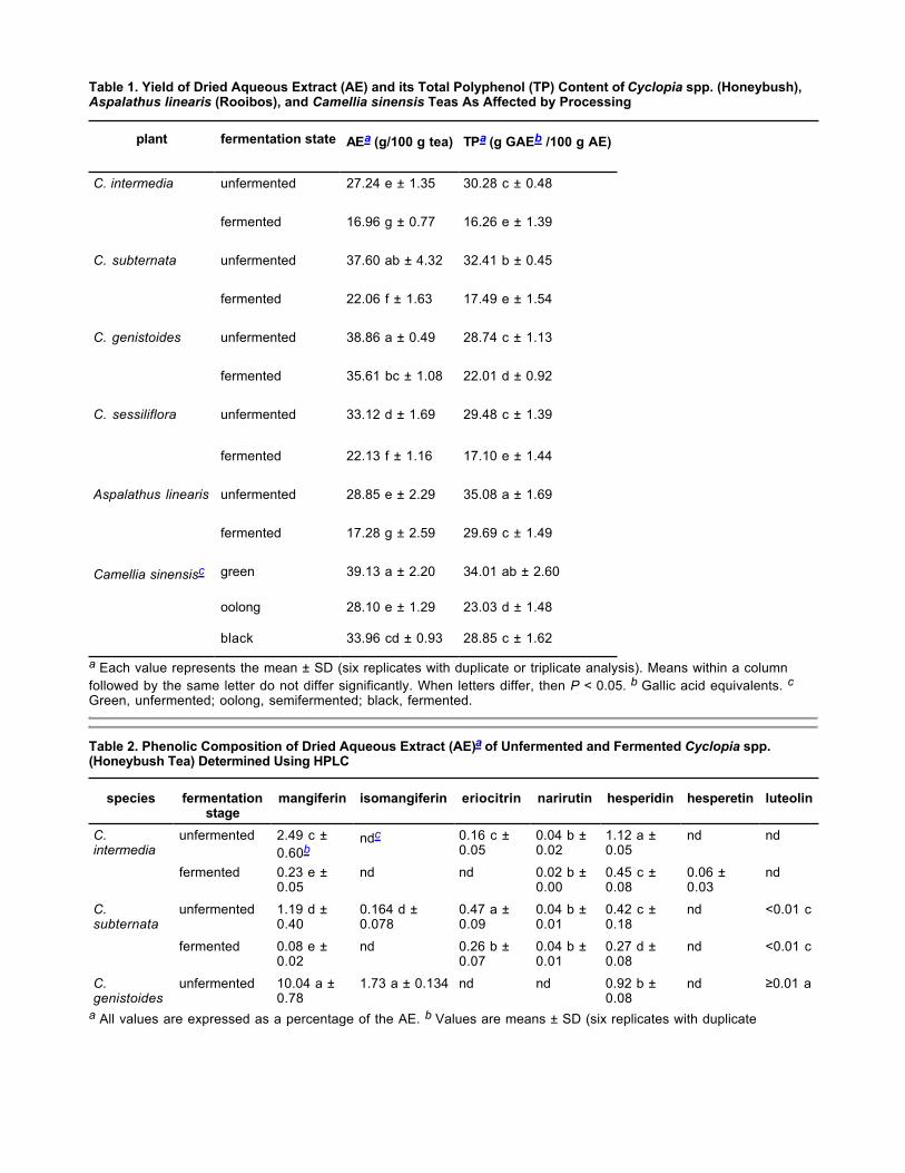
Table 1. Yield of Dried Aqueous Extract (AE) and its Total Polyphenol (TP) Content of Cyclopia spp. (Honeybush), Aspalathus linearis (Rooibos), and Camellia sinensis Teas As Affected by Processing
a Each value represents the mean ± SD (six replicates with duplicate or triplicate analysis). Means within a columnfollowed by the same letter do not differ significantly. When letters differ, then P < 0.05. b Gallic acid equivalents. cGreen, unfermented; oolong, semifermented; black, fermented.
Table 2. Phenolic Composition of Dried Aqueous Extract (AE)a of Unfermented and Fermented Cyclopia spp. (Honeybush Tea) Determined Using HPLC
a All values are expressed as a percentage of the AE. b Values are means ± SD (six replicates with duplicate
plant fermentation state AEa (g/100 g tea) TPa (g GAEb /100 g AE)
C. intermedia unfermented 27.24 e ± 1.35 30.28 c ± 0.48
fermented 16.96 g ± 0.77 16.26 e ± 1.39
C. subternata unfermented 37.60 ab ± 4.32 32.41 b ± 0.45
fermented 22.06 f ± 1.63 17.49 e ± 1.54
C. genistoides unfermented 38.86 a ± 0.49 28.74 c ± 1.13
fermented 35.61 bc ± 1.08 22.01 d ± 0.92
C. sessiliflora unfermented 33.12 d ± 1.69 29.48 c ± 1.39
fermented 22.13 f ± 1.16 17.10 e ± 1.44
Aspalathus linearis unfermented 28.85 e ± 2.29 35.08 a ± 1.69
fermented 17.28 g ± 2.59 29.69 c ± 1.49
Camellia sinensisc green 39.13 a ± 2.20 34.01 ab ± 2.60
oolong 28.10 e ± 1.29 23.03 d ± 1.48
black 33.96 cd ± 0.93 28.85 c ± 1.62
species fermentation stage
mangiferin isomangiferin eriocitrin narirutin hesperidin hesperetin luteolin
C. intermedia
unfermented 2.49 c ±0.60b
ndc 0.16 c ±0.05
0.04 b ±0.02
1.12 a ±0.05
nd nd
fermented 0.23 e ±0.05
nd nd 0.02 b ±0.00
0.45 c ±0.08
0.06 ±0.03
nd
C. subternata
unfermented 1.19 d ±0.40
0.164 d ±0.078
0.47 a ±0.09
0.04 b ±0.01
0.42 c ±0.18
nd <0.01 c
fermented 0.08 e ±0.02
nd 0.26 b ±0.07
0.04 b ±0.01
0.27 d ±0.08
nd <0.01 c
C. genistoides
unfermented 10.04 a ±0.78
1.73 a ± 0.134 nd nd 0.92 b ±0.08
nd ≥0.01 a

Table 2. Phenolic Composition of Dried Aqueous Extract (AE)a of Unfermented and Fermented Cyclopia spp. (Honeybush Tea) Determined Using HPLC
a All values are expressed as a percentage of the AE. b Values are means ± SD (six replicates with duplicateanalysis). Means within a column followed by the same letter do not differ significantly. When letters differ then P < 0.05. c Not detected.
Table 3. Relative Activity of Dried Aqueous Extract (AE) of Cyclopia spp. (Honeybush), Aspalathus linearis(Rooibos) and Camellia Teas in the ABTS Radical Cation Scavenging, Ferric Reducing Antioxidant Power, and Rat Liver Microsomal Lipid Peroxidation Assays
a Total antioxidant capacity using the ABTS radical scavenging assay (µmol of Trolox equivalents/mg of AE). b Total antioxidant capacity using the FRAP assay (µmol of Trolox equivalents/mg of AE). c Percentage of inhibition achieved at 0.02 mg/mL AE in the microsomal lipid peroxidation assay. d Values are mean ± SD (six replicates with triplicateanalysis). Means within a column followed by the same letter do not differ significantly. When letters differ, then P < 0.05. e Green, unfermented; oolong, semifermented; black, fermented.
species fermentation stage
mangiferin isomangiferin eriocitrin narirutin hesperidin hesperetin luteolin
fermented 4.29 b ±1.20
0.94 b ± 0.149 nd 0.20 a ±0.06
0.47 c ±0.01
nd 0.01 b
C. sessiliflora
unfermented 4.39 b ±0.35
0.90 c ± 0.063 0.33 b ±0.06
0.03 b ±0.00
0.50 c ±0.05
nd <0.01 c
fermented 0.19 e ±0.04
nd 0.13 c ±0.04
nd 0.45 c ±0.03
nd nd
plant fermentation stage ABTSa FRAPb lipid peroxidationc
C. intermedia unfermented 2.11 d ± 0.07d 1.70 c ± 0.06 36.37 d ± 1.81
fermented 0.96 g ± 0.08 0.75 h ± 0.06 23.63 f ± 3.30
C. subternata unfermented 2.03 d ± 0.06 1.66 cd ± 0.04 36.21 d ± 2.40
fermented 0.94 g ± 0.07 0.78 h ± 0.08 24.73 f ± 5.05
C. genistoides unfermented 1.99 d ± 0.03 1.47 ef ± 0.04 31.16 e ± 4.15
fermented 1.44 f ± 0.07 0.97 g ± 0.08 33.11 de ± 2.60
C. sessiliflora unfermented 1.97 d ± 0.10 1.63 cd ± 0.09 41.97 c ± 4.12
fermented 0.95 g ± 0.08 0.78 h ± 0.08 24.20 f ± 2.27
Aspalathus linearis unfermented 2.37 c ± 0.11 1.98 b ± 0.10 51.91 b ± 1.57
fermented 1.72 e ± 0.12 1.45 ef ± 0.08 41.07 c ± 3.23
Camellia sinensise green 4.34 a ± 0.40 2.50 a ± 0.31 59.35 a ± 4.01
oolong 2.34 c ± 0.13 1.35 f ± 0.07 51.43 b ± 2.17
black 2.96 b ± 0.17 1.55 de ± 0.07 51.74 b ± 3.30

Table 4. Relative Activitya of Pure Phenolic Compounds in the ABTS Radical Cation Scavenging, Ferric Reducing Antioxidant Power and Rat Liver Microsomal Lipid Peroxidation Assays
a Values are means of triplicate analysis. b The m/z of molecular ion obtained with LC-MS with negative ionization. cTrolox equivalent antioxidant capacity (TEAC) determined using the ABTS radical cation scavenging assay (µM ofTrolox required to give the same absorbance as 1µM of compound). d TEAC determined using the FRAP assay (µM ofTrolox required to give the same absorbance as 1 µM of compound). e Antioxidant activity of compounds in the rat liver microsomal lipid peroxidation assay (% inhibition achieved by 300 µM solution). f Not available for testing.