Effect of physical activity and sun exposure on vitamin D status of Saudi children and adolescents
6
RESEARCH ARTICLE Open Access Effect of physical activity and sun exposure on vitamin D status of Saudi children and adolescents Abdulaziz Al-Othman 1,2 , Sara Al-Musharaf 3 , Nasser M Al-Daghri 1,4,5,10* , Soundararajan Krishnaswamy 4 , Deqa S Yusuf 1 , Khalid M Alkharfy 1,6 , Yousef Al-Saleh 1,7 , Omar S Al-Attas 1,4,5 , Majed S Alokail 1,4,5 , Osama Moharram 8 , Shaun Sabico 4 and George P Chrousos 4,9 Abstract Background: Accumulating evidence suggests an increased prevalence of vitamin D deficiency in the Middle East. In this context, we aimed to determine whether the prevalence of vitamin D deficiency is related to degree of physical activity and sun exposure among apparently healthy Saudi children and adolescents, a little studied population. Methods: A total of 331 Saudi children aged 6–17 years (153 boys and 178 girls) were included in this cross sectional study. Levels of physical activity and sun exposure were determined using a standard questionnaire. Anthropometry, serum calcium and 25-(OH) vitamin D were analyzed. Results: All subjects were vitamin D deficient, the majority being moderately deficient (71.6%). Age was the single most significant predictor affecting 25 (OH) Vitamin D levels, explaining 21% of the variance perceived (p = 1.68 x 10 -14 ). Age-matched comparisons revealed that for groups having the same amount of sun exposure, those with moderate or are physically active will have higher levels of vitamin D status, though levels in across groups remained deficient. Conclusion: Vitamin D deficiency is common among Saudi children and adolescents, and is influenced by both sun exposure and physical activity. Promotion of an active outdoor lifestyle among Saudi children in both homes and schools may counteract the vitamin D deficiency epidemic in this vulnerable population. Vitamin D supplementation is suggested in all groups, including those with the highest sun exposure and physical activity. Keywords: Vitamin D, Saudi children Background The prevalence of vitamin D deficiency has increased rapidly worldwide in both children and adults during the past decade [1-3]. Around 30 to 50% of children and adults in the United Arab Emirates, Australia, Turkey, India, and Lebanon have 25(OH)D levels below 20 ng/ ml [4-7]. The high prevalence of vitamin D deficiency may be due to low exposure of the skin to ultra violet radiation (UVB) [8], higher skin pigmentation and low intake of vitamin D [9]. Other factors such as mutations in vitamin D receptor (VDR) polymorphism [10], low daily calcium intake [9], obesity and low social status have all been associated with low circulating vitamin D levels [11,12]. The kingdom of Saudi Arabia (KSA) has unique condi- tions that influence vitamin D status even though Saudi Arabia is sun-drenched throughout the year and tem- peratures often rise above 50 °C (122 °F) during summer. A recent study revealed counterintuitive effects of a sea- son to vitamin D status amongst Saudi locals and expatriates, revealing higher vitamin D status levels dur- ing winter months as compared with the summer [13]. * Correspondence: [email protected] 1 Prince Mutaib Chair for Biomarkers of Osteoporosis, King Saud University, Riyadh, Kingdom of Saudi Arabia 4 Biomarkers Research Program, Biochemistry Department, College of Science, King Saud University, Riyadh, Kingdom of Saudi Arabia Full list of author information is available at the end of the article © 2012 Al-Othman et al.; licensee BioMed Central Ltd. This is an Open Access article distributed under the terms of the Creative Commons Attribution License (http://creativecommons.org/licenses/by/2.0), which permits unrestricted use, distribution, and reproduction in any medium, provided the original work is properly cited. Al-Othman et al. BMC Pediatrics 2012, 12:92 http://www.biomedcentral.com/1471-2431/12/92
Transcript of Effect of physical activity and sun exposure on vitamin D status of Saudi children and adolescents
Al-Othman et al. BMC Pediatrics 2012, 12:92http://www.biomedcentral.com/1471-2431/12/92
RESEARCH ARTICLE Open Access
Effect of physical activity and sun exposure onvitamin D status of Saudi childrenand adolescentsAbdulaziz Al-Othman1,2, Sara Al-Musharaf3, Nasser M Al-Daghri1,4,5,10*, Soundararajan Krishnaswamy4,Deqa S Yusuf1, Khalid M Alkharfy1,6, Yousef Al-Saleh1,7, Omar S Al-Attas1,4,5, Majed S Alokail1,4,5, Osama Moharram8,Shaun Sabico4 and George P Chrousos4,9
Abstract
Background: Accumulating evidence suggests an increased prevalence of vitamin D deficiency in the Middle East.In this context, we aimed to determine whether the prevalence of vitamin D deficiency is related to degree ofphysical activity and sun exposure among apparently healthy Saudi children and adolescents, a little studiedpopulation.
Methods: A total of 331 Saudi children aged 6–17 years (153 boys and 178 girls) were included in this crosssectional study. Levels of physical activity and sun exposure were determined using a standard questionnaire.Anthropometry, serum calcium and 25-(OH) vitamin D were analyzed.
Results: All subjects were vitamin D deficient, the majority being moderately deficient (71.6%). Age was the singlemost significant predictor affecting 25 (OH) Vitamin D levels, explaining 21% of the variance perceived (p= 1.68 x10⁻¹⁴). Age-matched comparisons revealed that for groups having the same amount of sun exposure, those withmoderate or are physically active will have higher levels of vitamin D status, though levels in across groupsremained deficient.
Conclusion: Vitamin D deficiency is common among Saudi children and adolescents, and is influenced by bothsun exposure and physical activity. Promotion of an active outdoor lifestyle among Saudi children in both homesand schools may counteract the vitamin D deficiency epidemic in this vulnerable population. Vitamin Dsupplementation is suggested in all groups, including those with the highest sun exposure and physical activity.
Keywords: Vitamin D, Saudi children
BackgroundThe prevalence of vitamin D deficiency has increasedrapidly worldwide in both children and adults during thepast decade [1-3]. Around 30 to 50% of children andadults in the United Arab Emirates, Australia, Turkey,India, and Lebanon have 25(OH)D levels below 20 ng/ml [4-7]. The high prevalence of vitamin D deficiencymay be due to low exposure of the skin to ultra violet
* Correspondence: [email protected] Mutaib Chair for Biomarkers of Osteoporosis, King Saud University,Riyadh, Kingdom of Saudi Arabia4Biomarkers Research Program, Biochemistry Department, College of Science,King Saud University, Riyadh, Kingdom of Saudi ArabiaFull list of author information is available at the end of the article
© 2012 Al-Othman et al.; licensee BioMed CenCreative Commons Attribution License (http:/distribution, and reproduction in any medium
radiation (UVB) [8], higher skin pigmentation and lowintake of vitamin D [9]. Other factors such as mutationsin vitamin D receptor (VDR) polymorphism [10], lowdaily calcium intake [9], obesity and low social statushave all been associated with low circulating vitamin Dlevels [11,12].The kingdom of Saudi Arabia (KSA) has unique condi-
tions that influence vitamin D status even though SaudiArabia is sun-drenched throughout the year and tem-peratures often rise above 50 °C (122 °F) during summer.A recent study revealed counterintuitive effects of a sea-son to vitamin D status amongst Saudi locals andexpatriates, revealing higher vitamin D status levels dur-ing winter months as compared with the summer [13].
tral Ltd. This is an Open Access article distributed under the terms of the/creativecommons.org/licenses/by/2.0), which permits unrestricted use,, provided the original work is properly cited.
Al-Othman et al. BMC Pediatrics 2012, 12:92 Page 2 of 6http://www.biomedcentral.com/1471-2431/12/92
Saudis usually limit the time they spend outdoors duringthe day time. In summer months, for example, parentsdo not allow their children to engage in outdoor activ-ities during daytime. Women in particular receive littleor no sun-light since they cover their bodies with darkveils completely, for cultural and religious reasons.The aim of the present study was to determine the
prevalence of vitamin D deficiency and its associationwith behavioral factors like physical activity and expos-ure to sun light in an otherwise healthy group of Saudichildren, an understudied population in terms of vitaminD deficiency. The results should help in raising aware-ness as well as suggesting strategies to combat vitaminD deficiency in this part of the world and thus in redu-cing the incidence of various chronic diseases associatedwith vitamin D deficiency (e.g., osteomalacia, metabolicsyndrome, heart diseases. . .).
MethodsSubjectsIn this cross-sectional study, a total of 331 apparentlyhealthy Saudi boys and girls aged 6–17 years were en-rolled from 4 different Primary Health Care Centers indifferent areas within Riyadh city during the months ofMarch-December 2010, majority of whom wererecruited during the summer months of April to No-vember. Written informed consents for parents as wellas assent for children and/or adolescents were obtainedprior to inclusion. Subjects with chronic conditions suchas asthma, type 1 diabetes mellitus, hypertension, historyof cardiac, kidney or liver disease, use of medicationsknown to affect body weight (such as steroids), psychi-atric conditions, and those taking calcium, vitamin D ormultivitamin supplements were excluded from thestudy.Ethical approval was obtained from the Ethics Com-
mittee of the College of Science Research Center, KingSaud University, Riyadh, Saudi Arabia. A pre-designedand approved questionnaire used in a previous study[14] which includes medical history was answered for allparticipants with the help of their parents. The question-naire also sought information about sun exposure (fre-quency of exposure, duration of exposure) and physicalactivity, which is self reported (frequency and type of ac-tivities performed along with duration – number of min-utes per week).
Anthropometric measurementsSubjects were assigned to visit the primary care mostconvenient for them for the collection of anthropometricdata and blood extraction. Physical examination was car-ried out by the attending physician who ensured that theparticipants met the inclusion and exclusion criteria.Weight and height were recorded to the nearest 0.2 kg
and 0.5 cm, respectively, using an appropriate inter-national standard scale (Digital Pearson Scale, ADAMEquipment Inc., USA). Blood pressure was measuredusing an appropriate mercurial sphygmomanometer.Blood pressure was measured twice with 15-minuteintervaland the mean of the two readings was recorded.
Serum biochemical analysisBlood (� 10 cc) was withdrawn by a nurse after an over-night fast (> 10 hours) on the same day anthropometricinformation was gathered. Serum calcium was measuredusing standard analytical techniques (Konelab, Finland).Fasting blood glucose and lipids which included trigly-cerides, total, HDL- and LDL-cholesterol were measuredroutinely using the a chemical analyzer (Konelab, Fin-land). 25 (OH) Vitamin D was measured using enzyme-linked immunosorbent assay (ELISA) (I.D.S., Tyne &Wear, UK). The inter- and intra-assay variability of thisassay was 5.3 and 4.6%, respectively.
Statistical analysisStatistical analysis was carried out using the StatisticalPackage for the Social Science (SPSS 11.5, Chicago IL,USA). Frequencies were expressed in percentage (%),continuous variables were presented as mean ± standarddeviation. Analysis of variance (ANOVA) was done tocompare age-matched groups. Variables such as trigly-cerides and LDL-cholesterol were log transformed priorto parametric comparisons. Stepwise linear regressionanalysis was done to determine which among the para-meters measured are significant predictors for 25(OH)D(dependent variable). P-value significant at p< 0.05.
ResultsPrevalence of vitamin D deficiencyBased on their vitamin D deficiency status using differ-ent cut-off values: severe (<12.5 nmol/l), moderate(12.5-24.9 nmol/l), and mild (25.0-29.9 nmol/l) [15],11.4% of subjects had severe vitamin D deficiency while71.6% and 16.8% of subjects showed moderate and milddeficiencies, respectively (not shown in table). Age wasthe single most significant predictor affecting 25 (OH)Vitamin D levels, explaining 21% of the variance per-ceived (p= 1.68 x 10⁻¹⁴) Figure 1.
Physical activity and vitamin DThe subjects were divided into three groups based ontheir level physical activity, subjects were divided intothree groups: inactive, moderately active and active. Thesubjects who were physically inactive were approxi-mately 15% in both males and females, while the physic-ally active subjects were 27% and 17% in males andfemales, respectively. The lowest mean level of 25(OH)vitamin D in serum was found in the physically inactive

Figure 1 Effect of physical activity on 25(OH) vitamin D levels according to level of sun exposure; * denotes p-value< 0.05.
Al-Othman et al. BMC Pediatrics 2012, 12:92 Page 3 of 6http://www.biomedcentral.com/1471-2431/12/92
group [17.7 ± 1.6 nmol/l vs 22.7 ± 1.5 nmol/l (p< 0.05)].The BMI was highest in the physically inactive groupand this was borderline significant [23.1 ±7.9 vs20.1 ± 5.8 (p = 0.05)] (Table 1). Serum fasting glucose wasalso highest in the physically inactive group (p= 0.03). Inall groups, levels of 25(OH)D were less in the group withlesser physical activity, but significance was only notedin the no exposure group (p= 0.04) as well as the moder-ate physically active group versus physically inactivegroup under the weekly exposed group (p= 0.03)(Figure 2).
Table 1 General Characteristics of Subjects Based on Level of
Parameters Physically Inactive
N 52
Boys/Girls 25/27
Age (years) 13.4 ± 3.2
BMI (kg/m2) 23.1 ± 7.9
Waist (cm) 67.8 ± 26.2
Hips (cm) 82.8 ± 29.1
WHR 0.82 ± 0.10
Systolic BP (mmHg) 110.9 ± 10.7
Diastolic BP (mmHg) 70.9 ± 12.8
Glucose (mmol/L) 5.3 ± 0.91
Triglycerides (mmol/l)# 0.97 ± 0.53
Total Cholesterol(mmol/l) 4.2 ± 0.58
HDL Cholesterol (mmol/l) 1.0 ± 0.38
LDL Cholesterol (mmol/l)# 2.8 ± 0.71
25(OH) Vitamin D (nmol/l) 17.7 ± 1.6
Data presented as mean ± standard deviation; * denotes significance as compared tp< 0.05.
Sun exposure and vitamin DAssociations of 25 (OH) vitamin D were determined insubjects according to 3 groups based on level of expos-ure to sunlight. These were, no exposure (0 minute),daily exposure (10–30 minutes) and weekly exposure(40–160 minutes). Around 24% of the subjects had noexposure to sun light, 39.5% had once in a week expos-ure and 36.5% had daily exposure. Serum vitamin Dlevels increased with increasing sun exposure (notshown in table). Age was negatively and significantlyassociated with 25 (OH) vitamin D levels in all groups as
Physical Activity
Moderate PA Physically Active P value
205 74
87/118 41/33
12.6 ± 3.3 12.1 ± 3.6 0.11
21.6 ± 6.6 19.9 ± 5.8* 0.05
66.6 ± 19.4 63.8 ± 17.8 0.51
82.2 ± 21.3 78.4 ± 20.6 0.43
0.81 ± 0.11 0.81 ± 0.08 0.85
108.2 ± 11.6 109.2 ± 11.8 0.36
70.9 ± 10.4 71.2 ± 9.7 0.97
5.2 ± 0.64 4.9 ± 0.81* 0.03
0.91 ± 0.47 1.0 ± 0.69 0.42
4.0 ± 0.67 4.2 ± 0.90 0.31
1.0 ± 0.27 1.0 ± 0.29 0.73
2.7 ± 0.65 2.7 ± 0.53 0.71
21.2 ± 1.6* 22.7 ± 1.5* 0.01
o physically inactive; # denotes non-Gaussian variables. Significance at
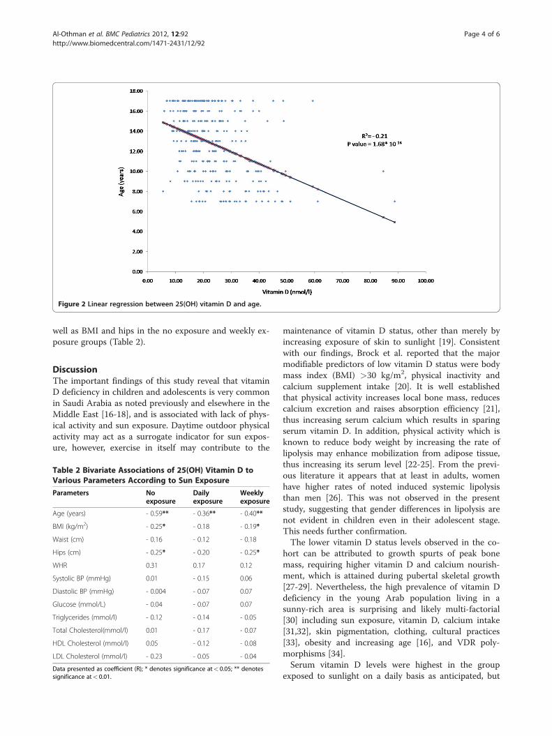
Figure 2 Linear regression between 25(OH) vitamin D and age.
Al-Othman et al. BMC Pediatrics 2012, 12:92 Page 4 of 6http://www.biomedcentral.com/1471-2431/12/92
well as BMI and hips in the no exposure and weekly ex-posure groups (Table 2).
DiscussionThe important findings of this study reveal that vitaminD deficiency in children and adolescents is very commonin Saudi Arabia as noted previously and elsewhere in theMiddle East [16-18], and is associated with lack of phys-ical activity and sun exposure. Daytime outdoor physicalactivity may act as a surrogate indicator for sun expos-ure, however, exercise in itself may contribute to the
Table 2 Bivariate Associations of 25(OH) Vitamin D toVarious Parameters According to Sun Exposure
Parameters Noexposure
Dailyexposure
Weeklyexposure
Age (years) - 0.59** - 0.36** - 0.40**
BMI (kg/m2) - 0.25* - 0.18 - 0.19*
Waist (cm) - 0.16 - 0.12 - 0.18
Hips (cm) - 0.25* - 0.20 - 0.25*
WHR 0.31 0.17 0.12
Systolic BP (mmHg) 0.01 - 0.15 0.06
Diastolic BP (mmHg) - 0.004 - 0.07 0.07
Glucose (mmol/L) - 0.04 - 0.07 0.07
Triglycerides (mmol/l) - 0.12 - 0.14 - 0.05
Total Cholesterol(mmol/l) 0.01 - 0.17 - 0.07
HDL Cholesterol (mmol/l) 0.05 - 0.12 - 0.08
LDL Cholesterol (mmol/l) - 0.23 - 0.05 - 0.04
Data presented as coefficient (R); * denotes significance at< 0.05; ** denotessignificance at< 0.01.
maintenance of vitamin D status, other than merely byincreasing exposure of skin to sunlight [19]. Consistentwith our findings, Brock et al. reported that the majormodifiable predictors of low vitamin D status were bodymass index (BMI) >30 kg/m2, physical inactivity andcalcium supplement intake [20]. It is well establishedthat physical activity increases local bone mass, reducescalcium excretion and raises absorption efficiency [21],thus increasing serum calcium which results in sparingserum vitamin D. In addition, physical activity which isknown to reduce body weight by increasing the rate oflipolysis may enhance mobilization from adipose tissue,thus increasing its serum level [22-25]. From the previ-ous literature it appears that at least in adults, womenhave higher rates of noted induced systemic lipolysisthan men [26]. This was not observed in the presentstudy, suggesting that gender differences in lipolysis arenot evident in children even in their adolescent stage.This needs further confirmation.The lower vitamin D status levels observed in the co-
hort can be attributed to growth spurts of peak bonemass, requiring higher vitamin D and calcium nourish-ment, which is attained during pubertal skeletal growth[27-29]. Nevertheless, the high prevalence of vitamin Ddeficiency in the young Arab population living in asunny-rich area is surprising and likely multi-factorial[30] including sun exposure, vitamin D, calcium intake[31,32], skin pigmentation, clothing, cultural practices[33], obesity and increasing age [16], and VDR poly-morphisms [34].Serum vitamin D levels were highest in the group
exposed to sunlight on a daily basis as anticipated, but
Al-Othman et al. BMC Pediatrics 2012, 12:92 Page 5 of 6http://www.biomedcentral.com/1471-2431/12/92
were nevertheless vitamin D deficient. Aside from theconventional factors previously mentioned contributingto vitamin D deficiency, the atmosphere in urban Riyadhis known to be saturated with dust particles and vehiclepollution that may affect the availability of UV radiation;since the dust is composed of mineral particles that bothabsorb and scatter sunlight and this might affect vitaminD synthesis [35].Our study acknowledges a few limitations. Data on
sun exposure and physical activity were based on admi-nistered questionnaires which are subject to recall bias.Data on which body parts were exposed were also notprovided. A prospective approach using a more con-trolled environment (experimental setting) rather thaninterview questionnaire might provide more definitiveanswers to the differences observed in the present study.Lastly, serum parathormone was not assessed in thestudy and it is still debatable whether vitamin D supple-mentation should be given to those who are deficient inthe absence of information on biological consequencesand non-availability of serum parathormone level.
ConclusionIn conclusion, vitamin D deficiency is very commonamong apparently healthy Saudi children and adoles-cents, as found in the present study. It is influenced bytheir level of physical activity and frequency of sun ex-posure. Increased outdoor physical activity among Saudichildren and adolescents should be encouraged not onlyat home but also in schools to promote a more activelife style that will counteract not only vitamin D defi-ciency but also conditions such as childhood obesity.Vitamin D supplementation in this vulnerable group isalso suggested but needs further studies.
Competing interestsThe authors declare no conflict of interest, both financial and non-financialfor this study.
AcknowledgementsThe authors are grateful to Mr. Benjamin Vinodson for the statistical analysesof the data. Many thanks to Prince Mutaib Chair for Osteoporosis for thefunding of this study.
Author details1Prince Mutaib Chair for Biomarkers of Osteoporosis, King Saud University,Riyadh, Kingdom of Saudi Arabia. 2College of Applied Medical Sciences, KingSaud University, Riyadh, Kingdom of Saudi Arabia. 3College of Science, KingSaud University Women's Section, Riyadh, Kingdom of Saudi Arabia.4Biomarkers Research Program, Biochemistry Department, College of Science,King Saud University, Riyadh, Kingdom of Saudi Arabia. 5Center of Excellencein Biotechnology Research, King Saud University, Riyadh, Kingdom of SaudiArabia. 6Clinical Pharmacy Department, College of Pharmacy, King SaudUniversity, Riyadh, Kingdom of Saudi Arabia. 7College of Medicine, King SaudUniversity of Health Sciences, Riyadh, Kingdom of Saudi Arabia. 8KingAbdulaziz University Hospital, King Saud University, Riyadh, Kingdom of SaudiArabia. 9First Department of Pediatrics, Athens University Medical School,Athens 11527, Greece. 10Biochemistry Department, College of Science, KingSaud University, PO Box, 2455, Riyadh 11451, Kingdom of Saudi Arabia.
Authors’ contributionsAA, SA and NMA for conception and design; KMA, YA, OSA and MSA for theacquisition of subjects and interpretation of data; DSY and OM for thesubject recruitment and sample collection; SK for the manuscriptdevelopment; SS and GC for the intellectual input and the final version ofthe manuscript. All authors have seen and approved the final version of thepaper.
Received: 21 February 2012 Accepted: 3 July 2012Published: 3 July 2012
References1. Bischoff-Ferrari HA, Giovannucci E, Willett WC, Dietrich T, Dawson-Hughes B:
Estimation of optimal serum concentrations of 25-hydroxyvitamin D formultiple health outcomes. Am J Clin Nutr 2006, 84:18–28.
2. Kumar J, Muntner P, Kaskel FJ, Hailpern SM, Melamed ML: Prevalence andAssociations of 25-Hydroxyvitamin D Deficiency in US Children: NHANES2001–2004. Pediatrics 2009, 124:e362–e370.
3. Rajpathak SN, Rimm EB, Rosner B, Willett WC, Hu FB: Calcium and dairyintakes in relation to long-term weight gain in US men. Am J Clin Nutr2006, 83:559–566.
4. El-Hajj Fuleihan G, Nabulsi M, Choucair M, Salamoun M, Hajj Shahine C,Kizirian A, Tannous R: Hypovitaminosis D in healthy school children.Pediatrics 2001, 107:E53.
5. Marwaha RK, Tandon N, Reddy DR, Aggarwal R, Singh R, Sawhney RC, SalujaB, Ganie MA, Singh S: Vitamin D and bone mineral density status ofhealthy schoolchildren in northern India. Am J Clin Nutr 2005, 82:477–482.
6. McGrath JJ, Kimlin MG, Saha S, Eyles DW, Parisi AV: Vitamin D insufficiencyin south-east Queensland. Med J Aust 2001, 174:150–151.
7. Sedrani SH: Low 25-Hydroxy vitamin D and normal serum calciumconcentrations in Saudi Arabia: Riyadh region. Ann Nutr Metab 1984,28:181–185.
8. Dawodu A, Agarwal M, Hossain M: Hypervitaminosis D and vitamin Ddeficiency in exclusively breast feeding infants and their mother insummer :a justification for vitamin D supplementation of breast-feedinginfants. J Pediatr 2003, 142:169–173.
9. Kamycheva E, Joakimsen RM, Jorde R: Intakes of Calcium and Vitamin DPredict Body Mass Index in the Population of Northern Norway. J Nutr2003, 133:102–106.
10. Ye WZ, Reis AF, Dubois-Laforgue D, Bellanne-Chantelot C, Timsit J, Velho G:Vitamin D receptor gene polymorphisms are associated with obesity intype 2 diabetic subjects with early age of onset. Eur J Endocrinol 2001,145:181–186.
11. Hirani V, Mosdol A, Mishra G: Predictors of 25-hydroxyvitamin D statusamong adults in two British national surveys. Br J Nutr 2009, 101:760–764.
12. Giovannucci E, Liu Y, Rimm EB, Hollis BW, Fuchs CS, Stampfer MJ, WillettWC: Prospective study of predictors of vitamin D status and cancerincidence and mortality in men. J Natl Cancer Inst 2006, 98:451–459.
13. Al-Daghri NM, Al-Attas OS, Alokail MS, Alkharfy KM, El-Kholie E, Yousef M,Al-Othman A, Al-Saleh Y, Sabico S, Kumar S, Chrousos GP: IncreasedVitamin D Supplementation Recommended during Summer Season inthe Gulf Region: A Counterintuitive Seasonal Effect in Vitamin DLevels in Adult, Overweight and Obese Middle-eastern Residents. ClinEndo 2012, 76:346–350.
14. Al-Disi D, Al-Daghri N, Khanam L, Al-Othman A, Al-Saif M, Sabico S,Chrousos G: Subjective sleep duration and quality influence dietcomposition and circulating adipocytokines and ghrelin levels in teen-age girls. Endocr J 2010, 57:915–923.
15. Moradzadeh K, Larijani B, Keshtkar AA, Hossein-Nezhad A, Rajabian R,Nabipour I, Omrani GH, Bahrami A, Gooya MM, Delavari A: Normativevalues of vitamin D among Iranian population: a population basedstudy. Int J Osteoporosis Metabolic Disorders 2008, 1:8–15.
16. Al-Daghri NM, Al-Attas OS, Alokail MS, Alkharfy KM, Yousef M, Nadhrah HM,Al-Othman A, Al-Saleh Y, Sabico S, Chrousos GP: Hypovitaminosis D andcardiometabolic risk factors among non-obese youth. Cent Eur J Med2010, 5:752–757.
17. Meguid NA, Hashish AF, Anwar M, Sidhom G: Reduced serum levels of 25-hydroxy and 1, 25-dihydroxy vitamin D in Egyptian children with autism.J Altern Complement Med 2010, 16:641–645.
18. Racinais S, Hamilton B, Li CK, Grantham J: Vitamin D and physical fitness inQatari girls. Arch Dis Child 2010, 95:854–855.

Al-Othman et al. BMC Pediatrics 2012, 12:92 Page 6 of 6http://www.biomedcentral.com/1471-2431/12/92
19. Scragg R, Holdaway I, Singh V, Metcalf P, Baker J, Dryson E: Serum 25-hydroxyvitamin D3 is related to physical activity and ethnicity but notobesity in a multicultural workforce. Aust N Z J Med 1995, 25:218–223.
20. Brock K, Cant R, Clemson L, Mason RS, Fraser DR: Effects of diet andexercise on plasma vitamin D (25(OH)D) levels in Vietnamese immigrantelderly in Sydney, Australia. J Steroid Biochem Mol Biol 2007, 103:786–792.
21. Specker BL: Evidence for an interaction between calcium intake andphysical activity on changes in bone mineral density. J Bone Miner Res1996, 11:1539–1544.
22. Holloszy JO, Coyle EF: Adaptations of skeletal muscle to enduranceexercise and their metabolic consequences. J Appl Physiol 1984,56:831–838.
23. Riedt CS, Cifuentes M, Stahl T, Chowdhury HA, Schlussel Y, Shapses SA:Overweight Postmenopausal Women Lose Bone With Moderate WeightReduction and 1 g/day Calcium Intake. J Bone Miner Res 2005, 20:455–463.
24. Tzotzas T, Papadopoulou FG, Tziomalos K, Karras S, Gastaris K, Perros P,Krassas GE: Rising serum 25-hydroxy-vitamin D levels after weight loss inobese women correlate with improvement in insulin resistance. J ClinEndocrinol Metab 2010, 95:4251–4257.
25. Zittermann A, Frisch S, Berthold HK, Gotting C, Kuhn J, Kleesiek K, Stehle P,Koertke H, Koerfer R: Vitamin D supplementation enhances the beneficialeffects of weight loss on cardiovascular disease risk markers. Am J ClinNutr 2009, 89:1321–1327.
26. Horton TJ, Dow S, Armstrong M, Donahoo WT: Greater systeminc lipolysisin women compared with men during moderate-dose infusion ofnorepinephrine and/or norepinephrine. J Appl Physiol 2009, 107:200–210.
27. Fournier PE, Rizzoli R, Slosman DO, Theintz G, Bonjour JP: Asynchronybetween the rates of standing height gain and bone mass accumulationduring puberty. Osteoporos Int 1997, 7:525–532.
28. Harkness LS, Cromer BA: Vitamin D deficiency in adolescent females.J Adolesc Health 2005, 37:75.
29. Theintz G, Buchs B, Rizzoli R, Slosman D, Clavien H, Sizonenko PC, BonjourJP: Longitudinal monitoring of bone mass accumulation in healthyadolescents: evidence for a marked reduction after 16 years of age atthe levels of lumbar spine and femoral neck in female subjects. J ClinEndocrinol Metab 1992, 75:1060–1065.
30. Weng FL, Shults J, Leonard MB, Stallings VA, Zemel BS: Risk factors for lowserum 25-hydroxyvitamin D concentrations in otherwise healthychildren and adolescents. Am J Clin Nutr 2007, 86:150–158.
31. Al-Musharaf S, Al-Othman A, Al-Daghri NM, Krishnaswamy S, Yusuf DS,Alkharfy KM, Al-Saleh Y, Al-Attas OS, Alokail MS, Moharram O, Yakout S,Sabico S, Chrousos GP: Vitamin D deficiency and calcium intake inreference to increased body mass index in children and adolescents. EurJ Peditr 2012, [Epub ahead of print].
32. Schrager S: Dietary Calcium Intake and Obesity. J Am Fam Med 2005,18:205–210.
33. Elsammak MY, Al-Wosaibi AA, Al-Howeish A, Alsaeed J: Vitamin Ddeficiency in Saudi Arabs. Horm Metab Res 2010, 42:364–368.
34. Wood RJ, Fleet JC: The genetics of osteoporosis: vitamin D receptorpolymorphisms. Annu Rev Nutr 1998, 18:233–258.
35. Sedrani SH, Elidrissy AW, Arabi KM: Sunlight and vitamin D status innormal Saudi subjects. Am J Clin Nutr 1983, 38:129–132.
doi:10.1186/1471-2431-12-92Cite this article as: Al-Othman et al.: Effect of physical activity and sunexposure on vitamin D status of Saudi children and adolescents. BMCPediatrics 2012 12:92.
Submit your next manuscript to BioMed Centraland take full advantage of:
• Convenient online submission
• Thorough peer review
• No space constraints or color figure charges
• Immediate publication on acceptance
• Inclusion in PubMed, CAS, Scopus and Google Scholar
• Research which is freely available for redistribution
Submit your manuscript at www.biomedcentral.com/submit