Histochemistry profile of the biceps brachii muscle fibres of ...
Upload
independentCategory
view
0download
0
Effect of high-load and high-volume resistance exercise on the tensiomyographictwitch response of biceps brachii
Juan Manuel García-Manso a, Darío Rodríguez-Matoso a, Samuel Sarmiento a, Yves de Saa a,Diana Vaamonde b,⇑, David Rodríguez-Ruiz a, Marzo Edir Da Silva-Grigoletto c
a Laboratorio de Análisis y planificación del entrenamiento deportivo, Physical Education Department, University of Las Palmas de Gran Canaria, SpainbMorphological Sciences Department, School of Medicine, University of Cordoba, SpaincCentro Andaluz de Medicina del Deporte, Cordoba, Spain
a r t i c l e i n f o
Article history:
Received 29 May 2011Received in revised form 6 January 2012Accepted 7 January 2012Available online xxxx
Keywords:
FatigueMuscle performanceTensiomyography
a b s t r a c t
The purpose of the present study was to assess the ability of TMG in detecting mechanical fatigue inducedby two different resistance exercises on biceps brachii: high-volume (HV), and high-load (HL). Sixteenhealthy subjects (age 25.1 ± 2.6 years; body mass 79.9 ± 8.9 kg; height 179 ± 7.4 cm) performed arm-curlin two different protocols (HV: 8 � 15 � 10 kg, HL: 5 � 3 � 30 kg). Tensiomyography was used to assessmuscle response to both exercise protocols. The contractile capacity of biceps brachii significantly variedby means of the effects of potentiation and fatigue mechanisms that take place at different exercisephases. The most significant changes correspond to values of maximum radial displacement of musclebelly (Dm), sustained contraction time (Ts), relaxation time (Tr), and contraction velocity (Vc). The behav-ior of these parameters is, in general, similar in both exercise protocols, but they show subtle differencesamong them. During the first set, in both protocols, values for Vc increase, along with a decrease in Tr, Ts,and Dm values. Fatigue onset was evident from changes in such parameters, with HL being the first inshowing these mechanisms. Tensiomyography has been shown to be highly sensitive in detecting fati-gue-induced changes.
Ó 2012 Elsevier Ltd. All rights reserved.
1. Introduction
It is well documented that, when performing repetitive contrac-tions, muscles experience, due to a fatigue effect, changes at bio-chemical, systemic, and structural levels that compromise theiractivity. An early definition of fatigue emphasizes that this func-tional response of the organismmeans a state inwhich an incapabil-ity tomaintain the required or expected force is observed (Edwards,1981; Bigland-Ritchie et al., 1983). Therefore, this state may be atransient phenomenon, of multifactorial origin, caused by physicalactivity. A gradual decrease in the force capacity of muscles or theendpoint of a sustained activity is observed by exhaustion of con-tractile function. Muscles that are used intensively show a progres-sive decline of performance which largely recovers after a sufficientperiod of rest. Conversely, potentiation is the increased functionalchange observed in themammalian striatedmuscle after a previousmuscle activity (Rassier, 2000; Abbate et al., 2000).
The cause of the loss of force during muscle fatigue has beenattributed to different mechanisms, ranging from the generation
of the central command to the interaction between contractile pro-teins. The site of impairment depends on the task being performed,on its magnitude and on its origin; it varies with type, duration andintensity of the performed activity (Bigland-Ritchie et al., 1983;Tesch et al., 1990; Enoka and Stuart, 1992; Sacco et al., 2000; Gan-devia, 2001; Hunter et al., 2003). According to this principle ofeffectiveness, it may be expected that acute fatigue determineschanges in muscle response with typical characteristics.
A large number of studies on muscle fatigue normally drawsattention to changes at the electrical excitation level, also knownas central or neural fatigue (Garland and Gossen, 2002; Amannand Dempsey, 2008), or changes at metabolic level, also knownas muscle or peripheral fatigue (de Ruiter et al.,1999; Sejerstedand Sjøgaard, 2000). Studies analyzing effects of fatigue on musclemechanical response are far sparser (Böl et al. 2011; Marini andVeicsteinas, 2010).
Tensiomyography (TMG), a non-invasive method for measuringthe contractile properties of skeletal muscle, could probably offeran interesting alternative for analyzing the effects of fatigue onmuscle response. Specific alterations in muscle mechanical re-sponse have been associated by means of this methodology withincreases in maximum radial displacement of the muscle belly(Dm) (Šimunic et al., 2005; Smith and Hunter, 2006; García-Manso
1050-6411/$ - see front matter Ó 2012 Elsevier Ltd. All rights reserved.doi:10.1016/j.jelekin.2012.01.005
⇑ Corresponding author. Address: Avda. Menendez Pidal s/n, School of Medicine,University of Cordoba, Cordoba, Spain. Tel./fax: +34 957 218259.
E-mail addresses: [email protected], [email protected] (D. Vaamonde).
Journal of Electromyography and Kinesiology xxx (2012) xxx–xxx
Contents lists available at SciVerse ScienceDirect
Journal of Electromyography and Kinesiology
journal homepage: www.elsevier .com/locate / je lek in
Please cite this article in press as: García-Manso JM et al. Effect of high-load and high-volume resistance exercise on the tensiomyographic twitch responseof biceps brachii. J Electromyogr Kinesiol (2012), doi:10.1016/j.jelekin.2012.01.005
et al., 2011), sustained contraction time (Ts) (García-Manso et al.,2011), and relaxation time (Tr) (Rusu et al., 2009; García-Mansoet al., 2011), as well as a decrease in contraction velocity (Vc) (Gar-cía-Manso et al., 2011). It is known that different exercise protocolselicit different muscle responses and fatigue which also involvedifferent pathways (metabolic vs. neuromuscular); thus, wehypothesize that TMG is a highly sensitive tool to detect responsechanges in fatigued muscle, and, furthermore, to differentiate be-tween different mechanisms in two strength exercise protocols.
Therefore, the aim of the present study was to assess the abilityof TMG in detecting mechanical fatigue induced by two differentresistance exercises on biceps brachii (BB).
2. Methods
2.1. Participants
Sixteen healthy, moderately active subjects (age 25.1 ±2.6 years; body mass 79.9 ± 8.9 kg; height 179 ± 7.4 cm), accus-tomed to strength training, volunteered to participate in the study.All subjects were informed of all details of the experimental proce-dures and the associated risks and discomforts. Each subject gavewritten informed consent forms previously approved by theResearch Ethics Committee of the University of Las Palmas de GranCanaria in line with the criteria of the Helsinki Declaration forresearch involving human beings.
2.2. Study design
Muscle response to two different resistance exercises was as-sessed in a cross-over design, in which participants performed bothprotocols in a randomized manner. The main dependent variablesmeasured were the mechanical properties of muscle response suchas maximum radial displacement of muscle belly (Dm), sustainedcontraction time (Ts), relaxation time (Tr), and contraction velocity(Vc). The independent variables were the two employed resistanceexercise protocols. A one-week period was left as washout be-tween both protocols.
2.3. Procedures
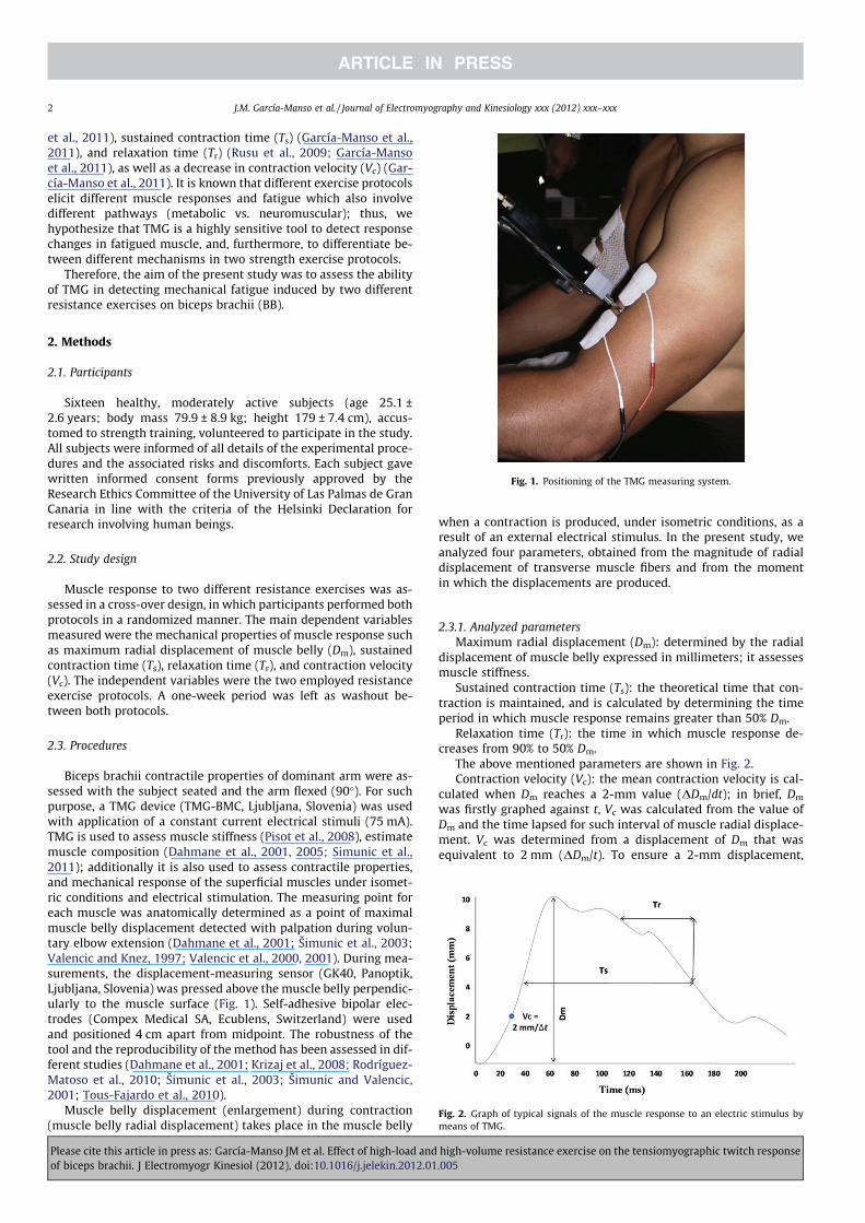
Biceps brachii contractile properties of dominant arm were as-sessed with the subject seated and the arm flexed (90°). For suchpurpose, a TMG device (TMG-BMC, Ljubljana, Slovenia) was usedwith application of a constant current electrical stimuli (75 mA).TMG is used to assess muscle stiffness (Pisot et al., 2008), estimatemuscle composition (Dahmane et al., 2001, 2005; Simunic et al.,2011); additionally it is also used to assess contractile properties,and mechanical response of the superficial muscles under isomet-ric conditions and electrical stimulation. The measuring point foreach muscle was anatomically determined as a point of maximalmuscle belly displacement detected with palpation during volun-tary elbow extension (Dahmane et al., 2001; Šimunic et al., 2003;Valencic and Knez, 1997; Valencic et al., 2000, 2001). During mea-surements, the displacement-measuring sensor (GK40, Panoptik,Ljubljana, Slovenia) was pressed above the muscle belly perpendic-ularly to the muscle surface (Fig. 1). Self-adhesive bipolar elec-trodes (Compex Medical SA, Ecublens, Switzerland) were usedand positioned 4 cm apart from midpoint. The robustness of thetool and the reproducibility of the method has been assessed in dif-ferent studies (Dahmane et al., 2001; Krizaj et al., 2008; Rodríguez-Matoso et al., 2010; Šimunic et al., 2003; Šimunic and Valencic,2001; Tous-Fajardo et al., 2010).
Muscle belly displacement (enlargement) during contraction(muscle belly radial displacement) takes place in the muscle belly
when a contraction is produced, under isometric conditions, as aresult of an external electrical stimulus. In the present study, weanalyzed four parameters, obtained from the magnitude of radialdisplacement of transverse muscle fibers and from the momentin which the displacements are produced.
2.3.1. Analyzed parameters
Maximum radial displacement (Dm): determined by the radialdisplacement of muscle belly expressed in millimeters; it assessesmuscle stiffness.
Sustained contraction time (Ts): the theoretical time that con-traction is maintained, and is calculated by determining the timeperiod in which muscle response remains greater than 50% Dm.
Relaxation time (Tr): the time in which muscle response de-creases from 90% to 50% Dm.
The above mentioned parameters are shown in Fig. 2.Contraction velocity (Vc): the mean contraction velocity is cal-
culated when Dm reaches a 2-mm value (DDm/dt); in brief, Dm
was firstly graphed against t, Vc was calculated from the value ofDm and the time lapsed for such interval of muscle radial displace-ment. Vc was determined from a displacement of Dm that wasequivalent to 2 mm (DDm/t). To ensure a 2-mm displacement,
Fig. 1. Positioning of the TMG measuring system.
Fig. 2. Graph of typical signals of the muscle response to an electric stimulus bymeans of TMG.
2 J.M. García-Manso et al. / Journal of Electromyography and Kinesiology xxx (2012) xxx–xxx
Please cite this article in press as: García-Manso JM et al. Effect of high-load and high-volume resistance exercise on the tensiomyographic twitch responseof biceps brachii. J Electromyogr Kinesiol (2012), doi:10.1016/j.jelekin.2012.01.005
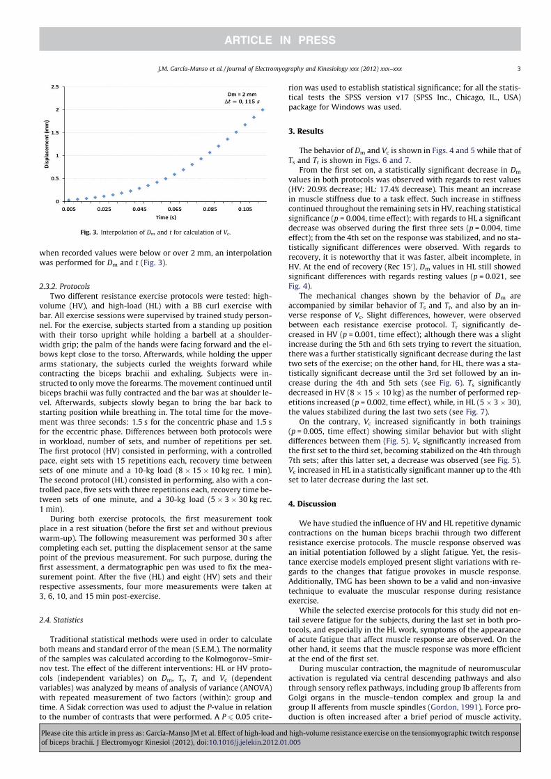
when recorded values were below or over 2 mm, an interpolationwas performed for Dm and t (Fig. 3).
2.3.2. Protocols
Two different resistance exercise protocols were tested: high-volume (HV), and high-load (HL) with a BB curl exercise withbar. All exercise sessions were supervised by trained study person-nel. For the exercise, subjects started from a standing up positionwith their torso upright while holding a barbell at a shoulder-width grip; the palm of the hands were facing forward and the el-bows kept close to the torso. Afterwards, while holding the upperarms stationary, the subjects curled the weights forward whilecontracting the biceps brachii and exhaling. Subjects were in-structed to only move the forearms. The movement continued untilbiceps brachii was fully contracted and the bar was at shoulder le-vel. Afterwards, subjects slowly began to bring the bar back tostarting position while breathing in. The total time for the move-ment was three seconds: 1.5 s for the concentric phase and 1.5 sfor the eccentric phase. Differences between both protocols werein workload, number of sets, and number of repetitions per set.The first protocol (HV) consisted in performing, with a controlledpace, eight sets with 15 repetitions each, recovery time betweensets of one minute and a 10-kg load (8 � 15 � 10 kg rec. 1 min).The second protocol (HL) consisted in performing, also with a con-trolled pace, five sets with three repetitions each, recovery time be-tween sets of one minute, and a 30-kg load (5 � 3 � 30 kg rec.1 min).
During both exercise protocols, the first measurement tookplace in a rest situation (before the first set and without previouswarm-up). The following measurement was performed 30 s aftercompleting each set, putting the displacement sensor at the samepoint of the previous measurement. For such purpose, during thefirst assessment, a dermatographic pen was used to fix the mea-surement point. After the five (HL) and eight (HV) sets and theirrespective assessments, four more measurements were taken at3, 6, 10, and 15 min post-exercise.
2.4. Statistics
Traditional statistical methods were used in order to calculateboth means and standard error of the mean (S.E.M.). The normalityof the samples was calculated according to the Kolmogorov–Smir-nov test. The effect of the different interventions: HL or HV proto-cols (independent variables) on Dm, Tr, Ts and Vc (dependentvariables) was analyzed by means of analysis of variance (ANOVA)with repeated measurement of two factors (within): group andtime. A Sidak correction was used to adjust the P-value in relationto the number of contrasts that were performed. A P 6 0.05 crite-
rion was used to establish statistical significance; for all the statis-tical tests the SPSS version v17 (SPSS Inc., Chicago, IL., USA)package for Windows was used.
3. Results
The behavior of Dm and Vc is shown in Figs. 4 and 5 while that ofTs and Tr is shown in Figs. 6 and 7.
From the first set on, a statistically significant decrease in Dm
values in both protocols was observed with regards to rest values(HV: 20.9% decrease; HL: 17.4% decrease). This meant an increasein muscle stiffness due to a task effect. Such increase in stiffnesscontinued throughout the remaining sets in HV, reaching statisticalsignificance (p = 0.004, time effect); with regards to HL a significantdecrease was observed during the first three sets (p = 0.004, timeeffect); from the 4th set on the response was stabilized, and no sta-tistically significant differences were observed. With regards torecovery, it is noteworthy that it was faster, albeit incomplete, inHV. At the end of recovery (Rec 150), Dm values in HL still showedsignificant differences with regards resting values (p = 0.021, seeFig. 4).
The mechanical changes shown by the behavior of Dm areaccompanied by similar behavior of Ts and Tr, and also by an in-verse response of Vc. Slight differences, however, were observedbetween each resistance exercise protocol. Tr significantly de-creased in HV (p = 0.001, time effect); although there was a slightincrease during the 5th and 6th sets trying to revert the situation,there was a further statistically significant decrease during the lasttwo sets of the exercise; on the other hand, for HL, there was a sta-tistically significant decrease until the 3rd set followed by an in-crease during the 4th and 5th sets (see Fig. 6). Ts significantlydecreased in HV (8 � 15 � 10 kg) as the number of performed rep-etitions increased (p = 0.002, time effect), while, in HL (5 � 3 � 30),the values stabilized during the last two sets (see Fig. 7).
On the contrary, Vc increased significantly in both trainings(p = 0.005, time effect) showing similar behavior but with slightdifferences between them (Fig. 5). Vc significantly increased fromthe first set to the third set, becoming stabilized on the 4th through7th sets; after this latter set, a decrease was observed (see Fig. 5).Vc increased in HL in a statistically significant manner up to the 4thset to later decrease during the last set.
4. Discussion
We have studied the influence of HV and HL repetitive dynamiccontractions on the human biceps brachii through two differentresistance exercise protocols. The muscle response observed wasan initial potentiation followed by a slight fatigue. Yet, the resis-tance exercise models employed present slight variations with re-gards to the changes that fatigue provokes in muscle response.Additionally, TMG has been shown to be a valid and non-invasivetechnique to evaluate the muscular response during resistanceexercise.
While the selected exercise protocols for this study did not en-tail severe fatigue for the subjects, during the last set in both pro-tocols, and especially in the HL work, symptoms of the appearanceof acute fatigue that affect muscle response are observed. On theother hand, it seems that the muscle response was more efficientat the end of the first set.
During muscular contraction, the magnitude of neuromuscularactivation is regulated via central descending pathways and alsothrough sensory reflex pathways, including group Ib afferents fromGolgi organs in the muscle–tendon complex and group Ia andgroup II afferents from muscle spindles (Gordon, 1991). Force pro-duction is often increased after a brief period of muscle activity,
Fig. 3. Interpolation of Dm and t for calculation of Vc.
J.M. García-Manso et al. / Journal of Electromyography and Kinesiology xxx (2012) xxx–xxx 3
Please cite this article in press as: García-Manso JM et al. Effect of high-load and high-volume resistance exercise on the tensiomyographic twitch responseof biceps brachii. J Electromyogr Kinesiol (2012), doi:10.1016/j.jelekin.2012.01.005

and this phenomenon is called post-activation potentiation (Fow-les and Green, 2003; Skurvydas and Zachovajevas, 1998). In thepresent case, as a result of the strength exercises used, all seemsto indicate that post-activation potentiation was responsible forthe observed changes in muscle response at the end of the firstset. This mechanism becomes significant with changes in Vc (in-crease), Dm (decrease), Ts (decrease), and Tr (decrease).
There is a correlation between this force potentiation and phos-phorylation of the myosin regulatory light chain (Sweeney et al.,1993). During muscle activity, after each action potential, Ca2+ re-leased forms the calcium/calmodulin complex, activating the ki-nase enzyme of the myosin light chains (Manning and Stull,1982; Persechini et al.,1985; Sweeney et al., 1993). This enzymeenables the estate of phosphorylation of the myosin light chains,yet the phosphorylation–dephosphorylation cycle taking placeduring each muscle activation is relatively slow. This phenomenonmay imply that the return to a rest state may be delayed for severalminutes after finishing the contraction. Post-activation potentia-tion is generally larger in fast-twitch than in slow-twitch fibers(Sweeney et al., 1993).
Changes in phosphorylation of the myosin regulatory lightchain, as well as alterations in muscle viscoelasticity, determinestructural alterations in muscle that are manifested as an increasein its stiffness. On one hand, Dm represents muscle radial displace-ment, and indirectly evaluates muscle stiffness; on the other, itvaries among subjects depending on how each muscle group hasbeen trained. Therefore, we may think that Dm’s behavior in re-sponse to strength exercise will vary depending on the workload,recovery time between repetitions, and the type of contraction per-formed. Low Dm values indicate a high muscle tone and excessivestiffness in the muscle structures. On the other hand, elevated val-ues indicate a lack of muscle tone or the appearance of muscle fa-tigue (Dahmane et al., 2001; Krizaj et al., 2008; Valencic et al.,2001). Fatigue becomes detectable when the observed changes
start inverting with regards to the behavior observed during thefirst exercise set. Important levels of fatigue imply decreases inVc and increases in the other three parameters (Dm, Ts, and Tr).
In recent years accumulating evidence has implicated alteredintracellular Ca2+ regulation as a major contributor to muscle fati-gue as sarcoplasmic reticulum Ca2+ release rate was markedly re-duced after voluntary fatiguing contractions. This mechanismcould likely be affected by sarcoplasmic reticulum Ca2+ releaseand all the Ca2+ buffers in the cell (Allen et al., 2008; Baylor andHollingworth, 1998; Li and Handschumacher, 2002). This fatigueis partly associated to the Na+–K+ pump regulation and to thechanges it entails in intracellular and extracellular Na+ and K+ con-centrations as shown by different studies (Clausen, 2003; Kabbaraet al., 2000; Sjøgaard et al., 1985). These changes in the Na+–K+
pump affect sarcolemma and T tubules depolarization decreasingthe release of Ca2+ and altering the muscle response.
In the HV protocol, the described processes were observed in aless intensive way, especially during the first moments, than in theHL protocol. The reason for this phenomenon may be the magni-tude of the loads in each case. Nevertheless, at the end of the sets,symptoms of intense fatigue could also be observed in the HV pro-tocol; such symptoms were detected by changes in the tendenciesobserved in the analyzed parameters (Vc, Dm, Ts, and Tr). Thesechanges vary, in magnitude and moment of appearance, accordingto the physical level of each subject.
With greater volume (120 repetitions) and lower load (10 kg)training, a major activation of the metabolic pathways used to sup-ply ATP occurs, and is accompanied by large increases in the accu-mulation of metabolic by-products which may be the main triggerof muscle fatigue (Allen et al., 1995; Cady et al., 1989; Dawsonet al., 1980; Fitts, 1994). In these cases, when the metaboreceptorsare activated, there is inhibition of the motoneuron pool via a re-flex pathway mediated by small-diameter group III and IV muscleafferents (Bangsbo, 1996; Sinoway et al., 1993). This response,
Fig. 4. Behavior of Dm values throughout the different sets in the two different protocols (HV and HL) and during the recovery phase. Values are mean ± S.E.M, n = 16 subjects.There was a significant time effect for Dm (p = 0.04) as determined by repeated measures ANOVA and Sidak postHoc test. ⁄P < 0.05 compared to HL. P1 = effect of the type oftime; P2 = effect of the type of exercise protocol; P3 = effect of the type of time � exercise protocol.
4 J.M. García-Manso et al. / Journal of Electromyography and Kinesiology xxx (2012) xxx–xxx
Please cite this article in press as: García-Manso JM et al. Effect of high-load and high-volume resistance exercise on the tensiomyographic twitch responseof biceps brachii. J Electromyogr Kinesiol (2012), doi:10.1016/j.jelekin.2012.01.005

most likely, also affects the reduction in voluntary drive throughspinal and supraspinal actions (Duchateau et al., 2002; Gandevia,2001).
To the changes provoked by fatigue on the action potential andthe myosin regulatory light chain activation, the alterations suf-fered by the fiber in the muscle relaxation mechanisms should
Fig. 5. Behavior of Vc values throughout the different sets in the two different protocols (HV and HL) and during the recovery phase. Values are mean ± S.E.M, n = 16 subjects.There was a significant time effect for Vc, (p = 0.05) as determined by repeated measures ANOVA and Sidak postHoc test. ⁄P < 0.05 compared to HL. P1 = effect of the type oftime; P2 = effect of the type of exercise protocol; P3 = effect of the type of time � exercise protocol.
Fig. 6. Behavior of Tr along the different sets in both protocols (HV and HL) and during the recovery phase. Values are mean ± S.E.M, n = 16 subjects. There was a significanttime effect for Tr (p = 0.001) as determined by repeated measures ANOVA and Sidak postHoc test. ⁄P < 0.05 compared to HL. P1 = effect of the type of time; P2 = effect of thetype of exercise protocol; P3 = effect of the type of time � exercise protocol.
J.M. García-Manso et al. / Journal of Electromyography and Kinesiology xxx (2012) xxx–xxx 5
Please cite this article in press as: García-Manso JM et al. Effect of high-load and high-volume resistance exercise on the tensiomyographic twitch responseof biceps brachii. J Electromyogr Kinesiol (2012), doi:10.1016/j.jelekin.2012.01.005

be added. Relaxation of skeletal muscle cells is a complex processthat involves changes in SR Ca2+ handling and cross-bridgefunction (Allen et al., 2008). Skeletal muscle fatigue is generallyaccompanied by a progressive slowing of relaxation, affecting theforce-generating potential. This situation is especially observedwhen the muscle works repeatedly with short recovery times be-tween each movement such as what happened in the usedprotocols.
In none of the protocols a complete recovery of Ts and Tr valueswas achieved during the 150 after termination of the exercise(Fig. 3). Ts recovery was 75.9% in HV, and 90.8% in HL. Tr recoverywas less efficient, reaching only values of 70.1% and 75.2%, respec-tively. The same happened with Dm (Fig. 2), where recovery was90.4% (HV) and 80.2% (HL). On the contrary, for Vc, in both cases(Fig. 2), recovery was complete 150 after finishing the exercise.Nonetheless, recovery was faster in HV, where values close to nor-mality were achieved three minutes after finishing the task. Thissituation indicates that Vc had a behavior similar to that detectedwhen studying recovery of the motor nerve which, according toBéliveau et al., 1991, recovers initial values of mean impulse fre-quency and conduction velocity of the motor nerve just few min-utes after performing a resistance exercise.
In summary, it can be observed how the manifestation of thecontractile capacity of biceps brachii significantly varies due tothe effects of potentiation and fatigue that exercise entails duringthe different phases. Fatigue may be detected by means of the anal-ysis that TMG provides us, especially the values of Dm, Vc, Ts, and Tr.These parameters reflect the alterations that take place in the mus-cle at the structural and neural levels. The behavior in theseparameters is, in general, similar in both used exercise models(HV and HL), yet showing subtle differences between them. In bothcases, Vc increased during the first set while Dm, Ts, and Tr de-creased. When fatigue starts appearing, these behaviors becameinverted, with the high-load work being the first in showing thesemechanisms.
References
Abbate F, Sargeant AJ, Verdijk PWL, de Haan A. Effects of hightfrecuency initialpulses and posttetanic potentiation on power output of skeletal muscle. J ApplPhysiol 2000;88:35–40.
Allen DG, Lännergren J, Westerblad H. Muscle cell function during prolongedactivity: cellular mechanisms of fatigue. Exp Physiol 1995;80:497–527.
Allen DG, Lamb GD, Westerblad H. Skeletal muscle fatigue: cellular mechanisms.Physiol Rev 2008;88:287–332.
Amann M, Dempsey JA. Locomotor muscle fatigue modifies central motor drive inhealthy humans and imposes a limitation to exercise. J Physiol2008;586:161–73.
Bangsbo J. Physiological factors associated with efficiency in high intensity exercise.Sports Med 1996;22:299–305.
Baylor SM, Hollingworth S. Model of sarcomeric Ca2+ movements, including ATPCa2+ binding and diffusion, during activation of frog skeletal muscle. J GenPhysiol 1998;112:297–316.
Béliveau L, Helal JN, Gaillard E, Van Hoecke J, Atlan G, Bouissou P. EMG spectral shiftand 31P-NMR-determined intracellular pH in fatigued human biceps brachiimuscle. Neurology 1991;41:1998–2001.
Bigland-Ritchie B, Johansson R, Lippold OC, Smith S, Woods JJ. Changes inmotoneuron firing rates during sustained maximal voluntary contractions. JPhysiol 1983;340:335–46.
Böl M, Stark H, Schilling N. On a phenomenological model for fatigue effects inskeletal muscles. J Theor Biol 2011;281:122–32.
Cady EB, Jones DA, Lynn J, Newham DJ. Changes in force and intracellularmetabolites during fatigue of human skeletal muscle. J Physiol 1989;418:311–25.
Clausen T. Na+–K+ pump regulation and skeletal muscle contractility. Physiol Rev2003;83:1269–324.
Dahmane R, Valencic V, Knez N, Erzen I. Evaluation of the ability to make non-invasive estimation of muscle contractile properties on the basis of the musclebelly response. Med Biol Eng Comput 2001;39:51–5.
Dahmane R, Djordjevic S, Simunic B, Valencic V. Spatial fiber type distribution innormal human muscle histochemical and tensiomyographical evaluation. JBiomech 2005;38:2451–9.
Dawson RM, Hemington N, Irvine RF. The inhibition and activation of Ca2+dependent phosphatidylinositol phosphodiesterase by phospholipids and bloodplasma. Eur J Biochem 1980;112:33–8.
de Ruiter CJ, Jones DA, Sargeant AJ, de Haan A. Temperature effect on the rates ofisometric force development and relaxation in the fresh and fatigued humanadductor pollicis muscle. Exp Physiol 1999;84:1137–50.
Duchateau J, Balestra C, Carpentier A, Hainaut K. Reflex regulation during sustainedand intermittent submaximal contractions in humans. J Physiol2002;541:959–67.
Fig. 7. Behavior of Ts along the different sets in both protocols (HV and HL) and during the recovery phase. Values are mean ± S.E.M, n = 16 subjects. There was a significanttime effect for Ts (p = 0.002) as determined by repeated measures ANOVA and Sidak postHoc test. ⁄P < 0.05 compared to HL. P1 = effect of the type of time; P2 = effect of thetype of exercise protocol; P3 = effect of the type of time � exercise protocol.
6 J.M. García-Manso et al. / Journal of Electromyography and Kinesiology xxx (2012) xxx–xxx
Please cite this article in press as: García-Manso JM et al. Effect of high-load and high-volume resistance exercise on the tensiomyographic twitch responseof biceps brachii. J Electromyogr Kinesiol (2012), doi:10.1016/j.jelekin.2012.01.005

Edwards RH. Human muscle function and fatigue. Ciba Found Symp 1981;82:1–18.Enoka RM, Stuart DG. Neurobiology of muscle fatigue. J Appl Physiol
1992;72:1631–48.Fitts RH. Cellular mechanisms of muscle fatigue. Physiol Rev 1994;74:49–94.Fowles JR, Green HJ. Coexistence of potentiation and low-frequency fatigue during
voluntary exercise in human skeletal muscle. Can J Physiol Pharmacol2003;81:1092–100.
Gandevia SC. Spinal and supraspinal factors in human muscle fatigue. Physiol Rev2001;81:1725–89.
García-Manso JM, Rodríguez-Ruiz D, Rodríguez-Matoso D, de Saá Y, Sarmiento S,Quiroga ME. Assessment of muscle fatigue after an ultraendurance triathlonusing Tensiomyography (TMG). J Sport Sci 2011;29:619–25.
Garland SJ, Gossen ER. The muscular wisdom hypothesis in human muscle fatigue.Exerc Sport Sci Rev 2002;30:45–9.
Gordon J. Spinal mechanisms of motor coordination. In: Kandel ER, Schwartz JH,Jessell TM, editors. Principles of neural science (3rd ed.). Englewood Cliffs,NJ: Prentice-Hall; 1991. p. 581–95.
Hunter SK, Lepers R, MacGillis CJ, Enoka RM. Activation among the elbow flexormuscles differs when maintaining arm position during a fatiguing contraction. JAppl Physiol 2003;94:2439–47.
Kabbara AA, Nguyen LT, Stephenson GMM, Allen DG. Intracellular calcium duringfatigue of cane toad skeletal muscle in the absence of glucose. J Muscle Res CellMotil 2000;21:481–9.
Krizaj D, Simunic B, Zagar T. Short-term repeatability of parameters extracted fromradial displacement of muscle belly. J Electromyogr Kinesiol 2008;18:645–51.
Li W, Handschumacher RE. Identification of two calcineurin B-binding proteins:tubulin and heat shock protein 60. Biochim Biophys Acta 2002;1599:72–81.
Manning DR, Stull JT. Myosin light chain phosphorylation–dephosphorylation inmammalian skeletal muscle. Am J Physiol Cell Physiol 1982;242:C234–41.
Marini M, Veicsteinas A. The exercised skeletal muscle: a review. Eur J TranslMyology 2010;20(3):105–20.
Persechini A, Stull JT, Cooke R. The effect of myosin phosphorylation on thecontractile properties of skinned rabbit skeletal muscle fibers. J Biol Chem1985;260:7951–4.
Pisot R, Narici MV, Simunic B, De Boer M, Seynnes O, Jurdana M, et al. Whole musclecontractile parameters and thickness loss during 35-day bed-rest. Eur J ApplPhysiol 2008;104(2):409–14.
Rassier DE. The effects of length on fatigue and twitch potentiation in humanskeletal muscle. Clin Physiol 2000;20:474–82.
Rodríguez-Matoso D, Rodríguez-Ruiz D, Sarmiento S, Vaamonde D, Da Silva-Grigoletto ME, García-Manso JM. Reproducibility of muscle responsemeasurements using tensiomyography in a range of positions. Rev Andal MedDeporte 2010;3:81–6.
Rusu L, Cernaianu S, Vasilescu M, Baltac G, Ciocanescu D, Fortan C. Assessment ofknee stability using neuromuscular measurement in soccer players. XVIIIInternational Congress on Sports Rehabilitation and Traumatology, Italy, 2009.p. 98–99.
Sacco P, Thickbroom GW, Byrnes ML, Mastaglia FL. Changes in corticomotorexcitability after fatiguing muscle contractions. Muscle Nerve 2000;23:1840–6.
Sejersted OM, Sjøgaard G. Dynamics and consequences of potassium shifts inskeletal muscle and heart during exercise. Physiol Rev 2000;80:1411–81.
Šimunic B, Valencic V. 2001. Non-invasive selective measurement of M. vastusmedialis and M. vastus lateralis contractile properties at different knee angles.In: Proceedings of the tenth electrotechnical and computer science conferenceERK, vol. B, Portoroz, Slovenia, Ljubljana: IEEE Region 8, Slovenian Section IEEE;24–26 September 2001. p. 363–66.
Šimunic B. 2003. Modelling of longitudinal and transversal skeletal muscle bellydeformation. PhD Thesis. School of Electrical Engineering. Ljubljana (Slovenia).2003.
Šimunic B., Rozman S, Pišot R. Detecting the velocity of the muscle contraction. In:III International symposium of new technologies in sports, Sarajevo; 2005.
Simunic B, Degens H, Rittweger J, Narici M, Mekjavic IB, Pisot R. Noninvasiveestimation of myosin heavy chain composition in human skeletal muscle. MedSci Sports Exerc 2011. doi:10.1249/MSS.0b013e31821522d0.
Sinoway LI, Hill JM, Pickar JG, Kaufman MP. Effects of contraction and lactic acid onthe discharge of group III muscle afferents in cats. J Neurophysiol1993;69:1053–9.
Sjøgaard G, Adams RP, Saltin B. Water and ion shifts in skeletal muscle of humanswith intense dynamic knee extension. Am J Physiol Regul Integr Comp Physiol1985;248:R190–6.
Skurvydas A, Zachovajevas P. Is post-tetanic potentiation, low frequency fatigue(LFF) and pre-contractile depression (PCD) coexistent in intermittent isometricexercises of maximal intensity? Acta Physiol Scand 1998;164:127–33.
Smith IJ, Hunter AM. The Effect of titanic stimulated induced fatigue on therelationship between TMG and force production of the gastrocnemius medialis.Med Sci Sports Exerc 2006;38(5):S179–80.
Sweeney HL, Bowman BF, Stull JT. Myosin light chain phosphorylation in vertebratestriated muscle: regulation and function. Am J Physiol Cell Physiol1993;264:C1085–95.
Tesch PA, Dudley GA, Duvoisin MR, Hather BM, Harris RT. Force and EMG signalpatterns during repeated bouts of concentric or eccentric muscle actions. ActaPhysiol Scand 1990;138:263–71.
Tous-Fajardo J, Moras G, Rodríguez-Jiménez S, Usach R, Doutres DM, Maffiuletti NA.Inter-rater reliability of muscle contractile property measurements using non-invasive tensiomyography. J Electromyogr Kinesiol 2010;20:761–6.
Valencic V, Knez N. Measuring of skeletal muscles dynamic properties. Artif Organs1997;21:240–2.
Valencic V, Djordjevic S, Knez N, Dahmane R, Coh M, Jurcic-Zlobec B, et al.Contractile properties of skeletal muscles detection by tensiomiographicmeasurement method. 2000 Pre-Olympic Congress, Brisbane, Australia, 2000.p. 507.
Valencic V, Knez N, Simunic B. Tensiomyography: detection of skeletal muscleresponse by means of radial muscle belly displacement. Biomed Eng2001;1:1–10.
Juan Manuel Garcia-Manso, B.S., M.S., Ph.D. receivedhis B.S. in Physical Activity and Sport Sciences fromthe Polytechnic University of Madrid, his M.S. in HighSports Performance from the Spanish Olympic Com-mittee and his Ph.D. in Sports Sciences from thePolytechnic University of Madrid. He is a Professor atthe University of Las Palmas de Gran Canarias Phys-ical Education Department and directs the ‘‘SportsTraining Analysis and Planning Laboratory (LAPED)’’.He has research interest in several areas of SportsSciences including tensiomyography.
Dario Rodriguez Matoso, B.S. received his B.S. inPhysical Activity and Sport Sciences from the Uni-versity of Las Palmas de Gran Canaria in 2009. He is aresearcher at the ‘‘Sports Training Analysis andPlanning Laboratory (LAPED)’’ of the University of LasPalmas de Gran Canaria. His main area of interest isthe use and applications of tensiomyography in dif-ferent populations.
Samuel Sarmiento, B.S., Ph.D. received his B.S. inPhysical Activity and Sport Sciences from the Uni-versity of Las Palmas de Gran Canaria in 2004 and hisPh.D. in Physical Activity, Health and Sports Perfor-mance from the same institution in 2008. He is apost-doc researcher of the University of Las Palmasde Gran Canarias Physical Education Departmentsince 2007. His main area of interest is the effect ofphysical activity on cognitive function of elderlypeople with Alzheimer-type deterioration.
Yves de Saá, B.S received his B.S. in Physical Activityand Sport Sciences from the University of Las Palmasde Gran Canaria. Since 2006 he is a research fellow atthe ‘‘Sports Training Analysis and Planning Labora-tory (LAPED)’’ of the University of Las Palmas de GranCanarias Physical Education Department.
J.M. García-Manso et al. / Journal of Electromyography and Kinesiology xxx (2012) xxx–xxx 7
Please cite this article in press as: García-Manso JM et al. Effect of high-load and high-volume resistance exercise on the tensiomyographic twitch responseof biceps brachii. J Electromyogr Kinesiol (2012), doi:10.1016/j.jelekin.2012.01.005
Diana Vaamonde, B.S., M.S., Ph.D. was born in theCanary Islands, Spain. She has received a B.S. Biologyfrom Washington and Lee University in 1998 and herPh.D. degree in Sport Sciences in 2006 from theUniversity of Córdoba, Spain. She is Assistant Pro-fessor at the Morphological Sciences Department ofthe University of Cordobas Medical School. She hasbeen director to several clinical laboratories and herresearch interest is the interaction of several biolog-ical systems as response to sports training. She hasrecently created the International Network on Phys-ical Exercise and Fertility.
David Rodriguez Ruiz, B.S., Ph.D. received his B.S. inPhysical Activity and Sport Sciences from the Uni-versity of Las Palmas de Gran Canaria in 1995 and hisPh.D. in Physical Education from the same institutionin 1999. He is Associate Professor of the University ofLas Palmas de Gran Canarias Physical EducationDepartment since 2007. He has been a researcher inthe ‘‘Sports Training Analysis and Planning Labora-tory (LAPED)’’ since 2008.
Marzo E. Da Silva-Grigoletto, B.S., M.S., Ph.D. wasborn in Brazil (RS). He received his Ph.D. degree inSport Sciences in 2006 from the University of Cór-doba, Spain. He is a M.S. in Research Methodology inHealth Sciences. He has been strength and condi-tioning trainer to Spanish Superleague volleyballteams and now is an advisor to several athletes andteams. He now teaches at the Physical Education andSports Department of the University of Seville, isEditor-in-Chief of the Andalusian Journal of SportsMedicine (Elsevier), President of the Scientific SportAssociation, and is in charge of the I + D + I of theInternational Institute of Physical Exercise Scienceand Health.
8 J.M. García-Manso et al. / Journal of Electromyography and Kinesiology xxx (2012) xxx–xxx
Please cite this article in press as: García-Manso JM et al. Effect of high-load and high-volume resistance exercise on the tensiomyographic twitch responseof biceps brachii. J Electromyogr Kinesiol (2012), doi:10.1016/j.jelekin.2012.01.005
Copyright © 2022 FDOKUMEN