Effect of Conditioned Stimulus Exposure During Slow Wave Sleep on Fear Memory Extinction in Humans
9
SLEEP, Vol. 38, No. 3, 2015 423 Slow Wave Sleep and Fear Memory—He et al. Study Objectives: Repeated exposure to a neutral conditioned stimulus (CS) in the absence of a noxious unconditioned stimulus (US) elicits fear memory extinction. The aim of the current study was to investigate the effects of mild tone exposure (CS) during slow wave sleep (SWS) on fear memory extinction in humans. Design: The healthy volunteers underwent an auditory fear conditioning paradigm on the experimental night, during which tones served as the CS, and a mild shock served as the US. They were then randomly assigned to four groups. Three groups were exposed to the CS for 3 or 10 min or an irrelevant tone (control stimulus, CtrS) for 10 min during SWS. The fourth group served as controls and was not subjected to any interventions. All of the subjects completed a memory test 4 h after SWS-rich stage to evaluate the effect on fear extinction. Moreover, we conducted similar experiments using an independent group of subjects during the daytime to test whether the memory extinction effect was specific to the sleep condition. Participants: Ninety-six healthy volunteers (44 males) aged 18–28 y. Measurements and Results: Participants exhibited undisturbed sleep during 2 consecutive nights, as assessed by sleep variables (all P > 0.05) from polysomnographic recordings and power spectral analysis. Participants who were re-exposed to the 10 min CS either during SWS and wakefulness exhibited attenuated fear responses (wake-10 min CS, P < 0.05; SWS-10 min CS, P < 0.01). Conclusions: Conditioned stimulus re-exposure during slow wave sleep promoted fear memory extinction without altering sleep profiles. Keywords: conditioned stimulus, extinction, fear memory, slow wave sleep Citation: He J, Sun HQ, Li SX, Zhang WH, Shi J, Ai SZ, Li Y, Li XJ, Tang XD, Lu L. Effect of conditioned stimulus exposure during slow wave sleep on fear memory extinction in humans. SLEEP 2015;38(3):423–431. INTRODUCTION New memories are gradually stabilized after initial learning through a process called consolidation, 1,2 during which memo- ries are vulnerable to be regulated. Consistent with this theory, some research has shown that the formation and consolidation of emotional memory are critical to adapting to a changing environment in animals and humans. 3–5 Most studies that have focused on memory consolidation have examined the effects of pharmacological treatments administered within several hours after learning tasks, demonstrating that memory consolidation is a time-dependent process, 6 during which protein synthesis is susceptible to being influenced and memories can be impaired or enhanced. 7–9 The use of pharmacological treatments admin- istered shortly after training (i.e., within the time window of consolidation 10 ) influence memory consolidation and result in the formation of long-term memories. SLOW WAVE SLEEP AND FEAR MEMORY EXTINCTION IN HUMANS Effect of Conditioned Stimulus Exposure during Slow Wave Sleep on Fear Memory Extinction in Humans Jia He (Postgraduate Student) 1,2 ; Hong-Qiang Sun, MD, PhD 1 ; Su-Xia Li, MD, PhD 2 ; Wei-Hua Zhang, MD 1 ; Jie Shi, MD, PhD 2 ; Si-Zhi Ai (Postgraduate Student) 2 ; Yun Li, MD 3 ; Xiao-Jun Li, MD 1 ; Xiang-Dong Tang, MD, PhD 3, ; Lin Lu, MD, PhD 1,2,4 1 Peking University Sixth Hospital/Institute of Mental Health and Key Laboratory of Mental Health and 2 National Institute on Drug Dependence, Peking University, Beijing, China; 3 Sleep Medicine Center, Translational Neuroscience Center, West China Hospital, Sichuan University, Chengdu, China; 4 Peking-Tsinghua Center for Life Sciences and PKU-IDG/McGovern Institute for Brain Research, Peking University, Beijing, China pii: sp-00026-14 http://dx.doi.org/10.5665/sleep.4502 Numerous recent studies have suggested that sleep is ben- eficial for memory consolidation, including emotional memory. Sleep was shown to enhance the retention of either declarative information 11,12 or procedural skills 13,14 compared with staying awake after accomplishing learning tasks. Several studies have suggested that emotional memories are specifically en- hanced by sleep, 2,15,16 and the strengthened effect on emotional memories can last for a relatively long time. 17 Additionally, the effects of consolidation on memory during slow wave sleep (SWS) and rapid eye-movement (REM) sleep have been doc- umented in human studies. 18 Increasing evidence indicates that REM sleep and SWS both contribute to nondeclarative memory 15,19,20 and declarative memory consolidation, 21–23 sug- gesting that sleep enhances the retention of newly encoded memories, depending on specific neurophysiological activity associated with particular stages, such as SWS and REM sleep. Contradictory findings have been reported concerning the association between sleep stages and emotional memories. One study indicated that REM-rich sleep enhanced aversion to previously viewed emotional pictures. 24 Other studies re- ported that the recall of negative pictures is less impaired by sleep deprivation. 25,26 In summary, how sleep affects emotional memory expression requires further investigation. Recent work has gradually focused on the role of SWS in memory modulation. Research findings suggest that memories that are learned while awake can be enhanced through the rep- resentation of the same context during SWS. 27–29 These results further indicate that consolidation is a labile state during which memory traces are prone to being changed, and SWS serves as a particular period that facilitates memory regulation. Hauner A commentary on this article appears in this issue on page 337. Submitted for publication January, 2014 Submitted in final revised form September, 2014 Accepted for publication October, 2014 Address correspondence to: Professor Lin Lu, Institute of Mental Health and National Institute on Drug Dependence, Peking University, 51, Hua- yuan Bei Road, Beijing 100191, China; Tel: +86-10-82805308; Fax: +86- 10-62032624; Email: [email protected] or Professor Xiang-Dong Tang, Sleep Medicine Center, West China Hospital, Sichuan University, 28 Dian Xin Nan Jie, Chengdu City, Sichuan Province, 610041, China; Tel: +86-28 8542 2733; Fax: +86-28 8542 2632; Email: [email protected]
-
Upload
independent -
Category
Documents
-
view
2 -
download
0
Transcript of Effect of Conditioned Stimulus Exposure During Slow Wave Sleep on Fear Memory Extinction in Humans
SLEEP, Vol. 38, No. 3, 2015 423 Slow Wave Sleep and Fear Memory—He et al.
Study Objectives: Repeated exposure to a neutral conditioned stimulus (CS) in the absence of a noxious unconditioned stimulus (US) elicits fear memory extinction. The aim of the current study was to investigate the effects of mild tone exposure (CS) during slow wave sleep (SWS) on fear memory extinction in humans.Design: The healthy volunteers underwent an auditory fear conditioning paradigm on the experimental night, during which tones served as the CS, and a mild shock served as the US. They were then randomly assigned to four groups. Three groups were exposed to the CS for 3 or 10 min or an irrelevant tone (control stimulus, CtrS) for 10 min during SWS. The fourth group served as controls and was not subjected to any interventions. All of the subjects completed a memory test 4 h after SWS-rich stage to evaluate the effect on fear extinction. Moreover, we conducted similar experiments using an independent group of subjects during the daytime to test whether the memory extinction effect was specific to the sleep condition.Participants: Ninety-six healthy volunteers (44 males) aged 18–28 y.Measurements and Results: Participants exhibited undisturbed sleep during 2 consecutive nights, as assessed by sleep variables (all P > 0.05) from polysomnographic recordings and power spectral analysis. Participants who were re-exposed to the 10 min CS either during SWS and wakefulness exhibited attenuated fear responses (wake-10 min CS, P < 0.05; SWS-10 min CS, P < 0.01).Conclusions: Conditioned stimulus re-exposure during slow wave sleep promoted fear memory extinction without altering sleep profiles.Keywords: conditioned stimulus, extinction, fear memory, slow wave sleep Citation: He J, Sun HQ, Li SX, Zhang WH, Shi J, Ai SZ, Li Y, Li XJ, Tang XD, Lu L. Effect of conditioned stimulus exposure during slow wave sleep on fear memory extinction in humans. SLEEP 2015;38(3):423–431.
INTRODUCTIONNew memories are gradually stabilized after initial learning
through a process called consolidation,1,2 during which memo-ries are vulnerable to be regulated. Consistent with this theory, some research has shown that the formation and consolidation of emotional memory are critical to adapting to a changing environment in animals and humans.3–5 Most studies that have focused on memory consolidation have examined the effects of pharmacological treatments administered within several hours after learning tasks, demonstrating that memory consolidation is a time-dependent process,6 during which protein synthesis is susceptible to being influenced and memories can be impaired or enhanced.7–9 The use of pharmacological treatments admin-istered shortly after training (i.e., within the time window of consolidation10) influence memory consolidation and result in the formation of long-term memories.
SLOW WAVE SLEEP AND FEAR MEMORY EXTINCTION IN HUMANS
Effect of Conditioned Stimulus Exposure during Slow Wave Sleep on Fear Memory Extinction in HumansJia He (Postgraduate Student)1,2; Hong-Qiang Sun, MD, PhD1; Su-Xia Li, MD, PhD2; Wei-Hua Zhang, MD1; Jie Shi, MD, PhD2; Si-Zhi Ai (Postgraduate Student)2; Yun Li, MD3; Xiao-Jun Li, MD1; Xiang-Dong Tang, MD, PhD3,; Lin Lu, MD, PhD1,2,4
1Peking University Sixth Hospital/Institute of Mental Health and Key Laboratory of Mental Health and 2National Institute on Drug Dependence, Peking University, Beijing, China; 3Sleep Medicine Center, Translational Neuroscience Center, West China Hospital, Sichuan University, Chengdu, China; 4Peking-Tsinghua Center for Life Sciences and PKU-IDG/McGovern Institute for Brain Research, Peking University, Beijing, China
pii: sp-00026-14 http://dx.doi.org/10.5665/sleep.4502
Numerous recent studies have suggested that sleep is ben-eficial for memory consolidation, including emotional memory. Sleep was shown to enhance the retention of either declarative information11,12 or procedural skills13,14 compared with staying awake after accomplishing learning tasks. Several studies have suggested that emotional memories are specifically en-hanced by sleep,2,15,16 and the strengthened effect on emotional memories can last for a relatively long time.17 Additionally, the effects of consolidation on memory during slow wave sleep (SWS) and rapid eye-movement (REM) sleep have been doc-umented in human studies.18 Increasing evidence indicates that REM sleep and SWS both contribute to nondeclarative memory15,19,20 and declarative memory consolidation,21–23 sug-gesting that sleep enhances the retention of newly encoded memories, depending on specific neurophysiological activity associated with particular stages, such as SWS and REM sleep. Contradictory findings have been reported concerning the association between sleep stages and emotional memories. One study indicated that REM-rich sleep enhanced aversion to previously viewed emotional pictures.24 Other studies re-ported that the recall of negative pictures is less impaired by sleep deprivation.25,26 In summary, how sleep affects emotional memory expression requires further investigation.
Recent work has gradually focused on the role of SWS in memory modulation. Research findings suggest that memories that are learned while awake can be enhanced through the rep-resentation of the same context during SWS.27–29 These results further indicate that consolidation is a labile state during which memory traces are prone to being changed, and SWS serves as a particular period that facilitates memory regulation. Hauner
A commentary on this article appears in this issue on page 337.
Submitted for publication January, 2014Submitted in final revised form September, 2014Accepted for publication October, 2014Address correspondence to: Professor Lin Lu, Institute of Mental Health and National Institute on Drug Dependence, Peking University, 51, Hua-yuan Bei Road, Beijing 100191, China; Tel: +86-10-82805308; Fax: +86-10-62032624; Email: [email protected] or Professor Xiang-Dong Tang, Sleep Medicine Center, West China Hospital, Sichuan University, 28 Dian Xin Nan Jie, Chengdu City, Sichuan Province, 610041, China; Tel: +86-28 8542 2733; Fax: +86-28 8542 2632; Email: [email protected]

SLEEP, Vol. 38, No. 3, 2015 424 Slow Wave Sleep and Fear Memory—He et al.
et al.30 demonstrated that reexposure to an odorant context during SWS in human subjects who were subjected to an ol-factory contextual fear conditioning paradigm decreased fear responses,30 which is inconsistent with previous fi ndings.29,31
Conditioned fear responses can be inhibited by repeated ex-posure to a conditioned stimulus (CS) in the absence of an unconditioned stimulus (US). This process is termed memory extinction. It is the simplest way to attenuate the expression of fear memory while awake.32 However, still unknown is whether reexposure to mild tones (CS) can elicit fear extinction during SWS. Our hypothesis is that reexposure to a CS during SWS promotes fear memory extinction in humans. We applied an auditory fear conditioning paradigm, with mild tones as the CS, to test this hypothesis.
METHODS
ParticipantsNinety-six volunteers (44 male; age, 24.0 ± 2.4 y
[mean ± standard deviation]) participated in the study. All of the female participants (n = 52) were reported to have regular menstrual cycles and were not using oral contraceptives prior to or during the study. They were asked to participate in the study 3–7 days before their menstrual cycle began. All of the volunteers were in good health with no history of neurological, psychiatric, or sleep disorders and no history or current use of medications known to affect cognitive functioning or sleep. Subjects were excluded from the study if they had a history of drug or alcohol addiction. The experiments were performed at the Sleep Medicine Center, West China Hospital, Sichuan Uni-versity, Chengdu, China. We conducted the study in a tempera-ture- and humidity-controlled room. All of the experimental procedures were conducted in accordance with the Declara-tion of Helsinki and performed with adequate understanding
and written informed consent from the sub-jects. The study pro-tocol was approved by the Research Ethics Board of the West China Hospital of Sichuan University. Each participant was paid 160 RMB (equiv-alent to $26.40 USD) upon completion of the study.
Behavioral ParadigmWe conducted the
experiments on 2 con-secutive nights. The fi rst night included adaptation, and the second night was the experimental session. On the fi rst night, we collected the sub-jects’ demographic
data and asked them to complete the Pittsburg Sleep Quality Index (PSQI) before sleep. The PSQI33 was used to investigate sleep habits and assess the subjective sleep quality of the par-ticipants, with a cutoff score of 5. The main purpose of the fi rst night was to exclude participants who had primary sleep disorders, such as sleep apnea or periodic limb movements. On the second night, all of the subjects (n = 66) underwent the fear conditioning paradigm approximately 30 min before sleep onset, followed by another two stages: stimulus exposure during SWS and a memory test. The timeline of the proce-dures is presented in Figure 1A.
The participants were randomly assigned to four groups: con-trol group (SWS-Ctr, n = 18), 3 min CS exposure group (SWS-3 min CS, n = 17), 10 min CS exposure group (SWS-10 min CS, n = 16), and 10 min control stimulus exposure group (SWS-10 min CtrS, n = 15). All of the participants were subjected to a Pavlovian fear conditioning paradigm with partial reinforce-ment. The CSs were auditory tones, and the US was a mild shock to the wrist. Two auditory tones (CS+ and CS−) were used. One tone (520 Hz, 65 dB; CS+) was paired with a mild shock to the wrist (US) on a 40% partial reinforcement schedule, and an-other tone stimulus (250 Hz, 65 dB; CS−) was never paired with the shock. We presented the CS+ with shock in four trials and the CS+ without shock in six trials. Thus, the CS+ was presented 10 times in the conditioning phase. The CS− was also presented 10 times. The CS+ reinforced trials were conducted in random order during 10 trials. The CtrS was irrelevant to the fear con-ditioning training and only presented during SWS in the 10 min CtrS group. The participants were instructed to pay attention to the tone and try to determine the relationship between the stimulus and shock during the training session.
We used a “night-half paradigm,” originally developed by Fowler et al.34 and Laroush et al.35 to avoid possible con-founding effects that result from rapid eye movement (REM)
Figure 1—Experimental design and protocol. (A) Timeline of the experiment. The light gray areas represent the sleep period, and the dark gray area represents the fi rst period of SWS when delivering the auditory stimuli. (B) A 4-h period of nocturnal sleep or daytime wakefulness separated an initial fear conditioning training phase (10 trials) from later retesting (three trials). The experimental protocol was completed in two independent experiments separated by 12 h.

SLEEP, Vol. 38, No. 3, 2015 425 Slow Wave Sleep and Fear Memory—He et al.
sleep. The subjects were allowed to sleep from 23:00 to 03:00, which was deemed SWS-rich sleep. During that time, all of the groups, with the exception of the control group, were re-exposed to the tone stimuli. A detailed description of the tone stimuli is presented in the Auditory Stimuli section. The sub-jects were awakened during the first sleep stage 1 or stage 2, which occurred after 4 h of sleep, because arousal from other sleep stages may influence subsequent test performance.36 The subjects were tested 15 min later to avoid sleep inertia.19 After completing the test, the participants were allowed to return to sleep and woken up at 07:00.
During the fear conditioning training and test phases, the participants were attached to skin conductance recording and shock electrodes. The skin conductance recording electrode was used to measure the skin conductance response (SCR). Shocks were delivered through a stimulating square electrode attached to the right inner wrist via a STM 200 stimulator (BioPac Systems, Goleta, CA, USA). The level of the shock was based on the criterion “uncomfortable, but not painful,” which was determined for each participant. The shock level began at mild (10 V) and gradually increased until the shock reached the maximum level (50 V).37,38 The auditory tones were presented for 3 sec, and the shocks were administered for 250 ms, which coterminated with the tones. The intertrial interval (ITI) was 10 sec. Stimulus presentation was controlled by a computer using E-Prime 2.0 professional (Psychology Soft-ware Tools, Inc.; http://www.pstnet.com). For all of the groups, the conditioning training phase consisted of 10 nonreinforced presentations of the CS− and four reinforced presentations of the CS+ (four CS+ stimuli presented with shock and six CS+ stimuli presented without shock). The test phase was similar to the conditioning training phase but only consisted of three presentations of the CS− and three presentations of the CS+ without shock.
Generally, we conducted the experiment with no instruc-tions or information given to the participants about tone pre-sentation during sleep. At the debriefing, two of the subjects reported to have noticed the tones while sleeping. These two subjects were then excluded from the final analysis.
Moreover, to investigate whether the extinction effect was specific to the sleep condition, we repeated a similar proce-dures in another experiment, using an independent group of awake subjects (n = 30) who were divided into two subgroups. One group of subjects (Wake-10 min CS, n = 15) stayed awake during the 10 min CS reexposure, and the other group served as controls (Wake-Ctr, n = 15) who were not subjected to any interventions but remained awake until the end of the experi-ment. Conditioning was performed at 10:00 in the morning, followed by a 4-h interval of daytime wakefulness similar to the nocturnal sleep period. Subsequently, the subjects were re-tested in three trials for conditioned responses to the CS+ and CS− 39,40 (Figure 1B).
Polysomnographic RecordingsOvernight polysomnographic recording included electro-
encephalography (EEG; including F3, F4, C3, C4, O1, and O2, with reference to the contralateral mastoid; International 10–20 system), bilateral electrooculography (EOG), bilateral electromyography (EMG), and electrocardiography (ECG).
Finger pulse oximetry was also recorded. High-pass filters were set at 0.3 Hz, and low-pass filters were set at 35 Hz for all of the EEGs and EOGs. Thirty-second epochs were used for manual analysis, and sleep stages were scored offline ac-cording to the criterion of the American Academy of Sleep Medicine (AASM) using the standard polysomnographic sleep recordings.
We collected polysomnographic parameters, including total time in bed (TIB), total sleep time (TST), sleep latency (SL), min and percentage of sleep stages 1, 2, 3, and REM sleep, wake after sleep onset (WASO), and sleep efficiency (SE = TST/TIB) for 2 consecutive nights.
Auditory StimuliAuditory tones were broadcast through two loudspeakers.
They were placed 1 m from the head of the bed. The first con-ditioned auditory stimulus (520 Hz sine-wave auditory tone, 65 dB) or control stimulus (1,250 Hz sine-wave auditory tone, 55 dB) was presented when we identified 2 min of continuous SWS after the participants fell asleep. We used these auditory stimuli based on a previous study, and they were adjusted in-dividually.41 The tone stimuli were presented in a 15 sec on/15 sec off schedule, which lasted 3 or 10 min. Once a microarousal occurred, the stimulus that evoked the microarousal were not be counted into the total numbers of stimuli. In summary, the stimuli were presented 12 times in the SWS-3 min CS group, 40 times in the SWS-10 min CS group, and 40 times in the SWS-10 min CtrS group.
Psychophysiological Recording and AssessmentThe SCR was acquired using two Ag-AgCl electrodes that
were attached to the second and third fingers of the left hand on the middle phalanges. The SCR waveforms were measured using a BioPac MP150 system and recorded with AcqKnowl-edge 4.2 software (BioPac Systems).
AcqKnowledge 4.2 software was also used to analyze SCR waveforms. The level of the SCR was determined by taking the base-to-peak difference for each waveform in a 3-sec window following stimulus onset. At each stage, the differential fear response was calculated by subtracting responses to the CS−
from responses to the CS+ in corresponding trials.38
Statistical AnalysisWe used one-way analysis of variance (ANOVA) to analyze
the demographic data and PSQI scores between the six groups. The polysomnographic variables for the 2 consecutive nights were analyzed using two-way repeated-measures ANOVA, with group as the between-subjects factor and time (first night and second night) as the within-subjects factor, followed by the Bonferroni/Dunn post hoc test.
EEG signals were sampled online at 512 Hz using Profu-sion Net Beacon software (Compumedics Sleep Study System, Melbourne, Australia). Detailed power spectral analysis was performed offline using fast Fourier transform (FFT) with Hanning window tapering, showing the frequency and power of an analyzed portion of the EEG signal (Compumedics Sleep Study System). We chose the C3 and C4 central electrodes to perform further analysis and defined the frequency bands as the following: delta (0.5–4 Hz), theta (4–8 Hz), alpha (8–12 Hz),
SLEEP, Vol. 38, No. 3, 2015 426 Slow Wave Sleep and Fear Memory—He et al.
beta (12–30 Hz), and gamma (30–40 Hz). Because no differ-ence was observed between the central C3 and C4 electrodes, the data were collapsed across these two electrodes. The mean EEG power at each frequency band across blocks was com-pared between tone-on and tone-off blocks using two-tailed paired t-tests. Data from eight subjects were excluded because of a lack of artifact-free epochs.
Both during the sleep and wakefulness conditions, sub-jects who showed successful levels of fear acquisition were included in the analysis. We assessed successful fear acqui-sition by skin conductance recording. The exclusion criteria were based on differential responses to the CS+ and CS− in the second half of acquisition. That is, subjects were excluded if the difference during acquisition was in the opposite direc-tion (CS− > CS+) or smaller than 0.1 μs38; two subjects were excluded based on this criterion. For the sleep conditions, the SCR results for the fear acquisition and test phases were analyzed using two-way repeated-measures ANOVA, with group as the between-subjects factor and trial as the within-subjects factor, followed by the Bonferroni/Dunn post hoc test. The effect of CS exposure on memory extinction in each group was assessed using a two-way ANOVA that evaluated main effects of group and time (the average of the last three learning trials and average of three test trials), followed by the Bonferroni/Dunn post hoc test to assess the extinction effect between groups. Moreover, we applied a three-way ANOVA that assessed the effect of CS exposure on memory extinction in distinct groups that evaluated main effects of group (CS exposure and No CS exposure), condition (SWS and wakeful-ness) and time (the average of the last three learning trials and average of three test trials), followed by the Bonferroni/Dunn post hoc test as appropriate.
Differences were considered significant when P < 0.05.
RESULTS
Population and Demographic DataA total of 96 healthy subjects (44 males; age, 24.0 ± 2.4 y
[mean ± standard deviation]) completed the study. Thirteen sub-jects were excluded from the final analysis because of failures in PSG recording (n = 3) and SCR extraction (n = 2), inadequate levels of fear acquisition (n = 2), maintenance of sleep during tone presentation (n = 2), and difficulty falling asleep after fear conditioning training on the experimental night (n = 4).
As shown in Table 1, no significant differences were found in age, body mass index, systolic blood pressure (SYS), diastolic blood pressure (DIA), and PSQI score among the six groups. All of the participants’ PSQI scores were less than 5, indicating that they had regular sleep habits and good sleep quality.
Effects of CS Exposure on Sleep ArchitectureThe participants were required to maintain a regular sleep-
wake cycle (i.e., go to sleep between 23:00 and 24:00 and wake up between 07:00 and 08:00) for at least 3 days before the study began. Table 2 shows that reexposure to the CS or ir-relevant tones during SWS on the experimental night did not affect sleep profiles (TST, F3,50 = 0.72, P = 0.55; SL, F3,50 = 0.89, P = 0.50; N1 [%], F3,50 = 0.74, P = 0.54; N2 [%], F3,50 = 0.57, P = 0.64; SWS [%], F3,50 = 0.36, P = 0.78; REM [%], F3,50 = 0.35, P = 0.79; WASO, F3,50 = 0.23, P = 0.86; SE, F3,50 = 0.24, P = 0.87).
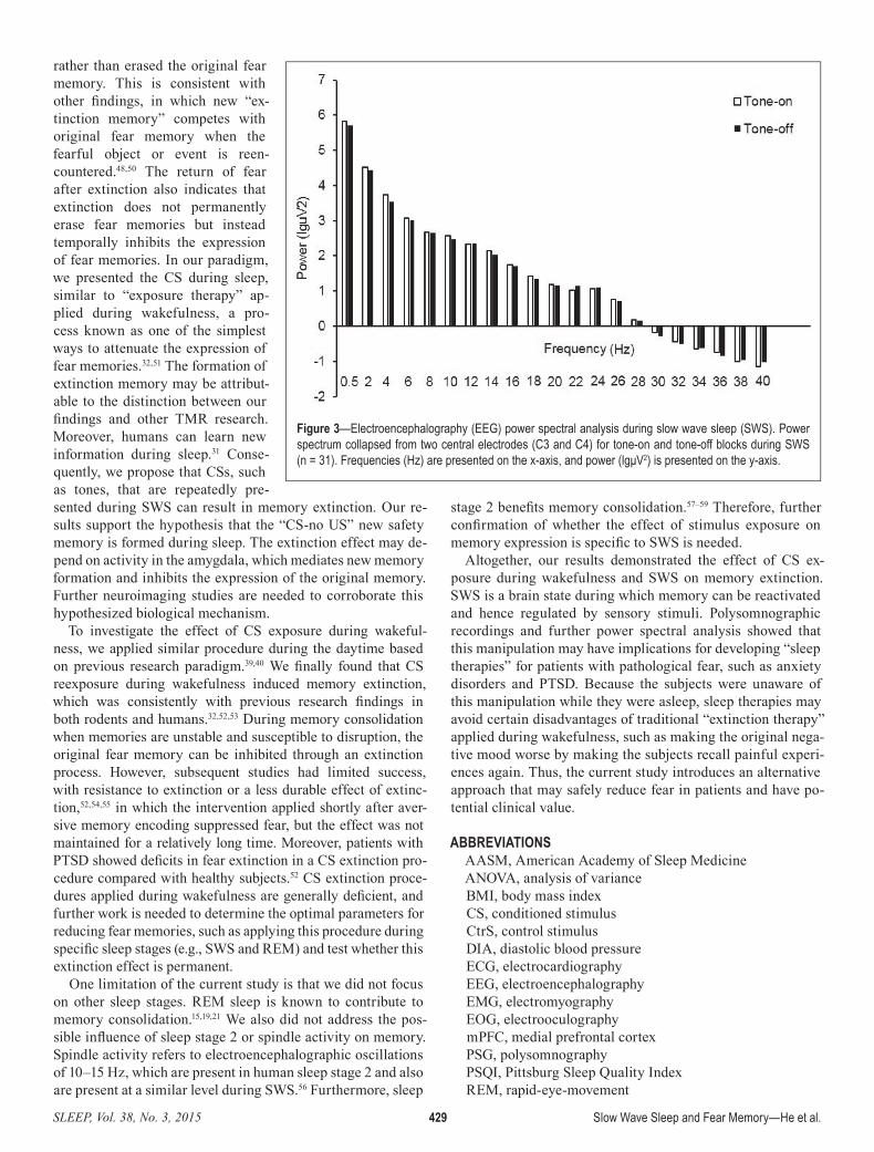
We compared PSG data between 2 days to roughly deter-mine the sleep architecture with and without CS+ presentation, and the sleep profile appeared to be unchanged. Further power spectral analysis confirmed that the subjects remained asleep during tone stimulus presentation (Figure 3). Mean EEG power for tone-on and tone-off blocks did not significantly differ for the following frequency bands derived from the central elec-trodes: delta (0.5–4 Hz, t31 = 0.065, P = 0.95), theta (4–8 Hz, t31 = −1.06, P = 0.30), alpha (8–12 Hz, t31 = 0.62, P = 0.54), beta (12–30 Hz, t31 = 0.79, P = 0.44), and gamma (30–40 Hz, t31 = 1.05, P = 0.30). Alpha and beta power were similar before compared to after tone onset, suggesting that cue presentation influenced memory extinction without briefly waking the par-ticipants. Further analysis over the full range of frequencies (1–40 Hz) showed an apparent increase in power during tone-on versus tone-off blocks in the frequency range of 10–16 Hz for central electrodes; however the increase did not reach the threshold for significance (P = 0.063).
Effects of CS Exposure on Memory ExtinctionThe SCR results during conditioning training and the test
are presented in Figure 2. For the sleep condition, all of the participants exhibited similar levels of fear acquisition after 10 conditioning trials (F3,50 = 1.94, P = 0.14). The decline in fear re-sponses from acquisition (the average of the last three learning trials) to testing (the average of three trials) for each group was assessed using a two-way ANOVA, with group as the between-subjects factor and time (the average of the last three learning
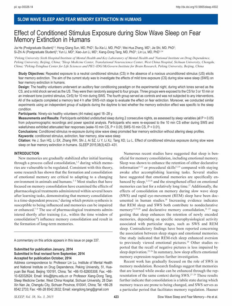
Table 1—Demographic data collected from the six groups (n = 83).
Wake-Ctr Wake-10 min CS SWS-Ctr SWS-3 min CS SWS-10 min CS SWS-10 min CtrS PAge (y) 23.21 ± 2.83 23.93 ± 2.34 24.07 ± 2.89 23.54 ± 1.90 23.23 ± 2.62 24.43 ± 2.28 0.756BMI (kg/m2) 21.16 ± 3.03 21.74 ± 3.96 20.48 ± 2.10 21.14 ± 2.40 20.80 ± 1.86 20.89 ± 2.35 0.881SYS (mmHg) 117.36 ± 8.98 116.33 ± 13.55 108.93 ± 9.19 119.00 ± 16.94 113.92 ± 10.47 110.43 ± 10.70 0.187DIA (mmHg) 75.86 ± 10.00 79.73 ± 9.70 71.79 ± 9.37 76.31 ± 8.29 72.92 ± 6.76 71.00 ± 6.34 0.070PSQI 3.29 ± 0.61 3.73 ± 1.03 4.21 ± 1.37 3.93 ± 1.38 3.77 ± 1.42 3.29 ± 1.20 0.277
The data are expressed as mean ± standard deviation (P value for one-way analysis of variance). BMI, body mass index; CS, conditioned stimulus; Ctr, control; CtrS, control stimulus; DIA, diastolic blood pressure; SYS, systolic blood pressure; PSQI, Pittsburgh Sleep Questionnaire Index; SWS, slow wave sleep; Wake-Ctr, wake control group; Wake-10 min CS, wake 10 min CS exposure group; SWS-Ctr, SWS control group; SWS-3 min CS, SWS 3 min CS exposure group; SWS-10 min CS, SWS 10 min CS exposure group; SWS-10 min CtrS, SWS 10 min control stimulus exposure group.
SLEEP, Vol. 38, No. 3, 2015 427 Slow Wave Sleep and Fear Memory—He et al.
trials and average of three test trials) as the within-subjects factor, followed by the Bonferroni/Dunn post hoc test. The analysis showed significant main effects of time (F1,50 = 17.122, P < 0.001) and group (F3,55 = 3.569, P < 0.05) and a group × time interaction (F3,55 = 10.000, P < 0.001). Concerning the change from conditioning to test, the post hoc test showed that an ex-tinction effect occurred in the 3 min CS group and 10 min CS group (P < 0.05, P < 0.01, respectively; Figure 2A). Concerning test performance, post hoc tests showed that there were sta-tistically significant differences between SWS-3 min CS (P < 0.05), SWS-10 min CS (P < 0.01) and the SWS-Ctr group (Figure 2A). Moreover, there also were significant differences between SWS-3 min CS (P < 0.05), SWS-10 min CS (P < 0.01) and the SWS-CtrS group (Figure 2A). These results indicate that CS exposure during SWS elicited fear memory extinc-tion and the presentation of other nonassociated tone stimuli did not disrupt the expression of memory, which excludes the possibility that the tone stimulus itself affected the memory trace. Moreover, in an additional control experiment with 30 other participants who remained awake, tones were presented after approximately a 1.5-h interval yoked between sleep and awake subjects. All of the participants exhibited similar levels of fear acquisition after 10 conditioning trials (F3,52 = 1.52, P = 0.22). We then conducted a group (CS exposure and No CS exposure) × condition (SWS and wakefulness) × time (the av-erage of the last three learning trials and average of three test trials) ANOVA and found significant main effects for group (F1,52 = 16.400, P < 0.01), time (F1,52 = 27.826, P < 0.01), and a significant time × group interaction (F1,52 = 28.234, P < 0.01).
The post hoc tests showed that CS presented during either SWS or wakefulness altered memory expression from acqui-sition to test compared with No CS exposure (both compari-sons, P < 0.01; Figure 2B). In addition, when we compared test performance only, we found that the decline of fear responses induced by CS exposure occurred in both the Wake-10 min CS group and SWS-10 min CS group compared with the Wake-Ctr group and SWS-Ctr group, respectively (both comparisons, P < 0.01; Figure 2B). Furthermore, the post hoc tests also found no significant differences between the Wake-10 min CS group and SWS-10 min CS group during the memory acquisition (P = 0.661) and test (P = 0.441) phases. These results revealed that CS exposure during either SWS or wakefulness promoted fear memory extinction.
DISCUSSIONThe main finding of the current study was that fear re-
sponses in the SWS-10 min CS and Wake-10 min CS groups both markedly declined during the test. This suggests that repeated CS exposure during SWS resulted in memory ex-tinction that may be comparable to exposure therapy applied during wakefulness. Moreover, this manipulation only af-fected fear memory, which was reactivated by reexposure to the CS during sleep, and left the nonreactivated fear memory intact when the subjects were exposed to the irrelevant stim-ulus during sleep. The sleep macrostructure during 2 consec-utive nights in each group and power spectral analysis that focused on the power difference between tone-on and tone-off blocks in distinct frequency bands during SWS suggested that
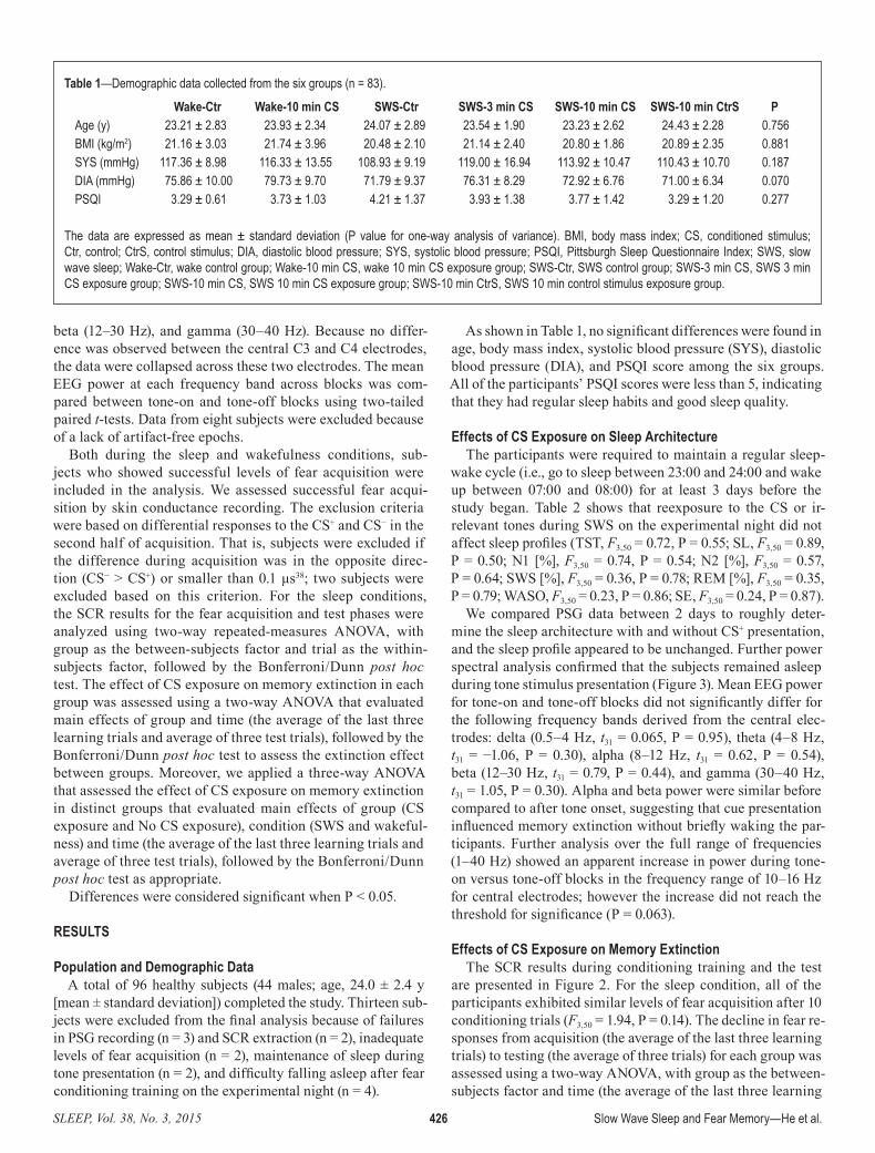
Table 2—Polysomnographic data for 2 consecutive nights (n = 54).
SWS-Ctr SWS-3 min CS SWS-10 min CS SWS-10 min CtrSAdaptation night
TST (min) 416.79 ± 19.42 420.85 ± 34.71 438.46 ± 36.30 428.94 ± 29.63SL (min) 12.96 ± 10.14 13.35 ± 6.52 14.73 ± 10.61 9.57 ± 4.73N1 (%) 9.84 ± 3.74 11.84 ± 3.70 9.34 ± 4.95 9.36 ± 1.65N2 (%) 39.30 ± 7.47 40.09 ± 5.07 43.14 ± 7.51 41.18 ± 5.78SWS (%) 30.86 ± 10.82 28.41 ± 7.34 26.85 ± 8.37 30.68 ± 8.55REM (%) 19.99 ± 4.21 19.25 ± 4.82 20.66 ± 5.77 18.99 ± 5.65WASO (min) 36.18 ± 18.88 32.08 ± 12.77 32.69 ± 14.54 37.96 ± 21.05SE (%) 88.00 ± 2.22 89.54 ± 2.63 88.62 ± 4.72 87.50 ± 4.45
Experimental nightTST (min) 395.82 ± 33.48 393.12 ± 45.04 406.62 ± 56.98 400.54 ± 28.47SL (min) 10.68 ± 7.18 13.50 ± 8.46 15.73 ± 11.47 11.43 ± 6.73N1 (%) 9.51 ± 5.51 11.25 ± 7.13 11.45 ± 5.44 9.59 ± 2.33N2 (%) 38.49 ± 9.91 36.55 ± 10.33 39.82 ± 6.68 39.34 ± 6.41SWS (%) 31.81 ± 13.51 31.43 ± 6.69 30.60 ± 7.69 33.21 ± 9.43REM (%) 20.20 ± 6.92 20.81 ± 9.73 17.37 ± 6.00 17.87 ± 5.25WASO (min) 36.04 ± 18.87 36.31 ± 15.48 43.35 ± 22.36 40.61 ± 18.17SE (%) 87.14 ± 6.15 84.46 ± 5.53 86.15 ± 6.72 85.36 ± 4.18
The data are expressed as mean ± standard deviation. Comparisons between groups were made using repeated-measures analysis of variance, followed by the Bonferroni/Dunn post hoc test. There was no significant difference between the sleep parameters on 2 nights among four groups. NI, sleep stage 1; N2, sleep stage 2; REM, rapid eye movement; SE, sleep efficiency; SL, sleep latency; SWS, slow wave sleep; TST, total sleep time; WASO, wake after sleep onset; SWS-Ctr, SWS control group; SWS-3 min CS, SWS 3 min CS exposure group; SWS-10 min CS, SWS 10 min CS exposure group; SWS-10 min CtrS, SWS 10 min control stimulus exposure group.

SLEEP, Vol. 38, No. 3, 2015 428 Slow Wave Sleep and Fear Memory—He et al.
reexposure to the tone stimuli during sleep did not alter sleep architecture or sleep quality. Therefore, CS reexposure during SWS appeared to promote fear memory extinction similarly to extinction during wakefulness but did not infl uence sleep profi les.
Sleep plays a critical role in memory consolidation. Several recent studies showed that learning-associated cues (e.g., odors or tones) presented during SWS enhanced human episodic and procedural memory retention.28,29,42 These fi ndings suggest that targeted memory reactivation (TMR) during sleep contributes to memory consolidation and retention. We applied a similar TMR approach during sleep, but the results were inconsistent with previous research. The contradictory fi ndings elicit a
conundrum about the feature of cues and the specifi c parameters for cues application that are required to reactivate and modify memory traces.43 A study that used contex-tual sounds during sleep failed to improve memory that was learned during wakeful-ness because the sounds were too common-place, such that they were not specifi cally and characteristically associated with the learned memory.44 In the current study, tones served as the CS that were specifi -cally and directly associated with an intrin-sically aversive stimulus (US) instead of background contextual cues. The stimulus intensity and closeness of the association between the stimuli and learned memory may be factors that contribute to this vari-ance. Future studies of TMR are needed to characterize the specifi c cues parameters for memory reactivation and modifi cation.
Our fi ndings are consistent with a re-cent study. Hauner et al.30 used a human olfactory fear conditioning paradigm and showed that reexposure to an odor during SWS weakened the fear response after a brief nap. However, Rolls et al.45 studied mice and reported a longer freezing dura-tion in response to a fear-relevant odorant after sleep, indicating that odor reexposure strengthened fear memory. It is known that the process of consolidating newly encoded memories requires gene expression and protein synthesis that begin shortly after training and last for approximately 6 h in various species and types of memory.7,10,46,47
Rolls et al. reexposed the mice to the odor to test the effect on memory 24 h after suc-cessful fear acquisition, during which the original fear memory was already well consolidated and preserved. The initial fear memory was extinguished only upon CS+ presentation after anisomycin infu-sion. In the current study, we reexposed the subjects to the tone stimulus during the fi rst period of SWS, and the time interval between conditioning and tone reexposure
was within the time window of memory consolidation.10 We did not apply any pharmacological intervention to directly disrupt gene expression or protein synthesis implicated in the consolidation process. Therefore, our manipulation directly targeted and infl uenced memory consolidation, which is a la-bile state for modulating memory.
Previous studies illustrated that three interconnected brain regions are involved in fear memory extinction: amygdala, me-dial prefrontal cortex (mPFC), and hippocampus.48 Hauner et al.30 suggested that activity in the left amygdala is involved in the biological mechanisms that underlie memory extinc-tion. They reported that odor reexposure during sleep induced the formation of a new “safety memory” in the amygdala30,49
Figure 2—Auditory stimulus exposure during SWS and wakefulness both attenuated fear responses. (A) Mean differential skin conductance response (SCR) (CS+ minus CS−) during conditioning (late phase) and test phases between the four groups in the sleep condition. Error bars represent standard error of the mean (SEM). * P < 0.05, ** P < 0.01 (two-way analysis of variance [ANOVA]). (B) Mean differential SCRs (CS+ minus CS−) during conditioning (late phase) and test phases between sleep and wakefulness conditions. Error bars represent SEM. * P < 0.05 (three-way ANOVA). CS, conditioned stimulus; CtrS, control stimulus.
SLEEP, Vol. 38, No. 3, 2015 429 Slow Wave Sleep and Fear Memory—He et al.
rather than erased the original fear memory. This is consistent with other fi ndings, in which new “ex-tinction memory” competes with original fear memory when the fearful object or event is reen-countered.48,50 The return of fear after extinction also indicates that extinction does not permanently erase fear memories but instead temporally inhibits the expression of fear memories. In our paradigm, we presented the CS during sleep, similar to “exposure therapy” ap-plied during wakefulness, a pro-cess known as one of the simplest ways to attenuate the expression of fear memories.32,51 The formation of extinction memory may be attribut-able to the distinction between our fi ndings and other TMR research. Moreover, humans can learn new information during sleep.31 Conse-quently, we propose that CSs, such as tones, that are repeatedly pre-sented during SWS can result in memory extinction. Our re-sults support the hypothesis that the “CS-no US” new safety memory is formed during sleep. The extinction effect may de-pend on activity in the amygdala, which mediates new memory formation and inhibits the expression of the original memory. Further neuroimaging studies are needed to corroborate this hypothesized biological mechanism.
To investigate the effect of CS exposure during wakeful-ness, we applied similar procedure during the daytime based on previous research paradigm.39,40 We fi nally found that CS reexposure during wakefulness induced memory extinction, which was consistently with previous research fi ndings in both rodents and humans.32,52,53 During memory consolidation when memories are unstable and susceptible to disruption, the original fear memory can be inhibited through an extinction process. However, subsequent studies had limited success, with resistance to extinction or a less durable effect of extinc-tion,52,54,55 in which the intervention applied shortly after aver-sive memory encoding suppressed fear, but the effect was not maintained for a relatively long time. Moreover, patients with PTSD showed defi cits in fear extinction in a CS extinction pro-cedure compared with healthy subjects.52 CS extinction proce-dures applied during wakefulness are generally defi cient, and further work is needed to determine the optimal parameters for reducing fear memories, such as applying this procedure during specifi c sleep stages (e.g., SWS and REM) and test whether this extinction effect is permanent.
One limitation of the current study is that we did not focus on other sleep stages. REM sleep is known to contribute to memory consolidation.15,19,21 We also did not address the pos-sible infl uence of sleep stage 2 or spindle activity on memory. Spindle activity refers to electroencephalographic oscillations of 10–15 Hz, which are present in human sleep stage 2 and also are present at a similar level during SWS.56 Furthermore, sleep
stage 2 benefi ts memory consolidation.57–59 Therefore, further confi rmation of whether the effect of stimulus exposure on memory expression is specifi c to SWS is needed.
Altogether, our results demonstrated the effect of CS ex-posure during wakefulness and SWS on memory extinction. SWS is a brain state during which memory can be reactivated and hence regulated by sensory stimuli. Polysomnographic recordings and further power spectral analysis showed that this manipulation may have implications for developing “sleep therapies” for patients with pathological fear, such as anxiety disorders and PTSD. Because the subjects were unaware of this manipulation while they were asleep, sleep therapies may avoid certain disadvantages of traditional “extinction therapy” applied during wakefulness, such as making the original nega-tive mood worse by making the subjects recall painful experi-ences again. Thus, the current study introduces an alternative approach that may safely reduce fear in patients and have po-tential clinical value.
ABBREVIATIONSAASM, American Academy of Sleep MedicineANOVA, analysis of varianceBMI, body mass indexCS, conditioned stimulusCtrS, control stimulusDIA, diastolic blood pressureECG, electrocardiographyEEG, electroencephalographyEMG, electromyographyEOG, electrooculographymPFC, medial prefrontal cortexPSG, polysomnographyPSQI, Pittsburg Sleep Quality IndexREM, rapid-eye-movement
Figure 3—Electroencephalography (EEG) power spectral analysis during slow wave sleep (SWS). Power spectrum collapsed from two central electrodes (C3 and C4) for tone-on and tone-off blocks during SWS (n = 31). Frequencies (Hz) are presented on the x-axis, and power (lgμV2) is presented on the y-axis.
SLEEP, Vol. 38, No. 3, 2015 430 Slow Wave Sleep and Fear Memory—He et al.
SCR, skin conductance responseSE, sleep efficiencySL, sleep latencySWS, slow wave sleepSYS, systolic blood pressureTIB, total time in bedTST, total sleep timeUS, unconditioned stimulusWASO, wake after sleep onset
ACKNOWLEDGMENTSThis work was supported in part by the Natural Science
Foundation of China (no. 81271489, 31230033, 81171251, and 81328010) and the National Basic Research Program of China (no. 2015CB856400). The authors are grateful to Fei Lei and Li-Na Du (Sleep Medicine Center, West China Hospital, Si-chuan University, Chengdu, China) for polysomnographic data recording and assistance with the analysis. We also thank Dr Larry D. Sanford for his aid in revising our manuscript.
DISCLOSURE STATEMENTThis was not an industry-supported study. The authors have
indicated no financial conflicts of interest.
REFERENCES1. McGaugh JL. Memory--a century of consolidation. Science
2000;287:248–51.2. Holland P, Lewis PA. Emotional memory: selective enhancement by
sleep. Curr Biol 2007;17:R179–81.3. Fanselow MS, Lester LS. A functional behavioristic approach to
aversively motivated behavior: predatory imminence as a determinant of the topography of defensive behavior. In: Bolles RC, Beecher MD eds. Evolution and Learning. Hillsdale, NJ, England: Lawrence Erlbaum Associates, Inc, 1988.
4. Maren S. Seeking a spotless mind: extinction, deconsolidation, and erasure of fear memory. Neuron 2011;70:830–45.
5. Ohman A, Mineka S. Fears, phobias, and preparedness: toward an evolved module of fear and fear learning. Psychol Rev 2001;108:483–522.
6. Bourtchouladze R, Abel T, Berman N, Gordon R, Lapidus K, Kandel ER. Different training procedures recruit either one or two critical periods for contextual memory consolidation, each of which requires protein synthesis and PKA. Learn Mem 1998;5:365–74.
7. Davis HP, Squire LR. Protein synthesis and memory: a review. Psychol Bull 1984;96:518–59.
8. McGaugh JL. Dissociating learning and performance: drug and hormone enhancement of memory storage. Brain Res Bull 1989;23:339–45.
9. Rosenblum K, Meiri N, Dudai Y. Taste memory: the role of protein synthesis in gustatory cortex. Behav Neural Biol 1993;59:49–56.
10. Schafe GE, LeDoux JE. Memory consolidation of auditory pavlovian fear conditioning requires protein synthesis and protein kinase A in the amygdala. J Neurosci 2000;20:RC96.
11. Tucker MA, Hirota Y, Wamsley EJ, Lau H, Chaklader A, Fishbein W. A daytime nap containing solely non-REM sleep enhances declarative but not procedural memory. Neurobiol Learn Mem 2006;86:241–7.
12. Lahl O, Wispel C, Willigens B, Pietrowsky R. An ultra short episode of sleep is sufficient to promote declarative memory performance. J Sleep Res 2008;17:3–10.
13. Korman M, Doyon J, Doljansky J, Carrier J, Dagan Y, Karni A. Daytime sleep condenses the time course of motor memory consolidation. Nat Neurosci 2007;10:1206–13.
14. Fischer S, Hallschmid M, Elsner AL, Born J. Sleep forms memory for finger skills. Proc Natl Acad Sci U S A 2002;99:11987–91.
15. Wagner U, Gais S, Born J. Emotional memory formation is enhanced across sleep intervals with high amounts of rapid eye movement sleep. Learn Mem 2001;8:112–9.
16. Walker MP, van der Helm E. Overnight therapy? The role of sleep in emotional brain processing. Psychol Bull 2009;135:731–48.
17. Wagner U, Hallschmid M, Rasch B, Born J. Brief sleep after learning keeps emotional memories alive for years. Biol Psychiatry 2006;60:788–90.
18. Diekelmann S, Born J. The memory function of sleep. Nat Rev Neurosci 2010;11:114–26.
19. Plihal W, Born J. Effects of early and late nocturnal sleep on priming and spatial memory. Psychophysiology 1999;36:571–82.
20. Aeschbach D, Cutler AJ, Ronda JM. A role for non-rapid-eye-movement sleep homeostasis in perceptual learning. J Neurosci 2008;28:2766–72.
21. Rauchs G, Bertran F, Guillery-Girard B, et al. Consolidation of strictly episodic memories mainly requires rapid eye movement sleep. Sleep 2004;27:395–401.
22. Fogel SM, Smith CT, Cote KA. Dissociable learning-dependent changes in REM and non-REM sleep in declarative and procedural memory systems. Behav Brain Res 2007;180:48–61.
23. Plihal W, Born J. Effects of early and late nocturnal sleep on declarative and procedural memory. J Cogn Neurosci 1997;9:534–47.
24. Wagner U. Changes in emotional responses to aversive pictures across periods rich in slow-wave sleep versus rapid eye movement sleep. Psychosom Med 2002;64:627–34.
25. Tamaki M, Matsuoka T, Nittono H, Hori T. Activation of fast sleep spindles at the premotor cortex and parietal areas contributes to motor learning: a study using sLORETA. Clin Neurophysiol 2009;120:878–86.
26. Atienza M, Cantero JL. Modulatory effects of emotion and sleep on recollection and familiarity. J Sleep Res 2008;17:285–94.
27. Bendor D, Wilson MA. Biasing the content of hippocampal replay during sleep. Nat Neurosci 2012;15:1439–44.
28. Antony JW, Gobel EW, O’Hare JK, Reber PJ, Paller KA. Cued memory reactivation during sleep influences skill learning. Nat Neurosci 2012;15:1114–6.
29. Rasch B, Buchel C, Gais S, Born J. Odor cues during slow-wave sleep prompt declarative memory consolidation. Science 2007;315:1426–9.
30. Hauner KK, Howard JD, Zelano C, Gottfried JA. Stimulus-specific enhancement of fear extinction during slow-wave sleep. Nat Neurosci 2013;16:1553–5.
31. Arzi A, Shedlesky L, Ben-Shaul M, et al. Humans can learn new information during sleep. Nat Neurosci 2012;15:1460–5.
32. Quirk GJ, Pare D, Richardson R, et al. Erasing fear memories with extinction training. J Neurosci 2010;30:14993–7.
33. Buysse DJ, Reynolds CF 3rd, Monk TH, Berman SR, Kupfer DJ. The Pittsburgh Sleep Quality Index: a new instrument for psychiatric practice and research. Psychiatry Res 1989;28:193–213.
34. Fowler MJ, Sullivan MJ, Ekstrand BR. Sleep and memory. Science 1973;179:302–4.
35. Yaroush R, Sullivan MJ, Ekstrand BR. Effect of sleep on memory. II. Differential effect of the first and second half of the night. J Exp Psychol 1971;88:361–6.
36. Stones MJ. Memory performance after arousal from different sleep stages. Br J Psychol 1977;68:177–81.
37. Soeter M, Kindt M. Disrupting reconsolidation: pharmacological and behavioral manipulations. Learn Mem 2011;18:357–66.
38. Schiller D, Monfils MH, Raio CM, Johnson DC, Ledoux JE, Phelps EA. Preventing the return of fear in humans using reconsolidation update mechanisms. Nature 2010;463:49–53.
39. Wagner U, Gais S, Haider H, Verleger R, Born J. Sleep inspires insight. Nature 2004;427:352–5.
40. Pace-Schott EF, Milad MR, Orr SP, Rauch SL, Stickgold R, Pitman RK. Sleep promotes generalization of extinction of conditioned fear. Sleep 2009;32:19–26.
41. Bruck D, Ball M, Thomas I, Rouillard V. How does the pitch and pattern of a signal affect auditory arousal thresholds? J Sleep Res 2009;18:196–203.
42. Rudoy JD, Voss JL, Westerberg CE, Paller KA. Strengthening individual memories by reactivating them during sleep. Science 2009;326:1079.
43. Oudiette D, Paller KA. Upgrading the sleeping brain with targeted memory reactivation. Trends Cogn Sci 2013;17:142–9.
44. Donohue KC, Spencer R. Continuous re-exposure to environmental sound cues during sleep does not improve memory for semantically unrelated word pairs. J Cogn Educ Psychol 2011;10:167–77.
SLEEP, Vol. 38, No. 3, 2015 431 Slow Wave Sleep and Fear Memory—He et al.
45. Rolls A, Makam M, Kroeger D, Colas D, de Lecea L and Heller HC. Sleep to forget: interference of fear memories during sleep. Mol Psychiatry 2013;18:1166–70.
46. Alberini CM. Genes to remember. J Exp Biol 1999;202:2887–91.47. Tischmeyer W, Grimm R. Activation of immediate early genes and
memory formation. Cell Mol Life Sci 1999;55:564–74.48. Pape HC, Pare D. Plastic synaptic networks of the amygdala for the
acquisition, expression, and extinction of conditioned fear. Physiol Rev 2010;90:419–63.
49. Welberg L. Learning and memory: to sleep, perchance to forget. Nat Rev Neurosci 2013;14:737.
50. Quirk GJ, Mueller D. Neural mechanisms of extinction learning and retrieval. Neuropsychopharmacology 2008;33:56–72.
51. Storsve AB, McNally GP, Richardson R. US habituation, like CS extinction, produces a decrement in conditioned fear responding that is NMDA dependent and subject to renewal and reinstatement. Neurobiol Learn Mem 2010;93:463–71.
52. Wessa M and Flor H. Failure of extinction of fear responses in posttraumatic stress disorder: evidence from second-order conditioning. Am J Psychiatry 2007;164:1684–92.
53. Archbold GE, Bouton ME, Nader K. Evidence for the persistence of contextual fear memories following immediate extinction. Eur J Neurosci 2010;31:1303–11.
54. Maren S, Chang CH. Recent fear is resistant to extinction. Proc Natl Acad Sci U S A 2006;103:18020–5.
55. Norrholm SD, Vervliet B, Jovanovic T, et al. Timing of extinction relative to acquisition: a parametric analysis of fear extinction in humans. Behav Neurosci 2008;122:1016–30.
56. De Gennaro L, Ferrara M. Sleep spindles: an overview. Sleep Med Rev 2003;7:423–40.
57. Fogel SM, Smith CT. Learning-dependent changes in sleep spindles and Stage 2 sleep. J Sleep Res 2006;15:250–5.
58. Nishida M, Walker MP. Daytime naps, motor memory consolidation and regionally specific sleep spindles. PLoS One 2007;2:e341.
59. Ruch S, Markes O, Duss SB, et al. Sleep stage II contributes to the consolidation of declarative memories. Neuropsychologia 2012;50:2389–96.