Effect of Cardiac Resynchronization Therapy on Left Atrial Reverse Remodeling and Spontaneous Echo...
11
Cardiac Resynchronization Therapy and Left Atrial Function 143 143 Tohoku J. Exp. Med., 2004, 202, 143-153 Received October 22, 2003; revision accepted for publication January 5, 2004. Address for reprints: Ahmet Vural, M.D., Fatih Mahallesi, Seymen Doktorlar Sitesi, B-Blok No: 7, 41100 Kuruçeşme/Kocaeli, Turkey. e-mail: [email protected] Effect of Cardiac Resynchronization Therapy on Left Atrial Reverse Remodeling and Spontaneous Echo Contrast AHMET VURAL, A YSEN AĞAÇDIKEN, DILEK URAL, T AYFUN S ¸AHIN, GULİZ KOZDAĞ, GÖKSEL KAHRAMAN, ERTAN URAL, HALUK AKBAS ¸, 1 KAYA SÜZER 1 and BAKI KOMSUOĞLU Department of Cardiology, and 1 Department of Cardiovascular Surgery, Kocaeli University Medical Faculty, Kocaeli, Turkey VURAL, A., AĞAÇDIKEN, A., URAL, D., S ¸ AHIN, T., KOZDAĞ, G., KAHRAMAN, G., URAL , E., AKBAS ¸ , H., S ÜZER, K. and KOMSUOĞLU, B. Effect of Cardiac Resynchronization Therapy on Left Atrial Reverse Remodeling and Spontaneous Echo Contrast. Tohoku J. Exp. Med., 2004, 202 (2), 143-153 ── Recent studies revealed reverse remodeling in left ventricle with cardiac resynchronization therapy (CRT). However, effects on left atrial remodeling, left atrial total emptying fraction and left atrial spontaneous echo contrast (SEC) have not been adequately evaluated. The aim of this study was to investigate the long-term changes in SEC, left atrial reverse remodeling, and left atrial total emptying fraction after CRT. Twenty patients with systolic heart failure and complete left bundle-branch block underwent implantation of biventricular pacemaker devices. Transthoracic and transesophageal echocardiography were performed one week before and one and six months after pacemaker implantation. After biventricular pacemaker implantation, significant clinical improvement was observed in all patients. Left atrial maximal and minimal volumes showed a significant progressive decline after CRT (reverse remodeling). Left atrial total emptying ejection fraction (LATEF) was 33±19% at baseline and increased to 37±10% and 41±11% at the 1st and 6th months respectively (p=0.01 and p=0.04). SEC was detected in 18 of 20 patients (90%) at the beginning of the study. After six months SEC disappeared in 5 patients and frequency of SEC reduced to 45%. Decrease in the intensity of the SEC was also statistically significant (at the 1st and 6th months; p=0.001 and p<0.001 respectively). Long-term CRT results in atrial reverse remodeling, increases LATEF, and reduces both frequency and intensity of atrial SEC. ──── cardiac resynchronization; atrial reverse remodeling; SEC © 2004 Tohoku University Medical Press
Transcript of Effect of Cardiac Resynchronization Therapy on Left Atrial Reverse Remodeling and Spontaneous Echo...
Cardiac Resynchronization Therapy and Left Atrial Function 143
143
Tohoku J. Exp. Med., 2004, 202, 143-153
Received October 22, 2003; revision accepted for publication January 5, 2004.Address for reprints: Ahmet Vural, M.D., Fatih Mahallesi, Seymen Doktorlar Sitesi, B-Blok No: 7, 41100
Kuruçeşme/Kocaeli, Turkey.e-mail: [email protected]
Effect of Cardiac Resynchronization Therapy on Left Atrial Reverse Remodeling and Spontaneous Echo Contrast
AHMET VURAL, AYSEN AĞAÇDIKEN, DILEK URAL, TAYFUN SAHIN, GULİZ KOZDAĞ, GÖKSEL KAHRAMAN, ERTAN URAL, HALUK AKBAS,1 KAYA SÜZER1 and BAKI KOMSUOĞLU
Department of Cardiology, and 1Department of Cardiovascular Surgery, Kocaeli University Medical Faculty, Kocaeli, Turkey
VURAL, A., AĞAÇDIKEN, A., URAL, D., SAHIN, T., KOZDAĞ, G., KAHRAMAN, G., URAL, E., AKBAS, H., SÜZER, K. and KOMSUOĞLU, B. Effect of Cardiac Resynchronization Therapy on Left Atrial Reverse Remodeling and Spontaneous Echo Contrast. Tohoku J. Exp. Med., 2004, 202 (2), 143-153 ── Recent studies revealed reverse remodeling in left ventricle with cardiac resynchronization therapy (CRT). However, effects on left atrial remodeling, left atrial total emptying fraction and left atrial spontaneous echo contrast (SEC) have not been adequately evaluated. The aim of this study was to investigate the long-term changes in SEC, left atrial reverse remodeling, and left atrial total emptying fraction after CRT. Twenty patients with systolic heart failure and complete left bundle-branch block underwent implantation of biventricular pacemaker devices. Transthoracic and transesophageal echocardiography were performed one week before and one and six months after pacemaker implantation. After biventricular pacemaker implantation, significant clinical improvement was observed in all patients. Left atrial maximal and minimal volumes showed a significant progressive decline after CRT (reverse remodeling). Left atrial total emptying ejection fraction (LATEF) was 33±19% at baseline and increased to 37±10% and 41±11% at the 1st and 6th months respectively (p=0.01 and p=0.04). SEC was detected in 18 of 20 patients (90%) at the beginning of the study. After six months SEC disappeared in 5 patients and frequency of SEC reduced to 45%. Decrease in the intensity of the SEC was also statistically significant (at the 1st and 6th months; p=0.001 and p<0.001 respectively). Long-term CRT results in atrial reverse remodeling, increases LATEF, and reduces both frequency and intensity of atrial SEC. ──── cardiac resynchronization; atrial reverse remodeling; SEC© 2004 Tohoku University Medical Press
A. Vural et al.144 Cardiac Resynchronization Therapy and Left Atrial Function 145
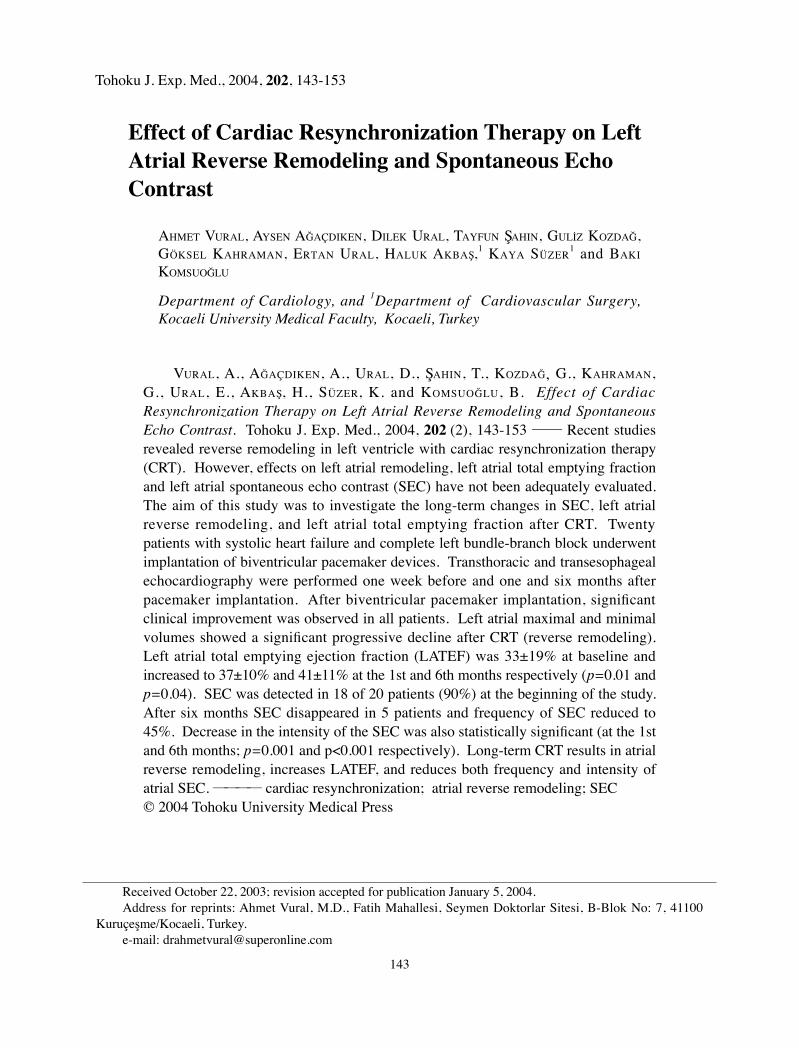
In patients with heart failure, the most fre-quent type of interventricular conduction delay is left bundle-branch block (Doval et al.1994; Aaronson et al. 1997). Conduction delay leads to abnormal ventricular depolarization and disturbance of the synchrony between inter- and intraventricular contraction and relaxation (Hardarson et al. 1987; Grines et al. 1989; Xiao et al 1992; Fahy et al. 1996; Nelson et al. 2000; Saxon et al. 2000). Disturbance of ventricular synchrony causes a shortening in diastolic filling and effective ejection time, reduces stroke vol-ume, prolongs mitral regurgitation and leads to an increase in global and regional wall stress (Grines et al. 1989; Xiao et al. 1992; Nelson et al. 2000). Previous studies have shown that interventricular conduction delay is also an independent predictor of deteriorated functional capacity and cardiac mortality in these patients (Doval et al.1994; Xiao et al. 1996; Aaronson et al. 1997).
Although not specifically studied for this particular group of patients, left atrial spontane-ous echo contrast (SEC) is frequently observed by transesophageal echocardiography in patients with left ventricular systolic dysfunction (Balck et al. 1991; Bilge et al. 1999). In patients with dilated cardiomyopathy (CMP) SEC is strongly related to left ventricular ejection fraction, cardiac output, left atrial diameter and left atrial flow ve-locities and is an indicator for subsequent throm-boembolic events (Daniel et al. 1988; Castello et al. 1990; Siostrzonek et al. 1993).
Cardiac resynchronization therapy leads to an acute and sustained improvement in left ven-tricular systolic function (Auricchio et al. 1999; Kass et al. 1999; Kerwin et al. 2000). Restoration of ventricular synchrony increases functional ca-pacity, lengthens exercise duration and improves life quality (Gras et al. 1998; Abraham 2000; Zardini et al. 2000; Cazeau et al. 2001).
Recent studies revealed a reverse remodel-ing in left ventricle with cardiac resynchroniza-tion therapy (Saxon et al. 1998; Lau et al. 2000; Stellbrink et al. 2001; Sogaard et al. 2002; Pitzalis et al. 2002). However, changes in left atrial
remodeling and effects on SEC have not been ad-equately evaluated. The aim of this study was to investigate the long-term effects of cardiac resyn-chronization therapy on left atrial SEC, left atrial reverse remodeling, left atrial total emptying frac-tion, left ventricular remodeling and systolic func-tion in patients with dilated CMP and left bundle branch block.
METHODS
PatientsThe study group consisted of 20 patients
with dilated CMP who had been referred to our clinic for biventricular pacemaker implantation (Table 1). All cases had PR interval ≧160 milli-seconds, QRS duration ≧150 milliseconds and left bundle branch block pattern. On echocar-diographic examination, all patients had ejection fraction <35%, left ventricular end-diastolic di-ameter >56 mm and asynchronous left ventricular contraction. Functional capacity was NYHA III in 13 cases and NYHA IV in 7 cases.
Exclusion criteria were age younger than 18 years, history of a recent acute coronary syndrome (<3 months), recent coronary by-pass operation (<3 months), terminal disease other than cardio-vascular diseases, expected survival less than 1 year, right bundle branch block and incomplete left bundle branch block.
All patients received optimal pharmacologi-cal treatment before and after pacemaker implan-tation. Oral anticoagulation therapy was given to three patients because of atrial fibrillation and one patients because of high-grade SEC before and after implantation. Betablokers were started to all patients and continued in those who could tolerate them.
Pacemaker implantationAll patients underwent ambulatory ECG
(electrocardiography) monitoring before pace-maker implantation. Electrophysiological study was performed in patients with complex ven-tricular arrhythmias. Medtronic InSync ICD (implantable cardioverler defibrilleder) (model
A. Vural et al.144 Cardiac Resynchronization Therapy and Left Atrial Function 145
7272, Medtronic Inc., Minneapolis, MN, USA) was implanted in three patients with a history of presyncope-syncope, clinically detected complex ventricular arrhythmias, and monomorphic ven-tricular tachycardia induced by electrophysiologi-
cal study. Three patient with permanent atrial fibrillation received Medtronic VVIR biventricu-lar pacemaker. In the other 14 patients Medtronic InSync III biventricular pacemaker (model 8042, Medtronic Inc.) was implanted.
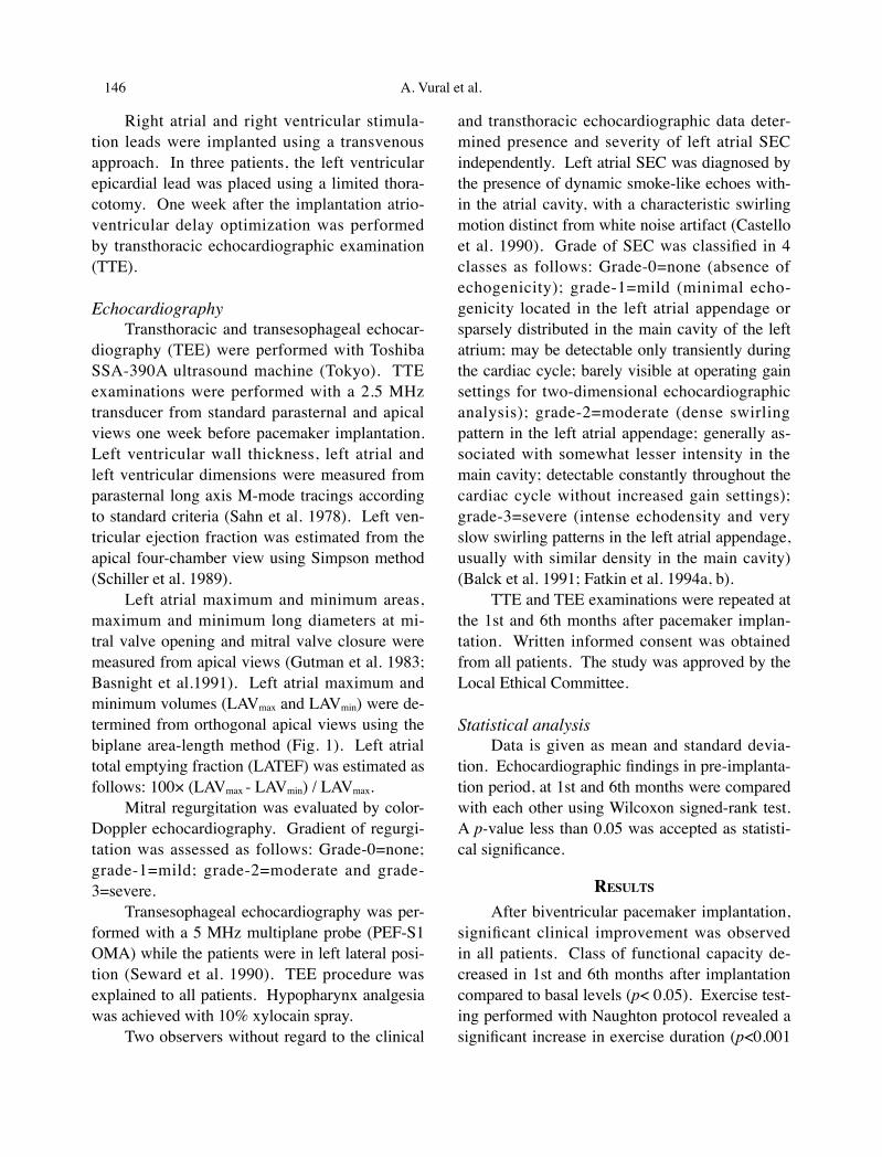
TABLE 1. Baseline clinical characteristics of the patients
Patient (=n) 20
Female/Male 7 / 13Etiology of cardiomyopathy (ischemic/nonischemic) 7 / 13NYHA III/IV 13 / 7Rhythm (sinus/atrial fibrillation) 17 / 3QRS (msec) 172±14Baseline PR interval (msec) 193±17LBBB (%) 100Diuretic (%) 90ACE-I (%) 75Digoxin (%) 40Beta-blocker (%) 45Spironolactone (%) 50Warfarin (%) 20Amiodarone (%) 35
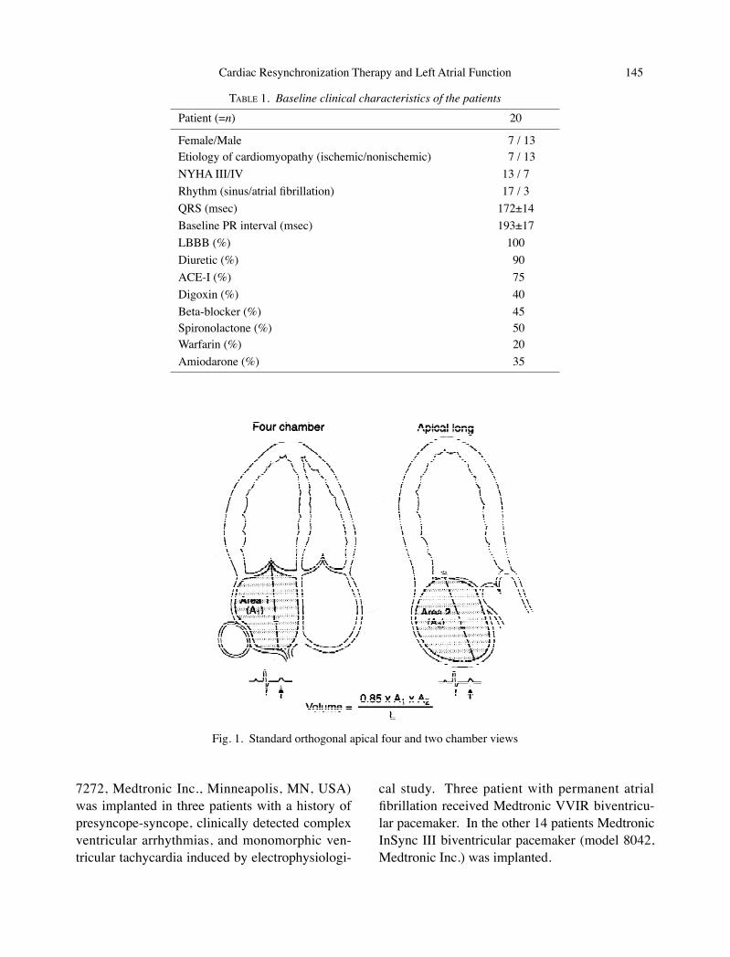
Fig. 1. Standard orthogonal apical four and two chamber views
A. Vural et al.146 Cardiac Resynchronization Therapy and Left Atrial Function 147
Right atrial and right ventricular stimula-tion leads were implanted using a transvenous approach. In three patients, the left ventricular epicardial lead was placed using a limited thora-cotomy. One week after the implantation atrio-ventricular delay optimization was performed by transthoracic echocardiographic examination (TTE).
EchocardiographyTransthoracic and transesophageal echocar-
diography (TEE) were performed with Toshiba SSA-390A ultrasound machine (Tokyo). TTE examinations were performed with a 2.5 MHz transducer from standard parasternal and apical views one week before pacemaker implantation. Left ventricular wall thickness, left atrial and left ventricular dimensions were measured from parasternal long axis M-mode tracings according to standard criteria (Sahn et al. 1978). Left ven-tricular ejection fraction was estimated from the apical four-chamber view using Simpson method (Schiller et al. 1989).
Left atrial maximum and minimum areas, maximum and minimum long diameters at mi-tral valve opening and mitral valve closure were measured from apical views (Gutman et al. 1983; Basnight et al.1991). Left atrial maximum and minimum volumes (LAVmax and LAVmin) were de-termined from orthogonal apical views using the biplane area-length method (Fig. 1). Left atrial total emptying fraction (LATEF) was estimated as follows: 100× (LAVmax - LAVmin) / LAVmax.
Mitral regurgitation was evaluated by color-Doppler echocardiography. Gradient of regurgi-tation was assessed as follows: Grade-0=none; grade-1=mild; grade-2=moderate and grade-3=severe.
Transesophageal echocardiography was per-formed with a 5 MHz multiplane probe (PEF-S1 OMA) while the patients were in left lateral posi-tion (Seward et al. 1990). TEE procedure was explained to all patients. Hypopharynx analgesia was achieved with 10% xylocain spray.
Two observers without regard to the clinical
and transthoracic echocardiographic data deter-mined presence and severity of left atrial SEC independently. Left atrial SEC was diagnosed by the presence of dynamic smoke-like echoes with-in the atrial cavity, with a characteristic swirling motion distinct from white noise artifact (Castello et al. 1990). Grade of SEC was classified in 4 classes as follows: Grade-0=none (absence of echogenicity); grade-1=mild (minimal echo-genicity located in the left atrial appendage or sparsely distributed in the main cavity of the left atrium; may be detectable only transiently during the cardiac cycle; barely visible at operating gain settings for two-dimensional echocardiographic analysis); grade-2=moderate (dense swirling pattern in the left atrial appendage; generally as-sociated with somewhat lesser intensity in the main cavity; detectable constantly throughout the cardiac cycle without increased gain settings); grade-3=severe (intense echodensity and very slow swirling patterns in the left atrial appendage, usually with similar density in the main cavity) (Balck et al. 1991; Fatkin et al. 1994a, b).
TTE and TEE examinations were repeated at the 1st and 6th months after pacemaker implan-tation. Written informed consent was obtained from all patients. The study was approved by the Local Ethical Committee.
Statistical analysisData is given as mean and standard devia-
tion. Echocardiographic findings in pre-implanta-tion period, at 1st and 6th months were compared with each other using Wilcoxon signed-rank test. A p-value less than 0.05 was accepted as statisti-cal significance.
RESULTSAfter biventricular pacemaker implantation,
significant clinical improvement was observed in all patients. Class of functional capacity de-creased in 1st and 6th months after implantation compared to basal levels (p< 0.05). Exercise test-ing performed with Naughton protocol revealed a significant increase in exercise duration (p<0.001
A. Vural et al.146 Cardiac Resynchronization Therapy and Left Atrial Function 147
and p<0.001) (Table 2).In echocardiographic examination, left ven-
tricular end-diastolic (LVEDD) and end-systolic diameters (LVESD), left ventricular end-diastolic (LVEDV) and end-systolic volumes (LVESV) de-creased significantly 1 and 6 months after implan-tation when compared to baseline values (Table 3).
Decrease in left ventricular volumes and diameters continued between the 1st and 6th months, however while the decrease in left ven-tricular end-diastolic volume was statistically
significant (p=0.04), reduction in end-systolic vol-ume remained statistically insignificant (p=0.06).
Ejection fraction, fractional shortening and cardiac index showed a significant increase after biventricular pacemaker implantation. Increase in cardiac index continued between the 1st and 6th months; however difference for the other param-eters was not statistically significant.
Left atrial diameter measured from para-sternal long axis view and left atrial minimum diameter measured from apical view decreased
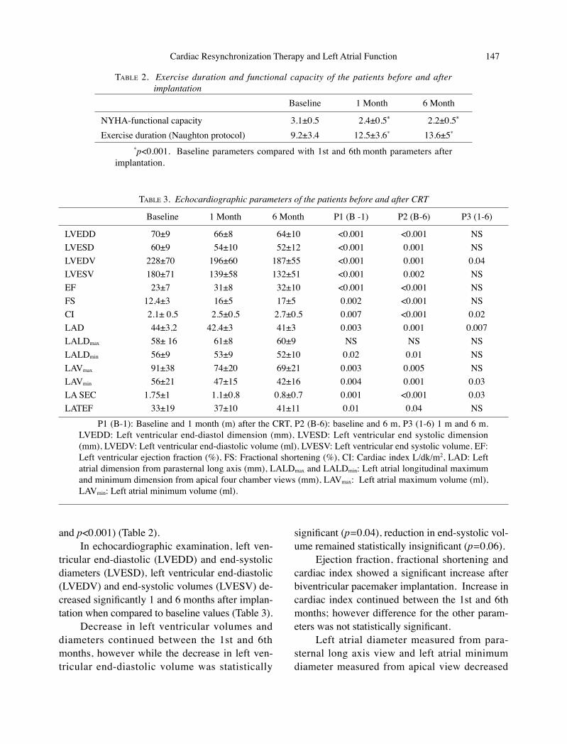
TABLE 2. Exercise duration and functional capacity of the patients before and after implantation
Baseline 1 Month 6 Month
NYHA-functional capacity 3.1±0.5 2.4±0.5* 2.2±0.5*
Exercise duration (Naughton protocol) 9.2±3.4 12.5±3.6* 13.6±5*
*p<0.001. Baseline parameters compared with 1st and 6th month parameters after implantation.
TABLE 3. Echocardiographic parameters of the patients before and after CRT
Baseline 1 Month 6 Month P1 (B -1) P2 (B-6) P3 (1-6)
LVEDD 70±9 66±8 64±10 <0.001 <0.001 NSLVESD 60±9 54±10 52±12 <0.001 0.001 NSLVEDV 228±70 196±60 187±55 <0.001 0.001 0.04LVESV 180±71 139±58 132±51 <0.001 0.002 NSEF 23±7 31±8 32±10 <0.001 <0.001 NSFS 12,4±3 16±5 17±5 0.002 <0.001 NSCI 2.1± 0.5 2.5±0.5 2.7±0.5 0.007 <0.001 0.02LAD 44±3,2 42.4±3 41±3 0.003 0.001 0.007LALDmax 58± 16 61±8 60±9 NS NS NSLALDmin 56±9 53±9 52±10 0.02 0.01 NSLAVmax 91±38 74±20 69±21 0.003 0.005 NSLAVmin 56±21 47±15 42±16 0.004 0.001 0.03LA SEC 1.75±1 1.1±0.8 0.8±0.7 0.001 <0.001 0.03LATEF 33±19 37±10 41±11 0.01 0.04 NS
P1 (B-1): Baseline and 1 month (m) after the CRT, P2 (B-6): baseline and 6 m, P3 (1-6) 1 m and 6 m. LVEDD: Left ventricular end-diastol dimension (mm), LVESD: Left ventricular end systolic dimension (mm), LVEDV: Left ventricular end-diastolic volume (ml), LVESV: Left ventricular end systolic volume, EF: Left ventricular ejection fraction (%), FS: Fractional shortening (%), CI: Cardiac index L/dk/m2, LAD: Left atrial dimension from parasternal long axis (mm), LALDmax and LALDmin: Left atrial longitudinal maximum and minimum dimension from apical four chamber views (mm), LAVmax: Left atrial maximum volume (ml), LAVmin: Left atrial minimum volume (ml).
A. Vural et al.148 Cardiac Resynchronization Therapy and Left Atrial Function 149
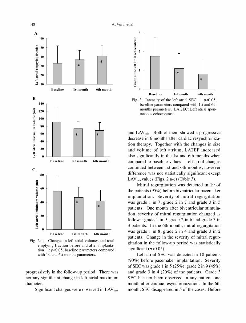
progressively in the follow-up period. There was not any significant change in left atrial maximum diameter.
Significant changes were observed in LAVmax
and LAVmin. Both of them showed a progressive decrease in 6 months after cardiac resynchroniza-tion therapy. Together with the changes in size and volume of left atrium, LATEF increased also significantly in the 1st and 6th months when compared to baseline values. Left atrial changes continued between 1st and 6th months, however difference was not statistically significant except LAVmin values (Figs. 2 a-c) (Table 3).
Mitral regurgitation was detected in 19 of the patients (95%) before biventricular pacemaker implantation. Severity of mitral regurgitation was grade 1 in 7, grade 2 in 7 and grade 3 in 5 patients. One month after biventricular stimula-tion, severity of mitral regurgitation changed as follows: grade 1 in 9, grade 2 in 6 and grade 3 in 3 patients. In the 6th month, mitral regurgitation was grade 1 in 8, grade 2 in 4 and grade 3 in 2 patients. Change in the severity of mitral regur-gitation in the follow-up period was statistically significant (p<0.05).
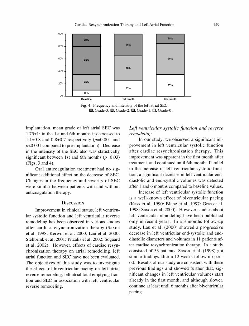
Left atrial SEC was detected in 18 patients (90%) before pacemaker implantation. Severity of SEC was grade 1 in 5 (25%), grade 2 in 9 (45%) and grade 3 in 4 (20%) of the patients. Grade 3 SEC has not been observed in any patient one month after cardiac resynchronization. In the 6th month, SEC disappeared in 5 of the cases. Before
��
��
��
��
��
��
�������� ��� ����� ��� �����
���
����
����
���
����
����
����
���
�
��
��
��
��
���
���
���
�������� ��� ����� ��� �����
���
���
����
���
����
����
��
����
�
��
��
��
��
�������� ��� ����� ���������
���
���
����
���
����
����
��
����
**
* *
**
��
��
��
��
��
��
�������� ��� ����� ��� �����
���
����
����
���
����
����
����
���
�
��
��
��
��
���
���
���
�������� ��� ����� ��� �����
���
���
����
���
����
����
��
����
�
��
��
��
��
�������� ��� ����� ���������
���
���
����
���
����
����
��
����
**
* *
**
��
��
��
��
��
��
�������� ��� ����� ��� �����
���
����
����
���
����
����
����
���
�
��
��
��
��
���
���
���
�������� ��� ����� ��� �����
���
���
����
���
����
����
��
����
�
��
��
��
��
�������� ��� ����� ���������
���
���
����
���
����
����
��
����
**
* *
**
A
B
C
**
* *
**
�
�
�
�
����� �� ��� ����� ��� �����
���
��
����
��
����
����
����
���
����
���
**
*
*
Fig. 3. Intensity of the left atrial SEC. *: p<0.05, baseline parameters compared with 1st and 6th months parameters. LA SEC: Left atrial spon-taneous echocontrast.
Fig. 2a-c. Changes in left atrial volumes and total emptying fraction before and after implanta-tion. *: p<0.05, baseline parameters compared with 1st and 6st months parameters.
A. Vural et al.148 Cardiac Resynchronization Therapy and Left Atrial Function 149
implantation, mean grade of left atrial SEC was 1.75±1; in the 1st and 6th months it decreased to 1.1±0.8 and 0.8±0.7 respectively (p=0.001 and p<0.001 compared to pre-implantation). Decrease in the intensity of the SEC also was statistically significant between 1st and 6th months (p=0.03) (Figs. 3 and 4).
Oral anticoagulation treatment had no sig-nificant additional effect on the decrease of SEC. Changes in the frequency and severity of SEC were similar between patients with and without anticoagulation therapy.
DISCUSSIONImprovement in clinical status, left ventricu-
lar systolic function and left ventricular reverse remodeling has been observed in various studies after cardiac resynchronization therapy (Saxon et al. 1998; Kerwin et al. 2000; Lau et al. 2000; Stellbrink et al. 2001; Pitzalis et al. 2002; Sogaard et al. 2002). However, effects of cardiac resyn-chronization therapy on atrial remodeling, left atrial function and SEC have not been evaluated. The objectives of this study was to investigate the effects of biventricular pacing on left atrial reverse remodeling, left atrial total emptying frac-tion and SEC in association with left ventricular reverse remodeling.
Left ventricular systolic function and reverse remodeling
In our study, we observed a significant im-provement in left ventricular systolic function after cardiac resynchronization therapy. This improvement was apparent in the first month after treatment, and continued until 6th month. Parallel to the increase in left ventricular systolic func-tion, a significant decrease in left ventricular end-diastolic and end-systolic volumes was detected after 1 and 6 months compared to baseline values.
Increase of left ventricular systolic function is a well-known effect of biventricular pacing (Kass et al. 1990; Blanc et al. 1997; Gras et al. 1998; Saxon et al. 2000). However, studies about left ventricular remodeling have been published only in recent years. In a 3 months follow-up study, Lau et al. (2000) showed a progressive decrease in left ventricular end-systolic and end-diastolic diameters and volumes in 11 patients af-ter cardiac resynchronization therapy. In a study consisted of 53 patients, Saxon et al. (1998) got similar findings after a 12 weeks follow-up peri-od. Results of our study are consistent with these previous findings and showed further that, sig-nificant changes in left ventricular volumes start already in the first month, and although slower, continue at least until 6 months after biventricular pacing.
Fig. 4. Frequency and intensity of the left atrial SEC.
25%35%
40%
50%45%
35%
15%20%
10%
25%
0%
20%
40%
60%
80%
100%
Baseline 1st month 6th month
Grade-3
Grade-2
Grade-1
Grade-0
, Grade-3;
25%35%
40%
50%45%
35%
15%20%
10%
25%
0%
20%
40%
60%
80%
100%
Baseline 1st month 6th month
Grade-3
Grade-2
Grade-1
Grade-0
, Grade-2;
25%35%
40%
50%45%
35%
15%20%
10%
25%
0%
20%
40%
60%
80%
100%
Baseline 1st month 6th month
Grade-3
Grade-2
Grade-1
Grade-0
, Grade-1;
25%35%
40%
50%45%
35%
15%20%
10%
25%
0%
20%
40%
60%
80%
100%
Baseline 1st month 6th month
Grade-3
Grade-2
Grade-1
Grade-0, Grade-0.
25%35%
40%
50%45%
35%
15%20%
10%
25%
0%
20%
40%
60%
80%
100%
Baseline 1st month 6th month
Grade-3
Grade-2
Grade-1
Grade-0
A. Vural et al.150 Cardiac Resynchronization Therapy and Left Atrial Function 151
Left ventricular reverse remodeling may be attributed to the improvement in left ventricular wall motion asynchrony, increase in contraction, and decrease in mitral regurgitation in addition to the decrease in left ventricular filling pressures. Decrease in peripheral resistance following the in-crease in cardiac output may also have decreased the left ventricular afterload and augmented the favorable remodeling in left ventricle (Capomolla et al. 2000).
The finding of progressive reduction in left ventricular volumes supports the previous data, that reverse remodeling observed after cardiac re-synchronization is not solely a result of correction of conduction delay, but is a change emerging in a time-dependent manner (Saxon et al. 1998; Lau et al. 2000).
Left atrial reverse remodeling and LATEFAfter resynchronization treatment, left atrial
diameters shortened and left atrial volumes di-minished significantly. LATEF has also showed a significant enhancement. Similar to the changes in left ventricle, left atrial changes were also more evident in the 1st month and slowed down in the following period.
Saxon et al. (1998) reported a significant re-duction in atrial volume index simultaneous with reverse ventricular remodeling 12 weeks after re-synchronization treatment. Findings of our study support the results of Saxon et al. and shows that changes in left atrium starts earlier after implanta-tion and continues at least until the 6th month. In our study, we also detected a significant increase in LATEF.
Atrial reverse remodeling and increase in the LATEF may be related to the improvement in left ventricular contraction, reduction of left ven-tricular end-systolic and end-diastolic volumes, decrease in mitral regurgitation and subsequently to the diminish in left atrial volume and pressure.
Rossi et al. (2002) showed an independent prognostic value of LAVmax in patients with dilat-ed CMP. Determinants of LAV were low ejection fraction, large left ventricular end-diastolic vol-
ume, degree of mitral regurgitation, atrial fibril-lation, and mitral flow E to A ratio. Patients with a LAV higher than 68.5 ml/m2 had a 3.8 times higher prognostic risk compared to patients with lower volumes. Decrease in LAVmax and reverse atrial remodeling achieved with biventricular pacing treatment may exert favorable effects on prognosis of patients with dilated CMP.
Effect of biventricular pacing on left atrial SEC
Left atrial SEC is a frequent finding in patients with left ventricular systolic dysfunc-tion (Siostrzonek et al. 1993; Bilge et al. 1999). Although it represents a relatively common event, underlying etiology is still not clear. In-vitro studies revealed that low blood flow velocity and erythrocyte aggregation play an important role in SEC development (Sigel et al. 1981).
Siostrzonek et al. (1993) showed that, in patients with dilated CMP, left atrial SEC occurs more frequently in those with larger left atrial diameters, lower cardiac output and left atrial flow velocities. In the same study, left ventricular systolic function and atrial fibrillation appeared as the most important determinants atrial flow velocity. In the study of Shiari and Mark (2000) left atrial diameter was an independent risk factor for the development of left atrial SEC in patients with sinus rhythm. Erythrocyte aggregation abnormalities in dilated CMP patients may also play a facilitating role for development of SEC (Siostrzonek et al. 1992).
In our study, left atrial SEC was detected is 18 (90%) of the patients before resynchronization treatment. The higher frequency of SEC ob-served in our study compared to previous reports (Vigna et al. 1992; Siostrzonek et al. 1993; Bilge et al. 1999) can be explained by more severe left ventricular systolic dysfunction and larger cardiac chambers of our cases.
After cardiac resynchronization treatment, frequency of SEC decreased gradually and in the 6th month it disappeared in 5 of 18 patients (28%). Grade 3 SEC detected in 20% of the patients

A. Vural et al.150 Cardiac Resynchronization Therapy and Left Atrial Function 151
before resynchronization treatment has not been observed in any patient after 1 month.
Decrease in frequency and intensity of SEC may also be explained by left atrial and ven-tricular reverse remodeling, and improved cardiac function. An enhanced left ventricular systolic function decreases the left ventricular end-diastol-ic pressure, and augments the rapid filling of the left ventricle. Decrease in left atrial pressure and the reverse remodeling of the left atrium improves the left atrial systolic function. These changes contribute to the decrease in blood stasis in left atrium and lead to decrease in SEC.
The relation between left atrial SEC, throm-bus formation and thromboembolic events has been confirmed in many studies (Daniel et al. 1988; Castello et al. 1990). The favorable effect of biventricular pacing on SEC suggests that a de-crease in thromboembolic events may be achieved by this new treatment.
ReferencesAaronson, K.D., Schwartz, J.S., Chen, T.M., Wong,
K.L., Goin, J.E. & Mancini, D.M.(1997) De-velopment and prospective validation of a clinical index to predict survival in ambulatory patients referred for cardiac transplant evalua-tion. Circulation, 95, 2260-2267.
Abraham, W.T. (2000) Rationale and design of a ran-domized clinical trial to assess the safety and efficacy of cardiac resynchronization therapy in patients with advanced heart failure: the Multi-center InSync Randomized Clinical Evaluation (MIRACLE). J. Card. Fail., 6, 369-380.
Auricchio, A., Stellbrink, C., Block, M., Sack, S., Vogt, J., Bakker, P., Klein, H., Kramer, A., Ding, J., Salo, R., Tockman, B., Pochet, T. & Spinelli, J. (1999) Effect of pacing chamber and atrio-ventricular delay on acute systolic function of paced patients with congestive heart failure. Circulation, 99, 2993-3001.
Balck, I.W., Hopkins, A.P., Lee, L.C., Walsh, W.F. & Jacobson, B.M. (1991) Left atrial spontaneous echo contrast: a clinical and echocardiographic analysis. J. Am. Coll. Cardiol., 18, 398-404.
Basnight, M.A., Gonzalez, M.S., Kershenovich, S.C. & Appleton, C.P. (1991) Pulmonary venous flow velocity: relation to hemodynamics, mitral flow velocity and left atrial volume and ejection
fraction. J. Am. Soc. Echocardiogr., 4, 547-558.Bilge, M., Güler, N., Eryonucu, B. & Aşker, M. (1999)
Frequency of left atrial thrombus and spontane-ous echocardiographic contrast in acute myo-cardial infarction. Am. J. Cardiol., 84, 847-849.
Blanc, J.J., Etienne, Y., Gilard, M., Mansourati, J., Munier, S., Boschat, J., Benditt, D.G. & Lurie, K.G. (1997) Evaluation of different ventricular pacing sites in patients with severe heart failure. Results of an acute hemodynamic study. Circu-lation, 96, 3273-3277.
Capomolla, S., Febo, O., Gnemmi, M., Riccardi, G., Opasich, C., Caporotondi, A., Mortara, A., Pinna, G.D. & Cobelli, F. (2000) Beta-block-ade therapy in chronic heart failure. Diastolic function and mitral regurgitation improvement by carvedilol. Am. Heart J., 139, 596-608.
Castello, R., Pearson, A.C. & Labovitz, A.J. (1990) Prevalence and clinical implications of atrial spontaneous echo contrast in patiens undergo-ing transesophageal echocardiography. Am. J. Cardiol., 65, 1149-1153.
Cazeau, S., Leclercq, C., Lavergne, T., Walker, S., Va r m a , C . , L i n d e , C . , G a r r i g u e , S . , Kappenberger, L., Haywood, G.A., Santini, M., Bailleul, C. & Daubert, J.C. (2001) Multisite Stimulation in Cardiomyopathies (MUSTIC) Study Investigators. Effect of multisite biven-tricular pacing in patients with heart failure and intraventricular conduction delay. N. Eng. J. Med., 344, 873-880.
Daniel, WG., Nellessen, U., Schröder, E., Nonnast-Daniel, B., Bednarski, P. & Lichtlen, P.R. (1988) Left atrial spontaneous echo contrast in mitral valve disease: an indicator for an increased thromboembolic risk. J. Am. Coll. Cardiol., 11, 1204-1211.
Doval, H.C., Nul, D.R., Grancelli, H.O., Perrone, S.V., Bortman, G.R. & Curiel, R. (1994) Ran-domised trial of low-dose amiodarone in severe congestive heart failure. Lancet, 344, 493-498.
Fahy, G.J., Pinski, S.L., Miller, D.P., McCabe, N., Pye, C., Walsh, M.J. & Robinson, K. (1996) Natural history of left bundle branch block. Am. J. Car-diol., 77, 1185-1190.
Fatkin, D., Herbert, E. & Feneley, M.P. (1994a) Hematologic correlates of spontaneous echo contrast in patients with atrial fibrillation and implications for tromboembolic risk. Am. J. Cardiol., 73, 672-676.
Fatkin, D., Kelly, R.P. & Feneley, M.P. (1994b) Rela-tions between left atrial appendage blood flow
A. Vural et al.152 Cardiac Resynchronization Therapy and Left Atrial Function 153
velocity, spontaneous echocardiographic con-trast and tromboembolic risk in vivo. J. Am. Coll. Cardiol., 23, 961-969.
Gras, D., Mabo, P., Tang, T., Luttikuis, O., Chatoor, R., Pedersen, AK., Tscheliessnigg, H.H., Deharo, J.C., Puglisi, A., Silvestre, J., Kimber, S., Ross, H., Ravazzi, A., Paul, V. & Skehan, D. (1998) Multisite pacing as a supplemental treatment of congestive heart failure: preliminary results of the Medtronic Inc Sync study. Pacing Clin. Electrophysiol., 21, 2249-2255.
Grines, C.L., Bashore, T.M., Boudoulas, H., Olson, S., Shafer, P. & Wooley, C.F. (1989) Functional abnormalites in isolated left bundle branch block: the effect of interventricular asynchrony. Circulation, 79, 845-853.
Gutman, J., Wang, Y.S., Wahr, D. & Schiller, N.B. (1983) Normal left atrial function determined by 2-dimensional echocardiography. Am. J. Cardiol., 51, 336-340.
Hardarson, T., Arnason, A., Eliasson, G.J., Palsson, K., Eyjolfsson, K. & Sigfusson, N. (1987) Left bundle branch block: prevalence, incidence, follow-up and outcome. Eur. Heart J., 8, 1075-1079.
Kass, D.A., Chen, C.H., Curry, C., Talbot, M., Berger, R., Fetics, B. & Nevo, E. (1999) Improved left ventricular mechanics from acute VDD pacing in patients with dilated cardiomyopathy and ventricular conduction delay. Circulation, 99, 1567-1573.
Kerwin, W.F., Botvinick, E.H., O’Connell, J.W., Merrick, S.H., DeMarco, T., Chatterjee, K., Scheibly, K. & Saxon, L.A. (2000) Ventricular contraction abnormalities in dilated cardiomy-opathy: effect of biventricular pacing to correct interventricular dyssynchrony. J. Am. Coll. Cardiol., 35, 1221-1227.
Lau, C.P., Yu, C.M., Chau, E., Fan, K., Tse, H.F., Lee, K., Tang, M.O., Wan, S.H., Law, T.C., Lee, P.Y., Lam, Y.M. & Hill, M.R. (2000) Reversal of left ventricular remodeling by synchronous biventricular pacing in heart failure. PACE, 23, 1722-1725.
Nelson, G.S., Berger, R.D., Fetics, B.J., Talbot, M., Spinelli, J.C., Hare, J.M. & Kass, D.A. (2000) Left ventricular or biventricular pacing im-proves cardiac function at diminished energy cost in patients with dilated cardiomyopathy and left bundle-branch block. Circulation, 102, 3053-3059.
Pitzalis, M.V., Iacoviello, M., Romito, R., Massari, F.,
Rizzon, B., Luzzi, G., Guida, P., Andriani, A., Mastropasqua, F. & Rizzon, P. (2002) Cardiac resynchronization therapy tailored by echo-cardiographic evaluation of ventricular asyn-chrony. J. Am. Coll. Cardiol., 40, 1615-1622.
Rossi, A., Cicoira, M., Zanolla, L., Sandrini, R., Golia, G., Zardini P. & Enriquez-Sarano, M. (2002) Determinants and prognostic value of left atrial volume in patients with dilated cardiomyopathy. J. Am. Coll. Cardiol., 40, 1425.
Sahn, D.J., De Maria, A., Kisslo, J. & Weyman, A. (1978) Recommendations regarding quantita-tion in M- mode echocardiography: results of a survey of echocardiographic measurements. Circulation, 58, 1072-1083.
Saxon, L.A., Kerwin, W.F., Cahalan, M.K., Kalman, J.M., Olgin, J.E., Foster, E., Schiller, N.B., Shinbane, J.S., Lesh, M.D., Merrick, S.H. (1998) Acute effects of intraoperative multisite ventricular pacing on left ventricular function and activation/contraction sequence in patients with depressed ventricular function. J. Cardio-vasc. Electrophysiol., 9, 13-21.
Saxon, L.A., Kumar, K.N. & De Marco, T. (2000) Heart failure and cardiac resynchronization therapies: US experience in the year 2000. Ann. Noninvas. Electrocardiol., 5, 188-194.
Seward, J., Khandheria, B.K., Edwards, W.D., Oh, J.K., Freeman, W.K. & Tajik, A.J. (1990) Biplanar transesophagial echocardiography: anatomic correlations, image orientation and clinical applications. Mayo. Clin. Prc., 65, 1193-1213.
Schiller, N.B., Shah, P.M., Crawford, M., DeMaria, A., Devereux, R., Feigenbaum, H., Gutgesell, H., Reichek, N., Sahn, D. & Schnittger, I. (1989) Recommandations for quantitation of the left ventricle by two-dimensional echocardiography. American Society of Echocardiography. Com-mittee on standarts, subcommittee on quanti-taion of two-dimensional echocardiograms. J. Am. Soc. Echocardiography., 2, 358-367.
Shiari, S. & Mark, V. (2000) Clinical and echocardio-graphic characteristics ofl left atrial spontane-ous echo contrast in sinus rhythm. J. Am. Coll. Cardiol., 35, 1932-1938.
Sigel, B., Coelho, J.C., Spigos, D.G., Flanigan, D.P., Schuler, J.J., Kasprisin, D.O., Nyhus, L.M. & Capek, V. (1981) Ultrasonography of blood during stasis and coagulation. Invest Radiol., 16, 71-76.
Siostrzonek, P., Koppensteiner, R., Kreiner, G., Madl, C., Gossinger, H.D., Heinz, G., Stumpflen, A.,
A. Vural et al.152 Cardiac Resynchronization Therapy and Left Atrial Function 153
Wendelin, B., Mosslacher, H. & Ehringer, H. (1992) Abnormal blood rheology in idiopathic dilated cardiomyopathy. Am. J. Cardiol., 69, 1497-1499.
Siostrzonek, P., Koppensteiner, R., Gossinger, H., Zangeneh, M., Heinz, G., Kreiner, G., Stumpflen, A., Buxbaum, P., Ehringer, H. & Mosslacher, H. (1993) Hemodynamic and hemorheologic determinants of left atrial spon-taneous echo contrast and thrombus formation in patients with idiopathic dilated cardiomyopa-thy. Am. Heart. J., 125, 430-434.
Sogaard, P., Egeblad, H., Kim, W.Y., Jensen, H.K., Pedersen, A.K., Kristensen, B.O. & Mortensen, P.T. (2002) Tissue Doppler imaging predicts improved systolic performance and reversed left ventricular remodeling during long-term cardiac resynchronization therapy. J. Am. Coll. Cardiol., 40, 723-730.
Stellbrink, C., Breithardt, O.A., Franke, A., Sack, S., Bakker, P., Auricchio, A., Pochet, T., Salo, R., Kramer, A. & Spinelli, J. (2001) The PATH-CHF (PAcing THerapies in Congestive Heart Failure) Investigators; CPI Guidant Congestive Heart Failure Research Group. Impact of car-
diac resynchronization therapy using hemody-namically optimized pacing on left ventricular remodeling in patients with congestive heart failure and ventricular conduction disturbances. J. Am. Coll. Cardiol., 38, 1957-1965.
Vigna, C., Russo, A., De Rito, V., Perna, G., Villella, A., Testa, M., Sollazzo, V., Fanelli, R. & Loperfido, F. (1992) Frequency of left atrial thrombi by transesophageal echocardiography in idiopathic and in ischemic dilated cardiomyopathy. Am. J. Cardiol., 70, 1500-1501.
Xiao, H.B., Brecker, S.G. & Gibson, D.G. (1992) Eff-ects of abnormal activation on the time course of left ventricular pressure pulse in dilated car-diomyopathy. Br. Heart J., 68, 403-407.
Xiao, H.B., Roy, C., Fujimoto, S. & Gibson, D.G. (1996) Natural history of abnormal conduction and its relation to prognosis in patients with dilated cardiomyopathy. Int. J. Cardiol., 53, 163-170.
Zardini, M., Tritto, M. & Bargiggia, G. (2000) The InSync Italian Registry: analysis of clinical outcome and considerations on the selection of candidates to left ventricular resynchronization. Eur. Heart J., 2, Suppl, 16-22.