efeito anti-hiperalgésico do óleo essencial - UEFS
189
1 UNIVERSIDADE ESTADUAL DE FEIRA DE SANTANA PROGRAMA DE PÓS-GRADUAÇÃO EM BIOTECNOLOGIA POLLYANA DE SOUZA SIQUEIRA LIMA EFEITO ANTI-HIPERALGÉSICO DO ÓLEO ESSENCIAL DE LIPPIA GRATA LIVRE E COMPLEXADO EM β-CICLODEXTRINA EM MODELOS ANIMAIS DE DOR CRÔNICA NÃO INFLAMATÓRIA E DOR NEUROPÁTICA Feira de Santana, BA 2018
-
Upload
khangminh22 -
Category
Documents
-
view
2 -
download
0
Transcript of efeito anti-hiperalgésico do óleo essencial - UEFS
1
UNIVERSIDADE ESTADUAL DE FEIRA DE
SANTANA
PROGRAMA DE PÓS-GRADUAÇÃO EM
BIOTECNOLOGIA
POLLYANA DE SOUZA SIQUEIRA LIMA
EFEITO ANTI-HIPERALGÉSICO DO ÓLEO ESSENCIAL
DE LIPPIA GRATA LIVRE E COMPLEXADO EM
β-CICLODEXTRINA EM MODELOS ANIMAIS
DE DOR CRÔNICA NÃO INFLAMATÓRIA E DOR
NEUROPÁTICA
Feira de Santana, BA
2018

2
POLLYANA DE SOUZA SIQUEIRA LIMA
EFEITO ANTI-HIPERALGÉSICO DO ÓLEO ESSENCIAL
DE LIPPIA GRATA LIVRE E COMPLEXADO EM
β-CICLODEXTRINA EM MODELOS ANIMAIS
DE DOR CRÔNICA NÃO INFLAMATÓRIA E DOR
NEUROPÁTICA
Tese apresentada ao Programa de Pós-Graduação em Biotecnologia da Universidade Estadual de Feira de Santana como requisito parcial à obtenção do grau de Doutor em Biotecnologia.
Orientadora: Profª. Drª. Angélica Maria Lucchese
Co-orientador: Prof. Dr. Lucindo José Quintans Júnior
Feira de Santana, BA
2018

3

4
Aos que agradeço, também dedico

5
AGRADECIMENTOS
Agradecer, agradecer, agradecer. Esta é uma palavra que não devo esquecer e nem me cansar
de repetir... Deus a cada manhã me dá uma nova chance de recomeçar de onde parei, de
começar de novo, pode ser da mesma forma ou de um jeito diferente, com infinitas
possibilidades e este dom é dEle, por isso a minha gratidão ao Deus que é meu TUDO!
Como se não bastasse me soprar a VIDA, Ele batalha junto e ainda levanta um exército. Um
Doutorado é uma grande batalha e ninguém vai sozinho! Se for, pára no início do caminho...
por isso eu agradeço ao meu Exército!
Às pessoas que me amam e olham pra mim como se eu fosse capaz, como se eu merecesse,
como se eu sempre valesse a pena. Este olhar é mágico! Por isso, agradeço a minha irmã
Jullyana, ela me enxerga assim, como se eu fosse capaz... e isto simplesmente não tem preço,
nem pode ser descrito...
Aos meus pais Osmar e Vanda, a minha irmã Rosana, ao meu esposo Wendell e aos meus
filhos Wendell Filho, Arthur e Mariana porque pra eles eu realmente valho a pena. Não
importa o que eu faça ou como faça, eles estão ao meu lado e, entendendo ou não, sabem que
tudo que eu executo, precisam estar por perto pra que seja bem sucedido. Por isso, eles
sempre estão lá...
À minha orientadora Angélica Maria Lucchese e ao meu co-orientador Lucindo Quintans-
Júnior porque as hierarquias lhes fazem selecionar pessoas...eu pedi e tive a honra de ser
escolhida por vocês para que eu pudesse aprender o quanto a Ciência é séria, o quanto é
necessário ter dedicação e afinco e quanto esforço e tempo são necessários para que um
trabalho tenha resultado. Não se pode escolher os melhores professores e achar que você
merece estar ali, pelo contrário você tem que fazer por merecer e quanto mais peso tem o
nome, maior a responsabilidade. Motivo de orgulho indescritível pronunciar seus nomes,
ilustríssimos orientadores, no meio acadêmico!
À Helton, secretário do Programa de Pós-graduação em Biotecnologia, por quem tenho
grande admiração pelo trabalho que faz, pela organização, educação e competência. Num
programa como este, é imprescindível alguém como você.
6
Às pessoas que trilharam comigo este caminho e abriram portas, facilitaram a minha vida, me
acolheram, me deram o prazer de conhecê-los: Profa Marilene Rocha, Horácio Bonfim,
Simone Teles, Ingrid, Gabriela, Edna, Serlyr, Thays Menezes, Hianna, Lucciano Brandão,
Débora Marchesine, Amanda, Sammya, Júnior e os fúncionários do Biotério, aos tantos
alunos de iniciação científca do LANEF-UFS que com tanta dedicação, foram peças
fundamentais na realização dos inúmeros testes deste trabalho.
À todos os colegas do Programa de Pós Graduação em Biotecnologia e do LAPRON.
Aos professores do Programa de Pós Graduação em Biotecnologia pela contribuição de sues
conhecimentos na minha formação.
À UEFS, por ser uma instituição a qual tenho um imenso carinho, responsável e fundamental
na minha formação.
À UFS, por fornecer toda a estrutura necessária para a execução dos meus experimentos e
quando não foi possível, abriu as portas de outras instituições,
À CAPES pelo financiamento dos estudos.
A todos vocês a minha eterna gratidão!
7
Percebi ainda outra coisa debaixo do sol:
os velozes nem sempre vencem a corrida;
os fortes nem sempre triunfam na guerra;
os sábios nem sempre têm comida;
os prudentes nem sempre são ricos;
os instruídos nem sempre tem prestígio;
pois o tempo e o acaso afetam a todos.
(Eclesiastes 9:11)
8
RESUMO
EFEITO ANTI-HIPERALGÉSICO DO ÓLEO ESSENCIAL DE LIPPIA GRATA LIVRE E COMPLEXADO EM β-CICLODEXTRINA EM MODELOS ANIMAIS DE DOR CRÔNICA NÃO INFLAMATÓRIA E DOR NEUROPÁTICA. Siqueira-Lima, de Souza Pollyana, Universidade Estadual de Feira de Santana, Feira de Santana-BA, 2018.
As dores crônicas associadas a estados não inflamatórios e neuropáticos são prevalentes e debilitantes permanecendo ainda sem um tratamento eficiente e seguro. Para tanto, este estudo foi delineado com o intuito de investigar através de ensaios funcionais e moleculares, o efeito anti-hiperalgésico do óleo essencial de Lippia grata livre e complexada em β-ciclodextrina (OEL/β-CD) em modelos animais de dor crônica não inflamatória (fibromialgia) e dor neuropática. Neste estudo foi possível demonstrar fortes evidências experimentais de como a β-ciclodextrina pode agir como um sistema de complexação de drogas seguro e de baixo custo, melhorando as propriedades farmacológicas dos terpenos, transforrnando estes produtos naturais em uma escolha atrativa para uso farmacológico. Ao realizar uma revisão sistemática, selecionando as espécies de Lippia com propriedades sobre o sistema nervoso central, este estudo observou que apesar de várias espécies apresentarem atividade analgésica poucos estudos exploraram o mecanismo de ação responsáveis por estes efeitos ou fizeram uma descrição fitoquímica detalhada ou ainda investigaram a toxicidade e/ou segurança terapêutica do uso continuado destas drogas. Apesar disto, os resultados das análises de extratos e óleos foram consistentes com a maioria dos relatos dos estudos etnofarmacológicos reafirmando a importância da medicina popular como guia para tais estudos. Utilizando um modelo de dor muscular não inflamatório, este estudo demonstrou que o OEL/βCD reduziu a hiperalgesia primária e secundária sem alterar a força muscular. Atribuiu estes efeitos ao possível envolvimento de receptores opiodérgicos e serotoninérgicos, corroborando com a hipótese de envolvimento da via descendente inibitória da dor, suportada por estudo in silico e pela expressão da proteina Fos no corno dorsal da medula, além da atividade antioxidante demonstrada pelo OEL e OEL/βCD. Ainda foi investigada a ação anti-hiperalgésica mecânica e térmica do OEL e OEL/βCD (24mg/kg) em modelos de dor neuropática (ligação parcial do nervo ciático) e de dor inflamatória persistente (CFA). A migração atenuada de leucócitos associada aos níveis reduzidos de TNF-α e IL-1β observados em modelo de pleurisia podem sugerir a redução de hiperalgesia e edema causados pela injeção intraplantar de CFA observadas após o tratamento oral com OEL e OEL/βCD. Este tratamento também reduziu o desenvolvimento de hiperalgesia mecânica e térmica desencandeada pela ligação parcial do nervo ciático. A redução dos níveis de TNF-α no nervo ciático e na medula bem como de fosforilação de NFκB e PKA nestas mesmos tecidos sugerem uma correlação positiva entre a ação do óleo e a redução do efeito álgico destes mediadores. O OEL (24mg/kg) e OEL/βCD/ (24mg/kg) ainda inibiram a nocicepção desencandeada pela injeção plantar de cinamaldeído (agonista do TRPA1) e mentol (agonista do TRPM8). O tratamento oral agudo e prolongado com OEL e OEL-βCD (24 mg/kg) não alterou a atividade motora dos animais. Os resultados citados indicam que o OEL e o OEL/βCD (24mg/kg) podem ser potencialmente interessantes para o desenvolvimento de drogas clinicamente relevantes para o tratamento das desordens dolorosas crônicas. Palavras-chave: dor crônica, dor não inflamatória, dor neuropática, óleo essencial, β-ciclodextrina, Lippia grata
9
ABSTRACT
ANTI-HIPERALGESIC EFFECT OF LIPPIA GRATA ESSENTIAL OIL FREE AND COMPLEXED IN β-CYCLODEXTRIN IN ANIMAL MODELS OF NON-INFLAMMATORY CHRONIC PAIN AND NEUROPATHIC PAIN Siqueira-Lima, de Souza Pollyana, Universidade Estadual de Feira de Santana, Feira de Santana-BA, 2018. Chronic pain associated with non-inflammatory and neuropathic states is prevalent and debilitating, and still remains without an efficient and safe treatment. For this purpose, this study was designed to investigate the antihyperalgesic effect of free and complexed Lippia grata essential oil on β-cyclodextrin (OEL/βCD) in animal models of chronic non-inflammatory pain (fibromyalgia) and neuropathic pain. In this study, it was possible to demonstrate strong experimental evidence of how β-cyclodextrin can act as a safe and low cost drug complexation system, improving the pharmacological properties of terpenes, transforming these natural products into an attractive choice for pharmacological use. In a systematic review, selecting the species of Lippia with properties on the central nervous system, this study observed that although several species present analgesic activity few studies have explored the mechanism of action responsible for these effects or have made a detailed phytochemical description or even investigated the toxicity and/or therapeutic safety of continued use of these drugs. Despite this, the results of the extracts and oils analyzes were consistent with most reports of ethnopharmacological studies reaffirming the importance of folk medicine as a guide for such studies. Using a non-inflammatory muscle pain model, this study demonstrated that OEL/βCD reduced primary and secondary hyperalgesia without altering muscle strength. It attributed these effects to the possible involvement of opiodergic and serotonergic receptors, corroborating the hypothesis of involvement of the pain inhibitory descending pathway supported by in silico study and the expression of the Fos protein in the dorsal horn of the medulla, in addition to the antioxidant activity demonstrated by OEL and OEL/βCD. The mechanical and thermal anti-hyperalgesic action of OEL and OEL/βCD (24mg / kg) in neuropathic pain (partial sciatic nerve ligation) and persistent inflammatory pain (CFA) models was also investigated. The attenuated migration of leukocytes associated with reduced levels of TNF-α and IL-1β observed in the pleurisy model may suggest the reduction of hyperalgesia and edema caused by the intraplantar injection of CFA observed after oral treatment with OEL and OEL/βCD. This treatment also reduced the development of mechanical and thermal hyperalgesia unleashed by the partial sciatic nerve ligation. Reduction of TNF-α levels in the sciatic nerve and medulla as well as phosphorylation of NFκB and PKA in these same tissues suggest a positive correlation between the action of the oil and the reduction of the algic effect of these mediators. The OEL (24mg/kg) and OEL/βCD (24mg/kg) did not inhibit the intraplantar injection-induced nociception of cinnamaldehyde (TRPA1 agonist) and menthol (TRPM8 agonist). The acute and prolonged oral treatment OEL and OEL-βCD (24 mg / kg) did not alter the motor activity of the animals. The results indicated that OEL/βCD (24mg/kg) may be potentially interesting for the development of drugs clinically relevant for the treatment of chronic pain disorders. Key words: chronic pain, non-inflammatory pain, neuropathic pain, essential oil, β-cyclodextrin, Lippia grata
10
LISTA DE FIGURAS
CAPÍTULO 2 Figure 1 – Structural formulas and names of some representative terpenes…………………………………………. 67 Figure 2 - Molecular structure of native cyclodextrins…………………………… 69 Figure 3 - A) Most stable conformation of β-caryophyllene/β-cyclodextrin complex (Adapted from Hădărugă et al., 2009). B) Mean plasma concentration-time profile of β-caryophyllene (BCP) after oral administration of free BCP or BCP/β-cyclodextrin (β-CD) inclusion complex (Adapted from Liu et al., 2013). C) Effects of β-caryophyllene (CA) or CA-β-cyclodextrin (CA-βCD) on formalin-induced orofacial nociceptive behavior. Vehicle (control), CA (10 or 20 mg/kg, p.o.) or CA-βCD (20 mg/kg, p.o.) were administered 90 min. before formalin injection (data shown results from first phase, 0–5 min., of the formalin test). Values represent mean + S.E.M. (n = 6 per group). *p<0.05 or **p< 0.001 versus control and ap<0.05 versus CA free (20 mg/kg, p.o.) (one-way ANOVA followed by Tukey’s test). (unpublished data) D) Fos-positive neurons in the lumbar spinal cord lamina I. Vehicle (control group, C) or β-caryophyllene/β-cyclodextrin (βCP-βCD) (20 mg/kg) were administered orally and, after 90…… 107 Figure 4 - A) Ten possible interactions of LIM and CDs obtained through molecular modeling. The green space represents the CD cavity (Adapted from Menezes et al., 2016). B) Time response curve for the antinociceptive effect of (A) p-cymene or (B) p-cymene/β-CD complex on acetic acid-induced writhing response in mice. Writhings were counted over 20 min following i.p. administration of acetic acid (0.65%). p-cymene or p-cymene/β-CD (40 mg/kg) was administered p.o. 0.5, 1, 2, 4, 8 or 16 h before acid acetic injection (0.65%). Control animals received an injection of vehicle by p.o. route. Each column represents mean ± S.E.M. (n = 8, per group). *p<0.05 or **p<0.001 vs. control (ANOVA followed by Tukey’s test) (Adapted from Quintans et al., 2013). C) Effect of carvacrol/β-cyclodextrin complex (CARV/β-CD) on the mechanical hyperalgesia induced by S180. Time-Effect Curve of CARV/β-CD (50 mg/kg) and CARV (100 mg/kg). *p< 0.05, **p < 0.01 and ***p < 0.001 vs. the control group (vehicle) (ANOVA followed by Tukey’s test) (Adapted from Guimaraes et al., 2015)…………………………………………………………………………... 109 CAPÍTULO 3 Figure 1. Effect of L. grata leaf essential oil complexed with β-cyclodextrin (LG-βCD) (6, 12 or 24 mg/kg; p.o.) or vehicle (saline; p.o.) on mechanical sensitivity induced by acidic saline in mice. Each point represents the mean ± S.E.M (n = 8, per group) of the ipisleteral paw withdrawal threshold. *p < 0.05, **p < 0.01 and ***p < 0.001 vs. control group (ANOVA followed by Tukey´s test)…………………………………………………………………………………
133 Figure 2. Effect of L. grata leaf essential oil complexed with β-cyclodextrin (LG- 134

11
βCD) (6, 12 or 24 mg/kg; p.o.) or vehicle (saline; p.o.) on the grip strength meter in mice. A. Hindpaw. B. Forepaw Values are expressed in mean ± S.E.M (n = 8, per group). ……………………………………………………………………… Figure 3. Effect of the pharmacological antagonists naloxone (5 mg/kg; i.p.) on muscle withdrawal thresholds in mice after treatment with L. grata leaf essential oil complexed with β-cyclodextrin (LG-βCD) (24 mg/kg; p.o.) or vehicle (saline; p.o.). Each point represents the mean ± S.E.M. of the ipsilateral muscle withdrawal thresholds (n = 8, per group). ***p < 0.01 vs. control group (ANOVA followed by Tukey´s test)…………………………………………..… 135 Figure 4. Effect of the pharmacological antagonists methysergide (1.5 mg/kg; i.p.) and yohimbine (2 mg/kg; i.p.) on muscle withdrawal thresholds in mice after treatment with L. grata leaf essential oil complexed with β-cyclodextrin (LG-βCD) (24 mg/kg; p.o.) or vehicle (saline; p.o.). Each point represents the mean ± S.E.M. of the ipsilateral muscle withdrawal thresholds (n = 8, per group). **p < 0.01 and ***p < 0.001 vs. control group (ANOVA followed by Tukey´s test)……………………………………………………………………….…… 136 Figure 5. Number of positive Fos cells in the dorsal horn in mice (A). Vehicle (B), or L. grata leaf essential oil complexed with β-cyclodextrin (LG-βCD) (24 mg/kg; p.o.) (C) was administered 90 min before the perfusion. Values expressed as mean ±S.E.M. (n = 6 per group). ∗∗p < 0.01 when compared with control (Student’s t test). 100 μm............................................................................ 136 Figure 6. Interactions of the bicyclogermacren with the receptors (A) alpha adrenergic and (B) 5-HT. We observed that bicyclogermacren-alpha adrenergic complex shows steric interactions with the residues TYR150, THR190, LLE180 and ARG140. The bicyclogermacren-5-HT complex presentes steric interactions with PHE341, THR140, VAL136, PHE340 and ASP135...................................... 137
Figure 7. Trans-carophyllene-μ opioid complex................................................. 138
CAPÍTULO 4 Figure 1. Effect of LG and LG/βCD on the number of PMNs (A) and on the levels of pro-inflammatory cytokines TNF-α (B) and IL-1β (C). The analyses were made from the exudates at 4 hours after the induction of pleurisy by carragenan injection. Data are reported as means ± SEM of 8 animals. Statistical analysis was performed using a one-way ANOVA followed by the Tukey’s test.**p<0.01, ***p<0.001 vs control group; ###p<0.01 vs sham group. 160 Figure 2. Effect of LG and LG/βCD on the time licking after the intraplantar injection of cinnamaldehyde 10 nM (A) and menthol 2,4 µM (B). Oral treatment was given 1 hour prior to injection and the paw licking or biting was recorded for 5 min (cinnamaldehyde) and 20 min (menthol). Data expressed as means ± SEM of 8 animals. Statistical analysis was performed using a one-way ANOVA followed by the Tukey’s test.**p<0.01, ***p<0.001 vs control group; ###p<0.01 vs sham group.
161
12
Figure. 3. Effect of LG and LG/βCD on (A) mechanical hyperalgesia 24 hours after the CFA injection and daily for 8 consecutive days; and on (B) paw edema 2 and 4 hours after the CFA injection. Data are reported as means ± SEM of 8-10 animals. Statistically significant differences from sham and control group as determined by One-way ANOVA followed by Bonferroni (A) and Tukey’s test (B). *p<0.05**p<0.01, ***p<0.001 vs control group; #p<0.05, ###p<0.01 vs sham group. 163 Figure. 4. Effect of LG and LG/βCD on (A) mechanical hyperalgesia response after partial sciatic nerve ligation. The measures were recorded before surgery (B), immediately before treatment (0 h), after treatment (1, 2, 3, 4, 6 and 8 h) and daily for 8 days with a two-day break (eighth and ninth day); and on (B) the time of latency on the hot plate 1 hour after the oral treatment. Data are reported as means ± SEM of 8-10 animals. Statistically significant differences from sham and control group as determined by One-way ANOVA followed by Bonferroni (A) and Tukey’s test (B). *p<0.05, ***p<0.001 vs control group. 164 Figure 5. Effect of LG and LG/βCD on the levels of pro-inflammatory cytokines TNF-α (A and D) and IL-1β (B and E) and anti-inflammatory cytokine IL-10 (C and F) on the sciatic nerve and spinal cord, respectively. Data expressed as means ± SEM of 8-10 animals. Statistically significant differences from sham and control group as determined by One-way ANOVA followed by Tukey’s test. *p<0.05**p<0.01, ***p<0.001 vs control group; #p<0.05, ##p<0.01, ###p<0.01 vs sham group. 166 Figure 6. Effect of LG and LG-βCD on phospho-NFκB/ NFκB immunocontent ratio (A) and PKAcα immunocontent (B) in the sciatic nerve and spinal cord of mice submitted to PSNL. Representative blots are shown (C). Data are reported as means ± SEM of 5-6 animals and expressed as percent of sham value. Statistically significant differences from sham and control group as determined by One-way ANOVA followed by Tukey’s test. *p<0.05**p<0.01, ***p<0.001 vs control group; #p<0.05, ##p<0.01, ###p<0.01 vs sham group 167 Figure 7. Effect of essential oil of Lippia grata on amplitude and sustained component of Ca2+ current (ICa) in dorsal root ganglion neurons from Wistar rats. (a) Representative traces of ICa recorded before and after 7s of oil application at 0.1, 0.3 or 1.0 µL/mL in the same cell. (b) Time course of peak Ca+2 currents showing their inhibition and recovery after washout. (d) Dose-response relationship between the essential oil concentration and the inhibition of the Ca+2 current amplitude. IC50 was 0.21 ± 0.02 and the Hill slope was 4.4 ± 0.8 169
13
LISTA DE TABELAS
CAPÍTULO 1 - Table 1 - Studies on CNS and analgesic profiles of Lippia genus 40 CAPÍTULO 2 - Table 1- Characteristics of the studies that described the formation of the inclusion complexes 72 Table 2 - Details of the included animal studies 97 Table 3 - Details of the included in vitro studies 101 Table 4 - Details of the included clinical studies 104 CAPÍTULO 3 Table 1 - Main secondary metabolites of Lippia grata leaf essential oil with respective MolDock energies regarding Alpha adrenergic, µ-opioid (µ-OR) and 5-HT receptors. 137 Table 2 - - In vitro antioxidant properties of the samples: L. grata leaf essential oil (LG) and L. grata leaf essential oil complexed with β-cyclodextrin (LG-βCD). 138

14
SUMÁRIO
1. INTRODUÇÃO 16
2 OBJETIVOS 22
3 CAPÍTULO 1 - CENTRAL NERVOUS SYSTEM AND ANALGESIC
PROFILES OF LIPPIA GENUS 24
4.
CAPÍTULO 2 - INCLUSION OF TERPENES IN CYCLODEXTRINS:
PREPARATION, CHARACTERIZATION AND
PHARMACOLOGICAL APPROACHES 59
5.
CAPÍTULO 3 - ANTI-HYPERALGESIC EFFECT OF Β-
CYCLODEXTRIN COMPLEXED WITH LIPPIA GRATA LEAF
ESSENTIAL OIL IN A CHRONIC MUSCULOSKELETAL PAIN
ANIMAL MODEL: COMPLEMENTED WITH A MOLECULAR
DOCKING AND ANTIOXIDANT SCREENING 125
6.
CAPÍTULO 4 - EVIDENCE FOR THE INVOLVEMENT OF THE
PKA PATHWAY AND INHIBITION OF VOLTAGE GATED CA2+
CHANNELS IN DRG NEURONS IN ANTIHYPERALGESIC
ACTIVITY PRODUCED BY THE ESSENTIAL OIL OF LIPPIA
GRATA/Β-CYCLODEXTRIN COMPLEX IN RODENT
NEUROPATHIC PAIN-LIKE MODELS 149
7 CONSIDERAÇÕES FINAIS 184
8. REFERÊNCIAS 186

15
INTRODUÇÃO

16
1 INTRODUÇÃO
A dor crônica é o problema de saúde humana mais prevalente, afetando mais de um
quarto da população mundial e aumenta sua incidência à medida que a população envelhece
(MOGIL, 2012). Esta desordem é caracterizada por uma dor significativa, duradoura além do
tempo de reparo tecidual, normalmente por meses ou até anos, portanto, de caráter contínuo
ou recorrente (WOLFE et al., 1990). Esses tipos de síndromes dolorosas são comuns em
indivíduos com doença cardíaca, acidente vascular cerebral, diabetes, herpes zoster e câncer
(ERNST et al., 2015). A dor crônica é um problema de saúde pública mundial e que tem
custos sociais e econômicos elevados. Em levantamento realizado pelo Institute of Medicine
Report, nos Estados Unidos, denominado de “aliviando a dor na América”, a dor é
responsável por despesas em saúde em torno de US$ 560-635 bilhões de dólares (GASKIN E
RICHARD, 2012). No Reino Unido afeta mais de 20% da população sendo a maior parte na
população economicamente ativa (KELLEHER et al., 2017). No Brasil este tipo de registro é
escasso e muitas vezes inconsistente, contudo, em um estudo realizado com pacientes com dor
crônica no Estado de São Paulo, verificou-se que 94,9% apresentava comprometimento da
atividade profissional, gerando prejuízos sociais e econômicos (KRELING et al., 2006).
Dentre as dores crônicas, as dores chamadas de “disfuncionais” são provavelmente as
de menor conhecimento neurofisiológico, pior prognóstico de tratamento devido a
refratariedade dos tratamentos existentes e menor adesão terapêutica (NAKAGURA, 2015). A
síndrome da fibromialgia (FM), uma modalidade de dor crônica, é caracterizada por dor
generalizada, hipersensibilidade, rigidez matinal, distúrbio do sono e fadiga pronunciada
(HSU et al., 2011). O critério diagnóstico proposto pelo Colégio Americano de Reumatologia
(ACR) inclui a dor difusa em conjunto com sensibilidade à palpação de 11 ou mais dos 18
tender points especificados (WOLFE et al., 2011). Apesar de ser considerada uma síndrome
reumatológica não inflamatória, várias evidências tem mostrado que citocinas pró-
inflamatórias como IL-6 e TNF-α estão elevadas no líquido cefalorraquidiano de pacientes
com FM (TSILIONI et al., 2016; BOISSONEAULT et al., 2017).
Outra dor crônica que é igualmente considerada um problema de saúde mundial é a
dor neuropática (DN). Tal condição é determinada como consequência direta de uma lesão ou
doença que afete o sistema somatossensorial (MIRANDA et al., 2016). A DN é uma entidade
complexa e de difícil tratamento, sendo considerada um dos maiores desafios da medicina
moderna (KISSIN, 2010). A DN é um tipo de dor que costuma ter um grande impacto na vida
de quem é acometido. Em comparação com outros tipos de dor, costuma ser mais intensa
17
,estar associada à incapacidade, e apresentar uma considerável diminuição na qualidade de
vida (ZHANG et al., 2014). Estudos com a população em geral usando instrumentos
validados de triagem observaram que 7-8% dos adultos têm dor crônica com características de
DN (BENNETT et al., 2012).
Tais lesões tornam-se cada vez mais frequentes na rotina dos atendimentos de urgência
dos hospitais em consequência do aumento da violência urbana, dos acidentes de trânsito,
acidentes profissionais e domésticos (DE SÁ et al., 2004; MAZZER et al., 2006), estando
entre os problemas neurológicos mais comuns. Apesar disso, a terapêutica atual tem se
mostrado pouco efetiva para a maioria dos pacientes acometidos (ZOCHODNE, 2008).
Apesar da complexidade desta disfunção e do número significativo de pacientes
acometidos, poucas terapias e intervenções estão disponíveis para deter ou reverter o dano que
lhes estão associados e, principalmente, sintomas como a dor crônica (ZOCHODNE, 2008). O
tratamento farmacológico da DN baseia-se em modular os mediadores inflamatórios
relacionados à lesão do nervo (por exemplo como TNF-alfa) ou no bloqueio das vias centrais
algésicas através do uso de fármacos opióides ou correlatos, bem como o uso de outras drogas
como estabilizadores de membrana (QUINTANS et al., 2014). Contudo, os eventos adversos
relacionados ao uso dos medicamentos e as baixas taxa adesão são limitadores do sucesso do
tratamento (ZOCHODNE, 2008).
De forma semelhante, o tratamento da FM é complexo e envolve formas de tratamento
heterogêneas devido as comorbidades relacionadas, tais como ansiedade, insônia, estresse,
fadiga, entre outras. De acordo com Menzies et al. (2016), apesar da descoberta de novas
drogas ou do reposicionamento de fármacos na última década com o uso da pregabalina,
duloxetina e milnacipran, o tratamento farmacológico da FM continua a usar abordagens que
são baseadas no perfil de dor central da doença, no entanto, não há um consenso sobre a
escolha ideal e a sequência do tratamento. Outro aspecto importante é a disponibilidade de um
número muito limitado de modelos experimentais, incluindo modelos animais, que
mimetizem a FM ou os principais sintomas e, consequentemente, o número de estudos
farmacológicos direcionados à busca de novas opções terapêuticas é insipiente na literatura.
Portanto, o tratamento farmacológico tanto da da FM como da DN continua a ser um
desafio para medicina atual, bem como o desenvolvimento de novas propostas terapêuticas.
De fato, as doenças crônicas apresentam altas taxas de abandono do esquema terapêutico,
sendo importante o estudo de sistemas mais modernos de administração de fármacos que com
um número menor de intervenções farmacológicas e/ou que diminuam a irritabilidade gástrica
possam ter: eficácia, segurança, diminuição dos efeitos adversos e, consequentemente, adesão
18
terapêutica (GOLDENBERG et al., 1996; GASKELL et al., 2014; OLIVEIRA et al., 2017).
Apesar da grande diversidade sintética derivada do desenvolvimento de química combinatória
e alto rendimento, os produtos naturais continuam sendo elementos extremamente importantes
das farmacopeias e estão relacionados com pelo menos 1/3 das novas drogas aprovadas pelo
Food and Drug Administration (FDA) (QUINTANS et al., 2014; OLIVEIRA et al., 2017;
PINA et al., 2017).
Os princípios ativos extraídos de plantas medicinais continuam despertando o interesse
científico e econômico, em virtude da grande diversidade de compostos com propriedades
farmacológicas, das quais se destacam as atividades analgésica e anti-inflamatória, e do
desenvolvimento de novos fármacos (CARLINI, 2003). Dutra et al. (2016) demonstraram que
vários estudos pré-clínicos e clínicos com algumas plantas medicinais brasileiras,
selecionadas em diferentes áreas de interesse, vêm sistematicamente sendo realizados por
grupos de pesquisa no Brasil e no exterior. Os autores destacam ainda, o crescente mercado
brasileiro de produtos à base de plantas, e os esforços dos pesquisadores brasileiros para
desenvolver novos fitomedicamentos.
Neste contexto, dentre os produtos naturais com propriedades terapêuticas e que
fornecem novas entidades químicas promissoras destacam-se dentre outras, as plantas
aromáticas ricas em óleos essenciais (BAKKALI et al., 2008). Os óleos essenciais (OEs) são
originados do metabolismo secundário das plantas e possuem composição química complexa,
destacando-se a presença de terpenos e fenilpropanoides (GONÇALVES et al., 2003). De
acordo com De Sousa (2011), Guimarães et al. (2013; 2014) e Gouveia et al. (2017) os
terpenoides são metabólitos secundários de grande interesse pela indústria farmacêutica
principalmente no estudo de compostos com potencial emprego como analgésicos e anti-
inflamatórios.
Paralelamente, o desenvolvimento de novas formulações farmacêuticas tende a alterar,
em breve, o conceito atual de medicamento (IGBAL et al., 2016). Assim, têm surgido nos
últimos anos, diversos sistemas de administração de fármacos com a finalidade de modelar a
cinética de liberação, melhorar a absorção, aumentar a estabilidade do fármaco ou vetorizá-lo
para uma determinada população celular (JANES et al., 2001; BREWSTER et al., 2008).
Esses sistemas terapêuticos surgiram da necessidade de minimizar os problemas que se
prendem com a administração das formas farmacêuticas tradicionais e, por exemplo, melhorar
as propriedades fisicoquímicas e farmacológicas de moléculas apolares (KURKOV e
LOFTSSON, 2013; SIQUEIRA-LIMA et al., 2016).
19
O uso de ciclodextrinas (CDs) em aplicações farmacêuticas envolvendo solubilização
e melhorias das propriedades farmacológicas especialmente de fármacos com baixa
polaridade tem se expandido exponencialmente a cada década, desde a descobertas das
primeiras ciclodextrinas (CDs), isoladas por Villiers, em 1891, a partir de produtos de
degradação de amido (GUEDES et al., 2008). As ciclodextrinas (CDs) são formadas por
unidades de glicopiranose unidas por ligações α (1-4), possuindo uma estrutura rígida em
forma de um cone truncado, onde os grupos OH secundários ligados aos carbonos C-2 e C-3
ocupam a base de maior diâmetro do tronco, enquanto as hidroxilas primárias ligadas ao
carbono C-6 localizam-se na base menor do tronco (CHALLA et al., 2005). Atualmente, as
CDs são utilizadas para melhorar algumas características físico-químicas de alguns fármacos,
nutracêuticos (termo não técnico para junção que resulta da combinação dos termos
"nutrição" e "farmacêutica") e/ou cosméticos (MARQUES, 2010). Os compostos lipofílicos,
tais como os óleos essenciais e seus metabólitos, quando incorporados às CDs, aumentam sua
solubilidade em água, estabilidade e eficácia farmacológicas (MARRETO et al., 2008;
SERAFINI et al., 2012; QUINTANS et al., 2013; QUINTANS-JÚNIOR et al., 2013). Ainda
alguns estudos têm demonstrado que complexos de inclusão contendo OEs ou monoterpenos
e CDs podem aumentar a meia vida plasmática, bem como a eficácia farmacológica, quando
comparado com os monoterpenos isolados, em modelos experimentais de analgesia e
inflamação (BRITO et al., 2015; OLIVEIRA et al., 2015; SIQUEIRA-LIMA et al., 2016).
O gênero Lippia (Verbenaceae) inclui aproximadamente 200 espécies entre ervas,
arbustos e pequenas árvores. As espécies estão distribuídas principalmente em regiões
tropicais e sub-tropicais, com destaque para países das Américas do Sul e Central e em alguns
países da África (TERBLANCHE e KORNELIUS, 1996). Espécies do gênero Lippia são
usadas principalmente para o tratamento de distúrbios gastrointestinais, respiratórios e como
analgésicos e anti-inflamatórios. Geralmente, o óleo essencial ou os compostos fenólicos
(flavonoides) desses extratos de plantas são descritos como princípios ativos (PASCUAL et
al., 2001).
Dentre as espécies de Lippia que estão sendo estudadas destaca-se a L. grata Schauer,
uma planta aromática, conhecida popularmente como “alecrim-serrote”, amplamente
distribuída no Nordeste Brasileiro, principalmente no Semi-Árido dos Estados da Bahia e
Sergipe (PASCUAL et al., 2001). As propriedades espasmolíticas e anti-inflamatória do óleo
essencial obtido das folhas de L. grata (OEL) foram demonstradas previamente, e esses
efeitos foram atribuídos à presença de carvacrol e timol, dois monoterpenos fenólicos
(SOUZA BRITO e SOUZA BRITO, 1993). De acordo com Viana et al (1981), a L. grata é

20
utilizada na medicina tradicional de alguns estados do Nordeste Brasileiro para o tratamento
de distúrbios dolorosos e inflamatórios.
Recentemente, nosso grupo demonstrou efeito antinociceptivo do OEL/βCD em
modelos de dor orofacial, mediado por mecanismos centrais e periféricos, com o provável
envolvimento do sistema glutamatérgico e inibição de citocinas pró-inflamatórias, como TNF-
α (SIQUEIRA-LIMA et al., 2013). Neste contexto em que as dores crônicas são condições de
difícil tratamento, que acarretam episódios dolorosos prolongados, considerando ainda a
existência de poucos tratamentos farmacológicos que produzam uma melhor condição clínica
aos pacientes, sem a produção de reações adversas consideráveis, torna-se desafiador o
desenvolvimento de novas preparações farmacêuticas utilizando plantas medicinais e/ou seus
metabólitos secundários que possuam aplicabilidade na hiperalgesia não inflamatória
experimental (fibromialgia experimental) e na dor neuropática ( lesão parcial do nervo
ciático).

21
OBJETIVOS
22
2 OBJETIVOS
2.1 OBJETIVO GERAL
Investigar o efeito anti-hiperalgésico do óleo essencial de Lippia grata livre e
complexado em β-ciclodextrina (OEL/β-CD) em modelo animal de dor crônica não
inflamatória (fibromialgia) e dor neuropática.
2.2 OBJETIVOS ESPECÍFICOS
Realizar revisão da literatura sobre os possíveis benefícios da inclusão dos compostos
terpênicos em ciclodextrinas e suas atividades farmacológicas;
Realizar revisão sistemática dos efeitos de plantas do gênero Lippia sobre o sistema nervoso
central (SNC);
Avaliar os efeitos do β-CD/OEL sobre a hiperalgesia mecânica em modelo animal de dor
crônica musculoesquelética caracterizando seu possível mecanismo farmacológico;
Avaliar o efeito do tratamento oral agudo e sub-crônico com OEL e OEL/β-CD sobre a
hiperalgesia e edema em modelo animal de dor inflamatória persistente e de dor neuropática.
Verificar o possível efeito do OEL e OEL/β-CD sobre a atividade motora de roedores.

23
CAPÍTULO 1

24
Central nervous system and analgesic profiles of Lippia genus
Pollyana S. Siqueira-Lima1,2#, Fabiolla R.S. Passos2,#, Angélica M. Lucchese1, Irwin R.A.
Menezes3, Henrique D.M. Coutinho3, Adley A.N. Lima4, Gokhan Zengin5, Jullyana S.S.
Quintans2*, Lucindo J. Quintans-Júnior2,*
#These authors contributed equally to the work.
1Department of Chemistry, State University of Feira de Santana, Feira de Santana, Bahia,
Brazil. 2Department of Physiology, Federal University of Sergipe, São Cristóvão, Sergipe, Brazil. 3Department of Biological Chemistry, Regional University of Cariri, Crato, Ceará, Brazil. 4Department of Pharmacy, Federal University of Rio Grande do Norte, Natal, Rio Grande do
Norte, Brazil. 5Science Faculty, Department of Biology, Selcuk University, Konya, Turkey.
*Corresponding authors: Lucindo Quintans-Júnior ([email protected]) and Jullyana S.S.
Quintans ([email protected])
Received 5 September 2018; Accepted 26 November 2018

25
Abstract: Many people use medicinal plants to relieve disorders related to the central nervous
system, such as depression, epilepsy, anxiety and pain, even though the effectiveness of most
of them has not yet been proven through scientific studies. Plants of the Lippia
genus,Verbenaceae, are widely used in ethnobotany as a food, for seasoning and in antiseptic
remedies. They are also marketed and used for the treatment of different types of pain,
including stomachache, abdominal pain and headache, as well as being used as sedatives,
anxiolytics and anticonvulsants. Despite their widespread use, there are no reviews on the
central nervous system profile of plants of this genus. Therefore, the databases Medline-
PubMed, Embase, Scopus and Web of Science were searched using the terms Lippia and
biologic activity. Thirty-five papers were found. Eleven species of Lippia showed central
nervous system activity, with leaves and the aerial parts of plants being the most commonly
used, especially in aqueous and ethanol extracts or volatile oil. The species are composed
mainly of terpenoids and phenylpropanoids, including polyketides, flavonoids and in less
quantity some alkaloids. Although several species of Lippia present analgesic activity, most
studies have not explored the mechanisms responsible for this effect, however, there is some
evidence that volatile oils and constituents of the extracts may be responsible for the relief of
some CNS disorders, but the effects on pain modulation seem to be the most exploited so far.
Key words: Verbenaceae, medicinal plants, pain, CNS disorders, inflammation

26
Introduction
The genus Lippia belongs to the family Verbenaceae and comprises about 250
herbaceous species of shrubs and is widely distributed all over Central and South America, as
well as tropical Africa (Terblanché and Kornelius, 1996; Aguiar and Costa, 2005). The
species are distributed in the arid regions of the southwestern United States of America, in the
deciduous tropical forests of Central America and in the tropical savannas (‘cerrados’) of
Brazil, which are the regions with high indexes of endemism (Salimena, 2002). Among the
prominent examples we can highlight the L. origanoides, which is popularly known as
‘oregano’ in Mexico, and is recognized in the Mexican Pharmacopoeia as a substitute for
common ‘oregano’ (Origanum vulgare). It is, therefore, widely used as a condiment in the
kitchen and in the preparation of several dishes (Oliveira et al., 2006). The dried and milled
leaves of some Lippia sp., or the flowers and fruits of this genus have been used as a
substitute for Thymus vulgaris (another species known as ‘oregano’) in spice mixtures for
pizzas and meats (Lorenzi and Matos, 2002; Santoro et al., 2007).
Brazil is considered to have the largest number of known species (Arthur et al., 2011)
represented by species conspicuous by their appearance during the short flowering phase and
also by their generally strong and pleasant fragrance (Bezerra et al., 1981). These features
make the use of this genus very widespread, ranging from in food preparation as a spice/herb,
in cosmetics, as well as in traditional medicine due to it being linked to a range of analgesic,
anti-inflammatory, antipyretic, antihypertensive and antimicrobial properties, as well as
having beneficial actions in relation to gastrointestinal conditions, menstrual symptoms, pain,
migraine and respiratory disorders (Pascual et al., 2001). Moreover, the Lippia genus has
shown to be of relative economic importance due to the different uses of its volatile oils and
the many medicinal uses of different species (Salimena, 2002), including their importance for
veterinary medicine and agriculture (Soares and Tavares-Dias, 2013).
Due the great medicinal and economic importance of plants and their wide distribution
across the regions of the country, the Brazilian government produced the National List of
Medicinal Plants Aimed by the Public Health System (SUS – Sistema Único de Saúde), a list
of vegetal species already used in traditional medicine which have potential to generate
products that could be relevant to the Public Health System. L. origanoides was included in
this list due to its pharmacological properties and its possible use in the development of new
pharmaceutical products, including medicaments (herbal medicines) and adjuvants
(Ministério da Saúde, 2009).
27
Among the biological effects reported for the genus Lippia, its central nervous system
properties are highlighted by a number of studies (Bezerra et al., 1981; Pascual et al., 2001;
Mamun-Or-Rashid et al., 2013). As the genus includes many aromatic plants rich in volatile
oils their pharmacological properties are commonly attributed to these oils. They are largely
comprised of terpene compounds which have already been shown to have clinical
applicability and are part of various drugs (Craveiro et al., 1988; Guimarães et al., 2013;
2014; Gouveia et al., 2017).
This study aims to examine research in relation to the use of species of the Lippia
genus directed to conditions related to the CNS. This will hopefully help to promote
improvements in methodological and theoretical methods; identify trends, overlaps and gaps
in research; as well as clarifying and summarizing the main existing works. Other studies of
this nature have been described in the literature, however, there has been no systematic review
focused on the correlation between the pharmacological effects and the chemical composition
of the plants and their influence on the CNS (Terblanché and Kornelius, 1996; Pascual et al.,
2001; Catalan and De Lampasona, 2002; Hennebelle et al., 2006; 2008, Oliveira et al., 2006;
Ombito et al., 2015).
Many studies just describe the use of plants of this genus in traditional medicine, often
with contradictory results, or only their use in food or as raw material). Therefore, considering
the importance of this plant genus and the absence of systematic reviews of its
pharmacological importance through preclinical studies, we carried out this extensive
systematic survey in order to support translational studies and/or new preclinical studies.
Search strategy
Four digital databases, Medline-PubMed, Embase, Scopus and Web of Science were
used to search for studies that met the inclusion criteria: preclinical animal-model studies of
CNS pharmacological studies of Lippia species. The database search was performed in the
period up to March 30, 2018 using the MesH and free search terms Lippia and biologic
activity. The search strategy structure was designed to include any study published that
assessed the pharmacological pre-clinical profile of the Lippia species. The search was limited
to animal-model studies. There was no contact with researchers and/or attempts to identify
non-published data.
All the electronic titles found, selected abstracts and complete texts from articles were
revised independently by at least two reviewers (JSSQ, PSSL). Discrepancies over the
inclusion/exclusion of studies were solved with a consensus meeting. Studies in humans,

28
literature reviews, editorials/letters, case reports, and isolated substances were excluded. The
information extracted included data on the Lippia species, the part of the plant used, their
main compounds, type of animal used, model of study and key findings.
Outcomes
A total of 3817 abstracts and citations were electronically identified in the first search.
After the exclusion of duplicate articles and the triage of relevant titles and abstracts, 776
titles were included in our list for analysis, trying to identify studies that evaluated Lippia that
met our inclusion criteria. Thirty five articles met the inclusion/exclusion criteria previously
established and were included in our review .
The search of the databases showed that studies related to the genus Lippia included a
broad range of species, with 27 different ones being the subject of research for different
purposes This alone confirms the importance of the ethnopharmacological study as a basis for
initiating preclinical studies. The areas of interest found in the articles are mainly
phytochemical studies, CNS disorders, pain and inflammation. Our survey identified the
following nine plant species as being the subject of research in studies: L. alba, L. multiflora,
L. gracilis, L. grata, L. origanoides, L. graveolens, , L. geminata, L. origanoides, and L.
adoensis. So, the number of species studied is still very small in relation to the number of
species of the genus, which reinforces our argument that more studies of this important genus
are required.
We found that the parts of the plants mainly used in the experimental protocols in the
studies were leaves and aerial parts, particularly as aqueous and ethanol extracts or volatile
oils. The part of the plants used is similar to that found in ethnopharmacological studies,
which shows that they are primarily used in infusions that require the leaves and aerial parts
(Oliveira, 2004; Hennebelle et al., 2008; De Carvalho Nilo Bitu et al., 2015). As we expected,
due to the presence of many aromatic plants in the genus which are used for the treatment of
diseases, studies with volatile oil, mainly terpenoids and phenylpropanoids, with some
polyketides and in less quantity some alkaloids, predominate in our survey (36%) (De Sousa,
2011).
Many VO mainly comprising terpenes whose various activities and mechanisms of
action have already been well described in the literature were found. The studies identified do
not state if the presence of terpenes influenced the selection of these mixtures (VO) for
pharmacological evaluation by the authors. However, we hypothesize that this may well be
the case given the number of studies which support the idea that VO are pharmacologically
29
interesting because of the presence of terpenes (Guimarães et al., 2013; Lillehei and Halcon,
2014; De Cássia da Silveira e Sá et al., 2017). Studies investigating its applicability in
diseases that cause pain, inflammation, oxidative stress imbalance and CNS disturbances are
common (Dobetsberger and Buchbauer, 2011; El Hadi et al., 2013; Ali et al., 2015).
Moreover, terpenes has demonstrated strong bioactivity on the modulation of cytokines and in
the inflammatory process, central nervous system activity, pain and nerve sensitization,
among other interesting pharmacological targets (González-Burgos and Gómez-Serranillos,
2012; Quintans et al., 2019; Santos et al., 2019).
Most of the studies performed an extensive phytochemical analysis to describe the
main compounds (terpenoids: carvacrol, p-cymene, o-cymene, thymol and E-caryophyllene,
and others chemical classes, such as flavonoids, phenolic acid, and alkaloids) (Table 1).
Terpenes and terpenoids are the primary constituents of the VO of many types of
medicinal plants and flowers. They are derived biosynthetically from units of isoprene, which
has the molecular formula C5H8. Terpenes are chemical entities having low molecular weight
and usually low water solubility. They can penetrate the blood-barrier and produce their
effects, anxiolytic, sedative and anticonvulsant, on the CNS (Quintans-Júnior et al., 2008; De
Sousa, 2011). The articles found in our review highlighted action on the CNS through the
GABAergic pathways, which corroborated the pharmacological evidence of the anxiolytic,
sedative, myorelaxant and anticonvulsant properties (Heldwein et al., 2012; Razavi et al.,
2017).
Moreover, drugs that enhance GABA-mediated inhibitory transmission, and
subsequently affect neuronal repetitive firing, can be of relevance in alleviating several
painful syndromes, since they can produce a membrane stabilizing effect on sensory neurons
and/or enhance intrinsic analgesic responses (Jasmin et al., 2004; Enna and McCarson, 2006).
This evidence may help to explain the fact that most of the studies presented in Table 1 (64%)
are studies of the possible effect of plants of this genus on pain management (or simple
screening for analgesic drugs, which was the most common type of study described).
Obviously, the fact that previous ethnopharmacological surveys had described the great
concentration of terpenes or flavonoids in the Lippia genus and their analgesic and anti-
inflammatory properties was important in encouraging many research groups to explore this
genus (Siqueira-Lima et al., 2014). Additionally, VO and extracts from Lippia genus with
antioxidant properties, which modulate the inflammatory process by reducing the production
of proinflammatory cytokines (such as IL1-β, TNF-α and others) and act on
neurotransmission systems that participate in descending pain-inhibitory mechanisms
30
corroborate this search more directed by the researchers (Leyva-López et al., 2016; Siqueira-
Lima et al., 2014; 2017).
Our review provides evidence that the Lippia genus is rich in flavonoid compounds, at
least in the species studied here, the most commonly described being extracts rich in
polyphenols (mainly flavonoids) and naringenin, apigenin, nodifloretin A, nodiflorin A,
nodifloridin A and others (Table 1). Flavonoids are a class of plant polyphenols that are
consumed in the human diet via vegetables, fruits, cereals, spices, and other plant-based
products (Pandey and Rizvi, 2009; Jaeger et al., 2017). Flavonoids are probably one of the
most important NP due to the potent biological molecules and are already described and used
in clinical practice for the treatment of various diseases, as they have a range off antioxidant,
anti-inflammatory, analgesic, anxiolytic and anticonvulsant properties (Diniz et al., 2015;
Nijveldt et al., 2001).
Lippia species
Lippia alba (Mill.) N.E.Br. ex Britton & P.Wilson, popularly known as “cidreira” (in
the south and southeast of Brazil) and “Basula” (in Hindi, in India), is a plant which is present
in Central and South America, being recorded in all regions of Brazil (Tavares et al., 2005).
Lippia alba is a fast growing plant with a mounding habit and round lavender-like blossoms
(Haldar et al., 2012). The main pharmacological studies, arising from folk use, found it had
varied activities including cardiovascular (Gazola et al., 2004), anticonvulsant (Soares, 2001),
sedative, analgesic, bronchodilator (Carvalho et al., 2018) antioxidant and anti-inflammatory
(Viana, 1998; Zétola et al., 2002; Hennebelle et al., 2008; Haldar et al., 2012; Hatano et al.,
2012), as well as antiulcerogenic effects (Pascual et al., 2001).
The composition of its VO presents quantitative and qualitative variation, leading to
its separation into chemotypes according to its major components (De Abreu Matos et al.,
1996; Frighetto et al., 1998; Zoghbi et al., 1998; Hennebelle et al., 2008). In Brazil, there are
at least three major chemotypes of L. alba with a large variation in some terpenoids,
especially citral, carvone and linalool; therefore having different pharmacological effects (De
Abreu Matos et al., 1996; Yamamoto, 2006; Linde et al., 2016).
The remarkable action of this plant species on the CNS was characterized by the
presence of nine articles demonstrating biological activities typically involving the central
pathways, including analgesic (with a central component), sedative, anticonvulsant and
anxiolytic effects. Viana et al. (1998) compared the analgesic and anti-inflammatory effects of
the VO from the leaves of two chemotypes: “citral” (type I) and “carvone” (type II). The
31
antinociceptive effect was more consistent and marked in the type II, but this effect was not
reversed by naloxone (an opioid antagonist). A central analgesic effect was also evident with
type I (rich in citral). Interestingly, Gonçalves et al. (2008) and Quintans-Junior et al. (2011)
demonstrated the central analgesic effects of carvone and citral with no involvement of the
opioid system, but with possible participation of blocking Na+-channels and the GABAergic
system, respectively. In addition, the anti-nociceptive action of citral was found to involve
significant activation of the 5-HT2A serotonin receptor (Nishijima et al., 2014). Sousa et al.
(2015) showed that VO of Lippia alba and its main constituent citral block the excitability of
rat sciatic nerves.
Haldar et al. (2012) reported that flavonoids in an aqueous extract of L. alba. have
analgesic and anti-inflammatory effects, which they attributed to the presence of polyphenol
compounds that inhibited the enzyme cyclooxygenase and subsequently inhibited
prostaglandin synthesis.
The most striking CNS effects for this plant are sedative, anticonvulsant and
anxiolytic. The anticonvulsant activity of L. alba (type I, “citral”) has been demonstrated in
classic screening tests for new antiepileptic drugs (Neto et al., 2009). Citral is a sedative and
anticonvulsant compound that produces its effects through , at least in part, the involvement
of the GABAergic system, but its effect on the stabilization of the neuronal membrane and the
blockade of families of ion channels act synergistically (Stotz et al., 2008; Quintans-Júnior et
al., 2010).
The anxiolytic effect studied elucidate a possible GABAergic action and it is believed
that this action is related to the presence of non-volatile substances (phenylpropanoids,
flavonoids and/or inositols), and also to volatile terpenoids (myrcene, citral, limonene and
carvone), which have been previously shown to have anticonvulsant and anxiolytic properties
(Viana et al., 2000; Zétola et al., 2002; Neto et al., 2009; Zhu et al., 2014).
In fact, much of the pharmacological evidence relating to Lippia genus, including L.
alba, is strongly associated with the presence of terpenes (mainly monoterpenes) with
remarkable action on the CNS (Passos et al., 2009; Guimarães et al., 2013; 2014; Quintans-
Júnior et al., 2013; Pina et al., 2017; Habtemariam, 2018).
Lippia multiflora Moldenke, popularly known as “chá-de-gambia”, is a species widely
used as an infusion in Africa. Traditionally, its leaves are used as a hot beverage (tea) to treat
fever, gastrointestinal disturbances, enteritis, and coughing (Adesina et al., 1995). The VO
isolated from its leaves and flowers contains p-cymene, thymol and carvacrol (Abena et al.,
2003), which have been attributed analgesic and antipyretic activities. Iwalewa et al. (2007)
32
demonstrated a decrease in the plasma level of both nitric oxide (NO) and malondialdehyde
(MDA) that was closely associated with the anti-inflammatory and analgesic activities
produced by VO.
p-Cymene and carvacrol produce an analgesic effect by the involvement of descending
pain-inhibitory mechanisms; by inhibition of pro-inflammatory cytokines (such as IL1β,
TNF-α, IL-4, TGF-β, and IL-17), by enhancement of anti-inflammatory cytokines (IL-10),
and by involvement of the opioid system (Lima et al., 2013; Guimarães et al., 2014; De
Santana et al., 2015; Kianmehr et al., 2016). Thus, these terpenes seem to be key to the effects
of VO from L. multiflora.
The pharmacological activity of the aqueous extract of L. multiflora was assessed by
Abena et al. (1998) who concluded that it had tranquilizer and analgesic profiles. However,
comparing the extract and VO to confirm the previous activities, the study suggested that the
crude extract produced more muscle relaxant effects and the VO was more analgesic (Abena
et al., 2001). The phytochemical screening of its crude extract demonstrated the remarkable
presence of alkaloids, tannins, flavonoids and saponins (Valentin et al., 1995; Oladimeji et al.,
2001). Although not an outstanding feature of the crude extract, the significant analgesic and
anti-inflammatory effects were attributed to the presence of alkaloids (Jigam et al., 2009).
Moreover, some of the most important monoterpenes found abundantly in L.
multiflora volatile oil inhibited allergic inflammation by the modulation of inflammatory
cytokines (Lima et al., 2013; Pina et al., 2018), which are synthesized within the CNS by glial
cells and neurons, and have modulatory functions on these same cells via interactions with
specific cell-surface receptors contributing, at least in part, to the central effects of terpenes
and similar compounds (Benveniste, 1998; Cho et al., 2017).
Lippia gracilis Schauer (“alecrim-da-chapada”) is a shrubby aromatic species that is
distributed in the Brazilian Northeast with a high occurrence in the states of Bahia, Segipe and
Piauí. It is probably one of the most popular Lippia species in the Brazilian Northeast due to
its medicinal properties and food application (Albuquerque et al., 2012). The VO of L.
gracilis is composed mainly of mono- and sesquiterpenes, and the main compounds are p-
cymene, γ-terpinene, carvacrol and thymol (Pessoa et al., 2005; Neves et al., 2008; Silva et
al., 2008; Mendes et al., 2010; Teles et al., 2010; Guilhon et al., 2011).
As already described here and reinforced in the excellent reviews published by De
Sousa (2011), De Cássia Da Silveira E Sá et al. (2013) and Guimarães et al. (2013; 2014) the
analgesic and anti-inflammatory profiles produced by the VO are attributed to the presence of
33
terpenes. They act by inhibiting inflammatory mediators, such as cytokines, and reducing
neuronal excitability in certain CNS areas.
Studies point out that thymol (another important compound found in VO) modulates
voltage-dependent Na+-channels (Haeseler et al., 2002), K+-channels (Elliott and Elliott,
1997), GABA A receptors (Mohammadi et al., 2001), α and -adrenergic receptors (Beer et
al., 2007), as well as being related to prostaglandin synthesis (Anamura et al., 1988), which
together may contribute to the control of painful sensations produced by this terpene.
The antinociceptive profile of p-cymene, the main compound from L. gracilis VO,
was assessed in animal models of pain in respect of contortions induced by acetic acid, the
formalin test and hot-plate test.; It has also been reported to have an anti-inflammatory effect
(Bonjardim et al., 2012). Moreover, Santana et al. (2011) reported an antinociceptive effect
through the opioid system which corroborates other studies that describe the effects of this
terpenoid on the CNS, due to, among other factors, its antioxidant profile (De Oliveira et al.,
2012; 2015).
Furthermore, the analgesic profile of carvacrol has already been described consistently
in a number of papers and patents for new drugs or pharmaceutical products (Guimarães et al.,
2014; Suntres et al., 2015; Oliveira et al., 2016). The effects of the VO of L. gracilis are
attributed to its anti-inflammatory actions, rather than to its profile on the CNS. Carvacrol is
able to block the recruitment of neutrophils, to reduce the release of IL-1β, TNF-α and NO,
and enhance levels of IL-10, resulting in a decrease in the production of inflammatory factors
and block hyperalgesic behavior (Guimarães et al., 2010; 2012; Lima et al., 2013; Pina et al.,
2017). It was also able to regulate COX-2 expression through its agonistic effect in PPARγ
(Hotta et al., 2010). Controversially, some authors attribute the effect on the opioid system,
but these data are contradictory in different articles and no evidence of direct involvement of
the opioid system in the analgesic effect of carvacrol has been found (Guimarães et al., 2010;
Cavalcante Melo et al., 2012).
Additionally, the monoterpenes found in the VO are extensively described as analgesic
and its lipophilic characteristics and molecular size facilitates both its passage through the
blood brain barrier, to produce local actions such as in relation to oxidative balance, to
manage the production of inflammatory factors (such as cytokines) and to directly block ion
channels (Abena et al., 2003; Mendes et al., 2010; González-Burgos and Gómez-Serranillos,
2012; Guimarães et al., 2013; 2014; Gouveia et al., 2017; Pina et al., 2017).
Guilhon et al. (2011) also investigated the mechanism of action producing the
analgesic behavior of the VO of L. gracilis. The authors clearly indicated the involvement of
34
cholinergic receptors in this process (as atropine inhibited the antinociceptive effect) and the
involvement of the opioid system (by antagonism produced by naloxone). However, the
different chemotypes may elicit significantly different biological responses as reported by
Mendes et al. (2010) (thymol - major component) and Guilhon et al. (2011) (carvacrol - major
component). Chemotypes of Lippia species with their different chemical profiles are equally
interesting for comparative study, since the difference in concentrations of the major
compounds is an area that should be better explored by drug manufacturers.
Another monoterpene presents in these oils, the γ-terpinene, was evaluated by De
Brito Passos et al. (2015); the authors demonstrated that γ-terpinene antinociception was
inhibited in the presence of naloxone, glibencamide, atropine and mecamylamine, suggesting
that this antinociceptive effect in models of chemical nociception was produced through the
cholinergic and opioid systems. These effects reflect the previously described
pharmacological profile of the major terpenes of VO which has a more complex, complete
and therapeutic action than the isolated terpenes.
The therapeutic effects on the CNS of thymol and carvacrol (the main compounds of
VO from L. gracilis) are related (directly or indirectly) to their anti-inflammatory and
antioxidant properties, being difficult to dissociate from it (Suntres et al., 2015; Parsaei et al.,
2016). Carvacrol and thymol have potent antioxidant potential, and probably exert a
protective action against free radicals, as well as inhibiting superoxide and superoxide-derived
reactive species. This is an attractive strategy to control the peripheral and central
sensitization associated with several painful states or to improve the neuronal functions of
neurotransmission systems in the control of anxiety, depression as well as reducing the status
epilepticus (defined as continuous convulsions lasting more than 30 min). Additionally,
terpenes seem to act directly as antioxidants through free radical scavenging mechanisms
and/or as indirect antioxidants by enhancing antioxidant status (enzymatic and non-
enzymatic) (González-Burgos and Gómez-Serranillos, 2012). Thus, the characteristics of
these two monoterpenes associated with the other terpenes present in the VO from L. gracilis
(or other VO from Lippia sp. rich in terpenes) must be acting synergistically to produce their
main biological properties.
Lippia origanoides Kunth is popularly known in Brazil as “alecrim-pimenta”
(“pepper-rosemary”), and is native to the northeastern region of Brazil and north of the state
of Minas Gerais (Brazil). This species was called L. sidoides, but recently it was renamed as
L. origanoides which has made it difficult to search for articles in the bibliographic databases,
however, it remains an attractive species for pharmacological study. In folk medicine, this
35
aromatic species is used as an antiseptic and antimicrobial (Veras et al., 2017) and is usually
applied topically on the skin, mucous membranes, mouth, and throat, or used for vaginal
washings (De Oliveira et al., 2014).
Similarly to other Lippia species, the crude extract of L. origanoides presented
antinociceptive activity, mainly in screening tests such as acetic acid-induced writhing and
formalin tests, but not in tests involving a greater participation of the CNS component, such
as the tail flick test (de Morais et al., 2016). This profile, more oriented to the anti-
inflammatory properties seems to be related to the presence of polyphenols in the extract
(Lima et al., 2016). The major constituents of the VO of L. origanoides are p-cymene, thymol
and myrcene and demonstrated an analgesic profile in chemical and thermal pain in pre-
clinical models. The activation of the opioidergic system appears to play a crucial role in the
observed analgesic profile produced by the VO (Marçal et al., 2006). As suggested for other
species of Lippia, the presence of p-cymene (Santana et al., 2011; Quintans-Júnior et al.,
2013) and myrcene may involve the mediation of endogenous opioids and α-adrenoreceptors
(Rao et al., 1990), as well as an increase in cGMP mediated by stimulation of the arginine-
NO-cGMP (Duarte et al., 1992). De Morais et al. (2016) suggested that the chemical
composition of VO of L. origanoides is variable (depending on the chemotype), therefore this
should drive the main biological potentialities, being a pivotal factor for the beginning of the
study with this species
Recently, some new approaches using nanotechnology and encapsulation of drugs
have shown promising results following the incorporation of different VO, including some
from the Lippia species (Quintans-Júnior et al., 2016; 2017; Siqueira-Lima et al., 2017). For
example, Botelho et al. (2016) demonstrated that a nanostructured thymol gel (the main
compound obtained from L. origanoides) was able to provide a significant MPO decreasing in
gingiva tissue confirming it to be effective in reducing gingival inflammation in this model.
The authors reported that this reduction in the inflammatory process (with the reduction of
pro-inflammatory cytokines) contributed to the reduction of pain.
Lippia grata Schauer is a native bush of the semi-arid area of Northeastern Brazil and
is used in folk medicine to treat pain and inflammation, but is poorly described in the
scientific literature with few reports, especially in relation to its pharmacological effects
(O’Leary et al., 2012). The leaf VO demonstrated antispasmodic activities attributed to the
presence of carvacrol and thymol (Craveiro et al., 1981; Santos et al., 2011).
Siqueira-Lima et al. (2014) identified a very different phytochemical profile of the VO
of L. grata than that described by Craveiro et al. (1981), using gas chromatography–mass
36
spectrometry (CG/MS) analysis, they demonstrated the main compounds of this VO to be:
camphor, E-caryophyllene, camphene, and bicyclogermacrene. Different chemotypes of
Verbenaceae (mainly from the Lippia genus) can produce different VO phytochemical
profiles and, in addition, the time of the year and the place where the botanical specimen was
collected can affect this profile. Consistent techniques in the collection and identification of
VO are essential for their standardization from an industrial perspective (Craveiro et al., 1981;
Tavares et al., 2005). In fact, identifying individual chemotypes is essential in choosing the
VO that is most appropriate to the aim of the study. Studies that are guided with support from
chemistry professionals who are able to identify these chemotypes are more likely to be
successful in their assessment of the CNS properties of Lippia species.
The effect of OE on orofacial pain in animal models was evaluated because of the
common clinical challenges for orofacial pain management. The authors used an approach
involving VO complexed with β-cyclodextrin (β-CD) (used to improve the water solubility
and bioavailability of VO). Cyclodextrins have been shown to be an important tool for
improving the analgesic effect of OE (Siqueira-Lima et al., 2014; 2016). The use of β-CD in
this case helped the VO to produce a stronger antinociceptive activity. The authors
demonstrated the involvement of both descending pain-inhibitory mechanisms and CNS areas
that contribute to controlling pain, such as the periaqueductal gray (PAG), Locus coruleus,
rostral ventromedial medulla (RVM) and the nucleus raphe magnus, in the attenuation of
orofacial pain by VO. These CNS areas appear to be modulated whenever terpenes and/or VO
act on the descending pain suppression pathway (Nascimento et al., 2014; Quintans-Júnior et
al., 2016; 2017; Araújo-Filho et al., 2017; Santos et al., 2018). Therefore, the chemical
characteristics of terpenes present in VO from Lippia species seems to be pivotal for the
variability of effects produced by them on the CNS. Their ability to easily pass through the
blood-brain barrier (which seems to be common to most terpenes) makes them attractive
targets to explore in various central disturbances (Kam et al., 2012).
Moreover, Vogt-Eisele et al. (2007) demonstrated that some monoterpenes (such as
camphor) activated TRPV3 receptors, which have been implicated in hyperalgesia, inflamed
tissues and possibly skin sensitization, and inhibited several related TRP channels, including
ankyrin-repeat TRP 1 (TRPA1) (Waning et al., 2007; Xu, 2005). Another terpene, -
caryophyllene, acts on CB2 receptors whose activation can produce a direct antinociceptive
response by causing the release of mediators from non-neuronal cells that alter the
responsiveness of primary afferent neurons to noxious stimuli (Ibrahim et al., 2005). -
Caryophyllene is one of the terpenes with a profile acting on CNS areas that modulate the
37
descending pain suppression pathway, at least when evaluated in a chronic non-inflammatory
widespread pain animal model (a rodent fibromyalgia-like model) (Quintans-Júnior et al.,
2016; 2018). Recently, VO of L. grata complexed with βCD enhanced the pharmacological
efficacy of the VO and produced a longer-lasting analgesic activity. The presence of -
caryophyllene was considered by the authors to play a key role in the pharmacological effect
(Siqueira-Lima et al., 2017). The authors pointed out that the VO was antagonized by
naloxone and partially antagonized by methysergide, but was not antagonized by yohimbine,
thus suggesting that the anti-hyperalgesic effect produced by VO is related to the opioid and
serotonergic systems. These features of VO are essential for the development of new
proposals for the management of chronic pain, especially in relation to ‘dysfunctional pain’
which are neglected by the drugs currently used. (Nagakura, 2015; Oliveira et al., 2017).
Lippia adoensis Hochst. was cited in two Ethiopian studies whose objective was to
screen for the analgesic properties of this and other plants in an attempt to validate their
traditional uses. With local names such as “kessie” or “kusaye”, this shrub is found in
different regions of Ethiopia at an altitude between 1.600-2.200m above sea level (Debella et
al., 2003; Makonnen et al., 2003). Pre-clinical studies using screening tests to assess analgesic
effects, such as the acetic acid-induced abdominal constrictions (Debella et al., 2003) and tail
flick, hot plate and tail-pinch tests (Makonnen et al., 2003) have revealed that the extract
produced an analgesic profile. The presence of phenolic compounds as major chemical
constituents may contribute to the analgesic effect, however, the authors themselves
acknowledge that the mechanisms of analgesia produced by the extracts need to be
investigated further.
Final comments
Our review discussed Lippia species being investigated in pre-clinical animal studies
that showed significant medicinal properties in relation to the CNS and that could be
important in the control of pain. We chose this approach due to the wide spectrum of plants of
this genus that are used for medicinal purposes. However, clinical studies are very rare
making systematic reviews very difficult. We therefore chose a more fruitful approach,
searching for preclinical studies. We imagine that in the near future it will be possible to carry
out systematic reviews of the results of clinical studies, as government institutions (as is
happening more in Brazil) start to support the development of herbal medicines from plant
species such as L. origanoides (formerly known as L. sidoides). Translational studies are

38
urgently required to validate the biological effects found in preclinical studies and especially
to corroborate the widespread use of these traditional medicinal plants.
Although several species of Lippia present activities on the CNS, the central analgesic
effects are the most commonly described. However, most studies have not explored the
mechanisms responsible for the effects observed and also have not identified the
inflammatory mechanisms involved in the processes, the participation of specific
neurotransmission systems or the CNS regions involved. Therefore, the majority of studies
carried out are speculative in their conclusions about CNS effects, as there are few studies
with a molecular approach and with a deep phytochemical study of the species. Thus, this is
an essential problem that needs to be solved if research is to translate these results into clinical
studies with humans.
Another worrying aspect of the pharmacological studies made using Lippia genus is
that there are few preclinical reports with chronic models that explore toxicity and/or the
therapeutic safety of the continuous use of these drugs (extracts or VO). Our survey did not
find any in the searched databases, although there may be some but this was not a focus of our
review. This represents a gap in knowledge that needs to be filled.
Furthermore, it is known that significant therapeutic properties of plant extracts are
due to the combined effects of several secondary metabolites. However, in our opinion,
studies with isolated compounds present some important advantages, as isolated compounds
from natural sources can be employed as tools in the identification of action mechanisms and
can also provide structural molds to obtain synthetic substances, though the capacity of the
synergistic effect that usually seems to happen with the use of extracts or VO will be lost.
Interestingly, the results with the extracts and the VO were consistent with the majority of
ethnopharmacological studies, which corroborated the importance of folk medicine as a kind
of guide for preclinical studies. Obviously, a consistent prior phytochemical study, knowledge
of the pharmacological properties of major components and a guided scientific approach are
key in the study of any natural product seeking to minimize possible false positive results.
Moreover, studies with species of the genus Lippia need to evaluate its effects in chronic
disease models and in long-term treatment in repeated doses.
A modern approach that is still little found in studies with Lippia species is the use of
pharmaceutical technology, such as nanotechnology, complexation of drugs (such as
cyclodextrins) or incorporation in polymers. The traditional approach of testing nonstandard
extracts or complex mixtures (as VO) may be a limitation in looking for more modern
preparations using pharmaceutical technology. These scientific barriers need to be urgently

39
overcome, seeking formulations that guarantee better pharmacological effects, low toxicity
and greater effectiveness.
Although the action mechanisms are not completely understood (in most studies),
either because of the mainly unspecific animal models used or due to the extracts evaluated
(non-standardized and without specific chemical markers) the findings of the articles
presented here strongly suggest that Lippia species are clinically promising and that its uses in
folk medicine are rational and appear to produce important clinical effects (since preclinical
studies corroborate these effects). Therefore, there is evidence that the constituents of the
extracts and VOs are candidates for the relief of some CNS disorders, such as anxiety and
perhaps can act as adjuvant in the treatment of the seizures of epileptic patients, as well as for
the management of painful conditions.
Acknowledgements
This study was supported by CNPq, CAPES and FAPITEC-SE, all agencies from Brazil.
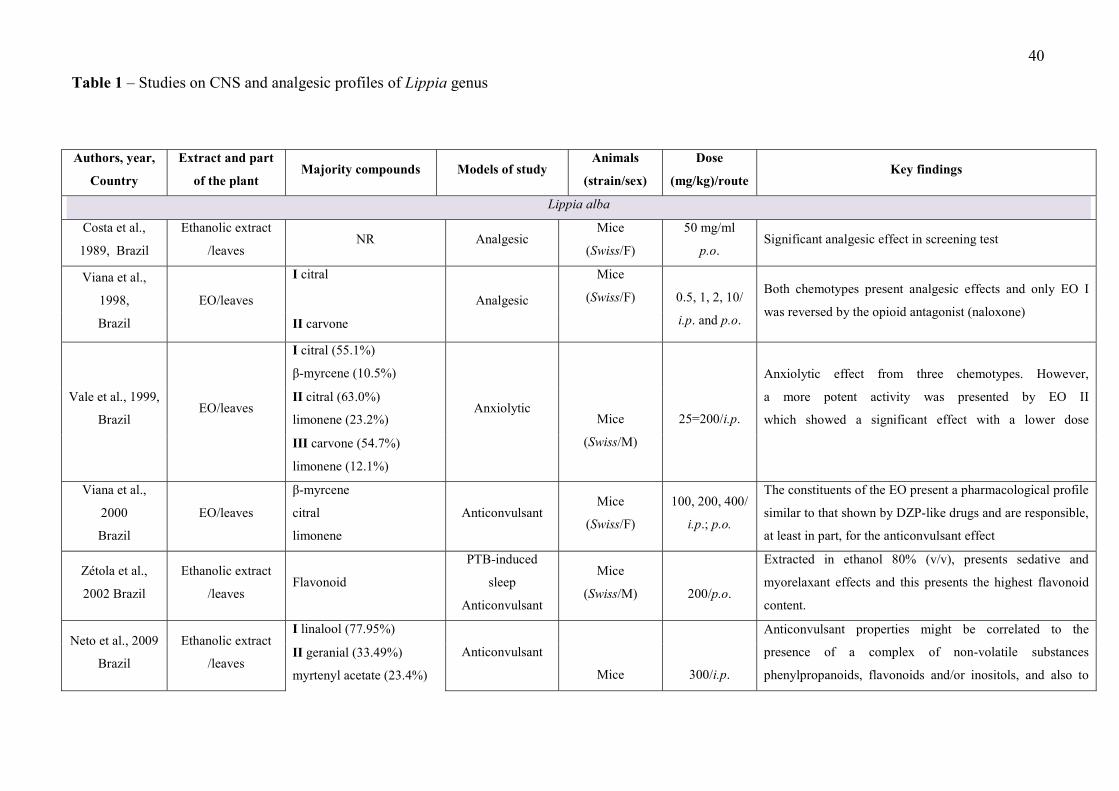
40
Authors, year,
Country
Extract and part
of the plant Majority compounds Models of study
Animals
(strain/sex)
Dose
(mg/kg)/route Key findings
Lippia alba
Costa et al.,
1989, Brazil
Ethanolic extract
/leaves NR Analgesic
Mice
(Swiss/F)
50 mg/ml
p.o. Significant analgesic effect in screening test
Viana et al.,
1998,
Brazil
EO/leaves
I citral
Analgesic
Mice
(Swiss/F)
0.5, 1, 2, 10/
i.p. and p.o.
Both chemotypes present analgesic effects and only EO I
was reversed by the opioid antagonist (naloxone) II carvone
Vale et al., 1999,
Brazil EO/leaves
I citral (55.1%)
β-myrcene (10.5%)
Anxiolytic
Mice
(Swiss/M)
25=200/i.p.
Anxiolytic effect from three chemotypes. However,
a more potent activity was presented by EO II
which showed a significant effect with a lower dose
II citral (63.0%)
limonene (23.2%)
III carvone (54.7%)
limonene (12.1%)
Viana et al.,
2000
Brazil
EO/leaves
β-myrcene
citral
limonene
Anticonvulsant Mice
(Swiss/F)
100, 200, 400/
i.p.; p.o.
The constituents of the EO present a pharmacological profile
similar to that shown by DZP-like drugs and are responsible,
at least in part, for the anticonvulsant effect
Zétola et al.,
2002 Brazil
Ethanolic extract
/leaves Flavonoid
PTB-induced
sleep
Anticonvulsant
Mice
(Swiss/M)
200/p.o.
Extracted in ethanol 80% (v/v), presents sedative and
myorelaxant effects and this presents the highest flavonoid
content.
Neto et al., 2009
Brazil
Ethanolic extract
/leaves
I linalool (77.95%)
Anticonvulsant
Mice
300/i.p.
Anticonvulsant properties might be correlated to the
presence of a complex of non-volatile substances
phenylpropanoids, flavonoids and/or inositols, and also to
II geranial (33.49%)
myrtenyl acetate (23.4%)
Table 1 – Studies on CNS and analgesic profiles of Lippia genus
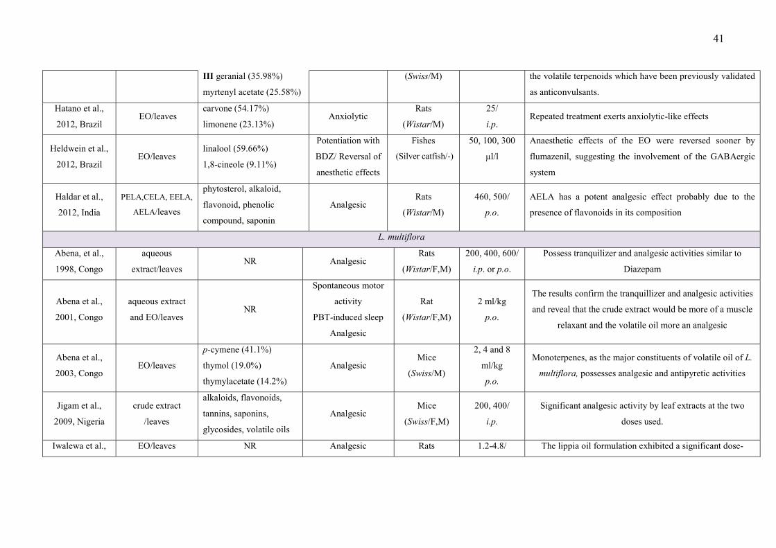
41
III geranial (35.98%)
myrtenyl acetate (25.58%)
(Swiss/M) the volatile terpenoids which have been previously validated
as anticonvulsants.
Hatano et al.,
2012, Brazil EO/leaves
carvone (54.17%)
limonene (23.13%) Anxiolytic
Rats
(Wistar/M)
25/
i.p. Repeated treatment exerts anxiolytic-like effects
Heldwein et al.,
2012, Brazil EO/leaves
linalool (59.66%)
1,8-cineole (9.11%)
Potentiation with
BDZ/ Reversal of
anesthetic effects
Fishes
(Silver catfish/-)
50, 100, 300
µl/l
Anaesthetic effects of the EO were reversed sooner by
flumazenil, suggesting the involvement of the GABAergic
system
Haldar et al.,
2012, India
PELA,CELA, EELA,
AELA/leaves
phytosterol, alkaloid,
flavonoid, phenolic
compound, saponin
Analgesic Rats
(Wistar/M)
460, 500/
p.o.
AELA has a potent analgesic effect probably due to the
presence of flavonoids in its composition
L. multiflora
Abena, et al.,
1998, Congo
aqueous
extract/leaves NR Analgesic
Rats
(Wistar/F,M)
200, 400, 600/
i.p. or p.o.
Possess tranquilizer and analgesic activities similar to
Diazepam
Abena et al.,
2001, Congo
aqueous extract
and EO/leaves NR
Spontaneous motor
activity
PBT-induced sleep
Analgesic
Rat
(Wistar/F,M)
2 ml/kg
p.o.
The results confirm the tranquillizer and analgesic activities
and reveal that the crude extract would be more of a muscle
relaxant and the volatile oil more an analgesic
Abena et al.,
2003, Congo EO/leaves
p-cymene (41.1%)
thymol (19.0%)
thymylacetate (14.2%)
Analgesic Mice
(Swiss/M)
2, 4 and 8
ml/kg
p.o.
Monoterpenes, as the major constituents of volatile oil of L.
multiflora, possesses analgesic and antipyretic activities
Jigam et al.,
2009, Nigeria
crude extract
/leaves
alkaloids, flavonoids,
tannins, saponins,
glycosides, volatile oils
Analgesic Mice
(Swiss/F,M)
200, 400/
i.p.
Significant analgesic activity by leaf extracts at the two
doses used.
Iwalewa et al., EO/leaves NR Analgesic Rats 1.2-4.8/ The lippia oil formulation exhibited a significant dose-

42
2007, Nigeria (emulsion) (Wistar/M) i.p. dependent analgesic effect, showing peripheral and central
activities. Level of the antioxidant markers as possible
mechanism of this activity.
Bassoueka et al.,
2015, France
aqueous
extracts/leaves
alkaloids, flavonoids,
steroids, tannins
Strychnine-induced
convulsion
Rats
(Wistar/F,M) 400, 800/p.o.
The extract of Lippia multiflora has no significant effect on
the parameters studied
L. gracilis
Mendes et al.,
2010, Brazil EO/leaves
thymol (32.68%)
p-cymene (17.82%)
methyl thymol (10.83%)
Analgesic Mice
(Swiss/M)
50, 100, 200/
p.o. The EO inhibited acid-acetic writhing.
Guilhon et al,
2011, Brazil EO/leaves
carvacrol (44.43%)
ο-cymene (9.42%)
γ-terpinene (9.16%)
Analgesic Mice
(Balb-c/ M)
10, 30,
100/p.o.
Antinociceptive effect (not reversed by Naloxone) could
potentially be mediated by cholinergic receptors and the
nitric oxide pathway.
Guimarães et al.,
2012, Brazil
methanolic
extract/leaves Naringenin Analgesic
Mice
(Swiss/M)
100, 200,
400/p.o.
Methanolic extract has a therapeutic potential for painful
conditions.
Lippia grata
Siqueira-Lima et
al., 2014,
Brazil
OE/leaves
camphor (27.2%)
E-caryophyllene (11.6%)
camphene (11.3%)
bicyclogermacrene (9.4%)
Analgesic
Mice
(Swiss/M)
6,12,24/p.o.
EO was capable of reducing the nociceptive face-rubbing in
capsaicin, glutamate and both phases of the formalin test.
The immunofluorescence protocol demonstrated that the
βCD-EO activated important areas in the CNS
Siqueira-Lima et
al., 2017,
Brazil
OE/leaves
camphor (27.2%)
E-caryophyllene (11.6%)
camphene (11.3%)
bicyclogermacrene (9.4%)
Analgesic Mice
(Swiss/M) 6,12,24/p.o.
Decreased paw withdrawal and muscle threshold. The OE
was shown to affect the opioidergic and serotoninergic
pathways. Fos protein immunofluorescence showed
decreased expression in the dorsal horn of the spinal cord.
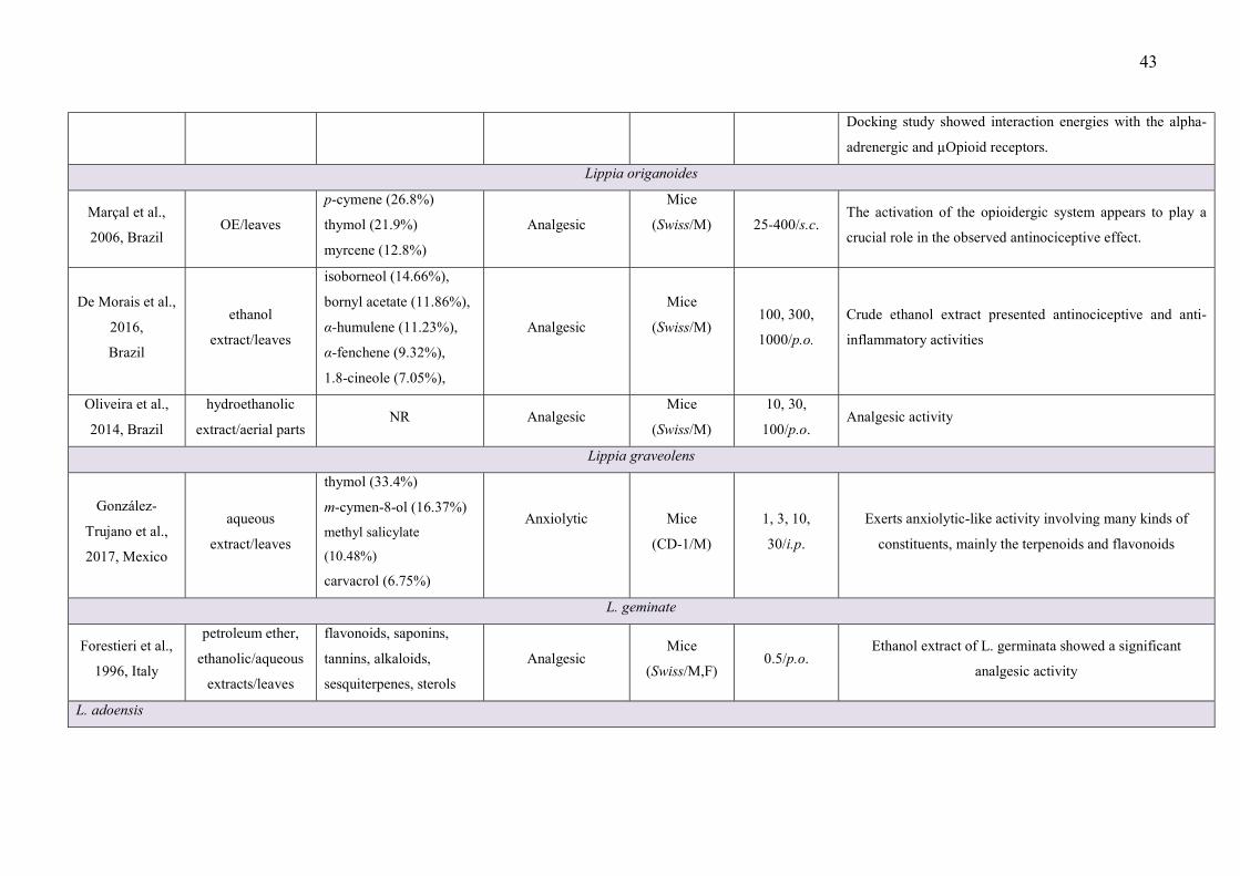
43
Docking study showed interaction energies with the alpha-
adrenergic and µOpioid receptors.
Lippia origanoides
Marçal et al.,
2006, Brazil OE/leaves
p-cymene (26.8%)
thymol (21.9%)
myrcene (12.8%)
Analgesic
Mice
(Swiss/M)
25-400/s.c. The activation of the opioidergic system appears to play a
crucial role in the observed antinociceptive effect.
De Morais et al.,
2016,
Brazil
ethanol
extract/leaves
isoborneol (14.66%),
bornyl acetate (11.86%),
α-humulene (11.23%),
α-fenchene (9.32%),
1.8-cineole (7.05%),
Analgesic
Mice
(Swiss/M) 100, 300,
1000/p.o.
Crude ethanol extract presented antinociceptive and anti-
inflammatory activities
Oliveira et al.,
2014, Brazil
hydroethanolic
extract/aerial parts NR Analgesic
Mice
(Swiss/M)
10, 30,
100/p.o. Analgesic activity
Lippia graveolens
González-
Trujano et al.,
2017, Mexico
aqueous
extract/leaves
thymol (33.4%)
m-cymen-8-ol (16.37%)
methyl salicylate
(10.48%)
carvacrol (6.75%)
Anxiolytic
Mice
(CD-1/M)
1, 3, 10,
30/i.p.
Exerts anxiolytic-like activity involving many kinds of
constituents, mainly the terpenoids and flavonoids
L. geminate
Forestieri et al.,
1996, Italy
petroleum ether,
ethanolic/aqueous
extracts/leaves
flavonoids, saponins,
tannins, alkaloids,
sesquiterpenes, sterols
Analgesic Mice
(Swiss/M,F) 0.5/p.o.
Ethanol extract of L. germinata showed a significant
analgesic activity
L. adoensis

44
Makonnen et al.,
2003,
Ethiopia
Aqueous and
ethanolic
extracts/leaves
phenolic acids,
flavonoids, glycosides Analgesic
Mice
(in-house
bred/M)
400, 600,
800/p.o.
Dose-dependent analgesia produced by both aqueous and
ethanol extracts.
Debella et al.,
2003, Ethiopia
aqueous and
ethanolic
extracts/leaves
phenolic compounds Analgesic
Mice
(in-house
bred/M)
400, 600,
800/p.o. Water extract seems to be slightly more potent at low dose
Debell et al.,
2005, Ethiopia
aqueous and
ethanolic
extracts/leaves
phenolic compounds Inducing pyrexia
Mice
(in-house
bred/M)
50, 100,
200/p.o.
Aqueous extract was found to have more potent antipyretic
effect than the ethanol extract.
Abbreviations: Volatile oil (VO); petroleum ether extracts (PELA); chloroform extracts (CELA); ethanol extracts (EELA) and aqueous extract (AELA); Not related (NR); Pentobarbital
(PBT); Benzodiazepine (BDZ), Diazepam (DZP); Male (M); Female (F), intraperitoneal (i.p.), orally (p.o.)
45
References
Abena, A.A., Atipo-Ebata, J.K., Hondi Assah, T., Diatewa, M., 2001. Psychopharmacological
properties of crude extract and essential oil of Lippia multiflora. Encephale 27, 360-364.
Abena, A.A., Diatewa, M., Gakosso, G., Gbeassor, M., Hondi-Assah, T., Ouamba, J.M.,
2003. Analgesic, antipyretic and anti-inflammatory effects of essential oil of Lippia
multiflora. Fitoterapia 74, 231-236.
Abena, A.A., Ngondzo-Kombeti, G.R., Bioka, D., 1998. Psychopharmacologic properties of
Lippia multiflora. Encephale 24, 449-454.
Adesina, S.K., Gbile, Z.O., Odukoya, O.A., 1995. Survey on indigenous useful plants of West
Africa with special emphasis on medicinal plants and issues associated with their
management, in: The United Nations University Programme on Natural Resources in
Africa. pp. 84-85.
Aguiar, J.S., Costa, M.C.C.D., 2005. Lippia alba (Mill.) N. E. Brown (Verbenaceae):
Levantamento de publicações nas áreas química, agronômica e farmacológica, no
període de 1979 a 2004. Rev. Bras. Plantas Med. 8, 79-84.
Albuquerque, C.C., Camara, T.R., Sant’ana, A.E.G., Ulisses, C., Willadino, L., Marcelino
Júnior, C., 2012. Effects of the essential oil of Lippia gracilis Schauer on caulinary
shoots of heliconia cultivated in vitro. Rev. Bras. Plantas Med. 14, 26-33.
Ali, B., Al-Wabel, N.A., Shams, S., Ahamad, A., Khan, S.A., Anwar, F., 2015. Essential oils
used in aromatherapy: a systemic review. Asian Pac. J. Trop. Biomed. 5, 601-611.
Amin, B., Noorani, R., Razavi, B.M., Hosseinzadeh, H., 2018. The effect of ethanolic extract
of Lippia citriodora on rats with chronic constriction injury of neuropathic pain. Cell J.
19, 528-536.
Anamura, S., Dohi, T., Shirakawa, M., Okamoto, H., Tsujimoto, A., 1988. Effects of phenolic
dental medicaments on prostaglandin synthesis by microsomes of bovine tooth pulp and
rabbit kidney medulla. Arch. Oral Biol. 33, 555-560.
Araújo-Filho, H.G., Pereira, E.W.M., Rezende, M.M., Menezes, P.P., Araújo, A.A.S.,
Barreto, R.S.S., Martins, A.O.B.P.B., Albuquerque, T.R., Silva, B.A.F., Alcantara, I.S.,
Coutinho, H.D.M., Menezes, I.R.A., Quintans-Júnior, L.J., Quintans, J.S.S., 2017. D-
Limonene exhibits superior antihyperalgesic effects in a β-cyclodextrin-complexed form
in chronic musculoskeletal pain reducing Fos protein expression on spinal cord in mice.
Neuroscience 358, 158-169.
Arthur, H., Joubert, E., De Beer, D., Malherbe, C.J., Witthuhn, R.C., 2011. Phenylethanoid
glycosides as major antioxidants in Lippia multiflora herbal infusion and their stability

46
during steam pasteurisation of plant material. Food Chem. 127, 581-588.
Bassoueka, D.J., Loufoua, B.A.E., Etou-Ossibi, A.W., Nsondé-Ntandou, G.F., Ondelé, R.,
Elion-Itou, R.D.G., Ouamba, J.M., Abena, A.A., 2015. Plantes anticonvulsivantes du
Congo, approche ethnobotanique. Phytotherapie 13, 298-305.
Beer, A.M., Lukanov, J., Sagorchev, P., 2007. Effect of thymol on the spontaneous contractile
activity of the smooth muscles. Phytomedicine 14, 65-69.
Benveniste, E.N., 1998. Cytokine actions in the central nervous system. Cytokine growth
factor rev. 9, 259-275.
Bezerra, P., Fernandes, A.G., Craveiro, A.A., Andrade, C.H.S., Matos, F.J.A., Alencar, J.W.,
Machado, M.I.L., Viana, G.S.B., Matos, F.F., Rouquayrol, M.Z., 1981. Composição
química e atividade biológica de óleos essenciais de plantas do Nordeste - gênero Lippia.
Cienc. Cult. 33, 1-14.
Bonjardim, L.R., Cunha, E.S., Guimarães, A.G., Santana, M.F., Oliveira, M.G.B., Serafini,
M.R., Araújo, A.A.S., Antoniolli, Â.R., Cavalcanti, S.C.H., Santos, M.R.V., Quintans-
Júnior, L.J., 2012. Evaluation of the anti-inflammatory and antinociceptive properties of
p-cymene in mice. Zeitschrift fur Naturforsch. C J. Biosci. 67, 15-21.
Botelho, M.A., Barros, G., Queiroz, D.B., Carvalho, C.F., Gouvea, J., Patrus, L., Bannet, M.,
Patrus, D., Rego, A., Silva, I., Campus, G., Araújo-Filho, I., 2016. Nanotechnology in
phytotherapy: antiinflammatory effect of a nanostructured thymol gel from Lippia
sidoides in acute periodontitis in Rats. Phyther. Res. 30, 152-159.
Carvalho, P.M.M., Macêdo, C.A.F., Ribeiro, T.F., Silva, A.A., Da Silva, R.E.R., de Morais,
L.P., Kerntopf, M.R., Menezes, I.R.A., Barbosa, R., 2018. Effect of the Lippia alba
(Mill.) N.E. Brown essential oil and its main constituents, citral and limonene, on the
tracheal smooth muscle of rats. Biotechnol. Reports 17, 31-34.
Catalan, C.A., De Lampasona, M.E., 2002. The chemistry of the genus Lippia (Verbenaceae),
in: Oregano: the genera Origanum and Lippia. p. 127-149.
Cavalcante Melo, F.H., Rios, E.R.V., Rocha, N.F.M., Citõ, M.D.C.D.O., Fernandes, M.L., De
Sousa, D.P., De Vasconcelos, S.M.M., De Sousa, F.C.F., 2012. Antinociceptive activity
of carvacrol (5-isopropyl-2-methylphenol) in mice. J. Pharm. Pharmacol. 64, 1722-1729.
Cho, K.S., Lim, Y.R., Lee, K., Lee, J., Lee, J.H., Lee, I.S., 2017. Terpenes from forests and
human health. Toxicol. Res. 33, 97-106.
Costa, M., Di Stasi, L.C., Kirizawa, M., Mendaçolli, S.L.J., Gomes, C., Trolin, G., 1989.
Screening in mice of some medicinal plants used for analgesic purposes in the state of
Sao Paulo. Part II. J. Ethnopharmacol. 27, 25-33.
47
Craveiro, A.A., Alencar, J.W., Matos, F.J.A., Andrade, C.H.S., Machado, M.I.L., 1981.
Essential oils from Brazilian Verbenaceae. Genus Lippia. J. Nat. Prod. 44, 598-601.
da Silveira e Sá, R. de C., Lima, T.C., da Nóbrega, F.R., de Brito, A.E.M., de Sousa, D.P.,
2017. Analgesic-like activity of essential oil constituents: An update. Int. J. Mol. Sci.
doi: 10.3390/ijms18122392.
de Abreu Matos, F.J., Machado, M.I.L., Craveiro, A.A., Alencar, J.W., 1996. Essential oil
composition of two chemotypes of lippia alba grown in northeast Brazil. J. Essent. Oil
Res. 8, 695-698.
De Carvalho Nilo Bitu, V., De Carvalho Nilo Bitu, V., Matias, E.F.F., De Lima, W.P., Da
Costa Portelo, A., Coutinho, H.D.M., De Menezes, I.R.A., 2015. Ethnopharmacological
study of plants sold for therapeutic purposes in public markets in Northeast Brazil. J.
Ethnopharmacol. 172, 265-272.
De Cássia Da Silveira E Sá, R., Andrade, L.N., De Sousa, D.P., 2013. A review on anti-
inflammatory activity of monoterpenes. Molecules doi: 10.3390/molecules18011227.
de Morais, S.R., Oliveira, T.L.S., de Oliveira, L.P., Tresvenzol, L.M.F., da Conceicao, E.C.,
Rezende, M.H., Fiuza, T. de S., Costa, E.A., Ferri, P.H., de Paula, J.R., 2016. Essential
oil composition, antimicrobial and pharmacological activities of Lippia sidoides Cham.
(Verbenaceae) from Sao Goncalo do Abaete, Minas Gerais, Brazil. Pharmacogn. Mag.
12, 262-270.
De Oliveira, M.L.M., Bezerra, B.M.O., Leite, L.O., Girão, V.C.C., Nunes-Pinheiro, D.C.S.,
2014. Topical continuous use of Lippia sidoides Cham. essential oil induces cutaneous
inflammatory response, but does not delay wound healing process. J. Ethnopharmacol.
153, 283-289.
De Oliveira, T.M., De Carvalho, R.B.F., Da Costa, I.H.F., De Oliveira, G.A.L., De Souza,
A.A., De Lima, S.G., De Freitas, R.M., 2015. Evaluation of p-cymene, a natural
antioxidant. Pharm. Biol. 53, 423-428.
De Santana, M.F., Guimarães, A.G., Chaves, D.O., Silva, J.C., Bonjardim, L.R., De Lucca
Júnior, W., De Souza Ferro, J.N., De Oliveira Barreto, E., Dos Santos, F.E., Soares,
M.B.P., Villarreal, C.F., De Souza Siqueira Quintans, J., Quintans-Júnior, L.J., 2015.
The anti-hyperalgesic and anti-inflammatory profiles of p-cymene: Evidence for the
involvement of opioid system and cytokines. Pharm. Biol. 53, 1583-1590.
De Sousa, D.P., 2011. Analgesic-like activity of essential oils constituents. Molecules 16,
2233–2252.
Debell, A., Makonnen, E., Zerihun, L., Abebe, D., Teka, F., 2005. In-vivo antipyretic studies
48
of the aqueous and ethanol extracts of the leaves of Ajuga remota and Lippia adoensis.
Ethiop. Med. J. 43, 111-118.
Debella, A., Makonnen, E., Abebe, D., Teka, F., Kidanemariam, A.T., 2003. Pain
management in mice using the aqueous and ethanol extracts of four medicinal plants.
East Afr. Med. J. 80, 435-439.
Diniz, T.C., Silva, J.C., Lima-Saraiva, S.R.G. De, Ribeiro, F.P.R.D.A., Pacheco, A.G.M., De
Freitas, R.M., Quintans-Júnior, L.J., Quintans, J.D.S.S., Mendes, R.L., Almeida,
J.R.G.D.S., 2015. The role of flavonoids on oxidative stress in epilepsy. Oxid. Med. Cell.
Longev. doi: 10.1155/2015/171756.
Dobetsberger, C., Buchbauer, G., 2011. Actions of essential oils on the central nervous
system: an updated review. Flavour Fragr. J. 26, 300-316.
Duarte, I.D.G., dos Santos, I.R., Lorenzetti, B.B., Ferreira, S.H., 1992. Analgesia by direct
antagonism of nociceptor sensitization involves the arginine-nitric oxide-cGMP
pathway. Eur. J. Pharmacol. 217, 225-227.
El Hadi, M.A.M., Zhang, F.J., Wu, F.F., Zhou, C.H., Tao, J., 2013. Advances in fruit aroma
volatile research. Molecules 18, 8200-8229.
Elliott, a a, Elliott, J.R., 1997. Voltage-dependent inhibition of RCK1 K+ channels by phenol,
p-cresol, and benzyl alcohol. Mol. Pharmacol. 51, 475-483.
Enna, S.J., McCarson, K.E., 2006. The role of GABA in the mediation and perception of pain.
Adv. Pharmacol. 54, 1-27.
Forestieri, A.M., Monforte, M.T., Ragusa, S., Trovato, A., Iauk, L., 1996. Antiinflammatory,
analgesic and antipyretic activity in rodents of plant extracts used in African medicine.
Phyther. Res. 10, 100-106.
Frighetto, N., De Oliveira, J.G., Siani, A.C., Das Chagas, K.C., 1998. Lippia alba Mill N.E.
Br. (Verbenaceae) as a source of linalool. J. Essent. Oil Res. 10, 578-580.
Gazola, R., Machado, D., Ruggiero, C., Singi, G., Macedo Alexandre, M., 2004. Lippia alba,
Melissa officinalis and Cymbopogon citratus: effects of the aqueous extracts on the
isolated hearts of rats. Pharmacol. Res. 50, 477-480.
Gonçalves, J.C.R., Oliveira, F.D.S., Benedito, R.B., de Sousa, D.P., de Almeida, R.N., de
Araújo, D.A.M., 2008. Antinociceptive activity of (-)-carvone: evidence of association
with decreased peripheral nerve excitability. Biol. Pharm. Bull. 31, 1017-1020.
González-Burgos, E., Gómez-Serranillos, M.P., 2012. Terpene compounds in nature: a review
of their potential antioxidant activity. Curr. Med. Chem. 19, 5319-5341.
González-Trujano, M.E., Hernández-Sánchez, L.Y., Ocotero, V.M., Dorazco-González, A.,
49
Fefer, P.G., Aguirre-Hernández, E., 2017. Pharmacological evaluation of the anxiolytic-
like effects of Lippia graveolens and bioactive compounds. Pharm. Biol. 55, 1569-1576.
Guilhon, C.C., Raymundo, L.J.R.P., Alviano, D.S., Blank, A.F., Arrigoni-Blank, M.F.,
Matheus, M.E., Cavalcanti, S.C.H., Alviano, C.S., Fernandes, P.D., 2011.
Characterisation of the anti-inflammatory and antinociceptive activities and the
mechanism of the action of Lippia gracilis essential oil. J. Ethnopharmacol. 135, 406-
413.
Guimarães, A.G., Oliveira, G.F., Melo, M.S., Cavalcanti, S.C.H., Antoniolli, A.R.,
Bonjardim, L.R., Silva, F.A., Santos, J.P.A., Rocha, R.F., Moreira, J.C.F., Araújo,
A.A.S., Gelain, D.P., Quintans-Júnior, L.J., 2010. Bioassay-guided evaluation of
antioxidant and antinociceptive activities of carvacrol. Basic Clin. Pharmacol. Toxicol.
107, 949-957.
Guimarães, A.G., Quintans, J.S.S., Quintans-Júnior, L.J., 2013. Monoterpenes with analgesic
activity-A systematic review. Phyther. Res. 27, 1-15.
Guimarães, A.G., Serafini, M.R., Quintans-Júnior, L.J., 2014. Terpenes and derivatives as a
new perspective for pain treatment: a patent review. Expert Opin. Ther. Pat. 24, 243-265.
Guimarães, A.G., Xavier, M.A., De Santana, M.T., Camargo, E.A., Santos, C.A., Brito, F.A.,
Barreto, E.O., Cavalcanti, S.C.H., Antoniolli, Â.R., Oliveira, R.C.M., Quintans-Júnior,
L.J., 2012. Carvacrol attenuates mechanical hypernociception and inflammatory
response. Naunyn. Schmiedebergs. Arch. Pharmacol. 385, 253-263.
Habtemariam, S., 2018. Iridoids and other monoterpenes in the Alzheimer’s brain: recent
development and future prospects. Molecules 23, doi: 10.3390/molecules23010117.
Haeseler, G., Maue, D., Grosskreutz, J., Bufler, J., Nentwig, B., Piepenbrock, S., Dengler, R.,
Leuwer, M., 2002. Voltage-dependent block of neuronal and skeletal muscle sodium
channels by thymol and menthol. Eur. J. Anaesthesiol. 19, 571-579.
Haldar, S., Kar, B., Dolai, N., Kumar, R.B.S., Behera, B., Haldar, P.K., 2012. In vivo anti-
nociceptive and anti–inflammatory activities of Lippia alba. Asian Pacific J. Trop. Dis.
2, S667-S670.
Hatano, V.Y., Torricelli, A.S., Giassi, A.C.C., Coslope, L.A., Viana, M.B., 2012. Anxiolytic
effects of repeated treatment with an essential oil from Lippia alba and (R)-(-)-carvone in
the elevated T-maze. Brazilian J. Med. Biol. Res. 45, 238-243.
Heldwein, C.G., Silva, L.L., Reckziegel, P., Barros, F.M.C., Bürger, M.E., Baldisserotto, B.,
Mallmann, C.A., Schmidt, D., Caron, B.O., Heinzmann, B.M., 2012. Participation of the
GABAergic system in the anesthetic effect of Lippia alba (Mill.) N.E. Brown essential

50
oil. Brazilian J. Med. Biol. Res. 45, 436-443.
Hennebelle, T., Sahpaz, S., Dermont, C., Joseph, H., Bailleul, F., 2006. The essential oil of
Lippia alba: analysis of samples from french overseas departments and review of
preview works. Chem. Biodivers. 3, 1116-1125.
Hennebelle, T., Sahpaz, S., Joseph, H., Bailleul, F., 2008. Ethnopharmacology of Lippia alba.
J. Ethnopharmacol. 116, 211-222.
Hotta, M., Nakata, R., Katsukawa, M., Hori, K., Takahashi, S., Inoue, H., 2010. Carvacrol, a
component of thyme oil, activates PPARalpha and gamma and suppresses COX-2
expression. J. Lipid Res. 51, 132-139.
Ibrahim, M.M., Porreca, F., Lai, J., Albrecht, P.J., Rice, F.L., Khodorova, A., Davar, G.,
Makriyannis, A., Vanderah, T.W., Mata, H.P., Malan, T.P., 2005. CB2 cannabinoid
receptor activation produces antinociception by stimulating peripheral release of
endogenous opioids. Proc. Natl. Acad. Sci. 102, 3093-3098.
Iwalewa, E.O., Oladimeji, F.A., Adewunmi, C.O., Osoniyi, O.R., Orafidiya, L.O., Adeloye,
O., Adeleke, F.B., Omodara, S.K., 2007. Involvement of nitric oxide and other
antioxidant markers in the anti-inflammatory and analgesic effects of Lippia multiflora
(Moldenke) leaf essential oil emulsion. Int. J. Essent. Oil Ther. 33, 283-285.
Jaeger, B.N., Parylak, S.L., Gage, F.H., 2017. Mechanisms of dietary flavonoid action in
neuronal function and neuroinflammation. Mol. Aspects Med. 61, 50-62.
Jasmin, L., Wu, M. V, Ohara, P.T., 2004. GABA puts a stop to pain. Curr. Drug Targets. CNS
Neurol. Disord. 3, 487-505.
Jigam, A.A., Akanya, H.O., Ogbadoyi, E.O., Dauda, B.E.N., Evans, E.C., 2009. In vivo
antiplasmodial, analgesic and anti-inflammatory activities of the leaf extract of Lippia
multiflora mold. J. Med. Plants Res. 3, 148-154.
Kam, A., M. Li, K., Razmovski-Naumovski, V., Nammi, S., Chan, K., Li, Y., Q. Li, G., 2012.
The protective effects of natural products on blood-brain barrier breakdown. Curr. Med.
Chem. 19, 1830-1845.
Kianmehr, M., Rezaei, A., Boskabady, M.H., 2016. Effect of carvacrol on various cytokines
genes expression in splenocytes of asthmatic mice. Iran. J. Basic Med. Sci. 19, 402-410.
Leyva-López, N., Gutierrez-Grijalva, E.P., Ambriz-Perez, D.L., Basilio Heredia, J., 2016.
Flavonoids as cytokine modulators: a possible therapy for inflammation-related diseases.
Int. J. Mol. Sci. 17, doi: 10.3390/ijms17060921. 1
Lillehei, A.S., Halcon, L.L., 2014. A systematic review of the effect of inhaled essential oils
on sleep. J. Altern. Complement. Med. 20, 441-451.

51
Lima, C.M., Serafini, M.R., Santos, G.P., Cardoso, J.C., Figueiredo, R.T., Santos, M.S.,
Melo, M.G.D., Silva, F.A.R., da Costa, L.P., Santos, A.F.C., Albuquerque-Júnior,
R.L.C., Quintans-Júnior, L.J., Araújo, A.A.S., 2016. Use of bone physicochemical
characterization and biochemical analyses in an experimental model. J. Therm. Anal.
Calorim. 123, 2179-2184.
Lima, M.D.S., Quintans-Júnior, L.J., De Santana, W.A., Martins Kaneto, C., Pereira Soares,
M.B., Villarreal, C.F., 2013. Anti-inflammatory effects of carvacrol: evidence for a key
role of interleukin-10. Eur. J. Pharmacol. 699, 112-117.
Linde, G.A., Colauto, N.B., Albertó, E., Gazim, Z.C., 2016. Chemotypes, extraction,
chemical composition and use of Lippia alba essential oil. Rev. Bras. Plantas Med. 18,
191-200.
Lorenzi, H., Matos, F.J.A., 2002. Plantas medicinais no Brasil: nativas e exóticas cultivadas,
Medicina.2ª edição, p. 576.
Makonnen, E., Debella, A., Abebe, D., Teka, F., 2003. Analgesic properties of some
Ethiopian medicinal plants in different models of nociception in mice. Phytother. Res.
17, 1108-1112.
Mamun-Or-Rashid, A.N.M.., Sen, M.K.., Jamal, M.A.H.M.., Nasrin, S., 2013. A
comprehensive ethnopharmacological review on Lippia alba M. Int. J. Biomed. Mater.
Res. 1, 14-20.
Marçal, R.M., Ptak, D.M., Krempser, R.R., Krempser, M.R., Cardoso, G.C., Santos, R.B.,
Blank, A.F., Alves, P.B., 2006. Antinociceptive effect of the essential oil of Lippia
sidoides on mice. Planta Med. 72, 291.
Mendes, S.S., Bomfim, R.R., Jesus, H.C.R., Alves, P.B., Blank, A.F., Estevam, C.S.,
Antoniolli, A.R., Thomazzi, S.M., 2010. Evaluation of the analgesic and anti-
inflammatory effects of the essential oil of Lippia gracilis leaves. J. Ethnopharmacol.
129, 391-397.
Ministério da Saúde, 2009. Política Nacional de Plantas medicinais e fitoterápicos, Brasília.
https://doi.org/NLM QV 766.
Mohammadi, B., Haeseler, G., Leuwer, M., Dengler, R., Krampfl, K., Bufler, J., 2001.
Structural requirements of phenol derivatives for direct activation of chloride currents via
GABA(A) receptors. Eur. J. Pharmacol. 421, 85-91.
Nagakura, Y., 2015. Challenges in drug discovery for overcoming “dysfunctional pain”: an
emerging category of chronic pain. Expert Opin. Drug Discov. 10, 1043-1045.
Nascimento, S.S., Camargo, E.A., Desantana, J.M., Araújo, A.A.S., Menezes, P.P., Lucca-
52
Júnior, W., Albuquerque-Júnior, R.L.C., Bonjardim, L.R., Quintans-Júnior, L.J., 2014.
Linalool and linalool complexed in β-cyclodextrin produce anti-hyperalgesic activity and
increase Fos protein expression in animal model for fibromyalgia. Naunyn.
Schmiedebergs. Arch. Pharmacol. 387, 935-942.
Neto, A.C., Netto, J.C., Pereira, P.S., Pereira, A.M.S., Taleb-Contini, S.H., França, S.C.,
Marques, M.O.M., Beleboni, R.O., 2009. The role of polar phytocomplexes on
anticonvulsant effects of leaf extracts of Lippia alba (Mill.) N.E. Brown chemotypes. J.
Pharm. Pharmacol. 61, 933-939.
Neves, I.A., Schwartz, M.O.E., Da Camara, C.A.G., 2008. Chemical composition of the leaf
oils of Lippia gracilis schauer from two localities of pernambuco. J. Essent. Oil Res. 20,
157-160.
Nijveldt, R.J., Van Nood, E., Van Hoorn, D.E.C., Boelens, P.G., Van Norren, K., Van
Leeuwen, P.A.M., 2001. Flavonoids: a review of probable mechanisms of action and
potential applications. Am. J. Clin. Nutr. 74, 418-425.
Nishijima, C.M., Ganev, E.G., Mazzardo-Martins, L., Martins, D.E., Rocha, L.R.M., Santos,
A.R.S., Hiruma-Lima, C.A., 2014. Citral: A monoterpene with prophylactic and
therapeutic anti-nociceptive effects in experimental models of acute and chronic pain.
Eur. J. Pharmacol. 736, 16-25.
O’Leary, N., Denham, S.S., Salimena, F., Múlgura, M.E., 2012. Species delimitation in
Lippia section Goniostachyum (Verbenaceae) using the phylogenetic species concept.
Bot. J. Linn. Soc. 170, 197-219.
Oliveira, D.R., Leitão, G.G., Bizzo, H.R., Lopes, D., Alviano, D.S., Alviano, C.S., Leitão,
S.G., 2006. Chemical and antimicrobial analyses of essential oil of Lippia origanoides
H.B.K. Food Chem. 101, 236-240.
Oliveira, D.R., Leitão, G.G., Fernandes, P.D., Leitão, S.G., 2014. Ethnopharmacological
studies of Lippia origanoides. Rev. Bras. Farmacogn. 24, 206-214.
Oliveira, M.A., Guimarães, A.G., Araújo, A.A.S., Quintans-Júnior, L.J., Quintans, J.S.S.,
2017. New drugs or alternative therapy to blurring the symptoms of fibromyalgia - a
patent review. Expert Opin. Ther. Pat. 27, 1147-1157.
Ombito, J.O., Salano, E.N., Yegon, P.K., Ngetich, W.K., Mwangi, E.M., Koech, G.K.K.,
Yegon, K., 2015. A review of the chemistry of some species of genus Aloe
(Xanthorrhoeaceae family). J. Sci. Innov. Res. JSIR 4, 49-53.
Pandey, K.B., Rizvi, S.I., 2009. Plant polyphenols as dietary antioxidants in human health and
disease. Oxid. Med. Cell. Longev. 2, 270-278.
53
Parsaei, P., Bahmani, M., Naghdi, N., Asadi-Samani, M., Rafieian-Kopaei, M., 2016. A
review of therapeutic and pharmacological effects of thymol. Der Pharm. Lett. 8, 150-
154.
Pascual, M.E., Slowing, K., Carretero, E., Sánchez Mata, D., Villar, A., 2001. Lippia:
traditional uses, chemistry and pharmacology: a review. J. Ethnopharmacol. 76, 201-214.
Passos, C.S., Arbo, M.D., Rates, S.M.K., Von Poser, G.L., 2009. Terpenóides com atividade
sobre o Sistema Nervoso Central (SNC). Rev. Bras. Farmacogn. 19, 140-149.
Passos, F.F. de B., Lopes, E.M., De Araújo, J.M., De Sousa, D.P., Veras, L.M.C., Leite,
J.R.S.A., De Castro Almeida, F.R., 2015. Involvement of cholinergic and opioid system
in γ-terpinene-mediated antinociception. Evid.-based Compl. Altern. Med. 2015. doi:
10.1155/2015/829414.
Pessoa, O.D.L., De Carvalho, C.B.M., Silvestre, J.O.V.L., Lima, M.C.L., Neto, R.M., Matos,
F.J.A., Lemos, T.L.G., 2005. Antibacterial activity of the essential oil from Lippia aff.
gracillis. Fitoterapia 76, 712-714.
Pina, L.T.S., Ferro, J.N.S., Rabelo, T.K., Oliveira, M.A., Scotti, L., Scotti, M.T., Walker,
C.I.B., Barreto, E.O., Quintans Júnior, L.J., Guimarães, A.G., 2018. Alcoholic
monoterpenes found in essential oil of aromatic spices reduce allergic inflammation by
the modulation of inflammatory cytokines. Nat. Prod. Res. doi:
10.1080/14786419.2018.1434634.
Pina, L.T.S., Gouveia, D.N., Costa, J.S., Quintans, J.S.S., Quintans-Júnior, L.J., Barreto,
R.S.S., Guimarães, A.G., 2017. New perspectives for chronic pain treatment: a patent
review (2010-2016). Expert Opin. Ther. Pat. 27, 787-796.
Quintans, J.S.S., Shanmugam, S, Heimfarth, L, Araújo, A.A.S., Almeida, J.R.G.S., Picot, L,
Quintans-Júnior., 2019. Monoterpenes modulating cytokines - A review. Food Chem.
Toxicol 123, 233-257.
Quintans-Junior, L., Guimarães, A.G., de Santana, M.T., Araújo, B.E.S., Moreira, F. V.,
Bonjardim, L.R., Araújo, A.A.S., Siqueira, J.S., Ângelo, A.R., Botelho, M.A., Almeida,
J.R.G.S., Santos, M.R.V., 2011. Citral reduces nociceptive and inflammatory response in
rodents. Rev. Bras. Farmacogn. 21, 497-502.
Quintans-Júnior, L., Moreira, J.C.F., Pasquali, M. a B., Rabie, S.M.S., Pires, A.S., Schröder,
R., Rabelo, T.K., Santos, J.P., Lima, P.S.S., Cavalcanti, S.C.H., Araújo, A.S., Quintans,
J.S.S., Gelain, D.P., 2013. Antinociceptive activity and redox profile of the
monoterpenes (+)-camphene, p-cymene, and geranyl acetate in experimental models.
ISRN Toxicol. doi: 10.1155/2013/459530.
54
Quintans-Júnior, L.J., Araújo, A.A.S., Brito, R.G., Santos, P.L., Quintans, J.S.S., Menezes,
P.P., Serafini, M.R., Silva, G.F., Carvalho, F.M.S., Brogden, N.K., Sluka, K.A., 2016. β-
caryophyllene, a dietary cannabinoid, complexed with β-cyclodextrin produced anti-
hyperalgesic effect involving the inhibition of Fos expression in superficial dorsal horn.
Life Sci. 149, 34-41.
Quintans-Júnior, L.J., Guimarães, A.G., Araújo, B.E.S., Oliveira, G.F., Santana, M.T.,
Moreira, F.V., Santos, M.R.V., Cavalcanti, S.C.H., Júnior, W.D.L., Botelho, M.A.,
Ribeiro, L.A.A., Nóbrega, F.F.F., Almeida, R.N., 2010. Carvacrol, borneol and citral
reduce convulsant activity in rodents. African J. Biotechnol. 9, 6566-6572.
Quintans-Júnior, L.J., Souza, T.T., Leite, B.S., Lessa, N.M.N., Bonjardim, L.R., Santos,
M.R.V., Alves, P.B., Blank, A.F., Antoniolli, A.R., 2008. Phythochemical screening and
anticonvulsant activity of Cymbopogon winterianus Jowitt (Poaceae) leaf essential oil in
rodents. Phytomedicine 15, 619-624.
Rao, V.S.N., Menezes, A.M.S., Viana, G.S.B., 1990. Effect of myrcene on nociception in
mice. J. Pharm. Pharmacol. 42, 877-878.
Rashidian, A., Farhang, F., Vahedi, H., Dehpour, A.R., Mehr, S.E., Mehrzadi, S., Rezayat,
S.M., 2016. Anticonvulsant effects of Lippia citriodora (Verbenaceae) leaves ethanolic
extract in mice: Role of GABAergic system. Int. J. Prev. Med. doi: 10.4103/2008-
7802.187251.
Razavi, B.M., Zargarani, N., Hosseinzadeh, H., 2017. Anti-anxiety and hypnotic effects of
ethanolic and aqueous extracts of Lippia citriodora leaves and verbascoside in mice.
Avicenna J. Phytomedicine 7, 353-365.
Salimena, F.R.G., 2002. Novos sinônimos e tipificações em Lippia sect. Rhodolippia
(Verbenaceae). Darwiniana 40, 121-125.
Santana, M.F., Quintans-Júnior, L.J., Cavalcanti, S.C.H., Oliveira, M.G.B., Guimarães, A.G.,
Cunha, E.S., Melo, M.S., Santos, M.R. V, Araújo, A.A.S., Bonjardim, L.R., 2011. p-
Cymene reduces orofacial nociceptive response in mice. Rev. Bras. Farmacogn. 21,
1138-1143.
Santoro, G.F., Das Graças Cardoso, M., Guimarães, L.G.L., Salgado, A.P.S.P., Menna-
Barreto, R.F.S., Soares, M.J., 2007. Effect of oregano (Origanum vulgare L.) and thyme
(Thymus vulgaris L.) essential oils on Trypanosoma cruzi (Protozoa: Kinetoplastida)
growth and ultrastructure. Parasitol. Res. 100, 783-790.
Santos, M.R.V, Moreira, F.V., Fraga, B.P., de Sousa, D.P., Bonjardim, L.R., Quintans, L.J.,
2011. Cardiovascular effects of monoterpenes: A review. Rev. Bras. Farmacogn. 21,
55
764-771.
Silva, W.J., Dória, G.A.A., Maia, R.T., Nunes, R.S., Carvalho, G.A., Blank, A.F., Alves, P.B.,
Marçal, R.M., Cavalcanti, S.C.H., 2008. Effects of essential oils on Aedes aegypti larvae:
Alternatives to environmentally safe insecticides. Bioresour. Technol. 99, 3251-3255.
Siqueira-Lima, P.S., Araújo, A.A.S., Lucchese, A.M., Quintans, J.S.S., Menezes, P.P., Alves,
P.B., de Lucca Júnior, W., Santos, M.R.V., Bonjardim, L.R., Quintans-Júnior, L.J., 2014.
β-Cyclodextrin complex containing Lippia grata leaf essential oil reduces orofacial
nociception in mice - evidence of possible involvement of descending inhibitory pain
modulation pathway. Basic Clin. Pharmacol. Toxicol. 114, 188-196.
Siqueira-Lima, P.S., Brito, R.G., Araújo-Filho, H.G., Santos, P.L., Lucchesi, A., Araújo,
A.A.S., Menezes, P.P., Scotti, L., Scotti, M.T., Menezes, I.R.A., Coutinho, H.D.M.,
Zengin, G., Aktumsek, A., Antoniolli, A.R., Quintans-Júnior, L.J., Quintans, J.S.S.,
2017. Anti-hyperalgesic effect of Lippia grata leaf essential oil complexed with β-
cyclodextrin in a chronic musculoskeletal pain animal model: complemented with a
molecular docking and antioxidant screening. Biomed. Pharmacother. 91, 739-747.
Santos, P.L., Matos, J.P.S.C.F., Picot, L., Almeida, J.R.G.S., Quintans, J.S.S., Quintans-
Júnior, L.J., 2019. Citronellol, a monoterpene alcohol with promising pharmacological T
activities - a systematic review. Food Chem. Toxicol. 123, 459-469.
Soares, L., 2001. Estudo tecnológico, fitoquímico e biológico de Lippia alba (Miller) NE
Brown ex Britt. & Wils.(Falsa-melissa) Verbenaceae. 189 p.
https://repositorio.ufsc.br/bitstream/handle/123456789/80021/186203.pdf?sequence=1&i
sAllowed=y
Soares, B.V., Tavares-Dias, M. 2013. Espécies de Lippia (Verbenaceae), seu potencial
bioativo e importância na medicina veterinária e aquicultura. Biota Amazônia, 3m 109-
123.
Sousa, D.G., Sousa, S.D.G., Silva, R.E.R., Silva-Alves, K.S., Ferreira-Da-Silva, F.W.,
Kerntopf, M.R., Menezes, I.R.A., Leal-Cardoso, J.H., Barbosa, R., 2015. Essential oil of
Lippia alba and its main constituent citral block the excitability of rat sciatic nerves.
Brazilian J. Med. Biol. Res. 48, 697-702.
Stotz, S.C., Vriens, J., Martyn, D., Clardy, J., Clapham, D.E., 2008. Citral sensing by transient
receptor potential channels in dorsal root ganglion neurons. PLoS One 3, doi:
10.1371/journal.pone.0002082.
Suntres, Z.E., Coccimiglio, J., Alipour, M., 2015. The bioactivity and toxicological actions of
carvacrol. Crit. Rev. Food Sci. Nutr. 55, 304-318.
56
Tavares, E.S., Julião, L.S., Lopes, D., Bizzo, H.R., Lage, C.L.S., Leitão, S.G., 2005. Análise
do óleo essencial de folhas de três quimiotipos de Lippia alba (Mill.) N. E. Br.
(Verbenaceae) cultivados em condições semelhantes. Rev. Bras. Farmacogn. 15, 1-5.
Teles, T.V, Bonfim, R.R., Alves, P.B., Blank, A.F., Jesus, H.C.R., Quintans-Jr, L.J., Serafini,
M.R., Bonjardim, L.R., Araújo, A.A.S., 2010. Composition and evaluation of the
lethality of Lippia gracilis essential oil to adults of Biomphalaria glabrata and larvae of
Artemia salina. Afr. J. Biotechnol. 9, 8800-8804.
Terblanché, F.C., Kornelius, G., 1996. Essential oil constituents of the genus Lippia
(Verbenaceae)- a literature review. J. Essent. Oil Res. 8, 471-485.
Turaskar, A.O., Bhongade, S.I., More, S.M., Dongarwar, A.S., Shende, V.S., Pande, V.B.,
2011. Effects of Lippia nodiflora extract on motor coordination, exploratory behavior
pattern, locomotor activity, anxiety and convulsion on albino mice. Asian J. Pharm. Clin.
Res. 4, 133-138.
Vale, T.G., Matos, F.J.A., De Lima, T.C.M., Viana, G.S.B., 1999. Behavioral effects of
essential oils from Lippia alba (Mill.) N.E. Brown chemotypes. J. Ethnopharmacol. 67,
127-133.
Valentin, A., Pélissier, Y., Benoit, F., Marion, C., Kone, D., Mallie, M., Bastide, J.M.,
Bessière, J.M., 1995. Composition and antimalarial activity in vitro of volatile
components of Lippia multiflora. Phytochemistry 40, 1439-1442.
Veras, H.N.H., Rodrigues, F.F.G., Botelho, M.A., Menezes, I.R.A., Coutinho, H.D.M., Costa,
J.G.M., 2017. Enhancement of aminoglycosides and β-lactams antibiotic activity by
essential oil of Lippia sidoides Cham. and the thymol. Arab. J. Chem. 10, S2790-S2795.
Viana, G.S., do Vale, T.G., Silva, C.M., Matos, F.J., 2000. Anticonvulsant activity of
essential oils and active principles from chemotypes of Lippia alba (Mill.) N.E. Brown.
Biol. Pharm. Bull. 23, 1314-1317.
Vogt-Eisele, A.K., Weber, K., Sherkheli, M.A., Vielhaber, G., Panten, J., Gisselmann, G.,
Hatt, H., 2007. Monoterpenoid agonists of TRPV3. Br. J. Pharmacol. 151, 530-540.
Waning, J., Vriens, J., Owsianik, G., Stüwe, L., Mally, S., Fabian, A., Frippiat, C., Nilius, B.,
Schwab, A., 2007. A novel function of capsaicin-sensitive TRPV1 channels:
involvement in cell migration. Cell Calcium 42, 17-25.
Xu, H., 2005. Camphor activates and strongly desensitizes the transient receptor potential
vanilloid subtype 1 channel in a vanilloid-independent mechanism. J. Neurosci. 25,
8924-8937.
Zétola, M., De Lima, T.C.M., Sonaglio, D., González-Ortega, G., Limberger, R.P., Petrovick,
57
P.R., Bassani, V.L., 2002. CNS activities of liquid and spray-dried extracts from Lippia
alba - Verbenaceae (Brazilian false melissa). J. Ethnopharmacol. 82, 207-215.
Zhu, H.L., Wan, J.B., Wang, Y.T., Li, B.C., Xiang, C., He, J., Li, P., 2014. Medicinal
compounds with antiepileptic/anticonvulsant activities. Epilepsia 55, 3-16.
Zoghbi, M.G.B., Andrade, E.H.A., Santos, A.S., Silva, M.H., Maia, J.G.S., 1998. Essential
oils of Lippia alba (Mill.) N.E.Br growing wild in the Brazilian Amazon. Flavour Fragr.
J. 13, 47-48.

58
CAPÍTULO 2

59
60
Inclusion of Terpenes in Cyclodextrins: Preparation, Characterization and Pharmacological
Approaches
Pollyana S.S. Limaa, Angélica M. Lucchesea, Heitor G. Araújo-Filhob, Paula P. Menezesb, Adriano
A.S. Araújob, Lucindo J. Quintans-Júniorb,*, Jullyana S.S. Quintansb,*
aPost-Graduate Program in Biotechnology, State University of Feira de Santana, Feira de Santana,
BA, Brazil.
bPost-Graduate Program in Health Sciences, Federal University of Sergipe, São Cristóvão, SE,
Brazil.
*Corresponding Address: L.J. Quintans-Junior or J.S.S. Quintans, Federal University of Sergipe,
Post-Graduate Program in Health Sciences, Av Marechal Rondon, Rosa Elza, Cristóvão, Sergipe,
Zip code: 49.000-100, Brazil. Phone: 55 (79) 2105-6645 E-mail: [email protected] or

61
Contents
1. Introduction
2. Terpenes
3. Cyclodextrins (CDs)
3.1 Types of CD
3.2 Restrictions on CD application and possible risks
3.3 Inclusion methods and complexation
3.4 Evidence of complexation
3.4.1 Spectroscopic characterization of the complex
4. Inclusion complexes – CDs and terpenes
4.1 Biological activities (therapeutic activity)
4.2 Pharmacokinetics
4.3 Pharmacological improvement
5. Conclusion
Acknowledgments
References
62
Abbreviations
CDs – cyclodextrins
α-CD – alpha cyclodextrin
β-CD – beta cyclodextrin
γ-CD – gamma cyclodextrin
DPPH – 2,2-diphenyl-1-picrylhydrazyl
DSC - differential scanning calorimetry
DTG - derivative thermogravimetry
FDA – Food and Drug Administration
FTIR - Fourier transform infrared spectroscopy
GA-18β - 18β glycyrrhetinic acid
GRAS - generally recognized as safe
HepG-2 – human hepatic cancer cell line
HPβCD – 2 hydroxypropyl-β-cyclodextrin
HPγCD – 2 hydroxypropyl-γ-cyclodextrin
NMR – nuclear magnetic resonance
NP – natural products
RAMEB - randomly methylated β-cyclodextrin
RYSMEB – low methylated β-cyclodextrin
SBEβCD – sulfobutyl ether β-cyclodextrin
TG –thermogravimetric
TGA - thermogravimetry
XRC - X-ray crystallography
XRD - X-ray diffraction
Highlights
Cyclodextrin-terpene complexes are promising host-guest systems
Insolubility in water and a short life-time limit the therapeutic use of terpenes
CD inclusion complexes enhance the bioavailability, pharmacological efficacy and safety of
terpenes
63
Abstract
Terpenes constitute the largest class of natural products and are important resources for the
pharmaceutical, food and cosmetics industries. However, due to their low water solubility and poor
bioavailability there has been a search for compounds that could improve their physicochemical
properties. Cyclodextrins (natural and derived) have been proposed for this role and have been
complexed with different types of terpenes. This complexation has been demonstrated by using
analytical techniques for characterizing complexes such as DSC, NMR, XRD, FTIR, and TGA. The
formation of inclusion complexes has been able to improve drug characteristics such as
bioavailability, solubility and stability; and to enhance biological activity and efficacy. This review
shows strong experimental evidence that cyclodextrins improve the pharmacological properties of
terpenes, and therefore need to be recognized as being possible targets for clinical use.
Keywords: natural products, terpenes, cyclodextrins, pharmaceutical technology

64
1. Introduction
Terpenoids constitute the most abundant group of secondary metabolites in higher plants with
around 30,000 compounds (Connolly & Hill, 1991; Moraes, Mescher & Tumlinson, 2001). It is a
diverse family of natural products (NP) derived from C5 isoprene units joined in a head-to-tail
fashion. They represent a class of NP that provide a great number of possible solutions to different
human-health issues (Liu et al., 2014b).
Terpenes have significant economic value due to their widespread use in a range of industries
including the pharmaceutical, food, and cosmetics sectors where they can be found as food
additives, flavorings, in fragrances and spices, drug excipients (to enhance penetration of the skin)
and in many other uses (Guimaraes, Serafini & Quintans-Junior, 2014; Guimaraes, Quintans &
Quintans Júnior, 2013). There is a wide range of possible therapeutic pharmacological uses for
terpenes including as: anticancer drugs, analgesics, anti-inflammatories, neuroprotective agents,
immune modulators and in wound healing (Ali et al., 2013; Araujo-Filho, Quintans-Junior, Barreto,
Almeida, Barreto & Quintans, 2016; Barreto et al., 2014; Guimaraes et al., 2015; Guimaraes,
Quintans & Quintans Júnior, 2013; Rodrigues, Sieglitz & Bernardes, 2016; Quintans, Antoniolli,
Almeida, Santana-Filho & Quintans-Junior, 2014)Although, there is no current data on the exact
value of terpenes in the pharmaceutical market, the sales of terpene-based pharmaceuticals in 2002
were around US $12 billion (Wang, Tang & Bidigare, 2005). Terpenes can be found in many
examples of where companies are in the early stages of commercializing technology or when
developing new products (Erickson, Nelson & Winters, 2012), and terpene use across a range of
industries shows significant annual growth, particularly in the pharmaceutical sector (Ahad et al.,
2009; Guimaraes, Serafini & Quintans-Junior, 2014).
The pharmaceutical and cosmetic industries have been increasingly using drug carrier systems, such
as cyclodextrins (CDs), in order to enhance the physico-chemical and pharmacological profiles of
non-polar drugs, while also improving side effect profiles. The complexation of various drugs with
CDs has improved their analgesic, anti-cancer and anti-inflammatory profiles, in both clinical and
preclinical studies (Brito, Araujo, Quintans, Sluka & Quintans-Junior, 2015; Gidwani & Vyas,
2015; Oliveira, Guimaraes, Araujo, Quintans, Santos & Quintans-Junior, 2015; Williamson,
Menzies, Nair, Tutuncu & Lipworth, 2009). The growing use of CDs with terpenes to make them
more bioavailable to pharmacological application has been noteworthy (Oliveira et al., 2015;
Guimaraes et al., 2015). However, there are no reviews that summarize the main technological,
chemical and pharmacological aspects of terpene complexation with CDs.

65
In this review, an overview was achieved by considering the pre-clinical, and clinical approaches of
the published works on complexes between CDs and terpenes, summarizing the existing data on
preparation methods and conducting an analysis and appraisal of the evidence on the results of
complexation in pharmaceutical applications. The review was based on articles published in the
period up to, and including, 2016. The search strategy followed the guidelines of the Preferred
Reporting Items for Systematic Reviews and Meta-Analyses (PRISMA) (see supplementary
material) (Moher, Liberati, Tetzlaff & Altman, 2009).
2. Terpenes
Terpenes have been isolated from living organisms, mainly plants (Singh & Sharma, 2015). These
compounds are also found in bacteria (Yamada et al., 2015), fungi (Lin et al., 2014), insects - such
as termites or butterflies (la Cruz, Santos Júnior, Rezende, Alves, Cancello & Rocha, 2014) and
marine organisms (Hegazy et al., 2015).
In general, terpenes represent a heterogeneous group of chemical substances constructed from
hydrocarbons. They consist of the combination of several 5-carbon-base units called isoprenes
(C5H8), mainly in a head-to-tail linkage. Oxidation in the carbon skeletons, cyclizations, proton and
methyl removal or shifts can lead to variations in their structures, giving rise to modified terpenes
(terpenoids), with other functional groups such as alcohols, aldehydes, or ketones (Dewick, 2002;
Singh & Sharma, 2015; Bach, 2010). Marine terpenes include halides, dichloroimines and
isonitriles which are very rare in terrestrial plants (Hegazy et al., 2015).
Terpenes are classified according to the number of isoprene units as hemiterpenes (C5),
monoterpenes (C10), sesquiterpenes (C15), diterpenes (C20), sesterpenes (C25), triterpenes (C30),
tetraterpenes (C40) and polyterpenes (up C40) (Figure 1) (Ashour, Wink & Gershenzon, 2010;
Dewick, 2002). Hemiterpenes are the simplest terpenes and less than a hundred compounds are
known as isoprene and the oxygenated derivatives prenol, tiglic acid, angelic acid and isovaleric
acid. Monoterpenes and sesquiterpenes are the major compounds in essential oils and occur in
different skeletons: acyclic (myrcene and farnesol), monocyclic (terpinolene and α-bisabolene) and
bicyclic (α-pinene and β-caryophyllene) (Singh & Sharma, 2015). Structures with heteroatoms such
as oxygen (α- terpineol and artemisinin) and chloride (longifone and acanthene A) can also be
found (Ashour, Wink & Gershenzon, 2010). Tricyclic rings have been reported such as α-santonin.
Iridoids are also a kind of bicyclic monoterpenoid in origin, with a cyclopentane ring fused to a six-
membered oxygen heterocycle, as in genipin (Nagatoshi, Terasaka, Nagatsu & Mizukami, 2011).
Sesterpenes are rare, while diterpenes and triterpenes have a great structural diversity, from acyclic

66
compounds (teprenone sapelenin G) to complex polycyclic rings, such as triptolide and
azadirachtin, with a high pattern of oxygenation (Hanson, 2009; Hill & Connolly, 2015).
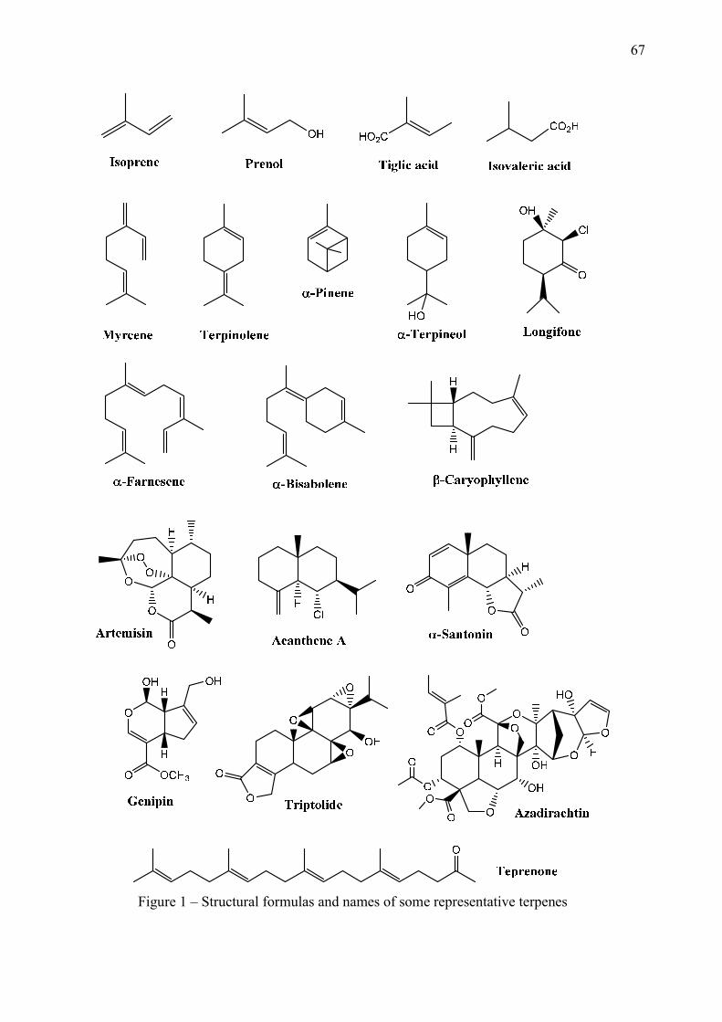
67
Figure 1 – Structural formulas and names of some representative terpenes
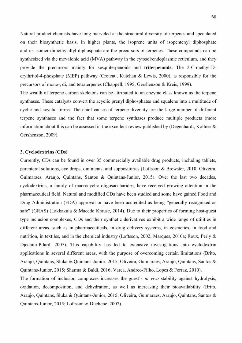
68
Natural product chemists have long marveled at the structural diversity of terpenes and speculated
on their biosynthetic basis. In higher plants, the isoprene units of isopentenyl diphosphate
and its isomer dimethylallyl diphosphate are the precursors of terpenes. These compounds can be
synthesized via the mevalonic acid (MVA) pathway in the cytosol/endoplasmic reticulum, and they
provide the precursors mainly for sesquiterpenoids and triterpenoids. The 2-C-methyl-D-
erythritol-4-phosphate (MEP) pathway (Croteau, Kutchan & Lewis, 2000), is responsible for the
precursors of mono-, di, and tetraterpenes (Chappell, 1995; Gershenzon & Kreis, 1999).
The wealth of terpene carbon skeletons can be attributed to an enzyme class known as the terpene
synthases. These catalysts convert the acyclic prenyl diphosphates and squalene into a multitude of
cyclic and acyclic forms. The chief causes of terpene diversity are the large number of different
terpene synthases and the fact that some terpene synthases produce multiple products (more
information about this can be assessed in the excellent review published by (Degenhardt, Kollner &
Gershenzon, 2009).
3. Cyclodextrins (CDs)
Currently, CDs can be found in over 35 commercially available drug products, including tablets,
parenteral solutions, eye drops, ointments, and suppositories (Loftsson & Brewster, 2010; Oliveira,
Guimaraes, Araujo, Quintans, Santos & Quintans-Junior, 2015). Over the last two decades,
cyclodextrins, a family of macrocyclic oligosaccharides, have received growing attention in the
pharmaceutical field. Natural and modified CDs have been studied and some have gained Food and
Drug Administration (FDA) approval or have been accredited as being “generally recognized as
safe” (GRAS) (Lakkakula & Macedo Krause, 2014). Due to their properties of forming host-guest
type inclusion complexes, CDs and their synthetic derivatives exhibit a wide range of utilities in
different areas, such as in pharmaceuticals, in drug delivery systems, in cosmetics, in food and
nutrition, in textiles, and in the chemical industry (Loftsson, 2002; Marques, 2010a; Roux, Perly &
Djedaini-Pilard, 2007). This capability has led to extensive investigations into cyclodextrin
applications in several different areas, with the purpose of overcoming certain limitations (Brito,
Araujo, Quintans, Sluka & Quintans-Junior, 2015; Oliveira, Guimaraes, Araujo, Quintans, Santos &
Quintans-Junior, 2015; Sharma & Baldi, 2016; Varca, Andreo-Filho, Lopes & Ferraz, 2010).
The formation of inclusion complexes increases the guest’s in vivo stability against hydrolysis,
oxidation, decomposition, and dehydration, as well as increasing their bioavailability (Brito,
Araujo, Quintans, Sluka & Quintans-Junior, 2015; Oliveira, Guimaraes, Araujo, Quintans, Santos &
Quintans-Junior, 2015; Loftsson & Duchene, 2007).
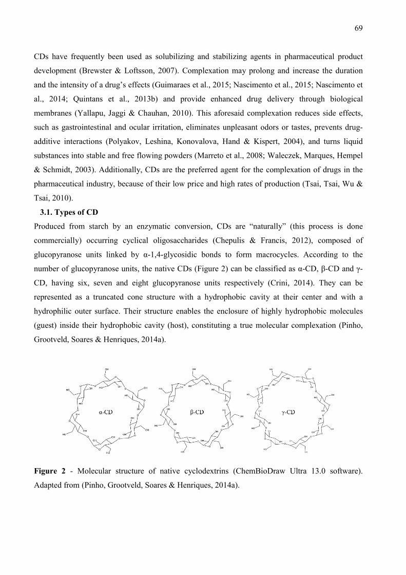
69
CDs have frequently been used as solubilizing and stabilizing agents in pharmaceutical product
development (Brewster & Loftsson, 2007). Complexation may prolong and increase the duration
and the intensity of a drug’s effects (Guimaraes et al., 2015; Nascimento et al., 2015; Nascimento et
al., 2014; Quintans et al., 2013b) and provide enhanced drug delivery through biological
membranes (Yallapu, Jaggi & Chauhan, 2010). This aforesaid complexation reduces side effects,
such as gastrointestinal and ocular irritation, eliminates unpleasant odors or tastes, prevents drug-
additive interactions (Polyakov, Leshina, Konovalova, Hand & Kispert, 2004), and turns liquid
substances into stable and free flowing powders (Marreto et al., 2008; Waleczek, Marques, Hempel
& Schmidt, 2003). Additionally, CDs are the preferred agent for the complexation of drugs in the
pharmaceutical industry, because of their low price and high rates of production (Tsai, Tsai, Wu &
Tsai, 2010).
3.1. Types of CD
Produced from starch by an enzymatic conversion, CDs are “naturally” (this process is done
commercially) occurring cyclical oligosaccharides (Chepulis & Francis, 2012), composed of
glucopyranose units linked by α-1,4-glycosidic bonds to form macrocycles. According to the
number of glucopyranose units, the native CDs (Figure 2) can be classified as α-CD, β-CD and γ-
CD, having six, seven and eight glucopyranose units respectively (Crini, 2014). They can be
represented as a truncated cone structure with a hydrophobic cavity at their center and with a
hydrophilic outer surface. Their structure enables the enclosure of highly hydrophobic molecules
(guest) inside their hydrophobic cavity (host), constituting a true molecular complexation (Pinho,
Grootveld, Soares & Henriques, 2014a).
Figure 2 - Molecular structure of native cyclodextrins (ChemBioDraw Ultra 13.0 software).
Adapted from (Pinho, Grootveld, Soares & Henriques, 2014a).

70
The choice of the suitable CD depends on the guest size because of the steric factors (Menezes et
al., 2015) and the administration route of interest. With the aim of improving β-CD solubility, new
CDs such as 2,6-Di-O-methyl-β-CD (DIMEB), hydroxipropil-β-cyclodextrin (HP-β-CD), randomly
methylated-β-cyclodextrin (RAMEB), and sulfobutyl ether β-cyclodextrin (SBE-β-CD), were
synthesized. Native and modified CDs, particularly HP-β-CD and sulfobutylether-γ-cyclodextrin
(SBEγCD), are widely used as excipients in nonclinical safety studies because of their excellent
physicochemical and biological properties and limited/reversible toxicity, particularly when dosed
orally, but also by the intravenous (i.v.) route (Rosseels, Delaunois, Hanon, Guillaume, Martin &
van den Dobbelsteen, 2013). Typically, hydrophilic CDs such as HP-β-CD, one of the β-CD
derivatives, are considered toxicity free at moderate oral and intravenous doses (Liao et al., 2016).
3.2 Restrictions of CDs application and possible risks
Despite the fact that CDs may be considered as GRAS by the FDA and have recently been
approved as an additive by the European Union (Arima, Hayashi, Higashi & Motoyama, 2015) they
are not free of toxicity, especially when used in improper administration routes. Thus, CD toxic
effects can be minimized by an appropriate choice of the CD or its derivative for the administration
route (Szejtli, 1998). All toxic studies have evidenced that orally administered CDs are practically
non-toxic, due to the lack of absorption from the gastrointestinal tract (Irie & Uekama, 1997). CDs
are hardly absorbed throughout the gastrointestinal tract and are degraded by bacterial enzymes in
the colon where they are treated as glucopyranose. This feature can be exploited for drug release
into the colon through the drug’s combination with CDs, which functions as a prodrug medication.
As a result, the drug is only released in the colon after its connection to the CDs has been degraded.
The drug can, therefore, act locally without any extensive systemic absorption, which is often
responsible for the adverse effects of drugs (Hirayama, Minami & Uekama, 1996; Minami,
Hirayama & Uekama, 1998; Uekama, Minami & Hirayama, 1997).
CDs for an enteric preparation can be classified as a time-controlled release agent, since the drug is
preferentially released in the intestinal tract (Rasheed & Kumar, 2008). This drug-guest system
seems to increase bioavailability, and consequently, is able to improve the therapeutic profiles of
volatile compounds such as monoterpenes (Pinho, Grootveld, Soares & Henriques, 2014b;
Marques, 2010a). Corroborating this approach, an intravenous dose of glaucocalyxin A, a diterpene,
was made possible due to the use of an inclusion complex with SBE-β-CD, demonstrating water
solubility significantly higher than that of β-CD. Additionally, it did not exhibit the nephrotoxicity
associated with β-CD and provided a protective effect against drug-induced cytotoxicity. This
resulted in a better solubility and a sustained function of the drug’s delivery (Ren, Jing, Chen, Miao

71
& Wei, 2014). In fact, choosing the best CDs for pharmacological applications depends upon the
chemical characteristics of the non-polar compound that is being complexed. If the CD is a
wildcard, by improving the drug’s biological activity, it minimizes any side effects.
In our investigation, the characteristics of the studies described in relation to the formation of the
inclusion complexes (Table 1), were examined in order to determine the efficiency of the
complexation process.
The naturally occurring β-CD was present in most of the studies (76%), of which 40% studied only
the β-CD complex, and 35% studied one or more branches of the β-CD complex. The derivate CDs
improved their toxicological profiles due a chemical modification of their hydroxyl groups. of the
natural CDs. Thus, many authors employed modified CDs, such as: M-β-CD (methyl-β-
cyclodextrin), HP-β-CD (hydroxypropyl-β-cyclodextrin), HP-γ-CD (hydroxypropyl-γ-
cyclodextrin), and SBE-β-CD (sulfobutylether-β-cyclodextrin), which all offer a high tolerance in
the human body and open new doors for the development of improved, more efficient injectable
formulations (Kurkov & Loftsson, 2013). The HP-β-CD complex appeared in 23% of the works and
is a very interesting β-CD derivation with improved water solubility (>70wt %). Furthermore, due
to hydroxypropyl substitution, the process of complexation with HP-β-CD was much facilitated
when compared with the native β-CD. In contrast, only a few studies chose α- and γ- CDs to
complex the natural products, probably related to the high cost.
The primary routes of administration were orally and intraperitoneally. The main positive effects
that were observed, and that were due to the use of inclusion complexes were: an improvement in
bioavailability (37%), better solubility (34%), an enhancement of biological activity (23%), an
enhanced efficacy (15%), and increased stability (15%). Furthermore, it was observed that there
were notable reductions in side effects and cytotoxicity, and a prolonged time effect.

72
Table 1 - Characteristics of the studies that described the formation of the inclusion complexes
Reference Classification Substance Type of
CD*
Method of
preparation
Evidence of complexation
Characterization
techniques**
Ratio of
complexation
(guest-host:
G-H)
General aspects described
Monoterpenes
(Donze &
Coleman, 1993),
France
(-)-borneol, terpineol, (+)-camphor,
(-)-carvone , geraniol , (+)-linalool,
cineole, (-)-fenchone, (+)-isomenthol,
citral, thymol, (-)-menthone, (+)-
menthol, o-cresol, eugenol, (+)-
limonene, (-)-bornyl acetate, anethol,
(+)-camphene, (-)-pinene, myrcene
β-CD Co-precipitation NMR 1:1 Physical properties of 21 substances were not
determinants alone complexation.
(Yoshii, Furuta,
Yasunishi &
Hirano, 1994),
Japan
(+)-limonene
α-CD
β-CD
γ-CD
Micro-aqueous Crystal analysis 1:1, 2:1 and
2:3
Low water content in the cavity of CD because
replace by D-Limonene caused through ethanol
effect.
(Moeder,
O'Brien,
Thompson &
Bicker, 1996),
(+) and (-)-α-pinene, (+) and (-)-β-
pinene, (+) and (-)-limonene, (+) and
(-)-camphene
β-CD
α-CD Not reported
Stoichiometric
coefficients of
complexation,
1:1 and 1:2
A difference in apparent formation constants was
only observed when a 1:2 terpene-CD complex was
formed.
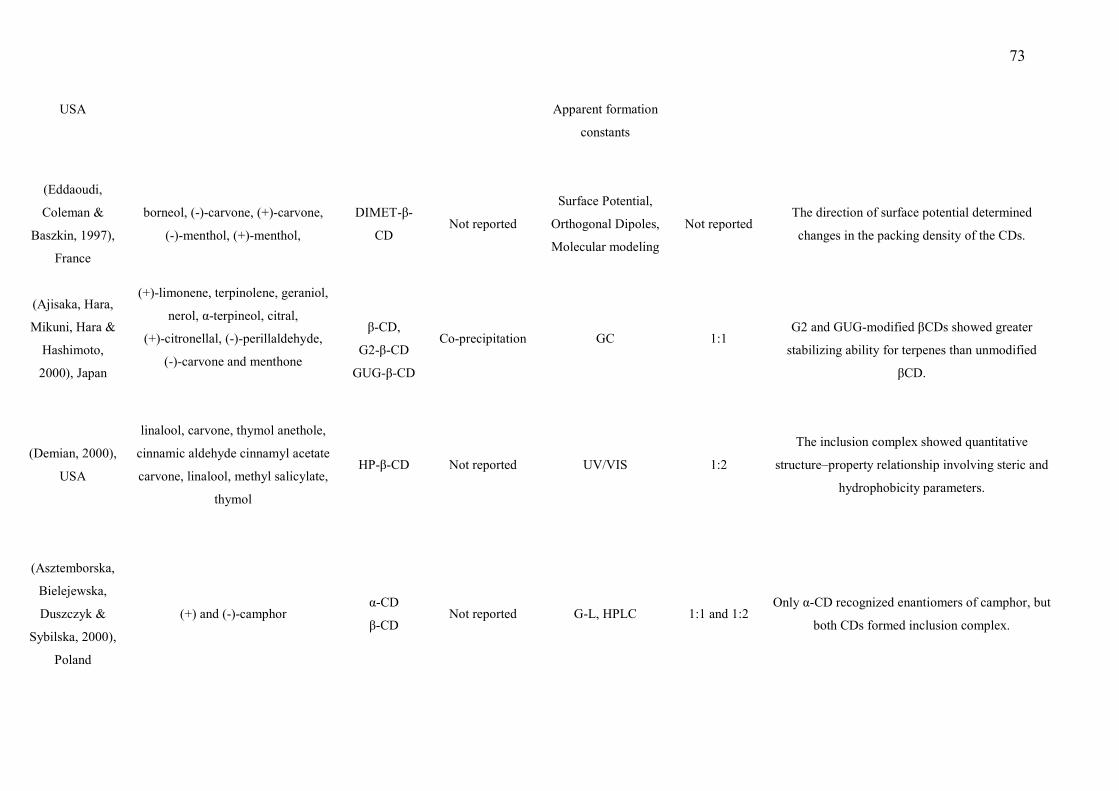
73
USA Apparent formation
constants
(Eddaoudi,
Coleman &
Baszkin, 1997),
France
borneol, (-)-carvone, (+)-carvone,
(-)-menthol, (+)-menthol,
DIMET-β-
CD Not reported
Surface Potential,
Orthogonal Dipoles,
Molecular modeling
Not reported The direction of surface potential determined
changes in the packing density of the CDs.
(Ajisaka, Hara,
Mikuni, Hara &
Hashimoto,
2000), Japan
(+)-limonene, terpinolene, geraniol,
nerol, α-terpineol, citral,
(+)-citronellal, (-)-perillaldehyde,
(-)-carvone and menthone
β-CD,
G2-β-CD
GUG-β-CD
Co-precipitation GC 1:1
G2 and GUG-modified βCDs showed greater
stabilizing ability for terpenes than unmodified
βCD.
(Demian, 2000),
USA
linalool, carvone, thymol anethole,
cinnamic aldehyde cinnamyl acetate
carvone, linalool, methyl salicylate,
thymol
HP-β-CD Not reported UV/VIS 1:2
The inclusion complex showed quantitative
structure–property relationship involving steric and
hydrophobicity parameters.
(Asztemborska,
Bielejewska,
Duszczyk &
Sybilska, 2000),
Poland
(+) and (-)-camphor α-CD
β-CD Not reported G-L, HPLC 1:1 and 1:2
Only α-CD recognized enantiomers of camphor, but
both CDs formed inclusion complex.

74
(Silva, Empis &
Teixeira-Dias,
2002), Portugal
Carvone β-CD Co-evaporation NMR 1:1
The chiral discrimination against carvone
enantiomers led to different water content and
structures in the inclusion compounds.
(Yu et al.,
2003), France
Camphor
α-CD
β-CD
γ-CD
Recrystallization
Phase-solubility,
NMR,
Circular dichroism
1:2 The techniques showed complexation of camphor
with all CDs.
(Dodziuk,
Nowinski,
Kozminski &
Dolgonos,
2003), Poland
camphor
α-CD Not reported NMR 1:1
NMR showed that is impossible to determinate the
binding constant for the camphor 1:2 complex.
(Asztemborska,
Sybilska,
Nowakowski &
Perez, 2003),
Poland
(+) and (-)-α-pinene, (+) and (-)-β-
pinene, citronellene, citronellal
menthone, pulegone, fenchone,
linalool, neomenthol, α-terpineol,
isopinocampheol, (+) and (-)-
limonene, (-)-α-phellandrene, (+) and
(-)-camphene, carvone, terpinen-4-ol
α-CD Not reported GC 1:1 and 1:2
Some terpenes were complexed in α-CD cavity,
others not complexed because remarkable
enantioselectivity.
(Liu et al.,
2004), China
cyclopentanol, cyclohexanol,
cycloheptanol, cyclooctanol), (±)-
β –CD Not reported NMR, ITC, ROESY 1:1
The binding modes revealed that the enhanced
molecular and chiral recognition ability was mainly
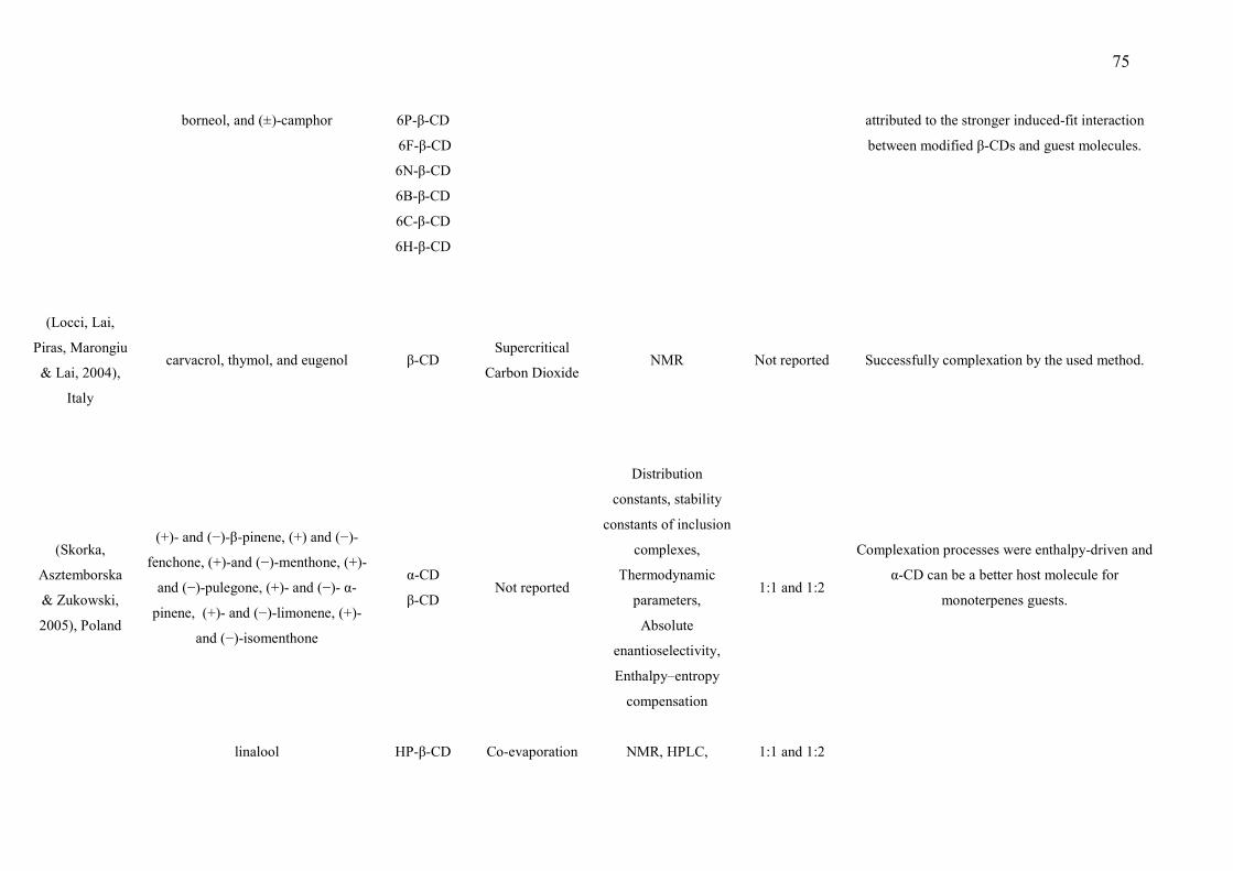
75
borneol, and (±)-camphor 6P-β-CD
6F-β-CD
6N-β-CD
6B-β-CD
6C-β-CD
6H-β-CD
attributed to the stronger induced-fit interaction
between modified β-CDs and guest molecules.
(Locci, Lai,
Piras, Marongiu
& Lai, 2004),
Italy
carvacrol, thymol, and eugenol β-CD Supercritical
Carbon Dioxide NMR Not reported Successfully complexation by the used method.
(Skorka,
Asztemborska
& Zukowski,
2005), Poland
(+)- and (−)-β-pinene, (+) and (−)-
fenchone, (+)-and (−)-menthone, (+)-
and (−)-pulegone, (+)- and (−)- α-
pinene, (+)- and (−)-limonene, (+)-
and (−)-isomenthone
α-CD
β-CD Not reported
Distribution
constants, stability
constants of inclusion
complexes,
Thermodynamic
parameters,
Absolute
enantioselectivity,
Enthalpy–entropy
compensation
1:1 and 1:2
Complexation processes were enthalpy-driven and
α-CD can be a better host molecule for
monoterpenes guests.
linalool HP-β-CD Co-evaporation NMR, HPLC, 1:1 and 1:2
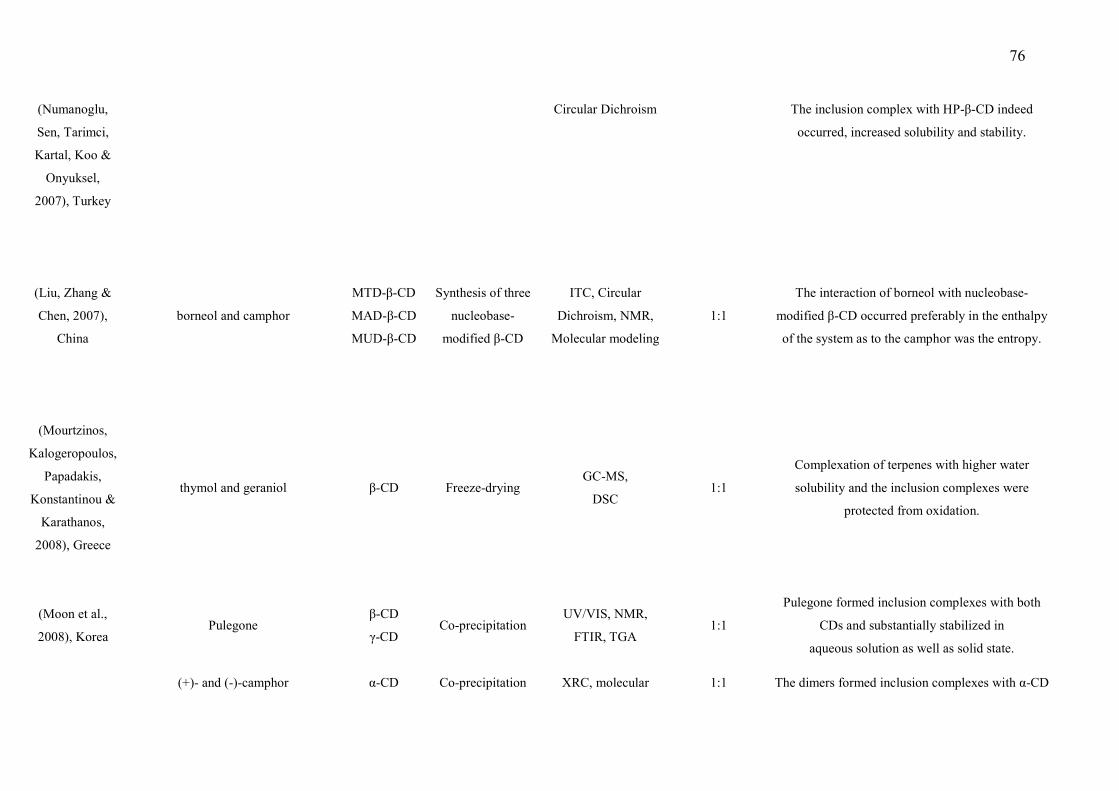
76
(Numanoglu,
Sen, Tarimci,
Kartal, Koo &
Onyuksel,
2007), Turkey
Circular Dichroism The inclusion complex with HP-β-CD indeed
occurred, increased solubility and stability.
(Liu, Zhang &
Chen, 2007),
China
borneol and camphor
MTD-β-CD
MAD-β-CD
MUD-β-CD
Synthesis of three
nucleobase-
modified β-CD
ITC, Circular
Dichroism, NMR,
Molecular modeling
1:1
The interaction of borneol with nucleobase-
modified β-CD occurred preferably in the enthalpy
of the system as to the camphor was the entropy.
(Mourtzinos,
Kalogeropoulos,
Papadakis,
Konstantinou &
Karathanos,
2008), Greece
thymol and geraniol β-CD Freeze-drying GC-MS,
DSC 1:1
Complexation of terpenes with higher water
solubility and the inclusion complexes were
protected from oxidation.
(Moon et al.,
2008), Korea
Pulegone β-CD
γ-CD Co-precipitation
UV/VIS, NMR,
FTIR, TGA 1:1
Pulegone formed inclusion complexes with both
CDs and substantially stabilized in
aqueous solution as well as solid state.
(+)- and (-)-camphor α-CD Co-precipitation XRC, molecular 1:1 The dimers formed inclusion complexes with α-CD
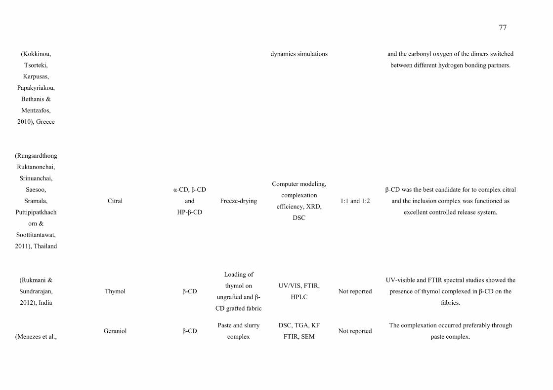
77
(Kokkinou,
Tsorteki,
Karpusas,
Papakyriakou,
Bethanis &
Mentzafos,
2010), Greece
dynamics simulations and the carbonyl oxygen of the dimers switched
between different hydrogen bonding partners.
(Rungsardthong
Ruktanonchai,
Srinuanchai,
Saesoo,
Sramala,
Puttipipatkhach
orn &
Soottitantawat,
2011), Thailand
Citral
α-CD, β-CD
and
HP-β-CD
Freeze-drying
Computer modeling,
complexation
efficiency, XRD,
DSC
1:1 and 1:2
β-CD was the best candidate for to complex citral
and the inclusion complex was functioned as
excellent controlled release system.
(Rukmani &
Sundrarajan,
2012), India
Thymol β-CD
Loading of
thymol on
ungrafted and β-
CD grafted fabric
UV/VIS, FTIR,
HPLC Not reported
UV-visible and FTIR spectral studies showed the
presence of thymol complexed in β-CD on the
fabrics.
(Menezes et al., Geraniol β-CD
Paste and slurry
complex
DSC, TGA, KF
FTIR, SEM Not reported
The complexation occurred preferably through
paste complex.

78
2012), Brazil
(Su et al.,
2012a), China
Borneol β-CD Ultrasound GC, DSC, XRD,
FTIR
1:1, 1:4, 1:6,
and
1:8
The stability and aqueous solubility of terpene were
significantly increased by inclusion with β-CD.
(Su et al.,
2012b), China
(+)-borneol HP-β-CD Ultrasound DSC, XRD, FTIR 1:6 (+)-borneol formed inclusion complexes with HP-
β-CD and had stability against heat and light.
(Serafini et al.,
2012), Brazil p-cymene β-CD
Slurry and paste
methods
DSC,
TGA/DTG, FTIR,
KF, SEM
1:1 p-cymene was efficiently complexed with β-CD by
paste method.
(Ceborska,
Szwed &
Suwinska,
2013), Poland
Isopulegol
β-CD Co-precipitation HPLC, XRC 1:1
The more stable complexes with
β-CD crystal structures were obtained showing the
formation of molecular capsules forming molecular
container.
(Ceborska,
Asztemborska,
Luboradzki &
Lipkowski,
camphene and fenchene
β-CD
Co-precipitation
NMR, GC, XRC
1:1
The 1:1 complexes of both (+/−) camphene and
fenchene with β-CD were obtained with stability
constant calculated.
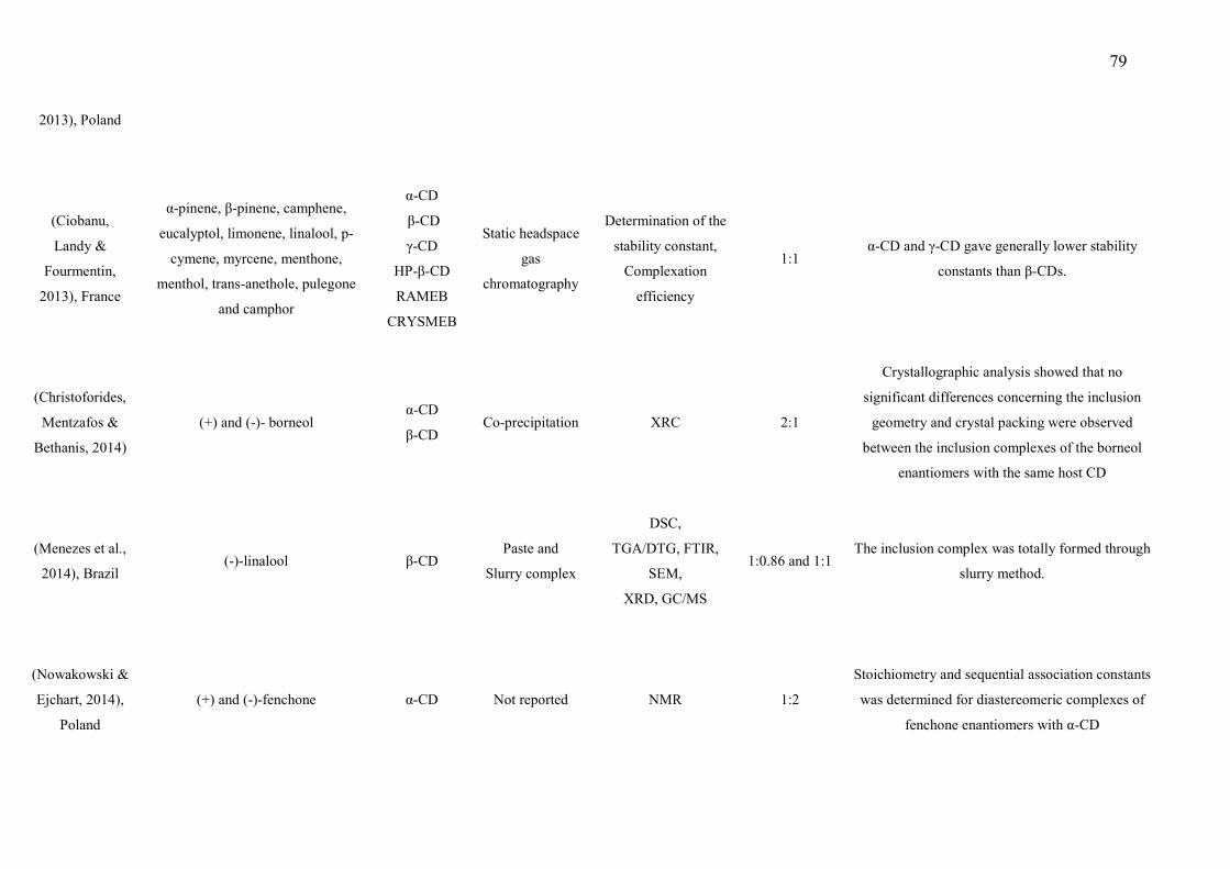
79
2013), Poland
(Ciobanu,
Landy &
Fourmentin,
2013), France
α-pinene, β-pinene, camphene,
eucalyptol, limonene, linalool, p-
cymene, myrcene, menthone,
menthol, trans-anethole, pulegone
and camphor
α-CD
β-CD
γ-CD
HP-β-CD
RAMEB
CRYSMEB
Static headspace
gas
chromatography
Determination of the
stability constant,
Complexation
efficiency
1:1 α-CD and γ-CD gave generally lower stability
constants than β-CDs.
(Christoforides,
Mentzafos &
Bethanis, 2014)
(+) and (-)- borneol α-CD
β-CD Co-precipitation XRC 2:1
Crystallographic analysis showed that no
significant differences concerning the inclusion
geometry and crystal packing were observed
between the inclusion complexes of the borneol
enantiomers with the same host CD
(Menezes et al.,
2014), Brazil
(-)-linalool β-CD Paste and
Slurry complex
DSC,
TGA/DTG, FTIR,
SEM,
XRD, GC/MS
1:0.86 and 1:1 The inclusion complex was totally formed through
slurry method.
(Nowakowski &
Ejchart, 2014),
Poland
(+) and (-)-fenchone α-CD Not reported NMR 1:2
Stoichiometry and sequential association constants
was determined for diastereomeric complexes of
fenchone enantiomers with α-CD
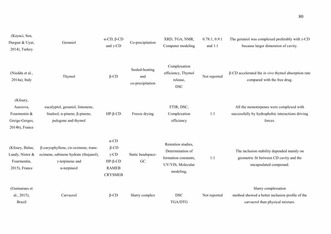
80
(Kayaci, Sen,
Durgun & Uyar,
2014), Turkey
Geraniol α-CD, β-CD
and γ-CD Co-precipitation
XRD, TGA, NMR,
Computer modeling
0.78:1, 0.9:1
and 1:1
The geraniol was complexed preferably with γ-CD
because larger dimension of cavity.
(Nieddu et al.,
2014a), Italy Thymol β-CD
Sealed-heating
and
co-precipitation
Complexation
efficiency, Thymol
release,
DSC
Not reported β-CD accelerated the in vivo thymol absorption rate
compared with the free drug.
(Kfoury,
Auezova,
Fourmentin &
Greige-Gerges,
2014b), France
eucalyptol, geraniol, limonene,
linalool, α-pinene, β-pinene,
pulegone and thymol
HP-β-CD Freeze drying
FTIR, DSC,
Complexation
efficiency
1:1
All the monoterpenes were complexed with
successfully by hydrophobic interactions driving
forces.
(Kfoury, Balan,
Landy, Nistor &
Fourmentin,
2015), France
β-caryophyllene, cis-ocimene, trans-
ocimene, sabinene hydrate (thujanol),
γ-terpinene and
α-terpineol
α-CD
β-CD
γ-CD
HP-β-CD
RAMEB
CRYSMEB
Static headspace-
GC
Retention studies,
Determination of
formation constants,
UV/VIS, Molecular
modeling,
1:1
The inclusion stability depended mainly on
geometric fit between CD cavity and the
encapsulated compound.
(Guimaraes et
al., 2015),
Brazil
Carvacrol β-CD Slurry complex
DSC
TGA/DTG
Not reported
Slurry complexation
method showed a better inclusion profile of the
carvacrol than physical mixture.
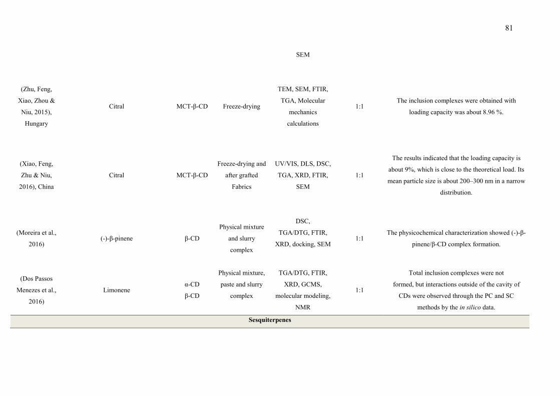
81
SEM
(Zhu, Feng,
Xiao, Zhou &
Niu, 2015),
Hungary
Citral MCT-β-CD Freeze-drying
TEM, SEM, FTIR,
TGA, Molecular
mechanics
calculations
1:1 The inclusion complexes were obtained with
loading capacity was about 8.96 %.
(Xiao, Feng,
Zhu & Niu,
2016), China
Citral MCT-β-CD
Freeze-drying and
after grafted
Fabrics
UV/VIS, DLS, DSC,
TGA, XRD, FTIR,
SEM
1:1
The results indicated that the loading capacity is
about 9%, which is close to the theoretical load. Its
mean particle size is about 200–300 nm in a narrow
distribution.
(Moreira et al.,
2016) (-)-β-pinene β-CD
Physical mixture
and slurry
complex
DSC,
TGA/DTG, FTIR,
XRD, docking, SEM
1:1 The physicochemical characterization showed (-)-β-
pinene/β-CD complex formation.
(Dos Passos
Menezes et al.,
2016)
Limonene α-CD
β-CD
Physical mixture,
paste and slurry
complex
TGA/DTG, FTIR,
XRD, GCMS,
molecular modeling,
NMR
1:1
Total inclusion complexes were not
formed, but interactions outside of the cavity of
CDs were observed through the PC and SC
methods by the in silico data.
Sesquiterpenes
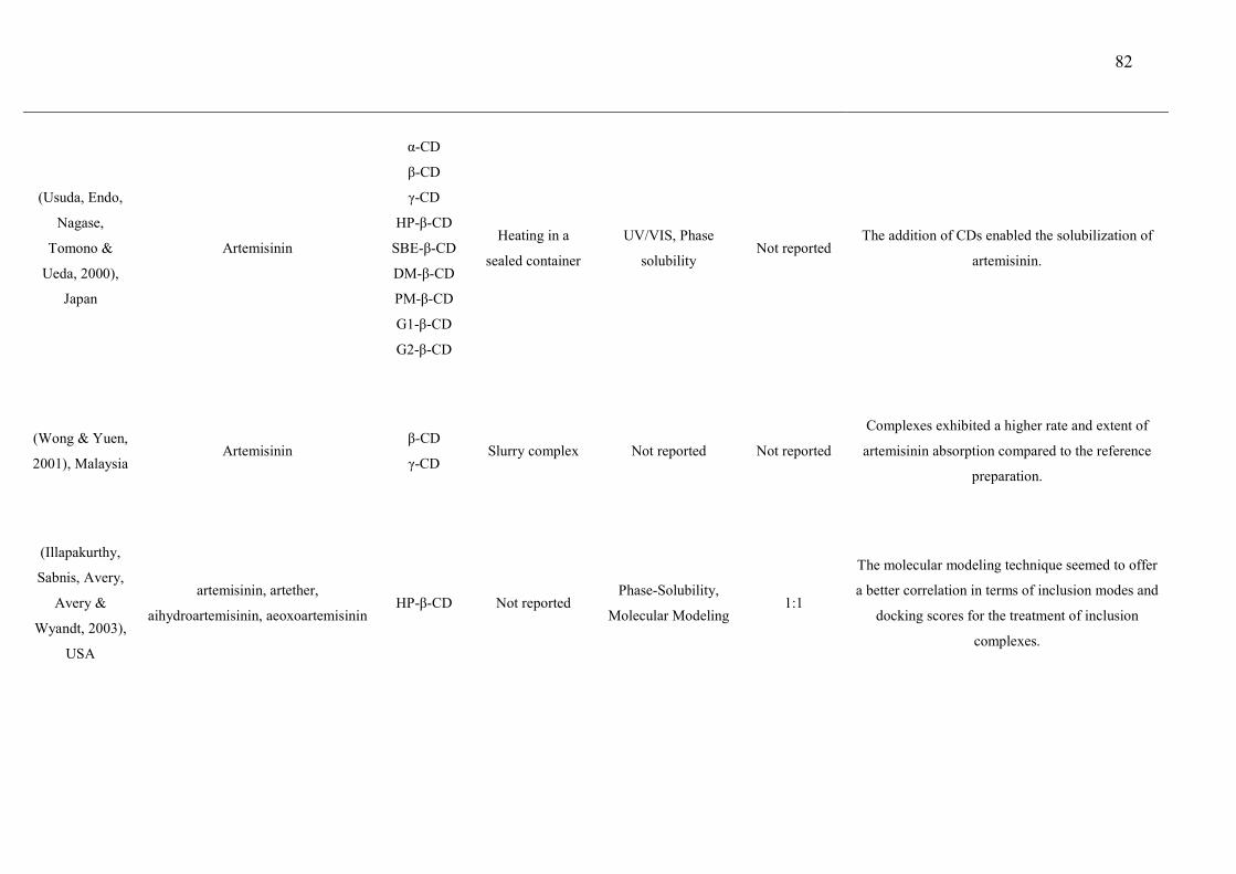
82
(Usuda, Endo,
Nagase,
Tomono &
Ueda, 2000),
Japan
Artemisinin
α-CD
β-CD
γ-CD
HP-β-CD
SBE-β-CD
DM-β-CD
PM-β-CD
G1-β-CD
G2-β-CD
Heating in a
sealed container
UV/VIS, Phase
solubility Not reported
The addition of CDs enabled the solubilization of
artemisinin.
(Wong & Yuen,
2001), Malaysia Artemisinin
β-CD
γ-CD Slurry complex Not reported Not reported
Complexes exhibited a higher rate and extent of
artemisinin absorption compared to the reference
preparation.
(Illapakurthy,
Sabnis, Avery,
Avery &
Wyandt, 2003),
USA
artemisinin, artether,
aihydroartemisinin, aeoxoartemisinin
HP-β-CD Not reported Phase-Solubility,
Molecular Modeling 1:1
The molecular modeling technique seemed to offer
a better correlation in terms of inclusion modes and
docking scores for the treatment of inclusion
complexes.
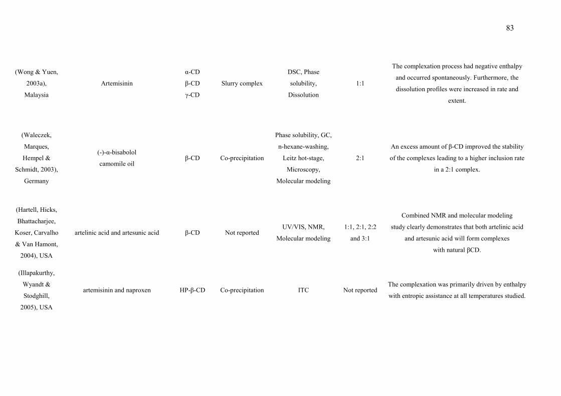
83
(Wong & Yuen,
2003a),
Malaysia
Artemisinin
α-CD
β-CD
γ-CD
Slurry complex
DSC, Phase
solubility,
Dissolution
1:1
The complexation process had negative enthalpy
and occurred spontaneously. Furthermore, the
dissolution profiles were increased in rate and
extent.
(Waleczek,
Marques,
Hempel &
Schmidt, 2003),
Germany
(-)-α-bisabolol
camomile oil β-CD Co-precipitation
Phase solubility, GC,
n-hexane-washing,
Leitz hot-stage,
Microscopy,
Molecular modeling
2:1
An excess amount of β-CD improved the stability
of the complexes leading to a higher inclusion rate
in a 2:1 complex.
(Hartell, Hicks,
Bhattacharjee,
Koser, Carvalho
& Van Hamont,
2004), USA
artelinic acid and artesunic acid β-CD Not reported UV/VIS, NMR,
Molecular modeling
1:1, 2:1, 2:2
and 3:1
Combined NMR and molecular modeling
study clearly demonstrates that both artelinic acid
and artesunic acid will form complexes
with natural βCD.
(Illapakurthy,
Wyandt &
Stodghill,
2005), USA
artemisinin and naproxen HP-β-CD Co-precipitation ITC Not reported
The complexation was primarily driven by enthalpy
with entropic assistance at all temperatures studied.
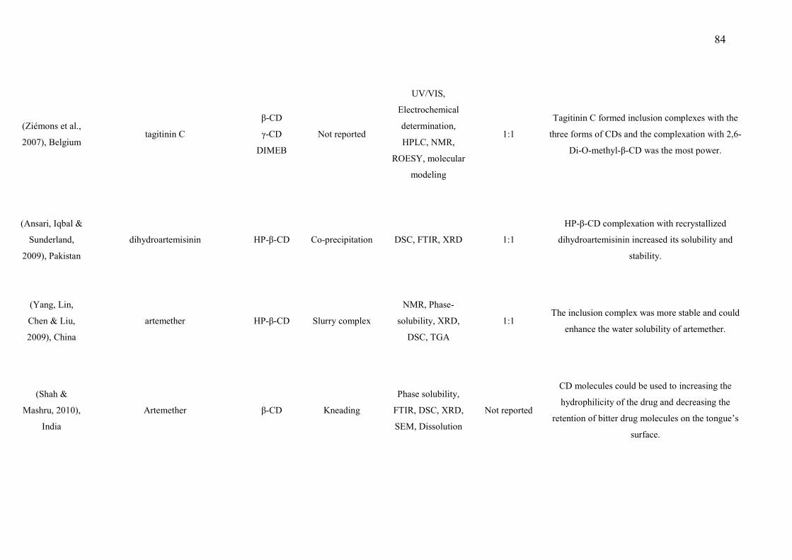
84
(Ziémons et al.,
2007), Belgium tagitinin C
β-CD
γ-CD
DIMEB
Not reported
UV/VIS,
Electrochemical
determination,
HPLC, NMR,
ROESY, molecular
modeling
1:1
Tagitinin C formed inclusion complexes with the
three forms of CDs and the complexation with 2,6-
Di-O-methyl-β-CD was the most power.
(Ansari, Iqbal &
Sunderland,
2009), Pakistan
dihydroartemisinin HP-β-CD Co-precipitation DSC, FTIR, XRD 1:1
HP-β-CD complexation with recrystallized
dihydroartemisinin increased its solubility and
stability.
(Yang, Lin,
Chen & Liu,
2009), China
artemether HP-β-CD Slurry complex
NMR, Phase-
solubility, XRD,
DSC, TGA
1:1 The inclusion complex was more stable and could
enhance the water solubility of artemether.
(Shah &
Mashru, 2010),
India
Artemether β-CD Kneading
Phase solubility,
FTIR, DSC, XRD,
SEM, Dissolution
Not reported
CD molecules could be used to increasing the
hydrophilicity of the drug and decreasing the
retention of bitter drug molecules on the tongue’s
surface.
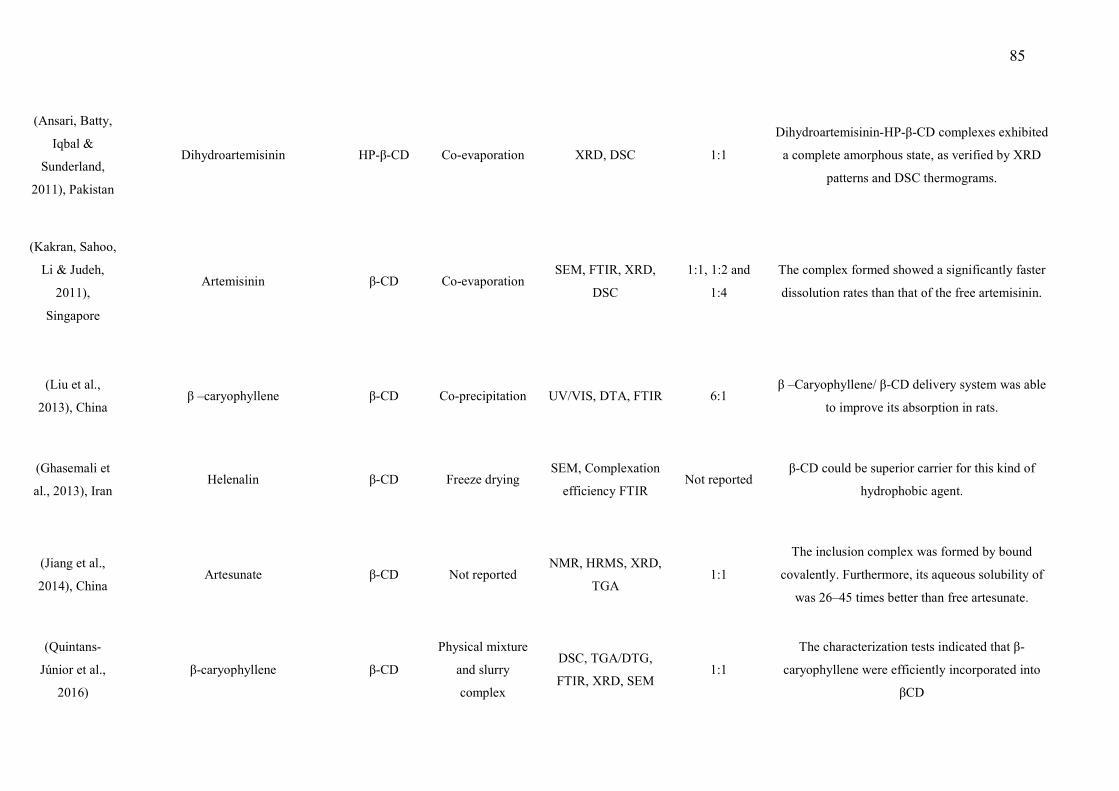
85
(Ansari, Batty,
Iqbal &
Sunderland,
2011), Pakistan
Dihydroartemisinin HP-β-CD Co-evaporation XRD, DSC 1:1
Dihydroartemisinin-HP-β-CD complexes exhibited
a complete amorphous state, as verified by XRD
patterns and DSC thermograms.
(Kakran, Sahoo,
Li & Judeh,
2011),
Singapore
Artemisinin β-CD Co-evaporation SEM, FTIR, XRD,
DSC
1:1, 1:2 and
1:4
The complex formed showed a significantly faster
dissolution rates than that of the free artemisinin.
(Liu et al.,
2013), China
β –caryophyllene β-CD Co-precipitation UV/VIS, DTA, FTIR 6:1 β –Caryophyllene/ β-CD delivery system was able
to improve its absorption in rats.
(Ghasemali et
al., 2013), Iran Helenalin β-CD Freeze drying
SEM, Complexation
efficiency FTIR Not reported
β-CD could be superior carrier for this kind of
hydrophobic agent.
(Jiang et al.,
2014), China Artesunate β-CD Not reported
NMR, HRMS, XRD,
TGA 1:1
The inclusion complex was formed by bound
covalently. Furthermore, its aqueous solubility of
was 26–45 times better than free artesunate.
(Quintans-
Júnior et al.,
2016)
β-caryophyllene β-CD
Physical mixture
and slurry
complex
DSC, TGA/DTG,
FTIR, XRD, SEM 1:1
The characterization tests indicated that β-
caryophyllene were efficiently incorporated into
βCD
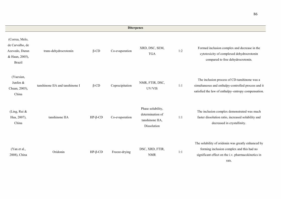
86
Diterpenes
(Correa, Melo,
de Carvalho, de
Azevedo, Duran
& Haun, 2005),
Brazil
trans-dehydrocrotonin β-CD Co-evaporation XRD, DSC, SEM,
TGA 1:2
Formed inclusion complex and decrease in the
cytotoxicity of complexed dehydrocrotonin
compared to free dehydrocrotonin.
(Yuexian,
Junfen &
Chuan, 2005),
China
tanshinone IIA and tanshinone I β-CD Coprecipitation NMR, FTIR, DSC,
UV/VIS 1:1
The inclusion process of CD-tanshinone was a
simultaneous and enthalpy-controlled process and it
satisfied the law of enthalpy–entropy compensation.
(Ling, Rui &
Hua, 2007),
China
tanshinone IIA HP-β-CD Co-evaporation
Phase solubility,
determination of
tanshinone IIA,
Dissolution
1:1
The inclusion complex demonstrated was much
faster dissolution ratio, increased solubility and
decreased in crystallinity.
(Yan et al.,
2008), China Oridonin HP-β-CD Freeze-drying
DSC, XRD, FTIR,
NMR 1:1
The solubility of oridonin was greatly enhanced by
forming inclusion complex and this had no
significant effect on the i.v. pharmacokinetics in
rats.
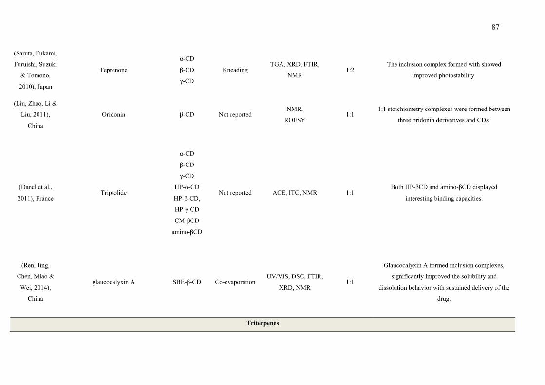
87
(Saruta, Fukami,
Furuishi, Suzuki
& Tomono,
2010), Japan
Teprenone
α-CD
β-CD
γ-CD
Kneading TGA, XRD, FTIR,
NMR 1:2
The inclusion complex formed with showed
improved photostability.
(Liu, Zhao, Li &
Liu, 2011),
China
Oridonin β-CD Not reported NMR,
ROESY 1:1
1:1 stoichiometry complexes were formed between
three oridonin derivatives and CDs.
(Danel et al.,
2011), France Triptolide
α-CD
β-CD
γ-CD
HP-α-CD
HP-β-CD,
HP-γ-CD
CM-βCD
amino-βCD
Not reported ACE, ITC, NMR 1:1 Both HP-βCD and amino-βCD displayed
interesting binding capacities.
(Ren, Jing,
Chen, Miao &
Wei, 2014),
China
glaucocalyxin A SBE-β-CD Co-evaporation UV/VIS, DSC, FTIR,
XRD, NMR 1:1
Glaucocalyxin A formed inclusion complexes,
significantly improved the solubility and
dissolution behavior with sustained delivery of the
drug.
Triterpenes

88
(Rode et al.,
2003), Germany Sericoside
γ-CD
HP-γ-CD
M-β-CD
HP-β-CD
Co-evaporation NMR, XRD, TGA,
DSC
1:2, 1:2.2 and
1:2.6
The inclusion complex improved the solubility of
sericoside and only complexes with γ-CD were
stable.
(Guo, Zhang,
Song, Wang,
Liu & Liu,
2003), China
oleanolic acid
α-CD
β-CD
γ-CD
Freeze-drying Solubility study and
ESI/MS 1:1 and 2:1
Direct correlation between stability and binding
forces of complexes in solution and gas phases.
(Liu, Chen,
Chen & Lin,
2005), China
Azadirachtin
β -CD
DM-β-CD
TM-β-CD
HP-β-CD
Co-precipitation XRD, TGA, DTA,
DSC, UV/VIS, NMR 1:1 and 2:1
The binding behavior was mainly dependent on the
individual structural features of host and guest.
(Shen, Yang,
Wu & Ma,
2005), China
Gossypol β-CD
Prepared under
microwave
Irradiation
UV/VIS, FTIR,
NMR, DSC, TGA 1:2
Gossypol formed inclusion complexes with β-CD
by the mentioned technique.
(Shen, Ying,
Yang & Wu,
2006), China
Gossypol β-CD
Prepared under
microwave
Irradiation
UV/VIS,
Fluorescence
spectral, SEM, XRD
Not reported
The formation of
the inclusion complex could be proposed as a new
formulation to optimize its pharmacological profile.
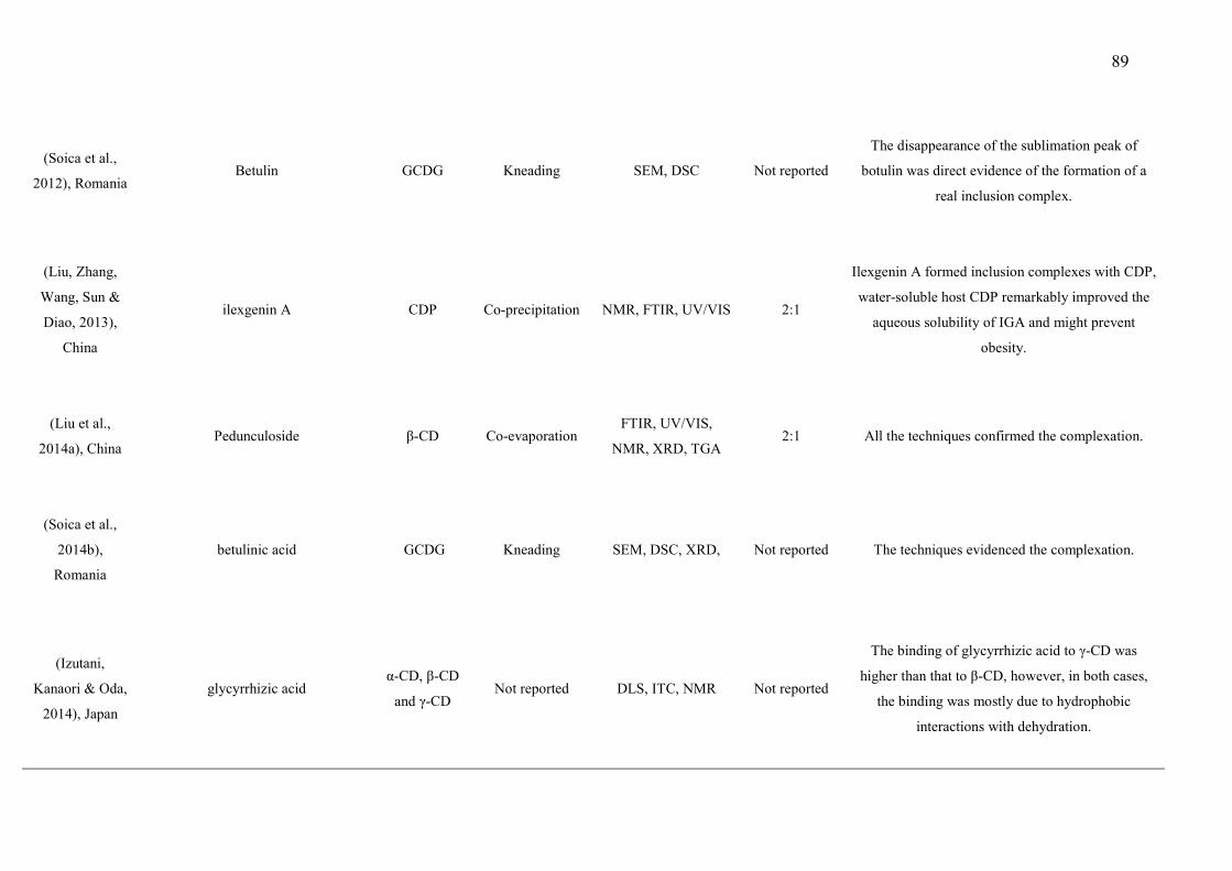
89
(Soica et al.,
2012), Romania Betulin GCDG Kneading SEM, DSC Not reported
The disappearance of the sublimation peak of
botulin was direct evidence of the formation of a
real inclusion complex.
(Liu, Zhang,
Wang, Sun &
Diao, 2013),
China
ilexgenin A CDP Co-precipitation NMR, FTIR, UV/VIS 2:1
Ilexgenin A formed inclusion complexes with CDP,
water-soluble host CDP remarkably improved the
aqueous solubility of IGA and might prevent
obesity.
(Liu et al.,
2014a), China Pedunculoside β-CD Co-evaporation
FTIR, UV/VIS,
NMR, XRD, TGA
2:1 All the techniques confirmed the complexation.
(Soica et al.,
2014b),
Romania
betulinic acid GCDG Kneading SEM, DSC, XRD, Not reported The techniques evidenced the complexation.
(Izutani,
Kanaori & Oda,
2014), Japan
glycyrrhizic acid α-CD, β-CD
and γ-CD Not reported DLS, ITC, NMR Not reported
The binding of glycyrrhizic acid to γ-CD was
higher than that to β-CD, however, in both cases,
the binding was mostly due to hydrophobic
interactions with dehydration.
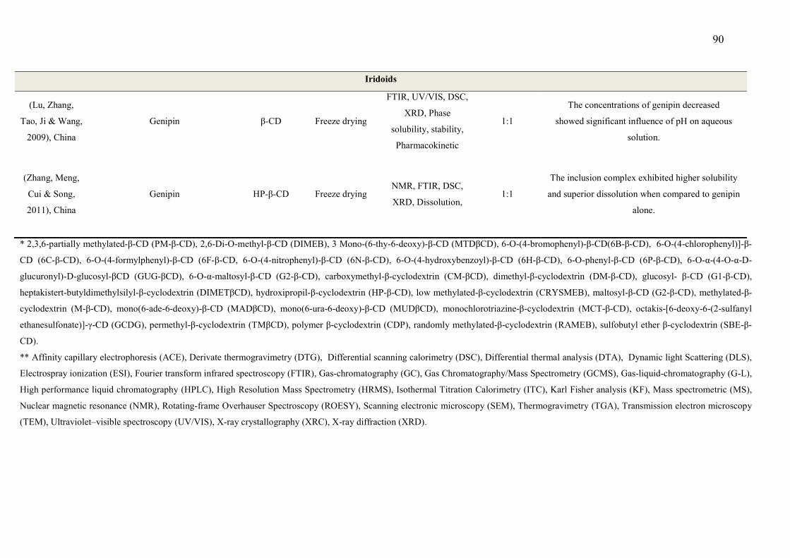
90
Iridoids
(Lu, Zhang,
Tao, Ji & Wang,
2009), China
Genipin β-CD Freeze drying
FTIR, UV/VIS, DSC,
XRD, Phase
solubility, stability,
Pharmacokinetic
1:1
The concentrations of genipin decreased
showed significant influence of pH on aqueous
solution.
(Zhang, Meng,
Cui & Song,
2011), China
Genipin HP-β-CD Freeze drying NMR, FTIR, DSC,
XRD, Dissolution, 1:1
The inclusion complex exhibited higher solubility
and superior dissolution when compared to genipin
alone.
* 2,3,6-partially methylated-β-CD (PM-β-CD), 2,6-Di-O-methyl-β-CD (DIMEB), 3 Mono-(6-thy-6-deoxy)-β-CD (MTDβCD), 6-O-(4-bromophenyl)-β-CD(6B-β-CD), 6-O-(4-chlorophenyl)]-β-
CD (6C-β-CD), 6-O-(4-formylphenyl)-β-CD (6F-β-CD, 6-O-(4-nitrophenyl)-β-CD (6N-β-CD), 6-O-(4-hydroxybenzoyl)-β-CD (6H-β-CD), 6-O-phenyl-β-CD (6P-β-CD), 6-O-α-(4-O-α-D-
glucuronyl)-D-glucosyl-βCD (GUG-βCD), 6-O-α-maltosyl-β-CD (G2-β-CD), carboxymethyl-β-cyclodextrin (CM-βCD), dimethyl-β-cyclodextrin (DM-β-CD), glucosyl- β-CD (G1-β-CD),
heptakistert-butyldimethylsilyl-β-cyclodextrin (DIMETβCD), hydroxipropil-β-cyclodextrin (HP-β-CD), low methylated-β-cyclodextrin (CRYSMEB), maltosyl-β-CD (G2-β-CD), methylated-β-
cyclodextrin (M-β-CD), mono(6-ade-6-deoxy)-β-CD (MADβCD), mono(6-ura-6-deoxy)-β-CD (MUDβCD), monochlorotriazine-β-cyclodextrin (MCT-β-CD), octakis-[6-deoxy-6-(2-sulfanyl
ethanesulfonate)]-γ-CD (GCDG), permethyl-β-cyclodextrin (TMβCD), polymer β-cyclodextrin (CDP), randomly methylated-β-cyclodextrin (RAMEB), sulfobutyl ether β-cyclodextrin (SBE-β-
CD).
** Affinity capillary electrophoresis (ACE), Derivate thermogravimetry (DTG), Differential scanning calorimetry (DSC), Differential thermal analysis (DTA), Dynamic light Scattering (DLS),
Electrospray ionization (ESI), Fourier transform infrared spectroscopy (FTIR), Gas-chromatography (GC), Gas Chromatography/Mass Spectrometry (GCMS), Gas-liquid-chromatography (G-L),
High performance liquid chromatography (HPLC), High Resolution Mass Spectrometry (HRMS), Isothermal Titration Calorimetry (ITC), Karl Fisher analysis (KF), Mass spectrometric (MS),
Nuclear magnetic resonance (NMR), Rotating-frame Overhauser Spectroscopy (ROESY), Scanning electronic microscopy (SEM), Thermogravimetry (TGA), Transmission electron microscopy
(TEM), Ultraviolet–visible spectroscopy (UV/VIS), X-ray crystallography (XRC), X-ray diffraction (XRD).
91
3.3 Inclusion methods and complexation efficiency
The effectiveness of CD applications is related to the amount of the guest that is
included in the cavity. This percentage depends on both the stability constant and the
host and guest concentrations. The strength of the host-guest complexation depends
mainly on the polarity and the geometric accommodation between the CD cavity and
the guest molecule. These parameters play a critical role in determining the
stoichiometry and the formation constant (Kf) of the inclusion complexes (Kfoury,
Auezova, Fourmentin & Greige-Gerges, 2014a; Marques, 2010b). Nieddu et al. (2014a)
proposed the following non-conventional method of preparation for the inclusion
complex and observed that it showed a very good loading efficiency.
The inclusion complex that was prepared by sealed-heating, using a 1:1 molar ratio
between the two components, was able to increase the dissolution rate of thymol, a
phenol monoterpene, which dissolved slowly in gastrointestinal-simulated fluid. This
effect is due to the well-known consequence of β-CD acting as a solubilizer of
substances that are poorly water soluble (Nieddu et al., 2014b). It is possible that this
method of preparation can produce a better complexation ratio. In addition,
Rungsardthong Ruktanonchai et al. (2011) studied citral, a monoterpene that is present
in the essential oils of citrus fruits, and its complexation with CDs by a freeze-drying
method. The obtained total complexation was with a 1:1 and a 1:2 molar ratio. Menezes
et al. (2014) demonstrated a partial complexation for a paste method and a total
complexation for a slurry method (1:0.86 and 1:1, respectively). In these cases, the
efficiency of a complexation depends upon the affinity and the size of drug, as well as
the type of CD and method of preparation (Valle, 2004).
Another complexation procedure is the solid-state mechanochemical method, which has
been developed through dynamic interdisciplinary research, and has been described in
the literature in studies, including those with natural products (Dushkin, 2010;
Voinovich, Perissutti, Grassi, Passerini & Bigotto, 2009). The main advantages of this
method are its speed, simplicity, cost-effectiveness, easy scalability and the reduction
of waste and solvent use (when required), which makes it an attractive option for
several application fields (Rinaldi, Binello, Stolle, Curini & Cravotto, 2015; Cravotto,
Garella, Carnaroglio, Gaudino & Rosati, 2012). The method consists of grinding
treatments that may dramatically enhance the solubility and bioavailability of poorly
water-soluble drugs (Xie, Yang, Shi, Zhu, Su & Wang, 2013) due to particle size
92
reduction, higher surface area and porosity. Ultrafine milling is a straightforward
technique based on impact forces and friction effects, which result in large amounts of
mechanical energy that usually cause structural changes, and even chemical reactions
including the enhancement of molecular interaction and complex formation. Moreover,
characteristics such as solubility and dissolution rate are improved because of the
reduced particle size (Bragagni, Maestrelli & Mura, 2010; Iwata et al., 2009).
These characteristics of CDs are interesting in relation to complex compounds, such as
terpenes, that present many limitations due to their low solubility in an aqueous medium
(Quintans-Júnior et al., 2016; Siqueira-Lima et al., 2014). CDs are usually able to
enhance the bioavailability of insoluble compounds by increasing their drug solubility,
their dissolution, and their drug permeability (Rasheed & Kumar, 2008). These effects
corroborate previous findings that CDs can improve various aspects of non-polar
compounds (Pinho, Grootveld, Soares & Henriques, 2014a). However, this does not
appear to be feasible for all non-polar compounds due to the different complexation
rates of major compounds. A better complexation with CDs is usually found with
compounds and/or active substances from essential oils (Menezes et al., 2015; Pinho,
Grootveld, Soares & Henriques, 2014a; Siqueira-Lima et al., 2014).
The biomedical applications of CDs are extremely attractive due to their low toxicity
and their low immunogenicity, and thus, they create a wide possibility for complexing
different drugs (Irie & Uekama, 1997; Zhang & Ma, 2013). However, CDs do not
readily permeate biological membranes due to their chemical structure, their molecular
weight, and their very low octanol/water partition coefficient (Loftsson, Sigfusson,
Sigurdsson & Masson, 2003). Only the free form of the drug, which was in equilibrium
with the drug/CD complexes, was capable of penetrating the lipophilic membranes.
Thus, the physicochemical properties of the drug (e.g., its solubility in water), the
composition of the drug’s formulation (e.g., aqueous or non-aqueous), and the
physiological composition of the membrane barrier (e.g., the presence of an aqueous
diffusion layer), will determine whether the CDs will enhance or will hamper the drug’s
delivery through a biological membrane (Loftsson, Masson & Brewster, 2004). This
seems to be quite advantageous for natural non-polar compounds such as terpenes
(Brito, Araujo, Quintans, Sluka & Quintans-Junior, 2015; Guimaraes et al., 2015;
Marques, 2010a; Pinho, Grootveld, Soares & Henriques, 2014a; Quintans et al., 2013b).
3.4 Evidence of Complexation
93
Some authors have reported that the relatively hydrophobic cavity of a CD can
accommodate non-polar molecules (such as terpenes) to form inclusion complexes
(Marques, 2010a; Pinho, Grootveld, Soares & Henriques, 2014a).
According to Ciobanu, Landy and Fourmentin (2013), the complexation behavior of
several types of CDs, with 13 monoterpenes focused on by the authors, suggested that
β-CD had better complexation efficiency than did α-CD and γ-CD because the cavity
size is suitable for small molecules. Indeed, β-CD might complex between 80% and
99% of the volatile flavor compounds that are present in food. Moreover, the
complexation efficiency of native β-CD is close to that of the modified β-CDs (HP β-
CD, RAMEB, and CRYSMEB). However, the solubility of modified β-CDs is greatly
enhanced when compared to native β-CDs, and higher complexation efficiencies might
be obtained.
It is interesting to note that measurements of complexation efficiency should be carried
out in order to determine the drug complexation percentage in the cavity of the CDs, as
well to establish whether the complexation was full or partial, based on the host:guest
ratio. This kind of a technique was only performed in 7% of the studies analyzed, using
HPLC, UV/vis and GC/MS. These analyses allow the level of drug entrapment in the
final product as well as the host:guest ratio to be measured. Haloci et al (2014) in a
study of the essential oil of Satureja montana reported the probable reasons for material
loss during the complexation procedure can be attributed to the oil, CD and complex
dissipation. There are several factors which may contribute to the loss of oil such as
retention of the oil in the solution after forming the complex, equilibrium of flavors
between the liquid and the complexed state, evaporation of surface oil during the long
complexation process and evaporation during the drying stage. The loss of the CD and
complex powder can be mainly attributed to their solubility in water (Haloci, Toska,
Shkreli, Goci, Vertuani & Manfredini, 2014).
3.4.1. Spectroscopic characterization of the complexes
The analytical techniques of characterization that are required to assess the formation of
a drug-CD inclusion complex are not a simple task. It often requires the use of different
analytical methods, whose results have to be combined and examined together, since
each method explores a particular feature of the inclusion complex (Mura, 2014). As
shown in Table 1, the most used techniques for the characterization of inclusion
complexes are DSC (40%), NMR (37%), XRD (34%), FTIR (33%), and TGA (23%).

94
Other techniques, such as DLS, were only used in 2.75% of the papers (i.e. in two
papers). The researchers were not determining the particle size. This information is very
important to explain phenomena of dissolution and the phase solubility analysis.
All of these techniques are very important in order to determine the inclusion complex
formation. In general, all of the techniques that were used for the characterization were
employed together, because one technique alone cannot provide comprehensive data on
complex formation, unless the technique is NMR. NMR provides direct evidence of the
inclusion of a guest molecule inside the CD cavity, which can be identified by
comparing the chemical shifts of the free guest molecule and the CD with those of its
complex (Van Axel Castelli et al., 2008). Such evidence is based on the fact that, if an
inclusion is present, then the physical or chemical environment is affected by the
hydrogen elements at the internal surface of the cavity (H3 and H5 of the CD) and the
included guest molecule (Tsao et al., 2011). Another interesting and little used
technique was molecular modeling. It was only performed in 10% of the publications.
This technique provides theoretical data for the complexation, and whether it can occur
or not, based on the geometry and the steric factors of the molecules under study. The
deformation energy (Edeformation) is determined by the difference between the energy of
the partners of the complex at their respective equilibrium geometry and their energy at
the complexed geometry (Menezes et al., 2016). The interaction energy (Einteraction) is
defined as the difference between the energy of the complex and the sum of the energies
of both partners at their complexed geometry, as described by Ziémons et al. (2007).
This technique is useful before a study because it is a theoretical method which can
estimate if complex formation is possible or not, thereby avoiding wasting time and
money on a study that will not yield satisfactory results.
4. Inclusion Complexes – CDs and Terpenes
Several studies have demonstrated the pharmaceutical or pharmacological benefits of a
cyclodextrin inclusion complex with natural products (Ceborska, 2014; Porte, Porte &
Oliveira, 2014), but we found no reviews that could be useful for developing new
approaches to improving CD-based host-guest complexes. We therefore conducted this
review in order to assess the evidence for improvements in the pharmacological effect
of natural products when included in CDs.
95
The use of some plant-derived compounds, such as terpenoids, have found some
restrictions in the pharmaceutical marketplace, since they have limited water solubility,
poor bioavailability, and can be affected by many factors, including temperature, pH
and light. Therefore, the integrity of bioactive molecules needs to be protected, and it is
necessary to use a formulation that can deliver them to physiological targets without
losing any bioactivity (Pinho, Grootveld, Soares & Henriques, 2014a). Finding ways to
enhance the absorption and the bioavailability of these compounds is one of the major
challenges that the pharmaceutical industry presently faces (Aburahma, 2015; Munin &
Edwards-Levy, 2011).
Several approaches have been explored in order to improve the therapeutic properties of
non-polar natural compounds, including the employment of drug-delivery systems such
as liposomes, nanoparticles or nanogels, surfactants, and CDs (Guimaraes et al., 2015;
Menezes et al., 2015; Quintans-Junior et al., 2013; Siqueira-Lima et al., 2014).
Cyclodextrins (CDs) seem to have a key role to play when they are used to improve the
chemical and pharmacological properties of non-polar drugs (Brito, Araujo, Quintans,
Sluka & Quintans-Junior, 2015; Oliveira, Guimaraes, Araujo, Quintans, Santos &
Quintans-Junior, 2015).
4.1 Biological Activities (Therapeutic activity)
In the studies in this review, terpenes have been shown to exhibit a wide range of
biological activities on the human body; as an analgesic, an anti-inflammatory, an
anticonvulsant, an antimicrobial, an antibacterial, an antitumoral, and in lipid
metabolism. The effects of β-CD were also assessed in respect of their biological
activities (Tables 2, 3 and 4).
4.2 Pharmacokinetics
Brito et al. (2015) and Oliveira et al. (2015) have recently reported that terpenes
complexed with CDs have improved bioavailability, solubility, and a reduced effective
dose, thereby having better pharmacological efficacy. These are probably the most
promising benefits of this type of a drug as they are mainly analgesic drugs. However,
the authors have noted that there are few pharmacokinetic studies that focus on the
drug’s potential and its usefulness in the development of new clinical drug candidates.
Ineffectiveness and toxicity are considered to be the major reasons for the failure of new
drugs, so pharmacokinetics has had a great impact on these undesirable effects (Rosa et
96
al., 2014). Pharmacokinetic studies, investigating drug concentration-effect
relationships, efficacy and safety have usually been conducted in animal models, in
order to predict the clinical setting for the drug (Cunha-Filho & Sá-Barreto, 2007;
Singh, 2006). The determination of pharmacokinetic parameters and the understanding
of the biological mechanisms underlying these important parameters is vitally important
for supporting the planning of the clinical stage (Amore, Gibbs & Emery, 2010). In
respect of pharmacokinetic studies with CD inclusion complexes containing terpenes, or
related compounds, we found few references in the literature and those studies we did
identify were often at an early stage.
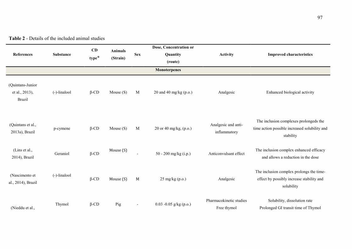
97
Table 2 - Details of the included animal studies
References Substance CD
type*
Animals
(Strain) Sex
Dose, Concentration or
Quantity
(route)
Activity Improved characteristics
Monoterpenes
(Quintans-Junior
et al., 2013),
Brazil
(-)-linalool β-CD Mouse (S) M 20 and 40 mg/kg (p.o.) Analgesic Enhanced biological activity
(Quintans et al.,
2013a), Brazil p-cymene β-CD Mouse (S) M 20 or 40 mg/kg, (p.o.)
Analgesic and anti-
inflammatory
The inclusion complexes prolongeds the
time action possible increased solubility and
stability
(Lins et al.,
2014), Brazil Geraniol β-CD
Mouse (S) - 50 - 200 mg/kg (i.p.) Anticonvulsant effect
The inclusion complex enhanced efficacy
and allows a reduction in the dose
(Nascimento et
al., 2014), Brazil
(-)-linalool
β-CD Mouse (S) M 25 mg/kg (p.o.) Analgesic
The inclusion complex prolongs the time-
effect by possibly increase stability and
solubility
(Nieddu et al., Thymol β-CD Pig - 0.03 -0.05 g/kg (p.o.)
Pharmacokinetic studies
Free thymol
Solubility, dissolution rate
Prolonged GI transit time of Thymol
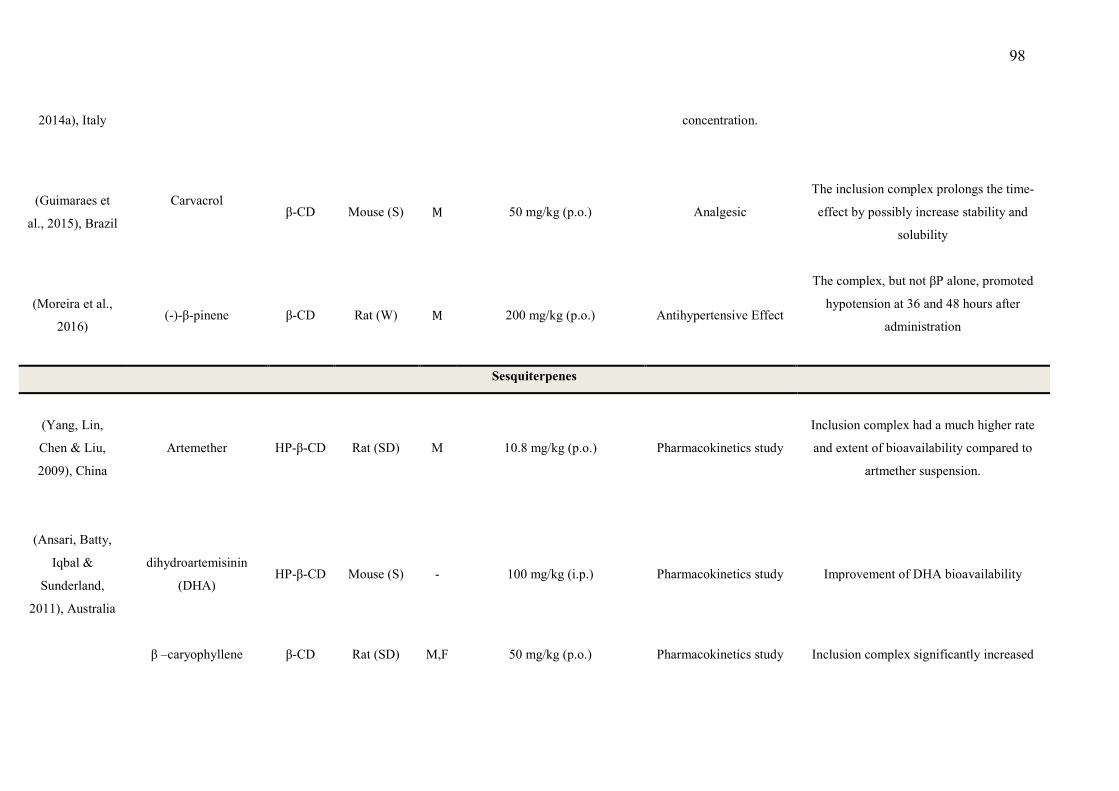
98
2014a), Italy
concentration.
(Guimaraes et
al., 2015), Brazil
Carvacrol
β-CD Mouse (S) M 50 mg/kg (p.o.) Analgesic
The inclusion complex prolongs the time-
effect by possibly increase stability and
solubility
(Moreira et al.,
2016) (-)-β-pinene β-CD Rat (W) M 200 mg/kg (p.o.) Antihypertensive Effect
The complex, but not βP alone, promoted
hypotension at 36 and 48 hours after
administration
Sesquiterpenes
(Yang, Lin,
Chen & Liu,
2009), China
Artemether HP-β-CD Rat (SD) M 10.8 mg/kg (p.o.) Pharmacokinetics study
Inclusion complex had a much higher rate
and extent of bioavailability compared to
artmether suspension.
(Ansari, Batty,
Iqbal &
Sunderland,
2011), Australia
dihydroartemisinin
(DHA) HP-β-CD Mouse (S) - 100 mg/kg (i.p.) Pharmacokinetics study Improvement of DHA bioavailability
β –caryophyllene β-CD Rat (SD) M,F 50 mg/kg (p.o.) Pharmacokinetics study Inclusion complex significantly increased
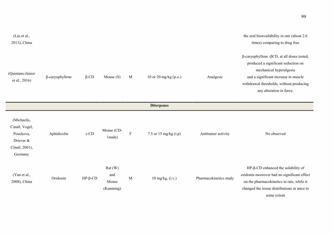
99
(Liu et al.,
2013), China
the oral bioavailability in rats (about 2.6
times) comparing to drug free
(Quintans-Júnior
et al., 2016) β-caryophyllene β-CD Mouse (S) M 10 or 20 mg/kg (p.o.) Analgesic
β-caryophyllene -βCD, at all doses tested,
produced a significant reduction on
mechanical hyperalgesia
and a significant increase in muscle
withdrawal thresholds, without producing
any alteration in force.
Diterpenes
(Michaelis,
Cinatl, Vogel,
Pouckova,
Driever &
Cinatl, 2001),
Germany
Aphidicolin γ-CD Mouse (CD-
1nude) F 7.5 or 15 mg/kg (i.p) Antitumor activity No observed
(Yan et al.,
2008), China Oridonin HP-β-CD
Rat (W)
and
Mouse
(Kunming)
M 10 mg/kg, (i.v.) Pharmacokinetics study
HP-β-CD enhanced the solubility of
oridonin moreover had no significant effect
on the pharmacokinetics in rats, while it
changed the tissue distributions in mice to
some extent
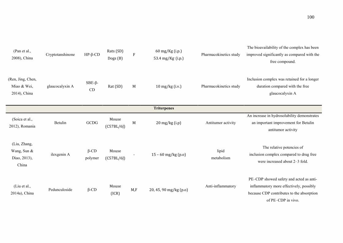
100
(Pan et al.,
2008), China
Cryptotanshinone HP-β-CD Rats (SD)
Dogs (B) F
60 mg/Kg (i.p.)
53.4 mg/Kg- (i.p.) Pharmacokinetics study
The bioavailability of the complex has been
improved significantly as compared with the
free compound.
(Ren, Jing, Chen,
Miao & Wei,
2014), China
glaucocalyxin A SBE-β-
CD Rat (SD) M 10 mg/kg (i.v.) Pharmacokinetics study
Inclusion complex was retained for a longer
duration compared with the free
glaucocalyxin A
Triterpenes
(Soica et al.,
2012), Romania Betulin GCDG
Mouse
(C57BL/6J) M 20 mg/kg (i.p) Antitumor activity
An increase in hydrosolubility demonstrates
an important improvement for Betulin
antitumor activity
(Liu, Zhang,
Wang, Sun &
Diao, 2013),
China
ilexgenin A β-CD
polymer
Mouse
(C57BL/6J) - 15 – 60 mg/kg (p.o)
lipid
metabolism
The relative potencies of
inclusion complex compared to drug free
were increased about 2–3 fold.
(Liu et al.,
2014a), China Pedunculoside β-CD
Mouse
(ICR) M,F 20, 45, 90 mg/kg (p.o)
Anti-inflammatory
PE–CDP showed safety and acted as anti-
inflammatory more effectively, possibly
because CDP contributes to the absorption
of PE–CDP in vivo.
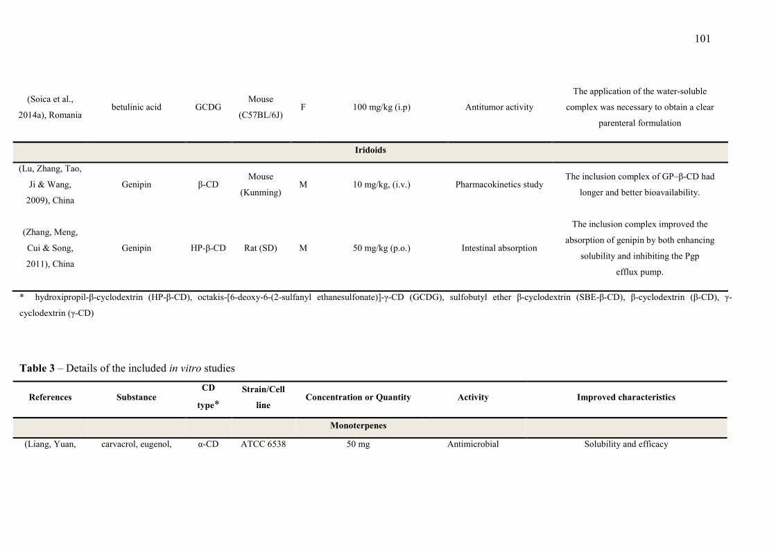
101
(Soica et al.,
2014a), Romania
betulinic acid GCDG Mouse
(C57BL/6J) F 100 mg/kg (i.p) Antitumor activity
The application of the water-soluble
complex was necessary to obtain a clear
parenteral formulation
Iridoids
(Lu, Zhang, Tao,
Ji & Wang,
2009), China
Genipin β-CD Mouse
(Kunming) M 10 mg/kg, (i.v.) Pharmacokinetics study
The inclusion complex of GP–β-CD had
longer and better bioavailability.
(Zhang, Meng,
Cui & Song,
2011), China
Genipin HP-β-CD Rat (SD) M 50 mg/kg (p.o.) Intestinal absorption
The inclusion complex improved the
absorption of genipin by both enhancing
solubility and inhibiting the Pgp
efflux pump.
* hydroxipropil-β-cyclodextrin (HP-β-CD), octakis-[6-deoxy-6-(2-sulfanyl ethanesulfonate)]-γ-CD (GCDG), sulfobutyl ether β-cyclodextrin (SBE-β-CD), β-cyclodextrin (β-CD), γ-
cyclodextrin (γ-CD)
Table 3 – Details of the included in vitro studies
References Substance CD
type*
Strain/Cell
line Concentration or Quantity Activity Improved characteristics
Monoterpenes
(Liang, Yuan, carvacrol, eugenol, α-CD ATCC 6538 50 mg Antimicrobial Solubility and efficacy
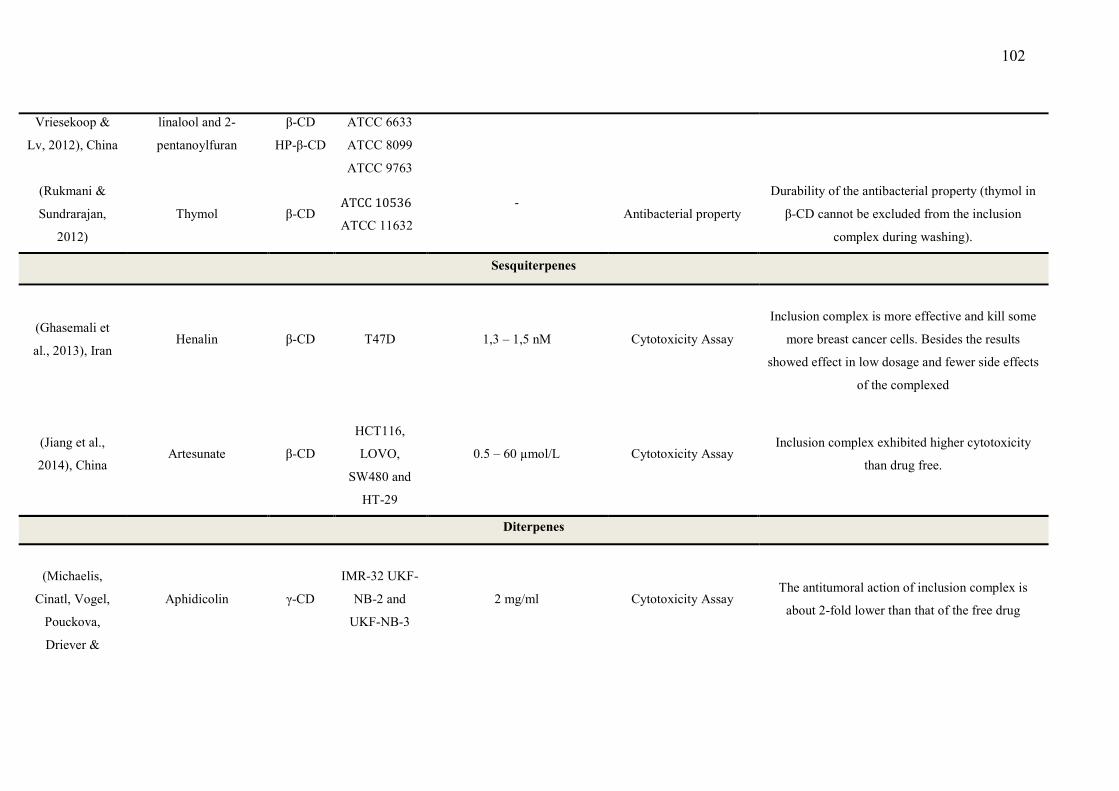
102
Vriesekoop &
Lv, 2012), China
linalool and 2-
pentanoylfuran
β-CD
HP-β-CD
ATCC 6633
ATCC 8099
ATCC 9763
(Rukmani &
Sundrarajan,
2012)
Thymol β-CD ATCC 10536
ATCC 11632
-
Antibacterial property
Durability of the antibacterial property (thymol in
β-CD cannot be excluded from the inclusion
complex during washing).
Sesquiterpenes
(Ghasemali et
al., 2013), Iran Henalin β-CD T47D 1,3 – 1,5 nM Cytotoxicity Assay
Inclusion complex is more effective and kill some
more breast cancer cells. Besides the results
showed effect in low dosage and fewer side effects
of the complexed
(Jiang et al.,
2014), China Artesunate β-CD
HCT116,
LOVO,
SW480 and
HT-29
0.5 – 60 µmol/L Cytotoxicity Assay Inclusion complex exhibited higher cytotoxicity
than drug free.
Diterpenes
(Michaelis,
Cinatl, Vogel,
Pouckova,
Driever &
Aphidicolin γ-CD
IMR-32 UKF-
NB-2 and
UKF-NB-3
2 mg/ml Cytotoxicity Assay The antitumoral action of inclusion complex is
about 2-fold lower than that of the free drug
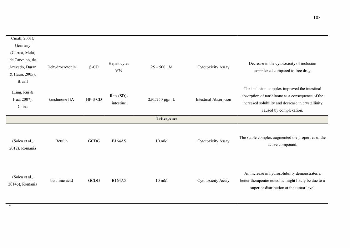
103
Cinatl, 2001),
Germany
(Correa, Melo,
de Carvalho, de
Azevedo, Duran
& Haun, 2005),
Brazil
Dehydrocrotonin β-CD Hepatocytes
V79 25 – 500 µM Cytotoxicity Assay
Decrease in the cytotoxicity of inclusion
complexed compared to free drug
(Ling, Rui &
Hua, 2007),
China
tanshinone IIA HP-β-CD Rats (SD)-
intestine 250#250 µg/mL Intestinal Absorption
The inclusion complex improved the intestinal
absorption of tanshinone as a consequence of the
increased solubility and decrease in crystallinity
caused by complexation.
Triterpenes
(Soica et al.,
2012), Romania
Betulin GCDG B164A5 10 mM Cytotoxicity Assay The stable complex augmented the properties of the
active compound.
(Soica et al.,
2014b), Romania
betulinic acid GCDG B164A5 10 mM Cytotoxicity Assay
An increase in hydrosolubility demonstrates a
better therapeutic outcome might likely be due to a
superior distribution at the tumor level
*

104
Table 4 – Details of the included clinical studies
References Substance CD
type* Population Concentration or Quantity Activity Improved characteristics
Sesquiterpenes
(Wong & Yuen,
2001), Malaysia Artemisinin
β-CD
γ-CD
12 healthy
human
volunteers (22
- 44 years old)
250 mg in 150 ml of water Pharmacokinetics
analysis
Inclusion complexes achieved much higher plasma
drug levels, thus indicating a higher rate and
extent of artemisinin absorption.
(Shah & Mashru,
2010), India Artemether β-CD 20 volunteers
1 g of CD and artemether was
dispersed in 50 mL of water
for 15 s.
Gustatory sensation test
The complete bitter taste masking of ARM was
achieved with improved in vitro drug release in
physical mixture. because it was further
incorporated into a reconstitutable dry suspension.
β-cyclodextrin (β-CD), γ- cyclodextrin (γ-CD), hydroxipropil-β-cyclodextrin (HP-β-CD), octakis-[6-deoxy-6-(2-sulfanyl ethanesulfonate)]-γ-CD (GCDG), sulfobutyl ether β-
cyclodextrin (SBE-β-CD), α- cyclodextrin (α-CD), β-cyclodextrin (β-CD), γ- cyclodextrin (γ-CD)
105
A number of pharmacokinetic studies evaluated animal models and highlighted the
potential benefits of terpenes with CDs. Yan et al. (2008) suggested that the complex of
oridonin and HP-β-CD approximately behaved as a true solution in plasma. The lower
drug concentration they found in the heart, kidneys, and spleen in their study can be
explained by the equilibrium mechanism of the CD drug after administration,
highlighting its possible beneficial use in the clinical application of oridonin. Assessing
the mean plasma concentration versus the time profiles of free β-caryophyllene and β-
caryophyllene/CD, after oral administration a comparison of the pharmacokinetic
parameters indicated that β-caryophyllene/CD had an earlier Tmax and a higher Cmax,
suggesting a better bioavailability of about 2.6 times (Liu et al., 2013). Importantly, the
complexation of β-caryophyllene and β-caryophyllene/CD improved the
pharmacological aspects when compared with terpene alone. This effect seems to be
related to an increased bioavailability of the active compound (Quintans-Júnior et al.,
2016). Lu et al. (2009) found that the relative bioavailability of the inclusion complex of
genipin/β-CD compared to free genipin was 305.3%. Corroborating this the apparent
bioavailability of artemether following a complexation with HP-β-CD was found to be
181% when compared to an artemether suspension, indicating that the complex had a
much higher rate and extent of bioavailability compared to artemether suspension
(Yang, Lin, Chen & Liu, 2009).
In an important clinical study with twelve healthy male volunteers that was conducted
to evaluate the absorption of artemisinin, an anti-malarial drug and a sesquiterpene
lactone, it was found that when complexed with the two forms of natural CDs, in
comparison with a normal commercially available preparation the complexes exhibited
a more rapid dissolution, and hence, absorption. Consequently, this enhanced the
therapeutic effectiveness of artemisinin (Wong & Yuen, 2003b). These results were also
played out in others studies using the derivatives of dihydroartemisinin (DHA), which
in a complexation with HP-β-CD, equally improved the pharmacokinetic parameters
when compared with DHA alone (Ansari, Batty, Iqbal & Sunderland, 2011). Artesunate
in β-CD conjugates with a cytotoxicity against three types of human colon cancer cell
lines (Jiang et al., 2014).
4.3 Pharmacological Improvement
106
Among the substances whose chemical properties can be improved by CDs are
essentials oils and their main compounds, such as terpenes and phenylpropanoids
(Guimaraes, Serafini & Quintans-Junior, 2014; Oliveira, Guimaraes, Araujo, Quintans,
Santos & Quintans-Junior, 2015). The pharmacologically beneficial effects that are
produced by forming CD-lipophilic drugs seem to improve their analgesic and anti-
inflammatory profiles (Brito, Araujo, Quintans, Sluka & Quintans-Junior, 2015;
Oliveira, Guimaraes, Araujo, Quintans, Santos & Quintans-Junior, 2015).
Monoterpenes are commonly able to produce analgesic, antioxidant and anti-
inflammatory effects, including their ability to treat chronic pain (Guimaraes, Quintans
& Quintans Júnior, 2013; Guimaraes, Serafini & Quintans-Junior, 2014; Quintans,
Antoniolli, Almeida, Santana-Filho & Quintans-Junior, 2014). These compounds
possess a poor oral absorption due to their extremely low aqueous solubility or their
extensive pre-systemic metabolism and this may be responsible for the unfavorable
pharmacokinetics of the molecule (Yadav, Suresh, Devi & Yadav, 2009). Some studies
have demonstrated an improvement in the oral absorption of terpenes after complexing
them with CDs and their derivatives and an increment in the pharmacological efficacy
of these compounds.(Guimaraes et al., 2015; Ishida et al., 2013; Nascimento et al.,
2015; Nascimento et al., 2014; Quintans et al., 2013b; Quintans-Junior et al., 2013;
Siqueira-Lima et al., 2014). However, the articles have little pharmacokinetic and
pharmacodynamic information about how CD-complex-drugs improve these particular
aspects. Research in this area may be a promising line of approach to understand how
monoterpene-CD-complexes enhance the effects and reduce the side effects of these
natural products.
Figure 3 provides studies that show chemical and pharmacological characteristics of the
β-caryophyllene/β-cyclodextrin complex (BCP-βCD). Hădărugă et al. (2009)
demonstrated using molecular modeling and docking studies that the rate of complex
formation between BCP and βCD is relatively low, but the stability of the BCP-βCD
complex seems to be higher when compared with other terpenes-βCD-complexes, such
as caryophyllene-oxide or α-bisabolol. In fact, βCD has an intrinsic ability to form
inclusion complexes with terpenes, which was strongly demonstrated in several studies
with BCP (Liu et al., 2013; Quintans-Júnior et al., 2016; Hădărugă, Hădărugă, Riviş &
Pârvu, 2009), and to enhance pharmacological aspects, mainly bioavailability and
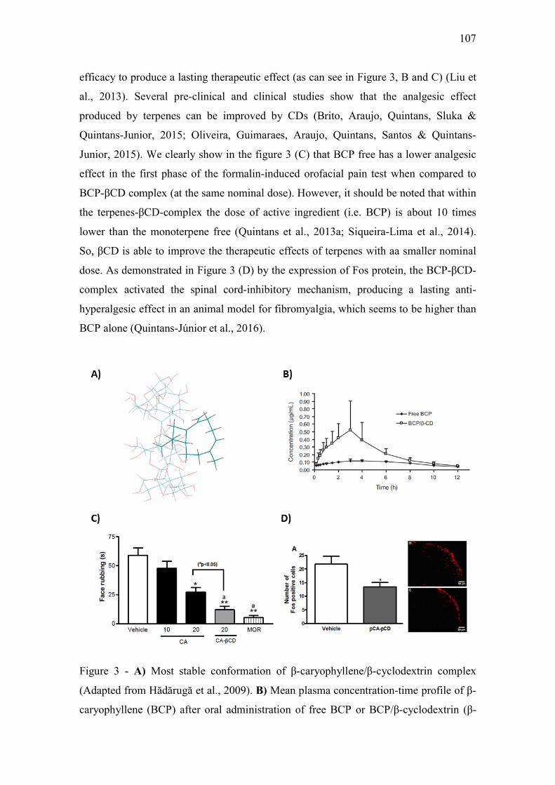
107
efficacy to produce a lasting therapeutic effect (as can see in Figure 3, B and C) (Liu et
al., 2013). Several pre-clinical and clinical studies show that the analgesic effect
produced by terpenes can be improved by CDs (Brito, Araujo, Quintans, Sluka &
Quintans-Junior, 2015; Oliveira, Guimaraes, Araujo, Quintans, Santos & Quintans-
Junior, 2015). We clearly show in the figure 3 (C) that BCP free has a lower analgesic
effect in the first phase of the formalin-induced orofacial pain test when compared to
BCP-βCD complex (at the same nominal dose). However, it should be noted that within
the terpenes-βCD-complex the dose of active ingredient (i.e. BCP) is about 10 times
lower than the monoterpene free (Quintans et al., 2013a; Siqueira-Lima et al., 2014).
So, βCD is able to improve the therapeutic effects of terpenes with aa smaller nominal
dose. As demonstrated in Figure 3 (D) by the expression of Fos protein, the BCP-βCD-
complex activated the spinal cord-inhibitory mechanism, producing a lasting anti-
hyperalgesic effect in an animal model for fibromyalgia, which seems to be higher than
BCP alone (Quintans-Júnior et al., 2016).
Figure 3 - A) Most stable conformation of β-caryophyllene/β-cyclodextrin complex
(Adapted from Hădărugă et al., 2009). B) Mean plasma concentration-time profile of β-
caryophyllene (BCP) after oral administration of free BCP or BCP/β-cyclodextrin (β-
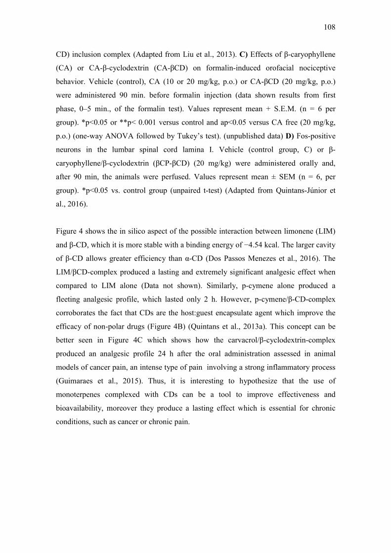
108
CD) inclusion complex (Adapted from Liu et al., 2013). C) Effects of β-caryophyllene
(CA) or CA-β-cyclodextrin (CA-βCD) on formalin-induced orofacial nociceptive
behavior. Vehicle (control), CA (10 or 20 mg/kg, p.o.) or CA-βCD (20 mg/kg, p.o.)
were administered 90 min. before formalin injection (data shown results from first
phase, 0–5 min., of the formalin test). Values represent mean + S.E.M. (n = 6 per
group). *p<0.05 or **p< 0.001 versus control and ap<0.05 versus CA free (20 mg/kg,
p.o.) (one-way ANOVA followed by Tukey’s test). (unpublished data) D) Fos-positive
neurons in the lumbar spinal cord lamina I. Vehicle (control group, C) or β-
caryophyllene/β-cyclodextrin (βCP-βCD) (20 mg/kg) were administered orally and,
after 90 min, the animals were perfused. Values represent mean ± SEM (n = 6, per
group). *p<0.05 vs. control group (unpaired t-test) (Adapted from Quintans-Júnior et
al., 2016).
Figure 4 shows the in silico aspect of the possible interaction between limonene (LIM)
and β-CD, which it is more stable with a binding energy of −4.54 kcal. The larger cavity
of β-CD allows greater efficiency than α-CD (Dos Passos Menezes et al., 2016). The
LIM/βCD-complex produced a lasting and extremely significant analgesic effect when
compared to LIM alone (Data not shown). Similarly, p-cymene alone produced a
fleeting analgesic profile, which lasted only 2 h. However, p-cymene/β-CD-complex
corroborates the fact that CDs are the host:guest encapsulate agent which improve the
efficacy of non-polar drugs (Figure 4B) (Quintans et al., 2013a). This concept can be
better seen in Figure 4C which shows how the carvacrol/β-cyclodextrin-complex
produced an analgesic profile 24 h after the oral administration assessed in animal
models of cancer pain, an intense type of pain involving a strong inflammatory process
(Guimaraes et al., 2015). Thus, it is interesting to hypothesize that the use of
monoterpenes complexed with CDs can be a tool to improve effectiveness and
bioavailability, moreover they produce a lasting effect which is essential for chronic
conditions, such as cancer or chronic pain.

109
Figure 4 - A) Ten possible interactions of LIM and CDs obtained through molecular
modeling. The green space represents the CD cavity (Adapted from Menezes et al.,
2016). B) Time response curve for the antinociceptive effect of (A) p-cymene or (B) p-
cymene/β-CD complex on acetic acid-induced writhing response in mice. Writhings
were counted over 20 min following i.p. administration of acetic acid (0.65%). p-
cymene or p-cymene/β-CD (40 mg/kg) was administered p.o. 0.5, 1, 2, 4, 8 or 16 h
before acid acetic injection (0.65%). Control animals received an injection of vehicle by
p.o. route. Each column represents mean ± S.E.M. (n = 8, per group). *p<0.05 or
**p<0.001 vs. control (ANOVA followed by Tukey’s test) (Adapted from Quintans et
al., 2013). C) Effect of carvacrol/β-cyclodextrin complex (CARV/β-CD) on the
mechanical hyperalgesia induced by S180. Time-Effect Curve of CARV/β-CD (50
mg/kg) and CARV (100 mg/kg). *p< 0.05, **p < 0.01 and ***p < 0.001 vs. the control
group (vehicle) (ANOVA followed by Tukey’s test) (Adapted from Guimaraes et al.,
2015).
Within this context, linalool a monoterpene found in the Ocimum and Thymus species,
when complexed with α-CD, β-CD, or HP-β-CD, showed a significant enhancement in
its water solubility that was established through specific tests (Pinho, Grootveld, Soares

110
& Henriques, 2014a). A pretreatment with linalool induced analgesic and anti-
inflammatory profiles which were comparatively better than when linalool was used
alone in an animal model with fibromyalgia, and when challenged by pro-inflammatory
agents (Nascimento et al., 2014; Quintans-Junior et al., 2013). More recently, some
studies have shown that complexation between terpenes and CDs can be more
pharmacologically activity even with lower molecular doses of active compound
(terpene) when compared with terpene non-complexed with CDs (Quintans et al.,
2013b; Guimarães et al., 2014; Menezes et al., 2015; Brito et al., 2015; Quintans-Júnior
et al., 2016).
One of the most studied pharmacological uses for terpenes has been its anticancer
effects. However, problems with toxicity, side effects, and resistance, encouraged the
development of new pharmacological tools to improve the properties of the drugs under
study (da Rocha, Lopes & Schwartsmann, 2001; Harvey, 2008). Michaelis, Cinatl,
Vogel, Pouckova, Driever and Cinatl (2001) studied an aphidicolin (APH), a tetracyclic
diterpene, and compared the cytotoxic actions between an APH free treatment and when
complexed with γ-CD against neuroblastoma cells. Surprisingly, when in vitro, the
antitumoral action of APH-CD was about 2-fold lower than that of the APH free
treatment. Others studies employed a cytotoxicity assay, as did Ghasemali et al. (2013),
who encapsulated a cytotoxic chemotherapeutic agent. They showed that β-CD-helanin
complexes inhibited the growth of a T47D breast cancer cell line in a time and dose-
dependent manner. Moreover, Soica et al. (2012) and Soica et al. (2014a) assessed the
pentacyclic triterpenes betulin and betulinic acid when complexed with γ-CD derivates.
They demonstrated that these complexes had a gain in solubility and a beneficial
influence in terms of anti-proliferative activity and in vivo tumor development,
respectively.
On the above, Complexation with cyclodextrins is a well-known procedure which
opens up new possibilities for their broad practical application in various fields to
improve the biological activities of terpenes in living systems.
5. Conclusion
Although CDs are not able to form complexes with compounds with a high molecular
weight and a larger size, they continue to be an innovative drug encapsulation system
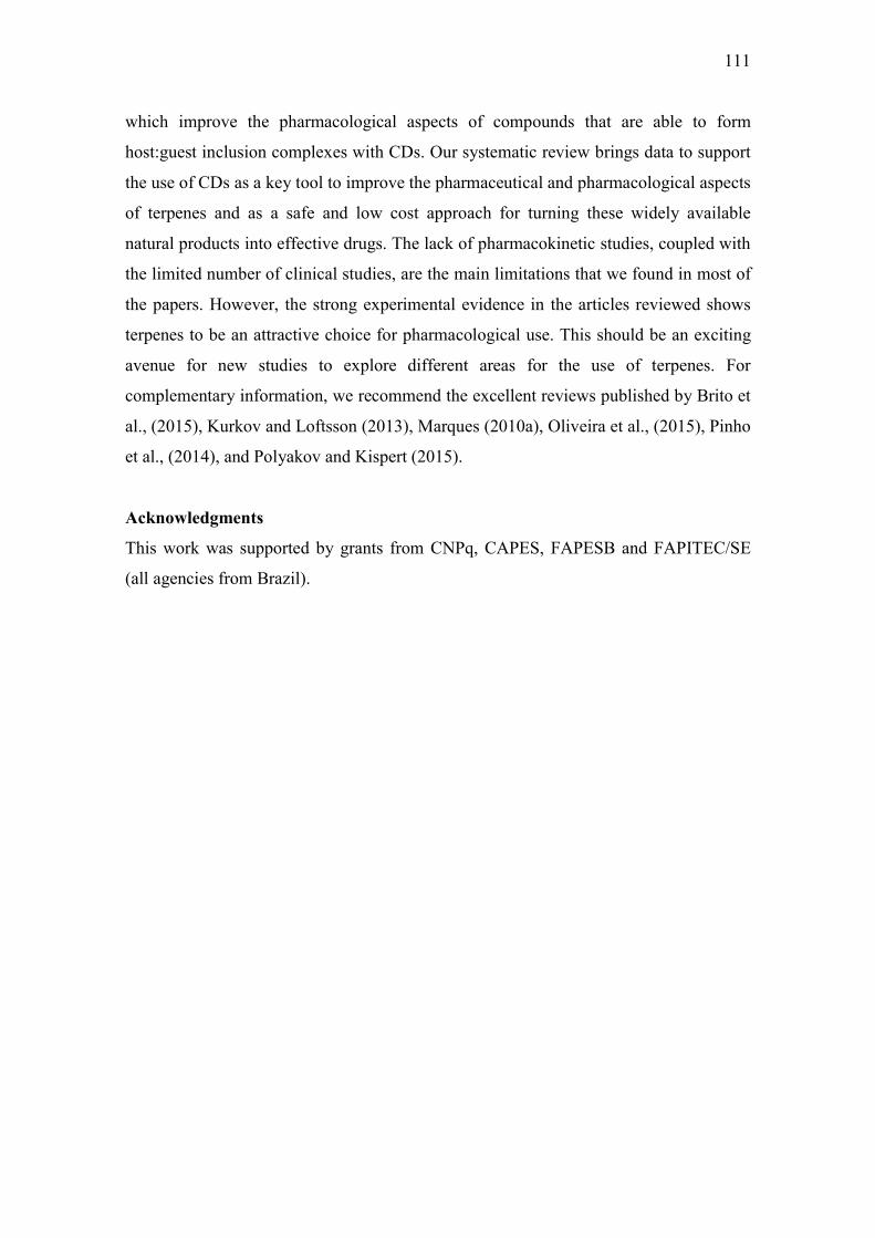
111
which improve the pharmacological aspects of compounds that are able to form
host:guest inclusion complexes with CDs. Our systematic review brings data to support
the use of CDs as a key tool to improve the pharmaceutical and pharmacological aspects
of terpenes and as a safe and low cost approach for turning these widely available
natural products into effective drugs. The lack of pharmacokinetic studies, coupled with
the limited number of clinical studies, are the main limitations that we found in most of
the papers. However, the strong experimental evidence in the articles reviewed shows
terpenes to be an attractive choice for pharmacological use. This should be an exciting
avenue for new studies to explore different areas for the use of terpenes. For
complementary information, we recommend the excellent reviews published by Brito et
al., (2015), Kurkov and Loftsson (2013), Marques (2010a), Oliveira et al., (2015), Pinho
et al., (2014), and Polyakov and Kispert (2015).
Acknowledgments
This work was supported by grants from CNPq, CAPES, FAPESB and FAPITEC/SE
(all agencies from Brazil).
112
References
Aburahma, M. H. (2015). Insights on novel particulate self-assembled drug delivery beads based on partial inclusion complexes between triglycerides and cyclodextrins. Drug Deliv, 1-15.
Ahad, A., Aqil, M., Kohli, K., Chaudhary, H., Sultana, Y., Mujeeb, M., et al. (2009). Chemical penetration enhancers: a patent review. Expert Opin Ther Pat, 19(7), 969-988.
Ajisaka, N., Hara, K., Mikuni, K., Hara, K., & Hashimoto, H. (2000). Effects of branched cyclodextrins on the solubility and stability of terpenes. Biosci Biotechnol Biochem, 64(4), 731-734.
Ali, A., Khajuria, A., Sidiq, T., Kumar, A., Thakur, N. L., Naik, D., et al. (2013). Modulation of LPS induced inflammatory response by Lawsonyl monocyclic terpene from the marine derived Streptomyces sp. Immunol Lett, 150(1-2), 79-86.
Amore, B. M., Gibbs, J. P., & Emery, M. G. (2010). Application of in vivo animal models to characterize the pharmacokinetic and pharmacodynamic properties of drug candidates in discovery settings. Comb Chem High Throughput Screen, 13(2), 207-218.
Ansari, M. T., Batty, K. T., Iqbal, I., & Sunderland, V. B. (2011). Improving the solubility and bioavailability of dihydroartemisinin by solid dispersions and inclusion complexes. Arch Pharm Res, 34(5), 757-765.
Ansari, M. T., Iqbal, I., & Sunderland, V. B. (2009). Dihydroartemisinin-cyclodextrin complexation: solubility and stability. Arch Pharm Res, 32(1), 155-165.
Araujo-Filho, H. G., Quintans-Junior, L. J., Barreto, A. S., Almeida, J. R., Barreto, R. S., & Quintans, J. S. (2016). Neuroprotective Effect of Natural Products on Peripheral Nerve Degeneration: A Systematic Review. Neurochem Res, 41, 647-658.
Arima, H., Hayashi, Y., Higashi, T., & Motoyama, K. (2015). Recent advances in cyclodextrin delivery techniques. Expert Opin Drug Deliv, 12(9), 1425-1441.
Ashour, M., Wink, M., & Gershenzon, J. (2010). Biochemistry of terpenoids: monoterpenes, sesquiterpenes and diterpenes. In: Wink M (ed) Annual plant reviews: biochemistry of plant secondary metabolism. New York: Wiley.
Asztemborska, M., Bielejewska, A., Duszczyk, K., & Sybilska, D. (2000). Comparative study on camphor enantiomers behavior under the conditions of gas-liquid chromatography and reversed-phase high-performance liquid chromatography systems modified with alpha- and beta-cyclodextrins. J Chromatogr A, 874(1), 73-80.
Asztemborska, M., Sybilska, D., Nowakowski, R., & Perez, G. (2003). Chiral recognition ability of alpha-cyclodextrin with regard to some monoterpenoids under gas-liquid chromatographic conditions. J Chromatogr A, 1010(2), 233-242.
Bach, T. (2010). Preclinical and Clinical Overview of Terpenes in the Treatment of Urolithiasis. European Urology Supplements, 9, 814-818.
Barreto, R. S., Albuquerque-Junior, R. L., Araujo, A. A., Almeida, J. R., Santos, M. R., Barreto, A. S., et al. (2014). A systematic review of the wound-healing effects of monoterpenes and iridoid derivatives. Molecules, 19(1), 846-862.
Bragagni, M., Maestrelli, F., & Mura, P. (2010). Physical chemical characterization of binary systems of prilocaine hydrochloride with triacetyl-β-cyclodextrin. Journal of Inclusion Phenomena and Macrocyclic Chemistry, 68(3), 437-445.
Brewster, M. E., & Loftsson, T. (2007). Cyclodextrins as pharmaceutical solubilizers. Adv Drug Deliv Rev, 59(7), 645-666.
113
Brito, R. G., Araujo, A. A., Quintans, J. S., Sluka, K. A., & Quintans-Junior, L. J. (2015). Enhanced analgesic activity by cyclodextrins - a systematic review and meta-analysis. Expert Opin Drug Deliv, 1-12.
Ceborska, M. (2014). Interactions of Native Cyclodextrins with Biorelevant Molecules in the Solid State: A Review. Current Organic Chemistry, 18, 1878-1885.
Ceborska, M., Asztemborska, M., Luboradzki, R., & Lipkowski, J. (2013). Interactions with beta-cyclodextrin as a way for encapsulation and separation of camphene and fenchene. Carbohydr Polym, 91(1), 110-114.
Ceborska, M., Szwed, K., & Suwinska, K. (2013). beta-Cyclodextrin as the suitable molecular container for isopulegol enantiomers. Carbohydr Polym, 97(2), 546-550.
Chappell, J. (1995). The Biochemistry and Molecular Biology of Isoprenoid Metabolism. Plant Physiology, 107(1), 1-6.
Chepulis, L. M., & Francis, E. (2012). An initial investigation into the anti-inflammatory activity and antioxidant capacity of alpha-cyclodextrin-complexed Manuka honey. J Complement Integr Med, 9, Article 25.
Christoforides, E., Mentzafos, D., & Bethanis, K. (2014). Structural studies of the inclusion complexes of the (+)- and (-)- borneol enantiomers in α- and β-cyclodextrin. Journal of Inclusion Phenomena and Macrocyclic Chemistry, 81, 193-203.
Ciobanu, A., Landy, D., & Fourmentin, S. (2013). Complexation efficiency of cyclodextrins for volatile flavor compounds. Food Research International, 53(1), 110-114.
Connolly, J. D., & Hill, R. A. (1991). Dictionary of Terpenoids. London: Chapman and Hall
Correa, D. H., Melo, P. S., de Carvalho, C. A., de Azevedo, M. B., Duran, N., & Haun, M. (2005). Dehydrocrotonin and its beta-cyclodextrin complex: cytotoxicity in V79 fibroblasts and rat cultured hepatocytes. Eur J Pharmacol, 510(1-2), 17-24.
Cravotto, G., Garella, D., Carnaroglio, D., Gaudino, E. C., & Rosati, O. (2012). Solvent-free chemoselective oxidation of thioethers and thiophenes by mechanical milling. Chemical Communications, 48(95), 11632-11634.
Crini, G. (2014). Review: a history of cyclodextrins. Chem Rev, 114(21), 10940-10975. Croteau, R., Kutchan, T. M., & Lewis, N. G. (2000). Natural products (secondary
metabolites). Biochemistry and Molecular Biology of Plants (pp. 1250–1318). New York: American Society of Plant Physiology.
Cunha-Filho, M. S. S., & Sá-Barreto, L. C. L. (2007). Utilização de ciclodextrinas na formação de complexos de inclusão de interesse farmacêutico. Revista de Ciências Farmacêuticas Básica e Aplicada, 28, 1-9.
da Rocha, A. B., Lopes, R. M., & Schwartsmann, G. (2001). Natural products in anticancer therapy. Curr Opin Pharmacol, 1(4), 364-369.
Danel, C., Duval, C., Azaroual, N., Vaccher, C., Bonte, J. P., Bailly, C., et al. (2011). Complexation of triptolide and its succinate derivative with cyclodextrins: affinity capillary electrophoresis, isothermal titration calorimetry and 1H NMR studies. J Chromatogr A, 1218(48), 8708-8714.
Degenhardt, J., Kollner, T. G., & Gershenzon, J. (2009). Monoterpene and sesquiterpene synthases and the origin of terpene skeletal diversity in plants. Phytochemistry, 70(15-16), 1621-1637.
Demian, B. A. (2000). Correlation of the solubility of several aromatics and terpenes in aqueous hydroxypropyl-beta-cyclodextrin with steric and hydrophobicity parameters. Carbohydr Res, 328(4), 635-639.
114
Dewick, P. M. (2002). The biosynthesis of C5-C25 terpenoid compounds. Nat Prod Rep, 19(2), 181-222.
Dodziuk, H., Nowinski, K. S., Kozminski, W., & Dolgonos, G. (2003). On the impossibility of determination of stepwise binding constants for the 1 : 2 complex of (+)-camphor with alpha-cyclodextrin. Org Biomol Chem, 1(3), 581-584.
Donze, C., & Coleman, A. W. (1993). β-CD Inclusion Complexes: Relative Selectivity of Terpene and Aromatic Guest Molecules Studied by Competitive Inclusion Experiments Journal of inclusion phenomena and molecular recognition in chemistry, 16, 1-15.
Dos Passos Menezes, P., Dos Santos, P. B., Doria, G. A., de Sousa, B. M., Serafini, M. R., Nunes, P. S., et al. (2016). Molecular Modeling and Physicochemical Properties of Supramolecular Complexes of Limonene with alpha- and beta-Cyclodextrins. AAPS PharmSciTech.
Dushkin, A., Meteleva ES, Tolstikova, TG, Khvostov, MV, Tolstikov, GA. (2010). Mechanochemical Preparation and Properties of Water-Soluble Intermolecular Complexes of Polysaccharides and β-Cyclodextrin with Pharmaceutical Substances. Chemistry for Sustainable Development, 18, 631-640.
Eddaoudi, M., Coleman, A. W., & Baszkin, A. (1997). Chiral Recognition by Molecular Monolayers: Inclusion of Terpenes in tert-Butyldimethylsilyl-O6-β-Cyclodextrin. Supramolecular Chemistry, 8(3), 177-180.
Erickson, B., Nelson, & Winters, P. (2012). Perspective on opportunities in industrial biotechnology in renewable chemicals. Biotechnol J, 7(2), 176-185.
Gershenzon, J., & Kreis, W. (1999). Biochemistry of terpenoids: monoterpenes, sesquiterpenes, diterpenes, sterols, cardiac glycosides and steroid saponins. Biochemistry of Plant Secondary Metabolism (pp. 222–299). Boca Raton: CRC Press.
Ghasemali, S., Nejati-Koshki, K., Tafsiri, E., Rahmati-Yamchi, M., Akbarzadeh, A., Alizadeh, E., et al. (2013). Inhibitory effects of beta-cyclodextrin-helenalin complexes on H-TERT gene expression in the T47D breast cancer cell line - results of real time quantitative PCR. Asian Pac J Cancer Prev, 14(11), 6949-6953.
Gidwani, B., & Vyas, A. (2015). A Comprehensive Review on Cyclodextrin-Based Carriers for Delivery of Chemotherapeutic Cytotoxic Anticancer Drugs. Biomed Res Int, 2015, 198268.
Guimaraes, A. G., Oliveira, M. A., Alves Rdos, S., Menezes Pdos, P., Serafini, M. R., Araujo, A. A., et al. (2015). Encapsulation of carvacrol, a monoterpene present in the essential oil of oregano, with beta-cyclodextrin, improves the pharmacological response on cancer pain experimental protocols. Chem Biol Interact, 227, 69-76.
Guimaraes, A. G., Quintans, J. S., & Quintans Júnior, L. J. (2013). Monoterpenes with analgesic activity--a systematic review. Phytother Res, 27(1), 1-15.
Guimaraes, A. G., Serafini, M. R., & Quintans-Junior, L. J. (2014). Terpenes and derivatives as a new perspective for pain treatment: a patent review. Expert Opin Ther Pat, 24(3), 243-265.
Guo, M., Zhang, S., Song, F., Wang, D., Liu, Z., & Liu, S. (2003). Studies on the non-covalent complexes between oleanolic acid and cyclodextrins using electrospray ionization tandem mass spectrometry. J Mass Spectrom, 38(7), 723-731.
Hădărugă, D. I., Hădărugă, N. G., Riviş, A., & Pârvu, D. (2009). Molecular modeling and docking studies on Compositae biocompounds–cyclodextrin interactions. Journal of Agroalimentary Processes and Technologies, 15, 273-282.
115
Haloci, E., Toska, V., Shkreli, R., Goci, E., Vertuani, S., & Manfredini, S. (2014). Encapsulation of Satureja montana essential oil in β-cyclodextrin. Journal of Inclusion Phenomena and Macrocyclic Chemistry, 80(1), 147-153.
Hanson, J. R. (2009). Diterpenoids. Nat Prod Rep, 26(9), 1156-1171. Hartell, M. G., Hicks, R., Bhattacharjee, A. K., Koser, B. W., Carvalho, K., & Van
Hamont, J. E. (2004). Nuclear magnetic resonance and molecular modeling analysis of the interaction of the antimalarial drugs artelinic acid and artesunic acid with beta-cyclodextrin. J Pharm Sci, 93(8), 2076-2089.
Harvey, A. L. (2008). Natural products in drug discovery. Drug Discov Today, 13(19-20), 894-901.
Hegazy, M. E., Mohamed, T. A., Alhammady, M. A., Shaheen, A. M., Reda, E. H., Elshamy, A. I., et al. (2015). Molecular architecture and biomedical leads of terpenes from red sea marine invertebrates. Mar Drugs, 13(5), 3154-3181.
Hill, R. A., & Connolly, J. D. (2015). Triterpenoids. Nat Prod Rep, 32(2), 273-327. Hirayama, F., Minami, K., & Uekama, K. (1996). In-vitro evaluation of biphenylyl
acetic acid-beta-cyclodextrin conjugates as colon-targeting prodrugs: drug release behaviour in rat biological media. J Pharm Pharmacol, 48(1), 27-31.
Illapakurthy, A. C., Sabnis, Y. A., Avery, B. A., Avery, M. A., & Wyandt, C. M. (2003). Interaction of artemisinin and its related compounds with hydroxypropyl-beta-cyclodextrin in solution state: experimental and molecular-modeling studies. J Pharm Sci, 92(3), 649-655.
Illapakurthy, A. C., Wyandt, C. M., & Stodghill, S. P. (2005). Isothermal titration calorimetry method for determination of cyclodextrin complexation thermodynamics between artemisinin and naproxen under varying environmental conditions. Eur J Pharm Biopharm, 59(2), 325-332.
Irie, T., & Uekama, K. (1997). Pharmaceutical applications of cyclodextrins. III. Toxicological issues and safety evaluation. J Pharm Sci, 86(2), 147-162.
Ishida, T., Miki, I., Tanahashi, T., Yagi, S., Kondo, Y., Inoue, J., et al. (2013). Effect of 18beta-glycyrrhetinic acid and hydroxypropyl gammacyclodextrin complex on indomethacin-induced small intestinal injury in mice. Eur J Pharmacol, 714(1-3), 125-131.
Iwata, M., Fukami, T., Kawashima, D., Sakai, M., Furuishi, T., Suzuki, T., et al. (2009). Effectiveness of mechanochemical treatment with cyclodextrins on increasing solubility of glimepiride. Pharmazie, 64(6), 390-394.
Izutani, Y., Kanaori, K., & Oda, M. (2014). Aggregation property of glycyrrhizic acid and its interaction with cyclodextrins analyzed by dynamic light scattering, isothermal titration calorimetry, and NMR. Carbohydr Res, 392, 25-30.
Jiang, R. J., Zhao, Y. L., Chen, Y. J., Xiao, D., Wang, F., Han, B., et al. (2014). Synthesis, characterization, and in vitro evaluation of artesunate-beta-cyclodextrin conjugates as novel anti-cancer prodrugs. Carbohydr Res, 400, 19-25.
Kakran, M., Sahoo, N. G., Li, L., & Judeh, Z. (2011). Dissolution enhancement of artemisinin with beta-cyclodextrin. Chem Pharm Bull (Tokyo), 59(5), 646-652.
Kayaci, F., Sen, H. S., Durgun, E., & Uyar, T. (2014). Functional electrospun polymeric nanofibers incorporating geraniol–cyclodextrin inclusion complexes: High thermal stability and enhanced durability of geraniol. Food Research International, 62, 424-431.
Kfoury, M., Auezova, L., Fourmentin, S., & Greige-Gerges, H. (2014a). Investigation of monoterpenes complexation with hydroxypropyl-beta-cyclodextrin. Journal of Inclusion Phenomena and Macrocyclic Chemistry, 80(1-2), 51-60.
116
Kfoury, M., Auezova, L., Fourmentin, S., & Greige-Gerges, H. (2014b). Investigation of monoterpenes complexation with hydroxypropyl-β-cyclodextrin. Journal of Inclusion Phenomena and Macrocyclic Chemistry, 80(1), 51-60.
Kfoury, M., Balan, R., Landy, D., Nistor, D., & Fourmentin, S. (2015). Investigation of the complexation of essential oil components with cyclodextrins. Supramolecular Chemistry, 27(9), 620-628.
Kokkinou, A., Tsorteki, F., Karpusas, M., Papakyriakou, A., Bethanis, K., & Mentzafos, D. (2010). Study of the inclusion of the (R)- and (S)-camphor enantiomers in alpha-cyclodextrin by X-ray crystallography and molecular dynamics. Carbohydr Res, 345(8), 1034-1040.
Kurkov, S. V., & Loftsson, T. (2013). Cyclodextrins. Int J Pharm, 453(1), 167-180. la Cruz, M. N. S., Santos Júnior, H. M., Rezende, C. M., Alves, R. J. V., Cancello, E.
M., & Rocha, M. M. (2014). Terpenos em cupins do gênero Nasutitermes (Isoptera, Termitidae, Nasutitermitinae). Química Nova, 37, 95-103.
Lakkakula, J. R., & Macedo Krause, R. W. (2014). A vision for cyclodextrin nanoparticles in drug delivery systems and pharmaceutical applications. Nanomedicine (Lond), 9(6), 877-894.
Liang, H., Yuan, Q., Vriesekoop, F., & Lv, F. (2012). Effects of cyclodextrins on the antimicrobial activity of plant-derived essential oil compounds. Food Chem, 135(3), 1020-1027.
Liao, Y., Zhang, X., Li, C., Huang, Y., Lei, M., Yan, M., et al. (2016). Inclusion complexes of HP-β-cyclodextrin with agomelatine: Preparation, characterization, mechanism study and in vivo evaluation. Carbohydr Polym, 147, 415-425.
Lin, H.-C., Tsunematsu, Y., Dhingra, S., Xu, W., Fukutomi, M., Chooi, Y.-H., et al. (2014). Generation of Complexity in Fungal Terpene Biosynthesis: Discovery of a Multifunctional Cytochrome P450 in the Fumagillin Pathway. Journal of the American Chemical Society, 136(11), 4426-4436.
Ling, W., Rui, L. C., & Hua, J. X. (2007). In situ intestinal absorption behaviors of tanshinone IIA from its inclusion complex with hydroxypropyl-beta-cyclodextrin. Biol Pharm Bull, 30(10), 1918-1922.
Lins, L. C. R. F., Santos, I. M. A., Melo, M. S., Menezes, P. P., Araújo, A. A. S., Nunes, R. S., et al. (2014). The anticonvulsant effect of geraniol and inclusion complex geraniol:β-cyclodextrin. Boletín Latinoamericano y del Caribe de Plantas Medicinales y Aromáticas, 13, 557-565.
Liu, C., Zhang, W., Wang, Q., Sun, Y., & Diao, G. W. (2013). The water-soluble inclusion complex of ilexgenin A with beta-cyclodextrin polymer--a novel lipid-lowering drug candidate. Org Biomol Chem, 11(30), 4993-4999.
Liu, C., Zhang, W., Yang, H., Sun, W., Gong, X., Zhao, J., et al. (2014a). A water-soluble inclusion complex of pedunculoside with the polymer beta-cyclodextrin: a novel anti-inflammation agent with low toxicity. PLoS One, 9(7), e101761.
Liu, C., Zhang, W., Yang, H., Sun, W., Gong, X., Zhao, J., et al. (2014b). A Water-Soluble Inclusion Complex of Pedunculoside with the Polymer β-Cyclodextrin: A Novel Anti-Inflammation Agent with Low Toxicity. PLoS One, 9(7), e101761.
Liu, H., Yang, G., Tang, Y., Cao, D., Qi, T., Qi, Y., et al. (2013). Physicochemical characterization and pharmacokinetics evaluation of beta-caryophyllene/beta-cyclodextrin inclusion complex. Int J Pharm, 450(1-2), 304-310.
Liu, W., Zhao, B., Li, Y. C., & Liu, H. M. (2011). NMR spectra and structures of oridonin derivatives complexes with beta-cyclodextrin. Magn Reson Chem, 49(9), 611-615.
117
Liu, Y., Chen, G. S., Chen, Y., & Lin, J. (2005). Inclusion complexes of azadirachtin with native and methylated cyclodextrins: solubilization and binding ability. Bioorg Med Chem, 13(12), 4037-4042.
Liu, Y., Yang, E. C., Yang, Y. W., Zhang, H. Y., Fan, Z., Ding, F., et al. (2004). Thermodynamics of the molecular and chiral recognition of cycloalkanols and camphor by modified beta-cyclodextrins possessing simple aromatic tethers. J Org Chem, 69(1), 173-180.
Liu, Y., Zhang, Q., & Chen, Y. (2007). Spectrophotometric and calorimetric titration studies on molecular recognition of camphor and borneol by nucleobase-modified beta-cyclodextrins. J Phys Chem B, 111(42), 12211-12218.
Locci, E., Lai, S., Piras, A., Marongiu, B., & Lai, A. (2004). 13C-CPMAS and 1H-NMR study of the inclusion complexes of beta-cyclodextrin with carvacrol, thymol, and eugenol prepared in supercritical carbon dioxide. Chem Biodivers, 1(9), 1354-1366.
Loftsson, T. (2002). Cyclodextrins and the Biopharmaceutics Classification System of Drugs. Journal of Inclusion Phenomena and Macrocyclic Chemistry, 44(1), 63-67.
Loftsson, T., & Brewster, M. E. (2010). Pharmaceutical applications of cyclodextrins: basic science and product development. J Pharm Pharmacol, 62(11), 1607-1621.
Loftsson, T., & Duchene, D. (2007). Cyclodextrins and their pharmaceutical applications. Int J Pharm, 329(1-2), 1-11.
Loftsson, T., Masson, M., & Brewster, M. E. (2004). Self-association of cyclodextrins and cyclodextrin complexes. J Pharm Sci, 93(5), 1091-1099.
Loftsson, T., Sigfusson, S. D., Sigurdsson, H. H., & Masson, M. (2003). The effects of cyclodextrins on topical delivery of hydrocortisone: the aqueous diffusion layer. STP pharma sciences, 13.
Lu, Y., Zhang, T., Tao, J., Ji, G., & Wang, S. (2009). Preparation, characterization, and pharmacokinetics of the inclusion complex of genipin-beta-cyclodextrin. Drug Dev Ind Pharm, 35(12), 1452-1459.
Marques, H. M. C. (2010a). A review on cyclodextrin encapsulation of essential oils and volatiles. Flavour and Fragrance Journal, 25, 313-325.
Marques, H. M. C. (2010b). A review on cyclodextrin encapsulation of essential oils and volatiles. . Flavour Frag. J., 25, 313–326.
Marreto, R. N., Almeida, E. E. C. V., Alves, P. B., Niculau, E. S., Nunes, R. S., Matos, C. R. S., et al. (2008). Thermal analysis and gas chromatography coupled mass spectrometry analyses of hydroxypropyl-β-cyclodextrin inclusion complex containing Lippia gracilis essential oil. Thermochimica Acta, 475(1–2), 53-58.
Menezes, P., Santos, P. B. P., Dória, G. A. A., Sousa, B. M. H., Serafini, M. R., Nunes, P. S., et al. (2016). Molecular Modeling and Physicochemical Properties of Supramolecular Complexes of Limonene with α- and β-Cyclodextrins. AAPS PharmSciTech, 1-9.
Menezes, P. P., Araujo, A. A., Doria, G. A., Quintans-Junior, L. J., de Oliveira, M. G., dos Santos, M. R., et al. (2015). Physicochemical characterization and analgesic effect of inclusion complexes of essential oil from Hyptis pectinata L. Poit leaves with beta-cyclodextrin. Curr Pharm Biotechnol, 16(5), 440-450.
Menezes, P. P., Serafini, M. R., Quintans-Júnior, L. J., Silva, G. F., Oliveira, J. F., Carvalho, F. M. S., et al. (2014). Inclusion complex of (−)-linalool and β-cyclodextrin. Journal of Thermal Analysis and Calorimetry, 115(3), 2429-2437.

118
Menezes, P. P., Serafini, M. R., Santana, B. V., Nunes, R. S., Quintans Júnior, L. J., Silva, G. F., et al. (2012). Solid-state β-cyclodextrin complexes containing geraniol. Thermochimica Acta, 548, 45-50.
Michaelis, M., Cinatl, J., Vogel, J. U., Pouckova, P., Driever, P. H., & Cinatl, J. (2001). Treatment of drug-resistant human neuroblastoma cells with cyclodextrin inclusion complexes of aphidicolin. Anticancer Drugs, 12(5), 467-473.
Minami, K., Hirayama, F., & Uekama, K. (1998). Colon-specific drug delivery based on a cyclodextrin prodrug: release behavior of biphenylylacetic acid from its cyclodextrin conjugates in rat intestinal tracts after oral administration. J Pharm Sci, 87(6), 715-720.
Moeder, C., O'Brien, T., Thompson, R., & Bicker, G. (1996). Determination of stoichiometric coefficients and apparent formation constants for α- and β-CD complexes of terpenes using reversed-phase liquid chromatography. Journal of Chromatography A, 736(1–2), 1-9.
Moher, D., Liberati, A., Tetzlaff, J., & Altman, D. G. (2009). Preferred reporting items for systematic reviews and meta-analyses: the PRISMA statement. Ann Intern Med, 151(4), 264-269, w264.
Moon, T. W., Lee, J. W., Jhee, K. H., Khang, K. W., Jeong, H. S., Yang, S. A., et al. (2008). Supramolecular Encapsulation of Pulegone from Oriental Herb, Schizonepeta tenuifolia Briquet by beta - and gamma -Cyclodextrins. Bulletin-Korean Chemical Society, 29, 1579-1582.
Moraes, C. M., Mescher, M. C., & Tumlinson, J. H. (2001). Caterpillar-induced nocturnal plant volatiles repel conspecific females. Nature, 410(6828), 577-580.
Moreira, I. J., Menezes, P. P., Serafini, M. R., Araujo, A. A., Quintans-Junior, L. J., Bonjardim, L. R., et al. (2016). Characterization and Antihypertensive Effect of the Complex of ( )-beta-pinene in beta-cyclodextrin. Curr Pharm Biotechnol.
Mourtzinos, I., Kalogeropoulos, N., Papadakis, S. E., Konstantinou, K., & Karathanos, V. T. (2008). Encapsulation of nutraceutical monoterpenes in beta-cyclodextrin and modified starch. J Food Sci, 73(1), S89-94.
Munin, A., & Edwards-Levy, F. (2011). Encapsulation of natural polyphenolic compounds; a review. Pharmaceutics, 3(4), 793-829.
Mura, P. (2014). Analytical techniques for characterization of cyclodextrin complexes in aqueous solution: A review. J Pharm Biomed Anal, 101c, 238-250.
Nagatoshi, M., Terasaka, K., Nagatsu, A., & Mizukami, H. (2011). Iridoid-specific glucosyltransferase from Gardenia jasminoides. J Biol Chem, 286(37), 32866-32874.
Nascimento, S. S., Araujo, A. A., Brito, R. G., Serafini, M. R., Menezes, P. P., DeSantana, J. M., et al. (2015). Cyclodextrin-complexed Ocimum basilicum leaves essential oil increases Fos protein expression in the central nervous system and produce an antihyperalgesic effect in animal models for fibromyalgia. Int J Mol Sci, 16(1), 547-563.
Nascimento, S. S., Camargo, E. A., DeSantana, J. M., Araujo, A. A., Menezes, P. P., Lucca-Junior, W., et al. (2014). Linalool and linalool complexed in beta-cyclodextrin produce anti-hyperalgesic activity and increase Fos protein expression in animal model for fibromyalgia. Naunyn Schmiedebergs Arch Pharmacol, 387(10), 935-942.
Nieddu, M., Rassu, G., Boatto, G., Bosi, P., Trevisi, P., Giunchedi, P., et al. (2014a). Improvement of thymol properties by complexation with cyclodextrins: in vitro and in vivo studies. Carbohydr Polym, 102, 393-399.

119
Nieddu, M., Rassu, G., Boatto, G., Bosi, P., Trevisi, P., Giunchedi, P., et al. (2014b). Improvement of thymol properties by complexation with cyclodextrins: In vitro and in vivo studies. Carbohydr Polym, 102, 393-399.
Nowakowski, M., & Ejchart, A. (2014). Complex formation of fenchone with α-cyclodextrin: NMR titrations. Journal of Inclusion Phenomena and Macrocyclic Chemistry, 79(3-4), 337-342.
Numanoglu, U., Sen, T., Tarimci, N., Kartal, M., Koo, O. M., & Onyuksel, H. (2007). Use of cyclodextrins as a cosmetic delivery system for fragrance materials: linalool and benzyl acetate. AAPS PharmSciTech, 8(4), E85.
Oliveira, M. G., Guimaraes, A. G., Araujo, A. A., Quintans, J. S., Santos, M. R., & Quintans-Junior, L. J. (2015). Cyclodextrins: improving the therapeutic response of analgesic drugs: a patent review. Expert Opin Ther Pat, 25(8), 897-907.
Pan, Y., Bi, H. C., Zhong, G. P., Chen, X., Zuo, Z., Zhao, L. Z., et al. (2008). Pharmacokinetic characterization of hydroxylpropyl-beta-cyclodextrin-included complex of cryptotanshinone, an investigational cardiovascular drug purified from Danshen (Salvia miltiorrhiza). Xenobiotica, 38(4), 382-398.
Pinho, E., Grootveld, M., Soares, G., & Henriques, M. (2014a). Cyclodextrins as encapsulation agents for plant bioactive compounds. Carbohydr Polym, 101, 121-135.
Pinho, E., Grootveld, M., Soares, G., & Henriques, M. (2014b). Cyclodextrins as encapsulation agents for plant bioactive compounds. Carbohydr Polym, 101(0), 121-135.
Polyakov, N. E., & Kispert, L. D. (2015). Water soluble biocompatible vesicles based on polysaccharides and oligosaccharides inclusion complexes for carotenoid delivery. Carbohydr Polym, 128, 207-219.
Polyakov, N. E., Leshina, T. V., Konovalova, T. A., Hand, E. O., & Kispert, L. D. (2004). Inclusion complexes of carotenoids with cyclodextrins: 1H NMR, EPR, and optical studies. Free Radic Biol Med, 36(7), 872-880.
Porte, A., Porte, L. H. M., & Oliveira, L. M. (2014). Chiral gas chromatography in the resolution of enantiomers involved in fruit flavours. Química Nova, 37, 1670-1679.
Quintans-Júnior, L. J., Araújo, A. A. S., Brito, R. G., Santos, P. L., Quintans, J. S. S., Menezes, P. P., et al. (2016). β-caryophyllene, a dietary cannabinoid, complexed with β-cyclodextrin produced anti-hyperalgesic effect involving the inhibition of Fos expression in superficial dorsal horn. Life Sci, 149, 34-41.
Quintans-Junior, L. J., Barreto, R. S., Menezes, P. P., Almeida, J. R., Viana, A. F., Oliveira, R. C., et al. (2013). beta-Cyclodextrin-complexed (-)-linalool produces antinociceptive effect superior to that of (-)-linalool in experimental pain protocols. Basic Clin Pharmacol Toxicol, 113(3), 167-172.
Quintans, J. S., Antoniolli, A. R., Almeida, J. R., Santana-Filho, V. J., & Quintans-Junior, L. J. (2014). Natural products evaluated in neuropathic pain models - a systematic review. Basic Clin Pharmacol Toxicol, 114(6), 442-450.
Quintans, J. S., Menezes, P. P., Santos, M. R., Bonjardim, L. R., Almeida, J. R., Gelain, D. P., et al. (2013a). Improvement of p-cymene antinociceptive and anti-inflammatory effects by inclusion in beta-cyclodextrin. Phytomedicine, 20(5), 436-440.
Quintans, J. S. S., Menezes, P. P., Santos, M. R., Bonjardim, L. R., Almeida, J. R., Gelain, D. P., et al. (2013b). Improvement of p-cymene antinociceptive and anti-inflammatory effects by inclusion in beta-cyclodextrin. Phytomedicine, 20(5), 436-440.
120
Rasheed, A., & Kumar, A. (2008). Cyclodextrins as Drug Carrier Molecule: A Review. Scientia Pharmaceutica, 76, 567-598.
Ren, L., Jing, J., Chen, G., Miao, Y., & Wei, P. (2014). Preparation, characteristic and pharmacological study on inclusion complex of sulfobutylether-beta-cyclodextrin with glaucocalyxin A. J Pharm Pharmacol, 66(7), 927-934.
Rinaldi, L., Binello, A., Stolle, A., Curini, M., & Cravotto, G. (2015). Efficient mechanochemical complexation of various steroid compounds with α-, β- and γ-cyclodextrin. Steroids, 98, 58-62.
Rode, T., Frauen, M., Muller, B. W., Dusing, H. J., Schonrock, U., Mundt, C., et al. (2003). Complex formation of sericoside with hydrophilic cyclodextrins: improvement of solubility and skin penetration in topical emulsion based formulations. Eur J Pharm Biopharm, 55(2), 191-198.
Rodrigues, T., Sieglitz, F., & Bernardes, G. J. (2016). Natural product modulators of transient receptor potential (TRP) channels as potential anti-cancer agents. Chem Soc Rev.
Rosa, G. M., Bianco, D., Parodi, A., Valbusa, A., Zawaideh, C., Bizzarri, N., et al. (2014). Pharmacokinetic and pharmacodynamic profile of dronedarone , a new antiarrhythmic agent for the treatment of atrial fibrillation. Expert Opin Drug Metab Toxicol, 10(12), 1751-1764.
Rosseels, M.-L. A., Delaunois, A. G., Hanon, E., Guillaume, P. J. P., Martin, F. D. C., & van den Dobbelsteen, D. J. (2013). Hydroxypropyl-β-cyclodextrin impacts renal and systemic hemodynamics in the anesthetized dog. Regulatory Toxicology and Pharmacology, 67(3), 351-359.
Roux, M., Perly, B., & Djedaini-Pilard, F. (2007). Self-assemblies of amphiphilic cyclodextrins. Eur Biophys J, 36(8), 861-867.
Rukmani, A., & Sundrarajan, M. (2012). Inclusion of antibacterial agent thymol on β-cyclodextrin-grafted organic cotton. Journal of Industrial Textiles, 42, 132-144.
Rungsardthong Ruktanonchai, U., Srinuanchai, W., Saesoo, S., Sramala, I., Puttipipatkhachorn, S., & Soottitantawat, A. (2011). Encapsulation of citral isomers in extracted lemongrass oil with cyclodextrins: molecular modeling and physicochemical characterizations. Biosci Biotechnol Biochem, 75(12), 2340-2345.
Saruta, N., Fukami, T., Furuishi, T., Suzuki, T., & Tomono, K. (2010). Powdered formulation of liquid oil terpenoid included in cyclodextrins for chemical stabilization and improved handling. Advanced Powder Technology, 21(3), 326-330.
Serafini, M. R., Menezes , P. P., Costa, L. P., Lima, C. M., Quintans Júnior, L. J., Cardoso, J. C., et al. (2012). Interaction of p -cymene with β -cyclodextrin Journal of Thermal Analysis and Calorimetry, 109, 951-955.
Shah, P. P., & Mashru, R. C. (2010). Palatable reconstitutable dry suspension of artemether for flexible pediatric dosing using cyclodextrin inclusion complexation. Pharm Dev Technol, 15(3), 276-285.
Sharma, N., & Baldi, A. (2016). Exploring versatile applications of cyclodextrins: an overview. Drug Deliv, 23(3), 739-757.
Shen, Y. L., Yang, S. H., Wu, L. M., & Ma, X. Y. (2005). Study on structure and characterization of inclusion complex of gossypol/beta cyclodextrin. Spectrochim Acta A Mol Biomol Spectrosc, 61(6), 1025-1028.
Shen, Y. L., Ying, W., Yang, S. H., & Wu, L. M. (2006). Determinations of the inclusion complex between gossypol and beta-cyclodextrin. Spectrochim Acta A Mol Biomol Spectrosc, 65(1), 169-172.
121
Silva, A. M., Empis, J. M., & Teixeira-Dias, J. J. (2002). Inclusion of carvone enantiomers in cyclomaltoheptaose (beta-cyclodextrin): thermal behaviour and H-->D and D-->H exchange. Carbohydr Res, 337(24), 2501-2504.
Singh, B., & Sharma, R. A. (2015). Plant terpenes: defense responses, phylogenetic analysis, regulation and clinical applications. 3 Biotech, 5(2), 129-151.
Singh, S. S. (2006). Preclinical pharmacokinetics: an approach towards safer and efficacious drugs. Curr Drug Metab, 7(2), 165-182.
Siqueira-Lima, P. S., Araujo, A. A., Lucchese, A. M., Quintans, J. S., Menezes, P. P., Alves, P. B., et al. (2014). beta-cyclodextrin complex containing Lippia grata leaf essential oil reduces orofacial nociception in mice - evidence of possible involvement of descending inhibitory pain modulation pathway. Basic Clin Pharmacol Toxicol, 114(2), 188-196.
Skorka, M., Asztemborska, M., & Zukowski, J. (2005). Thermodynamic studies of complexation and enantiorecognition processes of monoterpenoids by alpha- and beta-cyclodextrin in gas chromatography. J Chromatogr A, 1078(1-2), 136-143.
Soica, C., Danciu, C., Savoiu-Balint, G., Borcan, F., Ambrus, R., Zupko, I., et al. (2014a). Betulinic acid in complex with a gamma-cyclodextrin derivative decreases proliferation and in vivo tumor development of non-metastatic and metastatic B164A5 cells. Int J Mol Sci, 15(5), 8235-8255.
Soica, C., Dehelean, C., Danciu, C., Wang, H., Wenz, G., Ambrus, R., et al. (2012). Betulin Complex in γ-Cyclodextrin Derivatives: Properties and Antineoplasic Activities in In Vitro and In Vivo Tumor Models. Int J Mol Sci, 13(11), 14992.
Soica, C., Oprean, C., Borcan, F., Danciu, C., Trandafirescu, C., Coricovac, D., et al. (2014b). The synergistic biologic activity of oleanolic and ursolic acids in complex with hydroxypropyl-gamma-cyclodextrin. Molecules, 19(4), 4924-4940.
Su, J., Chen, J., Li, L., Li, B., Shi, L., Chen, L., et al. (2012a). Formation of beta-cyclodextrin inclusion enhances the stability and aqueous solubility of natural borneol. J Food Sci, 77(6), C658-664.
Su, J., Chen, J., Li, L., Li, B., Shi, L., Zhang, H., et al. (2012b). Preparation of natural borneol/2-hydroxypropyl-beta-cyclodextrin inclusion complex and its effect on the absorption of tetramethylpyrazine phosphate in mouse. Chem Pharm Bull (Tokyo), 60(6), 736-742.
Szejtli, J. (1998). Introduction and General Overview of Cyclodextrin Chemistry. Chem Rev, 98(5), 1743-1754.
Tsai, Y., Tsai, H.-H., Wu, C.-P., & Tsai, F.-J. (2010). Preparation, characterisation and activity of the inclusion complex of paeonol with β-cyclodextrin. Food Chem, 120(3), 837-841.
Tsao, J.-Y., Wu, C.-P., Tsai, H.-H., Peng, K.-C., Lin, P.-Y., Su, S.-Y., et al. (2011). Effect of hydroxypropyl-β-cyclodextrin complexation on the aqueous solubility, structure, thermal stability, antioxidant activity, and tyrosinase inhibition of paeonol. Journal of Inclusion Phenomena and Macrocyclic Chemistry, 72(3), 405-411.
Uekama, K., Minami, K., & Hirayama, F. (1997). 6A-O-[(4-biphenylyl)acetyl]-alpha-, -beta-, and -gamma-cyclodextrins and 6A-deoxy-6A-[[(4-biphenylyl)acetyl]amino]-alpha-, -beta-, and -gamma-cyclodextrins: potential prodrugs for colon-specific delivery. J Med Chem, 40(17), 2755-2761.
Usuda, M., Endo, T., Nagase, H., Tomono, K., & Ueda, H. (2000). Interaction of antimalarial agent artemisinin with cyclodextrins. Drug Dev Ind Pharm, 26(6), 613-619.
122
Valle, M. D. (2004). Cyclodextrins and their uses: a review. Process Biochem., 5, 1033-1046.
Van Axel Castelli, V., Trivieri, G., Zucchelli, I., Brambilla, L., Barbuzzi, T., Castiglioni, C., et al. (2008). Characterisation of an inclusion complex between cladribine and 2-hydroxypropyl-beta-cyclodextrin. J Pharm Sci, 97(9), 3897-3906.
Varca, G. H., Andreo-Filho, N., Lopes, P. S., & Ferraz, H. G. (2010). Cyclodextrins: an overview of the complexation of pharmaceutical proteins. Curr Protein Pept Sci, 11(4), 255-263.
Voinovich, D., Perissutti, B., Grassi, M., Passerini, N., & Bigotto, A. (2009). Solid state mechanochemical activation of Silybum marianum dry extract with betacyclodextrins: Characterization and bioavailability of the coground systems. J Pharm Sci, 98(11), 4119-4129.
Waleczek, K. J., Marques, H. M., Hempel, B., & Schmidt, P. C. (2003). Phase solubility studies of pure (-)-alpha-bisabolol and camomile essential oil with beta-cyclodextrin. Eur J Pharm Biopharm, 55(2), 247-251.
Wang, G., Tang, W., & Bidigare, R. R. (2005). Terpenoids as therapeutic drugs and pharmaceutical agents. Natural products (pp. 197-227). Totowa: Humana Press.
Williamson, P. A., Menzies, D., Nair, A., Tutuncu, A., & Lipworth, B. J. (2009). A proof-of-concept study to evaluate the antiinflammatory effects of a novel soluble cyclodextrin formulation of nebulized budesonide in patients with mild to moderate asthma. Ann Allergy Asthma Immunol, 102(2), 161-167.
Wong, J. W., & Yuen, K. H. (2001). Improved oral bioavailability of artemisinin through inclusion complexation with beta- and gamma-cyclodextrins. Int J Pharm, 227(1-2), 177-185.
Wong, J. W., & Yuen, K. H. (2003a). Inclusion complexation of artemisinin with alpha-, beta-, and gamma-cyclodextrins. Drug Dev Ind Pharm, 29(9), 1035-1044.
Wong, J. W., & Yuen, K. H. (2003b). Inclusion Complexation of Artemisinin with α-, β-, and γ-Cyclodextrins. Drug Development and Industrial Pharmacy, 29(9), 1035-1044.
Xiao, Z., Feng, N., Zhu, G., & Niu, Y. (2016). Preparation and application of Citral–monochlorotriazine-β-cyclodextrin Inclusion Complex nanocapsule. The Journal of The Textile Institute, 107(1), 64-71.
Xie, J., Yang, F., Shi, X., Zhu, X., Su, W., & Wang, P. (2013). Improvement in solubility and bioavailability of puerarin by mechanochemical preparation. Drug Dev Ind Pharm, 39(6), 826-835.
Yadav, V. R., Suresh, S., Devi, K., & Yadav, S. (2009). Effect of cyclodextrin complexation of curcumin on its solubility and antiangiogenic and anti-inflammatory activity in rat colitis model. AAPS PharmSciTech, 10(3), 752-762.
Yallapu, M. M., Jaggi, M., & Chauhan, S. C. (2010). Poly(beta-cyclodextrin)/curcumin self-assembly: a novel approach to improve curcumin delivery and its therapeutic efficacy in prostate cancer cells. Macromol Biosci, 10(10), 1141-1151.
Yamada, Y., Kuzuyama, T., Komatsu, M., Shin-ya, K., Omura, S., Cane, D. E., et al. (2015). Terpene synthases are widely distributed in bacteria. Proc Natl Acad Sci U S A, 112(3), 857-862.
Yan, Z., Xu, W., Sun, J., Liu, X., Zhao, Y., Sun, Y., et al. (2008). Characterization and in vivo evaluation of an inclusion complex of oridonin and 2-hydroxypropyl-beta-cyclodextrin. Drug Dev Ind Pharm, 34(6), 632-641.
123
Yang, B., Lin, J., Chen, Y., & Liu, Y. (2009). Artemether/hydroxypropyl-beta-cyclodextrin host-guest system: characterization, phase-solubility and inclusion mode. Bioorg Med Chem, 17(17), 6311-6317.
Yoshii, H., Furuta, T., Yasunishi, A., & Hirano, H. (1994). Minimum number of water molecules required for inclusion of d-limonene in the cyclodextrin cavity. J Biochem, 115(6), 1035-1037.
Yu, S. C., Bochot, A., Bas, G. L., Cheron, M., Mahuteau, J., Grossiord, J. L., et al. (2003). Effect of camphor/cyclodextrin complexation on the stability of O/W/O multiple emulsions. Int J Pharm, 261(1-2), 1-8.
Yuexian, F., Junfen, L., & Chuan, D. (2005). Preparation and study on the inclusion complexes of two tanshinone compounds with beta-cyclodextrin. Spectrochim Acta A Mol Biomol Spectrosc, 61(1-2), 135-140.
Zhang, J., & Ma, P. X. (2013). Cyclodextrin-based supramolecular systems for drug delivery: recent progress and future perspective. Adv Drug Deliv Rev, 65(9), 1215-1233.
Zhang, Y., Meng, F. C., Cui, Y. L., & Song, Y. F. (2011). Enhancing effect of hydroxypropyl-beta-cyclodextrin on the intestinal absorption process of genipin. J Agric Food Chem, 59(20), 10919-10926.
Zhu, G., Feng, N., Xiao, Z., Zhou, R., & Niu, Y. (2015). Production and pyrolysis characteristics of citral–monochlorotriazinyl-β-cyclodextrin inclusion complex. Journal of Thermal Analysis and Calorimetry, 120.
Ziémons, E., Dive, G., Debrus, B., Barillaro, V., Frederich, M., Lejeune, R., et al. (2007). Study of the physicochemical properties in aqueous medium and molecular modeling of tagitinin C/cyclodextrin complexes. J Pharm Biomed Anal, 43(3), 910-919.

124
CAPÍTULO 3

125

126
Anti-hyperalgesic effect of β-cyclodextrin complexed with Lippia grata leaf
essential oil in a chronic musculoskeletal pain animal model: complemented with a
molecular docking and antioxidant screening
Pollyana S. Siqueira-Limaa,c, Renan G. Britoa, Heitor G. Araújo-Filhoa, Priscila L.
Santosa, Angélica Lucchesec, Adriano A.S. Araújob, Paula P. Menezesb, Luciana Scottid,
Marcus T. Scottid, Irwin R.A. Menezese, Henrique D.M. Coutinhoe, Gokhan Zenginf,
Abdurrahman Aktumsekf, Jullyana S.S. Quintansa,*, Lucindo J. Quintans-Júniora,*
aLaboratory of Neuroscience and Pharmacological Assays (LANEF), bDepartment of
Pharmacy (DFA). Federal University of Sergipe, São Cristóvão, SE, Brazil. cGraduate Program of Biotechnology (PPGBiotec). The State University of Feira de
Santana, Feira de Santana, BA, Brazil. dGraduate Program of Natural Product and Synthetics. Federal University of Paraíba,
João Pessoa, PB, Brazil. eDepartment of Biological Chemistry. Regional University of Cariri, Crato, Ceará,
Brazil. fDepartment of Biology, Science Faculty, Selcuk University, Campus, 42250, Konya,
Turkey.
Corresponding address: Prof J.S.S. Quintans or Prof L.J. Quintans-Júnior. Laboratory
of Neuroscience and Pharmacological Assays (LANEF), Department of Physiology.
Federal University of Sergipe. Av. Marechal Rondon, S/N, Rosa Elza, CEP: 49.100-
000, São Cristóvão-SE, Brazil. Phone: 55 (79) 2105-6645. E-mail:

127
Abstract
Background: Due to its unclear pathophysiology, the pharmacological treatment of
fibromyalgia is a challenge for researchers. Studies using medicinal plants, such as
those from the genus Lippia, complexed with cyclodextrins have shown innovative
results. Objective: The aim of this study was to evaluate the effect of an inclusion
complex in a chronic musculoskeletal pain model, its central activity and its possible
interaction with neurotransmitters involved in pain. Method: After acid saline-induced
chronic muscle pain, male mice were evaluated for primary and secondary hyperalgesia
and muscle strength. Moreover, an antagonist assay was performed to assess the
possible involvement of the opioidergic, serotonergic and noradrenergic pathways. In
addition, Fos protein in the spinal cord was assessed, and a docking study and
antioxidant assays were performed. Results: The treatment with LG-βCD, especially in
the dose of 24 mg/kg, was able to significantly decrease (p<0.05) the paw withdrawal
and muscle threshold. Furthermore, LG-βCD was shown to affect the opioidergic and
serotonergic pathways. There were no significant changes in muscle strength. Fos
protein immunofluorescence showed a significant decrease in expression in the dorsal
horn of the spinal cord. The main compounds of LG showed through the docking study
interaction energies with the alpha-adrenergic and µOpioid receptors. In all antioxidant
assays, LG exhibited stronger antioxidant activities than LG-βCD. Conclusion: This
study suggested that LG-βCD could be considered as a valuable source for designing
new drugs in the treatment of chronic pain, especially musculoskeletal pain.
Keywords: Terpenes, camphor, caryophyllene, pain, opioid, 5-HT, antioxidant.

128
Introduction
Fibromyalgia (FM) is present in as much as 2% to 8% of the population (7 times more
prevalent in females than males), is characterized by generalized chronic pain, fatigue,
stiffness, and is often accompanied by memory problems, and sleep disturbance of variable
intensity [7]. FM is the most common rheumatic disease, and has been considered as a
‘dysfunctional pain’, a type of chronic pain associated with a broad range of clinical disorders
(such as irritable bowel syndrome, temporomandibular joint disease and interstitial cystitis,
and neuropathic pain), for which treatment is difficult, requiring both pharmacological and
non-pharmacological approaches, with an empiric approach to drug therapy focused toward
individual symptoms, particularly pain [21, 29].
The effectiveness of current drugs is limited, with many patients discontinuing use due
to low clinical efficacy, side-effects and poor ability to minimize the main symptoms [7, 21].
Most of these current medications, at least the majority, can be classified as ‘repositioned
drugs’ (the application of known drugs and compounds to treat new indications), so there has
been little ‘radical innovation’ to develop specific new drugs for FM [2, 29]. The
development of new drugs is a challenge for researchers and pharmaceutical companies
because its pathophysiology is still unclear, although there is evidence of involvement of a
genetic component, an unbalance of neurotransmitters and neuroendocrine , autonomic
nervous system (ANS) , and psychophysiological dysfunctions [6, 59].
Progress in the development of new drugs for the treatment of FM seems to lie in a
better understanding of its multifaceted pathophysiological pathways and relevant molecular
networks [7, 29]. Animal models are an essential tool in the development of new strategies for
FM symptom management [40, 51, 53]. However, studies involving models specific to
muscle pain are scarce in the literature, although muscle pain is a major clinical problem [43].
The non-inflammatory model mimics painful muscle diseases (such as FM), in which
inflammation is not present, so here responds to a variety of analgesics drugs (mainly drugs
such as opioid) but not to anti-inflammatory agents [43, 53].
Natural products (such as medicinal plants and secondary metabolites) seem to offer
an interesting alternative for the development of new drugs, due to their invaluable role as a
source of new molecules that can act by innovative mechanisms of action [24]. In addition,
herbal medicines are popular, self-prescribed treatments for rheumatic conditions [13, 30].
Plants rich in terpenes are an important resource for the development and study of analgesic
compounds that can act by modulation of the central nervous system (CNS) [9, 14, 15, 32].
129
The genus Lippia (Verbenaceae) includes approximately 200 species of herbs, shrubs
and small trees, mainly distributed throughout South and Central America, and the tropical
areas of Africa [35]. The species Lippia grata Schauer is a native bush found in Northeastern
of Brazil, which is used in folk medicine to treat pain and inflammation conditions [56].
Recently, our group demonstrated that L. grata leaf essential oil (LG), which is rich in
terpenes, produced an orofacial analgesic profile with involvement of the descending pain-
inhibitory mechanisms and activation of the nucleus raphe magnus (NRM) and periaqueductal
gray (PAG) (the areas of the CNS involved in pain modulation) [48, 49]. Moreover, we have
also demonstrated that the pharmacological effects of some essential oils and terpenes can be
improved by β-cyclodextrin (βCD) especially in terms of analgesic or anti-inflammatory
properties [25, 33, 40, 41, 45, 48].
In the present study, we aimed to assess the effect of L. grata leaf essential oil
complexed β-cyclodextrin (LG-βCD) in a chronic muscle pain animal model (considered to
be an animal model for FM) as well as assessing Fos protein in the spinal cord (by
immunohistochemistry) and evaluating any possible interactions with important
neurotransmitter pathways in pain modulation via in vivo (by pharmacological antagonism)
and in silico (by a docking study).
Material and Methods
Chemicals
β-cyclodextrin (≥97%, molecular weight: 1134.98 g.mol-1), and Tween 80 were
purchased from Sigma (USA). Tramadol, in free form without additives, was purchased from
Teuto/Pfizer (Anápolis-GO, Brazil). Ketamine and Xylazin were purchased from Cristália
(Itabira-SP, Brazil). Morphine, methysergide and yohimbine were obtained from Sigma-
Alterich (San Diego, CA, USA).
Preparation of binary mixture of LG with βCD
Preparation and physical-chemical characterization of the L. grata leaf essential oil
(LG) complexed with β-cyclodextrin (βCD) was carried out beforehand in accordance with
[49] the methodologies previously described by our group [28, 41].
Animals
130
Experimental protocols were performed using male C57BL/6J or male Swiss mice
obtained from the Animal Care at the Federal University of Sergipe (UFS/Brazil. The mice
were housed in controlled-temperature rooms (22-25°C), under a 12/12 h light-dark cycle,
with access to water and food ad libitum until use. All behavioral protocols were performed
between 8:00 a.m. and 2:00 p.m. Experimental protocols were approved by the Animal Care
and Use Committee at CEPA/UFS/Brazil (CEPA/UFS #57/11). All behavior experiments
were performed with the examiner blinded to group. All effort was sought to minimize the
number of mice used and any discomfort.
Acid saline-induced chronic muscle pain
The experimental procedure to induce chronic widespread non-inflammatory muscle
pain (an animal model for FM) followed that previously described by our group [52]. Animals
were anesthetized with 2% to 3% isoflurane, and 20 µL of pH 4.0 saline, adjusted with HCL
to ± 0.1 pH unit, was injected into the left gastrocnemius muscle. This procedure was
performed again 5 days after the first injection. This model produces a bilateral mechanical
hyperalgesia lasting for 4 weeks after the second injection as described previously by our
group [52].
Mechanical sensitivity of the muscle (primary hyperalgesia)
Mechanical withdrawal thresholds of the muscle were used to assess hyperalgesia of
the deep tissue as previously described [16]. The gastrocnemius muscle of the mouse was
squeezed with a pair of calibrated force-sensitive tweezers until the mouse withdrew from the
stimulus. The force at which the mouse withdrew was measured in millinewtons (mN) and
called the muscle withdrawal threshold. Each hindlimb was tested 3 times, and the 3 trials
were averaged. A decrease in threshold was interpreted as primary muscle hyperalgesia [10].
Mechanical sensitivity of the paw (secondary hyperalgesia)
Mechanical withdrawal threshold of the paw was measured with von Frey
monofilaments (2.44) applied to the plantar surface of the ipsilateral paw 10 times. Five trials
of 10 were averaged at each time period. An increase in the number of responses was
interpreted as secondary cutaneous hyperalgesia [52].
Pharmacological treatments

131
After confirming mechanical hyperalgesia 24h after induction of the model, mice
(n=8, per group) were treated with LG-βCD (6, 12 or 24 mg/kg; p.o., gavage, p.o.) or vehicle
(saline; p.o.) daily for 8 days. 24 hours after treatment, the mechanical hyperalgesia was re-
evaluated.
Muscle strength
To assess muscle strength, mice with acid-saline induced muscle pain (n=8, per group)
were treated with LG-βCD (6, 12 or 24 mg/kg; p.o.) or vehicle (saline; p.o.). Fore and
hindpaw grip strength was tested before, and at the end of the experiment in the same mice
that were tested for muscle and paw withdrawal thresholds. Mice were familiarized with the
apparatus twice a day for 2 days by performing the grip force task. Mice were pulled by the
tail to read grip strength of the fore and hindpaw. An average of three trials was recorded
(mN). The animals were evaluated 1h, 2h, 3h and 4h after the treatment.
Antagonism assessment
To assess the possible involvement of the opioidergic, serotonergic and noradrenergic
systems, different groups of animals (n=8 per group) were examined to establish if the anti-
hyperalgesia produced by LG-βCD was reversed by systemic delivery of naloxone (5 mg/kg;
i.p.), methysergide (1.5 mg/kg; i.p.) or yohimbine (2 mg/kg; i.p.). All antagonists were given
to animals which had gone through the chronic muscle pain model and received LG-βCD (24
mg/kg; p.o.) or vehicle (p.o.). Withdrawal thresholds of the muscle were tested before and 24h
after induction of the acidic saline model, and daily for 5 days. On Day 4, antagonists were
injected systemically, and behavior measured 30 minutes after injection.
Immunofluorescence for Fos
To evaluate the involvement in descending pain-inhibitory mechanisms through Fos
protein expression in the dorsal horn, animals with formalin induced pain (formalin 2%), as
standardized by [5, 45] were treated with LG-βCD (24 mg/kg, p.o.) or vehicle (isotonic saline,
p.o), and then perfused. Their spinal cords were collected and cryoprotected for
immunofluorescence processing to Fos protein [5, 40, 45]. Frozen serial transverse sections
(20 μm) of spinal cords were collected on gelatinized glass slides. The tissue sections were
stored at -80 °C until use. The sections were washed with phosphate buffer saline (PBS, 10
mM) 5 times for 5 min and incubated with 0.01 M glycine in PBS for 10 min. Non-specific
protein binding was blocked by the incubation of the sections for 30 min in a solution
132
containing 2% BSA. Then, the sections were incubated overnight with rabbit anti-Fos as
primary antibodies (k-25; 1:2000). Subsequently, the sections were incubated for two hours
with donkey anti-rabbit Alexa Fluor 555 as secondary antibodies (1:2000). The cover slip was
mounted with glycerol solution. As an immunofluorescence control for non-specific labeling,
sections were incubated without primary antibody. After each stage, slides were washed with
PBS 5 times for 5 min.
Acquisition and analyses of images
Pictures from Fos-positive areas were acquired for each animal with an Olympus IX2-
ICB (Tokyo, Japan). The brain regions were classified according to the Paxinos and Franklin
Atlas [37]. Neurons were counted by the free software Image J (National Institute of Health)
using a plug-in that uses the same level of label intensity to select and count the Fos-positive
cells [5, 40, 45].
Docking Study
The structures of the receptors were downloaded from the Protein Data Bank
(http://www.rcsb.org/pdb/home/home.do). The receptors investigated and their respective
PDB IDs are: Alpha adrenergic – 1hof, µOpioid – 5C1m and 5HT – 4ib4. For a comparative
study, we calculated the energy of formation of the complex obtained between three ligands:
camphor, trans-caryophyllene, bicyclogermacrene and the three selected molecular receptors.
Using the program Hyperchem v. 8.0.3 (HyperChem, 2009), the chemical structures of the
ligands were drawn, and their geometries were optimized using MM+ force field [1].
Afterwards, we performed a new geometry optimization based on the semi-empirical method
AM1 (Austin Model 1) [8, 11, 22]. The optimized structure was subjected to conformational
analyses using the Spartan for Windows 10.0 software. We selected the random search
method with 1,000 interactions, 100 cycles of optimization, and 10 conformers of lowest
minimum energy. The dihedrals were evaluated by rotation in accordance with the standard
(default) conditions of the program, in which the number of simultaneous variations was 1 to
8: acyclic chains were submitted to rotations from 60 to 180° and torsion rings were in the
range of 30 to 120°.
The ligand was submitted to molecular docking using the Molegro Virtual Docker v.
6.0.1 (MVD) [54]. All the water compounds were deleted from the receptors and the method
was prepared using default parameter settings on the same software: GRID of 15 Å of radius.
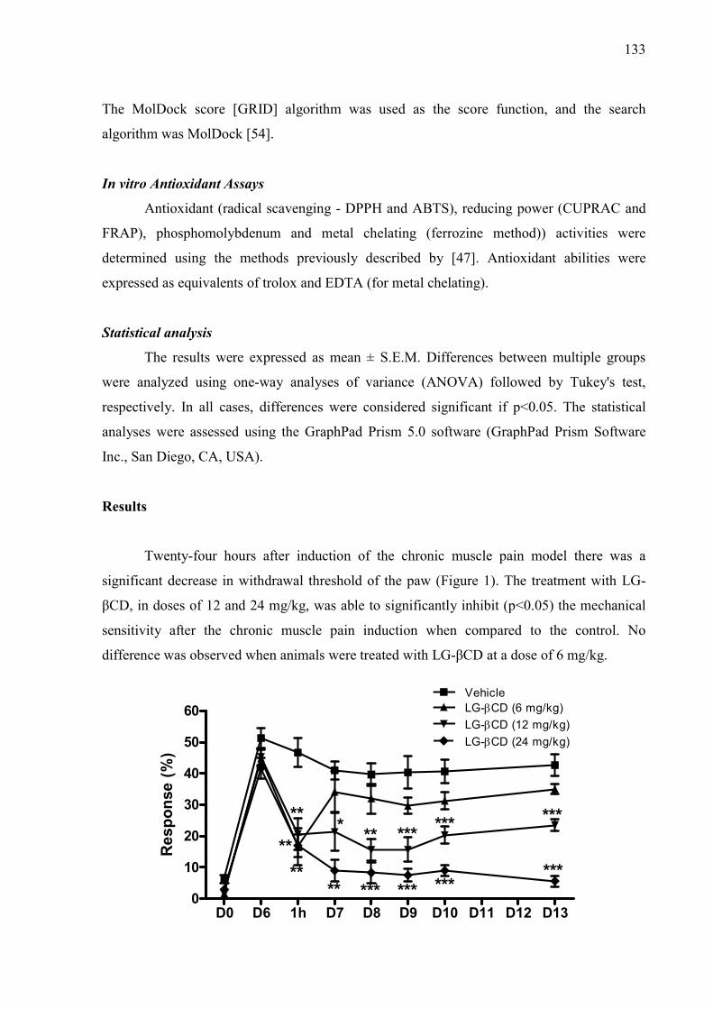
133
The MolDock score [GRID] algorithm was used as the score function, and the search
algorithm was MolDock [54].
In vitro Antioxidant Assays
Antioxidant (radical scavenging - DPPH and ABTS), reducing power (CUPRAC and
FRAP), phosphomolybdenum and metal chelating (ferrozine method)) activities were
determined using the methods previously described by [47]. Antioxidant abilities were
expressed as equivalents of trolox and EDTA (for metal chelating).
Statistical analysis
The results were expressed as mean ± S.E.M. Differences between multiple groups
were analyzed using one-way analyses of variance (ANOVA) followed by Tukey's test,
respectively. In all cases, differences were considered significant if p<0.05. The statistical
analyses were assessed using the GraphPad Prism 5.0 software (GraphPad Prism Software
Inc., San Diego, CA, USA).
Results
Twenty-four hours after induction of the chronic muscle pain model there was a
significant decrease in withdrawal threshold of the paw (Figure 1). The treatment with LG-
βCD, in doses of 12 and 24 mg/kg, was able to significantly inhibit (p<0.05) the mechanical
sensitivity after the chronic muscle pain induction when compared to the control. No
difference was observed when animals were treated with LG-βCD at a dose of 6 mg/kg.
D0 D6 1h D7 D8 D9 D10 D11 D12 D130
10
20
30
40
50
60VehicleLG-CD (6 mg/kg)
LG-CD (12 mg/kg)
LG-CD (24 mg/kg)
**
**
****
* **
***
***
***
***
***
***
***
Res
po
nse
(%
)
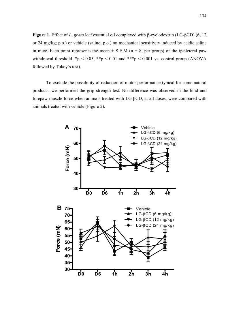
134
Figure 1. Effect of L. grata leaf essential oil complexed with β-cyclodextrin (LG-βCD) (6, 12
or 24 mg/kg; p.o.) or vehicle (saline; p.o.) on mechanical sensitivity induced by acidic saline
in mice. Each point represents the mean ± S.E.M (n = 8, per group) of the ipisleteral paw
withdrawal threshold. *p < 0.05, **p < 0.01 and ***p < 0.001 vs. control group (ANOVA
followed by Tukey´s test).
To exclude the possibility of reduction of motor performance typical for some natural
products, we performed the grip strength test. No difference was observed in the hind and
forepaw muscle force when animals treated with LG-βCD, at all doses, were compared with
animals treated with vehicle (Figure 2).
D0 D6 1h 2h 3h 4h30
40
50
60
70 VehicleLG-CD (6 mg/kg)
LG-CD (12 mg/kg)
LG-CD (24 mg/kg)
Fo
rce
(mN
)
A
D0 D6 1h 2h 3h 4h30
35
40
45
50
55
60
65
70
75 VehicleLG-CD (6 mg/kg)
LG-CD (12 mg/kg)
LG-CD (24 mg/kg)
Forc
e (m
N)
B
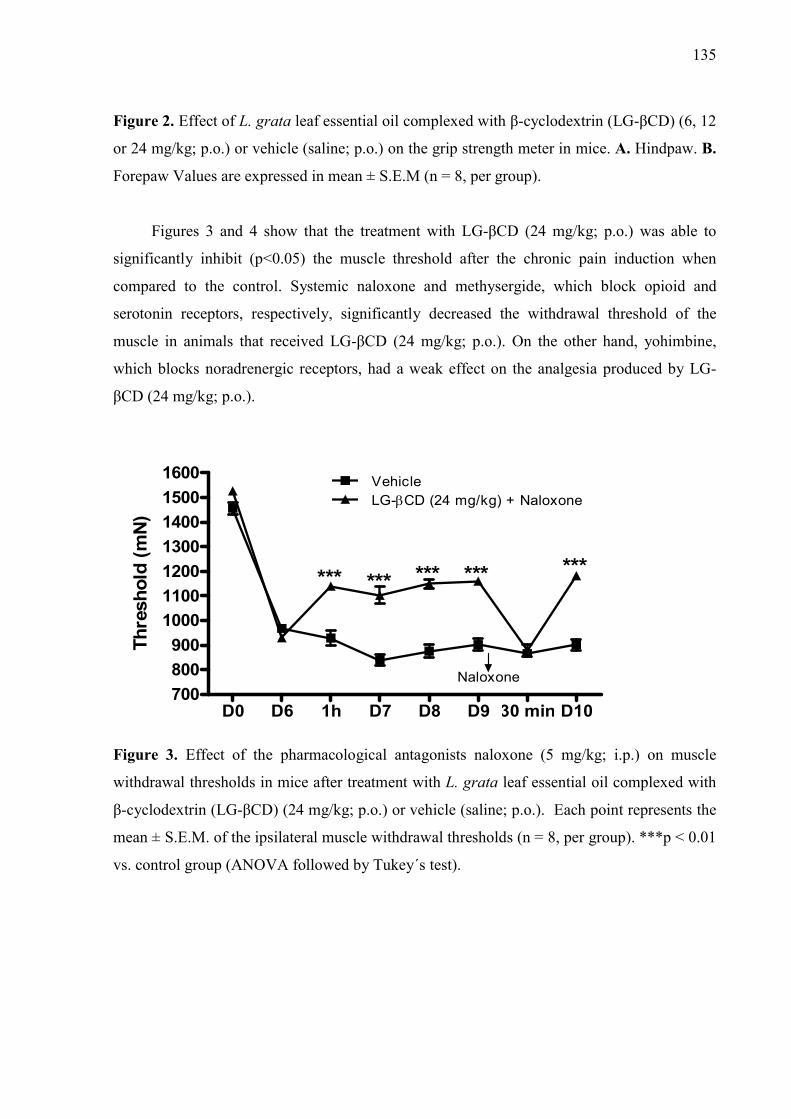
135
Figure 2. Effect of L. grata leaf essential oil complexed with β-cyclodextrin (LG-βCD) (6, 12
or 24 mg/kg; p.o.) or vehicle (saline; p.o.) on the grip strength meter in mice. A. Hindpaw. B.
Forepaw Values are expressed in mean ± S.E.M (n = 8, per group).
Figures 3 and 4 show that the treatment with LG-βCD (24 mg/kg; p.o.) was able to
significantly inhibit (p<0.05) the muscle threshold after the chronic pain induction when
compared to the control. Systemic naloxone and methysergide, which block opioid and
serotonin receptors, respectively, significantly decreased the withdrawal threshold of the
muscle in animals that received LG-βCD (24 mg/kg; p.o.). On the other hand, yohimbine,
which blocks noradrenergic receptors, had a weak effect on the analgesia produced by LG-
βCD (24 mg/kg; p.o.).
D0 D6 1h D7 D8 D9 30 min D10700
800
900
1000
1100
1200
1300
1400
1500
1600VehicleLG-CD (24 mg/kg) + Naloxone
Naloxone
*** *** *** *** ***
Th
resh
old
(m
N)
Figure 3. Effect of the pharmacological antagonists naloxone (5 mg/kg; i.p.) on muscle
withdrawal thresholds in mice after treatment with L. grata leaf essential oil complexed with
β-cyclodextrin (LG-βCD) (24 mg/kg; p.o.) or vehicle (saline; p.o.). Each point represents the
mean ± S.E.M. of the ipsilateral muscle withdrawal thresholds (n = 8, per group). ***p < 0.01
vs. control group (ANOVA followed by Tukey´s test).

136
D0 D6 1h D7 D8 D9 30 minD10750
1000
1250
1500
1750 VehicleLG-CD (24 mg/kg) + Methysurgide
LG-CD (24 mg/kg) + Yohimbine
***
******
***
***
***
***
******
**
***
***
Antagonists
Th
resh
old
(m
N)
Figure 4. Effect of the pharmacological antagonists methysergide (1.5 mg/kg; i.p.) and
yohimbine (2 mg/kg; i.p.) on muscle withdrawal thresholds in mice after treatment with L.
grata leaf essential oil complexed with β-cyclodextrin (LG-βCD) (24 mg/kg; p.o.) or vehicle
(saline; p.o.). Each point represents the mean ± S.E.M. of the ipsilateral muscle withdrawal
thresholds (n = 8, per group). **p < 0.01 and ***p < 0.001 vs. control group (ANOVA
followed by Tukey´s test).
Moreover, Fos protein immunofluorescence showed a significant decrease in expression (Fos-
positive cells) in the dorsal horn of the spinal cord, when compared to the control (p<0.01), as
demonstrated in Figure 5.
A B
C
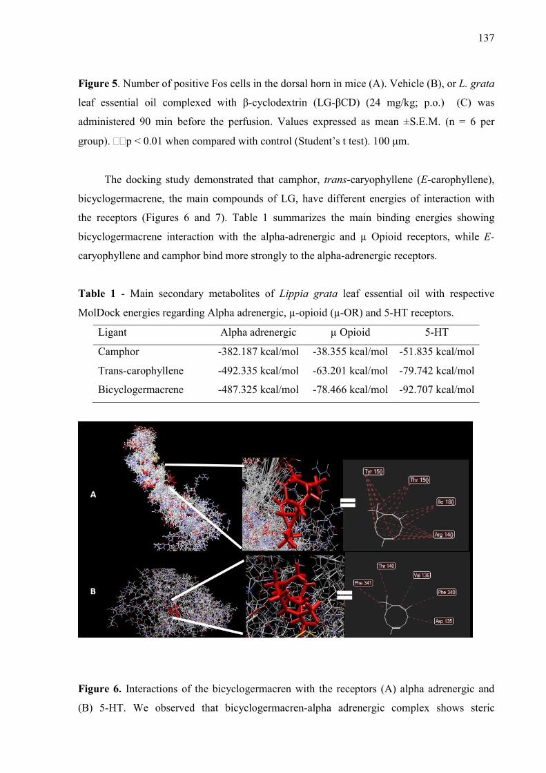
137
Figure 5. Number of positive Fos cells in the dorsal horn in mice (A). Vehicle (B), or L. grata
leaf essential oil complexed with β-cyclodextrin (LG-βCD) (24 mg/kg; p.o.) (C) was
administered 90 min before the perfusion. Values expressed as mean ±S.E.M. (n = 6 per
group). ∗∗p < 0.01 when compared with control (Student’s t test). 100 μm.
The docking study demonstrated that camphor, trans-caryophyllene (E-carophyllene),
bicyclogermacrene, the main compounds of LG, have different energies of interaction with
the receptors (Figures 6 and 7). Table 1 summarizes the main binding energies showing
bicyclogermacrene interaction with the alpha-adrenergic and µ Opioid receptors, while E-
caryophyllene and camphor bind more strongly to the alpha-adrenergic receptors.
Table 1 - Main secondary metabolites of Lippia grata leaf essential oil with respective
MolDock energies regarding Alpha adrenergic, µ-opioid (µ-OR) and 5-HT receptors.
Ligant Alpha adrenergic µ Opioid 5-HT
Camphor -382.187 kcal/mol -38.355 kcal/mol -51.835 kcal/mol
Trans-carophyllene -492.335 kcal/mol -63.201 kcal/mol -79.742 kcal/mol
Bicyclogermacrene -487.325 kcal/mol -78.466 kcal/mol -92.707 kcal/mol
Figure 6. Interactions of the bicyclogermacren with the receptors (A) alpha adrenergic and
(B) 5-HT. We observed that bicyclogermacren-alpha adrenergic complex shows steric
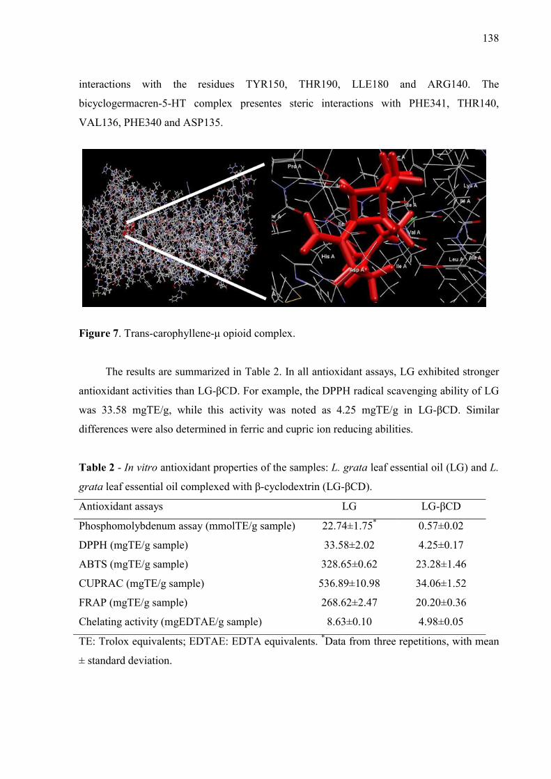
138
interactions with the residues TYR150, THR190, LLE180 and ARG140. The
bicyclogermacren-5-HT complex presentes steric interactions with PHE341, THR140,
VAL136, PHE340 and ASP135.
Figure 7. Trans-carophyllene-μ opioid complex.
The results are summarized in Table 2. In all antioxidant assays, LG exhibited stronger
antioxidant activities than LG-βCD. For example, the DPPH radical scavenging ability of LG
was 33.58 mgTE/g, while this activity was noted as 4.25 mgTE/g in LG-βCD. Similar
differences were also determined in ferric and cupric ion reducing abilities.
Table 2 - In vitro antioxidant properties of the samples: L. grata leaf essential oil (LG) and L.
grata leaf essential oil complexed with β-cyclodextrin (LG-βCD).
Antioxidant assays LG LG-βCD
Phosphomolybdenum assay (mmolTE/g sample) 22.74±1.75* 0.57±0.02
DPPH (mgTE/g sample) 33.58±2.02 4.25±0.17
ABTS (mgTE/g sample) 328.65±0.62 23.28±1.46
CUPRAC (mgTE/g sample) 536.89±10.98 34.06±1.52
FRAP (mgTE/g sample) 268.62±2.47 20.20±0.36
Chelating activity (mgEDTAE/g sample) 8.63±0.10 4.98±0.05
TE: Trolox equivalents; EDTAE: EDTA equivalents. *Data from three repetitions, with mean
± standard deviation.

139
Discussion
Previous study performed by our research group demonstrated that the inclusion
complex LG-βCD or LG alone produced a remarkable analgesic profile in orofacial pain
animal models with neuronal activation of CNS areas which are important areas to central
pain modulation, such as the Locus coruleus (LC), NRM and PAG. So, this study strongly
suggested the involvement of the descending inhibitory pain modulation pathway in the
production of analgesia after treatment with LG-βCD [48, 49]. Moreover, βCD enhanced the
pharmacological effects of LG by increasing efficacy and producing longer-lasting analgesic
activity compared with LG alone [48]. CDs are known to enhance the activity of analgesic
and anti-inflammatory drugs [4, 20, 33, 46], including when evaluated in FM experimental
protocols [30, 31, 33, 40]. Therefore, we assessed the hypothesis that LG-βCD could produce
an anti-hyperalgesic effect in a chronic non-inflammatory muscle pain protocol in mice
(considered to be an animal model for fibromyalgia, FM) due its CNS analgesic effects, and
also examine its antioxidant effect.
The current study demonstrated that oral administration with LG-βCD (6, 12 or 24
mg/kg, p.o.) also produced outstanding antihyperalgesic activity (significantly reducing the
primary and secondary hyperalgesia) (p<0.05; p<0.01; p<0.001) against a chronic non-
inflammatory muscle pain model induced by saline acid injection into the gastrocnemius
muscle of mice. LG-βCD produced no change in muscle strength, excluding the possibility of
a reduction in motor performance typical of some drugs that act in the CNS [38]. These
effects occurred with the probable involvement of opioid, and serotonin (5-HT) receptors (at
least in part), which suggest its potential applicability in the management of chronic pain,
such as FM. These effects may be related to the antioxidant profile demonstrated by LG.
FM is a differentiated pain syndrome, a type of ‘dysfunctional pain’, and there are a
limited number of animal models that mimic the major symptoms, mainly widespread pain
and the associated symptoms, such as fatigue and anxiety [10]. The non-inflammatory pain
model induced by repeated intramuscular (gastrocnemius muscle) saline acid injections in
rodents has been suggested as having validity to chronic widespread pain conditions (such as
FM) in humans [51, 52, 58]. Morphological evaluation of the gastrocnemius muscle shows no
apparent damage or inflammation associated with the administration of the saline acid [10,
52]. Moreover, central mechanisms involving the spinal cord, brainstem and cortex have been
associated with development of hyperalgesia in this animal model [55].
Additionally, it is likely that pain modulation results from a descending pain
modulatory pathway with inputs feeding into the midbrain PAG area. Neurons within the
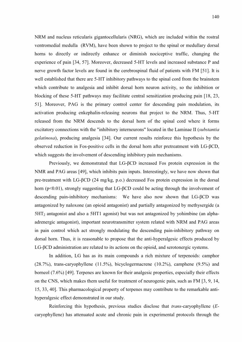
140
NRM and nucleus reticularis gigantocellularis (NRG), which are included within the rostral
ventromedial medulla (RVM), have been shown to project to the spinal or medullary dorsal
horns to directly or indirectly enhance or diminish nociceptive traffic, changing the
experience of pain [34, 57]. Moreover, decreased 5-HT levels and increased substance P and
nerve growth factor levels are found in the cerebrospinal fluid of patients with FM [51]. It is
well established that there are 5-HT inhibitory pathways to the spinal cord from the brainstem
which contribute to analgesia and inhibit dorsal horn neuron activity, so the inhibition or
blocking of these 5-HT pathways may facilitate central sensitization producing pain [18, 23,
51]. Moreover, PAG is the primary control center for descending pain modulation, its
activation producing enkephalin-releasing neurons that project to the NRM. Thus, 5-HT
released from the NRM descends to the dorsal horn of the spinal cord where it forms
excitatory connections with the "inhibitory interneurons" located in the Laminae II (substantia
gelatinosa), producing analgesia [34]. Our current results reinforce this hypothesis by the
observed reduction in Fos-positive cells in the dorsal horn after pretreatment with LG-βCD,
which suggests the involvement of descending inhibitory pain mechanisms.
Previously, we demonstrated that LG-βCD increased Fos protein expression in the
NMR and PAG areas [49], which inhibits pain inputs. Interestingly, we have now shown that
pre-treatment with LG-βCD (24 mg/kg, p.o.) decreased Fos protein expression in the dorsal
horn (p<0.01), strongly suggesting that LG-βCD could be acting through the involvement of
descending pain-inhibitory mechanisms: We have also now shown that LG-βCD was
antagonized by naloxone (an opioid antagonist) and partially antagonized by methysergide (a
5HT2 antagonist and also a 5HT1 agonist) but was not antagonized by yohimbine (an alpha-
adrenergic antagonist), important neurotransmitter system related with NRM and PAG areas
in pain control which act strongly modulating the descending pain-inhibitory pathway on
dorsal horn. Thus, it is reasonable to propose that the anti-hyperalgesic effects produced by
LG-βCD administration are related to its actions on the opioid, and serotonergic systems.
In addition, LG has as its main compounds a rich mixture of terpenoids: camphor
(28.7%), trans-caryophyllene (11.5%), bicyclogermacrene (10.2%), camphene (9.5%) and
borneol (7.6%) [49]. Terpenes are known for their analgesic properties, especially their effects
on the CNS, which makes them useful for treatment of neurogenic pain, such as FM [3, 9, 14,
15, 33, 40]. This pharmacological property of terpenes may contribute to the remarkable anti-
hyperalgesic effect demonstrated in our study.
Reinforcing this hypothesis, previous studies disclose that trans-caryophyllene (E-
caryophyllene) has attenuated acute and chronic pain in experimental protocols through the

141
involvement of the opioid and endocannabinoid systems [36]. Borneol and camphor are well-
known analgesic compounds described in several patented pharmaceuticals available on the
market to treat pain [15]. In addition, borneol has been shown to reduce mechanical
hyperalgesia in inflammatory rodent models by enhancing GABAA-mediated GABAergic
transmission in the spinal cord [19]. Borneol and camphor are drugs that have affinity for a
large number of receptors from the TRP family which are expressed in sensing neurons and
primary afferent nociceptors [27]. Moreover, camphene has shown potent central analgesic
activity and also an antioxidant profile in TBARS (Thiobarbituric acid reactive substances),
and TRAP/TAR (Total reactive antioxidant potential/Total antioxidant reactivity) assays, as
well as having the highest scavenging activities against different free radicals, such as
hydroxyl and superoxide radicals [39]: these antioxidant properties are associated with
modulation of pain in FM patients [17].
To further corroborate the results in vivo, we performed in silico assessment using a
molecular docking approach. Target validation by in silico analysis contributed to a
theoretical explanation of the drug-receptor relationship, and corroborated the evidence
created in vivo protocols [12, 42]. We demonstrated that camphor, E-caryophyllene,
bicyclogermacrene binds to the alpha-adrenergic receptor while bicyclogermacrene binds with
moderate energy to the µOpioid and 5-HT receptors (as shown in Table 1). The docking study
suggested that E-caryophyllene binds with weak energy to the µOpioid receptor, however, the
in vivo data demonstrated that the interaction between drug-receptor is enough to produces an
analgesic effect [36]. Controversially, our docking data suggested a strong binding energy
between the major compounds and the alpha-adrenergic receptor but our in vivo data (when it
was antagonized with yoimbine) did not corroborate this hypothesis. These apparent
contradictory results of the possible effect of LG-βCD in the alpha-adrenergic receptor could
be explained by the rich mixture of compounds present in the essential oil from L. grata
which can act synergistically or antagonistically [49, 50]. Moreover, the evidence of the
management of descending serotonergic pathways by LG, with the serotonergic neurons
projecting from the RVM (rostral ventromedial medulla) through the DLF (dorsolateral
funiculus) [55], was corroborated by the effect of oil components on the 5HT receptors (in
silico data) and more strongly by the antagonism of the analgesic effect by methysergide. So,
the docking data suggest that bicyclogermacrene and E-caryophyllene seem to act
synergistically in favor of anti-hyperalgesic effect.
Seeking to corroborate the hypothesis of LG or LG-βCD having an antioxidant role
which could contribute to their anti-hyperalgesic activity, we performed different chemical
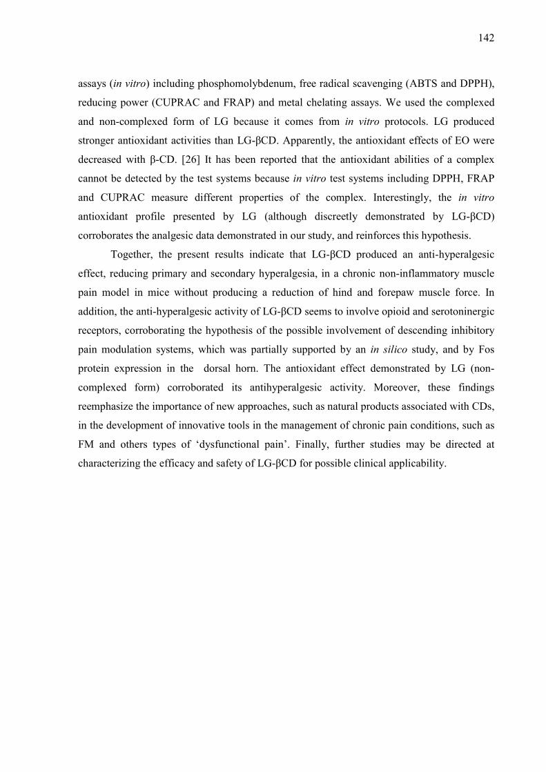
142
assays (in vitro) including phosphomolybdenum, free radical scavenging (ABTS and DPPH),
reducing power (CUPRAC and FRAP) and metal chelating assays. We used the complexed
and non-complexed form of LG because it comes from in vitro protocols. LG produced
stronger antioxidant activities than LG-βCD. Apparently, the antioxidant effects of EO were
decreased with β-CD. [26] It has been reported that the antioxidant abilities of a complex
cannot be detected by the test systems because in vitro test systems including DPPH, FRAP
and CUPRAC measure different properties of the complex. Interestingly, the in vitro
antioxidant profile presented by LG (although discreetly demonstrated by LG-βCD)
corroborates the analgesic data demonstrated in our study, and reinforces this hypothesis.
Together, the present results indicate that LG-βCD produced an anti-hyperalgesic
effect, reducing primary and secondary hyperalgesia, in a chronic non-inflammatory muscle
pain model in mice without producing a reduction of hind and forepaw muscle force. In
addition, the anti-hyperalgesic activity of LG-βCD seems to involve opioid and serotoninergic
receptors, corroborating the hypothesis of the possible involvement of descending inhibitory
pain modulation systems, which was partially supported by an in silico study, and by Fos
protein expression in the dorsal horn. The antioxidant effect demonstrated by LG (non-
complexed form) corroborated its antihyperalgesic activity. Moreover, these findings
reemphasize the importance of new approaches, such as natural products associated with CDs,
in the development of innovative tools in the management of chronic pain conditions, such as
FM and others types of ‘dysfunctional pain’. Finally, further studies may be directed at
characterizing the efficacy and safety of LG-βCD for possible clinical applicability.
143
List of Abbreviations
5-HT - 5-hydroxytryptamine
CDs – Cyclodextrins
CNS - Central Nervous System
DLF - dorsolateral funiculus
DPPH - 2,2-diphenyl-1-picryl-hydrazyl-hydrate
FM – Fibromyalgia
GABA - gamma-aminobutyric acid
LC - Locus ceruleus
LG - L. grata leaf essential oil
LG-βCD - L. grata leaf essential oil complexed β-cyclodextrin
NMR - nucleus raphe magnus
PAG - periaqueductal gray
TRP - transient receptor potential
βCD - β-cyclodextrin
RVM - rostral ventromedial medulla
Conflict of interest
The authors declare no conflict of interest.
Acknowledgments
This work was supported by grants from CNPq (305608/2013-4), FINEP, CAPES and
FAPITEC/SE (Agencies from Brazil). PS Lima-Siqueira has a scholarship from the Graduate
Program in Biotechnology (PPGBiotec) at the UEFS and R.G. Brito, H.G. Araújo-Filho, P.L.
Santos has a scholarship from the Graduate Program in Health Sciences (PPGCS) at the UFS.

144
References
[1] Allinger, N.L. Conformational analysis. 130. MM2. A hydrocarbon force field utilizing V1 and V2 torsional terms. J. Am. Chem. Soc., 1977, 99, 8127-8134. [2] Ashburn, T.T.; Thor, K.B. Drug repositioning: identifying and developing new uses for existing drugs. Nat Rev Drug Discov, 2004, 3, 673-683. [3] Batista, P.A.; Werner, M.F.; Oliveira, E.C.; Burgos, L.; Pereira, P.; Brum, L.F.; Santos, A.R. Evidence for the involvement of ionotropic glutamatergic receptors on the antinociceptive effect of (-)-linalool in mice. Neurosci Lett, 2008, 440, 299-303. [4] Brito, R.G.; Araujo, A.A.; Quintans, J.S.; Sluka, K.A.; Quintans-Junior, L.J. Enhanced analgesic activity by cyclodextrins - a systematic review and meta-analysis. Expert Opin Drug Deliv, 2015, 12, 1677-1688. [5] Brito, R.G.; Santos, P.L.; Prado, D.S.; Santana, M.T.; Araujo, A.A.; Bonjardim, L.R.; Santos, M.R.; de Lucca Junior, W.; Oliveira, A.P.; Quintans-Junior, L.J. Citronellol reduces orofacial nociceptive behaviour in mice - evidence of involvement of retrosplenial cortex and periaqueductal grey areas. Basic Clin Pharmacol Toxicol, 2013, 112, 215-221. [6] Ceko, M.; Bushnell, M.C.; Gracely, R.H. Neurobiology underlying fibromyalgia symptoms. Pain Res Treat, 2012, 2012, 585419. [7] Clauw, D.J. Fibromyalgia: a clinical review. Jama, 2014, 311, 1547-1555. [8] Cohen, N.C. The Molecular Modeling Perspective in Drug Design. In: Guidebook on Molecular Modeling in Drug Design; Academic Press: San Diego; 1996, pp. 1-17. [9] de Sousa, D.P. Analgesic-like activity of essential oils constituents. Molecules, 2011, 16, 2233-2252. [10] DeSantana, J.M.; da Cruz, K.M.; Sluka, K.A. Animal models of fibromyalgia. Arthritis Res Ther, 2013, 15, 222. [11] Dewar, M.J.S.E.; Zoebisch, G.; Healy, E.F.; Stewart, J.J.P. AM1 a New General Purpose Quantum Mechanical Molecular Model. J. Am. Chem. Soc., 1985, 107, 3902-3909. [12] Ekins, S.; Mestres, J.; Testa, B. In silico pharmacology for drug discovery: methods for virtual ligand screening and profiling. Br J Pharmacol, 2007, 152, 9-20. [13] Ernst, E. Herbal medicine in the treatment of rheumatic diseases. Rheum Dis Clin North Am, 2011, 37, 95-102. [14] Guimaraes, A.G.; Quintans, J.S.; Quintans Júnior, L.J. Monoterpenes with analgesic activity--a systematic review. Phytother Res, 2013, 27, 1-15. [15] Guimaraes, A.G.; Serafini, M.R.; Quintans-Junior, L.J. Terpenes and derivatives as a new perspective for pain treatment: a patent review. Expert Opin Ther Pat, 2014, 24, 243-265. [16] Ikeuchi, M.; Kolker, S.J.; Burnes, L.A.; Walder, R.Y.; Sluka, K.A. Role of ASIC3 in the primary and secondary hyperalgesia produced by joint inflammation in mice. Pain, 2008, 137, 662-669. [17] Iqbal, R.; Mughal, M.S.; Arshad, N.; Arshad, M. Pathophysiology and antioxidant status of patients with fibromyalgia. Rheumatol Int, 2011, 31, 149-152. [18] Jacobs, B.L.; Martin-Cora, F.J.; Fornal, C.A. Activity of medullary serotonergic neurons in freely moving animals. Brain Res Brain Res Rev, 2002, 40, 45-52. [19] Jiang, J.; Shen, Y.Y.; Li, J.; Lin, Y.H.; Luo, C.X.; Zhu, D.Y. (+)-Borneol alleviates mechanical hyperalgesia in models of chronic inflammatory and neuropathic pain in mice. Eur J Pharmacol, 2015, 757, 53-58. [20] Lauretti, G.R.; Mattos, A.L.; Lima, I.C. Tramadol and beta-cyclodextrin piroxicam: effective multimodal balanced analgesia for the intra- and postoperative period. Reg Anesth, 1997, 22, 243-248. [21] Lawson, K. Potential drug therapies for the treatment of fibromyalgia. Expert Opin Investig Drugs, 2016, 25, 1071-1081.
145
[22] Leach, A.R. Molecular Modelling: Principles and Applications. 2nd ed.; Prentice Hall: London, 2001. [23] Leonard, B.E. The HPA and immune axes in stress: the involvement of the serotonergic system. Eur Psychiatry, 2005, 13, 268-276. [24] Li, J.W.; Vederas, J.C. Drug discovery and natural products: end of an era or an endless frontier? Science, 2009, 325, 161-165. [25] Lima, P.S.; Lucchese, A.M.; Araujo-Filho, H.G.; Menezes, P.P.; Araujo, A.A.; Quintans-Junior, L.J.; Quintans, J.S. Inclusion of terpenes in cyclodextrins: Preparation, characterization and pharmacological approaches. Carbohydr Polym, 2016, 151, 965-987. [26] Lopez-Nicolas, J.M.; Rodriguez-Bonilla, P.; Garcia-Carmona, F. Cyclodextrins and antioxidants. Crit Rev Food Sci Nutr, 2014, 54, 251-276. [27] Marwaha, L.; Bansal, Y.; Singh, R.; Saroj, P.; Bhandari, R.; Kuhad, A. TRP channels: potential drug target for neuropathic pain. Inflammopharmacology, 2016, 24, 305-317. [28] Menezes, P.P.; Serafini, M.R.; Quintans-Júnior, L.J.; Silva, G.F.; Oliveira, J.F.; Carvalho, F.M.S.; Souza, J.C.C.; Matos, J.R.; Alves, P.B.; Matos, I.L.; Hădărugă, D.I.; Araújo, A.A.S. Inclusion complex of (−)-linalool and β-cyclodextrin. Journal of Thermal Analysis and Calorimetry, 2014, 115, 2429-2437. [29] Nagakura, Y. Challenges in drug discovery for overcoming 'dysfunctional pain': an emerging category of chronic pain. Expert Opin Drug Discov, 2015, 10, 1043-1045. [30] Nascimento, S.S.; Araujo, A.A.; Brito, R.G.; Serafini, M.R.; Menezes, P.P.; DeSantana, J.M.; Lucca, W., Jr.; Alves, P.B.; Blank, A.F.; Oliveira, R.C.; Oliveira, A.P.; Albuquerque, R.L., Jr.; Almeida, J.R.; Quintans, L.J., Jr. Cyclodextrin-complexed Ocimum basilicum leaves essential oil increases Fos protein expression in the central nervous system and produce an antihyperalgesic effect in animal models for fibromyalgia. Int J Mol Sci, 2014, 16, 547-563. [31] Nascimento, S.S.; Araújo, A.A.S.; Brito, R.G.; Serafini, M.R.; Menezes, P.P.; DeSantana, J.M.; Lucca Júnior, W.; Alves, P.B.; Blank, A.F.; Oliveira, R.C.M.; Oliveira, A.P.; Albuquerque-Júnior, R.L.C.; Almeida, J.; Quintans-Júnior, L.J. Cyclodextrin-Complexed Ocimum basilicum Leaves Essential Oil Increases Fos Protein Expression in the Central Nervous System and Produce an Antihyperalgesic Effect in Animal Models for Fibromyalgia. Int J Mol Sci, 2015, 16, 547-563. [32] Nascimento, S.S.; Camargo, E.A.; DeSantana, J.M.; Araujo, A.A.; Menezes, P.P.; Lucca-Junior, W.; Albuquerque-Junior, R.L.; Bonjardim, L.R.; Quintans-Junior, L.J. Linalool and linalool complexed in beta-cyclodextrin produce anti-hyperalgesic activity and increase Fos protein expression in animal model for fibromyalgia. Naunyn Schmiedebergs Arch Pharmacol, 2014, 387, 935-942. [33] Oliveira, M.G.; Brito, R.G.; Santos, P.L.; Araujo-Filho, H.G.; Quintans, J.S.; Menezes, P.P.; Serafini, M.R.; Carvalho, Y.M.; Silva, J.C.; Almeida, J.R.; Scotti, L.; Scotti, M.T.; Shanmugam, S.; Thangaraj, P.; Araujo, A.A.; Quintans-Junior, L.J. alpha-Terpineol, a monoterpene alcohol, complexed with beta-cyclodextrin exerts antihyperalgesic effect in animal model for fibromyalgia aided with docking study. Chem Biol Interact, 2016, 254, 54-62. [34] Ossipov, M.H.; Dussor, G.O.; Porreca, F. Central modulation of pain. J Clin Invest, 2010, 120, 3779-3787. [35] Pascual, M.E.; Slowing, K.; Carretero, E.; Sanchez Mata, D.; Villar, A. Lippia: traditional uses, chemistry and pharmacology: a review. J Ethnopharmacol, 2001, 76, 201-214. [36] Paula-Freire, L.I.; Andersen, M.L.; Gama, V.S.; Molska, G.R.; Carlini, E.L. The oral administration of trans-caryophyllene attenuates acute and chronic pain in mice. Phytomedicine, 2014, 21, 356-362.
146
[37] Paxinos, G.; Franklin, K. Paxinos and Franklin’s the Mouse Brain in Stereotaxic Coordinates. Academic Press (Elsevier), 2012. [38] Perl, E.R. Pain mechanisms: a commentary on concepts and issues. Prog Neurobiol, 2011, 94, 20-38. [39] Quintans-Junior, L.; Moreira, J.C.; Pasquali, M.A.; Rabie, S.M.; Pires, A.S.; Schroder, R.; Rabelo, T.K.; Santos, J.P.; Lima, P.S.; Cavalcanti, S.C.; Araujo, A.A.; Quintans, J.S.; Gelain, D.P. Antinociceptive Activity and Redox Profile of the Monoterpenes (+)-Camphene, p-Cymene, and Geranyl Acetate in Experimental Models. ISRN Toxicol, 2013, 2013, 1-11. [40] Quintans-Júnior, L.J.; Araújo, A.A.S.; Brito, R.G.; Santos, P.L.; Quintans, J.S.S.; Menezes, P.P.; Serafini, M.R.; Silva, G.F.; Carvalho, F.M.S.; Brogden, N.K.; Sluka, K.A. β-caryophyllene, a dietary cannabinoid, complexed with β-cyclodextrin produced anti-hyperalgesic effect involving the inhibition of Fos expression in superficial dorsal horn. Life Sci, 2016, 149, 34-41. [41] Quintans, J.S.S.; Menezes, P.P.; Santos, M.R.; Bonjardim, L.R.; Almeida, J.R.; Gelain, D.P.; Araujo, A.A.; Quintans-Junior, L.J. Improvement of p-cymene antinociceptive and anti-inflammatory effects by inclusion in beta-cyclodextrin. Phytomedicine, 2013, 20, 436-440. [42] Quintans, J.S.S.; Pereira, E.W.M.; Carvalho, Y.M.B.G.; Menezes, P.P.; Serafini, M.R.; Batista, M.V.A.; Moreira, C.D.L.F.A.; Lima, A.A.N.; Branco, A.; Almeida, J.R.G.S.; Gelain, D.P.; Zengin, G.; Araújo, A.A.S.; Quintans-Júnior, L.J. Host–guest inclusion complexation of β-cyclodextrin and hecogenin acetate to enhance anti-hyperalgesic effect in an animal model of musculoskeletal pain. Process Biochemistry, 2016. [43] Radhakrishnan, R.; Bement, M.K.; Skyba, D.; Sluka, K.A.; Kehl, L.J. Models of muscle pain: carrageenan model and acidic saline model. Curr Protoc Pharmacol, 2004, Chapter 5, Unit 5.35. [44] Roh, C.; Jung, U. Screening of crude plant extracts with anti-obesity activity. Int J Mol Sci, 2012, 13, 1710-1719. [45] Santos, P.L.; Brito, R.G.; Oliveira, M.A.; Quintans, J.S.; Guimaraes, A.G.; Santos, M.R.; Menezes, P.P.; Serafini, M.R.; Menezes, I.R.; Coutinho, H.D.; Araujo, A.A.; Quintans-Junior, L.J. Docking, characterization and investigation of beta-cyclodextrin complexed with citronellal, a monoterpene present in the essential oil of Cymbopogon species, as an anti-hyperalgesic agent in chronic muscle pain model. Phytomedicine, 2016, 23, 948-957. [46] Santos, P.L.; Brito, R.G.; Quintans, J.S.S.; Araujo, A.A.S.; Menezes, I.R.A.; Brogden, N.K.; Quintans Junior, L.J. Cyclodextrins as Encapsulation Agents to Improve the Anti-inflammatory Drugs Profile: a Systematic Review and Meta-Analysis. Current Pharmaceutical Design, 2017. [47] Savran, A.; Zengin, G.; Aktumsek, A.; Mocan, A.; Glamoclija, J.; Ciric, A.; Sokovic, M. Phenolic compounds and biological effects of edible Rumex scutatus and Pseudosempervivum sempervivum: potential sources of natural agents with health benefits. Food Funct, 2016, 7, 3252-3262. [48] Siqueira-Lima, P.S. Efeito do complexo de inclusão contendo betaciclodextrina e óleo essencial de Lippia grata (Verbenaceae) na nocicepção orofacial em modelos experimentais. Masters dissertation, Universidade Estadual de Feira de Santana - UEFS: Feira de Santana, 2013. [49] Siqueira-Lima, P.S.; Araujo, A.A.; Lucchese, A.M.; Quintans, J.S.; Menezes, P.P.; Alves, P.B.; de Lucca Junior, W.; Santos, M.R.; Bonjardim, L.R.; Quintans-Junior, L.J. beta-cyclodextrin complex containing Lippia grata leaf essential oil reduces orofacial nociception in mice - evidence of possible involvement of descending inhibitory pain modulation pathway. Basic Clin Pharmacol Toxicol, 2014, 114, 188-196.

147
[50] Sivamani, P.; Singaravelu, G.; Thiagarajan, V.; Jayalakshmi, T.; Ramesh Kumar, G. Comparative molecular docking analysis of essential oil constituents as elastase inhibitors. Bioinformation, 2012, 8, 457-460. [51] Sluka, K.A. Is it possible to develop an animal model of fibromyalgia? Pain, 2009, 146, 3-4. [52] Sluka, K.A.; Kalra, A.; Moore, S.A. Unilateral intramuscular injections of acidic saline produce a bilateral, long-lasting hyperalgesia. Muscle Nerve, 2001, 24, 37-46. [53] Sluka, K.A.; Rohlwing, J.J.; Bussey, R.A.; Eikenberry, S.A.; Wilken, J.M. Chronic muscle pain induced by repeated acid Injection is reversed by spinally administered mu- and delta-, but not kappa-, opioid receptor agonists. J Pharmacol Exp Ther, 2002, 302, 1146-1150. [54] Thomsen, R.; Christensen, M.H. MolDock: a new technique for high-accuracy molecular docking. J Med Chem, 2006, 49, 3315-3321. [55] Tillu, D.V.; Gebhart, G.F.; Sluka, K.A. Descending facilitatory pathways from the RVM initiate and maintain bilateral hyperalgesia after muscle insult. Pain, 2008, 136, 331-339. [56] Viana, G.S.B.; Matos, F.F.; Araujo, W.L.; Matos, F.J.A.; Craveiro, A.A. Essential Oil of Lippia grata: Pharmacological Effects and Main Constituents. Quarterly Journal of Crude Drug Research, 1981, 19, 1-10. [57] Willis, W.D., Jr. Central nervous system mechanisms for pain modulation. Appl Neurophysiol, 1985, 48, 153-165. [58] Yokoyama, T.; Maeda, Y.; Audette, K.M.; Sluka, K.A. Pregabalin reduces muscle and cutaneous hyperalgesia in two models of chronic muscle pain in rats. J Pain, 2007, 8, 422-429. [59] Yunus, M.B. Central sensitivity syndromes: a new paradigm and group nosology for fibromyalgia and overlapping conditions, and the related issue of disease versus illness. Semin Arthritis Rheum, 2008, 37, 339-352.

148
CAPÍTULO 4
149
Evidence for the involvement of the PKA pathway and inhibition of voltage
gated Ca2+ channels in DRG neurons in antihyperalgesic activity produced
by the essential oil of Lippia grata/β-cyclodextrin complex in rodent
neuropathic pain-like models
Pollyana S. Siqueira-Lima1, 2,#, Jullyana S.S. Quintans1,2,#,*, Luana Heimfarth1,2, Fabiolla R. S.
Passos1,2, Erik W. M. Pereira1,2, Marilia M. Rezende1,2, José E.R. Menezes-Filho1,2, Henrique
Douglas Coutinho, Rosana S.S. Barreto1,2, Adriano A.S. Araújo3, Aline S. Medrado5, Ligia A.
Naves5, Horácio F. Bomfim4, Angélica M. Lucchese4, Lucindo J. Quintans-Júnior1,2*
#These authors contributed equally.
1Multiuser Health Center Facility (CMulti-Saúde). 2Department of Physiology. 3Department
of Pharmacy. Federal University of Sergipe, São Cristóvão, SE, 49100-000 Brazil. 4Post-Graduate Program in Biotechnology, State University of Feira de Santana, Feira de
Santana, BA, Brazil. 5 Federal University of Minas Gerais.
*Corresponding authors: Federal University of Sergipe. Department of Physiology,
Laboratory of Neuroscience and Pharmacological Assays (LANEF). Ave Marechal Rondon,
sn, Rosa Elze, São Cristóvão, Sergipe, CEP: 49.100-000, Brazil. E-mail:
[email protected]; [email protected] (To: Prof L.J. Quintans-Júnior) or
[email protected] (To: Prof J.S.S. Quintans).
150
Abstract
Neuropathic pain (NP) is a difficult condition to treat because of the modest efficacy of
available drugs. New treatments are, therefore, required. In the present study we aimed to
investigate the effects of the essential oil from the leaf of Lippia grata alone or complexed in
β-cyclodextrin (LG or LG-βCD) on persistent inflammatory and neuropathic pain in a mouse
model. We also investigated Ca2+ currents in rat dorsal root ganglion (DRG) neurons. Male
Swiss mice were treated with LG or LG/βCD (24 mg/kg, i.g.) and their effect was evaluated
using an acute inflammatory pleurisy model and nociception triggered by intraplantar (i.pl.)
injection of an agonist of the TRPA1 and TRPM8 channels. We also tested their effect in two
chronic pain models: i.pl. injection of Freund´s Complete Adjuvant and partial sciatic nerve
ligation (PSNL). In the pleurisy model, LG reduced the number of leukocytes and the levels
of TNF-α and IL-1β. It also inhibited cinnamaldehyde and menthol-induced nociceptive
behavior. The pain threshold in mechanical and thermal hyperalgesia was increased and paw
edema was decreased in models of inflammatory and neuropathic pain. PSNL increased
inflammatory protein contents and LG and LG-βCD restored the the protein contents of TNF-
α, NF-κB, and PKA, but not IL-1β and IL-10. LG inhibited voltage gated Ca2+ channels from
DRG neurons. Our results suggested that LG or LG-βCD produce anti-hyperalgesic effect in
chronic pain models through reductions in TNF- levels and PKA, and inhibited voltage-
gated calcium channels and may be innovative therapeutic agents for the management of NP.
Key words: Natural prodcuts, pain, inflammatory pain, dorsal root ganglion, TNF-,
cyclodextrin.

151
1. Introduction
According to the World Health Organization (WHO), about 20% of the population
live with some degree of chronic pain, which is more common in women, older individuals
and people with relative deprivation. (Kehlet et al., 2006). Neuropathic pain (NP) is one of the
most important types of chronic pain. It is triggered by lesions to the somatosensory nervous
system that alter its structure and function (Costigan et al., 2009), causing hyperalgesia,
allodynia and spontaneous pain. (Kaulaskar et al., 2012). Because NP does not have a role in
protecting individuals against tissue damage it is not simply a symptom, but a disease in itself
(Yekkirala et al., 2017) (White et al., 2007). Moreover, NP is one of the most difficult types
of chronic pain to treat, due to the limitations of the current therapeutic arsenal and to the lack
of understanding of its neurophysiological basis.
Peripheral nerve injury induces the activation of spinal cord glial cells including
astrocytes, microglia, and oligodendrocytes (Tsuda et al., 2003; Zarpelon et al., 2016). Once
activated, glial cells release pro-inflammatory mediators (such as cytokines) that
activate/sensitize nociceptors in the spinal cord, enhancing nociceptive neurotransmission.
Cytokines such as tumor necrosis factor alpha (TNF-), interleukin (IL-1), and IL-33, which
induce nociceptor sensitization, play a key role in the production of pain commonly
experienced by patients with NP (Ji et al., 2013; Zarpelon et al., 2016). This complex
interaction between inflammatory mediators and neuronal cells leads to central sensitization,
decreases peripheral sensitization and induces spontaneous firing of nociceptors (Julius and
Basbaum, 2001; Pinho-Ribeiro et al., 2017).
Additionally, the voltage gated calcium channels (VGCCs) have a pivotal role in the
ascending pain pathways, because they are required for the generation and transmission of
the pain process, especially in primary afferent neurons (Zamponi et al., 2009). N-type
calcium channels are localized to synaptic nerve terminals in laminae 1 and 2 of the dorsal
horn where their activation can generate an increase in the release of neurotransmitters (such
as glutamate and substance P) that are essential in establishing pain and increasing pain
sensitivity (Krarup, 2003). Moreover, calcium channel activation in DRG cells by pro-
inflammatory cytokines (such as TNF- and IL-1) leads to increased cellular excitability
with consequent activation of the ascending pain pathways (James N.Campbell AND Richard
A. Meyer, 2006). Anti-TNF-α therapy has been shown to improve axonal regeneration and
consequently reduce pain in experimental models of sciatic nerve ligation. Thus, new drugs
that block or attenuate the action of VGCCs, or regulate of levels of pro-inflammatory

152
cytokines (such as TNF-) may be potentially useful for treating NP and may also help to
induce neural regeneration in these patients (Fornasari, 2017; Kato et al., 2010; Pina et al.,
2017).
The complexity and multiplicity of its underlying pathophysiological mechanisms has
made it difficult to identify tractable targets with broad involvement in NP (Yekkirala et al.,
2017). Thus, the first treatment of choice includes available analgesics - nonsteroidal anti-
inflammatory drugs, amine reuptake inhibitors, antiepileptic drugs and opioids – which have
varying, but typically low levels of analgesic efficacy, and are generally coupled with
deleterious effects (Cohen and Mao, 2014; Truini et al., 2003; Woolf and Mannion, 1999;
Yekkirala et al., 2017). As current treatments lack efficacy, some researchers have turned
their attention to medicinal plants as a source of new active compounds because they are
already widely used, especially in relation to the treatment of pain and inflammation (Calixto
et al., 2004; De Sousa, 2011; Guimarães et al., 2013).
The plants belonging to the genus Lippia (Verbenaceae) comprise more than 250
species, which are widely distributed in most tropical and subtropical countries. This genus is
constituted of aromatic plants, rich in essential oils (EO). Their pharmacological properties
are attributed to the terpene compounds making up most of the EO which have proven clinical
applicability, already being used in various drugs (Craveiro et al., 1981; Guimarães et al.,
2013; Pina et al., 2017).
Recently, our research group demonstrated that the essential oil of the species Lippia
grata (LG) complexed in β-cyclodextrin (β-CD), given orally, presented analgesic-like
activity in orofacial pain models and a fibromyalgia-like mice model (Siqueira-Lima et al.,
2017, 2014). Despite its proven effect as an antinociceptive agent in acute pain, probably
linked to some terpenoids such as camphor, borneol and β-caryophyllene, little is known
about the effect of this essential oil on chronic pain such as NP. Thus, we decided to
investigate whether the essential oil of L. grata leaves complexed in β-cyclodextrin (LG/βCD)
produces an anti-hyperalgesic effect in persistent neuropathic (partial sciatic nerve ligation
[PSNL]) and inflammatory pain (intraplantar injection of Freund’s Complete Adjuvant- CFA)
in rodent pain models and also the possible mechanisms underlying any anti-hyperalgesic
profile.
2.Material and Methods
2.1. Plant material and essential oil.

153
The fresh leaves of L. grata were collected in June 2011 in Capim Grosso, Bahia, in
north-eastern Brazil (11°19′28.4″S, 40°09′08″W). L. grata was identified by Dr. Tania Silva
(Herbarium of The State University of Feira de Santana, UEFS), where a voucher specimen
has been deposited (HUEFS 169543). The essential oil was extracted by hydrodistillation in
accordance with the methodologies previously described by our group (Siqueira-Lima et al.,
2014).
All procedures for access to genetic patrimony and associated traditional knowledge
were carried out and the project was registered in SISGEN (Cadastro A0FBC55)
2.2. Preparation of binary mixture of LG with βCD
Preparation and physical-chemical characterization of the L. grata (LG) leaf essential
oil complexed with β-cyclodextrin (βCD) was carried out in accordance with (Siqueira-Lima
et al., 2014) the methodologies previously described by our group (Menezes et al., 2014;
Quintans et al., 2013).
2.3. Animals
Experimental protocols were performed using male Swiss mice (28-35 g) and adult
male Wistar rats (180-220g) obtained from the Animal Facilities of the Federal University of
Sergipe (UFS/Brazil) and the Federal University of Minas Gerais (UFMG/Brazil)
respectively. Animals were randomly housed in controlled-temperature rooms (22-24°C),
under a 12/12 h light-dark cycle, with access to water and food ad libitum until use. All
behavioral protocols were performed between 9:00 a.m. and 3:00 p.m. Experimental protocols
were approved by the Animal Care and Use Committee at UFS/Brazil (65/15) and
UFMG/Brazil (CEUA 149/2015). Rats were used only for the electrophysiological recordings
in the dorsal root ganglia (DRG) neurons. The behavioral experiments and animal treatment
were performed with the examiner blinded to the group. We made all necessary efforts to
minimize the number of rodents used and any discomfort to them.
2.4. Carrageenan-induced pleurisy
Pleurisy was induced in the mice by intrapleural administration of 100μl of 1% (w/v)
carrageenan suspension in sterile saline solution (de Oliveira et al., 2012). A specially adapted
13x5 needle was introduced into the right side of the thoracic cavity for injection of the
carrageenan solution. The animals were pre-treated with LG or LG-βCD (24 mg/kg, gavage)
or vehicle 60 min. before the injection of the inflammatory agent. Four hours after induction
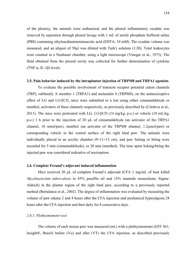
154
of the pleurisy, the animals were euthanized, and the pleural inflammatory exudate was
removed by aspiration through pleural lavage with 1 mL of sterile phosphate buffered saline
(PBS) containing ethylenediaminetetraacetic acid (EDTA; 10 mM). The exudate volume was
measured, and an aliquot of 50μl was diluted with Turk's solution (1:20). Total leukocytes
were counted in a Neubauer chamber, using a light microscope (Vinegar et al., 1973). The
fluid obtained from the pleural cavity was collected for further determination of cytokine
(TNF-α, IL-1β) levels.
2.5. Pain behavior induced by the intraplantar injection of TRPM8 and TRPA1 agonists
To evaluate the possible involvement of transient receptor potential cation channels
(TRP), subfamily A member 1 (TRPA1) and melastatin 8 (TRPM8), on the antinociceptive
effect of LG and LG/βCD, mice were submitted to a test using either cinnamaldehyde or
menthol, activators of these channels respectively, as previously described by (Córdova et al.,
2011). The mice were pretreated with LG, LG/βCD (24 mg/kg, p.o.) or vehicle (10 mL/kg,
p.o.) 1 h prior to the injection of 20 μL of cinnamaldehyde (an activator of the TRPA1
channel, 10 nmol/paw), menthol (an activator of the TRPM8 channel, 1.2μmol/paw) or
corresponding vehicle in the ventral surface of the right hind paw. The animals were
individually placed in an acrylic chamber (9×11×13 cm), and paw licking or biting were
recorded for 5 min (cinnamaldehyde), or 20 min (menthol). The time spent licking/biting the
injected paw was considered indicative of nociception.
2.6. Complete Freund’s adjuvant induced inflammation
Mice received 20 μL of complete Freund’s adjuvant (CFA 1 mg/mL of heat killed
Mycobacterium tuberculosis in 85% paraffin oil and 15% mannide monooleate; Sigma-
Aldrich) in the plantar region of the right hind paw, according to a previously reported
method (Bortalanza et al., 2002). The degree of inflammation was evaluated by measuring the
volume of paw edema 2 and 4 hours after the CFA injection and mechanical hyperalgesia 24
hours after the CFA injection and then daily for 8 consecutive days.
2.6.1. Plethysmometer test
The volume of each mouse paw was measured (mL) with a plethysmometer (EFF 361;
Insight®, Brazil) before (Vo) and after (VT) the CFA injection, as described previously

155
(Winter et al., 1962). The amount of paw swelling was determined for each mouse and data
were represented as paw volume variation (Δ, mL).
2.6.2. Measurement of Mechanical Hypersensitivity
The mice were acclimatized in individual transparent boxes on a wire-mesh platform
to allow access to the ventral surface of the hind paws. Their sensitivity to mechanical
stimulation generated by the gradual increase of pressure of a manual force transducer
(electronic analgesimeter, model EFF-301, Insight®, Brazil), adapted with a polypropylene
tip, was evaluated. This test indicates hyperalgesia by automatically recording the pressure
intensity needed to evoke a flexion reflex of the hind paw, which is defined as a withdrawal of
the paw followed by rapid movements, characterized as flinches (Vivancos et al., 2004).
2.6.3. Thermal hyperalgesia evaluation
For the hot plate test, the mice were placed in the hot plate apparatus (EFF 361;
Insight®, Brazil) maintained at 55°C. The first ipsilateral hind paw flexion reflex was
considered the nociceptive endpoint. The response latency was recorded before the surgery
and at the 10th day post-surgery. The maximum latency (cut-off) was set at 20s to avoid
tissue damage (Lemons and Wiley, 2012; Nucci et al., 2012). To obtain data purely derived
from the treatment, the inhibition values are presented as the difference between the basal
values of vehicle or drug-treated animals and the respective controls.
2.7. Segmental spinal nerve ligation-induced neuropathic pain
Partial sciatic nerve ligation (PSNL) was performed according to the procedure
previously described in mice (Malmberg and Basbaum, 1998). Mice were anaesthetized with
a premixed solution containing ketamine (80 mg/kg, i.p.) and xylazine (10 mg/kg, i.p.). Partial
ligation of the right sciatic nerve was performed by ligating the distal one-third to one-half of
the dorsal portion of the sciatic nerve. In sham-operated mice, the sciatic nerve was exposed
without ligation. The wound was closed and covered with 10% povidone-iodine solution. The
sham-operated mice received only vehicle (10 mL/kg, i.g.), and the PSNL-operated mice were
randomly divided into control and treatment groups, which received vehicle (10 mL/kg, i.g.)
or LG and LG/βCD (24 mg/kg, v.o.), respectively, 7 days after surgery. The mechanical
hyperalgesia response was recorded before surgery (B), immediately before treatment (0 h),
and after treatment (1, 2, 3, 4, 6 and 8 h) to verify the time-course effect of the treatment as

156
described below. To investigate the effects of repeated treatment on the mechanical
hyperalgesia response, LG and LG/βCD (24 mg/kg, i.g.) were administered once daily for 7
consecutive days (from the 8th to 14th day after PSNL), and the effects were examined 1 to 2
h after treatment. After 2 days without treatment, treatment was resumed on the 17th and 18th
days after PSNL to assess the development of LG and LG/βCD tolerance.
2.8. Determination of cytokines levels
The levels of TNF-α, IL-1β or IL-10 in sciatic nerve or lumbar spinal cord segments
(L4 and L6) were determined at 17 days after PLSN. The samples were homogenized in
150µL of phosphate-buffered saline (PBS 50 mM, pH 7.4) containing EDTA (0.3%), nonidet
P 40 (0.5%) and protease inhibitors. The samples were centrifuged at 6000 or 8000 rpm for 10
min at 4 ° C, and the supernatant obtained was used to determine cytokines levels. The
protein concentration was measured using the method of Bradford (Bradford, 1976) with
bovine serum albumin as the standard protein.
TNF-α, IL-1β and IL-10 levels were determined using enzyme-linked immunosorbent
assay (ELISA) kits (eBioscience®) according to the manufacturer’s instructions, and
colorimetric measurements at 450 nm were made using a microplate reader (ASYS®). The
concentration was obtained by interpolation from a standard curve. All results were
expressed as picograms (pg) of cytokine per milligram (mg) of total protein.
2.9. Western blot assay
For the preparation of total protein homogenate, the mice were killed by cervical
dislocation on the 17th day after PNSL, the spinal cord and the sciatic nerve were removed.
For Western blot assay the tissues were homogenized in 150 μl of a lysis solution RIPA
contend 1 mM EDTA, 20 mM Tris–HCl, pH 7,5, 150 mM NaCl, 1 mM EGTA, 1% sodium
deoxycholate, 1% nonidet 40, containing proteases and phosphatases inhibitors. For the
electrophoresis analysis, samples were dissolved in 25% (v/v) of solution containing 40%
glycerol, 5% mercaptoethanol, 50 mM Tris–HCl, pH 6.8 and boiled for 5 min. The protein
concentration was determined by the method of Bradford (1976) using BSA as the standard.
Tissue homogenates (50 μg) were separated by SDS-PAGE and transferred to PVDF
membranes (BioRad) for 16 h at 25 V in transfer buffer (48 Mm Tris, 39 mM glycine, 20%
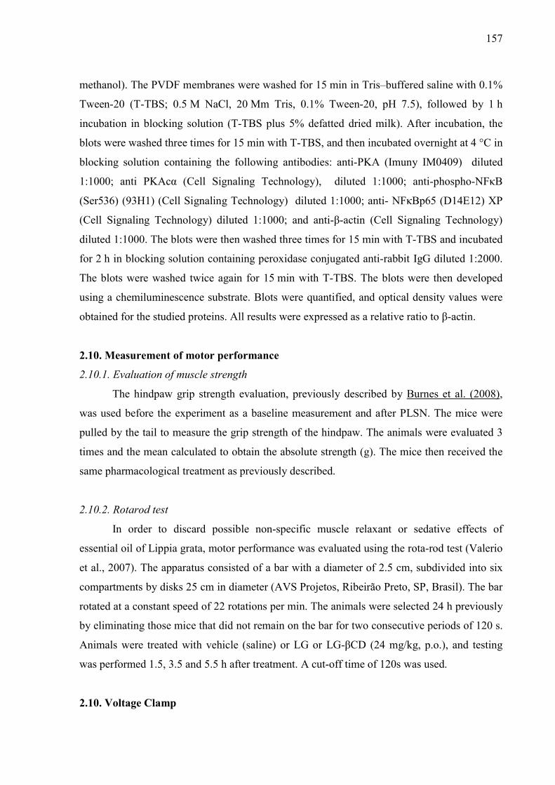
157
methanol). The PVDF membranes were washed for 15 min in Tris–buffered saline with 0.1%
Tween-20 (T-TBS; 0.5 M NaCl, 20 Mm Tris, 0.1% Tween-20, pH 7.5), followed by 1 h
incubation in blocking solution (T-TBS plus 5% defatted dried milk). After incubation, the
blots were washed three times for 15 min with T-TBS, and then incubated overnight at 4 °C in
blocking solution containing the following antibodies: anti-PKA (Imuny IM0409) diluted
1:1000; anti PKAcα (Cell Signaling Technology), diluted 1:1000; anti-phospho-NFκB
(Ser536) (93H1) (Cell Signaling Technology) diluted 1:1000; anti- NFκBp65 (D14E12) XP
(Cell Signaling Technology) diluted 1:1000; and anti-β-actin (Cell Signaling Technology)
diluted 1:1000. The blots were then washed three times for 15 min with T-TBS and incubated
for 2 h in blocking solution containing peroxidase conjugated anti-rabbit IgG diluted 1:2000.
The blots were washed twice again for 15 min with T-TBS. The blots were then developed
using a chemiluminescence substrate. Blots were quantified, and optical density values were
obtained for the studied proteins. All results were expressed as a relative ratio to β-actin.
2.10. Measurement of motor performance
2.10.1. Evaluation of muscle strength
The hindpaw grip strength evaluation, previously described by Burnes et al. (2008),
was used before the experiment as a baseline measurement and after PLSN. The mice were
pulled by the tail to measure the grip strength of the hindpaw. The animals were evaluated 3
times and the mean calculated to obtain the absolute strength (g). The mice then received the
same pharmacological treatment as previously described.
2.10.2. Rotarod test
In order to discard possible non-specific muscle relaxant or sedative effects of
essential oil of Lippia grata, motor performance was evaluated using the rota-rod test (Valerio
et al., 2007). The apparatus consisted of a bar with a diameter of 2.5 cm, subdivided into six
compartments by disks 25 cm in diameter (AVS Projetos, Ribeirão Preto, SP, Brasil). The bar
rotated at a constant speed of 22 rotations per min. The animals were selected 24 h previously
by eliminating those mice that did not remain on the bar for two consecutive periods of 120 s.
Animals were treated with vehicle (saline) or LG or LG-βCD (24 mg/kg, p.o.), and testing
was performed 1.5, 3.5 and 5.5 h after treatment. A cut-off time of 120s was used.
2.10. Voltage Clamp

158
We recorded calcium currents (Ica++) from adult male Wistar rats’ dorsal root
ganglia (DRG) neurons. We used the patch-clamp technique (Hamill et al., 1981) on neurons
that were perfused (1mL/min) and dialyzed with Na+,K+-free solutions containing channel
blockers to isolate calcium currents from other membrane currents. The bath solution
contained (in mM): choline chloride (105), TEA chloride (30), CaCl2 (2), MgCl2 (4) and
HEPES (10) and it was adjusted to pH 7.4 with tetraethylammonium hydrochloride. The
pipette internal solution was (in mM): CsCl (126), MgATP (1), EDTA (1), EGTA (10),
HEPES (10) and it was adjusted to pH 7.2 with CsOH. All experiments were performed at
room temperature.
The cells were kept in a holding potential of −80mV and at every 7 seconds we
applied depolarizing pulses to 0mV for 200ms. The liquid junction potential was corrected to
eliminate the difference of −6mV between the pipette and bath solutions. The sampling rate
was 10 kHz.
Pipettes were prepared from glass capillaries (World Precision Instruments, Sarasota,
USA) pulled and heat-polished (PP 830 puller and MF-830 microforge, Narishige, Tokyo,
Japan) to a tip resistance of 1 to 3 MΩ. Data were acquired using a patch-clamp amplifier
(Multiclamp 700B, Axon Instruments, USA) and a digitizer (Digidata 1200 series, Axon
Instruments, USA) controlled by the pClamp 8.2 software (Axon Instruments, USA).
The LG essential oil was diluted in DMSO (33% v/v) and added to the bath resulting
in the following concentrations: 0.1, 0.3 or 1.0 µL/mL. As control, we used DMSO at its
highest concentration.
We recorded calcium current (ICa) for 1 to 4 minutes, in order to estimate the current
rundown. Once the current reached stability we calculated the percentage of inhibition using
the last control and the first experimental currents. In some cells we performed multiple drug
applications, washing it at different intervals.
2.11. Statistical Analysis
Results were expressed as mean ± S.E.M. The differences between groups were
analyzed using one-way or two-way ANOVA, followed by the Tukey test or Bonferroni test,
respectively. Values of p <0.05 were considered statistically significant. All statistical
analyzes were performed using GraphPad Prism 5.0 software (GraphPad Prism Software Inc.,
San Diego, CA, USA). The dose response curves of Ica++ inhibition were fitted to a Hill
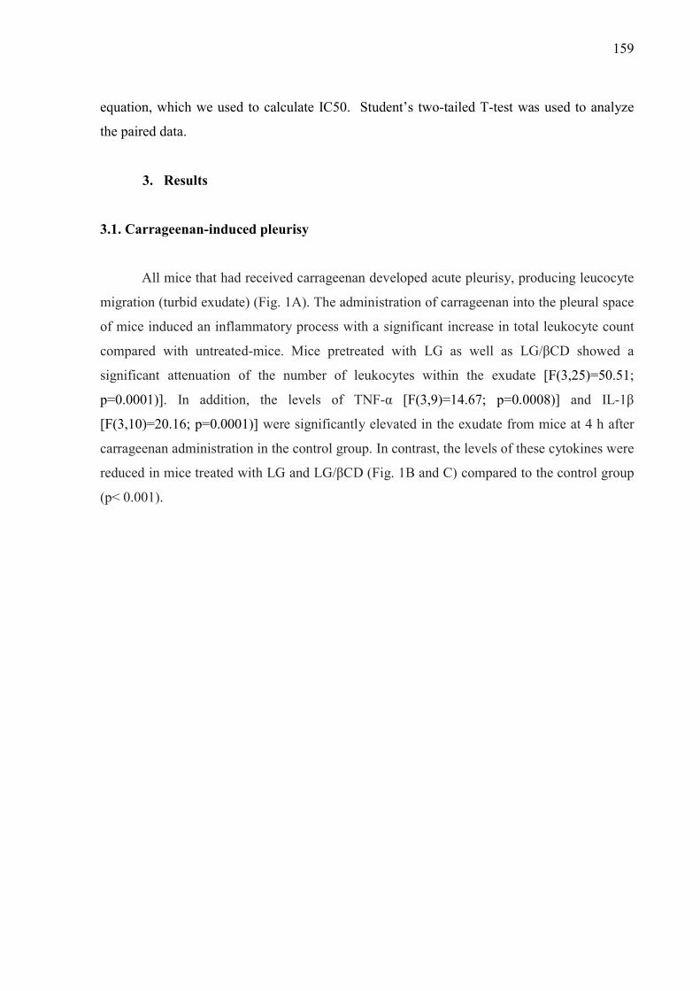
159
equation, which we used to calculate IC50. Student’s two-tailed T-test was used to analyze
the paired data.
3. Results
3.1. Carrageenan-induced pleurisy
All mice that had received carrageenan developed acute pleurisy, producing leucocyte
migration (turbid exudate) (Fig. 1A). The administration of carrageenan into the pleural space
of mice induced an inflammatory process with a significant increase in total leukocyte count
compared with untreated-mice. Mice pretreated with LG as well as LG/βCD showed a
significant attenuation of the number of leukocytes within the exudate [F(3,25)=50.51;
p=0.0001)]. In addition, the levels of TNF-α [F(3,9)=14.67; p=0.0008)] and IL-1β
[F(3,10)=20.16; p=0.0001)] were significantly elevated in the exudate from mice at 4 h after
carrageenan administration in the control group. In contrast, the levels of these cytokines were
reduced in mice treated with LG and LG/βCD (Fig. 1B and C) compared to the control group
(p< 0.001).
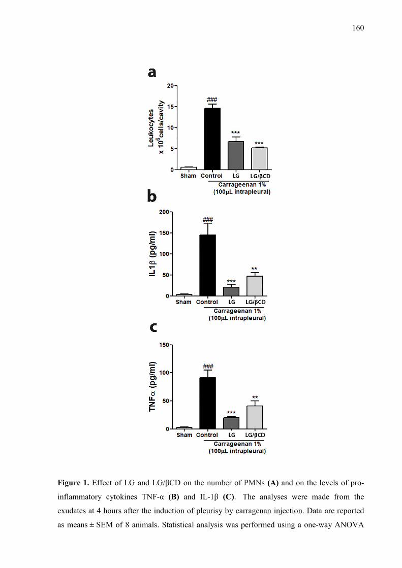
160
Figure 1. Effect of LG and LG/βCD on the number of PMNs (A) and on the levels of pro-
inflammatory cytokines TNF-α (B) and IL-1β (C). The analyses were made from the
exudates at 4 hours after the induction of pleurisy by carragenan injection. Data are reported
as means ± SEM of 8 animals. Statistical analysis was performed using a one-way ANOVA
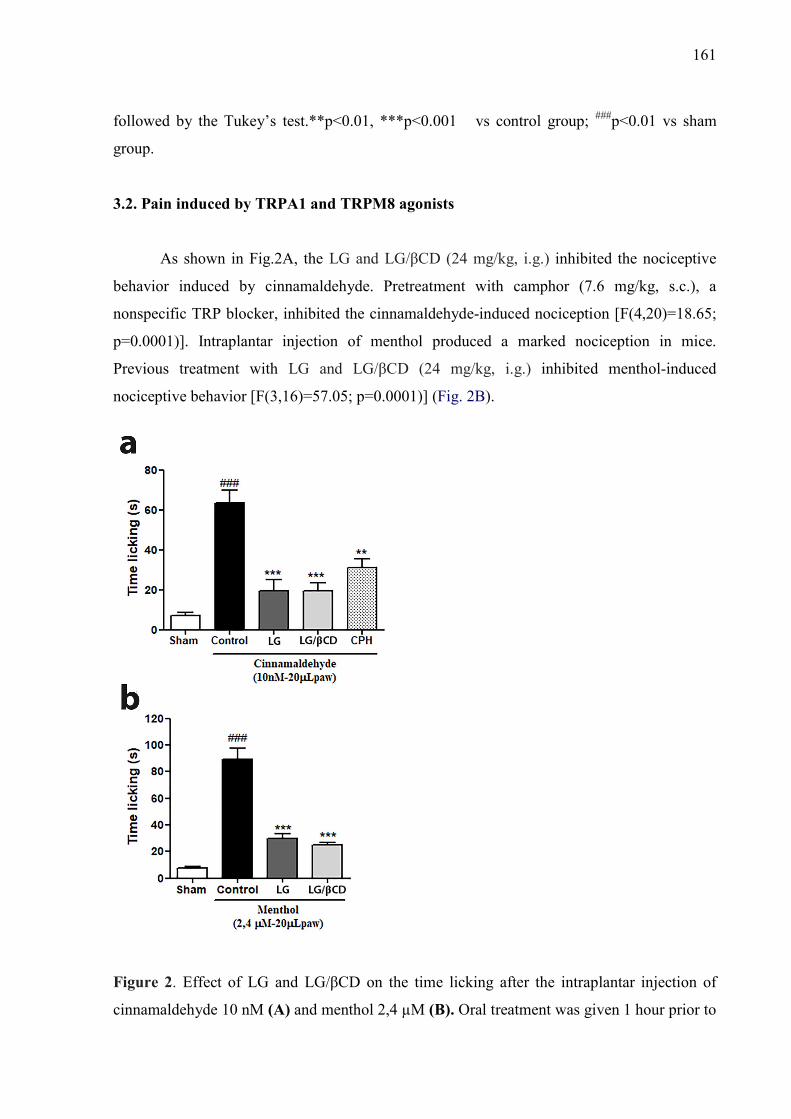
161
followed by the Tukey’s test.**p<0.01, ***p<0.001 vs control group; ###p<0.01 vs sham
group.
3.2. Pain induced by TRPA1 and TRPM8 agonists
As shown in Fig.2A, the LG and LG/βCD (24 mg/kg, i.g.) inhibited the nociceptive
behavior induced by cinnamaldehyde. Pretreatment with camphor (7.6 mg/kg, s.c.), a
nonspecific TRP blocker, inhibited the cinnamaldehyde-induced nociception [F(4,20)=18.65;
p=0.0001)]. Intraplantar injection of menthol produced a marked nociception in mice.
Previous treatment with LG and LG/βCD (24 mg/kg, i.g.) inhibited menthol-induced
nociceptive behavior [F(3,16)=57.05; p=0.0001)] (Fig. 2B).
Figure 2. Effect of LG and LG/βCD on the time licking after the intraplantar injection of
cinnamaldehyde 10 nM (A) and menthol 2,4 µM (B). Oral treatment was given 1 hour prior to
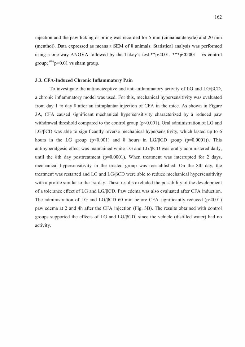
162
injection and the paw licking or biting was recorded for 5 min (cinnamaldehyde) and 20 min
(menthol). Data expressed as means ± SEM of 8 animals. Statistical analysis was performed
using a one-way ANOVA followed by the Tukey’s test.**p<0.01, ***p<0.001 vs control
group; ###p<0.01 vs sham group.
3.3. CFA-Induced Chronic Inflammatory Pain
To investigate the antinociceptive and anti-inflammatory activity of LG and LG/βCD,
a chronic inflammatory model was used. For this, mechanical hypersensitivity was evaluated
from day 1 to day 8 after an intraplantar injection of CFA in the mice. As shown in Figure
3A, CFA caused significant mechanical hypersensitivity characterized by a reduced paw
withdrawal threshold compared to the control group (p<0.001). Oral administration of LG and
LG/βCD was able to significantly reverse mechanical hypersensitivity, which lasted up to 6
hours in the LG group (p<0.001) and 8 hours in LG/βCD group (p=0.0001)). This
antihyperalgesic effect was maintained while LG and LG/βCD was orally administered daily,
until the 8th day posttreatment (p=0.0001). When treatment was interrupted for 2 days,
mechanical hypersensitivity in the treated group was reestablished. On the 8th day, the
treatment was restarted and LG and LG/βCD were able to reduce mechanical hypersensitivity
with a profile similar to the 1st day. These results excluded the possibility of the development
of a tolerance effect of LG and LG/βCD. Paw edema was also evaluated after CFA induction.
The administration of LG and LG/βCD 60 min before CFA significantly reduced (p<0.01)
paw edema at 2 and 4h after the CFA injection (Fig. 3B). The results obtained with control
groups supported the effects of LG and LG/βCD, since the vehicle (distilled water) had no
activity.

163
Figure. 3. Effect of LG and LG/βCD on (A) mechanical hyperalgesia 24 hours after the CFA
injection and daily for 8 consecutive days; and on (B) paw edema 2 and 4 hours after the CFA
injection. Data are reported as means ± SEM of 8-10 animals. Statistically significant
differences from sham and control group as determined by One-way ANOVA followed by
Bonferroni (A) and Tukey’s test (B). *p<0.05**p<0.01, ***p<0.001 vs control group; #p<0.05, ###p<0.01 vs sham group.
3.4. Segmental spinal nerve ligation-induced neuropathic pain
Partial sciatic nerve ligation produced a marked and long-lasting development of
hyperalgesia on the ipsilateral side eight days after PSNL in the control group, and in the
experimental groups treated with LG and LG/βCD (24 mg/kg, i.g.). The group that underwent
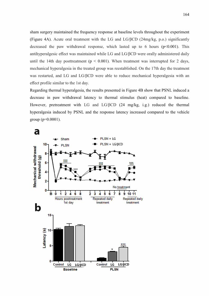
164
sham surgery maintained the frequency response at baseline levels throughout the experiment
(Figure 4A). Acute oral treatment with the LG and LG/βCD (24mg/kg, p.o.) significantly
decreased the paw withdrawal response, which lasted up to 6 hours (p<0.001). This
antihyperalgesic effect was maintained while LG and LG/βCD were orally administered daily
until the 14th day posttreatment (p < 0.001). When treatment was interrupted for 2 days,
mechanical hyperalgesia in the treated group was reestablished. On the 17th day the treatment
was restarted, and LG and LG/βCD were able to reduce mechanical hyperalgesia with an
effect profile similar to the 1st day.
Regarding thermal hyperalgesia, the results presented in Figure 4B show that PSNL induced a
decrease in paw withdrawal latency to thermal stimulus (heat) compared to baseline.
However, pretreatment with LG and LG/βCD (24 mg/kg, i.g.) reduced the thermal
hyperalgesia induced by PSNL and the response latency increased compared to the vehicle
group (p=0.0001).
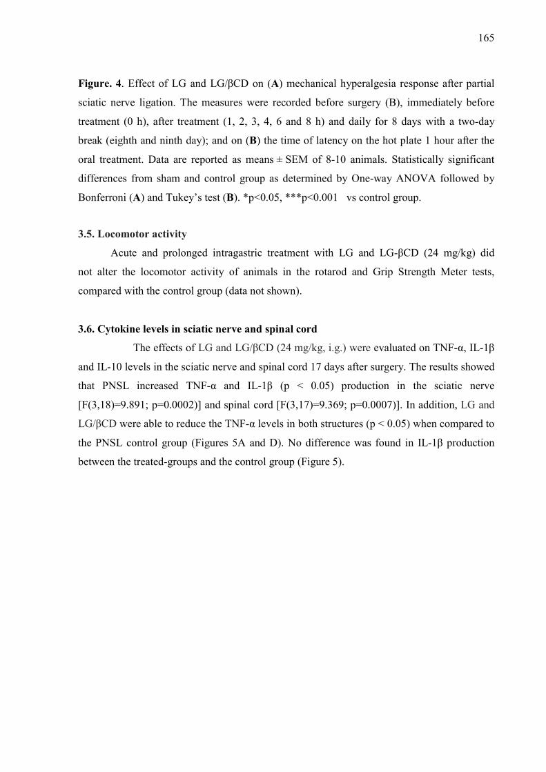
165
Figure. 4. Effect of LG and LG/βCD on (A) mechanical hyperalgesia response after partial
sciatic nerve ligation. The measures were recorded before surgery (B), immediately before
treatment (0 h), after treatment (1, 2, 3, 4, 6 and 8 h) and daily for 8 days with a two-day
break (eighth and ninth day); and on (B) the time of latency on the hot plate 1 hour after the
oral treatment. Data are reported as means ± SEM of 8-10 animals. Statistically significant
differences from sham and control group as determined by One-way ANOVA followed by
Bonferroni (A) and Tukey’s test (B). *p<0.05, ***p<0.001 vs control group.
3.5. Locomotor activity
Acute and prolonged intragastric treatment with LG and LG-βCD (24 mg/kg) did
not alter the locomotor activity of animals in the rotarod and Grip Strength Meter tests,
compared with the control group (data not shown).
3.6. Cytokine levels in sciatic nerve and spinal cord
The effects of LG and LG/βCD (24 mg/kg, i.g.) were evaluated on TNF-α, IL-1β
and IL-10 levels in the sciatic nerve and spinal cord 17 days after surgery. The results showed
that PNSL increased TNF-α and IL-1β (p < 0.05) production in the sciatic nerve
[F(3,18)=9.891; p=0.0002)] and spinal cord [F(3,17)=9.369; p=0.0007)]. In addition, LG and
LG/βCD were able to reduce the TNF-α levels in both structures (p < 0.05) when compared to
the PNSL control group (Figures 5A and D). No difference was found in IL-1β production
between the treated-groups and the control group (Figure 5).
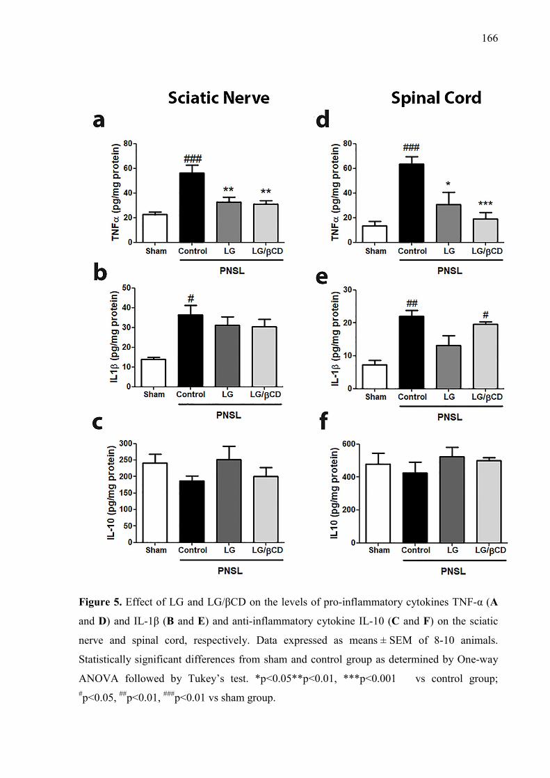
166
Figure 5. Effect of LG and LG/βCD on the levels of pro-inflammatory cytokines TNF-α (A
and D) and IL-1β (B and E) and anti-inflammatory cytokine IL-10 (C and F) on the sciatic
nerve and spinal cord, respectively. Data expressed as means ± SEM of 8-10 animals.
Statistically significant differences from sham and control group as determined by One-way
ANOVA followed by Tukey’s test. *p<0.05**p<0.01, ***p<0.001 vs control group; #p<0.05, ##p<0.01, ###p<0.01 vs sham group.
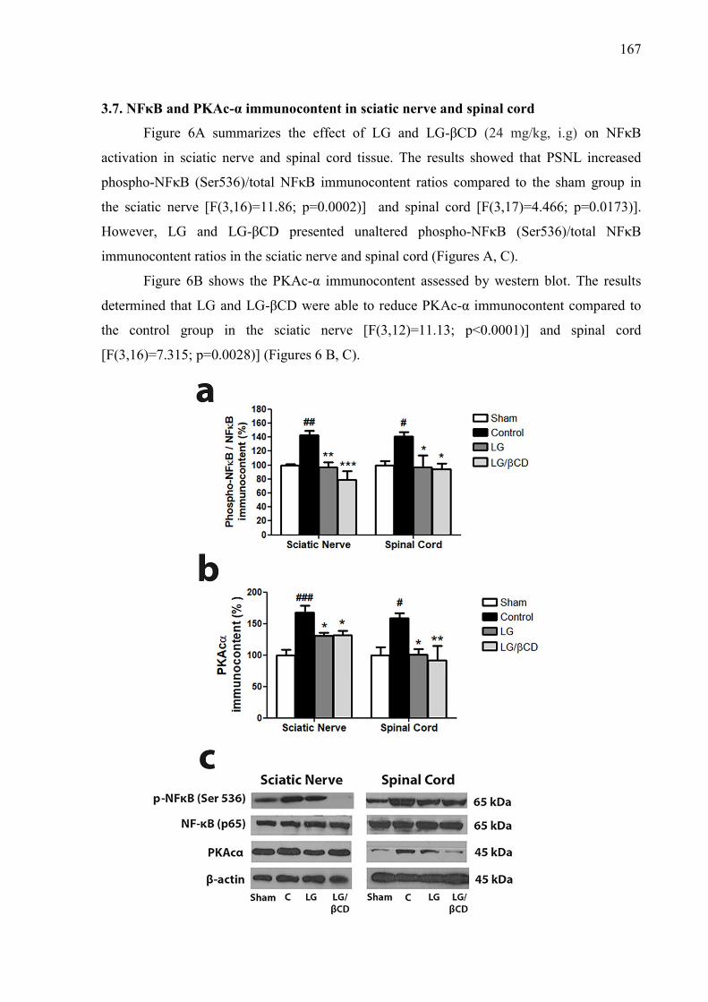
167
3.7. NFκB and PKAc-α immunocontent in sciatic nerve and spinal cord
Figure 6A summarizes the effect of LG and LG-βCD (24 mg/kg, i.g) on NFκB
activation in sciatic nerve and spinal cord tissue. The results showed that PSNL increased
phospho-NFκB (Ser536)/total NFκB immunocontent ratios compared to the sham group in
the sciatic nerve [F(3,16)=11.86; p=0.0002)] and spinal cord [F(3,17)=4.466; p=0.0173)].
However, LG and LG-βCD presented unaltered phospho-NFκB (Ser536)/total NFκB
immunocontent ratios in the sciatic nerve and spinal cord (Figures A, C).
Figure 6B shows the PKAc-α immunocontent assessed by western blot. The results
determined that LG and LG-βCD were able to reduce PKAc-α immunocontent compared to
the control group in the sciatic nerve [F(3,12)=11.13; p<0.0001)] and spinal cord
[F(3,16)=7.315; p=0.0028)] (Figures 6 B, C).
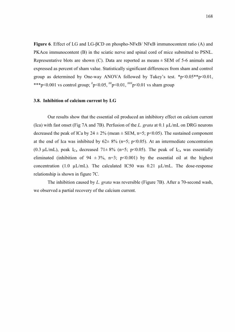
168
Figure 6. Effect of LG and LG-βCD on phospho-NFκB/ NFκB immunocontent ratio (A) and
PKAcα immunocontent (B) in the sciatic nerve and spinal cord of mice submitted to PSNL.
Representative blots are shown (C). Data are reported as means ± SEM of 5-6 animals and
expressed as percent of sham value. Statistically significant differences from sham and control
group as determined by One-way ANOVA followed by Tukey’s test. *p<0.05**p<0.01,
***p<0.001 vs control group; #p<0.05, ##p<0.01, ###p<0.01 vs sham group
3.8. Inhibition of calcium current by LG
Our results show that the essential oil produced an inhibitory effect on calcium current
(Ica) with fast onset (Fig 7A and 7B). Perfusion of the L. grata at 0.1 µL/mL on DRG neurons
decreased the peak of ICa by 24 ± 2% (mean ± SEM, n=5; p<0.05). The sustained component
at the end of Ica was inhibited by 62± 8% (n=5; p<0.05). At an intermediate concentration
(0.3 µL/mL), peak ICa decreased 71± 8% (n=5; p<0.05). The peak of ICa was essentially
eliminated (inhibition of 94 ± 3%, n=3; p<0.001) by the essential oil at the highest
concentration (1.0 µL/mL). The calculated IC50 was 0.21 µL/mL. The dose-response
relationship is shown in figure 7C.
The inhibition caused by L. grata was reversible (Figure 7B). After a 70-second wash,
we observed a partial recovery of the calcium current.
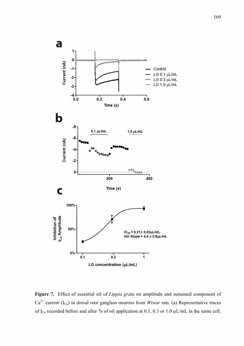
169
Figure 7. Effect of essential oil of Lippia grata on amplitude and sustained component of
Ca2+ current (ICa) in dorsal root ganglion neurons from Wistar rats. (a) Representative traces
of ICa recorded before and after 7s of oil application at 0.1, 0.3 or 1.0 µL/mL in the same cell.
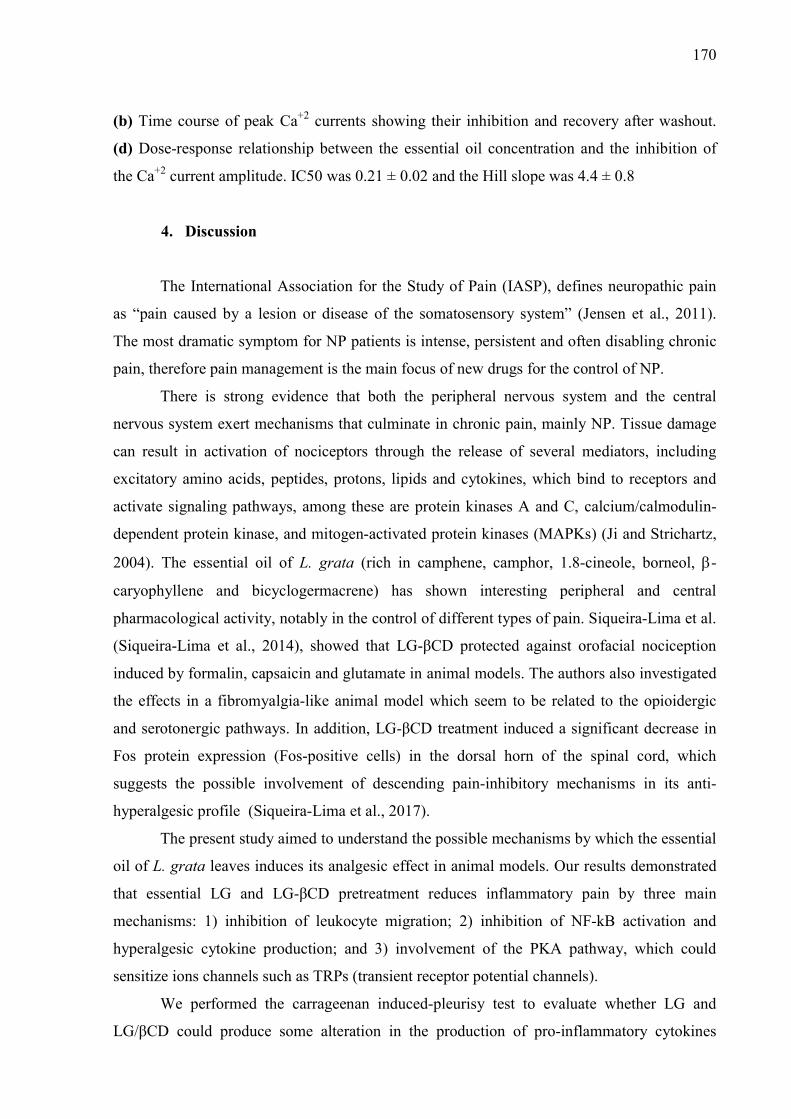
170
(b) Time course of peak Ca+2 currents showing their inhibition and recovery after washout.
(d) Dose-response relationship between the essential oil concentration and the inhibition of
the Ca+2 current amplitude. IC50 was 0.21 ± 0.02 and the Hill slope was 4.4 ± 0.8
4. Discussion
The International Association for the Study of Pain (IASP), defines neuropathic pain
as “pain caused by a lesion or disease of the somatosensory system” (Jensen et al., 2011).
The most dramatic symptom for NP patients is intense, persistent and often disabling chronic
pain, therefore pain management is the main focus of new drugs for the control of NP.
There is strong evidence that both the peripheral nervous system and the central
nervous system exert mechanisms that culminate in chronic pain, mainly NP. Tissue damage
can result in activation of nociceptors through the release of several mediators, including
excitatory amino acids, peptides, protons, lipids and cytokines, which bind to receptors and
activate signaling pathways, among these are protein kinases A and C, calcium/calmodulin-
dependent protein kinase, and mitogen-activated protein kinases (MAPKs) (Ji and Strichartz,
2004). The essential oil of L. grata (rich in camphene, camphor, 1.8-cineole, borneol, -
caryophyllene and bicyclogermacrene) has shown interesting peripheral and central
pharmacological activity, notably in the control of different types of pain. Siqueira-Lima et al.
(Siqueira-Lima et al., 2014), showed that LG-βCD protected against orofacial nociception
induced by formalin, capsaicin and glutamate in animal models. The authors also investigated
the effects in a fibromyalgia-like animal model which seem to be related to the opioidergic
and serotonergic pathways. In addition, LG-βCD treatment induced a significant decrease in
Fos protein expression (Fos-positive cells) in the dorsal horn of the spinal cord, which
suggests the possible involvement of descending pain-inhibitory mechanisms in its anti-
hyperalgesic profile (Siqueira-Lima et al., 2017).
The present study aimed to understand the possible mechanisms by which the essential
oil of L. grata leaves induces its analgesic effect in animal models. Our results demonstrated
that essential LG and LG-βCD pretreatment reduces inflammatory pain by three main
mechanisms: 1) inhibition of leukocyte migration; 2) inhibition of NF-kB activation and
hyperalgesic cytokine production; and 3) involvement of the PKA pathway, which could
sensitize ions channels such as TRPs (transient receptor potential channels).
We performed the carrageenan induced-pleurisy test to evaluate whether LG and
LG/βCD could produce some alteration in the production of pro-inflammatory cytokines
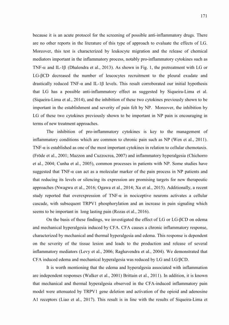
171
because it is an acute protocol for the screening of possible anti-inflammatory drugs. There
are no other reports in the literature of this type of approach to evaluate the effects of LG.
Moreover, this test is characterized by leukocyte migration and the release of chemical
mediators important in the inflammatory process, notably pro-inflammatory cytokines such as
TNF- and IL-1 (Dhalendra et al., 2013). As shown in Fig. 1, the pretreatment with LG or
LG-βCD decreased the number of leucocytes recruitment to the pleural exudate and
drastically reduced TNF-α and IL-1β levels. This result corroborated our initial hypothesis
that LG has a possible anti-inflammatory effect as suggested by Siqueira-Lima et al.
(Siqueira-Lima et al., 2014), and the inhibition of these two cytokines previously shown to be
important in the establishment and severity of pain felt by NP. Moreover, the inhibition by
LG of these two cytokines previously shown to be important in NP pain is encouraging in
terms of new treatment approaches.
The inhibition of pro-inflammatory cytokines is key to the management of
inflammatory conditions which are common to chronic pain such as NP (Wen et al., 2011).
TNF-α is established as one of the most important cytokines in relation to cellular chemotaxis.
(Fröde et al., 2001; Mazzon and Cuzzocrea, 2007) and inflammatory hyperalgesia (Chichorro
et al., 2004; Cunha et al., 2005), common processes in patients with NP. Some studies have
suggested that TNF-α can act as a molecular marker of the pain process in NP patients and
that reducing its levels or silencing its expression are promising targets for new therapeutic
approaches (Nwagwu et al., 2016; Ogawa et al., 2014; Xu et al., 2015). Additionally, a recent
study reported that overexpression of TNF-α in nociceptive neurons activates a cellular
cascade, with subsequent TRPV1 phosphorylation and an increase in pain signaling which
seems to be important in long lasting pain (Rozas et al., 2016).
On the basis of these findings, we investigated the effect of LG or LG-βCD on edema
and mechanical hyperalgesia induced by CFA. CFA causes a chronic inflammatory response,
characterized by mechanical and thermal hyperalgesia and edema. This response is dependent
on the severity of the tissue lesion and leads to the production and release of several
inflammatory mediators (Levy et al., 2006; Raghavendra et al., 2004). We demonstrated that
CFA induced edema and mechanical hyperalgesia was reduced by LG and LG/βCD.
It is worth mentioning that the edema and hyperalgesia associated with inflammation
are independent responses (Walker et al., 2001) Brittain et al., 2011). In addition, it is known
that mechanical and thermal hyperalgesia observed in the CFA-induced inflammatory pain
model were attenuated by TRPV1 gene deletion and activation of the opioid and adenosine
A1 receptors (Liao et al., 2017). This result is in line with the results of Siqueira-Lima et
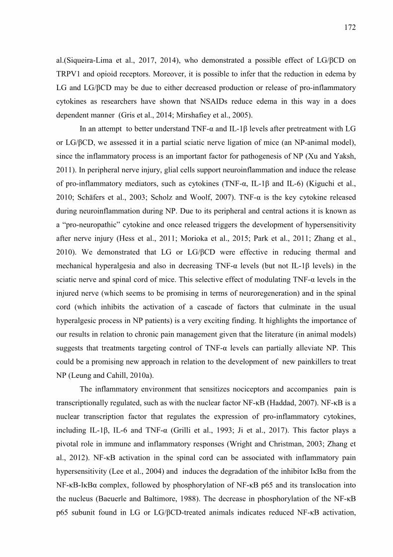
172
al.(Siqueira-Lima et al., 2017, 2014), who demonstrated a possible effect of LG/βCD on
TRPV1 and opioid receptors. Moreover, it is possible to infer that the reduction in edema by
LG and LG/βCD may be due to either decreased production or release of pro-inflammatory
cytokines as researchers have shown that NSAIDs reduce edema in this way in a does
dependent manner (Gris et al., 2014; Mirshafiey et al., 2005).
In an attempt to better understand TNF-α and IL-1β levels after pretreatment with LG
or LG/βCD, we assessed it in a partial sciatic nerve ligation of mice (an NP-animal model),
since the inflammatory process is an important factor for pathogenesis of NP (Xu and Yaksh,
2011). In peripheral nerve injury, glial cells support neuroinflammation and induce the release
of pro-inflammatory mediators, such as cytokines (TNF-α, IL-1β and IL-6) (Kiguchi et al.,
2010; Schäfers et al., 2003; Scholz and Woolf, 2007). TNF-α is the key cytokine released
during neuroinflammation during NP. Due to its peripheral and central actions it is known as
a “pro-neuropathic” cytokine and once released triggers the development of hypersensitivity
after nerve injury (Hess et al., 2011; Morioka et al., 2015; Park et al., 2011; Zhang et al.,
2010). We demonstrated that LG or LG/βCD were effective in reducing thermal and
mechanical hyperalgesia and also in decreasing TNF-α levels (but not IL-1β levels) in the
sciatic nerve and spinal cord of mice. This selective effect of modulating TNF-α levels in the
injured nerve (which seems to be promising in terms of neuroregeneration) and in the spinal
cord (which inhibits the activation of a cascade of factors that culminate in the usual
hyperalgesic process in NP patients) is a very exciting finding. It highlights the importance of
our results in relation to chronic pain management given that the literature (in animal models)
suggests that treatments targeting control of TNF-α levels can partially alleviate NP. This
could be a promising new approach in relation to the development of new painkillers to treat
NP (Leung and Cahill, 2010a).
The inflammatory environment that sensitizes nociceptors and accompanies pain is
transcriptionally regulated, such as with the nuclear factor NF-κB (Haddad, 2007). NF-κB is a
nuclear transcription factor that regulates the expression of pro-inflammatory cytokines,
including IL-1β, IL-6 and TNF-α (Grilli et al., 1993; Ji et al., 2017). This factor plays a
pivotal role in immune and inflammatory responses (Wright and Christman, 2003; Zhang et
al., 2012). NF-κB activation in the spinal cord can be associated with inflammatory pain
hypersensitivity (Lee et al., 2004) and induces the degradation of the inhibitor IκBα from the
NF-κB-IκBα complex, followed by phosphorylation of NF-κB p65 and its translocation into
the nucleus (Baeuerle and Baltimore, 1988). The decrease in phosphorylation of the NF-κB
p65 subunit found in LG or LG/βCD-treated animals indicates reduced NF-κB activation,
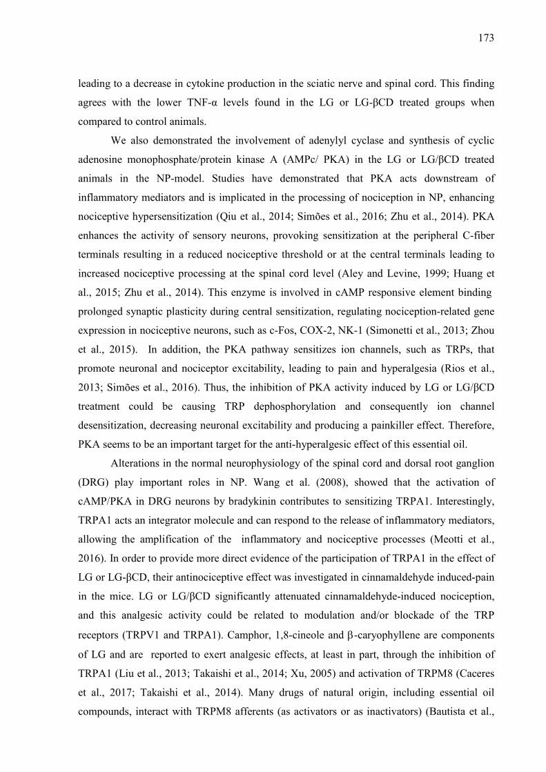
173
leading to a decrease in cytokine production in the sciatic nerve and spinal cord. This finding
agrees with the lower TNF-α levels found in the LG or LG-βCD treated groups when
compared to control animals.
We also demonstrated the involvement of adenylyl cyclase and synthesis of cyclic
adenosine monophosphate/protein kinase A (AMPc/ PKA) in the LG or LG/βCD treated
animals in the NP-model. Studies have demonstrated that PKA acts downstream of
inflammatory mediators and is implicated in the processing of nociception in NP, enhancing
nociceptive hypersensitization (Qiu et al., 2014; Simões et al., 2016; Zhu et al., 2014). PKA
enhances the activity of sensory neurons, provoking sensitization at the peripheral C-fiber
terminals resulting in a reduced nociceptive threshold or at the central terminals leading to
increased nociceptive processing at the spinal cord level (Aley and Levine, 1999; Huang et
al., 2015; Zhu et al., 2014). This enzyme is involved in cAMP responsive element binding
prolonged synaptic plasticity during central sensitization, regulating nociception-related gene
expression in nociceptive neurons, such as c-Fos, COX-2, NK-1 (Simonetti et al., 2013; Zhou
et al., 2015). In addition, the PKA pathway sensitizes ion channels, such as TRPs, that
promote neuronal and nociceptor excitability, leading to pain and hyperalgesia (Rios et al.,
2013; Simões et al., 2016). Thus, the inhibition of PKA activity induced by LG or LG/βCD
treatment could be causing TRP dephosphorylation and consequently ion channel
desensitization, decreasing neuronal excitability and producing a painkiller effect. Therefore,
PKA seems to be an important target for the anti-hyperalgesic effect of this essential oil.
Alterations in the normal neurophysiology of the spinal cord and dorsal root ganglion
(DRG) play important roles in NP. Wang et al. (2008), showed that the activation of
cAMP/PKA in DRG neurons by bradykinin contributes to sensitizing TRPA1. Interestingly,
TRPA1 acts an integrator molecule and can respond to the release of inflammatory mediators,
allowing the amplification of the inflammatory and nociceptive processes (Meotti et al.,
2016). In order to provide more direct evidence of the participation of TRPA1 in the effect of
LG or LG-βCD, their antinociceptive effect was investigated in cinnamaldehyde induced-pain
in the mice. LG or LG/βCD significantly attenuated cinnamaldehyde-induced nociception,
and this analgesic activity could be related to modulation and/or blockade of the TRP
receptors (TRPV1 and TRPA1). Camphor, 1,8-cineole and -caryophyllene are components
of LG and are reported to exert analgesic effects, at least in part, through the inhibition of
TRPA1 (Liu et al., 2013; Takaishi et al., 2014; Xu, 2005) and activation of TRPM8 (Caceres
et al., 2017; Takaishi et al., 2014). Many drugs of natural origin, including essential oil
compounds, interact with TRPM8 afferents (as activators or as inactivators) (Bautista et al.,
174
2007; Calixto et al., 2005; Julius, 2013). Our results are not clear enough to prove the
involvement of LG and LG-CD with TRPM8, however, the compounds present in LG such as
camphor and 1,8-cineole could possibly be acting on this receptor, thereby preventing its
activation.
The expression of TNF- in the spinal cord and DRG has been implicated in NP
establishment, where the over expression of this cytokine is upregulated in satellite cells of
the DRG after peripheral nerve injury (Leung and Cahill, 2010b). The mechanical
hyperalgesia induced by spinal nerve injury is associated with upregulation of satellite cells
and TNF- in the contralateral DRG (Hatashita et al., 2008; Ohtori et al., 2004). Therefore,
the overexpression of TNF-α in the sensory neurons increases the activity of TRPV1-
dependent Ca2+ influx, resulting in inflammatory sensitization and pain (Rozas et al., 2016).
The terpenes (camphor, 1.8-cineole and borneol) present in LG are compounds that
commonly act on the TRPA1 receptors and also modulate TNF-, thereby ameliorating pain
(Takaishi et al., 2014; Xu, 2005). Thus, the consistent inhibitory effect on the Ca2+ current of
DRG neurons produced by LG reinforces the hypothesis that Ca2+ and TRP receptors are
involved in the pharmacological mechanism of LG and that its major components may be
acting synergistically in relation to this.
We compared the biologic effects of pure LG (non-βCD-complexed) and LG/βCD for
the first time in “in vivo” protocols and found a significant analgesic effect, although the
mechanism of action is not yet clear. However, pure LG has low water solubility (data not
shown) and showed reduced effectiveness in the animal models we used that could limit its
clinical use, particularly in chronic conditions. However, cyclodextrins (CDs) have been
demonstrated to be a useful tool to improve a number of chemical and pharmacological
properties of non-polar compounds (Brito et al., 2015; Quintans et al., 2013; Siqueira-Lima et
al., 2017). Our findings showed that the use of the inclusion complex improved the analgesic
effect of LG, maintaining its action for 2 hours longer than pure LG. Although LG and
LG/βCD were used at the same dose (24 mg/kg) it is noteworthy that LG/βCD was prepared
using a molar concentration of 1:1, so the effective dose of LG present in the βCD-complex is
lower when compared to LG alone. This fact is extremely important because it means the
pharmacological effect was obtained with a smaller dose of LG.

175
5. Conclusion
The essential oil from L. grata leaf exerts an outstanding anti-hyperalgesic effect on pain of
different origins and with equally diverse pharmacological mechanisms. These results are in
line with the use of this medicinal plant in folk medicine for pain relief, and the complexation
of its essential oil in βCD was shown to increase its effectiveness. The antihyperalgesic effect
of LG is related to its anti-inflammatory profile (especially in mitigating pro-inflammatory
cytokines), with the involvement of the PKA pathway and also the inhibition of voltage Ca2+
current. Moreover, the inhibition of TNF-α appears to be key to its analgesic profile. Thus,
considering the limited number of drugs currently available for the treatment of NP, our
results provide a rationale for the development of interventions targeting the study of LG as a
potential new tool for chronic pain management in patients with NP, which could lead to
better treatments for those affected by this chronic pain in the future.
6. Acknowledgements
This study was financed in part by the Conselho Conselho Nacional de
Desenvolvimento Científico e Tecnológico – Brasil (CNPq), the Fundação de Apoio à
Pesquisa e a Inovação Tecnológica do Estado de Sergipe (Fapitec/SE) - Brasil, the
Coordenação de Aperfeiçoamento de Pessoal de Nível Superior - Brasil (CAPES - Finance
Code 001), and the Financiadora de Estudos e Projetos - Brasil (FINEP), all from Brazil.
Declarations of interest: none
Conflict of Interest: The authors declare that they have no conflict of interest.

176
References
Aley, K.O., Levine, J.D., 1999. Role of protein kinase A in the maintenance of inflammatory pain. J. Neurosci. 19, 2181–6.
Baeuerle, P.A., Baltimore, D., 1988. I kappa B: a specific inhibitor of the NF-kappa B transcription factor. Science (80-. ). 242, 540–546. https://doi.org/10.1126/science.3140380
Bautista, D.M., Siemens, J., Glazer, J.M., Tsuruda, P.R., Basbaum, A.I., Stucky, C.L., Jordt, S.E., Julius, D., 2007. The menthol receptor TRPM8 is the principal detector of environmental cold. Nature. https://doi.org/10.1038/nature05910
Bortalanza, L.B., Ferreira, J., Hess, S.C., Delle Monache, F., Yunes, R.A., Calixto, J.B., 2002. Anti-allodynic action of the tormentic acid, a triterpene isolated from plant, against neuropathic and inflammatory persistent pain in mice. Eur. J. Pharmacol. 453, 203–208. https://doi.org/10.1016/S0014-2999(02)02428-7
Bradford, M.M., 1976. A rapid and sensitive method for the quantitation of microgram quantities of protein utilizing the principle of protein-dye binding. Anal. Biochem. https://doi.org/10.1016/0003-2697(76)90527-3
Brito, R.G., Araújo, A.A., Quintans, J.S., Sluka, K.A., Quintans-Júnior, L.J., 2015. Enhanced analgesic activity by cyclodextrins – a systematic review and meta-analysis. Expert Opin. Drug Deliv. https://doi.org/10.1517/17425247.2015.1046835
Caceres, A.I., Liu, B., Jabba, S. V., Achanta, S., Morris, J.B., Jordt, S.E., 2017. Transient Receptor Potential Cation Channel Subfamily M Member 8 channels mediate the anti-inflammatory effects of eucalyptol. Br. J. Pharmacol. 174, 867–879. https://doi.org/10.1111/bph.13760
Calixto, J.B., Campos, M.M., Otuki, M.F., Santos, A.R.S., 2004. Anti-inflammatory compounds of plant origin. Part II. Modulation of pro-inflammatory cytokines, chemokines and adhesion molecules. Planta Med. 70, 93–103. https://doi.org/10.1055/s-2004-815483
Calixto, J.B., Kassuya, C.A.L., André, E., Ferreira, J., 2005. Contribution of natural products to the discovery of the transient receptor potential (TRP) channels family and their functions. Pharmacol. Ther. https://doi.org/10.1016/j.pharmthera.2004.11.008
Chichorro, J.G., Lorenzetti, B.B., Zampronio, A.R., 2004. Involvement of bradykinin, cytokines, sympathetic amines and prostaglandins in formalin-induced orofacial nociception in rats. Br. J. Pharmacol. 141, 1175–1184. https://doi.org/10.1038/sj.bjp.0705724
Cohen, S.P., Mao, J., 2014. Neuropathic pain: mechanisms and their clinical implications. BMJ 348, f7656–f7656. https://doi.org/10.1136/bmj.f7656
Córdova, M.M., Werner, M.F. de P., Silva, M.D. da, Ruani, A.P., Pizzolatti, M.G., Santos, A.R.S., 2011. Further antinociceptive effects of myricitrin in chemical models of overt nociception in mice. Neurosci. Lett. 495, 173–177. https://doi.org/10.1016/j.neulet.2011.02.007
Costigan, M., Scholz, J., Woolf, C.J., 2009. Neuropathic Pain: A Maladaptive Response of the Nervous System to Damage. Annu. Rev. Neurosci. 32, 1–32. https://doi.org/10.1146/annurev.neuro.051508.135531
Craveiro, A.A., Alencar, J.W., Matos, F.J.A., Andrade, C.H.S., Machado, M.I.L., 1981. Essential oils from Brazilian Verbenaceae. Genus Lippia. J. Nat. Prod. 44, 598–601. https://doi.org/10.1021/np50017a018
Cunha, T.M., Verri, W.A., Silva, J.S., Poole, S., Cunha, F.Q., Ferreira, S.H., 2005. A cascade of cytokines mediates mechanical inflammatory hypernociception in mice. Proc. Natl. Acad. Sci. U. S. A. 102, 1755–60. https://doi.org/10.1073/pnas.0409225102
de Oliveira, A.M., Conserva, L.M., de Souza Ferro, J.N., de Almeida Brito, F., Lyra Lemos,

177
R.P., Barreto, E., 2012. Antinociceptive and anti-inflammatory effects of octacosanol from the leaves of Sabicea grisea var. grisea in mice. Int. J. Mol. Sci. https://doi.org/10.3390/ijms13021598
De Sousa, D.P., 2011. Analgesic-like activity of essential oils constituents. Molecules 16, 2233–2252. https://doi.org/10.3390/molecules16032233
Dhalendra, G., Satapathy, T., Roy, A., 2013. Animal Models for Inflammation: A Review. Asian J. Pharm. Res 3, 207–212. https://doi.org/papers3://publication/uuid/D623F3B6-9E8E-4EA0-8714-8EE5E2D3E9DD
Fornasari, D., 2017. Pharmacotherapy for Neuropathic Pain: A Review. Pain Ther. https://doi.org/10.1007/s40122-017-0091-4
Fröde, T.S., Souza, G.E.P., Calixto, J.B., 2001. THE MODULATORY ROLE PLAYED BY TNF-α AND IL-1β IN THE INFLAMMATORY RESPONSES INDUCED BY CARRAGEENAN IN THE MOUSE MODEL OF PLEURISY. Cytokine 13, 162–168. https://doi.org/https://doi.org/10.1006/cyto.2000.0816
Grilli, M., Chiu, J.J.S., Lenardo, M.J., 1993. NF-kappa B and Rel: Participants in a multiform transcriptional regulatory system. Int. Rev. Cytol. 143, 1–62. https://doi.org/10.1016/S0074-7696(08)61873-2
Gris, G., Merlos, M., Vela, J.M., Zamanillo, D., Portillo-Salido, E., 2014. S1RA, a selective sigma-1 receptor antagonist, inhibits inflammatory pain in the carrageenan and complete Freund’s adjuvant models in mice. Behav. Pharmacol. 25, 226–235. https://doi.org/10.1097/FBP.0000000000000038
Guimarães, A.G., Quintans, J.S.S., Quintans-Júnior, L.J., 2013. Monoterpenes with Analgesic Activity-A Systematic Review. Phyther. Res. 27, 1–15. https://doi.org/10.1002/ptr.4686
Haddad, J.J., 2007. Cellular and molecular regulation of inflammatory pain, nociception and hyperalgesia - The role of the transcription factor NF-κB as the lynchpin nocisensor: Hyperalgesic or analgesic effect? Curr. Immunol. Rev. 3, 117–131. https://doi.org/10.2174/157339507780655504
Hamill, O.P., Marty, A., Neher, E., Sakmann, B., Sigworth, F.J., 1981. Improved patch-clamp techniques for high-resolution current recording from cells and cell-free membrane patches. Pflügers Arch. Eur. J. Physiol. https://doi.org/10.1007/BF00656997
Hatashita, S., Sekiguchi, M., Kobayashi, H., Konno, S.I., Kikuchi, S.I., 2008. Contralateral neuropathic pain and neuropathology in dorsal root ganglion and spinal cord following hemilateral nerve injury in rats. Spine (Phila. Pa. 1976). https://doi.org/10.1097/BRS.0b013e3181733188
Hess, A., Axmann, R., Rech, J., Finzel, S., Heindl, C., Kreitz, S., Sergeeva, M., Saake, M., Garcia, M., Kollias, G., Straub, R.H., Sporns, O., Doerfler, A., Brune, K., Schett, G., 2011. Blockade of TNF-alpha rapidly inhibits pain responses in the central nervous system. Proc Natl Acad Sci U S A 108, 3731–3736. https://doi.org/10.1073/pnas.1011774108
Huang, W.Y., Dai, S.P., Chang, Y.C., Sun, W.H., 2015. Acidosis Mediates the Switching of Gs‐PKA and Gi‐PKCepsilon Dependence in Prolonged Hyperalgesia Induced by Inflammation. PLoS One 10. https://doi.org/10.1371/journal.pone.0125022
James N.Campbell AND Richard A. Meyer, 2006. Mechanisms of Neuropathic Pain. Neuron 52, 77–92.
Jensen, T.S., Baron, R., Haanpää, M., Kalso, E., Loeser, J.D., Rice, A.S.C., Treede, R.D., 2011. A new definition of neuropathic pain. Pain. https://doi.org/10.1016/j.pain.2011.06.017
Ji, D., Zhou, Y., Li, S.S., Li, D., Chen, H., Xiong, Y.C., Zhang, Y.Q., Xu, H., 2017. Anti-nociceptive effect of dexmedetomidine in a rat model of monoarthritis via suppression of the TLR4/NF-κB p65 pathway. Exp. Ther. Med. 14, 4910–4918.
178
https://doi.org/10.3892/etm.2017.5196 Ji, R.R., Berta, T., Nedergaard, M., 2013. Glia and pain: Is chronic pain a gliopathy?, in: Pain.
https://doi.org/10.1016/j.pain.2013.06.022 Ji, R.R., Strichartz, G., 2004. Cell Signaling and the Genesis of Neuropathic Pain. Sci. Signal.
2004, re14-re14. https://doi.org/10.1126/stke.2522004re14 Julius, D., 2013. TRP channels and pain, Annu Rev Cell Dev Biol.
https://doi.org/10.1146/annurev-cellbio-101011-155833 Julius, D., Basbaum, A.I., 2001. Molecular mechanisms of nociception. Nature 413, 203–210.
https://doi.org/10.1038/35093019 Kato, K., Liu, H., Kikuchi, S., Myers, R.R., Shubayev, V.I., 2010. Immediate anti-tumor
necrosis factor-alpha (etanercept) therapy enhances axonal regeneration after sciatic nerve crush. J. Neurosci. Res. https://doi.org/10.1002/jnr.22202
Kaulaskar, S., Bhutada, P., Rahigude, A., Jain, D., Harle, U., 2012. Effects of naringenin on allodynia and hyperalgesia in rats with chronic constriction injury-induced neuropathic pain. J. Chinese Integr. Med. 10, 1482–1489. https://doi.org/10.3736/jcim20121223
Kehlet, H., Jensen, T.S., Woolf, C.J., 2006. Persistent postsurgical pain: risk factors and prevention. Lancet 367, 1618–1625. https://doi.org/10.1016/S0140-6736(06)68700-X
Kiguchi, N., Maeda, T., Kobayashi, Y., Fukazawa, Y., Kishioka, S., 2010. Macrophage inflammatory protein-1alpha mediates the development of neuropathic pain following peripheral nerve injury through interleukin-1beta up-regulation. Pain 149, 305–15. https://doi.org/10.1016/j.pain.2010.02.025
Krarup, C., 2003. An update on electrophysiological studies in neuropathy. Curr. Opin. Neurol 16, 603–612.
Lee, K.M., Kang, B.S., Lee, H.L., Son, S.J., Hwang, S.H., Kim, D.S., Park, J.S., Cho, H.J., 2004. Spinal NF-kB activation induces COX-2 upregulation and contributes to inflammatory pain hypersensitivity. Eur. J. Neurosci. 19, 3375–3381. https://doi.org/10.1111/j.0953-816X.2004.03441.x
Lemons, L.L., Wiley, R.G., 2012. Neuropeptide Y receptor-expressing dorsal horn neurons: Role in nocifensive reflex and operant responses to aversive cold after CFA inflammation. Neuroscience. https://doi.org/10.1016/j.neuroscience.2012.04.006
Leung, L., Cahill, C.M., 2010a. TNF-α and neuropathic pain - a review Review. J. Neuroinflammation 7, 1–11. https://doi.org/10.1186/1742-2094-7-27
Leung, L., Cahill, C.M., 2010b. TNF-α and neuropathic pain - a review Review. J. Neuroinflammation. https://doi.org/10.1186/1742-2094-7-27
Levy, A.S.A., Simon, O., Shelly, J., Gardener, M., 2006. 6-Shogaol reduced chronic inflammatory response in the knees of rats treated with complete Freund’s adjuvant. BMC Pharmacol. 6. https://doi.org/10.1186/1471-2210-6-12
Liao, H.Y., Hsieh, C.L., Huang, C.P., Lin, Y.W., 2017. Electroacupuncture Attenuates CFA-induced Inflammatory Pain by suppressing Nav1.8 through S100B, TRPV1, Opioid, and Adenosine Pathways in Mice. Sci. Rep. 7. https://doi.org/10.1038/srep42531
Liu, B., Fan, L., Balakrishna, S., Sui, A., Morris, J.B., Jordt, S.E., 2013. TRPM8 is the principal mediator of menthol-induced analgesia of acute and inflammatory pain. Pain 154, 2169–2177. https://doi.org/10.1016/j.pain.2013.06.043
Malmberg, a B., Basbaum, a I., 1998. Partial sciatic nerve injury in the mouse as a model of neuropathic pain: behavioral and neuroanatomical correlates. Pain 76, 215–222. https://doi.org/S0304-3959(98)00045-1 [pii]
Mazzon, E., Cuzzocrea, S., 2007. Role of TNF-alpha in lung tight junction alteration in mouse model of acute lung inflammation. Respir. Res. 8, 75. https://doi.org/10.1186/1465-9921-8-75
Menezes, P.P., Serafini, M.R., Quintans-Júnior, L.J., Silva, G.F., Oliveira, J.F., Carvalho,

179
F.M.S., Souza, J.C.C., Matos, J.R., Alves, P.B., Matos, I.L., Hǎdǎrugǎ, D.I., Araújo, A.A.S., 2014. Inclusion complex of (-)-linalool and β-cyclodextrin. J. Therm. Anal. Calorim. 115, 2429–2437. https://doi.org/10.1007/s10973-013-3367-x
Meotti, F.C., Figueiredo, C.P., Manjavachi, M., Calixto, J.B., 2016. The transient receptor potential ankyrin-1 mediates mechanical hyperalgesia induced by the activation of B1 receptor in mice. Biochem. Pharmacol. 125, 1–9. https://doi.org/10.1016/j.bcp.2016.11.003
Mirshafiey, A., Cuzzocrea, S., Rehm, B., Mazzon, E., Saadat, F., Sotoude, M., 2005. Treatment of experimental arthritis with M2000, a novel designed non-steroidal anti-inflammatory drug. Scand. J. Immunol. 61, 435–441. https://doi.org/10.1111/j.1365-3083.2005.01594.x
Morioka, N., Zhang, F.F., Nakamura, Y., Kitamura, T., Hisaoka-Nakashima, K., Nakata, Y., 2015. Tumor necrosis factor-mediated downregulation of spinal astrocytic connexin43 leads to increased glutamatergic neurotransmission and neuropathic pain in mice. Brain. Behav. Immun. 49, 293–310. https://doi.org/10.1016/j.bbi.2015.06.015
Nucci, C., Mazzardo-Martins, L., Stramosk, J., Brethanha, L.C., Pizzolatti, M.G., Santos, A.R.S., Martins, D.F., 2012. Oleaginous extract from the fruits Pterodon pubescens Benth induces antinociception in animal models of acute and chronic pain. J. Ethnopharmacol. https://doi.org/10.1016/j.jep.2012.06.020
Nwagwu, C.D., Sarris, C., Tao, Y.-X., Mammis, A., 2016. Biomarkers for Chronic Neuropathic Pain and their Potential Application in Spinal Cord Stimulation: A Review. Transl. Perioper. pain Med. https://doi.org/10.1016/j.antiviral.2015.06.014.Chronic
Ogawa, N., Kawai, H., Terashima, T., Kojima, H., Oka, K., Chan, L., Maegawa, H., 2014. Gene therapy for neuropathic pain by silencing of TNF-α expression with lentiviral vectors targeting the dorsal root ganglion in mice. PLoS One. https://doi.org/10.1371/journal.pone.0092073
Ohtori, S., Takahashi, K., Moriya, H., Myers, R.R., 2004. TNF-α and TNF-α receptor type 1 upregulation in glia and neurons after peripheral nerve injury: Studies in murine DRG and spinal cord. Spine (Phila. Pa. 1976). https://doi.org/10.1097/00007632-200405150-00006
Park, C.-K., Lu, N., Xu, Z.-Z., Liu, T., Serhan, C.N., Ji, R.-R., 2011. Resolving TRPV1- and TNF- -Mediated Spinal Cord Synaptic Plasticity and Inflammatory Pain with Neuroprotectin D1. J. Neurosci. 31, 15072–15085. https://doi.org/10.1523/JNEUROSCI.2443-11.2011
Pina, L.T.S., Gouveia, D.N., Costa, J.S., Quintans, J.S.S., Quintans-Júnior, L.J., Barreto, R.S.S., Guimarães, A.G., 2017. New perspectives for chronic pain treatment: a patent review (2010-2016). Expert Opin. Ther. Pat. 27, 787–796. https://doi.org/10.1080/13543776.2017.1297425
Pinho-Ribeiro, F.A., Verri, W.A., Chiu, I.M., 2017. Nociceptor Sensory Neuron–Immune Interactions in Pain and Inflammation. Trends Immunol. https://doi.org/10.1016/j.it.2016.10.001
Qiu, S., Zhang, M., Liu, Y., Guo, Y., Zhao, H., Song, Q., Zhao, M., Huganir, R.L., Luo, J., Xu, H., Zhuo, M., 2014. GluA1 phosphorylation contributes to postsynaptic amplification of neuropathic pain in the insular cortex. J. Neurosci. 34, 13505–15. https://doi.org/10.1523/JNEUROSCI.1431-14.2014
Quintans, J.D.S.S., Menezes, P.P., Santos, M.R.V., Bonjardim, L.R., Almeida, J.R.G.S., Gelain, D.P., Araújo, A.A.D.S., Quintans, L.J., 2013. Improvement of p-cymene antinociceptive and anti-inflammatory effects by inclusion in β-cyclodextrin. Phytomedicine 20, 436–440. https://doi.org/10.1016/j.phymed.2012.12.009
Raghavendra, V., Tanga, F.Y., DeLeo, J.A., 2004. Complete Freunds adjuvant-induced
180
peripheral inflammation evokes glial activation and proinflammatory cytokine expression in the CNS. Eur. J. Neurosci. 20, 467–473. https://doi.org/10.1111/j.1460-9568.2004.03514.x
Rios, E.R.V., Rocha, N.F.M., Carvalho, A.M.R., Vasconcelos, L.F., Dias, M.L., De Sousa, D.P., De Sousa, F.C.F., De França Fonteles, M.M., 2013. TRP and ASIC channels mediate the antinociceptive effect of citronellyl acetate. Chem. Biol. Interact. 203, 573–579. https://doi.org/10.1016/j.cbi.2013.03.014
Rozas, P., Lazcano, P., Piña, R., Cho, A., Terse, A., Pertusa, M., Madrid, R., Gonzalez-Billault, C., Kulkarni, A.B., Utreras, E., 2016. Targeted overexpression of tumor necrosis factor-α increases cyclin-dependent kinase 5 activity and TRPV1-dependent Ca2+ influx in trigeminal neurons. Pain 157, 1346–1362. https://doi.org/10.1097/j.pain.0000000000000527
Schäfers, M., Geis, C., Svensson, C.I., Luo, Z.D., Sommer, C., 2003. Selective increase of tumour necrosis factor-alpha in injured and spared myelinated primary afferents after chronic constrictive injury of rat sciatic nerve. Eur. J. Neurosci. 17, 791–804. https://doi.org/10.1046/j.1460-9568.2003.02504.x
Scholz, J., Woolf, C.J., 2007. The neuropathic pain triad: Neurons, immune cells and glia. Nat. Neurosci. https://doi.org/10.1038/nn1992
Simões, R.R., Dos Santos Coelho, I., Do Espírito Santo, C.C., Morel, A.F., Zanchet, E.M., Santos, A.R.S., 2016. Oral treatment with methanolic extract of the root bark of Condalia buxifolia Reissek alleviates acute pain and inflammation in mice: Potential interactions with PGE2, TRPV1/ASIC and PKA signaling pathways. J. Ethnopharmacol. 185, 319–326. https://doi.org/10.1016/j.jep.2016.03.050
Simonetti, M., Hagenston, A.M., Vardeh, D., Freitag, H.E., Mauceri, D., Lu, J., Satagopam, V.P., Schneider, R., Costigan, M., Bading, H., Kuner, R., 2013. Nuclear Calcium Signaling in Spinal Neurons Drives a Genomic Program Required for Persistent Inflammatory Pain. Neuron 77, 43–57. https://doi.org/10.1016/j.neuron.2012.10.037
Siqueira-Lima, P.S., Araújo, A.A.S., Lucchese, A.M., Quintans, J.S.S., Menezes, P.P., Alves, P.B., de Lucca Júnior, W., Santos, M.R.V., Bonjardim, L.R., Quintans-Júnior, L.J., 2014. β-Cyclodextrin Complex Containing Lippia grata Leaf Essential Oil Reduces Orofacial Nociception in Mice - Evidence of Possible Involvement of Descending Inhibitory Pain Modulation Pathway. Basic Clin. Pharmacol. Toxicol. 114, 188–196. https://doi.org/10.1111/bcpt.12145
Siqueira-Lima, P.S., Brito, R.G., Araújo-Filho, H.G., Santos, P.L., Lucchesi, A., Araújo, A.A.S., Menezes, P.P., Scotti, L., Scotti, M.T., Menezes, I.R.A., Coutinho, H.D.M., Zengin, G., Aktumsek, A., Antoniolli, A.R., Quintans-Júnior, L.J., Quintans, J.S.S., 2017. Anti-hyperalgesic effect of Lippia grata leaf essential oil complexed with β-cyclodextrin in a chronic musculoskeletal pain animal model: Complemented with a molecular docking and antioxidant screening. Biomed. Pharmacother. 91, 739–747. https://doi.org/10.1016/j.biopha.2017.05.009
Takaishi, M., Uchida, K., Fujita, F., Tominaga, M., 2014. Inhibitory effects of monoterpenes on human TRPA1 and the structural basis of their activity. J. Physiol. Sci. 64, 47–57. https://doi.org/10.1007/s12576-013-0289-0
Truini, A., Cruccu, G., Garcia-Larrea, L., 2003. Painful sensory neuropathy. N. Engl. J. Med. 349, 306-307; author reply 306-307. https://doi.org/10.1056/NEJMcp022282
Tsuda, M., Shigemoto-Mogami, Y., Koizumi, S., Mizokoshi, A., Kohsaka, S., Salter, M.W., Inoue, K., 2003. P2X4 receptors induced in spinal microglia gate tactile allodynia after nerve injury. Nature 424, 778–783. https://doi.org/10.1038/nature01786
Vinegar, R., F. Truax, J., L. Selph, J., 1973. Some Quantitative Temporal Characteristics of Carrageenin-Induced Pleurisy in the Rat, Proceedings of the Society for Experimental
181
Biology and Medicine. Society for Experimental Biology and Medicine (New York, N.Y.). https://doi.org/10.3181/00379727-143-37397
Vivancos, G.G., Verri, W.A., Cunha, T.M., Schivo, I.R.S., Parada, C.A., Cunha, F.Q., Ferreira, S.H., 2004. An electronic pressure-meter nociception paw test for rats. Brazilian J. Med. Biol. Res. https://doi.org/10.1590/S0100-879X2004000300017
Walker, J.M., Strangman, N.M., Huang, S.M., 2001. Cannabinoids and pain. Pain Res. Manag. 6, 74–9. https://doi.org/10.1093/bjacepd/mkg175
Wen, C.L., Chang, C.C., Huang, S.S., Kuo, C.L., Hsu, S.L., Deng, J.S., Huang, G.J., 2011. Anti-inflammatory effects of methanol extract of Antrodia cinnamomea mycelia both in vitro and in vivo. J. Ethnopharmacol. 137, 575–584.https://doi.org/10.1016/j.jep.2011.06.009
White, F.A., Jung, H., Miller, R.J., 2007. Chemokines and the pathophysiology of neuropathic pain. Proc. Natl. Acad. Sci. 104, 20151–20158. https://doi.org/10.1073/pnas.0709250104
Woolf, C.J., Mannion, R.J., 1999. Neuropathic pain: Aetiology, symptoms, mechanisms, and management. Lancet. https://doi.org/10.1016/S0140-6736(99)01307-0
Wright, J.G., Christman, J.W., 2003. The role of nuclear factor kappa B in the pathogenesis of pulmonary diseases: implications for therapy. Am. J. Respir. Med. 2, 211–9.
Xu, H., 2005. Camphor Activates and Strongly Desensitizes the Transient Receptor Potential Vanilloid Subtype 1 Channel in a Vanilloid-Independent Mechanism. J. Neurosci. 25, 8924–8937. https://doi.org/10.1523/JNEUROSCI.2574-05.2005
Xu, J., E, X., Liu, H., Li, F., Cao, Y., Tian, J., Yan, J., 2015. Tumor necrosis factor-alpha is a potential diagnostic biomarker for chronic neuropathic pain after spinal cord injury. Neurosci. Lett. https://doi.org/10.1016/j.neulet.2015.04.004
Xu, Q., Yaksh, T.L., 2011. A brief comparison of the pathophysiology of inflammatory versus neuropathic pain. Curr. Opin. Anaesthesiol. 24, 400–407. https://doi.org/10.1097/ACO.0b013e32834871df
Yekkirala, A.S., Roberson, D.P., Bean, B.P., Woolf, C.J., 2017. Breaking barriers to novel analgesic drug development. Nat. Rev. Drug Discov. https://doi.org/10.1038/nrd.2017.87
Zamponi, G.W., Lewis, R.J., Todorovic, S.M., Arneric, S.P., Snutch, T.P., 2009. Role of voltage-gated calcium channels in ascending pain pathways. Brain Res. Rev. https://doi.org/10.1016/j.brainresrev.2008.12.021
Zarpelon, A.C., Rodrigues, F.C., Lopes, A.H., Souza, G.R., Carvalho, T.T., Pinto, L.G., Xu, D., Ferreira, S.H., Alves-Filho, J.C., McInnes, I.B., Ryffel, B., Quesniaux, V.F.J., Reverchon, F., Mortaud, S., Menuet, A., Liew, F.Y., Cunha, F.Q., Cunha, T.M., Verri, W.A., 2016. Spinal cord oligodendrocyte-derived alarmin IL-33 mediates neuropathic pain. FASEB J. 30, 54–65. https://doi.org/10.1096/fj.14-267146
Zhang, H., Nei, H., Dougherty, P.M., 2010. A p38 mitogen-activated protein kinase-dependent mechanism of disinhibition in spinal synaptic transmission induced by tumor necrosis factor-alpha. J. Neurosci. 30, 12844–12855. https://doi.org/10.1523/JNEUROSCI.2437-10.2010
Zhang, Y., Wu, Y., Li, W., Zhou, C., Lu, F., Gao, T., Liu, Y., Cao, J., Zhang, Y., Zhou, C., 2012. Ketamine inhibits lipopolysaccharide-induced astrocytes activation by suppressing TLR4/NF-κB pathway. Cell. Physiol. Biochem. 30, 609–617. https://doi.org/10.1159/000341442
Zhou, X.L., Wang, Y., Zhang, C.J., Yu, L.N., Cao, J.L., Yan, M., 2015. COX-2 is required for the modulation of spinal nociceptive information related to ephrinB/EphB signalling. Eur. J. Pain (United Kingdom) 19, 1277–1287. https://doi.org/10.1002/ejp.657
Zhu, G.-Q., Liu, S., He, D.-D., Liu, Y.-P., Song, X.-J., 2014. Activation of the cAMP-PKA signaling pathway in rat dorsal root ganglion and spinal cord contributes toward induction and maintenance of bone cancer pain. Behav. Pharmacol. 25, 267–276.

182
https://doi.org/10.1097/FBP.0000000000000048

183
Considerações finais
184
CONSIDERAÇÕES FINAIS
De acordo com os resultados apresentados nesta tese podemos concluir que:
A revisão da literatura demonstrou fortes evidências experimentais de que a β-ciclodextrina
age como um sistema de complexação de drogas seguro e de baixo custo, melhorando as
propriedades farmacológicas dos terpenos, transformando estes produtos naturais em uma
escolha atrativa para uso farmacológico;
A revisão sistemática incluiu várias espécies de Lippia com propriedades sobre o sistema
nervoso central e com propriedades analgésicas em aboradgens pré-clínicas. Foi observado
que poucos estudos exploraram o mecanismo de ação responsável por estes efeitos ou fizeram
uma descrição fitoquímica detalhada ou ainda investigaram a toxicidade e/ou segurança
terapêutica do uso continuado destas drogas. Apesar disto, os resultados das análises de
extratos e óleos foram consistentes com a maioria dos relatos dos estudos etnofarmacológicos
reafirmando a importância da medicina popular como guia para tais estudos;
A administração oral do OEL/β-CD foi capaz de reduzir a hiperalgesia mecânica em modelo
animal de dor crônica musculoesquelética. Um dos mecanismos de ação pode estar associado
à ativação dos receptores das vias opiodérgicas e serotoninérgicas que, juntamente com a
redução da expressão da proteína Fos no corno dorsal da medulla, sugerem uma forte ação na
via inibitória descendente da dor. A análise de energias de interação dos compostos
majoritários aos receptores α-adrenérgicos, μ-opioides e 5HT, através de um estudo in silico
demonstrou fortes interações da cânfora e do E-cariofileno aos receptors alfa-adrenérgicos,
comportamento contrário ao demonstrado in vivo. A atividade antioxidante, que foi mais
pronunciada no OEL do que no OEL/β-CD, também reforça a ação anti-hiperalgésica;
O tratamento oral agudo e sub-crônico com OEL e OEL/β-CD reduziu a hiperalgesia
mecânica e o edema de pata induzidos pela injeção intraplantar de CFA, assim como a
hiperalgesia mecânica e térmica induzidas pela ligação parcial do nervo ciático também foram
reduzidas após o mesmo tratamento. Estes efeitos podem estar associados à redução da
migração de leucócitos, bem como redução da síntese/liberação de mediadores TNF-α e IL-
1β, observadas após o tratamento com OEL e OEL/β-CD no modelo de pleurisia. A redução
dos níveis de TNF-α , mas não da IL-1β no nervo ciático e na medulla, bem como de
185
fosforilação de NFκB e PKA nestes mesmos tecidos, reforçam uma correlação positiva entre a
ação do óleo e a redução do efeito álgico destes mediadores. O OEL e OEL/β-CD (24mg/kg)
ainda inibiram a nocicepção desencandeada pela injeção plantar de cinamaldeído (agonista do
TRPA1) e mentol (agonista do TRPM8);
O tratamento oral agudo e sub-crônico com OEL ou OEL/β-CD (24 mg/kg) não alterou a
atividade motora dos animais nos modelos de dor inflamatória persistente (CFA) ou dor
neuropática (PSNL), bem como não reduziu a força muscular no modelo de dor
musculoesquelética não inflamatória.
REFERENCIAS

186
BAKKALI F, AVERBECK S, AVERBECK D, IDAOMAR M. Biological effects of essential oils: A review. Food and Chemical Toxicology, 46:446–75, 2008. BENNETT et alPrevalence and aetiology of neuropathic pain in cancer patients: a systematic review. Pain. 153:359–65, 2012. BOISSONEAULT et al. Biomarkers for Musculoskeletal Pain Conditions: Use of Brain Imaging and Machine Learning. Curr Rheumatol Rep. 19(1):5, 2017. BREWSTER M.E. et al. Comparative interaction of 2-hydroxypropyl-beta-cyclodextrin and sulfobutylether-beta-cyclodextrin with itraconazole: phase-solubility behavior and stabilization of supersaturated drug solutions. Eur J Pharm Sci. 34(2-3):94-103, 2008. BRITO et al. Enhanced analgesic activity by cyclodextrins - a systematic review and meta-analysis. Expert Opin Drug Deliv. 12(10):1677-88, 2015. CARLINI EA. Plants and the central nervous system. Pharmacology Biochemistry and Behavior, 75:501-12, 2003. CHALLA et al. Cyclodextrins in drug delivery: an updated review. AAPS PharmSciTech 6(2):E329-357, 2005. DUTRA et al. Medicinal plants in Brazil: Pharmacological studies, drug discovery, challenges and perspectives. Pharmacological Research, 2016. DOI: 10.1016/j.phrs.2016.01.021. ERNST et al. Opioid medication practices observed in chronic pain patients presenting for all-causes to emergency departments: prevalence and impact on health care outcomes. J Manag Care Spec Pharm. 21:925–936, 2015. GASKIN DJ, RICHARD P. The economic costs of pain in the United States. J Pain Off J Am Pain Soc. 13:715–724, 2012. GASKELL et al. Oxycodone for neuropathic pain and fibromyalgia in adults. Cochrane Database Syst Rev. 6:CD010692, 2014. GUEDES et al. Ciclodextrinas: como adjuvante tecnológico para melhorar a biodisponibilidade de fármacos. Rev. Bras. Farm. (89)3:220-225, 2008. GONÇALVES et al. Produção e composição do óleo essencial de alfavaquinha (Ocimum selloi Benth.) em resposta a dois níveis de radiação solar. Revista Brasileira de Plantas Medicinais 6:8-14, 2003. GOUVEIA et al. Monoterpenes as Perspective to Chronic Pain Management: A Systematic Review. Curr Drug Targets. 2017 GUIMARÃES et al. Monoterpenes with analgesic activity—a systematic review. Phytotherapy Research, 27(1): 1–15, 2013. HSU ES. Acute and chronic pain management in fibromyalgia: updates on pharmacotherapy. Am J Ther. 18(6):487-509, 2011. IQBAL HM. Recent trends in nanotechnology-based drugs and formulations for targeted therapeutic delivery. Recent Pat Inflamm Allergy Drug Discov. 10(2): 86-93, 2016. JANES et al. Chitosan nanoparticlesas delivery systems for doxorubicin. J Control Release. 73:255–67, 2001. KELLEHE et al. Neurotrophic factors and their inhibitors in chronic pain treatment. Neurobiology of Disease 97: 127-138, 2017. KISSIN I. The development of new analgesics over the past 50 years: a lack of real breakthrough drugs. Anesth Analg. 110(3):780-9, 2010. KRELING et al. Prevalência de dor crônica em adultos. Rev Bras Enferm 59(4): 509-13, 2006. Kurkov SV, Loftsson T. Cyclodextrins. Int J Pharm. 453(1):167-80, 2013. MARQUES HMC. A review on cyclodextrin encapsulation of essential oils and volatiles. Flavour and Fragrance Journal 25: 313–326, 2010.
187
MARRETO et al. Thermal analysis and gas chromatography coupled mass spectrometry analyses of hydroxypropyl-β-cyclodextrin inclusion complex containing Lippia gracilis essential oil. Thermochimica Acta 475(1–2): 53–58, 2008. MAZZER et al. Avaliação qualitativa e quantitativa das lesões agudas por esmagamento do nervo isquiático do rato. Acta Ortopédica Brasileira, São Paulo, v. 14, n. 4, p. 220-225, 2006. MENZIES et al. Polypharmacy, opioid use, and fibromyalgia: a secondary analysis of clinical trial data. Biol Res Nurs. 19(1):97–105, 2016. MIRANDA et al. Nova classificação fisiológica das dores: o atual conceito de dor neuropática. Rev. dor, São Paulo, 17(1):2-4, 2016 . MOGIL J.F. Sex differences in pain and pain inhibition: multiple explanations of a controversial phenomenon. Nature Reviews Neuroscience 13, 859-866, 2012. NAGAKURA Y. Challenges in drug discovery for overcoming 'dysfunctional pain': an emerging category of chronic pain. Expert Opin Drug Discov. (10):1043-5, 2015. NOBLE et al. Analysis of upper and lower extremity peripheral nerve injuries in a population of patients with multiple injuries. The Journal of trauma, v. 45, n. 1, p. 116–122, 1998. OLIVEIRA et al. Cyclodextrins: improving the therapeutic response of analgesic drugs: a patent review. Expert Opin Ther Pat. 25(8):897-907, 2015. OLIVEIRA et al. New drugs or alternative therapy to blurring the symptoms of fibromyalgia-a patent review. Expert Opin Ther Pat. 2017. PASCUAL et al. Lippia: traditional uses, chemistry and pharmacology: a review. J Ethnopharmacol. 76(3):201-14, 2001. PINA et al. New perspectives for chronic pain treatment: a patent review (2010-2016). Expert Opinion on Therapeutic Patents. 27(7): 787-796, 2017. QUINTANS et al., Natural Products Evaluated in Neuropathic Pain Models - A Systematic Review. Basic Clin Pharmacol Toxicol, 114: 442–450, 2014. QUINTANS-JÚNIOR et al. β-Cyclodextrin-complexed (−)-linalool produces antinociceptive and anti-inflammatory effects superior to those of (−)-linalool in experimental protocols. Basic & Clinical Pharmacology & Toxicology (Print), 113(3):167-72, 2013. QUINTANS et al. Improvement of p-cymene antinociceptive and anti-inflammatory effects by inclusion in β-cyclodextrin. Phytomedicine. 20:436-440, 2013. SERAFINI et al. Interaction of p-cymene with beta-cyclodextrin. Journal of Thermal Analysis and Calorimetry 109:951–955, 2012. SIQUEIRA-LIMA et al., Inclusion of Terpenes in Cyclodextrins: Preparation, Characterization and Pharmacological Approaches. Carbohydrate Polymers, 151: 965-987, 2016. SIQUEIRA-LIMA et al. β-cyclodextrin complex containing Lippia grata leaf essential oil reduces orofacial nociception in mice - evidence of possible involvement of descending inhibitory pain modulation pathway. Basic & Clinical Pharmacology & Toxicology, 114: 188-196, 2014. TERBLANCHE FC, KORNELIUS G. 1996. Essential oil constituents of the genus Lippia (Verbenaceae)-A literature review. Journal of Essential Oil Research 8, 471–485. TSILIONI et al. Neuropeptides CRH, SP, HK-1, and Inflammatory Cytokines IL-6 and TNF Are Increased in Serum of Patients with Fibromyalgia Syndrome, Implicating Mast Cells. J Pharmacol Exp Ther. 356(3):664-72, 2016. WOLFE F, SMYTHE HA, YUNUS MB, BENNETT RM, BOMBARDIER C, GOLDENBERG DL, TUGWELL P, CAMPBELL SM, ABELES M, CLARK P et al.: The American College of Rheumatology 1990 Criteria for the Classification of Fibromyalgia. Report of the Multicenter Criteria Committee. Arthritis Rheum 33: 160-72, 1990.
188
WOLFE et al. Fibromyalgia criteria and severity scales for clinical and epidemiological studies: a modification of the ACR Preliminary Diagnostic Criteria for Fibromyalgia. J Rheumatol. 38:1113-22, 2011. ZHANG et al. Triptolide improves nerve regeneration and functional recovery following crush injury to rat sciatic nerve. Neuroscience Letters, v. 561, p. 198-202, 2014. ZOCHODNE, D.W., Neurobiology of Peripheral Nerve Regeneration. 1ª edição. Canadá: Cambridge University Press, 2008.
Ficha Catalográfica – Biblioteca Central Julieta Carteado
Lima, Pollyana de Souza Siqueira L71e Efeito anti-hiperalgésico do óleo esencial de Lippia grata livre e
complexado em β-ciclodextrina em modelos animais de dor crónica não inflamatória e dor neuropática./ Pollyana de Souza Siqueira Lima. Feira de Santana, 2018.
188f.: il. Orientador: Angélica Maria Lucchese Co-Orientador: Lucindo José Quintans Júnior Tese (doutorado) – Universidade Estadual de Feira de Santana.
Programa de Pós-Graduação em Biotecnologia, 2018.
1.Dor crônica. 2.Dor não inflamatória. 3.Dor neuropática. 4.Óleo essencial. 5.β-ciclodextrina. 6.Lippia grata. I.Lucchese, Angélica Maria, orient. II.Quintans Júnior, Lucindo José, co-orient. III.Universidade Estadual de Feira de Santana. IV. Título.
CDU : 616.8-009.7