Edinburgh Research Explorer - The University of Edinburgh
39
Edinburgh Research Explorer Maintained memory and long-term potentiation in a mouse model of Alzheimer’s disease with both amyloid pathology and human tau Citation for published version: Tulloch, J, Netsyk, O, Pickett, E, Herrmann, A, Jain, P, Stevenson, A, Oren, I, Hardt, O & Spires-Jones, T 2020, 'Maintained memory and long-term potentiation in a mouse model of Alzheimer’s disease with both amyloid pathology and human tau', European Journal of Neuroscience. https://doi.org/10.1111/ejn.14918 Digital Object Identifier (DOI): 10.1111/ejn.14918 Link: Link to publication record in Edinburgh Research Explorer Document Version: Peer reviewed version Published In: European Journal of Neuroscience General rights Copyright for the publications made accessible via the Edinburgh Research Explorer is retained by the author(s) and / or other copyright owners and it is a condition of accessing these publications that users recognise and abide by the legal requirements associated with these rights. Take down policy The University of Edinburgh has made every reasonable effort to ensure that Edinburgh Research Explorer content complies with UK legislation. If you believe that the public display of this file breaches copyright please contact [email protected] providing details, and we will remove access to the work immediately and investigate your claim. Download date: 18. Mar. 2022
-
Upload
khangminh22 -
Category
Documents
-
view
3 -
download
0
Transcript of Edinburgh Research Explorer - The University of Edinburgh
Edinburgh Research Explorer
Maintained memory and long-term potentiation in a mouse modelof Alzheimer’s disease with both amyloid pathology and humantau
Citation for published version:Tulloch, J, Netsyk, O, Pickett, E, Herrmann, A, Jain, P, Stevenson, A, Oren, I, Hardt, O & Spires-Jones, T2020, 'Maintained memory and long-term potentiation in a mouse model of Alzheimer’s disease with bothamyloid pathology and human tau', European Journal of Neuroscience. https://doi.org/10.1111/ejn.14918
Digital Object Identifier (DOI):10.1111/ejn.14918
Link:Link to publication record in Edinburgh Research Explorer
Document Version:Peer reviewed version
Published In:European Journal of Neuroscience
General rightsCopyright for the publications made accessible via the Edinburgh Research Explorer is retained by the author(s)and / or other copyright owners and it is a condition of accessing these publications that users recognise andabide by the legal requirements associated with these rights.
Take down policyThe University of Edinburgh has made every reasonable effort to ensure that Edinburgh Research Explorercontent complies with UK legislation. If you believe that the public display of this file breaches copyright pleasecontact [email protected] providing details, and we will remove access to the work immediately andinvestigate your claim.
Download date: 18. Mar. 2022

For Peer Review
EJN Journal Section: Clinical and Translational Neuroscience
Maintained memory and long-term potentiation in a mouse model of Alzheimer’s disease with both amyloid pathology and human tau
Jane Tulloch1, Olga Netsyk1, Eleanor K Pickett1, Abigail G Herrmann1, Pooja Jain1, Anna J Stevenson1, Iris Oren1, Oliver Hardt2,3*, and Tara L. Spires-Jones1*
1. The University of Edinburgh Centre for Discovery Brain Sciences and UK Dementia Research Institute, 1 George Square, Edinburgh, EH8 9JZ UK
2. McGill University, Department of Psychology, Montreal QC H3A 1B1, Canada 3. The Simons Initiative for the Developing Brain and The Patrick Wild Centre,
The University of Edinburgh, Edinburgh, United Kingdom
*Equal contributions and co-corresponding authors
Contact:Tara L. Spires-Jones, D. PhilThe University of Edinburgh Centre for Discovery Brain Sciences and UK Dementia Research Institute 1 George SquareEdinburgh EH8 9JZ UKPhone: +44(0) 131 651 1895 Fax: +44(0) 131 651 1832 [email protected] Oliver Hardt - [email protected]
Running title: Maintained memory and plasticity in a model of Alzheimer’sPages: 20Figures: 3Tables: 1Words in manuscript: 7713Words in abstract: 162
Keywords: LTP, dementia, novel object recognition, behaviour
Page 51 of 79 European Journal of Neuroscience
For Peer Review
Abbreviation List:AD – Alzheimer’s diseaseAPP – amyloid precursor proteinA – amyloid betaPS1 – presenilin 1LTP – long-term potentiationNA – numerical apertureNMDG - N-methyl-D-glucamine Ser - serine
Page 52 of 79European Journal of Neuroscience

For Peer Review
Abstract:
One of the key knowledge gaps in the field of Alzheimer’s disease research is the lack of understanding of how amyloid beta and tau cooperate to cause neurodegeneration. We recently generated a mouse model (APP/PS1+Tau) that develops amyloid plaque pathology and expresses human tau in the absence of endogenous murine tau. These mice exhibit an age-related behavioural hyperactivity phenotype and transcriptional deficits which are ameliorated by tau transgene suppression. We hypothesized that these mice would also display memory and hippocampal synaptic plasticity deficits as has been reported for many plaque bearing mouse models which express endogenous mouse tau. We observed that our APP/PS1+Tau model does not exhibit novel object memory or robust long-term potentiation deficits with age, whereas the parent APP/PS1 line with mouse tau did develop the expected deficits. These data are important as they highlight potential functional differences between mouse and human tau and the need to use multiple models to fully understand Alzheimer’s disease pathogenesis and develop effective therapeutic strategies.
Introduction:
Alzheimer’s disease (AD) is neuropathologically defined by brain atrophy and the
accumulation of amyloid plaques and neurofibrillary tangles. Amyloid pathology
develops very early in the disease process, and rare disease-causing mutations all
act through impacting amyloid beta (A) accumulation; however, tau accumulation in
neurofibrillary tangles is more closely associated with neurodegeneration and
dementia symptoms. These data led to the theory that A initiates the disease
Page 53 of 79 European Journal of Neuroscience

For Peer Review
process causing downstream changes in tau that lead to neurodegeneration (Hardy
& Higgins, 1992).
Soluble forms of A cause synaptic dysfunction and loss with deficits in long-
term potentiation (LTP) and plaque-associated synapse degeneration observed
robustly across many model systems (Koffie et al., 2009, 2011; Li et al., 2009; Klein,
2013; Spires-Jones & Hyman, 2014). Further, many mouse models that develop
amyloid pathology due to expression of familal AD genes and those that express
both amyloid and human mutant tau associated with human tauopathies develop
spatial memory deficits as assessed by behavioural tests including the water maze
and novel object recognition (Oddo et al., 2003; DaRocha-Souto et al., 2011;
Masuda et al., 2016; Sasaguri et al., 2017). An OVID-Medline search for papers
pubished between 1946 and June 2020 generated 56 results (search terms with the
terms (Amyloid beta-Peptides OR Amyloid) AND (tau Proteins OR Tauopathies)
AND Mice AND (Memory OR Long term potentiation)). Of these, the vast majority
(43) were studies looking at interventions that changed memory or LTP in mouse
models of AD, without examining the interplay of A and tau. Only 3 of these papers
specifically studied the interactions of these two pathological proteins in memory or
LTP. First, in mice expressing APP with the arctic mutation and wild-type human tau,
genetic reduction of -site APP cleaving enzyme (BACE) which lowered soluble A
levels improved memory concomitant with a reduction in mislocalisation of tau to
post synapses (Chabrier et al., 2012). Secondly, brain slices from tau knockout mice
do not exhibit LTP inhibition upon application of exogenous soluble A, indicating
Page 54 of 79European Journal of Neuroscience

For Peer Review
interplay between A and tau in LTP phenotypes (Shipton et al., 2011). Third, in
mice with amyloid pathology, tau knockout prevented spatial memory deficits
(Roberson et al., 2007). Several more studies suggest that lowering endogenous
mouse tau levels protects against other synaptic phenotypes in mice with plaques
(Ittner et al., 2010; Vossel et al., 2010; Roberson et al., 2011; DeVos et al., 2018).
However, many of these studies looking at interplay between A and tau in memory
and synaptic plasticity are complicated by the presence of endogenous mouse tau,
which is different from human tau and thus may have different functional properties.
Recent data also indicate that the interplay between A and tau is more complex
than a linear cascade and likely involves glia and neuron-glia interactions
(Henstridge et al., 2019).
We recently developed a mouse model without endogenous mouse tau
(MAPTnull) which expresses human familial AD mutant amyloid precursor protein
(APP) and presenilin 1 (PS1) and reversibly expresses human tau (APP/PS1+Tau
mice) (Pickett et al., 2019). These mice develop plaques, increases in inflammatory
gene expression, and decreses in synaptic gene expression, and an age-related
hyperactivity phenotype (Pickett et al., 2019). In this study, we tested the hypothesis
that these APP/PS1+Tau mice would exhibit synaptic plasticity and memory deficits
at the age when we previously observed behavioural and transcriptional phenotypes.
To do this, APP/PS1+Tau mice and APP/PS1 mice with endogenous mouse tau
were tested for novel object recognition memory and LTP.
Page 55 of 79 European Journal of Neuroscience

For Peer Review
Methods:
Subjects
APP/PS1+Tau mice were bred as previously described (Pickett et al., 2019). Briefly,
all mice in this colony had no endogenous mouse tau (homozygous
MAPTnull, FVB.Mapttm1(EGFP)Klt
(Tucker et al., 2001)), and feeder lines expressed (1) human Swedish mutant
amyloid precursor protein and presenilin 1 delta exon 9 under control of the Thy1
promoter (B6C3-Tg(APPsw,PSEN1dE9)85DboMm, (Jankowsky et al., 2004)) and a
tetracycline transactivator transgene downstream of a calcium calmodulin kinase II
promotor (CKtTA, B6.Cg-(Camk2a-tTA)1/MmayDboJ (Yasuda & Mayford, 2006))
and (2) human 0N4R tau under the control of a dox-off tetracycline transactivator
promotor (FVB-Tg(tetO-0N4R-MAPTwt)21221, (Hoover et al., 2010)). The two
feeder lines were crossed to generate 4 experimental genotypes all MAPTnull:
control mice expressing neither transgene, mice expressing the APP/PS1 transgene
only (APP/PS1), mice expressing the tau transgene only (Tau), and mice expressing
both the APP/PS1 and tau transgenes (APP/PS1+Tau). One cohort of
APP/PS1+Tau mice was tested for novel object recognition and open field behaviour
at 3, 6, and 9 months of age with rotarod testing at 9 months. This cohort was
sacrificed at 9 months of age and some brains used for the LTP study. Another
Page 56 of 79European Journal of Neuroscience
For Peer Review
cohort of littermates was tested for novel object recognition memory, open field
behaviour, and rotarod at 10.5 and 14.5 months of age to ensure we had not tested
mice too young to observe a memory phenotype. APP/PS1 and CKtTA mice with
endogenous mouse tau and their littermate controls were also tested at 9-10 months
of age. Supplemental tables 1-4 contain data from each mouse tested in these
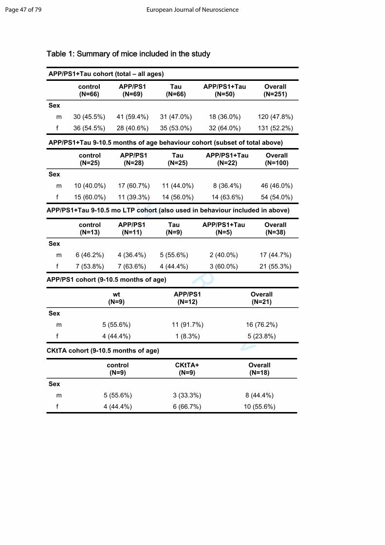
experiments. Table 1 shows a summary of the mice used in this paper.
Animal work was carried out in accordance with the Animals [Scientific
Procedures Act] 1986 (UK) and the council directive 2010/63EU of the European
Parliament and the Council of 22 September 2010 on the protection of animals used
for scientific purposes.
Novel object recognition and open field testing
Animals were tested for novel object recognition in a square box (40 x 40 x 60 cm)
made of dark opaque walls (laminated MDF) with approximately 2.5 cm of corn cob
bedding on the floor of the arena. Animals were recorded using an overhead camera
and the video signal was fed into Blackmagic Media Express computer software which
captured the movement of the animals. Each day animals were brought into the testing
room in their home cage upon the end of the 12 h dark cycle and allowed to settle for
1 hour. Individual experiments were composed of 3 phases: habituation, sampling and
probe, with all experiments performed in the morning. For habituation, animals were
Page 57 of 79 European Journal of Neuroscience

For Peer Review
exposed to the open field in the absence of any objects for 4 consecutive days. On
day 1, animals were introduced to the centre of the arena along with cage mates for
20 min. For days 2-4, individual animals were exposed to the arena for 10 min per
day, being placed into the open field facing a different corner of the arena each day,
the sequence of which was assigned using a random number generator. For each
experimental group, the order in which animals were placed in the arena was randomly
assigned using a random number generator. On day 4 of habituation, open field
exploration was recorded for 10 minutes using an overhead camera. These data were
published previously (Pickett et al., 2019); here the 9-10.5 month old data are shown
in figure 1a for reference purposes.
Following habituation, individual mice participated in a sampling phase of 3 sampling
trials and 1 probe trial. For the sampling phase (which took a total of 30 minutes per
mouse), animals were exposed 3 times to 2 identical objects for 5 min followed by 5
min in a holding cage (35 x 15 x 15 cm) that was identical to their home cages. The
objects remained in the same location during sampling, in two opposing corners of the
open field, each object put 10 cm from the corner. After the sampling phase animals
were returned to their home cage. The bedding on the floor of the arena was mixed
through to disperse odors, any waste material was removed, and objects were cleaned
with alcohol between mice. Probe trials were conducted 24 h after sampling and
consisted of a single 3 min exposure to 2 objects (1 familiar object from the sampling
phase and 1 novel object, both objects placed at the locations objects occupied during
Page 58 of 79European Journal of Neuroscience

For Peer Review
sampling), followed by 5 min in the holding cage prior to return to the home cage.
Once all trials were completed each day, mice remained in the testing room for 1 h to
rest prior to return to the holding room.
Over the course of an experiment each mouse participated three times in this task,
with 1 day between iterations. Each time, different objects were used. In each probe
trial we paired two objects that elicited comparable exploratory activity as determined
in a previous study in naïve mice. This way we ensured that pre-existing object
preferences did not bias behavior in the probe trials. The objects used in sampling and
probe phases were counterbalanced across genotypes and repetitions of this task to
ensure comparison between groups. Exploration was determined in a blind manner
using in-house software (available upon reasonable request from corresponding
author O.H.). Individual probe trials were manually scored, with exploration defined as
a mouse being within 2 cm of an object, directing its nose at the object and being
involved in active exploration such as sniffing or whisking. For each probe phase, a
discrimination index (d) was calculated ((time exploring novel object – time exploring
familiar object)/(time exploring both objects)). Mice are attracted to novelty, and values
of d > 0, indicating that they prefer exploring the novel object, suggest that they have
memory for the object encountered during sampling. Values of d = 0 indicate mice
treat both objects the same, and this absence of novelty preference suggests that they
no longer express memory for the object encountered during sampling. Probe trials
in which a mouse did not reach at least 10 s of total exploratory activity of objects were
Page 59 of 79 European Journal of Neuroscience

For Peer Review
excluded from the analysis. Effect size was calculated for wild-type mice with n=6
showing a mean of 0.296, standard deviation of 0.145 and Cohen’s d effect size of
2.041, indicating that groups of at least 6 have more than sufficient power to detect
intact novel object recognition memory. Group sizes for our experiments are outlined
in Table 1.
Rotarod testing
To test motor function, mice were tested with a Rotarod (IITC Life Science) for 4
consecutive days. Each day animals were brought into the testing room in their home
cage and allowed to settle for 1 h. Animals were placed on the Rotarod in separate
lanes with male and female mice running in separate trials. Each day, mice
participated in 3 baseline trials and 7 training trials, no trial lasting longer than 90 s.
Baseline trials consisted of 3 trials separated by 2-min long rests between trials. During
baseline trials, the drums rotated at a constant speed of 4 rotations per minute. Thirty
minutes after the baseline trials, mice underwent 7 experimental trials with 2-min long
rests in between. During the training trials, the rotational speed of the drums
accelerated, from a starting speed of 4 rotations per minute to a final speed of 25
rotations per minute that was reached within 60 s. The average latency to fall off of
the rotarod in experimental trials on the last day of testing was used to compare groups
of genotypes. Data from mice who consistently jumped off of the rotarod (as opposed
to running until they fell) were excluded from the analysis.
Page 60 of 79European Journal of Neuroscience

For Peer Review
Electrophysiology
Electrophysiology experiments were performed with the experimenter blind to
genotype. Acute horizontal brain slices were prepared as follows. Mice were deeply
anaesthetized by the inhalation of isoflurane followed by intra-peritoneal injection of
pentobarbital (20%, 0.7l/g). Animals were transcardially perfused with oxygenated
(95% O2, 5% CO2) protective NMDG-based solution of the following content (mM): 92
N-methyl-D-glucamine (NMDG), 2.5 KCl, 1.25 NaH2PO4, 30 NaHCO3, 20 HEPES, 25
glucose, 5 Na-ascorbate, 3 Na-pyruvate, 0.5 CaCl2, and 10 MgCl2, pH 7.3–7.4 (titrated
with 5M HCl). The mouse was decapitated and the brain was quickly dissected and
placed into room temperature oxygenated (95% O2-5% CO2) NMDG-based solution.
Frontal lobe area and cerebellum were removed from the brain. Left hemisphere of
the brain was mounted on the stage of a Vibrating Microtome 7000 (Campden
Instruments LTD, England, UK) and the first 3-4 hippocampal slices (400 μm) from the
dorsal part of the hemisphere were cut at horizontal plane. During recovery period
freshly prepared slices were incubated at 34°C for 12 min in oxygenated NMDG
solution. After that slices were transferred to submerged type storage chamber and
maintained at a room temperature for at least 2 h before use in an oxygenated artificial
cerebrospinal fluid (ACSF) of the following composition (mM): 119 NaCl, 3 KCl, 2.0
CaCl2, 1.3 MgSO4, 1.2 NaH2PO4, 26 NaHCO3 and 11 glucose, pH 7.35.
Slices were transferred to the interface type recording chamber and perfused with
oxygenated ACSF (1.5-2 mL/min). The temperature in the chamber was maintained at
Page 61 of 79 European Journal of Neuroscience

For Peer Review
25-26°C (PTC03, Scientific Systems Design, Ontario, Canada). Extracellular field
excitatory postsynaptic potential (fEPSP) recordings were obtained from the
hippocampal CA1 stratum radiatum using Differential AC Amplifier 1700 (A-M
Systems, Carlsborg, WA, USA). The recording electrode was an Ag/AgCl wire in
ACSF-filled extracellular glass microelectrode (1-1.5 MΩ). Evoked postsynaptic
responses were elicited by stimulation of Schaffer collaterals using a bipolar
stimulating electrode and DS3 Isolated current stimulator (Digitimer Ltd, Hertfordshire,
UK). Synaptic response profiles were determined by generating input–output curve
(stimulation intensity between 10 and 100 μA, 100 μs) for each slice. No significant
differences were found in the input-output curve between genotypes. The input-output
profile of the slice was used to set the stimulation strength for long term potentiation
(LTP) experiments. For LTP experiments, the stimulation intensity was set to elicit
30% of maximal fEPSP slope. This resulted in different absolute stimulus intensity
between slices and animals; however, this paradigm ensured that the recruitment of
synapses was standardised relative to the overall excitability of the slice. Data were
normalized to pre-tetanus for all groups which also controls for stimulus intensity
differences. For pre- and post-tetanus stimulation, the stimulation was applied every
30 s. After 10-20 min of stable baseline recording high-frequency tetanic stimulation
(HFS, 100 Hz, 1 s) was delivered. Data was amplified x100, band-pass filtered (1Hz –
5kHz) and digitized at 20.8 or 20.06kHz using an analogue-to-digital converter
(National Instruments, Austin, TX) and acquired using WinWCP (Strathclyde
Electrophysiology Software, Glasgow, UK).
Page 62 of 79European Journal of Neuroscience
For Peer Review
Offline data analysis was performed using ClampFit (Axon Instruments, USA). The
fEPSP slope was calculated between 2 manually positioned cursors. The first cursor
was positioned at the rising inflection of the fEPSP after the fiber volley. The second
cursor was set to measure the slope of monosynaptic EPSP, and placed before the
point where the rising slope began levelling off, to exclude population spikes.
Immunohistochemistry
For immunohistochemical analysis, mice were sacrificed by terminal anesthesia at 9
months of age after completing behavioural tests. The animals were perfused with
phosphate buffer saline, brains were dissected, and one hemisphere fixed for 48 hours
in 4% paraformaldehyde with 15% glycerol cryoprotectant. The other hemisphere had
tissue from entorhinal cortex and hippocampus prepared for array tomography as
described previously (Kay et al., 2013; Pickett et al., 2019). Coronal sections, 50 m
in thickness, were cut through the entire fixed hemisphere. Every 20th section from
n=5 mice per group was stained with pan A antibody AW7 (a generous gift from Prof
Dominic Walsh) and plaque fibrils counterstained with 0.05% Thioflavin S in 50%
ethanol. Images were acquired on a Zeiss AxioImager microscope with a 20x
objective. For array tomography, embedded tissue blocks were sectioned with an
ultracut microtome (Zeiss) into ribbons of 70nm serial sections. Ribbons were stained
with primary antibodies at 1:50 dilution in blocking buffer AW7 (secondary - donkey
anti rabbit 488, Invitrogen) one of the following tau antibodies: Total Tau (Gt, R&D
Page 63 of 79 European Journal of Neuroscience

For Peer Review
systems, secondary donkey anti goat 594, Invitrogen), PhF1 (a generous gift from
Peter Davies, secondary donkey anti mouse 594, Invitrogen), Alz50 (from Peter
Davies, secondary antibody donkey anti mouse IgM 594, Invitrogen), or CP13 (from
Peter Davies, secondary antibody donkey anti mouse 594, Invitrogen). After
secondary antibody staining, nuclei were counterstained with DAPI, coverslips were
mounted with ImmuMount, and images acquired on a Ziess AxioImager microscope
with a 63x, 1.4NA objective.
Data analysis
Statistical analyses were performed and graphs generated in R Studio v1.2.1335
running R package version 5.2.2. For novel object recognition discrimination ratios,
each genotype was compared to the hypothetical discrimination ratio of d=0 using a
one-sample t-test. P values were corrected for multiple comparisons with a Holm
adjustment. Data were checked for normality with Shapiro-Wilks normality tests. To
examine differences between genotypes and look for sex effects in the APP/PS1+Tau
line behavioural studies at 9-10.5 months of age, linear mixed effects models were
used with the experimental batch as a random variable with the formula:
lmer(Variable_of_Interest~Genotype+Sex+(1|testing_batch)). To examine effects of
age on behavioural phenotypes in APP/PS1+Tau and control genotypes, a linear
mixed effects model was used which accounted for 2 batches of testing and multiple
measurements per animal as follows:
lmer(Variable_of_Interest~Genotype*Age+Sex+(1|testing_batch)+(1|Mouse_ID)).
Page 64 of 79European Journal of Neuroscience
For Peer Review
Assumptions of the models were tested using visual inspection of the Q-Q plot of
residuals. To test correlations between variables, Spearman’s rank correlations were
performed followed by Holm adjustments for multiple comparisons.
For LTP data, the average fEPSP slope during a 10 min period before LTP
induction was taken as the baseline, and values were normalized to this value.
Potentiation was measured between 50 to 60 min after HFS. To avoid
pseudoreplication, a mean normalised fEPSP slope was calculated across slices per
animal. This mean normalised fEPSP slope was used in statistical comparison
between groups and in plotting the time course of LTP. Statistical analysis was carried
out using R. Normality of fEPSP slope data was tested using a Shapiro-Wilk normality
test. Two groups did not meet normality due to low sample size (APP/PS1+Tau, n=5)
and the presence of a single outlier (Tau). For these groups, results from both
parametric and non-parametric tests are reported for LTP testing. For between group
comparisons, homogeneity of variances was tested using the Bartlett test (p>0.05).
Results are reported as box and whisker plots showing each measured value
as an individual data point alongside the median (centre line) 75th percentile (top of
box) 25th percentile (bottom of box), largest value within 1.5 times interquartile range
above 75th percentile (top whisker), smallest value within 1.5 times interquartile range
below 25th percentile (bottom whisker). LTP data and line graphs show mean ± SEM,
Scatter plots show individual data points and linear regression lines with 95%
confidence intervals.
Page 65 of 79 European Journal of Neuroscience

For Peer Review
Data accessibility
Data used in this manuscript are provided in supplemental tables associated with the
paper. For detailed raw files (e.g. movies of mice doing novel object recognition),
contact authors.
Results:
To test the hypothesis that memory was impaired in APP/PS1+Tau mice, novel
object recognition memory was assessed in mice between 9 and 10.5 months of
age, a time point when we previously observed a hyperactivity behavioural
phenotype alongside transcriptional dysregulation ( Pickett et al., 2019). Fig1a shows
that as previously reported, these APP/PS1+Tau mice move farther in the open field,
a behavioural measure of hyperactivity (linear mixed effects model APP/PS1+Tau vs
control p<0.001, full statistics in supplemental table 5). Mice are innately curious and
prefer to explore novel over familiar objects. A discrimination ratio in novel object
recognition testing above 0 indicates preference for the novel object reflecting
memory of which object was previously explored. Novel object memory testing did
not uphold our hypothesis as all 4 genotypes exhibited significant novel object
recognition memory at this age (Figure 1b). For all groups, the discrimination ratio
was significantly different from 0 as determined by one-sample t-tests. After Holm
correction for multiple testing, the p values for each group were below 0.001 (please
see supplemental table 6 for exact t-statistics and p-values). We used a linear mixed
Page 66 of 79European Journal of Neuroscience

For Peer Review
effects model to determine whether there were differences between genotypes or
any sex effects while taking into account the testing was run in 2 batches (with
testing batch as a random variable in the model). This model indicates there were
no differences between genotypes in novel object recognition nor were there any
differences between male and female mice (supplemental table 7 contains the
results of the model). The total amount of time mice explored the objects (exploration
time) was examined as differing exploration times could contribute to differences in
memory. Although all groups exhibited novel object recognition memory, there were
significant differences between groups and between sexes in exploration time
(Figure 1c). A linear mixed effects model reveals that tau mice and APP/PS1+Tau
mice explore significantly more than control mice and females explore more than
males (supplemental table 8). However, in our dataset, we do not see a correlation
in any group between exploration time and novel object discrimination ratio (Figure
1d, Spearman correlation p values >0.3 for all genotypes). Thus, in our mice, the
different exploration times between groups is unlikely to affect novel object memory
(for similar results see Akkerman et al., 2012). Many previous studies have reported
novel object recognition deficits in APP/PS1 at ages when plaque deposition was
evident (Pedrós et al., 2014; Gu et al., 2016; Shen et al., 2017). We previously
reported plaque deposition in these APP/PS1+Tau mice starting by 6 months of age
(Pickett et al., 2019). Further, we previously reported that A levels in the brain are
approximately 15 pg/mg total protein and are not different between APP/PS1 and
APP/PS1+Tau mice and tau protein levels are approximately 10 ng/mg total protein
Page 67 of 79 European Journal of Neuroscience

For Peer Review
and do not differ between APP/PS1+Tau mice and Tau mice (Pickett et al., 2019).
Since both plaques and tangles have been associated in mice with spatial memory
disruption, we stained for plaques and phosphorylated tau. We confirm in this study
that both APP/PS1+Tau mice and littermate APP/PS1 mice at 9 months of age have
substantial plaque deposition (Figure 1e,f). In the APP/PS1+Tau mice, there are
small accumulations of phosphorylated tau around plaques, but no neurofibrillary
tangle pathology as we previously reported (Figure 1f). High resolution array
tomography imaging shows accumulation of small punctae of tau staining in multiple
forms (1g), total tau, phosphoTau labelled with PHF1 and CP13 antibodies, and
misfolded tau labelled with Alz50 antibody. To determine whether the lack of novel
object recognition in 9-10.5 month old APP/PS1+Tau mice was due to a delay in the
phenotype compared to the parent APP/PS1 line with endogenous mouse tau, we
completed an ageing study in these mice testing memory out to 14 months of age
(Figure 1g,h). Two cohorts of mice were tested, one at 3, 6, and 9 months of age,
and another at 10.5 and 14 months of age. APP/PS1+Tau mice still have intact
novel object recognition memory at 14 months of age (one-sided t-test adjusted p
value = 0.023), and a linear mixed effects model reveals there is not a significant
effect of genotype, age, or sex on memory (supplemental table 9). There are age
and sex effects on exploration time, but no genotype effects (supplemental table 10).
Rotarod testing was used to determine if there were motor deficits in this line. Subtle
changes in latency to fall off the rotarod were observed between sexes and in tau
mice compared to controls (Supplemental figure 1, supplemental table 11), however
Page 68 of 79European Journal of Neuroscience

For Peer Review
these were likely to be driven by the CKtTA activator transgene as will be discussed
later.
To determine whether the lack of memory phenotype in these mice was due to lack
of endogenous mouse tau and to confirm that in our hands the novel object
recognition test can detect memory deficits in an established model, we tested the
parent APP/PS1 line from Jackson labs that have endogenous mouse tau. As
expected, APP/PS1 mice with endogenous mouse tau had novel object recognition
deficits at 9 months of age, with their discrimination ratios being not significantly
different from zero (one-sample t-test adjusted p-value=0.489) whereas wild-type
littermates had a significant preference for the novel object (one-sample t-test
adjusted p-value=0.008, Figure 2a). This memory deficit was not due to differences
in total time exploring the objects which were not different between the 2 groups
(Figure 2b). As observed in the APP/PS1+Tau line and littermate controls, there
was also no correlation between novel object recognition and exploration time in
APP/PS1 mice with endogenous mouse tau or their wild-type littermates (Spearman
rank correlation adjusted p-value=1.000 for both groups, Figure 2c).
As a further control, we examined novel object recognition memory in mice
expressing only the CKtTA transactivator gene. This is important as this transgene is
used in APP/PS1+Tau mice to drive human tau expression, and we previously
observed this transgene affects brain weight in mice (Pickett et al., 2019). Here we
observe that there was no effect of this transgene on novel object recognition. Both
Page 69 of 79 European Journal of Neuroscience

For Peer Review
CKtTA and littermate control mice have intact recognition memory (one-sample t-
tests adjusted p-value<0.05 for both groups Figure 2d) and there is no difference
between genotypes or sexes (linear mixed effects model with testing batch as a
random variable, Supplemental table 12). Compared to controls, there is no effect of
this transgene or sex on exploration time in the CKtTA mice (linear mixed effects
model with testing batch as a random variable, Supplemental Table 13, Figure 2e).
There was also no correlation between novel object recognition discrimination ratio
and exploration time (Spearman rank correlation adjusted p-value=1.000 for both
groups, Figure 2f). There was a significant rotarod deficit in CKtTA mice
(supplemental figure 1, supplemental table 14), indicating that the driver transgene in
the absence of tau expression is sufficient to cause a motor phenotype. However,
this did not cause an impairment of the ability to explore objects nor did it cause
memory deficits.
To assay hippocampal LTP, we tetanized the Schaffer collateral pathway. Intact LTP
was observed in control, APP/PS1, and Tau genotypes at 9-10.5 months of age
(Figure 3; Wilcoxon test baseline = 100% - control: 138%, IQR 41%, V=91, adjusted
p=0.001; APP/PS1: 126%, IQR 12%, V=66, p=0.039; Tau: 137%, IQR 21%, V=45,
p=0.016). In APP/PS1+Tau mice at this age, although the EPSP slope 60 minutes
after tetanisation was 122% of baseline, this did not reach significance (IQR 10%,
V=15, p=0.06, adjusted p=0.25). However, we are hesitant to interpret this as a loss
of LTP since there is no significant difference in the extent of potentiation between
Page 70 of 79European Journal of Neuroscience
For Peer Review
genotypes in the APP/PS1+Tau cohort (Kruskall-Wallis Chi2(3)=1.72, p=0.63), and
the lack of significance in the APP/PS1+Tau group may be driven by the smaller
group size, a limitation of the number of animals available for experiments in this
complex cross.
In WT mice with endogenous mouse tau, tetanization resulted in a significant
potentiation of the fEPSP slope measured in CA1 stratum radiatum (129 ± 8.7% of
baseline, t(5)=3.4, Holm adjusted p=0.04, 2-tailed, one-sample t-test compared to
mean of 100%). In APP/PS1 littermates with endogenous mouse tau, the same
stimulation paradigm did not induce significant potentiation (109.9 ± 7.1% of
baseline, t(6)= 1.4, Holm adjusted p= 0.43 2-tailed, one-sample t-test compared to
mean of 100%; Figure 3). Together, these data indicate that tau is needed to drive
LTP deficits in APP/PS1 mice.
Discussion:
Understanding the cascade from early A accumulation through pathological tau
accumulation to neural circuit dysfunction and neurodegeneration remains one of the
key challenges facing the Alzheimer’s disease field (Karran et al., 2011; Mauricio et
al., 2019), and there are relatively few studies directly addressing the cooperation of
A and tau in disrupting memory and synaptic plasticity.
Page 71 of 79 European Journal of Neuroscience

For Peer Review
Our previous work in the APP/PS1+Tau mouse line and human postmortem tissue
indicate that both A and tau accumulate in synapses, where it may contribute to
synaptic dysfunction and downstream cognitive decline (Jackson et al., 2016, 2019;
Zhou et al., 2017; McInnes et al., 2018; Hesse et al., 2019; Pickett et al., 2019).
Similar to findings by Chabrier et al., 2012, who expressed mutant APP and wild-
type human tau, in the APP/PS1+Tau line, we observe tau accumulation in post
synapses, which is associated with a behavioural hyperactivity phenotype (Pickett et
al., 2019). In contrast, we did not observe memory deficits in APP/PS1+Tau mice .
This discrepancy could reflect that the mice in Chabrier et al retained endogenous
mouse tau in addition to expressing wild-type human tau, and mouse tau is known to
be important for synaptic phenotypes induced by A (Roberson et al., 2011; Shipton
et al., 2011).
Indeed, we confirm the importance of mouse tau driving A phenotypes in this study
as we observe that APP/PS1 mice expressing endogenous mouse tau do exhibit
both novel object memory and LTP deficits at 9-10.5 months of age as has been
reported previously (Pedrós et al., 2014; Gu et al., 2016; Shen et al., 2017)(Cai et
al., 2017; Wu et al., 2017). Whereas in agreement with previous reports indicating
that lowering endogenous mouse tau protects APP overexpressing mice (Ittner et al.,
2010; Vossel et al., 2010; Roberson et al., 2011; Shipton et al., 2011; DeVos et al.,
2018), here we observe that APP/PS1 mice on a MAPTnull background do not have
novel object recognition or LTP deficits compared to littermate controls.
Page 72 of 79European Journal of Neuroscience
For Peer Review
Surprisingly, expression of human tau in these mice did not simply reconstitute the
behaviour and plasticity deficits seen in APP/PS1 mice with mouse tau. These data
indicate that the differences between mouse and human tau may be important in
mediating the synaptotoxic effects of A Alternatively, the overexpression of human
tau in our APP/PS1+Tau line with the CamKII promotor controlled CKtTA
transactivator does not result in normal spatiotemporal expression of tau and has
higher levels than endogenous mouse tau, which may affect the interactions with A.
Either way, our results support the need for the use of multiple models alongside
descriptive human tissue studies to ensure translational relevance of mouse studies
in the Alzheimer’s field.
Author contributions:Designed study (I.O., O.H., T.L.S.-J.), conducted experiments (J.T., O.N., E.K.P., A.G.H., P.J.), analysed data (O.N., E.K.P., A.G.H., A.S., I.O., T.L.S.-J.), drafted paper (T.L.S.-J.).
Acknowledgements:This work was funded by the European Research Council (ERC) under the European Union’s Horizon 2020 research and innovation programme (Grant agreement No. 681181), the UK Dementia Research Institute which receives its funding from DRI Ltd, funded by the UK Medical Research Council, Alzheimer’s Society, and Alzheimer’s Research UK, Alzheimer’s Research UK and the Scottish Government Chief Scientist Office (ARUK SPG2013-1), a Wellcome Trust Translational Neuroscience PhD studentship, and Alzheimer’s Society.
Page 73 of 79 European Journal of Neuroscience

For Peer Review
Competing interests: TS-J is on the Scientific Advisory Board of Cognition Therapeutics and receives collaborative grant funding from 2 industry partners. None of these had any influence over the current paper.
References:
Akkerman, S., Blokland, A., Reneerkens, O., van Goethem, N.P., Bollen, E., Gijselaers, H.J.M., Lieben, C.K.J., Steinbusch, H.W.M., & Prickaerts, J. (2012) Object recognition testing: methodological considerations on exploration and discrimination measures. Behav. Brain Res., 232, 335–347.
Cai, H., Wang, Y., He, J., Cai, T., Wu, J., Fang, J., Zhang, R., Guo, Z., Guan, L., Zhan, Q., Lin, L., Xiao, Y., Pan, H., & Wang, Q. (2017) Neuroprotective effects of bajijiasu against cognitive impairment induced by amyloid-β in APP/PS1 mice. Oncotarget, 8, 92621–92634.
Chabrier, M.A., Blurton-Jones, M., Agazaryan, A.A., Nerhus, J.L., Martinez-Coria, H., & LaFerla, F.M. (2012) Soluble Aβ Promotes Wild-Type Tau Pathology In Vivo. J. Neurosci., 32, 17345–17350.
DaRocha-Souto, B., Scotton, T.C., Coma, M., Serrano-Pozo, A., Hashimoto, T., Serenó, L., Rodríguez, M., Sánchez, B., Hyman, B.T., & Gómez-Isla, T. (2011) Brain Oligomeric β-Amyloid but Not Total Amyloid Plaque Burden Correlates With Neuronal Loss and Astrocyte Inflammatory Response in Amyloid Precursor Protein/Tau Transgenic Mice. J Neuropathol Exp Neurol, 70, 360–376.
DeVos, S.L., Corjuc, B.T., Commins, C., Dujardin, S., Bannon, R.N., Corjuc, D., Moore, B.D., Bennett, R.E., Jorfi, M., Gonzales, J.A., Dooley, P.M., Roe, A.D., Pitstick, R., Irimia, D., Frosch, M.P., Carlson, G.A., & Hyman, B.T. (2018) Tau reduction in the presence of amyloid-β prevents tau pathology and neuronal death in vivo. Brain, 141, 2194–2212.
Gu, X.-H., Xu, L.-J., Liu, Z.-Q., Wei, B., Yang, Y.-J., Xu, G.-G., Yin, X.-P., & Wang, W. (2016) The flavonoid baicalein rescues synaptic plasticity and memory
Page 74 of 79European Journal of Neuroscience

For Peer Review
deficits in a mouse model of Alzheimer’s disease. Behavioural Brain Research, 311, 309–321.
Hardy, J.A. & Higgins, G.A. (1992) Alzheimer’s disease: the amyloid cascade hypothesis. Science, 256, 184-.
Henstridge, C.M., Hyman, B.T., & Spires-Jones, T.L. (2019) Beyond the neuron-cellular interactions early in Alzheimer disease pathogenesis. Nat. Rev. Neurosci., 20, 94–108.
Hesse, R., Hurtado, M.L., Jackson, R.J., Eaton, S.L., Herrmann, A.G., Colom-Cadena, M., Tzioras, M., King, D., Rose, J., Tulloch, J., McKenzie, C.-A., Smith, C., Henstridge, C.M., Lamont, D., Wishart, T.M., & Spires-Jones, T.L. (2019) Comparative profiling of the synaptic proteome from Alzheimer’s disease patients with focus on the APOE genotype. Acta Neuropathologica Communications, 7, 214.
Hoover, B.R., Reed, M.N., Su, J., Penrod, R.D., Kotilinek, L.A., Grant, M.K., Pitstick, R., Carlson, G.A., Lanier, L.M., Yuan, L.-L., Ashe, K.H., & Liao, D. (2010) Tau Mislocalization to Dendritic Spines Mediates Synaptic Dysfunction Independently of Neurodegeneration. Neuron, 68, 1067–1081.
Ittner, L.M., Ke, Y.D., Delerue, F., Bi, M., Gladbach, A., van Eersel, J., Wölfing, H., Chieng, B.C., Christie, M.J., Napier, I.A., Eckert, A., Staufenbiel, M., Hardeman, E., & Götz, J. (2010) Dendritic function of tau mediates amyloid-beta toxicity in Alzheimer’s disease mouse models. Cell, 142, 387–397.
Jackson, R.J., Rose, J., Tulloch, J., Henstridge, C., Smith, C., & Spires-Jones, T.L. (2019) Clusterin accumulates in synapses in Alzheimer’s disease and is increased in apolipoprotein E4 carriers. Brain Commun, 1, fcz003.
Jackson, R.J., Rudinskiy, N., Herrmann, A.G., Croft, S., Kim, J.M., Petrova, V., Ramos-Rodriguez, J.J., Pitstick, R., Wegmann, S., Garcia-Alloza, M., Carlson, G.A., Hyman, B.T., & Spires-Jones, T.L. (2016) Human tau increases amyloid β plaque size but not amyloid β-mediated synapse loss in a novel mouse model of Alzheimer’s disease. Eur J Neurosci, 44, 3056–3066.
Jankowsky, J.L., Fadale, D.J., Anderson, J., Xu, G.M., Gonzales, V., Jenkins, N.A., Copeland, N.G., Lee, M.K., Younkin, L.H., Wagner, S.L., Younkin, S.G., & Borchelt, D.R. (2004) Mutant presenilins specifically elevate the levels of the
Page 75 of 79 European Journal of Neuroscience

For Peer Review
42 residue beta-amyloid peptide in vivo: evidence for augmentation of a 42-specific gamma secretase. Hum. Mol. Genet., 13, 159–170.
Karran, E., Mercken, M., & Strooper, B.D. (2011) The amyloid cascade hypothesis for Alzheimer’s disease: an appraisal for the development of therapeutics. Nature Reviews Drug Discovery, 10, 698–712.
Kay, K.R., Smith, C., Wright, A.K., Serrano-Pozo, A., Pooler, A.M., Koffie, R., Bastin, M.E., Bak, T.H., Abrahams, S., Kopeikina, K.J., McGuone, D., Frosch, M.P., Gillingwater, T.H., Hyman, B.T., & Spires-Jones, T.L. (2013) Studying synapses in human brain with array tomography and electron microscopy. Nature Protocols, 8, 1366–1380.
Klein, W.L. (2013) Synaptotoxic Amyloid-β Oligomers: A Molecular Basis for the Cause, Diagnosis, and Treatment of Alzheimer’s Disease? Journal of Alzheimer’s Disease, 33, S49–S65.
Koffie, R.M., Hyman, B.T., & Spires-Jones, T.L. (2011) Alzheimer’s disease: synapses gone cold. Mol Neurodegener, 6, 63.
Koffie, R.M., Meyer-Luehmann, M., Hashimoto, T., Adams, K.W., Mielke, M.L., Garcia-Alloza, M., Micheva, K.D., Smith, S.J., Kim, M.L., Lee, V.M., Hyman, B.T., & Spires-Jones, T.L. (2009) Oligomeric amyloid beta associates with postsynaptic densities and correlates with excitatory synapse loss near senile plaques. Proc Natl Acad Sci U S A, 106, 4012–4017.
Li, S., Hong, S., Shepardson, N.E., Walsh, D.M., Shankar, G.M., & Selkoe, D. (2009) Soluble Oligomers of Amyloid β Protein Facilitate Hippocampal Long-Term Depression by Disrupting Neuronal Glutamate Uptake. Neuron, 62, 788–801.
Masuda, A., Kobayashi, Y., Kogo, N., Saito, T., Saido, T.C., & Itohara, S. (2016) Cognitive deficits in single App knock-in mouse models. Neurobiology of Learning and Memory, MCCS 2016, 135, 73–82.
Mauricio, R., Benn, C., Davis, J., Dawson, G., Dawson, L.A., Evans, A., Fox, N., Gallacher, J., Hutton, M., Isaac, J., Jones, D.N.C., Jones, L., Lalli, G., Libri, V., Lovestone, S., Moody, C., Noble, W., Perry, H., Pickett, J., Reynolds, D., Ritchie, C., Rohrer, J.D., Routledge, C., Rowe, J., Snyder, H., Spires-Jones, T., Swartz, J., Truyen, L., Whiting, P., & Therapeutics for Dementia Consortium (2019) Tackling gaps in developing life-changing treatments for dementia. Alzheimers Dement (N Y), 5, 241–253.
Page 76 of 79European Journal of Neuroscience

For Peer Review
McInnes, J., Wierda, K., Snellinx, A., Bounti, L., Wang, Y.-C., Stancu, I.-C., Apóstolo, N., Gevaert, K., Dewachter, I., Spires-Jones, T.L., Strooper, B.D., Wit, J.D., Zhou, L., & Verstreken, P. (2018) Synaptogyrin-3 Mediates Presynaptic Dysfunction Induced by Tau. Neuron, 97, 823-835.e8.
Oddo, S., Caccamo, A., Shepherd, J.D., Murphy, M.P., Golde, T.E., Kayed, R., Metherate, R., Mattson, M.P., Akbari, Y., & LaFerla, F.M. (2003) Triple-transgenic model of Alzheimer’s disease with plaques and tangles: intracellular Abeta and synaptic dysfunction. Neuron, 39, 409–421.
Pedrós, I., Petrov, D., Allgaier, M., Sureda, F., Barroso, E., Beas-Zarate, C., Auladell, C., Pallàs, M., Vázquez-Carrera, M., Casadesús, G., Folch, J., & Camins, A. (2014) Early alterations in energy metabolism in the hippocampus of APPswe/PS1dE9 mouse model of Alzheimer’s disease. Biochimica et Biophysica Acta (BBA) - Molecular Basis of Disease, 1842, 1556–1566.
Pickett, E.K., Herrmann, A.G., McQueen, J., Abt, K., Dando, O., Tulloch, J., Jain, P., Dunnett, S., Sohrabi, S., Fjeldstad, M.P., Calkin, W., Murison, L., Jackson, R.J., Tzioras, M., Stevenson, A., d’Orange, M., Hooley, M., Davies, C., Colom-Cadena, M., Anton-Fernandez, A., King, D., Oren, I., Rose, J., McKenzie, C.-A., Allison, E., Smith, C., Hardt, O., Henstridge, C.M., Hardingham, G.E., & Spires-Jones, T.L. (2019) Amyloid Beta and Tau Cooperate to Cause Reversible Behavioral and Transcriptional Deficits in a Model of Alzheimer’s Disease. Cell Reports, 29, 3592-3604.e5.
Roberson, E.D., Halabisky, B., Yoo, J.W., Yao, J., Chin, J., Yan, F., Wu, T., Hamto, P., Devidze, N., Yu, G.-Q., Palop, J.J., Noebels, J.L., & Mucke, L. (2011) Amyloid-β/Fyn-induced synaptic, network, and cognitive impairments depend on tau levels in multiple mouse models of Alzheimer’s disease. J. Neurosci., 31, 700–711.
Roberson, E.D., Scearce-Levie, K., Palop, J.J., Yan, F., Cheng, I.H., Wu, T., Gerstein, H., Yu, G.-Q., & Mucke, L. (2007) Reducing Endogenous Tau Ameliorates Amyloid ß-Induced Deficits in an Alzheimer’s Disease Mouse Model. Science, 316, 750–754.
Sasaguri, H., Nilsson, P., Hashimoto, S., Nagata, K., Saito, T., De Strooper, B., Hardy, J., Vassar, R., Winblad, B., & Saido, T.C. (2017) APP mouse models for Alzheimer’s disease preclinical studies. EMBO J, 36, 2473–2487.
Page 77 of 79 European Journal of Neuroscience

For Peer Review
Shen, L., Han, B., Geng, Y., Wang, J., Wang, Z., & Wang, M. (2017) Amelioration of cognitive impairments in APPswe/PS1dE9 mice is associated with metabolites alteration induced by total salvianolic acid. PLoS One, 12.
Shipton, O.A., Leitz, J.R., Dworzak, J., Acton, C.E.J., Tunbridge, E.M., Denk, F., Dawson, H.N., Vitek, M.P., Wade-Martins, R., Paulsen, O., & Vargas-Caballero, M. (2011) Tau Protein Is Required for Amyloid β-Induced Impairment of Hippocampal Long-Term Potentiation. J. Neurosci., 31, 1688–1692.
Spires-Jones, T.L. & Hyman, B.T. (2014) The intersection of amyloid beta and tau at synapses in Alzheimer’s disease. Neuron, 82, 756–771.
Tucker, K.L., Meyer, M., & Barde, Y.-A. (2001) Neurotrophins are required for nerve growth during development. Nat Neurosci, 4, 29–37.
Vossel, K.A., Zhang, K., Brodbeck, J., Daub, A.C., Sharma, P., Finkbeiner, S., Cui, B., & Mucke, L. (2010) Tau reduction prevents Abeta-induced defects in axonal transport. Science, 330, 198.
Wu, M., Shi, H., He, Y., Yuan, L., Qu, X., Zhang, J., Wang, Z., Cai, H., & Qi, J. (2017) Colivelin Ameliorates Impairments in Cognitive Behaviors and Synaptic Plasticity in APP/PS1 Transgenic Mice. J. Alzheimers Dis., 59, 1067–1078.
Yasuda, M. & Mayford, M.R. (2006) CaMKII Activation in the Entorhinal Cortex Disrupts Previously Encoded Spatial Memory. Neuron, 50, 309–318.
Zhou, L., McInnes, J., Wierda, K., Holt, M., Herrmann, A.G., Jackson, R.J., Wang, Y.-C., Swerts, J., Beyens, J., Miskiewicz, K., Vilain, S., Dewachter, I., Moechars, D., De Strooper, B., Spires-Jones, T.L., De Wit, J., & Verstreken, P. (2017) Tau association with synaptic vesicles causes presynaptic dysfunction. Nature Communications, 8, 1–13.
Figure 1: APP/PS1+Tau mice have intact object recognition memory despite plaque deposition. As previously reported (Pickett et al 2019), at 9-10.5 months of age, APP/PS1+Tau mice have a behavioural hyperactivity phenotype as they travel significantly farther than controls in the open field (a, * linear mixed effects model APP/PS1+Tau vs control p<0.0001). At this same age, all four genotypes in the APP/PS1+Tau cross have discrimination ratios significantly different from zero
Page 78 of 79European Journal of Neuroscience

For Peer Review
indicating intact object memory (b, * one-sample t-test p<0.001). Exploration time differed between groups (c, * linear mixed effects model Tau vs control p=0.019). Performance in the novel object recognition test was unlikely affected by differences in exploration times as none of the groups exhibited a correlation between these measures (d, Spearman correlation each group p>0.3, linear regression lines shown alongside grey 95% confidence intervals). Staining with pan A antibody AW7 (magenta), thioflavin S to label fibrils (cyan) and PHF1 phospho-tau antibody (yellow) shows plaques in hippocampus of both APP/PS1 and APP/PS1+Tau mice (e,f) and small accumulations of phospho-tau around plaques in APP/PS1+Tau mice (arrows, f). Higher resolution imaging with array tomography (g) reveals small accumulations of several forms of tau in APP/PS1+Tau mice: total tau, PHF1 positive tau phosphorylated at Ser396/404, CP13 positive tau phosphorylated at Ser202,and Alz50 positive misfolded tau. Examining averages of discrimination ratios per group over time (h) reveals that APP/PS1+Tau mice have intact object recognition memory up to 14 months of age (dotted line represents 2 different cohorts were tested at 3, 6, 9 months and 10.5, 14 months). Trajectories of individual mice are shown in (i). Individual mouse values are shown as points a-d with male mice indicated with circles and female mice with triangles. Numbers of mice per group are indicated over the boxes in box and whisker plots. Scale bar represents 100 microns in e,f, 10 microns in g.
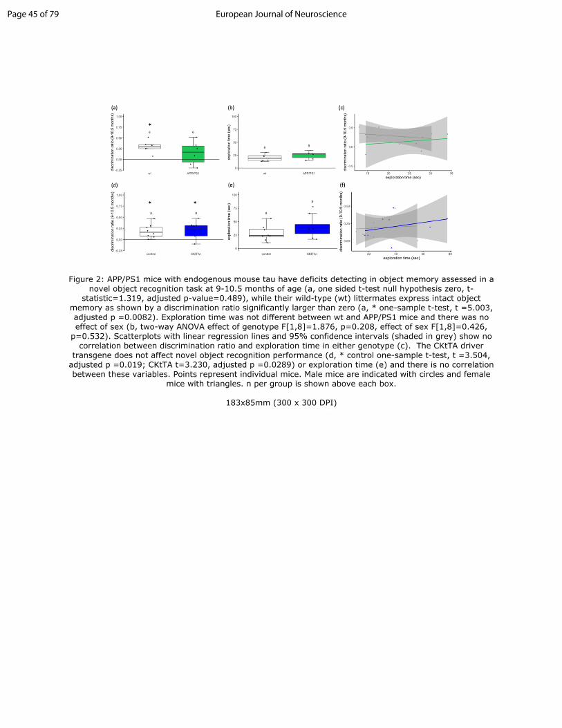
Figure 2: APP/PS1 mice with endogenous mouse tau have deficits detecting in object memory assessed in a novel object recognition task at 9-10.5 months of age (a, one sided t-test null hypothesis zero, t-statistic=1.319, adjusted p-value=0.489), while their wild-type (wt) littermates express intact object memory as shown by a discrimination ratio significantly larger than zero (a, * one-sample t-test, t =5.003, adjusted p =0.0082). Exploration time was not different between wt and APP/PS1 mice and there was no effect of sex (b, two-way ANOVA effect of genotype F[1,8]=1.876, p=0.208, effect of sex F[1,8]=0.426, p=0.532). Scatterplots with linear regression lines and 95% confidence intervals (shaded in grey) show no correlation between discrimination ratio and exploration time in either genotype (c). The CKtTA driver transgene does not affect novel object recognition performance (d, * control
Page 79 of 79 European Journal of Neuroscience

For Peer Review
one-sample t-test, t =3.504, adjusted p =0.019; CKtTA t=3.230, adjusted p =0.0289) or exploration time (e) and there is no correlation between these variables. Points represent individual mice. Male mice are indicated with circles and female mice with triangles. n per group is shown above each box.
Figure 3: LTP was induced with tetanization at time 0 (arrows). In slices from the APP/PS1+Tau line cohort which lack endogenous mouse tau, all genotypes exhibit fEPSP slopes above baseline (a). At 50-60 minutes post-tetanization (b), control, APP/PS1, and tau mice exhibit significant potentiation but the APP/PS1+Tau group is not significantly different from baseline (* p<0.05 Wilcoxon test compared to 1.0 with Holm corrected p values). No significant differences were found in the input-output curve between genotypes (c). Representative traces for the APP/PS1+Tau cohort are shown in (d). In APP/PS1 mice with endogenous mouse tau, LTP was impaired at 9-10 months of age, while wild-type littermates exhibited significant LTP (e, f, * Holm adjusted p=0.04, one-sample t-test compared to mean of 1.0). Again, no significant differences were found in the input-output curve between genotypes (g). Representative traces for the APP/PS1 cohort are shown in (h). Data shown in a and e are means +/- standard error. Scale bars represent 10ms, 0.5mV. Points in box plots represent individual mice, n per group is shown above each box.
Page 80 of 79European Journal of Neuroscience

For Peer Review
Figure 1: APP/PS1+Tau mice have intact object recognition memory despite plaque deposition. As previously reported (Pickett et al 2019), at 9-10.5 months of age, APP/PS1+Tau mice have a behavioural
hyperactivity phenotype as they travel significantly farther than controls in the open field (a, * linear mixed effects model APP/PS1+Tau vs control p<0.0001). At this same age, all four genotypes in the APP/PS1+Tau cross have discrimination ratios significantly different from zero indicating intact object memory (b, * one-sample t-test p<0.001). Exploration time differed between groups (c, * linear mixed effects model Tau vs control p=0.019). Performance in the novel object recognition test was unlikely affected by differences in
exploration times as none of the groups exhibited a correlation between these measures (d, Spearman correlation each group p>0.3, linear regression lines shown alongside grey 95% confidence intervals).
Staining with pan A antibody AW7 (magenta), thioflavin S to label fibrils (cyan) and PHF1 phospho-tau antibody (yellow) shows plaques in hippocampus of both APP/PS1 and APP/PS1+Tau mice (e,f) and small
accumulations of phospho-tau around plaques in APP/PS1+Tau mice (arrows, f). Higher resolution imaging with array tomography (g) reveals small accumulations of several forms of tau in APP/PS1+Tau mice: total
tau, PHF1 positive tau phosphorylated at Ser396/404, CP13 positive tau phosphorylated at Ser202,and
Page 43 of 79 European Journal of Neuroscience

For Peer Review
Alz50 positive misfolded tau. Examining averages of discrimination ratios per group over time (h) reveals that APP/PS1+Tau mice have intact object recognition memory up to 14 months of age (dotted line represents 2 different cohorts were tested at 3, 6, 9 months and 10.5, 14 months). Trajectories of
individual mice are shown in (i). Individual mouse values are shown as points a-d with male mice indicated with circles and female mice with triangles. Numbers of mice per group are indicated over the boxes in box
and whisker plots. Scale bar represents 100 microns in e,f, 10 microns in g.
183x280mm (300 x 300 DPI)
Page 44 of 79European Journal of Neuroscience
For Peer Review
Figure 2: APP/PS1 mice with endogenous mouse tau have deficits detecting in object memory assessed in a novel object recognition task at 9-10.5 months of age (a, one sided t-test null hypothesis zero, t-
statistic=1.319, adjusted p-value=0.489), while their wild-type (wt) littermates express intact object memory as shown by a discrimination ratio significantly larger than zero (a, * one-sample t-test, t =5.003, adjusted p =0.0082). Exploration time was not different between wt and APP/PS1 mice and there was no effect of sex (b, two-way ANOVA effect of genotype F[1,8]=1.876, p=0.208, effect of sex F[1,8]=0.426,
p=0.532). Scatterplots with linear regression lines and 95% confidence intervals (shaded in grey) show no correlation between discrimination ratio and exploration time in either genotype (c). The CKtTA driver
transgene does not affect novel object recognition performance (d, * control one-sample t-test, t =3.504, adjusted p =0.019; CKtTA t=3.230, adjusted p =0.0289) or exploration time (e) and there is no correlation between these variables. Points represent individual mice. Male mice are indicated with circles and female
mice with triangles. n per group is shown above each box.
183x85mm (300 x 300 DPI)
Page 45 of 79 European Journal of Neuroscience

For Peer Review
183x188mm (300 x 300 DPI)
Page 46 of 79European Journal of Neuroscience
For Peer Review
Table 1: Summary of mice included in the study
APP/PS1+Tau cohort (total – all ages)
control(N=66)
APP/PS1(N=69)
Tau(N=66)
APP/PS1+Tau(N=50)
Overall(N=251)
Sex
m 30 (45.5%) 41 (59.4%) 31 (47.0%) 18 (36.0%) 120 (47.8%)
f 36 (54.5%) 28 (40.6%) 35 (53.0%) 32 (64.0%) 131 (52.2%)
APP/PS1+Tau 9-10.5 months of age behaviour cohort (subset of total above)
control(N=25)
APP/PS1(N=28)
Tau(N=25)
APP/PS1+Tau(N=22)
Overall(N=100)
Sex
m 10 (40.0%) 17 (60.7%) 11 (44.0%) 8 (36.4%) 46 (46.0%)
f 15 (60.0%) 11 (39.3%) 14 (56.0%) 14 (63.6%) 54 (54.0%)
APP/PS1+Tau 9-10.5 mo LTP cohort (also used in behaviour included in above)
control(N=13)
APP/PS1(N=11)
Tau(N=9)
APP/PS1+Tau(N=5)
Overall(N=38)
Sex
m 6 (46.2%) 4 (36.4%) 5 (55.6%) 2 (40.0%) 17 (44.7%)
f 7 (53.8%) 7 (63.6%) 4 (44.4%) 3 (60.0%) 21 (55.3%)
APP/PS1 cohort (9-10.5 months of age)
wt(N=9)
APP/PS1(N=12)
Overall(N=21)
Sex
m 5 (55.6%) 11 (91.7%) 16 (76.2%)
f 4 (44.4%) 1 (8.3%) 5 (23.8%)
CKtTA cohort (9-10.5 months of age)
control(N=9)
CKtTA+(N=9)
Overall(N=18)
Sex
m 5 (55.6%) 3 (33.3%) 8 (44.4%)
f 4 (44.4%) 6 (66.7%) 10 (55.6%)
Page 47 of 79 European Journal of Neuroscience

For Peer Review
Supplemental figure 1: Rotarod testing indicates that the CKtTA driver transgene affects motor performance. In the APP/PS1+Tau line (a), there are significant differences in exploration time between Tau and control mice and APP/PS1+Tau and control mice (*, p<0.05 linear mixed effects model, supplemental table 11) and between male and female mice (p=0.003). This decrease in exploration time in the lines containing human tau is likely due not to tau expression but to the CKtTA activator transgene used to drive tau as in the parent CKtTA line (b), there is a significant decrease in CKtTA mice compared to controls (* b=14.006, t=2.325, p=0.0345) in the absence of human tau expression. In the APP/PS1 parent line with endogenous mouse tau, there is no difference between rotarod performance in APP/PS1 mice and wild-type littermates (c, one-way ANOVA F[1,6]=0.921, p=0.374), indicating that the observed memory deficits were not due to alterations in gross motor skills. Note mice that jumped off of the rotarod instead of running until they fell were excluded from this analysis. This precluded analysis of sex effects in the APP/PS1 line.
Page 48 of 79European Journal of Neuroscience

For Peer Review
183x81mm (300 x 300 DPI)
Page 49 of 79 European Journal of Neuroscience

For Peer Review
Graphical Abstract Text
Tulloch et al observe memory and long-term potentiation deficits in the APP/PS1 mouse model of familial Alzheimer’s disease, which are prevented by genetic removal of endogenous mouse tau. Expression of human tau in APP/PS1 mice with endogenous tau knockout does not reinstate the memory and plasticity deficits. These results highlight the importance of understanding differences between mouse and human tau in modelling Alzheimer’s disease.
Page 50 of 79European Journal of Neuroscience