Edges Enhancement of Medical Color Images Using Add Images
9
IOSR Journal of Research & Method in Education (IOSR-JRME) e-ISSN: 2320–7388,p-ISSN: 2320–737X Volume 2, Issue 4 (Jul. –Aug. 2013), PP 52-60 www.iosrjournals.org www.iosrjournals.org 52 | Page Edges Enhancement of Medical Color Images Using Add Images Dr. Ziad M. Abood Mustansiriyah University/ College of Education Abstract: It is important; medical images to be clear, because it will be built upon diagnosis of the patient's condition, and the degree of illness, and its appropriate treatment. The aim of present study is clarify the edges of color medical images by increasing the thickness to get rid of soft edges and some areas that do not appear when the edge is selected. It Method was used to add images by Matlab2012a program, finding the edge of medical images, and then add the resulting images to the original images, therefore, be fully match their home being with the same characteristics of size, features and more importantly, matching sites pixellike. This proposed method can the application of all types of medical images (MRI, CT, CAT, PAT,..) color and gray, with any size and any member of human’s body. Results gave high accuracy with the output images, if clarity and showed an increase in the thickness of the edges of the output images. Keywords: Medical Image Enhancement, Edges Detection, Add Images, Matlab. I. Introduction Medical image enhancement technologies have attracted much attention since advanced medical equipment’s were put into use in the medical field. Enhanced medical images are desired by a surgeon to assist diagnosis and interpretation because medical image qualities are often deteriorated by noise and other data acquisition devices, illumination conditions, etc. Also targets of medical image enhancement are mainly to solve problems of low contrast and the high level noise of a medical image. Medical image enhancement technologies have attracted many studies [1].The basic color space (RGB) conversion to other color spaces is important mainly concentrated on the edges enhancement of medical color image. - Previous studies: Muna F. et al [2],thispaper proposes a novel method for enhancing and sharpening medical color digitalimages. Low contrast and poor quality are main problems in the production of medicalimages. By using the wavelet transforms and Haar transform followed by using Sobel and Laplacian operator to obtain the sharpened image. First, a medical image wasdecomposed with wavelet transform. Secondly, all high-frequency sub-images were decomposed with Haar transform. Thirdly, noise in the frequency field was reduced by thesoft- threshold method. Fourthly, high-frequency coefficients were enhanced by differentweight values in different sub-images. Then, the enhanced image was obtained through theinverse wavelet transform and inverse Haar transform. Lastly, the filters are applied tosharpen the image; the resulting image is then subtracted from the original image.Experiments showed that this method can not only enhance an image’s details but can alsopreserve its edge features effectively. Hajj et al 1998 [3],a general framework is presented in this paper for edge detection and enhancement of medical images. The method is based on multiscale analysis using filter banks, and it is adaptive to a large number of features. Initially, an optimal one-scale filter is designed for the required detection. This one-scale filter is further extended to a set of multiscale filters, which in turn are used in designing the filter bank that would provide the desired multiscale responses. Subsequently, the scale space information is optimally combined in a maximumposteriori (MAP) classifier, whose design depends on the desired feature and the resulting filter bank. The method is robust to noisy conditions which are common to medical images in angiography, echocardiography, blood vessels, and others based on ultrasonic imaging, X-ray, and tomography. Gudmundsson et al 1998 [4],an algorithm is developed that detects well-localized, unfragmented, thin edges in medical images based on optimization of edge configurations using a genetic algorithm (GA). Several enhancements were added to improve the performance of the algorithm over a traditional GA. The edge map is split into connected subregions to reduce the solution space and simplify the problem. The edge-map is then optimized in parallel using incorporated genetic operators that perform transforms on edge structures. Adaptation is used to control operator probabilities based on their participation. The GA was compared to the simulated annealing (SA) approach using ideal and actual medical images from different modalities including magnetic resonance imaging (MRI), computed tomography (CT), and ultrasound. Quantitative comparisons were provided based on the Pratt figure of merit and on the cost-function minimization. The detected edges were thin, continuous, and well localized. Most of the basic edge features were detected;results for different medical
-
Upload
independent -
Category
Documents
-
view
1 -
download
0
Transcript of Edges Enhancement of Medical Color Images Using Add Images

IOSR Journal of Research & Method in Education (IOSR-JRME)
e-ISSN: 2320–7388,p-ISSN: 2320–737X Volume 2, Issue 4 (Jul. –Aug. 2013), PP 52-60 www.iosrjournals.org
www.iosrjournals.org 52 | Page
Edges Enhancement of Medical Color Images Using Add Images
Dr. Ziad M. Abood Mustansiriyah University/ College of Education
Abstract: It is important; medical images to be clear, because it will be built upon diagnosis of the patient's
condition, and the degree of illness, and its appropriate treatment. The aim of present study is clarify the edges of color medical images by increasing the thickness to get
rid of soft edges and some areas that do not appear when the edge is selected.
It Method was used to add images by Matlab2012a program, finding the edge of medical images, and
then add the resulting images to the original images, therefore, be fully match their home being with the same
characteristics of size, features and more importantly, matching sites pixellike.
This proposed method can the application of all types of medical images (MRI, CT, CAT, PAT,..) color and
gray, with any size and any member of human’s body.
Results gave high accuracy with the output images, if clarity and showed an increase in the thickness
of the edges of the output images.
Keywords: Medical Image Enhancement, Edges Detection, Add Images, Matlab.
I. Introduction Medical image enhancement technologies have attracted much attention since advanced medical
equipment’s were put into use in the medical field. Enhanced medical images are desired by a surgeon to assist
diagnosis and interpretation because medical image qualities are often deteriorated by noise and other data
acquisition devices, illumination conditions, etc. Also targets of medical image enhancement are mainly to solve
problems of low contrast and the high level noise of a medical image. Medical image enhancement technologies
have attracted many studies [1].The basic color space (RGB) conversion to other color spaces is important
mainly concentrated on the edges enhancement of medical color image.
- Previous studies:
Muna F. et al [2],thispaper proposes a novel method for enhancing and sharpening medical color
digitalimages. Low contrast and poor quality are main problems in the production of medicalimages. By using the wavelet transforms and Haar transform followed by using Sobel and Laplacian operator to obtain the
sharpened image. First, a medical image wasdecomposed with wavelet transform. Secondly, all high-frequency
sub-images were decomposed with Haar transform. Thirdly, noise in the frequency field was reduced by thesoft-
threshold method. Fourthly, high-frequency coefficients were enhanced by differentweight values in different
sub-images. Then, the enhanced image was obtained through theinverse wavelet transform and inverse Haar
transform. Lastly, the filters are applied tosharpen the image; the resulting image is then subtracted from the
original image.Experiments showed that this method can not only enhance an image’s details but can
alsopreserve its edge features effectively.
Hajj et al 1998 [3],a general framework is presented in this paper for edge detection and enhancement
of medical images. The method is based on multiscale analysis using filter banks, and it is adaptive to a large
number of features. Initially, an optimal one-scale filter is designed for the required detection. This one-scale
filter is further extended to a set of multiscale filters, which in turn are used in designing the filter bank that would provide the desired multiscale responses. Subsequently, the scale space information is optimally
combined in a maximumposteriori (MAP) classifier, whose design depends on the desired feature and the
resulting filter bank. The method is robust to noisy conditions which are common to medical images in
angiography, echocardiography, blood vessels, and others based on ultrasonic imaging, X-ray, and tomography.
Gudmundsson et al 1998 [4],an algorithm is developed that detects well-localized, unfragmented, thin edges in
medical images based on optimization of edge configurations using a genetic algorithm (GA). Several
enhancements were added to improve the performance of the algorithm over a traditional GA. The edge map is
split into connected subregions to reduce the solution space and simplify the problem. The edge-map is then
optimized in parallel using incorporated genetic operators that perform transforms on edge structures.
Adaptation is used to control operator probabilities based on their participation. The GA was compared to the
simulated annealing (SA) approach using ideal and actual medical images from different modalities including magnetic resonance imaging (MRI), computed tomography (CT), and ultrasound. Quantitative comparisons
were provided based on the Pratt figure of merit and on the cost-function minimization. The detected edges were
thin, continuous, and well localized. Most of the basic edge features were detected;results for different medical
Edges Enhancement of Medical Color Images Using Add Images
www.iosrjournals.org 53 | Page
image modalities are promising and encourage further investigation to improve the accuracy and experiment
with different cost functions and genetic operators.
Tarek et al 1998 [5],one of the most common degradations in medical images is their poor contrast
quality. This suggests the use of contrast enhancement methods as an attempt to modify the intensity distribution
of the image. In this paper, a new edge detected morphological filter is proposed to sharpen digital medical
images. This is done by detecting the positions of the edges and then applying a class of morphological filtering.
Motivated by the success of threshold decomposition, gradient based operators are used to detect the locations of the edges. A morphological filter is used to sharpen these detected edges. Experimental results demonstrate
that the detected edge deblurring filter improved the visibility and perceptibility of various embedded structures
in digital medical images. Moreover, the performance of the proposed filter is superior to that of other
sharpener-type filters.
Zhao et al 1998 [6], medical images edge detection is an important work for object recognition of the
human organs and it is an important pre-processing step in medical image segmentation and 3D reconstruction.
Conventionally, edge is detected ac-cording to some early brought forward algorithms such as gradient-based
algorithm and template-based algorithm, but they are not so good for noise medical image edge detection. In this
paper, basic mathematical morphological theory and operations are introduced at first, and then a novel
mathematical morphological edge detection algorithm is proposed to detect theedge of lungs CTimage withsalt-
and-peppernoise. Theexperimentalresultsshowthat the proposedalgorithm is moreefficientformedicalimagedenoising and edge detectionthantheusuallyused template-based edgedetection
algorithms andgeneral morphologicaledgedetectionalgorithms.
Lakshmi et al 1998 [7], medical Image Processing and its applications in Computer Assisted Diagnoses
(CAD) and therapy (e.g. Computer Assisted Surgery-CAS) are of increasing importance in modern medicine.
The most common degradations in medical images are their poor contrast quality. Medical Image Enhancement
is the art of examining images for identifying objects and judging their significance. The proposed paper uses
the concept of Genetic Algorithm which was proved to be the most powerful unbiased Optimization techniques
for sampling a large solution space. Because of unbiased stochastic sampling, they can be quickly adapted in
medical image processing. They can be applied for the medical image enhancement, segmentation, feature
extraction, classification and image generation. In this paper, we deal with medical image enhancement using
Genetic Algorithm (GA) and the Morphological filter to sharpen the detected edges thus improving the contrast
of the image. In this paper, was proposed method of image enhancement based on edge detection with adding image.
The structure of the paper is arranged as follows:section 1 included the introduction and section 2included the
background of theoretical.The samples images and proposed scheme of Algorithmin section 3. Section 4
included the results.Conclusions and future works are shown in section 5.
II. Background 2.1 Image Enhancement
Image enhancement is a process principallyfocuses on processing an image in such a way thatthe
processed image is more suitable than theoriginal one for the specific application. The word“specific” has significance. It gives a clue that theresults of such an operation are highly applicationdependent. In other words,
an image enhancementtechnique that works well for X-ray topographicimages may not work well for MR
images.The technique falls in two categories on thebasis of the domain they are applied on. These arethe
frequency and spatial domains. The frequency domainmethods work with the Fourier Transformsof the image.
The term spatial domain refers to thewhole of pixels of which an image is composed of.Spatial domain methods
are procedures that operatedirectly on the pixels. The process can be expressedas: [8]
g(x, y) = T[ f (x, y)]
Where f(x,y) is the input image, g(x, y) is theprocessed image, and T is an operator on f definedover some
neighborhood of (x,y). Number of enhancement techniques exists in the spatial domain.Among these are
histogram processing, enhancementusing arithmetic, and logical operations and filters.
2.2Medical Images
Medical images are a special kind of images.These images are used for the diagnostics of diseasesin the
patients [9, 10]. A number of modalities existfor obtaining these images. Among popular ones are Computed
Topographic Imaging (CT), MagneticResonance Imaging (MRI), etc. Our focus here willbe on the image
obtained through MagneticResonance Imaging (MRI).
Biologic tissues are comparatively transparent tox-rays and opaque to radiation with
intermediatewavelengths when proceeding from the shorter to thelonger wavelengths of the electromagnetic
spectrum.This is true for ultraviolet, visible, and, to someextent, infrared light and microwaves. However,there
is a window in tissue absorption through withradio waves can be used to probe deep inside thehuman body. The

Edges Enhancement of Medical Color Images Using Add Images
www.iosrjournals.org 54 | Page
benefits derived from low-energyradiation and unprecedented level of informationavailable from nuclear signals
combined to makeimaging by magnetic resonance a valuablebiomedical imaging modality. [11]
Magnetic Resonance Cholangiography (MRC) isan imaging method using a magnetic
resonanceimaging (MRI) scanner. Because MRC can acquirethe pancreatic duct with a high MR signal, it
hasbeen widely used for diagnosing diseases of thepancreatic duct, such as calculi and pancreatitis.However,
there are some limitations for use of MRC:first MRC images often involve other tissues (e.g.,fat, stomach) that
have a high MR signal becauseMRC imaging method gives a high MR signal forwater. Therefore, when we generate 2D projectedimages by means of maximum intensity projection(MIP), volume rendering (VR), or
others, thosetissues with high MR signal will overlap with thepancreatic duct and secondly, If we use a low-
testMRI or a thick slice of imaging parameter to reducethe imaging cost or to be faster the imaging time,some
parts of the pancreatic duct will disappearbecause of a partial volume effect (PVE). Such lacksmay impede the
physicians’ observation, and mightlead to a miss-diagnosis that is, the problem is thatthe MR signal of the
pancreatic duct is lower than orequals MR signals of the other tissues. Therefore,uses of image enhancement
techniques willcontribute to overcoming these limitations. [12]
2.3 Edge Detection
Edge detection refers to the process of identifying and locating sharp discontinuities in an image. The
discontinuities are abrupt changes in pixel intensity which characterize boundaries of objects in a scene. Classical methods of edge detection involve convolving the image with an operator (a 2-D filter), which is
constructed to be sensitive to large gradients in the image while returning values of zero in uniform regions.
Existing methods use aNxN matrix known as kernel to conclude decisions of a point being part of a potential
edge. In general and most cases, N=3 where the number of cells in the matrix is 9. Matrix operations are thus
performance costly. Also with the increase in the value of N to 4 or 5 or more, the calculations become more
complex and duration of first pass results keep increasing. Many existing methods propose to use noise
reduction filter as a pre-requisite for edge detection, thus increasing an additional pass in image processing. [13]
Edge detection is generally todefine edges of the object inside the image. Edgescan be found when the
difference between luminanceintensity from one point to the other appears. Practically, the more difference of
light luminance,the edges are easier to define. In contrast, the lesserthe difference the harder the edges can be
define.Edge detection evaluates the brightness of each area with difference luminance.[14]
The filtering technique in this research wasperformed by using Canny,Laplacian and Sobel technique,due to its evaluation capacity which filters the imageconstantly and spontaneously. This technique
facesseveral limitations i.e., adding thick layers to theedges due to its slope from the result of firstderivative
computation at different grayscale level;from highest to lowest or conversely opposition. Inaddition, the result
of the first derivative leads to theincorrect grayscale level from the original. Bycomputing the second derivative
we obtain Laplacianmethod which was proven to have a better filteringresult than the first method. This method
separatesthe thin layers of the area and determines thedifferences of each pixel accurately. Therefore, the
benefits of the Laplacianmethod completely outweigh those of gradient edge detection theequation is as follows:
[15]
2f(x,y) = f(x+1,y) + f(x-1,y)+f(x,y+1) + f(x,y-1) – 4f(x,y) … (1)
Edge detection with Laplacian has limitations aswell. The kernel is another important circumstance todetermine
the most suitable kernel for the pixelsfiltered. Sobel is considered a well replacement ofLaplacian filter. According to the Sobelcharacteristics, the total factor of the filter equals tozero which in certain cases it can
reduce theoverflow problematic [15]. In digital images f(x,y),Sobel operator, S(x,y) in the convolution
procedureand image were set to as the coefficients below:
= [f(x-1, y+1) + 2f(x-1,y)+f(x-1, y-1)] – [f(x-1, y+1) + 2f(x+1,y)+ f(x+1, y-1)] … (2a)
= [f(x-1, y-1) + 2f(x,y-1)+f(x, y-1)] + [f(x+1, y-1) + 2f(x,y+1)+ f(x+1, y+1)] … (2b)
Sm(x,y) = (A2+B2)½… (3)
A: The elements in the first and third column,
B: The elements in the first and third row.
After estimating above equation, we obtain:
Sm(x,y) A+B

Edges Enhancement of Medical Color Images Using Add Images
www.iosrjournals.org 55 | Page
Once we have our luminance map, we can use the Sobel operator to detect edges.
The Sobel operator calculates the directional change of the image intensity at each point, giving the direction of
the largest possible increase from light to dark and the rate of change in that direction, using uses a pair of 3×3
convolution masks, one estimating the directional change in the x-direction and the other estimating the
directional change in the y-direction.This is the convolution mask: [16]
… (4) Where A is the origin image.Once you have Gx and Gy values, combine them this way:
GG22
yxG
to get the directional change and consequently the gray representation of the edge.
John Canny considered the mathematical problem of deriving an optimal smoothing filter given the
criteria of detection, localization and minimizing multiple responses to a single edge [17]. He showed that the
optimal filter given these assumptions is a sum of four exponential terms. He also showed that this filter can be
well approximated by first-order derivatives of Gaussians. Canny also introduced the notion of non-maximum
suppression, which means that given the presmoothing filters, edge points are defined as points where the
gradient magnitude assumes a local maximum in the gradient direction. Looking for the zero crossing of the 2nd
derivative along the gradient direction was first proposed by Haralick [18]. It took less than two decades to find
a modern geometric variation meaning for that operator that links it to the Marr–Hildreth (zero crossing of the
Laplacian) edge detector. That observation was presented by Ron Kimmel and Alfred Bruckstein[19].
Although his work was done in the early days of computer vision, Canny edge detector (including its variations)
is still a state-of-the-art edge detector. Unless the preconditions are particularly suitable, it is hard to find an
edge detector that performs significantly better than Canny edge detector. The Canny-Deriche detector was derived from similar mathematical criteria as the Canny edge detector,
although starting from a discrete viewpoint and then leading to a set of recursive filters for image smoothing
instead of exponential filters or Gaussian filters.[20]
The differential edge detector described below can be seen as a reformulation of Canny's method from the
viewpoint of differential invariants computed from a scale space representation leading to a number of
advantages in terms of both theoretical analysis and sub-pixel implementation.
Other first-order methods
Different gradient operators can be applied to estimate image gradients from the input image or a
smoothed version of it. The simplest approach is to use central differences:
Lx(x, y) = -½ . Lx(x-1, y) + 0. Lx(x, y)+ ½ . Lx(x+1, y) … (5a) Ly(x, y) = -½ . Lx(x, y-1) + 0. Lx(x, y)+ ½ . Lx(x, y+1) … (5b)
corresponding to the application of the following filter masks to the image data:
… (6a)
The well-known and earlier Sobel operator is based on the following filters:
… (6b)
Given such estimates of first- order derivatives, the gradient magnitude is then computed as:
LL22
yx|L| … (7)
while the gradient orientation can be estimated as:
= a tan2 (Lx, Ly)… (8)
2.4 Color Edge Detection
In 1977 Professor RamakantN. of USC published paper on color edge detection, in which he extended the Hueckel operator, developed 4 years previously, to color images. [21]
Nearly all of them still try to "extend" a greyscale edge detector to color images. While it seems
intuitive to start with the solution to an easier problem in order to attack a more complex one, there is a
drawback in this particular case. We perceive grey levels to be ordered; the fact that "medium grey" and "white"
can be averaged together to produce "medium-light grey" does not bother anyone. However, if you take a red
and green region and try to approximate it with a single color, you will run into problems immediately.
Depending on your arrangement of colors, it could be yellow (in the spectrum), grey (CIE-Lab color space),

Edges Enhancement of Medical Color Images Using Add Images
www.iosrjournals.org 56 | Page
yellowish-grey (RGB, HSV) or impossible (the opponent-colors theory). Even if we could agree on the "correct"
color space, none of the colors mentioned is perceptually similar to red or green. This truth is the justification of
the compass operator's use of color signatures. [21]
Most of the literature can be placed into three categories: output fusion methods, multi-dimensional
gradient methods, and vector methods. Output fusion appears to be the most popular; the goal is to perform edge
detection three times, once each for red, green, and blue (or whatever color space is being used), and then the
output is fused to form one edge map, as shown by the following diagram:
Multi-dimensional gradient methods short-circuit the process somewhat by combining the three gradients into
one and detecting edges only once:
We find the decomposition of a multi-band image into a number of single-band images to be unpleasing. While
it is true that humans perceive color in three dimensions, it is unlikely that edge detection or gradient
computation takes place by projecting colors onto three separate axes.
2.5 Conversion to and from RGB
Y,Cb, and Cr are converted from R, G, and B as defined in CCIR Recommendation 601, but are normalized so
as to occupy the full 256 levels of a 8-bit binary encoding. Moreprecisely: [22]
Y = 256 * E'y
Cb = 256 * [E'Cb] + 128
Cr = 256 * [E'Cr] + 128… (9)
where the E'y, E'Cb and E'Cb are defined as in CCIR 601. Since values of E'y have arrange of 0 to 1.0 and those
for E'Cb and E'Cr have a range of -0.5 to +0.5, Y, Cb, andCr must be clamped to 255 when they are maximum
value.
RGB to YCbCr Conversion[22]
YCbCr (256 levels) can be computed directly from 8-bit RGB as follows:
Y = 0.299 R + 0.587 G + 0.114 B… (10a) Cb = - 0.1687 R - 0.3313 G + 0.5 B + 128… (10b)
Cr = 0.5 R - 0.4187 G - 0.0813 B + 128… (10c)
Note: Not all image file formats store image samples in the order Ro, Go,Bo, ... Rn, Gn, Bn. Be sure to verify the
sample order before converting an RGB file to JFIF.
YCbCr to RGB Conversion[22]
RGB can be computed directly from YCbCr (256 levels) as follows:
R = Y + 1.402 (Cr-128)… (11a)
G = Y - 0.34414 (Cb-128) - 0.71414 (Cr-128)… (11b)
B = Y + 1.772 (Cb-128)… (11c)
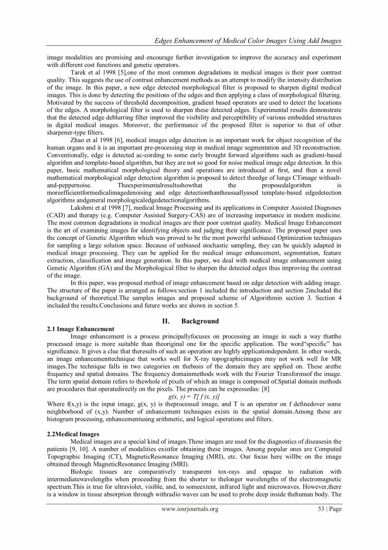
III. Samples Images and Algorithm Below, in figure (1), sample images of proposal method.And proposed scheme of Algorithm in figure (2).

Edges Enhancement of Medical Color Images Using Add Images
www.iosrjournals.org 57 | Page
Figure (1)Images samples
Figure (2) scheme proposed of Algorithm
IV. The Results: Figure (3a, b) shows the results of application of proposal method on the images in figure (1) by Matlab2012a
program.
Original image no. 1
Image acquisition
Edge detection (Canny, Laplacian, Sobel)
RGBto ycbcrcolor space
Find edge detection (Canny, Laplacian, Sobel)
for oneband (Red)
Add output image withOriginal image (Image acquisition)
ycbcr to RGBcolor space
Step a)
Step b)
Step c)
Step d)
Step e)

Edges Enhancement of Medical Color Images Using Add Images
www.iosrjournals.org 58 | Page
Canny Log Sobel
a)
b)
c)
d)
e)
Figure (3a)Therustles of present workapplication on image no. 1

Edges Enhancement of Medical Color Images Using Add Images
www.iosrjournals.org 59 | Page
Original image no. 2
Canny Log Sobel
a)
b)
c)
d)
e)
Figure (3b)The rustles of present work application on image no. 2

Edges Enhancement of Medical Color Images Using Add Images
www.iosrjournals.org 60 | Page
V. Conclusions and Future works: 5.1 Conclusions:
1- The present method “add image with its edges” working to improve and increase the thickness of the edges of
the color medical images (which is difficult to find their edges directly, such as grayscale images known filters), a special that captures a few due to poor quality image obtaining devices, whether or not the user experience.
2- This method helps in medical specialists see clearly on the edges of the resulting images of various types of
medical images (MRI, CT, cat, Pat, ...) color and gray, with any size and any member of human’s body.
5.2 Future works:
1. Apply other filters such as Prewitt and Roberts on the current image in the same method.
2. Application the current method pictures lawsuit in the field of remote sensing.
3. Application outdated video or unclear.
References: [1] Mallat, S. G., “Multifrequency channel decompositions of image and wavelet models”, IEEE Trans. Acoust. Speech Signal
Process.vol. 37, no.12, pp. 2091–2110, 1989.
[2] Muna F. Al-Samaraie. Nedhal Abdul Majied Al Saiyd, “Medical Colored Image Enhancement Using Wavelet Transform Followed
By Image Sharpening”,vol. 6 No. 5, Ubiquitous Computing and Communication Journal, www.ubicc.org.
[3] Hajj, Hazern M.Nguyen, Truong Quang; Chin, Roland T. Multiscale edge detection for medical image enhancement, vol. 3,
pp. 1115-1116, Conference Publications, IEEE, by ISVL,1996.
[4] Gudmundsson, M. El-Kwae, E.A.; Kabuka, M.R. Edge detection in medical images using a genetic algorithm, vol. 17 , Issue: 3 ,
pp. 469-474, Journals & Magazines, IEEE, by ISVL, 1998.
[5] Tarek A. Mahmoud, Stephen Marshall, Medical Image Enhancement Using Threshold Decomposition Driven Adaptive
Morphological Filter, 16th European Signal Processing Conference (EUSIPCO 2008), Lausanne, Switzerland, August 25-29, 2008.
[6] Zhao Yu-qian, Gui Wei-hua, Chen Zhen-cheng, Tang Jing-tian, Li Ling-yun, Medical Images Edge Detection Based on
Mathematical Morphology, Proceedings of the 2005 IEEE Engineering in Medicine and Biology 27th Annual Conference Shanghai,
China, September 1-4, 2005.
[7] B. Lakshmi, G. Lingaiah, S. S. Madhavi, “An enhanced Approach for Medical edge Image enhancement using genetic algorithm”,
International Journal of Computer Science and technology y, ISSN : 0976-8491 (Online), ISSN: 2229-4333 (Print), IJCST, vol.
3, I 1, 2012,
[8] Gonzalez, R. C. and Woods, R. E. Digital Image Processing. (2nd
ed). Addison-Wesley Longman Publishing Co., Inc. 2001.
[9] He, Huiguang; Tian, Jie; Zhao, Mingchang; Xue, Jian; Lu, Ke, “3D Medical Imaging Computation and Analysis Platform”, IEEE
International Conference on Industrial, vol. 6 No. 5, p. 8, Technology ICIT, Dec. 2006.
[10] LathaParthiban; R. Subramanian, “Medical Image Denoising using X-lets”, Annual India Conference, vol., Iss., pp. 1-6, Sept. 2006.
[11] D.Stark and W.Bradley Jr., Ed. Magnetic Resonance Imaging, St. Louis, MO: Mosby, 1992.
[12] Xu, D. and R. Wang, “An improved FoE model for image deblurring”. Int.J. Comput. Vis., 81: 167-171. DOI: 10.1007/s11263-008-
0155-3, 2009.
[13] Y. Watanabe, M. Dohke, T. Ishimori, Y. Amoh, K. Oda, A-Okumura, K. Mitsudo, and Y. Dodo, High-resolution
MRCholangiopancreatography, Critical Rev Diagno Imaging, vol. 39, pp. 111– 258, 1998.
[14] Michael J. Greaney, Douglas C. Hoffman, David F. Garway-Heath, MamdouhNakla, Anne L. Colemna, and Caprioli, “Comparison
of Optic Nerve Imaging Methods to Distinguish Normal Eyes from Those with Glaucoma”, IOVS, vol. 43, No. 1, pp. 140- 145,
2002
[15] Huiqi Li, and OpasChutatape, “Automated Feature Extraction in Color Retinal Images by a Model Based Approach”, IEEE
Transaction on Biomedical Engineering, vol. 51, no. 2, pp. 246-254, 2004.
[16] Yan, X., L. Kai-Yang, Y. Xuan-Dong, W. X. Fang and Z. X. Lin et al,“The application of the edge sharpening operator to the breast
near infrared”. Wuhan Univ. J. Nat. Sci., 2002, 7: 421425. DOI: 10.1007/BF02828241.
[17] Charles Poynton, “Digital Video and HDTV”, Chapter 24, pp. 291–292, Morgan Kaufmann, 2003.
[18] Alparone, L., L. Wald, J. Chanussot, C. Thomass and P. Gamba et al. Comparison of pan sharpening algorithms: Outcome of the
2006 GRS-S data-fusion contest. IEEE Trans. Geosci. Remote Sens., 45: 3012-3021. DOI: 10.1109/TGRS.2007.904923, 2007.
[19] Welsh, T., M. Ashikhmin and K. Mueller, Transferring color to greyscale images. ACM Trans. Graph., 21: 277-280. DOI:
10.1145/566570.566576, 2002.
[20] Zhang X, Wandell B A, “A spatial extension to CIELAB for digital color image reproduction,” Soc. for Info Disp.Symp. Tech
Digest; 27:731-734, 1996.
[21] Mark A. Ruzon, “A Short History of Color Edge Detection”.www.ai.stanford.edu
[22] Eric Hamilton, “JPEG File Interchange Format”, Version 1.02, 1992.