ED 368 492 REPORT NO AVAILABLE FROM PUB TYPE ...
407
ED 368 492 AUTHOR TITLE REPORT NO PUB DATE NOTE AVAILABLE FROM PUB TYPE EDRS PRICE DESCRIPTORS ABSTRACT DOCUMENT RESUME PS 6-2 243 Markel, Howard; And Others The Portable Pediatrician. ISBN-1-56053-007-3 92 407p. Mosby-Year Book, Inc., 11830 Westline Industt.ial Drive, St. Louis, MO 63146 ($35). Guides Non-Classroom Use (055) Reference Materials Vocabularies/Classifications/Dictionaries (134) Books (010) MF01/PC17 Plus Postage. *Adolescents; Child Caregivers; *Child Development; *Child Health; *Children; *Clinical Diagnosis; Health Materials; Health Personnel; *Medical Evaluation; Pediatrics; Reference Materials; Symptoms (Individual Disorders) This ready reference health guide features 240 major topics that occur regularly in clinical work with children nnd adolescents. It sorts out the information vital to successful management of common health problems and concerns by presentation of tables, charts, lists, criteria for diagnosis, and other useful tips. References on which the entries are based are provided so that the reader can perform a more extensive search on the topic. The entries are arranged in alphabetical order, and include: (1) abdominal pain; (2) anemias; (3) breathholding; (4) bugs; (5) cholesterol, (6) crying, (7) day care, (8) diabetes, (9) ears, (10) eyes; (11) fatigue; (12) fever; (13) genetics; (14) growth; (15) human bites; (16) hypersensitivity; (17) injuries; (18) intoeing; (19) jaundice; (20) joint pain; (21) kidneys; (22) Lyme disease; (23) meningitis; (24) milestones of development; (25) nutrition; (26) parasites; (27) poisoning; (28) quality time; (29) respiratory distress; (30) seizures; (31) sleeping patterns; (32) teeth; (33) urinary tract; (34) vision; (35) wheezing; (36) x-rays; (37) yellow nails; and (38) zoonoses, diseases transmitted by animals. (TJQ) *********************************************************************** Reproductions supplied by EDRS are the best that can be made from the original document. ***********************************************************************
-
Upload
khangminh22 -
Category
Documents
-
view
0 -
download
0
Transcript of ED 368 492 REPORT NO AVAILABLE FROM PUB TYPE ...

ED 368 492
AUTHORTITLEREPORT NOPUB DATENOTEAVAILABLE FROM
PUB TYPE
EDRS PRICEDESCRIPTORS
ABSTRACT
DOCUMENT RESUME
PS 6-2 243
Markel, Howard; And OthersThe Portable Pediatrician.ISBN-1-56053-007-392407p.Mosby-Year Book, Inc., 11830 Westline Industt.ialDrive, St. Louis, MO 63146 ($35).Guides Non-Classroom Use (055) ReferenceMaterials Vocabularies/Classifications/Dictionaries(134) Books (010)
MF01/PC17 Plus Postage.*Adolescents; Child Caregivers; *Child Development;*Child Health; *Children; *Clinical Diagnosis; HealthMaterials; Health Personnel; *Medical Evaluation;Pediatrics; Reference Materials; Symptoms (IndividualDisorders)
This ready reference health guide features 240 majortopics that occur regularly in clinical work with children nndadolescents. It sorts out the information vital to successfulmanagement of common health problems and concerns by presentation oftables, charts, lists, criteria for diagnosis, and other useful tips.References on which the entries are based are provided so that thereader can perform a more extensive search on the topic. The entriesare arranged in alphabetical order, and include: (1) abdominal pain;(2) anemias; (3) breathholding; (4) bugs; (5) cholesterol, (6)
crying, (7) day care, (8) diabetes, (9) ears, (10) eyes; (11)fatigue; (12) fever; (13) genetics; (14) growth; (15) human bites;(16) hypersensitivity; (17) injuries; (18) intoeing; (19) jaundice;(20) joint pain; (21) kidneys; (22) Lyme disease; (23) meningitis;(24) milestones of development; (25) nutrition; (26) parasites; (27)poisoning; (28) quality time; (29) respiratory distress; (30)seizures; (31) sleeping patterns; (32) teeth; (33) urinary tract;(34) vision; (35) wheezing; (36) x-rays; (37) yellow nails; and (38)zoonoses, diseases transmitted by animals. (TJQ)
***********************************************************************
Reproductions supplied by EDRS are the best that can be madefrom the original document.
***********************************************************************

U S. DEPARTMENT OF IIIIUCATSONOlfice of Educational Research and Improvement
EDUCATIONAL RESOURCES INFORMATIONCENTER (ERIC)
Wu* document Ras been reproduced imreceived from the person or organization
1 originating it.Manor changes Rave been made to improvereproduction Quality
Points of view or opinions stated in this docirmem do not risciessaity rmxissent officialOERI position or POW,/
, I '
-L;t4,4i
'PERMISSION TO REPRODUCE THISMATERIAL HAS BEEN GRANTED BY
ko...
Gecxx.s
TO THE EDUCATIONAL RESOURCESINFORMATION CENTER (ERIC)
- -.... - , !---.-.-,,.'.- --.7--- -_-,_ --...-
r
2,

-7;
419f N.,
,1.6
f
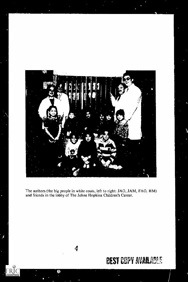
The authors (the big people in white coats, left to right. JAO, JAM, FAO, HM)and friends in the lobby of The Johns Hopkins Children's Center.
4
BEST COPY AVAILIT.F.

. J A
pediatricianHoward Markel, M.D.
Harriet Lane Horne Researcn FellowDepartment of PediatricsThe Johns Hopkins Children's Center;Fellow, Institute of the History of MedicineThe Johns Hopkins University School of MedicineBaltimore, Maryland
Jane A Oski, M.D.Department of PediatricsThe Johns Hopkins Children's Center;Clinical Fellow in PediatricsThe Johns Hopkins University School of MedicineBaltimore, Maryland
Frank A. Oski, M.D.Director and Pediatrician-in-ChiefThe Johns Hopkins Children's Center;Given Professor and ChairmanDepartment of PediatricsThe Johns Hopkins University School of MedicineBaltimore, Maryland
Julia A. McMillan, M.D.Deputy DirectorThe Johns Hopkins Children's Center;Associate Professor and Deputy ChairmanDepartment of PediatricsThe Johns Hopkins University School of MedicineBaltimore, Maryland
HANLEY & BELFUS, INC./PhiladelphiaMOSBY-YEAR BOOK, INC./St. Louis Baltimore Boston Chicago London
Philadelphia Sydney Toronto
5

4.
Publisher: HANLEY & BELFUS,210 South 13th StreetPhiladelphia, PA 19107(215) 546-7293
North American and Worldwide sales and distribution:
In Canada:
MOSBY-YEAR BOOK INC.11830 West line Industrial DriveSt. Louis, MO 63146
MOSBY-YEAR BOOK INC.5240 Finch Avenue EastUnit 1Scarborough, Ontario M1S 5A2Canada
The Portable Peck:Wien
(D 1992 by Hanley & Belfus, Inc. All rights reserved. Noreused, republished, or transmitted In any form or byof the publisher.
Library of Congress catalog card number 91-58777
Last digit Is the print number 9 8 7 6 5 4
6
ISBN 1-56053-007-3
part of this book may be reproduced,any means without written permission
3 2

DEDICATION
We dedicate this book to our parents:Bernice and Samuel MarkelBarbara and Frank OskiSara and Aram Oski
andDorothy and Robert McMillan,
and to all children, past, present, and future.
7
,
,

,
CONTENTS
Preface xi Carotenemia 52
Abdominal Masses 1 Cat Scratch Disease 53
Abdominal Pain 2 Cervicitis 54
Acid-Base 5 Chest Pain 56
Alopecia 6 Cholesterol 57
Alpha-Fetoprotein 7 Clinical Judgment 58
Amenorrhea 8 Cocaine 59
Androgens 10 Coma 60
Anemias 11 Congenital Heart Disease 62
Anion Gap 13 Constipation 65
Anorexia 14 Contraception 66
Antibiotics 16 Cortisol 67
Anticonvulsants 17 Cough 67
Apnea 19 Crayons 63
The Argyll Robertson Pupil 20 Creamatocrit 69
Arthritis 21 Crying 70
Ataxia 25 Cyanosis 73
Back Pain 29 Cystic Fibrosis 74
Bacterial Endocarditis 32 Cystitis 75
Basal Skull Fracture 34 Cytomegalovirus 75
Behavior 35 Day Care 77
Bladder 35 Dehydration 78
Blistering 36 Dermatology 79
Blood Cultures 39 Development 82
Body Temperature 40 Diabetes 87
Bone Marrow Examinations 40 Diarrhea-Chronic 91
Breast Masses 41 Dysmenorrhea 95
Breathholding 43 Dysmorphism 95
Bugs 44 Dysphagia 100
Calcium 49 Dysrhythmia 101
Candidal Infection 51 Dysuria 101
8
Vii

vMContents
Ears 103 Hoarseness 156
Encopresis 104 Human Bites 158
Endotracheal Intubation 104 Human Immunodeficiency 159
Enuresis 106Virus
Eosinophils 107 Hydrops 162
Epididymitis 108Hyperhidrosis 164
Epistaxis 109 Hyperleukocytosis 164
Epstein-Barr Virus 109 Hyperlipidemia 165
Erythrocyte Sedimentation 111 Hypersensitivity 165
Rate Hypertension 166
Eye 112 Hypogonadism 169
Failure to Thrive 115 Hypotonia-Neonatal 170
Fatigue 117 Idiopathic Thrombocytopenic 175
Fever 118 Purpura
Fontanels 124 Immunodeficiency 175
Foreign Body 127 Immunology 177
Fragile X Syndrome 128 Inborn Errors of Metabolism 177
Gastroenteritis 131 Incidence vs. Prevalence 182
Gastrointestinal Bleeding 132 Injuries 182
Genetics 134 Intoeing 183
Growth 135 Intraosseous Infusion 186
Gynccomastia 137 Intussusception 188
Head 139 Iron Deficiency Anemia 189
Headache 139 Iron Overdose 191
Hearing 140 Jaundice 193
Hematology 142 Joint Pain 196
Hematuria 142 Juvenile Rheumatoid Arthritis 197
Hemoglobin 147 Kawasaki's Disease 199
Hemoptysis 149 Kidneys 202
Henoch-Schönlein Purpura 151 Language 203
Hepatomegaly 152 Leukokoria 204
Herpes 153 Limb Pain 204
Hip 154 Limp 205
Hirschprung's Discase 155 Lumbar Puncture 206
Hirsutism 156 Lyme Disease 210
9

Contentslx
Lymphadenopathy 212 Pigmenturia 260(Generalized) Pleural Effusion 262
Magnesium 2 i 7 Poisoning 263Malignant Disease 218 Polyposis 267
Maple Syrup Urine Disease 220 Polyuria 268
Marfan's Syndrome 220 Porphyrias 269
Mean Corpuscular Volume 221 Potassium: Hyperkalemia 269
Meningitis 222 Potassium: Hypokalemia 269
Meningococcal Infection 225 Procedures 270
M icrocytosis 226 Proteinuria 272
Milestones 227 Pruritus 272
Mononucleosis 231 Puberty 274
Movement Disorders 232 Pupils 279
Murphy's Law (Medical 234 Purpura (Petechial and 280Murphology) Ecchymoses)
Neurofibromatosis 237 Pyuria and Bacteriuria 282
Neurologic Development 238 Quality Time 283
Neutropenia 239 Questions 285
Norm), 'qstemia 240 Red Cell 289
Nursemaid's Elbow 241 Renal Failure 289
Nutrition 243 Respiratory Distress 290
Obsessive-Compulsive Disorder 245 Respiratory Viruses 291
Occam's Razor 246 Retropharynx 293
Odor.; of Disease 246 Rett Syndrome 294
Osmolality 248 Rhinitis 295
Pain 249 Rhythmic Behaviors 297
Palsy 249 Rickets 298
Pancreatitis 250 Scrotum 303
Panniculitis 252 Seizures 304
Parasites 252 Sexuality 308
Parvovirus 253 Short Stature 309
Pelvic Inflammatory Disease 254 Shwachman's Syndrome 312
Pertussis 256 Sinuses 312
Phagocytes 257 Skin Signs 313
Phobias 258 Sleeping Patterns 313
I 0

XContents
Snake Bite 314 Tick 354
Sodium: Hypernatremia 320 Torticollis 355
Sodium. Hyponatremia 320 Tourette's Syndrome 356
Splenomegaly 320 Tracheoesophageal Fistula 357
The Spoiled Cbild Syndrome 321 Umbilicus 359
Sports 323 Upper Airway Obstruction 363
Stool 326 Urinalysis 364
Strabismus 328 Urine Output 366
Stridor 323 Urinary Tract 366
Stroke 330 N,aginal Bleeding 369
Sudden Death 333 Vas Deferens 370
Sunlight 333 Vesicles 370
Sweat Test 334 Vertigo and Syncope 372
Syncope 335 Vision 373
Syndromes and Eponyms 338 Vulnerable Child Syndrome 374
Syphilis 342 Walkers 377
Teeth 347 Wheezing 378
Tennis Elbow 347 Wound Care 378
Terminology 349 X-rays 381
Thalassemias 349 Yellow Nails 383
Theophylline 351 Zoonoses 385
Thrombocytosis 352 Index 387
1 1
PREFACE
The Portable Pediatrician is intended to instruct, enlighten, and entertainthose who are studying or providing for the health care needs of children andadolescents. Although this book is clearly not meant to be an all-encompassingtextbook of pediatrics, it is the authors' hope that the busy practitioner, houseofficer, medical student, or nurse can turn to these pages in the quest for animportant bit of information that solves an immediate problem or to replenish hisor her reservoir of knowledge in pediatrics.
We have arranged this book in a dictionary format so that the reader can lookup in alphabetical order the subject or key word at hand for quick and readyreference. A more complete index is available at the end of the volume.References on which the individual entries have been based are also provided sothat the reader can perform a more extensive search on the topic in question.
As in any endeavor, there are acknowledgements to be made. We would hketo express our thanks to Lori Waugh, who patiently transcribed our handwrittennotes into a typewritten manuscript; to Laurel Blewett, formerly ResearchLibrarian for the Department of Pediatrics, The Johns Hopkins Hospital; toJohn J. Hanky and Linda C. Belfus, our editors and publishers; to the manyinterns, residents, and medical students on whom we tried out much of thismaterial in the form of clinical rounds and teaching sessions; and, of course, tothe children who are our patients and who make coming to work each morningsuch a joyful experience.
Howard Markel, M.D.Jane A. Oski, M.D.Frank A. Oski, M.D.Julia A. McMillan, M.D.Baltimore, Maryland
12 xi

SIGNS AND SYMPTOMS-THE DIFFERENTIAL DIAGNOSIS
On some of the pages that follow are listed major signs and symptoms andtheir causes. The causes are classified as COMMON, UNCOMMON or RARE.The COMMON category contains those diseases that, in the aggregate, areresponsible for approximately 90% of the patients who have that particular signor symptom. The term is not meant to suggest that the entity itself is common.The designation UNCOMMON indicates that 1% to 10% of patients with thesymptom or sign will be found in that category, whereas the designation RAREindicates the diseases that are responsible for less than 1% of the symptom or signunder discussion. It is common sense, when confronted with any given sign orsymptom, to consider the COMMON causes first. (These entries are adaptedfrom Dietz HC, Oski FA: Presenting signs and symptoms. In Oski FA,DeAngelis CD, Feigin FD, Warshaw JB (eds): Principles and Practice ofPediatrics. Philadelphia, Lippincott. 1990, pp 2023-2053.)
"A'

ABDOMINAL MASSES
Common Causes
Appendiceal abscessBladder distentionFecal collectionHepatomegaly (any cause)HydronephrosisMulticystic dysplastic kidneyNeuroblastoma
Uncommon Causes
Adrenal hemorrhageHernia (± incarceration)Intestinal duplicationsIntussusceptionLeukemia
Rare Causes
AbscessAnterior meningoceleAortic aneurysmBenign cystic causes
Urachal cystMesenteric cystOmental cystPancreatic cyst/pseudocyst
BezoarHepatobiliary causes
Cholecystitis/ascendingcholangitis
Choledochal cystHemangioendotheliomaHydrops of the gallbladder
Hydrometrocolpos
Polycystic kidney disease(± liv.r involvement)
Pregnancy (± ectopic location)Pyloric stenosisSplenomegaly (any cause)Wilms' tumor
LymphomaOvarian cystRenal vein thrombosisTeratoma (abdominal/ovarian)
Intestinal causesIntestinal atresia (proximal
dilatation)Mairotation with volvulusMeconium plug/ileusRegional enteritis
Retroperitoneal lyrnphangiomaSolid tumors
Granuloma-thecal cell tumorHepatoblastoma
Hepatocellular carcinomaLymphomaMesoblastic nephromaNephroblastomatos isRhabdomyosarcoma
Reference: Oski FA, et al: Principles and Practice of Pediatrics. Philadelphia, J.B.Lippincott, 1990.
1 4
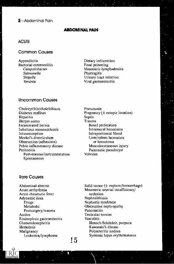
2Abdominal Pain
ABDOMINAL PAIN
ACUTE
Common Causes
AppendicitisBacterial enterocolitis
CampylobacterSalmonellaShigellaYersinia
Uncommon Causes
Cholecystitis/cholelithiasisDiabetes mellitusHepatitisHerpes zosterIncarcerated herniaInfectious mononucleosisIntussusceptionMeckel's diverticulumObstruction (adhesions)Pelvic inflammatory diseasePeritonitis
Post-trauma/instrumentationSpontaneous
Rare Causes
Abdominal abscessAcute arrhythmiaAcute rheumatic feverAdynamic ileus
DrugsMetabolicPostsurgery/trauma
AscitesEosinophitic gastroenteritisGlomerulonephritisHemolysisMalignancy
Leukemia/lymphoma
1 5
Dietary indiscretionFood poisoningMesenteric lymphadenitisPharyngitisUrinary tract infectionViral gastroenteritis
PneumoniaPregnancy (± ectopic location)SepsisTrauma
Bowel perforationIntramural hematomaIntraperitoneal bloodLiver/spleen laceration
or hematomaMusculocutaneous injuryPancreatic pseudocyst
Volvulus
Solid tumor (± rupture/hemorrhage)Mesenteric arterial insufficiency/
occlusionNephrolithiasisNephrotic syndromeObstructive nephropathyPancreatitisTesticular torsionVasculitis
Henoch-Schönleir. purpuraKawasaki's diseasePolyarteritis nodosaSystemic lupus erythematosus

RECURRENT
Common Causes
"Psychophysiologic"Conversion hysteriaDepressionIdiopathic recurrent pain
Uncommon Causes
AerophagiaConstipationDrugs
AntibioticsAnti onvulsantsAspirinBronchodilators
DysmenorrheaEnzymatic deficiency
(e.g., lactose intolerance)Food allergyHepatosplenomegaly (any etiology)
Rare Causes
Abdominal epilepsyAbdominal masses/ma:ignancies
LymphomaNeuroblastomaOvarian lesionsWilms' tumor
Abdominal migraine equivalentAcute intermittent porpnyriaAddison's diseaseAngioneurotic edemaBowel anomaly with obstruction
DuplicationMalrotat ionStenosisWeb
Choledoehal cystCollagen vascular diseaseCystic fibrosis (meconium plug/ileus
equivalent)
Reference: Oski FA, et al: PrinciplesLippincott, 1990.
Abdominal Pain-3
Reaction anxietySecondary gainTask-induced phobia (e.g., school, sports)
Hiatal herniaInflammatory bowel diseaseIrritable bowel syndromeMittelschmerz syndromeParasitic infection
AscariasisGiardiasisStrongyloidiasisTrichinelliasis
Peptic ulcerative diseaseSickle-cell anemiaUrinary tract infection
EndometriosisFamilial Mediterranean feverHeavy metal intoxicationHematocolposHirschsprung's diseaseHyperlipoproteinemiaHyperthyroidismHypoperfusion states
Coarctation of the aortaFamilial dysautonomiaSuperior mesenteric artery syndrome
Mesenteric cyst,Neurologic
CNS mass lesionRadiculopathySpinal cord injury/tumor
Recurrent/chronic arrhythmiaRecurrent pancreatitisWegener's granulomatosis
and Practice of Pediatrics. Philadelphia, J.B.
!6

4Abdominal Pain
The Differential Diagnosis of Acute LowerAbdominal Pain in Adolescent Women
The complaint of lower abdominal pain in a sexually active adolescent femalefrequently points toward the work-up for acute pelvic inflammatory disease (PID)..-3ymptoms that often accompany lower abdominal pain include urinary symptoms,nausea, vomiting, fever, malaise, and dyspareunia. Unfortunately these findingsare seen in other pathologic processes involving the reproductive tract, as well asdisease entities of the gastrointestinal tract and urinary tract. It is obvious,therefore, that one needs to consider a great many problems when evaluating theadolescent female complaining of lower abdominal pain.
Urinary TractCystitisPyelonephritisUrethritisOther
Gastrointestinal TractAppendicitisConstipationDiverticulitisGastroenteritisInflammatory bowel diseaseIrritable bowel syndromeOthe.
Reproductive TractAcute pelvic inflammatory diseaseCervicitisDysmenorrhea (primary/secondary)Ectopic pregnancyEndometriosisEndometritisMittelschmerz
Ovarian cyst (torsion/rupture)Pregnancy (intrauterine/ectopic)Ruptured follicleSeptic abortionThreatened abortionTorsion of adnexaTubo-ovarian abscess
The Closed-eyes Sign in Separating NonspecificAbdominal Pain from the Acute Abdomen
The child presenting with an acute onset of abdominal pain is frequently afrustrating problem for the pediatrician. Indeed, more than 90% of these childrenhave no organic source of pain that would be amenable to surgical intervention,and they are diagnosed as having "nonspecific abdominal pain." A group of threesurgeons at the Radcliffe Hospital of Oxford University looked for the presenceor absence of the "closed eyes" sign during abdominal palpation. Specifically, thesurgeons hypothesized that patients with nonspecific abdominal pain were morelikely to keep their eyes closed when an examiner palpates the abdomen, whereaspatients with abdominal pain of an organic source usually kept their eyes open.The surgeons reasoned that patients with genuine abdominal tenderness are morelikely to keep their eyes open in order to watch the examining physician carefullyand to avoid unnecessary pain. The Oxonians studied 158 consecutive patientspresenting to the emergency room with a complaint of abdominal pain. The datapresented below support a new version of an old adage; the eyes have it.
7
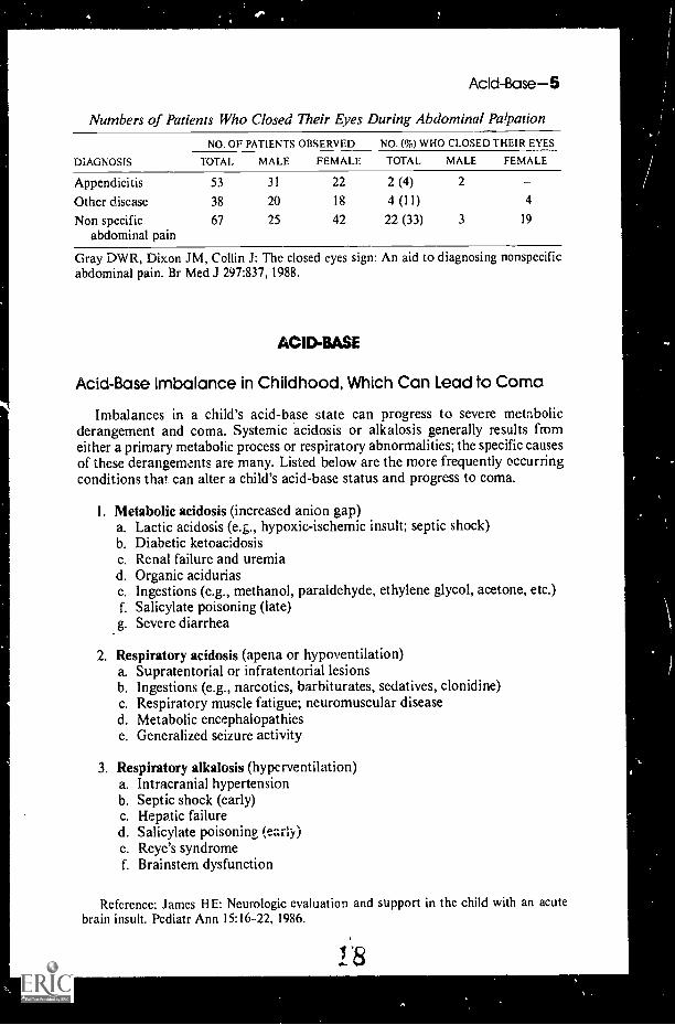
Add-Base-5
Numbers of Patients Who Closed Their Eyes During Abdominal Palpation
DIAGNOSIS
NO. OF PATIENTS OBSERVED NO. (70) WHO CLOSED THEIR EYES
TOTAL MALE FEMALE TOTAL MALE FEMALE
Appendicitis
Other diseaseNon specific
abdominal pain
53
38
67
31
20
25
22
18
42
2 (4)
4 (11)
22 (33)
2
3
-4
19
Gray DWR, Dixon JM, Collin J: The closed eyes sign: An aid to diagnosing nonspecificabdominal pain. Br Med J 297:837, 1988.
AC1D-BASE
Acid-Base Imbalance in Childhood, Which Can Lead to Coma
Imbalances in a child's acid-base state can progress to severe metabolicderangement and coma. Systemic acidosis or alkalosis generally results fromeither a primary metabolic process or respiratory abnormalities; the specific causesof these derangements are many. Listed below are the more frequently occurringconditions that can alter a child's acid-base status and progress to coma.
I. Metabolic acidosis (increased anion gap)a. Lactic acidosis (e.g., hypoxic-ischemic insult; septic shock)b. Diabetic ketoacidosisc. Renal failure and uremiad. Organic aciduriase. Ingestions (e.g., methanol, paraldehyde, ethylene glycol, acetone, etc.)f. Salicylate poisoning (late)g. Severe diarrhea
2. Respiratory acidosis (apena or hypoventilation)a. Supratentorial or infratentorial lesionsb. Ingestions (e.g., narcotics, barbiturates, sedatives, clonidine)c. Respiratory muscle fatigue; neuromuscular diseased. Metabolic encephalopathiese. Generalized seizure activity
3. Respiratory alkalosis (hyperventilation)a. Intracranial hypertensionb. Septic shock (early)c. Hepatic failured. Salicylate poisoning (early)e. Reye's syndromef. Brainstem dysfunction
Reference: James HE: Neurologic evaluation and support in the child with an acutebrain insult. Pediatr Ann 15:16-22, 1986.

6Alopecia
Relationship of pH to Paco, and Base Change
A rapid calculation will often be of great help in interpreting the significanceof blood gas values. When the child is very sick, every second saved can be ofenormous importance. Listed below are two useful facts that can enable you tointerpret the carbon dioxide and the pH results.
1. A change of Paco, of 10 torr is associated with a decrease or increase in pHof 0.08 units:
Paco, 10 torr 1 or I pH 0.08
For example:
Paco, 40 torrpH 7.40NormalPaco, 50 torr pH 7.32Respiratory Acidosis HypoventilationPaco, 30 torrpH 7.48Respiratory Alkalosis--Hyperventilation
2. A base change (base excess or base deficit) of 10 mEq/L is associated witha pH change of 0.15.
For example:
Paco, 40 torr--pH 7.25Normal Paco,--No respiratory componentCalculated pH 7.40Measured pH 7.25pH difference-0.15Base deficit = 10 mEq/L- --Metabolic acidosis
No respiratory componentMetabolic acidosis only
Modified from McMillan JA, et al: The Whole Pediatrician Catalog, Vol 3. Philadelphia,W.B. Saunders, 1982.
Common Causes
Alopecia areataDistal trichorrhexis nodosaPhysiologic (newborns)
Temporal recession at puberty
Uncommon Causes
Acute bacterial infectionsCellulitisFolliculitis decalvansPyoderma
BurnsCancer therapy
AntimetabolitesRadiation
ALOPECIA
1,9
Tinea capitisTraction alopeciaTrichotillomania (also trichologia)
Chemical injuryKerionProximal trichorrhexis nodosaPsoriasisSeborrheaViral infections
Herpes simplexVaricella

Rare Causes
Circumscribed alopeciaAndrogenic alopeciaAplasia cutisConradi's disease (autosomal domi-
nant chondrodysplasia punctata)Epidermal nevi-organoidFollicular aplasiaGoltz's syndrome (focal dermal
hypoplasia)Hair follicle hamartomaIncontinentia pigmenti
InfectionsTuberculosis
Inflammatory etiologiesKeratosis follicularisLichen planusMorpheaPorokeratosis of MibelliSarcoidSystemic lupus erythematosus
Myotonic dystrophyDiffuse alopecia
Anagen effluviumCytostatic agents in plant
MimosineSelemocystothionine
RadiumThallium
Anhidrotic ecterodermal dysplasiaAtrichia congenitaCartilage-hair hypoplasiaChondroectodermal dysplasiaCrouzon's syndrome (craniofacial
dystosis)Hair shaft deformities
Monilethrix
Alpha-fetoprotein 7
Diffuse alopecia (Cont.)Pi li torti
Classic formTrichopoliodystrophy (Menkes
syndrome)Trichorrhexis invaginataTrichorrhexis nodosa
Argininosuccinic aciduriaHallermann-Streiff syndrome
(mandibulo-oculofacial syndrome)Hidrotic ectodermal dysplasiaLanger-Giedion syndrome
(trichorhinophalangeal syndrometype II)
Marinesco-Sjögren syndromeOculodentodigital dysplasiaProgeriaRothmund-Thomson syndrome
(congenital poikiloderma)Telogen effluvium
ChildbirthChronic infection/illnessDrugs
AnticoagulantsAnticonvulsantsAntikeratinizing drugsAntithyroid drugsHeavy metalsHormones
Excessive dietingHigh feverHypothyroidismStressSurgery
Reference: Oski FA, et al: Principles and Practice of Pediatrics. Philadelphia, J.B.Lippincott, 1990.
ALPHAFETOPROTEIN
Maternal Seruma-fetoprotein Screening
Maternal serum a-fetoprotein (MSAFP) screening has been quite successfulin identifying neural tube defects in pregnancy. Approximately 80 to 85% of allopen neural tube defects can be detected by this method. There also may be a
20

8Amenorrhea
relationship of the MSAFP to other birth defects such as Down syncirome andvarious chromosomal abnormalities.
I. Findings associated with elevatedMore advanced gestational ageMultiple gestationFetal deathNeural tube defectsVentral wall defectsCongenital nephrosis
MSAFPOther fetal malformationsOligohydratninosPlacental anomalies or insufficiencyFetomaternal transfusionMaternal liver disease or malignancyNormal pregnancy
2. Adverse outcomes of pregnancy associated with unexplained MSAFP elevationsSpontaneous abortion Intrauterine growth retardationStillbirth Congenital anomaliesPrematurity Possibly pre-eclampsia
3. Findhigs associated with low MSAFPLess advanced gestational age Fetal chromosomal anomaliesMissed abortion (e.g., Down syndrome, trisomiesHydatidiform mole 13 and 18, Turner syndrome)Non-pregnancy Normal pregnancy
Reference: Burton BK: Maternal serum er-fetoprotein screening. Pediatr Ann 18:687-697, 1989.
AMENORRHEA
Amenorrhea in the Adolescent
Amenorrhea is defined as the absence of normal, spontaneous menstrual periodsin a woman of reproductive age. It is typically separated into two forms: primaryamenorrhea (the adolescent female who has never achieved menarche), and second-ary amenorrhea (the cessation of menstrual cycles, once menarche has occurred,for 3 to 6 months). The most common cause of amenorrhea is pregnancy (includingmissed abortion and ectopic pregnancy). It is vital to consider amenorrhea, whetherprimary or secondary, as a symptom and not a disease process in and of itself.Although there is a large number of disease entities that can yield amenorrhea, thebasic disease process generally involves one of the following dysfunctions: (1) inade-quate hormonal stimulation of the endometrium; (2) an iaability of the endometriumto respond to hormonal stimulation; or (3) an obstruction to the outflow of endo-metrial sloughing. The following table outlines the major causes of amenorrhea.
Etiology of Amenorrhea
I. CENTRAL. NERVOUS SYSTEM (GENERAL)A. Infection
I. Encephalitis2. Meningitis
B. NeoplasmI. Craniopharyngioma
B. Neoplasm (('ont.)2. Glioma3. Pineal tumor
C. Congenital anomalies1. Hydrocephaly2. Sellar malformation
21
Table continued on next page.
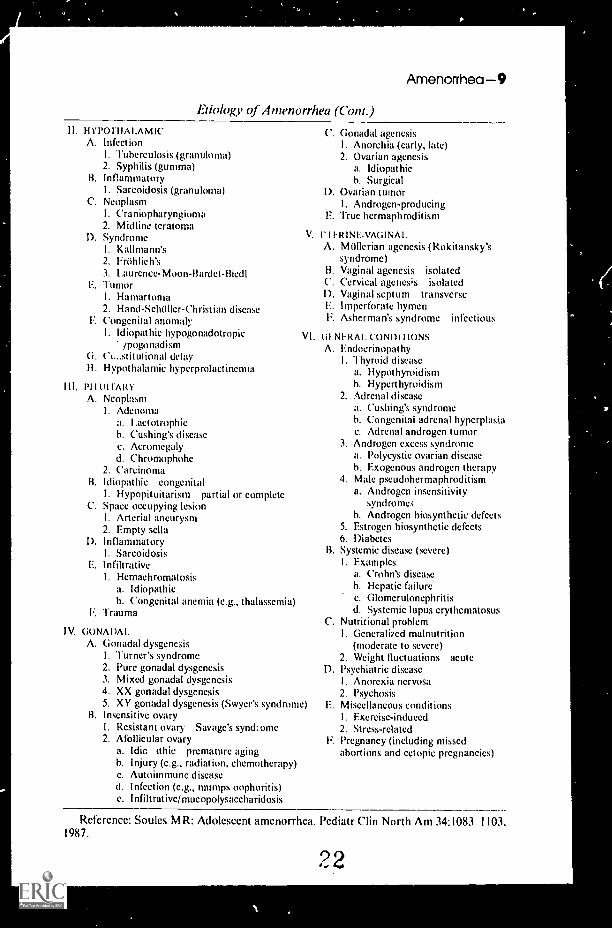
Amenorrhea9Etiology of Amenorrhea (Com.)
I. HYPO THALAMICA. Infection
I. Tuberculosis (granuloma)2. Syphilis (gumma)
B. InflammatoryI. Sarcoidosis (granuloma)
C. NeoplasmI. Craniopharyngiorna2. Midline tcratoma
D. SyndromeI. Kallmann's2. Frithlich's3. Laurence-Moon-Bardet-Biedl
F. TumorI. Hamartoma2. Hand-Schilller-Christian disease
E. Congenital anomalyI. Idiopathic hypogonadotropic
ipogonadismG. Cistitutional delayH. Hypothalamic hyperprolactinetma
111. PIIIUIARVA. Neoplasm
I. Adenomaa. Lactotrophich. Cushing's diseasec. Acromegalyd. Chromophohe
2. (arcinomaB. Idiopathic congenital
I. Hypopituitarism partial or completeC. Space occupying lesion
I. Arterial aneurysm2. Empty sella
I). InflammatoryI. Sarcoidosis
F. InfiltrativeI. Hemachromatosis
a. Idiopathicb. Congenital anemia (e.g., thalassemia)
F. Trauma
V.
VI.
IV. (R)NADAI.A. (ionadal dysgenesis
I. Turner's syndrome2. Pure gonadal dysgencsis3. Mixed gonadal dysgenesis4. XX gonadal dysgenesis5. XV gonadal dysgenesis (Swyer's syndrome)
B. Insensitive ovaryI. Resistant ovary Savage's synd:ome2. Afollicular ovary
a. Idic, ithic premature agingh. Injury (e.g., radiation, chemotherapy)c. Autoimmunc diseased. Infection (e.g., mumps oophoritis)c. Infiltrative/mucopolysaccharidosis
C. (lonadal agenesisI. Anorchia (early, late)2. Ovarian agencsis
a. Idiopathich. Surgical
I). Ovarian tumorI. Androgen-producing
F. True hermaphroditism
MINE-VAGINALA. Mtillerian agenesis (Rokitansky's
syndrome)B. Vaginal agencsis isolatedC. Cervical agenesis isolatedI). Vaginal septum transverseF. Imperforate hymenF. Asherman's syndrome infectious
GI NERAL CONDI I IONSA. Fndocrinopat hy
I. Thyroid diseasea. Hypothyroidismh. Hyperthyroidism
2. Adrenal diseasea. Cushing's syndromeh. Congenital adrenal hyperplasiac. Adrenal androgen tumor
3. Androgen excess syndromea. Polycystic ovarian diseaseh. Exogenous androgen therapy
4. Male pseudohermaphroditisma. Androgen insensitivity
syndrome,;b. Androgen biosynthetic defects
5. Estrogen biosynthetic defects6. Diabetes
B. Systemic disease (severe)I. Examples
a. Crohn's diseaseh. Hepatic failurec. Glomerulonephritisd. Systemic lupus erythematosus
C. Nutritional problemI. Generali/ed malnutrition
(moderate to severe)2. Weight fluctuations acute
D. Psychiatric diseaseI. Anorexia nervosa2. Psychosis
E. Miscellaneous conditionsI. Exercise-induced2. Stress-related
F. Pregnancy (including missedabortions and ectopic pregnancies)
Reference: Soules MR: Adolescent amenorrhea. Pediatr Clin North Am 34:1083 1103,1987.
22
10Androgens
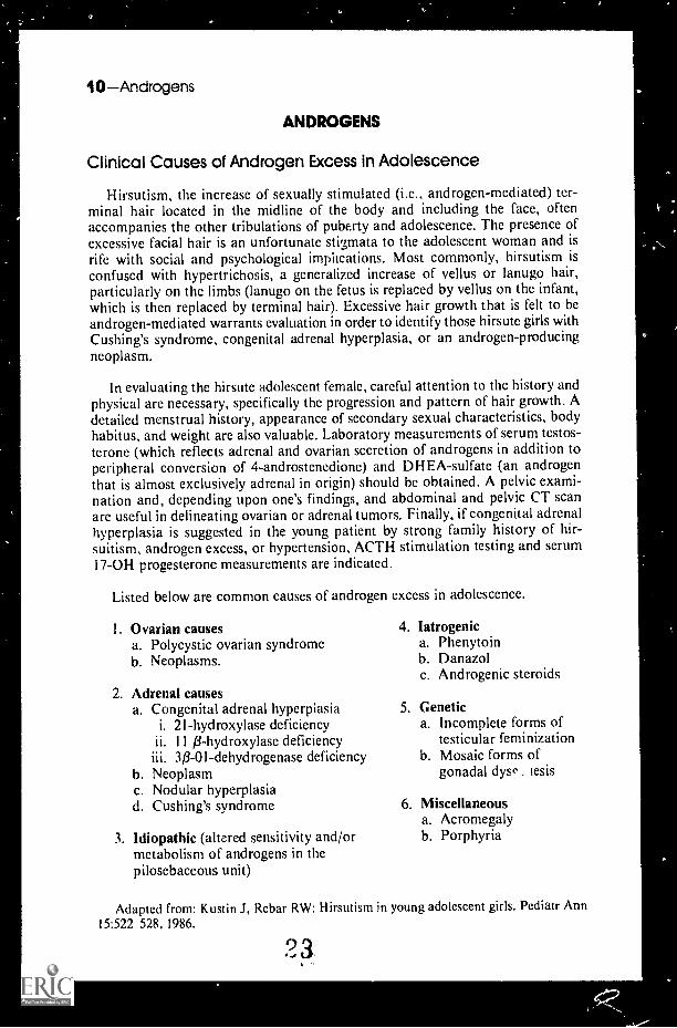
ANDROGENS
Clinical Causes of Androgen Excess in Adolescence
Hirsutism, the increase of sexually stimulated (i.c., androgen-mediated) ter-minal hair located in the midline of the body and including the face, oftenaccompanies the other tribulations of puberty and adolescence. The presence ofexcessive facial hair is an unfortunate stigmata to the adolescent woman and isrife with social and psychological implications. Most commonly, hirsutism isconfused with hypertrichosis, a generalized increase of vellus or lanugo hair,particularly on the limbs (lanugo on the fetus is replaced by vellus on the infant,which is then replaced by terminal hair). Excessive hair growth that is felt to beandrogen-mediated warrants evaluation in order to identify those hirsute girls withCushing's syndrome, congenital adrenal hyperplasia, or an androgen-producingneoplasm.
In evaluating the hirsute adolescent female, careful attention to the history andphysical are necessary, specifically the progression and pattern of hair growth. Adetailed menstrual history, appearance of secondary sexual characteristics, bodyhabitus, and weight are also valuable. Laboratory measurements of serum testos-terone (which reflects adrenal and ovarian secretion of androgens in addition toperipheral conversion of 4-androstenedione) and DH EA-sulfate (an androgenthat is almost exclusively adrenal in origin) should be obtained. A pelvic exami-nation and, depending upon one's findings, and abdominal and pelvic CT scanare useful in delineating ovarian or adrenal tumors. Finally, if congenital adrenalhyperplasia is suggested in the young patient by strong family history of hir-suitism, androgen excess, or hypertension, ACTH stimulation testing and serum17-0H progesterone measurements are indicated.
Listed below are common causes of androgen excess in adolescence.
I. Ovarian causesa. Polycystic ovarian syndromeb. Neoplasms.
2. Adrenal causesa. Congenital adrenal hyperpiasia
i. 2I-hydroxylase deficiencyii. 11 O-hydroxylase deficiency
30-01-dehydrogenase deficiencyb. Neoplasmc. Nodular hyperplasiad. Cushing's syndrome
3. Idiopathic (altered sensitivity and/ormetabolism of androgens in thepilosebaceous unit)
4. latrogenica. Phenytoinb. Danazolc. Androgenic steroids
5. Genetica. Incomplete forms of
testicular feminizationb. Mosaic forms of
gonadal dyse iesis
6. Miscellaneousa. Acromegalyb. Porphyria
Adapted from: Kustin J, Rebar RW: Hirsutism in young adolescent girls. Pediatr Ann15:522 528, 1986.

ANEMIAS
Rule of 34s
Anemias-1i
The sequence of numbers 34 applies to several values in pediatric hematology.These include:
34 mg bilirubin producedigm Hb
3.4 mg iron/gm Hh
3.42 nuclear lobes/neutrophil, the upper limit of normal when averaging 100 ormore neutrophils
1.34 cubic centimeters of oxygen carried by each gram of hemoglobin (if you don'tmind stretching thc rule of 34s a little)
Reference: Sills R: Personal communication, 1977.
Anemia in Early Infancy
During the first months of life there are many causes of anemia. Anemia duringthe first 3 months of life is rarely a result of nutritional iron deficiency. Theaccompanying table is intended to call your attention to the more likely causes ofanemia that occur at birth and at 2 or 3 months of age, as well as to provide youwith leads to establishing the diagnosis.
Common Causes of Anemia in Early Infancy
DIAGNOSIS SUPPORTING DATA
At birth Hemorrhage
Obstetric accidents (placentaprevia, abruptio placentae,incision of placenta, rupture ofcord, rupture of anomalousplacental vessel)
Occult hemorrhage
Fetomaternal
Twin-to-twin
Internal hemorrhage(intracranial, retroperitoneal,intrahepatic, intrasplenic,cephalhematoma)
lsoimmuMzat ion
History and visual inspection ofplacenta and cord
Demonstration of fetal cells inmaternal circulation
Demonstration of significantdifference in hemoglobin values ofidentical twins
Physical examination
Blood groups of mother and infant;evidence of antibody on infant'sred cells
9 4
Table continued on next page.

12Anemias
Common Causes of Anemia in Early Infancy (Cont.)
AGE DIAGNOSIS SUPPORTING D/VI-A
At birth(Com.)
Inherited defect of red cell (includesG-6-PD deficiency, pyruvatekinase deficiency, hereditaryspherocytosis, elliptocytosis,stomatocytosis, etc.)
Acquired defect (generally inassociation with hypoxemia,acidosis, or infection)
Red cell hypoplasia (Blackfan-Diamond syndrome, congenitalleukemia, osteopetrosis)
2-3 Iron deficiency as a consequence ofmonths previous hemorrhage
Late manifestation of previousisoimmunization
Hereditary defects of the red cell
Thalassemia major
Sickle cell anemia
Vitamin E deficiency
Folic acid deficiency
Persistent infection
Renal tubular acidosis
Red cell morphology, family history,and appropriate screening tests
Physical findings, red cell morphology,coagulation disturbance, blood andurine cultures, and serologic studiesand gamma-M determination
Rare disorders; bone marrow aspirate
Obstetric history when available
Blood types of mother and infant;maternal antibody titers
Persistence of hemolytic anemia; redcell morphology and laboratory tests
Red cell morphology, splenomegaly,persistence of fetal hemoglobinelevation, family studies
Red cell morphology, hemoglobinelectrophoresis
Infant of low birth weight; red cellmorphology, low serum E level,positive hydrogen peroxidehemolysis test
Premature infant, history of infectionsor diarrhea, red cell and marrowmorphology, response to folic acid
Elevated titers to rubella,cytomegalovirus, toxoplasmosis
Acidosis, hypochloremia, mildazotemia, urine pH of 6.0 or greaterin presence of acidosis
From McMillan JA, et al: The Whole Pediatrician Catalog. Philadelphia, W.B. Saunders,
1977, with permission.
Anemias Associated with a Low MCV*
98% of anemias with low MCV:Iron deficiencya-thalassemiaP-thalassemia
Others:Lead poisoningtProtein-calorie malnutritionCopper deficiencySideroblastic anemia
*MCV = mean cell volume.Microcytosis most commonly results from associated iron deficiency.
9 5

Anion Gap-13
AN ION GAP
In these days of automated laboratory procedures, most sick patients will havetheir serum electrolytes measured. Obvious abnormalities arc easily recognized.H idden clues to diagnosis are also present in these numbers. Interpretation of theanion gap" provides such a clue.
The principle of electroneutrality is always working and dictates that the sumof the positive charges, i.e., the mEq/L of cations, be exactly counterbalanced bythe number of negative charges, i.e., the mEq/L of anions.
The principal cations in the plasma include sodium, massium. calcium, andmagnesium. The principal anions are chloride, bicarbonate, carbonic acid,dissolved carbon dioxide, albumin, globulin, sulfate, phosphate, and the organicacids, lactic and pyruvic acid.
Measurement of all the anions and cations is not required for interpretationof the patient's status. The serum sodium and potassium are representative ofthe extracellular fluid cations, and, in fact, account for 95% of the cationspresent. Chloride and bicarbonate account for 85% of the anions. Thus, the sumof the usually measured anions does not fully counterbalance thc sum of themeasured cations. Their difference is termed the anion gap. Because of potassium'srelatively low and stable serum concentration, it has only a minor influence on theanion gap. Therefore the anion gap equation can be simplified to read as follows:
Anion gap = sodium (chloride + bicarbonate)
The normal value for the anion gap is approximately 12.0 ± 2.0. The normalrange for the anion gap is thus 8 to 16 mEq/L.
Causes of a high anion gap include:
Metabolic acidosisDehydrat ionTherapy with sodium salts of
strong acids
Therapy with certain antibioticsCarbenicillin, large doses
of sodium penicillinAlkalosis
Specific causes of high anion gap metabolic acidosis include:
Uremia Lactic acidosis Methanol intoxicationKetoacidosis Salicylate intoxication Paraldehyde toxicity
Specific causes of normal anion gap metabolic acidosis include:
Gastrointestinal bicarbonate lossDiarrhea or pancreatic fistula
UreterenterostomyDrugs
AcetazolamideSulfamylonCholestyramineAcidifying agents (ammonium chloride, oral calcium chloride, arginine,
hydrochloride, lysine hydrochloride)Rapid intravenous hydrationHyperalimentationPosthypocapniaRenal tubular acidosis

14Anorexia
Causes of a low anion gap include:
Reduced concentration ofunmeasured anions
DilutionHypoalbuminemia
Systematic underestimation ofserum sodium
Severe hypernatremiaHyperviscosity
Systematic overestimation ofserum chloride
BromismRetained nonsodium cations
ParaproteinemiaHypercalcemiaHypermagnesemia
Reference: Emmett M, Narins RG: Clinical use of the anion gap. Medicine 56:38, 1977.From McMillan JA, et al: The Whole Pediatrician Catalog, Vol 2. Philadelphia, W.B.
Saunders, 1979, pp 104 105, with permission.
ANOREXIA
Common Causes
Acute infectionApparent anorexia
Dieting/fear of obesityManipulative behaviorUnrealistic expectations of caretakers
Uncommon Causes
Chronic infectionDrugs
AminophyllineAmphetaminesAnticonvulsantsAntihistaminesAntimetabolitesDigitalisNarcotics
Esophagitis/gastroesophageal refluxFood aversion in athletesIron deficiency
Rare Causes
Acquired immunodeficiencysyndrome (AIDS)
Adrenogenital syndromeAlcohol/drug abuseAnorexia nervosaChronic diseaseCollagen vascular diseaseCongestive heart failure
?7
Irritable bowel syndromePregnancyPsychosocial deprivation
(neglect/abuse)Psychcsocial factors
Chronic mental/environmental stressAnxietyFearLoneliness/ boredom
DepressionGriefMania
Cyanotic heart diseaseElectrolyte disturbances
HypochloremiaHypokalemia
Endocrine diseaseAddison's diseaseDiabetes insipidusHyperparathyroidism

Endocrine Disease (Cont.)HypothyroidismPanhypopituitarisrn
Hypervitaminosis AInborn errors of metabolismKwashiorkorLead poisoningLiver failureNeurologic
Congenital degenerative diseaseDiencephalic syndromeHypothalamic lesionsIncreased intracranial pressureMental retardation/cerebral palsy
Reference: Oski FA. et al: PrinciplesLippincott, 1990.
Anorexia Nervosa and Bulimia
Anorexia-15
Pain avoidanceAppendicitisConstipationGastrointestinal obstructionInflammatory bowel diseasePancreatitisSuperior mesenteric syndrome
PolycythemiaPostsurgical outcomePulmonary insufficiencyRenal failureRenal tubular acidosisSchizophreniaZinc deficiency
and Practice of Pediatrics. Philadelphia, J.B.
Anorexia nervosa is characterized by excessive weight loss due to a self-inflicted starvation and a morbid, and often unrealistic, fear of becoming too fat.Amenorrhea frequently accompanies this disordei in women; a decreased libidohas been noted in anorexic men. A related eating disorder, bulimia, is compulsiveovereating followed by drastic attempts to avoid gaining weight as a result of theeating binge, e.g., self-induced vomiting, the ingestion ot self-prescribed laxativesor diuretics, and strenuous exercise. These two eating disorders share manyfeatures and can actually evolve from one into the other; classically, anorexianervosa evolves into bulimia. They also have distinctive features that candistinguish one from the other.
Comparison of Anorexia Nervosa (Food- Restricting) and Bulimia
ANOREXIA NERVOSA BULIMIA(E000-RESTRICTING)
Similar Features
I. Psychologicala. Fear of fatnessb. Active pursuit of weight lossc. Fear of loss of control of eating
2. Medicala. Orthostatic hypotensionb. Return to prepubertal breast
developmentc. A menorrhead. Bradycardiac. Lowered core temperature
d. Variable degree of distortion ofbody size
e. Family history of affective disorder
f. Constipationg. Acrocyanosish. Lanugo hairi. Pedal edemaj. Loss of subcutaneous lipid layer
and decreased muscle mass
(All of the above medical complications are the result of starvation.)
Tahle continued on next page.

16Antibiotics
Comparison of Anorexia Nervosa (Food-Restricting) and Bulimia (Cont.)
ANOREXIA NERVOSA BUL IMIA(FOOD-RESTRICTING)
Contrasting FeaturesI. Food intake severely restricted I. Control of intake is lost resulting in binges
2. Less vomiting, diuretic, or laxative 2. Self-induced vomiting, laxative andabuse diuretic abuse
3. Younger 3. Older
4. More obsessional, perfect ionistic 4. More histrionic, antisocial features withcharacteristics loss of impulse control
5. Denies hunger 5. Experiences hunger
6. Severe weight loss 6. Less severe but variable weight loss
7. Most of the medical complications 7. Many medical complications may stemstem from chronic starvation from starvation but there also exist a
number of gastrointestinal complaintsthat result from self-induced vomiting andlaxative and diuretic abuse (e.g., loss ofdental enamel, paratoid gland swelling,dry mouth, esophagitis, gastric dysrhyth-mia, irritable bowel syndrome, and consti-pation). Renal problems. hypokalemicalkalosis, cardiac arrhythmias. and tetanycan also result from '?.xative and diureticabuse. Scars on the dorsum of the handfrom frequent, self-induced vomiting.
8. Eating behavior is a source of pride 8. Eating behavior is a source of shame
9. Less sexually active 9. More sexually active
10. Amenorrhea or loss of sex drive 10. Variable amenorrhea and change in sexdrive
I I. Death from starvation acutely;chroikally from starvation orsuicide
I 1. Death from hypokalemia acutely;chronically from suicide
12. Patient is described as a "model" 12. Patient often exhibits behavioralchild abnormalities
Adapted from: Andersen AE: Anorexia nervosa and bulimia: Biological, psychological,and sociocultural aspects. In Galler JR (ed): Nutrition and Behavior. New York, PlenumPublishing, 1984, pp 305-338.
ANTIBIOTICS
Tasteful Antibiotics, or "Just a Spoonful of SugarHelps the Medicine Go Down"
These days, the successful marketing of antibiotic suspensions for children isas competitive as any other industry. Creating an effective and safe antibiotic issimply not enough. It also has to taste good! Listed below are the flavors, sugarcontent, and availability of some of the most commonly prescribed antibiotics ingeneral pediatric practicc:
29

Anticonvulsantsi 7
Antimicrobial Suspensions Tested
SVSPI NSIONI RAM NAMI-
OUNI-RICNAMF PRODUCFR
SURENCII It(mg 5 ml) COLOR AVOR
PRIMARYWWI NER(S)
Augmentin Amoxicillin!ciavulanate
Beecham 250 mg Cream Orange Saccharin
Trimox Amoxicillin Squibb 250 mg Pink Cherry Sucrose
Ceclor Cefaclor Lilly 250 mg Pink Strawberry Sucrose
Suprax Cefixime Leder le WO mg Cream Strawberry Sucrose
Kctlex Cephalexin Dista 250 mg Orange Bubble gum Sucrose
Dynapen Dicloxacillin Bristol 62.5 mg Pink Orange:pineapple
Saccharinisucrose
Pediazole ErythromycinES;sulfisoxa-iole
Ross 200 mg:600 mg
White Strawberry!banana
Sucrose
ErythrornycinES
Erythromycinethylsuc-cinate
Barr 200 mg Pink Cherry Sucrose
Ilosone Erythromycinestolate
Dista 250 mg Red Cherry Sucrose
Grifulvin V Griseofulvinmicrosiie
Ortho 125 mg Peach Raspberry Saccharin/sucrose
VecTids Penicillin VK Squibb 250 mg Red Berry-like Saccharin;Sucrose
Gantrisin Sulfisoxazole Roche 500 mg White Raspberry Sucrose
Achromycin-V Tetracycline I.ederle 25 mg Red Cherry Sucrose
Sulfatrim Trimethoprim.sulfamoh-oxaiole
Barre 40 mg?200 mg
Pink Cherry Saccharin'sucrose
Reference: Ruff N1E, et al: Antimicrobial drug suspensions: A blind comparison of tasteof 14 common pediatric drugs. Pediatr Infect Dis J 10:30 33, 1991, with permission.
ANTICONVULSANTS
The anticonvulsants often produce side-effects. The commonly used anticon-vulsants and their commonly produced side-effects are described in the tablebelow.
Side-el:frets of Commonly Used Anticonvulsants
ANTICONVULSANT PREDICTABLE IDIOSYNCRATIC
Carhamazepine Diplopia AgranulocytosisDizziness Aplastic anemiaDrowsiness HepatotoxicityHeadache PhotosensitivityNausea Stevens-Johnson syndromeHyponatrernia Lupus-like syndromeHypocalcemia Morbilliform rashOrofacial dyskinesia ThrombocytopeniaCardiac arrhythmia Pseudo lymphoma
Table continued on next page.
3 0

18Anticonvulsants
Side-effects of Commonly Used Anticonvulsants (Cont.)
ANTICONVULSANT PREDICTABLE IDIOSYNCRATIC
Sodium valproate Anorexia PeripheralDyspepsia edemaNausea Weight gainVomiting DrowsinessHair loss TremorRash
Phenytoin AnorexiaDyspepsiaNauseaVomitingAggressionAtaxiaCognitive
impairmentDepressionDrowsinessHeadacheNystagmus
Phenobarbitone FatigueListlessnessTirednessDepressionInsomnia*Distractability*Aggression*Poor memory
Primadone NauseaVomitingDrowsinessWeaknessDizzinessDiplopiaNystagmusAtaxiaPersonality
change
Ethosuximide AnorexiaNauseaVomitingAgitationDrowsiness
Clonazepamiclobazam
FatigueDizzinessDrowsinessAtaxiaIrritability*Aggression*
Paradoxicalseizures
Gum hypertrophyCoarse faciesHirsutismMegaloblastic
anemiaHyperglycemiaHypocalcemiaOsteomalaciaNeonatal
hemorrhage
Decreased libidoI mpotenceFolate
deficiencyNeonatal
hemorrhageHypocalcemiaOsteomalacia
PsychosisNeonatal
hemorrhageDecreased libidoImpotenceHypocaleemiaOsteomalaciaMegaloblastic anemiaNeonatal
hemorrhage
Headache1,ethargyParkinsonismPsychosis
Hyperkinesia*Hypersalivation*Bronchorrhea*Weight gainMuscle weaknessPsychosis
Acute pancreatitisHepatotoxicityThrombocytopeniaHyperammonemiaSt uporEneephalopathyTeratogenicity
Blood dyscrasiasLupus-like syndromeReduced serum IgAPseudolymphomaPeripheral neuropathyRashStevens-Johnson syndromeDupuytren's contractureHepatotoxicityTeratogenicity
Macropapular rashExfoliationToxic epidermal
necrolysisHepatotoxicityDupuytren's contractureFrozen shoulderTeratogenicity
RashAgranulocytosisThrombocytopeniaLupus-like syndromeTeratogenicity
RashErythema multiformeStevens-Johnson syndromeLupus-like syndromeAgranulocytosisAplastic anemia
RashThrombocytopenia
*In childrenAdapted from Brodie MJ: Anticonvulsants. Lancet 336:350 354, 1990, with permission
31

APNEA
Common Causes
Breathholding spellsBronchiolitisExtrinsic suffocationGastroesophageal reflux/aspiration
Uncommon Causes
AsthmaBronchopulmonary dysplasia "spellsCNS hypoperfusionCNS trauma/bleedCongenital airway anomalyHypoglycemiaHypoxemia/hypercarbia (severe)Infection
CroupMeningitis/encephalitis
Rare Causes
AnemiaArrhythmiaGlossoptosisGuil lain-Barre syndromeHypocalcemiaIncreased intracranial pressureInfantile botulismIntraventricular hemorrhageMacroglossiaMetabolic disease
Hyperammonemia
Apnea-19
Idiopathic (? CNS immaturity)PrematuritySeizure
Infection (Cont.)EpiglottitisPertussisPneumoniaSepsis
LaryngospasmI.yaryngo-tracheo-bronchomalaciaObstructive sleep apneaSIDSToxins/drugs
Metabolic disease (Cont.)Inborn errorsMetabolic alkalosis
MicrognathiaOndine's curseSpinal cord injury
Cervical spine instabilityDown syndromeDwarfism
TraumaTumor (CNS, airway)
Reference: Oski FA, et al: Principles and Practice of Pediatrics. Philadelphia, J.B.Lippincott, 1990.
Sleep Apnea
The child with unrecognized sleep apnea may present tr the pediatrician withany number of chief complaints: cardiovascular abnormalities, failure to thrive,pulmonary abnormalities, obesity, apparent mental retardation, and recurrentrespiratory infections. An inadequate history may fail to reveal the culprit.
Sleep apnea can occur in infants, children and adults of any age, although theincidence is known to increase with age and be more prevalent among males thanfemales. The diagnosis depends upon an eye for the predisposing factors and anear for the symptoms.
32

20Argyll Robertson Pupil
Sleep apnea: Predisposing factors
Enlarged tonsils or adenoidsUpper airway or maxillofacial
abnormalitiesHyperthyroidismObesity-Pickwickian syndrome
Down syndromeHypotonic cerebral palsyCongenital myopathiesPharyngeal "sphincter"Dysautonomia
Symptoms of sleep apnea patients % of patients
Snoring usually all night every night; worse withrespiratory infections
91
Apnea--observed by parents 81
Restless sleep and abnormal sleep positions 70
Awakenings from sleep at night 60
Nocturnal enuresis (children > 4 years of age) 33
Daytime somnolence 31
Irritability, hyperactivity 22
Cardiomegaly 6
The most common cause of sleep apnea in infancy and childhood is tonsillarand adenoidal hypertrophy, which may require surgical intervention. Beware thesymptoms and predispositions. You may avert congestive heart failure or corpulmonale!
References: Oski FA, et al: Principles and Practice of Pediatrics. Philadelphia, J.B.Lippincott, 1990.
Tunnessen WW: Pediatric puzzler: A sound clue to FTT. Contemp Peds, Sept. 1:8385, 1984.
THE ARGYU. ROBERTSON PUPIL
Its Clinical Significance
The Argyll Robertson pupil as a sign of tabes dorsalis or neurosyphilis wasdescribed in 1868 by the eye surgeon Douglas Moray Cooper Lamb ArgyllRobertson (1837-1909) of Edinburgh, Scotland. It is a miotic pupil that accom-modates but fails to react to direct light. The sign is caused by lesions to the areaimmediately rostral to the Edinger-Westphal nucleus of the midbrain and can befound in a number of conditions that affect this area (see figure). For example,Charles Dickens, in his 1855 novel Little Dorrit, described a young girl namedMaggy who was severely afflicted with "brain fever" or encephalitis and whoseeyes were "very little affected by light" and stood "unnaturally still." Moreimportant to the present day clinician is the association of the Argyll Robertsonpupil with Bannwarth's lymphocytic meningoradiculitis, a syndrome of radicularpain, cranial nerve palsies, and sensory and motor impairment secondary toinfection with Borrelia burgdorferi or Lyme disease.
33

PGI Lesion I
Arfhriiis-21
Acs.
Lesions in the shaded area (periaqueductal gray [PG)) interrupt descending pathwaysfrom the oculomotor complex to the Edinger-Westphal nucleus (EWN). SC = superiorcolliculus; A = aqueduct: RN = red nucleus; SN = substantia nigra; CP = cerebral peduncle.(From Dasco CC, Bortz DL, Am J Med 86:199 -202, 1989, with permission.)
Listed below are reported non-syphilitic causes of the Argyll Robertson pupil.
I. Diabetes mellitus2. Multiple sclerosis3. Wernicke's encephalopathy4. Dejerine-Sottas progressive hypertrophic neuritis5. Charcot-Marie-Tooth disease (peroneal muscular atrophy)6. Tumors and hemorrhage affecting the Edinger-Westphal nucleus (e.g.,
midbrain tumors such as pinealomas, third ventricle gliomas, andpituitary stalk tumors)
7. Herpes zoster8. Lyme disease (Bannwarth's syndrome)9. Sarcoidosis
10. von Economo's disease (encephalitis secondary to influenza)
References: Dasco CC, Bort/ DL: Significance of the Argyll Robertson pupil in clinicalmedicine. Am J Med 86:199 202, 1989.
Markel H: The childhood sufTering of Charles Dickens and his literary children. Pharos48:5-8, 1985.
ARTHRITIS
Differential Diagnosis of Childhood Arthritis
Arthritis in childhood represents a special problem to the pediatrician, becauseboth the types and etiologies cover a broad spectrum. In the child with suspectedseptic arthritis, an early diagnosis is especially important foi the prevention ofdeformities and/or functional impairment.
34

22Arthritis
Clinical Criteria for Diagnosis of Childhood Arthritis
I. Swelling of a joint2. Limitation of motion with heat, pain, or tenderness
DIAGNOSIS DEFINITION
Juvenile rheumatoid arthritis
Enteroarthritis
Septic arthritis
Transient synovitis of the hip(TSH)
Henoch-Schönlein purpura
Serum sickness
Acute transient arthritis
Arthralgia
Orthopedic disease
Others: mixed connectivetissue disease, systemic lupuserythematosus, polymyositis,acute lymphocytic leukemia
See J RA entries
Antecedent enterobacterial infection ( Yersinia,Salmonella, Shigella, or Campylohacter species)verified by stool culture or agglutination titer 1:160.
Positive bacterial culture from synovial fluid
Acute hip effusion verified by ultrasonography, roent-genography, synovial fluid aspirate, or clinical findings
Typical clinical picture with petechial rash and normalplatelet count
Acute urticaria 5 12 days after vaccination
Disease duration < 3 months; diagnosis of exclusion
'dint pain without trauma; no physical signs of arthritis
Arthroscopically or radiologically verified bone disease,or internal derangement of joint, especially knee
Laboratory Tests in the Differential Diagnosis of Juvenile Arthritis
PM IFNI GROUP
All children with jointsymptoms
'I I'S SIGNIFICANCT
Arthritis lasting longerthan 2 weeks
C reactive protein (CRP),eryt hrocyte sedimentationrate (ESR), CBC, plateletcount, urinalysis, bacterialculture of throat smear
Anti-nuclear antibodies,serum immunoglobulins,Yersinia antibiodies,Salmonella antibodies.stool bacterial culture
CRP > 20, ESR > 20, W BC >1500, and T° > 38.5°C suggestseptic or entcroarthritis. LowCRP and absence of fever withacute limp and hip pain suggestTSH. An ESR 20 in thepresence of a low CRP and nofever suggests JRA or otherconnective tissue disease andnecessitates further immunologicworkup..IRA may also presentas FUO.
Elevated in J RA and other CTdiseases. 1gC, elevated in JRA.Yersinia and/or salmonella Ab'sare thought to be valid indi-cators of enteroarthritis as arcpositive stool cultures. EA onsetgenerally acute while JRAnormally insidious.
0 5
Table continued on next page,

Arthritis-23
Laboratory Tests in the Differential Diagnosis of Juvenile Arthritis (Cont.)
N.TIENT GROUP TEST SIGNIFICANCE
Special indications Rheumatoid factor,antistreptolysin 0 (ASO)
Viral antibodies
Chlamydia antibodies
Rarely indicated in child < 8 years ofage. Both tests for suspected ARF.
Indicated when systemic onset JRAsuspected.
Rare in childhood reactive arthritis.More commonly seen in adults.
Reference: Kunnamo I, et al: Clinical Signs and laboratory tests ii the differentialdiagnosis of arthritis in children. Am J Dis Child 141:34-40, 1987.
The Three Modes of Onset ofJuvenile Rheumatoid Arthritis
Juvenile rheumatoid arthritis (JRA) differs from rheumatoid arthritis inadults in several ways, including types of onset. The three forms of onset of JRAarc: (1) the acute febrile onset (systemic disease), (2) the monoarticular orpauciarticular onset (oligoarthritis), and (3) polyarticular onset (polyarthritis).
Systemic disease is manifested by spiking fevers on a daily basis plus theappearance of a characteristic rash.
Oligoarthritis is defined as onset in four or fewer joints, often only one, usuallythe knee.
Polyarthritis is defined as onset in five or more joints.All three forms can mimic other diseases and the diagnosis is often one of
exclusion, It is important to be intimately familiar with the clinical signs andsymptoms of each type of onset to avoid the serious consequences of misdiagnosis.
Approximately 5% of all cases of JRA begin in childhood (by definitionbefore 16 years, usually between 1 and 3 years). It is the most common pediatricconnective tissue disease, and about a quarter of a million children in the U.S.are affected.
Three Modes of Onset of Juvenile Rheumatoid Arthritis
ACUTE FEBRII.EONSET
MONOARTICUAR POINARTICULARONSET ONSET
Per cent 20 30 50
Joint Mani-festations
One-half have no jointswelling at onset. Theother one-half have onlyarthralgia. Pain may beinferred from thc flexed-knee position in whichthese children tend to lie.
The knee is most commonsite of onset. Other sitesare ankle, elbow, wristand finger joints.Swelling, stiffness, andpain are usually minimal.Painful tendinitis orbursitis, especially of theheel, may be thepresenting symptom.
Four or more joints arcinvolved. May haveabrupt onset with pain-ful swelling of knees,ankles, feet, and hands.May have insidic usonset with no complaintof pain. Joint involve-ment must be inferredfrom guarding move-ments and knee-flexedposition.
36
Table wntinued on next page.

24Arthritis
Three Modes al Onset al Juvenile Rheumatoid Arthritis (Cont.)
ACUTE FE B R I ITONSET
MONOARTICULA RONSET
POLYARTICIlLARONSET
Joint Mani-festations(Cont.)
Fever Daily spikes to i05°F orhigher with temperaturefalling sometimes to sub-normal levels. Fever mayprecede arthritis by weeks,months, or years.
Rash 90ri have macular orslightly maculopapularrash usually on the trunkand extremities, occasion-ally on the neck and face.Rash is rarely pruritic, isusually fleeting withmacules appearing for afew hours during the dayor week, usually in con-junction with fever. Rashis more florid when theskin is rubbed or scratched(Köbner phenomenon).
lridocyclitis Rarely occurs in patientspresenting in this way.
I.ymphade-nopathy
Cardiacmani-festations
May be generalited.Splenomegaly may bepresent. Enlargedmesenteric nodes may leadto abdominal pain andvomiting. Lymphade-nopathy may suggestlymphoma or leukemia.
loq have pericarditisclinically. Pericarditis maylast 2 to 12 weeks and mayrecur years later.
Myoearditis and resultingheart failure tnay occur.
In early stages, thearthritis may beasymmetrical andmigrating.
There may be low-gradedaily fever spikes.
Rash is sometimes present.but is rarely of diagnostichelp.
'Fins group is most suscep-tible to ocular disease. It isoflen asymptomatic andmay smolder for weeks ormonths. It may be the firstmanifestation of the disease.If undetected and untreated,it may lead to blindnessfrom band keratopathyand cataracts. Diagnosismay be made only by slitlamp examination.
Infrequent.
Arthritis may be migra-tory at first. Cervicalspine may be involved.Subcutaneous nodulesare not present.
Low-grade fever withdaily spikes.
Maculopapular rash issometimes present.
Rarely occurs inpatients presenting inthis way.
Infrequent.
Infrequent.
37
Table continued on next page.
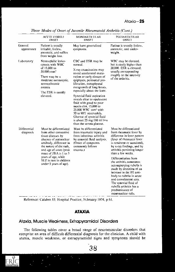
Ataxia 25
Three Modes of Onset of Juvenile Rheumatoid Arthritis (Cont.)
MONOARTICULAR POINARTICULARONSET ONSET
ActrrE FEBRILEONSEI
Generalappearance
I .aboratory
Differentialdiagnosis
Patient is usuallyirritable, listless,anorectic, and suffersfrom weight loss.
Ncutrophilic leuko-cytosis with WBCof 15,000 to50.000imm5
There may be amoderate normocytic,normochromicanemia
The ESR is usuallyelevated.
Must be differentiatedfrom other connectivetissue diseases byabsence of antinuclearantibody, difference inthc nature of the rash,and age of onset (peakonset of JRA is I to 3years of age, whileSI.E is rare in childrenunder 5 years of age).
M ay have generalizedsymptoms.
CBC and ESR may benormal.
X-ray examination mayreveal accelerated matu-ration or early closure ofepiphyses, periosteal pro-liferation, metaphyscalovergrowth of long bones.especially about the knee.
Synovial fluid aspirationreveals clear to opalescentfluid with good to poormucin clot. 15,000 to25,000 WBC, mm5 with50 to 9fri neutrophils.Glucose of synovial fluidis about 25 mg/100 m! lessthan the serum glucose.
Must be differentiatedfrom traumatic injury andfrom infectious arthritisby synovial fluid analysis.(Onset of symptomscommonly followstrauma.)
Patient is usually listless,anorectic, and under-weight.
WBC may be elevated,but is rarely higher than20,000. ESR is elevated.usually correspondingroughly to the intensityof the arthritis.
Must be differentiatedfrom rheumatic fever bydifference in fever pattern(fever of rheumatic feveris remittent or sustained),by x-ray findings, and byarthritis persisting longerthan a few weeks.
Differentiation fromthe arthritis sometimesaccompanying rubella ismade by detection of anincrease in the H I anti-body to rubella in acuteand convalescent sera.The synovial fluid ofrubella arthritis has apredominance ofmononuclear cells.
Reference: Calabro JJ: Hospital Practice, February 1974, p 61.
ATAXIA
Ataxia, Muscle Weakness, Extrapyramidal Disorders
The following tables cover a broad range of neuromuscular disorders thatcomprise an area of difficult differential diagnosis for the clinician. A child withataxia, muscle weakness, or extrapyramidal signs and symptoms should be
38

26Ataxia
examined with particular care because identification of the clinical disorder canoften indicate the site of the lesion.
Differential Diagnosis of Chronic Progressive Ataxia
CI !NU AlDISORDER
PRUCI-1)1N(IHIS IORY
lisumYI AR M-ONS/ I IN
CHILDREN EXAMINA1 ION
l'Sl'AlLAHORAIORY I:Sl'AI.
ENA MINAl ION PROGNOSIS
Arnold-Chiari Headache,malformat ion dysphagia
Hereditary.spinoeere-bellar ataxia
Abetalipo-proteinemia
Dentatecerebellaratax ia
Hereditarycerebellarataxia
Ataxiatelangiectasia
Cerebellartumors
Heredopathiaatacticapolyneuriti-formis
Multiplesclerosis
Spinal cordtumor
Stumbling, 7 10diiliness,familialincidence
Eatty diarrheaat 6 weeks to 2years ol age
2 17
Myoclonus, 7 17
convulsions
Familialincidence
Recurrentsinopulmonaryinfections intwo-thirds ofcases; familialincidence
Headache,vomiting
Anorexia, fail-ing vision,unsteady,familialincidence
Precedingneurologicsymptoms
May havenumbness orbladderdisorder
Palatal andtongue weakness,pyramidal signs,ataxia
Ataxia, loss ofposition sense,extensor plantarresponses, kypho-scoliosis, pcs cavus
Cerebellar ataxia,posterior columnsigns, retinitis pig-mentosa, scoliosis,pes cavus
Ataxia with severeintention tremor
3 17 Ataxia, opticatrophy, occasion-ally associatedposterior columnand pyramidaltract signs
1 3 Oculocutaneoustelangiectasia at 4to 6 years; ataxia,chorcoathetosis,dysarthria
Papilledcma,ataxia, nystagmus
4 7 Retinitispigmentosa,ataxia, deafness,polyneuropathy,ichthyosis
14 17 Optic neuritis;brain stem, cere-bellar, pyramidal,or sensory signs
Ataxia withweakness orsensory loss
May havehydrocephalus,spina bifida
FrequentassociatedFCC; changes
Acanthocytosis,lack of /3-lipo-protein inserum
Slowlyprogressive;stationaryafter surgery
Progressive,with deathusually by30 years ofage
Slowlyprogressive
Slowlyprogressive
Pricumoen- Slowlycephalogram: progressivesmallcerebellar folia
Chestroentgenogram:bronchiectasis;absence of IgAin serum
Skull roent-genogram:separation ofsutures
Elevatedphytanic acidin blood, in-creased spinalfluid protein
Spinal fluidmay revealincreased cells,protein, or -y-globin
Defect onmyelography
Deathbefore 25years of age
Progressiveuntiloperated
Slowlyprogressivewith death
Exacerbationsandremissions
Progressiveuntiloperated
39

INICAIDISORDER
Acutecerebellarataxia
Dilantinintoxication
('erebellartumor orabscess
Hartnupsyndrome
Mult iplesclerosis
Encepha-litides
Spinal cordtumor
Infectiouspolyneu-ropathy
Ataxia-27
Differential Diagnosis of Acute Ataxia
"RECEDINGHIS WRY EXAM INAI
ABORA I DRYE XA M ISM ION
USPAIPROGNOSIS
Half Lave hada prodromalsystemic illness,occasionallyexam hems
Convulsionstreated withphenytoin
Headache,vomiting
Skin eruptionson exposure tosun; familialincidence
Precedingneurologicsymptoms
Headache, stiffneck, fever
May havenumbness orbladderdisorder
Half have aprodromalsystemic illness
Cerebellarataxia
Cerebellarataxia,nystagmus
Papilledema,ataxia,nystagmus
Skin lesions,ataxia, nystag-mus, mentaldisturbances
Optic neuritis;brain stem,cerebellar,pyramidal orsensory signs
Cerebral andbrain stemsigns; also mayhave ataxia
Ataxia withweakness orsensory loss
Ataxia withmotor andsensory loss
Spinal fluidusually r
High serumphenytoin level
Separation ofcranial sutures
Aminoaciduria,increased indolein urine
Spinal fluid mayreveal increasedcells, protein or7-globulin
Spinal fluid:lymphocytosis;possible virusisolation or risein antibody titer
Defect onmyelography
Spinal fluid:normal cells,increased protein
Recovery
Recovery
Progressive untiloperated
Recurrent ataxia
Exacerbationsand remissions
May be fatal, orslow recoverywith or withoutresidual
Progressive untiloperated
May be fatal, butrecovery usuallycomplete
Differential Diagnosis of Disorders of Muscle, Anterior Horn Cell,and Peripheral Nerves
CLINICAL ANI)LABORMORY
FEATURES MUSCLE'AN1 ERIC*HORN CELL
PERIPHERALNERVES
Site of predisposition Usually proximal Proximal and/or Usually distaland axial distal cxtremity extremitymusculature musculature musculature
Deep tendon reflexes Preserved until Reduced to absent Reduced to absentlate in course early in course early in course
Sensation deficit Rarely observed Not observed Usually present
Fasciculations Usually absent Frequently present Occasionallypresent
Table continued on next page.

28Ataxia
Differential Diagnosis of Disorders of Muscle, Anterior Horn Cell,and Peripheral Nerves (Cont.)
CLINICAL ANDLABORATORY
FEATURES MUSCLEANTERIORHORN CELL
PERIPHERALNERVES
CS F protein Normal Normal or elevated Elevated or normal
ElectromyographyInterference Normal until late Reduced Reduced
pattern in disease
Fibrillationpotent ials
Action potentials
Not usually present Usually present
Short duration Prolonged withoccasional giantpotentials
Evoked sensory and Normal Normalmixed nervepotentials
Present
Prolonged withnormal or poly-phasic potentials
Absent, diminishedamplitude, orprolonged con-duction time
Differential Diagnosis of Extrapvrarnidal Disorders
DISORDER FAMILIAI SIGNS ASSOCIATED FINDINGS
Hepatolenticulardegeneration
Autosornalrecessive
Rigidity, tremor,dystonia, dementia,corneal ring,jaundice
Increased urinary andhepatic copper, lowserum ceruloplasmin
Juvenileparkinsonism
Rarely Resting tremor, rigid-ity, bradykinesia
Decr,ased dopaminelevel in substantianigra
Kernicterus No Athetosis, deafness,occasional intel-lectual impairment
Neonatalhyperbilirubinemia
Huntington's Autosomal Rigidity, chorea, con-disease dominant vulsions, dementia
Torsion dystonia Autosomaldominantor recessive
Dystonia, involuntarymovements, normalintellect
Chorea minor No Involuntary choreic Group A streptococcal(Sydenham's) movements, possible
carditisinfections
Absence ofhypoxanthine-guaninephosphoribosyltransferase
X-linkedrecessive
Choreoathetosis,mental retardation,self-mutilation
Increased urinary andblood uric acid
(Lesch-Nyhansyndrome)
Reference: Farmer TW (ed): Pediatric Neurology, New York, Harper & Row, 1975, pp400, 403, 411, and 466, with permission.
41

BACK PAI N
Common Causes
Mechanical derangement (musclestrain or poor posture)
Scheuermann's kyphosis
Uncommon Causes
Disc space infection (discitis)Rheumatic disordersSacroiliac joint infectionsSpina bifida occulta
Rare Causes
Aneurysmal bone cystAseptic necrosis of vertebraeBenign osteoblastomaEosinophilic granuloma of vertebraeHemangioma of boneHerniated nucleus pulposusMalignancy involving bone
(neurobiastoma, leukemia)
A Pain in the Back
ScoliosisSpondylolysis/spondylolisthesis
Spinal cord tumors (lipomas,teratomas)
Vertebral osteomyelitis
Osteomalacia of the spineParaspinal tumor or infecticnSecondary hyperpara-
thyroidismTuberculosis of the spineVertebral osteoid osteoma
The differential diagnosis of back pain in infants and children may not be aslengthy as that of chest pain, but the possibilities are equally perplexing. Unlikeback pain in adults, which frequently defies identification of an etiology, nearly75% of children with back pain have a definable cause. Because the presentationcan be variable, an understanding of the potential etiologies and a rational ap-proach to the work-up can save time and money in needless examinations andtests.
When a child presents with sudden-onset refusal to walk (or sit), irritability,elevated temperature, abdominal pain and/or nausea, vomiting, and anorexiaand laboratory studies consistent with inflammationthe physician mustimmediately differentiate between infectious and noninfectious etiologies.Although the distinction between the generally benign entity of discitis and
29
4 2

30Back Pain
inevitably destructive osteomyelitis is relatively simple (see table), one must alsoconsider meningitis, appendicitis, peritonitis, septic arthritis, and urinary tractinfections. With the help of the flow chart and tables below, differentiation willbe simpler and the oftentimes delayed diagnosis of discitis will not evade thepediatrician.
Back pain and/orgait refusal
Tc -99m No History o physical Yesit. Plain X-ray
bone scan evidence of trauma
Local area of Normal Fracture Normalincreased uptake
1r // Seek nonspinal Local CTVertebral and/or column etiology or MRI scan
disc disorder GU. GI, CNS
11, NormalPlain radiograph
of area of increasedNeurologic
Tc-99m uptakeIf I Yes
/ Normal
Vertebral bone
I.
Observe and reevaluate Myelography
destruction
Jr
* Suspect discitis
Posterior or Anteriorneural arch
Suspect infectionInvasive tumor
Local galliumCT/MRI Of indium WBC scan
cultures. antibiotics
Temperature -39°CESR?---50 mm, W8C>15,CCO
Yes No p Prescribesymptomatic rest.consider analgesia
and rigidimmobilization only if
pain is excessive
Repeal local plainX-rays at 4 wk
Bone destructionand/or Disc space narrowing
soft tissue mass with irregular adiacentvertebral margins
Consider tumorDISCITIS
Problem solving and discitis.
4 3

Back Pain-31
Discitis vs. Intervertebral Infection
INFF'.:TIOUS VERTEBRALDISCI ns OSTEOMYELITIS
Mean age 4 yr 9.8 yr
Sex (M:F) 0.6:1 2.4:1
Complaint cited most often Gait refusal Severe pain, even at rest
History of trauma 20%
Vertebral site Lumbar Thoracic or lumbar
Mean maximum temperature < 100°F > 101° F
Mean maximum WBC count < 8,000/AL > 15,000/AL
Mean maximum ESR < 35 mm > 50 mm
Plain radiographsAt outset Normal Normal
At 4 wk Disc space narrowing Bony destruction
Tc-99m bone scan at outset Positive Positive
Gallium scan at outset Negative Positive
Indium-labeled WBC scan Negative Positiveat outset
Blood or local tissue culture 2% positive 609 positive
Fate of disc Regenerates; often narrow Destroyed
Fate of vertebral body Unaffected Destroyed
Fate of neural arch Unaffected Often destroyed
Fatality 6%
Chronic persistent illness
Clinical duration 2 5 wk; always < 12 wk Many months
Vertebral Disorders in Children
ENTITY USUAL srrE ETIOI.OGY PEAK AGE BEST TEST LABS
Infection Thoracic Staphylococcus, 8 yr Plain x-ray; Blood andor lumbar TB or abscess look for tissue culturespine formation vertebral for 1 WBC,
destruction 1 ESR and1 platelets
TumorsMalignant Low back, Pelvic invasion Adolescence Plain x-ray; Histology
pelvis with marrow look for bonytumors or chon- lesion w/softdrosarcoma tissue mass
Nonmalig- Neural Ostcoblastoma Adolescence Plain x-ray Nonenant arches
Posterior Osteoid Adolescence Look forscleroticnidus ix/lucent halo
Table continued on next page.
4 4

32Bacterial Endocarditis
Vertebral Disorders in Children (Cont.)
ENTITY
Discitis
USUAL SITE ETIOLOGY PEAK AGE BEST TEST LABS
Lumbaranterioraspect
Spondylolysis L 4, L 5vertebrae
Spondylo- L 5, S 1
listhesis vertebrae
Scheuermann's Lowerkyphosis thoracic
vertebrae
Avascularnecrosis of epi-physeal end platesand discs
Traumatic defectin posterioraspect of parsarticularis
Forward slippage(L. 5 moves ante-rior to S- I) in ptw'spondylolysis
Osteochondrosiswiherniation ofdisc into vertebralbodies; wlanteriornarrowing of discspace; disc walledoff in vertebra(Schmorl's node)
4 yr Early: Tc- None99m uptake.After 4 wk:disc-spacenarrowingon plainx-ray
Early teens Oblique Noneplain x-ray
During Standing Nonegrowth lateral plainspurt x-ray
Adolescence Standing Nodelateral plainx-ray showsSchmorl'snode
References: Sills EM: What's causing the back pain? Contemp Pediatr Nov:85 96,1988.Leahy AL, et al: Discitis as a cause of abdominal pain in children. Surgery April:412-414,1984.
BACTERIAL ENDOCARDITIS
Extracardiac Manifestations of Bacterial Endocarditis
The patient with bacterial endocarditis presents both a diagnostic and atherapeutic challenge. The myriad manifestations of the disease result from thehemodynamic, embolic, and immunologic sequelae of the endovascular infection.
The following review of the more common extracardiac manifestations mayserve as an aid in diagnosis and management of this disease.
Extracardiac Manikstations of Bacterial Endocarditis
MANIFESTATION COMMENT
I. RenalI. Microscopic hematuria and
proteinuria2 Occasionally a7otemia3 Abnormalities usually resolve with
effective antimicrobial therapy
I. Biopsya. Focal glomerulonephritis orb. Diffuse proliferative glomerulo-
nephritis
Table continued on next page.
45

Bacterial Endocarditis-33
Extracardiac Manifestations of Bacterial Endocarditis (Cont.)
MANIFESTATION COMMENT
II. NeurologicI. Major neurologic complications are:a. Cerebral infarction in region of
middle cerebral arteries secondaryto emboli (most common neuro-logic complication)
b. Meningeal signs and symptomsc. Seizuresd. Intracranial hemorrhagee. Large macroscopic brain
abscesses are uncommonf. Microscopic brain abscesses are
common and reflect multiplemicroemboli
MusculoskeletalI. Arthralgia --usually in shoulder,
knee, hip2. True synovitis
a. Ankle, knee, wrist most frequentb. Usually sterilec. Biopsy shows acute inflammatory
changes3. Low back pain
a. Often severeb. Often demonstrates spinal
tenderness and decreased rangeof motion
c. X-rays usually normald. Usually not secondary to disc
space infection4. Myalgias --often localized to
thighs and calves5. Miscellaneous
a. Clubbing of the digitsb. Hypertrophic osteoarthropatnyc. Avascular necrosis of hip
IV. SkinI. Petechiae2. Osler's nodes3. Janeway lesions4. Periungual erythema5. Subungual "splinter" hemorrhages
2.
3.
4.
5.
6.
Neurologic complications occur in25-40% of patients with bacterialendocarditisThe mortality of patients with neuro-logic complications is > 50%Embolic phenomenon are usually seenin endocarditis due to S. aureus,Pneumococcus, Enterobacteriaceae,and anaerobic streptococciMitral valve endocarditis producesmajor cerebral emboli more frequentlythan aortic valve endocarditisMycotic aneurysms occur more fre-quently in the early course of acuteendocarditis than late in the course ofsubacute endocarditisCSF exam tends to reflect the natureof the infecting organisms; i.e. virulentorganisms are more likely to producemeningitis with a purulent CSF thanare less virulent organisms, which arelikely to produce a sterile "aseptic" CSF
1. Musculoskeletal findings are seen inapproximately 44% of patients withbacterial endocarditis
Table continued on next page.
4 6

34Basal Skull Fracture
Extracardiac Manifestations of Bacterial EndoCarditis (Cont.)
M ANIFESTATION COM MENT
V. HematologicI. Anemia2. Thrombocytopenia (in the absence of
disseminated intravascular coagulation)3. Monocytosis4. Splenomegaly5. Plasmacytosis of bone marrow6. Disseminated intravascular coag,ilation
VI. Serologic1. Elevated ESR2. Elevated serum gamma globulins3. Positive rheumatoid factor4. Positive antinuclear antibody5. Circulating immune complexes6. Presence in serum of cryoglobulins7. Low serum complement
I. Titers of circt.lating immunecomplexes highest in patients with:a. Right-sided endocarditisb. Extravascular manifestationsc. Signs of infection for more than
4 weeks
References: Pruitt AA, Rubin RH, Karchmer AW, Duncan GW: Medicine 57:329, 1978.Churchill MA, Geraci JE, Hunder GG: Ann Intern Med 87:754, 1977. Bajer AS, Theo-filopoulos AN, Eisenberg R, et al: N Engl J Med 295:1500, 1976.
From McMillan JA, et al: The Whole Pediatrician Catalog, Vol. 3. Philadelphia, W.B.Saunders, 1982, pp 294-297, with permission.
BASAL SKULL FRACTURE
Recognition of Basal Skull Fractures
Basal fractures through the floor of the skull are usually linear. They aredifficult to recognize in x-ray studies and are diagnosed clinically. The clues tothe diagnosis include:
I. Bleeding from nose, eyes, or ears, or discoloration in the mastoid area(ecchymosis behind the ear, or Battle's sign). The "racoon eye" may be seen,with a hematoma in the upper lid.
2. Blood and CSF behind the eardrum, causing a bulging of the membrane.Otorrhea occurs when the tympanic membrane is ruptured.
3. Cerebrospinal rhinorrhea. Some believe that testing the nasal discharge forthe presence of glucose is an indication of CSF leak: However, approximately75 to 90% of normal children will give a positive glucose oxidase test stripin their nasal secretions, which makes the use of such a test for CSF leakvalueless.
4. Cranial nerve palsies, involving cranial nerves I, III, and VIII.5. Appearance of "sinusitis."6. Presence of pneumocephaly.A basal skull fracture can lead to meningitis by spread of organisms from the
nose or ear, and prophylactic use of penicillins is justifiable.
Reference: Hull HE, Morrow G Ill: Glucorrhea revisited. JAMA, 234:1052, 1975.
4 7

Behavior/Bladder-35
BEHAVIOR
Behavioral Concerns of Parents
If you were to provide a behavioral checklist to middle-class parents of children1.5 to 6 years of age, which behaviors would they note as being of greatest concernto them? Listed below are thne behaviors listed in order of frequency.
Are you prepared to discuss these topics with parents?
Behavioral Concerns of Parents
BEHAVIOR PERCENTAGE
Stubbornness 29%-
Poor appetite 23%
Getting child to sleep 22%
Effects of both parents working 22%
Day care 19%
Restless sleep 18%
Temper tantrums 16%
Feelings hurt too easily 16(4-
Problems at meals 15%
"High strung"/easily upset 15%
Wanting too much attention 12%
Disobedient 12%
Hyperactive 12%
New sibling 11%
Moving 10%
Reference: Triggs EG, Perrin EC: Listening carefully: Improving communicationabout behavior and development. Clin Pediatr 28:185 192, 1989.
BLADDER
Bladder Capacity in Children
Bladder capacity correlates linearly with age from birth to the 11 th year. Thebladder capacity in ounces equals age in years plus 2, with a standard deviation of2 ounces. Knowledge of the functional bladder capacity, with a detailed history,may suggest a diagnosis of large or small bladder capacity. Children with infre-quent voiding tend to have larger bladder capacities, whereas those with frequencyor enuresis have smaller than predicted capacities.
Reference: Berger RM, et al: Bladder capacity (ounces) equals age (years) plus 2predicts normal bladder capacity and aids in diagnosis of abnormal voiding patterns. JUrol 129:347- 349, 1983.
48

36Blistering
BLISTERING
Neonatal Blistering Disorders
A number of disorders can give rise to neonatal blisters, ranging from thebenign suction blister (which is presumably caused by thumb, finger, or distalfore',.rm sucking in utero) to epidermolysis bullosa, a heterogeneous group ofinherited skin disorders notable for marked skin fragility. Listed below is thedifferential diagnosis for blistering disorders of the neonate.
Differential Diagnosis for Blistering Disorders
CONDITION ONSET PATHOLOGIC FEATURES CI.INICAl. FEATURES
Epidermolysisbullosa (EB)I. EB simplex (usually Usually Intraepidermal blisters
autosomal dominant at birthinheritance)
2. Junctional EB(autosomal recessiveinheritance)
At birth
3. Dystrophic EB (there At birthexist both autosomalrecessive and auto-somal dominantforms)
Billions congenital At birthichthyosiform erythro-derma (autosomaldominant)
Blistering occurs in thelamina lucida betwemthe epidermis anddermis
Blistering occurs in thedermis, below thelamina densa
Vacuolization of cellsof granular and upperspinous layers
Trauma causes blisters;patients have mildinvolvement withoutscarring in the absence ofinfection. Mucous mern-branes are usually spared.Teeth develop normally.Typically a benign coursewith normal life-span andno significant functionalimpairment.
Trauma causes extensiveblistering on the skin andany mucosa] membrane.In one type (junctional EBletalis of Herlitz-Pearson),GI involvement isfrequent, leading toperforation, sepsis, anddeath in early infancy. Inother subtypes, patientsfollow a more indolentcourse and survive toadulthood, althoughnonhealing cutaneouswounds yield significantmorbidity.
Lesions heal with miliaformation and markedscarring that can lead tocrippling deformities.
Red scaly skin; secondarybacterial infection; thick,grayish brown scales afterage 3 mo.
4 9
Table continued on next page.

Blistering-37
Differential Diagnosis for Blistering Disorders (Cont.)
CONDITION ONSET PATHOLOGIC FEATURES CI,INICAL FEATURES
Congenital herpes In first Intraepidermal blisters Blisters and bullae; positivesimplex virus 20 days; with multiple thin- Tzanck smear and viralinfection mean = 6 walled vesicles on an culture; fever, poor feeding,
days erythematous base hypothermia, and lethargy.
Aplasia cutis At birth Ulcer down to Absence of skin on scalp;congenita (usually subcutaneous tissue similar cutaneous defectsautosomal dominant, may be present elsewhere;but autosomal recess- limb abnormalities; someive also reported) cases associated with
epidermolysis bullosa.
Staphylococcal 2 30 Blisters below or Abrupt onset of erythema,scalded skin days within granular layer followed by blistering andsyndrome exfoliation; responds to
antibiotics.
Suction blisters At birth One or two blisters onthumb, finger, radial aspectof forearm, presumably dueto sucking in utero;spontaneous resolution.
Reference: Lin AN, Carter DM: Epidermolysis bullosa: When the skin falls apart. JPediatr 114:349 355, 1989.
Bullous Eruptions in the Newborn
Eruptions of ,icles (raised, fluid-filled lesions < 1 cm) and bullae (raised,fluid-filled lesions > I cm) in the neonatal period are due to a variety of mostlyunrelated conditions, with different treatments and prognoses in each category.The following table lists the principal criteria for the differential diagnosis ofbullous eruptions in the nursery.
Principal Criteria for Differential Diagnosis of Bullous Eruptions
DISEASE CHARACTER M UCOSAL (YI HEROR OF DIS I-RI- INVOLVE- ECTODERMAI.
CONDITION LESIONS BUTION M ENT DEFECTS SCARRING COURSE
Epider- Clear blisters; Sites of Yes Yes Yes or no Chronicmolysis sometimes trauma or or fatalbullosa hemorrhagic friction
noninflam-matory base
Bullous Blisters, General, Possible No Yes Shortimpetigo clear, opaque, particularly
purulent flexures
Congenital Bullae and Palms, soles, Yes Yes No (other Shortsyphilis maculo- trunk, and than
papules limbs rhagades)
5 (l
Table continued on next page.

38Blistering
Principal Criteria for Differential Diagnosis of Bullous Eruptions (Cont.)
DIM. AS1 CH ARACI FR M UCOSA1 01 H 1' ROR 01. DIM RI- INVOI VI- Hi OMR MAI
CONDITION 1,FSIONS BC 1 ION WV! In FFC IS SCARRING COVRSF
Dermatitis Vesicles and In infants Some- No M inimal One-thirdherpeti- bullae in face and times in long- curable,formis crops; also limbs standing Chronic
urt icari. chiefly cases orlesions involved recurrent
Burns Erythema, Anywhere No No Yes, if Dependsbullae, des- deep on type,quamation depth, and
therapy
Congenital Red urine, Areas No Pigmented Pig- Chronicporphyria photosen- exposed to teeth mented
sitivity of sunlight scarsskin, eryt he-ma, bullae
Erythema Dusky red Trunk, Yes No No Short ormultiforme circinate limbs, face recurrentbullosa plaques,
papules,bullae
Dermatitis May be No No No No Shortmedica- vesicular particularmentosa site
Papular Papules, Trunk No No No Short orurticaria bullae, only or recurrent
vesicles, limbspustules
Chickenpox Trunk, Yes No Yes Shortface, limbs
Smallpox Vesicles, Limbs, Yes No Yes Shortpustules trunk. face
Kaposi's Vesicles, Exposed No Pre-existing No May bevaricelliform pustules parts skin disease fataleruption of infantile
ecrema Or
Besnier'sprurigo
Herpes Vesicles Classical Nc No Yes Shortroster girdle
Bullous Raised Perium- Rarely Nil Nil Short witherysipelas tender ery- bilical, therapy
thema, limbs,bullac face, trunk
Benign Vesicles and Anywhere No No Nil Benignfamilial bullae chronicpemphigus(Hailey's disease)
51Table continued on next page.

Blood Cultures-39
Principal Criteria for Differential Diagnosis of Bulbous Eruptions (('ont.)
DISEASE CFIARACITR MIVOSAI. 01 HEROR OE IHS I RI. INVOI N/E- FrIODERMAI.
CONDITION I .1:SIONS FICTION MEN I- DEE1 CI S SCARRING C0URS1
Contact Often Anywhere No No No Shortdermatitis vesicles
and bullae
Phytophoto- Vesicles Areas No No No Shortdermatitis and bullae exposed to
sunlight
Acrodermatitis Crops Near Yes Hair scanty No May heenteropathica scaling, orifices, fatal
vesiculo- around eyes,bullous elbows,
knees,hands, feet
Reference: Lewis IC, Steven EM, Farquhar PM: Epidermolysis bullosa in thenewborn. Arch Dis Child 30:277, 1955, with permission.
BLOOD CULTURES
Changing the Needle When Inoculating Blood Cultures:A No-Benefit and High-Risk Procedure
For some time now, in response to the high prevalence of HIV and hepatitis Binfections, the U.S. Centers for Disease Control has recommended that needlesshould never be recapped in order to prevent unnecessary needle-stick injuries.Yet, many phlebotomists and physicians routinely recap and change needlesbefore blood-culture inoculation. A group of pediatricians and pathologists atthe University of Virginia were concerned about this clinical paradigm anddesigned a study to compare the extrinsic contamination rate in blood cultureswhen the needle was and was not changed.
The investigators had 108 medical students obtain 182 blood specimens fromeach other using standard methods. Each blood sample was inoculated into twoculture bottles. The first bottle was inoculated with the needle used for phlebotomy,and the second was inoculated after a needle change. Of the 182 culture bottles,4 (2.2%) were contaminated when the needle was not changed and 1 (0.6%) wascontaminated when the needle was changed. This small difference was found to
statistically insignificant, and the possibility of having failed to detect a 5%difference in contamination rate was small.
The conclusion of this study, therefore, was that the risk of needle-stick injuryincurred by changing the needle before inoculation of blood culture bottles seemsto be unjustified.
Reference: Leisure MK, Moore DM, Schwartzman JD, et al: Changing the needlewhen inoculating blood cultures. A no-benefit and high risk procedure. JAMA 264:21112112, 1990.
52

40Body Temperature/Bone Marrow Examinations
BODY TEMPERATURE
A Comparison of Rectal, Axillary, and Inguinal Temperaturesin Full-Term Newborn Infants
What is the mean temperature in the various sites commonly employed for themeasurement of a newbor I's temperature? In the study describcd below, rectalthermometers were insened 2.5 cm into thc rectum. Inguinal temperatures weremeasured by abducting the infant's leg, locating the femoral pulse, placing thebulb of the thermometer lateral to the pulse site, and adducting the leg to createa seal. Readings were recorded every 30 seconds after placement and werediscontinued when no change occurred for 90 seconds.
Akan and Maximal Temperatures and RangesIbr Rectal, Axillary, and Inguinal Sites
SUIT WAN TFMPFRATVRE RANCiF (°F)
Rectal 98.7 97.6 100.4Axillary 98.5 97.2 100.2Inguinal 97.9 96.4 99.2
Reference: Bliss-Holtz: J Nursing Research 38:85 87, 1989.
BONE MARROW EXAMINATIONS
Is There a Role for Bone Marrow Examinationsin the Child with Prolonged Fever?
When evaluating a child with prolonged fever, the question of whether or notto perform a bone marrow examination is often posed. Bone marrow examinationshave been shown to lyz of great use for aid in the diagnosis of malignancy, buttheir usefulness for detecting occult infections has been the source of a long and,excuse the pun, heated debate. Recently, a group of physicians at the TexasChildren's Hospital reviewed 414 cases of children with prolonged fevers in orderto assess this situation.
In their retrospective series, noninfectious causes of prolonged fever wererevealed by the bone marrow examination in 34 (8.2%) of the 414 study patients(e.g., malignant conditions such as acute leukemia, both newly diagnosed andrelapsed, lymphoma, solid tumors, and chronic myelocytic leukemia, in additionto nonmalignant illness such as virus-associated hemophagocytic syndrome,histiocytosis, and hypoplastic anemia). In the majority of these cases, a diagnosisof myelopthisis was clinically suspected before the bone marrow was obtained.
An infectior s etiology of prolonged fever was uncovered in 15 (3.6%) of thefebrile episodes. It should be noted that only one patient of the 414 childrenstudied had a positive marrow culture (for Salmonella, group D) withoutconcurrent positive cultures from any other source. In patients who wereimmunocompetent, the yield of positive marrow cultures was rather low (1.9%),whereas in immunocompromised children (particularly those with AIDS) theyield was 8.7%.
53

Breast Masses-41
With these data in mind, the following conclusions were reached.I. Bone marrow examination is indicated in the child with prolonged fever
and clinical or laboratory evidence consistent with malignancy.2. Bone marrow examination may be helpful in ascertaining the diagnosis of
opportunistic infection in the febrile, immunocompromised patient, espe-cially a child with AIDS.
3. Bone marrow examination in the child with prolonged fever but no findingssuggestive of malignancy or immunodeficiency is probably not warrantedas a means of detecting occult infection.
Reference: Hayani A, Mahoney DH, Fernbach DJ: Role of bone marrow examinationin the child with prolonged fever. J Pediatr 116:919 .920, 1990.
BREAST MASSES
Breast Masses and Lesions in the Infant,Child, and Adolescent
Lesions and masses of the breast generate a great deal of concern upondiscovery. This holds particularly true for such lesions among the pediatric agegroup, despite the fact that the overwhelming majority of lesions in this populationare benign. There do, however, exist some lesions that require ;mmediate atten-tion (e.g., mastitis in the newborn or developing breast and the rarely occurringmalignancy). Clinicians, therefore, need to be able to recognize and assess theselesions in order to offer appropriate treatment and reassurance to their patients.As each age group seen in pediatrics (i.e., neonatal, prepubertal, and adolescent)has its own set of breast masses or lesions, we present the differential diagnosisin a chronologic manner:
In.fancy
I. Neonatal hypertrophy presents as a palpable, tender mass with or withouta milky nipple secretion ("witch's milk"), which is most likely due to a lowprolactin level in a premature infant that rises postnatally to that of anormal-term infant. The milky discharge should abate within 4 to 6 weeksof life, although it may take up to I year for the breast enlargement torecede.
2. Mastitis and resultant abscess present as a tender, erythematous breastmass, usually with fever. This is, perhaps, the only breast lesion in thepediatric population that requires immediate intervention. Mastitis andresultant abscesses tend to occur in the infant aged 2 weeks to 6 months,but can occur at any time. Both gram-positive organisms (e.g.Staphylo-coccus aureus) and gram negative organisms (e.g., E. eoli) arc commonculprits; antibiotic therapy, therefore, should be broad in its spectrum.Warm compresses are also useful. Septicemia is a concern in the younginfant. Increased pressure and inflammation from the infection itself andsurgical drainage by an overzealous surgeon can result in future deformityof the breast in an infant or prepubertal girl.
5 4

42Breast Masses
3. Polythelia is the presei.e of one or more supernumerary nipples along the"milk-line," which extends from the axilla to the symphysis pubis. About50% of the patients with polythelia have some other congenital anomaly:renal anomalies lead the list.
Prepubertal
I. Premature thelarche refers to the onset of bilateral or unilateral breastdevelopment before the age of 7 It is most likely a disorder of hypothalamichormone receptor sensitivity, as opposed to hypothalamic-pituitary tracttumors or primary ovarian neoplasms.
2. Precocious puberty3. Unsustained puberty4. Pseudopuberty5. Gynecomastia (breast enlargement in males)6. Polythelia/polymastia (the presence of more than 2 breasts)
Adolescence
1. Thelarche: normal development of the female breast which may begin asearly as age 8 but, on average, occurs at age 11.
2. Gynecomastia3. Fibroadenoma is the most common breast lesion of adolescent females. It pre-
sents as a unilateral, mobile, slowly growing, isolated, rubbery mass (1-8 cmin size). These lesions are benign.
4. Juvenile giant fibroadenoma is a rapidly growing fibroadenoma seen mostcommonly in adolescent black females. It is a benign tumor usually treatedby surgical excision.
5. Cystosarcoma phylloides is a rapidly growing, large breast mass seen com-monly in adolescent black females. It is firm to palpation, has discrete massborders, and can cause skin or nipple retraction, necrosis, and discharge.Conversion to a malignant tumor has been reported and surgical excision isrecommended.
6. Cystic breast disease involves cystic lesions in the breast that become painfulduring the perimenstrual period. Because the disease is self-limiting andusually resolves at the close of adolescence, hormonal manipulation is ill-advised.
7. IntraductarPapilloma is a benign, subareolar, cylindrical mass with orwithout a brown to frankly bloody discharge.
8. Virginal breast hypertrophy refers to the symmetric enlargement of all breasttissue after puberty. No specific hormonal imbalance has been identified.The breasts can be quite painful and their size embarrassing.
9. Trauma-induced mass (fat necrosis) can result in subsequent scarring with afirm, palpable nodular mass.
10. Polythelia/polymastiaI 1 . Carcinoma12. Metastatic sarcoma
Reference: Dudgeon DI.: Pediatric breast lesions: Take the conservative approach.Contemp Pediatr 2:61 73, 1985.
55

Breathholding 43
BREATHHOLDI NG
Breathholding Spell or Idiopathic Epilepsy?
Breathholding is a common occurrence in infants and children 6 months to 4years of age, with about 5% having at least one breathholding spell and somelosing consciousness during L. prolonged attack. The onset is always with crying,and the characteristic picture of crying and breathholding distinguishes benignepisodes from convulsions. The behavior almost always disappears before schoolage. Some advise parents to leave the room during breathholding to discouragethe behavior, but this may be difficult to carry out.
Features of breathholding that distinguish it from grand mal seizures areshown in the accompanying table
Distinguishing Features of Breathholding and Grand Mal Seizures
GRAND MAI.(IDIOPATHI(' FPILFPSY)
ANOXIC CONVULSION(BREATH HOLDING SPELL)
Age or onset
Family history
Precipitatingfactors
Occurrenceduring sleep
Posture
Sequence andpatterns
Perspiration
Heart rate
Duration
Incontinence andtongue biting
Postictal state
Interictal FF.0
Oculocardiacactivation
fetal FEG
Rarely in infancy
None or positive for epilepsy
Usually absent (or specificsensory stimuli or nonspecificst resses)
Common
Variable
Single cry (may he absent) withloss of consciousness tonic
clonic phases. cyanosis mayoccur later in attack; flushedat first, pale after attack
Warm sweat
Markedly increased
Usually > 1 minute
Common
('onfusion and sleep common
Usually bilateral discharges
No response or bradyeardia;may have asystole of less
than 4 seconds; asymptomatic
Generalized, high-voltagepolyspike discharges, graduallysubsiding into slow waves anddepression for several minutes
Often begins in infancy
Often positive for breath-holding spellor fainting
Usually present (specific emotional ornociceptive stimuli)
Never
Usually erect
Long crying or single gasp, cyanosis orpallor loss of consciousness limp-ness clonic jerks opisthotonosclonic jerks
Cold sweat
Decreased, asystole. or slightlyincreased
Usually I minute or less
Uncommon (but may occur)
No confusion. Fatigue common
Usually normal
About 50% have asystole > 2 seconds,usually > 4 seconds; attack may beprecipated
lsoelectric pattern preceded and fol-lowed by diffuse high-voltage deltawaves, promptly reverting to normalpattern upon recovery of consciousness
References Lombroso CT, Lerman P: Breathholding spells (cyanotic and palidinfantile syncope). Pediatrics 39:563, 1967.
Dimario FJ: Breath-holding spells in childhood. Am .1 Dis Child 146:125 131, 1992.
56

44Bugs
BUGS
Bugs in the Band-Aid Box
Wide-eyed and frightened, they appear with white knuckles clutching themetal Band-Aid box. Their gaze is intense and expectant.
You suspect what is in the box without having them tell."Is it alive?" you ask phlegmatically.Fre4uently, residents of the Band-Aid box include the following:
Crab Lice (Phihirus pubis)This small (1 mm), round, reddish-brown louse causes itching. Transmission
is by close personal contact. On close examination, the crab louse is found in thepubic area with its head buried in a hair follicle or clutching two adjacent hairs.The dark nits are frequently difficult to find. Crabs may infest the chest andaxillary hair as well as the eyelashes. Treatment: 25 per cent benzyl benzoate orgamma benzene hexachloride on two successive days. Infested eyelashes aretreated with daily application of yellow oxide of mercury.
Crab louse (Phthirus pubis)
Scalp Lice (Pedlculus humanus var. capihs)This long (up to 4 mm), slender, white louse causes pruritus and excoriations
with frequent secondary infection. The densest involvement is posteriorly, behindthe ears. There may be tender occipital nodes as well as excoriated bites on theneck and shoulders. You may not find the adult louse, out the small white nitsglued to hair shafts are obvious. Nits fluoresce under Wood's light. Treatment:Gamma ben7ene hexachloride shampoo for two day, , repeated in a week. Combout nits with a fine-toothed comb.
Body Lice (Pediculus humanus var. capon's)The adult louse is 1 to 4 mm long and lives, loves, and lays eggs (nits) in the
seams of clothing. This louse feeds on the body, leaving an urticarial wheal witha hemorrhagic central punctum.
Examination of the skin reveals parallel linear excoriations that often aresecondarily infected. Treatment: Thorough laundering of clothes and bedding.Iron all seams. Bedding and clothing may be dusted with 10 per cent DDTpowder. 1 per cent gamma benzene hexachloride may be applied topically once.
Head or body louse ( Pediculus humanu.scapitis or corporis)
5 7

Bugs-45
Pinworms (Enteral:4as vermlcularis)The patient may find small, white worms at the anal orifice in the early
morning hours. Infestation produces intense perianal pruritus, which leads toexcoriations, lichenification, arid infection. Bruxism and nightmares are common.The diagnosis is usually made by identifying ova on transparent tape that hasbeen pressed to perianal skin at bedtime. Treatment: The Medical Letter hasrecommended pyrantel pamoate (Banminth) (11 mg/kg) as a single oral dose.Mebendazole, 100 x one dose, regardless of weight, may also be used. Thetreatment should be repeated in two weeks.
-110111111ftim....
Female pinworm (E. vermicularis)
Maggots (Hy Larvae)Rarely, maggots will be picked from an open sore, the nose, the ear canal, or
from the stool.
Maggot
Fish Tapeworm allphyllobothrlurn Iatum)This is a very large cestode that produces enormous numbers of yellowish
eggs. It has been an occupational disease of Jewish housewives who taste rawground fish to check seasoning when making gefilte fish. Thus, its incidence maybe decreasing (at least in this population). Immobile, white, flat segments may befound in the stool. Treatment is with niclosamide. 1 gm for children under 35 kgand 1.5 gm for children over 35 kg. The tablets should be chewed thoroughly.
Fish tapeworm (D. laturn)
Beet Tapeworm (Tasnla sagInata)Gravid, white, mobile segments of this worm may be passed in the stool.
Treatment: Quinacrine, 200 mg every 5-- 10 min for four dosages, on an emptystomach, followed by a magnesium sulfate purge 2 to 4 hr later. Niclosamide mayalso be employed in the same dose as for fish tapeworm.
5

46Bugs
Beef tapeworm (T. saginata)
Roundworm (Ascoris Iumbricoldes)Ascaris lurnbricoides is characterized by an elongated, cylindric, nonsegmented,
translucent, flesh-colored body 15-35 cm long. A cosmopolitan worm, ascarisinfects 25 per cent of the world's population. One or more worms may be passedin the stool or, less frequently, vomited. Worms have been known to crawl out ofthe nose, ear, and umbilical fissures! Treatment: Piperazine citrate syrup 75 mg/kg daily x 2. Pyrantel pamoate may also be employed as a single-dose therapy (11mg/kg with a 1-gm maximal dose).
Roundworm (A. lumbricoides)
DebrisVegetable particles, such as seeds (corn), stems, and celery, and other debris,
like dirt, gravel, stringy fuzz, and cellophane, can be swollen and discolored bypassage through the alimentary canal. Even a normal person would be alarmed,and the person with parasitophobia will be in panic. Treatment: Show the patientthe characteristics of the debris by hand lens or dissecting microscope.
MiscellaneousProducts of conception, menstrual blood clots thought to be products of
conception, "grape-like bodies" of hydatidiform mole, fragments of tampons, andclotted mucus arid blood from cystitis have all made it to the Band-Aid box.
Reference: Gottlieb AL Zamkoff KWfastremski MS, Scr.lio A, lmboden KJ: TheBand-Aid box. In The Whole Internist Catalog. Philadelphia, W.B. Saunders Company,1980, pp 497-499, with permission.
Keeping Bugs at Bay
For those of you who hate bugs of all kinds, including mosquitoes, chiggers,flies, and ticks, the following advice from The New York Times ought to come inhandy. We hope this makes your summer more pleasant and a bit iess itchy.

e-
Bugs-47
Keeping Bugs at Bay (Cont.)Entomologists predict clouds of mosquitos this summer, a result of heavy
spring rains. Drain all stagnant water on roofs and in yards. Chiggers, flies andother insects will also be out looking for food. Here are ways to keep flying pests,particularly mosquitoes, from assaulting you.
Repellents Repellents containing DEET are most effective, butapply with care. Do not apply to hands or face or use inconcentrations stronger than 50 percent; 15 percent isrecommended.
Clothing Don't look like a flower. Many bugs are attracted tobright colors.
Perspiration Wash off perspiration. Sweat produces a scent that at-tracts bugs.
Chlorine Add a capful or two of chlorinated bleach to your bathwater or take a swim in a chlorinated pool. The smellrepels most insects.
Sun oil Oily sun screens make skin too slippery for insects to geta grip.
Food and fragrance Avoid alcohol and foods high in serotonin, like bananasand nuts. Mosquitoes are attracted to those scents.Avoid perfume, cologne and hair spray.
Sources: Dr. Roger Grothaus, Raid Center for Insect Control, Racine, Wis., and Dr.Jeffrey D. Bernhart, New York City Department of Health.
From Guidepost: Keeping bugs at bay. The New York Times. 1991, with permission.
6 ( )
9.`
I.

Oh, for boyhood's painless play,Sleep that wakes in laughing day,Health that mocks the doctor's rules,Knowledge never learned of schools,Of the wild bee's morning chase,Of the wild flower's time and place,Flight of fowl and habitudeOf the tenants of the wood;How the tortoise bears his shell,How the woodchuck digs his cell,And the groundmole sinks his well;How the robin feeds her young,How the oriole's nest is hung,Where the whitest lilies blow,Where the freshest berries grow,Where the ground-nut trails its vine,Where the wood-grape's clusters shine,Of the black wasp's cunning way,Mason of his walls of clay,And the architectural plansOf gray-hornet artisans!For, eschewing books and tasks,Nature answers all he asks,Hand in hand with her he walks,Face to face with her he talks,Part and parcel of her joy,Blessings on the barefoot boy!
John Greenleaf WhittierThe Barefoot Boy
61
40

CALCIUM
Idiopathic Hypercalciuria in Childhood
The most common metabolic cause of renal calculi in adults is idiopathichypercalciuria (IH). Children with I H have been noted with increasing frequencyto present with a myriad of lower urinary tract signs and symptoms, includingcalculi, renal colic, hematuria (gross and microscopic), dysuria, frequency-urgency syndrome, pyuria, proteinuria (<100 mg/day), enuresis, osteopenia,failure to thrive, and recurrent urinary tract infections. Given the rather sig-nificant manifestations of 1H in childhood, the potential that these ct ildren willbecome stone-formers in adult life, and the well-known morbidity of urolithiasisin adults, the following review of idiopathic hypercalciuria in childhood isoffered.
I. Definition: Hypercalciuria is an abnormally high urine calcium excretionrate (>2 mg/kg body weight/day) and is either primary (idiopathic) orsecondary to a variety of pathologic entities. Investigators have identifiedtwo types of 1H: absorptive IH, which involves a defect of the intestinalabsorption of dietary calcium; and renal leak IH, which is related to adefect of incomplete renal tubular reabsorption of filtered calcium.
2. Disorders Associated with Hypercalciuria
Immobilization*Diuretic therapy (e.g., furosemide)*Corticosteroid therapy*Type 1 renal tubular acidosisHigh dietary calciumSarcoidosisSyndrome of inappropriate ADH
secretion
HyperparathyroidismCushing's syndromeMedullary sponge kidneyLead poisoningTubular dysfunction
(e.g.. Fanconi's syndrome,Wilson's disease)
Juvenile rheumatoid arthritis
3. Factors Affecting Renal Calcium Excretion
Glomerular filtration rateExtracellular volumeSerum calciumSerum phosphateBlood acid-base statusDietary intake of calcium
and sodium
Vitamin D metabolism (both at theintestinal and renal tubular levels)
Parathyroid hormoneCalcitoninGlucocorticoidsMineralocorticoidsDiuretics (e.g., furosemide)
*The most common causes of hypercalciuria in childhood are indicated by the asterisk.
49
62

50Calcium
4. The laboratory approach to evaluating a child with suspected hypercalciuria
Random urine calcium/creatinine ratioa. If the urine calcium/creatinine ratio is <0.18 mg/mg, quantify with a
24-hour urineb. If the 24-hour urine [Ca] > 2 mg/kg body weight, RULE OUT:
i. Secondary causes of hypercalciuria (obtain serum calcium, phosphate,magnesium, bicarbonate, alkaline phosphatase, and blood pH
ii. Urinary tract infections (perform urine culture).
5. Therapete:c approaches to children with noncalculi urinary tract disordersdue to Ill.a. General treatment measures include a fluid intake large enough to allow
a high urine flow rate but not large enough to complicate disorders suchas enuresis. Excess salt intake should be avoided because increaseddietary sodium and the subsequent increased renal filtered sodium loadcan lead to hypercalciuria. Dietary oxalate (e.g., fruit juices, chocolate,tea) should be avoided because urinary oxalate can serve as the nidus forearly urinary calcium crystalization.
b. Dietary calcium. Although restricting dietary calcium in adults withrenal stones secondary to I H is frequently recommended, such restrictionis usually not indicated in the growing child. A positive calcium balanceis optimal for normal development and bone and tissue growth.However, the restriction of dietary calcium is probably indicated inchildren who develop kidney stones due to absorptive IH and are at riskfor destructive renal disease but, again, the dietary calcium restrictionshould be limited as much as possible. Children who experience recur-rent gross hematuria and severe frequency-urgency syndrome due toabsorptive IH are the only patients who should be prescribed moderate-to-severe dietary calcium restrictions.
c. Pharmacologic therapy. The physician should carefully weigh the risksof the disease's morbidity against the potential side-effects of pharmaco-logic agents. Children presenting with recurrent gross hematuria, debili-tation or severe frequency-urgency syndrome, severe dysuria, persistenturinary incontinence, severe abdominal pain, or recurrent urinary tractinfections are all possible candidates for drug intervention.
Thiazides are the most commonly prescribed agent for I H. These drugspresumably enhance calcium reabsorption in the ascending loop of Henle.
Reference: Heiliezer JD. Canonigo BB, Bishof NA, Moore ES: Noncalculi urinarytract disorders secondary to idiopathic hypercalciuria in children. Pediatr Clin North Am34:711 718. 1987.
Nondairy Foods Rich in Calcium
Considei caring for a lactase-deficient child who required a high dietarycalcium intake. Which calcium-rich foods could you use rather than resorting tomedicinal calcium supplements? A few examples of other-than-cow-in-origincalcium are as follows:
63

CALCIUM: mgPER AVERAGE SERVING
CerealsBarley cereal, Gerbers, 1 cup . 231
Oatmeal, 3/4 cup 153
Pablum barley cereal,3/4 cup, cooked 2210
Pablum cereal, mixed,3/1 cup, cooked 2210
Cereal FloursCornmeal, whole grain, 1 cup 354Soy flour, low-fat, 1 cup 263Wheat flour, self-rising,
enriched, 1 cup 303
FishHerring, canned, solids
and liquids, 3V2 07. 147
Mackerel, canned, 31/2 oz. 260Sardines, Atlantic, canned
in oil, 31/2 oz 354Smelt, Atlantic, canned,
31/2 oz 358
FruitsFigs, dried. 31/2 oz 126
Oranges, 1 large 96
Candidal Infection-51
CALCIUM: mgPER AVERAGE SERVING
NutsAlmonds, unblanched, 31/2 oz 254Brazil nuts, 31/2 oz 186
Legumes and SeedsSoybean curd (Tofu), 4 07 . . . 154Beans, common, dried,
1/2 cup 144Garbanzo beans, dried,
1/2 cup 150
Sunflower seed kernels,31/2 oz 120
Syrups and SugarsMolasses, cane, third extraction,
blackstrap. 31/2 oz 579Maple sugar, 31/2 oz 180
VegetablesBroccoli, cooked, 1 cup 132Spoon cabbage, raw, 3I/2 oz 165
Collards, cooked, I/2 cup 152Dandelion greens, cooked,
1/2 cup 140
Lamb's-quarters, cooked,1/2 cup 258
Reference: American Academy of Pediatrics: Pediatric Nutrition Handbook, 2nd ed.Elk Grove Village, IL, American Academy of Pediatrics, 1985, p 372.
CANDIDAL INFECTION
Penile PlaqueAn Early Sign of Neonatal Candida Infection
Systemic candidal infections occur in immunosuppressed and immunocom-promised patients, particularly those whose normal bacterial flora are suppressedby the use of broad-spectrum antibiotics. Diagnosis of candidal infection is oftendelayed by the failure to suspect fungal disease as well as by delayed growth ofyeast from patient specimens.
Thc premature neonate is both immunodeficient and likely to be treated withmultiple antibiotics. The use of indwelling catheters provides an additional riskfactor for the development of candidal infection.
The development of a white plaque adherent to the tip of the penis butbeneath the foreskin of male premature infants may provide early evidence ofcandiduria and disseminated candidal infection. Urine, blood, and cerebrospinalfluid and/or endotracheal aspirate cultures should be used to confirm thesuspicion provoked by this finding. The detection of the penile plaque allowsearly initiation of antifungal therapy. It is hypothesized that the plaque is formedby Candida that originates in the perineum or the urine and then forms visiblegrowth in the moist area underlying the foreskin.
6 4

52Ca rotenernia
Careful examination of the genitalia is important even in the intensive carenursery.
Reference: Ruderman JW: A clue (tip-off) to urinary infection with Candida. PediatrInfect Dis J 9:586-588, 1990.
CAROTENEMIA
Carotenemia or, Better Expressed, Hypercarotenemia
Carotenemia is a common condition characterized by a yellow-orangediscoloration of the skin and concomitant elevated serum carotene levels. Themajority of these cases are harmless and due to the ingestion of large amounts ofcarotene-rich foods over a long period of time (e.g., carrots, sweet potatoes,squash). The syndrome is associated rarely with disease entities such as diabetesmellitus and hypothyroidism.
Conditions Associated with Carotenernia
Excessive dietary intake (the mostcommon cause of carotenemia)
Diabetes mellitusHypothyroidismSimmonds' disease (panhypopituitarism)Hypothalamic amenorrheaAnorexia nervosaHuman castrates
Clinical Manifestations
Liver diseaseRenal disease (e.g., chronic
glomerulonephritis andnephrotic syndrome)
Inborn errors of metabolismFamilial conditionsMalaria
1. Yellow pigmentation of the skin, most prominently on palms, soles, and naso-labial folds
2. Carotene is excreted by the sebaceous sweat glands; thus, the discoloration ofthe skin is most prominent where sweating is most profuse.
3. It gradually extends over the body.4. Sclera are always spared from carotene staining. (Corneurn of the skin has a
high lipid content with an affinity for carotene; the mucosa has no affinity tocarotene.)
5. Elevated serum carotene level.6. Patients rarely complain of constitutional symptoms, such as loss of appetite,
malaise, itching, and right upper quadrant abdominal pain.
Differential Diagnosis
J a un d iceLycopenemia (orange-reddish discoloration of the skin due to an increased
consumption of lycopene-rich foods, especially tomatoes)Excessive ingestion or percutaneous absorption of chemicals such as quinacrine,
mepacrine, dinitrophenol, saffron, picric acid, and canthaxanthin, the majorcoloring constituent in "tanning capsules."
65

Cat Scratch Disease-53
Treatment
Avoid carotene-rich foods but consume a well-balanced diet. The yellowdiscoloration resolves over several weeks to months, although serum carotenelevels drop severely after only 1 week of a carotene-poor diet. If carotenemia doesnot resolve or an underlying etiology is suspected, an appropriate investigationshould ensue.
Foods High in Carotene Content
Vegetables Fruits OtherAlfalfa Rutabagas Apples ButterAsparagus Spinach Apricots Egg yolksBeans Squash Berries Milk
Beet greens Sweet potatoes Cantaloupes Palm OilBroccoli Yellow turnips Figs Yellow corn
Carrots Watercress Mangoes Yellow fat
Chard OrangesCollard greens PapayasCucumbers PawpawsEndive PeachesEscarole PineapplesKale Prunes
Adapted from: Leung A KC: Carotenemia. Advances in Pediatrics 34:223 248, 1987.
CAT SCRATCH DISEASE
Diagnostic Criteria for Cat Scratch Disease
Cat scratch disease is a self-limited bacterial infection that is usuallytransmitted to humans by felines, although other animals have been implicated.It presents primarily in children with a single lymph node enlargement or regionallymphadenitis and an ocular, skin, or mucous membrane lesion in the region ofthe adenitis. Other manifestations include fever, malaise, headache, anorexia,rash, sore throat, splenomegaly, and, rarely, a severe, chronic, systemic form ofthe illness. The symptoms can last for weeks to months.
The most important historical information in confirming a diagnosis of catscratch disease is whether or not the patient has had contact with a cat; 99% ofcat scratch disease patients have had such contact, and 78% of that populationhave had contact with a kitten. An inoculation site is also vital to the diagnosis.Of the criteria listed below, at least 3 of the 4 noted are required for diagnosis ofcat scratch disease. To these criteria most cat scratch afficianados would addpertinent laboratory data to rule out other causes of lymphadenopathy.
I. Single or regional lymphadenopathy.2. Animal contact, with a scratch or inoculation lesion.3. Positive cat scratch skin test.4. Node or inoculum site with compatible histopathology or Warthin-Starry
stain positive organisms.
t; 6

54Cervicitis
Differential Diagnosis
Other infections causing adenopathy:Infectious mononucleosisMycobacterial infectionStaphylococcal infection
Other fungi
?Noninfectious disorders:
Streptococcal infectionTularemiaSyphilis
ToxoplasmosisSporotrichosis
Lymphoma Congenital cystsSarcoid Kawasaki disease
Reference. Moriarty RA, Margileth AM. Cat scratch disease. Infect Dis Clin NorthAm 1:575-590, 19r.
Unusual Manifestations of Cat Scratch Disease
Common associated symptoms of cat scratch disease include fever, malaise,headache, anu myalgia. One or more of these symptoms occur in approximately50% of patients with the disease. In addition, approximately 50% of patients willhave or recall a painless papule at the site of the scratch. The papule may progressto form a pustule or a vesicle, but it resolves spontaneously after I to 3 weeks andusually precedes the development of lymphadenopathy by several weeks.
A small number of patients with cat scratch disease may develop unusualmanifestations, many of which suggest disseminated involvement. These unusualfeatures include the following:
EncephalopathyRadiculitisOculoglandular syndromeThrombocytopeniaErythema nodosum
Erythema multiformePruritic, maculopapular rashAtypical pneumoniaOsteolytic bone lesionsHepatic and splenic granulomas
Because it is now known that at least some of the above unusual manifestationsof cat scratch disease may be seen in patients without associated lymphadenopathy,it is important to keep this diagnosis in mind when patients are seen with any ofthe problems listed.
References: Carithers HA: Cat-scratch disease: An overview based on a study of 1,200patients. Am .1 Dis Child 139:1124 1133, 1985; Delahoussaye PM, Osborne BM: Cat-scratch disease presenting as abdominal visceral granulomas. .1 Infect Dis 161:71 78,1990; Daye S. McHenry JA, Roscelli JD: Pruritic rash associated with cat scratch disease.Pediatrics 81:559 561, 1988; Malatack .1.1, Altman HA, Nard JA: Cat-scratch diseasewithout adenopathy. J Pediatr 114:101 ,104, 1989.
CERIACITIS
Cervicitis and Vulvovaginitis in the Adolescent
There exists much overlap in the clinical presentations of cervical, vaginal,uterine, fallopian tube, and urinary tract infections. Most of these entities share

Cervicitis-55
the same symptoms such as dysaria, vulvar .pruritis, dyspareunia, and increasedor altered vaginal discharge. It is essential, therefore, when evaluating adolescentwomen with such complaints to (1) exclude the diagnosis of upper tract disease,such as endometritis, salpingitis, and pyelonephritis; (2) differentiate amongvaginitis, cervicitis, urcthritis, and cystitis; and (3) identify the specific etiologicagent that is causing the infection so that the proper treatment can be prescribed.Listed below are the various causes of vulvovaginitis and cervicitis:
I. Vulvovaginitis
a. Physiologic leukorrhea (normal vaginal discharge that increases in volumewith estrogen stimulation)
b. Candidiasis (e.g., Candida alhicans and Torulopsis glahrata)c. Trichomoniasis (Trichomonas vaginalis)d. Bacterial vaginosis (this entity occurs in 30 to 50% of women with
vaginitis, making it the most common cause of abnormal vaginal discharge.It is probably the result of an interplay between the overgrowth ofGardnerella vaginalis and various anaerobes, and the subsequent decreasein the presence of lactobacilli that normally inhabit the vagina)
e. Foreign body (e.g., tampons, IUDs, etc.)f. Allergic or contact vulvovaginitis (e.g., contact with soaps and other
cleaning agents, spermicides, lubricants, douches, sanitary napkins, nylonor rayon underwear, obesity, hot weather, poor hygiene, etc.)Allergic seminal vulvovaginitisPsychosomatic illness should be considered when an adolescent frequentlypresents with vaginal symptoms but without objective evidence of vulvaror vaginal inflammation or discharge.
g.h.
2. Other Causes of Vulvovaginal Complaints
a. Systemic conditions.i. Fistulas from the bladder or rectum (e.g. Crohn's disease, Stevens-
Johnson syndrome, Behget's syndrome)ii. Tropical ulcerations (e.g., amebiasis, filariasis, tuberculosis, schisto-
somiasis)iii. Systemic illnesses (e.g., typhoid, smallpox, varicella, measles, scarlet
fever)iv. Dermatologic complaints (e.g., atopic dermatitis, seborrheic dermati-
tis, psoriasis, lichen sclerosus)v. Anatomic anomalies (e.g., aberrant urethral orifice, labial agglutina-
tion, urethral prolapse)
b. Vulvar lesionsi. Condyloma acuminaturnii. Genital herpes
iii. Syphilisiv. Chancroidv. Lymphogranuloma venereum
vi. Granuloma inguinalevii. Pediculosis
viii. Scabies
ix. Bartholinitisx. Skenitisxi. Tumors (e.g., carcinoma,
sarcoma, botryoides,vaginal polyps)
xii. Pemphigusxiii. Acute ulcerative vulvitisxiv. Lipschütz ulcer

56Chest Pain
3. Cervicitis
Whatever its etiology, cervicitis should be considered a sexually transmitteddisease with great potential to spread to other sexual partners as well as toextend, in the case of contact, from the cervix to the endometrium and salpinx.Etiologies include:
a. Chlamydia trachomatisb. Neisseria gonorrhoeaec. Herpes simplex virusd. Possible etiologic agents include Mycoplasma hominis, Ureaplasma
urealyticum, and group B streptococci.
Reference: Rosenfeld WI), Clark J: Vulvovaginitis and cervicitis. Pediatr Clin NorthAm 36:489 511, 1989.
CHEST PAIN
Common Causes
CostochondriticArthritisInfectious costochondritisTietze's syndrome
CoughHerpes zosterIdiopathicIndigestion (heartburn, esophagitis)
Uncommon Causes
ArrhythmiaCongenital heart diseaseCongestive heart failureEsophageal (trauma associated
with vomiting, foreign body)
Rare Causes
CholecystitisDiaphragmatic irritation
AbscessFitz-Hugh Curtis syndromePeritonitisRuptured viscusTumor
EndocarditisJuvenile rheumatoid arthritis
69
Mitral valve prolapseMusculoskeletal (strain, occult trauma)PneumonitisPsychogenicReactive airway diseaseSickle-cell diseaseTrauma
Pleuritis/pleurisyPneumothoraxPrecordial catch
Myocardial ischemia (e.g., anomalouscoronary artery)
MyocarditisOsteomyelitis (vertebrae, ribs)Peptic ulcerative diseasePericarditisPneumomediastinumPulmonary embolismRheumatic fever

Cholesterol-57
CHOLESTEROL
Screening and Managing Cholesterol Levels in Children
The following table summarizes the American Academy of Pediatrics (AAP)/American Heart Association's (AHA) and the National Cholesterol EducationProgram's (NCEP) recommendations for testing and managing cholesterol inchildren. The American Health Foundation's (AHF) recommended universal,population-based approach to cholesterol control is also presented in outline.
Recornmendations for Screening and Managing Cholesterol in Children
High-risk, patient-based approachAAP AHARECOMMENDATIONS NCEP RECOMMENDATIONS
Screeningstrategy
Screeningmethod
Management
Screen children over 2 yrof age who have a familyhistory of hyperlipidemia(parent, sibling, grand-parent, uncle. aum) orearly myocardial infarc-tion (males under 50 yr,females under 60 yr).
Screen initially with fast-ing lipid profile. Repeatand average Ifelevated, exclude second-ary causes ofhyperlipidemia.
Screen children over 2 yr who have a parentalhistory of hyperlipidemia (>240 mgidl totalcholesterol) or a family history of early CAD(undcr 55 yr for males and females).* Also con-sider screening if family history is not obtainableor patient has several risk factors for CAD inde-pendent of history (obesity, hyperteriiion,smoking).
If child has family history of early CAD, screeninitially with two lipid profiles.'In other high-risk patients, screen initially withnonfasting total cholestera If total cholesterol isborderline (170 199 mgkil), repeat and averageresults. If initial screening measurement is 200mg, dl or more, oi. thc average of two measure-ments is >170 mg/di, perform two lipid profiles.'
Total cholesterol is normal (<170 mg/di): Rou-tine care, repeat cholesterol testing 5 yr later.
I.ow-risk lipid profile (I.D11.-C <110 mg/d1, lessthan 75th percentile): Routine care, repeat lipidprofile every 5 yr.
Moderate-risk lipid profile (I.DI.-C 110 129 mg;dl, 75th to 95th percentile): Dietary counseling.follow-up lipid profile in I yr.
H igh-risk lipid profile (LDI.-C ?:130 mg/c11, 95thpercentile): Dietary counseling, perform lipidprofiles on parents, initiate step 1 diet. Repeatlipid profile in 6 wk. If unchanged, intensify step1 diet and repeat lipid profile in 3 mo. If stillunchanged, initiate step 2 diet and repeat lipidprofile in 3 mo. Refer to lipid specialist if diettherapy ineffective.
NCEP defines a family history of early CAD as having inrents or grand patents 55 yr of age or underwho have had one or more of the following: coronary atherosclerosis diagnosed by coronary arteriography;balloon angioplasty or coronary artery bypass surgery; documented myocardial infarction, anginapectoris, peripheral vascular disease, cerebrovascular disease, or sudden cardiac death.A repeat nonfasting totai cholesterol measurement or second fasting lipid profile should b.: done no soonerthan I wk after initial test and no later than 8 wk after initial test. The second measurement is unacceptableif the total cholesterol or 1..D1.-C level is within 30 mg;dl of the measurement. If unacceptable,obtain a third measurement.
Table continued on next page.
70

58Clinical Judgment
Rccommendations for Screening and Managing Cholesterol in Children (Cont.)
Universal, population-based approach (recommended by AHF)
Screeningstrategy
Screeningmethod
Screen all children over 2 yr of age. Consider repeat screening every 5 yrthereafter.
Screen initially with nonfasting total cholesterol, and proceed to fasting lipidprofile if levels are elevated as described above under NCEPrecommendations.
Management See recommendations above.
Reference: Modified from Schuman AJ: A guide to office cholesterol testing. ContempPediatr October:17-42, 1991.
CLINICAL JUDGMENT
Quantifying Judgment in the Evaluation of Sick Children
We all use judgment and experience to distinguish the "sick" child from the"not-so-sick" child. No amount of wisdom and no laboratory test will make thisdistinction accurate in every case, but the scale below will help you quantifyyour judgment, or at least it will help you recognize how you reach your ownconclusions.
Six Observation Items and Their Scales*
OBSERVATION ITEM I NORMAI.3 MODERATEIMPAIRMENT
5 SEVEREIMPAIRMENT
Quality of cry
Reaction tostimulation
Strong with normal Whimpering ortone or content and sobbingnot crying
parental Cries briefly thenstops or contentand not crying
If aware, staysawake; or if asleepand stimulated,wakes up quickly
Color Pink
State variation
Hydration
Response (talk.smile) to socialovert ures
Skin normal, eyesnormal, andmucous membranesmoist
Smiles or alertsmu)
Cries off and on
Eyes close brieflyawake; awakes withprolongedstimulation
Pale extremities oracrocyanosis
Skin, eyes normaland mouth slightlydry
Brief smile, alertsbriefly 2 mo)
*Source see reference below; \kith permission.
71
Weak or moaning orhigh pitched
Continual cry orhardly responds
Fails ; feet) or willnot ro
Pale, cyanotic,mottled, or ashen
Skin doughy ortentcd, and drymucous membranesandior sunken eyes
No smile; faceanxious, dull, andexpressionless or noalerting (.5_2 mu)

Cocaine-59
To use this system, assign a score of 1-3 for each of the observation items.When the investigators used the scale to evaluate children less than 24 months ofage who had fever of 38.3°C or more, only 2.7% of children with a score of lessthan 10 had a serious illness, whereas 92.3% of those with a score greater than orequal to 16 were seriously ill. Among those children with scores of II to 15,serious illness was found in 26.2%. A child was defined as having a serious illnessif a bacterial pathogen was isolated from blood, CSF, urine, stool, joint fluid, ordeep soft tissue aspirate, or if electrolyte abnormality, pulmonary infiltrates onchest x-ray, abnormal blood gases, or CSF pleocytosis was found.
It might be helpful to compare your own instincts to this scale to find out whyyou conclude what you do.
Reference: McCarthy PL., Sharpe MR, Spiesel SZ, et al: Observation scales to identifyserious illness in febrile children. Pediatrics 70:802, 806 (table), 1982.
COCAINE
Differential Diagnosis of Cocaine Intoxication
The acute manifestations of severe cocaine ingestion are many. Manifestationsmay he divided systematically:
I. Autonomic nervous systema. Dilated pupilsb. Tachycardia
2. Central nervous systema. Dysphoric agitation
ot stimulation3. Cardiovascular
a. Small doses slow the heart rateb. High doses increase the heart rate and elevatec. Dysrhythmias
4. Respiratory (stimulation followed by depression of the respiratory center)
It is important when entertaining the diagnosis of cocaine intoxication toconsider other clinical entities that may mimic it:
overactivityc. Diaphoresisd. Pallor secondary to vasoconstriction
b. Tremorc. Convulsions
d. Comae. Hyperthermia
the blood pressure
Medical differential of cocaine ingestion:
Thyroid stormHypoglycemiaThiamine deficiencyAcute psychosis
Sedative/hypnoticwithdrawal
Seizure disorders
Chemical differential of cocaine ingestion:
Stimulants/sympathomimeticsPhenylpropanolaminePhencyclidine (smoked)
AmphetaminesAnticholinergic agentHallucinogens
SepsisPheochromocytomaHead injury
Cyclic antidepressantsStrychnine
Reference: Mofemon NC, Caraccio TR: Cocaine. Pediatr Ann 16:864 873, 1987.
72
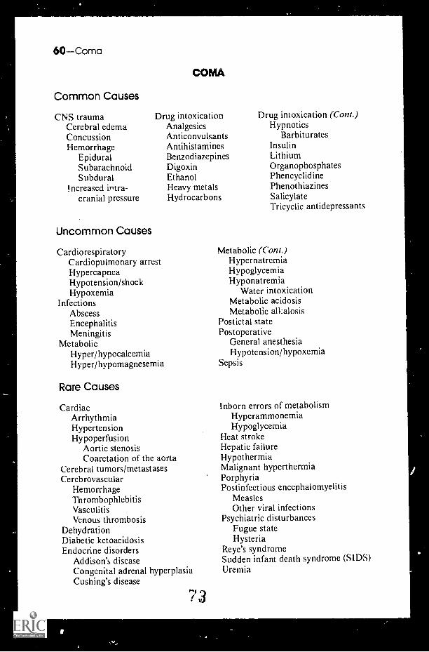
60 Coma
Common Causes
CNS traumaCerebral edemaConcussionHemorrhage
EpiduralSubarachnoidSubdural
Increased intra-cranial pressure
Uncommon Causes
COMA
Drug intoxicationAnalgesicsAnticonvulsantsAntihistaminesBenzodiazepinesDigoxinEthanolHeavy metalsHydrocarbons
CardiorespiratoryCardiopulmonary arrestHypercapneaHypotension/shockHypoxemia
InfectionsAbscessEncephalitisMeningitis
MetabolicHyper/hypocalcemiaHyper/hypomagnesemia
Rare Causes
CardiacArrhythmiaHypertensionHypoperfusion
Aortic stenosisCoarctation of the aorta
Cerebral tumors/metastasesCerebrovascular
HemorrhageThrombophlebitisVasculitisVenous thrombosis
DehydrationDiabetic ketoacidosisEndocrine disorders
Addison's diseaseCongenital adrenal hyperplasiaCushing's disease
Drug intoxication (Cont.)Hypnotics
BarbituratesInsulinLithiumOrganophosphatesPhencyclidinePhenothiazinesSalicylateTricyclic antidepressants
Metabolic (Cont.)HypernatremiaHypoglycemiaHyponatremia
Water intoxicationMetabolic acidosisMetabolic alkalosis
Postictal statePostoperative
General anesthesiaHypotension/hypoxemia
Sepsis
Inborn errors of metabolismHyperammonemiaHypoglycemia
Heat strokeHepatic failureHypothermiaMalignant hyperthermiaPorphyriaPostinfectious encephalomyelitis
MeaslesOther viral infections
Psychiatric disturbancesFugue stateHysteria
Reye's syndromeSudden infant death syndrome (SIDS)Uremia
Coma 61
Evaluation of the Comatose Child
There exist three important categories of central nervous system lesions thatcan cause alterations in one's level of consciousness: (1) supratentorial masslesions, (2) infratentorial mass lesions, and (3) metabolic abnormalities. Inevaluating the comatose child at the time of presentation, careful notice of theneurologic findings and determination of the type of CNS lesion incurred arevery useful in dictating acute medical management.
1. Supratentorial Mass Lesions
a. Important causes of supratentorial lesions that yield progressive deteriora-tion in children:
i. Cerebral hyperemia secondary to head traumaii. Epidural and subdural hematomas
Intracerebral hemorrhagesiv. Acute hydrocephalusv. Subdural hemorrhages
vi. Severe systemic hypertensionvii. Obstruction of an existing ventricular-peritoneal shuntviii. Bleeding arteriovenous malformation
b. Neurologic findings of supratentorial lesionsi. Initial signs and symptoms suggest focal hemispheric diseaseii. Signs progress in a rostral to caudal direction
Pupillary reflexes are usually depressediv. Motor signs are often symmetrical
2. Infratentorial Mass Lesions (Posterior Fossa)
These lesions can yield coma either by destroying the ascending reticularactivating system or by compression of that system by a mass or tumor.
a. Important causes of infratentorial lesions:i. Brainstem contusions associated with trauma
ii. Cerebellar hemorrhage or tumor with secondary hydrocephalusBrainstem encephalitis
iv. Basilar artery thrombosis
b. Neurologic findings of infratentorial lesionsi. Brainstem signs and symptoms are common.
ii. Signs are not rostral to caudal in evolution.iii. Cranial nerve palsies are common.iv. Abnormalities of the respiratory pattern are common and appear at
the onset of coma.
3. Metabolic Disorders
Metabolic disorders make up the majority of nontraumatic processes thatcause acute coma in the pediatric patient.
a. Important causes of metabolic coma:I. Hypoxic-ischemic coma (e.g., respiratory failure, shock, severe anemia,
apnea of infancy, carbon monoxide poisoning, cerebral vasculitis)
62Congenital Heart Disease
ii. Infections (e.g., encephalitis, meningitis, botulism)Postictal state
iv. Hypoglycemiav. Nonendocrine organ failure (e.g., hepatic and renal)
vi. Endocrine organ failure (e.g., pancreas, adrenal, thyroid, pituitary)vii. Poisonings (e.g., narcotics, barbiturates, sedatives, etc.)viii. Miscellaneous (e.g., Reye's syndrome, electrolyte abnormalities, and
hypothermia or hyperthermia)
b. Neurologic findings of metabolic lesionsi. Stupor or coma precede motor signs.ii. Motor signs are usually symmetrically depressed.
Pupillary reactions are preserved.iv. Acid-base imbalance is common.v. Seizures or abnormal motor movements are common findings.
Reference: James HF.: Neurologic evaluation and support in the child with an acutebrain insult. Pediatr Ann 15:16 22, 1986.
Ingestions and the Pupillary Examination in a Comatose Child
When confronted with a comatose child, the possibility of ingestion of a toxicor poisonous substance should always be considered. A history should beobtained addressing which medications and poisonous substances are at home,how they are kept separate from the child, and if there is any suspicion on theparents' part that the child may have ingested something. Modern advances intechnology and pharmacology have made the serum and urine toxicologicscreening tests most useful to the diagnosis of toxic ingestion. The examinationof a comatose child's pupillary size and reactivity, however, remains a usefulbedside exam that can be performed with a minimum of difficulty and time.Although the pupillary exam is not nearly as specific as the toxicologic screeningtests, it can point the way toward considering a diagnosis of ingestion of poison.
The Pupillary Examination and Coma
MYDRIASIS (DILATION OF THF PUPIL) MIOSIS (CONTRACTION OF THF PUPIL)
Amphetamines Cocaine Opiates MethadoneAntihistamines Ephedrine Barbiturates Carbon monoxideAtropine Ethyl alcohol Propoxyphen OrganophosphatesBotulism Snake venom Mcperidine Clonidine
Reference: James HE: Neurologic evaluation and support in the child with an acute braininsult. Pediatr Ann 15:16 22, 1986.
CONGENITAL HEART DISEASE
The First Manifestation of Congenital Heart Disease
Depending on the nature of the anatomic lesion, the first sign or symptom ofcongenital heart disease may vary. The table below is designed to serve as a guideto the most likely lesions, given the clinical presentation.
75

Marked Cyanosis
Transposition of thegreat arteries
Pulmonary atresia andstenosis with intactventricular septum
Tetralogy of Fa llot withsevere pulmonarystenosis
Complex pulmonaryatresias
Tricuspid atresiaEbstein's malformation
of the tricuspid valve
Congenital Heart Disease-63
Congestive Heart Failure
Aortic atresiaCoarctation of the aortaDouble outlet right
ventrick syndromePatent ductus arteriosusTruncus arteriosusVentricular septal defectArteriovenous fistulas
Abnormal Heart Rate
Supraventriculartachycardia
Heart block
Heart Murmur
Patent ductusarteriosus
Pulmonary stenosisAortic stenosisPulmonary artery
stenosisVentricular septal
defectArteriovenous fistulasAt rioventricular valve
regurgitations
References: Rowc R, Mehri7i A: The Neonate with Congenital Heart Disease. Phila-delphia, W.B. Saunders Company, 1968. p 105: Fyler DC (ed): Nadas' PediatricCardiology. Philadelphia, Hanley & Belfus, 1992.
Recurrence Risks for Congenital Heart Disease
Congenital cardiac lesions are thought to recur in three patterns of inheritance:
I. As part of a single gene defect syndrome (2%).2. Chromosomal abnormalities (4(7).3. Multifactorial inheritance (94%).Some single gene syndromes that often include cardiac defects are listed in the
following table.
Selected Single Mutant Gene Syndromes with Cardiovascular DiseaseOther than Coronary Artery
At I OMAI. DOMINANT AUTOSOMAL RFCESSIVE X-LINKFD
ApertCrou7onEhlers-DanlosForneyHolt-OramIHSS (not strictly
a syndrome)LeopardMarfanMyotonic dystrophyNeurofibromatosisOsteogenesis
imperfectaRomano-WardTreacher CollinsTuberous sclerosisUllrich-Noonan
Adrcnogenita' syndromeAlkaptonuriaCarpenterConrad iCutis laxaEllis-van CreveldFriedreich's ataxisGlycogenosis Ila, HIa, IVJervell and Lange-NielsenLaurence-Moon-BiedlMucolipidosis IIIMucopolysaccharidosis II, IV, V. VIOsteogenesis imperfeetaRefsumSeckelSmith-Lernli-OpitiThroinhocytopenia with absent radius (TAR)Weill-Marchesani
Incontinentia pigmentiMucopolysaccharidosis IIMuscular dystrophy

64Congenital Heart Disease
The recurrence risk for congenital heart disease in families in which onemember has one of these syndromes depends on the recurrence risk for thesyndrome (generally 25 to 50%) and the frequency with which congenital heartdisease is encountered in the syndrome.
The recurrence risk for congenital heart disease due to a chromosomalabnormality depends on the risk of recurrence of the chromosomal defect. Afamilial tendency for nondisjunction and the presence of a translocation in thechromosomal pattern of one parent may increase the likelihood of recurrence.Some chromosornal defects are associated with particular cardiac abnormalities.The more common chromosomal aberrations and their associated cardiac defectsare listed in the following table.
Corgenital Heart Disease in Selected Chromosomal Aberrations
POPL LATION STUDIED
PERCENTAGEINCIDENCE OF
C H D
MOS1 COMMON LESIONS
2 3
General population 1 VSD PDA AS D
4p 40 VSD ASD PDA5p (cri du chat) 25 VSD PDA ASDC mosaic 50 VSD13 trisomy 90 VS D PDA Dex
13q 50 VSD18 trisomy 90+ VSD PDA PS18q 50 VSD AV ASD21 trisomy 50 VSD AV canal ASDXO Turner 35 Coarc AS ASDXXXXY 14 PDA ASD
VSD = Ventricular septal defect. PDA = Patent ductus arteriosis. ASD = Atrial septa! defect.Dex = Dextrocardia. PS = Pulmonic stenosis. AV = Atrioventricular. Coarc = Coarctationof aorta. AS = Aortic stenosis.
The essential components of multifactorial inheritance of congenital heartdefects include: ( 1 ) a genetic predisposition to cardiovascular maldevelopment,(2) a genetic predisposition to be adversely affected by environmental teratogens,and (3) an environmental insult occurring at a vulnerable period of cardiac devel-opment (i.e., very early in pregnancy). Since there is no method of quantitatingthe presence of these three risks for a given offspring, a few percentages may bekept in mind:
I. In general, the risk of recurrence of congenital heart disease in a givenfamily is 1 to 5%.
2. The more common the heart defect in the affected family member, the morelikely that defect is to recur.
3. The risk to subsequent offspring of two parents triples if two existingfamily members are affected. (For example, the risk of recurrence of aventriculoseptal defect is about 5(7, if one previous sibling is affected;however, it increases to approximately 15% if one sibling and one parent ortwo siblings are affected.)
4. If the majority of family members have some form of congenital cardiacdefect the risk to subsequent offspring approaches 100%. More specifically,if three first-degree family members are affected, the risk in futurepregnancies is 60 to 100%.
77

Constipation-65
Recurrence risks for siblings of family members with congenital heart defectsare snown in the following table:
Recurrence Risks for Congenital Heart Defects in Siblings
DM-CTPERCFNT AT RISK
I SIBLING AFFECTED 2 SIBLINGS AFFECTED
Ventricular scptal defect 3 10
Patent ductus arteriosus 3 I 0
Atrial septal defect 2.5 8
Tetralogy of Fallot 2.5 8
Pulmonary stenosis 2 6Coarctation of aorta 2 6
Aortic stenosis 2 6Transposition 1.5 5
Endocardial cushion defects 3 10
Fibroelastosis 4 12
Hypoplastic left heart 2 6Tricuspid atresia 1 3
Ebstein anomaly 1 3
Truncus arteriosus 1 3
Pulmonary atresia 1 3
Based on combined data published during two decades from European and NorthAmerican populations. (From Nora JJ, Nora AH: Update on counseling the family witha first-degree relative with a congenital heart defect. Am J Med Genet 29:137-142, 1988,with permission.)
References: Nora JJ, Wolf RE: Recurrence risks in the family. In Kidd BSL., Rowe RD(eds): The Child with Congenital Heart Disease after Surgery. Mount Kisco, New York,Futura Publishing Company, 1976, pp 451 460; Fyler DC (ed): Nadas' PediatricCardiology. Philadelphia, Hanley 8c Belfus, 1992. Adapted from McMillan JA, et al: TheWhole Pediatrician Catalog, Vol. 3 Philadelphia, W.B. Saunders, 1982, with permission.
Common Causes
AppendicitisBreastfeeding (begins
around 6 weeks of age)Cow's milk ingestionDrugs
AnticholinergicsAntihistaminesNarcoticsPhenothiazines
Dysfunctional toilettraining
Emotional disturbancesFunctional ileusImmobility
CONSTIPATION
Inappropriate expectations of the caretakerIntentional withholdingIntestinal abnormalities
resiaHirschsprung's diseaseM icrocolonVolvulusWeb
Low dietary fiberMeconium plug/ileusMeningomyeloceleMental retardation/cerebral palsyPainful defecation (hemorrhoids, fissure,
skin iriatation)'7

66Contraception
Uncommon Causes
Diabetes mellitusElectrolyte disturbances
Hyper/hypocalcemiaHyperkalemia
HypothyroidismImperforate anus/anal stenosis
Rare Causes
AmyloidosisBotulismDolichocolonM, opathies/ myotoniasPheochromocytoma
Intestinal pseudo-obstructionLead poisoningSalmonellosisSpinal cord injury/tumorStarvation
Sacral malformationsScleroderrnaTetanusTethered cord
CONTRACEPTION
Side Effects of Hormonal Contraception
The mnemonic ACHES for recalling the dangerous side-effecs of oral contraceptiveuse is well known (A: abdominal pain; C: chest pain, cough, and shortness ofbreath; H: headaches; E: eye problems such as blurred vision; S: severe leg painin the calves and thighs). There exist, however, a great many other untowardeffects from oral contraceptive use that may paly a role in an adolescent woman'sdecision not to comply with this method of birth control. Arranged in terms ofthe hormonal cause, these untoward effects are presented below:
Hormonal Side Effects of Oral Contraceptives
ESTROGE NE XCESS
PROG ES I INEXCESS
ESTROGENDEFICIENCY
PROG ES1 INDEFICIENCY
Nausea, vomitingEdema bloatingCyclic weight gain
Dysmenorrhea,uterine cramps
Breast tenderness,increased breastsize, vascularheadaches
ChloasmaLactation supressionIrritability, depression
Fatigue, depressionAcne, oily skin.
hirsutism
AlopeciaIncreased appetite,
shortened menses
Decreased libidoHeadaches between
pill packages
Dilated leg veinsCholestatic
jaundice
Irritability, nervousnessHot flashes, motor
symptoms
Early midcycle spotting
Decreased amount ofearly menstrualflow
No withdrawal bleeding
Dry vaginal mucosa,atrophic vaginitis
HeadachesDepression
Late breakthroughbleeding andspotting
Heavy menstrualflow and clots
Delayed onset ofmenses, dys-menorrhea,weight loss
Adapted from Dickey RP: Medical approaches to reproductive regulation: The pill. InManaging Contraceptive Pill Patients, 4th ed. Oklahoma, Creative Inforinaties, Inc., 1984.
Reference: Shearin R B, Boehlke JR: Hormonal contraception. Pediatr Clin North Am36 697 715, 1989.
'Z9

Cortisol/Cough-67
CORTISOL
Cortisol Replacement During Febrile Episodes
Daily cortisol production rates among normal children have previously beenestimated at 12 mg/tn2/clay. The detection of clinical evidence of cortisol excecsamong children with adrenal insufficiency treated with this dose suggests that itmay be too high. A new study using recently developed, more accurate methodsof determining cortisol production rates in normal children suggests that 7 mg/m2/day may be a more appropriate dose.
Whatever the baseline dose of cortisol used to treat patients with adrenalinsufficiency, an increase in that dose is recommended during periods of stress.The most common form of generalized stress during childhood is febrile illness.The usual recommendation is that the daily dose of steroid should be increased2- to 3-fold during febrile illnesses. In fact, when 105 normal children 1 month to12 years of age were studied, those children with upper respiratory infection,streptococcal pharyngitis, and otitis media experienced serum cortisol increasesof 2- to 3-fold, whereas those children with pneumonia, fever of unknown origin,and bacterial meningitis demonstrated a 5- to 6-fold rise. It seems prudent toattempt to reproduce these apparently physiologic stress levels of serum cortisolfor children with inadequate intrinsic production during severe physiologic stress.It is also important to remember that oral replacement should be approximatelytwice the above recommendations because of poor oral absorption and hepaticbiodegradation.
References: Nickels DA, Moore DC: Serum cortisol responses in febrile children.Pediatr Infect Dis J 8:16 19, 1989; Linder BL, Esteban NV, Yergey AL, et al: Cortisolproduction rate in childhood and adolescence. J Pediatr 117:892- 896, 1990.
Common Causes
Allergic diseaseAspiration (direct or indirect)AtelectasisBacterial infection
BronchiectasisBronchitisPneumoniaSinusitisTrachcitis
Congestive heart failureEnvironmental pollutionForeign bodyGastroesophageal reflux
COUGH
6 0
Infections, otherChlamydiaMycoplasmaPertussis
Postnasal dripReactive airway diseaseSmoking/passive smokingViral infection
BronchiolitisCroupPneumonitisUpper respiratory infection

68Crayons
Uncommon Causes
Cystic fibrosisMalformation of the airwayMalignancy (primary or metastatic)Mediastinal adenopathyPsychogenic
Rare
Allergic bronchopulmonaryaspergillosis
Auricular nerve stimulationBronchogenic cystCongenital lobar emphysemaImmotile cilia syndromeLymphocytic interstitial pneumonitis
Habit (Psychogenic) Cough
TracheobronchomalaciaTracheoesophageal fistulaTuberculosisVascular ring
Opportunistic infections(PCP, CMV, MAI, fungal)
Parasitic infectionPulmonary embolismPulmonary hemosiderosisPulmonary sequestrationSarcoidosis
Habit cough is a transient tic disorder that may be seen in early adolescence,especially in puberty, and, by definition (DSM-III-R), lasts more than 1 monthand less than 1 year. The behavior may be seen as part of Gilles de la Tourettesyndrome but is usually an independent entity. The clinical features weredescribed by Berman:
. . persistent violent spasms of harky, harsh, nonproductive cough, occurring almostalways during the waking hours and unaccompanied by systemic signs and symptomsof chronic disease; a paucity or complete absence of abnormal findings in the chest;lack of response to most rotent cough preparations; and a cough that remains un-changed after exertion, laughter, infection, dampness, and extremes of temperature.
The diagnosis requires exclusion of organic causes. Habit cough usuallydisappears with sleep, which is not the case with most organic diseases.
Other motor tics include throat clearing, eye blinking, neck stretching, sniffing,grimacing, shrugging, and grunting. Hyper- and hypoventilation syndromes arealso possibilities.
Reference: Berman BA: Habit cough in adolescent children. Ann Allergy 24:43-46,1966.
CRAYONS
"Classic"Crayons
PHILADELPHIA, Oct. 2-- This week Binney & Smith Inc., the manufacturerof Crayola crayons, brought back eight colors from retirement, albeit temporarily.Last year, with much hoopla, the company stopped using maize, violet blue, rawumber, orange yellow, blur gray, green blue, orange red and lemon yellow andreplaced them with neon colors.
S

Creamatocrit-69
The crayons, now called "classics," will be packaged separately from a box of64 in a special commemorative tin with notes about the crayons and theirretirement, said J. O'Brien, a company spokesman. . . .
"The response has been amazing," said Mr. O'Brien, speaking of the withdrawalof the eight. "You just have to watch an adult open that box and sniff it. It's likea time machine."
For some, opening the green and yellow box is like Proust discovering 64madeleines. Mr. O'Brien, 32 years old, said he gets the feeling each morning inBinney & Smith's headquarters in Easton, Pa. As he opens his car door, he smellsa billion crayons melting. Some wno protested the withdrawal of the eight saidit was like removing a favorite baby blanket or toy.
Thousands were heartened by the crayons' temporary comeback. Others werenot.
"It's a limited victory," said Kenneth E. Lang, the founder of Rumps, the RawUmber and Maize Preservation Society. Mr. Lang, from Locust Valley, L.I., saidhe felt cheated.
"Raw umber and maize represent a bygone time in America," he said. "Youcan't draw a picture of Nebraska or Kansas or South Dakota without using thesecolors." . . .
From The New York Times, October 3, 1991, with permission.
CREAMATOCRIT
Now that more than 50% of women are initially breastfeeding their infants,questions commonly arise about the adequacy of the milk being produced.Abnormalities in milk composition are quite rare and are overdiagnosed. Themisinterpretation of a spot creamatocrit to determine the percentage of milk fatcontributes to this overdiagnosis. The creamatocrit can be a reliable reflection ofmilk fat content when a 24-hour sample or an entire expressed feeding from oneor both breasts is used and when the procedure has been standardized bycomparison with gravirnetric tests of milk fat content.
The creamatocrit takes advantage of the fact that fat is the major determinantof the energy value of the milk sample.
A sample of human milk is placed in a hematocrit tube and spun in amicrocentrifuge at full speed for 15 minutes. The fat rises to the top of thecolumn. The cream layer, easily visible, is read from the hematocrit capillarytube, and, like a hematocrit, is expressed as a percentage of the milk column inthe tube. This is the "creamatocrit."
This number can then be employed in a formula which will provide you withthe energy content of the milk expressed as kcal per liter. The formula is
kcal/liter = 290 + 66.8 x creamatocrit
For example: A human milk sample is found to have a creamatocrit of 5% Itscaloric value is:
290 + 66.8 x 5
or
625 calories per liter.

70 Cryi ng
Note: Creamatocrits should be read within one hour of centrifugation, becauseafter that time the cream column begins to "unpack," and falsely elevatedvalues are obtained.
Reference: Lemons JA, et al: Simple method for determining the calorie and fatcontent of human milk. Pediatrics 66:626 628, 1980.
CRYING
The Crying Infant
Unexplained, excessive crying in the afebrile infant usually achieves just whatit is meant to achieve: every adult around wants to find its cause and make it stop.In investigating the cause of excessive, piolonged crying in 56 infants, aged 4 daysto 245 months, who were brought to the emergency room of the Children'sHospital of Denver, a reason for the infant's distress was found for 46. Thehistory provided a clue to the cause for I 1 of the 56 infants, a careful physica!examination resulted in a diagnosis for 23 patients, and a variety of laboratorytests revealed the cause for 11. The final diagnosis included a broad array ofconditions, of which 61% were considered serious. The table below lists thediagnoses that explained the reason for excessive crying for these patients.
Diagnosi- in 56 Infanis will? Unexplained, Excessive Crying
DIAGNOSISNo wtru
DIAGNOSIS DIAGNOSISNO WITH
DIAGNOSIS
Idiopathic 10
Colic 6
1 nfect ious causesOtitis media* 10
Viral illness with anorexia, 2
dehydration*Urinary tract infection*Mild prodrome of
gast roenteritisHerpangina*Herpes stomatitis*
TraumaCorneal abrasion* 3
Foreign body in eye*Foreign body in
oropharynx*Tibial fracture*Clavicular fracture*Brown recluse snider bite*Hair tourniquc, syndrome
(toe)*
G istrointestinal tractConstipation 3
Intussusception*Gastroesophageal reflux
with esophagitis*Central nervous system
Subdural hematoma* 1
Encephalitis*Pscudoturnor cerebri* 1
Drug reaction:overdoseDTP' reaction* 1
Inadvertent pscudoephe-drine overdose*
BehaviorNight terrors 1
Overstiniulation 1
CardiovascularSupraventricular tach),cardia* 2
MetabolicGlutaric aciduria, type I* 1
Total 56
* Indicates cond it ni is considered serious.Diphtheria-tetanus-pertussis vaccine.
S 3

Crying-7i
'The components of the physical examination that were important in providingthe diagnosis are listed below.
COMPONIA I Oh PIIN'SICAI FXAM
NO. OF PATIFN'INW HO M COM PONI NI
PROVhD USFFU I= 30)
Otoscopy 10
Rectal exam 4
Fluorescein staining of cornea 3
Inspection underneath clothing 2
Palpation of bones 2
Oral exam 2
Auscultation of heart (tachyarrhythmia) 2
Laryngoscopic exam of hypopharynxFversion of eyelidPalpation of anterior fontanelleRetina! examNeurologic exam
Laboratory studies that identified the diagnosis are listed in the following table.
NO. Oh PA I II N ISDIA6NOS1 = I I)
Skeletal roentgenography 2
Lumbar puneture.cerebrospinal fluid analysis 2*
Electrocardiography 2
Computed tomographic scan of the head 2*
Barium enemaEsophag ramAmino and organic acid studiesUrinalysis
*One patient, with pseudotumor cerehri, required lumbar punctureand computed homographic scan of the head to make the diagnosis.
Reference: Poole SR: The infant with acute, unexplained, exce:;sive crying. Pediatrics88:450 455, 1991.
Colic, or Excessive Crying, in Young Inrants
Colic, from the Latin colicus, strictly means related to or associated with thecolon (and was originally an adjective). With mspect to excessive crying in
young infants, it has a poorly defined meaning that is connected to cryingsubstantially more than the mean amount for age in an infant under 3 months.Usually this is thought to be a result of an intestinal (or "colic") disorder ofparoxysmal pattern.

72Cryng
Normal Crying Time
In a 1962 study of 80 middle-class infants. Braze lton found the normal cryingtimes, usually concentrated in the evening, to be as follows:
2 hr/day at 2 weeks of age3 hr/clay at 6 weeks of ageI hr/day at 3 months of age
The clinical pattern of infant colic (also called paroxysmal fussing, infantile colic,evening colic, and 3-month colic) is well described:
I. Attacks occur suddenly, usually in the evening.2. They are characterized by a loud, almost continuous cry.3. They last several hours.4. The face of the infant is flushed, with occasional circumoral pallor.5. The abdomen is distended and tense.6. Legs are drawn up on the abdomen and the feet often cold. Legs may
extend periodically during forceful cries.7. The fingers are clenched.8. Relief is often noted from passage of flatus or feces.9. The attack is not quelled for long by feeding, even though the infant may
appear hungry and eats normally.10. The attack usually terminates from apparent exhaustion.
No consistent etiology has been identified.
Reference: Braze lton TB: Crying in infants. Pediatrics 29:579, 1962.
Inconsolable Crying
Nothing is as troublesome both to parent and physician as an infant who criesinconsolably. Diagnoses to consider when confronted with an infant who criescontinuously, particularly if it is shrill or high-pitched, include:
A corneal abrasionAn eyelash, or other foreign body, in the eyeGlaucomaColic and haussusceptionShaken baby syndromeMeningitisFracturesA DPI reactionAn open diaper pin in the skinStrand of hair wrapped around finger or penis
Reference: Harkness M.1: Coineal abra.,iun in infancy as a cause of inconsolablecrying. Pediatric Frnerg Care 5:242. 19g9.
k'1' 5

CYANOSIS
Common Causes
Acrocyanosis (especially cold stress)Apnea of prematurityAspiration
Direct (swallowing disorders,neuromuscular disease)
Indirect (gastroesophagealreflux, emesis)
AtelectasisBreath holdingBronchiolitisCongenital heart disease
Decreased pulmonary blood flow(no pulmonary hypertension)
Anomalous systemic venousreturn
Ebstein's anomalyHypoplastic right ventriclePulmonary stenosis:atresiaTetralogy of Fa llotTricuspid stenosisjatresia,
insufficiencyEisenmenger's syndromeIncreased pulmonary blood flow
Arteriovenous (AV) canalCoarctation (preductal)Hypoplastic left heartTotal anomalous pi lmonary
venous return (TAPVR)
Uncommon Causes
Abdominal distentionArterial thmmbosisBronchopulmonary dysplasiaChest wall abnormalities
Congenital bone:cartilageabnormalities
PectusFlail chest
Cystic fibrosisEpiglottitisForeign bodyHypovolemiaMediastinal massPersistent fetal circulationPickwick ian syndrome
CYanosis-73
Congenital heart disease (Con t.)Increased pulmonary blood
flow (Cont.)TranspositionTruncus arteriosusVentricular septal defect
(VSD), largePump failure
Aortic stenosis (severe)Coarctation (postductal)Patent ductus arteriosus (PDA)VSD
CroupCryingDrugs respiratory depressants
(e.g., narcotics, benzodiazepines)Hyaline menthrane diseaseMucous plugNasal obstructionPneumoniaPulmonary edemaReactive airway diseaseSeizuresSepsisSleep apnea (tonsillar adenoidal
hypertrophy)
Pleural effusionPneumothoraxPolycythemiaPulmonary hemorrhageRetropharyngeahperitonsillar abscessScoliosisTracheal compression
AbscessAdenopathyHemorrhageTumorVascular ring
Tracheobronchomalacia, stenosisVenous stasis
8 LI

74Cystic Fibrosis
Rare Causes
AngioedemaBronchogenic cystCentral nervous system disease
EdemaHemorrhageInfectionTrauma
ChylothoraxDiaphragmatic herniaFactitious (blue paint/dyes:makeup)GlossoptosisHemoglobinopathy
(M, low oxygen affinity)Hypoplastic lungsLaryngeal webLobar emphysemaMethemoglobinemia
Methemoglobin reductasedeficiency
Oxidant stressAcetophenetidinAntimalarialsBenzocaineCrayonsDisinfectantsEDTAHydralazineMarking dyesNaphthaleneNitrites
Amyl:butyl nitrateNitrate-contaminated well waterNitrate food additiveNitroglycerinPlant nitrates (e.g., carrots grown
Methemoglobinemia (Cont.)Oxidant stress (Cont.)
NitroprussidePrilocainePyridiumSulfonamidesVitamin K analogs
Ondine's cursePrimary pulmonary hypertensionPulmonary AV malformation/fistulaPulmonary embolism/thrombosisPulmonary hemosiderosisPulmonary sequestrationPulmonary tumor (primary
or metastatic)Respiratory muscle dysfunction
BotulismMuscular dystrophyMyasthenia gravisNeuromuscular blockadePhrenic nerve damageWerdnig-Hoffmann disease
Superior vena cava (SVC) syndromeTracheoesophageal fistulaTumorVocal cord paralysis
in contaminated soil)
CYSTIC FIBROSIS
The Thirty Faces of Cystic Fibrosis
Cystic fibrosis is the great imitator. It may first present in utero with a pictureOf mcconium peritonitis or its first manifestation may be sterility in the adultmale. Listed below are 30 ways the disease may first manifest itself. Be suspiciousand perform a sweat test when these problems arc encountered without aplausible alternative explanatis.7

Meconium ileus and meconiumperitonitis
Pancreatic insufficiencyand growth failure
Recurrent pulmonary infectionsIntestinal impaction and obstructionHypoproteinemia in infancy,
with edema and anemiaRectal prolapseCholestatic jaundice in neonatesCirrhosis of the liverPortal hypertensionGlucose intoleranceDiabetesAcute or recurrent pancreatitisVitamin K deficiency and bleedingVitamin A deficiencyVitamin E deficiency with
neurologic abnormalities
Hemicystitis: Think Zoster!
Cystitis/CMomegalovirus-75
Night crampsLactase deficiencyDuodena! ulcerCholelithiasis and cholecystitisChronic obstructive airway diseaseCor pulmonaleRecurrent episodes of asthmaHypertrophic pulmonary
osteoarthropathyNasal polypsOptic neuritisSalty taste of infant noted by
the motherHyponatremic dehydration in warm
weatherHypochloremia metabolic alkalosisHeat strokeInfertility in males
CYSTITIS
There exists a fascinating clinical entity known as hemicystitis, which is aninfection limited to one half of the bladder. It occurs with herpes zoster infectionof the bladder in which vesicles are unilateral, conforming to the affected nervesupply.
Reference: Nelson .11), McCracken GH: Newsletter. Pediatr Infect Dis 16(8):16, 1990.
CYTOMEGALOVIRUS
Cytomegalovirus Infection in the Newborn
Cytomegalovirus (CMV) is the most common herpes infection that occursduring the neonatal period. Exposure to CMV can occur from a congenitalsource (e.g., either a primary infection in a seronegative mother or reactivation oflatent virus in a seropositive mother), a natal source such as vaginal delivery ina mother shedding virus from the cervix, or postnatal sources (e.g., breast milkfrom a seropositive mother, blood transfusion from a seropositive donor, or closecontact with individuals actively shedding CMV).
CMV is especially dangerous to the developing fetus when thc pregnantwoman is experiencing primary infection; in such cases there is a 301;i mortalityrate among those CMV-infected infants who were symptomatic at birth.Surviving infants who were symptomatic at birth encounter the followingsequelae: microcephaly (70c,i), moderate-to-severe mental retardation (6l).
5:;15

76-0,rtomegalovirus
hearing loss (30%), and chorioretinitis (22%). Symptomatic infants withoutneurologic signs at birth remain at risk for such sequelae as failure to thrive,microcephaly, spastic quadriplegia, and deafness during the first year of life.Asymptomatic infants of mothers experiencing a primary CMV infection are atrisk for hearing defects and moderate-to-severe brain damage. Conversely,infants and fetuses who become infected with CMV as a result of recurrentviremia or perinatal exposure to a seropositive mother are generally protectedfrom symptomatic infection of any kind. A minority of such infants, however,may be at risk for hearing defects and learning problems.
Clinical Manifestations of CMV infection(among infants born to previously seronegative mothers)
Prenatal Infection
Hepatomegaly ChorioretinitisHyperbilirubinemia Optic atrophyPetechiae/thrombocytopenia StrabismusMicrocephaly MicrophthalmiaHydrocephalus CataractsPeriventricular calcifications Deafness
Infection During Vaginal Delivery
Protracted pneumonitis
Postnatal InfectionHealthy infants and children who acquire CMV infection suffer no permanent
sequelae and usually remain entirely asymptomatic. Symptomatic infection doesoccur rarely, however, and may include the following:
FeverHepatomegalyPneumonia
Reference: LaRussa P: Perinatal herpes virus infections. Ped Ann 13:659-670, 1984.
ci) 9

DAY CARE
What You Ought to Know About Your Day Care Center
With more and more parents relying on day care centers, clinicians need to beknowledgeable about what to look for in selecting such a center. One source ofinformation both for parents and clinicians is the National Association for ChildCare Resource and Referral Agency (NACCRRA), 2116 Campus Drive SE,Rochester, MN 55904 (507) 287-2220. This clearinghouse advises parents andothers on how to select a safe and reliable day care center.
Clinicians should urge parents to visit their day care center frequently (andunannounced) in order to make sure their children are cared for in a loving andpositive manner.
Parents also need to observe their children closely for behavioral changessuch as depression, aggressiveness, fear of the day care center, and signs of physi-cal abuse (e.g, bruises, abrasions) or neglect (e.g., diaper rash, bald spots on theback of the head indicating that the infant has been left supine all day, extremehunger, etc.).
In the case of older toddlers who are left in day care, parents should ask thechild what activities are offered during the day. Are constructive games andteaching employed or is the child merely parked in front of a television set?
Reference: Hoekelman RA: Day care, day care: Mayday! mayday! Pediatr Ann20:403 -404, 1991.
Pathogens Transmitted in Day Care Centers
Approximately 11 million children under the age of 6 years spend all or partof their day in a day care center. Some experts have predicted that by the year2000, 80% of all American mothers will be a part of the nation's workforce. Theresult, of course, is an ever-increasing reliance upon day care. One of the mostfrequent questions that comes up regarding day care has to do with parentsworrying about the risk of infections in the day care setting. The table belowsummarizes the most common pathogens you need to be aware of:
Pathogens Transmitted in Day Care Centers*
MOM- OFTRANSMISSIONS BACTERIA VIRUSES PARASITES
Direct Group A streptococci Herpes simplex PediculosisStaphylococcus aureus Herpes iostcr Scabies
Table continued on next page.
77
n

78Dehydration
Pathogens Transmitted in Day Care Centers* (Cont.)
MODE OFTRANSMISSIONS BACTERIA VIRUSES PARASITES
Respiratory
Fecal-oral
Haemophilusinfluenzae
Neisseriameningitidis
Bordetella perlussisMycobacterium
tuberculosis
Campylobacter sppEscherichia coliSalm-)nellaShigellaYersinia
AdenovirusCoxsakic A16 (hand-foot-
mouth disease)Epstein-Barr virusH HV6 (roseola)InfluenzaMeaslesMumpsParainfluenzal'arvovirus BI9 (fifth disease)Respiratory syncytial virusRhinovirusRubellaVaricella
EnterovirusesHepatitis ARotavirus
Contact with Cytomegalovirusinfected blood Hepatitis Band secretions Herpes simplex(urine, saliva) Human immunodeficiency
virus'
CryptosporidiumEntanweba hiswlyficaGiardiaIlymenolepsis nana
(dwarf tapeworm)Pinworms
*Adapted from Hendley 0.1: How germs are spread. In Donowitz LG (ed): Infection Controlin the Child Care Center and Preschool. Baltimore, Williams & Wilkins, 1991.To date no reported cases of HIV infection are known to have resulted from transmissionin day care centers.
Reference: Van R, Wun C-C, Morrow Al., Pickering L.K: The effect of diaper type andmerclothing on fecal contamination in day-care centers. JAMA 265:1840 1844, 1991.
DEHYDRATION
Predictable Fall of the Blood Urea Nitrogen(BUN) in a Dehydrated Child
Is the increased BUN in a child with diarrhea and wmiting always the resultof simple dehydration or can it reflect the presence of associated renal disease?Brill and coworkers found that the rate of fall of BUN in a dehydrated child withnormal renal function was predictable. They plotted BUN levels against time onsemilogarithmic graph paper. BUN had fallen to one-half the admission level in24 hours or less in all children with uncornplicated dehydration and diarrhea.
Line A in the accompanying figure reresents the slope along which the BUNshould fall in a child without renal disease or excess nitrogen load (e.g.,gastrointestinal bleed). Lines 13 and C represent 21/2 standard deiations on eitherside of that ratc of fall.
91

200
CD
8_j 2003
100 12 24 36 48 60 72
TIME (hrs)
Dermatology-79
Complicating disease should be investigated in the dehydrated child whoseBUN does not fall at a rate parallel to line A or within 21/2 standard deviationsfrom that rate.
Reference: Brill CB, Uretsky S. (iribetz I): J Pediatr 52:197. 1973. Adapted fromMcMillan JA, et al: The Whole Pediatrician Catalog. Philadelphia, W.B. Saunders,1977. p 97.
DERMATOLOGY
Skin Lesions
Can you speak "dermatologese'"? Or is it "dermaterminology"? In any event,understanding dermatology is impossible without a working knowledge of thesometimes exotic vocabulary of the specialty. The follow ing list defines some (butby no means all) of the more commonly used terms for skin lesions and relatedstructures and conditions.
Abscess A localized accumulation ofpurulent material so deep in the dermisor subcutaneous tissue that pus is usu-ally not visible on the surface of theskin.
Atrophy An acquired loss of underlyingtissue causing skin depression withintact epidermis.
Bulla A relatively large vesicle (diameter0.5 cm).
Carbuncle Coalescence of several furun-cies (see below).
Comedo (pl., comedones) A greasy plugin a sebaceous follicle capped by alayer of melanin, hence its black ap-pearance (blackhead).
Crust Dried exudate of body fluids(serous and Or hemorrhagic).
Cyst A sac that contains liquid or semi-solid material.
2
80Dermatology
Erosion-- A superficial deficit of epithe-lium.
Erythema Increased redness of skin fromcapillary dilatation.
Excoriations Linear, angular erosionscaused usually by scratching.
Furuncle--A deep necrotizing form offolliculitis with pus accumulation.
Figurate lesions Lesions forming ringsand arcs, usually erythematous.
HyperpigmentationExcessive pigmen-tation of any origin.
Hypopigmentation Loss of pigmentationof any origin.
Keratosis --- Benign horny lesion (alsocalled keratorna).
Lichenification A proliferation of kera-tinocytes and stratum corneum forminga plaque-like structure. The skin ap-pears thickened, and the skin markingsappear accentuated. The process resultsfrom repeated rubbing.
Macule- A circumscribed area of change(less than 2 cm in diameter) in normalskin color without elevation or depres-sion of the surface in relation to thesurrounding skin.
Milia Small, firm, white papules filledwith keratin.
Nodule A palpable, solid, round, or el-lipsoid lesion; it can be located in theepidermis or extend into the dermis orsubcutaneous tissue.
Papule --A solid elevated lesion generallyunderstood to be less than I cm ind ia meter.
PatchA large, flat lesion (greater than 2cm in diameter) with color differentfrom surrounding skin. Differs frommacule only in size.
Plaque Elevation above the skin surfacethat occupies a relatively large surfacearca in comparison with its heightabove the skin.
Pustule-- A circumscribed elevation ofskin that contains a purulent exudate.(Follicular pustules are conical andusually contain a hair in the center.)
Rash An inflammatory skin eruption.Scale A thin, platelike, external layer of
horny epidermis.Scar Change in skin character--a mark
secondary to trauma or inflammation.Sclerosis Circumscribed or diffuse har-
dening or induration in the skin.Tag Small, sessile protuberance of skin.Telangiectases Permanent dilatations of
blood capillaries that may or may notdisappear with the pressure of a glassslide.
Tumor A firm, solid, raised growthgreater than 5 cm in diameter.
Ulcer A deep, local deficit or excavationof skin and underlying tissue.
Vesicle A small (less than 0.5 cm in diam-eter) fluid-filled lesion. A "dew-drop."
Wart A benign keratotic tumor.Wheal A rounded or flat-topped eleva-
tion in the skin that it; characteristicallyevanescent, disappearing within hours.Lesions are the result of edema in theupper layers of the dermis.
Dermatologic Manifestations of Viral and Bacterial Infections
There are a number of cutaneous manifestations associated with bacterial andviral infections common among children. The specific skin lesions that appear areusually the result of various pathways involved in inflammation and necrosis,such as the complement casacade, localized or generalized Schwartzmanreactions, factors that yield hypotension and disseminated intravascular coagula-tion, and a host of yet undiscovered factors. The table lists the dermatologicmanifestations of common pediatric bacterial and viral infections and should beuseful in the bedside evaluation of thc patient presenting with "fever and a rash."
References: Pediatric viral infections. In Roche Handbook of Differential Diagnosis6(18):3 19, 1989. Yamanishi K. et al: Identifi:ation of human herpesvirus-6 as a causalagent for exanthem subitum. Lancet i:1065 1067, 1988.
93

ss.
Diff
eren
tial D
iagn
osis
of P
edia
tric
In e
ctio
ns w
ith D
ertn
atol
ogic
Man
ifest
atio
ns
CI.I
NIC
AI.
['NT
H 1
.C
AU
SA
TIV
E A
GF
N I
A(;
FC
LIN
ICA
IS
YN
DR
OM
I.T
Y P
F O
F R
AS
HD
IS1
RB
TT
ION
SIM
ILA
R E
N f
tIll
S
Ros
eola
infa
mH
erpe
ssir
us-6
*tu
rn te
xant
hern
suhi
tum
)
Fryt
hem
a in
fec-
Pars
ON
irus
B-1
9(i
mam
(fi
fth
dise
ase)
Mea
sles
Mea
sles
vir
us
6m
onth
s 4
year
s
Scho
ol-a
ge c
hild
ren
Infa
nts,
adu
lt,le
ss c
omm
on
All
ages
FeY
er. c
ough
: cur
sco
njun
ctis
Its
Feve
r, ir
rita
bilit
y: r
apid
lysi
s of
fey
er w
ithap
pear
ance
of
rash
Flu-
like
illne
ss
Han
d-fo
ot-a
nd-
Prim
ary:
('oxs
acki
e A
< 1
() y
ears
mou
th d
isea
sesi
ruse
sS
econ
dan:
Cox
sack
ie B
vii u
ses:
ent
erm
irm
71
Var
icel
laV
aric
ella
-mst
er ir
us90
(i o
f ca
ses
<15
yea
rs
Prim
anbu
ccal
cel
liilit
is11
infin
en:a
ety
peB
Set
titu
lary
:pn
etat
iont
ar,
S. a
urew
fl-h
emol
ytic
str
epto
cocc
i
S. a
ureo
sSt
uphs
loco
ceal
scal
ded-
skin
synd
rom
e
3 36
mon
ths
Infa
nts
Dis
cret
e m
.icul
ar o
rT
runk
with
ext
en-
papu
lar
rash
sion
to n
eck,
ex-
trem
ities
, fac
e
Bila
tera
l ery
them
a of
Face
, tru
nk, e
x-Sc
arle
t fev
erch
eeks
: "sl
appe
d ch
eeks
"tr
emiti
es; p
alm
sR
ubel
laL
acy-
retic
ular
exa
nthe
mso
les
spar
ed
Kop
lik s
pots
Star
ts o
n fa
ce.
Ent
erov
iral
infe
ctio
nM
acul
opap
ular
eru
ptio
n of
min
es d
ownw
ard
Mye
opla
sma
uppe
r tr
unk,
fac
e: s
prea
dsD
rug
erup
tion
to lo
wer
trun
k, e
xtre
mi-
ties:
bec
omes
con
flue
nt
Fey
er, a
nore
xia:
ora
lO
ral:
disc
rete
. ulc
erat
ise
Ant
erio
r m
outh
.A
phth
ous
stom
antis
pain
Skin
: mac
ulop
apul
ar,
hand
s, f
eet:
Var
icel
lave
sicu
lar
occa
sion
ally
Her
pes
sim
plex
trun
k. f
ace
Fes
er: p
ruri
tus:
mal
aise
Mac
ulop
apu,
ar. t
hen
vesi
-D
iffu
se, i
nclu
des
Inse
ct b
ites
des
on e
ry'h
emat
ous
scal
p, o
ral
Her
pes
sim
plex
base
, %Sh
ia r
uptu
rem
ucos
aC
rust
ing
as f
inal
sta
ge
Fey
cr. h
ack:
ref-
Ina
Uni
late
ral i
ndur
ated
Peri
orbi
tal.
chec
kO
rbita
l cel
lulit
isce
llulit
isPa
rotit
isIn
dist
inct
bor
der,
Vio
lace
ous
hue
Feve
r. ir
rita
bilit
y:se
ptic
emia
(ra
re):
eye,
nas
al d
isch
arge
Ten
der,
dif
fuse
ery
them
a-D
iffu
seto
m r
ash
prog
ress
ing
to b
ulla
ePo
sitis
e N
ikol
sky
sign
xfol
iatio
n
Bul
lous
impe
tigo
moI
nfor
me
Tox
ic e
pide
rmal
necr
olys
isPe
tnph
igus
Upi
derm
olys
is b
ullo
saK
awas
aki d
isea
se
Rel
eren
ces
Pedi
atri
c si
ral i
nfec
tions
. In
Roc
he H
andb
ook
IA D
iffe
rent
ial D
iagn
osis
6(1
8).3
19, 1
989.
Yam
anis
hi K
. et a
l. Id
entif
icat
ion
of h
uman
ber
pess
irus
-6 a
sca
usal
age
nt f
or e
xant
hcm
%ob
it=I
:mee
t i: 1
065
1067
, 198
)).
9 4
cl? 3 a
41-

82Development
Seborrheic Dermatitis or Atopic Dermatitis?
Many clinicians have difficulty distinguishing these two common entities.Seborrhea is the excretion by the sebaceous glands of abnormally copiusamounts of grease-like sebum. There is usually no underlying disorder. Atopy isa form of immediate hypersensitivity reaction to certain common allergens thatproduce the IgE antibody. reagin, and atopic dermatitis is the dermal manifestationof the allergic reaction. The following table should help you further to differentiatebetween the two conditions.
Sehorrheic Dermatitis vs. Atopic Dermatiti.v
SEBORRHFIC DER MKTITIS ATOPIC DER MATI FIS
Family history of 15 25('allergy
Character of Dry, scaly, "potato chip"individual lesion that may or maylesions not appear greasy
Color of lesion
Feature of lesion
Vesicles
Weeping andedema
Lichenification
Pruritus
Only slightly erythematous,hut more often of a salmoyellow, or brown color
More intense color at peri-phery clearing at center.Appears sharplydemarcated
Never present
Absent
Absent
Mild or moderate
40 60"
Erythema, papules. vesicles,weeping, scales, lichenifica-don, or a combination. Mayhave superimposed pyoderma
In acute phase, always red andn, often of an intense redness
More red at center. Gradoallytapers out at periphery, fadinginto normal skin
Present in acute phase
Always present at some time inevolution of disease
Characteristic of late stage
Paroxysmal and severe
Reference: Perlman 11 H: Helpful diagnosticdermatitis from seborrheic dermatitis. Ann Allergy
clues for differentiation of atopic23:583, 1965.
DEVELOPMENT
Developmental Delay: Seeking the Etiology
As we learn from medical school on, the physician's best diagnostic tools arethe history and physical exam. This dictum certainly holds true in the work-upof developmental delay.
Regardless of the age of the child at presentation, there are several strategiesthat the pediatrician can employ in the search for an etiology. The What, When,and How are invariably asked. The answers will often guide the parents in thedecision to have another child and how to come to terms with their child'sdisability. The following guidelines and accompanying table should prove quiteuseful when confronted with a developmentally delayed patient.

Development-83
I. Analyze the family's pedigree with attention paid to physical appearance,"birth defects," inheritance patterns of any disease, disabilities or dysmorphisms,consanguinity, and parental age. When possible, obtain photographs of theextended family.
2. Obtain a thorough prenatal history. Look for:a. First-trimester febrile illnesses; possible teratogenic exposures, including
alcohol, cigarette, and drug use.b. First through second trimesters: viral illnesses, maternal diabetes mellitus,
maternal medications, cardiorespiratory disease, and maternal metabolicabnormalities.
c. First through third trimesters: see table.
Temporal Approach to the Etiology of Developmental Delay
CLASSIFICMION ETIOLOGIC PERIOD 1 YPICAL. CAUSES
Genetic(Mendelian)
Chromosomal
Mult i factorial(genetic andenN i ron mental )
Environmental
Preconceptional
Preconception or earlymitotic phase
Preconception and first12 weeks of gestation
PrenatalFirst trimester (periodof C'NS morphogenesisand neuroblastproliferation)
Midpregnancv (periodof neuroblast proliferation)
Autosomal dominantNeurofibromatosisTuberous sclerosis
Autosomal recessivePhenylketonuriaGalactosemiaTay-Sachs disease
Sex-linked recessiveSex-linked nonspecific
mental retardationLesch-Nyhan syndrome
Sex-linked dominantAlbright's hereditary
osteodystrophy
PolysomyTrisomy 21 (Down
syndrome)Autosomal deletion
Cri du chat syndrome(deletion of short arm 5)
Sex chromosome aberrationsMultiple-X syndromesTurner's syndrome
Neural tube defectsMeningomyeloeeleFncephaloceleHydrocephaly
Cleft lip:cleft palate
Teratogenic agents (suspected)PhenytoinMaternal alcoholism
Intrauterine infection
nt rauterine infectionMaternid irrtdiationTcratogens
Table comintied on next page.
UDevelopment
Temporal Approach to the Etiology of Developmental Delay (Cont.)
CLASSIFICATION ETIOLOGIC PERIOD TYPICAL CAUSES
Environmental Prenatal (Cont.)(Cont.) Midpregnancy to peri-
natal period (rapid braingrowth, glial cell prolifer-ation, myelinization,dendritic tree formation)
Perinatal
Postnatal
Intrauterine infectionPreeclarnpsiaHemorrhageHormonal disturbancesMalnutritionPrematurityTeratogens
Intrapartum hemorrhage andanoxemia
Trauma:Breech deliveryDifficult delivery
MeningitisMalnutritionHead injury
EncephalitisLead poisoningNear drowning
3. Obtain a birth history. Approximately 30% of the cases of developmental delaycan be traced to difficulties in the prenatal period. It is wise, however, to avoidthe temptation to overinterpret problems in labor and delivery. As often aspossible, a hospital record of the birth and nursery stay should be procured.
4. Continue your detective work with a postnatal history. In addition to thecauses listed in the table, inquire about any known metabolic diseases andtheir treatment, diarrheal illnesses with possible hypernatremia, failure tothrive, history of seizures, loss of milestones, and apparent receptive or ex-pressive delays.
5 Examination of the patient: The physical exam should be undertaken with afine-toothed comb.a. Height, weight, and head circumference are invaluable in the setting of
developmental delay. [Refer to "Diagnosing Dysmorphism" (p. 95) orSmith's Recognizable Patterns of Human Malformation: Genetic, Embry-
,gic and Clinical Aspects (Philadelphia, W.B. Saunders, 1988]. Micro-cephaly, macrocephaly, growth retardation, and growth acceleration maysteer you toward a recognized syndrome and/or an etiology.
b. Transilluminate the head if hydrocephaly, hydranencephaly, porencephaly,or cerebral cortical atrophy are in your differential. You may obviate theneed for a CT scan with this simple test.
c Pay attention to the eye exam (see "Leukocoria: A Differential Diagnosis"[p. 204]). Look for a cherry red spot, chorioretinitis, retinitis pigmentosa,or rentrolental fibroplasia when examining the retina.
d. Do not neglect to assess hearing. Unidentified hearing loss can lead toprofound developmental delay.
e. Proceed with the examination of the skin, facies, and body as if you wereuncovering buried treasure. Remember that both major and minorabnormalities are more common in patients with developmental delay orfrank mental retardation. Compile a list of your findings and consult thesources noted above, beginning with the least common abnormality.
9 7

I.
.11
Development-85
6. Laboratory studies will often help secure a diagnosis, thus comforting theparents. They may also reveal chromosomal or metabolic abnormalities thatwill influence a couple's plans for other children. Consider pertinent laboratorystudies, chromosomal analyses, viral cultures, computed tomography, andultrasonography as you and the parents see the need.
References: Kce le DK: The developmentally delayed child: Pursuing the etiologicwork-up. Contemp Peds March:51-62, 1985.
Oski FA, et al: Principles and Practice of Pediatrics. Philadelphia, J.B. Lippincott,1990.
The Draw-A-Person Test
Goodenough called her test the "Draw-a-man" test when she first introducedthis superb simple screening test for intelligence. The name of the test haschanged, but the test and its scoring have remained the same.
Basal age = 3 years. For each four criteria, add 1 year to arrive at mental age,between ages 3 and 10 years. Instruct child to draw a complete person; no furtherinstructions.
Maturation ageChronological age
Twenty-eight criteria for scoring:I. Head present2. Legs present3. Arms present4. Trunk present5. Length of trunk greater
than breadth6. Shoulder indicated7. Both arms and legs attached
to trunk8. Legs attached to trunk and
arms to trunk at correctpoint
9. Neck present10. Outline of neck continuous
with that of head or trunk,or both
11. Eyes present12. Nose present13. Mouth present14. Both nose and mouth in two
dimensions, two lips shown15. Nostrils indicated16. Hair shown
1001
IQ
17. Hair on more than circumference ofhead, nontransparent, better thanscribble
18. Clothing present19. Two articles of clothing,
nontransparent20. Entire drawing, with sleeves and
trousers shown, free fromtransparency
21. Four or more articles of clothingdefinitely indicated
22. Costume complete withoutincongruities
23. Fingers shown24. Correct number of fingers shown25. Fingers in two dimensions, length
greater than breadth, angle sub-tended not greater than 180 degrees
26. Opposition of thumbs shown27. Hand shown as distinct from fingers
or arms28. Arm joint shown; either elbow,
shoulder, or both
Reference: Goodenough-Harris Drawing Test. New York, Harcourt Brace Jovanovich,1963.
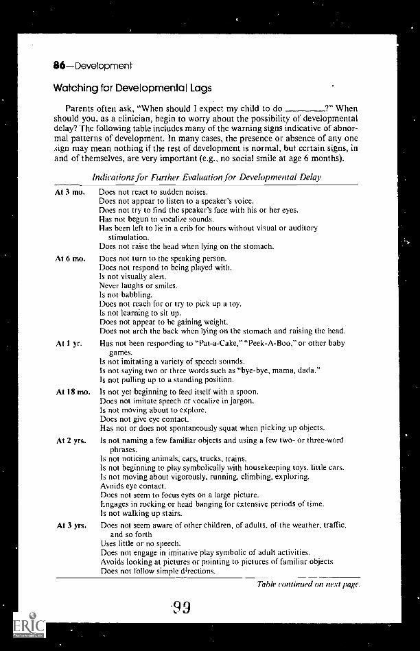
86Development
Watching for Developmental Lags
Parents often ask, "When should I expect my child to do 9" W henshould you, as a clinician, begin to worry about the possibility of developmentaldelay? The following table includes many of the warning signs indicative of abnor-mal patterns of development. In many cases, the presence or absence of any onesign may mean nothing if the rest of development is normal, but certain signs, inand of themselves, are very important (e.g., no social smile at age 6 months).
Indications for Further Evaluation for Developmental Delay
At 3 mo. Does not react to sudden noises.Does not appear to listen to a speaker's voice.Does not try to find the speaker's face with his or her eyes.Has not begun to vocalize sounds.Has been left to lie in a crib for hours without visual or auditory
stimulation.Does not raise the head when lying on the stomach.
At 6 mo. Does not turn to the speaking person.Does not respond to being played with.Is not visually alert.Never laughs or smiles.Is not babbling.Does not reach for or try to pick up a toy.Is not learning to sit up.Does not appear to be gaining weight.Does not arch the back when lying on the stomach and raising the head.
At 1 yr. Has not been respording to "Pat-a-Cake," "Peek-A-Boo," or other babygames.
Is not imitating a variety of speech sounds.Is not saying two or three words such as "bye-bye, mama, dada."Is not pulling up to a standing position.
At 18 mo. Is not yet beginning to feed itself with a spoon.Does not imitate speech cr vocalize in jargon.Is not moving about to explore.Does not give eye contact.Has not or does not spontaneously squat when picking up objects.
At 2 yrs. Is not naming a few familiar objects and using a few two- or three-wordphrases.
Is not noticing animals, cars, trucks, trains.Is not beginning to play symbolically with housekeeping toys, little cars.Is not moving about vigorously, running, climbing, exploring.Avoids eye contact.Does not seem to focus eyes on a large picture.Engages in rocking or head banging for extensive periods of time.Is not walking up stairs.
At 3 yrs. Does not seem aware of other children, of adults, of the weather, traffic,and so forth
Uses little or no speech.Does not engage in imitative play symbolic of adult activities.Avoids looking at pictures or pointing to pictures of familiar objectsDoes not follow simple directions.
99
Table continued on next page.

Diabetes-87
Indications JOr Further Evaluation for Developmental Delay (Cont.)
At 3 yrs.(Cwit.)
At 4 yrs.
Engages fOr long periods of time in repetitive behaviors like flipping pagesof a magazine, or spinning a wheel on a little truck, head banging, and
so forth.Cannot ride a tricycle if given plenty of opportunity to do so.
Does not have at least partially un&rstandable speech with sentences.Uses echolalic speech or frequent, bizarre, meaningless sounds.Doe'. not focus visually on pictures.Does not seem interested in listening to a simple story about his or her
experiences.Repeatedly tests all limits.Is so quiet and conforming that he or she never tests or tries anything new.
Has pronounced fears and phobias.Frequently engages in flapping of the arms or flipping of the hands
to express excitement.Runs about from one thing to another every minute or so without getting
fully involved in an activity.Is still untrained in toileting (occasional slips do occur at this age).Does not draw some sort of representation of human beings (at least a head
and a few features), if crayons or pencils have been available to the child.Stays on the periphery of the playroom, paying no attention to other
children for some weeks, after most children have overcome shyness and
begun to play with or near other children.Avoids eye contact.Engages in head banging or rocking.Cannot tolerate change or frustration without frequent 2-year-old tantrums.
Reference: Accardo, P.1, Capute AJ: The Pediatrician and the DevelopmentallyDelayed Child: A Clinical Textbook on Mental Retardation. Baltimore, University Park
Press, 1979.
DIABETES
Classification and Etiology of Diabetes Mellitus
Classification of Diabetes Mellitus
IDIOPATHIC SFCONDARY
Insulin-dependent (type I) Pancreatic trauma, disease, or resection
Non-insulin dependent (type II) Hormone-induced
Maturity onset diabetes of youth (MODY) Drugs and chemical agentsGenetic syndromesInsulin receptor abnormalitiesOther types
From Lebovitz HE: Etiology and pathogenesis of diabetes mellitus. Pediatr Clin North
Am 31:524, 1984, with permission.
Although the majority of cases of pediatric diabetes mellitus will be of the type
I classification, the remaining idiopathic and secondary etiologies cannot beoverlooked. Some clues in establishing a diagnosis between type 1 and type ll are
provided in the following table:
1

88Diabetes
Insulin-Dependent vs. Non-Insulin-Dependent Diabetes
INSULIN-DEPENDENT NON-INSUL1N-DEPENDENT
I. Association with HLAB81 D3 or HLA 815/ D4
2. Pancreatic insulin content3. Anti-islet antibodies4. Primary insulin resistance5. Concordance rate of identical
twins for diabetes mellitus
2.5 x expectedfrequency
0
85%
Minimal25 to 50%
Same frequency asnormal population
>50% of normal<5%Marked100%
From Lebovitz HE: Etiology and pathogenesis of diabetes mellitus. Pediatr Clin NorthAm 31:525, 1984, with permission.
The secondary causes of diabetes mellitus are too numerous to coveradequately in this text. There are, however, a number of hormonal and chemicalcauses that deserve mention, principally because treatment of the primarydisorder in hormonal abnormalities and removal of the offending agent inchemically induced diabetes mellitus frequently reverse the disease process.
Hormonally Induced Diabetes MellitusABNORMALITY PREVALENCE (WHERE KNOWN)
AcromegalyCushing's syndromePrimary aldosteronismPheochromocytomasGlucagonoma
20%20%
Diabetogenic Drugs
Diuretics and AntihypertensivesChlorthalidoneClonidineDiazoxideFurosemideMetalazoneThiazides
Hormonally Active AgentsACTHGlucagonGlucocorticoidsOral contraceptivesGrowth hormonesThyroid hormones (thyrotoxic doses)
Psychoactive AgentsChlorprothixeneHaloperidolLithium carbonatePhenothiazinesTricyclic antidepressants
Catecholamine andOther NeurologicallyActive AgentsDiphenylhydantoinEpinephrinelsoproterenolLevodopaNorepinephrine
Analgesic,Antipyretic andAnti-inflammatoryAgentsIndomethacin
AntineoplasticAgentsAlloxanL-AsparaginaseStreptozotocinMiscellaneousIsoniazidNicotinic acid
From Lebovitz HE: Etiology and pathogenesis of diabetes mellitus. Pediatr Clin NorthAm 31:527, 1984, with permission.
Diagnosis of Diabetes Mellitus
In the presence of symptoms, the diagnosis of diabetes mellitus is an uncompli-cated task: a child presenting with polydipsia, polyuria, polyphagia, and weightloss with an accompanying elevation of blood glucose and/or ketonemia leads the
1 01

tio
Diabetes-89
pediatrician to a rapid answer. In the absence of symptoms or the presence of mildsymptoms, however, the diagnosis of diabetes mellitus is much more difficult.
In children, the diagnostic criteria for diabetes mellitus are as follows:
I. Presence of symptoms of diabetes, such as polydipsia, polyuria, ketonuria,and weight loss, together with a random plasma glucose of 200 mg/dI
or
2. In asymptomatic children, both an elevated fasting glucose concentrationand a sustained elevated glucose concentration during an oral glucose toler-ance test (1.75 g/kg up to maximum of 75 g) on two or more occasions.
Fasting value:
Venous plasma 140 mg/c11Venous blood 120 mg/d1Capillary blood 120 mg/dl
2-hour OGTT value and anintervening value:
Venous plasma 200 mg/d1Venous blood 180 mg/dICapillary blood 200 mg/di
Reference: Adapted from Lebovit7 HE: Etiology and pathogenesis of diabetes mellitus.Pediatr Clin North Am 31:521 523, 1984.
Diabetic Ketoacidosis
Despite continued advances in control of the diabetic child, diabetic ketoaci-dosis remains an acute medical emergency. In the known and presenting diabeticchild, ketoacidosis is defined as "hyperglycemia with a blood glucose exceeding300 mg/di, ketonemia with total ketones (13 hydroxybutyrate and acetoacetate) ins.rum exceeding 3 mmol/ L or positive at a 1:2 dilution in scrum or undilutedurine with the sodium nitroprusside reaction (Acetest; Ketostix; ChemstripsUGK), and acilosis with pH reduced to less than 7.30 or reduced stsumbicarbonate to less than 15 mEq/ I.." The maintenance requirements for fluid andelectrolyte therapy for diabetic ketoacidosis are outlined below. The clinicianshould keep in mind that these values represent averages and that the extent ofdehydration and electrolyte imbalances vary with the duration of symptoms, thepossible presence of vomiting, and prior insulin administration.
Fluid and Electrolyte Maintenance in Diabetic Ketoacidosis
MAINTENANCE REQUIREMENTS* LOSSES'
Water
Sodium
Potassium
ChloridePhosphate
1500 nil/rn2
45 mEq/m2
35 mEq/m230 mEq/m2
10 rnEq/m2
100 ml/kg (range 60 100 ml/kg)6 mEq/kg (range 5 13 mEq/kg)5 mEq/kg (range 4 6 mEq/kg)4 mEq/kg (range 3 9 mEq/kg)3 mEq/kg (range 2 5 mEq/kg)
* Maintenance is expressed in surface area to permit uniformity because fluid requirementschange as weight increases.Losses are expressed per unit of body weight, since the losses remain relatively constantas a function of total body weight.
Reference: Sperling M: Diabetic ketoacidosis. Pediatr Clin North Am 31:591 610,
I.-) 21984, with permission.

90Diabetes
Glycosylated Hemoglobin Assay
The glycosylated hemoglobin assay provides the clinician with a profile ofglycemia during the previous 60 to 120 days. This is helpful in ascertaining inter-visit glycemia control and to determine the relationship between complicationsand compliance. The issue of compliance is particularly important in earlyadolescence, when responsibility for glycemic control shifts from the parents tothe teenager.
Estimate qf Blood Glucose Control Using Glycosylated Hemoglobin Assay
GINCOSYLAII-II-MOOLOBIN(c;)
PROBABIT WOOD UC051-RANG': (mg dl)
FSTIMAIT"CON1R01 "
5.4 7.4 60 120 Normal range
8 9 120 150 Excellent
9 10 150 180 Good
10 11 180 220 Fair
11 12 220 260 Fair
12 13 260 300 Fair to poor> 13 > 300 Very poor
Reference: Spack NP: Diabetes mellitus in adolescence. Adolescent Medicine State ofthe Art Reviews 2:529, 1991, with permission.
The Diabetic Patient with ConcomitantSystemic Disease
The following guidelines are intended to assist thc clinician or other caregiverin managing the diabetic patient with additional iilness:
Guidelines to Sick Day Management
1. Never skip insulin administration.
2. Check blood glucose and urinary ketones every 4 6 hours
3. Give supplemental sho '-acting insulin every 4 hours for elevated blood sugarand additional dose amounts for hyperglycemia with ketonuria.
4. Evaluate and treat the underlying illness.
5. If blood glucose levels are low (less than 120 mg/di), reduce short-acting insulinand give glucose-containing fluids.
6. If adequate fluid intake cannot be maintained or vomiting persists for more than2 hours, intravenous hydration is necessary.
7. Notify a clinician if blood glucose is more than 400 mgjd1 with moderate to largeacetone or change in patient's level of alertness or signs of dehydration (weighingevery 6 hours may be a useful guide.
Reference: Spack NP: Diabetes mellitus in adolescents. Adolescent-Medicine State ofthe Art Reviews 2:511, 1991, with permission.
103
A
et

DIARRHEACHRONIC
Common Causes
Antibiotic-inducedCarbohydrate malabsorption, hereditary
LactoseChemotherapy-inducedCystic fibrosisDietary
Allergy (milk, soy, other)Overfeeding
Uncommon Causes
Anatomic lesionsHirschsprung's diseaseMalrotation
Celiac diseaseIrritable bowel syndromeMalnutrition, starvation
Rare Causes
Diarrhea-91
InfectionBacterialHuman immunodeficiency
virus (HIV)Parasitic
Post infectiousCarbohydrate malabsorption
Necrotizing enterocolit isParenteral infections
Otitis mediaUrinary tract infections
Regional enteritisUlcerative colitis
Abeta- and hypobetalipoproteinemiaAdrenal insufficiencyBiliary atresiaBlind loop syndromeCarbohydrate malabsorption
Sucrose, isomaltose, glucose, galactoseChronic hepatitisEnterokinase deficiencyFamilial chloride diarrheaGanglioneuromaHyperthyroidismImmune deficiency
Combined immune deficiencyHypogammaglobulinemiaIgA deficiency
Intestinal ischemiaIntestinal lymphangiectasiaIntestinal pseudo-obstructionMesenteric artery insufficiencyNeuroblastomaPancreatic insufficiency and
neutropenia (Schwachman-Diamond-Oski syndrome)
Pancreatic tumorsRadiation-inducedShort gut syndromeSmall bowel tumors;
lymphosarcomaWolman's disease
The Common Bacterial Causes of Bloody Diarrhea
The association of diarrhea and the passage of small amounts of blood in thestool (hematochezia) can be due simply to hemorrhoids or mucosal tears causedby spasm, hypermotility, or irritation at the mucocutaneous junction. Thepersistent case of hematochezia associated with diarrhea, however, is most likelyindicative of an infectious or inflammatory etiology. In the pediatric patient,bloody diarrhea is usually a result of an infectious enteric pathogen.
The following table summarizes the types and manifestations of bloodydiarrhea caused by bacterial pathogens.
4

Blo
ody
Dia
rrhe
a of
Bac
teria
l Orig
in
INF
EC
TIO
US
AG
EN
T
Shi
geo
AG
E C
HIL
DM
OS
T A
FF
EC
TE
D
US
UA
LIN
CU
BA
TIO
NU
SU
AL
SO
UR
CE
PE
RIO
DO
F A
CQ
UIS
ITIO
NP
RE
SE
NT
ING
SE
AS
ON
SIG
NS
& S
YM
PT
OM
S
Sal
mon
ella
Cam
pylo
bacw
r
Yer
sini
a
C. d
iffic
ile
6 m
o-3
yr
Infa
nts
<2
yrso
met
imes
old
er
Infa
nts
and
child
ren
<6
yr
Tod
dler
s to
teen
ager
s
All
ages
afte
rne
onat
al p
erio
d
Esc
lwric
hia
coil
a.E
nter
oinv
asiv
eA
ll ag
es
b. E
nter
o-pa
thog
enic
N. g
onor
rhea
e
C. t
rach
omar
is
All
ages
Tee
nage
rs
Tee
nage
rs
36 7
2°F
ecal
-ora
l; co
ntam
inat
edfo
od o
r w
ater
24 4
8°O
ral v
ia c
onta
min
ated
food
or
wat
er
2 7
dO
ral v
ia c
onta
min
ated
food
or
wat
er; i
nfec
ted
pets
3-4
dO
ral v
ia c
onta
min
ated
food
or
wat
er;
pers
on-t
o-pe
rson
I6
wk
Alm
ost a
lway
s re
q. r
espr
ior
expo
sure
to a
nti-
biot
ics,
esp
ecia
lly a
mpi
cilli
n,cl
inda
myc
in, c
epha
losp
orin
s
24-7
2°F
ecal
-ora
l; co
ntam
inat
edfo
od o
r w
ater
1-3
dF
ecal
-ora
l; co
ntam
inat
edfo
od o
r w
ater
2-7
dA
nal i
nter
cour
se
2-3
wk
Ana
l int
erco
urse
War
mF
ever
, abd
omin
al p
ain
mon
ths
wat
ery
diar
rhea
beco
min
g bl
oody
War
mV
omiti
ng, a
bdom
inal
mon
ths
pain
, dia
rrhe
ady
sent
ery-
like
War
mS
ever
e ab
dom
inal
mon
ths
pain
, blo
ody
diar
rhea
Coo
ler
mon
ths
All
year
War
mm
onth
s
War
mm
onth
s
AU
yea
r
All
year
Fe%
er,
abd
omin
alpa
in, v
omiti
ng,
diar
rhea
Abd
omin
al p
ain,
diar
rhea
, dis
tent
ion,
bloo
d in
sto
ol
Fev
er, c
hills
, abd
omin
alpa
in, w
ater
y di
arrh
eabe
com
ing
dyse
nter
y-lik
e
Fev
er, n
ause
a, c
ram
psw
ater
y or
blo
ody
diar
rhea
in s
ome
sero
type
s
Dys
ente
ry, o
dyno
chez
ia
Dys
ente
ry, o
dyno
chez
ia
MA
IN P
AT
HO
-P
HY
SIO
LOG
ICA
LM
EC
HA
NIS
M
Ent
eroi
nvas
ive;
cyto
path
ic
Ent
eroi
nvas
i.:e
Cyt
olyt
ic e
xoto
xin
Ent
eroi
nvas
ive;
ente
roto
xige
nic
Cyt
otox
in
Ent
eroi
nvas
ive;
cyto
path
ic
Cyt
otox
in;
ente
roin
vasi
ve
Ent
eroi
nvas
ive;
cyto
toxi
c
Ent
eroi
nvas
ive;
cyto
toxi
c
4

Blo
ody
Dia
rrhe
a of
Bac
teria
l Orig
in (
Con
t.)
IN E
. FC
110
05A
GE
NT
Si 0
01.
FIN
DIN
GS
I,AB
OR
M O
RY
EN
DO
SC
OP
YH
IST
OLO
GY
BA
RIU
ME
NE
MA
Shi
gella
Blo
od, W
BC
sB
and
form
s >
Mild
-to-
seve
reA
cute
infla
m-
Nor
mal
tose
gmen
ted
form
sco
litis
mat
ion
colit
is-li
ke,
muc
o.,..
I ulc
ers
Sah
none
llaB
lood
, WB
Cs
Nor
mal
to1
WB
C L
shi
ftM
ild c
oliti
sA
cute
foca
lin
flam
mat
ion
Nor
mal
toco
litis
-like
Cam
pylo
hact
erB
lood
, WB
Cs
Nor
mal
to1
WB
CC
oliti
s-lik
eF
ocal
or
diffu
seac
ute
infla
mm
atio
nC
oliti
s-lik
e
Yer
sini
aR
are
PM
Ns,
bloo
dI W
BC
and
sed
rate
Cro
hn's
-likc
apht
hous
lesi
ons
Foc
al in
flam
mat
ion;
mon
os >
PM
Ns
Spa
sm c
ecum
,ab
norm
alte
rmin
alile
um
C. d
iffic
ileB
lood
,fe
w W
BC
sN
orm
al to
slig
htW
BC
Hyp
erem
ia; p
laqu
es,
foca
l or
diffu
seP
seud
omem
bran
es,
dist
ende
d gl
ands
,ac
ute
infla
mm
atio
nlo
cally
"Dirt
y"-c
olon
,m
ucos
alirr
egul
ariti
es,
nodu
lar
Esc
herie
hia
coil
a. E
nter
oinv
asiv
eB
lood
,fe
w W
BC
sN
orm
al o
rW
BC
Lesi
on in
R c
olon
;hy
pere
mic
exu
date
,sm
ulc
ers
Mild
infla
mm
ator
yer
osio
ns
h. E
nter
o-pa
thog
enic
Blo
od,
few
WB
Cs
I WB
C L
shi
ftH
yper
emia
, hem
or-
rhag
e, s
uper
ficia
lul
cera
tion
Non
spec
ific
colit
is"T
hum
bpr
intin
g"R
col
on
N. g
onor
rhea
eM
ucop
us,
bloo
dW
BC
I. s
hift
Sev
ere
proc
titis
to10
15
cmA
cute
diff
use
infla
mm
atio
nN
orm
al o
rpr
octit
is
C. t
rach
omat
isB
lood
, WB
Cs
Slig
ht I
WB
CP
roct
itis
to15
cm
Acu
te in
flam
mat
ion
gian
t cel
lsP
roct
itis,
str
ic-
ture
rec
tum
RS
Trim
etho
prim
-su
lfarn
etho
xazo
le
Non
e cx
ccpt
in s
ick
infa
nt<
1 yr
: chl
oram
phen
icol
,am
pici
llin,
trim
etho
prim
-su
lfam
etho
xazo
le
Ery
thro
myc
in s
tear
ate
Sel
dom
nee
ded:
tetr
acy-
clin
e, a
mpi
cilli
n, c
hlor
am-
phen
icol
, trim
etho
prim
-su
ram
etho
xazo
le
Van
com
ycin
, Bac
itrac
in,
met
roni
dazo
le,
chol
esty
ram
ine
Am
pici
llin,
gen
tam
icin
,tr
imet
hopr
im-
sulfa
met
hoxa
zole
ine
need
ed
Pen
icill
in, t
etra
cycl
ine
Tet
racy
clin
e, tr
imet
hopr
im-
sulfa
met
hoxa
zole
94Diarrhea
The stool culture, of course, yields the most definitive information in theconsideration of a patient with bloody diarrhea of an infectious source. Otheruseful tests that can be performed immediately include a microscopic examinationof the stool for the presence of mucus, red blood cells, and white blood cells,using either methylene blue or Wright's stain, a Gram stain, and visualexam:nation for ova and parasites. Immunoassays for the diagnosis of possibleviral etiologies of diarrhea (e.g., rotavirus, adenovirus, Norwalk virus, etc.) canalso bc helpful.
Reference: Silverman A: Common bacterial causes of bloody diarrhea. Pediatr Ann14:39 50, 1985, with permission.
Management Plan for an Infant Less Than '1 Year of Agewith Diarrhea Who Does Not Require Hospitalizationat the Initial Evaluation
First evaluation
a. Colitis (fecal leukocytes) 0 12 mo
h. No colitis; diarrhea <5 days
c. History of exposure toSalmwwIla
2. Follow-up evaluation
a. Diarrhea >5 days
b. Stool culture positiveBlood culture positive
c. Stool culture positive;blood culture negativei. Toxic or immuno-
compromisedii. Febrile
0 12 mo
o 3 mo
o 12 mo
0 12 mo
0 12 mo
<3 mo
iii. Febrile >3 mo
iv. Afebrile, improving
d. Stool culture positive; bloodculture not obtained at firstvisit.
0-12 mo
Antibiotics of choice: cefotaxime or ceftriaxone.
Stool culture; bloodculture if <3 mo
No stool culture
Stool culture
Stool culture
Admit; look for focalinfection in meningesbone, urinary tract
Admit, as above
Admit; do blood cultureand give antibioticsAdmit; do blood cultureand give antibioticsAdmit; do blood culture;withhold antibioticspending culture resultsReexamine, observe athomeSee 2-c.
Reference: St. Gcme .1W III, et al: Consensus: Management of Salmomlla infection inthc first year of life. Pcdiatr Infect Dis 7:615 621, 1988.
ti

4
--r
Dysmenorrhea/Dysmorphism-95
DYSMENORRHEA
Primary and Secondary Dysmenorrhea
Dysmenorrhea is a cramping pain in the lower abdomen and lower hack thatis temporally associated with menstrual blood flow. It may also be accompaniedby headache, nausea, or diarrhea. Epidemiologic studies reveal, with great con-sistency, the high prevalence rate of dysmenorrhea among adolescent girls.During adolescence, dysmenorrhea becomes more common as age increases. Thisis probably because primary dysmenorrhea the more common of the twotypes is associated with ovulatory menstrual cycles, and most girls are anovu-latory 18 to 24 months after menarche.
Primary dysmenorrhea is associated with no clinically detectable pelvicdisease or other disorder. It usually begins 18 months after menarche (with theonset of ovulation). The pain starts on the same day as blood flow, lasting a fewhours to 3 days, and is frequently accompanied by diarrhea in moderate orsevere cases. The etiology of primary dysmenorrhea remains unclear, althoughit is known that women who suffer from this disorder produce increasedamounts of prostaglandins F. and F2 in their menstrual fluids. These prosta-glandins cause the myornetrium to increase its resting muscle tone, which yieldsexcessive uterine contractions. The result is uterine ischemia and painfulcramping.
Secondary dysmenorrhea, which presents far less frequently among theadolescent age group, is usually associated with some pathologic process in thepelvis. The pain is unusually severe and it (1) begins at menarche (obstructiveform of secondary dysmenorrhea); (2) begins more than 3 years after menarche(e.g., endometriosis); or (3) is acute and related to one particular menstrualperiod (e.g., a complication of sexual activity such as a sexually transmittedinfection or pregnancy).
Conditions Associated with Secondary Dysmenorrhea
I . Genital tract infections, specifically 4.sexually transmitted endometritis orsalpingitis
2. Complications of pregnancy, e.g..threatened or ectopic pregnancy 5.
3. Endometriosis 6.
Congenital malformations of thegenital tract (with or without acomponent of blood flowobstruction)
Genital tract cysts and neoplasms
nt rauteri ne devices
Reference: Coupey SM, Ahlstrom P: CommonNorth Am 36:551 571, 1989,
menstrual disorders. Pediatr
DYSMORPHISM
Clin
Diagnosing Dysmorphism
The dysmorphic infant or child presents the pediatrician and the parent withseveral unsettling requirements: a correct diagnosis and, where possible, anetiology; a comprehensive management guide; and, a careful assessment ofrecurrence risks and rates.

96Dysmorphism
The pediatrician's best tools in the diagnosis of the dysmorphic child are hisor her eyes, the child's parents and other family members, and several guidingprinciples. The task involves the determination of the pathogenesis and closeattention to the appearance of marked or subtle patterns (Table I).
Table I. Pathogenic Mechanisms of Dysmorphism
TYPE OFDYSMORPHISM DEFINITION ASSOCIATED FACTORS EXAMPLES
Malformation A rudimentary ab-normality involvingdifferentiation or orga-niiation of an organpart, an organ, or bodypart representing anembryologic field.Occurs duringembryogenesis.
Deformation Represents a responseof normal tissue to ab-normal external forces.Tends to occur late inpregnancy.
Disruption Abnormality involvinga destructive process ina normally formedorgan.
Dysplasia Abnormality in the de-velopment (organiza-tion or differentiation)of cells and tissues asopposed to wholeorgans.
Genetic or chromosomalabnormality; terato-genic effect
Fetal: Large fetusMultiple fetusesMalformed fetusOligohydramniosUnusual placental site
Maternal: PrimigravidaSmall motherSmall uterusMalformed uterusUterine fibroids
Disruptive agentRadiationInfectionEarly amnion rupture
Ischemia
Vascular mechanism,hemorrhage
Occlusion
Genetic or unknown
Cleft lip/palateSpina bifidaCongenital heart
defectsDown syndrome
CraniostenosisPlagiocephaly-torti-
collis sequenceMicrognathiaEar deformitiesPectus carinatumScoliosisDorsiflexion of footClubfootFacial nerve palsiesErb's palsy
AnomalyMicrocephalyTORCHES syndromeAnencephaly, unusual
facial clefting, eyedefects, clefts, limb!digit abnormalitiesor amputations
Porcncephalic cysts,ileal atresia
Hemifacial microso-mia/Goldenhar'ssyndrome
Streeter's bandsLimb/digit amputationsGastroschisis
Tuberous sclerosusEctodermal dysplasiaNeurofibromatosisPresacral teratomaNeuroblastomaRetinoblastomaBeckwith-Wicdemann
syndrome--asexample of dysplasia-malformationcombination
1 ) 9
Dysmorphism-97
Looking for patterns:The anomalies described in the above table can be characterized as "major"
(those with functional, surgical, or cosmetic consequences) or "minor" (thosewithout consequences). Bear in mind that normal variants may constitute minoranomalies in the context of a syndrome.
A pattern of anomalies will often reveal the diagnosis and can present assyndromes, sequences, or associations.
Sequence:A sequence refers to an isolated developmental abnormality and its subsequent
structural consequences. An example is the initial mandibular hypoplasia of thePierre Robin sequence that results in small chin, cleft palate, obstructive airway,and anoxia. The cause of a sequence is not necessar4 defined.
Syndrome:A syndrome consists of a pattern of malformations that are recognized to
result from a specified cause, such as trisomies.
Association:Associations are recognized as nonrandom and significant groupings of
malfor nations without known etiologies. Two of the common associations areVACTERL and CHARGE. VACTERL, previously known as VATER, describesvertebral anomalies, anal atresia, cardiac defects, tracheoesophageal fistula withatresia, radial and renal defects, and limb defects. CHARGE consists ofcoloboma of the eye, heart defects, atresia of the choanae, growth and mentalretardation, genital anomalies in the male, and ear anomalies.
In addition to the examination directed at recognition of patterns, there areseveral measurements that can be made to facilitate a diagnosis. The size of thehands and feet as well as the ratio of upper body length (crown to pubis sym-physis) to lower body length (symphysis to soles) can signal dwarfing syndromes.The measurement of inner canthal distance, interpupillary distance, cornealdiameter, internipple distance, and penile length may lead to diagnostic clues.
Be aware of the risk factors for structural anomalies (Tables 2 and 3).
Thble 2. Children at Risk for Structural Anomalies
I. The child with a family historyof structural anomalies
2. The child with one knownstructural anomaly
3. The small-for-dates infant
4. The infant of a mother with diabetesmellitus, phenylketonuria, epilepsy,or alcoholism
5. The mentally retarded child
6. The "strange-looking" child
Table 3. Anomalies and Associated Conditions
LOCATION ANOMALY
Back
Chest
Digits
CONDITION*
Scoliosis
Pectus cxcavatum
Curved fifth fingers (clinodactyly)Finger-like thumbs
Stickler syndrome, neurofibromatosis
Marfan's syndrome
Russell-Silver syndromeHolt-Oram syndrome, Aasc syndrome
*These arc illustrative conditions; often other conditions will also have the anomaly.Table cvntintwd on next page.
no

98- Dysmorph ism
Table 3. Anomalies and Associated Conditions (Cont.)
LOCATION ANOMALY CONDITION'
Digits(Cont.)
Ears
Eyes
PolydactylyBroad thumbs/great toesSyndactylyNail hypoplasia
Second finger overlaps thirdfinger, fifth finger overlapsfourth finger
Low-setCrumpledSmallLargePreauricular tagsPreauricular pits
MicrophthalmosColoboma of irisStellate pattern of irisAbsent irisCataracts
Face Unique facial gestalt
Genitalia HypospadiasMicropcnisScrotal shawlHypoplasia of labia majora
Hair Widow's peakWhite forelockLow posterior hairlineScalp defectSparse to absentKinky
Hands,feet ShortLongAbnormal palm/sole creases
Head MacrocephalyMicrocephaly
Joints DislocationsElbow abnormalitiesAbsent patellaContractures
Limbs Long
Mandible
Maxilla
Mouth
ShortRadial hypoplasia
Hypopla-ia
Hypoplasia
LargeMultiple frenulaLarge tongue
Carpenter's syndrome, trisomy 13 syndromeRubinstein-Taybi syndromeApert's syndromeFetal hydantoin syndrome, Coffin-Siris
syndromeTrisomy 18, trisomy 13
Trisomy 18, Treacher Collins' syndromeBeal syndromeDown syndromeSotos' syndromeGoldenhar's syndromeBOR syndrome
Oculodentodigital dysplasiaCHARGE associationWilliams syndromeAniridia-Wilms' tumorHallermann-Streiff syndrome
Down syndrome, Williams syndrome,Prader-Willi syndrome, Cornelia de Langesyndrome, whistling face syndrome
Smith-Lemli-Opitz syndromeRobinow's syndromeAarskog's syndromeEscobar syndrome
Frontonasal dysplasiaWaardenburg syndromeTurner's syndrome, Noonan's syndromeTrisomy 13Hypohidrotic ectodermal dysplasiaMenkes' syndrome
Prader-Willi syndromeMarfan's syndromeSotos' syndrome, .)own syndrome
Hydrocephalus, Sotos' syndromeMany syndromes
Larsen's syndromeTurner's syndrome, XXXXY syndromeTrisomy 8Beal syndrome, arthrogrypotic conditions
Beal syndrome, Stickler syndrome,XYY syndrome
Many short-limbed dwarfing syndromesVATER association
Pierre Robin sequence
Nager syndrome
Goldenhar's syndromeOrofaciodigital syndromeBeckwith-Wiedemann syndrome
1 11
Table continued on next page.

Dysmorphism -99
Table 3. Anomalies and Associated Conditions (Cont.)
LOCATION ANOMALY CONDITIONS
Neck WebbedShort
Nose SmallLargeChoanal atresia
Ocular region HypertelorismHypotelorismShort palpebral fissuresEpicanthic foldUpward slant of palpebral
fissuresDownward slant of
palpebral fissuresSynophrys (fusion of
eyebrows in midline
Philtrum ProminentLongShortSmooth
Skin Cafe au lait spots
Teeth
Thorax
Pigmented neviMultiple lentiginesTelangiectases
Hemangiomata
HypodontiaCariesNeonatal
Small
Noonan's syndromeKlippel-Feil syndromc
Feta! warfarin effectSeckel's syndromeCHARGE association
Opiti syndrome, Aarskog's syndromeHoloprosencephalyFetal alcohol syndrome, blepharophimosisFetal trimethadione syndromeDown syndrome
Treacher Collins' syndrome
Cornelia de Lange syndrome
Trichorhinophalangeal syndromeWilliams syndromeCohen syndromeFetal alcohol syndrome
Neurofibromatosis. Russell-Silver syndrome,Bloom's syndrome
Turner's syndromeLeopard syndromeAtaxis-telangiectasia syndrome, Bloom's
syndrome, Rothmund-Thomsonsyndrome
Sturge-Weber syndrome
Hypohidrotic ectodermal dysplasiaDentinogenesis imperfectaEllis-van Creveld syndrome
Jeune's syndrome
Lastly, the pediatrician must be armed with the knowledge of recurrence risksand be able to appropriately counsel the concerned parent(s) (Table 4).
Table 4. Recurrence Risk in Families of Dysmorphic Children
RISK IN SIBLINGS PERCENTAGE CONDITION
Low 1 2 Trisomy 21 syndrome, trisomy 13, trisomy 183 7 Spina bifida, cleft palate/lip, hypospadias
Moderate 25 Autosomal recessive disease: Smith-Lemli-Opitisyndrome
X-linked recessive disease: X-linked hydrocephalus
High 50 Autosomal dominant disease: neurofibromatosis,tuberous sclerosis
Total 100 Chromosomal disorder: 21/21 translocationDown's syndrome with carrier parent
Reference: Kee le DK: A diagnostic approach to the dysmorphic child. Contemp PedNov:63 84, 1985. Tables 2 to 4 from this reference, with permission.
Oski FA, et al: Principles and Practice of Pediatrics. Philadelphia, J.B. Lippincott, 1990.
112
ft

1 00 Dysphagia
DYSPHAGIA
Common Causes
Cmical mucositisCaustic ingestionGastroesophageal reflux
with esophagitisRadiation/chemotherapy
Immatute sucking/swallowingmechanism
Uncommon Causes
Cerebral palsyCleft palateEsophageal spasmEsophageal strictureExternal compression of the esophagus
Esophageal diverticuliEsophageal duplicationMediastinal masses/tumorsVascular anomalies
Rare Causes
Choanal atresiaCollagen vascular disease
DermatomyositisScleroderma
DiphtheriaEsophageal atresia, web, cystLaryngeal cyst, cleftMuscular hypertrophy of the esophagusNeuromuscular causes
BotulismBulbar and suprabulbar palsy
Mobius syndromeChalasia/achalasia of the esophagusCongenital laryngeal stridorCranial nerve palsy
113
Oropharyngeal infectionsCervical adenitisEpiglottitisGingivitisHerpetic stomatitisPeritonsillar abscessPharyngitisRetropharyngeal abscessTooth abscess
Physiologic expulsion reflux
Foreign bodyInfectious esophagitis
Candida, herpesMacroglossia (any cause)MicrognathiaPharyngeal diverticuliPhysiologic (globus hystericus)Submucosal cleftTracheoesophageal fistula
Neuromuscular causes (Cont.)Demyelinating diseaseGuillain-Barré syndromeHypotoniasMuscular dystrophyMyasthenia gravisMyotonic dystrophyPharyngeal or cricopharyngeal
incoordinationTetanus
Pharyngeal cyst, cleftRuminationTemporomandibular ankylosis/
hypoplasiaTumors (oropharynx, esophagus)

4
DYSRHYTHMIA
Common Causes
AcidemiaCongenital heart diseaseDrugs
AntiarrhythmicsBeta blockersCaffeineCocaine
Uncommon Causes
Cardiomyopathy (dilated, hypertrophic,infiltrative)
Electrolyte disturbances (especiallyK, Ca, Mg)
Myocarditis
Rare Causes
Anomalous coronary arteryCentral nervous system
HemorrhageInfectionTrauma
Collagen vascular diseaseComplete congenital heart blockEndocrine (thyrotoxicosis, secondary
electrolyte disturbance)
Common Causes
Candidal dermatitis/vaginitisChemical urethritis (bubble bath)Contact dermatitis/vulvitis
Uncommon Causes
Foreign bodyHerpes simplexMeatitis
Dysrhythmia/Dysuria-101
Drugs (Cont.)PsychotropicsSympathomimetics
HypoxemiaIdiopathicPostoperative (cardiac procedures)
Sickle-cell diseaseSick-sinus syndromeWolff-Parkinson-White syndrome
(and/or other necessary bypasstracts)
Kawasaki diseaseMyocardial ischemiaMyocardial traumaMyocardial tumorsNeonatal lupusProlonged QT syndromeRheumatic fever
DYSURIA
UrethritisUrinary tract infectionViral cystitis
PinwormsUrethral trauma

02Dysuria
Rare Causes
AppendicitisBladder diverticulumBladder outlet obstruction
Posterior urethral valvesBladder stonesConstipationDrugs
AmitriptylineCytoxan
HematospermiaInterstitial cystitis
Meatal stenosisPosthitisProstatitisReiter's syndromeSchistosomiasisStevens-Johnson syndromeTuberculosisUrethral prolapseUrethral strictureVaricella
I'm sorry you are wiser,I'm sorry you are taller;
I liked you better foolish,And I liked you better smaller.
Aline (Mrs. Joyce) KilmerFor the Birthday of a Middle-Aged Child, Stanza I
115

EARS
Low-set Ears
Because of their association with various syndromes, low-set cars can be afinding of extreme importance. There is still a controversy over what constituteslow-set ears. We prefer a method of evaluation suggested by Dr. MurrayFeingold. In the figure, the face has a sheet of x-ray film held over it. On themargins of the film arc measurement scales, and the center of the film is bisectedby a horizontal line that is aligned with the medial canthi of the eyes. The amountof the car lying above the line is measured as well as the overall length of the ear.If the ear is below the center line, the ear is low set. If 10% or less of the overalllength of the ear is above the line, the ears are said to be low set. If low-set earsare found, look carefully for other physical abnormalities.
I f x 100 5. 10,
the ears arc low set.
Some Syndromes Associated with Low-set Ears
Apert's syndromeCamptomelic syndromeCarpenter's syndromeCri-du-chat syndromeDeletion of the short arm
of chromosome 13
Down's syndromeFetal hydantoin syndromeHallermann-Streiff
syndromeNoonan's syndromeSaethre-Chotzen syndrome
Seckel's syndromeTrisomy 13 syndromeTrisomy 18 syndromeTreacher-Collins
syndromeTurner's syndrome
Reference: Feingold M: Commentary, Yearbook of Pediatrics, p 152, 1977.
103

I 04 Encopres1s/Endotracheal Intubation
ENCOPRES IS
Rare CausesCommon Causes
Chronic constipationDiarrheal disordersEmotional disturbance
Uncommon Causes
Hirschsprung's disease
DiastematomyeliaEpidural abscessPoliomyelitisPostanorectal
surgeryOsteomyelitis of the
vertebral body
ENDOTRACHEAL I NTUBATION
Sacral agenesisSpinal cord tumorSyringomyeliaTransverse myelitis
Complications of Endotracheal Intubation
Endotracheal intubation in the setting of an intensive care unit, emergencyroom, or delivery suite is rarely as controlled a procedure as the same performedin an operating room. Unrecognized esophageal placement of an endotrachealtube is the most common complication of emergency intuhations and can leadrapidly to brain damage or death. Below are thc phases of progressive hypoxemia,their clinical pictures, and recommended resuscitative maneuvers.
Precritical Phase: Arterial oxygen content decreases from normal 19 vol% to 12vol% (± 70% saturation).
Sympathetic tone increases: HR increases by approximately 10 bpm.Systolic BP increases 10 15 mmHg.Diastolic BP unchanged.Pulse pressure widens.
Critical Phase: Arterial oxygen content ranges from 12 vol% to 9 vol% (50%saturation).
Vagal tone increases: systolic BP decreases, HR decreases.Cyanosis evident in nonanemic patients.
Terminal Phase: Arterial oxygen content falls below 9 vol% (<50% saturation).HR slow to 40 or fewer bpm.BP readings cannot be obtained.ECG readings may show marked sinus bradycardia in the absence of a
palpable pulse.
ftsusetationIn the absence of a known and immediately remediable problem, the tube
should be removed quickly and an oral or nasal airway should be placed. Thepatient should be administered 100% oxygen with a bag and mask until anunhurried reintubation can be performed. The use of atropine or vasopressorswill not correct the hypoxia.
Recognition and correction of the hypoxemia during the precritical andcritical phases will generally avert any catastrophic event. Terminal phase mayrequire initiation of BLS and ALS.*
*BLS = basic life support; ALS = advanced life support.
117
a

Endotracheal Intubation-105
Other Complications
Foreign bodies: Due to excessive lymphoid tissue in childhood, semirigid naso-tracheal tubes may cause dislodging of adenoid tissue and subsequent obstruction.
Bronchial intubation: The right bronchus is more susceptible to catheterizationdue to the obtuse angle at its junction with the trachea. Partial catheterization ofthe right bronchus may result in occlusion and collapse of the right upper lobe.
Tube distortion: Tubes constructed from soft rubber or metal reinforced plasticare less likely to kink, compress, or twist, but even they may distort, leading toocclusion and hypoxemia.
Effect of tube size: Due to the diameter of the pediatric airway, increased airwayresistance is a significant problem with thick-walled tubes. Refer to the chartbelow for selection of the appropriate thin-walled endotracheal tube.
Selection qf Endotracheal Tube
INEAN1 WEIGH I (E)ENDO MACH EA1. '11111 SIV1 ION CAFFIF 1 ER SIZEIN I ERNAI P. %METER IERI NCI! GAUG1.1
<1250 2.5 5 Fr1250 3000 3.0 6 Fr>3000 3.5 8 Fr
BY AGE
Full term 9 mo12 20 mo2 yr>2 yr
3.54.04.54.5 + age (yr)
4
References: Adriani J, et al: Complications of endotracheal intubations. South Med .181:739 744, 1988.
Firestone LL, et al (eds): Clinical Anesthesia Procedures of thc Massachusetts GeneralHospital. Boston, Little, Brown, 1988.
Verification Techniques for Proper Placement
Although no verification technique for the placement of endotracheal tubes iscompletely infallible, there are several methods that are quite reliable and oughtto be routinely performed after intubation.
1. The most reliable method of verification, detection of end-expired CO2,requires instruments that may not be available in all emergency situations. Ascarbon dioxide analyzers become less cumbersome and more available, thiswill be the method of choice.
2. Auscultation of 'weath sounds is historically accepted as a reliable methodfor verification of tracheal intubation. Recent studies have found that it maybe dependable only two-thirds of the time. Confounding factors in thepediatric age group include short necks and obesity. An important point:auscultation is most useful when performed both before and after intubation.Nonidentical sounds or unequal chest expansion hqupi alert the pediatricianto a possible problem. 1 61
106Enuresis
3. Pulse oximetry, now widely available, is a good adjunct to auscultation but ingeneral should not be the sole method of verification. Oximetry is an excellentmonitoring device for the intubated child at risk for mucous plugging orcompression. Regardless of the patient's risk status, pulse oximetry is usefulbecause pediatric patients tend to desaturate more quickly than their adultcounterparts.
4. Mouth-to-tube-insufflation remains a reliable, simple, and universally availabletechnique for verifying tube placement. A quick, but not too forceful, breathis expired into the tube connector by the physician. A properly placed tubewill allow gradual insufflation of the lungs. Expiration, against minimalresistance, should be felt against the physician's turned cheek. If esophagealplacement has occurred, the expired air will not equal the insufflated amount,the expulsion will be rapid, and the air will not be felt on the cheek.
Reference: Adriani J, et al: Complications of endotracheal intubation. South Med .181:739 744, 1988.
Firestone LL, et al (cds): Clinical Anesthesia Procedures of the Massachusetts GeneralHospital. Boston, Little, Brown, 1988.
ENURESIS
Common Causes
Developmental delay of bladder function and capacityPsychological
Uncommon Causes
DiabetesFood allergy
Rare Causes
Compulsive water drinkingDiabetes insipidus,
central or nephrogenic
Obstructive abnormalities of the urinary tractUrinary tract infections
Lumbosacral anomaliesSickle-cell anemiaSpinal cord tumors
Natural History of Nocturnal Enuresis
Most children stop wetting their beds between 2 and 4 years of age. Does thatmean that persistent bedwetting after 4 indicates an abnormality? Almostcertainly not. An organic disorder is rarely found in enuretic children, that is,children who are bedwetting after the age of 5.
Bedwetting after age 5 years is more common in boys, in children from largefamilies, and in children from lower socioeconomic groups, and it occurs morecommonly in families where one of the parents may have been a bedwetter.Nocturnal enuresis occurs once a month or more in 8% of school-age children.
119

Eosinophils--1 07
When bedwetting recurs after a period of dryness (termed regressive orsecondary enuresis), an explanation should be sought. The polyuria of diabetesmellitus may frequently present in this way. Also rule out urinary tract diseaseand diabetes insipidus.
Age by Which Bedwetting Slopped
AGF
BOYS
White (%) Black ((.)GIRLS
White (r.'0 Black (0-,)
<2 28 37 37 35
2 3 70 73 80 77
4 5 85 83 89 906 7 91 89 92 92
What becomes of the children, about 10 to 15%, who continue to wet after 4to 5 years of age?
There is a relative constancy to the percentage of children who spontaneouslyremit from their enuresis each year. The figure to remember is that any one childduring the course of 1 year has about a 15% chance of this problem going awayby itself. Thus, a child who wets his bed at age 5 will have about a 3% chance ofbedwetting by age 20. These figures apply only to the natural history of bed-wetting and not to those children whose bedwetting is related to an environmentalfactor.
Percentage Spontaneously Ceasing Bedwetting per Year
AGI.S 5 10 9 10 10 14 15 10 19
Percentage 14 16 16
Diurnal or daytime enuresis occurs much less frequently than nocturnal, withan incidencc of less than I% in a 7- to 12-year-old group.
References: Dodge WI', West ET', Bridgforth EB, et al: Am .1 Dis Child 120:32, 1970,Forsythe WI, Redmond A: Arch Dis Child 49:259, 1974.Adapted from McMillan .1A, et al: The Whole Pediatrician Catalog, Vol. 1 and Vol. 2.
Philadelphia, W.B. Saunders, 1977, pp 299 300; 1979, p 84.
EOSINOPHILS
Cerebrospinal Fluid Eosinophilia
Many laboratories now perform cytocentrifugation and differential cytologicstaining of the WBCs found in cerebrospinal fluid. The finding of CSFeosinophilia using this technique has led to the development of a list of thevarious agentsinfectious and otherwise --that can cause eosinophilic pleocytosisin the central nervous system. The following list includes the reported causes of"eosinophilic meningitis." For most of the causes listed, a careful history andphysical examination will provide the diagnosis.
1 2 11
108EpldIdymitis
Fungal infectionCoccidiornycosis meningitis
Viral infectionCoxsackie virus meningitisChronic lymphocytic
choriomeningitis virusSubacute sclerosing panencephalitis
Bacterial infectionTuberculous meningitisNeurosyphilis
CSF inflammationRadio logic dye used in myelographyRubber CSF shunt tubingCNS malignancyMultiple sclerosisCNS hemorrhage
Parasitic infectionNeurocysticercosis (caused by the
tapeworm Thenia soliuni andfound in Asia and Africa)
Toxocara canis and T catiTrichinellaNematode meningoencephalitis,
including that due to Angio-strongylus cantonensis andGnathostoma spinigerum, foundin Taiwan, Thailand, and thePacific islands
Amebic meningitis caused by thefree-living amebae Naegleriafowleri and Acanthamoehafound in warm fresh or brackishwater all over the world.
EPIDIDYMITIS
Epididymitis In Children and Adolescents
Epididymitis, though considered rare in prepubertal males, is also a commoncause of acute scrotum in childhood. Like torsion of the spermatic cord, thecommon clinical presentation includes pain, swelling, and erythema. Unliketorsion, the onset tends to be less acute.
Although definitive differentiation between cpididyrnitis and testicular torsionrequires radionuclide scanning or surgery, the history and physical exam shouldpoint the pediatrician in the correct direction. Once established, the diagnosis ofepididymitis often suggests further urologic investigation.
Epididymitis Compared with Testicular Torsion
FFATUR LPIDIDYMITIS TESTICULAR TORSION
Onset: Insidious onset often accompanied by signsand symptoms of urethritis or systemicbacterial and/or viral infection. Historymay be significant for recent trauma.
Physical Elevation of testis decreases pain.signs:
Gastrointestinal symptoms rare.
Cremasteric reflex present.
Causes: Bacterial: Coliform organisms, gonorrhea.Staphylococcus, M. Tuberculosis, C. tracho-triads; viral; traumatic; chemical (i.e., reflux);systemic diseases such as sarcoid, Kawasaki's,Henoch-SchOnlein purpura; idiopathic
Acute onset of pain
Elevation of testisincreases pain.
Abdominal pain and gastro-intestinal symptoms common
Cremasteric reflex present.
Anatomical defect
121

Epistaxis/Epsteln-Barr Virus-409
In the infant with suspected epididymitis, a thorough work-up for sepsis isindicated, because epididymitis in infancy is often a signpost of systemic illness.In the child less than 2 years of age and in older patients with recurrent episodes,a urologic work-up with an IVP and VCUG is recommended to rule out anyass(Hated genitourinary abnormality.
Reference: Likitnukul S. et al: Epididymitis in children and adolescents. Am i DisChild 141:41 44, 1987.
EPISTAXIS
Common Causes
Allergic rhinitisRepeated sneezingSecondary to dryness and crusting
over anterior portion of nasal septum
Uncommon Causes
Factor XI deficiencyHypertensionPlatelet dysfunction syndrome
Rare Causes
AngiofibromaAtaxia-telangiectasiaCongenital syphilisEhlers-Danlos syndromeForeign bodyMalariaMeaslesNasal angiomasNasal diphtheriaNasal polypOral contraceptives
TraumaExternalSelf-inflicted (nose picking)
Upper respiratory infection
Sickle cell anemiaThrombocytopenia from any causevon Willebrand's disease
Osler-Weber-Rendu disease(hereditary hemorrhagictelangiectasia)
PertussisRheumatic feverScarlet feverScurvyTyphoid feverVaricellaWegener's granulomatosis
EPSTEIN-BARR VIRUS
Epstein-Barr Viral Antibody Titers
Although the heterophile antibody test is sometimes positive in young childrenwith infection caused by the Epstein-Barr virus, heterophile positivity is certainlynot as reliable in pediatric patients as it is in adults. But don't despair. More spe-cific antibody assays are available. They allow you to determine on a single serumsample whether a patient is currently infected or has had the infection at sometime in the past. The most helpful tests are the following:
122

a
110Epstein-BarrVims
Interpretation of EBV Serum Antibody Patterns
INTERPRET ATION
IgG EARLYIgM IgG ANTIGEN
CAPSID CAPSID ANTINUCLEARANTIGEN ANTIGEN I? ANTIGEN
Susceptible
Acute primary infection(IM presentation)
Acute primary infection(non-I M presentationor asymptomatic)
Old, quiescent infection
Reactivated infection + or +
*A few (<10%) adults and an even greater number ( 10ei to 20ri) of children with acute IM developan antibody response directed to R instead of D component.A low antibody titer (_1:5 in our laboratory) may also be detected in acute infection.Occasionally a weak, probably nonspecific, antibody response to R component is present.Moderate, stable titers of antibody should be present.Stable levels of antibody, although in low or absent leels in immunosuppressed and immunodeficientpatients, arc present.
From Sumaya CV, Epstein-Barr virus serologic testing: Diagnostic indication and interpretations.Pediatr Infect Dis 5:337, 1986. Reproduced by permission of Williams & Wilkins Co.
The timing of these antibody rises is depicted in the accompanying figure.
640-
320-
(IgGICopsid Antigen160-
80- ----------- _Early Arthgen
2 40-(1gM) Capsid
Antigen s 4 yr
20-(1g M) CapsidAntigen <4 yr.
10- __________ - Fli.Tc-lear Antigen
02 4 2 4
-T6 8
Weeks MonthsTime Af ter Clinical Onset
Duration of scrum IgM and IgG antibody responses to EBV early antigen and nuclearantigen. The antibody response to EBV early antigen components may persist for yearsafter an episode of EBV infectious mononucleosis. Antibodies to EBV nuclear antigenmay be absent during acute infection but, once present, remain for life. (From SumayaCV, Ench Y. Pediatrics 75:1011, 1985. Reproduced by permission of the AmericanAcademy of Pediatrics.)
Heterophile antibodies are nonspecific and not directed against the virus.They cause agglutination of sheep, horse, beef, and goat red blood cells. Sinceother antibodies may cause such agglutination, care should be taken to avoid
123
4

Erythrocyte Sedimentation Rateill
false-positive reporting. Testing sera with horse RBCs is the most sensitivemethod, whereas the beef cell hemolysin test gives the most specific results. Themost reliable method for single slide tests is to absorb the sera with guinea pigkidney cells before adding horse RBCs. When the antibody is due to EBVinfection, the test should remain positive after such absorption. Many of therapid test kits include guinea pig kidney cells for this purpose. Rapid test kitsmay be accurate and helpful, but experienced personnel and fresh testingmaterial are essential for best results.
The list below provides a summary of the contents of some of the commerciallyavailable kits.
1 RADENAME
RED BLOODCEI I ('M' I)
USE OF GUINEAPIG KIDNEY
ABSORPTION MANUFACTURER
Mono-test Horse f No Wampole Labs.
Mono-Diff Horse Yes Wampole Labs.
Monospot Horse Yes Ortho DiagnosticsDiagluto Hocse Yes Beckman Instruments, Inc.
Monosticon Sheep Yes Organon Diagnostic Products
Mono-Stat Native and papaiil-treated sheep
Not needed Colab Labs., Inc.
Confirmikit Native and enzyme-treated horse
Not needed BBL-BioQuest. Div. ofBecton, Dickinson & Co.
Heterol Native and enzyme-treated horse
Not needed Difco
Reference: Andiman WA: J Pediatr 95:171, 1980. Figure courtesy of Dr. John Sullivan,X-Linked Lymphoproliferative Syndrome Registry. Department of Pathology, Universityof Massachusetts Medical Center, Worcester, MA.
Adapted from McMillan JA, et al: The Whole Pediatrician Catalog, Vol. 3. Philadelphia,W.B. Saunders, 1982, with permission.
ERYTHROCYTE SEDIMENTATION RATE
The Slow Sedimentation Rate
All of the fl,.tors responsible for determining the rate at which erythrocytessediment have not been identified. Factors that are known to influence thesedimentation rate include the quantity of fibrinogen, alpha 1-globulin, thegamma-M globulin, and the serum cholesterol, with the quantity of fibrinogenperhaps playing the most important role. In addition, alterations in the morpho-logic characteristics of the red cell or in cell surface charge that hinder rouleauformation will affect the erythrocyte sedimentation rate.
Everyone is familiar with the long and nondescript list of diseases thatproduce an increase in the erythrocyte sedimentation rate. It is generally notappreciated that certain disorders or drugs characteristically produce a slowsedimentation rate or a rate that is slower than would be anticipated. Disordersthat produce a slow sedimentation rate include:
124
112Eye
Anorexia nervosaHypofibrinogenemia, congenital
or acquiredAbetalipoproteinemia (acanthocytosis)Sickle cell anemia (if many sickled
forms are present)Pyruvate kinase deficiency (usually
postsplenectomy if associated with markedmorphologic alterations of the erythrocytes)
Hereditary spherocytosisCongestive heart failureNephrotic syndromeSteroid therapyAspirin administrationSerum sicknessHepatitis B
In patients with the nephrotic syndrome in whom an infection is suspected, themeasurement of the C-reactive protein provides a useful alternate screening test
From McMillan JA: The Whole Pediatrician Catalog, Vol. I. Philadelphia, W BSaunders, 1977, pp 226-227, with permission.
EYE
Causes of Severe Eye Injuries in Children
Approximately 160,000 school age children in the U.S. suffer from traumaticeye injuries of varying severity each year. Indeed, the only more commonpediatric ophthalmologic entity requiring hospital admission is strabismusAlthough trauma is the usual descriptive word for these eye injuries amongchildren, a variety of offending etiologies is responsible, including balls, sticks,fists, fingers, falls, glass, animal bites, and metallic foreign bodies. Listed beloware the most frequent diagnoses made among children with eye trauma severeenough to warrant hospital admission, subdivided into age groups.
Primary Diagnoses of Eye Trauma
DIAGNOSIS
AGE GROUP, NO. OF CASES
TOTAL (efl 0 5 YR 6 10 YR II 15 YR 16 20YR
Hyphema 81 (31.4) I I 23 30 17
Globe lacerations 48 (18.6) 15 7 12 14
Traumatic cataract 31 (12) 5 10 5 11
Eyelid laceration 28 (10.9) 12 10 3 3
Retinal detachment 19 (7.4) 0 2 9 8
Foreign body in eye 18 (7) 2 3 7 6
Orbital fractures 10 (3.9) 0 0 7 3
Injury to orbital tissue 7 (2.7) 2 0 4 1
Burns 4 (1.6) 2 0 1 1
Vitreous hemorrhage 4 (1.6) I 0 I 2
Traumatic glaucoma 3 (1.2) 1 1 1 0
Corneal abrasion 2 (<1) 0 2 0 0
Lens subluxation 2 (<1) 0 1 0
Conjunctival laceration I (<1) 0 0 0
Total (%) 258 (100) 51 (19.8) 59 (22.8) 80 (31.0) 68 (26.4)
1 95

Eye-113
Ruptured Globe
Traumatic Cataract, 12%
Hyphema
Lid Lacerations, 10.9%
Others, 12.13%
Ocular Foreign Body, 7%
RD. 7.4%
Primary admitting diagnoses of pediatric patients with ocular trauma (ages 0 to 20 years).Others include those less-frequent diagnoses listed in the table. RD indicates retinaldetachments.
Reference: DeRespinis PA, Caputo AR, Fiore PM, Wagner RS: A survey of severe eyeinjuries in children. Am J Dis Child 143:711 -716, 1989, with permission.
The Eyelash Syndrome
Adults can appreciate the fact that a foreign body in the eye is both annoyingand painful. Indoors the most common foreign body to produce such discomfmis the eyelash. We frequently forget that eyelashes may get into the eyes of infantsas well. The next time you are confronted with the problem of an irritable, cryingbaby for which you cannot find a suitable explanation, be sure to check the eyesfor a foreign body. If you find it, you can produce an instant cure.
If an older child complains of the sensation of a foreign body, there usually isone, particularly indoors. Oblique illumination with a flashlight can aiddetection. If nothing is seen, cheek the upper tarsal conjunctiva by eversion of thelid. This may call for instillation of a local anesthetic.
Most foreign bodies on or in the cornea or conjunctiva can be removed byirrigation or gentle wiping with a wet cotton applicator (also see next entry). Inthe event of an abrasion, instill an antibacterial ointment and follow up within 24hours to check on healing and for the presence of infection.
Removal of a Foreign Body
You may be called upon to remove a simple nonpenetrating foreign body fromthe eyeobjects such as eyelashes, dust, or dirt. All you need is a clean Band-Aid. Simply touch the foreign body with the adhesive portion of the Band-Aid.You can even do this to yourself with the aid of a mirror when necessary.
BEST COPY AVAILABLE"00.0401111tOMPAftliv-
114Eye
Reference: Pryatel W: Another use for a Band-Aid. Resident & Staff Physician,January, 1977, p 99.
r
Remember that as a teenager you are at thelast stage in your life when you will be happyto hear that the phone is for you.
Fran Lebowitz
127

FAILURE TO THRIVE
Common Causes
NeglectInadequate ingestion/metabolism
of caloriesDepression with anorexiaManipulative behaviorRumination as self-
stimulationSecondary malabsorption
Self-induced (vomiting,laxative abuse)
Specific deficiency(e.g., zinc, biotin)
StarvationSecondary neuroendocrine
abnormalitiesAbnormal cycling of
growth hormoneCortisol deficiency
Physical neglect/abusePsychosocial deprivationWithholding of food as
neglect/abuseIntentional withholding
of food"Unintentional" withholding
of food"Overwhelmed" caretakerLack of support systems
(financial/social)Primary personal needs
(e.g., drug/alcohol abuse)Time constraints (e.g.,
unsupervised eating,bottle propping)
Psychotic or depressedcaretaker
Nonorganic failure to thriveInadequate volume of feeds
Two feeds per dayToo little per feed
Colic"Difficult" feederFinancial factorsIgnoranceInexperienced/impatient
± compounding childfactors
Inappropriate foods for ageCultural factorsFad dietsFinancial factorsIgnorance
Incorrect preparation of formulaChronic dilution
Financial factorsIgnoranceProlonged use after gastro-
enteritisInappropriate additives
Normal variantsDelayed growth spurtEarly onset growth retardationGenetic "slightness"
Organic failure to thriveCNS etiologies
Mental retardation/cerebralpalsy
Neurodevelopmental retardationGastrointestinal etiologies
Chronic gastroenteritisGastroesophageal refluxPyloric stenosis
PrematuritySmall for gestational age
1 :1 115

116Failure to Thrive
Uncommon Causes
Defective utilization of caloriesChronic hypoxemiaDiabetes mellitus
Defects in absorptionCystic fibrosisEnzymatic deficienciesFood sensitivity/intoleranceHepatitisInflammatory bowel diseaseMilk allergyStarvation
Inadequacy of food intakeCleft lip/palateDyspnea of any cause
Congenital heart diseaseRespiratory disease/insufficiency
Rare Causes
Defective utilization of caloriesAdrenal insufficiencyChromosomal syndromesDiabetes insipidusDiencephalic syndromeDrugs/toxinsDysmorphogenic syndromesFetal exposure syndromesHypopituitarismHypothyroidismMetabolic disorders
AminoacidopathiesGalactosemiaOrganic aciduriasStorage diseases
Parathyroid disordersRenal tubular acidosis
Defects in absorptionAcrodermatitis enteropathicaBiliary atresia/cirrhosisCeliac diseaseHirschsprung's diseaseImmunologic deficiency
Necrotizing entercolitisPancreatic insufficiencyShort gut syndrome
1 9 9
Inadequacy of food intake (Cont.)Immature suck/swallowPharyngeal incoordination
Increased metabolismChronic anemiasChronic/recurrent infections
Otitis, sinusitis, pneumoniaParasitesTuberculosisUrinary flract infection
Chronic respiratory insufficiencyCongentiti.. -eart diseaseMalignancies
Inadequacy of food intakeChoanal atresiaCNS disorders
Cerebral insultsDegenerative diseasesDrugs/toxinsSubdural hematoma
Diaphragmatic hernia/hiatal herniaEsophageal atresiaGeneralized muscle weakness
Congenital hypotoniaMyasthenia gravisWerdnig-Hoffmann disease
Micrognathia/glossoptosisTracheoesophageal fistual
Increased metabolismAcquired heart diseaseAdrenocortical excessChronic inflammation
(e.g., JRA, SLE)Chronic seizure disorderDrugs/toxinsHyperaldosteronismHyperthyroidism

FATIGUE
Common Causes
Acute recovery from surgery,trauma, most illnesses
AnemiaChronic atopyEating disorders
Excessive dieting (± anorexianervosa, bulimia)
Excessive physical exertionMononucleosis (and most
viral infections)Obesity
Uncommon Causes
Acute bacterial infectionsBacteremiaMeningitis
Chronic hypoxemiaAsthmaCardiomyopathyChronic pulmonary diseaseCongenital heart diseaseCongestive heart failureCystic fibrosisHeart diseasePericarditisPulmonary hypertension
Chronic infectionsBrucellosisCytomegalic inclusion disease
Rare Causes
Acquired immunodeficiencysyndrome (AIDS)
Allergic tension fatigue syndromeConnective tissue diseases
DermatomyositisJuvenile rheumatoid arthritisMixed connective tissue diseaseSclerodermaSystemic lupus erythematosus
Endocrine disordersDiabetes insipidusDiabetes mellitus
Fatigue-1 /
PregnancyPsychosocial
Chronic boredomChronic depression/anxietyGriefStress (prolonged and severe)
Sedentary lifestyleSleep disorders
InsomniaSleep pattern disruption
(lack of REM sleep)
Chronic infections (Cont.)HistoplasmosisOsteomyelitisParasitic infestationsPyelonephritisSinusitisSubacute bacterial endocarditisToxoplasmosisTuberculosisUrinary tract infection
DehydrationHepatitisUpper airway obstruction
(sleep apnea)Pickwickian syndromeTonsillar-adenoidal hypertrophy
Endocrine disorders (Cont.)Hyper/ hypoadrenalismHyper/hypopituitarismHyper/hypothyroidismHyperparathyroidism
Hepatic insufficiencyHypoglycemiaInborn errors of metabolismInflammatory bowel diseaseIntussusceptionMalignancy
3Leukemia
kt,
118 Feve r
Malignancy (Cont.)LymphomaSolid tumors
Metabolic disturbancesHypermagnesemiaHypokalemiaHypomagnesemiaHyponatremia
NeurologicIntracranial hematomasMyasthenia gravisNarcolepsy
Renal tubular acidosisToxins and drugs
AlcoholAnalgesics and salicylatesAnticonvulsants
Fever of Unknown Origin
Toxins and drugs (Cont.)AntihistaminesBarbituratesCarbon monoxideCorticosteroidsDigitalisHeavy metalsInsulinNicotinePesticidesProgesteronesSedativesTetracyclineVitamin AVitamin D
Uremia
FEVER
Fever is defined here as a temperature, higher than 38.5°C for more than2 weeks.
Common Causes
Collagen vascular diseaseJuvenile rheumatoid arthritisLupus erythematosusPeriarteritis nodosa
FactitiousInfections
Atypical mycobacterial infectionsEpstein-Barr virus infectionsOsteomyelitisSinusitis, mastoiditisUrinary tract infections"Viral syndromes"
Uncommon Causes
Drug-inducedInfections
Cat-scratch diseaseCytomegalic inclusion diseaseLung abscessHepatitis
131
Inflammatory bowel diseasesRegional enteritisUlcerative colitis
MalignancyAcute lymphoblastic leukemiaNeuroblastomaHodgkin's diseaseNon-Hodgkin's lymphoma
Infections (Cont.)HistoplasmosisPelvic inflammatory diseaseSalmonellosis
Kawasaki diseaseLyme disease

Rare Causes
InfectionBehcet's syndromeDiabetes insipidus
CentralNephrogenic
Diencephalic syndromeEctodermal dysplasiaFamilial dysautonomiaHepatomaInfection
BlastomycosisBrucellosisHuman immunodeficiency
virus infectionLeptospirosisLiver abscessLymphogranuloma venereumMalariaPerinephric abscess
Fever-119
Infection (Cont.)PsittacosisQ feverRocky Mountain spotted feverStreptococcosisSubdiaphragmatic abscessToxoplasmosisTuberculosisTularemiaViral encephalitisVisceral larva migrains
Myelogenous leukemiaPancreatitisPeriodic disease (familial fever)Reticulum-cell sarcomaSarcoidosisSerum sicknessThyrotoxicosis
Fever of Unknown OriginContinued
Prolonged episodes of fever without an apparent explanation are an uncommondiagnostic problem in pediatrics. Because of their rarity, they represent anexacting challenge and provide the clinician with an unequaled opportunity todemonstrate his skills in both careful history taking and physical examination. Atleast 50% of "fevers of unknown origin" can be diagnosed by thoughtful attentionto details and very simple laboratory studies. Unfortunately, the designation"fever of unknown origin" often prompts a myriad of tests and radiographicprocedures in a nonsystematic fashion.
What are the usual causes of obscure, prolonged fevers in children and how dothey differ in etiology from those observed in adults? The accompanying tablesummarizes the findings in two studies involving infants and children andcontrasts them with a representative study of adult patients. Fever wasdefined asthe presence of a rectal temperature of 38.5°C (99.8° F) on at least four occasionsover a minimum period of 2 weeks.
Causes of Fever of Unknown Origin
INFANTS AND CHILDREN
Pizzo and Associates McClung
ADULTS
Jacoby and Swartz
Infections (52%) Infections (29%)
Viral syndromes RespiratoryRespiratory Central nervous systemCentral nervous system SalmonellosisUrinary tract EndocarditisOsteomyelitis Histoplasmosis
Infections (40%)
TuberculosisEndocarditisLocalized to peritoneum,
urinary tract, or liver
I 32Table continued on next page.

120Fever
Causes of Fever of Unknown Origin (Cont.)
INFANTS AND CHILDREN
Pizzo and Associates McClung
ADULTS
Jacoby and Swartz
Infections (Cont.) (52%)
EndocarditisTuberculosisHerpes simplex,
generalizedSinusitisSalmonellosis
Collagen-Vascular (20%)
Rheumatoid arthritisVasculitisAnaphylactoid purpuraLupus erythematosus
Neoplastic
LeukemiaLymphoma
Infections (Cont.) (29%)
BrucellosisEpstein-Barr infection
Collagen- Vascular (11%)
Rheumatoid arthritisLupusU nclassified
(6%) Neoplastic
LeukemiaLymphomaNeuroblastomaReticulum cell sarcoma
Miscellaneous (10%)
AgranulocytosisLamellar ichthyosisMilk allergyAgammaglobulinemiaBehget's syndromeAnicteric hepatitisRuptured appendixCentral nervous system
feverAspiration pneumonia
Undiagnosed
Miscellaneous (10%)
Regional enteritisThyroiditisSalicylate toxicityDiencephalic syndromeDehydration feverImmunodeficiency
Infections (Cont.) (40%)
Collagen-Vascular (/5%)
Rheumatoid arthritisRheumatic feverLupusPolyarteritisTemporal arteritisWegener's granulo-
matosis
Neoplastic (20%)
LeukemiaLymphomaMultiple myelomaColonic, pancreatic, and
renal tumorsMetastatic disease to
bone and liver
Miscellaneous (20%)
Granulomatous diseaseSarcoidHepatitis
Regional enteritisUlcerative colitisThrombophlebitisFactitious feverMediterranean feverCirrhosisWhipple's disease
Physically well children (9%)
(12%) Undiagnosed (32%) Undiagnosed (5%)
The Diagnostic Evaluation
I. Initial studies should be determiner"_y clues provided by the history andphysical examination. One must particularly search for a history of recentimmunizations, transfusions, travel, risk factors for HIV infection, andexposure to animals or other sick individuals.
133

Fever-121
2. Initial diagnostic procedures should include a complete blood count,urinalysis, erythrocyte sedimentation rate, chest film, and serum proteinelectrophoresis in addition to more specific studies indicated from thehistory and physical examination.
3. If sedimentation rate is elevated, if serum electrophoresis reveals a reversedalbumin-globulin ratio or increase in the alpha globulin fraction, or ifleukocytosis exists, these should all be considered evidence of an activedisease process.
4. If initial studies fail to provide a diagnosis, other useful studies mightinclude:
Blood cultures, urine cultures, stool culturesLiver function testsBone marrow biopsy and cultureAntinuclear antibodiesLatex fixation textLupus erythematosus preparationsUpper gastrointestinal filmsBarium enemaIntravenous pyelogramBone scanSinus films
5. Ultimately, the diagnosis may require a biopsy of skin, muscle, and/orliver.
6. It is useful to establish an orderly timetable for the pursuit of the diagnosis.All too often the investigation proceeds in an aimless fashion without alogical schedule.
References: Pizzo PA, Lovejoy EH Jr, Smith DH: Pediatrics 55:468, 1975. McClungJ: Am J Dis Child 124:544, 1972. Jacoby GA, Swartz MN: N Engl J Med 289:1407, 1972.
From McMillan JA, et al. The Whole Pediatrician Catalog. Philadelphia, W.I3.Saunders, 1977, with permission.
The Symptomatic Treatment of Fever
Would you throw cold water on the walls of an overheated room or would youturn down the thermostat? The answer seems obvious, yet the same logic is oftennot present when it comes to the management of fever in children. The rationaltreatment of fever requires an understanding of its pathophysiologic basis. Mostfebrile states in infants and children result from an abnormal elevation in thehypothalamic setpoint triggered by the release of interleukins. When this occurs,heat production is increased and heat loss is minimized. Much less commonly,fever is a result of excessive heat production alone, or when heat production isnormal but heat loss is faulty.
The accompanying table examines the pathophysiologic basis for fever anddescribes the corresponding appropriate treatment.
Reference: Lorin M: The Febrile Child. New York, John Wiley, 1982.
134

Path
ophy
siol
ogic
Bas
is f
or S
ympt
omat
ic T
reat
men
t of
Feve
r
DIS
EA
SE
PR
OC
ES
SC
AU
SIN
G F
EV
ER
PA
T H
OP
U Y
SIO
LOG
OF
FE
VE
RC
TIN
ICM
FIN
DIN
GS
AP
PR
OP
RIM
EN
ON
SP
EC
IFIC
1 R
EN
I ME
N]
INA
PP
RO
PR
IAT
EN
ON
SP
EC
IFIC
' TR
EA
TM
EN
T
Infe
ctio
n, m
alig
nanc
y,al
lerg
y, s
tero
id fe
ver,
colla
gen
dise
ase
End
ogen
ous
pyro
-ge
n ca
uses
ris
e in
hypo
thal
amic
setp
oint
Pat
ient
com
plai
ns o
f fee
ling
cold
; pilo
erec
tion;
col
dex
trem
ities
; abs
ent o
fm
inim
al s
wea
ting;
bod
y
Dru
g-in
duce
d lo
wer
ing
ofhy
poth
alam
ic s
etpo
int (
e.g.
,w
ith a
spiri
n, a
ceta
min
o-ph
en);
sup
ply
suffi
cien
t
l'hys
ical
rem
oval
of h
eat,
e.g.
,sp
ongi
ng, i
ce b
lank
et, i
ce w
ater
enem
as; w
ithou
t cha
nge
in s
et-
poin
t, th
ese
mea
sure
s w
ill c
ause
posi
tione
d to
min
imiz
ecl
othi
ng a
nd c
over
s fo
rdi
scom
fort
, inc
reas
e m
etab
olic
surf
ace
area
, shi
verin
gm
axim
al c
omfo
rt; a
void
shiv
erin
gra
te a
nd w
ill o
nly
low
er b
ody
tem
pera
ture
for
brie
f per
iod
CN
S le
sion
, DD
T p
oiso
n-A
gent
or
illne
ss a
cts
Sam
e as
abo
veD
rug-
indu
ced
low
erin
g of
Sam
e as
abo
ve
ing,
sco
rpio
n ve
nom
,ra
diat
ion,
epi
neph
rine
dire
ctly
on
hypo
thal
amus
tohy
poth
alam
ic s
etpo
int
theo
retic
ally
indi
cate
d as
and
nore
pine
phrin
cov
erdo
sera
ise
set p
oint
abov
e; it
is n
ot c
lear
lyes
tabl
ishe
d, h
owev
er, a
spo
ssib
le w
ith p
rese
ntly
avai
labl
e dr
ugs
Mal
igna
nt h
yper
thcr
mia
,hy
pert
hyro
idis
m, h
yper
-na
tren
tia, p
rimar
y de
fect
Hea
t pro
duct
ion
exce
eds
heat
loss
mec
hani
sms
Pat
ient
com
plai
ns o
f fee
ling
hot;
no p
iloer
ectio
n; h
otex
trem
ities
; act
ive
swea
t-
Und
ress
pat
ient
; phy
sica
lre
mov
al o
f hea
t, e.
g., i
cebl
anke
t, sp
ongi
ng
Atte
mpt
to lo
wer
set
poin
t (w
hich
isal
read
y se
t nor
mal
ly)
with
dru
gs,
e.g.
, asp
irin
poss
ible
toxi
city
of
in e
nerg
y m
etab
olis
m,
aspi
rin o
verd
ose
ing;
bod
y po
sitio
ned
tom
axim
ize
surf
ace
area
drug
with
out p
oten
tial b
enef
it
Ove
ruse
of s
auna
, exp
osur
eE
nviro
nmen
tal h
eat
Sam
e as
abo
veE
limin
ate
heat
sou
rce;
und
ress
Sam
e as
abo
ve
to in
dust
rial h
eat,
over
load
exc
eeds
patie
nt; p
hysi
cal r
emov
al o
f
dres
sing
norm
al h
eat l
oss
mec
hani
sms
heat
is e
ffect
ive
but i
s no
tus
ually
nec
essa
ry
Ect
oder
mal
dys
plas
ia,
burn
s, p
heno
thia
zine
,an
ticho
liner
gic
over
dose
,he
at s
trok
e
Def
ectiv
e he
at lo
ssm
echa
nism
sca
nnot
cop
e w
ithno
rmal
hea
t loa
d
Pat
ient
com
plai
ns o
f fee
ling
hot;
swea
ting
decr
ease
d(s
econ
dary
to d
isea
sepr
oces
s); h
ot e
xtre
miti
es;
body
pos
ition
ed to
max
imiz
e su
rfac
e ar
ea
Pro
vide
coo
l env
ironm
ent;
undr
ess
patie
nt: p
hysi
cal
rem
oval
of h
eat m
ay b
ene
cess
ary
Sam
e as
abo
ve
Ref
eren
ce: L
orin
M: T
he F
ebril
e C
hild
. New
Yor
k, J
ohn
Wile
y, 1
982.

Fever-123
Decline in Fever Following AcetaminophenWhat Does It Mean?
Many physicians assume that a febrile child who exhibits a reduction in feveralong with an improvement in general appearance following acetaminophenadministration is not likely to have a bacterial infection. 1- tne degree offever reduction does not distinguish between viral and bacterial infection, nordoes it help in selecting those children who are bacteremic. Comparison of thedegree of fever reduction in children with their eventual etiologic diagnosis hasyielded the following conclusions:
I. The improvement in clinical appearance following fever reduction makesthe identificat;on of patients with potentially life-threatening infectionmore difficult.
2. Patients with occult bacteremia and bacterial deep tissue infections experienceat least as great a eduction in fever 1 and 2 hours following acetaminophenadministration as do patients with self-limited viral infections.
3. Even patients with bacterial meningitis experience a mean temperaturereduction of 1.1°C following acetominophen administration.
These studies demonstrate that neither the observation of clinical improvementnor the history of defervescence following antipyretic administration shouldcomfort the physician when the patient's condition prior to antipyretic therapygave cause for concern.
Keferences: Baker MD, Fosarelli PD, Carpenter RO: Pediatrics 80:315, 1987. WeisseME, Miller G, Brien JH: Pediatr Infect Dis J 6:1091, 1987. Baker RC, Tiller T, BauscherJC, et al: Pediatrics 83:1016, 1989.
Hospitalization of Febrile InfantsWhat Is the Risk?
When we hospitalize young infants and treat them with intravenous antibioticsfor presumed sepsis, we believe we are decreasing their risk of serious disease andcomplications. In fact, hospitalization of young febrile infants is not only costly,it is risky. Of 190 febrile infants under 2 months of age evaluated in the outpatientclinics of The Johns Hopkins University Hospital and hospitalized for observationand treatment, 37 patients (19.5%) had 48 separate complications. Twenty-four(50%) of the complications resulted from intravenous administration of fluidsand/or antibiotics.
Complications During In-hospital Treatment of Febrile Infants
1 YPF OF COMPLICATION NO. 01- COMPLICATIONS
PreventableIV infiltrates requiring compresses 13
Sloughing of skin with IV therapy 10
Fluid overload 1
Gentamicin overdoseFever secondary to high isolette temperature 1
Untreated urinary tract infection 1
Distraught mother secondary to multiple lumbar punctures 1
Stolen Infant 1
Total 29
Table continued on next page.
136

1 24 Fontanels
Complications During In-hospital Treatment of Febrile Infants (Cont.)
TYPE OF COMPLICATION NO. OF COMPLICATIONS
OtherDiarrhea with onset > 72 hr after admission 12
Thrush/candidiasis 6Chloramphenicol sodium succinate-induced bone marrow 1
suppression
Total 19
In addition to the complications listed above, diagnostic misadventures duringhospitalization can lead to unnecessary costs and patient trauma. The tablebelow lists the diagnustic misadventures encountered during the hospitalizationof the same 190 infants mentioned above.
Misadventures During In-hospital Treatment of Febrile Infants
TYPE OF MISADVENTURE NO.
Contaminated CSF cultures 12
Contaminated blood cultures 4Abnormal urinalysis findings with no follow-up 43 normal chest roentgenograms in 1 patient 1
Suprapubic examination done to check contaminated,improperly labeled urine culture
1
2 repeated lumbar punctures, both negative after positivecounterimmunoelectrophoresis
1
Traumatized infant 2° to multiple lumbar punctures 2Kept 48 hr for neurologic consultation that was not done 1
Total 26
The next time you consider hospitalizing an infant "just for observation,"remember these potential complications and try to assure more good than harmcomes from your decision.
References: DeAngelis C, Joffe A, Wilson M, Willis E: latrogenic risks and financialcosts of hospitalizing febrile infants. Am J Dis Child 137:1146, 1983, with permission.
Baskin MN, O'Rourke EJ, Fleisher GR: Outpatient treatment of febrile infants 28 to 89days of age with intramuscular administration of ceftriaxone. J Pediatr 120:22--27, 1992.
FONTANELS
How Many Fontanels Are Present at Birth in the Infant's Skull?
There are actually six fontanels, but only two, the anterior and posterior, arenormally palpable.
The anterior fontanel is at the meeting place of the corona!, sagittal, andfrontal sutures. It is a diamond-shaped, fibrous tissue membrane covering atransient defect in ossification. It is the largest fontanel and measures about 4 cmin the A-P direction, and 2.5 cm transversely. The membrane pulses with the
137

Fontanels-125
infant's pulse and can be observed to be slightly depressed when the baby isupright and quiet. Molding of the skull from pressures during labor and deliverycan cause temporary overriding of the sutures and the impression of a smallerfontanel. Other less benign conditions causing smaller fontanel size are discussedin the following section.
The posterior fontanel is at the meeting place of the saggital and lambdoidsutures. It is triangular and usually less than 1 cm at the widest point.
Two pairs of fontanels, the sphenoidal and mastoid, appear on each side of theskull, are small and irregular, and are difficult to palpate.
Abnormal Fontanel Size
An abnormality in size of the anterior fontanel may be a tip-off to abnormalityin the infant. The figure displays the fontanel size,
defined aslength + width
2, as measured with a steel tape in 201 normal infants.
+2 S.D.
Age in months
The following tables list conditions associated with an unusually small (or pre-maturely closed) fontanel or with an unusually large fontanel.
Disorders in Which Premature Closureor Small Fontanel for Age May Be a Feature
MicrocephalyHigh Ca++/vitamin D ratio in pregnancy
CraniosynostosisHyperthyroidism
Normal variant
138

.126Fontanels
Disorders in Which Large Fontanel for Age May Be a Feature
SKELETAL DISORDERSCHROMOSOMALABNORMALITIES OTHER CONDITIONS
AchondroplasiaAminopterin-induced syndromeApert's syndromeCleidocranial dysostosisHypophosphatasiaKenny's syndromeOsteogenesis imperfectaPyknodysostosisVitamin D deficiency rickets
Down's syndrome13 Trisomy syndrome18 Trisomy syndrome
Athyrc ic hypothyroidismHaller mann-Streiff syndromeMalnutritionProgeriaRubella syndromeRussell-Silver syndrome
References: Popich GA, Smith DW: J Pediatr 80:749, 1972. Barness LA: Manual ofPediatric Physical Diagnosis, 6th ed. Chicago, Mosby-Year Book, 1990.
The Bulging Fontanel
A bulging fontanel in an infant is generally regarded as a sign of serious CNSdisease, such as:
Meningitis Cerebral hemorrhage Lead poisoningEncephalitis Intracranial abscess Sinus thrombosisHydrocephalus Subdural hematoma Tumor
The history, however, may suggest a benign cause. A congenital subgaleal cystover the anterior fontanel may simulate a bulging fontanel. Benign intracranialhypertension, a syndrome of increased intracranial pressure, normal ventricularsystem and CSF composition, and absence of focal neurologic signs can alsoproduce a bulging fontanel.
The causes of benign intracranial hypertension in infancy:
Impaired CSF absorptionObstructed inferior vena cava
secondary to intrathoracic massor obstructive lung disease
Obstruction of sagittal sinus secondaryto skull fracture or other cause
Endocrine/ MetabolicGalactosemiaAddison's diseaseHypophosphatasiaHypoparathyroidismHypothyroidism
DrugsHypervitaminosis ATetracyclines
Drugs (('ont.)Nalidixic acid
InfectionsRoseola infantum (herpes virus 6)Guillain-Barré syndrome
NutritionalHypovitaminosis ARapid brain growth following
starvat ionMiscellaneous
Polycythemia veraHeart diseaseAllergic diseasesAnemia (severe)Wiskott-Aldrich syndrome
References: Hagberg B, Silinp55 M: Acta Paediatr Scand 59:328 339, 1970.I3arnett H L. Pediatrics. New York, Appleton-Century-Crofts, 1972.From McMillan JA, et al: The Wile 43e6atrician Catalog. Philadelphia, W.B. Saunders,1977, with permission. k..3

Foreign Body-127
FOREIGN BODY
Where Is the Coinin the Esophagus or Trachea?
The list of foreign bodies encountered in various openings of children's bodiesis almost endless. A frequent problem is the swallowing or aspiration of objects.Coins are favored for this purpose (but note that hot dogs are one of the mostcommon causes of fatal aspiration). Opaque objects such as coins are accuratelylocalized with radiography. However, a frequent question that comes up inremoving a coin is whether it is in the esophagus or trachea.
In the esophagus, foreign bodies are usually found at one of three areas ofphysiologic narrowing: (I) below the cricopharyngeal muscle; (2) at the level ofthe aortic arch; or (3) just above the diaphragm. Foreign bodies lie in the planeof least resistance, and if a coin enters the esophagus, it will lie in a frontal planeand thus appear head-on in the anterior-posterior film of the chest and on-edgein a lateral film. In contrast, a coin in the trachea will come to rest in the sagittalplane and will appear on-edge in the A-P view and head-on in the lateral view.
Of course foreign bodies can lodge in the larynx and in bronchi, as well as inthe trachea. Objects in the upper airway can cause dysphagia from swelling, andobjects in the esophagus can cause airway problems from compression oroverflow of food or other secretions.
Other common locations for foreign bodies in children are the eye (see entryunder "EYE"), ear, nose, stomach and intestine.
Reference: Hollinger PH, Johnson KC: Foreign bodies in the air and food passages.Pediatr Clin North Am 1:827, 1954.
Foreign Bodies in the Air and Food Passages
A wise pediatrician once said, "When deliberating over a difficult diagnosis ina child, even if it's as clear-cut a case as otitis media, always consider theingestion of a foreign body!" The following should facilitate that thought process.
I. Underlying factors leading to foreign bodies of the esophagusNeuromuscular disorders (uncoordinated swallowing)Vascular compression (double aortic arch, etc.)Stricture or stenosis secondary to:
Congenital deformityRepaired tracheoesophageal fistulaReflux esophagitisCaustic injestion
Altered mental status (e.g., poor judgment secondary to age, underlyingmedical condition)
Alteration of sensation
2. Signs and symptoms of esophageal foreign bodiesRefusal to take oral feedings GaggingIncreased salivation "Foreign body" sensationVomiting Pain radiating to the sternalPain or discomfort with swallowing or back areaDrooling
1 4 0
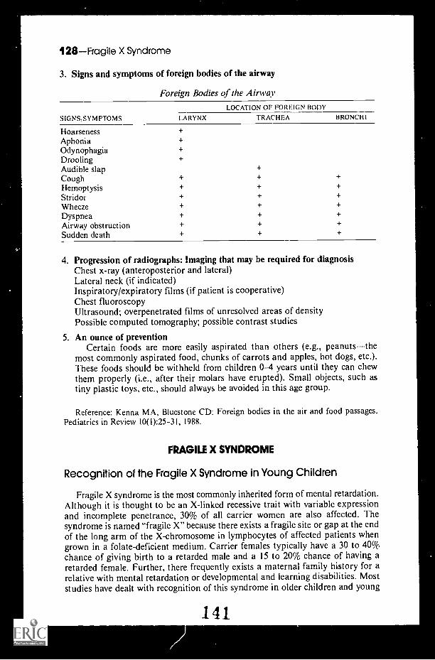
128Fragile X Syndrome
3 Signs and symptoms of foreign bodies of the airway
Foreign Bodies of the Airway
SIGNS:SYMPTOMS
LOCATION OF FOREIGN BODY
LARYNX TRACHEA BRONCHI
Hoarseness +
Aphonia +Odynophagia +Drooling +
Audible slap +
Cough + + +
Hernoptysis + + +
Stridor + + +
Wheeze + + +
Dyspnea + + +
Airway obstruction + + +
Sudden death + + +
4 Progression of radiographs: Imaging that may be required for diagnosisChest x-ray (anteroposterior and lateral)Lateral neck (if indicated)Inspiratory/expiratory films (if patient is cooperative)Chest fluoroscopyUltrasound; overpenetrated films of unresolved areas of densityPossible computed tomography; possible contrast studies
5. An ounce of preventionCertain foods are more easily aspirated than others (e.g., peanuts--the
most commonly aspirated food, chunks of carrots and apples, hot dogs, etc.).These foods should be withheld from children 0-4 years until they can chewthem properly (i.e., after their molars have erupted). Small objects, such astiny plastic toys, etc., should always be avoided in this age group.
Reference: Kenna MA, Bluestone CD: Foreign bodies in the air and food passages.Pediatrics in Review 10(l):25-31, 1988.
FRAGILE X SYNDROME
Recognition of the Fragile X Syndrome in Young Children
Fragile X syndrome is the most commonly inherited form of mental retardation.Although it is thought to be an X-linked recessive trait with variable expressionand incomplete penetrance, 30% of all carrier women are also affected. Thesyndrome is named "fragile X" because there exists a fragile site or gap at the endof the long arm of the X-chromosome in lymphocytes of affected patients whengrown in a folate-deficient medium. Carrier females typically have a 30 to 40%chance of giving birth to a retarded male and a 15 to 20% chance of having aretarded female. Further, there frequently exists a maternal family history for arelative with mental retardation or developmental and learning disabilities. Moststudies have dealt with recognition of this syndrome in older children and young
141

Fragile X Syndrome-129
adults, but many of the physical features, behavioral characteristics, and familyhistory features are apparent far earlier.
Prominent parental concerns that might bring such a child to a pediatrician'sattention include:
Developmental delaySpeech delayShort attention span or hyperactivityMouthing of objects persisting at an age beyond when it would be expectedDifficulty in disciplining the childFrequent temper tantrumsAutistic-like behaviors such as rocking, talking to oneself, spinning,
unusual hand movements, difficulty with transitions, preference forbeing alone, echolalia, and poor eye contact
Poor gross motor coordinationHistory of vomiting, spitting up, or colic during infancyHistory of frequent otitis mediaSelf-abusive behaviorsHand flappingDrooling persisting at an age beyond when it would be expectedHypotoniaFighting with othersPicaHand/ thumb sucking
While older children (8 to 12 years of age) are more likely to display the classicphysical features of fragile X syndrome (long face with a prominent jaw, largeprominent ears, and post-pubertal macroorchidism), patients as young as 2 or 3years have been noted to exhibit the following physical findings:
Long and/or wide and/orprotruding ears
Prominent jaw or long faceHigh arched palateFlattened nasal bridgeMicrocephaly or relative
macrocephalyApparent hypertelorismEpicanthic folds
Simian creases of palms; verticalcreases of soles
Long philtrumHemangiomaHyperextensible jointsAntimongoloid slant to eyesClinical impression of
macroorchidismProminent forehead
It is not feasible or sound to recommend chromosomal studies on all childrenwith developmental, learning, and behavioral disabilities. But these problems(particularly speech delay, unusual be viors, and developmental delay) taken incontext with a maternal family histo. )f mental retardation or developmentaldisabilities and the physical findings of long, wide, or protruding ears, a longface, flat nasal bridge, and a high arched palate probably warrant a !- .arch for thefragile X chromosome. A new method of identifying carriers of these mutationsby direct DNA analysis has recently been described.
References: Simkoi A, Hornstein L, Soukup S, Bagamery N: Fragile X syndrome:Recognition in young children. Pediatrics 83:547-552,1989.
Rousseau F, Hertz D, Biancalana V. et al: Direct diagnosis by DNA analysis of thefragile X syndrome of mental retardation. N Engl J Med 325:1673-1681,1991.
142

"4..4'444(-72;
e
-01.0401,"Mr
To make a prairie it takes a cloverand one bee,
One clover, and a bee,And revery.The revery alone will doIf bees are few.
Emily Dickinson
143BEST COPY AVAILABLE

GASTROENTERITIS
Rotavirus Versus Astrovirus
Over the past 20 years, pediatricians and virologists have gained great insightsinto the causes and nature of gastroenteritis. The enteric adenoviruses, such asrotavirus, have been of particular focus as a cause of diarrhea in infants andyoung children. Recently, reports of astrovirus-caused gastroenteritis has come tothe attention of clinicians, although their medical importance remains poorlydefined. Offered below is a comparison of the clinical findings associated withastrovirus and rotavirus gastroenteritis:
Clinical Findings Associated with Astrovirus and Rotavirus Gastroenteritis*
FINDINGS
ASTROVIRUS INFECTION(N 44)
(percent)
ROTAVIRUS INFECTION(N 175)
(percent)
Watery stools 61 67
Loose stools 41 35
Mucoid stools 55 51
Bloody stools 7 6
Nausea.' 71 88
Abdominal paint 58 63
Vomiting 61 67
Fever 80 83
Dehydration 5% 5 15
* Only stool samples in which no bacterial, parasitic, or other viral pathogens weredetected arc included.Includes data from Study I only (33 children with astroviruses and 116 with rotaviruses).
Reference: Herrmann JE, Taylor DN, Echeverria P, Blacklow NR: Astroviruses as acause of gastroenteritis in children. N Engl J Med 324:1757 -1760, 1991, with permission.
Chitterlings: Another Yersinia Food Group
Yersinia enterocolitica was first recognized as a cause of human infectionin 1933. In young children, infection generally causes acute gastroenteritis. Inolder children a picture of mesenteric adenitis that can easily be confused withappendicitis predominates. A 1988-89 outbreak of Yersinia enterocolitica inAtlanta has added chitterlings (also chitlins), the small intestines of pigs, to the listof agents of transmission. Although the epidemiology remains poorly understood,previously cited agents include contaniinated products, contact with sickpets, transfusion of contaminated blood products, and ingestion of raw pork.
144131

132Gastrointestinal Bleeding
The Atlanta outbreak was traced to households where chitterlings wereprepared for holiday meals. Preparation of chitterlings involves boiling the rawsmall intestines of pigs after fat and fecal matter have been removed. In nearly allof the Atlanta cases, the affected infants and children had contact with thechitterlings' preparers, although not the raw intestines themselves.
The take-home message is that when an infant or child presents with a pictureof acute pastroenteritis, a thorough investigation into intrahousehold contacts aswell as ingestion of possible contaminants is called for.
Clinical Manifestations of Yrsintosls
Diarrhea FeverMucoid stools VomitingBloody diarrhea or pus Abdominal paincolicky, diffuse
(10-20%) or localized to RLQ
DIfferontial Diagnosis
Shigella Yersinia psgudotuberculosis canEnteroinvasive E. coli cause identical picture to theSalmonella mesenteric adenitis of olderCampylobacter children.
References: Lee LA, et al: Yersinia enterocolitica 0:3 infections in infants and children,associated with the household preparation of chitterlings. N Engl J Med 322: 984 -987, 1990
Oski FA, et al: Principles and Practice of Pediatrics. Philadelphia, J.B. Lippincott, 1990
GASTROINTESTINAL BLEEDING
In the Neonate
Common Causes
Esophagitis Rare CausesGastritisT:igested maternal blood Acute ulcerative colitisNecrotizing entercolitis Gastric polypStress ulcer (gastric) Gastrointestinal duplication cyst
IntussusceptionUncommon Causes Leiomyoma
Milk allergyAcquired coagulopathy Nasal or pharyngeal bleedingGastroenteritis (Campylobacter Severe cyanotic congenital heart
infections) diseaseHemophilia Vascular malformation of the gutRectal trauma or gastrointestinal (hemangioma, telangiectasia,
trauma arteriovenous malformation)ThrombocytopeniaVitamin K deficiencyVolvulus 1 4 5

In Infancy
Common Causes
Anal fissuresEsophagitisGastritis (possibly due to
drug ingestion)GastroenteritisPolyps
Uncommon Causes
Acquired coagulation disturbanceHemophiliaHenoch-Schönlein purpuraInflammatory bowel diseaseMeckel's diverticulumParasitismPeptic ulcerThrombocytopenia
Gastrointestinal Bleeding-133
Rare Causes
Chronic granulomatous diseaseDiverticulitisEsophageal varicesHemangiomas and telangiectasiaHemolytic-uremic syndromeHemorrhoidsIntestinal foreign bodyLymphosarcomaPeutz-Jegher syndromePseudoxanthoma elasticumScurvy
Using the BUN/Creatinine Ratio in Localizing the Sourceof Gastrointestinal Bleeding in Children
Although most child n with gastrointestinal bleeding make the diagnostician'sjob somewhat easier presenting with a chief complaint of either hematemesisor bright red blood per rectum, there exists a gray zone in localizing the sourceof bleeding when a child presents with melena or altered blood in the stool.Frequently, this child undergoes a wide variety of costly and invasive diagnosticprocedures in order to determine the site of blood loss. A useful and inexpensivemeans of identifying upper gastrointestinal bleeding, however, lies in an evalu-ation of the blood urea nitrogen to creatinine ratio (BUN/Cr).
In a retrospective study of 40 children hospitalized for evaluation andtreatment of gastrointestinal bleeding at the Children's Hospital of Los Angeles,a BUN/Cr ratio >30 indicated an upper gastrointestinal bleeding source with aspecificity of 100%. Documentation of the source of bleeding was confirmed byendoscopy, surgery, or presence of hematemesis o; bright red blood per rectum.The rise in BUN after an upper gastrointestinal hemorrhage is probably a resultof increased hepatic catabolism of the absorbed amino acid load from theintraluminal blood. The bleeding, therefore, must occur proximal to the smallintestine's absorptive surface (e.g., proximal to the ligament of Treitz) in order tocause a significant rise in the BUN/Cr ratio. The sensitivity of the BUN/Cr ratiofor upper gastrointestinal bleeding, however, was 39%, because there existed awide range of BUN/Cr ratios among the upper GI bleeders (range = 10-140;mean = 34 ± 29 SD) in comparison to the lower GI bleeders (range = 3.3-30;mean = 16 ± 8.5 SD). A BUN/Cr ratio less than or equal to 30, therefore, can beconsistent with either an upper or a lower gastrointestinal bleeding site. The widerange of BUN/Cr ratios seen in children with upper gastrointestinal bleeding may
146

134Genetics
have been due to smaller volumes of blood loss, vomiting, or more rapid gastro-intestinal transit times in children when compared to adults.
Reference; Felber S, Rosenthal P. Henton D: The BUN/Creatinine ratio in localizinggastrointestinal bleeding in pediatric patients. J Pediatr Gastroenterol Nutr 7:685 687,1988.
Kool-Aid Colitis
A mother brings her child to your Emergency Room frightened by the newonset of bright red stools in the child's diaper. The child appears hemodynamicallystable and is without abdominal tenderness. What do you do?
If the stool guaiac is negative, ask the mother whether her child has had anycherry- or strawberry-containing beverages. Don't stop there. Ask about beets,tomato-containing products, and cherry or strawberry candies. Any and all ofthese substances can be the culprit when an alarmingly scarlet, nonclottingeffluent accompanies a stool.
Reference: Sack J: "Kool-Aid colitis" (letter). N Engl J Med 322:1012, 1990.
GENETICS
What is a Human Being's "Unique Genetic Make-up"?
The uniqueness of each individual stems from the fact that over one-fifth ofhis or her genes (i.e., proteins) are in a form that differs from that present in themajority of the population. All human diseases are a result of the interaction ofthe person's unique genetic make-up and the environment, and therefore geneticscan be considered the basis of all medicine.
Reference: Goldstein JL, Brown MS: Genetics. In Wilson JD, et al (eds): Harrison'sPrinciples of Internal Medicine, 12th ed. New Yo- k, McGraw-Hill, 1991, p 21.
Ethnicity As a Risk Factor
Disorders transmitted by the inheritance of a single mutant gene, termed men-delian or "simply inherited disorders," occur with increased frequency in specificethnic groups. Some examples of these relationships are listed in the table below.
Examples of Simply Inherited Disorders that Occur with Increased Frequencyin Specific Ethnic Groups
ETHNIC GROUP
African blacks
Armenians
SIMPLY INHERITED DISORDER
Hemoglobinopathies, especially Hb S. Hb C, persistentHb F, a and 13 thalassemias
Glucose-6-phosphate dehydrogenase deficiency
Familial Mediterranean fever
Table continued on next page.
147

Growl-h I 35
Examples of Simply Inherited Disorders that Occur with Increased Frequencyin Specific Ethnic Groups (Cont.)
ETHNIC GROUP SIMPLY INHERITED DISORDER
Ashkenazi Jews AbetalipoproteinemiaBloom's syndromeDystonia musculorum deformans (recessive form)Factor XI (PTA) deficiencyFamilial dysautonomia (Riley-Day syndrome)Gaucher's disease (adult form)Neimann-Pick diseasePentosuriaTay-Sachs disease
Chinese a-ThalassemiaGlucose-6-phosphate dehydrogenase deficiencyAdult lactase deficiency
Eskimos Pseudocholinesterase deficiencyAdrenogenital syndrome
Finns Congenital nephrosisMulibrey nanism
French Canadians Tyrosinemia
Japanese Acatalasemia
Lebanese Homozygous familial hypercholesterolemia
Mediterranean peoples (3-Thalassemia(Italians, Greeks, Glucose-6-phosphate dehydrogenase deficiencySephardic Jews) Familial Mediterranean fever
Glycogen storage disease, type I 11
Northern Europeans Cystic fibrosis
Scandinavians Alpharantitrypsin deficiencyLCAT (lecithin:cholesterol acyltransferase) deficiency
South African whites Porphyria variegataHomdzygous familial hypercholesterolemia
From Wilson JD, et al (eds): Harrison's Principles of Internal Medicine, 12th ed. NewYork, McGraw-Hill, 1991, with permission.
GROWTH
Predicted Weight and Height from Age
If you know the age of a child and want a rough estimate of the weight andheight:
For 3-12 mo:For 1-6 yr:For 7-12 yr:For 2-14 yr:
Weight (lb) = age (mo) + 11Weight (1b) = age (yr) x 5 + 17Weight (lb) = age (yr) x 7 + 5Height (in) = (21/4 x age) + 30
Reference: Graef JW, Cone TE: In Manual of Pediatric Therapeutics, 6th ed. Boston,Little, Brown, 1991.
148

136Growth
More on Predicted Heights
The formula of Tanner et al. demonstrates that height at age 3 years correlatesbetter with height at maturity than it does at any other age:
Adult height (cm) = 1.27 x height (at 3 yr) + 54.9 cm (males)
Adult height (cm) = 1.29 x height (at 3 yr) + 42.3 cm (females)
If you cannot remember these formulas or your programmable calculator hasbeen stolen, the commonly accepted statement that the child at age 2 hasachieved one half his or her final height is quite satisfactory. For girls, however,10 to 12 cm (2.54 to 4.00 in) must be subtracted from this predicted height. If theheight at 3 years is known, an alternative to the Tanner equation to predict finaladult height is to multiply the age 3 height by 1.87 for boys and 1.73 for girls.
Reference: Tanner et al: Arch Dis Child 31:372, 1956.Adapted from McMillan .IA, et al: The Whole Pediatrician Catalog, Vol. 3. Philadelphia,
W.B. Saunders, 1982, p 74.
Growth During Infancy: Weight
When assessing the growth of infants in a well-child care setting, a handy ruleof thumb is offered: the newborn infant typically loses between 5 and 10% of hisor her birth weight during the first few days of life (due to water loss). After that,the infant should gain 1 ounce or 30 grams, on average, per day. This weight gain,approximately 1 to 2 pounds per month, results in a doubling of the birth weightby six months of age. By the age of 2 years, weight gain should slow down toabout one-half pound per month. Please note that although this tip is quite usefulin the growing infant, it is not sensitive in the older child.
Growth During Infancy: Length
The greatest rise in linear growth occurs during infancy (the first 2 years oflife). The average length of a newborn American infant is 50.4 cm (2.0 SD) or19.8 inches (0.8 SD) in males and 49.7 crn (1.9 SD) or 19.6 inches (0.75 SD) infemales. The growth increment during this period should be 25 to 30 cm (10 to12 in) during the first year of life and 12 cm (5 in) during the second year of life.Male infants are typically heavier (by about 0.5 kg) and longer (by about 0.5 cm)than their female counterparts.
Growth During Infancy: Cranial and Brain Growth
The human brain begins its peak growth rate at birth and during the postnatalperiod. It should be oted that at birth, the infant's brain is only one-sixth of itsfinal weight. Consequently, the growth of the cranial vault parallels this rapiddevelopment in order to accommodate the increasing brain size. In fact, carefulevaluation of the skull size (head circumference) and shape gives the pediatriciana great deal of insight into the infant's neurologic development.
Mean head circumference at birth is approximately 35.3 cm (1.2 SD) or 13.9inches (0.5 SD). There should be a 5 cm increase in head circumference during
149

Gynecomastia -A 37
the first 3 months of life and an additional 6 cm increase by the end of the firstyear of life. Whereas the head circumference of a newborn infant is greater thanhis or her chest, this ratio should approximate 1:1 by age I year.
(N.B.: Closure of the fontanel is covered under "Fontanels.")
GYNECOMASTIA
The Differential Diagnosis of Gynecomastia
Gynecomastia is defined as the visible or palpable development of breasttissue in boys or men. It has been divided into four types:
Type I gynecomastia (pubertal gynecomastia or benign adolescent breasthypertrophy) refers to the common entity seen in pubertal males. In fact, manycite an incidence of 60 to 70% in this population. It is typically a firm, tendersubareolar mass anywhere from 1 to 5 ern in diameter. The pubertal adolescentfrequently complains of pain in the breasts, particularly when wearing bindingclothing. It usually spontaneously resolves within 2 years.
Type H gynecomastia (physiologic gynecomastia without evidence of underlyingdisease, or with evidence of organic disease including the effects of specific drugs)refers to a generalized, nonpainful breast enlargement. It is essential to differentiatebetween physiologic gynecomastia and breast enlargement due either to apathologic process or to the use of a specific drug. The physician should,therefore, obtain a careful history regarding the time of onset, family history,duration of the enlargement, history of systemic illness, weight change, and drugor medication use. Physical examiantion should include height, weight, bloodpressure, breast size, and Tanner staging of both breasts and genitals, in additionto a neurologic assessment. The most frequent causes of Type II gynecomastiaare listed in tabular form below.
Type HI gynecomastia is general obesity simulating gynecomastia, and TypeIV gynecomastia is pectoral muscle hypertrophy.
Common Causes of Type II GynecomastiaI. Idiopathic
II. Familial causesa. Associated with anosmia and testicular atrophyb. Reifenstein's syndrome (a type of familial male pseudohermaphroditism
secondary to partial androgen insensitivity)c. Associated with hypogonadism and small penisd. Others
III. Specific illnesses or syndromesa. Kleinfelter's syndrome f. Hemophiliab. Male pseudohermaphroditism g. Leprosyc. Testicular feminization syndrome h. Thyroid dysfunction (hyper-d. Tumors (e.g., seminoma, Leydig cell and hypothyroidism)
tumor, teratoma, feminizing adrenal i. Cirrhosis of the livertumor, hepatoma, bronchogenic j. Traumatic paraplegiacarcinoma) k. Chronic glomerulonephritis
e. Leukemia I. Starvation (on refeeding)
150

38 Gynecomastia
IV. Miscellaneous Drugsa. Amphetaminesb. Anabolic steroidsc. Birth control pillsd. Busulfan (and other
chemotherapeutic agents)e. Cimetidinef. Clomipheneg. Diazepamh. Corticosteroidsi. Digitalisj. Estrogensk. Human chorionic gonadotropin
m.InsulinIsoniazid (and otherantituberculosis drugs)KetoconazoleMarijuanaMethadone (and other narcotics)MethyldopaReserpineSpironolactoneTestosteroneTricyclic antidepressants
Adapted from: Greydanus DE, Parks DS, Farrell EG: Breast disorders in children andadolescents. Pediatr Clin North Am 36:601 638, 1989.
151

HEAD
Head Circumference in Term infants
When you are stuck without your growth grid, there is still a simple means ofdetermining if a head is growing normally. The little table below lists the expectedrate of increases in head circumference for term infants during the first year of life.
Rate qf Increases in Head Circumference
PERIOD HEAD CIRCUMFERENCE INCRF.MENTS
First 3 months4-6 months6 12 months
First year
2 cm!month = 6 cm
1 cm/month = 3 cm
0.5 cm/month = 3 cm
12 cm
HEADACHE
Common Causes
Extracranial infectionOtitis/mastoid it isPharyngitisSinusitisTooth abscess
Uncommon Causes
DepressionEye strainMeningitis/encephalitisTemporomandibular joint disease
Rare Causes
AllergyArnold-Chiari malformationCervical osteoarthritisChronic renal disease
Febrile illnessMigraineTension
AnxietyEnvironmental stress
TraumaConcussionOccipital neuralgia
Congenital erythropoietic porphyriaCranial bone diseaseDecreased intracranial pressure
Post-lumbar puncture
152139

140Hearing
DrugsAmphetaminesCarbon monoxideHeavy metalsIndomethacinMalidixic acidNitrates/nitritesOral contraceptivesSteroidsSulfaTetracyclineVitamin A
EpilepsyHyperventilationIncreased intracranial pressure
HydrocephalusMass/tumor/abscessPseudotumor cerebri
Leukemia infiltrationMetabolic
HyperammonemiaHypercarbiaHypoglycemiaHyponatremiaHypoxiaMetabolic acidosis
Myositis
PsychogenicConversion reactionMimicrySecondary gain
OrbitGlaucomaOrbital tumor
VascularAnemiaAneurysmArteritis
Giant cellPeriarteritis nodosaSubacute bacterial endocarditisSystemic lupus erythematosis (SLE)
AV malformationCerebral infarct
EmbolismThrombosis
Cluster headacheHemorrhage
EpiduralParenchymalSubdural
HypertensionPhlebitisVenous sinus thrombosis
HEARING
Which Infant Is at Risk for Hearing Loss?
Long before a delay in language development secondary to a hearing loss isnoted by a parent or physician, the astute pediatrician can single out infants atrisk. Screening for hearing loss is relatively inexpensive and easy to perform, andthe benefits of early detection are immeasurable. Be aware of the risk factors andlook for the absence of the normal newborn's response to sound: startling,blinking, crying, quieting, or other forms of alertness are the normal newborn'sreactions to sound.
Factors that Mandato Scumming for Hooting Lou
A blood relative with childhood hearing impairment.Anatomic malformations involving the head and neck.Bacterial meningitis --especially H. influenzae.Birthweight less than 1,500 g.Severe asphyxia as evidenced by low Apgar scores, arterial pH 7.25, coma,
seizures, or the need for continuous assisted ventilation.Unconjugated bilirubin > 17 mg/100 ml of scrum.Viral or other nonbacterial intrauterine fetal infections.
153

Hearing-141
Any neonate with known congenital defect: Recessive syndromes constitute 4%to 40% of childhood deafness; dominant syndromes constitute about 15%, x-linked syndromes about 2%; and rubella, 9 to 20% of cases of childhooddeafness.
Screening Tests
These screening tests allow evaluation of the infant at risk as early as 2 weeksof age:
I. Crib-o-gram: Crib movement is recorded in response to sound stimulusapplication.
2. Brainstem auditory evoked response (BAER): More sensitive than a crib-o-gram, BAERs record electrical potential at the skin overlying the nucleiand tracts of the auditory pathway.
3. Behavior assessment: See "Speech and Language Milestones."
References: Rowe LD: Hearing loss: The profound benefits of early diagnosisContemp Peds October:77 -85, 1985.
Oski FA, et al: Principles and Practice of Pediatrics. Philadelphia, J.B. Lippincott,1990.
Exogenous Causes of Hearing Loss
In addition to heredity, gestational age, dysmorphisms, and asphyxia, thereare several exogenous causes of hearing loss to consider in the evaluation ofcommunicative delay in infants and children.
Preconception and Prenatal
Cytomegalovirus RubellaHypoxia SyphilisMaternal drug abuse and alcoholism Toxemia, diabetes, or other severeOtotoxic drugs (quinine) maternal systemic diseaseIrradiation of parent Toxoplasmosis
Perinatal
HypoxiaMaternal infectionOtotoxic drugs (aminoglycosides)
Neonatal and Postnatal
Erythroblastosis fetalisHypoxiaInfantile measles or mumps
PrematurityTraumatic delivery
Meningitis, encephalitisOtotoxic drugs (aminoglycosides)
Reference: Rowe LD: Hearing loss: The profound benefits of early diagnosis. ContempPediatr October: 77-85, 1985.
154

N.
142Hematology/Hematuria
HEMATOLOGY
Laboratory Test Results in Disorders Producing Hypochromia and Microcytosis
DISOR DERRU M
IRON
IRON-BI N DINGCAPACITY E'ER R ITI N HP*
H E MOO LORI NIELFC-I-Ro-PHOIZESIS
Iron deficiency Decreased Increased Decreased Increased Normal
MARROWIRONSTORES
Decreased
Chronic disease Decreased Decreased Increased Increased Normalanemia
Increased
Sideroblasticanemia
Increased Normal Increased Decreased NormalRingsideroblasts
0-thalassemia Normal Normal Normaltrait
HemoglobinNormal Normal
A2 increased
a-thalassemia Normal Normal Normal Normal Normaltrait
Normal
*FEP indicates free erythrocyte protoporphyrin.Adapted from Steinberg MH, Dreiling BJ: Microcyto JAMA 249:85, 1983, with permission.
HEMATURIA
Common Causes
Benign causesBenign recurrent hematuriaFamilial hematuriaIdiopathic recurrent gross hematuriaPostural hematuria
ContaminationMenstrualMunchausen's syndromeMunchausen's syndrome by proxyPregnancy-related bleeding
HemoglobinopathiesHgb CHgb SCSickle-cell disease/trait (Hgb SS/SA)Sickle-thalassemia trait
HypercalciuriaDistal renal tubular acidosisDiuretic therapyEndocrine disorders
Diabetes mellitus
155
Hypercalciuria (Cont.)Endocrine disorders (Cont.)
HyperadrenocorticismHyperparathyroidismHypothyroidism
HypercalcemiaHyperphosphatemiaHypertensionImmobilizationJuvenile rheumatoid arthritisMedullary sponge kidneyMetabolic acidosisNeoplasmRenal tubular dysfunctionSarcoidosisVitamin D excess
Hypoxia, asphyxia, and circulatorycompromise
Acute tubular necrosisCortical and medullary necrosis

InfectionsCystitis (viral, bacterial)PyelonephritisUrethritis
Meatal stenosisNoninfectious cystitis
CytoxanRadiation
Perineal irritationPhimosis
Uncommon Causes
Bladder diverticuli/polypsCoagulopathiesDrug-induced
Analgesic nephropathyCephalosporinsCytoxanPenicillinSulfonamides
ExerciseGlomerular disorders
MesangioproliferativeMinimal change disease
Hydronephrosis
Rare Causes
Allergy"Apparent"
"Beeturia"BetadineBiliuriaDesferoxamineDyes
Ana lineCongo red
HemoglobinuriaMyoglobinuriaPhenothiazinesPorphyria
Diabetic nephropathyGlomerular disorders
AmyloidosisCrescentic glomerulonephritis (GN)Familial nephiritis (Alport's)Focal segmented proliferative GNFocal segmental sclerosis
Hematuria 143
Post-infectious glomerulonephritisTrauma
Fractured pelvisPostcatheterizationPostcircurncisionPostsurgeryRenal contusionRenal fractureUrethral trauma
Urethral ulceration
InfectionsEpididymitisProstatitis
MasturbationPeriureteritis (appendicitis, ileitis)Polycystic diseaseReflux nephropathyRenal calculiRenal vein thrombosisThrombocytopeniaUreteropelvi junction obstructionUrethral foreign bodyWilms' tumor
Goodpasture's syndromeIgA nephropathyMembranous GNMesangiocapillary GNSubacute bacterial endocarditisSystemic lupus erythematosus
(SLE)Wegener's granulomatosis
HemangiomaHematospermiaImmunologic
Hemolytic-uremic syndromeHenoch-Schönlein purpuraPolyarter itis nodosaS LE
InfectionsLeptospirosisMalariaSchistosomiasisToxoplasmosis
156

144 Hematuria
Infections (Com.)TuberculosisVaricella
Malignant hypertensionMedullary sponge kidneyNeoplasms
Bladder cancer
Neoplasms (Cont.)Prostatic concer
Renal infarctionRetroperitoneal fibrosisVitamin deficiency
ScurvyVitamin K deficiency
Evaluation of Hematuria in Children and Adolescents
After urinary tract infections, hematuria is the most frequently occurringabnormality in the genitourinary tract. One must carefully assess the complaintof hematuria in order to rule out or diagnose from a large list of potentiallyserious disorders. It is important, first, to assess whether or not actual blood orsimply a red substance is being excreted into the urine. A reagent strip(impregnated with orthotolodine-peroxide and enhanced with 6-methoxy-quinolone) makes this quite simple. False-negative results with the dipstickmethod are rare but can result from the presence of high urinary concentrationsof ascorbic acid. False-positive results, which are also rare, can occur in thepresence of a raging urinary tract infection where bacterial peroxidase is releasedin high quantities. Following a positive dipstick microscopic evaluation isrequired; if no red blood cells (RBCs) are noted in a fresh urine sample, oneshould entertain the diagnosis of hemoglobinuria or myoglobinuria.
Although gross hematuria is easily recognized upon visual inspection, the urineshould always be examined with dipstick and microscopic analysis to rule outother causes of red urine. Microscopic hematuria is usually defined as (a) three ormore consecutive urine samples with positive reagent strip test results and eithertwo or more RBCs per cubic millimeter in a fresh, uncentrifuged urine sample or(b) six or more RBCs per high-power field in a fresh urine sediment specimen.
Common Causes of Red Urine
1. Negative Dipstick Test (e.g., dyes, drugs, pigments)
a. Pink, red, Coca-Cola, burgundy-colored urine (drug and food ingestion):Aminopyrine, anthocyanin, azo dyes, beets,* blackberries, chloroquine,desferoxamine, mesylate, ibuprofen, methyldopa, nitrofurantoin, phenazo-pyridine, phenolphthalein, rifampin, rhodamine B, sulfasalazine, urates.
b. Dark brown, black:i. Disease-associated: alkaptonuria, homogentisic aciduria, melanin,
methemoglobinemia, tyrosinosisii. Drug and food ingestion: alanine, resorcinol, thymol.
2. Positive Dipstick Test but No RBCs on Microscopic Examination
a. Hemoglobinuriai. Drugs and chemicals: aspidium, betanaphthyl, carbolic acid, carbon
monoxide, chloroform, fava beans, mushrooms, naphthalene, pama-quine, phenylhydrazine, quinine, snake venom, sulfonamides.
*The excretion of red-purple or beet-colored urine (beeturia) after ingesting beetsshould prompt the clinician to work up the patient for iron-deficiency anemia.
157

Hematuria 145
ii. Disease-associated and other causes: all types of hemolytic anemias,hemolytic-uremic syndrome, septicemia, paroxysmal nocturnal hemo-globinuria, freshwater drowning, mismatched blood transfusions,cardiopulmonary bypass.
3. Positive Dipstick Tests with RBCs on Microscopic Examination
a. No RBC casts present: tumors, cysts, stones, obstructionb. RBC casts present:
i. Without proteinuria: IgA nephropathy, familial nephritis, benign orfamilial hematuria (in association with hypercalciuria)
ii. With proteinuria: Acute glomerulonephritis, Henoch-Schonlein pur-pura, systemic lupus erythematosus, chronic glomerulonephritis
iii. Heavy proteinuria: Nephrotic syndrome
The basic work-up and evaluation for the patient with hematuria shouidproceed as follows:
1. History (associated symptoms, precipitating events, pattern of hernaturia,familial occurrence).
2. Physical examination (presence of edema, elevated blood pressure, skinlesions, joint involvement).
3. Laboratory tests
a. Urinalysis (with confirmation of microscopic hematuria on two or moreoccasions).
b. Urine culture (gross hematuria occurs in 5% to 10% of children withsymptomatic urinary tract infection; the frequency of those children withsymptomatic urinary tract infection and microscopic hematuria is notknown).
c. Complete blood count with examination of peripheral smeard. Serum creatininee. ASO titer, streptozyme, C3 complement, anti-DNA antibody (e.g.,
poststreptococcal glomerulonephritis, SLE, proliferative GN)f. Quantitative urine protein, calcium, creatinine excretiong. Imaging studies (e.g., ultrasonography of the urine tract, intravenous
pyelography, voiding cystourethrography).
References: Adapted from Boineau FG, Lewy JE: Evaluation of hematuria in childrenand adolescents. Pediatr Rev 11:101- 107, 1989.
Norman ME: An office approach to hematuria and proteinuria. Pediatr Clin NorthAm 34:545- 560, 1987.
Hematuria Atter Blunt Trauma
Hematuria is a common finding in the aftermath of blunt trauma to theabdomen. Traditional thinking suggests that hematuria is a significant consequenceof genitourinary (GU) injury, and its presence typically elicits consideration ofradiologic imaging of the GU tract. Which patient, in fact, requires a study andwhich study should you choose?
15-

146Hematuria
Asymptomatic hematuria is not an indication for computed tomography(CT) of the abdomen. In a study of 378 consecutive children evaluated by CT ofthe abdomen, those who were asymptomatic had no evidence of organ injury. Achild with no signs or symptoms of abdominal injury (e.g., tenderness, ecchy-moses, distention) despite the presence of hematuria does not warrant a CTexam. Children with asymptomatic hematuria may be evaluated on a nonemer-gent basis with Doppler ultrasonography, excretory urography, or a radioactiverenal scan. In contrast, hematuria with abdominal symptoms is a significantmarker of injury to both urinary and nonurinary organs in the setting of bluntabdominal trauma.
References: Taylor GA, et al: Hematuria: A marker of abdominal injury in childrenafter blunt trauma. Ann Surg 208:688-693, 1988.
Jaffe D, Wesson D: Emergency management of blunt trauma in children. N Engl JMed 324:1477-1482, 1991.
Drug-induced Causes of Hematuria
THE DRUG I HE DISORDER
Penicillin and cephalosporin analogues, phenytoin
Phenacetin, nonsteroidal anti-inflammatory agents
Cyclophosphamide, mitotane
Cyclophosphamide, phenacetin
Anticoagulants
Allergic interstitial nephritis
Papillary necrosis
Chemical cystitis
Malignant neoplasm ofuroepithelium
Spontaneous bleeding or inductionof bleeding from an occult lesion
Reference: Schoolwerth AC: Hematuria and proteinuria: Their causes and consequences.Hospital Practice 22 (Oct 30):45-62, 1987.
Neonatal Hematuria
Gross hematuria is a rare presentation during the first month of life. Thefindings in one series of 35 patients are demonstrated below.
Findings in Series of 35 Patients
CAUSE
AGE AT ONSET
NO. OF I SI 2ND 3RD 4TH DURATIONPATIEN1 S WEEK WEEK WEFK WEEK (DAYS) REMARKS
Unknown I 1 8 2 1 1 3 I died of hyalinemembrane diseaseand pneumothorax;normal BUN levelin 9, elevated in 2
Renal vein 7 3 1 1 2 5 3 had diabctic mothers;thrombosis Bun level > 40 mg!
100 ml in all; 4 hadthrombocytopenia
Table continued on next page.
159

Hemoglobin-147
Findings in Series of 35 Patients (Cont.)
CAIISE
AGE AT ONSE1
NO. OF I SI 2ND 3R1) 41H 1)URA1 IONPATIENTS WEEK WEEK WEEK WEEK (DAYS) REMARKS
Polycystic dis- 6 6 2 4 5 died in neonatalease of kidney period, one at 4
months; all had in-creased BUN, 40to 80 mg/100 ml
Obstruct iveuropathy
Hydronephrosis 3 1 2 1 5 BUN level 25 to 30Ureteral valve 3 2 2 4 mg/100 ml; 2 withBladder neck 1 1 4 hydronephrosis un-
derwent difficultdelivery; 3 hadpyuria and bacteri-uria; palpablemass present in 2
Sponge kidney 3 2 1 1 Death within 4 to 6months of age
Wilms' tumor 2 1 2 Survived, patientdoing well 4 yearslater
Abdominal masses were palpated in all patients later found to have renal veinthrombosis or polycystic kidneys. Intravenous pyelograms were normal in allpatients in whom no cause was found for the hematuria, in the patients withposterior urethral valves, and in the one with bladder neck obstruction. IVP wasabnormal in all other patients. Voiding cystourethrogram demonstrated theabnormality in the patients with obstruction.
Conclusion: Abdominal palpation, IVP, and blood urea nitrogen levels arewarranted in all newborns presenting with gross hematuria. If the diagnosis is stillunavailable, voiding cystourethrogram is in order. A significant number of thesepatients, however, will have no evident cause for their hematuria, and will recoverspontaneously.
Reference: Emanuel B. Aronson N: neonatal hematuria. Am .1 Dis Child 128:204, 1974.Adapted from McMillan, et al (eds): The Whole Pediatrician Catalog. Philadelphia,
W.B. Saunders, 1977, pp 314 315, with permission.
HEMOGLOBIN
Classification of Red Cell Hemolytic Disordersby Predominant Morphology
In the following lists, nonhemolytic disorders of similar morphology areenclosed in parentheses for reference.
160

i48Hemoglobin
SpaerocytesHereditary spherocytosisABO incompatibility in neonatesImmunohemolytic anem;as with IgG-
or C3-coated red cellsAcute oxidant injury (hexose monophos-
phate shunt defects during hemolyticcrisis, oxidant drugs and chemicals)
Hemolytic transfusion reactionsClostridium welchii septicemiaSevere burns, other red cell thermal
injurySpider, bee, and snake venomsSevere hypophosphatemiaHypersplenism*
Bizarre PolkilocytesRed cell fragmentation syndromes
(micro- and macroangiopathichemolytic anemias)
Acute oxidant injury*Hereditary elliptocytosis in neonatesHereditary pyropoikilocytosis
ElliptocytesHereditary elliptocytosisThalassemias(Other hypochromic-microcytic
anemias)(Megaloblastic anemias)
StomatocytesHereditary stomatocytosisRh11 blood groupStornatocytosis with cold hemolysis(Liver disease, especially acute
alcoholism)(Mediterranean stomatocytosis)
Irreversibly &cricked CellsSickle cell anemiaSymptomatic sickle syndromes
Intraorythrocylic ParasitesMalariaBabesiosisBartonellosis
*Disease sometimes associated with this mReference: Oski FA: Differential diagnosis
Hematology of Infancy and Childhood, 3rd ed
1 61
Splculated or Crenated Wed CellsAcute hepatic necrosis (spur cell
anemia)UremiaRed cell fragmentation syndromes*Infantile pyknocytosisEmbden-Meyerhof pathway defects*Vitamin E deficiency*AbetalipoproteinemiaHeat stroke*McLeod blood group(Postsplenectomy)(Transiently after massive
transfusion of stored blood)(Anorexia nervosa)*
Target CellsHemoglobins 5, C, D, and EHereditary xerocytosisThalassernias(Other hypochromic-microcytic
anemias)(Obstructive liver disease)(Postsplenectomy)(Lecithin:cholesterol acyltransferas.
deficiency)
Prominent Basophilic StipplingThalassemiasUnstable hemoglobinsLead poisoning*Pyridine 5'-nucleotidase deficiency
Nonspecific or Normal MorphologyEmbden-Meyerhof pathway defectsHexose monophosphate shunt defectsUnstable hemoglobinsParoxysmal nocturnal
hemoglobinuriaDyserythropoietic anemiasCopper toxicity (Wilson's disease)Cation permeability defectsErythropoietic porphyriaVitamin E deficiencyHemolysis with infections*Rh hemolytic disease in neonatesParoxysmal cold hemoglobinuria*
orphology.of ancmia. In Nathan DC, Oski FA (eds):. Philadelphia, W.B. Saunders, 1987, p 270.

Hemoptysis-149
The Rise in Hemoglobin with Iron Therapy
How fast should the hemoglobin rise when you start treating your irondeficient patient with oral iron?
Two important factors must be considered when judging the adequacy of thehematologic response. They are (I) the initial hemoglobin value and (2) theduration of the period of observation. The lower the initial hemoglobin, thegreater is the hemoglobin rise per day. The shorter the observation period, thegreater is the calculated hemoglobin rise per day. As one gets cl^ser to a normalhemoglobin value, the daily rise in hemoglobin is much less. In our ownexperience in treating patients with hemoglobin values of less than 8.0 g/100 ml,one may anticipate a hemoglobin rise of 0.2 to 0.3 g per day and during the first7 to 10 days of therapy. During the period of 10 to 24 days, the hemoglobin risesat a rate of 0.15 g per day and slows after that point to a rate of 0.10 per dayuntil a normal level is achieved. Normally, the reticulocyte count begins toincrease in 48 to 72 hours and reaches a peak 7 to 10 days after the initiation oftherapy.
Listed below is another guide to the expected response as a function of theinitial hemoglobin level.
Hematologic Response to Oral Iron Therapy Based on Initial Hemoglobin Value
INITIAL HEMOGLOBIN HEMOGLOBIN RISE IN ONE WEEK
(g/d1) (g/d1)
2.0 5.05.1 6.06.1 7.07.1 8.08.1 9.09.1 10.0
10.1 11.011.1 12.0
1.61
1.531.171.11
0.980.570.720.40
Optimal responses to oral iron therapy are achieved by treating the patientwith ferrous sulfate. A patient shouk' receive 2 to 3 mg of elemental iron per kgthree times per day. The iron should be given between meals and neveradministered with milk.
Reference: Mehta BC, Lotliker KS, Patel JC: Indian J Med Res 61:1818, 1973.
HEMOPTYSIS
The Child with Hemoptysis
Hemoptysis is defined as the spitting up of blood that originates from thelungs or bronchial tree. Although this is a rare sign in children, it can bepotentially life-threatening. Rapid and thorough evaluation is vital in order toidentify and control the source of bleeding.
162

150Hemoptysis
In the work-up of the child with hemoptysis, it is necessary to begin byanatomically delineating where the bleeding is actually occurring. Most childrenwho spit up blood have an identifiable source of bleeding outside of the lowerrespiratory tract, such as epistaxis or trauma to the oropharynx. Differentiatinghemoptysis from hematemesis, on the other hand, can be difficult. Distinguishingfeatures of these two signs are summarized below:
Hcniloptys
Blood is usually bright red, frothy, and mixed with sputum (pus,organisms, and macrophages may be present).Anemia is not a common finding.Bleeding is often preceded by a gargling noise and is associated withcoughing.pH of blood is alkaline.
Hematemesis
Blood is usually dark red to brownish in color (e.g., coffee ground emesis).Often associated with anemia.Bleeding and emesis are often preceded or accompanied by nausea andretching.pH is acidic.
The different causes of hemoptysis are wide and varied, as detailed below:
1 . Infectious Causesa. Bacterial
i. Bronchiectasis (most com-monly associated with cysticfibrosis, immunodeficiencydisorders, dyskinetic ciliasyndrome, and pertussis).Necrotizing pneumonias (e.g.,Pseudomonas, Stapkylococcus,Klebsiella)
iii. Pulmonary tuberculosisiv. Lung abscessv. Bronchitisvi. Tracheitis
vii. Pertussis
b. Fungali. Aspergillosis
CoccidioidomycosisActinomycosis
iv. Mucormycosisv. Candidiasis
c. Parasitici. Paragonimiasis
EchinococcosisStrongylodiasis
iv. Ancylostomiasisd. Viral
i. LaryngitisLaryngotracheobronchit isPneumonitis
2. Foreign Bodies
3. Intrathoracic Defects and Lesions (Congenitala. Pulmonary arteriovenous fistula h.b. Hemangiomatous malformation i.
c. Neurenteric cyst j.d. Bronchogenic cyst k.e. Mitral stenosisf. Other cardiac anomalies I.
g. Pulmonary embolism and infarction m.
43 3
or Acquired)Aortic aneurysmPulmonary sequestrationArteriovenous fistulaVenous obstructiveconditionAnomalous vesselCongenital telangiectasia

0
Henoch-Schönlein Purpura--1151
4. Autoimmune Conditionsa. Wegener's granulomatosis d. Goodpasture's syndromeb. Pulmonary hemosiderosis e. Collagen vascular diseasec. Milk allergy
5. Traumaa. Compression, crush injury, or penetrating injuryb. Iatrogenic (e.g., postsurgical, postdiagnostic lung puncture, posttrans-
bronchial biopsy, barotrauma)
6. Neoplastic conditionsa. Endobronchial metastasis (e.g., d. Choriocarcinoma
metastatic osteogenic sarcoma) e. Endometriosisb. Bronchial adenoma f. Bronchiogenic carcinomac. Mediastinal teratoma
7. Hemoglobinopathy with pulmonary infarct (e.g., the "chest syndrome" ofsickle cell disease)
8. Factitious (as a manifestation of Munchausen syndrome)
References: Turcios NL, Vega M: The child with hemoptysis. Hospital Practice 22(Oct.15):214- 218, 1987.
Oski FA, et al: Principles and Practice of Pediatrics. Philadelphia, J.B. Lippincott,1990.
HENOCH-SCHONLEIN PURPURA
Neurologic Manifestations of Henoch-Schönlein Purpura (HSP)
Although the classic triad of purpuric rash, arthritis, and crampy abdominalpain remains pathognomonic for Henoch-Schönlein purpura, nervous systeminvolvement may be more common than we believe. Osier first described neuro-logic manifestations of HSP in 1914. It has not been until recently, however, thatthe protean neurologic symptoms have been recognized. Histopatiiology hasdemonstrated the characteristic fibrinoid necrosis in meningeal and cerebralparenchymal arterioles and small arteries that is found in the cutaneous lesions,GI blood vessel walls, and kidney mesangium (nephritis is also commonin HSP).
The neurologic symptoms of HS P can be nonspecific or dramatic, transient orpermanent, primary or late. Regardless of the case, the manifestations may beindicative of significant CNS disease requiring treatment and/or neurologic assess-ment and follow-up.
CNS Involvement in HSP
HeadacheMental status changes
BehaviorDepressed state of consciousness
SeizuresPartial, partial complex, generalizedStatus epilepticus
Focal neurologic deficitsAphasiaHemiparesisParaparesisQuadriplegiaCortical blindnessChoreaAtaxia
164

152Hepatomega ly
Peripheral NS Involvement in HSP
MononcuropathyFacial nerveUlnar nerveFemoral nerveSciatic nervePeroneal nerve
PolyneuropathyGuillain-BarrePolyradiculoneuropathyBrachial plexopathy
References: Be lman AL, et al: Neurologic manifestations of Schoenlein-Henochpurpura: Report of three cases and review of the literature. Pediatrics 75:687 -691, 1985.
Oski FA. et al: Principles and Practice of Pediatrics. Philadelphia, J B. Lippincott, 1990.Markel 1-1, McLean RH: Central nervous system involvement in hemolytic-uremic
syndrome. J Pediatr 114:901-902, 1989.
HEPATOMEGALY
Common Causes
Benign cystic diseaseBenign transient hepatomegaly
(usually with GI viral illness)Biliary tract obstruction
Alagille's diseaseAscending cholangitisBiliary atresiaCholedochal cyst
Congestive heart failureCystic fibrosisDiabetes mellitusHyperalimentationIron-deficiency anemiaLeukemia, lymphomaMalnutritionMaternal diabetesNeonatal hepatitis
Uncommon Causes
Chronic active hepatitisChronic anemiasErythroblastosis fetalisHamartomaHemangiomaHemolytic anemiasHepatic abscess (pyogenic)HepatoblastomaInflammatory bowel disease
165
Pulmonary hyperinflation("apparent" hepatomegaly)
SepticemiaSickle-cell anemiaToxin/drug reactions (hepatitis,
cholestasis, fatty infiltration)AcetaminophenBirth-control pillsCorticosteroidsHydantoinsPhenobal bitalSulfonamidesTetracycline
Viral hepatitisCMV, EBV, coxsackievirusHepatitis A, B, non-A, non-B
Liver hemorrhageMetastatic tumorsPericarditisReye's syndromeRocky Mountain spotted feverSystemic inflammatory disease
(e.g., JRA, SLE)Visceral larva migrans

Rare Causes
al-Antitrypsin deficiencyAmyloidosisBeckwith-Wiedemann syndromeBrucellosisBudd-Chiari syndromeCarnitine deficiencyChediak-Higashi syndromeCrigler-Najjar syndromeFarber's diseaseGalactosemiaGangliosidosis M 1Gaucher's diseaseGlycogen storage diseaseGranulomatous hepatitis
Chronic granulomatous diseaseSarcoidosisTuberculosis
HemochromatosisHemophagocytic syndromeHepatic porphyriasHepatocellular carcinomaHereditary fructose intoleranceHistiocytic syndromesHistoplasmosisHomocystinuriaHyperlipoproteinemia 1Hypervitaminosis A
Herpes-153
Infantile pyknocytosisInfantile sialidosisKlippel-Trenaunary-Weber syndromeLeptospirosisLipodystrophyMalariaMannosidosisMethylmalonic acidemiaMoore-Federmann syndromeMucolipidosisMucopolysacchardosesMulibrey nanismNiemann-Pick diseaseParasitic infections
A mebiasisFlukesSchistosomiasis
Rendu-Osler-Weber syndromeRicketsTangier's diseaseTyrosinemiaUrea cycle defectsVeno-occlusive diseaseWilson's diseaseWolman diseaseZellweger syndrome
HERPES
Neonatal Herpes Simplex Virus Infections
Herpes simplex virus (HSV) infection of the newborn is a feared and poten-tially devastating illness. As herpes virus infections continue to rise amongwomen in the child bearing years, so has the incidence of neonatal cases of F1SV.To further complicate matters, at least 60% of neonates who develop HSVinfections are born to women who have never recognized symptoms of genito-urinary HSV infection. Common means of transmitting HSV to the neonateinclude: (1) vaginal delivery through an infected birth canal; (2) ascendinginfection after rupture of membranes in a woman shedding HSV from the cervix;(3) postnatal contact with individuals exhibiting active skin lesions; and (4) trans-placental infections.
Serious neonatal IISV infection is less likely when the infant is born to amother with recurrent lesions, apparently because maternal antibody providessome measure of protection. Eighty percent of neonatal HSV infections arecaused by HSV-2 and 20% are due to HSV-1. Although serious neonatal disease
1_66

/
154Hip
due to HSV-1 has been described, neonates infected with HSV-1 are more likelyto survive without serious morbidity than those infected with HSV-2. Listedbelow are the common clinical manifestations of neonates with HSV infections:
Disseminated HSV Disease (mean age of presentation = 7 days)
Fever ShockLethargy BleedingConvulsions or other CNS findings HepatosplenomegalyPoor feeding JaundiceRespiratory distress Skin lesions*Pneumonitis
Localized HSV Disease+ (mean age of presentation = 11 days)
Skin and mucosal lesions (vesicular or ulcerated lesions on an erythemaous base)KeratoconjunctivitisCNS findings
*Although 50% of all infants with disseminated HSV develop skin lesions, they maynotihe present with the onset of symptoms.
Up to 60% of infants with local involvement of the skin, eyes, or mouth will progressto disscminated disease.
Reference: LaRussa P: Perinatal herpes virus infections. Ped Ann 13:659 -670, 1984.
HIP
Congenital Dislocation of the Hip:A New Diagnostic Method
Congenital dislocation of the hip (CDH) is known to result from severaletiologies (see below), including mechanical, hormonal, and hereditary factors. Itoccurs six times more frequently in otherwise normal females than in males. TheCatch-22 of CDH is that it is difficult to diagnose in infants, but the zcess oftreatment initiated after the child begins walking is poor.
In the past, pediatricians have relied on the Barlow and Orto'c, tests todiagnose CDH in the neonate. These tests are only reliable in the newborn periodbut remain an effective screening test. Since approximately 60% of unstable hipsin the newborn period resolve as laxity disappears, we need a reliable screeningtest for the infant from 2-4 months.
A recent study from Asahikawa, Japan, demonstrates the usefulness ofabnormal or asymmetrical inguinal folds in the frog-leg position for diagnosingCDH in the 3-4 month old child. Although the number of false positives washigh, the coincidence rates between radiographic diagnosis and abnormalinguinal folds for dislocation, subluxation, and acetabular dysplasia were 100%,100%, and 47%, respectively. Comparative coincidence rates for the limitedabduction test (Aliss' or Galeazzi's sign) were 0%, 60%, and 0%. Thus, abnormalinguinal folds are a more sensitive indicator of CDI-I than limited abduction andshould reduce the number of radiologic exams performed on infants.
1 67

Factors Involved in CDH
Mechanical: Breech deliveriesFirst born childOligohydramnios
Hormonal: Generalized ligamentouslaxii; results from in-creased circulating estro-gens and relaxin at thetime of birth (6:1).
Hereditary: Positive family history in20% of cases of CDH.
Physical Signs of CDH In 3-4 Month Old
Shortened legLimited passive abductionAsymmetric folds of femoral skinAbnormal inguinal folds
Tests for CDH
Barlow's and Ortolani's tests are per-formed with thumb and forefinger onthe lesser and greater trochanters. Onlyone hip should be examined at a time.
Barlow: Adduction and posterior pres-sure may produce a "clunk"of subluxation or dislocation.
Ortolani: Abducting and "lifting" hipback into place relocates thedislocation caused by Bar-low's test.
Abnormal inguinal folds: See figure.
Hirschprung's Disease-155
A negative
positive(right)
positive(lett)
positive(both)
Findings of inguinal folds.
Ando M, Gotoh E: Significance of inguinal folds for diagnosis of congenital dislo-cation of the hip in infants aged three to four months. J Pediatr Orthop 10:331-334.1990.
Oski FA, et al: Principles and Practice of Pediatrics. Phiiadelphia, J.B. Lippincott, 1990.
HIRSCHPRUNG'S DISEASE
Ruling Out Hirschprung's Disease
Hirschprung's disease, or congenital megacolon, is the most common cause ofobstruction of the colon in the neonatal period (about 33% of all neonatalobstructions).
Clinically, it is difficult to distinguish which infants have Hirschprung'sdisease in the neonatal period. Meconium plug syndrome, cystic fibrosis,hypothyroidism, and many other abnormalities may present with constipation,
IFS

1 56 Hirsutism/Hoarseness
obstruction, abdominal distention, or emesis. In contrast, infants who do notdevelop gastrointestinal signs or symptoms during the first 30 days of life do nothave Hirschprung's disease and do not require biopsy to rule out the disease.
Reference: Landman GB: A five-year chart review of children biopsied to rule outHirschprung's disease. Clin Pediatr 26:288-291, 1987.
HIRSUTISM
Common Causes
Familial or racial factorsIdiopathic hirsutism
Uncommon Causes
Central nervous system injuryDrugs
Anabolic steroidsBirth-control pillsCyclosporineDiazoxideDilantin
Rare Causes
Achard-Thiers syndromeAcromegalyAdrenal disorders
Adrenal carcinomaongenital adrenal hyperplasia
Cushing's syndromeVirilizing adrenal adenoma
Congenital erythropoietic porphyria
Common Causes
Physiologic hirsutismPregnancyPuberty
Drugs (Cont.)MinoxidilProgesteronesTestosterone
Emotional stress (?)Polycystic ovarian diseaseSevere malnutrition
Dysmorphogenic syndromes (many)HypothyroidismMale pseudohermaphroditismOvarian disorders
Pure gonadal dysgenesisVirilizing ovarian tumors
ArrhenoblastomaGranulosa-theca cell tumors
HOARSENESS
Caustic ingestionExcessive use of the voiceForeign bodyInfectious mononucleosisInstrumentation (naso/orogastric
tube)Laryngitis
169
LaryngotracheitisLaryngotracheobronchitisPostintubation hoarsenessPostnasal dripVocal cord nodulesVocal cord paralysis (postsurgical
trauma)
Uncommon Causes
Congenital vocal cord paralysisEpiglottitisHypocalcemia (e.g., hyper-
parathyroidism)HypothyroidismLaryngeal trauma
Rare Causes
AmyloidosisAngioneuroi ic edemaChromosomal abnormalities
AchondroplasiaBloom's syndromeCockayne's syndromeCri du chat syndromeDeLange's syndromeDiastrophic dwarfismDubowitz's syndromeDysautonomiaWilliams' syndrome
Congenital abnormalitiesArytenoid cartilage displacementCleftsCystsWebs
Cricoarytenoid arthritis (JRA)DiphtheriaRecurreni laryngeal nerve
impingementAberrant great vesselsCardiomegalyHemorrhageHilar adenopathyNeoplasm
Recurrent laryngeal nerve dysfunctionCentral nervous system disease
Arnold-Chiari malformationChediak-Higashi diseaseEncephalitisHallervorden-Spatz diseaseHuntington's choreaInfectionlschemiaKernicterusMeningitisMetabolic diseaseMultiple sclerosis
Hoarseness-157
LaryngomalaciaSicca syndromeToxins (chemotherapy, lead, mercury,
irradiation, smoke)Tracheitis (bacterial)Vocal cord polyps
Recurrent laryngeal nervedysfunction (Corn)
Central nervous systemdisease (Cont.)
PolyneuritisPseudobulbar palsyRamsay Hunt syndromeStorage diseaseSyphilisSyringobulbiaToxinTraumaTumorWilson's disease
Motor unit dysfunctionBotulismMuscular dystrophyMyasthenia gravisToxinsWerdnig-Hoffmann disease
SarcoidosisStorage disease (e.g., lysosomal)TetanyTuberculosisTumors of the larynx
AdenomaCarcinomaChond romaEctopic thyroidFibroangiomaFibromaFibrosarcomaHamartomaHemangiomaHygroma
Vocal cord hemorrhage(nontraumatic)
Wegener's granulomatosis
LeukemiaLymphomaMyomaMyxomaNeuroblasto maNeurofibromaPapillomaRhabdomyo-
sarcomaXanthoma
170

158Human Bites
HUMAN BITES
A Differential Diagnosis for Lesions That Mimic Human Bites
Human bites are a common occurrence, particularly in conjunction with fight-ing, abuse (both sexual and physical), patients who have been institutionalized,homicides, self-inflicted injuries, and in association with systemic illnesses (e.g.,Lesch-Nyhan syndrome, bulimia). Although most human bites are superficialabrasions, deeper lesions can be quite difficult to treat because of polymicrobialinfection and cellulitis. A thorough history and physical examination areimportant to differentiate human bite lesions from other dermatologic lesionsthat resemble them. Frequently, however, the history is inaccurate or absent incases of human bites.
The lesion of a human bite is usually annular or ovoid in shape; teeth marksmay be present but these usually become confluent with the passage of time. Akey finding in distinguishing human bites from other dermatologic disorders isthe presence or absence of scaly lesions; human bites are characteristicallynonscaly. Listed below is a table of dermatologic disorders that can mimic humanbites; identifying such lesions is of particular importance in suspected cases ofchild abuse.
Dermatologic Disorders That May Mimic Human Bites
DESCRIPTIONDISEASE OF LESION
HISTO-PATHOLOGY
PREDILECTION SPECIALSITES FEATURES
Fixed drugeruptions
Subacutecutaneouslupuserythema(LE)
Pityriasisrosca
One or severalsharply demar-cated erythe-rnatous lesions
Scaly erythematosuspapules thatenlarge, becomeconfluent to formannular and poly-cyclic lesions
Herald patch: ovalor round lesionwith centralsalmon color anddarker peripheralzone separated bycollarette of scale
Symmetric secondaryeruption in"Christmas tree"dist ribution(smaller thanherald patch)
Epidermis: hydropic Often involvedegeneration of face orbasal layer; dys- genitaliakeratotic kera-tinocytes
Dermis: pigmentincontenence
Epidermis: hyper-keratosis, follicularplugging, liquefac-tion degenerationof basal cells
Dermis, patchymononuclearinfiltrate
Epidermis:hyperkeratosis,hypogranulosis,acanthosis,spongiosis
Dermis: mixedsuperficial perivas-cular infiltratewith cosinophils
Shoulders,extensorsurfaces ofarms, dorsumof hands,upper back,chest
Usually ontrunk
Mild systemicillness; SS-A (Ro) andSS-B (La)antibodies
Variableprodromalsymptoms
171Table continued on next page.

Dermatologic Disorders
Human Immunodefidency Virus-159
That May Mimic Human Bites (Cont.)
DESCRIPTIONDISEASE OF LESION
HISTO-PATHOLOGY
PREDILECTION SPECIALSITES FEATURES
Dermatophy- Most common showstosis: tinea annular lesion withcorporis active, erythematous
border and centralclearing; scales
Granuloma Skin colored, erythem-annulare atous, or violaccous
papules that assumean annularconfiguration
Fungal organisms Glaborous skin KOHin stratt fl positivecorneum
Dermis: foci Hands and feet;of collagen trunkdegeneration(necrobiosis)
Reference: Gold MH, Roenigk HH, Smith ES, Pierce LJ: Human bite marks.Differential diagnosis. Clin Pediatr 28:329-331, 1989, with permission.
HUMAN IMMUNODEFICIENCY VIRUS
Indicator Diseases for HIV Infection in Children
Acquired immunodeficiency syndrome (AIDS) is increasing among childrenas a result of perinatal transmission from infected mothers. By 1987 AIDS wasthe leading cause of death in the U.S. among children 1-4 years of age. Thoughmany of these children are born to women known to be infected with the humanimmunodeficiency virus (HIV), mothers are often asymptomatic and undiagnosedat the time their children become ill. Recognizing the diseases with which AIDSin children is likely to present initially may allow for more rapid diagnosis amongthe children of undiagnosed mothers.
AIDS Indicator Diseases Among 1026 Patients with Perinatal lyAcquired HIV Infection
DISEASE NO. Ci
Pneutnocystis carinii pneumonia 345 34
Lymphoid interstitial pneumonitis 283 28
Recurrent bacterial infections 246 24HIV wasting syndrome 165 16
Candida csophagitis 132 13
HIV encephalopathy 116 I I
Cytomegalovirus disease 77 7
Pulmonary candidiasis 51 5
Cryptosporidiosis 31 3
Herpes simplex disease 30 3
Mycobacterium aviurn infection 29 3
*Some children had more than one reported disease.
References: Oxtoby MJ: Perinatally acquired human immunodeficiency virus infection.Pediatr Infect Dis J 9:606 619, 1990.
Hauger SH, Nicholas SW, Caspe WB: Guidelines for the care of children andadolescents with HIV infection. J Pcdiatr 119(1:2 suppl):S1 S66, 1991.
172

160Human Immunodeficiency Wus
Recognition and Management of the Infant at Risk
As the incidence of pediatric AIDS continues to rise in the U.S., it becomesincumbent upon the pediatrician to be both facile and competent in its diagnosisand management. Listed below are some helpful hints:
Table I. When to Suspect HIV Infection
Evidence of maternal HIV infectionClinical evidence of HIV
infection or immunodeficiencyIntravenous drug user and/or
sexually promiscuous
Evidence of infant HIV infectionGeneralized lymphadenopathyHepatosplenomegalySalivary gland enlargementUnexplained developmental delay
or encephalopathyRecurrent or persistent infections
From a geographic areawhere HIV infection is prevalent
Sexual partner of IV drug abuseror HIV-infected man
Unexplained wasting/failure to thriveChronic pneumonitisImmune thrombocytopenic purpuraLymphoid malignancyUnexplained hepatitis, nephropathy,
or cardiomyopathy
Table 2. Initial Laboratory Evaluation for Neonatal HIV Infection
I. Perform an ELISA test for HIV antibody. If positive, repeat. if repeat ELISA ispositive, perform Western blot. A positive Western blot confirms presence of HIVantibody. If possible, obtain a DNA PCR test, p24 antigen test, or HIV culture
2. If HIV ELISA test is negative and the patient is clinically well:Schedule regular (every other month) visitsConsider repeat HIV ELISA test if child's condition changes
3. If HIV ELISA test is negative and patient is ill, obtain DNA PCR, p24 antigentest, or HIV culture
Table 3. Immunodeficiency Syndromes That May Present in Infancy
Fetal alcohol syndrome
Di George syndrome
Adenosine deaminase deficiencyNucleoside phosphorylase deficiencyQuantitative immunoglobulin disorders
Disorders of neutrophil fulction(chronic granulomatous disease)
Wiskott-Aldrich syndrorne
Severe combined immunodeficiency
Malignancy
Table 4. Assessment of Immune Function in Infants at Risk for HIV Infection
CBC with differentialQuantitative assay of igG,
IgA, and IgMAssay of IgG subclassesCD4ICD8 lymphocyte counts
and ratio ("helper/suppressor" ratio)
Functional tests of cell-mediated immunity:skin testing for candida, trichophyton, mumps(if previously immunized for mumps)
Assay of isohemagglutinins or antibodiesto previously administered vaccines
Lymphocyte stimulation response toantigens and mitogens
1 73
Human Immunodeficiency Virus-161
Table 5. If the Infant Is HIV-positive but without Signs of Immune Deficiency
Mother should avoid breast-feedingPhysician should examine child
every other monthProvide education for the mother;
household re: mechanisms oftransfer of HIV, caring for baby.HIV transmission routes
Recheck ELISA every 3 mo in healaty childChild should become HIV-negative
(by ELISA) by 15 mo of ageContinue follow-up through at least
3 yr of ageRecheck HIV ELISA later if change in
clinical condition is suspicious
Table 6. Immunizing the Infant with HIV Infection
Do give:DPT (regular schedule)Hib (regular schedule)Inactivated polio vaccine
(same schedule as for OPV)*
Do not give:Live polio vaccine
M MR (at 15 mo)Pneumococcal vaccine (at 2 yr)Influenza vaccine (at 6 mo
or later)
BCG vaccine
*Inactivated polio vaccine should also be given to infants who are not infected but live ina household with an HIV-infected person.
Table 7. When to Start Prophylaxis for PCP*
AGE OF CD4 + COUNTPATIENT (CELLS pL)" ACTION RECOMMENDED
1-11 mo <1,5001,500 2,000
>2,000
12 23 mo <750750 1,000
>1,000
24- 72 mo <500500 -750750- 1,500
>1,500
>72 tno <200200- 300
>300 600>600
Start PCP prophylaxisNo prophylaxis; recheck CD4 + count in 1 moNo prophylaxis; recheck CD4 + count every 3-4 mo
Start PCP prophylaxisNo prophylaxis; recheck CD4 + count in 1 moNo prophylaxis; recheck CD4 + count every 3-4 mo
Start PCP ProphylaxisNo prophylaxis; recheck CD4 + count in 1 moNo prophylaxis; recheck CD4 + count every 3-4 moNo prophylaxis; recheck CD4 + count every 6 mo
Start PCP prophylaxisNo prophylaxis; recheck CD4 + count in 1 moNo prophylaxis; recheck CD4 + count every 3-4 moNo prophylaxis; recheck CD4 + count every 6 mo
*Applies to children who are HIV-infected, HIV-seropositive, orand born to an HIV-infected mother.
**Regardless of CD4 + count, start PCP prophylaxis if the CD4Prophylaxis is also indicated, regardless of age and CD4 + count,an episode of PCP.
Adapted from Centers for Disease Control.
less than 12 mo of age
+ % is less than 20%.for any child who had
Reference: From Trowbridge GL, Marshall GS, Fahner .113, Barbour SD: HIV: Recog-nizing and managing the infant at risk. Contemp Pediatr 8:1V with permission.

162 Hydrops
HYDROPS
Nonimmune Hydrops Fetalis
The original definition of nonimmune hydrops fetalis is attributed to E.L.Potter who described the condition in 1943 as "universal edema of the fetusunassociated with erythroblastosis." Currently the term hydrops is used todescribe the accumulation of fluid in specific interstitial tissues or body cavities,whereas hydrops fetalis refers to the generalized, pathologic accumulation offluid in serous cavities in the fetus with edema of the soft tissue.
Ultrasound is the most common means of diagnosing this condition pre-natally. The findings of such an examination typically reveal fetal skin edema,effusions in the body cavities (e.g., ascites), hydramnios, and placental edema.Upon delivery, the newborn infant displays gross edema and may be extremelydifficult to resuscitate due to ascites, pleural effusions, and an associated lunghypoplasia.
Etiology
In general, hydrops fetalis is separated into two categories: immune andnonimmune. Immune hydrops is most commonly secondary to Rh isoimmuniza-tion. Since the advent of anti-D globulin in the early 1960s, the majority of casesof fetal hydrops are nonimmune in nature. The incidence of nonimmune hydropsfetalis is between 1/2500 to 1/3500 newborns.
The pathologic mechanisms leading to this condition can be categorized asfollows:
I. Increased intracapillary hydrostatic pressure2. Decreased intracapillary osmotic pressure3. Damage to the peripheral capillary/vascular integrity.
Various combinations of these mechanisms in different conditions are seen (seeaccompanying table, "Causes of Nonimmune Hydrops").
The Investigation of Hydrops Feta'. .
When hydrops fetalis is discovered, either by ultrasound durirm a pregnancyor at birth, the following approach is recommended in order to extract themaximum amount of information:
I. Maternal work-upComplete blood count and indicesHemoglobin electrophoresisKleihauer-Betke stain of peripheral
bloodVDRL and TORCH titersAnti-Ro, systemic lupus erythematosis
preparation, and sedimentation rateOral glucose tolerance test
2. Fetal assessmentContinued ultrasound-cardiac work-upLimb-length, fetal moven1c7 5
3. AmniocentesisKarotype (a-fetoprotein)Virus cultdresEstablish culture for
appropriate metabolicor DNA testing
4. Fetal blood samplingKerotypeHemoglobin analysisIgM; specific culturesAlbumin and total protein

5. At deliveryKarotyping (as appropriate)PhotographyX-ray filmsFluid from effusion, ascites
(chyle, protein, culture)Placental examination
Hydrops-163
Detailed autopsy(as appropriate)
UrinalysisComplete blood countLiver function studiesViral titers
Causes of Nonimmune Hydrops
CAT [-GORY CONDITIONSAPPROXIMATE
oi OF CASES
Hematologic
Cardiovascular
Respiratory
Gastrointestinal
U rinary I renaldisorders
Chrornosomal
Placenta
Intrauterine infec-tion with or with-out hemolysis
Recognizedsyndromes
Metabolic disorders
M iscellaneous
Homozygous a-thalassemia; chronic fetomaternal 10
transfusion; twin-to-twin transfusion; acardius;atrioventricular shunts; hemorrhage or thrombo-sis; maternal drugs (chloramphenicol)
Severe congential heart disease (e.g, complex 20congenital heart defects, atrioventricular septaldefects, premature closure of the foramen ovale,hypoplastic left and right heart); arrythmias orcongential heart block; myocardial and endocar-dial disease; cardiac tumors (e.g., rhabdomyomas)
Cystic adenomatoid malformation of lung; dia- 5
phragmatic hernia; pulmonary lymphangiectasia;pulmonary sequestration; intrathoracic mass
Bowel atresias; volvulus; duplications of the gut; 5
peritonitisUrethral and ureteral atresia 5
Bladder neck obstructionPosterior urethral valvesCloacal malformationCongenital nephrosisTurner syndrome; trisomies 13, 18, 21; triploidy; 16
miscellaneous aneuploidyUmbilical vein thrombosis; torsion of cord;
chorioangiomaCytomegalovirus; toxoplasmosis; syphilis; parvo- 8
virus; parasitic diseases
Dwarfing syndromes (e.g., thanatophoric, Jeune,hypophosphatasia, achondrogenesis); arthrogry-posis; Neu-Laxova syndrome; Pena-Shokeir syn-dromes; Noonan syndrome; multiple pterygiumsyndromes; Meckel syndrome
Lysosomal storage disorders (including muco-polysaccharidoses); Gaucher disease; ganglio-sidoses; sialidosis
Amniotic band syndrome; fetal tumors (e.g., tera-toma, neuroblastomas, Wilms, angiomas
11
5 10
McGillivray BC, Hall JO: Nonimmune hydrops fetalis. Pediatrics in Review 9(6):197- 202,1987, with permission.
Reference: Potter EL: Universal edema of fetus unassociated with erythroblastosis. AmJ Obstet Gynecol 46:130 134, 1943. 176

164 Hyperhidrosis/Hyperleukocytosis
Common Causes
Emotional stimuliExerciseFever, recovery from fever
Uncommon Causes
Atopic predispositionChronic illness
BrucellosisPulmonary tuberculosis
Cluster headaches
Rare Causes
AcrodyniaAcromegalyAuriculotemporal syndromeCarbon monoxide poisoningCarcinoid syndromeCitrullinemiaDiencephalic syndromeFamilial dysautonomiaFamilial periodic paralysisHyperthyroidismInsulin overdose
HYPERH I DROS I S
Increased environmental temperatureIngestion of spicy foods
Congestive heart failureDrug withdrawalHypoglycemiaRespiratory failureSalicylate intoxication
Ipecac ingestionMyocardial infarctionOrganophosphate poisoningPhenylketonuriaPheochromocytomaPyridoxine deficiencySpinal cord injuryThrombocytopenia-absent radius
syndrome (TAR)Vasoactive intestinal peptide-
secreting tumor
HYPERLEU KOCYTOS IS
Blood Gas Determinations with Extreme Leukocytosis
Patients with hyperleukocytosis--white cell counts in excess of 200,000 permm3, as seen with leukemiamay have respiratory distress and/or hypoxiasecondary to leukocyte-thrombocyte aggregation in the lungs, to hyperviscosity,or to pneumonia. Because of these complications, arterial blood gas determinationsare often essential to management of these patients. Because leukocytes consumeoxygen, accurate determinations of arterial oxygen tension can only be made ifblood samples are immersed immediately in crushed ice and injected into the gasanalyzer within I minute. Blood gas measurements in patients with hyperleuko-cytosis should be considered unsuitable if there is a delay of more than I minute.
Reference: Shohat M, et al: Determination of blood gases in children with extremeleukocytosis. Crit Care Med 16:787 788, 1988.
177

Hyperlipidemia/Hypersensitivity-1 65
HYPERLIPIDEMIA
Causes of Secondary Hyperlipidemia in Childhood
Drug use--steroids, thiazides,13-blockers, isotretinoin(Accutane), anticonvulsants,oral contraceptives, alcohol
ObesityDiabetes mellitusHypothyroidismLipodystrophyPregnancy
Storage diseaseTay-Sachs, glycogenstorage diseases, etc.
Renal failureNephrotic syndromeSystemic lupus erythematosus
and collagen diseasesCholestasisAnorexia nervosaIdiopathic hypercalcemia
HYPERSENSITIVITY
Clinical Examples of Hypersensitivity Reactions
The immune system generally plays a protective role in maintaining a host'sresponse to potentially dangerous immunologic and infectious stimuli, yet thereare circumstances where the immune system's machinations, once set in motion,can produce tissue injury. These hypersensitivity reactions are divided into fourtypes: (I) the immediate or anaphylactic type, (2) the cytotoxic type, (3) theimmune complex or Arthus type, and (4) the delayed hypersensitivity reaction.Listed below are clinical examples of each reaction.
1. Type 1 hypersensitivity (allergic reactions mediated by IgE)a. Urticariab. Hay feverc. Allergic rhinitisd. Allergic conjunctivitise. Allergic asthma
f. Systemic anaphylaxis (e.g., reactionsto antibiotics, vaccines, foreign sera,hormones, medications, contrastagents, hymenoptera stings, snakevenom, blood products, and foods)
2. Type 2 hypersensitivity (antibody = dependent cytotoxicity)a. Erythroblastosis fetalis e. Goodpasture's syndrome (glomerulo-b. Acquired hemolytic anemia nephritis associated with hemoptysis)c. Thrombocytopenia f. Graft rejectiond. Pemphigus g. Neutropenia
h. Chronic keratitis
3. Type 3 hypersensitivity (immune complex or Arthus type)a. Microbial infection
i. Bacterial (e.g., streptococcal, glomerulonephritis, and lepromatousleprosy)
ii. Viral (e.g., cytotnegaloviral choriomeningitis)iii. Parasitic (e.g., toxoplasma retinochoroiditis)
b. Malignancy (e.g., solid tumor metastases, lymphoma, and leukemia)c. Autoimmune disorders (e.g., systemic lupus erythematosus, sympthetic
ophthalmia, rheumatoid arthritis, Sjogren's syndrome, lens-induced uveitis)
178

166Hypertension
d. Vasculitisi. Secondary to immune complexes involving any infection or tissue
antigensii. Erythema multiforme (Stevens ,Johnson)iii. Serum sickness
4. Type 4 hypersensitivity (delayed cell-mediated immunity)
a. Mantoux tuberculin test (which only becomes positive after 48 hours)b. Reactions to poison ivy, poison sumac, and poison oak contactc. Hashimoto's thyroiditisd. Transplantation reaction and graft rejection
References: Adapted from Henley WI.: Hypersensitivity reactions and tissue injury.Pediatr Ann 16:422 436, 1987.
Bochner B. Lichtenstein LM: Anaphylaxis. N Etigl .1 Med 324:1785 1790, 1991.
HYPERTENSION
Common Causes
AgitationAnxietyCoarctation of the aortaEssential hypertensionImmobilizationObesityPainRenal causes
Acute tubular necrosisCongenital anomalies
HydronephrosisNephrophthisisPolycystic kidneysRenal aplasia/hypoplasia/
dysplasiaSegmental hypoplasia
Glomerulonephritis (acuteand chronic)
Membranoproliferative, etc.Postinfectious
I.iddle's syndromeMiscellaneous nephropathy
AmyloidosisDiabetes mellitusGout
NephrolithiasisNephrotic syndrome
IdiopathicMinimal change disease
179
Renal causes (Cant.)Obstructive uropathyOther nephritides
Familial nephritisHemolytic-uremic syndromeHenoch-Schönlein purpuraHypersensitivity/transfusion
reactionPeriarteritis nodosaRadiationSystemic lupus erythema-
tosusPyelonephritisRenal failure (acute and chronic)Renal transplantationRenal vascular disease
Renal arteryAneurysmArteritis
Embolic diseaseExternal compression
Fibromuscular dysplasiaFistulaStenosisThrombosisTrauma
Renal vein thrombosisRetroperitoneal fibrosisTrauma

Renal causes (('ont.)Tumors
Extrinsic tumorsAdrenal carcinomaNeuroblastorna
Small pressure-cuff size
Uncommon Causes
Cardiovascular etiologiesAnemiaAortic aneurysm/thrunbosisArteriovenous fistula
Aortic insufficiencyAorticopulmonary windowPatent ductus arteriosus
Bacterial endocarditisIatrogenic hypervolemiaPolycythemiaPseudoxanthoma elasticumRadiation aortitisTakayasu's arteritis
Rare Causes
Renin-secreting tumors(J-G cell)
Wilms' tumor
BurnsCentral nervous system
Dysautonomia (Riley-Daysyndrome)
EncephalitisGuillain-Barré syndromeIncreased intracranial pressurePoliomyelitisNeurofibromatosis
Collagen vascularDermatomyositisScleroderma
CystinosisEndocrine
Congenital adrenal hyperplasia1143-hydroxylase deficiency17-hydroxylase deficiency
Cushing's syndrome
Hypertension-167
Drugs and chemicalsGlucocorticoidsGlycyrrhizic acid (licorice)Heavy metals (lead, cadmium,
mercury)MethysergideMineralocorticoidsMonoamine-oxidase inhibitorsOral contraceptivesPhencyclidineSodium saltsSympathomimetics (decongestants)Tricyclic antidepressants
Endocrine (Cont.)Hyperaldosteronism
PrimaryConn's syndromeDexamethasone-suppressibleIdiopathic nodular hyperplasia
SecondaryHyperthyroidismPheochromocytoma
Fabry's diseaseHypoxiaMalignant hyperthermiaMetabolic
HypercalcemiaHypernatremiaRTA with nephrocalcinosis
Sickle-cell anemiaStevcns-Johnson syndrome
Malicinant Hypertension in Children
The crisis of hypertension, or a hypertensive emergency, is heralded by ablood pressure high enough to cause damage to such target organs as the brain(hypertensive encephalopathy), eye (retinopathy, infarction of anterior visualpathways), kidncys (renal failure), and the heart (left ventricular hypertrophy and
lbo
168Hypertension
subsequent failure). Longstanding hypertension can also yield these effects butover a much longer time period. Severe hypertension in the pediatric age rangehas an incidence of 1 out of 1000 and is usually secondary to renal disease (e.g.,renal scarring from chronic pyelonephritis or obstructive uropathy, glomerulo-nephritis, and renovascular disease). Nonrenal etiologies include coarctation ofthe aorta and catecholamine-excess states. In tertiary medical centers, some ofthe most frequent and severe cases of hypertension are a result of complicationsfrom end-stage renal disease and postrenal transplantation.
Given the child or infant with an exceedingly high blood pressure, whethersymptomatic or not, the following data collection may be useful in elucidating theetiology and duration of the hypertension. Intravenous hypertensive therapy, inthe case of a hypertensive crisis, should be initiated quickly to prevent end-organdamage. If no hypertensive crisis exists and hypertension has been documentedon at least three different occasions, maintenance therapy with oral hypertensiveagents should be initiated.
1. Key Historical Data
History
a. Neonatal umbilical arterycatheterization
b. History of unexplained fever.urinary tract infection, failure ,othrive
c. Nocturia, peripheral edema,hematuria, failure to thrive
d. Joint pain and swelling, rashe. Palpitations, flushing, sweating,
fever, weight lossf. Weakness, muscle crampsg. Ingestion of drugh. Family history of hypertension,
renal disease
2. Physical Findings
a. Short stature, peripheral edema,pallor
b. Cafe-au-lait spotsc. Tachycardia, increased sweating
at rest, flushingd. Moon facies, truncal obesity,
striaec. Absent or delayed femoral
pulses, leg pressure significantlylower than arm pressureAbdominal bruitTachycardia, tachypnea, hcpa-tomegaly, ralcs
r.
g.
151
Significance
Renal artery stenosis
Reflux nephropathy
Renal disease
Connective tissue diseasePheochromocytoma
HyperaldosteronismDrug-induced hypertensionInherited renal disease
Renal disease
NeurofibromatosisPheochromocytoma
Cushing syndrome; steroid abuse
Coarctation of the aorta
Renovascular diseaseCongestive heart failure secondary
to severe or longstandinghypet tension

2. Physical Findings (Cont.)
h. Hypertensive fundoscopicchanges
i. Bell's palsyj. Neurologic deficit (e.g.,
absent pupillary reflex,hemiparesis)
3. Initial Laboratory Examination
Test
a. Complete blood count
b. Urinalysisc. Urea, creatinined. Sodium, potassium, blood gases
e. Reninf. Chest x-rayg. ECG
Hypogonadism-169
Chronic severe hypertension
Chronic severe hypertensionSide-effect of chronic or severe
acute hypertension
Possible Significance
Low hemoglobinchronic renaldisease
Low platelets and white cell countconnective tissue disease
Renal diseaseRenal diseaseRenal disease; mineralocorticoid
excessRenovascular diseaseEvidence of cardiac failureHypertensive cardiomyopathy
Reference: Adapted from Farine M, Arbus GS: Management of hypertensive emergen-cies in children. Pediatr Emerg Care 5:51 55, 1989.
HYPOGONAD1SM
Hypogonadism and Ohesity
The association between obesity and gonadal insufficiency or dysfunction haslong been noted, yet the exact relationship of these two problems remainsunclear. Endocrinologists have subdivided the myriad of syndromes and diseaseentities into four major categories:
I. Abnormaiitks of the peripheral metabolism of sex hormones
a. Obese adult men have been noted to have low serum testosterone levelsand pool ly developed secondary sexual characteristics.
b. Eunuchoid, hypogonadic ma!es are frequently obese and often lose weightwith the exogenous administration of testosterone.
c. Obesity in adult women is frequently associated with dysfunctional uterinebleeding, amenorrhea, and increased conversion of circulating androgensto estrogens.
2. Acquired hypothalamic conditions or Frohlich's syndrome (specifically lesionsto the ventromedial nucleus of the hypothalamus)
a. CraniopharynRiomab. Trauma
1(-2"N;',
70 Hypoton ia
3. Extragonadal endocrine disorders
a. Hypothyroidismb. Cushing's syndromec. Pseudohypoparathyroidism
4. Genetic syndromes of hypogonadism and obesity
a. Hypogonadotropie hypogonadisrni. Kallmann's syndrome (anosmia or hyposmia, midline defects including
cleft lip and palate, color blindness, neurosensory defects, renal andbone abnormalities)Prader-Willi syndrome (hypotonia in the newborn period, shortstature, mental retardation, diabetes mellitus and insulin resistance,distinctive facial features such as almond-shaped eyes)Laurenee-Moon-Bardet-Biedl syndrome (retinitis pigment osa, poly-dactyly, syndactyly, brachydactyly, mental retardation, spastic para-plegia, genitourinary tract anomalies)
iv. Biemond syndrome (iris coloboma, mental retardation, polydactyly)v. Börjeson-Forsman-Lehman syndrome (severe mental retardation,
short stature, coarse facies, microcephaly, seizures, nystagmus, ptosis)vi. Carpenter syndrome (acrocephalosyndactyly, mental retardation)
b. Hypergonadotropie hypogonadismi. Klinefelter syndrome (XXY chromosome, small testes, gynecomastia)
Alstrom syndrome (retinal degeneration, nerve deafness, diabetesmellitus, acanthosis nigricans, nephropathy)
Reference: Castro-Magana M: Hypogonadism and obesity. Pediatr Ann 13:491 500,1984.
HYPOTONIANEONATAL
Common Causes
AsphyxiaBenign, congenital
Uncommon Causes
Congenital joint laxityDown syndrome"Hypermobility syndrome"Hypothyroidism
Rare Causes
SepsisTrauma
Neonatal myastheniaSpinal cord injuryWerdnig-Hoffmann disease
Achondroplasia ,Congenital myopathiesCerebro-hepato-renal syndromc Central core diseaseCongenital lactic acidosis Myotubular myopathy
1 5 3

Congenital myopathies (Corn.)Nemaline myopathy
Cri du chat syndromeEhlers-Danlos syndromeFamilial dysautonomiaFetal warfarin syndromeGeneralized gangliosidosisGlycogen storage disease (Type 11)HyperammonemiaLidocaine toxicity
Hypotonia-171
MannosidosisMaple-syrup urine diseaseMarfan syndromeMyotonic dystrophyNonketotic hyperglycinemiaOsteogenesis imperfectaPrader-Willi syndromeTrisomy 13 syndromeWilliam's syndrome (idiopathic
hypercalcemia)
The Differential Diagnosis of Hypotonia
Hypotonia, or decreased muscle tone, can be a presenting sign in a number ofpatholGgic conditions affecting the central or peripheral nervous system. It isimportant to distinguish hypotorlia from muscle weakness, which is a diminutionof muscle power or strength but normal tone. Typically, when decreased musclestrength and tone appear together, diseases of the peripheral nervous systemshould be suspected; when hypotonia occurs without alterations in musclestrength, a disorder of the central nervous system should be investigated.
Anatomic localization of the process producing hypotonia is useful ingenerating a differential diagnosis. This can be done easily at the bedside.
1 . Diseases of the central nervous system or upper motor neuron disease(processes involving the brain and spinal cord but not the anterior horn cells):
a. Muscle strength is usually preserved and often normal.b. Deep tendon reflexes of the affected limbs are always preserved. (They
may be exaggerated if the disease in question affects the pyramidal tracts.)c. Infantile reflexes either persist or, if lost, return (e.g., the Babinski reflex,
palmar and plantar grasp, sucking, rooting, and snouting reflexes).d. Sensation is generally preserved and normal.e. The sign of hypotonia may be associated with dysequilibrium, such as gait
imbalance, wide stance, ataxia, or dysmetria with or without a tremor.
2. Diseases affecting the peripheral nervous system or lower motor neuron disease(processes involving the anterior horn cell, myoneural junction, and innervationof muscle):
a. Patients exhibit prominent weakness in concert with hypotonia.b. Deep tendon reflexes may be normal or decreased, but they are never
hyperreflexive; in diseases involving the anterior horn cells and peripheralmotor neurons, deep tendon reflexes are frequently absent.
c. Primitive or infantile reflexes are absent (with the exception of newborns).d. Sensation abnormalities are not present (with the exception of specific
disorders that affect both the peripheral motor and sensory nerves).e, Equilibrium is compromised secondary to impaired muscle strength; the
dysequilibrium, therefore, should be in proportion to the associatedmuscle weakness.
The following two tables list acute and subacute causes and chronic causes ofhypotonia.
1 c 4

72 Hypotonia
Acute and Subacute Diseases Producing Hypotonia
BRAIN ANDSPINAL CORD
ANTERIOR PERIPHERALHORN CELI. NERVE
MYONEURAIJUNCTION MUSCLE
MetabolicEncephalopathy
Hypoxia;ischemiaHypoglycemiaBilirubinAmmoniaAcidosis
ToxicEncephalopathy
DrugsPoisonsAnimal bites
(reptile, insect)Vaccinal
TraumaConcussionContusionHemorrhage
InfectionEncephalitisMening:tisMyelitis (transverse)
Para-Infectious AcuteCerebellar Ataxia
HydrocephalusNeoplasia
Posterior fossa tumorsCollagen VascularDisease
Poliomyelitis Guillain-BarreSyndromeTraumaPeripheralNeuropathy
Vitamin deficiency(B1, B6, $12, folate)Drug inducedHeavy metal (Ph)DiabetesUremiaPorphyriaDiphtheriaVaccinalCollagen vascular
disease
BotulismMyasthenicSyndromes
AntibioticsMetabolic
poisons(organo-pho,- nes)
Infectious MyositisViralParasitic
(trichinosis)Endocrine
HypothyroidismAddison's disease
Collagen vasculardisease
From Vannucci RC: Pediatr Ann 18:404 410, 1989, with permission.
Chronic Causes of Hypotonia
BRAIN ANDSPINA! CORD
AN-I ERIORHORN CEI.1
1 Congenital Malfor-mationsa. Disorders of
neurulationAnencephalyEncephaloceleMyclomeningocele
b. Disorders ofdiverticulation
HoloprosencephalyArnold-Chiari
malformationDandy-Walker
syndromcc. Disorders of
commissurationAgenesis of the
corpus collosumAgenesis or hypo-
plasia of theseptum pellueidum
I. Spinalmuscularatrophydisorders
2. Werdnig-Hoffmanndisease
PERIPHERALNERVE
MYONEURALJUNCIION MUSCLE
I. Congenital I. Myastheniamotor and gravissensoryneuropathies
2. Hereditarysensory andmotorneuropathies(e.g.. Charcot-Marie Tooth.Dejerine-Sottassyndromes)
3. Inheritedrecurrentfocalnem opathies
4. Familialdysautonomia
tc.i5
I. The muscular dys-trophy syndromes
2. Congenital myop-athies (e.g., centraicore disease,minicore disease,nemaline myopathy.severe x-linked myo-tubular myopathy)
3. Mitochondrialdisorders
4. Periodic paralyses5. Inflammatory
myopathies6. Myotonie
syndromes7. Endocrine
myopathies8. Metabolic inyop-
athies (c.g , (ilyco-gen storage diseaseof heart and muscleor Pompes disease)
Table continued on next page.

Hypotonia-173
Chronic Causes of Hypotonia (Cont.)BRAIN ANDSPINAL CORD
AN TERIOR PERIPHERAL MYONEURALHORN CELL NERVE JUNCTION MUSCLE
d. Disorders of histogencsisMicroccphaly VeraCerebral gigantisrnCerebellar aplasiaNeuronal heterotopiasLissencephaly
2. Inborn Errors of Metabolism(e.g., carbohydrate, amino acid,fatly acid)
3. Storagc Disordersa. Glycogenb. Gangliosidosisc. Mucopolysaccharidosisd. Mucolipidosise. Peroxisomal
4. Toxic encephalopathies5. Infectious encephalitis
(e.g.. TORCH)6. Hypothyroidism (congenital)7. Hydrocephalus
Reference: Vannucci RC: Differential diagnosis of diseases producing hypotonia. PedAnn 18:404--410, 1989.

-;*
I but use you a minute, then I resign you, stallion,Why do I need your paces when I myself out-gallop them?Even as I stand or sit passing faster than you.
157
Walt WhitmanFrom Song of Myse(f
BEST COPY AVAILABLE

IDIOPATHIC THROMBOCYTOPENIC PURPURA
Who Needs a Bone Marrow Examination?
Is a bone marrow examination necessary when you are faced with a child withthe clinical findings of acute idiopathic thrombocytopenic purpura whoseexamination results are otherwise normal and whose blood cell count nd bloodsmear reveal only thrombocytopenia?
Naturally the clinician, and the parent, worry that the thrombocytopenia maybe a manifestation of leukemia. A study in which the records of 2239 patientswith acute lymphoblastic leukemia were reviewed showed that none of thesechildren had significant thrombocytopenia with no other hematologic or physicalmanifestations of the leukemia.
A bone marrow examination is unnecessary in your patient if:
I. No blasts are present in the peripheral blood film;2. The platelet count is less than 50,000/mm3;3. The hemoglobin concentiation is more than 11.00 01;4. The absolute neutrophil count is more than 1500/mm3; and5. There is no organomegaly.
Reference: Dubansky AS, et al: Isolated thrombocytopenia in children with acutelymphoblastic leukemia: A rare event in a pediatric oncology study group. Pediatrics84:1068 -1071, 1989.
IMMUNODEFICIENCY
The Humoral Immunodeficiency Syndromes
Humoral immunodeficiency syndromes are characterized by an impairment inthe host's capacity to manufacture antibodies. Although the particular syndromemay be congenital or acquired, these patients exhibit little or no immunoglobulinupon serum testing. The typical child with humoral immunodeficiency presentswith frequent, recurrent, and persistent bacterial infections in association withlow immunoglobulin levels or a particular impairment in the production of aspecific antibody.
Children with humoral immunodeficiency are particularly susceptible to infectionsby encapsulated organisms (e.g., Streptococcus pneurnoniac, group A streptococci,and Hemophilus influenzae) because of an inability to produce opsonizing anti-bodies. Other pathogens that frequently infect these patients include mycoplasma,Giardia Iwithlia, Clo.stridium difficile, and Stapkvlocoecus aureus. The types offrequently occurring infections include pneumonia, upper respiratory tract infec-tions, otitis media, sinusitis, conjunctivitis, diarrhea, and furunculosis. Children
175

76Immunodeficiency
with humoral immunodeficiency, on the other hand, Have an intact ability to formT cells, natural killer cells, phagocytic cells, and complement, so that frequent orrecurrent viral, fungal, and parasitic infections are not a prominent feature.
The evaluation of a child who presents with frequent infections of the typedescribed above begins with a thorough history, specifically addressing whichinfections the child has had, the etiologic agents identified, and their naturalhistory in that host. A careful physical examination with attention to particularfindings consistent with the humoral immunodeficiency syndromes is alsowarranted. The laboratory examination should begin with determination ofquantitative serum IgG, IgA, and IgM; a functional antibody response toimmunization with diphtheria and tetanus; and complete blood count withattention to white cell morphology on the peripheral smear. Further work-upmight include quantitative serum isohemagglutinins; a bone marrow biopsy todetect plasma cells; a lymph node biopsy for histologic analysis of primaryfollicles, germinal centers, and plasma cells; B cell phenotypic markers; IgGsubclasses; and salivary IgA. The essential features of the primarily humoralimmunodeficiency syndromes are summarized in tabular form, below:
Humoral Immunodeficiency Syndromes
SYNDROME I M MUNE DEFECTCHARACTERISTICFEATURES
CI.I NICA I.ASSOCIATIONS
X-linked agamma- Block at level of Very low Ig levels Echovirus infectionglobulinemia pre-B No B cells in PB Dermatomyositis(Bruton's) B cell Lymphoreticular
malignancyIVGG indicated
Autosomal recessiv e Very low B cells Females also Similar to X-linkedagammaglobu-linemia
in PB affected (Bruton's)IVGG indicated
Transient hypogamma- Unknown B cell population Ig 1 and infectionsglobulinemiaof infancy
normal I by age 2IVGG not indicated
Common variable Intrinsic B cell B cells present Autoimmune diseaseimrnunodeficiency defects
T suppressoractivity
in PA Lymphadenopathyjsplenomegaly
Sprue-like syndromeT or B auto-
antibodiesLymphoreticular
malignancyIVGG not indicated
IgA deficiency Intrinsic B cell Common Autoimmune diseasedefect May be drug-induced Risk of anti-IgA ana-
T supressor Anti-IgA antibodies phylactoid reactionact ivity common IVGG not indicated
Hyper-IgM syndrome Defective isotype 1 1 1gM with low Autoimmune diseaseswitch IgG, IgA I.ymphadenopathy
IVGG indicated
Selective or Lack of T cell IgG2, IgG4 most IgA deficiencyqualitative "help" common IVGG used for broadlydeficiency Ig levels normal
or highresponse to anti-
gen challenge
Reference: Hassett .IM: Humoral immunodeficiency: A review.1987.
1 9Pediatr Ann 16:404 411,

Immunology/Inborn Errors of Metabolism-177
IMMUNOLOGY
Nonimmunologic Defense Mechanisms
The human body is blessed with a number of nonimmunologic defensemechanisms that serve as the "first line of defense" in preventing microbialinvasion. These include the skin, mucous membranes, and their secretorycomponents. It should also be noted that normal vascular perfusion of tissues,adequate flow of urine, bile and respiratory secretions, and the presence of normalcommensal bacterial flora are necessary in the daily prevention of microbialinvasion. Indeed, many pediatric patients who suffer from chronic recurrentinfections have defects in these anatomic and physical barriers rather than trueimmunodeficiencies. The workup of such a patient, therefore, should alwaysestablish the integrity of such anatomic, nonimmunologic barriers to infection.
Defects in Nonimtnunologic Defense Mechanisms Contributingto Recurrent Infections
Abnormal barriersEC/MaBurnsSkull fracturesSinus tracts
Abnormal vascular perfusionAngiopathy (e.g., diabetes)Edema (e.g.. nephrotic syndrome,
congestive heart failure)Infarction (e.g., sickle cell disease)
Microbiologic floraAlteration by antibiotic therapy
Abnormal drainageUreteral stenosisVesicoureteral refluxDysfunction of eustachian tubesCystic fibrosisCiliary dysfunctionTracheoesophagcal fistula
Foreign bodiesPulmonaryHeart valvesVascular cathetersUrinary catheters
Reference: Shyur S-D, Hill HR: Immunodeficiency in the 1990s. Pediatr Infect Dis J10:595 611, 1991.
INBORN ERRORS OF METABOLISM
Inborn Errors of Metabolism Presenting in the Neonatal Period
Inborn errors of metabolism are not as rare as we might believe. Althoughincidence rates are hard to come by due to undiagnosed cases, the possibility thata healthy full-term neonate who becomes suddenly ill has a treatable metabolicerror is nearly as likely as that infant having an acquired infection. The tragedyof inborn errors of metabolism in the neonatal period is that a missed diagnosiscan lead to rapidly progressive neurologic deterioration, coma, and death.
The characteristic symptoms of inborn errors are, like those of sepsis, largelynonspecific and variable: lethargy, failure to thrive, vomiting, seizures, andrespiratory distress in the immediate neonatal period. Taken as a group,conservative estimates suggest that 20% of disease among full-term neonateswithout risk factors can be accounted for by metabolic errors. While by no meansinclusive, the tables below indicate the more common inborn errors of metabolismthat present within the first days of life and the results of appropriate tests.
Early diagnosis and treatment are the keystones to the prevention of neurologicsequelae or death. When presented with a sick, full-term infant, pursue the usualsepsis work-up but do not neglect the evaluation for metabolic disease.

4
178Inborn Errors of Metabolism
Inborn Errors of Metabolism with Enzyme Deficienciesand Diagnostic Signs (Part I)
I-11.11NI ACIDDISOI2Drit I ZYMI BASF
ANION PLASMA ASMA ASMA ASMAGNP GI UCOSI2 I ACI-Al 1 l'1,11DA11 AMINO ACIDS
Urea Cycle Disorders
Carbamylphos- CPS I Respiratory N N N N Absent citrulline,phate sy nthe- alkalosis glutamine, ala-lase ((PS) nine, and arginine
OTC deficiency arc Respirators N N N N Same as CPS def.alkalosis
Citrullinemia Argino- Respiratory N N N N Citrullinc, gluta-succinate alkalosis mine, alaninc,synthetase arginine
Arginino- Arginino- Respiratory N N N N Argininosuccinatesuccinic succinasc alkalosis and its anhydrides.acidemia citrulline, gluta-
mine, and arginine
Disorders of Branched-chain Amino Acid Metabolism and leucine DegradationMaple syrup Branched- Metabolic
urine chain keto- acidosisacid dehy-drogenase
Isovaleric Isovaleryl- Metabolicacidcmia CoA delis- acidosis
drogenase
Propionic Propionyl-CoA Metabolicacidemia dehydrogenase acidosis
Methyl- Methylmalonyl- Metabolicmalomc CoA mutase, acidosisacidemia adenosylcobalmin
synthetic entyrne
N-1 N N N
N-1 N N N
N-1 I N-1 N
N-1 I N-1 N
Disorders of Carbohydrate Metabolism
Galactosemia Galactose Hyper-I-phosphate chloremicuridyltrans- metabolicferase acidosis
Glycogen I.G-6-phospha- Metabolicstorage tase; 111:6-6- acidosisdisease phosphatase
translocase
Others
Nonketotic Glyeine N to res-hypergly- cleavage piratorycinemia complex acidosis
Multiple Biotin, Metaboliccarboxylasc holocar- acidosisdcficiencs hoxylasc
synt het ase
Type II Multiple acyl- Metabolicglutaric ('oA dehy- acidosisaciduria drogenase,
electron transferflavoprotein (LH P1
Congenital Pyruvate dchy- Metabolic N 1 1 I Alamnelact ic drogenase acidosisacidosis 1PD El) complex.
pyruvate carboxy-lase, mitochondria!electron transportdefect
Leucine. iso-leccine, valine,alloisoleucinepresent
Glycine
Glycine one formwith homocystine
N
I
N
1
N
I
N
I N to alanine
N
N-1
N
N
N
I
N
N-1
N-1
N
N
Glycine
I ys- .
191able connnued on ne.sl page.

Inborn Errors of Metabolism-179
Inborn Errors of Metabolism with Enzyme Deficienciesand Diagnostic Signs (Part II)
It ASS.1 A URINE S RINI WOW URINFDISORDER NI14 loNrs RS. ORGANIC ACIDS OTHER
Urea Cycle Disorders
Carhamylphos- 4+
phate synthe-lase (CPS)
01C def iciency 4+
Citrullinemia 4+
Arginmo-succinicaciderrna
4+
Orotic acid X-linkedinheritance
Disorders of Branched-chain Amino Acid Metabolism and Leucine Degradation
Maple syrup N + + 2-oxoisovaleric, Odor ofurine 2-hydroxyisovaleric, maple
2-oxoisocaproic, syrup2-hydroxyisocaproic.2-oxo-3-methyhalerie.
Isovalenc N-2+ N-isovalerylglycine. Odor ofacidcmia 3-hytho,yisovale.ic. sweaty
free isovaleric acid feet
Propionic N-4+ 3.hydroxypropmnic Neutropenia.aciderma acid, methylcitrate thrombocytopenia
Methyl- N-4+ + Methylmalonic acid; Neutropenia.rnalonic may have lost thromhocytopeniaacidemia concentrations of
propionate metabolites
Disorders of Carbohdrate Metabolism
Galactosernia N May have tyrosine May present withmetabolites kith gram-ncgatise sepsis,liver dysfunction cataracts, and
hyperbiliruhinemia
Glycogen N Cholesterol, triglycerides.storage and uric acid; may bedisease masked in newborns by
frequent feedings
Others
Nonketotic N Segures usuallyhypergly- prominentcinema
Multiple N-2+ 3-methylcrotonyl,carhoxylase glycine, 3-hydroxy pro-deficiency pionic, 3-hydroxy iso-
valeric, rnethylcitrate
Type II N-2+ Glutaric acid, Odor of
glutanc 2-hydroxyglutaric acid, skeaty feet;aciduna and ethylmalonic and dysnrirphic
2-hydroxy imkaleric featuresdicarboxylic acids
Congenital N 2+ l.actic acid PDH-faciallactic dysmorphologyacidosis may be present
DN dinitrophenylhydrazint-; RS -- reducing substances
BEST COPY AVAILABLE1 '2

180Inborn Errors of Metabolism
Other Disorders of Inborn Errors of MetabolismReported in the Neonatal Period
3-Methylcrotonylglycinuria and 3-hydroxy-isovaleric aciduria
2-Methylacetoacetyl-CoA thiolase deficiencySuccinyl-CoA: 3-ketoacid-CoA transferase
deficiencyD-Glyceric acidemia5-0xoprolinuriaHyperornithinemia-hyperammonemia-
homocitrullinuria syndrome5, I 0-Methylenetetrahydrofolate reductase
deficiency
Molybdenum cofactor deficiencyShort-chain acyl-CoA dehydrogenase deficiencyLong-chain acyl-CoA dehydrogenase deficiency2-Ketoadipic aciduria3-Hydroxy-3-methylglutaryl-CoA lyase deficiencyFructose I,6-diphosphatase deficiencyPeroxisomal disorders: Zellweger syndrome,
neonatal adrenoleukodystrophy, infantileRefsum's disease, pseudo-Zellweger syndrome
Hepatorenal tyrosinemia
Absent
Ga lac losernia .MSUD. others
Symptoms(vomiting, lethargy, coma)
after 24 hours of age
Jr
Hyperammonemia
Present
Jr
Acidosisand/or ketosis
Key:MalePMMMA
USDOTCDASA
ALOASO
- maple syrup unne dmeasepropiomc acdemlaInelhylmalonic acidemmcarbemylptiospbale symhelase deficiencyornitthne Irenscarbarnylase creficieocymgmnosuccom acidergimnosuccinase deficiencyeigininosuccinate synthef asp deficiency
Present
Disorder of organicacid metabolismPAA. MMA, others
Absent.trace
Jr
Urine orotale
Low High
Jr
CPSD OTCD
1Absent
Jr
Urea cycle detect
Jr
Plasma cihulline
tpg-300 $irrs
Jr
Plasma ASAand anhydrides
ALD
1>100Orr:
Making the diagnosis when initial tests show hyperammonemia.BEST COPY AVAILABLE
193
ASD

000"
Inborn Errors of Metabolism-181
General Protocol for Diet-responsive Disorders
I. Discontinue intake of offending compounds and precursors.2. Correct fluid and electrolyte abnormalities.3. Institute hemodialysis in cases of progressive hyperammonemia or coma.
4. Provide a minimum of 120 cal/kg/d utilizing intravenous and oral nutrition.Mead Johnson product MJ80056 is a convenient source of nonprotein calories.
5. Institute pharmacologic trial of specific vitamin cofactor.6. Add minimal amounts of the essential offending compounds, as indicated by
careful and frequent monitoring of plasma levels.7. Adjust calories, fluids, and amounts of offending compounds individually
according to growth trid plasma concentrations.
From Arn PH, et al: Contcinporary Pediatrics 5(Dec):59, 1988, with permission.
Reference: Ann PH, et al: Inborn errors of metabolism: Not rare, not hopeless.Contemporary Pediatrics 5(Dec):47-63, 1988.
Clinical Syrnptomatology of Inborn Errors of Metabolismin the Neonate or Infant
The newborn or older infant with an acute onset of nonspecific symptoms isan all too frequent dilemma for pediatricians. Along with infections, cardiacdefects, gastrointestinal diseases, and insults to the central nervous system,however, an inborn error of metabolism (IEM) must be considered.
Symptoms indicating a possibility of an IEM (one or all):
I. Infant becomes acutely ill after period of normal behavior and feeding,this may occur within hours or weeks.
2. Neonate or infant has seizures and/or hypotonia, especially if seizures arenot retractible.
3. Neonate or infant has an unusual odor.
Symptoms indicating strong probability of an IEM, particularly when coupledwith the above symptoms:
I. Persistent or recurrent vomiting2. Failure to thrive (failure to gain weight or weight loss)3. Apnea or respiratory distress (tachypnea)4. Jaundice or hepatomegaly5. Lethargy6. Coma (particularly intermittent)7. Unexplained hemorrhage8. Family history of neonatal deaths, or of similar illness, especially in siblings9. Parental consanguinity
10. Sepsis (particularly Escherichia coli, a common form of sepsis in patientswith galactosemia)
Reference: Ward JC: Inborn errors of metabolism of acute onset in infancy. Pediatricsin Review 11(7):205--216, 1990. Adapted from Table 1 of cited paper.
194

182Incidence vs. Prevalence/Injuries
INCIDENCE VS. PREVALENCE
The incorrect use of these two terms is prevalent among clinicians. This isunfortunate both for interpretation of clinical information and statistics, and forthe accuracy of personal expression. Thc correct definitions are:
Incidence: The expression o: the rate at which a certain event occurs. Inparticular, the incidence rate is a rate in which the numerator is the numberof new cases of a disease in a population during a specified time and thedenominator is the number ( f the population at risk.
Number of new cases of a diseaseIncideme rate = (Per unit of time)
Total population at risk
Prevalence: The total cases in existence at a certain time in a designatedarea expressed as a rate.
Number of existing cases (At a certain time inPrevalence rate =Total population a designated area)
If the incidence is stable over time,
Prevalence = Incidence x Average duration of disease.
INJURIES
Is It Possible to Make a Difference?
Sweden has the lowest child injury death rate of any country in the world, 88deaths in 1988 in a population of 8.5 million. We admit up front the list of caveatsneeded in comparing the homogeneous society of Sweden with the complexpopulation of the United States. However, notice in the figure the progress inSweden in lowering childhood injury fatalities since 1957-1959. In 1988 10Swedish children drowned, compared to 100 in 1954. What happened?
Deaths/100,000
50
40
30
20
10
0
0 1-4 years II 6-14 years
U.S.-1957-59 Sweden-1957-59 U.S. - 1986 Sweden - 1986
Injury fatalities, Sweden and the United States, 1957 through 1959 and 1986. Data fromWorld Health Organization.
195BEST COPY AVAILABLE

InteIng 483
For the past 25 years the task of lowering childhood injuries and deaths inSweden was the responsibility of the Joint Committee for the Prevention ofChildhood Accidents, which embarked on a three-pronged approach that hasachieved remarkable results. The three parts of the pm :ess were:
1. A system for injury surveillance and prevention research to identify the impor-tant problems, to test countermeasures, and to implement better trauma care.
2. The provision of a safer environment for children through legislation and regu-lation. This included, for example, separating children from Inzards such astraffic and making the child's environment safer (windows, stairs, stoves, etc.).
3. Educating the public about injury prevention.
It has made a difference.
Reference: Bergman AB, Rivara FP: Sweden's experience in reducing childhoodinjury. Pediatrics 88:69-74, 1991.
1NTOEING
"Doctor, My Kid Walks Funny," or What to Do About lntoeing
Parents of infants and toddlers just learning how to walk frequently notice thesymptom of "toeing-in." Despite frantic expectations of special exercises, braces,or corrective shoes, most conditions that cause an intoed posture are rarely dueto a pathologic or orthopedic anomaly and are not much affected by interventions.Indeed, the overwhelming majority of such cases are within the norms of physicaldevelopment.
A General Game Plan for the Child Who Presents With intoeing
1. Reassure most parents that the condition of intoeing will resolve on its ownas the child progresses in his or her growth and development.
2. Identify those patients within the limits of normal development, explain thecondition carefully to the parents, and monitor the child over the course oftime to ensure resolution.
3. Avoid unnecessary treatments.4. Distinguish the rare orthopedic entities from the normal variants in order
to provide interventional therapy (see table).
Causes of lntoeing in ChildhoodINITIAL PRESENTATION NORMAL RESOLUTION(NONAMBULATORY CHILD) (AMBULATORY CHILD)
PROBLEMS REQUIRINGCORRECTION
Hips a. External rotation con-tracture (positionaland not a cause ofof actual intoeing)
b. Minimal or no internalrotation
a. External contractureis lost.
b. Greater internalrotation as a mani-festation of increasedfemoral anteversion
Rotational asymmetriesmay be the result ofhip dislocation.
Dislocation is treated byreduction and retentionof the femoral head intothe socket.
Tahle continued on next page

184Intoeing
Causes of Intoeing in Childhood (Cont.)INITIAL PRESENTATION NORM AI. RESOLUTION(NONAMBULATORY CHILD) (AMBULATORY CHILD)
PROBI.EMS REQUIRINGCORRECTION
Femur High angle of anteversion(40-60° at birth) apparentonly after external rota-tional contractures havebeen stretched out by am-bulation. This angle is acause of actual intoeing(see figure).
Long axis ofneck of femur
Transcondylar axisof kneeFemoral condyle
Gradual remodeling ofangle in childhoodreaching adult con-figuration of 10-20°between ages 5 and10 years.
Unresolved femoral an-teversion that interfereswith function is rare.Correction is by derota-tional external osteotomy(not recommended forcosmetic purposes)
Anteriorjoint capsule
Transcondylar axisof knee
Femoral condyle
Anteriorjoint capsule
The angle of femoral anteversion is created by the long axis of the neck of the femur and thetranscondylar axis of the knee joint ;,,id is normally 10° to 20° in adults (top). The angle ishigher in newborns (40° to 60°) and ;Ring children (bottom), producing intoeing, but usuallyremodels to the adult angle by age 5 to 8.
197Table continued on next page.

Intoei ng -185
Causes of intoeing in Childhood (C'ont.)INITIAT. PRESENTATION NORMAL RESOLUTION PROBLEMS REQUIRINGtNONAMBULATORY CHILD) (AMBULATORY CHILD) CORRECTION
LowerLeg
Internal tibial torsion isnormal in a nonwalkingchild and a cause ofactual intoeing (seefigure).
Medialmalleolus
Tibial torsion graduallyunwinds, disappearingbetween ages 18 monthsand 4 years.
........
Palella
Upper tibialplateau
Medialmalleolus
............
iP
Unresolved tibial torsionthat persists beyond age 6or 7 is rare. It requirescorrective osteotomy.
Transcondylar axisof knee
Transmalleolar axis
Lateral malleolus
Transrnalleolaraxis
Internal tibial torsion is one cause of intocing. When the patella is straight and facing forward,the normal position of the lateral mallcolus is 100 to 150 posterior to thc medial malleolus (top).If the lateral malleolus is on the same level as the medial malleolus, or anterior to it, internaltibial torsion is present (bottom).
1. 9 8Table continued on next page.

86Intraosseous Infusion
Causes of Intoeing in Childhood (Cont.)INITIAL PRESENTATION NORMAL RESOLUTION(NONAMBULATORY CHILD) (AMBULATORY CHILD)
PROBLEMS REQUIRINGCORRECTION
Foot a. Positional varus (theborders of the foot arestraight and there is noactual foot deformity)
b. Metatarsus adductus (afixed) deformity of thefoot in which the forepartof the foot is angledaway from the foot'smain longitudinal axistoward the midline).
c. Equinovarus (or clubfoot)
a. Positional varus disap-pears with standing,usually between 9 and14 months. lntoeingcaused by internal tibialtorsion and increasedfemoral anteversionceases as these condi-tions begin to resolve.
b. Rigid metatarsus ad-ductus requires serialcasting followed bythe use of a passiveholding device.
c. Clubfoot requires cor-rection with castingand surgery.
The physical examination should also include an assessment of the child'sspine. A search for spinal curvatures and dermatologic evidence of possibleneurologic disease shoilld be made. Minor neural tube defects, for example, maynot be obvious in a preadolescent patient, but after the pubertal growth spurt theycan present as asymmetrical muscle weakness and rotational weakness of the leg.
Reference: Rosman MA: When parents ask about intoeing. Contemporary Pediatrics4(1):116-122, 1987.
INTRAOSSEOUS INFUSION
The child presenting emergently with shock secondary to overwhelming sepsis,dehydration, trauma, and life-threatening status epilepticus demands immediatevascular access. This noble goal, unfortunately, is not always easily achieved. Theintraosseous infusion of fluids and drugs directly into the bone marrow, however,is an especially useful skill for the pediatrician to acquire for such situations. Itshould be reserved for the emergencies noted above and employed only whenother methods of intravenous access have failed.
Anatomy
The mai row sinusoids of long bones drain into medullary venous channels;nutrient and emissary veins drain into the systemic venous system. Marrowcavities are particularly appealing in patients with severe hypovolemia andperipheral circulatory shock, because they act as rigid and uncollapsable veins.Further, medications injected v:a intraosseous infusion are absorbed almostimmediately into the general circulation. One caveat that needs mentioning is tobe aware of the child's age; the soft, vascular red marrow in long bones seen in
199
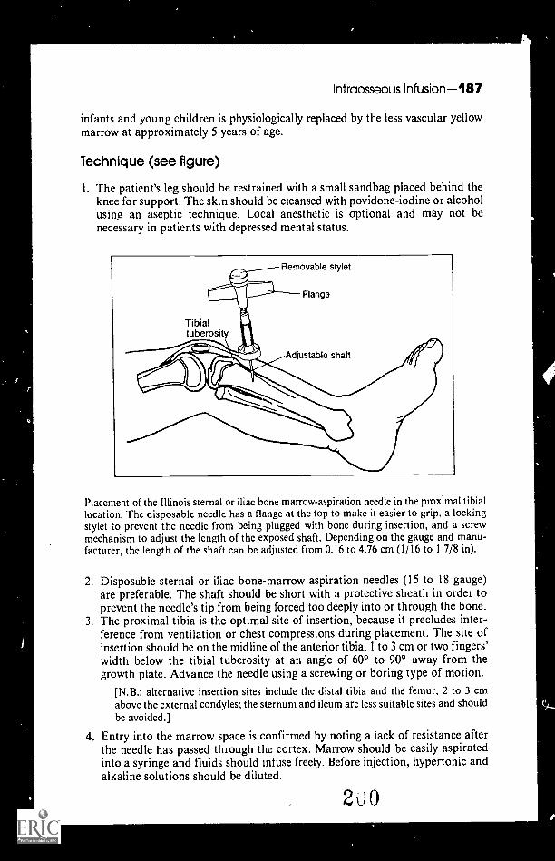
Intraosseous Infusion-487
infants and young children is physiologically replaced by the less vascular yellowmarrow at approximately 5 years of age.
Technique (see figure)
1. The patient's leg should be restrained with a small sandbag placed behind theknee for support. The skin should be cleansed with povidone-iodine or alcoholusing an aseptic technique. Local anesthetic is optional and may not benecessary in patients with depressed mental status.
Placement of the Illinois sternal or iliac bone marrow-aspiration needle in the proximal tibiallocation. The disposable needle has a flange at the top to make it easier to grip, a lockingstylet to prevent the needle from being plugged with bone during insertion, and a screwmechanism to adjust the length of the exposed shaft. Depending on the gauge and manu-facturer, the length of the shaft can be adjusted florn 0.16 to 4.76 cm (1/16 to I 7/8 in).
2. Disposable sternal or iliac bone-marrow aspiration needles (15 to 18 gauge)are preferable. The shaft should be short with a protective sheath in order toprevent the needle's tip from being forced too deeply into or through the bone.
3. The proximal tibia is the optimal site of insertion, because it precludes inter-ference from ventilation or chest compressions during placement. The site ofinsertion should be on the midline of the anterior tibia, 1 to 3 cm or two fingers'width below the tibial tuberosity at an angle of 60° to 900 away from thegrowth plate. Advance the needle using a screwing or boring type of motion.
[N.B.: alternative insertion sites include the distal tibia and the femur, 2 to 3 cmabove the external condyles; the sternum and ileum are less suitable sites and shouldbe avoided.]
4. Entry into the marrow space is confirmed by noting a lack of resistance afterthe needle has passed through the cortex. Marrow should be easily aspiratedinto a syringe and fluids should infuse freely. Before injection, hypertonic andalkaline solutions should be diluted.
200

488Intussusception
5. The needle should stand upright without support but must be stabilized andsecured by taping the flanges of the needle in order to prevent loss of access.
6. Flush the needle with a heparin-containing saline to prevent clotting beforeadministering a conventional solution.
7. The insertion site must be observed for evidence of extravasation.8. Once conventional vascular access has been obtained (normally within 1 to 2
hours after establishing the intraosseous infusion), the bone-marrow needleshould be removed. Longer intraosseous infusions increase the risk ofinfectious complications.
9. A sterile dressing should be placed over the dressing site and pressure appliedto the dressing for 5 minutes.
Possible Complications
I. Subcutaneous and occasional subperiosteal infiltration of fluid or leakage atthe puncture site.
2. Clotting of marrow in the needle, which can impede access.3. Localized cellulitis and subcutaneous abscesses have been reported in about
0.7% of all cases.4. Osteomyelitis has been reported in approximately 0.6% of cases.5. Theoretically, damage to the growth plates and marrow elements can occur, but
this is rarely observed. Fat and bone emboli are also of concern but rarely occur.
Contraindications
The procedure is contraindicated in children with osteogenesis imperfecta,osteopetrosis, and an ipsilateral fractured extremity due to risk of subcutaneousextravasation. The risk of infectious complication increases when the needle isintroduced through an area affected by cellulitis or burn injui ies. The procedureshould not be performed in children over the age of' 5 years.
Reference: Fisher DH: lntraosseous infusion. N Engl J Med 322:1579 1581, 1990.
I NTUSSUSCEPTION
The Need for Prompt Recognition
pediatrician is usually the first physician to see a child with an intussus-ception. Prompt recognition of this acute disorder will reduce morbidity andmortality. Remembering the following facts will facilitate early diagnosis andimprove management.
Age of Patients % Presenting at Gifen Age
Under 12 months 52%1-2 years 24%2-3 years 10%
3-7 years 11%
Over 7 years 3%
201

Signs and Symptoms
PainVomiting (at least once)Gross blood with stoolAbdominal mass
Iron Deficiency Anemia 189
% Presenting with GivenSign or Symptom
94%91%66%59%
Patients typically are healthy infants and children with no previous history ofgastrointestinal disease. Nearly all infants present with recent onset of abdominalpain and at least one episode of vomiting. The pain is characterized by the child'scrying and drawing his legs into his abdomen. Males are affected about twice asoften as females. The mass is usually sausage-shaped and is palpable along thecourse of the colon. On occasion one may elicit Dance's signan emptiness inthe right lower quadrant that reflects the fact that the intussuscepting bowel hasmoved out of this portion of the abdomen.
Etiology of Intussusception
In less than 10% of patients will an etiologic factor be determined. Specificcauses include Meckel's diverticulum (most common), ileal polyp, ileal granuloma,inspissated meconiurn in patients with cystic fibrosis, Henoch-Schönlein purpura,and lymphosarcoma.
Although the barium reduction will successfully reduce approximately 75% ofall intussusception, it is advisable fbr all patients over 6 years of age to haveelective exploratory laparotomy because of the high probability that intussuscep-lion at this age has a specific cause; it is frequently produced by an intestinallymphosarcorna.
Reference: Wayne ER, Cambell JB, Burrington JD, Davis WS: Radiology 107:597,1973.
From McMillan JA, et al: The Whole Pediatrician Catalog. Philadelphia, W.B.Saunders, 1977, p 382, with permission.
IRON DEFICIENCY ANEMIA
A Progression of Findings
Throughout the world anemia is the most common manifestation of nutritionaldeficiency. In the U.S. iron deficiency is associated with the majority ofnutritional anemias.
The onset of iron deficiency anemia is preceded by a sequence of abnormalitiesthat may exist a considerable time before the anemia. Given that iron deficiencyis a multisystem problem and that symptoms may occur well in advance of theonset of anemia, screening tests have been devised to detect iron deficiency beforeanemia is present. The sequential changes are indicate in the table.
''2

4'
190Iron Deficiency Anemia
Sequential Changes in the Development of Iron Deficiency
STAGE IISTAGE! IRON-DEFICIENT STAGE III
IRON DEPLETION ERYTHROPOIFSIS IRON DEFICIENCY
Serum ferritinA
Bone marrow iron
Decreasedlevels
Decreased
staining
Decreased
levels
Decreased
staining
Decreased
levels
Decreased
staining
Total serum ironbinding capacity
1
(TI BC) Normal Elevated Elevated
Serum iron Normal Decreased
levels
Decreased
levels
Erythrocyte proto- Normal Increased I ncreased
port hyrins A levels levels
1'
Hemoglobin Normal Normal Low
Hematocrit Normal Normal Low
Mean corpuscularvolume (MCV)
Normal Normal Aor low
Low
Red cell morphology Normal Normal MicrocytosisHypochromia
Reference: Bates H M: Lab Management 18:9, 1980.
Blue Scierae as a Sign of Iron Deficiency Anemia
In his classic textbook, The Principles and Practice of Medicine, Sir WilliamOsier described a common but rarely noted finding among iron-deficient,undernourished teenage girls: the presence of blue sclerae. A group of Britishgastroenterologists recently studied the presence of blue sclerae in associationwith iron deficiency anemia and found the sign to be far more sensitive andequally specific for iron deficiency anemia than the presence of mucosal pallor.Further, the search for blue sclerae is easier to confirm, because it is not affectedby skin tnickness, blood transfusions, pigmentation, or perfusion, which can beconfusing in an accurate as' . .ent of mucosal pallor.
The blue sclerae sign in :Ion deficiency anemia most likely results fromimpaired collagen synthesis; iron is a vital cofactor in the hydroxylation ofproline and lysine residues. The result is a thin sclera that allows greater visibilityof the choroid, and a bluish color is observed. Blue sclerae are associated withother diseases, as listed below, but these disorders are extremely rare and
2.)3

Iron Overdose-191
certainly less common than iron deficiency anemia in the general population. Thepresence of blue sclerae, therefore, should prompt the physician to consider apossible underlying iron deficiency.
Disease Entities Associated with Blue Sclera.
Iron deficiency anemiaAnemia secondary to chronic blood
loss (e.g., duodenal ulcers, inflam-matory bowel disease, glutenenteropathy)
Osteogenesis imperfectaInherited connective tissue disorde,
(e.g., Ehlers-Danlos syndrome,pseudoxanthoma elasticum)
Rheumatologic disorders(e.g., rheumatoid arthritis,systemic lupus erythematosis)
Malignancies of the gastrointestinaltract
Corticostcroid useAnemia secondary to hookworm
infection (ankylostomiasis)Myasthenia gravis
Reference: Ka lra L, Hamlyn AN, Jones BJM: Blue sclerae: A common sign of irondeficiency. Lancet ii:1267-1269, 1986.
Kollonychla A Sign of iron Deficiency
Koilonychia, or spooning of the nails, refers to the loss of the longitudinal andlateral convexity of the nail associated with thinning and fraying of its distalportion. The terminal and lateral borders are flared dorsally as the normalconvexity is replaced by flattening and concavity of the nail plate.
Although spooning of the nail can be seen as a result of local fungal infectionsor as a hereditary abnormality, it is also an early sign of iron deficiency in infantsand children as well as adults.
The nail of the index finger is more frequently and more severely deformed.The third finger and the thumb are also commonly involved. The spooningoccurs symmetrically, which should provide a clue to diagnosis. If still in doubt,then look at the feet. Most infants with spoon nails will also display the sameinvolvement on the nail of the big toe.
This provides a simple way of making a presumptive diagnosis of irondeficiency.
Reference: Hogan GR, Jones B: J Pediatr 77:1054, 1970.From McMillan JA, et al: The Whole Pediatrician Catalog, Vol. 2. Philadelphia, W.13.Saunders, 1979, p 202, with permission.
IRON OVERDOSE
Iron PoisoningAn Emergency Assessment
Because most patients with serum iron values in excess of 300 j.ig/dl are likelyto develop signs and symptoms of iron overdose, which clinical signs andlaboratory determinations do, in fact, predict a serum iron concentration ofgreater than 300 Ag/dl and serve as a guide for admission and therapy?
2 )4
192Iron Overdose
Five clinical and laboratory findings have been found to be significantlydifferent in patients with serum iron concentrations greater than or less than 300/.1g/d1. These five findings are:
1. White cell count > 15,000/mm32. Blood sugar > 150 mg/di3. Presence of vomiting4. Presence of diarrhea5. Radiopaque material visible on flat plate of abdomen.
Vomiting has the highest sensitivity value, as well as the highest negativepredictive value. The absence of vomiting makes is highly unlikely that thepatient has a serum iron in excess of 300 Ag/cil.
Importantly, patients do not develop signs or symptoms of acute iron toxicitymore than 6 hours after ingestion. Any patient who remains asymptomatic for 6hours following ingestion may be discharged with minimal risk of having adangerously elevated serum iron concentration.
In summary, initial management of iron overdose should consist of inducedemesis or lavage with a large bore tube. If any of the five screening tests describedabove is positive, a serum iron concentration of greater than 300 t4g/d1 is likely,and a serum iron concentration should be obtained. If a delay in obtaining aserum iron is anticipated, the patient should be given an intramuscular dose ofdeferoxamine (50 mg/kg up to a maximum of I g). If the drug pi oduces a "yinrose" color to the urine, indicating that the serum iron concentration exceeds thetotal iron-binding capacity, the patient requires treatment. Patients who havenegative results for all five tests should be observed for at least 6 hours forsymptoms. Those remaining asymptomatic may be discharged.
Reference: Lacouture PG, et al: Emergency assessment of severity in iron overdose byclinical and laboratory methods. J Pediatr 99:89-91,1981.
Never lend your car to anyone towhom you have given birth.
Erma Bombeck
2

z.
J
JAUNDICE
Jaundice in infancy
Visible jaundice occurs at serum bilirubin levels greater than 5 mg/d1. Mostinfants, full-term and premature, exhibit signs of a transient, unconjugatedhyperbilirubinemia during the first week of life. This "physiologic jaundice" is theresult of a complex interplay among several factors such as:
I. an elevated bilirubin load secondary to increased red blood cell (RBC)volume, decreased R BC survival, and an increased enterohepaticcirculation.
2. defective hepatic uptake of bilirubin from the serum due to diminishedlevels of ligandin and competition for binding to intracellular proteins.
3. defective bilirubin conjugation caused by decreased UDP-glucuronyl-transferase activity.
4. defective bilirubin excretion.
The National Collaborative Perinatal Project has determined that the majorityof infants with. physiologic jaundice will not have a serum bilirubin level > 12.9mg/d1 if full term, or 16 mg/d1 if premature. Risk factors for developinghyperbilirubinemia during the first week of life include breastfeeding, maternaldiabetes, induction of labor with oxytocin, maie sex, and Oriental race.
The essential questions to answer, when evaluating a jaundiced newborn, arethe severity of the hyperbilirubinemia and the type (conjugated vs. unconjugated)A rapid rise in serum bilirubin, such as an infant who develops hyperbilirubinemiain the first 24 hours of life or a rise in serum bilirubin greater than 5 mg/d1/day,warrants immediate investigation. A jaundiced infant without risk factors orprolonged jaundice (> I week in a full term; >2 weeks in a premature infant) alsoneeds to be evaluated, as does the infant with conjugated hyperbilirubinemia.
Unconjugated Hyperbilirubinomla
If the fractionation of the serum bilirubin documents as unconjugatedhyperbilirubinemia, then the search for its cause should progress as follows(please note that the history, physical examination and clinical course will helpguide the extent of the evaluation):
I. Fractionate serum bilirubin2. Blood type and Rh (mother and infant)3. Hemoglobin, hematocrit, and reticulocyte count4. Coombs test (direct and indirect)5. Peripheral blood smear6. Prothrombin time/partial thromboplastin time7. Platelet count
2193

194-Jaundice
8. Alkaline denaturation of hemoglobin test (of emesis)-adult vs. fetalhemoglobin
9. Sepsis work-up (blood, urine, cerebrospinal fluid culture)10. Thyroid screen thyroxine, triiodothyronine, thyroid-stimulating hormone)11. Phenobarbitol trial12. Interruption of breastfeeding
ri 5.3) Differential Diagnosis for Unconjugated Ityperbilirubinemia
1. Physiologic jaundice2. Hemolysis (e.g., ABO incompatibility; erythroblastosis fetalis; and red
blood cell defects such as spherocytosis, elliptocytosis, G-6-PD deficiency,and pyruvate kinase deficiency)
3. Hemorrhage (e.g., birth trauma, cephalohematoma)4. Breast milk jaundice5. Swallowed maternal blood6. Placental dysfunction7. Sepsis8. Clotting disorders9. Infant of a diabetic mother
10. Hypothyroidism11. Intestinal obstruction (e.g., pyloric stenosis, duodenal stenosis, or atresia)12. Crigler-Najjar syndrome (an hereditary disorder of glucuronyl transferase
resulting in an elevated unconjugated bilirubin level; Type II Crigler-Najjar is distinguished from Type I by a rapid decline in serum bilirubinlevel with phenobarbital therapy [5 mg/kg/day])
13. Lucey-Driscol syndrome (a syndrome of retention jaundice due todefective bilirubin conjugation in infants resulting from an unidentifiedfactor transmitted by the mother to her infant).
Conjugated HypetbilirubinemiaConjugated hyperbilirubinemia is always pathologic in the neonate and its
presence demands a diagnostic evaluation. The primary emphasis of this work-upis to identify those infants with treatable infectious and metabolic diseases, recog-nizable congenital or genetic disorders, or extrahepatic obstruction who wouldbenefit from surgical intervention. This evaluation should begin with a completehistory and physical examination, and the laboratory evaluation should proceedas follows:
1. Fractionate serum bilirubin2. Serum transaminases, alkaline
phosphatase (or 5'-nucleotidase),albumin, cholesterol
3. Prothrombin time4. Stool color5. Cultures (blood, urine, CSF, etc.)6. Hepatitis B surface antigen,
TORCH titers, VDRL7. Serum al-antitrypsin level8. Metabolic screen (urine/serum
amino acids; urine forreducing substances)
2 7
9. Thyroid screen10. Ophthalmologic examination11. Sweat chloride12. Skull, long bones, abdominal,
and chest x-ray films13. Abdominal ultrasound14. Duodenal intubation (string
test for duodenal fluid color,bilirubin, bile acids)
15. Hepatobiliary scintigraphy16. Percutaneous liver biopsy

Jaundice-195
Differential Dlagnosis of Conjugated Hyperbillrubinemia*
1. Extrahepatic obstruction 3. Persistent intrahepatica. Infantile obstructive cholestasis
cholangiopathy a. Paucity of intrahepaticBiliary atresia bile ductsNeonatal hepatitis b. Arteriohepatic dysplasiaCholedocholithiasis c. Benign recurrent intra-
b. Other causes hepatic cholestasisBile plug syndrome d. By ler diseaseCholedocholithiasis e. Hereditary cholestasisSpontaneous bile duct with lymphederna
perforation f. TrihydroxycoprostanicExtrinsic bile duct compression acidemia
2. Genetic and metabolic disorders 4. Acquired intrahepatica. Disorders of carbohydrate cholestasis
metabolism a. InfectiqnsGalactosemia Hepatitis BFructosemia (non-A, non-B?)Glycogen storage disease type IV Syphilis
b. Disorders of amino acid metabolism ToxoplasmosisTyrosinemia Rubella
c. Disorders of lipid metabolism CycomegalovirusNiemann-Pick disease HerpesGaucher disease Varicella
Wolman disease EchovirusCholesterol ester storage disease Coxsackievirus
d. Chromosomal disorders LeptospirosisTrisomy 18 TuberculosisDown syndrome Bacterial sepsis
e. Miscellaneous genetic & metabolic b. Drug-induceddisorders cholestasis
arAntitrypsin deficiency c. Cholestasis associatedNeonatal hypopituitarism with parenteralCystic fibrosis nutritionZellweger cerebrohepatorenal
syndromeFarniiia hepatosteatosis
*From Rosenthal P, Sinatra F: Pediatrics in Review 11:82, 1989, with permission.
Reference: Adapted from Rosenthal P, Sinatra F: Jaundice in infancy. Pediatric. inReview I I:79--86, 1989.
Progression of Dermal Icterus in the Newborn
The rate of rise of serum bilirubin in the newborn infant who is jaundicedshould always be monitored by laboratory determinations. The pediatrician,however, through simple examination of the infant, may make some estimationas to the rate of rise of serum bilirubin.

196Joint Pain
Dermal icterus has been shown to progress in a cephalopedal fashion; that is,as the infant's bilirubin rises, more of the skin becomes icteric. The icterus beginsat the head and neck and progresses caudally to the palms and soles. Thefollowing table correlates the level of indirect bilirubin with the area of skin thatis icteric in full-term infants whose jaundice is not due to Rh incompatibility.
Area of the Body Range of Indirect Bilirubin (mg/ POO ml)Head and neck 4 8Upper trunk 5-12Lower trunk and thighs 8-16Arms and lower legs 11-18Palms and soles > 15
As icterus progresses, the area that had been jaundiced remains jaundiced, sothat the entire body is icteric when the bilirubin rises above 15 mg/100 ml. Thefading of the icterus as the bilirubin level falls affects all body areas at the sametime, so that the intensity rather than the extent of the staining fades. Thestaining may progress more rapidly in the low birth weight infant, whereas theinfant with Rh disease may demonstrate a relative lag in dermal staining.
Correct estimation of the extent of icterus involves the examination of thecompletely undressed infant . der blue-white fluorescent light. Icterus may bedetected by blanching the skin with pressure of the thumb and noting the colorof the underlying skin. This is a more difficult determination to make in deeplypigmented black infants, but the palms and soles, at least, may be easilyexamined even in these patients.
Reference: Kramer LI: Advancement of dermal icterus in the jaundiced newborn. AmJ Dis Child 18:454,1969.
From McMillan JA, et al: The Whole Pediatrician Catalog. Philadelphia, W.B.Saunders, 1977, pp 127-128, with permission.
Common Causes
Chondromalacia patellaeGrowing painsOsteomyelitisOveruseSeptic arthritisSickle-cell diseaseSympathetic effusionTietze's syndromeTransient synovitisTrauma
Contusion
2
JOINT PAIN
Trauma (Com.)FractureHemarthrosisSprain/strain
Viral arthritisAdenovirusEpstein-Barr virusHepatitisMumpsRubellaVaricella

Uncommon Causes
Attention-seeking behaviorChild abuseForeign bodyLegg-Calve-Perthes diseaseMycoplasmaOsgood-Schlatter diseaseOsteochondritis dissecansPopliteal cystPsoriatic arthritis
Rare Causes
Bone tumorsCarpal-tarsal osteolysisCongenital joint laxity
Ehlers-Danlos syndromeMarfan syndromeStickler's syndrome
Cystic fibrosisFabry's diseaseGaucher's diseaseGiardiaGoutHyperlipoproteinemiaHyperparathyroidismIdiopathic chondrolysisImmunodeficiency
Complement deficiencyHypogammaglobulinemia
ImmunologicAcute rheumatic feverAnkylosing spondylitisBehget's syndromeDermatomyositisGiant-cell arteritisHenoch-Schonlein purpuraHepatitisInflammatory bowel diseaseJuvenile'rheumatoid arthritis
Juvenile Rheumatoid Art hritis-197
Reactive arthritisBruce IlaC'ampylohaeterSalmonellaShigeliaYersinia
Referred pain (retroperitoneal/intra-peritoneal inflammation)
Slipped capital femoral epiphysisSubluxation of the patella
Immunologic (Cont.)Kawasaki's diseaseMixed connective tissue diseasePolyarteritis nodosaReiter's syndromeSclerodermaSerum sicknessSjogren's syndromeSystemic lupus erythematosus
LeukemiaLipogranulomatosisLyme diseaseMucopolysaccharidosisMycobacterial diseasePsychogenic rheumatismReflex sympathetic dystrophyRicketsSarcoidosisStevens-Johnson syndromeSubacute bacterial endocarditisSyphilis
Charcot jointInfection
Thyroid diseaseVillonodular syncvitisWhipple's disease
JUVENILE RHEUMATOID ARTHRITIS(See also ARTHRITIS)
Features of Juvenile Rheumatoid Arthritis
Juvenile rheumatoid arthritis (iRA) is an entity with protean manifestations.There is a remarkable heterogeneity of JRA, which has been divided into three
210

198Juvenile Rheumatoid Arthritis
major categories: (1) systemic, (2) pauciarticular-onset type (in which four orfewer joints are affected), and (3) polyarticular-onset type (the type most similarto typical rheumatoid arthritis). There has been some acceptance amongrheumatologists and orthopedists to further subdivide the pauciarticular group.Early-onset pauciarticular J RA primarily affects young girls; chronic iridocyclitisis a common feature, as is the tendency to have positive antinuclear antibodytests. Late-onset pauciartieular JRA, on the other hand, affects mostly olderboys; sacroiliitis is a common feature and many patients go on to developankylosing spondylitis in adult life.
Features of the Three Major Tves of JRASYSTFM ONSFT POINARTICULAR ONSF I PALICIAR FICULAR ONSF I-
High fever ++++ 0 0
Rheumatoid rash ++++ +++ ++
Lymphadenopathy +++ ++ +
Splenomegaly +++ ++ ++
Hepatomegaly ++ + +
Pericarditis +++ + 0
Myocarditis ++ 0 0
Pncumonitis, pleuritis ++ + 0
Chronic iridocyclitis + + +++
Subcutaneous nodules 0 ++ 0
Leukocytosis +++ 0 0
Rheumatoid factor 0 +++ 0
++++ = most cases; +++ = many cases; ++ = some cases; + = occasional cases; 0 = rare orno cases.
Reference: Adapted from Tarana A: JRA and red herrings. Hospital Practice 23:129150, 1988.
The greatest poem ever knownIs one all poets have outgrown:The poetry, innate, untold,Of being only four years old.
Christopher ModeyFrom lb A Child
2 Li

KAWASAKPS DISEASE
Is It Really Kawasaki's Disease?
Kawasaki's disease, also known as mucocutaneous lymph node syndrome,occurs worldwide. It is an acute, febrile, multisystemic disease of children thatis usually benign and self-limited. In the United States the annual incidence is4.5-8.5 cases per 100,000 children below 5 years of age. In an outbreak in acommunity, the incidence can rise to 150 per 100,000 children.
Every house officer seems to make this diagnosis five times more frequentlythan that of scarlet fever and other more common red rashes. Is it reallyKawasaki's disease? The figure shows us what to expect if and when we are tomake this diagnosis, and the list below helps us to distinguish this disorder fromits imitators.
95%
I 1 1 I 1 Ilevet 38 40'C
Unresponsive to hnlittiottcl.
881,I--- Conjunctival
hyperemisI I90%
may respond (0 crol.coilerOolOt IWO vmete therapy
Cracied Ind bp:i
90%_J 11 50
75
2,53
Frythemstous Orel1 Strewbetty too;,t'`';"r H
750,
CONICS' aderofis
813.1
1.--rythemetousalms & soles
El erns, hands et
Polymorphous Iexanthern
94%
HPeeling r
finger & toe Ope1
MucoCuleneoui lymph Nod, Syndrome
Not ail symptoms & signs ere presenttuti,MannOusly Diagnosis depends On
ProoreInion &symptoms & signsOvnr 1111141.
And
2 Exclusion of other dieglales
Re122210...1.1t_i
717,.
Lteas1 ing::rjFle_F
I FIRST wEEK I SECOND WEEITT THIRD WEEK FOLK' TH WEEK I SECOND MONTH
BEST COPY AVAILABLE 2 1 2 199

200Kawasaki's Disease
Familiarity with its features will help you to make the diagnosis.
Major Manifestations
Fever in excess of 38.5° C for 5 daysRedness and induration of c alms and solesDesquamation of skin over fingers during convalescencePolymorphous exanthem over trunk; no vesiclesConjunctivitisRedness and fissu-ing of the lipsStrawLerry tongueDiffuse redness of oropharynxAcute, nonpurulent swelling of cervical lymph nodes
Other features of the disease may include tachycardia, gallop rhythm, distantheart sounds, heart murmurs, EKG changes, diarrhea, proteinuria, pyuria, leuko-cytosis, mild anemia, elevated platelet count, increased erythrocyte sedimentationrate, and increasing level of IgE during period of illness.
Less frequent manifestations include arthralgia, arthritis, aseptic meningitis,arid mild jaundice.
Mortality is approximately 0.3 to 2%. The most serious complication isvasculitis of the coronary arteries, although early deaths may result from severemyocarditis. Deaths are due primarily to thrombosis of large coronary-arteryaneurysms and resultant myocardial infarction, which occurs late in the course ofthe disease (between the third and fourth week). Coronary angiography duringthe illness may reveal abnormalities in as many as 60% of patients. These includeaneurysms, dilatation, stenosis, tortuosity, and irregularity of arterial vesselwalls. These appear to regress with recovery, but at present, the long-termprognosis is unknown.
Age incidence: The incidence is highest in children of I year of age andapproximately 80% of all patients are under 4 years of age.
Etiology: Unknown. No evidence of point source or person-to-person trans-mission has ever been documented.
Treatment: Unresponsive to antibiotics. When administered early, high-doseintravenous gamma globulin together with aspirin has been shown to be effectivein reducing coronary artery abnormalities.
Recurrences: Rare.May mimic some of the features of scarlet fever, measles, atypical measles,
rubella, Stevens-Johnson syndrome, juvenile rheumatoid arthritis, staphylococcalscalded-skin syndrome, and acrodynia (mercury poisoning).
References: Kawasaki T, Kosaki F, Okawa S. et al: A new infantile febrile mucocu-taneous lymph node syndrome prevailing in Japan. Pediatrics 54:271, 1974.
Kawasaki disease (editorial). Lancet i:675, 1976.Newburger JW. Takahashi M, Beiser AS, et al: A single intravenous infusion of
gamma globulin as compared with four infusions in the treatment of acute Kawasakisyndrome. N Engl J Med 324:1633 1639, 1991.
Shackelford PG, Strauss AW: Kawasaki syndrome (editorial). N Engl J Med 324:1664- 1666, 1991.
Gersony WM: Diagnosis and management of Kawasaki disease. JAMA 265(20):2699-2703, 1991.
213

Kawasaki's Disease-201
Clinical Features Associated with Kawasaki's Disease
The clinical features that substantiate a diagnosis of Kawasaki's disease arelisted below.
Diagnostic Criteria fa Kawasaki's Disease
I. Fever lasting for at least 5 days*2. Presence of four of the following five conditions:
a. Bilateral conjunctival injectionb. Changes of the mucosa of the oropharynx, including injected pharynx,
injected and/or dry fissured lips, strawberry tonguec. Changes of the peripheral extremities, such as edema and/or erythema
of hands and/or feet, desquamation usually begini,ing periunguallyd. Rash, primarily truncal; polymorphous but nonvesicularc. Cervical lymphadenopathy
3. Illness not explained by other known disease process.
Other features of Kawasaki's disease may help establish the diagnosis, mayexplain complications as they arise, or may allow for anticipatory management ofthe patient. These associated findings include the following:
Clinical Manifestations Associated with Kawasaki's Diseases
NONCARDIAC MANIFESTATIONS CARDIAC MANIFESTATIONS
Anterior uveitisArthritis/arthralgiaAseptic meningitisHepatic dysfunctionDiarrheaHydrops of the gallbladderPneumonitisSterile pyuriaTympanitis
Aortic aneurismCoronary aneurismLate coronary artery occlusionMyocarditisPericardial effusion
*Many experts believe that, in the presence of classic features, the diagnosis ofKawasaki's disease can be made (and limited treatment instituted) before the fifth day offever by experienced individuals.
Reference: Management of Kawasaki syndrome: a consensus statement prepared byNorth America participants of The Third International Kawasaki Disease Symposium,Japan, Deccmber, 1988. Pediatr Infect Dis J 8:663 -667, 1989.
Anterior Uveitis and Kawasaki's Disease
Anterior uveitis involves inflammation of the vessels of the anterior uvealtract, including the iris and the ciliary body. Anterior uveitis, also refP-red to asiridocyclitis, is a common manifestation of Kawasaki's disease, and its detectionduring the first week of illness may help establish the diagnosis. Slit-lampexamination is required in order to establish the presence of anterior uveitis, and,because the usual symptoms of eye pain and photophobia may be minimal, it hasbeen suggested that ophthamologic examination should be a routine part of theevaluation of patients suspected of having Kawasaki's disease.
2

202Kidneys
Other illnesses that may be associated with conjuncitivitis in thepatient are listed below. Anterior uveitis may be found in someconditions.
Streptococcal and staphylococcaltoxin-mediated diseases
Adenovirus and other viral infections(enterovirus, measles*)
Stevens-Johnson syndrome*Leptospirosis*Yersinia pseudottthereulosis
infectionRickettsia) infectionReiter's syndrome*
pediatricof these
Inflammatory bowel disease*Post-infectious immune
complex disease* (e.g.,post-meningococcal)
Sarcoidosis*Systemic lupus erythematosus*Behget's syndrome*Juvunile rheumatoid arthritis
(esp. early-onset pauciarticular-type J RA)
*May have evidence of anterior uvcitis on slit lamp examination.Reference: Smith LBH, Newburger JW, Burne JC: Kawasaki syndrome and the eye.
Pediatr Infect Dis J 8:116 -118, 1989.
KIDNEYS
A Technique for the Palpation of the Kidneys of Neonates
Congenital malformations of the urogenital tract occur in approximately 12%of all newborns. In 0.5% of all newborns, significant renal anomalies are present.These should be detected early in life in order to avoid subsequent complications.Almost all significant anomalies can be detected by careful abdominal palpation.A simple technique that will enable you to palpate the kidneys of 95% of allneonates is as follows:
I. Support the infant in a semireclining position facing you by placing yourleft hand behind the infant's shoulders, neck, and occiput.
2. Place the fingers of your right hand in the infant's left costovertebral angleposteriorly.
3. Use the thumb of your right hand to search the infant's abdomen system-atically, at first superficially and then deeply.
4. Deep palpation is performed by applying gentle, steadily increasingpressure subcostally in a posterior and cephalad direction. The thumb canthen be slipped downward without reducing the posteriorly directedpressure. Usually, the upper pole of the kidney can be felt trapped betweenthe descending thumb and the posteriorly placed fingers.
5. Next, change hands and examine the opposite side of the abdomen.
After practice on some two dozen infants, this technique can be mastered andsubsequently performed in 30 seconds. Because of its high yield, it deserves youroptimal skill and attention.
Reference: Perlman M, Williams J: Detection of renal anomalies by abdominalpalpation in newborn infants. Br Med J 2:347, 1976.
From McMillan JA, et al: The Whole Pediatrician Catalog. Philadelphia, W.B.Saunders, 1977, with permission.
215

LANGUAGE
Pattern of Normal Language DevelopmentA Summary
Pattern of Normal Language Development
AGE VOCALIZATION AND SPEECH RESPONSE COMPREHENSION
1 mo Much crying and whimpering; produces somevowel and few consonant sounds.
3 mo Different cries for pain, hunger, and discom-fort; decreased crying time; some repeti-tive sounds ("ga, ga, ga"); coos and sighs.
5 mo Babbles; vocal play; many repetitive sounds,all vowels, rn, k, g, b and p; laughs outloud.
7 mo Considerable variety in babbling, loudnessand rhythm of all vocalizations; adds d, 1,n and w to repertory of sounds; talks totoys.
9 mo Cries to get attention; increasing variations inpitch: "mama," "dada" and "baba" part ofvocal play but not associated with aperson or object.
11 mo May use one word correctly; imitates soundsand correct number of syllables, littlecrying.
1-2 yr Much unintelligible jargon; all vowelspresent; improves articulation so that 25%of words intelligible; names many objectsby 24 mo; much echolalia.
2 3 yr Tries new sounds but articulation lags behindvocabulary; 50 75% of words intelligible;often omits final consonants; jargonnearly absent.
3 4 yr Speech nears 100% intelligibility; faulty artic-ulations of / and r frequent; uses 3 4 wordsin sentences; uses a few plurals by 4 yr.
4-6 yr Syntax correct by 6 yr, forms 5- or 6-wordsentences that are compound or complexwith some dependent clauses; fluent;articulation good except for sh, z, eh andj; can express temporal relations; voicewell modulated in conversation.
Smiles: decreases activity; startlesat loud sounds.
Vocal gurgle in response tosoothing voice; some imitativeresponse to speech.
Imitative response to speechdecreased; turns and looks tosound; recognizes familiarvoice; vocalizes displeasure.
Gestures increase as part of vocalresponses to stimuli; responseto sound is increasinglyinfluenced by visual factors.
Retreats from strangers, oftenaccompanied by crying; mayimitate hand clapping.
Comprehends "no no"; responds to"bye-bye" or "patty-cake" withappropriate gestures.
Recognizes 150 300 words by24 mo; responds correctly toseveral commands, e.g., "sitdown," "give me that,""standup," "come here," etc.
Comprehends 800-1,000 words by3 yr; responds to manycommands using "on,""under," "up," etc.
Recognizes plurals, sex differences,adjectives and adverbs; com-prehends complex sentences.
Understands 2,500-3,000 words;carries out commandsinvolving 3-4 actions;comprehends "if," "because"and "why."
Reference: Adapted from Capute AJ, Accardo PJ: Developmental Disabilities inInfancy and Childhood. Baltimore, Blookes Publishing, 1991.
2 ! 6203
204Leukokorla/LImb Pain
LEUKOKORIA
Leukokoria: A Differential Diagnosis
Leukokoria, GI- the white pupil, is generally viewed by the examining physicianas a sign of ocular disease. It occurs when a lesion interferes with the path of theophthalmoscope's white light shined into a patient's eyes. Under normal cixum-stances, the ingoing beam passes through a clear cornea, aqueous humor, pupil,lens, vitreous, and retina to reflect off the vascular choroid. Given the vascularityof the choroid, the light reflected out of the eye appears red in color and is termedthe "normal red reflex." While the red reflex is certainly important to documentwhen performing an eye exam, it should be noted that even when it occursinti aocular lesions may be present. Indeed, any lesion not directly in the path ofthe ingoing beam would not interfere with the red reflex and would subsequentlybe undiagnosed. Listed below, in crder of frequency, are some of the morecommon causes of leukokoria.
1. Cataracts2. Persistent hyperplastic primary
vitreous3. Retrolental fibroplasia4. Retinal dysplasia5. Retinoblastoma6. Chorioretinal coloboma7. Retinal detachment8. Retinoschisis9. Congenital retinal folds
10. Persistent pupillary membrane11. Hyaloid cysts12. Uveitis13. Nematode endophthalmitis
(toxacara)14. Panophthalmitis15. Coats disease (unilateral
exudative retinitis)
16. Norrie's disease (sex-linkedrecessive disorder characterizedby bilateral blindness followingsevere retinal detachments,deafness, and mentalretardation)
17. Juvenile xanthogranuloma18. Ocular tumors19. Ocular trauma20. Vitreous hemorrhages21. Medullated nerve fibers22. Chorioretinal degeneration23. Incontinentia pigmenti24. Phakomatoses25. High myopia26. Intraocular foreign body
Reference: Catalono JD: Leukokoria the differential diagnosis of a white pupil.Pediatr Ann 12:498-505,1983.
Common Causes
Growing painsInfection
CellulitisOsteitisOsteomyelitisPost-rubella vaccination
LIMB PAIN
217
Infection (Cont.)Septic arthritisSoft-tissue abscessToxic synovitisViral myositis
Sickle-cell disease-vaso-occlusive crisis

TraumaChondromalacia patellaeCompartment syndromesDislocation and subluxationFractureHypermobility syndromeJoint strain, sprain, internal
damageMyositis ossificans
Uncommon Causes
Accessory tarsal ossicleCollagen vascular disease
(e.g., dermatomyositis, lupus)Conversion reactionsHenoch-Schonlein purpuraJuvenile rheumatoid arthritis
Rare Causes
Bone tumors (osteogenic sarcoma.Ewing's sarcoma, chondrosarcoma)
Cushing's syndromeFamilial Mediterranean feverHemophiliaHistiocytosis XHyperparathyroidismHypervitaminosis AInflammatory bowel diseaseLeukemiaMucopolysaccharidosis
LIMP
Common Causes
Attention-seeking behavior (usuallyafter minor trauma)
Calluses/corn/ingrown toenailsChondromalacia patellaeContusionForeign body (especially plantar surface)Fracture (may be occult)Growing painsHemophilia (hemarthrosis,
soft-tissue bleed)Immunization (local reaction)Leg length discrepancy
Limp-205
Trauma (Cont.)Pathologic fracturePostimmunizationShin splintsSoft-tissue contusion or
b gorrhageStress fractureTendonitis, fasciitis, bursitisTraumatic periostitis
Legg-Calvé-Perthes diseaseOsgood-Schlatter diseaseOsteochond rit is d issecansRheumatic feverTarsal coalition
MyopathiesNeuroblastomaOsteoporosisPopliteal cystRicketsScurvySlipped-capital femoral epiphysisSoft-tissue tumors (rhabdomyo-
sarcoma, fibrosarcoma)Sympathetic reflex dystrophy
MimicryMyositis (acute viral)Poorly fitting shoes (tight
or loose)Shin splintsSickle-cell disease (painful
crisis/infarction)Soft-tissue/cutaneous infectionSprain/strainTendonitisTorsion deformitesTransient synovitis
2 8

206Lumbar Puncture
Uncommon Causes
Arthritis (septic)Baker's cystBlount's diseaseBone tumor (benign and malignant)Calcaneal spursChild abuseCongenital contracturesCoxa varaErythema nodosumLegg-Calvé-Perthes disease
Ncomuseular diseaseAtaxiaCNS bleedCNS infectionFlaccid paralysisMigraineMuscular dystrophyPeripheral neuropathy
CausalgiaDiabetes mellitusGull lain-Barré syndromeHeavy metal intoxication
Rare Causes
Arthritis/arthralgiaAcute rheumatic feverDermatomyositisHenoch-Sehonlein purpuraInflammatory bowel diseaseJuvenile rheumatoid arthritisKawasaki's diseasePolyarteritis nodosaSerum sicknessSystemic lupus erythematosus
BrucellosisCaffey's diseaseCongenital joint laxity (Ehlers-Danlos)ErythromelalgiaFreiberg's disease
Neuromuscular disease (Com.)Peripheral neuropathy (Cont.)
Periodic paralysisPoliomyelitisTick paralysis
RadiculopathySpastic paralysis
Osgood-Schlatter diseaseOsteochondritis dissecansOsteomyelitisPhlebitisPlantar wartReferred pain
DiscitisEpidural/paraspinal abscessIliac adenitisIntraperitoneal infection/inflam-
mationPelvic inflammatory diseaseRetroperitoneal mass
Slipped capital femoral epiphysisSubluxation of the patella
HepatitisHypervitaminosis AHysteriaIntervertebral disc herniationKohler's diseaseLarsen-Johansson diseaseNeuroblastomaPott's diseasePyomyositisRicketsScurvySever's diseaseSinding-Larsen diseaseTrichinosis
W MBAR PUNCTURE
Estimating Lumbar-Puncture Depth in Children
The lumbar puncture (LP) is a frequently used diagnostic procedure particularlyamong infants and young children presenting with an acute infectious illness. Yet
2!9
4

Lumbar Puncture-207
the procedure can be difficult, particularly for physicians who do not performLPs with regularity, often resulting in inserting the needle too deeply anddisrupting the venous plexus that lies beneath the dura on the anterior wall of thevertebral canal. Such a traumatic lumbar puncture is contaminated with blood,rendering the cerebrospinal fluid white cell count all but useless. Further, abloody tap may confuse matters if a culture result is positive because the patientis bacteremic but does not have meningitis.
A group of pediatricians at the Medical College of Wisconsin have developeda linear regression analysis of how deep to insert the lumbar puncture needlebased upon the child's body surface area in square meters and the depth at whereCSF reflux occurred. Each lumbar puncture was performed by positioning thechild in the right lateral decubitus position with maximal flexion at the waist andneck. The needle was inserted, perpendicular in relation to the back, at the L3 orL4 vertebral interspace. Using the following equation:
Depth of lumbar puncture = 0.77 cm + 2.56 (body surface area in m2)
these physicians were able to estimate the depth of lumbar puncture to withinapproximately 5 mm in most young children without incurring trauma or CSFreflux (see figure).
I I i I I I I
0.0 0.2 0.4 0.6 0.8 1 0 1.2 1 4 1 6 1 8
Body-Sorface Area (m2)
Relation of the depth of lumbar puncture to body-surface area. Dotted lines represent the95% confidence limits for the predicted depth of lumbar puncture.
Reference: Bonadio WA, Smith DS, Metrou M, Dewitz 13: Estimating lumbarpuncture depth in children. N Engl J Med 319:952-9pi p8A.
çf
208Lumbar Puncture
How to Interpret the Bloody Tap
Spinal fluid is supposed to be clear. When it isn't, the preliminary informationreceived from the laboratory may be difficult to interpret. The effect of blood onCSF results (usually as a result of inserting the spinal needle too deeply andpiercing the vascular plexus ventral to the epidural space) has actually beenstudied, and these studies make it possible to take a logical approach to theinterpretation of the bloody tap.
CSF WBC. The effect of blood on the WBC is the most difficult parameter tocalculate. Though it is intuitive that the WBC count would be altered in proportionto the WBC count in the peripheral blood, many studies have demonstrated that itis not this simple. Certainly when the observed CSF WBC is higher than would bepredicted from the peripheral WBC/RBC ratio, or when the percentage of PMNsis higher than that in the peripheral blood count, infection involving the centralnervous system should be suspected. However, management decisions should notbe made based on the CSF WBC alone when the CSF is contaminated with blood.
CSF Glucose. When experiments have been done to determine the effect ofmixing blood with CSF, no change in CSF glucose concentration can bedemonstrated. Though it has been suggested that hypoglycorrhachia (abnormallylow glucose in CSF) may result from RBC contamination, experimental studieshave not confirmed this contention, and a low CSF glucose should 1- regardedas a low CSF glucose.
CSF Protein. There is no question that blood in the CSF raises the proteinconcentratiori. The increase in protein has been found to be approximately 1 mg/dl for every 1000 RBCs. This is only an approximation, however, and for mostpurposes the CSF protein concentration is not helpful when the tap is bloody.
Xanthochromia. When hemoglobin from lysed RBCs remains in the CSFfor an extended period of time, the breakdown products oxyhemoglobin,methemoglobin, and bilirubin create a yellowish discoloration of the CSF afterthe specimen is centrifuged. CSF contaminated by fresh blood, as in a traumaticlumbar puncture, remains clear and colorless after centrifugation. The pigmenta-tion that results from RBC breakdown persists for about 7 days after thehemorrhage has stopped. Since RBC lysis occurs in CSF after about 4 hours,specimens that are not analyzed within that time period may be xanthochromiceven if the blood resulted from fresh contamination.
Traumatic lumbar puncture occurs in one out of every 5 LP attempts inpediatric patients, and the likelihood of a traumatic tap increases inversely withthe age of the patient. Since bleeding usually occurs as a result of introducing thespinal needle too far, a method to avoid this problem has been devised. Thismethod involves removing the spinal needle stylet once the needle has beenintroduced through the e,pidermis and the dermis. As the needle is very slowlyadvanced, the flow of spinal fluid can be seen as soon as the needle tip is in thesubarachnoid space. The stylet should be replaced prior to withdrawing theneedle to minimize the pressure gradient between the subarachnoid space and theatmosphere as the needle is withdrawn. It is important that the stylet remain inthe needle until the epidermis and the dermis are traversed. Implants of smallislands of skin during lumbar puncture have been reported to lead to the laterdevelopment of epidermoid tumors along the needle track.
Reference: Boadio WA: Contemporary Pediatrics, November: 109 116, 1989.
221
v

N
Lumbar Puncture-209
Lumbar Punctures and the PeripheralWhite Blood Cell Count
As if there weren't enough factors to consider in the performance of a lumbarpuncture (LP), a team of Israeli pediatricians have brought up one more: thetiming of obtaining the peripheral white blood cell (WBC) in relation toperforming the LP. A prospective study of 26 neonates and infants suspected ofhaving meningitis noted a significant increase in the peripheral WBC after the LPwas completed (10, 960 ± 3,500 cells/MI before the LP; 13,300 ± 3,970 cells/IAafter the LP, p < 0.001). The greatest increase in white cells was seen in theneutrophil and lymphocyte fraction, presumably because these cells are soquickly released from the marginal granulocyte pool. There were no significantdifferences, before and after LP, in serum glucose, urea, hemoglobin, and plateletcounts. While the LP does not impair interpretation of the CSF glucose to serumglucose ratio, the data depicted below make a strong case for obtaining theperipheral WBC count prior to performing the LP (see figure and table).
22000
21000-
20000
19000
18000
17000
16000
15000
14000
13000
12000
11000-
10000
9000
8000-
7000-
Before AfterL.P L.P
110
:6 100
90.
LnO 80-c)
- J0o 70.00co
60
50
Before AfterL P L.P
WBC courts (left) and serum glucose levels (right) before and 10 15 minutes after LPprocedure (broad line indicates the change of the mean values, and the 2 pairs of smallerlines the SEM).
222

210Lyme Disease
Mean ± SD of the Blood Tests Studied Before and 10-15 MinutesAfter the LP Procedure
MEAN BLOOD LEVELS
BEFORE LP AFTER LP
WBC count (cellshil) 10960 ± 3500 13300 ± 3970*Glucose (mg/di) 85.3 ± 13.4 84.1 ± 12.6Hemoglobin (gr/d1) 11.6 ± 2.2 11.7 ± 2.2Thrombocytes (x103) 269 ± 113 315 ± 93Urea (mg/dl) 6.3 ± 2.3 6.5 ± 2.5
*p < 0.001 (T-paired test).
Reference: Shohat M, Goodman Z, Rogovin H, Nitzan M: The effect of lumbarpuncture procedure on blood glucose level and leukocyte count in infants. Clin Pediatr26:477-479, 1987.
LYME DiSEASE
Clinical Manifestations of Lyme Disease
Lyme disease is an arthropod-borne infection that is heralded by a distinctiveskin eruption (erythema chronicum migrans), followed, in stages, by neurologicor cardiac complications and arthritis. Its vector is the deer tick, Ixodesdammini, and the etiologic agent is the spirochete Borrelia burgdorferi. Becausethe clinical manifestations of Lyme disease are so protean in their presentation,with considerable overlap between the three major stages and their time ofappearance, the accompanying temporally organized flow chart of its naturalhistory ought to be useful, as well as the table.
TICK OP.RITE3 to 4 weeks
STAGEONE
PRIMARYERYTHEMA
CHRONICUMMIGRANS
malaise, fever"flu"-like illnessregional lymphadenopathy
I 50%
SECONDARY ERYTHEMACHRONICUM MIGRANS
headache, meningismusarthralgia, myalgiageneralized lymphadenopathy
10%(4 weeks after rash)
(4 to 6 weeks after rash)
(4 to 5 weeks after rash)8%
STAGETWO
NEUROLOGICCOMPLICATIONSVII nerve patsymeningitisradiculoneuritis
50%
CARDITISheart blockmyopericarditis
Clinical manifestations of Lyme disease.
223
STAGETHREE
ARTHRITIS
BEST COPY AVAILABLE

111
Lyme Disease-211
Manifestations of Lyme Disease by Stage*
SYSTEM* FARLY INFECTION LATE INFECTION
LOCALIZED DISSEMINATED PERSISTENT
(STAGE I) (STAGE 2) (STAGE 3)
Skin Erythema Secondary annular lesions, Acrodermatitis chronicamigrans malar rash, diffuse erythema atrophicans, localized
or urticaria, evanescent scleroderma-like lesionslesions, lymphocytoma
Musculo- Migratory pain in joints, Prolonged arthritis attacks,skeletal tendons, bursae, muscle, chronic arthritis, pe-system hone; brief arthritis attacks; ripheral enthesopathy,
myositis:; osteomyelitis:: periostitis or joint sub-panniculitis: luxations below lesions of
acrodermatitisNeurologic Meningitis, cranial neuritis, Chronic encephalomyelitis,
system Bell's palsy, motor or spastic parapareses,sensory radiculoneuritis, ataxic gait, subtle mentalsubtle encephalitis, mono- disorders, chronic axonalneuritis multiplex, myelitis: polyradiculopathy,chorea:, cerebellar ataxia: dementia:
Lymphatic Regional Regional or generalizedsystem lymphade- lymphadenopathy,
nopathy splenomegalyHeart Atrioventricular nodal block,
myopericarditis, pancarditisEyes Conjunctivitis, iritis:, choroid- Kcratitis
itis:, retinal hemorrhage ordetachment:, panophthal-mitis:
Liver Mild or recurrent hepatitisRespiratory Nonexudative sore throat,
system nonproductive cough, adultrespiratory distresssyndrome:
Kidney Microscopic hematuria orproteinuria
Genitourinary Orchitis:system
Constitutional Minor Severe malaise and fatigue Fatigue
symptoms
*The classification by stages provides a guideline for the expected timing of the illness's
manifestations, but this may vary from case to case.+ Systems are listed from the most to the least commonly affecied.
The inclusion of this manifestation is based on one or a few cases.From Steere AC. N Engl J Med 321:589, 1989, with permission.
Adapted from: Eichenfield AH: Diagnosis and management of Lyme disease. PediatrAnn 15: 583-594,1986.
Steere AC: Lyme disease. N Engl J Med 321:586-596,1989.
What Is the Long-term Course of Lyme Disease in Children?
The natural history of Lyme disease is not yet completely known. In a recentreport the authors studied the long-term course of Lyme arthritis in 46 children inwhom the onset of the disease occurred between 1976 and 1979 and who received
2 = A
212Lymphadenopathy
no antibiotic therapy for at least the first 4 years of the illness. Of the 46 children(age range, 2 to 15 years), 33 (72%) initially had erythema migrans, 7 (15%) hadinfluenza-like symptoms, and 6 (13%) had migratory joint pain. These mani-festations were followed by brief attacks of arthritis, particularly affecting theknee. The percentage of children with recurrent episodes of arthritis declined eachyear. By year 4, only 10 children still had a mean of two episodes of arthritis peryear; the duration of arthritis was generally longer in older children (P < 0.05).During the sixth year of illness, two children had keratitis, and more than 10 yearsafter the onset of disease, a subtle encephalopathy developed in two other children.Of the 39 children whom the authors were able to contact in 1988-1989, 12 (31%)still had occasional brief episodes of joint pain and 1 had marked fatigue. All 46children had positive IgG antibody responses to Borrelia burgdorferi throughoutthe illness and on long-term follow-up. As compared with those who becameasymptomatic, the children with recurrent symptoms more often had IgMresponses to the spirochete and had significantly higher IgG titers (P < 0.05).
The long-term course of initially untreated Lyme disease in children mayinclude acute infection followed by attacks of arthritis and then over several yearsby keratitis, subtle joint pain, or chronic encephalopathy. This combination ofsymptoms in a patient with a high IgG antibody titer to B. burgdorferi is ofconcern, and the appropriate treatment for these patients is not yet certain.
Reference: Szer IS, Tayler E, Steere AC: The long-term course of Lyme arthritis inchildren. N Engl J Med 325:159-163, 1991.
LYMPI4ADENOPATHY (GENERALIZED)
Common Causes
Infection (viral, fungal, spirochetal)Juvenile rheumatoid arthritisSerum sickness
Uncommon Causes
Drug reactionsAnticonvulsants, antithyroid,
isoniazidHodgkin's diseaseInfection, bacterialLeukemiaNon-Hodgkin's diseaseSystemic lupus erythematosus
Rare Causes
Angioimmunoblasticlymphadenopathy
DysgammaglobulinemiaGaucher's diseaseHemophagocytic syndromesHistiocytic medullary reticulosisHistiocytosisHIV infectionHyperthyroidismMetastatic neuroblastomaNiemann-Pick disease
A Diagnostic Approach to Lymphadenopathy
Is the lymphadenopathy generalized or localized? Generalized lymphadenopathyis defined as enlargement of more than two noncontiguous node regions.Generalized lymphadenopathy is caused by generalized disease.
225

Generalized LymphadenopathyWhat are associated signs and symptoms?
Rash?Hepatosplenomegaly?Thyroid enlargement?Joint involvement?Heart and lung abnormalities?Pallor?Easy bruising?
Infections
ExanthemsCytomegalovirusInfectious mononucleosisInfectious hepatitisTyphoid feverMalaria
Collagen Vascular Disease
Lupus erythematosusRheumatoid arthritis
PyogenicTuberculosisSyphilisToxoplasmosisBrucellosisHistoplasmosis
Immunologic Reactions
Serum sickness, drug reactionsGranulomatous disease (sarcoid)
Storage Disease
Gaucher's diseaseNiemann-Pick disease
Malignancies
LeukemiaLymphomaHistiocytosisNeuroblastoma, metastatic
Hyperthyroidism
Causes of Cervical Adentfis
CAUSE COMMENT
Lymphadenopathy-213
Localized LymphadenopaihySigns of infection in the involved node?Evidence of infection in the drainage area
of node?History of recent antigenic introduction in
the node's drainage area?
Supraclavicular Always considermediastiaal disease (tuberculosis, his-toplasmosis, coccidioidomycosis, sar-coidosis). Always consider lymphoma.In absence of evidence of pulmonaryinfection, early biopsy indicated.
Axillary -- Secondary to infections in thehand, arm, lateral chest wall, or lateralportion of the breast. May be result ofrecent immunization in the arm.
Epitrochlear Secondary to infectionson ulnar side of hand and forearm.Observed in tularemia when bite occurson finger. Also seen in secondarysyphilis.
Inguinal Infection in lower extremity,scrotum, penis, vulva, vagina, skinof lower abdomen, perineum, glutealregion, or anal canal. May be seen inlymphogranuloma venereum. Mayrepresent metastatic disease fromtesticular tumors or bony tumorsof the leg. Immunization in leg.
Cervical Generally the result of local-ized infection. See accompanying tablefor differential diagnosis.
Viral upper respira-tory infections
Bacterial infection
Tuberculosis
Most common cause. Nodes soft, minimally tender, and not asso-ciated with evidence of redness and warmth of overlying skin.
Streptococcus and staphylococcus most common etiologic agents.Usually secondary to previous or associated infection in drainagearea of node. More frequently unilateral. Signs of infectiontenderness, warmth, and redness generally present. Look for pri-mary focus of infection in scalp, mouth, pharynx, and sinuses.
Mycobacterium tuberculosis infections generally bilateral, involve mul-tiple nodes. Associated with evidence of chest disease and systemicsigns. Atypical mycobacteria infections more commonly unilateralinitially. Not associated, in general, with other foci of disease. Witheither agent, evidence of local warmth and redness uncommon.
Table continued on next page.

214Lymphadenopathy
CAUSE COMM ENT
Infectious Fever, malaise, preceding upper respiratory infection often noted.mononucleosis Splenomegaly common. Atypical lymphocytes present. Epstein-
Barr virus titers required for diagnosis in younger children.
Cytomegalovirus Indistinguishable clinically from Epstein-Barr virus infections.Toxoplasmosis Requires serologic studies to make the diagnosis.
Cat-scratch disease History of contact with young cat. May be preceded by history offever and malaise. Adenopathy restricted to area drained by initialcat scratch.
Sarcoidosis Disease bilateral. Chest x-ray almost always abnormal. May have ke-ratitis, iritis and evidence of bone disease.
Hodgkin's disease Common presenting symptom. Frequently unilateral at time of initialmanifestation. Node is rubbery, nontender, and not associated withsigns of inflammation. Make certain that supraclavicular involve-ment is not present. When present, strongly suspect lymphoma.
Non-Hodgkin's Bilateral at time of initial presentation in approximately 4Oci- oflymphoma patients. Cervical and submaxillary nodes commonly involved
together.
Algorithm: Generalized Lymphadenopathy
Is patient taking phenytoin, hydralazine,allopurinol, antithyrold or antileprosymedications?
No
EBV titer or monospotToxoplasma titerCMV serology
Positive
Yes
Negative
Blood culturesPPD
Histoplasrnosis, coccidioidorny.cosis, or brucella serology(if suggested by history)
ANACBC
CX11
Negative
F scisinnal biopsy
Positive
227
Stop drug treatmentand reevaluate in2-3 wk
Dx specific -diseas'e
Dx s[;ecifiC .diSCase
CX11 hilar nodes present?Consider sarcoidosis
tuberculosislymphoma
BEST COPY AVAILABLE

CE
RV
ICA
L LY
MP
HA
DE
NO
PA
TH
Y
SY
ST
EM
IC A
CU
TE
SY
MP
TO
MS
WIT
H F
EV
ER
IA
SY
MP
TO
MA
TIC
^Ns.
.
SM
ALL
NO
DE
< 3
cm
-1
N.N
.,
[ LA
RG
E N
OD
E >
3 o
nS
ING
LE. U
NIL
OB
AR
NE
CK
MA
SS
IM
ULT
ILO
BU
LAR
NO
NE
LU
CT
UA
NT
[F 1
7-.0
Ert
l A-W
T-1
SA
ME
AS
IN "
C"
Mea
sure
nod
* si
zeT
hroa
t cul
t. fo
e sl
aty.
Ant
gla
res
°alta
r* fo
rst
aphy
toco
ccus
loot
.)
Nee
dle
asp.
with
pm s
tain
& c
ultu
re
CE
IC
Sed
. rat
eT
est f
oe s
trep
tcco
ccus
EB
V a
nt ib
ody
tad
orm
onot
oot
PP
O s
kin
test
, int
erm
edia
te
stni
ngts
Nee
dle
ass
with
can
sta
inan
d cu
lture
Tox
opIa
ana.
CM
V s
erol
ogy
He
of e
xpos
urs
toki
tten
w c
at.s
crat
thdi
seas
e sk
in o
st
TR
EA
T W
ITH
AP
PR
OP
RIA
TE
AN
TIB
IOT
ICS
RE
EV
ALU
AT
E IN
2-3
WK
Car
eful
He
& P
E to
exc
lude
syst
emic
cau
se o
f car
vica
lm
ess
CX
RC
BC 1
IF A
LL T
ES
TS
NO
NC
ON
CLU
SIV
E A
ND
NO
DE
SIZ
EP
ER
SIS
TS
OR
EN
LAR
GE
S W
ITH
IN 4
-4 W
K
IF N
O C
HA
NG
E IN
SIZ
E, O
R L
AR
GE
R, C
ON
T. R
eA
ND
RE
EV
ALU
AT
E IN
2-3
WK
IF N
O C
HA
NG
E IN
SIZ
E O
R L
AR
GE
R
EX
CIS
ION
AL
BIO
PS
Y
2%28

216Lymphadenopathy
Algorithm: Localized Lymphadenopathy
........_._._._SUPRACLAVICULAR 1 Chest x fayAREA Excisional biopsy
[ Local infections]] Treat local InfectionOCCIPITAL AREA I
TREAURICULAR ConlunctivitT-I Scrapings for inclusionbodies; culture for chiamyditT1
AREA
Fix of exposure tokitten
Cet.scratch diseaseskin test, ifavailable
....
SUBMAX I LLAR Y endSUBMENTAL AREAS
IPPD skin test I
VDRL (optional)
ILocal infection
AXILLARY AREA
EPITROCHLEAR AREA
INGUINAL andILIAC AREAS
Unilateral
Blisters!
Local infection
Hx of exposure tokitten?
Consider PPD skintest, ANA, sad.rate
[ Local infection?
POPLITEAL AREA
[ABDOMINAL endPELVIC AREAS
11,11.
Consider ANA, VDRL,sod. rate
ILocal Infection?
Consider VDRL, culturefor gonorrhea
Local infection?
Local infection?
PPD skin tettSed. rate
,..
LTreat local infection I
PPD skin test
VON L
Throat culture
Consider excisional bx fornon.Hodgkin's lymphoma
rTreat local Infection
Coter/itch diseaseskin test, ifavailable
Treat local infection
[ Treat local Infection I
Treat local infection
Treat local infection
Reference: Bedros AA, Mann JP. Lymphadenopathy in children. Adv Pediatr 28:341,1981, with permission.
From McMillan JA, et al: The Whole Pediatrician Catalog. Philadelphia, W.B.Saunders, 1977 (Vol 1), pp 30-32, and 1982 (vol 3), pp 9 10, with permission.
229
'FF

MAGNESIUM
Magnesium Deficiency: A Common Problem
Magnesium deficiency has a reported incidence of 10% among all patients intertiary care hospitals. It is frequently associated with hypocalcemia and/or hypo-kalemia. Despite one's good intentions, dosing the hypomagnesemic, hypokalemic,hypocalcemic patient with large amounts of calcium and potassium salts will dolittle to correct his or her electrolyte imbalance until the serum magnesium isrestored to a normal value. Magnesium deficiency has numerous causes as notedin the table below; nutritional deficiency leads the list. Symptoms of hypomag-nesemia are manifested primarily as neuromuscular irritability (e.g., tetany,tremors, and seizures). Changes in personality, anorexia, nausea, abnormalcardiac rhythms and EKG changes can also be seen (see table).
Causes of Magnesium Deficiency*
Prolonged parenteral fluidadministration
Total parenteral nutrition withoutmagnesium
Nutritional
Starvation with metabolic acidosisProtein-calorie malnutritionKwashiorkorAlcoholism
Chronic diarrhea from any cause(e.g., chronic ulcerative colitis,Crohn's disease, laxative abuse,villous adenoma, adenocarcinomaof rectum)
Intestinal
MalabsorptionShort-bowel syndromeGluten enteropathyPancreatic insufficiency with
steatorrheaTropical sprueFamilial malabsorption of magnesium
Disease relatedRenal tubular acidosisAcute tubular necrosis
(diuretic phase)Chronic glomerulonephritisChronic pyelonephritisFamilial and sporadic renal
magnesium loss
Renal
Drug relatedDiuretics (furosemide, ethacrynic acid,
thiazides)Antibiotics (gentamicin, tobramycin,
ticarcillin, carbenicillin,amphotericin B)
Antineoplastic drugs (cisplatin, combina-tions of antibiotics and cytotoxic agents)
Cyclosporine
*From Hospital Practice, February 15, 1987, with permission.Table continued on next page.
217230

218Malignant Disease
Causes of Magnesium Deficiency (Cont.)
Endocrine and Metabolic
Primary and secondary aldosteronismHyperthyroidismExcessive lactationPregnancy (third trimester)Hypercalcemia
Primary hyperparathyroidism (due to hyper-calccmia; immediately postoperatively inpatients with osteitis fibrosa cystica)
Uncontrolled diabetes with marked glucosuriaAcute intermittent porphyria
Congenital, Neonatal
Maternal diabetesMaternal hyperparathyroidism or hypoparathyroidismExchange transfusions (citrate effect)
Reference: Flink EB: Magnesium deficiency. Causcs and effects. Hospital Practice22:116A-116P, 1987.
MALIGNANT DISEASE
Clues to Malignant Disease
Certain congenital malformations and acquired diseases arc recognized to beassociated with an increased incidence of malignancy. The conditions listed belowshould signal a warning and cause a high index of suspicion, regular observation,and appropriate studies for early detection of the associated malignancies.
In addition there are familial associations connected to certain tumors, such asbrain tumors, Hodgkin's disease, and Ewing sarcoma, which have been reportedin siblings more frequently than chance alone would explain. Awareness of theseassociations may allow for earlier detection of both malignancies and congenitaland other syndromes.
Congenital and Acquired Conditions Associated with Increased Riskof Malignancy in Childhood
CONDITION ASSOCIATED MALIGNANCY
Agammaglobulinemia
Albinism
Aniridia (non-familial)
Ataxia telangiectasia
l3eckwith's syndrome
Bloom's syndrome
Chédiak-H igashi syndrome
Congenital X-linked immuno-deficiency
D-trisomy
Down syndrome
Lymphoma, lymphosarcomaBasal cell carcinoma, squamous cell carcinoma
Wilms' tumor
Leukemia, lymphoma, lymphosarcomaWilms' tumor, liver carcinoma, adrenal cortical
carcinoma, nesidioblastosis of pancreas
LeukemiaLymphoma, lymphosarcoma, leukemia
Lymphoma, leukemia
Leukemia
Leukemia
2 31Table continued on next page.

Malignant Disease-219
Congenital and Acquired Conditions Associated with Increased Riskof Malignancy in Childhood (Cont.)
CONDITION ASSOCIATED MALIGNANCY
11 p syndrome Wilms' tumor
Familial pelyposis of colon Colonic carcinomaFamily history (first degree) Same or other malignancy
of malignancyFanconi anemia Leukemia, hepatomaGenitourinary anomalies Wilms' tumorGiant cell hepatitis Carcinoma of liver
Gonadal dysgenesis Gonadal cancerHemihypertrophy Wilms' tumor, adrenal cortical carcinoma,
liver carcinoma, hepatoblastoma
Hippel-Lindau disease PheochromocytomaHorner syndrome Neuroblastoma
1g M deficiency Lymphoma
I rrad iat ion:
in utero Leukemia
of head and neck in early life Thyroid carcinoma, brain and parotid tumorsfor retinoblastoma Osteosarcoma
for Wilms' tumor Osteosarcoma, osteochondroma
for neuroblastoma Osteosarcoma, osteochondromaKlinefelter's syndrome Leukemia
Multiple endocrine adenomatosis I Schwannoma(Wermer syndrome)
Multiple endocrine adenomatosis II Thyroid carcinoma, pheochromocytoma(Sipple syndrome)
Multiple mucosal neuromas Medullary thyroid carcinoma
Maternal stilbestrol during pregnancy Vaginal adenocarcinoma
Neurofibromatosis Pheochromocytoma, sarcoma,schwannoma, leukemia
Nevus sebaceous Basal cell carcinoma
Poland's syndrome Leukemia
Renal dysplasia Wilms' tumor
Severe combined immunodeficiency Lymphoma, leukemia
13 q syndrome Retinoblatoma
Thyroid cancer (medullary) Pheochromocytoma
Ulcerative colitis/regional ileitis Colonic carcinoma
Wiskott-Aldrich syndrome Lymphoma, lymphosarcoma
Xeroderma pigmentosum Basal cell or squamous cell carcinoma
References: Craven EM: Pediatric conditions associated with malignancy (letter).JAMA 215:795, 1971.
Feman SS, Apt L: Eye findings associated with pediatric malignancy. J PediatrOphthalmol 9:224, 1972.
Leventhal BG, in Behrman RE, Vaughan VC: Nelson Textbook of Pediatrics, 13th ed.Philadelphia, W.B. Saunders, 1987, p 1081.
2 '2 2

'4
220Maple Syrup Urine Disease/Marfan's Syndrome
MAPLE SYRUP URINE DISEASE
What Is the Intellectual Outcome?
Maple syrup urine disease (MSUD) is the most common inborn error ofamino acid metabolism and presents acutely in the neonatal period. ClassicMSUD is characterized by lethargy, poor feeding, vomiting, and alternatingperiods of hypertonicity and flaccidity. In untreated disease, progressive neurologicdeterioration, seizures, cerebral edema, coma, and death will usually occur withinthe first month of life.
A recent report of a controlled study of the intellectual outcome in 16 childrenwith MSUD compared the outcome of MSUD diagnosed after symptomsbecame apparent with that of MSUD diagnosed prospectively and treatedpresymptomatically. Affected children treated presymptomatically had higher IQscores than their affected siblings treated after their disease became symptomatic.The authors concluded that early and meticulous treatment of MSUD can resultin intellectually normal chiidren.
Therapy for MSUD consists of a diet low in branched-chain amino acids.However, iittle is known about the long-term clinical course of these patients ortheir lifespan.
Reference: Kaplan P, Mazur A, Field M, et al: Intellectual outcome in children withmaple syrup urine disease. J Pediatr 119:46-50, 1991.
MARFAN'S SYNDROME
Diagnostic Criteria for Marfan's Syndrome
Diagnostic Manifestations
Skeletal
Anterior chest deformity, especiallyasymmetric pectus excavatumor carinatum
Dolichostenomelia not due to scoliosisArachnodactylyVertebral column deformityTall stature, especially compared with
unaffected first-degree relativesHigh, narrowly arched palate and
dental crowdingProtrusio acetabulaeAbnormal appendicular joint mobility
Congenital flexion contracturesHypermobility
*A major manifestation.
233
Ocular
Ectopia lentis*Flat corneaElongated globeRetinal detachmentMyopia
Cardiovascular
Dilation of the ascending aorta*Aortic dissection*Aortic regurgitationMitral regurgitation due to
mitral valve prolapseCalcification of mitral annulusMitral valve prolapseAbdominal aortic aneurysmDysrhythmiaEndocarditis

Pulmonary
Spontaneous pneumothoraxApical bleb
Skin and integument
Striae atrophicaeInguinal herniaOther hernia
Mean Corpuscula, Volume-221
Central nervous system
Dural ectasia*Lumbosacral meningocele
Dilated cisterna magnaLearning disability (verbal performance
d iscrepancy)Hyperactivity with or without attention
deficit disorder
Requirements for Diagnosis
In the absence of an unequivocally affected first-degree relative:Involvement of the skeleton and at least two other systems; at least one majormanifestation.
In the presence of at least one unequivocally affected first-degree relative:Involvement of at least two systems: at least one major manifestation pre-ferred, but this will depend on family's phenotype.
Urine amino acid analysis in the absence of pyridoxine supplementation confirmsabsence of homocystinuria.
Conditions Most Often Considered In Differential Diagnosis
HomocystinuriaFamilial or isolated mitral valve prolapseFamilial or isolated annuloaortic ectasia (Erdheim disease)Congenital contractural arachnodactylyStickler syndrome
Reference: Beighton P, et al: Internal nosology of heritable disorders of connectivetissue. Am J Med Genet 29:581-593, 1988.
MEAN CORPUSCULAR VOLUME
Causes of Elevated MCV
Normal newbornReticulocytosisSpurious elevations (cold agglutinins)HypothyroidismLiver dysfunctionDown syndrome
Iron Deficiency or Thalassemia Trait?
Hereditary orotic aciduriaB12/folate deficiencyAplastic anemiaPreleukemiaLeukemiaDiamond-Blackfan syndrome
Children with mild microcytic anemias are commonly encountered in thepractice of pediatrics. Most of these patients have either iron deficiency or
3 4

222Meningitis
thalassemia trait. The use of red cell indices can provide a simple means ofmaking a presumptive diagnosis without requiring serum iron determinations orhemoglobin electrophoresis.
Two formulas employing these indices have been proposed. They are asfollows:
I. The Mentzer formula =Red cell count
M CV
Interpretation: Values in excess of 13.5 strongly suggest that the patient has irondeficiency anemia, whereas values below 11.5 indicate that thalassernia trait is themost likely diagnosis.
2. The discriminant function = MCV RBC (5 x Hb) 3.4
Interpretation: Positive values suggest a diagnosis of iron deficiency, whilenegative values indicate that thalassemia trait is the cause of the microcytic anemia.
Caution: These formulas are useful only in uncomplicated situations. Confusinganswers may be obtained in patients with associated hemolytic anemias or inpatients with thalassemia minor who have hemorrhage or are pregnant, or inpatients who are polycythemic secondary to chronic hypoxemia.
These formulas are useful in in'`ial evaluation of patients. If iron deficiency issuggested by the formula and the patient does not respond to iron therapy, thenfurther evaluation is indicated. A diagnosis of thalassemia trait should beconfirmed in at least one family member.
References: Mentzer WC Jr: Differentiation of iron deficiency from thalassemia traitLancet i:882, 1973.
England JM, Fraser PM: Differentiation of iron deficiency from thalassemia trait byroutine blood-count. Lancet i:449, 1973.
From McMillan JA, et al: The Whole Pediatrician Catalog. Philadelphia, W BSaunders, 1977, with permission.
MENINGITIS
Meningeal Signs
Inflamed meninges of any etiology (i.e., meningitis, intracranial bleeding,exposure to chemical agents, and CNS tumors) will produce the signs of Kernigand Brudzinski. These signs are frequently looked for and mentioned in thephysical evaluation of a toddler or child suspected of having meningitis, yet theeponyms are often mixed up or interchanged in the excitement of describing sucha patient. This confusion over the meningeal signs would, undoubtedly, inflameDrs. Kernig and Brudzinski, each of whom thought his sign was superior to theother's in diagnosing meningitis. In actuality, as bedside signs of meningealirritation, both signs are of equal value.
235

Meningitis-223
Kernig's sign. This sign is named for the Russian physician VladimirMichailovich Kernig (1840-1917), who described it in 1884. The examiner shouldplace the patient in a supine position and passively flex the hip to 900 while theknee is also passively flexed to about 900 (see figure below). In a positive Kernig'ssign, the patient's knee will resist passive extension and he may complain ofintense pain, presumably induced by stretching inflamed sciatic nerve roots. Thekey here is that Kernig's sign begins with the knee.
Testing for meningeal irritation(Kernig's sign). (From MacleodJ: Clinical Examination, 6th ed.Edinburgh, Churchill Livingstone,1983, with permission.)
-
Brudzinski's sign. Several signs are named for the Polish pediatrician Josefvon Brudzinski (1874-1917) who described the "neck sign" in 1909. With passiveflexion of the patient's neck, the examiner should note a flexion at the knee andhips (see figure below). This sign, like Kernig's sign, is a reflection of the patient'sprotective response to preventing the eager examining physician from stretchinghis or her inflamed sciatic and intradural nerve roots.
Brudzinski's neck sign:flexion of the neck bythe examiner produceship and knee flexion(B). From Verghese A,Gallemore G, Rev In-fect Dis 9:1190, 1987,with permission.
A 0 I
B Q A,Brudzinsici's leg signs. Even before describing the neck sign, Brudzinski
described contralateral reflex signs (the identical contralateral sign and thereciprocal contralateral sign). They are elicited less often than the neck sign.
23 6
,
,

224Meningitis
As described by Verghese and Gallemore,4 the identical contralateral reflexsign is elicited in the supine position. When the hip and knee on one side arepassively flexed by the examiner, the contralateral leg begins to flex.
The reciprocal contralateral reflex occurs when the leg that has flexed inresponse to passive flexion of the other leg begins to extend spontaneously. Thereciprocal contralateral reflex then follows the identical contralateral reflex andlooks like a little kick (see figure below). The contralateral reflex was present in66% of the cases of meningitis observed by Brudzinski.
\\
Brudzinski's leg signs. (A),Examiner passively flexespatient's leg (large arrow).(B) The identical contralat-eral sign: contralateral legbegins to flex (small arrows).(C) The reciprocal contra-lateral sign: the same leg thatexhibited the active flexionbegins to extend spontane-ously, a reflex resembling alittle kick (double arrows).(From Verghese A, Galle-more G: Rev Infect Dis 9:1191, 1987, with permission.)
References: I. Kernig W: Ueber ein Wenig Bemerktes Meningitis-Symptom. BerlinKlin Wschr 21:829-832, 1884.
2. Brudzinski J: Un signe nouveau sur les membres inferieurs dans les meningitis chezles enfants (signe de la nunque' Arch Med 12:745- 752, 1909.
3. Wilkins RH, Brody IA (eds): Neurological Classics. New York, Johnson Corp,1973, pp 104 107.
4. Verghese A, Gallemore G: Kernig's and Brudzinski's signs revisited. Rev lnf Dis9:1187-1192, 1987,
Persistent Pleocytosis
What can be learned from a repeat lumbar puncture in a child with bacterialmeningitis? Not very much.
The following table lists the CSF findings in 30 patients with bacterialmeningitis who had sequential lumbar punctures. None of the 30 suffered arelapse of meningitis.
237

Meningococcal Infection-225
Sequential Spinal Fluid Changes in Bacterial Meningitis
INFLUENZA E(21 PATIENTS) S. PNEUMONME(9 PATIENTS)
Day ofTherapy
Cells(mml)
Glucose(mg/c11)
Protein(mg/di)
No. ofPatients
Cells(aim')
Glucose(mg/dI)
Protein(mg/c11)
No. ofPatients
0 3162 ± 905* 36 ± 7 126 ± 22 21 3496 ± 934 28 ± 10 261 ± 57 9
(0-15,250) (0-104) (20-330) (7-7535) (0-100) (13-530)
I 3925 ± 1477 52 ± 8 88 ± 29 8 6940 ± 6629 56 ± 16 2
(135-9300) (27-76) (40-260) (330-13,500) (40-71)
2 1948 ± 732 45 ± 6 97 ± 16 9 2006 ± 715 52 ± 6 142 ± 44 3
(162-6100) (16-58) (70-140) (495-3580) (42-70) (56-196)
3 544 ± 252 50 ± 7 108 ± 37 5
(51-1368) (23-61) (34-218)
4-7 305 ± 164 42 ± 4 107 ± 22 11 346 ± 208 58 ± 3 79 ± 12 5
(48-1617) (22-64) (42-240) (65-1172) (55-63) (56-110)
8-10 44 ± 8 47 ± 3 38 ± 3 11 42 ± 6 61 ± 2 42 ± 6 5
(11-77) (32-55) (29-54) (5-98) (56-65) (23-56)
11-15 76 ± 10 51 ± 7 51 ± 7 18 17 ± 5 44 ± 3 49 ± 10 4
(3-160) (32-63) (23-122) (3-24) (38 50) (20-66)
>15 94 ± 20 48 ± 3 40 ± 3 I I 6 t I 57 ± 3 10 ± 5 3
(4-176) (38-61) (22 54) (5-7) (54 60) (5-15)
Numbers in parentheses represent the range.*Mean ± standard error.
= data insufficient.
Reference: Chartrand SA, Cho CT: Persistent pleocytosis in bacterial meningitis. JPediatr 88:424, 1976.
The Risk for Epilepsy Following Bacterial Meningitis
Most neurologic abnormalities following acute episodes of bacterial meningitisare transient and resolve without permanent loss or subsequent seizures.However, children with persistent neurologic deficits from cerebral injuriessustained during bacterial meningitis are at great risk for seizures, particularly ifthey had seizures during the acute episode. In most cases the epilepsy thatfollowed occurred within 5 years of the acute illness and the seizures were focalor had a focal onset and therefore were difficult to control.
Children with normal neurologic examinations after the acute episode have anexcellent chance of escaping serious neurologic sequelae, including seizures.
Reference: Pomeroy SL, Holmes SJ, Dodge PR, Feigin RD: Seizures and otherneurologic sequelae of bacterial meningitis in children. N Engl J Med 323:1651-1656, 1990.
MENINGOCOCCAL INFECTION
Skin Lesions and Prognosis in Meningococcal Infections
The presence, type, and location of skin lesions in meningococcal infectionscan serve as a useful, immediate indicator of prognosis.
The skin manifestations may be of threc types: BEST COPY AVAILABLE236

226Microcytosis
1. No lesions or other abnormalities2. Erythematous, macular, and/or petechial lesions in a generalized distribution
over the trunk and extremities.3. Large purpuric or ecchymotic lesions, usually on the extremities, in
association with petechiae.
The clinical manifestations of the disease vary little in groups with no lesionsor in those with the generalized macular or petechial eruption, although theincidence of meningitis tends to be increased in those with no skin manifestations.
In contrast, patients with ecchymotic and purpuric lesions have a greaterincidence of hyperpyrexia, coagulation abnormalities, shock, and death. Thetable below illustrates these differences.
Type of Skin Lesions Related to Various Clinical and Laboratory Factorsand Mortality
CLINICAL ANDI.ABORATORy FACTORS
SKIN MANIFESTATIONS
No Lesion or GeneraliiedMacular/Petechial Lesion (%)
Peripheral PurpuriciEcchymotic Lesion (%)
Meningitis 54 21
Lcukocytosis 85 53
Hyperpyrexia 27 57
Shock 8 62
Bleeding diathesis 7 62
Mortality 3 44
Reference: Toews WH, Bass JW: Skin manifestations of meningococcal infection. AmJ Dis Child 127:173, 1974.
From McMillan JA: The Whole Pediatrician Catalog. Philadelphia, W.B. Saunders,1977, pp 187-188, with permission.
MICROCYTOSIS
Screening Methods in Evaluating Microcytosis
MFTHOD FORMULA THALASSEMIA IRON OFFICIENCY
Discriminant function MCV (5 x Hb) < 1 >1RBC 8.4
MCH RBC <3.8 >3.8
MCV RAC <13 >130.01 x MCH x MCV <1,530 >1;530
RBC count >5.0 x 1012,1 <5.0 x 10¹²/L
Osmotic fragility Percent hemolysis <95% >95%
Coefficient of variation* (Ell X 1 00 <14% >14%
Volume distribution curve EVR50** <26 fL >27 fL
*Where ,14 z median cell volume and a standard deviation.**Estimated volume range for 50% of cells.Adapted from Johnson CS, Tegos C, Beutler E: Thalassemia minor: Routine erythrocyte measure-ments and differentiation from iron deficiency. Am J Clin Pathol 80:31, 1983, with permission.
239

Milestobes-227
MILESTONES
Milestones of DevelopmentA Summary
Most important milestones in italics
Newborn Pronepelvis high, knees under abdomen.
2-4 weeks Watches mother intently as she speaks to him.
1 month Ventral suspension (held prone, hand under abdomen)head upmomentarily: elbows flexed: hips partly extended, knees flexed.
4-6 weeks Smiles at mother in response to overtures.
6 weeks Ventral suspensionhead held up momentarily in same plane asrest of body. Some extension of hips and flexion of knees andelbows.
Pronepelvis largely flat, hips mostly extended. (But whensleeping the baby lies with pelvis high, knees under abdomen,like newborn baby.)
Pull to sit from the supinemuch head lag, but not complete:hands often open.
Supinefollows object 90 cm away over angle of 90°.
2 months Ventral suspensionmaintains head in same plane as rest of body.Hands largely open.Pronechin off couch. Plane of face 45° to couch.Smiles and vocalizes when talked to.Eyesfollow moving person.
3 months Ventral suspensionholds head up long time beyond plane ofrest of body.
Proneplane of face 45°-90° from couch.Pulled to sitonly slight head lag.Hands loosely open.Holds rattle placed in hand.Vocalizes a great deal when talked to.Follows object for 180° (lying supine).Turns head to sound (3 to 4 months) on a level with the ear.
4 months Proneplane of face at 90° to couch.Hands come together.Pulls dress over face.Laughs aloud.
5 months Proneweight on forearms.Pulled to sitno head lag.Supinefeet to mouth. Plays with toes.Able to go for object and get it.
6 months Proneweight on hands, extended arms.Pulled to sitno head lag.Supinelifis head spontaneously.Sits on floor, hands forward for support.
4 0

228Milestones
6 months Held in standing positionfull weight on legs.(Cont.) Rolls, prone to supine.
Begins to imitate (e.g., a cough).Chews.Transfers cube from one hand to another.
7 months Sits on floor seconds, no support.Roll, supine to prone.Held standingbounces.Feeds self with biscuit.Attracts attention by cough or other methods.Turns head to sound below level of ear.
8 months Sits unsupported. Leans forward to reach objects.Turns head to sound above level of ear.
9 months Stands, holding on. Pulls to stand or sitting position.Crawls on abdomen.
9-10 months Index finger approach.Finger thumb appositionpicks pellet between tip of thumb
and tip of forefinger.
10 months Creeps, hands and knees, abdomen off couch.Can change from sitting to prone and back.Pulls self to sitting position.Waves bye.Plays patacake.Helps to dressholding arm out for coat, foot for shoe, or
transferring object from one hand to another for sleeve.
11 months Offers object to mother, but will not release it.One word with meaning.Sittingpivots round without over-balancing.Walks, holding on to furniture: walks 2 hands held.
One year 2-3 words with meaning.Pronewalks on hands and feet like bear.Walks, one hand held.Casting objects, one after another, begins.Gives brick to mother.
13 months Walks, no support.Mouthing of objects stopped.Slobbering largely stopped.
15 months Creeps up stairs. Kneels.Cubestower of two.Takes off shoes.Feeds self picking up an ordinary cup. drinking, putting it down.Imitation of mother in domestic work (Tomestic mimicry').Jargon.
18 months No more casting.Gets up and down stairs, holding rail.Jumps, both feet.
241

18 months(Cont.)
21-24 months
2 years
24 months
30 months
Milestones-229
Seats self in chair.Cubestower of 3-4.Throws ball without falling.Takes off gloves, socks, unzips.Manages spoon well.Points to 3 parts of body on request.Booksturns pages, 2 or 3 at a time.Points to some objects, on request.Toilet controltells mother that he wants potty. Largely dry
by day.
Spontaneously joins 2 or 3 words together to make sentence.
Picks up object from floor without falling.Runs.Kicks ball without overbalancing.Turns door knob, unscrews end.Cubestower of 6 or 7.Puts on shoes, socks, pants: takes off shoes, socks.Points to 4 parts of body on request.Pencilimitates vertical and circular strokes.Bookturns pages singly.Mainly dry at night.Climbs stairs, two feet per step.
Motor: Gross
FineAdaptive
Language
Personal-social
Motor: Gross
FineAdaptive
Language
Personal-social
Runs well, no falling.Walks up and down stairs alone.Kicks large ball on request.Turns pages of book singly.Builds tower of 6-7 cubes.Aligns cubes for train.Imitates vertical and circular strokes.Uses pronouns.Three-word sentences; jargon discarded.Carries out 4 directions with ball ("on the
table," "to mother," "to me," "on thechair").
Verbalizes toilet needs consistently.Pulls on simple garment.Inhibits turning of spoon in feeding.Plays with domestic mimicry.
Jumps up and down.Walks backward.Holds crayon in fist.Copies crude circle, closed figure.Names some drawings: house, shoe,
ball, dog.Refers to self as "I".Knows full name.Helps put things awayUnbuttons large buttons.
2 4 2
230Milestones
3 years Motor:Gross Alternates feet going upstairs.Jumps from bottom step.Rides tricycle, using pedals.
Fine Holds crayon with fingers.Adaptive Builds tower of 9-10 cubes.
Imitates 3-cube bridge.Names own drawing.Copies circle and imitates cross.
Language Uses plurals.Gives action in picture book.Gives sex and full name.Obeys 2 prepositional commands ("on," "under").
Personal- Feeds self well.social Puts on shoes.
4 years Motor Walks downstairs alternating feet.Does broad jump.Throws ball overhand.Hops on one foot.
Adaptive Draws man with 2 parts.Copies cross.Counts 3 objects with correct pointing.Imitates 5-cube gate.Picks longer of two lines.
Language Names 1 or more colors correctly.Obeys 5 prepositional commands ("on," "under,"
"i back," "in front," "beside").Personal- Washes and dries face and hands; brushes teeth.
social Distinguishes front from back of clothes.Laces shoes.Goes on errands outside of home.
5 years Motor Skips, alternating feet.Stands on I foot more than 8 seconds.Catches bounced ball.
Adaptive Builds 2 steps with cubes.Draws unmistakable man with body, head, etc.Copies triangle.Counts 10 objects correctly.
Language Knows 4 colors.Names penny, nickel, dime.Descriptive comment on pictures.Carries out 3 commissions.
Personal- Dresses and undresses without assistance.social Asks meaning of words.
Prints few letters.
6 years Motor Advanced throwing.Stands on each foot alternately, eyes closed.Walks line backward, heel-toe.
Adaptive Builds 3 steps with blocks.Draws man with neck, hands, and clothes.
243

Mononucleosis-231
6 years Adaptive Adds and subtracts within 5.(Cont.) (Cont.) Copies diamond.
Language Uses Stanford-Binet items (vocabulary).Defines words by function or composition, e.g.,
"house is to live in."Personal- Ties shoelaces.
social Differentiates A.M. and P. M .Knows right from left.Counts to 30.
Reference: Adapted from Palmer FR: Streams of development. In Oski FA, et al (cds):Principles and Practice of Pediatrics. Philadelphia, J.B. Lippincott, 1990, pp 606- 615.
MONONUCLEOSIS
"Alice in Wonderland" Syndromeand Infectious Mononucleosis
Central nervous system involvement is estimated to occur in anywhere from0.7 to 20% of patients with infectious mononucleosis. The 20% figure includeselectroencephalographic abnormalities as a sole manifestation of central nervoussystem disease. The neurologic abnormalities may range from acute meningo-encephalitis to facial diplegia, retinal abnormalities, mononeuritis, and theGuillian-Barré syndrome.
To this list of neurologic complications should be added the presence ofmetamorphopsia or the "Alice in Wonderland" syndrome. Metamorphopsiarefers to the complaints of distortions in the apparent sizes, shapes, and spatialrelations of objects seen. This symptom has previously been recognized in somepatients with migraine, epilepsy, or drug-induced hallucinations.
When it occurs in infectious mononucleosis, as a manifestation of centralnervous system involvement, it may last from three weeks to three months.
When the patient begins to see things peculiarly, be sure you see correctly theperipheral blood smear, the Mono Spot Test, and, if necessary, the Epstein-Barrvirus titers.
References: Copperman SM: "Alice in Wonderland" syndrome as a presentingsymptom of infectious mononucleosis in children. Clin Pediatr 16:143, 1977.
Schnell RG, et al: Infectious mononucleosis: Neurologic and EEG findings. Medicine45:51, 1966.
From McMillan JA, et al: The Whole Pediatrician Catalog, Vol. 2. Philadelphia, W.B.Saunders, 1979, p 172, with permission.
Complications of Infectious Mononucleosis
Most children with infectious mononucleosis experience a typical episodewithout complications. However, complications, when they do occur, may be sodramatic that they become the principal manifestation of the disease. Among themost severe complications, and one perhaps most feared by clinicians, is splenic
244

232Movement Disorders
rupture, which can occur with minor trauma. Also, swelling of the upper airwaymay be very severe and cause occlusion. It is obviously important to recognize themany complications of this common disease. They include the following:
Neurologic
EncephalitisGuil lain-Barre syndromeFacial nerve palsyMeningoencephalitisAseptic meningitisTransverse myelitisSeizuresPeripheral neuritisMononeuritis multiplexOptic neuritisAcute psychosisDiplopiaReye's syndromeSubacute sclerosing panencephalitisPerceptual distortions (Alice
in Wonderland syndrome)
Hepatic
HepatitisMultiple granulomas
Cardiac
PericarditisMyocarditis
Hematologic
Hemolytic anemiaThrombocytopeniaGranulocytopeniaAplastic anemiaHemolytic-uremic syndromeDisseminated intravascular
coagulation
PulmonaryAirway obstructionInterstitial pneumonitisPulmonary infiltration
Other
Splenic ruptureGlomerulonephritis
Reference: Kai-ion DT: Infectious mononucleosis. Adv Pediatr 22:231, 1976.
MOVEMENT DISORDERS
Disorders of Movement
The patient is observed to be making unusual involuntary movements. Is it atic, a tremor, chorea, athetosis, or some other involuntary movement? The recog-nition and classification of the movement disorder is essential for the establishmentof a correct diagnosis.
Athetosis refers to a writhing, irregular movement associated with increasedtone in the distal extremities. These movements are primarily around the longaxis of the limb. Hyperextension of the digits is common. The movements areoften continuous, with the amplitude increased by volition or excitement. It isusually the result of birth injury or kernicterus.
Ballismus refers to rapid movements occurring usually at the shoulder, butthey may also be observed at the hip. They are irregular and consist of violenthurling, flinging, and throwing in the upper extremity and kicking or circumduc-tion in the lower extremity. It is usually unilateral (hemiballismus). In the adultthe lesion in the contralateral subthalamic nucleus is of vascular origin, while inchildren it represents a severe form of chorea.
215

Movement Disorders-233
Chorea, Greek for dance, may seem an incongruous term for these rapid,involuntary, nonrhythmic jerks of various parts of the body. They involve bothproximal and distal portions of the limbs but may involve the face and trunk aswell.
Dystonia refers to a movement disorder characterized by simultaneouscontraction of agonist and antagonist muscles. The muscular contraction occursprior to the onset of movement, leading to a tightening and stiffening of theaffected parts of the anatomy. The end position, following a movement, ismaintained for a prolonged period.
Myoclonus is an involuntary, repetitive, instantaneous, irregular contractionof a group of muscles, or more rarely, a single muscle.
Tremor is a rhythmic, oscillatory movement of a body part. It may bedistinguished from myoclonus and tics by the regularity and the equal force andspeed of the movement in both directions.
Tic, the most common movement disorder, consists of rapid stereotypedmovements in areas about the face, neck, and shoulder that are usually directedaway from the midline. They occur irregularly and last less than a second or mayoccur repetitively over several minutes. They are most obvious during excitementor emotional stress.
The table below summarizes the characteristic features of these movementdisorders:
Characteristics of Abnormal Movements
MovuMFN r spat) I ()CATION DIRECT ION ST FREOTYPE RHYTHMIC! TY INTERVAI
Athetosis Slow Most Axial Common; Not Continuous,prominent in rotations continuous rhythmic amplitudedistal limbs (writhing) movement in increased
and hyper- extremity by excite-extension ment
Ballismus Rapid Proximal, Hurling, Constant Not 0.5 to 120especially at flinging, locat ion; rhythmic secondsshoulder; also throwing, movementsat hip; some- kicking, varytimes trunk, circum-face, and ductingmuscles ofrespiration
Chorea R anid Generalized; Primarily None; move- Not 0.5 to 5may be at right ments generally rhythmic secondsunilateral angles to dance from
axis; also joint to joint;facial when proximalgrimacing; and severe mayflexion and appear semi-extension purposeful
Dystonia Rapid; Trunk, head, Any, often Common; Irregular Irregularslow; extremities twisting because of loca-very tion of move-slow re- ments, relativetaxation strength of con-
tractingmuscles
BEST COPY AVAILABLE Table continued on next page.
246

234Murphys Law
Characteristics of Abnormal Movements (Cont.)
MOVE MEN 1 SPEED LOCATION
Myeoclonus Very Localized orrapid generalized
Tic Rapid Usually in areasupplied bymotor cranialnerve (face,shoulder, neck)
Tremor Variable Usuallylocalized, oftenin hand
DIRECTION STEREOI YEE RFI VI HMICITY INIERVAI.
Any Stereotyped Irregular 0.5 to 5seconds
Rotational;away
Stereotyped Irregular 1 secondtominutes
Complex Extreme Very rhyth- 0.1 to 1
or simple stereotype mic; may beirregular
second
Reference: Swaiman KF (ed): Pediatric Neurology: PrinciplesC.V. Mosby, 1989.
From McMillan JA: The Whole Pediatrician Catalog, VolSaunders, 1979, pp 269-270, with permission.
and Practice, St. Louis,
. 2. Philadelphia, W.B.
MURPHY'S LAW (MEDICAL MURPHOLOGY)
Spitzer's Laws of Neonatology (Abridged)
1. The more stable a baby appears to be, the more likely he will "crump"that day.
2. The distance that you have to go ' a transport is directly proportional tothe degree of illness of the baby.
3. The nicer the parents, the sicker the baby.4. The incidei of neonatal problems increases dramatically if either parent
is a physicii-n or a nurse.5. Endotracheal tubes are designed to fall out (become plugged, etc.) at the
most critical moment.6. The milder the RDS, the sooncr the infant will find himself in 100%
oxygen and maximal ventilatory support.7. The longer a patient is discussed on rounds, the more certain it is that no
one has the faintest idea what's going on or what to do.8. The sickest infant in the nursery can always be discerned by the fact that
he is being cared for by the newest, most inexperienced nursing orientee.
9. The surest way to have an infant linger interminably is to inform theparents that death is imminent.
10. The probability of infection is directly proportional to the number ofantibiotics that an infant is already receiving.
11. Lasixe (vitamin L) will squeeze urine out of bricks. Unfortunately, itdoesn't always work as well in babies.
12. Antibiotics should always be continued for days. (Fill in the blankwith any number from 1 to 21.)
13. If you can't figure out what's going on with a baby, call the surgeons. Theywon't figure it out either, but they'll sure as hell do something about it.
Reference: Spitzer A: Spitzer's laws of neonatology. Clin Pediatr 20:733, 1981, with
permission.
217

Murphy's Law-235
Six Variations for Patients
I. Just because your doctor has a name for your condition doesn't mean heknows what it is.
2. The more boring and out-of-date the magazines in the waiting room, thelonger you will have to wait for your scheduled appointment.
3. Only adults have difficulty with child-proof bottles.4. You never have the right number of pills left on the last day of a prescription.5. The pills to be taken with meals will be the least appetizing ones.
CorollaryEven water tastes bad when taken on doctor's orders.
6. If your condition seems to be getting better, it's probably your doctorgetting sick.
Matz's Warning
Beware of the physician who is great at getting out of trouble.
Erma Bombeck's Rule
Never go to a doctor whose office plants have died.
Cochrane's Aphorism
Before ordering a test, decide what you will do if it is (1) positive or (2)negative. If both answers are the same, don't do the test.
Bernstein's Precept
The radiologist's national flower is the hedge.
Lord Cohen's Comment
The feasibility of an operation is not the best indication for its performance.
Telesco's Laws of Nursing
I. All the IVs are at the other end of the hall.2. A physician's ability is inversely proportional to his availability.3. There are two kinds of adhesive tape, that which won't stay on and that
which won't come off.4. Everybody wants a pain shot at the same time.5. Everybody who didn't want a pain shot when you were passing out pain
shots wants one when you are passing out sleeping pills.
Reference: Bloch A: Murphy's Law, Book Two. Los Angeles, Price/Stern/SloanPublishers, 1980, pp 62 64.
248

.
"Why, yes... we do have two childrenwho won't eat their vegetables.-
THE FAR SIDE cartoon by Gary Larson isreprinted by permission of Chronicle Features,San Francisco, CA.
2 1
BEST COPY AVAILABLE
NEUROFIBROMATOSIS
Diagnosing Neurofibromatosis in Children Under 6
One of the most common single gene disorders is neurofibromatosis, whichoccurs in I of 4000 live births. There is virtually a complete dominant penetranceof the gene for von Recklinghausen's neurofib-omatosis (neurofibromatosis-1)localized at the centromeric region of chromosome 17. Yet the diagnosis, par-ticularly in young children, is difficult because there exists so much variation ingene expression with age. Indeed, without a positive family historywhich onlyoccurs in 50% of all the casesthe diagnosis is based solely upon clinical signs.The need to commit to memory these signs often occurs to a pediatric intern themorning after admitting a child with café-au-lait spots. Fortunately a recentNational Institutes of Health Consensus Conference has delineated the diagnosticguidelines for neurofibromatosis-1 and -2.
Criteria for Diagnosis of Neurofibromatosis-I(von Recklinghausen's Neurofibromatosis)
Two or more of the following criteria are required for diagnosis:
I. Six or more cafe-au-lait macules largerthan 5 mm in greatest diameter inprepubertal individuals and larger than15 mm in postpubertal individuals.
2. Two or more neurofibromas of anytype, or one plexiform neurofibroma.
3. Freckling in the axillary or inguinalregion.
4. Optic glioma.
5. Two or more Lisch nodules (pigmentedhamartomas of the iris).
6. A distinctive osseous lesion, e.g.,sphenoid dysplasia or thinning of thelong bone cortex with or withoutpseudarthrosis.
7. A first-degree relative (parent, sibling,or offspring) with neurofibromatosis-1according to the above criteria.
Criteri,' for Diagnosis of Neurofibromatosis-2(Bilateral Acoustic or Central Neurofibromatosis)
I. Having a bilateral eighth nerve mass that can be seen with appropriate imagingtechniques (e.g., computed tomography, magnetic resonance imaging).
2. Having a first degree relative with neurofibromatosis-2 and eithera. an eighth nerve mass orb. two of the following:
i. neurofibroma iii. gliomameningioma iv. schwannorna lenticular opacity
v. juvenile posterior subcapsular
References: 1. Obringer AC, Meadows AT, Zackai EH: The diagnosis of neurofibro-matosis-1 in the child under the age of 6 years. Am J Dis Child 143:717 719, 1989.
2. National Institutes of Health: Neurofibromatosis: National Institutes of HealthConsensus Development Conference Statement. Bethesda, MD, National Institutes ofHealth, July 1987, p 6.
250237

238Neurologic Development
NEUROLOGIC DEVELOPMENT
Neurologic Signs of Infancy
A good pediatrician should know the time of appearance and the time ofdisappearance of the normal reflexes observed during infancy. If your patientdisplays alterations from the sequence described in the accompanying table, itshould alert you to the possibility of neurologic dysfunction.
Normal Reflexes Appearing in Infancy
RESPONSEAGE AT TIME AGE AT TIME OFOF APPEARANCE DISAPPEARANCE
Reflexes of position and movement
Moro reflex Birth 1-3 months
Tonic neck reflex (unsustained) Birth 5-6 months (partialup to 2-4 years)
Neck righting reflex 4-6 months I- 2 years
Landau response 3 months 1 2 years
Palmar grasp reflex Birth 4 months
Adductor spread of knee jerk Birth 7 months
Plantar grasp reflex Birth 8 15 months
Babinski response Birth Variable
Parachute reaction 8-9 months Variable
Reflexes to soundBlinking response Birth
Turning response Birth
Reflexes of vision
Blinking to threat 6-7 months
Horizontal following 4-6 weeks
Vertical following 2 3 months
Optokinetic nystagmus Birth
Postrotational nystagmus Birth
Lid closure to light Birth
Macular light reflex 4-8 months
Food reflexes
Rooting response awake Birth 3 4 months
Rooting response -asleep Birth 7-8 months
Sucking response Birth 12 months
Handedness 2 3 years
Spontaneous stepping Birth
Straight line walking 5 6 years
Reference: Children Are Different. Columbus, Ohio, Ross Laboratories, 1967, p 67.
251
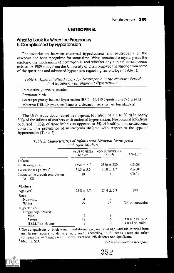
Neutropenia-239
NEUTROPENIA
What to Look for When the PregnancyIs Complicated by Hypertension
The association between maternal hypertension and neutropenia of thenewborn had been recognized for some time. What remained a mystery was theetiology, the mechanism of neutropenia, and whether any clinical consequencesexisted. A 1989 study from the University of Utah removed the shroud from someof the questions and advanced hypotheses regarding the etiology (Table 1).
Table I. Apparent Risk Factors for Neutropenia in the Newborn Periodin Association with Maternal Hypertension
Intrauterine growth retardationPremature birthSevere pregnancy-induced hypertension (BP > 160/110 C /proteinuria > 5 g/24 h)
Maternal HELLP syndrome (hemolysis, elevated liver enzymes, low platelets)
The Utah study documented neutropenia (duration of 1 h to 30 d) in nearly50% of the infants of mothers with maternal hypertension. Nosocomial infectionsoccurred in 23% of those infants as opposed to 3% of healthy, non-neutropeniccontrols. The prevalence of neutropenia differed with respect to the type ofhypertension (Table 2).
Table 2. Characteristics of Infants with Neonatal Neutropeniaand Their Mothers
NEUTROPENIA(N = 35)
NO NEUTROPENIA(N 37) P VALUE*
InfantsBirth weight (g)* 1550 ± 770 2530 ± 880 <0.001
Gestational age (wk)* 31.5 ± 3.5 36.0 ± 3.7 <0.001
Intrauterine growth retardation(n = 12)
10 2 <0.01
MothersAge (yr)* 25.8 ± 4.7 24.4 ± 5.7 NS
RaceNonwhite 4 7
White 29 28 NS vs. nonwhite
HypertensionPregnancy-induced
Mild 5 18
Severe 13 5 <0.002 vs. mildHELLP syndrome 8 3 <0.01 vs. mild
*The comparisons of birth weight, gestational age, maternal age, and the interval frommembrane rupture to delivery were made according to Student's t-test; the othercomparisons were made with Fisher's exact test. NS denotes not significant.Mean ± SD. Table continued on next page.
252

240Normoblastemia
Table 2. Characteristics of Infants with Neonatal Neutropeniaand Their Mothers (Cont.)
NEUTROPENIA(N 35)
NO NEUTROPENIA(N 37) P VALUE*
Hypertension (Com.)Chronic
Mild 2 2
Severe 5 8 NSHELLP syndrome 2 1 NS
DeliveryCesarean 32 13
Vaginal 3 24 <0.001 vs. cesarean
Interval between membranerupture and delivery (hr)s
12 ± 6 11 ± 6 NS
MedicationsMagnesium sulfate 14 25Magnesium + antihypertensives 10 7 NS
Concurrent illnessInfection 3 9
Diabetes mellitus 3 4 NS
I Mean ± SD.
Kinetic studies performed on cord blood of the neutropenic infants revealeddiminished neutropnil production as opposed to accelerated destruction orexcessive margination. The authors proposed two hypotheses to explain thediminished production: (1) deficiency of neutrophil-specific growth factors or (2)inhibition of neutrophil differentiation. The molecular mechanism remainsunknown. Until the mechanism is elucidated, therapy rests in recognition of thephenomenon and prophylactic antibiotic use as indicated.
Reference: Koenig JM, Christensen RD: Incidence, neutrophil kinetics, and naturalhistory of neonatal neutropenia associated with maternal hypertension. N Engl J Med321:557-562, 1989.
NORMOBLASTEMIA
The Cause of Nucleated Red Blood Cells in thePeripheral Blood in Children (Normoblastemia)
Childhood Diseases Associated with Normoblasternia
I. Hematologic/OncologicSevere anemia of any causeHemolytic anemiasIron deficiencyBlood lossMegaloblastic anemiaHistiocytosis
MyelofibrosisPreleukemiaLeukemiaLymphomaMyeloproliferative disordersSolid tumor invasion of bone marrow
Table continued on next page.
253

Nursemaid's Elbow-241
Childhood Diseases Associated with Normoblasternia (Cont.)
2. InfectionsBacterial infection (especially sepsis) OsteomyelitisTuberculosis Fungal
3. HypoxiaCongestive heart failure Asthma and other respiratoryCyanotic heart disease disease
4. OtherCollagen vascular diseases Diabetic ketoacidosisSarcoidosis Thermal injuryInflammatory bowel disease Vinca alkaloidsOsteopetrosis AspleniaGaucher's and other storage diseases Newborn (physiologic)Uremia ?'?Normal finding
From this long list the most common disorders include cardiac disease,hemolytic disorders, pulmonary disease, and bone marrow replacement.
Reference: Sills RH, et al: Am J Pediatr Hem Onc 5:173, 1983, with permission.
NURSEMAID'S ELBOW
Reducing Nursemaid's Elbow to Simple Terms
Subluxation or partial dislocation of the head of the radius is affectionatelytermed "nursemaid's elbow," because it typically arises subsequent to a sudden jerkor pull of a toddler's arm. Such a maneuver can be seen at any park, playground,or shopping mall on an hourly basis! More specifically, 90% of all cases ofnursemaid's elbow are due to the sudden longitudinal pull or traction at the wristwhen the elbow is fully extended and the forearm is pronated. It is typically seenin toddlers between the ages of 1 and 5 years, with a peak incidence amongchildren aged 15 to 30 months.
The child with nursemaid's elbow also tends to hold the arm slightly flexed atthe elbow and slightly pronated in order to avoid pain. Typically, the child whohas incurred such an injury will refuse to move the affected arm and complainsvociferously and painfully at any such attempt to manipulate the elbow, particularly
in supination and pronation.
The Anatomy of Nursornald's Elbow
Dislocation of the head of the radius is best understood by reviewing its anatomy(see figure). The radial head is wrapped by a cuff-like annular ligament. The annularligament attaches the radius to the ulna but also allows rotary motion of the radialhead. These ligamentous fibers combine with other ligaments of the elbow at theradiohumoral joint. Sudden longitudinal traction, when applied to a toddler's pro-nated forearm, stretches and tears the annular ligament at its distal attachment onthe radial neck. With continued traction, the annular ligament slips over the radialhead and, once the traction is released, these fibers can become caught betweenthe articular surface of the radial head and the capitellum. The result is pain.
25 4

242Nursemaid's Elbow
Normal annularligament
Tear withtraction
Entrappedfibers
The annular ligament covers the radial head and attaches the radius to the ulna. Withsudden longitudinal traction, the ligament stretches and tears. Fibers of the ligament arethen caught between the radial head and the capitellum.
A Prescription tor Red.zing Nursemaid's Elbow to a Mere Memory
X-rays are rarely indicated in this type of injury. Instead, following the maneu-vers illustrated in the figure below should correct the problem quickly and simply.
Supination maneuver Pronation maneuver
To reduce the injury, hold the elbow slightly flexed. Apply pressure over the radial headwith the thumb, then hold the child's wrist with the other hand and quickly move theforearm to either a supine or pronated position.
References: Nichols H H: Nursemaid's elbow: Reducing it to simple terms. ContemporaryPediatrics 5(5):50- 57, 1988.
Quan L, Marcuse E: The epidemiology and treatment of radial head subluxation. AmJ Dis Child 139:1194, 1985.
255
a

Nutrition-243
NUTRITION
Infant FoodsCalories and Their Distribution
When the infant is ready for strained or junior foods, it is important to beaware of the number of calories being provided and their source. The accom-panying table lists estimated calories derived from analysis of a variety ofproducts in each category.
Strained Foods
CATEGORY KCAL, 100 GM PERCENTAGE OF CALORIES
Protein Fat Carbohydrates
Juices 65 (45-98) 2 2 96
Fruits 85 (79 125) 2 2 96
Vegetables
Plain 45 (27-28) 14 6 80
Creamed 63 (42 94) 13 13 74
Meats 106 (86-194) 53 46 1
Egg yolks 192 (184-199) 21 76 3
High meat dinner 84 (63-106) 29 45 29
Desserts 96 (71 136) 4 7 89
Cereal 360 (349 393) 39 12 49
Cereal-fruit 85 (76-98) 18 6 76
Junior Foods
Fruits 85 (69-116) 2 2 96
Vegetables
Plain 46 (27 71) 12 7 81
Creamed 64 (45 72) 13 17 70
Meats 103 (88-135) 56 43 1
Sour dinner 61 (39-100) 15 27 58
Reference: FomonCompany, 1974, p 410,
SJ: Infant Nutrition, 2nd ed. Philadelphia, W.B. Saunderswith permission.
Are We Eating the "Wrong" Fruits & Vegetables?(Or What Could Be More Nutritious Than a Fresh Orange?)
Eighteen common fruits and vegetables are listed here, first in order of theirnutrient density and a second in order of their total nutrient contribution to theU.S. diet (density times tonnage). Our diets would improve considerably if weate more from the top of the lefthand list than from the bottom. There isnothing "wrong" with lettuce and oranges, of course, but notice how far downthey are in nutritional value. Note too how much more nutritious vegetables arethan fruits.
256

244Nutrition
NUTRIENT CONTRIBUTION OFCONCENTRATION NUTRIENTS TO DIET
CROP
BROCCOLI
SPINACH
BRUSSELS SPROUTS
LIMA BEANS
PEAS
ASPARAGUS
ARTICHOKES
CAULIFLOWER
SWEET POTATOES
CARROTS
SWEET CORN
POTATOES
CABBAGE
TOMATOES
BANANAS
LETTUCE
ONIONS
ORANGES
RANK
1
2
3
4
5
6
7
8
9
10
12
14
15
16
18
26
31
33
\
The "Skinniest" Cuts of Beef
CROP RANK
TOMATOES 1
ORANGES 2
POTATOES 3
LETTUCE 4
SWEET CORN 5
BANANAS 6
CARROTS 7
CABBAGE 8
ONIONS 9
SWEET POTATOES 10
PEAS 15
SPINACH 18
BROCCOLI 21
LIMA BEANS 23
ASPARAGUS 25
CAULIFLOWER 30
BRUSSELS SPROUTS 34
ARTICHOKES 36
Pro(, NI Allen Cleen, I nior,ilt ofCalifornia, I ).11, (Ro k 1978 78)
For those patients who have been placed on a low fat, low cholesterol diet butstill insist "real people eat beef," here are some of the "skinniest" cuts of beef youcan recommend:
Lean Beef
C I T OF BEEPFYI. OFROUND TOP LOIN ROUND TIP ROUND TIP rENDFRIDIN
TOPSIM DIN
Calories 143 cal 176 cal 157 cal 153 cal 179 cal 165 calTotal Fat 4.2 g 8.0 g 5.9 g 4.2 g 8.5 g 6 1 gSaturated Fat 1.5 g 3.1 g 2.1 g 1.4 g 3.2 g 2 4 gCholesterol 59 mg 65 mg 69 mg 72 mg 72 mg 76 mg
*Figures for a cooked and trimmed 3 oz serving; 4 oz uncooked beef yields a 3 oz cooked portion
References: USDA Handbook 8-13, 1990 (revised): U.S.R.D.A. National ResearchCouncil, 10th ed, 1989.
Stockman J: Journal Club Newsletter of the Northwestern Memorial Children'sHospital 4(3):9, 1989.
257

OBSESSIVE-COMPULSIVE DISORDER
Step on a Crack and You'll Break Your Mother's Back
Obsessive-compulsive disorder is a significant disturbance of childhood thathas not been well studied until recent years. The disorder appears to occur withgreater frequency than previously thought, usually in adolescence, and it is morecommon in boys than girls by at least 2 to I. It has a presentation very similarto adult OCD. Common obsessional thoughts concern contamination (e.g., feces,dirt, disease) and fears of wrongdoing; common compulsions are hand-washingrituals, grooming, and checking rituals.
Major Presenting Symptoms in 70 Consecutive Children and Adolescentswith Severe Primary Obsessive-Compulsive Disorder
COMPULSIONS
REPORTED SYMPTOMAT INITIAL INVERVIEV'NO. (%) OF PATIENTS'
Excessive or ritualized hand washing, showering, bathing, toothbrushing, or grooming
60 (85)
Repeating rituals (e.g., going in/out door, up/down from chair) 35 (51)
Checking (doors, locks, stove, appliances, emergency brake on car,paper route, homework, etc.)
32 (46)
Rituals to remove contact with contaminants 16 (23)
Touching 14 (20)
Measures to prevent harm to self or others 11 (16)
Ordering/arranging 12 (17)
Counting 13 (18)
Hoarding/collecting rituals 8 (11)
Rituals of cleaning household or inanimate objects 4 (6)
Miscellaneous rituals (e.g., writing, moving, speaking) 18 (26)
Concern with dirt, germs, or environmental toxins 28 (40)
Something terrible happening (fire/death/illness of self or lovedone, etc.)
17 (24)
Symmetry, order, or exactness 12 (17)
Scrupulosity (religious obsessions) 9 (13)
Concern or disgust with bodily wastes or secretions (urine, stool,saiiva)
6 (8)
Lucky/unlucky numbers 6 (8)
*Obsessions or compulsions are totaled, so the total exceeds 70.Table continued on next page.
245

246Occam's Razor/Odors of Disease
Major Presenting Symptoms in 70 Consecutive Children and Adolescentswith Severe Primary Obsessive-Compulsive Disorder (Cont.)
REPORTED SYMPTOMAT INITIAL INVERVIEW
COMPULSIONS NO. (c') OF PATIENTS*
Forbidden, aggressive, or perverse sexual thoughts, images, orimpulses
3 (4)
Fear might harm others/self 3 (4)
Concern with household items 2 (3)
Intrusive nonsense sounds, words, or music I (I)
From Swedo, et al: Arch Gen Psychiatry 46:337, 1989, with permission.
References: Swedo SE, Rapoport JL, Leonard H, et al: Obsessive-compulsive disorderin children and adolescents. Arch Gen Psychiatry 46:335 341, 1989.
Riddle MA, Scahill L, King R, et al: Obsessive compulsive disorder in children andadolescents: Phenomenology and family history. J Am Acad Child Adolesc Psychiatry29:766-772, 1991.
Oski FA: Principles and Practice of Pediatrics. Philadelphia, J.B. Lippincott, 1990, pp656 657.
OCCAM'S RAZOR
A Diagnostic PrincipleOccam's Razor
Without knowing it many clinicians apply Occam's razor to their diagnosticthinking. Occam's razor is a logical principle attributed to William of Occam,although it was used by some scholastic philosophers prior to him. The principlestates that a person should not increase, beyond what is necessary, the number ofentities required to explain anything, or that the person should not make moreassumptions than the minimum needed. This principle is often called the Law ofParsimony. Since the Middle Ages it has played an important role in eliminatingunnecessary elements from explanations. Remember William of Occam whenyou attempt to explain multiple symptoms in your patient with a single diagnosis.
ODORS OF DiSEASE
Unusual Odor as a Clue to Diagnosis
Can you smell a rat or sniff out a diagnosis? The sense of smell is not usedenough as part of the physical examination. Listed below are diseases associatedwith unusual odors.
Diseases Associated with Unusual Odors
DISEASE ENZYME DEFECT ODOR CLINICAL FEATURES TREATMIN1
Diabetes Lack of insulin or Acetone on Polyuria, polyphagia, Insulin admin-mellitus insulin activity breath, polydipsia, weight istration
fruity loss, acidosis, coma
259Table continued on next page
Odors of Disease-247
Diseases Associated with Unusual Odors (Cont.)
DISEASE ENZYME DEFECT ODOR CLINICAL FEATURES
Phenyl- Phenylalanineketonuria hydroxylase
Maplesyrupurined isease
Branched chaindecarboxylase
Oasthouse Defective transporturine of methionine,disease branched chain
amino acids,tyrosine, andphenylalanine
Odor of Isovaleryl CoAsweaty dehydrogenasefeet, Syn-drome I
Odor of Green acyldehy-sweaty drogenasefeet, Syn-drome II
Odor of cats Beta-methyl-syndrome crotonyl-CoA
carboxylase
Fish odor Unknownsyndrome
Fish odor Trimethylaminesyndrome oxida.se
Odor of Unknownrancidbuttersyndrome
Musty, Progressive mental"mousy," retardation, eczema,"horsey" decreased pigmenta-
tion, seizures,spasticity
Maple Marked acidosis, sei-syrup zures, coma leading to
death in first year ortwo of life or mentalsubnormality withoutacidosis or intermit-tent acidosis withoutmental retardation
Yeast-like; Mental retardation,dried- spasticity, hyperpnea,celery-like fever, edema
Sweaty feet
Sweaty feet
('at's urine
Recurrent bouts of aci-dosis, vomiting, dehy-dration, coma, aver-sion to protein foods
Onset of symptoms infirst week of life withacidosis, dehydration,seizures, and death
Neurologic disorderresembling Werdnig-Hoffmann disease,ketoacidosis, failureto thrive
Like dead Stigmata of Turner'sfish syndrome, neutro-
penia, recurrent infec-tions, anemia,splenomegaly
Like dead Unusual odor of sweat,fish skin and urine.
Normal development
Rancid Poor feeding, irritability,butter progressive neurologic
deterioration withseizures and death;hepatic dysfunction;possibly same asacute tyrosinosis
TREATMENT
Diet low inphenyl-alanine
Diet low inbranchedchain aminoacids; proteinrestrictionand/or thi-amine inlarge doses
Restrict methi-onine in diet
Restrict leucinein diet
High CHOdiet 0)
Low fat diet C?)
Leucine restric-tion (?)
Biotin admin-istration
Unknown
Elimination offish from thediet
Response todecreasedphenylalanineand tyrosineintake (?)
Reference: Mace JW, Goodman SI, Centerwall WR, et al: The child with an unusualodor. Clin Pediatr 15/57c-f2, 1976.
2 6 0

4.
248-0smolality
OSMOLALITY
Serum Osmolality
It is often important to estimate serum osmolality before the laboratorymeasurement becomes available. The following formula will make that estimationmore accurate.
The short cut approach is
Glucose BUNSerum osmolality = [Na(mEcy L.) = K (mEq/L)} x 2 +18 3
The normal value is 280.
EA
I know a babyWho smells like fresh muffinsWrapped in warm linenJust dried by the breezesBlown over the lilacsBrought out by the spring sunAnd back from the oceansWith Orient spices
261 BEST COPY AVAILABLE
41.1.

P
PAIN
The Precordial Catch Syndrome
In 1955 Miller and Texidor first described an entity in young adults they termed"precordial catch." It has proven to be a common entity. Perhaps as many as 50%of older adolescents and young adults will experience this sensation of a sudden,brief, nonradiating, periapical pain that is unrelated to exercise or exertion. Bothpatients and their parents are naturally concerned about heart disease, but youcan reassure them that the pain is of no cardiac significance. The precise causeof this painful sensation of "something being caught" and being forced to "freeze"in place is still unknown. The characteristics are described in the list below.
The Pain Itself
Onset:Location:
Duration:Description:
Localization:Radiation:
Sudden, unexpected, unprovoked.Left lower anterior aspect of chest; typically infra-apical at thesternal border.Brief (<3 minutes, usually 1 minute or less).Variable but superficial, knife- or needle-like, burning, stabbing,shooting, sharp, something catches.Site often localized by patient using one or more fingers.Nonradiating.
Related Factors
Respiration:
Exertion:Posture:
Taking a deep breath accentuates pain and makes patient'freeze." Forced inspiration, if possible, relieves pain.1nrelated to strenuous activity; usually occurs at rest.
Pain sometimes occurs when patient bends over or is slouched.Pain is relieved by stretching and straightening if possible.
'I
References: Reynolds JL: Precordial catch syndrome in children. South Med J 82:1228 1230, 1989.
Miller AJ, Texidor TA: "Precordial catch," a neglected syndrome of precordial pain.JA MA 159:1364-1365, 1955.
PALSY
Neonatal Phrenic Nerve PalsyThe "Belly Dancer's Sign"
Unilateral diaphragmat ic paralysis with or without brachial plexus injury maypresent in neonates as "respiratory distress." The chest roentgenogram may be
2 Q 2249

250Pancreatitis
misleading unless obtained in deep inspiration. Fluoroscopy is required todemonstrate paradoxical motion of the diaphragm on the involved side.
It should be remembered that the existence of diaphragmatic paralysis can berecognized by merely observing the movement of the umbilicus during therespiratory cycle. To perform this maneuver, note the position of the umbilicusat full expiration. Mark this position by placing your pen at the spot. Duringinspiration, the umbilicus can be seen to shift upward and toward the side of theparalyzed diaphragm. Other suggestive physical findings include unexplainedtachypnea without dyspnea, slightly decreased breath sounds on the paralyzedside, fine inspiratory rales on the paralyzed side if atelectasis is present, wideningof the subcostal angle on the affected side during inspiration, and flattening ofthe epigastrium on the side of the paralyzed diaphragm during inspiration. Themovement of the umbilicus is the sign most easily identified.
References: Nichols MM: Shifting umbilicus in neonatal phrenic palsy (the bellydancer's sign). Clin Pediatr 15:342, 1976.
Light JS: Respiratory shift in epigastric abdominal wall a physical sign seen with com-plete unilateral paralysis of the diaphragm in infants and children. J Pediatr 24:627, 1944.
From McMillan JA, et al (eds): The Whole Pediatrician Catalog. Philadelphia, W.B.Saunders, 1977, p 122, with permission.
PANCREATITIS
Acute Pancreatitk, in Children
Pancreatitis is an acknowledged but infrequently recognized cause of abdominalpain in children. The diagnosis is sometimes difficult. The following clinicaldescription may help.
Biology
Drugs/toxinsThiazidesSteroidsAzathioprineAlcoholTetracyclineSalicylazosulfapyridineChlorthalidoneFurosemideL-asparaginaseOral contraceptives
Trauma/surgery/child abuseBiliary tract disease
Choledochal cystStricture of the common bile ductCongenital stenosis of the ampulla
of VaterAnomalous insertion of the
common bile ductCholelithiasis/choleeystitis
InfectionMumps (even in the absence
of parotitis)Hepatitis B virusCoxsackie B5Epstein-Barr virusMycoplasmaInfluenza B
Diabetes mellitus (ketoacidosis)Perforated duodenal ulcerMiscellaneous
Hyperparathyroid ismSeptic shockCystic fibrosisPregnancyAcute porphyriaKwashiorkorHyperlipoproteinemia I and VScorpion bites
Idiopathic
f

Pancreatitis-251
Signs and Symptoms
I. Abdominal pain. Children may not localize the pain very well. It is usuallynoted to be in the upper quadrants or the periumbilical area. The pain is usuallyconstant, but it may be intermittent, and it may be made worse by eating. Theknee-chest position will usually relieve the pain.
2. Vomiting. Vomiting is aggravated by eating or drinking. It does aot relievethe pain.
3. Abdominal tenderness. Tenderness may be accompanied by guarding andrebound. Maximal tenderness is usually in the midepigastric region. Bowelsdunds may be normal, hypoactive, or absent.
4. Fever.5. Upper gastrointestinal hemorrhage. The hemorrhage is thought to result
from stress and may originate in the stomach, duodenum, or be caused by pene-tration of an ulcer into the head of the pancreas.
Laboratory Evaluation
1. Elevated bilirubin. This may be due to a stone in the common duct or toedema in the head of the pancreas.
2. X-ray changes. X-rays may document pleural effusion (most commonly onthe left side) and/or ascites. There may also be a dilated segment of small boweladjacent to the inflamed pancreas (sentinel loop). Isolated gaseous distention ofthe ascending colon and hepatic flexure may be present (colon cutoff sign). A CTof the pancreas often reveals the presence of a boggy, swollen organ.
3. Hyperglycemia. Diabetes mellitus may or may not follow pancreatitis.4. Hypocalcemia.5. Elevated serum amylase. The serum amylase usually begins to rise within
hours of the onset of symptoms. It usually peaks within the first 24 hours ofillness and returns to normal within 48 to 72 hours. Daily amylase determinationsare helpful in following patients. If the amylase rmains elevated for over twoweeks, a pseudocyst should be suspected. Amylase values may be normal inpatients with acute hemorrhagic pancreatitis.
6. Elevated serum lipase. These values tend to tbllow those of the serumamylase.
7 . Elevated urinary diastase. Timed urine collections are necessary for this
determination.8. Amylase clearance test. Amylase clearance may be elevated in patients with
severe burns or diabetic ketoacidosis, as well as in those with pancreatitis. It iscalculated from the following formula:
Cam (clearance of amylase) Amylase (urine) Creatinine (serum) x 100Ccr (clearance of creatinine) Amylase (serum) Creatinine (urine)
Treatmont
1 Relief of pain. This is best accomplished with meperidine given every threehours Its effect may be potentiated by promethazine.
2 Reduction of e.wcrine pancreatic secretion. The patient should fast, andintravenous fluids should be supplied. If intravenous fluids are required for morethan five days, parenteral alimentation should be initiated. A nasogastric tubf:
should be placed if the patient is nauseated or vomiting, or has an ileus.
252Panniculitis/Parasites
When oral feedings are initiated, they should consist of carbohydrates aloneinitially, because they cause the least stimulation to the pancreas.
Feedings should be restarted when abdominal tenderness has disappeared, anyileus has resolved, and urinary diastase or amylase clearance has become normal.
3. Treatment of shock and electrolyte abnormalities.Although anticholinergic drugs and antibiotics have been used in the treatment
of pancreatitis, their use has not improved the prognosis. Mortality may rangefrom about 20% with acute interstitial pancreatitis to about 80% with hemorrhagicpancreatitis.
Reference: Jordan SC, Ament ME: Pancreatitis in children and adolescents. J Pediatr91:211, 1977.
From McMillan JA, et al (eds): The Whole Pediatrician Catalog, Vol. 2. Philadelphia,W.B. Saunders, 1979, pp 240-243, with permission.
PANNICULITIS
What Do Popsicles and Horses Have in Common?
Both are associated with forms of cold-exposure panniculitis, characterized bysingle or multiple crops of tender nodules in the subcutaneous fat. Blood vesselsalso are usually affected, resulting in a histologic picture of fat-cell necrosis. Thenodules can be of a size less than 1 cm to over 10 cm across. The clinical pictureis one of reddish-purple discoloration and erythematous, enlarging nodules thatare often painful to palpation. The lesions are most obvious 24 to 48 hours afterthe cold injury. They are commonly confused with a cellulitis. The patients areafebrile and feel well, and the lesions subside without treatment in 2 to 3 weeks,leaving no permanent injury.
The popsicle form of panniculitis is produced by sucking on cold objects, suchas popsicles and ice cubes, or the lengthy application of the popsicle to any areaof the skin.
In equestrian cold panniculitis, the lesions appear on the outer thighs as aresult of prolonged horseback riding in freezing weather.
PARASITES
Sushi Eaters Beware!
H. L. Mencken used his father's method of separating the world's populationinto two groups: those who pay their bills and those who do not. An equally all-encompassing method might be those who eat sushi (the Japanese delicacy of rawtish) and those who don't. Recently, however, a report in the New EnglandJournal of Medicine appeared that might diminish the legions of raw fish eaters.The report noted a patient who presented with mild abdominal distension, directand rebound tenderness in the right lower quadrant, and an elevated white bloodcell count. After 6 hours observation, the patient's right lower quadranttenderness worsened, and she was taken to the operating room for emergencyappendectomy. At operation the appendix appeared grossly normal and apinkish-red, sinuous worm was found moving onto the surgical drapes just prior
2S5

Parvovirus-253
to surgical closure of the wound. The worm was identified as an early 4th stagelarva of the genus Eustrongylides, a nematode parasite of fish-eating birds thatis frequently found in raw fish. The patient's medical history was remarkable foreating sushi, prepared at her friend's house, the day before.
This case only adds to the growing list of parasitic diseases that can beacquired by ingesting infected fish that is raw or insufficiently cooked, smoked,salted, or marinated. The clinical presentation varies depending upon where theparasite has localized (e.g., the stomach or the intestines). Dominating features ofthese parastic infections include acute abdominal symptoms such as discomfort,guarding, nausea, severe epigastric pain, and rebound tenderness. An elevatedeosinophil count (30 to 40%) is also suspicious for a parasitic infection.
There are a number of parasites that have been reported to be indigenous tomany marine fish typically caught off the coasts of the U.S., Japan, and Europe(e.g., salmon, cod, whiting, herring, and haddock). They include:
Anisakis simplex, the most common parasitic disease of sushi eaters.Pseudoterranova (formerly Phocanema) decipiensContracecum speciesHeterophyes heterophyesDiphyllobothrium latum (broad or fish tapeworm)Nanophyetus salmincolaEustrongylidesGambierdiscus toxicus (ciguatera fish poisoning)
In order to avoid these parasitic infections, the CDC in Atlanta suggests thatyou cook seafood before consuming it; heating fish to 65°C for 10 minutes appearsto kill most worms. Freezing fish for a minimum of 5 days at 200 C (-4°F) alsokills most parasitic species. Other methods of preparing raw fish, such as briningor marinating, cannot be relied upon to destroy helminths. Visual inspection andcandling (holding the fish up to light) seem to be most reliable in the hands of anexperienced sushi chef.
Other fish-related diseases include scombroid (histamine fish poisoning) anda number of viral and bacterial infections from raw shellfish ingestion.
References: Wittner M, Turner TW. Jacquette G, et al: Eustrongylidiasis---a parasiticinfection acquired by eating sushi. N Engl J Med 320:1124-1126, 1989.
Schantz PM: The dangers of eating raw fish. N Engl J Med 320:1143-1145, 1989.Eastaugh J, et al: Infectious and toxic syndromes from fish and shellfish consumption.
Arch Intern Med 149:1735-1740,1989.Morrow JD, et al: Evidence that histamine is the causative toxin of scombroid-fish
poisoning. N Engl J Med 324:716 -720, 1991.
PARVOVIRUS
Beyond Fifth Disease: The Clinical Spectrum of Paryoyirus B19
Once the viral etiology of erythema infectiosum, or fifth disease, wasdetermined to be numan parvovirus B19, the true clinical spectrum of B19infection could be investigated. We now know that B19 infection in healthychildren can occur without the usual facial ("slapped cheek") rash or subsequentreticular rash on the extremities and trunk. We also know that BI9, when it
296
254Pelvic Inflammmatory Disease
infects certain populations, may result in prolonged disease with significantconsequences. It is the ability of parvovirus B19 to infect and lyse red blood cellprecursors that underlies its more serious effects among these patients. Thefollowing table lists the patient populations likely to suffer complicated infectionwith parvovirus BI9.
Patient Populations with Complicated Parvovirus B19 Infections
PATIENT POPULATION MANIFESTATION OF BI9 INFECTION
Healthy adults
Chronic hemolytic anemia
Immunodeficiency or immunosuppressionFetus
Arthropathy, with or without rashTransient aplastic crisisChronic, persistent, anemia
Fetal death asrociated with hydrops fetalis
It is important to remember that 40-60% of U.S. adults are immune toparvovirus B19 by virtue of previous infection. Thus many women of child-bearing age are not susceptible, even if exposed while pregnant. In addition, fetalhydrops and subsequent death result in less than 10% of pregnancies duringwhich the mother is known to have been infected. Congenital anomalies amonginfants born to mothers infected with B19 during pregnancy have not beenidentified.
There is no specific therapy available for parvovirus BI9 infection, althoughintravenous immunoglobulin has been used successfully to control persistentinfection in immunocompromised patients.
Reference: Anderson LI: Human parvovirus BI9. Pediatr Ann 19:509-513, 1990.
PELVIC INFLAMMATORY DISEASE
The Importance of the Pelvic Examination in SeparatingPID from Urinary and Gastrointestinal Disorders
Pelvic inflammatory disease (PID) is a serious problem among sexually activeadolescent women. Over 1,000,000 women of reproductive age contract PID eachyear; teenagers make up 20% of these cases. The incidence of PID amongadolescents rises each year as more and more teenagers become sexually activeand engage in unprotected sexual intercourse. The short-term complications ofPID include perihepatitis (Fitz-Hugh-Curtis syndrome) and tubo-ovarianabscesses, while long-term sequelac include an increased incidence of infertilityand ectopic pregnancies. For example, women with a history of one or moreepisodes of PID were found to have an involuntary infertility rate of 21%(compared to 3% in controls) and a sixfold increase in ectopic pregnancies evenwith antimicrobial intervention. In light of these complications the successfulP1D intervention that minimizes the patient's risk must include (1) earlyrecognition, (2) the use of broad spectrum antibiotics to treat the polymicrobialnature of the disease, (3) an emphasis on careful clinical reevaluation of thesuspected PID patient within 24 hours to detect antibiotic failure or a misdiag-nosis, and (4) evaluation and treatment of the patient's sexual partners.
2t37

Pelvic Inflammatory Disease-255
Unfortunately, the most frequently occurring symptoms of PID lack specificityin their delineation from other disease processes involving the reproductive,urinary, and gastrointestinal tracts, as depicted in the table below:
Common Clinical Symptoms of PID by Organ System
SYMPTOM REPRODUCTIVE URINARY GASTROINTESTINAL
Lower abdominal pain + + +Urinary symptoms + + +Intermenstrual bleeding + ± +Dysmenorrhea + _
Secondary amenorrhea + + ±Nausea/vomiting + .1- 4-
Fever + + +Malaise + 4- +Dyspareunia + + +
The history alone will lead the physician potentially to misdiagnose manywomen with acute PID, and this can put them at great risk for the short-term andlong-term sequelae. Although laparoscopy is the definitive means of determiningthe presence of acute PID, it is not feasible to perform the procedure on everyadolescent with lower abdominal pain because of its risks, costs, and the avail-ability of manpower. The performance of a careful pelvic examination, however,in context with the patient's history and chief complaints, can be useful in at leastseparating reproductive tract disease from urinary and gastrointestinal complaints.
Cotnmon Clinical Signs in Acute Lower Abdominal PainAmong Adolescent Females by Organ System
SYMPTOM REPRODUCTIVE URINARY GASTROINTESTINAL
Lower abdominal tenderness + + +Pcrineal rash +Vaginal discharge +Cervical mucopus +Uterine bleeding +Cervical motion tenderness + +Uterine tenderness or mass +Adnexal tenderness or mass +
Reference: Shafer MA, Sweet RL: Pelvic inflammatory disease in adolescent femalesPediatr Clin North Am 36:513 -532, 1989.
The Criteria for the Diagnosis of Acute PID
Given the limitations of history-taking, the pelvic examination, and laparoscopyfor PID, the following clinical diagnostic criteria are offered:
All three should he present:
I. Lower abdominal tenderness2. Cervical motion tenderness3. Adnexal tenderness (may be unilateral), plus (see following list)
.2f8

256Pertussis
One of the following should be present:
1. Temperature 38° C2. White blood cell count 10,500/mm33. Purulent material obtained by culdocentesis4. Inflammatory mass present on bimanual exam ± sonogram5. ESR > 15 mm/hr6. Evidence of N. gonorrhoeae and/or C. trachomatis in the endocervix.
a. Gram stain with gram-negative diplococcib. Monoclonal antibody for C. trachornatis
7. > 5 white blood cells per oil-immersion field on Gram's stain of endocervicaldischarge.
Reference: Sweet RC: r'elvic inflammatory disease and infertility in women. Infect DisClin North Am 1:199-215, 1987.
PERTUSSIS
Whooping Cough and the White Blood Cell Count
The white blood cell count can often be helpful in diagnosing pertussis. Amarked leukocytosis (WBC > 25,000/mm3) with a differential demonstrating thepresence of 50 to 90% lymphocytes is usually considered presumptive evidence ofpertussis in infants and children with a cough. (Definitive diagnosis is made byrecovery of B. pertussis, B. parapertussis, or B. bronchiseptica from a Dacron orcalcium alginate nasopharyngeal swab.)
Despite the availability of vaccines, pertussis is unfortunately still very muchwith us. It is not generally appreciated that infants under 6 months of age oftendo not display the aforementioned degree of leukocytosis and that the white cellcount may be normal during the prodromal phase of the illness.
The table below illustrates the range of white cell counts by age in patientswith pertussis.
White Cell Counts by Age in Patients with Pertussis
TOTAL WBC0 TO 6
MONTHS6 MONTHS TO
2 YEARS2 TO 5
YEARS 5 YEARSTOTALGROUP
5,000 to 15,000 38 6 14 31 23
15,000 to 25,000 31 49 32 31 36
25,000 to 50,000 29 33 45 31 34
>50,000 2 12 9 7 7
The percentage of lymphocytes in this group of patients varied from 27 to 99% with amean of 70.4%.
Patients with a leukemoid reaction (WBC > 50,000) are more likely to havepulmonary complications such as atelectasis and pneumonia.
The relation of thc total white cell count to the stage of the illness is illustratedin the following table.
2f9

Phagocytes-257
Relation of the Total White Cell Count to the State of the Illness
WHITE CELL COUNTCATARRHAL STAGE PAROXYSMAL STAGE
(WEEKS I -2) (() (WEEKS 3- 5) (%)
5,000 to 15,000 28 12
15,000 to 25,000 42 34>25,000 30 54
Reference: Brooksaler F, Nelson JD: Pertussis. Am J Dis Child 114:389, 1967.
PHASOCYTES
Congenital and Acquired Phagocytic Defects
Phagocytic cells are required to destroy microorganisms that would otherwiselead to disease in the host. The functions of these polymorphonuclear cells maybe divided as follows: (1) adherence to the vascular endothelium; (2) chemotaxisor the recognition of and migration to a specific chemical stimulus; (3) phago-cytosis; and (4) the killing of the ingested microorganism. Deficiency in serumopsonins (antibody or complement) or a deficiency in the number of phagocytes(neutropenia or asplenia) can also yield phagocytic dysfunction. Defects in one ormore of these roles can lead to recurrent and severe infections. The phagocyticdefects can either be congenital or acquired (in association with another disease).The congenital causes, which are extremely rare in frequency, include defects ofcell movement (e.g., hyperimmunoglobulinemia E syndrome, actin dysfunction,and glycoprotein deficiency) and defects of microbial activity (e.g., chronicgranulomatous disease, glucose 6-phosphate dehydrogenase deficiency, myelo-peroxidase deficiency, and Chediak-Higashi syndrome). Acquired or secondaryphagocytic defects are far more common as detailed in the table below:
Acquired or Secondary Phagocytic Defects
PRIMARY DISEASE OR CONDITIONS ASSOCIATED CELLULAR DEFECT
MalnutritionHypophosphatemiaDiabetes mellitusLeukemiaAcute alcohol intoxicationThermal injuryRheumatoid arthritisSystemic lupus erythematosisInflammatory bowel diseaseSystemic infectionsIron deficiencyPregnancySteroidsViral infection
Chemotaxis, phagocytosis, killingPhagocytosis, killingChemotaxis, adherenceAdherence, chemotaxis, phagocytosis, killingChemotaxisChemotaxis, killingChemotaxis, phagocytosis, opsoninsChemotaxisChemotaxis, phagocytosis, opsoninsChemotaxisChemotaxis, killingChemotaxis, killingChemotaxi, opsonizationChemotaxis, killing
Reference: Bell JB, Dougals SD: Phagocyte functions and defects: A ten-year update.Pediatr Ann 16:379-389, 1987.
270
258Phobias
PHOBIAS
Needle Phobia
A recent case report of a physician who displayed an involuntary fear ofreceiving injections and having his blood drawn reminds us of the distinct entitypsychiatrists call "needle phobia." (The 5-dollar word is belonephobia, from theGreek belone, meaning needle.)
Typically, phobias manifest themselves with the physical sign of transienttachycardia. Patients with needle or blood injury phobias, on the other hand,experience a diphasic cardiovascular response: tachycardia followed by bradycar-dia, hypotension, nausea, diaphoresis, vertigo, and syncope. On rare occasions,shock and death have been reported.
It is difficult to ascertain just how many children and adults have bona-fideneedle phobia as opposed to simply not liking needles inserted into their bodies.Some experts estimate it at 5% of the population. Whether or not patients withtrue needle phobia will even present to a medical clinic or simply avoid all formsof health care is a different question.
Psychiatrists and evolutionary biologists have hypothesized that needle phobiamay have a selective value over more traditional "fight or flight" responses tobodily harm. This is to say, there may be protective benefits in a fainting responseor adverse cardiovascular reflex when confronted with an aggressive internarmed with a needle. On the other hand, a fainting toddler might be preferableto a screaming one, especially when one is compelled to obtain blood!
References: Ellinwood EH, Hamilton JG: Case report of a needle phobia. J FamilyPrac 32:420 -422, 1991.
Marks 1: Blood-injury phobia: A review. Am J Psychiatry 145:1207 -1213, 1988.
Photophobia
Photophobia is an abnormal intolerance for light, usually a result of inflam-mation of the iris and ciliary body. It is not to be confused with photosensitivity,which is also associated with a long list of diseases, usually with skin signs. Abetter term for photophobia is photodysphoria, but the latter is seldom used.
Although the diseases associated with photophobia are often obvious andrelatively easily diagnosed (e.g., viral conjunctivitis, measles, or bacterial meningitis),there are other, more subtle conditions that must be considered when the primarydiagnosis is not so obvious. Some of these associations are listed below.
More Common Associated Infections
Measles Viral conjunctivitisCoxsackie B infection Arbovirus infectionLymphocytic choriomeningitis Bacterial meningitis
Less Common Associated Infections
Phlyctenular conjunctivitisYellow feverPsittacosis infections
271
Rickettsial infections(Rocky Mountain spotted fever,murine typhus)

Noninfectious Associations
Infantile glaucomaAlbinismVitamin A deficiencyKeratitis (e.g., Reiter's syndrome)Erythropoietic porphyriaAcute cerebellar ataxiaChédiak-Higashi syndromeAniridiaCystinosis
Phobias-259
MigraineCorneal ulcerHysteria (in older child)Arsenic poisoningMercury poisoningDrug toxicity
TrimethadioneEthosuccimidePAS
References: Wilson JD, et al (eds): Harrison's Principles of Internal Medicine, 12th ed.New York, McGraw Hill Book Company, 1991.
Illingworth RS: Common Symptoms of Disease in Children, 8th ed. Oxford, BlackwellScientific Publications, 1984.
Rudolph AM (ed): Pediatrics, 19th ed. New York, Appleton-Lange, 1991.
School Phobia
Vague physical complaints are frequently heard in the pediatrician's office.When combined with normal physical and laboratory findings and poor schoolattendance because of the complaints, the child is often found to have "schoolphobia," a descriptive term for anxiety over leaving home in the 6 to 10 year oldgroup.
Once a significant child-teacher conflict or fear of harassment by otherchildren has been ruled out, the physician's immediate goal should be the returnof the child t.o full school attendance. Steps in this direction to be discussedduring the visit are listed below.
I. Do a thorough physical examination and pertinent laboratory studies as soon aspossible. The child should then be given an unequivocal "clean bill of health." The findingsshould be conveyed to the parents along with a brief but sympathetic explanation aboutthe reality of symptoms caused by anxiety or depression.
2. The parents should be gently but firmly convinced that immediate return of thechild to school is essential. The parents must insist on the child's return to school for thisstep to be effective. Delay in return to school makes it increasingly difficult for the childto go back.
3. What to expect and what to do on school mornings should be reviewed with themother. She should not ask the child how he feels. If he is up he should go to school, evenif he is late or has missed the school bus. If he comes home at lunch he should be returned.If the child says he is ill, the mother should do one of two things. If questionably or mildlyill, he can be sent to school. If the mother feels the child is truly ill, he should be seen bythe physician early that same morning. The child is not to stay at home without seeing aphysician.
4. The person to be in charge of taking the child to school if he refuses to go shouldbe clarified. This may be one of the parents, another relative, school social worker, orother responsible adult.
5. The school principal should be contacted by the physician, who can ask the school'scooperation in helping the child return to school. This is especially important if the childhas a real or imaginary fear regarding some condition at school. The school nurse shouldalso be contacted if she has been sending the child home for minor illness. She should beasked to have the child rest in her office for a time rathcr than send him home
2",2

260Pig menturia
6. Weekly visits for several weeks are important for follow-up. A final visit severalmonths later will allow long-term assessment.
Failure of this program, if conscientiously carried out by parents, physicianand school personnel, suggests the necessity of psychiatric referral to explore thesevere dependency problems that are often present.
Prevention of school phobia and related dependency problems can be aided bythe encouragement of independence at appropriate times during infancy andpreschool problems. The following milestones of independence may be usefulguidelines.
WHEN CHILD SHOULD
By 6 months
By 2 years
By 3 years
As soon as ready
By 3 -4 years
By 4-5 years
be left with baby sitter while parents have evenings out.
be left home, while awake, with baby sitter.experience being left somewhere other than his home.
be allowed to feed, dress, and wash himself.be allowed to play in yard by himself.
be allowed to play in neighborhood by himself.
Reference: Schmitt BD: Pediatrics 48:433, 1971.From McMillan JA, et al: The Whole Pediatrician Catalog, Vol. 1. Philadelphia, W.B.Saunders, 1977, pp 44-45, with permission.
PIGMENTURIA
Myoglobinuria, Hemoglobinuria, or Porphyria?
The passage of large quantities of pigment in the urine often producesdiagnostic confusion. Many substances may color the urine, but few mimic theappearance of hemoglobin. Hemoglobinuria must be distinguished from myoglobinor porphyrin compounds. Both myoglobin and hemoglobin will give positiveresults on the commonly employed dipstick (Labstix) for heme. The followingtable should provide a guide in the initial differential diagnosis of the three majorcauses of pigmenturia.
Physical and Biochemical Features of the Pigmenturias
PHYSICAL EXAMINATION MYOGLOBINURIA HEMOGLOBINURIA PORPHYRIA
M usclesWeakness -I- ±Pain ± ±Edema -I-
Neuropathy (peripheral and +autonomic)
CNS dysfunction ±Skin lesion ±Abdominal pain Rare
273
Table continued on next page.
a

Pig menturia 261
Physical and Biochemical Features of the Pigmenturias (Cont.)
PHYSICAL EXAMINATION MYOGLOBINURIA HEMOGLOBINURIA PORPHYRIA
LABORATORY TESTS
UrineColor Brown Red-brown BurgundyBenzidineHematest-orthotoluidine80% (NH4)2SO4PPT ?
80% (NH4)2SO4SU PER +PorphobilinogenSpectrophotometry (a band) 582 (oxymyo) 577 (oxyhemo) 594 to 624*Taurine Increased NormalImmunodiffusion Specific Specific
SerumAppearance Clear Pink ClearHaptoglobin Normal Low NormalCreatine phosphokinases Marked increase Normal NormalCarnitine Increased Normal NormalImmunodiffusion Specific SpecificTriglycerides I In specific defects
*Varies with type.
The clinical circumstances may provide the most help in defining the cause ofpigment in the urine. The following conditions are associated with myoglobinuria.
Causes of Myoglobinemia and Myoglohinuria
Trauma and ischemic disease"Crush" syndromeArterial ischemia of extremities,
myocardial infarctionPressure necrosis (comatose states)Surgical procedures (orthopedic,
vascular, cardiac)
Exertional statesExertion in otherwise normal
individuals (military recruits)Convulsive disorders
Metabolic disordersAlcoholic myopathyAnesthetic associated syndromes (malignant hyperthermia)Defects in carbohydrate metabolism (McArdle's disease,
phosphofructokinase deficiency, syndrome of abnormal glycolysis)Defect in lipid metabolism (deficiency of carnitine, palmityl transferase)HypokalemiaToxins (heroin usei 's rhabdomyolysis, quail eater's disease,
Haff disease, snake and hornet venoms)
Hereditary myopathies ofunknown cause
Myositis syndromesDermatomyositis, poly-
myositis, systemic lupuserythematosus
Other factorsInfections?
Idiopathic rhabdornyolysis
Most hospital laboratories will make a definitive differentiation betweenmyoglobin and hemoglobin by performing a cellulose acetate electrophoresis.Unfortunately, if it is not between 8:00 A.M. and 5:00 P.M., you may be out of
274

262Pleural Effusion
luck getting the laboratory to perform this test. An alternative test, which ispresumptive of the presence of myogiobin, is based on differential solubility inammonium sulfate. This is based on the principle that myoglobin is soluble in80% saturated ammonium sulfate solution, whereas hemoglobin is not.
The test is performed as follows:
I. Clear the urine specimen by centrifugation or filtration.2. Add 2.8 gm of (NH4)2SO4 to 5 ml of urine, making an 80% saturated solution of
(NH4)2SO4. Allow the solution to stand for 5 minutes, then filter.3. If myoglobin is in the urine, it will remain in solution. If hemoglobin is in the urine.
it will precipitate and will be detected on the filter paper.
A presumptive positive test should be followed by an electrophoresis whenavailable. Whatever test is used, the urine must be absolutely fresh.
References: Robotham JL, Haddow JD: Rhabdomyolysis and myoglobinuria inchildhood. Pediatr Clin North Am 23:279, 1976.
Cifuentes E, Norman ME, Schwartz MW, et al: Myoglobinuria with acute renal failurein children. Clin Pediatr 15:63, 1976.
Rosse WF In Williams WJ, Beutler E, Erslev AJ, Rundles RW (eds): Hematology,2nd ed. New York, McSraw-Hill, 1978, p 613.
From McMillan JA, et al: The Whole Pediatrician Catalog. Philadelphia, W.B.Saunders, 1977, p 310. with permission.
PLEURAL EFFUSION
Pleural Effusions: Exudates or Transudates?
It seems the question always comes up. Is it a transudate or an exudate? Onlyan analysis of the fluid obtained by a thoracentesis can answer the question andeven then you can't always be certain.
A transudate occurs when the mechanical factors influencing the formation orreabsorption of pleural fluid are altered. Decreased plasma oncotic pressure, andelevated systemic or pulmonary hydrostatic pressure are alterations that commonlyproduce transudates. In contrast, an exudate results from inflammation or otherdiseases of the pleural surface. Common conditions producing an exudate include:pneumonia, tuberculosis, pancreatitis, pulmonary infarction, and systemic lupuserythematosus.
Reliance on a single test to distinguish an exudate from a transudate will fre-quently be misleading. In the past, the measurement of pleural fluid protein, orspecific gravity, or cell count has been employed as a diagnostic aid. Any singletest will give unacceptably high "false positive" or "false negative" results.
The use of the following three tests will enable you to correctly classifyvirtually all pleural effusions:
Pleural fluid protein 0.5 (suggests exudate)Serum protein
2. Pleural fluid LDH 200 IU (suggests exudate)
Pleural fluid LDH3. 0.6 (suggests exudate)
Serum LDH
275

Poisoning 263
The presence of two of these criteria strongly suggests a diagnosis ofexudatethe presence of all three virtually assures it.
Some other helpful facts include:About 80% of transudates will have a white cell count of less than 100/mm3,
while 80% of exudates will have white cell counts above 1000/mm3.Pancreatitis often produces a left-sided pleural effusion.If congestive heart failure is associated with a unilateral effusion, it is usually
ght-sided.
Reference: Light RW, MacGregor MI, Luchsinger PC, Ball WC Jr: Pleural effusions:The diagnostic separation of transudates and exudates. Ann Intern Med 77:507,1972.
Adapted from McMillan JA, et al: The Whole Pediatrician Catalog, Vol. 2. Philadelphia,W.B. Saunders, 1979, pp 221-222.
POISONING
An Unknown Poison
It is axiomatic that the most useful information in an accidental poisoning isthe label on the container. In over 90% of poisonings, the poison is known fromthe label or other source. However, situations sometimes arise where a possiblepoisoning has occurred, but the amount and nature of the ingested substance areunknown. Features that should suggest poisoning in an ill child include:
I. Abrupt onset of illness2. Child's age 1 to 4 years3. History of previous ingestion4. Multiple nrgan system involvement that does not fit single disease
A combination of symptoms will sometimes suggest the drug or poison involved:
Symptoms and Signs Possible Poison
Agitation, hallucinations, dilated pupils, brightred color to the skin, dry skin, and fever
Marked activity, tremors, headache, diarrhea,dry mouth with foul odor, 3weating, tachycardia,arrhythmia, dilated pupils
Slow respirations, pinpoint pupils, euphoria, or comaSalivation, lacrimation, urination, defecation, miosis,
and pulmonary congestionSleepiness, slurred speech, nystagmus, ataxiaHypernea, fever, and vomitingOculogyric cris:s, ataxia, and unusual posturing
of head and neckNausea, vomiting, sweatiness, and pallor are early
manifestations; late manifcstations includestupor and signs of liver failure
Atropine-like agentsLSD
Amphetamines
OpiatesOrganic phosphates or
poison mushroomsBarbiturates or tranquilizersSalicylates
Phenothiazines
Acetaminophen
The following list expands on signs and symptoms and the toxins with whichthey may be associated.
2'76

264Poisoning
Ataxia
AlcoholBarbituratesBromidesCarbon monoxideDiphenylhydantoin
Convulsions and muscle twitching
AlcoholAmphetaminesAntihistaminesBoric acidCamphorChlorinated hydrocarbon
insecticides (DDT)CocaineCyanide
Coma and drowsiness
AlcoholethylAntihistaminesBarbiturates and other hypnoticsCarbon monoxide
Paralysis
BotulismHeavy metals
Pupils
PinpointMushrooms (muscarine type)Narcotic depressants (opiates)
DilatedAmphetaminesAntihistaminesAtropineBarbiturates (coma)Cocaine
Nystaginus on lateral gazeBarbituratesMinor tranquilizers (meprobamate, benzodiazepine)
HallucinogensHeavy metalsOrganic solventsTranquilizers
LeadOrganic phosphate insecticidesPlants (lily of the valley, azalea,
iris, water hemlock)SalicylatesStrychnineWithdrawal from barbiturates,
benzodiazepine (Valium,Librium), meprobomate
CocaineNarcotic depressants (opiates)SalicylatesTranquilizers
Plants (conium in poison hemlock)Triorthocresyl phosphate
Organic phcsphate insecticides
EphedrineLS DMethanolWithdrawal-narcotic depressants
Pulse rate
SlowDigitalisLily of the valleyNarcotic depressants
RapidAlcoholAmphetamines
2 7 7
AtropineEphedrine

Respiratory alterations
RapidAmphetaminesBarbiturates (early)Carbon monoxideMethanolPetroleum distillatesSalicylates
Slow or depressedAlcoholBarbiturates (late)Narcotic depressants (opiates)Tranquilizers
Mouth
SalivationArsenicCorrosiveMercuryMushroomsOrganic phosphate insecticidesThallium
Breath odor
Acetone: acetone, alcohol (methyl,isopropyl), phenol, salicylates
Alcohol: alcohol (ethyl)Bitter almond,,: cyanideCoal gas: carbon monoxide
Skin color
Jaundice (hepatic or hemolytic)AnilineArsenicCarbon tetrachlorideCastor beanFava beanMushroomNaphthaleneYellow phosphorus
Violent ernesis often with hematemesis
AcetaminophenAminophyl!ineBacterial food poisoningBoric acidCorrosives
Poisoning-265
Wheezing and pulmonary edemaMushrooms (muscarine type)Narcotic depressants (opiates)Organic phosphate insecticidesPetroleum distillates
ParalysisOrganic phosphate insecticidesBotulism
DrynessAtropineAmphetaminesAntihistaminesNarcotic depressants
Garlic: arsenic, phosphorus, organicphosphate insecticides, thallium
Oil of wintergreen: methyl salicylatePetroleum: petroleum distillatesViolets: turpentine
CyanosisAniline dyes NitritesCarbon monoxide StrychnineCyanide
Red flushAlcoholAntihistaminesAtropineBoric acid
FluorideHeavy metalsPhenolSalicylates
Carbon monoxideNitritesRifampin
,
266Poisoning
Abdominal colic
Black widow spider biteHeavy metalsNarcotic depressant withdrawal
Oliguria-anuria
Carbon tetrachlorideEthylene glycolHeavy metalsHemolytic poisons (naphthalene,
plants)
MethanolMushroomsOxalatesPetroleum distillatesSolvents
Reference: Mofenson HC, Greensher J: The unknown poison. Pediatrics 54:336, 1974.
Carbamate and Organophosphate Poisoning
Despite many parents' valiant attempts, "Mr. Yuk" does not always detercurious children. Carbamates and organophosphates remain ubiquitous compo-nents of insecticides, and their toxic effects can reach the nervous sytem throughinhalation, absorption, and ingestion.
The carbamates and organophosphates are anticholinesterases that lead to theaccumulation of unhydrolyzed acetylcholine at the receptors. The result is con-tinued stimulation and, ultimately, paralysis of cholinergic transmission. Theorganophosphates penetrate the central nervous system and will show centraleffects that the carbamates do not.
Clinical Features of Carbamate and Organophosphate Insecticide Poisoning
Muscarinic Nicotinic
"Sludge"SalivationLacrimationUrinationDefecationGastrointestinal pain
& crampingEmesis
Miosis: if present, look forhyperactive bowel sounds
Muscle symptomsFasciculationsCramps and fatigueLoss of deep tendon
reflexesParalysis
TachycardiaHypertension
Central nervous system(organophosphates)
Severe headacheTremorAtaxiaRestlessnessSlurred speechGeneral weaknessSeizuresComaCardiorespiratory depression
RocognItion and Treatrnont
The muscarine effects generally precede the nicotinic effects. If you have a caseof suspected carbamate or organophosphate poisoning, the patient will beatropine-refractory: atropinization should occur in the nonpoisoned patientwithin 5 20 minutes after a dose of 0.05 mg/kg for a child, or I 2 mg for an adult.
Prior to the atropine test, the ABCs of Basic Life Support are mandatory. Thepatient should be thoroughly disrobed and rleansed due to possible continuedabsorption through dermal contact. If ingestion occurred, initiaie ipecac/lavageand charcoal.
279

Polyposis-267
Atropine is the cornerstone of therapy. Give the child 0.05 mg/kg as needed;give the adult 0.4 to 2.0 mg IV every 15-30 minutes until the patient cannot spit.In general, a 6- 12 h course of atropine is necessary. With carbamate poisoning,the cholinesterase complex is reversible. This is not the case with organophos-phates, so additional therapy with pralidoxime, a cholinesterase regenerator, isnecessary.
Reference: Mack RB: Carbamate poisoning: A Kafkaesque nightmare. Contemp PedsOctober:89-91,1985.
POLYPOSIS
Familial Polyposis
Familial polyposis is an autosomal dominant disorder notable for developmentof multiple adenomatous polyps in the colon and rectum. The polyps usually donot become apparent until after puberty. The incidence of familial polyposisranges from 1 in 7,000 to 1 in 10,000 births. The risk of developing colon cancerin affected individuals approaches 100% by age 55. Patients with familialpolyposis tend to seek medical attention because of either a family history ofpolyposis or symptoms of abdominal discomfort, rectal bleeding, and diarrhea.Frequently, the abdominal symptoms do not appear until 10 years after thedevelopment of polyps.
Clinical Fea lures
I. Autosomal dominant inheritance (rare spontaneous mutations have beenreported).
2. Onset in adolescence3. Onset of symptoms begin about 10 years after appearance of polyps.4. Multiple colonic adenomatous polyps5. Associated extracolonic lesions
a. Epidermoid cysts (usually on head, neck, and trunk)b. Subcutaneous fibromas (usually on the scalp, shoulders, arms and back)c. Desmoid tumors of the abdominal wall, mesentary, and retroperitoneumd. Osteoma (symptoms a-d in association with polyposis is also known as
Gardner syndrome).e. Sebaceous cystsf. Gastric and duodenal polypsg. Congenital hypertrophy of the retinal pigment epithelium (multiple
pigmented patches on one or both fundi)h. Abnormal dentition (including odontomas, dentigenous cysts, and
unerupted, missing, or supernumerary teeth)6. High risk of colon cancer
a. 100% risk in untreated patientsb. Early age of onset (median age is 39 years)c. Cancers arise from adenomatou polypsd. Synchronous colon carcinomas are common.e. Metachronous colon carcinomas are common.2

268Polyuria
Protocol for Screening Patients at Risk of Familial Polyposis
AGE ASYMPTOMATIC SYMPTOMATIC
< 13 None Flexible sigmoidoscopy
14-10 Annual flexible sigmoidoscopy Colonoscopy or double-contrastbarium enema
20 45 Annual flexible sigmoidoscopy, baseline Colonoscopy or double-contrastcolonoscopy, or double-contrast bariumenema at age 20, repeated every 3 years
barium enema each year
Surgical Management of Familial Polyposis Patients
COLONIC POLYPS PROCEDURE POSTOPERATIVE FOLLOW-UP
< 10 Endoscopic removal
Multiple polyposis with Colectomy with ileorectal<20 rectal polyps anastomosis, or
colectomy with mucosal Annualproctectomy and reservoilileonal anastomosis
Multiple polyposis with Total proctocolectomy Annual>20 rectal polyps with ileostomy
Every 6 months
Every 6 months
Reference: Boman BM, Levin B: Familial polyposis. Hosptal Practice 2I(May 15)155-170, 1986.
POLYURIA
Common Causes
Diabetes mellitusDiuretic abuse
AlcoholCaffeineMedications
latrogenicAggressive parenteral hydrationDiuretic use
Psychogenic polydipsiaRenal failureSickle-cell anemiaUrinary tract infection
Rare Causes
Uncommon Causes
Diabetes insipidus (central)Interstitial nephritis
Analgesic abuseDiphenylhydantoinMercury poisoningMethicillin reactionSulfonamides
Renal calculi/ hypercalcemiaRenal tubular acidosis
Bartter's syndromeCystinosisMedullary cystic disease of the kidneyNephrogenic diabetes insipidusNeuroblastoma/ganglioncuroblastomaPheochromocytoma

,
4
PORPHYRIAS
Which Type Is Present?
Porphyrias/Potassium-269
It is important to distinguish porphyrias from simple porphyrinuria, which isassociated with a number of common conditions. It is also possible to have somenotion of the various forms of porphyrias by clinical signs and routine laboratoryresults.
In general, of the porphyrias that are associated with excretion of excessiveamounts of porphyrin precursors, only acute intermittent porphyria is as3ociatedwith abdominal pain. Those porphyrias in which the latter part of the hemesynthesis pathway is affected are associated with excretion and accumulation ofporphyrins. These forms of the disease include congenital erythropoietic porphyria,erythropoietic protoporphyria, and porphyria cutanea tarda. Dermatologicmanifestations predominate in these forms.
The forms of the disease in which both porphyrias and their precursors areexcreted are associated with both abdominal pain and dermatologic manifesta-tions. These forms include porphyria variegata and hepatic coproporphyria.
The two most common forms of porphyria encountered in clinical practice inthe U.S. are porphyria cutanea tarda, with cutaneous signs, and acute intermittentporphyria, with neurologic symptoms, usually occurring in acute episodic attacks.
References: Wappner RS, Brandt IK: In Oski FA, et al (ed): Principles and Practice ofPediatrics. Philadelphia, J.B. Lippincott, 1990, p 132.
Kushner JP: Laboratory diagnosis of the porphyrias (editorial). N Engl J Med324:1432-1434,1991.
POTASSIUM: HYPERKALEMIA
Hyperkalemia is defined here as a serum potassium level higher than 5.5 mEci/L.
Common Causes Uncommon Causes Rare Causes
Acidosis Excessive potassium Addison's disease (adrenalRenal failure infusion insufficiency)Severe dehydration Shock Cell lysis syndromes
POTASSIUM: HYPOKALEMIA
Hypokalernia is defined here as a serum potassium level lower than 3.5 mEq/L.
Common Causes Uncommon Causes Rare Causes
Chronic diarrhea Excessive corticoids Amphotericin B therapyDiuretics Renal tubular disorders Baffler's syndromeMalnutrition Cushing's syndromeMetabolic alkalosis Familial periodic paralysis

270Procedures
Fist Clenching Pseudohyperkalemia
There are some lessons in medicine that must be learned over and over. Thecauses of pseudohyperkalemia appear to be one of those lessons.
When a non-hemolyzed specimen results in a laboratory report of hyperkalemiain the absence of excessive intake or decreased renal excretion, question yourselfor question the phlebotomist. Did the adolescent clench his or her fist or use anisometric handgrip? Did the infant or child struggle or resist during blooddrawing? Muscular contractions cause local release of potassium and can causefalse elevations in serum values (1 to 2 mmol per liter). Do not get caught in thegrip of pseudohyperkalemia!
Causes of Hyperkatemia (Serum (K+) > 4.9 mmol/L)
I. Pseudohyperkalemiaa. Local release due to muscular contractionb. Hemolyzed specimenc. Severe thrombocytosis (pH > 106 ml)d. Severe leukocytosis (WBC > 105 ml)
2. Excessive intakea. Potassium replacement therapyb. Potassium salts of antibioticsc. Salt substitutesd. High-potassium diet: bananas, orange juice, carrots, celery, broccoli
3. Decreased renal excretiona. Potassium-sparing diuretics (e.g., triamterene, spironolactone, amiloride)b. Renal insufficiencyc. Mineralocorticoid deficiencyd. Hyporeninemic hypoaldosteronism (diabetes mellitus)e. Tubular unresponsiveness to aldosterone (e.g., sickle cell disease, SLE)1. Heparin administration
4. Redistribution (excessive cellular release)a. Acidemia (each 0.1 decrease in pH, 0.4-0.6 mmol!L increase in K+)b. Insulin deficiencyc. Hypertonicityd. Hemolysise. Tissue necrosis, rhabdomyolysis, burnsf. Hyperkalemic periodic paralysis
Reference: Don BR. et al: Pseudohyperkalemia caused by fist clenching duringphlebotomy. N Engl .1 Med 322:1290 1292, 1990.
PROCEDURES
Site and Depth of Heel Skin Punctures in the Newborn
Every day, including Sundays, literally thousands of newborn infants haveheel punctures performed in order to obtain blood samples.
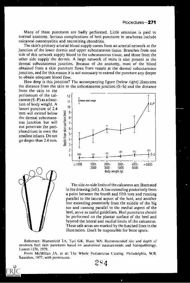
Procedures-271
Many of these punctures are badly performed. Little attention is paid tonormal anatomy. Serious complications of heel punctures in newborns includecalcaneal osteomyelitis and necrotizing chondritis.
The skin's primary arterial blood supply comes from an arterial network at thejunction of the lower dermis and upper subcutaneous tissue. Branches from oneside of this network supply blood to the subcutaneous tissue, and those from theother side supply the dermis. A large network of veins is also present at thedermal subcutaneous junction. Because of t.he anatomy, most of the bloodobtained from a skin puncture flows from vessels at the dermal subcutaneousjunction, and for this reason it is not necessary to extend the puncture any deeperto obtain adequate blood flow.
How deep is this junction? The accompanying figure (below right) illustratesthe distance from the skin to the subcutaneous junction (S-S) and the distancefrom the skin to theperiosteum of the cal-caneus (S-P) as a func-tion of body weight. Alancet puncture of 2.4mm will extend belowthe dermal subcutane-ous junction but willnot penetrate the peri-chondrium in even thesmallest infants. Do notgo deeper than 2.4 mm.
lien') end range
10S-P
9
8
7
6
5
4
31
2
1(10)
(91
19)111
(1.1
S-S
-< 1000 1001- 2001- 3001- 4001- >60002000 3000 4000 6000
Body weight (g1
The side-to-side limits of the calcaneus are illustratedin the drawing (left). A line extending posteriorly froma point between the fourth and fifth toes and runningparallel to the lateral aspect of the heel, and anotherline extending posteriorly from the middle of the bigtoe and running parallel to the medial aspect of theheel, serve as useful guidelines. Heel punctures shouldbe performed on the plantar surface of the heel andbeyond the lateral and medial limits of the calcaneus.These safe areas are marked by the hatched lines in theillustration. Don't be responsible for bone spurs.
Reference: Blumenfeld TA, Turi GK, Blanc WA: Recommended site and depth ofnewborn heel skin punctures based on anatomical measurements and histopathology.Lancet 1:230, 1979.
From McMillan JA, ct al: The Whole Pediatrician Catalog. Philadelphia, W.B.Saunders, 1977, with permission.
2'4'4
4

0
272Proteinuria/Pruritus
PROTEINURIA
Common Causes
Chronic pyelonephritisIsolated transient/intermittent
proteinuriaCold exposureCongestive heart failureExerciseFebrile illnessIdiopathic proteinuriaOrthostatic proteinuriaPregnancyTraumaUrinary tract infection
Rare Causes
DrugsCaptoprilFenoprofenGoldPenicillamineProbenecid
Nephritic sedimentHereditary nephritisI GA nephropathyMixed cryoglobulinemiaRapidly progressive glomerulo-
nephritisSubacute bacterial endocarditisSystemic lupus erythematosus
Nephrotic sedimentAmyloidosisDiabetes mellitus
Common Causes
Uncommon Causes
Nephritis sedimentMembranoproliferative glomerulo-
nephritisPostinfectious glomerulonephritis
Nephrotic sedimentMinimal change diseasePreeclampsia
Tubular proteinuriaAcute tubular necrosisObstructive uropathyPolycystic kidney disease
Nephrotic sediment (Cont.)Focal glomerulonephritisMembranous nephropathyMiscellaneous infections
Hepatitis BMalariaSyphilis
Overflow proteinuriaBence Jones proteinuriaLysozymuria (in leukemia)
Tubular proteinuriaAnalgesic abuseChronic hypertensionHypercalciuriaHyperuricemiaRadiation nephritis
PRURITUS
Atopic dermatitisCholestasis of pregnancyContact allergens (plants, cosmetics,
dyes, medications)Contact irritants (soaps, chemicals,
excrement, wool)Dermatitis herpetiformis
2 5
DrugsAminophyllineAspirinBarbituratesErythromycinGold
riseofulvin

Drugs (Cant.)IsoniazidOpiatesPhenothiazinesVitamin A
Dry skinAdvanced ageExcess bathing/strong detergentsLow humidity
Foreign bodyHepatitisHerpes gestationisHigh humidity
Uncommon Causes
Biliary obstructionDrug inducedExtrahepatic biliary obstructionPr'-nary biliary cirrhosis
Chronic renal failureHematopoietic malignancies
Hodgkin's diseaseLeukemia
Rare Causes
Autoimmune (SLE, AA)Congenital ectodermal disordersEndocrine disorders
Carcinoid syndromeDiabetes mellitusHyper/hypothyroidismHypoparathyroidism
Erythropoietic protoporphyria
Pturitus-273
Insect bites/infestationsFleas, mosquitos, scabies miters,
lice mites, chiggersIron-deficiency anemiaParasitic infection
PinwormsToxocara canis
Pityriasis roseaPsoriasisSeborrheic dermatitisSkin infections (bacterial/viral/fungal)UrticariaWater contact (aquagenic)
Hematopoietic malignancies (Cont.)Lymphoma
NeurodermatitisParasitic infection
CercariaHookwormsTrichinosis
Hematopoietic malignanciesMastocytosisMultiple myelomaPolycythemia vera
Malignant solid tumorsNeurologic syndromesPsychosis
Relieving the Sting or the Itch
Although a number of drugs and lotions are available for the treatment ofbites and rashes, home remedies are often as good or even better in producingrelief. Here are some tried and true home remedies:
Home Remedies for the Treatment of Bites and Rashes
FOR TRY
Bee stings, wasp stings,and jellyfish bites
Poison ivy
Adolph's meat tenderizer. Add a little water to thepowder and rub into the bite. Expect relief in minutesBan roll-on deodorant. Just rub on the rash and rubaway the itch.
2 c' 6Table continued on next page

274Puberty
Home Remedies for the Treatment of Bites and Rashes (Cont.)
FOR TRY
Chickenpox
Chigger bites
Spray starch. Just spray the lesions with this laundrystarch.
Clear nail polish. Paint each bite with nail polish. Thechigger suffocates and the itch disappears.
Reference: From McMillan J, et al (eds): The Whole Pediatrician Catalog, Vol. 3.Philadelphia, W.B. Saunders, 1982, with permission.
Pia ITV
Delayed Puberty and the Adolescent Female
Surely none of us can forget the emotional and physiologic complexities ofpuberty. The adolescent female who believes her development is delayed can becaught in a maelstrom of anxiety. For the majority of girls with "delayed"puberty, the cause is none other than normal variance. In these cases, thepediatrician is the perfect person to provide the needed reassurance thatdevelopment will occur. For the minority of females with a pathologic cause ofpubertal delay, diagnosis and, in some cases, treatment, are within reach. Thetables and evaluation plan that follow will help you and your patient in thisdelicate matter.
Late, Delayed or Arrested?
"Late" defines the onset of puberty at an age older than the average but within2 standard deviations of the mean.
"Delayed"when no signs of sexual development have begun by the age of 13.
"Arrested"when more than 5 years have passed between adrenarche orthelarche and menarche.
Tanner Stages and Their Mean Age of Appearance
STAGE
BREAST DEVELOPMENT PUBIC' DEVELOPMENT
MEAN AGE (YR) RANGE (YR) MEAN AGE (YR) RANGE (YR)
Tanner I(prepubertal)
Tanner II 11.2 8.0 13.0 11.7 9.3 14.1
Tanner III 12.2 10.0 14.3 12.4 10.2 14.6
Tanner IV 13.1 10.8 15.3 13.0 10.8-15.1
Tanner V 15.3 11.9 18.8 14.4 12.2 16.8
Menarche 12.6 10.0 16.0
27

Causes of Delayed Puberty
Constitutional Delay in Growth an
Puberty-275
d Development (CD GD): Family History
Hypogonadotrophic hypogonadism
Hypothalamic-pituitary disorders
Isolated deficiency of GnRHIsolated deficiency of LH. FSH, or both
PanhypopituitarismAssociated abnormalities
Kallman syndrome (anosomia)Prader-Willi syndromeBardet-Biedl syndrome
PostinflammatoryAutoimmune (hypophysitis)Infectious (meningitis,
encephalitis)
TraumaInfiltration
Histiocytosis XHemochromatosis
IrradiationTumor
CraniopharyngiomaOptic gliomaAdenoma
Functional gonadotropin deficiencyChronic systemic or endocrinologic
disease
Cardiovascular (congenitalor acquired)
Pulmonary (asthma, cystic fibrosis)
Hematologic (sickle cell disease)
Gastrointestinal (celiac disease,chronic inflammatory boweldisease, other causesof malabsorption)
Renal (renal tubular acidosis,renal failure)
Immunologic (chronic/persistentinfection, immunocompromise)
Collagen-vascular (SEE, J RA)
Endocrine (hypothyroidism,glucocorticoid excess,hyposomatotropism, IDDM)
Psychiatric (emotional stress)
Hypogonadotropic hypogonadism Cont.)
Weight loss
Anorexia nervosaMalabsorptionExercise
HmerprolactinemiaProlactinoma
Hypergonadotrophic hypogonadism
Primary gonadal abnormalitiesGonadal dysgenesis and its variantsInsensitivity to gonadotropinsDefects in steroidogenesis
Acquired gonadal failuresPostinflammatory
AutoimmuneInfectious
PosttraumaticVascularSurgical
InfiltrationGalactosemiaMyotonic dystrophyAtaxia-telangiectasia
ToxicIrradiationChemotherapy
Tumor
Hyperandrogenism
Polycystic ovary syndrome (PCOS)
Nonclassical congenital adrenalhyperplasia (CAH)
Anatomic genital abnormalities
Rokitansky syndrome (congenitalabsence of uterus and vagina)
Transverse vaginal septum and/orimperforate hymen
End-organ insensitivity to androgens(testicular feminization)
Reference: Schwartz ID, Root AW:Pcds Nov:83 104, 1989.
Puberty in girls: Normal or delayed? Contemp
2c:18
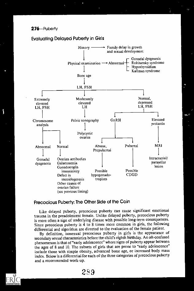
276Puberty
Evaluating Delayed Puberty in Girls
History Family delay in growthand sexual development
Gonadal dysgenesisPhysical examination *Abnormal Rokitansky syndrome
1
HypothyroidismKallman syndrome
Bone age
1
LH, FSH
I I I
Extremely Moderately Normal,elevated elevated depressed
LH, FSH LH LH, FSH
1 I I
I
I
Chromosome Pelvic sonography GnRH Elevated
analysisI
prolactin
1 I
Polycysticovaries
1 1 1
Abnormal Normal Absent, Pubertal MRI
1 1
PrepubertalI
Gonadal Ovarian antibodies
1
Intracranial/
dysgenesis Galactosemia parasellarGonadotropin lesion
insensitivity Possible Poss.bleDefect in hypogonado- CDGD
steroidogenesis tropismOther causes ofovarian failure(see previous listing)
Precocious Puberty: The Other Side of the Coin
Like delayed puberty, precocious puberty can cause significant emotionaltrauma in the preadolescent female. Unlike delayed puberty, precocious pubertyis more often a sign of underlying disease with possible long-term consequences.Since precocious puberty is 4 to 8 times more common in girls, the followingdifferential and algorithm are directed to the evaluation of the female patient.
By definition, isosexual precocious puberty in girls is the appearance ofsecondary sexual characteristics before the child's eighth birthday. An oft-confusedphenomenon is that of "early adolescence" where signs of puberty appear betweenthe ages of 8 and i O. The subsets of girls that are prone to "early adolescence"include those with simple obesity, advanced bone age, or increased body massindex. Below is a differential for each of the three categories of precocious pubertyand a recommended work-up.
299

Categories of Precocious Puberty (True and Complete)
1. Central isosexual precocityIdiopathic, often familialCNS anatomic defects
Septo-optic dysplasiaHydrocephalusCysts
PostinflammatoryMeningitisEncephalitisBrain abscess
TraumaIrradiationTumor
HamartomaNeurofibromaOptic gliomaAstrocytomaEpendymomaDysgerminomaCraniopharyngioma
Postpseudiososexualprecocity
Puberty-277
2. Pseudoisosexual precocityGonadotropin-dependent
HcG secreting tumors (very rare)LH&FSH secreting tumors (very rare)
Gonadotropin-independentOvarian estrogen
Granulosa cell tumorFollicular cysts
McCune-Albright syndrome(irregularly contoured café au laitspots, polyostotic fibrous dysplasia,and precocious puberty)
Adrenal estrogenFeminizing tumors
Exogenous estrogen or estrogen-like substances
Oral contraceptivesTopical estrogenCimetidineCannabisSpironolactoneDigitalis
3. Primary hypothyroidism
Incomplete Isosexual Precocity
Premature thelarche, with or without galactorrhea: peak incidence 6 m-2 y;persists from birth in 23% of girls.
Premature menarche: any vaginal bleeding in prepubertal females should alertthe pediatrician to possible sexual abuse or trauma, vaginal infection, tumors,foreign body, or urethral prolapse.
Premature adrenarche: incidence higher in African Americans. Congenitaladrenal hyperplasia variants.
Algorithm (see top of next page.)
Treatments exist to "turn off" the hypothalamic-pituitary ovarian axisthrough pharmacologic or surgical therapies. The long-term consequences ofprecocious sexual development include compromise of final adult stature,possibly increased risk of cervical and breast cancers due to extended exposureto unopposed estrogen, and psychological trauma. When precocious puberty issuspected, a referral to a pediatric endocrinologist is suggested.
Reference: Schwartz ID, Root AW: Puberty in girls: Early, incomplete or precocious?Contemp Peds Jan:147-156, 1990.
230
.

278Puberty
History CNS :nsultExogenous estrogen exposure
Physical examination. HypothyroidismPelvic/vaginal tumor
Bone age
Less than chronological age Greater than chronological ageI
I I IE2 adrenal steroids Depressed T. Extremely Pubertal E2(normal, elevated) Elevated TSH elevated E2
1
I I 1
Premature thelarche Hypothyroidism Ovarian neoplasmI
Possible Ovarian cyst No response PubertalPremature menarche Exogenous McCune-Albright
1I E2 syndrome
Premature CPP
IElevated 17-0H P MRI
I I
Congenital adrenal Hypothalamic/hyperplasia variants pituitary
neoplasm
Timing of Puberty and Its Biopsychosocial Correlates
Research on the timing of maturation, precocious puberty, and environmentalchange has reported fairly consistent findings. The influence of puberty and itstiming appear to have the greatest effects in the areas of self-conceptions (bodyimage and self-esteem), developmental needs (heterosexual relationships, peeraffiliations, family independence), school performance (academic performanceand problem behaviors), and environmental responses (peer, -parental, andteacher expectations). These effects vary as a function of:
I . gender,2. the relationship of the individual's pubertal status to that of his or her
peers,3. definitions of early and late timing, and4. the behavior under investigation.
In general, the most negative effects have been reported for early-maturingfemales. Some recent work has shown that the effects of early maturation may bedetrimental for both sexes, with early maturation in males being associated withthe early initiation of sexual activity and other risk behaviors.
References: Irwin CE Jr: The theoretical concept of at-risk adolescents. AdolescentMedicine: State Art Rev 1:1-14,1990.
Irwin CE Jr, Millstein SG, Turner R: Pubertal timing and adolescent risk taking: Arethey correlated? Pediatr Res 25:8A, 1989.
231
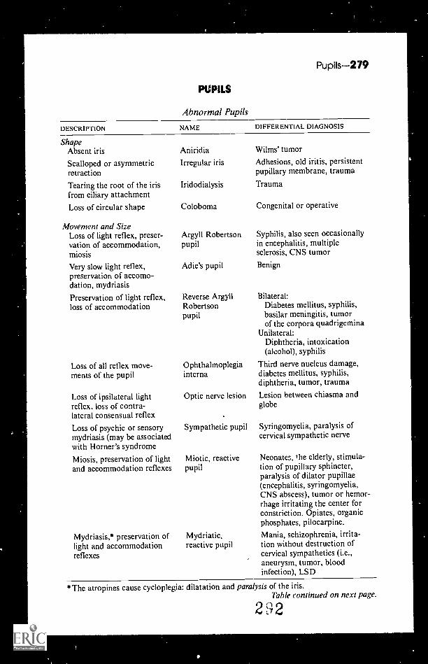
0
Pupils-279
PUPILS
Abnormal Pupils
DESCRIPTION NAME DIFFERENTIAL DIAGNOSIS
ShapeAbsent iris
Scalloped or asymmetricretraction
Tearing the root of the irisfrom ciliary attachment
Loss of circular shape
Movement and SizeLoss of light reflex, preser-vation of accommodation,miosis
Very slow light reflex,preservation of accomo-dation, mydriasis
Preservation of light reflex,loss of accommodation
Loss of all reflex move-ments of the pupil
Loss of ipsilateral lightreflex, loss of contra-lateral consensual reflex
Loss of psychic or sensorymydriasis (may be associatedwith Horner's syndrome
Miosis, preservation of lightand accommodation reflexes
Mydriasis,* preservation oflight and accommodationreflexes
Aniridia
Irregular iris
Iridodialysis
Coloboma
Argyll Robertsonpupil
Adie's pupil
Reverse ArgyllRobertsonpupil
Ophthalmoplegiainterna
Optic nerve lesion
Sympathetic pupil
Miotic, reactivepupil
Myd riatic,reactive pupil
Wilms' tumor
Adhesions, old iritis, persistentpupillary membrane, trauma
Trauma
Congenital or operative
Syphilis, also seen occasionallyin encephalitis, multiplesclerosis, CNS tumor
Benign
Bilateral:Diabetes mellitus, syphilis,basilar meningitis, tumorof the corpora quadrigemina
Unilateral:Diphtheria, intoxication(alcohol), syphilis
Third nerve nucleus damage,diabetes mellitus, syphilis,diphtheria, tumor, traumaLesion between chiasma andglobe
Syringomyelia, paralysis ofcervical sympathetic nerve
Neonates, the elderly, stimula-tion of pupillary sphincter,paralysis of dilator pupillae(encephalitis, syringomyelia,CNS abscess), tumor or hemor-rhage irritating the center forconstriction. Opiates, organicphosphates, pilocarpine.
Mania, schizophrenia, irrita-tion without destruction ofcervical sympathetics (i.e.,aneurysm, tumor, bloodinfection), LSD
*The atropines cause cycloplegia: dilatation and paralysis of the iris.Table continued on next page.
292

280Pu rpura
Abnormal Pupils
DESCRIPTION NAME DIFFERENTIAL DIAGNOSIS
Movement and Size (Cont.)Pupils alternately dilateand contract rapidly("tremor of the iris")
Hippus Multiple sclerosis, drug/alcoholoverdose, homocystinuria, centralscotoma with macular damage ordisease or injury to axial fibers ofoptic nerve
Congenital, traumatic, surgical
Variation of normal, iritis, diabetesmellitus, cervical sympathetic lesion,eye drops, glaucoma, unilateraldamage to third nerve fibers, syphi-lis, :.rigeminal neuralgia, carotid oraortic aneurysm, cranial lesion, cere-bral herniation, artificial eye
More than one pupil in an eye Polycoria
Inequality of size of pupils Anisocoria
Pupils dilate under lightstimulus
With strong deviation ofthe eyes, the pupil of theabducted eye is larger thanthat of the adducted eye
Parodoxical Syphilispupil (rare)
Tournay's Normalsign
From Gottlieb AJ, Zamkoff KW, Jastremski MS, Scalzo A, lmboden KJ: The WholeInternist Catalog. Philadelphia, W.B. Saunders Co., 1980, pp 120-121, with permission.
PURPURA (PETECHIAL AND ECCHYMOSES)
Common Causes
ThrombocytopeniaTraumaViral infections
Uncommon Causes
Abnormal platelet functionChild abuseCupping and coin rubbingDrug ingestion (aspirin)FactitiousHenoch-Schonlein purpuraHereditary coagulation disturbanceInfectionSentic emboli
miaiasculitisViolent coughing
2,)3
Rare Causes
Autoerythrocyte sensitizationBernard-Soulier (giant platelet)
syndromeCushing's syndromeDysproteinemiasGlanzmann's thrombastheniaHereditary hemorrhagic telangiectasiaMacular cerulaeMarfan's syndromeMay-Hegglin anomalyOsteogenesis imperfectaOsteopetrosisPlatelet storage pool diseaseProtein C deficiencyProtein S deficiencyPurpura fulminansSchamberg's diseaseScurvyVitamin K deficiency
'

Purpura 2841
Purpura and PetechiaeInterpreting the Sign
Every little bruise can have a meaning all its own. It is important to lookcarefully at a hemorrhagic lesion for the clue to its un ierlying disease. Listedbelow are some guides to the interpretation of this sign.
Thrombocytopenic purpura
Thrombocytopathic purpura
Vasculitic purpura(+/ thrombocytopenia)
Drug purpura(+1-- thrombocytopenia)
Allergic purpura(Henoch-Schonlein)
Purpura fulminans(skin manifestationsof DIC)
"Devil's pinches"(autoerythrosensitizationvs. factitious)
Hyperglobulinemicpurpura
Cryoglobulinemic purpura
Amyloid purpura
Scorbutic purpura
Senile purpura
Embolic purpura
Palatine petechiae
Petechiae are nonpalpable.Platelet count < 20.000/m1.
Easily bruised. Petechiae are rare.
Petechiae are palpable.
Often associated with hemorrhagic bullae in themouth.
Pruritic crops of symmetrical purpura on proximalextremities (4+ lower) associated with urticarialand erythematous lesions.
Large symmetrical ecchymoses, particularly on distalextremities, complicated by acral gangrene.Petechiae are rare.
Females. "Spontaneous"painful ecchymoses(+/- erythematous base) on anterior-lateral aspectof thigh and abdomen in a stepladder distribution.
Lower extremities (after exercise or prolongedstanding). Tendency for skin to develop brownishpigmentation. Identical to idiopathic nonhyper-globulinemic syndrome, called Schamberg'sdisease, except that the latter has normal serumglobulin levels.
Purpura (+/- gangrene) on exposed acral areas(fingers, nose, ears, face).
Spontaneous periorbital purpura (usually postValsalva maneuver). "Touch purpura."
Purpura around hair follicles (perifollicular petechiae).Characteristically associated with corkscrew hairs.Saddle distribution.
Purpleflat ecchymotic spots on extensor surface offorearms, dorsurn of hands, and neck in the elderly.Identical lesions are found in cachetic states andchronic hypercortisonism.
A. Sept;c embolicWhite centered petechial lesion often located onmucous membrance and conjunctivae (e.g., bacte-rial endocarditis).
B. Fat ernboliPetechiae limited to upper one-half of the body,particularly to anterior chest. Never seen on theface or back (skimming elfect).
Infectious mononucleosis.Sepsis.Trauma (e.g., dentures).
t' 4

282Pyuria and Bacteriuria
Petechiae(lesion 3 mm)
Almost always indicates a disturbance of platelets or avasculopathy. Rarely do they indicate an abnormalityof coagulation.
Reference: From McMillan JA, et al (eds): The Whole Pediatrician Catalog, Vol 2.Philadelphia, W.B. Saunders, 1979, pp 211-212, with permission.
PYURIA AND 8ACTERIURIA
Does pyuria always signify the presence of bacteriuria? Is bacteriuria alwaysassociated with the presence of pyuria?.The answer to both questions is "no."Pyuria in the absence of bacteriuria can be seen:
I. In dehydration2. In trauma3. In the presence of an irritating agent in the renal pelvis, bladder, or ureter.4. In rmal tuberculosis5. In acute and chronic glomerulonephritis6. After administration of oral polio vaccine7. After administration of intramuscular iron8. In renal tubular acidosis9. In association with a variety of viral infections
10. In Kawasaki disease
Listed below is a comparison between urine bacterial counts and leukocytecounts. Urines were collected by a clean voiding technique, and 5 ml wascentrifuged for 3 minutes at 3000 RPM.
COLONY COUNT(RACTERIA/m0
UNCENTRIFUGED10 or more 100 or moreWBC/rTIM3 WBC/MITI3
CENTRIFUGED5 or moreNVBC/HPF
105 61% 23% 43%
104-105 28% 0% 14%
103-104 23% 0% 6%
102-103 21% 1% 2%
Sterile 10% I% I%
Reference: Pry les CV, Lustik B: Pediatr Clin N Amer 18:233, 1971.From McMillan JA, et al: The Whole Pediatrician Catalog, Vol. 2. Philadelphia, W.B.
Saunders, 1979, p 255, with permission.
295

WHAT 5EA5ON OF "PROFILES ONPARADE" 44.10UW .96 COMPLETEWITHOUT A 11/5IT FROM OUROW FRIEND, OR. PAN A5IIER,TIOTEMAKER TO 771E MEU01412
MN'S IATE51 flANDS001:IS FOR ALL YOU BABY800MER5 WHO'VE FINALLYGOTTEN Afro CHIW-REARING,
/ RIGHT; XCIOR?
THAT'S RIGHT,
MARK! I CALLIT, "THE MELLOWPARENT: SHARINGMIR SPACE IdITH
PEPENDEN7S"
1111
IVt:Peqr2.7(4.0riT 1731331: "Id 3
FOR SURE, MARK/ 14.1ITH50 MANY UPSCALE FOLI(5al I /NG INTO PARENTINGTHESE PAYS, THERE'S JUSTA BIG NEED hOR YOURBASIC \
10.1-701

284Quality Time
t r, 61.4131=1;"'" ,.7711
1.4
IN''NE MELLOW War? r PISC05.5 5UCHPROBIEM5,45 WOKING fOR A 6000 HOUSE-wpeg, PREVENTING YOUR BABY FROM
EATING YOUR FERNS, AND THE INIPOR-,TANCE Of 51.106ETA6 QUALI7Y
0':79. ft."
'
...A.1.416.... m
If 'tg..__
\ ._. .-..r.i.4 ,.. ...,:,......__,..,. , . :ii
1 (; `- ---, -- -,..---- -----.
1.1
Kepi
r.13;f:wr 70.1esur
WHAT'S'QUALITY
PAN r
THAT5 A wiz PART OF THE DAYYOU SET A510E70 SPEND WITH YOURKIP. IT 00E5N7 MVE 70 BE MUCH,TEN MIAIUTES 15 USUALLY ENOUGH, BIJT
HA5 TO BE QUALITY! YOU CAN7J157WATCH TV YOU HAVE 70 FOCUS ON
THE CHILP5 NEEDS.
(
AP
.1 .....t..t.t.t,:40.s.Ityht.blar-tr, t..,111,V,.
WHAT IF ONE 711A75 QUANTITY TIME.Of ITS NEE-OS .TM NOT REALLY TALK-
_ 5 MORE WE? ING ABOIIT THE PROS-/ LEM 011112 WE, MARK./
11.-- O SC
/ . 1 :VIIAll, P. I
I F',.,, . _C. / EtJJ
.0 --------
9
DOONESBURY, copyright 1984. G. B. Trudeau. Reprinted with permission ofUniversal Press Syndicate. All rights reserved.
2 9 7 BEST COPY AVAILABLE

Questions-285
QUESTIONS
Thirty Questions That Parents Ask and PediatriciansShould Be Able to Answer
Listed below are questions frequently asked by parents. No one can have allthe answers, but you should begin to learn some of them in your pediatrictraining. We have stratified the questions, with their answers, as a function ofprobable experience. If you have any questions about our answers, drop us a line.
Questions that a PIA should be able to answer:
1. If I chop up foods, such as hot dogs, very fine, is it safe to feed these to my2-year-old?
A. If they are chopped into very small pieces, they are safe; but it isimportant to remember that hot dog chunks are a common cause ofaspiration in the 2-year-old. Another very dangerous food for any childunder 3 years of age is peanuts. "Only a nut would feed a nut to a child."Instruct parents in the Heimlich maneuver.
2. Can shaking a baby or tossing him or her into the air playfully be harmful?
A. Yes, both can be extremely harmful. Such play can injure the brain,neck, or spinal cord and has been known to produce retinal hemorrhages.
3. Is it true that it is harmful to swing or lift a child by holding his hands andpulling up on his arms?
A. Yes, this can produce a "nursemaid's elbow"--which is a dislocation ofthe elbow.
4. My husband smokes in the house. Is this really a health danger to my youngchildren?A. Absolutely. Smoking at home is known to aggravate asthma. There is a
higher incidence of otitis media and other forms of respiratory illnessamong children living in a home with smokers. By the way, side-streamsmoke also increases the non-smoking adult's risk for cancer. Get yourhusband to stop smoking or get rid of him.
5. I have a healthy 4-year-old. Should she receive the influenza vaccine'?
A. It is not necessary. The vaccine is advised for children with a variety ofchronic illnesses.
6. Is it true that penicillin will cure the symptoms of a sore throat within 24hours?A. Yes, but only if the symptoms are a result of an infection with the
streptococcus microorganism. Without treatment, it will take 48 to 72hours before symptoms subside.
7. Is it really possible for my son to have a streptococcal infection in his analarea?
A. Definitely. It is characterized by pain and redness, usually without fever.Penicillin is the cure. 298
o
286Questions
8. What is "croup"?A. Croup is the term applied to a condition that is characterized by a
inspiratory noise called "stridor." It is often accompanied by a coughthat sounds like the bark of a seal. Croup can be caused by infections orallergic swelling in the larnyx or "voice-box."
9. Is it safe to give my 10-month-old daughter swimming lessons?
A. Not a good idea. She can swallow a great deal of water while learning andthat may cause convulsions. She will not really l':.arn to swim at this age anddoes not have sufficient judgment to avoid water dangers when left alone.
10. Can you recommend a good book for me, a new parent, on child health?
A. Yes. The best book on this subject is entitled Your Child's Health, by Dr.Barton D. Schmitt and is published by Bantam Books and is available inpaperback.
Questions that a P1p2 should be able to answer:
I. My 16-year-old daughter often eats several glasses of ice cubes each day. Isthere something wrong with her?
A. Your daughter suffers from a condition termed "pagophagia" or ice-eating. "Pagos" is the Greek word for "ice" and "phagia" in Greek means"to cat." Compulsive ice-eating is usually a sign of iron deficiency andcan often be promptly cured with iron therapy.
2. Can the rash from poison ivy be spread by exposure to the fluid from poppedblisters?
A. No. Poison ivy results from a hypersensitivity reaction to the plant'sresins. Transmission only occurs by direct contact with the plant or withthe hands, clothing, or pets that have resin on them. The blister fluiddoes not contain the resin.
3. My daughter is almost 3 and still not toilet trained. What am I doing wrong?
A. Nothing. Toilet training should not begin until your chat; is ready.Readiness means that your child has the neurologic capacity to controlbodily functions, understands the concepts of toileting, knows thelanguage of the toilet, and, most importantly, wants to be clean and dry.This process may start as early as 1 year of age. I suspect that your childwill train herself very soon.
4. We have a kitten at home. Can a kitten or cat cause any health problems formy 2-year-old son?
A. Yes, definitely yes. Diseases and parasites such as cat scratch fever,toxoplasmosis, and Toxocara cati can be spread from a cat or kitten. Catdander is often responsible for asthma. Now that you have a child youdon't need any other pet. Get r_9-of the cat.
5. Is it safe to allow my son to play football with his high school team?
A. It depends on the status of his physical maturity relative to others hisown age, the protective equipment available, and the concern of thecoach for the emotional and physical welfare of the members of the team.
9 9
f
Questions-287
6. My 5-year-old periodically becomes hoarse. What should I do?
A. Get some ear plugs for yourself. The episodes of hoarseness are probablya consequence of excessive screaming on your child's part. "Screrrer'snodules" actual thickening of the vocal cordscan result from overuseof the voice. If hoarseness persists, your child should be seen by anotolaryngologist.
7. My child has pink urine after eating beets. What does this mean?
A. About 7% of the population will demonstrate "beeturia" after eating theequivalent of one beet. This is a genetic trait. Beeturia is also seen in avery high percentage of children and adults with iron deficiency anemia.This form of beeturia disappears with iron therapy. To be certain of thecause of your child's problem, a blood count should be obtained.
8. Is it appropriate for a young child to attend the funeral of his grandparent?
A. If he wants to go, then you should let him. Never force a child, of anyage, to attend a viewing or a funeral.
9. Does an exclusively breast-fed infant require supplemental iron?
A. Not during the first 6 months of life. Studies have shown that the ironstatus of an exclusively breast-fed infant is as good as an infant receivingan iron-fortified formula at age 6 months. If breast milk remains the solesource of nutrition beyond 6 months of age, iron supplements areindicated, because studies have shown that about 15% of such infants willbe iron deficient by 9 months of age.
10. What kind of shoes should I buy for my baby when she starts walking?
A. The least expensive sneakers you can find. Shoes are only necessary forthe protection of the feet from the dirt, glass, and manure of the streetsand are not required to enhance the act of locomotion.
Questions that a P14 should be able to answer:
1 My 2-year-old is a finnicky eater and I'm not sure she's eating all the foodsshe needs. Should I supplement her diet with vitamins?
A. No, they are unnecessary. Put a balanced meal before the child and shewill take what she needs. Vitamins are only "fish-food" for a personeating a balanced diet. The excess vitamins you provide will appear in thechild's urine, and when they are flushed down the toilet they willeventually reach the fish in the sea.
2. I'm a working mother with three young children. Not surprisingly, I oftencome home tired. Can you give me some advice to make returning homeafter a tough day better for all of us?
A. There are no easy answers for a task that stumps even "superwoman."But these suggestions may help:
When you pick up your child from day care, bring a snack for the trip home.Take turns with your spouse leaving work early, so one of you can pick up yourchild before you are both exhausted.
3U0

263Questions
Establish a homecoming routine that gives you recovery time. Tell yourchildren that they will get your full attention after you have a chance to catchyour breath and get dinner started.Reward your children for good behavior. Try a chart with gold stars or aspecial picture to color.Expect your school-age children to begin their homework after school andbefore you arrive.Reduce meal preparation during the week by cooking on weekends andfreezing the servings until you need them.If you can afford it, hire a high school student as a mother's helper for that firsthour at home.Involve the children with dinner chores, such as setting the table, clearing thetable, and the like'.Share the evening tasks with your spouse.If possible, take the family out to dinner occasionally to give everyone a break.
3. Should I give my child aspirin?A. No, except under a physician's direction for some very special circum-
stances, such as Kawasaki's disease or acute rheumatic fever.
4. Does it help to put butter on burns?A. The best thing to put on a burn immediately is ice. The ice will relieve the
pain and reduce the damage done by the burn. Topical vitamin E is muchbetter than butter in reducing the scarring and damage done by a small burn.
5. Can iron deficiency anemia produce any harm in a 1-year-old?A. Yes. Recent studies demonstrate that iron deficiency anemia results in
cognitive delays in 1- and 2-year-olds that may r,r)t be correctable withiron therapy.
6. When can you begin giving a child an allowance?
A. When you can afford it and when the child has some appreciation of thevalue of money and when you have agreed upon the fact that theallowance is dependent on the satisfactory performance of some respon-sibilities. This usually begins at age 8-9.
7. Can fruit juice cause stomach aches or diarrhea in my 6-year-old?A. Yes, if they consume large quantities of juice and if the juice contains
non-absorbable sugars such as sorbitol. Apple juice, for example,contains large quantities of sorbitol.
8. Can a child have an ear infect;on without a fever?
A. Yes. About one-half of episodes of otitis media are not associated withtemperature elevation beyond 100.4°
9. Our 12-year-old son wants an All-Terrain vehicle. Are they safe?
A. No. Don't buy it either for him or for you.
10. My teenage daughter says that kissing is good exercise. Is this true?
A. Not if her goal is to lose weight. It is estimated that a single kiss burnsup 9 calories and that by kissing three times a day for 1 year, she couldlose 2.8 pounds
3 ) 1

RED CELL
Red Cell Distribution Width
Age-appropriate Values for RBC Distribution Width
AGENO. OF
PATIENTSRBC DISTRIBUTIONWIDTH (MEAN ± SD)
1-6 mo 68 13.0 ± 1.57 -12 mo 84 13.7 ± 0.9
13-24 mo 108 13.4 ± 1.02 -3 yr 119 13.2 ± 0.84-5 yr 151 12.7 ± 0.96-8 yr 106 12.6 ± 0.89-11 yr 98 12.8 ± 1.0
Reference: Novak RW: Red blood cell distribution width in pediatric microcyticanemias. Pediatrics 80:251, 1987, with permission.
RENAL FAIWRE
The FENa Test: Use in the Differential Diagnosisof Acute Renal Failure
The physician is frequently faced with the problem of distinguishing prerenalazotemia from acute tubular necrosis in patients with acute renal failure.
In the oliguric phase of these two conditions, the renal tubule handles sodiumin distinctly different fashions. In prerenal azotemia, the renal tubule avidlyreabsorbs the filtered sodium; in acute tubular necrosis, the reabsorption ofsodium is restricted.
These observations provide the basis for a simple test for differentiating thesetwo conditionsthe "FENa test" (FENa is the excreted fraction of the filteredsodium).
The test is performed by measuring both sodium and creatinine in simultane-ously collected samples of plasma and urine.
The FENa is calculated as follows:
[Sodium] U[Sodium] P X 100
[Creatinine] U[Creatinine P]
U and P represent concentrations in urine and plasma, respectively.
3 2289

290Respiratory Distress
In general, an FENa of less than I indicates prerenal azotemia, and an FENa ofmore than 3 indicates acute tubular necrosis.
Reference: Espinel CH: The FENa test: Use in the differential diagnosis of acute renalfailure. JAMA 236:579, 1976.
From McMillan J, et al: The Whole Pediatrician Catalog. Philadelphia, W.B. Saunders,1977, p 316, with permission.
RESPIRATORY DISTRESS
The Diagnosis of Newborns with Acute Respiratory Distress
The newborn with clinical signs of acute respiratory distresscentral cyanosis,tachypnea (>60 breaths/min), tachycardia (>160 beats/min), retractions, grunting,and nasal flaringdemands immediate attention. A review of the maternalhistory, the age of onset of respiratory distress, a physical examination, and lab-oratory tests (particularly a criest x-ray) will greatly aid the clinician in assessingthis problem further. The following differential diagnosis should be of use:
1. Upper airway obstruction
Choanal atresiaMasses (encephalocele, tumor)MacroglossiaNasal stiffnessCleft palateLaryngeal obstruction (paralysis,
web, tumor, stenosis, atresia,malacia)
Tracheal obstruction (mass, web,stenosis, atresia, malacia, cleft,vascular ring, goiter)
2. PulmonaryHyaline membrane diseaseTransient tachypnea of the
newbornAspiration of meconium, gastric,
or amniotic fluidPneumoniaPneumothorax, pneumo-
mediastinumPersistent pulmonary hypertensionTracheoesophageal fistulaPulmonary hemorrhageHypoplasia or agenesis of
the lungsCystic disease (emphysema, cysts)Pleural effusions (e.g., chylothorax)Pulmonary sequestrations
3 :3
3. Cardiac
Cyanotic congenital heart diseaseAcyanotic congenital heart diseaseArrhythmia (paroxysmal supra-
ventricular tachycardia, block)Increased intravascular volume
(iatrogenic fluid overload)High output failure (hyper-
thyroidism, arterial-venousmalformation)
PneumopericardiumCardiomyopathy (infection, endo-
cardial fibroelastosis, hyper-trophic cardiomyopathy)
4. Thoracic
Chest wall deformities (chondro-dystrophies, rib deformities)
Masses (tumors, cysts)
5. Metabolic
HypoglycemiaInfant of a diabetic motherInborn errors of metabolism
6. Diaphragmatic
Hernia (foramen of Bochdalek)Paralysis (phrenic nerve)Eventration

7. Neuromuscular
CNS damage (trauma, hemorrhage)Medication (maternal sedation,
narcotic withdrawal)Muscular weakness (e.g.,
myasthenia gravis)Congenital defects
8. Infections
SepsisPneumonia (viral, bacterial)
Respiratory Viruses-291
9. Hematologic/vascular
Hyperviscosity, hypervolemiaAnemiaHemoglobinopathy
10. Other
AsphyxiaAcidosisHypothermiaHyperthermia
Reference: Schreiner RI Bradburn NC. Newborns with acute respiratory distress:Diagnosis and management. Pediatrics in Review 9(9):279-285,1988. Adapted from Table2 of cited paper.
RESPIRATORY VIRUSES
Clinical Syndromes Produced by Respiratory Viruses
Upper respiratory tract infections and pneumonia are among the most frequent-ly made diagnoses in pediatric clinics and emergency rooms, particularly during thewinter months. All too frequently, these infections are lumped together as upperrespiratory infections (U Rls), viral syndrome, or "influenza-like" syndrome. Manyviruses, aside from influenza, are capable of producing respiratory symptoms.
Clinical Syndromes Produced by Respiratory Viruses
VIRUSES CORY/A PHARYNGITIS CROUPFt II-LIKTII I.NESS PLFURODYNIA
TRACIIM-BRONCHI HS PISFAIMONIA
influcnnt A 0
Influcnia B
Influenza C
Parainfluenn(1 3)
0 *Respiratory
syncytial*
Coxsackie A
Coxsackie B
Echo 0
Adcnoviruses(1 7, 14, 21)
Rhinoviruscs(>100)
5* 5* 5*
Coronaviruscs
Herpcsviruses(I, 2)
Epstein-Barr
In children only
3 4

292Respiratory Virus
The term influenza, incidentally, originated in Italy during the 15th century. Aparticularly severe epidemic of a respiratory viral syndrome at that time wasattributed to the influentia (influence) of the stars and evil forces.
Reference: Rytel MW: Influenza and its complications. Recognition and prevention.Hospital Practice 22:1021- lO2V, 1987. Table adapted from cited reference.
The Clinical Manifestations of Respiratory Syncytial Virus
Respiratory syncytial virus (RSV) is an all too common cause of epidemicwinter respiratory disease in infants, children, and adults. Its severity spans awide spectrum ranging from a common cold to bronchiolitis (the most commonpresentation) to respiratory distress, apnea, cyanosis, and pneumonia. RSV ispotentially most life-threatening in infants with other underlying conditionssuch as congenital heart disease (especially conditions with high pulmonaryflow), bronchopulmonary dysplasia, other chronic irreversible pulmonarydisorders (e.g., cystic fibrosis, pulmonary hemosiderosis, bronchiolitis obliterans,and idiopathic pulmonary hypertension), and all chil...lren on immunosuppressiveregimens (e.g., transplantation patients, cancer patients treated with chemother-
y or radi:Aion therapy, and patients on high-dose steroid regimens) orcnildren with immunodeficiency syndromes (e.g., severe combined immunedeficiency).
Although bronchiolitis is the most frequent presentation of an RSV infection,pneumonia is the most common admitting diagnosis for infants infected withRSV who require hospitalization. Factors that contribute to the severity ofillness, in addition to those described above, include age, size of inoculum, andcharacteristics of an RSV infection:
Common characteristics (>75% of all cases)
RhinorrheaCoughAirway hyperactivityHypoxemiaAir trapping (hyperinflated lungs on x-ray examination)
Uncommon Characteristics (<20% of all cases)
Prolonged fever (T > 102° F) HepatosplenomegalyOtitis media Enlarged cardiac silhouetteHoarseness Hilar adenopathyPleural effusion
The differential diagnosis for RSV pneumonia should include:
I. Chlamydial pneumonia as it occurs in the same age group and presents withair trapping and wheezing. The distinguishing point between these two diseasesis that chlamydial disease is more prolonged and insidious than RSV.
2. Congenital anomalies of the respiratory tract3. Foreign bodies
:3 '15

Retropharynx-293
4. Reactive airway disease5. Infection with other respiratory agents (e.g., influenza virus, parainfluenza
viruses, adenovirus, pertussis (Bordetella), or mycoplasma).
_
Reference: Laufer DA, Edelson PJ: Respiratory syncytial virus infection and cardio-pulmonary disease. Pediatr Ann 16:644-653, 1987.
RETROPHARYNX
Retropharyngeal Abscess
The retropharyngeal space extends from the base of the skull to about the levelof the second thoracic vertebra. Abscess in this space results from suppuration ofthe lymph nodes, which run in two parallel chains and drain the nasopharynx,adenoids, and posterior paranasal sinuses. During childhood these nodes areprominent, t'it they atrophy during adolescence. Though trauma and adjacentvertebral oste3myelitis can predispose to the development of retropharyngealabscess, local respiratory tract infection is usually felt to be the initiating event.Extension of the infection may result in mediastinitis or asphyxia due toincreasing pressure or rupture of the abscess.
Both aerobic and anaerobic bacteria may be isolated from retropharyngealabscesses, and more than oae organism is often found. The frequent isolation ofStaphylococcus aureus and beta-lactamase-producing anaerobes warrants theuse of antibiotics effective against penicillin-resistant oropharyngeal flora.Surgical drainage of the abscess is critical.
The 63 bacteria isolated from 17 children with retropharyngeal abscess arelisted below:
Bacteria Isolated from Children with Retropharyngeal Abscess
ISOLATES NO. OF ISOLATES
Aerobic and facultativeGram-positive cocci
Viridans streptococci 11
Staphylococcus aureus 8
Beta-hemolytic Streptococcus Group A 6
Streptococcus pneumoniaeStreptococcus constellatusStreptococcus morbillorumMicrococcus 3
Gram-negative cocciNeisseria sp. 7
Gram-negative bacilliEikenella corrodens 3
Haemophilus influenzae (nontypable) 3
Haemophilus parainfluenzaeTotal no. of aerobes 45
Table continued on next page.
3Ls
INY

294Rett Syndrome
Bacteria Isolated from Children with Retropharyngeal Abscess (Cont.)
ISOLATES NO. OF ISOLATES
AnaerobicAnaerobic cocci
Peptostreptococcus species 3
Veilonella parvulaMicroaerophilic streptoccus 1
Gram-positive bacilliEubacterium lentum 2
Gram-negative bacilliBacteroides melaningogenicus 7
Bacteroides capillosus 2
Bacteroides species 1
Fusobacteriurn species 1
Total no. of anaerobes 18
Reference: Asmar BI: Bacteriology of retropharyngeal abscess in children. PediatrInfect Dis J 8:595-597, 1990.
RETT SYNDROME
The Diagnosis of Rett Syndrome
The diagnosis of Rett syndrome, a severe developmental disorder occurring inyoung girls, usually between 6 and 18 months of age, can be difficult in view ofthe fact that it is dependent on history and physical findings alone. There is nolaboratory test to confirm the diagnosis. These children experience rapid declinein motor and cognitive function after a period of apparently normal develop-ment. In those children who have begun to speak, all meaningful communica-tion is lost, including eye contact. The patients often experience interruptedsleep and periods of uncontrollable screaming. Prevalence is between 1/10,000and 1/15,000.
The implications of this diagnosis are tragic for both the patient and family.The criteria that can be used to support or exclude the diagnosis are listedbelow:
1. Necessary criteria
Normal prenatal and perinatal periodApparently normal development first 6 moNormal head circumference at birthDeceleration of head growth between 5 mo and 4 yrLoss of purposeful hand skills between 6 mo and 30 mo; communication
dysfunction; social withdrawalStereotypic hand movementsGait apraxia and truncal ataxia between I to 4 yrDiagnosis tentative until 2 to 5 yr of age
307

2. Supportive criteria
Breathing dysfunctionEEG abnormalitiesSeizuresSpasticity
3. Exclusion Criteria
Intrauterine growth retardationOrganomegaly signs of storage
diseaseRetinopathy/optic atrophyMicrocephaly at birth
Rhinitis-295
Peripheral vasomotor disturbancesScoliosisGrowth retardationHypotrophic small feet
Evidence of perinatally acquiredbrain damage
Identifiable metabolic disorderEvidence of serious CNS infection
or trauma
RHINITIS
Diagnosis and Natural History of Allergic Rhinitis
Allergic rhinitis, which is ranked by the National Center for Health Statisticsas the sixth most prevalent chronic condition in the U.S., has its peak incidencein childhood and adolescence. It is an atopic hypersensitivity response to foreignallergens mediated by IgE antibodies, but not all persons with IgE antibody haveclinical disease. The most common allergens are the following:
Grass pollens (late spring/early summer)Tree pollens (early spring)Weed pollens (late summer/autumn)Animal dandersHouse-dust mitesInsectsMold sporesFoods (uncommonly associated)
Symptoms include paroxysmal sneezing; watery, profuse rhinorrhea; nasalcongestion (stuffy nose); itching of the nose and eyes; and lacrimation and ocularredness. Other symptoms that can occur are noisy breathing, snoring, hyposmiaor anosmia, itchy palate or pharynx, throat clearing, and cough. Children mayhave "allergic shiners," a dark discoloration in the infraorbital regions secondaryto obstruction of venous drainage. The key to diagnosis is the temporalcorrelation of symptoms with allergic exposure.
As with most allergies, avoidance of the offending allergens is the mosteffective treatment, which makes identification of the allergens an importantcomponent of the therapeutic strategy. However, many of the methods commonlyused to diagnose allergies are only minimally helpful in managing allergicrhinitis, including early and late skin-test responses, measurement of IgE levels,and calculation of histamine release by basophils. The natural history of allergicrhinitis is presented in the schematic of sensitization (Phase I) and clinicaldisease (Phase 2) shown on the following page:
3

296Rhinitis
Phase 1Sensitization
IR_
Allergen Antigen-presenting cell
B cell
At.TICta71,:dic,
Plasma cell
EarlyInflammation
Allergen
IgE antibodies
Mediatorrelease
Blood Nerves
vessels Glands
CD4T cell
Processedallergen
IgE antibodies
Cellularinfiltration
Mast Eosinophilscell Basophils
MonocytesLymphocytes
SneezingRhinorrheaCongestion
Simplified schematic representation of thenatural history of allergic rhinitis. Duringphase 1 persons become sensitized to anallergen, and during phase 2 clinical diseasedevelops. The overwhelming majority of pa-tients have an early response on reexposure toallergen. The early response is dominated byactivation of mast cells and release of media-tors. After the early response, most patientshave cellular infiltration of the nasal mucosathat causes late inflammatory events. Theseinclude the spontaneous recurrence of releaseof mediators (late-phase reaction), hyperre-sponsiveness to irritants, and increased re-sponsiveness to allergen (priming). The circlesindicate the heterogeneity of these late in-flammatory events. The inflammation canresolve spontaneously, cause a complication,or potentially lead to an irreversible form ofchronic rhinitis. (From Naclerio RM, N EnglJ Med, 325: 861, 1991, with permission.)
Phase 2Clinical Disease
LateInflammation
<Resolution
. Complications
?Irreversibledisease
References: Naclerio RM: Allergic rhinitis. N Engl J Med 325:860-869, 1991.Simons FER: Allergic rhinitis and associated disorders. In Oski FA, et al (eds):
Principles and Practice of gediatrics. Philadelphia, J.B. Lippincott, 1990, pp 219-223.9

Rhythmic Behaviors-297
Persistent Rhinitis in the Newborn
Because neonates are often obligate nose breathers, nasal congestion andrhinorrhea may be a difficult problem. The causes of persistent rhinitis in thenewborn are listed below along with the treatment of each type.
Rhinitis in the Newborn
ENTITY CAUSE TREATMENT
Transient idiopathic Unknown Normal saline nosedrops may be instilledstuffy nose of the and then removed after a few minutesnewborn with cotton-tipped applicators or gentle
suction on a rubber bulb syringe. If
Chemical rhinitis
Pyogenic rhinit is
Congenital syphilis
Hypothyroidism
Choanal atresia
Nasal fracture
Due to overtreF nentof idiopathic stuffynose with topicalnasoconstrictors
These infants havebacterial infectiondespite absence ofpurulent discharge.Diagnose via cul-tures of discharge
Maternal syphilis
Congenitalhypothyroidism
Congenital defect
Birth trauma
the congestion interferes with feeding,2 drops of 0.125% phenylephrine (Neo-Synephrine) may be instilled in the nosejust before meals for several days.
Discontinue nosedrops. Use oraldecongestants 2 days.
Same as for idiopathic stuffy nose.
Penicillin.
Thyroid hormone replacement.
Place oral airway immediately. Definitivesurgery by otolaryngologist.
Diagnose by examination for subluxationof the nasal septum causing occlusionof the nasal passages. Refer tootolaryngologist.
Reference: Simons FER: Allergic rhinitis and associated disorders. In Oski FA, et al(eds): Principles and Practice of Pediatrics. Philadelphia, J.B. Lippincott, 1990, pp 219-223.
RHYTHMIC BEHAVIORS
Rocking and RoilingAnd Head Banging
Body rocking, head banging, and head rolling are three rhythmic behaviorsthat may show up in normal infants between 6 and 10 months of age and may lastup to 18 months. Head banging is the most upsetting to parents, who often con-sult pediatricians because of concern about self-injury, as well as the disruptionto the household, often in the middle of the night. Neighbors also have beenknown to urge intervention.
3 i
298 Rickets
The average banger is a male (about 3 to 1), usually awake and in bed, usuallybanging against the headboard, and usually not crying or showing any evidenceof temper. Some seem exceptionally relaxed and even blis.,ful during the activity.
The most common positions for head banging were described by de Lissovoyand further discussed by Hoder and Cohen:
I. The hands and knees position, in which the child stands on hands and knees androcks back and forth; on the forward motion the forehead or cranial cap is struckagainst the crib.
2. The sitting position, in which the child is braced or sitting against the side of the cribor the head board. The knees are drawn up or the legs may be straight out; the armsand hands serve to brace the body in motion. The motion is mainly a trunk move-ment, or it is limited to throwing the head repeatedly to the rear, striking the crib.
3. The prone position, in which the child is lying prone; the head is raised and thendropped on the pillow or mattress or brought down with considerable force.
4. Multiple positions, in which the child kneels, stands, or sits as he holds onto thebars or the railing of the crib while striking his forehead.
5. The supine position, in which , while supine, the child rolls either his head or hiswhole body from side to side with the head striking the sides of the crib.
In most cases these patterns of motor behavior are transient and resolvespontaneously. Parents should be reassured that no brain damage will result.
References: De Lissevoy: Head banging in early childhood. Child Dev 33:43- 56, 1962.Hader EL, Cohen DJ: Repetitive behavior patterns of childhood. In Levine MD, et al
(eds): Developmental-Behavioral Pediatrics. Philadelphia, W.B. Saunders, 1983, pp 612 614.
RICKETS
Origin of the Name
Most scholars believe that the term is derived from the Greek work "rachitis"(a disease of the spine), and hence the use of the medical synonym rachitis for"rickets." Professor H. A. Skinner felt the term originated from the Anglo-Saxonterm "wricken" (to twist). A 17th century writer named John Aubrey added stillanother "twist" to this story when he wrote:
1 will whilst 'tis in my mind insert this Remarque, viz., about 1620 one Ricketts ofNewberye, a Practitioner in Physick, was excellent at the Curing of Children withswoln heads, and small legges; and the Disease being new, and without a name, Hebeing so famous for the cure of it, they called the Disease the Ricketts . . . and now 'tis
good sport to see how they vex their lexicons, and fetch it from the Greek.
At any rate, when trying to look erudite on the wards, be careful not to confusethe 17th century "Dr. Ricketts" with Howard Taylor Ricketts (1871-1910), theAmerican pathologist who in 1906 discovered the etiology of Rocky Mountainspotted fever and other typhus-like diseases. These microorganisms are designatedthe genus Rickettsia and the family Rickettsiaceae in his honor.
Reference: Haubrich WSalidial Meanings. New York, Harcourt Brace Jovanovich,1984, pp 212-213.
v

9
Rickets-299
The Three-Stage Chemical Evolution of Rickets
Rickets --or avitaminosis D- -is a defect in the mineralization of the growingskeleton, including bone and the cartilage of the growth plate. The three stagesof the chemical evolution of the disease are as follows:
Stage I (Intestinal Calcium Transport Decreased)
Serum calcium decreasedSerum phosphorus normalSerum alkaline phosphatase normal
X-ray normalTetany may occur
Stage 2 (Compensatory Hyperparaihyrohlism)
Serum calcium normalSerum phosphorus decreasedSerum alkaline phosphatase
increased
Serum bicarbonate decreasedSerum chloride increasedAminoaciduriaX-rayactive rickets
Stage 3 (Parathyroid Response No Longer Sustains Normal Serum Calcium)
Serum calcium decreasedSerum phosphorus decreasedSerum alkaline phosphatase
increasedSerum bicarbonate decreased
Serum chloride increasedAminoaciduriaX-ray florid ricketsTetany may occur
References: Bergstrom W: Personal communication, 1991. Glorieux FH: Rickets: Thecontinuing challenge. N Engl J Med 325:1875 1877, 1991.
Causes of Rickets
CAL'S!. SOURCP OF DFFICIFNCY
Diet
Calcium deficiency
High phytin content (e.g., soy formula)Inadequate sunlight and vitamin D supplementation
MedicationsAntacidsFurosemideAnticonvulsants (phenytoin or phenobarbital)
PrematurityInadequate calcium intakeInadequate phosphate intakeVitamin D deficiency
Inadequate storesIncreased requirement (suspected but not proved)
Low intake
M alabsorption
Malapsorption
MalabsorptionExcretionMalabsorption
Low intake
Low intakeMalabsorption
Table12
continued on next page.3

300Rickets
CAUSE SOURCE OF DEFICIENCY
DistaseRenal insult iciencyHepatic insufficiencyMalabsorptionRenal tubular dysfunction
PhosphaturiaRenal tubular acidosis with hypercalciuriaAbsent renal I-hydroxylaseFanconi syndromePrimary or secondary to tubular damage in
cystinosis, tyrosinosis, galactosemia, fructoseintolerance, Wilson's disease, or poisoningiith lead or other metals
Hypophosphatasia (alkaline phosphatase deficiency)Calcitriol receptor dysfunction (genetic)
Decreased affinityIneffective nuclear translation
Tumor(s)
MalabsorptionMalabsorptionMalabsorption
ExcretionExcretionMalabsorptionMalabsorption, excretion
Local effect on bone matrixMalabsorption
Excretion
Reference: From Bergstrom W H: Twenty ways to get rickets in the 1990s. ConternpPediatr December:92, 1991, with permission.
Children at High Risk for Rickets
Small premature infantsUrban breast-fed infants who
do not receive supplementalvitamin D
Children with chronic renalinsufficiency
Children with biliary atresia orliver disease
Reference: From Bergstrom WH: Twenty ways to get rickets in the 1990s. ContempPediatr December:93, 1991, with permission.
Causes of Calcitriol Deficiency'
Insufficient UV exposure andinadequate vitamin Dsupplementation
Malabsorption of supplemmtalvitamin D in steatorrhea(acholic or celiac)
Defective hepatic 25-hydroxylation
Defective renal I-hydroxylationcaused by:
Parenchymal hypoplasia or damageHereditary absence of 1-hydroxylase
Genetic defect in the calcitriol receptor8Genetic defect in nuclear translation
of the calcitriol-receptor complex8
*Calcitriol deficiency leads to inadequate calcium and phosphate reabsorption.Reference: From Bergstrom WH: Twenty ways to get rickets in the 1990s. Contemp
Pediatr December:98, 1991, with permission.
313
oft

a.
Rickets-301
Radiographic Findings in Rickets
The radiographic signs of rickets are the same regardless of the disorder respon-sible for undermineralization.
Knees and wrists
Epin:t., tai centers are indistinctG. i..
Metaphyseal zones of provisionalcalcification have faint, irregularoutlines
Chest
Ends of ribs are expanded, cupped,indistinct, and appear fartherthan usual from the sternum
When rickets heals
Supernormal amounts of mineral,visible as dense transverse bands,appear in the formerly deficientzones of provisional calcification
Increased distance from the visiblemineralized portion of the shaftsto the epiphyseal centers is apparent
Ends of ulna and fibia are concaveEnds of bones are widenedIn severe rickets, d-nsity of the bone
shafts is reduced
Proximal humeri show changes listedfor knees and wrists but lesser indegree because linear growth isslower
Dense lines may also appear ii;subperiosteal osteoid parallel tothe bone shafts and can be misinter-preted as evidence of trauma
Reference: From Bergstrom WH: Twenty ways to get rickets in the 1990s. ContempPediatr December:100, 1991, with permission.
Diagnosis and Management of Rickets
CAUSE DIAGNOSTIC TOOLS MANAGEMENT
Calcium deficiencyLow intake History
Marginal intake + excess phytin HistoryExtreme prematurity History
(birth weight < 1,500 g)Steatorrhea Stool fat
Serum 25-0H-DI low
Anticonvulsants (pheno-barbital or phenytoin)
Furosemide
Renal tubular acidosis
History
Hypercalciuria (urincCa/Cr > 0.2)
Serum CO2 lowUrine pH 6.0 or aboveHypercalciuria (urine
Ca/Cr > 0.2)
Modify diet to include at least500 mg/d of CA
Modify dietAdjust intake to 200 mg/kg/d1
25-0H-DI (5 7 ug/kg/d)if scrum level is low;supplement dietary Cao
Vitamin D1,000 2000, lU/d
Base supplement: 3 10
mM/kg/d as NaHCO,or citrate
Vitamin D deficiencyInsufficient UV lightNo vitamin D supplement
HistoryLow 25-0H-D,
400 1U/c1 of vitamin D
3 4
Table continued on next page.
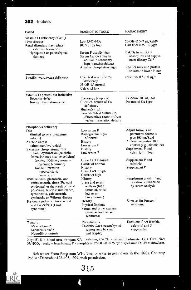
302Rickets
CAUSE DIAGNOSTIC TOOLS MANAGEMENT
Vitamin D deficiency (Cont.)Liver diseaseRenal disorders may reduce
calcitriol formation:Hypoplasia or parenchymal
damage
Low 25-0H-D3BUN or Cr high
Serum P usually highScrum Ca low (may be
normal in secondaryhyperparathyroidism)
Alkaline phosphatase high
25-0H-D 5-7 yglkg/c1I2Calcitriol 0.25-1.0 pg/d
CaCO3 to restrict Pabsorption and supple-ment dietary Cal3
Restrict milk and proteinsources to lower P load
Specific hydroxylase deficiency Chemical results of Cadeficiency
25-0H-D3 normalCalcitriol low
Vitamin D present but ineffectiveReceptor defectNuclear translation defect
Calcitriol 0.5-1.0 pg/d
Phenotype (alopecia)Chemical results of Ca
deficiencyHigh calcitriolSkin fibroblast cultures to
differentiate receptor fromnuclear translation defects
Calcitriol 10 30 pg,e1Parenteral Ca 1 g/d
Phosphorus deficiencyDiet
(limited to very prematureinfants)
Antacid excess(aluminum hydroxide)
Excessive phosphaturia fromtubular dysfunction (calcitriolformation may also be deficient)
Isolated, X-linked normo-calciuric (common)
Isolated, recessivehypercalciuric(very rare7)
With acidosis, glucosuria, andaminoaciduria alone (Fanconisyndrome) or the result of metalpoisoning, fructose intolerance,tyrosinemia, galactosemia,cystinosis, or Wilson's disease
Fanconi syndrome plus cerebraland eye defects (Lowesyndrome)
1.cw serum PRadiographic signs
of ricketsHistoryI.ow serum PHistoryLow serum P
Adjust formula orparenteral sourcc togive 100 mg/kgid
Alternative gastric HOcontrol (e.g., cimetidine)
Supplement P andcalcitrioP1 if low
Urine Ca:Cr normal Supplement P andCalcitriol normal calcitriolHistory Supplement PUrine Cai Cr highCalcitriol highHistory Supplement alkali. P andUrine and serum calcitriol as indicated
analysis (high by scrum analysisserum chloridelow serumbircarbonate)
History Same as for FanconiPhysical findings syndromeSerum and urine analysis
(same as for Fanconisyndrome)
TumorsMesenchymal5Sebaceous nevi",Neurofibromastosis
PhosphaturiaCalcitriol low (mesenchymal
tumors may be smalland cryptic)
Excision; if not feasible,calcitriol and Psupplement
Key: BUN = blood urea nitrogen: CA = calcium; CaC01 = calcium carbonate: Cr = Creatinine;NaHCO3= sodium bicarbonate; P = phosphorus; 25-OH-D3= 25 hydroxyvitamin D; UV = ultraviolet.
Reference: From Bergstrom WH: Twenty ways to get rickets in the 1990s. ContempPediatr December.102-103, 1991, with permission.
315
4

SCROTUM
The Infant and Child with Acute Scrotum
The presentation of an infant or child with an enlarged, tender, and dis-colored scrotum is generally a call for alarm, both on the part of the patient andthe physician. It is essential to diagnose quickly and correctly the patient withan acute scrotum to avoid irreversible testicular injury. This is particularly thecase w;.th the most common cause of acute scrotum in children, torsion of thetesticle.
Many urologists recommend testicular radionucleotide scans and Dopplerand scrotal ultrasound examinations to confirm the diagnosis of the acutescrotum. A simpler means to differentiate torsion from less threatening problemssuch as epididymitis and orchitis, however, is to test for the cremasteric reflexthe reflex is almost always absent in a patient with torsion (see also p. 304).
Causes of the Acute Scrotum
1 . Testicular torsiona. Extravaginal torsion is found exclusively in neonates. It is caused by a
twisting of the spermatic cord above the tunica vaginalis, thus cutting offthe blood supply. Transillumination can be useful in distinguishing extra-vaginal torsion from hydrocele.
b. I ntravaginal torsion is a twisting of the testes within the tunica vaginalis.It can occur anytime in life but is most common in the prepubertal andpostpubertal male.
2. Epididymitis3. Torsion of testicular appendages yields localized tenderness at the upper pole
of the testis or epididymis. A "blue-dot" sign is pathognomonic of this entity,representing the necrotic appendage beneath the skin.
4. Orchitis
5. Strangulated inguinal hernia
6. Idiopathic scrotal edema
7. Henoch-Schönlein purpura
8. Tumor9. Trauma
10. Extrascrotal disease (e.g., intraabdominal sepsis and formation ofa pyocele).
Reference: Hermann D: The pediatric acute scrotum. Pediatr Ann 18:198- 204, 1989.
3033 6

304Seizures
The Importance of the Cremasteric Reflexin Acute Scrotal Swelling in Children
The importance of accurate and rapid diagnosis of the acute scrotum cannot beoveremphasized. There exists a wide variety of diagnostic modalities that have beenreported to improve assessment and dictate which patient should undergo surgicalexploration. The most valuable aid in differentiating testicular torsion (whichrequires rapid correction to avoid testicular damage) from other causes of acutescrotal swelling remains the presence or absence of the cremasteric reflex. (The tes-ticles are suspended by the cremaster muscle [from the Greek kremaster, to hand.
In a prospective study of 245 boys, from the newborn period to age 18; whopresented with acute scrotal swelling, the presence of the cremasteric reflex(stroking the inner thigh to cause an elevation of the ipsilateral testis bycontraction of the cremaster muscle) was the most reliable clinical finding inruling out testicular torsion. The correlation between the presence of ipsilateralcremasteric reflex and the absence of testicular torsion was 100%. Absence of thisreflex, therefore, should strongly increase your suspicion of torsion.
Cremasteric Reflex in Acute Scrotal Swelling
DIAGNOSIS NO 01 S
CREMASTERIC REFLEX
PRESENT ABSENT
Testis torsion 56 0 56
Hydatid torsion 77 58 19
Epididymitis 47 31 16
Hernia/ hydrocele 22 8 14
Trauma 22 19 3
Leukemia 5 0 5
Varicocele 4 4 0
Orchialgia 4 2 2
Idiopathic scrotal edema 3 1 2
Henoch-Schonlein purpura 3 1 2
Testis tumor 1 0 1
Insect bite 1 1 0
245 125 120
Reference: Rabinavitz R: The importance of the cremasteric reflex in acute swelling inchildren. J Urol 132:89-90, 1984.
Common Causes
Febrile seizuresIdiopathic seizures
SEIZURES
Uncommon Causes
CNS infectionAseptic meningitisBacterial meningitisViral encephalitis
3 7
CNS injuryAnoxic encephalopathyChild abuseConcussionHemorrhage
Hypoglycemia

Rare Causes
CNS infectionCongenital infectionParasitic infectionSyphilisTetanusTuberculosis
Congenital CNS malformationAgenesis/dysgenesis
HoloproscencephalyPorencephaly
Hydrocephalus
Drugs/toxinsAminophyllineAmphetaminesAntihistaminesAtropineCamphorCarbon monoxideDrug withdrawalHeavy metalsHexachlorophineHydrocarbonsLocal anestheticsNarcoticsOrganophosphatesPenicillinPertussis toxoidPhencyclidineScabicidesSteroidsTricyclic antidepressants
Inborn errors of metabolismAminoacidopathyGalactosemiaOrganic aciduriaStorage disease
MetabolicHypernatretniaHypocalcemiaHypomagnesemiaHyponatremia
MiscellaneousArrhythmiaDysmorphogenic syndromes (many)KernicterusMetachromatic leukodystrophyPyridoxine deficiency
Seizures-305
Miscellaneous (Cont.)Rett syndromeReye's syndromeSubacute sclerosing
panencephalitis
Neurocutaneous syndromeIncontinentia pigmentiLinear sebaceous nevusNeurofibromatosisSturge-Weber diseaseTuberous sclerosis
Seizure mimicsBreathholding spellsHyperventilationMalingeringMasturbationMigraineMyoclonusNarcolepsyOrthostatic hypotensionPallid infantile syncopePanic disorderParoxysmal torticollis of infancyPseudoseizuresSandifer's syndrome (gastro-
esophageal reflux)Shivering on urinationShuddering attacksSleep disordersSyncopeTicsVertigo
Systemic infectionRoseolaShigella
Tumors
VascularA-V malformationEmbolic phenomenonHemorrhageHypertensionSickle-cell diseaseThrombosisVasculitis
3 1

306Seizures
Classification of Seizures and Epilepsy
Seizures and epilepsy are classified according to a scheme developed by theInternational League Against Epilepsy (ILAE). An abbreviated version of theclassification is shown below:
Classification of Seizures and Epileptic Syndromes
Partial seizuresSimple partial seizures (consciousness preserved)
With motor signs (jacksonian, adversive)With somatosensory or special sensory symptomsWith autonomic symptoms or signsWith psychic symptoms
Complex partial seizures (consciousness impaired)Simple partial onset followed by impaired consciousnessImpaired consciousness at onset
Secondarily generalized seizuresSimple partial seizures evolving to generalized tonic-clonic seizures
Complex partial seizures evolving to generalized tonic-clonic seizuresSimple partial seizures evolving to complex partial seizures, then to
generalized tonic-clonic seizures
Generalized-onset seizuresTonic-clonic seizuresAbsence seizuresAtypical absence seizuresMyoclonic seizuresTonic seizuresAtonic seizures
Localization-related (focal) epilepsiesIdiopathic
Benign focal epilepsy of childhoodSymptomatic
Chronic progressive epilepsiapartialis continua
Temporal-lobe epilepsyExtratemporal epilepsy
Generalized epilepsyIdiopathic
Benign neonatal convulsionsChildhood absence epilepsyJuvenile myoclonic epilepsyOther generalized idiopathic
epilepsyCryptogenic or symptomatic
West syndromc (infantile spasms)Early myoclonic encephalopathyLennox-Gastaut syndromeProgressive myoclonic epilepsy
Special syndromesFebrile seizures
Reference: Scheuer ML, Pedley TA: The evaluation and treatment of seizures. N Englj Med 323:1468-1474, 1990. Table reproduced from cited paper, with permission.
What Are the Criteria for Simple Febrile Seizure?
Age 6 months to 6 yearsGeneralized seizure (indicating involve-
ment of both cerebral hemispheres)of less than 20 minutes duration
Occurs within 24 hours of fever onset
Normal results from neurologic anddevelopmental examination
Negative family history of afebrileseizures
Reference: Schweich P: Emergency medicine except poisoning. In Oski FA, et al (eds):Principles and Practice of Pediatrics. Philadelphia, J.B. Lippincott, 1990, p 771.
3 1, 9

Seizures-307
To Treat or Not to Treat After a First Seizure?
Should the child receive antiepileptic drug therapy after a nonfebrile firstseizure? This is a central and still controversial issue in the management ofepilepsy. It begs the follow-up question, why treat seizures?
Seizures are treated mainly because of their psychosocial consequences.Children with epilepsy often have difficulty with interpersonal relationships andself-esteem, as well as vocational problems later in life. There are reports ofslightly increased mortality in association with seizure disorders, but there is yetno proof that patients on medication have less mortality than untreatedindividuals. Because 25-41% of patients taking antiepileptic drugs have recurrentseizures, the effectiveness of antiepileptic drugs in preventing recurrence ofseizures has also been questioned. Also, there is no evidence at this time thatseizures beget further seizures.
So which children should be treated'?The decision to treat should be based on several factors, including:
AgeType of seizureFrequency of seizures and time betweenTiming and circumstance of occurrence of seizuresRisk of further occurrencePrecipitating factorsRisk of drug treatment (30% of patients have side-effects requiring
modification of therapy)Probable consequences of further seizuresProbability of treatment success
The chance of having an additional seizure after the first is about 30% (range16-62%), and the second seizure tends to occur within 12 months. In childrenwith absence seizures, the occurrence is at the high end of the range. The chanceof having an additional seizure after the second is 50-75%. Studies of predictorsof seizure recurrence have reported that it is often possible to identify patientswith a relatively low risk of seizure recurrence.
Although the final decision to treat or not to treat must be made individuallyfor each patient, the following guidelines may be offered:
I. Treat a child who has experienced two or more tonic-clonic seizures.2. Treat children who experience seizures that impair consciousness, such as
absence and partial complex seizures. which tend to occur more oftensometimes daily --than the rare generalized tonic-clonic seizures and canimpair function because of their frequency.
References: Scheuer ML, Pedley TA: The evaluation and treatment of seizures. N EnglJ Med 323: 1468 1474, 1990,
Shinnar S. Berg AT, Moshe SL, et al: Risk of seizure recurrence following a firstunprovoked seizure in childhood: A prospective study. Pediatrics 85:1067 1085, 1990.
van Donselaar CA, Geerts AT, Schimscheimer R.1: Idiopathic first seizure in adult life:Who should be treated? Br Med J 302:620 623, 1991.
Vining EPG, Freeman JM: Management of childhood seizures. In Asbury AK, et al:Diseases of the Nervous System. Philadelphia, W.B. Saunders, 1986, pp 1018 1032.
Hauser WA, Anderson VE, Loewenson RB, McRoberts SM: Seizure recurrence aftcra first unprovoked seizure. N Engl .1 Med 307:522-528, 1982.
3 1 0

308-Sexuality
SEXUALITY
Sexual Behavior in Children: What's Normal?
Questions frequently arise in the clinic surrounding the topics of sexuality andsexual behavior in children. More often than not, these questions reveal a greatdeal about the comfort level and value system pa;ents attach to this developmentalissue. To aid in this dialogue, listed below are the frequencies of a large varietyof sexual behaviors noted among 880 preadolescent boys and girls (ages 2 -12).
Frequency of Sexual Behaviors (Percent Endorsement)
NO. ITEM (ABBREVIATEED) OVERALL2 6,
BOYS2 6,
GIRLS7 12,BOYS
" 12CIRLS
10. Puts mouth on sex parts 0.1 0.4 0.0 0.0 0.015. Asks to engage in sex acts 0.4 1.2 0.0 0.0 0.6
7. Masturbates with object 0.8 0.8 0.8 0.0 1.7
17. Inserts objects in vaginalanus 0.9 0.0 2.8 0.0 0.69. Imitates intercourse 1.1 0.8 0.4 2.4 1.1
14. Sexual sounds 1.4 0.4 0.8 3.9 0.6
30. French kisses 2.5 1.6 4.0 2.4 1.7
28. Undresses other people 2.6 4.4 4.4 0.5 0.0
29. Asks to watch explicit television 2.7 0.0 1.6 6.8 3.4
19. Imitates sexual behavior with dolls 3.2 0.8 4.0 1.5 7.5
2. Wants to be opposite sex 4.9 7.3 7.5 1.9 1.1
22. Talks about sexual acts 5.7 2.4 2.8 9.2 10.3
I. Dresses like opposite sex 5.8 6.0 9.5 3.4 2.9
8. Touches others' sex parts 6.0 8.9 5.6 4.9 4.0
16. Rubs body against people 6.7 8.5 8.3 4.4 4.6
31. Hugs strange adults 7.3 6.5 14.3 2.4 4.0
32. Shows sex parts to children 8.1 15.7 7.5 4.4 2.3
62. Uses sexual words 8.8 4.8 1.2 19.9 12.1
33. Overly aggressive, overly passive 10.4 8.1 17.5 6.3 8.6
27. Talks flirtatiously 10.6 8.5 15.9 2.9 14.9
13. Pretends to be opposite sex 13.0 16.9 20.6 2.9 8.0
4. Masturbates with hand 15.3 22.6 16.3 11.2 8.6
21. Looks at nude pictures 15.5 11.3 7.9 27.2 18.4
20. Shows sex parts to adults 16.0 25.8 17.9 9.7 6.9
3. Touches sex parts in public 19.7 35.5 19.0 15.5 2.9
34. Interested in opposite sex 23.0 21.0 20.6 19.9 32.8
18. Tries to look at people undressing 28.5 33.9 33.3 27.7 14.9
6. Touches breasts 30.7 43.5 48.4 11.7 9.2
26. Kisses nonfamily children 33.9 41.1 55.2 9.7 21.3
23. Kisses nonfamily adults 36.2 41.1 52.4 18.9 26.4
25. Sits with crotch exposed 36.4 35.1 59.1 15.5 29.9
24. Undresses in front of others 41.2 49.6 61.9 21.4 23.0
I I. Touches sex parts at home 45.8 64.1 54.4 36.4 18.4
5. Scratches crotch 52.2 58.1 67.9 40.8 34.5
35. Boy-girl toys 53.9 63.3 71.4 30.6 42.5
Additional items (Dec-Jan)42. Touches animal sex parts 1.3 4.5 0.0 0.0 0.0
37. Mouth on mother's breast 2 6 0.0 7.7 0.0 0.0
Table connnued on next page.

Short Stature-309
Frequency of Sexual Behaviors (Percent Endorsement) (Corn.)
NO. ITEM (ABI3REVIATEED) OVERALL2-6,
BOYS2-6,
GIRLS7-12,BOYS
7-12GIRLS
40. Overly friendly with strange men 7.1 4.5 11.5 2.9 8.0
36. Stands too close 11.6 6.8 15.4 14.7 8.0
41. Shy about undressing 38.7 29.5 32.7 50.0 52.0
43. Walks around nude 41.9 47.7 65.4 20.6 12.0
38. Walks around in underwear 52.9 54.5 75.0 44.1 16.0
39. Shy with strange men 64.5 63.6 80.8 47.1 56.0
Reference: Friedrich WN, et al: Normative88:456 -464, 1991, with permission.
sexual behavior in children. Pediatrics
SHORT STATURE
Use of Bone Age Determination in the Diagnosis of Short Stature
The cause for short stature may often be determined by careful history andphysical examination. Nutritional or emotional deprivation, chronic disease, ora history of short stature in other family members may provide an explanationfor decreased height. Facial appearance may suggest a genetic or chromosomal
abnormality. Organ enlargement may lead to a diagnosis of a storage disease.Often, however, the diagnosis is not readily apparent. In these cases, it is
helpful to begin with a comparison of skeletal maturation (bone age) to heightage and chronologic age. The table lists the diagnoses that should be suggested
by such a comparison, and the clinical features accompanying each diagnosis.
Comparison of Bone Age to Height Age and Chronologic Age
MEASUREMENT DIAGNOSIS SUGGESTED CLINICAL FEATURES
Bone age equal to orslightly behindchronologic age
Primordial short stature Birth weight and length belownormal for gestational age.
Subsequent growth parallel to,but below, 3rd percentile.
Normal onset and progressionof puberty.
Minor skeletLI abnormalities.Includes genetic and chromo-
somal aberrations, e.g.,Down's syndrome andTurner's syndrome.
Short stature as adult.
Familial short stature Normal length and weight forfirst I to 2 years of life.
Height falls below 3rd percen-tile at 5 to 10 years of age.
Puberty not delayed."Normal" adult height not
attained.
able continued on next page.
3 ?

31 0 Short Stature
Comparison of Bone Age to Height Age and Chronologic Age (Cont.)
MEASUREMENT DIAGNOSIS SUGGESTED CLINICAL FEATURES
Bone age retarded inrelation to chrono-logic age, but lessretarded thanheight age
Constitutional short stature Appropriate weight andlength for gestational ageat birth.
Slow growth during child-hood.
Delayed onset of puberty.Other family members may
remember similar growthpattern.
Important to differentiate fromhypothyroidism and growthhormone deficiency.
Ultimately reach "normal"adult height.
Metabolic disorders, e.g.:Hypophosphatemic
ricketsHypophosphatasiaMucopolysaccharidosesGlycogen storage
diseasesRenal tubular acidosisBanter's syndromeVasopressin-resistant
diabetes insipidus
Clinical and laboratory findingsconsistent with thesedisorders.
Organic acidemias andacidurias
Hemolytic anemiasDisorders of mineral
metabolismImmunoglobulin or
white blood cellabnormality
Others
Clinical and laboratory findingsconsistent with thesedisorders.
Chronic disease, e.g.:Chronic infectionHepatic diseasePulmonary diseaseRenal diseaseMalabsorptionMalignancyCollagen vascular
diseaseOthers
Clinical and laboratory findingsconsistent with the disease;initial clue may be increasederythrocyte sedimentationrate.
May exhibit variable growthrate over several years.
Bone age equal toor advanced incomparison withheight age
Familial short stature See above.
3 ;.!3
Mtge continued on next page.

.1*
Short Stature-311
Comparison of Bone Age to Height Age and Chronologie Age (Cont.)
MEASUREMENT DIAGNOSIS SUGGESTED CLINICAL FEATURES
Bone age equal toor advanced incomparison withheight age (Cont.)
Sexual precocity withandrogen excess
Increased linear growth early inlife with early closure ofepiphyses.
Clinical signs of androgenexcess (facial, axillary, andpubic hair, penile or clitoralenlargement).
Sexual precocity withestrogen excess
Early closure of epiphyseswithout prior augmentationof linear growth.
Clinical signs of estrogen excess(breast enlargement, galactor-rhea in females, and so on).
Bone age greatlydecreased and lessthan or equal toheight age
Hypothyroidism
Cushing's syndrome(most often iatrogenic)
Hypopituitarism andgrowth hormone defi-ciency. Causes include:Congenital absence of
pituitaryInfectionReticuloendotheliosesVascular infarcts and
anomaliesTraumaIrradiationSurgical resectionMalnutrition
Degree df growth retardationdepends upon age of onset.
Congenital hypothyroidism isassociated with severegrowth failure.
In juvenile hypothyroidism, thegrowth retardation is moreinsid ious.
Delayed dental age.
Truncal obesity, moon facies,violaceous striae, hirsutism,muscle weakness,hypertension.
Delayed dental age.Puberty often delayed.May have neurologic
abnormalit ies.
Maternal deprivation May have impaired motor andintellectual development.
May or may not be associatedwith malnutrition.
May have growth hormonedeficiency.
Reference: Gotlin RW, Mace .1 W: Diagnosis and management of short stature inchildhood and adolescence. Curr Probl Pediatr 2:4, 1972.
3 )

312Shwachman's Syndrome/Sinuses
SHWACHMAN'S SYNDROME
Pancreatic Insufficiency and Neutropenia
When Shwachman's syndrome was first described in 1964, the hallmarks ofthis rare entity were exocrine pancreatic insufficiency, bone marrow hypoplasiaand associated neutropenia, metaphyseal chondroplasia, growth retardation, andrecurrent soft tissue infections. Since that initial case report, many moremanifestations of Shwachman's syndrome have been elaborated and described.These protean features of the disorder are listed in the table below.
The exact pathogenic basis for the hematologic and other features of thismultisystem illness has yet to be determined, although some have hypothesizedthat the basic defect of the Shwachman syndrome may lie in the function of themicrotubular and microfilament elements of many different cell types in the body.The relative contributions of impaired c..tIlular motility, instead of neutropenia,to these patients' increased susceptibility toward infections is also unclear.
Features Associated with Shwachman's Syndrome
Exocrine pancreatic insufficiencySteatorrhea
Growth retardationSkeletal abnormalities
Metaphyseal dyschondroplasia, delayedmaturation, rib abnormalities, longbone tubulation, clinodactyly
Narrow thoraxHematologic abnormalities
Bone rnarow hypoplasia, neutropenia,thrombocytopenia, raised HbF,lymphoproliferative and myelo-proliferative neoplasia
Recurrent infectionsDefective neutrophil mobility
Neonatal problemsPoor feeding, respiratory
distressPsychomotor retardationHypotoniaHepatomegaly
Raised SGOT and SG PTRenal tubular dysfunctionichthyosisDental abnormalitiesDelayed pubertyDiabetes mellitusDysmorphic featuresEndocardial fibrosisHirschsprung's disease
References: Shwachman H, Diamond LK, Oski FA, Khow KT: The syndrome ofpancreatic insufficiency and bone marrrow dysfunction. J Pediatr 65:645, 1964.
Aggett PJ, Cavanagh NPC, Matthew DJ, et al: Shwachman's syndrome: A review of21 cases. Arch Dis Child 55:331, 1980.
Anderson DC, et al: Quantitative and functional disorders of granulocytes. In Oski FA,et al (eds): Principles and Practice of Pediatrics. Philadelphia, J.B. Lippincott, 1990, p 1535.
SINUSES
The Paranasal Sinuses and the Mastoid Sinus
At what age does sinusitis become a diagnostic possibility?It is useful to remember the ages at which the sinuses are pneumatized. Once
a true sinus is present, the possibility of infection exists.
Sinuses present at birth Anterior and posterior ethmoid.Maxillary antra.
3?5

Skin Signs/Sleeping Patterns-313
Two to four years Pneumatization of frontal sinsuses beginscomplete by 5 to 9 ycars of age.
Sphenoid sinus becomes visible by age 3.
The mastoid antrum is present at birth, and pneumatization of the temporalbone starts in early infancy. The mastoid process is not present at birth, but beginsto grow during the first year. Pneumatization is a slow, irregular process, but isgenerally complete prior to adolescence.
Sinusitis is seen with increased frequency in patients with cyanotic heartdisease, in leukemia and aplastic anemia while patients are neutropenic, in cysticfibrosis, and in patients with a history of nasal allergies.
Reference: McMillan JA, et al (cds): The Whole Pediatrician Catalog. Philadelphia,W.B. Saunders, 1977, pp 190 -191, with permission.
SKIN SIGNS
Tache Cérebrale and Dermatographia
Diagnostic information can be obtained from stroking the skin. When theskin over the abdomen, back, or chest is gently stroked with the fingernailor a blunted point, two major responses may be elicited: (1) tache cérébrale and(2) dermatographia.
In tache cerebrate (cerebral spot), the stroking produces a red streak that isflanked by thin, pale margins. This sign develops within 30 seconds of strokingand persists for several minutes. It has been noted to be present in patients withscarlet fever, hydrocephalus, a variety of febrile illnesses, and, most particularly,in meningitis. It can be used as an early clue to the presence of meningitis,particularly in the neonatal period. The French name derives from the presenceof the sign as a concomitant of several nervous (or "cerebral") diseases.
Dermatographia, meaning literally "writing on the skin," is the marking of theskin by rubbing with a blunted point at sufficient pressure. The stroking producesa white or pale line with red margins. This wheal is seen in patients with fair skin,in those with vasomotor instability, or in extreme form in patients with urticariapigmentosa (Darier's sign). Dermatographism, the tendency to show dermato-graphia, is present in 2- 5% of the population, but only a subgroup hassymptomatic dermatographism, one of the physical urticarias.
Reference: Martin GI: The significance of tache cérébrale in neonatal meningitis(letter). J Pediatr 87:322, 1975.
SLEEPING PATTERNS
Crying, Feeding, and Sleeping Patterns in infants 1 to 12Months of Age
What is normal crying time, feeding time, or sleeping time for infants? Mothersoften are concerned or complain that their baby is abnormal. With the followingguidelines you can either reassure them or be alerted to a possible problem.
3?6

314-Snake Bite
Mothers who feel a need for additional help generally have babies that cry formore than 6 hours per day, take more than 6.0 hours to feed, and spend less than7 hours sleeping.
Mean Times for Infant Activities
AC' rivn.y
MEAN TIMES IN HOURS (WT) H RANGES)
< 3 Mo 3 5 Mo 6-8 Mo > 9 Mo
Crying 1.6 1.3 1.4 1.1
(0 5.0) (0 9.5) (0- 3.0) (0 3.5)
Feeding 3.1 2.4 2.0 2.1
(1.0 6.8) (1.3 5.3) (1.0 3.8) (0.8 4.5)
Sleeping 15.2 14.3 13.5 13.4
(11.8 20.5) (10.0 18.5) (10.3 1 i.8) (10.3 16.0)
Reference: Michelsson K, et al: Child Care Health Dev 16:99 III, 1990.
SNAKE BITE
Is This a Poisonous Snake Bite?
This question is posed to physicians in offices and emergency rooms severalthousand times a year in this country. A correct and prompt answer is essentialfor proper treatment. Failure to use antivenom early when indicated can be fatal;its inappropriate employment for a bite by a harmless snake may be hazardousdue to severe reactions.
The problem has two parts. First, was the bite due to a harmless or apoisonous snake? Second, if the snake was venomous, is envenomation present orlikely? The question may be resolved by examination of the snake so that it maybe put in the category of either a venomous or a nonvenomous variety and byexamination of the patient to determine if venomation has occurred. Basicdependable guidelines for attaining both these goals will be outlined. They maybe used by the amateur who has no knowledge of serpents at all. This discussionapplies only to those snakes that are native to the continental United States anddoes not relate to foreign species introduced into this country as pets or exhibits.
Examination of the Snak
One should not attempt to identify the exact species, since this is often achallenge for even the genuine expert due to pitfalls involving confusing colorvariate (albinism and malanism) and deceptive patterns (atypical or absent).Undue delay may result by waiting to locate an available herpetologist in anearby zoo, museum, or zoology department. One should instead inspect thesnake and, from the guidelines provided, assign it to the harmless or harmfulgroup. The problem is somewhat simplified since in the mainland United Statesthere are only two families of indigenous poisonous serpents.
Crotalidae. These are the pit vipers, which include all rattlesnakes, cotton-mouths (water moccasin), and copperheach (highland moccasin). One or more ofthis family has been found in all states, with the exception of Maine, Alaska, andHawaii. The head is large and triangular. Thc neck is relatively slender, so thatit is readily distinguishable from the thick, heavy body. The pupil is vertically
3?7

Snake Blte-315
elliptical. Pit organs (loreal pits) are pat bognomonic of all members of this group.A pit is present on each side of the heaz and it resembles an extra nostril. Thepits are deep, readily visible between the eye and the nostril, and !ocated justbelow a line connecting these two structures. One or two fangs are found on theupper jaw of all pit vipers. They are specialized hollow or grooved teeth, whicharc recurved and longer than the othcr teeth. It is through these that the venomis injected. In this family the fangs are movable and when not in use arc foldedup against the palate. A white membrane may cover the fang down to the tip.Normally there are two fangs in the upper jaw One on each side of the maxillaso the classic bite pattern shows two fang punctures. However, one or both maybe broken ofr or shed, in which instance there may be only a single fang markpresent or none at all, No envenomation is possible if fangs are absent. Reservefangs are always present, so the missing fang is replaced soon. Rarely, one or tworeserve fangs may be functioning along with the customary complement of oneor two fangs. In such a circumstance the bite pattern will be atypical, demonstrat-ing three or four fang punctures.
Herpetologists identify species by meticulous scale counts of the head, neck,body, and tail. For practical purposes one may observe the scales (scutes, shields,plates) on the ventral surface of the body just posterior to the anus (subeaudalscales). In this family the subeaudal scales are usually arranged in a single row,but exceptions occur in which the rows are double. In the majority of harmlesssnakes the subcaudal scales are double, but this is not infallible either sinceexceptions are found. Rattles, of course, are specific for rattlesnakes and are notpresent in the copperhead, cottonmouth, other venomous species, or harmlesssnakes. They break off because of wear and tear, or during ecdysis (molting), sothe number is inconstant regardless of the age of the reptile. If all rattles havebeen lost or if the specimen is a baby that has not yet developed rattles, there willbe a slight enlargement at the tip of the tail known as the button, Other poisonoussnakes do not show a button, nor do nonvenomous species. Also, the end of thetail of a rattlesnake that has lost its rattles is short and blunt, whereas the tip ofthe tail of a harmless snake is usually gradually tapered.
Elapidoc This family is represented in this country only by the coral snake.Unlike the pit vipers the coral snake is restricted to the southern states and isgenerally not found north of' Arizona, Arkansas, or the Carolinas. Compared tothe rattlesnakes and moccasins, the head is narrow and the neck and body areslender, giving a cylindrical configuration which is quite different from the shapeof the pit vipers. The pupil is circular, thus resembling that of our indigenousharmless varieties. Pit organs are not present. Two fangs are present one oneach side of the maxilla (unless one or both have been shed). They arc erect,fixed, and smaller than those of the pit vipers. Subcaudal scales tend to be in adouble row similar to those of nonvenomous snakes, but rarely may be in a singlerow in the coral snake. Both rattles and tail button are absent. The coral snakeis an exception to the general rule of not trying species identification, since thereare confusing imitators that are harmless. Fortunately the nonvenomous mimicsarc easy to differentiate from the potent coral snake. The coral snake has a blacksnout and broad body rings of red and black that arc separated by a narrowerband of yellow. The mnemonic "red next to yellow kills a fellow" is helpful tokeep in mind. The harmless look-alikes (scarlet snake, scarlet king snake) showa grey or red snout and red and yellow rings that are separated by a black band.Here the mnemonic to remember is "red against black is venom lack."

316Snake Bite
Summary of Family CharacteristicsNative Harmless VersusNative Venomous Snakes In ihe Continental Untied States
Was to Differentiate Poisonous From Harmless Snakes
Poisonous (Pit vipers) Harmless
Poisonous
attle,nal.e. Rauh,
Andl plate Single rov. ,titx Judd! plate%
Opperhcad. A,: cottonmouths
NwAril Round pupil
Harmless
Anal plate Double ,on subeaudal plates
.' t:,;.7"
Nonvenomous Versus Venomous Characteristicsof Continental U.S. Snakes
NONVENOMOUS VENOMOUS
Oval Head Large and triangular (pit vipers); small andnarrow (coral snake)
Round Pupils Vertically elliptical (pit vipers); round (coralsnake)
Absent Pit organs Present in all pit vipers (copperhead, cotton-mouth, rattlesnakes); absent in coral snake
Absent Fangs Present in all venomous species. Large, long,recurved teeth. Long and movable in pitvipers. Short, erect, and fixed in coralsnake. Usually 2 (I on each side upper jaw)unless shed or reserve fangs also in use
Double row Subcaudal scales Single row in pit vipers, but with exceptions;usually, but (anus to tip of double row in coral snake, but exceptionsexceptions tail)
Absent Rattles Present in all rattlesnakes unless lost orundeveloped in baby. If missing a button,present at tip of tail. No rattles in othervenemous species or in harmless snakes
3 e' 9
Table continued on next page.

Snake Bite-317
Nonvenomous Versus Venomous Characteristicsof Continental U.S. Snakes (Cont.)
NONVENOMOUS VENOMOUS
Usually ends ingradual taper
Total of 6 with nofang marks; fourrows maxillary teethand 2 rows man-dibular teeth
Series scratches ortiny punctures(1-2 mm deep);pattern of mandi-bular teeth oftenimperfect
Tip of tail
Bite pattern(seldom perfect)
Number of rows
Appearance
Short and blunt in rattlesnake if rattleslost
Total of 4 with 1-2 fang marks (rarely 3-4if reserve fangs functional). Two rowswith fang marks from maxillary teeth.Two rows without fang marks frommandibular teeth
Series scratches or tiny punctures(1 2 mm deep) plus fang marks. Fangmarks recognizable as larger and deeperpunctures than those from nonfangteeth. Mandibular teeth often indistinct
Examination of the Patient
This is to determine if envenomation has taken place. If it has occurred, imme-diate vigorous therapy is required. If the snake was not captured, presence orabsence of envenomation will be the sole criterion available for deciding if thesnake was harmless or poisonous. Verbal descriptions of escaped snakes aregenerally unreliable. The bite pattern is helpful and may be diagnostic asindicated earlier, but it does not indicate if envenomation has in fact occurred.Even though the victim has been struck by a venomous serpent, envenomationmay not ensue. This is the result of various circumstances that influence the flowof venom, the amount of venom injected, and the toxicity of the venom.Evaluation of envenomation depends on the development of local and systemicsymptoms and signs. The following are the usual clinical effects which mayappear after injection of a sufficient amount of a potent venom.
Local Symptoms and Signs. The two P's (puncture and pain) and the two E's(edema and erythema) constitute the classic local reaction to deposition of a potentvenom in the tissues. At least two should be present to substantiate the diagnosis.
I. Puncture: One or two fang marks are present (rarely three or four if reserve fangsin use). These punctures are larger and deeper than those from the other teeth. Awheal or vesicle may develop at the site. If at least one fang mark is not present,then envenomation could not have taken place. Bleeding is usually brisk.
II. Pain: Usually develops within 5 to 10 minutes of the strike. It may be delayed up toan hour under certain conditions and may be lacking with a coral snake bite. In aclassic case of moderate to severe envenomation involving a pit viper, the painappears promptly and is severe and unremitting.
HI. Edema: Typically obvious within 5 to 10 minutes. It also may be delayed up to anhour or absent with a coral snake bite. The swelling may progress up the limbduring the next 36 hours and eventually reach the trunk. The overlying skinbecomes tense and shiny. The extent of the edema is one of the criteria used in theclinical grading of the severity of envenomation for assessing the amount of anti-venom required and for monitoring the progress of the case.
3 3 0

318Snake Bite
IV. Erythema: Redness is ordinarily visible within 5 to 10 minutes. It may not appear foran hour and may be absent after a bite by a coral snake. Later, other types of dis-coloration develop with pit viper bites as hemorrhages occur in the tissues. Eventuallysome blueness usually follows unlike the typical reaction to a severe insect bite.
NOTE: Exceptions in the time of appearance and number of these four cardin,,isigns occur in some pit viper bites owing to variability of potency andamount of venom injected. Also in the event of a fortuitous strike directlyinto a vessel, there may be absence of local manifestations along with therapid appearance of systemic signs. Local signs may be missing with coralsnake bites owing to the predominance of neurotoxin over hemotoxin.
V. Hemorrhage: Petechiae and ecchymoses commonly occur, particularly with pitviper bites. Oozing from fang marks often continues for several hours. This is incontradistinction to the wounds from nonfang teeth, which cease bleeding promptlywith both poisonous and nonpoisonous species.
VI. Paresthesias: Numbness and/or tingling frequently are noted at the bite site andaround the mouth.
VII. Late local signs: Tissue necrosis and thrombosis may develop, with sloughing oftissues and gangrene of the extremities. This type of response is common with pitvipers but less so with coral snakes due to the difference in the venoms. Localizedlymphadenopathy is a feature in some.
Systemic Symptoms and Signs. These are produced by the hematogenousor 'ymphogenous dissemination of the venom. Some of the toxins have enzymaticactivity. Owing to the multiplicity of the effects of these diverse protein mole-cules, the clinical manifestations are numerous and protean. A consistent clinicalpicture may not be present.
I. General: Lassitude, weakness, fatigue, nonwhirling dizziness, diaphoresis, sialorrhea,and the sensation of a "full" or "thick" tongue.
II. Pulmonic: Edema, respiratory failure, and death.
III. Cardiac: Hypotension, congestive failure, cardiac arrest, and death.
IV. Renal: Hematuria, proteinuria, azotemia, and renal failure with death.
V. Gastrointestinal: Nausea, emesis, hematemesis, and melena.
VI. Hematologic: Alterations of the coagulation system, with petechiae, ecehymoses,bleeding into subcutaneous and muscle tissues, hemorrhages into viscera, and bloodyeffusions into serous cavities. Laboratory determinations may demonstrate prolongedprothrombin time, thromboeytopenia, fibrinolysis, and prolonged bleeding andclotting times. Epistaxis, hematuria, hematemesis, and melena are common withsevere envenomation.
VII. Central nervous system: Headache, blurred vision, paresthesias, slurred speech,bulbar palsies, generalized convulsions, and paralyses of the extremities. Deeptendon reflexes are variable. The sensorium typically remains intact, with a lucidand oriented patient. Sometimes somnolence rnay be a feature, and occasionallyeuphoria is present if the individual has been dosed with the traditional snake biteremedy, whiskey. Disorientation and states bordering on delirium and mania mayoccur in some victims owing to the hysteria and snake phobia seen in some adultsfollowing exposure to a serpent.
VIII. Death: Fatalities are due to respiratory failure, cardiac decompensation, renalshutdown, hemorrhage, or irreversible shock.
3311
Snake Bite-319
The physician who has no knowledge of snakes can render an intelligentdecision about a snake bite by following the guidelines given regarding inspectionof the snake, observation of the bite pattern, and examination of the patient.Irrefutable proof of the poisonous nature of a snake native to the continentalUnited States includes the presence of fangs, pit organs, rattles, and a verticallyelliptical pupil. All of these are present in the pit vipers (rattlesnakes, copperheads,and cottonmouth moccasins). The coral snake lacks the pit organs and rattlesand has a circular pupil. Otherwise, all indigenous snakes with a round pupil areharmless. Fang marks are diagnostic of a venomous species and are larger anddeeper than the scratches or superficial punctures produced by the nonfang teeth.If the patient shows pain, puncture, edema, and erythema, envenomation hastaken place. These local signs appear within an hour of the bite, and systemicsigns develop later. Local manifestations may be absent with coral snake bite, inwhich case generalized signs and symptoms develop rapidly.
Specially prepared by Dr. William D. Alsever, Syracuse, New York. (From McMillanJA, et al (eds): The Whole Pediatrician Catalog, Vol. 2. Philadelphia, W.B. Saunders,1979, with permission.)
References:Conant R: A Field Guide to Reptile and Amphibians of Eastern and Central North
America. New York, Houghton-Mifflin, 1975.Dept. of the Navy, Bureau of Medicine and Surgery; Poisonous Snakes of the World:
A Manual for Use by the U.S. Amphibious Forces. Washington, D.C., U.S. GovernmentPrinting Office, 1968.
Stickel, WH: Venomous Snakes of the United States and Treatment of their Bites.Wildlife leaflet 339. Washington, D.C., U.S. Department of the Interior. Fish and WildlifeService, 1952.
Pope, CH, and Perkins, RM: Differences in the patterns of venomous and harmlesssnake bites. Arch Surg 49:331, 1944.
Parrish HM, et al: Snake bite: A pediatric problem. Clin Pediatr 4:237, 1965.Snyder R: Snake bite seminar. Am J Dis Child 103:117, 1962.Russell, FE, et al: Snake venom poisoning: Experiences with 550 cases. JAM A
233:341, 1975.Glass TG Jr: Snake bite. Hosp Med Am J Dis Child, July, 1971, p 31.Parrish H M, et al: Snake bite: Poisonous until proven otherwise. Patient Care, May
30, 1971, p 76.Russell FE: Injuries by Venomous Animals. National Clearinghouse for Poison
Control Centers, Bulletin for Jan Feb, 1967. Washington, D.C., U.S. Department ofHealth Education and Welfare, 1967.
Arnold RE: What To Do about Bites and Stings of Venomous Animals. New York,Collier Books, 1973.
Other readings:Auerbach PS, Geehr EC: Management of Wilderness and Environmental Emergencies,
2nd ed. St. Louis, C.V. Mosby, 1989.Boyer Hassen L: Reptile and arthropod envenomations. Occup Med Statc Art Rev
6:447 461, 1991.Kitchens CS: Envenomation by the Eastern coral snake: A study of 39 victims. JA MA
258:1615 1618, 1987.Klauber LM: Rattlesnakes: Their Habits, Life Histories, and Influence on Mankind,
abridged edition. Berkeley, CA, University of California Press, 1982.Pennell TC: The management of snake and spider bite in the southeastern United
States. Am Surgeon 53:198 204, 1987.
332

320Sodium/Spienomegaly
Russell FE, Banner W: Snake venom poisoning. In Rakel RE: Conn's CurrentTherapy. Philadelphia, W.B. Saunders, 1988, pp 1002-1005.
Stewart RM, et al: Antivenin and fasciotomy/debridement in the treatment of thesevere rattlesnake bite. Am J Surgery 158:543-547, 1989.
Tu AT: Rattlesnake Venoms: Their Action and Treatment. New York, Marcel Dekker,1982.
Wingert WA, Chan L: Rattlesnake bites in Southern California and rationale forrecommended treatment. West J Med 148:37-44, 1988.
SODIUM: HYPERNATREMIA
Hypernatremia is defined here as a serum sodium level higher than 145 mEq/L.
Common Causes
DiarrheaHigh environmental
temperatures
Uncommon Causes Rare Causes
Nephrogenic diabetes insipidusPostobstructive diuresisSalt poisoningSickle-cell nephropathy
SODIUM: HYPONATREMIA
Cushing's diseaseHypercalcemia
nephropathy
Hyponatremia is defined here as a sodium level lower than 130 mEci/L.
Common Causes
DiarrheaExcessive salt-free infusionsSyndrome of inappropriate
ADH secretion (SIADH)Water intoxication
Rare Causes
Uncommon Causes
Acute renal failureChronic renal failureCongestive heart failureHigh environmental
temperatures
Adrenal insufficiencyCirrhosisCystic fibrosis and excessive sweating
SPLENOMEGALY
Common Causes
Acute infections (bacterial, viral, rickettsia!, protozoal, spirochetal, myobacterial)Congenital hemolytic anemias
HemoglobinopathiesHereditary spherocytosisThalassemia major; thalassemia intermedia
333

Uncommon Causes
Congestive splenomegalyCyanotic congenital heart diseaseHodgkin's diseaseJuvenile rheumatoid arthritis
Rare Causes
Acquired autoimmune hemolytic anemiaAmyloidosisBeckwith-Wiedemann syndromeBrucellosisChronic granulomatous diseaseCongenital erythropoietic prophyriaDysgammaglobulinemiaHemophagocytic syndromesHistiocytosisHurler's syndrome and other
mucopolysaccharide disorders
Spoiled Child Syndrome-321
LeukemiaLupus erythematosusNon-Hodgkin's diseaseSevere iron-deficiency
Malaria (in the United States)Metastatic neuroblastomaMyelofibrosisOsteopetrosisSarcoidosisSerum sicknessSplenic cyst or hemangiomaStorage disease (e.g. Gaucher's,
Neimann-Pick)Wolman's disease
THE SPOILED CHILD SYNDROME
'The pediatrician is called upon to wear many hats. One of the more difficultroles is counselor to the parent concerned about a "spoiled" child. The conceptdemands differentiation between normal behavior patterns and the excessiveself-centeredness that marks the spoiled child. The cause of the spoiled childsyndrome is often not a lack of discipline by the parents, but a lack of consistentlimit-setting.
Age-Related Normal Behavior Pattern
The Crying Infant. Brazelton's time-honored study of crying in infancy(Pediatrics 29:579-588, 1962) indicated that the average infant cried 21/4 hoursper day for the first 7 weeks of life. Whether due to hunger, colic, or want ofattention, the infant's cry represents a genuine need to which the parent ought tobe encouraged to respond. After 3 to 4 months of age, a cry may become amanipulation demanding modification techniques on the rrt of the parent.
The Exploring Toddler. As the infant discovers his or mobility, curiositybecomes infinite. The "search and destroy" or "baby taste test" activities canfrighten or aggravate the most equanimitous parent. An understanding of thisstage as a normal part in development will help the parent to "child proof" thehome and to begin the process of setting limits for the child.
The Terrible Two's. As the child approaches 2 years of age, his sense ofautonomy is beginning to emerge. This is often the time that conflicts betweenthe parent and the newly assertive child begin. The characteristic independenceand resistance to parental authority can be dampened by a variety of techniques.One way to avoid confrontation is to offer the child choices within the parent's
334

322Spoiled Child Syndrome
limits. Thus, both the parent and the emerging individual maintain a sense ofcontrol.
As with most human behaviors, those of the normal child are on a continuumwith those labelled disruptive or spoiled. The family environment, the presence ofstressors, and the individual's inherent coping abilities can shift the balance. Inthe absence of childhood handicaps or family stresses, such as separation anddivorce, parental alcoholism, or parental mental illness, there are certainbehavior patterns that do not fit any but the "spoiled" category. In these cases,the parent needs both guidance and assurance that the setting of consistent limitsand appropriate punishments for infractions can be effective.
Behavior Patterns Suggestive of True Spoiling
Trained Night Feeding. Beal's 1969 study on night feeding concluded thatby 4 months of age, 95% of infants should sleep through the night without afeeding. The older infant who continues to cry for a 2 A.m. meal is often the childof caring parents who have attended every cry with a breast or bottle. Failure tobreak the snacking cycle and use cuddling or a pacifier can lead to the develop-ment of spoiling.
Trained Night Crying. As with trained night feeding, trained night cryingrepresents the infant's training of the parent. The new parent in particular isloathe to leave a crying infant in bed. But here, as with other infant behaviors, the"need" has to be distinguished from the "want" if the parent desires rest. Ahelpful hint might be placing the infant in his or her crib while still awake withan assurance that 10 to 15 minutes of crying is part of the infant's own settlingmechanism. Trained responsiveness from the parent can diminish the baby's ownability to achieve sleep.
Recurrent Temper Tantrums. A tantrum is a "fit of bad temper" representingboth anger and frustration. They generally surface in the toddler who isattempting to assert independence and can be a frightening spectacle for theparent. Tantrums, like limitations, are occasionally inevitable. If the ---itrum isrewarded by the loosening of restrictions, it is apt to recur. Reassure the parentsthat though the desire to please their child may be strong, ignoring the tantrumwill likely bring this behavior to an end without harming the child. Withtantrums, as with other disruptive behaviors, a little anticipatory guidance can goa long way.
The Toddler Who Is Out of Control. This is often a child whose parents have"tried everything" to modify the kicking, biting, refusals to eat, sleep, or toilettrain. If the modifications have been inconsistent, so will be the responses.Instruction in the use of "time out" and the importance of its regular use shouldhelp the troubled parents. Remind the parents that the un-training of disruptivebehaviors will not be miraculous and will require patience, but that withcontinued enforcement, they will be successful.
In summary, the parents of the unruly child need guidance and support. It isprobably best not to label a child "spoiled" but to stress the normal behaviorpatterns of children at various ages and to offer solutions for avoiding orcorrecting disruptive behaviors. By emphasizing consistency and de-emphasizingworry you can help the parents learn the effectiveness of limits.
Reference: McIntosh BJ: Spoiled Child Syndrome. Pediatrics 83:108 115, 1989.
335
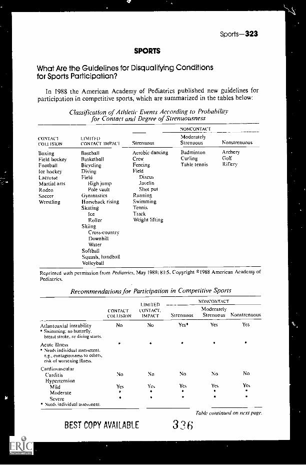
Sports-323
SPORTS
What Are the Guidelines for Disqualifying Conditionsfor Sports Participation?
In 1988 the American Academy of Pediatrics published new guidelines forparticipation in competitive sports, which are summarized in the tables below:
Classification of Athletic Events According to Probabilityfor Contact and Degree of Strenuousness
CONTACTCOLLISION
BoxingField hockeyFootballIce hockeyLacrosseMartial artsRodeoSoccerWrestling
LIMITEDCON FACT I M PAC I Strenuous
NONCONTACT
ModeratelyStrenuous
BaseballBasketballBicyclingDivingField
High jumpPole vault
GymnasticsHorseback risingSkating
IceRoller
SkiingCross-countryDownhillWater
SoftballSquash, handballVolleyball
Aerobic dancingCrewFencingField
DiscusJavelinShot put
RunningSwimmingTennisTrackWeight lifting
BadmintonCurlingTable tennis
Nonstrenuous
ArcheryGolfRiflery
Reprinted with permission from Pediatrics, May 1988; 81:5. Copyright ©1988 American Academy ofPediatrics.
Recommendations for Participation in Competitive Sports
CONTACTCOLLISION
LIMITEDCONIAcT,
IMPACT
NONCONTACT
ModeratelyStrenuous Strenuous Nonstrenuous
Atlantoaxial instability* Swimming: no butterfly,
breast stroke, or diving starts.
Acute illness* Needs individual assessment.
e.g., contagiousness to others,risk of worsening illness.
CardiovascularCarditisHypertension
MildModerateSevere
Needs individual assessment.
No
No
Yes
No
No
Yes
Yes*
No
Ycs
Yes Yes
No
Yes
No
Yes
BEST COPY AVAILABLE 336Table continued on next page.

324Sports
Recommendations for Participation in Competitive Sports (Cont.)
LIM ITFCONTACI CONTACT ModeratelyCOLLISION IMPACT Strenuous Strenuous Nonstrenuous
NONCONTACT
Cardiovascular (Cont.)Congenital heart diseasePatients with mild forms can beallowed a full range of physical activi-ties: patients with moderate or severeforms, or who are postoperative,should he evaluated by a cardiologistbefore athletic participation.
EyesAbsence or loss of function
of one eye * * * *+ t
Detached retina + r
Availability of American Societyfor Testing and Materials (ASTM)-approved eye guards may allowcompetitor to participate in mostsports, but this must be judged onan individual basis.Consult ophthalmologist.
Inguinal hernia Yes Yes Yes Yes Yes
Kidney: Absence of one No Yes Yes Yes Yes
Liver: Enlarged No No Yes Yes Yes
Musculoskeletal disorders * * *
Needs individual assessment.
NeurologicHistory of serious head or spine
trauma, repeated concussions,or craniotomy * * Yes Yes Yes
Convulsive disorderWell controlled Yes Yes Yes Yes Yes
Poorly controlled No No Yes+ Yes Yes:Needs individual assessment.
: No swimming or weight lifting.No archery or riflery.
Ovary: Absence of one Yes Yes Yes Yes Yes
RespiratoryPulmonary insufficiency * * * * Yes
Asthma Yes Yes Yes Yes YesMay be allowed to compete if oxygen-ation remains satisfactory during agraded stress test.
Sickle cell trait Yes Yes Yes Yes Yes
Skin: Boils, herpes, impetigo, scabies * * Yes Yes Yes* No gymnastics with mats, martial arts,
wrestling, or contact sports until notcontagious.
Spleen: Enlarged No No No Yes Yes
"lbsticle: Absence or undescended Yes' Yes* Yes Yes Yes' Certain sports may require protective cup.
Reprinted with permission from Pediatrics, May 1988; 81:5. Copyright (4) 1988 American Academyof Pediatrics.
Reference: DuRant RH, et al: Findings from the preparticipation athletic examinationand athletic injuries. Am J Dis Child 146:85 -91, 1992.
337BEST COPY AVAIL ARE

Sports-325
Menarche and Menstruation in the Athlete
The Committee on Sports Medicine of the AAP has recommended that anymedical evaluation of a female athlete should include a focus on menstrualhistory Pubertal development appears to be delayed in thin athletes, especiallyballet dancers and runners.
16
15
Age of 14
MenarchrIn Years 13
12
11
10
^
Sedentary High College Olympic BalletControls School Athletes Athletes, Dancers
Athletes 1976
Exercise arI age of menarche. Pubertal development appears to be delayedin thin athletes.
16
15
Age of 14
MenarcheIn Years 13
Athletes Trained Athletes TrainedBefore Menarche Postmenarcheally
Athletes who began their training premenarcheally experienced a delay inmenarche.
PREMENARCHAL DELAY INATHLETIC TRAINING MENARCHE BY
X 1 YEAR 5 MONTHS
For each year of training before menarch, menarche is delayed by 5 months.
338

326Stool
When should initiation of pubertal changes in an athlete be consideredabnormal? The following guidelines are helpf ul:
I. If no pubertal changes occur by the chronologic age of 13 years (twostandard deviations outside the normal variation), examination should bedone to rule out thyroid abnormalities, prolactin-secreting adenomas,ovarian dysgenesis, and chromosomal abnormality.
2. If there is no period by the age of 16 with some pubertal growth, then adefinite search must be made for anatomic causes of amenorrhea andmullerian agenesis.
3. The AAP Committee on Sports Medicine has recommended that a work-up be instituted if menarche is delayed by 1 year beyond the age of onsetof menses of other female family members.
4. If the patient, her parents, or coach are anxious about late menarche ordelayed puberty, a limited individualized work-up should be offered.
Some common causes of delayed menarche are listed below:
Some Common Causes of Delayed Menarche
HypothalamicSpace-occupying lesions (e.g., cranio-
pharyngioma, glioma)Functional disturbances of
hypothalamic-pituitary axis (e.g.,anorexia nervosa, emotional stress,athletics, eating disorders, drugs)
PituitaqHypopituitarism- -idiopathicProlactin-secreting adenomas
OvarianGonadal dysgenesis chromosomal
abnormalitiesTumorsPolycystic ovariesResistant ovary syndrome
Uterine or vaginalAbsence of uterus (e.g., mullerian agenesis)Complete or partial absence of vaginaImperforate hymen resulting in hematocolpos
OtherCongenital adrenal hyperplasiaHypothyroidism or hyperthyroidisrnDebilitating chronic disease (e.g., congenital heart disease,
Crohn's disease, collagen disorders, renal failure)
References: From Gidwani GP: The athlete and menstruation. Adolescent MedicineState of the Art Reviews 2:27 45, 1991, with permission.
Frisch RE, Wishak G, Vincent L: Delayed menarche and amenorrhea in balletdancers. N Engl J Med 303:17 19, 1980.
STOOL
Stool Frequency in Healthy Infants and Children
Knowledge of the normal range of bowel movements can help physicians andparents deal with concerns regarding both constipation and diarrhea. Althoughboth of these entities are more a function of the state of stool hydration, the issueof frequency frequently enters into the thinking. Listed in the following table aresome norms based on age:
339

Stool-327
Number of Stools Per Day
PERCENTILI-S
AGE 3 10 50 90 97
5 days 1 mo 0.9 1.3 2.7 5.1 6.0
1-5 mo 0.6 1.0 1.8 2.6 4.4
5 -12 mo 0.8 1.1 1.8 2.8 3.8
1 3 yr 0.6 0.8 1.4 2.2 2.9
3-6 yr 0.4 0.6 1.1 1.6 2.1
Over 6 yr 0.4 0.7 1.0 1.4 1.9
Reference: Fontana M, et al: Bowel frequency in healthy children. Acta Paediatr Scand78:682-684, 1989.
Stool Frequency in Infants as a Function of Feeding Style
Do breastfed infants have more stools than bottle-fed babies? On average, theanswer is yes, but great individual variation exists according to different feedings,as is revealed by the numbers below from a study of 185 infants under 3 monthsof age.
Stool Frequency
PFRCFN111 FS
1YPF Of- FFEDING 3 10 50 90 97
Human milk 0.8 1.1 2.9 5.5 6.1
Formula 0.8 0.9 2.0 2.8 3.9
Human milk + formula 0.8 0.8 2.3 3.6 5.7
Reference: Fontana M, et al: Bowel frequency in healthy children. Acta Paediatr Scand78:862 684, 1989.
The Floating Stool
There is a persistent myth that it is thc fat content that buoys the floating stool.We thought this myth had been deflated in the early 1970s in a series of poetictestaments in The New England Journal of Medicine. The following sample givesample argument to the fact that it's the air in stool that keeps it afloat, not the fat.
Floaters and Sinkers
7O the Editor: The recent article "Floating Stools -Flatusversus Fat." inspired me to embrace the Muse as follows:
While safe's the stool that comes a sinker,The floater's apt to be a stinker.
So it's not fat but, rather, flatusImparts the elevated status.
Freehold, NJ Joseph D. Teller
References: Teller JD: Floaters and sinkers. N Engl J Med 287:52, 1972, with permission.Levitt MD, Duane WC: Floating stools flatus 1.11.sus fat. N Engl J Med 386:973, 1972.

328--Strabismus/Stridor
STRABISMUS
Strabismus, or squint, is a result of one of the three major pathologic processes:
I. An imbalance in the ocular muscles of the two eyes as a result of maldevelopmentor innervation.
2. A difference in the refraction of the two eyes.3. A visual defect in one eye.
Strabismus may be either paralytic or nonparalytic. Nonparalytic strabismusis seen frequently in infants during the first 6 months of life. After this agestrabismus requires an explanation and treatment in order to avoid amblyopia. Aparalytic squint is abnormal at any age.
When the squint is of the nonparalytic type (concomitant), all muscles movethe eye normally, but they do not work in conjunction with each other. The twoeyes are in the same position relative to each other, whatever the direction ofgaze. The nonparalytic squint is not associated with diplopia. In young infantsthe presence of strabismus can easily be confirmed by shining a light at the eyesfrom directly in front of the patient. The reflection of the light should normallybe in the center of the pupil or at a corresponding point on both corneas.
When the squint is of the paralytic type (nonconcomitant) owing to muscleparalysis, the eyes are straight except when moved in the direction of theparalyzed muscle. If full ocular movements are elicited in one eye when the otheris covered, then a paralytic strabismus can be excluded.
Nonparalytic squint is seen in children with hydrocephalus, cerebral palsy,retinoblastoma, corneal opacities, and refractive errors.
Paralytic squint should suggest the presence of a brain stem lesion and in-creased intracranial pressure.
Reference: From McMillan JA, et al (cds): The Whole Pediatrician Catalog. Philadelphia,W.B. Saunders, 1977, p 12, with permission.
STRIDOR
Common Causes
Allergic reactionCroupForeign body aspirationHypertrophied tonsils/adenoidsPeritonsillar abscessPostinstrumentation edema
Uncommon Causes
Corrosive ingestionEpiglottitisGranuloma (postintubation/tracheostomy)Laryngeal trauma
3 t
Retropharyngeal abscessSecretionsSpasmotic croupSubglottic stenosis (congenital,
postintubation)Vocal cord nodules
Tracheitis (bacterial)Vocal cord paralysis (congenital
postsurgical)Vocal cord polyps

Stridor-329
Rare Causes
Angioneurotic edemaCongenital goiterCricoarytenoid arthritis (J RA)DiphtheriaEctopic thyroidEsophageal foreign bodyExternal tracheal compression
HemorrhageInfectionTumor
Farber's diseaseGlossoptosisHemangiomaHypoplastic larynx
Stymied by Stridor?
Internal laryngoceleLaryngeal papillomaLarangeal tumorsLaryngismus stridulus (rickets)MarcoglossiaOpitz-Frias syndromePierre Robin syndromePost-tracheostomy stricturePsychogenic stridorTetanyThyroglossal duct cystTracheoesophageal fistulaTracheo-laryngo-esophageal cleftVascular ring
Stridor is a harsh, high pitched sound made during breathing, especiallyinspiration. It is always indicative of a pathologic problem. Think in anatomicterms and you will usually find the cause.
Strldor at the Epiglottis
Congenital anomalies: Aryepiglottic cystDermoid cystThyrogiossal duct cystLingual thyroidFlabby epiglottis
Inflammatory disease: Epiglottitis: bacterial origin, allergic origin
Strider at the Larynx and Subglottic Region
Congenital anomalies: Hemangioma or lymphangiomaUnilateral or bilateral vocal cord paralysisLaryngeal and/or subglottic stenosisLaryngomalaciaLaryngeal cystPapilloma
Trauma: Birth injuryPostlaryngoscopyPost laryngeal catheterization
Inflammatory disease: LaryngitisLaryngeal abscessSubglottic edema (of allergic origin)
Foreign body' Radiopaque or radiolucent
Metabolic disorders. Laryngismus stridulus (rickets)
342

330 Stroke
Strider from the Trachea
Congenital anomalies:
Foreign body:
Postoperative:
Hemangioma or lymphangiomaTracheomalaciaCartilage ring abnormalities ("segmental malacia")
Radiopaque or radiolucent
After tracheal intubationStricture after tracheostomyNarrowing at the level of tracheoesophageal fistula
Stridor from Causes Originating Outside the Respiratory Tract
Congenital anomalies:
Inflammatory origin:
Foreign body:
Vascular ring or anomalous innominate arteryEsophageal atresiaTracheoesophageal fistulaAberrant or ectopic thyroid tissueCongenital goiterCarcinoma of thyroid
Retropharyngeal abscessRetroesophageal abscess
Within the esophagus
Postoperative: After tracheoesophageal fistula closureAfter mid-mediastinal surgery
Referet Grifnebaum M: Respiratory Stridor a challenge for the paediatricradiologist. Clin Radiol 24:485, 1973.
STROKE
Stroke in Children and Teenagers
The incidence of stroke in children and adolescents is extremely low; this islargely due to the rare occurrence of significant atherosclerosis in these agegroups. When strokes do occur in children and teenagers, they are often severeana frequently associated with seizure disorders, motor deficits, and death.Pers:stent aphasia, on the other hand, which is a common feature of stroke inadults, rarely accompanies stroke in children. When confronted with a childpresenting with signs and symptoms of a cerebrovascular accident, the clinicianneeds to consider three important factors before applying the differentialdiagnosis that appears below: (1) the patient's age, (2) the presence of other orunderlying medical conditions, and (3) the clinical presentation of the stroke.
I. Strokes not associated with underlying systemic disease
a. Acute hemiplegia of childhood (acute infantile hemiplegia) refers to thesudden onset of hemiparesis that is not associated with intracranial hemor-rhage. Of these cases 60% present with severe, generalized seizures andcoma. The neurologic examination is remarkable for weakness. Although
3 I3

Stroke-331
there exists a large number of pathologic entities that can cause acutehemiplegia of childhood, we shall divide them into five major processes:i. Occlusive vascular disease at the base of the brain associated with
telangiectasia of the basal ganglia (Moyamoya syndrome)ii. Occlusive vascular disease at the base of the brain without
telangiectasia.iii. Narrowing of the origin of the internal carotid arteryiv. Distal branch occlusion of the intracranial arteriesv. Corkscrew pattern in small terminal arteries
b. Intracranial hemorrhage is strongly suggested by the sudden onset of aneurologic deficit in association with headache, somnolence, and nuchalrigidity. The CT scan of the head is usually diagnostic.
i. Arteriovenous malformations are the most common cause of sub-arachnoid hemorrhage in children. They may or may not be associatedwith a neurologic deficit; a history of seizures may exist.
ii. Aneurysms are rare in infants but are more common as age increases.The initial episode of aneurysmal hemorrhage may not be associatedwith focal neurologic signs, but subsequent episodes often yieldsignificant deficits. Aneurysmal hemorrhages may rarely present asfrequent headaches or cranial nerve palsies. Polycystic kidney diseaseand coarctation of the aorta are predisposing factors to aneurysms.
2. Strokes associated with underlying systemic diseases
a. Congenital heart disease (particularly cyanotic heart disease)i. Stroke may occur to a right to left shunt that allows emboli to bypass
the lungs and enter the arterial circulation of the brain.ii. During surgical procedures requiring cardiac bypass, air emboli,
foreign material, and thrombi can yield neurologic deficits.iii. Venous thrombiiv. Polycythemia
b. Purulent venous thrombosis (secondary to pyogenic infections of themastoids, paranasal sinuses, scalp, or face)
i. Lateral sinus thrombosis can present with increased intracranialpressure and abducens (cranial nerve VI) paralysisSaggital sinus thrombosis can present with increased intracranialpressure and evolving neurologic signs.
iii. Cavernous sinus thrombosis is classically associated with proptosis,vascular engorgement of the bulbar conjunctivae, retinal hemorrhages,and extraoccular muscle palsies.
c. Traumai. Direct injury to the headii. Injury to the neck with intraoral damage (e.g., a traumatic injury to
the posterior pharyngeal wall) can yield a dissecting aneurysm of thecarotid vessels. With traumatic injuries to the head and neck, there istypically a latdit period of 2 to 24 hours, followed by the onset ofhemiparesis in association with somnolence and increased intracranialpressure; this latency period is probably because the trauma causes anintimal tear in a major artery, followed by dissecting aneurysmformation and thrombosis of the vessel.° A
;

332Stroke
d. Sickle cell anemia: Acute hemiparesis is a complication of older childrenand adolescents with sickle cell disease, most likely secondary to thrombosisin the capillaries and venules of the white matter. Vasoocclusive crises ofthe cerebral vessels have also been theorized to contribute to the incidenceof stroke in these children. Arterial thrombi in major vessels and intracranialhemorrhage occur rarely.
e. Homocystinuria: This is an autosomal recessive defect in methioninemetabolism that manifests itself as mental retardation, dislocation of thelenses, and tall stature. The defect also affects platelet function, which canyield an arterial or venous occlusion. Further, homocystine deficiencycauses endothelial damage and leads to increased platelet consumption.
f. Rare causes of stroke in childreni. Hematologic
(a) Thrombotic thrombocytopenic purpura and other consumptioncoagulopathies
(b) Thrombocytosis(c) Polycythemia
ii. Cardiac disorders(a) Arrythmias(b) Bacterial endocarditis(c) Atrial myxomaRheumatologic disorder(a) Vasculitis (e.g., periarteritis nodosa, giant cell arteritis, Takayasu's
arteritis)(b) Systemic lupus erythematosis
iv. Migraine(a) Hemiplegic migraine(b) Basilar artery migraine(c) Alternating hemiplegia of childhood
v. Viral infections (e.g., coxsackie A-9 encephalitis)vi. Neurocutaneous disorders
(a) Neurofibromatosis(b) Sturge-Weber syndrome(c) Tuberous sclerosis
vii. Metastatic neoplasms(a) Rhabdomyosarcoma(b) Neuroblastoma(c) Primary brain tumors
viii. Atherosclerotic disease(a) Progeria(b) Hypercholesterolemias(c) Hyperlipidemias
ix. High-dose radiation to head and neck yielding occlusion or stenosisof the internal carotid arteries (usually occurs 2 to 22 years after thecourse of radiotherapy)
x. Necrotizing angiitis associated with intravenous methaamphetamineabuse.
Reference: Golden GS: Strokes in children and adolescents. Stroke 9:169 171, 1978.
315

Sudden Death/Sunlight-333
SUDDEN DEATH
Sudden Death Among Young People
Although an uncommon phenomenon among children and young adults,sudden death is a startling and complex problem that presents itself with afrequency of 1.3 to 8.5 per 100,000 patient years.1.2 One-third to one-half of thesedeaths are secondary to cardiac disease. Congenital cardiac disease is a morecommon cause of sudden death during infancy and early childhood. Hypertrophiccardiomyopathy and precocious atherosclerosis are more frequent causes ofsudden death among adolescents and young adults.
Common Causes of Sudden Death in Young Persons
Noncardiac causesToxic substance abuseAbdominal hemorrhageCerebral hemorrhagePulmonary disease or abnormality
Pre-existing "known and clinicaldiagnosable cardiac causes"
MyocarditisHypertrophic cardiomyopathyMitral-valve prolapseMajor congenital heart lesions
Aortic stenosisTetralogy of FallotEbstein's anomalyPulmonary vascular
obstruction (primaryor secondary)
Cystic medial necrosis
Occult "unexpected cardiac causes"Conduction-system abnormalityHeart block (primary or secondary)Sinus-node dysfunction (primary or secondary)Ventricular tachyarrhythmia
Myocardial tumorRight ventricular dysplasiaQT prolongation syndromePrimary arrhythmia
Wolff-Parkinson-White syndromeCoronary arteritis or precocious atherosclerosisIntramural coronary arteryAnomolous origin of left coronary artery
from pulmoilary trunkAberrant origin from "wrong" sinus
of ValsalvaDissecting aortic aneurysm as a result
of Marfan's disease
References: 1. Liberthson RR, et al: Case records of the Massachusetts GeneralHospital (Case No. 22-1989). N Engl J Med 320:1475-1483, 1989.
2. Driscoll DJ, Edward WD: Sudden unexpected death in children and adolescents. JAm Coll Cardiol 5(Suppl):118B-121B, 1985.
3. Kennedy HL, Whitlock JA, Buckingham TA: Sudden death in young persons---anurban study (abstract) J Am Coll Cardiol 3:485, 1984.
SUNLIGHT
Sun Exposure: To Have or Have Not
We have entered a new era. Fewer and fewer people covet the once soughtafter "healthy glow" of a summer tan. As reports surface about the risks of ablistering burn before the third decade, more parents want advice about sunexposure. As a pediatrician, be prepared to answer questions about summer sunintensity and keep a mental list of the diseases and drugs that predispose young(and old) skin to photosensitivity.
316

334Sweat Test
Advise parents that the sun is at its maximum intensity between the hours of11 A.M. and 3 P.M. and offer this handy rule of thumb: "When your shadow isshorter than you are tall, the sun is more likely to burn you than at other times,so seek protection with proper clothing, shade, sun screens or other means"(Lancet 1:44, 1990).
Diseases Exacerbated or Precipitated by Sunlight
ViralHerpes simplexCertain viral exanthems
Genetic and metabolicXeroderma pigrnentosumAlbinismVitiligoDarier's disease (pseudoxanthoma
elasticum)Bloom's syndromeRothmund-Thornpson SyndromeCertain porphyriasHartnup's diseasePhenylketonuria
Collagen vascularSystemic lupus erythematosusDiscoid lupus erythematosusDermatomyositis
MiscellaneousSolar urticariaHydroa aestivale (Hutchinson's
summer prurigo) andvacciniforme
Photosensitive eczemaPolymorphous light eruption
Drugs Predisposing to Sunburn or Photoreaction
AntibioticsSulfonamidesTetracyclinesNalidixic acidGriseofulvin
Acne preparationsRetinoids (topical and systemic)
AntiepilepticsHydantoinTrimethadioneBarbiturates
OtherCertain chemotherapeutic agentsAntimalarialsPhenothiazinesCoal tarsPsoralensChlorothiazide diuretics
Reference: Hebert AA, Ester ly NB: When the sun takes its toll. Contetnp Peds June:16 21, 1985.
SWEAT TEST
The False Positive Sweat Test
A sweat chloride value in excess of 60 mEy/L is generally considereddiagnostic of cystic fibrosis. In adults, the normal range may be somewhathigher, i.e., 60-80 mEq/L. But even when the sweat test has been carefullyperformed (and this is not always the case), other causes of an elevated sweatchloride must be carefully considered before making a diagnosis. These othercauses include: 317

Adrenal insufficiency, untreatedEctodermal dysplasiaHereditary nephrogenic diabetes insipidusGlucose-6-phosphatase deficiencyPupillatonia, hyporeflexia, and segmental
hypohydrosis with autonomic dysfunction
Syncope-335
HypothyroidismMucopolysaccharidosesMalnutritionFucosidosis
False-negative sweat chloride results may be caused by edema.A diagnosis of cystic fibrosis should not be made on the basis of a positive
sweat test alone. At least one of the following four criteria must also be present:
. Documented family history of cystic fibrosis2. Chronic pulmonary disease3. Pancreatic insufficiency4. A genotype consistent with the diagnosis.
SYNCOPE
The Work-up of Syncopal Episodes
The symptom of syncope has been defined as the reversible, atraumatic loss ofconsciousness and is usually associated with an inability to stand upright. Thisreaction can be due to some underlying impairment in cardiac output resulting indiminished cerebral perfusion. Careful evaluation of the patient presenting withsyncopal episodes is clearly warranted, because that person may be at risk forinjury, toward himself or others, especially if engaged in some activity such asdriving, playing sports, crossing the street, and so on. Furthermore, patients whoare having syncopal attacks secondary to some form of cardiac impairment areat risk for serious arrhythmias and sudden death.
I. The Etiology of Syncope
a. Vascular/ reflex
Vasodepressor, orthostatic hypotension, cough, micturition, swallow,migraine, Takayasu disease, hyperventilation, carotid sinus, pregnancy,anemia, volume loss.
b. Psychologic
Hysteria, hyperventilation, fearful or threatening stimuli
c. Cardiac
Obstruction, arrhythmia, heart block, myocarditis, cardiomyopathy, muralvalve prolapse, pericardial effusion, prolonged QT syndrome, coronaryanomaly, pulmonary artery hypertension, right ventricular dysplasta
d. Neurologic
Epilepsy, vertigo, central autonomic insufficiency (e.g., Riley-Day syndromeand Shy-Drager syndrome)
318

336Syncope
e. Metabolic
Low values for glucose, clacium, magnesium, or pO2; abnormal values forsodium, potassium, or chloride
f. DrugsTricyclic antidepressants, antihypertensives, diuretics, barbiturates, pheno-thiazines, nitrates, cocaine, and other drugs of abuse.
2. The Prodrome in Vasovagal Syncope
(Vascular/reflex/psychologic categories of diagnosis account for the vastmajority of syncopal episodes in both children and adults)
a. Physiologic factorsHungerFatigueIllnessHot, crowded roomsPainAnxietyPerceived threatSight of blood
b. SymptomsPallor/clammy skinSweatingDilated pupilsBlurred visionNausea/epigastric distressLightheadednessDizzinessWeakness
3. The Reflex of Physiology in Syncopal Episodes
a. Recall the following important equations in cardiac physiology:i. Heart rate X stroke volume = cardiac outputii. Cardiac output X total peripheral resistance = blood pressure
iii. Heart rate X stroke volume X total peripheral resistance = bloodpressure
b. Factors that determine heart rate:Vagal tone (inhibitory)Catecholamines (stimulant)Sympathetic tone (stimulant)
Factors that determine stroke volume:Circulating blood volumeVenous return (e.g., muscle tone, respiratory motion, tissue pressure,
pregnancy)Sympathetic tone
d. Factors that determine total peripheral resistance:Baroreceptor tone (e.g., carotid sinus, aortic arch)Arteriolar tone (as determined by electrolyte balance, catecholamines,
and autonomic tone)Drugs
c.
4. Primary Workup for Patient with Syncope
Although the etiology of syncope may frequently be revealed from an accuratehistory, a number of laboratory tests are available when the history alone is notsufficient. The following table (Table 1) summarizes the primary workup of thesepatients:
319
1
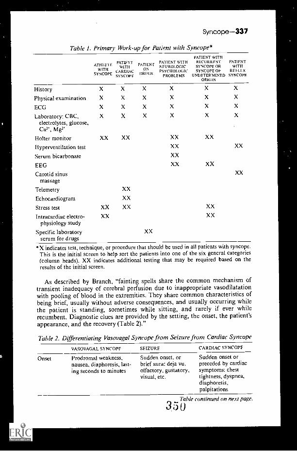
Syncope-337
Table I. Primary Work-up for Patient with Syncope*
ATH I.ETEWITH
SYNCOPE
PATLENTWITH
CARDIACSYNCOPE
PATIEN FON
DRUGS
PATIENT WITHPATIENT WITH RECURRENTNEUROLOGIC SYNCOPE ORPSYCHOLOGIC SYNCOPE OF
PROBLEMS UNDETERMINEDORIGIN
PATIENTWITH
REFLEXSYNCOPE
History
Physical examination
ECG
Laboratory: CBC,electrolytes, glucose,Ca2', M g2'
Holter monitor
Hyperventilation test
Serum bicarbonate
EEG
Carotid sinusmassage
Telemetry
Echocardiogram
Stress test
Intracardiac electro-physiology study
Specific laboratoryscrum for drugs
XX XX XX XX
XX
XX
XX XX
XX
XX
XX XX
XX
XX
XX
XX
*X indicates test, technique, or procedure that should be used in all patients with syncope.This is the initial screen to help sort the patients into one of the six general categories(column heads). XX indicates additional testing that may be required based on theresults of the initial screen.
As described by Branch, "fainting spells share the common mechanism oftransient inadequacy of cerebral perfusion due to inappropriate vasodilatationwith pooling of blood in the extremities. They share common characteristics ofbeing brief, usually without adverse consequences, and usually occurring whilethe patient is standing, sometimes while sitting, and rarely if ever whilerecumbent. Diagnostic clues are provided by the setting, the onset, the patient'sappearance, and the recovery (Table 2)."
Thble 2. Differentiating Vasovagal Syncope from Seizure from Cardiac Syncope
VASOVAGAL SYNCOPE SEIZURE. CARDIAC SYNCOPF
Onset Prodromal weakness,nausea, diaphoresis, last-ing seconds to minutes
Sudden onset, orbrief aura: deja vu,olfactory, gustatory,visual, etc.
Sudden onset orpreceded by cardiacsymptoms: chesttightness, dyspnea,diaphoresis,palpitations
Table continued on next page.
3 5 0

338Syndromes and Eponyms
Table 2. Differentiating Vasovagal Syncope from Seizurefrom Cardiac Syncope (Cont.)
VASOVAGAL SYNCOPE SEIZURE CARDIAC SYNCOPE
Typical Emotional upset,settings prolonged standing,
uncomfortable sur-roundings, or on firstarising with fullbladder
Occurrence Only when upright
Appearance Pallor, weak pulse
Residiuum Rapid recovery butmay recur on standing,occasional brief clonicmovements, or urinaryincontinence
Any setting, includingsleep, sometimesblinking lights, monot-onous music
An,. position
Cyanosis, stertorousbreathingProlonged recoverywith postictal state,Todd's paresis
Any setting, oftenwithout warning
Any position
Pallor, variable pulse
Recovery may be rapidor prolonged; if car-diac arrest: seizure-likeactivity, signs of cere-bral hypoxia
From Branch WT (ed): Office Practice of Medicine, 2nd ed. Philadelphia, W.B. Saunders,1987. with permission.
References: Ruckman RN: Cardiac causes of syncope. Pediatrics in Review 9(4):101108, 1978.
Kapoor WN, et al: A prospeaive evaluation and follow-up of patients with syncope. NEngl J Med 309:197 204, 1983.
Branch WT: Approach to syncope. J Gen Intern Med 1:49 58, 1986.
SYNDROMES AND EPONYMS
Despite mounting opposition in some quarters, the use of eponymic names forsyndromes, diseases, signs, et al. is likely to continue during all our lifetimes. Thedate and source of the originally published or reported description connected tothe eponym (not always the first published description) are interesting, both forold (e.g., Potes, 1779) and new (e.g., Kawasaki, 1974) syndromes and diseases.Listed below is a sampling:
Addison's disease.Addison T: Anaemia. Disease of the supra-renal capsules. London Hosp Gaz
43:517-518, 1849.
Budd-Chiari syndrome.Budd G: On diseases of the liver. London, Churchill, 1945.Chiari H: Erhahrungen tiber Infarktbildungen in der Leber des Menschen.
Zschr Heilk 19:475-512, 1898.
Calvé-Legg-Perthes syndrome.Calve F: Sur une forme particulière de coxalgie greffee. Sur les deformations
caracteristiques de l'extremité supérieure du femur. Rev Chir, Paris 42:54 -84,1910.
Legg AT: On obscure affection of the hip-joint. Boston Med & SJ 162:202-204, 1910.
3 5

Syndromes and Eponyms-339
Perthes GC: Ober Arthritis deformans juvenilis. Deut Zschr Chir 107:111-159, 1910.
Cockayne's syndrome.Cockayne EA: Dwarfism with retinal atrophy and deafness. Arch Dis Child,
London 11:1-8, 1936.
Down's syndrome.Down JL: Marriages of consanguinity in relation to degeneration of race.
London Hosp Clin Lect Rep 3:224-236, 1866.Down JL: Observations on an ethnic classification of idiots. London Hosp
Clin Lect Rep 3:259-262, 1866.
Ebstein's anomaly.Ebstein W: Ueber einen sehr seltenen Fall von Insufficienz der Valvula tricus-
pidalis bedingt durch eine angeborene hochgradige Missbildung derselben. ArchAnat Physiol, Leipzig, 1866, pp 238-253.
Ehlers-Danlos syndrome.Ehlers E: Cutis laxa, Neigung zu Haemorrhagien in der Haut, Lockerung
mehrerer Artikulationen. (Case for diagnosis.) Derrn Zschr 8:173-174, 1901.Dan los H: Un cas de cutis laxa avec tumeurs par contusion chronique de
coudes et des genoux (xanthome juvenile pseudo-diabetique de MM. Hallopeauet Mace de Lepinay). Bull Soc Fr Derm Syph 19:70-72, 1908.
Tetralogy of Fa llot.Fa llot EL: Contribution a l'anatomie pathologique de la maladie blue (cya-
nose cardioque). Marseille Med 24:77-93, 138-158, 270-286, 341-354, 403-420,1888.
Fitz-Hugh and Curtis syndrome.Fitz-Hugh T Jr: Acute gonococcic peritonitis of the right upper quadrant in
women. JAM A 102:2094- 2096, 1934.Curtis AH: A cause of adhesions in the right upper quadrant. JAMA 94:
1221-1222, 1930.
Goodpasture's syndrome.Goodpasture EW: The significance of certain pulmonary lesions in relation to
the etiology of influenza. Am J Med Sc 158:863-870, 1919.
Guillain-Barre syndrome.Guillain G, Barré J, Strohl A: Sur un syndrome de radiculo-nevrite avec
hyperalbuminose du liquide céphalo-rachidien sans reaction cellulaire. Remarquessur les caracteres cliniques et graphiques des reflexes tendineux. Bull Soc MedHop Paris 40:1462-1470, 1916.
Hodgkin's disease.Hodgkin T: On some morbid appearances of the absorbent glands and spleen.
Med Chir Tr, London 17:68-114, 1832.
Kawasaki disease.Kawasaki T, Kosaki F, Okawa S, et al: A new infantile acute febrile muco-
cutaneous lymph node syndrome (MLNS) prevailing in Japan. Pediatrics 54:271,
1974.
352

340Syndromes and Eponyms
Klinefelter's syndrome.Klinefelter HF Jr, Reifenstein EC Jr, Albright F: Syndrome characterized by
gynecomastia, aspermatogenesis without A-leydigism, and increased excretion offollicle-stimulating hormone. J Clin Endocr 2:615-627, 1942.
de Lange syndrome.Lange C de: Sur un type nouveau de dégénération (Typus amstelodamesis).
Arch Med Enf, Paris 36:713-719, 1933.Lange C de: Congenital hypertrophy of the muscles, extrapyramidal motor
disturbances and mental deficiency. A clinical entity. Am J Dis Child 48:243-268, 1934.
Marfan's syndrome.Marfan AB: Un case de deformation congenitale des quatre, membres plus
prononcée aux extremités, characterisee par l'allongement des os avec un certaindegré d'amincissement. Bull Soc Med Hop, Paris 13:220-226, 1896.
Achard C: Arachnodactylie. Bull Soc Med Hop, Paris 19:834-843, 1902.
Meckel's diverticulum.Meckel: Ueber die Divertikel am Darmkanal. Arch Physiol, Halle 9:421-453,
1809.
Méniere's syndrome.Meniere P: Sur une forme particuliére de surdité grave dependant d'une lesion
de l'oreille interne. Gaz Med, Paris 16:29, 1861.
Mibelli's disease.Mibelli V: Di una nuova forma de cheratosi "angiocheratoma." Gior Ital Mal
Vener 30:285-301, 1889.
Miinchausen's syndrome.Asher R: Munchausen's syndrome. Lancet i:339-341, 1951.
Niemann-Pick disease.Niemann AA: Ein unbekanntes Krankheitsbild. Jb Kinderh 79:1-10, 1914.Pick L: Der Morbus Gaucher und die ihm änlichen Krankheiten (die
lipoidzellige Splenohepatomegalie Typus Niemann und die diabetische Lipoid-zellenhypoplasie der Milz). Erg Inn Med Kinderh 29:519-627, 1926.
Pott's disease.Pott P: Remarks on that kind of palsy of the lower limbs which is frequently
found to accompany a curvature of the spine and is supposed to be caused by it,together with its method of cure. London, Johnson, 1779.
Reye's syndrome.Reye RD, Morgan G, Baral J: Encephalopathy and fatty degeneration of the
viscera. Lancet ii:749-752, 1963.
Riley-Day syndrome.Riley CM, Day RL, et al: Central autonomic dysfunction with defective
lacrimation. Report of five cases. Pediatrics 3:468-478, 1949.
Pierre Robin syndrome.Robin P: La glossoptose, un grave danger pour nos enfants. Paris, 1929.
353

Syndromes and Eponyms-341
Schönlein-Henoch purpura.Henoch H: Uber den Zusammenhang von Purpura und Intestinalstorungen.
Berlin Klin Wschr 5:417-519, 1868.Sch8nlein JL: Allemeine und specielle Pathologie und Therapie. Wtirzburg,
Ettinger, 1832.
Stevens-Johnson syndrome.Stevens AM, Johnson FC: A new eruptive fever associated with stomatitis and
ophthalmia. Report of two cases in children. Am J Dis Child 24:526-533, 1922.
Whipple's disease.Whipple GH: A hitherto undescribed disease characterized anatomically by
deposits of fat and fatty acids in the intestinal and mesenteric lymphatic tissues.Bull Johns Hopkins Hosp 18:382-391, 1907.
Wilson's disease.Wilson SA K: Progressive lenticular degeneration: A familial nervous disease
associated with cirrhosis of the liver. Brain 34:295-509, 1912.
Wolff-Park inson-White syndrome.Wolff L, Parkinson J, White PD: bundle-branch block with short P-R interval
in healthy young people prone to paroxysm: tachycardia. Am Heart J 5:685-704, 1930.
Reference: Jablonski S: Illustrated Dictionary of Eponymic Syndromes and Diseases.
Philadelphia, W.B. Saunders, 1969.
Who Was Down?
John Langdon Down was born near Plymouth, England on November 18,1828. He enrolled as a medical student at the London Hospital in 1853 andobtained a doctorate in 1859, after receiving the university gold medal forphysiology. In the same year-- I859he was appointed medical superintendentof the Eastwood Asylum for idiots, at Redhill, Surrey, England, a post he heldfor 10 years and where he wrote a paper titled "Observations on an ethnic
issification of idiots." He noted that many of his patients had similar clinicalfeatures and described them as follows:
The face is flat and broad and destitute of prominence. Cheeks are roundish andextended laterally. The eyes are obliquely placed and the internal canthi more thannormally distant from one another. The palpebral fissure is very narrow. The lips arelarge and thick with transverse fissures. The tongue is long, thick and much roughened.
The nose is small.
He stated that "Their resemblance to each other was such that, when placedside by side, it is difficult to believe that they are not the children of the sameparents.
Down's work at Eastwood brought him much recognition, and in 1869 he wasable to establish an institution at Redhill, Surrey for mentally retarded childrenof the wealthy. He named it Normansfield after his friend, Norman Wilkinson.At Normansfield, Down wrote his monograph titled Mental Affections ofChildhood and Youth, published in 1887, which contained the classic descriptionof Down's syndrome. He also mentioned adrenogenital dystrophy, which
354

342Syphilis
subsequently gained recognition as Frolich's syndrome. Down worked atNormansfield until his death in 1896.
For about 100 years the term "mongolism" was used as the primary descriptivename for Down's syndrome, with the eponyms "Down's" and "Langdon-Down's"used as alternatives, the hyphenated form having been preferred by Down in hislater life. However, controversy eventually arose because some regarded thereference to the Mongol ethnic group as insulting, and in 1965 representatives ofthe Mongolian People's Republic, in the World Health Organization approachedthe Director General and petitioned him to abandon the term "mongolism."Their request was accepted, and the eponym Down's syndrome was adopted.
References: Beighton P, Beighton G: The Man Behind the Syndrome. Heidelberg,Springer-Verlag, 1986, pp 40- 41.
Down JLH: Marriages of consanguinity in relation to degeneration of race. LondonHosp Clin Lect Rep 3:224, 1866.
Down JLH: Observations on an ethnic classification of idiots. London Hosp Clin LectRep 3:259, 1866.
SYPHILIS
Approaching Congenital Syphilis
The diagnosis of congenital syphilis, like the diagnosis of many congenitalinfections, is often confounded by the absence of symptoms or signs in thenewborn, as well as by the difficulty in interpreting neonatal serologic responsesto infection. The abrupt rise in the number of reported cases of congenitalsyphilis in the late 1980s, however, has increased the need for guidelines that willinsure the detection and appropriate management of newborns with this treatabledisease. The tables and figures below represent the recommended approach forsurveillance, diagnosis, evaluation, and treatment of congenital syphilis.
Congenital syphilis may be missed if serologic tests are not performed for boththe mother and her infant at the time of delivery. Even when these tests areperformed, some infants are not identified as having syphilis probably becausethe infection is very recent and there has been insufficient time for an antibodyresponse to develop. Some infants with congenital syphilis of later onset do notpresent with a typical rash; therefore, at least in areas where the disease isprevalent, serologic tests for syphilis should be included in the evaluation of allfebrile infants, even those with negative results on serologic testing at birth.
Table I. Surveillance Case Definition for Congenital Syphilis
For reporting purposes, congenital syphilis includes cases of congenitally acquiredsyphilis in infants and children, as well as syphilitic stillbirths.
I . Confirmed. A confirmed case of congential syphilis is a case in which Treponemapallidum is identified by darkfield microscopy, fluorescent antibody, or otherspecific stains in specimens from lesions, placenta, umbilical cord, or autopsymaterial.
355Table continued on next page.

Syphilis-343
Table I. Surveillance Case Definition for Congenital Syphilis (Cont.)
2. Presumptive. A presumptive case of congenital syphilis is either of the following:
a. Any case in which the infant's mother had untreated or inadequatelytreated* syphilis at delivery, regardless of findings in the infant
b. Any case in which the infant or child is reactive to a treponemal test forsyphilis and in which any one of the following is present:
i. Any evidence of congenital syphilis on physical examination (Table 11)ii. Any evidence of congenital syphilis on a long bone radiograph
iii. Reactivity to a CS F VDRL test*iv. Elevated CSF cell count or protein (without other cause)*v. Quantitative nontreponemal serologic titer that is fourfold higher than
the mother's (both specimens drawn at birth)vi. Reactive test for FTA-ABS 19S-IgM antibody*
3. Syphilitic stillbirth. A syphilitic stillbirth is defined as a fetal death in which themother had untreated or inadequately treated syphilis at delivery of a fetus aftera 20-week gestation or of a fetus weighing more than 500 gm.
Modified from Centers for Disease Control. MMWR 38:825-829, 1989.
*Inadequate treatment consists of any nonpenicillin therapy or penicillin given less than30 days before delivery.
*It may be difficult to distinguish between congenital and acquired syphilis after infancy.Signs may not be obvious and stigmata may not yet have developed. Abnormal values ofCSF VDRL test cell count, and protein, as well as IgM antibodies, may be found ineither congenital or acquired syphilis. Findings on long bone radiographs may help toindicate congenital syphilis. The diagnosis may ultimately be based on maternal historyand clinical judgment; the possibility of sexual abuse also needs to be considered.
Table 2. Evaluation for Early Congenital Syphilis
I. Maternal history, includingresults of serologic testingand treatment
2. Thorough physical examination
3. Long-bone radiographs
a. Diaphyseal periostitis
b. Osteochond rit is
c. Wimberger sign
4. Nontreponemal antibody titer
a. V DRL t.:.st (simultaneousquantitative serum titerfor mother and neonate)
5. Treponeniol antibody titer
FTA-ABS test
FTA-ABS on 19S-1gM fractionof serum (CDC)
6. CSF analysis
a. Cell count
b. Protein level determination
c. VDRL test
7. Other tests as clinically indicated
a. Chest radiography
b. Complete blood cell counti. Leukemoid reaction with or
without monocytosisor lymphocytosis
ii. Coombs negative hemolyticanemia
c. Platelet countI. Thrombocytopenia
d. Liver function tests
e. Urinalysis
8. HIV antibody test
356

344Syphilis
POSITIVE MATERNAL OR INFANT VDRL OR RPR TEST
MATERNAL FTA-ABS
NEGATIVE \ POSITIVE
FOLLOW ASSESS THE MATERNAL TREATMENT:UNTREATED, orDOES NOT HAVE A WELL-DOCUMENTEDHISTORY OF TREATMENT FOR SYPHILIS, orEVIDENCE OF RELAPSE OR REINFECTION, orINADEQUATELY TREATED'
WITH A NON-PENICILLIN REGIMEN, orWITH A NON-NEUROSYPHILIS REGIMEN INHIV SEROPOSITIVE MOTHERS
NONE / \ ANY
COMPLETE EVALUATION OF THE INFANTASSESS MATERNAL RISK FACTORS: AND TREAT WITH 10-14 DAY COURSE
TREATED LESS THAN 1 MONTH BEFOREDELIVERY, orHAS NOT DEMONSTRATED THE EXPECTEDDECREASE IN VDRL ANTIBODY TITERS, orHAS HAD INSUFFICIENT SEROLOGIC FOLLOW-UPDURING PREGNANCY TO ASSESSDISEASE ACTIVITY, orIS SEROPOSITIVE FOR HIV
NONE 1 ANY
FOLLOW COMPLETE EVALUATION OF THE INFANT:EVIDENCE OF INFECTION ON PHYSICAL EXAMINATION (Table II), orABNORMAL LONG-BONE RADIOGRAPHS, orABNORMAL CSF ANALYSIS (CELL COUNT, PROTEIN LEVEL, VDRL), orQUANTITATIVE VDRL TITER THAT ISFOURFOLD (OR GREATER) HIGHER THAN THE MOTHER'S, or
POSITIVE FTA-ABS ON 19S-IGM FRACTION OF SERUM (IF POSSIBLE), or*COMPLETE EVALUATION OF THE INFANT CAN NOT BE PERFORMED
NONE \ ANY
LOW RISK FOR CONGENITAL SYPHILIS TREAT WITH 10-14 DAY COURSECLOSE FOLLOW-UP CAN BE ASSURED
YES I \r NO
FOLLOW MATERNAL HIV ANTIBODY
NEGATIVE i \ POSITIVE
TREAT WITH SINGLE DOSE TREAT WITH 10-14 DAY COURSEOF BENZATHINE PENICILLIN G
Figure I. Algorithm for management of newborn infant born to mother with positivenontreponemal (VDRL or rapid plasma reagin) test result.
357

Syphilis-345
Table 3. Recommended Antimicrobial Treatment Regimens for InfantsBorn to Mothers with Positive VDRL Test Result
1. For confirmed or presumptive congenital syphilis (either item A or item B)
a. Crystalline penicillin G. 100,000 to 150,000 units/kg/day administeredintravenously in divided doses every 8-12 hours for 10-14 days
b. Procaine pencillin G, 50,000 units/kg/day administered once daily intramus-cularly for 10-14 days
2. Recommended only for infants at low risk for congenital syphilis who were bornto HIV-seronegative mothers adequately treated for syphilis and in whom closefollow-up cannot be ensured.
a. Ben7athine penicillin G, 50,000 units/kg (administered intramuscularly asone-time dose)
FOLLOW-UP FOR UNTREATED INFANTS:wpm. AND FTA-ABS TITER
AT 1, 2, 4,6, and 12 MONTHS
FOLLOW-UP FOR TREATED INFANTSVDRL TITER AT 1, 2, 4, 6 and 12 MONTHS, and
CSF ANALYSIS EVERY SIX MONTHS
vDRL TITER IS DECREASING BY 3-4 MONTHSOF AGE, and
op:NIL IS NEGATIVE BY 6 MONTHS OF AGE, andFTA-ABS IS NEGATIVE BY 12 MONTHS OF AGE
VDRL TITER IS DECREASING BY 3-4 MONTHSOF AGE, and
VDRL IS NEGATIVE BY 6 MONTHS OF AGE, andCSF VDRL IS NEGATIVE BY 6 MONTHS OF AGE andCSF CELL COUNT IS DECREASING, andCSF CELL COUNT IS NORMAL BY 2 YEARS OF AGE
YES \q, NO YES 1 NO
UNINFECTED REEVALUATE AND RE-TREAT ADEQUATELYWITH A REGIMEN RECOMMENDED FOR TREATED
SYPHILIS OF MORE THAN 1 YEAR'S DURATION
Figure 2. Follow-up management for an infant examined or treated for congenital syphilis
References: Ikeda MK, Jenson HB: Evaluation and treatment of congenital syphilis JPediatr 117:843-852, 1990.
Dorfman DH, Glaser JH: Congenital syphilis presenting in infants after the newbornperiod. N Engl J Med 323: 1299-1302. 1990.
3 r.; 8

MY HEART LEAPS UP
My heart leaps up when I beholdA rainbow in the sky:
So was it when my life began;So is it now I am a man;So be it when I shall grow old.
Or let me die!The Child is father of the Man;And I could wish my days to beBound each to each by natural piety.
William Wordsworth
-,
I

MTH
Eruptbn of Deciduous Teeth
The eruption of the first tooth in an infant is accompanied by parental pridein the fact that yet another milestone is reached. The figure below indicates theage and the order in which deciduous teeth erupt. The dot represents the meanage, whereas the wavy line demonstrates normal variation. Exceptions to thesequence of eruption are uncommon. Late eruption is unlikely to be of signifi-cance; however, it has been associated with both hypothyroidism and rickets.
AGE
0 4 6 a O 12 14 It, a 27 7, 24 76 28 1." V 14 26
LOwER cENTR AL INC ISOR
UPPER CENTRAL INCISOR
UPPE P LATERAL INC 1508 1..A.,H I
1
LOWER LATERAL INE IS )4
UPPER F IRST MOLAR
--rI
1.1".
LOWER FIRST AAOL AR
LOWER CUSPID
UPPER CUSPID
LOWER
URFE 8
SECOND moLR
SECOND MOLAR
0 2 4 6 a 2 I7 14 IL, ,a 20 22 24 26 8 I,
Reference: MacKeith R, Wood C: Digestion and absorption. In Infant Feeding andFeeding Difficulties. London, J. & A. Churchill, 1971, p 19, with permission.
TENNIS ELBOW
Tennis Elbow in Breasffeeding Mothers
Lateral epicondylitis or tennis elbow has been described in a variety of patients.It is classically seen when a person engages in repetitive, similar movements ofthe forearm extensor muscles, e.g., the continuous and monotonous swinging oftennis racket. The pain of lateral epicondylitis is particularly exacerbated byputting tension on the origin of the forearm extensor muscle, such as the activedorsitlexion of the wrist while grasping an object. Tennis elbow has also been
360347
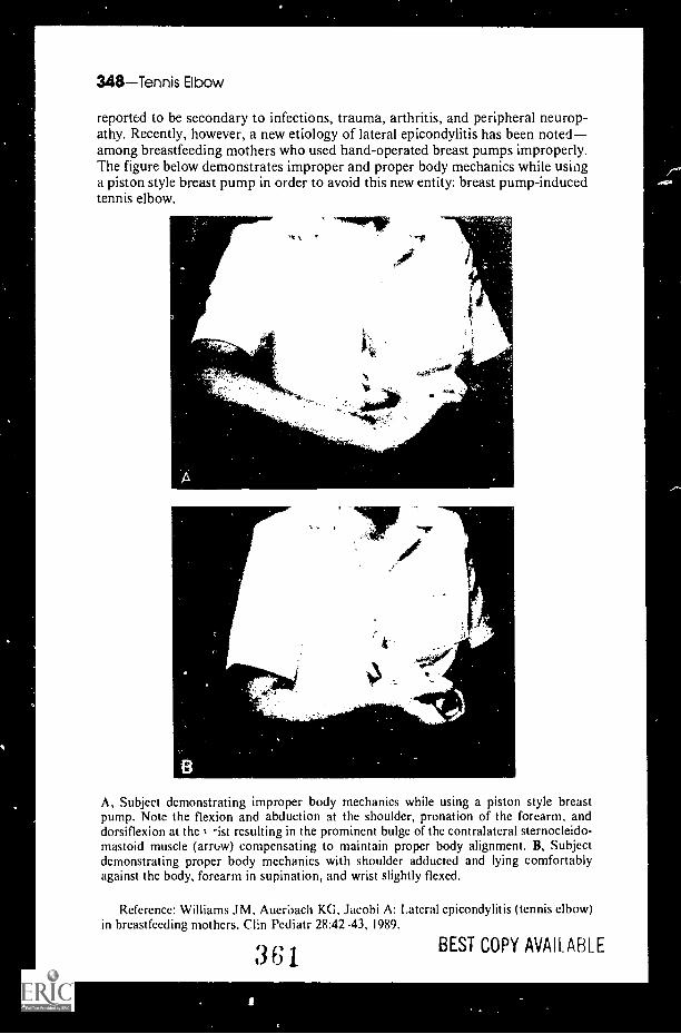
348Tennis Elbow
reported to be secondary to infections, trauma, arthritis, and peripheral neurop-athy. Recently, however, a new etiology of lateral epicondylitis has been notedamong breastfeeding mothers who used hand-operated breast pumps improperly.The figure below demonstrates improper and proper body mechanics while usinga piston style breast pump in order to avoid this new entity: breast pump-inducedtennis elbow.
'17F
A, Subject demonstrating improper body mechanics while using a piston style breastpump. Note the flexion and abduction at the shoulder, pronation of the forearm, anddorsiflexion at the -ist resulting in the prominent bulge of the contralateral sternocleido-mastoid muscle (arrow) compensating to maintain proper body alignment. B, Subjectdemonstrating proper body mechanics with shoulder adducted and lying comfortablyagainst the body, forearm in supination, and wrist slightly flexed.
Reference: Williams JM, Auerbach KG, Jacobi A: Lateral epicondylitis (tennis elbow)in breastfeeding mothers. Clin Pediatr 28:42 -43, 1989.
361 BEST COPY AVAILABLE
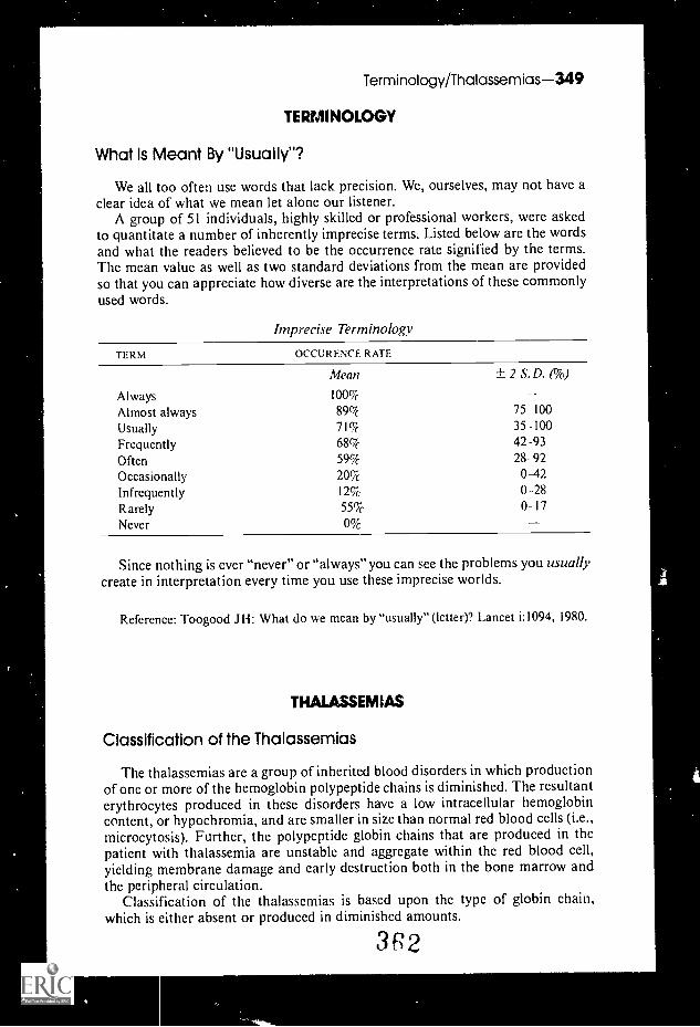
Terminology/Thalassemias-349
TERMINOLOGY
What Is Meant By "Usually"?
We all too often use words that lack precision. We, ourselves, may not have aclear idea of what we mean let alone our listener.
A group of 51 individuals, highly skilled or professional workers, were askedto quantitate a number of inherently imprecise terms. Listed below are the wordsand what the readers believed to be the occurrence rate signified by the terms.The mean value as well as two standard deviations from the mean are providedso that you can appreciate how diverse are the interpretations of thesecommonlyused words.
Imprecise Terminology
TERM OCCURENCE RATE
Always
Mean
100%
± 2 S.D. (%)
Almost always 89% 75 100
Usually 71% 35 -100
Frequently 68% 42 -93
Often 59% 28-92
Occasionally 20% 0-42Infrequently 1 2% 0-28Rarely 55% 0-17Never
Since nothing is ever "never" or "always" you can see the problems you usuallycreate in interpretation every time you use these imprecise worlds.
Reference: Toogood J H: What do we mean by "usually" (letter)? Lancet i:1094, 1980.
THALASSEMIAS
Classification of the Thalassemias
The thalassemias are a group of inherited blood disorders in which productionof one or more of the hemoglobin polypeptide chains is diminished. The resultanterythrocytes produced in these disorders have a low intracellular hemoglobincontent, or hypochromia, and are smaller in size than normal red blood cells (i.e.,microcytosis). Further, the polypeptide globin chains that are produced in thepatient with thalassemia are unstable and aggregate within the red blood cell,yielding membrane damage and early destruction both in the bone marrow andthe peripheral circulation.
Classification of the thalassemias is based upon the type of globin chain,which is either absent or produced in diminished amounts.
3 2

350Thalassemias
Clinical Features of Thalassemias
TYPE CLINICAL FEATURES
Alpha-thalassemia TypeSilent carrier
Thalassemia trait(heterozygous)
Hemoglobin H disease
Hydrops fetalis (homozygous)
Beta-thalassemia TypeSilent carrier
/3-thalassemia trait orminor (heterozygous)
Thalassemia intermedia
/3-thalassemia major
No clinical stigmataMild anemia; hypochromic and microcytic red cells
Splenomegaly; moderate-to-severe hemolytic anemia;mild jaundice
Death in utero
No clinical stigmataMild anemia; hypochromic and microcytic red cells;
elevated HGA2 and/or HGFSplenomegaly and severe anemia. Skeletal deformities,
frequent fractures and arthritis are complications.Severe anemia incompatible with life unless regular
blood transfusions are given.
Refer-nce: Festa RS: Modern management of thalassemia. Pediatr Ann 14:597-606,1985.
Complications of g-Thalassemia Major
ig-thalassemia major or Cooley's anemia is one of the most serious of thethalassemias. Patients with this disease can only survive with frequent bloodtransfusions, careful attention to iron balance, and supportive therapy. Thecomplications of /3-thalassemia major result mainly from (1) excessive hemato-poesis; (2) chronic hemolysis; and (3) iron overload with resultant organ damage.
1. Complications due to excessive hematopoesisa. Marked bone marrow hypertrophy and cortical thinningb. Bony changes, particularly in the craniofacial area ("rodent facies") secondary
to maxillary overgrowth, protrusion of teeth, separation of the orbits, flatten-ing of the nasal bridge, and malar prominence. These bony changes may yield:i. Chronic sinusitis
ii. Impaired hearingc. Pathologic fractures (particularly in weight-bearing bones)d. Lymphadenopathye. Hepatosplenomegaly
2. Complications due to chronic hemolysisa. Gallstonesb. Leg ulcers (usually seen in late adolescence and early adulthood)
3. Complications due to iron overload as a result of chronic transfusion therapy(ali tissues are affected by this iron overload, but the liver, spleen, and pancreasretain iron in the highest concentrations)a. Cardiac disease
I. Pericarditisii. Atrial and ventricular arrhythmias
iii. Congestive heart failureb. Hepatic disease (hepatic fibrosis)

Theophylline-351
4. Growth and endocrine dysfunctiona. Growth failure: Although children with thalassemia major on a chronic
transfusion program grow normally until the age of 12, their growthvelocity diminishes thereafter, and they fail to exhibit a pubescent growthspurt. Growth hormone levels are usually normal or elevated. The growthfailure is probably secondary to chronic disease and iron overload.
b. Delayed or incomplete sexual maturationc. Acquired hypothyroidism, hypoparathyroidism, and diabetes mellitus due
to hemochromatosis.
Incidentally, Thomas Cooley, the Detroit pediatrician who first describedthalassemia major in 1927, required only a microscope and his patients'peripheral smears to describe a disease that influerced the fields of hematology,human heredity, and population genetics.
References: Festa RS: Modern management of thalassemia. Pediatr Ann 14:597-606,1985.
Zuelzer WW: Thomas Cooley. In Pediatric Profiles. St. Louis, C.V. Mosby, 1957, pp135 143.
THEOPHYLLINE
Factors That Alter Theophylline Clearance in Children
Theophylline remains the most commonly used drug for treating children withasthma in the U.S. Despite improved formulations of theophylline that providea sustained release of the xanthine derivative, and the ability to tmasure serumtMophylline levels, many children are extremely variable in their absorption,metabolism, and clearance of the drug.
Theophylline has a theoretical bioavailability of 100%, and its metabolism andclearance are controlled principally by the liver, where 90% of the drug ismetabolized and then excreted in the urine. Any disease or condition affectingthe metabolic machinery of the liver, therefore, will play a sigr ficant role inaltering a patient's metabolism and absorption of theophylline.
Factors Affecting Theophylline Metabolism
Facters that reduce clearance:I. Liver disease2. Congestive heart failure: cor pulmonale3. Prolonged fever, particularly from viral infection4. Macrolide antibiotics such as erythromycin and troleandomycin5. Cimetidine6. Age less than 1 year7. Influenza vaccine8. Acute hypoxemia9. High carbohydrate/low protein diet
10. PropranololI I. Furosemide
3Table continued on next page.

352ThrombocyYosis
Factors Affecting Theophylline Metabolism (Cont.)
Factors that increase clearance1. Tobacco or marijuana smoking2. Charcoal broiled foods (consumed in
large quantities over a long periodof time)
3. Phenobarbital4. Phenytoin
5. Isoproterenol6. High protein/low carbo-
hydrate diet7. Pregnancy8. Rifampin9. Tegretol
References: Adapted from Tinkelman DG: Theophyllineuse and misuse in pediatricasthma. Hosp Prac 23:179-184, 1988.
Hen J: Office evaluation and management of pediatric asthma. Pediatr Ann 15:11 I-124, 1986.
1HROMBOCYTOSIS
Significance of Thrombocytosis
In these days of automated cell counters, platelet counts are often determinedwhether we ask for them or not. As is often the case with unrequested andunnecessary laboratory studies, "abnormal" results are frequently reported Itturns out that platelet counts that previously would have been considered to beabnormally elevated are not particularly unusual among healthy pediatricpatients. The figure below represents the distribution of platelet counts obtainedfrom 805 ambulatory pediatric patients. Although the largest number of patientshad platelet counts between 200,000 and 400,000/mm3, 12.9% of the children hadcounts of greater than 500,000/mm3, and 2% were greater than 700,000/mm3Children with elevated platelet counts were most often completely healthy, butsome had evidence of viral or bacterial infection, and they tended to be youngerthan the children with "normal" counts.
40
3 0
2 0
I 0
200 400 600 800
PLATELET COUNT (x103/mm3)
1000
Distribution of plateletcounts in 805 ambulatorypediatric patients.

Thrombocytosis-353
Although "elevated" platelet counts among healthy children may be normal,extreme thrombocytosis is usually associated with a recognizable disease state.Among 94 children with platelet counts greater than 900,000/mm3, only one childwas completely healthy. Recognized conditions associated with extreme throm-bocytosis include the following:
Infection of any kindRecovery from chemotherapyIron deficiencySplenectomyMalignanciesRespiratory distress
Inflammatory diseases such as Kawasakidisease, juvenile rheumatoid arthritis,and anaphylactoid purpura
Recent surgeryMetabolic diseasesNephrotic syndrome
References: Heath HW, Pearson HA: Thrombocytosis in pediatric outpatients. JPediatr 114:805-807, 1989.
Chan KW, Kaikov Y, Wadsworth LD: Thrombocytosis in childhood: A survey of 94patients. Pediatrics 84:1064-1067, 1989.
Thrombocytosis in Childhood
With the increased availability of electronic cell counting, increased plateletcounts are being noted more frequently. Listed below are the major causes ofplatelet counts in excess of 900,000/mm3.
InfectionBoth viral and bacterial infections, particularly of the central nervous system.
HematologicConditions in this group include consequences of chemotherapy, iron deficiency
anemia, chronic myelogenous leukemia, and the early postsplenectomy state.
RespiratoryRespiratory distress syndrome, with or without bronchopulmonary dysplasia;
severe respiratory obstruction.
Tissue damage or response to surgeryTrauma, postoperative response
Collagen vascular diseaseJuvenile rheumatoid arthritisWegener granulomatosisAnaphylactoid purpura
Metabolic diseasesWhen complicated with acidosis and dehydration
Nephrotic syndrome
An increase in platelet count can be viewed in many circumstances as an acutephase reactant.
Thrombocytosis, infants and children,consequences and requires no therapy.
is rarely associated with thrombotic
Reference: Chan KW, et al: Thrombocytosis in childhood: A survey of 94 patients.Pediatrics 84:1064 1067, 1989.
3 6

354Tick
TICK
Tick-Related Infection
Ticks are small, but they are large enough to carry with them many evensmaller microorganisms that they inject into unsuspecting human hosts. Most ofus are familiar with the clinical characteristics of the tick-borne infections in ourown locale, but we may not appreciate the importance of these diseases inpatients who have traveled prior to their illness. It is important, first, toremember to think of tick-related infection, and then to consider tick activity inthe area, the geographic distribution of ticks known to carry specific pathogens,sites of tick exposure, and signs and symptoms at presentation related to the timeof exposure. The incubation period for most of the infections carried by ticks is3 to 5 days to 2 weeks.
If you have trouble remembering the regional and clinical characteristics oftick-borne infection, the following table may help.
Tick-borne Diseases in Children
DISFASF ORGANISM VFCTORGEOGRAPHIC
RFSERVOIR DISTRIBUTION1 YPE 0I-ILLNESS
Babesiosis Babesiamicron
frodes dammini Rodents Coastal area, Malaria-like,islands of fever, anemia,Massachusetts, renal failureRhode Island,New York
Northeast,Midwest andWestern UnitedStates a
Southern. South-eastern andMidwestUnited States
Lyme disease Borreha Ixodes dammini. M igratoryburgdorferi Ixodes pacificus. birds
Amblyommaamericanum
Tularemia Francisella Dermacentor an- R.rularensis dersoni, Derma- dogs,
censor variabilis. rodents5Amblyomrnaamericanum
Rocky Moun- Rickettsia Dermacemor an- Dogs, cats,tain spotted rickertsii dersoni. Derma- rodents,fever centor variabilis, rabbits
Amblyommaamericanum.Haemaphysalisleporis-palustris
Erhlichiosis Erhhchia Rhipicephaluscanis sanguineus
Erhlichiasenneisu
Relapsing Borreliafever tonii. Borre-
lia hermsii,Rorreliaturicatac
Queensland Rickettsiatick typhus australis
FieVre Rickettsiahoutonneuse (wrath
Rhipichephalussanguineus
Ornithodorosmouhata
I xodid ticks
Ixodid ticks
Westernhemisphere.esr-ciallySoi heasternUnited States
Dogs Southern, South-eastern, Mid-west UnitedStates
Dogs, Japanrodents
Rodents, Western Moun;opossums, tains, Southernsquirrels, Plains, Unitedarmadillos States
Rodents, Easterndogs mar- Australiasupials
Dogs, Worldwiderodents
Fever, rash(ECM), head-ache, myalgias,multiple stages
Fever,lymphad-enopathy,pneumonia
Fever, headache,myalgias, rash,toxicity
Fever, chills,myalgias,hematologicabnormalities,similar toRMSF
Fever, chills, head-ache, myalgia,relapsing course
Similar to RMSF,usually milder
Similar to RMSF,usually milder
a Recent evidence for widespread disease in United States.Animals become ill with infection.
3 S 7
Table continued on next page.
BEST COPY AVAILABLE
Torticollis-355
Tick-borne Diseases in Children (Conl.)
DISEASE ORGANISM VEC1OR RESERVOIR
Asian ticktyphus
Rickettsia lxodid ticks Dogs, rodentssiberica
GEOGRAPHICDISTRIBUTION
Central Asia,Russia
TYPE OFILLNESS
Q fever Coxiella All endemic Cattle, sheep, Worldwideburneui speciesc goats
Colorado tick Orhivirus Dermacentor Rodents, deer Rocky Mountainfever andersoni states, Western
Canada andNorthern Sierras
Tick-borne Flavivirus Ixodes persul- Cattle, sheep, Central Asia,encephalitis cams. trades goats, Eastern Europe,
ricinus rodents Russia
Tick-bitegranuloma
Tick paralysis
All species
Dermacentorandersoni.Dermacentorvariabilis
Similar to RMSF,usually milder;regional lym-phadenopathy
Fever, headache,pneumonia
Fever, headache,malaise, myal-gias, leukopenia
Fever, headache,encephalitis,photophobia,hyperesthesias
Local reaction,granulorna,complement-mediated
Toxin-mediated,neurologic syn-drome, ataxia,areflexia, ascend-ing flaccid paraly-sis, mild fever
,lnfection usually acquired hy inhalation from animals; ticks important in animal transmission.ECM, erythema chronicum migrans.
Because the diagnosis of most of these infections depends upon serologictesting, which often must be performed in a reference laboratory, empiricantibiotic therapy is often required. Most of the tick-related infections can betreated with tetracycline or chloramphenicol; however, amoxacillin or penicillinare the appropriate alternatives to tetracycline in the child younger than 8 or 9years with suspected Lyme disease.
Reference: Jacobs RF: Tick exposure and related infections. Pediatr Infect Dis J7:612 614, 1988.
TORTICOLLIS
Common Causes
Congenital, muscular, or vertebral anomalies
Uncommon Causes
Cervical adenopathyCongenital nystagmusDrug-induced (e.g., phenothiazines,
haloperidol, metoclopramide,trimethobenzamide)
Paroxysmal
PharyngitisRetropharyngeal abscessSecondary to reflux esophagitis
(Sandifer's syndrome)Superior oblique muscle weakness
3 c 8 BEST COPY AVAILABLE

356Toureite's Syndrome
Rare Causes
Calcification of intervertebral disksDystonia musculorum deformansEosinophilic granuloma of cervical
vertebraeFibromyositisHepatolenticular degenerationJuvenile rheumatoid arthritisKernicterus
Osteomyelitis of the cervical vertebraePneumonia of an upper lobePosterior fossa tumorSpasmus nutansSpinal tumorSubluxation or dislocation of cervical
vertebrae
TOURETTE'S SYNDROME
Tics can occur commonly during childhood. Several studies estimate thatabout 3% of all children exhibit tics at some time. These symptoms are mostcommonly transient, however, and present as excessive blinking or grimacing.Less common are transient nonspecific vocalizations (e.g., clearing of the throat,sniffing, or frequent coughing).
There are occasions when the symptoms may become more chronic andpersistent. When limited to motor symptoms, the condition is called multiplechronic motor tic disorder. In the child between ages 2 and 14 presenting withboth vocal and motor symptoms that wax and wane in severity over time andhave been present for more than a year, the term Gilles de la Tourette's orTourette syndrome (TS) is used. Its etiology, unfortunately, remains poorlyunderstood. The types of tics seen in this fascinating syndrome are listed below:
The Symptoms
I. Motor ticsBlinkingGrimacingShruggingMouth-openingHead-jerkingTongue movements
2. Vocal ticsSniffingCoughingClearing of the throatHissingBarkingHonkingSnortingSqueaking
Extending or flexing neckJerking of trunkJerking of extremitiesTensing of abdominal musclesKickingLip-licking
BurpingRepetition of lettersRepetition of words or phrasesInvoluntary cursing (coprolalia)*ClickingWhistlingSpittingShrieking
*Although Georges Gilles de la Tourette's original 1885 description (Arch Neur, Paris,9:19 42, 158 200, 1885) stressed coprolalia as a cardinal feature, subsequent studies havenoted a 20 to 35% incidence rate of this symptom among patients with Tourette syndrome.Its absence, therefore, does not contradict the diagnosis.
3 9
0),

3. Complex symptomsSquattingJumpingTwirlingRepetitive touching of objects
and people
Tracheoesophageal Fistula-357
Repetitive sniffingObscene gestures (copropraxia)Head-bangingSelf-injury (biting, scratching)
Diagnosis and Treatment
A careful history and physical are warranted, because between 21% and 54%of children with TS have symptoms of attention deficit disorder (ADD). Further,there exists a strong relationship between ADD patients treated with stimulantmedications such as methylphenidate (Rita lin) and the subsequent developmentor exacerbation of tics.
It should be stressed that all of the medications suggested for Tourette syn-drome provide only symptomatic relief and are not curative. Further, no evidenceexists to suggest that early therapy with these medications has any effect on thelong-term prognosis of tic disorders. Many Tourette syndrome patients havesymptoms mild enough not to require pharmacologic intervention. In light of theside-effects from the medications listed, many physicians prefer not to use them.
I . Agents availableHaloperidol ClonidinePimozide ClonazepamFluphenazine
2. Potential side-effects of neuroleptic drugsAcute dystonic reactionsParkinsonian symptomsAnticholinergic symptomsSedation
Tardive dyskinesiaIncreased appetiteDepressionCognitive bluntingSchool phobias
References: Barabas G: Tourette's syndrome: An overview. Pediatr Ann 17:391 -393.1988.
Erenberg G: Pharmacologic therapy of tics in childhood. Pediatr Ann 17:395 403, 1988.Golden GS: The relationship between stimulant medication and tics. Pediatr Ann
17:405 408, 1988.
TRACHEOESOPHAGEAL FISTULA
Tracheoesophageal fistula (TEF) and esophageal atresia are the two types ofesophageal malformations causing upper intestinal obstruction. TEF is thefailure of the trachea and esophagus to divide linearly during embryogenesis.Esophageal atresia is the developmental occlusion of the esophagus in a localizedsegment of lumen. Both may present at birth with aspiration and both may occuras an isolated finding.
The diagnosis of tracheoesophageal fistula may be suggested by a variety ofclinical observations. The five types of fistula are depicted below, along with thesymptoms and signs that typically accompany them.
37o

358Tracheoesophageal Fistula
Type ASymptoms and Signs;
Excessive mucus, aspirationof saliva.
Scaphoid abdomen.No gas in bowel on x-ray.Cannot pass catheter into
stomach.Gradually increasing respi-
ratory distress.Polyhydramnios.
Type DSymptoms and Signs:
Type CSymptoms and Signs:
Type BSymptoms and Signs:
Polyhydramnios.Coughing, choking and
pneumonia from birth.Scaphoid abdomen.No gas in bowel on x-ray.
Most common (800i ofcases).
Excessive mucus.Gradually increasing respi-
ratory distress.Polyhydramnios frequent
but not severe.Gas in bowel on x-ray.
Coughing, choking, andpneumonia from birth.
Gas in bowel on x-ray.
Type ESymptoms and Signs:
Difficult to diagnose.Coughing or cyanosis with
feeding.Chronic aspiration pneu-
monia,
The differential diagnosis includes pharyngeal muscle weakness, vascularrings, and esophageal diverticula.
Discovery of a tracheoesophageal fistula should alert the physician to thepossibility that other congenital anomalies may be present. Anomalies that havebeen found to be associated include:
Vertebral Anal Cardip- Renal Limb
Reference: Koops BI Battaglia EC: The newborn infant. In Kempe CH, Silver HK,O'Brien D (cds): Current Pediatric Diagnosis and Treatment. Los Altos, California,Lange Medical Publications, 1984, p 84, with permission.
37.1
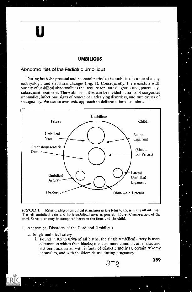
UMBILICUS
Abnormalities of the Pediatric Umbilicus
During both the prenatal and neonatal periods, the umbilicus is a site of manyembryologic and structural changes (Fig. 1). Consequently, there exists a widevariety of umbilical abnormalities that require accurate diagnosis and, potentially,subsequent treatment. These abnormalities can be divided in terms of congenitalanomalies, infections, signs of remote or underlying disorders, and rare causes ofmalignancy. We use an anatomic approach to delineate these disorders.
Fetus:
UmbilicalVein
OmphalomesentericDuct
Umbilicus
UmbilicalArtery
Child:
RuundLigament
(Shouldnot Persist)
LateralUmbilicalLigament
Urachus Obliterated Urachus
FIGURE 1. Relationship of umbilical structures in the fetus to those in the infant. Le.fi,The left umbilical vein and both umbilical arteries persist; Above, Cross-section of thecord. Structures may be compared between the fetus and the child.
I. Anatomical Disorders of the Cord and Umbilicus
a. Single umbilical arteryi. Found in 0.5 to 0.9% of all births, the single umbilical artery is more
common in whites than blacks; it is also more common in females andhas been associated with infants of diabetic mothers, certain trisomyanomdlies, and with thalidomide use during pregnancy.
3 "2 359
d.

,
414,
360Umbilicus
ii. There is a reported incidence of congenital anomalies associated withthe single umbilical artery, including renal, cardiovascular, pulmonary,genitourinary, cerebrospinal, musculoskeletal, facial, and occularabnormalities. The work-up of a single umbilical artery, therefore,should include a thorough physical examination, especially for dysmor-phism, parental counseling, good follow-up for identification ofproblems that may not be obvious at birth, and special screeningstudies (e.g., for cardiac or renal anomalies) as indicated.
b. Umbilical dysmorphismi. Aarskog syndrome; short stature with facial, digital, and genital
malformations and a prominent, protruding central portion of theumbilicus surrounded by a deep ovid depression.Reiger syndrome: goniodysgenesis, hypodontia, and a broad prominentumbilicus with redundant umbilical skin.Robinow syndrome: fetal face, short forearms with brachydactyly,genital hypoplasia, moderate dwarfing, and an abnormally highpositioned, broad, and poorly epithelialized umbilicus
iv. Beckwith-Wiedemann syndrome: macroglossia, gigantism, hyperinsu-linemia and frequently associated with omphalocele.
c. Disorders of the umbilical stumpi. Omphalitis is an infection of the stump, which is frequently followed by
sepsis.ii. Persistent omphalomesenteric remnants or ectopic viscera in a retained
stalk (usually the stump atrophies and separates 12-14 days after birth;a stalk still present at 3 to 4 weeks should be surgically explored andexcised).
iii. Umbilical or pyogenic granuloma (these should be ablated with silvernitrate)
d. Incomplete obliteration of the omphalomesenteric duct (Fig. 2)All of these entities require surglcal evaluation and repair.
i. Umbilical polypii. Umbilical sinus
Omphalomesenterie cyst (cysts are usually not diagnosed unless theyundergo torsion, become infected, or enlarge when distended bysecretions)
iv. Fibrotic bands between the umbilicus and intestinev. Meckel's diverticulumvi. Omphalomesenteric fistula (persistant vitelline duct)
e. Incomplete obliteration of the allantois (Fig. 3)These entities require surgical repair.
i. Urachal sinusUrachal cystUrachal diverticulum
iv. Patent urachus
f. Abnormalities of the umbilical ringFrnbryologleally, the umbilical ring must constrict and close after theintestine has migrated into the abdominal cavity.
i. Umbilical hernia (over 85(4 will close without surgical repair)
1,3 '73
I

Umbilicus-361
A
F
FIGURE 2. Omphalomesenteric anomalies: A, an umbilical polyp; B, umbilical sinus;C. an omphalomesenteric cyst, D. fibrotic bands between thc umbilicus; E. Meckel'sdiverticulum; /7, an omphalomesenteric fistula.
Gastroschisis is a defect in the abdominal wall of an extraumbilicallocation. It may represent a rupture, in utero, of an umbilical hernia ata weak point. Gastroschisis requires surgical treatment and closure toreturn the abdominal contents to their proper space.Omphalocele results from a failure of the intestine to return from thevitelline duct to the abdomen. The omphalocele is covered by a thin,membranous sac, which encloses the intestine and often the liver withthe cord arising from it. Risks include infection, dehydration, andhypothermia, and surgical closure is indicated. Both omphalocele andgastroschisis have a high association with other congenital anomalies(e.g., gastrointestinal, cardiac, renal, and chromosomal).
374

362Umbilicus
FIGURE 3. Urachal anomalies:: A, urachal sinus; B, cyst; C, diverticulum; 1.), patenturachus.
2. Other Disorders of the Umbilicusa. Umbilical tumors
i. Primary sarcomaii. Primary hemangiomas
Arteriovenous malformationsh. lntraabdominal hemorrhage can present with a bluish discoloration of an
umbilical hernia (Hoffstater's sign)c. Acute pancreatitis can be heralded by periumbilical bruising (Cullen's sign).
References: .%,.:apted from Black CT: Disorders of the pediao ic umbilicus. Resident andStaff Physician 35:64 84, 1989, with permission.
Sapien RE, Hodge D: Evaluation of the umbilicus..1 Am Acad Earn Phys 4:671 674, 1991

Upper Airway Obstruction-363
UPPER AIRWAY OBSTRUCTION
Infectious Causes of Upper Airway Obstruction:Distinguishing the Features of Viral Croup,Epiglottitis, and Bacterial Tracheitis
The child presenting to the clinic or emergency room with upper airwayobstruction demands immediate attention. There exist many causes of acuteupper airway obstruction, including foreign bodies, diphtheria, infectious mono-nucleosis, and ...zasles. More chronic or recurrent causes of stridor and upperairway obstruction include vascular rings, congenital heart disease, trachealstenosis from previous intubation, severe allergic reactions leading to laryngo-spasm, and recurrent angioneurotic edema. The most common causes of poten-tially life-threatening upper airway obstruction, however, are infectious in origin:(1) viral laryngotracheobronchitis or croup; (2) epiglottitis; and (3) bacterialtracheitis. A prompt diagnosis, obviously, requires a sound knowledge of theirdistinguishing features in order to assure proper medical management.
Clinical Features of Viral Croup, Epiglottitis, and Bacterial Tracheitis
CLINICAL FEATURE VIRAL CROUP EPIGLOTTH IS BACTERIALTRACH El FIS
Site of airway obstruction Infraglottis Supraglottis InfraglottisPatient age
Peak 2 yrs 3 -6 yrs 2 yrsRange 8 mo 5 yr 17 mo- adult 1 mo 9 yr
Sex M > F (2:1) M = F M = F
Duration of illness prior toadmission (hours)
12 78 hrs 2 48 hrs 24 -96 hrs
Prodrome Viral URI Uncommon Viral URI
Clinical features onpresentation
Stridor Common Uncommon CommonBarking cough >60ri Uncommon >50('iTemperature ("C) 37.8 38.6 39.2Hoarseness 20% Uncommon NoneRetractions Common Uncommon VariedWheering 5Ci None 10ri.
Cyanosis 10(.7 20(4- 10(-(
Dyspnagia None 10("( NoneDrooling None 10(' NoneAppearance Lying down,
non-toxicSitting up,
toxicVaried
Season Late spring;late fall
Year round Year round
Progression Slow Rapid Varied:slow rapid
WBC range 5 11,000 16 22,0(X) 8 20,000PMN range (ci) 40 80 GO 95 40 80Mean bands (ei) 7 30
_30
3 '7 6
Table contnnted wi next page.

364Urinalysis
Clinical Features of Viral Croup, Epigiottitis, and Bacterial Tracheitis (Cont.)
CLINICAL FEATURE VIRAL CROUP EPIGLOMTIS BACTERIALTRACHEITIS
X-ray findingsSubglottic narrowing (%) 90-100Enlarged epiglottis (%) NoneInfiltrate on admission (%) 10
Infiltrate during Commonadmission (%)
Response to racemic 95 0
epinephrine (%)
Positive tracheal cultures (%)Bacteria 0Viral 100
Positive blood cultures 0(bacteria) (%)
Hospitalization (%) 10
Respiratory arrest Rare
Treatment (%)Intubation 3
Tracheostomy <1
Mean duration of hospitali- 7
zation (days)
Recurrence
Etiologic agents Parainfluenza
None100<30<30
type 1, 2, 3;respiratorysyncytialvirus;rhinovirus
60-100Rare>50100
0
70 (epiglottis) 1000 50
90 0
100 100
Increased risk 25%
99 651 30
5 21
Rare Rare
Ha( ?hophilus S. aureus; Haemo-infl philus influenzae,type b, type b; strepto-/3-hemolytic coccus, group A;streptococci 13- and hemolytic
streptococci;Neisseria; E. eoh;others
Reference: Adapted from Hen .1: Current management of upper airway obstruction.Pediatr Ann 15:274- 294, 1986.
URINALYSIS
Dangers of the Dipstick
The urine dipstick is an extremely quick and convenient method of screeningfor pH, protein, glucose, hemoglobin, ketones, bilirubin, and urobilinogen. Thedipstick is a screening test, however, and its limitations must be kept in mind.The situations in which false positive and false negative results may be obtainedare listed below:
pH. This determination is not altered except when the pH of the urine isacutally altered by drugs.
Prokoin. A highly alkaline urine may cause a false positive dipstick for protein.
3 "7 7

Urinalysis-365
The alternative test used for detecting protein other than albumin involvesdenaturing the protein with heat or sulfosalicylic acid to produce turbidity. Afalse positive result in this alternate test may be produced by:
BuniodylChlorpromazinePrornazineCarinamideCephaloridineCephalothin
Radiographic agents such asiopanoic acid, iodopyracet,and iophenoxic acid
SulfamethoxazoleThy molTolbutamide
Glucose. The dipstick technique for glucose may be falsely positive in thepresence of bleach in the collecting vessel and vaginal powders containing glucose.
Ascorbic acid may produce a false negative result by retarding colordevelopment.
The Clinitest method may be used to quantitate the urinary glucose; however,other reducing agents may produce a false positive result. These include:
Sugars: galactose, lactose, Homogentisic acidlevulose, maltose, Glucuronic acidor pentose Bleach
Drugs that produce a false positive Clinitest result include:
Acetanalidep-aminosalicylic acidAntipyrineCephaloridineCephalithinChloramphenicolChlortetracycline
CinchophenDiatrizoateIsoniazidLevodopaNalidixic acidOxytetracyclineTetracycline
Hemoglobin. A false positive test may be produced by myoglobin or thepresence of oxidizing agents such as ascorbic acid.
Ketones. Aspirin may cause ketonemia in children, but the presence of aspirinin a ketonuric urine may produce a false negative dipstick.
False positive tests for ketones may be seen in the presence of:
LevodopaParaldehyde in the presence of ethanolPhenformin
Billrubln. False positives may be seen in the presence of:
PorphobilinigenSkatoleI ndoleLarge quantities of bilirubinp-aminosalicylic acidAntipyreneApronalideBromsulfophthalein
ChlorpromazinePhenazopyridinePhenothiazinesSulfadiazineSulfamethoxazoleSulfanilamideSulfonamides
Reference: Adapted from McMillan JA, et al (eds): 1 hc Whole Pediatrician Catalog,Vol. 2. Philadelphia, W.B. Saunders, 1979, pp 248 250, with permission.

366Urine Output/Urinary Tract
URINE OUTPUT
Urine Output Measurement in Premature Infants
Lest we forget, urine, like water, does evaporate. This is especially true in thesetting of the neonatal intensive care unit. A recent study of the rate and degreeof fluid evaporation from disposable diapers under radiant warmers or in infantisolettes showed that evaporation was a function of time and was inverselyrelated to the volume of fluid added to the diaper. Considering the importance otaccurate determination of urine output for assessment of hydration status, renalfunction and nutrient retention in the premature and term neonate, the moral ofthis tale is clear: adhesive urine bags and frequent diaper inspection areimperative in the care of the low birth weight infant.
Reference: Cooke R.I. et al: Urine output measurement in premature infants.Pediatrics 83:116-118, 1989.
URINARY TRACT
Delayed Urination in the Newborn
One of the kindest acts a neonate can perform for his pediatrician is to urinateearly in life. Ninety-nine to 100% of all normal infants urinate at least once by 48hours of age. Approximately 23% will void first in the delivery room, and the actmay not be reported to the nursery.
Failure to urinate by the first 1 to 2 days of life may be due to obstruction ofurine flow or to inability to form urine. Causes of obstruction include
I mperforate prepuce Neurogenic bladderUrethral strictures "Megacystic syndrome"Urethral diverticulum U retcroceleHypertrophy of the Renal tumors
verumontanum Cystic kidneys
Inability to form urine may result from
Postnatal intravascular hypovolemiaRestriction of oral fluidsBilateral renal agenesisCortical necrosisTubular necrosisBilateral renal vein thrombosisCongenital nephrotic syndromeCongenital pyelonephritisCongenital nephritis
Nonspecific symptoms or signs such as excessive crying, in itahility, poorfeeding, pallor, cmcsis, mottled skin, or weak pulse may suggest the developmentof uremia.

Urinary Tract-367
The physical examination may be more useful in establishing a specificdiagnosis.
Physical Examinaiion Following Delayed Urination
PAI.PATION OR PERCUSSIONOF DISTENDED BLADDER NO KIDNEYS PALPABLE PALPABLE RENAL MASS
1
Obstruction of urine flow
Examine meatus forpatency and lookfor epispadias orhypospadias
A urethral diverticulum maygive rise to a bulge alongthe dorsum of the penis
1
Bilateral renal agenesis
These infants are usuallymales and tend to havelowset ears, epicanthalfolds, and a flattenednose
1
Renal vein thrombosis
Infantile polycystickidneys
Hydronephrosis
Cystic dysplasia
Neoplasm
Reference: Moore ES, Galvez MD: Delayed rnicturition in the newborn period. JPediatr 80:867, 1972
Urinary Tract
It is tempting to omit a urine culture in young infants being evaluated for asource of fever, although it is well recognized that urinary tract infections in suchinfants are associated with nonspecific signs and symptoms. In their evaluationof 100 infants 5 days to 8 months of age, all of whom had been discharged fromthe nursery in good health, Ginsburg and McCracken found the followingcharacteristics:
75% of all infants with UTIs were less than 90 days of age.75% of those infants with UTIs who were younger than 3 months were male.95% of the male infants with UTI were uncircumcised.Fever, irritability, vomiting, and diarrhea were the only symptoms in over90%, of infants.Although bacteria could be visualized in 81% of stained urine samples, over50% of the urine samples had less than 10 WBCs per high power field whenexamined microscopically.25% of the infants had positive blood cultures, but all but one of thebacteremic infants were less than 3 months of age.45% of girls and only 7% of boys with UTI were found to have radiologicallydetected abnormalities.
Reference: Ginsberg CM, McCracken GH: Urinary tract infections in young infants.Pediatrics 69:409, 1982.
3S 0

Every night and every mornSome to misery are born.Every morn and every nightSome are born to sweet delight.Some are born to sweet delight,Some are born to endless night.
William BlakeFrom Auguries of Innocence
3c41.

VAGINAL BLEEDING
Abnormal Vaginal Bleeding in Adolescents
Pediatricians caring for adolescent females frequently see patients with thepresenting complaint of vaginal bleeding. Skill in defining the cause of thissource of bleeding is vital in order to differentiate benign processes from thosethat are potentially deleterious. By convention, abnormal vaginal bleeding isexcessive in duration and quantity, occurs more frequently than once every 20days, or is associated with anemia. While the overwhelming majority of suchabnormal vaginal bleeding during the teenage years is caused by dysfunctionaluterine bleeding, a condition most likely secondary to an immature hypothalamic-pituitary-gonadal axis, careful analysis and evaluation are indicated. Listedbelow are some entities that should be considered before applying the label ofdysfunctional uterine bleeding to such a patient.
1. Vaginaa. Foreign bodies (usually heralded by a foul-smelling, bloody discharge)b. Lacerationsc. Adolescents whose mothers were prescribed diethylstilbestrol (DES)
during their pregnancy in order to suppress spontaneous abortions
2. Cervix (over I million teenagers a year become pregnant)a. Spontaneous abortionb. Incomplete abortionc. Threatened abortion
3. Ovariesa. Functional ovarian cysts
(follicular or corpus luteal)
d. Ectopic pregnancye. Molar pregnancyf. Submucosal myomas
b. Tumorsc. Polycystic ovary disease
4. Hypothalamic-pituitary (e.g., prolactinomas)
5. Adrenalsa. Addison's disease (adrenal insufficiency)b. Congenital adrenal hyperplasia
6. Thyroida. Hypothyroidism b. Hyperthyroidism
7. Sexually transmitted diseasesa. Vaginitis (e.g., Trichomonas vaginalis infection)b. Cervicitis (e.g., Chlamydia trachomatis or Neisserki gonorrhoea(' infections)c. Uterus and salpinx (e.g., pelvic inflammatory disease)
8. Endometriosis
3369

370Vas DeferensNesicles
9. Medicationsa. Complications of contraceptives
i. Oral contraceptive pillii. Intrauterine devices
b. Anticoagulantsc. Gonadal and adrenal steroids
d.e.f.
g-
Reserpine phenothiazinesMonamine oxidase inhibitorsMorphineAnticholinergics
10. Hypothalamic-pituitary-gonadal dysfunctior.a. Chronic illnessb. Emotional stress, eating disorders, crash diets, obesity, exercise (all of the
aforementioned are more commonly associated with amenorrhea thanexcessive bleeding).
11. Bleeding disordersa. Hereditary or acquired thrombocytopeniab. Herf ,tary or acqud plat,:let function defectc. Vor. fillebrand's diseased. Facts. XIII or IX deficiency
12. Dysfunctional uterine bleeding (which is defined as abnormal vaginalbleeding that occurs in the absence of pregnancy, infection, neoplasms, orany other pathologic entity or disease).
References: Anderson MM, Irwin CE, Snyder DI,: Abnormal vaginal bleeding inadolescents. Pediatr Ann 15:697 707, 1986.
Cowan BD, Morrison JC: Management of abnormal genital bleeding in girls andwomen. N Engl J Med 324:1710 1715, 1991.
VAS DEFERENS
Unilateral Absence of the Vas Deferens: A Useful Clinical Sign
The vas deferens of an adolescent male is easily palpated during a routinephysical examination. The unilateral absence of the vas deferens, on the otherhand, is associated with a 79% likelihood of finding a missing ipsilateral func-tioning renal unit. Such an association makes the absence of the vas deferens asignificant anomaly, and examination for the presence of the ipsilateral renal unit(e.g., intravenous pyelogram) is mandatory.
Reference: Donohue RE, Fauver HE: Unilateral absence of the vas deferens. A usefulclinical sign. JA M A 261:1180 1182, 1989.
VESICLES
Vesicular cnd Vesiculopustular Eruptions in the Newborn
There exists a large differential diagnosis for the newborn infant presentingwith vesicular or vcsiculopustular lesions. The first distinguishing factor thephysician must ascertain is whether the lesions are caused by an infectious or
3 S 3

Vesicles-371
noninfectious etiology. This can typically be done rather quickly and inexpensivelywith the following diagnostic tests:
I. Gram stain2. Potassium hydroxide (KOH)
preparation3. Tzanck smear4. Bacterial culture
Given the potential for sepsis, one should assume an infectious etiology in thenewborn presenting with vesicular or vesiculopustular lesions and treat appro-priately before the culture results are known. A differential diagnosis is presentedbelow, separated in terms of infectious and noninfectious etiologies.
I. Infectious vesicular and vesiculopustular lesionsa. Herpes simplex (vesicles are on an erythematous base and are usually 1 to
3 mm in diameter, arranged either in clusters or singly; bullae, macularexanthems, purpura, and zosteriform eruptions have also been reported).
b. Congenital varicellac. Varicella-zoster (grouped vesicles on an erythematous base arranged in a
dermatomal or segmental pattern).d. C3ngenital cutaneous candidiasis (lesions are typically erythematous
riacules that progress over the course of 1 to 3 days through papular,vesicular, and pustular stages, followed by superficial desquamation. Yellowvesicles have also been reported).
e. Staphylococcal aureus infection can cause bullae, erosions, or diffusesuperficial desquamation (i.e., the staphylococcal scalded skin syndrome).
f. Congenital syphilis can yield vesicles and bullae (typically on the palmsand soles but any location is possible).
2. Noninfectious vesicular and vesiculopustular lesionsa. Erythema toxicum neonatorum (erythematous macules with a central
vesicle or pustule, primarily on the trunk). A Wright's stain of the vesicleor pustular contents revealing sheets of eosinophils is diagnostic.
b. Transient neonatal pustular melanosis (vesicopustules, collarettes of scale,and hyperpigmented macules may bc noted on the neck and trunk. Thevesicles and pustules resolve 48 hours after birth, leaving only scaly lesionsor hyperpigmented macules.
c. Heat rash or miliaria (usually a papular, vesicular or pustular rash).Frequently seen in the neonate who has been excessively warmed.
d. Letterer-Siwe disease (infantile form of histiocytosis X). Infants character-istically present with scalp eruptions similar to that of seborrheic dermatitis.Purpura, ulcers, vesicles, and pustules have also been reported. Biopsy isusually diagnostic. Multiorgan involvement is common.
e. Congenital self-healing reticulohistiocytosis (possibly a variant of Letterer-Siwe disease). Infants may present with vesicles but more typically displaycrythematous or blue papules and nodules.
f. Urticaria pigmentusa (lesions are classically tan or reddish-brown papules,macules or nodules that appear urticaric with rubbing, [Darier's sign]).The skin biopsy reveals a perivascular and epidermal mast cell proliferation.These lesions can yield vesicles and bullae during the newborn period.Bullous mastocytosis (lesions are classically large hullae or erosions;vesicles are seen rarely).
5. VDRL of the mother and infant6. Viral culture7. Fungal culture8. If necessary, a skin biopsy
g.
3 Q4

372Vertigo and Syncope
h. Epidermolysis bullosa (a heterogeneous group of inherited skin disordersyielding easy blistering, with bullae and erosions, of the skin). Gentlerubbing, or Nicolsky's sign, can produce such lesions.
i. Dermatitis herpetiformis (vesicles and hemorrhagic crusts, particularly onthe extremities).
j. Pemphigus vulgaris (blisters)k. Herpes gestationis (blisters)I. Incontinentia pigmenti (also known as Bloch-Sulzberger syndrome, mela-
noblastosis cutis linearis sive systematisata, melanosis corii degenerativa,and Absoe-Hansen's disease). It typically presents with erythema or vesiclesor both at birth and often looks like erythema toxicum neonatorum. Seenalmost exclusively in females, it is believed to be x-linked dominant andlethal in males. The lesions may be scattered but are more frequently linearin pattern. Neurologic, ocular, and dental anomalies are frequently present.
m. Incontinentia pigmenti achromians or hypomelanosis of Ito.
Reference: Rothman KF, et al: Case records of the Massachusetts General Hospital(Case #21-1989). New Engl J Med 320:1399-1410, 1989.
VERTIGO AND SYNCOPE
Vertigo (dizziness) and syncope (lightheadedness, fainting) may be difficultsymptoms for a child to distinguish between with certainty. Many entities thatare traditionally thought to cause syncope may also cause vertigo. Syncope willtherefore be discussed as a subheading of causes of vertigo.
Common Causes
Benign paroxysmal vertigoDrugs
AlcoholAnticonvulsantsAntihypertensivesAspirinDilantinGentamicinNarcoticsSedativesStreptomycin
Ear diseaseExternal canal impaction
CerumenForeign body
Inner car diseaseCholesteotoma (with
extension)FistulaMastoiditis (with extension)Suppurative labyrinthit is
3 `.,5
Ear disease (Cont.)Inner ear disease (Cont.)
Vestibular neuronitisViral (acute) labyrinthitis
Middle ear diseaseChronic suppurative otitis
(with extension)Flemotympanum (basilar
skull fracture)Otitis media (rare as isolated
finding)Serous otitis mediaTympanic membrane perforation
HeadacheBasilar artery migraine complexMigraine
Hyperventilation syndromeSeizure
Aura/recovery phaseReflex seizure
Visual impairment
Uncommon Causes
Central nervous systeminfection
AbscessEncephalitisMeningitis
Hypotension
Rare Causes
Adrenal inF 1".iciencyAnemiaArnold-Chiari malformationBenign positional vertigoBrain stem ischemiaBreath-holding spellsCentral nervous system tumors
Acoustic neuromaBrain stem gliomaCerebellar gliomaEpendymomaMedulloblastoma
Demyelinating diseaseMultiple sclerosis
Endocrine disordersAdrenal insufficiencyDiabetes mellitusThyrotoxicosis
HypertensionHypoglycemiaIncreased intracranial
pressureMênières syndromePellagraPsychosomatic illnessRamsay Hunt syndrome
Vision-373
TraumaBasilar skull fractureCerebellar lesion/hemorrhageLabyrinthine traumaPostconcussion syndrome
Syncope (many causes previously discussed)Cardiovascular etiologies
ArrhythmiaAtrioventricular blockCardioauditory syndromeEmery-Dreifuss muscular dystrophyMitral valve prolapseParoxysmal atrial tachycardiaParoxysmal ventricular tachycardiaProlonged QT syndromeSick-sinus syndrome
Cardiac anomaliesAortic stenosisPulmonary stenosisTetrology of Fa llotTranspositionTruncus arteriosus
Carotid sinus syncopeDysautonomia (Riley-Day syndrome)Idiopathic hypertrophic subaortic stenosisLeft atrial myxomaMyocardial infarctionOrthostatic hypotensionPulmonary hypertensionVasovagal stimulation
Vestibulocerebellar ataxia
VISION
The Visual Acuity of Normal Children
A friend of yours tells you that her I year old child has been examined by herpediatrician and is said to have "perfect 20/20" vision. Your child, who is alsohas 20/200 vision. Who should get a new physician?
Visual acuity at birth is poorer than at any other time of life and onlygradually improves to the 20/20 range at the time of entrance to kindergarten.The accompanying table indicates the expected average acuity of preschoolchildren. An acuity of 5/200 should not be misinterpreted to mean that the
3 Q6

374Vulnerable Child Syndrome
newborn is practically blind. Just stick your tongue out at a newborn and seewhat he does back to you!
Visual Acuity
AGE AVERAGE UNCORRECTED ACUITY
Binh 5/2001 year 20/2002 yePrs 20/403 ye rs 20/304 ye, rs 20/255 yeus 20/20
Reference: McCrary JA: "E-game" visual acuity test for preschool children. JAMA208:1195, 1969.
VULNERABLE CHILD SYNDROME
Parents of a child who was expected to die, or parents of an only child, orparents who have experienced the death of a child often react in a manner thatproduces a disturbance in the psychosexual development of their offspring. Learnto recognize the circumstances that produce "the vulnerable child" syndrome andits manifestations. The psychosexual disturbance manifests itself most commonlyin the following ways:
I. Difficulty with separation. Child may be briefly entrusted to the care ofgrandparents, but baby sitters are rarely used. In extreme instances, motherand child never separate. Sleep problems are common. The child frequentlysleeps with parents or in parents' room. Mother or father wakes frequentlyduring the night to check on the status of the child.
2. Infantilization. Parents are unable to set disciplinary limits. Parent isoverprotective, overindulgent, and oversolicitous. Child is overly dependent,disobedient, irritable, argumentative, and uncooperative. Children may bephysically abusive to parents. Feeding problems are common.
3. Bodily overconcerns. Hypochondriacal complaints, recurrent abdominal pain,headaches, and infantile fears are prominent. School absence is common.Mothers express concern about minor respiratory infections, stool habits,"poor color," circles under the eyes, and blueness when crying.
4. School underachievement. Unspoken agreement that the child is only safewith mother may produce separation anxiety that results in poor schoolperformance.
Predisposing Factors in the Production of the Vulnerable Child
1 Child is first-born to older parents who had resigned themselves to beingchildless.
2. Parents cannot have additional children as a result of a hysterectom3 or othersterilization procedure.
3 7

Vulnerable Child Syndrome-375
3. The patient was born with congenital anomaly.
4. The patient was born prematurely.
5. The patient has an acquired handicak e.g., epilepsy.
6. The child has had a truly life-threatening illness, such as erythroblastosis,nephrosis, or severe asthma.
7. During pregnancy the mother was told that the fetus might die.
8. Mother had a postpartum depression.
9. Mother has ambivalent feelings about child, such as instances where childwas born out of wedlock.
10. Parents have unresolved grief reaction as a result of loss of another child.
11. A hereditary disorder is present in the family, such as cystic fibrosis ormuscular dystrophy.
12. There is a psychological need on the part of the parents to find somethingphysically wrong with the child in order to displace unacceptable feelings
about the patient. Child is frequently brought to physicians because ofparents' suspicion of leukemia, brain tumor, rheumatic fever, or other serious
illness.
13. Separation of infant from his or her mother for phototherapy.
Treatment
1. Recognize the circumstances that may produce a vulnerable child and try toreassure parents about the health of the infant or child before symptomsappear.
2. Make authoritative statements about the child's well-being based on athoughtful, cumulative history, physical examination, and pertinent meas-urements and laboratory findings.
3. Point out to the parents and get them to accept the reasons for their unne-cessary concern, the child's responsive behavior, and the mutual reinforcementthat is present.
Do not produce the syndrome yourself with comments such as "I thought forsure he was going to die," or "If she hadn't gotten here when she did we wouldn'thave been able to save her," or "You are very lucky parents that we saved your
child."
Reference: Green M, Solnit AJ: Reactions to the threatened loss of a child; Avulnerable child syndrome. Pediatrics 34:58, 1964.
Adapted from McMillan JA, et al (eds): The Whole Pediatrician Catalog. Philadelphia,W.B. Saunders, 1977, pp 51-52, with permission.
3 '8

WALKERS
The A MA has summarized the epidemiology of walker injuries as follows:
1. Between 70% and 80% of infants will use a walker, mostly between ages 5 and12 months; twice as many boys use walkers as girls.
2. Of infants who use walkers, 30% to 40% will have an accident.
3. Most walker accidents are minor and relatively few iesult in contact with aphysician.
4. The most common types of accident involve falling down stairs, tipping over,and finger entrapment. Other injuries result from infants pulling objects downonto themselves.
5. Of infants seen in emergency departments for a walker injury, almost allserious trauma results from falling down stairs. Over 90% of all stairwellinjuries among infants less than 12 months of age are related to use of walkers.Closed head injury is the most common serious walker injury, followed byfractures (skull, arm, clavicle) and other trauma, such as burns, dentalinjuries, and lacerations.
6. Of infants with serious injury, about one third stop walker use immediately,one third stop use within 2 months (usually because infants begin walking ontheir own), and one third are still using a walker 2 months after the injury.
7. Most walker injuries occur in the home with one or both parents present. Ofinjuries involving stairs, about half occur in houses with stairwell gates.
8. Although the occurrence of trauma is unrelated to the age at first use, anumber of siblings, and parents' occupations, it is related to the amount oftime spent in the walker. Fewer than 30% who spend less than 2 hours a dayin a walker suffer a nonserious fall, compared with approximately 55% ofinfants who spend more than 2 hours per day in a walker.
9. The types of walkers involved in serious injury are fairly evenly dividedbetween the X-frame, in which the steel support bars form an X, and thecircular frame, in which the support bars go up in a straight vertical patternto reach the upper tray.
Approximately one million walkers are sold in the U.S. each year. Althoughrelatively rare, serious trauma does occasionally occur and physicians shouldcounsel parents about the use of walkers, especially near stairwells.
There is no evidenc e that walkers promote bipedal ambulation.
Reference: AMA Board of Trustees: Use of infant walkers. Am J Dis Child 145:933-934, 1991.
3 9377
0
378Wheezing/Wound Care
WHEEZING
Common Causes
AspirationDirect (e.g., defective swallow,
neuromuscular disease)Indirect (gastroesophageal reflux,
emesis)AsthmaAtopic diseaseBronchiectasisBronchiolitisBronchitisForeign-body aspirationPneumonitis
Rare Causes
al-Antitrypsin deficiencyAngioneurotic edemaCarcinoid syndromeFactitious wheezingLobar emphysemaNeoplasm/tumorPsychogenic airway obstructionPulmonary hemosiderosis
Uncommon Causes
Bronchopulmonary dysplasiaCongestive heart failureCystic fibrosisHypersensitivity pneumonitis
Allergic bronchopulmonaryaspergillosis
Mediastinal mass/adenopathyPulmonary edemaTracheobronchomalacia
Pulmonary sequestrationPulmonary vasculitisSarcoidosisTracheobronchostenosisTracheoesophageal fistulaVascular ring/slingVisceral larva migrans
WOUND CARE
In their zest to achieve antisepsis and ensure good wound healing, pediatriciansand nurses may be creating the cosmetic surgery cases for tomorrow. Routinewound care in the acute setting usually consists of a 1:1 dilution of hydrogenperoxide and stock povidone iodine. Both of these solutions are extremely toxicto exposed fibroblasts and are, therefore, counterproductive agents as often used.
Recommendations regarding the use of these antiseptics include:
A 1:100 dulution of hydrogen peroxide (greater strength may be used onalready mature granulation tissue).
A 1:1000 (1 ml/L) diluiion of povidone iodine. This is problematic, becausealthough fibroblast toxicity is minimized, so is bactericidal effect Theanswer is to paint the perimeter of the wound but not the wound itself.
Avoid antibiotic solutions in wounds in the acute stages due to recognizedcytotoxicity.
Reference: Oberg MS, Lindsey D: Do not put hydrogen peroxide or povidone iodineinto wounds. Am J Dis Child 141:27-28,1987.

Wound Care-379
Put Sugar, Not Salt, in Their Wounds
It may seem counterintuitive, but granulated sugar has proven to be one of thesafest, least expensive, and most "universal" antimicrobial treatments for infectedwounds and superficial lesions. The success of sucrose solution as an antimicrobialdepends on its water activity (aw), which defines the water requirements forgrowth of a given microorganism.
Studies demonstrated in The Lancet showed that 195 g of sugar in 100 g of water(aw = 0.858) completely inhibited growth of Staphylococcus aureus. S. aureus(aw = 0.86) happens to have the lowest aw of common bacterial pathogens, in-cluding streptococci, Klebsiella, E. coli, Corynebacte:ium, C. perfringens andother clostridia, and Pseudomonas.
The limiting factor in this extraordinarily available and effective method ofwound care is that application of solution to a wound causes an osmotic changein surrounding tissue. The osmotic pressure change results in dilution of thesucrose solution, necessitating the addition of more granulated sugar. This seemsa small inconvenience given the ubiquity of sucrose. When preparing your nextFirst Aid Kit, do not forget the sugar!
Reference: Chirife J, et al: Scientific basis for the use of granulated sugar in treatmentof infected wounds (letter). Lancet i:560 -561, 1982.
3 9 1

X-RAYS
It is doubtful that any resident or physician can imagine a single work day inthe hospital without ordering an x-ray of some kind. One of the most frequentconcerns parents have when consenting to a radiologic procedure has to do withthe amount of radiation exposure a particular x-ray yields. Listed below are therange of radiation doses generated in a variety of medical procedures and"nonmedical" activities:
Range of Radiation Doses Received in Various Medicalaid Nonmedical Activities
TYPE OF RADIATION
DOSE (RAD,REM, VERY
APPROXIMATE)*LENGTH OFEXPOSURE WHERE RECEIVED
MedicalChest film, newborn 0.004 Msec Skin entrance dose; exit
dose lowerCT, contiguous slices,
child2 5 Sec Scanned volume
Lateral of lumbosacralspine, adult
0.5 Sec Skin entrance dose; exitdose much lower
Cardiac catheterization 10 -100 Hr Skin entrance dose; exitdose much lower
Curative radiotherapy 7,000 Wk Tumor and adjacentstructures
NonmedicalNatural background
at sea level0.08 Yr Whole body
Some professional jetpilots and flight crews,from cosmic rays
1 Yr Whole body
Residents of certainareas of India withradioactive soil
3 Yr Whole body
Radiation workers,current permitted dose
5 Permitted/yr Radiation badge(usually worn on neck)
Dose at which half ofpopulation dies,nuclear warfare
450 Min Whole body
*To convert to grays, divide by 100.
Reference: Kirkpatrick JA, Griscom NT. Imaging procedures for children. In BehrmanR (ed) Nelson's Textbook of Pediatrics, 14th ed. Philadelphia, W.B. Saunders. 1992, p262, with permission.
3 9 2 381

YELLOW NAILS
Disorders of nail pigmentation have been associated with numerous condi-tions. The term chrornonychia refers to an abnormality in color of the substanceor surface of the nail plate and/or subungual tissues. Practically the entirecolor spectrum is represented in abnormalities of the nails, but this section willfocus only on yellow nails. White nails (leukonychia) are the most commonvariant.
The nail should be studied with the fingers relaxed and not pressed against asurface, which can alter the hemodynamics and give a false appearance. Thefingertip should then be blanched to try to differentiate between discoloration ofthe nail plate and the vascular bed. If a tropical agent is suspected as the cause,try removing it by scraping or by rubbing with a solvent.
The causes of staining include contact with exogenous agents, cosmetics, othertopical applications, tobacco, trauma, physical agents, and fungal and bacterialinfections.
Nails can provide an extended historical record of abnormalities of skinpigment that might otherwise go unnoticed.
Causes of Yellow Nails
TOPICAL/CONTACT
CosmeticsChloroxine (+ aluminum)Formaldehyde in hardenersFormaldehyde-phenol resinsHair dyesNail lacquersResorcinol in nail varnish
DermatoscsFogo selvagem (wildfire pemphigus)PsoriasisOnychomycosisYeast infection (dermatophytesand nondermatophytes)
External agentsChromium salts (yellow ochre color)Coal tar derivativesDichromatesDinitrophenol
External agents (('ont.)DiquatEpoxy systemsHydrofluoric acidNitric acid and derivativesPicric acidTetrylTobaccoWeed and insect poisonsYellow dyes, paints, polishes, and stains
Therapeutic agentsAmphotcricin BDinitrochlorobenzeneFluorescein
TraumaCaustic sodaHernatoma in resolutionThermal injury
3 3Table continued on next page.
383

384Yellow Nails
Causes of Yellow Nails (Cont.)
HEREDITARY1SYSTEMIC
Aplasia cutis with dystrophic nailsBeta caroteneChronic pulmonary diseaseDiabetes mellitusD-penicillamiueFamilial amyloidosis with poly-
neuropathyHyperbilirubinemiaIncontinentia pigmenti (slightly yellow)
Macular amyloidosis with familial naildystrophy
Pachyonychia congenitaPeripheral vascular diseasePorphyria cutanca tardaProgeriaRifampin ingestionTetracycline ingestionYellow nail syndrome and lymphedema
Reference: Baran R, Dawber RPR: Diseases of the Nails and Their Management.Oxford, Blackwell Scientific Publications, 1984, pp 63-73.
,
be b
3 4 BEST COPY AVAILABLE
to-
ZOONOSES
Diseases You Can Acquire from Pets and Animals
Everyone knows that dogs are "man's best friend" that is, unless you own acat. Indeed, pets are a valuable addition to the family and can enrich a child's lifewith affection, companionship, and a sense of responsibility. But as wonderful aspets are, they present a potential risk to the health of humans, particularlychildren. Listed below are the more common zoonoses and the animals withwhich they are associated:
Potential Host Distribution of Selected Zoonoses
DOMESTIC ANIMALS WILD ANIMALS
MAMMALS
0./V)
;121-
.1) 4...
6) 6) 0. tl) r0 .58uG.
0 (00 4 f,:?0 -4 0 "=UcAL,V5AU r ai c .CE :f 6
Viral DiseasesArbovirus XXXXX X X X X X X X
encephalitisCat-scratch disease X X
(virus suspected)Lymphocytic X X X
choriomeningit isNewcastle X X
Rabies XXXXXXXX XXXXXVesicular stomatitis X X X XYellow Fever X X X X
Rickettsia! DiseasesQ fever X X X X XRocky Mountain X X X X X
spotted fever
Spirochetal DiseasesLeptospirosis XXXXXXXX XXXXXRat-bite fever X X X X
Bacterial DiseaseAnthrax X XXXXX X XX XX XXXBrucellosis X XX X XXXX X X X X XErysipelas X X X X X X
3 5Table continued on next page.
38.5

386Zoonoses
Potential Host Distribution Qf Selected Zoonoses (Cont.)
DOMESTIC ANIMALS
6) C6, cn7.7 tti*- 4.) tu n YIo ctf ^a 0 cl
WII.D ANIMALS
MAMMALS
11tn0 v)O os 4) og
'.0 .:"0:. 63 4u
C.- tE ..4) = = OA 7, i
1.4 o.) " .c m c.-.o . c.,2
0., 14 < g 4 12..u0Bacterial Disease (('ont.)Hemorrhagic X X XX XXX XX
septicemiaListeriosis XXXX X X X X X XMelioidosis XXX XX XXX XPlague %X XXX X X X XPseudotuberculosis XXX X XXPsittacosis X XSalmonellosis XX X XXXX X XX X XX XXXX X XScarlet fever X XSeptic sore throat XStaphylococcosis XTetanus X XTuberculosis X XXXXX XX X X X XX X XTularemia XX XXXX XX XXXVibriosis X X XFungal DiseasesActinomycosis XXXX XXX X X X XAspergillosis XX XX X XX XCoccidioidomycosis X X XX X X XX XXCryptococcosis X XXXXXX X X X X XEpizootic lymphangitis XHistoplasmosis XXXX X XXXX X X X XNocardiosis X XXXXX X X XNorth American X X
blastomycosisRhinosporidiosis X XRingworm XXX XXX X X X XXXXXSporotrichosis X X X XStreptothricosis XXXX X X
ProtozoanAtnebiasis XBalantidiasis X XLeishmaniasis X X XPlasmodium (malaria) XSarcocystis XXXX X X XToxoplasmosis X X X X X X X XTrypanosomiasis XX XX X XXX X X XFrom Fowler ME: Curr Probl Pediatr 4:3, 1974, with permission.
References: Fowle. ME: Diseases of children acquired from nondomestic animals.Current Problems in Pediatrics 4:10, 1974.
Goscienski PJ: Zoonoses. Pediatr Infect Dis 2:69-81, 1983.
3 6
INDEX
Aarskog syndrome, 360Abdomen, computed tomography of, 146Abdominal masses, IAbdominal pain, 2-5
as pancreatitis sign, 251, 252At dominal palpation, for hematuria
evaluation, 147Abscess
mastitis-related, 41retropharyngeal, 293-294
Acetaminophenas fever therapy, 123poisoning, 263
ACHES (abdominal pain; chest pain; head-aches; eye problems; severe leg pain), 66
Acid-base imbalance, 5-6Acidcmia, 178, 179Acidosis
congenital lactic, 178, 179metabolic, 5respiratory, 5
Aciduria, glutaric type II, 178, 179Acquired immunodeficiency syndrome (AIDS),
159 161Addison's disease, 338Adenitis, cervical, 213 214Adic's pupil, 279'Adolescence, "early," 276Adolescent females
acute lower abdominal pain in, 4-tmenorrhea in, 8-9androgen excess in, 10as athletes, 325-326breast masses in, 42cervicitis in, 54 -56delayed puberty in, 274 276dysmenorrhea in, 95pelvic inflammatory disease in, 254 256vaginal bleeding in, 369 370
Adrenarche, premature, 277Agammaglobulinemia, 176Airway, foreign body in, 127- 128"Alice in Wonderland" syndrome, 231Alkalosis, respiratory, 5Allantois, incomplete obliteration of, 360, 361Allowance, 288All-terrain vehicles, 288Alopecia, 6-7Alpha-fetoprotein, 7-8Alström syndrome, 171'Amenerrhea, 8-9Amphetamine poisoning, 263Amylase clearance test, 251Androgens, 10Anemia, 11 -12
Coo'ey's, 350-351
Anemia (Cont.)iron-dcficiency, 189-191, 221-222, 287, 288,
353sickle cell, 332
Aneurysm, 331Animals, zoonoses transmitted by, 381 382Anion gap, 13- 14Aniridia, 279Anisocoria, 280Anorexia, 14- 16Anterior horn cell disorders, 27 -28
hypotonia and, 172Antibiotic suspensions, 16-17Antibiotic therapy, complications of, 123Anticonvulsants, side effects of, 17 -18Antiseptics, 378Aplasia cutis congenita, 37Apnea, 19-20Arginino-succinie acidemia, 178, 179Argyll Robertson pupil, 20 -21, 279Arteriovenous malformation, 331Arthritis, juvenile, 21 25
Lyme disease-related, 210, 211, 212rheumatoid, 23-25, 197-198
Arthus-type hypersensitivity, 165-166Ascaris lurnbricoides, 46Asian tick typhus, 355Aspiration, of foreign bodies, 127- 128Aspiration needle, bone-marrow, 187- 188Aspirin, 288Astrovirus, as gastroenteritis cause, 131Ataxia, 25- 28
poisoning-related, 264Atherosis, 232, 233Athletes, menarche and menstruation in,
325-326Atresia, esophageal, 357Atropine-like agents, poisoning by, 263Attention deficit disorder, 357Auscultation, of breath sounds, 105Autoimmuine conditions, as hemoptysis cause,
151
kvitaminosis D. See RicketsAxillary temperature, 40
Babesiosis, 354Back pain, 29 -32Bacterial infections
dermatologic manifestations of, 80- 81as fever of unknown origin cause, 119 -120,
122
as hemoptysis cause, 150of humoral immunodeficiency virus
infection patients, 175transmitted in day-care centers, 77-78
3'17387

388Index
Bacterial infections (Com.)as zoonoses, 385-386
Bacteriuria, pyuria and, 282Ballismus, 232,233Band-Aid, for ocular foreign body removal,
113-114Barbiturate poisoning, 263Basal skull, fracture of, 34Basophilic stippling, 148Beckwith-Wiedemann syndrome, 360Bedwetting, 106-107Beef, nutritional value of, 244Beeturia, 287Behavior, age-related normal patterns of,
321-322Behavioral concerns, of parents, 35Belly dancer's sign, 249-250Berstein's precept, 235Beta-methyl-crotonyl-coenzyme A carboxylase
deficiency, 247Biemond syndrome, 170Bilirubin. See also Hyperbilirubinemia
in dermal icterus, 195 196effect on urine dipstick test results, 365
Biteschigger, 274home remedy treatment of, 273-274human, 158-159snake, 314-320
Bladdercapacity of, 35herpes zoster infection of, 75
Blistering, 36-39Blood cultures, needle changing during
inoculation of, 39Blood gas determinations, in leukocytosis, 164Blood urea nitrogen (BUN), 78-79Blood urea nitrogen/creatinine ratio, for
gastrointestinal bleeding localization,133-134
Blue sclerae, as iron deficiency anemia sign,190-191
Body rocking, 297Body temperature, 40Bone age determination, for short stature
diagnosis, 309-311Bone marrow
:xaminations of, 40 41intraosseous infusion into, 186 188
Borjeson-Forsman-Lehman syndrome, 170Borrelia burgdorferi, 20,210,354Bowel movement, frequency in healthy
children, 326-327Brain
neonatal growth of, 136tumor of, 218
Branched-chain amino acid metabolismdegradation disorders, 179,178
Branched-chain amino acid transport defect,247
Branched-chain decarboxylase deficiency, 247
3.8
Breast development. See ThelarcheBreast disease, cystic, 42Breastfeeding
effect on infant's stool frequency, 327iron supplementation and, 287as tennis elbow cause, 347-348
Breast masses, 41-42Breast milk, creamatocrit of, 69-70Breathholding, 43Breath odor
as disease indicator, 246-247of poisoning patients, 265
Breath sounds, auscultation of, 105Bronchial intubation, 105Brudzinski's sign, 222,223-224Budd-Chiari syndrome, 338Bugs, 44-47. See also ParasitesBulimia, 15-16Bullous congenital ichthyosiform
crythroderma, 36Bullous eruptions, 37-39BUN (blood urea nitrogen), 78-79BUN/creatinine ratio, for gastrointestinal
bleeding localization, 133-134Burns, therapy for, 288Butter, as burn therapy, 288
Calcitriol deficiency, 300Calcium, 49-51
food sources of, 50-51idiopathic hypercalciuria and, 49-50
Calcium deficiency, of rickets, 299,300,301Calve-Legg-Perthes syndrome, 338-339Cancer. See also specific types of cancer
associated disorders of, 218-219as fever of unknown origin cause, 120,122as hemoptysis cause, 151
Candidiasis, 51-52congenital cutaneous, 371
Carbamate poisoning, 266-267Carbamylphosphate synthetase deficiency, 178,
179
Carbohydrate metabolism disorders, 178,179Carbon dioxide, end-expired, detection of,
105
Carotenemia, 52-53Cat, zoonoses transmitted by, 385-386Cat odor syndrome, 247Cat-scratch disease, 53-54,286,385Central nervous system
in hypotonia, 171in mononucleosis, 231
Central nervous system lesions, as coma cause,61
Cerebral spot, 313Cerebrospinal fluid. See also Lumbar puncture
in bacterial meningitis, 224-225bloody, 208cosinophils of, I07--108
Cervicitis, 54-56

CHARGE (coloboma, heart defects, atresia,retardation, genital anomalies and earanomalies), 97
Chest pain, 56Chickenpox, relief of itching of, 274Chitterlings, Yersinia enterocolitica
transmission by, 131-132Chlamydial pneumonia, 292Cholesterol, screening and management of, 57-58Chorea, 233Ciguatera fish poisoning, 253Citrullinemia, 178,179Clinical judgment, 58-59Clinitest, for urinary glucose, 365Closed-eyes sign, of abdominal pain, 4-5Cocaine intoxication, 59Cochrane's aphorism, 235Cockayne's syndrome, 339Cold, as panniculitis cause, 252Colic
abdominal, poisoning-related, 266as excessive crying cause, 71-72
Colitis, Kool-Aid, 134Collagen-vascular disorders
as fever of unknown origin cause, 120,122as thrombocytosis cause, 353
Coloboma, 279Colon
cancer of, 267polyposis of, 267-268
Colorado tick fever, 355Coma
acid-base imbalance in, 5causes of, 60evaluation of, 61-62poisoning-related, 264
Compulsions, 245-246Computed tomogiaphy (CT), abdominal, 146Congenital conditions, malignant disease-
associated, 218-219Congenital dislocations of the hip, 154-155Congenital heart disease, 62-65,331Constipation, 65-66Contraception, side effects of, 66Convulsions. See SeizuresCoprolalia, 356Cortisol, replacement during febrile episodes, 67Cough, 67-68Cranial growth, neonatal, 136-137Crayons, 68-69Creamatocrit, 69-70Cremasteric reflex, in acute scrotal swelling, 304Crigler-Najjar syndrome, 194Crotalidae, 314-315Croup, 286
distinguished from upper airway obstruction,363-364
Crying, 70 72infants' patterns of, 313-314,321at night, 322
Cullen's sign, 362
Index-389
Cyanosis, 73-74Cystic breast disease, 42Cystic fibrosis, 74-75
sweat test for, 334-335Cystitis, 75Cystosarcoma phylloides, 42Cytomegalovirus infection, 75-76Cytotoxicity, antibody-dependent, 165
Day care, 77-.78Debris, mistaken for parasites, 46Defense mechanisms, nonimmunologic, 177Dehydration, 78-79de Lange syndrome, 340Dermal icterus, in newborn, 195-196Dermatitis
atopic, 82herpetiformis, 372seborrheic, 82
Dermatographia, 313Dermatotogic disorders, 79-82
human bite-mimicking, 158-159skin lesions, 79-80
Developmentmilestones of, 227-231pubertal, 274-278
Developmental delay, 82-87"Devil's pinches," 281Diabetes mellitus, 87-89
diabetic ketoacidosis and, 89glycosylated hemoglobin assay for, 90odors associated with, 246
Diaphragmatic paralysis, 249-250Diarrhea
bloody, 91-94fruit juice-related, 288
Diastase, in pancreatitis, 251Diphyllobothriurn latum, 45Dipstick test, 364-365
for hematuria, 144-146Discitis, 31Diseases. See also specific diseases
eponymic names of, 338-341odors of, 246-247
Dog, zoonoses transmitted by, 385-386Down, John Langdon, 341-342Down's syndrome, 339,341-342Draw-A-Person Test, 85Dysmenorrhea, 95Dysmorphism, 95-99Dysphagia, 100Dysrhythmia, 101Dystonia, 233Dysuria, 101-102
Earsinfections of, without fever, 288low-set, 103
Eating disorders, 14 16
3 99

390Index
Ebstein's anomaly, 339Ecchymoses, 280-282Edema, snake bite-related, 317Effusion, pleural, 262-263Ehlers-Danlos syndrome, 339Elapidae, 315Elbow, dislocation of, 241-242, 285Elliptocyte, 148Encephalitis, tick-borne, 355Encopresis, 104Endocarditis, bacterial, 32 34Endocrine disorders, extragonadal, 170Endotracheal intubation, 104-106Ernerobius vermicularis, 45Enuresis, nocturnal, 106-107Enzyme deficiencies
inborn errors of metabolism and, 178 179transport defects, 247
Eosinophilia, cerebrospinal fluid, 107- 108Epicondylitis, lateral, 347-348Epidermolysis bullosa, 36, 37, 372Epididymitis, 108-109Epiglottis, stridor at, 329Epiglottitis, distinguished from upper airway
obstruction, 363 -364Epilepsy, 307
bacterial meningitis-related, 225classification of, 306
Epistaxis, 109Eponymic names, of syndromes and diseases,
338-341Epstein-Barr viral antibody titers, 109 -III, 231Erlichiosis, 354Erma Bombeck's rule, 235Erythema, snake bite-related, 318Erythema chronicum migrans, of Lyme
disease, 210, 211, 212Erythema infectiosum, 253- 254Erythema toxicum neonatorum, 371Erythrocyte sedimentation rate, slow, 111-112Esophagus
atresia of, 357foreign body in, 127
Ethnicity, a genetic disorder risk factor,134 .135
Eustro, .glides, 253Extrapyramidal disorders, 25-26, 28Eye, 112 114
foreign body in, 127severe injuries of, 112 113
Eyelash syndrome, 113
Failure to thrive, 115 116Fainting. See SyncopeFatigue, 117 118Feeding
infants' patterns of, 3B 314at night, 322
FEN- test, 289 290a-Fetoprotein, 7-8
4o0
Feveracetaminophen therapy for, 123bone marrow examinations and, 40-41cortisol replacement during, 67as illness indicator, 59in-hospital treatment of, 123-124symptomatic treatment of, 121-122of unknown origin, 118-121
Fibroadenoma, 42Fievre boutonneuse, 354Fifth disease, 253 254Fish
as food poisoning source, 253raw, as parasite source, 252-253
Fish odor syndrome, 247Fistula, tracheoesophageal, 357 -358Fitz-Hugh -Curtis syndrome, 254, 339Fluid therapy, complications of, 123Fly larvae, 45Fontanels, 124. 126Food
aspiration of, 127, 128calcium-rich, 50-51caloric content of, 243-244chopped, for toddlers, 285
Food poisoning, by seafood, 285Food reflexes, normal infant, 238Football, 286Foreign body
aspiration of, 127 128as hemoptysis cause, 150removal from eye, 113- 114
Fracture, basal skull, 34Fragile X syndrome, 128- 129Frohlich's syndrome, 169Fruit, nutritional value of, 243-244Fruit juice, as gastrointestinal disturbance
cause, 288Funeral, attendance by children, 287Fungal infections
as hemoptysis cause, 150as zoonoses, 382
Galactosemia, 178, 179Gastroenteritis, 131 132
Gastrointestinal bleeding, 132-134Gastroschisis, 361Genetic disorders, ethnicity risk factor in,
134- 135Genetic factors, in malignant disease, 218 -219Genital abnormalities, anatomic, 275Gilles de la Tourettc's syndrome, 356Glucose
cerebrospinal fluid content of, 208, 209, 219urine content of, 365
Glycogen storage disease, 178, 179Glycosylated hemoglobin assay, 90Goodpasture's syndrome, 339Grand mal seizure, 43Granuloma, tick-bite, 355
BEST COPY AVAILABLE

Green acyldehydrogenase deficiency, 247Gross motor development, milestones of,
229-230Growth, 135 137Guillain-Barré syndrome, 339Gynecomastia, 137-138
Handedness, 238Headache, 139-140Head banging, 297-298Head circumference, neonatal growth of, 139Hearing loss, 140-141Hearing reflexes, normal infant, 238Heat rash, 371Heel skin punctures, in newborn, 270 271Height, predicted, 135-136Hematemesis, 150Hematology, 142Hematuria, 142-147
causes of, 142-144evaluation of, 144-145
Hemicystitis, 75Hemiplegia, acute, of childhood, 330 331Hemoglobin, 147-149
effect on urine dipstick test results, 365glycosylated assay or 0 0iron therapy-related i,...rease of, 149
Hemoglobinuria, 260- 262Hemolytic disorders, red cell, 147 148Hemoptysis, 149-151Hemorrhage
gastrointestinal, 132-134intraabdominal, umbilical hernia-related,
362intracranial, 331snake bite-related, 318vaginal, 369-370
Henoch-Schonlein purpura, 151 -152, 281Hepatomegaly, 152-153Herpes simplex infection, 371
congenital, 37neonatal, 153 -154
Herpes zoster infection, of the bladder, 75Hip, congenital dislocation of, 154-155H ippus, 280Hirschsprung's disease, 155-156Hirsutism, 10, 156Histamine fish poisoning, 253Hoarseness, 156 -157, 287Hodgkin's disease, 339Hoffstader's sign, 362Home remedies, for bites and rashes, 273 274Homocystinuria, 332Hormonal contraception, side effects of, 66Hospitalization, of febrile infants, 123 124Human bitcs, 158- 159Human immunodeficiency virus (HIV)
infcction, 159 161recognition and management of, 160 161transmission by needles, 39
Index-391
Hydrops fetalis, 162-163Hyperandrogcnism, 275Hyperbilirubinemia
conjugated, 194-195unconjugated, I93--194
Hypercalciuria, idiopathic, 49-50':ypercarotenemia, 52-53Hyperglycemia, nonketotic, 178, 179Hyperhidrosis, 164Hyper-immunoglobulin M syndrome, 176Hyperkalemia, 269
pseudohyperkalemia versus, 270Hyperleakocytosis, 164Hyperlipidemia, 165Hypernatremia, 320Hypersensitivity, 165-166Hypertension, 166 -169
benign intracranial, 126malignant, 167-169maternal, neutropenia and, 239-240
Hypertrophyneonatal, 41virginal breast, 42
Hypochromia, 142Hypogammaglobulinemia, transient, of
infancy, 176Hypoglycorrhachia, 208Hypogonadism, 169 -170
hypergonadotrophic, 170, 275hypogonadotrophic, 170, 275
Hypokalemia, 269Hyponatremia, 320Hypothyroidism, late tooth eruption in, 347Hypotonia, neonatal, 170-173
Ice cubes, ingestion of, 286Icterus, dermal, 195-196Idiopathic thrombocytopenic purpura, 175Immune complex-type hypersensitivity,
165-166Immunity, delayed cell-mediated, 166Immunodeficiency, 175-176. See also Acquired
immunodeficiency syndrome (AIDS);Human immunodeficiency virus
(HIV) infectionImmunoglobulin A deficiency, 176Immunoglobulin E, allergic reactions mediated
by, 165Immunology, 177Impetigo, bullous, 37Inborn errors of metabolism, 177 181Incidence, versus prevalence, 182lncontinentia pigmenti, 372Infant. See also Neonate; Newborn
anemia in, 11 -12breast masses in, 41-42crying patterns of, 313- 314, 321, 322feeding patterns of, 313-314growth of, 135 137head circumference of, 139
4 (-) 1 BEST COPY AVAILABLE

4
392Index
Infant (Cont.)nutrition of, 243 244shaking and tossing of, 285sleeping patterns of, 313 314swimming lessons for, 286
Infantilization, 374Infections. See dso Bacterial infections; Fungal
infections; Viral infectionschronic recurrent, immunology of, 177as thrombocytosis cause, 353
Influenza, 291 292Influenza vaccine, 285Infratentorial mass lesions, 61Inguinal temperature, 40Injury. See also Trauma
fatal, 182 183Insecticide poisoning, 266 267Insects, 44 47. See also ParasitesInsufflation, mouth-to-tube, 106Intelligence testing, 85Intervertebral infection, 31Intoeing, 183 186Intraosseous infusion, 186- 188Intubation
bronchial, 105endotracheal, 104 106
Intussusception, 188 189Iridodialysis, 279Iron deficiency, compulsive ice-eating and, 286Iron deficiency anemia, 189 191, 221 222
beeturia and, 287in one-year-old child, 288as thrombocytosis cause, 353
Iron overdose, 191 192
Iron supplementation, for breast-fed infants,287
Iron therapy, hemoglobin increase in, 149Isovaleryl coenzyme A dehydrogenase
deficiency, 247Itching, treatment of, 272 274Ixodes datnmini, 210
Jaundice, 193 195poisoning-related, 265
Jellyfish stings, 273Joint pain, 196 197
Lyme disease-related, 212
Kallman's syndrome, 170Kawasaki's disease, 199 202, 339Kernig's sign, 222, 223Keteoacidosis, diabetic, 89Ketones, effect nn urine dipstick test results,
365Kidneys, palpation of, 202Kissing, as exercise, 288Klinefelter's syndrome, 170, 340Koilonychia, as iron deficiency anemia sign, 191Kool-Aid colitis, 134
4o2
Language developmentmilestones of, 228, 229, 230, 231normal, 203
Larynx, stridor at, 329Laurence-Moon-Bardet-Biedl syndrome, 170Length, of infants, 136Letterer-Siwe disease, 371Leucine degradation disorders, 178Leukocytosis, blood gas determinations for, 164Leukokoria, 204Lice, 44Limb pain, 204 205Limp, 205 206Lord Cohen's comment, 235Lowcr motor neuron disease, 171LSD, 263Lucey-Driscol syndrome, 194Lumbar puncture, 206 210
bloody tap interpretation, 208depth estimation of, 206 207peripheral white blood cell count and,
209 210Lyme disease, 20, 210 212, 354, 355I.ymphadenopathy
cervical, 215generalized, 212 215localized, 213, 216
Maggots, 45Magnesium deficiency, 217 218Malignant disease. See CancerMaple syrup urine disease, 178, 179, 220, 247Marfan's syndrome, 220 221, 340Mastitis, 41Mastocytosis, bullous, 371Mastoid sinus, 312 313Maternal serum or-fetoprotein, 6 7Matz's warning, 235Mean cell volume, in anemia, 12Mean corpuscular volume, 221 222Mean head circumference, neonatal, 136 137Meat, caloric content of, 243, 244Meckel's diverticulurn, 340Megacolon, congenital, 155 156Melanosis, transient neonatal pustular, 371Menarche
delayed, 325 326premature, 277
Meniere's syndrome, 340Meningeal signs, 222 224Meningitis, 222 225
bacterial, 224 225Meningococcal infection, 225 226Meningoradiculitis, Bannwarth's lymphocytic, 20Menstruation, in athletes, 325 326Mental retardation, fragile X syndrome-related,
128 129Metabolic disorders
as coma cause, 61 62as thrombocytosis cause, 353
BEST COPY AVAILABLE

Metabolism, inborn errors of, 177 -181with enzyme deficiencies, 178 -179
Methionine transport defect, 247Methylphenidate, 357Mibelli's disease, 340Microcytosis, 142, 226Milestones, of development, 227-231Miliaria, 371Miosis, 279Mongolism, 342Mononucleosis, infectious, 231 -232Mono Spot Test, 231Mortality rate, pediatric, in Sweden, 182-183Motor neuron disease. 171Mouth dryness, of poisoning patients, 265Movement disorders, 232-234Movement reflexes, normal infant, 238Mucocutaneous lymph node syndrome,
199-202Multiple carboxylase deficiency, 178, 179Multiple chronic motor tic disorder, 356MOnchausen's syndrome, 340Murphy's Law, 234 -235Muscle weakness, 25- 28Mushroom poisoning, 263Mydriasis, 279Myoclonus, 233Myoglobinemia, 261 262Myoglobinuria, 260- 262
Nails, yellow, 383 384Needle phobia, 258Needle-stick injury, 39Neonate
blistering disordcrs in , 36 39candidal infection in, 51..52hematuria in, 146- 147herpes simplex virus infection in, 153 154hypotonia in, 170 -173kidney palpation in, 202
Nephrotic syndrome, as thrombocytosis cause,353
Neurofibromatosis, 237Neurologic development, 238Neutropenia, 239-240
pancreatic insufficiency with, 312Newborn
cytomegalovirus infection in, 75 76delayed urination by, 366- 367dermal ictus in, 195-196development milestones of, 227heel skin puncture of, 270-271respiratory distress in, 290.291vesicular and vesiculopustular lesions in,
370 -372Niemann-Pick disease, 340Normoblastemia, 240- 241Nurcsemaid's elbow, 241-242, 285Nursing, Telesco's Laws of, 235Nutrition, 243-244
Index-393
Oasthouse urine disease, 247Obesity, hypogonadism and, 169-170Obsessive-compulsive disorder, 245-246Occam's razor, as diagnostic principle, 246Odors, of disease, 246-247Omphalocele, 361Omphalomesenteric duct, incomplete
obliteration of, 360, 361Ophthalmoplegia interna, 279Opiate poisoning, 263Organophosphate poisoning, 263, 266-267Osmolality, 248OTC deficiency, 178, 179Aitis media, without fever, 288
Paco2, 6Pagophagia, 286Pain
abdominal, 2. 5as pancreatitis sign, 251, 252
in back, 29 32in chest, 56in joints, 196-197in limbs, 204 205precordial catch syndrome of, 249of snake bites, 317
Palpationabdominal, for hematuria, 147of kidneys, 202
Palsy, of phrenic nerve, 249. 250Pancreatic insufficiency, with neutropenia,
312Pancreatitis, 250 252
periumbilical bruising and, 362Panniculitis, 252Papilloma, intraductal, 42Paralysis
diaphragmatic, 249 250poisoning-related, 264tick, 355
Paranasal sinuses, 312-313Parasites, 44 -47
intraerythrocytie, 148from seafood, 252-253transmitted in day-care centers, 77 -78
Parentsbehavioral concerns of, 35questions asked by, 285-288
Paresthesias, snake bite-related, 318Parvovirus B, 19, 253 -254Pediculus humanus var. capilis, 44Pediculus humanus var. corporis, 44Pelvic inflammatory disease, 4, 254 256Penicillin, as sore throat therapy, 285Penile plaque, 51-52Perihepatitis, 254Peripheral nerve disorders, 27. 28Peripheral nervous system
in Henoch-Schönlein purpura, 152in hypotonia, 171, 172
4(13BEST COPY AVAILABLE

A
394Index
Peripheral white blood cell count, ofcerebrospinal fluid, 209 -210
Pertussis, 256-257Petechiae, purpura and, 280-282Pets, zoonoses transmitted by, 381 -382Phagocytes, defects of, 257Phenothiazine poisoning, 263Phenylalanine hydroxylase deficiency, 247Phenylalanine transport defect, 247Phenylketonuria, odors associated with, 247Phobias, 258 260Phosphorus deficiency, of rickets, 302Photodysphoria, 258Photophobia, 258- 259Photosensitivity, 333 -334Phrenic nerve palsy, 249-- 250Phtbirus pubis, 44Pierre Robin syndrome, 340Pigmenturia, 260 262Pinworms, 45Platelet count, 352 353Pleocytosis, persistent, 224 225Pleural effusion, 262 263Pneumonia, chlamydial, 292Poikilocyte, bizarre, 148Poisoning, 263- 267
carharnate, 266 -267as coma cause, 62fish, 253organophosphate, 266 267by unknown poison, 263 266
Poison ivy, 273, 286Polycoria, 280Polyposis, familial, 267 268Polyuria, 268Porphyria, 260 261, 269Potassium
hyperkalemia, 269, 270hypokalemia, 269
Pott's disease, 340Prader-Willi syndrome, 170Precordial catch syndrome, 249Prevalence, incidence versus, 182Protein
cerebrospinal fluid content of, 208urine content of, 364
Proteinuria, 272Protozoan diseases, as zoonoses, 382Pruritus, 272-274Pseudohyperkalemia, fist-clenching, 270Puberty, 274 278
biopsychological correlates of, 278delayed, 274- 276precocious, 42, 276 278
Pubic development, 274Pulse oximetry, 106Pulse rate, in poisoining, 264Puncture
of heel skin, 270 271lumbar, 206 -210snake bite-related, 317
Pupillary examination, of comatose child,62
Pupils, 279 280Argyll-Robertson, 279, 331in poisoning, 264white, 204
Purpuraecchymoses, 280 -282Henoch-SchEinlein, 151 -152, 281, 341idiopathic thrombocytopenic, 175peteehial, 280 282
Pyuria, bacteriuria and, 282
Q fever, 355"Quality time," 283 284Queensland tick typhus, 354Questions, asked by parents, 285 288
Radiation, exposure to, 381Radius, dislocation of, 241 242Rancid butter odor syndrome, 247Rash
associated infections, 80 -81heat, 371home remedy treatment of, 273 274
Rectal temperature, 40Rectum, polyposis of, 267 268Red cell distribution width, 289Reflexes, normal infant, 238Reiger syndrome, 360Relapsing fever, 354Renal anomalies, congenital, incidence of, 202Renal failure, 289. 290Respiratory distress, 290 291
as thrombocytosis cause, 353Respiratory syncytial virus, 292 -293Respiratory viruses, 291-293Resuscitation, of endotracheal irtubation
patients, 104Reticulohistiocytosis, congenital self-healing,
371Retropharynx, 293 294Rett syndrome, 294 295Reye's syndrome, 340Rh disease, 196Rhinitis, 295 297Rhythmic behaviors, 297 -298Rickets, 298 302
causes of, 299 -300diagnosis and treatment of, 301 -302late tooth eruption in, 347
Rickettsia, 298Rickettsial diseases, as zoonoses, 385Riley-Day syndrome, 340Ritalin, 357Ritual behavior, 245Robinow syndrome, 360Rocky Mountain spotted fever, 354Rotavirus, as gastroenteritis cause, 131
BEST COPY AVAILABLE

Roundworm, 46Rule of 34s, 11
Salicylate poisoning, 263Salivation, by poisoning patients, 265School phobia, 259-260School underachievement, 374Sclerae, blue, as iron deficiency anemia sign,
190 191Scrotum, acute, 303-304Seizures, 304 307
classification of, 306grand mal, 43poisoning-related, 264
Serum, osmolality of, 248Sex hormones, peripheral metabolic
abnormalities of, 169Sexual behavior, in children, 308-309Shellfish poisoning, 253Shoes, first pair of, 287Short stature, 309 311Shwachman's syndrome, 312Sickle cell anemia, 332Sick led cells, 148Sinuses, 312 314Skin lesions, 79 80
in meningococcal infection, 225 226Skin signs, 313Skull, fracture of, 34Sleep apnea, 19 20Sleeping patterns, of infants, 313 314Smiling, by newborn, 227Smoking, passive, 285Snake bite, 314 320
examination of patient, 317 319examination of snake, 314-317
Sodiumhypernatremia, 320hyponatremia, 320
Sore throat, penicillin therapy for, 285Sound reflexes, normal, 238Spherocyte, 148Spirochetal diseases, as zoonoses, 385Spitzer's laws of neonatology, 234Splenomegaly, 320 321Spoiled child syndrome. 321-322Sports, 323-326
disqualifying conditions for participation in,323-324
Squint, 328Staphylococcal infection, dermatological
expression of, 371Staphylococcal scalded skin syndrome, 37Stevens-Johnson syndrome, 341Stings, home remedy treatmcnt of, 273Stomatocyte, 148Stool, 326-327Strabismus, 112,328Streptococcal infection, anal, 285Stridor, 286,328 n, _
3.111AVVA 11103 17-1-13
Index-395
Stroke, 330-332Subglottic region, stridor at, 329Suction blisters, 37Sudden death, 333Sugar, antimicrobial effects of, 379Sunlight exposure, 333.334Supratentorial mass lesions, 61Sushi, as parasite source, 252 253Sweat test, 334-335Sweaty feet syndromes I and II, 247Sweden, pediatric mortality rate in, 182 183Swimming lessons, for infants, 286Syncope, 335 338
vasovagal versus cardiac, 337-338vertigo and, 372 373
Syndromes. See also specific syndromeseponymic names of, 338 341
Syphilis, congenital, 342-345,371
Tache cerebrale, 313Taenia saginala, 45-46Tapeworm
beef. 45 46fish, 45,253
Teeth, deciduous, cruption of, 347Telesco's laws of nursing, 235Temper tantrums, 322Tennis elbow, in breastfeeding mothers,
347-348Terminology, imprecise, 349Terrible two's, 321 322Testicular torsion, 108, 303,304Tetralogy of Fallot, 339Thalassemias, 22I--222,349 351Thelarche, 274
normal, 42premature, 42,277
Theophylline, metabolism of, 351-352Thrombocytosis, 352 353Thrombosis, purulent venous, 331Tic, 233,234
of Tourette's syndrome, 356 357Tick-borne diseases, 354-355Toilet training, child's readiness for, 286Torticollis, 355 356Tourette's syndrome, 356-357Tournay's sign, 280Trachea, stridor from, 330Tracheitis, bacterial, 363-364Tracheoesophageal fistula, 357 358Tranquilizer poisoning, 263Trauma
as hematuria cause, 145 146as hemoptysis cause, 151lumbar puncture as, 208ocular, 112 114as stroke cause, 331as thrombocytosis cause, 353walker-related, 377
Tremor, 233,234
4 '15 BEST COPY AVAILABLE

396--Index
Trimethylamine oxidase deficiency, 247Tularemia, 354Tumor, umbilical, 362Tyrosine transport defect, 247
Umbilicus, abnormalities of, 359 362Upper airway obstruction, 363 -364Upper motor neuron disease, 171Upper respiratory infections, 291 293Urea cycle disorders, 178, 179Urinalysis, 364-365
dipstick test, 144 145, 364-365Urinary tract, 366-367
infections of, 367Urination, delayed, in newborn, 366 367Urine, pigment in, 260--262, 287Urine output, 366Urogenital malformations, congenital,
incidence of, 202Urticaria pigmentosa, 313, 371Uterus. dysfunctional bleeding from, 369 370Uveitis, anterior, 201 202
Vaccine, influenza, 285VACTERI, (vertebral anomalies, anal atresia,
cardiac defects, tracheoesophageal fistula,radial and renal defects and limb
defects), 97Vaginal bleeding, 369 -370Varicella-zoster virus, 371Vas deferens, unilateral absence of, 370Vegetables. nutritional value of, 243 244Vertebral disorders, 31 32Vertigo, 372 373Vesicular lesions, 370 372Vesiculopustular lesions, 370 372Viral infections
dermatologic manifestations of, 80 81transmitted in day-care centers, 77 78as zoonoses, 385
3.1SJIM Y103 TP,39 4 '..)6
Viruses, respiratory, 291 293Vision acuity, normal, 373 -374Vision reflexes, normal, 238Vitamin D deficiency, of rickets, 301- 302Vitamin supplements, 287%.on Recklinghausen's neurofibromatosis, 237Vulnerable child syndrome, 374- 375Vulvovaginitis, 54 56
Walkers, as injury cause, 377Walking
as developmental milestone. 228, 229, 230straight-line, 238
Weightduring infancy, 136predicted, 135
Wheezing, 378Whipple's disease, 341White blood cell count
of ce brospinal fluid, 208, 209 210in peliussis, 256 257
White pupil, 204Whooping cough, white blood cell count in,
256. 257Wilson's disease, 341Wolff-Parkinson-White syndrome, 341Working mothers, 287 288Wound care, 378 379
Xanthochromia, 208X-rays, types and doses of, 381
Yellow nails, causes of, 383 384Yersinia enterocolidea. transmission in
chitterlings, 131 132
Your Childs Ikahh (Schmitt), 286
Zoonoses, 385 386
BEST COPY AVAILABLE
1HE SH RH'S SMtn hulk litillIN fill 11111.111111/t11.1 h 1111,1%.1111.1
( 11.1i II \I \litrnatli% \I 1) '!!trics IPruitt `,11f .1i ',111111.r %. I 111 'Nu hiutul tit 111111.111C
f1,1 r. (Ili If As,
I Its sri 01111)01M Ii !MN hit II I 11.1..rd111.1r lit illy11111.1r And Ii.0 110111 1111r1111 h) .1 sOmIt.
(tr. Id Ituttsks t!"%erttiv, 1111I Nnn 1111 fit \.ktif stit listilitols. tit ( hum. in
(fit I M. Artuf sii 14.11 1
( 1(111( ( \1(I 'NI( 1(11',\ i \ IN. \I I Is I
>, : ,
! , I f! I \ \ \ I \\ \I i! \I I)
1 t
Nil 111( \I ( 1(1.1's5, 1\ III( )\1 1 /l )1 1 `.1 If if
\I II!!,!!!,!;!
I. !!!! !! n I
I ,' C. 1,, . ( 411
,.1!
\ I ' Iii
thR (,\ ( RII .11". Ul I \ 1 I 1(1 I)11(1( k 5-,(1\ `,.15,'
I ( \\ II I\ II \I (, \I If PI! H fil,II ii .11,1 \
..h
I ),
titl. t,ist I I 1
' 1'11/1 \ 1 Ii1( I (
1 1, 1(11 11\1(1/ \ C CI I\ \III ,1:.' \I \PI\ I i`dI \P \I I) ' RN!! ,,. F
' /,. ;
\ I R(.1( \ ( 2nd uddinn\I I \I \ \III\ \I 1)
.11,1 . II. I
\t. I
11.1iltn ,1.! ti
BEST COPY AVAILABLE
II II \pki \41)I
0)
14' ,
15.