Ecophysiology of Development: Sporophyte
48
Glime, J. M. 2017. Ecophysiology of Development: Sporophyte. Chapt. 5-9. In: Glime, J. M. Bryophyte Ecology. Volume 1. 5-9-1 Physiological Ecology. Ebook sponsored by Michigan Technological University and the International Association of Bryologists. Last updated 17 July 2020 and available at <http://digitalcommons.mtu.edu/bryophyte-ecology/>. CHAPTER 5-9 ECOPHYSIOLOGY OF DEVELOPMENT: SPOROPHYTE TABLE OF CONTENTS Sporophyte Structure........................................................................................................................................... 5-9-2 Sporophyte Nutrition.................................................................................................................................... 5-9-5 Seasonal Development ............................................................................................................................... 5-9-11 Why Does It Look Different? .................................................................................................................... 5-9-12 Seta Structure and Function .............................................................................................................................. 5-9-12 Seta Elongation .......................................................................................................................................... 5-9-14 Mosses ................................................................................................................................................ 5-9-14 Liverworts ........................................................................................................................................... 5-9-19 Tropisms .................................................................................................................................................... 5-9-20 Dispersal............................................................................................................................................................ 5-9-22 Capsule Development ....................................................................................................................................... 5-9-22 Light ........................................................................................................................................................... 5-9-22 Nutrients..................................................................................................................................................... 5-9-24 Water Needs ............................................................................................................................................... 5-9-25 Stomata ............................................................................................................................................... 5-9-26 Control of Sporophyte Morphology ........................................................................................................... 5-9-29 Capsule Shape ............................................................................................................................................ 5-9-30 Role of Calyptra .................................................................................................................................. 5-9-31 Neoteny ............................................................................................................................................... 5-9-32 Perichaetial Leaves............................................................................................................................................ 5-9-33 Hormone Interactions ........................................................................................................................................ 5-9-33 Spore Production ............................................................................................................................................... 5-9-35 Dehiscence ........................................................................................................................................................ 5-9-38 Tradeoffs ........................................................................................................................................................... 5-9-39 Habitat Adaptations ........................................................................................................................................... 5-9-41 Summary ........................................................................................................................................................... 5-9-43 Acknowledgments ............................................................................................................................................. 5-9-43 Literature Cited ................................................................................................................................................. 5-9-44
-
Upload
khangminh22 -
Category
Documents
-
view
1 -
download
0
Transcript of Ecophysiology of Development: Sporophyte
Glime, J. M. 2017. Ecophysiology of Development: Sporophyte. Chapt. 5-9. In: Glime, J. M. Bryophyte Ecology. Volume 1. 5-9-1 Physiological Ecology. Ebook sponsored by Michigan Technological University and the International Association of Bryologists. Last updated 17 July 2020 and available at <http://digitalcommons.mtu.edu/bryophyte-ecology/>.
CHAPTER 5-9
ECOPHYSIOLOGY OF DEVELOPMENT: SPOROPHYTE
TABLE OF CONTENTS
Sporophyte Structure ........................................................................................................................................... 5-9-2 Sporophyte Nutrition .................................................................................................................................... 5-9-5 Seasonal Development ............................................................................................................................... 5-9-11 Why Does It Look Different? .................................................................................................................... 5-9-12 Seta Structure and Function .............................................................................................................................. 5-9-12 Seta Elongation .......................................................................................................................................... 5-9-14 Mosses ................................................................................................................................................ 5-9-14 Liverworts ........................................................................................................................................... 5-9-19 Tropisms .................................................................................................................................................... 5-9-20 Dispersal ............................................................................................................................................................ 5-9-22 Capsule Development ....................................................................................................................................... 5-9-22 Light ........................................................................................................................................................... 5-9-22 Nutrients ..................................................................................................................................................... 5-9-24 Water Needs ............................................................................................................................................... 5-9-25 Stomata ............................................................................................................................................... 5-9-26 Control of Sporophyte Morphology ........................................................................................................... 5-9-29 Capsule Shape ............................................................................................................................................ 5-9-30 Role of Calyptra .................................................................................................................................. 5-9-31 Neoteny ............................................................................................................................................... 5-9-32 Perichaetial Leaves ............................................................................................................................................ 5-9-33 Hormone Interactions ........................................................................................................................................ 5-9-33 Spore Production ............................................................................................................................................... 5-9-35 Dehiscence ........................................................................................................................................................ 5-9-38 Tradeoffs ........................................................................................................................................................... 5-9-39 Habitat Adaptations ........................................................................................................................................... 5-9-41 Summary ........................................................................................................................................................... 5-9-43 Acknowledgments ............................................................................................................................................. 5-9-43 Literature Cited ................................................................................................................................................. 5-9-44
5-9-2 Chapter 5-9: Ecophysiology of Development: Sporophyte
CHAPTER 5-9
ECOPHYSIOLOGY OF DEVELOPMENT: SPOROPHYTE
Figure 1. Sporophytes with capsules of the moss Aloina rigida. Photo by Michael Lüth, with permission.
Sporophyte Structure
The innovation of a sporophyte that is dependent upon
the gametophyte (Figure 1), at least for its early
development (matrotrophy), can be considered one of the
major changes among plants in their imminent success on
land. This permitted the protection of the developing
embryo, the transfer of nutrients and "morphogenetic
solutes" from one generation to the next, the development
of a multicellular sporophyte generation, and the
production of non-swimming spores (Graham & Wilcox
2000). This sporophyte generation permitted the
development of chemically resistant tissues that could
survive the highly variable climatic conditions encountered
in a terrestrial existence.
One of my most memorable experiences at a bryological meeting was the presentation by Linda Graham that provided arguments for Coleochaete (Figure 2) or something similar as the origin for bryophytes. While her
arguments for gametophyte similarities were solid, we still did not understand the similarities of the sporophyte. Haig (2015) reminded us that both bryophytes and Coleochaete receive nutrients from the maternal gametophyte. But in Coleochaete, 3-5 cell divisions produce 8-32 zoospores (swimming spores, in this case haploid). Haig demonstrated that once the zygote of Coleochaete reaches a certain size, mitosis occurs. He hypothesized that the unpredictable nature of terrestrial life favored reduction in costs of unfertilized oogonia (egg-producing cells). He further suggested that the unpredictability of fertilization favored the production of larger zygotes that instead of producing zoospores it undergoes further division to produce diploid sporophytes. It would be interesting to experiment with the influence of water on this developmental stage, but if being submersed could still alter the zygotic size and divisions, we would see this at least sometimes among submersed species.

Chapter 5-9: Ecophysiology of Development: Sporophyte 5-9-3
Figure 2. Coleochaete sp., a thalloid green alga that protects its embryos with gametophyte tissues. Photo by Yuuji Tsukii, with permission.
The sporophyte of a bryophyte is composed of a foot imbedded in gametophyte tissue, a stalk (seta), and a capsule. Perhaps the most unique feature of the bryophyte sporophyte is the absence of branching. Watson (1974) reminds us that it is the sporophyte generation of bryophytes that must be compared to tracheophytes. In this regard, we find that the moss seta has hydroids and sometimes leptoids, forming a conducting strand (Figure 3), and the outer part of its seta has thick walls that provide support. Even an endodermis-like structure is present in Dawsonia polytrichoides (Figure 4), a member of the Polytrichales. Although there seems to be no lignin like that of tracheophytes, the capsule does have a cuticular covering. And the question of lignin presence is not answered yet. Ligrone et al. (2008) have reported that selective labels used to identify lignins in tracheophytes also are able to bind to cell walls in bryophytes, but in the bryophytes the indications of lignins are not tissue-specific. However, among the hornworts, Megaceros flagellaris (Figure 5) and M. fuegiensis spores and pseudoelaters (Figure 6) were labelled more intensely than in other cell types.
Figure 3. Tortula muralis seta cross section showing central strand with hydroids. From botany website, University of British Columbia, Canada, with permission.
Figure 4. Dawsonia polytrichoides, a moss with an endodermis-like structure in the capsule. Photo by Niels Klazenga, with permission.
Figure 5. Megaceros flagellaris with sporophytes. Photo by Li Zhang, with permission.
Figure 6. Megaceros spores and elaters, structures that show labels for lignins in the genus. Photo by Christine Cargill, with permission.
5-9-4 Chapter 5-9: Ecophysiology of Development: Sporophyte
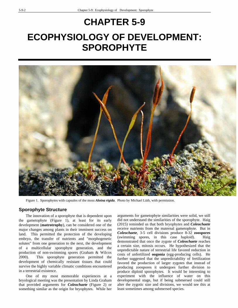
The advent of bryophytes brought several critical innovations that permitted existence on land. Several of these facilitated sporophyte persistence: efficient placental tissues to facilitate transfer of nutrients and hormones from the gametophyte to the sporophyte, sporangia with decay-resistant walls, sporopollenin in spore walls (Renzaglia et al. 2000; Graham & Gray 2001), and development of a cuticle (Proctor 1984). The ability to provide nutrition and protection for the sporophyte made it possible to produce numerous spores from a single fertilization.
Despite these important bryophyte innovations, the
capsule differs considerably among the three phyla and in
this regard provides the best distinguishing characters for
separating the three phyla (Renzaglia et al. 2000). In
mosses and liverworts, meiosis is synchronous throughout
the capsule, whereas in hornworts it continues over time
with the oldest spores at the tip while meiosis is still being
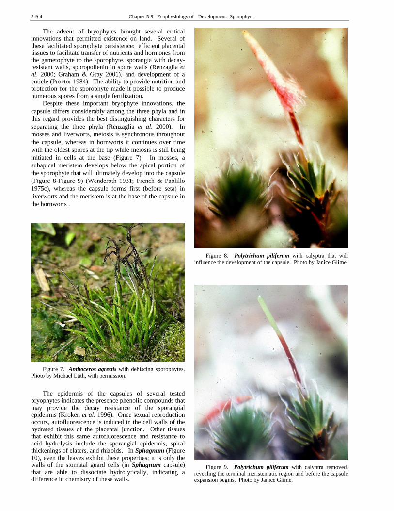
initiated in cells at the base (Figure 7). In mosses, a
subapical meristem develops below the apical portion of
the sporophyte that will ultimately develop into the capsule
(Figure 8-Figure 9) (Wenderoth 1931; French & Paolillo
1975c), whereas the capsule forms first (before seta) in
liverworts and the meristem is at the base of the capsule in
the hornworts .
Figure 7. Anthoceros agrestis with dehiscing sporophytes. Photo by Michael Lüth, with permission.
The epidermis of the capsules of several tested
bryophytes indicates the presence phenolic compounds that may provide the decay resistance of the sporangial epidermis (Kroken et al. 1996). Once sexual reproduction occurs, autofluorescence is induced in the cell walls of the hydrated tissues of the placental junction. Other tissues that exhibit this same autofluorescence and resistance to acid hydrolysis include the sporangial epidermis, spiral thickenings of elaters, and rhizoids. In Sphagnum (Figure 10), even the leaves exhibit these properties; it is only the walls of the stomatal guard cells (in Sphagnum capsule) that are able to dissociate hydrolytically, indicating a difference in chemistry of these walls.
Figure 8. Polytrichum piliferum with calyptra that will influence the development of the capsule. Photo by Janice Glime.
Figure 9. Polytrichum piliferum with calyptra removed, revealing the terminal meristematic region and before the capsule expansion begins. Photo by Janice Glime.
Chapter 5-9: Ecophysiology of Development: Sporophyte 5-9-5
Figure 10. Sphagnum auriculatum showing autofluorescence of the leaf. Photo by Janice Glime.
Sporophyte Nutrition
Before we can fully understand the development of the sporophyte, we must understand how it gets its energy, its signals, and its mineral nutrients. The energy source of the sporophyte has been a somewhat controversial topic. Its structure suggests dependency on the gametophyte, but its green color suggests it is able to carry out photosynthesis.
Boyce (2008) has suggested that loss of photosynthetic capacity in the moss sporophyte as it matures was driven by its small size and need for spore dispersal, the latter being supported by desiccation of the mature capsule. He argues that such size constraints on the physiology of the sporophyte are demonstrated by comparisons of size with anatomical detail and correlations between the axis, sporangium, and seta. Thus, we can expect that the degree of dependence on the gametophyte varies among the bryophyte taxa.
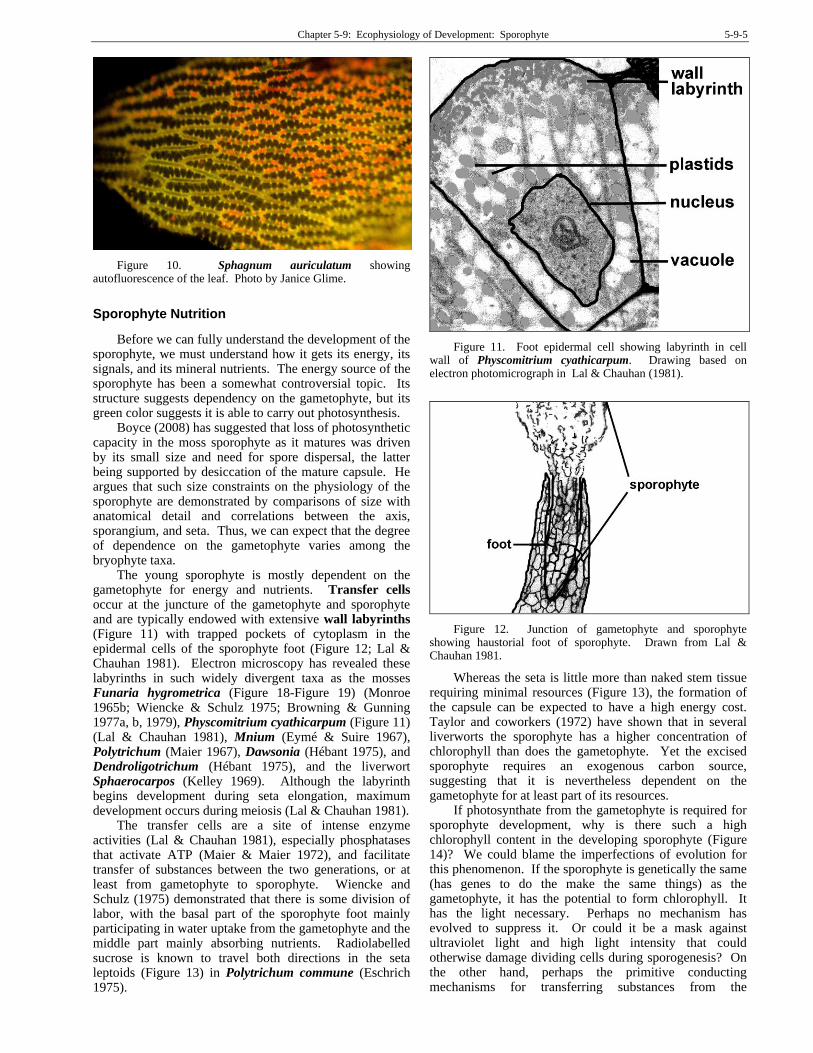
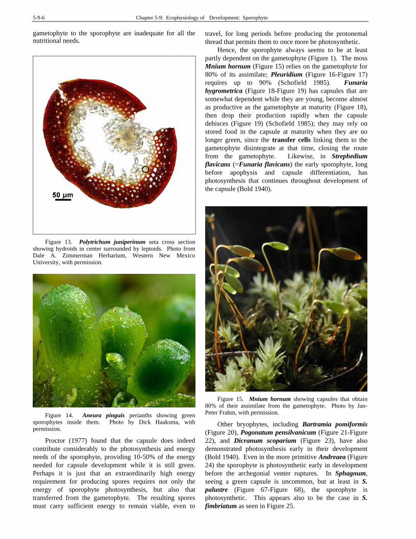
The young sporophyte is mostly dependent on the gametophyte for energy and nutrients. Transfer cells occur at the juncture of the gametophyte and sporophyte and are typically endowed with extensive wall labyrinths (Figure 11) with trapped pockets of cytoplasm in the epidermal cells of the sporophyte foot (Figure 12; Lal & Chauhan 1981). Electron microscopy has revealed these labyrinths in such widely divergent taxa as the mosses Funaria hygrometrica (Figure 18-Figure 19) (Monroe 1965b; Wiencke & Schulz 1975; Browning & Gunning 1977a, b, 1979), Physcomitrium cyathicarpum (Figure 11) (Lal & Chauhan 1981), Mnium (Eymé & Suire 1967), Polytrichum (Maier 1967), Dawsonia (Hébant 1975), and Dendroligotrichum (Hébant 1975), and the liverwort Sphaerocarpos (Kelley 1969). Although the labyrinth begins development during seta elongation, maximum development occurs during meiosis (Lal & Chauhan 1981).
The transfer cells are a site of intense enzyme activities (Lal & Chauhan 1981), especially phosphatases that activate ATP (Maier & Maier 1972), and facilitate transfer of substances between the two generations, or at least from gametophyte to sporophyte. Wiencke and Schulz (1975) demonstrated that there is some division of labor, with the basal part of the sporophyte foot mainly participating in water uptake from the gametophyte and the middle part mainly absorbing nutrients. Radiolabelled sucrose is known to travel both directions in the seta leptoids (Figure 13) in Polytrichum commune (Eschrich 1975).
Figure 11. Foot epidermal cell showing labyrinth in cell wall of Physcomitrium cyathicarpum. Drawing based on electron photomicrograph in Lal & Chauhan (1981).
Figure 12. Junction of gametophyte and sporophyte showing haustorial foot of sporophyte. Drawn from Lal & Chauhan 1981.
Whereas the seta is little more than naked stem tissue requiring minimal resources (Figure 13), the formation of the capsule can be expected to have a high energy cost. Taylor and coworkers (1972) have shown that in several liverworts the sporophyte has a higher concentration of chlorophyll than does the gametophyte. Yet the excised sporophyte requires an exogenous carbon source, suggesting that it is nevertheless dependent on the gametophyte for at least part of its resources.
If photosynthate from the gametophyte is required for sporophyte development, why is there such a high chlorophyll content in the developing sporophyte (Figure 14)? We could blame the imperfections of evolution for this phenomenon. If the sporophyte is genetically the same (has genes to do the make the same things) as the gametophyte, it has the potential to form chlorophyll. It has the light necessary. Perhaps no mechanism has evolved to suppress it. Or could it be a mask against ultraviolet light and high light intensity that could otherwise damage dividing cells during sporogenesis? On the other hand, perhaps the primitive conducting mechanisms for transferring substances from the
5-9-6 Chapter 5-9: Ecophysiology of Development: Sporophyte
gametophyte to the sporophyte are inadequate for all the nutritional needs.
Figure 13. Polytrichum juniperinum seta cross section showing hydroids in center surrounded by leptoids. Photo from Dale A. Zimmerman Herbarium, Western New Mexico University, with permission.
Figure 14. Aneura pinguis perianths showing green sporophytes inside them. Photo by Dick Haaksma, with permission.
Proctor (1977) found that the capsule does indeed
contribute considerably to the photosynthesis and energy
needs of the sporophyte, providing 10-50% of the energy
needed for capsule development while it is still green.
Perhaps it is just that an extraordinarily high energy
requirement for producing spores requires not only the
energy of sporophyte photosynthesis, but also that
transferred from the gametophyte. The resulting spores
must carry sufficient energy to remain viable, even to
travel, for long periods before producing the protonemal
thread that permits them to once more be photosynthetic.
Hence, the sporophyte always seems to be at least
partly dependent on the gametophyte (Figure 1). The moss
Mnium hornum (Figure 15) relies on the gametophyte for
80% of its assimilate; Pleuridium (Figure 16-Figure 17)
requires up to 90% (Schofield 1985). Funaria
hygrometrica (Figure 18-Figure 19) has capsules that are
somewhat dependent while they are young, become almost
as productive as the gametophyte at maturity (Figure 18),
then drop their production rapidly when the capsule
dehisces (Figure 19) (Schofield 1985); they may rely on
stored food in the capsule at maturity when they are no
longer green, since the transfer cells linking them to the
gametophyte disintegrate at that time, closing the route
from the gametophyte. Likewise, in Strephedium
flavicans (=Funaria flavicans) the early sporophyte, long
before apophysis and capsule differentiation, has
photosynthesis that continues throughout development of
the capsule (Bold 1940).
Figure 15. Mnium hornum showing capsules that obtain 80% of their assimilate from the gametophyte. Photo by Jan-Peter Frahm, with permission.
Other bryophytes, including Bartramia pomiformis
(Figure 20), Pogonatum pensilvanicum (Figure 21-Figure
22), and Dicranum scoparium (Figure 23), have also
demonstrated photosynthesis early in their development
(Bold 1940). Even in the more primitive Andreaea (Figure
24) the sporophyte is photosynthetic early in development
before the archegonial venter ruptures. In Sphagnum,
seeing a green capsule is uncommon, but at least in S.
palustre (Figure 67-Figure 68), the sporophyte is
photosynthetic. This appears also to be the case in S.
fimbriatum as seen in Figure 25.

Chapter 5-9: Ecophysiology of Development: Sporophyte 5-9-7
Figure 16. Young, green capsules of Pleuridium subulatum, nevertheless requiring 90% of their assimilate from the gametophyte. Photo by Kristian Peters, with permission.
Figure 17. Pleuridium subulatum with mature capsules with phenolic compounds that color them red. Photo by Paul Davison, with permission.
Figure 18. Funaria hygrometrica capsule demonstrating green color at full size but before full maturity. Photo by Sarah Gregg, through Creative Commons.
Figure 19. Funaria hygrometrica with brown color typical of dehiscing capsules. Photo by Juan Larrain, with permission.
Figure 20. Bartramia pomiformis with mature green capsule on left and dehisced red capsule on right. This moss is aptly called the apple moss. Photo by Des Callaghan, with permission.
Figure 21. Young plants of Pogonatum pensilvanicum with emerging green sporophytes. Photo by George J. Shepherd, through Creative Commons.
Figure 22. Mature sporophytes of Pogonatum pensilvanicum with its fully covering calyptra. Photo by George J. Shepherd, through Creative Commons.
5-9-8 Chapter 5-9: Ecophysiology of Development: Sporophyte
Figure 23. Dicranum scoparium with nearly mature green capsules. Photo by Michael Lüth, with permission.
Figure 24. Andreaea australis showing young, green capsules and older, brown capsules. Photo by Niels Klazenga, with permission.
Figure 25. Sphagnum fimbriatum with green capsules still inside the perichaetial leaves. Photo by Barry Stewart, with permission.
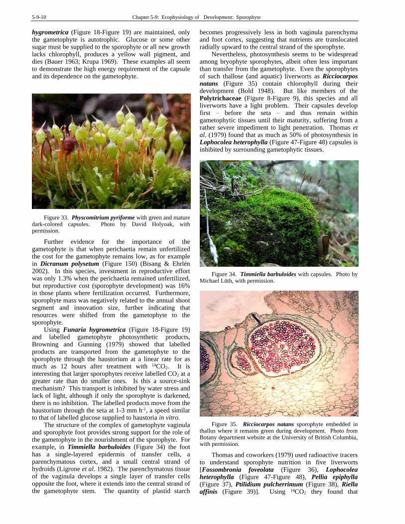
Courtice and coworkers (1978) have shown that sugars move from the gametophyte to the sporophyte in Physcomitrella (Figure 26), supporting the concept that the demands of the sporophyte are greater than its own production capacity. If we put these demands into an ecological and temporal context, need for a gametophytic supplement becomes obvious. For example, sporophytes of Polytrichum s.l. (Figure 28, Figure 29) can require up to 13 months to develop in some localities (Arnell 1905),
spanning a multitude of environmental conditions. When embryo development begins, environmental conditions can easily be less than favorable for photosynthetic activity. Patterson and Baber (1961) found that many temperate mosses were dormant in late summer and autumn. Such a dormant period, if it affects the sporophyte as well, greatly reduces its opportunity to provide its own food. The sporophyte furthermore has little exposed surface area for photosynthesis, and what surface there is, at least throughout most of the development, has its long axis oriented in the same direction as the light, thus minimizing its utility as a light-absorbing organ. It is reasonable, then, that the gametophyte, which is sensitive to moisture that must be available for growth and that has a large photosynthetic surface, can provide the food and the signals for the sporophyte. Furthermore, Hughes (1954) has demonstrated that it is the gametophyte and not the sporophyte that responds to photoperiod to control sporophyte development in Pogonatum aloides (Figure 27) and Polytrichum piliferum (Figure 28), supporting the concept that there is a need for conduction of substances into the sporophyte.
Figure 26. Physcomitrella patens with capsules covered by calyptrae. Note the projecting archegonial neck. Photo by Jan-Peter Frahm, with permission.
Figure 27. Pogonatum aloides with capsules that must receive signals from the gametophyte to control its development. Photo from Proyecto Musgo, through Creative Commons.

Chapter 5-9: Ecophysiology of Development: Sporophyte 5-9-9
Figure 28. Polytrichum piliferum with calyptras, a species where photoperiod control of the sporophyte occurs in the gametophyte. Photo by GNU Free Documentation License.
Krisko and Paolillo (1972) suggested that weight gain in the capsule was directly and linearly related to weight loss of the seta in mosses. In Polytrichum juniperinum (Figure 29) and Polytrichastrum ohioense (Figure 30), the capsule takes weight from the seta in culture if no dextrose is supplied to the capsule, but little seta loss occurs in the presence of dextrose. However, capsule weight gain is also a linear function of the length of the gametophyte explant, and in the presence of dextrose, the seta loss is suppressed, suggesting that the gametophyte is the most important source of carbon/weight gain for the capsule.
Figure 29. Polytrichum juniperinum capsules showing complete coverage by hairy calyptra. Photo by Janice Glime.
Renault et al. (1992) stated that dependence on the gametophyte for carbon nutrition is especially true for species of Polytrichum (Figure 28-Figure 29) and other Polytrichaceae. In Polytrichastrum formosum (Figure 31), sucrose was the main soluble sugar in both the gametophyte and sporophyte, with the highest concentration (~230 m) in the haustorium. Glucose was converted to sucrose after its absorption into the haustorium. On the other hand, the sugars in the vaginula (Figure 32) were mainly hexoses, with traces of trehalose. Renault et al. suggested that the conversion of sucrose to glucose and fructose at the haustorium interface, and the subsequent reconversion to sucrose after hexose absorption by haustorium cells, mainly governs the sugar
accumulation in the haustorium. The need for transfer of carbohydrate from the photosynthetic gametophyte to the sporophyte in the Polytrichaceae may relate in part to the large, hairy calyptra (Figure 29) in most members of the family. Its ability to completely cover the capsule and even close off its open end would make available light much less available. It would be interesting to correlate not only capsule size, but also relative calyptra size and thickness with dependency upon transfer of carbohydrates from the gametophyte.
Figure 30. Polytrichastrum ohioense with green capsules. The capsule of this moss absorbs some of its nutrition from its own seta. Photo by Bob Klips, with permission.
Figure 31. Polytrichastrum formosum with calyptrae over green capsules. Photo by Michael Lüth, with permission.
Figure 32. Vaginula of bryophyte. Photo from unknown source.
But not all bryophytes have such imposing calyptrae. Even species with little coverage by the calyptra require the nutritional support of the gametophyte. When photosynthetic sporophyte and gametophyte cultures of Physcomitrium pyriforme (Figure 33) and Funaria
5-9-10 Chapter 5-9: Ecophysiology of Development: Sporophyte
hygrometrica (Figure 18-Figure 19) are maintained, only the gametophyte is autotrophic. Glucose or some other sugar must be supplied to the sporophyte or all new growth lacks chlorophyll, produces a yellow wall pigment, and dies (Bauer 1963; Krupa 1969). These examples all seem to demonstrate the high energy requirement of the capsule and its dependence on the gametophyte.
Figure 33. Physcomitrium pyriforme with green and mature dark-colored capsules. Photo by David Holyoak, with permission.
Further evidence for the importance of the gametophyte is that when perichaetia remain unfertilized the cost for the gametophyte remains low, as for example in Dicranum polysetum (Figure 150) (Bisang & Ehrlén 2002). In this species, investment in reproductive effort was only 1.3% when the perichaetia remained unfertilized, but reproductive cost (sporophyte development) was 16% in those plants where fertilization occurred. Furthermore, sporophyte mass was negatively related to the annual shoot segment and innovation size, further indicating that resources were shifted from the gametophyte to the sporophyte.
Using Funaria hygrometrica (Figure 18-Figure 19) and labelled gametophyte photosynthetic products, Browning and Gunning (1979) showed that labelled products are transported from the gametophyte to the sporophyte through the haustorium at a linear rate for as much as 12 hours after treatment with 14CO2. It is interesting that larger sporophytes receive labelled CO2 at a greater rate than do smaller ones. Is this a source-sink mechanism? This transport is inhibited by water stress and lack of light, although if only the sporophyte is darkened, there is no inhibition. The labelled products move from the haustorium through the seta at 1-3 mm h-1, a speed similar to that of labelled glucose supplied to haustoria in vitro.
The structure of the complex of gametophyte vaginula and sporophyte foot provides strong support for the role of the gametophyte in the nourishment of the sporophyte. For example, in Timmiella barbuloides (Figure 34) the foot has a single-layered epidermis of transfer cells, a parenchymatous cortex, and a small central strand of hydroids (Ligrone et al. 1982). The parenchymatous tissue of the vaginula develops a single layer of transfer cells opposite the foot, where it extends into the central strand of the gametophyte stem. The quantity of plastid starch
becomes progressively less in both vaginula parenchyma and foot cortex, suggesting that nutrients are translocated radially upward to the central strand of the sporophyte.
Nevertheless, photosynthesis seems to be widespread among bryophyte sporophytes, albeit often less important than transfer from the gametophyte. Even the sporophytes of such thallose (and aquatic) liverworts as Ricciocarpos natans (Figure 35) contain chlorophyll during their development (Bold 1948). But like members of the Polytrichaceae (Figure 8-Figure 9), this species and all liverworts have a light problem. Their capsules develop first – before the seta – and thus remain within gametophytic tissues until their maturity, suffering from a rather severe impediment to light penetration. Thomas et al. (1979) found that as much as 50% of photosynthesis in Lophocolea heterophylla (Figure 47-Figure 48) capsules is inhibited by surrounding gametophytic tissues.
Figure 34. Timmiella barbuloides with capsules. Photo by Michael Lüth, with permission.
Figure 35. Ricciocarpos natans sporophyte embedded in thallus where it remains green during development. Photo from Botany department website at the University of British Columbia, with permission.
Thomas and coworkers (1979) used radioactive tracers to understand sporophyte nutrition in five liverworts [Fossombronia foveolata (Figure 36), Lophocolea heterophylla (Figure 47-Figure 48), Pellia epiphylla (Figure 37), Ptilidium pulcherrimum (Figure 38), Riella affinis (Figure 39)]. Using 14CO2 they found that
Chapter 5-9: Ecophysiology of Development: Sporophyte 5-9-11
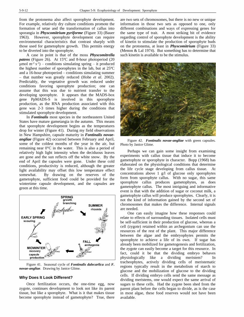
sporophytes of all five species were able to fix CO2 in the light. Nevertheless, the fixation rate per mg fresh weight was small compared to that of the gametophyte, with a sporophyte:gametophyte ratio of 0.12-0.39. The chlorophyll ratios were 1.07-3.30. Thus it is not surprising that radioactivity of Lophocolea sporophytes increased significantly after application of 14C-glucose to the supporting gametophytes. Perhaps most surprising in this study was finding that in Lophocolea heterophylla (Figure 47-Figure 48), 40% of the capsule photosynthesis occurred in the spores (Figure 40)!
Figure 36. Fossombronia foveolata with your sporophytes still within the perichaetial leaves. Photo by Des Callaghan, with permission.
Figure 37. Pellia epiphylla with sporophytes in various stages of seta elongations. Not the remains of green color in the capsule. Photo by Michael Lüth, with permission.
Figure 38. Ptilidium pulcherrimum with capsules. Photo by Hermann Schachner, through Creative Commons.
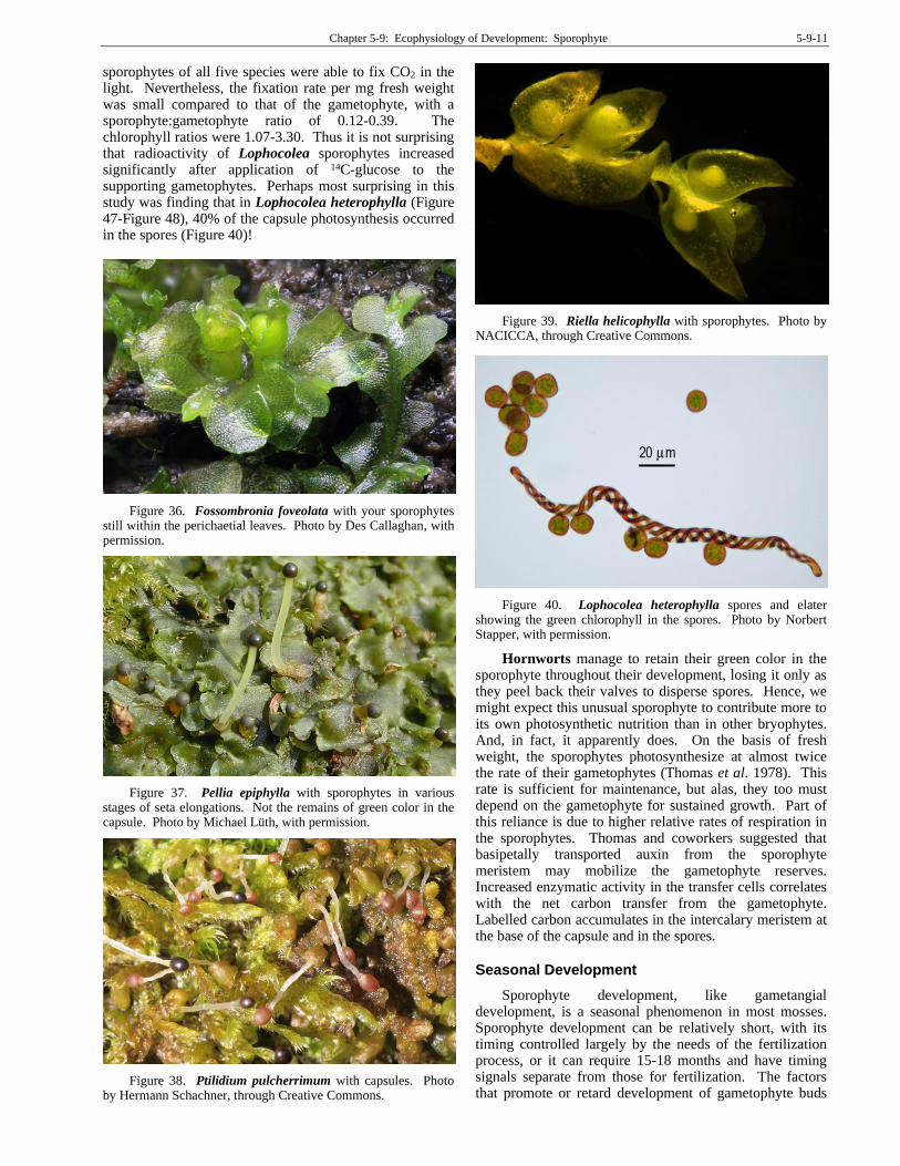
Figure 39. Riella helicophylla with sporophytes. Photo by NACICCA, through Creative Commons.
Figure 40. Lophocolea heterophylla spores and elater showing the green chlorophyll in the spores. Photo by Norbert Stapper, with permission.
Hornworts manage to retain their green color in the sporophyte throughout their development, losing it only as they peel back their valves to disperse spores. Hence, we might expect this unusual sporophyte to contribute more to its own photosynthetic nutrition than in other bryophytes. And, in fact, it apparently does. On the basis of fresh weight, the sporophytes photosynthesize at almost twice the rate of their gametophytes (Thomas et al. 1978). This rate is sufficient for maintenance, but alas, they too must depend on the gametophyte for sustained growth. Part of this reliance is due to higher relative rates of respiration in the sporophytes. Thomas and coworkers suggested that basipetally transported auxin from the sporophyte meristem may mobilize the gametophyte reserves. Increased enzymatic activity in the transfer cells correlates with the net carbon transfer from the gametophyte. Labelled carbon accumulates in the intercalary meristem at the base of the capsule and in the spores.
Seasonal Development
Sporophyte development, like gametangial development, is a seasonal phenomenon in most mosses. Sporophyte development can be relatively short, with its timing controlled largely by the needs of the fertilization process, or it can require 15-18 months and have timing signals separate from those for fertilization. The factors that promote or retard development of gametophyte buds
5-9-12 Chapter 5-9: Ecophysiology of Development: Sporophyte
from the protonema also affect sporophyte development. For example, relatively dry culture conditions promote the formation of setae and the transformation of callus into sporangia in Physcomitrium pyriforme (Figure 33) (Bauer 1963). However, sporophyte development can require environmental characteristics that contrast sharply with those used for gametophyte growth. This permits energy to be diverted into the sporophyte.
A case in point is that of the moss Physcomitrella patens (Figure 26). At 15ºC and 8-hour photoperiod (20 µmol m-2 s-1) – conditions simulating spring – it produced the highest number of sporophytes in the lab, but at 25ºC and a 16-hour photoperiod – conditions simulating summer – that number was greatly reduced (Hohe et al. 2002). Predictably, the vegetative growth was reduced under conditions favoring sporophyte production; one can assume that this was due to nutrient transfer to the developing sporophyte. It appears that the MADS-box gene PpMADS-S is involved in this sporophyte production, as the RNA production associated with this gene was 2-3 times higher during the conditions that stimulated sporophyte development.
In Fontinalis most species in the northeastern United States have mature gametangia in the autumn. This means that sporophyte development begins as the temperatures drop for winter (Figure 41). During my field observations in New Hampshire, capsule maturity in Fontinalis novae-angliae (Figure 42) occurred between February and April, some of the coldest months of the year in the air, but remaining near 0°C in the water. This is also a period of relatively high light intensity when the deciduous leaves are gone and the sun reflects off the white snow. By the end of April the capsules were gone. Under these cold conditions, productivity is reduced, although the greater light availability may offset this low temperature effect somewhat. By drawing on the reserves of the gametophyte, sufficient food could be provided for the wintertime capsule development, and the capsules are green at this time.
Figure 41. Seasonal cycle of Fontinalis dalecarlica and F. novae-angliae. Drawing by Janice Glime.
Why Does It Look Different?
Once fertilization occurs, the one-time egg, now zygote, continues development to look not like its parent tissue, but like a sporophyte. What is it that makes tissue become sporophyte instead of gametophyte? True, there
are two sets of chromosomes, but there is no new or unique information in those two sets as opposed to one, only different combinations and ways of expressing genes for the same type of trait. A most striking bit of evidence regarding control of sporophyte development is the ability of kinetin to stimulate the production of sporophyte buds on the protonema, at least in Physcomitrium (Figure 33) (Menon & Lal 1974). But something has to determine that such kinetin is available to be the stimulus.
Figure 42. Fontinalis novae-angliae with green capsules. Photo by Janice Glime.
Perhaps we can gain some insight from examining experiments with callus tissue that induce it to become gametophyte or sporophyte in character. Bopp (1968) has elaborated on the physiological conditions that determine the life cycle stage developing from callus tissue. At concentrations above 1 g/l of glucose only sporophytes form from sporophyte callus. With no sugar, this same sporophyte callus produces gametophytes, as does gametophyte callus. The most intriguing and informative event is that with the addition of sugar or coconut milk, a gametophyte callus will produce sporophytes. Clearly, it is not the kind of information gained by the second set of chromosomes that makes the difference. Internal signals are needed.
One can easily imagine how these responses could relate to effects of surrounding tissues. Isolated cells must be self-sufficient in their production of glucose, whereas a cell (zygote) retained within an archegonium can use the resources of the rest of the plant. This major difference between the algae and the embryophytes permits the sporophyte to achieve a life of its own. If sugar has already been mobilized for gametogenesis and fertilization, the zygote can easily become a target for this resource. In fact, could it be that the dividing embryo behaves physiologically like a dividing meristem? In tracheophytes, actively dividing cells of meristematic regions typically result in the metabolism of starch to glucose and the mobilization of glucose to the dividing cells. If dividing embryo cells send the same message as dividing meristems, one would expect the same arrival of sugars to these cells. Had the zygote been shed from the parent plant before the cells began to divide, as is the case in most algae, these food reserves would not have been available.
Chapter 5-9: Ecophysiology of Development: Sporophyte 5-9-13
Seta Structure and Function
The seta structure is not just an extension of the gametophyte stem, but rather is a unique structure in mosses and liverworts. It has food-conduction in relatively unspecialized parenchyma cells of the seta in mosses, including even Sphagnum (Figure 43-Figure 44) (Ligrone et al. 2000). The sporophyte axis of Bryophyta differs significantly from the independent sporophytes of the tracheophytes, but the sporophyte also shows remarkable differences among the bryophytes. In bryophytes, the sporophyte does not branch, whereas branching is typical among tracheophytes (Renzaglia et al. 2000). The expansion of the seta in Marchantiophyta (Figure 35-Figure 40) requires a rapid expansion of the cell wall without cell division to provide the elongate structure, a phenomenon accomplished by hydrostatic support. Hence, we can surmise that water is a necessity and we should expect the seta elongation to be timed with water availability. Anthocerotophyta (Figure 45-Figure 46) lack a seta and the capsule is anchored directly into the gametophyte tissue.
Figure 43. Sphagnum capsule ls. Photo from Botany Website, UBC, with permission.
When Cooke et al. (2002) surveyed the literature regarding auxin actions in Charophyta, bryophytes, and tracheophytes, they found a striking similarity in physiological mechanisms for regulating IAA (auxin) levels and responses to these levels, at least in the sporophytes. Both charophytes and liverworts synthesize IAA via a tryptophan-independent pathway in which IAA levels are regulated by the rates of IAA synthesis and degradation. All other land plants (mosses, hornworts, tracheophytes) use the same type of biosynthetic pathway
in their apical regions, but also can use IAA conjugation and conjugate hydrolysis reactions to increase the precision of the levels of IAA in both space and time. In bryophytes, IAA is involved in a number of developmental responses, including tropisms, apical dominance, and rhizoid initiation. But the only measurable transport known at that time (2002) in bryophytes was in the young setae of mosses.
Figure 44. Sphagnum capsules with pseudopodium and extremely short seta at the top of the foot. Photo by Joan Edwards, with permission.
Figure 45. Anthoceros punctatus showing the white sporophyte anchored in the gametophyte tissue. The involucre surrounds the base and may play a role in early development of the sporophyte. Photo by Des Callaghan, with permission.
5-9-14 Chapter 5-9: Ecophysiology of Development: Sporophyte
Figure 46. Nothoceros showing the sporophyte anchored in the gametophyte tissue. The involucre surrounds the base and may play a role in early development of the sporophyte. Photo by Juan Larrain with permission.
Seta Elongation
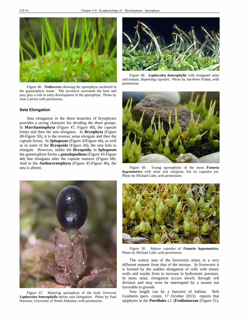
Seta elongation in the three branches of bryophytes provides a strong character for dividing the three groups. In Marchantiophyta (Figure 47, Figure 48), the capsule forms and then the seta elongates. In Bryophyta (Figure 49-Figure 50), it is the reverse; setae elongate and then the capsule forms. In Sphagnum (Figure 43Figure 44), as well as in some of the Bryopsida (Figure 26), the seta fails to elongate. However, unlike the Bryopsida, in Sphagnum the gametophyte forms a pseudopodium (Figure 43-Figure 44) that elongates after the capsule matures (Figure 68). And in the Anthocerotophyta (Figure 45-Figure 46), the seta is absent.
Figure 47. Maturing sporophyte of the leafy liverwort Lophocolea heterophylla before seta elongation. Photo by Paul Davison, University of North Alabama, with permission.
Figure 48. Lophocolea heterophylla with elongated setae and mature, dispersing capsules. Photo by Jan-Peter Frahm, with permission.
Figure 49. Young sporophytes of the moss Funaria hygrometrica with setae and calyptrae, but no capsules yet. Photo by Michael Lüth, with permission.
Figure 50. Mature capsules of Funaria hygrometrica. Photo by Michael Lüth, with permission.
The watery seta of the liverworts arises in a very different manner from that of the mosses. In liverworts it is formed by the sudden elongation of cells with elastic walls and results from in increase in hydrostatic pressure. In moss setae, elongation occurs slowly through cell division and may even be interrupted by a season not favorable to growth.
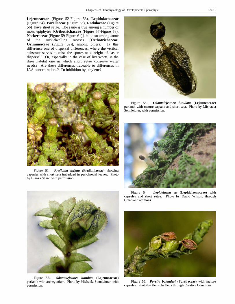
Seta length can be a function of habitat. Rob Gradstein (pers. comm. 17 October 2013) reports that epiphytes in the Porellales s.l. [Frullaniaceae (Figure 51),
Chapter 5-9: Ecophysiology of Development: Sporophyte 5-9-15
Lejeuneaceae (Figure 52-Figure 53), Lepidolaenaceae (Figure 54), Porellaceae (Figure 55), Radulaceae (Figure 56)] have short setae. The same is true among a number of moss epiphytes [Orthotrichaceae (Figure 57-Figure 58), Neckeraceae (Figure 59-Figure 61)], but also among some of the rock-dwelling mosses [Orthotrichaceae, Grimmiaceae (Figure 62)], among others. Is this difference one of dispersal differences, where the vertical substrate serves to raise the spores to a height of easier dispersal? Or, especially in the case of liverworts, is the drier habitat one in which short setae conserve water needs? Are these differences traceable to differences in IAA concentrations? To inhibition by ethylene?
Figure 51. Frullania inflata (Frullaniaceae) showing capsules with short seta imbedded in perichaetial leaves. Photo by Blanka Shaw, with permission.
Figure 52. Odontolejeunea lunulata (Lejeuneaceae) perianth with archegonium. Photo by Michaela Sonnleitner, with permission.
Figure 53. Odontolejeunea lunulata (Lejeuneaceae) perianth with mature capsule and short seta. Photo by Michaela Sonnleitner, with permission.
Figure 54. Lepidolaena sp (Lepidolaenaceae) with capsules and short setae. Photo by David Wilson, through Creative Commons.
Figure 55. Porella bolanderi (Porellaceae) with mature capsules. Photo by Ken-ichi Ueda through Creative Commons.
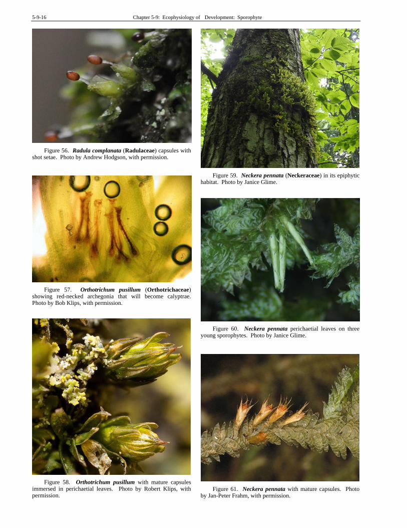
5-9-16 Chapter 5-9: Ecophysiology of Development: Sporophyte
Figure 56. Radula complanata (Radulaceae) capsules with shot setae. Photo by Andrew Hodgson, with permission.
Figure 57. Orthotrichum pusillum (Orthotrichaceae) showing red-necked archegonia that will become calyptrae. Photo by Bob Klips, with permission.
Figure 58. Orthotrichum pusillum with mature capsules immersed in perichaetial leaves. Photo by Robert Klips, with permission.
Figure 59. Neckera pennata (Neckeraceae) in its epiphytic habitat. Photo by Janice Glime.
Figure 60. Neckera pennata perichaetial leaves on three young sporophytes. Photo by Janice Glime.
Figure 61. Neckera pennata with mature capsules. Photo by Jan-Peter Frahm, with permission.
Chapter 5-9: Ecophysiology of Development: Sporophyte 5-9-17
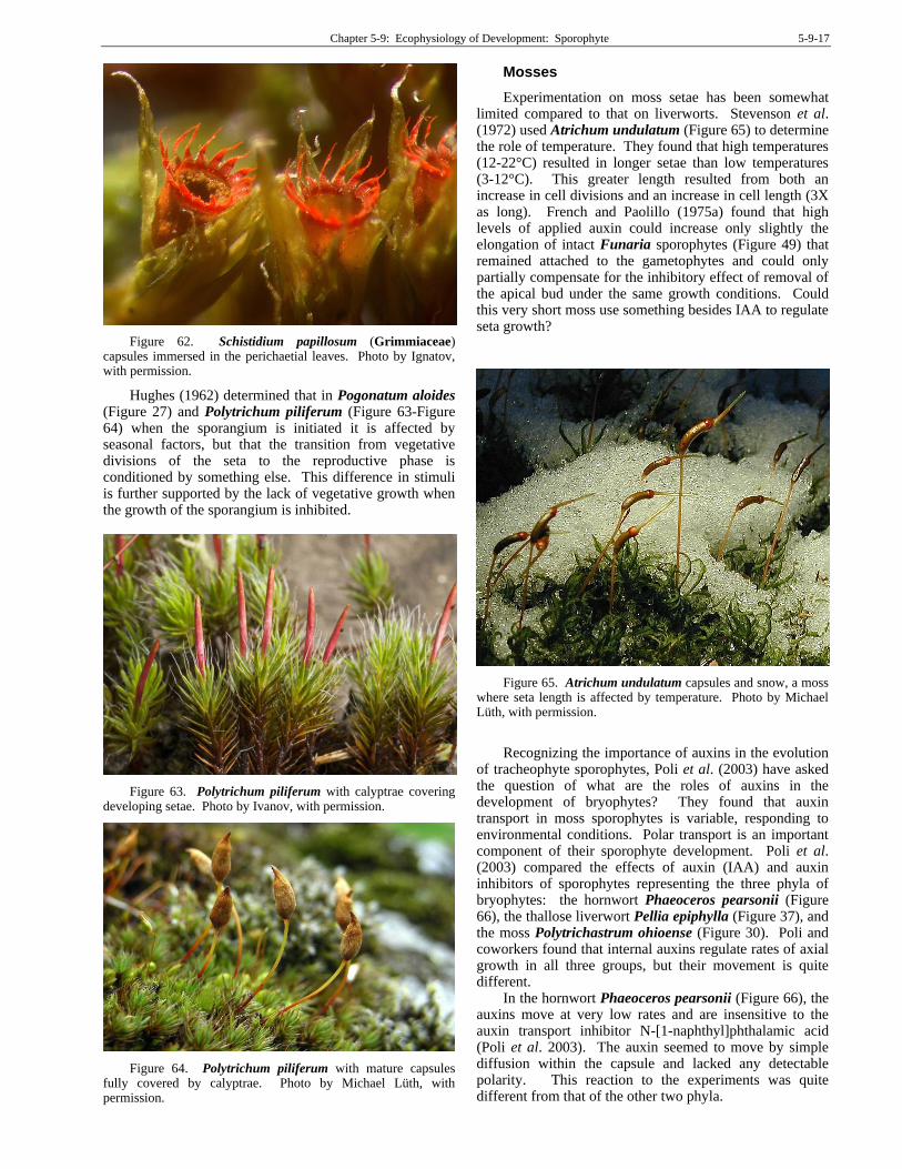
Figure 62. Schistidium papillosum (Grimmiaceae) capsules immersed in the perichaetial leaves. Photo by Ignatov, with permission.
Hughes (1962) determined that in Pogonatum aloides (Figure 27) and Polytrichum piliferum (Figure 63-Figure 64) when the sporangium is initiated it is affected by seasonal factors, but that the transition from vegetative divisions of the seta to the reproductive phase is conditioned by something else. This difference in stimuli is further supported by the lack of vegetative growth when the growth of the sporangium is inhibited.
Figure 63. Polytrichum piliferum with calyptrae covering developing setae. Photo by Ivanov, with permission.
Figure 64. Polytrichum piliferum with mature capsules fully covered by calyptrae. Photo by Michael Lüth, with permission.
Mosses
Experimentation on moss setae has been somewhat limited compared to that on liverworts. Stevenson et al. (1972) used Atrichum undulatum (Figure 65) to determine the role of temperature. They found that high temperatures (12-22°C) resulted in longer setae than low temperatures (3-12°C). This greater length resulted from both an increase in cell divisions and an increase in cell length (3X as long). French and Paolillo (1975a) found that high levels of applied auxin could increase only slightly the elongation of intact Funaria sporophytes (Figure 49) that remained attached to the gametophytes and could only partially compensate for the inhibitory effect of removal of the apical bud under the same growth conditions. Could this very short moss use something besides IAA to regulate seta growth?
Figure 65. Atrichum undulatum capsules and snow, a moss where seta length is affected by temperature. Photo by Michael Lüth, with permission.
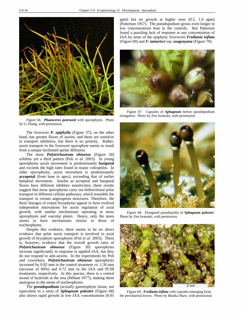
Recognizing the importance of auxins in the evolution
of tracheophyte sporophytes, Poli et al. (2003) have asked the question of what are the roles of auxins in the development of bryophytes? They found that auxin transport in moss sporophytes is variable, responding to environmental conditions. Polar transport is an important component of their sporophyte development. Poli et al. (2003) compared the effects of auxin (IAA) and auxin inhibitors of sporophytes representing the three phyla of bryophytes: the hornwort Phaeoceros pearsonii (Figure 66), the thallose liverwort Pellia epiphylla (Figure 37), and the moss Polytrichastrum ohioense (Figure 30). Poli and coworkers found that internal auxins regulate rates of axial growth in all three groups, but their movement is quite different.
In the hornwort Phaeoceros pearsonii (Figure 66), the auxins move at very low rates and are insensitive to the auxin transport inhibitor N-[1-naphthyl]phthalamic acid (Poli et al. 2003). The auxin seemed to move by simple diffusion within the capsule and lacked any detectable polarity. This reaction to the experiments was quite different from that of the other two phyla.
5-9-18 Chapter 5-9: Ecophysiology of Development: Sporophyte
Figure 66. Phaeoceros pearsonii with sporophytes. Photo by Li Zhang, with permission.
The liverwort P. epiphylla (Figure 37), on the other hand, has greater fluxes of auxins, and these are sensitive to transport inhibitors, but there is no polarity. Rather, auxin transport in the liverwort sporophyte seems to result from a unique facilitated apolar diffusion.
The moss Polytrichastrum ohioense (Figure 30) exhibits yet a third pattern (Poli et al. 2003). In young sporophytes auxin movement is predominantly basipetal and exceeds the high rates found in maize coleoptiles. In older sporophytes, auxin movement is predominantly acropetal (from base to apex), exceeding that of earlier basipetal movement. Insofar as acropetal and basipetal fluxes have different inhibitor sensitivities, these results suggest that moss sporophytes carry out bidirectional polar transport in different cellular pathways, which resemble the transport in certain angiosperm structures. Therefore, the three lineages of extant bryophytes appear to have evolved independent innovations for auxin regulation of axial growth, with similar mechanisms operating in moss sporophytes and vascular plants. Hence, only the moss seems to have mechanisms similar to those of tracheophytes.
Despite this evidence, there seems to be no direct evidence that polar auxin transport is involved in axial growth of bryophyte sporophytes (Poli et al. 2003). There is, however, evidence that the overall growth rates of Polytrichastrum ohioense (Figure 30) sporophytes increase significantly in response to applied IAA, but they do not respond to anti-auxins. In the experiments by Poli and coworkers, Polytrichastrum ohioense sporophytes increased by 0.82 mm in the control treatment vs. 1.30 mm (increase of 60%) and 0.72 mm in the IAA and PCIB treatments, respectively. In this species, there is a central strand of hydroids in the seta (Hébant 1977), making these analogous to the stems of tracheophytes.
The pseudopodium (actually gametophyte tissue, not equivalent to a seta) of Sphagnum palustre (Figure 68) also shows rapid growth in low IAA concentrations (0.01
ppm) but no growth at higher ones (0.5, 1.0 ppm) (Patterson 1957). The pseudopodium grows even longer in low concentrations than in the controls. But Patterson found a puzzling lack of response at any concentration of IAA by setae of the epiphytic liverworts Frullania inflata (Figure 69) and F. tamarisci ssp. asagrayana (Figure 70).
Figure 67. Capsules of Sphagnum before pseudopodium elongation. Photo by Zen Iwatsuki, with permission.
Figure 68. Elongated pseudopodia of Sphagnum palustre. Photo by Zen Iwatsuki, with permission.
Figure 69. Frullania inflata with capsules emerging from the perichaetial leaves. Photo by Blanka Shaw, with permission.
Chapter 5-9: Ecophysiology of Development: Sporophyte 5-9-19
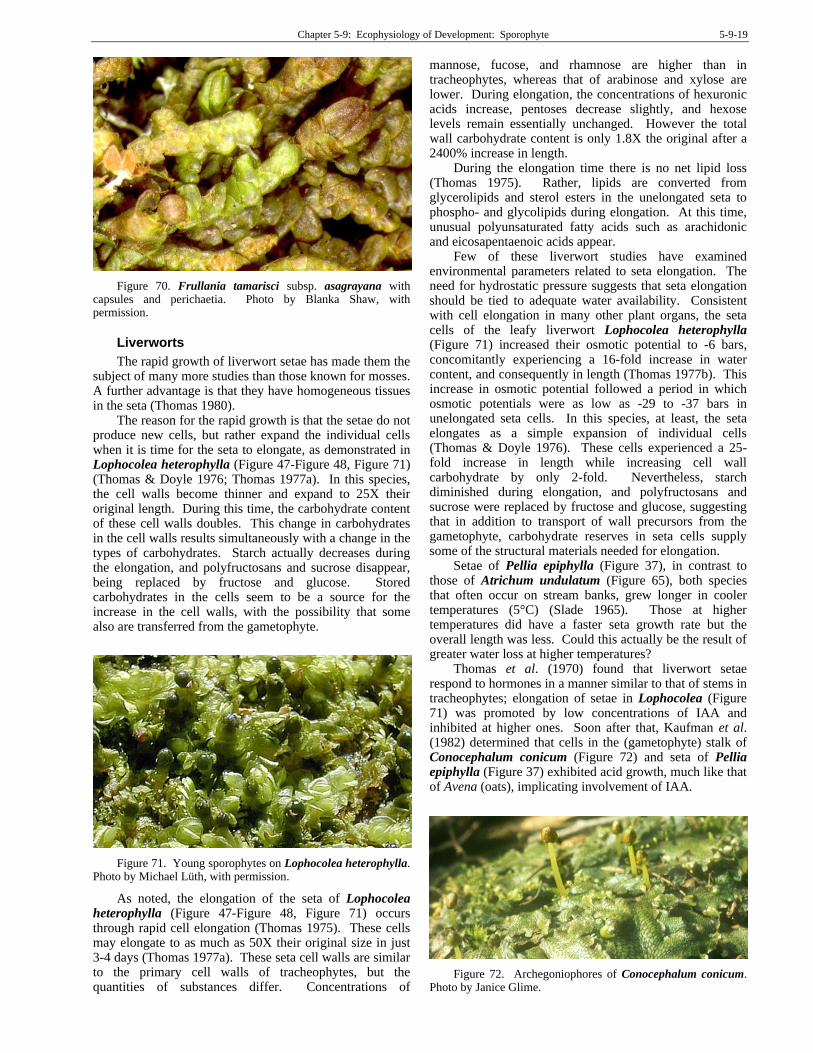
Figure 70. Frullania tamarisci subsp. asagrayana with capsules and perichaetia. Photo by Blanka Shaw, with permission.
Liverworts
The rapid growth of liverwort setae has made them the subject of many more studies than those known for mosses. A further advantage is that they have homogeneous tissues in the seta (Thomas 1980).
The reason for the rapid growth is that the setae do not produce new cells, but rather expand the individual cells when it is time for the seta to elongate, as demonstrated in Lophocolea heterophylla (Figure 47-Figure 48, Figure 71) (Thomas & Doyle 1976; Thomas 1977a). In this species, the cell walls become thinner and expand to 25X their original length. During this time, the carbohydrate content of these cell walls doubles. This change in carbohydrates in the cell walls results simultaneously with a change in the types of carbohydrates. Starch actually decreases during the elongation, and polyfructosans and sucrose disappear, being replaced by fructose and glucose. Stored carbohydrates in the cells seem to be a source for the increase in the cell walls, with the possibility that some also are transferred from the gametophyte.
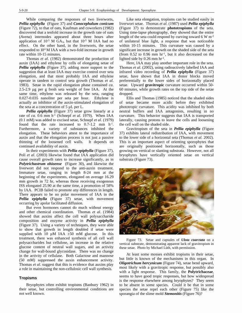
Figure 71. Young sporophytes on Lophocolea heterophylla. Photo by Michael Lüth, with permission.
As noted, the elongation of the seta of Lophocolea heterophylla (Figure 47-Figure 48, Figure 71) occurs through rapid cell elongation (Thomas 1975). These cells may elongate to as much as 50X their original size in just 3-4 days (Thomas 1977a). These seta cell walls are similar to the primary cell walls of tracheophytes, but the quantities of substances differ. Concentrations of
mannose, fucose, and rhamnose are higher than in tracheophytes, whereas that of arabinose and xylose are lower. During elongation, the concentrations of hexuronic acids increase, pentoses decrease slightly, and hexose levels remain essentially unchanged. However the total wall carbohydrate content is only 1.8X the original after a 2400% increase in length.
During the elongation time there is no net lipid loss (Thomas 1975). Rather, lipids are converted from glycerolipids and sterol esters in the unelongated seta to phospho- and glycolipids during elongation. At this time, unusual polyunsaturated fatty acids such as arachidonic and eicosapentaenoic acids appear.
Few of these liverwort studies have examined environmental parameters related to seta elongation. The need for hydrostatic pressure suggests that seta elongation should be tied to adequate water availability. Consistent with cell elongation in many other plant organs, the seta cells of the leafy liverwort Lophocolea heterophylla (Figure 71) increased their osmotic potential to -6 bars, concomitantly experiencing a 16-fold increase in water content, and consequently in length (Thomas 1977b). This increase in osmotic potential followed a period in which osmotic potentials were as low as -29 to -37 bars in unelongated seta cells. In this species, at least, the seta elongates as a simple expansion of individual cells (Thomas & Doyle 1976). These cells experienced a 25-fold increase in length while increasing cell wall carbohydrate by only 2-fold. Nevertheless, starch diminished during elongation, and polyfructosans and sucrose were replaced by fructose and glucose, suggesting that in addition to transport of wall precursors from the gametophyte, carbohydrate reserves in seta cells supply some of the structural materials needed for elongation.
Setae of Pellia epiphylla (Figure 37), in contrast to those of Atrichum undulatum (Figure 65), both species that often occur on stream banks, grew longer in cooler temperatures (5°C) (Slade 1965). Those at higher temperatures did have a faster seta growth rate but the overall length was less. Could this actually be the result of greater water loss at higher temperatures?
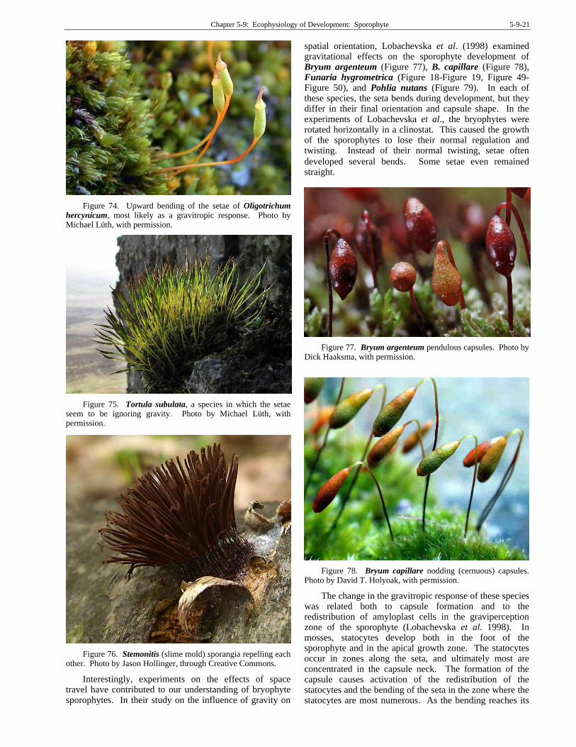
Thomas et al. (1970) found that liverwort setae respond to hormones in a manner similar to that of stems in tracheophytes; elongation of setae in Lophocolea (Figure 71) was promoted by low concentrations of IAA and inhibited at higher ones. Soon after that, Kaufman et al. (1982) determined that cells in the (gametophyte) stalk of Conocephalum conicum (Figure 72) and seta of Pellia epiphylla (Figure 37) exhibited acid growth, much like that of Avena (oats), implicating involvement of IAA.
Figure 72. Archegoniophores of Conocephalum conicum. Photo by Janice Glime.
5-9-20 Chapter 5-9: Ecophysiology of Development: Sporophyte
While comparing the responses of two liverworts, Pellia epiphylla (Figure 37) and Conocephalum conicum (Figure 72), to that of oats, Kaufman and coworkers (1982) discovered that a tenfold increase in the growth rate of oats (Avena) internodes appeared about three hours after application of 10-5 M GA3, but that 10-5 M IAA had no effect. On the other hand, in the liverworts, the setae responded to 10--5 M IAA with a two-fold increase in growth rate within 10-15 minutes.
Thomas et al. (1982) demonstrated the production of auxin (IAA) and ethylene by cells of elongating setae of Pellia epiphylla (Figure 37), adding more support to the suggestion that at least IAA may exercise control over seta elongation, and that most probably IAA and ethylene operate in tandem to control seta growth (Thomas et al. 1983). Setae in the rapid elongation phase contained ca. 2.5-2.9 µg per g fresh seta weight of free IAA. At the same time, ethylene was released by the seta, ranging 0.027-0.035 nanoliter per seta per hour. Ethylene is actually an inhibitor of the auxin-stimulated elongation of the seta at a concentration of 5 µL per L.
Pellia epiphylla (Figure 37) setae grow linearly at a rate of ca. 0.6 mm h-1 (Schnepf et al. 1979). When IAA (0.1 mM) was added to excised setae, Schnepf et al. (1979) found that the rate increased to 0.7-1.2 mm h-1. Furthermore, a variety of substances inhibited the elongation. These behaviors attest to the importance of auxin and that the elongation process is not just a passive thinning of the loosened cell walls. It depends on continued availability of auxin.
In their experiments with Pellia epiphylla (Figure 37), Poli et al. (2003) likewise found that IAA application did cause overall growth rates to increase significantly, as in Polytrichastrum ohioense (Figure 30), and likewise the liverwort did not respond to the anti-auxin treatment. Immature setae, ranging in length 8-24 mm at the beginning of the experiments, elongated on average 16.29 mm growth in 72 hr, whereas those receiving exogenous ISS elongated 25.90 at the same time, a promotion of 58% by IAA. PCIB failed to promote any differences in length. There appears to be no polar movement of IAA in the Pellia epiphylla (Figure 37) setae, with movement occurring by apolar facilitated diffusion.
But even hormones cannot do much without energy and other chemical coordination. Thomas et al. (1984) showed that auxins affect the cell wall polysaccharide composition and enzyme activity in Pellia epiphylla (Figure 37). Using a variety of techniques, they were able to show that growth in length doubled if setae were supplied with 10 µM IAA ±50 mM glucose. In this treatment, there was enhanced synthesis of all cell wall polysaccharides but cellulose, an increase in the relative glucose content of neutral wall sugars, and an activity change for wall-bound glycosidase. There was no change in the activity of cellulase. Both Galactose and mannose (50 mM) suppressed the auxin enhancement activity. Thomas et al. suggest that this is evidence that auxins play a role in maintaining the non-cellulosic cell wall synthesis.
Tropisms
Bryophytes often exhibit tropisms (Banbury 1962) in their setae, but controlling environmental conditions are not well known.
Like seta elongation, tropisms can be studied easily in liverwort setae. Thomas et al. (1987) used Pellia epiphylla (Figure 37) to demonstrate phototropisms of the seta. Using time-lapse photography, they showed that the entire length of the seta could respond by curving toward 6 W m-2 of unilateral blue light, a response that was noticeable within 10-15 minutes. This curvature was caused by a significant increase in growth on the shaded side of the seta (from 0.52 to 0.96 mm hr-1, but it also decreased on the lighted side by 0.26 mm hr-1.
Here, IAA may play another important role in the seta. Thomas et al. (2002), using radioactively labelled IAA and infrared video recording of Pellia epiphylla (Figure 37) setae, have shown that IAA in donor blocks moved preferentially to the lower sides of horizontally placed setae. Upward gravitropic curvature occurred within 50-60 minutes, while growth rates on the top side of the setae dropped.
Ellis and Thomas (1985) noticed that the shaded sides of setae became more acidic before they exhibited phototropic curvature. This acidity was inhibited by both neutral buffers and IAA antagonists, resulting in no curvature. This behavior suggests that IAA is transported laterally, causing protons to leave the cells and loosening the cell wall on the shaded side.
Gravitropism of the seta in Pellia epiphylla (Figure 37) exhibits lateral redistribution of IAA, with movement to the lower side of a horizontal seta (Thomas et al. 2002). This is an important aspect of orienting sporophytes that are originally positioned horizontally, such as those growing on vertical or slanting substrata. However, not all bryophytes have vertically oriented setae on vertical substrata (Figure 73).
Figure 73. Setae and capsules of Ulota coarctata on a vertical substrate, demonstrating apparent lack of gravitropism in these setae. Photo by Michael Lüth, with permission.
At least some mosses exhibit tropisms in their setae, but little is known of the mechanisms in this organ. In Oligotrichum hercynicum (Figure 74), setae bend upward, most likely with a gravitropic response, but possibly also with a light response. This family, the Polytrichaceae, seems to have good tropic responses, but how widespread is the response elsewhere among bryophytes? They seem to be absent in some species. Could it be that in some species the setae repel each other (Figure 75) like the sporangia of the slime mold Stemonitis (Figure 76)?
Chapter 5-9: Ecophysiology of Development: Sporophyte 5-9-21
Figure 74. Upward bending of the setae of Oligotrichum hercynicum, most likely as a gravitropic response. Photo by Michael Lüth, with permission.
Figure 75. Tortula subulata, a species in which the setae seem to be ignoring gravity. Photo by Michael Lüth, with permission.
Figure 76. Stemonitis (slime mold) sporangia repelling each other. Photo by Jason Hollinger, through Creative Commons.
Interestingly, experiments on the effects of space travel have contributed to our understanding of bryophyte sporophytes. In their study on the influence of gravity on
spatial orientation, Lobachevska et al. (1998) examined gravitational effects on the sporophyte development of Bryum argenteum (Figure 77), B. capillare (Figure 78), Funaria hygrometrica (Figure 18-Figure 19, Figure 49-Figure 50), and Pohlia nutans (Figure 79). In each of these species, the seta bends during development, but they differ in their final orientation and capsule shape. In the experiments of Lobachevska et al., the bryophytes were rotated horizontally in a clinostat. This caused the growth of the sporophytes to lose their normal regulation and twisting. Instead of their normal twisting, setae often
developed several bends. Some setae even remained straight.
Figure 77. Bryum argenteum pendulous capsules. Photo by Dick Haaksma, with permission.
Figure 78. Bryum capillare nodding (cernuous) capsules. Photo by David T. Holyoak, with permission.
The change in the gravitropic response of these species was related both to capsule formation and to the redistribution of amyloplast cells in the graviperception zone of the sporophyte (Lobachevska et al. 1998). In mosses, statocytes develop both in the foot of the sporophyte and in the apical growth zone. The statocytes occur in zones along the seta, and ultimately most are concentrated in the capsule neck. The formation of the capsule causes activation of the redistribution of the statocytes and the bending of the seta in the zone where the statocytes are most numerous. As the bending reaches its

5-9-22 Chapter 5-9: Ecophysiology of Development: Sporophyte
final stages, the greatest number of amyloplast zones remains on the convex side of the seta where the greatest growth has been occurring, relative to the concave side. These changes result in the change from vertical to horizontal growth that results in cernuous or pendulous capsules. Even the curvature of the capsule seems to be involved in this process in species like Funaria hygrometrica (Figure 18-Figure 19, Figure 49-Figure 50).
Figure 79. Pohlia nutans pendulous capsules. Photo by Hermann Schachner, through Creative Commons.
The changing gravitational pull resulting from clinostat rotation reduced the spatial reorientation of the seta and inhibited the differentiation of the capsule tissues (Lobachevska et al. 1998). The growth rate of the seta and capsule changed little. These effects suggest that gravity affects both spatial orientation and form of the capsule, and that the response is genetically controlled.
I have found nothing on tropisms in Anthocerotophyta (Figure 7, Figure 45-Figure 46), so I inquired on Bryonet. John Steel reported a species of Megaceros (Figure 5) growing on the underside of a rotting log. These sporophytes ignored gravity and grew straight out from the log.
This leaves us with many questions regarding tropisms in setae. What wavelengths of light can effect a response? Is there any correlation between gravitropism and seta length? Is gravitropism more common among bryophytes that grow on vertical surfaces? Is there any thigmotropism among setae? What is the role of ethylene in seta tropisms?
Dispersal
The seta can possibly facilitate dispersal in some species. For example, in Fissidens fontanus (Figure 80), the sporophyte is fragile and small. Joop Kortselius related the story on Bryonet (1 June 2016), based on Britton (1902). The seta is easily broken, often before the capsule is mature. In this case, the seta is green and fleshy, providing the nutrients and energy needed for the capsule to continue to grow while floating on the water surface. The calyptra remains attached.
Figure 80. Fissidens fontanus, an aquatic species with a small, fragile sporophyte. Photo by Michael Lüth, with permission.
Kortselius (Bryonet 1 June 2016) concludes that the capsule does indeed serve as a unit of dispersal in Fissidens fontanus (Figure 80). But the small capsules of this species are rarely observed in the field, in part because of this ability to fall off early. But in culture, they have appeared (Van Melick 1986) and even found later in herbarium packets, detached, among plants where they had been missed at the time of collection (Touw & Rubers 1989).
Capsule Development
Early embryo development, at least in Physcomitrium immersum, creates a filamentous structure (Lal & Bhandari 1968). As the capsule develops, it forms an outer air sac that surrounds the spore sac. In this species, there is no peristome. The foot that anchors the seta in the gametophyte is composed of densely cytoplasmic cells in the peripheral layer, supporting its haustorial function.
Like tracheophytes, both mosses (Figure 91) and hornworts (Figure 93) have stomata in the capsule, but liverworts lack them (Renzaglia et al. 2000). And mosses, like tracheophytes, can have conducting tissue in the sporophyte, but the mosses diverge from all other groups of plants in having a peristome in most.
Light
Early in its life the capsule is green and photosynthetic, typically gaining phenolic compounds that color it with age. Eventually it loses its photosynthetic capability and depends on stored reserves and the gametophyte. This ability to photosynthesize obviously requires light.
It is interesting that the translocation of carbohydrates (as glucose) to the sporophyte of Funaria hygrometrica (Figure 18-Figure 19, Figure 49-Figure 50) occurs in response to light (French & Paolillo 1976). French and Paolillo found that capsule morphology was abnormal in the dark because the spore sac failed to expand. Relatively low light intensity corrected these problems, and the authors felt that photoreceptors might be localized in the capsule. They agreed with Haberlandt (1886) that light affects more than just photosynthesis in the expansion of Funaria capsules, and that translocation is especially important in low light.
Chapter 5-9: Ecophysiology of Development: Sporophyte 5-9-23
This light relationship might explain why Rydgren and Økland (2002) found more capsules on segments in larger size classes and more identifiable females without them in smaller size classes (Figure 82), but this relationship also could imply that more energy is required than that available in the smaller segments (also possibly related to light availability), or that smaller segments had not yet reached the required degree of maturity. We have already discussed the need for a minimum size, or threshold, for the development of gametangia. It then follows that this same minimum size is necessary for the production of sporophytes, since sporophytes are not possible without an archegonium to house the egg, zygote, and embryo. This size requirement is supported by the study of Rydgren and Økland (2002) on Hylocomium splendens (Figure 81, Figure 82), where capsules increased in frequency on larger gametophores. Size thresholds for the archegonia are discussed earlier in the chapter on gametogenesis.
Figure 81. Hylocomium splendens with capsules. Photo from AnalogicalPlanet.com Alaska, with online permission.
Photosynthesis is probably not the only light need of the capsule. Krisko and Paolillo (1972) demonstrated that capsule expansion also requires light, with red light being more effective than white, blue, or green. But, then, red light is the most effective wave length for photosynthesis in plants.
In the liverworts Fossombronia foveolata (Figure 36), Lophocolea heterophylla (Figure 71), Pellia epiphylla (Figure 37), Ptilidium pulcherrimum (Figure 38), and Riella affinis (Figure 39), light was essential for sporophyte development, but surgically removed sporophytes developed slowly, with little increase in dry weight (Thomas et al. 1979). Nevertheless, sporophytes of
all five of these species fix CO2 in the light, but the calyptra and pseudoperianth inhibit this photosynthesis by as much as 50%. This is compensated by organic nutrients such as glucose that are supplied predominantly by the gametophyte.
Figure 82. Relationship of frequency of occurrence of number of female segments without capsules compared to those with capsules in five adult size classes of Hylocomium splendens over a five-year period. Redrawn from Rydgren & Økland 2002.
Light quality and photoperiod both play roles in sporophyte development in callus cultures (Bauer 1963). Constant light causes metabolic products to accumulate and damage the cultures. Short days down to 4 hours favor seta formation, whereas long days (16 hours) favor retention of the callus form; with fewer than 4 hours of light, the tendency to form protonemata increases. In total darkness, the entire callus forms a protonema. Light quality affects the sporophyte callus growth by retaining the callus form in blue light and forming a linear chain of cells in red light.
Light quality in the field varies with habitat, microhabitat, and season. In Ceratodon purpureus (Figure 83), setae develop in far-red light but not in red light (Hoddinott & Bain 1979). Since the far-red:red ratio increases with shading, the greatest seta expansion should occur under a green canopy. C. purpureus, however, more typically grows in the open, and setae are abundant there. Perhaps the far-red light stimulus is through the snow (setae are produced soon after the snow disappears), which increases the ratio of far-red:red light (Winchester pers. comm.). This could result in the abundant elongated setae we see early in spring as soon as the snow is gone, but at least some of this elongation occurs in the preceding autumn. If there is growth that responds to the far-red light under snow, we should expect a longer seta in the north than in the tropics, at least for open habitat things. Hmm... That should be relatively easy to check with a herbarium study. In fact, this ubiquitous north temperate moss seems rather rare in most of the tropics, where it is replaced by C.
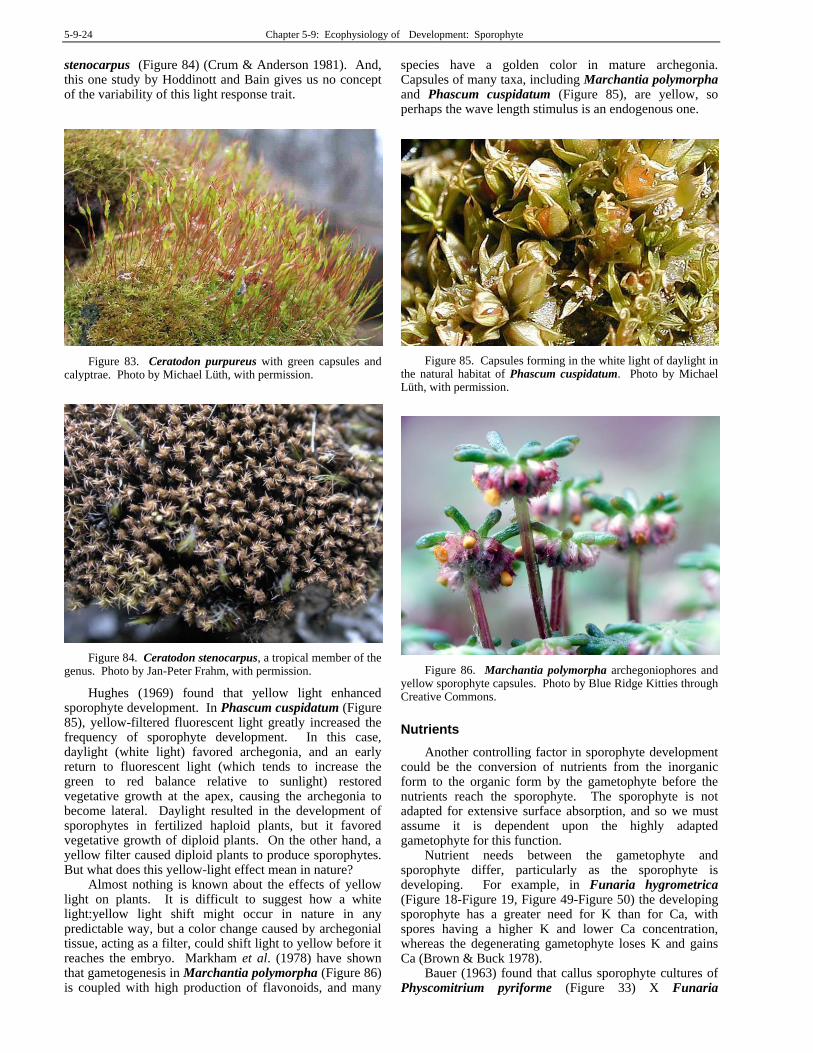
5-9-24 Chapter 5-9: Ecophysiology of Development: Sporophyte
stenocarpus (Figure 84) (Crum & Anderson 1981). And, this one study by Hoddinott and Bain gives us no concept of the variability of this light response trait.
Figure 83. Ceratodon purpureus with green capsules and calyptrae. Photo by Michael Lüth, with permission.
Figure 84. Ceratodon stenocarpus, a tropical member of the genus. Photo by Jan-Peter Frahm, with permission.
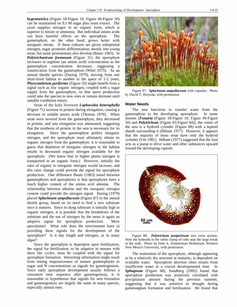
Hughes (1969) found that yellow light enhanced sporophyte development. In Phascum cuspidatum (Figure 85), yellow-filtered fluorescent light greatly increased the frequency of sporophyte development. In this case, daylight (white light) favored archegonia, and an early return to fluorescent light (which tends to increase the green to red balance relative to sunlight) restored vegetative growth at the apex, causing the archegonia to become lateral. Daylight resulted in the development of sporophytes in fertilized haploid plants, but it favored vegetative growth of diploid plants. On the other hand, a yellow filter caused diploid plants to produce sporophytes. But what does this yellow-light effect mean in nature?
Almost nothing is known about the effects of yellow light on plants. It is difficult to suggest how a white light:yellow light shift might occur in nature in any predictable way, but a color change caused by archegonial tissue, acting as a filter, could shift light to yellow before it reaches the embryo. Markham et al. (1978) have shown that gametogenesis in Marchantia polymorpha (Figure 86) is coupled with high production of flavonoids, and many
species have a golden color in mature archegonia. Capsules of many taxa, including Marchantia polymorpha and Phascum cuspidatum (Figure 85), are yellow, so perhaps the wave length stimulus is an endogenous one.
Figure 85. Capsules forming in the white light of daylight in the natural habitat of Phascum cuspidatum. Photo by Michael Lüth, with permission.
Figure 86. Marchantia polymorpha archegoniophores and yellow sporophyte capsules. Photo by Blue Ridge Kitties through Creative Commons.
Nutrients
Another controlling factor in sporophyte development could be the conversion of nutrients from the inorganic form to the organic form by the gametophyte before the nutrients reach the sporophyte. The sporophyte is not adapted for extensive surface absorption, and so we must assume it is dependent upon the highly adapted gametophyte for this function.
Nutrient needs between the gametophyte and sporophyte differ, particularly as the sporophyte is developing. For example, in Funaria hygrometrica (Figure 18-Figure 19, Figure 49-Figure 50) the developing sporophyte has a greater need for K than for Ca, with spores having a higher K and lower Ca concentration, whereas the degenerating gametophyte loses K and gains Ca (Brown & Buck 1978).
Bauer (1963) found that callus sporophyte cultures of Physcomitrium pyriforme (Figure 33) X Funaria
Chapter 5-9: Ecophysiology of Development: Sporophyte 5-9-25
hygrometrica (Figure 18-Figure 19, Figure 49-Figure 50) can be maintained on 9.1 M sugar plus yeast extract. The yeast supplies nitrogen in an organic form, which is superior to nitrate or ammonia. But individual amino acids can have harmful effects on the sporophyte. The gametophyte, on the other hand, grows better with inorganic nitrate. If these cultures are given suboptimal nitrogen, sugar promotes differentiation, mostly into young setae, but some protonemata also develop (Bauer 1963). In Polytrichastrum formosum (Figure 31), the sporophyte increases in arginine (an amino acid) concentration as the gametophyte concentration decreases, suggesting a translocation from the gametophyte (Whel 1975). As an annual shuttle species (During 1979), moving from one short-lived habitat to another in the space of 1-2 years, Physcomitrium pyriforme (Figure 33) might benefit from a signal such as low organic nitrogen, coupled with a sugar supply from the gametophyte, so that spore production could take the species to new sites or remain dormant until suitable conditions return.
Setae of the leafy liverwort Lophocolea heterophylla
(Figure 71) increase in protein during elongation, causing a
decrease in soluble amino acids (Thomas 1976). When
setae were severed from the gametophyte, they decreased
in protein, and seta elongation was attenuated, suggesting
that the synthesis of protein in the seta is necessary for its
elongation. Since the gametophyte prefers inorganic
nitrogen, and the sporophyte must ultimately obtain its
organic nitrogen from the gametophyte, it is reasonable to
guess that depletion of inorganic nitrogen in the habitat
results in decreased organic nitrogen available for the
sporophyte. (We know that in higher plants nitrogen is
transported in an organic form.) However, initially the
ratio of organic to inorganic nitrogen would increase, and
this ratio change could provide the signal for sporophyte
production. One difference Bauer (1963) noted between
gametophytes and sporophytes is that sporophytes have a
much higher content of the amino acid adenine. The
relationship between adenine and the inorganic nitrogen
content could provide the nitrogen signal. During (1979)
placed Splachnum ampullaceum (Figure 87) in the annual
shuttle group, based on its need to find a new substrate
once it matures. Since its dung substrate is initially high in
organic nitrogen, it is possible that the breakdown of the
substrate and the use of nitrogen by the moss is again an
adaptive signal for sporophyte production. More
speculation! What role does the environment have in
providing these signals for the development of the
sporophyte? Is it day length and nitrogen, as in many
algae? Since the sporophyte is dependent upon fertilization,
the signal for fertilization, to be adaptive in mosses with short life cycles, must be coupled with the signal for sporophyte formation. Interesting information might result from testing responsiveness of mature gametophytes to sugar and N concentrations as signals for gametogenesis. Since early sporophyte development usually follows a consistent time sequence after gametogenesis, it is reasonable to hypothesize that signals for seta formation and gametogenesis are largely the same in many species, especially annual ones.
Figure 87. Splachnum ampullaceum with capsules. Photo by David T. Holyoak, with permission.
Water Needs
The seta functions to transfer water from the gametophyte to the developing sporophyte. In some mosses [Funaria (Figure 18-Figure 19, Figure 49-Figure 50) and Polytrichum (Figure 63-Figure 64)], the center of the seta is a hydroid cylinder (Figure 88) with a leptoid sheath surrounding it (Hébant 1977). However, it appears that the majority of moss setae have only the hydroid cylinder (Vitt 1981). Hébant (1977) suggested that the foot acts as a pump to drive water and other substances upward toward the developing capsule.
Figure 88. Polytrichum juniperinum seta cross section. Note the hydroids in the white clump of cells near the large break in the stalk. Photo by Dale A. Zimmerman Herbarium, Western New Mexico University, with permission.
The maturation of the sporophyte, although appearing to be a relatively dry structure at maturity, is dependent on available water. Sporophyte abortion often results from insufficient water at a crucial developmental time. In Sphagnum (Figure 68), Sundberg (2002) found that sporophyte production was positively correlated with precipitation amount during the previous summer, suggesting that it was sensitive to drought during gametangium formation and fertilization. He found that
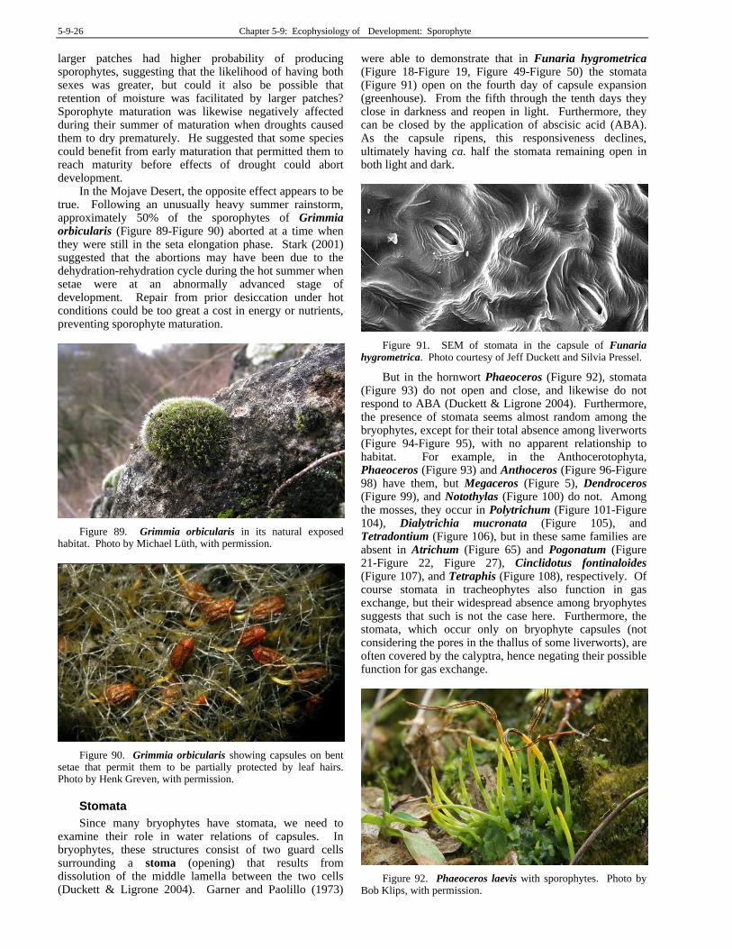
5-9-26 Chapter 5-9: Ecophysiology of Development: Sporophyte
larger patches had higher probability of producing sporophytes, suggesting that the likelihood of having both sexes was greater, but could it also be possible that retention of moisture was facilitated by larger patches? Sporophyte maturation was likewise negatively affected during their summer of maturation when droughts caused them to dry prematurely. He suggested that some species could benefit from early maturation that permitted them to reach maturity before effects of drought could abort development.
In the Mojave Desert, the opposite effect appears to be true. Following an unusually heavy summer rainstorm, approximately 50% of the sporophytes of Grimmia orbicularis (Figure 89-Figure 90) aborted at a time when they were still in the seta elongation phase. Stark (2001) suggested that the abortions may have been due to the dehydration-rehydration cycle during the hot summer when setae were at an abnormally advanced stage of development. Repair from prior desiccation under hot conditions could be too great a cost in energy or nutrients, preventing sporophyte maturation.
Figure 89. Grimmia orbicularis in its natural exposed habitat. Photo by Michael Lüth, with permission.
Figure 90. Grimmia orbicularis showing capsules on bent setae that permit them to be partially protected by leaf hairs. Photo by Henk Greven, with permission.
Stomata
Since many bryophytes have stomata, we need to examine their role in water relations of capsules. In bryophytes, these structures consist of two guard cells surrounding a stoma (opening) that results from dissolution of the middle lamella between the two cells (Duckett & Ligrone 2004). Garner and Paolillo (1973)
were able to demonstrate that in Funaria hygrometrica (Figure 18-Figure 19, Figure 49-Figure 50) the stomata (Figure 91) open on the fourth day of capsule expansion (greenhouse). From the fifth through the tenth days they close in darkness and reopen in light. Furthermore, they can be closed by the application of abscisic acid (ABA). As the capsule ripens, this responsiveness declines, ultimately having ca. half the stomata remaining open in both light and dark.
Figure 91. SEM of stomata in the capsule of Funaria hygrometrica. Photo courtesy of Jeff Duckett and Silvia Pressel.
But in the hornwort Phaeoceros (Figure 92), stomata (Figure 93) do not open and close, and likewise do not respond to ABA (Duckett & Ligrone 2004). Furthermore, the presence of stomata seems almost random among the bryophytes, except for their total absence among liverworts (Figure 94-Figure 95), with no apparent relationship to habitat. For example, in the Anthocerotophyta, Phaeoceros (Figure 93) and Anthoceros (Figure 96-Figure 98) have them, but Megaceros (Figure 5), Dendroceros (Figure 99), and Notothylas (Figure 100) do not. Among the mosses, they occur in Polytrichum (Figure 101-Figure 104), Dialytrichia mucronata (Figure 105), and Tetradontium (Figure 106), but in these same families are absent in Atrichum (Figure 65) and Pogonatum (Figure 21-Figure 22, Figure 27), Cinclidotus fontinaloides (Figure 107), and Tetraphis (Figure 108), respectively. Of course stomata in tracheophytes also function in gas exchange, but their widespread absence among bryophytes suggests that such is not the case here. Furthermore, the stomata, which occur only on bryophyte capsules (not considering the pores in the thallus of some liverworts), are often covered by the calyptra, hence negating their possible function for gas exchange.
Figure 92. Phaeoceros laevis with sporophytes. Photo by Bob Klips, with permission.

Chapter 5-9: Ecophysiology of Development: Sporophyte 5-9-27
Figure 93. Phaeoceros laevis with open stoma flanked by desiccated and shrunken epidermal cells well above dehiscence point of the capsule. Photo courtesy of Jeff Duckett, Ken P'ng, Karen Renzaglia, and Silvia Pressel.
Figure 94. Liverwort Fossombronia caespitiformis seta and capsule from New South Wales. Photo by Andras Keszei, with permission.
Figure 95. Fossombronia caespitiformis capsule showing absence of stomata. Photo by Andras Keszei, with permission.
Figure 96. SEM of Anthoceros punctatus sporophyte showing stomata. Photo courtesy of Jeff Duckett and Silvia Pressel.
Figure 97. SEM of Anthoceros punctatus sporophyte showing three stomata. Photo courtesy of Jeff Duckett and Silvia Pressel.
Figure 98. SEM of single stoma with guard cells on sporophyte of Anthoceros punctatus. Photo courtesy of Jeff Duckett and Silvia Pressel.
5-9-28 Chapter 5-9: Ecophysiology of Development: Sporophyte
Figure 99. Dendroceros crispus with dehiscing capsules. Photo by Jan-Peter Frahm, with permission.
Figure 100. Notothylas orbicularis with horizontal perichaetia. Photo by Michael Lüth, with permission.
Figure 101. Stomata at base of Polytrichum capsule. Photo by George Shepherd, through Creative Commons.
Figure 102. SEM of stomata at base of Polytrichum juniperinum capsule. Photo courtesy of Jeffrey Duckett and Silvia Pressel.
Figure 103. SEM of Polytrichum juniperinum stomata at capsule base. Photo courtesy of Jeff Duckett and Silvia Pressel.
Figure 104. SEM of Polytrichum juniperinum stoma at capsule base. Photo courtesy of Jeff Duckett and Silvia Pressel.
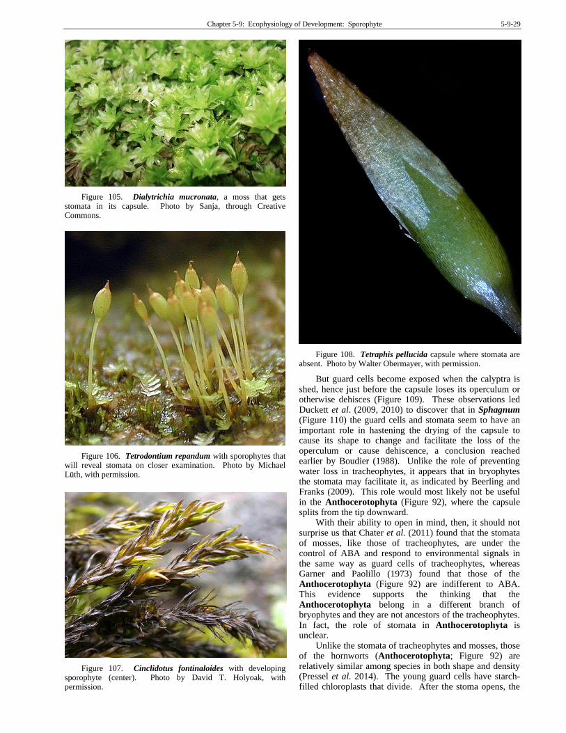
Chapter 5-9: Ecophysiology of Development: Sporophyte 5-9-29
Figure 105. Dialytrichia mucronata, a moss that gets stomata in its capsule. Photo by Sanja, through Creative Commons.
Figure 106. Tetrodontium repandum with sporophytes that will reveal stomata on closer examination. Photo by Michael Lüth, with permission.
Figure 107. Cinclidotus fontinaloides with developing sporophyte (center). Photo by David T. Holyoak, with permission.
Figure 108. Tetraphis pellucida capsule where stomata are absent. Photo by Walter Obermayer, with permission.
But guard cells become exposed when the calyptra is shed, hence just before the capsule loses its operculum or otherwise dehisces (Figure 109). These observations led Duckett et al. (2009, 2010) to discover that in Sphagnum (Figure 110) the guard cells and stomata seem to have an important role in hastening the drying of the capsule to cause its shape to change and facilitate the loss of the operculum or cause dehiscence, a conclusion reached earlier by Boudier (1988). Unlike the role of preventing water loss in tracheophytes, it appears that in bryophytes the stomata may facilitate it, as indicated by Beerling and Franks (2009). This role would most likely not be useful in the Anthocerotophyta (Figure 92), where the capsule splits from the tip downward.
With their ability to open in mind, then, it should not surprise us that Chater et al. (2011) found that the stomata of mosses, like those of tracheophytes, are under the control of ABA and respond to environmental signals in the same way as guard cells of tracheophytes, whereas Garner and Paolillo (1973) found that those of the Anthocerotophyta (Figure 92) are indifferent to ABA. This evidence supports the thinking that the Anthocerotophyta belong in a different branch of bryophytes and they are not ancestors of the tracheophytes. In fact, the role of stomata in Anthocerotophyta is unclear.
Unlike the stomata of tracheophytes and mosses, those of the hornworts (Anthocerotophyta; Figure 92) are relatively similar among species in both shape and density (Pressel et al. 2014). The young guard cells have starch-filled chloroplasts that divide. After the stoma opens, the
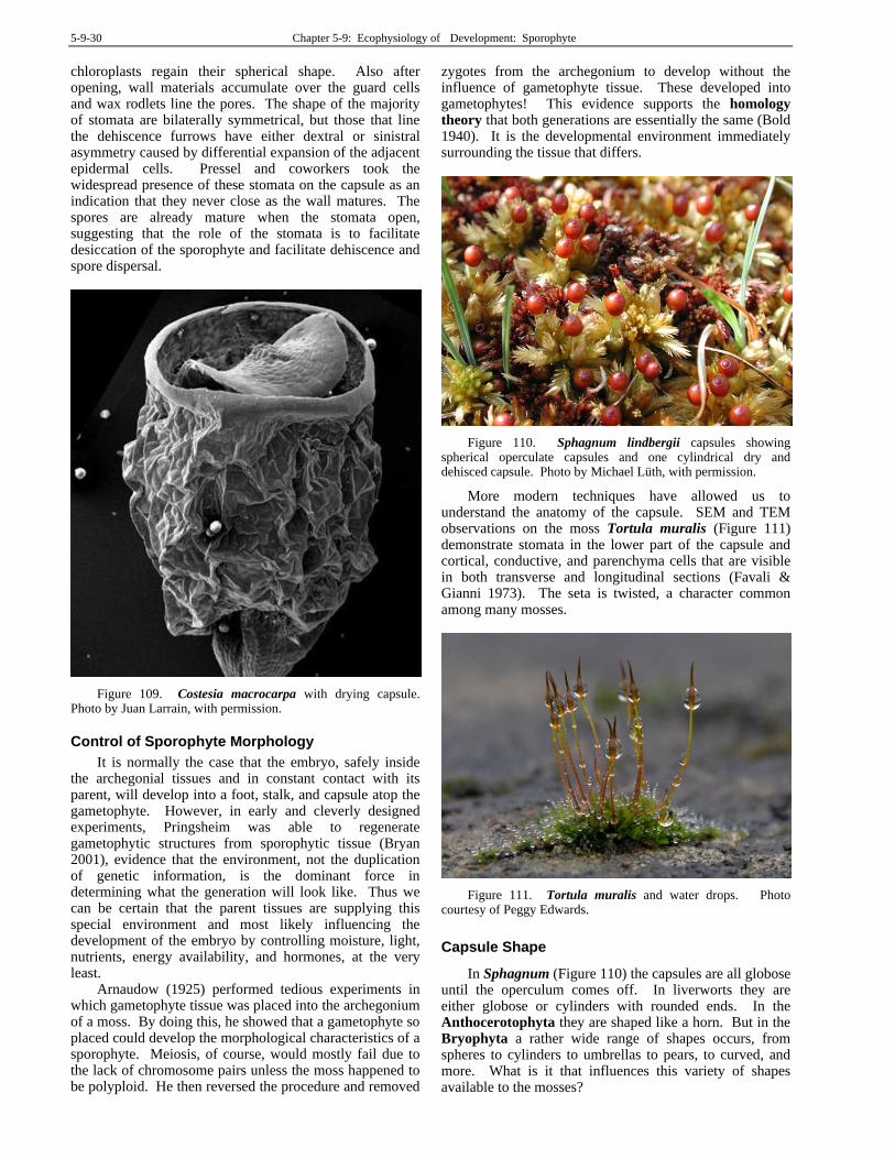
5-9-30 Chapter 5-9: Ecophysiology of Development: Sporophyte
chloroplasts regain their spherical shape. Also after opening, wall materials accumulate over the guard cells and wax rodlets line the pores. The shape of the majority of stomata are bilaterally symmetrical, but those that line the dehiscence furrows have either dextral or sinistral asymmetry caused by differential expansion of the adjacent epidermal cells. Pressel and coworkers took the widespread presence of these stomata on the capsule as an indication that they never close as the wall matures. The spores are already mature when the stomata open, suggesting that the role of the stomata is to facilitate desiccation of the sporophyte and facilitate dehiscence and spore dispersal.
Figure 109. Costesia macrocarpa with drying capsule. Photo by Juan Larrain, with permission.
Control of Sporophyte Morphology
It is normally the case that the embryo, safely inside the archegonial tissues and in constant contact with its parent, will develop into a foot, stalk, and capsule atop the gametophyte. However, in early and cleverly designed experiments, Pringsheim was able to regenerate gametophytic structures from sporophytic tissue (Bryan 2001), evidence that the environment, not the duplication of genetic information, is the dominant force in determining what the generation will look like. Thus we can be certain that the parent tissues are supplying this special environment and most likely influencing the development of the embryo by controlling moisture, light, nutrients, energy availability, and hormones, at the very least.
Arnaudow (1925) performed tedious experiments in which gametophyte tissue was placed into the archegonium of a moss. By doing this, he showed that a gametophyte so placed could develop the morphological characteristics of a sporophyte. Meiosis, of course, would mostly fail due to the lack of chromosome pairs unless the moss happened to be polyploid. He then reversed the procedure and removed
zygotes from the archegonium to develop without the influence of gametophyte tissue. These developed into gametophytes! This evidence supports the homology theory that both generations are essentially the same (Bold 1940). It is the developmental environment immediately surrounding the tissue that differs.
Figure 110. Sphagnum lindbergii capsules showing spherical operculate capsules and one cylindrical dry and dehisced capsule. Photo by Michael Lüth, with permission.
More modern techniques have allowed us to understand the anatomy of the capsule. SEM and TEM observations on the moss Tortula muralis (Figure 111) demonstrate stomata in the lower part of the capsule and cortical, conductive, and parenchyma cells that are visible in both transverse and longitudinal sections (Favali & Gianni 1973). The seta is twisted, a character common among many mosses.
Figure 111. Tortula muralis and water drops. Photo courtesy of Peggy Edwards.
Capsule Shape
In Sphagnum (Figure 110) the capsules are all globose until the operculum comes off. In liverworts they are either globose or cylinders with rounded ends. In the Anthocerotophyta they are shaped like a horn. But in the Bryophyta a rather wide range of shapes occurs, from spheres to cylinders to umbrellas to pears, to curved, and more. What is it that influences this variety of shapes available to the mosses?
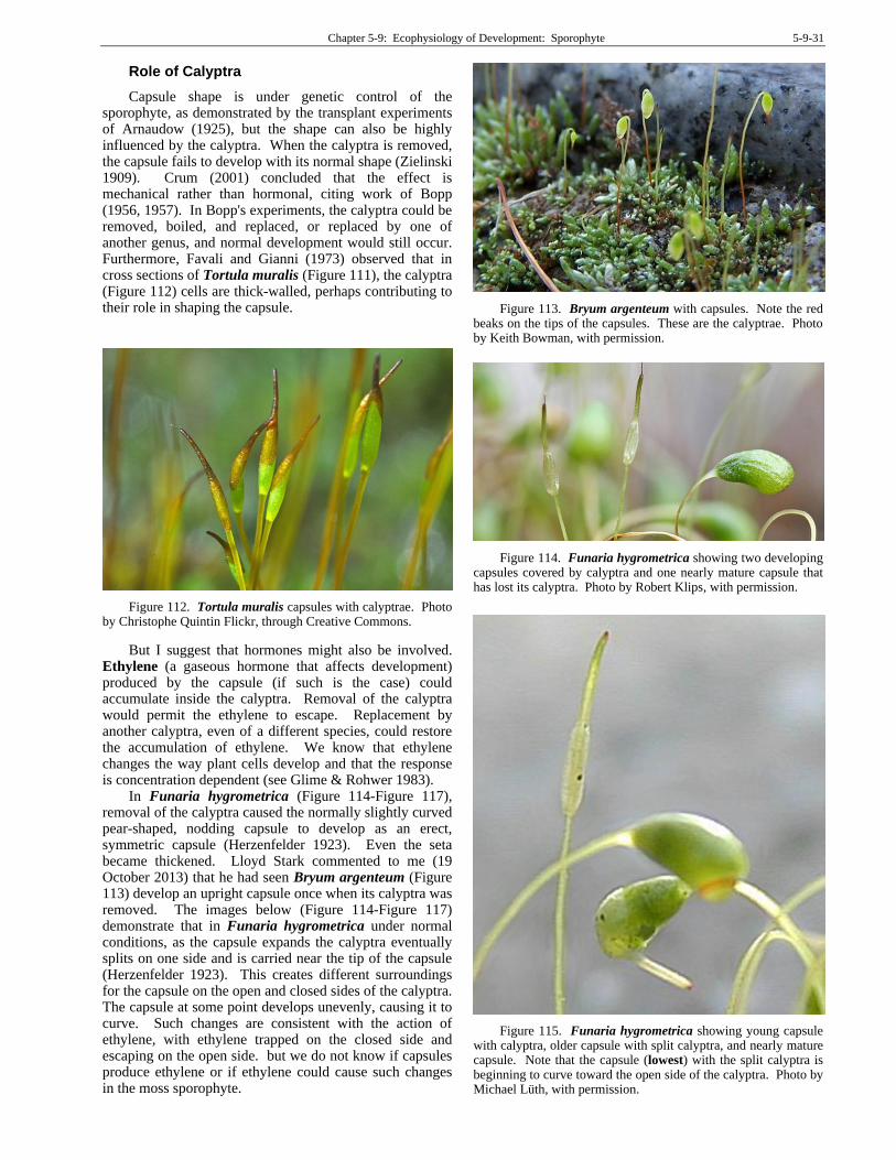
Chapter 5-9: Ecophysiology of Development: Sporophyte 5-9-31
Role of Calyptra
Capsule shape is under genetic control of the sporophyte, as demonstrated by the transplant experiments of Arnaudow (1925), but the shape can also be highly influenced by the calyptra. When the calyptra is removed, the capsule fails to develop with its normal shape (Zielinski 1909). Crum (2001) concluded that the effect is mechanical rather than hormonal, citing work of Bopp (1956, 1957). In Bopp's experiments, the calyptra could be removed, boiled, and replaced, or replaced by one of another genus, and normal development would still occur. Furthermore, Favali and Gianni (1973) observed that in cross sections of Tortula muralis (Figure 111), the calyptra (Figure 112) cells are thick-walled, perhaps contributing to their role in shaping the capsule.
Figure 112. Tortula muralis capsules with calyptrae. Photo by Christophe Quintin Flickr, through Creative Commons.
But I suggest that hormones might also be involved. Ethylene (a gaseous hormone that affects development) produced by the capsule (if such is the case) could accumulate inside the calyptra. Removal of the calyptra would permit the ethylene to escape. Replacement by another calyptra, even of a different species, could restore the accumulation of ethylene. We know that ethylene changes the way plant cells develop and that the response is concentration dependent (see Glime & Rohwer 1983).
In Funaria hygrometrica (Figure 114-Figure 117), removal of the calyptra caused the normally slightly curved pear-shaped, nodding capsule to develop as an erect, symmetric capsule (Herzenfelder 1923). Even the seta became thickened. Lloyd Stark commented to me (19 October 2013) that he had seen Bryum argenteum (Figure 113) develop an upright capsule once when its calyptra was removed. The images below (Figure 114-Figure 117) demonstrate that in Funaria hygrometrica under normal conditions, as the capsule expands the calyptra eventually splits on one side and is carried near the tip of the capsule (Herzenfelder 1923). This creates different surroundings for the capsule on the open and closed sides of the calyptra. The capsule at some point develops unevenly, causing it to curve. Such changes are consistent with the action of ethylene, with ethylene trapped on the closed side and escaping on the open side. but we do not know if capsules produce ethylene or if ethylene could cause such changes in the moss sporophyte.
Figure 113. Bryum argenteum with capsules. Note the red beaks on the tips of the capsules. These are the calyptrae. Photo by Keith Bowman, with permission.
Figure 114. Funaria hygrometrica showing two developing capsules covered by calyptra and one nearly mature capsule that has lost its calyptra. Photo by Robert Klips, with permission.
Figure 115. Funaria hygrometrica showing young capsule with calyptra, older capsule with split calyptra, and nearly mature capsule. Note that the capsule (lowest) with the split calyptra is beginning to curve toward the open side of the calyptra. Photo by Michael Lüth, with permission.

5-9-32 Chapter 5-9: Ecophysiology of Development: Sporophyte
Figure 116. SEM of nearly mature capsule of Funaria hygrometrica after the calyptra has split. Photo from website of Botany Department, University of British Columbia, with permission.
Figure 117. Funaria hygrometrica capsule showing the asymmetrical form it takes after the calyptra splits. Photo by Sarah Gregg, through Creative Commons.
Paolillo (1968) demonstrated that in Polytrichum
juniperinum (Figure 29), the splitting of the inner sheathing layer of the calyptra causes the capsule to develop bilateral symmetry. However, he found that in Funaria hygrometrica (Figure 114-Figure 117) splitting of the calyptra has no effect on capsule shape. Perhaps it depends on when it is split during the development, or the capsule is programmed to curve under both split and non-split calyptrae. Herzenfelder (1923) showed that it does not curve if the calyptra is removed. It would be interesting to put Polytrichum calyptrae on capsules before they curve to see if that inhibits the curvature, and also to put split Polytrichum calyptrae on some in place of their own.
The observed behaviors of these two species suggest to me that ethylene could be a controlling factor. Since ethylene is a gas, it can escape more easily on the side with the slit than on the closed side, thus altering the relative growth on the two sides. Another possibility is the difference in light, with IAA migrating to the darker (covered) side of the capsule; is this curvature really a tropism? Rate of drying might also differ. The fact that Funaria (Figure 114-Figure 117) does not respond to a split calyptra could result from its smaller, thinner calyptra and the fact that the calyptra covers very little of the
mature capsule, whereas the calyptra of Polytrichum (Figure 118) covers the entire capsule.
Figure 118. Polytrichum commune capsules showing calyptrae, and one capsule with the calyptra removed. Photo by Michael Lüth, with permission.
One factor that could contribute to the role of the calyptra is the presence of wax, but does that occur? Budke et al. (2011). The calyptra has the important role of protecting the apex of the sporophyte throughout development. This includes protection of the undifferentiated sporogenous tissue and the seta meristem from desiccation. Hence, Budke and coworkers set out to test for cuticle of the leafy gametophyte, sporangia, and calyptra of the moss Funaria hygrometrica (Figure 114-Figure 117). Using SEM and TEM, they identified a multi-layered cuticle on the calyptra (Figure 115-Figure 116) of this species. The beak of the calyptra has a cuticle that is thicker than on other parts examined. It furthermore has specialized thickenings called cuticular pegs, the first discovered in any moss. Budke and coworkers suggested that this extra protection at the apex was important to prevent desiccation of the developing sporophyte and might have played an important role in the evolution of the sporophyte generation.
Budke et al. (2012) further supported this supposition by demonstrating that the cuticle on the calyptra matures before that of the sporophyte in Funaria hygrometrica (Figure 114-Figure 117). In tracheophytes, this role of protection is carried out by leaf primordia. Using nine developmental stages of the sporophyte, they found that the calyptra has a four-layered cuticular covering at all stages. The sporophyte cuticle develops in older stages.
To further support their contention that the calyptra wax was an important protection against desiccation, Budke et al. (2013) removed the calyptra, removed the cuticle chemically, and returned the calyptra to the moss sporophyte. The mosses were then exposed to short-term dehydration. Removal of the cuticle under low humidity growing conditions caused significant negative effects on fitness of the sporophyte, including decreased survival, increased tissue damage, incomplete sporophyte development, greater peristome malformations, and decreased reproductive output.
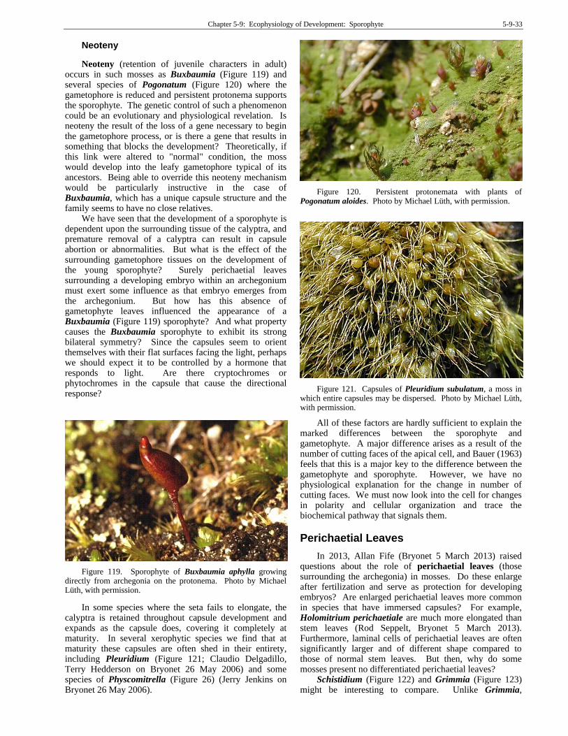
Chapter 5-9: Ecophysiology of Development: Sporophyte 5-9-33
Neoteny
Neoteny (retention of juvenile characters in adult) occurs in such mosses as Buxbaumia (Figure 119) and several species of Pogonatum (Figure 120) where the gametophore is reduced and persistent protonema supports the sporophyte. The genetic control of such a phenomenon could be an evolutionary and physiological revelation. Is neoteny the result of the loss of a gene necessary to begin the gametophore process, or is there a gene that results in something that blocks the development? Theoretically, if this link were altered to "normal" condition, the moss would develop into the leafy gametophore typical of its ancestors. Being able to override this neoteny mechanism would be particularly instructive in the case of Buxbaumia, which has a unique capsule structure and the family seems to have no close relatives.
We have seen that the development of a sporophyte is dependent upon the surrounding tissue of the calyptra, and premature removal of a calyptra can result in capsule abortion or abnormalities. But what is the effect of the surrounding gametophore tissues on the development of the young sporophyte? Surely perichaetial leaves surrounding a developing embryo within an archegonium must exert some influence as that embryo emerges from the archegonium. But how has this absence of gametophyte leaves influenced the appearance of a Buxbaumia (Figure 119) sporophyte? And what property causes the Buxbaumia sporophyte to exhibit its strong bilateral symmetry? Since the capsules seem to orient themselves with their flat surfaces facing the light, perhaps we should expect it to be controlled by a hormone that responds to light. Are there cryptochromes or phytochromes in the capsule that cause the directional response?
Figure 119. Sporophyte of Buxbaumia aphylla growing directly from archegonia on the protonema. Photo by Michael Lüth, with permission.
In some species where the seta fails to elongate, the calyptra is retained throughout capsule development and expands as the capsule does, covering it completely at maturity. In several xerophytic species we find that at maturity these capsules are often shed in their entirety, including Pleuridium (Figure 121; Claudio Delgadillo, Terry Hedderson on Bryonet 26 May 2006) and some species of Physcomitrella (Figure 26) (Jerry Jenkins on Bryonet 26 May 2006).
Figure 120. Persistent protonemata with plants of Pogonatum aloides. Photo by Michael Lüth, with permission.
Figure 121. Capsules of Pleuridium subulatum, a moss in which entire capsules may be dispersed. Photo by Michael Lüth, with permission.
All of these factors are hardly sufficient to explain the marked differences between the sporophyte and gametophyte. A major difference arises as a result of the number of cutting faces of the apical cell, and Bauer (1963) feels that this is a major key to the difference between the gametophyte and sporophyte. However, we have no physiological explanation for the change in number of cutting faces. We must now look into the cell for changes in polarity and cellular organization and trace the biochemical pathway that signals them.
Perichaetial Leaves
In 2013, Allan Fife (Bryonet 5 March 2013) raised questions about the role of perichaetial leaves (those surrounding the archegonia) in mosses. Do these enlarge after fertilization and serve as protection for developing embryos? Are enlarged perichaetial leaves more common in species that have immersed capsules? For example, Holomitrium perichaetiale are much more elongated than stem leaves (Rod Seppelt, Bryonet 5 March 2013). Furthermore, laminal cells of perichaetial leaves are often significantly larger and of different shape compared to those of normal stem leaves. But then, why do some mosses present no differentiated perichaetial leaves?
Schistidium (Figure 122) and Grimmia (Figure 123) might be interesting to compare. Unlike Grimmia,
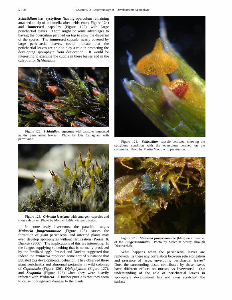
5-9-34 Chapter 5-9: Ecophysiology of Development: Sporophyte
Schistidium has systylious (having operculum remaining attached to tip of columella after dehiscence; Figure 124) and immersed capsules (Figure 122) with large perichaetial leaves. There might be some advantages to having the operculum perched on top to slow the dispersal of the spores. The immersed capsule, nearly covered by large perichaetial leaves, could indicate that the perichaetial leaves are able to play a role in protecting the developing sporophyte from desiccation. It would be interesting to examine the cuticle in these leaves and in the calyptra for Schistidium.
Figure 122. Schistidium agassazii with capsules immersed in the perichaetial leaves. Photo by Des Callaghan, with permission.
Figure 123. Grimmia laevigata with emergent capsules and short calyptrae. Photo by Michael Lüth, with permission.
In some leafy liverworts, the parasitic fungus Mniaecia jungermanniae (Figure 125) causes the formation of giant perichaetia, and infected plants may even develop sporophytes without fertilization (Pressel & Duckett (2006). The implications of this are interesting. Is the fungus supplying something that is normally produced by the fertilized egg? Pressel and Duckett suggested that indeed the Mniaecia produced some sort of substance that initiated this developmental behavior. They observed these giant perichaetia and abnormal perianths in wild colonies of Cephalozia (Figure 126), Diplophyllum (Figure 127), and Scapania (Figure 128) when they were heavily infected with Mniaecia. A further puzzle is that they seem to cause no long-term damage to the plants.
Figure 124. Schistidium capsule dehisced, showing the systylious condition with the operculum perched on the columella. Photo by Martin Mach, with permission.
Figure 125. Mniaecia jungermanniae (blue) on a member of the Jungermanniales. Photo by Malcolm Storey, through DiscoverLife.
What happens when the perichaetial leaves are removed? Is there any correlation between seta elongation and presence of large, enveloping perichaetial leaves? Does the surrounding tissue contributed by these leaves have different effects on mosses vs liverworts? Our understanding of the role of perichaetial leaves in sporophyte development has not even scratched the surface!
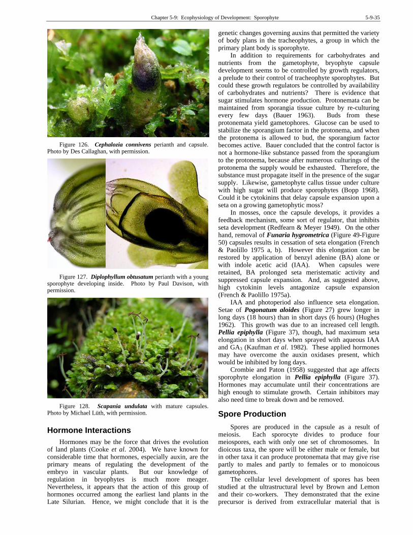
Chapter 5-9: Ecophysiology of Development: Sporophyte 5-9-35
Figure 126. Cephalozia connivens perianth and capsule. Photo by Des Callaghan, with permission.
Figure 127. Diplophyllum obtusatum perianth with a young sporophyte developing inside. Photo by Paul Davison, with permission.
Figure 128. Scapania undulata with mature capsules. Photo by Michael Lüth, with permission.
Hormone Interactions
Hormones may be the force that drives the evolution of land plants (Cooke et al. 2004). We have known for considerable time that hormones, especially auxin, are the primary means of regulating the development of the embryo in vascular plants. But our knowledge of regulation in bryophytes is much more meager. Nevertheless, it appears that the action of this group of hormones occurred among the earliest land plants in the Late Silurian. Hence, we might conclude that it is the
genetic changes governing auxins that permitted the variety of body plans in the tracheophytes, a group in which the primary plant body is sporophyte.
In addition to requirements for carbohydrates and nutrients from the gametophyte, bryophyte capsule development seems to be controlled by growth regulators, a prelude to their control of tracheophyte sporophytes. But could these growth regulators be controlled by availability of carbohydrates and nutrients? There is evidence that sugar stimulates hormone production. Protonemata can be maintained from sporangia tissue culture by re-culturing every few days (Bauer 1963). Buds from these protonemata yield gametophores. Glucose can be used to stabilize the sporangium factor in the protonema, and when the protonema is allowed to bud, the sporangium factor becomes active. Bauer concluded that the control factor is not a hormone-like substance passed from the sporangium to the protonema, because after numerous culturings of the protonema the supply would be exhausted. Therefore, the substance must propagate itself in the presence of the sugar supply. Likewise, gametophyte callus tissue under culture with high sugar will produce sporophytes (Bopp 1968). Could it be cytokinins that delay capsule expansion upon a seta on a growing gametophytic moss?
In mosses, once the capsule develops, it provides a feedback mechanism, some sort of regulator, that inhibits seta development (Redfearn & Meyer 1949). On the other hand, removal of Funaria hygrometrica (Figure 49-Figure 50) capsules results in cessation of seta elongation (French & Paolillo 1975 a, b). However this elongation can be restored by application of benzyl adenine (BA) alone or with indole acetic acid (IAA). When capsules were retained, BA prolonged seta meristematic activity and suppressed capsule expansion. And, as suggested above, high cytokinin levels antagonize capsule expansion (French & Paolillo 1975a).
IAA and photoperiod also influence seta elongation. Setae of Pogonatum aloides (Figure 27) grew longer in long days (18 hours) than in short days (6 hours) (Hughes 1962). This growth was due to an increased cell length. Pellia epiphylla (Figure 37), though, had maximum seta elongation in short days when sprayed with aqueous IAA and GA3 (Kaufman et al. 1982). These applied hormones may have overcome the auxin oxidases present, which would be inhibited by long days.
Crombie and Paton (1958) suggested that age affects sporophyte elongation in Pellia epiphylla (Figure 37). Hormones may accumulate until their concentrations are high enough to stimulate growth. Certain inhibitors may also need time to break down and be removed.
Spore Production
Spores are produced in the capsule as a result of meiosis. Each sporocyte divides to produce four meiospores, each with only one set of chromosomes. In dioicous taxa, the spore will be either male or female, but in other taxa it can produce protonemata that may give rise partly to males and partly to females or to monoicous gametophores.
The cellular level development of spores has been studied at the ultrastructural level by Brown and Lemon and their co-workers. They demonstrated that the exine precursor is derived from extracellular material that is
5-9-36 Chapter 5-9: Ecophysiology of Development: Sporophyte
deposited in an organized fashion on the sporocyte wall during meiotic prophase (Brown et al. 1986). This results in the distinctive patterns of exines seen on spores among various species. They suggested that this is clear evidence that the cell wall patterning of spores is a genetic result triggered in the sporocyte and may not require any genetic transcription following meiosis.
Spore dispersal is facilitated in most mosses by the movement of hygroscopic teeth that often trap the spores in spaces among the degenerate cells (Figure 129). These cells resorb their walls in such a way as to produce chambers along the teeth (Figure 130). The unequal binding of the walls creates a hygroscopic response to changes in moisture. Ingold (1959) changed the humidity levels 171 times in one moss with two rows of teeth, causing the dispersal of 15,647 spores! In Fissidens (Figure 131), unequal patterns of cellulose and hemicellulose cause peristome movement (Mueller 1973); in others, unequal suberization contributes (Schnepf et al 1978).
Spore number can vary considerably among bryophyte taxa, with mosses generally having a higher number than liverworts (Patidar et al. 1987). Capsule size is one factor in determining that number. However, spore size also determines spore number, with fewer large spores than small ones at the same capsule size – simple physics. This is somewhat true with liverwort spores in the Marchantiopsida, but the correlation is certainly not perfect (Table 1).
Figure 129. Peristome teeth of Bryum inclinatum with spores among them. Photo by Michael Lüth, with permission.
Figure 130. Peristome with trapped spores of Fontinalis squamosa. Photo by Janice Glime.
Figure 131. Fissidens bryoides capsules. Photo by Malcolm Storey, through Discover Life Creative Commons.
Table 1. Mean numbers and sizes of spores in fifteen liverwort species of the Marchantiopsida. From Patidar et al. 1987.
Species Number Size (µm) Riccia fluitans 180 60-75 Riccia billardieri 190 150-180 Riccia gangetica 196 130-140 Riccia discolor 210 120-160 Riccia hueberneriana 320 50-60 Cyathodium barode 490 40-50 Targionia hypophylla 1,200 30-40 Plagiochasma appendiculatum 2,200 60-70 Reboulia hemispherica 2,700 60-90 Asterella blumeana 2,900 60-75 Plagiochasma intermedium 3,200 60-70 Asterella angustata 3,300 60-65 Marchantia nepalensis 19,700 20-30 Marchantia palmata 20,100 20-30 Dumortiera hirsuta 21,200 22-26
Perennial mosses typically have small spores, less than
24 µm, permitting them to travel greater distances, whereas they can expand locally by vegetative means more easily than annual mosses (cf. spores sizes for Michigan mosses in Crum 1973 as discussed earlier under spore germination). Buxbaumia aphylla (Figure 119) has the smallest spores (6.5-8 µm) among Michigan mosses, perhaps contributing to its ability to colonize disturbed sites. Many acrocarpous mosses are annual; approximately 40% of these in Michigan have spores larger than 24 µm and range up to 68 µm. Larger spore size provides more food reserves that ensure greater success of establishment for these species that depend on spores for their year-to-year existence. Short-lived Antarctic mosses likewise have large spores, which Convey and Smith (1993) considered would help them in local colonization. The species in Michigan with the largest spores is the epiphyte

Chapter 5-9: Ecophysiology of Development: Sporophyte 5-9-37
Drummondia prorepens (Figure 132), which has multicellular spores measuring 60-100 µm.
Figure 132. Drummondia prorepens, the species that has the largest spores in Michigan. Photo by Dale Vitt, with permission.
Sundberg and Rydin (1998) found a positive correlation between capitulum size and capsule size, suggesting one could estimate number of spores from capsule size. Sphagnum tenellum (Figure 133) had a mean number of 18,500 spores per capsule, whereas the larger capitulum of S. squarrosum (Figure 134) had a mean of 243,000. Fenton and Bergeron (2006) found a similar relationship in Sphagnum capillifolium (Figure 135), where capsule-bearing colonies were significantly larger and taller than those without capsules, most likely related to an energy threshold. However, spore sizes among Michigan Sphagnum species suggest no correlation of spore size with plant size, with diameters ranging from 17 µm in S. warnstorfii (Figure 136) and a relatively large S. squarrosum to 42 µm in S. cuspidatum (Figure 137).
Figure 133. Sphagnum tenellum capsules. Photo by Dick Haaksma, with permission.
Figure 134. Sphagnum squarrosum with capsules. Photo by Michael Lüth, with permission.
Figure 135. Sphagnum capillifolium with capsules. Photo by J. C. Schou, with permission.
Figure 136. Sphagnum warnstorfianum, a species with small spores. Photo by Blanka Shaw, with permission.
Figure 137. Sphagnum cuspidatum with capsules. Photo by Bobby Hattaway <DiscoverLife>, with permission.
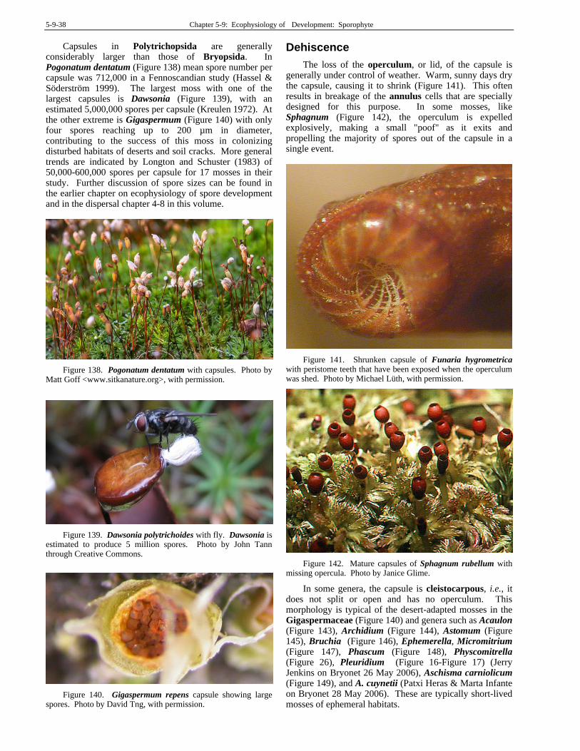
5-9-38 Chapter 5-9: Ecophysiology of Development: Sporophyte
Capsules in Polytrichopsida are generally considerably larger than those of Bryopsida. In Pogonatum dentatum (Figure 138) mean spore number per capsule was 712,000 in a Fennoscandian study (Hassel & Söderström 1999). The largest moss with one of the largest capsules is Dawsonia (Figure 139), with an estimated 5,000,000 spores per capsule (Kreulen 1972). At the other extreme is Gigaspermum (Figure 140) with only four spores reaching up to 200 µm in diameter, contributing to the success of this moss in colonizing disturbed habitats of deserts and soil cracks. More general trends are indicated by Longton and Schuster (1983) of 50,000-600,000 spores per capsule for 17 mosses in their study. Further discussion of spore sizes can be found in the earlier chapter on ecophysiology of spore development and in the dispersal chapter 4-8 in this volume.
Figure 138. Pogonatum dentatum with capsules. Photo by Matt Goff <www.sitkanature.org>, with permission.
Figure 139. Dawsonia polytrichoides with fly. Dawsonia is estimated to produce 5 million spores. Photo by John Tann through Creative Commons.
Figure 140. Gigaspermum repens capsule showing large spores. Photo by David Tng, with permission.
Dehiscence
The loss of the operculum, or lid, of the capsule is generally under control of weather. Warm, sunny days dry the capsule, causing it to shrink (Figure 141). This often results in breakage of the annulus cells that are specially designed for this purpose. In some mosses, like Sphagnum (Figure 142), the operculum is expelled explosively, making a small "poof" as it exits and propelling the majority of spores out of the capsule in a single event.
Figure 141. Shrunken capsule of Funaria hygrometrica with peristome teeth that have been exposed when the operculum was shed. Photo by Michael Lüth, with permission.
Figure 142. Mature capsules of Sphagnum rubellum with missing opercula. Photo by Janice Glime.
In some genera, the capsule is cleistocarpous, i.e., it does not split or open and has no operculum. This morphology is typical of the desert-adapted mosses in the Gigaspermaceae (Figure 140) and genera such as Acaulon (Figure 143), Archidium (Figure 144), Astomum (Figure 145), Bruchia (Figure 146), Ephemerella, Micromitrium (Figure 147), Phascum (Figure 148), Physcomitrella (Figure 26), Pleuridium (Figure 16-Figure 17) (Jerry Jenkins on Bryonet 26 May 2006), Aschisma carniolicum (Figure 149), and A. cuynetii (Patxi Heras & Marta Infante on Bryonet 28 May 2006). These are typically short-lived mosses of ephemeral habitats.
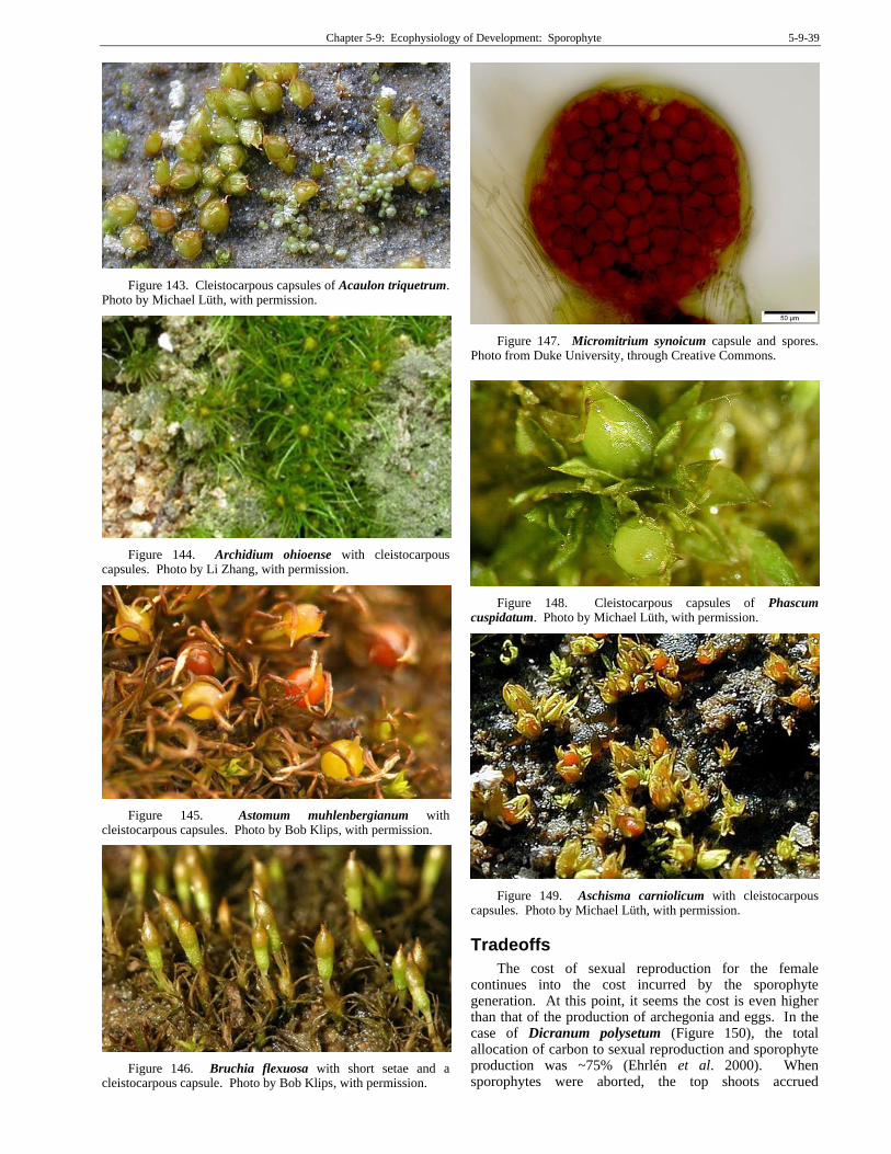
Chapter 5-9: Ecophysiology of Development: Sporophyte 5-9-39
Figure 143. Cleistocarpous capsules of Acaulon triquetrum. Photo by Michael Lüth, with permission.
Figure 144. Archidium ohioense with cleistocarpous capsules. Photo by Li Zhang, with permission.
Figure 145. Astomum muhlenbergianum with cleistocarpous capsules. Photo by Bob Klips, with permission.
Figure 146. Bruchia flexuosa with short setae and a cleistocarpous capsule. Photo by Bob Klips, with permission.
Figure 147. Micromitrium synoicum capsule and spores. Photo from Duke University, through Creative Commons.
Figure 148. Cleistocarpous capsules of Phascum cuspidatum. Photo by Michael Lüth, with permission.
Figure 149. Aschisma carniolicum with cleistocarpous capsules. Photo by Michael Lüth, with permission.
Tradeoffs
The cost of sexual reproduction for the female continues into the cost incurred by the sporophyte generation. At this point, it seems the cost is even higher than that of the production of archegonia and eggs. In the case of Dicranum polysetum (Figure 150), the total allocation of carbon to sexual reproduction and sporophyte production was ~75% (Ehrlén et al. 2000). When sporophytes were aborted, the top shoots accrued
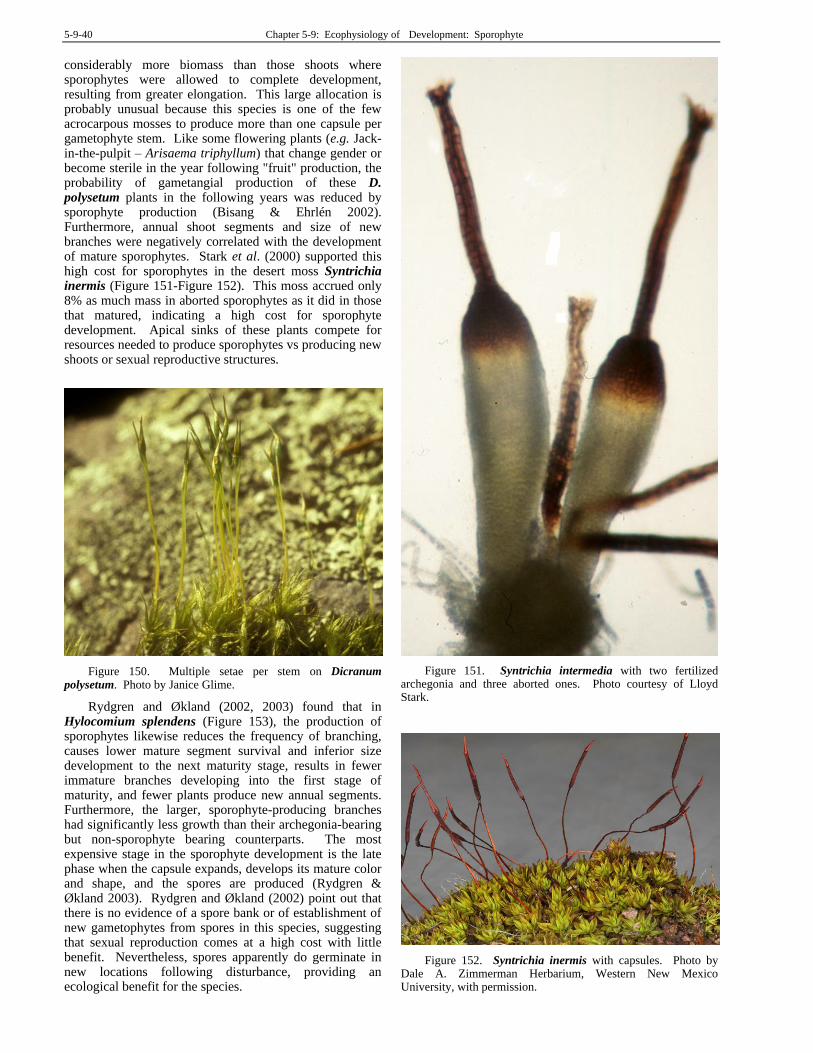
5-9-40 Chapter 5-9: Ecophysiology of Development: Sporophyte
considerably more biomass than those shoots where sporophytes were allowed to complete development, resulting from greater elongation. This large allocation is probably unusual because this species is one of the few acrocarpous mosses to produce more than one capsule per gametophyte stem. Like some flowering plants (e.g. Jack-in-the-pulpit – Arisaema triphyllum) that change gender or become sterile in the year following "fruit" production, the probability of gametangial production of these D. polysetum plants in the following years was reduced by sporophyte production (Bisang & Ehrlén 2002). Furthermore, annual shoot segments and size of new branches were negatively correlated with the development of mature sporophytes. Stark et al. (2000) supported this high cost for sporophytes in the desert moss Syntrichia inermis (Figure 151-Figure 152). This moss accrued only 8% as much mass in aborted sporophytes as it did in those that matured, indicating a high cost for sporophyte development. Apical sinks of these plants compete for resources needed to produce sporophytes vs producing new shoots or sexual reproductive structures.
Figure 150. Multiple setae per stem on Dicranum polysetum. Photo by Janice Glime.
Rydgren and Økland (2002, 2003) found that in Hylocomium splendens (Figure 153), the production of sporophytes likewise reduces the frequency of branching, causes lower mature segment survival and inferior size development to the next maturity stage, results in fewer immature branches developing into the first stage of maturity, and fewer plants produce new annual segments. Furthermore, the larger, sporophyte-producing branches had significantly less growth than their archegonia-bearing but non-sporophyte bearing counterparts. The most expensive stage in the sporophyte development is the late phase when the capsule expands, develops its mature color and shape, and the spores are produced (Rydgren & Økland 2003). Rydgren and Økland (2002) point out that there is no evidence of a spore bank or of establishment of new gametophytes from spores in this species, suggesting that sexual reproduction comes at a high cost with little benefit. Nevertheless, spores apparently do germinate in new locations following disturbance, providing an ecological benefit for the species.
Figure 151. Syntrichia intermedia with two fertilized archegonia and three aborted ones. Photo courtesy of Lloyd Stark.
Figure 152. Syntrichia inermis with capsules. Photo by Dale A. Zimmerman Herbarium, Western New Mexico University, with permission.
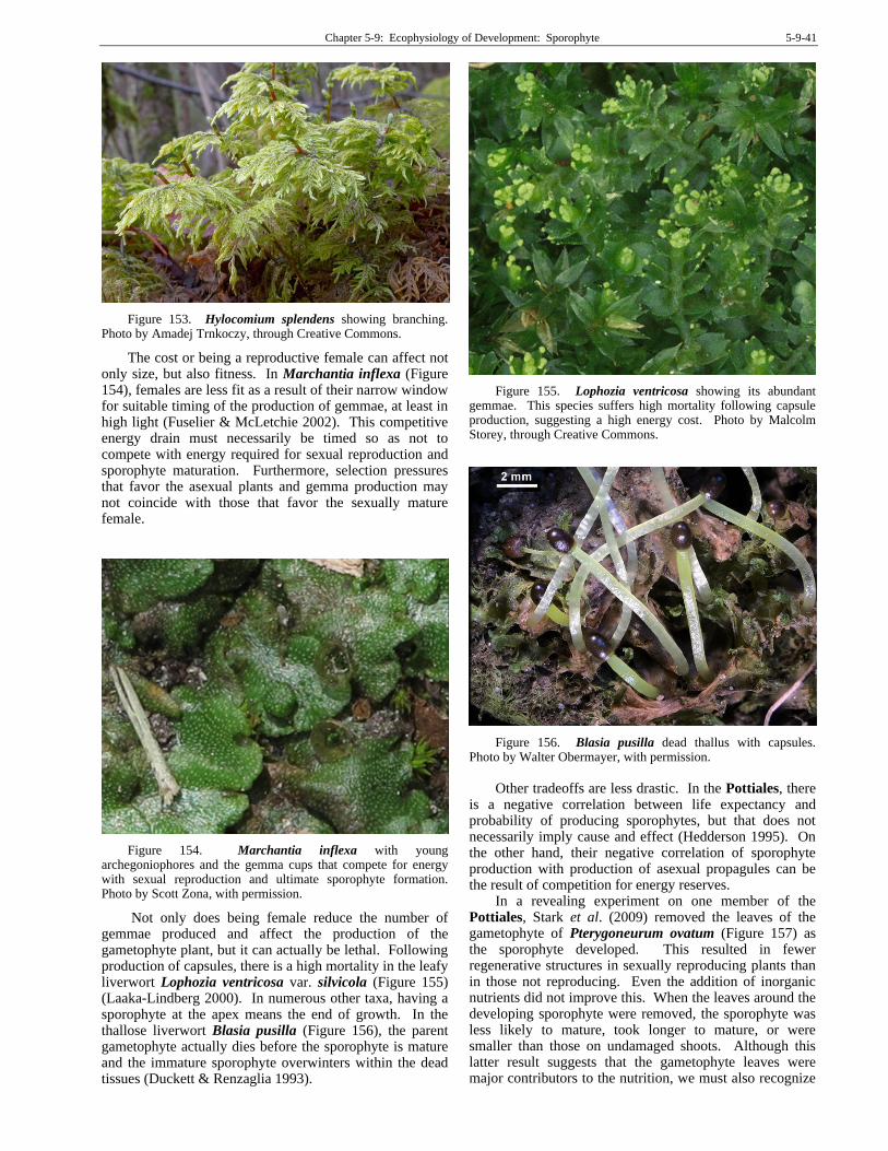
Chapter 5-9: Ecophysiology of Development: Sporophyte 5-9-41
Figure 153. Hylocomium splendens showing branching. Photo by Amadej Trnkoczy, through Creative Commons.
The cost or being a reproductive female can affect not only size, but also fitness. In Marchantia inflexa (Figure 154), females are less fit as a result of their narrow window for suitable timing of the production of gemmae, at least in high light (Fuselier & McLetchie 2002). This competitive energy drain must necessarily be timed so as not to compete with energy required for sexual reproduction and sporophyte maturation. Furthermore, selection pressures that favor the asexual plants and gemma production may not coincide with those that favor the sexually mature female.
Figure 154. Marchantia inflexa with young archegoniophores and the gemma cups that compete for energy with sexual reproduction and ultimate sporophyte formation. Photo by Scott Zona, with permission.
Not only does being female reduce the number of gemmae produced and affect the production of the gametophyte plant, but it can actually be lethal. Following production of capsules, there is a high mortality in the leafy liverwort Lophozia ventricosa var. silvicola (Figure 155) (Laaka-Lindberg 2000). In numerous other taxa, having a sporophyte at the apex means the end of growth. In the thallose liverwort Blasia pusilla (Figure 156), the parent gametophyte actually dies before the sporophyte is mature and the immature sporophyte overwinters within the dead tissues (Duckett & Renzaglia 1993).
Figure 155. Lophozia ventricosa showing its abundant gemmae. This species suffers high mortality following capsule production, suggesting a high energy cost. Photo by Malcolm Storey, through Creative Commons.
Figure 156. Blasia pusilla dead thallus with capsules. Photo by Walter Obermayer, with permission.
Other tradeoffs are less drastic. In the Pottiales, there is a negative correlation between life expectancy and probability of producing sporophytes, but that does not necessarily imply cause and effect (Hedderson 1995). On the other hand, their negative correlation of sporophyte production with production of asexual propagules can be the result of competition for energy reserves.
In a revealing experiment on one member of the Pottiales, Stark et al. (2009) removed the leaves of the gametophyte of Pterygoneurum ovatum (Figure 157) as the sporophyte developed. This resulted in fewer regenerative structures in sexually reproducing plants than in those not reproducing. Even the addition of inorganic nutrients did not improve this. When the leaves around the developing sporophyte were removed, the sporophyte was less likely to mature, took longer to mature, or were smaller than those on undamaged shoots. Although this latter result suggests that the gametophyte leaves were major contributors to the nutrition, we must also recognize

5-9-42 Chapter 5-9: Ecophysiology of Development: Sporophyte
that their removal changed the surrounding environment, and this could change the hormonal response during development.
Figure 157. Pterygoneurum ovatum with mature capsules. Photo by Kristian Peters, with permission.
With all of these tradeoffs, it would seem to be an advantage to delay production of sporophytes until the leafy part of the plant reaches a critical size, hence having a sufficient supply of energy. Jonsson and Söderström (1988) investigated this aspect in the epixylic (living on logs with bare wood) leafy liverwort Ptilidium pulcherrimum (Figure 158). They determined that the mean colony size for the first sporophyte production was 68 cm2, a size generally achieved in about 9 years. But antheridia are formed in the third year, suggesting that sporophytes remained unsuccessful for six years, perhaps due to insufficient energy reserves. Furthermore, capsule density and spore production increased significantly as the colony size increased. Both number of capsules and spore production had a six-fold variation among years. The number of spores ranged 18,000 to 44,000.
Figure 158. Ptilidium pulcherrimum with capsules, a plant that requires a critical size in order to produce capsules. Photo by Hermann Schachner, through Creative Commons.
Habitat Adaptations
It is easy to think of the gametophyte in terms of adaptations to its habitat, but the sporophyte is often neglected in such considerations. As a generation
dependent on the gametophyte, it has no choice where to develop and must therefore cope with the microhabitat provided for it. Nevertheless, different capsule shapes, sizes, and exposures seem to relate to habitat adaptations. If the sporophyte is adapted for a habitat different from that of the gametophyte, it may not be successful in producing spores. Therefore, selection pressures will favor those genotypes in which the gametophyte is adapted for the habitat in which the sporophyte is also successful.
Vitt (1981) contends that reduction of sporophyte characters is an adaptation to xeric habitats. These are manifest in shorter setae, reduced peristomes, and broader, erect capsules. Capsules of mosses in epiphytic habitats, which are typically xeric, are nearly all erect (Grout 1908). Reduction of the peristome can result from fusion or reduction of parts (Figure 159). This reaches its epitome in some ephemeral taxa, where the seta is virtually absent and there not only is no peristome, but there is no operculum; spores are large. Such reduction permits these taxa to reach maturity more quickly. In the saxicolous/epiphytic genus Orthotrichum (Figure 160), Vitt found that mesophytic taxa produced longer setae and capsules than more xerophytic taxa. More mesic members of the family, occurring in the tropics (e.g. Macromitrium; Figure 161), have longer setae, albeit shorter than in most non-epiphytic taxa. But for epiphytes and saxicolous bryophytes, the shorter seta may be lost because there is no selective advantage for dispersal when they are raised above the ground by their substrate.
Figure 159. Reduced peristome teeth of Orthotrichum acuminatum. Photo by Michael Lüth, with permission.
Figure 160. Capsules with short setae on the epiphytic Orthotrichum consimile. Photo by Michael Lüth, with permission.
Vitt (1981) observed that species occurring on mesic forest floors are more likely to have long, straight setae
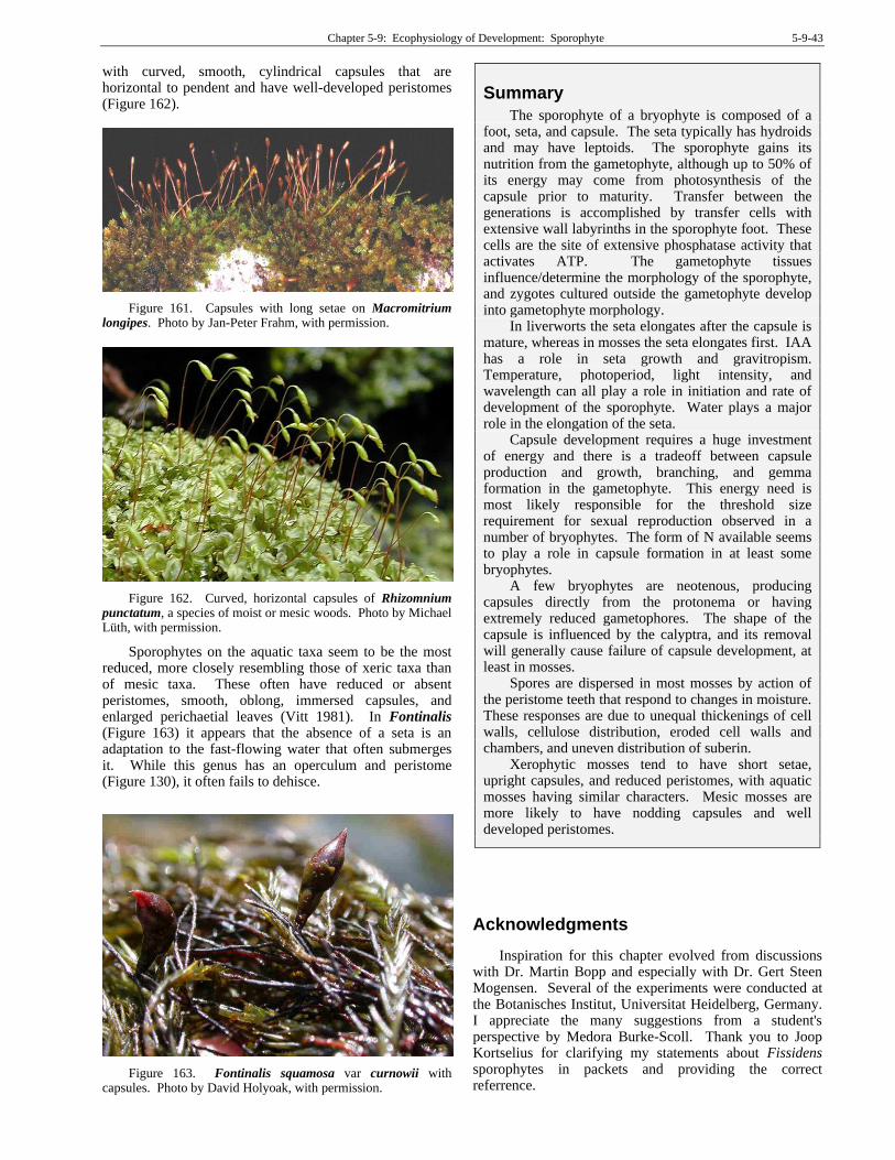
Chapter 5-9: Ecophysiology of Development: Sporophyte 5-9-43
with curved, smooth, cylindrical capsules that are horizontal to pendent and have well-developed peristomes (Figure 162).
Figure 161. Capsules with long setae on Macromitrium longipes. Photo by Jan-Peter Frahm, with permission.
Figure 162. Curved, horizontal capsules of Rhizomnium punctatum, a species of moist or mesic woods. Photo by Michael Lüth, with permission.
Sporophytes on the aquatic taxa seem to be the most reduced, more closely resembling those of xeric taxa than of mesic taxa. These often have reduced or absent peristomes, smooth, oblong, immersed capsules, and enlarged perichaetial leaves (Vitt 1981). In Fontinalis (Figure 163) it appears that the absence of a seta is an adaptation to the fast-flowing water that often submerges it. While this genus has an operculum and peristome (Figure 130), it often fails to dehisce.
Figure 163. Fontinalis squamosa var curnowii with capsules. Photo by David Holyoak, with permission.
Summary
The sporophyte of a bryophyte is composed of a foot, seta, and capsule. The seta typically has hydroids and may have leptoids. The sporophyte gains its nutrition from the gametophyte, although up to 50% of its energy may come from photosynthesis of the capsule prior to maturity. Transfer between the generations is accomplished by transfer cells with extensive wall labyrinths in the sporophyte foot. These cells are the site of extensive phosphatase activity that activates ATP. The gametophyte tissues influence/determine the morphology of the sporophyte, and zygotes cultured outside the gametophyte develop into gametophyte morphology.
In liverworts the seta elongates after the capsule is mature, whereas in mosses the seta elongates first. IAA has a role in seta growth and gravitropism. Temperature, photoperiod, light intensity, and wavelength can all play a role in initiation and rate of development of the sporophyte. Water plays a major role in the elongation of the seta.
Capsule development requires a huge investment of energy and there is a tradeoff between capsule production and growth, branching, and gemma formation in the gametophyte. This energy need is most likely responsible for the threshold size requirement for sexual reproduction observed in a number of bryophytes. The form of N available seems to play a role in capsule formation in at least some bryophytes.
A few bryophytes are neotenous, producing capsules directly from the protonema or having extremely reduced gametophores. The shape of the capsule is influenced by the calyptra, and its removal will generally cause failure of capsule development, at least in mosses.
Spores are dispersed in most mosses by action of the peristome teeth that respond to changes in moisture. These responses are due to unequal thickenings of cell walls, cellulose distribution, eroded cell walls and chambers, and uneven distribution of suberin.
Xerophytic mosses tend to have short setae, upright capsules, and reduced peristomes, with aquatic mosses having similar characters. Mesic mosses are more likely to have nodding capsules and well developed peristomes.
Acknowledgments
Inspiration for this chapter evolved from discussions with Dr. Martin Bopp and especially with Dr. Gert Steen Mogensen. Several of the experiments were conducted at the Botanisches Institut, Universitat Heidelberg, Germany. I appreciate the many suggestions from a student's perspective by Medora Burke-Scoll. Thank you to Joop Kortselius for clarifying my statements about Fissidens sporophytes in packets and providing the correct referrence.
5-9-44 Chapter 5-9: Ecophysiology of Development: Sporophyte
Literature Cited
Arnaudow, N. 1925. Über Transplantieren von Moosembryonen.
Flora 118/119: 17-26.
Arnell, H. W. 1905. Phaenological observations on mosses. Bryologist 8: 41-44.
Banbury, G. H. 1962. Geotropism of lower plants. In: Ruhland, W. (ed.). Encyclopedia of Plant Physiology, Vol. 17, Part 2. Springer-Verlag, Berlin, pp. 344-377.
Bauer, L. 1963. On the physiology of sporogonium differentiation in mosses. J. Linn. Soc. Bot. 58: 343-351.
Beerling, D. J. and Franks, P. J. 2009. Evolution of stomatal function in 'lower' land plants. New Phytol. 183: 921-925.
Bisang, I. and Ehrlén, J. 2002. Reproductive effort and cost of sexual reproduction in female Dicranum polysetum. Bryologist 105: 384-397.
Bold, H. C. 1940. The nutrition of the sporophyte in the Musci. Amer. J. Bot. 27: 318-322.
Bold, H. C. 1948. The occurrence of chlorophyll in the sporophyte of Ricciocarpus natans. Amer J. Bot. 35: 440-443.
Bopp, M. 1956. Die Bedeutung der Kalyptra für die Entwicklung der Laubmoossporogone. Ber. Deutsch. Bot. Ges. 69: 455-468.
Bopp, M. 1957. Entwicklungsphysiologische Untersuchungen an Moosmutatanten. I. Zur Wirkung der Laubmooskalyptra. Zeit. Indukt. Abstammungs-Vererbungslehre 88: 600-607.
Bopp, M. 1968. Control of differentiation in fern-allies and bryophytes. Ann. Rev. Plant Physiol. 19: 361-380.
Boudier, P. 1988. Différenciation structurale de l'épiderme du sporogone chez Sphagnum fimbriatum Wilson. [Structural differentiation of the epiderm of the sporogone of Sphagnum fimbriatum Wilson]. Ann. Sci. Nat. Bot. 13(8): 143-156.
Boyce, C. K. 2008. How green was Cooksonia? The importance of size in understanding the early evolution of physiology in the vascular plant lineage. Paleobiology 34: 179-194.
Britton, E. G. 1902. 0ctodiceras fontanum, its propagation, distribution and history. Bryologist 5: 83-85.
Brown, D. H. and Buck, G. W. 1978. Distribution of potassium, calcium and magnesium in the gametophyte and sporophyte generations of Funaria hygrometrica Hedw. Ann. Bot. 42: 923-929.
Brown, R. C., Lemmon, B. E., and Renzaglia, K. S. 1986. Sporocytic control of spore wall pattern in liverworts. Amer. J. Bot. 73: 593-596.
Browning, A. J. and Gunning, B. E. S. 1977a. An ultrastructural and cytochemical study of the wall-membrane apparatus of transfer cells using freeze-substitution. Protoplasma 93: 7-26.
Browning, A. J. and Gunning, B. E. S. 1977b. Structure and function of transfer cells in the sporophyte haustorium of Funaria hygrometrica Hedw. 1. The development and ultrastructure of the haustorium. J. Exper. Bot. 30: 1233-1246.
Browning, A. J. and Gunning, B. E. S. 1979. Structure and function of transfer cells in the sporophyte haustorium of Funaria hygrometrica Hedw. III. Translocation of assimilate into the attached sporophyte and along the seta of attached and excised sporophytes. J. Exper. Bot. 30: 1265-1273.
Bryan, V. S. 2001. Invited review – great discoveries in bryology and lichenology. Apospory in mosses discovered
by Nathanael Pringsheim in a brilliant epoch of botany. Bryologist 104: 40-46.
Budke, J. M., Goffinet, B., and Jones, C. S. 2011. A hundred-year-old question: Is the moss calyptra covered by a cuticle? A case study of Funaria hygrometrica. Ann. Bot. 107: 1279-1286.
Budke, J. M., Goffinet, B., and Jones, C. S. 2012. The cuticle on the gametophyte calyptra matures before the sporophyte cuticle in the moss Funaria hygrometrica (Funariaceae). Amer. J. Bot. 99: 14-22.
Budke, J. M., Goffinet, B., and Jones, C. S. 2013. Dehydration protection provided by a maternal cuticle improves offspring fitness in the moss Funaria hygrometrica. Ann. Bot. 111: 781-789.
Chater, C., Kamisugi, Y., Movahedi, M., Fleming, A., Cuming, A. C., Gray, J. E., and Beerling, D. J. 2011. Regulatory mechanism controlling stomatal behavior conserved across 400 million years of land plant evolution. Curr. Biol. 21: 1025-1029.
Convey, P. and Smith, R. I. L. 1993. Investment in sexual reproduction by Antarctic mosses. Oikos 68: 293-302.
Cooke, T. J., Poli, D. B., Sztein, A. E., and Cohen, J. D. 2002. Evolutionary patterns in auxin action. Plant Molec. Biol. 49: 319-338.
Cooke, T. J., Poli, D. B., and Cohen, J. D. 2004. Did auxin play a crucial role in the evolution of novel body plans during the Late Silurian–Early Devonian radiation of land plants? In: Hemsley, A. R. and Poole, I. The Evolution of Plant Physiology, Academic Press, London, pp. 85-108.
Courtice, G. R. M., Ashton, N. W., and Cove, D. J. 1978. Evidence for the restricted passage of metabolites into the sporophyte of the moss Physcomitrella patens (Hedw.) Br. Eur. J. Bryol. 10: 191-198.
Crombie, W. M. and Paton, J. A. 1958. An age effect on seta elongation in Pellia epiphylla. Nature 181: 5-41.
Crum, H. 1973. Mosses of the Great Lakes Forest. Contributions from the University of Michigan Herbarium 10: 1-404.
Crum, H. A. 2001. Structural Diversity of Bryophytes. The University of Michigan Herbarium, Ann Arbor, Michigan, 379 pp.
Crum, H. A. and Anderson, L. E. 1981. Mosses of Eastern North America. Columbia Univ. Press, New York, 2 Vol.
Duckett, J. G. and Ligrone, R. 2004. There are many ways of making water-conducting cells but what about stomata? Field Bryol. 82: 32-33.
Duckett, J. G. and Renzaglia, K. S. 1993. The reproductive biology of the liverwort Blasia pusilla L. J. Bryol. 17: 541-552.
Duckett, J. G., Pressel, S., P’ng, K. M. Y., and Renzaglia, K. S. 2009. Exploding a myth; the capsule dehiscence mechanism and the function of pseudostomata in Sphagnum. New Phytol. 183: 1053-1063.
Duckett, J., Pressel, S., P’ng, K. M. Y., and Renzaglia, K. 2010. The Sphagnum air-gun mechanism resurrected? Not with a closer look. New Phytol. 185: 889-891.
During, H. J. 1979. Life strategies of bryophytes: A preliminary review. Lindbergia 5: 2-18.
Ehrlén, J., Bisang, I., and Hedenäs, L. 2000. Costs of sporophyte production in the moss, Dicranum polysetum. Plant Ecol. 149: 207-217.
Ellis, J. G. and Thomas, R. J. 1985. Phototropism of Pellia: Evidence for mediation by auxin-stimulated acid efflux. J. Plant Physiol. 121: 259-264.

Chapter 5-9: Ecophysiology of Development: Sporophyte 5-9-45
Eschrich, W. 1975. Bidirectional transport. In: Aronoff, S., Dainty, J., Gorham, P. R., Srivastava, L. M., and Swanson, C. A. Phloem Transport. Plenum Press, NY, pp. 401-416.
Eymé, J. and Suire, C. 1967. Au sujet de l'infrastructure des cellules de la région placentaire de Mnium cuspidatum Hedw. (Mousse Bryale acrocarpe). Compt. Rend. Hebd. Séanc. Acad. Sci. (Paris) 265: 1788-1791.
Favali, M. A. and Gianni, F. 1973. Sporophyte ultrastructure in Tortula muralis Hedw. Österr. Bot. Zeit. 122: 323-331.
Fenton, N. J. and Bergeron, Y. 2006. Sphagnum spore availability in boreal forests. Bryologist 109: 173-181.
French, J. C. and Paolillo, D. J. Jr. 1975a. Effect of exogenously supplied growth regulators on intercalary meristematic activity and capsule expansion in Funaria. Bryologist 78: 431-437.
French, J. C. and Paolillo, D. J. Jr. 1975b. On the role of the calyptra in permitting expansion of capsules in the moss Funaria. Bryologist 78: 438-446.
French, J. C. and Paolillo, D. J. Jr. 1975c. Intercalary meristematic activity in the sporophyte of Funaria (Musci). Amer. J. Bot. 62: 86- 96.
French, J. C. and Paolillo, D. J. Jr. 1976. Effect of light and other factors on capsule expansion in Funaria hygrometrica. Bryologist 79: 457-465.
Fuselier, L. and McLetchie, N. 2002. Maintenance of sexually dimorphic preadult traits in Marchantia inflexa (Marchantiaceae). Amer. J. Bot. 89: 592-601.
Garner, D. L B. and Paolillo, D. J. Jr. 1973. On the functioning of stomates in Funaria. Bryologist 76: 423-427.
Glime, J. M. and Rohwer, F. 1983. The comparative effects of ethylene and 1-amino-cyclopropane-1-carboxylic acid on two species of Fontinalis. J. Bryol. 12: 611-616.
Graham, L. E. and Gray, J. 2001. The origin, morphology, and ecophysiology of early embryophytes: Neontological and paleontological perspectives. In: Gensel, P. G. and Edwards, D. (eds.). Plants invade the land: Evolutionary and ecological perspectives. Columbia University Press, New York, New York, USA, pp. 140-158.
Graham, L. E., and Wilcox, L. W. 2000. The origin of alternation of generations in land plants: A focus on matrotrophy and hexose transport. Phil. Soc. Royal Soc. London B 355: 755-767.
Grout, A. J. 1908. Some relations between the habitats of mosses and their structures. Bryologist 11: 97-100.
Haberlandt, G. 1886. Beitrag zur Anatomie und Physiologie der Laubmoose. Jahrb. Wiss. Bot. 17: 359-498.
Haig, D. 2015. Coleochaete and the origin of sporophytes. Amer. J. Bot. 102: 417-422.
Hassel, K. and Söderström, L. 1999. Spore germination in the laboratory and spore establishment in the field in Pogonatum dentatum (Brid.) Brid. Lindbergia 24: 3-10.
Hébant, C. 1975. Organization of the conducting tissue system in the sporophytes of Dawsonia and Dendroligotrichum. J. Hattori Bot. Lab. 39: 235-254.
Hébant, C. 1977. The conducting tissues of bryophytes. J. Cramer, Lehre, Germ., 157 pp. + 80 Plates.
Hedderson, T. A. 1995. Patterns of life history variation in the Funariales, Polytrichales and Pottiales. J. Bryol. 18: 639-675.
Herzenfelder, H. 1923. Experimente an Sporophyten von Funaria hygrometrica. Flora 116: 476-490.
Hoddinott, J. and Bain, J. 1979. The influence of simulated canopy light on the growth of six acrocarpous moss species. Can. J. Bot. 57: 1236-1242.
Hohe, A., Rensing, S. A., Mildner, M., Lang, D., and Reski, R. 2002. Day length and temperature strongly influence sexual reproduction and expression of a novel MADS-box gene in the moss Physcomitrella patens. Plant Biol. 4: 595-602.
Hughes, J. G. 1954. The physiology of reproduction in the Bryophyta. 8th International Botanical Congress, sec. 14/16: 122-124.
Hughes, J. G. 1962. The effects of day-length on the development of the sporophytes of Polytrichum aloides Hedw. and P. piliferum Hedw. New Phytol. 61: 266-273.
Hughes, J. G. 1969. Factors conditioning development of sexual and apogamous races of Phascum cuspidatum Hedw. New Phytol. 68: 883-900.
Ingold, C. T. 1959. Peristome teeth and spore discharge in mosses. Trans. Bot. Soc. Edinburgh 38: 76-88.
Jonsson, B. G. and Söderström, L. 1988. Growth and reproduction in the leafy hepatic Ptilidium pulcherrimum (G. Web.) Vainio during a 4-year period. J. Bryol. 15: 315-325.
Kaufman, P. B., Dayanandan, P., Thomas, R. J., Taylor, J., and Umberfield, P. J. 1982. Comparative analysis of rapid growth responses using three model systems: Conocephalum carpocephalum-stalk, Pellia seta, and Avena internode. J. Hattori Bot. Lab. 51: 195-202.
Kelley, C. 1969. Wall projections in the sporophyte and gametophyte of Sphaerocarpus. J. Cell Biol. 41: 910-914.
Kreulen, D. J. W. 1972. Spore output of moss capsules in relation to ontogeny of the archesporial tissue. J. Bryol. 7: 61-74.
Krisko, M. E. P. and Paolillo, D. J. Jr. 1972. Capsule expansion in the hairy-cap moss, Polytrichum. Bryologist 75: 509-551.
Kroken, S. B., Graham, L. E., and Cook, M. E. 1996. Occurrence and evolutionary significance of resistant cell walls in charophytes and bryophytes. Amer. J. Bot. 83: 1241-1254.
Krupa, J. 1969. Photosynthetic activity and productivity of the sporophyte of Funaria hygrometrica during ontogenesis. Acta Soc. Bot. Polon. 38: 207-215.
Laaka-Lindberg, S. 2000. Ecology of Asexual Reproduction in Hepatics. E-thesis. University of Helsinki, Finland. 28 pp.
Lal, M., and Bhandari, N. N. 1968. The development of sex organs and sporophyte in Physcomitrium cyathicarpum Mitt. Bryologist 71: 11-20.
Lal, M. and Chauhan, E. 1981. Transfer cells in the sporophyte – gametophyte junction of Physcomitrium cyathicarpum Mitt. Protoplasma 107: 79-83.
Ligrone, R., Gambardella, R., and Lucia Sposito, M. L. de. 1982. Ultrastructure of the sporophyte foot-gametophyte vaginula complex in Timmiella barbuloides (Brid.) Moenk. Planta 154: 414-425.
Ligrone, R., Duckett, J. G., and Renzaglia, K. S. 2000. Conducting tissues and phyletic relationships of bryophytes. Phil. Trans. R. Soc. Lond. B 355: 795-813.
Ligrone, R., Carafa, A., Duckett, J. G., Renzaglia, K. S., and Ruel, K. 2008. Immunocytochemical detection of lignin-related epitopes in cell walls in bryophytes and the charalean alga Nitella. Plant Syst. Evol. 270: 257-272.
Lobachevska, O. V., Demkiv, O. T., and Ripetskyj, R. T. 1998. Influence of gravity on spatial orientation and morphogenesis of moss sporophytes. Adv. Space Res. 21: 1141-1144.
Longton, R. E. and Schuster, R. M. 1983. Reproductive biology. In: Schuster, R. M. (ed.). New Manual of Bryology. Vol. 1. Hattori Botanical Lab, Nichinan, Japan, pp. 386-462.
5-9-46 Chapter 5-9: Ecophysiology of Development: Sporophyte
Maier, K. 1967. Wandlabyrinthe im Sporophyten von Polytrichum. Planta 77: 108-126.
Maier, K. and Maier, U. 1972. Localization of beta-glycerophosphatase and Mg+2-activated adenosine triphosphatase in a moss haustorium, and the relation of these enzymes to the cell wall labyrinth. Protoplasma 75: 91-112.
Markham, K. R., Moore, N. A., and Porter, L. J. 1978. Changeover in flavonoid pattern accompanying reproductive structure formation in a bryophyte. Phytochemistry 17: 911-913.
Melick, H. van. 1986. De verspreiding van Fissidens fontanus (La Pyl.) Lindb. Lindbergia 11: 169-171.
Menon, M. K. C. and Lal, M. 1974. Morphogenetic role of kinetin and abscisic acid in the moss Physcomitrium. Planta 115: 319-328.
Monroe, J. H. 1965b. Some aspects of ultrastructure in Funaria. Ph. D. Dissertation, University of Texas, Austin.
Mueller, D. M. J. 1973. The peristome of Fissidens limbatus Sullivant. University of California Publications in Botany 63: 1-34.
Paolillo, D. J. Jr. 1968. The effect of the calyptra on capsule symmetry in Polytrichum juniperinum Hedw. Bryologist 71: 327-334.
Patidar, K. C., Solanki, C. M., and Kaul, A. 1987. Spore output in Marchantiales. Bryologist 90: 251-252.
Patterson, P. M. 1957. The effect of indole-3-acetic acid on certain growth phases in bryophytes. Bryologist 60: 277-283.
Patterson, P. M. and Baber, J. S. 1961. Factors breaking vegetative dormancy in certain mosses. Bryologist 64: 336-338.
Poli, D. B., Jacobs, M., and Cooke, T. J. 2003. Auxin regulation of axial growth in bryophyte sporophytes: Its potential significance for the evolution of early land plants. Amer. J. Bot. 90: 1405-1415.
Pressel, S. and Duckett, J. G. 2006. The parasitic ascomycete Mniaecia jungermanniae induces the formation of giant perichaetia and apogamous sporophytes in leafy liverworts. Botany 84: 384-392.
Pressel, S., Goral, T., and Duckett, J. G. 2014. Stomatal differentiation and abnormal stomata in hornworts. J. Bryol. 36: 87-103.
Proctor, M. C. F. 1977. Evidence on the carbon nutrition of moss sporophytes from 14CO2 uptake and the subsequent movement of labelled assimilate. J. Bryol. 9: 375-386.
Proctor, M. C. F. 1984. Structure and ecological adaptation. In: Dyer, A. F. and Duckett, J. G. (eds.). The Experimental Biology of Bryophytes. Academic Press, London, pp. 9-37.
Redfearn, P. L. Jr. and Meyer, S. L. 1949. Physiological studies on mosses. VIII. Observations on the regeneration of setae of Physcomitrium turbinatum. Bryologist 52: 197-201.
Renault, S., Bonnemain, J. L., Faye, L., and Gaudillere, J. P. 1992. Physiological aspects of sugar exchange between the gametophyte and the sporophyte of Polytrichum formosum. Plant Physiol. 100: 1815-1822.
Renzaglia, K. S., Duff, R. J., Nickrent, D. L., and Garbary, D. 2000. Vegetative and reproductive innovations of early land plants: Implications for a unified phylogeny. Trans. Royal Soc. London 355: 769-793.
Rydgren, K. and Økland, R. H. 2002. Ultimate costs of sporophyte production in the clonal moss Hylocomium splendens. Ecology 83: 1573-1579.
Rydgren, K. and Økland, R. H. 2003. Short-term costs of sexual reproduction in the clonal moss Hylocomium splendens. Bryologist 106: 212-220.
Schnepf, E., Stein, U., and Deichgräber, G. 1978. Structure, function, and development of the peristome of the moss, Rhacopilum tomentosum, with special reference to the problem of microfibril orientation by microtubules. Protoplasma 97: 221-240.
Schnepf, E., Herth, W., and Morre, D. J. 1979. Elongation growth of setae of Pellia (Bryophyta): Effects of auxin and inhibitors. Zeit. Pflanzenphysiol. 94: 211-217.
Schofield, W. B. 1985. Introduction to Bryology. Macmillan Publishing Co., New York, 431 pp.
Slade, J. S. 1965. Physical factors influencing seta elongation in Pellia epiphylla. Bryologist 68: 440-445.
Stark, L. R. 2001. Widespread sporophyte abortion following summer rains in Mojave Desert populations of Grimmia orbicularis. Bryologist 104: 115-125.
Stark, L. R., Mishler, B. D., and McLetchie, D. N. 2000. The cost of realized sexual reproduction: Assessing patterns of reproductive allocation and sporophyte abortion in a desert moss. Amer. J. Bot. 87: 1599-1608.
Stark, L. R., Brinda, J. C., and McLetchie, D. N. 2009. An experimental demonstration of the cost of sex and a potential resource limitation on reproduction in the moss Pterygoneurum (Pottiaceae). Amer. J. Bot. 96: 1712-1721.
Stevenson, D. W., Rastorfer, J. R., and Showman, R. E. 1972. Effects of temperature on seta elongation in Atrichum undulatum. Ohio J. Sci. 72: 146-152.
Sundberg, S. 2002. Sporophyte production and spore dispersal phenology in Sphagnum: The importance of summer moisture and patch characteristics. Can. J. Bot. 80: 543-556.
Sundberg, S. and Rydin, H. 1998. Spore number in Sphagnum and its dependence on spore and capsule size. J. Bryol. 20: 1-16.
Taylor, J., Thomas, R. J., and Otero, J. G. 1972. Chromatographic and spectrophotometric analyses of the photosynthetic pigments of the gametophyte and the sporophyte of Lophocolea heterophylla. Bryologist 75: 36-42.
Thomas, R. J. 1975. Lipid composition of maturing and elongate liverwort sporophytes. Phytochemistry 14: 623-626.
Thomas, R. J. 1976. Correlation between protein content and cell elongation in setae of Lophocolea heterophylla sporophytes. J. Hattori Bot. Lab. 40: 87-90.
Thomas, R. J. 1977a. Wall analyses of Lophocolea seta cells (Bryophyta) before and after elongation. Plant Physiol. 59: 337-340.
Thomas, R. J. 1977b. Water relations of elongating seta cells in sporophytes of the liverwort Lophocolea heterophylla. Bryologist 80: 345-347.
Thomas, R. J. 1980. Cell elongation in hepatics: The seta system. Bull. Torrey Bot. Club 107: 339-345.
Thomas, R. J. and Doyle, W. T. 1976. Changes in the carbohydrate constituents of elongating Lophocolea heterophylla setae (Hepaticae). Amer. J. Bot. 63: 1054-1059.
Thomas, R. J., Taylor, J., and Montague, M. J. 1970. The growth response of Lophocolea heterophylla seta segments to exogenous IAA. Bryologist 73: 713-715.
Thomas, R. J., Stanton, D. S., Longendorfer, D. H., and Farr, M. E. 1978. Physiological evaluation of the nutritional autonomy of a hornwort sporophyte. Bot. Gaz. 139: 306-311.
Chapter 5-9: Ecophysiology of Development: Sporophyte 5-9-47
Thomas, R. J., Stanton, D. S., and Grusak, M. A. 1979. Radioactive tracer study of sporophyte nutrition in hepatics. Amer. J. Bot. 66: 398-403.
Thomas, R. J., Harrison, M. A., Behringer, F. J., Taylor, J., and Kaufman, P. B. 1982. Production of endogenous auxin and ethylene in elongating setae of Pellia epiphylla sporophytes. Bot. Soc. Amer. Misc. Publ. 162: 1.
Thomas, R. J., Harrison, M. A., Taylor, J., and Kaufman, P. B. 1983. Endogenous auxin and ethylene in Pellia (Bryophyta). Plant Physiol. 73: 395-397.
Thomas, R. J., Behringer, F. J., Lombard, C. S., and Sparkowski, J. J. 1984. Effects of auxin on wall polysaccharide composition and enzyme activity during extension-growth of Pellia (Bryophyta). Physiol. Plant. 60: 502-506.
Thomas, R. J., Caron, P. J., and Watt, R. S. 1987. Time-lapse photography of phototropism in Pellia epiphylla. Bryologist 90: 390-392.
Thomas, R. J., Woodward, T. G., and Rogoff, H. A. 2002. Growth asymmetry and auxin transport in setae of gravistimulated Pellia epiphylla sporophytes. Bryologist 103: 407-410.
Touw, A. and Rubers, W. V. 1989. De Nederlandse bladmossen: Flora en verspreidingsatlas van de Nederlandse Musci (Sphagnum uitgezonderd), Vol. 50. Stichting Uitgeverij Koninklijke Nederlandse Natuurhistorische Vereniging.
Vitt, D. H. 1981. Adaptive modes of the moss sporophyte. Bryologist 84: 166-186.
Watson, E. V. 1974. The Structure and Life of Bryophytes. Hutchinson University Library, London, 211 pp.
Wenderoth, H. 1931. Beiträge zur Kenntnis des Sporophyten von Polytrichum juniperinum Willdenow. Planta 14: 344-385.
Whel, C. 1975. Quantitative changes in free amino acids in Polytrichum formosum during development. Phytochemistry 14: 2337-2340.
Wiencke, C. and Schulz, D. 1975. Sporophyten Entwicklung von Funaria hygrometrica Sibth. I. Strukturelle Grundlagen der Wasser- und Nährstoffaufnahme im Haustorium. Protoplasma 86: 107-117.
Zielinski, F. 1909. Beiträge zur Biologie des Archegoniums und der Haube der Laubmoose. Flora 100: 1-36.

5-9-48 Chapter 5-9: Ecophysiology of Development: Sporophyte