
Cost-effectiveness of alternative methods for diabetic retinopathy screening
Upload
khangminh22Category
view
0download
0
1 2 3Naganandini S. , Rekha K. , Vanishree N.
ECONOMIC EVALUATION & ANALYSIS OF COST EFFECTIVENESS IN DENTAL RESEARCH
Original Research
Journal Of The Oxford Dental College
Email for [email protected]
JTODC, 3 (1), 2011 | 67 |
Dept of Public Health DentistryThe Oxford Dental College, Hospital and Research Center, Bangalore.
1Professor2PG Student
3Professor
ABSTRACT
Economic evaluation is an accepted method for
appraisal for health care programmes. Although it has been
widely used in medicine, but more recently has been used in
dentistry. Cost-effectiveness analyses are useful to inform
decision and policy makers about the managerial implications
of different treatment policies. Several principles of cost-
effectiveness analysis using a critical appraisal of a published
economic evaluation in dentistry are reviewed. An improved
understanding of the principles behind, and steps involved in
the critical appraisal of health economic studies, should
improve decision making within the dental community.
Keywords : Economic evaluation, cost-effectiveness, analysis.
INTRODUCTION
Economic evaluation may be defined as 'the
comparative analysis of alternative course of action in
terms of their costs and consequences' (Drummond et
al 1987). Economic evaluation is the identification,
measurement and valuation, and then comparison of
the costs (inputs) and benefits (outcomes) of two or
more alternative activities. It differs according to their
scope and intent. They can have a very narrow focus,
whereby evaluators are only concerned about the
resource consequences for their agency. Economic
evaluation is one of the tools available to help choose
wisely from a range of alternatives and implement
efficient resources. Prospective economic analyses are
best undertaken alongside other evaluations,
particularly outcome studies. Economic components
of research need not be excessively expensive. There
is, however, great merit in examining the economic
design from the beginning of a research planning
process as results may affect the overall design of the
study as well as the detail of data collection. It is
important, prior to undertaking a study, to determine
whether a full economic evaluation is warranted or
required. The objective of economic evaluation is to be
an aid to decision-making, not a complete basis for 1, 2making decisions.
Economic evaluation covers a number of specific
tecniques, such as cost effectivness and cost benefit
analysis, which can be used to address the question of
whether a program, project, intervention offers good
value for money. These methods have been used in
health care since 1960's, gaining much wider
applications in recent years with focus on medical
technology.

Health care resources are limited by the total
funds available, as well as through competition with
other areas, such as housing and education. This
raises the question of how to decide where the money
should be allocated most appropriately. The
establishment of the allocation of health care
resources should be efficient and eqiutable. The
rationale for economic evaluation is the pursuit of
efficiency in identifying and reallocating resources to
those health care interventions that offer greatest
health returns. Economic evaluation is important
because without systemic analysis, it is not possible to 3identify the relevant alternatives.
Cost-effectiveness analysis (CEA) is a form of
economic analysis that compares the relative costs
and outcomes (effects) of two or more courses of
action. Cost-effectiveness analysis is often used in the
field of health services, where it may be inappropriate
to monetize health effect. Typically the CEA is
expressed in terms of a ratio where the denominator is
a gain in health from a measure (years of life,
premature births averted) and the numerator is the
cost associated with the health gain. The most
commonly used outcome measure is quality-adjusted
life years (QALY). Cost-effectiveness analysis
compares the costs and health effects of an
intervention to assess the extent to which it can be
regarded as providing value for money. This informs
decision-makers who have to determine where to 4, 5allocate limited healthcare resources. in dentistry,
the method of cost-effectiveness analysis, and
methods for economic evaluations and policy analysis
in general, are in their infancy. The number of
published cost-effectiveness analyses is limited and 6application their application in dentistry is not precise.
Hence in our present review we want to shed
light on the principles of economic evaluation & cost
effectiveness analysis, and their role in conducting
research studies in dentistry.
DISTINGUISHING FEATURES OF ECONOMIC
EVALUATIONS
In economic evaluation the direct costs are the
value of the resources consumed in delivering care. It
sets out to answer two main questions: first, is a
procedure worth doing when compared others, with
the same resources and time. Second , are we satisfied 3that the resources be sepnt this way?
Different forms of economic evaluation :
COST MINIMIZATION ANALYSIS ( CMA)
The cost-minimization analysis is an economic
study in which two or more therapeutic alternatives
with the same effectiveness or efficacy are compared
in terms of net costs in order to establish the cheapest
alternative. The equivalence of the comparators in
terms of efficacy must be presented transparently and 3,7,8comprehensibly.
COST EFFECTIVNESS ANALYSIS (CEA)
The cost-effectiveness analysis is an economic
study in which the costs are expressed in monetary
units and the results in non-monetary units. Non-
monetary units may for example be: (1) years of life
gained, (2) hospital days prevented, (3) clinical
parameters (e.g. response or remission rates,
reduction in cholesterol, etc). Cost effectiveness
analysis proceeds by identification of a single outcome
of interest and expresses the incremental cost of
achieving the incremental benefit. An obvious
weakness with the strict cost-effectiveness
methodology is the enforced focus on a single
outcome dimension (in order to compute ratios), when
public health programmes can have multiple
outcomes, encompassing changes in survival and 7, 8, 9 health related quality of life.
COST BENEFIT ANALYSIS (CBA)
Cost benefit analysis values all costs and benefits
in the same monetary units. If benefits exceed costs,
the evaluation would recommend investing in the
programmed, and vice versa. CBAs are thus
intrinsically attractive, and theoretically an ideal
approach, but conducting them can be problematic
because of the difficulties associated with valuing
outcomes in monetary terms (including public
acceptability). Although this is the oldest and most
widely practiced method of economic evaluation in
other sectors of public spending, its application in
health has been problematic, largely because of the
difficulties in attaching monetary values to health 3, 7-10programs outcomes.
| 2 | JTODC, 3 (1), 2011
COST UTILITY ANALYSIS (CUA)
The cost-utility analysis follows the same
principle as the cost-effectiveness analysis. Costs are
assessed in monetary units and the benefit is
measured as a non-monetary but utility-adjusted
outcome, the quality adjusted life year (QALY). The
concept combines life expectancy and quality of life. If
quality of life is an important aspect of therapy, this
form of analysis should be chosen. Costutility analysis
measures, the impact of an intervention in terms of
improvements in preference weighted, health-related
quality of life such as the QALY. Costutility analyses
allow comparisons to be made across all areas of
health intervention, aiding resource allocation decision
making. But they do not capture the broader
nonhealth consequences and opportunity costs of 3,9,10programmes.
COST CONSEQUENCE ANALYSIS
Costconsequence analysis is similar to cost-
effectiveness analysis in terms of the questions
addressed, but is applied to evaluate interventions
with more than one multi-dimensional outcome. In
CCA, for each alternative the evaluation would
compute total (and component) costs, and measure
change along every one of the relevant outcome
dimensions. The cost and outcome results would need
to be reviewed by decision makers, and the different
outcomes weighed up (informally and subjectively)
and compared with costs.
While this approach has theoretical problems, as
it does not synthesize benefits and costs, it can be
used to look at issues of changing behavior that are so
crucial to public health interventions. CCA does not
attempt to combine measures of benefit into a single
measure of effectiveness, so it cannot be used to rank
interventions. Nevertheless it is a systematic
technique that allows decision makers to weigh and
prioritize the outcomes of an evaluation. It is possible
to produce cost-effectiveness comparisons for single
outcomes within the CCA framework. The analysis
involves focusing on a particular problem, then; using
the existing available data an appropriate method is
established for an analysis of costs and outcomes in a
common currency. The evidence collected needs to
relate to four questions: what works to improve
health; what works to reduce inequalities in
health; what works in changing behavior; and what
works in promoting uptake of behavior change 11, 12interventions?
CRITERIA FOR ASSESSING ECONOMIC
EVALUATION IN DENTAL RESEARCH
1) Was a well defined question posed in answerable
form?
2) Was a comprehensive description of the
competing alternatives given?
3) Was there evidence that the program's
effectiveness had been established?
4) Were all important and relevant costs and
consequences for each alternative identified?
5) Were costs and consequences measured
accurately in appropriate physical units?
6) Were costs and consequences valued credibly?
7) Were costs and consequences adjusted for
differential timing?
8) Was an incremental analysis of costs and
consequences of alternatives performed?
9) Was a sensitivity analysis performed?
10) Did the presentation and discussion of study 13results include all issues of concern to users?
LIMITATIONS OF ECONOMIC EVALUATIONS
The debate about the use and usefulness of
economic evaluation had intensified in recent years.
There are still doubts about whether economic
evaluation is really useful in health care decision
making. A number of reasons have been suggested for
this: the uncertainty in the cost effectiveness results,
concerns about the lack of availability of alternative
therapies, inadequate estimation of the comparative
clinical efficacy of the drugs being assessed and
concerns about the overall budgetary impact on the
health care system.
Two most worrying limitations of economic
evaluations are: 1) that decisions are often taken
quickly without adequate assessment of the evidence
(including cost effectiveness evidence) 2) budgetary
JTODC, 3 (1), 2011 | 1 |

arrangements rarely gives the right incentives for the
adoption of interventions that are shown to be cost
effective.
The following conditions appear to be important
while making decisions for economic evaluations :
1) Clear decision making process ; it is important to
know where the decisions are being made, who
makes them and the mechanisms for introducing
different categories of evidence.
2) Clear policy objectives: the policy objectives of
the decision maker need to be clear and
efficiency needs to be prominent among these
objectives.
3) Reasonable timelines and resources: the timeline
for making the decision and the availability of
resources to assess the evidence need to be
sufficiently generous to facilitate the
consideration of cost effectiveness evidence.
4) Appropriate incentives: the right incentives need
to exist for the implementation of those
treatments to lead to a more cost effective use of 14resources.
PRINCIPLES OF COST EFFECTIVENESS
ANALYSIS
Cost-effectiveness analysis (CEA) is a powerful
analytical technique that measures the health benefit
that is obtained from a given expenditure. All forms of
economic evaluations consider costs in a similar
manner; however they differ on how outcomes are
measured. In its classical form, CEA measures
outcomes in terms of natural units, such as mg/dl of
cholesterol or years of life gained. In a variation of this
approach, traditionally called cost-utility analysis
(CUA) but increasingly referred to as CEA in the
literature , the effectiveness of health interventions is
measured in common units of health related value,
such as the quality adjusted life year (QALY). These
measures are based on preferences expressed by
groups of patients or the public through the
assignment of subjective utility values to specific
health states. Several techniques, such as the
standard gamble and the time trade off models, are
used to obtain these utility values. Current
recommendations emphasize the use of CUA approach
when performing cost-outcome studies and the most
commonly used outcome measure in this type of
analysis is the QALY. QALYs incorporate three factors:
the size of quality improvement that a given treatment
or diagnostic intervention produces, the duration of
the health improvement, and the number of persons
that can be expected to benefit from the intervention.
CEA study does not determine whether the benefits
are worth the cost or not. In that sense, it is a 6, 9descriptive and not a prescriptive instrument.
CRITERIA BY WHICH A COST EFFECTIVE
ANALYSIS MAY BE ASSESSED
1. Did the analysis provide a full economic
comparison of health care strategies?
2. Were the costs and outcomes properly measured
and valued?
3. Was appropriate allowance made for uncertainty
in the analysis?
4. Are estimates of costs and outcomes related to
the baseline risk in the treatment population?
5. What are the incremental costs and outcomes of
each strategy?
6. Do incremental costs and outcomes differ
between subgroups?
7. How much does allowance for uncertainty
change the results?
Did the analysis provide a full economic
comparison of health care strategies ?
A cost-effectiveness analysis compares both
costs and outcome of two or more strategies.
Outcomes are expressed unvalued in clinical units of
effect such as number of decays prevented when
evaluating a fissure sealing program. A weighting
factor (utility) between zero (missing tooth) and one
(sound tooth) is used for quality-adjustment. It is
emphasized that a cost-effectiveness or cost-utility
analysis always means an explicit comparison of
treatment alternatives in terms of both costs and
outcomes. A new therapy could be compared with the
standard treatment strategy or with the no
intervention strategy. A more universal outcome
| 2 | JTODC, 3 (1), 2011

measure than QATY would be required for addressing
complex problems in implant and maxillofacial
Prosthodontics, orthodontics or oral surgery. Ideally,
an instrument that could be used to compare any
aspect in modern dentistry, including esthetics, would
be preferred, making it easier to compare outcomes
across dental specialties. However, such an outcome
measure still remains to be developed. An alternative
approach would be to express health-related
outcomes in terms of willingness-to-pay. Cost-
effectiveness analysis may be conducted from any of
the following perspectives: the society at large, a third-
party payer (e.g. an insurance company), the dental
community (providers of dental care), a dental
company, a managed care group or a patient
population. A cost-effectiveness analysis from a
societal perspective aims to include all types of costs.
Economic consequences of choosing an alternative
may include savings as well as direct medical costs
(e.g. costs for a surgical intervention), direct non-
medical costs (e.g. transportation costs), indirect costs
(e.g. lost work productivity) and intangible costs (e.g. 6pain and suffering).
Were the costs and outcomes properly
measured and valued?
Cost-effectiveness analysis is often based on
outcome data reported in clinical trials or meta-15analyse. Randomized controlled trials represent the
gold standard for evaluating the efficacy of a
treatment. But the external validity (i.e. the relevance
of the results to the general population) of a 16randomized controlled trial might be limited. When
performing an economic evaluation from the societal
perspective, it is often more important to adhere to the
external validity of the selected studies which
document the effectiveness of the treatment under a
real-world setting. Economic evaluations based on
mathematical modeling are therefore often needed to
extrapolate beyond the endpoint of clinical trials and to 17adjust for the desired degree of external validity. A
mathematical model should be validated before it is
used for policy recommendations. The validity of a
cost-effectiveness analysis might be questionable
when outcomes of a strategy are modeled over a
period that is by far beyond the follow-up period of the
original clinical trial. The time horizon used depends on
the therapy under evaluation. For example, the effect
of water fluoridation can accrue over a lifetime, but the
relevant effects of preoperative anesthesia are much
shorter in comparison, and an extrapolation of the
results over a patient's lifetime may be inappropriate.
Physical quantities of resources consumed by different
treatment strategies should be reported separately
from their unit prices. This facilitates a proper
interpretation of the results. Resource consumption
and unit costs often widely differ by geographical area
and make the generalizability of the results of a cost-
effectiveness analysis difficult. Not only costs incurred
to provide the therapy, but also future costs associated
with the therapy are helpful to derive an unbiased
estimate of resource consumption.
Was appropriate allowance made for the
uncertainty in the analysis?
In a sensitivity analysis input parameters are
varied over a defined range. This allows us to test how
sensitive the model is to key assumptions and data
variability. For example, one might not be sure about
the exact utility (0.9) of composite resin. One could
vary the utility in a range between say 0.81.0 to see
how the incremental cost-effectiveness ratio changes
with changes in utility estimates for composite resin.
The ranges for sensitivity analysis should be justified.
A model is said to be robust when various values for
input parameters do not have a major impact on the
results and conclusions of the analysis. This form of
sensitivity analysis, although not without limitations, is
predominant in published cost-effectiveness analyses.
Recently, researchers have begun to develop and
apply more sophisticated statistical methods such as
bootstrapping or Bayesian methods of analysis to
assess overall parameter uncertainty. Bayesian
Analysis relies on the idea that uncertainty can be
described by a distribution. Bootstrapping is a
computer-intensive resampling technique and has
become popular with the advent of cheap
computational power. A sensitivity analysis on more
than three parameters becomes difficult to interpret.
Bayesian methods of analysis would have been useful
to assess overall parameter uncertainty. However, this
may reflect an assessment of the uncertainty of expert
JTODC, 3 (1), 2011 | 1 |

opinions rather than an assessment of uncertainty of
the unknown true probabilities.
Are estimates of costs and outcomes related to
the baseline risk in the treatment population?
The baseline risk in the treatment population
often dramatically influences the costs and outcomes
in a cost-effectiveness analysis. For example, heavy
smokers are likely to have a higher failure rate after
implant insertion than non-smokers because of their
reduced wound healing capabilities and higher risk of
infection. This would translate into a higher cost
effectiveness ratio for this treatment modality in this
patient-subgroup compared to non-smokers. The
cost-effectiveness ratio would increase and become
less attractive in this case. Hence risk in baseline
treatment population is important to be determined 6before the start of the study.
What are the incremental costs and outcomes
of each strategy?
In order to compare costs and outcome of two or
more strategies, it is essential to compute the
incremental cost and incremental outcome which is
the difference in costs and outcomes observed
between two strategies. The incremental cost-
effectiveness ratio should inform decision makers
about the extra benefit that could be bought at any
extra cost. However, there is an ongoing debate about
the correct interpretation of incremental cost-18effectiveness ratios. The incremental cost-
effectiveness ratio is calculated by dividing the
incremental costs by the incremental effectiveness.
For example, let us assume that patients prefer tooth
colored composite resin (utility 0.9) over amalgam
(utility 0.7) for the restoration of a premolar. And let us
assume that a composite resin would last for ten years
and an amalgam for eleven years. An amalgam would
cost 200 CHF (costs in Swiss Francs) and a composite
resin 300 CHF. The incremental effectiveness of
composite resin over amalgam would be 1.3 QATYs
(0.9_10 years0.7_11 years) (Effectiveness in quality
adjusted tooth years) and the incremental costs 100
CHF (300 CHF200 CHF) respectively. Hence, the
incremental cost-effectiveness ratio in this
hypothetical example is 76, 9 CHF per QATY (100
CHF/1, 3 QATYs). If amalgam would be the standard
therapy, changing from amalgam to composite resin as
the new therapy would cost 76, 9 CHF per QATY
gained. On average, the patient would pay an
additional 77 CHF for the extra benefit of one QATY
when choosing composite resin instead of amalgam.
Do incremental costs and outcomes differ
between subgroups?
The cost-effectiveness of a therapy depends on
whom it is provided to. The baseline risk of morbidity
may vary from one patient subgroup to another. In
consequence, the cost-effectiveness of a therapy often
simultaneously changes from one patient subgroup to
another. This variation among patient- subgroups
might influence the decision to whom priority should 6be given for certain treatment modalities.
How much does allowance for uncertainty
change the results?
The 95% confidence interval is often used as a
range for sensitivity analysis when data from clinical
trials are used. Estimates based on assumptions or
expert opinion should be evaluated over a wide range
of values. Caution should be used in drawing
conclusions from a model tested with unjustified
narrow parameter ranges used in the sensitivity 6analysis.
CONCLUSION
While many considerations, such as affordability,
equity, and non-health benefits, may factor into
decisions about health spending, cost-effectiveness
analysis is an essential tool for decision makers. It can
guide decisions about where best to spend limited
resources and what to include in a package of health
services that responds to a population's greatest
health needs.
REFERENCES
1) MJ Sculpher, FS Pang, A Manca, MF Drummond, S
Golder, H Urdahl, LM Davies and A Eastwood. Health
Technology Assessment 2004; Vol. 8: No. 49.
2) World Health Organization, 2000. Workbook 8 ·
Economic Evaluations.
| 2 | JTODC, 3 (1), 2011

3) O'Brein. Principles of economic evaluation for
health care programs. The Journal of
Rheumatology 1995: 22:7.
4) Using Cost-Effectiveness Analysis for Setting
Health Priorities | Disease Control Priorities
Project.
5) Ceri Phillips. What is cost effectiveness? Second
edition, February 2009.
6) Peter P. Sendi, Andrew J. Palmer, Carlo. P.
Marinello. Some Principles of Cost- Effectiveness
Analysis in Dentistry. Acta Med Dent Helv 4:
6367 (1999).
7) Nils Oscarson, Health Economic Evaluation
Methods for Decision-Making in Preventive
Dentistry, Umeå University, 2006.
8) Ramesh Adhikari, Paul Gertler, and Anneli
Lagman. Economic Analysis of Health Sector
Projects - A Review of Issues, Methods, and
Approaches. March 1999.
9) José G Merino, MD, M Phil. Clinicians and the
economic evaluation of health salud pública de
méxico / vol.44, no.2, marzo-abril de 2002.
10) Evelyn Walter, Susanne Zehetmayr. Guidelines on
Health Economic Evaluation, April 2006.
11) Michael P. Kelly, David McDaid, Anne Ludbrook,
and Jane Powel. Economic appraisal of public
health interventions, Health development
agency. University of the West of England,
Bristol.
12) Susan. J. Cunningham. Current product &
practice an introduction to economic evaluation
of health care. J.O. vol 28, NO. 3.
13) Drummond MF, Stoddart GL, Torrence GW:
Methods for economic evaluation of health care
programmed. Oxford university press, 1987.
14) Micheal Drummond, Economic evaluation in
health care: is it really useful or are we just
kidding ourselves?
15) Palmer A J, Sendi P P: Meta-analysis in oral health
care. Oral Surg Oral Med Oral Pathol Oral Radiol
Endod: in press (1999).
16) Drummond M F, Richardson W S, O'Brein B J,
Levine M, Heyland D: Users'guides to the medical
literature. XIII. How to use an article on
economic analysis of clinical practice. A. Are the
results of the study valid? Evidence-Based
Medicine Working Group. JAMA 277: 15521557
(1997a).
17) Buxton M J, Drummond M F, Van Hout B A, Prince
R L, Sheldon T A, Szucst,Vray M: Modelling in
economic evaluation: an unavoidable fact of life.
Health Econ 6: 217227 (1997).
18) Birchi S, Gafni A: Changing the problem to fit the
solution: Johannesson and Weinstein's (mis)
application of economics to real world problems.
J Health Econ 12: 469476 (1993).
JTODC, 3 (1), 2011 | 1 |

1Uma Eswara
DENTIGEROUS CYST
ABSTRACT
Keywords :
Dentigerous cyst is a developmental odontogenic cyst,
which apparently develops by accumulation of fluid between
the reduced enamel epithelium and the tooth crown of an
unerupted tooth. There is usually no pain or discomfort
associated with the cyst unless there is acute inflammatory
exacerbation. Here, we report a case of dentigerous cyst in a
10-year-old male patient and its management
Dentigerous cyst, Odontogenic cyst
Case Report
Dept of PaedodonticsThe Oxford Dental College, Hospital and Research Center, Bangalore.
1Professor
INTRODUCTION
Odontogenic cysts are the ones that develop
from epithelial remnants of the tooth-forming organ.
Dentigerous cysts are the second most common
odontogenic cyst after the radicular cyst accounting 1for 24% of all the true cysts of the jaw. A Dentigerous
cyst is an epithelial-lined developmental cavity that
encloses the crown of an unerupted tooth at the
cementoenamel junction. Dentigerous cysts are
generally discovered when radiographs are taken to
investigate a failure of tooth eruption, missing tooth,
or malalignment. There is usually no pain or
discomfort associated with the cyst unless there is
acute inflammatory exacerbation. Radiographs show a
unilocular, radiolucent lesion characterized by a well-
defined sclerotic margins and associated with crown of
the unerupted tooth. While the normal follicular space
is 3-4 mm, a Dentigerous cyst can be suspected when 2the space is more than 5 mm.
Various treatment options have been proposed
for the management of Dentigerous cyst. Two most
Journal Of The Oxford Dental College
Email for [email protected]
common treatment modalities used are: 1. Total
enucleation of the cyst and 2. Marsupilization for
decompression of large volume cysts, or a
combination of both
CASE REPORT
A 10-year-old boy presented to the Department
of Pedodontics, with the chief complaint of swelling in
the lower left side of the jaw. Clinical history revealed
that the patient had a small painless diffused swelling,
which increased to the present size of single diffuse
swelling of about 3 x 2 cm over a period of 1 month.
Extraoral examination revealed hard firm swelling
present near the lower border of the mandible.
Intraoral examination revealed swelling of firm
consistency causing bulging of the cortical bone with
no pain and mobility of the teeth in the same region
(Figure 1). The swelling was non-tender and egg-shell
crackling was elicited in the region apical to the teeth
32, 73, 74 and 35. Radiographic examination showed
unilocular, radiolucent area extending from the root of
deciduous mandibular left lateral incisor to permanent
JTODC, 3 (1), 2011 | 1 |

| 2 | JTODC, 3 (1), 2011
Figure 1 : Preoperative clinical
Figure 2 : Preoperative radiograph, Orthopantomogram
Figure 3 : Preoperative Radiograph, Occlusal View
Figure 4 : Postsurgical enucleated clinical photograph
Figure 5 : Immediate Postoperative radiograph, Orthopantomogram
Figure 6 : Enucleated specimen
Figure 7 : Clinical Photograph Postoperative
Figure 8 : One month Postoperative radiograph, Orthopantomogram

JTODC, 3 (1), 2011 | 69 |
Figure 4 : : Clinical photograph of removable
flexible space maintainer inserted
mandibular left second premolar enclosing the tooth
buds of permanent mandibular left canine and first
premolar causing displacement of the same (Figure 2
and 3). Histopathologic examination of the aspiration
biopsy showed a cystic lesion, and presumptive
diagnosis of the dentigerous cyst was made.
Treatment procedure comprised of extraction of 32,
33, 34, 35, 73, 74, 75 and enucleation of cyst under
general anesthesia, which created a large window
(Figure 4 and 5). Chemical cauterization with Carnoy's
solution was done. The removed surgical specimen
(Figure 6) was histopathologically examined
confirming the diagnosis of dentigerous cyst and the
clinical and radiographic follow up was done (Figure 7
and 8) and a flexible space maintainer is placed
(Figure 9).
DISCUSSION
Dentigerous cysts are common developmental
cysts. Since cysts can attain considerable size with
minimal or no symptoms, early detection and removal
of the cysts is important to reduce morbidity. Although
evidence in the literature suggests that dentigerous
cysts occur more frequently during the second decade 6, 7 of life, these lesions can also be found in children and
adolescents. The incidence of dentigerous cysts is
twice as high in male patients as compared to female 8, 9counterparts. The most common sites of this cyst
are the mandibular and maxillary third molar and maxillary cuspid area,but also seen in association with
other unerupted tooth crowns as presented in this
case. Enucleation of the cyst was done along with
extraction of the involved teeth, clinical and
radiographic follow up was done to assess the bone
growth, a flexible space maintainer is placed, further
follow up is being carried out to assess the progress in
order to place implants or fixed denture.
REFERENCES
1. Daley TD, Pringle GA. Relative incidence of
odontogenic tumours and oral and jaw cysts in a
Canadian population. Oral Sur Oral Med Oral
Pathol 1994; 77:276-80.
2. Goaz PW, Stuart CW. Cysts of the jaws. In: Oral rdradiology, principles and interpretation. 3 Ed. St.
Louis: Mosby; 1994. p.400.
3. Delbem AC, Cunha RF, Vieira AE, Pugliesi DM.
Conservative treatment of radicular cyst in a 5-
yr, Old child: A case report. Ibt J Paediatr Dent
2003; 13:447-50.
4. Perez DM, Molare MV. Conservative treatment of
dentigerous cyst in children: A Report of 4 cases.
J Indian Soc Pedod Prev Dent 1996; 14:49-51.
5. Fortin T, Couder JL, Francois B, Huer A, Niogrer F,
Jourlin M, et al. Marsupilization of dentigerous
cyst associated associated with foreign body
using 3 D CT images: A case report. J Clin Pediatr
Dent 199; 22:29-33.
6. Arotiba JT, Lawoyin JO, Obiechina AE. Pattern of
occurrence of odontogenic cysts in Nigerians.
East Afr Med J 1998; 75:664-6.
7. Ziccardi VB, Eggleston Tl, Scheinder RE. Using
fenestration technique to treat a large
dentigerous cyst. J Am Dent Assoc 1997;
128:201-5.
8. Benn A, Altini M. Dentigerous cysts of
inflammatory origin: A clinicopathologic study.
Oral Surg Oral Radiol Endod 1996; 81:203-9.
9. Ustuner E, Fitoz S, Atasoy C, Erden I, Akyar S.
Bilateral maxillary dentigerous cysts: A case
report. Oral Surg Oral Med Oral Pathol Oral Radiol
Endod 2003; 95:632-5.

1 2 3Dr. Sadhana Shenoy S , Dr. Narayan TV , Dr. Leeky Mohanty , 4 5Dr. Saleha Jamadar , Dr. Geetha Kumari
PLASMABLASTIC LYMPHOMA A PRESENTING CONDITION OF
PREVIOUSLY UNDIAGNOSED HIV SEROPOSITIVITY
ABSTRACT
Keywords :
Plasmablastic lymphoma is an HIV associated Non-
Hodgkin's lymphoma that primarily affects oral cavity and
jaws. It accounts for 2.6% of all AIDS-related Non-Hodgkin's
lymphomas (NHL) and are reported to show an aggressive
behaviour and a poor prognosis with an average survival
period of six months. Here is a case report of a plasmablastic
lymphoma in a previously undiagnosed HIV positive female
patient aged 25 years who presented with an
ulceroproliferative growth in the buccal and lingual vestibule
extending into floor of the mouth in the lower right premolar-
molar region.
Non-Hodgkin's lymphoma, plasmablastic
lymphoma, HIV.
Case Report
Dept of Oral PathologyThe Oxford Dental College, Hospital and Research Center, Bangalore.
1Reader2Professor and HOD
3Professor4Reader
5Sernior LecturerMamata Dental College, Kammam, AP
INTRODUCTION
The immune system in patients with AIDS is
markedly abnormal and malignancies arising in this 1system show an atypical course . One such malignancy
is Non Hodgkin's lymphoma, the risk of which is 60
times greater in patients with HIV disease than in 2, 3otherwise healthy persons . Here is a report of one
such case of NHL in a previously undiagnosed HIV
patient.
REPORT OF A CASE
A 25 year-old female, poorly built and nourished
presented to the Department of Oral Medicine and
Radiology, The Oxford Dental College and Hospital,
Bangalore with a history of slow growing swelling of
the right side of the mandible. She gave a history of
slow growth of one week's duration and noticed a
Journal Of The Oxford Dental College
Email for [email protected]
JTODC, 3 (1), 2011 | 1 |
sudden increase in the size of swelling associated with
pain two days prior to her visit to our hospital. There
were no relevant family and personal history findings.
Extra-oral examination revealed a diffuse swelling on
right lower third of the face was seen extending
anteroposteriorly from corner of the mouth to
posterior border of ramus of the mandible and
superoinferiorly, from ala-tragal line to inferior border
of the mandible. Skin overlying the swelling was
normal with no surface changes. The swelling was
tender on palpation. Two right submandibular lymph
nodes were mobile and tender.
Intraoral examination revealed an ulcero-
proliferative growth measuring 3X2 cm and extending
from right buccal vestibule onto the lingual vestibule in
premolar-molar region involving floor of the mouth
with irregular margins. The lesion was soft in
consistency; and was not fixed to the underlying bone.

| 70 | JTODC, 3 (1), 2011
Pseudomembranous candidiasis was seen on tongue
and palate. The right mandibular first molar was
supra-erupted and was mobile (Fig 1-A,B). The OPG
revealed floating right mandibular first and second
molars with loss of interdental and inter radicular bone
(Fig 2).
Figure 1 : Diffuse ulceroproliferative growth in
buccal vestibule (A) extending on to lingual vestibule
and floor of the mouth (B).
A
B
Figure 2 : Orthopantamograph showing
supraerupted first and second right mandibular
molars.
The patient was posted to Department of oral
surgery for an incisional biopsy and was advised for
haematological investigations. All the parameters
were within normal limits except for Hb% (8.4 gm/dl).
The biopsy findings showed the tumour component
separated from the overlying surface epithelium by
grenz zone. On low power examination, there was a
diffuse arrangement of tumour cells interspersed with
tingible body macrophages giving a starry-sky
appearance. On high power examination, tumour cells
were large, monomorphic with increased nuclear
cytoplasmic ratio, indistinct cell boundaries and scant
cytoplasm. The nucleus was vesiculated, either
centrally or eccentrically placed with single prominent
nucleolus. The mitotic rate was high and abnormal
mitoses were also seen (Fig 3 A,B). However,
immunophenotyping could not be done due to
improper fixation of the tissue. A final diagnosis of
plasmablastic lymphoma was given based on the 4criteria given by Kane et al
A
B
Figure 3 : A - Tumour cells interspersed with
tingible body macrophages giving a starry sky
appearance (Hematoxylin-eosin stain; 20 X
magnification). B- Large monomorphic tumour cells
with indistinct cell boundaries, centrally/ eccentrically
JTODC, 3 (1), 2011 | 71 |
placed nuclei and single centrally placed prominent
nucleolus.
The patient was referred for further
investigations for HIV testing which showed positivity 3for tridot-test and a CD4 count of 80 cells/mm . She
was referred to a HIV clinic for treatment. She was lost
for further follow-up and her survival status is not
known.
DISCUSSION
Kaposi sarcoma is the most common neoplasm
associated with HIV. In India, however, Non- Hodgkin's
lymphoma (NHL) is the most commonly encountered
HIV associated malignancy. These occur at a younger
age compared to non HIV NHLs with a mean age of
occurrence of around 35-45 years and most commonly 5,6in males . Our case occurred in a 25yr old female
patient. PBL is an uncommon, recently described B-
cellderived lymphoma that displays distinctive affinity 7for extranodal presentation in the oral cavity . The
most important differential diagnostic considerations
are immunoblastic lymphoma, diffuse large B-cell
lymphoma (DLBCL), Burk i tt ' s lymphoma,
lymphoplasmacytoid lymphoma and plasmacytoma. It
has been seen that in a case of primary mucosal
lymphoma of gingiva and buccal mucosa, PBL should
be placed higher up on the list of differential diagnosis
than DLBCL as DLBCL usually affects the posterior part
of oral cavity around the Waldeyer's ring.
Extramedullary plasmacytoma on the other hand has
predilection for nasopharynx and it rarely involves the 4oral cavity, particularly gingivobuccal complex . In our
case, the lesion involved the right buccal vestibule and
the lingual vestibule in premolar-molar region
extending to the floor of the mouth. An increased
incidence of PBL is usually seen with worsening 8immunosuppression . The CD4 count in our case was
3found to be 80cells/mm . HIV-associated Burkitt's
lymphoma as in PBL shows a starry sky appearance
due to the presence of tingible body macrophages
containing abundant clear cytoplasm but is a small cell
lymphoma. Lymphoplasmacytoid lymphoma is a low
grade small cell lymphoma with a tendency for mucous
membrane b l e ed i ng , l ymphadenopa thy,
hepatomegaly and CNS abnormalities. The histological
picture shows small sized tumour cells with abundant
cytoplasm characteristic of lymphoplasmacytoid
differentiation. Presence of Russel bodies and intra
nuclear pseudo inclusions (Dutcher bodies) of IgM is 9evident . Based solely on clinical and microscopic
features, separation of plasmablastic lymphoma from
other categories of non- Hodgkin's lymphoma as well 10as plasmacytoma may be difficult . Although the
cellular morphology resembles immunoblastic diffuse
large B-cell lymphoma (working formulation), the
immunophenotypic features are unique in that
plasmablastic lymphomas typically fail to or only
weakly express CD20 and CD45RB and variably 7express CD79a. In our case immunophenotyping
could not be done due to improper fixation of the
tissue. Recently, certain morphological criteria have
been put forward by Shubada et al for diagnosing PBL.
These include predominant population of
plasmablasts which are large monomorphic cells with
high nuclear-cytoplasmic ratio, moderate amount of
amphophilic cytoplasm and round nucleus with
prominent central nucleolus, increased number of
mitotic and apoptotic bodies and absence of neoplastic
plasma cells (intimate admixture of mature plasma
cells with varying proportion of bi- / multinucleated,
pleomorphic and immature plasma cells at all stages of
maturity) in the background. Thus based on these
criteria, a final diagnosis of plasmablastic lymphoma
was given in our case.
In conclusion, plasmablastic lymphoma is a
unique AIDS-related entity that has a marked
predilection for oral mucosa. The diagnosis of non-
Hodgkin's lymphoma may therefore suggest HIV
positivity in patients with otherwise unknown
serostatus.
REFERENCES
1. Carbone A AIDS - related non-Hodgkin's
lymphomas: from Pathology and molecular
pathogenesis to treatment. Hum Pathol 2002, 33
: 392-404.
2. Porter S R, Diz Dios P, Kumar N, Stock C, Barett
AW, Scully C. Oral Plasmablastic lymphoma in a
previously undiagnosed HIV disease. Oral Surg
Oral Med Oral Pathol Oral Radiol Endod 1999, 87
: 730-4.
| 70 | JTODC, 3 (1), 2011
3. S Desai RS, SS Vanaki, Puranik RS, Giraddi G,
Pujari RV lymphoma presenting as a gingival
growth in a previously undiagnosed HIV-positive
patient.: A Case report. J Oral Maxillofac Surg
2007, 65: 1358-1361.
4. Kane S et al. Minimum diagnostic criteria for
plasmablastic lymphoma of Oral / Sinonasal
region encountered in a tertiary cancer hospital
of a developing country. J Oral Pathol Med 2009,
Jan : 38 (1) : 138 - 44.
5. Bonnet F, Chene G Evolving epidemiology of
malignancies in HIV, Curr Opin Oncol 2008, Sep :
20(5) : 234-40
6. Dhir AA and Sawant SP Malignancies in HIV: The
Indian scenario - Curr Opin Oncol 2008, 20: 517-
521.
7. Folk GS, Abbondanzo SL, Childers EL, Foss
RD. Plasmablastic lymphoma: a clinicopathologic
correlation Annals of Diagnostic Pathology
2006, 10: 8 12
8. Riedel DJ, Gonzalez-Cuyar LF, Zhao X F, Redfield
RR, Gilliam BL. Plasmablastic lymphoma of the
oral cavity: a rapidly progressive lymphoma
associated with HIV infection. Lancet Infect Dis
2008, 8:261-267
9. Delecluse HJ, Anagnostopolous I, Dallenbach H
et al: Plasmablastic lymphoma of the oral cavity:
A new entity associated with Human
immunodeficiency virus infection. Blood 1997;
89:1413.
10. Scheper MA, Nikitakis NG, Fernandes R, Gocke
CD, Ord RA, Sauk JJ. Oral plasmablastic
lymphoma in an HIV-negative patient: A case
report and review of the literature. Oral Surg
Oral Med Oral Pathol Oral Radiol Endod 2005
Aug;100(2):198-206

a b cVanishree. N , Chaithra. V , Naganandini. S
MANAGEMENT OF AN AVULSED PERMANENT TOOTH: A REVIEW
ABSTRACT
Keywords :
Tooth avulsion is one of the most serious of all dental
injuries. However, most caretakers either do not recognize
the relative urgency of this injury or do not know emergent
management when this does occur. Such injuries should be
recognized and treated expeditiously because several studies
support a more likely favorable prognosis with timely and
appropriate initial management. The primary goal of
dentistry is to preserve the dental structures. A major
challenge to this purpose is the management of trauma to the
teeth and their supporting tissues. Maxillofacial trauma often
results in tooth loss, causing esthetic, functional, and
psychological problems to the patients. In most situations,
accidental avulsions cause the loss of healthy teeth and
immediate replantation should be the treatment of choice.
This review highlights about the storage medium to transport
the avulsed permanent tooth, essential steps in the initial
diagnosis, recommendations, management of the avulsed
permanent tooth with open and closed apex during various
conditions and the complications during replantation have
also been reviewed.
Tooth avulsion, dental injury, permanent tooth,
traumatic injuries, replantation, tooth loss.
Review Article
Department of Public Health Dentistry,The Oxford Dental College, Hospital and Research Center, Bangalore.
aProfessorcHOD
bStudent
INTRODUCTION
As children start to attend school and engage in
various physical activities, trauma to the teeth, 1especially anterior teeth, becomes common. Avulsion
of permanent teeth is one of the most serious of all 2dental injuries. As per the American Academy of
Pediatric Dentistry, Avulsion is defined as the 3 “complete displacement of a tooth out of its socket.”
Journal Of The Oxford Dental College
Email for [email protected]
JTODC, 3 (1), 2011 | 1 |
The periodontal ligament (PDL) is severed, and
fracture of the alveolus may occur. The prognosis
depends on the immediate response of the
traumatized person himself or the person attending
the accident when it happens. Skill, knowledge and 2 immediate care are very important in this situation.
Tooth avulsion(permanent) and its management can 4 have long-term squeal on a patient's oral health.

Although not life threatening, tooth avulsion demands
the most urgent management to prevent replacement
resorption. With other factors being equal, successful
replantation of an avulsed tooth depends solely on
extra-oral drying time and the storage medium of the 5avulsed tooth. Tooth avulsion can result in loss of a
permanent tooth if not managed appropriately. The
expeditious recognition and management of these
injuries, both in and out of hospital setting and in the
emergency department, is associated with a more favorable prognosis. Education regarding preventive
measures and an expeditious and appropriate
response when this injury does occur is important.
Hence the aim of this review is to emphasize on the
diagnosis and management of the avulsed 6,7permanent tooth to favor a good prognosis.
PREVALENCE
Dental trauma occurs frequently in the pediatric
population. Trauma to the face especially to the oral
region occurs frequently and comprises 5% of all 8,9injuries. The most commonly affected are the
preschool children. In preschool children the trauma
rate is as high as 18% of all the injuries. Avulsion of a
permanent tooth is estimated to represent 0.5% 8, 10-12to16% of all dental injuries. The most commonly
8, 13-15 affected teeth are the maxillary central incisors.
Concomitant injury to the soft tissues and alveolar
bone fractures are also associated with tooth 13avulsion. Tooth avulsion is reported to have a
male/female predominance of 2:1 to 3:1, similar to the 13,15findings in most studies of dental trauma. However,
recent studies have shown a reduction in this sex
disparity. This may reflect increased participation in
sports by females and hence an increase in their
propensity towards injuries in general and dental 16 trauma specifically. They could be best prevented by
17 the use of mouth guards during physical exercise.
These mouth guards are customary made from soft
silicone and are readily available. Unfortunately they 18are mostly ignored except in some contact sports.
Injuries can occur during various situations. The
primary mechanisms for dental trauma in preschool
children are falls and collisions in and around the
home, often due to the developing motor skills of the 16,18-21child. Child abuse has also been noted to be a
8significant cause of dental trauma. Falls, motor
| 70 | JTODC, 3 (1), 2011
vehicle accidents, sports, and assaults account for
most causes of dental injuries in the older child, with
variation between countries, and also within regions of 8,10a country.
Avulsion of the permanent tooth is a true dental
emergency and appropriate on-site management can
help determine the ultimate prognosis. Many of these
incidents occur outside of the medical arena;
therefore, prompt action by caregivers, teachers,
coaches, or bystanders is critical to a favorable
prognosis. However, studies have consistently shown
that baseline knowledge of the emergency
management of tooth avulsion by laypeople and even
some emergency medical personnel and physicians is 22-24less than optimal.
STORAGE MEDIUM
In the cases where tooth replantation is not
possible at the scene of the incident, the tooth may be
transported in storage medium to aid preservation of
the PDL cells that remain on the root after the trauma.
The storage medium will serve as the supply of
essential nutrients to the tooth until replantation can
occur. If this is not possible, the tooth should be placed
in a suitable storage medium e.g. in a glass of milk or in
saline. The tooth can also be transported in the mouth,
keeping it between the molars and the inside of the
cheek. These are transport media that can be used and
named according to its viability respectively (milk,
saline and saliva) (Fig 1). Avoid dry storage or in water. 25-29Seek emergency dental treatment immediately.
a)
b)
c)
d)
Fig 1
a) : To hold the avulsed tooth by the crown surface.
b) : The tooth is stored in a suitable storage medium.
c) : Duration of Storage medium should be assessed
and immediate care needed.
d) : Tooth can be transported in the mouth between
the molars and inside the cheek.
JTODC, 3 (1), 2011 | 71 |
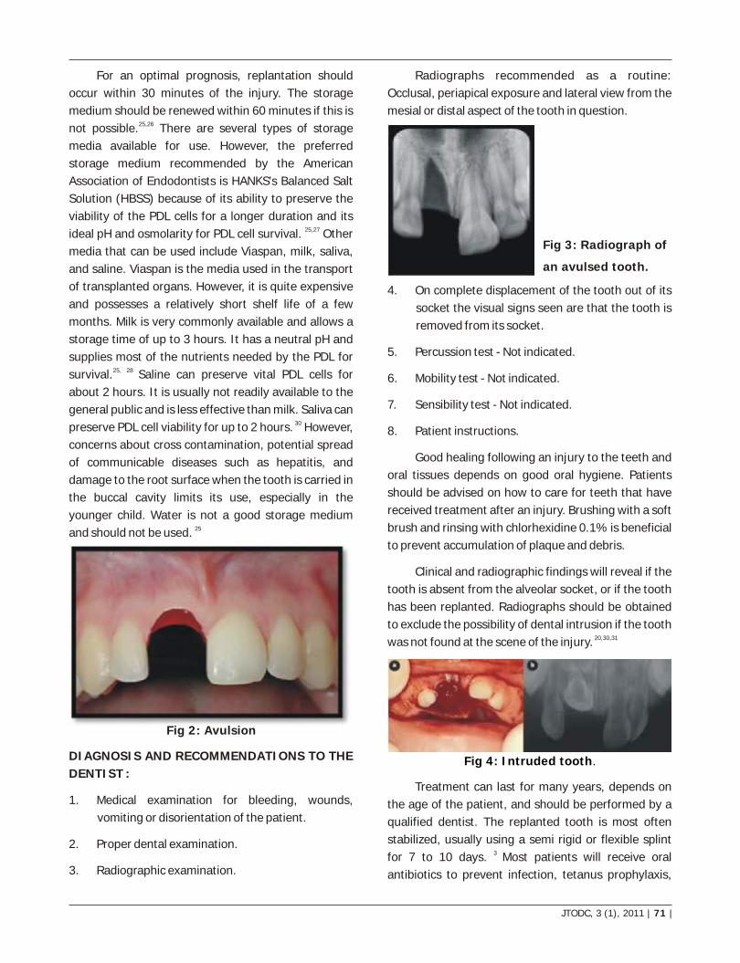
For an optimal prognosis, replantation should
occur within 30 minutes of the injury. The storage
medium should be renewed within 60 minutes if this is 25,26not possible. There are several types of storage
media available for use. However, the preferred
storage medium recommended by the American
Association of Endodontists is HANKS's Balanced Salt
Solution (HBSS) because of its ability to preserve the
viability of the PDL cells for a longer duration and its 25,27ideal pH and osmolarity for PDL cell survival. Other
media that can be used include Viaspan, milk, saliva,
and saline. Viaspan is the media used in the transport
of transplanted organs. However, it is quite expensive
and possesses a relatively short shelf life of a few
months. Milk is very commonly available and allows a
storage time of up to 3 hours. It has a neutral pH and
supplies most of the nutrients needed by the PDL for 25, 28 survival. Saline can preserve vital PDL cells for
about 2 hours. It is usually not readily available to the
general public and is less effective than milk. Saliva can 30preserve PDL cell viability for up to 2 hours. However,
concerns about cross contamination, potential spread
of communicable diseases such as hepatitis, and
damage to the root surface when the tooth is carried in
the buccal cavity limits its use, especially in the
younger child. Water is not a good storage medium 25and should not be used.
DIAGNOSIS AND RECOMMENDATIONS TO THE
DENTIST :
1. Medical examination for bleeding, wounds,
vomiting or disorientation of the patient.
2. Proper dental examination.
3. Radiographic examination.
Fig 2: Avulsion
Radiographs recommended as a routine:
Occlusal, periapical exposure and lateral view from the
mesial or distal aspect of the tooth in question.
Fig 3: Radiograph of
an avulsed tooth.
4. On complete displacement of the tooth out of its
socket the visual signs seen are that the tooth is
removed from its socket.
5. Percussion test - Not indicated.
6. Mobility test - Not indicated.
7. Sensibility test - Not indicated.
8. Patient instructions.
Good healing following an injury to the teeth and
oral tissues depends on good oral hygiene. Patients
should be advised on how to care for teeth that have
received treatment after an injury. Brushing with a soft
brush and rinsing with chlorhexidine 0.1% is beneficial
to prevent accumulation of plaque and debris.
Clinical and radiographic findings will reveal if the
tooth is absent from the alveolar socket, or if the tooth
has been replanted. Radiographs should be obtained
to exclude the possibility of dental intrusion if the tooth 20,30,31was not found at the scene of the injury.
Fig 4: Intruded tooth.
Treatment can last for many years, depends on
the age of the patient, and should be performed by a
qualified dentist. The replanted tooth is most often
stabilized, usually using a semi rigid or flexible splint 3 for 7 to 10 days. Most patients will receive oral
antibiotics to prevent infection, tetanus prophylaxis,

| 70 | JTODC, 3 (1), 2011
appropriate oral hygiene, and diet recommendations.
Potential and existing complications determine the 5timing for follow-up.
Management of the avulsed permanent tooth
According to the treatment guidelines given by
the International Association of Dental Traumatology 28(IADT),
1. TREATMENT OF AVULSED PERMANENT
TEETH WITH CLOSED APEX : If the avulsed tooth
has been brought to the dental clinic within the extra
oral dry time of 60 minutes stored in a storage medium
then; the area needs to be cleaned with water spray,
saline, or chlorhexidine and the gingival lacerations
should be sutured if present. The alveolar socket
should be examined for the fracture and the avulsed
tooth is repositioned with the help of a suitable
instrument. If the avulsed tooth has been kept in the
storage medium and brought to the dental clinic
beyond the extra-oral dry time of 60 minutes then the
delayed replantation is done but it has a poor long-
term prognosis. The periodontal ligament will be
necrotic and not expected to heal. The goal of doing
delayed replantation is to promote alveolar bone
growth to encapsulate the replanted tooth. In delayed
replantation the Root canal therapy can be done on the
tooth prior to replantation, or it can be done 710 days
later as for other replantations. The tooth has to be
immersed a 2% sodium fluoride solution for 20 min.
The tooth has to be replanted slowly with slight digital
pressure. Normal position of the replanted tooth
should be verified both clinically and radiographically.
A flexible splint for up to 2 weeks is placed. Systemic
antibiotics are administered. Tetracycline is the first
choice (Doxycycline 2x per day for 7 days at
appropriate dose for patient age and weight). The risk
of discoloration of permanent teeth should be
considered before systemic administration of
tetracycline in young patients. (In many countries
tetracycline is not recommended for patients under 12
years of age). In young patients Phenoxymethyl
Penicillin (Pen V), in an appropriate dose for age and
weight, can be given as alternative to tetracycline. The
expected eventual outcome of delayed replantation is
ankylosis and resorption of the root. In children below
the age of 15, if ankylosis occurs, and when the
infraposition of the tooth crown is more than 1 mm, it
is recommended to perform decoronation to preserve
the contour of the alveolar ridge.
Calcium hydroxide as an intra-canal medicament
is placed until filling of the root canal is completed. The
Patient is instructed to have a soft diet for up to 2
weeks and brush teeth with a soft toothbrush after
each meal and use a chlorhexidine (0.1%) mouth rinse
twice a day for 1 week and follow-up.
2. TREATMENT OF AVULSED PERMANENT
TEETH WITH OPEN APEX : If the avulsed tooth has
been kept in the storage medium and brought to the
dental clinic within the extra-oral dry time of 60
minutes then the area has to be cleaned with water
spray, saline or chlorhexidine. If contaminated, clean
the root surface and apical foramen with a stream of
saline. Remove the coagulum from the socket with a
stream of saline and then replant the tooth. If
available, cover the root surface with minocycline
hydrochloride microspheres before replanting the
tooth. Suture gingival lacerations if present.
Reposition it with a suitable instrument and verify
normal position of the replanted tooth both clinically
and radiographically. Apply a flexible splint for up to 2
weeks. Administer systemic antibiotics. For children 12
years and younger: Penicillin V at an appropriate dose
for patient age and weight. Refer the patient to a
physician for evaluation of need for a tetanus booster if
avulsed tooth has contacted soil or tetanus coverage is
uncertain. The goal for replanting still-developing
(immature) teeth in children is to allow for possible
revascularization of the tooth pulp. If that does not
occur, root canal treatment may be recommended
through the open apex prior to the replantation and
then patient is instructed to have a soft diet for up to 2
weeks and to brush teeth with a soft toothbrush after
each meal and recommend the use of chlorhexidine
(0.1%) mouth rinse twice a day for 1 week and follow-28, 29up.
COMPLICATIONS
Complications after replantation of avulsed teeth
are common and have a reported prevalence rate 33ranging from 57% to 80%, though reported to be as
high as 84% in one study. Certain complications,
including ankylosis (lack of mobility of the tooth),
JTODC, 3 (1), 2011 | 71 |
excessive mobility of the tooth, and resorption, may
occur during the next several years of dental follow-
up. Most replanted teeth will be lost in 5 to 7 years 34even if a root canal is completed after replantation.
CONCLUSION
There is in general a lack of knowledge in the
management of avulsion among the lay population.
This awareness could be raised by promotional
campaigns. The outcome of treatment depends on the 35 extent of injury, the stage of root. Dental treatment
should be performed only after potential life
threatening injuries such as neurological damage,
bleeding or aspiration of foreign bodies/teeth, are
treated or excluded. The only injury that requires
immediate treatment is tooth avulsion. Readily
available transporting media could make a big
difference in the outcome of replantation. Prophylactic
antibiotic cover is useful in the following situations:
Uncertain tetanus status, root fracture, replantation
and excessive soft tissue contamination or laceration.
Follow up plays an important role in the prognosis of 36, 37the replanted tooth.
REFERENCES
1. Chan A.W.K, Wong T.K.S, Cheung G.S.P et al. Lay
knowledge of physical education teachers about
the emergency management of dental trauma in
Hong Kong. Dent Traumatol 2001, 17 ;2 :77-
85(9).
2. Dental injuries. II. Avulsion of permanent teeth.
Dental Traumatol 2007; 23: 130136.
3. American Academy on Pediatric Dentistry Council
on Clinical Affairs. A guideline on management of
acute dental trauma. Pediatr Dent 2008;30(7
Suppl):175-83.
4. Lieger O, Graf C, El-Maaytah M, et al. Impact of
educational posters on the lay knowledge of
school teachers regarding emergency
management of dental injuries. Dent Traumatol
2009; 25:406-12.
5. Pohl Y., Filippi A. & Kirschner H et al. Results after
replantation of avulsed permanent teeth. II.
Periodontal healing and the role of physiologic
storage and antiresorptive- regenerative
therapy. Dent Traumatol, 2005, 21(2):93-101.
6. Al-Asfour A, Andersson L, Al-Jame Q. School
teachers' knowledge of tooth avulsion and dental
first aid before and after receiving information
about avulsed teeth and replantation. Dent
Traumatol 2008;24:43-9.
7. Andreasen JO, Andreasen FM, Andersson L et al.
Textbook and color atlas of traumatic injuries to
the teeth, 4th edn. Oxford: Blackwell
Munksgaard; 2007.
8. Petersson EE, Andersson L, Sorensen S et al.
Traumatic oral vs non-oral injuries. Swed Dent J
1997; 1 :5568.
9. Glendor U, Holling A, Andersson L et al. Incidence
of traumatic tooth injuries in children and
adolescents in the county of Vastmanland,
Sweden. Swed Dent J 1996;20:15-28.
10. Ivancic Jokic N, Bakarcic D, Fugosic V et al.
Dental trauma in children and young adults
visiting a university dental clinic. Dent Traumatol
2009;25:84-7.
11. Andreasen JO et al. Etiology and pathogenesis of
traumatic dental injuries. A clinical study of 1,298
cases. Scand J Dent Res 1970;78:329-42.
12. Tzigkounakis V, Merglova V, Hecova H et al.
Retrospective clinical study of 90 avulsed
permanent teeth in 58 children. Dental
Traumatol 2008;24:598-602.
13. Glendor U et al. Epidemiology of traumatic dental
injuriesa 12 year review of the literature. Dent
Traumatol 2008;24: 603-11.
14. Bruns T, Perinpanayagam H et al. Dental trauma
that require fixation in a children's hospital. Dent
Traumatol 2008;24: 59-64.
15. Wendt FP, Torriani DD, Assuncao MCP, et al.
Traumatic dental injuries in primary dentition:
epidemiological study among preschool children
in South Brazil. Dent Traumatol 2010;26:168-73.
16. Newsome P.R, Tran D.C & Cooke M.S et al. The
role of the mouth guard in the prevention of

| 70 | JTODC, 3 (1), 2011
sports-related dental injuries: a review. Int J
Paediatr Dent 2001;11:396-404.
17. Jorge KO, Moyses SJ, Ferreira E, et al. Prevalence
and factors associated to dental trauma in infants
1-3 years of age. Dent Traumatol 2009;25:185-9.
18. Bastone EB, Freer TJ, McNamara JR et al.
Epidemiology of dental trauma: a review of the
literature. Aust Dent J 2000; 45:2-9.
19. Flores MT et al. Traumatic injuries in the primary
dentition. Dent Traumatol 2002;18:287-98.
20. Eyuboglu O, Yilmaz Y, Zehir C, et al. A 6-year
investigation into types of dental trauma treated
in a paediatric dentistry clinic in Eastern Anatolia
region, Turkey. Dent Traumatol 2009;25:110-4.
21. Santos ME, Habecost AP, Gomes FV, et al.
Parent and caretaker knowledge about avulsion
of permanent teeth. Dent Traumatol 2009 ; 25:
203-8.
22. Oliviera TM, Sakai VT, Moretti AB, et al.
Knowledge and attitude of mothers with regards
to emergency management of dental avulsion. J
Dent Child 2007;74:200-2.
23. Qazi SR, Nasir KS. First-aid knowledge about
tooth avulsion among dentists, doctors and lay
people. Dent Traumatol 2009;25:295-9.
24. Sigalas E, Regan JD, Kramer PR, et al. Survival of
human periodontal ligament cells in media
proposed for transport of avulsed teeth. Dent
Traumatol 2004;20:21-8.
25. Cvek M, Granath LE, Hollender L et al. Treatment
of non-vital permanent incisors with calcium
hydroxide. 3. Variation of occurrence of ankylosis
of reimplanted teeth with duration of extra-
alveolar period and storage environment.
Odontol Rev 1974;25:43-56.
26. American Association of Endodontists.
Recommended guidelines of the American
Association of Endodontists for the treatment of
traumatic dental injuries - 2004. Availableat :
http://www.aae.org/NR/rdonlyres/73E3698B-
CABB-4D80-B4C4 A9EE07ECDAC9 / 0 / 2004
Trauma Guidelines . pdf Accessed 10/2010.
27. http://www.iadt-dentaltrauma.org Accessed on
3/2011, 2:00pm.
28. Blomlof L et al. Milk and saliva as possible storage
media for traumatically exarticulated teeth prior
to replantation. Swed Dent J 1981(8 Suppl):1-
26.
29. Flores MT, Andersson L, Andreasen JO et al.
Guidelines for the management of traumatic
dental injuries. II. Avulsion of permanent teeth.
Dent Traumatol 2007;23:130-6.
30. McTigue DJ et al. Managing injuries to the
primary dentition. Dent Clin North Am
2009;53:627-38.
31. Lin S, Zuckerman O, Fuss Z et al. New emphasis
in the treatment of dental trauma: avulsion and
luxation. Dent Traumatol 2007;23:297-303.
32. Andreasen JO, Malmgren B, Bakland LK. Tooth
avulsion in children: to replant or not. Endodon
Topics 2006;14:28-34.
33. Petrovic B, Markovic D, Peric T, et al. Factors
related to treatment and outcomes of avulsed
teeth. Dent Traumatol 2010;26:52-9.
34. Glendor U, Halling A, Andersson L, Eilert-3.
Petersson E et al. Incidence of traumatic tooth
injuries in children and adolescents in the county
of Vastmanland, Sweden. Swed Dent J
1996;20:1528.
35. Bruns T, Perinpanayagam H et al. Dental trauma
that require fixation in a children's hospital. Dent
Traumatol 2008;24: 59-64.
36. Ritter AV et al. Talking with patients. Dental
trauma (avulsed teeth). J Esthet Restor Dent
2004;16:267-8.

P. Latha Subramanya
IS HUMAN BREAST MILK CARIOGENIC?
ABSTRACT
Keywords :
In dentistry, there is quasi-consensus that
breastfeeding on demand, especially at night and if
prolonged, produces dental caries .There are no scientific
evidences proving that the human milk can be associated
with the development of caries. This is a complex relation to
be established, as it is often blurred by too many variables.
Objective of this article is to note the scientific evidences that
can prove or refute the assumption that nocturnal and on
demand breastfeeding are associated with caries in infants
and preschool children.
Early childhood caries, dental decay,
breastfeeding, risk-factors
Review Article
Department of Pedodontics,The Oxford Dental College, Hospital and Research Center, Bangalore.
Reader
INTRODUCTION
Dental caries is still one of the most common
diseases in the world today. Rampant caries in infants
and young children has long been recognized as a
clinical syndrome, which was described as early as the
middle of the last century. The typical causative triad
for caries consists of cariogenic microorganisms,
fermentable carbohydrates and a susceptible host, but
a multitude of risk factors are involved in ECC
development. ECC has been associated with
socioeconomic status (SES), parental education,
maternal nutrition, psychosocial issues and parenting
practices.The expression early childhood caries (ECC)
is currently used to replace the terms baby-bottle
tooth decay and nursing caries. The American
Academy of Pediatric Dentistry (AAPD) recognizes
early childhood caries (ECC; formerly termed “nursing
Journal Of The Oxford Dental College
Email for [email protected]
JTODC, 3 (1), 2011 | 1 |
bottle caries”, “baby bottle tooth decay”) as a 1significant public health problem. AAPD adopted the
term “early childhood caries” to reflect better its
multifactoral etiology The disease of ECC has been
defined as “the presence of 1 or more decayed
(noncavitated or cavitated lesions), missing (due to
caries), or filled tooth surfaces” in any primary tooth in
a child 71 months of age or younger.5,6 In children
younger than 3 years of age, any sign of smooth-
surface caries is indicative of severe early childhood
caries (S-ECC). From ages 3 through 5, 1 or more
cavitated, missing (due to caries), or filled smooth
surfaces in primary maxillary anterior teeth or a
decayed, missing, or filled score of =4 (age 3), =5 (age 1,24), or =6 (age 5) surfaces constitutes S-ECC
In dentistry, there is quasi-consensus that
breastfeeding on demand, especially at night and if 3,prolonged, produces caries. likewise, in pediatrics,
there are publications that share the same opinion.
The American Academy of Pediatric Dentistry (AAPD)
declared that breastfed and bottle-fed infants are at a
potentially devastating risk for caries due to
breastfeeding. This is related to prolonged and
repetitive feeding without proper oral hygiene, and is
also related to the fact that parents are encouraged to
offer their infants beverages in drinking cups before
their first year of life and to stop bottle-feeding 3between 12 and 14 months of life. Without an
accurate definition, the terms “prolonged exposure”
and “weaning” had different interpretations, which
culminated in the recommendation, by dentists, of
weaning and cessation of breastfeeding by the first
year of life . By discouraging prolonged breastfeeding 4and breastfeeding on demand, they overlook all the
well-documented benefits of breastfeeding and also
t he Wor l d Hea l t h O rgan i za t i on (WHO)
recommendation to maintain breastfeeding up to the 5second year of life or longer. Similarly, the American
Academy of Pediatrics considers that infants who are
put to bed with the bottle or who are breastfed during , 6the night are at great risk for dental caries. Therefore,
the presumable cariogenicity of breastmilk is an issue
of paramount importance because, along with its
substitutes, it is the major nutritional source during 7, 8infancy.
BREASTFEEDING VERSUS ECC: REASONS AND
COUNTERARGUMENTS
Breast milk is characterized by complex
constituents like IgA antibodies oligosaccharides ,
lactoferrin and hormones that may confer immunity
to the infant.Most authors argue that caries is
associated with breastfeeding when the consumption
pattern has certain characteristics such as ad libitum
feeding, large number of breast feedings a day,
prolonged breastfeeding and, mainly, frequent breast
feedings during the night, resulting in accumulation of
milk in the teeth, which, combined with reduced
salivary flow and lack of oral hygiene, may produce
tooth decay. In opposition to these arguments is the
fact that breast milk expressed directly into the soft
palate does not stagnate while being sucked and the
volume ingested by the infant is difficult to be 9quantified.
| 70 | JTODC, 3 (1), 2011
ANTHROPOLOGY DATA
Studies involving primitive cultures, in which the
rule was to breastfeed on demand, including
breastfeeding at night, up to 18-36 months, show an
extremely low prevalence of caries among children.
Classical studies show caries rates of 0.5% in Samoan 10 11infants and of 1.2% in Eskimo infants. Similar
results were obtained from anthropological studies, in
which preneolithic (12,000 B. C.) human skulls did not
reveal dental caries and neolithic (12,000 to 3,000 B.
C.) skulls with decayed teeth belonged predominantly
to old people. The analysis of 1,344 prehistorical
human deciduous teeth of Native Americans from
South Dakota, USA, revealed that only 19 (1.4%) were
decayed and only three had extensive carious lesions.
In modern times, the prevalence observed in 16 Native
American communities, with the same culture, was 1254%.
People that maintain ancestral eating habits have
a low prevalence of caries; however, when they come
into contact with modern civilization and its eating
habits, the prevalence rate increases drastically
Children's eating habits have dramatically
changed in the last years. Milk consumption has
decreased whereas the consumption of soft drinks,
juices, non-citric beverages and carbohydrates has 13increased. These habits have been correlated with a
higher prevalence of ECC.
DEFENSE SYSTEM OF HUMAN BREAST MILK
Studies have demonstrated that breast milk
reduces the risk of diseases, such as gastroenteritis,
infections, asthma, atopic diseases and diabetes 14, 15 mellitus, which have some influence on infant
feeding. Therefore, breastmilk supposedly protects
against caries, due to the reduced development of
disorders that contribute to the pathophysiology of
caries and due to the reduced use of cariogenic
medications.
Human milk is characterized by a complex
defense system that inhibits the growth of several 16microorganisms, including mutans streptococci. The
IgA antibodies found in the milk can interfere with the
colonization of pioneer streptococci and consequently
with the colonization of other bacteria that inhabit the

JTODC, 3 (1), 2011 | 1 |
oral cavity. Nutrient content, buffering capacity and
other defense mechanisms found in breastmilk may 17 [interfere with the existing microbiota Fig 1]
Breastmilk contains a mix of oligosaccharides
that is complex and exclusive to the human species,
found in tiny amounts in very few mammals, which
may act at the initial infectious stage by inhibiting 17bacterial adhesion to epithelial surfaces. Therefore,
studies that used lactose or milk from other animal
species may not have their results extrapolated to
breastmilk due to its distinct composition.
Qualitatively, it has been demonstrated that
human milk is not cariogenic, as the dental plaque it
forms is different from that formed by sucrose. In
addition, human milk does not cause clinically visible
mineral loss in the enamel, contrary to what occurs
with sucrose.
Experimentally, it has been shown that human
milk is not cariogenic because it does not decrease the
enamel pH significantly in breastfed infants, aged
between 12 and 24 months; allows moderate growth
of Streptococcus sobrinus (i.e., it does not inhibit or
stimulate the growth of this microorganism);
promotes enamel remineralization by way of calcium
and phosphate deposition on the enamel surface; has
a poor buffering capacity; and does not cause in vitro
enamel decalcification after twelve weeks. However,
when sucrose is added to human milk, caries
developed in the dentin within 3.2 weeks.

OFFICIAL VIEW OF THE AMERICAN ACADEMY
OF PEDIATRIC DENTISTRY (AAPD)
Cu r r en t l y, t h e AAPD suppo r t s t h e
recommendations made by the American Academy of
Pediatrics regarding breastfeeding (of at least one 1 year). However, it states that frequent feeding at night
including bottle-feeding, breastfeeding on demand
and frequent use of spill-proof drinking cups is
associated with ECC, but is not consistently implicated.
It recommends that infants should not be put to bed
with the baby bottle and that ad libitum breastfeeding
at night should be avoided after the eruption of the
first tooth. Therefore, future research should be
conducted about the effects of breastfeeding and
human milk consumption on oral health and on
dentofacial growth. This view is ambiguous and
arguable because the AAPD no longer includes
breastfeeding among cariogenic factors. Moreover, no
scientific evidence exists that human milk is
cariogenic, even if ingested ad libitum and during the
night. Concomitantly, the view of the AAPD can also
result in practical problems regarding the counseling
and guidance of parents of those infants who wake up
crying at night in order to be breastfed, simply
expressing a need that should be met for infants'
proper development. Finally, given the irreversible
nature of caries, an actual test involving humans could
be regarded as unethical.
CONCLUSION
Although there is no scientific evidence that
confirms the association between breastfeeding and
caries, many professionals still express disbelief at the
fact that human milk is non-cariogenic, thus cultivating
the myth into which this association turned. Exclusive
breastfeeding should be encouraged up to the sixth
month of life, maintained at least up to the second year
of life, with flexibility of schedules or shifts, and
complemented with appropriate weaning foods.
There is no scientific evidence that confirms that
breastmilk is associated with caries development. This
relationship is complex and contains several
confounding variables, mainly infection caused by
mutans streptococci, enamel hypoplasia, intake of
sugars in varied forms and social conditions
represented by parental educational and
socioeconomic level. The AAPD also acknowledges the
need for further scientific research in regards to the 18effects of breast-feeding on oral health
REFERENCES
1. American Academy of Pediatric Dentistry.
Symposium on the prevention of oral disease in
children and adolescents. Chicago, Ill; November
11-12, 2005: Conference papers. Pediatr Dent
2006;28(2);96-198
2. 2. Caufield PW, Griffen AL. Dental Caries. An
infectious and transmissible disease. Pediatr Clin
North Am. 2000;47:1001-19.
3. American Academy of Pediatric Dentistry. Oral
health polices. Baby bottle tooth decay/early
childhood caries. Reference Manual 1999-2000.
Pediatr Dent. 2000;21:18-9.
4. Valaitis R, Hesch R, Passarelli C, Sheehan D,
Sinton J. A systematic review of the relationship
between breastfeeding and early childhood
caries. Can J Public Health. 2000;91:411-7.
5. World Health Organization. Dentition status and
criteria for diagnosis and coding (Caries). WHO
Oral Health Surveys Basic Methods. 4th ed.
Geneva: WHO; 1997. p. 39-44.
6. Nainar SMH, Mohummed S. Dental health of
children. Clin Ped. 2004;43:129-33.
7. Slavkin HC. Streptococcus mutans, early
childhood caries and new opportunities. J Am
Dent Assoc. 1999;130:1787-92.
8. Weerheijm KL, Uyttendaele-Speybrouck BFM,
Euwe HC, Groen HJ. Prolonged demand breast-
feeding and nursing caries. Caries Res.
1998;32:46-50.
9. Cartwright A. Breast is best. Letter to British
Dental Journal 2008; 204:351-352
10. Restarski JS. Incidence of dental caries among
pure-blooded Samoans. US Naval Med Bull.
1943;41:1713-15.
| 70 | JTODC, 3 (1), 2011

11. Rosenbury T, Karshan M. Dietary habits of
Kuskokwim Eskimos with varying degrees of
dental caries. J Dent Res. 1937;16:307-9.
12. Bruerd B, Kinney MB, Bothwell E. Preventing
baby bottle tooth decay in American Indian and
Alaska Native communities: a model for
planning. Publ Health Rep. 1989;104:631-40.
13. Marshall TA, Levy SM, Broffitt B, Warren JJ,
Eichenberger-Gilmor JM, Burns TL, et al. Dental
caries and beverage consumption in young
children. Pediatrics. 2003 Sep;112(3 Pt 1):
e184-91.
14. Oddy WH, Peat JK. Breastfeeding, asthma, and
atopic disease: an epidemiological review of the
literature. J Hum Lact. 2003;19:250-61.
JTODC, 3 (1), 2011 | 1 |
15. Gimeno SG, Souza JM. IDDM and milk
consumption. A case-control study in Sao Paulo,
Brazil. Diabetes Care.1997;20:1256-60.
16. Marcotte H, Lavoie MC. Oral microbial ecology
and the role of salivary immunoglobulin A.
Microbiol Mol Biol Rev. 1998;62:71-109.
17. Kunz C, Rodrigues-Palmero M, Koletzko B, Jensen
R. Nutritional and biochemical properties of
human milk, Part I: General aspects, proteins,
and carbohydrates. Clin Perinatol. 1999;26:307-
33.
18. American Academy of Pediatric Dentistry,
Po l i cy on b reas t- feed ing . Ava i l ab le
fromURL:http://aappolicy.aappublications.org/c
gi/reprint/pediatrics;115/2/496.pdf. Accessed
July 23, 2008

a b c dDr. Naganandini. S , Dr. Sanjeev. K , Dr. Vanishree. N , Dr. Uday. P
TELEDENTISTRY : THE FUTURE OF DENTISTRY
ABSTRACT
Keywords :
Teledentistry is a relatively new field that combines
telecommunication technology and dental care. The initial
concept of teledentistry developed as part of dental
informatics (a new domain combining computer and
information science, engineering and technology in all areas
of oral health). In remote clinics, a patient must travel
hundreds of miles to receive specialty care. With the
implementation of teledentistry, there is a potential of saving
in cost and travel time required by the patient. Through the
use of video teleconferencing equipment, the lectures could
be broadcasted to any clinic where continuing dental
education is difficult to obtain. Needs assessment,
infrastructure and standardizations, telecommunication
options, costs, applicable computer hardware and software,
training of personnel a well designed teledentistry practice
needs to consider all of these issues. With thorough planning,
however, teledentistry has a bright future.
Teledentistry, Dental informatics,
Telecommunication, Telehealth
Review Article
Department of Public Health Dentistry,The Oxford Dental College, Hospital and Research Center, Bangalore.
cProfessorbHOD ,
a,dStudent
INTRODUCTION
Teledentistry is a relatively new adjunct in the
modern trend of telemedicine. Telemedicine, simply
put, is the clinical application of providing care at a distance. A variety of medical specialties now utilize
telemedicine in one form or another. Teleradiology,
telepathology, and telepharmacy were some of the
earliest telemedicine specialties and continue to be the 1most common applications of telemedicine today.
Teledentistry is a combination of telecommunications
Journal Of The Oxford Dental College
Email for [email protected]
JTODC, 3 (1), 2011 | 1 |
and dentistry which involves the exchange of clinical information and images over remote distances. Most
teledentistry programs to date have focused upon
distance management and administration of remote
facilities, learning and continuing education, and
consultation and referral services rather than
supervision of auxiliaries or direct patient care. Its
initial concept was developed as a part of Dental
informatics and defined as a set of establishing
technologies which allows for the diagnosis and
treatment of patients at sites remote from the
healthcare providers. Traditionally, this includes the

| 70 | JTODC, 3 (1), 2011
transfer of still or video images and data over
communications lines from a distant site for
consultation and evaluation by dental specialists. It
also includes remote education of both patients and 2dental providers.
FORMS OF TELEDENTISTRY:
Teledentistry can take two forms: “real-time
consultation” and “store and forward.”
Re a l - t i m e c o n s u l t a t i o n i n v o l v e s a
videoconference in which dental professionals and
their patients, at different locations, may see, hear,
and communicate with one another in real time using
advanced telecommunication technology and ultra-
high-bandwidth network connections. Store and
forward, on the other hand, involves the exchange of
clinical information and static images collected and 3stored in the telecommunication equipment.
In store and forward, the dental practitioner
collects all the required clinical information and digital
intraoral and extra-oral images and radiographs (or
scanned, originally non-digital images) and forwards
them for consultation and treatment planning via
established networks and/or the Internet. Later,
dentists are dispatched to the remote areas and
treatment provided in a far more timely, targeted, and 3cost-effective manner.
USES OF TELEDENTISTRY:
1. Patient Care: In remote clinics, a patient must travel
hundreds of miles to receive specialty care. With the
implementation of teledentistry, there is a potential of
saving in cost and travel time required by the patient.
2. Continuing Dental Education:
Through the use of video teleconferencing
equipment, the lectures could be broadcasted to any
clinic where continuing dental education is difficult to
obtain.
3. Dentist-Laboratory Communications:
The ability to send colour images of the patient's cases
and talk about it can help to prevent making
improperly constructed appliances thereby saving
time and money.
4. Second Opinion:
A dentist's judgment rendered to a patient is
limited by his ability to make diagnosis when a dental
practitioner is unsure of the diagnosis; it is best to seek 4another opinion.
TELEDENTAL EQUIPMENT
Teledentistry sites require some or all of the
following basic equipment. The exact equipment
required will depend on the nature of the site being
outfitted. Hub sites will be different from remote sites
hub sites will only require videoconferencing
equipment, whereas remote sites will require digital
denta l d iagnost ic equipment a long with
videoconferencing equipment. Moreover, mobile sites
may require different transmission equipment than
fixed sites (e.g. satellite dishes and modems). The
equipment listed does not include telecommunication
service equipment (e.g. routers, switches, T-1 lines,
etc.). The nature of the telecommunication equipment
will depend on the type of telecommunication service
utilized by each site. Further, the teledental equipment
does not include the equipment or supplies needed to
outfit a traditional dental unit with traditional dental
(which will be necessary in the remote sites). One
other note, there are a variety of manufacturers and
models. Thus, there are a variety of choices for each 5individual piece of equipment.
VIDEOCONFERENCING SYSTEM
The videoconferencing system should include: a
CODEC unit with a pan tilt video camera, monitor
(preferably one that can split screen or comes as two

JTODC, 3 (1), 2011 | 1 |
monitor units), mobile cart with shelf for laptop
computer or keyboard, back-up battery, input and
output connections, and have the ability to encrypt
and unencrypt data. Tandberg and Polycom are the
leaders in this industry.
Extra-oral Digital Camera
Extra-oral cameras are good for face, smile, arch,
and anterior teeth images. There are 3 types of digital
cameras: point and shoot, professional, and modified
point and shoots. Point and shoot are typically off-the-
shelf and can take good portrait photo images, but
have limited close-up and intraoral capabilities.
Professional level SLR cameras are top of the line
single lens reflex camera bodies that allow for the
addition of an assortment of lens and flash
attachments. While these can take the most accurate
images, they are the most difficult to manage.
Modified point and shoots are off-the-shelf cameras
that have been modified for dentistry. These cameras
have added hardware (e.g. macro lens and flashes) to
improve the macro capability of the camera and the
ability of the flash to disperse or expose correctly 5under macro conditions.
Extra-oral cameras should be able to capture
colour and have sufficient image resolution capacity.
Extra-oral cameras should include: at least 4
megapixel resolution, be capable of faster shutter
speeds, have through the lens viewing (SLR) or an LCD
monitor (for direct viewing for accurate, repeatable
framing alignment), manual focus macro lens (at least
3x optical zoom), dual point lighting (e.g. ring flash or
flash diffuser to distribute light evenly standard
flashes create washout), glass lens rather than plastic,
manual focus and f-stop (aperture-size) settings (for
consistent, repeatable results), a video-out port to
download to a monitor and/or a USB hub to download
to a computer, and selectable compression/resolution
levels for final imaging. Moreover, the camera system
should include mirrors and retractors (an occlusal
mirror, a buccal No. 1 mirror, and universal retractors),
and computer dental software that supports viewing
the images, storing the images, and editing the 5images.
INTRAORAL WAND DIGITAL CAMERA
Intraoral wand cameras are good for diagnostic
purposes because of the high intensity light they utilize
and the magnification they can produce. They are
good for individual teeth, for hard to reach areas and
where light is difficult (posterior areas), and are
excellent for locating early white spot lesions. Intraoral
wand cameras can take still images or video. Intraoral
wand cameras should be lightweight (to reduce
operator fatigue), have focus-free optics, integrate
seamlessly with other dental digital software
packages, utilize LED lighting (rather than fragile fibre-
optic or fan cooled lighting sources), and they should
come with USB or fire wire interfaces (for direct 5connectivity to a computer)
DIGITAL RADIOGRAPHIC EQUIPMENT
There are 3 ways to obtain digitized radiographs:
1) converting traditional film radiographs to digital
(digitized) images via a scanner the digitized images
are then transferred to and viewed on a computer; 2)
using phosphor plate technology a radiograph is
taken using a phosphor plate (instead of film) to store
the image, the plate is then scanned by a laser to a
produce an image which can be viewed on a
computer; and 3) using sensor or direct digital
technology radiographs are taken using digital
sensors the image is immediately shown on a
computer screen. The scanner and phosphor plate
technologies are slower, but less expensive. The
sensor technology is real time.
Computer
Every teledentistry site, whether a remote or hub
site, must be equipped with a computer. The computer
can be either a laptop or desktop computer. No matter
the type or brand of computer it must have the flowing
minimum requirements: CPU Speed: 2.8 GHz Pentium
4;
Operating System: Windows 2000 Service Pack 4
or Windows XP Service Pack 1 w/Microsoft knowledge
base KB822603 update; System RAM: 1 KB; Hard
Drive: 80 GB; CD-ROM Drive: 48x; Video Display 5Adapter: 64 MB RAM; and USB Port: Must be USB 2.0.

| 70 | JTODC, 3 (1), 2011
REVIEW OF LITERATURE:
The Department of Defence initiated the “Total
Dental Access” project in 1994. The Total Dental
Access project focused on three areas of dentistry:
patient care including referrals to specialists and
consultations; continuing education; and dental-
laboratory communications. The project utilized
multiple transfer technologies including image file
transfers by modem, image file transfers by satellite,
ISDN-based (Integrated Services Digital Network)
technology, POTS-based (Plain Old Telephone Service)
technology, and web-based technology. An analysis of
the Teledentistry project concluded that teledentistry
demonstrated was cost-effective within 6 months to a
year of initiation and that teledentistry improved
access and quality of care by facilitating better and
timely information to the dentists which improved
decision making and produced better communication 6between the dentists and their patient.
Marquette University School of Dentistry initiated
the Marquette University Dental Telehealth and
Education Link in 2003. The project aimed to create a
network linking Marquette and other health systems
with dental sites in remote areas where access to care
is problematic. The project utilized both store and
forward and interactive technology for the purposes of
primary care, consultation, education, and public
awareness programs. The Wisconsin Advanced
Telecommunications Foundation (WATF) was the
major funding agency on this project, but the
Milwaukee Area Health Education Centre and
Wisconsin Geriatric Education Centre also were 7sponsors.
In 2003 the Children's Hospital Los Angeles
Teledentistry Program began a store and forward
teledentistry program. Initially this program was run in
association with the University of Southern California
School of Dentistry (USCSD) Mobile Dental Clinic (see
below). This on-going program provides enhanced
dental treatment to children in rural, remote, 8underserved areas of California.
The USCSD mobile clinic was the first non-
military dental clinic in the United States to utilize
digital imaging and the Internet to diagnose and
treatment plan patients in remote locations. The
Harold McAlister Charitable Foundation and The
California Wellness Foundation (TCWF) grant funded
this project. While the mobile clinics continue to
operate, they no longer utilize teledentistry at this 9time.
In 2004, the State of Minnesota Department of
Health in conjunction with the University Of Minnesota
School Of Dentistry and the Hibbing Community
College Dental Clinic sponsored a Teledentistry
Project. The project continues to utilize direct
videoconferencing to create a telecommunication
network linking the University of Minnesota School of
Dentistry's specialists with dentists and dental
students in sites in remote rural areas where access to 10care is problematic.
The University of Rochester Medical Centre's
Eastman Dental Centre in association with Aetna
insurance established the Teledentistry in Childcare
Project in 2005. This project helps inner city families
easily access the oral health treatment they need for
their children in childcare. The project's goal is to
develop a new strategy for the prevention and early
detection of early childhood caries (ECC). The project
utilizes a computer and camera which allow dentists to 11examine and interact with a child in real-time.
In the summer of 2005, the University Of
Washington School Of Dentistry's Paediatric Dentistry
residency program began a videoconferencing project
based at a remote site at the Farm Workers Clinic in
Yakima Valley. The project entails both a distance
learning educational component, using both store and
forward and live videoconferencing capabilities, and a
clinical consultation component, which allows live
video consultations chair side. The project utilizes an
intra-oral camera in Yakima linked to the 12videoconferencing system in Seattle via the internet .
The University of Tennessee's Mid-South
Telehealth Consortium (MSTC) in collaboration with
the Tennessee Department of Health initiated a Mobile
Healthcare Telehealth Project in 2002. The
Department of Health and Human Services through
the USDA: Rural Utilities Service and NTIA: Technology
Opportunities Program with matching contributions
from program partners funded the project. The mobile
healthcare telehealth project provided mobile access
JTODC, 3 (1), 2011 | 1 |
to a variety of dental and ophthalmology services
previously unavailable in the rural communities of
central and western Tennessee. The dental outreach
program provides school-aged children with access to
dental screenings, cleanings, education, and the
application of dental sealants through a unique
partnership between hygienists from the TN
Department of Health and dentists at UTHSC College 13of Dentistry.
Recently, the University of Florida College of
Dentistry (UFCD) received a grant from the
Department of Health and Human Services, Health
Resources and Services Administration (HRSA), Office
for Advancement of Telehealth (OAT). The purpose of
the project is to enhance UFCD's State wide Network
for Community Oral Health and improve access to oral
health care for Florida residents. The project has 3
primary goals: to expand and evaluate video-
conferencing (VC) capabilities from the University of
Florida Gainesville campus to health facilities located
throughout the state; to develop and evaluate web-
based educational materials for dental students,
dental residents, faculty and practitioners; and to
develop and evaluate clinical consultation services
including the use of digital radiography for the efficient
exchange of diagnostic information across clinical
locations. The project will utilize both store and
forward and two-way interactive technologies. The
grant period runs from September 1, 2004 through 14February 28, 2006.
Since 2003, the Apple Tree/Head Start
Teledentistry Model has provided expanded access to
oral health by providing mobile, teledental oral health
care services in the Minneapolis, Minnesota area.
Dental hygienists utilize store and forward technology
(generally, utilizing portable, digital dental equipment
(e.g. intraoral camera) and a laptop) to deliver oral
health care services at five Head Start programs with
federal funding. Hygienists provide on-site
educational, diagnostic, and preventative services at
Head Start facilities so that an off-site “collaborating”
dentist can review findings and make the diagnosis
needed to schedule invasive treatment. The
legislatively approved “collaborative agreements”
allow for dental hygienists to provide limited oral
health care services (e.g. medical history, digital
images, screening, cleanings, and oral hygiene
education, but not fluoride treatments or sealants) off-15site, without “direct” supervision of a dentist.
The U.S. Department of Health and Human
Services Indian Health Service also utilizes
teledentistry to provide oral health care to American
Indians and Alaskan Natives in various states around
the country. However, there is little documented 16information regarding these programs.
CONCLUSION
Teledentistry is not a separate dental specialty.
Teledentistry does not create new oral health care
services. It simply provides an alternative method to
deliver existing services. Currently, teledental
technologies have not yet become an integral part of
mainstream oral health care. The reasons are many
including: reimbursement; regulatory and legal
sanction; privacy and security; compatibility and
interoperability of technology across systems;
sustainability; and acceptance of teledentistry by
patients and providers alike. Yet despite these
barriers, the technology currently exists to provide
teledental specialty consultation and referral services,
distance learning educational services, and limited
teledental clinical preventative services. It is not
farfetched to imagine that in the near future
teledentistry will be just another way to access an oral
health care provider. This is especially encouraging for
isolated populations who may have difficulty accessing
the oral health care system due to distance, ability to
travel, or lack of oral health care providers in their
area.
REFERENCES
1. Teledentistry: what is it now, and what will it be
tomorrow? Clark GT, J Calif Dent Assoc
28(2):121-7, 2000.
2. Teledentistry. An overview. Folke LE, Tex Dent J
118(1): 10-18, 2001.
3. Teledentistry in Rural California: A USC Initiative
S u - W e n C h a n g , D D S ; D a n i e l
Da.Journal.Vol.31.No.8.August.2003.

| 70 | JTODC, 3 (1), 2011
4. The evolution of Atele dentistry system within the
department of Defense. Rocca MA, Kadryk VL,
Pajak JC, Morris T Proc AMIA symp 1999:921-4.
5. Teledentistry and the future of dental practice.
Vandre RH, Kudry KVL. DentomaxillofacRadiol,
1999; 28(1): 60-1.
6. Teledentistry as a Method to Improve Oral Health
Access in Florida -SOHIP Teledentistry
Workgroup, 2006.
7. Marquette University School of Dentistry.
Telehealth Initiatives at Marquette University
School of Dentistry. Marquette University School
of Dentistry.
8. C h i l d r e n s H o s p i t a l L o s A n g e l e s .
eHealth Program: Teledentistry. Childrens
Hospital Los Angeles. Available at:
http://www.childrenshospitalla.org/body.cfm?id=
781.
9. University of Southern California School of
Dentistry. Mobile Clinic to Employ Digital
Technology. USC School of Dentistry Office of
Public Relations. Available at:
http://www.usc.edu/hsc/dental/update/january
03/community_01.htm
10. University of Southern California School of
Dentistry. Community Health Programs: Mobile
Clinic. University of Southern California School of
Dentistry. Available at:
http://www.usc.edu/hsc/dental/community/mo
bile_clinic.htm.
11. University of Minnesota School of Dentistry.
Teledentistry Project: Increasing Rural Access to
Dental Specialists. University of Minnesota
School of Dentistry. Available at :
http://www.dentistry.umn.edu/patients/tx_opti
ons/specialty_clinics/Teledentistry.html#whatstel
edentistry.
12. University of Rochester Medical Centre. Reaching
out to underserved children in childcare. University
of Rochester Medical Center. Available at:
http://www.urmc.rochester.edu/pr/news/story.c
fm?id=784.
13. University of Washington. Videoconferencing:
new era for residents and Yakima UW pediatric
dentistry connection. Pediatric Dentistry Alumni
News. Summer 2005;2(3):1-2.
14. University of Washington. What a difference
videoconferencing makes: WOW! Pediatric
Dentist Alumni News. Summer 2005;2(3):8.
15. The University of Tennessee's MidSouth
Telehealth Consortium. Telehealth Projects. The
University of Tennessee's MidSouth Telehealth
Consortium. Available at:
http://webster.utmem.edu/telemedicine/projects.
html
16. Apple Tree Dental. Clinical Innovations. Apple
Tree Dental. Available at:
http://www.appletreedental.org/AppleTreeInstit
ute/InstituteProjects/ClinicalInnovations.aspx.
17. Minnesota Head Start Association. Working
Toward Better Oral Health for Minnesota Head
Start Children and Families: A 2005 Progress
Report. Duluth, MN: Minnesota Head Start
Association, Inc.; 2005.

a b c , , ,d
Dr. Mahendra P Dr. Vikrant Verma Dr. Kishan Panicker Ge fDr. Bipin Chandra Reddy, Dr. Sanjay Mohanchandra, Dr. Hari Keerthy
ACCURACY OF MAXILLARY SUPERIOR REPOSITIONING
IN LEFORT I OSTEOTOMY - A REVIEW
ABSTRACT
Lefort I osteotomy with superior repositioning is one of
the most difficult maxillary osteotomies both technically and
to replicate preoperative planned movements in anterior and
posterior maxilla. Thereby leading to post operative
complications like open bite, post operative asymmetry,
excessive tooth show post operatively etc. In this article we
have reviewed literature as to determine the best method to
achieve accurate maxillary repositioning following Lefort I
osteotomy superior repositioning. According to most of the
authors using a combination of extraoral and intraoral
reference points intraoperatively leads to most accurate
maxillary repositioning.
Keywords : Lefort I, Orthognathic, Accuracy.
Review Article
Department of Oral and Maxillofacial
SurgeryThe Oxford Dental College, Hospital and Research Center, Bangalore.
aReaderbP.G. Student
cReaderdReader
eProfessorfSenior Lecturer
INTRODUCTION
Orthognathic surgery is gaining popularity as the
number of adults seeking orthodontic and
orthognathic surgical treatment increases. Its
popularity is also a result of greater predictability and
accuracy in its outcome. However, the planned
surgical outcome is dependent on the accuracy with
which the surgeon can achieve the planned
movements during the operation. This is not always 1achieved in the operating theatre. The surgical
techniques have not changed much over the years,
and the three-dimensional control of intraoperative
maxillary movement is not standard. If interocclusal
wafers are used, transverse and sagittal repositioning is predictable. Virtual, computer-assisted models can
improve the accuracy of the splints, but do not
Journal Of The Oxford Dental College
Email for [email protected]
JTODC, 3 (1), 2011 | 1 |
improve vertical control of the maxilla because of
autorotation. Schneider et al. showed that the most
important differences between planned and achieved 2movements are in the vertical dimension.
Intraoperative positioning of the maxilla in Le
Fort I osteotomy is usually guided by points marked on
bone above and below the osteotomy cuts. Reliance
on these internal reference points (IRPs) alone has
inherent weakness. Horizontal anteroposterior (AP)
movements of the maxilla can lead to inaccuracies in
measuring vertical movements because there is a
triangulation effect when reliance is placed on the IRP
above and below the bone cuts, as these two points 2are relatively close together . In addition, movements
at the incisor tips may be different from the IRP
selected on the maxilla.
Navigational operating results in greater
inaccuracies than the techniques already known in
orthognathic surgery. Several have been described for
vertical control, including intraoral or extraoral 12reference points , intraoperative face-bow transfer,
and the three-split technique with positioning plates.
Several studies have shown that extraoral reference 11-12points are more reliable than intraoral one . As
movement of the skin leads to inaccurate
measurements with skin markers, some authors have
proposed bone-anchored devices such as Kirschner 2(K) wires or miniscrews.
MATERIALS AND METHODS:
A Systemic computerized database search was
done using sciencedirect, wiley and Blackwell. The
terms orthognathic surgery, lefort 1 osteotomy and
maxillary osteotomy were used for searches. Once the
original search was completed, pertaining articles
were selected from the abstracts with following initial
inclusion criteria: clinical trials in humans,
orthognathic surgery with or without orthodontic
treatment and use of computer software or manual
tracings for predictions. The actual articles of the
selected abstracts thereafter retrieved and
independently reviewed again by the same authors.
The reference list of all the selected articles were also
searched for any potential articles that might have
been missed in the electronic search of the databases
and additional information not available through the
article was directly obtained from the print journals.
REVIEW:
3Giancarlo Renzi et al described a simple,
noninvasive intraoperative technique that is useful in
measuring the vertical dimension of the maxilla and
helps to indicate precise repositioning. In their study
they obtained intraoperative measurement of the
anterior vertical dimension of the maxilla between the
inferior margin of the infra orbital foramen and the
neck of the maxillary canine and the posterior vertical
dimension between the inferior margin of the infra
orbital foramen and the neck of the first molar
bilaterally. Subsequently, following Le Fort I
osteotomy, the maxilla was precisely repositioned in 3 2,4,6,7,8dimensions, But According to various studies
| 2 | JTODC, 3 (1), 2011
intraoral reference points are not as accurate as
extraoral reference points.
2T.K. Ong et al in their study utilized extraoral
reference point using a 2 mm titanium bone screw 10
mm long placed at the nasion through a stab incision
for vertical anterior maxillary repositioning and
internal reference points for posterior vertical
repositioning. Positioning of the mobilized maxilla in
the vertical dimension was guided externally by
repeated measurements of the distance between the
central hole in the nasion screw and a point on the
orthodontic bracket of a central incisor with large
calipers. Smaller calipers were used for internal
measurements of bony points marked above and
below the osteotomy cuts. An intermediate wafer was
used to control the AP and transverse dimensions.
Comparing pre and post surgical cephalometric
analysis they achieved 97% accuracy in anterior
vertical repositioning whereas only 77% accuracy in
posterior vertical repositioning.
1 W.B. Kretschmer et al in a study investigated the
accuracy of a modified pin system for the vertical
control of maxillary repositioning in bimaxillary
osteotomies. The preoperative cephalograms of 239
consecutive patients who were to have bimaxillary
osteotomies were superimposed on the postoperative
films. Planned and observed vertical and horizontal
movements of the upper incisor were analysed
statistically. The mean deviations of -0.07mm (95%
confidence intervals (CIs) -0.17 to 0.04 mm) for the
vertical movement and 0.12 mm (95% CI -0.06 to 0.30
mm) for the horizontal movement did not differ
significantly from zero. Comparison of the two
variances between intrusion and extrusion of the
maxilla did not differ significantly either (p = 0.51).
These results thereby suggested that the modified pin
system for vertical control combined with interocclusal
splints provided accurate vertical positioning of the
anterior maxilla in orthognathic surgery.
DISCUSSION
Historically many surgeons have used various
techniques to achieve accuracy in maxillary superior
repositioning following lefort I osteotomy with variable
success, each having its own advantages and

JTODC, 3 (1), 2011 | 1 |
disadvantages. Extraoral reference points on the skin
were used initially but they showed non-directional
mean deviations of up to 0.96 mm, but because of the
mobility of the skin this technique has been criticised 4,5in many papers.
The results of the studies presented by Polido et 6 7 8al, Stan-china et al, and Van Sickels et al have
revealed a high degree of imprecision in maxillary
surgical repositioning measured on the basis of IRPs,
with significant differences appearing in the vertical
dimension between the planned skeletal movements
and those actually accomplished.
Placement of a nasion screw as an external
reference point involved a small wound with minimal
scarring is being used with good accuracy in vertical 6dimension in anterior maxilla. This confirmed the 7findings of Ferguson and Luyk. Whereas positioning
of the posterior maxilla in the vertical dimension
compared with the anterior maxilla was less accurate
but satisfactory. One explanation is the inherent larger
margin of error as a result of superimposition of the
posterior molars on the two sides of the maxilla in the
lateral cephalometry. It is possible that the three-
dimensional guidance system advocated by certain
authors may achieve more accurate results. The use of
an anteriorly placed nasion screw as the external
reference point creates a geometry of measurements
that explains the better results anteriorly than
posteriorly. There is an arc of constant radius from the
screw that fixes the change in anterior height but
allows variation in the posterior horizontal and vertical 1measurements.
Studies with extraoral bony reference points
have shown better results. Ferguson and Luyk 4described a precision of 0.6 mm, while Perkins et al
reported even better results with a K-wire at the
nasion and an adaptable calliper to avoid triangulation 5effects; they presented a non-directional mean
deviation of 0.5 mm. Measurement with the modified
pin provides accurate vertical repositioning of the
maxilla in bimaxillary or Le Fort I osteotomies. In
contrast to the use of miniscrews, no scars are visible.
Direct measurement avoids sources of error such as
face-bow transfer, model operations, and wafers. The
vertical position of the incisors can also be controlled
at any time during the operation. It can also be
combined with virtual methods for the fabrication of
wafers. A combination of the new pin with positioning
plates for the fixation of the condyles might be an ideal
solution. Studies with larger samples of the K-wire
technique and a comparable statistical evaluation are
necessary to show clinically relevant differences
between the two devices. Kretschmer reported a
mean (SD) deviation of 0.06 (1.2) mm and 75.2% of 5the values within 1 mm either way.
Over a period of time it has been observed by
various surgeons in literature that extraoral reference
points in combination with IRPs give a satisfactorily 9-12accuracy in overall vertical repositioning of maxilla .
Unfortunately, immediate postoperative radiographs
were not used or the authors presented their methods
as technical notes, rather than objective assessments.
CONCLUSION
The simulated treatment plan can be transferred
to model surgery, and finally to the orthognathic
surgical procedures with relatively good hard and soft
tissue predictability. Use of external reference points
especially for maxillary procedures improves
predictability and facebow transfer and two thin
occlusal wafers further increase the accuracy.
However, the variability of the predicted hard and soft
tissue individual outcome seems to be relatively high
in maxilla, and caution should therefore be taken
when the planned and predicted hard and soft tissue
positional changes are presented to the patient
preoperatively.
RFERENCES
1. Accuracy of maxillary positioning in bimaxillary
surgery. W.B. Kretschmer, W. Zodera, G. Baciutb,
Mihaela Bacuitb, K. Wangerina. British Journal of
Oral and Maxillofacial Surgery 47 (2009) 446-449
2. Surgical accuracy in Le Fort I maxillary
osteotomies. T. K. Ong, R. J. Banks, A. J.
Hildreth. British Journal of Oral and Maxillofacial
Surgery (2001) 39, 96-102
3. Intraoperative measurement of maxillary
repositioning in a series of 30 patients with
maxillomandibular vertical asymmetries.

Giancarlo Renzi, MD Andrea Carboni, MD
Maurizio Perugini,MD Int J Adult Orthod
Orthognath Surg Vol. 17, No. 2, 2002
4. Ferguson JW, Luyk NH. Control of vertical
dimension during maxillary orthognathic
surgery. A clinical trial comparing internal and .
xternal fixed reference points. J Craniomaxillofac
Surg 1992;20:3336.
5. Perkins SJ, Newhouse RF, Bach DE. A modified
Boley gauge for accurate measurement during
maxillary osteotomies. J Oral Maxillofac Surg
1992;50:10189.
6. Planning and control of vertical dimension in Le
Fort I osteotomies. Kahnberg KE, Sunzel B,
Astrand P. J Craniomaxillofac Surg 1990; 18:
267-270.
7. Speculand B, Jackson M. A halo-caliper guidance
system for bimaxillary (dual-arch) orthognathic
surgery. J Maxillofac Surg 1984; 12: 167-173.
| 2 | JTODC, 3 (1), 2011
8. Neubert J, Bitter K, Somsiri S. Refined
intraoperative repositioning of the osteotomised
maxilla in relation to the skull and TMJ. J
Craniomaxillofac Surg 1988; 16: 8-12.
9. Heggie AAC. A calibrator for monitoring maxillary
incisor position during orthognathic surgery. Oral
Surg Oral Med Oral Pathol 1987; 64: 671-673.
10. maxillofacial surgery using virtual models. World
J Surg 2005;29:15308.
11. Stanchina R, Ellis III E, Gallo WJ, Fonseca RJ. A
comparison of two measures for repositioning
the maxilla during orthognathic surgery. Int J
Adult Orthodon Orthognath Surg 1988;3:14954.
12. Van Sickels JE, Larsen AJ, Triplett RG.
Predictability of maxillary surgery: a comparison
of internal and external reference marks. Oral
Surg Oral Med Oral Pathol 1986;61:5425

1 2 1 1 Dr.Gayatri.G , Dr.Savita.A.N , Dr.Shoba.C , Dr.Ahad.M.Hussain
DENTAL CARIES AND PERIODONTAL DISEASE
ABSTRACT
Dental caries and periodontal disease are the two oral
conditions that are present world wide. An association
between the two disease entities has been extensively
studied because of overlapping microbial aetiology. Dental
caries begins with a single microbial agent however
periodontal diseases are polymicrobial and poly immuno-
inflammatory in nature. This paper examines the relationship
between dental caries and periodontal disease.
Keywords :
Review Article
Department of PeriodonticsThe Oxford Dental College, Hospital and Research Center, Bangalore.
1Reader2Professor
INTRODUCTION
Dental caries and periodontal disease have
historically been considered the most important global
oral health burden. International data on caries
epidemiology confirm that tooth decay remains a
significant disease of childhood and that the
prevalence of dental caries among adults is high.
Likewise most children and adolescents have signs of
gingivitis and symptoms of periodontal disease are
prevalent among adults worldwide.
The tooth surfaces are unique in that they are the
only body part not subjected to metabolic turnover.
But the integrity of the teeth is assaulted by a microbial
challenge so great that dental infections rank as the
most universal affliction of humankind and the
discomfort caused by these infections are enormous.
Dental caries has been known since recorded
history, but was not an important health problem until
Journal Of The Oxford Dental College
Email for [email protected]
JTODC, 3 (1), 2011 | 1 |
sucrose became a major component of the human
diet. When sucrose is consumed frequently, organism
such as Streptococcus mutans emerge as the
predominant organism and it is this organism that has 1been uniquely associated with dental caries.
Frequent ingestion of sucrose-containing
products predisposes toward lower pH values and thus
selects for Streptococcus mutans. At the pH of 5.0 -5.5,
tooth mineral is solubilised, thereby buffering the
plaque and maintaining an environment suitable for
growth of Streptococcus mutans. As mineral is lost a
cavitation occurs in the enamel, and if this enlarges
and extends into the dentin, a semi closed system is
formed in which the pH value drops below 5.0. Under
these acidic conditions, growth of lactobacilli is
favoured, and these organisms succeed as the 1predominant flora in the carious lesion.
Periodontitis on the other hand is an infectious
disease caused by a specific or a group of specific

micro-organisms characterised by an inflammatory
process resulting in destruction of supporting 2structures.
The complexes of microorganisms involved are
usually gram negative, anaerobic, proteolytic,
asaccharolytic in nature. Porphyromonas gingivalis,
Tanerella forsythia, Prevotella intermedia, Trepenema
denticola and Aggregatibacter actinomycetem comitans,
Ekenella corrodens, Fusobacterium nucleatum are few of
the main microbes which play a role in pathogenesis of
this disease. These organisms posses an array of
virulence factors like leucotoxin, LPS, collagenases
etc., that enable them to invade and thrive in the
periodontal tissues. The host responds to these via a
inflammatory reaction involving neutrophils in the
beginning and later joined by lymphocytes, plasma
cells and macrophages. This inflammatory response,
although overwhelmingly protective, appears to be
responsible for a net loss of periodontal supporting
tissue, and leads to periodontal pocket formation, 2loosening of the teeth, and eventual tooth loss.
An association between the two disease entities
may relate to the microbiological aetiology, as well as
social and demographic risk factors such as age, sex,
place of residence and socioeconomic factors which may affect both the diseases. Trevonenet al. assessed
the risk factors associated with abundant dental caries and periodontal pocketingand found that there was no
significant association between sex and abundant
untreated dental caries, and that male sex was found
to be a factor consistently associated with periodontal pocketing. An irregular pattern of dental treatment
was significantly associated with untreated caries,
whereas this factor did not significantly influence
periodontal disease. This may be related to the
differences in the course of the two diseases and in the 3dental treatment received.
In a study by Barnett et al conducted on Down's
syndrome patients to assess the relation between
dental caries and periodontitis, it was found that the 4 two were negatively co-related. A negative association
between caries and periodontitis was also reported by
Sewon et. al who compared the caries level in
periodontitis free group with periodontits affected
| 2 | JTODC, 3 (1), 2011
individuals in a cross-sectional study. They
hypothesised that the higher number of intact teeth in
the periodontitis subjects may reflect their better
remineralisation capacity compared with that of 5periodontits free group.
Calculus, a well known predisposing factor of
adult periodontits and caries free teeth may both be
consequences of a high potential for mineralization in
the mouth. This suggestion is in accordance with that
of Schroeder who indicated that plaque mineralization 6may have a protective function for teeth. Also there
are several studies that have reported an inverse
correlation between the occurrence of calculus and 7,8, 9careis.
Skier and Mendel examined the periodontal
status of caries resistant and caries susceptible
individuals in 40 sex and age matched pairs and found
no significant difference between the two groups. In
another study by Ainamo et al a large population
consisting of 3344 Finish military conscripts, no co-
relation was found between dental caries and
periodontits. But the mean age group in this study was
only 20.3 years and thus a high prevelance of 10periodontal disease would not be expected . Jenkins
and Kinane studied dental caries and periodontal
disease in a large group of 800 unrefered dental
outpatients aged 17 to 73 years which determined a
high risk of periodontits in several age categories of
patients. They too concluded that there was no
evidence of an association between caries and 11periodontal experience.
The positive correlation found between caries
free surfaces and localised aggressive periodontits is
apparently not connected with the mineralization
process, because calculus is not a frequent finding in
localised aggressive periodontits. Here it could be
explained by the fact the predominant organism
causing localised juvenile periodontitis is
Aggregatibacter actinomycetem comitans which is a
micoaerophillic microbe surviving in an alkaline pH,
where as for the survival of streptococcus mutans an
acidic environment is required.
In a recent study by Mattila in 6335 subjects aged
above 30 years, a positive association between the
occurrence of dental caries and periodontal disease

JTODC, 3 (1), 2011 | 1 |
was reported. The association between the two was
more apparent in subjects with deep periodontal
pockets. The probability of having dental caries was
higher in those who had periodontal disease. The
bacteriological aetiology of both the diseases as well
as many possible background factors in common may 12explain this finding.
While these data show a conflicting relationship
between dental caries and periodontal disease, an
area of more interest is the association of smooth
surface caries and periodontitis. Smooth surface
caries involves interproximal caries and root caries.
Frequently, this type of caries cannot be detected
visually or manually with a dental explorer, so
radiographs are needed for early discovery of proximal
caries. Under Black's classification system, proximal
caries on posterior teeth (premolars and molars) are 1designated as Class II caries. Since the interproximal
areas are difficult to cleanse, the presence of these
class II caries will constantly harbour plaque resulting
in persistent inflammation of the intedental papilla.
Improper restoration of these interproximal caries
either because of an overhang or because the contact
point not being established, periodontal disease may
ensue. This is due not only in the quantitative increase
in plaque but also due to qualitative change of plaque
to more anaerobic and gram negative microbes which
are associated with periodontal disease. Proximal
caries and faulty restorations in this region can also
result in food impaction which can thus cause 13periodontal attachment loss.
Improved oral hygiene, use of fluoride containing
tooth pastes and other types of fluoride treatment
may be the reasons for growing number of older
people who are retaining more teeth than past
generations.With this increase in remaining teeth in
old age groups, more root surface become exposed by
gingival recession due to periodontal disease itself or
its treatment and /or faulty tooth brushing. Therefore
the risk for root surface caries seems to increase. On
an average the prevalence of root caries in adults is 43 14-63 %).
In summary, the published research on this
subject suffers from major short comings; one
drawback being that most of the studies were found to
be cross-sectional studies. The epidemiological data
obtained from cross-sectional studies allowed
conclusions to be drawn regarding the association
between untreated disease and related risk factors.
But this kind of survey will nevertheless reveal an
accumulation of cases of the slowly progressive
disease (e.g periodontal disease) by comparison with
the rapidly progressing one (dental caries). For
example the long lasting history of periodontal
diseases is one reason for increased percentage of the
severe cases with increasing age. The difference
between the period of development of caries and
periodontal disease may cause some bias when the 3 prevalence of the disease is compared. The studies
cited above have either not involved a sufficient
number of subjects spanning a large age range or
included selected subjects with recognised increased
susceptibility to periodontits which are not
representative of the general population.
It can thus be concluded that the simultaneous
occurrence of periodontal disease and dental caries
should be kept in mind also when defining associations
between dental diseases and other diseases. However
poor oral health is the main etiology for both caries
and periodontal disease. In addition to behavioural
and bacteriological background variables in common,
host related response may also be involved.
Longitudinal studies in which the teeth are lost for
known reasons would fully test this hypothesis.
BIBLIOGRAPHY
1. Roberson.T.M, Lundeen T.F.Cariology: The
lesion, etiology, prevention and control. pg 66-
67. Strudent's art and science of Operative thDentistry. 4 edition. Editors, Roberson TM,
Haymann HO, Swift EJ. Publishers Mosby Inc
2002.
2. Academy position paper. Epidemiology of
periodontal disease. J Periodontol 2005;76:
1406-19.
3. Trevonen T, Knuuttila M, Niminen P: Risk factors
associated with abundant dental caries and
periodontal pocketing. Community Dent Oral
Epidemiol 1991; 19:82-7.

4. Barnett ML, Press KP, Friedman D, Sonnenberg
EM. The prevalence of periodontitis and dental
caries in Down syndrome population. J
Periodontal 1986 May;57(s):288-93
5. Sewon L A, Parvinen T.H, SinisaloT.V.H, Larmas
M.A and Alanen P.J. Dental status of adults with
and without Periodontitis. J Periodontol 1988
Sep; 58: 595-598.
6. Schroeder H.E. Formation and Inhibition of
Dental Calculus, p 16, Berne, Hans Huber
Publishers, 1969.
7. Karshan M. and Rosebury, T.: Biochemical
characteristics of saliva of Kuskokwim Eskimos
correlated with dental caries and salivary
calculus. J Dent Res 16: 307, 1937.
8. Karshan M. and Rosebury, T.: Biochemical
characteristics of saliva of Kuskokwim Eskimos
correlated with dental caries and salivary
calculus. J Dent Res 16: 307, 1937.
9. Marthaler T.M. and Schroeder H.E.: DMF-
experience in children with and without
supragingival calculus on lower front teeth. Helv
Odontol Acta 10: 120, 1966.
10. Skier J, Mandel I.D. Comparative periodontal
status of caries resistant versus susceptible
adults. J periodontal 1980; 51:614-6.
11. Kinane DF, Jenkins WMM, Adonogianaki R,
MurrayGD. Cross-sectional assessment of caries
and periodontits risk within the same subject.
Community Dent Oral Epidemiol 1991;19: 78-81.
12. Mattila PT, Niskamen MC, Vehkalathi MM,
Nordland A and Knuuttila MLE. Prevalence and
simultaneous occurrence of periodontitis and
dental caries. J Clin Periodontol 2010; 37: 962-
967.
13. Padbury Jr A, Eber R, Wang H. L. Interactions
between the gingival and the margin of
restorations. J Clin Periodontol 2003; 30:
379385.
14. Reiker J, Van der Velden U, Barendregt DS, Loos
BG. A cross-sectional study into the prevalence
of root caries in Periodontal maintenance
patients. J Clin periodontal 1999; 26: 26-32.
| 2 | JTODC, 3 (1), 2011

1 2, 3Dr. Jins John , Dr. Ravindra C. Savadi Dr. Maruthi
PSYCHOLOGICAL MANAGEMENT OF
THE MAXILLOFACIAL PROSTHETIC PATIENT
ABSTRACT
Prosthodontists often use House's (1978) classifications
of philosophical, exacting, indifferent, and hysterical to
categorize the mental status of patients. This approach may
be meaningful for the typical prosthodontic patient, but it
may fall short in classifying those patients with life-
threatening diseases or who have suffered recent traumatic
events. Additionally, those patients in whom the face is
disfigured and/or those who have lost an important biological
function such as speech or swallowing will experience
changes in social acceptance that impact the psyche and
sense of well-being. The “philosophical” patient, the one who
appreciates the prosthodontic treatment, may abruptly
change demeanor upon the challenge of ablation of an
extensive facial cancer or a surgical/prosthetic reconstructive
outcome that is less than desirable.As the maxillofacial
patient's quality of life is altered and social integration
becomes difficult, the patient often collapse. Underlying
emotional issues that were subconsciously buried may come
to the surface, or unachievable expectations and
unreasonable demands may arise that hinder the
prosthodontist's ability to provide adequate treatment.
Further, in such a case it is critical for the prosthodontist to
assess whether treatment should be performed at all,
delayed until the patient's demeanor is more conducive to
treatment.This article reviews the psychology of patients
with maxillofacial defects.
Keywords : maxil lofacial, psychological,mental
attitude,facial disfigurement
Review Article
Department of ProsthodonticsThe Oxford Dental College, Hospital and Research Center, Bangalore.
1Reader2Professr and HOD
Journal Of The Oxford Dental College
Email for [email protected]
JTODC, 3 (1), 2011 | 1 |

INTRODUCTION
De Van stated, “Meet the mind of the patient
before meeting the mouth of the patient”. Hence, we
understand that the patient's attitudes and opinions
can influence the outcome of the treatment. The
health care provider in the process of patient
evaluation assesses the attitude, demeanor, and/or
behavior of the patient and attempts to classify his or
her mental status. Patient classifications in and of
themselves may offer the clinician a rubric that is
critical to patient management and treatment
planning. However, it is more important to understand
the etiology of behaviors and its potential impact upon
the treatment process in order to implement the
appropriate care.
DEFINITION
Psychology is defined as the study of the human
mind, mental characteristics of a person or group,
mental aspects of an activity, situation etc.
Psychologists observe and record how people and
other animals relate to one another and the
environment. They look for pattern that will help them
understand and predict behavior along with using
scientific methods to test their local ideas.
CLASSIFICATION
Dr. M.M.House in 1950 classified patient's (1):psychology into four types
CLASS I: PHILOSOPHICAL
a. Those who have presented themselves prior to
the extraction of their teeth, have had no experience in
wearing dentures, and do not anticipate any special
difficulties in that regard.
b. Those who have worn satisfactory dentures, are
in good health, are a well-balanced type and are in
need of further denture service. Generally they can be
described as easy-going, mentally well adjusted,
cooperative and confident of the dentist. These
patients have excellent confident prognosis.
CLASS II: EXACTING
These patients are precise, above average in
intelligence, concerned in their dress and appearance,
| 2 | JTODC, 3 (1), 2011
usually dissatisfied by their previous treatment, do not
have confidence in the dentist. It is very difficult to
satisfy them, but once satisfied they become the
dentist's greatest supporter.
CLASS III: HYSTERICAL
a. Those in bad health with long neglected
pathological mouth conditions and who are positive in
their minds that they can never wear dentures. They
are emotionally unstable and tend to complain without
justification.
b. Those who have attempted to wear dentures but
failed. They are thoroughly discouraged. They are of a
hysterical, nervous, very exacting temperament and
will demand efficiency and appearance from the
dentures equal to that of the most perfect natural
teeth. Unless their mental attitude is changed it is
difficult to give a successful treatment.
These patients do not want to have any
treatment done. They come out of compulsion from
their relatives and friends. They have a highly negative
attitude towards the dentist and the treatment. They
have unrealistic expectations and want the dentures to
be better than their natural teeth. They are the most
difficult patients to manage. They show poor
prognosis.
CLASS IV: INDIFFERENT
Those who are unconcerned about their
appearance and feel very little or no necessity for teeth
for mastication. They are, therefore uncooperative
and will hardly try to become accustomed to dentures.
They will not maintain the dentures properly and do
not appreciate the efforts and skills of the dentist.
This approach may be meaningful for a typical
prosthodontic patient, but it may fall short in
classifying those patients with life-threatening
diseases or who have suffered recent traumatic
events. Additionally, those patients in whom the face is
disfigured and/or those who have lost an important
biological function such as speech or swallowing will
experience changes in social acceptance that impact
the psyche and sense of well-being.

II. Charles Heartwell has also classified the (1)patient as:
1) Realistic a) Philosophical
b) Exacting
2) Resenters a) Indifferent
b) Hysterical
3) Resigned (in between group)
III. Blum in 1960 classified the patient's as
1) Reasonable (realistic)
2) Unreasonable(unrealistic)
PSYCHOLOGICAL CHANGES IN THE MAXILLO-
FACIAL PATIENT : Maxillofacial patients are often
classified by the etiology of their diagnosis into three
categories;
- Acquired defects
- Congenital defects.
- Developmental defects.
ACQUIRED DEFECTS
Patients with acquired maxillofacial defects have
had ablative cancer surgery or severe trauma. These
two groups are similar in that in both situations a
person who had relatively normal anatomy and
physiologic function subsequently lost them overnight.
Cancer patients differ from trauma patients, in some
important ways. The cancer patients are often faced
with the possibility of recurrence, more surgery,
chemotherapy or radiotherapy and the futility of the
process. Patients with smaller defects frequently will
be more demanding and have higher expectations
than patients with larger, more debilitating defects.
The trauma patient is usually younger than the cancer
patient, particularly if the trauma is self inflicted.
CONGENITAL DEFECTS
Those patients with maxillofacial birth defects
intuitively understand that they are different from the
norm and may believe that they are genetically
damaged or subhuman. They may not fit in with their
peer or age groups. They face the knowledge that
there may be a genetic predisposition to recurring
JTODC, 3 (1), 2011 | 1 |
incidence in their own progeny. Parents may have
difficulty in accepting their child or may blame
themselves for the birth defect, resulting in family
dysfunction and loss of family unity. Congenital
maxillofacial patients usually face multiple and
sequential surgeries, orthodontics, and prosthetic
procedures over several years in an attempt to correct
their defects. In cleft lip and palate patients one would
expect some variation, as the defect may range from a
simple cleft lip with minor loss of function to bilateral
cleft lip and palate with severe impairment in
swallowing, speech, and facial esthetics. Craniofacial
anomaly patients are at risk for learning disorders and
for internalizing and externalizing behavior problems.
DEVELOPMENTAL DEFECTS
Anomalies in growth and development may not
be readily apparent at first in the developmental defect
patient but will ultimately become so. The
developmental defect patient may display emotional
responses similar to the patient with congenital
defects. Because the developmental defect patient is
one in whom the defect becomes apparent over time,
the patient may or may not learn to deal with the
evolving process.
MENTAL ATTITUDE OF MAXILLOFACIAL (2)CANCER PATIENT
Physical trauma, body image and self-esteem;
The methods and procedures for medical
intervention currently in use may result in significant
functional disability and facial disfigurement. The
person who suffers from impairment of a function or
disfigurement not only suffers the inconvenience of a
loss in performance but also a psychological loss. As
others pity and devalue him. He devalues himself. If he
feels repulsive to others, he will likely become
repulsive to himself and as a result social relationships
may be disrupted. In addition, patients face emotional
adjustments concerning their deformity, sexual
relationships, self-image, and identity.
Facial disfigurement; “the face is the mirror of
the soul”. The facial deformed person is likely to feel
more or less isolated. He discovers that he is seen as a
social inferior and is assigned marginal or minority
status or both. Social participation, employment,

| 2 | JTODC, 3 (1), 2011
prestige, interpersonal relationships, personality
framework and a host of cultural activities may be
affected. Such patient's tries wearing a hat, a scarf,
and a coat collar turned up in an effort to hide as much
as possible of the deformity.
Individual difference in patient response;
Each patient uses his characteristic pattern in adapting
to the deformity and in adapting to the response to it
from his social support system. It is the unique quality
of personality structure that explains the variations in
attitudes and responses from one patient to another.
There will be difference in adjustment process
following the operation in each patient based on the
past influence and experience. Patient who was more
accustomed to being admired and who placed greater
importance on physical appearance will exhibit
exceptional difficulties in the adjustment process
compared to one who had been facially disfigured
earlier in life by an accident and seemingly had
resolved any internal feelings about appearing
significantly different from prevailing physical
standards.
Age and sex; It is true that often older patients
may be less concerned with their appearance than
younger ones. However, it depends on the patient's
perception of the importance of physical appearance
and physical admiration. In terms of sex
differentiation, female patients do not necessarily
have more difficulties in adjusting than do male
patients.
Functional disabilities; In addition to facial
deformity, the patient treated for head and neck
tumors may endure functional deficiencies that may
affect social interaction. For example inability to
control saliva, difficulty of lip closure, inarticulate
speech, uncontrollable nasal leakage of fluids etc.
depending on the degree of dysfunction patients
demonstrate a variety of response patterns. Some
refuse to engage in social activities beyond their social
support system. Others withdraw from the social
support system and refuse to take their meals in the
presence of anyone. Depression may be a factor in the
patient's self-imposed isolation.
As the maxillofacial patient's quality of life is
altered and social integration becomes difficult, the
patient's expectations to return to normalcy often
collapse. Underlying emotional issues that were
subconsciously buried may come to the surface, or
unachievable expectations and unreasonable
demands may arise, that hinder the prosthodontist's
ability to provide adequate treatment. The
philosophical patient, the one who cognitively
understands and is rational or who appreciates the
prosthodontic treatment being attempted, may
abruptly change demeanor upon the challenge of
ablation of an extensive facial cancer or a
surgical/prosthetic reconstructive outcome that is less
than desirable. Further, in such a case it is critical for
the prosthodontist to assess whether treatment
should be performed at all, delayed until the patient's
demeanor is more conducive to treatment, and/or
coordinated with services of supportive professionals
such as social workers or psychologists.
In practice, if at the examination level one
recognizes a patient with underlying psychological
conditions or confounding emotional factors, it may be
best to not treat until these are addressed. If
treatment commences without the fundamental
controls or sufficient rapport in place, the clinician is
likely to wonder in the middle of treatment how things
ever went awry and regret that treatment ever began.
There must be an unconditional commitment to the
same treatment goals by both doctor and patient.
Loss is a state of being deprived of or being
without something one has had and valued. The loss of
a facial feature or other body part due to cancer can be
one of the most painful experiences which include the
deprivation of the feature, possible rejection by their
spouses, friends, business associates, and their
community. If patients cannot develop successful
psychological and physical coping skills, they may
experience severe psychological trauma, even if the
loss appears minimal. It can trigger the fear of death at
the deepest level.
Grief is the opposite of what is considered to be
mental health the ability to cope, to love, and to work.
Grief cause physical illness, poor judgment, weakened
inhibition, clouded intellect, and blurred perception.
Therapy for children in such cases is strongly
indicated. Children often grieve their losses openly,

JTODC, 3 (1), 2011 | 1 |
and a dominant emotion for children toward a loss is
anger. Due to their egocentricity, children can often (3).blame themselves for losses and feel guilty
Improperly discharged, guilt and anger can set the
stage for later emotional difficulties as adolescents
and adults.
Prosthodontist will do well to recognize their own
past losses and grief and refer patients who need
validation to trained helping professionals. In most
cases, the prosthodontist will not have had to
experience the same level of loss as the patient, but
remembering losses of loved ones, friends, and
property will help the practitioner to be empathetic
with the patient and to be in a better position from
which to gauge the patient's psychological progress.
Consequently, this empathy will assist the
prosthodontist in making a decision regarding a
referral to a psychotherapist. At the same time the
prosthodontist will need to be wary of any unresolved
grief that may be triggered in response to the patient's
grief experience.
Grieving requires time out from routine living and
it cannot be rushed. Improper recognition of this can
cause prosthodontist to misinterpret behavior and add
confusion to the suffering. This can create a lack of
self-confidence in patients, weaken their sense of self,
bring despair, or trigger self-destructive behavior.
CLINICAL REFERRAL
The practitioner would be well advised to consult
with a social worker, psychologist, or psychiatrist as a
part of the treatment team to aid in preparing a plan
that will achieve the desired goal of the patient.
Without a complete assessment of the patient it is
difficult to project the reaction that a patient might
have to the surgical procedure or the placement of
prosthesis. However, understanding the disorders and
their symptoms will aid the practitioner in anticipating
various kinds of behaviors.
(4)FAMILY SUPPORT
Support from family and friends can be a great
help in coping with trauma. Patients may feel isolated
and lonely. Connections with other people who care
and try to understand can help the patient overcome
this isolation. Treatment professionals can support this
process by encouraging families to learn about the
trauma, the prosthesis, and how to help their family
member. Family support usually has a positive impact
on an individual recovering from such experiences.
Families need to achieve a state of homeostatic
balance to succeed and progress.
After a diagnosis of cancer or the realization of
the degree of the maxillofacial trauma, the structure of
the family can be greatly changed. Communication
patterns can be disrupted. People may be afraid to say
things to each other in the same way they did before.
The balance of power in the family can be affected.
Family therapy by a trained professional can help the
patient and family regain their homeostasis in the
wake of maxillofacial surgery or trauma. A practitioner
should be supportive of the family that surrounds the
patient and should provide them with as much
education as possible. Referral to a trained family
therapist is appropriate if one suspects that their
homeostatic balance is being jeopardized.
PATIENT-CENTERED TREATMENT PLANNING
Treatment plans should be planed that are
composed without the expressed wishes and input of (5).the patient and family Concepts in patient centered
planning dictate that the individual will direct the
planning process with a focus on what he or she wants
and needs. Professional staff will play a role in the
planning and delivery of treatment and may play a role
in the planning and delivery of supports. In this
strategy, individuals are provided with the most
appropriate services necessary to achieve the desired
outcomes. Patient-centered planning is a highly
individualized process designed to respond to the (6)expressed needs and desires of the individual . Each
individual has strengths and the ability to express
preferences and to make choices. The individual's
choices and preferences should always be considered,
if not always granted. Treatment and supports
identified through the process should be provided in
environments that promote maximum independence, (7).community connections, and quality of life A
person's cultural background must be recognized and
valued in the decision making process.

MENTAL HEALTH SERVICES
Before consulting or referring a patient to mental
health professionals, practitioners must know if their
patient is at a stage of acceptance of their maxillofacial
deficiencies to accept referrals. If this is not the case,
any referral will be met with immediate rejection and a
positive outcome of the prosthetic procedure will be
doubtful.
There are different disciplines that play a role in
mental health services. Social workers are trained to
provide psychotherapeutic or case management
services to the patient. They may differ in their views
from psychologists in that their view of the client
involves the client's family and environment and will
typically involves these areas in treatment planning.
Psychologists may be more psychodynamic or
individually focused than social workers. Psychiatrists
are specialized physicians and are the only mental
health practitioners who can prescribe medication.
PATIENT SUPPORT GROUPS
Practitioners may include a referral to a support
groups for the patient and/or the family. These groups
can aid in patients acceptance of their afflictions and (8).treatments Consequently, this acceptance will aid in
a positive prognosis and outcome of the treatment
intervention. Some support groups include:
1) About face, an international organization that
provides emotional support and information to
individuals who have facial differences and their
families. About face is recognized by the cleft
palate foundation of the American cleft palate-
craniofacial association as the leading support
organization for individuals and families whose
lives are affected by facial difference.
2) Support for people with oral and head and
neck cancer, Inc., a patient directed self help
organization dedicated to meeting the needs of
oral and head and neck surgery and cancer
patients.
3) Let's face it, an information and support
network for people with facial difference, the
families, friends, and professionals. They publish
an annual 50-page booklet with more than 150
resources for people with facial disfigurement.
CONCLUSION
The prosthodontist who can learn to actively
listen to patients, properly communicate with them,
and understanding of their emotional status, feelings
and desires for the treatment plans, will aid in positive
results in gaining their trust and confidence, thus
improving the patient's ability to accept the prosthesis
and the successful outcome of the treatment plan.
BIBLIOGRAPHY
1) Clinical maxillofacial prosthetics Thomas D. Taylor
2) Maxillofacial rehabilitation John Beumer.
3) Maxillofacial Prosthetics Varoujan A.Chalian
4) Daly B, Watt R, Batchelor P and Treasure E.
Overview of behavior change, Textbook of
essential Dental Public Health [Oxford], 1st ED
2003:
5) Mc Goldrick PM. Principles of Health Behavior
and health education, Text book of Community
Oral Health [ Author: Cynthia M Pine. Wright
publications] 5th Ed 1997:
6) L.M BAILEY ,Psychological considerations in
maxillofacial prosthetics:JPD Volume 34, Issue 5,
November 1975, Pages 533-538
7) Ronald P. Desjardins,Early rehabilitative
management of the maxillectomy patient:JPD
Volume 38, Issue 3, September 1977, Pages
311-318
8) Ayse Mese and Eylem Özdemir, Removable
Partial Denture in a Cleft Lip and Palate Patient: A
Case Report J Korean Med Sci. 2008 October;
23(5): 924927
| 2 | JTODC, 3 (1), 2011

Dr. Shobith R. Shetty, Dr. Saad Ahmed, Dr. Jyothi B. Alur, Dr. Revanna, Dr. Shilpashree
ODONTOGENIC CYSTS AND TUMORS
ABSTRACT
Keywords :
There are a variety of cysts and tumors that affect the
osseous marrow and cortex of the jaw bones, which may be
uniquely derived from the tissues of developing teeth. It is
important as an otolaryngologist to be aware of the variety of
tumors and the presenting symptoms in these patients. A
review of dental embryology is essential for further
discussion of this topic.
Lefort I, orthognathic. Accuracy.
Review Article
Department of Oral Pathology,The Oxford Dental College, Hospital and Research Center, Bangalore.
1Reader2Professor
ODONTOGENESIS
In the earliest stage of tooth development,
projections of dental lamina form invaginations into
underlying ectomesenchyme. These cells
differentiate into a layered cap with an inner and outer
enamel epithelium, which contain inner stratum
intermedium and stellate reticulum layers. Changes
also occur in the underlying ectomesenchyme forming
the dental follicle and dental papilla. Mesenchymally
derived odontoblasts form along the dental papilla and
secrete dentin, which induces the inner enamel
epithelium to become ameloblasts. Ameloblasts are
responsible for enamel production and eventual crown
formation. Cementoblasts and fibroblasts from the
dental follicle mesenchyme deposit cementum on the
root surface and form the periodontal membrane,
respectively. The penetration of these cells through
Herwig's sheath at the edge of the enamel organ give
rise to epithelial rests of Malassez within the
periodontal ligament. The enamel organ then
Journal Of The Oxford Dental College
Email for [email protected]
JTODC, 3 (1), 2011 | 1 |
involutes to a monolayer, which becomes squamoid
and ultimately fuses with the gingiva during eruption.
Diagnosis of Odontogenic Cysts and Tumors
The most important concept in the management
of odontogenic pathology is obtaining a complete
history and thorough physical examination. Questions
about pain, loose teeth, recent occlusal problems,
delayed tooth eruption, swellings, dysthesias or
intraoral bleeding may be associated with odontogenic
tumors and/or cysts. In addition, parasthesias,
trismus, and significant malocclusion may indicate a
malignant process. The onset and course of the
growth rate of a mass should be elicited.
The general head and neck examination should
include careful inspection, palpation, percussion and
auscultation of the affected part of the jaw and
overlying dentition. Auscultation of the affected part
of the jaw, as well as the common carotid and

| 2 | JTODC, 3 (1), 2011
bifurcation may identify the bruit of a vascular
malformation or tumor. Radiologic examination is
usually the first procedure of choice in the evaluation
of jaw related cyst and tumors. A panoranic radiograph
will often confirm clinical suspicions and have
implications as to differential diagnoses. There are a
variety of dental radiographic views that are routinely
obtained during a dentist office visit that may
incidentally discover occult cysts or tumors. In
general, well-demarcated lesions outlined by sclerotic
borders suggest benign growth, while aggressive
lesions tend to be ill-defined lytic lesions with possible
root resorption. With larger more aggressive lesions,
computerized tomography may more clearly identify
bony erosion and/or invasion into adjacent soft
tissues.
A differential diagnosis is developed and tissue is
then obtained for histologic identification of the lesion.
Fine needle aspiration is excellent for ruling out
vascular lesions prior to open biopsy and may be
helpful to diagnose inflammatory or secondarily
infected lesions. Open biopsy may be incisional
(preferred especially for larger lesions prior to
definitive therapy) or excisional (for smaller cysts and
unilocular tumors).
ODONTOGENIC CYSTS
All true odontogenic cysts are characterized by
epithelium lining a collagenous cyst wall. They are
believed to arise from proliferation of normally
quiescent epithelium in the jaw ( i.e., gingival rests of
Serres, rests of Malassez) Cysts can be divided into
inflammatory and developmental categories.
INFLAMMATORY CYSTS
Radicular (periapical) Cyst - This is the most
common odontogenic cyst (65%) and is thought to
arise from the epithelial cell rests of Malassez in
response to inflammation. In fact, practically all
radicular cysts originate in preexisting periapical
granulomas. Radiographic findings consist of a
pulpless, nonvital tooth that has a small well-defined
periapical radiolucency at its apex are diagnostic.
Large cysts may involve a complete quadrant with
some of the teeth occasionally mobile and some of the
pulps nonvital. Root resorption may be seen. The
cyst is painless when sterile and painful when infected.
Microscopically, the cyst is described with a connective
tissue wall that may vary in thickness, a stratified
squamous epithelium lining, and foci of chronic
inflammatory cells within the lumen. Treatment is
extraction of the affected tooth and its periapical soft
tissue or root canal if the tooth can be preserved.
Paradental Cyst - An inflammatory cyst
forming most often along the distal or buccal root
surface of partially impacted mandibular third molars,
this cyst is thought to be the result of inflammation of
the gingiva overlying a partly erupted third molar.
Radiographically, it presents as a radiolucency in the
apical portion of the root and represents from 0.5% to
4% of all odontogenic cysts. Treatment is by
enucleation.
DEVELOPMENTAL CYSTS
Dentigerous (follicular) Cyst - This is the
most common developmental cyst (24%) and is
thought to originate via the accumulation of fluid
between reduced enamel epithelium and a completed
tooth crown. It is usually found in the mandibular third
molars, maxillary canines, and maxillary third molars.
These cysts are most prevalent in the second to fourth
decades. Radiographically, a unilocular radiolucency
with well defined sclerotic margins encircling the
crown of an unerrupted tooth is seen. Most cysts are
asymptomatic, but large lesions can cause
displacement or resorption of adjacent teeth and pain.
Histologically, a cyst composed of thin connective
tissue walls lined by stratified non-keratinizing
squamous epithelium over a fibrocollagenous cyst
wall. Treatment is with enucleation or decompression
followed by enucleation if large.
Developmental Lateral Periodontal
Cyst - This cyst may arise from epithelial rests in the
periodontal ligament or may represent a primordial
cyst originating from a supernumerary tooth bud. It is
most frequently encountered in the mandibular
premolar region in adult men over 40 years. On
radiographs, this cyst is an interradicular radiolucency
with well-defined or corticated margins The adjacent
teeth usually show some degree of root divergence
and are vital. Microscopically, the cyst lining is either
nonkeratinizing stratified squamous or stratified

cuboidal epithelium with a minimally inflamed fibrous
wall. The treatment is surgical enucleation or
curettage with preservation of adjoining teeth.
Odontogenic Keratocyst (OKC) - This is a
specific and microscopically distinct form of
odontogenic cyst that may assume the character of
any of the odontogenic cysts. OKC comprises
approximately 11% of all cysts of the jaws and are
most often seen in the mandibular ramus and angle. It
may be associated with the crown of a tooth appearing
as a dentigerous cyst or may represent a keratinizing
var iant of the latera l per iodonta l cyst .
Radiographically, it can mimic any of the jaw cysts and
may appear as a well-marginated inter-radicular
radiolucency, a pericoronal radiolucency or a
multilocular radiolucency. When multiple keratocysts
of the jaws are observed, the nevoid basal cell
carcinoma syndrome should be investigated. The
histologic features of OKCs include a thin epithelial
lining with underlying connective tissue composed of a
thin collagen layer with islands of epithelium that may
represent other early cysts. Secondary inflammation
may mask these characteristic features of OKC,
resulting in misdiagnosis of a dentigerous, lateral
periodontal, paradental or other more benignly
behaving cyst. The most problematic clinical aspect of
the OKC is the high frequency of recurrence, up to
62% in some studies, most recurring within the first 5
years of treatment. The thin and friable lining of the
cyst wall often makes complete removal with
enucleation difficult. Also, satellite cysts within the
fibrous cyst wall may lead to recurrence if incompletely
removed. Treatment often depends on the extent of
the initial lesion. Small OKCs may be treated with
simple enucleation if the entire cyst lining can be
removed. Association with an impacted tooth requires
removal of the cyst and tooth. A number of authors
advocate removal of overlying soft tissues, which may
contain remnant epithelial elements, in an attempt to
decrease recurrences. The most common current
method is total enucleation with or without a
“peripheral ostectomy” to carefully excise the entire
specimen. A recent study by Bataineh, et al.,
promotes complete resection without continuity
defects through an intraoral approach. They advocate
resection of cortex bone approximately 1 cm around
the lesion with sacrifice of any teeth incontinuity with
the lesion. When perforation of the cortex occurred,
the overlying mucosa/soft tissues were also excised.
The osseous walls of the defect were abraded with
course surgical burs and the defect was packed with
Whitehead's varnish on Iodoform gauze for 5 to 8
days. The inferior alveolar nerve was free of
pathologic tissue and spared in all cases. No reported
recurrences with a follow up from 2 to 8 years were
found with this method. Long term follow-up with
periodic x-ray is recommended, as OKCs have been
known to recur 20 to 40 years after initial treatment.
Glandular Odontogenic Cyst (GOC) - This is
one of the more recently described odontogenic cysts.
It is uncommon, originally described in 1988 by
Gardner, et al.. Most have been reported to occur in
the mandible (87%), particularly the anterior region
(90%). The age range is from 14 to 90 years, with a
mean of 49.5 years. Swelling is the most common
complaint with pain about 40% of the time. These
cysts tend to have a very slow progressive growth.
Radiographically, they can present as either unilocular
or multilocular radiolucencies. Its histology shows a
stratified epithelium with cuboidal, sometimes ciliated,
surface lining cells. There is a polycystic nature to the
lesion with both secretory elements and stratified
squamous epithelium, often with epithelial spheres,
plaques, or plaque-like thickenings. There is
considerable overlap between the histologic features
of the GOC and central low-grade mucoepidermoid
carcinoma. This cyst has a considerable recurrence
potential, about 25% after either enucleation or
curettage, so some have suggested marginal
resection. Curettage or enucleation can still be
effective, provided the clinician follows the patient
closely for several years, and the lesion does not
involve the posterior maxilla.
NONODONTOGENIC CYSTS
Incisive Canal Cyst - This is a developmental
nonodontogenic cyst derived from embryonic
epithelial remnants of the nasopalatine duct or incisive th thcanal. It typically occurs in adults (4 to 6 decades)
with no sex predilection. It is a well-delineated oval or
heart-shaped radiolucency located between and apical
to the two maxillary central incisors in the midline.
JTODC, 3 (1), 2011 | 1 |

Palatal swelling is common, and occasionally, the
incisors will show evidence of root resorption. The
cyst is asymptomatic and is usually an incidental
finding on routine dental radiographs. Histologically,
the cyst may be lined by stratified squamous
epithelium, respiratory epithelium, or both.
Treatment may consist of surgical enucleation or
periodic radiographic follow-up. Progressive
enlargement warrants surgical intervention.
Stafne Bone Cyst - The Stafne (static) bone
cyst or submandibular salivary gland depression is
usually discovered incidentally on dental radiographs,
It is asymptomatic and is not a true cyst, rather an
anatomic depression in the lingual aspect of the body
of the mandible where normal salivary gland tissue
rests. The radiographs show a small, circular,
corticated radiolucency below the level of the
mandibular canal. Histologically, normal salivary
tissue is found and no treatment is required except
routine radiographic follow-up.
Traumatic Bone Cyst - The traumatic cyst is
not a true epithelial cyst, but represents an empty or
fluid-filled cavity of bone lined with a fibrous or
granulation tissue membrane. The term traumatic
was used to implicate trauma as the cause. However,
less than half of the instances are associated with any
significant trauma to the jaw with an unknown
etiology. The lesion is located most often in the body
or anterior portion of the mandible, and
radiographically it is radiolucent. A classic feature is its
tendency to scallop between the tooth roots. The
overlying teeth are vital. Microscopically, a thin
membrane of fibrous granulation tissue may line the
cavity. Treatment with exploratory surgery following
aspiration causes hemorrhage which may expedite
healing.
Surgical Ciliated Cyst (of Maxilla) -
Following a Caldwell-Luc operation, fragments of sinus
epithelial lining may become entrapped in the surgical
site. If this epithelium undergoes benign cystic
proliferation, a unilocular well-delineated radiolucency
will become evident in the maxilla. The lesion lies
within the alveolar bone subjacent to the antral floor
and is generally confined to an edentulous or inter-
radicular area in the posterior maxilla. Pain or
discomfort may be present. Histologically, the cyst is
lined by pseudostratified columnar ciliated epithelium
with an inflammatory connective tissue wall.
Treatment is with surgical enucleation.
Odontogenic Tumors
Epithelial Odontogenic Tumors
Ameloblastoma - The ameloblastoma is the
most common odontogenic tumor. It is a benign but
locally invasive neoplasm derived from odontogenic
epithelium. It has three different clinicopathologic
subtypes: multicystic (86%), unicystic (13%) and
peripheral (extraosseus 1%). It usually occurs in the th th4 and 5 decades without a gender predilection. In
the clinical sense, the ameloblastoma can be
considered a basal-cell carcinoma, to which it may be
related histologically. Classically, it presents as a
multilocular radiolucency with a predilection for the
posterior mandible. It may arise from the lining of a
dentigerous cyst but more often arises independently
of impacted teeth. It is characterized by a progressive
growth rate and , when untreated, may reach
enormous proportions. Early symptoms are often
absent, but late symptoms may include a painless
swelling, loose teeth, malocclusion, or nasal
obstruction. Maxillary tumors frequently perforate
into the antrum and may grow freely, with extension
into the nasal cavity, ethmoid sinuses, and skull base.
A small number of microscopically benign
ameloblastomas have been reported to undergo
distant metastases. Radiographs classically show a
wel l-circumscribed, expansi le soap-bubble
radiolucency with clearly demarcated borders.
However, the unilocular lesion is indistinguishable
from an odontogenic cyst. The extent of root
resorption may indicate a neoplastic process.
Microscopic features shows two patterns of
arrangement, plexiform and follicular, with no bearing
on growth potential, metastatic potential or prognosis.
Classic features are sheets and islands of tumor cells
showing an outer rim of columnar ameloblasts with
nuclei polarized away from the basement membrane.
The center of these nests is composed of stellate-
shaped epithelial cells that mimic the stellate reticulum
Rarely, they can exhibit cytologic features of
malignancy with squamous differentiation (less then
| 2 | JTODC, 3 (1), 2011
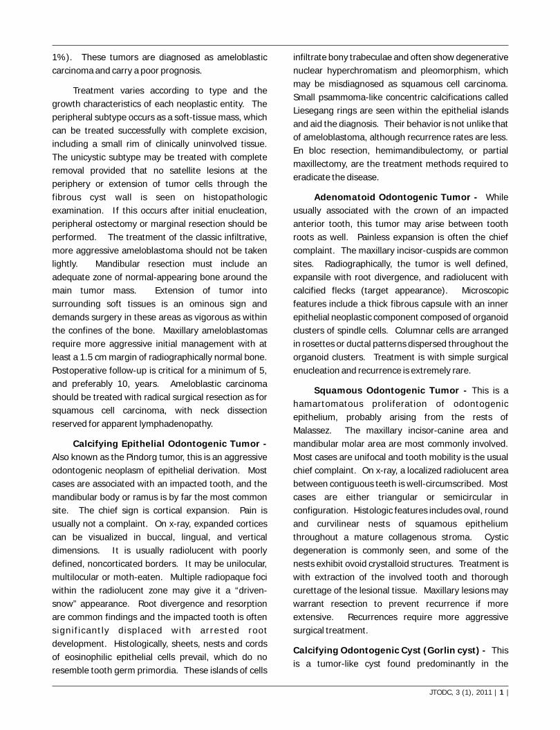
1%). These tumors are diagnosed as ameloblastic
carcinoma and carry a poor prognosis.
Treatment varies according to type and the
growth characteristics of each neoplastic entity. The
peripheral subtype occurs as a soft-tissue mass, which
can be treated successfully with complete excision,
including a small rim of clinically uninvolved tissue.
The unicystic subtype may be treated with complete
removal provided that no satellite lesions at the
periphery or extension of tumor cells through the
fibrous cyst wall is seen on histopathologic
examination. If this occurs after initial enucleation,
peripheral ostectomy or marginal resection should be
performed. The treatment of the classic infiltrative,
more aggressive ameloblastoma should not be taken
lightly. Mandibular resection must include an
adequate zone of normal-appearing bone around the
main tumor mass. Extension of tumor into
surrounding soft tissues is an ominous sign and
demands surgery in these areas as vigorous as within
the confines of the bone. Maxillary ameloblastomas
require more aggressive initial management with at
least a 1.5 cm margin of radiographically normal bone.
Postoperative follow-up is critical for a minimum of 5,
and preferably 10, years. Ameloblastic carcinoma
should be treated with radical surgical resection as for
squamous cell carcinoma, with neck dissection
reserved for apparent lymphadenopathy.
Calcifying Epithelial Odontogenic Tumor -
Also known as the Pindorg tumor, this is an aggressive
odontogenic neoplasm of epithelial derivation. Most
cases are associated with an impacted tooth, and the
mandibular body or ramus is by far the most common
site. The chief sign is cortical expansion. Pain is
usually not a complaint. On x-ray, expanded cortices
can be visualized in buccal, lingual, and vertical
dimensions. It is usually radiolucent with poorly
defined, noncorticated borders. It may be unilocular,
multilocular or moth-eaten. Multiple radiopaque foci
within the radiolucent zone may give it a “driven-
snow” appearance. Root divergence and resorption
are common findings and the impacted tooth is often
significantly displaced with arrested root
development. Histologically, sheets, nests and cords
of eosinophilic epithelial cells prevail, which do no
resemble tooth germ primordia. These islands of cells
infiltrate bony trabeculae and often show degenerative
nuclear hyperchromatism and pleomorphism, which
may be misdiagnosed as squamous cell carcinoma.
Small psammoma-like concentric calcifications called
Liesegang rings are seen within the epithelial islands
and aid the diagnosis. Their behavior is not unlike that
of ameloblastoma, although recurrence rates are less.
En bloc resection, hemimandibulectomy, or partial
maxillectomy, are the treatment methods required to
eradicate the disease.
Adenomatoid Odontogenic Tumor - While
usually associated with the crown of an impacted
anterior tooth, this tumor may arise between tooth
roots as well. Painless expansion is often the chief
complaint. The maxillary incisor-cuspids are common
sites. Radiographically, the tumor is well defined,
expansile with root divergence, and radiolucent with
calcified flecks (target appearance). Microscopic
features include a thick fibrous capsule with an inner
epithelial neoplastic component composed of organoid
clusters of spindle cells. Columnar cells are arranged
in rosettes or ductal patterns dispersed throughout the
organoid clusters. Treatment is with simple surgical
enucleation and recurrence is extremely rare.
Squamous Odontogenic Tumor - This is a
hamartomatous proliferation of odontogenic
epithelium, probably arising from the rests of
Malassez. The maxillary incisor-canine area and
mandibular molar area are most commonly involved.
Most cases are unifocal and tooth mobility is the usual
chief complaint. On x-ray, a localized radiolucent area
between contiguous teeth is well-circumscribed. Most
cases are either triangular or semicircular in
configuration. Histologic features includes oval, round
and curvilinear nests of squamous epithelium
throughout a mature collagenous stroma. Cystic
degeneration is commonly seen, and some of the
nests exhibit ovoid crystalloid structures. Treatment is
with extraction of the involved tooth and thorough
curettage of the lesional tissue. Maxillary lesions may
warrant resection to prevent recurrence if more
extensive. Recurrences require more aggressive
surgical treatment.
Calcifying Odontogenic Cyst (Gorlin cyst) - This
is a tumor-like cyst found predominantly in the
JTODC, 3 (1), 2011 | 1 |
mandibular premolar region. Nearly one quarter of
such cysts are peripheral, producing radiographically
evident calcification above the underlying cortex and
manifesting a gingival swelling. Intrabony lesions may
cause expansion, and teeth remain vital.
Radiographically, the lesion starts as a radiolucency
and progressively calcifies, yielding a target lesion
(opaque, with a circumferential lucent halo). Root
divergence is common. Histologically, the cyst lining is
composed of stratified squamous epithelium with a
polarized basal layer. The lumen contains eosinophilic
keratinized cells devoid of nuclei (ghost cells).
Enucleation with curettage is the treatment of choice
with rare recurrences.
MESENCHYMAL ODONTOGENIC TUMORS
Odontogenic Myxoma - This tumor is believed
to originate from the dental papilla or follicular
mesenchyme. It is usually multilocular and expansile,
sometimes associated with impacted teeth. On x-ray,
the radiolucency has coursing septae which look like a
finely reticulated spider web. These are slow growing
tumors but are aggressively invasive and may become
quite large. The body of the mandible is the favored
site. Microscopically, spindle and stellate fibroblasts
are associated with basophilic ground substance and
myxomatous tissue. Treatment should be with en bloc
resection to prevent recurrence, although curettage
may be attempted for more fibrotic lesions.
Central Odontogenic Fibroma - This tumor
shows more collagen and less ground substance than
the myxoma. Clinical findings, when present, include
swelling or depression of the palate mucosa with tooth
mobility. X-ray shows a uni- or multilocular
radiolucency involving periodontal and crestal bone
adjacent to dental roots. Recurrence is unlikely
following complete removal.
Cementoblastoma - This is a true neoplasm of
cementoblasts, which arises most often on the first
mandibular molars. The cortex is slightly expanded
both buccally and lingually without pain. The involved
tooth is ankylosed to the tumor mass and vital.
Percussion reveals an audible difference between
affected and unaffected teeth. On x-ray, the apical
mass may be lucent with either central opacities or a
solid opacity. A thin radiolucent halo can be seen
around densely calcified lesions. Microscopic
appearance of radially oriented trabeculae from the
attached root cementum with a rim of osteoblasts and
fibrous marrow is apparent. Treatment is with
complete excision with sacrifice of the involved tooth.
MIXED ODONTOGENIC TUMORS
The mixed odontogenic tumors include
ameloblastic fibroma, ameloblastic fibrodentinoma,
ameloblastic fibro-odontoma, and odontoma. Only
ameloblastic fibroma is entirely radiolucent. While all
of the mixed odontogenic tumors may begin as
radiolucent lesions, the remainder will eventually
develop radiopaque foci. The mixed odontogenic
tumors possess both epithelial and mesenchymal
tumor elements, and mimic the differentiation of the
developing tooth germ. The least differentiated is the
ameloblastic fibroma, which is composed of a diffuse
mass of embryonic mesenchyme traversed by
columnar or cuboidal odontogenic epithelium
resembling the dental lamina. Ameloblastic
fibrodentinomas are similar, yet a dense eosinophilic
dentinoid material lies next to the epithelial element.
Ameloblast ic f ibro-odontomas are further
differentiated in that both dentin and enamel matrix
are formed and mixed with ameloblastic fibroma
zones. The odontoma contains all elements of the
mature tooth germ yet does not have a significant soft
tissue cellular overgrowth. Enucleation or thorough
curettage with extraction of the impacted tooth is
recommended for these tumors. The ameloblastic
fibroma has a limited tendency to recur. There has
been a microscopically malignant and aggressive
mixed odontogenic tumor described as ameloblastic
fibrosarcoma. The ameloblastic fibrosarcomas are
aggressive and commonly recur after curettage,
therefore en bloc resection is recommended for these
tumors.
Related Jaw Lesions
Giant Cell Lesions
Central Giant Cell Granuloma (CGCG) - This
is a neoplastic-like reactive proliferation of the jaws
that accounts for less than 7% of all benign lesions of
the jaws in tooth-bearing areas. It commonly occurs
in children and young adults with a slight female
| 2 | JTODC, 3 (1), 2011

predilection. The lesion is more common in the
mandible than maxilla underlying anterior or premolar
teeth. Expansile lesions can cause root divergence or
resorption. The clinical features vary according to the
type of development the lesion assumes. Lesions may
be slow-growing and asymptomatic or rapidly
expanding with pain, facial swelling and root
resorption. The fast growing variants have a high rate
of recurrence. Because of the higher incidence of
these lesions among girls and women of child-bearing
years, hormonal influences have been suggested as
influential in their development. The radiographic
appearance ranges from unilocular to multilocular
radiolucencies with either well-defined or irregular
borders. Multinucleated giant cells, dispersed
throughout a hypercellular fibrovascular stroma often
with bony trabeculae are present on histology.
Treatment regimens for CGCG have historically
included curettage, segmental resection, and radiation
therapy. Radiation therapy has been discouraged
recently, due to the small risk of malignant
transformation to osteogenic sarcoma. Intralesional
steroids have also been advocated for managing CGCG
in younger patients as a nonsurgical alternative.
Individualized treatment depending on the
aggressiveness of the lesion is the rule. Small,
nonaggressive lesions will usually respond to through
excision with careful curettage with a recurrence rate
of less than 15%. Larger, more aggressive lesions,
which have higher recurrence rates, require more
extensive surgery, which may include en bloc
resection.
Brown Tumor of Hyperparathyroidism -
This represents a local manifestation of a systemic
metabolic disease that is histologically identical to
central giant cell granuloma. When this histology is
present, serum calcium and phosphorus should be
obtained, especially in older patients (unlikely to have
central giant cell granulomas), to rule out Brown
tumor.
Aneurysmal Bone Cyst - This is not a true cyst,
and is closely related to the giant cell granuloma with
its aggressive reactive process. The lesion is
composed of large vascular sinusoids, and blood can
be aspirated with a syringe. A bruit, however, cannot
be auscultated due to the low pressures. It has a great
potential for growth and can result in marked
expansion and deformity. A multilocular radiolucency
traversed by thin septae with cortical expansion is
present on x-ray. The mandible body is the most
frequent site. Histologically, large blood-filled
sinusoids lined by an endothelial layer with
surrounding fibroblastic, hypercellular tissue is
present. Simple enucleation is the preferred
treatment. Recurrence is rare.
FIBROOSSEOUS LESIONS
Fibrous Dysplasia - Fibrous dysplasia is the
most common disease of the jaws to manifest a
ground-glass radiographic pattern. There are two
forms, monostotic form, which is more common in the
jaws and cranium, and poyostotic, with is often
associated with McCune-Albright's syndrome
(cutaneous pigmentation, autonomic hyper-
functioning endocrine glands, and precocious
puberty). The monostotic variant is by far the most
common type seen when the jaw is involved and
presents as a painless expansile dysplastic process of
osteoprogenitor connective tissue. The maxilla is the
most common site of involvement. The lesion does
not cross the midline and tends to be limited to one
bone. The antrum is often obliterated, and the orbital
floor (with globe displacement) may be involved. The
histology is characterized by irregular osseous
trabeculae in a hypercellular fibrous stroma.
Treatment should be deferred, if possible until skeletal
maturity. Children with fibrous dysplasia should be
followed quarterly with clinical and radiographic
evaluation. Quiescent and non-aggressive lesions
that have been observed to exhibit no growth are
treated by contour excision for esthetic and/or
functional reasons. When disabling functional
impairment or paresthesia occurs, contour or en bloc
excision may be performed. Accelerated growth or
aggressive lesions require early surgical intervention
with en bloc resection and bone graft reconstruction.
Malignant transformation has been reported after
radiation therapy, which is contraindicated.
Ossifying Fibroma - Similar to fibrous dysplasia
histologically, this is a true neoplasm of the medullary
JTODC, 3 (1), 2011 | 1 |

portion of the jaws. These lesions arise from elements
of the periodontal ligament, and tend to occur in
younger patients, most often in the premolar-molar
region of the mandible. These tumors when small are
asymptomatic but frequently grow to expand the jaw
bone. On x-ray, a well-demarcated radiolucent lesion
is seen in the early stages which becomes increasingly
calcified with maturation. The progression from the
radiolucent to the radiopaque stage takes at least 6
years. After surgical excision of the lesion which tends
to shell out, recurrence is uncommon.
CONDENSING OSTEITIS
Focal areas of radiodense sclerotic bone are
found in about 4% to 8% of the population. These are
usually in the mandible around the apices of the first
molar and are thought to be reactive bony sclerosis to
low-grade pulpal inflammation. They are irregular in
shape, radiopaque with superimposed periapical
inflammation. Once formed, these lesions are stable.
No treatment is necessary.
CONCLUSION
In summary, there are a multitude of
odontogenic cysts and tumors that may present in
head and neck patients. The key to diagnosis is a
careful history and physical examination accompanied
by radiographic evidence and pathologic confirmation.
Many of these entities represent benign cysts and
tumors, however significant pathologic disease may
be lurking which necessitates prompt treatment and
immediate consultation as necessary.
References
1. Kumamoto, H, et al. Clear cell odontogenic
tumor in the mandible: report of a case with
duct-like appearances and dentinoid induction.
Journal of Oral Pathology & Medicine. 29(1): 43-
47. 2000
2. Yamamoto, H, et al. Clear cell odontogenic
carcinoma: A case report and literature review
of odontogenic tumors with clear cells. Oral
Surg, Oral Med, Oral Path, Oral Radiol Endod.
86(1): 86-89. 1998.
3. Manor, Y, Merdinger, O, Katz, J, Taicher, S.
| 2 | JTODC, 3 (1), 2011
Unusual peripheral odontogenic tumors in the
differential diagnosis of gingival swellings.
Journal of Clin Periodon. 26(12): 806-809.
1999.
4. Roberson, J, Crocker, D, Schiller, T. The diagnosis
and treatment of central giant cell granuloma.
JADA. 128: 81-84. 1997.
5. Tallan E, et al. Advanced giant cell granuloma: A
twenty-year study. Otolaryngol HNS110(4):
413-418. 1994.
6. Koppang, H, et al. Glandular odontogenic cyst:
report of two cases and literature review of 45
previously reported cases. J Oral Pathol Med.
27(9): 455-462. 1998.
7. Ramer, M, Mantazem, A, Lane, S, Lumerman, H.
Glandular odontogenic cyst: Report of a case
and review of the literature. Oral Surg Oral Med
Oral Pathol Oral Radiol Endod. 84(1): 54-57.
1997.
8. Bataineh, A, Al Qudah, M. Treatment of
mandibular odontogenic keratocysts. Oral Surg
Oral Med Oral Pathol Oral Radiol Endod. 86(1):
42-47. 1998.
9. Toida, M. So-called calcifying odontogenic cyst:
review and discussion on the terminology and
classification. J Oral Pathol Med. 27(2): 49-52.
1998.
10. Philipsen, H, Reichart, P. Squamous odontogenic
tumor: a benign neoplasm of the periodontium:
a review of 36 reported cases. J Clin Periodontol.
23(10): 922-926. 1996.
11. MacIntosh, R. Aggressive surgical management
of ameloblastoma. OMFS Clin of N Amer. 3(1):
73-97. 1991.
12. Kaban, L. Aggressive jaw tumors in children.
OMFS Clin of N Amer. 5(2): 249-265. 1993.
13. Bailey, et al. Head & Neck Surgery ndOtolaryngology. 2 edition. Volume 2(108):
1541-1562. Dierks, E and Bernstein, M.
Odontogenic cysts, tumors, and related jaw
lesions.

1 2 3Dr. Savithri Dattatreya , Dr. Shweta Kumari Poovam , Dr. Ravindra C. Savadi
OZONE THERAPY IN DENTAL PRACTICE
ABSTRACT
Ozone (also known as triatomic oxygen and trioxygen)
is a naturally occurring compound consisting of three oxygen
atoms. It is found in nature, in the form of a gas in the
stratosphere in a concentration of 110 ppm, being continually
created from and destroyed into molecular O .2
Ozone is one of the most powerful antimicrobial agents
available for use in medicine or dentistry. It is known to be
very effective when prescribed in sufficient concentration, for
an adequate period of time, and when delivered correctly.
This article provides an overview of ozone, its production,
uses and its applications in clinical practice
Keywords : Triatomic, ozone, anti-bacterial, anti-microbial
Review Article
Department The Oxford Dental College, Hospital and Research Center, Bangalore.
Prosthodontics
1,2Senior Lecturer3Prof. & HOD
INTRODUCTION
Atmospheric air is made up of nitrogen (71%),
oxygen (28%) and other gasses (1%) including ozone
which is altered by processes related to altitude, 1 temperature and air pollution.Ozone (also known as
triatomic oxygen and trioxygen) is a naturally
occurring compound consisting of three oxygen
atoms. It is found in nature, in the form of a gas in
the stratosphere in a concentration of 110 ppm,
being continually created from and destroyed into 1, 2molecular O .2
Both these chemical reactions are catalyzed by
very high frequency ultraviolet light from sunlight.
Consequently harmful ultraviolet radiations in the
stratosphere reaching the outer atmosphere from the
sun are absorbed by ozone. Therefore, ozone in
Journal Of The Oxford Dental College
Email for [email protected]
JTODC, 3 (1), 2011 | 1 |
stratosphere has a critical role in both the thermal
structure of the stratosphere as well as the ecological
framework for life on the Earth's surface. On the other
hand in the troposphere, ozone is produced by a
complicated series of chemical reactions involving the
components of automobile exhaust (NO ), sunlight 2
(especially in hot summer months), and oxygen 2consequently making it toxic.
Ozone, in the gaseous or aqueous phase, has
been shown to be a powerful and reliable antimicrobial
agent against bacteria, fungi, protozoa, and viruses.
The oxidant potential of ozone induces the destruction
of cell walls and cytoplasmic membranes of bacteria
and fungi. During this process, ozone attacks
glycoproteins, glycolipids, and other amino acids and
inhibits and blocks the enzymatic control system of the 3cell . This results in increased membrane permeability,

the key element of cell viability, leading to immediate
functional cessation. Then ozone molecules can
readily enter the cell and cause the microorganism 3, 4to die.
Medical Ozone is made when medical grade
oxygen is electrically activated (using an Ozone 3Generator) to form ozone. It is a mixture of the purest
oxygen and purest ozone.
According to its application, the ozone
concentration may vary between 1 and 100; g/ml
(0.05-5%). The ozone therapist determines the
complete dosage according to the medical/dental 4indication and the patient's condition.
This article systematically reviews the clinical
application and remineralization potentials of ozone in
dentistry. This would be of importance to future
researchers in terms of what has been tried and what
the potentials are for the clinical application of ozone in
dentistry
HISTORY
The German chemist Christian Friedrich
Schonbein (1840), of the University of Basel in
Switzerland is regarded as the Father of ozone 5,6, 7therapy. On passing an electrical discharge through
water, he produced a strange smelling gas, which he
called Ozone, derived from the Greek word 'ozein'
meaning odor. In 1857 Joachim Hansler, a German
physicist and physician, along with German physician,
Hans Wolff, developed the first ozone generator for
medical use. Dr. C. Lender in 1870, for the first time
applied O into medical field. He purified blood in test 3
1, 2, 7, 9tubes by using O. 3
With this, O application gained popularity in 3
therapeutic procedures. In 1881, it was used as a
disinfectant in the treatment of diphtheria. In October
1893, Holland became the first city to utilize a water
treatment plant using ozone. In World War I and II it
was used to treat wounded soldiers in the trenches. It
was also used in the treatment of deadly diseases such
as tuberculosis, pneumonia, diabetes, and to cure
wounds, gangrene and the effects of poisonous gas.
In 1950 Dr. E.A. Fisch, was the first German dentist to
use ozone on a regular basis in his dental practice in
Zurich, Switzerland and also publish numerous papers 2, 8on its application.
USES OF OZONE
INDUSTRIAL ACTIONS
On an industrial scale, ozone is used in food and
chemical industry. It may be used in preservation and
extension of shelf life of food, cold storage of meat and
prevention of growth of yeast and mould on fruits and
vegetables. It may also be used as a disinfectant agent
for water, for treating industrial waste and as an 2oxidizing agent in the organic chemical industry.
BIOLOGICAL ACTIONS
There are several known actions of ozone on
human body, such as immunostimulating and
analgesic, antihypoxic and detoxicating, antimicrobial,
bioenergetic and biosynthetic.
The antimicrobial effect of ozone is a result of its
action on cells by damaging its cytoplasmic membrane
due to ozonolysis of dual bonds and also due to ozone-
induced modification of intracellular membrane. This
action is non-specific and selective to microbial cells; it
does not damage human body cells because of their 2major antioxidative ability.
Ozone influences the cellular and humoral
immune systems. It stimulates proliferation of
immunocompetent cel ls and synthesis of
immunoglobulins. It also activates function of
macrophages and increases sensitivity of micro-
organisms to phagocytosis. When administered at low
concentrations, the organisms own resistance is
mobilized. As a response to this activation, the body's
immune cells produce special messengers called
cytokines. These molecules in turn activate other
immune cells, setting off a cascade of positive change
throughout the immune system, which is stimulated to
resist diseases. This means that the application of
medical ozone is extremely useful for immune
activation in patients with a low immune status and/or 2, 6, 7, 8immune deficit.
Ozone also causes the synthesis of biologically
active substances such as interleukins, leukotrienes
TABLE 1
| 2 | JTODC, 3 (1), 2011
and prostaglandins which are beneficial in reducing
inflammation and wound healing. It also brings about
the rise of pO2 in tissues and improves transportation
of oxygen in blood, which results in change of cellular
metabolism, activation of aerobic processes and use of
energetic resources. It also prevents formation of
erythrocyte aggregates and increases their contact
surface for oxygen transportation. Its ability to
stimulate the circulation is used in the treatment of
circulatory disorders and makes it valuable in
revitalizing organic functions. Ozone causes secretion
of vasodilators, which are responsible for dilatation of
arterioles and venules. It activates mechanisms of
protein synthesis, increases amount of ribosome and
mitochondria in cells. These changes on the cellular
level explain elevation of functional activity and 7, 8regeneration potential of tissues and organs.
PRODUCTION OF OZONE GAS
The various systems in use for the production of
ozone gas in medicine and dentistry include the ultra
violet systems, which produces low concentrations of
ozone used mainly in aesthesics and air purification;
cold plasma system for air and water purification; and
the corona discharge system which produces high
concentrations of ozone. The latter is the most
commonly used system in the medical/dental field as it
is easy to handle and has a controlled ozone 1production rate.
In a medical/dental ozone generator, the medical
grade O is converted to O in special tubes via a corona 2 3
discharge reaction (similar to lightning). This type of
generator is able to control the concentration of ozone
critical to delivering the correct dose in
micrograms/milliliters (mcg/ml). Concentration is
determined by exposure and contact time of the
medical-grade oxygen to the 5 to 13 millivolts [Bocci] 8, 9sealed-corona discharge tubes.
Because of ozone's physical properties in the
dental model, the ratio of ozone to oxygen is extremely
low. The typical average concentration of ozone used
in treatments is 25 micrograms of ozone per milliliter of
oxygen/ozone gas mixture. That translates into 0.25
parts of ozone to 99.75 parts of oxygen. Evidence-
based research has shown at this concentration, ozone
effectively kills bacteria, fungi, viruses, and 1, 8, 9parasites.
CLINICAL APPLICATION OF OZONE THERAPY
IN DENTISTRY
MANAGEMENT OF CARIES
Dental caries is a multifactorial disease, which is
characterized by a local destruction of the tooth. The
cavities arise as a result of complex biological
processes and are interplay of four principal factors:
the teeth, saliva, the microflora and the diet. Cavities,
starting as small demineralised areas below the outer
tooth surface, are caused by bacterial fermentation of
dietary carbohydrates to acids, particularly lactic acid, 4within the dental plaque.
Ozone therapy presents great advantages when
used as a support for conventional treatments like
prophylaxis and prevention of dental caries,
remineralisation of pit and fissures, root and smooth
surface caries and restoration of open cavitations
along with conventional conservative measures.
The longer exposure to ozone gas has a strong
bactericidal effect on microorganisms within the
dentinal tubules of deep cavities, which could result in
increasing the clinical success of restorations, with no
negative impact on dentin and enamel shear bond 4strength to adhesive restoration.
Quick and prompt relief from root sensitivity has been
documented after ozone spray for 60 seconds followed
by mineral wash onto the exposed dentine in a
repetitive manner. This desensitization of dentine lasts
for longer period of time thus effectively terminating
the root sensitivity problem
OZONE THERAPY IN ENDODONTICS
Prior studies were found to evaluate the
preventive and therapeutic effect of ozone in the
management of root caries:
6An in vitro study by Baysan et al. assessed the
antimicrobial effect of aqueous ozone on primary root
carious lesions. A significantly reduced microbiological
count was observed in the ozone-treated groups when
compared with the control groups.
JTODC, 3 (1), 2011 | 1 |

One study suggested that application of ozone
gas for a period of 10 to 20 seconds resulted in 99% of
the microorganisms being destroyed. However,
another report stated that 40 seconds of application
was insufficient to decontaminate the area and failed 4, 6to act on underlying infected dentin.
The oxidative power of ozone characterizes it as
an efficient antimicrobial and hence its indication for
use in endodontic therapy seems quite appropriate. Its
antimicrobial action has been demonstrated against
bacter ia l stra ins such as: Mycobacter ia,
Staphylococcus, Streptococcus, Pseudomonas,
Enterococcus and E. coli, S. aureus, E. faecalis,and C.
albicans using in vitro research models. Ozone showed
effectiveness over most of the bacteria found in cases
of pulp necrosis.
Also, crown discoloration is a major aesthetic
problem, especially in anterior root canal treated
teeth. Conventional bleaching is much more time
consuming and results are not often satisfactory. Also,
restoring the tooth with ceramic crowns is not always a
good idea.
Application of ozone for a minimum of 3-4
minutes on a tooth with canal filled with bleaching
agent is believed to bleach the tooth within minutes
thus giving the patient a happy and healthier-looking
smile.
The influence of ozonized water on the epithelial
wound healing process in the oral cavity was first 4, 5observed by Filippi . It was found that ozonized water
applied on the daily basis accelerates the healing rate
in oral mucosa especially during the first two
postoperative days.
It was also found that post- implant treatment
wounds treated with ozonized water heal more quickly
without the need for systemic medication than those
not treated with ozonized water. Application of ozone
therapy after tooth extraction and in case of post-
An exposed nerve when washed with
ozonated water followed by ozone gas prevents the
nerve from dying, thus making a root canal 4unnecessary.
OZONE THERAPY IN SURGERY
extraction complications was also found to be quite
useful
Microbial plaque accumulating on dentures is
composed of several microorganisms; mainly C.
albicans. Denture plaque control is essential for the
prevention of denture stomatitis. In an attempt to
solve this problem Arita et al. assessed the effect of
ozonated water in combination with ultrasonication on
C. albicans. Following exposure of dentures to flowing
ozonated water (2 or 4 mg/l) for one minute, they
found no viable C. albicans suggesting that the
application of ozonated water might be useful in
reducing the number of C.albicans on denture 2, 10bases.
Various studies assessed the efficacy of ozone
and its effect on dental materials. According to these
studies Ozone gas can be applied as prophylactic
treatment prior to etching and the placement of
sealant with no negative impact on sound enamel
physical properties, including knoop surface 2microhardness, or contact angle.
Ozone can also be applied for cleaning the surface of
removable partial denture alloys with little impact on
the quality of alloy in terms of reflectance, surface 2roughness, and weight.
OZONE THERAPY IN PERIODONTICS
Dental biofilm makes it difficult for antibiotics in
targeting putative periodontal pathogens. Higher
concentrations of antibiotics are required to kill these
organisms which are inevitably associated with toxic
adverse effect on the host microbial flora. The
application of ozone has showed promising results.
Both gaseous and aqueous ozone are used as a
substitute to mechanical debridement. Ozonated
water (4mg/l) strongly inhibited the formation of
dental plaque and reduced the number of sub gingival
pathogens both gram positive and gram negative
organisms. Furthermore ozonated water has strong 2bactericidal activity against bacteria in plaque biofilm.
Hence, application of ozone therapy in chronic gingival
and periodontal diseases has showed subjective as
5.
OZONE THERAPY IN PROSTHODONTICS
| 2 | JTODC, 3 (1), 2011

well as objective improvement of the periodontal
status of the patient.
OZONE TOXICITY
Properly performed ozone therapy, carried out by
expert physicians, can be very useful when orthodox
medicinal approach appears inadequate. The
unbelievable versatility of ozone therapy is due to the
cascade of ozone-derived compounds able to act on
several target organs leading to a multi-factorial 2, 12correction of a pathological state.
Saline-washed erythrocytes undergo extensive
hemolysis after ozone exposure. Cells in culture, even
if exposed to very low ozone concentrations for a long
time, undergo apoptosis. One-hour exposure of
saline-diluted blood to ozone induces genotoxic effects
on leukocyte
Ozone inhalation can be toxic to the pulmonary
system. The respiratory tract lining constitutes a very
thin, watery film containing minimal amount of
antioxidants that makes mucosal cells extremely
vulnerable to oxidation leading to upper respiratory
irritation.
Other known side-effects are epiphora, rhinitis,
cough, headache, occasional nausea, vomiting,
shortness of breath, blood vessel swelling, poor 12circulation, heart problems and at times stroke.
Some contraindications for the use of ozone
therapy include : Pregnancy, glucose-6-phosphate
dehydrogenase deficiency, hyperthyroidism, severe
anaemia, severe myasthenia, acute alcohol
intoxication, hemorrhage from any organ, a recent 2history of myocardial infarction and ozone allergy.
CONCLUSION
Hence we can conclude by stating that ozone,
when used with caution has several therapeutic
benefits for patients. Although it is potentially
mutagenic, experimental data performed under
physiological conditions and clinical evidence has
neither shown any cell damage nor adverse effects in 1,2patients .
Ozone therapy has been more beneficial than 2,4,12present conventional therapeutic modalities . The
elucidation of molecular mechanisms of ozone further
benefits practical application in dentistry. Treating
patients with ozone therapy reduces the treatment
time greatly and eliminates bacterial count more
precisely. The treatment is completely painless and
increases the patients' acceptability and compliance
with minimal adverse effects.
REFERENCES
1. R. Garg & S. Tandon. Ozone: A new face of
dentistry. The Internet Journal of Dental Science.
2009 ; 7 : 2
2. Amir Azarpazhooh, Hardy Limeback. The
application of ozone in dentistry: A systematic
review of literature. journal of dentistry 36
( 2008 ) 104 116
3. Celiberti P, Pazera P, Lussi A. The impact of ozone
treatment on enamel physical properties. Am J
Dent. 2006; 19(1):67-72.
4. Antibacterial effect of ozone on cariogenic
bacterial species. E. Johansson, R. Claesson,
J.W.V. van Dijken. Journal of dentistry 37 (2009)
449 453.
5. Bocci V. Ozone. A new medical drug. Dordrecht,
The Netherlands: Springer; 2005; 295p.
6. Baysan A, Whiley RA, Lynch E. Antimicrobial
effect of anovel ozone-generating device on
micro-organisms associated with primary root
carious lesions in vitro. Caries Research 2000;
34:498501.
7. Rickard GD, Richardson R, Johnson T, McColl D,
Hooper L. Ozone therapy for the treatment of
dental caries. Cochrane Database system Rev.
2004;3
8. Nogales C G, Ferrari P A, Kantorovich EO, Lage-
Marques JL. Ozone Therapy in Medicine and
Dentistry. J Contemp Dent Pract 2008 May;
(9)4:075-084.
JTODC, 3 (1), 2011 | 1 |

9. Seaverson K, Tschetter D, Kaur T. Patient guide to
oxygen/ozone therapy. Health centered cosmetic
dentistry. [Online]. [Cited 2010 January 13].
Available from:
10. Arita M, Nagayoshi M, Fukuizumi T, Okinaga T,
Masumi S, Morikawa M. Microbicidal efficacy of
ozonated water against Candida albicans
adhering to acrylic dentures plates. Oral
Microbiology and Immunology 2005; 20:206-10.
11. Elisa Magni, Marco Ferrari , Reinhard Hickel ,
Karin Christine Huth, Nicoleta Ilie. Effect of ozone
gas application on the mechanical properties of
dental adhesives bonded to dentin. Dental
Materials 24 (2008 ) 14281434
12. Velio Bocci. Ozone as Janus: this controversial
gas can be either toxic or medically useful.
Mediators of Inflammation, 13(1), 3_ 11,
February 2004.
URL:http://www.toothbythelake.net/
ozone_therapy.html
/
| 2 | JTODC, 3 (1), 2011

1 2 3 4 5 6Dr Savitha AN , Dr Sneha D , Dr Gayathri G , Dr Aditi D , Dr Geetha P and Dr Dwarakanath CD
THE POUCH AND TUNNEL TECHNIQUE FOR THE MANAGEMENT
OF MULTIPLE GINGIVAL RECESSION DEFECTS: A CASE SERIES
A Case Series
Department The Oxford Dental College, Hospital and Research Center, Bangalore.
of Periodontics,
1Professor2PG Student
3Reader4Senior Lecturer5Senior Lecturer
6HOD and Professor
INTRODUCTION
Periodontal plastic surgery is defined as the
surgical procedures performed to correct or eliminate
anatomic, developmental or traumatic deformities of 1the gingiva or alveolar mucosa. Gingival recession is
defined as the displacement of the gingival margin
Journal Of The Oxford Dental College
Email for [email protected]
JTODC, 3 (1), 2011 | 1 |
apical to the cemento-enamel junction (CEJ) with loss
of periodontal connective tissue fibers along with root 2 cementum and alveolar bone. Studies by Murray and
Gorman et al have shown that recession increases with
increasing age. A survey revealed that 88% of people
above 65 years of age and 50% of people between 18-
64 years of age have one or more sites with recession
ABSTRACT
Gingival recession is a common occurrence and its
prevalence increases with age. It can lead to clinical
problems, diminished cosmetic appeal and hence esthetic
concern. The desire for improved esthetics has increased
tremendously over the years. In the past, periodontal
treatment procedures were mainly aimed at preventing and
treating the existing periodontal diseases. However, with
increasing esthetic demands these surgical procedures are
modified so as to preserve and enhance esthetics by various
periodontal plastic surgical procedures. Periodontal plastic
surgery deals with procedures that are designed to enhance
esthetics, restore form, function and also include
regenerative modalities too.
This case series evaluates the effectiveness of
subepithelial connective tissue graft (SCTG) in coverage of
denuded roots using pouch & tunnel technique. This
technique preserves the intermediate papilla, accelerates the
initial wound healing & also applies less traction. Due to
minimal trauma at the recipient site, this procedure may be
advantageous in treatment of multiple recessions as
compared to other treatment modalities.
Keywords: Gingival recession, tunneling procedure,
subepithelial connective tissue grafts (SCTG).

3 (Kitchin et al , Ervin and Buchner et al). The main
indications for root coverage procedure are esthetic
demands, root hypersensitivity, root caries lesions and
cervical abrasions. Thus 4 .
If untreated, gingival recession may progress to the
point that it can compromise the prognosis of the tooth
in question.
The amount of recession is assessed clinically by
measuring in millimeters the distance from the CEJ
and the soft tissue margin. The recession of gingiva,
either localized or generalized, may be associated with
one or more surfaces, resulting in attachment loss and
root exposure. Marginal gingival recession, therefore
should not be viewed as merely a soft tissue defect, 5but as the destruction of both the soft and hard tissue.
6CAUSES OF GINGIVAL RECESSION:-
• Predisposing factors:-
1. Minimal attached gingiva;
2. Aberrant frenal pull;
3. Tooth malposition (fenestration and
dehiscence)
• Precipitating factors:-
1. Inflammation related to plaque;
2. Improper tooth brushing;
3. Iatrogenic factors such as crown
preparations extending subgingivally,
impression techniques involving gingival
retraction.
4. Poor orthodontic treatment where the teeth
are moved outside the labial or lingual
plate.
• Anatomical factors include abnormal tooth
position in the arch, aberrant path of eruption,
individual tooth shape.
• Pathological factors such as bone resorption due
to periodontal disease.
it is essential to carry out root
coverage surgery for the aforementioned conditions
Accepted procedures for multiple root coverage
include coronally advanced flap with/without free
mucosal graft, subepithelial connective tissue graft
and guided tissue regeneration. Other materials like
emdogain (EMD), acellular dermal matrix (ACDM)
have also been tried.Connective tissue grafts are an
important treatment options for periodontal and
implant reconstructive plastic surgery. Connective
tissue graft was first used by Edel (1974), Broome and
Taggart (1976) and Donn (1978), to increase the width
of keratinized gingiva. The use of connective tissue
grafts for treatment of gingival recession began in
1985 when Langer and Langer described SCTG
technique for covering gingival recession of both single 7 and multiple adjacent teeth. They described a
technique in which the graft is covered by the
overlying partial thickness flap. Nelson proposed the 8use of full thickness flap to cover the SCTG.
In 1985 Raetzke, described a different version of 9 connective tissue graft called “Envelope technique”.
Allen AL in 1994, in a modification of Raetzke's
t e chn ique , desc r i bed the “TUNNEL OR
SUPRAPERIOSTEAL ENVELOPE TECHNIQUE”, for 10 treatment of multiple adjacent gingival recession.
Santarelli et al adapted the tunnel technique by using a
single vertical incision. Mahn adapted the tunnel
approach for acellular dermal connective tissue
grafting by using full thickness procedure with vertical 11incisions.
Indications for pouch and tunnel technique
include:-
1. Miller's class I and class II gingival recession;
2. Lack of adequate donor tissue for lateral sliding
flap;
3. Presence of multiple and wide recessions in
maxillary teeth;
4. Increased recession in areas where esthetic
concerns is of great concern;
5. Exposed root sensitivity.
This case series outlines the advantages of
subepithelial connective tissue graft using pouch and
tunnel procedure, as outlined by Allen AL in 1994, for
treatment of multiple gingival recessions over other
treatment modalities.
| 2 | JTODC, 3 (1), 2011

CASE PRESENTATIONS
CASE NO 1
2%lidocaine with a concentration of
1:200000 epinephrine,
A 21 year old male patient reported with the
complaint of sensitivity in upper left anterior teeth
region. On examination Miller's Class I gingival
recession was present in relation to 23 and 24.The
width of attached gingiva was found to be adequate in
the region of 23 and 24. A pouch and tunnel technique
utilizingpalatal connective tissue graft for root
coverage was planned based on the indications stated
above.
The treatment protocol was explained to the
patient and an informed consent was obtained.
Routine periodontal therapy, including scaling and root
planing was performed. The patient was recalled after
one month for the surgery.
SURGICAL TECHNIQUE
RECEPIENT SITE PREPARATION
Following administration of local anesthesia i.e.
local infiltration of
sulcular incision through each
recession area were given with a number 15 blade. A
full thickness dissection was extended,using a
periosteal elevator, undermining the interdental
papilla, extending beyond the mucogingival junction
so as to relax the flap sufficiently to facilitate
placement of connective tissue graft. Care was taken
not to detach the tip of the interdental papilla. Each
pedicle adjacent to the recession was undermined
gently, without detaching it completely to prepare a
tunnel. The undermining of tissues to prepare the
tunnel was done by extending the undermining
laterally about 3-5 mm. Full thickness undermining
was done to elevate the flap in coronoapical direction.
DONOR SITE PREPARATION
The connective tissue graft was harvested using 12Lui's class I incision. The incision was placed between
distal aspect of canine and mesial aspect of first molar
area. After the graft was harvested, pressure was
applied to the donor area with gauze soaked in saline,
to control bleeding. The palatal flap was then sutured
with 4-0 direct interrupted suture.
GRAFT PLACEMENT
The graft was stabilized using a 5-0 resorbable
silk suture. The mesial aspect of the graft was pierced
with the needle, and the needle was passed passively
underneath the tunnel created between the adjacent
recessions. The suture was passed from the mesial
aspect of the tunnel and pushed gently to the distal
direction with a periosteal elevator so that the graft
could slide underneath the tunnel. The graft was
positioned coronal to cementoenamel junction. After
positioning, the graft was secured to the mesial and
distal aspect with sling sutures in order to prevent
movement of the graft.
Dry tin foil was applied to the recipient site. A
periodontal dressing (Coe pak) was placed over the foil
to stabilize and protect the donor tissue.
POST OPERATIVE INSTRUCTIONS
All the
sites healed uneventfully. The donor site appeared
normal in color and healthy after four weeks and the
recipient site was healthy with excellent color match
with adjacent tissues. After one year, complete root
coverage achieved, remained so without any adverse
effects. The patient reported satisfactory esthetic
results and loss of hypersensitivity.
The patient was advised to use 0.2%
chlorhexidine gluconate mouth rinse twice daily for 2
weeks. The patient was provided with the
postoperative home care instructions and was
prescribed analgesic to reduce postoperative pain and
discomfort and antibiotic (Amoxicillin 500 mg, three
times daily for five days) to reduce the risk of
postoperative infection. Sutures were removed after
10 days. Patient was recalled weekly for the first
month for postsurgical evaluation with reinforcement
of plaque control using a super soft brush. The patient
was further followed up at three and six month
intervals for supportive periodontal therapy.
A B
A : Miller's class 1 gingival recession
wrt 23,24
B: Crevicular incision given. Pouch and tunnel prepared.
JTODC, 3 (1), 2011 | 1 |

CASE NO 2
A 25 year old male patient complained of longer teeth
in relation to upper left anterior region. On
examination there was Miller's class 1 gingival
recession in relation to 21, 22 and 23.
CASE NO 3
A 20 year old female patient came with the chief
complaint of sensitivity in her upper right anterior
teeth. On examination there was Miller's class 1
recession in relation to 12, 13 and 14.
| 2 | JTODC, 3 (1), 2011
C D
C: SCTG graft harvested
from the palate
D: Graft stabilized
with 5-0 sutures
E F
E: Graft sutured F: Pack placed
1 year follow up
A B
A : Miller's class
1 gingivalrecession
wrt 21, 22, 23
B : Crevicular incision
given. Pouch and
tunnel prepared.
C D
C: SCTG graft harvested D: Graft stabilized with
5-0 sutures
E F
E: Graft sutured F: Pack placed
G
G: 1 year postoperative
A : Miller's class
1 gingival recession
wrt 12,13,14.
B : Crevicular
incision given.
A B
C: Pouch and
tunnel prepared
C D
D: SCTG graft harvested
E: Graft stabilized
E F
F: Graft securedwithin the tunnel
G: Pack placed H: 15 days postoperative
G H
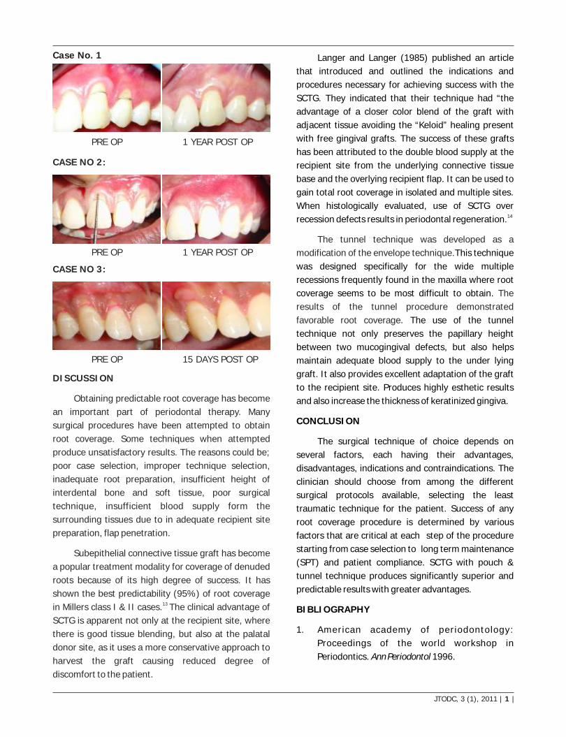
DISCUSSION
Obtaining predictable root coverage has become
an important part of periodontal therapy. Many
surgical procedures have been attempted to obtain
root coverage. Some techniques when attempted
produce unsatisfactory results. The reasons could be;
poor case selection, improper technique selection,
inadequate root preparation, insufficient height of
interdental bone and soft tissue, poor surgical
technique, insufficient blood supply form the
surrounding tissues due to in adequate recipient site
preparation, flap penetration.
Subepithelial connective tissue graft has become
a popular treatment modality for coverage of denuded
roots because of its high degree of success. It has
shown the best predictability (95%) of root coverage 13 in Millers class I & II cases. The clinical advantage of
SCTG is apparent not only at the recipient site, where
there is good tissue blending, but also at the palatal
donor site, as it uses a more conservative approach to
harvest the graft causing reduced degree of
discomfort to the patient.
Langer and Langer (1985) published an article
that introduced and outlined the indications and
procedures necessary for achieving success with the
SCTG. They indicated that their technique had “the
advantage of a closer color blend of the graft with
adjacent tissue avoiding the “Keloid” healing present
with free gingival grafts. The success of these grafts
has been attributed to the double blood supply at the
recipient site from the underlying connective tissue
base and the overlying recipient flap. It can be used to
gain total root coverage in isolated and multiple sites.
When histologically evaluated, use of SCTG over 14recession defects results in periodontal regeneration.
This technique
was designed specifically for the wide multiple
recessions frequently found in the maxilla where root
coverage seems to be most difficult to obtain.
The use of the tunnel
technique not only preserves the papillary height
between two mucogingival defects, but also helps
maintain adequate blood supply to the under lying
graft. It also provides excellent adaptation of the graft
to the recipient site. Produces highly esthetic results
and also increase the thickness of keratinized gingiva.
CONCLUSION
The surgical technique of choice depends on
several factors, each having their advantages,
disadvantages, indications and contraindications. The
clinician should choose from among the different
surgical protocols available, selecting the least
traumatic technique for the patient. Success of any
root coverage procedure is determined by various
factors that are critical at each step of the procedure
starting from case selection to long term maintenance
(SPT) and patient compliance. SCTG with pouch &
tunnel technique produces significantly superior and
predictable results with greater advantages.
BIBLIOGRAPHY
1. American academy of periodontology:
Proceedings of the world workshop in
Periodontics. Ann Periodontol 1996.
The tunnel technique was developed as a
modification of the envelope technique.
The
results of the tunnel procedure demonstrated
favorable root coverage.
Case No. 1
1 YEAR POST OP
1 YEAR POST OP
15 DAYS POST OP
PRE OP
PRE OP
PRE OP
CASE NO 2:
CASE NO 3:
JTODC, 3 (1), 2011 | 1 |

2. Wennstrom JL. Mucogingival surgery. In: Lang
NP, Karring T. Proceedings of the first European
workshop of periodontology. Quintessence
books. p193-209.
3. Serino G, Wennstrom JL, Lindhe J. The
prevalence and distribution of gingival recessions
in subjects with a high standard of oral hygiene. J
Clin Periodontol 1994;21:57-63.
4. Goldstein M, Brayer L and Schwartz Z. A Critical
Evaluation of Methods for Root Coverage. Crit
Rev Oral Biol Med1996;7:87-98.
5. Ahathya RS, Ramakrishnan T and Ambalavanan
N. Subepithelial connective tissue grafts for
coverage of denuded root surfaces. Indian J Dent
Res 2008;19:134-140.
6. Hall WB. Pure Mucogingival problems. Etiology,
treatment and prevention. Quint Int
1984;19:119-123.
7. Langer B, Langer L. Subepithelial connective
tissue graft for root coverage. J Periodontol
1985;56:715-720.
8. Nelson SW. The subpedicle connective tissue
graft. A bilaminar reconstructive procedure for
coverage of denuded root surfaces. J Periodontol
1987;58:95-102.
9. Raetzke PB. Covering localized areas of root
exposure employing the envelope technique. J
Periodontol 1985;56:397-402.
10. Allen AL. Use of supraperiosteal envelope in soft
tissue grafting for root coverage. Rationale and
technique. Int J Periodontics Restorative Dent
1994;14:216-227.
11. Tozum TF. Treatment of adjacent gingival
recessions with subepithelial connective tissue
grafts and the modified tunnel technique.
Quintessence Int 2003;34:7-13.
14. Bruno JF and Bowers GM. Histology of human
biopsy section following placement of
subepithelial connective tissue graft. Int J
Periodontics Restorative Dent 2000;20:224-231.
12. Liu CL and Weisgold AS. Connective tissue grafts:
A classification for incision design from the
palatal side and clinical case reports. Int J
Periodontics Restorative Dent 2002;22:373-379.
13. Miller PD. A classification of marginal tissue
recession. Int J Periodontics and Restorative
Dent 1985;2:9-13.
| 2 | JTODC, 3 (1), 2011

1 2 3 4Dr. Sanjay Mohanchandra , Dr Sahana.B.A , Dr Jyotsna Rao , Dr.Kishan Panickar , 5 6Dr.Bipin C Reddy , Dr Mahendra.P
AUTOGENOUS TOOTH TRANSPLANTATION AN EVALUATED STUDY
ABSTRACT
BACKGROUND - The aim of this study is to evaluate the
prognosis of autotransplanted completely or partially formed
roots of impacted third molars after replacing grossly
destructed first or second molars. The sample consisted 15
patients, age ranging from 18-28 years. After extraction of
the diseased molar, autotransplantation of third molar was
immediately performed and splinting was carried out. The
follow up study included recordings of clinical parameters like
mobility, probing periodontal pocket depth, gingivitis and
tender on percussion. Radiographs were taken to evaluate
pulpal breakdown, periapical radiolucency, internal and
external root resorption of the donor tooth. The results in this
study indicated 86.3% success rate of autotransplanted
teeth.
CONCLUSION - Autogenous tooth transplantation can be
used as an treatment procedure for replacing unrestorable
first and second molar.
A Case Series
Department of Oral Maxillofacial Surgery,The Oxford Dental College, Hospital and Research Center, Bangalore.
1Professor2,3,4,5,6Reader
INTRODUCTION
Autotransplantation is now a common procedure
in dentistry for replacing a missing tooth or
compromised tooth due to caries in children and
adolescents where implants and other prosthetic
replacements are contraindicated. Auto transplantation
offers one of the fastest and most economically
feasible means of replacing missing teeth.
Autogenous tooth transplantation is defined as
the transplantation of tooth from one site to another in
the same individual. The recipient site may be an
Journal Of The Oxford Dental College
Email for [email protected]
JTODC, 3 (1), 2011 | 1 |
extraction socket or surgically prepared site.(Natiella
et al,1970)
This procedure was originally described by
Widman (1915) for the transplantation of an impacted
canine to the normal site and subsequently by Aptel
(1948, 1956) and Miller (1951, 1956) for the
transplantation of lower third molar to fresh extraction
sites of lower first molar. Now autotransplantation has
come to involve most teeth in the oral cavity.
Auto transplantation of a tooth can be carried out
whenever there is suitable donor tooth available for an
edentulous site or freshly extracted socket within the

| 2 | JTODC, 3 (1), 2011
same oral cavity. Autotransplants, unlike most
transplants is out of its natural environment for only a
matter of seconds. Consequently, periodontal
regeneration is the rule rather than the exception.
The purpose of this study was to evaluate the
prognosis in addition to the causes of failure in 15
autotransplanted teeth in one year
AIMS AND OBJECTIVES
Auto transplantation is a viable option for treating
unrestorable teeth when a donor is available. The aim
of this study is to evaluate the prognosis of
autotransplanted teeth with complete and incomplete
root formation of impacted third molars, transplanted
to the region of freshly extracted socket of first or
second molars clinically and radiographically over one
year follow up period.
MATERIALS AND METHODS
This study consists allotransplantation of 15
patients with both mature and immature vertically
impacted third molars transplanted. At the time of
transplantation the age of the patient was ranging
from 18 to 28 yrs. The indication for transplantation
was extensive decay of first and second molars. Out
of fifteen autotransplanted teeth eleven teeth
consisted of roots which were fully developed and four
teeth one half to three fourth of the expected root
length. The transplanted teeth were placed in
infraocclusion. In cases of adequate initial stability of
transplanted teeth, fixation of the transplant was
carried out with a suture splint for 7 days. The suture
splint consisted of a 2-0 silk suture passing through the
gingiva, crossing the occlusal surface of the tooth
labiolingually. In cases of inadequate initial stability
of transplanted teeth which is defined as a horizontal
mobility of more than 2mm at the end of the operation
splinting was carried out with composite resin and wire
splint for 4 weeks, this splint consisted of 2mm X
0.5mm wire, attached with light cure composite to the
buccal surfaces of the transplanted tooth and the two
adjacent teeth. Antibiotic therapy was started with
500mg Amoxycillin one hour preoperatively, followed
by three times daily, orally for one week
postoperatively. Oral hygiene instructions were given
to all patients. Twenty four hours after surgery
patients were advised to rinse with 0.2% chlorhexidine
solution till removal of sutures. Each transplanted
tooth was clinically and radiologically assessed at
weekly intervals for one month and then at monthly
intervals of third, sixth and. twelth month. As a part of
clinical evaluation of transplanted teeth the following
clinical parameters were scored-tooth mobility,
tenderness on percussion, probing pocket depth and
gingivitis..
The mobility of the transplanted teeth was scored
by means of mobility test using two blunt end of the
instrument based on a scale of (0 - 3)
Grade 0 ; no abnormal tooth mobility
Grade 1 ; abnormal horizontal mobility of not more
than 1mm
Grade 2 ; abnormal horizontal mobility of more than
1mm
Grade 3 ; abnormal horizontal mobility of more than
1mm and axial mobility.
Probing pocket depth was recorded from the
gingival margin to the tip of the calibrated periodontal
probe (Hu-friedy) inserted into the pocket in defined
locations, ie distobuccal, buccal, mesiobuccal,
distolingual, lingual, mesiolingual with a moderate
probing force of 0.75N. The probing depth was
recorded to the nearest 1mm. Pockets with a depth of
more than 3mm were rated as pathological.
Gingivitis was scored according to the Sulcus
bleeding index by Muhleman and Son.S in 1971. The
probing was done in four areas, the labial and lingual
marginal gingiva(M) and the mesial and distal papillary
gingiva(P). The assessment of gingival bleeding is
done on a scale of (0-5) according to the following
criteria Gradeo-Grade.O; healthy appearance of P&M.
No bleeding upon sulcus probing
Grade 1; apparently healthy P&M. Showing no colour
or contour changes and no swelling, but bleeding from
sulcus on probing
Grade 2; Bleeding on probing and colour change

caused by inflammation (reddening), No swelling or
macroscopic edema
Grade 3; Bleeding on probing, change in colour, slight
edematous swelling
Grade 4; (1) Bleeding on probing, colour change,
obvious swelling
(2) Bleeding on probing and obvious swelling,
Grade 5; Spontaneous bleeding on probing, colour
change marked swelling with or without ulceration.
The corresponding contralateral tooth served as a
control for the bleeding index.
A percussion test was carried out with a blunt end
of metal instrument for all transplanted teeth to
determine clinical ankylosis. Vitality of the
transplanted tooth was tested by means of a cold test.
Also additional clinical parameters like soft tissue
appearance around the transplanted tooth and
whether there is any inflammation present at recipient
site were evaluated.
Radiological examination included intraoral films
and panoramic radiographs which contributed to the
evaluation of radiological parameters such as
periapical radiolucency, root resorption (external and
internal), ankylosis, and the onset of pulpal
breakdown seen as intrapulpal calcification. The crown
to root ratios of the transplanted teeth were calculated
and compared with contralateral crown to root ratio.
Root development after transplantation was only
approximately estimated by comparing with the
natural tooth contralateral to the donor tooth .For vital
transplants of developing teeth with open apices,
endodontic treatment of the transplant is not required
as these teeth got revascularised and reinnervated,.
However endodontic treatment is always required for
transplants of mature teeth with complete root
format ion. Endodont ic t reatment started
approximately one-month post operatively with
instrumenting the canals and filling with calcium
hydroxide, followed by Guttapercha filling from 3 to 6
months post operatively
(The success rate was defined as percentage of
the transplanted teeth fulfilling defined.)
JTODC, 3 (1), 2011 | 1 |
Criteria relative to the total number of
transplanted teeth in the sample the survival rate was
calculated as the percentage of transplanted teeth still
present and functioning well at the time of recall.
In this study successful transplantation was
defined as showing no root resorption and ankylosis, a
crown to root ratio less than 1 (i.e. suprabony part
shorter than the infra bony part), radiographic
evidence of further root growth, no abnormal mobility,
normal gingival contour and attachment level adjacent
to the transplanted tooth and showing no
inflammation at the recipient site.
CASE SELECTION
Patient selected were healthy with no systemic
diseases and they demonstrated an acceptable level of
oral hygiene. Most important was that they had a
suitable recipient site and donor tooth.
The recipient sites selected were first and second
mandibular molar regions, free from acute infections,
and having adequate amount of bone support in all
dimensions to allow for stabilization of the
transplanted teeth
DONOR TOOTH CRITERIA
A third molar tooth with either open or closed
apices, single or double rooted, partially or completely
erupted were selected as a donor tooth.
The donor tooth selected was positioned in such
a way that extraction was atraumatic with no abnormal
tooth morphology.
RESULTS
The study was conducted in 15 patients, out of
which 9 were male and 6 were female and the age was
ranging from 18 to 28 years. None of the patients had
systemic diseases. Among 15 autotransplanted
vertically impacted molars, 11 teeth were having
completely developed roots and 4 had partially
developed roots and among 11 completely developed
roots, 7 were transplanted to the socket of second
molar and 4 were transplanted to the socket of first
molar.

| 2 | JTODC, 3 (1), 2011
In the follow up study from 1st week to 1st year
the teeth were evaluated clinically for mobility, tender
on percussion, gingivitis and periodontal pocket depth
and radiographically for any pulpal break down in the
form of intra pulpal calcification, periapical
radiolucency and root resorption.
MOBILITY:
stIn the 1 week of clinical evaluation, there were
10 cases (66.7%) with grade 0 mobility and 5 cases nd(33.3%) grade 1 mobility. In 2 week there were 11
cases (73.3%) with grade 0 mobility and 4 cases rd rd(26.7%) grade 1 mobility and from 3 week to 3
month there were 13 cases (86.7%) with grade 0
mobility and 2 cases (13.3%) with grade 1 mobility. In
a period of 6th month to 1 year 13 cases (86.7%) had
grade 0 mobility and 2 cases (13.3%) had grade 2
mobility. So P value was 0.1278, which suggested that
it is statistically significant.
TENDERNESS ON PERCUSSION :
stAt 1 week in 14 cases (93.5) the teeth were not
tender, where as 1 tooth (6.7%) was tender which
improved in 1 year with P value of 0.1587 suggesting
that it was not statistically significant
GINGIVITIS:
stAt 1 week, 11 cases (73.3%) had grade 0
gingivitis and 4 cases 26.7% had grade 1 gingivitis. In nd2 week, 13 cases (86.7%) had grade 0 gingivitis and
rd2 cases (13.3%) had gradel gingivitis in 3 week all 15 thcases (100%) had grade 0 gingivitis, but from 4 week
thtill 6 month 13 cases (86.7%) had grade 0 gingivitis
and 2 cases (13.3%) had grade 1 gingivitis and at the
end of 1 year all 15 cases (100%) had grade 0
gingivitis. The P value was 0.01764 suggesting that it
is statistically significant.
POCKET DEPTH :
sl rdI In the evaluation from l week to 3 month, ththere was no pocket depth in all cases, but from 6
month to 1 year the pocket depth was present in 2
cases (13.3%) and the P value 0.3550
AUTOTRANSPLANTATION OF COMPLETELYFORMED ROOTS WITH RCT
AUTOTRANSPLANTATION OF COMPLETELY FORMED ROOTS WITHOUT RCT

JTODC, 3 (1), 2011 | 1 |
crown/root ratio of more than 1 and the other 2 teeth,
although had adequate initial stability, the crown/root
ratio was decreased and by the end of 1 year the
recipient socket was filled by bone.
DISCUSSION
The literature reports excellent success rates
following tooth transplantation when the appropriate
protocol is followed Transplantation of teeth in humans
is not a recently established surgical procedure.
Ambrose pare appears to have been the first to publish
a report about transplantation of teeth in 1584. In
1780, John Hunter described this operation more
ideally than had been before. Between 1950 and 1955,
detailed surgical techniques for the transplantation of rddeveloping teeth particularly from the 3 molar to the
st1 molar position have been first described and well
ON RADIOGRAPHIC EXAMINATION
The intrapulpal calcification was not observed in
any of the cases with a P > 0.05.
PERIAPICAL RADIOLUCENCY
This observation was absent in 13 cases (86.7%) th and present in 2 cases, over a period of 6 month to 1
year. P = 0.3550.
ROOT RESORPTION
External root resorption was seen in 2 cases
(13.3%) and was absent in 13 cases (86.7%) over a
period of 1 year. P > 0.05, which was statistically not
significant. Internal resorption was not observed in
any of the cases. In developing teeth consisting of
roots which were partially formed, 2 teeth had

| 2 | JTODC, 3 (1), 2011
documented by Apfel, Fong Hale and Clark, according
to these authors recipient site should have sufficient
dimensions, sound periodontal support and ! absence
of acute inflammation. Minimal handling of transplant
is important and great care should be taken not to
denude or even touch any of the hertwigs root sheath
or exposed pulpal tissue.
Miller in 1951 also reported successful auto
transplantation with careful planning and appropriate
surgical technique and Kahnberg in 1987, Andreasen
et al 1980, Nethander 1994, lundberg and lsaksson
1996, Majars et al 2004 also achieved high success
rates by autotransplantation.
The present study evaluated 15 auto
transplanted teeth which included teeth with both
completely and partially formed roots. On clinical
evaluation the present study revealed that,33% of
cases had mobility in the 1st postoperative week which
gradually decreased to 13.3% of cases by the end of
one year follow up period.. Although there was
tenderness on percussion, initially, but at the end of 1
year period the teeth showed no tenderness on
percussion. Gingivitis observed in all the cases initially
but disappeared by the end of 1 year period. The depth
of periodontal pockets evaluated clinically were with in
normal limits in all the cases, where as 13.3 % of cases
shown increased periodontal pocket depth by the end
of 1 year follow up period. Radiographic evaluation
showed no pulpal breakdown, but initially periapical
radiolucency was present, and it got regressed after
starting root canal treatment. External root resorption
was found in 2 cases, which suggested poor
adaptability of donar to the recipient site or damage to
periodontal ligament. In cases where the periodontal
ligament is traumatized due to poor handling during
transplantation external root resorption and ankylosis
often noted, hence the most significant determinant is
the continued vitality of periodontal membrane,
maintained by atraumatic extraction of the donar

JTODC, 3 (1), 2011 | 1 |
tooth and immediate transfer to the recipient site, for
the survival of the transplanted teeth. Lundberg and
Isaksson 1996 Mejare et al 2004 achieved a high
success rate in transplanting matured teeth and they
showed that it may be viable option in the absence of
other suitable donor tooth.
CONCLUSION
Autogenous transplantation of tooth with
complete or partially formed roots is considered as a
viable treatment option to conventional prosthetic and
implant rehabilitation for both therapeutic and
economic reasons Careful surgical and endodontic
procedures, together with careful case selection may
lead to satisfactory, aesthetic and functional
outcomes.
1. Tooth transplantation;a descriptive retrospective
study. R Kallu,F Vickler,C Poiliilis,S.Miwalli,G
Williams International journal of oral and
maxillofacial surgery 2005;34;745-755
2. autotransplantation of immature third molar :
influence fof different splinting methods and
fixation periods. Oskar Bauss,Reinhard
Schilke,Christian Fenske Dent traumatology
2002;18;322-328
3. Allo-and autotransplantation of mature teeth in
monkeys; a sequential time related histo-
quantitative study of periodontal and pulp
healing Shwartz O, Andreasen JO Dental
traumatology2002;18;246-261
4. Autogenous tooth transplantation: An alternative
implant placement. Cameson Mr, Cokie, Yau J.
Can Association 2001;67;92-96
5. autotransplantatioon of teeth; requirements for
predictable success. Mitsuhiro Tsokoboshi Dental
traumatology;2002;18;157-180
6. A clinical and radiographic study of 25
autotransplanted third molars. Y.Akiyama.H,
Fukuda, K Hashimoto J. of oral rehabilitation
1998;25:640-644.

1 2Dr. Srilakshmi. J , Dr. Ravindra C Savadi
LASER AND IT'S APPLICATIONS IN DENTISTRY
ABSTRACT
Several decades ago using laser for treatment purposes
was a taboo because of its complexicity and lack of proper
knowledge. But now with the advances in science and in the
field of lasers, lasers have been used extensively and
successfully in the dental arena for more than 20 yrs. Laser is
a form of electromagnetic radiation of varying wavelengths
that can be used in various procedures in dentistry. This
article provides an overview of laser and it's uses in dentistry.
Keywords : laser, electromagnetic spectrum,
monochromatic, coherent, radiation.
A Case Series
Dept of ProsthodonticsThe Oxford Dental College, Hospital and Research Center, Bangalore.
1Senior Lecturer2Prof & HOD
INTRODUCTION :
LASER is an acronym, which stands for Light 1Amplification by Stimulated Emission of Radiation. It
is a form of electromagnetic energy in which photons
are generated from a medium by stimulating the
medium from an external energy source. It is a
monochromatic, collimated and coherent beam of
light. The electromagnetic spectrum is an entire
collection of wavelengths ranging from gamma rays -12(10 m) to radio waves (300nm). The very short
wavelengths those below approximately 300 nm are
termed ionizing. This term refers to the fact that
higher-frequency (smaller wavelength) radiation has a
large photon momentum measured in electron volts
per photon. This higher photon energy can deeply
penetrate biologic tissue and produce charged atoms
and molecules. Wavelengths larger than 300nm have
less photon energy and cause excitation and heating
Journal Of The Oxford Dental College
Email for [email protected]
JTODC, 3 (1), 2011 | 1 |
of the tissue with which they interact. All available
dental lasers have emission wavelengths
approximately 500nm to 10,600nm. Lasers are within
the visible/invisible infrared portion of electromagnetic
spectrum and emit thermal radiation( Fig 1). When
used efficaciously and ethically, lasers are an
exceptional modality of treatment for many clinical
conditions that dentists or dental specialists treat on a
daily basis
Fig 1: Electromagnetic spectrum of
ionising radiation and laser wavelengths.

HISTORY :
In 1917, Albert Einstein first theorized about the
process which makes lasers possible called 'stimulated
emission' .Charles Townes and Arthur Schawlow in
1954 invented MASER (Microwave Amplification by
Stimulated Emission of Radiation). Theodore Maimen,
a scientist at Hughes Aircraft Company in 1960 made
the first successful optical or laser light .Gordon Gould
was the first person to use the word 'LASER'. From
here, there were a lot of inventions in this field such as
gas lasers, carbondioxide lasers, Nd:YAG lasers,
ophthalmogic use of ruby lasers; which have made the 2use of lasers easy and popular .
LASER PHYSICS :
Components of lasers are( fig 2) :
| 2 | JTODC, 3 (1), 2011
ACTIVE MEDIUM :
This active or laser medium can be a solid , liquid
or gas .This medium determines the wavelength of
the light emitted from the laser & is named after the
medium .,ex : ruby , argon, carbondioxide lasers.
HOUSING TUBE OR OPTICAL CAVITY :
It is made up of metal, ceramic or both .This
encapsulates the laser medium. It consists of 2
mirrors, one fully reflective & the other partially
transmititive which are located at either end of the
optical cavity.
EXTERNAL POWER SOURCE :
This source pumps or excites the atom in the
laser medium to their higher energy levels .This is
usually a light source, either a flashlight or arc-light,
but can be a diode laser unit or an electromagnetic
Fig 2: Components of laser.
coil. Atoms in the excited state spontaneously emit
photons of light which bounce back and forth between
the 2 mirrors in the laser tube, they strike other atoms
stimulating more spontaneous emission. Photons of
energy of the same wavelength and frequency escape
through the transmititive mirror as the laser beam. An
extremely small intense beam of energy that has the
ability to vaporize, coagulate and cut can be obtained if
a lens is placed in front of the beam. This lens helps to
concentrate the emitted energy and facilitates in
focussing it to a small area.
The emitted laser can be delivered either by hand
held device, articulated arms or optical fibers. The
properties of laser such as its monochromaticity,
coherency, collimation and excellent concentration of
energy have made their use popular.
COOLING SYSTEM
Laser light propagation results in heat
production. It increases with the power output of the
laser and hence, with heavy-duty tissue cutting lasers,
the cooling system represents the bulkiest
component. Co-axial coolant systems may be air or
water-assisted.
CONTROL PANEL
This allows variation in power output with time,
above that defined by the pumping mechanism
frequency. Other facilities may allow wavelength
change (multi-laser instruments) and print-out of
delivered laser energy during clinical use.
LASER EMISSION MODES
The dental laser device can emit the light energy
in two modalities as a function of time, constant on or
pulsed on and off. Thus, three different emission
modes are described. The first is continuous wave,
meaning that the beam is emitted at only one power
level for as long as the operator depresses the foot
switch. The second is termed gated-pulse mode,
meaning that there are periodic alternations of the
laser energy, much like a blinking light. This mode is
achieved by the opening and closing of a mechanical
shutter in front of the beam path of a continuous wave
emission. One variation of this type of pulsing is the
superpulsed mode, which significantly shortens the

JTODC, 3 (1), 2011 | 1 |
pulse width to \50 milliseconds. The third mode is
termed free-running pulsed mode, sometimes
referred to as ''true pulsed.'' This emission is unique in
that large peak energies of laser light are emitted for a
short time span, usually in microseconds, followed by
a relatively long time in which the laser is off.
Lasers can be used either in contact or non-
contact mode .
Laser types :
I. Based on wavelength
a. Soft lasers
b. Hard lasers
II. Based on lasing / active medium
ArF excimer
XeCl excimer
Argon ion
Ruby
Nd: YAG
HO: YAG
Er: YAG
CO2
LASER TISSUE INTERACTION :
Light can interact with tissues in four different
mechanisms ( fig 3):
Tissue temperature (0C) Observed effect
37-50 Hyperthermia
>60 Coagulation, protein
denaturation
70-90 Welding
100-150 Vaporization, ablation
>200 Carbonization
TARGET TISSUE EFFECTS IN RELATION 3TO TEMPERATURE
Fig 3: Laser tissue effects.
Reflect : reflected light bounces off the tissue surface
& is directed outward so less damage to other parts of
oral cavity .
Scatter : scattering occurs when the light energy
bounces from molecule to molecule within the tissue.
Absorption : occurs after a characteristic amount of
scattering and is responsible for thermal effects within
the tissue.
Transmission : light can also travel beyond a given
tissue boundary. Transmission irradiates the
surrounding tissue & must be quantified.
The principle effect of laser energy is
photothermal (i.e., the conversion of light energy into
heat). This thermal effect of laser energy on tissue
depends on the degree of temperature rise and the
corresponding reaction of the interstitial and
intracellular water.
ADVANTAGES OF LASERS :
Excellent visibility and reduced operating time
Relatively atraumatic and significant reduction in pain,
hence high patient acceptance
It is precise and portable
Hemostasis
Decreased scarring
Laser safety & Organisations regulating it are
CDRH Center for Devices & Radiological Health

�A�N�S�I� � �-� �A�m�e�r�i�c�a�n� �N�a�t�i�o�n�a�l� �S�t�a�n�d�a�r�d�s� �I�n�s�t�i�t�u�t�e�
�O�S�H�A� �-� �O�c�c�u�p�a�t�i�o�n�a�l� �S�a�f�e�t�y� �a�n�d� �H�e�a�l�t�h� �A�d�m�i�n�i�s�t�r�a�t�i�o�n
�L�A�S�E�R� �H�A�Z�A�R�D� �C�L�A�S�S�I�F�I�C�A�T�I�O�N� �(�A�N�S�I� �&� �1�O�S�H�A�)
�C�l�a�s�s� �I
�L�a�s�e�r�s� �i�n� �t�h�i�s� �c�a�t�e�g�o�r�y� �w�o�r�k�i�n�g� �u�n�d�e�r� �n�o�r�m�a�l�
�o�p�e�r�a�t�i�n�g� �c�o�n�d�i�t�i�o�n�s� �d�o� �n�o�t� �p�o�s�e� �a� �h�e�a�l�t�h� �h�a�z�a�r�d�.�
�T�h�e�s�e� �d�e�v�i�c�e�s� �u�s�u�a�l�l�y� �a�r�e� �t�o�t�a�l�l�y� �e�n�c�l�o�s�e�d�,� �a�n�d� �t�h�e�
�b�e�a�m� �d�o�e�s� �n�o�t� �e�x�i�t� �t�h�e� �h�o�u�s�i�n�g�.� �T�h�e� �o�u�t�p�u�t� �p�o�w�e�r� �o�f� �a�
�c�l�a�s�s� �I� �l�a�s�e�r� �i�s� �m�e�a�s�u�r�e�d� �i�n� �t�e�n�t�h�s� �o�f� �m�i�l�l�i�w�a�t�t�s�.
�C�l�a�s�s� �I�I
�L�a�s�e�r�s� �i�n� �t�h�i�s� �c�a�t�e�g�o�r�y� �e�m�i�t� �o�n�l�y� �v�i�s�i�b�l�e� �l�i�g�h�t� �w�i�t�h�
�l�o�w� �p�o�w�e�r� �o�u�t�p�u�t� �a�n�d� �d�o� �n�o�t� �n�o�r�m�a�l�l�y� �p�o�s�e� �a� �h�a�z�a�r�d�
�b�e�c�a�u�s�e� �o�f� �t�h�e� �n�o�r�m�a�l� �h�u�m�a�n� �b�l�i�n�k�i�n�g� �a�n�d� �a�v�e�r�s�i�o�n�
�r�e�a�c�t�i�o�n�s�.� �A� �s�u�p�e�r�m�a�r�k�e�t� �b�a�r� �c�o�d�e� �s�c�a�n�n�e�r�,� �a�n�d� �s�o�m�e�
�s�m�a�l�l� �l�a�s�e�r� �p�o�i�n�t�e�r�s� �d�e�m�o�n�s�t�r�a�t�e� �t�h�i�s� �c�l�a�s�s�.� �T�h�e�
�m�a�x�i�m�u�m� �a�l�l�o�w�a�b�l�e� �o�u�t�p�u�t� �p�o�w�e�r� �o�f� �t�h�e�s�e� �d�e�v�i�c�e�s� �i�s� �1�
�m�W�.� �T�h�e�r�e� �a�r�e� �t�w�o� �s�u�b�c�l�a�s�s�e�s�:
�C�l�a�s�s� �I�I� �a� �i�s� �h�a�z�a�r�d�o�u�s� �w�h�e�n� �d�i�r�e�c�t�l�y� �v�i�e�w�e�d� �f�o�r�
�l�o�n�g�e�r� �t�h�a�n� �1�0�0�0� �s�e�c�o�n�d�s�
�C�l�a�s�s� �I�I� �b� �h�a�s� �a� �d�a�n�g�e�r�o�u�s� �v�i�e�w�i�n�g� �t�i�m�e� �o�f� �o�n�e�
�f�o�u�r�t�h� �o�f� �a� �s�e�c�o�n�d�,� �w�h�i�c�h� �i�s� �t�h�e� �l�e�n�g�t�h� �o�f� �t�i�m�e� �o�f�
�a�n� �o�r�d�i�n�a�r�y� �b�l�i�n�k�i�n�g� �r�e�f�l�e�x�.
�C�l�a�s�s� �I�I�I� �a
�L�a�s�e�r�s� �i�n� �t�h�i�s� �c�a�t�e�g�o�r�y� �c�a�n� �e�m�i�t� �a�n�y� �w�a�v�e�l�e�n�g�t�h�
�a�n�d� �h�a�v�e� �o�u�t�p�u�t� �p�o�w�e�r� �l�e�s�s� �t�h�a�n� �0�.�5� �W� �o�f� �v�i�s�i�b�l�e� �l�i�g�h�t�
�o�r� �a�p�p�r�o�x�i�m�a�t�e�l�y� �0�.�1� �t�o� �0�.�2� �W� �i�n� �t�h�e� �o�t�h�e�r� �p�o�r�t�i�o�n�s� �o�f�
�t�h�e� �e�l�e�c�t�r�o�m�a�g�n�e�t�i�c� �s�p�e�c�t�r�u�m�.� �I�n� �t�h�i�s� �c�l�a�s�s�,� �w�h�e�n� �t�h�e�
�l�a�s�e�r� �l�i�g�h�t� �i�s� �v�i�e�w�e�d� �o�n�l�y� �m�o�m�e�n�t�a�r�i�l�y� �(�w�i�t�h�i�n� �t�h�e�
�a�v�e�r�s�i�o�n� �r�e�s�p�o�n�s�e� �p�e�r�i�o�d� �o�r� �b�l�i�n�k�i�n�g� �r�e�f�l�e�x�o�n�e�-�f�o�u�r�t�h�
�o�f� �a� �s�e�c�o�n�d�)�,� �i�t� �w�i�l�l� �n�o�t� �h�a�r�m� �t�h�e� �u�n�p�r�o�t�e�c�t�e�d� �e�y�e�.�
�T�h�e�s�e� �l�a�s�e�r�s� �h�a�v�e� �a� �c�a�u�t�i�o�n� �l�a�b�e�l� �o�n� �t�h�e�m�.
�C�l�a�s�s� �I�I�I� �b
�T�h�e�s�e� �l�a�s�e�r�s� �c�a�n� �p�r�o�d�u�c�e� �a� �h�a�z�a�r�d� �t�o� �t�h�e�
�u�n�p�r�o�t�e�c�t�e�d� �e�y�e� �i�f� �v�i�e�w�e�d� �d�i�r�e�c�t�l�y� �o�r� �v�i�e�w�e�d� �f�r�o�m�
�r�e�f�l�e�c�t�i�v�e� �l�i�g�h�t� �f�o�r� �a�n�y� �d�u�r�a�t�i�o�n�.� �C�l�a�s�s� �I�I�I� �b� �l�a�s�e�r�s� �w�i�l�l�
�n�o�t� �c�a�u�s�e� �r�e�f�l�e�c�t�i�v�e� �h�a�z�a�r�d�s� �w�h�e�n� �u�s�i�n�g� �m�a�t�t�e�d� �(�n�o�t�
�s�h�i�n�y�)� �s�u�r�f�a�c�e�s� �a�n�d� �d�o� �n�o�t� �n�o�r�m�a�l�l�y� �p�r�o�d�u�c�e� �f�i�r�e�
�h�a�z�a�r�d�s�.� �B�e�c�a�u�s�e� �t�h�e�s�e� �l�a�s�e�r�s� �u�s�u�a�l�l�y� �h�a�v�e� �d�e�n�t�a�l�
�t�r�e�a�t�m�e�n�t� �t�i�m�e� �m�e�a�s�u�r�e�d� �i�n� �m�i�n�u�t�e�s�,� �e�y�e� �p�r�o�t�e�c�t�i�o�n�
�m�u�s�t� �b�e� �u�s�e�d�.
Class IV
This category of lasers is hazardous from direct
viewing and may produce hazardous diffuse
reflections. These devices also produce fire and skin
hazards. It must be emphasized that the human
blinking and aversion reflexes will not serve as eye
protection when using dental laser instruments.
SAFETY RECOMMENDATIONS :
All class iv dental lasers are potentially hazardous
& hence manufacturers safety precautions
should be strictly followed.
Fire & electric hazards can be prevented by dry
floor, avoid flammable liquids & gases, soaking
surgical drapes and gauze in sterile saline.
Personal protective equipments such as eye
goggles, mouth masks ,high vacuum evacuation
and proper ventilation should be ensured
APPLICATIONS OF LASERS IN DENTISTRY :
Lasers are used in measurement and diagnosis
such as Holography, Laser Doppler flowmetry to
assess pulpal blood flow, Spectroscopy (caries
diagnosis). Laser Flourescence systems for diagnosis
of caries, combining a detection system with
therapeutic laser has allowed automated removal of 4subgingival calculus from teeth and dental implants.
For detection of dental caries in pits and fissures, laser
fluorescence offers greater sensitivity than
conventional visual and tactile methods. Detection of
proximal lesions is technically more difficult, and in this
setting, argon laser-induced fluorescence offers a 5-6valuable adjunct to conventional methods. The
differential water content of early fissure caries and
sound occlusal enamel has also led to the development
of methods using the carbon dioxide laser to reveal 7-8such lesions, and to modify the fissure system to
increase resistance to future carious attack. Bacterial
porphyrins in dental calculus give a strong 9fluorescence signal, which can be used to control
lasers used for scaling.
Lasers are also used in scanning of phosphor
plate digital radiographs and conventional radiographs
for teleradiography .Argon lasers are used in flow
| 2 | JTODC, 3 (1), 2011

cytometric analysis of cells and cell sorting .Used in
Confocal microscopic imaging of hard and soft tissues.
A more
powerful laser-initiated photochemical reaction is
photodynamic therapy (PDT), which has been
employed in the treatment of malignancies of the oral
mucosa, particularly multi-focal squamous cell carcinoma
The argon laser produces high intensity visible
blue light (488nm) which is able to initiate
photopolymerization of light-cured dental restorative
materials which use camphoroquinone as the
photoinitiator. Er-based dental lasers can also be used
to remove resin composite resin and glass-ionomer
cement restorations and to etch tooth structure.
Used in direct pulp capping, apicocetomy, drying
and disinfection of root canal .Used in operculectomy,
removal of hyperkeratotic lesions, treatment of
apthous ulcers. It is also used in crown lengthening,
gingivoplasty, gingivectomy, frenectomy. Used in
preparing hard tissues for implant surgery, recovery
stage of implants, decontamination of implant
surfaces, treating peri- implantitis
Phase III :
Here its used for bleaching of both vital and
nonvital teeth. Nd:YAG, Er:YAG, Er, diode lasers, are
The lasers are used in various phases of dental
treatment plan:
Phase I :
Used in biopsies, cutting , coagulation ,incision
,excision or ablation of tissues. Argon with 514nm
wavelength has its peak absorption in tissues
containing haemoglobin, hemosiderin &melanin thus,
it has excellent hemostatic capabilities. Acute
inflammatory periodontal disease and highly
vascularized lesions, such as a hemangioma, are
ideally suited for treatment by the argon laser
.
The holomium laser of wavelength 2100nm is
frequently used in oral surgery for arthroscopic
surgery on the temporomandibular joint & in surgical
management of internal derangements of the TMJ.
Used in stimulation of wound healing and
producing analgesic effect.
Phase II :
| 2 | JTODC, 3 (1), 2011
used for caries excavation, preventive pit and fissure
sealing. Also used in treatment of dentinal
hypersensitivity by blocking dentinal tubules resulting
in change in hydraulic pressure conductance.
In dental lab its used in welding of dental alloys,
sterilization of endodontic files & reamers, scanning
models for orthodontics or holographic storage,
scanning of crown preparations for CAD-CAM,
sintering of ceramics, CAD-sintering fabrication, CAD
polymer fabrication of splints or surgical models,
cutting ceramics. Dental lasers can also be used to
remove resin composite resin and glass-ionomer
cement restorations, and to etch tooth structure. Laser
holographic imaging is a well established method for
storing topographic information, such as crown
preparations, occlusal tables, and facial forms. The use
of two laser beams allows more complex surface detail 10,11to be mapped using interferometry, while
conventional diffraction gratings and interference
patterns are used to generate holograms and contour 12-15profiles.
CONCLUSION :
REFERENCES:
1. Lasers in clinical dentistry .Dental Clinics of North
America .2004 ;48:4.
The use of lasers has become a topic of much
interest and is a promising field in dental therapy. The
results achieved following irradiation of biologic tissue
by a specific wavelength of laser is directly related to
the selected parameters. Laser treatment is expected
to serve as an alternative or adjunctive to conventional
mechanical therapy in dentistry due to various
advantages, such as easy handling, short treatment
time, hemostasis and decontamination and
sterilization effects. To use lasers safely in a clinic, the
practitioner should have precise knowledge of the
characteristics and effects of each laser system and
their applications as well as a full understanding of the
conventional treatment procedures, and finally should
exercise appropriate caution during their use. Finally
science is the search for truth, and it is dynamic and
constantly changing; in this regard, it is important that
we keep an open mind to emerging technologies and
apply therapies that are best for our patients.
2. Parker S. Introduction, History of lasers & laser
light production.BDJ 2007;202:21-31.
3. An overview of lasers in dentistry, Academy of
laser dentistry 2008.
4. Walsh Ll J. The current status of laser
applications in dentistry. Australian Dental
Journal 2003 ; 48(3) : 146-155.
5. Bjelkhagen H, Sundström F, Angmar-Månsson B,
Ryden H. Early detection of enamel caries by the
luminescence excited by visible laser light. Swed
Dent J 1982;6:1-7.
6. Hafström-Bjorkman U, Sundström F, de Josselin
de Jong E, Oliveby A, Angmar- Månsson B.
Comparison of laser fluorescence and longitudinal
microradiography for quantitative assessment of in
vitro enamel caries. Caries Res 1992;26:241-247.
7. Longbottom C, Pitts NB. CO2 laser and the
diagnosis of occlusal caries: in vitro study. J Dent
1993;21:234-239.
8. Benedetto MD, Antonson DE. Use of CO2 laser for
visible detection of enamel fissure caries.
Quintessence Int 1988;19:187- 190.
9. Folwaczny M, Heym R, Mehl A, Hickel R.
Subgingival calculus detection with fluorescence
induced by 655 nm InGaAsP diode laser
radiation. J Periodontol 2002;73:597-601.
10. Fogleman EA, Kelly MT, Grubbs WT. Laser
interferometric method for measuring linear
polymerization shrinkage in light cured dental
restoratives. Dent Mater 2002;18:324-330.
11. Rosin M, Splieth CH, Hessler M, Gartner CH,
Kordass B, Kocher T. Quantification of gingival
edema using a new 3-D laser scanning method. J
Clin Periodontol 2002;29:240-246.
12. Wakabayashi K, Sohmura T, Takahashi J, et al.
Development of the computerized dental cast
form analyzing system three dimensional
diagnosis of dental arch form and the
investigation of measuring condition. Dent Mater
J 1997;16:180-190.
13. Ayoub AF, Wray D, Moos KF, et al. Three-
dimensional modelling for modern diagnosis and
planning in maxillofacial surgery. Int J Adult
Orthodon Orthognath Surg 1996;11:225-233.
14. Motohashi N, Kuroda T. A 3D computer-aided
design system applied to diagnosis and
treatment planning in orthodontics and
orthognathic surgery. Eur J Orthod 1999;21:
263-274.
15. Ryden H, Bjelkhagen H, Soder PO. The use of laser
beams for measuring tooth mobility and tooth
movements. J Periodontol 1975;46:421-425.
JTODC, 3 (1), 2011 | 1 |

Dr. Shweta Kumari Poovani, Dr. Ravindra C Savadi,
Dr. Malathi Dayalan, Dr. Savithri Dattatreya
ANATOMIC BASIS FOR IMPLANT
SELECTION AND POSITIONING - A REVIEW
ABSTRACT
As implant choices have increased dramatically over the
past decade, the task of selecting an appropriate implant has
become increasingly important. This article offers guidelines
that are based on an analysis of the tooth and root anatomy.
Since proper positioning of the implant within the site is also
critical, this explains how implant positioning can be
simplified with formulas based on the anatomic spacing of
natural teeth.
Keywords : Anatomic guide, Implant position, Implant
selection.
Review Article
Department of Prosthodontics
The Oxford Dental College,
Hospital and Research Center,
Bangalore.
Senior Lecturer
Professor and HOD
Professor
Senior Lecturer
INTRODUCTION
The goal of modern dentistry is to restore the
stomatognathic system to normal function, comfort,
esthetics, speech and health regardless of the atrophy,
disease or injury. As a result of continued research in
dental implant designs, materials and techniques,
predictable success is now a reality for the
rehabilitation of many challenging clinical 1situations .Continued high rate of success achieved
with ossiointegrated dental implants is the appropriate
result of changes in traditional diagnosis and
treatment planning of prosthetic restorations. This
situation is contrasted to the usual approach of
matching an implant design to a biologic condition to 2.achieve an acceptable level of tolerance The
procedures of patient selection, diagnosis, treatment
planning, implant selection, surgical placement and
prosthetic management are intellectually and
technically demanding. Meticulous attention to the
Journal Of The Oxford Dental College
Email for [email protected]
finest details is required to achieve the level of success 3that has been documented in literature .
Although the dental profession has accepted the
concept of using implants as analogues for natural
teeth, the strategy of choosing implants based on
tooth anatomy has not received proper attention. This
article explains how implant positioning can be
simplified with formulas based on the anatomic 4spacing of natural teeth .
IMPLANT RECOMMENDATIONS RELATED TO
TOOTH ANATOMY
The selection of an appropriate implant to use as
a root analogue is the first step in providing support for
crown and bridge restoration, which will maximize
esthetics through the development of a proper
emergence profile. Understanding the dimensions of
actual teeth is the key to making this selection easy.
Two considerations are :
1. Mesiodistal size of the natural crown, which
determines the space required for a restoration.
| 2 | JTODC, 3 (1), 2011

2. Mesiodistal dimension of the tooth root, which
determines the size of appropriate implant.
Cemento-enamel junction minus 2 mm is a good
anatomic location to assess the average size of a tooth
root and thus determine the optimal implant size for
replacing the tooth. This dimension should not be
exceeded however, within this restraint, the larger the
implant, the more advantages it offers in terms of
emergence profile, bone-to-implant interface, and 5stability of the prosthesis .
Numerous publications have provided data on
the average mesiodistal and buccolingual dimensions
of the crown and the dimensions of the cervix. These
dimensions have been further supplemented with the
measurements of tooth roots on the molds of human
JTODC, 3 (1), 2011 | 1 |
Mesiodistal Mesiodistal Mesiodistal Recommended
Crown (mm) CEJ (mm) CEJ minus 2 mm Implant (mm)
Central 8.6 6.4 5.5 4.1, 4.3, 5.0
Lateral 6.6 4.7 4.3 3.25, 3.5, 4.1, 4.3
Cuspid 7.6 5.6 4.6 4.1, 4.3
1st Bicuspid 7.1 4.8 4.2 4.1, 4.3
2nd Bicuspid 6.6 4.7 4.1 4.1, 4.3
1st Molar 10.4 7.9 7.0 4.1, 4.3, 5.0, 6.0
2nd Molar 9.8 7.6 7.0 4.1, 4.3, 5.0, 6.0
Table-1 : Mesiodistal Crown and Root Diameter of Maxillary Teeth and
Implant Recommendations
teeth 2 mm below the cemento-enamel junction.
Drawing on this data, the following specific
recommendations for implant selection have been
compiled.
PROPER POSITIONING OF DENTAL IMPLANTS
Precise positioning is particularly critical when
the implant is to be used as a support for a crown and
bridge restoration. In such cases, proper spacing is
essential to both establish an esthetic relationship
between the implant and the tooth and achieve a
proper emergence profile in the final restoration. The
dentist must learn to assess the potential for functional
and esthetic success during the diagnosis or treatment
planning phase. The quality and quantity of both bone
and gingiva before implant treatment are excellent 5predictors of final esthetic results .
Mesiodistal Mesiodistal Mesiodistal Recommended
Crown (mm) CEJ (mm) CEJ minus 2 mm Implant (mm)
Central 5.3 3.5 3.5 3.25, 3.5
Lateral 5.7 3.8 3.5 3.25, 3.5
Cuspid 6.8 5.2 4.1 4.1, 4.3
1st Bicuspid 7.0 4.8 4.5 4.1, 4.3
2nd Bicuspid 7.1 5.0 4.7 4.1, 4.3
1st Molar 11.4 9.2 9.0 4.1, 4.3, 5.0, 6.0
2nd Molar 10.8 9.1 8.5 4.1, 4.3, 5.0, 6.0
Table - 2 : Mesiodistal Crown and Root Diameter of Mandibular Teeth
and Implant Recommendations

PRINCIPLES OF IMPLANT POSITIONING
When discussing implant positioning, four
parameters must be correctly addressed to achieve
both optimal esthetic results and biologic health.
1) Vertical Positioning of the implant in the
bone :
In esthetically demanding situations, implants
must be placed below the crest of the gingiva at a level
that respects biologic health and still provides the
ability to create a proper emergence profile. An
implant placed above the height of the gingiva would
be exposed and thus would represent a failure in an
esthetically motivated patient. It would, however,
provide a superior biological result by keeping the
sulcus free of component interfaces or cements.
The attachment mechanisms for both the teeth
and the implants have been extensively investigated.
The biologic attachment mechanism consists of a
sulcus, junctional epithelium, connective tissue
attachment, and a periodontal ligament that is
attached to bone. An implant, in contrast, has a sulcus,
junctional epithelial attachment that extends almost to
the level of bone, and a direct bone-to-implant
interface. The hemidesmosomal attachment for both
the teeth and the implants is very weak. However, the
connective tissue attachment located just below the
epithelial attachment serves as a robust mechanism
that protects the underlying bone from the oral
environment. This protective connective tissue
attachment is lacking around implants.
Analysis of the periodontal attachment structures
for teeth have led to the concept of biologic width that
determines the depth of placement of crown margins
to maintain gingival health. Using the attachment
mechanism of teeth is recommended as a guide to
establishing the proper vertical level for the placement
of dental implants. The goal is not only to create a
proper emergence profile for an esthetically pleasing
restoration, but it is also essential to place the implant
at a level that does not compromise peri-implant
health. Placement of the implants prosthetic platform
approximately 3 mm below the crest of the gingiva. In
most patients, 3 mm is adequate to allow for the
prosthetic creation of an esthetically emergence
profile, yet it is shallow enough to maintain gingival
health.
2) Bucco-lingual Positioning of the implant in
the bone
In many situations, the buccolingual width of
bone determines the location of implant placement.
The implant must be positioned far enough bucally to
provide proper esthetics, but it must not invade or
compromise the thin plate of buccal bone. This bone is
responsible for supporting the overlying gingiva, which
in turn affects the esthetics of the restoration by
providing soft tissue framing. Implants that are placed
too far buccally cause a breakdown of the labial bone.
Implants that are properly placed buccolingually do
not invade the buccal bone and provide a good
foundation for an esthetic restoration.
3) Mesiodistal Positioning of the implant in
the bone :
The third parameter of proper implant
positioning is the mesiodistal positioning of the
fixtures. In crown and bridge restorations, this
parameter is critical for achieving superior esthetics.
Teeth are viewed from the labial. Patients can
immediately identify implants that have been
improperly positioned mesiodistally..The following
anatomic model includes formulae that simplify the
task of determining the proper placement of dental
implants.
MINIMUM DISTANCE
The minimum distance necessary between the
centers of two implants placed in overdentures or fixed
detachable situations is the closest spacing that allows
for sufficient bone and gingiva between the implants
to maintain good tissue health.
The following simple formula can be used to
calculate this distance:R (the radius of implant 1
number 1) + R (the radius of implant number 2) + 2 2
mm A distance of 2 mm is designed to preserve the
tissue health. Placement closer than 2 mm will not
support tissue health, nor will it allow for all the
necessary prosthetic attachments. Three millimeters
can also be chosen, creating a greater zone of tissue
health.
| 2 | JTODC, 3 (1), 2011

JTODC, 3 (1), 2011 | 1 |
IDEAL OR ANATOMIC DISTANCE
The goal is to space implants mesiodistally so
that an emergence profile can be created that, when
viewed from the buccal perspective, has the
appearance of natural teeth. This mesiodistal spacing
is critical for esthetic restorations. The following
formula can be used to calculate the ideal or anatomic
distance for crown and bridge restorations :Width of
tooth 1÷ 2 + width of tooth 2÷ 2
As discussed, a full-arch reconstruction can use
the ideal spacing formula for a crown and bridge
restoration. However, a different treatment is required
at the surgical stage. The site of the implant to replace
the mandibular right central incisor is meticulously
located using a guide pin. When the guide pin is in
place, the patient is instructed to close gently to
ensure that a proper occlusal relationship existed with
the right maxillary central incisor. Once the location of
this implant is established, all remaining implants are
located using formulaic spacing (Table 3) and correct
implants are selected (Table 2), both from anatomic
principles. No surgical guide is used to determine the
abutments and final restorations.
IMPLANT POSITIONING
The anatomic placement of implant leaves little
room for error. An alternative approach is to leave 2
mm between the implant and the adjacent natural
tooth. The formula for this spacing is 2 mm plus the
radius of the implant. Natural teeth in some
circumstances can be very close to each other. There is
often a requirement to place an implant between two
natural teeth. In this circumstance, the formulas do
Maxillary Mandibular
Central-Central 8.6 5.3
Central-Lateral 7.6 5.5
Lateral- Cuspid 7.1 6.2
Cuspid-1st Bicuspid 7.4 6.9
1st Bicuspid-2nd Bicuspid 6.9 7.1
2nd Bicuspid-1st Molar 8.5 9.3
1st Molar-2nd Molar 10.1 11.1
Table-3 : Standard Average Distances
between Centres of Implants
6not always apply . When considering the scenario in
which an implant is being used to replace a
congenitally missing lateral incisor ,A smaller implant
(e.g. 3.25 mm or 3.5 mm in diameter) must be chosen
and placed precisely with little margin for error.
4) TRAJECTAROY OR ANGULATION OF THE
IMPLANT
Implants should be angled perpendicular to the
plane of occlusion. The bone of the maxilla and
mandible are not always perpendicular to the plane of
occlusion, especially in the mandibular posterior and
maxillary anterior. Angled abutments to correct
angulations off the perpendicular are not only 7acceptable but predictable .
SURGICAL GUIDES
Implant recommendations for specific tooth and
formulas for proper spacing are of value only if there is
a method that translates this information into the
proper placement of implants in the surgical theater. 8This result is accomplished by surgical guides .
CONCLUSION
Selection and placement of implants requires
careful patient evaluation, surgical planning, and 9prosthetic execution . Success is enhanced by 1)
Adequate physical and mental health of the patient to
allow the safe placement of implants. 2) Presurgical
planning that includes medical counseling,
psychological testing, radiographs, diagnostic casts,
wax trial dentures, and surgical implant guides. 3)
Bone that is of adequate quality and quantity to
enhance implant placement. 4) Soft tissue health that
encourages uncomplicated healing of surgical site by
primary intention. 5) Communication between the oral
surgeon and the prosthodontist or restorative dentist.
6) Thorough understanding of the entire surgical and
prosthetic process on the part of the patient. 7) A
commitment to meticulous oral hygiene and ongoing
maintenance on the part of the patient.
If these steps are followed, the selection and
placement of implant can be a predictably successful
procedure that enhances the treatment options that
can be offered to the dental patient.

REFERENCES
1. Carl E. Misch 2002 “Contemporary Implant
Dentistry”, Second Edition,Mosby Publications.
2. Steven E. Eckert and William R. Laney 1989
“Patient evaluation and prosthodontic treatment
planning for osseointegrated implants.” Dental
Clinics of North America; 33(4): 599-617.
3. C. G. Petrikowski et al. 1989 “Presurgical
radiographic assessment for implants.” The
Journal of Prosthetic Dentistry; 61(1): 59-64.
4. Charles A., Babbush 2001 “Dental Implants : The stArt and Science”, 1 Edition, W. B. Saunders
Company.
5. Christopher H. J. Basten 1995 “The use of radio-
opaque templates for predictable implant
| 2 | JTODC, 3 (1), 2011
placement.” Quintessence International; 26(9):
609-612.
7. David R. Burns 1988 “Template for positioning
and angulation of intraosseous implants.” The
Journal of Prosthetic Dentistry; 60(4) : 479-483.
8. Eric D. Adrian et al. 1992 “Trajectory surgical
guide stent for implant placement.” The Journal
of Prosthetic Dentistry; 67(5) : 687-691.
9. Michael J. Engelman et al. 1988 “Optimum
placement of osseointegrated implants.” The
Journal of Prosthetic Dentistry; 59(4): 467-473.
10. Michael Norton 1995 “Dental implants: A guide stfor the general practitioner”, 1 Edition,
Quintessence Books.
1 2 3 4Dr. Karan Ganapathy K. Dr. Shendre Shrikant Dr. Chandrashekar M. H. Dr. Raghunandan C.
INTERDISCIPLINARY CORRECTION OF CLASS III MALOCCLUSION - A CASE REPORT
ABSTRACT
As implant choices have increased dramatically over the
past decade, the task of selecting an appropriate implant has
become increasingly important. This article offers guidelines
that are based on an analysis of the tooth and root anatomy.
Since proper positioning of the implant within the site is also
critical, this explains how implant positioning can be
simplified with formulas based on the anatomic spacing of
natural teeth.
Keywords : Anatomic guide, Implant position, Implant
selection.
Case Report
Department of Prosthodontics
The Oxford Dental College,
Hospital and Research Center,
Bangalore.
1Private Practitioner
2Professor3Professor
4Reader
INTRODUCTION
An Angle Class III malocclusion means that the
mandibular first molar is anteriorly placed in relation to
the maxillary first molar. It is a symptomatic or
phenotypic description that uses the first molars as the 1criterion . With such a classification, correction is
aimed at achieving a Class I key relation and normal
overbite and overjet, regardless of the position of the
maxilla and mandible.
More often than not there exists a skeletal
component to such a malocclusion. Skeletal change, if
needed, is usually limited to the mandible. This means
that the relationship of the maxillary first molar to the 2cranial base is not considered . Therefore, it can be
applied only to certain types of Class IlI patients. It is
Journal Of The Oxford Dental College
Email for [email protected]
| 2 | JTODC, 3 (1), 2011
quite important to have an etiologic classification of
malocclusion based on the skeletal discrepancy to
achieve ideal treatment planning and results.
Being such an enigma in orthodontics, it is
prudent to carefully consider every aspect presented
by the case to plan a flawless result.
CASE DETAILS
A 19 year old female patient reported to the
department of orthodontics and dentofacial
orthopedics, The Oxford Dental College with a chief
complaint of a protrusive lower jaw.
CLINICAL EXAMINATION
The patient had a concave profile, an anterior
divergent face with competent lips. Intraoral
examination revealed a Class III Molar and canine
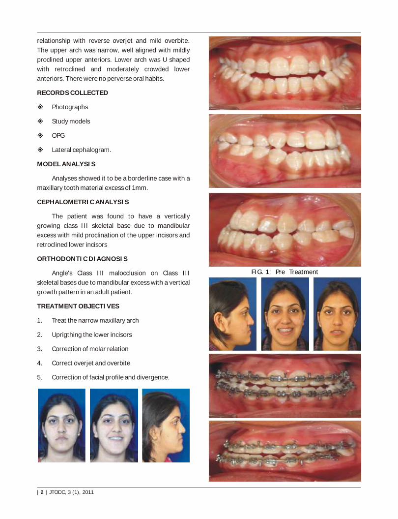
relationship with reverse overjet and mild overbite.
The upper arch was narrow, well aligned with mildly
proclined upper anteriors. Lower arch was U shaped
with retroclined and moderately crowded lower
anteriors. There were no perverse oral habits.
RECORDS COLLECTED
Photographs
Study models
OPG
Lateral cephalogram.
MODEL ANALYSIS
Analyses showed it to be a borderline case with a
maxillary tooth material excess of 1mm.
CEPHALOMETRIC ANALYSIS
The patient was found to have a vertically
growing class III skeletal base due to mandibular
excess with mild proclination of the upper incisors and
retroclined lower incisors
ORTHODONTIC DIAGNOSIS
Angle's Class III malocclusion on Class III
skeletal bases due to mandibular excess with a vertical
growth pattern in an adult patient.
TREATMENT OBJECTIVES
1. Treat the narrow maxillary arch
2. Uprigthing the lower incisors
3. Correction of molar relation
4. Correct overjet and overbite
5. Correction of facial profile and divergence.
| 2 | JTODC, 3 (1), 2011
FIG. 1: Pre Treatment

Phase 1 would consist of non-extraction
presurgical orthodontic treatment. The upper arch
would be leveled and aligned and mildly expanded. In
the lower arch the incisors would be uprighted as
decompensation and the arch would be leveled and
aligned.
Phase 2 would consist of the orthognathic
surgery. A mandibular set-back would be carried out
by performing a bilateral sagittal split osteotomy.
Post-surgical orthodontics would then be
performed to achieve acceptable dental and occlusal
relationship.
To achieve these goals a Pre-adjusted Edgewise
Appliance MBT 0.022 slot was chosen
TREATMENT SEQUENCE
Patient was bonded with a MBT 0.022 pre-
adjusted appliance
Both arches were leveled and aligned using the
regular sequence of wires
Stainless steel wires were sequentially used after
niti wires to achieve the mild expansion of the
upper arch
Lower incisor were allowed to upright by natural
torque expression
Open coil spring was used in the region of 42 to
gain space for its derotation and alignment
On completion of the alignment the patient was
stabilized on 19X25 SS wires
At this stage models were taken and mock
surgery was performed
Acrylic Surgical splints were fabricated after
mock surgery
BSSO was performed to set the mandible back by
7mm
Genioplasty was not performed as per patient's
request to prevent psychological loss of identity
After a brief period of rehabilitation occlusal
settling was done using seating elastics as part of
post-surgical orthodontics
JTODC, 3 (1), 2011 | 1 |
TREATMENT PLAN
Considering the skeletal and dental relationship a
surgical treatment plan was decided upon
FIG. 2: Pre Surgery
FIG. 3: Post Treatmant

| 2 | JTODC, 3 (1), 2011
Patient was debonded when dental, skeletal and
occlusal relationship was found to be satisfactory
Patient was given a lower canine to canine
permanent retainer as well as upper and lower
removable wrap around retainers
Regular follow-up is being continued
CONCLUSION
A successful interdisciplinary treatment of
skeletal Class III malocclusion was performed.
Dentally, the objectives of class I molar and canine
relationship with adequate overjet and overbite and
absence of any form of crossbite was achieved.
Patient's profile shows marked improvement to a
straight profile and divergence. But the foremost
indication of successful treatment, that usually
escapes notice, was the patient's elation.
REFERENCES
1. Angle EH. Classification of malocclusion. Dental
Cosmos. 1899;41:248264
2. Park JU., Baik SH. Classification of Angle Class III
malocclusion and its treatment modalities. Int J
Adult Orthod Orthognath Surg Vol. 16, No.1,
2001
3. Obwegeser H. Profile planning based on
alterations in the positions of the bases of the
facial thirds. J Oral Maxillofac Surg 1986;44:
302311.
4. Toffol LD, Pavoni C, Baccetti T, Franchi L, Cozza
P.Orthopedic treatment outcomes in Class III
malocclusion. A systematic review. Angle Orthod.
2008 May;78(3):561-73
5. Kim BM, Kang BY, Kim HG, Baek SH. Prognosis
prediction for Class III malocclusion treatment by
feature wrapping method. Angle Orthod. 2009
Jul;79(4):683-91
6. Treatment of adult Class III malocclusions with
orthodontic therapy or orthognathic surgery:
receiver operating characteristic analysis. Am J
Orthod Dentofacial Orthop. 2011 May; 139 (5) : e
485 - 93
Measurements Pre-treatment Post-Treatment
ANB -3 1
WITS -9 -1
Effective length of mandible 142 137
Facial axis -7 -4
Y - axis 67 63
FMA 40 35
LAFH 88 87
Inter incisal angle 132 129
UI - SN 108 107
UI - PALATAL PLANE 115 112
L1 - MP 74 81
FMIA 68 63

1 2 3Dr. Uma Eswara , Dr. Priya Subramaniam , Dr. Nandan N
TOOTH FRAGMENT EMBEDDED IN THE LOWER LIP :
A CASE REPORT
ABSTRACT
Traumatic injury to the crown of anterior tooth is very
common and the injury may be complicated with lower lip
lacerations. This case describes of tooth fragments
embedded in the lower lip following dental trauma which was
surgically removed.
Keywords : Dental trauma, lower lip, tooth fragment.
Case Report
Department of Pedodontics
The Oxford Dental College,
Hospital and Research Center,
Bangalore.
1Professor2Professor and HOD
3PG Student
INTRODUCTION
Oro dental injury causes pain and distress to
children and can be very challenging to the dentist.
Coronal fracture after trauma to the permanent
dentition is extremely frequent with prevalence of
15%-90% [1,2,3]. It is often complicated by soft
tissue involvement. A large number of dental traumas
are associated with injuries to lip, gingival and oral
mucosa [1]. It is commonly observed that the impact
of force is directed towards the incisors leading to their
fracture and soft tissue laceration [4]. Tooth fragments
can get embedded in the lip or less commonly in the
tongue [5].
If such fragments are undetected at the time of
emergency treatment, it can lead to infection,
disfiguring fibrosis and may even indicate medical
negligence. Hence it is very important for the clinician
to locate a missing tooth fragment with the help of a
detailed history, careful examination and
investigation, before wound closure is carried out.
Journal Of The Oxford Dental College
Email for [email protected]
| 2 | JTODC, 3 (1), 2011
CASE REPORT
An eight year old girl reported to the Department
of Pedodontics and Preventive Dentistry, with a chief
complaint of broken upper front tooth (Figure-1).
Patient's mother gave a two month old history of
trauma while playing indoors. Following trauma, there
was bleeding and laceration on the inner aspect of
both lips. The girl was unsure of whether she had spat
out the broken tooth fragment along with blood. No
Figure 1 - Fractured upper front tooth

attempt was made to locate the fractured tooth
fragment at the site of accident. Trauma was not
associated with loss of consciousness. The girl was
taken immediately to a general physician who
administered tetanus toxoid, rendered palliative
treatment and prescribed antibiotics and analgesics.
Swelling of the lower lip was observed about 20 days
after the trauma (Figure-2).
Intra oral examination showed a discolored,
fractured permanent maxillary right central incisor
(11), with considerable loss of enamel and dentin. Soft
tissue examination revealed a slight swelling of the
lower lip. On palpation it appeared to be a hard
spherical mass of about 1 cm in diameter and was
tender. Intraoral periapical radiograph of the fractured
incisor showed fracture line closely approximating the
pulp, with two-third (6.5cms) of root formation
complete. Another intra oral periapical radiograph that
was taken by placing a film between the teeth and
lower lip revealed of about 0.5cm, in relation to the
right side.
Apexification of 11 was carried out using Mineral
Trioxide Aggregate followed by root canal obturation
and coronal restoration of 11. Surgical removal of the
intra labial mass was planned under local anesthesia.
Prior to surgery, routine blood investigations were
carried out.
Mental nerve block was given along with local
infiltration all around the hard mass. An intra oral
incision was made below the palpable mass, using a
Bard Parker blade no.15. On reflection of the flap,
small fragments of hard tissue could be palpated.
Since it was enmeshed in the surrounding tissue, a
large spoon excavator was used to remove individual
Figure 2 - Swelling of the lower lip after the trauma
fragments. A small area of surrounding tissue was also
excised. Care was taken not to compress the lip in
order to prevent the occurrence of mucocele. A
radiograph was taken to confirm removal of all
fragments. Closure was done with interrupted sutures
(figure-3). Routine post surgical instructions were
given, with particular attention to diet and oral
hygiene. Patient was prescribed antibiotics and
analgesics.
Figure 3 - Closure done with interrupted sutures
On gross examination, the excised fragments
appeared to be a portion of the fractured tooth
fragment (figure-4). Sections of the fragments for
histopathological examination could not be prepared
because of their small size and brittle nature.
However examination under a binocular microscope
(Polaroid) confirmed the fragments to be part of the
fractured tooth.
Figure 4 - Fractured tooth fragment
JTODC, 3 (1), 2011 | 1 |

Histopathological examination of the excised soft
tissue revealed normal salivary gland tissue. The child
was recalled after a week for suture removal. Clinical
and radiographic evaluation of the patient over two
months has shown successful healing. Post operative
radiograph failed to show evidence of any tooth
fragment remaining in the lower lip.
DISCUSSION
Most foreign bodies are accidental findings and
can be worrying for both parents and clinicians.
Usually a fractured or missed incisor does not pose any
problem in diagnosis. However, when this situation is
added to a soft tissue laceration, attention should be
given to the whereabouts of the teeth[6]. Small tooth
fragments that are embedded in the lower lip are
subjected to continuous movement as a result of
contraction of the orbicularis oris muscle.
Due to the magnitude of soft tissue trauma
associated with minor tooth fracture, the physician
may often be the first to see the child. In many cases,
fragments are overlooked during soft tissue repair in
general hospitals [7]. In the present case, following
trauma the patient was taken to a medical practitioner
who rendered palliative treatment, but did not pay
attention to the lost tooth fragment. Dental surgeons
at hospitals have considerable role to play in alerting
their medical colleagues on the hard and soft tissue
examination which should include the extent of the
wound and verification for foreign bodies. According to
Camilleri [8], there seems to be a greater risk of tooth
fragments being embedded in the lips when the
wound is large. Tooth fragments embedded in the soft
tissue may not be easily detectable clinically. If
laceration and bleeding make the clinical examination
difficult, simple soft tissue and occlusal radiographs
help in their detection. A radiographic examination will
be able to demonstrate a variety of typical foreign
bodies such as tooth fragments, calculus, gravel,
glass, metallic or non metallic restorations and
fragments of paint [4, 7]. Complete removal of all
foreign body is important, so as to prevent infection
disfiguring, scarring and tattooing [9]. Thus any
neglect or failure to locate a lost or missing tooth
fragment at the time of examination can have harmful
sequelae [6].
REFERENCES
1. Rajab LD. Traumatic dental injuries in children
presenting for dental treatment at the
Department of Pediatric Dentistry, Faculty of
dentistry, University of Jordan, 1997-2000. Dent
Traumatol 2003 ; 19;6-11.
2. Andreasen JO, Raven JJ. Epidemiology of
traumatic dental injuries to primary teeth. Int J
Oral Surg 1988; 3: 37-41.
3. Hamdan MA, Rajab LD. Traumatic injuries to
permanent anterior teeth among 12 year old
schoolchildren in Jordan. Community Dent
Health 2003; 20 : 89-93.
4. Wadkar MA, Dhusia HK, Narkhede PR. Foreign
body in the lip: a case report. J Indian Dent Assoc
1986 ; 58: 147-148.
5. Snawder KD, Toole TJ, Bastwi Ae. Broken tooth
fragments embedded in soft tissue. J Dent Child
1979; 46:145.
6. da Silva AC, de Moraes M, Bastos EG, Mooreira
RWF, Passeri LA. Tooth fragment embedded in
the lower lip after dental trauma: case reports.
Dent Traumatol 2005; 21:115-120.
7. Snawder KD, Bastwi Ae, Toole TJ Tooth
fragments lodged in unexpected areas. JAMA
1976; 236:1378-1379.
8. Camilleri GE. Incisor fragment in the lip- report of
case. Oral Surg Oral Mrd Oral Path 1963; 16:
1294-1296.
9. Mcdonnel DG, McKietnam EX. Broken tooh
fragments embedded in the tongue: a case
report. British J of Oral Maxillofacial Surg
1986;24: 464-466.
| 2 | JTODC, 3 (1), 2011
Copyright © 2022 FDOKUMEN