Dynamics and retention of misfolded proteins in native ER membranes
8
© 2000 Macmillan Magazines Ltd articles 288 NATURE CELL BIOLOGY | VOL 2 | MAY 2000 | www.nature.com/ncb Dynamics and retention of misfolded proteins in native ER membranes Sarah Nehls*, Erik L. Snapp*, Nelson B. Cole*, Kristien J.M. Zaal*, Anne K. Kenworthy*, Theresa H. Roberts*, Jan Ellenberg†, John F. Presley*, Eric Siggia‡ and Jennifer Lippincott-Schwartz*§ *Cell Biology and Metabolism Branch, National Institute of Child Health and Human Development, Building 18T, National Institute of Health, Bethesda, Maryland 20892, USA †Gene Expression and Cell Biology Program, European Molecular Biology Laboratory, Heidelberg, Germany ‡Center for Studies in Physics and Biology, Rockefeller University, New York, New York 10021, USA §e-mail: [email protected] When co-translationally inserted into endoplasmic reticulum (ER) membranes, newly synthesized proteins encounter the lumenal environment of the ER, which contains chaperone proteins that facilitate the folding reactions necessary for protein oligomerization, maturation and export from the ER. Here we show, using a temperature-sensitive variant of vesicular stomatitis virus G protein tagged with green fluorescent protein (VSVG–GFP), and fluorescence recovery after photobleaching (FRAP), the dynamics of association of folded and misfolded VSVG complexes with ER chaperones. We also investigate the potential mechanisms underlying protein retention in the ER. Misfolded VSVG–GFP complexes at 40 ° C are highly mobile in ER membranes and do not reside in post-ER compartments, indicating that they are not retained in the ER by immobilization or retrieval mechanisms. These complexes are immobilized in ATP-depleted or tunicamycin-treated cells, in which VSVG–chaperone interactions are no longer dynamic. These results provide insight into the mechanisms of protein retention in the ER and the dynamics of protein-folding complexes in native ER membranes. model system for studying pathways for protein folding and misfolding in the ER and their effects on the retention and export of ER proteins is provided by the temperature-sensi- tive ts045 variant of VSVG, which contains a single lumenal amino- acid change that leads to misfolding and retention within the ER at 40 °C 1–4 . Upon shifting to 32 °C, misfolding is reversed; VSVG then correctly folds and is exported from the ER for delivery to the plasma membrane. Correct folding of VSVG in the ER requires the formation of intrachain disulphide bonds and involves interactions with the folding enzyme protein disulphide isomerase (PDI) and two chaperones, the ATPase BiP (GRP78) and calnexin 5–8 . The chaperone interactions promote the formation of a mature, trans- port-competent form of VSVG that exists as an oxidized (disul- phide-bonded), noncovalently associated homotrimer containing two N-linked sugar chains 7,9 . Biochemical analyses have shown that when misfolded VSVG is retained in the ER at 40 °C, complexes of VSVG–BiP and VSVG– calnexin are formed 6–8 , although the size, organization and dynam- ics of these complexes in native ER membranes are not known. This has made it difficult to distinguish between different models for retention of VSVG in the ER at 40 °C, which include: immobiliza- tion or aggregation of VSVG–chaperone complexes in ER mem- branes; failure of VSVG complexes to be recognized by ER export machinery; and continuous retrieval of misfolded VSVG complexes from a post-ER compartment 10,11 . Here we distinguish between these models of ER retention and investigate the dynamics of VSVG–chaperone interactions by meas- uring the diffusional mobility of folded and misfolded forms of VSVG–GFP 3 that are localized in the ER. The diffusional mobility of VSVG–GFP was quantified using FRAP, in which fluorescent proteins in a small area are irreversibly photobleached by an intense laser flash and fluorescence recovery through the exchange of bleached for nonbleached protein is measured using an attenuated laser beam 12 . Mobility parameters, including the diffusion coeffi- cient, D, and the mobile fraction, can be derived from the kinetics of fluorescence recovery to provide insight into the dynamics of VSVG–GFP complexes. For example, if misfolded VSVG molecules are associated with slowly diffusing ER proteins, form large aggre- gates, or are present in a protein-dense environment, then their apparent D value will be lower than for proteins freely diffusing in a lipid bilayer. Alternatively, if they associate irreversibly with immobile ER components or segregate into membrane sub- domains, their observed mobile fraction value will be low. Our results, obtained using FRAP and repetitive photobleaching techniques, show that both folded and misfolded forms of VSVG– GFP are completely mobile in ER membranes, have a diffusion coef- ficient close to the theoretical limit for protein diffusion in a lipid A Figure 1 Attachment of GFP to ts045 VSVG does not alter its temperature- dependent folding. Cells transfected with VSVG–GFP were incubated for 24 h at 40°C or at 32 °C in the presence of brefeldin A, which blocks ER export of proteins 16 . Cells were then fixed and stained with the conformation-specific monoclonal antibody I14 to identify correctly folded forms of VSVG–GFP. In cells incubated at 32 °C (upper panels), correctly folded VSVG–GFP retained in the ER was recognized by the I14 antibody, as shown by the complete overlap of antibody staining and distribution of the chimaera. In cells incubated at 40 °C (lower panels), VSVG–GFP was not recognized by the I14 antibody, indicating that it is misfolded in the ER at this temperature. Scale bar represents 10 μm. ts045 VSVG–GFP I14 Merged image 32 °C 40 °C
Transcript of Dynamics and retention of misfolded proteins in native ER membranes

articles
Dynamics and retention of misfolded proteins in native ER membranesSarah Nehls*, Erik L. Snapp*, Nelson B. Cole*, Kristien J.M. Zaal*, Anne K. Kenworthy*, Theresa H. Roberts*,
Jan Ellenberg†, John F. Presley*, Eric Siggia‡ and Jennifer Lippincott-Schwartz*§*Cell Biology and Metabolism Branch, National Institute of Child Health and Human Development, Building 18T, National Institute of Health,
Bethesda, Maryland 20892, USA†Gene Expression and Cell Biology Program, European Molecular Biology Laboratory, Heidelberg, Germany
‡Center for Studies in Physics and Biology, Rockefeller University, New York, New York 10021, USA§e-mail: [email protected]
When co-translationally inserted into endoplasmic reticulum (ER) membranes, newly synthesized proteins encounter the lumenal environment of the ER, which contains chaperone proteins that facilitate the folding reactions necessary for protein oligomerization, maturation and export from the ER. Here we show, using a temperature-sensitive variant of vesicular stomatitis virus G protein tagged with green fluorescent protein (VSVG–GFP), and fluorescence recovery after photobleaching (FRAP), the dynamics of association of folded and misfolded VSVG complexes with ER chaperones. We also investigate the potential mechanisms underlying protein retention in the ER. Misfolded VSVG–GFP complexes at 40 °C are highly mobile in ER membranes and do not reside in post-ER compartments, indicating that they are not retained in the ER by immobilization or retrieval mechanisms. These complexes are immobilized in ATP-depleted or tunicamycin-treated cells, in which VSVG–chaperone interactions are no longer dynamic. These results provide insight into the mechanisms of protein retention in the ER and the dynamics of protein-folding complexes in native ER membranes.
model system for studying pathways for protein folding andmisfolding in the ER and their effects on the retention andexport of ER proteins is provided by the temperature-sensi-
tive ts045 variant of VSVG, which contains a single lumenal amino-acid change that leads to misfolding and retention within the ER at40 °C1–4. Upon shifting to 32 °C, misfolding is reversed; VSVG thencorrectly folds and is exported from the ER for delivery to theplasma membrane. Correct folding of VSVG in the ER requires theformation of intrachain disulphide bonds and involves interactionswith the folding enzyme protein disulphide isomerase (PDI) andtwo chaperones, the ATPase BiP (GRP78) and calnexin5–8. Thechaperone interactions promote the formation of a mature, trans-port-competent form of VSVG that exists as an oxidized (disul-phide-bonded), noncovalently associated homotrimer containingtwo N-linked sugar chains7,9.
Biochemical analyses have shown that when misfolded VSVG isretained in the ER at 40 °C, complexes of VSVG–BiP and VSVG–calnexin are formed6–8, although the size, organization and dynam-ics of these complexes in native ER membranes are not known. Thishas made it difficult to distinguish between different models forretention of VSVG in the ER at 40 °C, which include: immobiliza-tion or aggregation of VSVG–chaperone complexes in ER mem-branes; failure of VSVG complexes to be recognized by ER exportmachinery; and continuous retrieval of misfolded VSVG complexesfrom a post-ER compartment10,11.
Here we distinguish between these models of ER retention andinvestigate the dynamics of VSVG–chaperone interactions by meas-uring the diffusional mobility of folded and misfolded forms ofVSVG–GFP3 that are localized in the ER. The diffusional mobilityof VSVG–GFP was quantified using FRAP, in which fluorescentproteins in a small area are irreversibly photobleached by an intenselaser flash and fluorescence recovery through the exchange ofbleached for nonbleached protein is measured using an attenuatedlaser beam12. Mobility parameters, including the diffusion coeffi-cient, D, and the mobile fraction, can be derived from the kineticsof fluorescence recovery to provide insight into the dynamics ofVSVG–GFP complexes. For example, if misfolded VSVG moleculesare associated with slowly diffusing ER proteins, form large aggre-gates, or are present in a protein-dense environment, then their
apparent D value will be lower than for proteins freely diffusing ina lipid bilayer. Alternatively, if they associate irreversibly withimmobile ER components or segregate into membrane sub-domains, their observed mobile fraction value will be low.
Our results, obtained using FRAP and repetitive photobleachingtechniques, show that both folded and misfolded forms of VSVG–GFP are completely mobile in ER membranes, have a diffusion coef-ficient close to the theoretical limit for protein diffusion in a lipid
A
Figure 1 Attachment of GFP to ts045 VSVG does not alter its temperature-dependent folding. Cells transfected with VSVG–GFP were incubated for 24 h at 40 °C or at 32 °C in the presence of brefeldin A, which blocks ER export of proteins16. Cells were then fixed and stained with the conformation-specific monoclonal antibody I14 to identify correctly folded forms of VSVG–GFP. In cells incubated at 32 °C (upper panels), correctly folded VSVG–GFP retained in the ER was recognized by the I14 antibody, as shown by the complete overlap of antibody staining and distribution of the chimaera. In cells incubated at 40 °C (lower panels), VSVG–GFP was not recognized by the I14 antibody, indicating that it is misfolded in the ER at this temperature. Scale bar represents 10 µm.
ts045 VSVG–GFP I14 Merged image
32 °C
40 °C
© 2000 Macmillan Magazines Ltd288 NATURE CELL BIOLOGY | VOL 2 | MAY 2000 | www.nature.com/ncb
articles
bilayer and do not reside in separate ER subdomains or post-ER com-partments. This implies that misfolded VSVG complexes are not nor-mally tethered to an underlying ER scaffold or immobilized within amatrix of aggregated proteins, and that they are not retrieved frompost-ER compartments. Our data thus support the idea that reten-tion of misfolded VSVG molecules in the ER at 40 °C occurs predom-inantly as a result of the failure of these proteins to be recognized byER export machinery. In conditions in which VSVG–chaperoneinteractions become irreversible and extensive, including ATP deple-tion and tunicamycin treatment7,13,14, VSVG–GFP molecules areimmobilized and the diffusional mobility of soluble ER proteins isaltered. These results indicate that when interactions between mis-folded proteins and ER chaperones are not dynamic, the diffusionalmobility of many proteins in the ER becomes restricted, perhapsthrough the formation of an extensive ER matrix.
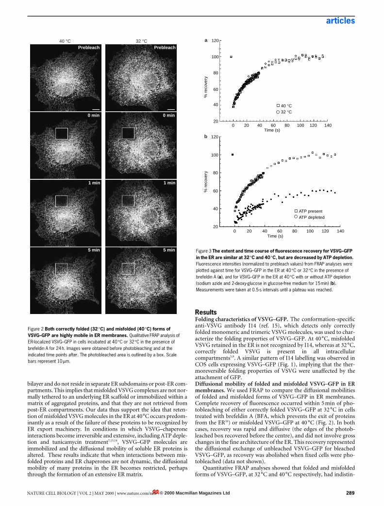
ResultsFolding characteristics of VSVG–GFP. The conformation-specificanti-VSVG antibody I14 (ref. 15), which detects only correctlyfolded monomeric and trimeric VSVG molecules, was used to char-acterize the folding properties of VSVG–GFP. At 40 °C, misfoldedVSVG retained in the ER is not recognized by I14, whereas at 32 °C,correctly folded VSVG is present in all intracellularcompartments5,9. A similar pattern of I14 labelling was observed inCOS cells expressing VSVG–GFP (Fig. 1), implying that the ther-moreversible folding properties of VSVG were unaffected by theattachment of GFP. Diffusional mobility of folded and misfolded VSVG–GFP in ERmembranes. We used FRAP to compare the diffusional mobilitiesof folded and misfolded forms of VSVG–GFP in ER membranes.Complete recovery of fluorescence occurred within 5 min of pho-tobleaching of either correctly folded VSVG–GFP at 32 °C in cellstreated with brefeldin A (BFA, which prevents the exit of proteinsfrom the ER16) or misfolded VSVG–GFP at 40 °C (Fig. 2). In bothcases, recovery was rapid and diffusive (the edges of the photob-leached box recovered before the centre), and did not involve grosschanges in the fine architecture of the ER. This recovery representedthe diffusional exchange of unbleached VSVG–GFP for bleachedVSVG–GFP, as recovery was abolished when fixed cells were pho-tobleached (data not shown).
Quantitative FRAP analyses showed that folded and misfoldedforms of VSVG–GFP, at 32 °C and 40 °C respectively, had indistin-
Figure 2 Both correctly folded (32 °C) and misfolded (40 °C) forms of VSVG–GFP are highly mobile in ER membranes. Qualitative FRAP analysis of ER-localized VSVG–GFP in cells incubated at 40 °C or 32 °C in the presence of brefeldin A for 24 h. Images were obtained before photobleaching and at the indicated time points after. The photobleached area is outlined by a box. Scale bars represent 10 µm.
40 °C 32 °CPrebleach Prebleach
0 min
1 min
5 min
0 min
1 min
5 min Figure 3 The extent and time course of fluorescence recovery for VSVG–GFP in the ER are similar at 32 °C and 40 °C, but are decreased by ATP depletion. Fluorescence intensities (normalized to prebleach values) from FRAP analyses were plotted against time for VSVG–GFP in the ER at 40 °C or 32 °C in the presence of brefeldin A (a), and for VSVG–GFP in the ER at 40 °C with or without ATP depletion (sodium azide and 2-deoxy-glucose in glucose-free medium for 15 min) (b). Measurements were taken at 0.5-s intervals until a plateau was reached.
% r
ecov
ery
Time (s)
ATP presentATP depleted
120
100
80
60
40
200 20 40 60 80 100 120 140
b
20
40
60
80
100
120
0 20 40 60 80 100 120 140
% r
ecov
ery
Time (s)
40 °C32 °C
a
© 2000 Macmillan Magazines LtdNATURE CELL BIOLOGY | VOL 2 | MAY 2000 | www.nature.com/ncb 289

articles
guishable D values (0.4–0.5 µm2 s–1) with both forms showing 100%mobility (Fig. 3a, Table 1). D values did not vary with the durationof the non-permissive temperature (data not shown), the presenceor absence of BFA (Table 1) or the overall level of expression ofVSVG–GFP (data not shown). Values for D and mobile fraction ofVSVG–GFP were comparable to those of other GFP-tagged pro-teins residing in ER membranes, including lamin-B receptor (LBR),the β-subunit of the signal-recognition-particle receptor (SRβ) andthe Golgi enzyme galactosyltransferase (GalT) in cells treated withBFA (Table 1). They were also similar to those of other GFP-taggedproteins present in the Golgi complex17, all of which had diffusionalmobilities at 32 °C and 40 °C that were close to the theoretical limitfor proteins in a lipid bilayer18,19. Mechanism of retention of VSVG–GFP in the ER at 40 °C. Giventhat misfolded VSVG–GFP molecules diffuse at a rate close to theviscosity limit in ER membranes, it is clear that these molecules arenot retained in the ER at 40 °C by mechanisms involving protein
immobilization. Two alternative mechanisms of protein retentionin the ER are selective retrieval of proteins from post-ER compart-ments, and failure of proteins to be recognized by ER exportmachinery. To distinguish between these two models, we repeti-tively photobleached a 15-µm-square box across the ER while mon-itoring fluorescence throughout the cell. This allowed us todetermine whether VSVG–GFP cycles through separate, discontin-uous post-ER structures (as predicted by the retrieval model) or dif-
Table 1 D and mobile fraction values for ER-localized GFP chimaeras.Chimaera Temp. (°C) Treatment D (µm2 s–1) ±
s.d.Mf ± s.d. n
VSVG–GFP 32 BFA 0.49 ± 0.06 99 ± 4.9 13VSVG–GFP 40 None 0.45 ± 0.03 102 ± 3.5 23VSVG–GFP 40 BFA 0.42 ± 0.03 108 ± 3.6 8LBR–GFP 32 None 0.41 ± 0.10 97 ± 3.9 7LBR–GFP 40 None 0.50 ± 0.19 93 ± 4.5 9SRβ–GFP 32 None 0.26 ± 0.03 93 ± 6.4 12GalT–GFP 32 BFA 0.48 ± 0.03 94 ± 5.5 7GalT–GFP 40 BFA 0.42 ± 0.06 107 ± 6.6 7Means ± s.d. of values for D and mobile fraction (Mf), for recovery of fluorescence afterphotobleaching of cells expressing GFP chimaeras under the indicated treatment conditions.Statistical analyses of D and Mf values were carried out using a two-tailed student t-test. Nosignificant differences were observed for VSVG, LBR or GalT at different temperatures.
Figure 4 Repetitive photobleaching of cells expressing KDELR–GFP and VSVG–GFP reveals different mechanisms for their retention in the ER. A 15 × 15 µm box was repeatedly bleached in cells expressing KDELR–GFP (upper panels) or VSVG–GFP (lower panels) at 40 °C. Between each bleach, the entire field of view was imaged at low laser power to determine the extent of fluorescence outside the box that was lost as a result of photobleaching within the box. After repeated bleaching, cells expressing KDELR–GFP contain residual fluorescent structures
(arrows) and show a loss of ER fluorescence, indicating that recycling of KDELR–GFP back to the ER from pre-Golgi and Golgi structures occurs more slowly than lateral diffusion of KDELR–GFP across the ER lipid bilayer. In contrast, the complete bleaching of the ER and absence of residual fluorescent structures in cells expressing VSVG–GFP indicates that highly mobile, misfolded VSVG–GFP complexes at 40 °C never leave the ER. Scale bar represents 10 µm.
KDELR–GFP
VSVG–GFP
Prebleach
Prebleach 0.4 min
0.4 min
8.5 min
8.5 min
21.1 min
21.1 min
Figure 5 Cells expressing KDELR–GFP contain more residual fluorescent structures than cells expressing VSVG–GFP after repeated photobleaching. Pixel-intensity variance (see Methods) in cells expressing VSVG–GFP or KDELR–GFP was plotted against time. Values are means ± s.e.m. from three separate cells. A lower value indicates an increasingly homogeneous population of pixels of similar intensities.
VSVG–GFPKDELR–GFP
100
80
60
40
20
00 5 10 15 20 25
Pix
el-in
tens
ity v
aria
nce
(s.d
.)
Time (min)
© 2000 Macmillan Magazines Ltd290 NATURE CELL BIOLOGY | VOL 2 | MAY 2000 | www.nature.com/ncb
articles
fuses only within the ER (consistent with the export-defect model).In the retrieval model, repetitive photobleaching would leave pock-ets of fluorescence corresponding to VSVG–GFP in post-ER com-partments, assuming that recycling of VSVG–GFP occurs moreslowly than its lateral diffusion across the ER lipid bilayer. This pat-tern was observed for KDEL receptor tagged with GFP (KDELR–GFP)17, which rapidly cycles between ER and Golgi membranes(Figs 4, 5; arrows in Fig. 4 denote residual KDELR–GFP fluores-cence in post-ER compartments). In the export-defect model,repetitive photobleaching of the ER would remove all fluorescencefrom the cell, assuming that ER membranes are continuous20 andthat all VSVG–GFP molecules in the ER are mobile. Consistent withthis model, we observed uniform loss of VSVG–GFP fluorescence at40 °C (Figs 4, 5). Retention of VSVG–GFP in the ER at 40 °C thusseems to be a result of failure to efficiently export VSVG-GFP mol-ecules.Immobilization of VSVG–GFP in ATP-depleted cells. The rapidmobility of misfolded VSVG–GFP at 40 °C suggests that VSVG–chaperone complexes formed at this temperature (includingVSVG–BiP and VSVG–calnexin) are either too small to affect thediffusional mobility of VSVG or are highly dynamic (that is, theirformation is reversible). In ATP-depleted cells, BiP (an ATP-dependent chaperone)21 is persistently bound to VSVG22, leading tointerchain crosslinking and aggregation of VSVG7,8. In FRAP anal-yses of cells deprived of ATP for 15–30 min, VSVG–GFP was immo-bilized at 40 °C (Fig. 3b, Table 2), with no effect on the D value ofthe mobile pool. Immobilization was largely reversed by returningcells to normal (ATP-containing) medium for 20 min (Table 2).ATP depletion had no influence on either the mobile fraction or Dvalues of another membrane protein, GalT–GFP, which was local-ized to the ER using BFA (Table 2). VSVG–GFP was only immobi-lized in the specialized folding environment of the ER, as there wasno change in recovery or mobility of VSVG–GFP during ATPdepletion at 32 °C in cells in which VSVG–GFP was localized in theGolgi complex (data not shown).
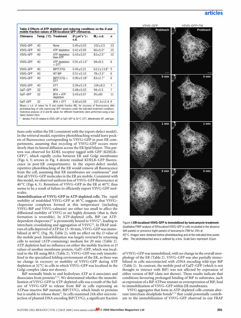
BiP normally binds to and hydrolyses ATP as it associates anddissociates from proteins21,22. We determined whether the immobi-lization of VSVG–GFP in ATP-depleted cells was a result of the fail-ure of VSVG–GFP to release from BiP in cells expressing anATPase-inactive BiP mutant, BiP(T37G), which binds to proteinsbut is unable to release them23. In cells examined 24 h after microin-jection of plasmid DNA encoding BiP(T37G), a significant fraction
of VSVG–GFP was immobilized, with no change in the overall mor-phology of the ER (Table 2). VSVG–GFP was also partially immo-bilized in cells microinjected with cDNA encoding wild-type BiP(Table 2). In contrast, the mobile pool of GalT–GFP (which is notthought to interact with BiP) was not affected by expression ofeither version of BiP (data not shown). These results indicate thatconditions favouring prolonged binding of BiP to substrates, suchas expression of a BiP ATPase mutant or overexpression of BiP, leadto immobilization of VSVG–GFP within ER membranes.
VSVG aggregates that form in ATP-depleted cells contain aber-rant interchain disulphide bonds5,7,9 that could potentially contrib-ute to the immobilization of VSVG–GFP observed in our FRAP
Figure 6 ER-localized VSVG–GFP is immobilized by tunicamycin treatment. Qualitative FRAP analysis of ER-localized VSVG–GFP in cells incubated in the absence (left panels) or presence (right panels) of tunicamycin (TM) for 24 h at 40 °C. Images were obtained before photobleaching and at the indicated time points after. The photobleached area is outlined by a box. Scale bars represent 10 µm.
VSVG–GFP VSVG–GFP+TM
Prebleach Prebleach
0 min 0 min
1 min 1 min
5 min 5 min
Table 2 Effects of ATP depletion and reducing conditions on the D and mobile fraction values of ER-localized GFP chimaeras.Chimaera Temp. (°C) Treatment D (µm2 s–1) ±
s.d.Mf ± s.d. n
VSVG–GFP 40 None 0.45 ± 0.03 102 ± 3.5 23VSVG–GFP 40 ATP depletion 0.41 ± 0.05 66 ± 5.0*
* denotes P ≤0.05 relative to VSVG–GFP or GalT–GFP at 32 °C. DTT, dithiothreitol; WT, wild type.
15VSVG–GFP 40 ATP depletion
then ATP0.43 ± 0.07 83 ± 2.5* 10
VSVG–GFP 40 ATP depletion + DTT
0.91 ± 0.13* 94 ± 8.3 6
VSVG–GFP 40 BiP(T37G) 0.45 ± 0.21 63.2 ± 13.8* 5VSVG–GFP 40 WT BiP 0.51 ± 0.10 78 ± 3.3* 6VSVG–GFP 40 BiP(T37G) +
DTT0.90 ± 0.18* 83 ± 2.7* 6
VSVG–GFP 40 DTT 0.59 ± 0.19 104 ± 8.0 7GalT–GFP 32 BFA 0.48 ± 0.03 94 ± 5.5 7GalT–GFP 32 BFA + ATP
depletion0.43 ± 0.07 95 ± 60 11
GalT–GFP 32 BFA + DTT 0.60 ± 0.09 107.3 ± 12.4 6Means ± s.d. of values for D and mobile fraction (Mf), for recovery of fluorescence afterphotobleaching of cells expressing GFP chimaeras under the indicated treatment conditions.Statistical analyses of D and Mf values for different treatments were performed using a two-tailed student t-test.
© 2000 Macmillan Magazines LtdNATURE CELL BIOLOGY | VOL 2 | MAY 2000 | www.nature.com/ncb 291

articles
analyses. Within 15 min of the addition of dithiothreitol, whichreduces disulphide bonds and inhibits their formation24,25, to ATP-depleted cells or cells expressing BiP(T37G), immobilization ofVSVG–GFP was reversed and D was increased by a factor of 2.2 rel-ative to control values (Table 2). Despite their mobilization whentreated with dithiothreitol, VSVG–GFP molecules were not recog-nized by the I14 antibody, implying they were still misfolded (datanot shown). These results indicate that the formation of disulphidebonds is important for VSVG–GFP immobilization under condi-tions of ATP depletion or BiP(T37G) overexpression. Reduction ofdisulphide bonds had a general effect on the diffusion of membraneproteins in the ER, as the D value of GalT–GFP (which contains nointrachain disulphide bonds17) localized to the ER using BFA wasincreased after treatment with dithiothreitol (Table 2).Immobilization of VSVG–GFP in tunicamycin-treated cells.Sugar moieties are important for increasing the solubility of proteinsin the ER to facilitate folding and reduce aggregation26,27. VSVG con-tains two N-linked sugar chains and requires these oligosaccharidesto fold correctly and be exported from the ER6–8. VSVG–GFP wasimmobilized in cells treated for 24 h at 40 °C with tunicamycin, whichblocks the synthesis of dolichol-linked oligosaccharides and therebyinhibits the addition of N-linked sugars to proteins (Figs 6, 7).Roughly 50% of VSVG–GFP was immobile in these cells and the Dvalue of the mobile pool was reduced by a factor of 1.5 relative to con-trols (Fig. 7, Table 3). Addition of dithiothreitol to cells treated with
tunicamycin did not significantly reverse the immobilization pheno-type, although the D value of the mobile pool increased under theseconditions. It therefore seems that, in the absence of N-linked sugars,VSVG-GFP is not freely mobile in ER membranes and undergoesaggregation that cannot be reversed by reducing conditions. Immo-bilization of proteins by tunicamycin treatment was specific to pro-teins that are normally glycosylated, as GalT–GFP (which contains noN-linked sugars17) in cells treated with tunicamycin was only slightlyimmobilized and its D value was increased by a factor of 1.8 relativeto controls (Table 3).Effect of ATP depletion and tunicamycin on the ER lumenal envi-ronment. The changes in diffusional mobility of VSVG–GFP local-ized to the ER, involving disruption of protein-folding pathways,led us to ask how the overall ER environment is affected under theseconditions. To address this question, we used a soluble probe tar-geted to the ER lumen, comprising GFP carrying an ER signalsequence (ss–GFP). In untreated cells, ss–GFP diffused significantlyfaster than proteins embedded in ER membranes, completely recov-ering into a photobleached box within 8 s of photobleaching (Fig. 8).ATP depletion resulted in slower diffusion of ss–GFP in the ER thanin control cells as well as a decrease in its mobile fraction value (Fig.9a, b). The effect of ATP depletion was reversed upon the return ofcells to normal medium (data not shown), but was unaffected by
Table 3 Effect of tunicamycin treatment on the D and mobile fraction values of ER-localized GFP chimaeras.Chimaera Temp. (°C) Treatment D (µm2 s–1) ±
s.d.Mf ± s.d. n
VSVG–GFP 40 None 0.45 ± 0.03 102 ± 3.5 23VSVG–GFP 40 TM 0.30 ± 0.04*
* denotes P ≤0.05 relative to VSVG–GFP at 40 °C or GalT–GFP at 32 °C. TM, tunicamycin; DTT,dithiothreitol.
50 ± 2.9* 12VSVG–GFP 40 TM + DTT 0.47 ± 0.10 60 ± 3.2* 9GalT–GFP 32 BFA 0.48 ± 0.03 94 ± 5.5 7GalT–GFP 32 BFA + TM 0.78 ± 0.10* 81 ± 2.2 6Means ± s.d. of values for D and mobile fraction (Mf), for recovery of fluorescence afterphotobleaching of cells expressing GFP chimaeras under the indicated treatment conditions.Statistical analyses of D and Mf values for different treatments were carried out using a two-tailed student t-test.
Figure 7 Immobilization of VSVG–GFP by tunicamycin treatment. Fluorescence intensities from FRAP analyses were plotted against time for VSVG–GFP in the ER at 40 °C for 24 h in the presence or absence of tunicamycin (TM). Measurements were taken at 0.5-s intervals until a plateau was reached and fluorescence intensities were normalized to prebleach values.
% r
ecov
ery
120
100
80
60
40
20
0 20 40 60 80 100 120 1400
Time (s)
VSVG–GFPVSVG–GFP+TM
Figure 8 Diffusional mobility of the soluble ER protein ss–GFP is affected by inhibition of N-linked glycosylation. Qualitative FRAP analysis of ss–GFP in cells incubated at 40 °C for 24 h in the absence (left panels) or presence (right panels) of tunicamycin (TM). Images were obtained before photobleaching and at the indicated time points after. In tunicamycin-treated cells, partial recovery is evident immediately after photobleaching, in contrast to untreated cells. Scale bars represent 10 µm.
SS–GFP ss–GFP+TMPrebleach Prebleach
0 s
0.7 s
8.0 s
0 s
0.7 s
8.0 s
© 2000 Macmillan Magazines Ltd292 NATURE CELL BIOLOGY | VOL 2 | MAY 2000 | www.nature.com/ncb

articles
incubation of cells with dithiothreitol, either before or during ATPdepletion (Fig. 9b). In contrast, ss–GFP diffused significantly fasterin cells treated with tunicamycin than in untreated cells (Figs 8, 9a,b). These results indicate that whereas energy depletion may causelumenal proteins to diffuse more slowly, possibly because of exten-sive crosslinking of BiP-containing protein aggregates within the ERlumen, tunicamycin treatment may markedly increase their mobil-ity. As ss–GFP does not contain sugar moieties, these results indi-cate that N-linked sugars on ER glycoproteins may be important indetermining the viscous properties of the ER lumen. Without suchsugar moieties, the ER lumen seems to be less viscous.
DiscussionWe have shown that VSVG–GFP diffuses freely and rapidlythroughout the ER when properly folded at 32 °C or misfolded at40 °C. This implies that misfolded VSVG–GFP complexes at 40 °Care not impeded by lumenal or cytoplasmic interactions, orrestricted diffusion through a matrix. What then explains the ER-retention phenotype of these molecules at 40 °C? Although selec-tive retrieval from post-ER compartments underlies the ER local-ization of several membrane proteins including ERGIC-53, p58and KDELR28–30, our data indicate that VSVG–GFP may beretained in the ER by a different mechanism. Repetitive photob-leaching of a small area in the ER containing VSVG–GFP at 40 °Celiminated cellular fluorescence (including the nuclear envelope)with no pockets of fluorescence remaining. This contrasts withresults for cells expressing the rapidly recycling molecule KDELR–GFP, in which an identical bleaching protocol left pockets of flu-orescence representing KDELR–GFP molecules that had not yet
recycled back to the ER. These results indicate that misfoldedVSVG–GFP complexes may be retained in the ER at 40 °C eitherby failing to be released from dynamically interacting folding fac-tors or by failing to be recognized by ER export machinery.
At 40 °C the association of misfolded VSVG with ER chaperones,including BiP and calnexin, is enhanced6–8. Why is there no differencein the diffusional mobility of these complexes and of correctly foldedVSVG–GFP complexes in the ER at 32 °C? The most likely explana-tions are that VSVG–chaperone complexes that form at 40 °C areeither too small to affect the diffusional mobility of VSVG, or that theformation of these complexes is highly reversible. The apparent D val-ues (0.4–0.5 µm2 s–1) for misfolded and correctly folded VSVG–GFPcomplexes in the ER, at 40 °C and 32 °C respectively, were similar tothose for other ER localized membrane proteins, including GalT, SRβ,LBR (Table 1), and major histocompatibility complex (MHC) class Iproteins31, all of which have diffusional mobilities close to the theoret-ical limit for proteins in a lipid bilayer. They are higher than the D val-ues reported for ER-localized TAP31 and cytochrome P450 (ref. 32).The lower D value for TAP may reflect the large size of TAP com-plexes, which are involved in peptide loading of MHC class Iproteins33. These complexes have an estimated diatmeter of 600–1,000Å and are thought to consist of hundreds of molecules31. The rate oflateral diffusion of a protein embedded in a bilayer is proportional tothe logarithm of the radius of the diffusing molecule, so VSVG–GFPcomplexes (which have a twofold higher D value than TAP) must beat least an order of magnitude smaller than TAP complexes.
When we prevented VSVG from dissociating from chaperonesthrough ATP depletion, which causes interchain crosslinking andaggregation of VSVG complexes7, a significant pool of VSVG–GFPmolecules became immobilized. There are several possible explana-tions for this. The VSVG aggregates could be of variable size andsome may therefore be too big to diffuse. Alternatively, mobileVSVG–GFP aggregates may form a crosslinked network in whichboundaries in some areas become closed, preventing diffusion ofother VSVG–GFP aggregates across the area. However, this wouldbe expected to result in entrapment of other membrane proteins andour data showed no effect of ATP depletion on the D or mobile frac-tion values for ER-localized GalT–GFP. A third possibility is thatATP depletion leads to the association of VSVG with other proteins,including BiP, calnexin and PDI, and their interacting substrates,which are either already relatively immobile in the ER or becomeimmobile under ATP depletion by crosslinking into a scaffold.
To explore the third possibility we investigated the function ofBiP, an ATP-dependent chaperone that forms complexes withVSVG at 40 °C5,7, in the immobilization of VSVG–GFP during ATPdepletion. ATP depletion causes BiP to bind persistently to VSVGas well as to other substrates5,7,22. This may explain why VSVG–GFPis immobilized in ATP-depleted cells, as prolonged BiP bindingmay cause VSVG to be present in a more extended conformation,allowing crosslinking with other proteins. This idea is supported byour finding that conditions, other than ATP depletion, that favourprolonged binding of BiP to substrates, including expression of aBiP ATPase mutant and overexpression of BiP, also lead to immo-bilization of VSVG–GFP molecules in the ER.
Interchain disulphide bonds within VSVG aggregates, whichform in ATP-depleted cells7,13, are necessary to immobilize VSVG–GFP in ATP-depleted cells, as reduction of these disulphide bondsthrough treatment with dithiothreitol reversed the immobilizationcaused by either ATP depletion or overexpression of the BiPmutant. Dithiothreitol treatment alone increased the diffusioncoefficient of GalT–GFP (which does not contain disulphidebonds) in ER membranes. This may reflect a general effect of thereduction of disulphide bonds on membrane viscosity, or a moredirect effect of proteins no longer associating in dynamiccrosslinked aggregates.
We found that treatment of cells with tunicamycin also inhibitsthe diffusional mobility of VSVG–GFP. The immobile pool underthese conditions may result from the formation of large aggregates
Figure 9 Effects of ATP depletion, tunicamycin and dithiothreitol on mobility of ss–GFP. a, Fluorescence intensities from quantitative FRAP analyses were plotted against time for ss–GFP in the ER at 40 °C with no treatment, after 24 h of tunicamycin (TM) treatment, or after 15 min of ATP depletion. b, Half-time values for recovery of fluorescence after photobleaching of cells expressing ss–GFP in the ER lumen subjected to the indicated treatments (see Methods). Lower values indicate faster recoveries. Values are means from at least six cells. Values for the different treatment conditions were compared to those from untreated cells using a two-tailed student t-test. * denotes P ≤0.01; DTT, dithiothreitol.
ss–GFP minus ATPss–GFP+TMss–GFP
100
80
60
40
20
0
% r
ecov
ery
0 1 2 3 4Time (s)
a
Untreated
ATP depletion
ATP depletion then DTT
**
**
DTT then ATP depletion
TM
0 0.2 0.4 0.6 0.8Half-time (s)
b
© 2000 Macmillan Magazines LtdNATURE CELL BIOLOGY | VOL 2 | MAY 2000 | www.nature.com/ncb 293

articles
of misfolded VSVG, as N-linked sugars have an important functionin the folding pathways of VSVG and of other proteins in the ER26,27.If so, these aggregates would seem to be distinct from VSVG–GFPaggregates formed in ATP-depleted cells, as treatment with dithio-threitol reversed the immobilization phenotype in ATP-depletedbut not tunicamycin-treated cells. The observed increase in the Dvalue of GalT–GFP, which is not known to be a substrate of BiP orto contain N-linked sugars, during tunicamycin treatment may bea result of a less crowded lumenal environment, caused either byenhanced aggregation of normally N-glycosylated proteins or bythe absence of carbohydrate side chains on glycoproteins. The smallimmobile pool of GalT–GFP under these conditions may resultfrom GalT–GFP being trapped in networks of immobile proteinaggregates.
To investigate how the ER lumenal environment, which isthought to be a viscous gelatinous mass without internal structure34,is affected by ATP depletion and tunicamycin treatment, we meas-ured the diffusional mobility of a soluble ER protein, ss–GFP, underthese conditions. In untreated cells, the half-time for recovery of ss–GFP into a photobleached box was extremely rapid, many timesfaster than that for recovery of VSVG–GFP into a box of similarsize. This indicates that movement in the ER lumenal space maynormally be unrestricted, and is consistent with the previous find-ing that the rate of diffusion of KDEL–GFP in the ER lumen isslightly lower than that of cytoplasmic solutes20. However, when theassociation of chaperones with ER proteins is enhanced by ATPdepletion or blocking of oligosaccharide addition onto glycopro-teins by tunicamycin treatment, there are concomitant globaleffects on the ER lumenal environment.
Whereas ATP depletion caused ss–GFP to diffuse moreslowly, tunicamycin treatment caused it to diffuse much morequickly. Our results from tunicamycin treatment indicate thatthe presence of oligosaccharide side chains on proteins may be animportant factor in determining the lumenal viscosity of the ER.Branched oligosaccharides on proteins are large (roughly 20 Å inlength) and extend through volumes of ~104 Å3 (refs 12, 35).They are therefore likely to occupy a significant volume of the ERlumen and to restrict diffusion through their polymer network.This is supported by our finding that proteins diffuse much fasterwhen oligosaccharides are absent from proteins in tunicamycin-treated cells.
Diffusion of ss–GFP is slowed in ATP-depleted cells. Thisindicates that the ER lumen may be dynamic and capable offorming a dense protein mesh, which impedes the diffusion ofsoluble ER proteins, when chaperone–protein interactions arepromoted. This matrix may be composed of the lumenal moietiesof membrane proteins such as VSVG, which, through prolongedbinding to BiP, may form aberrant crosslinks with proteins thatextend deep into the ER lumen. Further study is needed to under-stand the regulation of such an ER lumenal matrix, includinghow its density is controlled so that soluble markers change theirmobility. The quantitative methods described here for determin-ing the mobility of proteins in the ER may be useful in answeringthese questions. They may also be important in investigating fur-ther the ER characteristics that underlie the quality-control func-tions of protein folding and maturation36,37 and protein-unfolding responses38. h
MethodsCell lines, antibodies and reagents.COS-7 cells were grown in DMEM (Biofluids, Rockville, MD) supplemented with 10% fetal bovine
serum (FBS), 2 mM glutamine, 100 U ml–1 penicillin and 100 µg ml–1 streptomycin at 37 °C in a 5% CO2 incubator and were used in all experiments. I14 [1E9F9] antibodies were kindly provided by D. Lyles
(Wake Forest University, Winston-Salem, NC). BFA (Epicentre Technologies, Madison, WI) was used at
5 µg ml–1. Cellular ATP levels were depleted using 50 mM 2-deoxy-glucose and 0.02% sodium azide in glucose-free medium. Tunicamycin (Sigma) was used at 5 µg ml–1 for 24 h to block addition of
oligosaccharide chains to proteins, and dithiothreitol (Sigma) was used at 5 mM for 15 min to reduce
disulphide bonds. cDNA (AU: OK?) plasmids encoding BiP and BiP(T37G) were kindly provided by L. Hendershot (University of Tennessee Medical Center, Memphis, TN).
Construction and expression of GFP chimaeras.Cloning and expression of VSVG–GFP, LBR–GFP, GalT–GFP and KDELR–GFP were carried out as
described3,17, 39. The SRβ–GFP construct comprises the mouse cDNA for SRβ fused to the GFP(S65T)
variant in the mammalian expression vector pcDNA1.1 (Invitrogen). The ss–GFP construct, cloned into
the mammalian expression vector pCDM8, encodes the first 19 amino acids of hen egg lysozyme fused
to GFP, omitting its initiation methionine residue. Cells were transfected with chimaeric DNA by
electroporation as described17, typically incubated at 37 °C for 24 h and then transferred to 40 °C or 32 °C
in BFA as indicated. Alternatively, cells at 80–90% confluence in 6-well plates were transfected with 1 µg
plasmid DNA with FuGENE6 transfection reagent (Boehringer) and incubated at 40 °C. For the effect of
BiP(T37G) expression, cells expressing VSVG–GFP were microinjected with 2 mg ml–1 plasmid DNA and
2 mg ml–1 rhodamine dextran for identification of injected cells. Cells were fixed in 2% formaldehyde and
stained with I14 antibodies as described16
.
FRAP analyses.Analyses were performed on a temperature-controlled stage of a Zeiss LSM410 confocal microscope,
using the 488-nm line of a 400-mW Ar/Kr laser with a 100×, 1.4 NA objective. A defined region (outlined
box in figures) was photobleached at full laser power (100% power, 100% transmission, 30 s); recovery
of fluorescence was monitored by scanning the whole cell at low laser power (30% power, 0.3%
transmission). No photobleaching was observed during recovery.
Quantitative diffusion measurements, for generating recovery plots, and D values were obtained by
photobleaching the entire depth of a 4-µm-wide strip extending across cell borders as described17.
Repetitive photobleaching experiments were carried out at 40 °C on the temperature-controlled stage of
a Zeiss LSM410 confocal microscope as described17.
To quantify the presence of residual fluorescent structures in cells subjected to repetitive
photobleaching (Fig. 5), standard deviations of pixel intensity within a 15 µm × 15 µm area were measured
using NIH Image 1.62, in which one pixel represents 0.25µm × 0.25 µm. A larger s.d. corresponds to a
wider range of pixel-intensities. Thus, an area containing both bright structures and dim surrounding
pixels would have a large s.d. Conversely, a smaller s.d. corresponds to a relatively homogenous
population of pixels of similar intensities. Values were normalized for each FLIP series by dividing by the
s.d. at time zero (prebleach) for each cell and multiplying by 100. This process was carried out for three
cells expressing KDELR–GFP and three cells expressing VSVG–GFP. Means ± s.e.m. for the three cells at
each time point were plotted. The area evaluated did not include the Golgi, nucleus, or bleached box.
Recovery of the ss–GFP chimera was too fast to measure accurately using the Zeiss LSM410 confocal
microscope. Quantitative FRAP measurements for this protein were therefore made using a Zeiss 510
confocal microscope with a 40 ×, 1.3 NA objective. We compared relative recovery rates for ss–GFP using
the half-time for recovery of fluorescence towards the asymptote. As for D-value calculations, time was
corrected by setting time zero as equal to the half-time of the bleach. Qualitative FRAP analyses were
carried out by photobleaching a defined region (outlined box in Figs) at full laser power (100% power,
100% transmission) and then monitoring recovery of fluorescence by scanning the defined region at low
laser power (100% power, 0.3% transmission).
For all quantitative FRAP analyses, values for D, half-time and mobile fraction were compared with
control conditions for each chimaera for significance using a two-tailed student t-test. Values of P <0.05
were considered significant.
RECEIVED 9 SEPTEMBER 1999; REVISED 9 FEBRUARY 2000; ACCEPTED 16 MARCH 2000; PUBLISHED 7 APRIL 2000.
1. Kreis, T. E. & Lodish, H. F. Oligomerization is essential for transport of vesicular stomatitis viral
glycoproteins to the cell surface. Cell 46, 929–937 (1986).
2. Bergmann, J. E. Using temperature-sensitive mutants of VSV to study membrane protein biogenesis.
Methods Cell Biol. 32, 85–110 (1989).
3. Presley, J. F. et al. ER-to-Golgi transport visualized in living cells. Nature 389, 81–85 (1997).
4. Doms, R. W., Keller, D. S., Helenius, A. & Balch, W. E. Role of adenosine triphosphate in regulating
the assembly and transport of vesicular stomatitis virus G protein trimers. J. Cell Biol. 105, 1957–1969
(1987).
5. Machamer, C. E., Doms, R. W., Bole, D. G., Helenius, A. & Rose, J. K. Heavy chain binding protein
recognizes incompletely disulfide-bonded forms of vesicular stomatitis virus G protein. J. Biol. Chem.
265, 6879–6883 (1990).
6. De Silva, A., Balch, W. E. & Helenius, A. Quality control in the endoplasmic reticulum: folding and
misfolding of vesicular stomatitis virus G protein in cells and in vitro. J. Cell Biol. 111, 857–866
(1990).
7. De Silva, A., Braakman, I. & Helenius, A. Post-translational folding of vesicular stomatitis virus G
protein in the ER: involvement of noncovalent and covalent complexes. J. Cell Biol. 120, 647–655
(1993).
8. Hammond, C. & Helenius, A. Folding of VSVG protein: sequential interaction with BiP and calnexin.
Science 266, 456–458 (1994).
9. Doms, R. W., Ruusala, A., Machamer, C., Helenius, A. & Rose, J. K. Differential effects of mutations
in three domains on folding, quaternary structure, and intracellular transport of vesicular stomatitis
virus G protein. J. Cell Biol. 107, 89–99 (1988).
10. Hammond, C. & Helenius, A. Quality control in the secretory pathway. Curr. Opin. Cell Biol. 7, 525–
539 (1995).
11. Hammond, C. & Helenius, A. Quality control in the secretory pathway: retention of a misfolded viral
membrane glycoprotein involves cycling between the ER, intermediate compartment and Golgi
apparatus. J. Cell Biol. 126, 41–52 (1994).
12. Edidin, M. in Mobility and Proximity in Biological Membranes (eds Damjanovich, S., Edidin, M.,
Szollosi, J & Tron, L.) 109–135 (CRC, Boca Raton, Florida, 1994).
13. Braakman, I., Helenius, J. & Helenius, A. Role of ATP and disulphide bonds during protein folding
in the endoplasmic reticulum. Nature 356, 260–262 (1992).
14. Leavitt, R., Schlessinger, S. & Kornfeld, S. Impaired intracellular migration and altered solubility of
nonglycosylated glycoproteins of vesicular stomatitis virus and Sindbis virus. J. Biol. Chem. 252,
9018–9023 (1977).
15. Lefrancois, L. & Lyles, D. S. The interaction of antibody with the major surface glycoprotein of
vesicular stomatitis virus. I. Analysis of neutralizing epitopes with monoclonal antibodies. Virology
121, 157–166 (1982).
© 2000 Macmillan Magazines Ltd294 NATURE CELL BIOLOGY | VOL 2 | MAY 2000 | www.nature.com/ncb

articles
16. Lippincott-Schwartz, J. et al. Microtubule-dependent retrograde transport of proteins into the ER in
the presence of brefeldin A suggests an ER recycling pathway. Cell 60, 821–836 (1990).
17. Cole, N. B. et al. Diffusional mobility of Golgi proteins in membranes of living cells. Science 273, 797–
801 (1996).
18. Poo, M., & Cone, R. A. Lateral diffusion of rhodopsin in the photoreceptor membrane. Nature 247,
438–441 (1974).
19. Hughes, B. D., Pailthorpe, B. A., White, L. R. & Sawyer, W. H. Extraction of membrane
microviscosity from translation and rotational diffusion coefficients. Biophys. J. 37, 673–676 (1982).
20. Dayel, M. J., Hom, E. F. Y. & Verkman, A. S. Diffusion of green fluorescent protein in the aqueous-
phase lumen of endoplasmic reticulum. Biophysical J. 76, 2843–2851 (1999).
21. Kassenbrock, C. K. & Kelly, R. B. Interaction of heavy chain binding protein (BiP/GRP78) with
adenine nucleotides. EMBO J. 8, 1461–1467 (1989).
22. Dorner, A. J., Wasley, L. C. & Kaufman, R. J. Protein dissociation from GRP78 and secretion are
blocked by depletion of cellular ATP levels. Proc. Natl Acad. Sci. USA 87, 7429–7432 (1990).
23. Hendershot, L.M. et al. In vivo expression of mammalian BiP ATPase mutants causes disruption of
the endoplasmic reticulum. Mol. Biol. Cell 6, 283–296 (1995).
24. Braakman, I., Helenius, J. & Helenius, A. Manipulating disulfide bond formation and protein folding
in the endoplasmic reticulum. EMBO J. 11, 1717–1722 (1992).
25. Tatu, U., Braakman, I. & Helenius, A. Membrane glycoprotein folding, oligomerization and
intracellular transport: effects of dithiothreitol in living cells. EMBO J. 12, 2151–2157 (1993).
26. Helenius, A. How N-linked oligosaccharides affect glycoprotein folding in the endoplasmic
reticulum. Mol. Biol. Cell 5, 253–265 (1994).
27. Helenius, A., Trombetta, E. S., Hebert, D. N. & Simons, J. F. Calnexin, calreticulin and the folding of
glycoproteins. Trends Cell Biol. 7, 193–201 (1997).
28. Saraste, J. & Kuismanen, E. Pathways of protein sorting and membrane traffic between the rough
endoplasmic reticulum and the Golgi complex. Semin. Cell Biol. 3, 343–355 (1992).
29. Schweizer, A. et al. Identification of an intermediate compartment involved in protein transport from
endoplasmic reticulum to Golgi apparatus. Eur. J. Cell Biol. 53, 185–196 (1990).
30. Lewis, M. J. & Pelham, H. R. Ligand induced redistribution of a human KDEL receptor from the
Golgi complex to the endoplasmic reticulum. Cell 68, 353–364 (1992).
31. Marguet, D. et al. Lateral diffusion of GFP-tagged H2Ld molecules and of GFP–TAP1 reports on the
assembly and retention of these molecules in the endoplasmic reticulum. Immunity 11, 231–240
(1999).
32. Szcesna-Skorupa, E., Chen, C. D., Rogers, S. & Kemper, B. Mobility of cytochrome P450 in the
endoplasmic reticulum membrane. Proc. Natl Acad. Sci. USA 95, 14793–14798 (1998).
33. Howard, J. C. Supply and transport of peptides presented by class I MHC molecules. Curr. Opin.
Immunol. 7, 69–76 (1995).
34. Koch, G. L. E. Reticuloplasmins: a novel group of proteins in the endoplasmic reticulum. J. Cell Sci.
87, 491–492 (1987).
35. Wier, M. & Edidin, M. Constraint of the translational diffusion of a membrane glycoprotein by its
external domains. Science 242, 412–414 (1988).
36. Helenius, A., Marquardt, T. & Braakman, I. The endoplasmic reticulum as a protein-folding
compartment. Trends Cell Biol. 2, 227–231 (1992).
37. Hurtley, S. M. & Helenius, A. Protein oligomerization in the endoplasmic reticulum. Annu. Rev. Cell
Biol. 5, 277–307(1989).
38. Sidrauski, C., Chapman, R. & Walter, P. The unfolded protein response: an intracellular signalling
pathway with many surprising features. Trends Cell Biol. 8, 245–249 (1998).
39. Ellenberg, J. et al. Nuclear membrane dynamics and reassembly in living cells: targeting of an inner
nuclear membrane protein in interphase and mitosis. J. Cell Biol. 138, 1193–1206 (1997).
ACKNOWLEDGEMENTS
We thank B. Nichols, K. Hirschberg and J. Bonifacino for comments and suggestions and P. Walter
(USCF, San Francisco, CA) for SRβ DNA. E.S. is supported by a grant (ROI GM59018-01) from the NIH.
Correspondence and requests for materials should be addressed to J.L-S.
© 2000 Macmillan Magazines LtdNATURE CELL BIOLOGY | VOL 2 | MAY 2000 | www.nature.com/ncb 295