Downregulation of miR-200a induces EMT phenotypes and CSC-like signatures through targeting the...
13
Downregulation of miR-200a Induces EMT Phenotypes and CSC-like Signatures through Targeting the b-catenin Pathway in Hepatic Oval Cells Jie Liu 1. , Bai Ruan 1. , Nan You 1,2. , Qike Huang 1. , Weihui Liu 1,3 , Zheng Dang 1 , Weihua Xu 1 , Ti Zhou 1 , Ru Ji 1 , Yang Cao 1 , Xia Li 1 , Desheng Wang 1 , Kaishan Tao 1 *, Kefeng Dou 1 * 1 Department of Hepatobiliary Surgery, Xijing Hospital, Fourth Military Medical University, Xi’an, Shaanxi, People’s Republic of China, 2 Department of Hepatobiliary Surgery, Xinqiao Hospital, Third Military Medical University, Chongqing, People’s Republic of China, 3 PLA Center of General Surgery, General Hospital of Chengdu Army Region, Chengdu, People’s Republic of China Abstract Hepatocellular carcinoma (HCC) can be derived from malignant transformed adult hepatic progenitor cells. However, the regulatory factors and molecular mechanisms underlying the process are not well defined. Our previous microRNA (miRNA) microarray analysis revealed a significant decrease of miR-200a level in F344 rat HCC side population (SP) fraction cells versus their normal counterparts. In the present study, we further investigated the effect of miR-200a on hepatic oval cell (HOC) phenotypes. We first confirmed downregulated miR-200a levels in rat hepatoma cells compared with WB-F344 cells. Next, by lentivirus-mediated loss-of-function studies, we showed that stable knockdown of miR-200a confers a mesenchymal phenotype to WB-F344 cells, including an elongated cell morphology, enhanced cell migration ability and expression of epithelial mesenchymal transition (EMT)-representative markers. Concomitantly, several cancer stem cell (CSC)-like traits appeared in these cells, which exhibit enhanced spheroid-forming capacity, express putative hepatic CSC markers and display superior resistance to chemotherapeutic drugs in vitro. Furthermore, bioinformatics analysis, luciferase assays and western blot analysis identified b-catenin (CTNNB1) as a direct and functional target of miR-200a. Knockdown of miR-200a partially activated Wnt/b-catenin signaling, and silencing of b-catenin functionally attenuated anti-miR-200a effects in vitro in WB-F344 cells. At length, in vivo xenograft assay demonstrated the acquisition of tumorigenicity of WB- F344 cells after miR-200a siliencing. Collectively, our findings indicate that miR-200a may function as an important regulatory factor in neoplastic transition of HOCs by targeting the b-catenin pathway. Citation: Liu J, Ruan B, You N, Huang Q, Liu W, et al. (2013) Downregulation of miR-200a Induces EMT Phenotypes and CSC-like Signatures through Targeting the b-catenin Pathway in Hepatic Oval Cells. PLoS ONE 8(11): e79409. doi:10.1371/journal.pone.0079409 Editor: Rajeev Samant, University of Alabama at Birmingham, United States of America Received June 17, 2013; Accepted September 20, 2013; Published November 15, 2013 Copyright: ß 2013 Liu et al. This is an open-access article distributed under the terms of the Creative Commons Attribution License, which permits unrestricted use, distribution, and reproduction in any medium, provided the original author and source are credited. Funding: This work was supported by grants from the National Natural Science Foundation of China (Grants No. 81170419, 81272648, 81302168) and the Major Program of the National Natural Science Foundation of China (Grants No. 81030010/H0318). The funders had no role in study design, data collection and analysis, decision to publish, or preparation of the manuscript. Competing Interests: The authors have declared that no competing interests exist. * E-mail: [email protected] (KD); [email protected] (KT) . These authors contributed equally to this work. Introduction Hepatocellular carcinoma (HCC) is the most common type of primary liver cancer, which accounts for the third most frequent cause of cancer-related death worldwide [1]. It is now well accepted that hepatocarcinogenesis is a complex, multi-step process associated with the accumulation of various genetic and epigenetic alterations [2]; however, the molecular pathogenesis of HCC remains mostly obscure. Elucidating and identifying novel molecules critically involved in the development of HCC could provide an alternative strategy for HCC prevention and therapy. A growing body of evidence supports the hypothesis that cancers are initiated and maintained by a small subset of cells, termed cancer stem cells (CSCs) [3,4]. Furthermore, CSCs might originate from normal stem/progenitor cells in certain patholog- ical processes [5,6]. In HCC, candidate hepatic CSCs have been isolated and identified by several research groups [7,8]. Moreover, certain hepatic CSCs emerging during chronic liver injury share many common signaling pathways, including transforming growth factor beta (TGF-b) [9], b-catenin [10] and surface markers [11], with normal hepatic progenitor cells (HPCs) or hepatic oval cells (HOCs). In addition, there is also evidence demonstrating that dysregulated HPCs/HOCs possess tumor-initiating ability in vivo [12,13]. These findings suggest that HPCs/HOCs might be involved in the genesis of hepatic CSCs. However, the specific molecular mechanism(s) remain(s) to be determined. MicroRNAs (miRNAs or miRs) are a class of endogenous small noncoding RNAs (0–22 nt) that negatively regulate gene expres- sion at the post-transcriptional level [14]. Recently, increasing studies have revealed that many miRNAs play crucial roles in tumorigenesis and cancer progression [15,16]. More importantly, it has been demonstrated that several miRNAs participate in regulating self-renewal, differentiation and transformation in normal stem cells and CSCs [17,18,19,20]. The miR-200 family is a group of evolutionarily conserved miRNAs, comprising five members (miR-200a, -200b, -200c, -141 and -429). In addition to PLOS ONE | www.plosone.org 1 November 2013 | Volume 8 | Issue 11 | e79409
-
Upload
independent -
Category
Documents
-
view
0 -
download
0
Transcript of Downregulation of miR-200a induces EMT phenotypes and CSC-like signatures through targeting the...

Downregulation of miR-200a Induces EMT Phenotypesand CSC-like Signatures through Targeting the b-cateninPathway in Hepatic Oval CellsJie Liu1., Bai Ruan1., Nan You1,2., Qike Huang1., Weihui Liu1,3, Zheng Dang1, Weihua Xu1, Ti Zhou1,
Ru Ji1, Yang Cao1, Xia Li1, Desheng Wang1, Kaishan Tao1*, Kefeng Dou1*
1 Department of Hepatobiliary Surgery, Xijing Hospital, Fourth Military Medical University, Xi’an, Shaanxi, People’s Republic of China, 2 Department of Hepatobiliary
Surgery, Xinqiao Hospital, Third Military Medical University, Chongqing, People’s Republic of China, 3 PLA Center of General Surgery, General Hospital of Chengdu Army
Region, Chengdu, People’s Republic of China
Abstract
Hepatocellular carcinoma (HCC) can be derived from malignant transformed adult hepatic progenitor cells. However, theregulatory factors and molecular mechanisms underlying the process are not well defined. Our previous microRNA (miRNA)microarray analysis revealed a significant decrease of miR-200a level in F344 rat HCC side population (SP) fraction cellsversus their normal counterparts. In the present study, we further investigated the effect of miR-200a on hepatic oval cell(HOC) phenotypes. We first confirmed downregulated miR-200a levels in rat hepatoma cells compared with WB-F344 cells.Next, by lentivirus-mediated loss-of-function studies, we showed that stable knockdown of miR-200a confers amesenchymal phenotype to WB-F344 cells, including an elongated cell morphology, enhanced cell migration ability andexpression of epithelial mesenchymal transition (EMT)-representative markers. Concomitantly, several cancer stem cell(CSC)-like traits appeared in these cells, which exhibit enhanced spheroid-forming capacity, express putative hepatic CSCmarkers and display superior resistance to chemotherapeutic drugs in vitro. Furthermore, bioinformatics analysis, luciferaseassays and western blot analysis identified b-catenin (CTNNB1) as a direct and functional target of miR-200a. Knockdown ofmiR-200a partially activated Wnt/b-catenin signaling, and silencing of b-catenin functionally attenuated anti-miR-200aeffects in vitro in WB-F344 cells. At length, in vivo xenograft assay demonstrated the acquisition of tumorigenicity of WB-F344 cells after miR-200a siliencing. Collectively, our findings indicate that miR-200a may function as an importantregulatory factor in neoplastic transition of HOCs by targeting the b-catenin pathway.
Citation: Liu J, Ruan B, You N, Huang Q, Liu W, et al. (2013) Downregulation of miR-200a Induces EMT Phenotypes and CSC-like Signatures through Targeting theb-catenin Pathway in Hepatic Oval Cells. PLoS ONE 8(11): e79409. doi:10.1371/journal.pone.0079409
Editor: Rajeev Samant, University of Alabama at Birmingham, United States of America
Received June 17, 2013; Accepted September 20, 2013; Published November 15, 2013
Copyright: � 2013 Liu et al. This is an open-access article distributed under the terms of the Creative Commons Attribution License, which permits unrestricteduse, distribution, and reproduction in any medium, provided the original author and source are credited.
Funding: This work was supported by grants from the National Natural Science Foundation of China (Grants No. 81170419, 81272648, 81302168) and the MajorProgram of the National Natural Science Foundation of China (Grants No. 81030010/H0318). The funders had no role in study design, data collection and analysis,decision to publish, or preparation of the manuscript.
Competing Interests: The authors have declared that no competing interests exist.
* E-mail: [email protected] (KD); [email protected] (KT)
. These authors contributed equally to this work.
Introduction
Hepatocellular carcinoma (HCC) is the most common type of
primary liver cancer, which accounts for the third most frequent
cause of cancer-related death worldwide [1]. It is now well
accepted that hepatocarcinogenesis is a complex, multi-step
process associated with the accumulation of various genetic and
epigenetic alterations [2]; however, the molecular pathogenesis of
HCC remains mostly obscure. Elucidating and identifying novel
molecules critically involved in the development of HCC could
provide an alternative strategy for HCC prevention and therapy.
A growing body of evidence supports the hypothesis that
cancers are initiated and maintained by a small subset of cells,
termed cancer stem cells (CSCs) [3,4]. Furthermore, CSCs might
originate from normal stem/progenitor cells in certain patholog-
ical processes [5,6]. In HCC, candidate hepatic CSCs have been
isolated and identified by several research groups [7,8]. Moreover,
certain hepatic CSCs emerging during chronic liver injury share
many common signaling pathways, including transforming growth
factor beta (TGF-b) [9], b-catenin [10] and surface markers [11],
with normal hepatic progenitor cells (HPCs) or hepatic oval cells
(HOCs). In addition, there is also evidence demonstrating that
dysregulated HPCs/HOCs possess tumor-initiating ability in vivo
[12,13]. These findings suggest that HPCs/HOCs might be
involved in the genesis of hepatic CSCs. However, the specific
molecular mechanism(s) remain(s) to be determined.
MicroRNAs (miRNAs or miRs) are a class of endogenous small
noncoding RNAs (0–22 nt) that negatively regulate gene expres-
sion at the post-transcriptional level [14]. Recently, increasing
studies have revealed that many miRNAs play crucial roles in
tumorigenesis and cancer progression [15,16]. More importantly,
it has been demonstrated that several miRNAs participate in
regulating self-renewal, differentiation and transformation in
normal stem cells and CSCs [17,18,19,20]. The miR-200 family
is a group of evolutionarily conserved miRNAs, comprising five
members (miR-200a, -200b, -200c, -141 and -429). In addition to
PLOS ONE | www.plosone.org 1 November 2013 | Volume 8 | Issue 11 | e79409

extensive participation in inhibiting epithelial mesenchymal
transition (EMT) in various cancer cells [21], the miR-200 family
is also inversely associated with regulating CSC phenotypes of
breast cancer [22,23], pancreatic cancer [24] and ovarian cancer
[25]. However, the function miR-200a exerts on hepatic stem cells
and hepatic CSCs is rarely reported. Interestingly, using miRNA
microarray and real-time quantitative polymerase chain reaction
(qRT-PCR) analysis, our previous study showed that miR-200a
was greatly downregulated in the F344 rat HCC side population
(SP) fraction cells compared with their normal counterparts [26].
To this end, we hypothesized that miR-200a dysregulation might
be implicated in the malignant transformation of hepatic stem
cells.
Herein, we report the use of rat liver, oval-like progenitor cells
(WB-F344) to investigate the function and regulation of miR-200a
on their phenotypes. Using loss-of-function studies, we demon-
strated for the first time that suppression of miR-200a is associated
with CSC-like features and the EMT phenotype in WB-F344 cells
in vitro, and is responsible for the acquisition of tumorigenicity
in vivo. Furthermore, we identified b-catenin (CTNNB1) as the
functional downstream target of miR-200a, and activation of the
Wnt/b-catenin pathway is responsible, at least partially, for miR-
200a-silencing-mediated biological functions in WB-F344 cells.
These results provide new insight into miRNA function and open
a new perspective for developing novel therapeutic strategies
aimed at targeting EMT and hepatic CSCs.
Materials and Methods
Cell Lines and Cell CultureThe rat hepatic oval cell line WB-F344 (abbreviated WB cells)
used in this study is structurally and phenotypically simple
epithelial cells that were isolated from the liver of an adult male
Fischer 344 rat [27]. Their morphological and biological
properties have been previously characterized as mostly resem-
bling the oval cells [28]. Furthermore, these cells could not form
tumors when injected into nude mice [9].
The WB cell line, normal hepatic cell line BRL and hepatoma
cell lines (H-4-II-E, CBRH-7919, RH-35) were obtained from the
Cell Bank of the Chinese Academy of Sciences (Shanghai, China).
Cells were routinely maintained in Dulbecco’s modified Eagle’s
medium/Ham’s F12 medium (DMEM/F12, HyClone, Logan,
UT) supplemented with 10% fetal bovine serum (FBS, Invitrogen,
Carlsbad, CA), 100 U/mL penicillin and 100 U/mL streptomycin
at 37uC in a humidified incubator containing 5% CO2. Only the
cells in logarithmic growth phase were used throughout the
research. Cells at low to passage 10 were used for the subsequent
study.
RNA Extraction and qRT-PCR AnalysisTotal RNA, including miRNAs, was extracted from cells using
TRIzol reagent (Invitrogen). Expression levels of rno-miR-200a
were quantified using a miScript PCR System (QIAGEN, Hilden,
Germany), including a miScript II RT Kit, miScript Primer Assays
and miScript SYBR Green PCR Kit. Small nuclear RNA U6 was
employed for internal normalization.
For mRNA analysis, complementary DNA (cDNA) was
generated with oligo-dT primers using the Primescript RT reagent
Kit (TaKaRa, Dalian, China). Amplification of the generated
cDNA was performed using SYBR Premix EX Taq II (TaKaRa)
on a Bio-Rad IQTM5 Detection System (Bio-Rad, Hercules, CA).
The rat housekeeping gene b-actin was used as an internal control
to normalize mRNA expression levels of target genes. All of the
above experiments were performed according to the manufactur-
er’s instructions.
The PCR conditions were 3 min at 95uC, followed by 40 cycles
of 95uC for 15 s, 60uC for 30 s and 72uC for 60 s. All of the qRT-
PCR reactions were run in triplicate, and data were analyzed
according to the comparative Ct (22DDCt) method. The qPCR
primers (listed in Table 1) were designed and synthesized by
Sangon Biotech, Co., Ltd (Shanghai, China).
Oligonucleotide Construction and Lentiviral TransductionThe oligonucleotide of mature miR-200a antagomir (59-
CCGGACATCGTTACCAGACAGTGTTATTTTTG-39) was
chemosynthesized, amplified and cloned into GV232-Puro Vec-
tors by Genechem Co., Ltd. (Shanghai, China). The correct
sequences and insertions were confirmed by DNA sequencing. WB
cells were lentivirally transfected with either the GV232-Puro-anti-
miR-200a recombined vector (WB-anti-miR-200a) or empty
GV232-Puro vector (negative control, WB-miR-NC). Oligonucle-
otide transfection or lentivirus construction was performed using
Lipofectamine 2000 reagent (Invitrogen) according to the manu-
facturer’s instructions. The transduced cells with a cell density of
over 40% confluency were exposed to puromycin dihydrochloride
(1 mg/mL, Sigma, St. Louis, MO) for resistance selection. When
all of the cells in the non-transfected control culture were killed,
puromycin-resistant cell clones were picked and passaged in
medium containing a half concentration of puromycin (0.5 mg/
mL) in the first round of selection. Lentivirus-mediated silencing of
miR-200a was verified by qRT-PCR and western blot analysis.
Protein Extraction and Western Blot AnalysisCells were washed three times with ice-cold phosphate-buffered
saline (PBS) and lysed in RIPA buffer (50 mM Tris (pH 7.4),
150 mM NaCl, 0.5% sodium deoxycholate, 1 mM EDTA, 1%
Triton X-100) containing fresh protease and phosphatase inhibitor
Table 1. Sequences of qRT-PCR primers used for mRNAanalysis.
mRNA Sequence
ZEB2 Forward (59-39) CCAACTCTGATGAACTGCTGAA
Reverse (59-39) CTTTTCTCTGCTCAAACCATTC
EpCAM Forward (59-39) CTGGCGTGGAACTCAGAACTTA
Reverse (59-39) GACACACACACACACACACACA
CD133 Forward (59-39) CGAATGACTTCCCTCAAGATTT
Reverse (59-39) CCAGGATGACGCAGATAAGAAC
ABCG2 Forward (59-39) TGGTTTGGACTCAAGCACAG
Reverse (59-39) CTGGTGAATGGAGAAGATGATG
CK19 Forward (59-39) AACCACGAGGAGGAAATTAGTG
Reverse (59-39) TATCTGGATCTGCGTAGTGTGG
AFP Forward (59-39) AATCTGTTCCTCATTGGCTACA
Reverse (59-39) GCTCACCATCTTCCCTGTCA
ALB Forward (59-39) GACAAAGCAGCCTGCCTGAC
Reverse (59-39) TTCTGCGAACTCAGCATTGG
MYC Forward (59-39) GATGTGGTGTCTGTGGAAAAGA
Reverse (59-39) CTGTGTGGAGGTTTGCTGTG
b-actin Forward (59-39) GGAGATTACTGCCCTGGCTCCTA
Reverse (59-39) GACTCATCGTACTCCTGCTTGCTG
doi:10.1371/journal.pone.0079409.t001
miR-200a Regulates Hepatic Oval Cell Phenotypes
PLOS ONE | www.plosone.org 2 November 2013 | Volume 8 | Issue 11 | e79409

cocktails (Sigma). The lysate was then centrifuged at 12,000 rpm
at 4uC for 25 min. Aliquots of the supernatant were denatured in
boiling water for 5 min and quantified for the next analysis. Equal
amounts of protein extracts (30 mg) were subjected to 10% sodium
dodecyl sulfate-polyacrylamide (SDS-PAGE) gel electrophoresis
and transferred to polyvinylidene difluoride (PVDF) membranes
(Millipore, Billerica, MA) using a Bio-Rad apparatus. The
membranes were incubated in 5% nonfat milk for 1 h at room
temperature and washed with PBS, followed by incubation with
primary antibodies against b-catenin (1:800; Cell Signaling
Technology), ZEB2, E-cadherin, N-cadherin, Vimentin, Cy-
clinD1, c-Myc (1:500; Santa Cruz Biotechnology), and b-actin
(1:1500; Zhongshan Goldenbridge, Ltd) overnight at 4uC. After
rewarming and repeated washing, membranes were then incubat-
ed in the appropriate horseradish peroxidase (HRP)-conjugated
secondary antibody (1:5000, Abcam) at room temperature for 1 h.
The blots were developed using enhanced chemiluminescence
detection reagents (Pierce, Rockford, IL) and scanned with a
Molecular Imager System (Bio-Rad).
Cell Proliferation AssayWB-miR-NC or WB-anti-miR-200a cells were seeded in a 24-
well plate (Corning, Lowell, MA) at 16104/well in triplicate and
maintained under standardized culture conditions. At selected
time intervals, different cells were trypsinized into single-cell
suspensions, and the cell number was calculated by a hemocy-
tometer. The experiment was performed in triplicate.
Cell Apoptosis AssayThe number of apoptotic cells was assessed by determining
caspase activation and Annexin V-FITC/PI staining. For caspase
activation detection, Caspase-Glo 3/7 assay kit (Promega,
Madison, WI) was used according to the manufacturer’s instruc-
tions. Briefly, WB-miR-NC or WB-anti-miR-200a cells were
plated at 16104/well into clear, opaque-wall 96-well plates
(Corning) and incubated for 24 h. After the medium was removed,
Caspase-Glo 3/7 reagent (100 ml) was added, gently mixed, and
incubated at room temperature for 30 min. Five independent
detections were performed, and the luminescence was determined
using a Luminometer (Bio-Rad).
For flow cytometer detection of cell apoptosis, Annexin V-
FITC/PI Apoptosis Detection Kit (Jingmei, Shanghai, China) was
used. Briefly, the WB-miR-NC or WB-anti-miR-200a cells were
collected, washed in cold PBS, incubated for 15 min with Annexin
V-FITC and PI according to the manufacturer’s protocol, and
then analyzed by a FACSCalibur flow cytometer and CellQuest
software (BD Biosciences, San Jose, CA).
Cell Spheroid-formation AssaySingle-cell suspensions of WB-miR-NC or WB-anti-miR-200a
cells were plated at a density of 16106 cells per well in 6-well
Ultra-Low Attachment Plates (Corning) and maintained in serum-
free medium for 7 days. The number of spheroids was counted
and statistically analyzed. Representative images were acquired
under an inverted microscope (Olympus, Tokyo, Japan).
Chemo-resistance AssayWB-miR-NC or WB-anti-miR-200a cells were cultured in 6-
well plates (Corning) at 16105 cells per well and were then treated
with paclitaxel (10 ng/mL) or doxorubicin (30 ng/mL), respec-
tively. The optimal doses of paclitaxel and doxorubicin were
determined in our preliminary experiments (data not shown). After
48 h of exposure, cell apoptosis was tested by flow cytometer as
described above. The tests were performed in triplicate.
In vitro Migration AssayThe migration ability of the cells was assessed using uncoated
Transwell Chambers (8 mm pore size; Millipore). Briefly, 600 mL
of the cell culture medium supplemented with 10% FBS was
added to the lower 24-well chamber as a chemo-attractant. WB-
miR-NC or WB-anti-miR-200a cells were resuspended in serum-
free medium, and then 200 ml of the single-cell suspension (26104
cells) was seeded onto the upper chamber of each transwell. After
incubation for 24 h, the cells that did not migrate were removed
from the upper surface of the membranes using a sterile cotton
swab. The membranes were fixed with 4% formaldehyde and
stained with 0.05% crystal violet. Finally, the cells attached to the
lower surface of the membranes were counted at 2006magnifi-
cation in five randomly selected areas per well. Each experiment
was performed in triplicate.
miR-200a Target Luciferase Reporter AssayTo validate predicted target genes, oligonucleotides (35 bp)
containing wild-type or the mutated binding site for miR-200a
from the rat b-catenin mRNA (CTNNB1) 39-UTR were annealed
and ligated into the EcoRI and PstI sites of the pGL3-control-mcs2
reporter vector (named pGL3-CTNNB1-wt or pGL3-CTNNB1-
mut). The oligonucleotide sequences were as follows: CTNNB1-wt
(F: 59-AATTCCTCGTAGTGTTAAGTTATAGTGAATCTG-
CA-39 and R: 59-GATTCACTATATAACTTAACACTAC-
GAGG-39) and CTNNB1-mut (F: 59-AATTCCTCGTAGTGG-
GAAGTTATAGTGAATCTGCA-39 and R: 59-GATTCACTA-
TATCCCTTAACACTACGAGG-39).
WB cells were co-transfected with the above recombinant
plasmids and 50 nM anti-miR-200a or anti-miR-control (Ambion,
Austin, TX) using Lipofectamine 2000 (Invitrogen). A pRL-TK
Renilla luciferase vector (Promega) was simultaneously co-trans-
fected for normalization. After transfection for 48 h, luciferase
activity was analyzed using a Dual-Luciferase Reporter Assay
System (Promega) according to the manufacturer’s protocol.
Three independent experiments were performed, and data were
statistically analyzed.
Immunofluorescence StainingCellular localization of b-catenin was determined by indirect
immunofluorescence staining. Briefly, cells grown on coverslips
were fixed in 4% fresh paraformaldehyde for 20 min and then
permeabilized with 0.3% Triton X-100 in PBS for 10 min at room
temperature. After the cells were blocked for 15 min with 10%
normal goat serum, the coverslips were incubated overnight at 4uCwith primary antibodies against b-catenin (1:100, Cell Signaling
Technology). After being extensively washed with PBS, the cells
were incubated with species-specific Alexa Fluor 488-labeled
secondary antibody (1:200; Zhongshan Goldenbridge, Ltd) for 1 h
at room temperature. Finally, the coverslips were washed,
counterstained with 49, 69-diamidino-2-phenylindole (DAPI;
0.1 mg/mL, Sigma) for 5 min and mounted on glass slides.
Immunofluorescence images were photographed under a fluores-
cence microscope (Olympus).
Transient Transfection of siRNAsFor b-catenin inhibition studies, transient transfection of WB-
anti-miR-200a cells with small-interfering RNA (siRNA) targeting
CTNNB1 mRNA (siCTTNB1, Santa Cruz Biotechnology) or
control siRNA (siControl, Santa Cruz Biotechnology) was
miR-200a Regulates Hepatic Oval Cell Phenotypes
PLOS ONE | www.plosone.org 3 November 2013 | Volume 8 | Issue 11 | e79409
performed. Briefly, 16105 cells were plated per well in 6-well
plates in media containing 10% FBS to achieve 50% confluence,
and then transfection of siCTNNB1 or siControl was performed
using Oligofectamine (Invitrogen) to reach a final RNA concen-
tration of 100 nM according to the manufacturer’s protocol. At
48 h post transfection, the cells were harvested for protein
extraction and western blot analysis as previously described. Cell
spheroid formation and migration were also assayed at 48 h post
transfection. The effect of siCTTNB1 was confirmed by three
independent transfection experiments, and representative results
are shown.
Top/Fop Luciferase Reporter AssayTopFlash (wild-type) and FopFlash (mutant) vectors are a set of
TCF-reporter plasmids that have been widely used for indicating
b-catenin/TCF signaling activity. To assess b-catenin-mediated
transcription activity in WB-anti-miR-200a cells, we employed the
Top/Fop reporter gene system as previously described [29].
Briefly, one day before transfection, cells were plated in a 24-well
plate and were then transiently transfected with 100 ng of b-
catenin-responsive firefly luciferase reporter plasmid TopFlash
(Millipore) or negative control FopFlash (Millipore), as well as
10 ng of internal control pRL-TK Renilla luciferase vector
(Promega) using Lipofectamine 2000 (Invitrogen) following the
manufacturer’s instructions. After transfection for 24 h, cells were
harvested and analyzed for firefly and Renilla luciferase activity by
the Dual-Luciferase Reporter Assay System (Promega). To further
validate the function of siCTNNB1 on b-catenin-mediated
transcription activity, WB-anti-miR-200a cells were co-transfected
with TopFlash or FopFlash and siCTNNB1 using Lipofectamine
2000 (Invitrogen) in the same way. Cells were harvested at 48 h
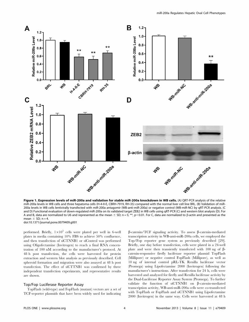
Figure 1. Expression levels of miR-200a and validation for stable miR-200a knockdown in WB cells. (A) QRT-PCR analysis of the relativemiR-200a levels in WB cells and three hepatoma cells (H-4-II-E, CBRH-7919, RH-35) compared with the normal liver cell line BRL. (B) Validation of miR-200a levels in WB cells lentivirally transfected with miR-200a antagomir (WB-anti-miR-200a) or negative control (WB-miR-NC) by qRT-PCR analysis. (Cand D) Functional evaluation of down-regulated miR-200a on its validated target ZEB2 in WB cells using qRT-PCR (C) and western blot analysis (D). ForA and B, data are normalized to U6 and represented as the mean 6 SD; n = 5; **, p,0.01. For C, data are normalized to b-actin and presented as themean 6 SD; n = 4.doi:10.1371/journal.pone.0079409.g001
miR-200a Regulates Hepatic Oval Cell Phenotypes
PLOS ONE | www.plosone.org 4 November 2013 | Volume 8 | Issue 11 | e79409

miR-200a Regulates Hepatic Oval Cell Phenotypes
PLOS ONE | www.plosone.org 5 November 2013 | Volume 8 | Issue 11 | e79409

post transfection for luciferase activity detection. Each experiment
was performed in triplicate, and the fold change in TopFlash
activity compared with that in FopFlash activity is shown.
In vivo Xenograft Tumorigenicity AssayAthymic nude mice (BALB/C-nu/nu, 4–6 weeks old, male)
were obtained from Animal Center of Chinese Academy of
Science (Shanghai, China), housed under specific pathogen-free
conditions and cared for in accordance with the guidelines of the
laboratory animal centre of Fourth Military Medical University
(Xi’an, China). All studies involving animals were approved by the
Research Animal Care and Use Committee of Fourth Military
Medical University. WB-miR-NC or WB-anti-miR-200a cells
were mixed with Matrigel Basement Membrane Matrix (BD
Biosciences) at ratio of 1:1 and then subcutaneously implanted into
left flanks (WB-miR-NC) or right flanks (WB-anti-miR-200a) of
five nude mice at 26106 cells per injection, respectively. The mice
were maintained under standardized conditions, monitored every
2 days after second week of inoculation and sacrificed 40 days post
inoculation. Resultant subcutaneous tumors were pictured,
collected and measured in weight. Tumor tissues were fixed in
4% formaldehyde, embedded in paraffin for hematoxylin and
eosin (H&E) staining to analyze the histopathology under the
microscope (Olympus).
Statistical AnalysisData are presented as the mean 6 standard deviation (SD) from
at least three independent experiments. Student’s t-test (two tailed)
or Student–Newman–Keuls (SNK test, ANOVA) was employed to
analyze differences using SPSS 17.0 software (Chicago, IL). A
probability p-value less than 0.05 was considered statistically
significant.
Results
Expression Levels of miR-200a and Validation for StablemiR-200a Knockdown in WB Cells
To investigate the possible role of miR-200a on OCs, we first
performed qRT-PCR to analyze the expression levels of miR-200a
in WB cells as well as in BRL normal rat liver cells and three
hepatoma cell lines (H-4-II-E, CBRH-7919, RH-35). Our results
demonstrated that miR-200a expression was much higher in WB
cells than in hepatoma cells (Figure 1A), indicating that down-
regulated miR-200a may be associated with HCC malignant
phenotypes. As a result, we introduced miR-200a loss-of-function
studies in WB cells in the following research.
Next, we established lentivirus-mediated stable WB-anti-miR-
200a and WB-miR-NC cell lines. QRT-PCR analysis showed the
expression level of miR-200a in WB-anti-miR-200a cells decreased
compared with WB-miR-NC cells, whereas its level was constant
in WB-miR-NC cells versus WB cells (Figure 1B), suggesting that
lentivirus infection was highly efficient and did not perturb
endogenous miR-200a expression in WB cells. In addition, after
miR-200a silencing in WB cells, mRNA expression of ZEB2, a
well-acknowledged target of miR-200a, was constant (Figure 1C),
whereas its protein expression increased (Figure 1D). These results
indicate that miR-200a is effectively and functionally suppressed in
WB-anti-miR-200a cells, serving as the basis of the remaining
experiments.
Stable Knockdown of miR-200a Facilitates CSC-likePhenotypes in WB Cells
It has been demonstrated that CSCs with stem/progenitor cell
characteristics possess excessive self-renewal capability [30]. In our
study, cell proliferation assays showed that miR-200a silencing was
associated with a time-dependent promotion of cell growth in WB
cells (Figure 2A). Strikingly, in the spheroid formation assay, more
spheroids were observed in suspension cultured WB-anti-miR-
200a cells compared with WB-miR-NC cells, and the number of
spheroids expanded even faster in WB-anti-miR-200a cells after
serially passaging in both groups (Figure 2B), suggesting that miR-
200a knockdown enhanced the self-renewal capacity of WB cells.
In addition, the differences in cell proliferation and spheroid
forming ability were not due to differences in apoptosis, because
caspase-3/7 activity and the ratio of apoptotic cells were similar
between miR-200a silencing cells and control cells (Figure 2C and
2D).
To further determine the characteristics of WB-anti-miR-200a
cells, expression of putative hepatic CSC markers were examined
by qRT-PCR. As shown in Figure 2E, expression of EpCAM,
CD133, ABCG2, CK19 and AFP were much higher in WB-anti-
miR-200a cells than in WB-miR-NC cells, whereas the ALB
mRNA level was relatively downregulated, indicating that WB-
anti-miR-200a cells are in a less differentiated state. Interestingly,
expression of the oncogene c-myc was also up-regulated in WB-
anti-miR-200a cells (Figure 2E), suggesting that miR-200a
silencing may also promote WB cells to obtain transformed
characteristics.
Chemo-resistance is one of the key hallmarks of CSCs. By
assessing the apoptotic cells after exposure to anticancer drugs, we
showed that WB-anti-miR-200a cells exhibited reduced apoptosis
upon paclitaxel or doxorubicin (15.162.1% or 11.261.9%,
respectively) treatment, in contrast to WB-miR-NC cells
(25.863.4% or 18.362.7%, respectively) (Figure 2F), indicating
that WB-anti-miR-200a cells might be more resistant to anticancer
drugs. All together, these data strongly suggest that stable
knockdown of miR-200a stimulates hepatic CSC-like character-
istics in WB cells.
Figure 2. Stable knockdown of miR-200a facilitates CSC-like phenotypes in WB cells. (A) Growth curve of WB-miR-NC and WB-anti-miR-200a cells determined by cell counting. Data are expressed as the mean 6 SD; n = 3; *, p,0.05. (B) Representative images of spheroids formed by WB-miR-NC and WB-anti-miR-200a cells in the spheroid formation assay (left, magnification6100). Number of spheroids formed in the primary, secondaryand tertiary generations of suspension cultured WB-miR-NC or WB-anti-miR-200a cells (right). Data represent means from four randomly selectedfields under the microscope, and error bars represent SD. *, p,0.05; **, p,0.01. (C and D) Apoptosis of WB-miR-NC and WB-anti-miR-200a cellsmeasured by caspase-3/7 assay and Annexin V and PI staining. Data are expressed as the mean 6 SD (C) and representative dot plots of apoptosistests are shown (D); n = 5. (E) Expression of EpCAM, CD133, ABCG2, CK19, AFP, ALB and c-myc in WB-anti-miR-200a cells measured by qRT-PCR. Dataare normalized to b-actin, shown relative to the level in WB-miR-NC cells and expressed as the mean 6 SD; n = 3; *, p,0.05; **, p,0.01. (F) WB-miR-NCand WB-anti-miR-200a cells were treated with 10 ng/ml paclitaxel or 30 ng/ml doxorubicin for 48 h and then subjected to FACS with Annexin V andPI staining, respectively. Representative dot plots (left) and the mean percentage of apoptotic cells (6 SD) from three independent experiments(right) are shown; *, p,0.05; **, p,0.01.doi:10.1371/journal.pone.0079409.g002
miR-200a Regulates Hepatic Oval Cell Phenotypes
PLOS ONE | www.plosone.org 6 November 2013 | Volume 8 | Issue 11 | e79409

Stable Knockdown of miR-200a Confers MesenchymalCharacteristics to WB Cells
We obtained evidence that miR-200a knockdown might also be
associated with an EMT-like phenotype in WB cells. Morpholog-
ically, WB-anti-miR-200a cells were partly spindle shaped similar
to mesenchymal cells, whereas WB-miR-NC cells were tightly
bound, oval-like cells with an epithelial phenotype (Figure 3A).
Furthermore, in vitro migration assays revealed that WB-anti-
miR-200a cells exhibited enhanced migration capacity compared
with WB-miR-NC cells (Figure 3B). We also examined the protein
expression of epithelial (E-cadherin) and mesenchymal (N-
cadherin, Vimentin) markers by western blotting. As shown in
Figure 3C, consistent with their elongated mesenchymal mor-
phology, down-regulated E-cadherin and up-regulated N-cad-
herin, as well as Vimentin, were detected in WB-anti-miR-200a
cells. Based on these morphologic changes as well as biological and
biochemical behaviors shown in WB-anti-miR-200a cells, we
conclude that miR-200a silencing induces the EMT-like pheno-
type in WB cells.
Downregulated miR-200a could Directly Target CTNNB1and Activate the Wnt/b-catenin Pathway in WB Cells
Accumulating evidence has demonstrated that the Wnt/b-
catenin pathway plays an important role in oval cell activation,
proliferation, and expansion in HCC subsets [31,32,33]. Thus,
we postulated that miR-200a might target genes associated
with Wnt/b-catenin signaling. By computational algorithms from
TargetScan and mirBase target database, we found that the 39-
UTR of rat b-catenin (CTNNB1) mRNA contains a conserved
putative miR-200a target site (Figure 4A). To validate the predi-
Figure 3. Stable knockdown of miR-200a confers mesenchymal characteristics to WB cells. (A) Morphological changes in WB-miR-NC andWB-anti-miR-200a cells (magnification6200). (B) Evaluation of in vitro migration abilities of WB-miR-NC and WB-anti-miR-200a cells by transwellmigration assay. Representative images (upper, magnification6200) and the mean number of migrated cells (6 SD) in five randomly selected fieldscounted under the microscope (lower) are shown; **, p,0.01. (C) Western blot analysis of epithelial (E-cadherin) and mesenchymal (N-cadherin andvimentin) markers in WB-miR-NC and WB-anti-miR-200a cells.doi:10.1371/journal.pone.0079409.g003
miR-200a Regulates Hepatic Oval Cell Phenotypes
PLOS ONE | www.plosone.org 7 November 2013 | Volume 8 | Issue 11 | e79409

miR-200a Regulates Hepatic Oval Cell Phenotypes
PLOS ONE | www.plosone.org 8 November 2013 | Volume 8 | Issue 11 | e79409

ction, a 39-UTR fragment of rat CTNNB1 mRNA containing
wild-type or a mutated binding site for miR-200a was cloned into a
luciferase reporter vector. The results showed that anti-miR-200a
transfection significantly increased the expression of luciferase
containing a CTNNB1-wt 39-UTR binding site for miR-200a
in WB cells compared with the control (Figure 4B). However, this
stimulative effect was abrogated by mutations in the seed
complementary sites of the 39-UTR of CTNNB1 (Figure 4B).
Moreover, we also performed western blot analysis to detect
b-catenin protein levels in WB-anti-miR-200a cells and WB-
miR-NC cells. As shown in Figure 4C, miR-200a silencing
induced the protein expression of CTNNB1.
Accumulation of intracellular b-catenin, especially in the
nucleus, is an indication of activated b-catenin-dependent
signaling; hence, we further determined whether enhanced b-
catenin expression impacted b-catenin pathway activity. The
immunofluorescence staining data illustrated that b-catenin
expression showed strong cytoplasmic and some nuclear localiza-
tion when miR-200a was silenced in WB cells (Figure 4D).
Additionally, WB-anti-miR-200a cells exhibited increased Top-
Flash luciferase activity compared with WB-miR-NC cells
(Figure 4E). Moreover, the expression of cyclin D1 and c-myc,
two representative target genes of the Wnt/b-catenin pathway,
was also enhanced following the knockdown of miR-200a in WB
cells (Figure 4C). Taken together, our data indicate that CTNNB1
mRNA is a direct target of miR-200a, and knockdown of miR-
200a could activate, at least in part, the Wnt/b-catenin signaling
pathway in WB cells.
The Anti-miR-200a Effects can be Partially Attenuated bySilencing of CTNNB1 in WB-anti-miR-200a Cells
To further elucidate whether elevated b-catenin expression
and activated b-catenin pathway are functionally associated with
miR-200a-silencing-mediated biological activities of WB cells,
we used siCTNNB1 transfection and examined its effects on
WB-anti-miR-200a cells. We confirmed that transfection of
siCTNNB1 did not induce a detectable miR-200a expression
change in WB-anti-miR-200a cells (data not shown). The
silencing of CTNNB1 and inhibition of b-catenin-mediated
transcription activity in WB-anti-miR-200a cells were confirmed
by western blot analysis (Figure 5A) and Top/Fop luciferase
reporter assays (Figure 5B), respectively. As expected, the
stimulatory effect of miR-200a-silencing on cyclin D1 and c-
myc expression was abolished by siCTNNB1 transfection
(Figure 5A). Furthermore, we observed dramatic diminished
spheroid formation (Figure 5C) and reduced migration abilities
(Figure 5D) following CTNNB1 silencing in WB-anti-miR-200a
cells. Collectively, these results demonstrate that activation of
the Wnt/b-catenin pathway is functionally relevant to miR-
200a-silencing-mediated biological activities of WB cells.
Downregulation of miR-200a Confers Tumorigenicity toWB Cells in vivo
As in vitro studies showed that stable knockdown of miR-
200a facilitates CSC-like signatures and EMT phenotypes in
WB cells, we performed xenograft formation assay to further
investigate the tumorigenicity mediated by miR-200a suppres-
sion. As shown in Figure 6A, WB-anti-miR-200a cells developed
visible tumors on every right flank of the five nude mice,
whereas no exhibited tumor was observed on the left flanks
injected with WB-miR-NC cells. Histological analysis of
subcutaneous tumors by H&E staining showed disorganized
tumor cells, hyperchromatic nuclei and high nuclear-cytoplasmic
ratio, which assembles HCC-specific features (Figure 6B). To
conclude, the in vivo results indicated that downregulation of
miR-200a in WB cells could induce tumorigenicity and finally
give rise to tumors in nude mice.
Discussion
With progress in the identification of CSCs, emerging evidence
strongly support the notion that HCC is driven by a small
population of cells through their excessive self-renewal capacity,
production of heterogeneous progeny, resistance to chemotherapy
and limitless dividing [34]. Recently, recognition of the role of
HOCs in the carcinogenic process prompted a new belief that
HCC might arise by maturation arrest of liver adult stem cells
[35]. In other words, hepatic CSCs could originate from normal
HOCs. As an important regulatory factor, the miR-200 family has
been identified as suppressors of CSC-like or EMT-like pheno-
types and functions in various normal and cancer cells
[22,23,24,36]. Our previous study also showed that miR-200a
was greatly downregulated in the F344 rat HCC SP fraction cells
compared with their normal counterparts [26]. In this study, using
loss-of-function studies, we further investigated the function of
miR-200a on HOCs.
The WB-F344 epithelial cell line was first derived from the liver
of an adult male Fischer 344 rat [27] and has been proposed to be
an in vitro model of HOC [28]. As an initial step, the expression
pattern of miR-200a was assessed in the WB-F344 cell line, BRL
normal liver cell line and three other hepatoma cell lines (H-4-II-
E, CBRH-7919, RH-35). We provide evidence that miR-200a
expression was relatively higher in WB cells and BRL cells than in
three other rat hepatoma cell lines. These results were also in line
with previous studies reporting downregulation of miR-200a in
poorly differentiated HCC cell lines compared with well differen-
tiated cell lines and in HCC tissues compared with the adjacent
noncancerous hepatic tissues [37,38]. Therefore, it can be
calculated that dysregulated miR-200a may be intimately associ-
ated with malignant phenotypes of HCC.
Next, using a lentiviral vectors approach, we transfected WB
cells with miR-200a antagomirs and stably silenced miR-200a in
WB cells, results that were confirmed by qRT-PCR and western
blotting. Functional studies showed that WB cells stably lacking
miR-200a exhibited CSC-like properties. Four lines of evidence
Figure 4. miR-200a directly targets CTNNB1 and associates with Wnt/b-catenin pathway activity in WB cells. (A) Predicted alignment ofmiR-200a and a potential binding site at the 39-UTR of rat CTNNB1 mRNA (624–630 nt) by TargetScan. (B) Effect of miR-200a on CTNNB1 expressiondetermined by a luciferase reporter assay. WB cells were co-transfected with anti-miR-200a (or anti-miR-control) and the pGL3-CTNNB1-wt (or pGL3-CTNNB1-mut) vector. Data are normalized by the ratio of Firefly and Renilla luciferase activities measured at 48 h post-transfection and are shown asthe mean 6 SD; n = 3; **, p,0.01. (C) Expression of b-catenin, c-myc and cyclin D1 in WB-miR-NC and WB-anti-miR-200a cells detected by westernblot analysis. (D) Cellular localization of b-catenin visualized by immunofluorescence staining in WB-miR-NC and WB-anti-miR-200a cells. Nuclearcounterstaining was performed by DAPI (magnification6200). (E) The activity of b-catenin signaling determined by a luciferase reporter assay usingthe wild-type (TopFlash) or mutant (FopFlash) TCF4-reporter plasmids. Data are presented as fold increases in firefly luciferase over Renilla activityfrom three independent experiments; *, p,0.05.doi:10.1371/journal.pone.0079409.g004
miR-200a Regulates Hepatic Oval Cell Phenotypes
PLOS ONE | www.plosone.org 9 November 2013 | Volume 8 | Issue 11 | e79409

Figure 5. The anti-miR-200a effects are partially attenuated by silencing of CTNNB1 in WB-anti-miR-200a cells. (A) The protein levels ofb-catenin, c-myc and cyclin D1 measured by western blot analysis after siCTNNB1 transfection in WB-anti-miR-200a cells. (B) The b-catenin-mediatedtranscription activity determined by the Top/Fop ratio after siCTNNB1 transfection in WB-anti-miR-200a cells. Data are represented as the mean 6 SD;n = 3; **, p,0.01. (C) Representative images of spheroids formed by WB-anti-miR-200a cells before and after siCTNNB1 transfection (left,magnification æ 100). Bar graph (right) represents the mean number of spheroids from five randomly selected fields under the microscope, and errorbars represent SD. **, p,0.01. (D) Representative images of transwell migration before and after siCTNNB1 transfection in WB-anti-miR-200a cells (left,magnification 6 200). Bar graph (right) represents the mean number of migrated cells (6 SD) in five randomly selected fields counted under themicroscope. **, p,0.01.doi:10.1371/journal.pone.0079409.g005
miR-200a Regulates Hepatic Oval Cell Phenotypes
PLOS ONE | www.plosone.org 10 November 2013 | Volume 8 | Issue 11 | e79409

support this finding. First, stable knockdown of miR-200a
enhanced proliferation and conferred a greater potential for self
renewal capacity even after serial passages in WB cells. Second,
putative hepatic CSC markers (EpCAM [30], CD133 [8] and
ABCG2 [39]) were up-regulated after miR-200a knockdown.
Third, WB-anti-miR-200a cells were in a less differentiated state
by higher expression of primitive liver markers AFP and CK19,
and lower expression of the mature liver marker ALB. Fourth,
miR-200a silencing led to superior anti-apoptosis capacity to
chemotherapeutic drugs in WB cells. Specifically, although more
evidence is needed, it can be postulated that this chemo-resistant
phenomenon might be due to the above observed increased
expression of ABCG2, which is a universal CSC marker always
implicated in resistance to chemotherapeutic agents [40,41].
EMT is a critical process involved in cancer progression and
metastasis. During EMT, loss of epithelial markers and gain in the
expression of mesenchymal markers facilitate cells to undergo
changes in cell morphology, which is accompanied by enhanced
cell motility and migration [42]. In addition, emerging data also
suggest a role for this process in generating CSC-like features in
epithelial cells [43,44,45]. Recently, it has been confirmed that the
miR-200 family plays a key role in negatively regulating the EMT
process [21]. More strikingly, miR-200 family members have been
reported to be directly regulated by p53, further highlighting their
role in tumor progression [46]. In our study, WB cells stably
lacking miR-200a also presented mesenchymal characteristics,
including elongated cell morphology, enhanced cell migration
ability, down-regulated E-cadherin and up-regulated N-cadherin,
as well as Vimentin. This is the first report, to the best of our
knowledge, demonstrating that downregulation of miR-200a is
associated with EMT and CSC-like phenotypes in HOCs. Our
finding is in accordance with phenomena previously observed in
pancreatic cells [47] and nasopharyngeal carcinoma cells [48],
indicating that miR-200a indeed plays an extensive and profound
role in the context of cancer initiation and progression.
Regarding the molecular mechanisms underlying the role of
miR-200a in WB cells, we used combined bioinformatics and
experimental approaches to identify CTNNB1 as a direct
functional downstream target of miR-200a. CTNNB1 encodes
b-catenin, which is not only an essential component of the
cadherin adhesion protein complex to regulate epithelial cell
integrity and polarity but also a central modulator of the canonical
Wnt signaling pathway. It has been well established that
accumulated cytosolic b-catenin can translocate into the nucleus
and then form the b-catenin/TCF complex to activate transcrip-
tion of Wnt target genes [49]. Consistent with these theories, we
demonstrated in this study that stable knockdown of miR-200a
enhanced the b-catenin protein level. Moreover, elevated b-
Figure 6. Downregulation of miR-200a confers tumorigenicity to WB cells in vivo. (A) Subcutaneous tumors developed by WB-miR-NC orWB-anti-miR-200a for 40 days post inoculation (left). Pictures of collected subcutaneous tumors were taken (middle) and tumor weights are shown(right). (B) Representative images of H&E staining of xenografted tumors are shown (left, magnification6200; right, magnification6400).doi:10.1371/journal.pone.0079409.g006
miR-200a Regulates Hepatic Oval Cell Phenotypes
PLOS ONE | www.plosone.org 11 November 2013 | Volume 8 | Issue 11 | e79409
catenin showed strong cytoplasmic and nuclear localization, which
is consistent with increased TopFlash luciferase activity and
enhanced expression of Wnt target genes, c-myc and cyclin D1. In
addition, a previous study found that EpCAM can act as a Wnt
downstream molecule to maintain stemness properties in HCC
subsets [30]. Thus, it is reasonable to speculate that activated b-
catenin in this study might also account for the elevated EpCAM
expression that was shown previously.
The Wnt/b-catenin pathway is a highly conserved signaling
cascade participating in various cellular processes, including cell
proliferation, differentiation, migration, adhesion, and survival
[50]. Recently, Wnt/b-catenin signaling has been reported to
correlate with an EMT-like states [51] and CSC genesis [52], self-
renewal [53] and chemo-resistance [54] in many cancers. More
importantly, the literature has shown that activation of Wnt/b-
catenin signaling confers HOCs/HPCs with high self-renewal
potential and tumorigenesis capacity in the liver [12,32].
Consistent with their findings, a rescue experiment in our study
found WB-anti-miR-200a transduced with siCTNNB1, but not
scrambled siControl, showing diminished spheroid formation and
reduced migration ability in vitro. The function of miR-200a
exerting on WB cells is indeed, at least in part, attributed to b-
catenin regulation in Wnt/b-catenin signaling.
It has been discovered that one miRNA can possibly target
multiple genes in regulating cellular processes. In previous studies,
miR-200a regulating the EMT process by targeting EMT-
activating transcriptional factors ZEB1 and ZEB2 has been
thoroughly elaborated in different types of cancers [55,56]. In
our study, we also found knockdown of miR-200a led to
overexpression of ZEB2 protein in WB cells. Thus, in addition
to directly targeting CTNNB1 and regulating the Wnt/b-catenin
pathway, there is a possibility that miR-200a may also exert its
function on WB cells via targeting ZEB2. Thus, more in-depth
investigations are required to determine the complex interactions
of miR-200a-dependent molecular regulatory network systems in
HOCs.
In addition to in vitro findings, the xenograft tumorigenicity
assay further confirmed the contributions of miR-200a silencing to
malignant transformation of WB cells in vivo. Acquisition of
tumorigenicity in oval cell lines is not unusual since it has been
reported that long time exposure of WB-F344 cells to TGF-b or
transfection with HBx gene in LE/6 cells (another HOC line)
following treatment with aflatoxin B1 obtained same results [9,57].
Moreover, except for EMT process and b-catenin pathway, miR-
200a has also been found participating in epigenetic modulation
through histone deacetylase 4/SP1/miR-200a regulatory network
[38]. Thus, it can be calculated that miR-200a may have
significant influence on cell functions through extensive mecha-
nisms which needs further exploring.
To conclude, our data suggested that downregulation of miR-
200a in liver oval cells promotes EMT and CSC-like traits in vitro,
partially through the regulation of b-catenin signaling, and finally
give rise to tumorigenicity in vivo. These findings underscore the
importance of miR-200a in inhibiting neoplastic phenotypes in
normal adult hepatic progenitor cells. Exploring miR-200a-based
prevention and therapeutics might benefit HCC clinically.
Acknowledgments
We sincerely thank Jiuxu Bai, Liang Zhou and Fuqin Zhang for their
substantial technical assistance and helpful discussions.
Author Contributions
Conceived and designed the experiments: WL KT KD. Performed the
experiments: JL BR. Analyzed the data: QH ZD WX TZ YC. Contributed
reagents/materials/analysis tools: RJ XL DW. Wrote the paper: JL NY.
References
1. Forner A, Llovet JM, Bruix J (2012) Hepatocellular carcinoma. Lancet 379:1245–1255.
2. Aravalli RN, Steer CJ, Cressman EN (2008) Molecular mechanisms of
hepatocellular carcinoma. Hepatology 48: 2047–2063.
3. Pardal R, Clarke MF, Morrison SJ (2003) Applying the principles of stem-cell
biology to cancer. Nat Rev Cancer 3: 895–902.
4. Soltanian S, Matin MM (2011) Cancer stem cells and cancer therapy. TumourBiol 32: 425–440.
5. Reya T, Morrison SJ, Clarke MF, Weissman IL (2001) Stem cells, cancer, and
cancer stem cells. Nature 414: 105–111.
6. Sell S (2004) Stem cell origin of cancer and differentiation therapy. Crit Rev
Oncol Hematol 51: 1–28.
7. Yang ZF, Ngai P, Ho DW, Yu WC, Ng MN, et al. (2008) Identification of localand circulating cancer stem cells in human liver cancer. Hepatology 47: 919–
928.
8. Zhu Z, Hao X, Yan M, Yao M, Ge C, et al. (2010) Cancer stem/progenitor cells
are highly enriched in CD133+CD44+ population in hepatocellular carcinoma.Int J Cancer 126: 2067–2078.
9. Wu K, Ding J, Chen C, Sun W, Ning BF, et al. (2012) Hepatic transforming
growth factor beta gives rise to tumor-initiating cells and promotes liver cancerdevelopment. Hepatology 56: 2255–2267.
10. Thompson MD, Awuah P, Singh S, Monga SP (2010) Disparate cellular basis of
improved liver repair in beta-catenin-overexpressing mice after long-termexposure to 3,5-diethoxycarbonyl-1,4-dihydrocollidine. Am J Pathol 177: 1812–
1822.
11. Alison MR, Islam S, Lim S (2009) Stem cells in liver regeneration, fibrosis and
cancer: the good, the bad and the ugly. J Pathol 217: 282–298.
12. Chiba T, Zheng YW, Kita K, Yokosuka O, Saisho H, et al. (2007) Enhanced
self-renewal capability in hepatic stem/progenitor cells drives cancer initiation.
Gastroenterology 133: 937–950.
13. Chiba T, Seki A, Aoki R, Ichikawa H, Negishi M, et al. (2010) Bmi1 promotes
hepatic stem cell expansion and tumorigenicity in both Ink4a/Arf-dependent
and -independent manners in mice. Hepatology 52: 1111–1123.
14. Bartel DP (2004) MicroRNAs: genomics, biogenesis, mechanism, and function.
Cell 116: 281–297.
15. He L, Thomson JM, Hemann MT, Hernando-Monge E, Mu D, et al. (2005) AmicroRNA polycistron as a potential human oncogene. Nature 435: 828–833.
16. Lee YS, Dutta A (2007) The tumor suppressor microRNA let-7 represses the
HMGA2 oncogene. Genes Dev 21: 1025–1030.
17. Yu F, Yao H, Zhu P, Zhang X, Pan Q, et al. (2007) let-7 regulates self renewal
and tumorigenicity of breast cancer cells. Cell 131: 1109–1123.
18. Wellner U, Schubert J, Burk UC, Schmalhofer O, Zhu F, et al. (2009) The
EMT-activator ZEB1 promotes tumorigenicity by repressing stemness-inhibiting
microRNAs. Nat Cell Biol 11: 1487–1495.
19. Zhao C, Sun G, Li S, Lang MF, Yang S, et al. (2010) MicroRNA let-7b regulates
neural stem cell proliferation and differentiation by targeting nuclear receptor
TLX signaling. Proc Natl Acad Sci U S A 107: 1876–1881.
20. Ma S, Tang KH, Chan YP, Lee TK, Kwan PS, et al. (2010) miR-130b
Promotes CD133(+) liver tumor-initiating cell growth and self-renewal via tumor
protein 53-induced nuclear protein 1. Cell Stem Cell 7: 694–707.
21. Mongroo PS, Rustgi AK (2010) The role of the miR-200 family in epithelial-
mesenchymal transition. Cancer Biol Ther 10: 219–222.
22. Shimono Y, Zabala M, Cho RW, Lobo N, Dalerba P, et al. (2009)
Downregulation of miRNA-200c links breast cancer stem cells with normal
stem cells. Cell 138: 592–603.
23. Iliopoulos D, Lindahl-Allen M, Polytarchou C, Hirsch HA, Tsichlis PN, et al.
(2010) Loss of miR-200 inhibition of Suz12 leads to polycomb-mediated
repression required for the formation and maintenance of cancer stem cells. Mol
Cell 39: 761–772.
24. Bao B, Wang Z, Ali S, Kong D, Li Y, et al. (2011) Notch-1 induces epithelial-
mesenchymal transition consistent with cancer stem cell phenotype in pancreatic
cancer cells. Cancer Lett 307: 26–36.
25. Wu Q, Guo R, Lin M, Zhou B, Wang Y (2011) MicroRNA-200a inhibits
CD133/1+ ovarian cancer stem cells migration and invasion by targeting E-
cadherin repressor ZEB2. Gynecol Oncol 122: 149–154.
26. Liu WH, Tao KS, You N, Liu ZC, Zhang HT, et al. (2011) Differences in the
properties and mirna expression profiles between side populations from hepatic
cancer cells and normal liver cells. PLoS One 6: e23311.
27. Tsao MS, Smith JD, Nelson KG, Grisham JW (1984) A diploid epithelial cell
line from normal adult rat liver with phenotypic properties of ‘oval’ cells. Exp
Cell Res 154: 38–52.
28. Li X, Li Y, Kang X, Guo K, Li H, et al. (2010) Dynamic alteration of protein
expression profiles during neoplastic transformation of rat hepatic oval-like cells.
Cancer Sci 101: 1099–1107.
miR-200a Regulates Hepatic Oval Cell Phenotypes
PLOS ONE | www.plosone.org 12 November 2013 | Volume 8 | Issue 11 | e79409

29. Anand M, Lai R, Gelebart P (2011) beta-catenin is constitutively active and
increases STAT3 expression/activation in anaplastic lymphoma kinase-positive
anaplastic large cell lymphoma. Haematologica 96: 253–261.
30. Yamashita T, Ji J, Budhu A, Forgues M, Yang W, et al. (2009) EpCAM-positive
hepatocellular carcinoma cells are tumor-initiating cells with stem/progenitor
cell features. Gastroenterology 136: 1012–1024.
31. Apte U, Thompson MD, Cui S, Liu B, Cieply B, et al. (2008) Wnt/beta-catenin
signaling mediates oval cell response in rodents. Hepatology 47: 288–295.
32. Yang W, Yan HX, Chen L, Liu Q, He YQ, et al. (2008) Wnt/beta-catenin
signaling contributes to activation of normal and tumorigenic liver progenitor
cells. Cancer Res 68: 4287–4295.
33. Itoh T, Kamiya Y, Okabe M, Tanaka M, Miyajima A (2009) Inducible
expression of Wnt genes during adult hepatic stem/progenitor cell response.
FEBS Lett 583: 777–781.
34. Oishi N, Wang XW (2011) Novel therapeutic strategies for targeting liver cancer
stem cells. Int J Biol Sci 7: 517–535.
35. Sell S, Leffert HL (2008) Liver cancer stem cells. J Clin Oncol 26: 2800–2805.
36. Tellez CS, Juri DE, Do K, Bernauer AM, Thomas CL, et al. (2011) EMT and
stem cell-like properties associated with miR-205 and miR-200 epigenetic
silencing are early manifestations during carcinogen-induced transformation of
human lung epithelial cells. Cancer Res 71: 3087–3097.
37. Hung CS, Liu HH, Liu JJ, Yeh CT, Chang TC, et al. (2012) MicroRNA-200a
and -200b Mediated Hepatocellular Carcinoma Cell Migration Through the
Epithelial to Mesenchymal Transition Markers. Ann Surg Oncol.
38. Yuan JH, Yang F, Chen BF, Lu Z, Huo XS, et al. (2011) The histone
deacetylase 4/SP1/microrna-200a regulatory network contributes to aberrant
histone acetylation in hepatocellular carcinoma. Hepatology 54: 2025–2035.
39. Zhang N, Li R, Tao KS, Cao DY, Ti ZY, et al. (2010) Characterization of a
stem-like population in hepatocellular carcinoma MHCC97 cells. Oncol Rep 23:
827–831.
40. Cheng C, Liu ZG, Zhang H, Xie JD, Chen XG, et al. (2012) Enhancing
Chemosensitivity in ABCB1- and ABCG2-Overexpressing Cells and Cancer
Stem-like Cells by An Aurora Kinase Inhibitor CCT129202. Mol Pharm.
41. Mao J, Song B, Shi Y, Wang B, Fan S, et al. (2013) ShRNA targeting Notch1
sensitizes breast cancer stem cell to paclitaxel. Int J Biochem Cell Biol 45: 1064–
1073.
42. Thiery JP (2002) Epithelial-mesenchymal transitions in tumour progression. Nat
Rev Cancer 2: 442–454.
43. Mani SA, Guo W, Liao MJ, Eaton EN, Ayyanan A, et al. (2008) The epithelial-
mesenchymal transition generates cells with properties of stem cells. Cell 133:
704–715.
44. Morel AP, Lievre M, Thomas C, Hinkal G, Ansieau S, et al. (2008) Generation
of breast cancer stem cells through epithelial-mesenchymal transition. PLoS One3: e2888.
45. Gupta PB, Onder TT, Jiang G, Tao K, Kuperwasser C, et al. (2009)
Identification of selective inhibitors of cancer stem cells by high-throughputscreening. Cell 138: 645–659.
46. Kim T, Veronese A, Pichiorri F, Lee TJ, Jeon YJ, et al. (2011) p53 regulatesepithelial-mesenchymal transition through microRNAs targeting ZEB1 and
ZEB2. J Exp Med 208: 875–883.
47. Sureban SM, May R, Lightfoot SA, Hoskins AB, Lerner M, et al. (2011)DCAMKL-1 regulates epithelial-mesenchymal transition in human pancreatic
cells through a miR-200a-dependent mechanism. Cancer Res 71: 2328–2338.48. Xia H, Cheung WK, Sze J, Lu G, Jiang S, et al. (2010) miR-200a regulates
epithelial-mesenchymal to stem-like transition via ZEB2 and beta-cateninsignaling. J Biol Chem 285: 36995–37004.
49. Clevers H (2006) Wnt/beta-catenin signaling in development and disease. Cell
127: 469–480.50. Dihlmann S, von Knebel Doeberitz M (2005) Wnt/beta-catenin-pathway as a
molecular target for future anti-cancer therapeutics. Int J Cancer 113: 515–524.51. Lee JM, Dedhar S, Kalluri R, Thompson EW (2006) The epithelial-
mesenchymal transition: new insights in signaling, development, and disease.
J Cell Biol 172: 973–981.52. Rogers HA, Sousa S, Salto C, Arenas E, Coyle B, et al. (2012) WNT/beta-
catenin pathway activation in Myc immortalised cerebellar progenitor cellsinhibits neuronal differentiation and generates tumours resembling medullo-
blastoma. Br J Cancer 107: 1144–1152.53. Zhao C, Blum J, Chen A, Kwon HY, Jung SH, et al. (2007) Loss of beta-catenin
impairs the renewal of normal and CML stem cells in vivo. Cancer Cell 12:
528–541.54. Chau WK, Ip CK, Mak AS, Lai HC, Wong AS (2012) c-Kit mediates
chemoresistance and tumor-initiating capacity of ovarian cancer cells throughactivation of Wnt/beta-catenin-ATP-binding cassette G2 signaling. Oncogene.
55. Korpal M, Lee ES, Hu G, Kang Y (2008) The miR-200 family inhibits
epithelial-mesenchymal transition and cancer cell migration by direct targetingof E-cadherin transcriptional repressors ZEB1 and ZEB2. J Biol Chem 283:
14910–14914.56. Saydam O, Shen Y, Wurdinger T, Senol O, Boke E, et al. (2009)
Downregulated microRNA-200a in meningiomas promotes tumor growth byreducing E-cadherin and activating the Wnt/beta-catenin signaling pathway.
Mol Cell Biol 29: 5923–5940.
57. Li CH, Wang YJ, Dong W, Xiang S, Liang HF, et al. (2011) Hepatic oval celllines generate hepatocellular carcinoma following transfection with HBx gene
and treatment with aflatoxin B1 in vivo. Cancer Lett 311: 1–10.
miR-200a Regulates Hepatic Oval Cell Phenotypes
PLOS ONE | www.plosone.org 13 November 2013 | Volume 8 | Issue 11 | e79409