DNA in ancient bone – Where is it located and how should we extract it
10
Annals of Anatomy 194 (2012) 7–16 Contents lists available at ScienceDirect Annals of Anatomy jo ur n al ho mepage: www.elsevier.de/aanat DNA in ancient bone – Where is it located and how should we extract it? Paula F. Campos a , Oliver E. Craig b,c,d , Gordon Turner-Walker e , Elizabeth Peacock f , Eske Willerslev a , M. Thomas P. Gilbert a,∗ a Centre for GeoGenetics, Natural History Museum of Denmark, University of Copenhagen, Øster Voldgade 5-7, 1350 Copenhagen, Denmark b BioArCh, Department of Biology, University of York, Heslington, York YO10 5YW, UK c BioArCh, Department of Archaeology, University of York, Heslington, York YO10 5YW, UK d BioArCh, Department of Chemistry, University of York, Heslington, York YO10 5YW, UK e School of Cultural Heritage Conservation, National Yunlin University of Science and Technology, 123 University Road Sector 3, Douliou, 640 Yunlin, Taiwan f Section for Archaeology and Cultural History, Norwegian University of Science and Technology, NO-7491 Trondheim, Norway a r t i c l e i n f o Article history: Received 7 March 2011 Received in revised form 20 July 2011 Accepted 20 July 2011 Keywords: Bone Ancient DNA DNA extraction Collagen Hydroxyapatite s u m m a r y Despite the widespread use of bones in ancient DNA (aDNA) studies, relatively little concrete infor- mation exists in regard to how the DNA in mineralised collagen degrades, or where it survives in the material’s architecture. While, at the macrostructural level, physical exclusion of microbes and other external contaminants may be an important feature, and, at the ultrastructural level, the adsorption of DNA to hydroxyapatite and/or binding of DNA to Type I collagen may stabilise the DNA, the relative con- tribution of each, and what other factors may be relevant, are unclear. There is considerable variation in the quality of DNA retrieved from bones and teeth. This is in part due to various environmental factors such as temperature, proximity to free water or oxygen, pH, salt content, and exposure to radiation, all of which increase the rate of DNA decay. For example, bone specimens from sites at high latitudes usually yield better quality DNA than samples from temperate regions, which in turn yield better results than samples from tropical regions. However, this is not always the case, and rates of success of DNA recovery from apparently similar sites are often strikingly different. The question arises as to whether this may be due to post-collection preservation or just an artefact of the extraction methods used in these different studies? In an attempt to resolve these questions, we examine the efficacy of DNA extraction methods, and the quality and quantity of DNA recovered from both artificially degraded, and genuinely ancient, but well preserved, bones. In doing so we offer hypotheses relevant to the DNA degradation process itself, and to where and how the DNA is actually preserved in ancient bone. © 2011 Elsevier GmbH. All rights reserved. 1. Introduction The long-term survival of mineralised tissues such as bone (and to some degree, teeth) is normally dependent upon rapid burial in sediments, independently of whether terrestrial or marine. Subse- quently, their chemical and physical properties undergo substantial change, in a manner determined by the environment. For example, in aerated soils fungi and bacteria colonise the pore spaces of bones and begin the breakdown of mineralised tissues within a few years (Bell et al., 1996; Jans et al., 2004). In contrast, cyanobacteria are principally responsible for initial microbial attack in freshwater and marine environments (Turner-Walker and Jans, 2008; Pesquero et al., 2010), which accelerates bone degradation by increasing its porosity (Nielsen-Marsh and Hedges, 1999). Despite an increasing body of knowledge about the degradation of the bone itself, much ∗ Corresponding author. E-mail address: [email protected] (M.T.P. Gilbert). less is known about how the DNA in the bone degrades, and indeed, even how or where it is preserved. While some have argued that DNA in the mineralised collagen of bone and teeth hypothetically undergoes a retarded rate of decomposition because of its adsorp- tion to hydroxyapatite (e.g. Collins et al., 1995; Hagelberg et al., 1989; Lindahl, 1993), and others have argued that the mummifi- cation of individual cells, and the physical exclusion of microbes and other external contaminants from the smallest pores of skele- tal tissues may play a role in DNA survival (Hummel and Herrmann, 1994), we lack a comprehensive picture of the DNA–bone relation- ship. The relationship is unlikely to be simple, as significant variation exists in the quality and quantity of DNA that has been recovered from old bone – even among samples collected from environments that appear to be similar. For example, DNA recovery success rate was high in several large-scale studies of permafrost-preserved samples, including bison (Bison sp., success rate 352/442 [Shapiro et al., 2004]) and musk ox (Ovibos moschatus, success rate 207/446 [Campos et al., 2010b]). However, similar studies yielded much 0940-9602/$ – see front matter © 2011 Elsevier GmbH. All rights reserved. doi:10.1016/j.aanat.2011.07.003
Transcript of DNA in ancient bone – Where is it located and how should we extract it

D
PMa
b
c
d
e
f
a
ARRA
KBADCH
1
tsqcia(pmepb
0d
Annals of Anatomy 194 (2012) 7– 16
Contents lists available at ScienceDirect
Annals of Anatomy
jo ur n al ho mepage: www.elsev ier .de /aanat
NA in ancient bone – Where is it located and how should we extract it?
aula F. Camposa, Oliver E. Craigb,c,d, Gordon Turner-Walkere, Elizabeth Peacockf, Eske Willersleva,. Thomas P. Gilberta,∗
Centre for GeoGenetics, Natural History Museum of Denmark, University of Copenhagen, Øster Voldgade 5-7, 1350 Copenhagen, DenmarkBioArCh, Department of Biology, University of York, Heslington, York YO10 5YW, UKBioArCh, Department of Archaeology, University of York, Heslington, York YO10 5YW, UKBioArCh, Department of Chemistry, University of York, Heslington, York YO10 5YW, UKSchool of Cultural Heritage Conservation, National Yunlin University of Science and Technology, 123 University Road Sector 3, Douliou, 640 Yunlin, TaiwanSection for Archaeology and Cultural History, Norwegian University of Science and Technology, NO-7491 Trondheim, Norway
r t i c l e i n f o
rticle history:eceived 7 March 2011eceived in revised form 20 July 2011ccepted 20 July 2011
eywords:onencient DNANA extractionollagenydroxyapatite
s u m m a r y
Despite the widespread use of bones in ancient DNA (aDNA) studies, relatively little concrete infor-mation exists in regard to how the DNA in mineralised collagen degrades, or where it survives in thematerial’s architecture. While, at the macrostructural level, physical exclusion of microbes and otherexternal contaminants may be an important feature, and, at the ultrastructural level, the adsorption ofDNA to hydroxyapatite and/or binding of DNA to Type I collagen may stabilise the DNA, the relative con-tribution of each, and what other factors may be relevant, are unclear. There is considerable variation inthe quality of DNA retrieved from bones and teeth. This is in part due to various environmental factorssuch as temperature, proximity to free water or oxygen, pH, salt content, and exposure to radiation, all ofwhich increase the rate of DNA decay. For example, bone specimens from sites at high latitudes usuallyyield better quality DNA than samples from temperate regions, which in turn yield better results thansamples from tropical regions. However, this is not always the case, and rates of success of DNA recovery
from apparently similar sites are often strikingly different. The question arises as to whether this may bedue to post-collection preservation or just an artefact of the extraction methods used in these differentstudies? In an attempt to resolve these questions, we examine the efficacy of DNA extraction methods,and the quality and quantity of DNA recovered from both artificially degraded, and genuinely ancient, butwell preserved, bones. In doing so we offer hypotheses relevant to the DNA degradation process itself,and to where and how the DNA is actually preserved in ancient bone.. Introduction
The long-term survival of mineralised tissues such as bone (ando some degree, teeth) is normally dependent upon rapid burial inediments, independently of whether terrestrial or marine. Subse-uently, their chemical and physical properties undergo substantialhange, in a manner determined by the environment. For example,n aerated soils fungi and bacteria colonise the pore spaces of bonesnd begin the breakdown of mineralised tissues within a few yearsBell et al., 1996; Jans et al., 2004). In contrast, cyanobacteria arerincipally responsible for initial microbial attack in freshwater andarine environments (Turner-Walker and Jans, 2008; Pesquero
t al., 2010), which accelerates bone degradation by increasing itsorosity (Nielsen-Marsh and Hedges, 1999). Despite an increasingody of knowledge about the degradation of the bone itself, much
∗ Corresponding author.E-mail address: [email protected] (M.T.P. Gilbert).
940-9602/$ – see front matter © 2011 Elsevier GmbH. All rights reserved.oi:10.1016/j.aanat.2011.07.003
© 2011 Elsevier GmbH. All rights reserved.
less is known about how the DNA in the bone degrades, and indeed,even how or where it is preserved. While some have argued thatDNA in the mineralised collagen of bone and teeth hypotheticallyundergoes a retarded rate of decomposition because of its adsorp-tion to hydroxyapatite (e.g. Collins et al., 1995; Hagelberg et al.,1989; Lindahl, 1993), and others have argued that the mummifi-cation of individual cells, and the physical exclusion of microbesand other external contaminants from the smallest pores of skele-tal tissues may play a role in DNA survival (Hummel and Herrmann,1994), we lack a comprehensive picture of the DNA–bone relation-ship.
The relationship is unlikely to be simple, as significant variationexists in the quality and quantity of DNA that has been recoveredfrom old bone – even among samples collected from environmentsthat appear to be similar. For example, DNA recovery success rate
was high in several large-scale studies of permafrost-preservedsamples, including bison (Bison sp., success rate 352/442 [Shapiroet al., 2004]) and musk ox (Ovibos moschatus, success rate 207/446[Campos et al., 2010b]). However, similar studies yielded much
8 s of Anatomy 194 (2012) 7– 16
licsd2puebdt
fcn1isArotIrTpb2
waoiupDf
Aappaolcrcg(atm2ssm(eoeabim
P.F. Campos et al. / Annal
ower success rates, for example 27/122 saiga antelope (Saiga tatar-ca [Campos et al., 2010a]). Given that most of the samples wereollected from places with similar environments (permafrozenoils) and were thus presumably exposed to similar diagenetic con-itions (Hedges, 2002; Nielsen-Marsh et al., 2007; Smith et al.,007), the question arises as to what caused the discrepancy? Oneotential answer could be differences in the types of degradationndergone during the samples’ history, both taphonomic, and post-xcavation (at least one study has argued that freshly excavatedones are better for ancient DNA analyses due to acceleration ofegradation in museum storage resulting from elevated tempera-ures and greater access to oxygen [Pruvost et al., 2007]).
The degradation of DNA in archaeological bone is not a straight-orward topic, as multiple chemical processes may act to bothross-link and fragment the molecule’s chemical backbone, anducleotide bases may be either removed or altered (e.g. Lindahl,993; Pääbo, 1989; Hansen et al., 2006). Regardless of the underly-
ng chemical reasons, the end result of most of these processes is theame – the lengths of amplifiable DNA molecules decrease rapidly.lthough several factors affect the rate of this decay, including envi-onmental salt content, exposure to radiation, pH, and availabilityf oxygen and free water, it is temperature that is believed to playhe key role in the longevity of aDNA molecules (Lindahl, 1993).n brief, an exponential relationship ensures that degradation rateapidly increases with temperature (Lindahl and Nyberg, 1972).hus for any given age, cold preserved samples are more likely torovide usable genetic material than those of a similar age that haveeen buried (or stored) at warmer temperatures (Smith et al., 2001,003).
While DNA degradation is obviously a key factor in determininghether aDNA can be recovered, an alternate explanation for vari-
ble success rates may be that the observed results do not reflectn the quality of the DNA per se, but where and how it is preservedn the bone, and the efficiency of the different extraction methodssed. It is clear that a comprehensive understanding of bone com-osition and its diagenesis is crucial for determining the location ofNA in ancient bone, and hence for selecting appropriate samples
or study and the extraction techniques to apply.Macroscopically, bone is composed of two main architectures.
t the jointed ends of long bones, and in flat sheet-like bones suchs the sternum and skull vault, it comprises an outer layer of com-act bone that surrounds a load-bearing network of intersectinglanes and buttresses called trabeculae. These are termed corticalnd trabecular bone respectively (the latter is also called cancellousr spongy bone). The mid-shafts of long bones are principally hol-ow tubes of cortical bone (Currey, 2002). Microscopically, boneonsists of a hard, apparently homogeneous intercellular mate-ial, within or upon which can be found a number of characteristicell types including osteoblasts and the osteoprogenitor cells thative rise to them; i.e. osteocytes, osteoclasts, and bone lining cellsthat are essentially inactive osteoblasts) (Fig. 1). These cells coverll available bone surfaces, the exact type of cell depending uponhe physiological status of the bone tissue; i.e. resorption, for-
ation/mineralisation or quiescence (Ortner and Turner-Walker,003). Osteoblasts are mononucleate immature bone cells respon-ible for bone formation (Fig. 1). Located on the surface of osteoideams, they secrete osteoid, a protein mixture that subsequentlyineralises with a non-stoichiometric carbonated hydroxyapatite
HAP) to become the rigid, load-bearing solid that is bone min-ral. Osteoblasts also produce hormones, such as prostaglandinsr alkaline phosphatase, an enzyme that has a role in the min-ralisation of bone (Ortner and Turner-Walker, 2003). Osteocytes
re star shaped mature bone cells that originate when osteoblastsecome trapped within the matrix they produce, occupying spacesn the bone known as lacunae (Fig. 1). Osteoclasts (Fig. 1) are large,ultinucleated cells located on bone surfaces in what are called
Fig. 1. Histological structure of compact bone.Modified with permission from Gilbert et al. (2005).
Howship’s lacunae (or resorption pits), and are responsible for boneresorption – the process of removing bone tissue by dissolving itsmineralised matrix and breaking up the osteoid (Nijweide et al.,1986; Ortner and Turner-Walker, 2003). Compact bone is perme-ated by an interconnected network of pores represented by theHaversian canals and canals of Volkman, which carry blood ves-sels and nerves, and canaliculi and which contain the cytoplasmicprocesses that connect adjacent osteocytes (Fig. 1).
Structurally, the majority of bone is composed of bone matrix.This consists of both an inorganic fraction composed of cryp-tocrystalline carbonated hydroxyapatite (to which DNA may adsorb[Lindahl, 1993]), and an organic fraction composed principallyof Type I collagen as well as various non-collagenous pro-teins and glycoproteins, such as glycosaminoglycans, osteocalcin,osteonectin, ostepontin, bone sialoprotein and cell attachment fac-tor (Tuross, 2003). A simplified view of the relationships betweencollagen and hydroxyapatite is given in Fig. 2. Tropocollagenmolecules (∼300 nm in length and ∼1.5 nm in diameter) self aggre-gate extra-cellularly into fibrils with mean diameters of around50 nm (Tzaphlidou and Berillis, 2005). The fibril is stabilised bypost-translational modifications and cross-links between adjacentcollagen molecules. These intermolecular bonds are such that thereis an offset in the alignment among the collagen molecules so thatthere are gaps between the end of one molecule and the begin-ning of the next. The collagen molecules interdigitate in such away that there are gap zones (where there is high density of gaps)and overlap zones where the molecules are well aligned and moreclosely packed. The 40 nm gap zone together and the 27 nm overlapzone are responsible for the 67 nm banded appearance of collagenfibrils when seen in TEM images. The initial mineralisation of col-lagen takes place in the gap zone and progresses along the fibrils,
small crystallites developing both within and on the surfaces offibrils. Full mineralisation is accomplished by the replacement ofwater between fibrils by mineral and the bulk of the mineral loadis deposited here.
P.F. Campos et al. / Annals of Anatomy 194 (2012) 7– 16 9
Fig. 2. A simplified view of the relationships between collagen and hydroxyapatite. The osteoid that forms the template for mineralised tissues forms by the extracellularself-aggregation of collagen tropocollagen molecules. These “collagen triplets” align and interdigitate to form rope-like fibrils with “gap zones” resulting from a quarter-staggered arrangement in the way the tropocollagen molecules align. The gap zones and overlap zones give the collagen fibrils a banded appearance that repeats every67 nm. In the gap zones the fibrils are less closely aligned and more disordered than in the overlap zones. In bone the fibrils pack in a quasi-hexagonal arrangement withthe banded zones in register and with diameters that range from 30 to 70 nm (mean diameter ∼50 nm). The fibrils are fully hydrated with bound water and are surroundedby extracellular fluid containing non-collagenous proteins, proteoglycans, glycosaminoglycans and possibly cellular remnants – including DNA. Mineralisation is initiated inthe gap zones and on the surfaces of fibrils. With progressive mineralisation, HAP platelets fill the gap zone and extend along channels within the fibrils between adjacenttropocollagen molecules. In the final stages of mineralisation, HAP crystals grow and fill the interfibrillar spaces, the mineral growing at the expense of the water content.C ure bo(
D(ls2gsrstttbemacc
lmmss
onsequently the bulk of the mineral lies in the interfibrillar spaces. Dried, fully matTodoh et al., 2009).
While some evidence has been published that demonstrates thatNA can adsorb to hydroxyapatite and influence crystal growth
e.g. Lindahl, 1993; Okazaki et al., 2001), the nature of the col-agen/DNA interactions are more obscure and have been rarelytudied. However, theoretical models (Mrevlishvili and Svintradze,005) and in vitro experiments (Kitamura et al., 1997) strongly sug-est that nuclear DNA not only binds to collagen but can act as acaffold or matrix in the aggregation of collagen molecules into fib-ils (fibrillogenesis). There is little evidence, however, that largetrands of DNA are incorporated into mineralised collagen sincehis would distort the regular structure of the fibrils; somethinghat has not been observed (Orgel et al., 2001). On the other hand,he possibility of short fragments of either nuclear DNA or mtDNAecoming trapped in aggregating or mineralising fibrils cannot bexcluded. Furthermore, it is possible that the gap zones, which areore disordered than the overlap regions (Orgel et al., 2005) may
lso be sites where smaller DNA fragments may become bound toollagen molecules. These may then be encapsulated within HAPrystallites as mineralisation proceeds.
It is quite feasible that, during bone resorption and formation,arge amounts of mtDNA are released into the forming osteoid
atrix following the apoptosis of osteoclasts or osteoblasts. Frag-ents of DNA would then be available to bind to the outer
urfaces of collagen fibrils in the mineralising osteoid or to theurfaces of developing HAP crystallites. Of course in aDNA studies
ne tissues comprise approximately 46% collagen, 46% HAP and 8% water by volume
the picture is made even more complex by the potential releaseof tissue decomposition products, including DNA and collagenfragments released by chemical and/or microbial degradation ofun-mineralised osteoid. The two proposed mechanisms for DNApreservation in bone, i.e. binding to: (a) mineral and (b) colla-gen have important implications for how DNA is most efficientlyextracted, considering that most protocols involve the removal anddiscard of the mineral phase.
In this paper we synthesise new experimental evidence and pre-viously published data, in order to present the current knowledgeon DNA degradation in bone and the efficacy of various extractionmethods in retrieving DNA from both artificially degraded and trulyold bone. We offer hypotheses as to the DNA degradation processitself, and where and how the DNA is actually preserved long termin archaeological and fossil bones.
2. Materials and methods
We have undertaken two experiments in this study. The firstinvestigates the degradation of mitochondrial DNA (mtDNA) andcollagen in serial data sets of modern cow bone that had been left
to degrade in different depositional environments: in two NorthEuropean bogs, at Lejre (Denmark) and Rømyra (Norway). Thethird set of experimentally buried bones were interred on thesea bed at Marstrand Harbour (Sweden) as part of archaeological
1 s of An
e(sg
whtBi2lptkpTqilpbotEtotrc
2
tsdhwP
eufpqanDGStador
siwvmbeba
0 P.F. Campos et al. / Annal
xperiments into bone and other cultural artefact diagenesisTurner-Walker and Peacock, 2008). The second experiment con-ists of mtDNA and collagen degradation analyses on a dataset ofenuinely ancient permafrost-preserved musk ox bone.
We investigated the relative amounts of DNA associatedith the organic (principally collagen) and inorganic (principallyydroxyapatite) components of the bone using a DNA extrac-ion protocol as outlined Campos et al. (2009) (protocol 19.5.2.2.).riefly, bone samples were powdered, then demineralised through
ncubation in 0.5 M EDTA (pH 8.0) at room temperature (20 ◦C) for4 h. The insoluble residue was centrifuged into a “collagen” pel-
et, and the supernatant was removed. We assume that any DNAresent in this supernatant was liberated from the hydroxyapatite,hus to quantify this we purified it from the EDTA using a Qiaquickit (Qiagen, Valencia, CA). DNA was recovered from the remainingellet through digestion and purification using a DNeasy Blood &issue Kit (Qiagen, Valencia, CA). The DNA extractions, and subse-uent quantitative real time PCR (qPCR) analyses were performed
n a dedicated clean room facility following strict procedures to helpimit the effect of contamination, including physical isolation fromost-PCR laboratories, nightly UV irradiation of the laboratory andenches, use of full body suits and disposable latex gloves, and usef molecular biology grade reagents and consumables. DNA extrac-ions, and subsequent qPCR analyses were performed on both theDTA soluble and non-soluble fractions of the bone, that we pos-ulate represent the DNA available in principally the inorganic, andrganic, components of the bones, respectively (although see Sec-ion 4 for alternate possibilities). For convenience we henceforthefer to the two bone digestion fractions as the hydroxyapatite andollagen fractions.
.1. qPCR analyses
As detailed below, each sample was extracted in duplicate orriplicate, so that subsequent data presented for each individualample were based on an average of the two/three indepen-ent extractions. Furthermore, DNA was extracted from both theydroxyapatite and collagen fraction independently, thus thereere four to six extractions per sample. Subsequently quantitative
CR (qPCR) was used to generate relative quantitative data.Each qPCR reaction was performed on a dilution series for each
xtract of 1×, 0.25× and 0.125×, using a Stratagene MXPro 3000P,nder the following cycling parameters: enzyme activation 95 ◦Cor 5 min, 40 cycles of 95 ◦C for 15 s and 60 ◦C for 1 min, usingrimers as detailed in the relevant sections below. Following eachPCR run, a dissociation curve was implemented between 50◦
nd 90 ◦C to monitor for amplification of primer–dimer or alter-ate non-specific amplifications. Each 25 �l reaction contained 1 �lNA, 1× buffer, 2.5 mM MgCl2, 100 nM each dNTP, 0.1 �l Amplitaqold (Roche, Basel, Switzerland), 400 nM of each primer, and 1 �lYBR Green/Rox mix (Invitrogen, Carlsbad, CA). Following qPCR,he data (Ct values and dissociation curve melt temperatures) weressessed visually to ensure that inhibition or spurious amplificationid not compromise the results. When compromised results werebserved (principally due to inhibition or PCR set-up errors), suchesults were discarded and the relevant qPCRs were repeated.
The qPCR analyses were undertaken without use of a moleculartandard. In contrast all analyses were undertaken using a relativenternal control as follows. Firstly one sample in each experiment
as chosen to represent the benchmark sample, either due to itsirtue of being the undegraded control (sea and bog burial experi-ents), or the sample with the highest DNA concentration (ancient
one experiments). Once it was verified that the dilutions for thextracts on these samples did not show any sign of being affectedy inhibition (demonstrated through appropriate relative PCRmplification curve shift), the particular dilution that yielded the
atomy 194 (2012) 7– 16
lowest Ct value (hence had the highest DNA concentration) was fur-thermore specified as the standard. In order to calculate the relativecontent of DNA in the extractions, a standard curve was generatedby arbitrarily setting the DNA value in the undiluted standard sam-ple to 1,000,000, and for the 0.25× and 0.125× dilutions to 250,000and 125,000, respectively. Ultimately the DNA content of all extrac-tions could be expressed as a value, which was directly comparableto the standard extract. Finally, an average value could be calculatedfor the hydroxyapatite and pellet extractions from each sample,based on the average of the values for each of the replicate extrac-tions. This final value was used to quantify the DNA in the variousexperiments detailed below.
2.2. Cow bone – seabed
The diaphyses of metapodials from freshly slaughtered cowswere cleaned to remove marrow, washed and air-dried. Part of onediaphysis was kept in the museum store as an unburied control. Sixsub-samples were then buried 0.5 m below the seabed in MarstrandHarbour (Sweden), over a period of one to three years. Half of thediaphyses were directly exposed to the sediment and half wereisolated from the sediment by water-permeable geotextile. Thesalinity of the water was 25‰, with an annual temperature fluctua-tion between 5 and 12 ◦C (Björdal and Nilsson, 2008). The pH of thesediment varied between 7.2 and 7.5. Further details on the sam-ples and the sampling location are described by Bergstrand et al.and Peacock. Small sections were sawn from each bone (includingthe unburied control) and stored frozen at −20 ◦C (with an unburiedcontrol) until they were processed in triplicate. DNA was analysedfrom the hydroxyapatite and collagen fractions of 0.01 g subsam-ples of bone powder drilled from the compact bone, with threereplicates per time series per burial environment. The relative DNAcontent of the extracts was quantified using qPCR for an 82 bp frag-ment of the cow mtDNA control region, using primers QcowF (5′
GGGTCGCTATCCAATGAATTT 3′) and QcowR (5′ AGAGGAAAGAATG-GACCGTTT 3′). Subsequent analyses of this dataset included (i) acomparison of the total DNA content in the sample through time(the sum of the average amount of the hydroxyapatite and colla-gen fractions per sample) and (ii) the percentage of the DNA in thehydroxyapatite and collagen fraction per sample.
2.3. Cow bone – bogs
A similar experiment to the above was performed on cowmetapodials buried in two wetland bogs, environments that arecommon in cold and temperate climates of the northern hemi-sphere and from which well-preserved bones are sometimesrecovered. These are formed by the accumulation of dead plantmaterial, mainly mosses and lichens, and are a common burial envi-ronment for archaeological material in NW Europe. The sampleswere buried at depths of 1 m for between one and four years, atLejre (Denmark) and Rømyra (Norway). The pH of the sites andaverage annual soil temperatures at the burial depth were as fol-lows: Lejre pH 5.6, 8.6 ◦C, Rømyra pH 5.0, 4.2 ◦C. For full details onthese samples and the physical and chemical characterisation ofthe two sites see Turner-Walker and Peacock (2008). DNA was sub-sequently extracted in duplicate from each sample from both thehydroxyapatite and the collagen fractions, and analysed by qPCR,as above. Furthermore, an acid insoluble collagen fraction was pre-pared from the experimentally degraded bones following Smithet al. (2007). Briefly, sub-samples of the bone were demineralisedin excess HCl (0.6 M, 4 ◦C, 3 days); the supernatant removed and
the remaining acid insoluble fraction was rinsed three times withddH2O, lyophilised and weighed. The results are reported as weightpercent of original bone powder. To prepare a ‘purer’ collagen frac-tion, the acid insoluble fractions were gelatinised in weak HCl (pH
P.F. Campos et al. / Annals of Anatomy 194 (2012) 7– 16 11
Fd
3na
2
pDbbuamcalbeuty
3
3
acam(bsat
ig. 3. Relative percentage of PCR amplifiable mtDNA present in the cow boneatasets in comparison to unburied control sample.
.0; 80 ◦C for 24 h, the centrifuged at 2000 × g) and the super-atants were lyophilised and weighed. The ‘gelatinised’ fractionsre reported as weight percent of original bone powder.
.4. DNA and collagen survival in ancient musk ox bone
Nine permafrost preserved musk ox (O. moschatus) bone sam-les (also studied in Campos et al., 2010b) were used in this study.NA was extracted from triplicate samples of 0.01 g bone, fromoth the hydroxyapatite and collagen fractions of the decalcifiedone, as above. The mtDNA content of the extracts was quantifiedsing qPCR with primers 59F (5′ ATCAGCCATGCTCACACATAACTG)nd 149R (5′ GGGCCTTTGACTGGCCATAG) that amplify 90 bp of thetDNA control region. Final quantities of DNA per sample were cal-
ulated relative to the highest value in the total musk ox dataset,nd analysed for the relative content in the hydroxyapatite and col-agen fractions for each sample as detailed above. The percentagey weight of the acid insoluble collagen fraction was determined forach of the fossil bones, as above, modern bovine bone powder wassed as a control. Radiocarbon dates have been published for six ofhe samples, and range from 14,000 ± 80 to infinite, radiocarbonears before present (14C BP) (Campos et al., 2010b).
. Results
.1. mtDNA retrieval in the different burial environments
Results from the burial experiments show a rapid decrease in themount of total mtDNA recovered in comparison to the unburiedontrol. In both bog and sea burial data sets the total amount ofmplifiable mtDNA in the cow bone drops to <10% of the totaltDNA recovered from control bone, by the first year after burial
Fig. 3). After this very rapid decrease, this amount appears to sta-
ilise, at least during the three–four years encompassed by thistudy. With regards to the samples placed on the seabed, thereppears to be little difference between the bones that were exposedo the sediment and those isolated by geotextile (Fig. 3). Examin-Fig. 4. Percentage collagen content (by weight) of cow bones from the experimentalbog-burial sites.
ing the organic preservation in the bog-buried bones (Fig. 4) thosefrom Lejre appear to lose collagen over the first two years and thenstabilise. In contrast, the collagen in the Rømyra bones is better con-served, and even seems to increase in the one-year sample. This isconsistent with a slight surface demineralisation of the bones atRømyra identified previously (Turner-Walker and Peacock, 2008).
The question of where the mtDNA is preserved in the bonesis more complicated. For both data sets, the relative level of themtDNA in the collagen versus hydroxyapatite fraction rapidlydecreases after death (Fig. 5). For the bog data, where this relation-ship can be compared to total bone collagen content, we observethat in the control (unburied) bones, around 75% of the amplifiablemtDNA is obtained from the collagen, whereas about 25% comesfrom the hydroxyapatite extract (Figs. 5 and 6). After only one yearof burial the relative proportion of the collagen and hydroxyap-atite extracts has been reversed (Figs. 5 and 6). Whether the totalamount of mtDNA recovered (and also the amount in the hydrox-yapatite and collagen fractions, which depends on the total mtDNAcontent) is linearly, exponentially or otherwise correlated with thetotal amount of collagen remaining in the bone is unclear. Whileour data suggests a possible linear relationship, more data wouldbe required, in particular from bones containing lower levels ofcollagen, to resolve this question (Fig. 6, inset).
3.2. mtDNA content in ancient musk ox bones
Do the results for real ancient bones bear any similarities tothe experimental burials? The majority of ancient samples exhib-ited good collagen survival, with several specimens having lostonly a couple of percent of their original collagen. Although thetotal relative mtDNA content of the ancient samples showed nocorrelation with sample age (Table 1), as with the buried bones
there is a correlation between bone collagen and mtDNA content(Fig. 7). Assuming that the sample with the highest mtDNA con-tent is an outlier where some different mechanism has influencedpreservation, if this is excluded from the dataset then the remaining
12 P.F. Campos et al. / Annals of Anatomy 194 (2012) 7– 16
Fig. 5. Relative percentage of DNA in the hydroxyapatite versus collagen fractions of the cow bone datasets. Fig. 4A: bone samples buried at sea in Sweden (Marstrand), lefteither uncovered or covered. Fig. 4B: bone samples from Danish (Lejre) and Norwegian (Rømyra) bogs. Age of sample (years) is indicated along X axis.
Fig. 6. Total DNA yield of the bog burial samples, and those in the hydroxyapatite and collagen fractions, relative to unburied control, compared to bone collagen content(by weight). Inset: Same with control sample removed.
Table 1Collagen content and radiocarbon dates, where available, of the samples used in this study. The results are reported as weight percent of original bone powder.
Musk Ox Relative DNAa Ratiob Collagen 14C years BPc 14C Lab nod
993 92% 506% 8.7% Infinite AAR11727927 4% 428% 17.5% N/A995 4% 300% 13.8% N/A955 10% 32% 20.8% 24,150 ± 210 AAR12059950 34% 7% 20.7% 14,730 ± 90 AAR11746971 100% −15% 19.8% 14,000 ± 80 AAR11754924 2% −32% 14.1% 20,350 ± 160 AAR12055962 3% −114% 15.2% N/A954 5% −182% 17.2% 10,755 ± 65 AAR12058
a Total mtDNA relative to sample with highest level (971).b Ratio of DNA in the collagen versus hydroxyapatite fraction. Positive value indicative in more DNA in the collagen fraction, negative indicative of more DNA in the
hydroxyapatite fraction.c N/A = no data available.d From Campos et al. (2010b).

P.F. Campos et al. / Annals of Anatomy 194 (2012) 7– 16 13
F tite an9 A cont
sscttfm
4
aDhpudvbndgEDtdgtsmtpsic
4
tcot
ig. 7. Total DNA yield of the ancient musk ox samples, and those in the hydroxyapa71), compared to bone collagen content (by weight). Inset: Same with highest DN
pecimens appear to show a linear relationship between collagenurvival and mtDNA preservation (Fig. 7, inset) – although as dis-ussed above, more data from less well-preserved bones is neededo verify this relationship. We furthermore observe that althoughhe relative mtDNA content of the hydroxyapatite versus collagenractions of each sample varies, there are similar proportions of
tDNA in the hydroxyapatite and collagen fractions (Fig. 7, inset).
. Discussion
Under the (possibly controversial–see Schwarz et al., 2009)ssumption that our mtDNA assay reflects approximately the totalNA content of a bone, and that the DNA extracted from theydroxyapatite and collagen extractions accurately represent DNAreservation in those two components of the bone, our data allowss to develop a number of hypotheses based on both the degra-ation of DNA in bone and how it is preserved. With regard to thealidity of the latter point, we caution that the relationship may note as straightforward as we postulate. Although currently there iso data to test the hypothesis, it is conceivable that during bonerilling some of the DNA that was originally present in the colla-en matrix may be solubilised/disrupted and thus be released inDTA digestion. If so, this would lead to an overestimate in theNA component in the mineral phase of the bone. A further poten-
ial unknown is the extent to which collagen is fully demineraliseduring EDTA treatment. We postulate that the mineral hiding in theap zone (Fig. 2) is the last to be sequestered, and may remain inhe collagen after EDTA treatment. Since DNA is implicated in theelf-aggregation of collagen molecules into fibrils, this gap zoneay also be a location for some DNA preservation. To fully resolve
his issue, future experiments will need to be performed. In thisaper however, we discuss the implications of the data, under theimplest assumption, that the DNA liberated in the two fractionss a direct representation of its presence in the hydroxyapatite andollagen components, respectively.
.1. Rapid DNA loss in bone over the first year post death
The analyses of the mtDNA content in buried cow bone during
he first few years of burial, whether in bogs or on the seabed, indi-ate that, independent of the physical and chemical characteristicsf the burial environment, there is large decrease in the DNA con-ent within the first year after death. It is known that microbiald collagen fractions, relative to the sample containing highest level of DNA (Sampleaining samples removed.
attack on un-butchered bone occurs early in the diagenetic process(Jans et al., 2004; Trueman and Martill, 2002; Yoshino et al., 1991).One plausible explanation for the observed decrease in mtDNA isthe rapid putrefaction of soft tissue (e.g. osteoblasts, osteoclasts,bone-lining cells and blood cells) in the bone, as opposed to thedegradation of the presumably more refractory DNA trapped withinosteocyte lacunae or the mineralised osteoid. This is consistent withthe observation of relatively little change in the collagen content,but large loss of total DNA, of the buried bones.
4.2. The role of the organic and inorganic matrix for long-termDNA survival
An additional explanation for the above may relate to rapid ini-tial loss of the ca. 2% of collagen that constitutes the un-mineralisedosteoid that lies directly under the bone-lining cells of all livingbone. In particular, should a proportion of the DNA be bound tothis, then one would expect it to be lost as this exposed collagendegraded in the early post-mortem period. Given the magnitude ofthe mtDNA losses we observed in our cow bones over the first yearof burial (>90%), it seems most likely that this is a complementary,rather than alternate, explanation to the above.
An additional question relates to what underlies the observa-tions that a significant proportion of the mtDNA in the non-controlcow, and all ancient musk ox bones, is found in the hydroxyap-atite fraction. A commonly accepted theory of DNA preservationin bone is that it is somehow trapped in the organic matrix,either forming a complex with collagen, entombed within proteinmatrix, or cross-linked to proteins (Mrevlishvili and Svintradze,2005; Kitamura et al., 1997). Of course the internal porosity ofbone during post-mortem degradation may also harbour frag-ments of collagen and other proteins released by the hydrolysis ormicrobial degradation of un-mineralised osteoid. This potentiallyprovides an additional route for the adsorption and complexingof DNA in ancient bones. This ‘working hypothesis’ underlies whymany methods of DNA extraction from bone involve discardingthe hydroxyapatite fraction, that represents the dissolved inor-ganic component (e.g. Leonard et al., 2000), instead focusing onrecovering DNA from the collagen remainder. Furthermore, there
is some evidence that collagen promotes the stabilisation of thehydration shell and double helix of DNA through hydrogen bondformation (Mrevlishvili and Svintradze, 2005), which might pro-long DNA survival. However, should the findings discussed above
1 s of An
bipywsdnbmtapurllw
ptmgabdtmw
cpoDaotu
aawerc
tfiepsfsh
lttvWrtoopt
4 P.F. Campos et al. / Annal
e accurate, it would imply that the inorganic component of bones at least as important in the long-term survival of DNA. It hasreviously been speculated that DNA may be stabilised by hydrox-apatite (Lindahl, 1993; Okazaki et al., 2001), and can surviveithin bioapatite crystals in bone (Salamon et al., 2005). It is pre-
umed that this adsorption or encapsulation takes place in vivouring growth and remodelling of bone. Another possible mecha-ism is that post-mortem degradation of the cellular components ofone tissues releases fragments of DNA of various lengths into theicroscopic pore spaces of bone where they mingle with a solu-
ion saturated in calcium and phosphate ions, and thus may bedsorbed by or encapsulated in re-precipitating HAP. Further sup-ort for this mechanism is provided by the sizes of aDNA fragmentssually recovered from ancient bone which are typically in theange 60–150 bp (Prüfer et al., 2010) equivalent to 22–54 nm. Theseengths compare favourably with the typical sizes of HAP crystal-ites found in bone at 2–5 nm thick, 15–55 nm long and 5–25 nm
ide (Nudelman et al., 2010).Thus we postulate four possible locations for the post-mortem
reservation of DNA fragments in ancient bones; (a) bound tohe collagen fibrils and subsequently overlain by HAP as the fibril
ineralises, (b) bound to and/or encapsulated by HAP crystallitesrowing in the interfibrillar spaces as the osteoid mineralises, (c)dsorbed onto collagen fibrils released into the macroporosity ofone as un-mineralised osteoid undergoes chemical or microbialegradation, and (d) bound to and/or encapsulated by HAP crys-allites as bone mineral re-crystallises or re-precipitates. Of these
echanisms (a) and (b) may take place in vivo, while (c) and (d)ould be post-mortem or diagenetic phenomena.
Our data clearly support the hypothesis that aDNA may survivelosely bound to mineral phases, suggesting that the hydroxya-atite component is at least, or even more, important than therganic component. We note that it is unlikely that the pattern ofNA yields in the collagen and hydroxyapatite extractions is due ton effect of the EDTA extraction protocols because there were nobservable differences in results from the ancient musk ox whenemperatures and EDTA volumes were modified (PFC and MTPG,npublished data).
Our observations are not unique, in a similar experiment onncient mammoth bone Schwarz et al. (2009) demonstrate thatpproximately 40% of the 84 bp fragments of the mtDNA recoveredere similarly present in the hydroxyapatite fraction, while Ottoni
t al. (2009) also observe that substantial amounts of DNA can beecovered from the hydroxyapatite fraction of 9th to 10th centuryattle bones from Coppergate, York, UK.
For the majority of our ancient musk ox samples (6/7 samples),he proportion of mtDNA in each of the extraction fractions is suf-ciently high (ratios of between 1:4 and 1:1) that the discarding ofither one of the fractions would lead to a loss of a considerable pro-ortion of the total mtDNA. In addition to obvious ramifications fortudies that aim to recover as much endogenous DNA as possiblerom an ancient sample, the observations enable us to hypothe-ise further on the relationship between the DNA, collagen andydroxyapatite.
Firstly there appears to be a ‘threshold’ effect with regards to col-agen content – above a certain level the DNA is predominantly inhe collagen fraction (in this data set ca. 20%, although we cautionhat this value is based on a relatively limited number of obser-ations, so future analyses will be required to substantiate this).e speculate that if our above mentioned hypothesis about the
ole of collagen in un-mineralised osteoid is correct, it may be thathe ‘best preserved’ musk ox simply have some un-mineralised
steoid remaining. This is certainly plausible given their permafrostrigin. Secondly, should DNA be bound to collagen and hydroxya-atite separately, one would expect a loss of total bone collageno correlate with a DNA loss in the collagen fraction, but not inatomy 194 (2012) 7– 16
the hydroxyapatite fraction. However, as is clear from both themusk ox and buried bone data sets, this is not the case (insets forFigs. 6 and 7). Specifically, the mtDNA content of both fractionsdecreases in a similar manner. This suggests that either (i), theDNA is simultaneously bound to the hydroxyapatite and the col-lagen, and loss of one leads to loss of the other, or that (ii), theloss of collagen by hydrolysis both leads to the loss of the col-lagen fraction of the DNA, but also exposes the hydroxyapatiteto dissolution/re-precipitation, thus losing any DNA bound exclu-sively to the hydroxyapatite.
One possible explanation for the relatively large proportionsof DNA found in the collagen and EDTA extracts is that the DNAbecomes trapped in the osteoid as it forms. It is thought that oncethe osteoid is formed by osteoblasts it is slowly mineralised bythe progressive replacement of water by mineral (Lees, 2003). Anyextracellular DNA that lay in the osteoid would become encap-sulated by bone apatite and thus shielded from degradation, themineral in turn being protected by the collagen – a sort of mutualprotection (Collins et al., 2002). Although mature bone containsapproximately 23% collagen by weight, collagen makes up approx-imately 50% by volume. Thus if a buried bone suffers partial collagenloss via chemical degradation, with the resulting opening up ofthe microporosity, the exposed bioapatite may be subject to par-tial dissolution and reprecipitation (Collins et al., 2002; Hedgesand Millard, 1995). Any DNA encapsulated in the bioapatite wouldthus also be lost. Assuming that DNA is initially evenly distributedthroughout the osteoid, then, when the bone is subsequently sam-pled and demineralised, half the surviving DNA will be released intothe EDTA solution, leaving half remaining bound to the collagen.
Despite these hypotheses, however, it is clear that the rela-tionship of DNA and the inorganic matrix is not straightforward.Firstly, we are unable to explain why our buried cow bone dataset contained much larger proportions of mtDNA in the hydrox-yapatite than collagen fractions. Does this represent a very earlystage of diagenesis in which poorly mineralised osteoid decays andreleases calcium and phosphate ions that subsequently reprecip-itate elsewhere, trapping DNA fragments arising from soft tissuedecay? Through a series of experiments targeting different size DNAmolecules, Schwarz et al. (2009) have observed that the hydroxya-patite fraction is enriched in smaller DNA fragments (both mtDNAand nuclear DNA – nuDNA) when compared to the collagen fraction.They postulate two different explanations for this, (i) that the DNAassociated to the mineral matrix is more prone to post-mortemdegradation, and/or (ii) there is a filtration effect, where small DNAfragments present in the collagen fraction are either released orretained in the matrix depending on their size. It is clear that futurestudies will be required to clarify the relationship between DNA,collagen and hydroxyapatite further.
4.3. The relationship of DNA and collagen preservation
Several methods, such as collagen content, crystallinity indexanalyses, amino acid racemisation, thermal age calculations, andcytosine to uracil deamination patterns (observed as cytosine tothymine, C → T, or guanine to adenine, G → A, changes in ancientsequences) have been suggested as proxies for survival of DNA inbone samples (e.g. Götherström et al., 2002; Poinar et al., 1996;Schwarz et al., 2009; Smith et al., 2003). If correct, these wouldserve as valuable tools to predict DNA presence in ancient sam-ples prior to the undertaking of a genetic study – something thatwould be especially useful when resources are scarce and a largenumber of samples are available to choose between. The validity
of such proxies requires their correlation with the amount of DNApresent in the bone. In this regard, our data is supportive of studiesthat have argued for a correlation between DNA yield and proteincontent in bones (e.g. Poinar et al., 1996; Poinar and Stankiewicz,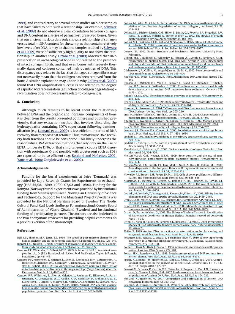
s of An
1teatvletpomdnbfor
5
bivharerEtaY
A
poRfapCofta
R
B
B
C
C
C
P.F. Campos et al. / Annal
999), and contradictory to several other studies on older sampleshat have failed to note such a relationship. For example, Schwarzt al. (2009) do not observe a clear correlation between collagennd DNA content in a series of permafrost preserved bones. Givenhat our ancient musk ox data only shows a relationship of collagenersus total mtDNA content for the samples that contain relativelyow levels of mtDNA, it may be that the samples studied by Schwarzt al. (2009) were of sufficiently high quality to not show the rela-ionship. In another study, Ottoni et al. (2009) observed that DNAreservation in archaeological bone is not related to the presencef intact collagen fibrils, and that even bones with severely ther-ally damaged collagen contained high quantities of DNA. This
iscrepancy may relate to the fact that damaged collagen fibres mayot necessarily mean that the collagen has been removed from theone. A similar explanation may underlie why Collins et al. (2009)ound that DNA amplification success is not related to the degreef aspartic acid racemisation (a function of collagen degradation) –acemisation does not necessarily relate to collagen loss.
. Conclusion
Although much remains to be learnt about the relationshipetween DNA and the organic and inorganic components of bone
t is clear from the results presented both here and published pre-iously, that any extraction method that involves discarding theydroxyapatite-containing EDTA supernatant following deminer-lisation (e.g. Leonard et al., 2000) is less efficient in terms of DNAecovery than methods that retain it. Thus, to maximise DNA recov-ry both fractions should be considered. This likely underpins theeason why aDNA extraction methods that rely only on the use ofDTA to liberate DNA, or that simultaneously couple EDTA diges-ion with proteinase K (and possibly with a detergent such as SDS)re reported to be so efficient (e.g. Rohland and Hofreiter, 2007;ang et al., 1998; Zołedziewska et al., 2002).
cknowledgements
Funding for the burial experiments at Lejre (Denmark) wasrovided by Lejre Research Grants for Experiments in Archaeol-gy (HAF 35/98, 15/99, 10/00, 07/02 and 10/06). Funding for theømyra (Norway) burial experiments was provided by institutional
unding from Vitenskapsmuseet, Norwegian University of Sciencend Technology. Support for the Marstrand Project (Sweden) wasrovided by the National Heritage Board of Sweden, The Nordicultural Fund, Carl Jacob Lindbergs Fornminnesfond, County Boardf Administration of Västra Götaland (Sweden) and institutionalunding of participating partners. The authors are also indebted tohe two anonymous reviewers for providing helpful comments on
previous version of the manuscript.
eferences
ell, L.S., Skinner, M.F., Jones, S.J., 1996. The speed of post-mortem change to thehuman skeleton and its taphonomic significance. Forensic Sci. Int. 82, 129–149.
jördal, C.G., Nilsson, T., 2008. Reburial of shipwrecks in marine sediments: a long-term study on wood degradation. J. Archaeol. Sci. 35, 862–872.
ampos, P.F., Willerslev, E., Gilbert, M.T.P., 2009. Isolation of DNA from ancient sam-ples. In: Liu, D. (Ed.), Handbook of Nucleic Acid Purification. Taylor & Francis,Boca Raton, pp. 441–461.
ampos, P.F., Kristensen, T., Orlando, L., Sher, A., Kholodova, M.V., Götherström, A.,Hofreiter, M., Drucker, D.G., Kosintsev, P., Tikhonov, A., Baryshnikov, G.F., Willer-slev, E., Gilbert, M.T.P., 2010a. Ancient DNA sequences point to a large loss ofmitochondrial genetic diversity in the saiga antelope (Saiga tatarica) since thePleistocene. Mol. Ecol. 19, 4863–4875.
ampos, P.F., Willerslev, E., Sher, A., Orlando, L., Axelsson, E., Tikhonov, A., Aaris-
Sørensen, K., Greenwood, A.D., Kahlke, R.-D., Kosintsev, P., Krakhmalnaya, T.,Kuznetsova, T., Lemey, P., MacPhee, R., Norris, C.A., Shepherd, K., Suchard, M.A.,Zazula, G.D., Shapiro, B., Gilbert, M.T.P., 2010b. Ancient DNA analyses excludehumans as the driving force behind late Pleistocene musk ox (Ovibos moschatus)population dynamics. Proc. Natl. Acad. Sci. U. S. A. 107, 5675–5680.atomy 194 (2012) 7– 16 15
Collins, M., Riley, M., Child, A., Turner-Walker, G., 1995. A basic mathematical sim-ulation of the chemical degradation of ancient collagen. J. Archaeol. Sci. 22,175–183.
Collins, M.J., Nielsen-Marsh, C.M., Hiller, J., Smith, C.I., Roberts, J.P., Prigodich, R.V.,Wess, T.J., Csapo, J., Millard, A., Turner-Walker, G., 2002. The survival of organicmatter in bone: a review. Archaeometry 44, 383–394.
Collins, M.J., Penkman, K.E.H., Rohland, N., Shapiro, B., Dobberstein, R.C., Ritz-Timme,S., Hofreiter, M., 2009. Is amino acid racemization a useful tool for screening forancient DNA in bone? Proc. R. Soc. B-Biol. Sci. 276, 2971–2977.
Currey, J.D., 2002. Bones: Structure and Mechanics. Princeton University Press,Princeton.
Gilbert, M.T.P., Rudbeck, L., Willerslev, E., Hansen, A.J., Smith, C., Penkman, K.E.H.,Prangenberg, K., Nielsen-Marsh, C.M., Jans, M.E., Arthur, P., 2005. Biochemicaland physical correlates of DNA contamination in archaeological human bonesand teeth excavated at Matera, Italy. J. Archaeol. Sci. 32, 785–793.
Götherström, A., Collins, M., Angerbjörn, A., Lidén, K., 2002. Bone preservation andDNA amplification. Archaeometry 44, 395–404.
Hagelberg, E., Sykes, B., Hedges, R., 1989. Ancient bone DNA amplified. Nature 342,485.
Hansen, A.J., Mitchell, D.L., Wiuf, C., Paniker, L., Brand, T.B., Binladen, J., Gilichin-sky, D.A., Ronn, R., Willerslev, E., 2006. Crosslinks rather than strand breadsdetermine access to ancient DNA sequences from sediments. Genetics 173,1175–1179.
Hedges, R.E.M., 2002. Bone diagenesis: an overview of processes. Archaeometry 44,319–328.
Hedges, R.E.M., Millard, A.R., 1995. Bones and groundwater – towards the modelingof diagenetic processes. J. Archaeol. Sci. 22, 155–164.
Hummel, S., Herrmann, B., 1994. Y-Chromosomal DNA from Ancient Bones AncientDNA. Springer-Verlag, New York, pp. 205–210.
Jans, M., Nielsen-Marsh, C., Smith, C., Collins, M., Kars, H., 2004. Characterisation ofmicrobial attack on archaeological bone. J. Archaeol. Sci. 31, 87–95.
Kitamura, E., Iwamoto, C., Sakairi, N., Tokura, S., Nishi, N., 1997. Marked effect ofDNA on collagen fibrillogenesis in vitro. Int. J. Biol. Macromol. 20, 241–244.
Lees, S., 2003. Mineralization of type I collagen. Biophys. J. 85, 204–207.Leonard, J.A., Wayne, R.K., Cooper, A., 2000. Population genetics of ice age brown
bears. Proc. Natl. Acad. Sci. U. S. A. 97, 1651–1654.Lindahl, T., 1993. Instability and decay of the primary structure of DNA. Nature 362,
709–715.Lindahl, T., Nyberg, B., 1972. Rate of depurination of native deoxyribonucleic acid.
Biochemistry 12, 5151–5154.Mrevlishvili, G., Svintradze, D., 2005. DNA as a matrix of collagen fibrils. Int. J. Biol.
Macromol. 36, 324–326.Nielsen-Marsh, C.M., Hedges, R.E.M., 1999. Bone porosity and the use of mer-
cury intrusion porosimetry in bone diagenesis studies. Archaeometry 41,165–174.
Nielsen-Marsh, C.M., Smith, C.I., Jans, M.M.E., Nord, A., Kars, H., Collins, M.J., 2007.Bone diagenesis in the European Holocene II: taphonomic and environmentalconsiderations. J. Archaeol. Sci. 34, 1523–1531.
Nijweide, P.J., Burger, E.H., Feyen, J.H.M., 1986. Cells of bone: proliferation, differen-tiation, and hormonal regulation. Physiol. Rev. 66, 855–886.
Nudelman, F., Pieterse, K., George, A., Bomans, P.H.H., Friedrich, H., Brylka, L.J.,Hilbers, P.A.J., de With, G., Sommerdijk, N.A.J.M., 2010. The role of collagen inbone apatite formation in the presence of hydroxyapatite nucleation inhibitors.Nat. Mater. 7, 1004–1009.
Okazaki, M., Yoshida, Y., Yamaguchi, S., Kaneno, M., Elliott, J.C., 2001. Affinity bindingphenomena of DNA onto apatite crystals. Biomaterials 22, 2459–2464.
Orgel, J.P.R.O., Miller, A., Irving, T.C., Fischetti, R.F., Hammersley, A.P., Wess, T.J., 2001.The in situ supermolecular structure of type I collagen. Structure 9, 1061–1069.
Orgel, J.P.R.O., Irving, T.C., Miller, A., Wess, T.J., 2005. Microfibrillar structure of typeI collagen in situ. Proc. Natl. Acad. Sci. U. S. A. 103 (24), 9001–9005.
Ortner, D., Turner-Walker, G., 2003. The Biology of Skeletal Tissues, in Identificationof Pathological Conditions in Human Skeletal Remains, second ed. AcademicPress, 11-35.
Ottoni, C., Koon, H., Collins, M., Penkman, K., Rickards, O., Craig, O., 2009. Preservationof ancient DNA in thermally damaged archaeological bone. Naturwissenschaften96, 267–278.
Pääbo, S., 1989. Ancient DNA: extraction, characterization, molecular cloning, andenzymatic amplification. Proc. Natl. Acad. Sci. U. S. A. 86, 1939.
Pesquero, M.D., Ascaso, C., Alcalá, L., Fernández-Jalvo, Y., 2010. A new taphonomicbioerosion in a Miocene lakeshore environment. Palaeogeogr. Palaeoclimatol.Palaeoecol. 295, 192–198.
Poinar, H., Hoss, M., Bada, J., Paabo, S., 1996. Amino acid racemization and the preser-vation of ancient DNA. Science 272, 864.
Poinar, H.N., Stankiewicz, B.A., 1999. Protein preservation and DNA retrieval fromancient tissues. Proc. Natl. Acad. Sci. U. S. A. 96, 8426–8431.
Prüfer, K., Stenzel1, U., Hofreiter, M., Pääbo, S., Kelso, J., Green, R.E., 2010. Compu-tational challenges in the analysis of ancient DNA. Genome Biol. 11 (5), R47,doi:10.1186/gb-2010-11-5-r47.
Pruvost, M., Schwarz, R., Correia, V.B., Champlot, S., Braguier, S., Morel, N., Fernandez-Jalvo, Y., Grange, T., Geigl, E.M., 2007. Freshly excavated fossil bones are best foramplification of ancient DNA. Proc. Natl. Acad. Sci. U. S. A. 104, 739.
Rohland, N., Hofreiter, M., 2007. Comparison and optimization of ancient DNAextraction. Biotechniques 42, 343–352.
Salamon, M., Tuross, N., Arensburg, B., Weiner, S., 2005. Relatively well preservedDNA is present in the crystal aggregates of fossil bones. Proc. Natl. Acad. Sci. U.S. A. 102, 13783–13788.

1 s of An
S
S
S
S
S
T
6 P.F. Campos et al. / Annal
chwarz, C., Debruyne, R., Kuch, M., McNally, E., Schwarcz, H., Aubrey, A., Bada, J.,Poinar, H., 2009. New insights from old bones: DNA preservation and degra-dation in permafrost preserved mammoth remains. Nucleic Acids Res. 37,3215–3229.
hapiro, B., Drummond, A.J., Rambaut, A., Wilson, M.C., Matheus, P.E., Sher, A.V.,Pybus, O.G., Gilbert, M.T.P., Barnes, I., Binladen, J., Willerslev, E., Hansen, A.J.,Baryshnikov, G.F., Burns, J.A., Davydov, S., Driver, J.C., Froese, D.G., Harington,C.R., Keddie, G., Kosintsev, P., Kunz, M.L., Martin, L.D., Stephenson, R.O., Storer,J., Tedford, R., Zimov, S., Cooper, A., 2004. Rise and fall of the Beringian steppebison. Science 306, 1561–1565.
mith, C.I., Chamberlain, A.T., Riley, M.S., Cooper, A., Stringer, C.B., Collins, M.J., 2001.Neanderthal DNA: not just old but old and cold? Nature 410, 772–773.
mith, C.I., Chamberlain, A.T., Riley, M.S., Stringer, C., Collins, M.J., 2003. The thermalhistory of human fossils and the likelihood of successful DNA amplification. J.Hum. Evol. 45, 203–217.
mith, C.I., Nielsen-Marsh, C.M., Jans, M.M.E., Collins, M.J., 2007. Bone diagene-
sis in the European Holocene I: patterns and mechanisms. J. Archaeol. Sci. 34,1485–1493.odoh, M., Tadano, S., Giri, B., Nishimoto, M., 2009. Effect of gradual demineralizationon the mineral fraction and mechanical properties of cortical bone. J. Biomech.Sci. Eng. 4, 230–238.
atomy 194 (2012) 7– 16
Trueman, C., Martill, D., 2002. The long term survival of bone: the role of bioerosion.Archaeometry. 44, 371–382.
Turner-Walker, G., Peacock, E.E., 2008. Preliminary results of bone diagen-esis in Scandinavian bogs. Palaeogeogr. Palaeoclimatol. Palaeoecol. 266,151–159.
Turner-Walker, G., Jans, M., 2008. Reconstructing taphonomic histories using histo-logical analyses. Palaeogeogr. Palaeoclimatol. Palaeoecol. 266, 227–235.
Tuross, N., 2003. Recent Advances in Bone, Dentin and Enamel Biochemistry, inIdentification of Pathological Conditions in Human Skeletal Remains, seconded. Academic Press, pp. 65–72.
Tzaphlidou, M., Berillis, P., 2005. Collagen fibril diameter in relation to bone site. Aquantitative ultrastructural study. Micron 36, 703–705.
Yang, D.Y., Eng, B., Waye, J.S., Dudar, J.C., Saunders, S.R., 1998. Technical note:improved DNA extraction from ancient bones using silica-based spin columns.Am. J. Phys. Anthropol. 105, 539–543.
Yoshino, M., Kimijima, T., Miyasaka, S., Sato, H., Seta, S., 1991. Microscopical study
on estimation of time since death in skeletal remains. Forensic Sci. Int. 49,143–158.Zołedziewska, M., Gronkiewicz, S., Dobosz, T., 2002. Comparison of various decal-cificators in preparation of DNA from human rib bones. Anthropol. Rev. 65,75–80.