Dissertazia na english
340
Dissertation submitted to the Combined Faculties for the Natural Sciences and for Mathematics of the Ruperto-Carola University of Heidelberg, Germany, for the degree of Doctor of Natural Sciences Presented by: Diploma biologist Thomas-Benjamin Seiler Born in: Erlangen Date of oral examination: …...................................
-
Upload
uni-augsburg -
Category
Documents
-
view
1 -
download
0
Transcript of Dissertazia na english

Dissertation
submitted to the Combined Faculties for the Natural Sciences
and for Mathematics of the Ruperto-Carola University of Heidelberg, Germany,
for the degree of Doctor of Natural Sciences
Presented by: Diploma biologist Thomas-Benjamin Seiler
Born in: Erlangen
Date of oral examination: …...................................


Total or biomimetic extracts or direct contact exposure?
Comparative research towards a realistic
ecotoxicological characterisation of sediments
Referees: Prof Dr Thomas Braunbeck
Heidelberg Institute of Zoology, University of Heidelberg
Prof Dr Heinz Karrasch
Geographical Institute, University of Heidelberg

Acknowledgements
"Each of us has cause to think with deep gratitude of those who have lighted the flame within us."Albert Schweitzer
If a PhD thesis represents more than just the product of laboratory work and scientific writing, this is surely due to the persons who were directly and indirectly involved. Hence, my deep gratitudes go to all those colleagues and friends accompanying me throughout these very enjoyable years of scientific research.
First of all, I would like to thank Prof Dr Thomas Braunbeck, Aquatic Ecology & Toxicology Section at the Heidelberg Institute of Zoology, for giving me the opportunity to prepare my thesis under his official supervision and, thus, being my first referee.
I am no less thankful to Prof Dr Henner Hollert, Department of Ecosystem Analysis at RWTH Aachen University, my supervisor, who let me experience as much freedom as required to evolve my academic as well as creative spirits. Besides, I am truly grateful to Prof Hollert and Dr Jan Wölz for the pleasant collaboration during the 2007 establishment of the Department of Ecosystem Analysis at RWTH Aachen University.
My expressed appreciation also go to Prof Dr Heinz Karrasch for being my second referee and by this showing his interest in research on sediment toxicity.
What made the years of my thesis so special was a very friendly working atmosphere created by the various colleagues in Heidelberg and Aachen, in particular Nina Best, Kerstin Bluhm, Markus Brinkmann, Marit Ernst, Tilman Floehr, Andrea Gerstner, Prof Dr Arnold V Hallare, Sebastian Hudjetz, Dr Steffen Keiter, Dr Thomas Kosmehl, Dr Eva Lammer, Erik Leist, Sibylle Maletz, Sabine Niebergall, Sabrina Peddinghaus, Dr Paula Suares Rocha, Ruben Strecker, Dr Jan Wölz and Hanno Zielke. Furthermore, I would like to thank Conny Bernecker, Sebastian Heger and Kerstin Winkens for their professional support of my laboratory work.
Essential for good scientific research is reliable and inspiring collaboration. From the large number of colleagues I had the pleasure to work with, my sincere thanks go to the following: Dr Werner Brack, Tobias Schulze, Dr Georg Streck (UFZ Helmholtz Centre for Environmental Research, Leipzig), Dr Katrin Schwab (Currenta, Leverkusen) and Dr Andrew C Rastall (Rifcon, Hirschberg) for collaborating in the research on sediment extraction, PD Dr Wolfgang Ahlf (Technical University Hamburg-Harburg, Hamburg), Dr Ute Feiler, Dr Peter Heininger, Denise Spira (Federal Hydrological Institute, Koblenz), Daniel Gilberg (ECT Oekotoxikologie, Flörsheim), Dr Monika Hammers-Wirtz (gaiac, Aachen), Dr Sebastian Höss (Ecossa, Starnberg), Dr Michael Meller (ecotox consult, Ludwigshafen), Dr Helga Neumann-Hensel (Dr. Fintelmann & Dr. Meyer, Hamburg), Dr Richard Ottermanns (RWTH Aachen University, Aachen) and Dr Jürgen Weber (Nordum, Rostock) for cooperation in the SeKT joint research project on sediment contact tests. I am furthermore thankful to the German Federal Ministry of Education and Research (BMBF) for funding the SeKT project.
I also thank the SETAC Europe Student Advisory Council, especially Markus Brinkmann, Dr. Amy Brooks, Mirco Bundschuh, Marloes Caduff, Andre Dabrunz, Cornelia Kienle and Karen Van Hoecke, for the pleasant time.
Always at my side were my family, my dearest friends, and Astrid.


Content I
Content
Summary...................................................................................................................................12Zusammenfassung.....................................................................................................................141 Introduction............................................................................................................................18
1.1 The application of scientific research within ecotoxicology..........................................181.2 A necessity to understand the processes in aquatic ecotoxicology.................................191.3 Strategies for the ecotoxicological characterisation of sediments..................................201.4 Comparative investigations of sediment extraction and contact tests............................211.5 Objectives of the study...................................................................................................241.6 References......................................................................................................................25
2 Introductory Part A.................................................................................................................322.1 Abstract...........................................................................................................................322.2 Abbreviations..................................................................................................................322.3 Introduction....................................................................................................................322.4 A short outline of extraction...........................................................................................35
2.4.1 Geosorbents, accessibility, and extractability.........................................................352.4.2 Application of soil and sediment extracts...............................................................372.4.3 Fractions and the toxic potential.............................................................................38
2.5 Extraction and clean-up..................................................................................................382.6 Possible alterations affecting reliability of extracts........................................................38
2.6.1 Extraction and extract testing.................................................................................392.6.2 Sample processing prior to extraction....................................................................41
2.7 Recommendations for research and technology.............................................................422.7.1 Quality of data........................................................................................................422.7.2 Future recommendations of extract preparation.....................................................43
2.8 Summary and conclusion...............................................................................................452.9 References......................................................................................................................46
3 Chapter 3................................................................................................................................543.1 Abstract...........................................................................................................................543.2 Introduction....................................................................................................................553.3 Materials and Methods...................................................................................................57
3.3.1 Sediment samples...................................................................................................573.3.2 Membrane dialysis extraction (MDE)....................................................................573.3.3 Soxhlet extraction...................................................................................................583.3.4 Cell cultures............................................................................................................593.3.5 Statistical analysis...................................................................................................593.3.6 Bioassays................................................................................................................60
3.4 Results and Discussion...................................................................................................613.4.1 Comparison to Soxhlet extraction..........................................................................62
3.5 Conclusions....................................................................................................................68

Content II
3.6 References......................................................................................................................694 Chapter 4................................................................................................................................76
4.1 Abstract...........................................................................................................................764.2 Introduction....................................................................................................................764.3 Material and methods.....................................................................................................78
4.3.1 Chemicals...............................................................................................................784.3.2 Study area and sample collection...........................................................................784.3.3 Sample storage and preparation..............................................................................784.3.4 Grain size distribution and content of organic carbon............................................784.3.5 Extraction Methods.................................................................................................794.3.6 Extraction and hydrolysis of sediment residues.....................................................804.3.7 Extracts preparation................................................................................................804.3.8 Silica gel fractionation............................................................................................804.3.9 Gas chromatography / Mass spectrometry.............................................................804.3.10 Toxicity testing.....................................................................................................814.3.11 Data analysis.........................................................................................................81
4.4 Results and discussion....................................................................................................824.4.1 Chemical analysis...................................................................................................824.4.2 Comparison of different extraction approaches......................................................834.4.3 Compound specific extractability of PAH..............................................................854.4.4 Toxicity analysis.....................................................................................................87
4.5 Conclusions....................................................................................................................884.6 Acknowledgements........................................................................................................894.7 References......................................................................................................................89
5 Chapter 5................................................................................................................................985.1 Abstract...........................................................................................................................985.2 Abbreviations..................................................................................................................985.3 Introduction....................................................................................................................995.4 Material & Methods.....................................................................................................100
5.4.1 Samples and sampling..........................................................................................1005.4.2 Extract treatment and storage...............................................................................1015.4.3 Soxhlet extraction (SOX).....................................................................................1015.4.4 Membrane dialysis extraction (MDE)..................................................................1015.4.5 Methanol/Water extraction (MEOH)....................................................................1015.4.6 Hydroxypropyl-β-cyclodextrin extraction (HBCD).............................................1015.4.7 Tenax-TA extraction (TENAX)............................................................................1025.4.8 Accelerated solvent extraction (ASE)...................................................................1025.4.9 Gel permeation chromatography (GPC)...............................................................1025.4.10 Accelerated membrane-assisted clean-up (AMAC)...........................................1035.4.11 Direct accelerated membrane assisted clean-up (DAMAC)...............................1035.4.12 Process controls..................................................................................................1035.4.13 Cell cultures........................................................................................................103

Content III
5.4.14 Neutral red retention assay (NR)........................................................................1035.4.15 EROD induction assay (EROD).........................................................................1045.4.16 Zebrafish embryo assay (Fish embryo test; FET)...............................................1045.4.17 Graphical evaluation and statistical analysis......................................................105
5.5 Results..........................................................................................................................1055.5.1 Process controls....................................................................................................1055.5.2 Comparability with respect to extraction power...................................................1055.5.3 Ranking according extraction power and reproducibility....................................1085.5.4 Correlations concerning strength of effect potential............................................109
5.6 Discussion.....................................................................................................................1115.7 Conclusion & Outlook..................................................................................................1125.8 Acknowledgements.......................................................................................................1135.9 References....................................................................................................................113
6 Chapter 6..............................................................................................................................1226.1 Abstract.........................................................................................................................1226.2 Abbreviations................................................................................................................1236.3 Introduction..................................................................................................................1236.4 Material & Methods.....................................................................................................126
6.4.1 Samples and sampling..........................................................................................1266.4.2 Chemicals and solvents.........................................................................................1266.4.3 Clean-up with gel permeation chromatography (GPC)........................................1266.4.4 Accelerated membrane assisted clean-up (AMAC)..............................................1276.4.5 Accelerated solvent extraction (ASE)...................................................................1276.4.6 Membrane dialysis extraction (MDE)..................................................................1276.4.7 Soxhlet extraction (SOX).....................................................................................1286.4.8 Direct Accelerated Membrane Assisted Clean-up (DAMAC)..............................1286.4.9 Tenax-TA extraction (TENAX)............................................................................1286.4.10 Hydroxypropyl-β-cyclodextrin extraction (HBCD)...........................................1296.4.11 Methanol/Water extraction (MEOH)..................................................................1296.4.12 Ultrasonic extraction with hexane:acetone.........................................................1296.4.13 Extraction with methanolic hydrolysis...............................................................1296.4.14 Quality control....................................................................................................1306.4.15 Instrumental analysis..........................................................................................1306.4.16 Biological analysis..............................................................................................1306.4.17 Statistical analysis...............................................................................................1306.4.18 Artificial mixtures and calculation of Toxic Equivalents (TEQs) for EROD-data....................................................................................................131
6.5 Results..........................................................................................................................1316.5.1 Total amounts of PAHs and chlorinated compounds – first extraction step.........1316.5.2 Second and third extraction..................................................................................1326.5.3 Compound specific composition of extracts.........................................................133
6.6 Discussion....................................................................................................................136

Content IV
6.6.1 Ranking according to extraction power – first extraction step.............................1366.6.2 Comparability with respect to extraction efficiency – first extraction step..........1386.6.3 Comparison of all three extraction steps..............................................................1406.6.4 Compound specific correlation analysis...............................................................1406.6.5 Comparison with results from bioanalytical methods..........................................1436.6.6 Confirmation of EROD-induction using artificial mixtures.................................143
6.7 Conclusion & Outlook..................................................................................................1456.8 Supplemental data........................................................................................................1466.9 Acknowledgements......................................................................................................1466.10 References..................................................................................................................147Supplemental data..............................................................................................................152
7 Chapter 7..............................................................................................................................1627.1 Abstract.........................................................................................................................1627.2 Introduction..................................................................................................................1637.3 Material and Methods...................................................................................................165
7.3.1 Sediments, Substances and Spiking......................................................................1657.3.2 Extractions............................................................................................................1667.3.3 Fish embryo test with Danio rerio........................................................................1697.3.4 Data processing and statistical analyses...............................................................170
7.4 Results..........................................................................................................................1717.5 Discussion....................................................................................................................1737.6 Outlook.........................................................................................................................1757.7 Acknowledgements .....................................................................................................1767.8 References....................................................................................................................176
8 Chapter 8..............................................................................................................................1848.1 Abstract.........................................................................................................................1848.2 Introduction..................................................................................................................1848.3 Material & Methods.....................................................................................................187
8.3.1 Samples and sampling..........................................................................................1878.3.2 Extraction of sediment samples............................................................................1888.3.3 Chemical analysis.................................................................................................1888.3.4 Bioassays..............................................................................................................1888.3.5 Graphical evaluation, calculation of potentials, and statistical analysis...............189
8.4 Results..........................................................................................................................1898.5 Discussion....................................................................................................................1928.6 Conclusions & Perspectives.........................................................................................1958.7 References....................................................................................................................196
9 Introductory Part B...............................................................................................................2049.1 Abstract.........................................................................................................................2049.2 Introduction..................................................................................................................2049.3 The significance of fish as sentinel species..................................................................2079.4 The use of whole fish in sediment toxicity assessment................................................208

Content V
9.4.1 Issues on the use of fish for toxicity studies ........................................................2089.4.2 Methodological variations in sediment toxicity using whole fish........................210
9.5 The use of primary fish cell culture and fish cell lines as substitute for whole organism bioassays.......................................................................................................226
9.5.1 Advantages of using fish cell culture and fish cell lines.......................................2269.5.2 Cell donors in primary culture and cell lines........................................................2279.5.3 Basic methodology...............................................................................................2279.5.4 Applications in sediment toxicity assessment.......................................................233
9.6 The use of fish egg (embryo) for sediment toxicity assessment..................................2359.6.1 Historical account.................................................................................................2359.6.2 Significance of using fish embryo........................................................................2369.6.3 Use of fish embryo in sediment toxicity...............................................................2369.6.4 The use of fish egg test with zebrafish (Danio rerio)...........................................238
9.7 The use of gene expression analysis with RT-PCR and gene expression profiling using microarrays in fish for sediment assessment......................................................2469.8 Summary and Perspectives...........................................................................................2509.9 References....................................................................................................................253
10 Chapter 10..........................................................................................................................27010.1 Abstract.......................................................................................................................27010.2 Introduction................................................................................................................27010.3 Materials and Methods...............................................................................................272
10.3.1 Sediment sampling..............................................................................................27210.3.2 Sediment analysis...............................................................................................27310.3.3 Sediment-contact tests........................................................................................27510.3.4 Data analysis.......................................................................................................275
10.4 Results........................................................................................................................27810.4.1 Sediment properties............................................................................................27810.4.2 Response to sediment samples............................................................................283
10.5 Discussion..................................................................................................................28910.5.1 Influence of sediment properties........................................................................28910.5.2 Toxicity thresholds..............................................................................................29010.5.3 Battery of sediment contact tests........................................................................292
10.6 Conclusions................................................................................................................29210.7 Acknowledgements....................................................................................................29310.8 References..................................................................................................................293
11 Chapter 11..........................................................................................................................30011.1 Abstract.......................................................................................................................30011.2 Introduction................................................................................................................30011.3 Material & Methods...................................................................................................302
11.3.1 Sediment samples and sampling.........................................................................30211.3.2 Spiking................................................................................................................30211.3.3 Sediment contact tests.........................................................................................304

Content VI
11.3.4 Statistical analysis...............................................................................................30611.4 Results........................................................................................................................30611.5 Discussion...................................................................................................................31011.6 Conclusions & Perspectives.......................................................................................31411.7 Acknowledgements.....................................................................................................31411.8 References..................................................................................................................315
12 Conclusions........................................................................................................................32212.1 Confounding factors in sediment assessment.............................................................32212.2 Proposal for a strategical framework..........................................................................323
13 Scientific contributions......................................................................................................328

Summary 3
Summary
Sediment toxicity is an important scientific research subject and besides one of the main practical applications of ecotoxicology. Due to the multifaceted nature of sediments in combination with varying physical-chemical properties of environmental contaminants and a multitude of different living and feeding behaviours of the benthic community, estimation of the toxicological risk associated to polluted sediments appears to get easily hampered. Hence, for a maximum of veracity in sediment assessment at least applied methods and technologies should provide reliability of gained results. However, while a large number of differing approaches are commonly utilized to investigate contamination of sediments, little attention is brought to confounding factors that might influence the outcome of such applied research. Moreover, knowledge still lacks on how the various approaches compare and can be combined into a comprehensive investigation strategy. The present study aims at filling parts of these gaps through investigation and characterisation of (1) sediment extraction procedures and (2) whole sediment contact tests.
After identifying the main confounding factors within preparation of exhaustive extracts, considerations are formulated regarding procedures with a reduced risk of altering samples. A novel vigorous but completely passive extraction method is then introduced, which works with dialysis over a semipermeable membrane driven only by strong gradients (named membrane dialysis extraction, MDE). Initially, MDE extracts were compared to samples from the commonly applied Soxhlet extraction protocol in biotests on cytotoxity, embryo toxicity and dioxin-like activity. Following a first application in a study on two riverine sediments, wherein data from the above-mentioned biotest systems as well as chemical analysis of 18 PAHs were obtained using extracts from MDE, Soxhlet and ultrasonic extraction, the new approach was part of a comprehensive comparison of five different vigorous leaching techniques. Extracts from another two riverine sediments were tested for their toxic effectiveness with respect to cytotoxity, embryo toxicity, and dioxin-like activity, and a number of compound classes, including PAHs, PCBs and DDXs, were determined chemically. Furthermore, three biomimetic extraction procedures were investigated in parallel. All techniques were evaluated in terms of extraction power and reproducibility. In a further comparative study, MDE, Soxhlet and two biomimetic methods involving either HBCD or Tenax® were compared to direct contact exposure using the fish embryo test on Danio rerio. Summarizing all four studies, the ability of MDE, Soxhlet and ASE® to yield toxic substances from sediment samples was evaluated by ranking data from biotests and chemical analyses.
The topic of sediment contact tests is initiated by an extensive literature review on the use of fish as a test organism in sediment investigation, covering also direct contact exposure. In order to allow reliable testing of sediments, six contact assays were thoroughly investigated for toxicity thresholds and test conditions. It was furthermore intended to identify reference sediments that are applicable for all test systems. Subsequently, the contact test battery was applied on the reference sediments spiked with a cocktail of either heavy metals or of organic substances. Resulting data were evaluated with respect to sensitivity and applicability of the different sediment contact tests.
MDE was found to provide extraction powers comparable to automated ASE®-methods for most applications, while working at a reduced risk of alteration of resulting extracts. The Soxhlet procedure also gave overall good agreement with ASE®-based methods and MDE, but results indicate an elevated risk of loss of target analytes. Among the biomimetic approaches, Tenax proved best in replicating results from contact tests, while HBCD extracts revealed vigorous extraction powers in some cases. In general, biomimetic methods revealed strong variability of data among each other, complicating the investigation of the bioavailable contaminant fractions.
With the sediment contact assays, toxicity thresholds were derived for all six investigated test systems. Furthermore, one natural as well as one formulated reference sediment for the whole test battery could be identified. The majority of assays returned clear dose-response relations for both sediments contaminated with either heavy metals of organic substances. However, the contact tests gave different sensitivities for the sediment-contaminant combinations and also showed varying steepness of the response curves. Based on the results and available literature data, sediment organic matter, clay content, substance properties and feeding as well as living habits were accounted for the observed differences in contaminant availability.
The study concludes that investigations into sediment toxicity are likely to be influenced by a multitude of different parameters. As a consequence, it proposes a comprehensive strategical framework that involves several approaches in sediment toxicity assessment and points out required future developmental effort. Most pressing is an automated procedure for vigorous extraction at moderate temperatures, conceivable to be realized through a combination of ASE-methods and membrane dialysis. Furthermore, increased understanding of contaminant availability and test species behaviour is essential.

Summary 4

Zusammenfassung 5
Zusammenfassung
Die toxische Belastung von Sedimenten ist ein wichtiges Forschungsfeld und eines der Hauptanwendungsgebiete der Ökotoxikologie. Die Kombination aus der vielfältigen Zusammensetzung von Sedimenten, der breiten Varianz physisch-chemischer Eigenschaften von Umweltkontaminanten und einer großen Zahl unterschiedlicher Verhaltensweisen benthischer Organismen kann die Abschätzung toxikologischer Risiken durch belastete Sedimente erschweren. Um dennoch ein möglichst hohes Maß an Realitätsnähe der Daten zu gewährleisten müssen die verfügbaren Methoden verlässliche Ergebnisse liefern können. Allerdings finden störende Einflussfaktoren der verwendeten Technologien und Untersuchungsansätze bei der Untersuchung von kontaminierten Sedimentproben nur geringe Beachtung. Zudem ist kaum bekannt, wie vergleichbar die verschiedenen Herangehensweisen sind, und es fehlt ein umfassendes Untersuchungskonzept, das die Stärken mehrerer Methoden nutzt. Die vorliegende Studie versucht, über die vergleichende Charakterisierung von (1) Technologien zur Sedimentextraktion und (2) Sediment-Kontakt-Tests Teile dieser Wissenslücken zu füllen.
Einer einleitenden Abhandlung zur Sedimentextraktion als Verfahren und der damit verbundenen Probleme bzw. ihrer möglichen Lösungen folgt die Vorstellung einer passiven Extraktionsmethode (genannt Membrane Dialysis Extraction, MDE), welche mittels Dialyse über eine semipermeable Membran Sedimentproben schonend extrahiert. Die resultierenden Extrakte wurden zunächst bioanalytisch hinsichtlich Zelltoxizität, Embryotoxizität und dioxinähnlicher Wirksamkeit untersucht und mit Soxhlet-Extrakten verglichen. In einer Studie mit zwei Flusssedimenten wurden MDE-Extrakte anschließend wiederum bzgl. Zelltoxizität, Embryotoxizität und dioxinähnlicher Wirksamkeit mit Proben aus Soxhlet- und Ultraschallextraktion verglichen. Zusätzlich wurden in allen Extrakten die Konzentrationen von 18 PAHs bestimmt. Die MDE-Methodik wurde dann in einer umfassenden Untersuchung vier weiteren erschöpfenden Extraktionsverfahren gegenübergestellt und in Biotests sowie chemischer Analyse von PAHs, PCBs und DDXs hinsichtlich ihrer Extraktionsleistung und Reproduzierbarkeit charakterisiert. In der gleichen Studie wurden zudem drei biomimetische Extraktionsmethoden miteinander und mit den erschöpfenden Technologien verglichen. Eine weitere Studie von MDE, Soxhlet sowie den biomimetischen Ansätzen HBCD und Tenax® untersuchte Extrakte und direkten Sedimentkontakt im Fisch Embryo Test mit dem Zebrabärbling Danio rerio. Die Ergebnisse für MDE- Soxhlet- und ASE-Extrakte aus allen vier Studien wurden abschließend mit Bezug auf die Extraktionsleistung vergleichend ausgewertet. Sedimentkontakttests werden als Teil einer umfangreichen Literaturübersicht zur Nutzung von Fischen bei der Untersuchung von Sedimenten einleitend behandelt. In der darauf folgenden Studie wurden sechs Kontakttest-Systeme vergleichend untersucht um Toxizitätsschwellenwerte und Standard-Testbedingungen zu definieren. Des Weiteren wurden im Rahmen dieser Untersuchung zwei für alle Tests verwendbare Referenzsedimente identifiziert. Diese wurden sodann mit Mischung von Schwermetallen oder organischen Schadstoffen dotiert und in allen sechs Biotests untersucht. Die Ergebnisse dienten dazu, die Testsysteme hinsichtlich ihrer Sensitivität und Anwendbarkeit zu charakterisieren.
Die MDE zeigte eine den automatisierten ASE-Methoden vergleichbare Extraktionsleistung, bei gesenktem Risiko, dass der Extraktionsprozess die Probe stark verändert. Auch Soxhlet war vergleichbar mit MDE und ASE, jedoch wurden Hinweise für ein Risiko gefunden, durch die Prozedur Analyten zu verlieren. Mit der Tenax-Extraktion konnten die Ergebnisse aus dem Sedimentkontakttest am besten reproduziert werden, während die Stringenz von HBCD sogar an die erschöpfenden Methoden heranreichte. Grundsätzlich unterlagen die Daten für biomimetische Verfahren starken Schwankungen, was die Untersuchung der bioverfügbaren Schadstofffraktionen erschwerte. Für alle sechs Sedimentkontakttests konnten Toxizitätsschwellenwerte errechnet werden, und die meisten Systeme lieferten klare Dosis-Wirkungs-Beziehungen für die Belastung durch Schwermetalle bzw. organische Substanzen. Allerdings ergaben sich starke Unterschiede zwischen den Sensitivitäten der einzelnen Testverfahren. Anhand von Daten aus der Literatur wurden organische Materie, Tonanteil, Substanzeigenschaften und Verhaltensweisen der Testorganismen als entscheidend identifiziert.
Aus der vorliegenden Studie wird ersichtlich, dass die toxikologische Untersuchung von Sedimenten durch eine Vielzahl von Faktoren beeinflusst werden kann. Dementsprechend wird ein umfassender strategischer Untersuchungsansatz vorgeschlagen und außerdem auf notwendige Forschung und Entwicklung hingewiesen, um die Bewertung von Sedimentbelastung verlässlicher zu gestalten. Unbedingt nötig ist in diesem Zusammenhang eine automatisierte, erschöpfende Extraktion bei moderaten Temperaturen, die mittels einer Kombination aus ASE und Membrandialyse erreicht werden kann. Daneben ist ein tieferes Verständnis der Verfügbarkeit von Kontaminanten, v.a. im Zusammenspiel mit den Verhaltensweisen der Testorganismen, entscheidend.

Zusammenfassung 6

Introduction
"The aim of science is not to open a door to infinite wisdom,but to set a limit to infinite error." Bertolt Brecht


1 Introduction 9
1 Introduction
1.1 The application of scientific research within ecotoxicology
Ecotoxicology is a rather young research field, defined as "the description of hazardous alterations of structures and functions in ecosystems caused by environmental pollutants" (Nagel 1988) that demands for "a well-balanced assessment […] based on studies integrating analytical, toxicological and ecological information […]" (Brouwer et al. 1990) usually obtained from laboratory and field studies covering all biological levels (Fent 1998). Due to the complexity of the environment as such, ecotoxicology still holds a multitude of principles and phenomena to be discovered. On the other hand, work in this area has already very early been focused on solving problems in a practical approach upon applying concepts and methods developed before and employ resulting findings in regulation processes (Truhaut 1977, Van Straalen 2003). Already Truhaut outlined with his 1977 introduction into the concept of ecotoxicology not only the necessity of fundamental research in order to first comprehensively understand behaviour of environmental contaminants, but also drew a picture of a research field that will and has to directly avert "the ominous consequences for the health and well being of man" which may result from "the multifarious chemical pollution of the environment". As a consequence, research within ecotoxicology may be characterized as historically grown, often without being validated with up-to-date knowledge, adjusted to increasing experience and optimized using state-of-the-art technologies. Thus, ecotoxicological investigations might fail to deliver results with highest veracity, causing misinterpretation of data and subsequent inappropriate action. Being considered and utilized as an applied science, this scenario bears a strong risk for ecotoxicological investigations in general.
„There are no such things as applied sciences, only applications of science.“ Louis Pasteur
Despite from all practical aspects of ecotoxicological research, the main subject still is scientific research. This sometimes might get hampered when economical issues are taken into account, e.g. in terms of chemicals testing or the assessment of contaminated sediments in order to determine further treatment (decontamination, deposition etc.). However, scientifically valid application of ecotoxicological techniques is crucial for accurate assessment of the hazardous potential of any given sample type, and this requirement demands for reliable procedures in analytical as well as bioanalytical investigations. Hence, ecotoxicologists should question and review their portfolio of methods, concepts and principles on a regular basis. Outdated techniques should then be banned or updated. New technologies that can add to the quality of results should be thoroughly tested and included in protocols or investigation strategies when found appropriate.

1 Introduction 10
1.2 A necessity to understand the processes in aquatic ecotoxicology
„Lucky is he who has been able to understand the causes of things.“ Virgil
Among the most important causes for misunderstanding and misinterpretation of ecotoxicological data, lack of knowledge of causative interrelations surely plays a strong role. This deficiency is not limited to the processes influencing mobility, uptake, behaviour or fate of environmental contaminants (Jaffe 1991, Klein 1989, Knezovich et al. 1987, Richards & Shieh 1986, Schwarzenbach et al. 2006), but covers also the practical side of ecotoxicological research. From sorption of substances to lab ware over exposure principles and test conditions up to the whole process of sample preparation and treatment, various parameters might influence results drawn from experiments (Escher & Hermens 2004, US EPA 1996). Bioaccessibility and the organism-specific bioavailability are key issues for risk assessment in ecosystems and also of major concern regarding bioanalytical investigations (Alexander 2000, Brack et al. 2009, Ehlers & Luthy 2003, Reichenberg & Mayer 2006, Semple et al. 2003) . With respect to the vast number of parameters, processes, properties and issues, and considering their presumable impact on results in ecotoxicology, compromises appear to be essential at least for the majority of investigations. However, research has to continuously refine the knowledge used for risk evaluation in ecotoxicology.
A large part of ecotoxicological research deals with water and aquatic ecosystems. Beside historical reasons (Macek 1980, Pritchard 1993, Truhaut 1977), this fact can be attributed to the importance of these habitats as the main receiving bodies for any contamination introduced into the environment by point and diffuse sources (Farrington 1991, Pritchard 1993). In addition, also soil carries water content which is highly determinant for mobility, behaviour and fate of soil contaminants. Sediments in aquatic ecosystems provide various functions, such as habitat, food source, or just temporary substrate – e.g. during early life stages – for limnic and marine organisms (Palmer et al. 2000, SedNet 2004b, a). Most important from an ecotoxicological point of view is their ability to adsorb and, thus, temporarily detoxify contaminants that entered the water phase. However, legacy contamination can be remobilized due to, e.g., dredging, bioturbation or flood events (Ahlf et al. 2002, Eggleton & Thomas 2004, Hollert et al. 2007, Westrich & Förstner 2005, Wölz et al. 2008). This transforms sediments to secondary sources of pollution and makes them a major concern regarding water quality in European surface waters within implementation of the Water Framework Directive, as stressed by Förstner (2002).
Sediments are a highly complex matrix comprising mineral and organic particles, organic carbon components dissolved in pore water, such as humic substances (Burton Jr 1991), and large microbial communities forming so called biofilms of extracellular polysaccharides around and between particles (Battin & Sengschmitt 1999, Decho 2000). Consequently, sediments are rather difficult to investigate. As many contaminants get sequestered into pores

1 Introduction 11
of organic macromolecular structures – a process called "ageing " – and become unavailable to organisms (Alexander 2000), vigorous extraction techniques were introduced into sediment toxicology to exhaustively leach the full spectrum of compounds (e.g. Bandh et al. 2000, Josefsson et al. 2006).
1.3 Strategies for the ecotoxicological characterisation of sediments
Exhaustive extracts of sediments are proven to represent the total hazardous potential of a given sample (Dean 1996, Dean & Xiong 2000), thus putatively overestimating actual risk for organisms but allowing a more or less reliable assessment, as pointed out by Alexander (2000). Nevertheless, it has to be taken into account that the preparation of exhaustive extracts produces a hard-to-estimate risk of alteration of the original sample (e.g. Beiras et al. 1998, Belkessam et al. 2005, Puchalski et al. 1999, Schuytema et al. 1989). The extraction process might cause severe alterations of the original contaminant spectrum. Many procedures apply heat to facilitate the separation process and by this introduce auxiliary energy to a system containing a myriad of chemical substances and reactive groups (Steinberg et al. 2000, Steinberg et al. 2006). Resulting extracts might no longer represent the original ecotoxicological state at the sampling site and, thus, compromise the reliability of subsequent risk analysis and assessment.
An upcoming alternative approach in risk assessment of sediments is the investigation of bulk sediment samples by applying contact assays (Harkey et al. 1994, Hollert et al. 2003, Nebeker et al. 1984). Other than extract testing, data on effects due to direct sediment contact are considered to provide information about the bioavailability of contaminants. Therefore, obtained results can represent a very realistic scenario regarding the ecotoxicological state of a given sampling site.
However, while ecotoxicological investigations might rely more and more on contact assays in the future, with respect to chemical analysis and cell-based bioanalytical approaches sediment extraction cannot be abandoned. Moreover, due to limited sample availability it is often necessary to assay extracts using miniaturized biotest systems, such as the Ames fluctuation assay (Gatehouse 1978, Perez et al. 2003) or a 96-well version of the fish embryo test with Danio rerio (Seiler et al. 2009). Hence, extraction procedures are still necessary and should be subject to regular adaptation as well as optimization. Within the last years, much effort has been put into the development of extraction principles that provide extracts which can mimic bioavailability for organisms getting into contact with sediments (Cornelissen et al. 2001, Kelsey et al. 1997, Reid et al. 2000). These protocols are aimed at the rapidly desorbing and therefore readily available contaminant fraction, which is often identified as being degradable by microbes (Doick et al. 2005). However, concepts for such mild extraction

1 Introduction 12
cannot be ubiquitous and data for every individual setting of, e.g., sediment characteristics, organism, substance class – to name but a few – have to be determined empirically (Semple et al. 2004).
Already in 1990, Peter Chapman postulated with the Sediment Quality Triad (Chapman 1990, 2000) that ecotoxicological investigation and assessment of sediments need to be based on several different lines of evidence. Chapman and Hollert (2006) further discriminate between bioanalytical approaches (e.g. sediment contact, extracts, elutriates) as separate lines of evidence, and also suggest to integrate different extraction techniques as well as effect-directed analysis into comprehensive studies for sediment assessment. But also within these approaches all individual parts and steps have to fit together and provide a holistic investigation strategy, as discussed before. The present study aims at the thorough evaluation and characterization of the most important aspects of sediment extraction on the one hand and sediment contact tests on the other. Both approaches are primarily analyzed for their applicability, their suitability and how they add to the aim of a realistic risk assessment of sediment contamination.
1.4 Comparative investigations of sediment extraction and contact tests
Initially, the two different concepts of testing either sediment extracts – prepared using exhaustive or biomimetic methods – or (untreated) bulk sediment samples in bioanalytical investigations are comprehensively characterized and critically discussed.
For sediment extraction, a short historical abstract defines the origin of this preparative technique as a necessary tool for chemical analysis, which was the main approach for investigations in the early years of sediment toxicology (Introductory Part A). Following a brief introduction of the most common and promising extraction procedures, preparation of sediment extracts is reviewed in terms of general usage in ecotoxicological investigations. Then, issues of concern associated to extraction in general as well as specific characteristics of leaching sediments – such as the application of heat, the role of particulate organic matter – are detailed and critically discussed. The chapter closes with some suggestions on how to deal with data from extract testing and demands the development of new, more reliable, at best passive methods, which reduce the risk of altering sediment upon sample preparation.
Chapter 1 introduces passive membrane dialysis, a recent approach in sediment extraction and a possible alternative vigorous leaching technique, that reduces the risk of alteration of sediment samples upon extraction. An extraction procedure based on passive dialysis over a semipermeable membrane (called Membrane Dialysis Extraction, MDE) is described with a detailed protocol. Biotest data are presented, which compare the effectiveness of sediment extracts derived from MDE with that of samples gained using the classical Soxhlet extraction. The results clearly show similar leaching power of either method and, furthermore, indicate

1 Introduction 13
possible alteration of the hazardous potential in Soxhlet extracts due to the use of heat within the preparation process. It is concluded that the principle of membrane dialysis is a promising passive approach towards the preparation of extracts that may represent better the toxicological situation in situ.
Following this proof-of-concept, MDE was applied within a study of sediment samples from the German Saar River and compared regarding its extraction power to the Soxhlet procedure as well as extraction using an ultrasonic bath (Chapter 2). Data for the different extracts were recorded for effectiveness in biotests and also regarding the identity of the contaminants by means of chemical analysis. The study demonstrated that MDE is equal to Soxhlet in terms of stringency of the separation process. Results also again indicated a possible loss of contaminants upon Soxhlet extraction. As the main difference between both procedures is the application of heat, the loss might be attributed to degradation of thermally labile substances or undesired chemical reactions. Ultrasonic extraction gave lower extraction power than the other two approaches. However, resulting extracts were clearly more effective and revealed higher concentrations of target analytes than samples prepared using a biomimetic procedure
‐ ‐with cyclodextrins (hydroxypropyl β cyclodextrin; HBCD).
Nowadays, the industry standard in extraction is not the Soxhlet method any more, but the highly sophisticated pressurized liquid extraction (PLE). Moreover, vigorous extraction of complex environmental samples such as sediments is controversially discussed in general. Firstly, the relevance of knowledge of the hazardous potential rather than the actual hazardous impact is considered questionable, and secondly the process of extract preparation is suspected to alter the original contaminant spectrum in a way that overestimation or underestimation of the ecotoxicological effectiveness might compromise any risk assessment. Hence, biomimetic techniques are considered much more reliable with respect to the veracity of gained data. In order to define similarities and differences between several vigorous and few biomimetic extraction procedures, a comprehensive study was carried out focused on extraction power, variability and reproducibility (Chapters 3 and 4). PLE, MDE and Soxhlet proved comparable, while the biomimetic approaches gave strong variation of the analytical and bioanalytical results. The study concludes that the choice between the investigated extraction methods can be led by considerations other than stringency, such as sample amount to be treated, effort necessary or simple available facilities. For biomimetic extraction, no basic recommendation seems possible, except that several approaches should be applied in parallel and whole sediment contact assays can help to identify the best procedure for a given investigation.
Biomimetic extraction of sediments is regularly defined operationally through the speed of desorption from sediment particles as a measure of accessibility for sediment-dwelling organisms. The rapidly and the slow desorbing fractions are considered to be readily bioavailable. Contaminants with this behaviour are also degradable by the microbial

1 Introduction 14
community, and biomimetic extraction procedures are often compared to this parameter. However, as outlined above, massive variation can be found regarding the ecotoxicological effectiveness of extracts derived from different biomimetic methods when tested in bioassays. Therefore, a direct comparison between biotest data from exhaustive as well as biomimetic extraction and whole sediment contact test results is presented in Chapter 5. An extraction based on Tenax®-TA beads provided the best agreement with direct sediment contact, but strong variation of results were recorded for experiments with these extracts. The study furthermore revealed that biomimetic methods are able to prepare extracts containing the whole extractable hazard potential, depending on the progress of ageing of contaminants.
Closing the topic of sediment extraction, Chapter 6 summarizes as a kind of review data for total extracts from all four studies mentioned above and compares the exhaustive extraction procedures Soxhlet, PLE and MDE using a meta-analytical approach based on the separation potentials of these techniques. This analysis confirms – as already shown in Chapters 3 and 4 – that MDE provides stringency of the leaching process at least equal to Soxhlet and PLE. Furthermore, again indications are found for a reduced risk of loss of effective substances during sample preparation with MDE. As a consequence, novel techniques are deemed required that utilize passive principles like membrane dialysis or at least employ mild conditions in order to reduce the risk of alteration of the sample. Also, it is demanded that analytical and bioanalytical data are cautiously presented and interpreted, with all uncertainties of sediment extraction taken into account.
Whole sediment contact assays as a principle are introduced in the Introductory Part B, with a focus on fish-based test systems, as the bioassays used in the whole study strongly relate to toxicity on fish. Utilization of fish for test organisms in sediment assessment is presented in a comprehensive literature review. After summarizing past development and application of biotests with fish, the chapter gives extensive details on the current role in bioanalytical investigations and also provides an outlook on future technologies and challenges.
While whole sediment contact tests can definitely be seen as the most realistic approach towards the evaluation of the actual impact of contaminated sites on their corresponding ecosystem, standard strategies for their application and data interpretation are still lacking. Chapter 7 introduces the German SeKT joint project (SeKT = Sediment-Kontakt-Tests, sediment contact tests), which comparatively determined standard test parameters, toxicity thresholds, sensitivities to pollutants (organic compounds and heavy metals) and applicability within sediment assessment for a range of six different sediment contact test systems. The first milestone of this project was aimed at defining reference sediments suitable for all applied bioassays, and subsequently calculate test-specific toxicity threshold values from the obtained data.

1 Introduction 15
Milestone 2 of the SeKT project (presented in Chapter 8) determined whether the contact assays are able to deliver dose-response relations when exposed to different concentrations of mixtures of either organic substances or heavy metals. The obtained results clearly showed that all test systems can reveal concentration-dependent effects of the spiked sediment samples. Derived data also provided information on the sensitivities of the different bioassays regarding organic compounds and heavy metals contamination, and furthermore indicated that sediment organic carbon has strong impact on the availability of toxicity to the different applied test organisms, except for Myriophyllum aquaticum in the plant assay. Several assays exhibited high sensitivities only for distinct combinations of sediment type and contaminant. This allows the compilation of case-specific biotest batteries for each individual study or investigation strategy.
1.5 Objectives of the study
In summary, the present study analyzes several aspects of the application of ecotoxicological procedures within the risk assessment of sediments. It aims at identifying shortcomings, suggesting optimizations or alternative approaches, and attempts to provide increased understanding for a number of parameters as well as confounding factors in the whole process. Sediment extraction using passive dialysis is thoroughly investigated and characterized. Comparisons between exhaustive and biomimetic extraction as well as between extract testing and direct contact exposure try to elucidate, which approach or combination of techniques may provide the highest level of veracity, i.e. representation of the real situation in situ. Furthermore, sediment contact assays are compared with each other to identify the most suitable combinations of bioassays as reliable tools that might add to the aim of a more realistic evaluation of contaminated sediments.
A series of basically comparative research investigates, (1) whether passive dialysis is a possible alternative to common exhaustive leaching procedures for sediments, (2) how five common and recently developed extraction methods compare regarding power and reproducibility of extraction, (3) how three common biomimetic extraction techniques compare regarding power and reproducibility of extraction, (4) to which extent extracts from two exhaustive and two biomimetic techniques can reproduce the effectiveness determined using direct contact exposure, (5) how six sediment contact tests with different test organisms can be characterized with common parameters in order to equalize test conditions, and (6) how these test systems compare with respect to their ability to detect and describe contamination of sediments by either heavy metals or organic substances.
1 Introduction 16
1.6 References
Ahlf W, Hollert H, Neumann-Hensel H, Ricking M (2002): A guidance for the assessment and evaluation of sediment quality - A german approach based on ecotoxicological and chemical measurements. J Soils Sediments, 37-42
Alexander M (2000): Aging, Bioavailability, and Overestimation of Risk from Environmental Pollutants. Environ Sci Technol 34, 4259-4266
Bandh C, Björklund E, Mathiasson L, Näf C, Zebuhr Y (2000): Comparison of accelerated solvent extraction and Soxhlet extraction for the determination of PCBs in Baltic Sea sediments. Environ Sci Technol 34, 4995
Battin TJ, Sengschmitt D (1999): Linking sediment biofilms, hydrodynamics, and river bed clogging: Evidence from a large river. Microb. Ecol. 37, 185-196
Beiras R, His E, Seaman MNL (1998): Effects of storage temperature and duration on toxicity of sediments assessed by Crassostrea gigas oyster embryo bioassay. Environ Toxicol Chem 17, 2100-2105
Belkessam L, Lecomte P, Milon V, Laboudigue A (2005): Influence of pre-treatment step on PAHs analyses in contaminated soils. Chemosphere 58, 321-328
Brack W, Bandow N, Schwab K, Schulze T, Streck G (2009): Bioavailability in effect-directed analysis of organic toxicants in sediments. Trac-Trend Anal Chem 28, 543-549
Brouwer A, Murk AJ, Koeman JH (1990): Biochemical and Physiological Approaches in Ecotoxicology. Funct Ecol 4, 275-281
Burton Jr GA (1991): Assessing the toxicity of freshwater sediments. Environ Toxicol Chem 10, 1585-1627
Chapman PM (1990): The Sediment Quality Triad Approach to Determining Pollution-Induced Degradation. Science of the Total Environment, 815-826
Chapman PM (2000): The Sediment Quality Triad: then, now and tomorrow. Int. J. Environ. Pollut. 13, 351-356
Chapman PM, Hollert H (2006): Should the sediment quality triad become a tetrad, a pentad, or possibly even a hexad? J Soils Sediments 6, 4-8
Cornelissen G, Rigterink H, Ten Hulscher DEM, Vrind BA, Van Noort PCM (2001): A simple tenax® extraction method to determine the availability of sediment-sorbed organic compounds. Environ Toxicol Chem 20, 706-711
Dean JR (1996): Accelerated solvent extraction of polycyclic aromatic hydrocarbons from contaminated soil. Analytical Communications 33, 191-192
Dean JR, Xiong G (2000): Extraction of organic pollutants from environmental matrices: selection of extraction technique. Trends Analyt Chem 19, 553-564
Decho AW (2000): Microbial biofilms in intertidal systems: an overview. Cont Shelf Res 20, 1257-1273
Doick KJ, Dew NM, Semple KT (2005): Linking catabolism to cyclodextrin extractability: Determination of the microbial availability of PAHs in soil. Environ Sci Technol 39, 8858-8864
Eggleton J, Thomas KV (2004): A review of factors affecting the release and bioavailability of contaminants during sediment disturbance events. Environ Int 30, 973-980

1 Introduction 17
Ehlers G, Luthy R (2003): Contaminant bioavailability in soil and sediment. Environ Sci Technol 37, 295A–302A
Escher BI, Hermens JLM (2004): Internal exposure: Linking bioavailability to effects. Environmental Science & Technology 38, 455a-462a
Farrington JW (1991): Biogeochemical processes governing exposure and uptake of organic pollutant compounds in aquatic organisms. Environ. Health Perspect. 90, 75-84
Fent K (1998): Ökotoxikologie. Georg Thieme Verlag, 288 ppFörstner U (2002): Sediments and the European Water Framework Directive. J Soils Sediments 2, 2-3Gatehouse D (1978): Detection of mutagenic derivatives of cyclophosphamide and a variety of other
mutagens in a microtitre fluctuation test, without microsomal activation. Mutat. Res. 53, 289-296
Harkey GA, Landrum PF, Klaine SJ (1994): Comparison of whole-sediment, elutriate and pore-water exposures for use in assessing sediment-associated organic contaminants in bioassays. Environ Toxicol Chem 13, 1315-1329
Hollert H, Keiter S, König N, Rudolf M, Ulrich M, Braunbeck T (2003): A new sediment contact assay to assess particle-bound pollutants using zebrafish (Danio rerio) embryos. J Soils Sediments 3, 197 – 207
Hollert H, Dürr M, Haag I, Wölz J, Hilscherova K, Blaha L, Gerbersdorf SU (2007): Influence of hydrodynamics on sediment ecotoxicity. In: Förstner U , Westrich B (Editors), Sediment dynamics and pollutant mobility in rivers: An interdisciplinary approach. Springer, Heidelberg, pp. 401-416
Jaffe R (1991): Fate of Hydrophobic Organic Pollutants in the Aquatic Environment - a Review. Environmental Pollution 69, 237-257
Josefsson S, Westbom R, Mathiasson L, Bjorklund E (2006): Evaluation of PLE exhaustiveness for the extraction of PCBs from sediments and the influence of sediment characteristics. Anal Chim Acta 560, 94
Kelsey JW, Kottler BD, Alexander M (1997): Selective chemical extractants to predict bioavailability of soil-aged organic chemicals. Environ Sci Technol 31, 214-217
Klein W (1989): Mobility of environmental chemicals, including abiotic degradation. In: Bourdeau P, Haines JA, Klein W , Krishna Murti CR (Editors), Ecotoxicology and climate. John Wiley & Sons Ltd, Chichester
Knezovich JP, Harrison FL, Wilhelm RG (1987): The bioavailability of sediment-sorbed organic-chemicals - a review. Water Air Soil Pollut 32, 233-245
Macek KJ (1980): Aquatic Toxicology - Fact or Fiction. Environ. Health Perspect. 34, 159-163Nagel R 1988: Umweltchemikalien und Fische - Beiträge zu einer Bewertung. Habilitationsschrift
Thesis, Universität Mainz, Mainz, 256 ppNebeker AV, Cairns MA, Gakstatter JH, Malueg KW, Schuytema GS, Krawczyk DF (1984):
Biological Methods for Determining Toxicity of Contaminated Fresh-Water Sediments to Invertebrates. Environmental Toxicology and Chemistry 3, 617-630
Palmer MA, Covich AP, Lake S, Biro P, Brooks JJ, Cole J, Dahm C, Gibert J, Goedkoop W, Martens K, Verhoeven J (2000): Linkages between aquatic sediment biota and life above sediments as potential drivers of biodiversity and ecological processes. Bioscience 50, 1062-1075

1 Introduction 18
Perez S, Reifferscheid G, Eichhorn P, Barcelo D (2003): Assessment of the mutagenic potency of sewage sludges contaminated with polycyclic aromatic hydrocarbons by an Ames fluctuation assay. Environmental Toxicology and Chemistry 22, 2576-2584
Pritchard JB (1993): Aquatic Toxicology - Past, Present, and Prospects. Environ. Health Perspect. 100, 249-257
Puchalski M, Horvath G, Loughran M, Elsner G, Koskinen W (1999): Pesticide-contaminated soil sample stability during frozen storage. J Environ Qual 28, 726-729
Reichenberg F, Mayer P (2006): Two complementary sides of bioavailability: Accessibility and chemical activity of organic contaminants in sediments and soils. Environ Toxicol Chem 25, 1239-1245
Reid BJ, Stokes JD, Jones KC, Semple KT (2000): Nonexhaustive cyclodextrin-based extraction technique for the evaluation of PAH bioavailability. Environ Sci Technol 34, 3174-3179
Richards DJ, Shieh WK (1986): Biological Fate of Organic Priority Pollutants in the Aquatic Environment. Water Research 20, 1077-1090
Schuytema GS, Nebeker AV, Griffis WL, Miller CE (1989): Effects of freezing on toxicity of sediments contaminated with ddt and endrin. Environ Toxicol Chem 8, 883-891
Schwarzenbach RP, Escher BI, Fenner K, Hofstetter TB, Johnson CA, von Gunten U, Wehrli B (2006): The challenge of micropollutants in aquatic systems. Science 313, 1072-1077
SedNet (2004a): Sediment, a valuable resource that needs Europe's attention. SedNet recommendations for sediment research priorities related to the soil research clusters
SedNet (2004b): Contaminated sediments in European river basins. SedNet BookletSeiler TB, Strecker R, Higley E, Leist E, Hecker M, Braunbeck T, Hollert H (2009): Downscaling the
DarT assay for the benefit of higher throughput and lower sample consumption, Proceedings, 19th SETAC Europe Annual Meeting, Göteborg, Sweden, May 31-June 4
Semple KT, Moriss AWJ, Paton GI (2003): Bioavailability of hydrophobic organic contaminants in soils: fundamental concepts and techniques for analysis. Eur. J. Soil Sci. 54, 809–818
Semple KT, Doick KJ, Jones KC (2004): Defining bioavailability and bioaccessibility of contaminated soil and sediment is complicated. Environ Sci Technol 38, 228A-232
Steinberg CEW, Haitzer M, Brüggemann R, Perminova IV, Yashchenko NY, Petrosyan VS (2000): Towards a Quantitative Structure Activity Relationship (QSAR) of dissolved humic substances as detoxifying agents in freshwaters. Internat Rev Hydrobiol 85, 253-266
Steinberg CEW, Kamara S, Prokhotskaya VY, Manusadzianas L, Karasyova TA, Timofeyev MA, Jie Z, Paul A, Meinelt T, Farjalla VF, Matsuo AYO, Burnison BK, Menzel R (2006): Dissolved humic substances - ecological driving forces from the individual to the ecosystem level? Freshw. Biol. 51, 1189-1210
Truhaut R (1977): Eco-Toxicology - Objectives, Principles and Perspectives. Ecotoxicol. Environ. Saf. 1, 151-173
US EPA (1996): Ecological effects test guidelines - OPPTS 850.1000 Special considerations for conduction aquatic laboratory studies. Washington (DC)
Van Straalen N (2003): Ecotoxicology becomes stress ecology. Environmental Science & Technology 37, 324a-330a

1 Introduction 19
Westrich B, Förstner U (2005): Sediment dynamics and pollutant mobility in rivers (SEDYMO): Assessing catchment-wide emission-immission relationships from sediment studies BMBF coordinated research project SEDYMO (2002-2006). J Soils Sediments 5, 197-200
Wölz J, Engwall M, Maletz S, Takner HO, van Bavel B, Kammann U, Klempt M, Weber R, Braunbeck T, Hollert H (2008): Changes in toxicity and Ah receptor agonist activity of suspended particulate matter during flood events at the rivers Neckar and Rhine - a mass balance approach using in vitro methods and chemical analysis. Environ Sci Pollut Res 15, 536-553

1 Introduction 20

Introductory Part A
The risk of altering soil and sediment samplesupon extract preparation for analytical and
bio-analytical investigations – a review
T-B Seiler1, T Schulze2, H Hollert1
1 Department of Ecosystem Analysis, Institute for Environmental Research (Biology 5), RWTH Aachen University, Worringerweg 1, 52074 Aachen, Germany
2 Department of Effect-Directed Analysis, UFZ Helmholtz-Centre for Environmental Research, Permoserstr. 15, 04318 Leipzig, Germany
Published in Anal Bioanal Chem (2008) 390:1975-1985. DOI 10.1007/s00216-008-1933-z


Risk of altering sediment samples 23
2 Introductory Part A
2.1 Abstract
Organic total extracts play an important role in soil and sediment risk assessment. Beside a routine application in analytical chemistry, they are used in bioanalytical investigations as a “worst-case scenario” or, e.g., in order to simulate chronic intoxication, and as samples for effect-directed analysis. While theoretically providing highly reliable data and good reproducibility, the whole process of sample handling and extract preparation can lead to extracts that might fail to accurately represent a toxic potential of their corresponding sampling site. This review identifies and discusses the most important possible alterations that have the potential to lead to over and, more often, underestimation of the effectiveness of extracts. Since incorrect data will compromise soil and sediment risk assessment as a whole, results from analytical and bioanalytical investigations of extracts demand cautious interpretation. Reliability of extract testing grows with reproducibility; experiments should therefore be repeated with independent extraction replicates. New or optimized extraction procedures should circumvent the issues mentioned here while being suitable for routine application.
2.2 Abbreviations
POM particulate organic matterDOM dissolved organic matterAOC amorphous organic carbonCGC carbonaceous organic carbonEDA effect-directed analysisTIE toxicity identification evaluationPLE pressurized liquid extractionGPC gel-permeation chromatographyDMSO dimethyl sulfoxideMDE membrane dialysis extraction
2.3 Introduction
Extraction of organic contaminants as applied in soil and sediment toxicology is the transfer of particle-bound compounds into a liquid phase using an appropriate solvent [1]. Resulting extracts are widely used as one sample type to investigate the toxicity of the sample (Table 1).

Risk of altering sediment samples 24
Extraction as a technology originates from applications in chemistry. One of the most important and still widespread extraction methods, the one Franz von Soxhlet invented in 1879 for the determination of the fat content of mother’s milk [2], has found its way into nearly every working field of analytical chemistry over the last 100 years.
Table 1 Selected studies of the past 4 years, which investigated solid sample extracts using (among others) bio-analytical approaches
Sample State Method Biotests Ref.
Airborne particulate matter
native Soxhlet Ames assay [151]
Sediment freeze-dried Soxhlet Comet assay, Ames assay [148]
Sediment freeze-dried Soxhlet DotBlot/RNAse Protection assay [85]
Sediment native Ultra-Turrax EDA with a comprehensive biotest battery [132]
Sediment freeze-dried Soxhlet hsp70 in fish eggs (Danio rerio) [46]
Sediment freeze-dried Soxhlet EDA with EROD induction, Ames assay [20]
Sediment freeze-dried Soxhlet Retinoid signalling reporter gene assay [153]
Sediment freeze-dried Soxhlet Confirmation and EDA with Scenedesmus vacuolatus inhibition assay
[51]
Sediment native Soxhlet Development of Monoporeia affinis [155]
Sediment freeze-dried Ultrasonic In-vivo vitellogenin assay [156]
Sediment native Soxhlet Nanoinjection in rainbow trout eggs (acute effects and EROD induction)
[55,56]
Sediment dried PLE DR-CALUX assay [157]
Sediment freeze-dried Soxhlet AhR mediated activity with cell lines GPC.2D.Luc,H4IIE (DR-CALUX) and RTL-W1
[161]
Sediment, SPM freeze-dried Soxhlet Comet assay, Ames assay, YES assay, Fish egg assay (Danio rerio), DHA assay, Neutral red retention assay
[160]
Sludge native Shaking Comet assay, Neutral red retention assay, Ames assay
[149]
Sludge native Soxhlet Umu-C assay, Fish egg assay (Danio rerio), DR-CALUX assay
[150]
Sludge native Shaking DR-CALUX assay, EROD induction assay [152]
Soil native PLE L-929 growth inhibition assay [154]
Soil dried Shaking Vibrio fisheri inhibition assay [158]
Solid waste native Shaking CELCAD assay, DR-CALUX assay [159]

Risk of altering sediment samples 25
Ecotoxicology also discovered extraction techniques via the chemical approach to toxicity assessment. The question of whether a given sample is contaminated by whatever chemical compound, and the need to identify the contamination, inevitably requires the separation and concentration of target analytes. Consequently, most extraction technologies applied within ecotoxicological studies were developed with the
requirements of chemical analysis in mind, leading to the use of extraction methods within toxicity testing as common, already approved tools. Before the idea of bioavailability and bioaccessibility arose and brought a new view of toxicity [3–15], one major requirement was high performance of the separation process in order to yield – at best – all pollutants present in the sample.
However, while for chemical analysis new extraction procedures are always evaluated by determining recovery rates using standard protocols [1], little or even no effort has been put into addressing the question of whether total extracts are suitable for investigation with bio-analytical tests.
Suitability in these terms means that the sample can develop its own whole toxicological impact when tested in bioassays. Otherwise, possible under or overestimation will compromise accurate toxicological risk assessment [6, 16, 17].
For example, it is generally accepted that sorption of hydrophobic organic compounds by organic content such as particulate organic matter (POM) and dissolved organic matter (DOM) decreases their availability to organisms [7, 18, 19]. Nevertheless, the extent to which organic matter can alter biotest results when testing crude extracts has still not been explicitly investigated. In addition to organic matter, crude extracts contain many other compounds coextracted with the compounds of interest, e.g. pigments, elemental sulfur, and other natural and anthropogenic compounds. Consequently, an appropriate clean-up step prior to chemical analysis is deemed mandatory to avoid matrix effects in chemical and bio-analytical analysis. Despite this, comparisons of bio-analytical data obtained for cleaned and for crude extracts in parallel are scarce in the literature [16, 20-23].
However, many issues concerning the reliability of organic total extracts as samples in soil and sediment toxicology are rather general, and often also apply to environmental chemistry. Given the few studies addressing or even assessing the influence of environmental co-factors such as DOM, POM, etc., in context with sediment toxicity testing and analysis, the whole process of extract preparation, beginning from sample collection, requires a critical review, detailed discussion and – as a result – precise recommendations. Ultimately, this should lead to optimized procedures or even new technologies.
Risk of altering sediment samples 26
With this review of extract preparation and analysis, putting the main focus on bio-analytical investigations of total extracts, we aim to address the above discussed issues and provide a new perspective on soil and sediment extraction. While various authors [6, 24-35] already thoroughly discussed specific problems related to sample pretreatment, this review tries to compile the different issues in a comprehensive overview. Furthermore, based on our own research we want to provide suggestions on how to address this important topic in order to enhance the reliability of soil and sediment extract analysis. However, it is not our intention to discuss the different concepts of bioavailability and bioaccessibility nor will this article attempt to evaluate the approach of extracting only (bio)accessible compounds.
Because concerns about anthropogenic pollution of terrestrial and aquatic ecosystems primarily focus organic contaminants, this paper relates the process of extraction by default to the separation of organic compounds from solid environmental samples.
Each type of extract represents a certain toxic potential of a sample, depending on the chemical compounds that were extracted using a specific method. Hence, within this article we differentiate between distinct toxic potentials and the entire toxic potential, which ideally includes the overall toxic load.
2.4 A short outline of extraction
2.4.1 Geosorbents, accessibility, and extractability
The concept of geosorbents in their functions as sorption domains for anthropogenic hydrophobic organic compounds has been discussed and extensively reviewed by several authors in the last decade [8, 10, 36-39]. The geosorbent domains include inorganic surfaces (i.e. clay minerals), different kind of soft, rubbery or amorphous organic matter (AOC) (e.g. plant residues, humic acids and anthropogenic carbon like nonaqueous-phase liquids), and hard, glassy, or condensed carbonaceous organic matter (CGC) (e.g. black carbon, coal, kerogen) [36, 39] (Fig. 1). The AOC shows linear and non-competitive absorption with fast sorption-desorption kinetics and the CGC is responsible for the non-linear, extensive, and competitive adsorption of organic compounds with slow sorption- desorption kinetics [36]. Absorption is mechanically regarded as a simple diffusive partitioning into the AOC matrix and adsorption is understand as a process of atomic interaction between the compounds and the CGC, and certainly with inorganic sorption domains. Inorganic sorption domains (e.g. adsorption at mineral surfaces, partitioning in micro or nanopores), in contrast, have no significant influence on sorption interactions of hydrophobic organic compounds as a result of steric interactions of the compounds, lack of attractive adsorption sites, and humidity [36, 39, 40].

Risk of altering sediment samples 27
Reichenberg and Mayer discussed the more mechanistic concept of chemical activity or potential which is “the energetic state of the chemical that determines the potential for spontaneous physicochemical processes, such as diffusion and partitioning” [9]. In contrast to the operationally defined concept of accessibility it determines the direction and extent of diffusion between environmental compartments and is linked directly to fugacity and freely dissolved concentration [9, 41].
Fig 1 Relationships between amorphous organic carbon (AOC) and carbonaceous organic carbon (CGC) geosorbents, desorption kinetics, biological uptake, and microbial biodegradation (BSAF: biota-solid-phase-accumulationfactors). The dots represent organic molecules (cf. Ref. [36], modified)
The underlying mechanisms of geosorbents and chemical activity approaches are crucial regarding extractability of organic compounds in soils and sediments for toxicological risk assessment. According to geosorbent theory, the geosorbent characteristics are linked to their influence on contaminant accessibility. Therefore, selection of the extraction technique and solvent and the settings of extraction conditions interact with the variety of extraction methods or chemical endpoints. These endpoints are referred to as fractions of organic compounds which are targeted by the particular extraction method. Potentially available are:
1. the bioavailable fraction of freely available compounds that are able to cross an organism’s cellular membrane from a medium at a given time, and
2. bioaccessible compounds, defined as being able to cross a cellular membrane only if the organism has access to these chemicals [10].

Risk of altering sediment samples 28
Residual and therefore not readily available and extractable are:
3. compounds that are bound inside the sediment matrix with (very) slow or even no desorption, and
4. the fraction that is chemically bound to the soil or sediment matrix (bound residues) [9, 42].
2.4.2 Application of soil and sediment extracts
Extraction of solid matrices such as soil and sediment is originally aimed at yielding as much toxic compounds as possible, in order to be able to assess the entire hazardous potential of the samples as accurately as possible [3, 6]. The inclusion of these data into a risk assessment can then aid in making decisions regarding what action has to be taken to either deposit, detoxify, or remediate the sampling site [43-45]. Total extracts (i.e. obtained using exhaustive procedures) are prepared using rather vigorous conditions like heat and high pressure, and they are believed to contain also compounds which would not have acute adverse effects on organisms but could slowly intoxicate fauna and flora of an ecosystem through bioaccumulation and related processes [46]. Hence, total extracts are regarded to be a “worst-case-scenario” to assess the potential toxic impact polluted soils or sediments would have on the environment through a massive disturbance of the system, e.g. a flood event [47-49]. Another common applications are effect-directed analysis (EDA) and toxicity identification evaluations (TIE), where biological and chemical analysis are combined with physicochemical manipulation and fractionation techniques to allow for toxicant identification and confirmation [20, 50-56]. As total extracts contain compounds of the residual, (very) slow desorbing fraction, they are also believed to simulate a chronic contamination via short term exposure in biotests, supplementing investigations of porewater and elutriates [57].
In recent years, total extracts as a sample type within ecotoxicological studies were partly displaced by whole samples in contact assays [57-64]. Anyhow, it has to be assumed that the accessibility of contaminants varies by the route of exposure and thus also among species [6]. Some organisms might have the ability to facilitate desorption or to overcome the binding of sequestered molecules [6]. Consequently, while providing a more realistic approach to the ecotoxicological situation of a site, risk assessment regarding bioavailability is limited and potentially less reliable because of the complexity of nature itself. Total extracts, on the other hand, are the result of a non-selective separation process and by this reproducibly represent an entire toxic potential of soil and sediment samples. This, of course, is only the case if the whole procedure of sample preparation leaves the original sample as well as the final extract unmodified.

Risk of altering sediment samples 29
2.4.3 Fractions and the toxic potential
Extracts derived from soils and sediments that are used for toxicological assessment always represent only a certain part of putative contamination [65-67]. The type of fraction – i.e. the sorption domain which is affected – initially depends on the method used for extraction. While classic extraction techniques are optimized for exhaustive separation of the residual fraction and the bound residues fraction from the solid phase, more recent non-exhaustive approaches are aimed at the bioaccessible fraction only, in order to assess the potential actual impact an environmental sample has on organisms [38, 68-77].
Which contaminants are represented by an extract is also a question of the solvent used during extraction. Nonpolar solvents, for example hexane, will only yield rather hydrophobic compounds, whereas e.g. acetone can also dissolve the more polar ones. Therefore, the effectiveness of an extract will also be influenced by the choice of solvent [25, 48, 50].
The choice of method, solvent, and several other parameters of relevance used in the extraction of a sample in consequence affects not only the results in any applied test, but ultimately also the conclusions that can be drawn from the data for a subsequent risk assessment. Thus, prior to investigating environmental samples using both analytical and bio-analytical approaches, target contaminant groups of concern should be well defined. The appropriate extraction technique can then be chosen accordingly.
2.5 Extraction and clean-up
Underlying extraction and clean-up procedures have a fundamental function in bio-analytical testing of soils and sediments. As this issue has been extensively discussed in a series of previously published papers, in this review only some examples of recent techniques are given [e.g. 25, 78-80]. Routinely applied methods for exhaustive extraction of soils and sediments are pressurized-liquid extraction (PLE) [34, 53, 81-85], microwave-assisted extraction [86, 87], Soxhlet extraction [16, 34, 83, 87-89], supercritical fluid extraction [34, 83], ultrasonic extraction [79, 89-91], and Ultraturrax extraction [92, 93] using different kinds of solvent and solvent mixture in various combinations. Commonly used techniques for crude extract clean-up and fractionations are column chromatography [46, 90, 92-94], gel-permeation chromatography [86, 95, 96] and (highpressure) liquid chromatography [97-99]. During recent years, polymer-based methods of extraction and clean-up have also been developed and applied in bio-analytical investigations [16, 100-103].

Risk of altering sediment samples 30
2.6 Possible alterations affecting reliability of extracts
Regardless of the particular procedure or solvent used, the toxic potential an extract will exhibit in a biotest and the contaminants detected by chemical analyses can and will be altered by a set of issues related to extraction, especially of soil and sediment samples. This subsequently will have a profound influence on the discussion based on these results, and thus, on the entire toxicological risk assessment.
2.6.1 Extraction and extract testing
As heat is applied in many extraction procedures to aid the separation process, the question needs to be raised whether all contaminants originally existing in an extracted sediment sample can withstand the sometimes high temperatures. Volatile and semivolatile compounds are a widely recognised problem [104-108]. The extraction of volatile organic compounds requires specialised procedures to prevent their loss [109, 110]. However, complex environmental samples contain a large number of possible toxicants with differing physico-chemical characteristics. Consequently, preparation and detection procedures are always adequate for some compounds only [99, 111-121]. On the other hand, there is evidence that a variety of target analytes become thermally degraded upon extraction [28, 122, 123]. Regarding Soxhlet extraction, an additional issue is that the temperature of the solvent reservoir increases during the process [16]. The boiling point of acetone, for example, is 56 °C, and during the first cycles after the onset of extraction the solvent will evaporate as soon as this temperature is reached in the heated round bottom flask of the Soxhlet facility. However, later on the components which are returned to the solvent reservoir after being extracted from the sample can raise the evaporation temperature of the solvent due to their differing boiling points.
Beside the risk of the loss of thermally labile and (semi-) volatile components, heat during the extraction process serves as a source of auxiliary energy, that can facilitate chemical reactions between a vast variety of reactive groups within the extract. Humic substances, especially, carry a myriad of different chemical groups, a large number of which are highly reactive, such as phenolic, alcoholic, and carboxylic groups [124, 125]. There is a great deal of uncertainty regarding the type of chemical reactions that can occur while soil and sediment samples are extracted using heat. These chemical reactions, however, can degrade, transform, and create contaminant molecules [126]. The dilemma is that investigating the chemical alterations which heat causes during the extraction process, requires knowledge of the compounds the sample contains. This, on the other hand, can be accomplished only by means of chemical analysis, which makes it necessary to extract the sample. As a consequence, any extract prepared using a heat-based extraction technique might not be representative of the original spectrum of contaminants in the corresponding solid sample.

Risk of altering sediment samples 31
Crude extracts of soils and sediments contain many compounds co-extracted with the compounds of interest. These include pigments, elemental sulfur, organic matter, and other natural and anthropogenic compounds [92, 127-130]. As matrix compounds can mask and mimic toxic effects, e.g. sulfur, and interfere with analytical methods, clean-up of the crude extracts is required to remove them [82, 83, 128, 131]. Treatment with sulfuric acid or copper powder and gelpermeation chromatography (GPC) is usually used for the removal of sulfur [90, 130-133]. Unfortunately, treatment with sulfuric acid or copper powder can also lead to an alteration of the sample or extract due to oxidation, pH change, and catalytic effects. Therefore GPC or fractionation with column chromatography or HPLC is the preferred approach for sulfur removal or sampling in a defined fraction [132, 133]. Regardless of the chosen approach, the fractionation and clean-up protocols should be carefully evaluated to prevent any loss of target and non-target compounds.
Organic matter plays an important role for the representativeness of the toxicity of, especially, total extracts of the original sample. The different types of organic matter that can be found in soil and sediment samples and become extracted alongside target analytes provide a large variety of sorption phases for contaminants. In particular, hydrophobic organic compounds can associate with the organic matrix in many different ways [8, 10, 36, 38, 39, 124, 134]. Being not (bio)accessible, these compounds may fail to cause their assumed effects in bioassays, which then will lead to an underestimation of the toxic potential [18, 135-137]. Furthermore, organic matter can disturb photometric measurements [138]. Depending on the applied assay, this can increase or decrease the observed effect. Finally, erroneous data can also be caused by organic matter particles blocking pipette tips and therefore circumvent exact volume measurements of, e.g., crude extracts tested in a bioassay. Thus, actual concentrations may differ from expected concentrations and resulting data can underestimate the risk potential.
One approach to avoid these issues could be to strictly apply appropriate clean-up steps prior to toxicity testing. However, most clean-up procedures are time and materialconsuming while bearing the risk of losing or reducing the concentrations of target analytes due to uncontrolled sorption processes during the procedure.
Solubility of compounds is a well known problem from bio-analytical assessment of single substances and substance mixtures. Some compounds can only be dissolved in a particular solvent, perhaps even requiring a precise temperature, and in some cases under stirring for a longer time. However, as soon as the solution is mixed with the aqueous medium of an applied biotest, substances can precipitate again due to alterations of the solution equilibrium and/or polarity; the latter can also be caused by a changed pH value. The same holds true for extracts, with the difference that the precipitating compounds and their initial concentrations

Risk of altering sediment samples 32
are unknown. Hence, there is no possibility of evaluating the representativeness of the actual concentrations in a biotest by means of chemical analysis. Furthermore, testing various solvents is not applicable, especially as extracts can be available in limited amounts only.
Dimethyl sulfoxide (DMSO) is a solvent widely used as the carrier of organic compounds from organic extracts to the biotests, due to its low toxicity in comparison to other solvents like acetone, dichloromethane, methanol, or ethanol [139]. However, the utility of DMSO as a solvent in biotests is somewhat controversial, and its toxicity to the test organism may be underestimated in some cases [140]. Given these uncertainties, the reliability of DMSO as an appropriate carrier and for dosing of a wide range of organic pollutants in biotesting may be questionable [141], but literature dealing with this topic is scarce. To conclude, when testing extracts with bioassays, special attention should be paid to possible precipitation of extract components and influence of the solvent, and resulting data must be interpreted with caution.
2.6.2 Sample processing prior to extraction
It is very common to dry soil and in particular sediment samples prior to any extract preparation steps [26, 27, 127], and most extraction methods can only be used with dried samples [127]. In addition, dried solid material is easy to handle and allows for easy homogenization. Furthermore, dry samples can be sieved in order to remove large components such as stones and small twigs, or to reduce the grain size to a certain fraction on which the projected investigation might focus. Unfortunately, drying of soils and sediments is by far the strongest interference with the original state of the sample within the preparation procedures. As Northcott and Jones [142] outlined in a comprehensive review, all sorts of parameters, for example temperature, moisture, or salinity, can, if changed, alter the characteristics of a fresh sample. The drying process causes numerous changes in a highly complex, rather unknown system of physicochemical and biological processes [142]. Even more problematic is the technology of freeze-drying. Freezing has additional impact on the treated sample, such as altered sorption-desorption equilibrium, decreased volume, and overall changes in physical appearance [29-32, 143]. Most concerning in terms of ecotoxicological assessment are observations that the toxicity of organic contaminants in frozen samples seems to decrease [29, 31]. In contrast, Beiras and co-workers [29] demonstrated that sediment samples can acquire toxic potential upon freezing.
Before a sample can be subjected to further preparation steps, it has to be initially taken, transported to the laboratory and often is stored for shorter or longer time periods [143]. At every point during this process, changes in temperature can occur. A temperature other than that the sample had at the sampling site, however, must be considered as a sudden alteration of environmental conditions, to which every component of the complex system soil/sediment shows a specific reaction [142]. Irrespective of the specific reaction this may cause, a change in temperature is a severe disturbance that has the potential to alter a freshly taken sample. On

Risk of altering sediment samples 33
the other hand, maintaining a complete cooling chain from the sampling site and the moment of sampling to the laboratory and the onset of extraction is difficult to achieve, especially for ecotoxicological purposes [103, 144-147]. Apart from that, cooling is also a change in temperature. Cooling at 4 °C over a long time has been found to significantly decrease the toxicity of fresh sediments [29]. However, if a sample stays uncooled, biodegradation takes place and changes the spectrum of contaminants prior to any other preparation step, so that the hazardous potentials of resulting extracts definitely fail to represent the toxicological situation at the site of interest [143]. As a consequence, the process a sample is subjected to prior to extraction can putatively alter its toxic potential so that it no longer represents the true toxicological state at the sampling site.
2.7 Recommendations for research and technology
Considering the multiple issues and resulting uncertainties discussed in the previous section, great care has to be taken when sampling, treating, and extracting sediment and soil samples for later chemical or biological analysis. Nevertheless, for a comprehensive assessment of the toxicological situation of a sampling site extraction of soil and sediment samples cannot be omitted. Total extracts allow for biological and chemical analysis of the toxic potentials of a sample which is frequently used in order to, e.g., evaluate the corresponding sampling site’s environmental impact upon dredging or during flood events.
2.7.1 Quality of data
Very important in terms of reliability is the fact, that each of the mentioned impacts will only potentially affect a soil or sediment sample. Although specific influences can be expected by means of the sample pre-treatment applied, a more or less exact prediction of the alterations is impossible. Hence, uncertainty grows regarding the reliability of data from analytical and bio-analytical investigations using (total) extracts as a sample type. Reproducibility therefore becomes one measure of the quality of results. As a consequence, it is highly recommended to test several independent extraction replicates rather than just the same sample repeatedly in bioassays and chemical analysis. If at the end the data for all extraction replicates do not differ by more than a range which is tolerable for the respective investigation – bioassay results, e.g., are generally accepted to vary in a range of about 20% – then the outcome of the tests can be considered valid; at least concerning the toxic potential of the extracts.
However, quality control in terms of accuracy (i.e. the degree of veracity) of the data obtained is impossible, because the original toxic load of soil and sediment samples will always stay unknown. This demands an alternative concept of quality assurance. With respect to all possible uncertainties of exhaustive extraction as a sample-preparation technology, the most promising approach is to preserve quality in the first place. As long as alterations during sampling, transportation, storage, preparation, and extraction are able to change

Risk of altering sediment samples 34
characteristics of freshly taken soil or sediment samples, any corresponding extracts might not reliably represent the actual toxic potential of the sampling site. It is therefore necessary to either optimize available techniques towards the recommendations of proper total extract preparation, or completely new approaches have to be thought of, developed and provided to the scientific community.
2.7.2 Future recommendations of extract preparation
In order to address all uncertainties related to current sample processing and extract preparation, vigorous extraction procedures in future should provide the following:
1. reduced risk of loss of contaminants due to thermal degradation, volatilisation, and undesirable chemical reactions, by utilizing passive or at least low-temperature processes for separation;
2. integrated clean-up step based on technologies that avoid loss of target analytes through uncontrolled sorption or other processes;
3. the option to extract fresh soil and sediment samples; and
4. a design allowing for on-site start of the extraction, so that all possible alterations a sample could undergo during transport and storage would no longer be of any concern (Fig. 2).
An example of a potentially suitable approach is passive dialysis over semipermeable membranes. A recently introduced procedure (membrane dialysis extraction, MDE) based on this principle has been shown to exhibit an extraction performance similar to that of exhaustive Soxhlet treatment using acetone [16]. Results of bioassays and chemical analysis were in good agreement between these two extraction methods [103]. In further investigations, extracts obtained with MDE applied to fresh sediment samples rather than dry material were tested in bioassays [16]. The recorded toxicity was highly comparable to effects caused by MDE extracts of dry sediment, proving basic applicability of passive membrane dialysis for the preparation of fresh sediment extracts. Additionally, an initial protocol exists for the onset of MDE treatment onsite. Thus, this approach can serve as proof of concept addressing most of the problems connected to sample processing in general as outlined by this paper.
Although having various advantages regarding the reliability of extract testing, the application of the MDE procedure is limited due to being relatively time and cost intensive, the latter primarily because of the consumption of a large amount of solvent. Concerning future method development the recommendations outlined above require extraction instruments taking into account the uncertainties of sample preparation while being sufficient for routine application in all fields of ecotoxicology.

Risk of altering sediment samples 35
Fig 2 Scheme of sample-preparation procedures and possible alterations by most important parameters. Right side: conventional strategy involving storage, transport and drying prior to extraction. Left side: approach with a method allowing the extraction to start on-site, as postulated in this review

Risk of altering sediment samples 36
2.8 Summary and conclusion
Driven by an increasing understanding of bioavailability, contact assays and passive sampling approaches have gained major attention in soil and sediment toxicology within the last two decades, partly displacing or at least supplementing total extracts as a traditional sample type in ecotoxicological studies. However, total extracts still play an important role in risk assessment both for analytical chemistry and bio-analytical investigations. Total extracts are applied in order to assess the overall toxic potential of a sample in terms of a “worst-case scenario”. They are believed to be able to simulate chronic intoxication via short term toxicity tests, and are essential for the application of effect-directed analysis. Moreover, while bioavailability and bioaccessibility are highly dependent on a vast variety of parameters and are therefore rather difficult to determine, these extracts can provide good reproducibility and thus reliability of the results.
However, the process of sample preparation and exhaustive extraction in soil and sediment toxicology can have strong impacts on a sample by changing the original spectrum of organic compounds. In this review we identified a multitude of:
1. possible alterations samples might undergo until finally tested with bioassays or chemically analysed; and
2. parameters, which can influence the results.
Temperature changes during sampling, transportation and storage can possibly decrease or increase toxicity. Heat-based extractions bear the risk of thermal degradation and volatile loss of target analytes, and resulting extracts contain organic matter that could disturb, in particular, bioanalytical investigations. Obtained toxicological data for extracts can therefore lack reliability concerning the actual toxic loads of the sites they are representing. As a consequence, results from investigations involving total extracts should be interpreted with all issues in mind and repeated several times with independent extraction replicates. Furthermore, new or optimized extraction procedures have to allow for more reliable extract preparation while providing high efficiency in sample processing.
Extract testing and analysis is common in soil and sediment toxicology. But total extracts bear a high risk of delivering incorrect analytical and bio-analytical results, compromising the reliability of any risk assessment based on such data. What is needed for the future is a new view and understanding of exhaustive extraction by considering its possibilities without disregarding the limitations.

Risk of altering sediment samples 37
2.9 References
1. Raynie DE (2006) Anal Chem 78:3997-40032. Soxhlet F (1879) Dinglers Polytech J 232:4613. Alexander RR, Alexander M (2000) Environ Sci Technol 34:1589-15934. Knezovich JP, Harrison FL, Wilhelm RG (1987) Water Air Soil Pollut 32:233-2455. Eggleton J, Thomas KV (2004) Environ Int 30:973-9806. Alexander M (2000) Environ Sci Technol 34:4259-42667. Talley JW, Ghosh U, Tucker SG (2002) Environ Sci Technol 36:477-4848. Ehlers G, Luthy R (2003) Environ Sci Technol 37:295A-302A9. Reichenberg F, Mayer P (2006) Environ Toxicol Chem 25:1239-124510. Semple KT, Doick KJ, Jones KC (2004) Environ Sci Technol 38:228A-232A11. Lyytikainen M, Hirva P, Minkkinen P, Hamalainen H, Rantalainen AL, Mikkelson P, Paasivirta J,
Kukkonen JV (2003) Environ Sci Technol 37:3926-393412. Brown DG (2007) Environ Sci Technol 41:1194-119913. Kraaij R, Seinen W, Tolls J (2002) Environ Sci Technol 36:3525-352914. Guha S, Jaffe PR, Peters CA (1998) Environ Sci Technol 32:2317-232415. Chung NH, Alexander M (1998) Environ Sci Technol 32:855-86016. Seiler TB, Rastall AC, Leist E, Erdinger L, Braunbeck T, Hollert H (2006) J Soils Sediments
6:20-2917. Mayer P, Reichenberg F (2006) Environ Toxicol Chem 25:2639-264418. Haitzer M, Hoss S, Traunspurger W, Steinberg C (1998) Chemosphere 37:1335-136219. McCarthy JF, Southworth GR, Ham KD, Palmer JA (2000) Environ Toxicol Chem 19:352-35920. Brack W, Schirmer K, Erdinger L, Hollert H (2005) Environ Toxicol Chem 24:2445-245821. Engwall M, Broman D, Dencker L, Naf C, Zebuhr Y, Brunstrom B (1997) Environ Toxicol
Chem 16:1187-119422. Engwall M, Naf C, Broman D, Brunstrom B (1998) Ambio 27:403-41023. Jong Seong K, Villeneuve DL, Kannan K, Chul Hwan K, Giesy JP (1999) Environ Sci Technol
33:4206-421124. Ho K, Quinn J (1993) Environ Toxicol Chem 12:615-62525. Dean JR, Xiong G (2000) Trends Anal Chem 19:553-56426. Belkessam L, Lecomte P, Milon V, Laboudigue A (2005) Chemosphere 58:321-32827. Berset JD, Ejem M, Holzer R, Lischer P (1999) Anal Chim Acta 383:263-27528. Luque De Castro MD, Garcia-Ayuso LE (1998) Anal Chim Acta 369:1-1029. Beiras R, His E, Seaman MNL (1998) Environ Toxicol Chem 17:2100-210530. Day KE, Kirby RS, Reynoldson TB (1995) Environ Toxicol Chem 14:1333-134331. Schuytema GS, Nebeker AV, Griffis WL, Miller CE (1989) Environ Toxicol Chem 8:883-89132. Puchalski M, Horvath G, Loughran M, Elsner G, Koskinen W (1999) J Environ Qual 28:726-72933. Ramos L, Ramos JJ, Brinkman UAT (2005) Anal Bioanal Chem 381:119-14034. Hawthorne SB, Grabanski CB, Martin E, Miller DJ (2000) J Chromatogr A 892:421

Risk of altering sediment samples 38
35. Burton Jr GA (1992) Sediment collection and processing: factors affecting realism. In: Burton Jr GA (ed) Sediment toxicity assessment. Lewis, Boca Raton, pp 37-66
36. Cornelissen G, Gustafsson O, Bucheli TD, Jonker MTO, KoelmansAA, Van Noort PCM (2005) Environ Sci Technol 39:6881-6895
37. Cornelissen G, Kukulska Z, Kalaitzidis S, Christanis K, Gustafsson O (2004) Environ Sci Technol 38:3632-3640
38. Ehlers GAC, Loibner AP (2006) Environ Pollut 141:494-51239. Luthy RG, Aiken GR, Brusseau ML, Cunningham SD, Gschwend PM, Pignatello JJ, Reinhard M, Traina SJ, Weber WJ, Westall JC (1997) Environ Sci Technol 31:3341-334740. Ruegner H, Kleineidam S, Grathwohl P (1999) Environ Sci Technol 33:1645-165141. Kraaij R, Mayer P, Busser FJM, van het Bolscher M, Seinen W, Tolls J, Belfroid AC (2003)
Environ Sci Technol 37:268-27442. Puglisi E, Murk AJ, van den Bergt HJ, Grotenhuis T (2007) Environ Toxicol Chem 26:2122-
212843. Ahlf W, Hollert H, Neumann-Hensel H, Ricking M (2002) J Soils Sediments 2:37-4244. den Besten PJ, de Deckere E, Babut MP, Power B, Angel DelValls T, Zago C, Oen AMP, Heise S
(2003) J Soils Sediments 3:144-16245. Long E, Hong C, Severn C (2001) Environ Toxicol Chem 20:46-6046. Hallare AV, Kosmehl T, Schulze T, Hollert H, Koehler HR, Triebskorn R (2005) Sci Total
Environ 347:254-27147. Braunbeck T, Strmac M (2001) J Aquatic Ecosystem Stress Recov 8:337-35448. Hollert H, Dürr M, Erdinger L, Braunbeck T (2000) Environ Toxicol Chem 19:528-53449. Campbell M, Bitton G, Koopman B, Delfino J (1992) Environ Toxicol Water Qual 7:329-33850. Brack W (2003) Anal Bioanal Chem 377:397-40751. Grote M, Brack W, Walter HA, Altenburger R (2005) Environ Toxicol Chem 24:1420-142752. Brack W, Klamer H, López de Alda M, Barceló D (2007) Environ Sci Pollut Res 14:30-3853. Brack W, Schirmer K (2003) Environ Sci Technol 37:3062-307054. Chapman P, Hollert H (2006) J Soils Sediments 6:4-855. Sundberg H, Ishaq R, Akerman G, Tjarnlund U, Zebuhr Y, Linderoth M, Broman D, Balk L
(2005) Toxicol Sci 84:63-7256. Sundberg H, Ishaq R, Tjarnlund U, Akerman G, Grunder K, Bandh C, Broman D, Balk L (2006)
Can J Fish Aquat Sci 63:1320-133357. Hollert H, Keiter S, König N, Rudolf M, Ulrich M, Braunbeck T (2003) J Soils Sediments
3:197-20758. Feiler U, Kirchesch I, Heininger P (2004) J Soils Sediments 4:261-26659. Kosmehl T, Krebs F, Manz W, Braunbeck T, Hollert H (2007) J Soils Sediments 7:377-38760. Stesevic D, Feiler U, Sundic D, Mijovic S, Erdinger L, Seiler T, Heininger P, Hollert H (2007) J
Soils Sediments 7:342-34961. Weber J, Kreutzmann J, Plantikow A, Pfitzner S, Claus E, Manz W, Heininger P (2006) J Soils
Sediments 6:84-9162. Ahlf W (2007) J Soils Sediments 7:67-67

Risk of altering sediment samples 39
63. Heise S, Ahlf W (2005) J Soils Sediments 5:9-1564. Neumann-Hensel H, Melbye K (2006) J Soils Sediments 6:201-20765. de Maagd PGJ (2000) Environ Toxicol Chem 19:25-3566. Deboer J (1988) Chemosphere 17:1803-181067. Hynning PA (1996) Water Res 30:1103-110868. Bergknut M, Sehlin E, Lundstedt S, Andersson PL, Haglund P, Tysklind M (2007) Environ
Pollut 145:154-16069. Cuypers C, Clemens R, Grotenhuis T, Rulkens W (2001) Soil Sediment Contam 10:459-48270. Hawthorne SB, Poppendieck DG, Grabanski CB, Loehr RC (2002) Environ Sci Technol
36:4795-480371. Landrum PF, Robinson SD, Gossiaux DC, You J, Lydy MJ, Mitra S, Ten Hulscher TEM (2007)
Environ Sci Technol 41:6442-644772. Macrae JD, Hall KJ (1998) Environ Sci Technol 32:3809-381573. Nilsson T, Hakkinen J, Larsson P, Bjorklund E (2006) Environ Pollut 140:87-9474. Reid BJ, Stokes JD, Jones KC, Semple KT (2000) Environ Sci Technol 34:3174-317975. Schwab K, Brack W (2007) J Soils Sediments 7:178-18676. Ten Hulscher TEM, Postma J, Den Besten PJ, Stroomberg GJ, Belfroid A, Wegener JW, Faber
JH, Van der Pol JJC, Hendriks AJ, Van Noort PCM (2003) Environ Toxicol Chem 22:2258-2265
77. You J, Landrum PF, Lydy MJ (2006) Environ Sci Technol 40:6348-635378. Hyötyläinen T, Riekkola ML (2007) Trends Anal Chem 26:788-80879. Song YF, Jing X, Fleischmann S, Wilke BM (2002) Chemosphere 48:99380. Hess P, de Boer J, Cofino WP, Leonards PEG, Wells DE (1995) J Chromatogr A 703:417-46581. Wang C, Wang Y, Kiefer F, Yediler A, Wang Z, Kettrup A (2003) Ecotoxicol Environ Saf 56:211-
21782. Josefsson S, Westbom R, Mathiasson L, Bjorklund E (2006) Anal Chim Acta 560:9483. Schantz MM, Bowadt S, Benner BA, Wise SA, Hawthorne SB (1998) J Chromatogr A 816:21384. Cizmas L, McDonald T, Phillips TD, Gillespie AM, Lingenfelter RA, Kubena LF, Phillips TD,
Donnely KC (2004) Environ Sci Technol 38:5127-513385. Hollert H, Dürr M, Holtey-Weber R, Islinger M, Brack W, Färber H, Erdinger L, Braunbeck T
(2005) Environ Sci Pollut Res 12:347-36086. Yusa V, Pastor A, Guardia Mdl (2005) Anal Chim Acta 540:355-36687. Parera J, Santos FJ, Galceran MT (2004) J Chromatogr A 1046:1988. Martens D, Gfrerer M, Wenzl T, Zhang A, Gawlik BM, Schramm K-W, Lankmayr E, Kettrup A
(2002) Anal Biochem 372:562-56889. Marvin CH, Allan L, McCarry BE, Bryant DW (1992) Int J Environ Anal Chem 49:221-23090. Ricking M, Terytze K (1999) Environ Geol 37:40-4691. Bossio JP, Harry J, Kinney CA (2008) Chemosphere 70:858-86492. Heim S, Hucke A, Schwarzbauer J, Littke R, Mangini A (2006) Acta Hydrochim Hydrobiol
34:34-5293. Schwarzbauer J, Littke R (2004) J Soils Sediments 4:177-183

Risk of altering sediment samples 40
94. Bundt J, Herbel W, Steinhart H, Franke S, Francke W (1991) J High Resolut Chromatogr 14:91-98
95. Meadows JC, Tillitt DE, Schwartz TR, Schroeder DJ, Echols KR, Gale RW, Powell DC, Bursian SJ (1996) Arch Environ Contam Toxicol 31:218-224
96. Fernandez P, Grifoll M, Solanas AM, Bayona JM, Albaiges J (1992) Environ Sci Technol 26:817-829
97. Lübcke-von Varel U, Streck G, Brack W (2008) J Chromatogr A 1185:31-4298. Hollert H, Dürr M, Olsman H, Halldin K, Bavel Bv, Brack W, Tysklind M, Engwall M,
Braunbeck T (2002) Ecotoxicology 11:323-33699. Brack W, Altenburger R, Ensenbach U, Möder M, Segner H, Schüürmann G (1999) Arch
Environ Contam Toxicol 37:164-174100. Seiler T, Ricking M, Rastall A, Zielke H, Kosmehl T, Braunbeck T, Hollert H (2007) Expanded
possibilities: optimization and new applications for membrane dialysis extraction (MDE). Proc, 17th SETAC Europe Annual Meeting, Porto, Portugal, May 20-24
101. Wenzel KD, Vrana B, Hubert A, Schuurmann G (2004) Anal Chem 76:5503-5509102. Streck H-G, Schulze T, Brack W (submitted) Anal Chim Acta103. Schulze T, Seiler T, Hollert H, Schröter-Kermani C, Pekdeger A (2007) Extractability and
toxicity of potentially toxic organic pollutants in riverine sediments. Proc, 17th SETAC Europe Annual Meeting, Porto, Portugal, May 20-24
104. Gerth J, Forstner U (2004) Environ Sci Pollut Res Int 11:49-56105. Delle Site A (2001) J Phys Chem Ref Data 30:187-439106. Kromer T, Ophoff H, Stork A, Fuehr F (2004) Environ Sci Pollut Res 11:107-120107. Huckins JN, Petty JD, Lebo JA, Almeida FV, Booij K, Alvarez DA, Cranor WL, Clark RC,
Mogensen BB (2002) Environ Sci Technol 36:85-91108. Pavlostathis SG, Mathavan GN (1992) Environ Sci Technol 26:532-538109. West OR, Siegrist RL, Mitchell TJ, Jenkins RA (1995) Environ Sci Technol 29:647-656110. Chiu KH, Yak HK, Wai CM, Lang Q (2005) Talanta 65:149-154111. Carr RS, Nipper MG, Biedenbach JM, Hooten RL, Miller K, Saepoff S (2001) Arch Environ
Contam Toxicol 41:298-307112. Ho KT, McKinney RA, Kuhn A, Pelletier MC, Burgess RM (1997) Environ Toxicol Chem
16:551-558113. Kronholm J, Kettunen J, Hartonen K, Riekkola M-L (2004) J Soils Sediments 4:107-114114. McCarthy LH, Thomas RL, Mayfield CI (2004) Lakes Reservoirs Res Manag 9:89-102115. Meney KM, Davidson CM, Littlejohn D (1998) Analyst 123:195-200116. Petrovic M, Lacorte S, Viana P, Barcelo D (2002) J Chromatogr A 959:15-23117. Schwab AP, Su J, Wetzel S, Pekarek S, Banks MK (1999) Environ Sci Technol 33:1940-1945118. van Leeuwen SPJ, de Boer J (2007) J Chromatogr A 1153:172-185119. Guerin T (1999) J Environ Monit 1:63-67120. Hewitt AD (1998) Environ Sci Technol 32:143-149121. Kuran P, Sojak L (1996) J Chromatogr A 733:119-141

Risk of altering sediment samples 41
122. Barriada-Pereira M, Concha-Grana E, Gonzalez-Castro MJ, Muniategui-Lorenzo S, Lopez-Mahia P, Prada-Rodriguez D, Fernandez-Fernandez E (2003) J Chromatogr A 1008:115-122
123. Beausse J (2004) Trends Anal Chem 23:753-761124. Steinberg CEW, Haitzer M, Bruggemann R, Perminova IV, Yashchenko NY, Petrosyan VS
(2000) Int Rev Hydrobiol 85:253-266125. Steinberg CEW, Kamara S, Prokhotskaya VY, Manusadzianas L, Karasyova TA, Timofeyev MA,
Jie Z, Paul A, Meinelt T, Farjalla VF, Matsuo AYO, Burnison BK, Menzel R (2006) Freshw Biol 51:1189-1210
126. Li WD, Pickard MD, Beta T (2007) Food Chem 104:1080-1086127. Smedes F, deBoer J (1997) Trends Anal Chem 16:503-517128. Svenson A, Viktor T, Remberger M (1998) Environ Toxicol Water Qual 13:217-224129. Schwarzenbach RP, Gschwend P, Imboden DM (1993) Environmental organic chemistry. Wiley,
New York130. Salizzato M, Bertano V, Pavoni B, Ghrardini AV, Ghetti PF (1998) Environ Toxicol Chem
17:655-661131. Cetkauskaite A, Pessala P, Södergren A (2004) Environ Toxicol 19:372-386132. Kammann U, Biselli S, Reineke N, Wosniok W, Danischewski D, Hühnerfuss H, Kinder A,
Sierts-Herrmann A, Theobald N, Vahl H-H, Vobach M, Westendorf J, Steinhart H (2005) J Soils Sediments 5:225-232
133. Klamer HJC, Leonards PEG, Lamoree MH, Villerius LA, Akerman JE, Bakker JF (2005) Chemosphere 58:1579-1587
134. Schwarzbauer J, Ricking M, Littke R (2003) Environ Sci Technol 37:488-495135. Haitzer M, Abbt-Braun G, Traunspurger W, Steinberg CEW (1999) Environ Toxicol Chem
18:2782-2788136. Haitzer M, Burnison BK, Hoss S, Traunspurger W, Steinberg CEW (1999) Environ Toxicol
Chem 18:459-465137. Haitzer M, Hoss S, Traunspurger W, Steinberg C (1999) Aquat Toxicol 45:147-158138. Greene M, Bulich A, Underwood S (1992) Measurement of soil and sediment toxicity to
bioluminescent bacteria when in direct contract for a fixed time period. Proc 65th Annual Conference and Exposition of the Water Environment Federation, New Orleans, LA, USA, September 20-24
139. Marquis O, Millery A, Guittonneau S, Miaud C (2006) Chemosphere 63:889-892140. Mortensen AS, Arukwe A (2006) Aquat Toxicol 79:99-103141. Bandow N, Altenburger R, Paschke A, Brack W (2007) PDMS coated stirring bars – a new
method to include the bioavailability in the effect-directed analysis of contaminated sediments. Proc 17th SETAC Europe Annual Meeting, Porto, Portugal, May 20-24
142. Northcott GL, Jones KC (2000) Environ Toxicol Chem 19:2418-2430143. US Environmental Protection Agency (2000) Methods for measuring the toxicity and
bioaccumulation of sediment-associated contaminants with freshwater invertebrates, 2nd edn. Duluth, MN
144. ISO/DIS 5667-17 (2007-08)145. ISO/DIS 5667-15 (2007-07)

Risk of altering sediment samples 42
146. Weinfurtner K, Hund-Rinke K, Kördel W, Schlüter C (2004) German environmental specimen bank: today’s soil sampling and archiving for a future analysis and assessment Eurosoil 2004
147. Schulze T, Ricking M, Schröter-Kermani C, Körner A, Denner H-D, Weinfurtner K, Winkler A, Pekdeger A (2007) J Soils Sediments 7:361-367
148. Kosmehl T, Krebs F, Manz W, Erdinger L, Braunbeck T, Hollert H (2004) J Soils Sediments 4:84-94
149. Klee N, Gustavsson LK, Kosmehl T, Engwall M, Erdinger L, Braunbeck T, Hollert H (2004) Environ Sci Pollut Res 11:313-320
150. Gustavsson L, Hollert H, Jonsson S, van Bavel B, Engwall M (2007) Environ Sci Pollut Res 14:202-211
151. Erdinger L, Durr M, Hopker KA (2005) Environ Sci Pollut Res Int 12:10-20152. Gustavsson LK, Klee N, Olsman H, Hollert H, Engwall M (2004) Environ Sci Pollut Res
11:379-387153. Novak J, Benisek M, Pachernik J, Janosek J, Sidlova T, Kiviranta H, Verta M, Giesy JP, Blaha
L, Hilscherova K (2007) Environ Toxicol Chem 26:1591-1599154. Ragnvaldsson D, Brochu S, Wingfors H (2007) J Hazard Mater 142:418-424155. Wiklund AKE, Broman BSD (2005) Mar Pollut Bull 50:660-667156. Schlenk D, Sapozhnikova Y, Irwin MA, Xie LT, Hwang W, Reddy S, Brownawell BJ, Armstrong
J, Kelly M, Montagne DE, Kolodziej EP, Sedlak D, Snyder S (2005) Environ Toxicol Chem 24:2820-2826
157. Sanctorum H, Windal I, Hanot V, Goeyens L, Baeyens W (2007) Arch Environ Contam Toxicol 52:317-325
158. Leitgib L, Kalman J, Gruiz K (2007) Chemosphere 66:428-434159. Olsman H, Schnurer A, Bjornfoth H, van Bavel B, Engwall M (2007) Environ Sci Pollut Res
14:36-43160. Keiter S, Rastall A, Kosmehl T, Erdinger L, Braunbeck T, Hollert H (2006) Environ Sci Pollut
Res 13:308-319161. Keiter S, Grund S, van Bavel B, Hagberg J, Engwall M, Kammann U, Klempt M, Manz W,
Olsman H, Braunbeck T, Hollert H (2008) Anal Bioanal Chem 390:2009-2019

Chapter 3
Membrane Dialysis Extraction (MDE):A Novel Approach for Extracting Toxicologically Relevant Hydrophobic Organic Compounds from
Soils and Sediments for Assessment in Biotests
T-B Seiler1, AC Rastall2, E Leist1, L Erdinger2, T Braunbeck1, H Hollert1
1 Department of Zoology, Aquatic Ecology and Toxicology, University of Heidelberg, Im Neuenheimer Feld 230, 69120 Heidelberg, Germany
2 Institute of Hygiene and Medical Microbiology, University of Heidelberg, INF 324, 69120 Heidelberg, Germany
Published in J Soils Sediments (2006) 6(1):20-29. DOI 10.1065/jss2006.01.151


Novel Membrane Dialysis Extraction (MDE)
3 Chapter 3
3.1 Abstract
Goal, Scope and Background. Organic solvents are routinely used to extract toxicants from polluted soils and sediments prior to chemical analysis or bioassay. Conventional extraction methods often require the use of heated organic solvents, in some cases under high pressure. These conditions can result in loss of volatile compounds from the sample and the degradation of thermally labile target analytes. Moreover, extracts of soils and sediments also frequently contain substantial quantities of organic macromolecules which can act as sorbing phases for target analytes and in doing so interfere with both chemical analysis and bioassays. Membrane dialysis extraction (MDE) is described as a simple, passive extraction method for selectively extracting toxicologically relevant hydrophobic organic compounds (HOCs) from polluted soils and sediments and anaylzed for its applicability in ecotoxicological investigations.
Methods. Toxicologically relevant hydrophobic organic compounds were extracted from wet and dry sediments by sealing replicate samples in individual lengths of pre-cleaned low-density polyethylene (LD-PE) tubing and then dialysing in n-hexane. The efficacy of the MDE method for use in ecotoxicological investigations was assessed by testing the concentrated extracts in the neutral red assay for acute cytotoxicity, in the EROD assay for the presence of dioxin-like compounds and in the Danio rerio fish egg assay for embryotoxic and teratogenic effects. Conditions of the sediment sample (with or without water content), dialysis membrane length and duration of dialysis were analyzed with respect to their impact on three endpoints. Results of the MDE investigations were compared to data obtained in samples prepared using conventional Soxhlet extraction.
Results and Discussion. The membrane dialysis extraction was found to be at least as efficient as Soxhlet methodology to extract toxicologically relevant HOCs from sediment samples. In most cases, MDE-derived extracts showed a higher toxicological potential than the Soxhlet extracts. Lack of any significant effects in any MDE controls indicated these differences were not caused by contamination of the LD-PE membrane used. The elevated toxicological potential of MDE extracts is most likely the result of enhanced bioavailability of toxic compounds in consequence of lower amounts of organic macromolecules (i.e. sorbing phases) in the MDE extracts. This effect is probably the result of a size-selective restriction by the LD-PE membrane.
Conclusion. Membrane dialysis extraction was found to be a simple, efficient and cost-effective method for the extraction of sediment samples. MDE can be used to extract toxicologically relevant hydrophobic organic compounds from both wet and dry sediments without the risk of loosing volatile and thermally labile target analytes. The size-selectivity of

Novel Membrane Dialysis Extraction (MDE)
the LD-PE membrane also appears to have the capacity to increase the bioavailablity of potential target analytes in the resulting extracts by retaining much of the organic macromolecules present in the sample. Thus, results suggest that MDE may be particularly useful for the extraction of toxicologically relevant hydrophobic organic compounds from soils and sediments for bioassays and other ecotoxicological investigations.
Recommendation and Perspective. Further validation of MDE has been initiated and the applicability of the methodology to other sample types will be investigated. Of particular interest is the potential application of MDE to recover hydrophobic target analytes from biological samples such as muscle, other soft tissues and blood.
3.2 Introduction
The extraction and concentration of target analytes is a necessary prerequisite for most analytical methodologies developed to determine concentrations of individual compounds or assess the overall toxic potential of soil and sediment samples in biotests. 'Soxhlet' extraction is a commonly used technique for extracting non-polar to moderately polar compounds from solid samples with organic solvents (Luque De Castro & Garcia-Ayuso 1998). More recently, a number of quite sophisticated extraction techniques have been developed including microwave-assisted extraction (MAE), supercritical fluid extraction (SFE) and pressurized fluid extraction (PFE; Dean & Xiong 2000, Luque De Castro & Garcia-Ayuso 1998). Organic extraction has been widely used in the fields of environmental analysis and ecotoxicology for extracting target analytes from various sample types (Biselli et al. 2005, Gustavsson et al. 2004, Longwell & Stiles 1968, Maia et al. 2002). Extraction methods have also been used extensively in conjunction with various bioassays to determine the cytotoxic, dioxin-like, embryotoxic, mutagenic and estrogenic potential of air-borne particulate matter, sludge samples, soils and sediments (e.g., Alexander & Alexander 2000, Brack et al. 2005, Erdinger et al. 2005, Hollert et al. 2005, Jarvis et al. 1996, Kammann et al. 2005). However, three potentially limiting shortcomings are associated with the Soxhlet methodology and other of the abovementioned extraction techniques.
Firstly, compounds extracted from the sample are continually heated to at least the boiling point of the extracting solvent. The organic solvents most frequently used in Soxhlet extraction, dichloromethane (DCM), acetone and n-hexane have boiling points of approximately 40, 56 and 69°C, respectively. Whilst these temperatures are not excessive, own observations indicate that boiling temperatures in the solvent reservoir increase due to the materials extracted from the sample. Furthermore, SFE systems work at temperatures up to 100°C, and during PFE extraction the solvent may be heated up to 200°C. In pressurized MAE, in some cases solvent temperatures can reach 300°C. Target analytes can therefore be subjected to excessive temperatures incurring (1) the risk of degradation for thermally labile compounds and (2) the loss of volatile components from the extract.

Novel Membrane Dialysis Extraction (MDE)
Secondly, both soils and sediments usually contain substantial amounts of organic macromolecules such as humic substances derived e.g. from the degradation of plant material. As a group, humic substances tend to be rich in aliphatic and aromatic carbon, phenolic hydroxyl, alcohol hydroxyl and carboxylic groups which provide numerous potential binding sites for both hydrophobic and hydrophilic organic contaminants. During conventional extraction processes, substantial amounts of organic macromolecules are invariably extracted alongside target analytes and usually form a dark brown or black precipitate when the extracting solvent is reduced in volume. Some target analyte classes including certain pesticides and PAHs have been shown to readily bind to the humic fraction (Laor & Rebhun 1997, Poerschmann et al. 1997, Spark & Swift 2002). This binding process can result in a reduction in bioconcentration factors of up to 98% (Haitzer et al. 1998) and probably results from the inability of large carbon-toxicant complexes to partition into the small transient gaps (approximately 1.0 ×10⁻³ µm) that form between the phospholipid molecules constituting the epithelium of respiratory organs (Opperhuizen et al. 1985).
Thirdly, in some cases an exchange of the extracting solvent to a carrying solvent more compatible with bioassays is required. Typically, acetone and n-hexane are replaced by dimethylsulfoxide (DMSO) or ethanol. However, this requires the removal of all of the original extracting solvent from the sample (usually by evaporation under nitrogen) and often results in the formation of viscous tar-like precipitates by organic macromolecules. These precipitates can be both difficult to re-dissolve and interfere with subsequent handling of the sample by obstructing fine pipette tips. The presence of insoluble residues may also result in the generation of erratic concentration-response curves, when extracts are subjected to bioassays. Moreover, in cases where the results of bioassays are determined photometrically, the lightabsorbing properties of some residues can result in the generation of false positive findings.
In 1990, Huckins and co-workers reported the use of a novel dialytic method utilising a low-density polyethylene (LDPE) membrane for the extraction of hydrophobic organic contaminants from fish lipids. Random thermally-induced motion of the LD-PE polymer chains results in the generation of transient cavities into which neutral organic molecules can partition and diffuse through the membrane in a process termed 'diffusional jump-transfer' (Comyn 1985). A key feature of the LD-PE membrane is that the maximum size of the transient cavities is approximately 1 nm. Compounds with cross-sectional diameters lower than 1 nm can therefore diffuse through an LD-PE membrane, whereas larger molecules are retained effectively. Huckins et al. (1990) reported recovery rates of 88 - 101% for various organochlorine compounds including pesticides and PCBs, when spiked fish lipids were sealed in 50 µm thick LD-PE tubing and dialysed in cyclopentane. Meadows et al. (1993) reported recovery rates > 85% for two PCBs from fish lipids after 72 h dialysis in either nhexane or 80:20 vol:vol n-hexane/DCM with < 4% recovery (carryover) of the lipid. More

Novel Membrane Dialysis Extraction (MDE)
recently, Rantalainen et al. (2000) reported recoveries between 77 and 108% for various PCBs and PCDD/Fs from seal blubber and concluded that membrane dialysis was an effective, cost-efficient method for extracting hydrophobic organic contaminants from lipids. In a study to compare methods used to determine the availability of PAHs in marine sediments, Macrae & Hall (1998) sealed PAH-spiked sediment and ashed-clay slurries into LD-PE tubing and dialysed in pentane for 24 h. PAH recoveries between 46 and 100% for the sediments and 61-100% for the clays were reported. For both sample types, recovery appeared to be inversely correlated with both molecular weight and hydrophobicity. Together, these reports suggest that LD-PE membrane dialysis might be a suitable alternative to conventional techniques for extracting toxicologically relevant hydrophobic organic compounds from soils and sediments prior to toxicological assessment. In particular, membrane dialysis extraction (MDE) has the potential to overcome the problems associated with conventional extraction methods. The aim of the current study was, therefore, to assess the efficiency of membrane dialysis extraction (MDE) to recover toxicologically relevant contaminants from soils and sediments by comparing the toxicological potential of MDE extracts to those obtained by conventional Soxhlet extraction.
3.3 Materials and Methods
3.3.1 Sediment samples
The sediment used throughout the present study was derived from a larger near-surface sample taken at the Upper Rhine at Iffezheim in a depth of 4 cm in 2001. Previous studies have shown this sample to have significant EROD-inducing, embryotoxic and cytotoxic potential (Hinger 2003, König et al. 2002, Kosmehl et al. 2004), respectively. Pore water was removed from sub-samples by centrifuging at 3,000 g at 4°C for 10 min. The upper aqueous phases were discarded, and 250 ml portions of the solid phase were transferred to round bottom flasks, rotated and shock-frozen at -30°C. Final traces of water were removed by freeze-drying on an Alpha 1-4 freeze-drier (Christ, Osterode, Germany). The dried sediments were then homogenised, sieved by means of an 1.25 mm mesh sieve to remove any coarse particulate material and stored at 4°C in darkness until required.
3.3.2 Membrane dialysis extraction (MDE)
In order to validate the effect of modified experimental conditions during MDE extraction, the membrane surface area (either 100 or 375 cm2), the duration of dialysis (either 24 or 48 h) and variable sediment conditions (either wet or dry) were analyzed. To prevent loss of target analytes during the drying process, wet sediments were prepared by adding 4.5 ml artificial water ISO 7346-3, (ISO 1984)) to 2.5 g sediment dried as detailed above. The LD-PE membrane was conditioned by pre-extracting batches of ten 85 cm × 2.5 cm × 50 µm LD-PE 'layflat' tubing (Jencons, Leighton Buzzard, UK) in 2.5 L n-hexane (p.a. grade; Merck,

Novel Membrane Dialysis Extraction (MDE)
Darmstadt, Germany) for 3 × 48 h with complete solvent changes after each 48 h. The nhexane was distilled and recycled after each 48 h period. Once cleaned, membranes were dried by suspending in a fume hood for approximately 15 min and the last 1 cm removed from either end. The cleaned and dried membranes were then coiled and stored under argon in a solvent-rinsed vapour-tight stainless steel container at -27°C until required.
Sediments were prepared for dialytic extraction by sealing samples in pre-extracted LD-PE membranes. Either 25 or 80 cm of pre-extracted LD-PE was loosely suspended in a frame comprising a support and two adjustable clamps. The lower clamp was positioned below the upper clamp to allow the membrane to form a deep bight (Fig. 1a). This ensured that the central portion of the membrane was lower than both open ends to allow the addition of the sediment samples. Either wet sediment (2.5 g dry sediment mixed with 4.5 ml artificial water) or dry sediment (2.5 g) were inserted into the open lower end of the membrane with the aid of a glass funnel (see Fig. 1a). The open lower end of the membrane was then heat-sealed (molecular weld) with a Polystar 100 GE heat sealer (Rische and Herfurth, Hamburg, Germany) and released from the clamp. Sediments were evenly distributed and spread through the interior of the membrane, and any air was expelled by means of a bent glass rod. The open upper end of the membrane was then heat-sealed at a pre-determined point to give a seal-to-seal length of either 20 or 75 cm (equivalent to a diffusive surface area of either 100 or 375 cm2, respectively; Fig. 1b).
The sealed membrane was released from the upper clamp and any excess LD-PE was removed before being coiled and sealed inside a 250 ml brown glass jar containing 150 ml n-hexane (p.a. grade, Merck, Darmstadt, Germany). Dialysis was then allowed to proceed for either 24 or 48 h at room temperature. Following dialysis, the hexane was reduced to approximately 5 ml by rotary evaporation and then close to dryness under a gentle nitrogen stream. Residues were redissolved in 500 µl dimethylsulfoxide (DMSO) for bioassay.
3.3.3 Soxhlet extraction
Soxhlet extraction of dried sediment was carried out as described by Hollert et al. (2000). Briefly, two 40 g portions of dried sediment were weighed into two 200 ml extraction thimbles (Schleicher & Schuell, Dassel, Germany). The extraction thimbles were stoppered with glass wool, placed in Soxhlet extractors and extracted with 400 ml acetone (p.a. grade, Reidel-de-Haën, Seelze, Germany) for 14 h at 8 - 10 cycles per hour. Following extraction, the acetone was reduced in volume, and residues were re-dissolved in DMSO as described above.

Novel Membrane Dialysis Extraction (MDE)
Fig 1 Techniques used to prepare samples for MDE. a: Introduction of the sample into the membrane tubing and the application of the lower heat-seal. b: Spreading the sample and voiding trapped air using a bent glass rod before applying the upper heat-seal
3.3.4 Cell cultures
The fibroblast-like permanent cell line RTL-W1 (Lee et al. 1993) used for the cytotoxicity and EROD-induction bioassays were kindly provided by Drs. N.C. Bols and L. Lee (University of Waterloo, Canada). RTL-W1 cells were originally derived from Rainbow trout (Oncorhynchus mykiss) liver and have been shown to have a high biotransformation capacity when exposed to cytochrome P4501A (CYP1A)-inducing compounds such as PAHs and PCDD/Fs (Behrens et al. 2001, Lee et al. 1993). RTL-W1 cells were maintained in 75 cm2 plastic culture flasks (TPP, Trasadingen, Switzerland) in Leibowitz's L15 medium (Sigma-Aldrich, Deisenhofen, Germany) supplemented with 9% foetal bovine serum (Sigma-Aldrich) and 1 % penicillin/streptomycin solution (10,000 U/10,000 µg/ml) in 0.9% NaCl (SigmaAldrich) at 20°C.
3.3.5 Statistical analysis
Statistical analysis of the data was performed using SigmaStat 3.0 (Systat Sofware, Erkrath, Germany). MDE data sets for the different extraction parameters (dialysis duration, membrane surface area and sediment condition) were analysed for significant differences
Leave smallopening
LD-PEtubing
Sediment
Glassfunnel
Position oflower heat-seal
a
Expelled air
Position ofupper heat-seal
Sample spreadusing a bent glass rod
Lowerheat-seal
Secureb

Novel Membrane Dialysis Extraction (MDE)
using either Students ttest (for normally distributed data) or the Mann-Whitney rank-sum (U) test (in case data sets failed normality). Oneway analysis of variance (ANOVA) was then carried out to elucidate significant differences between MDE and Soxhlet extraction.
3.3.6 Bioassays
Neutral red retention assay
The acute cytotoxicity of the sediment extracts was determined with the neutral red retention assay as detailed by Babich & Borenfreund (1992) with modifications described by Klee et al. (2004). Sediment extracts were serially diluted in L15 medium along seven wells in six replicates of a 96-well microtitre plate (TPP) to give a final concentration range of 0.78-50 mg/ml. 3,5-Dichlorophenol (DCP, RiedeldeHaën) was used as a positive control at a maximum concentration of 80 mg/L medium. Confluent cultures of RTLW1 cells were trypsinized, and the resulting cell suspension was added to each well of the microtitre plate. After incubation at 20°C for 48 h, cells were incubated with neutral red (2-methyl-3-amino-7-dimethylamino-phenanzine) for 3 h, and neutral red retention was measured at 540 nm with a reference wavelength of 690 nm using a Spectra ™III multiwell plate reader (Tecan, Crailsheim, Germany). Second-order polynomial dose-response curves expressing the viability of the cells compared to controls were plotted using Prism 4.0 (GraphPad, San Diego, USA), and the cytotoxic potential of individual extracts was subsequently calculated as NR50 values.
EROD induction assay
The presence of dioxin-like compounds in the sediment extracts was determined using the ethoxyresorufin-O-deethylase (EROD) induction assay as described by Behrens et al. (1998) with slight modifications by Gustavsson et al. (2004). Briefly, confluent RTL-W1 cells were trypsinized, re-seeded into 96-well microtitre plates (TPP) and exposed to sediment extracts diluted in L15 medium to give 8 dilutions with 6 replicates each covering a range between 0.39 and 50 mg/ml (maximum DMSO concentration < 1%). 2,3,7,8-Tetrachlorodibenzo-p-dioxin (TCDD) was serially diluted to give a final concentration range of 6.25-200 pM on two separate rows of each plate as a positive control. The test plates were incubated at 20°C for 72 h. EROD induction was terminated by removing the growth medium and freezing at -80°C to kill and disrupt the cells. After at least 1 h plates were thawed and 100 µl of 1.2 µM 7-ethoxyresorufin were added to each well, before deethylation was initiated for 10 min with 0.09 µM NADPH in phosphate buffer. The reaction was stopped by adding 100 µl of 0.54 mM fluorescamine in acetonitrile. Resorufin was measured fluorometrically at an excitation wavelength of 544 nm and emission at 590 nm using a GENios plate reader (Tecan, Crailsheim, Germany). Whole protein was determined fluorometrically using the fluorescamine method (excitation 355 nm, emission 590 nm; Brunstrom & Halldin 1998, Hollert et al. 2002). Fluorescent units from the EROD measurement were converted to mass

Novel Membrane Dialysis Extraction (MDE)
of resorufin and protein with the aid of calibration curves. Dose-response curves for EROD induction as the specific enzyme activity were computed by non-linear regression (Prism 4.0) using the Boltzmann sigmoid curve as a model equation. The concentration of each sample causing 25% of the TCDD-induced maximum EROD activity was defined as EC25 values.
Zebrafish embryo assay
The embryotoxic potential of the sediment extracts was determined with the zebrafish (Danio rerio) embryo assay according to Nagel (2002) in the modifications described by Hollert et al. (2003). Fish were maintained in a breeding condition and eggs harvested as detailed by Nagel (1986). Sample extracts were diluted in artificial water to give a concentration range of 1.63-25 mg/ml. Ten fertilised zebrafish eggs were selected and transferred to individual wells of 24-well microtitre plates (one egg per well) along with 2 ml of the diluted sample. The plates were then covered with adhesive film and incubated for 48 h at 25°C.
Table 1 Endpoints recorded in fish egg and early life-stage assays to determine the mortality of zebrafish embryos and larvae according to Nagel (2002) and Hollert et al. (2003) ● = Lethal effect
Endpoint 24 h 48 h
Lack of somite formation ● ●
Coagulation of embryos or larvae ● ●
Non-detachment of tail ● ●
Non-development of eyes ●
Lack of heart function ●
Lack of blood circulation ●
The developing embryos were inspected after 24 and 48 h following the onset of exposure. Lethal endpoints were recorded, and mortalities were determined according to Table 1 (Hollert et al. 2003, Nagel 2002).
Results from individual plates were regarded valid, if negative controls exhibited less than 10% effect in any of the recorded endpoints (DIN 38415-T6; DIN 2001). Median effective concentrations (EC50 values) for each sample-exposure-duration combination were calculated by plotting second-order polynomial dose-response curves with Prism 4.0.
3.4 Results and Discussion
The MDE was found to be easy to perform and, with the exception of the heat-sealing apparatus, required no specialised equipment. When concentrated, MDE dialysates were found to be translucent, lightly to moderately coloured with little or no precipitate (Fig. 2a). In contrast, concentrated Soxhlet extracts were opaque, strongly coloured (dark green-black) and contained substantial amounts of dark brown-black precipitate (Fig. 2b).

Novel Membrane Dialysis Extraction (MDE)
Sediment samples often contain considerable amounts of relatively high molecular weight (500-5000 u) humic substances, which can reach concentrations of 500-900 mg/g in pore water (Rand et al. 1995). It is therefore likely that a significant proportion of the colour and precipitation observed in the Soxhlet samples was due to the presence of coextracted humic substances. In contrast, the paler colouring and lack of precipitate in the MDE extracts suggested that the size restriction by the LD-PE membrane effectively restricted the diffusion of organic macromolecules like high molecular weight humic substances from the sample into the dialytic medium.
Fig 2 Examples of concentrated sediment extracts obtained by MDE (a) and conventional Soxhlet methodology (b)
3.4.1 Comparison to Soxhlet extraction
Significant effects were elicited in each bioassay by all Soxhlet and MDE extracts. In contrast, no significant effects were seen in any of the process control extracts. Antioxidants, softeners and slip agents are routinely added to plastics such as LD-PE to enhance performance and aid the manufacturing process (Wolf 1992). Moreover, LD-PE will readily sequester volatile hydrophobic compounds from the ambient air (Ockenden et al. 1998, Soderstrom & Bergqvist 2004). The possibility therefore existed that either additives or other contaminants bound to the LD-PE membrane might have caused effects in the bioassays and led to false positive results. However, the lack of significant effects in any process control clearly demonstrated that the toxic effects observed were due to compounds extracted from the sediment samples rather than to non-target contaminants from the LDPE membrane. Although non-conditioned membranes were not tested on their own, we strongly recommend to routinely pre-extract all LD-PE membranes destined for use in MDE as detailed above.
In many cases, the toxic activity by MDE-derived extracts appeared to be equal to or greater than that of the corresponding Soxhlet extracts (Fig. 3a-f). Statistical analysis revealed that the level of effect caused by five of the MDE extract types was significantly higher than that of the respective Soxhlet extracts. The MDE-derived extracts of wet sediment samples dialysed in membranes with the larger surface area were significantly more cytotoxic (see Fig. 3a, p < 0.01) and embryotoxic (see Fig. 3c, p < 0.05) than Soxhlet extracts. The MDE-derived extracts of dry sediment samples dialysed in membranes with the smaller surface area and for
a b
Novel Membrane Dialysis Extraction (MDE)
48 h gave significantly greater activity (p < 0.05) in the EROD assay, if compared to Soxhlet extracts (see Fig. 3b, e). In contrast, the only MDE extract found to have an effect significantly lower than the respective Soxhlet extract was the wet sediment dialysed for 24 h and tested in the EROD assay (see Fig. 3e, p < 0.05). Finally, MDE extracts of wet sediment samples dialysed for 48 h were also significantly more embryotoxic (p < 0.05) than the Soxhlet extracts (see Fig. 3f).
Several studies have indicated that, for many fish species, passive diffusion across gills (i.e. bioconcentration) is the predominant route of uptake for hydrophobic compounds with log Kow ≤ approx. 6 (Qiao et al. 2000, Randall et al. 1998). Likewise, for the cells and fish embryos used in this study, bioconcentration (or passive diffusion across the chorion) was the dominant route of uptake for the majority of hydrophobic toxicants in the extracts. However, bioconcentration appears to be restricted to compounds with cross sectional diameters of approximately the same size as the transient cavities (1 nm) in LD-PE membrane (Opperhuizen et al. 1985). The binding of small, freely-dissolved hydrophobic toxicants to large humic molecules should therefore result in a reduction in the readily bioconcentratable fraction (cf. Steinberg et al. 2000). In several studies, the addition of humic substances to water has been shown to reduce the bioavailability of hydrophobic organic compounds to various fish species (Black & McCarthy 1988, Freidig et al. 1998, McCarthy & Jimenez 1985).
During the MDE process, small hydrophobic toxicants bound to organic macromolecules in the sediment samples would have been encouraged to disassociate from these relatively large molecules and diffuse across the LD-PE membrane into the dialytic medium. The selective removal of organic macromolecules during MDE should therefore have resulted in an increase in the proportion of hydrophobic toxicants freely dissolved, and therefore readily bioavailable in the extract. The increase in toxic potentials of some MDEderived extracts (relative to Soxhlet extracts) could thus be due to increased bioavailability resulting from the selective removal of organic macromolecules from the extracts. However, it is also possible that some of the differences observed in the toxic potential might have resulted from the loss of volatile and thermolabile toxicants from the Soxhlet extracts during the extraction process. Furthermore, given the physicochemical properties of extracted target analytes, the multiple Soxhlet methodology performed in the current study can be considered to be relatively non-selective and compounds with a wide range of polarities can be expected to have been extracted (Hollert et al. 2000). In contrast, the hydrophobic nature of LD-PE acts as a selective barrier to polar compounds, which are effectively excluded from the resulting extract. It is therefore possible that whilst the hydrophobic toxicants present in the MDE extracts were more readily bioavailable to the cells and organisms used in the bioassays, the Soxhlet extracts probably contained significantly higher concentrations of polar toxicants present in the sediment. Differences in toxicant composition between the two extract types

Novel Membrane Dialysis Extraction (MDE)
could therefore have had marked effects on the response in the bioassays and, ultimately, on both the extent and magnitude of any significant difference. In order to examine these possibilities, aliquots of the original Soxhlet extracts were subjected to a second round of extraction using the MDE methodology, and the resulting extracts were re-evaluated in the three bioassays along with raw (i.e. non-dialysed) Soxhlet extracts.
Fig 3 Comparison of the results of bioassays performed on the extracts of wet and dry sediments obtained by MDE and conventional Soxhlet extraction. Effects of the membrane surface are represented in Fig. 3a-c, and effects of dialysis duration are shown in Fig. 3d-e. Data are given as means ± SD , * = p ≤ 0,05, *** p ≤ 0,001; Large / Small: Results for extracts prepared using 375 cm2 and 100 cm2 membranes, respectively, 24 / 48 h: Results for extracts prepared with 24 and 48 h of dialysis, respectively. Sox: Results for Soxhlet extracts
The Soxhlet extracts subjected to MDE were found to elicit significantly higher embryotoxic and EROD-inducing potential than the raw Soxhlet extracts (data not shown). Whereas some thermolabile and volatile target analytes may have been lost during the original Soxhlet extraction, this would have been the case for both the raw Soxhlet extracts and those subjected to MDE. The observed differences in the toxic effectiveness for the two extract types were therefore probably due to the selectivity of the MDE process. Differences in the range of toxicants present in the raw and MDE-treated Soxhlet extracts probably did exist. However, the elevated level of toxicity by the MDE-treated Soxhlet extracts seems to support the conclusion that the selective removal of organic macromolecules from the extracts results in an increase in the bioavailability of any toxicants present.
Cytotoxicity
24 h 48 h 24 h 48 h Sox05
1015202530354045
Incr
easi
ng c
ytot
oxic
effe
ct
Wet Dry
n=7 n=9 n=7 n=10 n=6
NR
75-V
alue
[mg/
ml]
MDE extracts vs. Soxhlet extracts
Effect of membrane surface area
Effect of dialysis duration
a b c
d e f
Cytotoxicity
Large Sm all Large Sm all S ox0
10
20
30
40
50
n=6n=7 n=9
Incr
easi
ng c
ytot
oxic
effe
cts
n=5 n=12
*
Wet Dry
* *
NR
75-v
alue
[mg/
ml]
Cytotoxicity
24 h 48 h 24 h 48 h Sox05
1015202530354045
Incr
easi
ng c
ytot
oxic
effe
ct
Wet Dry
n=7 n=9 n=7 n=10 n=6
NR
75-V
alue
[mg/
ml]
EROD induction
Large Sm all Large Sm all Sox0.00.10.20.30.40.50.60.70.80.9
n=8n=6 n=6
Incr
easi
ng d
ioxi
n-lik
e ef
fect
s
n=4 n=10
Wet Dry
*
n=6
EC
25-v
alue
[mg/
ml]
EROD induction
24 h 48 h 24 h 48 h Sox0.00.10.20.30.40.50.60.70.80.9
*
*
n=4 n=8 n= 4 n=6 n= 8
Incr
easi
ng d
ioxi
n-lik
e ef
fect
s
Wet Dry
EC25
-val
ue [m
g/m
l]Embryo toxicity
Large Sm all Large S m all Sox0
5
10
15
20
25
n=3n=4 n=2
Incr
easi
ng e
mbr
yoto
xic
effe
cts
n=2 n=6
Wet Dry
*
EC 5
0-v
alue
[mg/
ml]
Embryo toxicity
24 h 48 h 24 h 48 h Sox0
5
10
15
20
25
n=3n=3 n=3
*
Incr
easi
ng e
mbr
yoto
xic
effe
cts
n=3 n=5
Wet Dry
EC 5
0-v
alue
[mg/
ml]
Novel Membrane Dialysis Extraction (MDE)
In the future, it should be possible to gain an insight into the relative contributions of the polar and non-polar components of soil or sediment samples to its total toxic potential. Soxhlet extracts could be subjected to MDE and then both the dialysate (primarily containing non-polar toxicants) and the residues retained in the membrane (primarily containing polar toxicants) could be tested in appropriate bioassays. Combined Soxhlet-MDE extraction could therefore be used as both an extraction/clean-up methodology and as a fractionation technique to separate polar from non-polar target analytes.
3.4.2 Comparison of extraction parameters
The degree of response elicited by the various MDE extract types in each bioassay appeared to depend on both MDE parameters (surface area, duration of dialysis and sediment conditions) and the bioassay in question. In the cytotoxicity test, the condition of the sediment, membrane surface area and duration of dialysis all appeared to influence NR75
concentrations. The mean NR75 concentration for the wet sediment samples dialysed in membranes with the larger surface area was significantly lower than that for both the wet sediments (p < 0.001) and dry sediments (p < 0.01) dialysed in membranes with the smaller surface area (Fig. 4a). Additionally, although the differences were not statistically significant, the NR75 concentrations of samples dialysed for 48 h appeared to be lower than those dialysed for 24 h regardless of sediment conditions (Fig. 4d). This held true for the fish egg assay, where EC50 values for the samples dialysed for 48 h were slightly lower than those of samples dialysed for 24 h (Fig. 4f).
In the EROD assay, the primary parameter affecting the degree of response was the condition of the sediment: EC25 values for the dry sediments were significantly lower than those of the wet sediments regardless of membrane surface area or duration of dialysis (Fig. 4b, e). Conversely, for a given sediment type, EC25 values appeared to be independent of both membrane surface area and the duration of dialysis (Fig. 4b, e). In contrast to the results of the EROD assay, regardless of the size of surface area or dialysis time, mean EC 50 values for wet sediments in the fish embryo assay appeared to be lower than those for dry sediments (Fig. 4c, f). However, with the exception of dry sediments dialysed for 24 h vs. wet sediments dialysed for 48 h (p < 0.05, Fig. 4f), no significant differences were found in fish embryo assay EC50
values.
The correct interpretation of the results shown in Fig. 4 is difficult and hampered by a lack of statistical significance between what otherwise appeared to be real differences in response (e.g., Fig. 4d). In all three bioassays, the response elicited by samples dialysed for 48 h was greater than that of samples dialysed for 24 h (see Fig. 4 b, d, f). It is likely that these differences existed because the time required for the system to reach equilibrium was closer to 48 h than to 24 h. However, the transfer of hydrophobic compounds from the sediment sample into the dialytic medium should follow first order kinetics. As such, the rate of transfer

Novel Membrane Dialysis Extraction (MDE)
between the sample and dialytic medium would have been greatest shortly after the onset of dialysis. The rate of mass transfer would then have decreased and eventually reached an asymptotic value as the system approached equilibrium and no net exchange took place. It is therefore likely that the greater proportion of the total mass of target analytes was recovered within the first 24 h of dialysis. Moreover, in addition to target analytes diffusing from the sample to the dialytic medium reservoir, hexane molecules will readily diffuse through the membrane into the sample.
Fig 4 Comparison of the results of bioassays performed on the extracts of wet and dry sediments obtained using MDE. Effects of the membrane surface are represented in Fig. 4 a-c, and effects of the dialysis duration in Fig. 4d-f. Data are given as means ± SD, * = p ≤ 0,05, ** p ≤ 0,01; Large / Small: Results for extracts prepared using 375 and 100 cm2-membranes, respectively. 24 / 48 h: Results for extracts prepared with 24 h of dialysis, respectively
After 48 h dialysis, the volume of hexane inside an empty membrane (375 cm2 surface area) can exceed 9 ml (Fig. 5). The relatively small differences in response between the 24 h and 48 h samples were therefore probably due to a combination of first order mass transfer kinetics and the influx of dialytic medium into the membrane. It is possible that target analyte recovery could be enhanced by exchanging the dialysis solvent after approximately 12 or 24 h. It is also possible that, if the dialytic solvent is exchanged during the extraction process, a smaller volume could be used. The effects of dialytic solvent volume, exchange and recycling on target analyte recovery are currently under investigation.
MDE extraction parameters
Effect of membrane surface area
Effect of dialysis duration
a b c
d e f
Embryo toxicity
Large Sm all Large Sm all0
10
20
n=4 n=2
Incr
easi
ng e
mbr
yoto
xic
effe
cts
n=2 n=6
Wet Dry
EC
50-v
alue
[mg/
ml]
Embryo toxicity
24 h 48 h 24 h 48 h0
5
10
15
20
25
n=3 n=3
Incr
easi
ng e
mbr
yoto
xic
effe
cts
n=3 n=5
Wet Dry
*
EC
50-v
alue
[mg/
ml]
Cytotoxicity
Large Sm all Large Sm all0
10
20
30
40
50
n=7 n=9
Incr
easi
ng c
ytot
oxic
effe
cts
n=5 n=12
Wet Dry
* ****
NR
75-v
alue
[mg/
ml]
EROD induction
Large Sm all Large Sm all0.00.10.20.30.40.50.60.70.80.9
n=6In
crea
sing
dio
xin-
like
effe
cts
n=4 n=10
Wet Dry
n=6
* **
EC
25-v
alue
[mg/
ml]
EROD induction
24 h 48 h 24 h 48 h0.00.10.20.30.40.50.60.70.80.9
**
n=4 n=8 n= 4 n=6
Incr
easi
ng d
ioxi
n-lik
e ef
fect
s
Wet Dry
EC25
-val
ue [m
g/m
l]
Cytotoxicity
24 h 48 h 24 h 48 h05
1015202530354045
Incr
easi
ng c
ytot
oxic
effe
ct
Wet Dry
n=7 n= 9 n=7 n=10
NR
75-V
alue
[mg/
ml]

Novel Membrane Dialysis Extraction (MDE)
Fig 5 The mean volume (n = 3) of dialysis solvent (n-hexane) found inside empty membranes as a function of dialysis time (r2 = 0.99)
Secondly, the influence of membrane surface area on MDE efficacy appears to depend on sediment conditions. For dry sediments, membrane surface had little effect on the degree of toxicity in the bioassays: MDE extracts from membranes with a smaller surface area (100 cm2) were at least as toxic as those carried out with membranes of the larger surface area (375 cm2). However, results from all three bioassays indicated that MDE of wet sediments is more efficient when carried out using membranes with a larger surface area. The extent of these differences varied between the bioassays, but was only statistically significant in the cytotoxicity test, where the mean NR75 value for wet sediments extracted in membranes with the larger surface area was the lowest of the four treatments (see Fig. 4a). At present, the cause(s) of these differences remain unclear, but could be linked to differences in the rate or extent of influx of the dialytic medium into the membranes. In particular, a film of water on the inside of the LD-PE membranes containing wet sediments may have retarded the influx of hexane from the dialytic medium. Alternatively, it is also possible that differences existed in the rates at which toxic compounds diffused through the interior of the membranes. Adding water to the sediments may have immediately initiated the extraction process for the less hydrophobic components and served to provide a medium through which these more polar compounds could have diffused to reach the membrane's inner surface. These processes are currently being investigated.
Finally, although the presence of water does appear to influence the degree of response in a particular bioassay, results clearly indicate that toxic hydrophobic compounds can readily be extracted from wet sediments using the MDE methodology described here. For some applications, the use of MDE could both eliminate the requirement for expensive drying equipment and reduce the overall time required to extract individual samples. MDE may therefore provide a cost-effective alternative to conventional techniques for laboratories with limited resources. Moreover, due to the minimized requirement for specialised equipment, a scenario can be envisaged in which the extraction of hydrophobic target analytes from sediment or soil samples could be initiated in situ and in which dialysis is allowed to proceed
0 20 40 60 800
5
10
15
Time [h]
Volu
me
of h
exan
ein
mem
bran
e [m
l]

Novel Membrane Dialysis Extraction (MDE)
already when the samples are brought to the laboratory. This would reduce both overall handling time and the oxidative or metabolic degradation of target analytes following collection of the sample.
3.5 Conclusions
The current study demonstrates the ability of MDE to recover toxicologically relevant hydrophobic organic contaminants from both wet and dry sediments. MDE is simple, easy to perform, and results suggest that MDE is at least as efficient as conventional Soxhlet methodology (using acetone) at extracting hydrophobic cytotoxic, dioxin-like and embryotoxic compounds from sediment samples. One of the primary advantages of MDE appears to be the selective removal of target analytes from organic macromolecules, which may otherwise corrupt the results of bioassays. While sediment extraction using an Ultra-Turrax high-speed dispersion tool (cf. Schwarzbauer et al. 2000) works like MDE at ambient temperatures, the dialysis additionally excludes this high molecular matrix.
Further investigations are required to fully optimise MDE: these include the effects of further variables such as dialysis duration and solvent exchange as well as the effect of organic carbon concentration and water content of the sample.
An accurate assessment of target analyte recovery rates based on chemical analysis needs to be made followed by a comparison to other common extraction methodologies. In conjunction with bioassay data, this will help to evaluate MDE with respect to its applicability for various biotests. Chemical analyses should give an insight into the influence of high molecular weight organic molecules on the bioavailability of contaminants in crude sediment extracts.
In conclusion, MDE apparently has great potential to provide an effective alternative to conventional methodologies for the extraction of hydrophobic compounds from soils and sediments and may be particularly useful in studies which require the extraction of multiple samples as well as for laboratories with limited facilities. In this context, MDE is currently being established at the University of Montenegro (Podgorica, Montenegro) and the University of Shkodra (Shkodra, Albania) as part of an ongoing field validation process within the EULIMNOS project framework (a transboundary joint-venture between the Universities at Podgorica and Shkodra (Hollert et al. 2004, Kostanjsek et al. 2005, Rakocevic-Nedovic & Hollert 2005, Rastall et al. 2004)).
This study has been carried out within the framework of the EULIMNOS project initiated by the German Rectors Conference (HRK). The authors are particularly grateful for the support and assistance by Christine Jung, Jan Wölz, Steffen Keiter and Thomas Kosmehl throughout this study. We also would like to thank Drs. Niels C Bols and Lucy Lee (University of Waterloo, Canada) for providing RTL-W1 cells.

Novel Membrane Dialysis Extraction (MDE)
This paper has been developed on the basis of presentations given at the 9th and 10th SETAC-GLB annual meetings held at Aachen and Basel
3.6 References
Alexander RR, Alexander M (2000): Bioavailability of genotoxic compounds in soils. Environ Sci Technol 34, 1589-1593
Babich H, Borenfreund E (1992): Neutral red assay for toxicology in vitro. In: Watson RR (ed), In vitro methods of toxicology. CRC - Press, Boca Raton, Florida, pp 238-251
Behrens A, Schirmer K, Bols NC, Segner H (1998): Microassay for rapid measurement of 7-ethoxyresorufin-O-deethylase activity in intact fish hepatocytes. Marine Environ Res 46, 369-373
Behrens A, Schirmer K, Bols NC, Segner H (2001): Polycyclic aromatic hydrocarbons as inducers of cytochrome P4501A enzyme activity in the rainbow trout liver cell line, RTL-W1, and in primary cultures of rainbow trout hepatocytes. Environ Toxicol Chem 20, 632-43
Biselli S, Reineke N, Heinzel N, Kammann U, Franke S, Hühnerfuss H, Theobald N (2005): Bioassay-directed fractionation of organic extracts of marine surface sediments from the North and Baltic Sea – Part I: Determination and identification of organic pollutants. JSS – J Soils & Sediments 5, 171-181
Black M, McCarthy J (1988): Dissolved organic macromolecules reduce the uptake of hydrophobic organic contaminants by the gills of rainbow trout (Salmo gairdneri). Environ Toxicol Chem 7, 593-600
Brack W, Erdinger L, Schirmer K, Hollert H (2005): Identification of mutagenicity and EROD-inducing potency in aquatic sediments. Environ Toxicol Chem 24, 2445-2458
Brunstrom B, Halldin K (1998): EROD induction by environmental contaminants in avian embryo livers. Comp Biochem Physiol C 121, 213-219
Comyn J (1985): Polymer Permeability. Elsevier Applied Science Publications, New York, USADean JR, Xiong G (2000): Extraction of organic pollutants from environmental matrices: selection of
extraction technique. Trends Analyt Chem 19, 553-564DIN (2001): German standard methods for the examination of water, waste water and sludge –
Subanimal testing (group T) – Part 6: Toxicity to fish. Determination of the non-acute-poisonous effect of waste water to fish eggs by dilution limits (T 6). DIN 38415-6, German Standardization Organization
Erdinger L, Dürr M, Höpker K-A (2005): Correlations between mutagenic activity of organic extracts of airborne particulate matter, NOx, and sulphur dioxide in Southern Germany. Results of a two-year study. ESPR – Environ Sci & Pollut Res 12, 10-20
Freidig A, Garicano E, Busser J, Hermans J (1998): Estimating the impact of humic acid on the bioavailability and bioaccumulation of hydrophobic chemicals in guppies using kinetic solid phase extraction. Environ Toxicol Chem 17, 998-1004
Gustavsson LK, Klee N, Olsman H, Hollert H, Engwall M (2004): Fate of Ah receptor agonists during biological treatment of an industrial sludge containing explosives and pharmaceutical residues. ESPR – Environ Sci & Pollut Res 11, 379-87

Novel Membrane Dialysis Extraction (MDE)
Haitzer M, Hoess S, Traunspurger W, Steinberg C (1998): Effects of dissolved organic matter (DOM) on the bioconcentration of organic chemicals in aquatic organisms: A review. Chemosphere 37, 1335-1362
Hinger T (2003): Biotransformationsenzyme als In vitro-Biomarker für die Belastung von Rheinsedimenten – Vergleich zwischen der Zelllinie RTL-W1 und embryonaler Hühnerleberkultur. Master Thesis, 49 pp
Hollert H, Braun B, Mijovic S, Rakaj M, Rastall A, Seiler TB, Erdinger L (2004): The EULIMNOS project: Strengthening cross-border scientific research and higher education in the Lake Shkodra/Skadar Region through a multidisciplinary transboundary approach to conservation. ESPR – Environ Sci & Pollut Res 11, 135
Hollert H, Dürr M, Erdinger L, Braunbeck T (2000): Cytotoxicity of settling particulate matter (SPM) and sediments of the Neckar river (Germany) during a winter flood. Environ Toxicol Chem 19, 528-534
Hollert H, Dürr M, Holtey-Weber R, Islinger M, Brack W, Färber H, Erdinger L, Braunbeck T (2005): Endocrine disruption of water and sediment extracts in a non-radioactive dot blot/ RNAse protection-assay using isolated hepatocytes of rainbow trout – Deficiencies between bioanalytical effectiveness and chemically determined concentrations and how to explain them. ESPR – Environ Sci & Pollut Res 12, 347-360
Hollert H, Dürr M, Olsman H, Halldin K, Bavel B van, Brack W, Tysklind M, Engwall M, Braunbeck T (2002): Biological and chemical determination of dioxin-like compounds in sediments by means of a sediment triad approach in the catchment area of the Neckar River. Ecotoxicology 11, 323-336
Hollert H, Keiter S, König N, Rudolf M, Ulrich M, Braunbeck T (2003): A new sediment contact assay to assess particle-bound pollutants using zebrafish (Danio rerio) embryos. JSS – J Soils & Sediments 3, 197 - 207
Huckins J, Tubergen M, Lebo A, Gale R, Schwartz T (1990): Polymeric film dialysis in organic solvent media for cleanup of organic contaminants. J Assoc Off Anal Chem 73, 290-293
ISO (1984): Water quality. Determination of the acute lethal toxicity of substances to a freshwater fish (Brachydanio rerio Hamilton-Buchanan (Teleostei, Cyprinidae)). Part 3: Flowthrough method. ISO-Standard 7346/3
Jarvis AS, Honeycutt ME, McFarland VA, Bulich AA, Bounds HC (1996): A comparison of the ames assay and Mutatox in assessing the mutagenic potential of contaminated dredged sediment. Ecotoxicol Environ Saf 33, 193-200
Kammann U, Biselli S, Reineke N, Wosniok W, Danischewski D, Hühnerfuss H, Kinder A, Sierts-Herrmann A, Theobald N, Vahl H-H, Vobach M, Westendorf J, Steinhart H (2005): Bioassaydirected fractionation of organic extracts of marine surface sediments from the North and Baltic Sea – Part II: Results of the biotest battery and development of a biotest index. JSS – J Soils & Sediments 5, 225-232
Klee N, Gustavsson LK, Kosmehl T, Engwall M, Erdinger L, Braunbeck T, Hollert H (2004): Changes in toxicity and genotoxicity of industrial sewage sludge samples containing nitroand amino-aromatic compounds following treatment in bioreactors with different oxygen regimes. ESPR – Environ Sci & Pollut Res 11, 313-320

Novel Membrane Dialysis Extraction (MDE)
König N, Garke V, Kosmehl T, Glaß B, Golod A, Leist E, Wetterauer B, Johannsen H, Braunbeck T, Hollert H (2002): Der Zebrabärbling in Fischeitest und Comet-Assay – Embryotoxische Untersuchungen von Rheinsedimenten. Report to the Federal Institute of Hydrology, Heidelberg
Kosmehl T, Krebs F, Manz W, Erdinger L, Braunbeck T, Hollert H (2004): Comparative genotoxicity testing of Rhine River sediment extracts using the Comet assay with permanent fish cell lines (RTG-2 and RTL-W1) and the Ames test. JSS – J Soils & Sediments 4, 84-94
Kostanjsek R, Lapanje A, Drobne D, Nikcevic S, Perovic A, Zidar P, Štrus J, Hollert H, Karaman G (2005): Bacterial Community Structure Analyses to Assess Pollution of Water and Sediments in the Lake Shkodra/Skadar, Balkan Peninsula. ESPR – Environ Sci & Pollut Res 12, 361-368
Laor Y, Rebhun M (1997): Complexation-Flocculation: A new method to determine binding coefficients of organic contaminants to dissolved humic substances. Environ Sci Technol 31, 3558-3565
Lee LE, Clemons JH, Bechtel DG, Caldwell SJ, Han KB, Pasitschniak-Arts M, Mosser DD, Bols NC (1993): Development and characterization of a rainbow trout liver cell line expressing cytochrome P450-dependent monooxygenase activity. Cell Biol Toxicol 9, 279-94
Longwell AC, Stiles SS (1968): Removal of yolk from oyster eggs by Soxhlet extraction for clear chromosome preparations. Stain Technology 43, 63-68
Luque De Castro MD, Garcia-Ayuso LE (1998): Soxhlet extraction of solid materials: An outdated technique with a promising innovative future. Anal Chim Acta 369, 1-10
Macrae JD, Hall KJ (1998): Comparison of methods used to determine the availability of polycyclic aromatic hydrocarbons in marine sediment. Environ Sci Technol 32, 3809-3815
Maia MB, Silva NH, Silva EF, Catanho MTJ, Schuler ARP, Pereira EC (2002): Antinociceptive activity of crude extracts and Atranorin obtained from the lichen Cladina dendroides (des Abb.) Ahti. Acta Farm. Bonaerense 21, 259-264
McCarthy J, Jimenez B (1985): Reduction in bioavailability to bluegills of polycyclic aromatic hydrocarbons bound to dissolved humic material. Environ Sci Technol 26, 346-352
Meadows J, Tillitt D, Huckins J, Schroeder D (1993): Large-scale dialysis of sample lipids using a semipermeable membrane device. Chemosphere 26, 1993-2006
Nagel R (1986): Untersuchungen zur Eiproduktion beim Zebrabärbling (Brachydanio rerio, Ham.-Buch.). J Appl Ichthyol 2, 173-181
Nagel R (2002): DarT: The embryo test with the zebrafish Danio rerio – A general model in ecotoxicology and toxicology. ALTEX 19, 38-48
Ockenden W, Prest H, Thomas G, Sweetman A, Jones K (1998): Passive air sampling of PCBs: Field calculation of atmospheric sampling rates by triolein-containing semipermeable membrane devices. Environ Sci Technol 32, 1538-1543
Opperhuizen A, Van der Velde E, Gobas F, Liem P, Van der Steen J (1985): Relation between bioconcentration in fish and steric factors of hydrophobic chemicals. Chemosphere 14, 1871-1896
Poerschmann J, Kopinke FD, Pawliszyn J (1997): Solid Phase Microextraction to study the Sorption of organotin compounds onto particulate and dissolved humic organic matter. Environ Sci Technol 31, 3629

Novel Membrane Dialysis Extraction (MDE)
Qiao P, Gobas FA, Farrell AP (2000): Relative contributions of aqueous and dietary uptake of hydrophobic chemicals to the body burden in juvenile rainbow trout. Arch Environ Contam Toxicol 39, 369-77
Rakocevic-Nedovic J, Hollert H (2005): Phytoplankton community and chlorophyll a as trophic state indices of Lake Skadar (Montenegro, Balkan). ESPR – Environ Sci & Pollut Res 12, 146-152
Rand G, Wells P, McCarty L (1995): Introduction to aquatic toxicology. In: Rand GM (Editor), Fundamentals of Aquatic Toxicology: Effects, Environmental Fate and Risk Assesment. Taylor & Francis, London, pp 3-67
Randall DJ, Connell DW, Yang R, Wu SS (1998): Concentrations of persistent lipophilic compounds in fish are determined by exchange across the gills, not through the food chain. Chemosphere 37, 1263-70
Rantalainen AL, Cretney WJ, Ikonomou MG (2000): Uptake rates of semipermeable membrane devices (SPMDs) for PCDDs, PCDFs and PCBs in water and sediment. Chemosphere 40, 147-58
Rastall AC, Neziri A, Vukovic Z, Jung C, Mijovic S, Hollert H, Nikcevic S, Erdinger L (2004): The identification of readily bioavailable pollutants in Lake Shkodra/Skadar using semipermeable membrane devices (SPMDs), bioassays and chemical analysis. ESPR – Environ Sci & Pollut Res 11, 240-253
Schwarzbauer J, Littke R, Weigelt V (2000): Identification of specific organic contaminants for estimating the contribution of the Elbe river to the pollution of the German Bight. Org Geochem 31, 1713-1731
Soderstrom HS, Bergqvist PA (2004): Passive air sampling using semipermeable membrane devices at different wind-speeds in situ calibrated by performance reference compounds. Environ Sci Technol 38, 4828-34
Spark KM, Swift RS (2002): Effect of soil composition and dissolved organic matter on pesticide sorption. Sci Total Environ 298, 147-61
Steinberg CEW, Haitzer M, Brüggemann R, Perminova IV, Yashchenko NY, Petrosyan VS (2000): Towards a Quantitative Structure Activity Relationship (QSAR) of dissolved humic substances as detoxifying agents in freshwaters. Internat Rev Hydrobiol 85, 253-266
Wolf R (1992): Plastics, Additives. In: Elvers B (ed), Ullmann's Encyclopedia of Industrial Chemistry. VCH Publishers Inc., Weinheim, pp 459-501

Novel Membrane Dialysis Extraction (MDE)

Chapter 4
Extractability and toxicity of potentiallytoxic organic pollutants in riverine sediments
T Schulze1,2, T-B Seiler3, C Schröter-Kermani4, H Hollert3, T Braunbeck5, A Pekdeger1
1 Free University Berlin,, Department of Earth Sciences, Hydrogeology, Malteserstr. 74-100, 12249 Berlin, Germany
2 Department of Effect-Directed Analysis, UFZ Helmholtz-Centre for Environmental Research, Permoserstr. 15, 04318 Leipzig, Germany
3 Department of Ecosystem Analysis, Institute for Environmental Research (Biology 5), RWTH Aachen University, Worringerweg 1, 52074 Aachen, Germany
4 Federal Environment Agency, FG II 1.1, Corrensplatz 1, 14195 Berlin, Germany
5 Department of Zoology, Aquatic Ecology and Toxicology, University of Heidelberg, Im Neuenheimer Feld 230, 69120 Heidelberg, Germany
To be submitted to J Soils Sediments


Extractability of organic pollutants 67
4 Chapter 4
4.1 Abstract
The consideration of bioaccessible fractions of contaminants in sediments was recognized as a key part of the assessment of their likely risks to the aquatic environment. The aim of this study was to examine contaminated riverine sediments for the extractability and potential toxicity with procedures representing different fractions of contaminants. Sediment samples were treated with different extraction methods including ultrasonic and Soxhlet extraction with acetone, Membrane Dialysis Extraction, and extraction with (2-hydroxpropyl)-β-cyclodextrin. The extracts were analysed for priority organic pollutants and tested for toxicity using the cytotoxicity and the EROD assay. SOX and MDE approaches were comparable in their extraction power regarding PAH and the cytotoxicity thereof. However, the EROD activity was not comparable due to size-exclusion effects of the polyethylene membrane used in MDE such that for example the sediment macromolecule matter was not extracted and might not provoked toxic effects. The HBCD extraction provided 3.4% of the total PAH content in the sediments and might be an appropriate approach to predict the bioaccessible fraction. The USE approach was ambiguously, because it neither reached the amounts extracted using the MDE or SOX approach nor it was non-depletive such as the HBCD method. Hence, the USE extraction is not an appropriate method for the determination of the proposed chemical endpoints.
4.2 Introduction
The river basin management that accounts for the probability of contaminant emission and their potential impact on the environment is an inherent part of the European Water Framework Directive (WFD) (Directive 2000/60/EC). Thus a risk based sediment management that accounts for the probability of contaminant emission and their impact on the environment is recommended (Apitz 2006, Chapman & Hollert 2006, Den Besten et al. 2006, Förstner 2002, Quevauviller 2006) to estimate negative effects of dredging (Babut et al. 2006, Den Besten et al. 2006) and floods . In this approach the riverine sediments are recognized as secondary sources of contaminants, and the suspended particulate matter (SPM) is the carrier of these contaminants (Schulze et al. 2007).
The consideration of bioaccessible fractions of contaminants in sediments and riparian soils is a key part of the assessment of their likely risks to the aquatic environment (Brack et al. 2009). However, no comprehensive concept for analysis and classification exists (Brack et al. 2009, Ehlers & Loibner 2006, Reichenberg et al. 2006). Although sediment contact tests are tools to characterize most properly the bioavailability in toxicity testing (Kosmehl et al. 2006, Kraaij et al. 2002) they are not yet able to identify toxicant and receptor targets.

Extractability of organic pollutants 68
The definition of bioavailability and bioaccessibilty is a topic of controversy discussion (Brack et al. 2009, Reichenberg et al. 2006, Seiler et al. 2008). Hence, in our approach these terms are defined as: bioavailable compounds are able to cross an organism’s cellular membrane from a medium at a given time, and bioaccessible compounds are defined as being able to cross a cellular membrane only if the organism has access to these chemicals (Brack et al. 2009, Seiler et al. 2008, Semple et al. 2004).
All other compound are defined as residual and not readily available or extractable are compounds that are bound inside the sediment matrix with (very) slow or even no desorption and that chemically bound to the soil or sediment matrix (Seiler et al. 2008).
Various chemical extraction methods were developed for the assessment and prediction of potential bioaccessible and determination of residuals fractions of organic compounds in sediments: (1) non-exhaustive for the bioaccessible – rapidly desorbing – fraction and (2) exhaustive methods for the residual fractions. Non-exhaustive extraction methods mimicking bioaccessibillity or bioavailability are for example solid-phase microextraction (SPME) (Bergknut et al. 2007, Namieśnik et al. 2005, Peñalver et al. 1999, Ramos et al. 1998, Van der Wal et al. 2004), extraction with absorber resins as TENAX or XAD (Cornelissen et al. 1997, Lei et al. 2004, Lu et al. 2006), SPMD (Bergknut et al. 2007, Leppänen & Kukkonen 2006, Lu & Wang 2003, Lyytikäinen et al. 2003, MacRae & Hall 1998, Petty et al. 1995, Petty et al. 1998, Tusseau-Vuillemin et al., Verweij et al. 2004), methods using mild solvents like butanol (Kelsey et al. 1997, Swindell & Reid 2006, Tang et al. 1999), mixtures of methanol/water (Chung & Alexander 1998, Kelsey et al. 1997), surfactants (Brown 2007, Cuypers et al. 2002, Guha et al. 1998) and HBCD (Allan et al. 2006, Bergknut et al. 2007, Cuypers 2001, Sabaté et al. 2005). In effect-directed analysis (EDA) or toxicity identification evaluation (TIE) the bioavailability was considered as a main challenge avoiding biasing prioritization of toxic fractions and compounds towards lipophilic compounds which are less or not bioavailable (Brack et al. 2009). New bioavailability and partition based dosing techniques for the direct usage in biotests were recently published by some authors .
Generally, resulting extracts are analyzed in biotests or test sets regarding different endpoints. The aim of this study was to examine contaminated riverine sediments for the extractability and potential toxicity with procedures representing non-depletive and exhaustive extraction [Soxhlet extraction (SOX), membrane dialysis extraction (MDE), hydroxypropylene-β-cyclodextrin extraction (HBCD), ultrasonic extraction (USE), hydrolysis] and biotests (Neutral red test, EROD activity with RTL-W1-cells), respectively.

Extractability of organic pollutants 69
4.3 Material and methods
4.3.1 Chemicals
The solvents used were Picograde® purchased from LGC Promochem (Wesel, Germany) if noticed nothing else. All certified reference standard solutions for chemical analysis were obtained from Dr. Ehrenstorfer (Augsburg, Germany) or LGC Promochem (Wesel, Germany). Other chemicals were supplied by Sigma-Aldrich (Deisenhofen, Germany) or Merck (Darmstadt, Germany). All gases (helium 5.0, nitrogen 5.0) used were delivered by Messer ( Griesheim, Germany).
4.3.2 Study area and sample collection
The sediment samples were collected in the River Saar (Germany) using a stainless steel Van Veen grab sampler (Hydrobios, Kiel, Germany) in autumn 2003 (Figure 1). Location S1 (Lat.: 49° 22' 18'' N / Lon: 7° 02' 16'' E; Saar km 90.1) is located at the East Harbour of the city of Saarbrucken (Germany). Location S2 (Lat.: 49° 36' 95'' N / Lon: 6° 70' 00'' E; Saar km 54.7) is located upstream to Rehlingen Barrage (Germany) (Schulze et al. 2007). The River Saar is characterized as an heavily modified water body according to WFD (European°Community 2000).
4.3.3 Sample storage and preparation
The samples were homogenized 5 minutes by hand by means of a polypropylene spatula in a stainless steel tub and filled in stainless steel containers. After that, they were shock frozen using dry ice and stored at –18°C. An aliquot of the unfrozen and homogenized samples were stored in polyethylene bottles for analysis of grain size distribution and stored at 4 °C. The frozen samples were freeze-dried in laboratory, sieved through a stainless steel test sieve with a hole-plate (mesh: 2 mm; Retsch GmbH, Haan, Germany) and stored in amber bottles at –30°C in the dark until analysis. The unfrozen wet samples for analysis of grain size distribution were stored at 4 °C and processed within a week to prevent alteration.
4.3.4 Grain size distribution and content of organic carbon
Standard procedures were used for analysis of grain size distribution (ISO 11277, ISO 1998; meshes: 2 mm, 630 µm, 200 µm, 63 µm, 20 µm; Retsch GmbH, Haan, Germany) and loss on ignition at 550 °C (LOI) (DIN 19684-3) . Divergent from ISO 11277 the sediments were not treated by acid or hydrogen peroxide to remove carbonate or organic matter. The content of total organic carbon (TOC) was analysed according to ISO 10694 (ISO 1995) by means of a C-Mat 500 (Stroehlein Instruments, Juwe GmbH, Viersen, Germany).

Extractability of organic pollutants 70
Combustion of sediments at 375 °C for 24 h in an oxygen rich atmosphere and subsequent analysis of organic carbon content is a common method for estimation of black carbon content (BC) (Cornelissen et al. 2004, Gustafsson et al. 1997, Sundelin et al. 2004). The content of TOC in the combustion residue is defined operationally as the BC. Aliquots of 0.5 g of freeze-dried sediments were treated at 375 °C for 24 h.
4.3.5 Extraction Methods
Soxhlet Extraction was carried out with 20 g portions of freeze-dried sediment, weighed into 100 ml extraction thimbles (Schleicher & Schuell, Dassel, Germany). The extraction thimbles were placed in Soxhlet extractors and extracted with 200 ml acetone for 8 h at 8 - 10 cycles per hour.
Membrane Dialysis Extraction (MDE) was applied according to Seiler et al. (2006a). Portions of dry sediment (2.5 g) were filled into pre-extracted (48h, n-hexane, p.a. grade, Merck) low-density polyethylene (LD-PE) dialysis membranes (Jencons, Leighton Buzzard, UK). Sediments were evenly distributed and spread through the interior of the membrane, and any air was expelled by means of a bent glass rod. The open upper end of the membrane was then heat-sealed at a pre-determined point to give a seal-to-seal length of either 75 cm (equivalent to a diffusive surface area of 350 cm2). The sealed membrane was placed inside a 250 ml brown glass jar containing 150 ml n-hexane (p.a. grade, Merck, Darmstadt, Germany). Dialysis was then allowed to proceed for 48 h at room temperature.
Ultrasonic Extraction (USE) was performed with 10 g portions of freeze-dried sediment under different conditions: (1) Sediments were double extracted ultrasonically at 35 kHz (Sonorex Super RK 514, Bandelin, Berlin, Germany) for 15 min with 40 ml n-hexane/acetone (1:1) after vortex mixing for 2 min and shaken for 1 h at room temperature at 100 U min-1
using an orbital shaker (IKA, Stauffen, Germany). (2) Sediments were one-time extracted ultrasonically for 15 min with 40 ml acetone after vortex mixing for 2 min and horizontally shaken for 1 h at room temperature at 100 U min-1.
Extraction with (2-hydroxpropyl)-β-cyclodextrin (HBCD) was carried out according to Reid et al. (2000). Briefly, 5 g of freeze-dried sediments were extracted in centrifugation jars with Teflon® caps with 100 ml of 50 mM HBCD (Sigma-Aldrich) in purified water (SERALPUR Pro 90 CN, Seral, Gelman Sciences Inc., Ann Arbor, U.S.A.) orbitally shaken for 20 h at 20 ± 2°C at 100 U min-1 (IKA, Stauffen, Germany). The supernatant was removed by centrifugation at 1972 g and subsequently liquid-liquid extracted 3 x 5 minutes with 20 ml dichloromethane at pH 2 (acidified with 1 M hydrochloric acid, Suprapur®, Merck) (Schwarzbauer et al. 2003). HBCD degrades to glucose at pH <3 and thus the complex is dissipated (Saenger 1980, Schwarz-Barać 2003).

Extractability of organic pollutants 71
4.3.6 Extraction and hydrolysis of sediment residues
Sediment residues derived from SOX, MDE, HBCD and USE were extracted double by ultrasonic extraction with acetone/hexane as describe above (method 1). Sediment residues of this extraction step were hydrolysed using 2 M KOH (p.a. grade, Merck) in methanol (Eschenbach et al. 1994) for 1 h at 70 °C in centrifugation jars with Teflon caps. The supernatant was removed, passed through a glass microfibre filter (GF/C, Whatman, Brentfort, UK) and liquid-liquid extracted as described above. Following extraction, the organic extracts were dried with Na2SO4 (organic analysis grade), reduced in volume by means of rotation evaporation and a gentle nitrogen stream close to dryness and solvent changed to n-hexane. Vortex mixing for 2 min was used to dispense sediments and solvents in centrifugation jars.
4.3.7 Extracts preparation
Extracts of primary Soxhlet, MDE, HBCD and acetone ultrasonic extraction were reduced in volume using rotary evaporation and split into equal aliquots for mass spectrometric and ecotoxicological analysis. The aliquots were evaporated close to dryness under a gentle nitrogen stream and dissolved in n-hexane for chemical as well as in dimethylsulfoxide (DMSO, Merck) for ecotoxicological analysis.
4.3.8 Silica gel fractionation
Organic extracts were separated into six fractions by column chromatography (2 g silica gel 60, Merck, Darmstadt, Germany) (Ricking & Terytze 1999). Mixtures of n-pentane, dichloromethane and methanol were used as eluent (Bundt et al. 1991, Franke et al. 1998, Heim et al. 2005). Extracts from hydrolysis were separated into two fractions using dichloromethane (fraction 1) and methanol (fraction 2) as eluent (Schwarzbauer et al. 2003). The fractions were reduced to 200 µl under a gentle stream of N2.
4.3.9 Gas chromatography / Mass spectrometry
Gas chromatographic-mass spectrometric (GC/MS) analyses were carried out on a HP 5890 II GC coupled to a HP MSD 5971 A (Agilent, Palo Alto, USA), equipped with a 60 m x 0.25 mm i.d. x 0.25 µm film DB-XLB fused capillary silica column (Agilent J&W, Folsom, USA). Chromatographic conditions were as follows: 300 °C injector temperature, 2 µl splitless injection at an oven temperature of 80 °C, then programmed at 4 °C min-1 to 310 °C (25 min isotherm). Carrier gas velocity (Helium 5.0) was 25 cm s-1. The mass spectrometer was operated in electron impact ionisation mode (EI+, 70 eV) with a source temperature of 180 °C. External five-point-calibration was used for quantification in single ion monitoring mode (SIM). Prior to analysis an internal reference standard solution containing 4.0 ng µl-1 acenapthene-d10, phenanthrene-d12, chrysene-d12 and perylene-d12 were
Extractability of organic pollutants 72
added to each fraction for recovery correction. Selected samples were analysed in the scan mode with 50–550 amu and a scan time of 1.5 scans s-1 under the same chromatographic conditions.
4.3.10 Toxicity testing
Neutral Red retention assay
Acute cytotoxic effects were determined with the Neutral Red retention assay (Babich & Borenfreund 1992) with modifications (Klee et al. 2004). Cells from CYP1a-expressing cell line RTL-W1 were exposed to serial dilutions of sediment extracts along seven wells in six replicates of a 96-well microtitre plate (TPP, Trasadingen, Switzerland) at a final concentration range of 1.56–100 mg/ml. 40 mg/L 3.5-dichlorophenol was used as a positive control. After incubation at 20 °C for 48 h, cells were stained with neutral red (2-methyl-3-amino-7-dimethylamino-phenanzine) for 3 h, and neutral red retention was determined at 540 nm with a reference wavelength of 690 nm using a GENios plate reader (Tecan, Crailsheim, Germany).
EROD induction assay
The EROD-inducing potential of sediment extracts was assayed using the ethoxyresorufin-O-deethylase (EROD) induction assay (Behrens et al. 1998), using an optimized protocol (Seiler et al. 2006a). Briefly, RTL-W1 cells were-seeded into 96-well microtitre plates and exposed to sediment extracts in 8 dilution steps with 6 replicates. 2,3,7,8-Tetrachlorodibenzo-p-dioxin (TCDD) was serially diluted on two separate rows of each plate as a positive control. Following incubation at 20 °C for 72 h, EROD induction was terminated by disrupting the cells at -80 °C. Subsequently, 100 µl of the substrate 7-ethoxyresorufin were added to each well, before deethylation was initiated for 10 min with NADPH in phosphate buffer. The reaction was stopped by adding 100 µl of fluorescamine in acetonitrile. The production of resorufin as a metabolite of the substrate was recorded fluorometrically at 544 nm (excitation) and 590 nm (emission) using a GENios plate reader. Whole protein was also determined fluorometrically using the fluorescamine method (excitation 355 nm, emission 590 nm). Fluorescent units were converted to mass of resorufin and protein with the aid of calibration curves. Keiter et al. (2008) determined a variability of EROD EC25 by ± 35 % using a dataset of n = 59 positive controls.
4.3.11 Data analysis
Toxicological data
Analyses of concentration-responses curves were performed by non-linear least-square fitting using the software package GraphPad Prism® 5 (GraphPad 2007).

Extractability of organic pollutants 73
Neutral red retention and EROD induction was calculated relative to the controls. Concentration-response relationships were calculated using the following sigmoid log-logistic Hill-model (Equation 1) (GraphPad, 2007):
]](log[21
2 101 pxEXCx
AAAy
(1)
Where 1A represents the maximum response fixed to 100%, 2A the minimum response fixed to zero and the parameters x , p and y represent the concentration, slope and response, respectively.
Significance testing
Significance testing was performed either using the Kruskal-Wallis test followed by Dunn’s post-test or one-way analysis of variance (ANOVA) with Tukey’s post test to elucidate significant differences between the different extraction methods using GraphPad Prism® 5.
4.4 Results and discussion
4.4.1 Chemical analysis
Grain size distribution
The grain size distribution showed a highly content of the fraction below 63 µm of both sediments investigated (Table 1). However, the sediment S2 was finer grained as sediment S1. In comparison with sediment data from German rivers S1 could be classified as a coarse and S2 as a middle grained sediment (Schulze et al. 2007). Due to possible alterations of the sediments during sieving procedures, the sediments were only sieved below 2 mm to normalize the sediments for further experiments.
Table 1 Sampling locations and physico-chemical characterization of the sediment samples (LOI: loss on ignition; TOC: total organic carbon; BC: black carbon)
Carbon Content
Grain size (%)
Sample code
Latitude °N / Longitude °E
LOI(%)
TOC(%)
BC(%)
<20 µm
20-63 µm
63-200 µm
200-2000 µm
S1 49° 22' 18'' /7° 02' 16''
7.4 ± 1.4 3.0 ± 0.8 0.4 ± 0.1 22.4 19.4 43.4 14.8
S2 49° 36' 95'' / 6° 70' 00''
10.3 ± 0.1 4.5 ± 0.1 0.9 ± 0.1 44.1 25.6 28.5 1.8

Extractability of organic pollutants 74
Organic and black carbon
The sediments S1 and S2 a total organic carbon content (TOC) of 3 % and 4.5 %, respectively (Table 1). Comparing to other sediment from German rivers these were low to middle TOC values (Schulze et al. 2007). The BC levels of 0.4 % and 0.9 % were very low comparing to other sediments (Cornelissen et al. 2005)
Extractability of PAHs.
The sediment were analysed for 16 EPA-PAH and additionally benzo[e]pyrene and perylene. Previously, we found only very low amounts of polychlorinated biphenyls and no organochlorine compounds such as HCH, HCB or DDX (DDT, DDD, DDE) in sediments of these locations (Schulze et al. 2005). Therefore these compounds were not included in this study.
4.4.2 Comparison of different extraction approaches
Total amounts of PAH are plotted in Fig. 1 showing the first extraction step and the subsequent second Ac:Hx extraction as well as the hydrolysis step. The first extraction was performed with the different extraction approaches to yield the bioaccessible fraction (HBCD) (Cuypers et al. 2002, Fai et al. 2009, Reid et al. 2004, Rhodes et al. 2010) and the exhaustive fraction (MDE, SOX) (Seiler et al. 2006a, Seiler et al. 2008). The second extraction step was carried out with residual sediments to estimate the completeness of the first extraction step. Subsequently, the hydrolysis was applied to gain the bound residues from macromolecular sediment organic matter due to cleavage of ester bonds by chemical degradation (Eschenbach et al. 1994, Schwarzbauer et al. 2003).
The total sum of PAH ranged between 256 ng/g dry weight (dw) for HBCD and 6948.5 ng/g dw for SOX extraction in S1 and between 242.8 ng/g dw for HBCD and 6741.9 for MDE extraction in S2, respectively. The HBCD extraction resulted in both sediments with the lowest amounts in primary extraction steps with significant differences comparing to USE, MDE and SOX (Fig. 2A, p ≤ 0.05) and on average 3.4% of the whole PAH fraction in the sediments. The USE extraction significantly yielded higher amounts than HBCD, but was less exhaustive than MDE and SOX (Fig. 2A, p ≤ 0.05). The MDE and SOX extraction elicited the highest concentrations in the first extraction step (Fig. 2) that were considered to be the most depletive extraction methods (Seiler et al. 2006a).
Considerable amounts of PAH of 8322.3 ng/g dw (S1) and 5414 ng/g dw (S2) were extracted in the second extraction step of HBCD extraction as expected (Fig. 1 and Fig. 2B). The residues of MDE extraction in this step were elevated due to possible incomplete extraction of sediment S1. The extraction using SOX was exhaustive in the first extraction step (Fig. 1).

Extractability of organic pollutants 75
However, in total there was a non significant loss of PAH during SOX extraction and subsequent procedures (Fig. 2D, p > 0.05) through feasible evaporation of analytes during extraction procedure (Seiler et al. 2008).
Fig 1 Summarized concentrations of PAHs in sediment extracts of sediment S1 and S2 from first extraction step (blank), second extraction step with acetone/n-hexane (slashed) and the hydrolysis step (solid) given in ng/g dw (dw: dry weight; HBCD: 1-hydroxypropyl)-β-cyclodextrin; USE: ultrasonic extraction; MDE: membrane dialysis extraction; SOX: Soxhlet extraction)
The hydrolysis step resulted in significant two- to ten-fold higher residues in HBCD extraction comparing to USE, MDE and SOX extraction (Fig. 1 and Fig. 2C, p > 0.05). The residues of USE, MDE and SOX extraction were similarly. Comparing the summarized PAH concentrations for all extraction steps there was found no significant divergence between the total amounts of PAHs extracted (Figure 2D).
In summary the results from MDE and SOX extraction provided similar extractability or extraction power for the first extraction step. The USE approach was ambiguously, because it neither reached the amounts extracted using the MDE or SOX approach nor it was non-depletive such as the HBCD method. HBCD gained a low quantity of 3.4 % of the whole PAH fraction in the first extraction step – operationally defined as the bioaccessible fraction. The latter results was comparable to the findings of Reichenberg et al., they found an average of 4 % of PAH accessible in a gas plant soil.
HBCDUSE
MDESOX
HBCDUSE
MDESOX
0
2000
4000
6000
8000
10000
primary extraction Ac:Hx extraction hydrolysis
S1 S2
PAH
(ng/
g dw
)

Extractability of organic pollutants 76
Fig 2 Comparison of PAH results in sediment S1 and S2 from first (A) and second extraction steps (B), hydrolysis (C) and total amounts extracted using first extraction, Ac:Hx extraction and hydrolysis (B) [HBCD (solid): 1-hydroxypropyl)-β-cyclodextrin; USE (doted): ultrasonic extraction; MDE (slashed): membrane dialysis extraction; SOX (blank): Soxhlet extraction] (Data are given as means ± MD ng/g dw; *: p ≤ 0.05, **: p ≤ 0.01, p ≤ 0.001, ANOVA with Tukey’s post test)
4.4.3 Compound specific extractability of PAH
Figure 3 depicts the correlations of individual PAH compounds obtained from the different primary extraction methods for sediment S1 (Fig. 3A-C) and S3 (Fig. 3D-F). The correlations showed very good values (R2 > 0.9) with linear regression terms for the most relations between the SOX, MDE and USE extraction, respectively. The extractability of the particular PAH is related to the physico-chemical properties thereof and the sediment characteristics (e.g. content of black carbon or amorphous organic matter). Therefore the correlations between the different extraction approaches should be similar for methods with same leaching power.
HBCD
USE
MDE
SOX
0
2000
4000
6000
8000
10000A
**
***
*
PAH
(ng/
g dw
)
HBCD
USE
MDE
SOX
0
2000
4000
6000
8000
10000B
**
*
PAH
(ng/
g dw
)
HBCDUSE
MDESOX
0200400600
2000
4000
6000
8000
10000C *
PAH
(ng/
g dw
)
HBCDUSE
MDESOX
0
2000
4000
6000
8000
10000D
PAH
(ng/
g dw
)

Extractability of organic pollutants 77
Fig 3 Correlations of individual PAH concentrations (ng/g dw) between different primary extraction steps in sediment S1 (Figure 2 A-C) and sediment S2 (Figure 2 D-F)
In Figure 4 are plotted the fractions of the PAH grouped by the numbers of aromatic rings. In the first extraction step there were extracted similar amounts of different fractions comparing USE, SOX and MDE (Fig. 4A). However, the fractions of five- and six-ring PAH in HBCD were revealed in slight higher relative abundances than by the other methods. This observation might be explained because of the selectivity of the HBCD for larger and thus more hydrophobic PAHs. HBCD is water soluble torus-shaped cyclic oligosaccharide with a hydrophilic shell and a hydrophobic centre (cavity) forming inclusion complexes with hydrophobic compounds (Saenger 1980, Schwarz-Barać 2003). Hence, the water-solubility of the compounds is elevated because of the HBCD complexes the substances with a hydrophobic affinity such that a higher amount of even strong hydrophobic compounds might be desorbed from the sediments. The secondary extraction step revealed a similar pattern regarding HBCD and MDE extraction and USE and SOX extraction, respectively (Fig. 4B). In the hydrolysis step the extraction yielded in high amounts of >80% of two- and three-ring PAHs during HBCD extraction (Fig. 4C). The small PAH molecules prevailed in the USE, SOX and MDE extracted as well.

Extractability of organic pollutants 78
Fig 4 Comparison of PAHs fractions grouped by numbers of aromatic rings extracted by (A) primary extraction HBCD, USE, Soxhlet and MDE extraction, (B) secondary extraction step using a mixture of acetone/n-hexane (1:1;v:v) by ultrasonic extraction and (C) hydrolysis. Amounts of PAHs are given in mean values of sediment S1 and S2 and are normalized to the sum of PAHs extracted with each extraction method
4.4.4 Toxicity analysis
The bioanalytical investigation of the extract types gave similar results for SOX and MDE and for both sediment samples regarding cytotoxicity (Figure 5A). Data from the EROD assay were still comparable, but SOX extracts had an overall stronger impact on the RTL-W1 cells (Figure 5B). USE-derived extracts from sample S2 matched the cytotoxicity data for MDE

Extractability of organic pollutants 79
and SOX, whereas for sample S1 clearly lower effectiveness was found. For HBCD extracts, no or only neglectable responses were recorded. This findings can not be explained with the compond-specific composition of the extracts (Fig. 2 and 4), as at least SOX and MDE extracts were highly comparable in terms of both PAH concentration and composition of the PAH fraction. The USE extracts showed similar composition of PAHs, but yielded lower compound concentrations. Hence, other differences between SOX and MDE extracts might be responsible for the differences in ecotoxicological effectiveness. Sulphur and sediment organic matter (SOM) are regularly extracted alongside contaminants by exhaustive extraction (Salizzato et al. 1998, Schwarzenbach et al. 1993). Whereas sulphur can be expected in both extract types, the appearance of SOM is limited to SOX extracts. The size exclusion limit of LD-PE membranes (~10 nm) keeps any organic macromolecules inside the membrane during dialysis (Seiler et al. 2006b). Sulphur, furthermore, is only known to have cytotoxic potential (Ricking et al. 2004, Svenson et al. 1996, Svenson et al. 1998). In the EROD assay, this effect is avoided by applying test concentrations below a cytotoxic impact. On the other hand, SOM comprises countless chemically functional groups (Steinberg et al. 2000, Steinberg et al. 2006), with a strong potential to interact with cellular mechanisms, such as the monooxogenase-based biotransformation system (Steinberg et al. 2006). Hence, this content could be a possible cause for the elevated reactions of the cells upon exposure to SOX extracts that lead to stronger dioxin-like activity.
Fig 5 Relative effect potentials for the primary extraction methods HBCD, USE, SOX and MDE in Neutral Red test for cytotoxicity (A) and EROD induction assay regarding dioxin-like activity (B)
4.5 Conclusions
The extractability and potential toxicity of contaminated river sediments was investigated to gain information about the extraction power of the different approaches and the respective toxicity. The results from MDE and SOX extraction provided similar extractability or extraction power for the first extraction step. The cytotoxicity test confirmed the result. However, the EROD activity was lower in the MDE extracts due to size-exclusion effects of the polyethylene membrane such that e.g. the SOM was not extracted and might not provoked
USE S1USE S2SOX S1SOX S2MDE S1MDE S2
0.0
0.2
0.4
0.6
0.8
1.0
Rel
ativ
e cy
toto
xic
pote
ntia
l
A
HBCD S1
HBCD S2
USE S1
USE S2
SOX S1
SOX S2
MDE S1
MDE S20.00
0.25
0.50
0.75
1.00
Rel
. ER
OD
indu
ctio
n po
t.
B

Extractability of organic pollutants 80
toxic effects. The USE approach was ambiguously, because it neither reached the amounts extracted using the MDE or SOX approach nor it was non-depletive such as the HBCD method. Hence, the USE extraction is not an appropriate method for the determination of the proposed chemical endpoints. HBCD gained a low quantity of 3.4% of the whole PAH fraction in the first extraction step – operationally defined as the bioaccessible fraction. The results provided evidence that the MDE approach has exhaustive extraction power that is comparable to conventional SOX approach. Further research on the comparability of MDE with other depletive extraction methods is necessary. HBCD was confirmed as method providing the bioaccessible fraction, however, there is more research required regarding the compound classes extracted by HBCD and comparison with other partitioning based non-depletive extraction methods such as e.g. TENAX to get more information on the bio-mimicking mechanisms of these approaches.
4.6 Acknowledgements
This project was partly funded by the German Federal Environmental Agency (UBA) (FKZ 301 02 013 / FKZ 301 02 018). We feel particularly grateful to our student workers and laboratory technicians. For technical and logistical support, we are very grateful to the German Federal Waterways and Shipping Administration and the Department of Biogeography the University of Trier.
4.7 References
Allan L, Semple KT, Hare R, Reid BJ (2006): Prediction of mono- and polycyclic aromatic hydrocarbon degradation in spiked soils using cyclodextrin extraction. Environ. Pollut. 144, 562-571
Apitz SE (2006): Managing European Sediments: Can We Expand Our Ecological Risk Assessment Paradigms? J Soils & Sediments 6, 1
Babich H, Borenfreund E (1992): Neutral red assay for toxicology in vitro. In: Watson RR (Editor), In vitro methods of toxicology. CRC - Press, Boca Raton, Florida, pp. 238-251
Baborowski E, Claus E, Friese K, von der Kammer F, Kasimir P, Pelzer P, Heininger P (2005): Comparison of Different Monitoring Programs of the 2002 Summer Flood in the River Elbe. Acta hydrochim. hydrobiol. 33, 404-417
Babut MP, Delmas H, Bray DM, Durrieu C, Perrodin Y, Garric J (2006): Characterizing the Risks to Aquatic Ecosystems: A Tentative Approach in the Context of Freshwater Dredged Material Disposal. Integ. Environ. Assess. Manag. 2, 330-343
Bandow N, Altenburger R, Lübcke-von Varel U, Paschke A, Streck G, Brack W (2009): Partitioning-Based Dosing: An Approach To Include Bioavailability in the Effect-Directed Analysis of Contaminated Sediment Samples. Environ. Sci. Technol. 43, 3891–3896
Behrens A, Schirmer K, Bols NC, Segner H (1998): Microassay for rapid measurement of 7-ethoxyresorufin-O-deethylase activity in intact fish hepatocytes. Mar. Environ. Res. 46, 369-373

Extractability of organic pollutants 81
Bergknut M, Sehlin E, Lundstedt S, Andersson PL, Haglund P, Tysklind M (2007): Comparison of techniques for estimating PAH bioavailability: Uptake in Eisenia fetida, passive samplers and leaching using various solvents and additives. Environ. Pollut. 145, 154-160
Brack W, Bandow N, Schwab K, Schulze T, Streck G (2009): Bioavailability in effect-directed analysis of organic toxicants in sediments. TrAC Trends Anal. Chem. 28, 543-549
Brown DG (2007): Relationship between Micellar and Hemi-Micellar Processes and the Bioavailability of Surfactant-Solubilized Hydrophobic Organic Compounds. Environ. Sci. Technol. 41, 1194-1199
Bundt J, Herbel W, Steinhart H, Franke S, Francke W (1991): Structure-type separation of diesel fuels by solid phase extraction and identification of the two- and three-ring aromatics by capillary GC-Mass spectrometry. J. High Resolut. Chromatogr. 14, 91-98
Chapman P, Hollert H (2006): Should the sediment quality triad become a tetrad, a pentrad or possibility even a hexad? J Soils & Sediments 6, 4-8
Chung N, Alexander M (1998): Differences in sequestration and bioavailability of organic compounds aged in dissimilar soils. Environ. Sci. Technol. 32, 855-860
Cornelissen G, Van Noort PCM, Goverse HAJ (1997): Desorption kinetics of chlorobenzenes, polycyclic aromatic hydrocarbons and polychlorinated biphenyls: sediment extraction with TENAX and effects of contact time and solute hydrophobicity. Environ. Toxicol. Chem. 16, 1531-1537
Cornelissen G, Kukulska Z, Kalaitzidis S, Christianis K, Gustafsson Ö (2004): Relation between environmental black carbon sorption and geochemical sorbent characteristics. Environ. Sci. Technol. 38, 3632-3640
Cornelissen G, Gustafsson Ö, Bucheli TD, Jonker MTO, Koelmans AA, Van Noort PCM (2005): Extensive sorption of organic compounds to black carbon, coal, and kerogen in sediments and soils: mechanisms and consequences for distribution, bioaccumulation, and biodegradation. Environ. Sci. Technol. 39, 6881-6895
Cuypers C 2001: Bioavailability of polycyclic aromatic hydrocarbons in soils and sediments: prediction of bioavailability and characterization of organic matter domains. Ph.D. Thesis Thesis, Wageningen University, Wageningen, 161 pp
Cuypers C, Pancras T, Grotenhuis T, Rulkens W (2002): The estimation of PAH bioavailability in contaminated sediments using hydroxypropyl-b-cyclodextrin and Triton X-100 extraction techniques. Chemosphere 46, 1235-1245
Den Besten PJ, Deckere Ed, Babut M, Power B, DelValls TA, Zago C, Oen AMP, Heise S (2006): Biological Effects-based Sediment Quality in Ecological Risk Assessment for European Waters. J Soils & Sediments 3, 144-162
DIN (2000): Part 3: Determination of the loss on ignition and the residue of soil after ignition. In: Deutsches°Institut°für°Normung (Hrsg.), Methods of soil investigations for agricultural water engineering. Chemical laboratory tests. Deutsches Institut für Normung (DIN), Berlin
Ehlers GAC, Loibner AP (2006): Linking organic pollutant (bio)availability with geosorbent properties and biomimetic methodology: A review of geosorbent characterisation and (bio)availability prediction. Environ. Pollut. 141, 494-512
Eschenbach A, Kästner M, Bierl R, Schaefer G, Mahro B (1994): Evaluation of a New, Effective Method to Extract Polycyclic Aromatic Hydrocarbons from Soil Samples. Chemosphere 28, 683-692

Extractability of organic pollutants 82
European Community (2000): Directive 2000/60/EC of the European Parliament and of the Council of 23 October 2000 establishing a framework for Community action in the field of water policy, Official Journal of the European Communities, pp. 0001-0072
Fai PB, Grant A, Reid BJ (2009): Compatibility of hydroxypropyl-[beta]-cyclodextrin with algal toxicity bioassays. Environ. Pollut. 157, 135-140
Förstner U (2002): Sediments and the European Water Framework Directive. J Soils & Sediments 2, 54
Franke S, Schwarzbauer J, Francke W (1998): Arylesters of alkylsulfonic acids in sediments. Part III of organic compounds as contaminants of the Elbe River and its tributaries. Fresenius' Journal of Analytical Chemistry 360, 580-588
GraphPad (2007): GraphPad Prism. GraphPad, San Diego California USAGuha S, Jaffe PR, Peters CA (1998): Bioavailability of Mixtures of PAHs Partitioned into the Micellar
Phase of a Nonionic Surfactant. Environ. Sci. Technol. 32, 2317-2324Gustafsson Ö, Haghesta F, Chan C, MacFarlane J, Gschwend PM (1997): Quantification of the dilute
sedimentary soot phase for PAH speciation and bioavailability. Environ. Sci. Technol. 31, 203-209
Heim S, Ricking M, Schwarzbauer J, Littke R (2005): Halogenated compounds in a dated sediment core of the Teltow canal, Berlin: Time related sediment contamination. Chemosphere 61, 1427-1438
Hollert H, Dürr M, Islinger M, Erdinger L, Braunbeck T (2000): Cytotoxicity of settling particulate matter and sediments of the Neckar River (Germany) during a winter flood. Environ. Toxicol. Chem. 19, 528-534
ISO (1995): Determination of organic and total carbon after dry combustion (elementary analysis), Soil quality. International Organization for Standardization (ISO), Geneva
ISO (1998): Determination of particle size distribution in mineral soil material - Method by sieving and sedimentation In: ISO (Hrsg.), Soil quality. International Organization for Standardization (ISO), Geneva
Keiter S, Grund S, van Bavel B, Hagberg J, Engwall M, Kammann U, Klempt M, Manz W, Olsman H, Braunbeck T, Hollert H (2008): Activities and identification of aryl hydrocarbon receptor agonists in sediments from the Danube river. Anal. Bioanal. Chem. 390, 2009-2019
Kelsey J, Kottler B, Alexander M (1997): Selective chemical extractants to predict bioavailability of soil-aged organic chemicals. Environ. Sci. Technol. 31, 214-217
Klee N, Gustavsson LK, Kosmehl T, Engwall M, Erdinger L, Braunbeck T, Hollert H (2004): Changes in toxicity and genotoxicity of industrial sewage sludge samples containing nitro- and amino-aromatic compounds following treatment in bioreactors with different oxygen regimes. ESPR - Environ Sci & Pollut Res 11, 313-320
Kosmehl T, Hallare AV, Reifferscheid G, Manz W, Braunbeck T, Hollert H (2006): A novel contact assay for testing genotoxicity of chemicals and whole sediments in zebrafish embryos. Environ. Toxicol. Chem. 25, 2097-2106
Kraaij RH, Seinen W, Tolls J (2002): Direct evidence of sequestration in sediments affecting the bioavailability of hydrophobic organic chemicals to benthic deposit-feeders. Environ. Sci. Technol. 36, 3525-3529

Extractability of organic pollutants 83
Lei L, Suidan M, Khodadoust A, Tabak H (2004): Assessing the bioavailability of PAHs in field-contaminated
sediment using XAD-2 sssisted desorption. Environ. Sci. Technol. 38, 1786-1793Leppänen MT, Kukkonen JVK (2006): Evaluating the role of desorption in bioavailability of
sediment-associated contaminants using oligochaetes, semipermeable membrane devices and Tenax extraction. Environ. Pollut. 140, 150-163
Lu X, Reible DD, Fleeger JW (2006): Bioavailability of polycyclic aromatic hydrocarbons in field-contaminated anacostia river (washington, dc) sediment. Environ. Toxicol. Chem. 25, 2869-2874
Lu Y, Wang Z (2003): Accumulation of organochlorinated pesticides by triolein-containing semipermeable membrane device (triolein-SPMD) and rainbow trout. Water Res. 37, 2419-2425
Lyytikäinen M, Hirva P, Minkkinen P, Hämäläinen H, Rantalainen A-L, Mikkelson P, Paasivirta J, Kukkonen JVK (2003): Bioavailability of sediment-associated PCDD/F and PCDEs: relative importance of contaminant and sediment characteristics and biological factors. Environ. Sci. Technol. 37, 3926-3934
MacRae JD, Hall KJ (1998): Comparison of Methods Used To Determine the Availability of Polycyclic Aromatic Hydrocarbons in Marine Sediment. Environ. Sci. Technol. 32, 3809-3815
Namieśnik J, Zabiegała B, Kot-Wasik A, Partyka M, Wasik A (2005): Passive sampling and/or extraction techniques in environmental analysis: a review. Anal. Bioanal. Chem. 381, 279-301
Peñalver A, Pocurull E, Borrull F, Marcé RM (1999): Trends in solid-phase microextraction for determining organic pollutants in environmental samples. Trends Anal. Chem. 18, 557-568
Petty JD, Huckins JN, Martin DB, Adornato TG (1995): Use of semipermeable membrane devices (SPMDS) to determine bioavailable organochlorine pesticide residues in streams receiving irrigation drainwater. Chemosphere 30, 1891-1903
Petty JD, Poulton BC, Charbonneau CS, Huckins JN, Jones SB, Cameron JN, Prest HF (1998): Determination of bioavailable contaminants in lower Missouri River following the flood of 1993. Environ. Sci. Technol. 32, 837-842
Quevauviller P (2006): Science-Policy Interfacing in the Context of the WFD Implementation. J Soils & Sediments 6, 259-261
Ramos EU, Meij SN, Væs WHJ, Verhaar HJM, Hermes JLM (1998): Using solid-phase microextraction to determine partition coefficients to humic acids and bioavailable concentrations of hydrophobic chemicals. Environ. Sci. Technol. 32, 3430-3435
Reichenberg F, Karlson UG, Gustafsson Ö, Long SM, Pritchard PH, Mayer P Low accessibility and chemical activity of PAHs restrict bioremediation and risk of exposure in a manufactured gas plant soil. Environ. Pollut. In Press, Corrected Proof
Reichenberg F, Mayer, Philipp (2006): Two complementary sides of bioavailability: accessibility and chemical activity of organic contaminants in sediments and soils. Environ. Toxicol. Chem. 25, 1239-1245
Reid BJ, Stokes JD, Jones KC, Semple KT (2000): Nonexhaustive cyclodextrin-based extraction technique for the evaluation of PAH bioavailability. Environ. Sci. Technol. 34, 3174-3179
Reid BJ, Stokes JD, Jones KC, Semple KT (2004): Influence of hydropropyl-b-cyclodextrin on the extraction and biodegradation of phenanthrene in soil. Environ. Toxicol. Chem. 23, 550-556

Extractability of organic pollutants 84
Rhodes AH, McAllister LE, Semple KT (2010): Linking desorption kinetics to phenanthrene biodegradation in soil. Environ. Pollut. 158, 1348-1353
Ricking M, Terytze K (1999): Trace Metals and Organic Compounds in Sediment Samples from the River Danube in Russe and Lake Srebarna (Bulgaria). Environ. Geol. 37, 40-46
Ricking M, Neumann-Hensel H, Schwarzbauer J, Svenson A (2004): Toxicity of octameric elemental sulphur in aquatic sediments. Environ Chem Lett 2, 109-112
Sabaté J, Vinas M, Solanas AM (2005): Bioavailability assessment and environmental fate of polycyclic aromatic hydrocarbons in biostimulated creosote-contaminated soil. Chemosphere 63, 1648-1659
Saenger W (1980): Cyclodextrin-Einschlußverbindungen in Forschung und Industrie. Angew. Chem. 92, 343-361
Salizzato M, Pavoni B, Ghirardini AV, Ghetti PF (1998): Sediment toxicity measured using Vibrio fischeri as related to the concentrations of organic (PCBs, PAHs) and inorganic (metals, sulphur) pollutants. Chemosphere 36, 2949-2968
Schulze T, Ricking M, Winkler A, Pekdeger A 2005: Entwicklung einer Verfahrensrichtlinie Sedimente und Schwebstoffe - Abschlussbericht an das Umweltbundesamt (Dessau). FKZ 301 02 013, Freie Universität Berlin, Berlin
Schulze T, Ricking M, Schröter-Kermani C, Körner A, Denner H-D, Weinfurtner K, Winkler A, Pekdeger A (2007): The German Environmental Specimen Bank: Sampling, processing, and archiving sediment and suspended particulate matter J Soils & Sediments 7, 361-367
Schwab K, Brack W (2007): Large Volume TENAX® Extraction of the Bioaccessible Fraction of Sediment-Associated Organic Compounds for a Subsequent Effect-Directed Analysis. J Soils & Sediments 7, 178-186
Schwab K, Altenburger R, Lübcke-von Varel U, Streck G, Brack W (2009): Effect-directed analysis of sediment-associated algal toxicants at selected hot spots in the river Elbe basin with a special focus on bioaccessibility. Environ. Toxicol. Chem. 28, 1506–1517
Schwarz-Barać S 2003: Enantiodiskriminierung und Temperaturabhängigkeit bei der Polymerisation Cyclodextrin-komplexierter Monomere in wässriger Phase. Inaugural Thesis, Universität Düsseldorf, Düsseldorf, 151 pp
Schwarzbauer J, Ricking M, Littke R (2003): DDT-related compounds bound to the nonextractable particulate matter in sediments of the Teltow Canal, Germany. Environ. Sci. Technol. 37, 488-495
Schwarzenbach RP, Gschwend P, Imboden DM (1993): Environmental Organic Chemistry. John Wiley & Sons, Inc., New York, 681 pp
Seiler T-B, Rastall AC, Leist E, Erdinger L, Braunbeck T, Hollert H (2006a): Membrane dialysis extraction (MDE): a novel approach for extracting toxicologically relevant hydrophobic organic compounds from soils and sediments for assessment in biotests. J Soils & Sediments 6, 20-29
Seiler T-B, Rastall AC, Leist E, Erdinger L, Braunbeck T, Hollert H (2006b): Membrane dialysis extraction (MDE): A novel approach for extracting toxicologically relevant hydrophobic organic compounds from soils and sediments for assessment in biotests. J Soils Sediments 6, 20-29
Seiler T-B, Schulze T, Hollert H (2008): The risk of altering soil and sediment samples upon extract preparation for analytical and bio-analytical investigations—a review. Anal. Bioanal. Chem. 390, 1975-1985

Extractability of organic pollutants 85
Semple KT, Doick KJ, Jones KC, Barauel P, Harms H (2004): Defining bioavailability and bioaccessibility of contaminated soil and sediment is complicated. Environ. Sci. Technol. 38, 229A-231A
Steinberg CEW, Haitzer M, Bruggemann R, Perminova IV, Yashchenko NY, Petrosyan VS (2000): Towards a quantitative structure activity relationship (QSAR) of dissolved humic substances as detoxifying agents in freshwaters. Int. Rev. Hydrobiol. 85, 253-266
Steinberg CEW, Kamara S, Prokhotskaya VY, Manusadzianas L, Karasyova TA, Timofeyev MA, Jie Z, Paul A, Meinelt T, Farjalla VF, Matsuo AYO, Burnison BK, Menzel R (2006): Dissolved humic substances - ecological driving forces from the individual to the ecosystem level? Freshw. Biol. 51, 1189-1210
Sundelin B, Eriksson Wiklund A-K, Lithner G, Gustafsson Ö (2004): Evaluation of the role of black carbon in attenuating bioaccumulation of polycyclic aromatic hydrocarbons from field-contaminated sediments. Environ. Toxicol. Chem. 23, 2611-2617
Svenson A, Edsholt E, Ricking M, Remberger M, Rottorp J (1996): Sediment contaminants and microtox toxicity tested in a direct contact exposure test. Environmental Toxicology and Water Quality 11, 293-300
Svenson A, Viktor T, Remberger M (1998): Toxicity of elemental sulfur in sediments. Environ Toxicol Water Qual 13, 217-224
Swindell AL, Reid BJ (2006): Comparison of selected non-exhaustive extraction techniques to assess PAH availability in dissimilar soils. Chemosphere 62, 1126-1134
Tang J, Robertson BK, Alexander M (1999): Chemical-extraction methods to estimate bioavaibility of DDT, DDE and DDD in soil. Environ. Sci. Technol. 33, 4346-4351
Ten Hulscher TEM, Postma J, Den Besten PJ, Stromberg GJ, Befroid A, Wegener JW, Faber J, Van der Pol JJC, Hendriks AJ, Van Noort PCM (2003): Tenax extraction mimics benthic and terrestrial bioavailability of organic compounds. Environ. Toxicol. Chem. 22, 2258-2265
Tusseau-Vuillemin M-H, Gourlay C, Lorgeoux C, Mouchel J-M, Buzier R, Gilbin R, Seidel J-L, Elbaz-Poulichet F (2007) Dissolved and bioavailable contaminants in the Seine river basin. Sci. Tot. Environ. 375, 244-256
Van der Wal L, Jager T, Fleuren RHLJ, Barendregt A, Sinnige TL, Van Gestel C, Hermens J (2004): Solid-phase microextraction to predict bioavailability and accumulation of organic micropollutants in terrestrial organisms after exposure to a field-contaminated soil. Environ. Sci. Technol. 38, 4842-4848
Van Noort PCM, Cornelissen G, ten Hulscher TEM, Vrind BA, Rigterink H, Belfroid A (2003): Slow and very slow desorption of organic compounds from sediment: influence fo sorbate planarity. Water Res. 37, 2317-2322
Verweij F, Booij K, Satumalay K, Van der Molen N, Van der Oost R (2004): Assessment of bioavailable PAH, PCB and OCP concentrations in water, using semipermeable membrane devices (SPMDs), sediments and caged carp. Chemosphere 54, 1675-1689
Wölz J, Fleig M, Schulze T, Maletz S, Lübcke-von Varel U, Reifferscheid G, Kühlers D, Braunbeck T, Brack W, Hollert H (Online first): Impact of contaminants bound to suspended particulate matter in the context of flood events, Journal of Soils and Sediments

Extractability of organic pollutants 86

Chapter 5
On the comparability of procedures forsediment extraction in environmental assessment.
Part A: Bioanalytical investigations
T-B Seiler1, T Schulze2, G Streck2, K Schwab3, W Brack2, T Braunbeck4, H Hollert1
1 Department of Ecosystem Analysis, Institute for Environmental Research (Biology 5), RWTH Aachen University, Worringerweg 1, 52074 Aachen, Germany
2 Department of Effect-Directed Analysis, UFZ Helmholtz-Centre for Environmental Research, Permoserstr. 15, 04318 Leipzig, Germany
3 Currenta GmbH & Co. OHG, CHEMPARK Leverkusen, 51368 Leverkusen, Germany
4 Department of Zoology, Aquatic Ecology and Toxicology, University of Heidelberg, Im Neuenheimer Feld 230, 69120 Heidelberg, Germany
To be submitted to Environ Toxicol Chem


Comparability of extraction. Part A 89
5 Chapter 5
5.1 Abstract
In a comprehensive study on extraction power, reproducibility and applicability, five vigorous and three mild extraction techniques were applied to dried sediment samples. Compared procedures were Soxhlet extraction, membrane dialysis extraction (MDE), accelerated solvent extraction (ASE®), direct accelerated membrane-assisted clean-up (DAMAC), Tenax®-TA extraction, hydroxypropyl-β-cyclodextrin (HBCD) extraction and methanol/water extraction. ASE extracts were also subjected to gel permeation chromatography (GPC) or accelerated membrane-assisted clean-up (AMAC). Resulting extracts were investigated in the Neutral red retention and EROD induction assays (with RTL-W1 cells) as well as the fish embryo test with zebrafish (Danio rerio). Concentrations of polycyclic aromatic hydrocarbons and chlorinated compounds, such as polychlorinated biphenyls, pesticides and chlorobenzenes, were determined for all extracts types by GC-MS (cf. [1], this issue).
The Soxhlet approach turned out to be very stringent and reliable concerning comparability between extraction replicates. Likewise, MDE proved to be suitable for exhaustive sediment extraction. ASE using AMAC performed slightly better than ASE with subsequent GPC clean-up, indicating AMAC to be a promising alternative clean-up method. DAMAC was found to require further development and optimization.
The mild extraction procedures TENAX and HBCD gave highly variable results. In contrast, methanol/water extraction proved to be a very reliable technique for the separation of certain – putatively polar – toxic compounds from solid sediment matrices. Extraction of bioavailable compounds should therefore routinely rely on several extraction approaches in parallel, and supplemental whole sediment contact assays could be used to identify the most compatible extract for subsequent chemical analysis. Future research needs to be directed towards a better understanding of bioavailability, eventually resulting in more reliable biomimetic extraction methods.
5.2 Abbreviations
AMAC Accelerated Membrane-Assisted Clean-up, clean-up via membrane dialysis in Accelerated Solvent Extraction
ASE Accelerated Solvent ExtractionASE/AMAC Accelerated Solvent Extraction with subsequent Accelerated Membrane-
Assisted Clean-upASE/GPC Accelerated Solvent Extraction with subsequent Gel Permeation
Chromatography clean-up

Comparability of extraction. Part A 90
DAMAC Direct Accelerated Membrane-Assisted Clean-up, combined extraction and clean-up via membrane dialysis in Accelerated Solvent Extraction
EROD Ethoxyresorufin-O-deethylase induction assayFET Fish Embryo Toxicity testGPC Gel Permeation ChromatographyHBCD Hydroxypropyl-β-Cyclodextrin extractionLD-PE Low-density polyethyleneMDE Membrane Dialysis ExtractionMEOH Extraction using methanol/water 1:1, v/vNR Neutral red retention assayMOST Sediment sampled near the town of Most at Bilina RiverPREL Sediment sampled near the town of Přelouč at Elbe RiverSOX Soxhlet extractionTENAX Extraction using Tenax-TA beads
5.3 Introduction
The assessment of sediment contamination is a common task in applied ecotoxicology [e.g. 2, 3-5]. It is a pre-requisite for decisions on the treatment of dredged materials [6] and part of the evaluation of water quality of lakes, rivers and streams under the EU Water Framework Directive [WFD; 7]. Furthermore, sediment pollution plays a key role in the estimation of possible adverse effects during floods, which can massively disturb legacy sediment layers [8-12], turning these into secondary sources of contamination, with suspended particulate matter as the carrier [13]. As a consequence of climate change, such events are considered to increase in number and severity within the coming centuries [14].
Comprehensive assessment of sediment contamination should always be based on several parallel lines of evidence, including biological effects, chemical analyses and investigations into community structure [15]. Whereas chemical analyses are applicable for the identification of contaminants, only bioassays can deliver information about possible adverse effects on the ecosystem.
In vitro biotests provide high efficiency in ecotoxicological high-throughput investigations, and increase reliability due to standardized methods under permanently controlled conditions. A considerable number of test systems has been made available for various different lethal and sublethal endpoints, providing acute toxicity data as well as results on mechanism-specific effectiveness [e.g. 16, 17-20]. For a review on literature on sediment investigations involving biotests, see [21].

Comparability of extraction. Part A 91
Toxicity testing and assessment of putatively contaminated sediments can be accomplished using various sample types derived from the original sampling site. Possible exposure scenarios are pore water, aqueous elutriates, sediment samples (either dried or native), and extracts [22, 23]. For the preparation of sediment extracts a broad range of procedures is available [24-26].
Extractability of contaminants, however, highly depends on the individual sample matrix and the specific technique, providing different levels of extraction power [27]. Correspondingly, several rather mild extraction procedures like hydroxypropyl-β-cyclodextrin extraction or Tenax®-TA extraction, which focus on the rapidly desorbing contaminant fractions, are considered to mimic bioavailability of – at least – hydrophobic compounds in subsequent biotests [28-31]. Together with whole sample investigations using sediment contact assays [32], these data can provide a realistic picture of the ecotoxicological burden on-site.
In contrast, vigorous methods such as pressurized liquid extraction (PLE; trademarked as Accelerated Solvent Extraction (ASE®) by Dionex, Sunnyvale, CA, USA), microwave assisted extraction, membrane dialysis extraction (MDE) and the Soxhlet technique aim at the preparation of total extracts [21, 24, 33], including also very slowly desorbing contaminants, which would be only hardly accessible for the majority of organisms under natural conditions [27, 34-37].
Extracts resulting from each individual procedure therefore represent distinct contaminant fractions with corresponding bioavailability, and each extract type reveals a particular hazardous potential of the original sample [38-40].
In the present study, extracts from eight extraction procedures (5 exhaustive techniques versus 3 mild ones mimicking bioavailability) were compared with respect to ecotoxicological effectiveness (part A, this paper) and target analyte concentrations [part B, 1]. For part A, different extract types resulting from parallel extraction of two sediment samples using the eight approaches were tested for cytotoxicity, dioxin-like activity and embryo toxicity. Beside extraction power, reproducibility and applicability were analyzed by means of effect concentration values and compared among the applied techniques.
5.4 Material & Methods
5.4.1 Samples and sampling
Near-surface sediments from the river Elbe close to the towns of Bílina and Přelouč, Czech Republic, were sampled in 2005 by means of an Ekman-Birge dredge and stored cooled at 4 °C. Subsamples were centrifuged for pore water removal, shock-frozen at -30 °C and freeze-dried. Subsequently, dried sediments were sieved at < 63 µm and stored at 4 °C until extraction. Two subsamples of each sediment were then extracted in parallel with the compared extraction techniques, giving 4 extracts from each approach.

Comparability of extraction. Part A 92
The sampling sites had been characterized within the integrated EU project MODELKEY [41], and the sediments were considered suitable for a comparison of extraction techniques.
5.4.2 Extract treatment and storage
Following the respective extraction or clean-up procedure, extracts were reduced to a volume of approx 2 ml using rotary evaporation and then close to dryness under a gentle nitrogen stream. Residues were re-dissolved in 2 ml hexane and split into two equal aliquots. For bioassay, extracts were reduced again and re-dissolved in dimethylsulfoxide (DMSO). All samples were stored at -20 °C until further use.
5.4.3 Soxhlet extraction (SOX)
Soxhlet extraction was based on the method applied by [16]. An appropriate number of were pre-extracted using hexane (Picograde®, LCG Promochem, Wesel, Germany. 10 g portions of dried sediment were weighed into 100 ml Soxhlet extraction thimbles (Schleicher & Schuell, Dassel, Germany), which were pre-extracted with hexane (Picograde®, LCG Promochem). Acetone extraction was carried out at 8 - 10 cycles per h over 8 h.
5.4.4 Membrane dialysis extraction (MDE)
MDE was carried out according to Seiler and co-workers [42], with slight changes as detailed below. In brief, 2.5 g of dried sediment were inserted into 80 cm of pre-extracted LD-PE membrane (Jencons, Leighton Buzzard, UK) . The sediment was evenly distributed by means of a bent glass rod prior to introduction of the membrane into a 250 ml brown glass jar containing 200 ml n-hexane (p.a.; Merck). Membrane ends were secured and sealed with the surface grinded lid. Dialysis was allowed to proceed for 48 h at room temperature.
5.4.5 Methanol/Water extraction (MEOH)
Methanol/water extraction was performed according [43]. Potions of 10 g dried sediment sample were weighed into individual centrifuge jars with a 1:1 mixture of 50 ml methanol (Picograde®, LCG Promochem, Wesel, Germany) and 50 ml purified water. The jars were sealed with Teflon caps, vortexed for 2 min and horizontally shaken at 100 rpm for 2 h at 20 °C (IKA, Stauffen, Germany). The supernatant was separated by centrifugation at 1972 g, filtered over GF/C microfibre filters (Whatman, Brentfort, UK) and re-extracted by means of liquid-liquid extraction with methylene chloride.
5.4.6 Hydroxypropyl-β-cyclodextrin extraction (HBCD)
Hydroxypropyl-β-cyclodextrin extraction followed the protocol by [29]. 10 g portions of dried sediment samples were weighed into separate centrifuge jars with Teflon caps, and 100 ml of a 50 mM HBCD solution in purified water were added. The jars were vortexed for 2 min and

Comparability of extraction. Part A 93
horizontally shaken at 100 rpm for 2 h at 20 °C (IKA). The supernatant was separated by centrifugation at 1972 g, filtered over GF/C microfibre filters (Whatman) and re-extracted by means of liquid-liquid extraction with methylene chloride.
5.4.7 Tenax-TA extraction (TENAX)
Tenax extraction was carried out as described by Schwab et al. [44]. Briefly, Tenax-TA beads obtained from Alltech International (mesh 60-80, Deerfield, IL, USA) were cleaned by pressurized liquid extraction with solvents of different polarity. After drying the beads in a nitrogen stream for 2 h at 60, 110, and 200 °C, fresh sediment (equivalent of 125 g dry weight), 180 g of clean Tenax beads and approximately 3 L of deionized water were vigorously stirred for 24 h at 20 °C. Following separation of the sediment suspension from the Tenax-TA, loaded beads were washed until the water phase was clear, and then extracted using 2.5 L of acetone followed by 2.5 L of hexane. The solvent phase was reduced in volume close to dryness and residues re-dissolved in dichloromethane. Finally, particles were removed using a combination of glass microfibre filters and polytetrafluorethylene (PTFE) frits. Particle-free extracts were purified by GPC.
5.4.8 Accelerated solvent extraction (ASE)
Dried sediments were subjected to a 2-step extraction procedure with an ASE® 200 device (Dionex, Sunnyvale, CA). The first step applied 3 static cycles of 10 min with hexane:dichloromethane 50:50 (v/v) at 80 °C, 10 MPa and 60 % flush volume. The second step consisted of 3 static cycles using toluene at 140 °C as solvent, while other parameters remained the same. Immediately after extraction, resulting extracts were purified using either GPC or AMAC. All solvents were obtained from Merck (Darmstadt, Germany) and of Suprasolv® or Lichrosolv® grade.
5.4.9 Gel permeation chromatography (GPC)
Prior to the clean-up procedure, samples were evaporated close to dryness under a gentle nitrogen stream and re-dissolved in dichloromethane (Merck, Suprasolv® grade). Extracts were filtrated using a glass cartridge containing a combination of glass microfibre filters and PTFE frits to remove solid particles. Clean-up was performed applying an automated gel permeation chromatography (GPC) system (AccuPrep MPS™, Antec GmbH, Sindelsdorf, Germany). The chromatography column (3.5 x 38 cm) was filled with BioBeads S-X3 (200-400 mesh, J2 Scientific, MO, USA), and dichloromethane served as eluent. Using the fraction collector, only the second out of three fractions was processed, whereas the first and the last fraction containing macromolecules and sulphur, respectively, were discarded. The volume of each fraction was determined with a calibration mixture prepared according to US EPA Method 3640A [45], and by monitoring the retention time using a UV-detector at a wavelength of 253 nm.

Comparability of extraction. Part A 94
5.4.10 Accelerated membrane-assisted clean-up (AMAC)
Accelerated membrane assisted clean-up (AMAC) is a newly developed clean-up method and has recently been described in detail [46]. Briefly, ASE extracts were concentrated to 0.5 ml and transferred into pre-cleaned bags prepared from LD-PE tubes (Polymer-Synthese-Werk GmbH, Rheinberg, Germany) using a heat-sealing apparatus (Sealboy 2-1038, Audion Elektro, Kleve, Germany). This sealing technique was also applied to completely enclose the extract within the membrane bag. Membranes with the extracts were placed in a 33 ml ASE cell inside an ASE® 200 device, which pressed a 70:30 (v:v) solvent mixture of hexane and acetone (Merck, Suprasolv® grade) into the cell and, thus, started diffusion of compounds from the extracts across the membrane into the solvent. The device was operated with 16 cycles lasting 10 minutes each, a pressure of 3.45 MPa, a temperature of 40 °C, a flush volume of 60 % and a nitrogen purge time of 60 seconds. The high number of automatic solvent exchanges and the elevated temperatures accelerated the dialysis compared to classical dialysis procedures.
5.4.11 Direct accelerated membrane assisted clean-up (DAMAC)
Direct accelerated membrane-assisted clean-up (DAMAC) is a method still in development and constitutes a consecutive combination of extraction by ASE and clean-up by AMAC. 2 g of dried sediment were directly introduced into the membrane bag and dialysed as described for AMAC.
5.4.12 Process controls
For each extraction method, a process control was performed using exactly the same parameters as in the respective treatment, but without sample. Resulting control extracts were tested in parallel to the obtained sediment extracts with all bioassays.
5.4.13 Cell cultures
The fibroblast-like permanent cell line RTL-W1 [47] used for the cytotoxicity and EROD induction bioassays were kindly provided by Drs. N.C. Bols and L. Lee (University of Waterloo, Canada). RTL-W1 cells were maintained in 75 cm2 plastic culture flasks (TPP, Trasadingen, Switzerland) in Leibowitz's L15 medium (Sigma-Aldrich, Deisenhofen, Germany) supplemented with 9 % foetal bovine serum (Sigma-Aldrich) and 1 % penicillin/streptomycin solution (10,000 U/10,000 µg/ml) in 0.9 % NaCl (Sigma-Aldrich) at 20 °C.
5.4.14 Neutral red retention assay (NR)
Acute cytotoxicity of the sediment extracts was determined in the Neutral red retention assay as detailed by Babich & Borenfreund [48] with modifications described by Klee and co-workers [49]. Sediment extracts were serially diluted in L15 medium along seven wells in six

Comparability of extraction. Part A 95
replicates of a 96-well microtitre plate (TPP) to give a final concentration range of 0.78–50 mg sediment equivalent (SEQ) per ml medium (mg/ml). 3,5-Dichlorophenol (DCP; Riedel-deHaën, Selze, Germany) was used as a positive control at a maximum concentration of 40 mg/L medium. Confluent cultures of RTL-W1 cells were trypsinized and the resulting cell suspension was added to each well of the microtitre plate. After incubation at 20 °C for 48 h, cells were incubated with Neutral red (2-methyl-3-amino-7-dimethylamino-phenanzine) for 3 h, and neutral red retention was measured at 540 nm with a reference wavelength of 690 nm using a GENios plate reader (Tecan, Crailsheim, Germany). Dose-response curves expressing viability of the cells compared to controls were plotted and cytotoxic effectiveness was calculated as NR50 values.
5.4.15 EROD induction assay (EROD)
The presence of aryl hydrocarbon receptor (AhR) agonists in the sediment extracts was determined in duplicates using the ethoxyresorufin-O-deethylase (EROD) induction assay as described by Behrens and co-workers [50] with slight modifications introduced by more recent studies [51, 52]. Briefly, confluent RTL-W1 cells were trypsinized, re-seeded into 96-well microtitre plates (TPP) and exposed to sediment extracts diluted in L15 medium to give 8 dilutions with 6 replicates each covering a range between 0.39 and 50 mg SEQ/ml medium (maximum DMSO concentration < 1 %). 2,3,7,8-Tetrachlorodibenzo-p-dioxin (TCDD) was serially diluted to give a final concentration range of 6.25-100 pM on two separate rows of each plate as a positive control. The test plates were incubated at 20 °C for 72 h. EROD induction was terminated by removing the growth medium and freezing at -80 °C to kill and disrupt the cells. After at least 1 h, plates were thawed and 100 µl of 1.2 µM 7-ethoxyresorufin were added to each well. Deethylation was initiated with 0.09 µM NADPH in phosphate buffer, and after 10 min the reaction was stopped by addition of 100 µl of 0.54 mM fluorescamine in acetonitrile. Resorufin was determined fluorometrically at an excitation wavelength of 544 nm and an emission wavelength of 590 nm using a GENios plate reader (Tecan, Crailsheim, Germany). Whole protein was determined fluorometrically using the fluorescamine method (excitation 355 nm, emission 590 nm; [53], [cf. 54]). Fluorescent units from the EROD measurement were converted to mass of resorufin and protein via calibration curves. Dose-response curves for EROD induction as the specific enzyme activity were computed, and the concentration of each sample causing 25 % of the TCDD-induced maximum EROD activity was defined as EC25TCDD values.
5.4.16 Zebrafish embryo assay (Fish embryo test; FET)
The embryotoxic potential of the sediment extracts was determined with the zebrafish (Danio rerio) embryo assay [17, 55], adapted to a high throughput system in 96-well plates. Fish were maintained in a breeding condition and eggs harvested as detailed by Nagel [56, cf. 57]. Sample extracts were diluted in 1.5 ml artificial water (half the volume required for the test),

Comparability of extraction. Part A 96
but at twice the highest concentration, i.e. 200 mg SEQ/ml water. Serial dilutions with 5 steps (12.5, 25, 50, 100, 200 mg SEQ/ml water) were prepared in 25 ml beakers for pre-incubation of the fish eggs. Ten fertilized zebrafish eggs were selected per beaker and transferred alongside 150 µl of artificial water each, using a micropipette equipped with a widened 200 µl tip. Thus, the volume of each extract dilution was increased to 3 ml with half of the initial concentration (i.e. 6.25, 12.5, 25, 50, 100 mg SEQ/ml water). Eggs were then transferred to individual wells of 96-well microtitre plates (one egg per well) along with 200 µl of diluted sample. Finally, plates were covered with adhesive film and incubated for 48 h at 26 °C.
The developing embryos were inspected after 48 hpf. Lethal endpoints were recorded, and mortalities were determined according DIN 38415-T6 [cf. 57]. Results from the individual plates were regarded valid, if negative controls showed less than 10 % effect. Median effective concentrations (LC50 values) for each sample were calculated by plotting dose-response curves.
5.4.17 Graphical evaluation and statistical analysis
All effect data were plotted against extract concentrations with GraphPad Prism 5.0 (GraphPad Software, San Diego, USA). Data points were fitted using sigmoid non-linear regression as a model equation, and effect concentrations were determined by means of this regression curve. Mean values for determined effective concentrations were compared statistically by One-Way ANOVA with Newman-Keuls' post-hoc test using Prism 5.0 and SigmaStat 3.5 (Systat, Erkrath, Germany). Similarities between the investigated extract types were identified by average linkage hierarchical cluster analysis of the data derived from in vitro bioassays using R version 2.9.1 (http://www.r-project.org). Further details on statistical analyses are given in the discussion.
5.5 Results
5.5.1 Process controls
None of the investigated process control extracts revealed elevated background toxicity, proving that all effect data obtained were due to extracted contaminant fractions. The whole sample treatment process of each extraction procedure apparently did not cause any undesired contamination.
5.5.2 Comparability with respect to extraction power
SOX and MDE were comparable in all investigations (Fig. 1, Table 1). Extracts from ASE/AMAC were also comparable to SOX and MDE-derived samples, whereas ASE/GPC produced extracts with overall significantly lower cytotoxic potential than SOX. DAMAC extracts showed effects in the Neutral red retention and the EROD assays comparable to other

Comparability of extraction. Part A 97
putatively exhaustive extraction methods. However, regarding embryo toxicity extracts obtained using DAMAC had significantly lower effectiveness than SOX, MDE and ASE/GPC.
TENAX performed statistically comparable to the vigorous procedures. In contrast, regarding embryo toxicity, TENAX showed even significantly higher potential than DAMAC. The mild approaches HBCD and MEOH showed significant differences to all other methods in almost every comparison. MEOH provided extracts which were principally less toxic than samples obtained using more stringent procedures, such as SOX, MDE, ASE/GPC – at least in the Neutral red retention and EROD assays. In contrast, statistics for EROD data proved no significance for any comparison with vigorous methods.
SOXMDE
ASE/AMAC
DAMACASE/G
PC
TENAXHBCDMEOH
0
20
4050
100
150MOSTPREL
n = 2
n =
1 n =
1
a
strength of effectNR
50 [m
g/m
l]
SOXASE/A
MACMDE
ASE/GPC
DAMAC
TENAXMEOHHBCD
0.0
0.5
1.01
35
80
PRELMOST
n =
2
n = 4
b
strength of effect
EC
25TC
DD
[mg/
ml]
SOXMDE
ASE/GPC
ASE/AMAC
DAMAC
TENAXMEOHHBCD
0
20
40
60
80
100MOSTPREL
n = 2
c
n.d.
strength of effectLC50
[mg/
ml]
Fig 1 Ecotoxicological effects of the different
extract types, separated into exhaustive (left
group) and biomimetic (right group) methods.
Data are given as means of 2-4 independent
biotests ± range. Results were ranked according
mean effect values for both sediment samples.
Higher bars indicate less effect. a) cytotoxicity
(NR), b) dioxin-like activity (EROD), c) embryo
toxicity (FET)

Comparability of extraction. Part A 98
Table 1 Statistical analysis of pairs of extraction procedures, listed by ecotoxicological endpoints. Order for EROD data: MOST/PREL. For NR and FET, data for MOST and PREL were pooled. Findings in brackets may be questioned from the result graphs. NR: cytotoxicity, EROD: dioxin-like activity, FET: fish embryo toxicity
NR SOX MDE ASE/GPC ASE/AMAC DAMAC HBCD TENAX MEOH
SOX - ns * ns ns - ns ***
MDE - ns ns ns - ns ***
ASE/GPC - ns ns - ns ***
ASE/AMAC - ns - ns ***
DAMAC - - ns ***
HBCD - - -
TENAX - ***
MEOH -
EROD SOX MDE ASE/GPC ASE/AMAC DAMAC HBCD TENAX MEOH
SOX - ns ns ns ns *** ns ***/(ns)
MDE - ns ns ns *** ns ***/(ns)
ASE/GPC - ns ns ***/** ns ***/(ns)
ASE/AMAC - ns *** ns ***/(ns)
DAMAC - *** ns ***/(ns)
HBCD - *** ns/**
TENAX - ***/(ns)
MEOH -
FET SOX MDE ASE/GPC ASE/AMAC DAMAC HBCD TENAX MEOH
SOX - ns ns ns ** - ns ns
MDE - ns ns * - ns ns
ASE/GPC - ns * - ns ns
ASE/AMAC - ns - ns ns
DAMAC - - ** ns
HBCD - - -
TENAX - ns
MEOH -*/**/***: significant difference with p < 0.05/0.01/0.001, ns: not significantly different

Comparability of extraction. Part A 99
5.5.3 Ranking according extraction power and reproducibility
Based on the mean effect concentration values of all replicates for each sediment sample and biotest, all extraction methods were assigned ranks in ascending order of the respective mean (Table 2). SOX received the highest ranking for nearly every sample-biotest-combination, followed by MDE and ASE/AMAC. ASE/GPC and DAMAC were the least effective techniques regarding effect potentials of corresponding extracts.
With high effectiveness in the Neutral red retention assay and especially the fish embryo test, TENAX ranked number 1 among the biomimetic approaches. MEOH reached lower rankings in all experiments.
Table 2 Ranking of extract types according their effectiveness in the cytotoxicity (NR), dioxin-like activity (EROD) and embryo toxicity (FET) assays
MOST PREL MeanRank
OverallRankingNR EROD FET NR EROD FET
Exhaustive extraction procedures
SOX 1 1 1 1 1 2 1.2 1MDE 2 3 3 2 2 3 2.5 2ASE/AMAC 4 2 5 3 5 5 4.0 3ASE/GPC 5 4 4 6 4 4 4.5 4DAMAC 3 5 7 5 3 7 5.0 5
Biomimetic extraction procedures
TENAX 6 6 2 4 6 1 4.2 1MEOH 8 7 6 7 7 6 6.8 2HBCD 7 8 8 8 8 8 7.8 3
As a measure for the reproducibility of extraction power, overall mean effect concentration values per ecotoxicological endpoint were determined, based on data for both sediment samples. For every mean value, the range was then expressed in percent, giving a relative range. Finally, corresponding sets of relative range values were averaged resulting in a mean relative range for each approach, with lower values indicating higher reproducibility (Table 3).
MEOH provided lowest deviations within the entire study, followed by SOX and MDE, forming a group with close reproducibility. ASE/GPC and ASE/AMAC resulted in slightly higher variance, whereas TENAX and DAMAC performed comparably poor.

Comparability of extraction. Part A 100
Table 3 Mean range in percent of effect values determined for each extract type with the respective biotest. NR: cytotoxicity, EROD: dioxin-like activity, FET: embryo toxicity
Range [%]
Mean range [%] RankNR EROD FET
MEOH 24.57 81.67 55.37 53.87 1SOX 45.86 86.02 44.66 58.85 2MDE 28.83 66.21 84.33 59.79 3ASE/GPC 67.28 61.02 77.86 68.72 4ASE/AMAC 14.06 154.48 42.39 70.31 5TENAX 31.63 194.46 130.12 118.74 6DAMAC 49.74 234.42 94.96 126.37 7
5.5.4 Correlations concerning strength of effect potential
Results for all total extracts were evaluated with respect to correlation. Data for the MOST sample were plotted against PREL, points were fitted by means of linear regression and correlation analyses were carried out. Cytotoxic effectiveness could be documented in comparable relative strength by most extracts for both sampling sites (Fig. 2a). Results for MDE, ASE/AMAC, ASE/GPC and DAMAC were close to each other, whereas SOX extracts gave stronger toxicity. Dioxin-like activities of total extracts did not correlate significantly (p = 0.454; Fig. 2b). Strong differences between MOST and PREL extracts were found for ASE/AMAC and DAMAC.
Data from the fish embryo assay were highly correlated (Fig. 2c; p < 0.01, Pearson coefficient: 0.956). Again, MDE and ASE-based methods (ASE/AMAC and ASE/GPC) formed a separate group of close mean effect concentrations.
For comparison of all biotest results, lowest recorded values for effect concentrations from each biotest and sample were divided individually by every other value for the same test system. Resulting ratios are a measure for the effectiveness relative to the corresponding strongest effect. These values were plotted against each other and analyzed for correlation (Fig. 2d). A significant correlation with p < 0.05 (Pearson coefficient: 0.894) indicated that all exhaustive extraction procedures were comparable between MOST and PREL. Again, MDE, ASE/AMAC and ASE/GPC grouped close together.
A cluster analysis revealed strong relations between MDE, ASE/AMAC and ASE/GPC (Fig. 3), which supports the observed grouping in the correlation analyses discussed before. Furthermore, the DAMAC approach turned out to be closer to these methods than SOX. As already indicated before, TENAX extracts were at least in part comparable to samples obtained using any of the exhaustive methods.

Comparability of extraction. Part A 101
Fig 2 Correlations of mean effect data and 95 % confidence intervals (dashed lines) for MOST and PREL extracts obtained using the five different exhaustive extraction procedures. a) cytotoxicity, b) dioxin-like activity, c) embryo toxicity, d) pooled data for all three biotests expressed as effect potentials (see text for explanation)
Fig 3 Dendrogram of biotest results obtained for each extraction type by average linkage hierarchical cluster analysis. Low height and small distance indicate closer relationship of the samples with respect to toxicological effectiveness
ME
OH
TEN
AX
SO
X
DA
MA
C
AS
E/G
PC
AS
E/A
MA
C
MD
E
0.0
0.5
1.0
1.5
2.0
2.5
Cluster Dendrogram
Hei
ght
0 5 10 15 20-20
-10
0
10
20
30
40
SOX MDE
ASE/AMAC
DAMAC
ASE/GPC
p = 0.108
MOST, mean NR50 [mg/ml]
PREL
, mea
n N
R5
0 [m
g/m
l]
0.0 0.1 0.2 0.3 0.40.0
0.1
0.2
0.3
0.4
MDESOX
ASE/AMAC
ASE/GPCDAMAC
p = 0.454
MOST, mean EC 25TCDD [mg/ml]
PREL
, mea
n EC
25TC
DD [m
g/m
l]
0 20 40 60 800
20
40
60
80
SOXMDE
ASE/GPCASE/AMAC
DAMAC
p < 0.01; rPearson: 0.956
MOST, mean LC50 [mg/ml]
PREL
, mea
n LC
50 [
mg/
ml]
a
c
b
d
0.0 0.5 1.0 1.50.0
0.5
1.0
1.5
SOX
ASE/AMAC
MDE
ASE/GPC
DAMAC
p < 0.05; rPearson: 0.894
MOST, mean effect potential
PREL
, mea
n ef
fect
pot
entia
l
Comparability of extraction. Part A 102
5.6 Discussion
SOX gave lowest EC values in almost every comparisons and also relatively low variability. Whereas the graphs suggested higher toxicity by most SOX extracts, these were not statistically different. Some authors, e.g. Saim and co-workers [58], also found the Soxhlet process to have high extraction powers. Likewise, Berset and co-workers [59] reported on small variations for recoveries of 16 EPA-PAHs through Soxhlet extraction. However, it is generally accepted that ASE is at least comparable to Soxhlet [e.g. 60, 61-65]. In various studies the ASE technique provided even higher recovery potential [e.g. 66, 67-69]. The two ASE-based methods turned out to be closely related to each other as well as to MDE. Equality of the procedures in terms of leaching power was also indicated by the grouping in correlation analyses. Especially for EROD induction, effect concentrations were close, and, hence, differences could mainly be considered as the intrinsic uncertainty of measurement. Based on these findings, it can be concluded that ASE as a technique is similar to SOX and MDE with respect to leaching power and reproducibility. Still, GPC clean-up might carry the risk of loss of effective substances. The weakest of five putatively exhaustive extraction procedures was DAMAC. While the approach definitely is a good effort, the technique requires further development and optimization.
It has to be taken into account, that SOX extracts contained substantial amounts of sediment organic matter, while all other methods were followed by a clean-up step (ASE/AMAC, ASE/GPC) or consisted of a combination of extraction and clean-up (MDE, DAMAC). It has been controversially discussed whether sediment-borne organic matter facilitates bioconcentration of bound contaminants [70-72], possibly causing elevated effects in the EROD fish embryo assays.
Furthermore, SOX extracts were not cleaned from sulphur prior to biotesting. Likewise, all extracts obtained with membrane based methods, i.e. MDE, ASE/AMAC and DAMAC, contained at least small amounts of sulphur. Elemental sulphur can cause toxicity [73-79], and might have had contributed to cytotoxic effects. Gel permeation chromatography is able to remove sulphur, so that extracts obtained by ASE/GPC can be considered sulphur free, as are biomimetic extraction procedures (MEOH, HBCD, TENAX). The significantly lower cytotoxicity of ASE/GPC extracts obtained from PREL might be explained by removal of sulphur.
Another issue are the high temperatures during SOX extraction. These pose a risk of alteration to the whole process of sample preparation [21]; contaminants might be degraded, modified (activated/deactivated) or even created [80, 81]. ASE methods also use elevated temperatures upon extraction, and these extracts did show lower ecotoxicological effectiveness than MDE in various experiments.

Comparability of extraction. Part A 103
Results for TENAX, MEOH and HBCD did not show much concordance. Furthermore, variability of effect data for TENAX was high compared to MEOH and HBCD. It is conceivable that extraction of the bioavailable contaminant fraction differs between samples and techniques. Different sediments might contain similar overall toxic potential, but due to contaminant spectrum and sediment characteristics, the bioavailable fractions are likely to be diverse with respect to compound identity and concentrations as well as ecotoxicological effectiveness [82-88]. Though unusual, strong variances of biotest results for TENAX extracts have been found before [89] and assumed to be caused by sediment properties. MEOH showed low extraction power, as expected for an extraction procedure based on a polar solvent [43]. Since reproducibility was high, MEOH can be considered a reliable approach for the extraction of a certain rather polar contaminant fraction from sediments.
The present paper compares the different methods on the basis of biological effects. A second part of the whole study, published by Streck et al. [90], evaluates the eight extraction and clean-up methods using chemical target analysis of polycyclic aromatic hydrocarbons and of chlorinated compounds like e.g. polychlorinated biphenyls, pesticides and chlorobenzenes.
5.7 Conclusion & Outlook
SOX, MDE, ASE/GPC and ASE/AMAC showed statistically similar extraction power as well as comparable reproducibility and applicability for exhaustive extraction of sediment samples. A major advantage of MDE is the passive principle, which reduces the risk of alteration of the original samples and, thus, adds to veracity of obtained results. AMAC proved to be an alternative to GPC clean-up, possibly reducing the risk of loss of target analytes. DAMAC, the combination of ASE and AMAC in one procedure, is an approach worthwhile but requires further development and optimization.
TENAX, MEOH and HBCD produced extracts with highly different effectiveness and therefore do not allow a determination of the direct impact of contaminated sediments at the sampling site. Consequently, it is strongly recommended that investigations in this respect include several mild extraction methods at a time. Moreover, such studies should be accompanied by whole sediment tests using appropriate contact assays e.g. [17, 91, 92]. In that way, comparison of the biotest results can lead to the most compatible extracts for subsequent chemical analysis. Future research needs to provide a better understanding of bioavailability and the most important influencing parameters. Further development should focus on extraction techniques which reliably separate readily accessible contaminants rather than the operationally defined rapidly desorbing fraction.

Comparability of extraction. Part A 104
5.8 Acknowledgements
The authors greatly appreciate the cluster analysis provided by Markus Brinkmann. We are grateful to Ines Rein, Marion Heinrich, Silke Meyer Joachim Bartels and Erik Leist for technical support. We would also like to thank Drs. Niels C Bols and Lucy Lee (University of Waterloo, Canada) for providing RTL-W1 cells. The presented work was partly funded by the European Commission through the Integrated Project MODELKEY (Contract-No. 511237-GOCE), by the German Federation of Industrial Research Associations (Contract-No. KF 0011005 UL6) and by the German Federal Environmental Agency (FKZ 30102018).
5.9 References
[1] Streck G, Seiler T-B, Schulze T, Schwab K, Braunbeck T, Hollert H, Brack W. 2010. On the comparability of procedures for sediment extraction in environmental assessment. Part B: Chemical analytical investigations. Environ Toxicol Chem in prep.
[2] Wenning RJ, Ingersoll CG eds. 2002. Use of sediment quality guidelines and related tools for the assesment of contaminated sediments. SETAC.
[3] Heise S. 2009. Sediments in river basins. J Soils Sediments 9:393-399.[4] Karlsson J, Sundberg H, Åkerman G, Grunder K, Eklund B, Breitholtz M. 2008. Hazard
identification of contaminated sites—ranking potential toxicity of organic sediment extracts in crustacean and fish. J Soils Sediments 8:263-274.
[5] Wölz J, Borck D, Witt G, Hollert H. 2009. Ecotoxicological characterization of sediment cores from the western Baltic Sea (Mecklenburg Bight) using GC–MS and in vitro biotests. J Soils Sediments 9:400-410.
[6] den Besten PJ, de Deckere E, Babut MP, Power B, Angel DelValls T, Zago C, Oen AMP, Heise S. 2003. Biological effects-based sediment quality in ecological risk assessment for European waters. J Soils Sediments 3:144-162.
[7] Brils J. 2004. Sediment monitoring under the EU water framework directive. J Soils Sediments 4:72-73.
[8] Salomons W. 2005. Sediments in the catchment-coast continuum - Dedicated to Prof. Dr. Ulrich Forstner on his 65th birthday. J Soils Sediments 5:2-8.
[9] Foerstner U, Heise S, Schwartz R, Westrich B, Ahlf W. 2004. Historical contaminated sediments and soils at the river basin scale - Examples from the Elbe River Catchment Area. J Soils Sediments 4:247-260.
[10] Hilscherova K, Dusek L, Kubik V, Cupr P, Hofman J, Klanova J, Holoubek I. 2007. Redistribution of organic pollutants in river sediments and alluvial soils related to major floods. J Soils Sediments 7:167-177.
[11] Wölz J, Engwall M, Maletz S, Takner HO, van Bavel B, Kammann U, Klempt M, Weber R, Braunbeck T, Hollert H. 2008. Changes in toxicity and Ah receptor agonist activity of suspended particulate matter during flood events at the rivers Neckar and Rhine - a mass balance approach using in vitro methods and chemical analysis. Environ Sci Pollut Res 15:536-553.

Comparability of extraction. Part A 105
[12] Wölz J, Cofalla C, Hudjetz S, Roger S, Brinkmann M, Schmidt B, Schaffer A, Kammann U, Lennartz G, Hecker M, Schüttrumpf H, Hollert H. 2009. In search for the ecological and toxicological relevance of sediment re-mobilisation and transport during flood events. J Soils Sediments 9:1-5.
[13] Schulze T, Ricking M, Schröter-Kermani C, Körner A, Denner H-D, Weinfurtner K, Winkler A, Pekdeger A. 2007. The German Environmental Specimen Bank: Sampling, processing, and archiving sediment and suspended particulate matter J Soils Sediments 7:361-367.
[14] Wilby RL, Orr HG, Hedger M, Forrow D, Blackmore M. 2006. Risks posed by climate change to the delivery of Water Framework Directive objectives in the UK. Environ Int 32:1043-1055.
[15] Chapman PM, Hollert H. 2006. Should the sediment quality triad become a tetrad, a pentad, or possibly even a hexad? J Soils Sediments 6:4-8.
[16] Hollert H, Dürr M, Erdinger L, Braunbeck T. 2000. Cytotoxicity of settling particulate matter (SPM) and sediments of the Neckar river (Germany) during a winter flood. Environ Toxicol Chem 19:528-534.
[17] Hollert H, Keiter S, König N, Rudolf M, Ulrich M, Braunbeck T. 2003. A new sediment contact assay to assess particle-bound pollutants using zebrafish (Danio rerio) embryos. J Soils Sediments 3:197 – 207.
[18] Keiter S, Grund S, van Bavel B, Hagberg J, Engwall M, Kammann U, Klempt M, Manz W, Olsman H, Braunbeck T, Hollert H. 2008. Activities and identification of aryl hydrocarbon receptor agonists in sediments from the Danube river. Anal Bioanal Chem 390:2009-2019.
[19] Kosmehl T, Krebs F, Manz W, Erdinger L, Braunbeck T, Hollert H. 2004. Comparative genotoxicity testing of Rhine River sediment extracts using the Comet assay with permanent fish cell lines (RTG-2 and RTL-W1) and the Ames test. J Soils Sediments 4:84-94.
[20] Hilscherova K, Machala M, Kannan K, Blankenship AL, Giesy JP. 2000. Cell bioassays for detection of aryl hydrocarbon (AhR) and estrogen receptor (ER)-mediated activity in environmental samples. Environ Sci Pollut Res 7:159-171.
[21] Seiler T-B, Schulze T, Hollert H. 2008. The risk of altering soil and sediment samples upon extract preparation for analytical and bio-analytical investigations—a review. Anal Bioanal Chem 390:1975-1985.
[22] Harkey GA, Landrum PF, Klaine SJ. 1994. Comparison of whole-sediment, elutriate and pore-water exposures for use in assessing sediment-associated organic contaminants in bioassays. Environ Toxicol Chem 13:1315-1329.
[23] Ahlf W, Hollert H, Neumann-Hensel H, Ricking M. 2002. A guidance for the assessment and evaluation of sediment quality - A german approach based on ecotoxicological and chemical measurements. J Soils Sediments:37-42.
[24] Dean JR, Xiong G. 2000. Extraction of organic pollutants from environmental matrices: selection of extraction technique. Trends Analyt Chem 19:553-564.
[25] Puglisi E, Murk AJ, van den Bergt HJ, Grotenhuis T. 2007. Extraction and bioanalysis of the ecotoxicologically relevant fraction of contaminants in sediments. Environ Toxicol Chem 26:2122-2128.
[26] Hollert H, Ernst M, Seiler TB, Wölz J, Braunbeck T, Kosmehl T, Keiter S, Grund S, Ahlf W, Erdinger L, Dürr M. 2009. Strategien zur Sedimentbewertung – ein Überblick. Umweltwiss Schadst Forsch 21:160-176.

Comparability of extraction. Part A 106
[27] Ehlers GA, Loibner AP. 2006. Linking organic pollutant (bio)availability with geosorbent properties and biomimetic methodology: a review of geosorbent characterisation and (bio)availability prediction. Environ Pollut 141:494-512.
[28] Schwab K, Altenburger R, Lübcke-von Varel U, Streck G, Brack W. 2009. Effect-directed analysis of sediment-associated algal toxicants at selected hot spots in the river Elbe basin with a special focus on bioaccessibility. Environ Toxicol Chem 28:1506-1517.
[29] Reid BJ, Stokes JD, Jones KC, Semple KT. 2000. Nonexhaustive cyclodextrin-based extraction technique for the evaluation of PAH bioavailability. Environ Sci Technol 34:3174-3179.
[30] Cornelissen G, Rigterink H, Ten Hulscher DEM, Vrind BA, Van Noort PCM. 2001. A simple Tenax® extraction method to determine the availability of sediment-sorbed organic compounds. Environ Toxicol Chem 20:706-711.
[31] Eisentraeger A, Rila J-P, Hund-Rinke K, Roembke J. 2004. Proposal of a testing strategy and assessment criteria for the ecotoxicological assessment of soil or soil materials. J Soils Sediments 4:123-128.
[32] Höss S, Ahlf W, Fahnenstich C, Gilberg D, Hollert H, Melbye K, Meller M, Hammers-Wirtz M, Heininger P, Neumann-Hensel H, Ottermanns R, Ratte H-T, Seiler T-B, Spira D, Weber J, Feiler U. 2009. Variability of freshwater sediment contact tests in sediments with low anthropogenic contamination - Determination of toxicity thresholds. Environ Pollut in press.
[33] Bandh C, Björklund E, Mathiasson L, Näf C, Zebuhr Y. 2000. Comparison of accelerated solvent extraction and Soxhlet extraction for the determination of PCBs in Baltic Sea sediments. Environ Sci Technol 34:4995.
[34] Reichenberg F, Mayer P. 2006. Two complementary sides of bioavailability: Accessibility and chemical activity of organic contaminants in sediments and soils. Environ Toxicol Chem 25:1239-1245.
[35] Semple KT, Doick KJ, Jones KC. 2004. Defining bioavailability and bioaccessibility of contaminated soil and sediment is complicated. Environ Sci Technol 38:228A-232.
[36] Ehlers G, Luthy R. 2003. Contaminant bioavailability in soil and sediment. Environ Sci Technol 37:295A–302A.
[37] Mayer P, Reichenberg F. 2006. Can highly hydrophobic organic substances cause aquatic baseline toxicity and can they contribute to mixture toxicity? Environ Toxicol Chem 25:2639-2644.
[38] Deboer J. 1988. Chlorobiphenyls in bound and non-bound lipids of fishes - comparison of different extraction methods. Chemosphere 17:1803-1810.
[39] de Maagd PGJ. 2000. Bioaccumulation tests applied in whole effluent assessment: A review. Environ Toxicol Chem 19:25-35.
[40] Hynning PA. 1996. Separation, identification and quantification of components of industrial effluents with bioconcentration potential. Water Res 30:1103-1108.
[41] Brack W, Bakker J, De Deckere E, Deerenberg C, Van Gils J, Hein M, Jurajda P, Kooijman B, Lamoree M, Lek S, Lopez De Alda MJ, Marcomini A, Mun?oz I, Rattei S, Segner H, Thomas K, Von Der Ohe PC, Westrich B, De Zwart D, Schmitt-Jansen M. 2005. MODELKEY. Models for assessing and forecasting the impact of environmental key pollutants on freshwater and marine ecosystems and biodiversity. Environ Sci Pollut Res 12:252-256.

Comparability of extraction. Part A 107
[42] Seiler T-B, Rastall AC, Leist E, Erdinger L, Braunbeck T, Hollert H. 2006. Membrane dialysis extraction (MDE): A novel approach for extracting toxicologically relevant hydrophobic organic compounds from soils and sediments for assessment in biotests. J Soils Sediments 6:20-29.
[43] Kelsey JW, Kottler BD, Alexander M. 1997. Selective chemical extractants to predict bioavailability of soil-aged organic chemicals. Environ Sci Technol 31:214-217.
[44] Schwab K, Brack W. 2007. Large volume TENAX (R) extraction of the bioaccessible fraction of sediment-associated organic compounds for a subsequent effect-directed analysis. Journal of Soils and Sediments 7:178-186.
[45] US Environmental Protection Agency. 1994 Method 3640A.Gel-Permeation Cleanup. Washington (DC).
[46] Streck HG, Schulze T, Brack W. 2008. Accelerated membrane-assisted clean-up as a tool for the clean-up of extracts from biological tissues. Journal of Chromatography A 1196-1197:33.
[47] Lee LE, Clemons JH, Bechtel DG, Caldwell SJ, Han KB, Pasitschniak-Arts M, Mosser DD, Bols NC. 1993. Development and characterization of a rainbow trout liver cell line expressing cytochrome P450-dependent monooxygenase activity. Cell Biol Toxicol 9:279-294.
[48] Babich H, Borenfreund E. 1992. Neutral red assay for toxicology in vitro. In Watson RR, ed, In vitro methods of toxicology. CRC - Press, Boca Raton, Florida, pp 238-251.
[49] Klee N, Gustavsson LK, Kosmehl T, Engwall M, Erdinger L, Braunbeck T, Hollert H. 2004. Changes in toxicity and genotoxicity of industrial sewage sludge samples containing nitro- and amino-aromatic compounds following treatment in bioreactors with different oxygen regimes. Environ Sci Pollut Res 11:313-320.
[50] Behrens A, Schirmer K, Bols NC, Segner H. 1998. Microassay for rapid measurement of 7-ethoxyresorufin-O-deethylase activity in intact fish hepatocytes. Mar Environ Res 46:369-373.
[51] Gustavsson LK, Klee N, Olsman H, Hollert H, Engwall M. 2004. Fate of Ah receptor agonists during biological treatment of an industrial sludge containing explosives and pharmaceutical residues. Environ Sci Pollut Res 11:379-387.
[52] Olsman H, Engwall M, Kammann U, Klempt M, Otte J, van Bavel B, Hollert H. 2007. Relative differences in aryl hydrocarbon receptor-mediated response for 18 polybrominated and mixed halogenated dibenzo-p-dioxins and -furans in cell lines from four different species. Environ Toxicol Chem 26:2448-2454.
[53] Kennedy SW, Jones SP. 1994. Simultaneous measurement of cytochrome P4501a catalytic activity and total protein concentration with a fluorescence plate reader. Anal Biochem 222:217-223.
[54] Hollert H, Dürr M, Olsman H, Halldin K, Bavel Bv, Brack W, Tysklind M, Engwall M, Braunbeck T. 2002. Biological and chemical determination of dioxin-like compounds in sediments by means of a sediment triad approach in the catchment area of the Neckar River. Ecotoxicology 11:323 - 336.
[55] Nagel R. 2002. DarT: The embryo test with the zebrafish Danio rerio - a general model in ecotoxicology and toxicology. ALTEX 19:38-48.
[56] Nagel R. 1986. Untersuchungen zur Eiproduktion beim Zebrabärbling (Brachydanio rerio, Ham.-Buch.). J Appl Ichthyol 2:173-181.

Comparability of extraction. Part A 108
[57] Braunbeck T, Böttcher M, Hollert H, Kosmehl T, Lammer E, Leist E, Rudolf M, Seitz N. 2005. Towards an alternative for the acute fish LC50 test in chemical assessment: The fish embryo toxicity test goes multi-species - an update. ALTEX 22:87-102.
[58] Saim Na, Dean JR, Abdullah MP, Zakaria Z. 1997. Extraction of polycyclic aromatic hydrocarbons from contaminated soil using Soxhlet extraction, pressurised and atmospheric microwave-assisted extraction, supercritical fluid extraction and accelerated solvent extraction. J Chromatogr A 791:361-366.
[59] Berset JD, Ejem M, Holzer R, Lischer P. 1999. Comparison of different drying, extraction and detection techniques for the determination of priority polycyclic aromatic hydrocarbons in background contaminated soil samples. Anal Chim Acta 383:263-275.
[60] Antunes P, Viana P, Vinhas T, Capelo JL, Rivera J, Gaspar E. 2008. Optimization of pressurized liquid extraction (PLE) of dioxin-furans and dioxin-like PCBs from environmental samples. Talanta 75:916-925.
[61] Burkhardt MR, Zaugg SD, Burbank TL, Olson MC, Iverson JL. 2005. Pressurized liquid extraction using water/isopropanol coupled with solid-phase extraction cleanup for semivolatile organic compounds, polycyclic aromatic hydrocarbons (PAH), and alkylated PAH homolog groups in sediment. Anal Chim Acta 549:104-116.
[62] Spinnel E, Danielsson C, Haglund P. 2008. Rapid and cost-effective analysis of polychlorinated dibenzo-p-dioxins and polychlorinated dibenzofurans in soil, fly ash and sediment certified reference materials using pressurized liquid extraction with an integrated carbon trap. Anal Bioanal Chem 390:411-417.
[63] Abrha Y, Raghavan D. 2000. Polychlorinated biphenyl (PCB) recovery from spiked organic matrix using accelerated solvent extraction (ASE) and Soxhlet extraction. J Hazard Mater 80:147-157.
[64] Sporring S, Bowadt S, Svensmark B, Björklund E. 2005. Comprehensive comparison of classic Soxhlet extraction with Soxtec extraction, ultrasonication extraction, supercritical fluid extraction, microwave assisted extraction and accelerated solvent extraction for the determination of polychlorinated biphenyls in soil. J Chromatogr A 1090:1-9.
[65] Schantz MM, Nichols JJ, Wise SA. 1997. Evaluation of pressurized fluid extraction for the extraction of environmental matrix reference materials. Anal Biochem 69:4210-4219.
[66] Hawthorne SB, Grabanski CB, Martin E, Miller DJ. 2000. Comparisons of Soxhlet extraction, pressurized liquid extraction, supercritical fluid extraction and subcritical water extraction for environmental solids: recovery, selectivity and effects on sample matrix. J Chromatogr A 892:421.
[67] Martens D, Gfrerer M, Wenzl T, Zhang A, Gawlik BM, Schramm KW, Lankmayr E, Kettrup A. 2002. Comparison of different extraction techniques for the determination of polychlorinated organic compounds in sediment. Anal Bioanal Chem 372:562-568.
[68] Bandoniene D, Gfrerer M, Lankmayr EP. 2004. Comparative study of turbulent solid-liquid extraction methods for the determination of organochlorine pesticides. J Biochem Biophys Methods 61:143-153.
[69] Itoh N, Numata M, Aoyagi Y, Yarita T. 2008. Comparison of low-level polycyclic aromatic hydrocarbons in sediment revealed by Soxhlet extraction, microwave-assisted extraction, and pressurized liquid extraction. Anal Chim Acta 612:44-52.

Comparability of extraction. Part A 109
[70] Haitzer M, Höss S, Traunspurger W, Steinberg C. 1998. Effects of dissolved organic matter (DOM) on the bioconcentration of organic chemicals in aquatic organisms: A review. Chemosphere 37:1335-1362.
[71] Haitzer M, Akkanen J, Steinberg C, Kukkonen JVK. 2001. No enhancement in bioconcentration of organic contaminants by low levels of DOM. Chemosphere 44:165-171.
[72] Nikkilä A, Kukkonen JVK. 2001. Effects of dissolved organic material on binding and toxicokinetics of pyrene in the waterflea Daphnia magna. Arch Environ Contam Toxicol 40:333-338.
[73] Svenson A, Viktor T, Remberger M. 1998. Toxicity of elemental sulfur in sediments. Environ Toxicol Water Qual 13:217-224.
[74] Kammann U, Biselli S, Reineke N, Wosniok W, Danischewski D, Hühnerfuss H, Kinder A, Sierts-Herrmann A, Theobald N, Vahl H-H, Vobach M, Westendorf J, Steinhart H. 2005. Bioassay-directed fractionation of organic extracts of marine surface sediments from the North and Baltic Sea - Part II: Results of the biotest battery and development of a biotest index. J Soils Sediments 5:225-232.
[75] Salizzato M, Bertano V, Pavoni B, Ghrardini AV, Ghetti PF. 1998. Sensitivity limits and EC50 values of the Vibrio fischeri test for organic micropollutants in natural and spiked extracts from sediments. Environ Toxicol Chem 17:655-661.
[76] Salizzato M, Pavoni B, Ghirardini AV, Ghetti PF. 1998. Sediment toxicity measured using Vibrio fischeri as related to the concentrations of organic (PCBs, PAHs) and inorganic (metals, sulphur) pollutants. Chemosphere 36:2949-2968.
[77] Pardos M, Benninghoff C, Thomas RL, Khim-Heang S. 1999. Confirmation of elemental sulfur toxicity in the Microtox (R) assay during organic extracts assessment of freshwater sediments. Environ Toxicol Chem 18:188-193.
[78] Jacobs MW, Delfino JJ, Bitton G. 1992. The toxicity of sulfur to Microtox (R) from acetonitrile extracts of contaminated sediments. Environ Toxicol Chem 11:1137-1143.
[79] Ricking M, Neumann-Hensel H, Schwarzbauer J, Svenson A. 2004. Toxicity of octameric elemental sulphur in aquatic sediments. Environ Chem Lett 2:109-112.
[80] Lopez-Avila V, Benedicto J, Bauer KM. 1998. Stability of organochlorine and organophosphorus pesticides when extracted from solid matrixes with microwave energy. J AOAC Int 81:1224-1232.
[81] Li WD, Pickard MD, Beta T. 2007. Effect of thermal processing on antioxidant properties of purple wheat bran. Food Chem 104:1080-1086.
[82] Lyytikainen M, Hirva P, Minkkinen P, Hämäläinen H, Rantalainen AL, Mikkelson P, Paasivirta J, Kukkonen JV. 2003. Bioavailability of sediment-associated PCDD/Fs and PCDEs: relative importance of contaminant and sediment characteristics and biological factors. Environ Sci Technol 37:3926-3934.
[83] Thorsen WA, Cope WG, Shea D. 2004. Bioavailability of PAHs: Effects of soot carbon and PAH source. Environ Sci Technol 38:2029-2037.
[84] Björklund E, Bowadt S, Mathiasson L, Hawthorne SB. 1999. Determining PCB sorption/desorption behavior on sediments using selective supercritical fluid extraction. 1. Desorption from historically contaminated samples. Environ Sci Technol 33:2193-2203.

Comparability of extraction. Part A 110
[85] Leppänen MT, Kukkonen JVK. 2000. Effect of sediment-chemical contact time on availability of sediment-associated pyrene and benzo(a)pyrene to oligochaete worms and semi-permeable membrane devices. Aquat Toxicol 49:227-241.
[86] Xiao B, Yu Z, Huang W, Song J, Peng P. 2004. Black carbon and kerogen in soils and sediments. 2. Their roles in equilibrium sorption of less-polar organic pollutants. Environ Sci Technol 38:5842-5852.
[87] Park SS, Erstfeld KM. 1999. The effect of sediment organic carbon content on bioavailability of hydrophobic compounds in aquatic ecosystems. Environ Pollut 105:9-15.
[88] Leppänen MT, Kukkonen JVK. 2006. Evaluating the role of desorption in bioavailability of sediment-associated contaminants using oligochaetes, semipermeable membrane devices and Tenax extraction. Environ Pollut 140:150-163.
[89] Zielke H, Seiler T-B, Niebergall S, Leist E, Zimmer H, Erdinger L, Braunbeck T, Hollert H. 2010. The impact of extraction methodologies on the toxicity of sediments in the zebrafish (Danio rerio) embryo test (FET). J Soils Sediments in prep.
[90] Streck G, Seiler T-B, Schulze T, Schwab K, Braunbeck T, Hollert H, Brack W. 2010. On the comparability of procedures for sediment extraction in environmental assessment. Part B: Chemical analytical investigations. Environ Toxicol Chem in prep.
[91] Feiler U, Kirchesch I, Heininger P. 2004. A new plant-based bioassay for aquatic sediments. J Soils Sediments 4:261-266.
[92] Weber J, Kreutzmann J, Plantikow A, Pfitzner S, Claus E, Manz W, Heininger P. 2006. A novel particle contact assay with the yeast Saccharomyces cerevisiae for ecotoxicological assessment of freshwater sediments. J Soils Sediments 6:84-91.

Chapter 6
On the comparability of procedures forsediment extraction in environmental assessment.
Part B: Chemical analytical investigations
G Streck1 , T-B Seiler2, T Schulze1, K Schwab1,3, T Braunbeck4, H Hollert2, W Brack1
1 Department of Effect-Directed Analysis, UFZ Helmholtz-Centre for Environmental Research, Permoserstr. 15, 04318 Leipzig, Germany
2 Department of Ecosystem Analysis, Institute for Environmental Research (Biology 5), RWTH Aachen University, Worringerweg 1, 52074 Aachen, Germany
3 Currenta GmbH & Co. OHG, CHEMPARK Leverkusen, 51368 Leverkusen, Germany
4 Department of Zoology, Aquatic Ecology and Toxicology, University of Heidelberg, Im Neuenheimer Feld 230, 69120 Heidelberg, Germany
To be submitted to Environ Toxicol Chem


Comparability of extraction. Part B 113
6 Chapter 6
6.1 Abstract
Eight combinations of extraction and clean-up procedures were compared for their extraction power and repeatability using two sediment samples. Five approaches included exhaustive extraction methods: Accelerated solvent extraction (ASE®) combined either with gel permeation chromatography (GPC) or the recently developed accelerated membrane assisted clean-up (AMAC); Soxhlet extraction; direct accelerated membrane assisted clean-up (DAMAC), a new method integrating an extraction with pressurized liquid extraction and a clean-up using a semipermeable membrane; and the recently developed membrane dialysis extraction (MDE). Three of the applied procedures were methods reflecting bioaccessibility, namely an extraction with Tenax®-TA beads and with hydroxypropyl-β-cyclodextrin (HBCD) in an aqueous solution, or bioavailability, namely a methanol/water extraction (MeOH). All extracts were analyzed for polycyclic aromatic hydrocarbons (PAHs), polychlorinated biphenyls (PCBs), chlorobenzenes and chloropesticides. Subsamples of all extractions were furthermore subjected to three bioassays (Neutral red retention assay with RTL-W1 cells, EROD induction assay with RTL-W1 cells, and the fish embryo test with Danio rerio; see part A of this study, [1]). Residues of extracted sediments were further extracted and chemically analyzed to check for the completeness of the first extraction.
For chlorinated compounds, exhaustive methods showed statistically equal extraction efficiencies. Only DAMAC gave slightly lower results for all compound classes, indicating that this method requires further optimization. For PAHs, MDE/GPC proved to be the most efficient extraction procedure, while the combination of Soxhlet and GPC performed significantly lower compared to all other exhaustive methods. The newly developed membrane based methods MDE and AMAC convincingly demonstrated their suitability for treatment of sediment samples.
Biomimetic extraction methods, which are operationally defined, performed quite different. Tenax-TA beads yielded significantly higher amounts than the other two procedures. MeOH extracted only few PCBs and CBs in low quantities. Treatment with HBCD resulted in a shift of the extract’s chemical composition, indicating different affinities of compounds to the extracting agent hydroxypropyl-β-cyclodextrin. This leads to the conclusion that a better understanding of the underlying processes of the methods as well as of bioaccessibility and bioavailability is necessary.

Comparability of extraction. Part B 114
6.2 Abbreviations
AMAC Accelerated Membrane-Assisted Clean-up, clean-up via membrane dialysis in ASE
ASE Accelerated Solvent ExtractionASE/AMAC ASE with subsequent AMAC clean-upASE/GPC ASE with subsequent GPC clean-upDAMAC Direct Accelerated Membrane-Assisted Clean-up, combined extraction and
clean-up via membrane dialysis in ASEEROD EROD induction assayGPC Gel Permeation ChromatographyHBCD Hydroxypropyl-β-Cyclodextrin extractionMDE Membrane Dialysis ExtractionMEOH Extraction using methanol/water 1:1, v/vNR Neutral red retention assayMOST Sediment sampled near the town of Most at Bilina RiverPREL Sediment sampled near the town of Přelouč at Elbe RiverSOX Soxhlet extractionTENAX Extraction using Tenax-TA beads
6.3 Introduction
Assessing the toxic potential of sediments traditionally encompasses the determination of contaminants by means of chemical target analysis and the characterization of toxicity using bioassays [2, 3]. Chemical analysis and the application of a large number of in vitro bioassays require extraction of the toxicants as a pre-requisite. With few exceptions, e.g. solid phase microextraction (SPME), analysis methods for organic toxicants are based on the extraction into an organic solvent phase [4, 5], followed by clean-up procedures prior to chemical and often also biological analysis. As a consequence, extraction and clean-up procedures are central components of the analytical process, and final results of a study will depend to some extent on the chosen methods, regardless whether a study aims to monitor concentrations of single compounds or to determine the toxicity of sediments.
Extraction of compounds from solid matrices is a complicated process with different steps involved [6]. Compounds are bound to the sediment matrix, consisting e.g. of different types of organic matter and clay minerals. During extraction, compounds first have to be desorbed, and then they diffuse through the organic sediment matrix or within pores to the matrix/fluid interface [6, 7]. Afterwards, the compounds have to be solvated by the extraction fluid and finally transported to the bulk of the extraction phase used for analysis [8]. Extraction can therefore be described by compound and matrix dependent processes like sorption/desorption, diffusion and distribution as well as by convective transport. Thus, extraction is influenced by

Comparability of extraction. Part B 115
both the physicochemical properties of the compounds and the characteristics of the sediments. Selection of the extraction method hence affects these processes in a multitude of ways, e.g. by desorption and diffusion coefficients being dependent on the chosen extraction solvent or temperature. Exhaustive extraction approaches aim to completely leach analytes from the matrix, e.g. by applying high volumes or using several batches of an extracting phase or through long extraction durations. In principal, exhaustive extraction can be regarded as partitioning of compounds between the sediment matrix and the extraction solvent. Extraction methods aiming at bioaccessibility/bioavailability, on the other hand, focus on the fast desorbing or the water soluble fraction of contaminants [9, 10].
Clean-up procedures are necessary to remove matrix components from the extracts. For chemical analysis, clean-up of the extracts is indispensable to achieve reliable results [11]. Therefore, it is always a combination of extraction and clean-up procedures that has to be chosen by the analyst for toxicity assessment of sediments.
We present a study on eight different extraction and clean-up procedures aiming to compare their extraction power and reproducibility in view of chemical and biological analysis of sediments. The first part of the whole study describes extraction efficiency of the eight procedures using different bioassays as integrating methods of analysis [1]. In this second part, the extracts were analysed for different target analytes, allowing a correlation of the findings with the chemical properties of the analytes.
Five approaches consisted of exhaustive extraction procedures: (1) Pressurized liquid extraction using an accelerated solvent extractor (ASE®) combined with gel permeation chromatography as clean-up (ASE/GPC). These methods are common for the analysis of sediments [12-20], and therefore, ASE/GPC was used within this study as a benchmark method. (2) The same extraction method was combined with the recently developed accelerated membrane assisted clean-up (AMAC) [11]. A key feature of this approach is to separate target analytes from matrix components with a semipermeable membrane made of polyethylene. (3) The third approach consisted of a direct accelerated membrane assisted clean-up (DAMAC). Extraction and clean-up is carried out simultaneously by introducing the sediment into a polyethylene membrane and extracting the membrane by means of a pressurized liquid extractor as described for the AMAC. (4) Membrane dialysis extraction (MDE) follows similar working principles as the above mentioned approaches, but is based on passive dialysis without the use of auxiliary energy sources. Extraction and the separation of analytes from the matrix is done in one step at ambient temperature and pressure [21]. For chemical analysis, MDE was complemented by GPC and is abbreviated throughout this manuscript as MDE/GPC. (5) Finally, Soxhlet extraction was combined with gel permeation chromatography (SOX/GPC). Soxhlet represents a traditional method and has long been a standard method for the extraction of solid matrices [14, 22-27].

Comparability of extraction. Part B 116
Three of the eight applied procedures have been proposed in the literature as methods mimicking bioaccessibility or bioavailability. (6) Tenax®-TA has been used by several research groups to determine a fraction of compounds in sediments that is readily desorbable. This rapid desorbing fraction is identified as the bioaccessible fraction [28-33]. (7) An aqueous solution of hydroxypropyl-β-cyclodextrin (HBCD) has been used by several researchers to estimate the bioaccessible fraction of non-polar compounds in soils or sediments [5, 9, 34-37]. HBCD are torus- or bucket-shaped cyclic oligosaccharides with a hydrophilic shell and a hydrophobic cavity. They are soluble in water, while at the same time forming inclusion complexes of hydrophobic compounds encapsulated in the cavity [37, 38]. The method is based on the fact that compounds that are readily desorbable from sediment will be in the aqueous phase during the extraction procedure and can then be removed by forming inclusion complexes with HBCD. One assumption of this approach is that the hydrophobic compound has to fit into the cavity. (8) Extraction with different kinds of solvents, usually alcohols or mixtures of solvents and water have been proposed as mild extraction procedures for the determination of bioavailability [35, 39-41]. These approaches are based on the observation that the amount of compounds extracted correlate well with the uptake into test organisms. We used a mixture of methanol:water 1:1 (v:v) as suggested by Kelsey et al. [42].
Fig 1 Scheme of the study design with the different applied cascading extraction and analysis steps
The biomimetic methods extract per definition only the bioaccessible fraction of compounds, thus leaving stronger bound or less accessible fractions of compounds in the sample [31]. But also exhaustive methods are usually not able to remove all fractions of compounds from a sample. So called non-extractable fractions remain in the sample, depending on the extraction
ASE
GPC
ASE
AMAC
DAMAC MDE
GPC
SOX
GPC
TENAX
GPC
HBCD
GPC
MeOH
GPC
Extraction/clean-up
Extraction complete?
Ultrasound assisted clean-up + Methanolic hydrolysis
Exhaustive method Biomimetic method
Chemical Analysis and bioanalysis
Chemical Analysis
ASE
GPC
ASE
AMAC
DAMAC MDE
GPC
SOX
GPC
TENAX
GPC
HBCD
GPC
MeOH
GPC
Extraction/clean-up
Extraction complete?
Ultrasound assisted clean-up + Methanolic hydrolysis
Exhaustive method Biomimetic method
Chemical Analysis and bioanalysis
Chemical Analysis

Comparability of extraction. Part B 117
power of the method [43]. Within this study, the completeness of the first extraction step was verified by subjecting the extracted sediment residues to two further extraction steps and subsequent additional chemical analysis (Fig. 1). Firstly, an ultrasound-assisted solvent extraction with hexane:acetone 1:1 (v:v) removed non-bioaccessible but extractable fractions. Then, a methanolic hydrolysis was applied as a more rigid procedure for stronger bound compounds [44].
6.4 Material & Methods
6.4.1 Samples and sampling
Two sediment samples were collected by means of an Eckman-Birge dregde at the Czech part of the River Elbe near the town of Přelouč and in the River Bílina downstream of the waste water treatment plant of the town of Most. Samples from Přelouč will be abbreviated throughout the manuscript with PREL, samples from the River Bílina with MOST. After transport to the laboratory, samples were homogenized and stored cooled at 4° C. Subsamples were centrifugated for porewater removal, shock-frozen at -30 °C and freeze-dried. Subsequently, dried sediments were sieved using a mesh < 63 µm.
6.4.2 Chemicals and solvents
A standard containing the 16 EPA-PAHs was purchased from Dr. Ehrenstorfer GmbH (Augsburg, Germany). Chlorinated compounds (PCBs, DDX, CBs and HCHs) were obtained from Promochem (Wesel, Germany). Solvents were obtained from Merck (Darmstadt, Germany) and of Suprasolv® or Lichrosolv® grade unless otherwise noted. An aliquot of a solution of Benzo[a]pyrene-D12 (Promochem) was added to each sample as injection standard.
6.4.3 Clean-up with gel permeation chromatography (GPC)
Gel permeation chromatography (GPC) was used as standard clean-up in combination with most of the applied extraction procedures, except ASE/AMAC and DAMAC. If necessary, samples were evaporated to dryness under a gentle nitrogen stream and re-dissolved in dichloromethane prior to the clean up procedure. Extracts were filtrated using a glass cartridge containing a combination of glass microfibre filters and PTFE-frits to remove solid particles. Clean up was performed applying an automated gel permeation chromatography (GPC) system (AccuPrep MPS™, Antec GmbH, Sindelsdorf, Germany). The chromatography column (3.5 x 38 cm) was filled with BioBeads S-X3 (200-400 mesh, J2 Scientific, Missouri, USA) and dichloromethane served as eluent. Using the fraction collector of the system, only the second out of three fractions were further processed, while the first fraction containing macromolecules like humic substances or lipids were discarded as well as the last fraction

Comparability of extraction. Part B 118
containing sulphur. The volume of each of the three fractions was determined with a calibration mixture prepared according to US EPA Method 3640A [19], and by monitoring the retention time using a UV-detector at a wavelength of 253 nm.
6.4.4 Accelerated membrane assisted clean-up (AMAC)
Accelerated membrane assisted clean-up (AMAC) is a newly developed clean-up method and has recently been described in detail [11, 45]. AMAC was used in this study as one of two alternate clean-up methods in combination with extraction by ASE. Briefly, ASE-extracts were concentrated to 0.5 mL and then transferred into pre-cleaned bags prepared from LD-PE tubes (Polymer-Synthese-Werk GmbH, Rheinberg, Germany) using a heat-sealing apparatus (Sealboy 2-1038, Audion Elektro, Kleve, Germany). This sealing technique was also applied to completely enclose the extract within the membrane bag. In a next step, membranes with the extracts were placed in a 33 mL cell of an ASE® 200 device serving as automatic dialysis system. Solvent (hexane:acetone 70:30 (v:v); Merck, Suprasolv® grade) was pressed into the cell and started a diffusion of compounds present in the extract through the membrane into the solvent. The ASE device was operated with 16 cycles lasting 10 minutes each, a pressure of 3.45 MPa, a temperature of 40 °C, a flush volume of 60 % and a nitrogen purge time of 60 seconds. The high number of automatic solvent exchanges and the elevated temperature accelerate the dialysis compared to classical dialysis procedures. All dialysates were collected, evaporated and re-dissolved in hexane, divided into two equal parts (for chemical and biological analysis), and stored at -20°C until further use.
6.4.5 Accelerated solvent extraction (ASE)
Dried sediments were subjected to a 2-step extraction procedure with an ASE 200 device (Dionex, Sunnyvale, CA). The first step consisted of 3 static cycles of 10 min with hexane:dichloromethane 50:50 (v/v) at 80 °C, 10 MPa and 60 % flush volume. The second step consisted of another 3 static cycles using toluene at 140 °C as extraction solvent, while other PLE-parameters remained the same. Resulting extracts were concentrated close to dryness using a rotary evaporator and further purified with AMAC or GPC as described above.
6.4.6 Membrane dialysis extraction (MDE)
MDE was carried out according Seiler and co-workers [46], with slight changes as detailed below. In brief, 2.5 g of dry sediment were inserted into 80 cm of pre-extracted LD-PE membrane (Jencons, Leighton Buzzard, UK). The sediment was evenly distributed by means of a bent glass rod, and the membrane was then introduced into a 250 ml brown glass jar containing 200 ml n-hexane (p.a. grade; Merck, Darmstadt, Germany). Membrane ends were secured and sealed with the surface grinded lid. Following dialysis for 48 h at room temperature, hexane was reduced to approximately 2 ml by means of rotary evaporation and

Comparability of extraction. Part B 119
then close to dryness under a gentle nitrogen stream. Residues were re-dissolved in hexane, and samples were divided in two equal parts. Extracts designated for chemical analysis were stored at -20 °C until clean-up by GPC.
6.4.7 Soxhlet extraction (SOX)
Soxhlet extraction of sediment samples was based on the method applied by [23]. An appropriate number of 100 ml Soxhlet extraction thimbles (Schleicher & Schuell, Dassel, Germany) were pre-extracted using n-hexane (Picograde®, LCG Promochem, Wesel, Germany). 10 g portions of dried sediment were weighed into pre-extracted pulp thimbles, and then placed in Soxhlet extractors. Extraction was carried out for 8 h with acetone (Picograde®, LCG Promochem) as a solvent, which is recommended by several authors for the extraction of PAHs from solid matrices [47, 48]. Subsequently, acetone was reduced in volume using rotary evaporation, residues were re-dissolved, and extracts divided in two parts for biological and chemical analysis. Extracts in methylene chloride provided for chemical analysis were then purified using GPC.
6.4.8 Direct Accelerated Membrane Assisted Clean-up (DAMAC)
Direct accelerated membrane assisted clean-up (DAMAC) is a method combining the extraction by ASE and the clean-up with a membrane as described for AMAC. DAMAC is a method which is yet not fully optimized and is here described for the fist time. 2 g of dried sediment were directly placed within a membrane bag (Polymer-Synthese-Werk GmbH; 10 cm length, 80 µm thick). The sediment filled bag was then placed together with a metal mesh in a 33 mL ASE cell. The mesh prevents clinging of the membrane on the walls of the cells and allows solvent to reach the membrane unrestricted. Then, samples were simultaneously extracted and dialysed under the conditions as described in the paragraph AMAC. Extracts were analyzed after pre-concentration without further clean-up.
6.4.9 Tenax-TA extraction (TENAX)
Tenax extraction was carried out as described in detail by Schwab et al. [49]. Briefly, Tenax-TA obtained from Alltech International (mesh 60-80, Deerfield, IL, USA) were cleaned by pressurized liquid extraction with solvents of different polarity. After drying the beads in a nitrogen stream for 2 h at 60, 110, and 200 °C, fresh sediment (equivalent of 125 g dry weight), 180 g of clean TENAX and approximately 3 L of deionized water were vigorously stirred for 24 h at 20 °C. After termination, the sediment suspension was isolated from the TENAX, which forms a well-separated layer on the top of the suspension. Loaded Tenax beads were then washed until the water phase was clear and extracted using 2.5 L of acetone followed by 2.5 L of hexane. The solvent phase was reduced in volume to dryness, and

Comparability of extraction. Part B 120
residues were re-dissolved in dichloromethane. Finally, particles were removed using a combination of glass microfibre filters and polytetrafluorethylene (PTFE)-frits, and particle-free extracts further purified using GPC.
6.4.10 Hydroxypropyl-β-cyclodextrin extraction (HBCD)
Hydroxypropyl-β-cyclodextrin extraction followed the protocol by [9]. Of each sediment sample, 10 g d.w. were weighed into separate centrifuge jars, and 100 ml of a 50 mM HBCD solution in purified water were added. The jars were sealed with Teflon caps, mixed for 2 min using a Vortex and horizontally shaken for 2 h at 20 °C. The supernatant was separated by means of centrifugation at 1972 G, filtrated using glass microfibre filters (GF/C, Whatman, Brentfort, UK) and re-extracted via liquid-liquid extraction with methylene chloride and purified with GPC.
6.4.11 Methanol/Water extraction (MEOH)
Methanol/Water extraction was performed as described by [42]. 10 g d.w. of each sediment sample were weighed into individual centrifuge jars and a mixture of 50 ml methanol (Picograde®, LCG Promochem, Wesel, Germany) and purified water (1:1; v:v) was added. The jars were sealed with Teflon caps, mixed for 2 min using a Vortex, and horizontally shaken for 2 h at 20 °C. The supernatant was separated by centrifugation at 1972 G, filtrated through glass microfiber filters (GF/C, Whatman, Brentfort, UK) and re-extracted by means of liquid-liquid extraction with methylene chloride, then purified with GPC.
6.4.12 Ultrasonic extraction with hexane:acetone
An ultrasonic assisted extraction served as a second extraction step in order to determine the completeness of the first extraction with one of the eight applied methods. Ultrasonic extraction was performed with 10 g portions of freeze-dried sediment. Sediments were double extracted ultrasonically at 35 kHz (Sonorex Super RK 514, Bandelin, Berlin, Germany) for 15 min with 40 ml n-hexane/acetone (1:1) after vortex mixing for 2 min and shaken for 1 h at room temperature at 100 rpm using an orbital shaker (IKA, Stauffen, Germany). Resulting extracts were purified with GPC after exchanging the solvent to methylene chloride.
6.4.13 Extraction with methanolic hydrolysis
Extracted samples were submitted to a third extraction step with methanolic hydrolysis according to Eschenbach et al. [44]. With this alkaline saponification of the already treated samples, strongly bound compounds can be extracted. Briefly, sediment material were mixed with methanolic potassium leach (1 M KOH in methanol) and heated for 1 h at 70 °C. Resulting extracts were concentrated to dryness, re-dissolved in methylene chloride and subjected to clean-up by GPC.

Comparability of extraction. Part B 121
6.4.14 Quality control
Blanks were prepared in duplicate for each extraction method according to the specific protocol. Purified sea sand (Merck, Darmstadt, Germany) was used as surrogate for sediment. In order to be able to compare the extraction steps, all reported results are corrected for blanks of the respective method.
6.4.15 Instrumental analysis
Extracts were analyzed for PAHs and chloro-organics with a HP5973 mass spectrometer and a HP6890 capillary column gas chromatograph equipped with a HP5-MS capillary column (length 30 m, inner diameter 0.32 mm, film thickness of 0.25 µm). The system was set to the following temperatures: injector at 250 °C; interface between the gas chromatograph and the ion source at 280 °C; , ion source at 250 °C. The oven was operated with a temperature program, starting at 60 °C for one minute, and then the temperature was increased by 30 K/min to 150 °C, continuing with a gradient of 6 K/min to 186 °C until it reached the final temperature of 280 °C at 4 K/min. The final temperature was held for 16.5 minutes. The carrier gas was helium, with an average velocity of 37 cm/s. A 1 µL-aliquot was injected in the pulsed splitless mode. The analysis was conducted in electron impact ionisation mode (EI+, 70 eV) with Single Ion Monitoring (SIM). Concentrations of the target analytes were calculated using an external calibration. An aliquot of a solution of benzo[a]pyrene-D12 or pyrene-d10 (Promochem) was added to each sample as injection standard after finalization of the clean-up.
6.4.16 Biological analysis
All extracts of the first extraction step were also analyzed with three bioassays: Cytotoxicity was measured in the Neutral red assay [50, 51], the embryotoxic potential was analyzed using a zebrafish (Danio rerio) embryo assay [52], and an EROD assay was conducted to determine the presence of aryl hydrocarbon receptor (AhR) agonists [53, 54]. Details on the procedures have been described in part A of this study [1].
6.4.17 Statistical analysis
Recoveries for the analyzed compound classes obtained by the different methods were statistically compared applying a one-way analysis of variance (ANOVA), with the recoveries for Soxhlet extraction and clean-up by GPC set arbitrarily to 100 %. Differences of averages were tested using Newman-Keul’s post-hoc test. Further details on statistical analyses are given in the discussion. Statistical calculations were done with the software Statistica 8.0 (Statsoft, Tulsa, USA).

Comparability of extraction. Part B 122
6.4.18 Artificial mixtures and calculation of Toxic Equivalents (TEQs) for EROD-data
Artificial mixtures of the substances determined by chemical analysis were prepared in concentrations as found in the extracts and analyzed with the EROD assay in order to compare the results with those of the sediment extracts and to verify whether the main EROD inducers have been covered. For that purpose, the concentration-response curves for EROD induction were fitted by non-linear regression to a logistic sigmoid curve (Prism 5.03, GraphPad Inc., USA). Toxic equivalents (TEQs) were derived by calculating the EROD-inducing potencies in relation to that of the positive reference (10-10 M 2,3,7,8-tetrachlorodibenzo-p-dioxin (TCDD)) as described by Engwald et al. [55]. By using the concentration (extract EC25TCDD) of the sediment extract or mixture that caused 25 % of the TCDD-induced maximum EROD activity and the respective concentration of the positive reference (TCDD EC25) the TEQs were given as
mLSEQgECextract
mLngECTCDDTEQTCDD25
25 .
The EC25TCDD was considered to be a more appropriate measure than EC50TCDD because it avoids determination of concentrations were the curve flattens and is close to its maximum value [55, 56].
6.5 Results
6.5.1 Total amounts of PAHs and chlorinated compounds – first extraction step
Five different groups of compounds were targeted: PAHs, PCBs, chlorinated benzenes (CBs), five isomers of HCH, DDT as well as its degradation products DDD and DDE (DDX). In all samples, HCHs were below the limit of detection. Total concentrations of the four other compound classes are given in figure 1 (MOST) and figure 2 (PREL).
Total amounts of 16 EPA-PAHs ranged between 5,230 ng/g SEQ (SOX/GPC) and 11,700 ng/g SEQ (MDE/GPC) for samples from MOST and 4,530 ng/g SEQ (SOX/GPC) and 11,430 ng/g SEQ (ASE/AMAC) for samples from PREL when using exhaustive extraction procedures. Surprisingly, amounts of PAHs obtained by SOX/GPC turned out to be considerably lower than those obtained with ASE-based methods or MDE/GPC. Other exhaustive extracts delivered comparable results for both sites. Total concentrations of PAHs received after the first extraction step with mild extraction procedures, which are thought to reflect bioaccessibility, were also lower. TENAX yielded values in the same range as SOX (1700 ng/g SEQ (MOST), 5000 ng/g SEQ (PREL)), while with HBCD and MEOH total concentrations did not exceed 285 ng/g SEQ.

Comparability of extraction. Part B 123
Four of the exhaustive methods, SOX/GPC, ASE/GPC, ASE/AMAC and MDE/GPC performed similar when extracting chlorinated compounds. SOX/GPC extracted slightly higher total amounts of PCBs and CBs from the sediment MOST as well as DDX from the sediment PREL. Concentrations of CBs were close to their limit of quantification. Regarding exhaustive methods, extraction power for the three groups of chlorinated compounds was in general lowest for DAMAC.
Fig 2 Sum of compound classes analyzed using different extraction and clean-up methods for samples from sampling site Přelouč (PAH = polycyclic aromatic hydrocarbons, CB = chlorobenzenes, PCB = polychlorinated biphenyls, DDX = DDT + DDE + DDD). The error bar indicates the maximum estimated error based on n = 2 independent samples and calculated from the three consecutive treatments for each sample. * = only first extraction performed
6.5.2 Second and third extraction
After performing the first extraction step a second and third extraction procedure was applied with the residual sediment. This was done to estimate the completeness of the first extraction step. An ultrasound-assisted solvent extraction with acetone/hexane served as second extraction, while the final procedure applied was methanolic hydrolysis. Extracts obtained by the second and third step were purified using GPC. In the case of ASE/GPC, no further extraction was carried out since for this treatment the extraction step was the same as for ASE/AMAC. ASE/GPC served throughout this study only as a benchmark method for the combined first extraction/clean-up process.
PAH / PREL
0
5000
10000
15000
20000
SO
X/G
PC
ASE
/AM
AC
DA
MA
C
MD
E/G
PC
AS
E/G
PC
*
TEN
AX
HB
CD
MeO
H
conc
entra
tion
[ng/
g S
EQ]
Hydrolysis2nd extraction1st extraction
CB / PREL
0
5
10
15
20
25
30
SOX
/GPC
AS
E/AM
AC
DAM
AC
MD
E/G
PC
ASE
/GPC
*
TE
NAX
HB
CD
MeO
H
conc
entra
tion
[ng/
g SE
Q]
PCB / PREL
0
250
500
750
1000
1250
SO
X/G
PC
AS
E/A
MA
C
DA
MA
C
MD
E/G
PC
AS
E/G
PC
*
TE
NA
X
HB
CD
MeO
H
conc
entra
tion
[ng/
g S
EQ
]
DDX / PREL
0
30
60
90
120
150
SO
X/G
PC
AS
E/A
MA
C
DA
MA
C
MD
E/G
PC
AS
E/G
PC
*
TE
NA
X
HB
CD
MeO
H
conc
entra
tion
[ng/
g S
EQ
]

Comparability of extraction. Part B 124
Concerning exhaustive methods, substantial amounts of substances could be leached from ASE/AMAC treated sediments (Fig. 2 and Fig. 3). With the second extraction step, between 11 % (PAHs, PREL) and 43 % (CBs, MOST) of the total amount for the respective compound group could be further extracted. However, the extraction power of ASE/AMAC was already comparable to other exhaustive methods for the first step. Thus, the second step added an “extra amount” to the total sum of chlorinated compounds. An explanation for this phenomenon could be that the pressure applied during first extraction filled tiny pores of the sediment particles with solvent [57, 58], reaching bound residues of chlorinated compounds and making them more accessible for the subsequent treatment with the second extraction step.
SOX/GPC showed low extraction power for PAHs in the first extraction step. However, also the second extraction step yielded low amounts of PAHs, indicating that the first step was complete. The third extraction step, which extracts stronger bound residues, proved to be comparable to other exhaustive methods. It is therefore conceivable that the low concentrations of PAHs extracted within the first step were at least partly due to volatilization of substantial amounts of PAHs during Soxhlet extraction.
As expected, the second step extracted considerable amounts of compounds from sediments already extracted using either TENAX, HBCD or MeOH. These amounts represent – operational defined by each method – non-bioaccessible fractions. In general, aggregated amounts obtained with each of the biomimetic methods and the corresponding two subsequent extraction steps proved to be comparable to the other applied methods. Only low quantities of substances were extracted with the third step, usually less than 7 % of the total amount. Higher values were achieved for MeOH extracted sediments (especially PAHs from MOST: 26.4 %; PREL: 10.0 %).
6.5.3 Compound specific composition of extracts
Fig. 4 and Fig. 5 depicts the relative composition of the extracts obtained by the different methods in respect to individual targeted PCBs and PAHs (see also Supplemental Data, Table 1S). For reasons of clarity and comprehensibility, PAHs were grouped according to their molecular weight. The relative composition of extracts using vigorous extraction procedures was in general similar even if the total amount of compounds differed for the applied methods (Fig. 2 and Fig. 3). This observation is valid for both sediment samples. For SOX/GPC, the relative proportion of low molecular PAHs (acenaphtylene, acenaphthene, fluorene) was higher than for other methods. TENAX yielded extracts with compositions similar to that of the exhaustive methods; however, ratios of higher molecular PAHs and PCBs were lower and that of compounds with medium molecular weight (e.g. pyrene) slightly higher. HBCD extracts showed a different composition with a shift towards higher chlorinated PCBs, especially PCB-194. A relative depletion towards certain PAHs occurred, with lower relative

Comparability of extraction. Part B 125
amounts of six-ring PAHs, phenathrene, anthracene as well as fluoranthene and pyrene. Chlorobenzenes could not be detected in HBCD extracts. This particular extraction behaviour of HBCD could be observed for both sediment samples. The reason for this shift lies probably in the different affinity of the compounds for the HBCD molecule’s cavity, which is responsible for the extractive properties. With MeOH also different relative compositions of compounds were obtained, however, lacking a clear pattern. Only single PCBs and CBs were detected in MeOH extracts in low quantities.
Fig 3 Sum of compound classes analyzed using different extraction and clean-up methods for samples from sampling site Most (PAH = polycyclic aromatic hydrocarbons, CB = chlorobenzenes, PCB = polychlorinated biphenyls, DDX = DDT + DDE + DDD). The error bar indicates the maximum estimated error based on n = 2 independent samples and calculated from the three consecutive treatments for each sample; * = only first extraction performed
PAH / MOST
0
5000
10000
15000
20000
SO
X/G
PC
AS
E/A
MA
C
DA
MAC
MD
E/G
PC
AS
E/G
PC
TE
NA
X
HB
CD
MeO
H
conc
entra
tion
[ng/
g SE
Q]
Hydrolysis2nd extraction1st extraction
CB / MOST
0
5
10
15
20
SOX/
GPC
ASE
/AM
AC
DA
MAC
MD
E/G
PC
ASE
/GP
C *
TEN
AX
HBC
D
MeO
H
conc
entra
tion
[ng/
g SE
Q]
PCB / MOST
0
300
600
900
SO
X/G
PC
AS
E/A
MA
C
DA
MA
C
MD
E/G
PC
AS
E/G
PC
*
TE
NA
X
HB
CD
MeO
H
conc
entra
tion
[ng/
g S
EQ
]
DDX / MOST
0
50
100
150
200
SO
X/G
PC
AS
E/A
MA
C
DA
MA
C
MD
E/G
PC
AS
E/G
PC
*
TE
NA
X
HB
CD
MeO
H
conc
entra
tion
[ng/
g S
EQ
]

Comparability of extraction. Part B 126
Fig 4 Distribution of targeted PAHs in samples from the sites Přelouč and Most. Values are given as percentage of the sum of PAHs. PAHs were grouped: Acy-Ace-Fl: acenaphthylene +acenaphthene +fluorene; Phen-Ant: phenanthrene + anthracene; Flu-Pyr: fluoranthene + pyrene; BaA-Chry: benzo[a]anthracene + chrysene; BFlu-BaP: benzofluoranthenes + benzo[a]pyrene; IPy-DbA-BPer: Indenco[cd]pyrene + dibenzo[ah]anthracene + benzo[ghi]perylene
6.6 Discussion
6.6.1 Ranking according to extraction power – first extraction step
Similar to Part A of this study [1] and as a first step to evaluate the methods, results of the first extraction step were ranked according to extraction power (Table 1). For this purpose, recovered amounts of each compound class were compared between the exhaustive and biomimetic methods, respectively, and methods which provided the highest recovery for the respective compound were assigned the lowest rank number. Calculating the arithmetic mean from assigned ranks led to a rank for each method (Supplemental Data Table 1S). The calculation was performed for each site (PREL, MOST), and then an overall rank was assigned. It should be emphasized that ranking the results only allows a rough and first overview of the method performances. ASE/GPC achieved the highest overall ranking, followed by ASE/AMAC and MDE/GPC, which both performed equally. For the sample MOST the reverse order could be observed due to the better performance of ASE/AMAC and MDE/GPC with respect to the extraction of PAHS from this sample. SOX/GPC and DAMAC reached rank 4 and 5, respectively, of the exhaustive methods. The biomimetic methods ranked in the order TENAX, then HBCD and then MeOH.

Comparability of extraction. Part B 127
Fig 5 Distribution of targeted PCBs in samples from the sites Přelouč and Most. Values are given as percentage of the sum of PCBs
Power of extraction itself is not the only important descriptor in order to evaluate an extraction/clean-up approach. The repeatability of a method, which is the variation of measurements under constant conditions caused by random errors, is of equal importance. Repeatability of an analysis procedure is inter alia subject to the matrix as well as to the specific compound. It is furthermore often concentration dependent: An analysis carried out near the limit of quantification commonly leads to higher deviations between repeated measurements. As a measure for the repeatability, the range of the results for each compound for the same sediment and method was divided by the associated arithmetic mean, resulting in a relative range (Supplemental Data, Table 2S). Relative ranges for each method were then averaged (Table 2), describing the overall performance of a method concerning repeatability. Furthermore, relative ranges calculated for each individual compound class analyzed with the different procedures were compared by assigning ranks. Thus, each method was evaluated compound by compound, and then the ranks obtained for each procedure were averaged (Table 2; Supplemental Data, Table 2S). Finally, an overall rank was calculated using the

Comparability of extraction. Part B 128
results of the two sediment samples. Repeatability of the methods HBCD and MeOH were not assessed in this way because results obtained with these methods were often near to the limit of quantification.
Table 1 Ranks assigned to the different extraction methods according to results for each compound obtained with the first extraction step in ng/g SEQ, separately for the samples PREL and MOST, and thereof calculated overall ranks (see Supplemental Data Table 1S for further detail). The arithmetic mean is the average rank calculated for all compounds analysed with one method. Higher ranking indicates higher extraction power of the method. Ranking was done separately for biomimetic methods
Repeatability was comparably high for TENAX, ASE/GPC, ASE/AMAC and MDE/GPC. The biomimetic approach TENAX showed the best repeatability, followed by ASE/GPC. MDE/GPC and ASE/AMAC performed comparable to each other. Higher variability for MDE/GPC samples from MOST could be at least partly attributed to the three PAHs with lowest molecular weight of all analyzed PAHs (acenaphtylene, acenaphthene and fluorene). It is possible, that these more volatile compounds were lost while concentrating one of the duplicate extracts from MOST during the process of sample preparation. Omitting these three PAHs from the calculations led to an average relative range of 17.1 %. DAMAC and SOX/GPC showed for both PREL and MOST the lowest repeatability. The average relative range of 48.6 % of DAMAC observed for the sample PREL manifesting itself in the highest rank number of all approaches was especially due to a high variability in the results for PAHs and CBs.
Summarizing the performance characteristics of the investigated approaches, it becomes clear that ASE/GPC provided the best results of exhaustive methods with respect to extraction power and repeatability, followed by ASE/AMAC and MDE/GPC. Clearly behind these
PREL MOST Both sites
Exhaustive Arith. mean
Rank Arith. mean
Rank Arith. mean
Overall Rank
ASE/GPC 1.9 1 2.8 3 2.3 1
MDE/GPC 2.5 2 2.7 2 2.6 2.5
ASE/AMAC 2.7 3 2.6 1 2.6 2.5
SOX/GPC 3.7 4 3.0 4 3.4 4
DAMAC 4.3 5 4.0 5 4.1 5
Biomimetic
TENAX 1.2 1 1.1 1 1.1 1
HBCD 2.4 2.5 2.4 2 2.4 2
MEOH 2.4 2.5 2.5 3 2.5 3
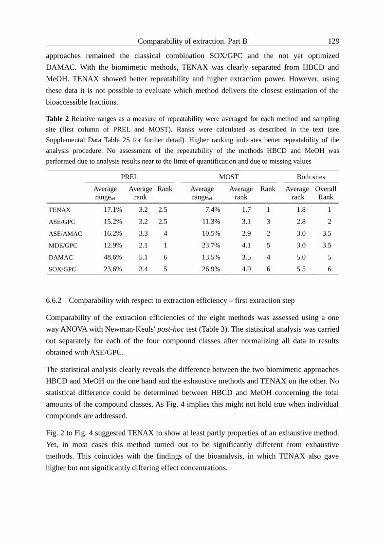
Comparability of extraction. Part B 129
approaches remained the classical combination SOX/GPC and the not yet optimized DAMAC. With the biomimetic methods, TENAX was clearly separated from HBCD and MeOH. TENAX showed better repeatability and higher extraction power. However, using these data it is not possible to evaluate which method delivers the closest estimation of the bioaccessible fractions.
Table 2 Relative ranges as a measure of repeatability were averaged for each method and sampling site (first column of PREL and MOST). Ranks were calculated as described in the text (see Supplemental Data Table 2S for further detail). Higher ranking indicates better repeatability of the analysis procedure. No assessment of the repeatability of the methods HBCD and MeOH was performed due to analysis results near to the limit of quantification and due to missing values
6.6.2 Comparability with respect to extraction efficiency – first extraction step
Comparability of the extraction efficiencies of the eight methods was assessed using a one way ANOVA with Newman-Keuls' post-hoc test (Table 3). The statistical analysis was carried out separately for each of the four compound classes after normalizing all data to results obtained with ASE/GPC.
The statistical analysis clearly reveals the difference between the two biomimetic approaches HBCD and MeOH on the one hand and the exhaustive methods and TENAX on the other. No statistical difference could be determined between HBCD and MeOH concerning the total amounts of the compound classes. As Fig. 4 implies this might not hold true when individual compounds are addressed.
Fig. 2 to Fig. 4 suggested TENAX to show at least partly properties of an exhaustive method. Yet, in most cases this method turned out to be significantly different from exhaustive methods. This coincides with the findings of the bioanalysis, in which TENAX also gave higher but not significantly differing effect concentrations.
PREL MOST Both sites
Average rangerel
Average rank
Rank Average rangerel
Average rank
Rank Average rank
OverallRank
TENAX 17.1% 3.2 2.5 7.4% 1.7 1 1.8 1
ASE/GPC 15.2% 3.2 2.5 11.3% 3.1 3 2.8 2
ASE/AMAC 16.2% 3.3 4 10.5% 2.9 2 3.0 3.5
MDE/GPC 12.9% 2.1 1 23.7% 4.1 5 3.0 3.5
DAMAC 48.6% 5.1 6 13.5% 3.5 4 5.0 5
SOX/GPC 23.6% 3.4 5 26.9% 4.9 6 5.5 6

Comparability of extraction. Part B 130
Table 3 Determined statistical relations for the different pairs of extraction procedures (first extraction step only/combined results for all three steps) in respect of extraction power for targeted compound classes using a one way ANOVA with Newman-Keul’s post-hoc test; recoveries for ASE/GPC extracts were assumed to be 100 %. ns = differences of averages are not significant; *** = significant on a level < 0.001; ** = significant on a level < 0.01; * = significant on a level < 0.05
PAHs SOX/GPC ASE/AMAC DAMAC MDE/GPC ASE/GPC TENAX HBCD MeOHSOX -/- ***/*** ***/* ***/*** ***/* ns/ns ***/* ***/nsASE/AMAC -/- ns/ns */ns ns/ns ***/* ***/ns ***/***
DAMAC -/- **/ns ns/ns ***/ns ***/ns ***/*
MDE/GPC -/- */ns ***/* ***/ns ***/***
ASE/GPC -/- ***/ns ***/ns ***/*
TENAX -/- **/ns ***/ns
HBCD -/- ns/ns
MeOH -/-
CBs SOX/GPC ASE/AMAC DAMAC MDE/GPC ASE/GPC TENAX HBCD MeOHSOX -/- ns/ns **/ns ns/ns ns/ns ***/* -/ns ***/nsASE/AMAC -/- ns/* ns/ns ns/ns **/* -/ns ***/ns
DAMAC -/- ns/ns ns/ns */ns -/ns **/ns
MDE/GPC -/- ns/ns **/ns -/ns ***/ns
ASE/GPC -/- ***/ns -/ns ***/ns
TENAX -/- -/* ns/ns
HBCD -/- -/-
MeOH -/-
PCBs SOX/GPC ASE/AMAC DAMAC MDE/GPC ASE/GPC TENAX HBCD MeOHSOX -/- ns/ns */ns ns/ns ns/ns ***/* ***/ns ***/nsASE/AMAC -/- ns/* ns/ns ns/ns ***/*** ***/ns ***/ns
DAMAC -/- ns/ns */ns **/* ***/ns ***/ns
MDE/GPC -/- ns/ns ***/* ***/ns ***/ns
ASE/GPC -/- ***/* ***/ns ***/ns
TENAX -/- **/*** **/**
HBCD -/- ns/ns
MeOH -/-
DDX SOX/GPC ASE/AMAC DAMAC MDE/GPC ASE/GPC TENAX HBCD MeOHSOX -/- ns/ns ns/ns ns/ns ns/ns **/ns ***/ns ***/nsASE/AMAC -/- ns/ns ns/ns ns/ns */** ***/* ***/*
DAMAC -/- ns/ns ns/ns ns/ns ***/ns ***/ns
MDE/GPC -/- ns/ns ns/ns ***/ns ***/ns
ASE/GPC -/- */ns ***/ns ***/ns
TENAX -/- ***/ns ***/ns
HBCD -/- ns/ns
MeOH -/-

Comparability of extraction. Part B 131
The statistical analysis confirmed the lower performance of SOX/GPC compared to the ASE-based methods or MDE/GPC for the quantification of PAHs. On the other hand, data for MDE/GPC were also statistically different from that for the ASE-based methods. With MDE/GPC samples, higher amounts of PAHs were determined (Fig. 2 and Fig. 3). Since MDE/GPC involves the use of a membrane, and resulting extracts were further purified by GPC, these differences have to be attributed to the performance of the extraction itself. ASE extraction is believed to be very vigorous [1, 59-61], with two different solvent mixtures and a total of six cycles. Extraction by both Soxhlet and ASE applies elevated temperatures, while MDE is operated at ambient temperature. Thus, as a hypothesis, higher recoveries of MDE/GPC may be due to the lower temperature during extraction leading to lower losses due to evaporation during the extraction procedure itself. For other compounds, the exhaustive methods delivered comparable results, with few exceptions regarding the non-optimized DAMAC. It remains unclear, why results varied for PAHs but not for other compounds.
6.6.3 Comparison of all three extraction steps
After the first extraction step, sediment residues from all methods (except ASE/GPC) were extracted again with hexane:acetone in an ultrasonic bath followed by methanolic hydrolysis. Combined results from all three steps should in principle be the same for all approaches. As for the first extraction step, an ANOVA with Newman-Keuls' post-hoc test was carried out to prove this hypothesis (Table 3). Actually, for most clean-up/extraction procedures the analysis revealed equality of the combined results. However, statistical analysis of the data gave also several differences.
Combined results for sediments extracted with TENAX in the first place generally showed significantly lower values (with the exception of PAHs from the sample PREL). The TENAX method itself consists of two steps: Firstly, the sorbent binds compounds dissolved in an aqueous sediment-TENAX slurry. Then, after removing the sorbent from the slurry, gets re-extracted with solvents. It is possible that some of the compounds were not fully re-extracted from the Tenax beads or that losses occurred during evaporation of the solvent leading to lower values for the first of the three extraction steps.
For PAHs, SOX/GPC differed significantly from the other approaches (except TENAX and MeOH) when considering all three extraction steps. Since the ultrasonic extraction with hexane:acetone extracted only minor amounts of PAHs from sediment residues treated with SOX/GPC, the most probable cause for this observations are losses during application of the first extraction/clean-up procedure.

Comparability of extraction. Part B 132
6.6.4 Compound specific correlation analysis
Binding of compounds to sediments is dependent on the physicochemical properties of the substances as well as of sediment characteristics. By definition, an exhaustive extraction method should deliver a complete extraction for all compounds and be independent of the sample [62]. Results for every single compound of all extracts gained with an exhaustive method were compared for the two different sediment samples. All data were normalized to results obtained with the reference method, ASE/GPC, and data from Přelouč were plotted against those from Most (Fig. 6). Data points should be close to unity if the extraction method had a similar leaching power for the specific compound compared to ASE/GPC. If the extraction/clean-up method (as well as the reference method) is truly exhaustive for all compounds and the two sediments, then all data points should group closely together in a cloud point. Deviations should then be randomly distributed and only caused by measurement uncertainties, and no correlation between the data from PREL and MOST should be observable. If one of the methods discriminates only due to the physicochemical properties of the compounds but not due to differences in sediment composition, data points for the respective method should form a linear curve with a slope of one. Considering normalized data from all four methods, correlation between the pooled data from both sites can also occur when the methods itself deliver different results without discrimination because of sediment or compound properties (Fig. 6, Supplemental Data Table 3S).
For all groups of compounds, the normalized ASE/AMAC data are clustered. This finding is on one hand not surprisingly since the same extraction method was used for ASE/GPC. On the other hand, this finding implies that the two clean-up procedures are comparable and do not discriminate specific compounds due to their properties. Only three PAHs deviate from the point cloud, namely acenaphthene showing lower normalized values as well as Indeno[cd]pyrene and Benzo[ghi]perylene in the upper right corner of the diagram. It is striking that these three PAHs have the lowest and the highest molecular weights and, therefore, different physicochemical properties than the other PAHs. No data set was available for acenaphthylene, a compound with similar properties compared to acenaphthene. Observed deviations of these three PAHs have to be attributed to the clean-up method (AMAC or GPC). Normalized ASE/AMAC values are in general slightly but significantly below unity for chlorinated compounds and slightly above unity for PAHs (Student’s t-test, α = 5 %; values from both sites pooled). Thus, AMAC appears to have slightly higher clean-up efficiencies for PAHs but lower ones for PCBs compared to GPC.
In contrast to ASE/AMAC, data points obtained with DAMAC normalized to results from ASE/GPC were not clustered but formed a linear curve with a slope of approximately 1 and with linear correlation coefficients R² significantly deviating from zero. This could be observed for PAHs and PCBs (Fig. 6) as well as for pooled data. This indicated that combined extraction and clean-up power for the current DAMAC method is compound specific.

Comparability of extraction. Part B 133
Furthermore, for most compounds the power was clearly lower than that for ASE/GPC. The authors assume that a higher power and, thus, results independent from the physicochemical properties of the compounds – similar to ASE/AMAC – can be reached with a modified protocol using e.g. more extraction cycles.
Fig 6 Correlation of amounts of target compounds extracted with exhaustive methods normalized to amounts extracted by ASE/GPC. Results for hexachlorobenzene received with SOX were excluded from correlation calculations. For PCBs, the correlation coefficient is given with and without data obtained by SOX
Linear relationships of the normalized results are also observed for PCBs, PAHs and pooled data for MDE/GPC and SOX/GPC. However, correlation of PREL and MOST data for PCBs is weak and not significant (significance level for correlation coefficient: α = 8.6 % (MDE/GPC) and α = 5.1 % (SOX/GPC), Supplemental Data, Table 3S; test of correlation coefficient [63]). While the slope of the linear curve for MDE/GPC was almost unity (PCBs: 0.85; PAHs: 1.12; pooled data: 1.04), indicating a comparable efficiency regardless of the sediment, results for SOX/GPC were dependent on the extracted sediment (slopes for PCBs: 0.33; PAHs: 0.58; pooled data: 0.56).
Results for the four analyzed chlorobenzenes show virtually the same extraction power for each of the four normalized extraction methods (except hexachlorobenzene for SOX). The same can be observed for DDX. However, normalized recoveries differ markedly from method to method. Hence, regression lines and correlation coefficients as given in Fig. 6 reflect the different powers of the methods and are not caused by compound or sediment specific influences. For both compound groups, extraction powers for SOX/GPC compared to

Comparability of extraction. Part B 134
ASE/GPC are remarkably higher (up to a factor of 3). Since the applied clean-up method in both cases was GPC, the differences have to be attributed to the extraction method. The chosen Soxhlet extraction with acetone appears to be much more efficient than extraction by ASE, MDE or DAMAC.
6.6.5 Comparison with results from bioanalytical methods
Ranking order received from chemical analysis (Table 1, Table 2) differed from that obtained by evaluating the same extracts with several bioassays. In part A of this study [1], cytotoxicity was assessed with the Neutral red assay [50, 51], the embryotoxic potential was determined with a zebrafish (Danio rerio) embryo assay [52], and aryl hydrocarbon receptor (AhR) agonists were determined by applying the EROD assay [53, 54]. With bioanalysis, among the exhaustive procedures highest effect concentrations in all three bioassays as well as best repeatability were achieved with Soxhlet extracts. MDE/GPC proved to be the second best method, while procedures based on ASE in combination with either GPC or AMAC lagged behind. The reason for this observation lies most likely in the different suitability of the applied procedures for different compounds. SOX/GPC, for example, proved high extraction power for chlorinated compounds but resulted in low recoveries for PAHS. In general, toxicity is a parameter summarizing the effects of all present compounds. Therefore, other compounds than those analyzed may also have contributed to the observed toxicity. However, the performance of the extraction/clean-up methods in respect of these (unknown) compounds cannot be determined. This consideration further implies that in principle the outcome of toxicity studies or of studies which rely on bioanalyses, e.g. for the purpose of identification of toxicants as in bioassay-directed analysis, is at least partly dependent on the chosen extraction and clean-up procedures.
6.6.6 Confirmation of EROD-induction using artificial mixtures
Artificial mixtures representing the concentrations determined by chemical analysis were tested in an EROD assay to verify whether observed discrepancies between the different extraction methods and between chemical and biological analysis could be explained by possible discrimination of certain compound groups (Fig. 7). Data of HBCD- and MeOH-extracts are not shown, since both methods yielded only very low amounts of targeted compounds (Fig. 2 and Fig. 3) which made it difficult to determine an EC25 value in the EROD-asssay of the artificial mixture. A comparison appeared especially valuable for extracts treated with Soxhlet, for which chemical analysis showed significantly lower concentrations of PAHs compared to other approaches, while bioanalysis marked this method as the one with the highest effects. Several of the PAHs determined are known to induce EROD activity [56, 64-68]. Analyzed PCBs, CBs and DDX are not assigned any dioxin-like activity [69-72];

Comparability of extraction. Part B 135
however, some of the analyzed compounds as suppressors of EROD induction, e.g. DDT or PCBs [73, 74]. Therefore, artificial mixtures contained all analyzed compounds at the concentration levels as determined with the individual extraction/clean-up procedures.
TEQs derived from extracted sediments and artificial mixtures were quite comparable for the ASE-based methods (ASE/AMAC, ASE/GPC, ASE/DAMAC), indicating that the findings of the chemical analysis explained results from the EROD-assay on sediment extracts. That was not the case for SOX/GPC, MDE/GPC and TENAX. For SOX/GPC, 28 % (MOST) and 48 % (PREL) of the EROD-inducing potency could be attributed to the analyzed substances. Similar observations were made for MDE/GPC (MOST: 45 %; PREL: 53 %) and for TENAX-extracts (MOST: 60; PREL: 65 %). Apparently, other compounds than the chemically analyzed ones are in part responsible for the observed EROD-induction in extracts of SOX/GPC, MDE/GPC and TENAX.
The various applied methods make use of different solvents for extraction. Thus, it could be hypothesized that differences in polarity of the solvents may play a role and different compound classes are extracted. However, solvent used for Soxhlet extraction was acetone while MDE hexane was carried out with hexane, and DAMAC with a mixture of hexane:acetone 70:30 (v:v). Therefore, it appears to be unlikely that differences in the methods’ performance can be attributed solely to different solvent systems. The ASE-based methods are run under elevated temperatures (80 °C and 140 °C for ASE/AMAC and ASE/GPC, 40 °C for DAMAC) and pressure. It is possible, that some compounds degrades under these conditions and thus do not contribute to the total EROD-induction. However, further experiments are necessary to clarify what the reason for the observed differences in the methods’ performances is.
Fig 7 Comparison of extracts and artificial mixtures using EROD data. TEQs were calculated with EC25 values. Error bars indicate range of two independent measurements; artificial mixtures were analyzed without repetition; * = EC25 not calculable

Comparability of extraction. Part B 136
6.7 Conclusion & Outlook
Five exhaustive extraction methods combined with clean-up procedures have been compared for extraction power and repeatability. Several of the methods were developed just recently, namely MDE, AMAC and DAMAC [11, 21]. With the exception of SOX/GPC in the case of PAHs, and with some qualifications concerning DAMAC, the applied procedures exhibited statistically similar extraction power.
ASE/AMAC turned out to be a suitable alternative to ASE/GPC. One advantage of AMAC is its large capacity for matrix removal [11]. Thus, ASE/AMAC appears to be very useful for all applications (e.g. effect-directed or bioassay-directed analysis) where bigger amounts of sample have to be extracted, which requires the removal of huge amounts of matrix components. MDE proved to be a method which is at least comparable to other exhaustive extraction methods. MDE has the advantage to work without expensive equipment and without auxiliary energy sources, making it usable also in remote places. Furthermore, passive dialysis reduces the risk of alteration during the extraction process. DAMAC appears to be an interesting and consequent further development of the ASE/AMAC approach. It integrates extraction and clean-up in one step and, thus, offers potential for saving time in the laboratory as well as reducing the amount of solvent used per sample. However, DAMAC still needs more developmental effort and optimization. A DAMAC-based procedure at moderate temperatures could provide quantitative extraction with high efficiency, i.e. low time and solvent consumption, while taking into account considerations regarding the risk of alteration of the extracted sample by common vigorous methods, such as ASE. As results for MDE indicate that already a passive dialysis can provide extraction powers comparable to ASE, combining membrane dialysis with automated extraction promises to be sufficient also at reduced temperatures.
The classical combination SOX/GPC performed relatively poor regarding the extraction of PAHs from the two sediments. The reason for this finding remains unclear. Pure acetone was used for Soxhlet extraction, and it is conceivable that with other solvents extraction power for PAHs would have been higher. However, neither the second nor the third extraction step yielded considerable amounts of PAHs, leading to the conclusion that extraction with Soxhlet was truly exhaustive, but maybe losses occurred during extraction. Gel permeation chromatography as clean-up was used also in combination with other procedures without any obvious losses. Therefore, the clean-up appears not to be responsible for a putative loss of PAHs. SOX/GPC extracted various compounds (PCBs, CBs, DDX), which were targeted simultaneously, in amounts comparable to other exhaustive methods. However, in comparison with chloro-organics, PAHs undergo more easily reactions, e.g. in the presence of light, which could be a source of losses [75-78]. Another possibility is that elevated temperatures during Soxhlet extraction lead to volatilization of PAHs with lower molecular weight. However,

Comparability of extraction. Part B 137
lower recoveries were not restricted to lower molecular weight PAHs. No internal standards were spiked onto the sediment prior to extraction or into the extract prior to clean-up since one part of each extract was dedicated for bioanalysis.
Three of the eight procedures were thought to mirror bioaccessibility or to correlate with bioavailability. It is not possible nor was it the aim of this study to prove this hypothesis. However, these three methods produced rather different results. Amounts obtained by TENAX were for some compound groups almost as high as those from exhaustive methods. Extracts gained with HBCD showed a different composition than that of other procedures. The reason for this observation could be a differing affinity of the compounds for the cavity of the cyclodextrin ring. Quantities extracted with MeOH were generally low, with PCBs and CBs almost missing. Probably the solvent mixture used was not optimal for the targeted compounds. Kelsey et al. [42] showed that the solvent composition is crucial for using mild extraction procedures for determining bioavailability-mimicking fractions in soils or sediments, and that for dissimilar compounds different mixtures have to be applied. As a consequence, a better understanding of bioaccessibility is necessary. Future development should lead to extraction methods that reliably determine bioaccessible fractions instead of being based on operational definitions.
6.8 Supplemental data
Table S1: Averaged concentrations for each method and sediment and assigned ranks
Table 2S: Analysis of the repeatability: calculations of the relative range and their ranks
Table 3S: Results of correlation analysis: correlation coefficients, slopes and intercepts for data normalized to ASE/GPC
6.9 Acknowledgements
Many thanks go to Marion Heinrich, Thomas Anger and Ines Rein for help with the analyses, and Sebastian Heger as well as Kerstin Winkens for conducting the EROD assay. As usual, conducting such a study is not possible with all the helpful hands in the laboratory. We would also like to thank Drs. Niels C Bols and Lucy Lee (University of Waterloo, Canada) for providing RTL-W1 cells. The presented work was partly funded by the German Federation of Industrial Research Associations (AiF, Contract-No. KF 0011005 UL6) and by the German Federal Environmental Agency (FKZ 30102018).

Comparability of extraction. Part B 138
6.10 References
[1] Seiler T-B, Schulze T, Streck G, Schwab K, Brack W, Braunbeck T, Hollert H. 2010. On the comparability of procedures for sediment extraction in environmental assessment. Part A: Ecotoxicological hazard potential. Environmental Toxicology & Chemistry. in prep.
[2] Chapman PM. 1990. The sediment quality triad approach to determining pollution-induced degradation. Sci Total Environ 97-98:815.
[3] Chapman PM, Hollert H. 2006. Should the sediment quality triad become a tetrad, a pentad, or possibly even a hexad? Journal of Soils and Sediments 6:4-8.
[4] Hong PKA, Nakra S. 2009. Rapid extraction of sediment contaminants by pressure cycles. Chemosphere 74:1360-1366.
[5] Fai PB, Grant A, Reid BJ. 2009. Compatibility of hydroxypropyl-[beta]-cyclodextrin with algal toxicity bioassays. Environ Pollut 157:135-140.
[6] Pawliszyn J. 2003. Sample Preparation: Quo Vadis? Anal Chem 75:2543.[7] Cornelissen G, van Noort PCM, Govers HAJ. 1998. Mechanism of Slow Desorption of Organic
Compounds from Sediments: A Study Using Model Sorbents. Environ Sci Technol 32:3124-3131.
[8] Pawliszyn J. 2002. Unified theory of extraction. In Pawliszyn J, ed, Sampling and sample preparation for field and laboratory: fundamental and new directions in sample preparation. Elsevier, Amsterdam, pp 253-278.
[9] Reid BJ, Stokes JD, Jones KC, Semple KT. 2000. Nonexhaustive cyclodextrin-based extraction technique for the evaluation of PAH bioavailability. Environ Sci Technol 34:3174-3179.
[10] Cornelissen G, Rigterink H, Ferdinandy MMA, Van Noort PCM. 1998. Rapidly desorbing fractions of PAHs in contaminated sediments as a predictor of the extent of bioremediation. Environ Sci Technol 32:966-970.
[11] Streck HG, Schulze T, Brack W. 2008. Accelerated membrane-assisted clean-up as a tool for the clean-up of extracts from biological tissues. J Chromatogr A 1196-1197:33-40.
[12] Martens D, Gfrerer M, Wenzl T, Zhang A, Gawlik BM, Schramm KW, Lankmayr E, Kettrup A. 2002. Comparison of different extraction techniques for the determination of polychlorinated organic compounds in sediment. Anal Bioanal Chem 372:562-568.
[13] Kiguchi O, Saitoh K, Ogawa N. 2007. Simultaneous extraction of polychlorinated dibenzo-p-dioxins, polychlorinated dibenzofurans and coplanar polychlorinated biphenyls from contaminated soil using pressurized liquid extraction. J Chromatogr A 1144:262-268.
[14] Martinez E, Gros M, Lacorte S, Barcelo D. 2004. Simplified procedures for the analysis of polycyclic aromatic hydrocarbons in water, sediments and mussels. J Chromatogr A 1047:181-188.
[15] Bandh C, Bjorklund E, Mathiasson L, Naf C, Zebuhr Y. 2000. Comparison of accelerated solvent extraction and soxhlet extraction for the determination of PCBs in Baltic Sea sediments. Environ Sci Technol 34:4995-5000.
[16] Olivella MA. 2005. Trace analysis of polycyclic aromatic hydrocarbons in suspended particulate matter by accelerated solvent extraction followed by gas chromatography-mass spectrometry. Anal Bioanal Chem 383:107-114.

Comparability of extraction. Part B 139
[17] Navarro P, Cortazar E, Bartolome L, Deusto M, Raposo JC, Zuloaga O, Arana G, Etxebarria N. 2006. Comparison of solid phase extraction, saponification and gel permeation chromatography for the clean-up of microwave-assisted biological extracts in the analysis of polycyclic aromatic hydrocarbons. J Chromatogr A 1128:10-16.
[18] Fernandez P, Bayona JM. 1992. Use of off-line gel-permeation chromatography normal-phase liquid-chromatography for the determination of polycyclic aromatic compound in environmental samples and standard reference material (air particulate matter and marine sediment). Journal of Chromatography 625:141-149.
[19] US Environmental Protection Agency. 1994. Method 3640A.Gel-Permeation Cleanup, Washington (DC).
[20] EPA US. 2007. Standard Method 3545A, Pressurized Fluid Extraction. Vol 2009.[21] Seiler TB, Rastall AC, Leist E, Erdinger L, Braunbeck T, Hollert H. 2006. Membrane dialysis
extraction (MDE): A novel approach for extracting toxicologically relevant hydrophobic organic compounds from soils and sediments for assessment in biotests. Journal of Soils and Sediments 6:20-29.
[22] Soxhlet F. 1879. Die gewichtsanalytische Bestimmung des Milchfettes. Polytechnisches Journal 232:461-465.
[23] Hollert H, Durr M, Erdinger L, Braunbeck T. 2000. Cytotoxicity of settling particulate matter and sediments of the Neckar River (Germany) during a winter flood. Environ Toxicol Chem 19:528-534.
[24] Hollert H, Durr M, Holtey-Weber R, Islinger M, Brack W, Farber H, Erdinger L, Braunbeck T. 2005. Endocrine disruption of water and sediment extracts in. a non-radioactive dot blot/RNAse protection-assay using isolated hepatocytes of rainbow trout - Deficiencies between bioanalytical effectiveness and chemically determined concentrations and how to explain them. Environmental Science and Pollution Research 12:347-360.
[25] Wang WT, Meng BJ, Lu XX, Liu Y, Tao S. 2007. Extraction of polycyclic aromatic hydrocarbons and organochlorine pesticides from soils: A comparison between Soxhlet extraction, microwave-assisted extraction and accelerated solvent extraction techniques. Anal Chim Acta 602:211-222.
[26] EPA US. 1996. Standard Method 3540C, Soxhlet Extraction. Vol 2009.[27] Harkey GA, Young TM. 2000. Effect of soil contaminant extraction method in determining
toxicity using the Microtox (R) assay. Environ Toxicol Chem 19:276-282.[28] de la Cal A, Eljarrat E, Grotenhuis T, Barcelo D. 2008. Tenax (R) extraction as a tool to evaluate
the availability of polybrominated diphenyl ethers, DDT, and DDT metabolites in sediments. Environ Toxicol Chem 27:1250-1256.
[29] Cornelissen G, Rigterink H, ten Hulscher DEM, Vrind BA, van Noort PCM. 2001. A simple Tenax (R) extraction method to determine the availability of sediment-sorbed organic compounds. Environ Toxicol Chem 20:706-711.
[30] Leppanen MT, Kukkonen JVK. 2006. Evaluating the role of desorption in bioavailability of sediment-associated contaminants using oligochaetes, semipermeable membrane devices and Tenax extraction. Environ Pollut 140:150-163.
[31] Puglisi E, Murk AJ, van den Bergt HJ, Grotenhuis T. 2007. Extraction and bioanalysis of the ecotoxicologically relevant fraction of contaminants in sediments. Environ Toxicol Chem 26:2122-2128.

Comparability of extraction. Part B 140
[32] van der Heijden SA, Jonker MTO. 2009. PAH Bioavailability in Field Sediments: Comparing Different Methods for Predicting in Situ Bioaccumulation. Environ Sci Technol 43:3757-3763.
[33] Brack W, Bandow N, Schwab K, Schulze T, Streck G. 2009. Bioavailability in effect-directed analysis of organic toxicants in sediments. TrAC, Trends Anal Chem 28:543-549.
[34] Allan IJ, Semple KT, Hare R, Reid BJ. 2006. Prediction of mono- and polycyclic aromatic hydrocarbon degradation in spiked soils using cyclodextrin extraction. Environ Pollut 144:562-571.
[35] Swindell AL, Reid BJ. 2006. Comparison of selected non-exhaustive extraction techniques to assess PAH availability in dissimilar soils. Chemosphere 62:1126-1134.
[36] Cuypers C, Pancras T, Grotenhuis T, Rulkens W. 2002. The estimation of PAH bioavailability in contaminated sediments using hydroxypropyl-beta-cyclodextrin and Triton X-100 extraction techniques. Chemosphere 46:PII S0045-6535(0001)00199-00190.
[37] Stokes JD, Wilkinson A, Reid BJ, Jones KC, Semple KT. 2005. Prediction of polycyclic aromatic hydrocarbon biodegradation in contaminated soils using an aqueous hydroxypropyl-beta-cyclodextrin extraction technique. Environ Toxicol Chem 24:1325-1330.
[38] Dean JR, Scott WC. 2004. Recent developments in assessing the bioavailability of persistent organic pollutants in the environment. TrAC, Trends Anal Chem 23:609-618.
[39] Tang JX, Alexander M. 1999. Mild extractability and bioavailability of polycyclic aromatic hydrocarbons in soil. Environ Toxicol Chem 18:2711-2714.
[40] Tang J, Robertson BK, Alexander M. 1999. Chemical-Extraction Methods To Estimate Bioavailability of DDT, DDE, and DDD in Soil. Environ Sci Technol 33:4346-4351.
[41] Reid BJ, Jones KC, Semple KT. 2000. Bioavailability of persistent organic pollutants in soils and sediments - a perspective on mechanisms, consequences and assessment. Environ Pollut 108:103-112.
[42] Kelsey JW, Kottler BD, Alexander M. 1997. Selective chemical extractants to predict bioavailability of soil-aged organic chemicals. Environ Sci Technol 31:214-217.
[43] Song YF, Jing X, Fleischmann S, Wilke BM. 2002. Comparative study of extraction methods for the determination of PAHs from contaminated soils and sediments. Chemosphere 48:993-1001.
[44] Eschenbach A, Kastner M, Bierl R, Schaefer G, Mahro B. 1994. Evaluation Of A New, Effective Method To Extract Polycyclic Aromatic-Hydrocarbons From Soil Samples. Chemosphere 28:683-692.
[45] Streck G, Schulze T, Brack W. 2008. Accelerated Membrane Assisted Clean-up: Eine leistungsfähige Methode zur Aufreinigung matrixbehafteter Extrakte. LCGC Ausgabe in deutscher Sprache März/April:6-10.
[46] Seiler T-B, Rastall AC, Leist E, Erdinger L, Braunbeck T, Hollert H. 2006. Membrane dialysis extraction (MDE): A novel approach for extracting toxicologically relevant hydrophobic organic compounds from soils and sediments for assessment in biotests. J Soils Sediments 6:20-29.
[47] Berset JD, Ejem M, Holzer R, Lischer P. 1999. Comparison of different drying, extraction and detection techniques for the determination of priority polycyclic aromatic hydrocarbons in background contaminated soil samples. Anal Chim Acta 383:263-275.
[48] Hartmann R. 1996. Polycyclic aromatic hydrocarbons (PAHs) in forest soils: Critical evaluation of a new analytical procedure. International Journal Of Environmental Analytical Chemistry 62:161-173.

Comparability of extraction. Part B 141
[49] Schwab K, Brack W. 2007. Large volume TENAX (R) extraction of the bioaccessible fraction of sediment-associated organic compounds for a subsequent effect-directed analysis. Journal of Soils and Sediments 7:178-186.
[50] Babich H, Borenfreund E. 1992. Neutral red assay for toxicology in vitro. In Watson RR, ed, In vitro methods of toxicology. CRC - Press, Boca Raton, Florida, pp 238-251.
[51] Klee N, Gustavsson LK, Kosmehl T, Engwall M, Erdinger L, Braunbeck T, Hollert H. 2004. Changes in toxicity and genotoxicity of industrial sewage sludge samples containing nitro- and amino-aromatic compounds following treatment in bioreactors with different oxygen regimes. ESPR - Environ Sci & Pollut Res 11:313-320.
[52] Hollert H, Keiter S, König N, Rudolf M, Ulrich M, Braunbeck T. 2003. A new sediment contact assay to assess particle-bound pollutants using zebrafish (Danio rerio) embryos. JSS – J Soils & Sediments 3:197 – 207.
[53] Behrens A, Schirmer K, Bols NC, Segner H. 1998. Microassay for rapid measurement of 7-ethoxyresorufin-O-deethylase activity in intact fish hepatocytes. Marine Environ Res 46:369-373.
[54] Gustavsson LK, Klee N, Olsman H, Hollert H, Engwall M. 2004. Fate of Ah receptor agonists during biological treatment of an industrial sludge containing explosives and pharmaceutical residues. ESPR - Environ Sci & Pollut Res 11:379-387.
[55] Engwall M, Broman D, Ishaq R, Naf C, Zebuhr Y, Brunstrom B. 1996. Toxic potencies of lipophilic extracts from sediments and settling particulate matter (SPM) collected in a PCB-contaminated river system. Environ Toxicol Chem 15:213-222.
[56] Hollert H, Durr M, Olsman H, Halldin K, Van Bavel B, Brack W, Tysklind M, Engwall M, Braunbeck T. 2002. Biological and chemical determination of dioxin-like compounds in sediments by means of a sediment triad approach in the catchment area of the River Neckar. Ecotoxicology 11:323-336.
[57] Jansen B, Nierop KGJ, Kotte MC, de Voogt P, Verstraten JM. 2006. The applicability of accelerated solvent extraction (ASE) to extract lipid biomarkers from soils. Applied Geochemistry 21:1006-1015.
[58] Richter BE, Jones BA, Ezzell JL, Porter NL, Avdalovic N, Pohl C. 1996. Accelerated solvent extraction: A technique for sample preparation. Anal Chem 68:1033-1039.
[59] Dean JR. 1996. Accelerated solvent extraction of polycyclic aromatic hydrocarbons from contaminated soil. Analytical Communications 33:191-192.
[60] Heemken OP, Theobald N, Wenclawiak BW. 1997. Comparison of ASE and SFE with Soxhlet, sonication, and methanolic saponification extractions for the determination of organic micropollutants in marine particulate matter. Anal Chem 69:2171-2180.
[61] Popp P, Kiel P, Möder M. 1997. Application of accelerated solvent extraction followed by gas chromatopgraphy, high-performance liquid chromatography and gas chromatography-mass spectrometry for the determination of polycyclic aromatic hydrocarbons, chlorinated pesticides and polychlorinated dibenzo-p-dioxins and dibenzofurans in solid wastes. J Chromatogr A 774:203 - 211.
[62] Dean JR, Xiong GH. 2000. Extraction of organic pollutants from environmental matrices: selection of extraction technique. TrAC, Trends Anal Chem 19:553-564.
[63] Streck G. 2004. Einführung in die Statistik für Geoökologen und andere Naturwissenschaftler. Books on Demand GmbH, Norderstedt.

Comparability of extraction. Part B 142
[64] Bosveld ATC, de Bie PAF, van den Brink NW, Jongepier H, Klomp AV. 2002. In vitro EROD induction equivalency factors for the 10 PAHs generally monitored in risk assessment studies in The Netherlands. Chemosphere 49:75.
[65] Brunström B, Broman D, Näf C. 1991. Toxicity and EROD-inducing potency of 24 polycyclic aromatic hydrocarbons (PAHs) in chick embryos. Archives of Toxicology 65:485.
[66] Bols NC, Schirmer K, Joyce EM, Dixon DG, Greenberg BM, Whyte JJ. 1999. Ability of polycyclic aromatic hydrocarbons to induce 7-ethoxyresorufin-O-deethylase activity in a trout liver cell line. Ecotoxicology And Environmental Safety 44:118-128.
[67] Billiard SM, Bols NC, Hodson PV. 2004. In vitro and in vivo comparisons of fish-specific CYP1A induction relative potency factors for selected polycyclic aromatic hydrocarbons. Ecotoxicology And Environmental Safety 59:292-299.
[68] Behrens A, Schirmer K, Bols NC, Segner H. 2001. Polycyclic aromatic hydrocarbons as inducers of cytochrome P4501A enzyme activity in the rainbow trout liver cell line, RTL-W1, and in primary cultures of rainbow trout hepatocytes. Environ Toxicol Chem 20:632-643.
[69] Van den Berg M, Birnbaum L, Bosveld ATC, Brunstrom B, Cook P, Feeley M, Giesy JP, Hanberg A, Hasegawa R, Kennedy SW, Kubiak T, Larsen JC, van Leeuwen FXR, Liem AKD, Nolt C, Peterson RE, Poellinger L, Safe S, Schrenk D, Tillitt D, Tysklind M, Younes M, Waern F, Zacharewski T. 1998. Toxic equivalency factors (TEFs) for PCBs, PCDDs, PCDFs for humans and wildlife. Environ Health Perspect 106:775-792.
[70] Engwall M, Broman D, Naf C, Zebuhr Y, Brunstrom B. 1997. Dioxin-like compounds in HPLC-fractionated extracts of marine samples from the east and west coast of Sweden: Bioassay- and instrumentally-derived TCDD equivalents. Marine Pollution Bulletin 34:1032-1040.
[71] Clemons JH, Dixon DG, Bols NC. 1997. Derivation of 2,3,7,8-TCDD toxic equivalent factors (TEFs) for selected dioxins, furans and PCBs with rainbow trout and rat liver cell lines and the influence of exposure time. Chemosphere 34:1105-1119.
[72] Clemons JH, Myers CR, Lee LEJ, Dixon DG, Bols NC. 1998. Induction of cytochrome P4501A by binary mixtures of polychlorinated biphenyls (PCBs) and 2,3,7,8-tetrachlorodibenzo-p-dioxin (TCDD) in liver cell lines from rat and trout. Aquatic Toxicology 43:179-194.
[73] Jeong HG, Kim JY. 2002. Effects of o,p'-DDT on the 2,3,7,8-tetrachlorodibenzo-p-dioxin-inducible CYP1A1 expression in murine Hepa-1c1c7 cells. Food and Chemical Toxicology 40:1685.
[74] Binelli A, Ricciardi F, Riva C, Provini A. 2006. New evidences for old biomarkers: Effects of several xenobiotics on EROD and AChE activities in Zebra mussel (Dreissena polymorpha). Chemosphere 62:510-519.
[75] Zepp RG, Schlotzhauer PF. 1979. Photoreactivity of selected aromatic hydrocarbons in water. In Jones PW, Leber P, eds, Polynuclear Aromatic Hydrocarbons. Ann Arbor Science Publishers Inc., Ann Arbor, MI, pp 141-158.
[76] Mill T, Mabey WR, Lan BY, Baraze A. 1981. Photolysis Of Polycyclic Aromatic-Hydrocarbons In Water. Chemosphere 10:1281-1290.
[77] Miller JS, Olejnik D. 2001. Photolysis of polycyclic aromatic hydrocarbons in water. Water Research 35:233-243.
[78] Sabate J, Bayona JM, Solanas AM. 2001. Photolysis of PAHs in aqueous phase by UV irradiation. Chemosphere 44:119-124.

Comparability of extraction. Part B 143
Supplemental data
On the comparability of procedures for sediment extraction in environmental assessment. Part B: Chemical analytical investigations
G. Streck1, T.-B. Seiler2, T. Schulze1, K. Schwab1,3, W. Brack1, T. Braunbeck4, H. Hollert2
1 Department of Effect-Directed Analysis, UFZ Helmholtz-Centre for Environmental Research, 04318 Leipzig, Germany
2 Department of Ecosystem Analysis, Institute for Environmental Research (Biology 5), RWTH Aachen University, 52074 Aachen, Germany
3 Currenta GmbH & Co. OHG, CHEMPARK Leverkusen, 51368 Leverkusen, Germany
4 Aquatic Ecology and Toxicology Group, Department of Zoology, University of Heidelberg, 69120 Heidelberg, Germany

Averaged concentrations from two independent analyses for each method and assigned rank for the respective compound; n.d. = not detectedConcentration (ng/g SEQ) Rank
SOX/GPC ASE/AMAC DAMAC MDE/GPC ASE/GPC SOX/GPC ASE/AMAC DAMAC MDE/GPCAcenaphthylene 136.8 79.0 42.2 52.3 115.8 1 3 5 4
119.1 84.8 39.6 48.4 102.7 1 3 5 459.2 186.3 84.4 154.4 190.5 5 2 4 3
532.0 1414.9 884.0 1172.9 1446.1 5 2 4 392.1 236.9 134.5 174.3 226.1 5 1 4 3
782.3 1494.2 1371.2 2187.5 1552.9 5 3 4 11082.9 2018.8 1809.4 2861.8 2142.4 5 3 4 1
Benz(a)anthracene 321.1 562.7 523.6 828.3 603.8 5 3 4 1466.9 734.7 678.9 1126.9 806.5 5 3 4 1
Benzo(b)fluoranthene 486.8 625.6 559.5 1502.4 687.2 5 3 4 1Benzo(k)fluoranthene 171.4 207.8 206.5 0.0 237.7 4 2 3 5Benzo(a)pyrene 315.9 489.2 450.7 507.0 473.1 5 2 4 1Indeno(1,2,3-cd)pyrene 226.9 437.9 308.9 389.4 344.5 5 1 4 2Dibenz(a,h)anthracene 59.3 74.7 60.4 67.5 75.4 5 2 4 3Benzo(ghi)perylene 370.6 537.6 414.3 547.6 388.1 5 2 3 11,2,4,5-chlorobenzene 4.2 2.0 1.5 2.2 2.3 1 4 5 31,2,3,4-chlorobenzene 2.7 1.3 1.0 1.4 1.5 1 4 5 3Penta-chlorobenzene 2.0 1.0 0.7 1.1 1.1 1 4 5 3Hexa-chlorobenzene 4.1 9.1 6.3 9.7 10.2 5 3 4 2
223.5 228.4 151.4 248.7 248.6 4 3 5 1145.6 153.5 109.6 161.2 180.1 4 3 5 2
34.3 51.1 35.4 46.8 57.2 5 2 4 357.3 63.0 54.6 62.5 71.8 4 2 5 344.8 51.7 43.5 51.7 57.1 4 3 5 236.1 40.2 34.9 36.5 44.3 4 2 5 36.6 6.1 4.9 5.6 7.0 2 3 5 4
31.8 n.d. n.d. n.d. n.d. 1 3.5 3.5 3.548.8 n.d. n.d. n.d. n.d. 1 3.5 3.5 3.5n.d. n.d. n.d. n.d. n.d. - - - -
Arithmetic mean 3.7 2.7 4.3 2.5Rank 4 3 5 2

continuedConcentration (ng/g SEQ) Rank
SOX/GPC ASE/AMAC DAMAC MDE/GPC ASE/GPC SOX/GPC ASE/AMAC DAMAC MDE/GPCAcenaphthylene 111.7 n.d. 29.1 11.8 53.8 1 5 3 4
110.4 46.7 43.4 26.1 60.6 1 3 4 527.0 103.0 77.7 84.3 106.5 5 2 4 3
279.2 827.0 650.0 907.7 772.1 5 2 4 1932.6 3357.7 2299.2 2356.7 2954.1 5 1 4 3561.3 1437.2 1342.1 2056.7 1252.8 5 2 3 1469.3 1096.4 1081.5 1692.6 1005.6 5 2 3 1
Benz(a)anthracene 270.5 685.1 644.1 882.3 634.7 5 2 3 1380.4 841.5 789.3 1228.3 775.8 5 2 3 1
Benzo(b)fluoranthene 446.3 909.2 769.9 1462.6 752.4 5 2 3 1Benzo(k)fluoranthene 162.9 336.0 344.8 201.6 299.9 5 2 1 4Benzo(a)pyrene 252.4 572.1 534.0 489.0 471.5 5 1 2 3Indeno(1,2,3-cd)pyrene 220.3 629.2 573.7 518.3 490.6 5 1 2 3Dibenz(a,h)anthracene 54.4 109.9 93.7 107.3 98.5 5 1 4 2Benzo(ghi)perylene 227.3 470.9 461.2 461.6 346.2 5 1 3 21,2,4,5-chlorobenzene 1.3 0.6 0.4 0.6 0.6 1 4 5 31,2,3,4-chlorobenzene 1.0 0.5 0.3 0.5 0.6 1 4 5 3Penta-chlorobenzene 1.7 0.8 0.5 0.8 0.9 1 4 5 3Hexa-chlorobenzene 8.3 4.4 2.9 4.7 5.0 1 4 5 3
31.9 18.8 13.7 19.2 20.2 1 4 5 310.1 10.2 7.0 10.2 10.7 4 3 5 236.2 32.8 23.1 31.8 35.6 1 3 5 4
160.3 121.0 103.6 116.7 129.2 1 3 5 4138.3 105.5 93.9 103.4 122.2 1 3 5 4102.1 79.7 64.4 71.6 84.5 1 3 5 4
17.7 10.1 8.4 9.1 11.4 1 3 5 432.6 13.9 9.3 15.4 12.9 1 3 5 282.5 30.7 21.3 33.7 27.6 1 3 5 2n.d. 72.7 65.4 76.6 72.7 5 2 4 1
Arithmetic mean 3.0 2.6 4.0 2.7Rank 4 1 5 2

continuedConcentration (ng/g SEQ) Rank
TENAX HBCD MeOH TENAX HBCD MeOHAcenaphthylene n.d. n.d. n.d. - - -
5.19 n.d. n.d. 1 2.5 2.536.07 22.72 24.91 1 3 2
438.86 2.73 30.77 1 3 21036.70 5.56 111.03 1 3 21010.93 1.90 28.74 1 3 2807.01 1.63 21.96 1 3 2
Benz(a)anthracene 310.51 20.76 4.64 1 2 3550.82 19.26 7.30 1 2 3
Benzo(b)fluoranthene 284.08 23.01 21.48 1 2 3Benzo(k)fluoranthene 163.67 0.53 1.55 1 3 2Benzo(a)pyrene 100.98 14.89 13.07 1 2 3Indeno(1,2,3-cd)pyrene 109.97 0.73 1.11 1 3 2Dibenz(a,h)anthracene 26.42 0.43 0.77 1 3 2Benzo(ghi)perylene 95.83 1.15 1.15 1 3 21,2,4,5-chlorobenzene n.d. n.d. n.d. - - -1,2,3,4-chlorobenzene 0.37 n.d. n.d. 1 2.5 2.5Penta-chlorobenzene 0.43 n.d. n.d. 1 2.5 2.5Hexa-chlorobenzene 2.37 n.d. 0.21 1 3 2
n.d. 0.60 n.d. 2.5 1 2.54.4915 1.271 n.d. 1 2 3
8.782 1.3335 n.d. 1 2 324.0365 0.977 n.d. 1 2 319.9525 0.7295 n.d. 1 2 3
7.911 0.697 0.275 1 2 30.7085 0.959 1.021 3 2 1
8.327 0.855 0.768 1 2 320.3355 1.467 1.18 1 2 314.577 n.d. n.d. 1 2.5 2.5
Arithmetic mean 1.1 2.4 2.5Rank 1 2 3

continuedConcentration (ng/g SEQ) Rank
TENAX HBCD MeOH TENAX HBCD MeOHAcenaphthylene n.d. n.d. n.d. - - -
4.1 n.d. n.d. 1 2.5 2.521.5 22.6 22.8 3 2 1
237.7 4.7 12.3 1 3 233.8 3.0 3.2 1 3 2
384.1 3.6 7.5 1 3 2568.1 5.0 9.7 1 3 2
Benz(a)anthracene 83.1 21.5 1.4 1 2 3173.4 20.4 2.2 1 2 3
Benzo(b)fluoranthene 63.0 24.0 21.9 1 2 3Benzo(k)fluoranthene 27.7 1.5 1.0 1 2 3Benzo(a)pyrene 26.2 15.7 14.1 1 2 3Indeno(1,2,3-cd)pyrene 17.1 1.9 1.0 1 2 3Dibenz(a,h)anthracene 4.3 0.9 0.9 1 3 2Benzo(ghi)perylene 30.6 3.7 1.3 1 2 31,2,4,5-chlorobenzene 0.3 n.d. n.d. 1 2.5 2.51,2,3,4-chlorobenzene n.d. n.d. 0.2 2.5 2.5 1Penta-chlorobenzene 0.3 n.d. n.d. 1 2.5 2.5Hexa-chlorobenzene 1.5 n.d. 0.5 1 3 2
157.2 1.1 9.1 1 3 2124.9 1.9 5.3 1 3 220.4 1.8 n.d. 1 2 340.3 1.1 n.d. 1 2 330.8 1.1 n.d. 1 2 315.5 0.6 n.d. 1 2 3
1.3 2.0 n.d. 2 1 324.3 0.5 0.8 1 3 233.0 1.2 2.0 1 3 2n.d. n.d. n.d. - - -
Arithmetic mean 1.2 2.4 2.4Rank 1 2.5 2.5

Calculations of the relative range and their ranks; the relative range was calculated as range of the results for each compound for the same sediment andmethod divided by the associated arithmetic mean
Relative range [%] RankSOX/GPC ASE/AMAC DAMAC MDE/GPC ASE/GPC TENAX SOX/GPC ASE/AMAC DAMAC MDE/GPC ASE/GPC
Acenaphthylene 6.8 64.5 20.1 23.9 - 6.8 2 5 3 46.1 103.9 18.4 19.5 83.1 6.1 2 6 3 41.9 115.9 2.8 13.8 10.3 1.9 1 6 2 54.1 75.2 11.2 12.2 1.6 4.1 3 6 4 51.6 71.7 4.1 1.0 8.0 1.6 2 6 3 10.5 52.4 2.5 12.5 4.1 0.5 1 6 2 50.2 51.1 1.6 12.6 3.8 0.2 1 6 2 5
Benz(a)anthracene 4.6 46.9 2.5 11.0 4.7 4.6 3 6 2 53.7 47.8 2.2 13.3 3.7 3.7 3 6 2 5
Benzo(b)fluoranthene 8.6 47.8 0.4 15.6 14.9 8.6 3 6 1 5Benzo(k)fluoranthene 25.3 55.5 - 0.3 3.9 25.3 4 5 - 1Benzo(a)pyrene 15.3 48.4 12.9 6.5 8.3 15.3 5 6 3 1Indeno(1,2,3-cd)pyrene 15.0 49.8 3.7 11.5 6.7 15.0 5 6 1 3Dibenz(a,h)anthracene 18.8 78.7 14.8 9.5 4.7 18.8 5 6 4 3Benzo(ghi)perylene 16.9 45.7 1.7 8.3 7.9 16.9 5 6 1 41,2,4,5-chlorobenzene 71.4 71.9 67.8 69.1 - 71.4 4 5 2 31,2,3,4-chlorobenzene 67.7 77.4 63.8 65.2 - 67.7 4 5 2 3Penta-chlorobenzene 42.6 47.5 33.4 46.3 - 42.6 3 5 2 4Hexa-chlorobenzene 19.4 29.5 0.1 2.3 16.5 19.4 4 6 1 2
21.7 20.6 10.5 4.6 16.5 21.7 5 4 2 115.1 10.9 - 11.9 9.6 15.1 4 2 - 318.9 18.7 9.2 11.1 151.6 18.9 4 3 1 20.7 9.0 0.5 3.6 7.3 0.7 2 5 1 36.9 36.9 2.9 3.8 8.1 6.9 3 5 1 2
18.4 24.2 2.7 4.5 5.9 18.4 4 5 1 226.4 25.0 10.0 12.4 5.4 26.4 5 4 2 39.2 33.3 3.7 16.2 10.9 9.2 2 6 1 47.2 0.5 31.6 4.3 11.6 7.2 3 1 5 2
- - - - - - - - - -Arithmetic mean 16.2 48.6 12.9 15.2 17.1 16.2 3.3 5.1 2.1 3.2 3.2

continuedRelative range [%] Rank
SOX/GPC ASE/AMAC DAMAC MDE/GPC ASE/GPC TENAX SOX/GPC ASE/AMAC DAMAC MDE/GPC ASE/GPCAcenaphthylene 6.2 - 8.1 114.4 5.5 - 2 - 3 4
9.7 2.9 17.4 67.5 4.6 109.4 3 1 4 550.3 8.4 12.3 49.1 10.5 21.1 6 1 3 537.1 6.2 2.3 22.0 16.7 0.4 6 3 2 526.9 5.0 5.6 18.7 7.0 0.0 6 2 3 534.9 13.7 9.2 22.9 17.3 0.6 6 3 2 535.6 8.8 7.8 23.4 10.8 0.2 6 3 2 5
Benz(a)anthracene 32.3 10.7 11.9 12.6 15.3 0.2 6 2 3 434.4 14.3 12.3 22.8 17.1 0.0 6 3 2 5
Benzo(b)fluoranthene 18.7 9.5 19.0 36.6 11.8 7.0 4 2 5 6Benzo(k)fluoranthene 21.7 7.3 10.6 - 9.0 7.2 5 2 4 -Benzo(a)pyrene 16.4 12.0 14.4 19.7 12.0 2.0 5 3 4 6Indeno(1,2,3-cd)pyrene 24.3 9.7 16.5 28.3 11.6 3.6 5 2 4 6Dibenz(a,h)anthracene 18.4 6.1 21.9 34.2 7.8 1.1 4 2 5 6Benzo(ghi)perylene 23.7 9.2 14.0 5.6 11.7 1.3 6 3 5 21,2,4,5-chlorobenzene 25.7 32.2 16.0 32.2 31.2 - 2 4 1 41,2,3,4-chlorobenzene 16.0 27.2 5.8 29.3 25.3 - 2 4 1 5Penta-chlorobenzene 19.1 1.0 0.1 14.5 11.9 1.4 6 2 1 5Hexa-chlorobenzene 22.4 7.4 11.8 4.4 3.1 2.1 6 4 5 3
25.4 1.3 10.8 1.2 6.3 - 5 2 4 156.0 5.8 12.8 - 0.8 1.0 5 3 433.6 22.8 36.0 15.0 12.9 0.7 5 4 6 322.7 8.4 6.1 14.1 10.6 1.7 6 3 2 523.1 5.5 0.3 3.8 1.2 1.9 6 5 1 418.1 19.6 30.2 14.0 18.6 4.4 3 5 6 249.6 16.3 27.0 19.9 15.3 0.1 6 3 5 433.9 0.2 5.6 3.8 8.2 6.5 6 1 3 216.2 17.2 31.5 7.7 8.4 10.8 4 5 6 1
- 5.0 13.8 3.4 6.1 0.2 - 3 5 2Arithmetic mean 26.9 10.5 13.5 23.7 11.3 7.4 4.9 2.9 3.5 4.1 3.1

Table 3S Correlation coefficients, slope and intercept for data normalized to ASE/GPC from PREL and MOST for PAHs and PCBs and for all compounds analysed (pooled data); α denominates the significance level for the assumption of a statistically significant linear correlation between the two variables
Linear equation R2 αAll compounds
SOX/GPC y=0.56x+0.15 0.64 < 0.1 %MDE/GPC y=1.04x-0.05 0.70 < 0.1 %ASE/AMAC y=0.82x+0.22 0.44 < 0.1 %DAMAC y=1.37x-0.09 0.82 < 0.1 %
PAHs y=1.08x+0.005 0.80 < 0.1 %SOX/GPC y=0.58x-0.12 0.88 < 0.1 %MDE/GPC y=1.12x-0.07 0.86 < 0.1 %ASER/AMAC y=0.69x+0.43 0.55 0.23 %DAMAC y=0.94x+0.26 0.93 < 0.1 %
PCBs y=0.21x+0.61 0.21 1.5 %SOX/GPC y=0.33x+0.40 0.57 5.1 %MDE/GPC y=0.85x+0.12 0.48 8.6 %ASE/AMAC y=-0.14x+1.02 0.04 68.6 %DAMAC y=1.19x-0.17 0.87 0.24 %

Chapter 7
The impact of extraction methodologieson the toxicity of sediments in the zebrafish
(Danio rerio) embryo test (FET)
H Zielke1, T-B Seiler1, S Niebergall2, E Leist2, M Brinkmann1, D Spira3, G Streck4, W Brack4, U Feiler3, T Braunbeck2, H Hollert1
1 Department of Ecosystem Analysis, Institute for Environmental Research (Biology V), RWTH Aachen University, Worringerweg 1, 52074 Aachen, Germany
2 Aquatic Ecology and Toxicology Section, Department of Zoology, University of Heidelberg, Im Neuenheimer Feld 230, 69120 Heidelberg, Germany
3 Department Biochemistry and Ecotoxicology, Federal Institute of Hydrology (BfG), Am Mainzer Tor 1, 56070 Koblenz, Germany
4 Department of Effect-Directed Analysis, UFZ Helmholtz-Centre for Environmental Research, Permoserstr. 15, 04318 Leipzig, Germany
To be submitted to J Soils Sediments


Impact of extraction in FET 153
7 Chapter 7
7.1 Abstract
Goal, Scope and Background. Traditionally, methods for sediment extractions are characterized using chemical analyses. However, in order to evaluate sediment extracts with regard to biological effects and, thus, bioaccessibility, extraction methods have to be compared to effect data obtained from experiments with in situ exposure scenarios, i.e. sediment contact tests. This study compares four extraction methods and sediment contact test data from a previous project with respect to predictive power in the fish embryo assay with zebrafish (Danio rerio).
Methods. A natural and an artificial sediment spiked with a mixture of six organic pollutants (2,4 dinitrophenol, diuron, fluoranthene, nonylphenol, parathion ethyl and pentachlorophenol) were extracted using (a) membrane dialysis extraction (MDE), (b) a Soxhlet procedure, (c) hydroxypropyl-β-cyclodextrin (HPCD) or (d) Tenax®-TA. Whereas the former two are regarded being exhaustive with respect to non-covalently bound contaminants, the latter two are considered to predict bioaccessibility). Resulting extracts were tested in the fish embryo test (FET) with Danio rerio for embryotoxic and teratogenic potential.
Results and Discussion. Mortalities caused by organic extracts from Soxhlet extraction and MDE were high, as expected. However, HPCD extracts turned out to be at least as effective as extracts obtained with these two methods. One possible reason might be short ageing time of the spiked sediments. Only Tenax®-TA extracts gave results comparable to the sediment contact assay for natural sediment, but revealed low reproducibility. Significant differences between natural and artificial sediment were found for extracts obtained with techniques using native (i.e. non-freeze-dried) sediments, i.e. HPCD and Tenax®-TA. In contrast, MDE and Soxhlet extracts did not differentiate between tested natural and artificial sediment, which might be accounted for by equalising effects of freeze-drying.
Conclusion. Four extraction methods could successfully be differentiated with respect to their stringency and predictiveness for bioaccessibility. The fish embryo sediment contact test with zebrafish could be documented as a suitable tool for comparing direct sediment contact tests and assays with extractions. MDE was confirmed as an alternative to Soxhlet extraction. High mortalities induced by HPCD extracts underline the need to include ageing into consideration when assessing sediments. Although Tenax®-TA may basically be used to predict bioavailability in the fish embryo test, the high variability observed warrants further investigation of the relation between effect and extractability. Apparently, freeze-drying can severely affect sediment properties, potentially eliminating individual properties of natural sediments.

Impact of extraction in FET 154
Outlook. Further studies will focus on the interaction between ageing time and biological effectiveness, especially concerning HPCD extraction, and on the characterisation of pure substances.
7.2 Introduction
Sediments have the property to both bind pollutants (e.g. Alexander 1995, 2000, Burton 1991, Ehlers & Loibner 2006, Hollert et al. 2007, Huang et al. 2003) as well as release them, e.g., during flood events (Brils et al. 2007, Hollert et al. 2000, Smit et al. 2008, Wölz et al. 2009, Wölz et al. 2008). As a consequence, sediment contamination considerably impacts water quality and the ecological status of water bodies. However, although the role of sediments for water protection has been addressed by scientists since the 1970s (Burton 1991), sediment relevance has only recently been fully recognized and acknowledged, e.g. by inclusion of sediments into regulatory frameworks such as the European Water Framework Directive (Förstner 2009, Hollert et al. 2009, Hollert et al. 2007).
Sediments are highly complex systems, and the assessment strategy has profound impact on the outcome of a given study. Extraction is a widely used approach to prepare sediment samples for biotesting (e.g. Arditsoglou & Voutsa 2008, De la Cal et al. 2008, Hallare et al. 2005, Karlsson et al. 2008, Kosmehl et al. 2007, Qiao et al. 2008, Wölz et al. 2008). Although extractions inevitably alter the tested materials by transfer of the pollutants from the sediment phase to a solvent (Seiler et al. 2008), they provide a wide variety of possible insights into the contaminant spectrum of the test sample, however, depending on the procedure applied. Extraction procedures range from vigorous methods for exhaustive extraction to procedures that have been specifically developed to predict bioaccessibility and yield only a certain fraction of pollutants (e.g. Cornelissen et al. 2001, Luque de Castro & Garcia-Ayuso 1998, Reid et al. 2000, Seiler et al. 2006, Seiler et al. 2008). Researchers may easily adapt experimental setups to the demands of the respective study by choosing an extraction method with appropriate properties. However, in turn, appropriate interpretation of results requires profound knowledge of the specific characteristics of the extraction method applied.
Direct sediment contact tests are an alternative tool for sediment assessment which, by definition, directly determines bioaccessibility (Feiler et al. 2002, Heise & Ahlf 2005, Hollert et al. 2003, Marklevitz et al. 2008, Neumann-Hensel & Melbye 2006, Pane et al. 2008). These test systems simulate natural conditions by exposing the test organisms to solid test materials (i.e. sediments) with minimal alteration of the sample in comparison to most extraction procedures (Seiler et al. 2008). In Germany, the SeKT (Sediment KontaktTest = sediment contact test) joint project framework, funded by the German Federal Ministry of Education and Research (BMBF) was initiated in order to compare recently developed limnic sediment contact tests by addressing reference conditions, control sediments and toxicity thresholds (Feiler et al. 2009, Feiler et al. 2005, Höss et al. 2010).

Impact of extraction in FET 155
When assessing sediments with either extractions or in situ contact tests, one factor limits comparability: Each natural sediment has unique characteristics and may vary distinctly from each other in its abiotic and biotic properties. One consequence is an inherent variability of sediment toxicity tests performed with natural sediment. In contrast, artificial sediments lack the bacterial abundance and diversity as well as the complex organic matrix found in natural sediments (Fleming et al. 1998, Goedkoop et al. 2005).Therefore, artificial sediments can be used to investigate basic principles, but often lack the ability to mimic field conditions.
The present study aims at improving the understanding and interpretation of bioassay results obtained from sediment testing by relating the stringency of extraction directly to bioaccessibility. An artificial and a natural sediment were spiked with a complex mixture of six organic contaminants (2,4-dinitrophenol, diuron, fluoranthene, nonylphenol, parathion-ethyl and pentachlorophenol) and extracted using four different extraction methods. The samples originated from the SeKT project framework and, thus, allowed a comparison between direct sediment contact test data from SeKT and extraction data, addressing the following factors:
(a) The extraction methods were selected with regard to their stringency: Soxhlet extraction (SOX; Bjorklund et al. 2002, Luo et al. 2009, Luque de Castro & Garcia-Ayuso 1998, Wölz et al. 2008), membrane dialysis extraction (MDE; Macrae & Hall 1998, Seiler et al. 2006), hydroxyl-propyl-β-cyclodextrin extraction (HPCD; Reid et al. 2000, Van der Heijden & Jonker 2009) and Tenax®-TA extraction (TNX; Cornelissen et al. 2001, De la Cal et al. 2008, Schwab & Brack 2007, Ten Hulscher et al. 2003, Van der Heijden & Jonker 2009). SOX is a widely known and commonly applied technique. Several protocols for SOX have been proven to yield extracts containing all leachable contaminant fractions at high recovery rates (Luque de Castro & Garcia-Ayuso 1998, Seiler et al. 2008). The distribution and acceptance of this method are the reasons for including SOX into the present study. MDE is a recently introduced procedure by Seiler et al. (2006), based on previous work by Macrae and Hall (1998). This passive leaching technique also provides exhaustive extracts regarding non-covalently bound contaminants without using any auxiliary energy sources and, thus, effectively reduces the risk of loss of volatile or thermally labile substances. Hence, the method can be addressed as an exhaustive passive extraction technique for solid environmental samples (Seiler et al. 2006). HPCD and TNX are both techniques considered to provide extracts which represent the bioaccessible fractions of pollutants (Brack et al. 2009, Cornelissen et al. 2001, De la Cal et al. 2008, Ten Hulscher et al. 2003, Van der Heijden & Jonker 2009). Within the present study, SOX was used as a reference and expected to yield highly effective extracts. The novel method MDE was compared to SOX in order to further support its classification as a vigorous extraction procedure. HPCD and TNX were tested to confirm their predictiveness for bioaccessibility.
Impact of extraction in FET 156
(b) This comparison was carried out at the biotest level, i.e. with the fish embryo assay with zebrafish (Braunbeck et al. 2005, Nagel 2002). Most extraction methods have so far been characterized in relation to chemically determined uptake and accumulation of pollutants (e.g. Cornelissen et al. 2001, De la Cal et al. 2008, Moermond et al. 2007, Sormunen et al. 2009, Swindell & Reid 2006, Ten Hulscher et al. 2003). To our knowledge, only few studies have been conducted that comparatively investigate the direct or observable biological effects of sediment extracts (Andersson et al. 2009, Kosmehl et al. 2007, Seiler et al. 2006). Since directly displayed effects are easily observable endpoints in bioassays, this approach readily provides information about potentially negative impacts of sediment contamination on aquatic organisms. Furthermore, the zebrafish is a well established vertebrate model organism with benthic eggs, which provides insight into potential effects on freshwater fish in general (Braunbeck et al. 2005, Lammer et al. 2009, Nagel 2002). The choice of four different methods in this regard allowed an evaluation whether extraction stringency translates into different observable effects.
(c) One of the tests applied within the project was the sediment contact test with zebrafish Danio rerio (Hollert et al. 2003). Use of sediments from the SeKT project enabled a direct comparison of extract-induced effects with effects in in situ zebrafish sediment contact tests. Thus, a more realistic classification of extract-related effects became possible. If the two biomimetical methods HPCD and TNX also predicted bioaccessibility at the level of observable biological effects in a 48 h toxicity assay with zebrafish, their respective extract toxicity was assumed to be in agreement with the contact test results. In contrast, SOX and MDE were expected to give significantly higher toxicities.
(d) Simultaneous testing of natural and artificial sediments covered influences of sediment type and accompanying parameters on biological effects. In accordance with results from the SeKT project framework and known properties of the two sediments used, such as organic matter content and organic carbon content (Feiler et al. 2009, Seiler et al. 2010a), as well as additional inherent differences between natural and artificial sediments in general as described in literature, e.g the composition of the bacterial community (Goedkoop et al. 2005, Verrhiest et al. 2002), higher effects were expected for extracts from the tested artificial OECD 218 sediment.
7.3 Material and Methods
7.3.1 Sediments, Substances and Spiking
Sediments originated from the SeKT framework project. Natural sediment had been sampled at Altrip, a back water of the river Rhine near Worms (Rhineland-Palatinate, Germany) in August 2006 (Feiler et al. 2009, Feiler et al. 2005, Höss et al. 2010, Seiler et al. 2010a) . Total organic carbon was 35 g/kg. Sediment composition was 0.8 % gravel, 1.3 % sand, 74.1 % silt

Impact of extraction in FET 157
and 23.4 % clay. Chemical analyses revealed no relevant background contamination. Artificial sediment had been prepared according to OECD guideline 218 (OECD 2004) except for a kaolin clay content reduced from 20 % to 5 % and replaced with quartz sand (F36; Quarzwerke Frechen, Frechen, Germany). In order to exclude any background effects by residual contamination (natural sediment) or formulation (artificial sediment), unspiked sediments had been prepared as sediment controls.
Both sediments had been spiked with a mixture of 2,4-dinitrophenol, diuron, fluoranthene, nonylphenol, parathion-ethyl and pentachlorophenol at 100 mg/kg dry weight each within the SeKT project framework (Feiler et al. 2009, Feiler et al. 2005, Höss et al. 2010, Seiler et al. 2010a). For purposes of comparison and since the observed effects could not be attributed to single compounds, all further calculations are based on the total of 600 mg organic contaminant/kg dry weight. The detailed spiking procedure is given in Seiler et al. (2010a). The mixture was applied to a previously dried portion of 10 % wet weight of sediment to be spiked. Acetone (picograde, Sigma Aldrich, Deisenhofen) was used as solvent for the organic substances and completely evaporated at room temperature before remixing the spiked portion with the remaining 90 % sediment. To exclude background effects caused by residual acetone, a solubiliser control was prepared in parallel. After merging the two sediment fractions and thorough stirring, the sediments were equilibrated at 20 °C and aerated for 5 - 7 days before further usage. Between experiments, spiked sediments were stored in a darkroom at 4 °C. Therefore, all results are given as nominal concentrations.
7.3.2 Extractions
Depending on the extraction method, either freeze-dried or native sediment was applied. All glassware was rinsed successively with distilled water, acetone p.a. (Applichem, Darmstadt, Germany) and n-hexane p.a. (Merck, Darmstadt, Germany) followed by duplicate rinsing with the solvent applied in the respective extraction method. Each extraction was replicated three or four times, with replications on the extraction level for all methods except for SOX, where the replicates were realized on the biotest level. For the purpose of the present study, a replicate is defined as an independent extraction of a sample taken from the homogenized spiked stock sediment and subsequent testing of the resulting extract in the fish embryo assay with embryos from randomly selected hatching groups. In case of SOX, one replicate was defined as one test of the same extract in an independent fish embryo assay with embryos from randomly selected hatching groups. No two replicates were carried out on the same day. Range-finding tests were carried out to identify relevant concentration ranges.
Soxhlet extraction
Soxhlet extraction was carried out according to the protocol described by Hollert et al. (2000). In brief, 4.5 g of freeze dried sediment were placed in a cellulose extraction thimble (Whatman, Maidstone, England), covered with glass wool (Riedel-de-Haën, Seelze,

Impact of extraction in FET 158
Germany) and extracted with 400 ml acetone for 14 h at 6 to 8 cycles per hour. All extracts were reduced first using a Laborota 4011-digital rotary evaporator (Heidolph, Kehlheim, Germany) with a vacuum of 450 mbar (CVC 2, Vacubrand, Wertheim, Germany) at 35 °C (acetone) and 48 °C (n-hexane), respectively, to a volume of 1 - 2 ml and then close to dryness under a continuous nitrogen stream (Hollert et al. 2000). After re-dissolving in 500 µl dimethyl-sulfoxide (DMSO, Mallinckrodt Baker, Deventer, Netherlands), extracts were stored at -20 °C until further use. The final concentration of SOX extracts was 9 g DW SEQ/ml. In addition to sediment controls and solubiliser controls, process controls with extraction thimbles containing no sediment were performed along with each extraction replicate.
Membrane dialysis extraction
Membrane dialysis extraction (MDE) was conducted following the protocol of Seiler et al. (2006). Possibly toxicologically relevant production residues were removed from membranes prior to utilization by means of a newly developed multi-reflux cleaning facility (Fig. 1). Three 85 cm sections of a 50 µm thick low-density polyethylene membrane (Jencons, Leighton Buzzard, UK) were coiled with tweezers, inserted into the Soxhlet extractor of the facility and pre-extracted with 500 ml n-hexane for 24 h at approx. 12 - 14 cycles/h. Once cleaned, membranes were dried by suspending in a fume hood for approximately 15 min and the last 1 cm was cut off from either end. The cleaned and dried membranes were then coiled and stored in a solvent-rinsed vapour-tight stainless steel container at -20 °C under nitrogen until required.
For sediment extraction, 1.5 g freeze dried sample were inserted into the membranes which were subsequently transferred to brown glass jars containing 150 ml n-hexane and dialysed for 48 h at room temperature. After this period, membranes were carefully removed; extracts were reduced in volume and re-dissolved as described above. The final extract concentration was 3 g dry weight sediment equivalent (SEQ)/ml DMSO. Extractions were replicated four times for each sample. Sediment, solubiliser and process controls with empty membranes as well as solvent controls without membranes were carried out in parallel.
Hydroxypropyl-β-cyclodextrin extraction
Hydroxypropyl-β-cyclodextrin extraction (HPCD) was based on the protocol described by Reid et al. (2000) and modified according to the results of preliminary tests. 1.5 g sediment dry weight equivalent of each sample were shaken horizontally together with 30 ml 50 mM HPCD (Sigma-Aldrich, Steinheim, Germany) in 100 ml Erlenmeyer flasks for 20 h at room temperature. The flasks were covered with parafilm (Pechiny, Chicago, USA) to prevent sample loss due to volatilisation (Reid et al. 2000). After shaking, samples were transferred to 50 ml centrifuge tubes (Greiner Bio-One, Kremsmünster, Austria) and centrifuged at 1900 g for 20 min. The resulting supernatant was separated and adjusted to pH ≤ 2 with 1 M HCl (Riedel-de-Haën, Seelze, Germany). Pollutants were recovered from the HPCD by overlaying

Impact of extraction in FET 159
the aqueous phase with 60 ml n-hexane, followed by shaking for 10 h at room temperature on a horizontal shaker. The hexane phase was then collected with a glass separation funnel and subsequently reduced and re-dissolved like the other extracts types (see above). The final extract concentration was 3 g dry weight sediment equivalent (SEQ)/ml DMSO. Extractions were repeated four times for each sample. As controls, sediment, solubiliser and process controls (HPCD only) were run.
Fig 1 Cold Soxhlet pre-extraction, called PRESCOT. A distillation facility and a Soxhlet extractor are connected by two Liebig condensers. The solvent (as a rule, 500 ml n-hexane) is evaporated similarly to common distillation procedures (1), cooled by a Liebig condenser (2) and drips into a 150 ml Soxhlet extractor (3) containing the membranes on glass beads (4). Each Soxhlet cycle, hexane is flushed back via a second Liebig condenser (5) into the reservoir (6)
Tenax® TA extraction
Tenax® TA extraction (TNX) was carried out with modifications and adaptions of several protocols (Cornelissen et al. 2001, Cornelissen et al. 1997, Ten Hulscher et al. 2003). Prior to use, Tenax® TA (Sigma-Aldrich) was pre-extrated three times with 10 ml/g distilled water, three times with 10 ml/g acetone and three times with 10 ml/g n-hexane, respectively, to remove potential production residues (Cornelissen et al. 1997). For handling reasons, quantitative removal of any cleaning solvent had to be ensured prior to application of the next solvent. For this, after rinsing, Tenax® TA was dried each time overnight in a 250 ml Erlenmeyer flask at 75 - 90 °C.

Impact of extraction in FET 160
For extraction, 1.5 g Tenax® TA were added to 1.0 g sediment dry weight equivalent of each sediment sample and control in 100 ml Erlenmeyer flasks. After addition of 70 ml distilled water, the flasks were shaken on a horizontal shaker for 6 h at room temperature. Subsequently, samples were quantitatively transferred to a 100 ml separation funnel, and the sediment and most of the water was removed. Tenax® TA beads were then extracted for 30 s three or four times with a total volume of 30 ml n-hexane (Ten Hulscher et al. 2003). The resulting extracts were finally reduced in volume and re-dissolved like the other extract types (see above). The final extract concentration was 3 g dry weight sediment equivalent (SEQ)/ml DMSO. Each extraction was independently repeated four times. As controls, sediment (Altrip sediment only), solubiliser and process controls were run.
7.3.3 Fish embryo test with Danio rerio
Fish culture
Zebrafish were maintained according to the methods given in Braunbeck et al. (2005) and Hollert et al. (2003) in flow-through 30 L aquariums at 26 ± 1 °C in hatching groups of 6 males and 6 females each at an artificial day/night-rhythm of 14/10 h. The animals were fed with dry feeding flakes (TetraMin™, Tetra GmbH, Melle) and Artemia sp. nauplii (Great Salt Lake Artemia Cysts, Sanders, Ogden, USA).
Embryo collection
For spawning, the animals were transferred into special breeding aquaria, which contained plastic plants in order to stimulate mating. Spawning occurred within 0.5 - 1 h after the beginning of illumination. In order to prevent egg predation, bottoms of the aquaria had been replaced with a mesh with 1 mm openings, so that eggs fell through and could be easily collected.
Test protocol
The fish embryo assay was conducted according to DIN 38415-6 (DIN 2001) and the methods given in Seiler et al. (2006) and Nagel (2002). Artificial water according to ISO 7346/3 (1996) was used as the test medium (294.0 mg/l CaCl2 · 2 H2O, 123.3 mg/l MgSO4 · 7 H2O, 63.0 mg/l NaHCO3 and 5.5 mg/l KCl).
Per concentration, 10 embryos preselected for normal development were exposed in 2 ml of test solution each. Positive controls (PC, 3.7 mg/L 3,4-dichloroaniline) and negative controls (NC, artificial water only) were tested using 20 and 40 embryos, respectively. Sediment, solubiliser, process and solvent controls were tested in a concentration equal to the highest tested sample concentration. NCs were carried out with artificial water only, while PCs contained a concentration of 3.7 mg/l DCA (DIN 2001, Nagel 2002). The exposure lasted 48 h at 26 ±1 °C with subsequent microscopical examination and evaluation of the embryos.

Impact of extraction in FET 161
A test was regarded valid if mortality in the NC was ≤ 10 %. Mortality criteria were (a) coagulation, (b) lack of heartbeat, (c) missing somite development and (d) failure of tail detachment from the yolk sack (DIN 2001).. In addition, sublethal parameters including reduced heart beat (not to be confused with the acute mortality criterion “no heartbeat”), lack of blood stream, edema formation, complete lack of or reduced pigmentation, delayed development, maldevelopment and spine malformations were recorded (Hollert et al. 2003, Nagel 2002).
Sediment contact assay with Danio rerio embryos
Sediment contact test results were obtained within the SeKT project framework (Hollert et al. 2003, Seiler et al. 2010a). This test was carried out in 6-well plates with 3 g test sediment and 5 ml artificial water per 5 fish embryos and well, and 15 embryos (3 wells) per sample according to the protocol established by Hollert and co-workers (2003). In order to compare LC50 values from the sediment contact test (mg pollutants/g sediment dry weight) to those for extracts in the present study (mg pollutants/ml test volume), an adaption became necessary. This was achieved relating contact test data to total water volume. Total water volume consists of artificial water plus pore water (= wet weight minus dry weight). The known dry weight fraction of 3 g sediment in each well yielded the maximal mass of pollutants in the contact test system, which was then divided by the total water volume, resulting in the desired joint concentration unit of ng pollutant/ml test volume.
Validity
Only tests with negative control mortality ≤ 10 %were accepted as valid.
7.3.4 Data processing and statistical analyses
Results were evaluated using an Excel 2008 sheet (Microsoft, Redmond, USA) and plotted in Graphpad Prism 4 (Graphpad, San Diego, USA). A sigmoid dose-response regression and the corresponding LC50 values were determined for each replicate. Finally, means and standard deviations for the samples were calculated. Statistical analyses were carried out with SigmaStat 3.5 (Systat, Chicago, USA). It was assumed that statistical samples were not obtained from the same statistical population, since five different methods were applied in sample preparation. Therefore, LC50 values of each independent test replicate were used as raw data and individually compared pair-wise with t-tests. If tests for normality distribution or variance homogeneity were negative, a rang-based Kruskal-Wallis-test was performed.

Impact of extraction in FET 162
7.4 Results
Results are summarized in figure 2. Statistical comparison of the results for natural and artificial sediment revealed significant differences between both sediment types in the whole sediment contact test as well as in the hydroxyl-propyl-β-cyclodextrin-extraction (HPCD) and Tenax® TA (TNX) extraction, but not for the Soxhlet (SOX) and membrane dialysis extractions (MDE; Fig. 2).
When evaluating the extraction methods separately for Altrip and OECD 218, two groups of effectiveness become apparent (Fig. 3). Whereas SOX, MDE and HPCD extracts resulted in comparable toxicities to zebrafish embryos, TNX extracts were less toxic. However, it should be noted that data for TNX of natural sediment exhibited the highest standard deviation (36.8 % of mean) of all extracts as well as the sediment contact tests (SCT), ranging from 0.9 % (artificial sediment in SCT) to 25.7 % (artificial sediment in SOX) of respective means.
Fig 2 Comparison of 48 h LC50 values for all extract types and whole sediment exposure (SCT) in the fish embryo assay. SOX: Soxhlet extraction; MDE: membrane dialysis extraction; HPCD: hydroxy-propyl-β-cyclodextrin extraction; TNX: Tenax® TA extraction; SCT: direct sediment contact test; Sediments were spiked with an organic mixture of 600 mg organic contaminant/kg (2,4-dinitrophenol, diuron, fluoranthene, nonylphenol, parathion-ethyl and pentachlorophenol; 100 mg/kg each). Numbers in columns indicate independent extraction replicates, except Soxhlet (= biotest replicates). Asterisks indicate significant difference (Student t-test, p ≤ 0.05). Error bars indicate standard deviation
Comparison LC50-values natural and artificial sediment
SOX MDE HPCD TNX SCT0
1000
2000
3000
4000
5000
6000
7000
8000
9000
10000
11000
12000Natural sedimentArtificial sediment
Incr
easi
ng to
xicity
3 4 3 3 4 4 4 4
*3 3
*
*
mg
dry
weig
ht e
quiva
lent
/ml t
est v
olum
e

Impact of extraction in FET 163
TNX extracts turned out to be significantly less effective than HPCD when applied on natural sediment and than SOX, MDE and HPCD when carried out with artificial sediment. Furthermore, MDE results were significantly less toxic than SOX and HPCD results for the natural sediment. For OECD 218, HPCD extracts were significantly more toxic than SOX and MDE extracts (Fig 3).
Comparing the extractions with the SCT, extracts from all methods except TNX had significantly stronger toxic effects than were observed in the direct contact experiments, regardless of the sediment type (Fig. 3). TNX extracts from artificial sediment were significantly less toxic than sediment directly tested in the SCT, whereas toxicity of extracts from natural sediment was in range with the SCT (Fig. 3).
Fig 3 Comparison of 48 h LC50 values for all extract types and whole sediment exposure (SCT) in the fish embryo assay. SOX: Soxhlet extraction; MDE: membrane dialysis extraction; HPCD: hydroxy-propyl-β-cyclodextrin extraction; TNX: Tenax® TA extraction; SCT: direct sediment contact test; Sediments were spiked with an organic mixture of 600 mg organic contaminant/kg (2,4-dinitrophenol, diuron, fluoranthene, nonylphenol, parathion-ethyl and pentachlorophenol, 100 mg/kg each). Numbers in columns indicate independent extraction replicates, except Soxhlet (= biotest replicates). Different letters indicate statistical difference (Student t-test, p ≤ 0.05). Error bars indicate standard deviation
All extracts induced sublethal effects after 48 h which were highly reproducible and, correlated well with respective mortalities (Fig. 4). There was a distinct increase with concentrations for occurrence and severity of lethal and sublethal effects. The effects showed
Comparison LC50-valuesextracts and sediment contact test
0
1000
2000
3000
4000
5000
6000
7000
8000
9000
10000
11000
12000Natural sedimentArtificial sediment
Incr
easi
ng to
xicity
SOX
SOXMDEMDEHPCD
HPCD
SCT TNX
TNX SCT
a a
b
cd
3 3 4 4 3 4 3 4 4 3LC50
in n
g po
lluta
nt/m
l tot
al w
ater
vol
ume
Impact of extraction in FET 164
the following progression: (a) developmental arrest at the epiboly stage, (b) lack of tail detachment and delayed development including no heartbeat and lack of pigmentation, (c) no pigmentation, no heartbeat and edema, and (d) reduced pigmentation and occasional edemata in the lowest effect concentrations without mortality (see Fig. 4).
7.5 Discussion
A comparison of results for natural and artificial sediments reveals a good correspondence for extracts from native (non-freeze-dried) samples (HPCD, TNX and SCT), which is likely to be due to the inherent properties of native sediments conditions. Organic matter dominates sorption of organic compounds at levels ≥ 0.1 % of total sediment (Northcott & Jones 2000). Binding to organic matter reduces bioavailability and, thus, toxicity of pollutants (e.g. Ghosh et al. 2000, Nam et al. 1998, Ramos et al. 1998). The influence of natural organic matter cannot be fully mimicked with artificial sediments, so that natural sediments regularly provide higher binding capacities for organic pollutants (Fleming et al. 1998). Furthermore, whereas artificial sediments usually bear reduced bacterial abundance and diversity (Goedkoop et al. 2005), the higher abundance of bacteria in natural sediments may have lead to a degradation of effective compounds and, thus, could explain the lower mortality observed.
For SOX and MDE, the sediment samples were freeze-dried prior to extraction. Freeze-drying most likely has eliminated distinct differences such as the composition of the bacterial community. In parallel, properties of the organic matter might have been altered and rendered more similar between the two types of sediments (Northcott & Jones 2000, Seiler et al. 2008). Such changes are likely to account for the observed lack of significant difference of effect in extracts from natural and artificial sediment obtained with SOX and MDE. On the other hand, result suggest that freeze-drying may severely affect sediment properties and modify effects observed with natural sediments.
With respect to the differences between the different tested extraction methods as well as between extraction methods and the sediment contact test, SOX was employed as reference method. The high mortality induced by SOX extracts correlates with expectations, since this method has frequently been documented to exhaustively extract the non-covalently bound fraction of organic compounds from sediments (e.g. Hollert et al. 2000, Reid et al. 2000, Santos et al. 1997, Stokes et al. 2005).
The comparable toxicity of MDE and SOX extracts for artificial sediment underlines the applicability of MDE as an alternative vigorous method (Schulze et al. 2010, Seiler et al. 2006, Seiler et al. 2010b, Seiler et al. 2010c, Streck et al. 2010). However, the significant difference between these two techniques found for extracts of natural sediment also indicates the need for further research into this correlation. One possible reason may be retention of organic matter as strong sorption phases in MDE (Seiler et al. 2006, Seiler et al. 2008).

Impact of extraction in FET 165
Fig 4 Progression of sublethal and lethal effects after 48 h of exposure; natural sediment, MDE, first replicateA: early somite stage, 11.54 mg dry weight sediment equivalent (DW SEQ)/mlB: late somite stage, 9.23 mg DW SEQ /mlC: tail not detached (t.n.d.), no heartbeat (n.h.), reduced pigmentation, edema, 8.08 mg DW SEQ /mlD: no heartbeat (n.h.), reduced pigmentation, 4.63 mg DW SEQ /mlE: no heartbeat (n.h.), reduced pigmentation, edema (e.), 3.46 mg DW SEQ /mlF: normal development, 1.15 mg DW SEQ /mlG: normal development, sediment controlH: normal development, solubiliser controlI: normal development, negative control
HPCD also provided extracts at least as toxic as the vigorous techniques SOX and MDE. This finding clearly contradicts the majority of published studies on HPCD, which reported the method to be predictive of the contaminant fraction bioaccessible for bacteria and earthworms (Chung & Alexander 1999, Cuypers et al. 2002, Hickman & Reid 2005, Tang & Alexander

Impact of extraction in FET 166
1999). The higher toxicity of extracts from HPCD may be explained by the short ageing time of the sediments used. For example, HPCD had been shown to recover 100 % of phenanthrene one day after initial spiking (Swindell & Reid 2006). However, this extractive capacity was significantly reduced after 40 and 80 days storage (Swindell & Reid 2006). Whereas samples in the study by Swindell and Reid (2006) were kept at 15 °C, the sediments in the present extraction study were stored at only 4 °C. Such a decreased temperature distinctly slows down chemical and biological processes and, thus, most likely delayed also processes of ageing. However, other studies showed that the ageing of fluoranthene, one of the organic contaminants in the mixture applied, can be completed within 75 or 60 days at a temperature of 20 °C and 22 ± 2°C, respectively (Moermond et al. 2007, Tang & Alexander 1999). Therefore, other possible explanations have to be taken into account as well. Particulate organic matter present in SOX extracts may have masked contaminant toxicity (Seiler et al. 2008) in comparison to HPCD, although this does not explain the lack of difference between MDE and HPCD.
For TNX, one hypothesis to be tested was whether the predictivity for contaminant bioaccessibility holds true on the level of biological effects in a zebrafish embryo test. For the artificial sediment, bioaccessibility was significantly underestimated with TNX, whereas for the natural sediment the high variability and reduced reproducibility in independent replicates decreased the power of results obtained. To our knowledge, similar variations have not been reported before. They may relate to special properties of the used natural sediment such as the high silt content. As indicated by numerous studies (i.e. Cuypers et al. 2001, Morrison et al. 2000, Ten Hulscher et al. 2003, Van der Heijden & Jonker 2009, You et al. 2006) , it is apparent that TNX may be used to predict bioaccessibility, but the relation of extracted amount of compound and “bioaccessible” compound has to be determined individually for each new combination of sample, organism, endpoint and extraction time.
In summary, the present study successfully characterized four different extraction methods and the direct sediment contact test with zebrafish based on a complex chemical mixture with regard to biological effectiveness. TNX was identified as a possibly suitable method for the prediction of bioaccessibility on the biological effect level in the sediment contact test with zebrafish embryos. The other three methods (SOX, MDE and HPCD) overestimated bioaccessibility with regard to this definition, as was expected for SOX and MDE. For HPCD, this result may be explained with the disproportionate extraction of a single compound or with insufficient ageing.
7.6 Outlook
Experiments to elucidate ageing-related changes in the bioaccessibility of pure substances have been initiated with special focus on the relationship between extractability of organic contaminants via HPCD and ageing. Furthermore, an extension of the scope towards

Impact of extraction in FET 167
additional “mild” extraction methods (e.g. persulfate oxidation (Cuypers et al. 2001, Cuypers et al. 2000), XAD (Carroll et al. 1994), tetrahydrofurane (Tang et al. 2002, Tang et al. 1999), n-butanol (Andersson et al. 2009), C18 membranes (Tang et al. 2002, Tang et al. 1999), solid phase microextraction (Hawthorne et al. 1998, Ramos et al. 1998, Zambonin et al. 1998) or other sediment contact test systems (e.g. Feiler et al. 2002, Heise & Ahlf 2005, Moermond et al. 2007) might prove useful to further our understanding of the reactions of organisms to sediment contamination.
7.7 Acknowledgements
This study was funded by the German Federal Ministry of Education and Research (BMBF; SeKT 02WU0604). We would like to thank the German Federal Hydrological Institute (Bundesanstalt für Gewässerkunde, BfG) for sampling of Altrip sediment and ECT Oekotoxikologie GmbH (Flörsheim a.M., Germany) for preparation of the OECD 218 sediment.
7.8 References
Alexander M (1995): How toxic are toxic chemicals in soil? Environmental Science and Technology 29, 2713-2717
Alexander M (2000): Aging, bioavailability, and overestimation of risk from environmental pollutants. Environmental Science and Technology 34, 4259-4265
Andersson E, Rotander A, von Kronhelm T, Berggren A, Ivarsson P, Hollert H, Engwall M (2009): AhR agonist and genotoxicant bioavailability in a PAH-contaminated soil undergoing biological treatment. Environmental Science and Pollution Research 16, 521-530
Arditsoglou A, Voutsa D (2008): Determination of phenolic and steroid endocrine disrupting compounds in environmental matrices. Environmental Science and Pollution Research 15, 228-236
Bjorklund E, Holst C, Anklam E (2002): Fast extraction, clean-up and detection methods for the rapid analysis and screening of seven indicator PCBs in food matrices. TrAC - Trends in Analytical Chemistry 21, 39-52
Brack W, Bandow N, Schwab K, Schulze T, Streck G (2009): Bioavailability in effect-directed analysis of organic toxicants in sediments. Trac-Trends in Analytical Chemistry 28, 543-549
Braunbeck T, Boettcher M, Hollert H, Kosmehl T, Lammer E, Leist E, Rudolf M, Seitz N (2005): Towards an alternative for the acute fish LC50 test in chemical assessment: The fish embryo toxicity test goes multi-species - An update. Altex 22, 87-102
Brils J et al. (2007): Sediment management: An essential element of river basin management plans. Journal of Soils and Sediments 7, 117-132
Burton GA (1991): Assessing the toxicity of fresh-water sediments. Environmental Toxicology and Chemistry 10, 1585-1627
Carroll KM, Harkness MR, Bracco AA, Balcarcel RR (1994): Application of a Permeant Polymer Diffusional Model to the Desorption of Polychlorinated-Biphenyls from Hudson River Sediments. Environmental Science & Technology 28, 253-258

Impact of extraction in FET 168
Chung N, Alexander M (1999): Effect of concentration on sequestration and bioavailability of two polycyclic aromatic hydrocarbons. Environmental Science and Technology 33, 3605-3608
Cornelissen G, Van Noort PCM, Govers HAJ (1997): Desorption kinetics of chlorobenzenes, polycyclic aromatic hydrocarbons, and polychlorinated biphenyls: Sediment extraction with Tenax® and effects of contact time and solute hydrophobicity. Environmental Toxicology and Chemistry 16, 1351-1357
Cornelissen G, Rigterink H, Ten Hulscher DEM, Vrind BA, Van Noort PCM (2001): A simple tenax® extraction method to determine the availability of sediment-sorbed organic compounds. Environmental Toxicology and Chemistry 20, 706-711
Cuypers C, Clemens R, Grotenhuis T, Rulkens W (2001): Prediction of petroleum hydrocarbon bioavailability in contaminated soils and sediments. Soil & Sediment Contamination 10, 459-482
Cuypers C, Pancras T, Grotenhuis T, Rulkens W (2002): The estimation of PAH bioavailability in contaminated sediments using hydroxypropyl-ß-cyclodextrin and Triton X-100 extraction techniques. Chemosphere 46, 1235-1245
Cuypers H, Grotenhuis T, Joziasse J, Rulkens W (2000): Rapid persulfate oxidation predicts PAH bioavailability in soils and sediments. Environmental Science and Technology 34, 2057-2063
De la Cal A, Eljarrat E, Grotenhuis T, Barcelo D (2008): Tenax (R) extraction as a tool to evaluate the availability of polybrominated diphenyl ethers, DDT, and DDT metabolites in sediments. Environmental Toxicology and Chemistry 27, 1250-1256
DIN (2001): DIN 38415-6: Deutsche Einheitsverfahren zur Wasser-, Abwasser- und Schlammuntersuchung: Suborganismische Testverfahren (Gruppe T) Teil 6: Giftigkeit gegenüber Fischen: Bestimmung der nicht akut giftigen Wirkung von Abwasser auf die Entwicklung von Fischeiern über Verdünnungsstufen (T 6). Deutsches Institut für Normung e. V., Berlin, 14 S.
Ehlers GAC, Loibner AP (2006): Linking organic pollutant (bio)availability with geosorbent properties and biomimetic methodology: A review of geosorbent characterisation. and (bio)availability prediction. Environmental Pollution 141, 494-512
Feiler U, Krebs F, Heininger P (2002): Aquatic plant bioassays used in the assessment of water quality in German rivers, 11th International Symposium on Aquatic Weeds. Springer, Moliets et Maa, FRANCE, pp. 67-71
Feiler U, Ahlf W, Hoess S, Hollert H, Neumann-Hensel H, Meller M, Weber J, Heininger P (2005): The SeKT Joint Research Project: Definition of reference conditions, control sediments and toxicity thresholds for limnic sediment contact tests. Environmental Science and Pollution Research 12, 257-258
Feiler U, Ahlf W, Fahnenstich C, Gilberg D, Hammers-Wirtz M, Höss S, Hollert H, Melbye K, Meller M, Neumann-Hensel H, Ratte HT, Seiler TB, Spira D, Weber J, Heininger P 2009: Final report SeKT framework project - Definition von Referenzbedingungen, Kontrollsedimenten und Toxizitätsschwellenwerten für limnische Sedimentkontakttests - SeKT
Fleming RJ, Holmes D, Nixon SJ (1998): Toxicity of permethrin to Chironomus riparius in artificial and natural sediments. Environmental Toxicology and Chemistry 17, 1332-1337
Förstner U (2009): Sediments and priority substances in river basins. Journal of Soils and Sediments 9, 89-93

Impact of extraction in FET 169
Ghosh U, Gillette JS, Luthy RG, Zare RN (2000): Microscale location, characterization, and association of polycyclic aromatic hydrocarbons on harbor sediment particles. Environmental Science & Technology 34, 1729-1736
Goedkoop W, Widenfalk A, Haglund AL, Steger K, Bertilsson S (2005): Microbial characterization of artificial sediment and comparisons with natural sediments - Implications for toxicity testing. Environmental Toxicology and Chemistry 24, 2725-2733
Hallare AV, Kosmehl T, Schulze T, Hollert H, Kohler HR, Triebskorn R (2005): Assessing contamination levels of Laguna Lake sediments (Philippines) using a contact assay with zebrafish (Danio rerio) embryos. Science of the Total Environment 347, 254-271
Hawthorne SB, Grabanski CB, Hageman KJ, Miller DJ (1998): Simple method for estimating polychlorinated biphenyl concentrations on soils and sediments using subcritical water extraction coupled with solid-phase microextraction. Journal of Chromatography A 814, 151-160
Heise S, Ahlf W (2005): A new microbial contact assay for marine sediments - Dedicated to Prof. Dr. Ulrich Forstner on his 65th birthday. Journal of Soils and Sediments 5, 9-15
Hickman ZA, Reid BJ (2005): Towards a more appropriate water based extraction for the assessment of organic contaminant availability. Environmental Pollution 138, 299-306
Hollert H, Durr M, Erdinger L, Braunbeck T (2000): Cytotoxicity of settling particulate matter and sediments of the Neckar River (Germany) during a winter flood. Environmental Toxicology and Chemistry 19, 528-534
Hollert H, Keiter S, König N, Rudolf M, Ulrich M, Braunbeck T (2003): A new sediment contact assay to assess particle-bound pollutants using zebrafish (Danio rerio) embryos. Journal of Soils and Sediments 3, 197-207
Hollert H, Seiler TB, Blaha L, Young AL (2007): Multiple stressors for the environment: Present and future challenges and perspectives. Environmental Science and Pollution Research 14, 222-222
Hollert H, Ernst M, Ahlf W, Duerr M, Erdinger L, Grund S, Keiter S, Kosmehl T, Seiler T-B, Woelz J, Braunbeck T (2009): Strategies for assessing sediment toxicity - a review. Umweltwissenschaften und Schadstoff-Forschung 21, 160-176
Höss S, Ahlf W, Fahnenstich C, Gilberg D, Hollert H, Melbye K, Meller M, Hammers-Wirtz M, Heininger P, Neumann-Hensel H, Ottermanns R, Ratte HT, Seiler TB, Spira D, Weber J, Feiler U (2010): Variability of freshwater sediment contact tests in sediments with low anthropogenic contamination - Determination of toxicity thresholds. Environmental Pollution, in press
Huang WL, Ping PA, Yu ZQ, Fu HM (2003): Effects of organic matter heterogeneity on sorption and desorption of organic contaminants by soils and sediments. Applied Geochemistry 18, 955-972
Karlsson J, Sundberg H, Akerman G, Grunder K, Eklund B, Breitholtz M (2008): Hazard identification of contaminated sites - ranking potential toxicity of organic sediment extracts in crustacean and fish. Journal of Soils and Sediments 8, 263-274
Kosmehl T, Krebs F, Manz W, Braunbeck T, Hollert H (2007): Differentiation between bioavailable and total hazard potential of sediment-induced DNA fragmentation as measured by the comet assay with zebrafish embryos. Journal of Soils and Sediments 7, 377-387
Lammer E, Carr GJ, Wendler K, Rawlings JM, Belanger SE, Braunbeck T (2009): Is the fish embryo toxicity test (FET) with the zebrafish (Danio rerio) a potential alternative for the fish acute toxicity test? Elsevier Science Inc, pp. 196-209

Impact of extraction in FET 170
Luo JP, Ma M, Liu C, Zha JM, Wang ZJ (2009): Impacts of particulate organic carbon and dissolved organic carbon on removal of polycyclic aromatic hydrocarbons, organochlorine pesticides, and nonylphenols in a wetland. Journal of Soils and Sediments 9, 180-187
Luque de Castro MD, Garcia-Ayuso LE (1998): Soxhlet extraction of solid materials: an outdated technique with a promising innovative future. Analytica Chimica Acta 369, 1-10
Macrae JD, Hall KJ (1998): Comparison of methods used to determine the availability of polycyclic aromatic hydrocarbons in marine sediment. Environmental Science and Technology 32, 3809-3815
Marklevitz SAC, Almeida E, Flemming J, Hellou J (2008): Determining the bioavailability of contaminants and assessing the quality of sediments. Journal of Soils and Sediments 8, 86-91
Moermond CTA, Roessink I, Jonker MTO, Meijer T, Koelmans AA (2007): Impact of polychlorinated biphenyl and polycyclic aromatic hydrocarbon sequestration in sediment on bioaccumulation in aquatic food webs. Environmental Toxicology and Chemistry 26, 607-615
Morrison DE, Robertson BK, Alexander M (2000): Bioavailability to earthworms of aged DDT, DDE, DDD, and dieldrin in soil. Environmental Science & Technology 34, 709-713
Nagel R (2002): DarT: The embryo test with the zebrafish Danio rerio - a general model in ecotoxicology and toxicology. Altex-Alternativen Zu Tierexperimenten 19, 38-48
Nam K, Chung N, Alexander M (1998): Relationship between organic matter content of soil and the sequestration of phenanthrene. Environmental Science & Technology 32, 3785-3788
Neumann-Hensel H, Melbye K (2006): Optimisation of the solid-contact test with Artbrobacter globiformis. Journal of Soils and Sediments 6, 201-207
Northcott GL, Jones KC (2000): Experimental approaches and analytical techniques for determining organic compound bound residues in soil and sediment. Environmental Pollution 108, 19-43
OECD (2004): OECD guideline 218: Sediment-water chironomid toxicity using spiked sediment. Organisation for Economic Cooperation and Development, Berlin
Pane L, Giacco E, Corra C, Greco G, Mariottini GL, Varisco F, Faimali M (2008): Ecotoxicological evaluation of harbour sediments using marine organisms from different trophic levels. Journal of Soils and Sediments 8, 74-79
Qiao M, Huang S, Wang Z (2008): Partitioning characteristics of PAHs between sediment and water in a shallow lake. Journal of Soils and Sediments 8, 69-73
Ramos EU, Meijer SN, Vaes WHJ, Verhaar HJM, Hermens JLM (1998): Using solid-phase microextraction to determine partition coefficients to humic acids and bioavailable concentrations of hydrophobic chemicals. Environmental Science and Technology 32, 3430-3435
Reid BJ, Stokes JD, Jones KC, Semple KT (2000): Nonexhaustive cyclodextrin-based extraction technique for the evaluation of PAH bioavailability. Environmental Science and Technology 34, 3174-3179
Santos FJ, Sarrion MN, Galceran MT (1997): Analysis of chlorobenzenes in soils by headspace solid-phase microextraction and gas chromatography-ion trap mass spectrometry. Journal of Chromatography A 771, 181-189
Schulze T, Seiler TB, Hollert H, Schröter-Kermani C, Pekdeger A (2010): Extractability and toxicity of potentially toxic organic pollutants in riverine sediments. in prep.

Impact of extraction in FET 171
Schwab K, Brack W (2007): Large volume TENAX (R) extraction of the bioaccessible fraction of sediment-associated organic compounds for a subsequent effect-directed analysis. Journal of Soils and Sediments 7, 178-186
Seiler TB, Rastall AC, Leist E, Erdinger L, Braunbeck T, Hollert H (2006): Membrane dialysis extraction (MDE): A novel approach for extracting toxicologically relevant hydrophobic organic compounds from soils and sediments for assessment in biotests. Journal of Soils and Sediments 6, 20-29
Seiler TB, Schulze T, Hollert H (2008): The risk of altering soil and sediment samples upon extract preparation for analytical and bio-analytical investigations - a review. Analytical and Bioanalytical Chemistry 390, 1975-1985
Seiler TB, Ahlf W, Fahnenstich C, Gilberg D, Melbye K, Meller M, Hammers-Wirtz M, Heininger P, Hollert H, Höss S, Neumann-Hensel H, Ottermanns R, Ratte HT, Spira D, Weber J, Feiler U (2010a): SeKT joint research: Investigation of formulated and natural spiked sediment samples in limnic sediment contact assays. in prep.
Seiler TB, Schulze T, Streck G, Schwab K, Zielke H, Brinkmann M, Bernecker C, Brack W, Braunbeck T, Hollert H (2010b): Passive membrane dialysis is a promising approach for the exhaustive extraction of PAHs from dried sediment samples. Environmental Science and Pollution Research in prep
Seiler TB, Streck G, Schulze T, Schwab K, Brack W, Braunbeck T, Hollert H (2010c): On the comparability of procedures for sediment extraction in environmental assessment. Part A: Biological effect potentials. Environmental Toxicology and Chemistry in prep
Smit MPJ, Grotenhuis T, Bruning H, Rulkens WH (2008): Desorption of dieldrin from field aged sediments: Simulating flood events. Journal of Soils and Sediments 8, 80-85
Sormunen AJ, Leppanen MT, Kukkonen JVK (2009): Examining the role of temperature and sediment-chemical contact time on desorption and bioavailability of sediment-associated tetrabromo diphenyl ether and benzo(a)pyrene. Ecotoxicology and Environmental Safety 72, 1234-1241
Standardization IOf (1996): ISO 7346/3: Water quality -- Determination of the acute lethal toxicity of substances to a freshwater fish [Brachydanio rerio, Hamilton-Buchanan (Teleostei, Cyprinidae)] -- Part 3: Flow-through method. Iso Guideline
Stokes JD, Wilkinson A, Reid BJ, Jones KC, Semple KT (2005): Prediction of polycyclic aromatic hydrocarbon biodegradation in contaminated soils using an aqueous hydroxypropyl-ß-cyclodextrin extraction technique. Environmental Toxicology and Chemistry 24, 1325-1330
Streck G, Seiler TB, Schulze T, Schwab K, Braunbeck T, Hollert H, Brack W (2010): On the comparability of procedures for sediment extraction in environmental assessment. Part B: Recovery of target analytes. Environmental Toxicolology and Chemistry in prep
Swindell AL, Reid BJ (2006): Comparison of selected non-exhaustive extraction techniques to assess PAH availability in dissimilar soils. Chemosphere 62, 1126-1134
Tang J, Robertson BK, Alexander M (1999): Chemical-extraction methods to estimate bioavailability of DDT, DDE, and DDD in soil. Environmental Science and Technology 33, 4346-4351
Tang J, Liste HH, Alexander M (2002): Chemical assays of availability to earthworms of polycyclic aromatic hydrocarbons in soil. Chemosphere 48, 35-42
Tang JX, Alexander M (1999): Mild extractability and bioavailability of polycyclic aromatic hydrocarbons in soil. Environmental Toxicology and Chemistry 18, 2711-2714

Impact of extraction in FET 172
Ten Hulscher TEM, Postma J, Den Besten PJ, Stroomberg GJ, Belfroid A, Wegener JW, Faber JH, Van Der Pol JJC, Jan Hendriks A, Van Noort PCM (2003): Tenax extraction mimics benthic and terrestrial bioavailability of organic compounds. Environmental Toxicology and Chemistry 22, 2258-2265
Van der Heijden SA, Jonker MTO (2009): PAH bioavailability in field sediments: Comparing different methods for predicting in situ bioaccumulation. Environmental Science & Technology 43, 3757-3763
Verrhiest GJ, Cortes S, Clement B, Montuelle B (2002): Chemical and bacterial changes during laboratory conditioning of formulated and natural sediments. Chemosphere 46, 961-974
Wölz J, Engwall M, Maletz S, Takner HO, van Bavel B, Kammann U, Klempt M, Weber R, Braunbeck T, Hollert H (2008): Changes in toxicity and Ah receptor agonist activity of suspended particulate matter during flood events at the rivers Neckar and Rhine - a mass balance approach using in vitro methods and chemical analysis. Environmental Science and Pollution Research 15, 536-553
Wölz J, Cofalla C, Hudjetz S, Roger S, Brinkmann M, Schmidt B, Schaffer A, Kammann U, Lennartz G, Hecker M, Schuttrumpf H, Hollert H (2009): In search for the ecological and toxicological relevance of sediment re-mobilisation and transport during flood events. Journal of Soils and Sediments 9, 1-5
You J, Landrum PF, Lydy MJ (2006): Comparison of chemical approaches for assessing bioavailability of sediment-associated contaminants. Environmental Science & Technology 40, 6348-6353
Zambonin CG, Catucci F, Palmisano F (1998): Solid phase microextraction coupled to gas chromatography-mass spectrometry for the determination of the adsorption coefficients of triazines in soil. Analyst 123, 2825-2828

Chapter 8
Passive membrane dialysis is a promising approachfor the exhaustive extraction of PAHs
from dried sediment samples
T-B Seiler1, T Schulze2, G Streck2, W Brack2, T Braunbeck3, H Hollert1
1 Department of Ecosystem Analysis, Institute for Environmental Research (Biology V), RWTH Aachen University, Worringerweg 1, 52074 Aachen, Germany
2 Department of Effect-Directed Analysis, UFZ Helmholtz-Centre for Environmental Research, Permoserstr. 15, 04318 Leipzig, Germany
3 Aquatic Ecology and Toxicology Section, Department of Zoology, University of Heidelberg, Im Neuenheimer Feld 230, 69120 Heidelberg, Germany
To be submitted to Envion Sci Pollut Res


Passive dialysis for extraction of PAHs 175
8 Chapter 8
8.1 Abstract
The preparation of total extracts is a common procedure in sediment toxicology, especially regarding subsequent chemical analysis of compound concentrations. Conventional extraction techniques utilize heat in order to facilitate the separation process, which increases the risk of alteration of the original sample. Membrane dialysis extraction (MDE), in contrast, works without auxiliary energy sources while still providing extraction power comparable to established procedures.
This paper presents a meta-analysis of data from four comparative investigations with extracts from MDE, pressurized liquid extraction (PLE) and Soxhlet extraction of seven different sediment samples. For direct comparability, results were expressed relative to the respective highest effect in a given biotest and highest concentration of 16 polycyclic aromatic hydrocarbons (PAHs), individually for each sample. Calculated values were then analyzed statistically for significant differences.
MDE and PLE extracts were at least in limited agreement regarding hazardous impact, whereas extracts derived from Soxhlet extraction showed significantly higher effect potentials in most cases. On the contrary, chemical analysis gave significantly lower concentrations of target analytes for Soxhlet extracts than for MDE-derived samples. These findings indicate a leaching power of MDE comparable to the most common conventional exhaustive extraction methods. Sulphur contents in MDE and Soxhlet extracts are discussed as a likely cause for elevated cytotoxic effects, if compared to PLE-derived extracts. Results reveal that a multitude of different parameters in sediment extraction may act as sources of variability and, thus, of uncertainty in obtained data.
8.2 Introduction
Toxicity testing and assessment of putatively contaminated sediments can be realized using various different sample types derived from the original sampling site. Beside pore water, aqueous elutriates and whole sediment samples (either dried or native), extracts are commonly investigated (Ahlf et al. 2002, Harkey et al. 1994). A broad range of procedures for extract preparation is available, providing different levels of leaching power. Extractions using, e.g., cyclodextrins, Tenax®-TA or simply water focus on the rapidly desorbing contaminant fractions and are considered to yield readily accessible substances (Cornelissen et al. 2001, Eisenträger et al. 2004, Reid et al. 2000). In contrast, exhaustive methods such as pressurized liquid extraction (PLE), microwave assisted extraction (MAE) and Soxhlet extraction aim at the preparation of total extracts also including very slowly desorbing contaminants (Bandh et al. 2000, Dean & Xiong 2000), which would naturally not be accessible for the majority of

Passive dialysis for extraction of PAHs 176
organisms (Ehlers & Luthy 2003, Ehlers & Loibner 2006, Mayer & Reichenberg 2006, Reichenberg & Mayer 2006, Semple et al. 2004). Commonly applied protocols provide a multitude of setups for several parameters, such as type of extraction solvent, duration of treatment, or temperature and pressure conditions. Extracts resulting from each individual procedure therefore represent distinct bioaccessible contaminant fractions and, hence, each extract type is prone to assign a specific hazard potential of the original sample to the specific probe (de Maagd 2000, Deboer 1988, Hynning 1996).
As a major drawback, common techniques for exhaustive sediment extraction utilize heat as an auxiliary source of energy in order to facilitate the separation process. This procedure increases the risk of alteration of the original sample and its toxic potential due to uncontrollable chemical reactions putatively responsible for bioactivation, degradation or even new creation of contaminants (Seiler et al. 2008). As a consequence, biotest results and chemical analysis data may easily over-, but also underestimate the hazardous impact of sediment samples.
One promising alternative to prepare total extracts without auxiliary energy input and, therefore, with reduced risk of alteration, is passive dialysis over semipermeable membranes at room temperature. Simply driven by strong gradients of extraction solvents and dissolved contaminants, this approach seems capable of providing leaching powers comparable to conventional exhaustive extraction procedures (Seiler et al. 2006). A protocol based on this principle has been introduced as membrane dialysis extraction (MDE).
Since then, comparisons have been carried out for MDE, PLE and Soxhlet extraction regarding toxic effectiveness and concentrations of target analytes (Schulze et al. 2010, Seiler et al. 2006, Seiler et al. 2010, Streck et al. 2010, Zielke et al. 2010) . Figure 1 gives an overview of the analytical strategy of the specific studies.
The initial comparison of MDE and Soxhlet was carried out on a dried near-surface sample at Iffezheim, Upper Rhine River, Germany (Seiler et al. 2006). Extracts were subjected to three different bioassays, namely the Neutral red retention assay, the EROD induction assay, and the fish embryo test with zebrafish (Danio rerio). Used data represent several independent extraction replicates.
Experiments associated to the SeKT project (Feiler et al. 2005) involved dried samples from Altrip in Germany (at an old sidearm of Rhine River) and artificial sediment according to OECD 218/219 (Zielke et al. 2010). Both sample types were spiked with a cocktail of six hydrophobic organic substances and extracted subsequently in quadruplicate. Results from the fish embryo test with MDE extracts were compared to data for extracts derived from Soxhlet extraction.
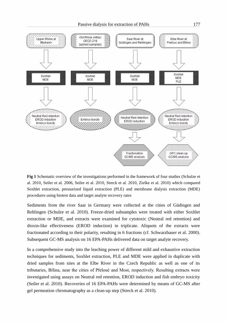
Passive dialysis for extraction of PAHs 177
Fig 1 Schematic overview of the investigations performed in the framework of four studies (Schulze et al. 2010, Seiler et al. 2006, Seiler et al. 2010, Streck et al. 2010, Zielke et al. 2010) which compared Soxhlet extraction, pressurized liquid extraction (PLE) and membrane dialysis extraction (MDE) procedures using biotest data and target analyte recovery rates
Sediments from the river Saar in Germany were collected at the cities of Güdingen and Rehlingen (Schulze et al. 2010). Freeze-dried subsamples were treated with either Soxhlet extraction or MDE, and extracts were examined for cytotoxic (Neutral red retention) and dioxin-like effectiveness (EROD induction) in triplicate. Aliquots of the extracts were fractionated according to their polarity, resulting in 6 fractions (cf. Schwarzbauer et al. 2000). Subsequent GC-MS analysis on 16 EPA-PAHs delivered data on target analyte recovery.
In a comprehensive study into the leaching power of different mild and exhaustive extraction techniques for sediments, Soxhlet extraction, PLE and MDE were applied in duplicate with dried samples from sites at the Elbe River in the Czech Republic as well as one of its tributaries, Bílina, near the cities of Přelouč and Most, respectively. Resulting extracts were investigated using assays on Neutral red retention, EROD induction and fish embryo toxicity (Seiler et al. 2010). Recoveries of 16 EPA-PAHs were determined by means of GC-MS after gel permeation chromatography as a clean-up step (Streck et al. 2010).

Passive dialysis for extraction of PAHs 178
The present communication summarizes the results of these different studies and evaluates the comparability of membrane dialysis with the two conventional procedures Soxhlet extraction and PLE, using a meta-analysis approach with rank sum-based scaling of the determined effect and target analyte concentrations. The question, whether the currently available membrane dialysis protocol is a procedure suitable for a more reliable extract preparation and whether it can replace other conventional techniques for recovery of PAHs, is critically discussed.
Analyzed results represent a broad variety of conditions for sampling and sample types, extraction, and determination of compound concentrations. Rather than comparing chemical data given as recovery rates, the assessment is also based on different biological effects. Therefore, findings from this study take into account the possible variability of investigations of sediments and the diversity of questions that might be addressed through the discussed extract types.
8.3 Material & Methods
8.3.1 Samples and sampling
Table 1 provides an overview over samples and sampling. Comprehensive descriptions are published with the individual studies (Schulze et al. 2010, Seiler et al. 2006, Seiler et al. 2010, Streck et al. 2010, Zielke et al. 2010). All natural sediment samples were shock-frozen at -30 °C, subsequently freeze-dried and stored at 4 °C. The OECD sediment was prepared with only 5 % kaolin clay content.
Table 1 Itemized overview of sampling conditions, sample treatment and sample properties for the four different studies analyzed within this paper
Study Sample Sampling Water removal Sieving TOC [g/kg] Notes
Rhine (Seiler et al. 2006)
surface Van Veen yes 1.25 mm 40.0
Old Rhine (Zielke et al. 2010)
surface Van Veen no None 34.0 spiked*
OECD (Zielke et al. 2010)
artificial preparation no None 20.0 spiked*
Saar (Güd/Reh) (Schulze et al. 2010)
surface Van Veen yes < 2 mm 30.0/45.0
Elbe (Prel/Most) (Seiler et al. 2010, Streck et al. 2010)
surface Ekman-Birge yes < 63 µm 49.8/109.1
*Diuron, pentachlorophenyl, nonylphenol, fluoranthene, 2,4-dinitrophenol, parathion-ethyl

Passive dialysis for extraction of PAHs 179
8.3.2 Extraction of sediment samples
The different extraction procedures were applied following already published protocols and are detailed elsewhere for each individual study (Schulze et al. 2010, Seiler et al. 2006, Seiler et al. 2010, Streck et al. 2010, Zielke et al. 2010). Extracting solvents were changed to dimethyl sulfoxide for bioassay and n-hexane for chemical analysis, respectively. Extracts were stored at -20 °C.
For membrane dialysis extraction (MDE), sediment samples were inserted into pre-extracted LD-PE 'layflat' tubing, sealed and dialysed with n-hexane inside brown glass jars. Soxhlet extraction was carried out on dried sediment weighed into extraction thimbles and placed in Soxhlet extractors with acetone. In pressurized liquid extraction (PLE), dried sediments were subjected to a two-step extraction procedure with an ASE® device, first using a mixture of n-hexane and dichloromethane and then toluene as solvent. Immediately after PLE, resulting extracts were purified using gel permeation chromatography.
8.3.3 Chemical analysis
Details on the performed chemical analysis are given by the individual studies (Schulze et al. 2010, Streck et al. 2010). Extracts from the rivers Saar and Elbe were analyzed by means of gas chromatographic-mass spectrometric methods in the selected ion mode (SIM) and quantified using an external five-point-calibration. Results were corrected with an internal standard of deuterated PAHs.
8.3.4 Bioassays
Bioassays were conducted according published protocols. Details are given in Seiler et al. 2006, Seiler et al. 2010, Streck et al. 2010, Zielke et al. 2010.
The cell line RTL-W1 (Lee et al. 1993) was kindly provided by Drs. N.C. Bols and L. Lee (University of Waterloo, Canada). Cells were maintained in plastic culture flasks in Leibowitz's L15 medium supplemented with foetal bovine serum and penicillin/streptomycin solution at 20 °C.
Within the Neutral red retention assay on acute cytotoxic effects (1992, Klee et al. 2004), RTL-W1 cells were exposed to serial dilutions of sediment extracts along seven wells in six replicates of a 96-well microtitre plate. After incubation, cells were stained with Neutral red (2-methyl-3-amino-7-dimethylamino-phenanzine) and Neutral red retention was determined photometrically.
The EROD-inducing potential of sediment extracts was assayed using the ethoxyresorufin-O-deethylase (EROD) induction assay (Behrens et al. 1998, Olsman et al. 2007, Seiler et al. 2006, Wölz et al. 2008). RTL-W1 cells were seeded into 96-well microtitre plates and exposed to sediment extracts in eight dilution steps with six replicates. Following incubation,

Passive dialysis for extraction of PAHs 180
cells were disrupted, 7-ethoxyresorufin was added to all wells and deethylation was initiated with NADPH. The reaction was stopped by adding acetonitrile. The production of resorufin as well as the whole protein was determined fluorometrically.
Embryotoxic potentials of sediment extracts were investigated with the zebrafish (Danio rerio) embryo assay (cf. Hollert et al. 2003, Nagel 2002). Sample extracts were diluted in artificial water, transferred alongside ten fertilized zebrafish eggs to individual wells of microtitre plates (one egg per well) and incubated. Lethal endpoints were recorded and mortalities were determined after 48 hours post-fertilization.
8.3.5 Graphical evaluation, calculation of potentials, and statistical analysis
All effect data were plotted against extract concentrations with GraphPad Prism 5.0 (GraphPad Software, San Diego, USA). Data points were fitted using either second-order polynomial or sigmoid non-linear regression as a model equation. Effect concentrations were determined by means of this regression curve as the extract concentrations causing a particular strength of effect: NR50 values gave the concentrations at which 50 % cell viability compared to the controls were detected in the Neutral red retention assay; EC25TCDD values were calculated as the concentrations that caused 25 % of the maximum EROD inductions found for TCDD; LC50 values defined extract concentrations for which 50 % of the exposed fish embryos showed lethal effects.
In order to compare biotest results from the different studies with respect to the leaching power of extraction methods, relative effect potentials were calculated for a meta-analysis based on a scaling of rank sums (cf. Canfield et al. 1994, Hollert et al. 2002). This was achieved by dividing the lowest EC value from a test system with all other EC values from that test system, individually for each sample. The resulting values 0 < X ≤ 1 are ratios of the highest effectiveness of a given sample to all other data gained with the same sample. By calculating these ratios separately for each sample, any differences in sediment characteristics like, e.g., total organic contents and grain size were already considered, since they were identical in all compared treatments. Subsequently, mean potentials per extraction procedure per test were calculated, and results were plotted in box-and-whisker graphs using GraphPad Prism. Similar calculations were carried out with the data from chemical analyses, resulting in recovery potentials for each method; these were plotted in a column graph. Mean values were compared statistically using Prism 5 and SigmaStat 3.5 (Systat, Erkrath, Germany).
8.4 Results
In all three biotest systems, strongest effects were recorded for Soxhlet extracts. Consequently, the lowest EC values for calculation of the relative effect potentials were all provided by data from Soxhlet extracts resulting in highest potentials for these samples (Table 2.).

Passive dialysis for extraction of PAHs 181
Table 2 Relative effect potentials for the three extract types tested in three different biotests
Method EROD Neutral red Fish embryo
Soxhlet 0.22 - 1.00 0.52 - 1.00 0.41 - 1.00MDE 0.27 - 0.87 0.39 - 1.00 0.27 - 0.63PLE 0.03 - 0.42 0.14 - 0.30 0.24 - 0.56
Comparing the mean effect potentials of the three extraction procedures revealed limited agreement between PLE and MDE, but quite significant differences to Soxhlet extractions. As displayed by figure 2, Soxhlet extracts showed higher EROD-inducing activity than MDE and PLE extracts (Fig. 2a), which were comparable in their effectiveness. Statistical analysis according to Kruskal-Wallis with Dunn's post-hoc test revealed significant differences with p < 0.05 (Soxhlet vs. MDE) and p < 0.01 (Soxhlet vs. PLE). The same holds true for the fish embryo test (Fig. 2b), where extracts from MDE and PLE showed high comparability, while Soxhlet extracts revealed clear-cut stronger toxic potentials (p < 0.01 and p < 0.001, respectively, according to Kruskal-Wallis with Dunn's post-hoc test). Quite different results were obtained for the Neutral red retention assay (Fig. 2c). Here, Soxhlet- and MDE-derived extracts yielded comparable data, whereas PLE extracts were significantly less toxic (p < 0.001; one-way ANOVA with Tukey's post-hoc test).
By calculating effect potentials, values were normalized to their corresponding test system and the investigated samples. Thus, it was possible to group all results with respect to the extract type and make an overall comparison of their effectiveness (Fig. 2d). A non-parametric analysis of variance with the post-hoc test according to Dunn revealed that also regarding the overall effect potential, Soxhlet extracts were significantly more toxic than MDE and PLE extracts (p < 0.001). Furthermore, MDE extracts turned out to induce significantly stronger overall effects in the three biotests than extracts obtained using PLE (p < 0.05).
The comparison of the potentials to concentrate hazardous compounds, as calculated from chemical analyses of PAHs, had an outcome completely contradictory to the findings for the biotest results. A statistical analysis according to Kruskal-Wallis with Dunn's post-hoc test showed that Soxhlet extracts contained significantly lower target analyte concentrations than extracts obtained using MDE (p < 0.05; Fig. 3). PLE extracts gave also lower concentration values, but differences were not statistically significant.

MDE PLE SOX0.0
0.2
0.4
0.6
0.8
1.0*
4 2 4
Rel
ativ
e re
cove
ry p
oten
tial
Passive dialysis for extraction of PAHs 182
Fig 2 Comparison of relative effect potentials regarding applied test systems. Data are shown as medians (lines), means (crosses), 25-75 percentiles (boxes), 10-90 percentiles (whiskers) and points outside the percentiles (dots). S and M denote significant differences to Soxhlet and MDE, respectively, and asterisks represent the corresponding alpha-levels. Numbers above the x-axis indicate n for each dataset. a) EROD assay, b) fish embryo assay, c) Neutral red retention test, d) overall comparison. For more details on the statistics, see text
Fig 3 Comparison of overall PAH recovery potentials. Data are given as means with standard deviations. Numbers above the x-axis indicate n for each dataset. *: Significant difference (ANOVA on ranks with Dunn‘s test, p < 0.05)
0.0
0.2
0.4
0.6
0.8
1.0
50 38 18
M*
S***
S***
0.0
0.2
0.4
0.6
0.8
1.0
20 14 6
S*
S**
0.0
0.2
0.4
0.6
0.8
1.0
15 14 8
S**
S***
0.0
0.2
0.4
0.6
0.8
1.0
14 10 4
S***M***
a b
c d
Soxhlet MDE PLE
Rel
ativ
e ef
fect
pot
entia
l
S/M : significant difference to Soxhlet/to MDE; */**/*** : < 0.05/0.01/0.001

Passive dialysis for extraction of PAHs 183
8.5 Discussion
The data re-analyzed in the present communication were derived from four studies with MDE extracts investigating seven different sediment samples, and also conditions of sampling and sample preparation varied to some extent (see Table 1). EC values were calculated based on at least two completely independent replicates. Furthermore, several experiments involved even completely independent extraction replicates rather than just repeated testing of the same sample with the applied biotest systems. As a consequence, comparisons and statistical analysis deployed on the data sets can be considered of high reliability. All found differences and unexpected results, in conclusion, are very likely due to influences of parameters that differ between the extract types and need to be identified within the following discussion.
With respect to possible alterations due to heat as an auxiliary energy source, it is likely that during Soxhlet and PLE extraction toxic compounds may have been at least partially degraded, modified or even created de novo (Seiler et al. 2008). Hence, when considering Soxhlet comparable with PLE in terms of leaching power, as widely done (cf. Dean & Xiong 2000), accordance between these two extract types might be expected. Since MDE is performed at room temperature and does not utilize auxiliary energy to facilitate the separation process, it might be suspected that MDE extracts show differences to those from Soxhlet extraction and PLE.
Compound concentrations in the PLE extracts can be considered quantitative, since PLE protocols have been proven to provide very stringent and complete exhaustive extraction conditions (Camel 2001, Dean & Xiong 2000). High temperature and pressure during the extraction process combined with fresh solvent rinsing and a nitrogen-assisted purging step at the end of each cycle guarantee for maximum leaching power and quantitative retrieval of the prepared extract. Table 3 sums up studies on the recovery of the most important environmental contaminant classes from complex environmental samples using PLE. According to these data, PLE is capable of quantitatively extracting, among others, PAHs from sediment samples.
Since MDE extracts were comparable to PLE extracts in chemical analysis, membrane dialysis seems to be as powerful as the sophisticated automatic PLE, with respect to extraction of PAHs from sediment samples. Soxhlet extracts, on the other hand, contained significantly lower concentrations of PAHs, but this extract type caused significantly higher effects regarding EROD-induction and embryo toxicity compared to MDE- and PLE-derived extracts. For the Neutral red assay on acute cytotoxicity, significantly lower effect potentials were found for PLE extracts than for extracts from either Soxhlet extraction or MDE. As a consequence, parameters other than the leaching power and/or the risk of alteration due to heat can be assumed to have an influence on experimental results.

Passive dialysis for extraction of PAHs 184
Table 3 Summary of PLE application for extraction of different classes of target analytes (adapted from Dean & Xiong (2000)
Compounds Recovery [%] Refs.
PAHs Quantitative, 91-95 % (Berset et al. 1999, Burkhardt et al. 2005, Harb & Aldstadt 2004, Hawthorne et al. 2000, Itoh et al. 2008, Popp et al. 1997, Ramos et al. 2000, Saim et al. 1997, Schantz et al. 1997)
Organic chlorinated pesticides
Quantitative (Gfrerer et al. 2004, Popp et al. 1997, Schantz et al. 1997)
Polychlorinated biphenyls Quantitative, 83-131 % (Antunes et al. 2008, Bandh et al. 2000, Björklund et al. 1999, Josefsson et al. 2006, Martens et al. 2002, Schantz et al. 1997, Szostek et al. 1999)
Polychlorinated dibenzodioxins/furans
Quantitative (Antunes et al. 2008, Popp et al. 1997, Spinnel et al. 2008)
Herbicides 47-99 % (Conte et al. 1997)
Sulphur is known to cause acute toxicity, particularly in cell-based bioassays (Jacobs et al. 1992, Kammann et al. 2005, Pardos et al. 1999, Ricking et al. 2004, Salizzato et al. 1998a, Salizzato et al. 1998b, Svenson et al. 1998). Whereas sulphur was not removed from Soxhlet and MDE extracts prior to biotesting, the gel permeation chromatography (GPC) step after PLE cleaned resulting samples. This obviously correlates with significantly stronger effects in the Neutral red retention test on acute cell toxicity found for Soxhlet- and MDE-derived extracts. Therefore, it is possible that these two extract types exceeded the effectiveness of PLE extracts mainly because of sulphur content, but not due to organic contaminant concentrations. Soxhlet extraction, as a consequence, provided extracts with higher impact despite lower recovery of PAHs.
Beside sulphur, sediment organic matter (SOM; e.g. particulate and dissolved organic matter, humic and fulvic acids, black carbon) might have had an influence on biotests as well as chemical analyses. SOM can act as a strong sorption phase for hydrophobic organic substances (Ehlers & Loibner 2006, Haitzer et al. 1999, Huang et al. 2003, Luthy et al. 1997, Steinberg et al. 2000). Although former studies suggested low concentrations of dissolved organic matter to increase bioconcentration of hydrophobic organic contaminants in sediment dwelling organisms upon feeding, several authors also proved a significant decrease of bioavailability for various species (Bejarano et al. 2005, Gourlay et al. 2005, Guerrero et al.
Passive dialysis for extraction of PAHs 185
2003, Haitzer et al. 2001, Haitzer et al. 1998, Nikkilä & Kukkonen 2001). Consequently, if extracts tested in bioassays still contain SOM, compounds might also fail to contribute to toxic effects because of their strong association to this adsorbent.
Macromolecular organic components were removed from PLE extracts during the GPC clean-up prior to biotesting. MDE extracts are considered to be basically free of SOM: The size restriction limit of 1 nm of the semipermeable membrane deployed for dialysis excludes all particulate matter already upon extraction (Seiler et al. 2006). Soxhlet extracts, however, contained substantial amounts of particulate organic matter as generally experienced with this type of extract (Hawthorne et al. 2000).
Unfortunately, the presence or absence of SOM cannot explain findings from the extract comparisons outlined before, because Soxhlet extracts were the most effective regarding embryo toxicity, cytotoxicity and dioxin-like activity. However, it is also conceivable, that contaminants associated to SOM failed to contribute to the toxic effects, while putatively higher sulphur content compared to MDE caused elevated toxicity, at least in Neutral red retention assay and fish embryo test. Hence, the comparability of cytotoxic effect potentials of Soxhlet and MDE extracts might indicate lower sulphur contents for the MDE-derived samples. On the other hand, hexane in MDE and acetone in Soxhlet extraction can be considered comparable regarding sulphur recovery potential (Gryglewicz & Gryglewicz 2001), and temperatures applied in the latter one might also have caused degradation of contaminants.
In contrast to the higher effectiveness in biotests, Soxhlet samples gave significantly lower target analyte concentrations in chemical analyses. While not being cleaned for investigation in biotests, Soxhlet- and MDE-derived extracts were treated using either GPC or silica gel fractionation as a pre-requisite for GC-MS analysis. Therefore, it can be assumed that removal of SOM during clean-up had caused loss of contaminants. On the other hand, GPC clean-up was also applied on PLE extracts prior to GC-MS analysis, but these samples showed clearly higher target analyte concentrations than Soxhlet-derived extracts. It has to be considered, however, that data for recovery potentials of PLE extracts are based on only two replicates, while Soxhlet data provide four independent values. Hence, differences regarding chemical analysis between resulting extracts from the two methods should be interpreted cautiously.
As a third potential source of interference, compounds other than PAHs may have been extracted with differential stringency by the three leaching procedures. Acetone, used in Soxhlet extraction, is a slightly polar solvent and therefore capable of dissolving also rather polar substances. Recent studies provided evidence that also more hydrophilic contaminants can have high ecotoxicological impact (Kidd et al. 2007, Sanderson et al. 2007). Schwab and co-workers (2009) found that acute toxicity effects by extracts of sediments of industrial regions in Germany (Bitterfeld) and the Czech Republic (near Pardubice) were not only

Passive dialysis for extraction of PAHs 186
caused by PAHs but also by compounds in the polar fractions, namely heterocyclic molecules, 7H-benzo[de]anthracen-7-one and N-phenyl-2-naphthylamine. Furthermore, especially regarding mechanism-specific effectiveness, such as dioxin-like activity, polar compounds have gained increasing attention (Wölz et al. 2008).
Finally, the possibility of degradation or adsorption of compounds during extraction and clean-up should be considered. During GPC, nitroaromatic compounds may adsorb to the materials of the chromatographic column, thus lowering the available fraction of such compounds after clean-up (US Environmental Protection Agency 1994). The high temperatures during extraction using PLE may result in degradation of thermolabile compounds, e.g. aminoaromatics. Nitro- as well as aminoaromatics are known to act as aryl hydrocarbon receptor agonists and also show mutagenic properties (Gustavsson et al. 2007, Gustavsson et al. 2004, Klee et al. 2004). Therefore, lower toxicity observed with the EROD assay might be at least partially due to degradation or adsorption during extraction and clean-up.
The high number of confounding factors associated with sediment extraction as a consequence of the complex and mostly unknown nature of samples makes precise interpretations of the data extremely difficult. Nevertheless, the presented meta-analysis indicates that MDE is at least as powerful as the applied PLE protocol regarding extraction of PAHs from sediment samples. With respect to biological effectiveness, MDE extracts showed toxic impacts at least as strong as extracts obtained using PLE.
8.6 Conclusions & Perspectives
Present results provide strong evidence that passive MDE procedures provide exhaustive leaching power comparable to the sophisticated and automated PLE technique. Data from biotesting as well as contaminant concentrations in extracts obtained using membrane dialysis indicate at least limited concordance with PLE extracts. Significant differences in effect potentials as found for acute cytotoxicity can be attributed to adverse impact of elemental sulphur in MDE extracts, but not in PLE extracts.
The concept of passive membrane dialysis can thus be considered a promising alternative to other common procedures for exhaustive extraction. At the same time – being a passive extraction method – MDE reduces the risk of uncontrolled alterations of the original sample during the separation process and by this adds to the reliability of ecotoxicological investigations of sediments. However, the MDE protocol is not suitable for high throughput sample preparation due to the relatively high requirement for time and labour. Further establishment and future routine application of membrane dialysis-based extraction procedures therefore require optimized protocols and, if possible, automation. With respect to emerging, more polar environmental contaminants, the system needs to be enhanced towards

Passive dialysis for extraction of PAHs 187
selectable membrane-solvent combinations, which allow a separation of either a broad analyte spectrum or particular substance classes with specific physico-chemical characteristics. This requires testing of polymeric materials providing different physical properties, especially permeability, along with solvents of varying polarity.
In addition to passive extraction principles, membrane dialysis may have further important benefits to sample preparation: As indicated before (Seiler et al. 2006), MDE is capable of extracting target analytes from water-containing sediment samples with leaching power comparable to Soxhlet extraction. Research into this gave first promising results, and preliminary protocols for the extraction of completely untreated, native sediment samples are being developed. Experiments have been initiated to validate the method as an alternative for the extraction of fresh whole sediment samples. Such an advanced approach would allow starting the extraction in the field immediately after sampling. Thus, extraction of native samples using membrane dialysis techniques would avoid several confounding factors associated with sample preparation in sediment toxicology (Seiler et al. 2008).
8.7 References
Ahlf W, Hollert H, Neumann-Hensel H, Ricking M (2002): A guidance for the assessment and evaluation of sediment quality - A german approach based on ecotoxicological and chemical measurements. J Soils Sediments, 37-42
Antunes P, Viana P, Vinhas T, Capelo JL, Rivera J, Gaspar E (2008): Optimization of pressurized liquid extraction (PLE) of dioxin-furans and dioxin-like PCBs from environmental samples. Talanta 75, 916-925
Babich H, Borenfreund E (1992): Neutral red assay for toxicology in vitro. In: Watson RR (Editor), In vitro methods of toxicology. CRC - Press, Boca Raton, Florida, pp. 238-251
Bandh C, Björklund E, Mathiasson L, Näf C, Zebuhr Y (2000): Comparison of accelerated solvent extraction and Soxhlet extraction for the determination of PCBs in Baltic Sea sediments. Environ Sci Technol 34, 4995
Behrens A, Schirmer K, Bols NC, Segner H (1998): Microassay for rapid measurement of 7-ethoxyresorufin-O-deethylase activity in intact fish hepatocytes. Mar Environ Res 46, 369-373
Bejarano AC, Decho AW, Chandler GT (2005): The role of various dissolved organic matter forms on chlorpyrifos bioavailability to the estuarine bivalve Mercenaria mercenaria. Mar Environ Res 60, 111-130
Berset JD, Ejem M, Holzer R, Lischer P (1999): Comparison of different drying, extraction and detection techniques for the determination of priority polycyclic aromatic hydrocarbons in background contaminated soil samples. Anal Chim Acta 383, 263-275
Björklund E, Bøwadt S, Nilsson T, Mathiasson L (1999): Pressurized fluid extraction of polychlorinated biphenyls in solid environmental samples. J Chromatogr A 836, 285-293
Burkhardt MR, Zaugg SD, Burbank TL, Olson MC, Iverson JL (2005): Pressurized liquid extraction using water/isopropanol coupled with solid-phase extraction cleanup for semivolatile organic compounds, polycyclic aromatic hydrocarbons (PAH), and alkylated PAH homolog groups in sediment. Anal Chim Acta 549, 104-116

Passive dialysis for extraction of PAHs 188
Camel V (2001): Recent extraction techniques for solid matrices - Supercritical fluid extraction, pressurized fluid extraction and microwave-assisted extraction: Their potential and pitfalls. Analyst 126, 1182-1193
Canfield TJ, Kemble NE, Brumbaugh WG, Dwyer FJ, Ingersoll CG, Fairchild JF (1994): Use of Benthic Invertebrate Community Structure and the Sediment Quality Triad to Evaluate Metal-Contaminated Sediment in the Upper Clark-Fork River, Montana. Environ Tox Chem 13, 1999-2012
Conte E, Milani R, Morali G, Abballe F (1997): Comparison between accelerated solvent extraction and traditional extraction methods for the analysis of the herbicide diflufenican in soil. J Chromatogr A 765, 121-125
Cornelissen G, Rigterink H, Ten Hulscher DEM, Vrind BA, Van Noort PCM (2001): A simple tenax® extraction method to determine the availability of sediment-sorbed organic compounds. Environ Toxicol Chem 20, 706-711
de Maagd PGJ (2000): Bioaccumulation tests applied in whole effluent assessment: A review. Environ Toxicol Chem 19, 25-35
Dean JR, Xiong G (2000): Extraction of organic pollutants from environmental matrices: selection of extraction technique. Trends Analyt Chem 19, 553-564
Deboer J (1988): Chlorobiphenyls in bound and non-bound lipids of fishes - comparison of different extraction methods. Chemosphere 17, 1803-1810
Ehlers G, Luthy R (2003): Contaminant bioavailability in soil and sediment. Environ Sci Technol 37, 295A–302A
Ehlers GA, Loibner AP (2006): Linking organic pollutant (bio)availability with geosorbent properties and biomimetic methodology: a review of geosorbent characterisation and (bio)availability prediction. Environ Pollut 141, 494-512
Eisenträger A, Rila J-P, Hund-Rinke K, Römbke J (2004): Proposal of a testing strategy and assessment criteria for the ecotoxicological assessment of soil or soil materials. J Soils Sediments 4, 123-128
Feiler U, Ahlf W, Hoess S, Hollert H, Neumann-Hensel H, Meller M, Weber J, Heininger P (2005): The SeKT Joint Research Project: Definition of reference conditions, control sediments and toxicity thresholds for limnic sediment contact tests. Environ Sci Pollut Res 12, 257-258
Gfrerer M, Chen S, Lankmayr EP, Quan X, Yang F (2004): Comparison of different extraction techniques for the determination of chlorinated pesticides in animal feed. Anal Bioanal Chem 378, 1861-7
Gourlay C, Mouchel JM, Tusseau-Vuillemin MH, Garric J (2005): Influence of algal and bacterial particulate organic matter on benzo a pyrene bioaccumulation in Daphnia magna. Sci Total Environ 346, 220-230
Gryglewicz G, Gryglewicz S (2001): Determination of elemental sulfur in coal by gas chromatography - mass spectrometry. Fresenius J Anal Chem 370, 60-63
Guerrero NRV, Taylor MG, Wider EA, Simkiss K (2003): Influence of particle characteristics and organic matter content on the bioavailability and bioaccumulation of pyrene by clams. Environ Pollut 121, 115-122

Passive dialysis for extraction of PAHs 189
Gustavsson L, Hollert H, Jonsson S, van Bavel B, Engwall M (2007): Reed beds receiving industrial Sludge Containing Nitroaromatic compounds - Effects of outgoing water and bed material extracts in the umu-C genotoxicity assay, DR-CALUX assay and on early life stage development in Zebrafish (Danio rerio). Environ Sci Pollut Res 14, 202-211
Gustavsson LK, Klee N, Olsman H, Hollert H, Engwall M (2004): Fate of Ah receptor agonists during biological treatment of an industrial sludge containing explosives and pharmaceutical residues. Environ Sci Pollut Res 11, 379-87
Haitzer M, Höss S, Traunspurger W, Steinberg C (1998): Effects of dissolved organic matter (DOM) on the bioconcentration of organic chemicals in aquatic organisms: A review. Chemosphere 37, 1335-1362
Haitzer M, Höss S, Traunspurger W, Steinberg C (1999): Relationship between concentration of dissolved organic matter (DOM) and the effect of DOM on the bioconcentration of benzo[a]pyrene. Aquat Toxicol 45, 147-158
Haitzer M, Akkanen J, Steinberg C, Kukkonen JVK (2001): No enhancement in bioconcentration of organic contaminants by low levels of DOM. Chemosphere 44, 165-171
Harb JG, Aldstadt JH (2004): An improved pressurized liquid extraction method for the determination of polycyclic aromatic hydrocarbons in freshwater sediments by gas chromatography-mass spectrometry. Anal. Lett. 37, 2835-2850
Harkey GA, Landrum PF, Klaine SJ (1994): Comparison of whole-sediment, elutriate and pore-water exposures for use in assessing sediment-associated organic contaminants in bioassays. Environ Toxicol Chem 13, 1315-1329
Hawthorne SB, Grabanski CB, Martin E, Miller DJ (2000): Comparisons of Soxhlet extraction, pressurized liquid extraction, supercritical fluid extraction and subcritical water extraction for environmental solids: recovery, selectivity and effects on sample matrix. J Chromatogr A 892, 421
Hollert H, Heise S, Pudenz S, Brüggemann R, Ahlf W, Braunbeck T (2002): Application of a sediment quality triad and different statistical approaches (hasse diagrams and fuzzy logic) for the comparative evaluation of small streams. Ecotoxicology 11, 311-321
Hollert H, Keiter S, König N, Rudolf M, Ulrich M, Braunbeck T (2003): A new sediment contact assay to assess particle-bound pollutants using zebrafish (Danio rerio) embryos. J Soils Sediments 3, 197 – 207
Huang WL, Ping PA, Yu ZQ, Fu HM (2003): Effects of organic matter heterogeneity on sorption and desorption of organic contaminants by soils and sediments. Appl Geochem 18, 955-972
Hynning PA (1996): Separation, identification and quantification of components of industrial effluents with bioconcentration potential. Water Res 30, 1103-1108
Itoh N, Numata M, Aoyagi Y, Yarita T (2008): Comparison of low-level polycyclic aromatic hydrocarbons in sediment revealed by Soxhlet extraction, microwave-assisted extraction, and pressurized liquid extraction. Anal Chim Acta 612, 44-52
Jacobs MW, Delfino JJ, Bitton G (1992): The toxicity of sulfur to Microtox (R) from acetonitrile extracts of contaminated sediments. Environ Toxicol Chem 11, 1137-1143
Josefsson S, Westbom R, Mathiasson L, Bjorklund E (2006): Evaluation of PLE exhaustiveness for the extraction of PCBs from sediments and the influence of sediment characteristics. Anal Chim Acta 560, 94

Passive dialysis for extraction of PAHs 190
Kammann U, Biselli S, Reineke N, Wosniok W, Danischewski D, Hühnerfuss H, Kinder A, Sierts-Herrmann A, Theobald N, Vahl H-H, Vobach M, Westendorf J, Steinhart H (2005): Bioassay-directed fractionation of organic extracts of marine surface sediments from the North and Baltic Sea - Part II: Results of the biotest battery and development of a biotest index. J Soils Sediments 5, 225-232
Kidd KA, Blanchfield PJ, Mills KH, Palace VP, Evans RE, Lazorchak JM, Flick RW (2007): Collapse of a fish population after exposure to a synthetic estrogen. Proc Natl Acad Sci USA 104, 8897-8901
Klee N, Gustavsson LK, Kosmehl T, Engwall M, Erdinger L, Braunbeck T, Hollert H (2004): Changes in toxicity and genotoxicity of industrial sewage sludge samples containing nitro- and amino-aromatic compounds following treatment in bioreactors with different oxygen regimes. Environ Sci Pollut Res 11, 313-320
Lee LE, Clemons JH, Bechtel DG, Caldwell SJ, Han KB, Pasitschniak-Arts M, Mosser DD, Bols NC (1993): Development and characterization of a rainbow trout liver cell line expressing cytochrome P450-dependent monooxygenase activity. Cell Biol Toxicol 9, 279-94
Luthy RG, Aiken GR, Brusseau ML, Cunningham SD, Gschwend PM, Pignatello JJ, Reinhard M, Traina SJ, Weber WJ, Westall JC (1997): Sequestration of Hydrophobic Organic Contaminants by Geosorbents. Environ Sci Technol 31, 3341
Martens D, Gfrerer M, Wenzl T, Zhang A, Gawlik BM, Schramm KW, Lankmayr E, Kettrup A (2002): Comparison of different extraction techniques for the determination of polychlorinated organic compounds in sediment. Anal Bioanal Chem 372, 562-568
Mayer P, Reichenberg F (2006): Can highly hydrophobic organic substances cause aquatic baseline toxicity and can they contribute to mixture toxicity? Environ Toxicol Chem 25, 2639-2644
Nagel R (2002): DarT: The embryo test with the zebrafish Danio rerio - a general model in ecotoxicology and toxicology. ALTEX 19, 38-48
Nikkilä A, Kukkonen JVK (2001): Effects of dissolved organic material on binding and toxicokinetics of pyrene in the waterflea Daphnia magna. Arch Environ Contam Toxicol 40, 333-338
Olsman H, Schnurer A, Björnfoth H, van Bavel B, Engwall M (2007): Fractionation and determination of Ah receptor (AhR) agonists in organic waste after anaerobic biodegradation and in batch experiments with PCB and decaBDE. Environ Sci Pollut Res 14, 36-43
Pardos M, Benninghoff C, Thomas RL, Khim-Heang S (1999): Confirmation of elemental sulfur toxicity in the Microtox (R) assay during organic extracts assessment of freshwater sediments. Environ Toxicol Chem 18, 188-193
Popp P, Keil P, Möder M, Paschke A, Thuss U (1997): Application of accelerated solvent extraction followed by gas chromatography, high-performance liquid chromatography and gas chromatography-mass spectrometry for the determination of polycyclic aromatic hydrocarbons, chlorinated pesticides and polychlorinated dibenzo-p-dioxins and dibenzofurans in solid wastes. J Chromatogr A 774, 203-211
Ramos L, Vreuls JJ, Brinkman UAT (2000): Miniaturised pressurised liquid extraction of polycyclic aromatic hydrocarbons from soil and sediment with subsequent large-volume injection-gas chromatography. J Chromatogr A 891, 275-286
Reichenberg F, Mayer P (2006): Two complementary sides of bioavailability: Accessibility and chemical activity of organic contaminants in sediments and soils. Environ Toxicol Chem 25, 1239-1245

Passive dialysis for extraction of PAHs 191
Reid BJ, Stokes JD, Jones KC, Semple KT (2000): Nonexhaustive cyclodextrin-based extraction technique for the evaluation of PAH bioavailability. Environ Sci Technol 34, 3174-3179
Ricking M, Neumann-Hensel H, Schwarzbauer J, Svenson A (2004): Toxicity of octameric elemental sulphur in aquatic sediments. Environ Chem Lett 2, 109-112
Saim Na, Dean JR, Abdullah MP, Zakaria Z (1997): Extraction of polycyclic aromatic hydrocarbons from contaminated soil using Soxhlet extraction, pressurised and atmospheric microwave-assisted extraction, supercritical fluid extraction and accelerated solvent extraction. J Chromatogr A 791, 361-366
Salizzato M, Bertano V, Pavoni B, Ghrardini AV, Ghetti PF (1998a): Sensitivity limits and EC50 values of the Vibrio fischeri test for organic micropollutants in natural and spiked extracts from sediments. Environ Toxicol Chem 17, 655-661
Salizzato M, Pavoni B, Ghirardini AV, Ghetti PF (1998b): Sediment toxicity measured using Vibrio fischeri as related to the concentrations of organic (PCBs, PAHs) and inorganic (metals, sulphur) pollutants. Chemosphere 36, 2949-2968
Sanderson H, Laird B, Pope L, Brain R, Wilson C, Johnson D, Bryning G, Peregrine AS, Boxall A, Solomon K (2007): Assessment of the environmental fate and effects of ivermectin in aquatic mesocosms. Aquat Toxicol 85, 229-240
Schantz MM, Nichols JJ, Wise SA (1997): Evaluation of pressurized fluid extraction for the extraction of environmental matrix reference materials. Anal. Biochem. 69, 4210-4219
Schulze T, Seiler T-B, Hollert H, Schröter-Kermani C, Pekdeger A (2010): Extractability and toxicity of potentially toxic organic pollutants in riverine sediments. in prep
Schwab K, Altenburger R, Lübcke-von Varel U, Streck G, Brack W (2009): Effect-directed analysis of sediment-associated algal toxicants at selected hot spots in the river Elbe basin with a special focus on bioaccessibility. Environ Toxicol Chem 28, 1506-1517
Schwarzbauer J, Littke R, Weigelt V (2000): Identification of specific organic contaminants for estimating the contribution of the Elbe river to the pollution of the German Bight. Org Geochem 31, 1713-1731
Seiler T-B, Rastall AC, Leist E, Erdinger L, Braunbeck T, Hollert H (2006): Membrane dialysis extraction (MDE): A novel approach for extracting toxicologically relevant hydrophobic organic compounds from soils and sediments for assessment in biotests. J Soils Sediments 6, 20-29
Seiler T-B, Schulze T, Hollert H (2008): The risk of altering soil and sediment samples upon extract preparation for analytical and bio-analytical investigations—a review. Anal Bioanal Chem 390, 1975-1985
Seiler T-B, Streck G, Schulze T, Schwab K, Brack W, Braunbeck T, Hollert H (2010): On the comparability of procedures for sediment extraction in environmental assessment. Part A: Bioanalytical investigations. Environ Toxicol Chem in prep
Semple KT, Doick KJ, Jones KC (2004): Defining bioavailability and bioaccessibility of contaminated soil and sediment is complicated. Environ Sci Technol 38, 228A-232
Spinnel E, Danielsson C, Haglund P (2008): Rapid and cost-effective analysis of polychlorinated dibenzo-p-dioxins and polychlorinated dibenzofurans in soil, fly ash and sediment certified reference materials using pressurized liquid extraction with an integrated carbon trap. Anal Bioanal Chem 390, 411-417

Passive dialysis for extraction of PAHs 192
Steinberg CEW, Haitzer M, Brüggemann R, Perminova IV, Yashchenko NY, Petrosyan VS (2000): Towards a Quantitative Structure Activity Relationship (QSAR) of dissolved humic substances as detoxifying agents in freshwaters. Internat Rev Hydrobiol 85, 253-266
Streck G, Seiler T-B, Schulze T, Schwab K, Braunbeck T, Hollert H, Brack W (2010): On the comparability of procedures for sediment extraction in environmental assessment. Part B: Chemical analytical investigations. Environ Toxicol Chem in prep
Svenson A, Viktor T, Remberger M (1998): Toxicity of elemental sulfur in sediments. Environ Toxicol Water Qual 13, 217-224
Szostek B, Tinklenberg JA, Aldstadt JH (1999): A simple method for the quantitative microextraction of polychlorinated biphenyls from soils and sediments. Chemosphere 38, 3131-3139
US Environmental Protection Agency (1994 ): Method 3640A.Gel-Permeation Cleanup. Washington (DC)
Wölz J, Engwall M, Maletz S, Takner HO, van Bavel B, Kammann U, Klempt M, Weber R, Braunbeck T, Hollert H (2008): Changes in toxicity and Ah receptor agonist activity of suspended particulate matter during flood events at the rivers Neckar and Rhine - a mass balance approach using in vitro methods and chemical analysis. Environ Sci Pollut Res 15, 536-553
Zielke H, Seiler T-B, Niebergall S, Leist E, Zimmer H, Erdinger L, Braunbeck T, Hollert H (2010): The impact of extraction methodologies on the toxicity of sediments in the zebrafish (Danio rerio) embryo test (FET). J Soil Sediment in prep

Introductory Part B
The versatile, changing, and advancing roles of fishin sediment toxicity assessment – a review
AV Hallare1,2, T-B Seiler1, H Hollert1
1 Department of Ecosystem Analysis, Institute for Environmental Research (Biology 5), RWTH Aachen University, Worringerweg 1, 52074 Aachen, Germany
2 Department of Biology, CAS University of the Philippines Manila, Padre Faura, Manila 1000, Philippines
Accepted for publication in J Soils Sediments


Fish in sediment toxicity assessment 195
9 Introductory Part B
9.1 Abstract
Background, aim, and scope. Sediments serve as integral and dynamic parts of our aquatic systems. Within the last 15 to 20 years, however, the scientific community has begun noticing contamination and deterioration of sediment quality at an alarming rate worldwide. Sediments are now perceived to contain hazardous pollutants that can directly impact water quality, and hence, can become very stressful to aquatic life. As a consequence, global efforts had been initiated since early 1970s, towards finding ways to assess sediment quality. Because of their obvious ecological and economic significance, fish have remained to be a major taxonomic group for appraising the general quality of aquatic systems. However, for sediment risk assessment, fish have lagged behind invertebrates due to their mobility and generally, pelagic lifestyle. To our knowledge this is the first paper that comprehensively treats and reviews the versatile roles of fish in assessing the health state of aquatic sediments.
Main features. Through literature search, this review attempted to trace the progress on the use of various approaches as well as describes the future prospects of using fish as sentinels for sediment quality assessment. Initially, the use of whole fish (juveniles or adults) bioassays has contributed immensely to our understanding of sediment contamination and ecotoxicology. But due to economic as well as ethical issues linked to the use of live vertebrate animals for toxicity testing, the approach has shifted to using fish cell cultures and fish embryos. Much newer approaches involving receptors and gene arrays in fish cells to elucidate the mode of actions of sediment-borne contaminants are very promising. The review paper also explores some of the issues associated with the use of whole fish, fish cell cultures, fish embryos, and fish gene expression profiles in sediment toxicity evaluations.
Conclusions and perspectives. Overall, the present review has comprehensively explored the changing, and progressing roles of fish for use in sediment toxicity evaluation. Indeed the usefulness of this taxon as test organisms has defined a big part in the advancement of sediment toxicology. Many novel additions to existing knowledge are expected to come as more and more variations in methodology involving the use of fish for sediment assessment are currently being developed in many laboratories around the world.
9.2 Introduction
In recent years, scientific experts from all over have realized that sediments play a pivotal role in achieving good ecological status of global aquatic systems (Chapman and Wang 2001; Förstner et al. 2004,2008; Netzband 2007; Menzel et al. 2009). Undisturbed natural sediments serve as an essential, integral, and dynamic part of our freshwater, estuarine, and coastal ecosystems. Sediments provide breeding grounds as well as nutrient sources for many

Fish in sediment toxicity assessment 196
organisms while their dynamics and gradients form optimum conditions to support biodiversity. However, once sediments are contaminated, they become sinks and secondary sources of potentially hazardous toxicants (Hollert et al. 2000; Hallare et al. 2005c; Davoren et al. 2005; Barbee et al. 2008; Wolz et al. 2009) thereby influencing overall water quality. Polluted sediments then become a major source of stress to aquatic organisms (Burton and Scott 1992; DelValls et al. 1998), thereby bringing disastrous impacts not only on aquatic ecosystem integrity but also on wildlife and human health.
Contaminated sediments first began to be noticed as a serious environmental issue in the early 1970's when several fish species caught in the rivers and harbours of Great Lakes were showing various forms of tumors (Sonstegard, 1977; Delfino 1979; Hayes et al. 1990). This was paralleled by the reported rise in the concentrations of the pesticides DDT, polychlorinated biphenyls (PCBs), as well as heavy metals in sediments (as summarized in Rodgers et al. 1985). Sharp decline in many amphibian species worldwide was also blamed for the deteriorating quality of aquatic sediments (Savage et al. 2002). For the first time, sediments, which had been historically viewed to be ‘reasonably clean’ (Baudo et al. 1999, Karlsson et al. 2008), have been tagged as a major hazard for the environment (Burgess and Scott 1992; DelValls et al. 1998). Lying just below the almost uncontaminated water column are the sediments which have become highly toxic following long periods of contaminant deposition (Huuskonen et al. 1998). Among organisms that inhabit contaminated aquatic environments, those living on or near the bottom suffer the greatest risk. This is due to the bulk of contaminants that have been sequestered from the water column and incorporated into the underlying sediment. Sediments can accumulate large quantities of chemicals, particularly poorly soluble organic compounds that may be rapidly taken up by benthic fish, through direct contact with the sediment and the interstitial water, as well as from ingested food (prey) (Hinkle-Conn et al. 1998; Vigano` et al., 2001). The exposure of fish to prey-borne or sediment-borne contaminants ultimately leads to trophic transfer to higher levels (Di Pinto 1996). Thus, pollution monitoring or environmental risk assessment of aquatic ecosystems should not be limited to the water phase, but must also include the sediment (Power et al. 1992; Almeida et al. 2005; Hallare et al. 2005c; Förstner et al. 2008).
The assessment of sediment quality has been historically restricted to routine chemical analyses. However, quantifying contaminant concentrations alone cannot provide enough information to sufficiently appraise the probable adverse effects or time-dependent bioavailability of these materials to aquatic organisms (Ingersoll et al. 1995; Davoren et al. 2005; Kilemade et al. 2004, 2009). One major achievement during the last decades was the development of sediment toxicity protocols. Toxicity testings have been shown to be very useful in environmental and chemical hazard assessments because they can be done relatively quickly and inexpensively compared to multiple chemical analyses of synthetic organics (Burton and Scott 1992) and the data are easily interpreted. Until now, there is still a growing

Fish in sediment toxicity assessment 197
interest to develop and apply new methods for evaluating contaminated sediments. Despite the fact that sediment toxicology is a fairly young science (Burton and Scott 1992), many bioassay test systems have already been developed in the 1970s and have contributed immensely in defining sediment quality criteria. Many species ranging from bacteria, microalgae, yeast, crustaceans, cladocerans, mysids, oligochaetes, polychaetes, snails, bivalves, echinoderms, fish, amphibians, and even higher plants have been utilized for sediment toxicity tests. Over the last few years, much progress has been made to create standardized methodologies. Beside the recommended procedures which have become available (ASTM 2007, 2008; Environment Canada 1990, USEPA 1994 and OECD 1992,2001), the European Sediment Research Network SedNet (Barcelo and Petrovic 2007) and SETAC Europe consistently held workshops on sediment toxicity testing and bioassay procedures. A number of reviews have also flourished in the literature concerning the use of various assays for assessing quality of freshwater (Giesy and Hoke 1989; Burton 1991, Hansen et al. 2007), marine (Nendza 2002), and estuarine sediments (Chapman and Wang 2001) as well as on the use of particular groups of organisms such as microbes (van Beelen 2003), freshwater invertebrates (Ingersoll et al. 1995), and amphipods and polychaetes (Bat 2005).
Albeit much have been written on fish for testing pure chemicals as well as environmental samples, no reviews to date have been written on the versatile and changing roles of fish as applied exclusively to sediment toxicity analysis. Thus, the main aim of our paper was to explore the varying niches of fish in toxicity testing that have advanced the way we conduct ecological and ecotoxicological assessments of sediments. Specifically, the present review has the following objectives: (1) to describe the significance and suitability of using fish for appraising the real state of aquatic sediments (2) to chronologically present the emergence of various methodologies and approaches involving the use of fish for an integrated sediment quality evaluation (3) to associate the changing and progressing roles of fish for sediment quality assessments with the parallel advancement seen in the field of biomarker research and environmental genomics – this is related to the improvement of the weight of evidence approach to risk assessment – and (4) to explore and discuss the various issues that have surfaced regarding the use of live fish and the consequent shift towards use of fish cells and cultures, fish embryo, and fish microarrays. Consequently, the review is divided into four major parts:
1. the use of whole fish in sediment quality assessment,
2. the use of primary cultures and fish cell lines as substitute for the whole animal,
3. the use of fish embryo as part of a holistic approach to sediment risk assessment, and
4. the use of gene expression analyses and microarrays in fish tissues.
Fish in sediment toxicity assessment 198
9.3 The significance of fish as sentinel species
For several obvious reasons, fish species have continued to draw attention among ecotoxicologists who are interested in assessing the impacts of water-borne and sediment-borne contaminants (Powers 1989; van der Oost 2003). Fish, above anything else, is a widely distributed form of life that can inhabit freshwater, marine, and estuarine environment. Within the aquatic habitat, fish are strongly-dependent on both water and sediment phase during their lifetime, while quite a number of species are completely benthic in their lifestyle. Fish also play major roles in the aquatic food-webs. They are involved in the transfer of energy linking the lower and higher trophic levels, and thus, maintaining ecosystem stability. In both local and global scales, fish are economically very important as they provide the main sources of animal protein for those living in coastal regions. Sociologically, fishing also offers large recreational value in many cultures.
The strong connection between fish and human health has already been made clear in previous works (eg. Dawe et al.1964). The increased frequency of neoplasms in bottom-feeding fish inhabiting polluted environments may serve as early warning indicators of carcinogenic hazards to humans. Understanding how toxicants affect fish behaviour, physiology, and population responses can therefore be of highest ecological relevance. Hence, assessing the health state of fish is important for protecting human economics and health.
At ecological levels, toxicant-induced changes in fish populations affect the entire aquatic community and threaten the equilibrium of the whole ecosystem where they live. Fish species were shown to be valuable indicators of contamination since they accumulate metals, PAHs, or PCBs as a function of the contamination level of the sediments. Because of the reported decline in both fish and amphibian populations around the world, many workers indicated a great responsibility for finding the exact reason for the decline (Almeida et al. 2005;Keiter et al. 2006). Interest concerning the effects of environmental stressors on health and disease in fish and other marine organisms has continued to increase in recent years. Compared to invertebrates, fish has the advantage of having large size for an adequate tissue chemical analysis (Chappie and Burton 2000). The body concentrations of the chemicals usually reflect the amount taken up by fish over a period of time. Meanwhile, the embryonic and larval developments of fish have been utilized in regular toxicity assays for monitoring and risk assessment programmes. In the last decades fish have been increasingly used in toxicity tests with sediments (Di Giulio et al. 1993; Vigano et al. 1998, 2001).
Within the context of the EU’s Water Framework Directive (Directive 2000/60/EC), fish have been regarded as one of the indicators for ecological quality, apart from water chemistry, hydromorphology, aquatic macroinvertebrates, and aquatic flora. Thus, all EU member states are required to deliver factual and timely information regarding fish stocks, i.e. abundance, species composition, and age structure of all fish present in a water body within the River

Fish in sediment toxicity assessment 199
Basin Districts (RBDs) (Champ et al. 2009). Data on these elements, especially on fish biology, are perceived to have a key role to understanding and predicting consequences of environmental change which could lead to more sustainable management practices. The EU Water Framework Directive basically aims at maintaining high status of waters in areas where they have already been reached, preventing any deterioration in the existing status of waters and achieving at least ‘good status’ in relation to all waters by 2015.
9.4 The use of whole fish in sediment toxicity assessment
In the last decades fish have been increasingly used in toxicity tests with sediments (Di Giulio et al. 1993; Vigano et al. 1998; Fragoso et al. 2006; Keiter et al. 2006; Karlsson et al. 2008; Costa et al. 2008; Rocha et al. 2009; Leaver et al. 2010). The earliest studies on water and sediment toxicity analyses have employed whole live fish, either collected from contaminated habitat sources or exposed to contaminants under laboratory or field conditions (Table 1). This represents the simplest approach for testing the ecotoxicological burden of environmental samples.
9.4.1 Issues on the use of fish for toxicity studies
Two important issues have surfaced concerning the utilization of fish as test species in toxicology. One focuses on the ethical issues associated with the general use of vertebrates in toxicity testing while the other questions the suitability of fish (having pelagic lifestyle) in sediment toxicity evaluations.
Although fish studies had shown high relevance for the assessment of sediments, they should not be conducted routinely because of ethical concerns about biotesting with vertebrates (Lange et al. 1995; Segner 2004; Braunbeck et al. 2005; Zhou et al. 2006). A conservative estimate is that about one million fish are killed each year in the EU Member States alone for research and regulatory purposes (Castano et al. 2003). This value is expected to increase by additional 4.4 million with the commencement of the new EU regulation for industrial chemicals, REACh (Registration, Evaluation, and Authorization of Chemicals) (Tanneberger et al. 2008). In order to execute REACh in an ethically and financially acceptable manner, the European Commission encourages the development and application of alternatives to animal tests. Agencies, such as the European Partnership for Alternative Approaches to Animal Testing (EPAA), provide impetus in the form of funding for researches on alternative test methods in aquatic toxicology. Many toxicologists themselves also advocated the limited use of adult fish for toxicity tests and that the application of such tests must be scientifically justified on a case to case basis (Friccius et al. 1995; Lange et al. 1995; Nendza, 2002; Nagel, 2002; Braunbeck et al. 2005). Only if considered indispensable, acute (96 h LC50) or chronic Early Life Stage (ELS) tests may be conducted, for example, with turbot, Scophthalmus maximus (obtainable from commercial suppliers) or three-spined stickleback, Gasterosteus

Fish in sediment toxicity assessment 200
aculeatus (Nendza, 2002). More recent approaches utilizing fish cells instead of the whole organism have been developed (see Section 4). In addition, fish embryos have also been found to be a good replacement (Lammer et al. 2009) and will be discussed in detail in Section 5. Due to technological advancements in molecular biology, it is now possible to use readily-available fish DNA chips for discovering mechanisms of chemical toxicities at the gene level (Section 6).
Another issue revolves on the utility of fish for the assessment of contaminated sediments. Some have argued (e.g. Ankley, 1991; Ingersoll et al. 1995; Bat, 2005) that fish are typically pelagic making them inappropriate for sediment toxicity as compared to benthic life forms (e.g invertebrates). However, several studies (e.g. Burton, 1991; Cunha et al. 2007; Kilemade et al. 2009) claimed that many fish species are flat-shaped and are quite adapted to benthic lifestyles, and therefore, can be used for determining sediment pollution. Sediment-feeding (or illiophagous) fish also have the ability to tolerate low concentrations of dissolved oxygen which enables them to inhabit polluted environment (Black,1983). Benthic fish may rapidly take up organic contaminants that have accumulated in sediments, both through direct physical contact with the sediment or interstitial water. In addition, uptake of waterborne toxicants through respiration, and ingestion of carcinogens from the organic mud are possible routes of exposure (Hinkle-Conn et al. 1998; Vigano` et al. 2001; Hopkins et al. 2003). A survey of literature revealed that most fish used in sediment toxicology were benthic fish species including flounder (e.g. Beyer et al. 1996), halibut (e.g. Schlenk et al. 2005), streaked prochilods (Almeida et al. 2005), plaice (e.g. Nagler and Cyr,1997), sole (e.g. Jimenez-Tenorio et al. 2007; Costa et al. 2008)), spot (e.g. Roberts et al. 1989; Hinkle-Conn et al.1998), catfish (Di Guilio et al. 1993), chubsuckers (Hopkins et al. 2003), and turbot (e.g. Kilemade et al. 2004,2009). A recent study by Fragoso et al. (2006) compared the differential response between rainbow trout (pelagic species) and white sucker (benthic species). EROD activity was found to be greater in white sucker. However, when basal (negative control) activity was accounted for, no difference in response between the species could be determined. Finally, many adult pelagic and epibenthic organisms also depend on sediments as breeding substrates, so that effects on reproductive behaviour, growth, embryo development, or hatchability are critical endpoints to consider (Burton, 1991).
Despite all criticisms and drawbacks, fish remained as the most used and most ideal species for aquatic toxicity tests. Apart from their obvious ecological relevance, which they share with other aquatic species, they have an added commercial, cultural, and health significance to humans (Hayes et al. 1990).
Fish in sediment toxicity assessment 201
9.4.2 Methodological variations in sediment toxicity using whole fish
One significant aim of this review paper was to describe the various approaches on the use of whole fish for sediment toxicity evaluation with respect to: selection of exposure scenario, choice of fish species, selection of exposure duration, deciding on appropriate control fish or sediments, preference of working with single test species or with a multispecies bioassay, and selection of appropriate biomarkers. The following section is intended to serve as a benchmark for future prospects on the use of fish for sediment toxicity analysis.
Selection of exposure scenario
Sediment is described as a naturally complex, heterogenous geological matrix, possessing a number of possible routes by which biota maybe exposed to sediment-borne contaminants. The question on which exposure scenario is best to use for sediment toxicity analysis has been a major issue during the past decades. Since sediments are found at the bottom, which is perceived to be inaccessible for aquatic organisms, the mechanisms on how exactly contaminants can become bioavailable and consequently induce toxicity to aquatic organisms have fascinated ecotoxicologists for quite a long time. In sediment, contaminants are believed to be in equilibrium between the solid and the soluble phases. For non-ionic organic pollutants, the equilibrium is linked to the sediment organic matter fraction and to the octanol-water partitioning coefficient of the contaminant. On the other hand, for ionic contaminants and metals, the equilibrium between both phases depends not only on organic matter fraction but to a host of other parameters (Hansen et al. 2007). It is, therefore, necessary to carefully select the most appropriate exposure scenario to ensure successful assessment of sediments.
Sediment toxicity tests can be conducted using a variety of exposure phases: whole sediments, pore (interstitial) water phase, elutriate (water-extractable) phase, sediment extracts, and in situ assays. Since each one of them has its own strengths and weaknesses, it is usually difficult to recommend a particular system to meet all study objectives (for a detailed review see Burton 1991). Hence, it is recommended that different test phases (solid or liquid) or even in situ testing are carried out to obtain the varying information that are needed to assess hazard versus risk in sediment quality assessment. In general, routes of sediment exposures (e.g. particle contact, food, pore water, overlaying water) for aquatic organisms are determined by the habitats where the organisms live (e.g. sediment infauna, sediment epifauna, etc.) and the presumed bioavailability of the sediment contaminants in the habitats (Power et al. 1992; Cheung et al. 1997). Bioassays performed on pore water or elutriates can be used to evaluate the toxicity of chemicals present in the soluble phase, whereas whole sediment bioassays are necessary to assess the toxicity of total sediment, including both soluble and solid phases. This is the reason why many opted to use whole sediment and considered it to be the most realistic exposure scenario that takes into account bioavailability (e.g. Feiler et al. 2005, Kosmehl et al. 2006, 2007). However, there were some studies (e.g.

Fish in sediment toxicity assessment 202
Ankley,1991; Solomon and Sibley 2002; Davoren et al. 2005; Fragoso et al. 2006) that still recommended the use of specific exposure scenarios under certain conditions. The description of different types of exposure scenarios, their advantages and disadvantages, and how they have been applied using fish species are reviewed below:

Selected studies on the use of whole fish in sediment toxicity assessment
Test vertebrate Habitat of species
Test duration
ExposureMethod
Endpoint Control set up Experimental/Field Conditions
Sediment Phase References
Anguilla anguilla(European eels)
Freshwater 8 and 48 h Field study Plasma cortisolGlucose and lactate
Laboratory control T: 15±1˚CDO:8.03±0.6 mg/L
In situ caging Teles et al. 2004
Catastomus commersoni(white sucker)
Freshwater 96 h Laboratory;Static
CYP1A induction(EROD assay)
Reference sediment or water only
T: 15˚CP:16h:8h L/D
Whole sediment Fragoso et al. 2006
Cyprinus carpio(common carp)
Freshwater 3 wks (3 yr repetition
Field study Genotoxicity (micronucleus test and comet assay)
Reference site T: 12.4-20.6˚C In situ caging Klobucar et al. 2010
Gadus morhua (atlantic Marine 3 months (13 wks)
Field study Bioaccumulation (FAC in bile)CYP1A1 inductionPlasma aspartate aminotransferase
Survival rateCell localization of CYP1A inductionLiver histopathology
Reference site T: 10˚CS: 31 ppt
In situ caging
In situ caging
Beyer et al. 1996
Husøy et al. 1996
Erimyzon sucetta(chubsucker)
Freshwater 78 d Laboratory Fin erosionSprint speedCritical swimming speed
Control sand T: 25˚CpH: 7.20DO:7.59 mg/LC: 223.89 µS/cm
Whole sediment Hopkins et al. 2003
Ictalurus nebulosus (bullheads)
Freshwater Once a week for 18 months
Skin painting Epidermal papilloma Bullheads with solvent control
(-) Sediment extracts Black, 1983
Ictalurus punctatus(channel catfish)
Freshwater 28 d
14 d
Laboratory; Flow through
Field study
Biotransformation enzyme induction (EROD,ECOD)Bile metabolites (PAH)Antioxidant enzyme (SOD and catalase)Reduced and oxidized glutathionemalondialdehyde (lipid peroxidation)DNA strand breaksEROD inductionCYP1A1 mRNA Immunoreactive protein
Reference sediment
Laboratory control
T:18-23°C
T:9-15°C
Whole sediment
In situ caging
Di Giulio et al. 1993
Haasch et al. 1993

Test vertebrate Habitat of species
Test duration
ExposureMethod
Endpoint Control set up Experimental/Field Conditions
Sediment Phase References
Leiostomus xanthurus Estuarine 30 min
24 h, 7 d, 12 d, 21 d, and 28 d
8 d, 18 d, 28 d
Laboratory;Static
AvoidanceFeeding behavior
Survival based on LC50 and LT50
Survival Skin lesions (fin erosion)Gill erosionHematocritLiver histopathology
Azoic sediment
Reference site
Reference site
(-)
25.8+/-1.2 °CDO:7.0+/-0.8 mg/LS: 15.1+/-1.0 ppt
T:20.7-27.8°CS:18.3-20.4 pptpH:7.2-7.8DO: 2.7-8.5 mg/L
Whole sediment
Bulk sediment suspended sediments
Bulk sediment
Hinkle-Conn et al.1998
Roberts et al. 1989
Hargis et al. 1984
Limanda limanda(common dab)
Marine 3 months Laboratory; Flow through
Field study
Clinical and histological changes (skin ulcerations and gill hyperplasia)Antibody detection (agglutination assay)
PAH metabolites in bile
Reference sediment
(-)
T: 10˚C
(-)
Whole sediment
In situ collection
Bucke et al 1989
Kammann 2007Micropterus salmoides(largemouth bass)
Freshwater 7 d Field study EROD inductionCYP1A1 mRNA Immunoreactive protein
Laboratory control T:9-15°C In situ caging Haasch et al. 1993
Oncorhynchus kisutch Freshwater 7 days Field study Genotoxicity biomarkers(micronucleus,flow cytometric analysis and post DNA labeling
Reference site T: 6.9°CpH:8-8.1DO:10-11.5 mg/LTDS:60.8-61.4 ppm
In situ caging Barbee et al. 2008
Oncorhynchus mykiss (rainbow trout)
Freshwater 96 h
21 d
Laboratory, Static
Laboratory; Semi static
CYP1A induction(EROD assay)
Biological parameters (CF and HSI)Biochemical (MFO system induction via EROD and BMPO), PAHs metabolites in bile)Genotoxic damage (comet assay)
Reference sediment or water only
Unexposed fish
T: 15˚CP:16h:8h L/D
T:13 °C
Whole sediment
Whole sediment
Fragoso et al. 2006
Inzunza et al. 2006

Test vertebrate Habitat of species
Test duration
ExposureMethod
Endpoint Control set up Experimental/Field Conditions
Sediment Phase References
41 d
96-h21-28 d
30 d
Field study
Laboratory;Static
Field study
Cytochrome P450 monooxygenases (MFO) activityPAH metabolites in bile
Survival (EC50)
Genotoxicity (micronucleus)Bile metabolitesMonooxygenases
Undeployed fish
Fine silt-clay agricultural soil
Undeployed fish
(-)
T:10°CpH: 8.0C: 200 µS/cm H :100 mg/L CaCO3
(-)
In situ caging
Pore water andWhole sediment
In situ caging
Barra et al. 2001
Kemble et al. 1994
DeFlora et al. 1993
Oreochromis niloticus (nile tilapia)
Freshwater 24 h Laboratory, Flow through
Field collection
Histopathology (liver)Haematology (lymphocytes and abnormal cells)Biochemical (EROD induction, total protein content)
Genotoxicity(micronucleus)
Uninjected fish
Fish from reference site
T:28°C
(-)
Sediment extracts
(-)
Zapata-Perez et al.2000
Rocha et al. 2009
Oryzias latipes(medaka)
Freshwater 90 d Laboratory, Multiple pulse-dose
MortalityFin erosionNon-neoplastic liver abnormality
Water controlAcetone carrier control
T:26˚C Sediment extracts and fractions
Fabacher et al. 1991
Pimephales promelas (fathead minnow)
Freshwater 96 h
4 d
4 d
Laboratory, Static
Survival (LC50) Reconstituted water
10% mineral water
Reference sediment
Reference sediment
T:25°CP:16L:8D
T:25°C
T:25°CP:16L:8D
Whole sedimentpore water andelutriates
Pore water only
Whole sedimentpore water and elutriates
Ankley (1991)
Ankley et al. 1990
Schubauer-Beriganand Ankley 1991

Test vertebrate Habitat of species
Test duration
ExposureMethod
Endpoint Control set up Experimental/Field Conditions
Sediment Phase References
10 d MortalityBioaccumulation
(-) In situ caging Mac et al. 1990
Platichthyes flesus (flounder)
Marine 3 yrs
3 mos (13 wks)
Field study
Field study
Plasma and hepatic retinoid levels
Bioaccumulation (FAC in bile)CYP1A1 inductionPlasma aspartate aminotransferase
Survival rateCell localization of CYP1A inductionLiver histopathology
Clean sandy sediment and water from reference site
(-)
T: 10˚CS: 31 ppt
In situ caging (mesocosm)
In situ caging
Besselink et al. 1998
Beyer et al. 1996
Husøy et al. 1996
Prochilodus lineatus(streaked prochilod)
Freshwater 24 h and 96 h
Laboratory, Static Hepatosomatic indexPlasma ion levelsPlasma glucose levelsGluthathione-S-transferase activityLiver catalase
Fish exposed to waters only
T: 19.5-23.3°CpH: 5.3-7.7C: 107-180 µS/cm
Whole sediment Almeida et al. 2005
Pleuronectes vetulus(English sole)
Marine 5 wks Laboratory, Static &Injection
Hepatic DNA adductsFluorescent aromatic compounds in bile
Reference sediment (-) Whole sediment and sediment extracts
French et al. 1996
Rhombosolea tapirina(greenback flounder)
Estuarine 6 wks Laboratory with dredging simulation
CYP1A induction (EROD assay)Histological (epidermal erosion and liver necrosis)Growth responses
Reference sediment T:15.64±0.05°CS:16.89±0.18 pptDO: 7.64±0.05 mg/LNH3:0.25±0.001ppm
Whole sediment Mondon et al. 2001
Scophthalmus maximus Marine 3 wks LaboratoryStatic renewal
GenotoxicityCytochrome P450
Reference sediment T:15°CP:15h:9h L/DpH:8S:35±2 pptDO: 74%
Whole sediment Kilemade et al.2004,2009
Solea senegalensis(Senegalese sole)
Estuary 28 d Laboratory, Static renewal
Genotoxicity markers:Erythrocyte Nuclear
Reference sediment T: 18±1°CpH: 7.9±0.2
Whole sediment Costa et al. 2008

Test vertebrate Habitat of species
Test duration
ExposureMethod
Endpoint Control set up Experimental/Field Conditions
Sediment Phase References
Marine 60 d Laboratory, Static renewal
Abnormalities (ENA) by micronucleus testDNA-strand breakage by comet assay
Biochemical (metallothioneins and EROD)Histopathology (gills and liver)
Reference sediment
S: 33±1,DO: 40–45%, Total NH3: 2–4mgL−1)
T: 19+/-2°CpH: 8.0-8.5S: 32+/-2DO: 80% saturation
Whole sediment Jimenez-Tenorio etal. 2007
Sparus aurata(seabream)
Marine
Estuary
Marine
60 d
240 h (10 d)
14 d
Laboratory, Static renewal
Laboratory, Flow through
Laboratory, Static renewal
Biochemical (metallothioneins and EROD)Histopathology (gills and liver)
Ethoxyresorufin-O-deethylase, liver and gill glutathione S-transferases, muscle lactate dehydrogenase,and brain acetylcholinesterase
Survival, superficial alteration, hematocrit analysis,and histological damage
Reference sediment
Reference site
Reference site
T: 19+/-2 °CpH: 8.0-8.5S: 32+/-2DO: 80% saturation12 fish per
T:18°C S: 34 ppt
T:19+/-1°CDO: 80% saturation
Whole sediment
Whole sediment
Whole sediment
Jimenez-Tenorio et al.2007
Cunha et al.2007
DelValls et al. 1998
Trachinotus obatus(pompano)
Marine 48 h and 96 h Laboratory, Static Mortality (LC50) Artificial seawater -NG- Elutriates Cheung et al. 1997
T: temperature; S: salinity; P: photoperiod; DO: dissolved oxygen; C: conductivity; H: hardness; TDS: total dissolved solids (-) : no information given

Fish in sediment toxicity assessment 208
Whole sediment phase The earliest studies on sediment toxicity testing with fish have been carried out using bulk (whole, native) sediments (McCain et al. 1978; USEPA 1981, Hargis et al. 1984). Whole sediment exposures comprise the primary and simplest tool for sediment toxicity assessments (Burton and Scott, 1992; Chapman and Wang et al. 2001) as they offer the least changes in sediment physicochemical conditions and present the widest variety of possible exposure routes. In newer terminology it is called “sediment contact test phase”, since effort is ensured that intact organisms or in vitro systems are exposed to contaminants borne from sediments (Hollert et al. 2003; Feiler et al. 2005). In the case of fish, the test species are exposed via several routes to maximize transfer rates: water-soluble compounds in the water column, particle-bound contaminants contacting gill as well as gut tissues, and food (brine shrimp, Artemia sp.), hatched in sediment suspensions from the test sites). Conventionally, it is designed by placing 4-5 cm of sediments from reference and contaminated sites on the aquarium tank floor before filling them with water. In some cases, it is necessary to mix sediments with a stainless steel spoon to achieve a homogenous mixture before testing (Francis et al. 1984; French et al. 1996; Fragoso et al. 2006). Sediment contaminant concentrations are realized through a variety of test systems (e.g. static, recirculating, or static renewal) (Burton, 1991) or a system to simulate dredging effects (Mondon et al. 2001). Then the fish are exposed for a defined duration which runs from a few hours to days or even several weeks. To avoid possible volume effects, fish loading should be equivalent to 1-2 L of water/g of fish/day (Sprague 1969; Fragoso et al. 2006), or about 10 L of water for five 1-3 g trout. After exposition to sediment and control conditions, fish survival and malformations data are typically collected. Subsequent to anesthetizing in benzocaine (150 ppm, Mondon et al. 2001) or tricaine methanesulfonate (MS-222) solution, fish are sacrificed for the evaluation of selected mechanistic-based endpoints. The endpoints cover acute and long-term toxicity, bioaccumulation, endocrine effects, carcinogenicity and mutagenicity and toxic effects on reproduction (Bucke et al. 1989; Roberts et al. 1989; Ankley 1991; Almeida et al. 2005; Inzunsa et al 2006; Jimenez-Tenorio et al. 2007; Barbee et al. 2008; Cunha et al. 2007; Costa et al. 2008) (Table 1). Up to now, laboratory studies with bulk sediments have continued to document that many of the samples from numerous locations exhibited acute and/or chronic toxicity to a variety of test species. Through whole sediment toxicity analysis, the information derived concerning the chemically-induced alterations (eg. tumor incidence) in resident organisms had become critical to the design of successful remediation plans. However, despite the consideration of the whole sediment phase as the most realistic scenario, Solomon and Sibley (2002) argued that since hydrophobic contaminants are slowly desorbed from the particles, the use of whole sediment in short time duration may not induce effects in receptive organisms and thereby underestimate the toxicity of the tested sediment. This is further aggravated when the sediments have high organic content which tends to significantly reduce the amount of bioavailable single molecules that can be taken up by fish (Fragoso et al. 2006). These observations often encourage critical

Fish in sediment toxicity assessment 209
discussion whether a whole sediment exposure of fish is really possible, especially if compared to organisms living in the sediments (as oligochaetes, etc). Under these circumstances, the selection of suitable organism (i.e. benthic fish) is of supreme importance.
Pore (interstitial) water phase The use of sediment pore water as test phase goes with the assumption that organisms receive most of their exposure through contact with interstitial water. This test phase was first conducted with benthic invertebrates and later applied to fish (Ankley et al. 1990; Schubauer-Berigan and Ankley 1991); standard protocols are also available (e.g. Burton, 1991; Liss and Ahlf 1997). Pore water is usually extracted from the solid phase by centrifugation of 250 ml aliquots of sediment for 45 min at 4000 g in a refrigerated centrifuge (4°C). In some studies, pore water was being extracted using a sediment tube with a micro-pump (Luckenbach et al. 2001). After preparation, particulate matter in the samples is allowed to settle for at least 24 h prior to decanting pore water for toxicity tests. Pore water is stored at 4 °C, generally for less than 72 h before testing. In some cases, the pore water is filtered through a 0.45 µm pore size cellulose acetate membrane or glass fibre filter prior to testing. Filtering might be necessary to remove some particles that are still in suspension in the water column even after centrifugation and that might interfere with the test (e.g. density of algal cells, interference with photometric measurements). On the other hand, some chemicals can adsorb on the membrane and take away the contaminants (Burton 1991) or might get lost when sorbed to removed particles. Therefore, it is better to avoid filtration if it is not absolutely necessary (Hansen et al. 2007). The toxicity of pore water also changes with time and, hence, toxicity tests must be conducted immediately after isolating pore water samples, and test solutions should be renewed frequently during exposures (every 12 to 24 h).
Using the fathead minnow in a pore water test, Ankley et al. (1990) has shown for the first time that a significant amount of the acute toxicity of the pore water to fish was due to ammonia. The identification of ammonia, a naturally-occurring compound in sediments, as potentially important sediment-associated toxicant has implications for the sediment toxicity assessment and control in freshwater and marine systems. Ankley et al. (1991) also showed that pore water is a reasonable test fraction for predicting the presence of toxicity in bulk sediments, whereas elutriate is a poor predictor of bulk sediment toxicity. The use of upper water column organisms is also inappropriate for evaluating in situ toxicity for benthic species. For a review of the good, the bad, and the ugly sides of pore water toxicity testing, readers are referred to the works of Chapman et al. 2002.
Elutriated water phase Sediment can also be elutriated with water (elutriates) wherein the ratio of solid matter to liquid phase is usually set at 1:10. Elutriates are usually prepared by rotating a 4:1 v: v) water: sediment mixture (1,600 ml total volume) in a 2-L flask on a shaker table for 1 h. The dilution water is very hard reconstituted water, hardness = 320 mg/L as CaCO3, which approximates the hardness of the sediment pore water. In some cases, the

Fish in sediment toxicity assessment 210
diluent used is artificial seawater (salinity, 30% prepared from commercially available salt, Instant Ocean®) (Cheung et al. 1997). Elutriate water is then separated from sediment by centrifuging at 2,500 g for 30 min at 4°C. The resulting supernatant must be used within 72 h for toxicity tests. Elutriate samples are generally not filtered before testing. The elutriate sediment toxicity test is intended both to simulate remobilization of sediments and to provide a better measure of the amount of a substance that is exchanged between the sediment and the aqueous phase during dredging and disposal (USEPA, 1991; Ankley et al. 1992). One major drawback is that this test phase may under- or overestimate the bioavailability and hence, the toxicity of contaminants present in the intact sediment (Hollert et al. 2003; Davoren et al. 2005), which further supported the contention that pore water corresponded better to bulk sediment toxicity (Schubauer-Berigan and Ankley 1991) than elutriates do. Based on literature survey, quite a few studies have addressed the use of elutriates with fish as test species, apart from those conducted by USEPA (1977). Few studies, though, have been conducted using fish culture (Section 4.0). In 1997, Cheung and colleagues successfully assessed sediment quality of Hongkong harbour by using different trophic organisms (including fish) exposed to elutriated sediment samples. Their findings supported that the presence of ammonia in sediment samples seemed to interfere with the detection of contamination (compare with Ankley 1991).
Sediment extracts Sediment extract phase is the proper choice when we want to assess the total hazard potential of sediments (i.e. bioavailable + nonbioavailable fractions)(Gagné et al. 1996; Strmac et al. 2002; Hollert et al. 2000, 2003; Viganó et al. 2003; Hallare et al. 2005c; Keiter et al. 2006; Kosmehl et al. 2007; Seiler et al. 2008). One advantage of using organic extracts is that it can precede the fractionation steps leading to identification of soluble chemicals and toxicant groups as required for effect-directed analysis (EDA). One major drawback of the extract phase, however, is its failure to consider the normal rate or extent to which toxicants can be taken up by sediment dwelling organisms (bioavailability) or the extent they cause adverse effects. It simplifies exposure by accelerating the bioavailability rate of especially strongly bound hydrophobic substances compared to intact whole sediment phase (Eriksson et al. 2005). Consequently, the possible long-term toxicity effects could be investigated in a much shorter time frame, enough to make this exposure phase to be considered as the ‘worst-case scenario’. Due to the particularly large amount of sample required for testing, exposure of live fish to extracts may not be a practical option. As a result, only a handful of studies have actually utilized whole fish for sediment extract exposure. A special technique called ‘skin painting’ was introduced by Black (1983) for testing sediment extracts from Buffalo River, New York on bullheads (Ictalurus nebulosus). Here, the bullheads were netted from the holding tanks and the area between the upper lip and the occiput was blotted dry with 4 x 4 gauze pads before the test material (PAH-containing extract) was applied to this area with a cotton tip applicator. A brief period was allowed for the evaporation of excess solvent and the absorption of the test material. Fish were placed in

Fish in sediment toxicity assessment 211
the recovery tank prior to their return to the holding tanks to minimize the transfer of test material to the holding tanks. Controls were handled in similar manner except that the painting contains only the extraction solvent used. Fish were painted once a week for up to 18 months. Epidermal hyperplasia and multiple epidermal papillomas were observed to develop in exposed brown bullheads. Grossly visible nodules also developed in the livers of bullheads fed a diet containing Buffalo River sediment extract, and one fish had a large cholangioma. Meanwhile, instead of skin painting, French et al. (1996) injected sediment extracts intramuscularly between the dorsal epaxial and axial muscles of the English sole (Pleuronectes vetulus). After 3 days of exposure, the liver tissue was sampled and screened for genotoxicity. Livers showed significant formation of polycyclic aromatic compounds (PAC) – DNA adducts.
Aside from gross morphological abnormalities and genotoxic endpoints, other toxicity biomarkers (eg. biochemical and haematological changes) were correlated to the concentrations of PAHs and PCBs contained in extracts of sediments (Zapata-Perez et al. 2000). Most recent applications of sediment extracts, however, were conducted with fish culture and fish egg assays, and will be the focus of the next sections. The predominant approach has been to concentrate organic sediment extracts and to subsequently expose the cells and/or inject the extracts to embryos in a carrier solvent. In several and newer studies (e.g. Hilscherova et al. 2001; Brack and Schirmer, 2003;Michallet-Ferrier et al. 2004; Kamman et al. 2005; Brack et al. 2005, Wölz et al. 2009) sediment extracts were further fractionated and fractions were utilized to fully characterize and/or identify the toxicants responsible for the observed effects.
In situ testing In situ testing is a relatively more recent approach in aquatic ecotoxicology wherein test organisms are directly exposed to the conditions (e.g. temperature, light, pH, dissolved oxygen, suspended solids, salinity, water flow, competitors and predators) of the investigated environment. This system therefore minimizes or avoids alterations of exposure conditions that could otherwise happen when samples are transferred and tested in the laboratory. Many studies considered in situ toxicity tests in the natural (in situ) setting to be more realistic than traditional laboratory experiments (using the same organisms) since the latter allows partial control of some variables by the experimenter (e.g. Beyer et al. 1996; Baudo et al.1999; Barbee et al. 2008;Klobucar et al. 2010). Studies on sediments basically involve the use of specifically designed benthic chambers for containing the test invertebrates. On the other hand, when applied to fish, especially designed animal cages, which can be suspended in the water column or deployed directly onto the sediments, are used (Mac et al. 1990). In a study by Luckenbach (2001, 2003), fish eggs were housed inside incubators that were deployed on the stream bottom. Another variant of in situ testing is through ‘mesocosm’ studies (Burgess and Scott, 1992; Besselink et al. 1998) wherein a section of an ecosystem is isolated, thus permitting the investigation of population dynamics, toxicity, bioaccumulation

Fish in sediment toxicity assessment 212
or pathological responses of the test organism. One example of the use of a mesocosm is the partitioning of a section of lake from surface water to bottom sediments with large plastic enclosures (SETAC-Europe,1991). This will ensure studies on the biology and chemistry of both ecological compartments.
A large number of studies have employed the in situ technique with practically all aquatic organisms (for a review see Chappie and Burton, 2000). However, it is not easy to establish which biological compartment is responsible for the toxicity (whether water or sediment) as exposition covers both compartments. Based on literature survey, only few authors have clearly defined the use of in situ technique for sediment toxicity evaluation in fish. In the case of fish caging studies, most were focused on measuring the bioaccumulation of contaminants from waters or sediments (Fragoso et al. 2006; Chappie and Burton, 2000), as fish has this advantage of a large size for adequate tissue chemical analysis. The body concentrations of the chemicals usually reflect the amount taken up by fish over a period of time.
Through the use of in situ testing with fathead minnow (Pimephales promelas), Mac et al. (1990) demonstrated that the magnitude of accumulation in laboratory exposures was similar to that in organisms caged in the field. Barra et al. (2001) supported this by reporting that significant PAH bioavailability was determined with rainbow trout fish caged in Biobio River, as manifested by increased induction of MFO enzymes and levels of PAH metabolites in the bile. In 2004, Teles and colleagues used caged fish to assess the success of a contamination clean-up process in Vouga River, Portugal. Their results demonstrated that even 2 years after closing of a bleached kraft pulp mill effluent (BKPME), the river water still contained sediment-associated chemicals responsible for the higher level of stress responses in the European eels. Very recent studies have also indicated that in situ caging of fish may serve as a useful strategy to monitor for genotoxic agents in aquatic systems (Barbee et al. 2008; Klobucar et al. 2010). Many other biological responses to contaminant exposure (biomarkers) have been utilized as endpoints for in situ studies with fish. These include mortality (Mac et al. 1990), liver histopathology (Husøy et al. 1996, Mondon et al. 2001), EROD induction (Haasch et al. 1993; Beyer et al. 1996; Mondon et al. 2001), level of CYP1A1 mRNA (Haasch et al. 1993), cellular localization of CYP1A induction (Husøy et al. 1996), level of immunoreactive protein (Haasch et al. 1993), cytochrome P450 monooxygenase (MFO) activity (DeFlora et al. 1993; Barra et al. 2001), plasma cortisol, glucose, and lactase levels (Teles et al. 2004), PAH metabolites in bile (Barra et al 2001; Beyer et al. 1996; DeFlora et al 1993; Kammann, 2007), plasma as well as hepatic retinoid levels (Besselink et al. 1998), micronucleus assay for genotoxicity (DeFlora et al. 1993; Rocha et al. 2009), and plasma aspartate aminotransferase (Beyer et al. 1996).
In situ testing, as with all experimental manipulations, is not exempt from artifacts associated with caging, like e.g. altered flow, altered food availability, sedimentation, accumulation of suspended solids, and competitive and/or predatory interaction. In addition, some

Fish in sediment toxicity assessment 213
disadvantages such as transportation, acclimation, field stresses, vandalism or loss of cages could be expected. Thus, monitoring control and/or reference performance is necessary to distinguish contaminant-related from artifact-related biological effects. Artifacts causing altered contaminant bioavailability may result in bioaccumulation and toxicity results different than those observed in laboratory bioassays or natural populations. However, an appropriately designed in situ study with improved exposure to realistic field conditions will presumably outweigh the more subtle artifacts of caging in most cases. For more tips and suggestions on designing an in situ study, the readers are referred to Chappie and Burton (2000).
Selection of fish species
Sediment toxicity testing with fish has started with the use of the fathead minnows (Pimephales promelas). However, as shown in Table 1, various other species have been increasingly used for sediment toxicity assessment as whole (adult) fish. But how are test fish species selected? The primary criteria for fish species selection should be the species’ ecological and/or economical importance to the region (e.g. Rhombosolea tapirina in Mondon et al. 2001, O. kisutch in Barbee et al. 2008; Sparus aurata in Cunha et al. 2007 ) and its relative sensitivity to sediment contamination (e.g. Scophthalmus maximus in Kilemade et al. 2009). Other factors should also be considered including the ease of collection (or purchasing), convenient size, adaptation in laboratory (ease of handling), lifestyle (benthic or pelagic), indigenous to disposal site or closely related to an indigenous species (Cheung et al. 1997; Guy et al. 2006). It must also be ensured that the selected fish are in good health and free from any apparent malformations.
All of these criteria were either singly or, if applicable, jointly satisfied in surveyed studies. For example, the choice of the spot (Leiostomus xanthurus) for determining the effects of contaminated sediments from Elizabeth river estuary in Virginia was due to the fact that it was a bottom-feeding sciaenid and naturally occurring spring summer migrant of the river whose juveniles adapt readily to laboratory conditions (Hargis et al. 1984). Spots actively agitate the surface of the sediments with their fins and body movements while foraging. This would account for the high incidence of severe erosion and hyperemia around their pectoral, caudal, and pelvic fins. It is suspected that dissolved chemicals and chemical suspenoids of contaminated sediment are involved. The selection of gilthead seabream (Sparus aurata) in Cunha et al. 2007 study is due to its economic significance as well as its distribution in the Atlantic and Mediterranean coastal waters, estuaries, and lagoons, including the Sado River estuary. It is intensively cultured in marine and estuarine waters, and it can be found in both pristine and contaminated sites. Furthermore, it is easy to maintain in laboratory conditions. On the other hand, Solea senegalensis and Sparus aurata were selected in carrying out sediment toxicity studies in the coast of Spain because they were common commercial species

Fish in sediment toxicity assessment 214
(Jimenez-Tenorio et al. 2007). The selection of Prochilodus lineatus is due to its detritivorous habit, meaning that it can be in contact with xenobiotics in sediment and it has relatively high sensitivity to toxicants (Almeida et al. 2005).
In some studies, the selection of species to be used for sediment assessment goes with the kind of exposure scenario to be used. For instance, benthic fish must be used when investigating whole sediment exposure, while pelagic fish species are suitable for pore water or elutriate samples (Ankley 1991). For fish cage studies, the catfish (Ictalurus punctatus) and large-mouth bass (Micropterus salmoides) were chosen due to their relative hardiness and ability to withstand the conditions in the river exposure protocol (Haasch et al. 1993). Despite the fact that tropical ecosystems are currently threatened by human activities and environmental degradation, little research has been done on the impact of contaminants on tropical ecosystems and aquatic biota, and only few tropical fish species have been employed in sediment toxicity tests such as tilapia (e.g. Zapata-Perez et al. 2005), and medaka (e.g. Fabacher et al. 1991). Some other authors (e.g. Almeida et al. 2005) have recommended the use of Neotropical freshwater fish (e.g. Prochilodus lineatus (Valenciennes, 1847) (=P. scrofa Steindachner, 1881) since there is still a lack of data concerning the effects of toxic agents on tropical fish species. Other investigators tend to select fish test species (e.g. Danio rerio and Pimephales promelas) which, though not benthic, have been in a suite of toxicity tests as standard test organisms (Ankley et al. 1992). Because standard species have long been used for other types of studies or bred and reared for the aquarium pet industry, much is known about their biology, genetics, and nutrition.
Once species are selected, they are usually obtained or procured from nearby unpolluted site which also served as reference site (e.g. Beyer et al. 1996), from an established aquaculture or hatchery station (e.g. Cheung et al. 1997; Costa et al. 2008), or directly from local pet shops.
Selection of appropriate biomarkers
Whole fish sediment toxicity tests were initially confined to the most usual acute endpoints of survival and/or malformations expressed in terms of LC50 or EC50 values (Hoke et al. 1980; Hargis et al. 1984; LaBlanc and Suprenant 1985; Ankley 1990, 1991). Determining acute toxicity tests seems to be a good approach to assess the effects of sediment contamination. However, sublethal bioassays should also be performed to determine chronic biological effects not provided by the acute toxicity tests. Mechanism-based, sublethal bioassays provide a more practical and more sensitive index of bioavailability, and at the same time give strong correlation between increasing contamination of sites and the degree of biomarker response (Jimenez-Tenorio et al. 2007; Fragoso et al. 2006).
The selection of specific biomarkers is often dictated by what kind of pollutants are suspected to be borne in the sediments (e.g. PAH, heavy metals). But, it is also usually dependent on the expertise and/or availability of chemicals and equipment in the local laboratory.

Fish in sediment toxicity assessment 215
Several authors (e.g. Giesy and Hoke 1989; Tollefsen et al. 2006) have proposed that a single biomarker alone may not be sufficient to adequately characterize subtle, sublethal effects of pollutants. This is because aquatic and benthic organisms typically are exposed to complex mixtures of contaminants and each or a combination of pollutants may likewise target single or closely-related biomarker responses. For example, three biomarkers for genotoxicity have been conventionally and effectively used for assessing PAH toxicity in aquatic organisms including erythrocyte micronuclei (clastogenicity and genotoxicity), flow cytometry or FCM (chromosome damage and DNA changes) and 32P-postlabeling (quantification of DNA adducts) (Barbee et al. 2008). The comet assay, another known biomarker for genotoxicity, has been employed in newer studies (Kammann et al. 2004; Costa et al. 2008; Kosmehl et al. 2008) and was even reported to be more sensitive than the micronucleus test (Klobucar et al. 2010). Thus, a battery of simple bioassays is recommended to provide rapid, holistic, and relative toxicity values which account for bioavailability, interactions, and even the mechanisms of actions (Giesy and Hoke 1989). Because bioassays are a direct measure of functional responses, they should have more impact on the decision making process than criteria based on concentrations of chemicals alone.
Based on our literature survey, multiple biomarkers have been routinely-used in several studies to estimate the level of bioaccumulation and/or bioavailability of sediment-borne contaminants (Table 1). Most of the studies have utilized specific mechanism-based endpoints. However, other interesting parameters such as swimming performance (Hopkins et al. 2003), feeding behaviours (Hinkle-Conn et al. 1998) and susceptibility to diseases (Bucke et al. 1989) were also reported.
Use of fish in a single study or as part of a multi-species test
In order to satisfy the call for a multifaceted integrated approach to sediment toxicity, application of the fish assay is usually set as part of a bigger project involving several test species representing various trophic levels (Ankley et al. 1991,1992). It is often desirable to use a variety of test species when assessing the toxicity of complex mixtures of compounds, since differences exist in organisms’ sensitivity to chemical contaminants (Giesy and Hoke 1989).There are also a number of possible routes of exposure (solid phase or liquid phase) which disqualify some species from being used. Alternatively, some organisms cannot be tested in one exposure exclusively. Hence, to cover more exposure routes as complete as possible, using different species becomes mandatory (Davoren et al. 2005). For example, algae have been demonstrated to be best for pore water and elutriate exposure whereas fish embryos and bacteria are good for solid phase exposures (Cheung et al. 1997; Hollert et al. 2003; Hallare et al. 2005; Cachot et al. 2009).

Fish in sediment toxicity assessment 216
Several of the surveyed studies have made use of fish along with other test species ranging from bacteria to higher plants. In 1997, Cheung and colleagues assessed sediment toxicity in Hongkong harbour by using different trophic organisms which include bacteria, two microalgae (Skeletonema costatum, a diatom and Dunaliella tertiolecta, a flagellate), juvenile shrimp (Metapenaeus ensis) and juvenile fish (Trachinotus obtaus). They recommend that a combination of a liquid-phase bioassay using diatom and a solid-phase bioassay using Microtox® test should be used for screening of a large number of sediment samples. Test organisms chosen for use in tests on the toxicity of bulk sediment from lower Fox River/Green Bay included the amphipod Hyalella azteca, the oligochaete Lumbriculus variegatus, the chironomid Chironomus riparius, the mayfly Hexagenia limbata, and the fathead minnow, P. promelas (Ankley et al. 1991). Although the latter species is not benthic, it was included because it is a standard test organism in aquatic toxicology and, as such, provided a useful comparison with the four benthic test species. Kemble et al. (1994) used Oncorhynchus mykiss with Daphnia, Microtox®, Hyatella azteca, and Chironomus riparius. In both studies, Hyalella (amphipod) was confirmed to be the most sensitive species.
Duration of exposure, identification of controls, and reference conditions
The duration of exposure to sediments varies from 96 hours to 3 months to 3 years, depending on the exposure phase. Presumably, in situ and whole sediment exposure requires longer exposure to allow for the natural course of chemical uptake by test organisms. The longest exposition in the surveyed literature was conducted in an in situ mesocosm study using flounder to determine toxicity of sludge from Rotterdam Harbour (Besselink et al. 1998). However, another in situ caging conducted by Teles et al. (2004) was accomplished only within 8 h. Exposure to sediment extracts is shortest due to the fact that the (bio)availability of chemicals has been increased (Seiler et al. 2008) and it is expected that the chemicals, if present, can immediately target the specific receptors leading to toxicity responses. However, methods involving microinjection of sediment extracts to sac fry required 12 months before visible neoplasms or hepatic carcinoma could be observed (Metcalfe et al. 1990). In a quite unusual study by Hinkle-Conn et al. (1998), the exposure was set to 30 min since they were only concerned about whether sediment-borne PAHs can affect the feeding behaviour of the juvenile spot.
There are many concepts of control in sediment toxicity analysis. First is the idea of control sediment. The sediments collected from reference sites usually serve as the control. In one study (Kemble et al. 1994), fine silt-clay agricultural soil (instead of sediments) has been used. In cases when no reference sediment could be obtained, the workers used a water control (e.g. Almeida et al. 2005, Ankley et al. 1991) or considered unexposed fish as the appropriate reference (Barra et al. 2001; Inzunsa et al. 2006). For in situ testing, caging of fish in parallel to a reference (clean) station serves as control (Husøy et al. 1996). For studies using sediment extract, the acetone carrier and water serve as controls (Black 1983; Fabacher
Fish in sediment toxicity assessment 217
et al. 1991). For pore-water and elutriate test, 10% mineral water or reconstituted water have been used as controls (Ankley et al. 1990). Although working with fish cell lines, Davoren et al. 2005 proposed the use of salinity controls when dealing with estuarine sediments.
Other factors such as the physical and chemical characteristics of sediment as well as environmental parameters of the exposure media can influence bioavailability and, consequently, the toxicity. One major factor is the organic content of the sediments. Higher values of total organic carbon (TOC) often lead to a weaker biomarker response. This is related to a decreased uptake by the organism as the contaminants, particularly organic substances, become sorbed to sediment organic matter. Another essential factor that had the greatest influence on most toxicity responses is temperature (Fragoso et al. 2006). Fish exposed to a temperature lower than their acclimation temperature are more vulnerable to chemical effects than fish exposed at or above this threshold temperature. This phenomenon could be explained by the integrated effect of increased temperature, gill ventilation rate, uptake across the gills, AhR receptor binding, gene activation kinetics, enzyme kinetics, metabolism, and excretion rates (Sleiderink et al. 1995).
9.5 The use of primary fish cell culture and fish cell lines as substitute for whole organism bioassays
9.5.1 Advantages of using fish cell culture and fish cell lines
In an effort to reduce the inherent economic and ethical constraints associated with the use of live fish for toxicological testing, methods involving the use of primary cell cultures and/or permanent cell lines were developed both for the screening and toxicity ranking of chemicals or environmental samples. This technique is an in vitro model system which retains the basic characteristics of the more complex in vivo condition, yet at the same time could be controlled experimentally (Baksi and Frazier 1990). Data likewise revealed a good correlation between the relative ranking orders of pollutants to fish cells with their water-borne in vivo toxicity to live fish (Bols et al. 1985; Magwood and George 1996). The rationale for the use of cultured cells for toxicity tests lies on the fact that the primary interaction between chemicals and biota occurs either at cellular surface or within cells, hence, most effects are exerted at cellular levels (Fent, 2001). Since in vitro cells do not possess the multiple defense mechanisms present in intact organisms (e.g. excretion, detoxification, storage, nonspecific binding), they are frequently more susceptible to the basic cytotoxic effects of a chemical, and respond at lower concentrations than would affect the whole organism (Ekwall 1983). Consequently, such approaches allow the study of specific actions within the cell without the complicating effects of other organ systems (Dipple et al. 1983). Other advantages of applying cell culture for toxicological research have been repeatedly reviewed (Baksi and Frazier 1990; Braunbeck 1998; Segner 1998; Belfiore and Anderson 2001; Schirmer 2006). Among these are the absence of genetic differences between cells, reduced variability between experiments, fast

Fish in sediment toxicity assessment 218
screening of large numbers of chemicals in multiwell plates using few test substances and producing little waste, and finally affording opportunity to establish cause-effect relationship to support field observations. Due to the increasing number of chemicals requiring rapid ecotoxicological evaluation, a preliminary tiered approach to toxicity testing using fish cell cultures is of growing importance in aquatic ecotoxicology.
9.5.2 Cell donors in primary culture and cell lines
Isolated liver cells (hepatocytes) have, so far, the most extensively used cells for primary culture as well as for permanent cell lines. This is because the fish liver represents the major target organ for the metabolism of most chemicals. Primary cultured hepatocytes also maintain most of their original differentiated in vivo characteristics, and therefore facilitate extrapolation of results in vivo (Zhou et al. 2006). As a consequence, fish hepatocytes have been extensively used for ecotoxicological exposure assessment studies (Segner 1998; Braunbeck and Strmac 2001; Schirmer 2006). However, other cells from fish have also been found to be useful as target cells for toxicants, such as the gonads, skin epithelia, endocrine tissues, muscle cells, white blood cells, and gill epithelia. The most widely used fish donor of cells for culture was the rainbow trout (Oncorhynchus mykiss), especially for hepatocytes. In addition to rainbow trout, other fish species, which have been formerly used as test subjects, have also served as donors for primary culture and cell lines (Table 2 and 3).
9.5.3 Basic methodology
For the preparation of primary culture, hepatocytes from rainbow trout are usually collected from the donor fish (rainbow trout with a mean of 250 +/- 50 g) by double perfusion method (Gagne et al. 1996). Cells are then distributed in a 96-well plate at a density of 1 x 10⁶/ml in sterile L-l5 medium supplemented with 1% FBS, 100 units of penicillin, 100 mg/l of streptomycin and 1 mg/l amphotericin B. Subsequently, hepatocytes are exposed to concentrations of sediment extracts for 24 h at 15°C alongside specific positive controls (e.g. BaP for genotoxicity and cyp1A1 induction) and standard test negative control (e.g pure medium, DMSO). After this incubation period, the cells are analyzed for specific biomarkers of viability, dioxin-like activity, genotoxicity, etc. In the case of fish cell lines, investigators usually grow and maintain their own fish cell lines for use in toxicity assessment. Standard cell culture and maintenance procedures are strictly followed including the use of specific culture media, incubation temperature, antibiotics, FCS, and CO2 humidifier system (if necessary), to ensure continuous supply of cell lines. Prior to actual exposures to test samples, cells are often seeded in 96-well plates and allowed to grow to 100% confluence. Subsequently, the medium is discarded and replaced by test samples and the control media. The duration of exposures and other parameters to be considered will all depend on the

Fish in sediment toxicity assessment 219
bioassay to be used. It is also customary to perform first a cell viability test (e.g. Neutral red retention test) to ensure that the response of cells to bioassay test is not influenced by inherent cellular toxicity.

Selected studies on the use of primary fish cell culture on sediment toxicity assessment
Primary cell Fish species source
Sediment exposure phase
Endpoints Biomarker assay used References Important Findings
Hepatocytes Oncorhynchus mykiss
Organic extracts Morphological changes andbiochemical alterations
Ultrastructural alterations, lactatedehydrogenase,alanine aminotransferase, catalase, glutathione S-transferase, acid phosphatase, and lipid peroxidation
Strmac and Braunbeck 2000, 2001
Cytological and biochemical changes inisolated hepatocytes can discriminatebetween different levels of contamination of waters and sediments.
Organic extracts Cytotoxicity, CYP1A induction, genotoxicity, and metallothionein (MT) levels
Estrogenicity(vitellogenin synthesis)
Cytotoxicity, genotoxicity, CYP1A activation, and estrogenicitiy
Propidium iodide (PI) exclusion test, MCFOD activity, Alkaline precipitation assay (APA),Nick translation assay (NTA), Silver saturation assay (SSA)
Non-radioactive dot blot/RNAse protection assay
Membrane integrity test, Fast micromethod, EROD Assay, and vitellogenin induction
Gagne et al. 1996, 1995
Hollert et al. 2005
Tollefsen et al. 2006
Lack of correlation between chemicaland toxicological data suggests that suchresponses could be due to chemicalinteractions or to unknown chemicalswhich are at play.
Numerous investigated extracts revealedan estrogen activity comparable to thatof the positive control(1 nM 17ß-estradiol corresponding to 270 ng/L inthe test medium)
Multiple mechanisms of toxic actionmay improve our understanding ofcellular toxicity of complex mixtures andlead to a more holistic approach toenvironmental monitoring of contaminated sediments.
Oncorhynchus mykiss
Organic extracts Genotoxicity Comet assay Kamman et al. 2000 Total organic carbon (TOC) as well asthe different compositions of contaminants present in the sedimentextracts may contribute to the genotoxiceffects.
Epidermal cells Oncorhynchus mykiss
Sediment elutriates Multixenobiotic resistance (MXR)
P-glycoproteins (P-gp) expression (immunocytochemistry)
Ni Shuilleabhain et al. 2005
The presence of an mdr1-like mechanism in trout epidermal cells canbe induced/modulated by environmentalcontaminants present in sedimentelutriates.

Selected studies on the use of fish cell lines in sediment toxicity assessment
Fish cell lines Fish species source
Sediment exposure phase
Endpoints Biomarker assay used References Important Findings
BB cell line(dorsal muscle cell
Ictalurus nebulosus Organic extracts and fractions
Cytotoxicity Genotoxicity
Neutral red assay,Unscheduled DNA synthesis (UDS)
Ali et al. 1993 Two or more chemical groups in organicfraction act synergistically leading to theobserved higher induction of UDS levels.
CHSE-214(embryo cells); EPC(epithelium); and
(gonad cells)
Oncorhynchus tsawytschaCyprinus carpioOncorhynchus mykiss
Aqueous elutriates(estuary)
Cytotoxicity (lysosomal and mitochondrial functions)
Neutral red and Alamar blue assays
Davoren et al. 2005 RTG-2 cells, due to their tolerance to osmolaritychanges, are the most suitable for testingestuarine aqueous elutriate samples.
(epithelium)Cyprinus carpio Organic extracts
(marine)Genotoxicity(DNA strand break)
Comet assay Kamman et al. 2001, 2004
The genotoxic effects of marine sedimentextracts to EPC cells are related to the amountof contaminants and organic matter content ofsediments.
epitheliumCyprinus carpio Organic extracts
(marine)Stress levels Hsp70 induction Kinder et al. 2007 In most cases contaminant concentrations were
too low to cause an effect. Effects of the testedsediments are, thus, attributed to other contaminants or rather to mixtures of stressors.
(gonad cells)Oncorhynchus mykiss(rainbow trout)
Organic extracts(freshwater)
Organic extracts (marine)
Cytotoxicity
Genotoxicity(DNA strand break)
CytotoxicityGenotoxicity(chromosomal damage)
Neutral red assaysuccinic acid dehydrogenase andlactate dehydrogenase release
Comet assay
Cell count andAnaphase test
Hollert et al. 2000
Kosmehl et al. 2004
Kocan et al.1985Landolt and Kocan, 1984
Neither native nor concentrated water samples(maximum concentration of DMSO, 1%)induced cytotoxic effects. In contrast, bothacetone sediment and SPM extracts showedclear-cut cytotoxicity.
RTL-W1 proved to be more effective indetecting genotoxicity in surface sedimentsamples compared to the less biotransformation-competent cell line RTG-2.
Cell cultures responded similarly to both thesediment extracts and to known mutagenic/carcinogenic model compounds.
(hepatoma cells)Poeciliopsis lucida(top minnow)
PAH fractions of organic extracts(freshwater)
Sediment extracts and/or fractions
Cytotoxicity;CYP1A1 induction
Dioxin-like activity
Total protein contentEROD assay andPorphyrin content
EROD induction
Huuskonen et al. 1998, 2000
Kinani et al. 2010
Effects observed in PLHC-1 cells representedmore likely the potential than the actual hazardof lipid soluble compounds in the sediments.
PAH like compounds were major contributors(20–60%) to the total dioxin-like activities.However analysed PAHs explained only part ofoverall activity in PLHC-1 cells.

Fish cell lines Fish species source
Sediment exposure phase
Endpoints Biomarker assay used References Important Findings
(hepatoma cells)
(recombinant hepatoma cells)
Poeciliopsis lucida(top minnow)
Sediment extracts and/or fractions
Dioxin-like responses
CYP1A induction
EROD assayLuciferase activity
EROD assay
(Hilscherova et al. 2001)
(Traven et al. 2008)
(Louiz et al. 2008)
(Michallet-Ferrier et al. 2004)
Greater responsiveness, sensitivity, andreproducibility were observed for recombinantthan wild-type cells. PAHs were responsiblefor most of the AhR-mediated activity insediments.
Samples with relatively low levels of prioritypollutants showed a strong CYP1A responsein PLHC-1 cells. Thus, it becomes necessaryto reconsider the list of priority pollutants thatis used to evaluate the risk of adverse effectsto marine wildlife.
Dioxin-like activities were higher after 4 hexposure than after 24 h, and varied accordingto the sites and the sampling season. PAHsaccounted for only a small part (up to 4%) ofthe detected biological activities, suggestingthat other readily metabolised EROD-inducingcompounds were present.
Significant ER- and AhR-activities weremostly found in the most polar fraction(containing mostly PAHs). The five strongestestrogenic extracts were also the most efficientto induce EROD activity.
RTL-W1 (fibroblasts)
Oncorhynchus mykiss(rainbow trout)
Sediment organic extracts
Sediment organic
Cytotoxicity;Genotoxicity(DNA strand breaks);Dioxin-like activity
Genotoxicity (DNA strand breaks)
Neutral red assayComet assayEROD assay
Comet assay
(Keiter et al. 2006, 2008)
Seitz et al. 2008
In vitro tests elucidated a very unhomogenousdistribution of cytotoxic and genotoxic effects.All tested Danube river sediments inducedAhR-mediated activities in the three dioxin-specific bioassays. H4IIE cells showed highestinduction rates, followed by RTL-W1 cells, andthen by the GPC.2D cells.
A novel statistical approach called ‘3 stepanalysis’ was developed to ensure a

Fish cell lines Fish species source
Sediment exposure phase
Endpoints Biomarker assay used References Important Findings
extracts
Sediment organic extracts
Remobilized sediments (suspended particulate matter)
Sediment organic extracts
Sediment organic extracts and fractions
Sediment organic extracts and fractions
Genotoxicity(DNA strand breaks)
CytotoxicityAhR agonist activity
CytotoxicityAhR agonist activity
CytotoxicityAhR agonist activity
CytotoxicityDioxin-like activity
Comet assay
Neutral red retentionEROD induction
Neutral red retentionEROD induction
EROD induction
Neutral red retentionEROD induction
Rocha et al. 2009
Wölz et al. 2009
Wölz et al. 2008
Brack et al. 2005
Brack and Schirmer 2003
straightforward, precise, and realistic assessment of genotoxic potential based oncomet assay data
Results of in vitro comet assay with RTL-W1gave a high correlation with the in vivomicronucleus assay performed with native fishcollected in the field.
EPA-PAHs contributed > 40% of AhR agonistactivities in sediments collected from ahistorically used industrial dumping site.(verify)
Persistent compounds in SPM sampled over thecourse of flood events PCDD/Fs and PCBs,could be shown to contribute only to a minorportion of the overall AhR-mediated activity.
Only a minor portion of the total EROD-inducing potency resulted from PCDD/Fs,PCBs, PCNs and priority PAHs.
Nonpriority heterocyclic polyaromatic compounds were identified and confirmed asmajor CYP1A-inducing compounds in acontaminated sediment and were found to besignificantly more potent than the referencecompound, benzo[a]pyrene.
(fibroblasts)Lepomis macrochirus(bluegill sunfish)
Sediment organic extracts(marine)
CytotoxicityGenotoxicity(chromosomal damage)
Cell countAnaphase test
Kocan et al.1985,Landolt and Kocan, 1984
Cell cultures responded similarly to both thesediment extracts and to known mutagenic/carcinogenic model compounds.

Fish in sediment toxicity assessment 224
9.5.4 Applications in sediment toxicity assessment
Despite the great wealth of studies using primary cell cultures and fish cell lines in toxicology (Bols 1985; for a review see Castano and Gomez-Lechon 2005), most of them were directed towards general toxicology assessment of simple and pure chemicals. Out of the 150 fish cell lines that have been established thus far (Zhou et al. 2006), only a few studies have actually applied them for sediment toxicity assessment (examples, see Table 2 and 3), and this operationally sets the limitations of the present review. The earliest applications of fish cells were for detecting mutations and chromosomal macrolesions following exposure to marine sediment extracts (Chapman et al. 1982, Landolt and Kocan 1984). In terms of primary culture, only isolated cells from rainbow trout have so far been used in sediment toxicity analysis (Table 2), including liver cells (Gagne et al. 1995,1996; Gagne and Blaise, 1995; Strmac and Braunbeck 2000; Braunbeck and Strmac 2001; Hollert et al. 2005; Tollefsen et al. 2006), epidermal cells (Ni Shuilleabhain et al. 2005), and white blood cells (Kammann et al. 2000). These cells were exposed to organic extracts of sediments, except for studies with epidermal cell cultures, which made use of sediment elutriates as the exposure phase (Ni Shuilleabhain et al. 2005). Endpoints used to determine toxicity range from ultrastructural and morphological changes to biochemical alterations, viability, cytrochrome P4501A activity, genotoxicity, metallothionein levels, estrogenicity, and multixenobiotic resistance (Table 2). With the exception of Gagne et al. (1995,1996) who reported lack of correlation between chemical and toxicological data using isolated cells of hepatocytes, all of these studies have demonstrated the suitability of using these cells to discriminate different levels of contamination of waters and sediments.
According to our survey, the number of sediment toxicity studies using permanent fish cell lines is greater than that of primary cell culture (Table 3). The advantages of cell lines are – among others – being standardizable and relatively low variability, being more convenient and easy to handle, and being less laborious. The use of cell lines is not subject to ethical issues since not a single fish is needed for obtaining the cells. However, one major setback is that cell lines are less differentiated and have lost most of their original genetical/biochemical characteristics. In contrast, primary cultured cells keep most of their original characters and could be used as a bridge between cell lines and in vivo systems. Moreover, primary cultured cells are considered to be more sensitive, with a high metabolic capacity compared to cell lines.
Fish cell lines addressed in the present review include BB cell line from the dorsal muscles of brown bullhead (Ictalurus nebulosus), CHSE-214 from embryo cells of Chinook salmon (Oncorhynchus tsawytscha), EPC from the epithelium of carp (Cyprinus carpio), RTG-2 from the gonad cells of rainbow trout (Oncorhynchus mykiss), PLHC-1 from hepatoma cells of top minnow (Poeciliopsis lucida), RTL-W1 from the fibroblasts of rainbow trout (Oncorhynchus mykiss) liver, and BF-2 from fibroblasts of bluegill sunfish (Lepomis macrochirus). From the

Fish in sediment toxicity assessment 225
initial investigation of sediment extracts using simple cytotoxicity and genotoxicity endpoints (Kocan et al. 1985; Landolt and Kocan 1984), a growing number of newer mechanistic-based endpoints and assays have been tapped to understand the cytotoxicity (Hollert et al. 2000), genotoxicity (Kosmehl et al. 2004; Keiter et al. 2006; Kammann et al. 2000,2001,2004; Seitz et al. 2008); cyp1A or dioxin-like activity (Brack et al. 2003, 2005, Keiter et al. 2008; Louiz et al. 2008; Michallet-Ferrier et al. 2004; Hilscherova et al. 2001; Traven et al. 2008, Huuskonnen et al. 2000), and proteotoxicity (Kinder et al. 2007) potentials of sediments.
A few authors made use of several cell lines for comparing their sensitivity to sediment contaminants. Using aqueous elutriates from an estuary, Davoren et al. (2005) compared the cytotoxicity responses of the three different cell lines (CHSE-214, EPC, and RTG-2) made of different cells and from different fish species sources. A differential response was observed for the cytotoxicity assays following exposure treatments, which emphasizes the importance of employing multiple endpoints for the determination of toxicity. Of the three cell lines utilised in this study, RTG-2 cells were the most suitable for the testing of estuarine aqueous elutriate samples on the basis of tolerance to osmolality changes whereas EPC cells were particularly sensitive to the effects of increasing sample osmolality (0.34 Osm kg_1). Therefore, when performing toxicology studies of estuarine sediments, Davoren et al. 2005 recommended the use of RTG-2 fish cell lines over EPC. Kosmehl et al. 2004, however, reported that RTL-W1 cells have proved to be more effective in detecting genotoxicity in surface sediment samples compared to the less biotransformation-competent cell line RTG-2. Hilscherova et al. 2001 made an attempt to compare wild type fish hepatoma cell lines (PLHC-1) and its corresponding recombinant cell lines (RLT2.0) to evaluate the 2,3,7,8-tetrachlorodibenzo-p-dioxin (TCDD)-like activity in extracts of river sediments. The results showed greater responsiveness, sensitivity, and reproducibility observed in recombinant than wild-type cells.
As to the question on which test system is better to use for sediment assessment, Huuskonen et al. (1998) exposed fish hepatoma cells (PLHC-1) and a benthic invertebrate (Chironomus riparius) to lipid-extractionable PAH fractions and whole sediment, respectively. In terms of bioavailability, they reported that the PLHC-1 cells proved their strength in categorizing different sites with respect to sediment PAH contamination. However, the effects seen in the PLHC-1 cells represented more likely the potential than the actual hazard of lipid soluble compounds in the sediments. While the PLHC-1 bioassays expressed the toxicological and physiological effects of only a certain type of pollutants – the PAH fraction – the midge bioassays expressed the toxicological effects of the whole sediment. Therefore, the midges were able to respond, e.g. to heavy metals, as well as PAHs, and thus may be a more relevant indicator of the severity of the harmful effects caused by pollution. Differences in the responses of the studied bioassays suggest that test batteries may help to clarify the effects of aquatic pollution from various perspectives. Davoren et al. 2005 suggested possible resolution
Fish in sediment toxicity assessment 226
by introducing the use of aqueous elutriates instead of sediment extracts in the fish culture bioassays. The use of extracts is not a good indicator of the bioavailability of the sediment-associated contaminants whereas the use of aqueous elutriates better simulates the in situ or in vivo exposures. Kammann et al. (2000, 2001, 2004) also reminded on the role of total organic carbon (TOC) as well as the different compositions of contaminants present in marine sediment extracts which may contribute to genotoxic effects observed in fish cell bioassays in vitro. In addition, they also proposed the use of fish enzyme suspension as alternative to rat S9 in order to further reduce the use of laboratory animals for genotoxicity testing.
Another innovation involving fish cell system has been its tandem use with effect-directed analysis (EDA) of sediment-borne pollutants. Through the use of mechanistic-based biomarkers in fish cells, it is possible to identify those chemicals from contaminant mixtures that are causing the specific effects. Hilscherova et al. (2001) reported that most of the TCDD-like activity displayed in PLHC-1 and RTL-2 cells was accounted for by the compounds identified and quantified with instrumental analysis. Furthermore, polycyclic aromatic hydrocarbons (PAHs) were found to be responsible for most of the AhR-mediated activity in marine sediments. On the other hand, the same procedures have also concluded that the CYP1A induction response in PLHC-1 cells cannot be attributable only to priority pollutants but also to some inducing unknowns present in the sediment samples (Traven et al. 2008; Kinani et al. 2010). This is further confirmed in a related study by Louiz et al. (2008) wherein PAHs accounted for only a small part (up to 4%) of the detected biological activities, suggesting that other readily metabolised EROD-inducing compounds were present. As a consequence, more and more authors are now recommending a reconsideration of the list of priority pollutants for evaluating the risk of adverse effects to marine wildlife (Fent and Blätscher, 2000; Hollert et al. 2002; Brack et al. 2005; Louiz et al. 2008; Traven et al. 2008; Hecker and Hollert 2009: Wölz et al. 2008;2009).
9.6 The use of fish egg (embryo) for sediment toxicity assessment
9.6.1 Historical account
The use of early life stages of fish is nothing new in toxicology studies. Since the 1970s, fish eggs and/or embryos have been used for testing pure or mixed toxicants, especially heavy metals and several pharmaceuticals (Skidmore 1965; Ozoh 1979). Some studies also combined the effects of environmental stressors (e.g. Rosenthal and Alderlice, 1976; Hallare et al. 2005a), such as temperature and pH with single or mixed toxicants. Other authors made use of fish eggs for assessing the pollution state of aquatic systems (e.g. von Westernhagen et al. 1997; Luckenbach et al. 2001; Klummp et al. 2002; Hollert et al. 2003; Sundberg et al. 2005; Hallare et al. 2005b,2009; Keiter et al. 2006).

Fish in sediment toxicity assessment 227
The method of using fish embryos in an early life stage test has already been well-documented and standardized for several freshwater species (eg. ASTM, US EPA, OECD, DIN). For sediment toxicity assessment, in particular, there have been attempts also to use fish eggs and embryos (Francis et al. 1984; Ensenbach 1998; Strmac et al. 2002). However, this method only gained momentum when the call for total reduction or replacement of adult fish testing had been persistently advocated by some workers (Nagel 2002; Braunbeck et al. 2005). In 2003, Hollert and colleagues have refined and subsequently developed a new sediment contact assay using zebrafish (Danio rerio) embryo assay to determine toxicity of particle-bound pollutants in sediments. Since then, the use of this approach has been continuously applied and expanded for use in several sediment risk assessment studies (e.g. Hallare et al. 2005; Keiter et al. 2006; Kosmehl et al. 2007,2008). Nevertheless, the use of fish egg assay in sediment toxicity analysis is still considered a relatively new approach.
9.6.2 Significance of using fish embryo
The developing fish embryo is generally considered to be the most sensitive stage in the life cycle of fish (Kristensen 1995; Sundberg et al. 2005; Braunbeck et al. 2005). The development of delicate tissues and organs takes place during these stages, a process which can easily be disrupted by aggressive environmental conditions, including exposure to toxic compounds (Foekema et al. 2008). Their sensitivity is further aggravated by their small body volume and a large body surface that is still covered by undifferentiated epithelia (Oberemm, 2000). For this reason, toxicity tests with early life stages of fish, the so-called ELS (Early Life Stage)-fish tests, are often applied for assessing the toxic potential of substances and environmental samples. The early life stages of fish provide an array of developmental parameters, which could serve as biomarkers of toxicant effects (Nagel 2002; Hallare et al. 2004; Braunbeck et al. 2005,). The impact of toxicants on embryo growth and ontogeny could be extrapolated to assess effects at population levels (Ensenbach and Nagel, 1997). Apart from its sensitivity, this test offers practical advantages over fish acute toxicity tests. It requires less test volume and space, which allows the use of higher numbers of test organisms and replicates and, thus, improves the statistical power of test results. For a more detailed treatment of the main weaknesses of fish acute toxicity tests and of how embryo tests could solve many of these issues, the readers are referred to the papers of Braunbeck et al. (2005) and Wedekind et al. (2007).
9.6.3 Use of fish embryo in sediment toxicity
A survey of literature revealed that only a limited number of species have been utilized for evaluating teratogenic and embryotoxic hazards of contaminated sediments in fish. These include rainbow trout (Oncorhynchus mykiss), goldfish (Carassius auratus), large mouth bass (Micropterus salmoides), brown trout (Salmo trutta), stone loach (Barbatula barbatula),
Fish in sediment toxicity assessment 228
white sucker (Catostomus commersoni), fathead minnows (Pimephales promelas), mummichogs (Fundulus heteroclitus), medaka (Oryza latipes), and the zebrafish (Danio rerio; formerly Brachydanio rerio) (Table 4 and 5).
Earlier studies on sediment embryo toxicity were confined to the assessment of heavy metal accumulation. Fish embryos of fathead minnows (Pimephales promelas) and mummichogs (Fundulus heteroclitus) were reported to show similar types and severity of malformations regardless of whether they were exposed to contaminated sediments or to a reference heavy metal (zinc sulphate) (Dawson et al. 1988; Guy et al. 2006). Francis et al. (1984) demonstrated that the accumulation rate of contaminants was higher in large mouth bass (Micropterus salmoides) since its embryo-larval stages have direct contact with cadmium-enriched sediments compared to goldfish (Carassius auratus) and leopard frog (Rana pipiens) with only a limited contact. Vigano et al. (1995) exposed growing larvae of rainbow trout – instead of eggs – to river Po sediments and reported that a battery of microsomal and cytosolic enzyme activities can be induced by a short-term direct exposure to contaminated sediments. Both of the above studies supported the contention that direct contact with sediment was the major route of exposure to contaminants and that interstitial water probably carried substantially higher contaminant concentrations than overlying water. Two studies of Luckenbach et al. (2001,2003) proved that exposure of trout and stone loach eggs (Salmo trutta and Barbatula barbatula, respectively) to both sediment eluates or in the field (in situ egg incubation) of a more polluted stream (Körsch creek, Germany) resulted in higher mortality rates, lowest hatching success rates, lowest growth rates, and lowest recruitment rate.
Method of exposing rainbow trout (Oncorhynchus mykiss) embryos to sediment extracts has been introduced by Metcalfe and colleagues in 1990. This technique called ‘sac fry microinjection’ involved anesthetizing first the sac fry by immersion in CO2-saturated water and then immobilizing them as they attached firmly on a piece of dry filter paper. Injection was accomplished by inserting the microliter syringe needle into the yolk in an anterioposterior direction and by gently pressing the dispenser to ensure that solutions were dispensed in 0.5/µL volumes. After all sac fry had resumed normal activity in the recovery vessel, they were placed in raceways for rearing. When fish were 12 months old, they were killed by an overdose of an anesthetic, tricaine methanesulfonate (MS-222). The trout were then examined externally and internally for grossly visible neoplasms, and their livers fixed for histological examination. Using this procedure, Metcalfe et al (1990) found out that Hamilton Harbour (an embayment in western Lake Ontario) sediment extracts induced hepatocellular carcinomas in fish while no carcinogenicity was observed from reference sediment. A quite related but more sophisticated method called ‘fish egg nanoinjection’ was employed by Sundberg et al. (2005) and Karlsson et al. (2008). The day after fertilization, rainbow trout eggs were exposed to sediment extracts and fractions from a polluted bay using

Fish in sediment toxicity assessment 229
the nanoinjection technique. Briefly, the triolein-dissolved exposure solutions were first transferred, using a vacuum suction pump, into aluminium silicate capillaries with sharp elliptical tips. With the help of a stereomicroscope and a one-dimensional hydraulic manipulator and a Pico-injector, the solutions (less than 1% (v/v) of the egg volume) are then injected into the yolk of the egg by penetrating both the chorion and the perivitelline membrane. Malformations in newly-hatched larvae were investigated. The exposures were terminated when the larvae had consumed two-thirds of their yolk content (180 to 240 degree days posthatch). After recording of individual lengths, the livers were dissected, pooled, homogenized, and stored in liquid nitrogen prior to biochemical analyses that included EROD induction. Sundberg et al. (2005), who conducted an effect-directed analysis involving fish egg nanoinjection, showed that fractions with PACs were more teratogenic than with DACs (mainly PCBs). No clear relationship between aromaticity and EROD induction as well as between teratogenicity and EROD induction were likewise observed, underlining the need for a battery of biomarkers in estimating environmental risk. On the other hand, Karlsson et al. (2008), noted that some sediment samples obtained from different locations contained toxic semi-polar compounds, which are not normally considered in sediment risk assessment.
In addition to the more common endpoints such as hatching rate, mortalities and abnormalities, a relatively new biomarker called cII mutation assay was developed using transgenic Japanese medaka (Oryzias latipes) (Cachot et al. 2007). The authors claimed that the said assay can provide a comprehensive assessment of a wide range of genotoxic and nongenotoxic effects of aquatic pollutants. Using organic extracts and fractions of sediments, Karlsson et al. (2008) reinforced the idea that there is really a need to review the list of priority pollutants since those that are not normally considered in risk assessment of sediments were the ones actually causing fish embryo toxicity, for example, in rainbow trout.
9.6.4 The use of fish egg test with zebrafish (Danio rerio)
Description
Zebrafish (Danio rerio) is undeniably the most frequently used species for investigating detrimental effects of aquatic pollutants in fish embryos. Early life stage test with zebrafish has been considered simple and rapid and its relative sensitivity to monitor health state of the water is highly recommended by previous workers (Oberemm 2000; Schulte and Nagel 1994). Zebrafish has been shown to be a suitable test species due to the following reasons: (1) it is a small, freshwater species which is easy to grow in aquaria and to maintain in different environments, has a short generation time, and breeds almost all year round, (2) the eggs (1.0–1.2 mm diameter), which develop quite rapidly and synchronously, are transparent allowing the developmental features to be easily monitored and examined using light microscopy, and

Fish in sediment toxicity assessment 230
(3) much has already been written about the development (eg. see Kimmel et al. 1995; Westerfield, 1998) and ecotoxicology of this species (eg. see Laale 1977; Dave and Xiu 1991; Scholz et al. 2008).
The sediment contact assay using zebrafish eggs (Hollert et al. 2003) is an offshoot of the original zebrafish embryo assay (DIN 38415-T6, 2001), which is a widely-used bioassay system for the analysis of single, pure chemicals and environmental samples (Nagel 2002; Braunbeck et al. 2005). Since experiments with embryos are considered as an alternative to animal experiments, the zebrafish embryo assay has the advantage of not being subject to either ethical issues or regulation by the current European Union legislation for the protection of animals used for experimental and other scientific purposes (Commission of the European Communities; Scholz et al. 2008). Starting in 2005, the assay has become a mandatory component in routine whole effluent testing in Germany. Furthermore, the zebrafish embryo assay has also been subjected to standardization at the international level (OECD guidelines 1992, 1998, 2006a). Upon spawning, the zebrafish eggs sink straight to the bottom surface and come into direct contact with the sediments and possible contaminants. That is why this method was conceived to offer the most realistic scenario concerning bioavailability of chemicals in field situations (Küster and Altenburger, 2008). Initial studies that made use of sediment contact test with zebrafish eggs to monitor toxic effects of native sediments on a microtiter scale have only been published quite recently (e.g. Hollert et al. 2003; Hallare et al. 2005; Keiter et al. 2006).
Methodology
The complete procedure for testing whole sediments with zebrafish embryos, including information on test species, fish maintenance, spawning procedure, test concentrations and controls, toxicological endpoints, and data collection and analysis, is given elsewhere (cf, Hollert et al. 2003, Braunbeck et al. 2005; Lammer et al. 2009). Briefly, fertilized zebrafish eggs (4 to 32 cell stages) are exposed to different concentrations of the whole dry sediment samples in 6-well microtiter plates (cf, Fig. 2). Three wells on the plate are allotted for each concentration and each well contains 3 g of sediment per 5 ml of artificial ISO water that is previously aerated to oxygen saturation. Each well contains 5 fish eggs. Two negative controls are used: a water control and a sediment control. A total of 20 eggs for each of the controls is used. The 3.7 mg/L 3,4-Dichloroaniline (DCA) solution is used as positive control, and to this 10 eggs are exposed. All the embryos are observed after 24 and 48 hours post fertilization under the microscope. Oxygen concentrations are checked after 48 hours. A recent study showed that, in principle, the fish embryo test and sediment contact test could be performed without any problems up to approximately 2.0 mg/L dissolved oxygen. However, up to 90% mortality is expected if oxygen level drops to 0.56 mg/L (Strecker et al. to be submitted). The

Fish in sediment toxicity assessment 231
toxicological endpoints used to determine the lethality in embryos and larvae are given in Braunbeck et al. (2005) and DIN 38415-T6: egg coagulation, non-development of somites, tail not detached from yolk, and no recognizable heart beat.
Important Findings
Many important notions concerning the behaviour and toxicity of sediment-borne pollutants have been reinforced through the use of zebrafish embryo assay. Firstly, sediments indeed serve as sinks for hazardous substances. Whole sediments (and not the water phase) obtained from both Laguna and Taal Lake (Philippines) induced embryotoxic responses in zebrafish embryos (Hallare et al. 2005, 2009). The presence of teratogenic substances was supported by the presence of nonpoint and point sources of pollution along the periphery of the lake especially on the Western side. On the other hand, the accumulation of ammonia and heavy metals from unconsumed fish feeds from aquaculture sites in Taal Lake has been inferred to be the causes of embryotoxic responses in the zebrafish embryos. Secondly, the zebrafish egg contact assay was also found to distinguish between levels of pollution in sediments. Lack of embryotoxic responses was reported in both W4 quartz control as well as sediments obtained from a reference site (Eberbach 1) compared to very high mortalities in sediments from a frequently resuspended site due to shipping traffic (Eberbach 2) (Hollert et al. 2003). In another study, a significant retardation in development was observed among zebrafish embryos after 24, 48, and 72 h incubation with contaminated native sediments from Ehingen along the upper Danube River (Keiter et al. 2006).
When compared to whole sediments, more severe embryotoxic and teratogenic responses were elicited in embryos exposed to organic extracts (Hollert et al. 2003; Hallare et al. 2005). In a follow up study by Kosmehl et al. (2007), the genotoxic response of zebrafish embryos between the bioavailable and nonbioavailable fractions of the sediments was compared. Results showed that a major part of potentially genotoxic compounds seem to remain particle-bound and ineffective. Conversely, the organic extracts seem to contain enriched concentrations even of hardly soluble substances. In addition, the genotoxic responses using comet assay between two test systems (cells from macerated zebrafish larvae) and RTL-W1 cell lines showed a very good correlation (Kosmehl et al. 2008).

Selected studies on the use of fish embryos for sediment toxicity assessment
Test vertebrate Test duration Methods Endpoints Control set up Experimental Conditions
Sediment Assessed Phase
Reference Important findings
198 h Semistatic Hatching rate Artificial water T:8˚C - 15˚C Elutriates Luckenbach et al. 2001
Xenobiotics in the more pollutedstream caused embryotoxic effectson the stone loach.
Carassius auratus
(large mouth bass)
4 d after hatching
Static Survival rates at hatching and posthatching
Distilled deionized water
T: 22.1-22.5˚CpH: 7.9-8.4DO: 6.6-8.1 mg/L (85%)
Spiked whole sediment
Francis et al. 1984
Largemouth bass embryosaccumulated more Cd than goldfishdue to their greater contact with thetest sediments. Interstitial water hashigher Cd concentrations thanoverlying water.
heteroclitus L)(mummichog)
7 or 21 d posthatch
Static renewal Morphometric measurements
Unspiked controlT:25±3˚C Spiked whole sediment
Guy et al. 2006 Early life stages of mummichog aresensitive to zinc-spiked sediments.
Onchorynchus
(rainbow trout)
7 d
230-240 degree days post-hatch
Static renewal
Microinjection of sac fry andflow through systems
Nanoinjection of eggs andFlow through
Benzo[a]pyrene hydroxylase (AHH)Ethoxyresorufin-O-deethylase (EROD)Aminopyrene-N-demethylase (APDM)UDP glucuronyl transferase (UDPGT)Glutathione reductase Glutathione peroxidase Glucose 6-phosphate dehydrogenase (G6PD)6-phosphogluconate dehydrogenase (6GPD)Nonprotein thiols
Visible neoplasmsHepatic carcinomas
TeratogenicityAhR-mediated toxicity (EROD induction)
River sand
Reference sediment and uninjected control
Carrier controlUninjected controls
T: 13±1°CpH:8.35±0.06DO:9.1±0.7 mg/LCaCO3:289.5 mg/L
T: 10-15°C
T:6.5-9.4°CpH: 8-8.2
Whole sediments
Sediment extracts
Organic extracts and fractions
Vigano et al 1995
Metcalfe et al. 1990
Sundberg et al. 2005
Several biotransformation enzymeswere already present andmeasurable even in the sac frystage. Induction of these activitiescould be induced even by short-term exposure to bottom-riversediment.
Sediment extracts inducedhepatocellular carcinomas in fishwhile no carcinogenicity wasobserved from reference sediment.
Fractions with PACs moreteratogenic than with DACs. Noclear relationship betweenaromaticity and EROD induction as

Test vertebrate Test duration Methods Endpoints Control set up Experimental Conditions
Sediment Assessed Phase
Reference Important findings
28 d
systems
Nanoinjection of eggs andFlow through systems
Mortalityhemorrhage, asymmetric yolk sac scoliosis, edema, andcraniofacial deformities
Carrier controlUninjected controls
T:8.8°C
Organic extracts and fractions
Karlsson et al. 2008
well as between teratogenicity andEROD induction were observed,underlining the need for a battery ofbiomarkers in estimatingenvironmental risk
Some locations contained toxicsemi-polar compounds, which arenot normally considered insediment risk assessment
Oryzias latipes
Japanese medaka)
10 d Static Acute toxicity, time to hatch, larval and adult mortalities, skeletal malformations, mutation induction (cII mutation assay), and internal lesions, tumors
DMSO control T:25±1˚CP:8h:16hL/D
Reference sediment spiked with organic extracts
Cachot et al. 2007The use of medaka in combinationwith the cII mutation assayprovides a comprehensiveassessment of a wide range ofgenotoxic and nongenotoxic effectsof aquatic pollutants.
(fathead minnow)
6 d Static renewal Gross terata (head, eye, gut, skeletal, cardiovascular, edema)Head to tail length
Reconstituted water (modified FETAX solution,MFS)
T:22-25˚C Elutriates extracted with MFS
Dawson et al. 1988
The types and severity ofmalformations observed weresimilar between the sedimentextracts and reference toxicant(zinc sulphate) tests.
60 d (semistatic)Until swim up(semifield & field)
SemistaticSemifieldField
MortalityDevelopmental rateHeart rateHatching rateGrowth measurement
Artificial water T:8˚C (semistatic) 9˚C (semifield)
Elutriates and pore waters
Luckenbach et al. 2001
In the more polluted stream,recruitment of brown trout wasdrastically impaired. Viability ofeggs was also reduced bysuspended solids in the water.
110 d -151 d Field Developmental rateGrowth factorsMortalityHatching rate
Standard freshwater
T:8˚C (lab control) In situ caging Luckenbach et al. 2003
Higher embryotoxicity occurred inthe more polluted stream. However,heavy infestation with ectoparasitesin the less polluted stream causedhigh mortality in hatchlings andjuveniles.

Selected studies on the use of zebrafish (Danio rerio) embryo for sediment toxicity assessment
Methods Endpoint Control set up Experimental Conditions
Exposure Phase Reference Important findings
Static renewal
Static renewal
Static renewal
Static renewal
Static renewal
Static renewal
Static renewal
DNA fragmentation (comet assay)
DNA fragmentation(comet assay)
Embryotoxic endpoints
Developmental defectsStress protein responses (hsp70)
Developmental defects
Teratogenic effects
Embryotoxic malformations
Silica control and water controlSolvent control
Silica control and water controlDMSO control
Silica control and water control
Silica control and water controlDMSO control
Silica control
DMSO control
Silica control and water controlSolvent control
T:27±0.1˚CP: 12h:12h L/D
T:27±0.1˚CP: 12h:12h L/D
T:27±0.1˚CP: 12h:12h L/D
T:26±0.5˚CP: 12h:12h L/D
T:26±0.5˚CP: 12h:12h L/D
T: 26°C
T:27±0.1˚CP: 12h:12h L/D
Whole sediments and organic extracts
Whole sediments and organic extracts
Whole sediment and pore water
Whole sediments and organic extracts
Whole sediments
Organic extracts and fractions
Whole sediment Pore water Organic extracts
(Kosmehl et al. 2007)
(Kosmehl et al. 2008)
(Keiter et al. 2006)
(Hallare et al. 2005)
(Hallare et al. 2009)
(Kamman et al. 2004)
(Hollert et al. 2003)
Most of the potentially genotoxic compounds remainparticle-bound and ineffective. Conversely, the organicextracts seem to contain enriched concentrations evenof hardly soluble substances.
Single-cells from macerated zebrafish larvae showedDNA strand-breaks after exposure to both solid-phaseand organic sediment extracts. There was a good correlation (r =0.90) in maximum induction coefficientsbetween embryonic primary cells and RTL-W1 cells inthe established comet protocol.
Embryotoxicity was clearly higher in native sedimentsthan with corresponding pore waters. At least for fisheggs – the bioavailability of particle-bound lipophilicsubstances in native sediments is higher than generallyassumed.
More severe embryotoxic and teratogenic responses were elicited in embryos exposed to organic extracts.Weak to strong upregulation of hsp 70 levels was alsoregistered among embryos exposed to both exposures.
Differential quality exists between the water and sediment phase of a lake ecosystem that harbors extensive fish aquaculture Embryotoxicity is attributedto ammonia and copper that have accumulated from unconsumed fish feeds.
PCBs and PAHs are not likely to be the causes of theobserved effects. The toxic potential was more pronounced in fractions having polarity higher than those possessed by the above compounds.
The bioavailability of particle-bound substances in native sediments is higher than generally assumed. TheEC20 values for sediment extracts were 8x lower thannative sediments.

Methods Endpoint Control set up Experimental Conditions
Exposure Phase Reference Important findings
Heart beat frequencyAbnormalitiesHatching rateSwimming activityMortality
Artificial waterDMSO control
T:28±0.5˚CP: 14h:12h L/D
Eluates and organic extracts
(Strmac et al. 2002)
Dose- and time-related effects following exposure toKo¨rsch sediment eluates and extracts included: hatching failure, reduced hatching rates. increased mortality, reduction of heart beat frequency and appearance of yolk sac oedema.

Fish in sediment toxicity assessment 236
Through the German BMBF-funded joint project called SeKT (German: SedimentKontaktTests (2005-2008), Feiler et al. 2005), all newly-developed contact assays had been investigated in terms of reference conditions, control sediments, and toxicity thresholds. The fish egg contact assay with Danio rerio has been shown to exhibit low variability when employed for testing low to moderately-contaminated sediments (Hoss et al. revised manuscript submitted). Furthermore, a number of new insights and developments concerning the utility of fish egg contact assay in sediment risk assessment, have been noted from this project and are shown below:
1. The fish egg contact test system has been optimized further to make it more suitable for testing native sediment samples.
2. Actual oxygen concentration available for the fish egg is more crucial than the overall concentration in the water phase (cf. Strecker et al. to be submitted). Gentle shaking has been shown to be sufficient to distribute available oxygen and to prevent any developmental retardation due to hypoxia.
3. Using spiked samples, the dose-response relationship can be determined for both heavy metals and organics.
4. The fish egg contact assay can distinguish a broad range of different effect potentials in various sediment types.
5. By applying colloidal silica, the recovery rate of fish eggs can be significantly increased.
Future Prospects
The fish egg contact assay with Danio rerio, as detailed above, is indeed a very promising tool for assessing the bioavailable hazard potential of sediments. However, one major drawback of the test system is that it can provide details only on the embryotoxic (mortality, teratogenic malformations, hatching delay, etc) potential of contaminated sediments. There is no information on which biochemical mechanisms have played a role and brought about such organismic responses. Interestingly, through the development of various mechanism-based bioassays which parallel the sediment contact assay test with zebrafish embryos, it is now possible to integrate them to be able to provide greater insights or evidences into the hazard potential of sediments. In other words, such emerging approaches intend to link the observed lethality and morphological aberrations observed in sediment-exposed embryos with what is happening at the biochemical and cellular levels. After exposure of embryos to sediment for a defined period of time (usually 48 h), they can be further analyzed for possible genotoxic, mutagenic, dioxin-like, proteotoxic, immune modulation, and estrogenic responses due to sediment-borne contaminants. In this way, the fish egg contact assay can expand the framework for the Weight-of-Evidence (WOE) approach to risk evaluation (Chapman and Hollert 2006). For instance, if sediment samples were found to cause embryo toxicity in

Fish in sediment toxicity assessment 237
zebrafish embryos and there was a strong correlation with induction of comets (assay for genotoxicity), these results could intensify the evidence on the presence of harmful substances. Since this idea is relatively new, only very few studies have so far utilized a sediment contact test with zebrafish embryos coupled to mechanism-based bioassays to characterize biological activities of contaminants in aquatic sediments. Recent studies, for example, reported the detection of DNA fragmentation via comet assay in single cells of Danio rerio embryos to demonstrate the genotoxic effects by exposure to model compounds or river sediments (Kosmehl et al. 2007, 2008; Seitz et al. 2008). Prior to that, another study revealed weak to strong upregulation of hsp 70 levels (as a measure of proteotoxicity) among zebrafish embryos exposed to both bulk samples as well as organic extracts of sediments obtained from a tropical lake (Hallare et al. 2005). More recently, gene expression analyses using DNA arrays with approx. 20.000 genes have been applied with both sediment extracts and native sediments (Kosmehl, 2007, Kosmehl et al. to be submitted) (See next section). Just recently, a novel joint research project (DanTox) has commenced, which takes advantage of combining sediment contact test with zebrafish embryos and gene expression analysis to elucidate how exposure to sediment-borne contaminants affect multiple metabolic pathways leading to particular kinds of toxic response. The long term objective of this project will be the development of a DNA-chip containing selected genes which will be a useful tool for elucidating molecular and physiological mechanisms of toxicity (Keiter et al. 2010) Surely, the fish egg sediment contact assay with Danio rerio, being a relatively new method in sediment toxicology, will have a long way to go. It remains open to challenges and needs for future research.
9.7 The use of gene expression analysis with RT-PCR and gene expression profiling using microarrays in fish for sediment assessment
One of the most promising technologies which has shown spectacular growth within the last decade is the use of gene expression analysis with RT-PCR for environmental toxicology monitoring. Pollutants present in environmental media (water, soils, or sediments) can activate expression of certain genes leading to consequent induction of toxicant-responsive proteins (e.g. CYP1A protein, p53, hsp 70, GSH, spiggin, vitellogenin, etc.). Thus, the level of expression of contaminant-affected gene transcripts could be tapped either as an independent environmental biomarker or as a substitute for classical protein biomarkers (Kammann et al. 2008; Van der Oost et al. 2003, Weil et al. 2009). Very recently, another method of localizing and quantifying gene expression called the fluorescent in situ hybridization (FISH) using confocal microscopy in Japanese medaka was developed by Park et al. (2009). FISH combined with histology enables advanced elucidation of molecular effects of chemicals by associating changes in gene expression with histological effects. Even more impressive was the integration of DNA microarray technology in ecotoxicology and risk assessment studies. DNA microarrays (or DNA chips) are tools that can be used to simultaneously monitor the

Fish in sediment toxicity assessment 238
expression of hundreds to thousands of genes within a single experiment, giving an investigator the ability to determine how exposure to chemicals affects multiple metabolic pathways responsible for particular kinds of toxic response (Travis et al. 2003; Yang et al. 2009). This approach also makes it possible to separate the mode of actions resulting from exposure to single from that of joint chemicals (i.e. additive or non-additive) based on the pattern of gene expression changes (or molecular signatures) they elicit both in vitro and in vivo (Krasnov et al. 2007). Gene expression profiles obtained by DNA microarrays are believed to provide a more comprehensive, sensitive and characteristic insight into toxicity than typical toxicological parameters such as morphological changes, altered reproductive capacity or mortality (Steinberg et al. 2008; Völker et al. 2007).
As with previous approaches, fish have also been used as test species and as sources of genes for gene expression studies. Due to the uncontainable growth in environmental toxicogenomics, more and more papers involving fish for gene expression measurement and profiling continue to appear in the literature. However, most of these studies were directed towards determining effects of model compounds ranging from various estrogenic and xenoetrogenic compounds, synthetic polycylic musks, produced waters, pharmaceuticals, beta-naphthoflavone, 2,4 DNT, polychlorinated biphenyls (PCBs), polycyclic aromatic hydrocarbons (PAHs), halogenated aromatic hydrocarbons (HAHs), different heavy metals such as arsenic, copper, chromium, uranium and mercury, and even nanoparticles. Steinberg et al. (2008) provides a synopsis of gene expression profiles of selected fish species exposed to various environmental stressors as single compounds as well as mixtures. Such a large and growing number of articles will not be addressed here. Applications of these technologies for assessing sediment toxicity, however, are still wanting. This is despite the availability by now of microarray platforms in many teleosts including bottom-dwelling fishes (Miller and Maclean 2008), the availability of public databases and softwares for DNA microarray experiments, and the presence already of a number of outstanding discussion and review papers on these approaches (e.g. Larkin et al. 2002; Travis et al. 2003; Snell et al. 2003; Lettierri 2006; Denslow et al. 2007; Steinberg et al. 2008). As for sediment risk assessment, there has been a growing enthusiasm on using Ceonorhabditis elegans (Menzel et al. 2009) because of the availability of the whole genome sequence for this species. In this pilot study, the nematode was exposed to three sediments of German rivers with varying (low, medium and high) levels of heavy metal and organic contamination and the gene expression was profiled using a whole genome DNA microarray approach. Their results showed that genes involved in disaccharide and glycogen metabolism were generally affected whereas those for oxidative phosphorylation, ribosome biogenesis, metabolism of xenobiotics, ageing and several developmental processes were found to be differentially regulated only in response to the most contaminated sediment. Although not exposed to sediments per se, the bottom-dwelling amphipod, Leptocheirus plumulosus was investigated for gene expression analysis after exposure to the explosive 2,4,6 TNT and phenanthrene by Perkins and Lotufo (2003).

Fish in sediment toxicity assessment 239
They found that expression of the genes for actin and a retrotransposone element, hopper, were dependent on the exposure and tissue concentrations of those chemicals. As noted previously, newer proposals (eg. Keiter et al. 2010) will make use of zebrafish genes and/or microarrays to define the molecular modes of action of sediment-borne contaminants.
In general, the use of gene expression and profiling studies in fish for sediment evaluation can still be considered at its inception stage. So far, only studies using caged fish (Roberts et al. 2005) or those that were caught directly from polluted and unpolluted sites (Lie et al. 2009; Kammann et al. 2008; Quiros et al. 2007; George et al. 2004) constitute the most representative gene expression studies with sediments (Table 6 and 7). By working with juvenile fish (Oncorhynchus mykiss, Oncorhynchus clarkiis x mykiss, and Salmo salar) placed in cages and exposed in situ at reference and contaminated sites on the Cache la Poudre River (CO, USA), the Arkansas River (CO,USA), the St John River (NB, Canada), and two urban creeks near Dayton (OH, USA), Roberts et al. (2005) were able to demonstrate differences in CYP1A, metallothionein, and vitellogenin mRNA production unique to each site. This indicates that specific types of compounds were bioavailable and present in sufficient amounts to elicit transcriptional responses in the organism. Quiros et al. (2007) analyzed the gene expression in a bottom-feeding fish, barbel (Barbus graellsii), collected from organochlorine-polluted sites along the Ebro river basin. Their results demonstrate the utility of barbel CYP1A mRNA expression, but not that of MT-1 or MT-2, as a biomarker in field studies. In an effort to determine whether the level of gene expression correlates well with the levels of protein, Kammann et al. (2008) compared CYP1A mRNA and EROD activity in dab (Limanda limanda) collected from the North Sea. Only a minor but significant correlation (r=0.32, p<0.05, n=123) was obtained, which led them to conclude that these two parameters are apparently not closely linked. Because EROD and CYP1A mRNA in dab follow different physiological principles, their application will lead to related but not identical monitoring results. This should be taken into account when future marine monitoring programmes are designed. Same results were previously reported by George et al. (2004) involving the levels of CYP1A, metallothionein, and vitellogenin mRNAs in flounder (Platicthyes flesus) exposed to estuarine pollution and revealing no direct correlation between the levels of gene transcripts and their protein products on an individual basis. However, both studies agreed that despite these limitations, their results demonstrate that measurements of mRNA levels of specific genes or their protein products indicate induction in polluted environments and are thus valid measures in biomonitoring studies. All of the above studies pointed out that the use of caged or collected fish and measurement of gene induction using RT-PCR was shown to be a sensitive, effective, first-tier tool for assessing contaminant exposure.
The use of fish microarray for assessing changes in global gene expression has also received much interest in recent years (Table 7). A bottom-feeding fish called European flounder (Platichthys flesus) has been used to monitor contamination along the coastline in the UK.
Fish in sediment toxicity assessment 240
The Tyne estuary is polluted with polycyclic aromatic hydrocarbons (PAHs) and heavy metals while the Alde estuary (UK) is a relatively unpolluted site. Williams et al. 2003 used a custom cDNA microarray to compare gene expression patterns in fish samples caught from the two estuaries. Seven transcripts were found to be significantly higher in the Tyne male fish: cytochrome P450 1A (CYP1A), uridine diphosphate glucuronosyl transferase (UDPGT), a-HSD-glycoprotein, dihydropyrimidine dehydrogenase, aldehyde dehydrogenase, paraxonase, and copper/ zinc superoxide dismutase (Cu/Zn SOD). On the other hand, four transcripts were found to be significantly less in the same group: elongation factor 1 and 2, Int-6, a subunit of the eukaryotic translation initiation factor, and complement component 3. For female fish samples, no significant differences were observed in gene expression patterns which could be due to the high variation within each site. This study illustrates the potential for microarrays to be used on field sampled fish without a sequenced genome. It also illustrates the limitations inherent to the use of microarrays in aquatic toxicology, namely high levels of variation that can mask effects of contaminants.
A very recent study by Lie et al. (2009) made use of a small-scale, custom-made cDNA microarray, the CodStress array, consisting of 746 expressed sequence tag clones encoding stress-responsive and immune-relevant proteins to investigate the effects of contaminants on gene expression in two natural populations of Atlantic cod (Gadus morhua) from western Norway. Samples of cod liver were obtained from one unpolluted reference site (Øystese/Jondal, Norway), from a farmed cod, and from two contaminated sites (Store Lungegardsvann, Bergen, Norway, and Sørfjorden, Odda, Norway) and were then analyzed by microarray. Two genes involved in biotransformation, cytochrome P4501A (CYP1A) and sulfotransferase 1 (SULT1), were up-regulated in males but not in females from Store Lungegardsvann compared to the reference site. Genes related to metal-induced stress, such as heme oxygenase, ferritin, and metallothionein, were up-regulated in female cod from Sørfjorden compared to female cod from the reference site. The distinction in gene expression profiles between cod from the various locations reflected the composition of environmental contaminants at each site.
The only paper so far, which actually employed microarray technology in fish for sediment toxicity evaluation was that of Kosmehl (2007) and Kosmehl et al. (to be submitted). The authors compared the gene expression profiles in Danio rerio embryos exposed to two sediments of the river Rhine (Reckingen and Iffezheim, Germany). Genes associated with fatty acid transport, fatty acid metabolism and beta oxidation of fatty acids as well as genes related to structural proteins (eg. serin proteases) were found to be down-regulated in response to extract exposure. On the other hand, cytochrome P450 1A1 and 1C1, as well as the heat induced chaperones HSP (heat shock proteins), and the natural killer cell enhancing factor were up-regulated. Kosmehl (2007) suggested that the observed results in the embryos may indicate that the regular heterotrophic metabolism of the zebrafish embryos is reduced in

Fish in sediment toxicity assessment 241
order to combat the contamination and for ongoing transformation processes, revealing an oncogenic potential of the sediments. There have been no other similar studies currently published in the literature. Thus, this again proves that gene expression profiling to characterize sediment toxicity is still at its formative stage and demands more studies in the future. Despite its strengths and promises, microarray technology is not free from inherent limitations (as detailed in Denslow et al. 2006 and Lettieri et al. 2007) such as the absence of sequence information for nonmodel species, high levels of variations that can mask effects of contaminants among others, and the requirements for the application of advanced statistical and mathematical analysis.
9.8 Summary and Perspectives
Since the early 1970s, the scientific community has already begun developing tools and approaches for sediment quality assessment. This impetus came after realizing that sediments are indeed integral parts of the aquatic systems and that they are being contaminated globally at an alarming rate. Though the appraisal of sediment quality has been historically restricted to chemical analyses, one major achievement during the last decades was the development of sediment toxicity protocols. Toxicity testings have been shown to be of great value for environmental hazard assessments since they can be done relatively quickly and inexpensively compared to multiple chemical analyses of synthetic organics and heavy metals. Over the last few years, much progress has been made towards the creation of standardized methodologies. A number of reviews have also flourished in the literature concerning the use of various assays for assessing quality of freshwater, marine, and estuarine sediments as well as on the use of particular groups of organisms for toxicity testing.
Table 6 Studies on gene expression analysis in fish after exposure to contaminated sites (sediments)
Fish species Conditions Genes Investigated References
Barbus graellsii (bottom-feeding barbs)
Contaminated river in Ebro River basin, Spain
CYP1AMetallothionein (MT)-1 and 2
Quiros et al. 2007
Limanda limanda(common dab)
Polluted marine (North Sea) CYP1A Kamman et al. 2008
Oncorhynchyus mykissOncorhynchyus clarkii x mykissSalmo salar
Three contaminated rivers in US and Canada
CYP1AMetallothioneinVitellogenin
Roberts et al. 2005
Platicthys flesus(flounder)
Polluted estuary (UK) CYP1AMetallothioneinVitellogenin
George et al. 2004

Fish in sediment toxicity assessment 242
Table 7 Studies on gene expression profile (microarray) in fish as endpoint for sediment toxicity evaluation
Fish species Conditions Marker Genes/Gene Clusters References
Up-regulated Down-regulated
Gadus morhua(Atlantic cod )
Contaminated marine sites in Norway(Fish samples collected)
Cytochrome P4501A (CYP1A) and sulfotransferase 1(SULT1heme oxygenase, ferritin, and metallothioneinGPX4HO1, ferritin, MT, and complement component 8
Tumor protein p53Major histocompatibility complex class
Lie et al. 2009
Danio rerio(zebrafish embryos)
Contaminated river sediments(Fish embryos exposed to sediment extracts)
Cytochrome P4501A1 and 1C1
Heat induced chaperones HSP (heat shock proteins)
Natural killer cell enhancing factor
Proteins for fatty acid transport, fatty acid metabolism and beta oxidation of fatty acids Structural proteins (eg. serin proteases)
Kosmehl, 2007
Because of their obvious ecological, economic, and socio-cultural significance, coupled with the practical advantage of using them, fish have continued to draw attention among ecotoxicologists who are interested in assessing the impacts of water-borne and sediment-borne contaminants (Section 2.0). Within the context of the EU’s Water Framework Directive (Directive 2000/60/EC), which aims at achieving at least ‘good status’ in all European waters by 2015, fish have been regarded as one of the principal ecological quality indicators. In view of that, this paper attempted to provide a comprehensive review on the changing and progressing roles of fish for sediment quality assessments which parallel the advancement seen in the field of biomarker research and environmental genomics.
Beginning with Section 3.0, we explored the simplest approach for testing the toxicity of sediments, that is, through the use of the whole (juvenile or adult) fish. This method has been intensely scrutinized because of two issues (Section 3.1). One focuses on the ethical issues associated with the use of vertebrates for toxicity assessment while the other questions the suitability of fish (for having pelagic lifestyle) in sediment toxicity evaluations. Despite these criticisms and drawbacks, fish remained to be the most used and most ideal species for aquatic toxicity tests.
In order to satisfy the need for alternative methods for using whole fish, there was a remarkable shift towards new approaches such as as the use of fish culture and cell lines (Section 4.0), fish embryos (Section 5.0), and fish microarrays (Section 6.0).
With the use of fish culture and fish cell lines in sediment toxicity evaluation (Section 4.0), a number of important findings have been revealed and reinforced. Various fish cell lines display different sensitivities to sediment contaminants, and at the same time, recombinant fish cells exhibit greater responsiveness and reproducibility than the wild-type cells. If
Fish in sediment toxicity assessment 243
comparison is done on the suitability of fish cell line system (eg. PLHC-1) against a benthic invertebrate system (eg. Chironumus riparius), it was shown that the bioassay using fish cell line could be very effective for screening toxicity of only certain types of pollutants in sediments (eg. PAH fraction), whereas, the invertebrate assay is more effective for toxicological effects of whole sediments. Using fish cells for the screening marine sediment extracts has also raised an important issue – the role played by total organic carbon (TOC). This factor should be considered when analyzing genotoxic effects of sediments. Another innovation involving fish cell system has been its tandem use with effect-directed analysis (EDA) of sediment-borne pollutants. Through the use of mechanism-based biomarkers in fish cells, it is possible to identify those chemicals from contaminant mixtures that are causing the specific effects. Initially, the toxicities in fish cells could be explained by the presence of chemically-analyzed priority pollutants. However, more and more evidences proved that the same toxicities can also be attributed, to a larger extent, to nonpriority pollutants and to some inducing unknowns present in the sediment samples.
The use of fish embryos has also accomplished a lot as far as knowledge on behaviour and toxicity of sediment-borne pollutants are concerned (Section 5.0). Through the use of fish embryo test, the differential quality between water and sediment-phase of aquatic systems has been well-documented in many laboratory and field situations. Also, when compared to whole sediments, more severe embryo toxicity is generally elicited in embryos exposed to organic extracts. A major part of the potentially toxic compounds has also been found to remain particle-bound and ineffective whereas the organic extracts seem to contain enriched concentrations even of hardly-soluble substances. As shown in fish cells, the use of fish embryo for sediment evaluation has reinforced the need to review the list of priority pollutants since those that are not normally considered in risk assessment of sediments were the ones actually causing fish embryo toxicity, for example, in rainbow trout.
Compared to previous sections, the use of gene expression and profiling studies in fish for sediment evaluation is in its truly formative stage that demands more studies in the future (Section 6.0). Nevertheless, several significant findings were already reported in the literature. For instance, it has been demonstrated that sediments indeed contain specific types of bioavailable compounds which are enough to elicit transcriptional responses in the organism. However, the levels of gene transcripts do not usually correlate well with their protein products suggesting that these two parameters are apparently not closely-linked. This should be taken into account when future marine monitoring programmes are designed. A more advanced technology concerns the use of fish microarray for assessing changes in global gene expression. Genes that are related to the sediment-borne toxicants are upregulated which reflect the level of environmental contamination between and among sites. Despite its strengths and promises, microarray technology is not free from inherent limitations such as

Fish in sediment toxicity assessment 244
the absence of sequence information for nonmodel species, high levels of variations that can mask effects of contaminants among others, and the requirements for the application of advanced statistical and mathematical analysis.
The use of fish (whether of whole fish, fish culture and cell lines, fish embryos, and fish genes) in sediment toxicology has continued to benefit the whole scientific community, in particular, the sediment toxicologists. As current methods and approaches are being refined and new technologies are being developed in many laboratories worldwide, we will expect many novel additions to existing knowledge in the near future. Challenges for future research are given, but not confined to the following:
1. Despite the current availability of various biotest methods involving fish for sediment toxicity analysis, there is still a need for further development, intercalibration, and standardization of these methods to substantially reduce variability of results and thereby, improve the reliability of the assessments.
2. Application and optimization of the effect-directed analysis (EDA) approach, in tandem with fish cells and fish embryos, in sediment toxicity assessment are still very necessary. Prospective data can be used to further the need to review the current list of priority pollutants.
3. Further enhancement of the use of gene expression in zebrafish embryos. In particular, the development of stressor-specific microarrays to identify molecular modes of actions of sediment-borne toxicants.
4. Further researches are also needed to determine the biochemical relationships among toxicity responses, such as between induction of CYP1A and teratogenicity in fish embryos, and between induction of CYP1A and estrogenicity and/or genotoxicity upon exposure to sediment-borne contaminants, and
5. Further development of more innovative and more economic testing approaches for the risk assessment of sediments, especially, those that provide alternatives for experiments with adult fish.
9.9 References
Ali F, Lazar R, Haffner D, Adeli K. 1993. Development of a rapid and simple genotoxicity assay using a brown bullhead fish cell line: Application to toxicological surveys of sediments in the Huron–Erie corridor. J Great Lakes Res 19:324–351.
Almeida JS, Meletti PC, Martinez CBR. 2005. Acute effects of sediments taken from an urban stream on physiological and biochemical parameters of the neotropical fish Prochilodus lineatus. Comp Biochem Physiol Part C 140:356–363.
Ankley GT, Katko A, Arthur JW. 1990. Identifcation of ammonia as an important sediment-associated toxicant in the lower Fox River and Green Bay, Wisconsin. Environ Toxicol Chem 9:313-322.

Fish in sediment toxicity assessment 245
Ankley GT. 1991. Predicting the toxicity of bulk sediments to aquatic organisms with aqueous test fractions: pore water vs. elutriate. Environ Toxicol Chem 10:1359-1366.
Ankley GT, Lodge K, Call DJ, Balcer MD, Brooke LT, Cook PM, Kreis Jr RG, Carlson AR, Johnson RD, Niemi GJ, Hoke RA, West CW, Giesy JP, Jones PD, Fuying ZC. 1992. Integrated assessment of contaminated sediments in the Lower Fox and Green Bay, Wisconsin. Ecotox Environ Saf 23:46-63.
ASTM E1688-00a. 2007. Standard guide for determination of the bioaccumulation of sediment-associated contaminants by benthic invertebrates.
ASTM E1367-03.2008 Standard Test Method for Measuring the Toxicity of Sediment-Associated Contaminants with Estuarine and Marine Invertebrates
Baksi SM, Frazier JM. 1990. Review – Isolated fish hepatocytes – model systems for toxicology research. Aquat Toxicol 16: 229–256.
Bat L. 2005. A review of sediment toxicity bioassays using the amphipods and polychaetes. Turk J Fish Aquat Sci. 5: 119-139.
Barbee GC, Barich J, Duncan B, Bickham JW, Matson CW. Hintze CJ, Autenrieth RL, Zhou GD, McDonald TJ, Cizmas L, Norton D, Donnely KC. 2008. In situ biomonitoring of PAH-contaminated sediments using juvenile coho salmon (Oncorhynchus kisutch). Ecotox Environ Saf 71:454-464.
Barcelo D, Petrovic M (eds). 2007. Sustainable management of sediment resources: Sediment quality and impact assessment of pollutants. SedNet. Elsevier B.V. Amsterdam.
Barra R, Sanchez-Hernandez JC, Orrego R, Parra O, Gavilan JF. 2001. Bioavailability of PAHs in the Biobio river (Chile): MFO activity and biliary fluorescence in juvenile Oncorhynchus mykiss. Chemosphere 45:439-444.
Baudo R, Beltrami M, Rossi D.1999. In situ tests to assess the potential toxicity of aquatic sediments. Aquat Ecosyst Health Management 2:361–365
Belfiore NM, Anderson SL. 2001. Effects of contaminants on genetic patterns in aquatic organisms: a review. Mutat Res 489:97-122.
Besselink HT, Flipsen E, Eggens ML,Vethaak AD, Koeman JH, Brouwer A.1998. Alterations in plasma and hepatic retinoid levels in flounder (Platichthys flesus) after chronic exposure to contaminated harbor sludge in a mesocosm study. Aquat Toxicol 42:271–285.
Beyer J, Sandvikb M, Hylland K, Fjeld E, Egaas E, Aas E, Utne Skfueb J, Gokssyr A. 1996. Contaminant accumulation and biomarker responses in flounder (Platichthys ilesus L.) exposed by caging to polluted sediments in Sarrfjorden, Norway. Aquat Toxicol 36:75-98.
Black JJ. 1983. Field and laboratory studies of environmental carcinogenesis in Niagara River fish. J Great Lakes Res 9: 326-334.
Bols NC, Boliska SA, Dixon DG, Hodson PV, Kaiser KLE. 1985. The use of fish cell cultures as an indication of contaminant toxicity to fish. Aquat Toxicol 6:147–155.
Brack W, Schirmer K. 2003. Effect-directed identification of oxygen and sulfur heterocycles as major polycyclic aromatic cytcohrome P4501A-inducers in a contaminated sediment. Environ Sci Technol 37:3062-3070.
Brack W, Schirmer K, Erdinger L, Hollert H.2005. Effect-directed analysis of mutagens and ethoxyresorufin-O-deethylase inducers in aquatic sediments. Environ Toxicol Chem 24:2445-2458.

Fish in sediment toxicity assessment 246
Braunbeck T. 1998. Cytological alterations in fish hepatocytes – in vivo and in vitro biomarkers of environmental contamination. In:Braunbeck T, Hinton DE, and Streit B. (eds). Fish Ecotoxicology. Experientia, Suppl. Ser. Birkhaüser, Basel, pp. 61-140.
Braunbeck T, Strmac M. 2001. Assessment of water and sediment contamination in small streams by means of cytological and biochemical alterations in isolated rainbow trout (Oncorhynchus mykiss) hepatocytes. J Aquat Ecosyst Stress Recov 8: 337–354.
Braunbeck T. Böttcher M, Hollert H, Kosmehl T, Lammer E, Leist E, Rudolf M, Seitz N. 2005. Towards an alternative fort he acute fish LC50 test in chemical assessment: The fish embryotoxicity test goes multispecies – an update. ALTEX 22:87-102.
Bucke D, Dixon PF, Feist SW, Law RJ.1989. The measurement of disease susceptibility in dab, Limanda limanda L. following long-term exposure to contaminated sediments: Preliminary studies. Mar Environ Res 28:363-367.
Burgess, R.M., Scott, K.J., 1992. The significance of in-place contaminated marine sediments on the water column: processes and effects. In: Burton, G.A. (Ed.). Sediment Toxicity Assessment, Lewis, Ann Arbor, pp. 129.
Burton GA. Jr. 1991. Assessing the toxicity of freshwater sediments: annual review. Environ Toxicol Chem. 10:1585-1627.
Burton GA, Jr, Scott KJ.1992. Sediment toxicity evaluations their niche in ecological assessments. Environ Sci technol. 26(11): 2069-2075. Castano A, Cantarino MJ, Catillo P, Tarazona JV. 1996. Correlation between the RTG-2 cytotoxicity test EC50 and in vivo LC50 rainbow trout bioassay. Chemosphere 32:2141–2157.
Cachot J, Law M, Pottier D, Peluhet L, Norris M, Budzinski H, Winn R. 2007. Characterization of toxic effects of sediment-associated organic pollutants using the ƛ transgenic medaka. Environ Sci technol 41:7820-7836.
Castano A, Gomez-Lechon M.2005. Comparison of basal cytotoxicity data between mammalian and fish cell lines: A literature survey. Toxicol in Vitro 19:695-705.
Champ WST, Kelly FL, King JJ. 2009. The Water Framework Directive: Using fish as a management tool. Biol Environ: Proc Royal Irish Acad 109: 191-206.
Chapman PM, Vigers GA, Farrell MA, Dexter RN, Quinlan EA, Kocan RM, Landolt M. 1982. Survey of biological effects of toxicants upon Puget Sound biota. I. Broad-scale toxicity study. U.S. Dept. of Commerce, NOAA Tech. Memo OMPA-25, 98 pp.
Chapman PM, Wang F. 2001. Assessing sediment contamination in estuaries. Environ Toxicol Chem 20:3-22.
Chapman PM, Wang F, Germano J, Batley G. 2002. Pore water testing and analysis: the good, the bad, and the ugly. Mar Pollut Bull 44:359-366.
Chapman PM, Hollert H. 2006. Should the sediment quality triad become a tetrad, a pentad, or possibly even a hexad? J Soils Sediments 6:4-8.
Chappie DJ, Burton Jr GA. 2000. Applications of aquatic and sediment toxicity testing in situ. Soil Sediment Contam 9:219–245.
Cheung YH, Neller A, Chu KH, Tam NFY, Wong CK, Wong YS, Wong MH. 1997. Assessment of sediment toxicity using different trophic organisms. Arch Environ Contam Toxicol 32:260-267.

Fish in sediment toxicity assessment 247
Costa PM, Lobo J, Caeiro S, Martins M, Ferreira AM, Caetano M, Vale C, DelValls TA., Costa MH. 2008. Genotoxic damage in Solea senegalensis exposed to sediments from the Sado Estuary (Portugal): Effects of metallic and organic contaminants. Mutat Res/Gen Toxicol Environ Mutat 654:29-37.
Cunha I, Neuparth T, Caeiro S, Costa MH, Guilhermino L. 2007. Toxicity ranking of estuarine sediments on the basis of Sparus aurata biomarkers. Environ Toxicol Chem 26:444-453.
Dave G, Xiu R. 1991. Toxicity of mercury, copper, nickel, lead, and cobalt to embryo and larvae of zebrafish, Brachydanio rerio. Arch Environ Contam Toxicol 21:126– 34.
Davoren M, Ni Shuilleabhain S, Hartl MGJ, Sheehan D, O’Brien NM, Halloran JO, Van Pelt FNAM, Mothersill C. 2005. Assessing the potential of fish cell lines as tools for the cytotoxicity testing of estuarine sediment aqueous elutriates. Toxicol in Vitro 19: 421-431.
Dawe CJ, Stanton ME, Schwartz FJ.1964. Hepatic neoplasms in native bottom feeding fish of Deep Creek Lake, Maryland. Cancer Res 24:1194-1201.
Dawson DA, Stebler EF, Burks SL, Bantle JA. 1988. Evaluation of the developmental toxicity of metal-contaminated sediments using short-term fathead minnow and frog embryo-larval assays. Environ Toxicol Chem 7:27-34.
DeFlora S, Vigano L, D’Agostini F, Camoirano A, Bagnasco M, Bennicelli C, Melodia F, Arillo A. 1993. Multiple genotoxicity biomarkers in fish exposed in situ to polluted river water. Mutat Res 319:167–177.
DelValls TA, Blasco J, Sarasquete MC, Forja JM, Gomez-Parra A. 1998. Evaluation of heavy metal sediment toxicity in littoral ecosystems using juveniles of the fish Sparus aurata. Ecotox Environ Safety 41:157-167.
Delfino JJ. 1979. Toxic substances in the Great Lakes. Env Sci Technol 13: 1462-1468.Denslow ND, Garcia-Reyero N, Barber DS. 2007. Fish ‘n’ chips: the use of microarrays for aquatic
toxicology. Mol BioSyst.3:172-177.Di Giulio RT, Habig C, Gallagher EP. 1993. Effects of black rock harbour sediments on indices of
biotransformation, oxidative stress, and DNA integrity in channel catfish. Aquat Toxicol 26:1-22.
DIN 2001. German standard methods for the examination of water, waste water and sludge—Subanimal testing (group T) —Part 6: Toxicity to fish. Determination of the Non-acute-Poisonous Effect of Waste Water to Fish Eggs by Dilution Limits (T 6). DIN 38415-6. German Standardization Organization.Berlin, Germany
DiPinto LM. 1996. Trophic transfer of a sediment-associated organophosphate pesticide from meiobenthos to bottom feeding fish. Arch Environ Contam Toxicol 30:459–466.
Dipple A. Bigger CAH.1983. Metabolic properties of in vitro systems In: Cellular systems for toxicity testing. Williams GM, Dunkel VC, Ray VA. (eds). Ann NY Acad Sci 407:26-33
Ekwall B. 1983. Screening of toxic compounds in mammalian cell cultures. In: Cellular systems for toxicity testing. Williams GM, Dunkel VC, Ray VA. (eds). Ann NY Acad Sci 407:64-77.
Ensenbach U. 1998.Embryonic development of fish—a model to assess the toxicity of sediments to vertebrates. Fresenius Environ Bull 7:531–538.
Ensenbach U, Nagel R. 1997. Toxicity of binary chemical mixtures: effects on reproduction of zebrafish (Brachydanio rerio). Arch Environ Contam Toxicol 32:204–210.

Fish in sediment toxicity assessment 248
Environment Canada, Biological Test Method: Acute Lethality Test Using Rainbow Trout, EPS 1/RM/9, 1990, Cat. No. EN 49-24/1-9E, ISBN 0-662-18074-7.
Eriksson-Wiklund A-K, Sundelin B, Broman D. 2005. Toxicity evaluation by using intact sediments and sediment extracts. Mar Pollut Bull 50:660–667
Fabacher DL, Besser JM, Schmitt CJ, Harshbarger JC, Peterman PH, Lebo JA. 1991. Contaminated sediments from tributaries of the Great Lakes: Chemical characterization and carcinogenic effects in medaka (Oryzias latipes) Arch Environ Contam Toxicol 20:17-34.
Feiler U, Ahlf W, Hoess S, Hollert H, Neumann-Hensel H, Meller M, Weber J, Heininger P. 2005. The SeKT joint research project: definition of reference conditions, control sediments and toxicity thresholds for limnic sediment contact tests. Environ Sci Pollut Res 12:257–258
Fent K. 2001. Fish cell lines as versatile tools in ecotoxicology: assessment of cytotoxicity,cytochrome P4501A induction potential and estrogenic activity of chemicals and environmental samples. Toxicol in Vitro 15:477-488.
Friccius T, Schulte C, Ensenbach U. et al. 1995. Der Embryotest mit dem Zebrabärbling – eine neue Möglichkeit zur Prüfung und Bewertung der Toxizität von Abwasserproben. Vom Wasser 84: 407-418.
Foekema EM, Deerenberg CM, Murk AJ. 2008. Prolonged ELS test with the marine flatfish sole (Solea solea) shows delayed toxic effects of previous exposure to PCB 126. Aquat Toxicol 90:187-203.
Förstner U, Heise S, Schwartz R, Westrich B, Ahlf W. 2004. Historical contaminated sediments and soils at the river basin scale. J Soils Sediments 4:247-260.
Förstner U, Heise S, Ahlf W, Westrich B. 2008. Data quality assurance of sediment monitoring. In: Quevauviller Ph, Borchers U, Thompson C, Simonat T (eds) The Water Framework Directive - Ecological and Chemical Monitoring. Chapter 8.2, p. 375-390. John Wiley & Sons., London
Fragoso N, Hodson PV, Zambon S. 2006. Evaluation of an exposure assay to measure uptake of sediment PAH by fish. Environ Monit Assess 116:481-511.
Francis PC, Birge WJ, Black JA.1984. Effects of cadmium-enriched sediment on fish and amphibian embryo larvale stages. Ecotoxicol Environ Saf 8:378-387.
French BL, Reichert WL, Horn T, Nishimoto M, Sanborn HR, Stein JE. 1996. Accumulation and dose-response of hepatic DNA adducts in English sole (Pleuronectes vetulus) exposed to a gradient of contaminated sediments. Aquat Toxicol 36: 1-16.
Gagné F, Blaise C. 1995. Evaluation of the genotoxicity of environmental contaminants in sediments to rainbow trout hepatocytes. Environ Toxicol Water Qual 10: 217–229.
Gagne F, Trottiera S, Blaise C, Sproull J, Ernst B. 1995. Genotoxicity of sediment extracts obtained in the vicinity of creosote-treated wharf to rainbow trout hepatocytes. Toxicol Lett 78:175-182.
Gagne F, Blaise C, Bermingham N. 1996. Lethal and sublethal effects of marine sediment extracts on rainbow trout hepatocytes. Toxicol Lett 87:85–92.
George S, Gubbins M, MacIntosh A, Reynolds W, Sabine V, Scott A, Thain J. 2004. A comparison of pollutant biomarker responses with transcriptional responses in European flounders (Platichthyes flesus) subjected to estuarine pollution. Mar Environ Res 58:571-575.
Giesy JP, Hoke RA. 1989. Freshwater sediment toxicity bioassessment – Rationale for species selection and test design. J Great Lakes 15: 539-569.

Fish in sediment toxicity assessment 249
Guy CP, Pinkney AE, Taylor MH. 2006. Effects of sediment-bound zinc contamination on early life stages of the mummichog (Fundulus heteroclitus L.) in the Christina Watershed, Delware, USA. Environ Toxicol Chem 25:1305-1311.
Haasch ML, Prince R, Wejksnora PJ, Cooper KR, Lech JJ. 1993. Caged and wild fish: Induction of hepatic cytochrome P-450 (CYP1A1) as an environmental biomonitor. Environ Toxicol Chem 12:885-895.
Hallare AV, Köhler H-R, Triebskorn R. 2004. Developmental toxicity and stress protein responses in zebrafish embryos after exposure to diclofenac and its solvent, DMSO. Chemosphere 56:659– 66.
Hallare AV, Schirling M, Luckenbach T, Köhler H-R, Triebskorn R.2005a. Combined effects of temperature and cadmium on developmental parameters and biomarker responses in zebrafish (Danio rerio) embryos. J Therm Biol 30:7-17.
Hallare AV, Pagulayan R, Lacdan N, Köhler H-R, Triebskorn R. 2005b. Assessing water quality in a tropical lake using biomarkers in zebrafish embryos: developmental toxicity and stress protein responses. Environ Monit Assess 104:171-187.
Hallare AV, Kosmehl T, Schulze T, Hollert H, Köhler H-R, Triebskorn R. 2005c. Assessing contamination levels of Laguna Lake sediments (Philippines) using a contact assay with zebrafish (Danio rerio) embryos. Sci Total Environ 347: 254-271.
Hallare AV, Factor P, Santos E, and Hollert H. 2009. Assessing impact of fish cage culture on Taal Lake (Philippines) water and sediment quality using the zebrafish embryo assay. Philippine J Sci 138:91-104.
Hansen PD, Blasco J, DelValls TA, Poulsen V, van den Heuvel-Greve.2007. Biological analaysis (Bioassays, biomarkers, biosensors). In: Barcelo D, Petrovic M (eds) Sustainable management of sediment resources: Sediment quality and impact assessment of pollutants. SedNet. Elsevier B.V. Amsterdam.
Hargis WJ, Roberts MH, Zwerner DE. 1984. Effects of contaminated sediments and sediment-exposed effluent water on an estuarine fish: Acute toxicity. Mar Environ Res 14:337-354.
Hayes MA, Smith IR, Rushmore TH, Crane TL, Thorn C, Kocal TE, and Ferguson HW. 1990. Pathogenesis of skin and liver neoplasms in white suckers from industrially polluted areas in Lake Ontario. Sci Total Environ 94:105-123.
Hecker M, Hollert H. 2009. Effect-directed analysis (EDA) in aquatic ecotoxicology: state of the art and future challenges. Environ Sci Pollut Res 16:607-613.
Hilscherova K, Kannan K, Kang YS, Holoubek I, Machala M, Masunaga S, Nakanishi J, Giesy JP. 2001. Characterization of dioxin-like activity of sediments from a Czech river basin. Environ Toxicol Chem 20:2768-2777.
Hinkle-Conn C, Fleeger JW, Gregg JC, Carman KR. 1998. Effects of sediment-bound polycyclic aromatic hydrocarbons on feeding behavior in juvenile spot (Leiostomus xanthurus Lacepede: Pisces). J Expt Mar Biol Ecol 227:113-132.
Hoke RA, Prater BL. 1980. Relationship of percent mortality of four species of aquatic biota from 96-hour sediment bioassays of five Lake Michigan harbors and elutriate chemistry of the sediments. Bull Environ Contam Toxicol 25:394-399.
Hollert H, Dürr M, Erdinger L, Braunbeck T . 2000. Cytotoxicity of settling particulate matter and sediments of the Neckar river (Germany) during a winter flood. Environ Toxicol Chem 19: 528-534.

Fish in sediment toxicity assessment 250
Hollert H, Keiter S, Konig N, Rudolf M, Ulrich M, Braunbeck T. 2003. A new sediment contact assay to assess particle-bound pollutants using zebrafish (Danio rerio) embryos. J Soils Sediments 2003:197– 207.
Hollert H, Dürr M, Olsman H, Halldin K, Bavel V B, Brack W, Tysklind M, Engwall M, Braunbeck T. 2002. Biological and chemical determination of dioxin-like compounds in sediments by means of a sediment triad approach in the catchment area of the river Neckar. Ecotoxicology 2002:11:323–36.
Hollert H, Dürr M, Holtey-Weber R, Islinger M, Brack W, Färber H, Erdinger L, Braunbeck T.2005. Endocrine disruption of water and sediment extracts in a non-radioactive dot blot/RNAse protection assay using isolated hepatocytes of rainbow trout – Deficiencies between bioanalytical effectiveness and chemically determined concentrations and how to explain them. Environ Sci Pollut Res 12:347–360.
Hopkins WA, Snodgrass JW, Staub BP, Jackson BP, and Congdon JD. 2003. Altered swimming performance of a benthic fish (Erimyzon sucetta) exposed to contaminated sediments. Arch Environ Contam Toxicol 44:383-389.
Hoss S, Ahlf W, Fahnenstich C, Gilberg D, Hollert H, Melbye K, Meller M, Hammers-Wirtz M, Heininger P, Neumann-Hensel H, Ottermanns R, Ratte HT, Seiler TB, Spira D, Weber J, Feiler U. (2010). Variability of sediment-contact tests in freshwater sediments with low-level anthropogenic contamination – Determination of toxicity thresholds. Environ Pollut DOI: 10.1016/j.envpol.2010.05.013
Husøy A, Myers MS, Goksøyr A. 1996. Cellular localization of cytochrome P450 (CYPl A) induction and histology in Atlantic cod (Gadus morhua L.) and European flounder (Platichthys flesus) after environmental exposure to contaminants by caging in Sørfjorden, Norway. Aquat Toxicol 36:53374
Huuskonen SE, Ristola TE, Tuvikene A, Hahn ME, Kukkonen JVK, Lindström-Seppa P. 1998. Comparison of two bioassays, a fish liver cell line (PLHC-1) and a midge (Chironomus riparius), in monitoring freshwater sediments. Aquat Toxicol 44:47–67
Huuskonen SE, Tuvikene A, Trapido M, Fent K, Hahn ME. 2000. Cytochrome P4501A induction and porphyrin accumulation in PLHC-1 fish cells exposed to sediment and oil shale extracts. Arch. Environ Contam Toxicol 38:59–69.
Ingersoll CG, Ankley GT, Benoit DA, Brunson EL, Burton GA, Dwyer FJ. 1995. Toxicity and bioaccumulation of sediment-associated contaminants using freshwater invertebrates: a review of methods and applications. Environ Toxicol Chem 14:1885-1194.
Inzunza B, Orrego R, Penalosa M, Gavilan JF, Barra R. 2006. Analysis of CYP4501A1, PAHs metabolites in bile, and genotoxic damage in Oncorhynchus mykiss exposed to Biobio river sediments, Central Chile. Ecotoxicol Environ Safety 65:242-251.
Jimenez-Tenorio N, Morales-Caselles C, Kalman J, Salamanca MJ, de Canales MLG, Sarasquete C, Del Valls TA. 2007. Determining sediment quality for regulatory purposes using fish chronic bioassays. Environ Int 33:474-480.
Johnson L, Landahl J, Kubin L, Horness B, Myers M, Collier Stein J. 1998. Assessing the effects of anthropogenic stressors on Puget Sound flatfish populations. J. Sea Res. 39:125–137.
Kammann U, Riggers JC, Theobald N, Steinhart H. 2000. Genotoxic potential of marine sediments from the North Sea. Mutat Res 467:161-168.

Fish in sediment toxicity assessment 251
Kammann U, Bunke M, Steinhart H, Theobold N. 2001. A permanent fish cell line (EPC) for genotoxicity testing of marine sediments with the comet assay. Mutat Res 498:67–77.
Kammann U, Biselli S, Hühnerfuss H, Reineke N, Theobald N, Vobach M, Wosniok W. 2004. Genotoxic and teratogenic potential of marine sediment extracts investigated with comet assay and zebrafish test. Environ Pollut 132:279–287
Kammann U, Biselli S, Reineke N, Wosniok W, Danischewski D, Hühnerfuss H, Kinder A, Sierts-Herrmann A, Theobald N,Vahl H-H, Vobach M, Westendorf J, Steinhart H. 2005. Bioassay-directed fractionation of organic extracts of marine surface sediments from the North and Baltic Sea. J Soils Sediments 5:225–232.
Kammann U. 2007. PAH metabolites in bile fluids of Dab (Limanda limanda) and flounder (Platichthys flesus): Spatial distribution and seasonal changes. Environ Sci Pollut Res 14:102-108.
Kammann U, Lang T, Berkau AJ, Klempt M. 2008. Biological effect monitoring in dab (Limanda limanda) using gene transcript of CYP1A1 or EROD – a comparison. Environ Sci Pollut Res 15:600-605.
Karlsson J, Sundberg, Akerman G, Grunder K, Eklund B, Breitholtz M. 2008. Hazard identification of contaminated sites – ranking potential toxicity of organic sediment extracts in crustacean and fish. J Soils Sediments 8:263-274.
Keiter S, Rastall A, Kosmehl T, Wurm K, Erdinger L, Braunbeck T, Hollert H. 2006. Ecotoxicological assessment of sediment, suspended matter, and water samples in the Upper Danube river. A pilot study in search for the causes for the decline of fish catches. Environ Sci Pollut Res 13:308-319.
Keiter S, Grund S, van Bavel B, Hagberg J, Engwall M, Kammann U, Klempt M, Manz W, Olsman H, Braunbeck T, Hollert H. 2008. Activities and identification of aryl hydrocarbon receptor agonists in sediments from the Danube River. Anal Bioanal Chem 390:2009-2019.
Keiter S, Peddinghaus S, Feiler U, von der Goltz B, Hafner C, Ho NY, Rastegar S, Otte JC, Ottermanns R, Reifferscheid G, Strähle U, Braunbeck T, Hammers-Wirtz M, Hollert H. 2010. DanTox – a novel joint research project using zebrafish (Danio rerio) to identify specific toxicity and molecular modes of action of sediment-bound pollutants. J Soils Sediments 10:714-717
Kemble NE, Brumbaugh WG, Brunson EL, Dwyer FJ, Ingersoll CG, Monda DP, Woodward DF. 1994. Toxicity of metal-contaminated sediments from the Upper Clark Fork River, Montana, to aquatic invertebrates and fish in laboratory exposures. Environ Toxicol Chem 13:1985-1997.
Kimmel C, Ballard W, Kimmel SR. et al. 1995. Stages of embryonic development of the zebrafish. Dev Dynamics. 203: 253-310.
Kilemade MF,Hartl MGJ,Sheehan D,Mothersill C,vanPelt F,O’Halloran J, O’Brien N. 2004. Genotoxicity of field-collected inter-tidal sediments from Cork Harbour,Ireland to juvenile turbot (Scophthalmus maximus). Environ Mol Mutagen 44: 56–64.
Kilemade M, Hartl MF, O’Halloran J, O’Brien NM, Sheehan D, Mothersill C, vanPelt FNAM. 2009. Effects of contaminated sediment from Cork Harbour, Ireland on the cytochrome P450 system of turbot. Ecotoxicol Environ Safety 72 :747-755.
Kinani S, Bouchonnet S, Creusot N, Bourcier S, Balaguer P, Porcher J-M, Ait-Aissa S. 2010. Bioanalytical characterization of multiple endocrine- and dioxin-like activities in sediments from reference and impacted small rivers. Environ Pollut 158: 74-83.

Fish in sediment toxicity assessment 252
Kinder A, Sierts-Herrmann A, Biselli S, Heinzel N, Hühnerfuss H, Kammann U, Reineke N, Theobald N, Steinhart H. 2007. Expression of heat shock protein 70 in a permanent cell line (EPC) exposed to sediment extracts from the North Sea and the Baltic Sea. Mar Environ Res 63:506-515.
Klobucar GIV, Stambuk A, Pavlica M, Peric MS, Hackenberger BK, Hylland K. 2010. Genotoxicity monitoring of freshwater environments using caged carp (Cyprinus carpio). Ecotoxicology 19:77-84.
Klumpp DW, Humphrey C, Huasheng H. Tao F. 2002. Toxic contaminants and their biological effects in coastal waters of Xiamen, China. II. Biomarkers and embryo malformation rates as indicators of pollution stress in fish. Mar Pollut Bull 44, 761–769.
Kocan, R. M., K. M. Sabo & M. L. Landolt, 1985. Cytotoxicity/ genotoxicity: The application of cell culture techniques to the measurement of marine sediment pollution. Aquat Toxicol 6: 165–177.
Kosmehl T, Krebs F, Manz W, Erdinger L, Braunbeck T, Hollert H. 2004. Comparative genotoxicity testing of Rhine river sediment extracts using the permanent cell lines RTG-2 and RTL-W1 in the comet assay and Ames assay. J Soils Sediments 4:84–94.
Kosmehl T. 2007. Molecular biomarkers in zebrafish embryos: Towards a more realistic approach in sediment assessment. Inaugural dissertation. University of Heidelberg. 271 pp.
Kosmehl T, Krebs F, Manz W, Braunbeck T, Hollert H 2007. Differentiation between bioavailable and total hazard potential of sediment induced DNA fragmentation as measured by the comet assay with zebrafish embryos. J Soils Sediments 7:377–387
Kosmehl T, Hallare AV, Braunbeck T, Hollert H. 2008. DNA damage induced by genotoxicants in zebrafish (Danio rerio) embryos after contact exposure to freeze-dried sediment and sediment extracts from Laguna Lake (The Philippines) as measured by the comet assay. Mut Res 650: 1-14.
Krasnov A, Afanasyev S, Oikari A. 2007. Hepatic responses of gene expression in juvenile brown trout (Salmo trutta lacustris) exposed to three model contaminants applied singly and in combination. Environ Toxicol Chem 26:100-109.
Kristensen P. 1995. Sensitivity of embryos and larvae in relation to other stages in the life cycle of fish: A literature review in: R. M¨uller and R. Lloyd (eds), Sublethal and Chronic Effects of Pollutants on Freshwater Fish, FAO, Oxford, UK, pp. 155–166.
Küster E, Altenburger R. 2008. Oxygen decline in biotesting of environmental samples – Is there a need for consideration in the acute zebrafish embryo assay? Environ Toxicol 23:745-750.
Laale HW. 1977. The biology and use of zebrafish, Brachydanio rerio, in fisheries research: A literature review. J Fish Biol 10:121-173.
Lammer E, Carr GJ, Wendler K, Rawlings JM, Belanger SE, Braunbeck Th. 2009. Is the fish embryo toxicity test (FET) with the zebrafish (Danio rerio) a potential alternative for the fish acute toxicity test? Comp Biochem Physiol Part C 149:196-209.
Landolt ML, Kocan RM. 1984. Lethal and sublethal effects of marine sediment extracts on fish cells and chromosomes. Helgol Meersunters 37:479-491.
Lange M, Gebauer W, Markl J, Nagel R. 1995. Comparison of testing acute toxicity on embryo of zebrafish, Brachydanio rerio and RGT- 2 cytotoxicity as possible alternatives to the acute fish test. Chemosphere 30:2087-2102.

Fish in sediment toxicity assessment 253
Larkin P, Folmar LC, Hemmer MJ, Poston AJ, Lee HS, Denslow ND. 2002. Array technology as a tool to monitor exposure of fish to xenoestrogens. Mar Environ Res 54:395-399.
Leaver MJ, Diab A, Boukouvala E, Williams TD, Chipman JK, Moffat CF, Robinson CD, George SG. 2010. Hepatic gene expression in flounder chronically exposed to multiply polluted estuarine sediment: Absence of classical exposure ‘biomarker’ signals and induction of inflammatory, innate immune and apoptotic pathways. Aquat Toxicol 96:234-245.
LeBlanc GA, Suprenant DC. 1985. A method of assessing the toxicity of contaminated freshwater sediments. In RD Cardwell, R Purdy and RC Bahner (eds). Aquatic Toxicology and Hazard Assessment Seventh Symposium STP 854. American Society for Testing and Materials. Philadelphia, PA. pp. 269-283.
Lettieri T. 2006. Recent applications of DNA microarray technology to toxicology and ecotoxicology. Environ Health Pers 114:4-9.
Lie KK, Lanzen A, Breilid H, Olsvik PA. 2009. Gene expression profiling in Atlantic cod (Gadius morhua L.) from two contaminated sites using a custom-made cDNA microarray. Environ Toxicol Chem 28:1711-1721.
Liss W, Ahlf W. 1997. Evidence from whole-sediment, pore water, and elutriate testing in toxicity assessment of contaminated sediments. Ecotox Environ Saf 36:140-147.
Louiz I, Kinani S, Gouze ME, Ben-Attia M, Menif D, Bouchonnet S, Porcher JM, Ben-Hassine OK, Ait-Aissa S. 2008. Monitoring of dioxin-like, estrogenic and anti-androgenic activities in sediments of the Bizerta lagoon (Tunisia) by means of in vitro cell-based bioassays: Contribution of low concentrations of polynuclear aromatic hydrocarbons (PAHs). Sci Total Environ 402:318-329.
Luckenbach T, Kilian M, Triebskorn R, Oberemm O. 2001. Fish early life stage tests as a tool to assess embryotoxic potentials in small streams. J Aquat Ecosyst Stress Recov 8:355–370.
Luckenbach T, Kilian M, Triebskorn R, Oberemm A. 2003. Assessment of the developmental success of brown trout (Salmo trutta f. fario L.) embryos in two differently polluted streams in Germany. Hydrobiologia 490:53–62.
Mac MJ, Noguchi GE, Hesselberg RJ, Edsall CD, Shoesmith JA, Bowker JD. 1990. A bioaccumulation bioassay for freshwater sediments. Environ Toxicol Chem 9:1405–1414.
Magwood S, George S.1996. In vitro alternatives to whole animal testing. Comparative cytotoxicity studies of divalent metals in established cell lines derived from tropical and temperate water fish species in a neutral red assay. Mar Environ Res 42:37-40.
McCain BB, Hudgins HO, Gronlund WD, Hawkes JW, Brown DW, Myers MS, Vandermuelen JH.1978. Bioavailability of crude oil from experimentally oiled sediments to English sole (Parophrys vetulus) and pathological consequences. J Fish Res Bd Can 35:657-664.
Menzel R, Swain SC, Hoess S, Claus E, Menzel S, Steinberg CEW, Reifferscheid G, Stürzenbaum SR. 2009. Gene expression profiling to characterize sediment toxicity – a pilot study using Caenorhabditis elegans whole genome microarrays. BMC Genomics 10:160-174.
Metcalfe CD, Balch GC, Cairns VW, Fitzsimons JD.1990. Carcinogenic and genotoxic activity of extracts from contaminated sediments in Western Lake Ontario. Sci Total Environ 94:125-141.
Michallet-Ferrier P, Ait-Aissa S, Balaguer P, Dominik J, Douglas Haffner G, Pardos M. 2004. Assessment of estrogen (ER) and aryl hydrocarbon receptor (AhR) mediated activities in organic sediment extracts of the Detroit river, using in vitro bioassays based on human MELN and teleost PLHC-1 cell lines. J Great Lakes Res 30:82-92.

Fish in sediment toxicity assessment 254
Miller KM, Maclean N. 2008. Teleost microarrays: development in a broad phylogenetic range reflecting diverse applications. J Fish Biol 72:2039-2050.
Mondon JA, Duda S, Nowak BF. 2001. Histological,growth and 7-ethoxyresorufin-O-deethylase (EROD) activity responses of greenback flounder Rhombosolea tapirina to contaminated marine sediment and diet. Aquat Toxicol 54:231–247.
Nagel R. 2002. DarT: The embryo test with the zebrafish Danio rerio—A general model in ecotoxicology and toxicology. ALTEX: Alternativen zu Tierexperimenten 19:38–48.
Nendza M. 2002. Inventory of marine biotest methods for the evaluation of dredged material and sediments. Chemosphere 48:865-883.
Netzband A, Brils J, Brauch HJ, et al. 2007. Sediment management: An essential element of river basin management plans. J Soils Sediments 7:117–132.
Ni Shuilleabhain S, Davoren M, Mothersill C, Sheehan D, Hartl MGJ, Kilemade M, O’Brien NM, O’Halloran J, Van Pelt FNAM, Lyng FM. 2005. Identification of a multixenobiotic resistance mechanism in primary cultured epidermal cells from Oncorhynchus mykiss and the effects of environmental complex mixtures on its activity. Aquat Toxicol 73:115–127.
Oberemm A. 2000.The use of a refined zebrafish embryo bioassay for the assessment of aquatic toxicity. Lab Animal 29: 32–40.
OECD (Organization for Economic Cooperation and Development). 1992. Guidelines for testing of chemicals 210: Fish, Early Life Stage Toxicity Test. Paris, France.
OECD (Organization for Economic Cooperation and Development. 1998. OECD guidelines for the testing of chemicals. Section 2: Effects on Biotic Systems Test No. 212: Fish, Short-term Toxicity Test on Embryo and Sac-Fry Stages. Paris, France.
OECD (Organization for Economic Cooperation and Development). 2001. Guidelines for Testing of Chemicals, Proposal for a new Guideline 218, Sediment-Water Chironomid Toxicity Test Using Spiked Sediment. Paris, France.
OECD (Organization for Economic Cooperation and Development). 2006a. Guideline for testing of chemicals; Draft Proposal for a new guideline, Fish Embryo Toxicity (FET) Test.
Ozoh PTE. 1979. Malformations and inhibitory tendencies induced to Brachydanio rerio (Hamilton-Buchanan) eggs and larvae due to exposures in low concentrations of lead and copper ions. Bull Environ Contam Toxicol 21:668–675.
Park JW, Tompsett AR, Zhang X, Newsted JL, Jones PD, Au DWT, Kong R, Wu RSS, Giesy JP, Hecker M. 2009. Advanced fluorescence in situ hybridization to localize and quantify gene expression in Japanese medaka (Oryzias latipes) exposed to endocrine-disrupting compounds. Environ Toxicol Chem 28:1951-1962.
Perkins EJ, Lotufo GR. 2003. Playing in the mud-using gene expression to assess contaminant effects on sediment dwelling invertebrates. Ecotoxicology 12:453-456.
Powers DA. 1989. Fish as model systems. Science 246:352-358.Power EA, Munkitittrick KR, Chapman PM.1992. An ecological impact assessment framework for
decision-making related to sediment quality. Standard Technical Pub. 1124. American Society for Testing and Materials. Philadelphia, PA.
Quiros L, Pina B, Sole M, Blasco J, Lopez MA, Riva MC, Barcelo D, Raldua D. 2007. Environmental monitoring by gene expression biomarkers in Barbus graellsii: Laboratory and field studies. Chemosphere 67: 1144-1154.

Fish in sediment toxicity assessment 255
Roberts AP, Oris JT, Burton GA Jr, Clements WH. 2005. Gene expression in caged fish as a first-tier indicator of contaminant exposure in streams. Environ Toxicol Chem 24:3092-3098
Roberts MH, Hargis WJ Jr, Strobel CJ, De Lisle PF.1989. Acute toxicity of PAH contaminated sediments to the estuarine fish, Leiostomus xanthurus. Bull Environ Contam Toxicol 42:142-149.
Rodgers PW, Kieser MS, Peterson GW. 1985. Summary of the Existing Status of the Upper Great lakes Connecting Channels Data. Limno-Tech Inc., Ann Arbor, MI.
Rocha PS, Luvizotto GL, Kosmehl T, Böttcher M, Storch V, Braunbeck T, Hollert H. 2009. Sediment genotoxicity in the Tiete River (Sao Paulo, Brazil): In vitro comet assay versus in situ micronucleus assay studies. Ecotoxicol Environ Safety 72:1842-1848.
Rosenthal H, Alderdice DF. 1976. Sublethal effects of environmental stressors, natural and pollutional, on marine fish eggs and larvae. J Fish Res Board Can 33:2047-2065.
Savage WK, Quimby FW, DeCaprio AP. 2002. Lethal and sublethal effects of polychlorinated biphenyls on Rana sylvatica tadpoles. Environ Toxicol Chem 21:168-174.
Scholz S, Fischer S, Gündel U, Küster E, Luckenbach T, Voelker D. 2008. The zebrafish embryo model in environmental risk assessment – applications beyond acute toxicity testing. Environ Sci Pollut Res 15:394-404.
Schirmer K. 2006. Proposal to improve vertebrate cell cultures to establish them as substitutes for the regulatory testing of chemicals and effluents using fish. Toxicology 224:163–183.
Schlenk D, Sapozhnikova Y, Irwin M, Xie L, Hwang W, Reddy S, Brownawell BJ, Armstrong J, Kelly M, Montagne DE.2005. In vivo bioassay-guided fractionation of marine sediment extracts from the Southern California Bight, USA, for estrogenic activity. Environ Toxicol Chem 24:2820-2826.
Schulte C, Nagel, R. 1994. Testing acute toxicity in the embryo of zebrafish, Brachydanio rerio, as an alternative to the acute fish test: Preliminary results. ATLA 22:12–19.
Schubauer-Berigan MK, Ankley GT.1991. The contribution of ammonia, metals and nonpolar organic compounds to the toxicity of sediment interstitial water from an Illinois River tributary. Environ Toxicol Chem 10:925-939.
Segner H. 1998. Isolation and primary culture of teleost hepatocytes. Comp Biochem Physiol 120: 71–81.
Segner H. 2004. Cytotoxicity assay with fish cells as an alternative to the acute lethality assay with fish. ATLA 32, 375–382.
Seiler TB, Schulze T, Hollert H. 2008. The risk of altering soil and sediment samples upon extract preparation for analytical and bio-analytical investigations – a review. Anal Bioanal Chem 390:1975-1985.
Seitz N, Böttcher M, Keiter S, Kosmehl T, Manz W, Hollert H, Braunbeck T. 2007. A novel statistical approach for the evaluation of comet assay data. Mutat Res 652:38-45.
SETAC-Europe 1991. Guidance document on testing procedures for pesticides in freshwater static microcosms. Workshop 3-4. July 1991. Monks Wood Exp. St. UL.
Skidmore JF. 1965. Resistance to zinc sulphate of the zebrafish (Brachydanio rerio Hamilton-Buchanan) at different phases of its life history. Ann Appl Biol 56:47–53.
Fish in sediment toxicity assessment 256
Sleiderink HM, Beyer J, Scholtens E, Godsoyr A, Nieywenhuize J, Van Liere JM, Boon JP. 1995. Influence of temperature and polyaromatic contaminants on CYP1A levels in North Sead ab (Limanda limanda). Aquat Toxicol 32:189-209.
Snell TW, Brogdon SE, Morgan MB. 2003. Gene expression profiling in ecotoxicology. Ecotoxicology 12:475-483.
Solomon KR, Sibley P. 2002. New concepts in ecological risk assessment: where do we go from here? Mar Pollut Bull 44:279-285.
Sonstegard RA. 1977. Environmental carcinogenesis studies in fishes of the Great Lakes of North America. Ann New York Acad Sci. 298:261-269.
Sprague JB. 1969. Measurement of pollutant toxicity to fish. I. Bioassay methods for acute toxicity. Wat Res 3:793-821.
Steinberg CEW, Stürzenbaum SR, Menzel R. 2008. Genes and environment – striking the fine balance between sophisticated biomonitoring and true functional environmental genomics. Sci Total Environ 400:142-161.
Strecker R, Seiler TB, Hollert H, Braunbeck T. (to be submitted). Oxygen requirements of zebrafish (Danio rerio) embryos in embryotoxicity tests with environmental samples.
Strmac M, Braunbeck Th. 2000. Isolated hepatocytes of rainbow trout (Oncorhynchus mykiss) as a tool to discriminate between differently contaminated small river systems. Toxicol in Vitro 14: 361–377.
Strmac M, Oberemm A, Braunbeck Th. 2002. Assessment of sediment toxicity to early life stages of fish: effects of sediments from differently polluted small rivers on zebrafish (Danio rerio) embryos and larvae. J Fish Biol 61:24–38.
Sundberg H, Ishaq R, Åkerman G, Tjärnlund U, Zebühr Y, Linderoth M, Broman D, Balk L. 2005. A bio-effect directed fractionation study for toxicological and chemical characterization of organic compounds in bottom sediment. Toxicol Sci 84:63-72.
Tanneberger K, Kramer NI, Scholz S, Bols NC, Lee LEJ, Hafner C, Hermens JLM, Schirmer K. 2008. CEIISens-Eco8: Development of a strategy to replace acute fish toxicity tests. Annual EPAA Conference. Research into alternative approaches (3Rs) in regulatory testing: Are we on the right track? 03.11.2008. Brussels, Belgium.
Teles M, Santos MA, Pacheco M. 2004. Responses of European eel (Anguilla Anguilla L.) in two polluted environments: in situ experiments. Ecotoxicol Environ Saf 58:373-378.
Tollefsen KE, Bratsberg E, Boyum O, Finne EF, Gregersen IK, Hegseth M, Sandberg C, Hylland K. Use of fish in vitro hepatocyte assays to detect multi-endpoint toxicity in Slovenian river sediments. Mar Environ Res 62:S356-S359.
Traven L, Zaja R, Loncar J, Smital T, Micovic V. 2008. CYP1A induction potential and the concentration of priority pollutants in marine sediment samples – In vitro evaluation using the PLHC-1 fish hepatoma cell line. Toxicology in Vitro 22:1648–1656.
Travis CC, Bishop WE, Clarke DP. 2003. The genomic revolution: what does it mean for human and ecological risk assessment? Ecotoxicology 12: 489-495.
USEPA U.S Environmental Protection Agency. 1977. Ecological evaluation of proposed discharge of dredged material into ocean waters. Environmental Effects Laboratory, US Army Engineer Waterways Experiment Station, Vicksburg. MS.

Fish in sediment toxicity assessment 257
USEPA U.S Environmental Protection Agency. 1981. Development of bioassay procedures for defining pollution of harbour sediments EPA 600/S3-81-025. Duluth, MN.
USEPA U.S Environmental Protection Agency. 1994. Methods for measuring the toxicity and bioaccumulation of sediment-associated contaminants with freshwater invertebrates. EPA 600/R-94/024. Duluth, MN.
USEPA U.S. Environmental Protection Agency. 2001. Draft report on the incidence and severity of sediment contamination in surface waters of the United States, national sediment quality survey. Washington DC: USEPA. EPA-823-F-01-031.
USEPA U.S. Environmental Protection Agency. 2002. Contaminated sediment remediation guidance for hazardous waste sites. Washington DC: USEPA, Office of Solid Waste and Emergency Response. OSWER 9355.0-85.
Van Beelen P. 2003. A review on the application of microbial toxicity tests for deriving sediment quality guidelines. Chemosphere 53:795-808.
Van der Oost, R, Beyer J, Vermeulen NPE. 2003. Fish bioaccumulation and biomarkers in environmental risk assessment: a review. Environ Toxicol Pharmacol 13:57–149.
Vigano`L, Arillo A, Falugi C, Melodia F. 1998. Histochemical and biochemical markers in trout larvae exposed to river sediments. Chemosphere 37, 2797–2807.
Vigano L, Arillo A, Falugi C, Melodia F, Polesello S. 2001. Biomarkers of exposure and effect in flounder (Platichthys flesus) exposed to sediments of the Adriatic Sea. Mar Pollut Bull 42:887–894.
Völker D, Vess C, Tillmann M, Nagel R, Otto GW, Geisler R, Schirmer K, Scholz S. 2007. Differential gene expression as a toxicant-sensitive endpoint in zebrafish embryos and larvae. Aquat Toxicol 81:355-364.
von Westernhagen H, Dethlefsen V. 1997. The use of malformations in pelagic fish embryos for pollution assessment. Hydrobiologia 352:241–250
Wedekind C, von Siebenthal B, Gingold R. 2007. The weaker points of fish acute toxicity tests and how tests on embryos can solve some issues. Environ Poll 148:385–389.
Weil M, Scholz S, Zimmer M et al. 2009. Gene expression analysis in zebrafish embryos: A potential approach to predict effect concentrations in the fish early life stage test. Environ Toxicol Chem 28:1970-1978.
Westerfield, M. (2000). The zebrafish book: a guide for the laboratory use of zebrafish (Brachydanio rerio). 3rd edition.USA-Eugene: University of Oregon Press, Institute of Neuroscience.
Williams TD, Gensberg K, Minchin SD, Chipman JK. 2003. A DNA expression array to detect toxic stress response in European flounder (Platichthys flesus). Aquat Toxicol 65:141-157.
Wolz J, Hudjetz S, Roger S, Brinkmann M, Schmidt B, Schaffer A, Kammann U, Lennartz G, Hecker M, Schuttrumpf H, Hollert H. 2009. In search for the ecological and toxicological relevance of sediment re-mobilisation and transport during flood events. J Soils & Sediments 9:1-5.
Wölz J, Borck D, Witt G, Hollert H. 2009. Ecotoxicological characterization of sediment cores from the western Baltic Sea (Mecklenburg Bight) using GC-MS and in vitro biotests. J Soils Sediments 9:400-410.

Fish in sediment toxicity assessment 258
Wölz J, Engwall M, Maletz S, Olsman Takner H, van Bavel B, Kammann U, Klempt M, Weber R, Braunbeck T, Hollert H. 2008. Changes in toxicity and Ah receptor agonst activity of suspended particulate matter during flood events at the rivers Neckar and Rhine – a mass balance, approach using in vitro methods and chemical analysis. Environ Sci Pollut Res 15:536-553.
Yang LX, Ho NY, Alshut R, Legradi J, Weiss C, Reischl M, Mikut R, Liebel U, Muller F, Strahle U. 2009. Zebrafish embryos as models for embryotoxic and teratological effects of chemicals. Repro Toxicol 28: 245-253
Zapata-Perez O, Sima-Alvarez R, Norena-Barroso E, Guemes J, Gold-Bauchot G, Ortega A, Albores-Medina A. 2000. Toxicity of sediments from Bahia de Chetumal, Mexico, as assessed by hepatic EROD induction and histology in nile tilapia Oreochromis niloticus. Mar Environ Res 50:385-391.
Zhou B, Liu C, Wang J, Lam PKS,Wu SS. 2006.Primary cultured cells as sensitive in vitro model for assessment of toxicants-comparison to hepatocytes and gill epithelia. Aquat Toxicol 80:109-118.

Chapter 10
Variability of sediment-contact tests in freshwater sediments with low-level anthropogenic contamination
– Determination of toxicity thresholds
S Höss1,2, W Ahlf3, C Fahnenstich3, D Gilberg4, H Hollert5, K Melbye6, M Meller4,7, M Hammers-Wirtz8, P Heininger9, H Neumann-Hensel6, R Ottermanns10, H-T Ratte10, T-B Seiler5, D Spira9, J Weber11, U Feiler9
1 Ecossa, Giselastr. 6, 82319 Starnberg, Germany
2 Institute of Biodiversity – Network (IBN), Dreikronengasse 2, 93047 Regensburg, Germany
3 Institute of Environmental Technology and Energy Economics, Technical University Hamburg-Harburg, Eißendorfer Str. 40, 21071 Hamburg, Germany
4 ECT Oekotoxikologie, Böttgerstr. 2-14, 65439 Flörsheim, Germany
5 Department of Ecosystem Analysis, Institute for Environmental Research (Biology 5), RWTH Aachen University, Worringerweg 1, 52074 Aachen, Germany
6 Dr. Fintelmann & Dr. Meyer, Mendelssohnstr. 15D, 22761 Hamburg, Germany
7 ecotox consult, Kreuzstr. 12, 67063 Ludwigshafen, Germany

8 gaiac, Research Institute for Ecosystem Analysis and Assessment, RWTH AachenUniversity, Worringerweg 1, 52056 Aachen, Germany
9 Federal Institute of Hydrology (BfG), Am Mainzer Tor 1, 56070 Koblenz, Germany
10 Chair for Environmental Biology and Chemodynamics, Institute for Environmental Research (Biology 5), RWTH Aachen University, Worringerweg 1, 52074 Aachen, Germany
11 Nordum GmbH & Co KG, Am Weidenbruch 22, 18126 Kessin/Rostock
Published in Environ Pollut (2010) online first. DOI: 10.1016/j.envpol.2010.05.013

Variability of sediment-contact tests 261
10 Chapter 10
10.1 Abstract
Freshwater sediments with low levels of anthropogenic contamination and a broad range of geochemical properties were investigated using various sediment-contact tests in order to study the natural variability and to define toxicity thresholds for the various toxicity endpoints. Tests were performed with bacteria (Arthrobacter globiformis), yeast (Saccharomyces cerevisiae), nematodes (Caenorhabditis elegans), oligochaetes (Lumbriculus variegatus), higher plants (Myriophyllum aquaticum), and the eggs of zebrafish (Danio rerio). The variability in the response of some of the contact tests could be explained by particle size distribution and organic content. Only for two native sediments could a pollution effect not be excluded. Based on the minimal detectable difference (MDD) and the maximal tolerable inhibition (MTI), toxicity thresholds (% inhibition compared to the control) were derived for each toxicity parameter: >20% for plant growth and fish-egg survival, >25% for nematode growth and oligochaete reproduction, >50% for nematode reproduction and >60% for bacterial enzyme activity.
Capsule: Sediment-contact tests require toxicity thresholds based on their variability in native sediments with low-level contamination
10.2 Introduction
The ambitious aim of the European Water Framework Directive (WFD) is to achieve a good ecological status of surface waters in all European river basins by the year 2015 (European Community, 2000). However, the presence of contaminated sediments is one of several obstacles potentially hindering the achievement of this goal (De Zwart et al., 2009). Sediments are often highly contaminated by chemicals that have been introduced into the water body, where they tend to bind to particles and thus accumulate as these particles settle in the sediments (Power et al., 1992). Ignoring this functional aspect of sediments, as sink and source of contaminants, can lead to erroneous conclusions concerning the ecotoxicological status thus far achieved (Förstner, 2002). Therefore, sediment quality assessment is an important component of environmental risk assessment. Accordingly, sediment toxicity tests, in which benthic organisms are exposed to bulk sediment (sediment-contact tests) are appropriate tools for assessing the potential hazard of contaminated sediments, as they consider more realistic exposure conditions than aqueous toxicity tests (Chapman and Anderson, 2005; Ingersoll et al., 1997; Ingersoll et al., 1995).
Sediment-contact tests aim to assess the toxicity of anthropogenic contaminants that have been introduced into freshwater ecosystems. However, environmental samples do not only differ in their quantity and quality of contamination, but also in terms of their geochemical

Variability of sediment-contact tests 262
properties, such as grain size distribution or content of organic matter. These sediment properties might also affect the test organisms and thus impede the interpretation of toxicity data. This has already been shown for various benthic organisms in freshwater sediments (Ankley et al., 1994; Ankley et al., 1993; Höss et al., 1999; Sibley et al., 1998; Suedel and Rodgers, 1994) and estuarine or marine sediments (DeWitt et al., 1988; DeWitt et al., 1989; Nipper and Roper, 1995; Swartz et al., 1985). Due to the different ways in which the various benthic organisms interact with sediment (e.g. epibenthic, endobenthic, and tube-dwelling organisms), it is not possible to generalize the influence of sediment properties on organisms. Instead, whether or not a certain sediment property is able to bias the output of a toxicity test and to which degree it might do so strongly depend on the type of test organism and toxicity endpoint.
In toxicity tests, organismal effects can only be detected by comparing the response of a certain toxicity endpoint, such as survival, growth or reproduction, to a test sediment with the response to a negative control, in which, by definition, no toxic effect occurs. This negative control can be a formulated sediment that is composed of commercially available, mineral and organic particles without chemical contamination or a field-collected natural control sediment (ASTM, 2005; Kemble et al., 1999; Suedel et al., 1996). In both cases, the sediment’s inherent properties rarely exactly match those of the test sediment. Consequently, the observed difference in the organism’s response to the contaminated vs. the control sediment might be due to differences in these inherent properties, rather than to the contaminants in the test sediment. This inherent variability among uncontaminated sediments, produces a background noise that has to be considered in toxicity tests and thus in the criteria used to define toxicity.
For acute tests, sediments that inhibit a toxicity endpoint by more than 20% compared to the control or reference sediment are often regarded as toxic, regardless of the test organism. However, Chapman and Anderson (2005) concluded that this 20% threshold might not be appropriate for chronic toxicity tests. Instead, it is necessary to identify the variability of single toxicity endpoints in reference sediments in order to be able to define the appropriate toxicity threshold, thus distinguishing between “natural variability” among sediments and the “toxic effects” of anthropogenic contaminants (Ahlf and Heise, 2005). Comparable approaches were published by Hunt et al. (2001) and Reynoldson et al. (2002) who set up test-specific tolerance limits or effect classes based on the response of benthic invertebrates to reference sediments, with the goal of determining elevated toxicity relative to reference conditions.
In the present study, six different standardized sediment contact tests were compared in terms of their variability among natural sediments characterized by low to moderate anthropogenic contamination and a wide range of geochemical properties. The test battery consisted of organisms from various trophic and organizational levels (bacteria, fungi, plants, invertebrates and vertebrates) with different uptake routes for contaminants. This approach allowed us to

Variability of sediment-contact tests 263
consider, on the one hand, the variety of mode of actions of sediment-associated contaminants and, on the other hand, the different exposure routes in sediments (dissolved and particulate phases). Tests were performed with Arthrobacter globiformis (decomposer; bacteria; Neumann-Hensel and Melbye, 2006; Rönnpagel et al., 1995), Saccharomyces cerevisiae (decomposer; fungi; Weber et al., 2006), Myriophyllum aquaticum (primary producer; higher plants; Feiler et al., 2004), Caenorhabditis elegans (primary consumer; nematode; Traunspurger et al., 1997a), Lumbriculus variegatus (primary consumer; oligochaete; Phipps et al., 1993), and Danio rerio (secondary consumer; fish; Hollert et al., 2003). The choice of the appropriate organisms took into account the degree of standardization. As the ecologically most relevant organisms are in most cases not the easiest to culture, standardized toxicity test often use model organisms that represent relevant organism groups. Accordingly, in the present study, model organisms, including the yeast S. cerevisiae, the nematode C. elegans and the zebra fish D. rerio were used; however, most of these organisms are abundant (Crocker et al., 2000; Hussner, 2009; Talwar and Jhingran, 1991; Wachs, 1967), or at least occur in freshwater ecosystems (Zullini, 1988). Moreover, all of the tests carried out in the present study were already used in previous studies assessing the toxicity of freshwater sediments (Ahlf and Heise, 2005; Keiter et al., 2006; Phipps et al., 1993; Stesevic et al., 2007; Traunspurger et al., 1997b).
The aim of the joint research project, SeKT (funded by the German Ferderal Ministry of Education and Research), is to validate a battery of sediment contact tests for assessing the toxicity of native freshwater sediments (Feiler et al., 2005). This study, which represents the first part of SeKT, investigated the variability in the response of the individual sediment-contact test organisms arising from natural sediment properties, i.e. properties distinct from anthropogenic contamination. The following hypotheses were tested: (1) The test organisms differ in their responses to the native sediments with low-level anthropogenic contamination. (2) The different responses can be explained by the measured sediment properties and considered as reflecting the natural variability of the contact tests. A further aim of the study was to set up toxicity thresholds for each endpoint to distinguish toxic (undesirable adverse) effects from natural variability.
10.3 Materials and Methods
10.3.1 Sediment sampling
Sediment samples were taken from ten sampling sites (Table 1; Fig. 1). The sediments were selected according to the following criteria: (1) low-level anthropogenic contamination, (2) variation in their geochemical properties (mainly grain size and organic content), (3) derived from lotic (rivers) and lentic (lakes) systems or (4) from different river basins. Some of the sediments were obtained as part of routine monitoring programs in Germany (Federal Institute of Hydrology, Germany) and the Netherlands (Lahr et al., 2003). Surface sediments (010 cm)

Variability of sediment-contact tests 264
were collected in winter 2005/2006 with a stainless steel Van Veen grab sampler, homogenized, and stored in plastic jars in the dark at a temperature below 4 °C until further use.
Table 1 Investigated native freshwater sediments; R = River, L = Lake
Acronym Site Coordinates / River km River catchment Type
PA-R Müritz-Elde-Wasserstrasse (channel; Parchim)
53°25’ N, 11°50’ O / 72.3 km Elbe River
PO-L Starnberger See (littoral zone; Possenhofen) 47°58’ N, 11°19’ O Donau Lake
ST-L Starnberger See (profundal zone; Starnberg) 48°0’ N, 11°20’ O Donau Lake
BA-R Donau (back water; Bad Abbach)
48°56’ N, 12°3’ O / 2402.6 km Donau River
JO-R Donau (barrage; Jochenstein) 48°26’ N, 8°30’ O / 2203.5 km Donau River
DM-L Drontermeer (Netherlands) 52° 30’ N, 5°51’ O Rhein Lake
LO-L Lohmer See 53°41’ N, 12°5’ O Warnow-Peene Lake
N1-L Stechlin See (littoral zone; Neuglobsow) 53°9’ N, 13°3’ O Elbe Lake
AA-R Rhein (back water; Altrip) 49°26’ N, 8°30’ O / 416.9 km Rhein River
N2-L Stechlin See (profundal zone; Neuglobsow) 53°9’ N, 13°3’ O Elbe Lake
10.3.2 Sediment analysis
The sediments were characterized with respect to their geochemical properties, nutrient content and concentrations of priority pollutants and analyzed according to standard procedures. Pore water was obtained by centrifuging the samples for 20 min at 17,000 g. Dry weight was determined after drying the material at 105 °C until a constant weight was reached (DIN 38414 S2). Grain size distribution was analyzed by sieving dry sediments for the sand fractions (DIN 18123) and by pipette analysis for the fine fractions (DIN ISO 11277). In whole-sediment samples, organic matter content was analyzed as loss on ignition (LOI; DIN EN 12879 S3a) and total organic carbon (TOC; DIN ISO 10694). Nitrogen, phosphorous, sulphur, and mineral contents were analyzed according to DIN ISO 11261, DIN 38414 S12, DIN ISO 15178, and DIN ISO 11466, respectively. In pore water, dissolved organic carbon (DOC) was determined according to DIN 38409 H3. Total nitrogen and phosphorous were analyzed in the pore water fraction using the methods described for whole sediment analyses.

Variability of sediment-contact tests 265
Fig 1 Map of the sampling sites in Germany (9) and the Netherlands (1); for definitions of abbreviations, see Table 1
Concentrations of pollutants were analyzed in freeze-dried sediments that had been sieved to achieve a size <2 mm. The list of investigated parameters included anthropogenic contaminants that are typically enriched in sediments, such as heavy metals and persistent organic pollutants. The concentrations of the analyzed contaminants were normalized to dry weight of the sediments. In order to compare the concentrations with sediment quality guidelines (MacDonald et al., 2000), concentrations of selected organic chemicals were also normalized to 1% TOC. Heavy metals and minerals were analyzed from aqua regia extracts (DIN ISO 11466) using atomic absorption spectroscopy. Polycyclic aromatic hydrocarbons (PAH; EPA list of 16 compounds) were analyzed from extracts using HPLC and fluorescence detection (DIN 38414 S21). Polychlorinated biphenyls (PCB; 7 congeners), hexachlorocyclohexane (-, -, -HCH), hexachlorobenzne (HCB), and p-p’-DDT and its homologues were analyzed from extracts using gas chromatography (GC) separation and electron capture detection, according to DIN 38414 S20. Mineral oil content (petroleum-

Variability of sediment-contact tests 266
derived hydrocarbons) was determined by GC using a flame ionization detector, according to ISO TR 11046. Alkylphenols were detected after solid-liquid extraction using GC/mass selective detection. Organotin was alkylated, extracted with hexane, and analyzed using GC/atomic emission detection. For each sample, two replicates (independent subsamples) were analyzed, with two injections for each replicate analysis. To monitor methodological analyte losses, certified reference or external control standard material was used. Procedural blanks were carried out, covering the total analytical procedure.
10.3.3 Sediment-contact tests
All sediment contact tests were carried out according to standard procedures (bacteria: ISO/CD 10871, ISO, 2009b; nematodes: ISO/FDIS 10872, ISO, 2009a; oligochaetes: OECD 225, OECD, 2007), or published test protocols (yeast: Weber et al., 2006; fish eggs: Hollert et al., 2003; plants: Feiler et al., 2004). Table 2 summarizes all of the relevant test conditions and criteria. Sediments were pre-treated according to test specific methods to assure aerobic conditions during the test. Each test system made use of the appropriate artificial control sediment was used, according to the specific needs of the test organisms to achieve optimal test performance. For the nematode and yeast contact tests, all ten native sediments were studied in a single experiment. For all other contact tests, two test series were carried out (first series: PA-R, PO-L, ST-L, BA-R, JO-R, DM-L; second series: LO-L, N1-L, AA-R, N2-L), in which the toxicity endpoints in the various sediments were compared to those in the respective artificial control sediment. The control sediments of the two test series were called C1 and C2.
10.3.4 Data analysis
Principal Component Analysis (PCA; Hotelling, 1933) maps information from a large number of variables onto a smaller number of linear combinations, thereby simplifying the data interpretation. Variables are sorted in descending order with respect to their variability. This quantifies the relevance of variables with respect to the extracted patterns. PCA was calculated by use of CANOCO for Windows Ver. 4.53 (Microcomputer Power) (Ter Braak and Šmilauer, 2002). Sediment characteristics were standardized by variables standard deviation (PCA based on correlation matrix, centering by species). Multivariate correlations between the variables in PCA were calculated as the cosine of the angle between the vectors in the 2-dimensional ordination space formed by the first two ordination axes.
Hierarchical agglomerative classification (cluster analysis) is a frequently used method to group a large number of objects in a smaller number of clusters. Calculation of cluster analysis was performed by use of PC-Ord Ver. 5 (MjM Software Design) (McCune and Mefford, 1999). Euclidean distance was used in combination with Ward's method.

Description and test conditions for the applied test systems
Bacteria Yeast Nematodes Oligochaetes Plants Fish egg
Arthrobacter globiformis(strain ATCC 8010)
Saccharomyces cerevisiae(isolate N 06.98)
Caenorhabditis elegans (wild type; strain N2)
Lumbriculus variegatus (MÜLLER)
Myriophyllum aquaticum
Danio rerio(“Westaquarium” strain)
German collection of microorganisms (DSM)
Nordum GmbH & Co. KG, Germany
Caenorhabditis Genetic Center, MN, USA
Co. Etzbach, Mechernich-Bergheim, Germany
University of Jena, Germany
German FederalEnvironment Agency
ISO/DIS 10871 - ISO/FDIS 10872 OECD 225 ISO NWIP Based on DIN 38415-6Rönnpagel et al. 1994; Neumann-Hensel and Melbye 2006
Weber et al. 2006 Traunspurger et al. 1997; Höss et al., 1999
Phipps et al. 1993; Egeler et al. 2005
Feiler et al. 2004 Hollert et al. 2003
Toxicity parameter Enzyme activity(resorufin formation)
Fermentation (ml CO2 h-1)
Growth (based on body length); Reproduction (Number of offspring per test organism)
Reproduction (Total number of organisms)
Growth rate (based on fresh weight)
Survival
30 °C 16 h at 28 °C; 6 h at 40 °C
20 ± 0.5 °C 18 – 22 °C 24 ± 0.5 °C 27 °C
6 h 22 h 96 h 28 d 10 d 48 h- - Escherichia coli OP50
(109 cells ml-1)Fish food (Tetramin; 0.5 – 0.75 g per worm and day)
- -
Amount of tested sediment (wet weight)
0.6 g 40 g 0.5 g 60-90 g 200 g 3 g
Control Sediment Quartz sand Quartz sand ISO/FDIS 108721 OECD 2182 OECD 2073 Quartz sand
3 3 4 (control: 9) 6 3 (control: 6) 1Validity Criteria 5fold increase in
fluorescence> 25 ml CO2 h-1 ≥ 80% fertility; ≥ 30
offspring per test organism
Reproduction: ≥ 18 organisms
Growth rate ≥ 0.075 Survival ≥ 90 %
40% coarse quartz sand (0.1-0.4 mm); 30% fine quartz sand (0.1 mm); 20% Al2O3; 4.5% Fe2O3; 0.5% dolomite; 1% CaCO3; 4% peat
75% sand; 20% kaolin; 5% peat 3 70% sand; 20% kaolin; 10% peat

Variability of sediment-contact tests 268
Linear models were fitted using the routine lm (Chambers and Hastie, 1992) in package stats from the statistical software environment R (R Development Core Team, 2009). A typical model has the form 'response ~ terms' where 'response' is the (numeric) response vector and 'terms' is a series of terms which specifies a linear predictor for 'response'.
Model selection techniques attempt to find the model that best explains the data with a minimum of free parameters. Adding additional parameters to the model increases the likelihood but may result in overfitting. Model selection based on the Akaike Information Criterion (AIC) was performed by use of the routine AIC (Sakamoto et al., 1986), also from the stats package in R. The preferred model was the one with the lowest AIC.
Coefficients describing the variability of every single toxicity endpoint and to defining the appropriate toxicity threshold for each endpoint were calculated. A test’s inherent coefficient of variation, CVi, considers the variability of a test parameter regardless of any environmental factor and is calculated from the variance of a test parameter within each of the investigated sediments (artificial control sediment, native sediments).
CVix = SDx/Meanx 100, [1]
where Meanx and SDx are, respectively, the mean and standard deviation of a test parameter as calculated from replicates of the respective control or reference sediment x. For native sediments the mean CVix over all ten sediments was calculated (CViS). For the artificial control sediment, a separate CVi was calculated (CViC).
The coefficient of variation between different native sediments, CVs, considers the influence of sediment characteristics (besides pollution) and was calculated from the variance of a test parameter x between the various investigated native sediments.
CVs = SDS-RV/MeanS-RV 100 [2]
where MeanS-RV and SDS-RV are, respectively, the mean and standard deviation of the test parameter expressed as relative values (RV) with respect to the control sediment (% of control response) of all investigated native sediments.
For estimating the appropriate toxicity threshold for the different sublethal toxicity endpoints, the potential minimal detectable difference (MDD) and the maximal tolerable inhibition (MTI) were calculated.
The MDD is based on the test inherent variability of a test parameter and was determined for each investigated sediment:
% MDD Sx =
100 ×t × SD C2
nC
+ SD Sx2
nSx
Mean C
[3]

Variability of sediment-contact tests 269
Where t is the tabulated value of the Student´s t distribution (alpha = 0.05, one-sided, df = nC+nRS-2), SDC
2, SDSx2 and MeanC are, respectively, the variances or mean of the test
parameter for the control sediment (C) and the native sediment x (Sx) and nC and nSx are the numbers of replicates for the control sediment (C) and the investigated native sediment x (Sx), respectively. The calculated MDDs were expressed as a percentage of the control response. Finally, an average MDD was calculated over all single MDDs.
The MTI (maximal inhibition compared to the control that is still within the natural variability) refers to a specific control sediment (in this case the test-specific artificial sediment) and was based on the variability caused by natural sediment characteristics:
%MTI = Mean (%IS) + SD (%IS) [4]
where Mean (%IS) and SD (%IS) are, respectively, mean and standard deviation of percent inhibition of a certain toxicity endpoint in a native sediment S compared to the respective control sediment. Percent inhibition was defined as follows:
%IS = 100 - XS/XC 100 [5]
where XS and XC are, respectively, the mean values of a certain toxicity endpoint X in a native sediment S and the respective control sediment (C).
Thus, the MTI is dependent on the difference between the response to the control sediment and to all native sediments and on the variability in native sediments, as expressed by the standard deviation SD (%IS).
In contrast to sublethal toxicity parameters, for the test with fish eggs a mortality > 20% in native sediments was considered as not tolerable. Thus, it was not necessary to use MDD and MTI to define the toxicity threshold.
One-way ANOVAs were used to determine statistical differences between the responses in natural sediments vs. control sediment and treatments were compared with a post-hoc Dunnett test ( = 0.05, two-sided).
10.4 Results
10.4.1 Sediment properties
In terms of their geochemical sediment properties, the investigated sediments varied considerably (Table 3), with dry weights ranging from 17 to 57%, total organic carbon (TOC) from 3.4 to 14.3%, and contents of sand, silt and clay ranging from 2 to 62%, 31 to 85%, and 6 to 23%, respectively. Sediments N1-L and PO-L can be described as silty sand, PA-R, DM-L, LO-L and N2-L as sandy silt, and ST-L, BA-R, JO-R and AA-R as clayey silt. Sediments PA-R and N2-L showed the highest contents of organic matter with 27.5 and 26.7% loss on

Variability of sediment-contact tests 270
ignition (LOI) and 14.3 and 8.2% TOC, respectively. Sediments PO-L, JO-R, and N1-L had the lowest contents of organic matter with 4.3, 4.5 and 6.7% LOI and 4.2, 3.4, and 3.4% TOC, respectively.
The sediments were found to have a relatively low level of anthropogenic contamination. According to the consensus based sediment quality criteria of MacDonald et al. (2000), the mean quotient of measured contaminant concentrations to predicted effect concentrations (PEC1; above which effects are predicted), mean PEC-Q, was < 0.3 for all sediments (Table 4), which thus were predicted to be not toxic (MacDonald et al., 2000). For most samples even the threshold effect concentration (TEC; below which no effect can be expected) were not or only slightly exceeded (maximal TEC-Q: 0.35 – 1.6). Only AA-R was considered as moderately polluted, as the concentrations of the majority of heavy metals exceeded the TEC (Hg by a factor of 3.2).
Cluster analysis showed that the investigated sediments could be assigned to three groups (Fig. 2a). Cluster 1 consisted only of AA-R, which is characterized by very fine texture (74% silt, 23% clay; Table 3) but also by degree of pollution higher than that of other samples (Table 4). Cluster 2 consisted of four sediments (PA-R, BA-R, JO-R, DM-L) that also showed high proportions of silt and clay (mean: 81%; Table 3) as well as mildly elevated contaminant concentrations with maximal TEC-Qs of 1.1 to 1.6 (Table 4). Cluster 3 comprised sandier sediments with very low pollution (PO-L, ST-L, LO-L, N1-L, N2-L). With the exception of DM-L, all lake sediments could be assigned to cluster 3 and all river sediments to clusters 1 and 2.
PCA showed that the geochemical sediment properties were highly intercorrelated with contaminant concentrations (Fig. 2b). Along the horizontal axis (PC1; explaining 41% of the variance), the samples were separated in terms of their grain size distribution (clay minerals: Al, Li; particle size fractions) and metal contents, so that cluster 3 sediments appeared on the left side of the plot, and cluster 1 and 2 sediments on the right side. Along the vertical axis (PC2; explaining 18% of the variance), the samples were separated according to the content of organic matter (N, P, S, TOC, LOI), mineral oil, and dry weight. Accordingly, PA-R and DM-L, organically rich samples with low dry weights and slightly elevated contents of mineral oil were positioned in the upper part of the plot N1-L, PO-L and AA-R, samples with low organic contents and high dry weights, in the lower part. PCA was used to reduce the large number of variables could be reduced to four principal components (PC1 to 4) that, due to their strong intercorrelations, correlated with multiple variables (multivariate correlation: r > 0.95 or r < 0.95; Table 5).
1 Acronym not to be mistaken for PEC = Predicted Environmental Concentration

Table 3 Geochemical properties of investigated sediments
Parameter Acronym Unit PA-R PO-L ST-L BA-R JO-R DM-L LO-L N1-L AA-R N2-L
Dry weight DW % 17 56 29 31 57 22 24 38 37 20Loss on ignition LOI % 28 4.3 9.1 11 4.5 15 17 6.7 13 27Total carbon TC % 14 11 14 8.4 4.0 7.7 10 8.2 6.9 14Total organic carbon TOC % 14 4.2 8.5 4.3 3.4 6.9 6.2 5.9 3.4 8.2
Nitrogen N mg kg-1 11 1.6 3.6 3.4 1.3 6.2 6.8 2.6 2.9 6.4
Phosphor P g kg-1 1.6 0.2 0.3 1.0 0.7 0.6 2.7 0.1 0.7 0.2
Sulfur S % 1.6 < 0.06 < 0.07 < 0.09 < 0.07 1.3 0.15 0.06 0.05 0.11Aluminum Al % 1.1 0.18 0.32 2.0 1.6 1.3 0.24 0.06 2.0 0.08Iron Fe % 2.9 0.19 0.26 2.1 2.5 1.9 0.48 0.23 2.2 0.3Magnesium Mg % 0.26 1.6 1.1 2.1 2.4 0.44 0.16 0.08 1.2 0.11Calcium Ca % 2.3 25.8 20.7 12.6 6.6 3.9 19.4 24.4 12.7 35.6
Lithium Li mg kg-1 4.1 0.40 0.60 6.9 7.5 5.4 0.80 0.60 15.0 0.80
Grain size distribution>2000µm Gravel % 0.80 1.2 0.0 0.30 0.0 0.0 1.2 1.1 0.80 0.20630 – 2000 µm Sa630 % 0.5 1.8 0.20 0.40 0.0 0.1 1.1 3.9 0.40 2.3200 – 630 µm Sa200 % 5.9 14 1.0 0.50 0.10 3.4 3.6 38.2 0.40 8.563 -200 µm Sa63 % 19 40 3.9 2.5 16 26 27 20 0.90 1563 – 2000 µm Sand % 26 55 5.1 3.4 16 29 32 62 1.7 2520 – 63 µm Si20 % 30 20 39 15 47 33 33 15 4.5 252 – 20 µm Si2 % 33 17 46 59 28 24 27 16 70 382 – 63 µm Silt % 63 37 85 74 75 56 60 31 74 63< 2µm Clay % 12 7.9 10 23 9.0 15 6.5 5.9 23 11Pore water
Dissolved organic carbon DOC mg l-1 10 19 14 14 23 15 22 19 11 8.7
Total nitrogen TN mg l-1 2.6 16.6 3.8 2.3 14 7.1 13 2.5 5.3 3.1
Total phosphor TP mg l-1 0.74 0.14 0.37 1.5 0.15 0.43 1.3 0.12 0.55 0.28
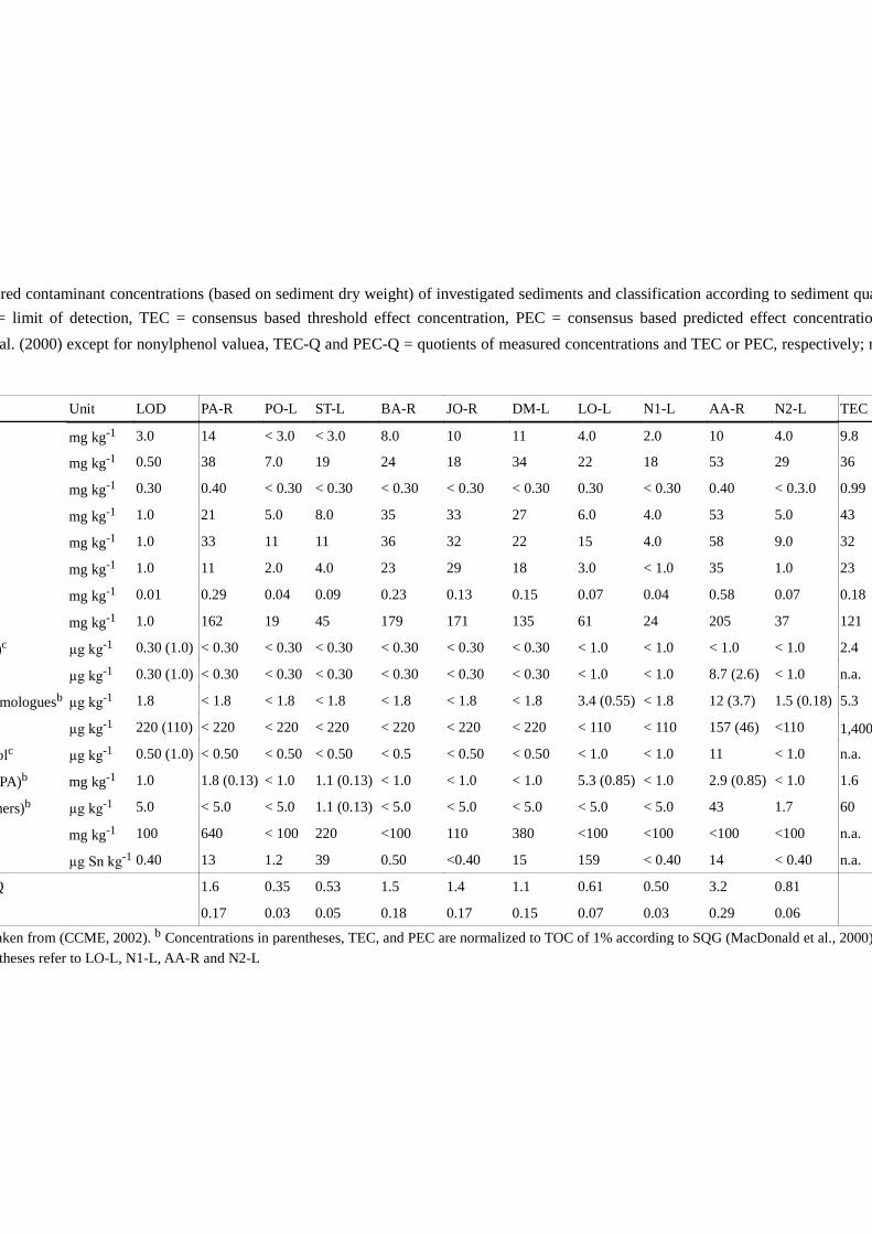
Measured contaminant concentrations (based on sediment dry weight) of investigated sediments and classification according to sediment quality guidelines(SQG); LOD = limit of detection, TEC = consensus based threshold effect concentration, PEC = consensus based predicted effect concentration according toMacDonald et al. (2000) except for nonylphenol valuea, TEC-Q and PEC-Q = quotients of measured concentrations and TEC or PEC, respectively; n.a. = value not
Unit LOD PA-R PO-L ST-L BA-R JO-R DM-L LO-L N1-L AA-R N2-L TEC
mg kg-1 3.0 14 < 3.0 < 3.0 8.0 10 11 4.0 2.0 10 4.0 9.8
mg kg-1 0.50 38 7.0 19 24 18 34 22 18 53 29 36
mg kg-1 0.30 0.40 < 0.30 < 0.30 < 0.30 < 0.30 < 0.30 0.30 < 0.30 0.40 < 0.3.0 0.99
mg kg-1 1.0 21 5.0 8.0 35 33 27 6.0 4.0 53 5.0 43
mg kg-1 1.0 33 11 11 36 32 22 15 4.0 58 9.0 32
mg kg-1 1.0 11 2.0 4.0 23 29 18 3.0 < 1.0 35 1.0 23
mg kg-1 0.01 0.29 0.04 0.09 0.23 0.13 0.15 0.07 0.04 0.58 0.07 0.18
mg kg-1 1.0 162 19 45 179 171 135 61 24 205 37 121
-)c µg kg-1 0.30 (1.0) < 0.30 < 0.30 < 0.30 < 0.30 < 0.30 < 0.30 < 1.0 < 1.0 < 1.0 < 1.0 2.4
µg kg-1 0.30 (1.0) < 0.30 < 0.30 < 0.30 < 0.30 < 0.30 < 0.30 < 1.0 < 1.0 8.7 (2.6) < 1.0 n.a.
DDT and homologuesb µg kg-1 1.8 < 1.8 < 1.8 < 1.8 < 1.8 < 1.8 < 1.8 3.4 (0.55) < 1.8 12 (3.7) 1.5 (0.18) 5.3
µg kg-1 220 (110) < 220 < 220 < 220 < 220 < 220 < 220 < 110 < 110 157 (46) <110 1,400
Octa-chlorostyrolc µg kg-1 0.50 (1.0) < 0.50 < 0.50 < 0.50 < 0.5 < 0.50 < 0.50 < 1.0 < 1.0 11 < 1.0 n.a.
PAH (16 US EPA)b mg kg-1 1.0 1.8 (0.13) < 1.0 1.1 (0.13) < 1.0 < 1.0 < 1.0 5.3 (0.85) < 1.0 2.9 (0.85) < 1.0 1.6
PCB (7 congeners)b µg kg-1 5.0 < 5.0 < 5.0 1.1 (0.13) < 5.0 < 5.0 < 5.0 < 5.0 < 5.0 43 1.7 60
mg kg-1 100 640 < 100 220 <100 110 380 <100 <100 <100 <100 n.a.
µg Sn kg-1 0.40 13 1.2 39 0.50 <0.40 15 159 < 0.40 14 < 0.40 n.a.
Maximal TEC-Q 1.6 0.35 0.53 1.5 1.4 1.1 0.61 0.50 3.2 0.81
0.17 0.03 0.05 0.18 0.17 0.15 0.07 0.03 0.29 0.06
Interim SQG taken from (CCME, 2002). b Concentrations in parentheses, TEC, and PEC are normalized to TOC of 1% according to SQG (MacDonald et al., 2000) LODs in parentheses refer to LO-L, N1-L, AA-R and N2-L

Variability of sediment-contact tests 273
Fig 2 Cluster analysis (a) and principal component analysis (PCA; b) for the 10 native sediments based on geochemical properties and contaminant concentrations; for definitions of abbreviations, see Tab. 1, Tab. 3 and Tab. 4; MO = mineral oil; OCS = Octa-chlorostyrol
Table 5 Variables that significantly correlated with factors of PCA (multivariate correlation; r > 0.95; r < -0.95); PC = principal component; for definitions of abbreviations see Table 3; OCS = Octa-chlorostyrol
PCs Eigenvalues Significant variables
r > 0.95 r < -0.95
PC 1 0.409 × Fine particles (clay, silt, Al, Li, Fe)× Heavy metals (Cu, Cr, Ni, Hg, Zn)
× Coarse particles (sand)
PC 2 0.176 × Organic matter (LOI, TOC, N, P, S)× Mineral oil
× Dry weight
PC 3 0.141 × Chloroorganic chemicals (DDT, HCB, PCB, OCS); Cd
× N, Li
× Mineral oil× Silt× S
PC 4 0.100 × TN, DOC (Pore water)× Particles 63-200 µm (sa63)
× TOC

Variability of sediment-contact tests 274
10.4.2 Response to sediment samples
The toxicity endpoints of the various sediment-contact tests varied considerably among the different investigated sediments, with several of the sediments differing significantly from the control (Table 6; p < 0.05, one-way ANOVA, post-hoc Dunnett). Only plant growth in any native sediment was not significantly different from that in the control (Table 6; p > 0.05, one-way ANOVA). Compared to the respective control sediment, the various toxicity endpoints reached relative values of 32–130% (bacterial enzyme activity), 1–106% (yeast fermentation), 70–102% (nematode growth), 43–230% (nematode reproduction), 69–155% (oligochaete reproduction), 83–123% (plant growth rate), and 0–105% (fish egg survival) (Fig. 3). For fish egg survival, one extreme value (0% survival) was observed for PA-R.
The response of the various test organisms was compared with the principal components of the PCA, by testing a linear model, to explain the variability of the toxicity endpoints with the sediment properties. The results show that nematode growth and reproduction, as well as plant growth were significantly influenced by variables that were correlated with PC1, whereas nematode growth and reproduction showed a negative and plant growth a positive coefficient (Table 7). Nematodes might have been negatively influenced by the high contents of fine sediments, however also by slightly elevated concentrations of metals. Comparisons of the response of the organisms (Table 6) with the clusters in Fig. 2a clearly showed that both the highest values for nematode growth and reproduction were found in samples of cluster 3 (left side of the PCA plot: PO-L, ST-L, LO-L, N1-L, N2-L; Fig. 2b). The plants, however, preferred the fine sediments, despite their slightly elevated contamination. M. aquaticum showed the highest growth rates in sediments belonging to clusters 1 and 2 (AA-R, BA-R, JO-R, DM-L; Fig. 2a; Tab. 6) positioned on the right side of the PCA plot (Fig. 2b). Additionally, plant growth was significantly related to PC4 (Tab. 7; positive coefficient), which is positively correlated with dissolved carbon and nitrogen concentrations in the pore water (Tab. 5). According to the linear model, fish egg survival was significantly influenced by variables that correlated with PC2, with a negative coefficient (Table 8). This was perhaps due to PC2-related factors, i.e. the relatively high contents of organic matter, elevated concentrations of mineral oil, and low dry weights (Table 5). The linear model did not reveal significant correlations of PC3 with the toxicity endpoints. Thus, variables that were correlated with PC3 (e.g. chloroorganic chemicals) did not significantly influence the organisms (Table 7). Neither for yeast fermentation, nor for oligochaete reproduction did the linear model reveal a significant relation to any of the principal components (Table 7).

Response of toxicity endpoints of the various sediment-contact tests to the investigated native sediments (see Table 1); C = artificial control sediment (C1 =test 1; C2 = test 2); * and + indicate significant lower and higher values compared to the control, respectively (p < 0.05; one-way ANOVA, post-hoc
Bacteria Yeast Nematodes Oligochaetes Plants
Enzyme activity (fluorescence min-1)
Fermentation (ml CO2 h-1)
Growth (µm)
Reproduction(offspring per test organism)
Reproduction(total number of organisms)
Growth rate
SD Mean SD Mean SD Mean SD Mean SD Mean SD
4.4 51.5 0.7 1313.4 109.8 57.0 17.6 31.5 5.3 0.080 0.009
113.7 * 2.9 7.3 * 3.1 1083.1 * 56.7 50.7 11.3 38.8 9.4 0.081 0.003
114.0 * 6.5 17.8 * 21.2 1162.0 * 80.8 53.2 8.0 37.0 3.8 0.086 0.013
2.0 9.3 * 12.7 1192.5 25.6 50.5 3.0 48.8 + 1.9 0.077 0.002
0.6 25.3 2.5 917.1 * 64.1 24.5 * 8.0 38.0 5.5 0.099 0.011
4.6 50.0 6.7 1000.6 * 90.4 32.0 10.6 31.3 3.9 0.098 0.013
112.2 * 8.4 0.7 * 1.2 1067.3 * 57.4 46.9 11.1 40.3 + 2.9 0.095 0.004
2.3 35.7 2.2 0.100 0.006
3.3 63.0 4.6 1255.9 84.9 72.5 15.9 38.7 4.8 0.108 0.009
172.4 + 8.3 54.7 12.2 1273.4 73.8 93.0 + 30.1 24.7 * 3.8 0.088 0.006
12.3 45.7 3.1 956.0 * 81.4 33.4 * 5.0 40.2 5.8 0.118 0.003
190.9 + 3.8 32.0 7.6 1341.6 57.3 130.8 + 23.0 33.0 4.3 0.083 0.003

Variability of sediment-contact tests 276
Fig 3 Response of the various sediment contact tests to the 10 investigated native sediments as percentage of the respective control; for definitions of abbreviations, see Table 1
10.4.3 Variation coefficients and toxicity thresholds
For calculations of variation coefficients, MDD, and MTI, and in the subsequent definition of the toxicity thresholds only those samples were considered in which pollution effects could be excluded. Therefore, the moderately polluted sample AA-R was omitted. For fish egg survival also PA-R was omitted, because due to the significant correlation with PC2, a toxic effect of mineral oil could not be excluded as a cause for the strong effect in this sample.
Bacterial enzyme activity showed a very low test-inherent variation either in the artificial control sediment or in the native sediments, with a CVi < 5% (Table 8). The yeast contact test showed also a very low CVi in the artificial control (CViC = 1.4%), whereas in the native sediments the CVi was quite high with an average value of 61% (Table 8). Nematode growth varied marginally between the replicates of the different treatments: this was the case for the artificial control sediment as well as for the native sediments, since under either condition the CVi did not exceed 10% (Table 8). Nematode reproduction showed a higher test inherent variation: here, the CVi was higher in the artificial control sediment (CViC = 31%) than in most native sediments (mean CViS = 22.7%) (Table 8). Oligochaete reproduction was characterized by a maximal CViC of 17% for the artificial control sediment and an average CViS of 13% for the native sediments (Table 8). Plant growth showed a lower test-inherent variation, with a maximal CViC of 11% and a mean CViS of 8% (Tab. 8). For the fish egg contact test the CVi was not calculated because only one replicate was set up.

Linear models for organism response (% of control) against principle components (lm (formula = “organism response” ~ PC1+PC2+PC3+PC4)); AIC =akaike information criterion (criterion for model selection); significant coefficients and overall models, p(F), are printed bold (alpha = 0.05); Bac = bacterial enzymeactivity; Yeast = yeast fermentation; Nema-G: nematode growth; Nema-R: nematode reproduction; Oligo: oligochaete reproduction; Plant: plant growth; Fish: fish
Intercept PC1 PC2 PC3 PC4
coeff. p (interc.) coeff. p (coeff.) coeff. p (coeff.) coeff. p (coeff.) coeff. p (coeff.) p(F)
0.56 0.000 -0.13 0.061 0.04 0.565 0.14 0.058 0.081
0.49 0.004 -0.02 0.827 -0.14 0.239 0.15 0.205 0.370
0.84 0.000 -0.09 0.002 0.02 0.323 0.03 0.148 0.0080.45 0.000 -0.16 0.033 0.04 0.500 0.07 0.251 0.094 0.141 0.104
0.73 0.000 0.04 0.422 0.422
0.87 0.000 0.08 0.004 0.03 0.167 0.03 0.126 0.059 0.013 0.011
0.85 0.000 -0.05 0.437 -0.23 0.004 0.012

Relevant characteristics of all sediment contact tests: CVic = coefficient of test inherent variation of control sediment; CVirs = mean CVi of reference
sediments; T1/T2 = Test 1/Test 2; Min = minimal value; Max = maximal value; MDD = mean minimal detectable difference (one way); CVs = coefficient ofsediment variation; MTI = maximal tolerable inhibition; n.d. = not determined
Bacteria Yeast Nematodes Oligochaetes Plants Fish eggs
Test inherent criteria Enzyme activity Fermentation Growth Reproduction Reproduction Growth rate Survival
No. of samples used for n = 9 n = 9 n = 9 n = 9 n = 9 n = 9 n = 8
Excluded samples AA-R AA-R AA-R AA-R AA-R AA-R AA-R, PA-R
(T1/T2) 2.4/1.6 1.4 8.4 31.0 16.9/6.1 11.2/10.0 n.d.
(Min; Max) 3.5 (0.6; 7.9) 60.8 (7.3; 173.2) 5.8 (2.1; 9.0) 22.7 (5.8; 33.1) 12.6 (4.0; 24.2) 7.8 (2.8; 15.0) n.d.
% MDD (Min; Max) 3.8 (1.9; 7.2) 19.0 (3.8; 44.0) 6.5 (5.3; 7.5) 27.8 (19.1; 45.9) 14.7 (9.1; 25.4) 14.4 (9.2; 20.1) n.d.
Response to sediments Relative value (% of control)
31.9 1.3 69.8 43.1 69.2 83.2 86.1
130.2 106.1 102.1 229.6 155.0 123.0 105.3
73.3 52.7 87.1 108.0 112.7 105.2 96.0
Standard deviation 35.1 38.9 10.5 57.8 24.3 14.5 6.1
47.8 73.8 12.1 53.5 21.6 13.7 6.4
61.8 86.2 23.4 49.7 11.6 9.3 10.1
Toxicity threshold (% Inhibition to control) 60 n.d. 25 50 25 20 20

Variability of sediment-contact tests 279
The response of plant growth, nematode growth, oligochaete reproduction and fish egg survival to the native sediments showed the least variability, with a coefficient of variance (CVs) of < 25%, followed by bacterial enzyme activity (48%), nematode reproduction (54%) and yeast fermentation (74%) (Table 8).
The appropriate toxicity threshold for each test system was determined based on the maximal MDD and the MTI, whereas always the higher value of the two coefficients was used. The various toxicity endpoints showed maximal MDD and MTI values ranging from 7 to 46%, and 9 to 86%, respectively (Tab. 8; Fig. 4). Only for oligochaete reproduction and plant growth, the MDD was higher than the MTI, and thus was taken as basis for the toxicity threshold. For bacterial enzyme activity, as well as nematode growth and reproduction, the MTI was the decisive coefficient. The respective MDD or MTI value was then rounded to the next multiple of five, to obtain feasible toxicity thresholds. As a result, the toxicity thresholds were set to 20% inhibition for plant growth, 25% inhibition for nematode growth and oligochaete reproduction, 50% inhibition for nematode reproduction, and 60% inhibition for bacterial enzyme activity. For yeast fermentation, no toxicity threshold was defined due to the high variability of the test system (MTI > 80%). In spite of a MTI of 10 for fish egg survival, the toxicity threshold was set to 20% (following the validity criterion), as no information on the test inherent variability was available. Statistical analyses showed that the power for detecting significant differences at the various toxicity thresholds were sufficiently high (> 0.8; SigmaStat, SPSS Inc, Chicago, IL, USA).
Fig 4 Means (error bars = standard deviation) of %inhibition (compared to control sediments) of toxicity endpoints in native sediments (n = 9; fish: n = 8; see Table 8); dotted lines mark the MTI (maximal tolerable inhibition); Bac = bacterial enzyme activity; Yeast = yeast fermentation; Nema-G: nematode growth; Nema-R: nematode reproduction; Oligo: oligochaete reproduction; Plant: plant growth; Fish: fish-egg survival

Variability of sediment-contact tests 280
10.5 Discussion
10.5.1 Influence of sediment properties
The response of the test organisms to the different native sediments with low to moderate contamination varied considerably. The sediment coefficients of variance were markedly lower for nematode growth, oligochaete reproduction, plant growth and fish-egg survival (CVS of 12, 22, 13, and 6.4%, respectively), than for bacteria, yeast and nematode reproduction (CVS of 48, 74 and 54, respectively). For nematode growth, the variability was comparable to that determined in a study with freshwater sediments, in which C. elegans was exposed to 26 low-level-polluted sediments (CVS = 10.1; Höss et al., 1999). In reference soils, C. elegans had a lower variability with a CVSoil of 4% for growth and 31% for reproduction (Höss et al., 2009). The variability of L. variegatus reproduction in the native sediments was comparable to the values reported for Tubifex tubifex. In that study, Reynoldson et al. (2002) investigated the variability of sublethal toxicity endpoints for T. tubifex exposed to 105 reference sediments: CVS of 12% and 34% were determined for the number of cocoons and offspring per adult, respectively. The variability of Eisenia andrei, a soil oligochaete, in reference soils, however, was found to be considerably higher (CVSoil of 44%; Römbke et al., 2006). In the same study, a CVSoil of > 50% was determined for the growth of turnip rape (Brassica rapa) in reference soils (Römbke et al., 2006), which is considerably higher than the variability of the growth rate of Myriophyllum in the present study (Table 8).
A linear model suggested that grain-size distribution, organic matter and anthropogenic pollution might have influenced the response of the various organisms to the native sediments (Tables 5 and 7). As the study parameters were strongly intercorrelated, the influence on the organisms could not be unequivocally attributed to individual properties of the sediments. However, due to the relatively low contaminant concentrations in most of the sediments, toxic effects on the organisms were not likely. Indeed, with the exception of AA-R, all sediments showed contaminant concentrations that were below or close to threshold values that are considered as not harmful for benthic invertebrates (threshold effect concentrations, TEC; Table 4; MacDonald et al., 2000). However, in the AA-R sample, by contrast, part of the effect on bacteria (36% inhibition compared to control) and nematodes (growth: 27%, reproduction: 41% inhibition compared to control), might have been caused by an elevated Hg concentration (Table 4). For bacteria, EC50 values for Hg in water of 0.3 mg l -1 and 0.9 mg l-1 were reported for Pseudomonas fluorescens and Vibrio fischeri, respectively (Brown et al., 1996; McCloskey et al., 1996). Although, in sediments bioavailability of Hg is assumed to be lower than in water, effects of Hg cannot be excluded at a concentration of 0.6 mg kg-1 (AA-R; Tab. 4). For C. elegans, the LOEC for Hg in water was found to be 2 mg l-1 (Donkin and Williams, 1995). Thus, effects caused by Hg in AA-R are not likely. Regarding the strong effect of PA-R on fish egg survival, an effect of mineral oil that was

Variability of sediment-contact tests 281
found in comparably high concentrations in this sample (Tab. 4), cannot be excluded. In water, for mineral oil a LC50 of 100 mg l-1 was reported for rainbow trout and blue gill (Office of Pesticide Programs, 2000).
In the low-level-contaminated sediments, it was more likely that organisms were influenced by particle size distribution and the content of organic matter. Similar results were reported in a previous study involving C. elegans, in which growth was found to be negatively correlated with clay content (Höss et al., 1999), perhaps due to lower food availability for the nematodes. While the particles <2 µm can be swallowed by the nematodes together with the food bacteria, they are less nutritious. L. variegatus is a sediment ingester that can take up larger particles than C. elegans. Although choice experiments with freshwater oligochaetes showed that the worms (Tubifex, Limnodrilus, Stylodrilus) avoided coarse sands and headed for fine, muddy sediments (Wachs, 1967), grain size did not influence L. variegatus in the present study. For plants water uptake depends on the water capacity of the substrate. In finer substrates, for example silty sediments, more water is capillary bound and thus available for the plant. Furthermore, nutrients (minerals, e.g. Mg) are usually bound to fine-grained sediment particles. Therefore, the observed positive correlation of minerals and grain size reflect the better supply of water and nutrients in fine-grained sediments (Barko and Smart, 1986). This possibility is supported by positive correlations of plant growth to nitrogen and DOC concentrations in the pore water (PC4; Tables 4 and 7). Fish egg survival, by contrast, was not influenced by grain size distribution but was related to the content of organic matter. It was previously shown that D. rerio is affected by organic matter, such as humic substances (Cazenave et al., 2006). For the bacteria no significant correlation to the measured geochemical sediment properties were found (Table 7). Instead, the relatively high variability of the enzyme activity observed in the bacteria contact test might have been due to other factors, not measured in this study. It might also be at least partly explained by the varying quenching effects of the different native sediments. A fluorescent dye can lose energy without emitting light during contact with other substances, resulting in a reduced fluorescent signal. Therefore, a calibration method was developed and tested and is described in detail in (Heise and Ahlf, 2005). In the yeast contact test the reasons behind the strong inhibition excerted by some of the sediments remain unclear, as it could not be explained by the measured sediment properties (Table 7).
10.5.2 Toxicity thresholds
The need for test-specific thresholds or limits that set the boundary between reference conditions or natural variability and toxic effects has already been stated in other studies (Hunt et al., 2001; Reynoldson et al., 2002; Thursby et al., 1997). Based on the variability of lethal and sublethal toxicity endpoints in reference sediments, Reynoldson et al. (2002) established three categories of responses to toxicity for four benthic invertebrate species (Chironomus riparius, Hyalella azteca, Hexagenia spp., Tubifex tubifex): not toxic, potentially

Variability of sediment-contact tests 282
toxic and toxic. Similar to the approach of the present study, the delineations for the three categories were developed from the standard statistical parameters of mean and standard deviation (mean ± SD) of an endpoint measured in reference sediments. In contrast to the present study, however, Reynoldson et al. (2002) inserted a “buffer zone” of potential toxicity between the not toxic and toxic categories, instead of defining sharp threshold. A comparable approach was used by Hunt et al. (2001) who set up individual “sediment toxicity tolerance limits” for the survival of marine amphipods and the development sea urchin embryo/larval, to determine elevated toxicity relative to reference conditions. Thursby et al. (1997) presented a toxicity threshold approach that considers the entire test system by using a historical database of minimal significant differences (MSD), rather than searching only for a statistically significant difference between a sample and the control in a single test run. These toxicity thresholds were applied to a large data set and were shown to be useful for interpreting sediment toxicity data (Phillips et al., 2001). Samples were regarded as toxic, if both statistical significance and detectable significance (below the MSD derived threshold) indicated a toxic effect.
The present study combines these approaches and considers the statistical power of a test system as well as the influence on the organisms of natural sediment properties that increase the background noise, from which a toxic effect has to be distinguished. For toxicity endpoints showing relatively low variability within treatments (between replicates; low CVi and MDD) and between sediments (low CVs and MTI), such as plant growth, oligochaete reproduction and nematode growth, the toxicity thresholds were set quite low, at 20 and 25% inhibition (Table 5). The high toxicity threshold of the bacterial enzyme activity, despite a very low MDD of <10%, derived from the high variability between the various sediments (CVs: 48%) and the fact that the control enzyme activity in quartz sand higher than in natural sediments. This combination led to the high MTI of 62% and a toxicity threshold of 60%. The use of an alternative, more realistic, artificial control sediment might help to get a lower MTI and thus also a lower toxicity threshold for the bacteria contact test. The yeast contact test showed an exceptionally high variability between the various native sediments that could not be explained by the measured sediment properties or contaminant concentrations. An MTI close to 90% did not allow the definition of a reasonable toxicity threshold that is able to detect a contaminant effect out of the large background noise.
The MTI as the basis, albeit not the only one, for the toxicity threshold, generally accounts for differences in an organism’s response to an artificial sediment vs. natural sediments. Formulated, artificial sediments are often designed to yield optimal performance of the test organism but this might also exceed the performance in natural reference sediments (Fig. 3; Kemble et al., 1999). Therefore, in addition to the control sediment, the use of a reference sediment that is similar to the native test sediment, but free of contamination is always recommended (ASTM, 1990; US EPA, 1998). However, as a suitable reference site is not

Variability of sediment-contact tests 283
always available, a reliable toxicity threshold, established on the basis of a permanently available control sediment, is crucial for accurate interpretation of the toxicity of native sediments.
10.5.3 Battery of sediment contact tests
With the exception of the yeast contact test, all studied contact tests appear to be promising tools for sediment toxicity assessments. As the organisms tolerated different types of freshwater sediments from lakes and rivers with a considerable range of geochemical properties, it can be broadly applied for assessing sediment toxicity. Moreover, a battery of sediment-contact tests with organisms from different organizational and trophic levels, as proposed in the SeKT-project (Feiler et al., 2005), has several advantages over a test battery using only macro-invertebrates (ASTM, 2005). First, different exposure routes can be considered. A. globiformis (bacteria), M. aquaticum (plant), and eggs of D. rerio (fish) are exposed to sediment-associated contaminants mainly via the dissolved phase in pore water as well as by direct contact with contaminant-loaded particles. Although the adult zebrafish might not come into contact with contaminated sediments, fish embryos in their eggs very likely do, as cyprinids commonly spawn on finely grained sediment. Meiobenthic nematodes, represented by C. elegans, are relatively small and live in the interstitial space. Thus, the pore water, containing all dissolved and colloidal substances, and fine particles are relevant for the nematode’s uptake of pollutants (Höss et al., 2001). L. variegatus takes up the sediment particles with all bound contaminants which become newly available in the gut of these oligochaetes (e.g. Leppänen and Kukkonen, 1998). Second, the use of organisms from different organizational and trophic levels, and thus with a broad variety of receptors for environmental chemicals, allows the assessment of chemicals with different modes of action. Third, in the proposed battery, short-term (few hours to few days: bacteria, nematodes, fish eggs) and longer-term (days to weeks: plants and oligochaetes) tests are included, which allows rapid screening but also the evaluation of long-term effects.
10.6 Conclusions
This study investigated the response of several sediment-contact tests on freshwater sediments with both low to moderate contamination considerable variability in terms of their geochemical properties. The variability of the test systems that partly could be explained by individual sediment properties was considered as natural variability and used as a basis for defining toxicity thresholds. Only one test system, the yeast contact test, showed an exceptionally high variability in sediments with low-level contamination and thus cannot be recommended for testing native sediments. In all other contact tests, the variability between the lowly contaminated sediments was low enough to define reasonable toxicity thresholds. Thus, the tests fulfilled an important prerequisite for assessing the toxicity of freshwater sediments with a broad range of geochemical properties. However, the presented toxicity

Variability of sediment-contact tests 284
threshold should not be regarded as “set in stone”. With a growing data base for lowly contaminated sediments, it probably will be necessary to adjust the toxicity thresholds for these contact tests. Overall, an ecologically relevant battery of sediment contact tests can be recommended in which test organisms of different trophic levels and with various exposure routes are used: bacteria, nematodes, oligochaetes, plants and fish eggs. When carried out with reasonable toxicity thresholds, these test systems offer a pragmatic approach to sediment risk assessment.
10.7 Acknowledgements
This study was funded by the German Federal Ministry of Education and Research (BMBF; SeKT project numbers: 02WU0598 to 02WU0604). We acknowledge the valuable input of Hans-Jürgen Pluta (German Federal Environment Agency, UBA) and Ulrich Förstner (Technical University Hamburg-Harburg), who accompanied the SeKT project as members of the scientific advisory board. The gifts of Caenorhabditis elegans and Escherichia coli from the Caenorhabditis Genetic Center (Theresa Stiernagle) are gratefully acknowledged. Many thanks to Philipp Egeler for sharing his knowledge of oligochaetes. We also want to thank Wendy Ran for revising the English language. Moreover, we thank three anonymous reviewers, whose comments considerably helped to improve the quality of this paper.
10.8 References
Ahlf, W., Heise, S., 2005. Sediment toxicity assessment - Rationale for effect classes - Dedicated to Prof. Dr. Ulrich Forstner on his 65th birthday. Journal of Soils and Sediments 5, 16-20.
Ankley, G.T., Benoit, D.A., Balough, J.C., Reynoldson, T.B., Day, K.E., Hoke, R.A., 1994. Evaluation of potential confounding factors in sediment toxicity tests with three freshwater invertebrates. Environmental Toxicology and Chemistry 13, 627-635.
Ankley, G.T., Benoit, D.A., Hoke, R.A., Leonard, E.N., West, C.W., Phipps, G.L., Mattson, V.R., Anderson, L.A., 1993. Development and evaluation of test methods for benthic invertebrates and sediments: Effects of flow rate and feeding on water quality and exposure conditions. Archives of Environmental Contamination and Toxicology 25, 12-19.
ASTM, 1990. Standard guide for conducting sediment toxicity tests with freshwater invertebrates. American Society for Testing and Materials, Westconshohocken, PA, USA.
ASTM, 2005. Standard test method for measuring the toxicity of sediment-associated contaminants with freshwater invertebrates. American Society for Testing and Materials, West Conshohocken, PA, USA.
Barko, J.W., Smart, R.M., 1986. Sediment-related mechanisms of growth limitation in submersed macrophytes. Ecology 67, 1328-1340.
Brown, J.S., Rattray, E.A.S., Paton, G.I., Reid, G., Caffoor, I., Killham, K., 1996. Comparative assessment of the toxicity of a papermill effluent by respirometry and a luminescence-based bacterial assay. Chemosphere 32, 1553-1561.
Variability of sediment-contact tests 285
Cazenave, J., De los Angeles Bistoni, M., Zwirnmann, E., Wunderlin, D.A., Wiegand, C., 2006. Attenuating effects of natural organic matter on microcystin toxicity in zebra fish (Danio rerio) embryos—benefits and costs of microcystin detoxication. Environmental Toxicology 21, 22-32.
CCME, 2002. Canadian Sediment Quality Guidelines for the Protection of Aquatic Life: Nonylphenol and Its Ethoxylates. Canadian Council of Ministers of the Environment.
Chambers, J.M., Hastie, T.J., 1992. Statistical Models in S. Wadsworth & Brooks/Cole, Pacific Grove, CA, USA.
Chapman, P.M., Anderson, J., 2005. A decision-making framework for sediment contamination. Integrated Environmental Assessment and Management 1, 163-173.
Crocker, F.H., Fredrickson, J.K., White, D.C., Ringelberg, D.B., Balkwill, D.L., 2000. Phylogenetic and physiological diversity of Arthrobacter strains isolated from unconsolidated subsurface sediments. Microbiology 146, 1295-1310.
De Zwart, D., Posthuma, L., Gevery, M., Von der Ohe, P.C., De Deckere, E., 2009. Diagnosis of ecosystem impairment in a multiple-stress context—How to formulate effective river basin management plans. Integrated Environmental Assessment and Management 5, 38-49.
DeWitt, T.H., Ditsworth, G.R., Swartz, R.C., 1988. Effects of natural sediment features on survival of the phoxocephalid amphipod, Rhepoxynius abronius. Marine Environmental Research 25, 99-124.
DeWitt, T.H., Swartz, R.C., Lamberson, J.O., 1989. Measuring the acute toxicity of estuarine sediments. Environmental Toxicology and Chemistry 8, 1035-1048.
Donkin, S.G., Williams, P.L., 1995. Influence of developmental stage, salts and food presence on various end points using Caenorhabditis elegans for aquatic toxicity testing. Environmental Toxicology and Chemistry 14, 2139-2147.
European Community, 2000. Directive 2000/60/EC of the European Parliament and the council of 23 October 2000 establishing a framework for Community action in the field of water policy. Official Journal of the European Community L 327, 1-73.
Feiler, U., Kirchesch, I., Heininger, P., 2004. A new plant bioassay for aquatic sediments. Journal of Soils and Sediments 4, 261-266.
Feiler, U., Ahlf, W., Höss, S., Hollert, H., Neumann-Hensel, H., Meller, M., Weber, J., Heininger, P., 2005. The SeKT Joint Research Project: Definition of reference conditions, control sediments and toxicity thresholds for limnic sediment contact tests. Environmental Science and Pollution Research 12, 257-258.
Förstner, U., 2002. Sediments and the European Water Framework Directive. Journal of Soils and Sediments 2, 54.
Heise, S., Ahlf, W., 2005. A new microbial contact assay for marine sediments. Journal of Soils and Sediments 5, 9-15.
Hollert, H., Keiter, S., König, N., Rudolf, M., Braunbeck, T., 2003. A new sediment contact assay to assess particle-bound pollutants using zebrafish (Danio rerio) embryos. Journal of Soils and Sediments 3, 197-207.
Höss, S., Haitzer, M., Traunspurger, W., Steinberg, C.E.W., 1999. Growth and fertility of Caenorhabditis elegans (Nematoda) in unpolluted freshwater sediments - response to particle size distribution and organic content. Environmental Toxicology and Chemistry 18, 2921-2925.

Variability of sediment-contact tests 286
Höss, S., Henschel, T., Haitzer, M., Traunspurger, W., Steinberg, C., 2001. Toxicity of cadmium to Caenorhabditis elegans (Nematoda) in whole sediment and porewater - the ambiguous role of organic matter. Environmental Toxicology and Chemistry 20, 2794-2801.
Höss, S., Jänsch, S., Junker, T., Moser, T., Römbke, J., 2009. Assessing the toxicity of contaminated soils using the nematode Caenorhabditis elegans as test organism. Ecotoxicology and Environmental Safety 72, 1811-1818.
Hotelling, H., 1933. Analysis of a complex of statistical variables into principal components. The Journal of Educational Psychology 24, 417-441.
Hunt, J.W., Anderson, B.S., Phillips, B.M., Newman, J., Tjeerdema, R., Fairey, R., Puckett, H.M., Stephenson, M., Smith, R.W., Wilson, C.J., Taberski, K.M., 2001. Evaluation and use of sediment toxicity sites for statistical comparisons in regional assessments. Environmental Toxicology and Chemistry 20, 1266-1275.
Hussner, A., 2009. Growth and photosynthesis of four invasive aquatic plant species in Europe. Weed Research 49, 506-515.
Ingersoll, C.G., Dillon, T., Biddinger, G.R., Ingersoll, C.G., Dillon, T., Biddinger, G.R., 1997. Ecological Risk Assessment of Contaminated Sediments. SETAC Press, Pensacola, Fl, USA.
Ingersoll, C.G., Ankley, G.T., Benoit, D.A., Brunson, E.L., Burton, G.A., Dwyer, F.J., Hoke, R.A., Landrum, P.F., Norberg-King, T.J., Winger, P.V., 1995. Toxicity and bioaccumulation of sediment-associated contaminants using freshwater invertebrates: A review of methods and applications. Environmental Toxicology and Chemistry 14, 1885-1894.
ISO, 2009a. Water quality - Determination of the toxic effect of sediment and soil samples on growth, fertility and reproduction of Caenorhabditis elegans (Nematoda). ISO/DIS 10872, International Organization for Standardization, Geneva, Switzerland.
ISO, 2009b. Water quality — Determination of the inhibition of dehydrogenase activity of Arthrobacter globiformis — Solid contact test using the redox dye resazurine. ISO/DIS 10871, International Organization for Standardization, Geneva, Switzerland.
Keiter, S., Rastall, A., Kosmehl, T., Wurm, K., Erdinger, L., Braunbeck, T., Hollert, H., 2006. Ecotoxicological assessment of sediment, suspended matter and water samples in the upper Danube River - A pilot study in search for the causes for the decline of fish catches. Environmental Science and Pollution Research 13, 308-319.
Kemble, N.E., Dwyer, F.J., Ingersoll, C.G., Dawson, T.D., Norberg-King, T.J., 1999. Tolerance of freshwater test organismes to formulated sediments for use as control materials in whole-sediment toxicity tests. Environmental Toxicology and Chemistry 18, 222-230.
Lahr, J., Maas-Diepeveen, J.L., Stuijfzand, S.C., Leonards, P.E.G., Drüke, J.M., Lücker, S., Espeldoorn, A., Kerkum, L.C.M., van Stee, L.L.P., Hendriks, A.J., 2003. Responses in sediment bioassays used in the Netherlands: Can observed toxicity be explained by routinely monitored priority pollutants? Water Research 37, 1691-1710.
Leppänen, M.T., Kukkonen, J., 1998. Relative importance of ingested sediment and pore water as bioaccumulation routes for pyrene to oligochaete (Lumbriculus variegatus, Müller). Environmental Science and Technology 32, 1503-1508.
MacDonald, D.D., Ingersoll, C.G., Berger, T.A., 2000. Development and Evaluation of Consensus-Based Sediment Quality Guidelines for Freshwater Ecosystems. Archives of Environmental Contamination and Toxicology 39, 20-31.

Variability of sediment-contact tests 287
McCloskey, J.T., Newman, M.C., Clark, S.B., 1996. Predicting the relative toxicity of metal ions using ion characteristics: microtox bioluminescence assay. Environmental Toxicology and Chemistry 15, 1730-1737.
McCune, B., Mefford, M.J., 1999. Multivariate Analysis of Ecological Data, PC-ORD, Version 4, Program Documentation. MjM Software Design, Oregon, USA.
Neumann-Hensel, H., Melbye, K., 2006. Optimisation of the solid-contact test with Artbrobacter globiformis. Journal of Soils and Sediments 6, 201-207.
Nipper, M.G., Roper, D.S., 1995. Growth of an amphipod and a bivalve in uncontaminated sediments: Implications for chronic toxicity assessments. Marine Pollution Bulletin 31, 424-430.
OECD, 2007. Sediment – water Lumbriculus toxicity test using spiked sediment. Organisation for Economic Cooperation and Development Paris, France.
Office of Pesticide Programs, 2000. Pesticide Ecotoxicity Database (Formerly: Environmental Effects Database (EEDB)). Environmental Fate and Effects Division, U.S.EPA, Washington, D.C.
Phillips, B.M., Hunt, J.W., Anderson, B.S., Puckett, H.M., Fairey, R., Wilson, C.J., Tjeerdema, R., 2001. Statistical significance of sediment toxicity test results: Threshold values derived by the detectable significance approach. Environmental Toxicology and Chemistry 20, 371-373.
Phipps, G.L., Ankley, G.T., Benoit, D.A., Mattson, V.R., 1993. Use of the aquatic oligochaete Lumbriculus variegatus for assessing the toxicity and bioaccumulation of sediment-associated contaminants. Environmental Toxicology and Chemistry 12, 269-279.
Power, E.A., Chapman, P.M., Burton, G.A., 1992. Assessing sediment quality. In: Burton, G.A. (Ed.), Sediment Toxicity Assessment. Lewis Publisher, Boca Raton, pp. 1-18.
R Development Core Team, 2009. R: A Language and Environment for Statistical Computing. R Foundation for Statistical Computing, Vienna, Austria.
Reynoldson, T.B., Thompson, S.P., Milani, D., 2002. Integrating multiple toxicological endpoints in a decision-making framework for contaminated sediments. Human and Ecological Risk Assessment 8, 1569-1584.
Römbke, J., Jänsch, S., Junker, T., Pohl, B., Scheffczyk, A., Schallnass, H.-J., 2006. Improvement of the applicability of ecotoxicological tests with earthworms, springtails, and plants for the assessment of metals in natural soils. Environmental Toxicology and Chemistry 25, 787.
Rönnpagel, K., Liss, W., Ahlf, W., 1995. Microbial bioassay to assess the toxicity of solid-associated contaminants. Ecotoxicology and Environmental Safety 31, 99-103.
Sakamoto, Y., Ishiguro, M., Kitagawa, G., 1986. Akaike Information Criterion Statistics. D. Reidel Publishing, Dordrecht, The Netherlands.
Sibley, P.K., Benoit, D.A., Ankley, G.T., 1998. Life cycle and behavioural assessment of the influence of substrate particle size on Chironomus tentans (Diptera: Chironomidae) in laboratory assays. Hydrobiologia 361, 1-9.
Stesevic, D., Feiler, U., Sundic, D., Mijovic, S., Erdinger, L., Seiler, T.B., Heininger, P., Hollert, H., 2007. Comparing growth development of Myriophyllum spp. in laboratory and field experiments for ecotoxicological testing. Journal of Soils and Sediments 7, 342-349.
Suedel, B.C., Rodgers, J.H., Jr., 1994. Responses of Hyalella azteca and Chironomus tentants to particle size distribution and organic matter content of formulated and natural freshwater sediments. Environmental Toxicology and Chemistry 13, 1639-1648.

Variability of sediment-contact tests 288
Suedel, B.C., Deaver, E., Rodgers, J.H., Jr., 1996. Formulated sediment as a reference and dilution sediment in definitive toxicity tests. Archives of Environmental Contamination and Toxicology 30, 47-52.
Swartz, R.C., DeBenm, W.A., Jones, J.K.P., Lamberson, J.O., Cole, F.A., Cardwell, R.D., Purdy, R., Bahner, R.C., 1985. Phoxocephalid amphipod bioassay for marine sediment toxicity. Aquatic Toxicology and Hazard Assessment STP 854 American Society for Testing and Materials. ASTM, Philadelphia, pp. 284-307.
Talwar, P.K., Jhingran, A.G., 1991. Inland fishes of India and adjacent countries. A. A. Balkema, Rotterdam.
Ter Braak, C.J.F., Šmilauer, P., 2002. CANOCO reference manual and CanoDraw for Windows user’s guide: Software for canonical community ordination (Version 4.5). Biometris, Wageningen, The Netherlands.
Thursby, G., Heltshe, J., Scott, K.J., 1997. Revised approach to toxicity test acceptability criteria using a statistical performance assessment. Environmental Toxicology and Chemistry 16, 1322-1329.
Traunspurger, W., Haitzer, M., Höss, S., Beier, S., Ahlf, W., Steinberg, C.E.W., 1997a. Ecotoxicological assessment of aquatic sediments with Caenorhabditis elegans (Nematoda) - A method for testing in liquid medium and whole sediment samples. Environmental Toxicology and Chemistry 16, 245-250.
Traunspurger, W., Haitzer, M., Hoss, S., Beier, S., Ahlf, W., Steinberg, C., 1997b. Ecotoxicological assessment of aquatic sediments with Caenorhabditis elegans (Nematoda) - A method for testing liquid medium and whole-sediment samples. Environ Toxicol Chem 16, 245 - 250.
US EPA, 1998. Evaluation of dredged material proposed for discharge in waters of the U.S. - Testing manual. EPA-823-B-98-004, U.S. Environmental Protection Agency, Washington, D.C.
Wachs, B., 1967. Die Oligochaeten-Fauna der Fließgewässer unter besonderer Berücksichtigung der Beziehung der Tubificiden-Besiedlung und dem Substrat. Archiv für Hydrobiologie 63, 310-386.
Weber, J., Kreutzmann, J., Plantikow, A., Pfitzner, S., Claus, E., Manz, W., Heininger, P., 2006. A novel particle contact assay with the yeast Saccharomyces cerevisiae for ecotoxicological assessment of freshwater sediments. Journal of Soils and Sediments 6, 84-91.
Zullini, A., 1988. The ecology of the Lambro river. Rivista di Idrobiologia 27, 39-58.

Chapter 11
Investigation on sensitivity measures and applicability of limnic sediment contact assays using formulated
and natural spiked sediment samples
T-B Seiler1, W Ahlf2, C Fahnenstich2, D Gilberg3, H Hollert1, S Höss4,5, K Melbye6, M Meller3,7, M Hammers-Wirtz8, P Heininger9, H Neumann-Hensel6, R Ottermanns10, H-T Ratte10, D Spira9, J Weber11, U Feiler9
1 Department of Ecosystem Analysis, Institute for Environmental Research (Biology 5), RWTH Aachen University, Worringerweg 1, 52074 Aachen, Germany
2 Institute of Environmental Technology and Energy Economics, Technical University Hamburg-Harburg, Eißendorfer Str. 40, 21071 Hamburg, Germany
3 ECT Oekotoxikologie, Böttgerstr. 2-14, 65439 Flörsheim, Germany
4 Ecossa, Giselastr. 6, 82319 Starnberg, Germany
5 Institute of Biodiversity – Network (IBN), Dreikronengasse 2, 93047 Regensburg, Germany
6 Dr. Fintelmann & Dr. Meyer, Mendelssohnstr. 15D, 22761 Hamburg, Germany
7 ecotox consult, Kreuzstr. 12, 67063 Ludwigshafen, Germany

8 gaiac, Research Institute for Ecosystem Analysis and Assessment, RWTH AachenUniversity, Worringerweg 1, 52056 Aachen, Germany
9 Federal Institute of Hydrology (BfG), Am Mainzer Tor 1, 56070 Koblenz, Germany
10 Chair for Environmental Biology and Chemodynamics, Institute for Environmental Research (Biology 5), RWTH Aachen University, Worringerweg 1, 52074 Aachen, Germany
11 Nordum GmbH & Co KG, Am Weidenbruch 22, 18126 Kessin/Rostock
To be submitted to J Soils Sediments

Sensitivity measures of sediment contact assays 291
11 Chapter 11
11.1 Abstract
The SeKT joint research project (German: SedimentKontaktTests) compared established sediment contact assays representing different trophic levels and several contaminant exposure routes within freshwater sediments. Test organisms were bacteria (Arthrobacter globiformis), fungi (Saccharomyces cerevisiae), nematodes (Caenorhabditis elegans), oligochaetes (Lumbriculus variegatus; toxicity and bioaccumulation), fish (Danio rerio), and higher plants (Myriophyllum aquaticum).
In working package 2 of the project, formulated and natural control sediments were spiked with either a mixture of four heavy metals or six organic pollutants and investigated using the different test systems. Sensitivities for the two types of contamination and slopes of the dose-response relations were determined for each bioassay and evaluated with respect to organism type, exposure pathway, sediment characteristics and type as well as sorption behaviour of the contaminants.
The plant assay was identified as a test system that can detect low concentration of both heavy metals and organic substances. Likewise, oligochaetes showed suitability for testing on either contamination, whereas nematodes turned out to be more sensitive for heavy metals. In contrast, fish embryos were exceptionally insensitive for heavy metals, but detected organic substances at very low concentrations. Bacteria gave low sensitivities in all experiments. Results for yeast cells were quite inconsistent, with highest sensitivities for heavy metals only in formulated sediments. Slopes of the response curves, which translate to ranges of concentrations that the test systems are able to discriminate, were steeper for formulated sediments than for natural samples. Plants, Bacteria and nematodes gave mostly flat slopes, i.e. good discrimination capabilities. In general, higher sensitivities and steeper slopes were recorded for formulated sediments, This can be accounted to higher availability of toxicants due to lack of sorption capacity, especially of naturally formed organic matter and biofilms.
Obtained data provide an overview over the individual properties and capabilities of each test system, and may guide researchers while selecting the appropriate biotest battery for a given investigation.
11.2 Introduction
Sediments play a key role for the ecological status of aquatic ecosystems. Being a highly complex matrix they form the habitat for an abundant biocoenosis and host a multitude of bio-chemical transformation processes. The assessment of sediments regarding anthropogenic contamination is a prerequisite for the decision on the treatment of dredged material and part of the evaluation of water quality of lakes, rivers and streams. As sediments are both source
Sensitivity measures of sediment contact assays 292
and sink for environmental pollutants, bioturbation (Power & Chapman 1992), flood events (Hollert et al. 2000) or dredging and relocation activities (Koethe 2003) can remobilize legacy contamination and pose a risk to aquatic organisms. Thus, contaminated sediments in many European river basins will continuously affect surface water quality for many years to come (SedNet 2004). As a consequence, implementation of the European Water Framework Directive (EWFD) – which is aimed at a good ecological and chemical status in the surface waters of European river basins by the year 2015 – might get hampered (Förstner 2002).
Monitoring and assessment of sediment quality assumes high significance in order to achieve the aims of the European Water Framework Directive (Brils 2004) and therefore is an important part of integrated environmental risk assessment (Ingersoll et al. 1997). Whole-sediment exposure protocols represent the most realistic scenario to simulate in situ exposure conditions in the laboratory (Chapman & Hollert 2006, Heise & Ahlf 2005). However, until now there is no agreement in how to acquire and to evaluate the data of the various available sediment contact assays. The SeKT joint research project (German: SedimentKontaktTests) was initiated with the aim to compare recently developed sediment contact assays. Standardized test systems, which use organisms of different trophic levels that represent the various microhabitats within freshwater sediments, were applied. This covers also a broad range of contaminant exposure routes, from direct contact to ingestion. Test organisms were bacteria (Arthrobacter globiformis), fungi (Saccharomyces cerevisiae), nematodes (Caenorhabditis elegans), oligochaetes (Lumbriculus variegatus; toxicity and bioaccumulation), fish (Danio rerio), and higher plants (Myriophyllum aquaticum).
Within the first working package, experiments were carried out with unpolluted sediments in order to identify control sediments suitable for all applied bioassays (Höss et al. 2010). As a result, one formulated (according to OECD Guideline 218 (OECD 2004)) and one natural sediment (Altrip, Old Rhine River, Germany) were defined as being sufficiently applicable with all test systems. Furthermore, toxicity thresholds were derived for the individual biotests based on the obtained effect data.
The present paper reports on the outcome of working package 2. Formulated and natural control sediments were spiked with increasing concentrations of either a mixture of four heavy metals or an organic compounds cocktail comprising of six different widespread organic pollutants. Data were evaluated regarding sensitivity of the test organisms to exposure with heavy metals or organic compounds, and ranges of the concentrations causing effects were calculated. Findings are discussed with respect to organism type, exposure pathway, sediment characteristics and sorption behaviour of the contaminants.

Sensitivity measures of sediment contact assays 293
11.3 Material & Methods
11.3.1 Sediment samples and sampling
A natural sediment sample was taken from the sampling site at Altrip, Old Rhine River, Germany. This site has been defined before as the reference for natural sediments within the SeKT joint research project (Höss et al. 2010). Surface sediment (0–10 cm) was collected in August 2006 with a stainless steel Van Veen grab sampler, homogenized, and stored in plastic jars in the dark at a temperature below 4 °C until further use.
The formulated sediment sample was prepared according to OECD guideline 218 but with reduced kaolin content of 5 % fresh weight for the reason, that higher kaolin content caused toxic effects detailed in (Höss et al. 2010).
11.3.2 Spiking
Sediments were spiked according OECD guidelines 207 (OECD 1984) and 218 (OECD 2004). Substances used were
Organic compounds Heavy metals
• Diuron PESTANAL • Zink chloride
• Parathion-ethyl • Nickel(II) chloride x 6 H2O
• 2,4-Dinitrophenol • Copper(II) chloride x 2 H2O
• Nonylphenol PESTANAL • Cadmium chloride x H2O
• Fluoranthene
• Pentachlorophenol
Initially, stock solutions of organic compounds and heavy metals mixtures were prepared in acetone and water, respectively, and diluted to give final concentrations in the sediment samples as displayed by tables 1 and 2. Stock solutions of heavy metals were prepared using plastic labware.
Spiking with organic compounds
10 % of wet weight from each sediment sample were separated per concentration step and dried overnight at 105 °C. These dry subsamples were then grinded to assure small, homogeneous particle sizes and transferred to glass petri dishes. Previously prepared spiking solutions were now added to each subsample. Under occasional stirring and mixing, the solvent was allowed to evaporate for 3-4 d. Following evaporation, subsamples were re-mixed with their corresponding sediment until complete homogenization. Prior to testing, sediments were equilibrated for 5-7 d at 20 °C in the dark and again thoroughly homogenized.

Sensitivity measures of sediment contact assays 294
Table 1 Concentration steps of heavy metals (HMs) and organic compounds* (OCs) spiked into formulated sediment. Data for HMs are given in % of the respective highest concentrations**
HMs [%] OCs [mg/kg] Plants Nematodes Bacteria
(native)Bacteria (dried) Oligochaetes Yeast Fish
embryos
100 100 + + + + + +33.33 33.33 + + + + + + + +11.11 11.11 + + + + + + + + + + + + + +3.70 3.70 + + + + + + + + + + + + + +1.23 1.23 + + + + + + + + + + + + + +0.41 0.41 + + + + + + + +0.14 0.14 + + + + + +*Diuron, Pentachlorophenyl, Nonylphenol, Fluoranthene, 2,4-Dinitrophenol, Parathion-ethyl**Zinc/Nickel/Copper/Cadmium. 100 % correspond to 4750/3400/2250/15 mg/kg dw
Table 2 Concentration steps of heavy metals (HMs) and organic compounds* (OCs) spiked into natural sediment. Data for HMs are given in % of the respective highest concentrations**
HMs [%] OCs [mg/kg] Plants Nematodes Bacteria
(native)Bacteria (dried) Oligochaetes Yeast Fish
embryos
1600 + + +800 + + +400 + + +
200 200 + + + + +100 100 + + + + + + + + + + + + + +50 33.33 + + + + + + + + + +25 11.11 + + + + + + + + + +12.5 3.70 + + + + + + + + + +6.25 1.23 + + + + + + + +
1.00 +0.20 +0.04 +0.008 +0.0016 +
*Diuron, Pentachlorophenyl, Nonylphenol, Fluoranthene, 2,4-Dinitrophenol, Parathion-ethyl**Zinc/Nickel/Copper/Cadmium. 100 % correspond to 4750/3400/2250/15 mg/kg dw

Sensitivity measures of sediment contact assays 295
Spiking with heavy metals
Prior to the spiking procedure, water contents of all sediment samples were reduced. Then the spiking solutions were directly mixed into the sediments, in volumes that reconstituted the original water contents of each sediment. Subsequently, samples were homogenized and equilibrated for 7 d at 20 °C in the dark. Finally, all sediments were again thoroughly homogenized.
11.3.3 Sediment contact tests
All bioassays followed protocols already applied within the SeKT joint research project. For more details see (Höss et al. 2010).
Bacteria
The bacterial contact test was performed according to (Neumann-Hensel & Melbye 2006) and ISO/DIS 10871 (ISO 2009). Briefly, either freeze-dried or native A. globiformis, obtained from the German collection of microorganisms (DSM), was cultivated in a 100 ml flask in autoclaved growth media, diluted 1:3 with water (v/v). After inoculation, cell density was adjusted to an optical density (OD600) of 0.4. 0.6 g sediment sample together with 0.6 ml distilled water were added to individual wells of 24-well microplates, and the inoculum (0.4 ml) was added after inactivation of local soil microfauna at 80 °C. The microplates were then shaken for 2 h at 30 °C on a horizontal shaker. Following this exposure period, resazurine (45 mg/L) dissolved in buffer was added to each well. During 60 min of shaking at 30 °C, dehydrogenase activity was determined every 15 min via formation of resorufin using a fluorometer (em. 535 nm, exc. 590 nm).
Yeast
The yeast contact test was performed according to (Weber et al. 2006). In brief, 40 g sediment were mixed with 20 ml growth medium in 100 ml Erlenmeyer flasks, boiled by microwaves (600 W) and cooled down. After this, 0.4 ml yeast cell suspension were thoroughly mixed with the sample and incubated for 16–18 h at 28 °C. Fermentation was induced by successively adding 30 and 40 ml of warm water (40 °C), mixing and transferring to pre-warmed 500 ml Duran glass flasks. The suspension was finally topped up to a total volume of 100 ml. 10 ml medium were added and subsequently incubated for 1 h on a rotary shaker (150 rpm) at 40 °C. Prior to measurement, glass vessels were evacuated for 1 min and 200 mbar without interruption of the rotary shaking. Arising CO2-pressure (mbar) was recorded by means of automatic nanometric screw top sensors (OxiTop Control B6, WTW, Germany) for 5 h.

Sensitivity measures of sediment contact assays 296
Nematodes
The nematode bioassay on C. elegans was carried out – with few modifications – following standard methods (ISO/DIS 10872) and (Traunspurger et al. 1997). Summarized, 5 first-stage (J1) juvenile worms (mean initial body length 276 µm (± 36 µm, SD; n = 30)) were transferred to each well of 12-well polystyrene multidishes (Nunc, Wiesbaden, Germany), containing 0.5 g of sediment (wet weight) and 0.5 ml of E. coli suspended in M9-medium as food supply. After 96 h incubation at 20 °C, worms were heat-killed at 50 °C, and 0.5 ml of an aqueous solution of Rose Bengal (0.5 g x l-1) were added as stain. Nematodes were then separated from the sediment using colloidal silica (Ludox TM50; Sigma-Aldrich, Munich, Germany) at a density of 1.13 g cm-3. Reproduction was quantified under a dissecting microscope at 25-fold magnification. Body lengths were determined under a light microscope at 100-fold magnification using a microscale.
Oligochaetes
The sediment contact test with Lumbriculus variegatus was carried out according to standard procedures (Egeler et al. 2005, Phipps et al. 1993), using synchronized earthworm cultures. Five to seven days prior to addition of the worms the artificial sediment was amended with finely ground leaves of stinging nettle (Urtica sp.; Urtica-powder) and cellulose (α-cellulose powder) to an amount of 0.5% of sediment dry weight. In tests with field sediment, the worms were fed with a suspension of fish food (TetraMin®) to an amount of 0.50-0.75 mg per worm and day. Field sediments and the artificial control sediment (according OECD Guideline 218) were conditioned for one day and 11-14 days, respectively. Worms were then added and exposed to the sediment-water systems (60-90 g sediment) for a period of 28 d at 18-22 °C. Survival, reproduction and biomass were evaluated as biological endpoints.
Plants
The sediment contact test with Myriophyllum aquaticum was performed following the protocols described by Feiler et al. (Feiler et al. 2004) and Stesevic et al. (Stesevic et al. 2007). Briefly, Myriophyllum aquaticum (parrot feather; obtained from Jungnickel, University of Jena, Germany) was grown vegetatively under defined growth conditions. Sediments were suspended in a glass vessel each using Steinberg's medium (DIN EN ISO 20079). The two whorls growing just below the head-whorl were cut from 21-days-old plants, weighed and put directly into the sediment-containing vessels. Following 10 days exposure at 24 ± 5 °C, fresh weight of each whole plant at the end of the experiment was determined to calculate the growth rate. Toxicity can be quantified by the intensity of the effect as % inhibition.

Sensitivity measures of sediment contact assays 297
Fish embryos
The sediment contact assay with zebrafish (Danio rerio) embryos according (Hollert et al. 2003) is based on the internationally standardized whole effluent assay (ISO 15088:2007), which is also proposed for chemical testing (Braunbeck et al. 2005). In this study, the test was performed using native instead of freeze-dried sediment samples. Fish were maintained in a breeding condition and eggs harvested as described by (Nagel 1986). Sediments were weighed in 6-well plates (TPP, Trasadingen, Switzerland) at 3 g per well and 3 wells per sample. 5 ml of ventilated artificial water (ISO 7346/3) were added per well and 5 fertilized eggs were transferred to each well. Plates were covered with adhesive film (Renner, Dannstadt, Germany) and incubated at 26 ± 1 °C for 48 h. Finally, embryos were inspected for effects and mortality was calculated according standard lethal criteria (egg coagulation, non-development of somites, tail not detached from yolk, no recognizable heart beat), taking into account ≤ 10 % effect to be neglectable (DIN 38415-T6).
11.3.4 Statistical analysis
For all toxicity tests EC50 values and 95% confidence limits were estimated by probit analysis using the software ToxRat Professional (Version 2.09, ToxRat Solutions GmbH, Alsdorf, Germany).
11.4 Results
With spiked natural sediment, dose-response relations for heavy metals as well as organic compounds could be obtained for all biotest systems, except for yeast cells. Dose-response curves from non-linear sigmoid regression analyses for all assays are displayed in Fig. 1. Fish embryos revealed very high effects of organic compounds in natural sediment and had to be exposed to an additional concentration series, as detailed in Table 2.
Tests with spiked formulated sediment showed dose-response relations for most biotests. Regarding reproduction of nematodes, no effect of organic compounds could be recorded. EC50 values were derived and expressed either in mg/kg for organic contaminants or as percentage of the highest concentration applied with each treatment for heavy metals (Table 3).
Clear differences were found between results for the formulated and the natural sediment for both heavy metals and organic substances. The majority of test systems gave lower EC values when exposed to spiked formulated sediment compared to the results gained for spiked natural sediment (Fig. 2). Factors of this decreased effectiveness ranged from < 2 to more than 30 for heavy metals and from < 2 to over 20 for organic compounds.

Sensitivity measures of sediment contact assays 298
Fig 1 Dose-response curves obtained for formulated and natural sediment spiked with either heavy metals or organic compounds. Data are given as % inhibition compared to test-specific controls against % of heavy metal highest concentration and mg/kg dw org. contaminants, respectively. a) bacteria (native), b) bacteria (dried), c) fish embryos, d1) growth and d2) reproduction of nematodes, e1) growth and e2) reproduction of oligochaetes, f) plants, g) yeast
10 100 1000 100000
100
150
50
10 100 1000 10000-50
0
100
150
50
0.1 1 10 100 1000-50
0
50
100
150
1 10 100 1000 100000
50
100
150
1 10 100 1000 10000-50
0
100
150
50
1 10 100 1000-50
0
50
100
150
1 10 100 10000
100
150
50
0.1 1 10 100 10000
100
200
300
50
10 100 10000
50
100
150
organic compoundsheavy metalsnatural sediment (Altrip)a b c
d1 d2 e1
e2 f g
1 10 100 1000-50
0
50
100
150
1 10 100 10000
50
100
150
0.1 1 10 100 10000
50
100
150
1 10 100 1000-50
0
50
100
150
1 10 100 1000-50
0
50
100
150
0.1 1 10 1000
50
100
150
0.1 1 10 100-50
0
50
100
150
0.1 1 10 100-50
0
50
100
150
0.1 1 10 100 1000-50
0
50
100
150
organic compoundsheavy metalsformulated sediment (OECD)a b c
d1 d2 e1
e2 f g

Sensitivity measures of sediment contact assays 299
Table 3 EC50 values from the different contact test approaches determined for a mixture of either heavy metals (HMs) or organic compounds (OCs), spiked into either formulated or natural sediment. Values for heavy metals are given as % of the respective highest concentrations of each heavy metal (see Table 1 and 2). 95 % confidence intervals in brackets
Formulated Natural
HMs [%] OCs [mg/kg dw] HMs [%] OCs [mg/kg dw]
Bacteria (native) 4.4 23.2 [8.9-60.3]
41.6 [39.5-43.7]
496.0 [314.8-716.7]
Bacteria (dried) 13.7 [9.5-18.8]
44.1 [26.2-89.7]
59.6 [57.7-62.2]
139.5 [103.2-173.6]
Fish embryos 12.2 [2.5-52.7]
1.5 [0.4-6.4]
68.1 2.4 [0.5-11.8]
Nematodes (growth) 5.6 [5.53-5.64]
41.0 16.2 [14.5-18.5]
265.4 [226.6-361.0]
Nematodes (repro) 4.8 [4.5-5.4]
n.d. 6.6 201.9 [201.5-202.4]
Oligochaetes (growth) 1.9 36.1 14.5 [9.1-23.1]
20.3 [18.7-21.6]
Oligochaetes (repro) 2.6 [2.57-2.61]
9.7 12.8 [10.2-15.8]
17.0 [15.3-18.9]
Plants 2.5 3.0 12.8 [11.5-14.3]
2.5 [1.3-4.6]
Yeast 1.1 [0.2-5.3]
21.4 [16.4-28.0]
35.8 109.5
In order to compare sensitivities of the different applied contact tests, EC50 values were considered dilutions of the respective highest concentrations of each treatment, and dilution factors were calculated and plotted as bars in descending order (Fig. 3). It turned out, that plants provided intermediate capabilities for detecting both heavy metals and organic compounds in formulated sediment as well as heavy metals in natural sediment, while sensitivity was considerably high regarding natural sediment spiked with organic compounds. The oligochaete reproduction test and the oligochaete growth test revealed basic comparability to each other. Both systems showed intermediate responses to heavy metals in either sediment type as well as organic compounds in natural sediment. They both exhibited relatively low sensitivities for organic compounds spiked in formulated sediment. Low sensitivities were also found for the yeast approach with both sediment types carrying organic compounds, whereas detection sensitivity for heavy metals in formulated sediment was the highest of all test systems. Reproduction of nematodes proved highly suitable for the detection of heavy metals when exposed to the natural sediment, while growth of nematodes gave only intermediate sensitivities. Highest sensitivities for organic compounds regarding

Sensitivity measures of sediment contact assays 300
both sample types were recorded for zebrafish embryos, but performance in detecting heavy metals was worse than any other applied biotest. The native and the dried bacteria contact assay showed constantly low sensitivities for any sediment-contaminant combination.
Beside from sensitivity measures, it is interesting for the evaluation of bioassays to investigate the range of contamination a test system is able to respond to. This corresponds with the slope of the respective dose-response curve. A steep slope, i.e. high slope value, indicates that the test system can differentiate only in a small concentration range, while assays giving flat slopes can detect a broader range of concentrations. Reciprocal slope values of all dose-response curves were plotted as stacked bars and are displayed in Fig. 4.
Fig 2 Differences between EC50 values for heavy metals (HMs, upper graph) and organic compounds (OCs, lower graph) spiked into either formulated or natural sediment
0.5
1
2
4
8
16
32
64
128
naturalformulated
Bac (dried)Bac (native)
Nema (repro)
Nema (growth)
Fish embryos
Oligo (repro)
Oligo (growth)Plants
Yeast strength of effectEC
50 H
Ms
[% o
f hig
hest
con
c.]
1248
163264
128256512
1024formulated
strength of effect
Bac (dried)
Bac (native)
Nema (growth)Nema (repro)
Fish embryos
Oligo (repro)Oligo (growth)
Plants
Yeast
natural
EC50
OCs
[mg/
kg d
w]
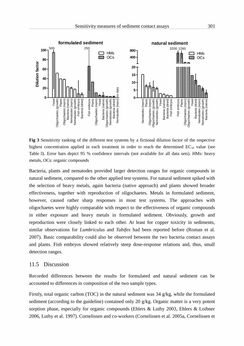
Sensitivity measures of sediment contact assays 301
Fig 3 Sensitivity ranking of the different test systems by a fictional dilution factor of the respective highest concentration applied in each treatment in order to reach the determined EC50 value (see Table 3). Error bars depict 95 % confidence intervals (not available for all data sets). HMs: heavy metals, OCs: organic compounds
Bacteria, plants and nematodes provided larger detection ranges for organic compounds in natural sediment, compared to the other applied test systems. For natural sediment spiked with the selection of heavy metals, again bacteria (native approach) and plants showed broader effectiveness, together with reproduction of oligochaetes. Metals in formulated sediment, however, caused rather sharp responses in most test systems. The approaches with oligochaetes were highly comparable with respect to the effectiveness of organic compounds in either exposure and heavy metals in formulated sediment. Obviously, growth and reproduction were closely linked to each other. At least for copper toxicity in sediments, similar observations for Lumbriculus and Tubifex had been reported before (Roman et al. 2007). Basic comparability could also be observed between the two bacteria contact assays and plants. Fish embryos showed relatively steep dose-response relations and, thus, small detection ranges.
11.5 Discussion
Recorded differences between the results for formulated and natural sediment can be accounted to differences in composition of the two sample types.
Firstly, total organic carbon (TOC) in the natural sediment was 34 g/kg, while the formulated sediment (according to the guideline) contained only 20 g/kg. Organic matter is a very potent sorption phase, especially for organic compounds (Ehlers & Luthy 2003, Ehlers & Loibner 2006, Luthy et al. 1997). Cornelissen and co-workers (Cornelissen et al. 2005a, Cornelissen et
formulated sediment
Yea
stO
ligoc
haet
es (g
row
th)
Pla
nts
Olig
ocha
etes
(rep
ro)
Bact
eria
(nat
ive)
Nem
atod
es (r
epro
)N
emat
odes
(gro
wth
)Fi
sh e
mbr
yos
Bac
teria
(drie
d)
Fish
em
bryo
sPl
ants
Olig
ocha
etes
(rep
ro)
Yea
stB
acte
ria (n
ativ
e)O
ligoc
haet
es (g
row
th)
Nem
atod
es (g
row
th)
Bac
teria
(drie
d)N
emat
odes
(rep
ro)0
20
40
60
80
100HMsOCs
no d
ata
500 250
Dilu
tion
fact
ornatural sediment
Nem
atod
es (r
epro
)Pl
ants
Olig
ocha
etes
(rep
ro)
Olig
ocha
etes
(gro
wth
)N
emat
odes
(gro
wth
)Y
east
Bact
eria
(nat
ive)
Bact
eria
(drie
d)Fi
sh e
mbr
yos
Fish
em
bryo
sPl
ants
Olig
ocha
etes
(rep
ro)
Olig
ocha
etes
(gro
wth
)Ye
ast
Bac
teria
(drie
d)N
emat
odes
(rep
ro)
Nem
atod
es (g
row
th)
Bac
teria
(nat
ive)
0
5
10
15
20
400
800
OCsHMs
3200 1260

Sensitivity measures of sediment contact assays 302
al. 2005b) reported on strong sorption of Diuron to black carbon (BC) in BC-enriched sediment, and a study by Krishna and Philip (Krishna & Philip 2008) revealed, that sorption of lindane, methyl parathion and carbofuran in Indian soils was positively correlated with the proportion of organic content. Organic carbon as a part of organic matter has also been shown to contribute substantially to the sorption of heavy metals to soils and sediments (Ankley et al. 1996, Koelmans 1998, Pacakova et al. 2000). Tyler and McBride (Tyler & Mcbride 1982) found less extractability of copper, nickel, zinc and cadmium for organic soils, and Usman (Usman et al. 2005) accounted decreased water-extractable concentrations of copper, nickel and cadmium to the presence of organic carbon. Strong binding of copper to organic carbon has also been pointed out in a study by Mahony (Mahony et al. 1996).
Fig 4 Detection ranges of the investigated contact assays for the different sediment-contaminant combinations. Bars represent reciprocal slopes of the dose-response curves shown in Fig. 1. HMs: heavy metals, OCs: organic compounds
Secondly, organic matter in the natural sediment was of a natural origin and very likely comprised different types of carbon content. The formulated sediment, on the other hand, was prepared using fine grinded peat moss as the carbon content. An important aspect governing the sorption behaviour of contaminants in sediments is the specific organic carbon composition (i.e., type, quality, chemical state) (Ahrens & Hickey 2002). Especially coal, ash and soot, which are commonly referred to as "black carbon", offer one of the most important binding phases for sorption processes in soils and sediments (Koelmans et al. 2006, Xiao et al.
0 200 400 600
Yeast
Plants
Oligochaetes (growth)
Oligochaetes (repro)
Fish embryos
Nematodes (growth)
Nemotades (repro)
Bacteria (native)
Bacteria (dried)
natural, OCsnatural, HMsformulated, OCsformulated, HMs
Reciprocal slope values

Sensitivity measures of sediment contact assays 303
2004). Huang and co-workers (Huang et al. 2003) concluded that "black carbon and kerogen […] may dominate the overall nonlinear sorption by soils and sediments." Furthermore, for selected PAHs and PCBs it has been shown that the size of the rapidly desorbing fraction was greater regarding younger organic matter (Kukkonen et al. 2003). Comparable influence of the composition of organic matter is discussed for the availability of heavy metals, which were found to associate to, e.g., natural humus to a greater extent than to cellulose in formulated sediments (Besser et al. 2003).
Thirdly, the clay fraction in the natural sediment was clearly larger (23.4 %) than in the formulated sediment (5 % kaolin as clay content). Krishna and Philip (Krishna & Philip 2008) found in their study also evidence for relevant sorption of the three investigated pesticides to clay. Likewise, other cases in the literature indicate impact of clay content on the degree of sorption and therefore availability of organic compounds in soils and sediments (Li et al. 2007, Spark & Swift 2002). Regarding heavy metals, it has been shown that their sorption is negatively correlated with the particle size of putative adsorbents (Jain & Ram 1997). Furthermore, Usman and co-workers (Usman et al. 2005) reported on a strong decrease of water-extractable copper, nickel, zinc and cadmium after addition of bentonite clay minerals to sediment samples, which was reflected by reduced toxicity. Therefore, clay can be considered to affect the availability of heavy metals to test organisms.
The observation that several test systems exhibited low sensitivities regarding formulated sediments spiked with the organic compounds cocktail was quite unexpected. Peat moss, used for the preparation of the formulated sediment, does not provide that characteristics of organic matter supporting strong sorption. Hence, contaminants should have been highly available, resulting in high sensitivities and small ranges of effectiveness. Interestingly, all test organisms which feed more or less directly on sediment particles gave such data. A possible explanation can therefore be, that the main exposure route for oligochaetes and nematodes was ingestion of particle-bound compounds. Several authors define ingestion as the major pathway for bioaccumulation of sediment-associated contaminants in deposit feeders (Boese et al. 1990, Leppanen & Kukkonen 1998, Mayer et al. 1996). Natural sediment, on the contrary, very likely provided higher sorption capacity, and, thus, higher amounts of contaminants were bound to sediment particles, directly intoxicating oligachaetes and nematodes while feeding upon it. Unlike oligocheates and nematodes, bacteria do not ingest sediment particles. However, microbes are known to feed on organic macromolecules in soil and sediment and, thus, accumulate on particulate organic matter (Characklis et al. 2005, Ritzrau 1996, Sessitsch et al. 2001, White 1994). This could explain the large detection ranges also found for the Arthrobacter contact tests.
In general, the cell-based test systems, i.e. bacteria and yeast assays, showed low sensitivities for both organic compounds and heavy metals; only exception was the yeast approach regarding heavy metals in formulated sediment. Compared to this, oligochaetes responded to

Sensitivity measures of sediment contact assays 304
lower concentrations of heavy metals, indicating that direct access via feeding makes contaminants more readily available to sediment-ingesting test organisms. Mayer and co-workers (Mayer et al. 1996) measured significantly elevated solubilization of both copper and PAHs by treatment with digestive fluids extracted from two marine oligochaete species, and concluded that gut passage of particle-associated contaminants increases the bioavailable fraction. Nematodes showed also higher sensitivity to heavy metals, yet only when exposed to natural sediment. This concurs with an assumed higher sorption capacity of the Altrip sample for heavy metals, due to the organic matter composition. However, as nematodes are very much smaller than oligochaetes, they are unlikely to directly ingest organic sediment particles, but feed on bacteria associated to these. As a consequence, they might get into closer contact to contaminants which are bound to sediment organic matter. Furthermore, reproduction of nematodes reacted twice as sensitive as the growth parameter did. Hence, it seems as if heavy metal intoxication has a much stronger impact on fertility (generative cells) than on development (somatic cells) of nematodes. Likewise, sensitivity of zebrafish embryo development for heavy metal exposure was comparably low. For salmonids, the early eggs were found to be the least sensitive phases in terms of heavy metal toxicity (Eaton et al. 1978, Finn 2007, Mckim et al. 1978, Shazili & Pascoe 1986), and a study by Hallare (Hallare et al. 2005) describes increasing sensitivity to cadmium of zebrafish larvae when hatched. Furthermore, Chen and co-workers (Chen et al. 2004) observed elevated cumulative zinc-induced mortality after hatching of zebrafish embryos and accounted this to a switch from constitutive expression of maternal metallothionein (Mt) mRNA to specific expression of embryonic Mt genes.
The ubiquitous sensitivity of the plants assay can be accounted to the roots system, which penetrates the whole sediment. The water supply of plants is primarily realized via root hairs and fine lateral roots, which are protruding into the water pores of soils and sediments. In addition, the negatively charged mucus sequestered by roots upon growing can facilitate uptake of heavy metal ions. As a consequence, all types of contaminants in all parts of the sediment become available. Especially Diuron, a selective herbicide affecting the photosynthesis complex (Devlin et al. 1983, Lundegardh 1965), is known to enter plants preferably via the roots and gets easily transported to stems and leaves (Moyer et al. 1972, Smith & Sheets 1967). Nash (1968) reported that uptake of Diuron into oat seedlings and especially their roots appeared to be relatively independent of soil parameters, including organic matter content. This stands in contrast to the strong influence of TOC on the results in all other bioassays as discussed above. It can also be assumed that Diuron and its metabolites were responsible for the major part of effectiveness in plants recorded for the organic compounds cocktail.

Sensitivity measures of sediment contact assays 305
11.6 Conclusions & Perspectives
Parallel testing of formulated and natural sediments spiked with either a mixture of four heavy metals or a cocktail comprising six different organic contaminants returned dose-response relations for most of the six applied sediment contact test systems. However, differences were found with respect to the lowest concentration detectable and the range of concentration steps that the assays were able to discriminate, expressed as slopes of the response curves.
The plants contact assay turned out to be suitable for detection of heavy metals as well as organic compounds in very different matrices. Furthermore, the system responded to a mediocre concentration range of organic compounds or heavy metals. To selectively investigate heavy metal toxicity of sediment samples, both oligochaete endpoints seem useful, but also the yeast assay and the nematode reproduction test appeared to be suitable with respect to sediments comparable with OECD and Altrip, respectively. Oligochaete reproduction as well as the yeast cells had relatively broad detection ranges for heavy metals, while growth of oligochaetes and reproduction of nematodes responded in a narrow concentration range. Selective detection of organic contamination at very high sensitivities might be achieved using the fish egg sediment contact test and the reproduction assay with nematodes. While detection ranges of fish embryos were relatively small, the nematode test provided better capabilities for discrimination. Both bacteria toxicity assays appeared to be applicable for investigating sediments regarding heavy metals and organic compounds in both sediment types, but can detect contaminants only at elevated concentrations. However, due to rather flat dose-response relations, these test systems allow to detect a large range of contaminant concentrations. Results also indicate impact of sediment parameters, particularly organic matter, on the availability of contaminants and, thus, on the effective concentrations in the different bioassays. Uptake of organic substances and heavy metals is likely to depend on their sequestration between the sediment components. Consequently, availability in each test system is governed by the respective organism’s feeding and living habits.
Obtained data provide an overview over the individual properties and capabilities of each test system, and may guide researchers while selecting the appropriate biotest battery for a given investigation. However, in order to fully understand findings from sediment contact tests, further investigations into the influencing factors are necessary and should be subject to ongoing research.
11.7 Acknowledgements
This study was funded by the German Federal Ministry of Education and Research (BMBF; SeKT project numbers: 02WU0598 to 02WU0604). We acknowledge the valuable input of Hans-Jürgen Pluta (German Federal Environment Agency, UBA) and Ulrich Förstner (Technical University Hamburg-Harburg), who accompanied the SeKT project as members of
Sensitivity measures of sediment contact assays 306
the scientific advisory board. The gifts of Caenorhabditis elegans and Escherichia coli from the Caenorhabditis Genetic Center (Theresa Stiernagle) are gratefully acknowledged. Many thanks to Philipp Egeler for sharing his knowledge of oligochaetes.
11.8 References
Ahrens MJ, Hickey CW (2002): Extractability of polycyclic aromatic hydrocarbons in sediments: A matter of association?, Interact 2002 Conference, Sydney, Australia, pp. 193-199
Ankley GT, DiToro DM, Hansen DJ, Berry WJ (1996): Technical basis and proposal for deriving sediment quality criteria for metals. Environ Toxicol Chem 15, 2056-2066
Besser JM, Brumbaugh WG, May TW, Ingersoll CG (2003): Effects of organic amendments on the toxicity and bioavailability of cadmium and copper in spiked formulated sediments. Environ Toxicol Chem 22, 805-815
Boese BL, Lee H, Specht DT, Randall RC, Winsor MH (1990): Comparison of Aqueous and Solid-Phase Uptake for Hexachlorobenzene in the Tellinid Clam Macoma-Nasuta (Conrad) - a Mass Balance Approach. Environ Toxicol Chem 9, 221-231
Braunbeck T, Böttcher M, Hollert H, Kosmehl T, Lammer E, Leist E, Rudolf M, Seitz N (2005): Towards an alternative for the acute fish LC50 test in chemical assessment: The fish embryo toxicity test goes multi-species - an update. ALTEX 22, 87-102
Brils J (2004): Sediment monitoring under the EU water framework directive. J Soils Sediments 4, 72-73
Chapman PM, Hollert H (2006): Should the sediment quality triad become a tetrad, a pentad, or possibly even a hexad? J Soils Sediments 6, 4-8
Characklis GW, Dilts MJ, Simmons OD, Likirdopulos CA, Krometis LAH, Sobsey MD (2005): Microbial partitioning to settleable particles in stormwater. Water Res 39, 1773-1782
Chen WY, John JAC, Lin CH, Lin HF, Wu SC, Lin CH, Chang CY (2004): Expression of metallothionein gene during embryonic and early larval development in zebrafish. Aquat Toxicol 69, 215-227
Cornelissen G, Gustafsson O, Bucheli TD, Jonker MTO, Koelmans AA, Van Noort PCM (2005a): Extensive sorption of organic compounds to black carbon, coal, and kerogen in sediments and soils: Mechanisms and consequences for distribution, bioaccumulation, and biodegradation. Environ Sci Technol 39, 6881-6895
Cornelissen G, Haftka J, Parsons J, Gustafsson O (2005b): Sorption to black carbon of organic compounds with varying polarity and planarity. Environ Sci Technol 39, 3688-3694
Devlin RM, Murkowski AJ, Zbiec II, Karczmarczyk SJ, Skorska EM (1983): Influence of Buthidazole, Diuron, and Atrazine on Some Light Reactions of Photosynthesis. Weed Sci 31, 879-883
Eaton JG, Mckim JM, Holcombe GW (1978): Metal Toxicity to Embryos and Larvae of 7 Freshwater Fish Species .1. Cadmium. Bull Environ Contam Toxicol 19, 95-103
Egeler P, Meller M, Schallnass HJ, Gilberg D (2005): Validation of a sediment toxicity test with the endobenthic aquatic oligochaete Lumbriculus variegatus by an international ring test. R&D No.: 202 67 429. Berlin, Germany

Sensitivity measures of sediment contact assays 307
Ehlers G, Luthy R (2003): Contaminant bioavailability in soil and sediment. Environ Sci Technol 37, 295A–302A
Ehlers GA, Loibner AP (2006): Linking organic pollutant (bio)availability with geosorbent properties and biomimetic methodology: a review of geosorbent characterisation and (bio)availability prediction. Environ Pollut 141, 494-512
Feiler U, Kirchesch I, Heininger P (2004): A new plant-based bioassay for aquatic sediments. J Soils Sediments 4, 261-266
Finn RN (2007): The physiology and toxicology of salmonid eggs and larvae in relation to water quality criteria. Aquat Toxicol 81, 337-354
Förstner U (2002): Sediments and the European Water Framework Directive. J Soils Sediments 2, 2-3Hallare AV, Schirling M, Luckenbach T, Kohler HR, Triebskorn R (2005): Combined effects of
temperature and cadmium on developmental parameters and biomarker responses in zebrafish (Danio rerio) embryos. J Therm Biol 30, 7-17
Heise S, Ahlf W (2005): A new microbial contact assay for marine sediments - Dedicated to Prof. Dr. Ulrich Forstner on his 65th birthday. J Soils Sediments 5, 9-15
Hollert H, Dürr M, Erdinger L, Braunbeck T (2000): Cytotoxicity of settling particulate matter (SPM) and sediments of the Neckar river (Germany) during a winter flood. Environ Toxicol Chem 19, 528-534
Hollert H, Keiter S, König N, Rudolf M, Ulrich M, Braunbeck T (2003): A new sediment contact assay to assess particle-bound pollutants using zebrafish (Danio rerio) embryos. J Soils Sediments 3, 197 – 207
Höss S, Ahlf W, Fahnenstich C, Gilberg D, Hollert H, Melbye K, Meller M, Hammers-Wirtz M, Heininger P, Neumann-Hensel H, Ottermanns R, Ratte H-T, Seiler T-B, Spira D, Weber J, Feiler U (2010): Variability of freshwater sediment contact tests in sediments with low anthropogenic contamination - Determination of toxicity thresholds. Environ Pollut in press
Huang WL, Ping PA, Yu ZQ, Fu HM (2003): Effects of organic matter heterogeneity on sorption and desorption of organic contaminants by soils and sediments. Appl Geochem 18, 955-972
Ingersoll CG, Dillon T, Biddinger GR (1997): Ecological Risk Assessment of Contaminated Sediments. SETAC Press, Pensacola, Fl, USA
ISO (2009): Water quality — Determination of the inhibition of dehydrogenase activity of Arthrobacter globiformis — Solid contact test using the redox dye resazurine. ISO/DIS 10871
Jain CK, Ram D (1997): Adsorption of metal ions on bed sediments. Hydrological Sciences Journal 42, 713-723
Koelmans AA (1998): Geochemistry of suspended and settling solids in two freshwater lakes. Hydrobiologia 364, 15-29
Koelmans AA, Jonker MTO, Cornelissen G, Bucheli TD, Van Noort PCM, Gustafsson O (2006): Black carbon: The reverse of its dark side. Chemosphere 63, 365-377
Koethe F (2003): Existing sediment management guidelines: An overview. What will happen with the sediment/dredged material? J Soils Sediments, 139-143
Krishna KR, Philip L (2008): Adsorption and desorption characteristics of lindane, carbofuran and methyl parathion on various Indian soils. J. Hazard. Mater. 160, 559-567

Sensitivity measures of sediment contact assays 308
Kukkonen JVK, Landrum PF, Mitra S, Gossiaux DC, Gunnarsson J, Weston D (2003): Sediment characteristics affecting desorption kinetics of select PAH and PCB congeners for seven laboratory spiked sediments. Environ Sci Technol 37, 4656-4663
Leppanen MT, Kukkonen JVK (1998): Relationship between reproduction, sediment type, and feeding activity of Lumbriculus variegatus (Muller): Implications for sediment toxicity testing. Environ Toxicol Chem 17, 2196-2202
Li JG, Sun HW, Zhang Y (2007): Desorption of pyrene from freshly-amended and aged soils and its relationship to bioaccumulation in earthworms. Soil Sediment Contam 16, 79-87
Lundegardh H (1965): Influence of Diuron [3-(3,4-Dichlorophenyl)-1,1-Dimethylurea] on Respiratory and Photosynthetic Systems of Plants. P Natl Acad Sci USA 53, 703-&
Luthy RG, Aiken GR, Brusseau ML, Cunningham SD, Gschwend PM, Pignatello JJ, Reinhard M, Traina SJ, Weber WJ, Westall JC (1997): Sequestration of Hydrophobic Organic Contaminants by Geosorbents. Environ Sci Technol 31, 3341
Mahony JD, DiToro DM, Gonzalez AM, Curto M, Dilg M, DeRosa LD, Sparrow LA (1996): Partitioning of metals to sediment organic carbon. Environ Toxicol Chem 15, 2187-2197
Mayer LM, Chen Z, Findlay RH, Fang JS, Sampson S, Self RFL, Jumars PA, Quetel C, Donard OFX (1996): Bioavailability of sedimentary contaminants subject to deposit-feeder digestion. Environ Sci Technol 30, 2641-2645
Mckim JM, Eaton JG, Holcombe GW (1978): Metal Toxicity to Embryos and Larvae of 8 Species of Freshwater Fish .2. Copper. Bull Environ Contam Toxicol 19, 608-616
Moyer JR, Mckerche.Rb, Hance RJ (1972): Influence of Adsorption on Uptake of Diuron by Barley Plants. Can J Plant Sci 52, 668-670
Nagel R (1986): Untersuchungen zur Eiproduktion beim Zebrabärbling (Brachydanio rerio, Ham.-Buch.). J Appl Ichthyol 2, 173-181
Nash RG (1968): Plant Uptake of 14c-Diuron in Modified Soil. Agron J 60, 177-&Neumann-Hensel H, Melbye K (2006): Optimisation of the solid-contact test with Artbrobacter
globiformis. J Soils Sediments 6, 201-207OECD (1984): Guideline for the Testing of Chemicals No. 207: Earthworm acute toxicity test.
Organisation for Economic Co-operation and Development (OECD)OECD (2004): Guideline for the Testing of Chemicals No. 218: Sediment-water chironomid toxicity
test using spiked sediment. Organisation for Economic Co-operation and Development (OECD)Pacakova V, Pockeviciute D, Armalis S, Stulik K, Li JH, Vesely J (2000): A study of the distribution of
lead, cadmium and copper between water and kaolin, bentonite and a river sediment. J Environ Monit 2, 187-191
Phipps GL, Ankley GT, Benoit DA, Mattson VR (1993): Use of the Aquatic Oligochaete Lumbriculus-Variegatus for Assessing the Toxicity and Bioaccumulation of Sediment-Associated Contaminants. Environ Toxicol Chem 12, 269-279
Power EA, Chapman PM (1992): Assessing sediment quality. In: Burton GA (Editor), Sediment toxicity assessment. Lewis-Publishers, pp. 1-18
Ritzrau W (1996): Microbial activity in the benthic boundary layer: Small-scale distribution and its relationship to the hydrodynamic regime. J Sea Res 36, 171-180

Sensitivity measures of sediment contact assays 309
Roman YE, De Schamphelaere KAC, Nguyen LTH, Janssen CR (2007): Chronic toxicity of copper to five benthic invertebrates in laboratory-formulated sediment: Sensitivity comparison and preliminary risk assessment. Sci Total Environ 387, 128-140
SedNet (2004): Sediment, a valuable resource that needs Europe's attention. SedNet recommendations for sediment research priorities related to the soil research clusters
Sessitsch A, Weilharter A, Gerzabek MH, Kirchmann H, Kandeler E (2001): Microbial population structures in soil particle size fractions of a long-term fertilizer field experiment. Appl. Environ. Microbiol. 67, 4215-4224
Shazili NAM, Pascoe D (1986): Variable Sensitivity of Rainbow-Trout (Salmo-Gairdneri) Eggs and Alevins to Heavy-Metals. Bull. Environ. Contam. Toxicol. 36, 468-474
Smith JW, Sheets TJ (1967): Uptake Distribution and Metabolism of Monuron and Diuron by Several Plants. J. Agric. Food Chem. 15, 577-&
Spark KM, Swift RS (2002): Effect of soil composition and dissolved organic matter on pesticide sorption. Sci Total Environ 298, 147-61
Stesevic D, Feiler U, Sundic D, Mijovic S, Erdinger L, Seiler T-B, Heininger P, Hollert H (2007): Application of a New Sediment Contact Test with Myriophyllum aquaticum and of the Aquatic Lemna Test to Assess the Sediment Quality of Lake Skadar. J Soils Sediments 7, 342-349
Traunspurger W, Haitzer M, Hoss S, Beier S, Ahlf W, Steinberg C (1997): Ecotoxicological assessment of aquatic sediments with Caenorhabditis elegans (nematoda) - A method for testing liquid medium and whole-sediment samples. Environ Toxicol Chem 16, 245-250
Tyler LD, Mcbride MB (1982): Mobility and Extractability of Cadmium, Copper, Nickel, and Zinc in Organic and Mineral Soil Columns. Soil Sci. 134, 198-205
Usman A, Kuzyakov Y, Stahr K (2005): Effect of clay minerals on immobilization of heavy metals and microbial activity in a sewage sludge-contaminated soil. J Soils Sediments 5, 245-252
Weber J, Kreutzmann J, Plantikow A, Pfitzner S, Claus E, Manz W, Heininger P (2006): A novel particle contact assay with the yeast Saccharomyces cerevisiae for ecotoxicological assessment of freshwater sediments. J Soils Sediments 6, 84-91
White GF (1994): Multiple interactions in riverine biofilms - surfactant adsorption, bacterial attachment and biodegradation, International Specialized Research Seminar on Biological Degradation of Organic Chemical Pollutants in Biofilm Systems, Copenhagen, Denmark, pp. 61-70
Xiao B, Yu Z, Huang W, Song J, Peng P (2004): Black carbon and kerogen in soils and sediments. 2. Their roles in equilibrium sorption of less-polar organic pollutants. Environ Sci Technol 38, 5842-52

Sensitivity measures of sediment contact assays 310

Conclusions


12 Conclusions 313
12 Conclusions
12.1 Confounding factors in sediment assessment
Sediments are a highly complex matrix with a large number of properties which influence transport, behaviour and, thus, fate of environmental contaminants. Considering the multifaceted nature of sediments, it is no surprise that also the strategies to gain data about their contamination provide a broad range of ideas, concepts, principles and technologies. Furthermore, all different approaches have advantages and bear risks for a reliable assessment of the toxic impact of sediment samples. As a consequence, the choice of the a certain method or rather of a couple of methodologies can readily and strongly influence the findings from a given investigation and, thereafter, the estimation of an environmental risk. Eventually, a specific taken action might not be appropriate due to inaccurate data and false assumptions.
Already sample transport and storage might lead to loss of contaminants upon sorption to vessel surfaces. Freezing and drying of sediment samples can also alter the toxicity. Sediment investigations based on extracts might get hampered by the loss of contaminants during sample preparation, such as volatilization or thermal degradation, and even the creation of new effective compounds during heated exhaustive extraction processes is conceivable. Clean-up treatments might further alter resulting extracts, and as most preparation strategies consist of a number of cascading single steps, reproducibility of results gained using extracts is limited; not to mention possible influences of biotest protocols and chemical analysis procedures. Consequently, several parallel independent extraction replicates are highly recommended.
Biomimetic extractions, which aim at the rapidly desorbing contaminant fractions, are limited by the operational definition of bioaccessibility. The degree of accessibility of a given compound is at least a function of several sediment parameters, particularly organic carbon content, physical-chemical properties of the substance, and environmental conditions, such as pH-value or temperature. Appropriate methods for biomimetic extraction have to retain the whole natural situation of the sample during the separation process. Any alterations that occur upon extraction could influence the sorption behaviour and would then compromise estimation of, e.g. risk for sediment-dwelling organisms. Furthermore, bioaccessibility does not consider the biology of the individual species, as bioavailability does. Hence, for a reliable risk assessment with respect to a certain benthic community, every single species would have to be exposed to sediment extracts in a biotest approach.
Toxicity testing of sediments can be realized through direct contact exposure, circumventing most of the confounding factors detailed before regarding extract testing. However, as also with contact tests the entire benthic community should be considered, a broad battery of assays has to be applied. Still, it is very unlikely, that a selection of test systems can

12 Conclusions 314
completely resemble all species existing at a sampling site. Therefore, model organisms for trophic levels, habitats or taxonomical groups are often used. However, this requires concepts to easily compare toxicity data from the different assays. Common reference sediments are necessary as well as knowledge of toxicity thresholds. Furthermore, detailed information about the living and feeding behaviour of each test organism are essential, in order to correlate these with observed effects, substance behaviour and sediment properties.
While sediment contact tests can largely decrease the risk of altering an investigated sediment sample, various cell-based bioassays and miniaturized versions of several test systems still require extracts for their application. In addition, identification of toxicants completely depends on extraction of the contaminant spectrum as a pre-requisite for chemical analysis. On the other hand, toxicity of a given extract can only be determined using biotests, and the question about the relevance of the recorded effects for the benthic community in situ demands for a comparison of extract data with results from direct contact exposure.
12.2 Proposal for a strategical framework
As a conclusion, all different approaches have importance for comprehensive investigation strategies of sediment contamination, and none can to-date entirely substitute the other. With future development it might eventually be possible to describe contact test results for a variety of organisms by means of biomimetic extracts. Additionally, substitution of whole organism tests by cell-based assays might lead to higher throughput and better reproducibility in the ecotoxicological characterisation of sediments. Yet, the vast variability of sediment-organism-contaminant combinations is very unlikely to be completely replicated by extracts and cell-based model test systems.
Given the necessity of a reliable sediment toxicity assessment, e.g. within the maintenance of waterways and harbours, a multiple line-of-evidence approach is proposed:
(1) Exhaustive extracts should provide information about the whole contaminant spectrum, necessary to estimate the 'actual risk' being the product of strength and likelihood of adverse effects on the ecosystem. In order to strengthen the data basis of such an investigation, a larger number of subsamples should be extracted in parallel. Appropriate extraction techniques therefore need to allow high-throughput preparation of extracts. Up-to-date, however, this requirement is associated to a high risk of uncontrolled chemical reactions of contaminants during the leaching process. Automated extraction like utilized by ASE methods combined with membrane dialysis as in MDE and at moderate temperatures promises to produce quantitative extracts at a substantially reduced risk of alteration of the hazard potential.
(2) Whole sediment contact tests should be conducted for the determination of the actual ecotoxicological situation in situ. The applied biotest battery has to resemble at least

12 Conclusions 315
the key species for the respective ecosystem. When using model test organisms for, e.g. different trophic levels, they must exhibit comparable feeding and living behaviours. These information are, hence, essential for all involved species in situ as well as in vitro. For accurate interpretation of the resulting data, the composition of the respective sediment needs to be known and correlated to physical-chemical properties of the substances found in exhaustive extracts.
(3) Biomimetic extracts should be used to determine the accessible fraction of contaminants. These results can then serve to estimate the 'current risk' in situ. As currently no biomimetic extraction procedure appears to be capable of generally and reliably mimicking results from direct contact exposure, several extractions should be conducted in parallel. Again, this requires even more the availability of automated high-throughput extraction, and future development of such techniques is crucial. In order to determine the extract type being closest to the bioavailability of contaminants for a given organism, bioanalytical data for biomimetic extracts should be correlated with results from contact tests. The selected extract can then be subjected to chemical analysis for identification of key contaminants.
Figure Scheme of the proposed multiple-line investigation strategy for a more reliable sediment toxicity assessment. Symbols depict requirements that would enhance the feasibility with respect to scientifical, practical and economical considerations
Technological requirement Required knowledge Should be reduced or avoided

12 Conclusions 316
This approach is a sophisticated investigation strategy and would add a large portion of reliability to sediment toxicity assessment. However, limited financial resources will often not allow to follow the whole concept in every detail. As a consequence, until (1) development of extraction techniques leads to reliable and cost-effective tools for exhaustive and biomimetic extraction, (2) research on contaminant behaviour in the context of sediment properties, environmental conditions and biology of organisms provides comprehensive information about bioavailability, and (3) novel miniaturized and cell-based bioassays allow efficient toxicity testing of a large number of extracts with good correlation to direct contact exposure, sediment toxicity assessment should at least take into account the considerations and confounding factors as detailed in this study, and interprete resulting data cautiously.

Scientific contributions


13 Scientific contributions 319
13 Scientific contributions
Research articles in international peer-reviewed journals(Impact Factor 2009 in brackets)
Hallare A, Seiler T-B, Hollert H (2010): The versatile, changing, and advancing roles of fish in sediment toxicity assessment – a review. J Soils Sediments accepted [ISI: 2.613]
Höss S, Ahlf W, Fahnenstich C, Gilberg D, Hollert H, Melbye K, Meller M, Hammers-Wirtz M, Heininger P, Neumann-Hensel H, Ottermanns R, Ratte H-T, Seiler T-B, Spira D, Weber J, Feiler U (2010): Variability of freshwater sediment contact tests in sediments with low anthropogenic contamination - Determination of toxicity thresholds. Environ Pollutonline first DOI 10.1016/j.envpol.2010.05.013 [ISI: 3.135]
Schneider A, Brinkmann M, Gerstner A, Wölz J, Heger S, Weber R, Engwall M, Seiler T-B, Hollert H (2009): Assessment of Dioxin-like toxicity in soils contaminated by a chloralkali process and a Leblanc factory. Organ Halogen Compound in press
Seiler T-B, Schulze T, Hollert H (2008): The risk of altering soil and sediment samples upon extract preparation for analytical and bio-analytical investigations—a review. Anal Bioanal Chem 390, 1975-1985 [ISI: 3.48]
Stesevic D, Feiler U, Sundic D, Mijovic S, Erdinger L, Seiler T-B, Heininger P, Hollert H (2007): Application of a New Sediment Contact Test with Myriophyllum aquaticum and of the Aquatic Lemna Test to Assess the Sediment Quality of Lake Skadar. J Soils Sediments 7, 342-349 [ISI: 2.613]
Seiler T-B, Rastall AC, Leist E, Erdinger L, Braunbeck T, Hollert H (2006): Membrane dialysis extraction (MDE): A novel approach for extracting toxicologically relevant hydrophobic organic compounds from soils and sediments for assessment in biotests. J Soils Sediments 6, 20-29 [ISI: 2.613]
Research articles to be submitted to international peer-reviewed journals
Bluhm K, Heger S, Ernst M, Wölz J, Seiler T-B, Schäffer A, Hollert H (2010): Ecotoxicological investigations on biofuels - a literature re-view. Environ Health Perspectives invited to be submitted (by the Editor-in-Chief)
Schulze T, Seiler T-B, Hollert H, Schröter-Kermani C, Braunbeck T, Pekdeger A (2010): Extractability and toxicity of potentially toxic organic pollutants in riverine sediments. J Soils Sediments to be submitted

13 Scientific contributions 320
Seiler T-B, Ahlf W, Fahnenstich C, Gilberg D, Melbye K, Meller M, Hammers-Wirtz M, Heininger P, Hollert H, Höss S, Neumann-Hensel H, Ottermanns R, Ratte H-T, Spira D, Weber J, Feiler U (2010): Investigation on sensitivity measures and applicability of limnic sediment contact assays using formulated and natural spiked sediment samples. J Soils Sediments to be submitted
Seiler T-B, Schulze T, Streck G, Schwab K, Zielke H, Brinkmann M, Bernecker C, Brack W, Braunbeck T, Hollert H (2010): Passive membrane dialysis is a promising approach for the exhaustive extraction of PAHs from dried sediment samples. Environ Sci Pollut Res to be submitted
Seiler T-B, Streck G, Schulze T, Schwab K, Brack W, Braunbeck T, Hollert H (2010): On the comparability of procedures for sediment extraction in environmental assessment. Part A: Bioanalytical investigations. Environ Toxicol Chem to be submitted
Streck G, Seiler T-B, Schulze T, Schwab K, Braunbeck T, Hollert H, Brack W (2010): On the comparability of procedures for sediment extraction in environmental assessment. Part B: Chemical analytical investigations. Environ Toxicol Chem to be submitted
Zielke H, Seiler T-B, Niebergall S, Leist E, Zimmer H, Erdinger L, Braunbeck T, Hollert H (2010): The impact of extraction methodologies on the toxicity of sediments in the zebrafish (Danio rerio) embryo test (FET). J Soil Sediment to be submitted
Manuscripts for research articles in international peer-reviewed journals
Ernst M, Seiler T, Vatosous D, Hallare A, Braunbeck T, Hollert H (2010): Investigation into the endocrine disrupting potential of sediments from Lake Skadar and establishment of a geographical information system. J Soils Sediments in prep
Higley E, Grund S, B.-Seiler T, Varel UL-v, Brack W, Schulze T, Giesy JP, Hollert H, Hecker M (2010): Effects of Upper Danube River Sediments on Steroidogenesis Using Chemical Fractionation and the H295R assay in prep
Seiler T-B, Best N, Fernqvist MM, Smith K, Mayer P, Hollert H (2010): Passive dosing can contribute to the reliability of in-vitro effect data of PAHs from the fish embryo test with Danio rerio. in prep
Seiler T-B, Perovic A, Bushati N, Nikcevic S, Keiter S, Miteva V, T. K, Pesic V, Karaman G, Maric D, Misurovic A, Rastall AC, Erdinger LF, Hollert H (in prep): Integrative Assessment of sediments of the Lake Skadar/Shkodra using a Triad approach. J Soils Sediments
Zielke H, Seiler T-B, Niebergall S, Leist E, Zimmer H, Erdinger L, Braunbeck T, Hollert H (in prep): Uptake of heavy metals by zebrafish embryos.

13 Scientific contributions 321
Research articles in national peer-reviewed journals
Hollert H, Ernst M, Seiler TB, Wölz J, Braunbeck T, Kosmehl T, Keiter S, Grund S, Ahlf W, Erdinger L, Dürr M (2009): Strategien zur Sedimentbewertung – ein Überblick. Umweltwiss Schadst Forsch 21, 160-176
Rocha P, Keiter S, Pompêo M, Mariani CF, Brandimarte AL, Seiler T-B, Kosmehl T, Böttcher M, Wölz J, Braunbeck T, Storch V, Hollert H (2006): Weight-of-Evidence-Studie zur Sedimentbelastung des Tietê River in Brasilien. Umweltwiss Schadst Forsch 18, 70
Editorials, discussion articles and reports in international peer-reviewed journals
Brinkmann M, Brooks A, Dabrunz A, Gomez-Eyles J, Van Hoecke K, Kienle C, Seiler T-B, Bundschuh M (2010): SETAC Europe 19th annual meeting, Gothenburg, Sweden: next step towards fulfilling students’ needs. Environmental Science and Pollution Research 17, 244-245
Bundschuh M, Dabrunz A, Bollmohr S, Brinkmann M, Caduff M, Gomez-Eyles J, Kienle C, Melato M, Patrick-Iwuanyanwu K, Van Hoecke K, Seiler T-B, Brooks A (2009): 1st Young Environmental Scientists (YES) Meeting—New challenges in environmental sciences. Environmental Science and Pollution Research 16, 479-481
Hollert H, Heinrich A, Seiler T-B (2008): The first impact factor. Journal of Soils and Sediments 8, 203-205
Hollert H, Seiler T-B, Blaha L, Young AL (2007): Multiple Stressors for the Environment: Present aund Future Challenges and Perspectives. Env Sci Pollut Res 14, 222
Hollert H, Braun B, Mijovic S, Rakaj M, Rastall A, Seiler T-B, Erdinger L (2004): The EULIMNOS project: strengthening cross-border scientific research and higher education in the Lake Shkodra/Skadar Region through a multidisciplinary transboundary approach to conservation. ESPR - Environ Sci & Pollut Res 11, 135
Editorials, discussion articles and reports in national peer-reviewed journals
Brinkmann M, Seiler TB, Bundschuh M, Dabrunz A, Brooks A, Gomez-Eyles JL, Van Hoecke K, Kienle C (2009): SETAC Europe 19th annual meeting, Gothenburg, Sweden: Next step towards fulfilling students’ needs. Umweltwissenschaften und Schadstoff-Forschung 21, 412-413
Brinkmann M, Seiler TB, Dabrunz A, Bundschuh M, Kienle C (2009): 1st Young Environmental Scientists Meeting (YES-Meeting). Umweltwissenschaften und Schadstoff-Forschung 21, 113-114
Seiler T, Brooks A, Reinhard D, Fossi F, Martins N, Bundschuh M (2007): Der SETAC Europe Student Advisory Council (SAC). Umweltwissenschaften und Schadstoff-Forschung 19, 72-72

13 Scientific contributions 322
Feiler U, Ahlf W, Hoess S, Seiler T-B, Hollert H, Melby K, Neumann-Hensel H, Meller M, Weber J, Heininger P (2005): Das SeKT Verbundprojekt: Definition von Referenzbedingungen, Kontrollsedimenten und Toxizitätsschwellenwerten für limnische Sedimentkontakttests. Umweltwiss Schadst Forsch 17, 250-251
Book chapters
Hollert H, Seiler T-B, Hallare A (2009): Sediment contact assay with fish egg as part of a holistic approach to sediment risk assessment. In: Sediment Contact Tests: Reference conditions, control sediments, toxicity thresholds. Veranstaltungen Symposium 13/14 Nov 2008 Koblenz Germany. Druckpartner Moser Druck + Verlag Gmbh, Rheinbach. ISSN 1866-220X
Diploma thesis
Seiler T-B 2004: Development and evaluation of a new membrane dialysis extraction method for wet and dry sediment samples and as a clean-up technique for acetonic Soxhlet extracts. Diploma thesis Thesis, Ruprecht-Karls-University Heidelberg, Heidelberg, 83 pp
Platform presentations
Higley E, Grund S, Seiler T, Schulze T, Varel UL-v, Brack W, Wölz J, Giesy J, Hollert H, Hecker M (2010): Assessment of Upper Danube River sediment using new fractionation techniques, the Danio rerio embryo assay, the Ames fluctuation assay and the H295R Steroidogenesis Assay, Proceedings, 20th SETAC Europe Annual Meeting, Seville, Spain, May 23-27
Heger S, Winkens K, Schneider A, Brinkmann M, Maletz S, Wölz J, Agler MT, Angenent LT, Seiler T, Hollert H (2009): Assessing the ecotoxicological effects of bioenergy extraction processes, Proceedings, 30th SETAC North America Annual Meeting, New Orleans, USA, November 19-23
Feiler U, Ahlf W, Fahnenstich C, Gilberg D, Hammers-Wirtz M, Höss S, Hollert H, Melbye K, Meller M, Neumann-Hensel H, Ratte H-T, Seiler T-B, Spira D, Weber J, Heininger P (2008): Sediment contact tests as tool for the assessment of sediment quality in German rivers (SeKT-project), Proceedings, 5th SETAC World Congress, Sydney, Australia, August 3-7
Seiler T-B, Schulze T, Streck G, Schwab K, Zielke H, Brinkmann M, Bernecker C, Brack W, Braunbeck T, Hollert H (2008): Evaluation of the leaching power of passive membrane dialysis compared to conventional extraction procedures, Proceedings, 18th SETAC Europe Annual Meeting, Warsaw, Poland, May 25-29

13 Scientific contributions 323
Strecker R, Seiler T-B, Hollert H, Braunbeck T (2008): Sauerstoffbedingungen im Sedimentkontakttest mit dem Zebrabärbling (Danio rerio), Proceedings, 3rd Joint Annual Meeting of SETAC-GLB and GDCh - Environmental Chemistry and Ecotoxicology Group, Frankfurt/Main, Germany, September 23-26
Zielke H, Seiler T-B, Niebergall S, Leist E, Streck G, Zimmer H, Erdinger L, Brack W, Braunbeck T, Hollert H (2008): Comparison of different methods for sediment extraction using fish egg assay with zebrafish (Danio rerio), Proceedings, 5th SETAC World Congress, Sydney, Australia, August 3-7
Schulze T, Seiler T-B, Schwab K, Streck G, Hollert H, Brack W (2007): Vergleich milder und stringenter Extraktionsverfahren für die Risikobewertung von Sedimenten, Proceedings, 12th SETAC-GLB Annual Meeting, Leipzig, Germany, September 12-14
Feiler U, Ahlf W, Fahnenstich C, Gilberg D, Hammers-Wirtz M, Hoess S, Hollert H, Meller M, Melbye K, Neumann-Hensel H, Ratte H-T, Seiler T-B, Weber J, Heininger P (2006): The SeKT joint research project: Definition of Reference Conditions, Control Sediments and Toxicity Thresholds for Limnic Sediment Contact Tests, Proceedings, 16th SETAC Europe Annual Meeting, The Hague, Netherlands, May 7-11
Feiler U, Ahlf W, Fahnenstich C, Gilberg D, Hammers-Wirtz M, Höss S, Hollert H, Meller M, Melbye K, Neumann-Hensel H, Ratte H-T, Seiler T-B, Weber J, Heininger P (2006): The SeKT Joint Research Project: Definition of Reference Conditions, Control Sediments and Toxicity Thresholds for Limnic Sediment Contact Tests, Proceedings, International Symposium on Sediment Dynamics and Pollutant Mobility in River Basins (SEDYMO), Hamburg, Germany, March 26-29
Schulze T, Seiler T-B, Hollert H, Schröter-Kermani C, Pekdeger A (2006): Extrahierbarkeit und Toxizität potentiell toxischer organischer Verbindungen in Sedimenten der Saar, Proceedings, 11th SETAC-GLB Annual Meeting, Landau, Germany, September 3-5
Seiler T-B, Schulze T, Leist E, Rastall AC, Erdinger L, Braunbeck T, Hollert H (2006): Membran Dialyse Extraktion (MDE): Eine einfache, flexible und kostengünstige Alternative zu herkömmlichen organischen Extraktionsverfahren, Proceedings, 11th SETAC-GLB Annual Meeting, Landau, Germany, September 3-5
Hollert H, Seiler T-B, Rocha PS, Rakocevic J, Perovic A, Perovic S, Sundic D, Stesevic D, Rakaj M, Neziri A, Bushati N, Rastall AC, Feiler U, Kostanjsek R, Erdinger LF (2005): EULIMNOS: Ökologische und ökotoxikologische Untersuchungen am Shkodra/Skadar See (Montenegro/Albanien), Proceedings, Annual Meeting Deutsche Gesellschaft für Limnologie/SIL-Tagung, Karlsruhe, Germany, September 26-30
Seiler T-B, Leist E, Rastall AC, Schulze T, Erdinger L, Braunbeck T, Hollert H (2005): Membrane Dialysis Extraction: A novel method for extracting hydrophobic organic contaminants from soils and sediments, Proceedings, 10th SETAC-GLB Annual Meeting, Basel, Switzerland, September 28-30

13 Scientific contributions 324
Poster presentations
Adamzyk C, Zielke H, Gerringer M, Spira D, Feiler U, Seiler T-B, Ahlf W, Hollert H (2010): Challenges of the miniaturized Solid Contact Assay with Arthrobacter globiformis, Proceedings, 20th SETAC Europe Annual Meeting, Seville, Spain, May 23-27
Bluhm K, Heger S, Ernst M, Wölz J, Seiler T-B, Schäffer A, Hollert H (2010): Ecotoxicological investigations on biofuels - a literature review, Proceedings, 20th SETAC Europe Annual Meeting, Seville, Spain, May 23-27
Heger S, Bluhm K, Brinkmann M, Winkens K, Schneider A, Wollenweber M, Maletz S, Wölz J, Agler M, Angenent L, Seiler T, Hollert H (2010): What's up inside the reactor - biotests for risk assessment of biofuel fermentation, Proceedings, 20th SETAC Europe Annual Meeting, Seville, Spain, May 23-27
Loerks J, Seiler T, Mayer P, Ahlf W, Heise S, Fernqvist MM, Schmidt K, Witt G, Hollert H (2010): Is the fish egg test with Danio rerio comparable between sediment contact, exposure to biomimetic extracts, and passive dosing using PDMS?, Proceedings, 20th SETAC Europe Annual Meeting, Seville, Spain, May 23-27
Seiler T-B, Best N, Fernqvist MM, Smith K, Mayer P, Hollert H (2010): Passive dosing can contribute to the reliability of in vitro data on adverse effects of PAHs on fish embryos and cell lines, Proceedings, 20th SETAC Europe Annual Meeting, Seville, Spain, May 23-27
Simon A, Zielke H, Spira D, Feiler U, Schmidt B, Seiler T, Schaeffer A, Kukkonen J, Hollert H (2010): Chemical and radioactive analyses of the partitioning of organic chemicals in sediment-water-organism-systems, Proceedings, 20th SETAC Europe Annual Meeting, Seville, Spain, May 23-27
Zielke H, Adamzyk C, Oellers J, Schneider AJ, Spira D, Feiler U, Seiler T-B, Hollert H (2010): Effects of ageing on the biological effectiveness of spiked sediments, Proceedings, 20th SETAC Europe Annual Meeting, Seville, Spain, May 23-27
Adamzyk C, Zielke H, Hollert H, Seiler T-B, Gerringer M, Spira D, Feiler U, Ahlf W (2009): Wirkung organischer Monosubstanzen im Bakterienkontakttest mit Arthrobacter globiformis, Proceedings, 14th SETAC-GLB Annual Meeting, Freising, Germany, October 5-7
Bluhm K, Heger S, Ernst M, Wölz J, Seiler T, Hollert H (2009): Biofuels and their ecotoxicological relevance – a literature review, Proceedings, 30th SETAC North America Annual Meeting, New Orleans, USA, November 19-23
Bluhm K, Heger S, Ernst M, Seiler T-B, Wölz J, Hollert H, Schäffer A (2009): Ökotoxikologische Bewertung von Biokraftstoffen – ein Überblick über bisherige Forschungsarbeiten weltweit, Proceedings, 14th SETAC-GLB Annual Meeting, Freising, Germany, October 5-7
Meyer W, Laumann S, Achten C, Seiler T-B, Hollert H (2009): Bioverfügbarkeit und Toxizität geogener polyzyklischer aromatischer Kohlenwasserstoffe aus unverbrannter Kohle, Proceedings, 14th SETAC-GLB Annual Meeting, Freising, Germany, October 5-7

13 Scientific contributions 325
Heger S, Winkens K, Schneider A, Brinkmann M, Maletz S, Wölz J, Agler MT, Angenent LT, Seiler T-B, Hollert H (2009): Assessing the ecotoxicological effects of bioenergy extraction processes, Proceedings, 19th SETAC Europe Annual Meeting, Göteborg, Sweden, May 31-June 4
Higley E, Grund S, Seiler T, Varel UL-v, Brack W, Schulze T, Wölz J, Zielke H, Giesy J, Hollert H, Hecker M (2009): Toxicity and Mutagenicity of Danube River Sediments Determined by Chemical Fractionation, the Danio rerio Embryo Assay and the Ames Fluctuation Test, Proceedings, 30th SETAC North America Annual Meeting, New Orleans, USA, November 19-23
Higley E, Seiler T, Varel UL-v, Brack W, Schulze T, Giesy J, Hollert H, Hecker M (2009): Effects of Upper Danube River Sediments on Steriodogenesis using a Combination of Chemical Fractionation and the H295R assay, Proceedings, 30th SETAC North America Annual Meeting, New Orleans, USA, November 19-23
Heger S, Winkens K, Schneider A, Brinkmann M, Wollenweber M, Maletz S, Wölz J, Agler MT, Angenent LT, Seiler T-B, Hollert H (2009): Assessing the ecotoxicological effects of bioenergy extraction processes, Proceedings, 14th SETAC-GLB Annual Meeting, Freising, Germany, October 5-7
Schäffer A, Roß-Nickoll M, Ratte H-T, Schmidt B, Preuß TG, Ottermanns R, Wölz J, Seiler T-B, Hollert H (2009): Neuer Masterstudiengang Ökotoxikologie an der RWTH Aachen – für eine exzellente Ausbildung in der Ökotoxikologie, Proceedings, 14th SETAC-GLB Annual Meeting, Freising, Germany, October 5-7
Schneider AJ, Brinkmann M, Gerstner A, Wölz J, Heger S, Weber R, Bogdal C, Engwall M, Takasuga T, Seiler T-B, Hollert H (2009): A combined strategy for detecting dioxin-like compounds in soils from former factories of chloralkali-electrolysis and leblanc-soda-production, Proceedings, 19th SETAC Europe Annual Meeting, Göteborg, Sweden, May 31-June 4
Seiler T-B, Best N, Fernqvist MM, Smith K, Mayer P, Hollert H (2009): Passive dosing can contribute to the reliability of in vitro data on adverse effects of PAHs on fish embryos and cell lines, Proceedings, 30th SETAC North America Annual Meeting, New Orleans, USA, November 19-23
Seiler TB, Strecker R, Higley E, Leist E, Hecker M, Braunbeck T, Hollert H (2009): Downscaling the DarT assay for the benefit of higher throughput and lower sample consumption, Proceedings, 19th SETAC Europe Annual Meeting, Göteborg, Sweden, May 31-June 4
Seiler T-B, Strecker R, Leist E, Braunbeck T, Hecker M, Hollert H (2009): Downscaling the DarT assay for the benefit of higher throughput and lower sample consumption, Proceedings, 14th SETAC-GLB Annual Meeting, Freising, Germany, October 5-7
Winkens K, Otte JC, Brinkmann M, Zielke H, Wölz J, Seiler T-B, Hollert H (2009): Weiterentwicklung eines biologischen Testverfahrens zur Messung der Aktivität von Ah-Rezeptor-Agonisten in Danio rerio Fischeiern, Proceedings, 14th SETAC-GLB Annual Meeting, Freising, Germany, October 5-7

13 Scientific contributions 326
Zielke H, Adamzyk C, Rehage R, Spira D, Preuss TG, Schmidt B, Feiler U, Seiler T-B, Hollert H (2009): Effects of ageing on bioavailability and uptake of pollutants from spiked sediments, Proceedings, 19th SETAC Europe Annual Meeting, Göteborg, Sweden, May 31-June 4
Zielke H, Adamzyk C, Rehage N, Schneider AJ, Preuss TG, Schmidt B, Seiler T-B, Hollert H, Gerringer M, Weigel E, Spira D, Feiler U (2009): Einfluss von Alterung auf die Bioverfügbarkeit und Aufnahme sedimentassoziierter Schadstoffe, Proceedings, 14th SETAC-GLB Annual Meeting, Freising, Germany, October 5-7
Zielke H, Brinkmann M, Ottermanns R, Preuß TG, Roß-Nickoll M, Schmidt B, Ratte H-T, Schäffer A, Seiler T-B, Hollert H (2009): Das Studentenlabor “Faszination Umwelt” - Universität und Industrie Hand in Hand für eine exzellente Ausbildung in der Ökotoxikologie, Proceedings, 14th SETAC-GLB Annual Meeting, Freising, Germany, October 5-7
Ernst M, Gerstner A, Heger S, Rnkovic S, Perovic A, Strecker R, Erdinger L, Wölz J, Braunbeck T, Hollert H, Seiler T-B (2008): Assessing the sediment quality of Lake Shkodra, Proceedings, 3rd Joint Annual Meeting of SETAC-GLB and GDCh - Environmental Chemistry and Ecotoxicology Group, Frankfurt/Main, Germany, September 23-26
Feiler U, Ahlf W, Fahnenstich C, Gilberg D, Hamers-Wirtz M, Höss S, Hollert H, Meller M, Melbye K, Neumann-Hensel H, Ratte T, Seiler T-B, Spira D, Weber J, Heininger P (2008): SeKT Verbundprojekt: Vergleichende Untersuchungen von limnischen anthropogen belasteten Sedimenten mit unterschiedlichen Sedimentkontakttests, Proceedings, 3rd Joint Annual Meeting of SETAC-GLB and GDCh - Environmental Chemistry and Ecotoxicology Group, Frankfurt/Main, Germany, September 23-26
Schneider AJ, Brinkmann M, Gerstner A, Wölz J, Heger S, Weber R, Bogdal C, Seiler T-B, Engwall M, Hollert H (2008): Eine kombinierte bioanalytische und chemische Untersuchungsstrategie zum Nachweis von Dioxinen und Dioxin-ähnlichen Verbindungen in Böden aus ehemaligen Fabriken der Chloralkali-Elektrolyse und Leblanc Soda-Produktion, Proceedings, 3rd Joint Annual Meeting of SETAC-GLB and GDCh - Environmental Chemistry and Ecotoxicology Group, Frankfurt/Main, Germany, September 23-26
Simon A, Rhiem S, Elbers S, Wölz J, Seiler T-B, Zielke H, Hollert H (2008): Praktikumsversuch: Bodenextrakte im EROD-Assay mit permanenten Fischzellen, Proceedings, 3rd Joint Annual Meeting of SETAC-GLB and GDCh - Environmental Chemistry and Ecotoxicology Group, Frankfurt/Main, Germany, September 23-26
Strecker R, Seiler T-B, Arain S, Leist E, Feiler U, Braunbeck T, Hollert H (2008): SeKT project TV7: Polluted whole sediment samples in the Danio rerio sediment contact assay, Proceedings, 18th SETAC Europe Annual Meeting, Warsaw, Poland, May 25-29
Strecker R, Seiler T-B, Hollert H, Braunbeck T (2008): Sauerstoffbedingungen im Sedimentkontakttest mit dem Zebrabärbling (Danio rerio), Proceedings, 3rd Joint Annual Meeting of SETAC-GLB and GDCh - Environmental Chemistry and Ecotoxicology Group, Frankfurt/Main, Germany, September 23-26

13 Scientific contributions 327
Zielke H, Seiler T-B, Niebergall S, Leist E, Zimmer H, Erdinger L, Braunbeck T, Hollert H (2008): Comparison of different methods for sediment extraction using the fish egg assay with zebrafish (Danio rerio), Proceedings, 18th SETAC Europe Annual Meeting, Warsaw, Poland, May 25-29
Melbye K, Neumann-Hensel H, Ratte H-T, Seiler T-B, Weber J, Heininger P (2007): SeKTVerbundprojekt: Toxizitätsvergleich von unterschiedlichen limnischen Sedimentkontakttests mit dotiertennatürlichen und künstlichen Sedimenten, Proceedings, 12th SETAC-GLB Annual Meeting, Leipzig, Germany, September 12-14
Rocha P, Brack W, Jurajda P, Ondracková M, Wölz J, Seiler T-B, Kosmehl T, Braunbeck T, Storch V, Hollert H (2007): Assessment of ecotoxicological risks and hazard factors of contaminated sediments from European freshwater ecosystems, Proceedings, International conference "Risk Assessment in European River Basins - State of the Art and Future Challenges, Leipzig, Germany, November 12-15
Rocha P, Jurajda P, Ondracková M, Wölz J, Seiler T-B, Kosmehl T, Storch V, Braunbeck T, Brack W, Hollert H (2007): Assessment of ecotoxicological risks and hazard factors of contaminated sediments from European freshwaterecosystems, Proceedings, 12th SETAC-GLB Annual Meeting, Leipzig, Germany, September 12-14
Schulze T, Seiler T-B, Hollert H, Schröter-Kermani C, Pekdeger A (2007): Extractability and toxicity of potentially toxic organic pollutants in riverine sediments, Proceedings, 17th SETAC Europe Annual Meeting, Porto, Portugal, May 20-24
Schulze T, Seiler T-B, Schwab K, Streck G, Brack W, Hollert H (2007): Comparison of different extraction methods and biotests for risk assessment of contaminated sediments, Proceedings, 17th SETAC Europe Annual Meeting, Porto, Portugal, May 20-24
Seiler T-B, Niebergall S, Zielke H, Lammer E, Leist E, Zimmer H, Erdinger L, Braunbeck T, Hollert H (2007): Sediment contact assay with Danio rerio in the SeKT joint research project. Proceedings, 17th SETAC Europe Annual Meeting, Porto, Portugal, May 20-24
Seiler T-B, Ricking M, Rastall A, Kosmehl T, Braunbeck T, Hollert H (2007): Expanded possibilities: Optimization and new applications for membrane dialysis extraction (MDE), Proceedings, 17th SETAC Europe Annual Meeting, Porto, Portugal, May 20-24
Seiler T-B, Strecker R, Niebergall S, Lammer E, Leist E, Braunbeck T, Hollert H (2007): Der Sediment-Kontakt-Test mit Danio rerio im SeKT-Verbundprojekt, Proceedings, 12th SETAC-GLB Annual Meeting, Leipzig, Germany, September 12-14
Seiler T-B, Ricking M, Rastall A, Kosmehl T, Braunbeck T, Hollert H (2007): Expanded possibilities: Optimization and new applications for membrane dialysis extraction (MDE), Proceedings, Fachsymposium Toxikologie in Baden-Württemberg am Forschungszentrum Karlsruhe, Karlsruhe, Germany
Feiler U, Ahlf W, Fahnenstich C, Gilberg D, Hammers-Wirtz M, Höss S, Hollert H, Meller M, Melbye K, Neumann-Hensel H, Ratte H-T, Seiler T-B, Weber J, Heininger P (2006): The SeKT Joint Research Project: Definition of Reference Conditions, Control Sediments and Toxicity

13 Scientific contributions 328
Thresholds for Limnic Sediment Contact Tests, Proceedings, International Symposium on Sediment Dynamics and Pollutant Mobility in River Basins (SEDYMO), Hamburg, Germany, March 26-29
Hollert H, Keiter S, Seiler T-B, Seitz N, Kosmehl T, Braunbeck T (2006): A Novel Sediment Contact Assay for testing Embryotoxicity and Genotoxicity in Zebra Fish Embryos, Proceedings, International Symposium on Sediment Dynamics and Pollutant Mobility in River Basins (SEDYMO), Hamburg, Germany, March 26-29
Neziri A, Rastall A, Hollert H, Seiler T-B, Otte J, Erdinger L (2006): PCBs in SPMD and fish samples from Shkodra/ Skadar Lake and EROD induction of SPMD samples, Proceedings, 16th SETAC Europe Annual Meeting, The Hague, Netherlands, May 7-11
Perovic A, Perovic S, Seiler T-B, Rocha P, Neziri A, Rakocevic J, Stesevic D, Bushati N, Keiter S, Rastall A, Erdinger L, Hollert H (2006): EULIMNOS project and Integrative Assessment of sediments of the Lake Skadar/Shkodra using a Triad approach, Proceedings, 16th SETAC Europe Annual Meeting, The Hague, Netherlands, May 7-11
Rocha P, Keiter S, Seiler T-B, Kosmehl T, Böttcher M, Wölz J, Pompêo M, Brandimarte A, Mariani C, Braunbeck T, Storch V, Hollert H (2006): Integrated assessment of sediment contamination in Tietê River, Brazil: first results, Proceedings, IX Congresso Brasileiro de Ecotoxicologia, Ecotox, Sao Pedro, Brazil, July 3-6
Rocha P, Storch V, Braunbeck T, Pompêo M, Brandimarte A, Mariani C, Seiler T-B, Kosmehl T, Keiter S, Böttcher M, Hollert H (2006): Integrated assessment of sediment contamination in Tietê River, Brazil, Proceedings, 16th SETAC Europe Annual Meeting, The Hague, Netherlands, May 7-11
Rocha P, Keiter S, Seiler T-B, Wölz J, Kosmehl T, Braunbeck T, Storch V, Hollert H (2006): Weight-of-evidence study to assess sediment contamination in Tietê River, Brazil, Proceedings, 11th SETAC-GLB Annual Meeting, Landau, Germany, September 3-5
Seiler T-B, Rastall AC, Schulze T, Ricking M, Kosmehl T, Leist E, Erdinger L, Braunbeck T, Hollert H (2006): Membrane Dialysis Extraction: A novel approach for extracting hydrophobic organic compounds from soils and sediments for assessment in biotests, Proceedings, 16th SETAC Europe Annual Meeting, The Hague, Netherlands, May 7-11
Seiler T-B, Niebergall S, Zielke H, Leist E, Braunbeck T, Hollert H (2006): Der Fischeitest mit Danio rerio im SeKTVerbundprojekt, Proceedings, 11th SETAC-GLB Annual Meeting, Landau, Germany, September 3-5
Seiler T-B, Rastall AC, Leist E, Erdinger L, Braunbeck T, Hollert H (2006): A novel Membrane Dialysis Extraction method for wet and dry Sediment Samples, Proceedings, International Symposium on Sediment Dynamics and Pollutant Mobility in River Basins (SEDYMO), Hamburg, Germany, March 26-29

13 Scientific contributions 329
Rocha P, Pompêo M, Brandimarte A, Mariani C, Seiler T-B, Kosmehl T, Keiter T, Böttcher M, Braunbeck T, Storch V, Hollert H (2005): Integrated assessment of sediment contamination in Tietê River, Brazil, Proceedings, 10th SETAC-GLB Annual Meeting, Basel, Switzerland, September 28-30
Lapanje A, Nikcevic S, Perovic A, Hollert H, Erdinger L, Seiler T-B, Drobne D, Zidar P, Strus J, Kostanjsek R (2005): Monitoring of the Lake Skadar by TRIAD approach and microbial diversity profiling, Proceedings, 15th SETAC Europe Annual Meeting, Lille, France, May 22-26
Perovic A, Nikcevic S, Bushati N, Otte J, Seiler T-B, Sundic D, Erdinger L, Hollert H (2005): An evaluation of restuls from monitoring and ecotoxicity testing of the Skadar/Shkodra Lake by TRIAD approach, Proceedings, 15th SETAC Europe Annual Meeting, Lille, France, May 22-26
Seiler T-B, Rastall AC, Leist E, Schulze T, Erdinger L, Braunbeck T, Hollert H (2005): Membrane Dialysis Extraction: A novel method for extracting hydrophobic organic contaminants from soils and sediments, Proceedings, 15th SETAC Europe Annual Meeting, Lille, France, May 22-26
Stesevic D, Feiler U, Puric M, Sundic D, Mijovic S, Erdinger L, Seiler T-B, Heininger P, Hollert H (2005): Application of a new sediment contact test with Myriophyllum and of the aquatic Lemna test assessing sediments of Lake Skadar, Proceedings, 15th SETAC Europe Annual Meeting, Lille, France, May 22-26
Sundic D, Perovic A, Hollert H, Seiler T-B, Stesevic D, Karaman GS (2005): Composition of macrozoobenthos communities as an indicator for water and sediment quality of Skadar/Shkodra Lake, Proceedings, 15th SETAC Europe Annual Meeting, Lille, France, May 22-26
Lapanje A, Nikčević S, Perović A, Hollert H, Seiler T-B, Erdinger L, Rastall A, Drobne D, Zidar P, Štrus J, Kostanjšek R (2004): Ecotoxicological assessment of the lake Skadar/Shkodra by TRIAD approach and microbial diversity profiling, Proceedings, 2nd Joint Annual Meeting of SETAC-GLB and GDCh - Environmental Chemistry and Ecotoxicology Group, Aachen, Germany, October 6-8
Neziri A, Vukovic Z, Rastall A, Seiler T-B, Hollert H, Mijovic S, Erdinger L (2004): Analysis of hydrophobic organic pollutants in water, sediment and fish species of Shkodra/Skadar Lake by using SPMDs and membrane dialysis extraction, Proceedings, 2nd Joint Annual Meeting of SETAC-GLB and GDCh - Environmental Chemistry and Ecotoxicology Group, Aachen, Germany, October 6-8
Perović A, Nikcević S, Bushati N, Seiler T-B, Rastall A, Šundić D, Erdinger L, Hollert H (2004): An evaluation of results from monitoring and eco-toxicity testing of the Skadar/Shkodra Lake by TRIAD approach, Proceedings, 2nd Joint Annual Meeting of SETAC-GLB and GDCh - Environmental Chemistry and Ecotoxicology Group, Aachen, Germany, October 6-8

13 Scientific contributions 330
Perovic A, Bushati N, Nikcevic S, Seiler T-B, Keiter S, Miteva V, Kosmehl T, Pesic V, Karaman G, Maric D, Misurovic A, Rastall AC, Erdinger LF, Hollert H (2004): Integrative Assessment of sediments of the Lake Skadar/Shkodra using a Triad approach, Proceedings, 14th Annual meeting of SETAC Europe, Prague, Czech Republic, April 18-22
Seiler T-B, Jung C, Leist E, Rastall AC, Erdinger LF, Braunbeck T, Hollert H (2004): Development and Evaluation of a Membrane Dialysis Extraction method for wet and dry sediment samples and as a clean-up technique for acetonic Soxhlet extracts, Proceedings, 2nd Joint Annual Meeting of SETAC-GLB and GDCh - Environmental Chemistry and Ecotoxicology Group, Aachen, Germany, October 6-8, Aachen