Discover Reliable Tools for Protein Analysis - East Port Praha
149
1 Discover Reliable Tools for Protein Analysis Discover Reliable Tools for Protein Analysis
-
Upload
khangminh22 -
Category
Documents
-
view
1 -
download
0
Transcript of Discover Reliable Tools for Protein Analysis - East Port Praha

1 Discover Reliable Tools for Protein Analysis
Discover Reliable Tools for Protein Analysis

Supporting Science Around the WorldWith a portfolio of more than 3,000 products covering the fields of genomics, protein analysis and expression, cellular analysis, drug discovery and genetic identity, Promega is a global leader in providing innovative solutions and technical support to life scientists in academic, industrial and government settings.
Promega provides products globally through its 15 branches and over 50 distributors. Serving more than 100 countries, Promega supports labs with its molecular tools, technical support and customer service.
Promega first certified to ISO standards in 1998. Promega Madison, USA, maintains certification to the ISO 13485 standard for the manu-facture of medical devices. Currently, 15 Promega facilities around the world have certified ISO standards.
1 Cloning System and Protein Expression Vectors 1.1 Flexi® Cloning System 7
1.2 Human ORF-Clone Library 9
1.3 Mammalian Expression Vectors 11
1.4 Competent Bacteria for Cloning 13
2 Bacterial Strains for Protein Expression 2.1 Single Step (KRX) Competent Cells for Protein Expression 17
2.2 BL21 Competent Cells for Protein Expression 20
3 Cell-Free Protein Expression Systems 3.1 Translation Systems: mRNA-based 25
3.2 Transcription and Translation Systems: DNA-based 29
3.3 Cell-Free Protein Labeling Reagents 37
3.4 Membrane Vesicles for Signal Peptide Cleavage and Core Glycosylation 40
4 Protein Purification 4.1 Affinity-based Protein Purification 45
HaloTag® Fusion Proteins 48
His-tagged Proteins 51
Biotinylated Proteins 54
4.2 Magnetic Affinity-based Purification and Pull-down Strategies 56
GST-tagged Proteins 57
HaloTag® Fusion Proteins 59
His-tagged Proteins 60
Magnetic Separation Devices 62
5 Antibody Purification and Labeling 5.1 Antibody Purification 65
Magne™ Protein A Beads and Magne™ Protein G Beads 67
5.2 Antibody Labeling 69
pHAb Amine and Thiol Reactive Dyes 71
Table of Contents

6 Functional Protein Analysis using HaloTag® Technology 6.1 Cellular Imaging 77
Fluorescent HaloTag® Ligands: Protein Localization and Trafficking 77
Janelia Fluor® 549 and 646 HaloTag® Ligands 79
6.2 Protein Interaction Analysis 81
NanoBRET™ Technology: Live–Cell Protein:Protein Interaction Assays 81
HaloTag® Mammalian Pull-Down System: Protein–Protein Interactions 83
HaloCHIP™ System: Protein:DNA Interactions 85
HaloLink™ Array System: HaloTag® Protein Arrays 87
7 Protein Characterization by Mass Spectrometry 7.1 Trypsin 91
7.2 Alternative Proteases 97
7.3 Glycosidases 101
7.4 ProteaseMAX™ Surfactant 104
7.5 Protein Extracts for LC/MS Instrument Performance Monitoring 107
7.6 Antibody Characterization/Fragmentation 112
7.7 Overview of Proteases for Mass Spectrometry Sample Preparation 114
8 Protein Detection Tools For Western Blotting and ELISA 8.1 Conjugated Secondary Antibodies (AP and HRP) 120
8.2 Substrates for ELISA 121
8.3 Substrates for Western Blotting 123
8.4 Additional Reagents 127
9 Additional Information Expression Vectors, Standard MCS Vectors 130
Protein Conversion 134
Amino Acids, Genetic Code, Gel Percentage 135
Ordering Information 137
Table of Contents

Cloning System and Protein Expression Vectors1Flexi® Cloning System 7
Human ORF-Clone Library 9
Mammalian Expression Vectors 11 Regulated Mammalian Expression System 11
pTargeT™ Mammalian Expression Vector System 12
Competent Bacteria for Cloning 13 JM109 Competent Cells 13
Pro 5-alpha Competent Cells 14
5 Discover Reliable Tools for Protein Analysis

6 Discover Reliable Tools for Protein Analysis
Cloning System and Protein Expression VectorsFunctional protein analysis usually requires recombinant expression of the protein of interest. For this purpose, the protein coding sequence is cloned into a suitable expression vector and transferred into cells. Promega offers a wide range of tools to facilitate cloning into vectors for expression in prokaryotes, eukaryotes or cell-free expression systems. Additionally, in collabora-tion with the Kazusa DNA Research Institute, human ORF-clone gene sets and libraries are available. The ORF-clones in the library are extensively validated and are available as native ORFs and N-terminal HaloTag® fusions (HaloTag® ORF-clones). Furthermore, specialized expression vectors as well as competent cells for vector propagation can be found in this chapter.

4. Protein Characterization for Mass Spectrometry
7 Discover Reliable Tools for Protein Analysis
Flexi® Cloning SystemFlexi® Vector Systems are directional cloning systems that provide a method for transferring protein-coding sequences between different expression vectors without the need to resequence.
Description
Flexi® Vector Systems provide an efficient and high-fidelity method for transferring protein-encoding DNA into vectors capable of expressing native (non-tagged) protein or protein with an amino- (N-) or carboxy- (C-) terminal tag in bacterial, mammalian or cell-free expres-sion systems. Once your protein-coding region is cloned into a Flexi® Vector, you can easily shuttle it into other Flexi® Vectors with different configurations without the need for resequencing (Figure 1.1).
In vitro Cell-Free Expression
Mammalian Expression
Mammalian Two-Hybrid
In vitro Protein:Protein
Interactions
Protein Purification
Protein Immobilization
Cellular Imaging
Bacterial Expression
5664
MA
TNT® Systems, Wheat Germ
Extract
HaloTag®
Technology
HaloTag®
Technology
HaloTag®
Technology
GST, HQ
CheckMate™ System
Flexi Cloning System
®
Figure 1.1. The Flexi® Vector Systems allow easy and efficient transfer of a protein-coding region between a wide variety of expres-sion vectors without the need to resequence.
Principle
The Flexi® Vector System uses two rare-cutting restric-tion enzymes, SgfI and PmeI (both 8-cutters) in a simple, directional cloning method for protein-coding sequences.
The desired protein-coding region is amplified by PCR before being cloned into one of the Flexi® Vectors (Figure 1.2). An easy tool is available at the Promega website for primer design and to scan the nucleic acid sequence of the protein of interest sequence for SgfI and PmeI sites.
Flexi® Systems allow direct insertion into the type of vector suited to the experimental design. All Flexi® Vectors carry the lethal barnase gene, which is replaced by the DNA fragment of interest and acts as a negative selection marker for successful ligation of the PCR-amplified insert. To transfer the protein-coding region from one Flexi® Vector (donor) to another Flexi® Vector (acceptor) choose an appropriate acceptor vector with the desired expression and tag options (Figure 1.3). The donor and acceptor vectors are digested with the Flexi® Enzyme Blend (SgfI and PmeI) prior to ligation of the insert, transformation and selection of cells. The PmeI site contains the stop codon for the protein-coding region and appends a single valine residue to the C-terminus of the protein.
C-terminal Flexi® Vectors allow expression of C-terminal-tagged proteins. While these vectors can act as acceptors of a protein-coding region flanked by SgfI and PmeI sites, they lack a PmeI site and contain a different blunt-end site, EcoICRI (Figure 1.3, Panel B). When the blunt PmeI and EcoICRI ends are joined, the stop codon is not recre-ated, allowing readthrough into the C-terminal peptide sequence. However, this joined sequence cannot be cut by either PmeI or EcoICRI, so the protein-coding region cannot be removed from the C-terminal Flexi® Vectors and transferred to other Flexi® Vectors. In other words, transfer into C-terminal Flexi® Vectors is not reversible (i.e., it is a one-way exchange). By cloning the PCR fragment first into a native or N-terminal Flexi® Vector, the ability to transfer to any other Flexi® Vector is preserved (Figure 1.3, Panel A).
Cloning System and Protein Expression Vectors1Flexi® Cloning System

8 Discover Reliable Tools for Protein Analysis
Cloning System and Protein Expression Vectors1Flexi® Cloning System
Getting started with Flexi® Vector Cloning
The Flexi® Vector Cloning System provides an easy way to get started with cloning and expression of genes of interest. For cloning there are many Flexi® Vectors from which to choose (see Table 9.1). However, starting with C-terminal fusion vectors is not recommend since the protein-coding regions cannot be transferred into other vectors (Figure 1.3, Panel B).
4599
MA
PCR
SgfI
PmeI
SgfI PmeI
SgfI PmeI
DNA Purification
DNA Purification
Digestion with Flexi®
Enzyme Blend
Digestion with Flexi®
Enzyme Blend
Ligation
protein-coding region
protein-coding region
AcceptorFlexi® Vector
Transformationand Selection
lethal gene
SgfI PmeIlethal gene
SgfI PmeI
Figure 1.2. Cloning a protein-coding region into a Flexi® Vector. PCR primers are designed to append SgfI and PmeI sites onto the protein-coding region. The digested PCR product is ligated into the acceptor vector that has been digested with SgfI and PmeI. Following transformation, the cells are selected with an antibiotic appropriate Flexi® Vector used.
Figure 1.3. Transferring protein-coding regions in the Flexi® Vector Systems. Protein-coding region can be shuttled between vectors using two rare-cutting restriction endonucleases, SgfI and PmeI. The Flexi® Vectors contain a lethal gene, barnase, for positive selection of the protein-coding sequence and an antibiotic resistance marker for selection of colonies containing the Flexi® Vector. Panel A. Transfer between Flexi® Vectors for expression of native or N-terminal-tagged fusion proteins is reversible (i.e., is a two-way exchange) between native and N-terminal Flexi® Vector. Panel B. C-terminal Flexi® Vectors contain SgfI and EcoICRI sites and are designed to allow expression of C-terminal-tagged proteins. Joining PmeI and EcoICRI blunt ends eliminates the stop codon present in the PmeI site and allows readthrough to the C-terminal protein-coding sequences in the C-terminal Flexi® Vectors. Since both restriction sites are destroyed by joining, transfer into C-terminal Flexi® Vectors is not reversible (i.e., is a one-way exchange).
4824
MC
Sgf I EcolCR I
lethal gene
Kanr
Kanr
Sgf I ≠ Pme I, ≠ EcolCR I
protein-codingregion
protein-codingregion
Ampr
Sgf I Pme I
SgfI PmeIlethal gene
Ampr
Kanr
SgfI PmeIlethal gene Kanr
SgfI PmeI
protein-codingregion
protein-codingregion
Ampr
SgfI PmeI
B. Transfer of a protein-coding region into C-terminal Flexi® Vectors.
A. Transfer of a protein-coding region between N-terminal or native Flexi® Vectors.
Ordering InformationFlexi® System, Entry/Transfer (Cat.# C8640)
Carboxy Flexi® System, Transfer (Cat.# C9320)
The Flexi® System, Transfer Kit (Cat.# C8820)

9 Discover Reliable Tools for Protein Analysis
Human ORF-Clone LibraryHuman protein expression without cloning.
Description and Principle
The Promega Open-Reading-Frame (ORF-) clone library consists of more than 9,000 experimentally validated human clones for native and tagged protein expres-sion. The tagged ORF-clones are fused to HaloTag®, a protein fusion tag that is used in multiple applications such as cellular imaging, protein purification and protein pull-down (see Chapters 4 & 6). HaloTag® ORF-clones (FHC-clones) are provided in the Flexi® Vector pFN21A, suitable for transient protein expression in mammalian cells. In FHC clones HaloTag® is fused to the N-terminus of the ORF sequence and expression of the protein of interest is under the control of a CMV promoter (Figure 1.4). The native expression clones (FXC clones) are provided in Flexi® Vector pF1K (Figure 1.5). All ORF-clones can be easily transferred into other Flexi® Vectors with different features (see Table 9.1). For testing different expression strengths we offer vectors with a modified CMV immediate-early enhancer/promoter for constitutive expression in mammalian cells.
Features and Benefits
Experimentally validated ORF-clone library: • Sequence validation of both the 5′- and 3′-end
sequences by single-pass sequencing.
• Size confirmation through agarose gel electrophoresis.
• Expression validation by SDS-PAGE of expressed HaloTag® fusions in HEK293 cells (only for HaloTag® clones).
• Fluorescent microscopy demonstrating in situ labeling and visualization of HaloTag® fusions with HaloTag® TMR Ligand (only for HaloTag® clones) in HEK293 cells.
How to find and order an ORF-clone
Available ORF-clones can be found by using the online tool Find My Gene™ at: www.promega.com. Find My Gene™ allows the search by ORF-clone Name, Gene Symbol/ID/Name, Accession Number, or by blasting the
7549
MC
FHC-ORF ClonesORF in pFN21A
HaloTag® CMV Flexi® Vector
Ampr
ori
SgfI
PmeISV40 latepoly(A) signal
T7
HaloTag® OpenReadingFrame
TEV site
CMV Enhancer/Promoter
intron
Gene Open
Reading Fra
me
Figure 1.4. Vector map of HaloTag® ORF-clones.
Figure 1.5. Vector map of native ORF-clones.
protein or nucleic acid sequence of interest. Alignments of the sequence of interest with the offered ORF-clones can be performed within the online tool. The ORF-clones can be ordered online, using FAX or email with respective catalog/clone number.
Cloning System and Protein Expression Vectors1
4816
MC
FXC-ORF ClonesORF in
pF1K T7 Flexi® Vector
ori
PmeI
T7 Terminator
Kanr
Sgf IT7
rrnBTerminator
Gene Open
Reading Frame
Human ORF-Clone Library

10 Discover Reliable Tools for Protein Analysis
Cloning System and Protein Expression Vectors1
Additional Information
The ORF-clones are provided in 100ng of purified plasmid DNA in TE buffer. We recommend that you transform your ORF-clone in competent cells, and create a bacterial glycerol stock. Upon request, the generation of other ORF-clones is offered via Promega Custom Ordering (www.promega.com/products/manufacturing-and-custom-capabilities/). The Arabidopsis Biological Resource Center at the Ohio State University distributes HaloTag® ORF-clones from Arabidopsis. For more information visit: www.arabidopsis.org/abrc/halo_tagged_orf_clones.jsp
1214
4TA
Residue Position
Sign
al
1,500
170 180 190 2000
1,000
500
A. Sequence validated 5´ and 3´ ends sequenced
B. Insert validated Size of clone insert confirmed
C. Expression validated Clones expressed in HEK293 cells
D. Imaging validated Protein expression confirmed with HaloTag® TMR Ligand
Figure 1.6. Experimental validation of ORF-clones. All ORFs clone are sequence validated (5’- and 3’-ends sequenced; (Panel A) and insert validated to confirm the size of the cloned insert (Panel B). The HaloTag® ORFs are also expression validated by SDS-PAGE (Panel C) and imaging validated by fluorescent microscopy using HaloTag® TMR Ligand (Panel D).
Table 1.1. Overview of the Human ORF-clone Collection.
Features HaloTag® Collection Native Collection
Size of Collection >9,000 >6,300
Fusion TagHaloTag® for protein purification, imaging,
pull-downs and NanoBRET™.Native
Validated Clones
Sequence Validated ✓ 100% clones ✓ 100 % clones
Insert Validated ✓ 99% clones ✓ 97%
Expression Validated ✓ 99% clones (–)
Imaging Validated ✓ 78% clones (–)
Format DNA DNA
Human ORF-Clone Library

11 Discover Reliable Tools for Protein Analysis
Regulated Mammalian Expression SystemInducible expression in mammalian cells.
Ordering Information Regulated Mammalian Expression System (Cat.# C9470)
Principle
The protein coding region of interest is cloned into either the pF12A RM Flexi® Vector or pF12K RM Flexi® Vector, both of which are specially designed for Regulated Mammalian (RM) protein expression. These vectors incorporate regula-tory promoter sequences upstream of the protein-coding region and are compat-ible with the Flexi® Vector System. In transient transfection paradigms, the pF12A or pF12K RM Flexi® Vector containing the protein-coding region of interest is co-transfected into mammalian cells together with the pReg neo Vector. The pReg neo Vector is designed to express a chimeric transactivator protein that interacts with the regulatory promoter region in the pF12A and pF12K RM Flexi® Vectors in a regulated fashion in response to coumermycin and novobiocin. Additionally, the pReg neo Vector encodes a neomycin phosphotransferase gene that allows stable cell selection and generation with the antibiotic G-418.
Mammalian Expression Vectors
Features and Benefits• Enhanced Data: High level of
controlled induction combined with low basal protein expression.
• Regulated Expression: Dose-response induction of protein expression; rapid and sensitive on/off switch for protein expression.
• Versatility: Compatible with other Flexi® Vectors.
ReferencesZhao, H-F. et al. (2003) A coumermycin/novobi-ocin-regulated gene expression system. Hum. Gene Ther. 14(47), 1619–29.
6056
MA
Gene of interestpF12 RM Flexi®
Vector
pReg neoVector
SV40-6λOP-miniCMV
λRep-GyrB-AD
12λOP-miniCMV
= λRep-GyrB-AD chimeric activator
Novobiocin
6XλOp TATA
12XλOp TATA Gene
12XλOp TATA Gene
ActivatorpSV40
6XλOp TATA ActivatorpSV40
On
No expression
Low levels of expression
Auto-amplificationof activator
Off
Low levels
X
X
+Coumermycin
Figure 1.7. Diagram of the coumermycin-regulated mammalian expression system.
Description
The Regulated Mammalian Expression System features low basal levels, robust and rapid induction, and downregulation of gene expression in mammalian cells. The Regulated Mammalian Expression System is based on a novel on/off switch that relies on the rapid and sensitive modulation by coumermycin-related compounds of a chimeric transac-tivator protein. The levels of protein expression can be regulated by adjusting the coumermycin concen-tration. More significantly, this expres-sion can be promptly and effectively switched off by adding novobiocin (Figure 1.7).
Cloning System and Protein Expression Vectors1 Cloning System and Protein Expression Vectors1

12 Discover Reliable Tools for Protein Analysis
Cloning System and Protein Expression Vectors1Mammalian Expression Vectors
pTargeT™ Mammalian Expression Vector SystemPCR products can be cloned directly into the T-overhang of the pTargeT™ Vector and used for protein expression in mammalian cells under a CMV promoter.
Description
The pTargeT™ Mammalian Expression Vector System is a convenient system for cloning PCR products and for expressing cloned PCR products in mammalian cells. The pTargeT™ Vector carries the human cytomegalovirus (CMV) immediate-early enhancer/promoter region to promote constitutive expression of cloned DNA inserts in mammalian cells. For cloning of ampllifed PCR products in general, the pGEM®-T Vector and pGEM®-T Easy Vector Systems are recommended.
Principle
The vector is prepared by digestion with EcoRV followed by addition of a 3´-terminal thymidine to each end. These single 3´T-overhangs at the insertion site greatly improve the efficiency of ligation of a PCR product into the plasmid in two ways. First, the overhangs prevent recircularization of the vector; second, they provide a compatible overhang for PCR products, as thermostable polymerases add a single deoxyadenosine, in a template-independent fashion, to the 3´-ends of amplified fragments.
Features and Benefits• Simple PCR Cloning: T-overhangs permit direct liga-
tion of PCR products. Note: If amplifying long fragments, use GoTaq® Long PCR Master Mix, which produces A-overhangs.
• Strong, Constitutive Expression: The CMV enhancer/promoter region allows strong, constitutive expression in many cell types.
• Blue/White Screening: Easy identification of recombinant clones. A single digest removes the insert DNA.
• Stable Transfectants: Select for stable transfectants using the antibiotic G-418.
SgfI 664
I-PpoI 851
BglII 5665
SV40 Enhancer/EarlyPromoter
SV40 Latepoly (A)
fl oriSynthetic poly(A)
Ampr
ori
CMVEnhancer/Promoter
Intron
Neo
pTARGET™Vector
(5670bp)
TT
EcoRIBamHINheIXhoIMluI
lacZ
lacZ
SmaIKpnISalIAccINotIEcoRI
T7
�
12501256126412701276
129313011303130413111318
T overhangs
1505
VA
07_6
A
Figure 1.8. pTargeT™ Mammalian Expression Vectors for transient and stable protein expression.
Ordering Information pTargeT™ Mammalian Expression Vector (Cat.# A1410)
pGEM®-T Vector (Cat.# A3600)
pGEM®-T Easy Vector Systems (Cat.# A1360, A1380)
ReferencesDastidar, S.G. et.al. (2011) FoxG1 promotes the survival of postmitotic neurons. J. Neurosci. 31(2), 402–13.
Carpenter, J.E. et al. (2011) Autophagosome formation during varicella-zoster virus infection following endoplasmic reticulum stress and the unfolded protein response. J. Virol. 85(18), 9414–24.

13 Discover Reliable Tools for Protein Analysis
JM109 Competent Cells Competent cells for high-efficiency transformation of vectors for cloning purposes.
Description and Principle
JM109 Competent Cells are derived from an E.coli K strain that is recA– and endA– to minimize recombination and improve the quality of plasmid DNA. In addition, the cells carry the F´ episome, which allows blue/white screening. The Competent Cells are available for convenient transformation in two efficiencies: at greater than 108cfu/μg and at greater than 107cfu/μg. In addition, single-use sizes are supplied for maximal ease-of-use.
Features and Benefits• Convenient: Ready-to-use; no preparation time
necessary, blue/white screening.
• Reliable: Transformation efficiencies guaranteed.
• Safe: The recA– mutation prevents undesirable recombination events, and the endA– mutation in JM109 cells prevents carryover nuclease in miniprep DNA.
Additional Information
JM109 Genotype: endA1, recA1, gyrA96, thi, hsdR17 (rk–, mk+), relA1, supE44, Δ(lac-proAB), [F´traD36, proAB, laqIqZΔM15].
Ordering Information Single-Use JM109 Competent Cells >108cfu/μg (Cat.# L2005)
JM109 Competent Cells >108cfu/μg (Cat.# L2001)
JM109 Competent Cells >107cfu/μg (Cat.# L1001)
Competent Bacteria for Cloning
Cloning System and Protein Expression Vectors1

14 Discover Reliable Tools for Protein Analysis
Competent Bacteria for Cloning
Pro 5-alpha Competent CellsCompetent cells for maximal efficiency transformation of vectors for difficult cloning experiments.
Description and Principle
Single-Use Pro 5-alpha Competent Cells are an E.coli strain that can be used for the efficient transformation of unmethylated DNA derived from PCR, cDNA and many other sources. The elimination of nonspecific endonuclease I (endA1) enables the highest quality plasmid preparations. The strain is resistant to phage T1 (fhuA2) and suitable for blue/white screening by α-complementation of the β-galactosidase gene.
Features and Benefits• Convenient: Ready-to-use; no preparation time necessary, blue/white
screening.
• Reliable: Transformation efficiencies guaranteed.
• Safe: The recA– mutation prevents undesirable recombination events.
Additional Information
Pro 5-alpha Genotype: fhuA2, Δ(argF-lacZ), U169, phoA, glnV44, φ 80, Δ(lacZ)M15, gyrA96, recA1, relA1, endA1, thi-1, hsdR17.
Ordering Information Single-Use Pro 5-alpha Competent Cells >109cfu/μg (Cat.# L1221)
Figure 1.9. Standard transformation protocol using Single-Use Competent Cells.
Add 450µl roomtemperatureSOC medium.Incubate for 60 minutes at 37°Cwith shaking.
Dilute eachreaction 1:10and 1:100.
Heat-shock for15–20 secondsin a 42°C waterbath. Do not shake.
Immediately placeon ice for 2 minutes.
Plate 100µl on antibiotic medium.
Add DNA.
Immediately placetube on ice for30 minutes.
Thaw frozenCompetent Cellson ice.
42°C
1109
2MA
Cloning System and Protein Expression Vectors1
Single-Step (KRX) Competent Cells for Protein Expression 17
BL21Competent Cells for Protein Expression 20
Bacterial Strains for Protein Expression2
15 Discover Reliable Tools for Protein Analysis

16 Discover Reliable Tools for Protein Analysis
Bacterial Strains for Protein ExpressionProtein expression in Escherichia coli (E. coli) has been a popular means of producing recombinant proteins for several decades. E. coli is a well-established host that offers easy genetic manipulation, short and inexpensive culture. Additionally, E. coli has a long history of being able to produce many different types of proteins.
The T7 RNA Polymerase System is the most popular approach for producing proteins in E. coli. In this system, an expression vector containing a gene of interest, cloned downstream of the T7 promoter, is introduced into a T7 expression host. T7 expression hosts such as DE3 strains have a chromosomal copy of the phage T7 RNA polymerase gene. When an inducer such as IPTG or rhamnose is added to the culture, T7 RNA polymerase is expressed and tran-scribes the gene of interest, followed by translation of the desired protein by endogenous protein translation machinery.
Promega offers ready–to–use competent cells for expres-sion of recombinant proteins in E. coli.
17 Discover Reliable Tools for Protein Analysis
Bacterial Strains for Protein Expression2
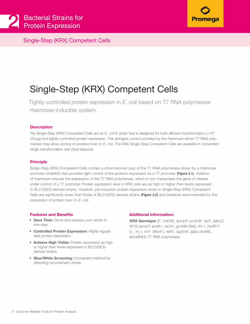
Single-Step (KRX) Competent CellsTightly-controlled protein expression in E. coli based on T7 RNA polymerase rhamnose-inducible system.
Description
The Single-Step (KRX) Competent Cells are an E. coli K strain that is designed for both efficient transformation (>108 cfu/μg) and tightly-controlled protein expression. The stringent control provided by the rhamnose-driven T7 RNA poly-merase may allow cloning of proteins toxic to E. coli. The KRX Single-Step Competent Cells are available in convenient single transformation size (50μl aliquots).
Principle
Single-Step (KRX) Competent Cells contain a chromosomal copy of the T7 RNA polymerase driven by a rhamnose promoter (rhaBAD) that provides tight control of the proteins expressed via a T7 promoter (Figure 2.1). Addition of rhamnose induces the expression of the T7 RNA polymerase, which in turn transcribes the gene of interest under control of a T7 promoter. Protein expression level in KRX cells are as high or higher than levels expressed in BL21(DE3)-derived strains. However, pre-induction protein expression levels in Single-Step (KRX) Competent Cells are significantly lower than those of BL21(DE3)-derived strains (Figure 2.2) and therefore recommended for the expression of protein toxic to E. coli.
Features and Benefits• Save Time: Clone and express your vector in
one step.
• Controlled Protein Expression: Highly regulat-able protein expression.
• Achieve High Yields: Protein expression as high or higher than levels expressed in BL21(DE3)-derived strains.
• Blue/White Screening: Convenient method for detecting recombinant clones.
Single-Step (KRX) Competent Cells
Additional Information:
KRX Genotype: [F´, traD36, ΔompP, proA+B+, lacIq, Δ(lacZ)M15] ΔompT, endA1, recA1, gyrA96 (Nalr), thi-1, hsdR17 (rk
–, mk+), e14– (McrA–), relA1, supE44, Δ(lac-proAB),
Δ(rhaBAD)::T7 RNA polymerase.

18 Discover Reliable Tools for Protein Analysis
Bacterial Strains for Protein Expression2
Figure 2.1. Tightly controlled inducible expression with L-Rhamnose in KRX E. coli. T7 RNA polymerase expression is under the control of the rhaP
BAD promoter in the KRX strain. This promoter is subject to multiple
levels of control. In the presence of preferred carbon sources, such as glucose, cyclic AMP (cAMP) concentrations are low and the cAMP receptor protein (CRP) does not activate transcription. Upon depletion of glucose, cAMP levels rise and CRP can activate transcription at rhaP
BAD.
In addition, L-rhamnose can bind to RhaR, which binds the rhaPSR promoter, resulting in the production of active RhaS and more RhaR. RhaS also binds rhamnose, which then binds the rhaP
BAD promoter,
resulting in the production of high levels of T7 RNA polymerase. The T7 RNA polymerase in turn transcribes the gene of interest.
ReferencesMalu, B et al. (2013) A nondiscriminating glutamyl-tRNA synthetase in the plasmodium apicoplast: the first enzyme in an indirect aminoacylation pathway. J. Biol. Chem. 288(45), 32539–52.
Barquilla, A et al. (2012) Third target of rapamycin complex negatively regu-lates development of quiescence in Trypanosoma brucei. Proc.Natl. Acad. Sci, 109(36), 14399–404.
Ordering Information Single-Step (KRX) Competent Cells (Cat.# L3002)
Single-Step (KRX) Competent Cells
WORD
Rhamnose
cAMP
1214
5MA
CRP
T7 RNA polymeraserhaPBADrhaPSRrhaSrhaR
RhaR
RhaS+
++

19 Discover Reliable Tools for Protein Analysis
Bacterial Strains for Protein Expression2
6032
MD
14
A. B.
8
43
1,700
1
10
100
1,000
10,000
BL21(DE3) BL21(DE3)pLysS
Rosetta™ 2pLysS
KRX
Indu
ctio
n Ra
tio
BL21(DE3) BL21(DE3)pLysS
Rosetta™ 2pLysS
KRX
Norm
aliz
ed lu
min
esce
nce
(RLU
/OD)
Pre-induction Expression Post-induction Expression
0
20
40
60
80
100
120
140
6032
MD
14
A. B.
8
43
1,700
1
10
100
1,000
10,000
BL21(DE3) BL21(DE3)pLysS
Rosetta™ 2pLysS
KRX
Indu
ctio
n Ra
tio
BL21(DE3) BL21(DE3)pLysS
Rosetta™ 2pLysS
KRX
Norm
aliz
ed lu
min
esce
nce
(RLU
/OD)
Pre-induction Expression Post-induction Expression
0
20
40
60
80
100
120
140
Single-Step (KRX) Competent Cells
Figure 2.2. Pre-induction and post-induction expression levels of firefly luciferase. KRX shows only very low pre-induction (Panel A) and very high post-induction levels (Panel B) compared to other strains such as BL21(DE3).

20 Discover Reliable Tools for Protein Analysis
Bacterial Strains for Protein Expression2
BL21 Competent Protein Expression CellsInducible recombinant protein expression in E. coli.
Description
BL21(DE3)pLysS Competent Cells and Single-Use BL21(DE3)pLysS Competent Cells allow high-efficiency protein expression of any gene that is under the control of a T7 promoter. The strain carries both the DE3 lysogen and the plasmid pLysS. pLysS constitutively expresses low levels of T7 lysozyme, which reduce basal expression of recombinant genes by inhibiting basal levels of T7 RNA polymerase. High protein expression is achieved by IPTG addition. Competent cells are available in standard format (200μl aliquots) as well as in 50μl aliquots.
Principle
BL21(DE3)pLysS is a derivative of BL21 that has the T7 RNA polymerase gene under the control of the lacUV5 promoter. This arrangement is on a phage genome, called λDE3. λDE3 is inserted into the chromosome of BL21 to make BL21(DE3). pLysS is a plasmid that contains the T7 lysozyme gene (LysS). The T7 lysozyme binds to T7 RNA polymerase causing inhibition until induction by the addition of IPTG. When IPTG is added, the amount of T7 RNA polymerase increases and over-comes the inhibition by LysS.
Features and Benefits• T7 RNA Polymerase under the Control of the lac
UV5 Promoter: Inducible protein expression.
• Deficient in Proteases lon and OmpT: Increased stability of expressed protein.
• pLysS Plasmid: Lower background expression of tar-get genes.
Additional Information:
Genotype: F–, ompT, hsdSB (rB–, mB–), dcm, gal, λ(DE3), pLysS, Cmr.
ReferencesFirdaus, M. et al. (2013) The pH sensitivity of murine heat shock protein 47 (HSP47) binding to collagen is affected by mutations in the breach histidine cluster. J. Biol. Chem. 288(6), 4452–61
Otsu, W. et al. (2013) A new class of endoplasmic reticulum export signal PhiXPhiXPhi for transmembrane proteins and its selective interaction with Sec24C. J. Biol. Chem. 288(25), 18521–61
Yamazaki, D. et al. (2013) srGAP1 regulates lamellipodial dynamics and cell migratory behavior by modulating Rac1 activity. Mol.Biol. Cell. 24(21), 3393–05
Ordering Information BL21(DE3)pLysS Competent Cells (Cat.# L1191)
Single-Use BL21(DE3)pLysS Competent Cells (Cat.# L1195)
BL21 Cells

21 Discover Reliable Tools for Protein Analysis
21 Discover Reliable Tools for Protein Analysis
Cell-Free Protein Expression Systems
3.1 Translation Systems: mRNA-based 25 Rabbit Reticulocyte Lysate System, Nuclease-Treated 26
Flexi® Rabbit Reticulocyte Lysate System 27
Wheat Germ Extract 28
3.2 Transcription and Translation Systems: DNA-based 29 Rabbit Reticulocyte Lysate Systems TnT® SP6 Coupled Reticulocyte Lysate System 31
TnT® T7 Coupled Reticulocyte Lysate System 31
TnT® T3 Coupled Reticulocyte Lysate System 31
TnT® T7 Quick Coupled Transcription/Translation System 31
TnT® SP6 Quick Coupled Transcription/Translation System 31
TnT® T7 Quick for PCR DNA 31
Wheat Germ Extracts TnT® SP6 High-Yield Wheat Germ Protein Expression System 33
Insect Cell Lysate System TnT® T7 Insect Cell Extract Protein Expression System 34
E. coli Extracts E. coli S30 Extract System for Linear Templates 35
S30 T7 High-Yield Protein Expression System 36
3.3 Cell-Free Protein Labeling Reagents 37
FluoroTect™ GreenLys in vitro Translation Labeling System 38
Transcend™ Non-Radioactive Translation Detection Systems 39
3.4 Membrane Vesicles for Signal Peptide Cleavage and Core Glycosylation 40
Canine Pancreatic Microsomal Membranes 41
3

22 Discover Reliable Tools for Protein Analysis
IntroductionCell-free protein synthesis is an important tool for molecular biologists in basic and applied sciences. It is increasingly being used in high-throughput functional genomics and proteomics, with significant advantages compared to protein expression in live cells. Cell-free protein synthesis is essential for the generation of protein arrays, such as nucleic acid programmable protein array (NAPPA) and enzyme engineering using display technologies. The cell-free approach provides the fastest way to correlate phenotype (function of expressed protein) to genotype. Protein synthesis can be performed in a few hours using either mRNA template in translational systems or DNA template (plasmid DNA or PCR fragments) in coupled transcription and translation systems. Furthermore, cell-free protein expression systems are indispensable for the expression of toxic proteins, membrane proteins, viral proteins and for proteins that undergo rapid proteolytic degradation by intracellular proteases.

23 Discover Reliable Tools for Protein Analysis
Cell-Free Protein Expression Systems3
Origins of Cell-Free Expression Systems
Cell-free protein expression lysates are generated from cells engaged in a high rate of protein synthesis, such as immature red blood cells (reticulocytes). The most frequently used cell-free expression systems originate from rabbit reticulocytes, wheat germ and E. coli. There are two types of cell-free expression systems: Translation Systems and Coupled Translation and Transcription (TnT®) Systems (Figure 3.1). Both types of systems provide the macromolecular components required for translation, such as ribosomes, tRNAs, aminoacyl-tRNA synthetases, initiation, elongation and termination factors. To ensure efficient translation, each extract has to be supplemented with amino acids, energy sources (ATP, GTP), energy regenerating systems and salts (Mg2+, K+, etc.). For eukaryotic systems creatine phosphate and creatine phosphokinase serve as energy regenerating system, whereas prokaryotic systems are supplemented with phosphoenol pyruvate and pyruvate kinase. Coupled transcription and translation systems are supplemented with phage-derived RNA polymerase (T7, T3 or SP6) allowing the expression of genes cloned downstream of a T7, T3 or SP6 promoter.
Selection of Cell-Free Protein Expression
Many different cell-free expression systems derived from prokaryotic and eukaryotic source are available. The choice of the system is dependent on several factors, including the origin of the template RNA and DNA, protein yield or whether the protein of interest requires post-translational modification (e.g., core glycosylation). We offer translation systems (mRNA-based) and coupled transcription/translation systems (DNA-based) from prokary-otic and eukaryotic sources. Table 3.2 provides an overview of translational systems and Table 3.3 provides an overview of coupled translation/tran-scription systems.
Figure 3.1. Cell-free protein expression systems are divided into mRNA-based translation systems and in DNA-based transcription/translation systems.
PlasmidDNA
PCR Fragmentsor
Cell-Free Translation Systems
Cell-Free Transcription/Translation Systems
mRNA
Protein
RNA
1217
6MA

24 Discover Reliable Tools for Protein Analysis
Table 3.1. Applications of Cell-Free Protein Synthesis
Functional Genome/ Proteome Analysis
• Gene mutation/deletion analysis (e.g., enzyme kinetics)
• Protein domain mapping
• Characterization of protein interactions
• Gel Shift EMSA
• Generation of protein arrays
Expression of Difficult-to- Express Proteins
• Cell-toxic proteins, membrane protein, viral proteins, kinases
Protein Evolution/ Enzyme Engineering
• Display technologies (e.g., ribosome, mRNA display, in vitro compartmental-ization)
• Evolution of antibodies in vitro by ribosome display
Analysis of Transcriptional/Translational Regulation
• Structural RNA analysis such as char-acterization of regulatory elements for translation (e.g., UTRs, Capping, IRES)
• RNA virus characterization
Screenings
• Screening of chemical libraries for effect on translation
• Drug screening (e.g., antibiotics)
Protein Labeling
• Open systems allow the incorporation of labeled amino acids

25 Discover Reliable Tools for Protein Analysis
Table 3.2. Overview of Cell-FreeTranslation Systems that use mRNA as a Template.
Translation System
Nuclease-Treated
Signal Cleavage & Core Glycosylation with CMM*
Labeling Options**
Luciferase Control RNA
Protein Yield
Rabbit Reticulocyte
Lysate System,
Nuclease-Treated
(Cat.# L4960)
+ +
Met,Cys,Leu,
FluoroTect™;
Transcend™
+ 1–4 µg/ml
Flexi® Rabbit
Reticulocyte Lysate
(Cat.# L4540) ***
+ +
Met, Cys, Leu,
FluoroTect™;
Transcend™
+ 1–4 µg/ml
Wheat Germ Extract
(Cat.# L4380)+ -
Met, Cys, Leu,
FluoroTect™;
Transcend™
+ 0.6–3 µg/ml
* CMM: Canine Microsomal Membranes
** The lysates are provided with three Amino Acid Mixtures for the incorporation of labeled amino acids like methionine, cysteine & leucine. Transcend™ tRNA (Cat.# L5070; L5080) and FluoroTect™ (Cat.# L5001) can be used to incorporate biotinylated or fluorescently labeled lysine residues.
*** The system provides greater flexibility of reaction conditions than standard rabbit reticulocyte lysate systems. The Flexi® Rabbit Reticulocyte Lysate System allows translation reactions to be optimized for a wide range of parameters, including Mg2+ and K+ concentrations and the option to add DTT.
Cell-free translation systems are used for protein expression of either in vitro transcribed mRNA or mRNA isolated from tissues or cells. These systems are used to express single proteins as well as multiple proteins in high-throughput applications such as display tech-nologies. Furthermore, cell-free translation systems are useful for functional and structural RNA analysis, or to study aspects of the translational machinery. Eukaryotic translation systems originate from either rabbit reticu-locyte lysates (RRL) or wheat germ extracts (WGE). We offer three mRNA-based translation systems. The extracts are treated with microccal nuclease to destroy
endogenous mRNA and thus reduce background translation to a minimum (Table 3.2).
The Flexi® Rabbit Reticulocyte Lysate System offers greater flexibility in reaction conditions by allowing translation reactions to be optimized for a wide range of parameters, including Mg2+ and K+ concentrations. The Wheat Germ Extract is a useful alternative to the RRL systems for expressing small proteins or for expressing proteins known to be abundant in RRL. Researchers expressing proteins from plants or yeasts or other fungi also may find WGE preferable to RRL.
3.1 Translation Systems: mRNA-based
OVERVIEW
Cell-Free Protein Expression Systems3

26 Discover Reliable Tools for Protein Analysis
Cell-Free Protein Expression Systems3
Rabbit Reticulocyte Lysate System, Nuclease-TreatedIn vitro protein synthesis starting from mRNA.
Description
Rabbit Reticulocyte Lysate (RRL), Nuclease-Treated, is optimized for mRNA translation by the addition of several supple-ments. These include hemin, which prevents activation of the heme-regulated eIF-2a kinase; an energy-generating system consisting of phosphocreatine kinase and phosphocreatine; and calf liver tRNAs to balance the accepting tRNA popula-tions, thus optimizing codon usage and expanding the range of mRNAs that can be translated efficiently. In addition, the lysates are treated with micrococcal nuclease to eliminate endogenous mRNA. RRLs post-translationally modify proteins via phosphorylation, acetylation and isoprenylation. Signal peptide cleavage and core glycosylation also can be achieved by the addition of Canine Pancreatic Microsomal Membranes. See Table 3.1 for additional applications.
Ordering Information Rabbit Reticulocyte Lysate (RRL), Nuclease-Treated (Cat.# L4960)
Principle
In RRL translation reactions, mRNA is used as template for translation. In general, optimal results will be achieved after an incubation time of 1.5 hours at 30°C. However, many template-related factors affect translation efficiency of specific mRNAs in the RRL system and should be considered when designing in vitro translation experiments. The optimal mRNA concentration will vary for particular transcripts and should be determined empirically. In addition, the presence of certain nucleic acid sequence elements can have profound effects on initiation fidelity and translation efficiency. Poly(A)+ tails, 5´-caps, 5´-untranslated regions and the sequence context around the AUG start (or secondary AUGs in the sequence) all may affect translation of a given mRNA.
Features and Benefits• Consistent: Reliable and consistent translation with each
lot.
• Optimized and Ready-to-Use: The treated Rabbit Reticulocyte Lysate is optimized for translation.
• Convenient: Luciferase Control RNA included.
Figure 3.2. Flow chart of in vitro translation procedure using Rabbit Reticulocyte Lysate.
Translation Systems: mRNA-based
mRNA
Protein
mRNA
1214
6MB
Rabbit Reticulocyte
Lysate
Incubate for 1.5 hour at 30°C.
Synthesize RNA in vitro orIsolate mRNA from tissue cells
Analyze with activity assays or protein detection.

27 Discover Reliable Tools for Protein Analysis
Cell-Free Protein Expression Systems3Translation Systems: mRNA-based
Flexi® Rabbit Reticulocyte Lysate SystemIn vitro protein synthesis starting from mRNA. Optimize translation for low-expressing mRNA.
Description
The Flexi® Rabbit Reticulocyte Lysate System is widely used to identify mRNA species and characterize their products. It provides greater flexibility of reaction conditions than the Rabbit Reticulocyte Lysate, Nuclease-Treated, by allowing translation reactions to be optimized for a wide range of parameters, including Mg2+ and K+ concentrations. See Table 3.1 for additional applications.
Ordering Information Flexi® Rabbit Reticulocyte Lysate System (Cat.# L4540)
Principle
As with the standard Rabbit Reticulocyte Lysate, the Flexi® Rabbit Reticulocyte Lysate System is optimized for translation by addition of the following supplements: hemin, to prevent inhibition of initiation factor eIF-2α; an energy-generating system consisting of pretested phosphocreatine kinase and phosphocreatine; calf liver tRNAs to balance the accepting tRNA populations, thus optimizing codon usage and expanding the range of mRNAs that can be translated efficiently; and micro-coccal nuclease to eliminate endogenous mRNA, thus reducing background translation. This eukaryotic system has been reported to post-translationally modify proteins via phosphorylation, acetylation and isoprenylation. With the addition of Canine Pancreatic Microsomal Membranes signal peptide cleavage and core glycosyl-ation can occur. The Flexi® Rabbit Reticulocyte Lysate System provides greater flexibility of reaction conditions than standard RRL systems.
Features and Benefits• Consistent: Reliable and consistent translation with
each lot.
• Easy Optimization: To aid in optimizing magnesium concentrations, the endogenous magnesium concentration is provided for each lot of Flexi® Lysate.
• Convenient: Luciferase Control RNA and detection reagent included.

28 Discover Reliable Tools for Protein Analysis
Cell-Free Protein Expression Systems3Translation Systems: mRNA-based
Wheat Germ ExtractIn vitro protein synthesis starting from mRNA.
Description
Wheat Germ Extract (WGE) is a well-defined processed and optimized extract from wheat germ. It contains the cellular components necessary for protein synthesis (tRNA, ribosomes and initiation, elongation and termination factors). The extract is supplemented with an energy-generating system (phosphocreatine/phosphocreatine kinase), and with spermidine to stimulate the efficiency of chain elongation. Only exogenous amino acids and mRNA are needed to initiate translation. Potassium acetate can be used to optimize translation for a wide range of mRNAs. See Table 3.1 for additional applications.
Principle
Wheat Germ Extract is a useful alternative to the Rabbit Reticulocyte Lysate (RRL) systems for expressing small proteins or for expressing proteins expected to be abundant in RRL. Researchers expressing proteins from plants, yeast or other fungi also may find Wheat Germ Extract preferable to RRL.
Features and Benefits• Optimized Extract: Assists in expression of eukaryotic
messages that do not express well in RRL.
• Flexible: Three Amino Acid Mixtures are provided, which enable different radioisotope choices.
• Robust: Potassium Acetate is provided to enhance translation for a wide range of mRNAs.
• Convenient: Luciferase Control RNA included.
Ordering Information Wheat Germ Extract (Cat.# L4380)

29 Discover Reliable Tools for Protein Analysis
Cell-Free Protein Expression Systems3
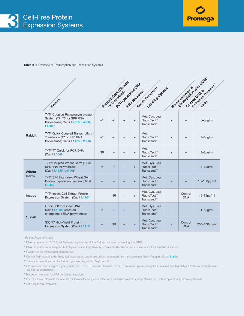
Coupled transcription and translation (TnT®) systems offer researchers time-saving alternatives for eukary-otic in vitro transcription and translation, by coupling these processes in a single tube format. TnT® Systems are used for a variety of applications in low- to high-throughput functional genome and proteome analyses, as summarized in Table 3.1. TnT® Systems are supple-mented with T7, T3 or SP6 RNA polymerases, allowing protein expression from DNA cloned downstream of a T7, T3 or SP6 promoter.
We offer TnT® Systems originating from eukaryotic sources such as rabbit reticulocyte, wheat germ and insect cells as well as from prokaryotic E. coli extracts (Table 3.3).
The highest production rates are normally achieved with E. coli extracts. However, eukaryotic systems often produce eukaryotic proteins with higher activity. Therefore, the origin of the protein of interest should be considered when selecting a cell-free expression system.
DNA Template Consideration: Plasmids and PCR-Fragments
The performance of cell-free systems depends on the DNA template used. Basically, any vector containing T7, SP6 or T3 promoters can be used with TnT® Systems. However, there are several points to consider when engi-neering a DNA fragment or plasmid for optimal expres-sion in a eukaryotic system: (i) the ATG start codon in the sequence should be the first ATG encountered following the transcription start site; (ii) ideally, following the promoter, the ATG is included in a Kozak consensus sequence; (iii) a stop codon should be included at the 3´- terminus of the sequence; and (iv) a synthetic poly(A) tail should be included following the stop codon. Additionally, vectors used in the TnT® T7 Coupled Wheat Germ System should either be linearized or have a T7
transcription terminator in a circular template.
In prokaryotic systems, the selection of a start codon gener-ally depends on the presence of a ribosomal binding site (RBS; Shine-Dalgarno sequence), which contains a signal that marks the start of the reading frame. The presence of an optimal RBS can greatly increase expression in prokary-otic systems. The prokaryotic system does not recognize ATGs upstream of the ATG start codon unless they contain a properly positioned RBS.
Promega vectors approved for use with TnT® Systems can be found in Table 9.1.
The template considerations mentioned above are also valid for using PCR fragments as templates for the TnT® reaction. For the generation of the PCR fragments for protein expres-sion in eukaryotic systems, the integration of a Kozak sequence downstream of a T7 or SP6 promoter is recom-mended (Figure 3.4).
Labeling of Proteins during in vitro Synthesis All TnT® Systems are provided with three different Amino Acid Mixtures for the incorporation of radiolabeled amino acids like methionine, cysteine and leucine. Transcend™ tRNA and FluoroTect™ Systems can be used to incorporate biotinylated or fluorescently-labeled lysine residues (see Section 3.3).
Signal Peptide Cleavage and Core Glycosylation
Rabbit reticulocyte lysate has been reported to post-transla-tionally modify proteins via phosphorylation, acetylation and isoprenylation. However, the addition of Canine Pancreatic Microsomal Membranes (CMM), to RRLs is required to achieve signal peptide cleavage and core glycosylation of the translation product.
3.2 Transcription and Translation Systems: DNA-based
OVERVIEW
30 Discover Reliable Tools for Protein Analysis
Cell-Free Protein Expression Systems3
Table 3.3. Overview of Transcription and Translation Systems
NR: Not Recommended1 DNA templates for TnT® E.coli Systems requires the Shine Dalgarno ribosomal binding site (RBS).2 DNA templates for eukaryotic TnT® Systems should preferably contain the Kozak consensus sequence for translation initiation.3 CMM: Canine Microsomal Membranes. 4 Control DNA contains the firefly luciferase gene. Luciferase activity is detected by the Luciferase Assay Reagent (Cat.# E1500). 5 Translation reactions can be further optimized by adding Mg2+ and K+.6 SP6 circular plasmids give higher yields than T7 or T3 circular plasmids; T7 or T3 linearized plasmid may be considered as templates; SP6 linearized plasmids
are not recommended.7 Not recommended for SP6 containing template.8 For T7 circular plasmids include the T7 terminator sequence; otherwise linearized plasmids are preferred; for SP6 templates only circular plasmids.9 Only linearized templates.
Rabbit
TnT® Coupled Reticulocyte Lysate System (T7, T3, or SP6 RNA Polymerase; Cat.# L4610, L4950, L4600)5
+6 +7 – +Met, Cys, Leu, FluoroTect™, Transcend™
+ + 3–6µg/ml
TnT® Quick Coupled Transcription/Translation (T7 or SP6 RNA Polymerase; Cat.# L1170, L2080)
+6 +7 – +Met, FluoroTect™, Transcend™
+ + 3–6µg/ml
TnT® T7 Quick for PCR DNA (Cat.# L5540)
NR + – +Met, FluoroTect™, Transcend™
+ – 3–6µg/ml
Wheat Germ
TnT® Coupled Wheat Germ (T7 or SP6 RNA Polymerase) (Cat.# L4130, L4140)4
+8 +7 – +Met, Cys, Leu, FluoroTect™, Transcend™
– + 3–6µg/ml
TnT® SP6 High-Yield Wheat Germ Protein Expression System (Cat.# L3260)
+ + – +Met, Cys, Leu, FluoroTect™, Transcend™
– – 10–100µg/ml
InsectTnT® Insect Cell Extract Protein Expression System (Cat.# L1101)
+ NR – +Met, Cys, Leu, FluoroTect™, Transcend™
–Control
DNA15–75µg/ml
E. coli
E coli S30 for Linear DNA (Cat.# L1030) relies on endogenous RNA polymerases
+9 + + –Met, Cys, Leu, FluoroTect™, Transcend™
– + 1–5µg/ml
S30 T7 High-Yield Protein Expression System (Cat.# L1110)
+ NR + –Met, Cys, Leu, FluoroTect™, Transcend™
–Control
DNA200–500µg/ml
Syste
m
Control D
NA &
D
etec
tion R
eagen
t4
Labeli
ng Optio
ns
Plasm
id D
NA (Circ
ular
o
r Lin
eariz
ed)
Signal
cleav
age
&
g
lycosy
latio
n with
CM
M3
RBS Req
uired1
Kozak
Prefe
rred2
PCR-gen
erat
ed D
NA
Yield

31 Discover Reliable Tools for Protein Analysis
TnT® Coupled Reticulocyte Lysate SystemsRobust eukaryotic cell-free expression systems for the expression of functional mammalian proteins in a simple one-step procedure.
1537
MD
TNT® Quick Master Mix
Add TNT® RNA Polymerase
Add TNT® RNA Reaction Buffer
TNT® Rabbit Reticulocyte Lysate.
1. Add label of choice.
2. Add DNA template and Nuclease-Free Water.
3. Inubate at 30∘C for 60–90 minutes.
4. Separate translation products by SDS-PAGE.
Add Amino Acid Mixture Minus Methionine.
Add RNasin® Ribonuclease Inhibitor
TNT® CoupledReticulocyte
Lysate System
DetectTNT® Quick Coupled
Transcription/Translation
System Less Time,Less Handling!
Figure 3.3. Comparison of the TnT® Coupled Reticulocyte Lysate System and the TnT® Quick Coupled Transcription/Translation System protocols.
Description and Principle
We offer two types of Rabbit Reticulocyte Lysate Transcription and Translation (TnT®) Systems: The TnT® Coupled (T7, T3, SP6) System and the TnT® Quick Coupled (T7, SP6) System. The main difference between these systems is that the TnT® Quick Coupled System provides a master mix containing all the reaction compo-nents required in one tube, whereas the TnT® Coupled System has all the reaction components provided in separate tubes (Figure 3.3). TnT® T7 Quick for PCR DNA is a rapid and convenient coupled TnT® System designed for expression of PCR-generated DNA templates. The system is robust and able to express a variety of proteins ranging in size from 10–150kDa. The lysates are supplied with all reagents needed for TnT® reactions including RNA
Transcription and Translation Systems: DNA-based
Cell-Free Protein Expression Systems3
polymerases. To use these systems, DNA is added directly to TnT® Lysate and incubated in a 50μl reaction for 60–90 minutes at 30°C. See Table 3.1 for additional applications.
Features and Benefits• Use in Multiple Applications: The TnT® Systems are
widely used for functional genomics and proteomics analyses.
• Save Time: The TnT® Reaction is completed in a maxi-mum of 1.5 hours.
• Complete System: All reagents for the TnT® Reaction are provided (except for labeled amino acids).
• Reliable: Can eliminate solubility issues by using an in vitro mammalian system.

32 Discover Reliable Tools for Protein Analysis
Cell-Free Protein Expression Systems3Transcription and Translation Systems: DNA-based
2802
TA
1 2 3 4 5 1 2 3 4 5
1 2 3 4 5 1 2 3 4 5
1 2 3 4 5 1 2 3 4 5
A.
B.
C.
TNT® T7 Quick TNT® T7 Quick for PCR
Vendor A TNT® T7 Quick for PCR
Vendor B TNT® T7 Quick for PCR
Figure 3.5. TnT® T7 Quick for PCR was used to express variants of the APC gene and BRCA1 gene. PCR fragments were used as starting material for the TnT® reaction. Transcend™ tRNA was included in the reaction for the incorporation of biotinylated lysine residues. Lane 1 contains the no DNA controls; lane 2, APC Seg 2 PCR fragment; lane 3, APC Seg 3 PCR DNA fragment; lane 4, BRCA1 Seg 3 PCR fragment; lane 5, the Luciferase T7 Control DNA.
Ordering InformationTnT® Coupled Reticulocyte Lysate Systems: TnT® SP6 Coupled Reticulocyte Lysate System (Cat.# L4600)
TnT® T7 Coupled Reticulocyte Lysate System (Cat.# L4610)
TnT® T3 Coupled Reticulocyte Lysate System (Cat.# L4950)
TnT® Quick Coupled Transcription/ Translation Systems: TnT® T7 Quick Coupled Transcription/Translation System (Cat.# L1170)
TnT® SP6 Quick Coupled Transcription/Translation System (Cat.# L2080)
TnT® T7 Quick for PCR DNA (Cat.# L5540)
5’(N)6–10 - TATTTAGGTGACACTATAG(N)3–6 - CCACCATGG - (N)17–22 -3’
SP6 Promoter Kozak region Sequence-specific Nucleotides
5’(N)6–10- TAATACGACTCACTATAGGG(N)3–6 - CCACCATGG - (N)17–22 -3’
T7 Promoter Kozak region Sequence-specific Nucleotides
Eukaryotes
SP6
T7
1214
7MB
Figure 3.4. Forward primers used to generate PCR fragments for protein expression in TnT® Systems.

33 Discover Reliable Tools for Protein Analysis
Cell-Free Protein Expression Systems3
TnT® SP6 High-Yield Wheat Germ Protein Expression SystemIn vitro protein synthesis starting from DNA.
Description
The TnT® SP6 High-Yield Wheat Germ Protein Expression System is a convenient, quick, single-tube, coupled transcrip-tion/translation system designed to express up to 100μg/ml of protein. The TnT® SP6 High-Yield Wheat Germ Protein Expression System expresses genes cloned downstream of an SP6 RNA polymerase promoter. This cell-free expression system is prepared from an optimized wheat germ extract and contains all the components (tRNA, ribosomes, amino acids, SP6 RNA polymerase, and translation initiation, elongation and termination factors) necessary for protein synthesis directly from DNA templates. See Table 3.1 for additional applications.
Principle
The TnT® SP6 High-Yield Wheat Germ Protein Expression System can be used with standard plasmid DNA or PCR-generated templates containing the SP6 promoter. However, to achieve optimal yield, specialized vectors designed for Wheat Germ Extracts such as pF3A WG (BYDV) Flexi® Vector or pF3K WG (BYDV) Flexi® Vector are recommended. DNA templates are directly added to the SP6 High Yield Master Mix and incubated in a 50μl reaction for 2 hours at 25°C. Expressed proteins can be used directly or purified for related applications.
Features and Benefits• Save Time: Generate proteins in two hours,
compared to days when using cell-based (E. coli) systems.
• Choose Your Format: Use plasmid- or PCR-generated templates.
• Generate Full-Length Protein: Generate soluble, full-length protein and avoid problems associated with E. coli systems.
Ordering Information TnT® SP6 High-Yield Wheat Germ Protein Expression System (Cat.# L3260, L3261)
Figure 3.6. Proteins of different size and origin were expressed using TnT® SP6 High-Yield Wheat Germ Protein Expression System in the presence of FluoroTect™ tRNA for lysine residue labeling. Samples were separated by SDS-PAGE and imaged using a fluorescence scanner.
9873
TA
TNT® T7 Quick System andFluoroTect™ tRNA
TNT® T7 Quick System andTranscend™ tRNA
TNT® Wheat Germ Systemand FluoroTect™ tRNA
TNT® Wheat Germ Systemand Transcend™ tRNA
GFPeIF
4E
Nanos
1
PPP1ca
Lucif
eras
e
Minus-D
NA
Contro
l
GFPeIF
4E
Nanos
1
PPP1ca
Lucif
eras
e
Minus-D
NA
Contro
l GFPeIF
4E
Nanos
1
PPP1ca
Lucif
eras
e
Minus-D
NA
Contro
l
GFPeIF
4E
Nanos
1
PPP1ca
Lucif
eras
e
Minus-D
NA
Contro
l
25015010075
50
37
25
102
7652
38
24
17
kDa
kDa 102
7652
38
24
17
kDa
25015010075
50
37
25
kDa
A. B.
C. D.
Transcription and Translation Systems: DNA-based
34 Discover Reliable Tools for Protein Analysis
Cell-Free Protein Expression Systems3
TnT® T7 Insect Cell Extract Protein Expression SystemIn vitro protein synthesis starting from a DNA template.
Description
The TnT® T7 Insect Cell Extract Protein Expression System is a convenient, quick, single-tube, coupled transcription and translation system for the cell-free expression of proteins. See Table 3.1 for additional applications.
Principle
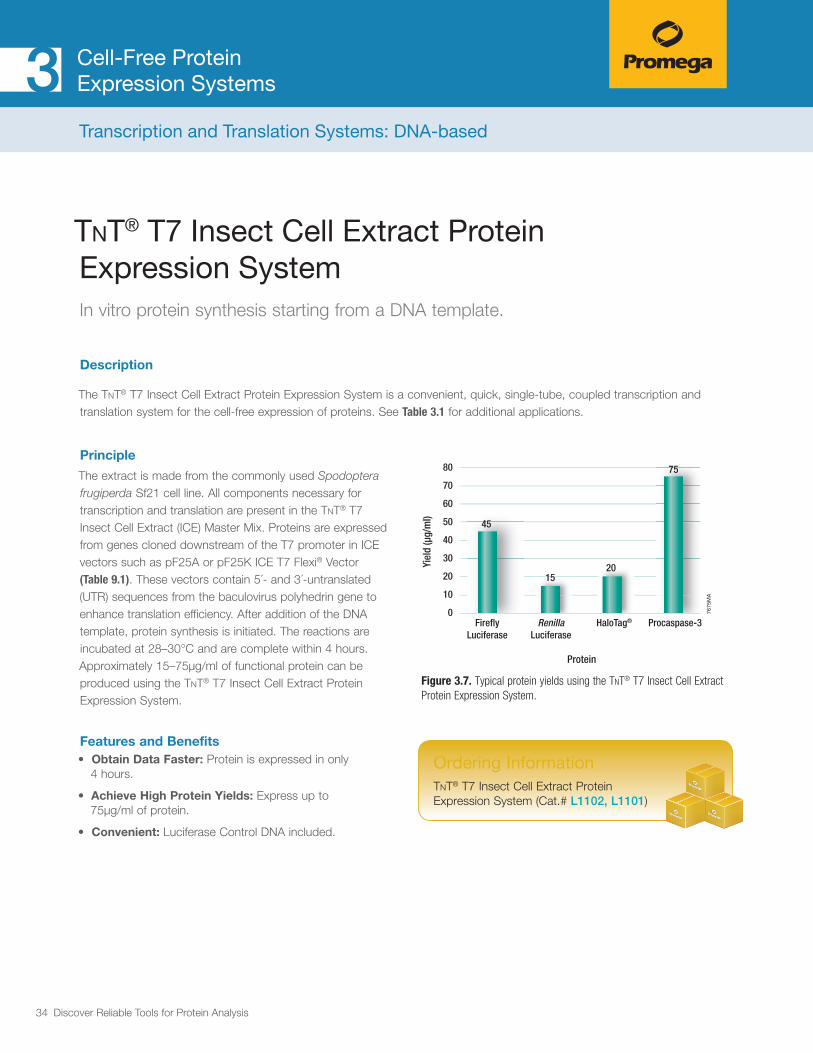
The extract is made from the commonly used Spodoptera frugiperda Sf21 cell line. All components necessary for transcription and translation are present in the TnT® T7 Insect Cell Extract (ICE) Master Mix. Proteins are expressed from genes cloned downstream of the T7 promoter in ICE vectors such as pF25A or pF25K ICE T7 Flexi® Vector (Table 9.1). These vectors contain 5´- and 3´-untranslated (UTR) sequences from the baculovirus polyhedrin gene to enhance translation efficiency. After addition of the DNA template, protein synthesis is initiated. The reactions are incubated at 28–30°C and are complete within 4 hours. Approximately 15–75μg/ml of functional protein can be produced using the TnT® T7 Insect Cell Extract Protein Expression System.
Features and Benefits• Obtain Data Faster: Protein is expressed in only
4 hours.
• Achieve High Protein Yields: Express up to 75μg/ml of protein.
• Convenient: Luciferase Control DNA included.
0
10
20
30
40
60
50
80
70
1520
75
45
Yiel
d (µ
g/m
l)
FireflyLuciferase
RenillaLuciferase
HaloTag® Procaspase-3
Protein
7675
MA
Figure 3.7. Typical protein yields using the TnT® T7 Insect Cell Extract Protein Expression System.
Ordering Information TnT® T7 Insect Cell Extract Protein Expression System (Cat.# L1102, L1101)
Transcription and Translation Systems: DNA-based

35 Discover Reliable Tools for Protein Analysis
Cell-Free Protein Expression Systems3
E. coli S30 Extract System for Linear TemplatesIn vitro protein synthesis starting from DNA.
Description
The E. coli S30 Extract System for Linear Templates allows successful transcription/translation of linear DNA templates. You need only to provide linear DNA containing a prokaryotic E. coli-like promoter (such as lacUV5, tac, λPL (con) and λ-PR). A ribosome binding site is required to direct the synthesis of proteins in vitro. In vitro-generated RNA from DNA templates lacking an E. coli promoter may also be used in this system, but protein yields produced from linear DNA templates will be decreased 1–10%.
Principle
The S30 Extract for Linear Templates is prepared from an E. coli B strain (SL119), which is deficient in OmpT endoproteinase, lon protease and exonuclease V (recBCD). The absence of protease activity results in greater stability of expressed proteins. The recD mutation of the SL119 strain produces a more active S30 Extract for Linear DNA than the previously described nuclease-deficient, recBC-derived S30 extracts. However, the S30 Extract for Linear Templates is less active than the S30 Extract System for Circular DNA. An easy-to-perform, nonradioactive positive control reaction using the Luciferase Assay Reagent provided, allows detection of luciferase gene expres-sion in the E. coli S30 System for linear templates. The control reaction produces high light output for several minutes, allowing the researcher to choose from several detection methods, including simple visual observation of luminescence.
Features and Benefits• Flexible: Various templates can be used: DNA fragments,
PCR-synthesized DNA, ligated overlapping oligonucle-otides, in vitro-generated RNA and prokaryotic RNA.
• Complete: Contains all necessary components for coupled transcription/translation.
• Optimized: Premix is optimized for each lot of S30 Extract.
• Control DNA: Easy detection of firefly luciferase expression using (included) Luciferase Assay Reagent.
Ordering Information E. coli S30 Extract System for Linear Templates (Cat.# L1030)
Transcription and Translation Systems: DNA-based

36 Discover Reliable Tools for Protein Analysis
Cell-Free Protein Expression Systems3
E. coli S30 T7 High-Yield Protein Expression SystemIn vitro protein synthesis starting from DNA.
Description
The S30 T7 High-Yield Protein Expression System is an E. coli extract-based protein synthesis system. It simplifies the tran-scription and translation of DNA sequences cloned in plasmid or lambda vectors containing a T7 promoter, by providing an extract that contains T7 RNA polymerase for transcription and all necessary components for translation.
Principle
The E. coli S30 T7 High-Yield Protein Expression System is designed to express up to 500μg/ml of protein in one hour from plasmid vectors containing a T7 promoter and a ribosome binding site. The protein expression system provides an extract that contains T7 RNA polymerase for transcription and is deficient in OmpT endoproteinase and lon protease activity. All other necessary components in the system are optimized for protein expression. This results in greater stability and enhanced expression of target proteins. Control DNA expression results in production of Renilla luciferase, which can be detected by Coomassie® Blue staining following SDS-PAGE or assayed with Renilla Luciferase Assay System (Cat.# E2810).
Features and Benefits• Obtain Data Faster: Protein expression in only one hour.
• Achieve High Protein Expression: Express up to 500μg/ml of protein for multiple applications.
• Scalable: Convenient screening protocol for high-throughput protein expression.
Figure 3.8. Coupled in vitro transcription/translation of circular DNA templates using the S30 T7 High-Yield Protein Expression System. The protein-coding sequences cloned into pFN6A (HQ) Flexi® Vector were expressed as described in the S30 T7 High-Yield Protein Expression System Technical Manual #TM306, resolved by SDS-PAGE (4–20% Tris-glycine) and visualized by Coomassie® blue staining (Panel A), fluorescence scanning (Panel B), or transferred to PVDF membrane, treated with Streptavidin Alkaline Phosphatase (Cat.# V5591) and stained with Western Blue® Stabilized Substrate for Alkaline Phosphatase (Cat..# S3841; Panel C). For each gel: lane 1, no DNA; lane 2, Renilla luciferase; lane 3, Monster Green® Fluorescent Protein; lane 4, HaloTag® protein; lane 5, α-galactosidase (BCCP = E. coli biotin carboxyl carrier protein).
Ordering Information S30 T7 High-Yield Protein Expression System (Cat.# L1110, L1115)
7637
TA
10075
503525
15
kDa1 2 3 4 5 1 2 3 4 5 1 2 3 4 5
A. B. C.
BCCP
Transcription and Translation Systems: DNA-based

37 Discover Reliable Tools for Protein Analysis
Cell-Free Protein Expression Systems3
Labeling and detection of proteins expressed using cell-free systems is necessary for most applications such as protein:protein interaction and protein:nucleic acid interaction studies. FluoroTect™ Detection and Transcend™ Detection Systems were developed for non-radioactive protein labeling during cell-free protein synthesis. Both labeling products are based on the incorporation of labeled lysine residues into the polypeptide chain. The labeled protein products can be easily detected either by fluorescent imaging after SDS-PAGE or by western blotting using streptavidin conjugates either to horse-radish peroxidase (Strep-HRP) or Alkaline Phosphatase (Strep-AP).
3.3 Cell-Free Protein Labeling Reagents
OVERVIEW
Figure 3.9. Detection protocols using FluoroTect™ GreenLys
tRNA and Transcend™ tRNA.
Standard RadioisotopicIncorporation and Detection
Transcend™ Biotinylated Lysine tRNAIncorporation and Detection
M* M*Translation withincorporation of [35S]-met (1 hour)
Translation withincorporation of biotinylated lysine (1 hour)
SDS PAGE (1 hour)
Fix gel (30 minutes)
SDS PAGE (1 hour)
L LL
Chemiluminescentdetection
Colorimetricdetection
Transfer to PVDFor nitrocellulosemembrane (1 hour)
Transfer to PVDF or nitrocellulose membrane (1 hour)
Block, bind Strep-AP, wash (2 hours)
Block, bind Strep-HRP, wash (2 hours)
Add Chemiluminescent Substrate and expose to X-ray film(2−20 minutes)
Add Western Blue® Substrate to develop colored bands (1−10 minutes)
Expose to X-ray film(4−10 hours)
Treat with enhancer (30 minutes)
Dry gel (1 hour)
FluoroTect™ GreenLys tRNAIncorporation and Detection
L* L*Translation withincorporation of fluorescent lysine (1 hour)
SDS PAGE (1 hour)
Detection using afluoroimaging instrument(2−5 minutes)
0878MD10_0A
38 Discover Reliable Tools for Protein Analysis
Cell-Free Protein Expression Systems3Cell-Free Protein Labeling Reagents
FluoroTect™ GreenLys in vitro Translation Labeling SystemLabeling and detection of in vitro synthesized proteins.
Description
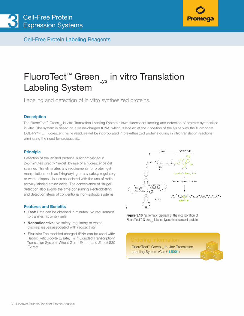
The FluoroTect™ GreenLys in vitro Translation Labeling System allows fluorescent labeling and detection of proteins synthesized in vitro. The system is based on a lysine-charged tRNA, which is labeled at the ε position of the lysine with the fluorophore BODIPY®-FL. Fluorescent lysine residues will be incorporated into synthesized proteins during in vitro translation reactions, eliminating the need for radioactivity.
Principle
Detection of the labeled proteins is accomplished in 2–5 minutes directly “in-gel” by use of a fluorescence gel scanner. This eliminates any requirements for protein gel manipulation, such as fixing/drying or any safety, regulatory or waste disposal issues associated with the use of radio-actively-labeled amino acids. The convenience of “in-gel” detection also avoids the time-consuming electroblotting and detection steps of conventional non-isotopic systems.
Features and Benefits• Fast: Data can be obtained in minutes. No requirement
to transfer, fix or dry gels.
• Nonradioactive: No safety, regulatory or waste disposal issues associated with radioactivity.
• Flexible: The modified charged tRNA can be used with: Rabbit Reticulocyte Lysate, TnT® Coupled Transcription/Translation System, Wheat Germ Extract and E. coli S30 Extract.
Figure 3.10. Schematic diagram of the incorporation of FluoroTect™ Green
Lys-labeled lysine into nascent protein.
Ordering Information FluoroTect™ GreenLys in vitro Translation Labeling System (Cat.# L5001)

39 Discover Reliable Tools for Protein Analysis
Cell-Free Protein Expression Systems3
Transcend™ Nonradioactive Translation Detection SystemsLabeling and detection of in vitro synthesized proteins.
Description
The Transcend™ Nonradioactive Translation Detection Systems allow nonradioactive detection of proteins synthesized in vitro. Using these systems, biotinylated lysine residues are incorporated into nascent proteins during translation, eliminating the need for labeling with [35S]methionine or other radioactive amino acids
Principle
This biotinylated lysine is added to the translation reaction as a precharged ε-labeled biotinylated lysine-tRNA complex (Transcend™ tRNA) rather than a free amino acid. After SDS-PAGE and blotting, the bioti-nylated proteins can be visualized by binding either Streptavidin-Alkaline Phosphatase (Streptavidin-AP) or Streptavidin-Horseradish Peroxidase (Streptavidin-HRP), followed either by colorimetric or chemiluminescent detection (see Chapter 8). Typically, these methods can detect 0.5–5ng of protein within 3–4 hours after gel electrophoresis. This sensitivity is equivalent to that achieved with [35S]methionine incorporation and autoradiographic detection 6–12 hours after gel electro-phoresis.
Features and Benefits• Sensitive: The biotin tag allows detection of 0.5–5ng
of translated protein.
• Safe: No radioisotope handling, storage or disposal is required.
• Flexible: Results can be visualized by using colorimetric or chemiluminescent detection.
Figure 3.11. Schematic diagram of the incorporation of Transcend™ labeled lysine into nascent protein.
ACC O
C
OO C
O
NH3
ε
α
NH SN
N
tRNA
lysine spacer arm biotin
Transcend™ Biotinylated tRNA
+
Translation reaction
Lys Lys Lys
Biotin
0877
MC
Ordering Information Transcend™ Colorimetric Translation Detection System (Cat.# L5070)
Transcend™ Chemiluminescent Translation Detection System (Cat.# L5080)
Cell-Free Protein Labeling Reagents

40 Discover Reliable Tools for Protein Analysis
Cell-Free Protein Expression Systems3
12
29
1M
A
mRNA
Translation
Membrane insertion
Protein
Mature protein
Signalpeptide
Microsomalmembranes
Signal peptidecleavage
Glycosylation
Microsomal vesicles are used to study cotranslational and initial post-translational processing of proteins. Processing events such as signal peptide cleavage, membrane insertion, translocation and core glycosylation can be examined by the translation of the appropriate mRNA in vitro in the presence of these microsomal membranes.
3.4 Membrane Vesicles for Signal Peptide Cleavage and Core Glycosylation
OVERVIEW
Figure 3.12. Schematic of signal peptide cleavage and introducing core glycosylation by use of canine microsomal membranes in combination with rabbit reticulocyte lysate cell-free protein expression system.

41 Discover Reliable Tools for Protein Analysis
Cell-Free Protein Expression Systems3
Canine Pancreatic Microsomal MembranesExamination of signal peptide cleavage, membrane insertion, translocation and core glycosylation of in vitro expressed proteins.
Description
Canine Pancreatic Microsomal Membranes are used to study cotranslational and initial posttranslational processing of proteins in combination with in vitro expressed protein using Rabbit Reticulocyte Systems (RRLs). Processing events such as signal peptide cleavage, membrane insertion, translocation and core glycosylation can be examined by the translation of the appropriate mRNA in vitro in the presence of these microsomal membranes. In addition, processing and glycosylation events may be studied by transcription/translation of the appropriate DNA in TnT® RRL Systems.
Principle
Processing and glycosylation events can be studied with Rabbit Reticulocyte Lysate Cell-free expression systems. To assure consistent performance with minimal translational inhibition and background, microsomes have been isolated free of contaminating membrane fractions and stripped of endogenous membrane-bound ribosomes and mRNA. Membrane preparations are assayed for both signal peptidase and core glycosyl-ation activities using two different control mRNAs. The two control mRNAs supplied with this system are the precursor for β-lactamase (or ampicillin resistance gene product) from E. coli and the precursor for α-mating factor (or α-factor gene product) from S. cerevisiae.
Ordering Information Canine Pancreatic Microsomal Membranes (Cat.# Y4041)
Membrane Vesicles for Signal Peptide Cleavage and Core Glycosylation
Features and Benefits• Minimal Translational Inhibition, Minimal
Background: Microsomes are stripped of endogenous membrane-bound ribosomes and mRNA.
• Compatible: Can be used with TnT® RRL Systems, Rabbit Reticulocyte Lysate and Flexi® Lysate.
• Reliable Results: Control mRNAs are supplied.

42 Discover Reliable Tools for Protein Analysis
Cell-Free Protein Expression Systems3
Overview Articles
Arduengo, M et al. (2007) The role of cell-free rabbit reticulocyte lystate expression systems in func-tional proteomics. In: Cell Free Protein Expression. Kudlicki, W.A. et al. eds. Landes Bioscience, Austin, TX. 1–18.
Functional Genome/Proteome Analysis
Gene Mutation/Deletion Analysis (e.g., Enzyme Kinetics)
Park, N., Skern, T. and Gustin, K.E. (2010) Specific cleavage of the nuclear pore complex protein Nup62 by a viral protease. J. Biol. Chem. 285(37), 28796–805.
Protein Domain Mapping
Wong, R.W. and Blobel, G. (2008) Cohesin subunit SMC1 associates with mitotic microtubules at the spindle pole. Proc. Natl. Acad. Sci. USA 105(40), 15441–5.
Characterization of Protein Interactions
Wong, R.W. and Blobel, G. (2008) Cohesin subunit SMC1 associates with mitotic microtubules at the spindle pole. Proc. Natl. Acad. Sci. USA 105(40), 15441–5.
Tando, T. et al. (2010) Requiem protein links RelB/p52 and the Brm-type SW1/SNF complex in a noncanonical NF-kB pathway. J. Biol. Chem. 285(29), 21951–60.
Delgehyr, N. et al. (2012) Drosophila Mgr, a Prefoldin subunit cooperating with von Hippel Lindau to regulate tubulin stability. Proc. Natl. Acad. Sci. USA 109(25), 5729–34.
Muratore, G. et al. (2012) Small molecule inhibitors of influenza A and B viruses that act by disrupting subunit Interactions of the viral polymerase. Proc. Natl. Acad. Sci. USA 109(16) 6247–52.
Gel Shift EMSA
Fuchs, A. et al. (2012) Transcriptional Interpretation of the EGF receptor signaling gradient. Proc. Natl. Acad. Sci. USA 109(5), 1572–77.
Li, M-D. et al. (2012) O-GlcNAc transferase in involved in glucocorticold receptormediated trans-repression. J. Biol. Chem. 287(26), 12904–12.
Generation of Protein Arrays
Wright, C. et al. (2012) Detection of Multiple Autoantibodies in Patients with Ankylosing Spondylitis Using Nucleic Acid Programmable Protein Arrays. Mol. Cell. Proteomics. 11(2), M9.00384.
Zárate, X. et al. (2010) Development of high-yield autofluorescent protein microarrays using hybrid cell-free expression with combined Escherichia coli S30 and wheat germ extracts. Proteome Science 8, 32.
Nath, N. et al. (2008) Improving protein array performance: Focus on washing and storage conditions. J. Proteome Res. 7,(20) 4475–82.
Hurst, R. et al. (2009) Protein-protein interaction studies on protein arrays: Effect of detection strate-gies on signal-to-background ratios. Anal. Biochem. 392, 45–53
Protein Evolution/Enzyme Engineering
Display Technologies (e.g., Ribosome, mRNA Display, in vitro Compartmentalization)
Fujimori S. et al. (2012) Next-generation sequencing coupled with a cell-free display tech-nology for high-throughput production of reliable interactome data. Sci. Rep. 2, 691.
Eukaryotic Ribosome Display Selection Using Rabbit Reticulocyte Lysate. J.A. Douthwaite, ed. In: Ribosome Display and Related Technologies. 2012, Springer.
Arduengo, M. et al. (2007) The role of cell-free rabbit reticulocyte lystate expression systems in functional proteomics. In: Cell Free Protein Expression. Kudlicki, W.A. et al. eds. Landes Bioscience, Austin, TX. 1–18.
Evolution of Antibodies in vitro by Ribosome Display
Evolution of Antibodies in vitro by Ribosome Display; B.M. Edwards, M. He, eds. In: Antibody Engineering, 2012, Springer.
Expression of Difficult-to-Express Proteins
Cell-toxic Proteins, Membrane Protein, Viral Proteins, Kinases
Schwarz, D., Dotsch, V. and Bernhard, F. (2008) Production of membrane proteins using cell-free expression systems. Proteomics 8(19) 3933–46.
Nozawa, A., Nanamiya, H. and Tozawa, Y. (2010) Production of membrane proteins through the wheat germ cell-free technology. Methods Mol. Biol. 607, 213–8.
Katayama, Y. et al. (2010) Cell-free synthesis of cytochrome c oxidase, a multicomponent membrane protein. J. Bioengerg. Biomembr. 42(3), 235–40.
Abdine, A. et al. (2010) Structural study of the membrane protein MscL using cell-free expression and solid-state NMR. J. Magn. Reson. 204(1), 155–9.
Maslennikov, I. et al. (2010) Membrane domain structures of three classes of histidine kinase recep-tors by cell-free expression and rapid NMR analysis. Proc. Natl. Acad. Sci. USA 107(24), 10902–7.
McDowell, M. et al. (2013); Phosphorylation of Kaposi’s sarcoma-associated herpesvirus proces-sivity factor ORF59 by a viral kinase modulates its ability to associate with RTA and oriLyt. J. Vir. 87(24), 8038–52.
Whinston. et al. (2013); Activation of the Smk1 mitogen-activated protein kinase by developmen-tally regulated autophosphorylation. Mol.Cell. Biol. 33(4), 688–70.
Jailais, Y. et al.(2011) Tyrosine phosphorylation controls brassinosteroid receptor activation by triggering membrane release of its kinase inhibitor. Gene Dev. 25(3), 232–37.
Leippe, D.et.al. (2010) Cell-free expression of protein kinase a for rapid activity assays. Anal. Chem. Insights. 19(5), 25–36.
Screenings
Screening of Chemical Libraries for Effect on Translation
Galam, L. et al. (2007) High-throughput assay for the identification of Hsp90 inhibitors based on Hsp90-dependent refolding of firefly luciferase. Bioorg. Med. Chem. 15(5), 1939–46.
Drug Screening
Pratt, S.D. et al. (2004) Strategy for discovery of novel broad-spectrum antibacterials using a high-throughput Streptoccocus pneumoniae transcription/translation screen. J. Biomol. Screen. 9(1), 3–11.
Protein Labeling
Labeling of Proteins in Cell-Free Expression Systems Using FluoroTect™ System
Elson, C. et al. (2013) Microfluidic affinity and ChIP-seq analyses converge on a conserved FOXP2-binding motif in chimp and human, which enables the detection of evolutionarily novel targets. Nuc. Acids. Res. 41(12), 5991–04.
Schmidt. R. et al. (2013) Salt-responsive ERF1 regulates reactive oxygen species-dependent signaling during the initial response to salt stress in rice. Plant Cell. 25(6), 2115–31.
Meirer, M. et al. (2013); Proteome-wide protein interaction measurements of bacterial proteins of unknown function. Proc. Natl. Acad. Sci. USA 110(2), 477–82.
Labeling of Proteins in Cell-Free Expression Systems using Transcend™ System
Pan. M. et al. (2012), Duck Hepatitis A virus possesses a distinct type IV internal ribosome entry site element of picornavirus. J. Vir. 86(2), 1129–44.
Bhowmick, R. et al. (2013) Rotavirus-encoded nonstructural protein 1 modulates cellular apoptotic machinery by targeting tumor suppressor protein p53. J. Vir. 87(12), 6840–50.
Korczeniewski, J. and Barnes, B. (2013); The COP9 signalosome interacts with and regulates interferon regulatory factor 5 protein stability. Mol. Cell. Biol. 33(6), 1124–38.
Walter, P. and Blobel, G. (1983) Preparation of microsomal membranes for cotranslational protein translocation. Meth. Enzymol. 96, 84–93.
Arduengo, M (2006) Reconstituting Endoplasmic Reticulum-Associated Degradation (ERAD) in Rabbit Reticulocyte Lysate. Cell Notes 15, 8–10.
Chapter 3 References

43 Discover Reliable Tools for Protein Analysis
Protein Purification
4.1 Affinity-based Protein Purification 45 HaloTag® Fusion Proteins
HaloTag® Protein Purification System (E. coli) 48
HaloTag® Mammalian Protein Purification and Detection Systems 49
His-tagged Proteins
HisLink™ Protein Purification Resin 51
HisLink™ Spin Protein Purification System 52
HisLink™ 96 Protein Purification System 53
Biotinylated Proteins
SoftLink™ Soft Release Avidin Resin 54
PinPoint™ Xa Protein Purification System (Production and Purification) 55
4.2 Magnetic Affinity-based Purification and Pull-Down Strategies 56
GST-tagged Proteins
MagneGST™ Protein Purification System 57
HaloTag® Fusion Proteins Magne™ HaloTag® Beads 59
His-tagged Proteins
MagneHis™ Protein Purification System 60
Maxwell® 16 Polyhistidine Protein Purification Kit 61
Magnetic Stands and Spacers
Magnetic Stands and Spacers 62
4

44 Discover Reliable Tools for Protein Analysis
Protein PurificationProtein purification is fundamental to the study of protein function and involves a series of processes to express, enrich and purify a protein of interest from a complex mixture such as cell lysate. The fastest and most powerful method for this purpose is affinity purification. During the purification of recombinant proteins it is convenient to use affinity tags such as HaloTag, GST- or His-tag. By introducing a cleavage-site between the protein fusion tag and the protein sequence, the tag can be enzymatically removed after the purification procedure.
In this chapter different affinity tags will be introduced as well as the corresponding resins and magnetic supports that can be used for low- to high-throughput protein purification.

45 Discover Reliable Tools for Protein Analysis
4.1 Affinity-based Protein Purification
OVERVIEW
Affinity purification is a powerful method to enrich and purify a recombinant protein from a cell lysate. For this purpose an affinity tag is placed either on the N- or C-terminus of the protein of interest. There are numerous affinity tags that can be used for protein purification; the most common tags are listed in Table 4.1. Affinity tags vary greatly based on size, strength-of-binding and effect on protein solubility and stability.
The first step to purification of a recombinant protein is the preparation of the cell lysate or supernatant. Cell lysis can be accomplished using a variety of methods, including nonenzymatic methods (e.g., sonication or French press) or the use of hydrolytic enzymes, such as lysozyme (for E. coli) or detergent reagents such as FastBreak™ Cell Lysis Reagent. For secreted proteins, minimal supernatant preparation is required, followed by selective binding, washing and elution of the purified protein. After purification the protein may be cleaved with a protease to remove the affinity tag.
Table 4.1. Comparison of Properties of Commonly Used Affinity Tags for Protein Purification.
TagSize (kDa)
Affinity Strength
Solubility Enhancement
His ~1 ++ –
Strep ~1 +++ –
FLAG
HA~1 +++ –
GST ~26 + ++
MBP ~42 + +++
HaloTag ~34 ++++ +++
SUMO ~12 + +++
Protein Purification4
Types of Affinity Tags
Polyhistidine (His) Tags
The most commonly used tag to purify and detect recombinant expressed proteins is the polyhistidine tag. Protein purification using His tags relies on the affinity of histidine residues for immobilized chelated metal such as nickel, which allows selective protein purification. The metal is immobilized and is covalently attached to a solid support such as agarose beads.
His tags offer several advantages for protein purification. The small size of the His tag makes it less immuno-genic than larger tags, although it does not enhance the solubility of the fused proteins. The His tag can be placed on either the N- or C-terminus of the protein of interest. And finally, the interaction of the polyhistidine tag with the metal support material does not depend on the tertiary structure of the His tag, making it possible to purify proteins under denaturing conditions such as 8M urea and 6M guanidine hydrochloride. Post binding and washing, the protein is eluted with a high concentration of imidazole, (greater than 100mM) or with other elution methods, including low pH, and EDTA.

46 Discover Reliable Tools for Protein Analysis
Protein Purification4
Biotin Tags
The power of the streptavidin-biotin interaction is used in a number of applications, including the detection of various biomolecules. Bacterial streptavidin and its analog chicken avidin have a very high binding affinity for biotin (vitamin H). In fact, this is one of the strongest non-covalent interactions known in biology. The strength of the binding is a major drawback for its use as a puri-fication technique, since the elution conditions would have to be so harsh that they would destroy the purified protein.
Nevertheless, there are several ways to circumvent this strong binding and harness the power of the streptav-idin-biotin interaction for the purification of recombinant proteins. One strategy is to fuse the protein of interest (POI) to a peptide sequence that is biotinylated in vivo. The biotinylation peptide sequence is fused to a protease cleavage site allowing a gentle release of the protein of interest by protease digestion. Other strate-gies are based on biotin-mimicking peptides such as Strep Tag, which binds to native and modified versions of streptavidin and avidin, enabling successful protein elution under mild conditions (e.g., competitive elution with D-desthiobiotin).
Glutathione-S-Transferase (GST)
The use of the affinity tag glutathione-S-transferase (GST) is based on the strong affinity of GST for immo-bilized glutathione-covered matrices. Glutathione-S-transferases are a family of multifunctional cytosolic proteins that are present endogenously in eukaryotic organisms but normally not found in bacteria. As such, GST-based purification is not recommended for eukaryotic systems (e.g., insect, mammalian). The 26kDa GST affinity tag enhances the solubility of many eukaryotic proteins expressed in bacteria. After capture and washing, the tagged protein is eluted with soluble glutathione.
HaloTag® Protein Tag
Protein fusion tags are frequently used to aid in expression of suitable levels of soluble protein as well as for purification. The HaloTag® fusion protein is engineered to enhance expression and solubility of recombinant proteins in E. coli, as well as to provide superb recovery of even low-expressing proteins, such as in mammalian expression systems. HaloTag® technology is based on the formation of a covalent bond between a protein fusion tag (HaloTag®) and synthetic chemical ligands. By interchanging different synthetic ligands researchers ultimately control the function and properties of the HaloTag® fusion protein. The HaloTag® system is designed to provide broad experimental flexibility and superb performance in both cell-based and biochemical assays such as protein purification, without switching fusion tags, or recloning. Available HaloTag® ligands include magnetic and nonmagnetic beads, glass slides, cell-permeable and impermeable fluorescent ligands, biotin, and reactive ligands to enable researchers to build their own custom HaloTag® ligands and surfaces. (Figure 4.1). For protein purification HaloTag® Technology is compatible with many protein expression systems and can be applied to proteins expressed in E. coli, mammalian cells and cell-free systems. HaloTag® protein is a good affinity tag for puri-fication and also a good solubility tag. The lack of an endog-enous equivalent of the HaloTag® protein in mammalian and most prokaryotic cells minimizes the chances of nonspecific interactions. The combination of covalent capture with rapid binding kinetics overcomes the equilibrium-based limitations associated with traditional affinity tags, and enables efficient capture even at low expression levels.

47 Discover Reliable Tools for Protein Analysis
Protein Purification4
1214
8MB
HaloTag®
HaloTag® Ligand (Chloroalkane + Functional Group)
Covalent Bond
TEVcleavage
site
Covalent bond between HaloTag® protein and HaloTag® ligand (chloroalkane + functional group).
+ O OCl–
O O
Cl–
POI
HaloTag®
POI
Fluorescent Ligands: for Cellular Imaging, Detection and Protein-Interaction Studies (BRET). They come in many different colors, as cell permeable ligands and as non-permeable ligands.
Non-fluorescent Ligands: for Protein Detection, e.g., Biotin; PEG-Biotin.
Surface Ligands: for Protein Purification and Protein-Interaction Studies (Pull-downs), e.g., Magnetic Beads; Resins; Arrays.
Reactive Ligands: to attach functional group of choice, e.g., Positron Emission Tomography (PET) ligands, magnetic resonance reagents.
Figure 4.1. Schematic of HaloTag® technology consisting of the HaloTag® protein fused to a protein of interest (POI) and a selection of synthetic HaloTag® ligands carrying different functional groups. HaloTag® ligands specifically and covalently bind to the HaloTag® protein fusions.

48 Discover Reliable Tools for Protein Analysis
Protein Purification4
HaloTag® Protein Purification System (E. coli)Purification of HaloTag® fusion proteins from E. coli lysates.
Description
The HaloTag® Protein Purification System allows covalent, efficient and specific capture of proteins expressed in E. coli as HaloTag® fusion proteins. When a HaloTag® protein is fused to a target protein it functions as a robust solubility tag in addition to an affinity purification tag (note that solubility enhancement is achieved when a HaloTag® is fused to the N-terminus of the protein).The HaloTag® Protein Purification System allows stringent washing because of the covalent attachment of the HaloTag® fusion protein to the resin. ProTEV Protease cleaves the protein of interest from the HaloTag® protein, which is bound to the HaloLink™ Resin.
Principle
A protein of interest (POI) fused to a HaloTag® protein will covalently bind to the HaloLink™ Resin. The covalent nature of the linkage enables stringent and lengthy wash condi-tions without leaching the POI off the resin. After washing, the POI is eluted by proteolytic cleavage with ProTEV Protease. The ProTEV Protease contains a HQ-tag similiar to His-tag, allowing protease removal with the included HisLink™ resin.
Features and Benefits• Higher Solubility and Activity: Compared to
His-tag, GST and MBP affinity tags.
• High Purity: Covalent capture allows extensive and/or stringent washes without loss of bound protein, resulting in very low (<0.1%) nonspecific binding and a highly pure protein.
• High Protein Recovery: Rapid covalent capture; recovery is highly efficient > 75%.
• High Yield: >7mg of HaloTag® fusion protein per ml of HaloLink™ Resin.
• Tag-Free Protein of Interest: Due to proteolytic release coupled with protease removal.
Additional Information
Depending on the orientation of fusion constructs, 6 amino acids for N-terminal fusion constructs or 13 amino acids for C-terminal fusion constructs will remain on the POI after ProTEV cleavage.
ReferencesOhana, R.F. et al. (2009) HaloTag7: a genetically engineered tag that enhances bacterial expression of soluble proteins and improves protein purification. Protein Expr Purif. 68(1), 110–20.
Neunuebel M.R. et al. (2012) Legionella pneumophila LidA affects nucleotide binding and activity of the host GTPase Rab1. J Bacteriol. 194(6), 1389–400.
Verger, A. et al. (2013) The Mediator complex subunit MED25 is targeted by the N-terminal transactivation domain of the PEA3 group members. Nucl. Acids Res. 41(9), 4847–59.
Ordering Information HaloTag® Protein Purification System (Cat.# G6280)
Affinity-based Protein Purification

49 Discover Reliable Tools for Protein Analysis
Protein Purification4
HaloTag® Mammalian Protein Purification and Detection SystemsQuick purification of HaloTag® fusion proteins from mammalian cell culture lysates and supernatant.
Description
The HaloTag® Mammalian Protein Purification System is an optimized kit for rapid purification of HaloTag® fusion proteins from mammalian cell culture lysates and cell culture supernatants. An optimized TEV protease recognition site within the interconnecting polypeptide separates the HaloTag® protein and the fusion partner. The kit contains HaloLink™ Resin, HaloTEV Protease, 50X Protease Inhibitor Cocktail as well as Spin Columns. The covalent binding of HaloTag® fusion proteins, coupled with the low nonspecific binding of the HaloLink™ Resin, provides superior purity and recovery of recombinant proteins from cultured mammalian cells, even at low expression levels. The HaloTag® Mammalian Protein Detection and Purification System also contains a fluores-cent ligand (TMRDirect™ Ligand) for the easy detection of HaloTag® fusion proteins by in-gel imaging, flow cytometry or microscopy applications. The simple-to-use fluorescent detection of HaloTag® fusion proteins facilitates rapid opti-mization of expression and purification conditions.
Principle
The protein of interest (POI), when fused to the HaloTag® protein, will covalently bind to the HaloLink™ Resin (Figure 4.2). The covalent nature of the linkage enables stringent and lengthy wash conditions without concern of leaching the protein of interest off the resin. Post wash the POI is eluted by proteolytic cleavage with HaloTEV Protease. Since the HaloTEV Protease is fused to HaloTag® protein, the cleavage step and protease capture can be performed in a single step that separates the POI from the HaloTag fusion tag and the HaloTEV Protease.
9532
MB
Covalently bind theHaloTag® fusion protein (POI) to HaloLink™ Resin.
Release POI usingHaloTEV Protease, which will remain covalently attached to the HaloLink™ Resin.
Recover purifiedprotein of interest.
Protein of InterestHaloTEV Protease HaloTag® protein
HaloLink™
ResinHaloLink™
Resin
9532
MA
POIPOI
POI
POI
Figure 4.2. Schematic of the purification steps used with the HaloTag® Mammalian Protein Purification System.
Affinity-based Protein Purification
Features and Benefits• High Purity: Covalent capture allows extensive and/or
stringent washes without loss of bound protein, result-ing in very low (<0.1%) nonspecific binding and a highly pure protein.
• High Protein Recovery: Rapid covalent capture. Recovery is highly efficient, commonly >75%.
• High Yield: >7mg HaloTag® fusion protein per ml of HaloLink™ Resin.
• Easily Scalable: From 1ml up to 1L of mammalian cell culture.
• Easy Detection: Fluorescent HaloTag® ligands facilitate monitoring and optimization of the protein expression and purification procedure.

50 Discover Reliable Tools for Protein Analysis
Protein Purification4
Fluorescent labeling of HaloTag® fusion proteins with the HaloTag® TMRDirect™ Ligand provides a rapid and convenient method to optimize protein expression and to monitor purification efficiency. HaloTag® protein fusions are briefly incubated with HaloTag® TMRDirect™ Ligand prior to SDS-PAGE (Figure 4.3). After electrophoresis the proteins can be imaged without additional processing using a either a fluorescence gel scanner, such as GE Typhoon, or BIORAD ChemiDoc™ MP system.
Affinity-based Protein Purification
Ordering Information HaloTag® Mammalian Protein Purification System (Cat.# G6790)
HaloTag® Mammalian Protein Detection and Purification System (Cat.# G6795)
Figure 4.3 Schematic diagram of the fluorescent labeling of HaloTag® fusion protein with the HaloTag® TMRDirect™ Ligand.
9688
MA
Dilute HaloTag® TMRDirect™ Ligand to make a 50µM working solution.
Combine lysate, HaloTag® ProteinPurification Buffer and HaloTag® TMRDirect™ Ligand.
Add 4X SDS gel loading buffer.
Load samples onto SDS-polyacrylamide gel.
Scan gel (532Ex, 580Em).
Incubate at room temperaturefor 15 minutes.
Incubate at 70°C for 3 minutes.
References
Mammalian Cells
Ohana, R. et al. (2011) HaloTag-based purification of functional human kinases from mammalian cells. Protein Expr. Purif. 76(2), 154–64.
Sun, X. et al. (2013) Hsp90 inhibitor 17-DMAG decreases expression of conserved herpesvirus protein kinases and reduces virus production in Epstein-Barr virus-infected cells. J. Virol. 87(18), 10126–38.
Ryu MJ, (2012) Oncogenic Kras expression in postmitotic neurons leads to S100A8-S100A9 protein overexpression and gliosis. J. Biol. Chem. 287(27), 22948–58.
Plant Cells
Lang, C. (2006) Purification from Plant. HaloTag: a new versatile reporter gene system in plant cells. J. Exp. Bot. 57(12), 2985–92.
Yeast
Kardon, J.R. (2009) Regulation of the processivity and intracellular localization of Saccharomyces cerevisiae dynein by dynactin. Proc. Natl. Acad. Sci. USA. 106(14), 5669–74.
Additional Information
Depending on the orientation of fusion constructs, 6 amino acids for N-terminal fusion constructs or 13 amino acids for C-terminal fusion constructs will remain on the POI after HaloTEV cleavage.

51 Discover Reliable Tools for Protein Analysis
Protein Purification4
HisLink™ Protein Purification ResinPurification of polyhistidine (His)-tagged proteins from bacterial and mammalian cells.
Description and Principle
HisLink™ Protein Purification Resin is a macroporous silica resin modified to contain a high level of tetradentate chelated nickel (>20mmol Ni/ml settled resin). The resin is designed to efficiently capture and purify overexpressed polyhistidine-tagged proteins. The HisLink™ Resin also may be used for general applications requiring an immobilized metal affinity chromatography (IMAC) matrix. The resin performs well in either column, batch or vacuum-based methods with a binding capacity of >15mg/ml of resin. The HisLink™ Protein Purification Resin is useful in all general immobilized metal affinity chromatography (IMAC) applications matrix as well as in low- to medium-pressure liquid chromatography systems. In batch format, HisLink™ Resin settles easily and may be separated from the lysate without filtration or centrifugation, simply by decanting. This enables rapid processing of larger quantities of lysate and the ability to purify protein without clearing the lysate of insoluble cellular debris.
Features and Benefits• Optimized Yields: Binding capacity >15mg/ml.
• Save Time: Purify polyhistidine- or HQ-tagged proteins from cleared or crude cell lysates.
• Flexible: Use standard gravity column chromatogra-phy or automated applications such as FPLC.
ReferencesMochida, S. et al. (2010) A recombinant catalytic domain of matriptase induces detachment and apoptosis of small-intestinal epithelial IEC-6 cells cultured on laminin-coated surface. J. Biochem. 148(6), 721-32.
Wei, L. et al. (2010) LPA19, a Psb27 homolog in Arabidopsis thaliana, facilitates D1 protein precursor processing during PSII biogenesis. J. Biol. Chem. 285(28), 21391-8.
Herold, S. et al. (2009) Sab, a novel autotransporter of locus of entero-cyte effacement-negative shiga-toxigenic Escherichia coli O113:H21, contributes to adherence and biofilm formation. Infect. Immun. 77(8), 3234-43.
Ordering Information HisLink™ Protein Purification Resin (Cat.# V8823, V8821)
Affinity-based Protein Purification

52 Discover Reliable Tools for Protein Analysis
Protein Purification4Affinity-based Protein Purification
HisLink™ Spin Protein Purification SystemPurification of polyhistidine (His)-tagged proteins in small volumes from bacterial cells.
Description
The HisLink™ Spin Protein Purification System provides a simple and fast system for purifying overexpressed His-tagged proteins from a 700μl sample of E. coli cell culture, using either a centrifuge- or vacuum-based method. The system contains cell lysis buffer, HisLink™ Resin, DNase I, Buffers, Collection Tubes and Spin Columns.
Ordering Information HisLink™ Spin Protein Purification System (Cat.# V1320)
Principle
Protein can be purified directly from culture medium containing bacterial cells expressing a polyhistidine-tagged protein. The bacterial cells are lysed using FastBreak™ Cell Lysis Reagent, followed immediately by addition of HisLink™ Protein Purification Resin to the culture. Addition of these reagents results in simultaneous bacterial cell lysis and binding of the polyhistidine-tagged proteins. The samples then are transferred to a Spin Column where unbound protein is removed while the affinity resin is washed, and the target protein is recovered by elution. This system requires the use of a tabletop centrifuge or vacuum manifold. A schematic diagram of protein purification using the HisLink™ Spin System is shown in Figure 4.4.
Features and Benefits• Simple: No cell culture preparation steps
(no preclearing) required.
• Quick: No lengthy lysozyme incubations required to lyse cells.
• Efficient: Binding capacity of 1mg of polyhistidine-tagged protein per spin column.
ReferenceEngel, L. et al. (2006) HisLink™ Spin Protein Purification System: Maximum Versatility in a Small Package. Promega Notes 93, 2-4.
55
83
MA
Lyse cells andbind protein to the resin.
Transfer contents to Spin Column in a Collection Tube or Vacuum Adapter.
Spin ColumnSpin Column
Collection TubeVacuum Adapter
Centrifuge or apply vacuum for 5 seconds.
Centrifuge or apply vacuum for 5 seconds.
Centrifuge 1 minute.
Add HisLink™ Binding/Wash Buffer.
Place Spin Column into a new 1.5ml microcentrifuge tube. Add 200µl of HisLink™ Elution Buffer.
Repeat wash steps.
Figure 4.4. Schematic of polyhistidine-tagged protein purification using the HisLink™ Spin Protein Purification System.

53 Discover Reliable Tools for Protein Analysis
Protein Purification4
HisLink™ 96 Protein Purification SystemPurification of polyhistidine (His)-tagged proteins from bacterial and mammalian cells using a vacuum-based format.
Description
The HisLink™ 96 Protein Purification System provides a simple, and quick method of high-throughput purification of polyhi-stidine- or HQ-tagged overexpressed proteins from E. coli using a vacuum-based method. The system is designed to purify expressed polyhistidine-tagged proteins directly from deep-well 96-well culture plates. The HisLink™ System is amenable to manual or automated methods, such as the Beckman Coulter Biomek® 2000 or FX for high-throughput applications. The System contains cell lysis buffer, HisLink™ Resin, DNase I, Buffers and HisLink™ 96 Filtration & Collection Plates.
Principle
In preparation for protein purification, bacterial cells expressing a polyhistidine-tagged protein are lysed in culture using the provided FastBreak™ Cell Lysis Reagent. The HisLink™ Resin is added to the lysate and mixed; the polyhistidine-tagged proteins bind within a few minutes. Transfer the samples to a Filtration Plate, wash the resin to remove contaminants, and recover the target protein by elution (Figure 4.5).
Features and Benefits• Simple: No centrifugation required—lysis buffer is
added directly to cells in culture medium.
• Quick: No long lysozyme incubations are required for cell lysis.
• Versatile: Perform purification manually or on an automated platform.
• Efficient: Binding capacity of 1mg of polyhistidine-tagged protein per well.
Additional Information
Note: This system requires the use of the Vac-Man® 96 Vacuum Manifold or compatible vacuum manifold.
ReferenceEngel, L. et al. (2005) HisLink™ 96 Protein Purification System: Fast Purification of Polyhistidine-Tagged Proteins. Promega Notes 90, 15–18.
4927
MB
Add FastBreak™ Cell Lysis/DNase I Solution and HisLink™ Resin to 1ml cellcultures in deep-well plate.
Transfer lysates andHisLink™ Resin to filtrationplate and apply vacuum.
Filtration Plate
Elution Plate
Overnight cultures
Wash.
Elute protein.
Purified polyhistidine- orHQ-tagged proteins.
Figure 4.5. Flow diagram of polyhistidine-tagged protein purification using the HisLink™ 96 Protein Purification System.
Ordering Information HisLink™ 96 Protein Purification System (Cat.# V3680, V3681)
Affinity-based Protein Purification

54 Discover Reliable Tools for Protein Analysis
Protein Purification4Affinity-based Protein Purification
SoftLink™ Soft Release Avidin ResinReversible binding and purification of biotinylated proteins under mild nondenaturing conditions, purification of biotinylated antibodies and other proteins as well as in vivo biotinylated fusion proteins from PinPoint™ Vectors.
Description
SoftLink™ Avidin Resin can be used for the isolation and purification of biotinylated molecules. SoftLink™ Resin is a rigid, meth-acrylate polymeric gel filtration matrix, functionalized with a covalently bound, monomeric avidin that does not leach under elution conditions. The monomeric avidin enables purification of biotinylated proteins under mild elution conditions due to a lower Kd than tetrameric biotin (10-7M vs. 10-15M). Monomeric avidin allows specific capture and gentle elution that preserves the integrity of sensitive proteins.
Ordering Information SoftLink™ Avidin Resin (Cat.# V2011, V2012)
Features and Benefits• Sensitive: Binds 20–40nmol of biotinylated protein per
1ml of settled resin. Up to 4mg of biotinylated protein has been purified per ml resin.
• Gentle: Elution of biotinylated proteins under mild nonde-naturing conditions (5mM biotin).
• Reusable: Regenerates at least 10 times without loss of binding capacity.
• Flexible: Purification by batch or column method, in both mild and stringent conditions such as pH 2-13, ionic strength up to 2M NaCl, denaturing conditions (6M gua-nidine, 1% SDS).
ReferencesHoke, D.E. et al. (2008) LipL32 is an extracellular matrix-interacting protein of Leptospira spp. and Pseudoalteromonas tunicata. Infect. Immun. 76(5), 2063–9.
Field, S. et al. (2008)The ‘zinc knuckle’ motif of Early B cell Factor is required for transcriptional activation of B cell-specific genes. Mol. Immunol. 45(14), 3786–96.
Zamft, B. et al. (2012) Nascent RNA structure modulates the transcrip-tional dynamics of RNA polymerases. Proc. Natl. Acad. Sci. USA 109(23), 8948–53.

55 Discover Reliable Tools for Protein Analysis
Protein Purification4
PinPoint™ Xa Protein Purification SystemProduction and purification of fusion proteins that are biotinylated in E. coli.
1. Wash. 2. Elute with free biotin.
PinPoint™ Expression Vector
1. Express fusion protein in E. coli.2. Lyse cells, digest with nuclease.3. Pellet cell debris.
Biotinylated fusion protein in crude cell supernatant
Detection usingavidin reagents
Proteolytic removal of biotinylated tag peptide
0367
MC
03_3
A
Bind to SoftLink™ Soft Release Avidin Resin.
Figure 4.6. Schematic diagram of recombinant protein expression and purification using the PinPoint™ Xa Protein Purification System.
Description and Principle
The PinPoint™ Xa Protein Purification System is designed for the production and purification of fusion proteins that are biotinylated at the N-terminus in vivo. The DNA coding for the protein of interest is cloned into a PinPoint™ Vector downstream of a sequence encoding a 14kDa peptide that becomes biotinylated in vivo. Biotinylated fusion proteins are produced in E. coli (JM109 and HB101 E. coli bacterial strains, or other strains without the birA mutation) and are affinity-purified using the SoftLink™ Soft Release Avidin Resin. The PinPoint™ Vectors feature the encoded endoproteinase Factor Xa, for proteolytic removal of the biotinylated peptide post-purification.
The system contains 3 cloning vectors in multiple combinations of sense reading frames, an avidin- conjugated resin, Streptavidin-Alkaline Phosphatase, a purification column and biotin. The PinPoint™ Xa Control Vector contains the chloramphenicol acetyl-transferase (CAT) gene and is provided as a means of monitoring protein expression, purification and processing conditions. The system generally yields 1–5mg of protein per liter of E. coli culture.
Features and Benefits• In vivo Biotinylation Tag: Allows purification of
fusion proteins labeled with biotin.
• Easy-to-Use: Purification of biotinylated proteins with the SoftLink™ Resin can be performed by col-umn or batch purification.
• Flexible: PinPoint™ Vectors are supplied for all reading frames.
• Gentle Release Conditions: SoftLink™ Resin allows release of the fusion protein under nondenaturing conditions.
Ordering Information PinPoint™ Xa Protein Purification System (Cat.# V2020)
Affinity-based Protein Purification
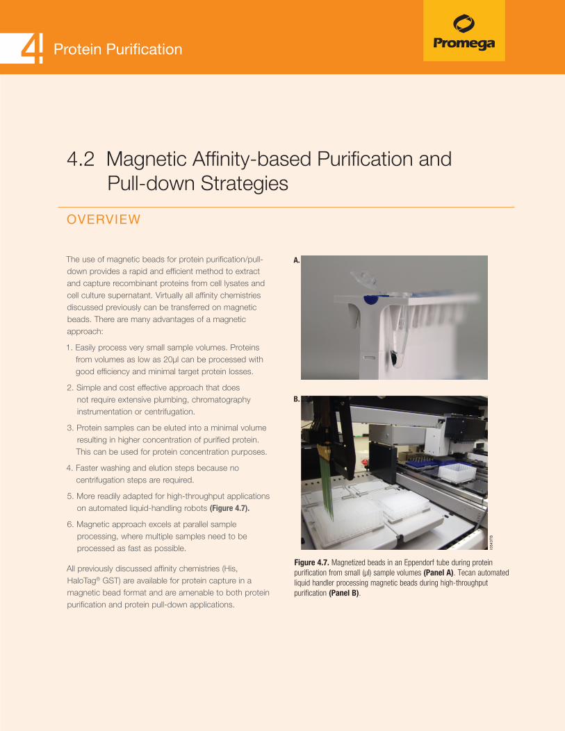
56 Discover Reliable Tools for Protein Analysis
Protein Purification4
The use of magnetic beads for protein purification/pull-down provides a rapid and efficient method to extract and capture recombinant proteins from cell lysates and cell culture supernatant. Virtually all affinity chemistries discussed previously can be transferred on magnetic beads. There are many advantages of a magnetic approach:
1. Easily process very small sample volumes. Proteins from volumes as low as 20μl can be processed with good efficiency and minimal target protein losses.
2. Simple and cost effective approach that does not require extensive plumbing, chromatography instrumentation or centrifugation.
3. Protein samples can be eluted into a minimal volume resulting in higher concentration of purified protein. This can be used for protein concentration purposes.
4. Faster washing and elution steps because no centrifugation steps are required.
5. More readily adapted for high-throughput applications on automated liquid-handling robots (Figure 4.7).
6. Magnetic approach excels at parallel sample processing, where multiple samples need to be processed as fast as possible.
All previously discussed affinity chemistries (His, HaloTag® GST) are available for protein capture in a magnetic bead format and are amenable to both protein purification and protein pull-down applications.
4.2 Magnetic Affinity-based Purification and Pull-down Strategies
OVERVIEW
Figure 4.7. Magnetized beads in an Eppendorf tube during protein purification from small (µl) sample volumes (Panel A). Tecan automated liquid handler processing magnetic beads during high-throughput purification (Panel B).
1054
3TB
A.
B.

57 Discover Reliable Tools for Protein Analysis
Protein Purification4
MagneGST™ Protein Purification SystemManual or automated magnetic purification or pull-down of glutathione-S-trans-ferase (GST) fusion proteins from crude or cleared E. coli lysates.
Description
The MagneGST™ Protein Purification System includes immobilized glutathione paramagnetic particles, buffers and cell lysis reagents to isolate GST-tagged protein directly from a crude or cleared lysate. GST-tagged proteins can be purified from 1ml to 50ml of culture. Samples also can be processed using a robotic platform. MagneGST™ particles are supplied as a 25% slurry and have a binding capacity of 5–10mg of GST protein per 1ml of settled resin.
Principle
Bacterial cells containing a GST-fusion protein are lysed using the provided MagneGST™ Cell Lysis Reagent or with an alternative lysis method, then the MagneGST™ particles are added directly to the crude lysate. GST-fusion proteins bind to the MagneGST™ particles. Unbound proteins are washed away, and the GST-fusion target protein is recovered by elution with 50mM glutathione (Figure 4.8).
For capture and verification of interacting proteins (pull-down), the GST-tagged bait protein is expressed in E. coli and the potential prey proteins are expressed in cell-free system. The bait is bound on the MagneGST™ particles and is then used to capture different prey proteins from cell-free expression reactions (Figure 4.8).
Features and Benefits• Simple: One-step purification of multiple samples
with easy handling. No lysate clearing needed.
• Scalable Protocol: Use 1–50ml of cell culture. Obtain 5–10mg of GST protein per 1ml of settled beads.
• Efficient: Achieve high yields with little or no nonspecific background.
ReferencesMeloni, A. et al. (2010) DAXX is a new AIRE-interacting protein. J. Biol. Chem. 285(17), 13012–21.
Maier R.H, et al. (2010) Epitope mapping of antibodies using a cell array-based polypeptide library. J. Biomol. Screen. 15(4), 418–26.
Morimoto, H. et al. (2008) Procollagen C-proteinase enhancer-1 (PCPE-1) interacts with beta2-microglobulin (beta2-m) and may help initiate beta2-m amyloid fibril formation in connective tissues. Matrix Biol. 27(3), 211–9.
Ordering Information MagneGST™ Protein Purification System (Cat.# V8600, V8603)
Magnetic Affinity-based Protein Purification

58 Discover Reliable Tools for Protein Analysis
Protein Purification4
MP
6544
MB
TNT®-based Cell-Free Expression
of Prey Protein
Cell Lysis
Bait expressedas GST Fusion
in E.coli
Bind, Wash and Capture onMagneGST™ Glutathione Particles
Mix and AllowInteraction to Occur
Gel Electrophoresis Followed By Detection (Western Blotting or Autoradiography)
Detect Prey Protein
Wash and Elute Complex
Plasmid ContainingProtein Coding Sequence
for Prey Protein
RNAPromoter
Prey Sequence
GST
B
B - Bait ProteinP - Prey Protein
GST - Glutathione S-transferaseMP - Magnetic Particle
GST
B
GST
BMP P
P
GST
B P
Figure 4.8. Schematic diagram of MagneGST™ Protein Purification in combination with protein pull-down.
Magnetic Affinity-based Protein Purification

59 Discover Reliable Tools for Protein Analysis
Protein Purification4
Magne™ HaloTag® BeadsManual or automated high-throughput protein purification and pull-down of HaloTag® fusion proteins from E. coli and mammalian cell cultures, at input volumes from 20μl to 50ml.
Ordering Information Magne™ HaloTag® Beads (Cat.# G7281, G7282)
Figure 4.9 Schematic diagram of the HaloTag® fusion purification protocol using Magne™ HaloTag® Beads.
1044
1MA
Equilibrate Magne™ HaloTag® Beads.
Add lysate, and bindHaloTag® fusionprotein to Magne™ HaloTag® Beads.
Wash.
Cleave protein bound to bead using HaloTEV Protease.
Elute purified protein.
HaloTag® protein
Fused proteinHaloTEV Protease
Description and Principle
The Magne™ HaloTag® Beads provide a convenient method to covalently capture HaloTag® fusion proteins with magnetic particles for protein pull-downs and puri-fication. HaloTag® fusion proteins may be expressed in cell-based and cell-free systems. Cell lysates are mixed with Magne™ HaloTag® Beads and HaloTag® fusion proteins bound covalently to the beads. These magnetic beads offer a high-binding capacity (≥20mg/ml) for purifying HaloTag® fusion proteins with low nonspecific protein binding. After washing, the protein of interest can be released from the beads by TEV Protease cleavage (either HaloTEV or ProTEV) at the optimized TEV recognition site. Proteolytic release yields the protein of interest, while the HaloTag® protein and HaloTEV Protease remain covalently attached to the beads (Figure 4.9).
Features and Benefits• Simple: No centrifugation or vacuum is required
once the cells are lysed.
• High Recovery: Binding capacity ≥20mg of purified HaloTag® fusion protein per ml of settled particles.
ReferencesVerger A. et.al. (2013) The Mediator complex subunit MED25 is targeted by the N-terminal transactivation domain of the PEA3 group members. Nucl. Acids Res. 41(9), 4847–59.
Yoshida S. et al. (2013) Androgen receptor promotes sex-independent angiogenesis in response to ischemia and is required for activation of vascular endothelial growth factor receptor signaling. Circulation 2(128), 60–71.
Nagaki K. et al. (2012) Isolation of centromeric-tandem repetitive DNA sequences by chromatin affinity purification using a HaloTag7-fused centromere-specific histone H3 in tobacco. Plant Cell Rep. 31(4), 771–9.
Magnetic Affinity-based Protein Purification

60 Discover Reliable Tools for Protein Analysis
Protein Purification4
MagneHis™ Protein Purification SystemManual or automated high-throughput protein purification and pull-down of polyhistidine-tagged proteins, from crude E. coli cell lysates, insect or mammalian cell lysates or cell supernatant.
Description and Principle
The MagneHis™ Protein Purification System provides a quick and simple method for the purification of polyhistidine-tagged, overexpressed proteins. Paramagnetic precharged nickel particles (MagneHis™ Ni-Particles) are used to isolate His-tagged fusion proteins directly from a crude cell lysate. Sample volumes from 20μl to 50ml can be easily processed manually. Samples can also be processed using a robotic platform such as the Beckman Coulter Biomek® 2000 or FX or Tecan Genesis® RSP. This Purification System contains magnetic beads and buffers as well as FastBreak™ Cell Lysis Reagent. His tagged proteins can be expressed either intracellularly or secreted into cell media. Bacterial, insect or mammalian cells can be lysed with FastBreak™ Cell Lysis Reagent or with other lysis methods. MagneHis™ Ni-Particles are then added to the lysate and bind to His tag containing proteins. Unbound proteins are washed away, and the target protein is recovered by elution with imidazole (Figure 4.10). Purification can be performed under native and denaturing conditions.
Ordering Information MagneHis™ Protein Purification System (Cat.# V8500, V8550)
Features and Benefits• Fast: No long incubations with lysozyme are required for
cell lysis.
• Simple: No centrifugation or vacuum after cell lysis.
• Efficient: Binding capacity of up to 1mg of polyhistidine-tagged protein per 1ml of MagneHis™ Ni-particles.
• Versatile: Purify His-tagged proteins in manual format or at high-throughput with an automated liquid handler.
ReferencesGuo, H. et al. (2010) Osteopontin and protein kinase C regulate PDLIM2 activation and STAT1 ubiquitination in LPS-treated murine macrophages. J Biol Chem. 285(48), 37787–96.
Yun CS, et al. (2010) Pmr, a histone-like protein H1 (H-NS) family protein encoded by the IncP-7 plasmid pCAR1, is a key global regulator that alters host function. J Bacteriol. 192(18), 4720–31.
Koschubs, T. et al. (2010) Preparation and topology of the Mediator middle module. Nucleic Acids Res. 38(10), 3186–95.
Magnetic Affinity-based Protein Purification
Lyse.
Bind.
Elute.
Pure Native Protein Pure Denatured Protein
Native Conditions Denaturing Conditions
3810MD
Wash.
HH
H
H
H
Cells
H
H
H
M M
MMH
MagneHis™ Ni-ParticleM
Polyhistidine TagH
H
Figure 4.10. Schematic of the MagneHis™ Protein Purification System protocol.

61 Discover Reliable Tools for Protein Analysis
Protein Purification4
Maxwell® 16 Polyhistidine Protein Purification KitAutomated purification of polyhistidine-tagged proteins from bacterial cultures and mammalian and insect proteins.
Description and Principle
The Maxwell® 16 Polyhistidine Protein Purification Kit consists of cartridges prefilled with MagneHis™ Ni-Particles and buffers that are used with the Maxwell® 16 Instrument to provide an easy method for efficient, automated purification of polyhisti-dine-tagged protein from various cells (Table 4.2). The Maxwell® Instrument is supplied with a preprogrammed purification procedure and reagent cartridges specifically designed to maximize simplicity and convenience. The instrument can process up to 16 samples in approximately 40 minutes.
Ordering Information Maxwell® 16 Polyhistidine Protein Purification Kit (Cat.# AS1060)
Features and Benefits• Save Hands-On Time: Prefilled cartridges eliminate
reagent preparation, multiple pipetting steps, centrifu-gation and additional sample manipulation.
• Consistent Results: Across all samples.
5933
MA
Label side
Ridge side
Lysis Buffer
Wash Buffer
Wash Buffer
Wash Buffer
Wash Buffer
Empty
Sample
Plunger
Contents User Adds:
MagneHis™ Ni-Particles
12
3
4
5
67
Figure 4.12. Maxwell 16® Polyhistidine Protein Purification Sample Cartridge
Figure 4.11. The Maxwell® 16 Instrument uses paramagnetic-particle technology to extract DNA, RNA or protein from up to 16 samples in less than 45 minutes. The paramagnetic particles and reagents are supplied in prefilled cartridges for faster separa-tions with less hands-on time.
Magnetic Affinity-based Protein Purification
Table 4.2. Sample Types and Maximum Processing Capacity per Cartridge.
Sample Type Processing Capacity
Bacterial Culture Up to 20 O.D.600
Mammalian Cell Culture Cells
Up to 5 x 10⁶ cells
Insect Cell Culture Cells Up to 5 x 10⁶ cells
Mammalian or Insect Cell Culture Medium
1ml

62 Discover Reliable Tools for Protein Analysis
Protein Purification4
Magnetic Separation DevicesManual or automated protein purification using magnetic particles.
Description
Promega offers a wide range of magnetic devices for separations from 0.5ml microcentrifuge tubes to 15ml or 50ml conical tubes, to 96- and 384-well standard and deep-well plates. The magnetic separation device for plates is useful for both manual and automated liquid-handling.
MagneSphere® Technology Magnetic Separation Stands
MagnaBot® Magnetic Separation Devices
2312
TA07
_8A
39
93
TA0
2_
3A
MagneSphere® Technology Magnetic Separation Stand (two-position). Up to two sample volumes (50µl –1.0ml). Left to Right: Cat.# Z5331, Z5332, Z5333.
MagnaBot® 96 Magnetic Separation Device for 96-well standard- or deep-well-plates (20µl– 1.0ml). Cat.# V8151.
2309
TA07
_8A
MagneSphere® Technology Magnetic Separation Stand (twelve-position). Up to two sample volumes (50µl–1.0ml). Left to Right: Cat.# Z5341, Z5342, Z5343.
2311
TA07
_8A
3417
TA05
_1A
PolyATtract® System 1000 Magnetic Separation Stand. One Sample volume (1-50ml). Cat.# Z5410.
MagnaBot® 384 Magnetic Separation Device. Cat.# V8241.
MagnaBot® II Magnetic Separation Device for 96-well PCR plate. Cat.# V8351.
Magnetic Separation Devices
63 Discover Reliable Tools for Protein Analysis
Antibody Purification and Labeling
5.1 Antibody Purification 65 Magne™ Protein A Beads and Magne™ Protein G Beads 67
5.2 Antibody Labeling 69 pHAb Amine and Thiol Reactive Dyes 71
5
63 Discover Reliable Tools for Protein Analysis

64 Discover Reliable Tools for Protein Analysis
Antibody Purification and LabelingAntibodies are an indispensable tool for bioresearch, diagnosis and therapy, thus there is a great demand for antibody purification strategies. These strategies include antibody purification from various sources, such as serum (polyclonal antibodies), ascites (monoclonal antibodies) and cell culture supernatant of a hybridoma cell line (monoclonal antibodies). Polyclonal antibodies are a mixture of different antibodies produced by a diverse popu-lation of B cells. They may be of different isotypes, have different antigen specificities or recognize a different antigen epitope.
In contrast, moncolonal antibodies are produced by specific B cells that are clonally related; these antibodies are the same isotype, and have identical specificity for the antigen epitope. Depending on the starting material used, various antibody purifica-tion strategies exist, including classical chromatography (e.g., ion exchange), affinity-purification (e.g., on immobilized protein A and protein G beads) and antigen-specific affinity purification.
This chapter introduces the use of magnetic protein A and G beads for antibody purification as well as for on-bead antibody labeling.

65 Discover Reliable Tools for Protein Analysis
The choice of the antibody purification method depends on the starting material, the intended application, and the manufac-turing scale. However, most antibody purification strategies include the use of immobilized Protein A or Protein G (Table 5.1). These bacterial proteins have different affinities for various antibody species and isotypes (Table 5.2). Immobilized Protein A and Protein G can be used to purify monoclonal antibodies from ascites fluid or cell culture supernatant. However, in the case of polyclonal antibodies (serum samples) the use of Protein A or Protein G will enrich mainly the IgG fraction (Figure 5.1). lgG is the main antibody isotype found in blood. For target-specific antibody isolation from serum, antigen-specific affinity-purification is required.
5.1 Antibody Purification OVERVIEW
Figure 5.1. Different workflows for antibody purification using Protein A and Protein G. Monoclonal antibodies can be purified using Protein A or Protein G (Panel A). The IgG fraction of polyclonal antibodies from serum can be enriched using Protein A or Protein G. Furthermore, Protein A or Protein G can be used to deplete the IgG fraction from serum. Note: For target-specific antibody purification from serum antigen-specific affinity-purification is required (Panel B).
Monoclonal antibody purification from cell culture supernatant or ascites fluid
Protein A or Protein G
Result: Purified monoclonal antibody
Result: Enriched IgG pool
Result: Mainly IgG-depleted serum.
Protein A or Protein G
Polyclonal antibodypurification from serum
Eluate Flowthrough
1214
9MA
A.
B.
Table 5.1. Features of Native Ig-binding Proteins: Protein A and Protein G.
Protein A Protein G
Species Staphylococcus aureus Streptococcus spp. (group C and G)
Native Size 40-60 kDa 40-65 kDa
Ig-Binding TargetHeavy chain constant region (Fc ) of IgG
(CH2-CH3 region)
Heavy chain constant region (Fc ) of IgG
(CH2-CH3 region)
Antibody Purification and Labeling5

66 Discover Reliable Tools for Protein Analysis
Antibody Purification and Labeling5
Table 5.2. Relative Recovery of Different Antibody Species and Isotypes by Magne™ Protein A Beads and Protein G Beads.
Species Isotype Magne™ Protein A Beads (µg) Magne™ Protein G Beads (µg)
Bovine lgG 36.2 33
Goat lgG 26.2 26
Human
lgA 4.8 0.4
lgG1 35.2 39.4
lgG2 34.4 32.6
lgG3 0 37.6
lgG4 28.8 38
lgM 8.2 0
Mouse
lgG1 18.6 30.4
lgG2a 33.2 31.6
lgG2b 29.6 31.4
lgG3 16 9.2
Porcine lgG 35.2 27.6
Rat
lgG1 29.4 34.2
lgG2a 0 33
lgG2b 0 31.2
Sheep lgG 26.6 25.6
Note: Starting material for the recovery experiment was comprised of 50μg of purified antibody diluted in 1ml of buffer and captured with 50μl of bead slurry (20%). Experiment performed in triplicate (CV ≤10%).
66 Discover Reliable Tools for Protein Analysis

67 Discover Reliable Tools for Protein Analysis
Magne™ Protein A Beads and Magne™ Protein G BeadsManual or automated antibody purification from different sample types; antibody enrichment or antibody depletion from serum samples; antibody labeling with small molecules or fluorophores.
Description
Magne™ Protein A Beads and Magne™ Protein G Beads are magnetic affinity beads with high specificity and high capacity for binding antibodies from cell culture supernatant, ascites fluid and serum samples. Antibody purification can be performed easily from a single sample or multiple samples in parallel, or in a high-throughput automated fashion. The magnetic beads allow for superior purification and recovery of concentrated antibodies from small input volumes (20μl) by decreasing losses normally associated with handling of small volumes and nonmagnetic resins.
Add antibody sample. Capture antibodies on beads (30−60 minutes).
Elute purified antibodiesusing 100mM glycine
(pH 2.7) and neutralize.
Magnetize resin and wash away contaminants.
Purified antibodies 1120
7ME
Figure 5.2. Antibody purification using Magne™ Protein A Beads or Magne™ Protein G Beads.
Principle
Recombinant Protein A from Staphylococcus and recombinant Protein G from Streptococcus are cova-lently attached in an oriented fashion to magnetic beads. Biological samples are added, and antibodies are captured by the beads. Using magnetic devices, beads are attached and unbound material is washed away. Finally, antibodies are eluted using lower pH buffer and the solution is neutralized (Figure 5.2).
Features and Benefits• High Binding Capacity: Binding capacities of 25mg of
antibody per ml of settled beads are observed, depending on antibody species and isotype.
• Simple: Easy to handle beads with fast magnetic response.
• High Purity: Due to low nonspecific binding.
• Flexible: Handles sample volumes from 20μl–50ml.
Antibody Purification
Antibody Purification and Labeling5

68 Discover Reliable Tools for Protein Analysis
Antibody Purification and Labeling5Antibody Purification
Ordering Information Magne™ Protein A Beads (Cat.# G8781, G8782, G8783)
Magne™ Protein G Beads (Cat.#. G7471, G7472, G7473)
1120
8TB
SM A
Mouse Ascites
Albumin IgG heavy chain (55kDa)
IgG light chain (25kDa)
kDa
250
150
100
75
50
37
25
20
15
10
Cell Media
SM A G G
SM = Starting MaterialA = Antibody purified with Magne™ Protein A BeadsG = Antibody purified with Magne™ Protein G Beads
Input Antibody Sample Type
Figure 5.3. Antibody purified from various sample types using Magne™ Protein A and Magne™ Protein G Beads. Antibody was purified from 50µl of cell culture media (mouse IgG1), mouse ascites (IgG2a) using 50µl of Magne™ Protein A Beads (A) and Magne™ Protein G Beads (G). Samples were analyzed by adding 1µl of starting material (SM) or 5µl of purified sample to SDS buffer, and heating at 80°C for ten minutes before loading onto a 4–20% Tris-glycine gel. The gel was stained with SimplyBlue® Safe Stain.
Additional Information:• Magnetic stands and spacer for the use with this product
are listed in Chapter 4.
• High-throughput antibody purification platforms such as the ReliaPrep™ LV 32 HSM Instrument (Cat.# A1715) or a robotic liquid handler platform such as the Beckman Coulter Biomek® FX can be used.
• Custom solutions for using the beads with a Maxwell® Instrument are possible upon request.
• Note: Magne™ Protein A Beads and Magne™ Protein G Beads are not recommended for use in immunoprecipita-tion (IP) or Co-IP applications.
ReferencesGodat, B. and Nath, N. (2012) High-Capacity and High-Purity Antibody Purification Using Magnetic Beads. [Promega Corporation web site. November 2012. Accessed 1-20-2014. http://www.promega.com/resources/pubhub/high-capacity-and-high-purity-antibody-purification/

69 Discover Reliable Tools for Protein Analysis
5.2 Antibody Labeling OVERVIEW
Antibodies can be labeled with a diverse number of small molecules including biotin, fluorophores (e.g., FITC, Alexa Dyes, Cy3) and other large proteins (e.g., horseradish peroxidase). These antibody conjugates are useful reagents in bioresearch mainly for analyte/antigen detection in ELISA, fluorescent microscopy and for antibody-based assays. A specific class of antibody conjugates, Antibody Drug Conjugates (ADCs), is comprised of antibodies labeled with cytotoxic compounds intended for human therapy to target and kill specific cells (e.g., solid tumors). An ADC binds to target cells via a specific antibody-cell surface antigen interaction, the antibody and drug conjugate are internalized into endosomes. In the endo-somes the linker between the cytotoxic drug and antibody are cleaved causing the release of the cytotoxic molecule and subsequent killing of the target cell. There are three aspects of ADC production that require optimization: the targeting antibody, the labile linker and the cytotoxic agent.
1190
5MB
Capture antibody withMagne™ Protein A or Protein G Beads.
Wash awayunreacted free dye.
Wash awaycontaminants.
Elute and neutralize to obtain purified and labeled antibodies.
Buffer exchange. Add reactive dye.
Figure 5.4. Schematic diagram of on-bead antibody conjugation/labeling.
Antibody Purification and Labeling5
69 Discover Reliable Tools for Protein Analysis
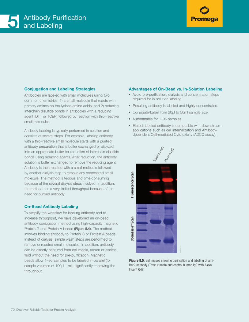
70 Discover Reliable Tools for Protein Analysis
Antibody Purification and Labeling5
Conjugation and Labeling Strategies
Antibodies are labeled with small molecules using two common chemistries: 1) a small molecule that reacts with primary amines on the lysines amino acids; and 2) reducing interchain disulfide bonds in antibodies with a reducing agent (DTT or TCEP) followed by reaction with thiol-reactive small molecules.
Antibody labeling is typically performed in solution and consists of several steps. For example, labeling antibody with a thiol-reactive small molecule starts with a purified antibody preparation that is buffer exchanged or dialyzed into an appropriate buffer for reduction of interchain disulfide bonds using reducing agents. After reduction, the antibody solution is buffer exchanged to remove the reducing agent. Antibody is then reacted with a small molecule followed by another dialysis step to remove any nonreacted small molecule. The method is tedious and time-consuming because of the several dialysis steps involved. In addition, the method has a very limited throughput because of the need for purified antibody.
On-Bead Antibody Labeling
To simplify the workflow for labeling antibody and to increase throughput, we have developed an on-bead antibody conjugation method using high-capacity magnetic Protein G and Protein A beads (Figure 5.4). The method involves binding antibody to Protein G or Protein A beads. Instead of dialysis, simple wash steps are performed to remove unreacted small molecules. In addition, antibody can be directly captured from cell media, serum or ascites fluid without the need for pre-purification. Magnetic beads allow 1–96 samples to be labeled in-parallel (for sample volumes of 100μl–1ml), significantly improving the throughput.
Advantages of On-Bead vs. In-Solution Labeling• Avoid pre-purification, dialysis and concentration steps
required for in-solution labeling.
• Resulting antibody is labeled and highly concentrated.
• Conjugate/Label from 20μl to 50ml sample size.
• Automatable for 1–96 samples.
• Eluted, labeled antibody is compatible with downstream applications such as cell internalization and Antibody-dependent Cell-mediated Cytotoxicity (ADCC assay).
1192
5TA
Tras
tuzu
mab
Hum
an Ig
G
Fluo
resc
ence
Sca
nCo
omas
sie®
Sca
n
Figure 5.5. Gel images showing purification and labeling of anti-Her2 antibody (Trastuzumab) and control human IgG with Alexa Fluor® 647.
70 Discover Reliable Tools for Protein Analysis

Antibody Purification and Labeling5Antibody Labeling
pHAb Amine and Thiol Reactive DyespH Sensor Dyes for Screening Antibody Internalization
Description
pHAb Reactive Dyes are pH sensor dyes that have very low fluorescence at pH > 7 and a dramatic increase in fluorescence as the pH of the solution becomes acidic. pHAb Dyes have excitation maxima (Ex) at 532nm and emission maxima (Em) at 560nm. pHAb Dyes are designed specifically for antibody labeling and are available in two reactive forms suitable for antibody conjugations.
Principle
pHAb Amine Reactive Dyes have a succinimidyl ester group that reacts with primary amines on lysine amino acids of antibodies. pHAb Thiol Reactive Dyes have a maleimide group that reacts with thiols. This maleimide group is conjugated to the antibody after the cysteine disulfide bonds in the hinge region of the antibody are reduced to thiols using a reducing agent.
A key feature of pHAb Dyes is the two sulfonate groups per dye, which increase solubility in water and reduce the aggregation often seen with other non-sulfonated dyes. pHAb Dyes maintain their fluorescence response to pH change even after conjugation to antibody, as shown in Figure 5.6.
pHAb Reactive Dyes can be conjugated to antibodies using two different workflows. One is the traditional solution-based (or in-solution) chemistry workflow. The other workflow, shown in Figure 5.7, is on-bead conjugation, which uses magnetic protein A and protein G affinity beads to selectively capture antibodies from solutions containing purified antibody or directly from biological samples expressing antibodies (i.e., from cell culture media).
Features and Benefits• Accurately Determine Antibody Internalization: Fluorescence increases upon internalization, with lower pH
inside the cell.
• Use with On-bead Conjugation: Conjugate directly from biological samples expressing antibodies.
• Compatible with Plate-based Assays: Measure internalization in 96-well plates, real time.
• Minimal Background Fluorescence: Only conjugated dyes are internalized and thus fluorescent. Free dye outside the cell has negligible fluorescence.
71 Discover Reliable Tools for Protein Analysis

Antibody Purification and Labeling5Antibody Labeling
1290
3MA
4 5 6 7 8 90
10,000
20,000
30,000
40,000
50,000
60,000
C.B.A.
pH
Fluo
resc
ence
(RFU
)
pHAb Amine Reactive Dye
Trastuzumab-pHAb Amine Reactive Dye
pHAb Thiol Reactive Dye
Trastuzumab-pHAb Thiol Reactive Dye
N
NN
O
O
O
O
CO2-
N+N
SO3H SO3H
O
N
NN
O
O
HN
CO2-
N+N
SO3H SO3H
O
O
Figure 5.6. Fluorescence is shown as a function of pH for Trastuzumab labeled with pHAb Amine Reactive Dye or pHAb Thiol Reactive Dye compared to pHAb Amine Reactive Dye or pHAb Thiol Reactive Dye alone.
Figure 5.7. On-bead conjugation of antibody using Magne™ Protein A Beads or Magne™ Protein G Beads.
Magne™ Protein A orProtein G Beads.
Antibody solution(purified or unpurified).
Antibody solution(purified).
Buffer exchange.
Conjugation through Thiols
Conjugation through Amines
Conjugation through Thiols
Conjugation through Amines
A.
B.
Add pHAb AmineReactive Dye.
Dialyze to removeunreacted dye.
Dialyze.Dialyze.
Ab-pHAbconjugate.
Ab-pHAbconjugate.
Reducedantibody.
Reducedantibody.
Add DTTor TCEP.
Add pHAb ThiolReactive Dye.
Add pHAb ThiolReactive Dye.
1290
4MA
Wash. Add pHAbAmine Reactive Dye. Wash. Elute and
neutralize.
Wash. Elute andneutralize.
Add DTTor TCEP.
Ordering Information pHAb Amine Reactive Dye (Cat.# G9841, G9845)
pHAb Thiol Reactive Dye (Cat.#. G9831, G9835)
72 Discover Reliable Tools for Protein Analysis

Functional Protein Analysis using HaloTag® Technology6Cellular Imaging 77
Fluorescent HaloTag® Ligands: Protein Localization and Trafficking 77
Janelia Fluor® 549 and 646 HaloTag® Ligands 79
Protein Interaction Analysis 81 NanoBRET™ Technology: Live Cell Protein–Protein Interaction Assays 81
HaloTag® Mammalian Pull-Down System: Protein–Protein Interactions 83
HaloCHIP™ System: Protein–DNA Interactions 85
HaloLink™ Array System: HaloTag® Protein Arrays 87
73 Discover Reliable Tools for Protein Analysis

74 Discover Reliable Tools for Protein Analysis
Functional Protein Analysis using HaloTag
®
Technology Understanding the functional role of proteins and their intracellular behavior is increasingly important. Often multiple protein fusion tags are required to fully characterize a specific protein of interest, such as use of one or more fusion tags for imaging and protein capture/purification applications. Recloning the same protein-coding DNA sequence of interest with multiple tags can be slow and cumbersome, requiring revalidation of the new construct in functional assays prior to use. What is needed is a single recombinant protein tag that provides application flexibility and superb performance for protein expression and localization, protein purification, protein interaction discovery, screening and further functional analyses. The HaloTag® Technology and the HaloTag® fusion proteins address this research need.

75 Discover Reliable Tools for Protein Analysis
Functional Protein Analysis using HaloTag® Technology6
1214
8MB
HaloTag®
HaloTag® Ligand (Chloroalkane + Functional Group)
Covalent Bond
TEVcleavage
site
Covalent bond between HaloTag® protein and HaloTag® ligand (chloroalkane + functional group).
+ O OCl–
O O
Cl–
POI
HaloTag®
POI
Fluorescent Ligands: for Cellular Imaging, Detection and Protein-Interaction Studies (BRET). They come in many different colors, as cell permeable ligands and as non-permeable ligands.
Non-fluorescent Ligands: for Protein Detection, e.g., Biotin; PEG-Biotin.
Surface Ligands: for Protein Purification and Protein-Interaction Studies (Pull-downs), e.g., Magnetic Beads; Resins; Arrays.
Reactive Ligands: to attach functional group of choice, e.g., Positron Emission Tomography (PET) ligands, magnetic resonance reagents.
Figure 6.1. Schematic of HaloTag® technology consisting of the HaloTag® protein fused to a protein of interest (POI) and a selection of synthetic HaloTag® ligands carrying different functional groups. HaloTag® ligands specifically and covalently bind to the HaloTag® protein fusions.
HaloTag® technology is based on an engineered catalytically inactive bacterial hydrolase of 34kDa that reacts, under physiological conditions, with chloroalkane ligands (HaloTag® ligands) to form a highly specific and irreversible covalent bond. By changing the ligands it is possible to control the function and application of the tagged protein of interest.
The HaloTag® system is designed to provide broad experimental flexibility in both cell-based and biochemical assays without switching fusion tags or recloning. HaloTag® ligands coupled to magnetic and nonmagnetic beads and glass slides are available. Cell-permeable and imperme-able fluorescent ligands and biotin ligands are also available as well as reactive ligands to enable the creation of custom HaloTag®-linked constructs (Figure 6.1).
Features and Benefits
• Multiple Applications: Use a single protein fusion tag for all your needs.
• Minimal Background: HaloTag® protein has no homology to other mammalian cell proteins.
• Irreversible Covalent Attachment: Covalent binding to HaloTag® ligands and surfaces allows stringent washing, resulting in low background.

76 Discover Reliable Tools for Protein Analysis
Functional Protein Analysis using HaloTag® Technology6
ReferencesUrh, M. et al. (2012) HaloTag, a Platform Technology for Protein Analysis. Curr Chem Genomics. 6,72-8.
Los, G.V. et al. (2008) HaloTag: a novel protein labeling technology for cell imaging and protein analysis. ACS Chem Biol. 3(6), 373-82.
Figure 6.2. One genetic HaloTag® construct can be used for multiple applications.
1216
9TB
HaloTag®
VectorHaloTag® protein
Protein of interest
Covalent captureon resin or surfaces
Protein labeling usingHaloTag® fluorescent ligands
Protein display Bioluminescence resonance energy transfer (BRET)
Protein interactions Protein purification Protein detection Cellular imagingLive and fixed-cell imaging
Protein localization and translocation(spatial and temporal)
High contentscreening
Pull-down of protein:protein complexes
Capture of protein:DNA interactions (CHIP)
Protein Purification (from E. coli or mammalian cells)
Immobilization of proteins on surfaces
Express HaloTag® fusion protein.
Direct protein detection with fluorescent ligands without Western blotting Detection of protein:
protein interactions in living cells using BRET (Bioluminescence Resonance Energy Transfer)

77 Discover Reliable Tools for Protein Analysis
Fluorescent HaloTag® Ligands for Cellular ImagingCellular Imaging: Live and fixed cells; protein localization and trafficking; protein turnover and pulse-chase analysis; FACS analysis.
Functional Protein Analysis using HaloTag® Technology6
Description
Fluorescent HaloTag® ligands (see Figure 6.3) are avail-able for the detection of HaloTag® protein fusions in live cells for direct imaging or for labeling in fixed cells. HaloTag® imaging can be multiplexed with fluorescent proteins (e.g., GFP) in live cells and fluorescent antibody staining in fixed cells and tissues. HaloTag® ligands are based on small organic dyes; they retain their fluores-cent properties after fixation, enabling multiplexing of immunocytochemistry experiments.
Principle
Proteins of interest fused to HaloTag® protein can be labeled with various fluorescent ligands in live or fixed cells using a simple labeling protocol (Figure 6.4). HaloTag® ligands are noncytotoxic and allow permanent labeling due to covalent attachment to the HaloTag® protein. There are different types of fluorescent ligands for intracellular protein labeling (cell-permeant) as well as for cell surface protein labeling (cell-impermeant). Rapid ligands can label as quickly as 5–15 minutes after addition to cells expressing the HaloTag® protein. The direct label ligands can be added at the time of cell plating or transfection and will label the tagged protein of interest (POI) as it is expressed (without the need to wash away the unbound ligand). In addition to direct labeling, the dyes can be used in standard labeling protocols that include wash steps. Lastly, permeant and impermeant ligands of different colors can be used in tandem to track protein translocation to and from the plasma membrane (Figure 6.5). The range of differently colored HaloTag® ligands allows easy changes in POI color and easy integration with other fluorophores like GFP or other cellular labeling dyes.
Figure 6.3. Maximum excitation and emission spectra for the HaloTag® Ligands.
HaloTag® Coumarin 362nm 450nm Intracellular
labeling G8581, G8582
HaloTag® Alexa Fluor®
488499nm 518nm Cell-surface
labeling G1001, G1002
HaloTag® R110Direct™ (”NoWash” ligands)
498nm 528nm Intracellularlabeling G3221
HaloTag® TMRDirect™ (”NoWash” ligands)
552nm 578nm Intracellularlabeling G2991
HaloTag® Alexa Fluor®
660654nm 690nm Cell-surface
labeling G8471, G8472
HaloTag® Oregon Green®
492nm 520nm Intracellularlabeling
G2801, G2802
HaloTag® DiAcFAM
492nm 521nm Intracellularlabeling
G8272, G8273
LigandExcitationMaximum
EmissionMaximum
IntendedUse ofLigands
OrderingInformation
5530
LA
Features and Benefits• Localization: Robust protein imaging in live or fixed
cells (e.g., 4% PFA-fixation).
• Trafficking and Turnover: Directly observe spatially- or temporally-separated protein populations with one or two colors in live cells. Label first with cell-imper-meable and then with cell-permeable ligands.
• Cell Sorting: Simple non-antibody based cell labeling with multiple fluorophores.
• Flexible: Variety of HaloTag® ligands in different colors allows flexible combination with other labeling dyes (e.g., DNA staining dyes) as well as with immunofluorescence reagents.
Cellular Imaging

78 Discover Reliable Tools for Protein Analysis
Functional Protein Analysis using HaloTag® Technology6
7531
MB
Proceed with other analyses(e.g., SDS-PAGE, fluoroimaging, Western blotting).
Day 1: Plate cells
Day 2: Introduce HaloTag®
genetic construct into cells using standard transfection techniques
Day 3: Label cells with HaloTag® Ligand of choice
Wash unbound ligand from sample or Add “no wash” ligand of choice
HaloTag® Protein
HaloTag® Ligand
Fixed-cell imaging Live-cell imaging
Image fixed cellsor perform ICC.
Image live cells orchase with spectrallydistinct ligand priorto imaging.
Excitation light
Fluorescence
Figure 6.4. Overview of protocol for live-cell and fixed-cell imaging using HaloTag® Technology. No washing steps are required with the “No Wash” DIRECT® HaloTag® ligands.
ReferencesYamaguchi, K. et al. (2009) Pulse-chase experiment for the analysis of protein stability in cultured mammalian cells by covalent fluorescent labeling of fusion proteins. In: Reverse Chemical Genetics, Methods in Molecular Biology 577, H. Koga ed. Humana Press.
Huybrechts, S.J. et al. (2009) Peroxisome dynamics in cultured mammalian cells. Traffic 10(22),1722-33.
Svendsen, S. et al. (2008) Spatial separation and bidirectional trafficking of proteins using a multi-functional reporter. BMC Cell Biol. 9(17).
cell-impermeable
cell-permeable
HaloTag®
Vector
Transfect cells.
12 hours
1217
0TA
Cell Surface Intracellular
0 0.5 1 2 4 8 12 hours
Cell surface
A.
B.
C.
D.
Figure 6.5. Membrane trafficking of ß-integrin fragment fused to HaloTag® (N-terminal fusion). (A) Cells stably expressing ß1Int-HaloTag were labeled first with the cell-impermeable ligand HaloTag® Alexa Fluor® 488 and then with the cell-permeable HaloTag® TMR ligand. (B) Images were taken with a confocal microscope immediately after labeling to show cell-surface and intracellular localization of ß1Int-HaloTag. (C) Cells were re-imaged 12 hours after labeling to show receptor internalization and trafficking to the plasma membrane. (D) Fluoroscan image of an SDS gel: Cell lysates taken at 0, 0.5, 1, 2, 4, 8 and 12 hours after labeling were run on SDS-PAGE. Fluoroscan shows bidirectional trafficking of ß1Int-HaloTag as reflected in the upper band (cell surface ß1Int-HaloTag) and lower band (intracellular ß1Int-HaloTag). Image copyright BMC Cell Biology. Usage license can be found at: http://www.biomedcentral.com/authors/license. Image has been modified from original published version.
Cellular Imaging

79 Discover Reliable Tools for Protein Analysis
Functional Protein Analysis using HaloTag® Technology6Cellular Imaging
Janelia Fluor® 549 and 646 HaloTag® LigandsSingle-Use Ligands for High-Resolution lmaging in Live Cells
Description
The Janelia Fluor® 549 and 646 HaloTag® Ligands enable fluorescent labeling, confocal imaging and high-resolution single-molecule imaging studies of HaloTag® fusion proteins in living cells (Figure 6.6). The Janelia Fluor® dyes are the result of a simple substitution of a N,N-dimethylamino group with an azetidine ring, resulting in improved quantum yield and photostability.
The Janelia Fluor® dyes fused to HaloTag® proteins result in enhanced brightness and improved imaging and detection of endogenous proteins even at low expres-sion levels. The Janelia Fluor® 549 or 646 HaloTag®
Ligands are well-suited for use in Fluorescence Activated Cell Sorting analysis (Figure 6.7), standard confocal imaging and in-gel detection using a fluorimager.
Features and Benefits• Single-Rapid cell labeling
• High signal-to-noise ratio and specificity
• Molecule labeling
Figure 6.6. Parental U2OS cells and U2OS cells expressing HaloTag® containing a nuclear localization sequence were adhered to glass-bottom chamber slides and labeled with 200nM Janelia Fluor® 549 or Janelia Fluor® 646 HaloTag® Ligand for 15 minutes. Cells were imaged with 561nm laser excitation for Janelia Fluor® 549 HaloTag® Ligand (Panel A) and 637nm laser excitation for Janelia Fluor® 646 HaloTag® Ligand (Panel B). In HaloTag®-expressing cells, labeling is restricted to the nucleus. Parental cells (Live Control) show no labeling. The top row of each panel is fluorescent signal only. The bottom row shows transmitted images collected at the same respective laser excitation with the fluorescence images overlaid. Images were collected using a Nikon Eclipse Ti confocal microscope equipped with NIS Elements software and a 40X plan fluor oil immersion objective.
1524
0TA
A. B.Live Live Control Live Live Control

80 Discover Reliable Tools for Protein Analysis
Functional Protein Analysis using HaloTag® Technology6Cellular Imaging
Figure 6.7. HEK293 cells were edited using CRISPR-Cas9 to tag endogenous BRD4 protein with HaloTag® at the N terminus. The edited cells pool was labeled with Janelia Fluor® 646 HaloTag® Ligand and sorted on a BD FACSMelody™ sorter to isolate single clones. After expansion of cell populations, HaloTag®-expressing clones and parental HEK293 cells labeled with Janelia Fluor® 646 HaloTag® Ligand were analyzed by flow cytometry on a BD LSRFortessa™ cell analyzer. Clonal populations expressing HaloTag fused to endogenous BRD4 (blue histogram) showed a fivefold shift in mean fluorescence compared to labeled parental HEK293 cells (pink histogram).
00
100
200
Coun
t
Janelia Fluor® 646 HaloTag® Ligand
300
400
500
HaloTag® Clone
HEK293
102 103 104 105
ReferencesGrimm, J. et al. (2015) A general method to improve fluorophores for live-cell and single- molecule microscopy. Nat. Methods 12, 244–50.
Courtney, N. et al. (2018) Excitatory and inhibitory neurons utilize different Ca2+ sensors and sources to regulate spontaneous release. Neuron 98, 977–91.
Presman, D.M. et al. (2017) Quantifying transcription factor binding dynamics at the single-molecule level in live cells. Methods 123, 76–88.
Guo, M. et al. (2018) Single-shot super-resolution total internal reflection fluorescence microscopy. Nature Methods 15, 425–8.
Chong, S. et al. (2018) Imaging dynamic and selective low-complexity domain interactions that control gene transcription. Science 361, eaar2555.
Ordering Information Janelia Fluor® 549 HaloTag® Ligand: (Cat.# GA1110, GA1111)
Janelia Fluor® 646 HaloTag® Ligand: (Cat.# GA1120, GA1121)

81 Discover Reliable Tools for Protein Analysis
NanoBRET™: Live Cell Protein:Protein Interaction Assays Study protein:protein interactions in living cells, even at physiological expression levels, using Bioluminescence Resonance Energy Transfer (BRET) real-time measurements.
Functional Protein Analysis using HaloTag® Technology6Protein Interaction Analysis
Description
NanoBRET™ uses the very bright NanoLuc® Luciferase as a BRET donor and the HaloTag® Protein as an acceptor (Figure 6.8). NanoBRET™ Technology has many advantages compared to other BRET technologies, including higher signal-to-background ratios that enable BRET measurements in a broad dynamic assay window. NanoLuc® Luciferase is a small protein (19 kDa) with a very high light output (100X brighter than Renilla Luciferase). In combination with the HaloTag® protein and the optimized red-shifted NanoBRET™ Ligand, this new technology allows BRET measurements at physio-logical expression levels and may be suitable for BRET measurements in difficult-to-transfect cells and primary cells. The NanoBRET™ Nano-Glo® Detection System provides the NanoBRET™ Nano-Glo® Substrate used by NanoLuc® Luciferase to generate the donor signal and the HaloTag® NanoBRET™ 618 Ligand for the fluo-rescent energy acceptor. This ligand is added directly to cells during plating, and the NanoBRET™ Nano-Glo® Substrate is added to the sample just before measuring donor and acceptor emission.
Principle
NanoBRET™ technology measures the interaction between the energy donor fusion Protein A-NanoLuc® Luciferase and the energy acceptor fusion, Protein B-HaloTag® protein (Figure 6.8). As it can be difficult to predict the optimal orientation and placement of the energy donor and acceptor, we recommend evaluating all possible combinations of N- and C-terminal protein fusions to NanoLuc® and HaloTag® proteins on both protein interaction partners (A, B), since both orientation and composition of the fusion protein can affect protein
expression and/or activity. To select the best BRET pairs, eight different fusion constructs are used (four NanoLuc® protein fusions and four HaloTag® protein fusions; Table 6.1).
Features and Benefits
• High Signal-to-Background Values: Excellent signal-to-background and high dynamic range due to large spectral separation of donor emission and acceptor emission. Very low bleeding of donor emission into acceptor emission.
• Bright Donor Signal: Strong signal from donor enables use of weak promoters for physiological expression levels; lower cell number; difficult-to-transfect cells.
• Simple Background Calculation: A simple background calculation is possible by performing the assay in the absence of the NanoBRET™ fluorescent ligand. This feature is not possible in other BRET systems that use intrinsically fluorescent proteins.
• Custom Assay Design Options: NanoBRET™ Starter Systems are available in MCS or Flexi® options to enable simple generation of NanoLuc® and HaloTag ® fusion proteins of your choice. Flexi® Vectors enable easy shut-tling of ORFs from one vector to another (Chapter 1).
• Ready to use ORF Clones Available: Choose from >9,000 human HaloTag® ORF clones to design your assay with the Flexi Starter System.
• Prebuilt NanoBRET™ Assays Available: A large number of prebuilt, optimized NanoBRET™ assays are available to measure protein:protein interactions in key target areas such as epigenetics, transcription and signaling.

82 Discover Reliable Tools for Protein Analysis
Functional Protein Analysis using HaloTag® Technology6Protein Interaction Analysis
Table 6.1. HaloTag® and NanoLuc® Fusion Construct Combinations.
ReferencesDeplus, R. et al. (2013) TET2 and TET3 regulate GlcNAcylation and H3K4 methylation through OGT and SET1/COMPASS. EMBO J. 32(5),645-55.
Additional Information
For full list of pre-built NanoBRET™ protein:protein interac-tion (PPI) assays see www.promega.com/nanobret
Test Pair
N-Terminal HaloTag® Fusion Protein
C-Terminal HaloTag® Fusion Protein
N-Terminal NanoLuc® Fusion Protein
C-Terminal NanoLuc® Fusion Protein
1 Protein X Protein Y
2 Protein X Protein Y
3 Protein X Protein Y
4 Protein X Protein Y
5 Protein Y Protein X
6 Protein Y Protein X
7 Protein Y Protein X
8 Protein Y Protein X
Figure 6.8. The NanoBRET™ Assay. Panel A. Depiction of energy transfer from a NanoLuc®-Protein A fusion (energy donor) to a fluorescently labeled HaloTag®-Protein B fusion (energy acceptor) upon interaction of Protein A and Protein B. Panel B. Spectral separation of the NanoLuc® emission (460nm) and the fluorescent HaloTag® NanoBRET™ ligand emission (618nm), and calculation of the NanoBRET™ ratio.
Ordering Information NanoBRET™ Nano-Glo Detection Systems: (Cat.# N1661, N1662, N1663)
NanoBRET™ MCS Starter System: (Cat.# N1811)
NanoBRET™ Flexi Starter System: (Cat.# N1821)
NanoLuc® OOHL HT
Protein A Protein B
HL: HT:HaloTag® NanoBRET™618 Ligand
HaloTag® protein
Fluorescence
+ substrate
A. B.
Background
NanoBRET™ emission
BRET energy transferNanoLuc® luciferase
4000
0.1
0.2
0.3
0.4
0.5
0.6
0.7
0.8
0.9
1.0
450 500 550 600 650 700
Wavelength (nm)
Rela
tive
Inte
nsity
Acceptor signal
Donorsignal
Acceptor 618nm Em
Donor 460nmEm
= Ratio
1274
9TA

83 Discover Reliable Tools for Protein Analysis
HaloTag® Mammalian Pull-Down SystemCapture and analysis of interacting proteins.
Description
HaloTag® Mammalian Pull-Down System is used to isolate and identify intracellular protein complexes from mammalian cells. HaloTag® fusion proteins form a highly specific covalent bond with the HaloLink™ Resin, allowing rapid and efficient capture of protein complexes even at very low, endogenous levels of bait protein expression. These benefits, coupled with the low nonspecific binding of the HaloLink™ Resin, improve the rate of successful complex capture and identification of physiologically relevant protein interactions in mammalian cells. The HaloTag® Mammalian Pull-Down and Labeling System also includes the HaloTag® TMRDirect™ Ligand, which allows optimization of protein expression levels, to study cellular localization, trafficking and protein turnover using the same HaloTag® genetic construct.
Principle
The basic HaloTag® pull-down experimental scheme is depicted in Figure 6.9. HaloTag® fusion proteins can be expressed in mammalian cells either transiently or stably, and used as bait to capture interacting proteins or protein complexes. After cell lysis, the HaloTag® fusion protein, bound to its interacting protein partners, is captured on the HaloLink™ Resin. The captured complexes are gently washed and eluted either using SDS elution buffer (or other denaturing conditions such as 8M urea), or cleaved from the resin using TEV protease*. The recovered complexes are suitable for analysis by a variety of methods including, Western blotting and mass spectrometry.
Functional Protein Analysis using HaloTag® Technology6
9033
MA
HaloTag®
fusion construct
POI
HT
Transfection or stable expression.
Express HaloTag® fusion proteinand assemble protein complex.
Lyse cells.
Capture protein complex onHaloLink™ Resin.
Wash HaloLink™ Resin.
Elute with SDS buffer or cleave with TEV (optional).
Analyze with downstreamapplication.
TEVcleavage
SDSelution
Protein of Interest HaloTag® Protein
HaloLink™Resin
POI
POI
HT
POI
HT
POI
POI
HT
HT
Figure 6.9. Schematic of the HaloTag® Mammalian Pull-Down System protocol.
Protein Interaction Analysis
84 Discover Reliable Tools for Protein Analysis
Functional Protein Analysis using HaloTag® Technology6
Ordering Information HaloTag® Mammalian Pull-Down System (Cat.# G6504)
HaloTag® Mammalian Pull-Down and Labeling System (Cat.# G6500)
HaloTag® TMRDirect™ Ligand (Cat.# G2991)
Features and Benefits• Improved Capture: Rapid and covalent attachment of
HaloTag® protein to its resin enhances capture of protein partners, including transient interactions.
• Compatibility: Compatible with all downstream methods of analysis, including mass spectrometry.
• Ability to Fluorescently Label HaloTag® Fusion Protein: Optimize protein expression levels and deter-mine cellular protein localization.
Additional Information
*TEV protease is also available separately.
ReferencesGalbraith, M.D. et al. (2013) HIF1A Employs CDK8-Mediator to Stimulate RNAPII Elongation in Response to Hypoxia. Cell 153(6), 1327–39.
Kalashnikova, A. et al. (2013) Linker histone H1.0 interacts with an extensive network of proteins found in the nucleolus. Nucl. Acids Res. 41(7) 4026-35.
Protein Interaction Analysis

85 Discover Reliable Tools for Protein Analysis
HaloCHIP™ SystemCapture of Protein:DNA interactions.
Ordering InformationHaloCHIP™ System (Cat.# G9410)
Description
The HaloCHIP™ System is used for the covalent capture of intracellular protein:DNA complexes without the use of antibodies. This kit offers an efficient and robust alternative to the standard chromatin immunoprecipi-tation (ChIP) method and contains HaloLink™ Resin, Mammalian Lysis Buffers, Wash Buffers and HaloCHIP™ Blocking ligands.
Principle
Proteins of interest (e.g., transcription factors) are expressed in cells as HaloTag® fusion proteins, cross-linked to DNA with formaldehyde, and captured cova-lently on HaloLink™ Resin. Covalent capture allows the use of extensive and stringent wash conditions that are not possible when antibodies and other noncovalent tags are used for pull-down. The ability to use stringent wash conditions results in reduced background and an increased signal-to-background ratio of detected DNA fragments. After covalent capture, stringent washing removes nonspecifically bound nuclear proteins and DNA; heating reverses the crosslinking between the DNA and the HaloTag®-bound transcription factor, releasing the DNA for subsequent analysis (either RT-PCR or sequencing). See Figure 6.10.
Functional Protein Analysis using HaloTag® Technology6
Features and Benefits• No Requirement for Antibody: No need for ChIP-
qualified antibodies.
• Obtain Results Faster: Obtain data in 24–48 hours with fewer steps, minimizing potential experimental errors and reducing artifacts.
• Improved Signal-to-Background Ratios: Enables detection of small changes in protein binding patterns using a minimal number of cells.
ReferencesNagaki, K. et al. (2012) Isolation of centromeric-tandem repetitive DNA sequences by chromatin affinity purification using a HaloTag7-fused centro-mere-specific histone H3 in tobacco. Plant Cell Rep. 31(4), 771–9.
Felder, T. et al. (2011) Characterization of novel proliferator-activated receptor gamma coactivator-1alpha (PCG-1alpha) in human liver. J. Biol. Chem. 286, 42923-36.
Protein Interaction Analysis

86 Discover Reliable Tools for Protein Analysis
Functional Protein Analysis using HaloTag® Technology6
Sample DNA
Background DNA
Expression of HaloTag® Fusion Protein
Transfection
Analyze
Release of DNA by Reversal of Crosslinks
Wash HaloLink™ Resin. Covalent capture allows highly stringent washes to remove nonspecific proteins and DNA.
Capture using the HaloLink™ Resin
Add HaloCHIP™ Blocking Ligandto the control sample to prevent binding to HaloLink™ Resin.
Lysis, Sonication
Split Sample (For Blocking Control Only)
Formaldehyde Crosslinking
Analyze 6633
MB
Protein remains bound to the HaloLink™ Resin.
HaloTag® proteinHT Transcription factorTF HaloLink™ ResinHL HL
HT
TF
HT
TF
Experimental Sample
HT
TF
HL
HL
Experimental Sample
HT
TF
HL
HL
HT
TF
HL
HaloCHIP™ Blocking Ligand
HaloCHIP™
Blocking Ligand
Covalent Capture of Chromatin Complexes Using HaloTag® Technology
HaloTag®
FusionConstruct
HT
TF
1 to 1.5 Days
StepsIllustration
Figure 6.10. Schematic diagram of the HaloCHIP™ System protocol.
Protein Interaction Analysis
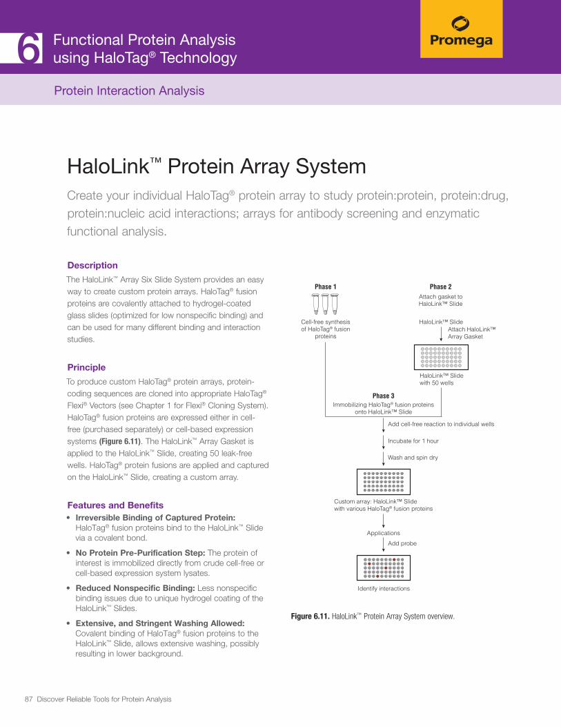
87 Discover Reliable Tools for Protein Analysis
HaloLink™ Protein Array SystemCreate your individual HaloTag® protein array to study protein:protein, protein:drug, protein:nucleic acid interactions; arrays for antibody screening and enzymatic functional analysis.
Description
The HaloLink™ Array Six Slide System provides an easy way to create custom protein arrays. HaloTag® fusion proteins are covalently attached to hydrogel-coated glass slides (optimized for low nonspecific binding) and can be used for many different binding and interaction studies.
Principle
To produce custom HaloTag® protein arrays, protein-coding sequences are cloned into appropriate HaloTag® Flexi® Vectors (see Chapter 1 for Flexi® Cloning System). HaloTag® fusion proteins are expressed either in cell-free (purchased separately) or cell-based expression systems (Figure 6.11). The HaloLink™ Array Gasket is applied to the HaloLink™ Slide, creating 50 leak-free wells. HaloTag® protein fusions are applied and captured on the HaloLink™ Slide, creating a custom array.
Features and Benefits• Irreversible Binding of Captured Protein:
HaloTag® fusion proteins bind to the HaloLink™ Slide via a covalent bond.
• No Protein Pre-Purification Step: The protein of interest is immobilized directly from crude cell-free or cell-based expression system lysates.
• Reduced Nonspecific Binding: Less nonspecific binding issues due to unique hydrogel coating of the HaloLink™ Slides.
• Extensive, and Stringent Washing Allowed: Covalent binding of HaloTag® fusion proteins to the HaloLink™ Slide, allows extensive washing, possibly resulting in lower background.
Figure 6.11. HaloLink™ Protein Array System overview.
7893
MA
Custom array: HaloLink™ Slidewith various HaloTag® fusion proteins
Phase 1 Phase 2Attach gasket toHaloLink™ Slide
HaloLink™ SlideAttach HaloLink™ Array Gasket
HaloLink™ Slidewith 50 wells
Phase 3Immobilizing HaloTag® fusion proteins
onto HaloLink™ Slide
Add cell-free reaction to individual wells
Incubate for 1 hour
Wash and spin dry
Applications
Add probe
Identify interactions
Cell-free synthesisof HaloTag® fusion
proteins
Functional Protein Analysis using HaloTag® Technology6Protein Interaction Analysis

88 Discover Reliable Tools for Protein Analysis
Functional Protein Analysis using HaloTag® Technology6
Promega Products HaloLink™ Array Six Slide System (Cat.# G6190)
ReferencesHurst, R. et al. (2009) Protein-protein interaction studies on protein arrays: Effect of detection strategies on signal-to-background ratios. Analytical Biochem. 392, 45–53.
Hoppe, S. et al. (2012) Microarray-based method for screening of immuno-genic proteins from bacteria. J. Nanobiotechnol. 10,12.
Wang, J. et al. (2013) A versatile protein microarray platform enabling antibody profiling against denatured proteins. Proteomics Clin. App. 7, 378–8
Protein Interaction Analysis

89 Discover Reliable Tools for Protein Analysis
77.1 Trypsin 91 Trypsin Gold, Mass Spec Grade 92
Trypsin/Lys-C Mix for Improved Protein Digestion 93
Rapid Digestion-Trypsin and Rapid Digestion-Trypsin/Lys-C Kits 95
Sequencing Grade Modified Trypsin 96
Immobilized Trypsin 97
7.2 Alternative Proteases 98 Specific Proteases: rAsp-N; Lys-C; Lys-N; Arg-C; Asp-N; Glu-C 100
Low-Specific Protease: Chymotrypsin 101
Nonspecific Proteases: Elastase; Pepsin; Thermolysin 101
7.3 Glycosidases 102 PNGaseF 103
EndoH 103
7.4 ProteaseMAX™ Surfactant 105 ProteaseMAX™ Surfactant for Optimized Proteolysis 106
7.5 Protein Extracts for LC/MS Instrument Performance Monitoring 108
MS Compatible Human Protein Extract, Digest 109
MS Compatible Yeast Protein Extract, Digest 110
6 × 5 LC-MS/MS Peptide Reference Mix 111
7.6 Antibody Characterization/Fragmentation 113
AccuMap Low pH Digestion Kit 114
IdeS Protease 115
7.7 Overview of Proteases for Mass Spectrometry Sample Preparation 116
7 Protein Characterization by Mass Spectrometry

90 Discover Reliable Tools for Protein Analysis
Protein Characterization by Mass SpectrometryMass spectrometry (MS) has become a powerful tool in proteomics for proteome-wide analysis and characterization of proteins from a variety of organisms and cell types. Recent advances in mass spectrometry provide tools for protein identification, protein characterization, relative and absolute quantitation, and the study of post-translational modifications and protein:protein interactions. Proteins are generally digested with proteases to generate peptides for MS analysis followed by sequencing (tandem MS). Promega provides high-quality proteases and other reagents that are critical to sample preparation for mass spectrometry.

91 Discover Reliable Tools for Protein Analysis
Trypsin is the most widely used protease in mass spectrometry sample preparation. It is a highly-specific serine protease, which cleaves at the carboxylic side of lysine and arginine residues. Protein digestion with trypsin generates peptides of optimal sizes for mass spec analysis. Tryptic peptides have a strong C-terminal charge, and therefore they can be efficiently ionized.
Trypsin is used for in-gel or in-solution digestion of proteins. Post digestion, the resulting peptides are introduced into the mass spectrometer and identified by peptide mass fingerprinting or tandem mass spec-trometry (MS/MS). This approach is called “bottom-up” proteomics and uses identification at the peptide level to detect and characterize proteins.
The stringent specificity of trypsin is essential for char-acterizing proteins using mass spectrometry. Promega’s high quality trypsin is derived from porcine trypsin and modified to give the highest proteolytic activity and cleavage specificity (Figure 7.1).
7.1 Trypsin: Mass Spectrometry Sample Preparation
OVERVIEW
Figure 7.1. The high quality of Promega trypsin is achieved by chemical modification, affinity purification and strict lot-release criteria. Trypsin Gold, Mass Spectrometry Grade (Cat.# V5280) is mass spectrometry-qualified.
Protein Characterizationby Mass Spectrometry7

92 Discover Reliable Tools for Protein Analysis
Protein Characterizationby Mass Spectrometry7Trypsin: Mass Spectrometry Sample Preparation
Trypsin Gold, Mass Spectrometry GradeDigestion of proteins into peptides via in-gel or in-solution techniques, followed by mass spec. Typical applications include protein identification, protein quantitation and post-translation modifications via liquid chromatography-mass spectrometry/mass spectrometry analysis.
Description
Trypsin Gold, Mass Spectrometry Grade, is manufactured to provide maximum specificity. Lysine residues in porcine trypsin are modified by reductive methylation, yielding a highly active and stable molecule that is extremely resistant to autolytic digestion. The specificity of the purified trypsin is further improved by TPCK treatment, which inactivates chymotrypsin. Additionally, the high performance of Trypsin Gold is assured by selecting affinity fractions with the highest proteolytic activity.
Ordering Information Trypsin Gold, Mass Spectrometry Grade (Cat.# V5280)
Principle
For in-gel digestion, protein samples are run on SDS-PAGE. Protein bands are stained and the bands of interest are excised. A buffer containing resuspended trypsin is added and the reaction is incubated overnight at 37°C. The resulting peptides are then extracted from the gel and analyzed by mass spec.
For in-solution digestion, protein samples are denatured, reduced and alkylated. Trypsin is added and the solution is incubated for 3-12 hours at 37°C. The resulting peptides are desalted and analyzed by mass spectrometry.
Features and Benefits• Application Qualified: Each lot is qualified by mass
spectrometry.
• Pure: Trypsin Gold is prepared by TPCK treatment fol-lowed by affinity purification.
• Good Value: Stable for up to 5 freeze-thaw cycles, thus minimizing reagent waste.
• Referenced in Thousands of Papers: Reliable and customer proven.
3868TA
11_2A
Mass (m/z)
Coun
ts
1,5001,000 2,000 2,500 3,000
5,000 842.
5197
9.36
897
3.7
1013
.710
18.5
511
02.0
9
1346
.74
1582
.06
2099
.49
2197
.57
2253
.74
2511
.07
2583
.66 28
51.3
7
10,000
15,000
20,000
Figure 7.2. Spectrogram of bovine carbonic anhydrase II digested by Trypsin Gold, Mass Spectrometry Grade. A 500ng sample of carbonic anhydrase II was separated by gel electrophoresis and digested with 500ng of Trypsin Gold, Mass Spectrometry Grade, overnight at 37°C.*

93 Discover Reliable Tools for Protein Analysis
Trypsin/Lys-C Mix, Mass Spectrometry GradeEnhanced in-solution protein digestion. Applications include: protein identification, protein quantitation and characterization of post-translation modifications via LC-MS/MS analysis.
Description
Trypsin/Lys-C Mix, Mass Spectrometry Grade, is a mixture of Trypsin Gold, Mass Spectrometry Grade, and rLys-C, Mass Spec Grade. The Trypsin/Lys-C Mix is designed to improve digestion of proteins or protein mixtures in solution. Replacing trypsin with Trypsin/Lys-C Mix has multiple benefits for protein analysis, including more accurate mass spectrometry-based protein quantitation and improved analytical reproducibility. Trypsin/Lys-C Mix also provides greater tolerance to trypsin-inhibiting agents, assuring efficient digestion of proteins for which purification is limited or not feasible.
Ordering Information Trypsin/Lys-C Mix, Mass Spec Grade (Cat.# V5071, V5072, V5073)
Principle
Typical trypsin reactions do not digest proteins to completion, missing 10–30% of cleavage sites (Figure 7.3). Incomplete digestion affects protein identifica-tion, reproducibility of mass spectrometry analysis and accuracy of protein quantitation. The number of missed cleavage sites may be even higher if the protein is not properly purified or contains protease-inhibiting contami-nants. Using the conventional trypsin digestion protocol (i.e., overnight incubation under nondenaturing condi-tions), Trypsin/Lys-C Mix improves protein digestion by eliminating the majority of missed lysine cleavages. The mixture is stable since trypsin is modified and therefore resistant to digestion by Lys-C.
Features and Benefits• Simple to Use: Use standard overnight digestion with
non-denaturing conditions.
• Enhanced Proteolysis: Increase peptide recovery by enhancing cleavage efficiency at lysine residues and elimi-nating the majority of missed cleavages (Figures 7.3 and 7.4).
• Tolerant to Trypsin-Inhibiting Contaminants: Generate mass spectrometry data from low-quality sample material.
ReferencesSaveliev, S. et al. (2013) Trypsin/Lys-C protease mix for enhanced protein mass spectrometry analysis. Nature Methods 10, Published online 30 November 2013. http://www.nature.com/app_notes/nmeth/2013/131211/pdf/nmeth.f.371.pdf
Protein Characterizationby Mass Spectrometry7Trypsin: Mass Spectrometry Sample Preparation

94 Discover Reliable Tools for Protein Analysis
Protein Characterizationby Mass Spectrometry7
In a typical digest,10–30% of cleavage sites are
“missed” (undigested).
Trypsin/Lys-C Mix misses fewer lysine cleavage sites and increases overall proteolytic efficiency.
Digested sample
Yeast extract
Mouse extract
Missed K
18.6%
6.6%
Missed R
3.6%
1.1%
Trypsin cleavage specificity
Regular one-stepovernight digestion
with Trypsin
Regular one-stepovernight digestion with Trypsin/Lys-C Mix
Missed K(80–90% of
missed cleavages)
Missed R Missed RMissed K
NNNNNK NNNNNR NNNNNN
Trypsin/Lys-C cleavage specificity
NNNNNK NNNNNR NNNNNN
Digested sample
Yeast extract
Mouse extract
Missed K
2.6%
2.9%
Missed R
4%
1.5%
11788M
C
1122
1MATenfold decrease in missed cleavages.
40% increasein unique peptides.
17% increase in identified proteins.
Trypsin Trypsin/Lys-C Mix
57%
5.8%
Trypsin Trypsin/Lys-C Mix
1490 2092
Trypsin Trypsin/Lys-C Mix
415 484
Figure 7.4. Improved mass spec analysis of a protein extract containing residual amounts of methanol and chloroform, which inhibit trypsin but have no effect on Trypsin/Lys-C Mix performance.
Figure 7.3. Side-by-side comparison of cleavage sites missed by trypsin or the Trypsin/Lys-C Mix using a standard digestion protocol.
Trypsin: Mass Spectrometry Sample Preparation

95 Discover Reliable Tools for Protein Analysis
Protein Characterizationby Mass Spectrometry7Trypsin: Mass Spectrometry Sample Preparation
Rapid Digestion-Trypsin and Rapid Digestion-Trypsin/Lys-C KitsShorten Digestion Time to as Little as 60 Minutes
Description
The Rapid Digestion-Trypsin and Rapid Digestion-Trypsin/Lys-C Kits are designed to shorten protein digestion times to 60 minutes versus the typical 4–18 hours required for protein digestion. Both kits contain three components: i) protease (Trypsin or Trypsin/Lys-C Mix); ii) protease Resuspension Buffer; and iii) Rapid Digestion Buffer optimized for faster diges-tions. Protein digestion with these kits follows a simple-to-use protocol that is both fast and efficient. The protocol is flexible, and can accommodate a large range of sample volumes and protein concentrations.
Ordering Information Rapid Digestion Trypsin Kit (Cat.# VA1060)
Rapid Digestion Trypsin/Lys-C Kit (Cat.# VA1061)
Principle
In an effort to shorten the time required to prepare samples for LC-MS/MS analysis, we developed a specialized trypsin preparation that supports rapid and efficient digestion at temperatures as high as 70°C. There are several benefits to this approach. First, proteolytic reaction times are dramatically shortened. Second, because no chemical denatur-ants have been added, off-line sample cleanup is not necessary, leading to shorter preparation times and diminished sample losses.The Rapid Digestion Kits are highly flexible. They can accommodate a variety of additives, including reducing and alkylating agents.
We also developed a Rapid Digestion—Trypsin/Lys-C Kit. Like the Trypsin/Lys-C Mix previously developed to prepare maximally efficiently proteolytic digests, particularly for complex mixtures, Rapid Digestion—Trypsin/Lys C is ideally suited for studies that require improved reproducibility across samples. In many examples the quality of data generated by using the Rapid Digestion format exceeds that obtained with the conventional overnight procedure (Figure 7.5).
Figure 7.5. Comparison of peptide spectral matches, Rapid Digestion versus overnight digestion.
PSM
s 1,000
1,200
1,400
1,600
1,800
600
400
Thyroglobulin Serotransferrin Myoglobin Carbonic Anhydrase 2
200
800
0
Rapid Digestion-Trypsin Overnight

96 Discover Reliable Tools for Protein Analysis
Protein Characterizationby Mass Spectrometry7
Sequencing Grade Modified TrypsinDigestion of proteins into peptides via in-gel or in-solution techniques, followed by mass spec analysis. Typical applications include protein identification, protein quantitation and analysis of post-translation modifications via LC-MS/MS analysis.
Description
Trypsin is a serine protease that specifically cleaves at the carboxylic side of lysine and arginine residues. The stringent specificity of trypsin is essential for protein identification. Native trypsin is subject to autoproteolysis, generating pseudo-trypsin, which exhibits a broadened specificity including a chymotrypsin-like activity. Such autoproteolysis products present in a trypsin preparation could result in additional peptide fragments that may interfere with database analysis of the mass of fragments detected by mass spectrometry. Sequencing Grade Modified Trypsin is chemically modified to prevent auto-proteolysis and assure high proteolytic activity over the course of digestion reaction.
Ordering InformationSequencing Grade Modified Trypsin (Cat.# V5111, V5117)
Sequencing Grade Modified Trypsin, Frozen (Cat.# V5113)
Principle
For in-gel digestion, protein samples are run on SDS-PAGE. Protein bands are stained and bands of interest are excised. A buffer containing resuspended trypsin is added and incubated overnight at 37°C. The resulting peptides are then extracted from the gel and analyzed by mass spec.
For in-solution digestion, protein samples are dena-tured, reduced and alkylated. Trypsin is then added and the solution is incubated for 3-12 hours at 37°C. The resulting peptides are desalted and analyzed by mass spec.
Features and Benefits• Maximum Specificity and Purity: Trypsin is
prepared by TPCK treatment followed by affinity purification.
• High Stability: Due to reductive methylation of lysine residues.
• Reliable and Customer-Proven: Referenced in more than 2,000 publications.
• Additional Value: Stability is ensured for up to 5 freeze-thaw cycles, minimizing leftover reagents.
• Variety of Packaging: We offer flexibility in packag-ing depending on experimental design and scope.
ReferencesTrask. S. et al. (2013) Mutations in the rotavirus spike protein VP4 reduce trypsin sensitivity but not viral spread. J. Gen, Vir. 94, 1296-300.
Trautner. C. et al. (2013) The sll1951 gene encodes the surface layer protein of Synechocystis sp. strain PCC 6803. J. Bact. 195(23), 5370-80.
Lord, M. et al. (2013) Sulfation of the bikunin chondroitin sulfate chain deter-mines heavy chain·hyaluronan complex formation. J. Biol. Chem. 288(22), 22930-41.
Gordon,S. et al. (2013) Multi-dimensional co-separation analysis reveals protein-protein interactions defining plasma lipoprotein subspecies. Mol. Cell, Prot. 12(11), 3123-34.
Jiang,W. et al. (2013) Mass spectrometry method to identify aging pathways of Sp- and Rp-tabun adducts on human butyrylcholinesterase based on the acid labile P-N bond. Toxicol. Sci. 132(2), 390-98.
Burkhart J. et al. (2012) Systematic and quantitative comparison of digest efficiency and specificity reveals the impact of trypsin quality on MS-based proteomics. J. Proteomics 75(4), 1454-1462.
Trypsin: Mass Spectrometry Sample Preparation
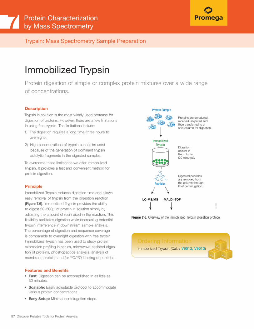
97 Discover Reliable Tools for Protein Analysis
Protein Characterizationby Mass Spectrometry7Trypsin: Mass Spectrometry Sample Preparation
Immobilized TrypsinProtein digestion of simple or complex protein mixtures over a wide range of concentrations.
Ordering Information Immobilized Trypsin (Cat.# V9012, V9013)
Description
Trypsin in solution is the most widely used protease for digestion of proteins. However, there are a few limitations in using free trypsin. The limitations include:
1) The digestion requires a long time (three hours to overnight).
2) High concentrations of trypsin cannot be used because of the generation of dominant trypsin autolytic fragments in the digested samples.
To overcome these limitations we offer Immobilized Trypsin. It provides a fast and convenient method for protein digestion.
Principle
Immobilized Trypsin reduces digestion time and allows easy removal of trypsin from the digestion reaction (Figure 7.6). Immobilized Trypsin provides the ability to digest 20–500μl of protein in solution simply by adjusting the amount of resin used in the reaction. This flexibility facilitates digestion while decreasing potential trypsin interference in downstream sample analysis. The percentage of digestion and sequence coverage is comparable to overnight digestion with free trypsin. Immobilized Trypsin has been used to study protein expression profiling in serum, microwave-assisted diges-tion of proteins, phoshopeptide analysis, analysis of membrane proteins and for 18O/16O labeling of peptides.
Features and Benefits• Fast: Digestion can be accomplished in as little as
30 minutes.
• Scalable: Easily adjustable protocol to accommodate various protein concentrations.
• Easy Setup: Minimal centrifugation steps.
Figure 7.6. Overview of the Immobilized Trypsin digestion protocol.80
21M
A
Protein Sample
Peptides
LC-MS/MS MALDI-TOF
Proteins are denatured, reduced, alkylated and then transferred to a spin column for digestion.
Digestion occurs in the column (30 minutes).
Digested peptides are removed fromthe column through brief centrifugation.
ImmobilizedTrypsin

98 Discover Reliable Tools for Protein Analysis
Protein Characterizationby Mass Spectrometry7
Trypsin is the most widely used endoprotease. It cleaves proteins in a highly specific manner and generates peptides 7–20 amino acids in length, with a strong C-terminal charge, making trypsin ideal for mass spectrometry analysis.
However, there are certain proteins and protein mixtures where trypsin digestion alone is not efficient enough. Examples include digestion of membrane proteins and analysis of histone post-translational modifications (PTMs). Furthermore, certain tryptic peptides are too small or too large for mass spec analysis. In these situations alternative proteases provide a viable solution, either alone or in combination with trypsin.
Table 7.1. Alternative Proteases and Corresponding Cleavage Sites.
7.2 Alternative Proteases for Mass Spectrometry
OVERVIEW
Protease Cleavage Site Optimal pH Example of Use
Lys-C and rLys-C Specific Protease
NNNNK NNN (K is lysine) 7.0–9.0
Digests membrane and other proteolytically resistant
proteins; generates larger peptides than trypsin,
an advantage for certain mass spec methods
(for example, ETD).
Lys-N Specific Protease
NNNN KNNN (K is lysine) 7.0–9.0
Digests membrane and other proteolytically resistant
proteins; generates larger peptides than trypsin,
an advantage for certain mass spec methods
(for example, ETD).
Arg-C Specific Protease
NNNNR NNN (R is arginine)
Arg-C can, at a lesser degree, cleave
at lysine also.
7.6–7.9Facilitates analysis of histone post-translational
modifications; used in proteome-wide analysis.
Glu-C Specific Protease
NNNNE NNN (E is glutamate) Glu-C
can, at a lesser degree, cleave at aspar-
tate residue also.
4.0–9.0
Used as alternative to trypsin if trypsin produces too
short or too long peptides or if tryptic cleavage sites
are not accessible.
Asp-N and rAsp-N Specific Protease
NNNN DNNN (D is aspartate) 4.0–9.0 Similiar to Glu-C.
Chymotrypsin Low Specific Protease
NNNN(F/Y/W) NNN (F,Y and W) are
aromatic residues phenylalanine, tyrosine
and tryptophan)
7.0–9.0Digests hydrophobic proteins (for example,
membrane proteins).
Pepsin Nonspecific Protease
Nonspecific Protease (advantage: most
active at low pH)1.0–3.0
Used in structural protein studies and antibody
analysis; digests proteolytically resistant, tightly
folded proteins.
Thermolysin Nonspecific Protease
Nonspecific Protease (advantage: remains
active at high temperature)5.0–8.5
Digests proteolytically resistant, tightly folded
proteins; used in structural protein studies.
Elastase Nonspecific Protease
Nonspecific Protease 9.0 Used to increase protein coverage.

99 Discover Reliable Tools for Protein Analysis
Protein Characterizationby Mass Spectrometry7
Figure 7.7. Increased protein coverage using (Panel A) both trypsin and chymotrypin; (Panel B) multiple proteases in separate digestion reactions. Note that the fusion map is the combination of the results obtained by digestion with each protease alone.
8690
MA
102030405060708090
100
Pe
rcen
t Pro
tein
Cov
erag
e
ChymotrypsinTrypsin
0
NCBI Accession Number
P0044
1
P0070
9
P0404
0
P0826
3
P0733
9
P0673
2
P6293
7
P6176
9
P0278
7
P1208
1
P1059
9
P0275
3
A.
9523
MA
304050607080
01020
Asp-NGlu-C
TrypsinFusion Map
Asp-N Glu-C Trypsin Fusion Map
Perc
ent P
rote
in C
over
age
Protease
B.
Digestion with Alternative Proteases Increase Protein Coverage
Digestion with an alternative protease, individually or in combination with trypsin, creates a unique peptide map that may include sequences not seen with trypsin digestion alone. Overlaying peptides obtained with alternative proteases and trypsin, increases protein coverage and overall confidence in protein identification (Figure 7.7).
Alternative Proteases Improve Analysis of Post-Translational Modifications (PTMs)
In certain cases, trypsin is not suitable for PTM analysis. For example, histone PTMs are complex and some, such as acety-lation and methylation, prevent trypsin digestion. As shown in Figure 7.8, trypsin digestion of histone H4 identified several PTMs; however, certain PTMs were missing. By digesting histone H4 with Arg-C, additional PTMs were identified, including mono- and dimethylated, and acetylated lysine and arginine residues.
Figure 7.8. Histone H4 post-translational modifications identified in trypsin and Arg-C digests.
1069
1MA
MSGRGKGGKG LGKGGAKRHR KVLRDNIQGI TKPAIRRLAR RGGVKRISGL...
N terminus of H4 histone
Dimethylated siteidentified in trypsin digest
Methylated siteidentified in trypsin digest
Acetylated site identified intrypsin digest
Additional methylated siteidentified in Arg-C digest
Additional dimethylated siteidentified in Arg-C digest
Additional acetylated siteidentified in Arg-C digest

100 Discover Reliable Tools for Protein Analysis
Alternative ProteasesImprove protein sequence coverage, enhance digestion of tightly-folded proteins, improve the identification of post-translation modifications (PTMs).
Specific Proteases
Arg-C (Clostripain), Sequencing Grade, is an endo-peptidase that cleaves at the C-terminus of arginine residues, including the sites next to proline. Arg-C activity is optimal in the pH range of 7.6–7.9.
Asp-N, Sequencing Grade, is an endoproteinase that hydrolyzes peptide bonds on the N-terminal side of aspartic acid residues. Asp-N activity is optimal at pH 4.0–9.0.
Glu-C, Sequencing Grade, is a serine protease that specifically cleaves at the C-terminus of glutamic acid residues. In ammonium bicarbonate and ammonium acetate the enzyme specificity is higher at the glutamic residues. In phosphate buffers cleavage occurs at the aspartic and glutamic residues. Glu-C activity is optimal at pH 4.0–9.0.
Lys-C, Mass Spec Grade, is a serine protease isolated from Lysobacter enzymogenes as a highly purified protease that hydrolyzes specifically at the carboxyl side of Lys. . Lys-C retains proteolytic activity under protein denaturing conditions such as 8M urea, which is used to improve digestion of proteolytically resistant proteins. Lys-C activity is optimal in the pH range of 7.0–9.0. This protease can be used alone or in combination with other proteases to produce protein digests for peptide mapping applications or protein identification by peptide mass fingerprinting or MS/MS spectral matching.
Lys-N, Mass Spec Grade, is a metalloprotease that cleaves at the amino side of lysine residues. Lys-N is a zinc metalloprotease derived from Grifola frondosa that retains proteolytic activity under protein denaturing conditions such as 8M urea, which is used to improve digestion of proteo-lytically resistant proteins. Charged amino-terminal peptide fragments generated by Lys-N are useful for de novo sequencing with ETD fragmentation techniques.
rAsp-N, Mass Spec Grade, is a recombinant protease that cleaves proteins N-terminal to aspartic acid residues. The enzyme was cloned from Stenotrophomonas maltophilia and purified from E. coli. rAsp-N is a highly active protease suitable for proteomic analysis of complex mixtures as well as peptide mapping of purified proteins, including thera-peutic monoclonal antibodies. The protease is provided in 10μg quantities, in a conical tube, for simple and consistent resuspension.
rLys-C, Mass Spec Grade, is a recombinant Lys-C expressed in E. coli. The sequence origin of rLys-C is Protease IV from Pseudomonas aeruginosa. Similarly to a native Lys-C, rLys-C cleaves at the C-terminus of lysine residues with exceptional specificity. rLys-C retains proteo-lytic activity under protein denaturing conditions such as 8M Urea, used to improve digestion of proteolytically resistant proteins. rLys-C activity is optimal at pH 8.0–9.0.
Protein Characterizationby Mass Spectrometry7Alternative Proteases for Mass Spectrometry
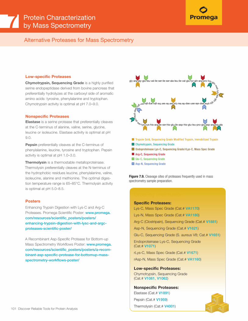
101 Discover Reliable Tools for Protein Analysis
Figure 7.9. Cleavage sites of proteases frequently used in mass spectrometry sample preparation.
gly-arg-gly-glu-leu-val-ile-ser-ile-ser-ala-leu-ile-val-glu-tyr-gln-arg-pro-trp-gln-met-lys-ala-gly-gly-thr-glu-asp
-ph
e-val-ser-
phe-asp-ala-asn-asp-ile-leu-lys-ser-ile-ser-his-glu-arg-glu-
lys-cys-his-arg-ile-ser-his-glu-ile-asp-his-glu-leu-pro-glu-asp-arg-trp-ala
86
34M
E
Arg-C, Sequencing Grade
Glu-C, Sequencing Grade
Asp-N, Sequencing Grade
Trypsin Gold, Sequencing Grade Modified Trypsin, Immobilized Trypsin
Chymotrypsin, Sequencing Grade
Endoproteinase Lys-C, Sequencing Grade/rLys-C, Mass Spec Grade
Low-specific Proteases
Chymotrypsin, Sequencing Grade is a highly purified serine endopeptidase derived from bovine pancreas that preferentially hydrolyzes at the carboxyl side of aromatic amino acids: tyrosine, phenylalanine and tryptophan. Chymotrypsin activity is optimal at pH 7.0–9.0.
Nonspecific ProteasesElastase is a serine protease that preferentially cleaves at the C-terminus of alanine, valine, serine, glycine, leucine or isoleucine. Elastase activity is optimal at pH 9.0.
Pepsin preferentially cleaves at the C-terminus of phenylalanine, leucine, tyrosine and tryptophan. Pepsin activity is optimal at pH 1.0–3.0.
Thermolysin is a thermostable metalloproteinase. Thermolysin preferentially cleaves at the N-terminus of the hydrophobic residues leucine, phenylalanine, valine, isoleucine, alanine and methionine. The optimal diges-tion temperature range is 65–85°C. Thermolysin activity is optimal at pH 5.0–8.5.
Posters
Enhancing Trypsin Digestion with Lys-C and Arg-C Proteases. Promega Scientific Poster: www.promega.com/resources/scientific_posters/posters/enhancing-trypsin-digestion-with-lysc-and-argc-proteases-scientific-poster/
A Recombinant Asp-Specific Protease for Bottom-up Mass Spectrometry Workflows Poster: www.promega.com/resources/scientific_posters/posters/a-recom-binant-asp-specific-protease-for-bottomup-mass-spectrometry-workflows-poster/
Protein Characterizationby Mass Spectrometry7Alternative Proteases for Mass Spectrometry
Ordering Information
Specific Proteases:Lys-C, Mass Spec Grade (Cat.# VA1170)
Lys-N, Mass Spec Grade (Cat.# VA1180)
Arg-C (Clostripain), Sequencing Grade (Cat.# V1881)
Asp-N, Sequencing Grade (Cat.# V1621)
Glu-C, Sequencing Grade (S. aureus V8; Cat.# V1651)
Endoproteinase Lys-C, Sequencing Grade (Cat.# V1071)
rLys-C, Mass Spec Grade (Cat.# V1671)
rAsp-N, Mass Spec Grade (Cat.# VA1160)
Low-specific Proteases: Chymotrypsin, Sequencing Grade (Cat.# V1061, V1062)
Nonspecific Proteases: Elastase (Cat.# V1891)
Pepsin (Cat.# V1959)
Thermolysin (Cat.# V4001)

102 Discover Reliable Tools for Protein Analysis
Protein Characterizationby Mass Spectrometry7
Glycosylation is the most abundant post-translational modification (PTM) in eukaryotic proteins. Glycosylation structures on proteins have diverse functions and can serve as disease biomarkers, such as for the progression of cancer. Therefore, tools that facilitate glycoprotein characterization are extremely important.
Endoglycosidases and exoglycosidases are tools for structural and functional analysis of glycoproteins. Endoglycosidases like PNGase F (see Figure 7.10) and Endo H are routinely used to remove carbohydrates from N-glycosylated proteins. For the analysis of O-glycosylated proteins, a mixture of endoglycosidases and exoglycosidases is recommended.
7.3 Glycosidases
OVERVIEW
1115
8ME
Mass Spec Analysis of Peptides
PNGase F
D
D
D
D
N
N
N-glycans
Trypsin
GlcNAc
Mannose
Fucose
Galactose
Sialic Acid
N Asparagine
D Aspartic Acid
Figure 7.10. Identification of N-glycosylated sites in proteins. Asn-linked glycans can be cleaved enzymatically by PNGase F, yielding intact oligo-saccharides and a protein in which Asn residues at the site of N-glycosylation are converted to Asp.

103 Discover Reliable Tools for Protein Analysis
Protein Characterizationby Mass Spectrometry7
GlycosidasesIdentification of glycosylation sites by LC-MS/MS; characterization of glycoproteins and glycan structures; monitor protein trafficking (endoplasmic reticulum, golgi); correlation of structural and functional data.
Description
PNGase F is a recombinant glycosidase cloned from Elizabethkingia miricola and overexpressed in E. coli. PNGase F has a molecular weight of 36kDa. PNGase F catalyzes the cleavage of N-linked oligosaccharides between the innermost N-Acetylglucosamine (GlcNAc) and asparagine residues (Asn) of high mannose, hybrid, and complex oligosaccharides from N-linked glycopro-teins (Figure 7.11). Due to the enzymatic reaction, intact oligosaccharides are released and Asn residues are converted to aspartic acids (Asp) at the former sites of N-glycosylation. The conversion (deamination) of Asn to Asp leads to a monoisotopic mass shift of 0.9840 Da that is used in tandem mass spectrometry (MS/MS) for the identification of N-glycosylated sites in proteins. PNGase F will not remove oligosaccharides containing α-(1,3)-linked core fucose commonly found on plant glycoproteins.
Endo H is a recombinant glycosidase cloned from Streptomyces plicatus and overexpressed in E. coli. Endo H cleaves within the chitobiose core of high mannose and some hybrid oligosaccharides from N-linked glycoproteins. In contrast to PNGase F, Endo H does not cleave N-glycosylated proteins of the complex type. The enzymatic activity of EndoH leaves one GlcNac residues at the N-glycosylated protein site leading to a shift in molecular weight (M.W.). This M.W. shift is used in mass spectrometry for the identification of N-glycosylated sites.
1112
3MA
High Mannose Structure
Hybrid Structure
Complex Glycan Structure
α1,6
α1,3 β1,4 β1,4
α1,6
α1,3 β1,4 β1,4
α1,6
α1,3 β1,4 β1,4
N,N’-diacetylchitobiose core
PNGase FCleavage Site
Asn
Asn
Asn
Endo HCleavageSite
Endo H is unable to cleave N-linked complex-type glycans
GlcNAc
Mannose
FucoseGalactose
Sialic Acid
Figure 7.11. Cleavage specificity of PNGase F and Endo H on N-glycans.
Glycosidases

104 Discover Reliable Tools for Protein Analysis
Protein Characterizationby Mass Spectrometry7
Ordering InformationPNGase F (Cat.# V4831)
Endo H (Cat.# V4871, V4875)
Fetuin (Cat.# V4961)
Figure 7.12. Detection of N-glycosylated proteins in SDS-PAGE. After glycan cleavage, the respective proteins show a higher mobility in SDS-PAGE. The application of both PNGase F and Endo H is informative for the presence of complex glycan structures, as shown here for Immunoglobulin G (IgG).
1217
1TA
100
75
50
37
2520
15
10
kDa PNGase F M – + – +
PNGase F substrate
IgG Ovalbumin
Endo H substrate
Endo HM – + – +IgG Ovalbumin
200
116
97
66
45
31
21
614
kDa
A. B.
Glycosidases (continued)

105 Discover Reliable Tools for Protein Analysis
Protein Characterizationby Mass Spectrometry7
Detergents are routinely used for protein extraction, solubilization and/or denaturation. However, detergents are known to interfere with mass spectrometry analysis. ProteaseMAX™ Surfactant is a mass spec-compatible detergent that can be used for improved protein extraction from cells and tissues, for protein solubilization/denaturation as well as for improved in-gel and in-solution protein digestion. Unlike other detergents/denaturants such as SDS and Urea, ProteaseMAX™ Surfactant is compatible with MS analysis. It degrades over the course of digestion reaction. Samples can then be easily analyzed with mass spectrometry.
7.4 ProteaseMAX™ Surfactant
OVERVIEW
ReferencesPirmoradian, M. et al. (2013) Rapid and deep human proteome analysis by single-dimension shotgun proteomics. Mol. Cell. Prot. 12, 3330–8.
Table 7.2. Comparison of Cell-lysis/Protein Extraction Methods
Cell-lysis/ Extraction Buffer
Number of Peptides
Number of Proteins
Urea 17,024 ± 148 3,326 ± 20
SDC* 22,171 ± 403 3,698 ± 18
ProteaseMAX™ Surfactant 29,884 ± 228 4,465 ± 100
ProteaseMAX™ Surfactant
and Cell Debris**33,098 ± 283 4,655 ± 51
Figure 7.13. Multiple applications of ProteaseMAX™ Surfactant for improved protein mass spectrometry sample preparation.
1229
6MA
ProteaseMAX™
Surfactant
Protein solubilization• Efficient resolubilization of precipitated proteins• Solubilization of hydrophobic proteins
Efficient digestion oftightly folded proteins• Denatures proteins for improved protease access
In-gel protein digestion• Improved protein identification• Streamlined procedure
Protein extraction from cells and tissues• High protein yield• Improved recovery of membrane proteins
A comparison of different detergents in cell-lysis buffers (Urea, SDC, ProteaseMAX™ Surfactant) revealed that ProteaseMAX™ Surfactant is optimal for efficient cell lysis/protein extraction for mass spectrometry sample preparation. The results of Pirmoradian et al. suggest that the addition of ProteaseMAX™ Surfactant to the cell-lysis buffer/extraction buffer provides the highest number of peptide/protein identifications (Table 7.2).
*SDC: Sodium desoxycholate.** Cell debris was included in the trypsin digestion step.
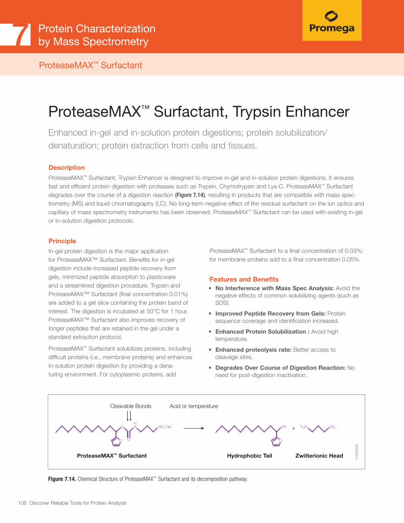
106 Discover Reliable Tools for Protein Analysis
Protein Characterizationby Mass Spectrometry7
ProteaseMAX™ Surfactant, Trypsin EnhancerEnhanced in-gel and in-solution protein digestions; protein solubilization/ denaturation; protein extraction from cells and tissues.
Description
ProteaseMAX™ Surfactant, Trypsin Enhancer is designed to improve in-gel and in-solution protein digestions. It ensures fast and efficient protein digestion with proteases such as Trypsin, Chymotrypsin and Lys-C. ProteaseMAX™ Surfactant degrades over the course of a digestion reaction (Figure 7.14), resulting in products that are compatible with mass spec-trometry (MS) and liquid chromatography (LC). No long-term negative effect of the residual surfactant on the ion optics and capillary of mass spectrometry instruments has been observed. ProteaseMAX™ Surfactant can be used with existing in-gel or in-solution digestion protocols.
Principle
In-gel protein digestion is the major application for ProteaseMAX™ Surfactant. Benefits for in-gel digestion include increased peptide recovery from gels, minimized peptide absorption to plasticware and a streamlined digestion procedure. Trypsin and ProteaseMAX™ Surfactant (final concentration 0.01%) are added to a gel slice containing the protein band of interest. The digestion is incubated at 50°C for 1 hour. ProteaseMAX™ Surfactant also improves recovery of longer peptides that are retained in the gel under a standard extraction protocol.
ProteaseMAX™ Surfactant solubilizes proteins, including difficult proteins (i.e., membrane proteins) and enhances in-solution protein digestion by providing a dena-turing environment. For cytoplasmic proteins, add
1122
9MA
O NH
SO3 Na− +
OO
OH
O
H3N SO3 −+
Cleavable Bonds
ProteaseMAX™ Surfactant
Acid or temperature
Hydrophobic Tail Zwitterionic Head
+
Figure 7.14. Chemical Structure of ProteaseMAX™ Surfactant and its decomposition pathway.
ProteaseMAX™ Surfactant to a final concentration of 0.03%; for membrane proteins add to a final concentration 0.05%.
Features and Benefits• No Interference with Mass Spec Analysis: Avoid the
negative effects of common solubilizing agents (such as SDS).
• Improved Peptide Recovery from Gels: Protein sequence coverage and identification increased.
• Enhanced Protein Solubilization : Avoid high temperature.
• Enhanced proteolysis rate: Better access to cleavage sites.
• Degrades Over Course of Digestion Reaction: No need for post-digestion inactivation.
ProteaseMAX™ Surfactant

107 Discover Reliable Tools for Protein Analysis
Protein Characterizationby Mass Spectrometry7
Ordering Information ProteaseMAX™ Surfactant, Trypsin Enhancer (Cat.# V2071, V2072)
1125
3MD
26% increasein unique peptides.
17% increase in identified proteins.
Trypsin
1407 1778
Trypsin
223 273
Trypsin withProteaseMAX™
Surfactant
Trypsin withProteaseMAX™
Surfactant
Figure 7.15. Example of improved peptide/protein identifications when using ProteaseMAX™ Surfactant for in-gel digestion of a complex protein sample.
1082
7MA
Conventional in-gel protein digestion
ProteaseMAX™ Surfactant Advantage for In-gel Protein Digestion
Overnight in-gel digestion
Peptide extraction(1.5–2 hours)
Mass spec analysis
In-gel protein digestionwith ProteaseMAX™ Surfactant
1-hour in-gel digestion
Mass spec analysis
Figure 7.16. Comparison of ProteaseMAX ™ Surfactant-assisted versus standard in-gel digestion procedure ProteaseMAX™ Surfactant offers time and labor savings.
ReferencesSaveliev, S. (2013) Mass spectrometry compatible surfactant for optimized in-gel protein digestion. Anal. Chem. 85(2) 907-14.
Pirmoradian, M. et al. (2013) Rapid and deep human proteome analysis by single-dimension shotgun proteomics. Mol. Cell Prot. 12(11), 3330-38.
Kalashnikova, A. et al. (2013) Linker histone H1.0 interacts with an extensive network of proteins found in the nucleolus. Nucl. Acids. Res. 41(7), 4026-35.
Silva, C. et al. (2013) Interaction of Mycobacterium leprae with human airway epithelial cells: adherence, entry, survival, and identification of potential adhesins by surface proteome analysis. Infect. Immun. 81(7), 2645-59.
Pearson, R. et al. (2013) Regulation of H2O2 stress-responsive genes through a novel transcription factor in the protozoan pathogen Entamoeba histolytica. J. Biol. Chem. 288(6), 4462-74.
ProteaseMAX™ Surfactant

108 Discover Reliable Tools for Protein Analysis
Protein Characterizationby Mass Spectrometry7
Adequate monitoring of instrument performance for proteomic studies requires complex protein reference materials. Whole-cell protein extracts provide the needed complexity. We offer mass spec-compatible whole-cell reference protein extracts from yeast and human cells, in a convenient, pre-digested lyophilized form. Consistency is monitored by various protein and peptide qualitative and quantitative methods including amino acid analysis and LC/MS analysis (Table 7.3).
For routine reporting on MS instrument sensitivity and dynamic range as well various LC parameters, we recom-mend use of the 6 x 5 LC-MS/MS Peptide Reference
Mix. The product is a mixture of 30 peptides: 6 sets of 5 isotopologues of the same peptide sequence. The isotopologues differ only by the number of stable, heavy-labeled amino acids incorporated into the sequence. The labels consist of uniform 13C and 15N atoms. Chromatographically, each of the isotopologues is indis-tinguishable; however, since they differ in mass, they are clearly resolved by mass spectrometry. The isotopologues of each peptide are present in a series of tenfold dilutions. This format allows assessment of instrument dynamic range and sensitivity from a single run.
7.5 Protein Extracts for LC/MS Instrument Performance Monitoring
OVERVIEW
Promega (MS Compatible Human Protein Extract,
Digest, Cat.# V6951)
Vendor X(HeLa Protein Digest Standard)
Nonbiological
post-translational
modifications
Deamidation spectra: <12% Not tested; no specification
Oxidation spectra: <5% Oxidation spectra: <10%
Carbamylation spectra: <5% Carbamylation spectra: <10%
Missed clevages <10% <10%
Peptide quality
Measured by Amino Acid Analysis (AAA) to quantitate the
amount of each amino acid in the sampleA280
Protein fragmentation: 1% or less Not tested; no specification
Matching spectra: >65% Not tested; no specification
Lot-to-lot
consistency
≥12,462 unique peptides LC-MS chromatogram conforms to reference
Total proteins: ≥1805 4.8%
Protein identification >85% of 1,194 human core peptides Ratio of peptide area to reference = 0.75–1.125
Protein quantitation reproducibility is monitored by relative
abundance of ten reference proteinsNot tested
Table 7.3. Promega Human Extract has Enhanced Quality Control Specifications and Higher Performance than Other Commercial Extracts.

109 Discover Reliable Tools for Protein Analysis
Protein Characterizationby Mass Spectrometry7
MS Compatible Human Protein Extract, DigestReady-to-use predigested human extracts for Liquid Chromatography/Mass Spectrometry (LC/MS) instrument performance monitoring and method development.
Description
MS Compatible Human Protein Extract, Digest is a lyophilized tryptic peptide mixture from K562 human cells (myelogenous leukemia cell line) designed for monitoring of LC/MS instrument performance. The digest is also suitable for MS method development such as isobaric mass tag labeling. Stringent quality control over the manufacturing process assures repro-ducibility of this reference material. MS Compatible Human Protein Extract, Digest is provided in ready-to-use format elimi-nating the need for sample preparation prior to MS analysis.
Figure 7.17. Lot-to-lot reproducibility. Base peak chromatograms of three different human protein extract batches analyzed by LC/MS. Each run used 1µg of human pre-digested protein extract injected into the instrument (Waters NanoAquity HPLC system interfaced to a ThermoFisher Q Exactive). Peptides were resolved with 2h gradient.
1229
4TA
A.
B.
C.
Ordering Information MS Compatible Human Protein Extract, Digest (Cat.# V6951)
MS Compatible Human Protein Extract, Intact (Cat.# V6941)
Preparation
MS Compatible Human Protein Extract, Digest, is prepared by digestion of K562 whole-cell protein extract with the Trypsin/Lys-C Mix. The digestion procedure is optimized to assure the most efficient proteolysis with less than 8% tryptic missed cleavages. The digest is SPE (C18) cleaned-up to remove nonpeptide material and lyophilized. The digest can be reconstituted in formic acid or TFA and used for instrument performance monitoring. In general, 1μg of digest per injection is recommended.
Features and Benefits• Ready-to-Use: No clean-up or digestion required.
• High Material Complexity: Complex proteome with a large dynamic range.
• Lot-to-Lot Consistency: Stringent QC over the production process.
• Validated Reference Material: Tested with all the major LC/MS platforms.
Protein Extracts for LC/MS Instrument Performance Monitoring

110 Discover Reliable Tools for Protein Analysis
MS Compatible Yeast Protein Extract, DigestReady-to-use predigested yeast extracts for Liquid Chromatography/Mass Spectrometry LC/MS instrument performance monitoring and method development.
Description
MS Compatible Yeast Protein Extract, Digest from Saccharomyces cerevisiae is a lyophilized tryptic peptide mixture designed for monitoring LC/MS instrument performance. The digest is also suitable for MS method development such as isobaric mass tag labeling. Stringent quality control over the manufacturing process assures reproducibility of this reference material.
Preparation
MS Compatible Yeast Protein Extract, Digest, is prepared by digestion of yeast whole-cell protein extract with the Trypsin/Lys-C Mix. The digestion procedure is optimized to assure the most efficient proteolysis with less than 8% tryptic missed cleavages. The digest is SPE (C18) cleaned-up to remove nonpeptide material and lyophilized. The digest can be reconstituted in formic acid or TFA and used for instrument performance monitoring. In general, 1μg of digest per injection is recommended.
Figure 7.18. Lot-to-lot quantitative reproducibility of yeast extract. Relative protein quantitative levels were determined by spectral counting. The data showed high protein quantitative reproducibility between the different lots.
1229
5MA
0
20
40
60
80
100
120
140
160
Spec
tral
Cou
ntin
g
Protein ID
Lot 1
Lot 2
Lot 3
HS90B ENOA PRKDC HS90A ACTG CH60 FASTBB5 TBB2C TBA2B
Features and Benefits• Ready-to-Use: No clean-up or digestion required.
• Well-Annotated Proteome: The small yeast proteome is a convenient reference material.
• Lot-to-Lot Consistency: Stringent QC throughout the production process.
• Validated Reference Material: Tested with all the major LC/MS platforms.
Ordering Information MS Compatible Yeast Protein Extract, Digest (Cat.# V7461)
MS Compatible Yeast Protein Extract, Intact (Cat.# V7341)
Protein Characterizationby Mass Spectrometry7
Protein Extracts for LC/MS Instrument Performance Monitoring

111 Discover Reliable Tools for Protein Analysis
Protein Characterizationby Mass Spectrometry7
Peptide Mix for LC/MS Instrument Performance Monitoring
6 × 5 LC-MS/MS Peptide Reference Mix Unique peptide mix/software designed to monitor, analyze and record a variety of LC-MS/MS parameters.
Description
The 6 × 5 LC-MS/MS Peptide Reference Mix is a unique reagent designed to monitor liquid chromatography (LC) and mass spectrometry (MS) instrument performance and to assist in method development/optimization. The product is a mixture of 30 peptides: 6 sets of 5 isotopologues of the same peptide sequence. The isotopologues differ only by the number of stable, heavy-labeled amino acids incorporated into the sequence. The labels consist of uniform 13C and 15N atoms. Chromatographically, each of the isotopologues is indistinguishable; however, since they differ in mass, they are clearly resolved by mass spectrometry. The isotopologues of each peptide are present in a series of tenfold dilutions. This format allows assessment of instrument dynamic range and sensitivity from a single run (Figure 7.19). Peptides with a wide range of hydrophobicities were chosen to enable reporting of LC column performance. In addition there is a mass separation of at least 4 daltons between the isotopologues, so that even low resolution instruments can distinguish the masses.
PReMiS™ Software Tool
The 6 × 5 LC-MS/MS Peptide Reference Mix is designed for use with the complementary PReMiS™ Software tool (available by download) that reports on key liquid chro-matography and mass spec parameters. The parameter reports can be exported to CSV or saved as “.pdf” files. In addition to the general reporting feature, performance parameters can be tracked over time, allowing a clear assessment of trends to pinpoint poor performance and maintenance needs. For those laboratories that have multiple instruments, the ability to compare parameters across instruments will also be available. Thermo (.raw) and ABSCIEX (.wiff) are available for direct importing. Other vendor formats can be imported after conversion to “.mzml.”
Features and Benefits
Unique peptide formulation
• Save time by assessing LC and MS parameters (e.g., dynamic range and sensitivity) in one run with a single reagent
• Optimize and streamline method development
Complementary software provides routine analysis and historical monitoring
• Eliminate time-consuming manual calculations
• Ensure consistent instrument performance over time
• Compare performance of multiple instruments over time
Peptides are AAA qualified
• Accurate reporting of instrument sensitivity and dynamic range
Multiple applications
• Compatible with neat or complex mixture analysis
Ordering Information 6 x 5 LC-MS/MS Peptide Reference Mix (Cat.# V7491, V7495)

112 Discover Reliable Tools for Protein Analysis
Peptide Mix for LC/MS Instrument Performance Monitoring (continued)
Figure 7.19. Schematic illustrating standard workflow and correlating features of the 6 × 5 LC-MS/MS Peptide Reference Mix. A mixture of 6 peptide sets is actually 30 peptides, in total. Each of the peptides sets is a mixture of 5 isotopologues that are distinguished only by mass through the incorporation of stable, heavy-labeled amino acids. Panel A. The isotopologues are chemically and chromatographically identical and therefore appear as 6 peaks. Panel B. Each of the isotopologues is mixed with a 10-fold molar increase (lightest to heaviest). Panel C. A plot of the log of the peak height versus the log (molar amount) on column has a slope of 1. This analysis enables confirmation of detection limits and can also be used to confirm linearity of up to 5 logs of dynamic range.
496.
7 499.
7 502.
2
504.
7 509.
3
414.
7 417.
7 420.
7
424.
2 428.
2
553.
8 556.
8 559.
8
562.
8 566.
8
447.
3 450.
3 453.
3
456.
3 459.
8
521.
8
525.
3 528.
8
532.
3 537.
3
518.
3 521.
8 525.
3
530.
3 535.
3
496.7499.7
502.2504.7
509.3
414.7417.7
420.7424.2
428.2
553.8556.8
559.8562.8
566.8
447.3450.3
453.3456.3
459.8
521.8525.3
528.8532.3
537.3
518.3521.8
525.3530.3
535.3
Hydrophobicity
496.7(m/z)
499.7(m/z)
502.2(m/z)
504.7(m/z)
509.3(m/z)
VTSGSTSTSR
414.7(m/z)
417.7(m/z)
420.7(m/z)
424.2(m/z)
428.2(m/z)
LASVSVSR
553.8(m/z)
556.8(m/z)
559.8(m/z)
562.8(m/z)
566.8(m/z)
YVYVADVAAK
447.3(m/z)
450.3(m/z)
453.3(m/z)
456.3(m/z)
459.8(m/z)
VVGGLVALR
521.8(m/z)
525.3(m/z)
528.8(m/z)
532.3(m/z)
537.3(m/z)
LLSLGAGEFK
518.3(m/z)
521.8(m/z)
525.3(m/z)
530.3(m/z)
535.3(m/z)
LGFTDLFSK1X
0.1X
0.01X
0.0001X
0.001X
Time (minutes)
Tota
l Ion
Inte
nsity
(TIC
)Lo
g (in
tens
ity)
Log
(pea
k he
ight
)
A.
B.
C.
(mass/charge)
Log (mole)
1239
3TA
Peptide MolarAmounts
Protein Characterizationby Mass Spectrometry7

113 Discover Reliable Tools for Protein Analysis
Protein Characterizationby Mass Spectrometry7
Therapeutic monoclonal antibodies are large, complex molecules that undergo numerous post-translational modifications (PTMs). Numerous varieties of PTMs are known to exist on antibodies. Because these PTMs can affect the efficacy and safety profile of the drug, the structural variants must be assessed and quantified. In-depth characterization of antibody PTMs remains a significant hurdle because their large size (~150kDa) makes mass spectrometry analysis extremely challenging.
IdeS Protease is highly specific and cleaves human (and some other species) IgG specifically at one site in the lower hinge region. Because of the exquisite specificity of the enzyme, it produces highly homogeneous Fc and Fab fragments which are then readily analyzed using techniques such as mass spectrometry (Figure 1) or X-ray crystallography.
7.6 Antibody Characterization/Fragmentation
OVERVIEW

114 Discover Reliable Tools for Protein Analysis
Protein Characterizationby Mass Spectrometry7Antibody Characterization/Fragmentation
A.
B.
1396
2MA
GLEWIGAIYPGNGDTSYNQK Unmodified peptide
Deamidated form
Scrambling is suppressed
Conventional digestion (pH 8)
Low pH digestion
Retention time (minutes)
Retention time (minutes)
46.13
47.44
46.08
Conventional digestion (pH 8)
Low pH digestion
HC319-LC134scrambled bond
Mass = 497.7491; Z = 4
SGTASVVCLLNNFYPR
CK
39 40 41 42 43 44 45 46 47 48 49 50 51 52
0
50
100
0
50
100
68.5 69.5 70.5 71.5 72.5
70.93
70.8370.78
70.6870.62
70.16
70.4570.36
71.06 71.31
71.46
71.5871.77 72.15
69.0 70.0 71.0 72.0 73.0
0
4020
1008060
0
4020
1008060
LC134
HC319
Deamidation is suppressed
Principle
Nonenzymatic posttranslational modifications (PTMs) spontaneously occur in biotherapeutic proteins during manufacturing and storage. The major nonenzymatic PTMs are deamidation, disulfide bond scrambling and oxidation. These modifications can affect efficacy and stability of biotherapeutic proteins and are the subject of careful monitoring. Nonenzymatic PTMs can also be induced during protein sample preparation for peptide mapping and can compromise analysis. Major causes of nonenzymatic PTMs induced during sample preparation include alkaline pH as well as excipients and impurities that have protein oxidizing activity. The AccuMAP™ Low pH Protein Digestion Kits suppress artificial nonenzymatic PTMs, as sample preparation is performed at low pH under either reducing or nonreducing conditions.
Figure 7.20. Suppression of deamidation and disulfide bond scrambling in IgG digested with AccuMAP™ Low pH Protein Digestion Kit. Panel A shows extracted ion chromatogram of a peptide from Rituximab antibody digested at conventional conditions (pH 8) and at low pH. The data show that asparagine (highlighted in red) was deamidated in this peptide at pH 8. In contrast, deamidation was fully suppressed at low pH. Panel B shows an extracted ion chromatogram of a peptide with a scrambled disulfide bond from Panitumumab antibody digested under conventional conditions (pH 8) and at low pH.
AccuMap™ Low pH Protein Digestion Kit Suppress Nonenzymatic PTMs During Protein Sample Prep
Description
The AccuMap™ Low pH Protein Digestion Kit is designed for accurate, reproducible characterization of biotherapeutic proteins by peptide mapping using LC/MS and/or UV HPLC. The entire sample preparation procedure is performed at low (mildly acidic) pH to suppress artificial deamidation and disulfide bond scrambling. The kit also contains an optional agent for suppression of protein oxidation during sample preparation.
Ordering Information AccuMap™ Low pH Protein Digestion Kits (Cat.# VA1040, VA1050)
Product may not be available in all countries. Please contact your local Promega representative for information.

115 Discover Reliable Tools for Protein Analysis
Protein Characterizationby Mass Spectrometry7
Features and Benefits• Fast and Easy: Digestion in 30 minutes with no optimization.
• Highly Specific and Reproducible: Cleaves exclusively at a single site below the hinge to produce F(ab′)2 and Fc fragments.
• High Performance: Essentially 100% complete digestion.
• Versatile: Effectively cleaves human IgG1–IgG4, monkey, sheep, rabbit, humanized and chimeric IgGs as well as Fc-fusion proteins.
Figure 7.21. Schematic showing cleavage specificity of IdeS Protease. The orange cysteines (C) indicate where the interchain disulfide bonds are formed to the opposite heavy chain. The orange glycines (G) indicate the IdeS cleavage site.
Ordering Information IdeS Protease (Cat.# V7511, V7515)
1233
6TB
IdeSIgG
(150kDa)
DTTLC
(25kDa)
Fc/2(25kDa)
F(ab′)2
(100kDa)Fd′
(25kDa)
Fc/2 +Glycans
LC Fd′
Fc/2 + Glycans LC Fd′
A.
B.
C.
Denature
Fc(50kDa)
Tim
e
Antibody Characterization/Fragmentation
IdeS ProteaseRecombinant protease engineered to simplify antibody fragmentation and characterization.
Description
The Immunoglobulin-degrading enzyme from Streptococcus pyogenes (IdeS) is a highly specific protease that cleaves Immunoglobulin G (IgG) at a single site below the hinge region, yielding F(ab’)2 and Fc fragments. The IdeS Protease can be used to characterize therapeutic antibody candidates using LC/MS. Promega IdeS Protease is a recombinant, engineered version of the enzyme from S. pyogenes.
Product may not be available in all countries. Please contact your local Promega representative for information.

116 Discover Reliable Tools for Protein Analysis
Protein Characterizationby Mass Spectrometry7
Table 7.4. Characteristics and Reaction Conditions for Trypsin Proteases.
Trypsin Gold, Sequencing Grade Modified Trypsin, Sequencing Grade Modified Trypsin, Frozen Immobilized Trypsin Trypsin/Lys-C Mix
Cat.# V5280; V5111; V5117; V5113 V9012; V9013 V5071; V5072; V5073
Source and Size Porcine Pancreas (23.8kDa) Porcine Pancreas (23.8kDa) Mix of Tryspin-Gold & rLys-C
Cleavage Sites
C-terminal of Lys, Arg. Does not cleave if Lys and Arg are followed by Pro. Asp and Glu at C-terminal side of Lys and Arg inhibit cleavage.
C-terminal of Lys, Arg. Does not cleave if Lys and Arg are followed by Pro. Asp and Glu at C-terminal side of Lys and Arg inhibit cleavage.
C-terminal of Lys, Arg. Does not cleave if Lys and Arg are followed by Pro. In contrast to trypsin, Trypsin/Lys-C efficiently toler-ates presence of Glu and Asp at C-terminal side of Lys and Arg.
Protease Protein Ratio (w/w)
1:20 to 1:100 see Technical Manual 1:25 to 1:50
pH Range for Digestion
pH 7–9 pH 5–9 pH 8
Reaction Conditions50–100mM Tris-HCl (pH 8) or 50–100mM NH4HCO3(pH 7.8). Digest overnight hours at 37°C.
50mM NH4HCO3 (pH 7.8). Digest for 30 minutes at room temperature.
50–100mM Tris-HCl (pH 8) or 50mM NH4HCO3 (pH7.8). Digest overnight at 37°C.
Buffer Compatibility Tris-HCl, NH4HCO3 Tris-HCl, NH4HCO3 Tris-HCl, NH4HCO3
In-Gel Digestion Compatibility
Yes No Yes
ProteaseMAX™ Compatibility
Yes Yes Not tested
Notes
Most widely used protease in mass spectrometry. All Promega trypsin is treated with TPCK to inactivate chymotrypsin contamination and chemically modified (methylated) to minimize autoproteolysis. All Promega trypsin is resistant to mild denaturing conditions (1–2M urea and 0.1% SDS). They retain 48% activity in 2M Guanidine Chloride.
Used if rapid digestion is required.Urea ≤ 4M; guanidine HCl ≤ 3M; methanol < 60%; DTT ≤ 50mM; TCEP ≤ 5mM; pH 5–9; Iodoacetamide ≤ 300mM.
Trypsin/Lys-C mix is the most robust trypsin preparation. It is designed to improve general protein digestion. Trypsin/Lys-C mix uses the same regular overnight diges-tion at 37°C under non- or mildly denaturing conditions as trypsin alone.
7.7 Proteases for Mass Spectrometry Sample PreparationADDITIONAL INFORMATION

117 Discover Reliable Tools for Protein Analysis
Table 7.5. Characteristics and Reaction Conditions for Specific Alternative Proteases.
rLys-C, Mass Spec Grade
Lys-C, Mass Spec Grade
Lys-N, Mass Spec Grade
rAsp-N, Mass Spec Grade
Cat.# V1671 VA1170 VA1180 VA1160
Source and Size
Pseudomonas aeruginosa. Expressed in E.coli (27.7kDa)
Lysobacter enzymogenes (30kDa)
Grifola frondosa (18kDa) Stenotrophomonas maltophilia (25kDa)
Cleavage Sites
C-terminal of Lys. Does not cleave if Lys is followed by Pro. Asp and Glu at C -terminal side of Lys inhibit cleavage.
C-terminal of Lys. Does not cleave if Lys is followed by Pro. Asp and Glu at C -terminal side of Lys inhibit cleavage.
Cleaves at the N-terminus of Lysine.
Primarily on the N-terminal side of aspartic acid residues. Cleavage on the N-terminal side of glutamic acid residues can occur at a slower rate.
Protease Protein Ratio (w/w)
1:20 to 1:50 1:20 to 1:100 1:20 to 1:100 1:10 to 1:100
pH Range for Digestion
pH 8–9 pH 7–9 pH 7–9
rAsp-N has maximal activity at pH 8, but pH buffers ranging from 6–9 can be used.
Reaction Conditions
50-100mM Tris-HCl (pH 8) or 50mM NH4HCO3 (pH 7.8). Digestion 2–18 hours at 37°C.
Incubate at 37°C for 2–18 hours.
Incubate at 37°C for 2–18 hours.
Incubate 37C for 60 minutes
Buffer Compatibility
Tris-HCl, NH4HCO3 50mM Tris (pH 8). 50mM Tris (pH 8).
Ammonium acetate pH 5-6HEPES pH 7Tris pH 8-9Ammonium Bicarbonate pH 8
In-Gel Digestion Compatibility
Yes Not tested Not tested Not tested
ProteaseMAX™ Compatibility
Yes Not tested Not tested Not tested
Notes
Inexpensive alternative to a native Lys-C protease. Similarly to a native protease, rLys-C tolerates high denaturing conditions such as 8M urea. Used to digest tightly folded proteo-lytically resistant proteins. Also used as a trypsin alternative if larger peptides are preferable for the analysis. If urea is used in protein sample preparation, avoid high temperature. High temperature induces protein carbamylation in the presence of urea.
Tolerates high denaturing conditions such as 8M urea. Used to digest tightly folded proteolytically resis-tant proteins. Also used as a trypsin alternative if larger peptides are preferable for analysis. If urea is used in protein sample preparation, avoid high temperature. High temperature induces protein carbamylation in the presence of urea.
Denaturants: Lys-N is active in up to 6M urea, although activity will vary depending on concentration.If urea is used in protein sample preparation, avoid high temperature. High temperature induces protein carbamylation in the presence of urea.
rAsp-N, Mass Spec Grade, is lyophilized in Tris (pH 8.0) with NaCl and stabilizing sugars. Reconstitute in 50µl of ultrapure water and mix gently. Reconstituted rAsp-N can be stored at 4°C for at least 8 weeks. For longer storage, single-use aliquots can be stored at –65°C or below. Avoid freeze-thaw cycles.rAsp-N has a histidine tag that can be used to remove it from solution.
Protein Characterizationby Mass Spectrometry7

118 Discover Reliable Tools for Protein Analysis
Table 7.5. Characteristics and Reaction Conditions for Specific Alternative Proteases. (continued )
Asp-N Arg-C Glu-C
Cat.# V1621 V1881 V1651
Source and Size
Pseudomonas fragi (24.5kDa)
Clostridium histolyticum (Subunits: 45kDa and 12kDa)
Staphylococcus aureus V8(27kDa)
Cleavage Sites N-terminal of Asp. C-terminal of Arg. Also cleaves at Lys albeit at lower efficiency.
C-terminal of Glu. Low level cleavages might occur at Asp residues too albeit at 100–300 fold lower effi-ciency.
Protease Protein Ratio (w/w)
1:20 to 1:200 1:20 to 1:350 1:20 to 1:200
pH Range for Digestion
pH 4–9 pH 7.6–7.9 pH 4–9
Reaction Conditions
50mM Tris-HCl (pH 8).Digestion 2–18 hours at 37°C.
50mM Tris-HCl (pH 7.6-7.9), 5mM CaCl2, 2mM EDTA, >2mM DTT. Digestion 2–18 hours at 37°C.
100mM NH4HCO3 (pH 7.8), 50-100 mM HCL (pH8). Digestion 2–18 hours at 37°C.
Buffer Compatibility
Tris-HCl, NH4HCO3 Tris-HCl, NH4HCO3
NH4HCO3, Ammonium acetate
In-Gel Digestion Compatibility
Yes Yes Yes
ProteaseMAX™ Compatibility
Yes Yes Yes
Notes
Can be used as a trypsin alternative to achieve better distribution of cleavage sites.100% activity retained in the presence of urea (up to 3.5 M), guanidine HCL (1M), SDS (up to 0,028%), ProteaseMax™ Surfactant (up to 0,026%), acetonitrile (up to 60%), EDTA (up to 2 mM); DTT or ß-mercapto-ethanol
Used in analysis of histone modifications. Requires DTT, cysteine or other reducing agent and CaCl2 for activity.
Can be used as a trypsin alternative to achieve better distribution of cleavage sites. Glu-C activity and cleavage specificity is affected by buffer condi-tions. In ammonium biocar-bonate and other non-phosphate buffers, Glu-C cleaves at C-term of Glu. Glu-C cleaves at C-term of Glu and Asp in phosphate buffer.
Protein Characterizationby Mass Spectrometry7
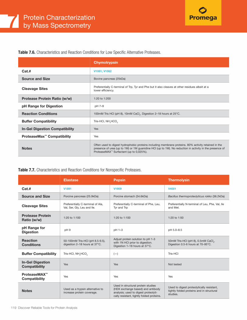
119 Discover Reliable Tools for Protein Analysis
Protein Characterizationby Mass Spectrometry7
Table 7.7. Characteristics and Reaction Conditions for Nonspecific Proteases.
Elastase Pepsin Thermolysin
Cat.# V1891 V1959 V4001
Source and Size Porcine pancreas (25.9kDa) Porcine stomach (34.6kDa) Bacillus thermoproteolyticus rokko (36.2kDa)
Cleavage Sites Preferentially C-terminal of Ala, Val, Ser, Gly, Leu and Ile.
Preferentially C-terminal of Phe, Leu, Tyr and Trp
Preferentially N-terminal of Leu, Phe, Val, Ile and Met.
Protease Protein Ratio (w/w)
1:20 to 1:100 1:20 to 1:100 1:20 to 1:50
pH Range for Digestion
pH 9 pH 1–3 pH 5.0–8.5
Reaction Conditions
50-100mM Tris-HCl (pH 8.5-9.5), digestion 2–18 hours at 37°C.
Adjust protein solution to pH 1-3 with 1N HCl prior to digestion. Digestion 1–18 hours at 37°C.
50mM Tris-HCl (pH 8), 0.5mM CaCl2. Digestion 0.5-6 hours at 70–95°C.
Buffer Compatibility Tris-HCl, NH4HCO3 (—) Tris-HCl
In-Gel Digestion Compatibility
Yes Yes Not tested
ProteaseMAX™ Compatibility
Yes Yes Yes
Notes Used as a trypsin alternative to increase protein coverage.
Used in structural protein studies (HDX exchange based) and antibody analysis; used to digest proteolyti-cally resistant, tightly folded proteins.
Used to digest proteolytically resistant, tightly folded proteins and in structural studies.
Table 7.6. Characteristics and Reaction Conditions for Low Specific Alternative Proteases.
Chymotrypsin
Cat.# V1061; V1062
Source and Size Bovine pancreas (25kDa)
Cleavage Sites Preferentially C-terminal of Trp, Tyr and Phe but it also cleaves at other residues albeit at a lower efficiency.
Protease Protein Ratio (w/w) 1:20 to 1:200
pH Range for Digestion pH 7–9
Reaction Conditions 100mM Tris HCl (pH 8), 10mM CaCl2. Digestion 2–18 hours at 25°C.
Buffer Compatibility Tris-HCl, NH4HCO3
In-Gel Digestion Compatibility Yes
ProteaseMax™ Compatibility Yes
NotesOften used to digest hydrophobic proteins including membrane proteins. 80% activity retained in the presence of urea (up to 1M) or 1M guanidine HCl (up to 1M). No reduction in activity in the presence of ProteaseMAX™ Surfactant (up to 0,025%).

120 Discover Reliable Tools for Protein Analysis
Conjugated Secondary Antibodies 120
Substrates for ELISA 121 AttoPhos® AP Fluorescent Substrate System 121
TMB One Solution 122
Substrates for Western Blotting 123 Western Blue® Stabilized Substrate for Alkaline Phosphatase 123
TMB Stabilized Substrate for Horseradish Peroxidase 124
ECL Western Blotting Substrate 125
ProtoBlot® II AP Systems with Stabilized Substrate and Western Express® Fast Blotting 126
Additional Reagents 127 Broad Range Protein Molecular Weight Marker 127
Protease Inhibitor Cocktail 128
8 Protein Detection Tools for Western Blotting & ELISA
120 Discover Reliable Tools for Protein Analysis

121 Discover Reliable Tools for Protein Analysis
Protein Detection Tools for Western Blotting and ELISAWestern blot and ELISA are widely used analytical techniques for the specific detection of proteins in samples such as cells, tissues and other extracts. For both techniques protein-spe-cific antibodies (primary antibodies) are required. Upon bind-ing of a primary antibody to its target, a conjugated second-ary antibody directed against a species-specific heavy chain portion of the primary antibody is added: for example, an anti-mouse secondary antibody binds to a primary antibody generated in mouse. Secondary antibodies, which are most frequently conjugated to reporter enzymes such as horse-radish peroxidase (HRP) or alkaline phosphatase (AP), will bind to one primary antibody. Depending on the substrates used, the conjugated enzymes catalyze a colorimetric, fluo-rescent or chemiluminescent reaction enabling sensitive detection with different instruments and scanners.
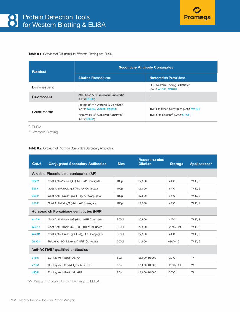
122 Discover Reliable Tools for Protein Analysis
Protein Detection Toolsfor Western Blotting & ELISA8
Table 8.2. Overview of Promega Conjugated Secondary Antibodies.
Cat.# Conjugated Secondary Antibodies SizeRecommended Dilution Storage Applications*
Alkaline Phosphatase conjugates (AP)
S3721 Goat Anti-Mouse IgG (H+L), AP Conjugate 100µl 1:7,500 +4°C W, D, E
S3731 Goat Anti-Rabbit IgG (Fc), AP Conjugate 100µl 1:7,500 +4°C W, D, E
S3821 Goat Anti-Human IgG (H+L), AP Conjugate 100µl 1:7,500 +4°C W, D, E
S3831 Goat Anti-Rat IgG (H+L), AP Conjugate 100µl 1:2,500 +4°C W, D, E
Horseradish Peroxidase conjugates (HRP)
W4021 Goat Anti-Mouse IgG (H+L), HRP Conjugate 300µl 1:2,500 +4°C W, D, E
W4011 Goat Anti-Rabbit IgG (H+L), HRP Conjugate 300µl 1:2,500 -20°C/+4°C W, D, E
W4031 Goat Anti-Human IgG (H+L), HRP Conjugate 300µl 1:2,500 +4°C W, D, E
G1351 Rabbit Anti-Chicken IgY, HRP Conjugate 300µl 1:1,000 –20/+4°C W, D, E
Anti-ACTIVE® qualified antibodies
V1151 Donkey Anti-Goat IgG, AP 60µl 1:5,000-10,000 -20°C W
V7951 Donkey Anti-Rabbit IgG (H+L) HRP 60µl 1:5,000-10,000 -20°C/+4°C W
V8051 Donkey Anti-Goat IgG, HRP 60µl 1:5,000-10,000 -20°C W
*W: Western Blotting; D: Dot Blotting; E: ELISA
Table 8.1. Overview of Substrates for Western Blotting and ELISA.
ReadoutSecondary Antibody Conjugates
Alkaline Phosphatase Horseradish Peroxidase
Luminescent -ECL Western Blotting SubstrateW
(Cat.# W1001, W1015)
FluorescentAttoPhos® AP Fluorescent SubstrateE
(Cat.# S1000)-
Colorimetric
ProtoBlot® AP Systems (BCIP/NBT)W
(Cat.# W3940, W3950, W3960)
Western Blue® Stabilized SubstrateW
(Cat.# S3841)
TMB Stabilized SubstrateW (Cat.# W4121)
TMB One SolutionE (Cat.# G7431)
E ELISA W Western Blotting

123 Discover Reliable Tools for Protein Analysis
Protein Detection Toolsfor Western Blotting & ELISA8
Conjugated Secondary Antibodies Detection of primary antibodies in Western blotting, enzyme-linked immunosorbent assay (ELISA) and dot blotting.
Description
High-quality, polyclonal secondary antibodies are raised in goat, rabbit or donkey. These polyclonal antibodies are immunoaffinity-purified using corresponding immo-bilized antigens. They are conjugated to horseradish peroxidase (HRP) or alkaline phosphatase (AP) enzymes.
The Anti-ACTIVE® qualified secondary antibodies are specifically tested for the use with Promega Anti-ACTIVE® primary antibodies, which are tools to measure activation of three members of the Mitogen-Activated Protein Kinase (MAPK) superfamily. The primary anti-bodies are specific for dually-phosphorylated active forms of MAPK, p38 and JNK. They exhibit minimal cross-reactivity to goat, mouse and sheep IgG, bovine serum albumin (BSA) and proteins in mammalian cell extracts. These secondary antibody conjugates provide low backgrounds and highly specific signals.
Features and Benefits• Approved: Use with confidence, as supported by
numerous publications.
• Ready-to-Use Formulation: No need to reconsti-tute the antibody.
• Flexible Dispensing: We can readily accommodate large-scale custom orders.
Figure 8.1. Western blot (immunoblot) for β-actin in cytoplasmic lysate from HEK293T cells. The blot was performed on a serial dilution of lysate; each lane contains an indicated amount of total protein (µg). Primary antibody (monoclonal) was used at 1:5,000; Anti-Mouse IgG, HRP Conjugate secondary antibody (Cat.# W4021) was used at 1:2,500; blot imaged with ECL Western Blotting Substrate (Cat.# W1015) and 1-minute exposure.
1053
0TA
kDa80
605040
30
20
4 1.6 0.64 0.26 0.1 0.04 (µg)
ReferencesChang, H.-Y. et al. (2011) Domain analysis of protein P30 in Mycoplasma pneumoniae cytadherence and gliding motility. J. Bacteriol. 193(7), 1726–33.
Prenner, G. et al. (2011) Is LEAFY a useful marker gene for the flower-inflorescence boundary in the Euphorbia cyathium? J. Exp. Bot. 62(1), 345–50.
Hu, D. et al. (2012) Novel insight into KLF4 proteolytic regulation in estrogen receptor signaling and breast carcinogenesis. J. Biol. Chem. 287(17), 13584–97.

124 Discover Reliable Tools for Protein Analysis
Protein Detection Toolsfor Western Blotting & ELISA8
AttoPhos® AP Fluorescent Substrate SystemFluorescent development of ELISA.
Features and Benefits• Sensitivity: Low fluorescence signal until
enzymatically activated, detection of AP to 0.1 attomole.
• Low Background: Low fluorescence from interfering biological molecules.
• Linearity: Linear kinetics over five orders of magnitude of AP concentration.
Ordering Information AttoPhos® AP Fluorescent Substrate System (Cat.# S1000)
2798
MA1
1_9B
S
S O
O
O–
O– O–P
Pi
N
S
N
N
S
NAlk Phos pH = 9–10.3
AttoPhos® Substrate (BBTP) (low fluorescence) BBT anion (high fluorescence)
Figure 8.2. Fluorescence signal observed for a serial dilution of calf intestinal alkaline phosphatase (CIAP) treated with 1mM AttoPhos® Substrate in a multiwell plate. The graph represents the increasing fluorescence per unit of AP at 15 minutes post addition of AttoPhos® Substrate.
3765MA07_2A
•
•
••••••0
1,000
2,000
3,000
4,000
5,000
6,000
7,000
0 0.25 0.5 0.75 1.0
RFU
AP (milliunits)
Figure 8.3. The reaction of AttoPhos® Substrate with AP to produce highly fluorescent BBT and inorganic phosphate (Pi).
ReferencesMichaud, A. et al. (2013) Absence of cell surface expression of human ACE leads to perinatal death. Hum. Mol. Genet. Nov. 14 [Epub ahead of print]
Meinel, S. et al. (2013) Mineralocorticoid receptor interaction with SP1 gener-ates a new response element for pathophysiologically relevant gene expres-sion. Nucl. Acids Res. 41(17), 8045–60.
Chen, Y. et al. (2013) Common tolerance mechanisms, but distinct cross-reactivities associated with gp41 and lipids, limit production of HIV-1 broad neutralizing antibodies 2FS and 4E10. J. Immunol. 191(3), 1260–75.
Description
The AttoPhos® AP Fluorescent Substrate System provides a highly sensitive fluorescent alkaline phos-phatase (AP) substrate. The system includes AttoPhos® Substrate, AttoPhos® Buffer and Calibration Solution. AttoPhos® Substrate is cleaved by alkaline phosphatase to produce inorganic phosphate (Pi) and the alcohol 2′-[2-benzothiazoyl]-6´-hydroxybenzothiazole (BBT).
This enzyme-catalyzed conversion of the phosphate form of AttoPhos® Substrate to BBT is accompanied by an enhancement in fluorescence properties. Relative to AttoPhos® Substrate, the BBT anion has highly increased quantum efficiency and fluorescence excita-tion, also the emission spectra are shifted into the visible region. Relative to other fluorometric reporters, the BBT anion has an unusually large Stokes’ shift of 120nm, resulting in a higher signal-to-noise ratio and higher overall detection sensitivity. The excitation of the fluores-cence is at 435nm, emission at 555nm.
Substrates for ELISA

125 Discover Reliable Tools for Protein Analysis
Protein Detection Toolsfor Western Blotting & ELISA8
Substrates for ELISA
TMB One SolutionColorimetric Development of ELISA.
Description
TMB One Solution is a safe, convenient ready-to-use working solution for the detection of HRP-conjugated antibodies in an ELISA format. HRP-conjugated antibodies react with the chromogenic substrate 3,3´,5,5´-tetramethylbenzidine (TMB) yielding a blue-colored solution. After reaching the desired color intensity, the reaction is stopped by addition of an acidic solution, which leads to a change in color from blue to yellow. Plates are analyzed on an ELISA reader at 450nm.
Ordering Information TMB One Solution (Cat.# G7431)
Features and Benefits• Convenient: Single solution provided ready-to-use;
add, incubate, stop and read. This homogeneous reagent reduces assay variation.
• Stable: Stable for 12 months at 4°C, providing extended shelf life; the assay end product is stable for at least one hour after stopping the assay.
• Safe: Provided in a slightly acidic, nonhazardous pro-prietary buffer without aprotic solvents; non-caustic to plastics used in automated systems.
• Sensitive: Low background provides higher assay sensitivity.
Additional Information
The TMB One Solution has been developed to work with Promega Emax
® ELISAs for BDNF, GDNF, and TGFβ1. It can be used for any ELISA using HRP conju-gated secondary antibodies.
ReferencesSmith, A.D. et al. (2013) Selenium status alters the immune response and expulsion of adult Heligosomoides bakeri worms in mice. Infect. Immun. 81, 2546–53.
Yamazaki, T. et al. (2012) The ddY mouse: a model of postprandial hypertriglyceridemia in response to dietary fat. J. Lipid Res. 53, 2024–37.
Apidianakis, Y. et al. (2012) Down-regulation of glutathione S-transferase o 4 (hGSTA4) in the muscle of thermally injured patients is indicative of susceptibility to bacterial infection. FASEB J. 26, 730–7.
0 100 200 300 400 500
Abso
rban
ce (4
50nm
)
BDNF (pg/ml)
0 7.8 15.6 31.3
0
0.2
0.4
0.6
0.8
1.0
1.2
1.4
1.6
1.8
2.0
00.050.100.150.200.25
r2 = 0.999
3234MA01_1A
Figure 8.4. Representative BDNF standard curve obtained using the ELISA “BDNF E
max® ImmunoAssay System” (Cat.# G7610). The inset is an
enlargement of the 0–31.3pg/ml portion of the graph.

126 Discover Reliable Tools for Protein Analysis
Protein Detection Toolsfor Western Blotting & ELISA8
Western Blue® Stabilized Substrate for Alkaline Phosphatase (AP)Substrate for Alkaline Phosphatase for Western blots and Dot blots.
Ordering Information Western Blue® Stabilized Substrate for Alkaline Phosphatase (Cat.# S3841)
Description
Western Blue® Stabilized Substrate for Alkaline Phosphatase is a stable, ready-to-use substrate for Western blots and immunoscreening. It is a mixture of 5-bromo-4-chloro-3-indolyl-phosphate (BCIP) and nitro blue tetrazolium (NBT) in a proprietary stabilizing buffer. Western Blue® Substrate should be used directly and without dilution. This liquid substrate deposits a permanent dark purple stain on membrane at sites with an alkaline phosphatase-conjugated antibody. Western Blue® Substrate is as sensitive as other reagents based on the BCIP/NBT formulation.
Features and Benefits• Convenient: Ready-to-use formulation does not
require dilution or reagent mixing.
• Sensitive: Substrate is as sensitive as other commer-cially available BCIP/NBT formulations and reagents.
• Stable: Stable for one year at room temperature.
ReferencesCedeno-Laurent, F. et al. (2012) Galectin-1 triggers an immunoregulatory signature in Th cells functionally defined by IL-10 expression. J. Immunol. 188(7), 3127–37.
Petrova, N.S. et al. (2012) Carrier-free cellular uptake and the gene-silencing activity of the lipophilic siRNAs is strongly affected by the length of the linker between siRNA and lipophilic group. Nucl. Acids Res. 40(5), 2330–44.
To, W.S. and Midwood, K.S. (2011) Identification of novel and distinct binding sites within Tenascin-C for soluble and fibrillar fibronectin. J. Biol. Chem. 286(17), 14881–91.
1052
7TB
80
6050
40
30
20
kDa20 8.0 3.2 1.3 0.5 0.2 (ng)
Figure 8.5. Western blot (immunoblot) for β-actin in cytoplasmic lysate from HEK293T cells. The blot was performed on a serial dilution of lysate; each lane contains an indicated amount of β-actin (ng). Primary antibody (monoclonal) used at 1:5,000; Anti-Mouse IgG, AP Conjugate secondary antibody (Cat.# S3721) used at 1:2,500; blot imaged with Western® Blue Stabilized Substrate for Alkaline Phosphatase (Cat.# S3841).
Substrates for Western Blotting

127 Discover Reliable Tools for Protein Analysis
Protein Detection Toolsfor Western Blotting & ELISA8
Substrates for Western Blotting
TMB Stabilized Substrate for Horseradish Peroxidase (HRP)Substrate for Western blots and Dot blots.
Ordering Information TMB Stabilized Substrate for HRP (Cat.# W4121)
Description
TMB Stabilized Substrate for horseradish peroxidase is a stable, ready-to-use TMB (3,3´, 5,5´-tetramethyl-benzidine) color development substrate for localization of horseradish peroxidase-conjugated antibodies on Dot blots and Western blots. It is easier to use than 4-chloro-1-naphthol (CN), which must be prepared immediately before use. TMB Stabilized Substrate comes premixed and fully diluted in a proprietary buffer containing less than 0.5% organic solvent.
Features and Benefits• Convenient: Premixed, ready-to-use; in proprietary
buffer containing less than 0.5% organic solvents.
• Stable: Stable at room temperature for 12 months.
• Sensitive: At least 3X more sensitive than 4-chloro-1-naphthol (CN).
• Long-Lasting Color: Color is much more stable than CN and photographs more easily.
1052
9TA
80
6050
40
30
20
kDa4 1.6 0.64 0.26 0.1 0.04 (µg)
Figure 8.6. Western blot (immunoblot) for β-actin in cytoplasmic lysate from HEK293T cells. The blot was performed on a serial dilution of lysate; each lane contains an indicated amount of total protein (µg). Primary antibody (monoclonal) used at 1:5,000; Anti-Mouse IgG, HRP Conjugate secondary antibody (Cat.# W4021) used at 1:2,500; blot imaged with TMB Stabilized Substrate for horseradish peroxidase (Cat.# W4121).

128 Discover Reliable Tools for Protein Analysis
Protein Detection Toolsfor Western Blotting & ELISA8
Ordering Information ECL Western Blotting Substrate (Cat.# W1001, W1015)
ECL Western Blotting Substrate Substrate for the detection of HRP-conjugated antibodies for Western blots and Dot blots.
Description
ECL Western Blotting Substrate is a nonradioactive, enhanced luminol-based chemiluminescent substrate for the detection of horseradish peroxidase (HRP) conjugates on immunoblots. The ECL Western Blotting Substrate detects and visualizes the presence of picogram (pg) amounts of antigen through the use of photographic or other suitable chemiluminescent imaging methods.
Features and Benefits• Save Time: No optimization required; you can switch
from other entry-level ECL substrates.
• Save Money: Use Promega’s Entry Level ECL.
1052
3TA
kDa80
605040
30
20
20 8 3.2 1.3 0.5 0.2 (ng)
Figure 8.7. Western blot (immunoblot) for β-actin in cytoplasmic lysate from HEK293T cells. The blot was performed on a serial dilution of lysate; each lane contains an indicated amount of β-actin (ng) as quantitated by ELISA.
Substrates for Western Blotting

129 Discover Reliable Tools for Protein Analysis
Protein Detection Toolsfor Western Blotting & ELISA8
Substrates for Western Blotting
ProtoBlot® II AP Systems with Stabilized Substrate and Western Express® Fast BlottingSubstrate for the detection of alkaline phosphatase (AP) conjugated antibodies in Western blots.
Description and Principle
The ProtoBlot® II AP Systems with Stabilized Substrate are designed for the rapid and sensitive detection of proteins or other macromolecular antigens immobilized on nitrocellulose or PVDF membranes. Proteins can be transferred from gels after electrophoresis (Western blots) or bound directly from solution (“dot” blots).
The Western Express® Fast Blotting Protocol is included with the system and can reduce dramatically the time required to perform immunodetection.
Features and Benefits• Fast: Easy-to-use Western Express® Protocol allows
the detection of dot blots in 30–45 minutes and the detection of Western blots in 1–2 hours.
• Convenient: The system contains Western Blue® Stabilized Substrate for AP, which is a ready-to-use solution of BCIP/NBT. No reagent preparation is required for the substrate.
Ordering Information ProtoBlot® II AP System with Stabilized Substrate, Human (Cat.# W3940)
ProtoBlot® II AP System with Stabilized Substrate, Mouse (Cat.# W3950)
ProtoBlot® II AP System with Stabilized Substrate, Rabbit (Cat.# W3960)

130 Discover Reliable Tools for Protein Analysis
Protein Detection Toolsfor Western Blotting & ELISA8
Broad Range Protein Molecular Weight MarkersSDS-PAGE protein size marker.
Ordering Information Broad Range Protein Molecular Weight Markers (Cat.# V8491)
Description
The Broad Range Protein Molecular Weight Markers consist of nine clearly identifiable bands at convenient molecular weights. The protein sizes are 10, 15, 25, 35, 50, 75, 100, 150 and 225kDa. Each protein is present at a concentration of 0.1μg/μl, except for the 50kDa protein, which is present at 0.3μg/μl and serves as a reference indicator, having triple the band intensity of the other proteins.
These markers are intended for use as a size standard when performing SDS-PAGE (sodium dodecyl sulfate-polyacrylamide gel electrophoresis) for estimation of the molecular weight of the protein of interest. Note that they are not stained and will need to be visual-ized using common in-gel staining reagents such as Coomassie®, Silver, or other staining methods
Features and Benefits• Reference Band: Band at 50kDa is 3X intensity for
use as a reference.
• Convenient: Nine bands at evenly spaced intervals.
• Fast: Ready to load.
Additional Information
Sufficient for 100 lanes at 5μl per lane.
– 225
kDa
– 150
– 100– 75
– 50
– 35
– 25
– 15
– 10
3480
TA07
_1A
4–20% Tris-Glycine SDS-PAGE gel
Figure 8.8. Broad Range Protein Molecular Weight Markers; 4-20% Tris-Glycine SDS-PAGE.
Additional Reagents

131 Discover Reliable Tools for Protein Analysis
Protein Detection Toolsfor Western Blotting & ELISA8
Protease Inhibitor CocktailInhibition of endogenous proteases during protein purification from mammalian or insect cell cultures.
Ordering Information Protease Inhibitor Cocktail (Cat.# G6521)
Description
Protease Inhibitor Cocktail is used to prevent protein degradation after lysing cells. The product is a mixture of six different protease inhibitors with different target protease specificities. The inhibitor cocktail is EDTA-free and provided as a powder, ready for reconstitution in 1ml of either 100% ethanol or DMSO to obtain a 50X working solution.
Features and Benefits• Broad Specificity: Inhibitor cocktail is effective
against a diverse number of proteases.
• Excellent Potency: Reagent provides the best-in-class level of protease inhibition.
• Highly Compatible: Works with a wide array of protein fusion tags (e.g., Flag® tag, His tag, GST tag) and capture technologies. It is ideally suited for HaloTag® Technology-based approaches.
ReferencesGalbraith, M.D. et al. (2013)HIF1A Employs CDK8-Mediator to Stimulate RNAPII Elongation in Response to Hypoxia. Cell 153(6), 1327–1339.
Deplu, R. et al. (2013) TET2 and TET3 regulate GlcNAcylation and H3K4 methylation through OGT and SET1/COMPASS. EMBO J. 32(5), 645–55.
Table 8.3. Compounds included in the Protease Inhibitor Cocktail.
InhibitorMode of Action Target
Benzamidine HCl
Reversible Trypsin like/Serine Proteases
Leupeptin Reversible Serine/Cysteine Proteases
Pepstatin A Reversible Aspartic Acid Proteases
1,10 Phenanthroline
Chelator Metalloproteases
PMSF Reversible Serin Proteases
Bestatin Reversible Amino Peptidases
Additional Information
Additional Reagents

132 Discover Reliable Tools for Protein Analysis
Table 9.1. Overview of Protein Expression Vectors.
Flexi® Vector Name1
Cat. # Drug Selection2
Expression Application
Promoter for Expression
Peptide Fusion Tag
E. coli Mammalian Cells
Cell-Free Transcription/
Translation
N- terminus
C- terminus
pF1A T7 Flexi® Vector
pF1K T7 Flexi® Vector
C8441
C8451
Ampicillin
Kanamycin
Inducible expression of native protein
T7 T7
pFN2A (GST) Flexi® Vector
pFN2K (GST) Flexi® Vector
C8461
C8471
Ampicillin
Kanamycin
Soluble expression and purification via an N-terminal GST tag
T7 T7 GST
pF3A WG (BYDV) Flexi® Vector
pF3K WG (BYDV) Flexi® Vector
L5671
L5681
Ampicillin
Kanamycin
In vitro wheat germ expression of native protein
T7, SP6
pF25A ICE T7 Flexi® Vector
pF25K ICE T7 Flexi® Vector
L1061
L1081
Ampicillin
Kanamycin
In vitro insect cell free expression of native protein
T7
pFN6A (HQ) Flexi® Vector
pFN6K (HQ) Flexi® Vector
C8511
C8521
Ampicillin
Kanamycin
Inducible expression and protein purification via a metal affinity resin
T7 T7 HQHQHQ
pFC7A (HQ) Flexi® Vector
pFC7K (HQ) Flexi® Vector
C8531
C8541
Ampicillin
Kanamycin
Inducible expression and protein purification via a metal affinity resin
T7 T7 HQHQHQ
pF4A CMV Flexi® Vector
pF4K CMV Flexi® Vector
C8481
C8491
Ampicillin
Kanamycin
Constitutive high expression of native protein
CMV T7
pF5A CMV-neo Flexi® Vector
pF5K CMV-neo Flexi® Vector
C9401
C9411
Ampicillin/Neomycin
Kanamycin/NeomycinM
Constitutive high expression of native protein with selec-tion for stable transfectants
CMV T7
1 The “pF” indicates the vector is a Flexi® Vector suitable for Flexi® cloning (see chapter 1 for details).The letter after “pF” indicates the position of any expression tags (“N” for an N-terminal expression tag and “C” for a C-terminal expression tag). The letters “A” and “K” designate the bacterial drug selection for the vector (“A”= ampicillin and “K”= kanamycin).
2 The “M” indicates the vector provides resistance to the indicated drug in mammalian cells.
Additional Information9
Expression Vectors

133 Discover Reliable Tools for Protein Analysis
Additional Information9
Flexi® Vector Name1
Cat. # Drug Selection2
Expression Application
Promoter for Expression
Peptide Fusion Tag
E. coli Mammalian Cells
Cell-Free Transcription/
Translation
N- terminus
C- terminus
pF9A CMV hRluc-neo Flexi® Vector
C9361AmpcillinNeomycinM
Constitutive high expression of native protein with selec-tion and reporter screening for stable transfectants
CMV T7
pFN10A (ACT) Flexi® Vector
C9331Ampicillin/Neomycin
Mammalian in vivo protein:protein interaction
CMV T7HSV VP16 activation domain
pN11A (BIND) Flexi® Vector
C9341 AmpicillinMammalian in vivo protein:protein interaction
CMV T7GAL4 DNA-
binding domain
pF12A RM Flexi® Vector
pF12K RM Flexi® Vector
C9431
C9441
Ampicillin
Kanamycin
Regulated mammalian protein expression
12λOP-Mini CMV
pFN18A HaloTag® T7 Flexi® Vector
pFN18K HaloTag® T7 Flexi® Vector
G2751
G2681
Ampicillin
Kanamycin
Protein interaction analysis and protein purification
T7 T7 HaloTag®
pFN19A HaloTag®
T7 SP6 Flexi® Vector
pFN19K HaloTag® T7 SP6 Flexi® Vector
G1891
G1841
Ampicillin
Kanamycin
Protein interaction analysis T7 T7, SP6 HaloTag®
pFC20A HaloTag® T7 SP6 Flexi® Vector
pFC20K HaloTag® T7 SP6 Flexi® Vector
G1681
G1691
Ampicillin
Kanamycin
Protein interaction analysis T7 T7, SP6 HaloTag®
pFC14A HaloTag® CMV Flexi® Vector
pFC14K HaloTag® CMV Flexi® Vector
G9651
G9661
Ampicillin
Kanamycin
Cell imaging and protein interaction analysis; high constitutive expression
CMV T7 HaloTag®
1 The “pF” indicates the vector is a Flexi® Vector suitable for Flexi® cloning (see chapter 1 for details).The letter after “pF” indicates the position of any expression tags (“N” for an N-terminal expression tag and “C” for a C-terminal expression tag). The letters “A” and “K” designate the bacterial drug selection for the vector (“A”= ampicillin and “K”= kanamycin).
2 The “M” indicates the vector provides resistance to the indicated drug in mammalian cells.
Table 9.1. Overview of Protein Expression Vectors (continued).

134 Discover Reliable Tools for Protein Analysis
Flexi® Vector Name1
Cat. # Drug Selection2
Expression Application
Promoter for Expression
Peptide Fusion Tag
E. coli Mammalian Cells
Cell-Free Transcription/
Translation
N- terminus
C- terminus
pFC15A HaloTag® CMVd1 Flexi® Vector
pFC15K HaloTag® CMVd1 Flexi® Vector
G1611
G1601
Ampicillin
Kanamycin
Cell imaging and protein interaction analysis; medium constitutive expression
T7 CMVd1 T7, SP6 HaloTag®
pFC16A HaloTag® CMVd2 Flexi® Vector
pFC16K HaloTag® CMVd2 Flexi® Vector
G1591
G1571
Ampicillin
Kanamycin
Cell imaging and protein interaction analysis; low constitutive expression
T7 CMVd2 T7, SP6 HaloTag®
pFC17A HaloTag® CMVd3 Flexi® Vector
pFC17K HaloTag® CMVd3 Flexi® Vector
G1551
G1321
Ampicillin
Kanamycin
Cell imaging and protein interaction analysis; very low constitutive expres-sion
T7 CMVd3 T7, SP6 HaloTag®
pFN21A HaloTag® CMV Flexi® Vector
pFN21K HaloTag® CMV Flexi® Vector
G2821
G2831
Ampicillin
Kanamycin
Cell imaging and protein interaction analysis; high constitutive expression
CMV T7 HaloTag®
pFN22A HaloTag® CMVd1 Flexi® Vector
pFN22K HaloTag® CMVd1 Flexi® Vector
G2841
G2851
Ampicillin
Kanamycin
Cell imaging and protein interaction analysis; medium constitutive expression
T7 CMVd1 T7, SP6 HaloTag®
pFN23A HaloTag® CMVd2 Flexi® Vector
pFN23K HaloTag® CMVd2 Flexi® Vector
G2861
G2871
Ampicillin
Kanamycin
Cell imaging and protein interaction analysis; low constitutive expression
T7 CMVd2 T7, SP6 HaloTag®
1 The “pF” indicates the vector is a Flexi® Vector suitable for Flexi® cloning (see chapter 1 for details).The letter after “pF” indicates the position of any expression tags (“N” for an N-terminal expression tag and “C” for a C-terminal expression tag). The letters “A” and “K” designate the bacterial drug selection for the vector (“A”= ampicillin and “K”= kanamycin).
2 The “M” indicates the vector provides resistance to the indicated drug in mammalian cells.
Table 9.1. Overview of Protein Expression Vectors (continued).
Additional Information9

135 Discover Reliable Tools for Protein Analysis
Additional Information9
Flexi® Vector Name1
Cat. # Drug Selection2
Expression Application
Promoter for Expression
Peptide Fusion Tag
E. coli Mammalian Cells
Cell-Free Transcription/
Translation
N- terminus
C- terminus
pFC30A His6HaloTag® T7 Flexi® Vector
pFC30K His6HaloTag® T7 Flexi® Vector
G8321
G8381
Ampicillin
Kanamycin
Inducible expression and protein purification via metal affinity resin or HaloTag®
T7 T7His6
HaloTag®
pFC27A HaloTag® CMV-neo Flexi® Vector
pFC27K HaloTag® CMV-neo Flexi® Vector
G8421
G8431
Ampicillin/NeomycinM
Kanamycin/NeomycinM
Cell imaging and protein interaction analysis; high constitutive expression with selection for stable transfectants
CMV T7 HaloTag®
pFN24A HaloTag® CMVd3 Flexi® VectorpFN24K HaloTag® CMVd3 Flexi® Vector
G2881
G2981
Ampicillin
Kanamycin
Cell imaging and proteininteraction analysis; very low constitutive expression
T7 CMVd3 T7, SP6 HaloTag®
pFN28A HaloTag® CMV-neo Flexi® Vector
pFN28K HaloTag® CMV-neo Flexi® Vector
G8441
G8451
Ampicillin/NeomycinM
Kanamycin/NeomycinM
Cell imaging and protein interaction analysis; high constitutive expression with selection for stable transfectants
CMV T7 HaloTag®
pFN31A Nluc CMV-Hygro Flexi® Vector
pFN31K Nluc CMV-neo Flexi® Vector
N1311
N1321
Ampicillin/HygromycinM
Kanamycin/NeomycinM
NanoLuc® fusion proteins; NanoBRET™ CMV NanoLuc®
pFC32A Nluc CMV-Hygro Flexi® Vector
pFC32K Nluc CMV-neo Flexi® Vector
N1331
N1341
Ampicillin/HygromycinM
Kanamycin/NeomycinM
NanoLuc® fusion proteins; NanoBRET™ CMV NanoLuc®
1 The “pF” indicates the vector is a Flexi® Vector suitable for Flexi® cloning (see chapter 1 for details).The letter after “pF” indicates the position of any expression tags (“N” for an N-terminal expression tag and “C” for a C-terminal expression tag). The letters “A” and “K” designate the bacterial drug selection for the vector (“A”= ampicillin and “K”= kanamycin).
2 The “M” indicates the vector provides resistance to the indicated drug in mammalian cells.
Table 9.1. Overview of Protein Expression Vectors (continued).
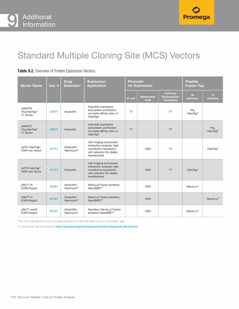
136 Discover Reliable Tools for Protein Analysis
Additional Information9
Table 9.2. Overview of Protein Expression Vectors.
Standard Multiple Cloning Site (MCS) Vectors
*The “M” indicates the vector provides resistance to the indicated drug in mammalian cells.
For all vectors visit our website: http://www.promega.de/resources/vector-sequences/all-vectors/
Vector Name Cat. #Drug Selection*
Expression Application
Promoter for Expression
Peptide Fusion Tag
E. coli Mammalian Cells
Cell-Free Transcription/
Translation
N- terminus
C- terminus
pH6HTN His6HaloTag®
T7 VectorG7971 Ampicillin
Inducible expression and protein purification via metal affinity resin or HaloTag®
T7 T7His6
HaloTag®
pH6HTC His6HaloTag® T7 Vector
G8031 Ampicillin
Inducible expression and protein purification via metal affinity resin or HaloTag®
T7 T7His6
HaloTag®
pHTC HaloTag® CMV-neo Vector
G7711Ampicillin/NeomycinM
Cell imaging and protein interaction analysis; high constitutive expression with selection for stable transfectants
CMV T7 HaloTag®
pHTN HaloTag® CMV-neo Vector
G7721 Ampicillin
Cell imaging and protein interaction analysis; high constitutive expression with selection for stable transfectants
CMV T7 HaloTag®
pNLF1-N [CMV/Hygro]
N1351Ampicillin/NeomycinM
NanoLuc® fusion proteins; NanoBRET™ CMV NanoLuc®
pNLF1-C [CMV/Hygro]
N1361Ampicillin/NeomycinM
NanoLuc® fusion proteins; NanoBRET™ CMV NanoLuc®
pNLF1-secN [CMV/Hygro]
N1371Ampicillin/NeomycinM
Secretory NanoLuc® fusion proteins; NanoBRET™ CMV NanoLuc®

137 Discover Reliable Tools for Protein Analysis
Additional Information9
Table 9.3. Protein Conversions.
Protein Conversion
Protein Molar Conversion
100pmol of 100kDa protein 10µg
100pmol of 50kDa protein 5µg
100pmol of 10kDa protein 1µg
100pmol of 1kDa protein 100ng
Protein/DNA Conversions
1kb of DNA
333 Amino Acids of Coding
Capacity
37kDa protein
270bp DNA 10kDa protein
810bp DNA 30kDa protein
1.35kb DNA 50kDa protein
2.7kb DNA 100kDa protein
Average MW of an Amino Acid 110 daltons

138 Discover Reliable Tools for Protein Analysis
Additional Information
Amino Acid Three-Letter Abbreviation One-Letter Symbol Molecular Weight
Alanine Ala A 89Da
Arginine Arg R 174Da
Asparagine Asn N 132Da
Aspartic acid Asp D 133Da
Asparagine or Aspartic acid Asx B —
Cysteine Cys C 121Da
Glutamine Gln Q 146Da
Glutamic Acid Glu E 147Da
Glutamine or Glutamic acid Glx Z —
Glycine Gly G 75Da
Histidine His H 155Da
Isoleucine lle I 131Da
Leucine Leu L 131Da
Lysine Lys K 146Da
Methionine Met M 149Da
Phenylalanine Phe F 165Da
Proline Pro P 115Da
Serine Ser S 105Da
Threonine Thr T 119Da
Tryptophan Trp W 204Da
Tyrosine Tyr Y 181Da
Valine Val V 117Da
The average molecular weight of an amino acid is 110Da.
Table 9.4. Amino Acid Abbreviations and Molecular Weights.
Amino Acids
Additional Information9

139 Discover Reliable Tools for Protein Analysis
Additional Information9
U C A G
U
UUU Phe UCU Ser UAU Tyr UGU Cys U
UUC Phe UCC Ser UAC Tyr UGC Cys C
UUA Leu UCA Ser UAA Stop UGA Stop A
UUG Leu UCG Ser UAG Stop UGG Trp G
C
CUU Leu CCU Pro CAU His CGU Arg U
CUC Leu CCC Pro CAC His CGC Arg C
CUA Leu CCA Pro CAA Gln CGA Arg A
CUG Leu CCG Pro CAG Gln CGG Arg G
A
AUU lle ACU Thr AAU Asn AGU Ser U
AUC lle ACC Thr AAC Asn AGC Ser C
AUA lle ACA Thr AAA Lys AGA Arg A
AUG Met ACG Thr AAG Lys AGG Arg G
G
GUU Val GCU Ala GAU Asp GGU Gly U
GUC Val GCC Ala GAC Asp GGC Gly C
GUA Val GCA Ala GAA Glu GGA Gly A
GUG Val GCG Ala GAG Glu GGG Gly G
2nd Position
3rd P
ositio
n1st
Po
siti
on
Termination codons are in bold. AUG start codon is in bold italics.
Table 9.5. Genetic Code.
Table 9.6. Recommended Acrylamide Gel Percentages for Resolution of Proteins on Polyacrylamide Gels.
Genetic Code
Gel Percentages
Recommended % Acrylamide Protein Size Range
8 40–200kDa
10 –100kDa
12 10–40kDa

140 Discover Reliable Tools for Protein Analysis
Ordering Information9
Chapter 1: Cloning System and Protein Expression Vectors Size Cat.#
Flexi® Cloning System
Flexi® System, Entry/Transfer 5 entry & 20 transfer reactions C8640
Flexi® System, Transfer 100 transfer reactions C8820
Carboxy Flexi® System, Transfer 50 transfer reactions C9320
HaloTag® Cloning Starter System 1 each G6050
HaloTag® Flexi® Vectors—CMV Deletion Series Sample Pack 9x2µg G3780
10X Flexi® Enzyme Blend (SgfI & PmeI) 25µg R1851
100µg R1852
Carboxy Flexi® Enzyme Blend (SgfI & EcoICRI) 50µg R1901
Kazusa Human ORF Library *www.promega.com/findmygene
GeneX- Native human ORF in pF1K 100ng–10ng/µl FHCxxxx*
GeneX - HaloTag® human ORF in pFN21A 100ng–10ng/µl FHCxxxx*
Mammalian Expression Vectors (see Table 9.1; Table 9.2)
Regulated Mammalian Expression System 1 system C9470
Coumermycin A1 5mg C9451
Novobiocin Sodium Salt 1g C9461
pReg neo Vector 20µg C9421
pF12A RM Flexi® Vector 20µg C9431
pF12K RM Flexi® Vector 20µg C9441
pTargeT™ Mammalian Expression Vector System 20 reactions A1410
pGEM®-T Vector System I 20 reactions A3600
pGEM®-T Vector System II 20 reactions A3610
pGEM®-T Easy Vector System I 20 reactions A1360
pGEM®-T Easy Vector System II 20 reactions A1380
Competent Bacteria for Cloning
Single-Use JM109 Competent Cells, >108cfu/µg 1ml - (20 × 50µl) L2005
JM109 Competent Cells, >108cfu/µg 1ml - (5 × 200µl) L2001
JM109 Competent Cells, >107cfu/µg 1ml - (5 × 200µl) L1001
Single-Use HB101 Competent Cells, >108cfu/µg 1ml - (20 × 50µl) L2015
HB101 Competent Cells, >108cfu/µg 1ml - (5 × 200µl) L2011
IPTG, Dioxane-Free 1g V3955
5g V3951
50g V3953
X-Gal 100mg/2ml V3941
Chapter 2: Bacterial Strains for Protein Expression Size Cat.#
Single Step (KRX) Competent Cells for Protein Expression
Single Step (KRX) Competent Cells 20 x 50µl L3002
L-Rhamnose Monohydrate 10g L5701
50g L5702
BL21 Competent Cells for Protein Expression
Single-Use BL21(DE3)pLysS Competent Cell 1ml (20 × 50µl) L1195
BL21(DE3)pLysS Competent Cells, >106cfu/µg 1ml (5x 200µl) L1191

141 Discover Reliable Tools for Protein Analysis
Additional Information9
Chapter 3: Cell-Free Protein Expression Systems Size Cat.#
mRNA-based Translation Systems
Rabbit Reticulocyte Lysate System, Nuclease Treated 30 reactions L4960
Flexi® Rabbit Reticulocyte Lysate System 30 reactions L4540
Wheat Germ Extract 5 x 200µl L4380
Rabbit Reticulocyte Lysate/Wheat Germ Extract Combination System 24 reactions L4330
DNA-based Transcription and Translation Systems: Rabbit Reticulocyte Lysate Systems
TnT® SP6 Coupled Reticulocyte Lysate System 8 reactions (Trial Size) L4601
40 reactions L4600
TnT® T7 Coupled Reticulocyte Lysate System 8 reactions (Trial Size) L4611
40 reactions L4610
TnT® T3 Coupled Reticulocyte Lysate System 40 reactions L4950
TnT® T7/T3 Coupled Reticulocyte Lysate System 40 reactions L5010
TnT® T7/SP6 Coupled Reticulocyte Lysate System 40 reactions L5020
TnT® T7 Quick Coupled Transcription/Translation System 5 reactions (Trial Size) L1171
40 reactions L1170
TnT® SP6 Quick Coupled Transcription/Translation System 5 reactions (Trial Size) L2081
40 reactions L2080
TnT® T7 Quick for PCR DNA 40 reactions L5540
DNA-based Transcription and Translation Systems: Wheat Germ Extracts
TnT® SP6 Coupled Wheat Germ Extract System 40 reactions L4130
TnT® T7 Coupled Wheat Germ Extract System 40 reactions L4140
TnT® T7/SP6 Coupled Wheat Germ Extract System 40 reactions L5030
TnT® SP6 High-Yield Wheat Germ Protein Expression System 10 reactions L3261
40 reactions L3260
DNA-based Transcription and Translation Systems: Insect Cell Extract
TnT® T7 Insect Cell Extract Protein Expression System 10 reactions L1101
40 reactions L1102
pF25A ICE T7 Flexi® Vector 20µg L1061
pF25K ICE T7 Flexi® Vector 20µg L1081
DNA-based Transcription and Translation Systems: E. coli S30 Extract Systems
E. coli S30 Extract System for Linear Templates 30 reactions L1030
E. coli S30 Extract System for Circular DNA 30 reactions L1020
E. coli T7 S30 Extract System for Circular DNA 30 reactions L1130
S30 T7 High-Yield Protein Expression System 8 reactions L1115
24 reactions L1110
Luciferase Control RNA 20µg - 1mg/ml L4561
Magnesium Acetate 100 µl - 25mM L4581
Potassium Chloride 200µl - 2.5M L4591
Amino Acid Mixture, Complete 175µl - 1mM L4461
Amino Acid Mixture Minus Cysteine 175µl - 1mM L4471
Amino Acid Mixture Minus Methionine and Cysteine 175µl - 1mM L5511
Amino Acid Mixture Minus Leucine 175µl - 1mM L9951
Amino Acid Mixture Minus Methionine 175µl - 1mM L9961
Cell-Free Protein Labeling Reagents
FluoroTect™ GreenLys in vitro Translation Labeling System 40 reactions L5001
Transcend™ Colorimetric Translation Detection System 30 reactions L5070

142 Discover Reliable Tools for Protein Analysis
Ordering Information9
Chapter 4: Protein Purification Size Cat.#
Affinity-based Protein Purification: HaloTag® Fusion Proteins
HaloTag® Protein Purification System (E.coli) 1 each G6280
HaloTag® Protein Purification System Sample Pack 1 each G6270
HaloTag® Mammalian Protein Detection and Purification System Sample Pack (E.coli) 1 each G6799
HaloTag® Mammalian Protein Purification System 1 each G6790
HaloTag® Mammalian Protein Detection and Purification System 1 each G6795
HaloTEV Protease 1,000u G6601
4,000u G6602
HaloTag® TMRDirect™ Ligand 30µl G2991
HaloLink™ Resin 1.25ml G1912
2.5ml G1913
10ml G1914
25ml G1915
Protease Inhibitor Cocktail, 50X 1ml G6521
Mammalian Lysis Buffer 40ml G9381
Affinity-based Protein Purification: His-tagged Proteins
HisLink™ Protein Purification Resin 5ml V8823
50ml V8821
HisLink™ Spin Protein Purification System 25 reactions V1320
HisLink™ 96 Purififcation System 1 x 96 blank V3680
5 x 96 blank V3681
Affinity-based Protein Purification: Biotinylated Proteins
SoftLink™ Soft Release Avidin Resin 1 mL V2011
5 mL V2012
PinPoint™ Xa Protein Purification System (Production & Purification) 1 system V2020
Magnetic Affinity-based Purification and Pull-Down Strategies: Biotinylated Proteins
MagneGST™ Protein Purification System 40 reactions V8600
200 reactions V8603
Magnetic Affinity-based Purification and Pull-Down Strategies: HaloTag® Fusion Proteins
Magne™ HaloTag® Beads, 20% Slurry 1 mL G7281
5 mL G7282
Magnetic Affinity-based Purification and Pull-Down Strategies: His-tagged Proteins
MagneHis™ Protein Purification System 65 reactions V8500
325 reactions V8550
Maxwell® 16 Polyhistidine Protein Purification Kit 48 preps AS1060
Chapter 3: Cell-Free Protein Expression Systems Size Cat.#
Cell-Free Protein Labeling Reagents
Transcend™ Chemiluminescent Translation Detection System 30 reactions L5080
Transcend™ tRNA 30µl L5061
FluoroTect™ GreenLys in vitro Translation Labeling System 40 reactions L5001
Transcend™ Colorimetric Translation Detection System 30 reactions L5070
Transcend™ Chemiluminescent Translation Detection System 30 reactions L5080
Membrane Vesicles for Signal Peptide Cleavage and Core Glycosylation
Canine Pancreatic Microsomal Membranes 50µl Y4041

143 Discover Reliable Tools for Protein Analysis
Additional Information9
Chapter 6: Functional Protein Analysis using HaloTag® Technology Size Cat.#
Cellular Imaging: HaloTag® Ligands
Janelia Fluor® 549 HaloTag® Ligand 5µg GA1110
3 x 5µg GA1111
Janelia Fluor® 646 HaloTag® Ligand 5µg GA1120
3 x 5µg GA1121
HaloTag® Alexa Fluor® 488 Ligand 30µl G1001
15µl G1002
HaloTag® Oregon Green® Ligand 30µl G2801
15µl G2802
HaloTag® TMRDirect™ Ligand 30µl G2991
HaloTag® R110Direct™ Ligand 30µl G3221
HaloTag® TMR Ligand 30µl G8251
15µl G8252
HaloTag® diAcFAM Ligand 30µl G8272
15µl G8273
Chapter 5: Antibody Purification and Labeling Size Cat.#
Antibody Purification
Magne™ Protein G Beads, 20% Slurry 1ml G7471
5ml (5 x 1ml) G7472
50ml G7473
Magne™ Protein A Beads, 20% Slurry 1ml G8781
5ml (5 x 1ml) G8782
50ml G8783
Antibody Labeling
pHAb Amine Reactive Dye 1 x 250µg G9841
4 x 250µg G9845
pHAb Thiol Reactive Dye 1 x 250µg G9831
4 x 250µg G9835
Chapter 4: Protein Purification Size Cat.#
Magnetic Stands and Spacers
MagneSphere® Technology Magnetic Separation Stand (two-position) 0.5ml Z5331
1.5ml Z5332
12 x 75mm Z5333
MagneSphere® Technology Magnetic Separation Stand (twelve-position) 0.5ml Z5341
1.5ml Z5342
12 x 75mm Z5343
PolyATtract® System 1000 Magnetic Separation Stand 1 each Z5410
MagnaBot® 96 Magnetic Separation Device 1 each V8151
MagnaBot® II Magnetic Separation Device 1 each V8351
MagnaBot® Flat Top Magnetic Separation Device 1 each V6041
Plate Clamp 96 1 each V8251
Plate Stand 1 each V8261
Deep Well MagnaBot® 96 Magnetic Separation Device 1 each V3031
Heat Transfer Block 1 each Z3271
Heat Block Insert 1 each Z3651

144 Discover Reliable Tools for Protein Analysis
Ordering Information9
Chapter 6: Functional Protein Analysis using HaloTag® Technology Size Cat.#
HaloTag® Biotin Ligand 30µl G8281
15µl G8282
HaloTag® Alexa Fluor® 660 Ligand 30µl G8471
15µl G8472
HaloTag® Coumarin Ligand 30µl G8581
15µl G8582
HaloTag® PEG-Biotin Ligand 30µl G8591
15µl G8592
HaloTag® Succinimidyl Ester (O2) Ligand 5mg P1691
HaloTag® Amine (O2) Ligand 5mg P6711
HaloTag® Amine (O4) Ligand 5mg P6741
HaloTag® Succinimidyl Ester (O4) Ligand 5mg P6751
HaloTag® Thiol (O4) Ligand 5mg P6761
HaloTag® Iodoacetamide (O4) Ligand 5mg P6771
Protein Interaction Analysis: NanoBRET™: Live Cell Protein:Protein Interaction Assay
NanoBRET™ Nano-Glo® Detection Systems 200 assays N1661
1,000 assays N1662
10,000 assays N1663
HaloTag® Mammalian Pull-Down System 24 reactions G6504
HaloTag® Complete Pull-Down System 1 each G6509
Protein Interaction Analysis: HaloTag® Mammalian Pulldown Kits
HaloTag® Mammalian Pull-Down and Labeling System 24 reactions G6500
HaloTag® Mammalian Pull-Down System 24 reactions G6504
HaloTag® Complete Pull-Down System 1 each G6509
Protein Interaction Analysis: HaloCHIP™ System
HaloCHIP™ System 20 reactions G9410
Additional Reagents
Protease Inhibitor Cocktail, 50X 1ml G6521
HaloTag® Control Vector 20µg G6591
Anti-HaloTag® Monoclonal Antibody 200µg G9211
Anti-HaloTag® pAb 200µg G9281
Mammalian Lysis Buffer 40ml G9381
HaloTag® Standard Protein 30µg G4491
Chapter 7: Protein Characterization by Mass Spectrometry Size Cat.#
Trypsin
Trypsin Gold, Mass Spectrometry Grade 100µg V5280
Rapid Digestion - Trypsin 100µg VA1060
Rapid Digestion - Trypsin/Lys-C 100µg VA1061
Sequencing Grade Modified Trypsin 100µg (5 x 20µg) V5111
100µg V5117
Sequencing Grade Modified Trypsin, Frozen 100µg (5 x 20µg) V5113
Trypsin/Lys-C Mix, Mass Spec Grade 20µg V5071
100µg V5072
100µg (5x 20 µg) V5073
Immobilized Trypsin 2 ml V9012
2x2 ml V9013

145 Discover Reliable Tools for Protein Analysis
Additional Information9
Nonspecific Proteases
Elastase 5mg V1891
Pepsin 250mg V1959
Thermolysin 25mg V4001
Glycosidases
PNGase F 500 units (10u/µl) V4831
Endo H 10,000 units (500u/µl) V4871
50,000 units (500u/µl) V4875
Fetuin (control protein) 500µg (10mg/ml) V4961
ProteaseMAX™ Surfactant
ProteaseMAX™ Surfactant, Trypsin Enhancer 1mg V2071
5 x 1mg V2072
Protein Extracts for LC/MS Monitoring
MS Compatible Human Protein Extract, Digest 100µg V6951
MS Compatible Human Protein Extract, Intact 1mg V6941
MS Compatible Yeast Protein Extract, Digest 100µg V7461
MS Compatible Yeast Protein Extract, Intact 1mg V7341
Peptide Mix For LC/MS Monitoring
6 × 5 LC-MS/MS Peptide Reference Mix 25pmoles V7491*
200pmoles V7495**
Antibody Characterization/Fragmentation
AccuMAP™ Low pH Protein Digestion Kit 10 reactions VA1040
100 reactions VA1050
IdeS Protease† 5,000u V7511
25,000u (5 × 5,000u) V7515
Chapter 8: Protein Detection Tools for Western Blotting & ELISA Size Cat.#
Conjugated Secondary Antibodies: AP
Anti-Mouse IgG (H+L), AP Conjugate 100µl S3721
Anti-Rabbit IgG (Fc), AP Conjugate 100µl S3731
Anti-Human IgG (H+L), AP Conjugate 100µl S3821
Anti-Rat IgG (H+L), AP Conjugate 100µl S3831
Conjugated Secondary Antibodies: HRP
Anti-Rabbit lgG (H+L), HRP Conjugate 300µl W4011
Anti-Mouse IgG (H+L), HRP Conjugate 300µl W4021
Anti-Human IgG (H+L), HRP Conjugate 300µl W4031
Anti-Chicken IgY, HRP Conjugate 300µl G1351
Chapter 7: Protein Characterization by Mass Spectrometry Size Cat.#
Alternative Proteases: Specific Proteases
Arg-C, Sequencing Grade 10µg V1881
Asp-N, Sequencing Grade 2µg V1621
Glu-C, Sequencing Grade 50µg (5 x 10µg) V1651
Lys-C, Mass Spec Grade 20µg VA1170
Lys-N, Mass Spec Grade 20µg VA1180
rAsp-N, Mass Spec Grade 10µg VA1160
rLys-C, Mass Spec Grade 15µg V1671
Low-Specific Proteases
Chymotrypsin, Sequencing Grade 25µg V1061
100µg (4 x 25µg) V1062

146 Discover Reliable Tools for Protein Analysis
Ordering Information9
Substrates for Western Blotting
BCIP/NBT Color Development Substrate 1.25/2.5ml S3771
Western Blue® Stabilized Substrate for Alkaline Phosphatase 100ml S3841
ECL Western Blotting Substrate 250ml W1001
500ml W1015
TMB Stabilized Substrate for Horseradish Peroxidase 200ml W4121
Tween® 20 2.5ml W3831
Blot-Qualified BSA 10g W3841
ProtoBlot® II AP System with Stabilized Substrate, Human 1 each W3940
ProtoBlot® II AP System with Stabilized Substrate, Mouse 1 each W3950
ProtoBlot® II AP System with Stabilized Substrate, Rabbit 1 each W3960
Additional Reagents
Protease Inhibitor Cocktail, 50x 1ml G6521
Broad Range Protein Molecular Weight Markers 100 lanes V8491†Check with local Promega Branch or Distributor for availability.
*Supplied as frozen liquid. Includes 6 additional maximal recovery tubes for aliquots.
**Supplied lyophilized. Includes 6 additional maximal recovery tubes for aliquots.
Chapter 8: Protein Detection Tools for Western Blotting & ELISA Size Cat.#
Anti-ACTIVE® Qualified Antibodies
Donkey Anti-Goat IgG, AP 60µl V1151
Donkey Anti-Goat IgG, HRP 60µl V8051
Substrates for ELISA
AttoPhos® AP Fluorescent Substrate System 3 × 36mg S1000
1 × 36mg (Trial Size) S1001
AttoPhos® Substrate 36mg S1011
100mg S1012
1g S1013
AttoPhos® Buffer 60ml S1021
240ml S1022
TMB One Solution 100ml G7431

147 Discover Reliable Tools for Protein Analysis
Additional Information9
Product Use Limitations, Warranty, DisclaimerPromega manufactures products for a number of intended uses. Please refer to the product label for the intended use
statements for specific products. Promega products contain chemicals which may be harmful if misused. Due care
should be exercised with all Promega products to prevent direct human contact.
Each Promega product is shipped with documentation stating specifications and other technical information. Promega
products are warranted to meet or exceed the stated specifications. Promega’s sole obligation and the customer’s
sole remedy is limited to replacement of products free of charge in the event products fail to perform as warranted.
Promega makes no other warranty of any kind whatsoever, and SPECIFICALLY DISCLAIMS AND EXCLUDES
ALL OTHER WARRANTIES OF ANY KIND OR NATURE WHATSOEVER, DIRECTLY OR INDIRECTLY, EXPRESS
OR IMPLIED, INCLUDING, WITHOUT LIMITATION, AS TO THE SUITABILITY, PRODUCTIVITY, DURABILITY,
FITNESS FOR A PARTICULAR PURPOSE OR USE, MERCHANTABILITY, CONDITION, OR ANY OTHER
MATTER WITH RESPECT TO PROMEGA PRODUCTS.
In no event shall Promega be liable for claims for any other damages, whether direct, incidental, foreseeable,
consequential, or special (including but not limited to loss of use, revenue or profit), whether based upon warranty,
contract, tort (including negligence) or strict liability arising in connection with the sale or the failure of Promega
products to perform in accordance with the stated specifications.
Product claims are subject to change. Please contact Promega Technical Services ([email protected]) or
access the Promega online catalog (www.promega.com/products/) for the most up-to-date information on
Promega products.

148 Discover Reliable Tools for Protein Analysis
Please visit the Promega online catalog (www.promega.com/products/)
to view patent and licensing information for individual products.
Applications mentioned in Promega literature are provided for
informational purposes only. Promega does not warrant that all applications
have been tested in Promega laboratories using Promega products.
Promega products labeled “For Laboratory Use” are intended For
Research Use Only outside the United States.
Promega has used all commercially reasonable efforts to ensure that this
catalog is accurate and free of errors. No claims may be derived from
any printing or typing errors. Prices are in local currency as indicated.
The prices contained herein exclude all taxes, handling fees and/or
transportation costs. Contact Promega for exact information on these
costs. Local terms and conditions may apply on sales and services. For
the latest product pricing and related information visit: www.promega.com
or contact your local Promega representative.
Please contact Promega for customized packaging or if you wish to
order larger quantities.
Life Science P
rod
ucts C
AT
AL
OG
20
18
Copyright
Trademarks
©2019 Promega Corporation. All Rights Reserved.
Prices and specifications subject to change without prior notice.
Trademarks referenced herein are either registered trademarks or trademarks of Promega Corporation in the U.S. and/or other
countries. The names of actual companies and products mentioned herein and/or third party trademarks, trade names and logos
contained herein may be the trademarks of their respective owners. Any rights not expressly granted herein are reserved.
Legal References9

149 Discover Reliable Tools for Protein Analysis
©2019 Promega CorporationPrinted in USA50835476 | PART #BR266
Promega Corporation2800 Woods Hollow RoadMadison, WI 53711-5399 USATel: 608-274-4330Fax: 608-277-2516Toll-Free Tel: 800-356-9526Toll-Free Fax: 800-356-1970Internet: www.promega.com
Promega BioSciences, LLCSan Luis Obispo, CA, USA
Promega BioSystems, Inc.Seoul, South Korea
Promega BioSystems Sunnyvale, Inc.Sunnyvale, CA, USA
Shanghai Promega Biological Products, Ltd.Shanghai, China
Terso Solutions, Inc.Madison, WI, USA
Australia, SydneyTel: 02 8338 3800Fax: 02 8338 3855Freecall: 1800 225 123Freefax: 1800 626 017E-mail: [email protected]
Brazil, São PauloTel/Fax: +55 11 5096 3770E-mail: [email protected]
China, BeijingTel: 86 10 5825 6268Fax: 86 10 5825 6160 Toll-Free: 800 810 8133E-mail: [email protected]
France, LyonTel: 04 37 22 50 00Fax: 04 37 22 50 10Numero Vert Client (gratuit): 0 800 48 79 99E-mail: [email protected] Vert Technique (gratuit): 0 800 48 80 00E-mail: [email protected]
Germany/Austria/Poland, WalldorfTel (D, AT): +49 6227 6906 291Tel (PL): +48 22 531 06 67Fax (D, AT): +49 6227 6906 222Fax (PL): +48 22 531 06 69E-mail (D, AT):[email protected] (PL): [email protected]
India, New DelhiTel: +91 11 43005814/15 Fax: +91 11 41035028E-mail: [email protected]
Italy, MilanTel: 02 54 05 01 94Fax: 02 56 56 16 45Numero Verde: 800 69 18 18E-mail: [email protected]
Japan, TokyoTel: 03 3669 7981Fax: 03 3669 7982E-mail: [email protected]
Korea, SeoulTel: 82 1588 3718Fax: 82 2 2638 5418E-mail: [email protected]
Belgium/Luxembourg/ The Netherlands, LeidenTel: +31 (0)71-532 42 44Fax: +31 (0)71-532 49 07E-mail: [email protected]
Pacific Asia Region, SingaporeTel: +65 65133450Fax: +65 67735210E-mail: [email protected]
Spain, MadridTel: +34 916 621 126 Fax: +34 916 615 835 E-mail: [email protected]
Estonia & Nordic Region, StockholmTel: +46 8 452 2450Fax: +46 8 452 2455 E-mail: [email protected]
Switzerland, DübendorfCustomer ServiceTel: 044 878 90 00Fax: 044 878 90 10E-mail: [email protected] Technical Service Tel: 044 878 90 20E-mail: [email protected]
United Kingdom, SouthamptonTel: 023 8076 0225Fax: 023 8076 7014Free Phone: 0800 378994Free Fax: 0800 181037E-mail: [email protected]
For more information about Promega Branch Offices and Distributors visit: www.promega.com/support/contact-promega