Diorganotin (IV) derivatives of ONO tridentate Schiff base: Synthesis, crystal structure, in vitro...
11
This article appeared in a journal published by Elsevier. The attached copy is furnished to the author for internal non-commercial research and education use, including for instruction at the authors institution and sharing with colleagues. Other uses, including reproduction and distribution, or selling or licensing copies, or posting to personal, institutional or third party websites are prohibited. In most cases authors are permitted to post their version of the article (e.g. in Word or Tex form) to their personal website or institutional repository. Authors requiring further information regarding Elsevier’s archiving and manuscript policies are encouraged to visit: http://www.elsevier.com/copyright
-
Upload
independent -
Category
Documents
-
view
5 -
download
0
Transcript of Diorganotin (IV) derivatives of ONO tridentate Schiff base: Synthesis, crystal structure, in vitro...
This article appeared in a journal published by Elsevier. The attachedcopy is furnished to the author for internal non-commercial researchand education use, including for instruction at the authors institution
and sharing with colleagues.
Other uses, including reproduction and distribution, or selling orlicensing copies, or posting to personal, institutional or third party
websites are prohibited.
In most cases authors are permitted to post their version of thearticle (e.g. in Word or Tex form) to their personal website orinstitutional repository. Authors requiring further information
regarding Elsevier’s archiving and manuscript policies areencouraged to visit:
http://www.elsevier.com/copyright

Author's personal copy
Original article
Diorganotin(IV) derivatives of ONO tridentate Schiff base: Synthesis, crystalstructure, in vitro antimicrobial, anti-leishmanial and DNA binding studies
Shaukat Shujha a, Afzal Shah a, Zia-ur-Rehman a, Niaz Muhammad a, Saqib Ali a,*, Rumana Qureshi a,Nasir Khalid b, Auke Meetsma c
aDepartment of Chemistry, Quaid-i-Azam University, Islamabad 45320, PakistanbChemistry Division, Pakistan Institute of Nuclear Science and Technology, PO Nilore, Islamabad, PakistancCrystal Structure Center, Chemical Physics, Zernike Institute for Advanced Materials, University of Groningen, Nijenborgh 4, NL-9747 AG Groningen, The Netherlands
a r t i c l e i n f o
Article history:Received 12 August 2009Received in revised form11 March 2010Accepted 15 March 2010Available online 18 March 2010
Keywords:N0-(2-Hydroxybenzylidene)formohydrazideDiorganotin(IV) complexesCrystal structureDNA-binding
a b s t r a c t
Six new diorganotin(IV) derivatives of N0-(2-hydroxybenzylidene)formohydrazide (H2L) with generalformula R2SnL, where R ¼ Ph (1), Me (2), Bu (3), Oct (4), t-Bu (5), Et (6), and L ¼ [OC6H4CHNNCHO] havebeen synthesized and characterized by different analytical techniques. Crystal structure of Me2SnL (2)authenticated distorted square-pyramidal geometry around the Sn atom. The CV and UVeVis spectro-scopic data indicated intercalation of complexes into DNA with binding affinity varying in the sequence:3 (1.69 � 104 M�1) > 1 (1.10 � 104 M�1) > 2 (9.61 � 103 M�1). Some of these compounds were found tobe good antibacterial, antifungal and leishmanicidal agents.
� 2010 Elsevier Masson SAS. All rights reserved.
1. Introduction
The study of organotin(IV) complexes has gained growinginterest in the last four decades due to their ability to interact withDNA, indicating their potential candidature in cancer chemo-therapy [1e4]. Several organotin(IV) Schiff base complexes havebeen investigated for their structural diversity and biocidal activi-ties [5e7]. Cagnoli et al. found some organotin(IV) complexes to beevenmore active in vitro than the clinically used cis-platin [8]. Someother workers have established the quantitative structure/activityand structure/property relationships of these compounds [9].However, little work has been carried out on the ONO donortridentate chelating ligands, derived from the condensation ofsalicylaldehyde with hydrazides [10]. The present work is centeredon the synthesis of diorganotin(IV) Schiff base derivatives of formichydrazide and their characterization by different analytical tech-niques. Some of these compounds have good antimicrobial activi-ties. Keeping the anticancer activity of dibutyltin(IV) complexes[11] in view, we selected three potential antitumor organotin(IV)complexes, with the one having dibutyltin(IV) moiety to evaluatetheir DNA binding parameters. Up to our knowledge, this is the first
report on the in vitro evaluation of the binding parameters oforganotin(IV) schiff base complexes with DNA by electrochemicaland spectroscopic techniques.
2. Results and discussion
2.1. Synthesis
The ligand was synthesized by direct reaction of formic hydra-zide with salicylaldehyde in ethanol. The yellow organotin(IV)complexes were prepared by reacting together ligand, organotin(IV) dichloride, triethylamine in 1:1:2 and ligand, dioctyltin(IV)oxide in 1:1 ratios in dry toluene. All the compounds were obtainedin good yield. The detailed syntheses and physical data arepresented in Experimental section.
2.2. Spectroscopic studies
In the ligand the vibration bands appearing at 3345, 3184 and1689 cm�1 were assigned to nNeH, nOeH and nC]O vibration modes,respectively. Due to enolization and deprotonation of the ligand oncomplexation with diorganotin moiety, these bands disappeared.The shifting of nC]N band at 1613 cm�1 to lower energy suggested
* Corresponding author. Tel.: þ925190642208; fax: þ92512873869.E-mail address: [email protected] (S. Ali).
Contents lists available at ScienceDirect
European Journal of Medicinal Chemistry
journal homepage: http: / /www.elsevier .com/locate/ejmech
0223-5234/$ e see front matter � 2010 Elsevier Masson SAS. All rights reserved.doi:10.1016/j.ejmech.2010.03.015
European Journal of Medicinal Chemistry 45 (2010) 2902e2911

Author's personal copy
the coordination of azomethine nitrogen with Sn atom [12]. Theshifting of nNeN band from 1053 cm�1 to 1072e1082 cm�1 oncomplexation is attributed to the decrease in repulsion of the lonepairs of electrons on the nitrogen atoms [13]. New bands were alsoobserved in the region of 500e555 cm�1 (nSneO) and 450e470 cm�1
(nSneN) [14].The 1H NMR spectral data showed signals at 10.12, 11.67 and
8.68 ppm due toeOH,eCHO andeNHN] protons, respectively. Allthese signals were absent from the spectra of complexes owing tothe conversion of the aldo-imine form of ligand to imine-ol formand subsequent deprotonation. The coordination of azomethinenitrogen with Sn atom shifted the CH]N proton resonance signaldown field to 8.60e8.65 ppm with 3J (119Sn, 1H) coupling constantvalue of 38e51 Hz. All the other protons of the ligand and the Rgroups bonded to Sn atom appeared in the expected region. The2J (119Sn, 1H) coupling constant (79 Hz) of complex 2 correspondingto CeSneC angel of 129.6� was evaluated by Lockhart’s equation,q ¼ 0.0161 [2J]2 � 1.32 [2J] þ 133.4 [15]. Thus, confirmed five-coordinate Sn atom in solution. In 13C NMR, the angle calculated(127.4e133.4�) for compounds 2, 3, 5 and 6, using equation1J ¼ 10.7qe778 [16], affirmed penta-coordination of Sn in solution.In all complexes, the signals observed in 13C NMR are in conformitywith expected composition.
The presence of a single 119Sn peak for complexes 1e6 in therange �163.2 to �337 ppm is in conformity with the formation ofa single species having penta-coordinated Sn atom [17].
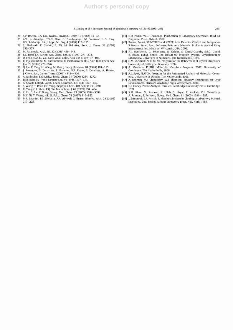
2.3. Mass spectrometry
The ligand and its complexes showed molecular ion peak ofsignificant intensity with the characteristic peak pattern of Sn. Theproposed fragmentation pattern of ligand and complex 2 is depic-ted in Scheme 6. The initial fragmentation is due to the loss ofa methyl group, which then gave a base peak [C8H6N2O2Sn]þ in thespectrum due to the elimination of a second methyl group. Theother fragments with significant abundances were [CH3Sn]þ,[CH3Sn]þ and [Sn]þ. The rest of the diorganotin(IV) compoundsfollowed almost the same fragmentation route as 2, the massspectral data is given in the Experimental section.
2.4. Crystal structure
The molecular structure with atomic numbering scheme forcompound 2 is given in Fig. 1. The crystallographic data, selectedbond lengths and angles are listed in the Tables 1 and 2. The
bond distances and angles of the complex 2 are comparable withthe values for analogous compounds [18]. The structure ofcomplex 2 consists of a deprotonated ONO dibasic tridentateligand bonded to the (CH3)2Sn(IV) moiety via two oxygen anda nitrogen atom, forming a O2NC2 core around the Sn atom.The ligand is non-planar, probably due to the steric requirementsof the five and six membered chelate rings formed. Thegeometry around Sn atom can be characterized by the value ofs ¼ (b � a)/60, where b is the largest and a the second largestbasal angles around the Sn atom. The s value is zero for perfectsquare pyramid (a ¼ b ¼ 180�) and unity for a perfect trigonalpyramidal geometry (a ¼ 120�) [19]. For compound 2(b ¼ O1eSneO2 ¼ 152.9� and a ¼ C9eSneC10 ¼ 140.7�) thes value (0.2) indicates a distorted square-pyramidal geometrywith two enolic oxygens and two methyl carbons in the equa-torial positions and the azomethinic nitrogen at the apicalposition. The tendency of going from trigonal-bipyramidalgeometry towards a square-pyramidal one is most probablyassociated with secondary intermolecular Sn/O interactions(Fig. 2). The SneO1 and SneO2 bond lengths (2.110 and 2.169 Å)are less than the sum of Van der Waals radii of Sn and O (2.8 Å).The O1eSneN1, and O2eSneN1 angles are 82.25� and 72.47�,respectively. The C9eSneC10 angle (140.70�) show a largedeviation from the linear value (180�) and the angle calculatedfrom 2J(119Sne1H) (129.6�) and 1J[119Sne13C] (133.4�) by Lockhart
Fig. 1. ORTEP view of Me2SnL (2) with atomic numbering scheme. Thermal ellipsoidsare drawn at the 50% probability level. Hydrogen atoms are shown as spheres ofarbitrary radii.
Table 1Crystal data and structure refinement parameters for Me2SnL (2).
Empirical formula C10H12N2O2SnFormula mass 310.93Crystal system TetragonalSpace group I41/a, 88a (Å) 13.4693(4)b (Å) 13.4693(4)c (Å) 24.9037(17)V (Å3) 4518.1(4)Z(Z0) 16(1)Crystal habit/size (mm) Block/0.35 � 0.29 � 0.15T (K) 100(1)r (g cm�3) 1.828m (Mo Ka) (cm�1) 22.44F(000) 2432Total reflections 19718Independent reflections 2799For (Fo � 4.0 s (Fo)) 2749RðFÞ ¼ PðjjFoj � jFcjjÞ=
P jFoj for Fo > 4.0 s (Fo) 0.0223wRðF2Þ ¼ ½P½wðF2o � F2c Þ2�=½
PwðF2o Þ2��1=2 0.0558
Goodness-of-fit 1.043q Range (�) 2.69e29.28Data/restrictions/params 2799/0/139Largest diff. peak and hole (e �3) �0.12 and 0.17(4)
Table 2Selected bond lengths (Å) and angles (�) for Me2SnL (2).
SneO1 2.110(2) O1eC1 1.312(4)SneO2 2.169(2) O2eC8 1.302(4)SneN1 2.184(2) N1eN2 1.409(3)SneC9 2.106(4) N1eC7 1.295(4)SneC10 2.112(4) N2eC8 1.289(4)O1eSneO2 152.89(9) N2eN1eC7 115.6(2)O1eSneN1 82.25(9) N1eN2eC8 110.0(3)O1eSneC9 88.93(13) O1eC1eC2 119.0(3)O1eSneC10 96.55(10) O1eC1eC6 123.2(2)O2eSneN1 72.47(9) SneO1eC1 129.21(18)O2eSneC9 93.36(13) SneO2eC8 113.12(18)O2eSneC10 98.63(10) SneN1eN2 116.97(18)N1eSneC9 115.96(13) SneN1eC7 127.13(19)N1eSneC10 103.34(11) N1eC7eC6 126.2(3)C9eSneC10 140.70(14) O2eC8eN2 127.1(3)
S. Shujha et al. / European Journal of Medicinal Chemistry 45 (2010) 2902e2911 2903

Author's personal copy
equation [14,15]. The SneN1 bond distance is 2.184 Å, closer tothe sum of covalent radii of Sn and N (2.15 Å) and significantlyless than the sum of van der waals radii (3.75 Å). The C1eO1,C8eO2 bond length (1.313 Å, 1.302 Å) and C1eN1eN2 angle(109.99�) for compound 2 are match-well with the reportedvalues [17].
2.5. Biological activity
2.5.1. Antibacterial activityThe Schiff base ligand and its diorganotin(IV) derivatives were
screened for their antibacterial activity against 2 gram positive(Bacillus subtilis, Staphylococcus aureus) and 4 gram negativebacteria (Eschericha coli, Shigella flexenari, Pseudomonas aeruginosa,Salmonella typhi), Table 3. Despite the reported antibacterialactivity of various Schiff bases [20,21], the synthesized ligand wasfound inactive against all bacterial strains. Most of the diorganotin(IV) derivatives significantly inhibit gram-positive bacteria growth.This may be due to the ease of permeation of the complexes owing
to the simplicity in the cell wall of these strains [22]. The dimethyl-(2) and dibutyltin(IV)-(3) complexes show highest activity, whichsubsides as the alkyl chain length increases [23,24].
2.5.2. Antifungal activityThe antifungal activity of synthesized compounds were tested
in vitro against human pathogenic fungal strains including yeasts(Candida albicans ATCC 2192, Candida glabrata ATCC 90030),dermatophytes (Microsporum canis ATCC 9865, Trichphytonlongifusus ATCC 22397), opportunistic moulds (Aspergillus flavusATCC 1030 and Fusarium solani ATCC 11712) by using the agartube dilution test. Micoanazole (200 mg/ml) and Amphotericin B(200 mg/ml) were used as standards drugs. The percent growthinhibition of the compounds is summarized in Table 4. All thefungal strains except A. flavus are resistant to the ligand. Activity ofcompounds 1, 2 and 6 is fairly good while for the rest of thecompounds low activity was observed.
2.5.3. CytotoxicityThe in vivo lethality to brine shrimp nauplii was used to assess
the cytotoxicity of synthesized compounds [25]. The results arepresented in Table 5. Compound 3 show highest toxicity larger thanthe standard drug with LD50 0.691 mg/ml.
2.5.4. Anti-leishmanial activityThe Anti-leishmanial activity of ligand and complexes (1e6)
were obtained against the pathogenic Leishmania major, usingAmphotericin B (0.50 mg/ml) and Pentamidine (5.78 mg/ml) asstandard drugs. The data are presented in Table 6. The reportedcompounds produced a significant reduction in viable promas-tigotes. Compound 6 has the highest potential as leishmanicidalagent with IC50 0.90 mg/ml followed by compound 3(IC50 0.96 mg/ml) and 5 (IC50 0.98 mg/ml). The activity of thesecompounds is excellent and far exceed the level for the reportedorganotin(IV) compounds [26].
2.6. Voltammetric study of complex-DNA interaction
The cyclic voltammetric behavior of 3.00 mM complex 1 in theabsence and presence of 50 mMDNA at bare GCE is shown in Fig. 3A.The voltammogram of the free complex 1 shown in Fig. 3A(a)featured a single well defined and stable cathodic peak at �1.531 Vversus SCE in 10% aqueous DMSO at 25 �C. The absence of anodicpeak in the reverse scan indicated the irreversibility of the elec-trochemical process. The electrochemical signal at �1.531 V isattributed to the reduction of Snþ4 to Snþ2 state. The broadness ofthe peak as indicated by jEp � Ep/2j ¼ 70 mV, may be due to theoverlap of two 1e� reduction peaks. The cyclic voltammogram ofcomplex 1 in the presence of 50 mM DNA (Fig. 3A(b)), underwent
Fig. 2. The molecular packing of Me2SnL (2) showing the arrangement of monomericunits.
Table 3Antibacterial activityaed (diameter of inhibition zone) of N0-(2-hydrox-ylbenzylidene) formohydrazide and its diorganotin(IV) derivatives.
Bacterium (ATCC No.) Inhibition zone diameter (mm) Referencedrug
H2L 1 2 3 4 5 6
Bacillus subtillis (11774) e 18 15 20 10 e 12 37Staphylococcus aureus
(25923)e e 20 15 e 10 9 26
Eschericha coli (11229) e 15 10 18 11 12 9 30Shigella flexenari (10782) e e 17 14 11 9 10 36Pseudomonas aeruginosa
(10145)e 10 12 9 e e 11 32
Salmonella typhi (10749) e e e 17 e 15 30
a In vitro agar well-diffusion method, conc. 1 mg mL�1 DMSO.b Reference drug, Imipenum 10 mg/disc.c Zone diameter (Activity): 3e6 mm (non significant), 7e9 mm (low), 10e12 mm
(good), >12 mm (significant).d Clinical Implication: Escherichia coli, infection of wounds, urinary tract and
dysentery; Bacillus subtilis, food poisoning; Shigella flexenari, blood diarrhea withfever and severe prostration; Staphylococcus aureus, food poisoning, scaled skinsyndrome, endocarditis; Pseudomonas aeruginosa, infection of wounds, eyes, septi-cemia, Salmonella typhi, typhoid fever, localized infection [25].
Table 4Antifungal activity a,b,c (% inhibition) of N0-(2-hydroxybenzylidene)formohydrazideand its diorganotin(IV) derivatives.
Fungus (ATCC No.) Inhibition % MIC(mg/ml)
H2L 1 2 3 4 5 6
Candida albicans (2129) 0 0 35 0 30 10 20 110.8Aspergeilus flavus (1030) 40 50 45 10 0 0 40 20.0Microsporum canis (9865) 0 10 0 0 10 20 0 98.4Fusarium solani (11712) 0 0 40 20 0 0 30 73.2Trichophyton longifusus
(22397)0 40 20 0 0 0 0 70.0
Candida glabrata (90 030) 0 50 0 0 5 0 35 110.8
a Concentration: 200 mg mL�1 of DMSO.b MIC: Minimum inhibitory concentration.c % (standard drug) ¼ 100.
S. Shujha et al. / European Journal of Medicinal Chemistry 45 (2010) 2902e29112904

Author's personal copy
31.46% decrease in peak current and 79 mV positive shift in peakpotential. The substantial diminution in peak current can presum-ably be due to the decrease in free drug concentration due to theformation of slowly diffusing, heavy molecular weight 1-DNAadduct. The obvious positive peak potential shift could be attrib-uted to the intercalation of 1 into the stacked base pair pockets ofDNA as reported by the previous investigators [27,28]. Thevoltammetric parameters obtained from the cyclic voltammogramsof 3.00 mM 2 and 3with and without DNA (Fig. 3B and C) are listedin Table 7.
The greater decrease in peak current (33.65%) of 3 as comparedto 2 (24.88%) by the addition of 50 mM DNA is due to the lowerdiffusion coefficient of 3eDNA complex as compared to 2eDNA,which will be discussed thoroughly in subsequent section. Thepositive shift in peak potentials of both the compounds by theaddition of DNA reflects intercalation as the dominant mode ofinteraction, in which the drug inserts itself into the stacked basepairs domain of DNA.
The gradual decay in peak current of 1, 2 and 3 by the addition ofvarying concentration of DNA, ranging from 10 to 60 mM, can beused to quantify the binding constants of 1-DNA, 2-DNA and 3-DNAadducts by the application of the following equation [29].
log ð1=½DNA�Þ ¼ log K þ log ðIH�G=ðIG � IH�GÞÞ (1)
where, K is the binding constant, IG and IH�G are the peak currentsof the free guest (G) and the adduct (H � G), respectively.
The plots of log (1/[DNA]) versus log (IH�G/(IG � IH�G)) (Fig. 4)yielded K ¼ 1.10 � 104 M�1, 9.61 � 103 M�1 and 1.69 � 104 M�1 for1-DNA, 2-DNA and 3-DNA complexes, respectively. The greater Kvalue of 1 as compared to 2 is attributed to the presence ofextended aromatic system which may bind strongly with DNAbases [30]. The strong affinity of 3 for DNA, indicated by its greaterintercept (Fig. 4) as compared to 1 could be assigned to the addi-tional hydrophobic interactions of the bulky butyl moiety with thenucleotide bases. The K values of these diorganotin(IV) e DNAadducts are greater than those observed for similar DNA-inter-calating Cr and Ru complexes, [CrCl2(dicnq)2]þ and [Ru(dicnq)3]þ2,for which the binding constants have been reported as 1.20 � 103
and 9.70 � 103 M�1, respectively [31e33].To know whether the electrochemical process is diffusion
controlled or adsorption controlled, the values of peak current,I were plotted vs. the square root of the scan rate (n1/2) in thepresence and absence of DNA using RandleseSevcik equation forirreversible process [34,35].
I ¼ 2:69� 105nðanÞ1=2AC�oD
1=2n1=2 (2)
Where I is the peak current (A), A is the surface area of theelectrode (cm2), C�
o is the bulk concentration (mol cm�3) of theelectroactive species, D is the diffusion coefficient (cm2 s�1), a isthe transfer coefficient with a value of 0.34 as obtained fromjEpeEp/2j ¼ 47.7 mV/an and n is the scan rate (V s�1).
The linearity of the plots demonstrates that the main masstransport of these complexes (Fig. 4A) and their DNA adducts(Fig. 4B), to the electrode surface is controlled by diffusion step [36].The diffusion coefficients of the free and DNA bound adducts of
Table 5Brine Shrimp (Artemia salina) lethality bioassay of N0-(2-ydroxybenzylidene)formohydrazide and its diorganotin(IV) complexes.
Sample Code H2L 1 2 3 4 5 6
LD50 mg/ml e e 95.70 0.691 e e 88.30
Standard drug Etoppside LD50 7.46 mg/mL.
Table 6Anti-leishmanial activity of N0-(2-hydroxybenzylidene)formohydrazide and itsdiorganotin(IV) complexes.
Sample Code IC50 (mg/ml) � S.D
H2L 24.0 � 0.021 6.50 � 0.042 2.08 � 0.043 0.96 � 0.024 4.26 � 0.045 0.98 � 0.026 0.90 � 0.02
-17
-14
-11
-8
-5
-2
1
4-1.8-1.6-1.4-1.2
E / V vs. SCE
I/μA
a
b
-12
-9
-6
-3
0
3-2-1.8-1.6-1.4
E / V vs. SCE
I/μA
a
b
-14
-10
-6
-2
2-2-1.8-1.6-1.4
E / V vs. SCE
I/μA
a
b
A
B
C
Fig. 3. A. Cyclic voltammograms of 3.00 mM Ph2SnL (1) in 10% aqueous DMSO with0.1 M TBAP as supporting electrolyte in the absence (a) and presence of 50 mM DNA (b)at 25 �C. Working electrode: Glassy carbon with a geometric area of 0.071 cm2, scanrate 100 mV/s. B. Cyclic voltammograms of 3.00 mM Me2SnL (2) in 10% aqueous DMSOwith 0.1 M TBAP as supporting electrolyte in the absence (a) and presence of 50 mMDNA (b) at 25 �C. Working electrode: Glassy carbonwith a geometric area of 0.071 cm2,scan rate 100 mV/s. C. Cyclic voltammograms of 3.00 mM Bu2SnL (3) in 10% aqueousDMSO with 0.1 M TBAP as supporting electrolyte in the absence (a) and presence of50 mM DNA (b) at 25 �C. Working electrode: Glassy carbon with a geometric area of0.071 cm2, scan rate 100 mV/s.
S. Shujha et al. / European Journal of Medicinal Chemistry 45 (2010) 2902e2911 2905
Author's personal copy
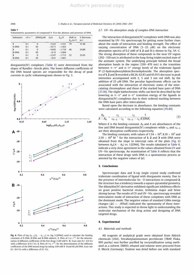
diorganotin(IV) complexes (Table 8) were determined from theslopes of RandleseSevcik plots. The lower diffusion coefficients ofthe DNA bound species are responsible for the decay of peakcurrents in cyclic voltammograms shown in Fig. 3.
2.7. UVeVis absorption study of complex-DNA interaction
The interaction of diorganotin(IV) complexes with DNAwas alsoexamined by UVeVis spectroscopy for getting some further cluesabout the mode of interaction and binding strength. The effect ofvarying concentration of DNA (5e25 mM) on the electronicabsorption spectra of 0.2 mM of 1, 2 and 3 is shown in Fig. 5AeC.The strong absorption of these compounds in the near UV region(292e330 nm) is attributed to the long-living triplet excited state ofthe aromatic system. The underlying principle behind the broadabsorption bands in the region (350e470 nm) is the transitionbetween pep* and nep* energy levels of the tridentate ligandN0-(2-hydroxybenzylidene)formohydrazide. The absorption spec-tra of 1, 2 and 3 recorded a 68.26, 62.83 and 65.91% decrease in peakintensities accompanied with 2, 5 and 3 nm red shift, by theaddition of 25 mM DNA. The peculiar hypochromic effects can beassociated with the interaction of electronic states of the inter-calating chromophore and those of the stacked base pairs of DNA[37,38]. The slight bathochromic shifts can best be described by thelowering in pep* and nep* transition energy of the ligands indiorganotin(IV) complexes due to their ordered stacking betweenthe DNA base pairs after intercalation.
Based upon the decrease in absorbance, the binding constantswere calculated according to the following equation [39,40].
A0
A� A0¼ 3G
3H�G � 3Gþ 3G3H�G � 3G
1K½DNA� (3)
Where K is the binding constant, A0 and A are absorbances of thefree and DNA bound diorganotin(IV) complexes while 3G and 3H�Gare their absorption coefficients respectively.
The binding constants, with values of 1.54 � 104, 8.19 � 103 and2.59 � 104 M�1 for the interaction of 1, 2 and 3 with DNA wereobtained from the slope to intercept ratio of the plots (Fig. 6)between A0/(A � A0) vs. 1/[DNA]. The results tabulated in Table 8,indicate a very close agreement in the values obtained from CV andUVeVis spectroscopy. An examination of Table 8 reflects that theinteraction of these drugs with DNA is a spontaneous process asattested by the negative values of DG.
3. Conclusions
Spectroscopic data and X-ray single crystal study confirmedtridentate coordination of ligand with diorganotin moiety. Due tothe presence of intermolecular Sn/O interactions in compound 2,the structure has a tendency towards a square-pyramidal geometry.The dibutyltin(IV) derivative exhibited significant inhibitory effectson gram positive bacterial strains, leshminia major and brineshrimp larvae. The results of CV and UVeVis. spectroscopy revealedintercalative mode of interaction of these complexes with DNA asthe dominant mode. The negative values of standard Gibbs energychanges (DG ¼ �RTlnK) indicated the spontaneity of these inter-actions. This study is expected to throw light in understanding themolecular mechanism of the drug action and designing of DNAtargeted drugs.
4. Experimental
4.1. Materials and methods
All reagents of analytical grade were obtained from Aldrichchemicals (USA). Tetrabutylammonium perchlorate (TBAP, Fluka,99% purity) was further purified by recrystallization using meth-anol as a solvent. DMSO, ethanol and toluene were procured fromE. Merck (Germany). Toulene was dried before use with standard
Table 7Voltammetric parameters of compound 1e3 in the absence and presence of DNA.
Substance n/V s�1 [DNA]/mM I/mA Epc/V Shift inEpc/m V
% Decreasein I
1 0.1 0 �15.63 �1.531 79 31.461-DNA 0.1 50 �10.71 �1.4522 0.1 0 �10.85 �1.710 46 24.882-DNA 0.1 50 �8.15 �1.6643 0.1 0 �12.67 �1.713 52 33.653-DNA 0.1 50 �8.41 �1.661
4.2
4.6
5
0.15 0.65 1.15 1.65log(IH-G)/(IG-IH-G))
log(
1/[D
NA
](M
))
32
1
-40
-30
-20
-10
00.2 0.4 0.6 0.8
υ1/2(V/s)1/2
I / μ
A
31
2
-30
-20
-10
00.2 0.4 0.6 0.8
υ1/2(V/s)1/2
I/ μ
A
132
A
B
Fig. 4. Plots of log (IH�G/(IG � IH�G)) vs. log (1/[DNA]) used to calculate the bindingconstants of 1-DNA, 2-DNA and 3-DNA adducts. A. Plots of I vs. n1/2, for the determi-nation of diffusion coefficients of the free drugs (3.00 mM 1e3). Scan rates 0.1e0.6 V/swith a difference of 0.1 V/s. B. Plots of I vs. n1/2, for the determination of the diffusioncoefficients of the DNA bound drugs by taking 3.00 mM 1e3 and 60 mM DNA. Scan rate0.1e0.6 V/s with a difference of 0.1 V/s.
S. Shujha et al. / European Journal of Medicinal Chemistry 45 (2010) 2902e29112906

Author's personal copy
procedure [41]. The melting points were determined on an elec-trothermal melting point apparatus, model MP-D Mitamura RikenKogyo (Japan) by using capillary tubes and are uncorrected.Elemental analyses were performed using Leco CHNS-932elemental analyzer (USA). The IR spectra of all samples wererecorded as KBr discs or in NaCl cells using Bio-Rad Excaliber FT-IR,model FTS 300MX spectrometer (USA) in range of 4000e400 cm�1.NMR (1H and 13C) spectra were obtained on a Brucker 300 MHz FTNMR spectrometer using CDCl3 as solvent. 119Sn NMR spectra wereobtained with Me4Sn as an external reference. The cyclic voltam-metric experiments were performed by PGSTAT 302 with AutolabGPES version 4.9 Eco Chemie, Utrecht, the Netherlands. Measure-ments were carried out in a conventional three electrode cell withsaturated calomel electrode (SCE) from Fisher scientific company(Cat No. 13-639-51) as a reference electrode, a thin Pt wire ofthickness 0.5 mm with an exposed end of 10 mm as the counterelectrode and a bare glassy carbon electrode (GCE) witha geometric area of 0.071 cm2 as the working electrode. Prior toexperiments, the GCEwas polished with 0.25 mmdiamond paste ona nylon buffing pad. For electrochemical measurements the testsolution was kept in an electrochemical cell (model K64 PARC)connected to the circulating thermostat LAUDA model K-4R. Argonwas used for flushing out oxygen before every electrochemicalassay. Absorption spectra were measured on a UVeVis Spectrom-eter; Shimadzu 1601. Table Top Centrifuge, Model PLC-05 (Taiwan)was used for the extraction of DNA.
4.2. Synthesis
4.2.1. Synthesis of Schiff base ligandAn ethanolic solution of salicylaldehyde (0.1 mol, 12.2 g) was
added slowly to the solution of formic hydrazide (0.1 mol, 6.0 g) inethanol with constant stirring. Themixturewas refluxed for 1 h andon cooling crystalline solid product was obtained (Scheme 1). Thestructure of ligand and numbering scheme are given in Scheme 2.
-7
-6
-5
-4
-3
-2
-1
00 0.05 0.1 0.15 0.2 0.25
1/[DNA]/(μM)-1
Ao/(
A-A
o)
2
1
3
Fig. 6. Plots of Ao/(A � Ao) vs. 1/[DNA] for the determination of binding constants ofComplex-DNA adducts by taking 0.2 mM drug and 5e25 mM DNA with a difference of5 mM aliquot of DNA.
Table 8The binding constants and Gibbs free energies of 1-DNA, 2-DNA and 3-DNA adducts as determined by voltammetry and UVeVis. spectroscopy along with the diffusioncoefficients of the free and DNA bound species.
Drug-DNAadduct
CV Spectroscopy
107Df/cm2 s�1 108Db/cm2 s�1 K/M�1 �DG/kJ mol�1 K/M�1 �DG/kJ mol�1
1-DNA 2.37 0.15 1.10 � 104 23.05 1.54 � 104 23.392-DNA 2.05 6.49 9.61 � 103 22.72 8.19 � 103 22.333-DNA 2.29 7.89 1.69 � 104 24.11 2.59 � 104 25.18
0
0.4
0.8
1.2
1.6
280 330 380 430 480
Wavelength/nm
Abs
orba
nce
0
0.3
0.6
0.9
1.2
1.5
1.8
280 330 380 430 480Wavelength/nm
Abs
orba
nce
0
0.5
1
1.5
2
280 330 380 430 480Wavelength/nm
Abs
orba
nce
A
B
C
Fig. 5. A Absorption spectra of 0.2 mM Ph2SnL (1) in the absence (a) and presence of5 mM, (b) 10 mM, (c) 15 mM, (d) 20 mM, (e), and 25 mM DNA (f). The arrow directionindicates increasing concentrations of DNA. B. Absorption spectra of 0.2 mM Me2SnL(2) in the absence (a) and presence of 5 mM, (b) 10 mM, (c) 15 mM, (d) 20 mM, (e), and25 mM DNA (f). The arrow direction indicates increasing concentrations of DNA.C. Absorption spectra of 0.2 mM Bu2SnL (3) in the absence (a) and presence of 5 mM, (b)10 mM, (c) 15 mM, (d) 20 mM, (e), and 25 mM DNA (f). The arrow direction indicatesincreasing concentrations of DNA.
S. Shujha et al. / European Journal of Medicinal Chemistry 45 (2010) 2902e2911 2907
Author's personal copy
4.2.1.1. 2.2.1.1.N0-(2-hydroxybenzylidene)formohydrazide. Yield 82%,m.p. 187e189 �C. Anal. Calc. for C8H8N2O2 (M ¼ 164): C, 58.53; H,4.91; N, 17.06. Found: C, 58.49; H, 4.89; N, 17.03%. EI-MS, m/z (%):[C8H8N2O2]þ 164 (50), [C7H7N2O]þ 135 (8), [C7H6NO]þ 120 (80),[C7H5NO]þ 119 (100), [C7H7O]þ 107 (11), [C6H5]þ 77 (44), [C5H5]þ 65(24), [C4H9]þ 57 (3), [C4H3]þ 51 (38) FT-IR (cm�1, KBr): 3355m, nNeH;3184m, nOeH; 1701s, nC¼O; 1613m, nC¼N; 1053m, nNeN. 1H NMR(ppm): H-2: 6.89 (d, 1H, phenyl, 3JHeH ¼ 8.4), H-3: 7.24 (t, 1H,phenyl, 3JHeH ¼ 7.6), H-4: 6.85 (t, 1H, phenyl, 3JHeH ¼ 7.8), H-5: 7.60(d, 1H, phenyl, 3JHeH ¼ 7.6), H-7: 8.32 (s, 1H, CH]N), H-8: 11.67(s, 1H, CHO), NH: 8.68 (s, 1H, NH), OH: 10.12 (s, 1H, OH), 13C NMR(ppm): C-7: 148.1 (HC]N); C-8: 165.2 (CHONH); 157.4, 116.8, 132.3,120.1, 131.6, 119.1 (Ph-C)
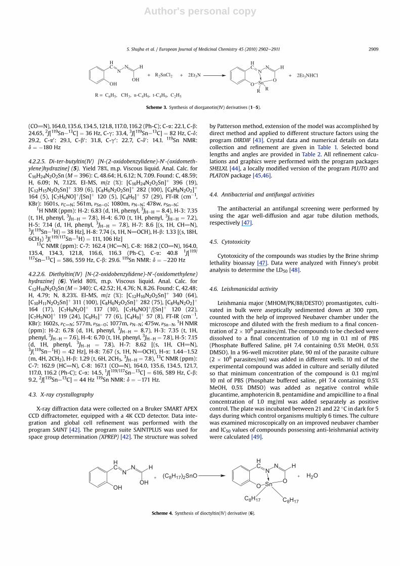
4.2.2. Synthesis of the complexes (1e6)The diorganotin(IV) complexes were synthesized by two
method.Method I: A 3.0 mmol (0.49 g) of N0-(2-hydroxybenzylidene)
formohydrazide, 6 mmol (0.83 ml) triethylamine and dialkylltindichloride (3.0 mmol) weremixed together in 100ml toluene. After5 h stirring at room temperature, the yellow solution was filteredoff to remove the Et3NHCl salt. The filtrate was rotary evaporator toget a yellow product (Scheme 3).
Method II: The dioctyltin(IV) complex was synthesized by sus-pending the dioctyltin(IV) oxide and ligand in stoichiometricamounts in dry toluene. The mixture was refluxed for 3 h, andwater formed during the reaction was removed by the Dean andStark apparatus. Yellow oily product was obtained by removingsolvent under reduce pressure (Scheme 4).
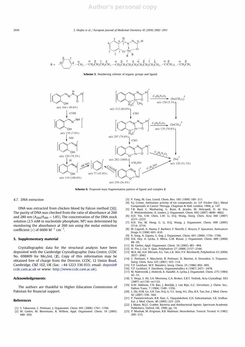
The structure and numbering template of diorganotin(IV)complexes is illustrated in Scheme 5.
4.2.2.1. Diphenyltin(IV) [N-(2-oxidobenzylidene)-N0-(oxidomethylene)hydrazine] (1). Yield 84%, m.p. 160e162 �C. Anal. Calc. forC20H16N2O2Sn (M¼ 436): C, 55.21; H, 3.71; N, 6.44. Found: C, 55.19;H, 3.72; N, 6.39%. EI-MS, m/z (%): [C20H16N2O2Sn]þ 436 (56),[C14H11N2O2Sn]þ 359 (22), [C8H6N2O2Sn]þ 282 (100), [C8H8N2O2]þ
165 (11), [C7H9N2O]þ 137 (11), [C7H6NO]þ/[Sn]þ 120 (37),[C7H5NO]þ 119 (11), [C6H5]þ 77 (36), [C4H9]þ 57 (5) [C4H3]þ 51 (27),[C6H5Sn]þ 197 (48) FT-IR (cm�1, KBr): 1609s, nC]N; 567m, nSneO;1074m, nNeN; 486w, nSneN. 1H NMR (ppm): H-2: 7.12 (d, 1H, phenyl,3JHeH ¼ 8.4), H-3: 7.41e7.50 (m, 1H, phenyl), H-4: 6.81 (t, 1H,phenyl, 3JHeH ¼ 7.8), H-5: 7.21 (d, 1H, phenyl, 3JHeH ¼ 7.8), H-7: 8.65[(s, 1H, CH]N), 3J(119Sne1H) ¼ 51 Hz], H-8: 7.38e7.51 (m, 1H,N]CH), H-b: 7.84e7.90 (m, 4H, phenyl), H-g, H-d: 7.38e7.51 (m, 6H,phenyl), 13C NMR (ppm): C-7: 163.0 (HC]N), C-8: 167.5 (CO]N),163.6, 135.8, 134.7, 122.2, 11.7, 116.3 (Ph-C); C-a: 139.0, C-b: 136.7 2J
[119Sne13C] ¼ 56 Hz, C-g: 129.0 3J[119/117Sne13C] ¼ 97, 89 Hz, C-d:130.7 4J[119Sne13C] ¼ 17 Hz, 119Sn NMR: d ¼ �337 Hz
4.2.2.2. Dimethyltin(IV) [N-(2-oxidobenzylidene)-N0-(oxidomethylene)hydrazine] (2). Yield 80%, m.p. 116e118 �C. Anal. Calc. forC10H12N2O2Sn (M¼ 312): C, 38.63; H, 3.89; N, 9.01. Found: C, 38.59;H, 3.85; N, 9.03%. EI-MS, m/z (%): [C10H12N2O2Sn]þ 312 (84),[C9H9N2O2Sn]þ 297 (80), [C8H6N2O2Sn]þ 282 (100), [C8H8N2O2]þ
164 (6), [C7H9N2O]þ 137 (27), [C7H6NO]þ/[Sn]þ 120 (51), [C7H5NO]þ
119 (23), [C6H5]þ 77 (23), [C4H3]þ 51 (48), [C2H6Sn]þ 150 (5),[CH3Sn]þ 135 (71) FT-IR (cm�1,KBr): 1605s, nC]N; 563m, nSneO;1072m, nNeN, 484w,nSneN.
1H NMR (ppm): H-2: 6.77 (d, 1H, phenyl, 3JHeH ¼ 8.1), H-3: 7.36(t, 1H, phenyl, 3JHeH ¼ 7.8), H-4: 6.75 (t, 1H, phenyl, 3JHeH ¼ 7.8),H-5: 7.18 (d, 1H, phenyl, 3JHeH ¼ 7.8), H-7: 8.62 [(s, 1H, CH]N),3J(119Sne1H) ¼ 46 Hz], H-8: 7.63 (s, 1H, N]OCH), H-a: 0.83 (s, 6H,2CH3), 2J(119/117Sne1H) ¼ 79, 76 Hz 13C NMR (ppm): C-7: 162.8(HC]N), C-8: 166.6 (CO]N), 163.7, 135.8, 134.5, 121.8, 117.3, 116.2(Ph-C), Ca: 1.55 1J[119/117Sne13C] ¼ 650, 620 Hz, 119Sn NMR:d ¼ �163 Hz
4.2.2.3. Dibutyltin(IV) [N-(2-oxidobenzylidene)-N0-(oxidomethylene)hydrazine] (3). Yield 78%, m.p. viscous liquid. Anal. Calc. forC16H24N2O2Sn (M¼ 396): C, 48.64; H, 6.12; N, 7.09. Found: C, 48.61;H, 6.10; N, 7.06%. EI-MS, m/z (%): [C16H24N2O2Sn]þ 396 (32),[C12H15N2O2Sn]þ 339 (76), [C8H6N2O2Sn]þ 282 (100), [C8H8N2O2]þ
164 (21), [C7H9N2O]þ 137 (26), [C7H6NO]þ/[Sn]þ 120 (26),[C7H5NO]þ 119 (16), [C4H9]þ 57 (99), [C4H3]þ 51 (6), [C8H18Sn]þ 234(12). FT-IR (cm�1, KBr): 1609s, nC]N; 565m, nSneO; 1080m, nNeN;481w, nSneN, 1H NMR (ppm): H-2: 6.77 (d, 1H, phenyl, 3JHeH ¼ 8.1),H-3: 7.35 (t, 1H, phenyl, 3JHeH ¼ 7.8), H-4: 6.73 (t, 1H, phenyl,3JHeH ¼ 7.8), H-5: 7.16 (d, 1H, phenyl, 3JHeH ¼ 7.8), H-7: 8.61 [(s, 1H,CH]N), 3J(119Sne1H) ¼ 43 Hz], H-8: 7.66 (s, 1H, N]OCH), H-a:1.60e1.69 (m, 4H, 2CH2), H-b: 1.48e1.54 (m, 4H, 2CH2), H-g: 1.36(m, 4H, 2CH2), H-d: 0.89 (t, 6H, 2CH3, 3JHeH ¼ 7.2), 13C NMR (ppm):C-7: 162.7 (HC]N), C-8: 167.2 (CO]N), 164.0, 135.6, 134.5, 121.7,117.0, 116.2 (Ph-C), C-a: 22.4 1J[119/117Sne13C] ¼ 600, 575 Hz, C-b:26.8 2J[119Sne13C] ¼ 36 Hz, C-g: 26.2 3J[119Sne13C] ¼ 90 Hz, C-d:13.6 (SneBu). 119Sn NMR: d ¼ �202 Hz
4.2.2.4. Dioctyltin(IV) [N-(2-oxidobenzylidene)-N0-(oxidomethylene)hydrazine] (4). Yield 77%, m.p. Viscous liquid. Anal. Calc. forC24H40N2O2Sn (M¼ 508): C, 56.82; H, 7.95; N, 5.52. Found: C, 56.79;H, 7.92; N, 5.49%. EI-MS, m/z (%): [C24H40N2O2Sn]þ 508 (4),[C16H23N2O2Sn]þ 395 (14), [C8H7N2O2Sn]þ 283 (64), [C8H6N2O2Sn]þ
282 (32), [C8H8N2O2]þ 164 (10), [C7H9N2O]þ 137 (13), [C7H6NO]þ/[Sn]þ 120 (9), [C7H5NO]þ 119 (7), [C6H5]þ 77 (4), [C4H9]þ 57 (100),[C4H3]þ 51 (4). FT-IR (cm�1, KBr): 1603s, nC]N; 566m, nSneO; 1082m,nNeN; 489w, nSneN, 1H NMR (ppm): H-2: 6.76 (d, 1H, phenyl,3JHeH ¼ 8.1), H-3: 7.35 (t, 1H, phenyl, 3JHeH ¼ 7.8), H-4: 6.70 (t, 1H,phenyl, 3JHeH ¼ 7.8), H-5: 7.16 (d, 1H, phenyl, 3JHeH ¼ 7.8), H-7: 8.61[(s, 1H, CH]N), 3J(119Sne1H) ¼ 41 Hz], H-8: 7.66 (s, 1H, N]OCH),H-a: 1.65e1.72 (m, 4H, 2CH2), H-b: 1.49e1.58 (m, 4H, 2CH2), H-g-g0:1.23e1.46 (br, 16H, 2CH2 CH2CH2 CH2), H-d0: 0.88 (t, 6H, 2CH3,3JHeH ¼ 7.2), 13C NMR (ppm): C-7: 162.6 (HC]N), C-8: 167.2
HC
OH
N
HN H
O1
23
45
67
8
Scheme 2. Numbering scheme of ligand (H2L).
C
OH
H
O
+ H C
O
NH NH2
HC
OH
N
HN H
O
-H2O
HC
OH
NN H
OH
aldo-imine form imine-ol form
Scheme 1. Synthesis of ligand (H2L).
S. Shujha et al. / European Journal of Medicinal Chemistry 45 (2010) 2902e29112908
Author's personal copy
(CO]N), 164.0, 135.6, 134.5, 121.8, 117.0, 116.2 (Ph-C); C-a: 22.1, C-b:24.65, 2J[119Sne13C] ¼ 36 Hz, C-g: 33.4, 3J[119Sne13C] ¼ 82 Hz, C-d:29.2, C-a0: 29.1, C-b0: 31.8, C-g0: 22.7, C-d0: 14.1. 119Sn NMR:d ¼ �180 Hz
4.2.2.5. Di-ter-butyltin(IV) [N-(2-oxidobenzylidene)-N0-(oxidometh-ylene)hydrazine] (5). Yield 78%, m.p. Viscous liquid. Anal. Calc. forC16H24N2O2Sn (M¼ 396): C, 48.64; H, 6.12; N, 7.09. Found: C, 48.59;H, 6.09; N, 7.12%. EI-MS, m/z (%): [C16H24N2O2Sn]þ 396 (19),[C12H15N2O2Sn]þ 339 (6), [C8H6N2O2Sn]þ 282 (100), [C8H8N2O2]þ
164 (5), [C7H6NO]þ/[Sn]þ 120 (5), [C4H9]þ 57 (29), FT-IR (cm�1,KBr): 1601s, nC]N; 561m, nSneO; 1080m, nNeN; 478w, nSneN;
1H NMR (ppm): H-2: 6.83 (d, 1H, phenyl, 3JHeH ¼ 8.4), H-3: 7.35(t, 1H, phenyl, 3JHeH ¼ 7.8), H-4: 6.70 (t, 1H, phenyl, 3JHeH ¼ 7.2),H-5: 7.14 (d, 1H, phenyl, 3JHeH ¼ 7.8), H-7: 8.6 [(s, 1H, CH]N),3J(119Sne1H) ¼ 38 Hz], H-8: 7.74 (s, 1H, N]OCH), H-b: 1.33 [(s, 18H,6CH3) 3J(119/117Sne1H) ¼ 111, 106 Hz]
13C NMR (ppm): C-7: 162.4 (HC]N), C-8: 168.2 (CO]N), 164.0,135.4, 134.3, 121.8, 116.6, 116.3 (Ph-C), C-a: 40.8 1J[119/117Sne13C] ¼ 586, 559 Hz, C-b: 29.6. 119Sn NMR: d ¼ �220 Hz
4.2.2.6. Diethyltin(IV) [N-(2-oxidobenzylidene)-N0-(oxidomethylene)hydrazine] (6). Yield 80%, m.p. Viscous liquid. Anal. Calc. forC12H16N2O2Sn (M¼ 340): C, 42.52; H, 4.76; N, 8.26. Found: C, 42.48;H, 4.79; N, 8.23%. EI-MS, m/z (%): [C12H16N2O2Sn]þ 340 (64),[C10H11N2O2Sn]þ 311 (100), [C8H6N2O2Sn]þ 282 (75), [C8H8N2O2]þ
164 (17), [C7H9N2O]þ 137 (10), [C7H6NO]þ/[Sn]þ 120 (22),[C7H5NO]þ 119 (24), [C6H5]þ 77 (6), [C4H9]þ 57 (8), FT-IR (cm�1,KBr): 1602s, nC]N; 577m, nSneO; 1077m, nNeN; 475w, nSneN. 1H NMR(ppm): H-2: 6.78 (d, 1H, phenyl, 3JHeH ¼ 8.7), H-3: 7.35 (t, 1H,phenyl, 3JHeH ¼ 7.6), H-4: 6.70 (t, 1H, phenyl, 3JHeH ¼ 7.8), H-5: 7.15(d, 1H, phenyl, 3JHeH ¼ 7.8), H-7: 8.62 [(s, 1H, CH]N),3J(119Sne1H) ¼ 42 Hz], H-8: 7.67 (s, 1H, N]OCH), H-a: 1.44e1.52(m, 4H, 2CH2), H-b: 1.29 (t, 6H, 2CH3, 3JHeH ¼ 7.8), 13C NMR (ppm):C-7: 162.9 (HC]N), C-8: 167.1 (CO]N), 164.0, 135.6, 134.5, 121.7,117.0, 116.2 (Ph-C); C-a: 14.5, 1J[119/117Sne13C] ¼ 616, 589 Hz, C-b:9.2, 2J[119Sne13C] ¼ 44 Hz 119Sn NMR: d ¼ �171 Hz.
4.3. X-ray crystallography
X-ray diffraction data were collected on a Bruker SMART APEXCCD diffractometer, equipped with a 4K CCD detector. Data inte-gration and global cell refinement was performed with theprogram SAINT [42]. The program suite SAINTPLUS was used forspace group determination (XPREP) [42]. The structure was solved
by Patterson method, extension of the model was accomplished bydirect method and applied to different structure factors using theprogram DIRDIF [43]. Crystal data and numerical details on datacollection and refinement are given in Table 1. Selected bondlengths and angles are provided in Table 2. All refinement calcu-lations and graphics were performed with the program packagesSHELXL [44], a locally modified version of the program PLUTO andPLATON package [45,46].
4.4. Antibacterial and antifungal activities
The antibacterial an antifungal screening were performed byusing the agar well-diffusion and agar tube dilution methods,respectively [47].
4.5. Cytotoxicity
Cytotoxicity of the compounds was studies by the Brine shrimplethality bioassay [47]. Data were analyzed with Finney’s probitanalysis to determine the LD50 [48].
4.6. Leishmanicidal activity
Leishmania major (MHOM/PK/88/DESTO) promastigotes, culti-vated in bulk were aseptically sedimented down at 300 rpm,counted with the help of improved Neubaver chamber under themicroscope and diluted with the fresh medium to a final concen-tration of 2 � 106 parasites/ml. The compounds to be checked weredissolved to a final concentration of 1.0 mg in 0.1 ml of PBS(Phosphate Buffered Saline, pH 7.4 containing 0.5% MeOH, 0.5%DMSO). In a 96-well microtiter plate, 90 ml of the parasite culture(2 � 106 parasites/ml) was added in different wells. 10 ml of theexperimental compound was added in culture and serially dilutedso that minimum concentration of the compound is 0.1 mg/ml10 ml of PBS (Phosphate buffered saline, pH 7.4 containing 0.5%MeOH, 0.5% DMSO) was added as negative control whileglucantime, amphotericin B, pentamidine and ampicilline to a finalconcentration of 1.0 mg/ml was added separately as positivecontrol. The platewas incubated between 21 and 22 �C in dark for 5days during which control organisms multiply 6 times. The culturewas examined microscopically on an improved neubaver chamberand IC50 values of compounds possessing anti-leishmanial activitywere calculated [49].
HC
OH
NN H
OH+
HC
O
NN H
O+ H2O
Sn
C8H17 C8H17
(C8H17)2SnO
Scheme 4. Synthesis of dioctyltin(IV) derivative (6).
HC
OH
NN H
OH+ R2SnCl2 + 2Et3N
HC
O
NN H
O+ 2Et3NHCl
Sn
R RR = C6H5, CH3, n-C4H9, t-C4H9, C2H5
Scheme 3. Synthesis of diorganotin(IV) derivatives (1e5).
S. Shujha et al. / European Journal of Medicinal Chemistry 45 (2010) 2902e2911 2909
Author's personal copy
4.7. DNA extraction
DNA was extracted from chicken blood by Falcon method [50].The purity of DNAwas checked from the ratio of absorbance at 260and 280 nm (A260/A280 ¼ 1.85). The concentration of the DNA stocksolution (2.5 mM in nucleotide phosphate, NP) was determined bymonitoring the absorbance at 260 nm using the molar extinctioncoefficient (3) of 6600 M�1 cm�1.
5. Supplementary material
Crystallographic data for the structural analysis have beendeposited with the Cambridge Crystallographic Data Centre, CCDCNo. 698809 for Me2SnL (2), Copy of this information may beobtained free of charge from the Director, CCDC, 12 Union Road,Cambridge, CBZ 1EZ, UK (fax: þ44 1223 336 033; email: [email protected] or www: http://www.ccdc.cam.ac.uk).
Acknowledgements
The authors are thankful to Higher Education Commission ofPakistan for financial support.
References
[1] S. Tabassum, C. Pettinari, J. Organomet. Chem. 691 (2006) 1761e1766.[2] M. Gielen, M. Biesemans, R. Willem, Appl. Organomet. Chem. 19 (2005)
440e450.
[3] P. Yang, M. Guo, Coord. Chem. Rev. 185 (1999) 189e211.[4] A.J. Crowe, Antitumor activity of tin compounds. in: S.P. Fricker (Ed.), Metal
Compounds in Cancer Therapy. Chapman & Hall, London, 1994, p. 147.[5] T.B. Baul, C. Masharing, G. Ruisi, R. Jirasko, M. Holcapek, D. de Vos,
D. Wolstenholme, A. Linden, J. Organomet. Chem. 692 (2007) 4849e4862.[6] H.D. Yin, S.W. Chen, L.W. Li, D.Q. Wang, Inorg. Chim. Acta 360 (2007)
2215e2223.[7] H.D. Yin, M. Hong, G. Li, D.Q. Wang, J. Organomet. Chem. 690 (2005)
3714e3719.[8] M. Cagnoli, A. Alama, F. Barbieri, F. Novelli, C. Bruzzo, F. Sparatore, Anticancer
Drugs 9 (1998) 603e610.[9] X. Song, A. Zapata, G. Eng, J. Organomet. Chem. 691 (2006) 1756e1760.
[10] D.K. Dey, A. Lycka, S. Mitra, G.M. Rosair, J. Organomet. Chem. 689 (2004)88e95.
[11] M. Gielen, Appl. Organomet. Chem. 16 (2002) 481e494.[12] H. Yin, J. Cui, Y. Qiao, Polyhedron 27 (2008) 2157e2166.[13] M.A. Ali, A.H. Mirzam, A.L. Tan, L.K. Wei, P.V. Bernhardt, Polyhedron 23 (2004)
2037e2043.[14] C. Pettinari, F. Marchetti, R. Pettinari, D. Martini, A. Drozodov, S. Troyanov,
Inorg. Chim. Acta 325 (2001) 103e114.[15] T.P. Lockhart, W.F. Manders, Inorg. Chem. 25 (1986) 892e895.[16] T.P. Lockhart, F. Davidson, Organometallics 6 (1987) 2471e2478.[17] M. Nádvorník, J. Hole�cek, K. Handlí�r, A. Ly�cka, J. Organomet. Chem. 275 (1984)
43e51.[18] S. Shuja, S. Ali, S.A. Meetsma, G.A. Broker, E.R.T. Tiekink, Acta Crystallogr. E63
(2007) m1130em1132.[19] A.W. Addison, T.N. Rao, J. Reedijk, J. van Rijn, G.C. Verschoor, J. Chem. Soc.,
Dalton Trans. 7 (1984) 1349e1356.[20] L. Shi, H.M. Ge, S.H. Tan, H.Q. Li, Y.C. Song, H.L. Zhu, R.X. Tan, Eur. J. Med. Chem.
42 (2007) 558e564.[21] P. Panneerselvam, R.R. Nair, G. Vijayalakshmi, E.H. Subramanian, S.K. Sridhar,
Eur. J. Med. Chem. 40 (2005) 225e229.[22] J. Mann, M.J.C. Crabbe, Bacteria and Antibacterial Agents. Spectrum Academic
Publishers, Oxford, UK, 1990, pp. 74.[23] P. Mushak, M. Krigman, R.B. Mailman, Neurobehav. Toxicol. Teratol. 4 (1982)
209e215.
OH
NNH
OH
N
OH
NHN H
O
m/z 164 ( 49.6%)
-COH
m/z 135 (7.8%)
m/z 120 (79.2%)
-NH
OH
N
m/z 119 (100.0%)
-H
m/z 77 (43.8%)
-HCNO
O
NN H
OSnCH3H3C
O
NN H
OSn
O
NN H
OSn
H3C
-CH3
-CH3
m/z 312 (84.8%)
m/z 297 (79.8%)
m/z 282 (100%)
[Sn(CH3)2]+
[SnCH3]+
[Sn]+
m/z 150 (5.3%)
m/z 135 (70.8%)
m/z 120 (51.3%)
O
NSn
m/z 238 (26.4%)
-CH2NO
-C8H6N2O2
-C8H6N2O2
-C8H
6N2O
2
-C 7H 4
NO-CNOSn
Scheme 6. Proposed mass fragmentation pattern of ligand and complex 2.
12
3
45
67
8HC
O
NN H
OSn
R R
CH3 CH2CH2CH2CH3 CH2CH2CH2CH2CH2CH2CH2CH3 C(CH3)3 CH2CH3R =α
βδ
γα α β δγα β δγ
, , ,,,α β δγ′′ ′′ α β α β
Scheme 5. Numbering scheme of organic groups and ligand.
S. Shujha et al. / European Journal of Medicinal Chemistry 45 (2010) 2902e29112910
Author's personal copy
[24] S.V. Doctor, D.A. Fox, Toxicol. Environ. Health 10 (1982) 53e62.[25] A.V. Krishnaraju, T.V.N. Rao, D. Sundararaju, M. Vanisree, H.S. Tsay,
G.V. Subbaraju, Int. J. Appl. Sci. Eng. 4 (2006) 115e125.[26] S. Shahzadi, K. Shahid, S. Ali, M. Bakhtiar, Turk. J. Chem. 32 (2008)
333e353.[27] M. Aslanoglu, Anal. Sci. 22 (2006) 439e443.[28] E.C. Long, J.K. Barton, Acc. Chem. Res. 23 (1990) 271e273.[29] Q. Feng, N.Q. Li, Y.Y. Jiang, Anal. Chim. Acta 344 (1997) 97e104.[30] R. Vijayalakshmi, M. Kanthimathi, R. Parthasarathi, B.U. Nair, Bull. Chem. Soc.
Jpn. 78 (2005) 270e276.[31] Q. Lie, P. Yang, H. Wang, M. Guo, J. Inorg. Biochem. 64 (1996) 181e195.[32] J. Rusanova, S. Decurtins, E. Rosanov, H.S. Evans, S. Delahaye, A. Hauser,
J. Chem. Soc., Dalton Trans. (2002) 4318e4320.[33] A. Ambroise, B.G. Maiya, Inorg. Chem. 39 (2000) 4264e4272.[34] J.E.B. Randles, Trans. Faraday Soc. 44 (1948) 327e338.[35] A. Sevcik, Collect. Czech. Chem. Commun. 13 (1948) 337e349.[36] S. Wang, T. Penz, C.F. Yang, Biophys. Chem. 104 (2003) 239e248.[37] X. Yang, G.L. Shen, R.Q. Yu, Microchem. J. 62 (1999) 394e404.[38] Z. Xu, G. Bai, C. Dong, Bioorg. Med. Chem. 13 (2005) 5694e5699.[39] M.Y. Ni, Y. Wang, H.L. Li, Pol. J. Chem. 71 (1997) 816e822.[40] M.S. Ibrahim, I.S. Shehatta, A.A. Al-nyeli, J. Pharm. Biomed. Anal. 28 (2002)
217e225.
[41] D.D. Perrin, W.L.F. Armengo, Purification of Laboratory Chemicals, third ed.Pergamon Press, Oxford, 1988.
[42] Bruker, Smart, SAINTPLUS and XPREP. Area Detector Control and IntegrationSoftware. Smart Apex Software Reference Manuals. Bruker Analytical X-rayInstruments. Inc, Madison, Wisconsin, USA, 2006.
[43] P.T. Beurskens, G. Beurskens, R. Gelder, S. García-Granda, S.R.O. Gould,R. Israël, J.M.M. Smits, The DIRDIF-99 Program System, CrystallographyLaboratory. University of Nijmegen, The Netherlands, 1999.
[44] G.M. Sheldrick, SHELXL-97. Program for the Refinement of Crystal Structures.University of Göttingen, Germany, 1997.
[45] A. Meetsma, PLUTO. Molecular Graphics Program. 2007. University ofGroningen, The Netherlands, 2006.
[46] A.L. Spek, PLATON. Program for the Automated Analysis of Molecular Geom-etry. University of Utrecht, The Netherlands, 2006.
[47] A. Rahman, M.I. Choudhary, W.J. Thomsen, Bioassay Techniques for DrugDevelopment. Harward Academic Press, Amsterdam, 2001.
[48] D.J. Finney, Probit Analysis, third ed. Cambridge University Press, Cambridge,1971.
[49] K.M. Khan, M. Rasheed, Z. Ullah, S. Hayat, F. Kaukab, M.I. Choudhary,A. Rahman, S. Perveen, Bioorg. Med. Chem. 11 (2003) 1381e1387.
[50] J. Sambrook, E.F. Fritsch, T. Maniatis, Molecular Cloning: a Laboratory Manual,second ed. Cod. Spring harbour laboratory press, New York, 1989.
S. Shujha et al. / European Journal of Medicinal Chemistry 45 (2010) 2902e2911 2911