DigitalCommons@USU - Utah State University
52
Utah State University Utah State University DigitalCommons@USU DigitalCommons@USU All Graduate Theses and Dissertations Graduate Studies 5-1995 Effects of Antioxidants on Development of In Vitro Fertilized Effects of Antioxidants on Development of In Vitro Fertilized Bovine Embryos Bovine Embryos Bret L. Anderson Utah State University Follow this and additional works at: https://digitalcommons.usu.edu/etd Part of the Animal Sciences Commons Recommended Citation Recommended Citation Anderson, Bret L., "Effects of Antioxidants on Development of In Vitro Fertilized Bovine Embryos" (1995). All Graduate Theses and Dissertations. 3920. https://digitalcommons.usu.edu/etd/3920 This Thesis is brought to you for free and open access by the Graduate Studies at DigitalCommons@USU. It has been accepted for inclusion in All Graduate Theses and Dissertations by an authorized administrator of DigitalCommons@USU. For more information, please contact [email protected].
-
Upload
khangminh22 -
Category
Documents
-
view
0 -
download
0
Transcript of DigitalCommons@USU - Utah State University

Utah State University Utah State University
DigitalCommons@USU DigitalCommons@USU
All Graduate Theses and Dissertations Graduate Studies
5-1995
Effects of Antioxidants on Development of In Vitro Fertilized Effects of Antioxidants on Development of In Vitro Fertilized
Bovine Embryos Bovine Embryos
Bret L. Anderson Utah State University
Follow this and additional works at: https://digitalcommons.usu.edu/etd
Part of the Animal Sciences Commons
Recommended Citation Recommended Citation Anderson, Bret L., "Effects of Antioxidants on Development of In Vitro Fertilized Bovine Embryos" (1995). All Graduate Theses and Dissertations. 3920. https://digitalcommons.usu.edu/etd/3920
This Thesis is brought to you for free and open access by the Graduate Studies at DigitalCommons@USU. It has been accepted for inclusion in All Graduate Theses and Dissertations by an authorized administrator of DigitalCommons@USU. For more information, please contact [email protected].

ii
ABSTRACT
Effects of Antioxidants on Development of
In Vitro Fertilized Bovine Embryos
by
Bret L. Anderson, Master of Science
Utah State University, 1995
Major Professor: Dr. Thomas D. Bunch Department: Animal, Dairy and Veterinary Sciences
Free radicals are short-lived molecules that can cause
decreased embryonic development in vitro. Antioxidants are
molecules that block free radical formation or guard against
their harmful effects. Many studies have linked exposure of
media to light and culturing of embryos in high (20%) oxygen
concentrations to free radical production. Some of the
antioxidants used in culture media are superoxide dismutase
(SOD), catalase, zinc (II), ethylenedinitrilo tetraacetic
acid (EDTA), mannitol, vitamin E, dimethyl sulfide, and
taurine. Most research involving antioxidants and embryonic
development has been conducted on non-farm animals,
particularly mouse and rabbit. Studies have shown that
antioxidants in vitro culture improved embryo development to
the blastocyst stage.
In this study, we evaluated the effects of SOD and
catalase on bovine embryo development. Four concentrations

iii
of SOD (0, 1500, 3000, 6000 IU/ml) and catalase (0, 75, 100,
125 pg/ml) and combinations of the two antioxidants were
evaluated through maturation, fertilization, and culture.
SOD and catalase were first reconstituted in water and then
diluted to their final concentrations. Oocytes were matured
in M-199 plus 0.5 pg/ml LH , 5 pg/ml FSH, and 10% FBS at 39
·c in 5% co2 for 24 hours. They were then placed in
fertilization-TALP with heparin and 1 X 10⁶/ml sperm.
Embryos were cultured in CR2 medium supplemented with
alanine, glycine, and 3 mg/ml of fatty-acid free bovine
serum albumin in modular incubators with 5% co2, 5% o2, and
90% N2. Embryo development was evaluated on day 8. Three
replicates with approximately 50 embryos per treatment were
used to evaluate the effects of SOD and catalase. The
control had better embryo development than all treatments.
The treatment that was most similar to the control was
treatment 2, which consisted of no SOD and 75 pg/ml
catalase.
Based on these observations, levels of both SOD and
catalase were lowered to 0, 100, 250, and 500 IU/ml and 0,
10, 25, and 50 pg/ml, respectively . Although these levels
appeared to improve embryo development, there were no
statistical differences. Based on the culture system and
media currently used along with the precautions against
light and oxygen concentration, we did not find any
beneficial effects of supplementing medium with SOD or
catalase.
iv
(51 pages)
ACKNOWLEDGMENTS
I would like to express my deep appreciation to Dr .
Thomas D. Bunch, professor of animal, dairy and veterinary
s ciences, for his guidance and assistance throughout my
graduate program at Utah State University. I would also
like t o thank the other members who served on my thesis
committee : Dr . Kenneth L. White, associate professor of
animal, dairy and veterinary sciences, and Dr . LeGrande C.
Ellis, professor of biology.
v
In addition, I would like to extend appreciation to the
following graduate students and laboratory technicians:
Caiping Yue, Vicki Farrar, Charoe nsri Thonabulsombat, and
William Reed for their time, assistance, and suggestions. I
also wish to thank Eddie Sullivan for his patience as he
taught me IVF .
also wish to thank E . A. Miller Inc. for their
generous donation of bovine ovaries . Without their
c ontribution, work in this area would be next to impossible .
A special thanks to my dear family for the love,
support, and encouragement as I pursued my master ' s degree.
I would also like to acknowledge the Experiment Station
and the Utah Center of Excellence for their assistance in
funding this project .
Bret L . Anderson

ABSTRACT
ACKNOWLEDGMENTS
LIST OF TABLES
INTRODUCTION
LITERATURE REVIEW
MATERIALS AND METHODS
RESULTS AND DISCUSSION
CONCLUSION
REFERENCES
CONTENTS
vi
Page
ii
v
vii
1
4
19
26
37
39
vii
LIST OF TABLES
Table Page
1 Embryo development at high antioxidant concentrations ...... ... . . .... ......... . . . . ..... . .. . 27
2 ANOVA table on the results of embryos cultured in high antioxidant concentrations .... ... ... . . . . . ... 28
3 Comparison of all combinations of the four levels testing signif i cance and the average number of embryo d i visions based on SOD concentration ......... 29
4 Embryo development at reduced antioxidant concentrations .... .. . . ..... . ....... . ...... . . . ...... . 30
ANOVA table on the results of embryos cultured in low antioxidant concentrations . ...... . ......... .. 31

INTRODUCTION
Factors that affect mammalian embryo development have
been a topic of extensive research for a number of years.
One area that continues to be of interest is creating an in
vitro environment for embryo production that closely mimics
conditions of the female reproductive tract. Numerous
studies have been conducted to determine why embryos do not
develop as readily in vitro as they do in vivo. The fact
that mammalian embryos produced in vitro develop slower or
less efficiently has been well documented in several species
(Wright and Bondioli, 1981; Fisher, 1987). Embryos often
stop their development at what is referred to as the
"block." In most species the block occurs when the genetic
system shifts from maternal to genomic DNA synthesis. Many
researchers have speculated on the cause of the block. Most
recently, there has been evidence reported in the literature
that free radicals may be a contributing factor to the
developmental retardation of embryos (Li et al., 1993).
Since free radicals are common in most in vitro cell
culture systems, researchers have begun to either remove or
minimize their effects. One of the first areas to be
investigated was to alter the amount of oxygen concentration
to which embryos are exposed. Most embryo culture systems
are maintained under a gas atmosphere of 5% co2 and 95%
humidified air . Some researchers have cultured embryos
under an atmosphere of 5-8% o2, which is within the range of
o2 concentrations within the mammalian oviduct. This is
2
considerably lower than the 20% 0 2 that is in humidified air
(Li et al . , 1993). By lowering the o2, there was an
increase in the percentage of embryos that developed to the
blastocyst stage. Beneficial affects of lowering o2
concentrations could be detected as low as 1% o2.
It has been established that the concentration of 0 2 in
in vitro culture systems plays a significant role in the
production of harmful reactive oxygen species (Noda et al.,
1991; Li et al., 1993; Nakayama et al., 1994). Studies have
also shown that the incorporation of free radical scavengers
into culture systems alters the adverse effects of reactive
oxygen species (Noda et al., 1991; Li et al., 1993;
Natsuyama et al., 1993a) . One study performed in bovine
(Luvoni et al., 1994) found that incorporation of superoxide
dismutase (SOD) into embryo culture media exposed to
atmospheric oxygen did increase the number of embryos
developing to the blastocyst stage. However, if embryos
were cultured with SOD under low oxygen concentration, the
increase in embryo development was not nearly as great.
Catalase and SOD, which are both free radical scavengers,
have been shown to be beneficial to the in vitro development
of mouse embryos (Nonogaki et al., 1991; Nonogaki et al.,
1992 ; Natsuyama et al . , 1993a ; Chun et al . , 1994; Luvoni et
al., 1994). In these studies, they reported that
blastulation rate increased with the use of radical
scavengers .
free radical production. Another way that is probably
equally damaging to embryos is exposure of media and(or)
embryos to fluorescent light. An unpublished study
performed in our laboratory showed that prolonged exposure
of bovine embryos to fluorescent light decreased the
percentage that developed to the blastocyst stage. Another
study done on the hamster also verified the detrimental
effects on embryo development (Nakayama et al., 1994). The
detrimental effects on development have been linked to free
radical production in prolonged light exposure and high
oxygen concentration cultures. These two free-radical
causing factors should be of concern to anyone dealing with
in vitro embryo production .
Our laboratory expends considerable effort studying
factors that affect preimplantation embryo development in
livestock species. The types of studies range from
evaluating factors affecting oocyte maturation and in vitro
fertilization, to the development of a better culture
medium. This study was designed to evaluate the effects of
catalase , SOD, and a reduced oxygen concentration in all
phases (maturation of the oocyte, fertilization, and
culture) of in vitro bovine embryo production.
3
4
LITERATURE REVIEW
A ' free radical" is a molecule with one or more
unpaired electrons (Gutteridge and Halliwell, 1988; Kehrer,
1993) . Free radicals exist in free form, which makes them
highly unstable. In their free form, they interact with
various tissue components, causing metabolic and genetic
dysfunction . Free radicals exist in various forms,
consisting of both organic and inorganic molecules. While
these molecules are very reactive and sometimes destructive,
they are very necessary for the normal activity of many
biological processes (Dianzani, 1992) . Studies on free
radicals date back to the mid 1950's. Their destructive
and(or) beneficial effects on in vivo and in vitro systems
continue to be actively pursued. The scope of this study
will focus on the effects of oxygen free radicals in a
bovine in vitro embryo culture system .
There are three types of highly reactive oxygen free
radicals: superoxide radical, hydrogen peroxide, and the
hydroxyl radical. These molecules have single unpaired
electrons and therefore are classified as free radicals
(Marklund, 1985).
Many studies have been conducted to find out where and
how free radicals are formed. Free radicals are commonly
formed within cells by the leakage of electrons to oxygen
during electron transport. More specifically, this happens
during oxidation of mitochondrial cytochrome P450, in the
endoplasmic reticulum, or by activity of other oxidase
systems (Halliwell and Gutteridge, 1989; Gille and Joenje,
1991 ; Johnson and Nasr-Esfahano, 1994) . A study that
quantified free radicals by electron-spin-resonance signals
in rat liver mitochondria found them to be very abundant.
Not only were free radicals abundant in the liver samples,
but the levels of radicals would also change by either
starving the animals (increasing radical production) or by
chemically inhibiting respiration (decreasing radical
production) (Slater, 1972). Free radicals are also formed
throughout normal cellular metabolism . They also result
from the metabolism of some drugs and xenobiotics (Bendich,
1993) .
Free radicals form in in vitro cultures . Exposure of
cultures to light, both fluorescent and UV, have been shown
to have detrimental effects . Living organisms absorb light
by either endogenous or exogenous photosensitizers, which in
the presence of oxygen cause oxidation, which leads to many
chemical and biological effects (Blum, 1964). Unpublished
studies in our laboratory have indicated that exposure of
embryos to fluorescent light for as little as 15 min tended
to cause developmental disruption.
The superoxide anion is produced through the reduction
of molecular oxygen by the addition of a single electron
(Fee and Valentine, 1977). Controlling the production of
the superoxide radical may minimize potential adverse

6
effects in tissue culture systems. As was pointed out in a
study done by Warren et al . (1987), there are quite a number
of different ways that radicals can be produced . One of the
common methods to produce oxygen radicals is by changing
oxygen concentrations. Normally, 0 2 + 4e + 4H1 produces
2H2o. Variations in the normal pathway of production of
2H2o creates highly reactive oxygen species . The addition
of an electron to o2 produces superoxide. The addition of a
second electron and hydrogen atom to the superoxide anion
results in hydrogen peroxide. The combination of two
superoxide anions forms hydrogen peroxide. Hydrogen
peroxide combined with another electron and a hydrogen atom
results in the production of hydroxyl radicals (Ellis,
1990a). Another common means of hydroxyl radical production
occurs during the Fenton reaction. In this reaction,
ferrous iron reacts with hydrogen peroxide to produce ferric
iron and hydroxyl radicals. As has been indicated, the
processes that form oxygen free radicals are numerous.
Due to the configuration of free radicals, they are
highly reactive and may cause cell injury. Injury is any
deviation from normal metabolic activity, whether determined
histopathologically or by chemical analysis (Slater, 1972) .
Whenever cellular defense mechanisms are unable to keep up
with an assault , such as in times of disease, cellular
dysfunction occurs (Kaul et al., 1993) . An example is free
radical buildup in tissues during organ transplant
procedures . Transplants are very susceptible to radical
buildup due to long periods of ischemia. One study showed
that after long-term ischemia, reoxygenation of the
myocardium resulted in production of high levels of
superoxide anions. The mechanism(s) of superoxide
production may be by xanthine oxidase activity, neutrophil
activation, and(or) arachidonate cascade activation.
Superoxide accumulation may inhibit enzyme activation and
lipid peroxidation in the sarcolemma, which then could lead
to intracellular calcium buildup and excitation-contraction
uncoupling (Keith, 1993) . Another study showed that injury
to myocardial cells may result from a disruption of the
membranes, altering their integrity and increasing their
fluidity and permeability (Ferrari et al . , 1991).
Transplants of liver, kidney, and lung have also been shown
to produce free radicals during periods of ischemia.
Presence of free radicals was determined by increased
quantities of antioxidants (Paller and Jacob, 1994; Gao et
al., 1995; Katz et al., 1995) .
Lack of oxygen in tissues followed by reperfusion has
been linked to perinatal brain damage and breast cancer.
The perinatal brain is less susceptible than at any other
time throughout development, but periods of hypoxia still
lead to brain tissue injury. It is thought that the same
deleterious mechanisms in heart transplants play a role in
damaging the perinatal brain (Kjellmer, 1991).
7
8
Under conditions that cause localized hypoxia, ATP is
broken down to the superoxide radical. This reaction occurs
over several steps involving a host of intermediates . The
products of superoxide radicals have been shown to be
carcinogenic in breast tissue. Chemical carcinogens in the
breast lead to fibroblast proliferation, hyperplasia of
endothelium, cellular atypia, and then cancer. Murrell
(1991) continued by suggesting that radicals tend to be
formed in nonlactating breasts. Patients who have high
levels of prolactin and low levels of oxytocin are most
susceptible.
Free radicals have also been linked to diseases of the
central nervous system and aging. Cadet and Kahler (1994)
believe that oxygen free radicals play a role in some
neuropsychiatric and movement disorders such as
schizophrenia and neuroleptic-induced tardive dyskinesia.
The presence of oxyradicals interferes with the normal
metabolism of catecholamines. They have shown that treatment
of tardive dyskinesia with antioxidants helps to alleviate
some of the movement problems associated with the disease.
Researchers (Pacifici and Davies, 1991) have theorized
on the role of free radicals and the process of aging.
Their studies have shown an accumulation of oxidatively
damaged cellular components, which increases in age. Their
study proposes a free radical theory of aging. The theory
states that the activities of oxidant repair enzymes

decrease and therefore contribute to the progressive
accumulation of oxidant damage with aging . The theory
continues by indicating that the ability of the organism to
respond to the oxidative stresses may decline over years,
thus predisposing older cells and organisms to oxidant
damage.
9
Free radical production in vivo is primarily the result
of oxygen concentration . The amount of oxygen in tissues in
the intact animal, however, is much more difficult to
determine than in a more simple in vitro culture system.
The balance between too much oxygen and too little is called
the "oxygen paradox . " The oxygen paradox simply states that
oxygen is necessary for higher life forms, but it can also
be toxic to the same organism under certain conditions . As
stated by Hooper (1989, p. 181), " We can' t live without it,
but it is hard to live with it ."
Free radicals also cause adverse affects in plant cells
(Yuan and Zhang, 1992) . In barley cells , Yuan found that
the frequency of sister-chromatid exchange (SCE) and sister
chromatid differentiation (SCD) could be controlled with
oxidants. The amount of SCE increased when plants cells
were exposed to oxidant compounds, which caused production
of free radicals. Exposure to sulfhydryl compounds blocked
any adverse effects and inhibited SCE . Yuan further
r e ported that the amount of free radicals in root tips is
directly correlated with the amount of SCE in root tip
10
cells . Free oxygen species have also been shown to damage
DNA in human cell lines (Baker and He, 1991). Barker and He
showed that in a human adenocarcinoma cell line , amounts of
various hydroperoxide species were discovered to cause
breaks in the DNA . Different reactive species caused breaks
in single strands while others affected both strands of the
DNA . In most cases, the affected cell lines had the
inherent capability to repair the breaks caused by free
radicals. However, some breaks were repaired only 80% of
the time. Unchecked breaks lead to decreased cell growth
and eventually death.
As indicated, free radicals damage chromosomes, which
leads to altered cell growth . Dumitrescu (1992) speculated
on how fre e radicals regulate normal cell proliferation . He
suggested that oxygen-derived free radicals play a role in
initiating and promoting neoplastic transformations in
cultured cell lines and that transformations cause the
activation of specific oncogenes. Thus, the presence of
free radicals in some cell lines causes differentiation into
unique cell types that behave differently from the original
cell line.
Another way that free radicals affect cell viability is
by interfering with cellular processes that are necessary
for cell growth and proliferation (Vincent et al . , 1991).
Free radicals may interfere with the cell cycle. One group
(Michel et al. , 1992) observed that cells exposed to free

11
radicals arrest at G2 of the cell cycle. Hydrogen peroxide
caused a variation in the amount of mRNA produced by c-myc
and c-Ha-ras . Lower levels of mRNA were produced when
hydrogen peroxide levels rose. These gene products have
been shown to be essential in the G1 phase of the cell cycle
(Vincent et al . , 1991; Michel et al., 1992). Genes encoding
for antioxidants have also been found in mouse and bovine
e mbryos as well as bovine oviductal cultures (Harvey et al.,
1995). In that study, evidence of antioxidant production
was found in embryos, particularly viable embryos .
Therefore, viable embryos have a means of protection against
the assaults of free oxygen species .
In lieu of what is known about the effects of free
radicals on biological systems, similar effects may come
into play in in vitro embryo culture. Papers dealing with
some of the factors that contribute to the "block" in embryo
development are available (Noda et al., 1991; Legge and
Sellens, 1991). The block usually occurs after a few cell
divisions . In the mouse it is at the 2-cell stage
(Natsuyama et al., 1993b). In cattle and sheep it is at the
8- to 16-cell stage (Kopecny and Niemann, 1993; Gardner et
al., 1994). As already discussed, the higher the oxygen
concentration the higher the production of free radicals .
Until recently, researchers grew embryos in 5% co2 and
atmospheric oxygen. However, with the discovery that
embryos develop poorly when exposed to high concentrations
of oxygen, levels were lowered from those of standard
protocols for in vitro culture. Many found that as the
concentration of oxygen to which the embryos were exposed
decreased, more embryos passed through the block (Goto et
al., 1992; Umaoka et al . , 1992).
1 2
Very little has been published regarding the physical
effects of free radicals on embryo development. As
indicated earlier, there are a number of factors that can
lead to free radical production. By altering factors that
contribute to free radical formation, there has been an
improvement in the in vitro production of embryos . One
example of regulating the oxygen concentration that affects
the production of radicals has already been cited. Another
example of potential effects is exposure of embryos to
fluorescent light. Studies in our laboratory have shown
that embryo viability decreases as exposure time to
fluorescent light increases. Others have also found
beneficial effects of regulating the amount of light
(Nakayama et al . , 1994). The presence of phenol red (a
commonly used Ph indicator) in media plays a role in
trapping light particles, which may lead to the production
of free radicals. Zieger et al . (1991) found that free
radicals increased in hepes buffered media exposed to
fluorescent light from a flow hood . The increase in the
amount of free radicals was significantly different from the
media and cells held under dark conditions.
Tests are available to measure the presence of free
radicals in culture media, but the numbers of embryos
required for testing make the test impractical. The best
approach is to assume that free radicals are present in in
vitro culture systems and that by adding antioxidants it
will reduce free radicals and their detrimental effects .
The role of the antioxidant is to inhibit oxidation.
13
As was previously discussed, free radicals are the product
of oxidation reactions. There are a number of different
types of antioxidants. Some of the more commonly used
antioxidants in media are catalase, ascorbate peroxidase,
glutathione peroxidase, superoxide dismutase, and many
others (Ellis, 1990b). Not all antioxidants work in the
same manner. Each antioxidant targets a particular reaction
product by either preventing its production or quickly
changing it to a less destructive form.
Antioxidants have been shown to have positive effects
on both in vitro and in vivo systems. Recent television
advertisements have drawn attention to the beneficial
affects of antioxidants. One advertisement claimed that
using antioxidants, in this case a vitamin, would remove
free radicals from around the brain and thus reduce
headaches. Research that actually shows positive effects of
dietary uses of antioxidants is scarce, probably due to the
difficulty of proving that antioxidants were solely
responsible for the outcome observed. This is why most
studies on the effects of antioxidants have been conducted
in in vitro systems.
14
Some reports indicate that antioxidants work better
when two or more types are used concomitantly in culture
(Parshad et al ., 1977). It has been shown, for instance,
that the addition of SOD and catalase to an ascites tumor
cell culture seemed to enhance better cell growth . Other
tissues prefer either high or low levels of one or more
antioxidants in order to thrive . Therefore, combinations of
antioxidants that work for one type of tissue culture may
not be beneficial for other types. Even the concentrations
of antioxidants must be determined for specific cell types
in order to obtain beneficial effects from their addition
(Ellis, 1991).
The effects of SOD and catalase on the survival of
pancreatic b-cells in culture were studied by Asayama et al.
(1986). They showed that the presence of free radicals
caused damage to the islet cells . The damage, however,
could be inhibited when SOD and catalase were added to the
cultures. SOD and catalase are two antioxidants that occur
naturally and have thus been incorporated into many tissue
culture systems. In another study by Reiter (1993), the
effectiveness of melatonin (natural antioxidant) was
compared to that of mannitol and glutathione. The study
showed that melatonin was a more powerful free radical
scavenger than either mannitol or glutathione. In fact,
15
melatonin protected susceptible molecules and prevented DNA
degradation. It is thought that melatonin directly binds to
specific regions of the nucleus, thus providing protection
to DNA. Reiter further indicated that melatonin is a better
antioxidant due to its twofold ability to protect. First,
melatonin breaks down hydrogen peroxide to water and,
secondly, in the event that hydroxyl radicals are formed,
melatonin scavenges and removes them.
Vitamins are classified as antioxidants and are
purported to have protective properties against reactive
oxygen species (Zimmerman and Keys, 1991). In their study
conducted on bovine rod outer segments, the addition of
vitamin E and dithiothreitol proved to be beneficial in
inhibiting ill effects of radicals on membrane
phospholipids. The addition of these compounds to cultures
assisted in the removal of peroxidized fatty acids, thus
providing protection against oxidative damage. Vitamin E
supplementation has also been shown to guard against
chromosomal aberrations in Chinese hamster cells in culture
(Sugiyama et al., 1991). Vitamin E was thought to protect
against clastogenic and mutagenic affects of chromate
compounds in culture.
The beneficial effects of antioxidants in tissue
culture, as previously cited, have been well documented in
the literature. The references cited herein represent a
small portion of the work that has been conducted on free
16
radical scavengers . The beneficial supplementation of
antioxidants to certain types of tissue culture media has
lead to the incorporation of antioxidants into embryo
culture media. For a number of years scientists have
conducted experiments to improve the reproductive efficiency
of animals. A major thrust has focused on the production of
media that more closely resemble fluid that bathes the
embryo in the female reproductive tract. To date, studies
on the effects of antioxidants in embryo culture have been
limited predominantly to the mouse, rabbit, and cattle. The
majority of work has been done in the mouse .
In the mouse system, there is strong evidence that
antioxidants increase the efficiency of embryo production in
vitro (Ericksson and Borg, 1991; Nonogaki et al . , 1991;
Umaoka et al . ,1992; Natsuyama et al . , 1993a, 1993b; Chun et
al., 1994). Part of this mouse research also focused on the
reduction of oxidative stresses on developing embryos. In
these studies, SOD, ethylenedinitrilo tetraacetic acid
(EDTA), thioredoxin, apotransferrin, catalase, and
glutathione peroxidase were tested for their efficacy in
improving in vitro embryo development. Each free radical
scavenger was found to exhibit some protection, which
resulted in increases in blastulation, provided some growth
promoting factors, and decreased embryonic dysmorphogenesis.
Other studies on the mouse have used SOD and catalase
to overcome the 2-cell block (Goto et al., 1992; Nonogaki et
17
al., 1992) . Goto and Nonogaki further hypothesized that
free oxygen radicals play a role in the termination of
development at the 2-cell stage. A series of tests was
conducted by two different research groups (Noda et al . ,
1991; Legge and Sellens, 1991). These studies concluded that
the addition of SOD had a positive role in assisting embryos
through the 2-cell block. Catalase was not as effective as
superoxide dismutase.
There has been less research on the effects of
antioxidants on rabbit embryos than on the mouse . Li et al.
(1993) and Lindenau and Fischer ( 1994) reported there were
no adverse or toxic effects on embryo development when
adding SOD, catalase, or taurine to culture media. They
also concluded that SOD and taurine, at the concentrations
us ed in their studies, had a positive effect on embryo
development. Catalase had neither a positive nor a negative
effect.
There have been very few reports on the effects of
antioxidants in media used for cattle embryo culture . One
study, however , by Luvoni et al . (1994) showed that the
addition of SOD at a low concentration in oocyte maturation
media and a high concentration in the embryo culture media
outperformed treatments without antioxidants. Luvoni et al .
concluded that further work should be done to evaluate the
effectiveness of antioxidants in culture of bovine embryos .
Throughout the course of this review, I have cited

18
methods for the detection of free radicals as well as
oxidative stress. Methods still need to be developed to
study the quantity as well as the cellular distribution of
free radicals to fully understand their role in health and
disease. Past studies do, however, show positive advantages
to using antioxidants in tissue and embryo culture systems.
MATERIALS AND METHODS
Superoxide dismutase (SOD) (Sigma Chemical Co ., St
Louis , MO, catalog number S-5395) and Catalase (Sigma,
catalog number S-40) were added either singly or in
combination to culture medium . Both antioxidants were
19
reconstituted in ultrapure sterile water at concentrations
of 1mg/200pl for SOD and 1 mg/ml for catalase. Antioxidants
were either freshly made before use, or frozen-stored at
-2o'c and thawed only once just before use .
Four different levels of each antioxidant were used in
each phase of the study . In the first phase the levels of
SOD were 0, 1500, 3000, and 6000 IU/ml , and for catalase 0,
75, 100, and 125 pg/ml. All combinations of SOD and
catalase at these four levels were used . The concentrations
for the first treatments are as follows (SOD/Catalase ) : T1
0/0, T2 0 / 75, T3 0/100, T4 0/125, T5 1500/0, T6 3000 / 0, T7
6000/0, T8 1500/75, T9 1500/100, T10 1500/125, T11 3000/75 ,
T12 3000/100, T13 3000/125, T14 6000/75, T15 6000/100, and
T16 6000 / 125 . There were two replicates for each treatment
level . In the second phase, the levels of SOD were 0, 100,
250, and 500 IU/ml, and for catalase 0, 10, 25, and 50
pg/ml . All combinations of SOD and catalase at these four
levels were used and consisted of the following
(SOD/Catalase): T1 0/0, T2 0/10, T3 0/25, T4 0/50, T5 100/0,
T6 250 / 0, T7 500/0, T8 100/10, T9 100/25, T10 100/50, T11
250/10 , T12 250/25, T13 250 / 50, T14 500/10, T15 500 / 25, and
20
T16 500/50. There were three replicates for each treatment
level .
Stock medium was prepared at a 2x concentration for the
in vitro maturation (IVM), in vitro fertilization (IVF), and
in vitro culture (IVC) processes . The medium was prepared
separately for each treatment by mixing the 2x stock
solution with the appropriate volume of the antioxidant
solution and then adjusting with ultrapure sterile water to
a 1x concentration.
Ovaries were collected from a local abattoir within
30 min after death of the animal . Ovaries were removed from
the reproductive tracts and placed into a thermos bottle
containing 0.9% saline and transported to the laboratory .
The ovaries were washed with sterile 0.9% saline three time s
in order to remove any blood or debris from the ovaries.
Oocy t e s were then a s pirated using a vacuum pump (150 mm
Hg) attached to a 50-ml centrifuge tube (Fisher Scientific,
Santa Clara , CA). Follicles ranging in size from 1 to 7 mm
were aspirated using an 18-gauge needle . The aspirate was
collected in the 50-ml centrifuge tube. When the level of
the follicular fluid in a tube reached about 40 ml, the tube
was replaced . The centrifuge tube was set aside for
approximately 10 min , which allowed for the oocytes to
settle to the bottom of the tube.
The selection of good quality oocytes was started as
soon as the oocytes settled in the centrifuge tube . The
21
oocytes were located and examined in 100x15mm Falcon grid
dishes (Falcon , Lincoln Park, NJ). A Hepes-buffered washing
solution was poured into the search dish with just enough
volume to cover the bottom of the dish . The pellet of cells
at the bottom of the 50-ml tube was aspirated with a glass
pasteur pipette. The fluid containing the pellet was then
dispersed throughout the search dish. Oocytes with evenly
granulated cytoplasm and with more than three to four layers
of compact cumulus cells were removed from the search dish
and placed into a drop of sterile washing medium (T . L . Hepes
with 0.3 g BSA fraction V, 1% Pen/Strep, filter sterilized).
Groups of 50 oocytes were then washed by transferring them
through a series of four drops of the sterile washing
medium . With each transfer, cellular debris was left
behind . After the oocytes were washed, they were randomly
assigned to 1 of the 16 treatment groups for maturation .
The medium used for oocyte maturation was an M-
199/Earles salt stock solution (Hyclone Laboratories Inc . ,
Logan, UT) supplemented with 10% fetal bovine serum (FBS)
(HyClone), 0.5% bovine follicle stimulating hormone (bFSH)
(Nobl Laboratories Inc., Sioux Center, IA), 0.5% bovine
luteinizing hormone (bLH) (Nobl), 1% penicillin/streptomycin
(pen/strep) (Sigma), and various concentrations of
antioxidants as described . Maturation medium was prepared
approximately 12 h before it was used to allow sufficient
time to equilibrate in the incubator . The final volume of

the maturation medium in each treatment was 250 ~1 .
Maturation was carried out in 4-well Nunc dishes (Nunc,
Denmark) in 5% co2 at 39·c. Oil was not layered on top of
the medium during maturation.
22
After 24 h in the maturation media, the oocytes were
fertilized. Oocytes from each group were washed separately
in the sterile washing media. This was necessary to remove
glucose from the surface of the oocytes. Failure to wash
glucose off the oocytes before exposure to sperm resulted in
delayed fertilization. After the oocytes were washed, they
were returned to a 4-well Nunc dish (Nunc) containing
fertilization talp (Bavister and Yanagimachi, 1977) as well
as the specific concentration of antioxidant for a given
treatment . Care was given to ensure that each group of
oocytes remained in the correct treatment.
The fertilization talp (fert-talp) (Bavister and
Yanagimachi , 1977), also a 2x stock solution, was prepared
as follows. Five milliliters of fert-talp is placed into a
clean 15-ml Falcon tube (Falcon) and 60 mg of fatty acid
free bovine serum albumin (BSA) (Sigma) was added. Next,
50 ~l of a 25-Mm stock solution of pyruvate (Sigma) was
added, followed by 50 ~l of pen/strep (100x) (Sigma). The
medium was prepared 4 to 6 h before the expected time of
usage so that the pH had time to equilibrate to 7.4 in the
incubator . The methods used for the in vitro fertilization
were similar to those reported by Parrish and others (1986).

23
Sperm was prepared for fertilization by first thawing a
straw of characterized semen in a 37 C water bath for 1 min .
Characterization of the semen had been done previously .
Characterization of semen is done by adding various known
concentrations of Heparin to fert-talp in order to maximize
the number of oocytes fertilized but minimize the amount of
poly spermy . Once the appropriate concentration of Heparin
has been ide ntified, the semen is said to be characterized .
The semen used for this experiment was taken from a single
collection of one bull. This was done to minimize the any
variation between straws of semen. The semen was layered on
top of a Percoll separation gradient (90% on the bottom and
45% on the top), and placed into a centrifuge at 700 xG for
30 min . During the Percoll separation, motile spe rm
pelleted at the bottom of the 15-ml Falcon (Falcon)
centrifuge tube .
After 30 min of centrifugation , the Percoll gradients
were as p irated to the 200-~1 mark on the tube, thus leaving
the sperm pellet at the bottom of the tube . The s p erm
pellet was gently resuspended with 180 pl of sperm talp . A
portion (5 pl) of the resuspended sperm was transferred into
a 500-~ 1 snap-top microfuge tube containing 95 ~1 water.
The water killed the sperm and the concentration of the
sperm was then calculated using a hemocytometer. Upon
calculating a final sperm concentration of 1 million sperm
per ml, 20 ~1 of the sperm and sperm T . L . suspension were
24
added to the 445 pl of fert-talp containing the washed
oocytes . Heparin was then added at a concentration of 0 . 025
mg/ml . Heparin is added to assist in the capacitation of
the sperm . Oocytes were exposed to the sperm for 18 to 20
h .
After 18 h of fertilization, the embryos were moved to
CR2 medium (Rosenkrans and First, 1991) . This was
accomplished by removing each group of embryos from the
fertilization dish and placing them into a 15-ml Falcon
(Falcon) tube containing about 1 ml of sterile washing
media. The embryos were then vortexed on a Baxter S/P
vortex mixer at a setting of 8 for 2 min 15 s. The embryos
were then rinsed from the 15-ml tube and placed into another
drop of sterile washing media . The CR2 used for IVC was
also a 2x concentration. CR2 was supplemented with 3 mg / ml
of BSA fraction V (Sigma), 1% glycine (1M stock solution)
and 1% alanine (100 mM stock solution). CR2 was prepared
about 12 h in advance so that it had time to equilibrate in
the incubator. Drops of CR2 (30 pl) were placed in a dish
and covered with silicon oil (Dimethylpolysiloxane) (Sigma),
and allowed to equilibrate . Groups of 10 embryos were
transferred from the washing medium into each 30 pl drop of
CR2. Each treatment was placed into the incubator until all
16 treatments were completed . By placing each treatment into
culture individually, it increased the time required for all
treatments, but it prevented mixing treatments by mistake.

25
When all treatments were transferred into culture, the
16 dishes were placed in a modular incubator and charged
with a gas mixture consisting of 5% co2, 5% o2, and 90% N.
Gas was passed through the dish for 3 min to ensure that all
other gas present was replaced . The modular incubator was
then sealed and incubated at 39 C for 7 d . After 7 d in
culture, dishes were removed from the modular incubator and
the embryos evaluated for stage of development. The data
were then analyzed by ANOVA .
26
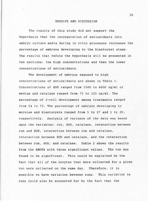
RESULTS AND DISCUSSION
The results of this study did not support the
hypothesis that the incorporation of antioxidants into
e mbryo culture media during in vitro processes increases the
percentage of embryos developing to the blastocyst stage .
The res u lts that refute the hypothesis will be presented in
two sections: the high concentrations and then the lower
concentrations of antioxidants.
The development of embryos exposed to high
concentrations of antioxidants are shown in Table 1 .
Concentrations of SOD ranged from 1500 to 6000 ug/ml of
medium and catalase ranged from 75 to 125 ug/ml . The
percentage of 2-cell development among treatments ranged
from 54 to 73 . The percentage of embryos developing to
morulae and blastocysts ranged from 5 to 27 and 1 to 20,
respectively. Analysis of variance of the data was based
upon the variables : run, SOD, catalase, interaction between
run and SOD, interaction between run and catalase,
interaction between SOD and catalase, and the interaction
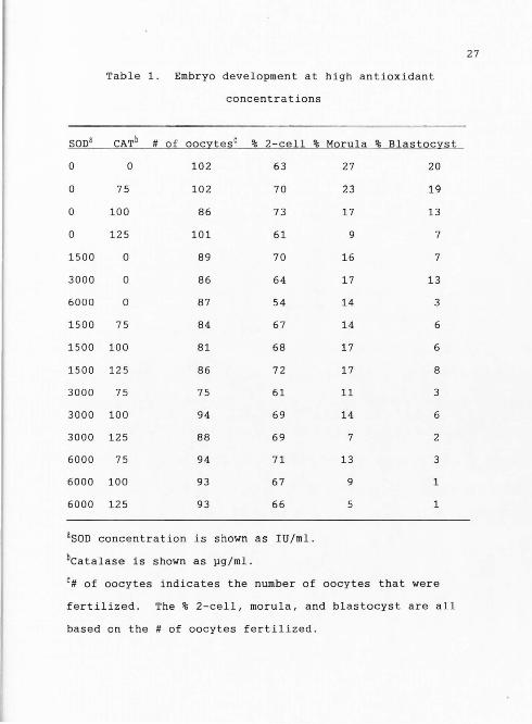
between run, SOD, and catalase. Table 2 shows the results
from the ANOVA with three significant values . The run was
found to be significant. This could be explained by the
fact that all of the oocytes that were collected for a given
run we re collected on the same day. Therefore, it is
possible to have variation between runs. This variation in
runs could also be accounted for by the fact that the
27
Table 1. Embryo development at high antioxidant
concentrations
SODa CATb # of oocytesc % 2-cell % Morula % Blastocyst
0 0 102 63 27 20
0 75 102 70 23 19
0 100 86 73 17 13
0 125 101 61 9 7
1500 0 89 70 16 7
3000 0 86 64 17 13
6000 0 87 54 14 3
1500 75 84 67 14 6
1500 100 81 68 17 6
1500 125 86 72 17 8
3000 75 75 61 11 3
3000 100 94 69 14 6
3000 125 88 69 7 2
6000 75 94 71 13 3
6000 100 93 67 9 1
6000 125 93 66 1
aSOD concentration is shown as IU/ml.
bcatalase is shown as pg/ml.
c# of oocytes indicates the number of oocytes that were
fertilized. The % 2-cell, morula, and blastocyst are all
based on the # of oocytes fertilized.
Source
Run
Table 2. ANOVA table on the results of embryos
cultured in high antioxidant concentrations
DF MS F Value
1 104.82 31.20**
28
SOD 3 16 . 67 4.96**
Run & SOD 3 8.25 2 . 46
Cat 3 3.76 1.12
Run & Cat 3 16.93 5.04**
Cat & SOD 9 4 . 65 1. 39
Run & Cat & SOD 9 4.16 1. 24
** Effect significant at r < 0.01
Run*Cat interaction was also significant . Superoxide
dismutase in the culture media had a negative effect . As
the level of SOD increased, fewer embryos developed to the
blastocyst stage .
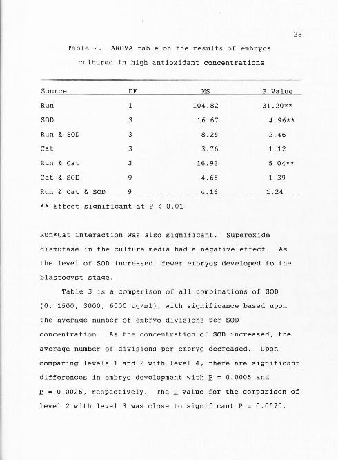
Table 3 is a comparison of all combinations of SOD
(0, 1500, 3000, 6000 ug/ml), with significance based upon
the average number of embryo divisions per SOD
concentration. As the concentration of SOD increased, the
average number of divisions per embryo decreased. Upon
comparing levels 1 and 2 with level 4, there are significant
differences in embryo development with r = 0.0005 and
!: = 0.0026, respectively. The £-value for the comparison of
level 2 with level 3 was close to significant r = 0.0570.
29
Table 3. Comparison of all combinations of the four levels
testing significance and the average number of embryo
divisions based on SOD concentration
SOD Concentration (IU/mll Division
0 2. 086a
1500 2 . 042 3
3000 1 . 8263b
6000 1 . 624b
a,b means with different superscripts differ significantly
~ < 0.01
#
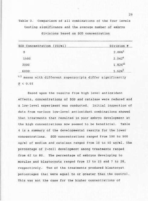
Based upon the results from high level antioxidant
effects, concentrations of SOD and catalase were reduced and
a low-level experiment was conducted. Initial inspection of
data from various low-level antioxidant combinations showed
that treatments that resulted in poor embryo development at
the high concentrations now seemed to be beneficial . Table
4 is a summary of the developmental results for the lower
concentrations. SOD concentrations ranged from 100 to 500
ug/ml of medium and catalase ranged from 10 to 50 ug/ml . The
percentage of 2-cell development among treatments ranged
from 62 to 80. The percentage of embryos developing to
morulae and blastocysts ranged from 12 to 23 and 7 to 28,
respectively. Ten of the treatments produced blastocyst
percentages that were equal to or greater than the control.
This was not the case for the higher concentrations of

Table 4 . Embryo development at reduced antioxidant
concentrations
30
SODa CATb # of Oocytesc % 2-Cell % Morula % Blastocyst
0 0
0 10
0 25
0 50
100 0
250 0
500 0
100 10
100 25
100 50
250 10
250 25
250 50
500 10
500 25
500 50
76
82
82
73
74
71
77
71
72
78
75
70
72
76
70
68
67
74
74
67
62
68
71
68
69
73
72
80
68
59
61
68
ason concentration is shown as IU/ml.
bcatalase is shown as pg/ml.
16
17
12
14
20
17
23
21
18
23
12
20
13
16
9
19
17
22
7
15
23
21
19
28
18
26
12
26
15
21
7
24
c# of oocytes indicates the number of oocytes that were
fertilized . The % 2-cell, morula, and blastocyst are all
based on the # of oocytes fertilized.

31
antioxidants. Even with what appeared to be greater embryo
development within some of the lower level antioxidant
treatments, there were no significant differences based on
ANOVA .
Table is the ANOVA table and ~-values. Only the run
variable was significant. Run had a ~-value of 0.0001, which
is highly significant. The other significant value was the
interaction between run, SOD, and catalase(~= 0 . 0107).
The significance of these two values is explained by
variation in the three groups of oocytes which were used in
the study.
Table 5. ANOVA table on the results of embryos cultured
in low antioxidant concentrations
Source OF MS F Value
Run 2 39.92 9.83**
SOD 3 2 . 72 0.67
Run & SOD 6 3.39 0 . 83
Cat 3 1. 67 0.41
Run & Cat 6 3 . 14 0. 77
Cat & SOD 9 4 . 96 1.22
Run & Cat & SOD 18 7.86 1.94*
* Effect significant at ~ < 0 . 05
** Effect significant at ~ < 0.01
32
In both high and low antioxidant concentrations the run
was significant. The variation is primarily due to the
variability in oocyte quality between collection periods,
which is a common problem in trying to maintain quality
control for in vitro fertilization systems . Variability in
oocyte collections is difficult to control since ovaries are
coming from slaughter animals whose nutrition , hormonal, and
reproductive status is unknown. Furthermore, cows coming
into the slaughterhouse have oftentimes been on exogenous
hormones. The only way to alleviate some of the variation is
to routinely followed a rigid protocol for collecting and
handling the ovaries. Therefore, variation between
collection periods, which has shown up as a significant
difference in this study, is most likely a composite cow
effect within the group slaughter on a given day .
The results of this study do not corroborate the
studies conducted in the mouse (Ericksson and Borg, 1991;
Nonogaki et al ., 1991 ; Goto et al., 1992; Nonogaki et al.,
1992; Umaoka et al. ,1992; Natsuyama et al . , 1993a, 1993b;
Chun et al., 1994) and the rabbit (Li et al., 1993; Lindenau
and Fischer, 1994). Other work on the effects of
antioxidants in the bovine have shown similar results to
what we found in this study (Luvoni et al., 1994). George
Seidel, Jr. (1995-personal communication) at Colorado State
University indicated that his research group tested the
effects of antioxidants in bovine embryo culture media and
observed no beneficial effects .
33
The types of media used for embryo culture often
account for differences in embryo development among species .
In this study, bovine embryos were cultured in CR2, which is
a simple-defined medium. Luvoni et al. (1994) cultured
embryos in a complex medium that was necessary for co
culture and reported beneficial effects in supplementing
media with antioxidants . In this case the beneficial
effects experienced by the use of antioxidants could be
associated with free radical production by the monolayers on
which the embryos were growing. If somatic cell monolayers
are responsible for radical production, then antioxidants
would be an important factor in their removal.
It is also possible that different media trap light
differently. Therefore, more free radicals would be
produced in light-trapping media . An example is phenol red
trapping light in culture media . The CR2 medium used in
this study contained no phenol red. If in the previously
cited studies phenol red was used in the media, then a
greater amount of fluorescent light would have been trapped.
Antioxidants would therefore have had a positive effect on
embryo development by tying up free radicals. This could
possibly explain why the treatments in those studies out
perform the controls.
Another possibility for the differences between the
34
results of this study as compared to the other cited reports
may be the difference in the culture systems in which the
embryos were grown . Noda et al. (1991) and Li et al. (1993)
cultured in 5% co2 in humidified air. In this study embryos
were cultured in a humidified atmosphere of 5% co2, 5% o2,
and 90% N2. Higher o2 concentrations can cause an increased
production of free radicals, which negatively affect
embryonic development. Antioxidants do not freely cross
embryo membranes without some type of carrier. Therefore,
without incorporation of a carrier with the antioxidants,
the positive effects seen by using antioxidants in embryo
culture are probably due to the elimination of radicals
produced by excessive oxygen concentration or exposure to
fluorescent light. If caution is given to both oxygen
concentration and exposure to light, I do not believe that
it is necessary to add antioxidants.
Another possibility may be that in this study, high
levels of SOD in the culture had a detrimental effect on
embryo development. However, Luvoni et al. (1994) reported
some beneficial effects using SOD in a bovine embryo culture
system. The difference between this study and the study of
Luvoni et al. (1994) is the difference in the embryo culture
environment. In this study extreme effort was made to
reduce the exposure of embryos to factors shown to cause
free radical development. Superoxide dismutase and catalase
were not necessary to scavenger free radicals and therefore
35
did not have a positive effect. One study showed that
addition of exogenous SOD caused variable results (Johnson
and Nasr-Esfahani, 1994). Those researchers further stated
that it is still unclear how exogenous SOD could remove
intracellular superoxides that do not readily cross
membranes. Free radicals are, however, important for
electron transport. One possible explanation to the
negative response in this study may have been the low levels
of free radicals present during embryo culture. The
increased concentrations of antioxidants in the high-level
treatment groups could have removed the beneficial effects
of free radicals. High levels of antioxidants may have
interfered with the production of free radicals, which in
turn could limit the production of ATP. Without sufficient
amounts of ATP, essential life processes would be altered
and the embryo would then die.
Finally, some have shown that viable embryos produce
antioxidants. It is then conceivable to hypothesize that if
caution was given to factors causing radical formation, that
the embryos could be more viable. If they were more viable,
they were probably producing antioxidants and the additional
antioxidants may have had a toxic or negative effect on
development.
Whether antioxidants have a positive or negative effect
will depend on the embryo culture system used. High
concentrations of o2 or light-trapping substances in the

36
media may require the use of antioxidants to improve embryo
development . On the contrary , if 0 2 tension is lowered and
light-trapping compounds such as phenol red are removed from
the media , then adding antioxidants may have a negative
effect on embryo growth and development .
37
CONCLUSION
Even though previous studies showed that there were
beneficial effects to the use of antioxidants in embryo
production, my results do not corroborate this in the bovine
model. I believe that free radicals play a role in
developmental obstruction of embryos . I am also confident
that certain types of media, laboratory procedures, and
culture systems are conducive to free radical production.
Our system of IVF embryo production pays special attention
to the presence of phenol red in the media, exposure of
embryos to direct fluorescent light, and culturing of
embryos in reduced 0 2 concentration. Each of these factors
has been cited previously as a factor contributing to
radical production . It is my belief that if the above
mentioned factors are not closely monitored, free radicals
may be produced in concentrations that could depress embryo
development. By controlling the factors that cause radical
production, it is possible to successfully culture embryos
without antioxidants. However, if those factors are not
controlled, antioxidants may need to be added in order to
achieve desired embryo development. Therefore, at this
time, the use of SOD and catalase in maturation,
fertilization, and culture will not be incorporated into our
laboratory standard operating procedures.
In the future, I hope that studies will continue to
be done in order to elucidate the role of free oxygen
38
radicals in embryo production. Understanding the positive
role (if one exists) of free radicals could perhaps shed
some light on controlling the concentration of radicals that
embryos are exposed to . Then, free radicals could be
allowed in culture at concentrations necessary to carry out
cellular processes, and any concentrations above that could
be removed by antioxidants before cellular damage occurs .
39
REFERENCES
Asayama, K., N.W. Kody, and I.M. Burr. 1986 . Effect of vitamin E deficiency and selenium deficiency on insulin secretory reserve and free radical scavenging systems in islets decreased of islet manganosuperoxide dismutase. J. Lab. Clin. Med . 107:459.
Baker, M.A., and S.Q. He . 1991. Elaboration of cellular DNA breaks by hydroperoxides. Free Radic. Biol. Med. 1(6):563 .
Bavister, B. D., and R. Yanagimachi. 1977. The effects of sperm extracts and energy sources on the motility and acrosome reaction of hamster spermatozoa in vitro. Biol. Reprod. 16 : 228 .
Bendich, A. 1993. Physiological role of antioxidants in the immune system. J . Dairy Sci . 76(9):2789.
Blum , H.F. 1964 . Photodynamic Action and Disease Caused by Light. Hafner, New York.
Cadet, J.L . , and L.A. Kahler. 1994. Free radical mechanisms in schizophrenia and tardive dyskinesia. Neurosci . Biobehav . Rev. 18(4) :457.
Chun, Y.S., J.H. Kim, H.T. Lee, and K. S . Chung. 1994 . Effect of superoxide dismutase on the development of preimplantation mouse embryos. Theriogenology 41(2): 511.
Dianzani, M.U. 1992 . Free radicals in physiology and pathology . Boll. Soc . Ital. Biol. Sper. 68(8-9):491.
Dumitrescu, C . 1992. The role of free radicals in normal and pathologic cell proliferation . Rom. J . Endocrinol . 30(1-2):45.
Ellis, L.C . 1990a. Free radicals in tissue culture : Part I, What are free radicals . Art Sci. 9(2):1.
Ellis, L.C. 1990b . Free radicals in tissue culture: Part III . Antioxidant systems and antioxidants . Art Sci. 9 ( 4) : 1.
Ellis, L.C. 1991. Free radicals in tissue culture: Part IV . Effects on cells in culture . Art Sci. 10(1) : 1.
40
Ericksson, U. J., and L . A. Borg. 1991. Protection by free oxygen radical scavenging enzymes against glucoseinduced embryonic malformations in vitro . Diabetologia. 34(5):325.
Fee, J.A., and J.S. Valentine . 1977. Chemical and physical properties of superoxide. In : A.M. Michelson, J.M. McCord, and I. Fridovich (Ed.) Superoxide and Superoxide Dismutase. p . 78. Academic Press, New York.
Ferrari, R., C. Ceconi, S. Curello, A. Cargnoni, 0. Alfieri, A. Pardini, P. Marzella, and 0. Visioli. 1991. Oxygen free radicals and myocardial damage: Protective role of thiol-containing agents. Am. J. Med. 91(3C):95S.
Fisher, B . 1987 . Developmental retardation in cultured preimplantation rabbit embryos . J. Reprod. Fertil . 79:115.
Gao, W., H.D . Connor, J.J. Lemasters, R.P. Mason, and R.G. Thurman. 1995. Primary nonfunction of fatty livers produced by alcohol is associated with a new antioxidant-insensitive free radical species. Transplant. 59(5) : 674.
Gardner, D. K., M. Lane, A. Spitzer, and P . A. Batt . 1994. Enhanced rates of cleavage and development for sheep zygotes cultured to the blastocyst stage in the absence of serum and somatic cells: Amino acids, vitamins, and culturing embryos in groups stimulate development. Biol. Reprod. 50(2):390.
Gille, J.J . P., and H. Joenje . 1991. Biological significance of oxygen toxicity : an introduction. In : C. VigoPelfrey (Ed . ) Membrane Lipid Oxidation. p. 1. CRC Press, Boca Raton, FL.
Goto, Y., Y. Noda, K. Narimoto, Y. Umaoka, and T. Mori. 1992. Oxidative stress on mouse embryo development in vitro. Free Radic. Biol. Med . 13(1):47.
Gutteridge, J.M.C., and B. Halliwell. 1988. The antioxidant proteins of extracellular fluids. In: C.K. Chow (Ed.) Cellular Antioxidant Defense Mechanisms, Vol. II . p. 1. CRC Press, Boca Raton, FL.
Halliwell, B., and J . M.C . Gutteridge . 1989 . Free Radicals in Biology and Medicine, 2nd ed. New York: Oxford University Press, New York .
Harvey, M.B . , M. Y. Arcellana-Panlilio, X. Zhang, G.A . Schultz, and A. J. Watson. 1995. Expression of genes encoding antioxidant enzymes in preimplantation mouse and cow embryos and primary oviduct cultures employed for embryo coculture . Biol. Reprod . 53:532.
41
Hooper, C. 1989 . Free Radicals: Research on biochemical bad boys comes of age . J . NIH Res. 1:181 .
Johnson, M.H., and M.H. Nasr-Esfahano. 1994. Radical solutions and culture problems: Could free oxygen radicals be responsible for the impaired development of preimplantation mammalian embryos in vitro? Bioessays 16 :31.
Katz, A., K. T . Oldham, K.S. Guice, and A.G . Coran . 1995. Oxidized glutathione as a marker of ischemia reperfusion associated with single lung transplantation. J. Am. Coll. Surg . 180(1):25.
Kaul, N. , N. Siveski-Iliskovic, M. Hill, J. Slezak, and P . K. Singal. 1993 . Free radicals and the heart . J . Pharmacal. Toxicol . Methods . 30(2):55.
Kehrer, J.P . 1993. Free radicals as mediators of tissue injury and disease. Crit. Rev. Toxicol . 23(1):21 .
Keith, F. 1993. Oxygen free radicals in cardiac transplantion. J. Card. Surg . 8(2 supplement):245 .
Kjellmer, I. 1991. Mechanisms of perinatal brain damage. Ann. Med. 23(6):675.
Kopecny, V., and H. Niemann. 1993 . Formation of nuclear microarchitecture in_the preimplantation bovine embryo at the onset of transcription : Implications for biotechnology. Theriogenology 39(1):109.
Legge, M., and M.H. Sellens. 1991. Free radical scavengers ameliorate the 2-cell block in mouse embryo culture . Hum. Reprod. 6(6) : 867.
Li, J., R.H. Foote, and M. Simkin. 1993. Development of rabbit zygotes cultured in protein-free medium with catalase, taurine, or superoxide dismutase. Biol. Reprod. 49(1):33.
Lindenau, A . , and B. Fischer. 1994 . Effect of oxygen concentration in the incubator' s gas phase on the development of cultured preimplantation rabbit embryos. Theriogenology 41(4):889.

42
Luvoni, G.C., L. Keskintepe, J. Rzucidlo, A. Kenimer, and B. G. Brackett. 1994 . Superoxide dismutase (SOD) nd glutathione (GSH) affect fertilization and development of bovine oocytes. Biol. Reprod. 50(Supplement 1) : 189.
Marklund, S.L. 1985. Oxygen toxicity and protective systems . Clin . Toxicol. 23(4-6):289.
Michel, C . , F. Vincent, C . Duval, M.C. Poelman, and M. Adolphe. 1992. Toxic effects and detection of oxygen free radicals on cultured articular chondrocytes generated by menadione . Free Radic. Res. Commun . 17(4) : 279.
Murrell, T.G . 1991. Epidemiology and biochemical support for a theory on the cause and prevention of breast cancer. Med. Hypotheses . 36(4) : 389 .
Nakayama, T., Y. Noda, Y. Goto, and T. Mori. 1994 . Effects of visible light and other environmental factors on the production of oxygen radicals by hamster embryos. Theriogenology 41 : 499.
Natsuyama, S. , Y. Noda, K. Narimoto, and T.Mori. 1993a . Role of protein supplements in the culture of mouse embryos. Theriogenology. 40(1) : 149.
Natsuyama, S . , Y. Noda, M. Yamashita, Y. Nagahama, and T. Mori. 1993b. Superoxide dismutase and thioredoxin restore defective p34cdc2 kinase activation in mouse two- c ell block . Biochim. Biophy . Acta . 1176(1-2):90.
Noda, Y., H. Matsumoto, Y. Umaoka, K. Tatsumi, J. Kishi, and T. Mori. 1991 . Involvement of superoxide radicals in the mouse two-cell block. Mol . Biol . Reprod . 28(4)356.
Nonogaki T., Y. Noda , K. Narimoto, Y. Umaoka, and T. Mori. 1991 . Protection from oxidative stress by thioredoxin and superoxide dismutase of mouse embryos fertilized in vitro. Hum. Reprod . 6(9):1305.
Nonogaki, T., Y. Noda, K.Narimoto, Y.Umaoka, and T. Mori. 1992 . Effects of superoxide dismutase on mouse in vitro fertilization and embryo culture system. J. Assist. Reprod . Genet. 9(3):274.
Pacifici, R.E . , and K.J . Davies. 1991. Protein , lipid and DNA repair Systems in oxidative stress: The freeradical theory of aging revisited. Gerontology 37(1-3):166.

Paller, M.S . , and H.S. Jacob. 1994. Cytochrome P-450 mediates tissue-damaging hydroxly rad i cal formation during reoxygenation of the kidney . Proc. Nat. Acad. Sci. USA . 91(15) : 7002.
43
Parrish, J.J., J.L. Susko-Parrish, M.L . Leibfried-Rutledge, E.S. Critser, W.H . Eyestone, and N.L. First . 1986 . Bovine in vitro fertilization with frozen-thawed semen . Theriogenology 25(4):591.
Parshad, R . , K.K . Sanford, G.M. Jones, F.M. Price, and W. G. Taylor . 1977 . Oxygen and light effects on chromosomal aberrations in mouse cells in vitro . Exp. Cell. Res. 104 : 199 .
Reiter, R. J. 1993. Interactions of pineal hormone melatonin with oxygen-centered free radicals : A brief review. Braz. J. Med . Biol. Res. 26(11):1141.
Rosenkrans, C.F . , Jr., and N.L. First. 1991 . Culture of bovine zygotes to the blastocyst stage: Effects of amino acids and vitamins. Theriogenology 35:266.
Slater , T.F. 1972 . Free Radical Mechanisms in Tissue Injury . Pion Limited, London, England .
Sugiyama, M. , X. Lin, and M. Costa. 1991 . Protective effect of vitamin E against chromosomal aberrations and mutation induced by sodium chromate in chinese hamster V79 cells . Mut. Res. 260:19.
Umaoka, Y . , Y. Noda, K. Narimoto, and T . Mori. 199 2. Effect s of oxygen toxicity on early development of mouse embryos . Mol. Reprod. Dev. 31(1) :2 8 .
Vincent, F., M. Corral, N. Defer, and M. Adolphe . 1991 . Effects of oxygen free radicals on articular chondrocytes in culture : c-myc and c-Ha-ras messenger RNAs and proliferation kinetics. Exp . Cell. Res. 192(2):333.
Warren, J . S., K.J. Johnson, and P . A . Ward. 1987. Oxygen radicals in cell injury and cell death. Pathol. Immunopathol. Res. 6:301.
Wright, R.J. Jr., and K.R . Bondioli. 1981 . Aspects of in vitro fertilization and embryo culture in domestic animals . J . Anim. Sci. 53:702.

44
Yuan, H., and Z. Zhang. 1992. Some factors affecting sisterchromatid differentiation (SCD) and sister-chromatid exchange (SCE) in hordeum vulgare. Mut. Res . 272(2):125.
Zieger, M.A., D.J. Glofcheski, J.R. Lepock, and J. Kruuv. 1991. Factors influencing survival of mammalian cells exposed to hyperthermia : Effects of hepes, free radicals, and H202 under light and dark conditions. Cryobiology 28(1):8.
Zimmerman, W.F., and S. Keys. 1991. Effects of the antioxidants dithiothreitol and vitamin E on phospholipid metabolism in isolated rod outer segments . Exp. Eye Res. 52(5):607.