Different Transcriptional Response toXanthomonas citri subsp.citribetween Kumquat and Sweet Orange...
17
Different Transcriptional Response to Xanthomonas citri subsp. citri between Kumquat and Sweet Orange with Contrasting Canker Tolerance Xing-Zheng Fu 1,2 , Xiao-Qing Gong 1 , Yue-Xin Zhang 1 , Yin Wang 1 , Ji-Hong Liu 1 * 1 Key Laboratory of Horticultural Plant Biology (MOE), National Key Laboratory of Crop Genetic Improvement, College of Horticulture and Forestry Science, Huazhong Agricultural University, Wuhan, China, 2 Citrus Research Institute, Chinese Academy of Agricultural Sciences, Chongqing, China Abstract Citrus canker disease caused by Xanthomonas citri subsp. citri (Xcc) is one of the most devastating biotic stresses affecting the citrus industry. Meiwa kumquat (Fortunella crassifolia) is canker-resistant, while Newhall navel orange (Citrus sinensis Osbeck) is canker-sensitive. To understand the molecular mechanisms underlying the differences in responses to Xcc, transcriptomic profiles of these two genotypes following Xcc attack were compared by using the Affymetrix citrus genome GeneChip. A total of 794 and 1324 differentially expressed genes (DEGs) were identified as canker-responsive genes in Meiwa and Newhall, respectively. Of these, 230 genes were expressed in common between both genotypes, while 564 and 1094 genes were only significantly expressed in either Meiwa or Newhall. Gene ontology (GO) annotation and Singular Enrichment Analysis (SEA) of the DEGs showed that genes related to the cell wall and polysaccharide metabolism were induced for basic defense in both Meiwa and Newhall, such as chitinase, glucanase and thaumatin-like protein. Moreover, apart from inducing basic defense, Meiwa showed specially upregulated expression of several genes involved in the response to biotic stimulus, defense response, and cation binding as comparing with Newhall. And in Newhall, abundant photosynthesis-related genes were significantly down-regulated, which may be in order to ensure the basic defense. This study revealed different molecular responses to canker disease in Meiwa and Newhall, affording insight into the response to canker and providing valuable information for the identification of potential genes for engineering canker tolerance in the future. Citation: Fu X-Z, Gong X-Q, Zhang Y-X, Wang Y, Liu J-H (2012) Different Transcriptional Response to Xanthomonas citri subsp. citri between Kumquat and Sweet Orange with Contrasting Canker Tolerance. PLoS ONE 7(7): e41790. doi:10.1371/journal.pone.0041790 Editor: Randall P. Niedz, United States Department of Agriculture, United States of America Received May 4, 2012; Accepted June 25, 2012; Published July 26, 2012 Copyright: ß 2012 Fu et al. This is an open-access article distributed under the terms of the Creative Commons Attribution License, which permits unrestricted use, distribution, and reproduction in any medium, provided the original author and source are credited. Funding: This work is financially supported by National Science Foundation of China (30921002), Fok Ying Tong Education Foundation (114034), Special Fund for Agro-scientific Research in the Public Interest (201003067), the National High Technology Research and Development Program (863 Program) of China (2011AA100205), and Hubei Provincial Natural Science Foundation (2009CDA080). The funders had no role in study design, data collection and analysis, decision to publish, or preparation of the manuscript. Competing Interests: The authors have declared that no competing interests exist. * E-mail: [email protected] Introduction During the last decade, tremendous advancements have been achieved in the citrus industry throughout the world. However, the citrus industry worldwide suffers from an array of threats from biotic or abiotic stresses. Citrus canker caused by Xanthomonas citri subsp. citri (Xcc) is a devastating disease that has caused substantial losses in citrus-growing countries in the past decades. The canker symptoms include raised lesions on the surface of leaves, stems and fruits, with oily, water-soaked and pustule-like edges surrounded by chlorotic haloes [1,2]. At present, the strategy for canker disease management relies on an integrated system encompassing both compatible cultural practices and phytosanitary measures, such as the eradication of inoculum sources and the application of copper- containing bactericides or antibiotics [2]. Nevertheless, since there are certain limitations associated with both cultural practices and chemical control, the issue has not been completely addressed. For example, application of copper bactericides not only increases management costs, but also raises concerns regarding environ- mental contamination and food safety. Furthermore, evolution of bacterial genomes over time has led to copper resistance [3–5]. Therefore, identification of effective compounds that can replace or supplement copper-containing chemicals is necessary. In the long run, selection or breeding of resistant cultivars may be the best solution for combating Xcc challenge in regions in which Xcc is endemic. As a fundamental step toward making these approaches possible, it is necessary to elucidate the molecular responses to Xcc invasion in the host plant. During their long evolutionary process, plants have evolved a multitude of cellular, molecular, physiological, and biochemical alterations in order to adapt to or survive under adverse conditions, including biotic stresses caused by pathogens like Xcc. Of these alterations, molecular response at the transcriptional level has been demonstrated to be crucial for establishing a set of defense mechanisms against invading pathogens. Accumulating evidence has shown that expression of a large spectrum of genes is induced on exposure to microbial invasion in various plants [6]. The products of these genes might function to directly protect the host plant from damage caused by pathogens, or act as regulatory molecules by perceiving stress signals and transmitting them to downstream targets. These genes constitute a delicate network that plays key roles in combating pathogens. The biotic stress-induced PLOS ONE | www.plosone.org 1 July 2012 | Volume 7 | Issue 7 | e41790
Transcript of Different Transcriptional Response toXanthomonas citri subsp.citribetween Kumquat and Sweet Orange...

Different Transcriptional Response to Xanthomonas citrisubsp. citri between Kumquat and Sweet Orange withContrasting Canker ToleranceXing-Zheng Fu1,2, Xiao-Qing Gong1, Yue-Xin Zhang1, Yin Wang1, Ji-Hong Liu1*
1 Key Laboratory of Horticultural Plant Biology (MOE), National Key Laboratory of Crop Genetic Improvement, College of Horticulture and Forestry Science, Huazhong
Agricultural University, Wuhan, China, 2 Citrus Research Institute, Chinese Academy of Agricultural Sciences, Chongqing, China
Abstract
Citrus canker disease caused by Xanthomonas citri subsp. citri (Xcc) is one of the most devastating biotic stresses affectingthe citrus industry. Meiwa kumquat (Fortunella crassifolia) is canker-resistant, while Newhall navel orange (Citrus sinensisOsbeck) is canker-sensitive. To understand the molecular mechanisms underlying the differences in responses to Xcc,transcriptomic profiles of these two genotypes following Xcc attack were compared by using the Affymetrix citrus genomeGeneChip. A total of 794 and 1324 differentially expressed genes (DEGs) were identified as canker-responsive genes inMeiwa and Newhall, respectively. Of these, 230 genes were expressed in common between both genotypes, while 564 and1094 genes were only significantly expressed in either Meiwa or Newhall. Gene ontology (GO) annotation and SingularEnrichment Analysis (SEA) of the DEGs showed that genes related to the cell wall and polysaccharide metabolism wereinduced for basic defense in both Meiwa and Newhall, such as chitinase, glucanase and thaumatin-like protein. Moreover,apart from inducing basic defense, Meiwa showed specially upregulated expression of several genes involved in theresponse to biotic stimulus, defense response, and cation binding as comparing with Newhall. And in Newhall, abundantphotosynthesis-related genes were significantly down-regulated, which may be in order to ensure the basic defense. Thisstudy revealed different molecular responses to canker disease in Meiwa and Newhall, affording insight into the response tocanker and providing valuable information for the identification of potential genes for engineering canker tolerance in thefuture.
Citation: Fu X-Z, Gong X-Q, Zhang Y-X, Wang Y, Liu J-H (2012) Different Transcriptional Response to Xanthomonas citri subsp. citri between Kumquat and SweetOrange with Contrasting Canker Tolerance. PLoS ONE 7(7): e41790. doi:10.1371/journal.pone.0041790
Editor: Randall P. Niedz, United States Department of Agriculture, United States of America
Received May 4, 2012; Accepted June 25, 2012; Published July 26, 2012
Copyright: � 2012 Fu et al. This is an open-access article distributed under the terms of the Creative Commons Attribution License, which permits unrestricteduse, distribution, and reproduction in any medium, provided the original author and source are credited.
Funding: This work is financially supported by National Science Foundation of China (30921002), Fok Ying Tong Education Foundation (114034), Special Fund forAgro-scientific Research in the Public Interest (201003067), the National High Technology Research and Development Program (863 Program) of China(2011AA100205), and Hubei Provincial Natural Science Foundation (2009CDA080). The funders had no role in study design, data collection and analysis, decisionto publish, or preparation of the manuscript.
Competing Interests: The authors have declared that no competing interests exist.
* E-mail: [email protected]
Introduction
During the last decade, tremendous advancements have been
achieved in the citrus industry throughout the world. However, the
citrus industry worldwide suffers from an array of threats from
biotic or abiotic stresses. Citrus canker caused by Xanthomonas citri
subsp. citri (Xcc) is a devastating disease that has caused substantial
losses in citrus-growing countries in the past decades. The canker
symptoms include raised lesions on the surface of leaves, stems and
fruits, with oily, water-soaked and pustule-like edges surrounded
by chlorotic haloes [1,2]. At present, the strategy for canker disease
management relies on an integrated system encompassing both
compatible cultural practices and phytosanitary measures, such as
the eradication of inoculum sources and the application of copper-
containing bactericides or antibiotics [2]. Nevertheless, since there
are certain limitations associated with both cultural practices and
chemical control, the issue has not been completely addressed. For
example, application of copper bactericides not only increases
management costs, but also raises concerns regarding environ-
mental contamination and food safety. Furthermore, evolution of
bacterial genomes over time has led to copper resistance [3–5].
Therefore, identification of effective compounds that can replace
or supplement copper-containing chemicals is necessary. In the
long run, selection or breeding of resistant cultivars may be the
best solution for combating Xcc challenge in regions in which Xcc
is endemic. As a fundamental step toward making these
approaches possible, it is necessary to elucidate the molecular
responses to Xcc invasion in the host plant.
During their long evolutionary process, plants have evolved a
multitude of cellular, molecular, physiological, and biochemical
alterations in order to adapt to or survive under adverse
conditions, including biotic stresses caused by pathogens like
Xcc. Of these alterations, molecular response at the transcriptional
level has been demonstrated to be crucial for establishing a set of
defense mechanisms against invading pathogens. Accumulating
evidence has shown that expression of a large spectrum of genes is
induced on exposure to microbial invasion in various plants [6].
The products of these genes might function to directly protect the
host plant from damage caused by pathogens, or act as regulatory
molecules by perceiving stress signals and transmitting them to
downstream targets. These genes constitute a delicate network that
plays key roles in combating pathogens. The biotic stress-induced
PLOS ONE | www.plosone.org 1 July 2012 | Volume 7 | Issue 7 | e41790

expression of a large number of genes suggests that the nature of
the biotic stress response might be more complex than expected;
this is one of the reasons for the difficulty in developing a clear-cut
network for the biotic stress response. As a result, although myriad
molecular components responsive to pathogenic attack have been
identified in a wide range of plants, the highly complex and
interconnected network per se is still far from being fully
understood. Moreover, it is worth mentioning that molecular
responses may vary from plant to plant, although some parts of the
responses may be common. It is therefore important to identify
transcriptional changes in a given plant species under pathogenic
stress in order to unravel the molecular elements that are specific
to the plant itself.
Previously, researchers preferred to isolate and functionally
analyze individual genes involved in the stress response. However,
this is a piecemeal strategy and contributes little to a comprehen-
sive understanding of the defense-related transcriptome that is
controlled by quantitative mechanisms [7]. The advent of
emerging research platforms like expression sequence tag (EST)
databases, genome sequencing, and microarrays offers a good
opportunity to expedite our efforts towards a better understanding
of the molecular mechanisms underlying the biotic stress response.
Of note, the recent availability of commercial cDNA chips
provides a high-throughput approach to exploit a multitude of
genes associated with many physiological processes. Transcrip-
tomic profiling of gene expression using microarrays has been
carried out in many plants under biotic stress, including Arabidopsis
[6,8], birch [9], sunflower [10], poplar [11], citrus [12], rice
[13,14], grape [15], and cotton [16]. Such analytical tools may
reveal global gene expression changes, facilitate the elucidation of
the defense response at the molecular level, and provide a
significant amount of knowledge regarding potential mechanisms
responsible for disease resistance, which will underpin the
rationale for developing resistant germplasms via genetic engi-
neering.
In order to isolate genes that are potentially related to canker
resistance, Deng et al. [17] obtained 2 BAC clones containing all
the features of the rice Xa21 protein, which represents a unique
class of plant disease-resistance genes (R). Recently, Cernadas et al.
[12] investigated the early molecular events (occurring at 4 and
48 h after inoculation) leading to canker development in sweet
orange by analyzing changes in transcript levels using differential
display, suppressed subtractive hybridization, and microarrays.
Subsequently, Cernadas and Benedetti [18] assessed the expres-
sion patterns of cell-wall remodeling genes following Xcc infection.
However, no information is yet available on a comparative
transcriptome analysis between genotypes with contrasting toler-
ance levels to citrus canker disease. In spite of the conserved
protective mechanisms among plants, resistant and susceptible
genotypes may vary in their response to pathogen infection.
Therefore, insightful investigation using a pair of genotypes with
contrasting disease resistance phenotypes will help us better
understand the molecular mechanisms underlying disease toler-
ance. Citrus canker has a fairly broad host range in Rutaceae and
can affect many important citrus species and varieties despite the
differences in the field resistance of these varieties [19]. It has been
well documented that Meiwa kumquat (Fortunella crassifolia) is
immune to Xcc, whereas Newhall navel orange (Citrus sinensis
Osbeck) is one of the highly susceptible commercial varieties
[2,20]. In the current study, we applied the Affymetrix citrus
genome GeneChip for a pairwise comparison of the gene
expression profiles of Meiwa and Newhall following Xcc
inoculation, in order to gain valuable insight into the mechanisms
underlying canker resistance in the former.
Results
Comparison of canker disease development in Meiwaand Newhall
To identify differences in the Xcc response between Meiwa and
Newhall under our experimental conditions, the leaves of both
species were pinprick-inoculated with the citrus canker bacterium
and cultured in a growth chamber. As shown in Figure 1A, tiny
and slightly raised lesions began to appear on the adaxial surface
of Newhall leaves at 5 days post-inoculation (DPI); such lesions
were not found in Meiwa. At 7 DPI, the symptoms became more
conspicuous in Newhall, with typical crateriform lesions surround-
ed by water-soaked margins, whereas only a few tiny blister-like
lesions were present in Meiwa. In addition, bacterial growth assay
demonstrates that the bacterial population in the leaves of Meiwa
was significantly smaller than in Newhall (Figure 1B). These results
demonstrate that Meiwa was remarkably less susceptible to Xcc
infection when compared with Newhall. In the current exper-
iment, the differences in canker symptoms were apparent at 5 DPI,
prior to the outbreak of the most serious symptoms. Therefore,
inoculated leaves collected at 0 and 5 DPI were subjected to
microarray analysis.
Global expression profiles of Meiwa and Newhall afterpinprick inoculation
To reveal differences in the response to Xcc challenge between
these 2 genotypes with contrasting disease tolerances and obtain
new insights into the molecular mechanisms underlying canker
tolerance, the transcriptomes of Meiwa and Newhall were
compared by analyzing their global gene expression profiles using
citrus genome Genechip. After statistical analysis, 794 and 1324
differentially expressed genes (DEGs) were identified as canker-
responsive genes in Meiwa and Newhall, respectively. Among
these genes, the expression of 530 genes was upregulated while
that of 264 was downregulated in Meiwa. On the other hand, the
expression of 610 genes was upregulated while that of 714 was
downregulated in Newhall (Figure 2A). In addition, 230 out of
these DEGs were identified in both Meiwa and Newhall, of which
150 showed upregulated expression while 80 showed downregu-
lation. Moreover, of the 564 genes that showed significant
expression only in Meiwa, 380 genes showed upregulated
expression while 184 were downregulated following Xcc infection.
In contrast, of the 1094 genes that showed significant expression
only in Newhall, 460 were upregulated and 634 showed
downregulation (Figure 2B–C). These data indicate that Meiwa
and Newhall display noticeable differences in the Xcc response at
the transcriptional level. Interestingly, the tolerant genotype,
Meiwa, had significantly fewer canker-responsive genes than the
canker-sensitive genotype Newhall.
All of the genes with significantly upregulated and downregu-
lated expression levels in Meiwa and Newhall were aligned against
the Arabidopsis database by using Citrus HarvEST software
(Version 1.25), and the detailed sequence description is shown in
Table S1 and Table S2. MapManBin functional annotation was
also performed using the best-matched Arabidopsis Genome
Initiative (AGI) number in the Plant Proteome Database (PPDB),
on the basis of which the DEGs were grouped into 33 categories
(Figure S1). With the exception of genes without any assignment,
the main categories in both Meiwa and Newhall were related to
the cell wall, secondary metabolism, hormone metabolism, stress
response, miscellaneous, RNA, protein, signaling, and transport.
Interestingly, expression of most of the stress-related genes was
upregulated in Meiwa, but downregulated in Newhall. In addition,
a total of 74 genes involved in photosynthesis showed significantly
Responses of Kumquat and Sweet Orange to Canker
PLOS ONE | www.plosone.org 2 July 2012 | Volume 7 | Issue 7 | e41790

downregulated expression in Newhall. On the contrary, only 6
genes showed downregulated expression in Meiwa (Table S1 and
S2).
Verification of microarray results by semi-quantitativereverse transcription polymerase chain reaction (RT-PCR)
In order to confirm the reliability of the microarray data,
expression patterns of 10 up-regulated genes and 2 down-regulated
genes were randomly selected and assessed by semi-quantitative
RT-PCR using gene-specific primers (Table S3). These selected
genes putatively encode protease inhibitor (Cit.8464.1.S1_s_at),
glucanase (Cit.30519.1.S1_s_at), phenylalanine-ammonia lyase
(Cit.9590.1.S1_at), pectin acetylesterase (Cit.9134.1.S1_s_at),
chitinase (Cit.302.1.S1_s_at), peroxidase (Cit.8519.1.S1_x_at,
Cit.8514.1.S1_x_at), protein-binding transcription regulator
(Cit.6280.1.S1_at), defense-related protein (Cit.36070.1.S1_at),
oxidoreductase (Cit.24087.1.S1_s_at), and unknown proteins
(Cit.3761.1.S1_x_at, Cit.17178.1.S1_x_at). As shown in Figure 3,
the results of RT-PCR were largely consistent with the microarray
data, albeit the relative expression levels (ratio of gene levels before
Figure 1. Comparison of canker disease development in ‘Meiwa’ and ‘Newhall’. (A) Leaves of ‘Meiwa’ and ‘Newhall’ were pinprick-inoculated with citrus canker bacterium and periodically observed within 7 d, and the pictures were taken at 5 and 7 days post inoculation (DPI). (B)Quantitative comparison of the bacterial population at the inoculation sites of ‘Meiwa’ and ‘Newhall’ leaves after 6 days inoculation.doi:10.1371/journal.pone.0041790.g001
Figure 2. Number of differentially expressed genes in ‘Meiwa’ and ‘Newhall’ after statistical analysis. (A) Number of significantlyupregulated and downregulated genes in ‘Meiwa’ and ‘Newhall’. (B–C) Venn diagram shows the number of upregulated (B) and downregulated (C)genes that are expressed in common or in special between ‘Meiwa’ and ‘Newhall’.doi:10.1371/journal.pone.0041790.g002
Responses of Kumquat and Sweet Orange to Canker
PLOS ONE | www.plosone.org 3 July 2012 | Volume 7 | Issue 7 | e41790

and after Xcc inoculation) seen with RT-PCR differed from those
seen with GeneChip data in several instances (Cit.8464.1.S1_s_at,
Cit.30519.1.S1_s_at, Cit.9590.1.S1_at, Cit.3761.1.S1_x_at,
Cit.8519.1.S1_x_at, and Cit.17178.1.S1_x_at) in Meiwa or
Newhall. Such experimental differences are not unique to this
study; this phenomenon has been reported in earlier studies [21,22]
and can be attributed to intrinsic differences between the methods
[22].
Common regulated genes between Meiwa and NewhallAs mentioned above, 230 genes, 150 up-regulated and 80
down-regulated, showed significantly altered expression in both
Meiwa and Newhall following Xcc infection; these are defined
here as common regulated genes. These genes are logically the
ones that might offer basic defense against the canker disease in
citrus. It has to be pointed out that although the expression of
these genes was upregulated and downregulated in common
between both genotypes, we observed significantly greater changes
in the expression levels of 45 upregulated and 19 downregulated
genes (difference value .4.0, marked with red and blue color,
respectively) in Meiwa than in Newhall (Table S4).
To further analyze these common regulated genes, the
transcripts were categorized according to their annotated function
with respect to biological processes, molecular functions, and
cellular components, on the basis of the blast and GO term
annotation using Blast2GO software [23]. The biological processes
mediated by these common regulated genes were primarily
associated with metabolic processes, response to stimulus, cellular
processes, biological regulation, and others (Figure 4, Table 1).
Molecular functions were primarily related to catalytic and
binding activity, transport, transcription regulation, enzyme
regulation, electron carrier activity, antioxidant activity, nutrient
reservoir activity, and molecular transduction. Interestingly, the
last 3 categories were only present in the genes with upregulated
expression. The cellular component categories included cell,
organelle, extracellular region, macromolecular complex, and
membrane-enclosed lumen. Most of these categories, such as
metabolic processes, response to stimulus, antioxidant activity,
transcription regulation, enzyme regulation, molecular transduc-
tion, and extracellular region, contained larger numbers of genes
with typically upregulated expression than those with typically
downregulated expression, whereas the electron carrier activity,
transport, and macromolecular complex categories encompassed a
greater proportion of genes with downregulated expression than
genes with upregulated expression (Figure 4). Singular Enrichment
Analysis (SEA) [24] of these GO terms was additionally carried out
as described in the Materials and Methods section (Table 2). In
total, 25 remarkably enriched GO terms, such as plant-type cell
wall organization, polysaccharide metabolism, glucan metabolism,
flavonoid metabolism, response to biotic stimulus, hydrolase
activity, oxidoreductase activity, calcium ion binding, apoplast,
and cell wall, were identified in the upregulated genes (Table 2,
Table 3). This suggests that the genes with typically upregulated
expression under these GO terms played essential roles in the
response to canker disease. Interestingly, no GO terms were
enriched in the genes with typically downregulated expression,
which indicates that on Xcc challenge, both Meiwa and Newhall
predominantly rely on the positively regulated genes to defend
against invasion by the incoming pathogen.
Differentially expressed genes specific to MeiwaSince Meiwa is more tolerant to citrus canker disease than
Newhall, DEGs present only in Meiwa may have distinctive roles
in fighting against bacterial canker. A total of 564 genes, 380 with
upregulated and 184 with downregulated expression, were
grouped into this category, accounting for 1.87% of all the
Affymetrix Citrus genome Genechip probe sets. To better
understand the functions of these specifically regulated genes,
Figure 3. Verification of the microarray results by semi-quantitative RT-PCR. (A) RT-PCR results of the genes. The cDNA of ‘Meiwa’ and‘Newhall’ leaves sampled at 0 (designated as M0 and N0, respectively) and 5 (designated as M5 and N5, respectively) days post inoculation (DPI) wasamplified with specific primers of the selected genes, using Actin as a control. (B) Comparison of the expression ratios (M5/M0 and N5/N0) betweenRT-PCR analysis and the microarray data. The expression ratios were calculated by quantifying the band density using the Quantity One software.doi:10.1371/journal.pone.0041790.g003
Responses of Kumquat and Sweet Orange to Canker
PLOS ONE | www.plosone.org 4 July 2012 | Volume 7 | Issue 7 | e41790

BLAST analysis and GO term annotation were performed (Table
S5).
Based on the biological process, the DEGs were classified into
several major groups, such as the metabolic processes, cellular
processes, response to stimulus, biological regulation, and others
(Table 3, Figure 5). In terms of molecular function, these genes were
related to catalytic activity, binding, transport, molecular transduc-
tion, transcription regulation, enzyme regulation, and electron
carrier activity, and others. The cellular component categories
included cell, organelle, extracellular region, macromolecular
complex, and membrane-enclosed lumen. Similar to the trend seen
among the common regulated genes, the number of genes with
upregulated expression in these categories was higher than that of
the genes with downregulated expression. To analyze the correla-
tion between these functional categories and the response to canker
disease, SEA was again performed as described above. For genes
that were specifically upregulated in Meiwa, a total of 46
significantly enriched terms were identified, including lipid locali-
zation, carbohydrate metabolism, chitin metabolism, response to
biotic stimulus, glucan metabolism, defense response, hydrolase
activity, chitinase activity, water channel activity, carbohydrate
binding, oxidoreductase activity, cation binding, cell wall, and
vesicle (Tables 4, 5 and 6). However, for the genes that are
specifically downregulated in Meiwa, only 9 significantly enriched
terms were identified, including fatty acid metabolism, oligosac-
charide metabolism, galactose metabolism, transferase activity,
hydrolase activity, phosphoprotein phosphatase activity, and others.
This indicates that the enriched categories were remarkably fewer in
the genes with downregulated expression than in the genes with
upregulated expression. In the case of Newhall, 5 and 128
significantly enriched terms were identified specifically in the genes
with upregulated and downregulated expression, respectively (Table
S6). Unlike in Meiwa, the number of enriched terms in Newhall was
markedly higher in the genes with downregulated expression than in
the ones with upregulated expression.
Discussion
Transcriptomic characterization of Meiwa and Newhallupon Xcc infection
Comparative study is an important and effective strategy for the
critical analysis of genotypes with contrasting stress tolerance.
Figure 4. Functional categorization of the common upregulated and downregulated genes in ‘Meiwa’ and ‘Newhall’ based on theGO annotation.doi:10.1371/journal.pone.0041790.g004
Responses of Kumquat and Sweet Orange to Canker
PLOS ONE | www.plosone.org 5 July 2012 | Volume 7 | Issue 7 | e41790

Previous studies have shown that kumquat is tolerant to citrus
canker disease, while sweet orange is highly susceptible [2]. This is
supported by the phenotypic observation of symptoms following
pinprick inoculation of Meiwa and Newhall in our study. To better
understand the molecular mechanisms underlying the difference in
Xcc tolerance, and to identify the essential genes involved in
canker disease tolerance, we carried out a transcriptome compar-
ison of Meiwa and Newhall using the commercially available
Citrus Affymetix GeneChip Array. The microarray data showed
that the transcriptional profile of Meiwa was quite different from
that of Newhall in response to canker disease. A striking difference
is that a relatively smaller number of genes were induced in the
tolerant Meiwa than in the susceptible Newhall following Xcc
infection (Figure 2), which is consistent with the magnitude of the
developing cankers in these two genotypes. Our data are consistent
with the results of Taji et al. [25], Walia et al. [26], and Sun et al.
[27] who presented data to show that on exposure to salinity,
relatively fewer genes were expressed in salt-tolerant tomato, rice,
or salt cress (Thellungiella halophila) plants than in the sensitive ones.
Recently, Zheng et al. [28] reported that under drought stress
condition, a drought-tolerant maize inbred line had fewer
drought-responsive genes than did a drought-sensitive line. One
of the explanations for this phenomenon might be that tolerant
genotypes exhibit a limited molecular response at the transcrip-
tional level because they suffer from a relatively lower degree of
stress injury than the susceptible ones. Notably, in Meiwa, the
number of genes with upregulated expression was approximately
twice that of the genes with downregulated expression. In contrast,
in Newhall, genes with upregulated expression were fewer than
those with downregulated expression. This result indicates that
Meiwa might mainly deploy positive regulation in response to
canker disease, and that negative regulation is predominant in
Newhall.
MapManBin classification [29] indicates that the expression of a
large number of photosynthesis-related genes was downregulated
in Newhall, including those encoding proteins involved in
photosystem I (PSI), photosystem II (PSII), ATP synthase, and
the Calvin cycle. In contrast, the expression of only a few genes
involved in photosynthesis was downregulated in Meiwa. Down-
regulation of the expression of photosynthesis-related genes
showed good agreement with the findings in previous studies,
indicating that the expression of many photosynthetic genes was
repressed on encountering a biotic attack [30–32]. These results
suggest that photosynthetic function in the host plant is
Table 1. Significantly enriched GO terms (adjust P,0.05) of common upregulated genes in ‘Meiwa’ and ‘Newhall’ after SingularEnrichment Analysis.
Enrichment terms Number Adjust P-Value
Biological Process
plant-type cell wall organization 6 9.70E-05
polysaccharide metabolic process 12 9.70E-05
polysaccharide catabolic process 5 0.005
cellular nitrogen compound metabolic process 11 0.027
cellular polysaccharide metabolic process 7 0.027
cellular glucan metabolic process 7 0.027
glucan metabolic process 7 0.027
carbohydrate metabolic process 14 0.027
response to stimulus 26 0.027
flavonoid metabolic process 5 0.027
flavonoid biosynthetic process 5 0.027
response to biotic stimulus 7 0.035
aromatic compound biosynthetic process 7 0.035
cellular amino acid derivative biosynthetic process 7 0.045
Molecular function
hydrolase activity, hydrolyzing O-glycosyl compounds 11 1.50E-05
hydrolase activity, acting on glycosyl bonds 11 2.20E-05
carboxylesterase activity 7 0.00063
oxidoreductase activity 20 0.0021
oxidoreductase activity, acting on paired donors, with incorporation or reduction of molecular oxygen 6 0.0021
hydrolase activity 23 0.02
calcium ion binding 6 0.022
Cellular component
extracellular region 11 1.60E-05
apoplast 5 0.0051
external encapsulating structure 6 0.021
cell wall 6 0.021
doi:10.1371/journal.pone.0041790.t001
Responses of Kumquat and Sweet Orange to Canker
PLOS ONE | www.plosone.org 6 July 2012 | Volume 7 | Issue 7 | e41790

Table 2. Significantly enriched GO terms (adjust P,0.05) of specifically regulated genes in ‘Meiwa’ after Singular EnrichmentAnalysis.
Up-regulated genes Down-regulated genes
Term Number Adjust P-Value Term NumberAdjust P-Value
Biological Process Biological Process
lipid localization 5 7.30E-07 fatty acid metabolic process 7 0.043
carbohydrate metabolic process 39 7.30E-07 oligosaccharide metabolic process 7 0.043
polysaccharide metabolic process 22 7.30E-07 galactose metabolic process 5 0.043
chitin catabolic process 7 2.10E-05 secondary metabolic process 10 0.043
chitin metabolic process 7 2.30E-05
glycoside metabolic process 16 2.60E-05
cell wall macromolecule catabolic process 7 6.10E-05
cellular carbohydrate metabolic process 24 0.00021
aminoglycan catabolic process 7 0.00021
response to biotic stimulus 16 0.00025
cellular glucan metabolic process 15 0.00026
glucan metabolic process 15 0.00026
polysaccharide catabolic process 8 0.00026
cell wall macromolecule metabolic process 7 0.0003
cellular polysaccharide metabolic process 15 0.0003
sucrose metabolic process 13 0.0003
aminoglycan metabolic process 7 0.00041
disaccharide metabolic process 13 0.00041
starch metabolic process 13 0.00043
oligosaccharide metabolic process 13 0.00046
cell wall modification 5 0.0021
response to other organism 13 0.0026
multi-organism process 14 0.006
defense response 17 0.017
plant-type cell wall organization 5 0.022
response to stimulus 50 0.043
cellular amino acid derivative metabolicprocess
15 0.043
cellular response to chemical stimulus 10 0.045
Molecular function Molecular function
hydrolase activity, hydrolyzing O-glycosylcompounds
23 6.40E-10 transferase activity, transferringhexosyl groups
10 0.00094
hydrolase activity, acting on glycosyl bonds 23 3.00E-09 transferase activity, transferringglycosyl groups
10 0.0066
chitinase activity 8 1.50E-06 hydrolase activity, hydrolyzingO-glycosyl compounds
7 0.016
water transmembrane transporter activity 5 0.0004 phosphoprotein phosphatase activity 5 0.016
water channel activity 5 0.0004 hydrolase activity, acting onglycosyl bonds
7 0.024
carbohydrate binding 9 0.0004
oxidoreductase activity 40 0.001
pepsin A activity 6 0.0012
ion binding 54 0.0067
cation binding 54 0.0067
Cellular component
external encapsulating structure 20 1.20E-09
cell wall 20 1.20E-09
Responses of Kumquat and Sweet Orange to Canker
PLOS ONE | www.plosone.org 7 July 2012 | Volume 7 | Issue 7 | e41790
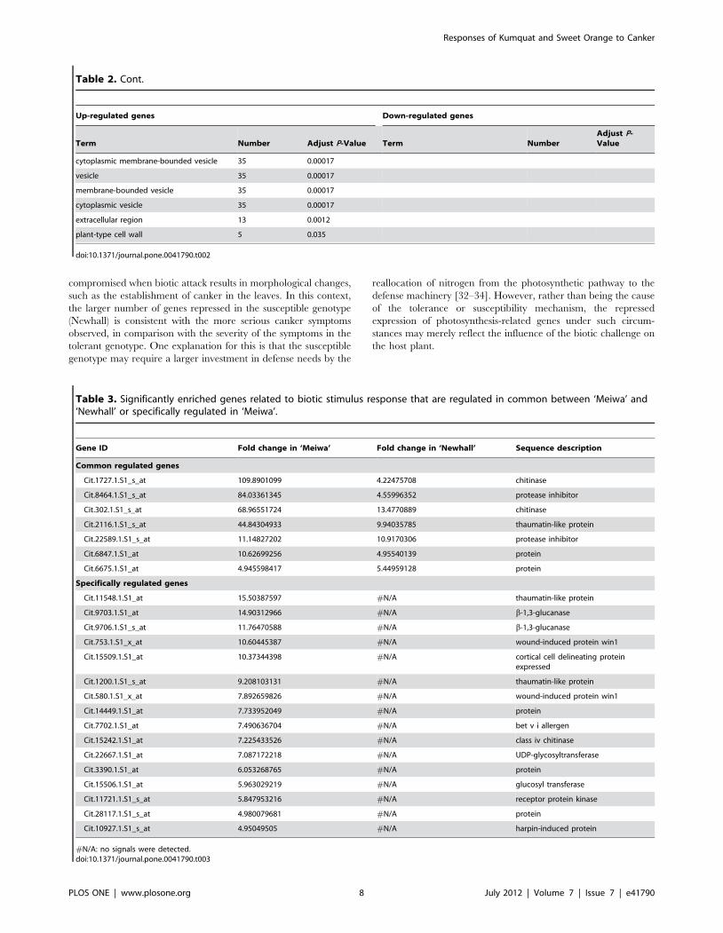
compromised when biotic attack results in morphological changes,
such as the establishment of canker in the leaves. In this context,
the larger number of genes repressed in the susceptible genotype
(Newhall) is consistent with the more serious canker symptoms
observed, in comparison with the severity of the symptoms in the
tolerant genotype. One explanation for this is that the susceptible
genotype may require a larger investment in defense needs by the
reallocation of nitrogen from the photosynthetic pathway to the
defense machinery [32–34]. However, rather than being the cause
of the tolerance or susceptibility mechanism, the repressed
expression of photosynthesis-related genes under such circum-
stances may merely reflect the influence of the biotic challenge on
the host plant.
Table 2. Cont.
Up-regulated genes Down-regulated genes
Term Number Adjust P-Value Term NumberAdjust P-Value
cytoplasmic membrane-bounded vesicle 35 0.00017
vesicle 35 0.00017
membrane-bounded vesicle 35 0.00017
cytoplasmic vesicle 35 0.00017
extracellular region 13 0.0012
plant-type cell wall 5 0.035
doi:10.1371/journal.pone.0041790.t002
Table 3. Significantly enriched genes related to biotic stimulus response that are regulated in common between ‘Meiwa’ and‘Newhall’ or specifically regulated in ‘Meiwa’.
Gene ID Fold change in ‘Meiwa’ Fold change in ‘Newhall’ Sequence description
Common regulated genes
Cit.1727.1.S1_s_at 109.8901099 4.22475708 chitinase
Cit.8464.1.S1_s_at 84.03361345 4.55996352 protease inhibitor
Cit.302.1.S1_s_at 68.96551724 13.4770889 chitinase
Cit.2116.1.S1_s_at 44.84304933 9.94035785 thaumatin-like protein
Cit.22589.1.S1_s_at 11.14827202 10.9170306 protease inhibitor
Cit.6847.1.S1_at 10.62699256 4.95540139 protein
Cit.6675.1.S1_at 4.945598417 5.44959128 protein
Specifically regulated genes
Cit.11548.1.S1_at 15.50387597 #N/A thaumatin-like protein
Cit.9703.1.S1_at 14.90312966 #N/A b-1,3-glucanase
Cit.9706.1.S1_s_at 11.76470588 #N/A b-1,3-glucanase
Cit.753.1.S1_x_at 10.60445387 #N/A wound-induced protein win1
Cit.15509.1.S1_at 10.37344398 #N/A cortical cell delineating proteinexpressed
Cit.1200.1.S1_s_at 9.208103131 #N/A thaumatin-like protein
Cit.580.1.S1_x_at 7.892659826 #N/A wound-induced protein win1
Cit.14449.1.S1_at 7.733952049 #N/A protein
Cit.7702.1.S1_at 7.490636704 #N/A bet v i allergen
Cit.15242.1.S1_at 7.225433526 #N/A class iv chitinase
Cit.22667.1.S1_at 7.087172218 #N/A UDP-glycosyltransferase
Cit.3390.1.S1_at 6.053268765 #N/A protein
Cit.15506.1.S1_at 5.963029219 #N/A glucosyl transferase
Cit.11721.1.S1_s_at 5.847953216 #N/A receptor protein kinase
Cit.28117.1.S1_s_at 4.980079681 #N/A protein
Cit.10927.1.S1_s_at 4.95049505 #N/A harpin-induced protein
#N/A: no signals were detected.doi:10.1371/journal.pone.0041790.t003
Responses of Kumquat and Sweet Orange to Canker
PLOS ONE | www.plosone.org 8 July 2012 | Volume 7 | Issue 7 | e41790

Despite the aforementioned differences, Meiwa and Newhall
still share a large number of common regulated genes following
Xcc infection. These genes may be necessary for the response to
biotic stress and may play basic roles in defense against Xcc. This
is logical, as plants have evolutionarily developed conserved
defense machinery against invading pathogens irrespective of their
stress tolerance capacity, suggesting that they may exhibit the same
subsets of gene expression and signaling pathways under adverse
environmental stresses. This implies that the expression of these
genes can be also induced in other citrus species upon bacterial
invasion. Although we did not try to identify the expression
patterns of common regulated genes in other cultivars, it is
interesting to note that several common regulated genes, such as
cytochrome P450, xyloglucan endotransglycosylase, phenylala-
nine-ammonia lyase, expansin, peroxidase, and chitinase-related
genes, have been shown to be induced in other citrus cultivars,
such as ‘Pera’ and ‘Cristal’, following Xcc inoculation [12]. The
identification of common regulated genes is not unique to this
study; it has been reported in other stressful conditions. For
instance, Zheng et al. [28] presented data to show that drought-
tolerant and drought-sensitive maize lines expressed a common set
of genes in response to drought stress.
DEGs that are specifically present in Meiwa are more important
than the common regulated genes, as the former might provide
valuable information on the molecular basis of canker tolerance in
this genotype. We used GO term enrichment analysis to gain more
insight into these genes, as it is traditionally an efficient strategy to
analyze the representation of genes under different categories by
comparing gene expression profiles to the background [24]. GO
term enrichment analysis revealed a remarkable difference in gene
distribution frequency between Meiwa and Newhall: the former
has a larger number of genes enriched in the upregulated gene
Figure 5. Functional categorization of 380 upregulated and 184 downregulated genes that are specifically regulated in ‘Meiwa’based on GO annotation.doi:10.1371/journal.pone.0041790.g005
Responses of Kumquat and Sweet Orange to Canker
PLOS ONE | www.plosone.org 9 July 2012 | Volume 7 | Issue 7 | e41790

Table 4. Significantly enriched cell wall-related genes that are regulated in common between ‘Meiwa’ and ‘Newhall’ or specificallyregulated in ‘Meiwa’.
Gene IDFold change in‘Meiwa’ Fold change in ‘Newhall’ Sequence description
Common regulated genes
Plant-type cell wall organization
Cit.30858.1.S1_at 6.406149904 5.99880024 alpha-expansin 4
Cit.14005.1.S1_s_at 7.72797527 6.978367062 Expansin
Cit.14005.1.S1_at 5.564830273 7.183908046 Expansin
Cit.8697.1.S1_at 13.42281879 5.750431282 ---NA---
Cit.11232.1.S1_s_at 16.58374793 15.69858713 unnamed protein product [Vitis vinifera]
Others
Cit.10363.1.S1_s_at 7.122507123 14.24501425 brassinosteroid-regulated protein bru1
Cit.302.1.S1_s_at 68.96551724 13.47708895 Chitinase
Cit.29964.1.S1_at 6.618133686 12.75510204 Protein
Cit.988.1.S1_at 7.598784195 5.920663114 Protein
Cit.30519.1.S1_s_at 129.8701299 11.65501166 xyloglucan endotransglycosylase
Cit.16722.1.S1_at 6.472491909 23.25581395 xyloglucan endotransglycosylase
Specifically regulated genes
Cell wall macromolecule metabolic
Cit.22311.1.S1_s_at 8.873114463 #N/A acidic chitinase
Cit.8262.1.S1_x_at 19.92031873 #N/A class i chitinase
Cit.8276.1.S1_x_at 34.01360544 #N/A class i chitinase
Cit.15242.1.S1_at 7.225433526 #N/A class iv chitinase
Cit.14913.1.S1_s_at 4.985044865 #N/A Protein
Cit.580.1.S1_x_at 7.892659826 #N/A wound-induced protein win1
Cit.753.1.S1_x_at 10.60445387 #N/A wound-induced protein win1
Cell wall modification
Cit.17701.1.S1_s_at 7.052186178 #N/A pectin methylesterase
Cit.9059.1.S1_s_at 8.403361345 #N/A pectin methylesterase
Cit.11169.1.S1_s_at 4.784688995 #N/A pectinesterase family protein
Cit.28980.1.S1_s_at 8.802816901 #N/A Protein
Cit.6756.1.S1_at 8.230452675 #N/A Protein
Plant-type cell wall organization
Cit.102.1.S1_s_at 12.65822785 #N/A a-expansin 4
Cit.10687.1.S1_s_at 5.417118093 #N/A a-expansin 4
Cit.2228.1.S1_s_at 102.0408163 #N/A extensin-like protein
Cit.8700.1.S1_at 42.91845494 #N/A mucin partial
Cit.8700.1.S1_s_at 83.33333333 #N/A mucin partial
Others
Cit.23827.1.S1_at 6.349206349 #N/A at1g68400 t2e12_5
Cit.22421.1.S1_x_at 4.137360364 #N/A miraculin-like protein 2
Cit.29356.1.S1_x_at 5.878894768 210.9542 miraculin-like protein 2
Cit.29368.1.S1_x_at 4.638218924 28.0095 miraculin-like protein 2
Cit.35442.1.S1_x_at 5.503577325 226.3034 miraculin-like protein 2
Cit.72.1.S1_s_at 8.984725966 #N/A miraculin-like protein 2
Cit.7991.1.S1_x_at 11.12347052 #N/A miraculin-like protein 2
Cit.8083.1.S1_x_at 15.50387597 #N/A miraculin-like protein 2
Cit.5836.1.S1_s_at 7.082152975 #N/A ---NA---
Cit.8683.1.S1_s_at 4.933399112 #N/A non-symbiotic hemoglobin class 1
Cit.8463.1.S1_at 7.385524372 #N/A protease inhibitor
Cit.13455.1.S1_s_at 6.349206349 #N/A xyloglucan endotransglycosylase
Responses of Kumquat and Sweet Orange to Canker
PLOS ONE | www.plosone.org 10 July 2012 | Volume 7 | Issue 7 | e41790

cluster, while the latter had more categories of genes enriched in
the downregulated cluster.
Significantly enriched genes in response to bioticstimulus
According to the GO term enrichment analysis, 7 and 16 genes
responsive to biotic stimulus were significantly enriched in the
common regulated and Meiwa-specifically regulated genes,
respectively (Table 1 and Table 2). Based on functional
annotation, these genes, encoding proteins such as chitinase,
proteinase inhibitor, thaumatin-like protein, b-1, 3-glucanase,
wound-induced protein win1, receptor protein kinase, and other
unknown proteins, are directly involved in responses to biotic
stimuli. Proteinase inhibitors (PIs) belong to the PR6 family, which
is widely distributed in the plant kingdom [35,36]. Several reports
have shown that plant PIs are an essential part of the natural
defense against pathogens [37–39]. These results provide a clue
that PIs may potentially perform specific functions in response to
citrus canker. Thaumatin-like proteins (TLPs), categorized under
the PR5 family, have been shown to accumulate when plants are
attacked by pathogens [35]. In addition, in vitro bioassays have
shown that TLPs possess antifungal activity [40]. Datta et al. [41]
presented data to show that the overexpression of a TLP gene
conferred resistance to sheath blight disease in transgenic rice.
These data led us to hypothesize that the induction of TLP genes
might be an integral part of the defense machinery against canker
in Meiwa. Receptor-like kinases act on the recognition of
pathogen-associated molecular patterns, such as bacterial flagellin,
which trigger immunity and effector-mediated immune responses
[42,43]. In the present study, a receptor kinase was abundantly
enriched in the subset of genes specifically regulated in Meiwa,
indicating that Meiwa might activate downstream defenses in a
more efficient manner. Although the functions of these genes have
not been verified herein, we can speculate that these genes are
closely involved in the canker resistance of Meiwa, in light of
previous studies.
The expression levels of cell wall and polysaccharidemetabolism-related genes change markedly in Meiwaupon canker infection
Among both common regulated and Meiwa-specific genes, an
array of genes related to cell wall and polysaccharide metabolism
was significantly enriched. The plant cell wall plays an important
role in basal defense, as it is the primary region of the host-
pathogen interaction and constitutes the first physical barrier to
limit pathogen colonization [44–47]. Enriched cell wall-related
genes, such as expansin and xyloglucan endotransglycosylase
(XET), are responsible for plant cell wall organization, macro-
molecule metabolism, and cell wall modification (Table 4).
Expansins are a family of proteins found within plant cell walls
that are responsible for cell wall disassembly, cell separation, and
cell expansion [48]. Genes coding for expansins have been shown
to be responsive to biotic or abiotic stresses [49–52]. Recently,
expansin genes were found to be induced when citrus plants were
challenged with Xcc or Huanglongbing pathogen [12,53]. Up-
regulation of the expansin gene suggests that following Xcc
inoculation, host plants may accumulate a larger amount of this
protein, which functions as a cell wall loosening agent to increase
cell wall extensibility [51]. XET catalyzes the cleavage of the
xyloglucan backbone, a major structural hemicellulose polysac-
charide in the primary cell wall, to form secondary cell walls. The
induction of these genes indicates that modification of cell wall
flexibility may be a crucial protective strategy in Meiwa to limit
pathogen invasion or spread in the internal tissues.
It has been well documented that high-molecular-weight
polysaccharides such as chitin and peptidoglycan are the major
components of the cell wall of pathogens. Here, Xcc infection led
to a significant induction of genes involved in chitin and glucan
metabolism in Meiwa, including chitinase, endo-b-1, 4-glucanase
(EGase), and b-1, 3-glucanase (Table 5). This is consistent with the
work of Cernadas et al. [12], who found that inoculation with the
citrus canker pathogen enhanced the transcriptional levels of these
genes in sweet orange. Chitinase (EC 3.2.1.14) hydrolyzes the b-1,
4-glycoside bond present in the chitin polymer to release chitin
fragments, such as chitooligosaccharides or chitin oligomers, from
cell walls and thereby activate plant innate immunity [54–56]. The
presence of abundant chitinase genes in Meiwa suggests that the
degradation of the cell wall of invading pathogens is more
extensive in this genotype, leading to inhibition of pathogen
proliferation and the spread or induction of systemic defense.
Moreover, EGase functions in cell wall loosening, which is
important for expansion or major cell wall disruption [57,58]. b-
1, 3-glucanase, hydrolyzing the 1, 3-b-D-glucosidic linkages of b-1,
3-glucan, has been shown to play a crucial role in plant pathogen
defense [59–61].
Genes related to cation binding are prominentlyenriched in Meiwa upon Xcc infection
Among the genes specifically regulated in Meiwa, as many as 54
genes were significantly enriched in the cation-binding category,
including those involved in the binding of iron, calcium, copper,
zinc, and other unknown ions (Table 2 and Table 6). In contrast,
this category of genes was not enriched among the genes
specifically regulated in Newhall. This finding implies that the
cation-binding process may be, at least in part, responsible for the
difference in canker tolerance between these two genotypes.
Copper ion binding is potentially of interest, because copper-
containing bactericides have been extensively applied to control
citrus canker disease [2,62–64]. Very recently, Yuan et al. [65]
reported that the copper level is a key determinant of the defense
response to Xanthomonas oryzae causing blight in rice. The induction
of copper ion binding suggests that copper redistribution may be
modified in Meiwa in order to protect the host plant from Xcc
bacterial invasion, as has been documented by Yuan et al. [65].
Apart from copper ion binding, there are 14 genes related to iron
ion binding, 7 of which encode cytochrome P450 (Table 6). Iron is
Table 4. Cont.
Gene IDFold change in‘Meiwa’ Fold change in ‘Newhall’ Sequence description
Cit.2949.1.S1_s_at 4.444444444 #N/A xyloglucan endotransglycosylase
#N/A: no signals were detected.doi:10.1371/journal.pone.0041790.t004
Responses of Kumquat and Sweet Orange to Canker
PLOS ONE | www.plosone.org 11 July 2012 | Volume 7 | Issue 7 | e41790

an important micronutrient for virtually all living organisms, and
iron homeostasis commonly occurs in pathogen-host interactions.
Bacteria need to acquire iron from the host for their own survival,
and the host can limit bacterial pathogen invasion through an
iron-withholding model [66]. Enrichment of these genes in Meiwa
demonstrates that the uptake of iron ions by bacteria from the host
plant may be efficiently restrained, leading to growth arrest of the
invading pathogens. Cytochrome P450s, a group of heme-
containing enzymes that are ubiquitously present in bacteria and
plants [67], have been shown to regulate the biosynthesis of
defense-related compounds [68,69]. Calcium ion binding probably
plays an important signaling role in response to canker disease,
because calcium is a well-known second messenger in numerous
plant signaling pathways [70]. However, the physiological or
Table 5. Significantly enriched genes involved in polysaccharide metabolism that are regulated in common between ‘Meiwa’ and‘Newhall’ or specifically regulated in ‘Meiwa’.
Gene ID Fold change in ‘Meiwa’ Fold change in ‘Newhall’ Sequence description
Common regulated genes
Glucan metabolism
Cit.35196.1.S1_s_at 16.50165017 6.397952655 21 kDa protein
Cit.10363.1.S1_s_at 7.122507123 14.24501425 brassinosteroid-regulated protein BRU1
Cit.3554.1.S1_s_at 7.4019245 4.528985507 glycosyl hydrolase family 9 protein
Cit.28480.1.S1_s_at 6.353240152 7.710100231 protein
Cit.4211.1.S1_at 5.882352941 13.12335958 protein
Cit.16722.1.S1_at 6.472491909 23.25581395 xyloglucan endotransglycosylase
Cit.30519.1.S1_s_at 129.8701299 11.65501166 xyloglucan endotransglycosylase
Others
Cit.20412.1.S1_s_at 67.11409396 13.96648045 basic endochitinase-like protein
Cit.1727.1.S1_s_at 109.8901099 4.224757076 chitinase
Cit.302.1.S1_s_at 68.96551724 13.47708895 chitinase
Cit.2392.1.S1_at 25.51020408 10.68376068 endo-b-1,4-glucanase
Cit.6847.1.S1_at 10.62699256 4.955401388 protein
Specifically regulated genes
Chitin metabolism
Cit.22311.1.S1_s_at 8.873114463 #N/A acidic chitinase
Cit.8262.1.S1_x_at 19.92031873 #N/A class I chitinase
Cit.8276.1.S1_x_at 34.01360544 #N/A class I chitinase
Cit.15242.1.S1_at 7.225433526 #N/A class IV chitinase
Cit.14913.1.S1_s_at 4.985044865 #N/A protein
Cit.580.1.S1_x_at 7.892659826 #N/A wound-induced protein win1
Cit.753.1.S1_x_at 10.60445387 #N/A wound-induced protein win1
Glucan metabolism
Cit.14715.1.S1_s_at 8.680555556 #N/A acid invertase
Cit.9204.1.S1_s_at 5.941770648 #N/A ADP-glucose pyrophosphorylase small subunit
Cit.21302.1.S1_s_at 6.172839506 #N/A alcohol dehydrogenase
Cit.17208.1.S1_at 4.66853408 #N/A a-amylase
Cit.9703.1.S1_at 14.90312966 #N/A b-1,3-glucanase
Cit.9706.1.S1_s_at 11.76470588 #N/A b-1,3-glucanase
Cit.25122.1.S1_s_at 9.652509653 #N/A endo-b-1,4-glucanase
Cit.2945.1.S1_s_at 7.936507937 #N/A endo-b-1,4-glucanase
Cit.30208.1.S1_at 10.41666667 #N/A hydrolyzing o-glycosyl
Cit.9059.1.S1_s_at 8.403361345 #N/A pectin methylesterase
Cit.11169.1.S1_s_at 4.784688995 #N/A pectinesterase family protein
Cit.28980.1.S1_s_at 8.802816901 #N/A protein
Cit.6756.1.S1_at 8.230452675 #N/A protein
Cit.13455.1.S1_s_at 6.349206349 #N/A xyloglucan endotransglycosylase
Cit.2949.1.S1_s_at 4.444444444 #N/A xyloglucan endotransglycosylase
#N/A: no signals were detected.doi:10.1371/journal.pone.0041790.t005
Responses of Kumquat and Sweet Orange to Canker
PLOS ONE | www.plosone.org 12 July 2012 | Volume 7 | Issue 7 | e41790

Table 6. Significantly enriched cation binding related genes in ‘Meiwa’ specifically regulated gene cluster.
Gene ID Fold change in ‘Meiwa’ Sequence description
Iron ion binding
Cit.21723.1.S1_s_at 6.353240152 1-aminocyclopropane-1-carboxylate oxidase
Cit.30535.1.S1_s_at 5.296610169 1-aminocyclopropane-1-carboxylate oxidase
Cit.11159.1.S1_s_at 6.325110689 cytochrome P450
Cit.11965.1.S1_at 9.478672986 cytochrome P450
Cit.12598.1.S1_s_at 4.269854825 cytochrome P450
Cit.15523.1.S1_at 4.901960784 cytochrome P450
Cit.24211.1.S1_s_at 4.911591356 cytochrome P450
Cit.28253.1.S1_at 5.184033178 cytochrome P450
Cit.30299.1.S1_at 6.067961165 cytochrome P450
Cit.9904.1.S1_s_at 22.98850575 lipoxygenase
Cit.31330.1.S1_at 5.757052389 NADPH oxidase
Cit.858.1.S1_s_at 11.37656428 peroxidase 12
Cit.30798.1.S1_at 10.86956522 peroxidase precursor
Cit.5316.1.S1_at 18.86792453 protein
Calcium ion binding
Cit.17208.1.S1_at 4.66853408 alpha-amylase
Cit.8157.1.S1_s_at 4.837929366 caffeoyl- 3-o-methyltransferase
Cit.24075.1.S1_at 4.980079681 CDPK-related protein kinase
Cit.31330.1.S1_at 5.757052389 NADPH oxidase
Cit.39387.1.S1_at 18.4501845 pectate lyase
Cit.858.1.S1_s_at 11.37656428 peroxidase 12
Cit.30798.1.S1_at 10.86956522 peroxidase precursor
Cit.11691.1.S1_at 10.46025105 protein
Cit.17648.1.S1_x_at 4.184100418 translationally controlled tumor protein
Copper ion binding
Cit.18726.1.S1_at 6.765899865 amine oxidase
Cit.2409.1.S1_s_at 4.551661356 laccase 110a
Cit.37464.1.S1_at 4.391743522 l-ascorbate oxidase
Cit.11172.1.S1_s_at 8.223684211 pectinesterase like protein
Cit.7209.1.S1_at 15.52795031 polyphenol oxidase
Zinc ion binding
Cit.21302.1.S1_s_at 6.172839506 alcohol dehydrogenase
Cit.26919.1.S1_s_at 5.341880342 ARP protein
Cit.30588.1.S1_s_at 4.387889425 ARP protein
Cit.11286.1.S1_at 5.871990605 E3 ubiquitin-protein ligase rnf149-like
Cit.20848.1.S1_at 4.076640848 protein
Cit.30654.1.S1_s_at 6.858710562 protein
Cit.38488.1.S1_at 8.244023083 protein
Cit.950.1.S1_s_at 9.900990099 protein
Cit.22054.1.S1_at 4.923682915 ring-h2 finger protein
Cit.25290.1.S1_s_at 6.105006105 ring-h2 finger protein
Cit.6340.1.S1_s_at 9.803921569 RNA binding
Other cation binding
Cit.22311.1.S1_s_at 8.873114463 acidic chitinase
Cit.21938.1.S1_s_at 4.297378599 b-1,3-glucanase
Cit.9703.1.S1_at 14.90312966 b-1,3-glucanase
Cit.9706.1.S1_s_at 11.76470588 b-1,3-glucanase
Cit.24017.1.S1_at 12.01923077 cytochrome
Responses of Kumquat and Sweet Orange to Canker
PLOS ONE | www.plosone.org 13 July 2012 | Volume 7 | Issue 7 | e41790

molecular relevance of zinc ion binding and that of other cations
remains to be determined, as limited information is available so far
on their activation upon pathogen attack.
Taken together, a comparative transcriptomic analysis of
Meiwa and Newhall in this study reveals that they differ greatly
in the molecular response to citrus canker, which may partially
explain their phenotypical variation with regard to disease
tolerance. When challenged with Xcc bacteria, expression of
genes involved in polysaccharide metabolism, biotic stimulus
response, cell wall strengthening, and cation binding was altered,
thereby promoting the production/synthesis of a large spectrum of
second metabolites and modifying ion homeostasis. These
biological processes may work cooperatively to limit bacterial
penetration, proliferation, spread, and growth, conferring canker
tolerance. By contrast, in Newhall only a few basal responsive
proteins such as chitinase, glucanase, and thaumatin-like protein
were activated, leading to the production of a limited amount of
relevant products that function to protect the host against the
canker pathogen. In addition, Xcc attack repressed the expression
of several genes associated with photosynthesis in Newhall. The
data presented herein revealed the molecular mechanisms
underlying the contrasting canker tolerance between Meiwa and
Newhall, and the Meiwa-specific regulated genes hold great
potential for engineering canker tolerance in the future. The next
challenge is to narrow down the genes screened in this study based
on expression patterns and to finally exploit and functionally
identify the genes that are truly responsible for the canker
tolerance. In addition, creation of transgenic plants with enhanced
canker tolerance using the genes tapped from this study will be of
paramount significance for providing novel germplasms that can
be integrated into citrus breeding pipeline in the long run.
Materials and Methods
Plant materials and bacterial strainsLeaves were collected from uniform and healthy summer flushes
of 15-year-old Meiwa (Fortunella crassifolia) and Newhall (Citrus
sinensis Osbeck) plants grown in the same orchard in the Citrus
Research Institute, Huazhong Agricultural University (Wuhan,
China). The primary source of the inoculum used in this study was
Xcc strain A (X02-007), provided by Prof. Hong Ni (Huazhong
Agricultural University). The bacteria were maintained at 28uC in
SPA medium containing sucrose 20 g/l, peptone 5 g/l, K2HPO4
0.5 g/l, MgSO4N7 H2O 0.25 g/l, and agar 15 g/l (pH 7.2–7.4).
Pinprick inoculation of leaves and samplingThe bacterial strain was cultured in liquid SPA medium at 28uC
and shaken overnight at 200 rpm, then collected by centrifugation
and re-suspended in the medium at a concentration of about 108
cells/ml before inoculation. The collected leaves were washed with
distilled water and then subjected to inoculation on the abaxial
side using an inoculating needle (0.5 mm in diameter). Four
inoculations, each composed of 5 pricks, were made on both sides
of the midvein, and a 10-ml aliquot of the bacterial suspension was
dropped onto each prick. Following inoculation, the leaves were
placed on wet filter paper in Petri dishes, which were then sealed
with parafilm to maintain high humidity for bacterial growth. The
Petri dishes were kept at 28uC in a plant growth chamber for the
indicated periods. Initiation of symptoms was scored within a 7-d
cycle. The leaves were immediately immersed in liquid nitrogen
and stored at 280uC till use. Leaves sampled at 0 and 5 DPI were
used for microarray analysis.
Bacterial growth assayBacterial population in the inoculated sites collected at 6 DPI
(after canker appearance) was examined based on earlier report
[71]. In brief, the inoculated sites of same size were disinfected
with 2% (v/v) sodium hypochlorite for 10 s and 75% ethanol for
3 min. The leaf discs were then ground in sterile distilled water,
followed by dilution and spread on SPA medium. After an
incubation for 2 d at 28uC the number of colonies was counted in
order to calculate the colony-forming units (cfu), expressed as cfu/
ml.
Total RNA isolation, probe preparation, and microarrayhybridization
Total RNA was isolated from samples collected at 0 and 5 DPI
using the Trizol reagent (Invitrogen, Carlsbad, CA) according to
the supplier’s recommendations. The RNA samples were treated
with amplification-grade DNase I (Takara, Dalian, China) at 37uCto remove any contaminant genomic DNA. Gene expression
profiles of Meiwa and Newhall before (0 DPI) and after (5 DPI)
Xcc inoculation was analyzed by the Affymetrix Citrus Genome
Table 6. Cont.
Gene ID Fold change in ‘Meiwa’ Sequence description
Cit.31237.1.S1_at 5.015045135 cytochrome P450
Cit.4425.1.S1_at 14.5137881 cytochrome P450
Cit.17456.1.S1_at 6.784260516 cytochrome P450 79a2
Cit.25122.1.S1_s_at 9.652509653 endo-b-1,4-glucanase
Cit.252.1.S1_s_at 19.34235977 glycosyl hydrolase family 1 protein
Cit.30841.1.S1_s_at 12.36093943 glycosyl hydrolase family 1 protein
Cit.10770.1.S1_s_at 4.178854994 h(\+)-transporting ATPase plant fungi plasma membrane
Cit.8767.1.S1_at 6.76132522 heavy-metal-associated domain-containing expressed
Cit.6076.1.S1_s_at 6.779661017 metal ion binding
Cit.1827.1.S1_s_at 4.606172271 peroxidase
Cit.14913.1.S1_s_at 4.985044865 protein
Cit.17173.1.S1_s_at 7.496251874 urease accessory protein g
#N/A: no signals were detected.doi:10.1371/journal.pone.0041790.t006
Responses of Kumquat and Sweet Orange to Canker
PLOS ONE | www.plosone.org 14 July 2012 | Volume 7 | Issue 7 | e41790

GeneChip one-cycle target labeling and control kit (Affymetrix,
Santa Clara, CA) according to the manufacturer’s instructions; this
was done by Gene Technology Company Limited (Shanghai,
China). For GeneChip analysis, 10 mg of total RNA was first
reverse transcribed into double-stranded cDNA using a T7-
Oligo(dT) promoter primer, then transcribed to complementary
RNA (cRNA) in vitro in the presence of T7 RNA polymerase and a
biotinylated nucleotide analog/ribonucleotide mix for cRNA
amplification and biotin labeling. The resultant biotinylated
cRNA targets, which were labeled with either Cy5 (5 DPI
samples) or Cy3 (0 DPI samples), were then cleaned up,
fragmented, and hybridized with the Citrus Genome GeneChip
Array, which contained 30,171 probe sets representing up to
33,879 citrus transcripts based on EST sequences obtained from
several citrus species and citrus hybrids. According to the
published sweet orange genome (version 1, http://www.
phytozome.net), the transcripts on the array account for 73.4%
of the whole genome. Cy5-labeled cRNAs were hybridized with
Cy3-labeled cRNAs for each genotype. Hybridization was
performed on each of the materials tested with 2 biological
replicates and two technical replicates (dye-swap).
Data analysisAfter the washing procedure was completed, the probe array
was scanned using the Affymetrix GeneChip Scanner 3000. The
images were analyzed using the Affymetrix GeneChip Operating
Software (GCOS 1.4) to generate raw data, which was saved as
CEL files. The CEL files were then imported into Bioconductor
system (R software) using the Affy package for quantile normal-
ization to obtain Robust Multi-array Average (RMA) data
containing the expression values. For statistical analysis of
differentially expressed genes between Meiwa and Newhall, the
RankPord package in R software [72] was used to calculate the
number of false-positive predictions (FPP), which is also known as
the false discovery rate (FDR) [73]. Probe sets with an FDR#0.5
and a 4-fold change were considered as differentially expressed
genes at a statistically significant level. DEGs in Meiwa and
Newhall were functionally annotated using the Citrus HarvEST
software (Version 1.25, http://harvest.ucr.edu/, University of
California) by aligning the consensus sequences of all probe sets to
the sequences in the Arabidopsis database, and the MapManBin
[29] functional categorization was carried out online in the Plant
Proteome Database (PPDB) [74] using the best matched AGI
number. For further analysis of the common regulated genes and
Meiwa-specifically regulated genes, Blast analysis and GO term
annotation were carried out using Blast2GO software [23]. GO
terms for each of the 3 main categories, biological process,
molecular function, and cellular component, were obtained from
sequence similarity using default parameters. To analyze GO term
enrichment of significant DEGs, SEA was performed online
through agriGO (http://bioinfo.cau.edu.cn/agriGO), a GO
analysis tool kit for the agricultural community [24]. In brief,
the probe ID numbers of common regulated genes or specifically
regulated genes were first uploaded into the agriGO, and the
Citrus Affymetrix Genome Array was selected as the background.
Thereafter, statistical P-values were calculated using the hypergeo-
metric method, and multiple comparison correction was done
using the Benjamini-Yekutieli method to adjust P-values [75]. GO
terms with an adjusted P value,0.05 were considered to be
significantly enriched in the leaves of Meiwa and Newhall before
and after inoculation.
Semi-quantitative RT-PCR analysisSemi-quantitative RT-PCR was employed to verify the
microarray results. The same RNA samples tested in the
hybridization experiments were used for cDNA synthesis using
the ReverTra Ace-a-TM kit (Toyobo, Osaka, Japan) following the
manufacturer’s instructions. Primers specific to 10 upregulated
and 2 downregulated genes were designed using the Primer
Premier 5 software (PRIMER Biosoft International, Palo Alto,
CA) based on the consensus sequences (Table S3). The amplifi-
cation was carried out in a thermal cycler (Bio-Rad, Hercules, CA)
with a program of 28 cycles of 30 s at 94uC, 30 s at 55uC and 45 s
at 72uC. The same cDNA was amplified with primers specific to
an actin gene, which was used as an internal positive control. Band
density was quantified using Quantity One Software (Version
4.6.2, Bio-Rad). PCR amplification of each gene was performed in
triplicate.
Supporting Information
Figure S1 MapManbin classification of differentiallyexpressed genes in ‘Meiwa’ and ‘Newhall’.
(TIF)
Table S1 List and Mapman analysis of differentiallyexpressed genes in ‘Meiwa’.
(XLS)
Table S2 List and Mapman analysis of differentiallyexpressed genes in ‘Newhall’.
(XLS)
Table S3 Sequences of the specific primers used for thesemi-quantitative RT-PCR analysis.
(XLS)
Table S4 The common upregulated (150) or downregu-lated genes (80) in ‘Meiwa’ and ‘Newhall’ after Xccinfection, among which 45 upregulated and 19 downreg-ulated in ‘Meiwa’ showed significantly higher foldchange than ‘Newhall’ (difference value .4, markedwith color).
(XLS)
Table S5 The specifically upregulated (380) or down-regulated (184) genes in ‘Meiwa’ after Xcc infection.
(XLS)
Table S6 Significantly enriched GO terms of thespecifically regulated genes in ‘Newhall’ after SingularEnrichment analysis.
(XLS)
Acknowledgments
The authors are grateful to Dr. Shao-Hua Zeng for critical reading of the
manuscript.
Author Contributions
Conceived and designed the experiments: JHL. Performed the experi-
ments: XZF XQG YXZ YW. Analyzed the data: XZF JHL. Contributed
reagents/materials/analysis tools: JHL. Wrote the paper: XZF JHL.
Responses of Kumquat and Sweet Orange to Canker
PLOS ONE | www.plosone.org 15 July 2012 | Volume 7 | Issue 7 | e41790

References
1. Schubert TS, Rizvi SA, Sun X, Gottwald TR, Graham JH, et al. (2001) Meeting
the challenge of eradicating citrus canker in Florida-again. Plant Diseas 85: 340–356.
2. Das AK (2003) Citrus canker - A review. Journal of Applied Horticulture 5: 52–
60.
3. Stall RE, Loschke DC, Jones JB (1986) Linkage of copper resistance andavirulence loci on a self-transmissible plasmid in Xanthomonas campestris pv.
vesicatoria. Phytopathology 76: 240–243.
4. Behlau F, Canteros BI, Minsavage GV, Jones JB, Graham JH (2011) Molecular
Characterization of Copper Resistance Genes from Xanthomonas citri subsp citri
and Xanthomonas alfalfae subsp citrumelonis. Applied and Environmental Microbi-
ology 77: 4089–4096.
5. Behlau F, Jones JB, Myers ME, Graham JH (2012) Monitoring for resistantpopulations of Xanthomonas citri subsp citri and epiphytic bacteria on citrus trees
treated with copper or streptomycin using a new semi-selective medium.European Journal of Plant Pathology 132: 259–270.
6. Schenk PM, Kazan K, Wilson I, Anderson JP, Richmond T, et al. (2000)
Coordinated plant defense responses in Arabidopsis revealed by microarray
analysis. Proceedings of the National Academy of Sciences of the United Statesof America 97: 11655–11660.
7. Eulgem T (2005) Regulation of the Arabidopsis defense transcriptome. Trends
in Plant Science 10: 71–78.
8. Tao Y, Xie Z, Chen W, Glazebrook J, Chang HS, et al. (2003) Quantitativenature of Arabidopsis responses during compatible and incompatible interac-
tions with the bacterial pathogen Pseudomonas syringae. Plant Cell 15: 317–330.
9. Wright DP, Johansson T, Le Quere A, Soderstrom B, Tunlid A (2005) Spatialpatterns of gene expression in the extrametrical mycelium and mycorrhizal root
tips formed by the ectomycorrhizal fungus Paxillus involutus in association withbirch (Betula pendula) seedlings in soil microcosms. New Phytologist 167: 579–596.
10. Alignan M, Hewezi T, Petitprez M, Dechamp-Guillaume G, Gentzbittel L
(2006) A cDNA microarray approach to decipher sunflower (Helianthus annuus)
responses to the necrotrophic fungus Phoma macdonaldii. New Phytologist 170:523–536.
11. Rinaldi C, Kohler A, Frey P, Duchaussoy F, Ningre N, et al. (2007) Transcript
profiling of poplar leaves upon infection with compatible and incompatiblestrains of the foliar rust Melampsora larici-populina. Plant Physiology 144: 347–366.
12. Cernadas RA, Camillo LR, Benedetti CE (2008) Transcriptional analysis of the
sweet orange interaction with the citrus canker pathogens Xanthomonas axonopodis
pv. citri and Xanthomonas axonopodis pv. aurantifolii. Molecular Plant Pathology 9:
609–631.
13. Fujiwara S, Tanaka N, Kaneda T, Takayama S, Isogai A, et al. (2004) RicecDNA microarray-based gene expression profiling of the response to flagellin
perception in cultured rice cells. Molecular plant-Microbe Interactions 17: 986–
998.
14. Qiu D, Xiao J, Xie W, Liu H, Li X, et al. (2008) Rice gene network inferredfrom expression profiling of plants overexpressing OsWRKY13, a positive
regulator of disease resistance. Molecular Plant 1: 538–551.
15. Albertazzi G, Milc J, Caffagni A, Francia E, Roncaglia E, et al. (2009) Geneexpression in grapevine cultivars in response to Bois Noir phytoplasma infection.
Plant Science 176: 792–804.
16. Miao WG, Wang XB, Song CF, Wang Y, Ren YH, et al. (2010) Transcriptomeanalysis of Hpa1Xoo transformed cotton revealed constitutive expression of genes
in multiple signalling pathways related to disease resistance. Journal of
Experimental Botany 61: 4263–4275.17. Deng Z, Gmitter FGJr (2003) Cloning and characterization of receptor kinase
class disease resistance gene candidates in citrus. Theoretical and Applied
Genetics 108: 53–61.
18. Cernadas RA, Benedetti CE (2009) Role of auxin and gibberellin in citruscanker development and in the transcriptional control of cell-wall remodeling
genes modulated by Xanthomonas axonopodis pv. citri. Plant Science 177: 190–195.
19. Gottwald TR, Graham JH (2000) Canker. In: Compendium of citrus diseases, 2ndedtion. Edited by Timmer LW, Garnsey SM, Graham H. American
Phytopathological Society Press: 5–8.
20. Wang Y, Fu XZ, Liu JH, Hong N (2011) Differential structure and physiologicalresponse to canker challenge between ‘Meiwa’ kumquat and ‘Newhall’ navel
orange with contrasting resistance. Scientia Horticulturae 128: 115–123.
21. Lopez C, Soto M, Restrepo S, Piegu B, Cooke R, et al. (2005) Gene expressionprofile in response to Xanthomonas axonopodis pv. manihotis infection in cassava
using a cDNA microarray. Plant Molecular Biology 57: 393–410.
22. Gandıa M, Conesa A, Ancillo G, Gadea J, Forment J, et al. (2007)
Transcriptional response of Citrus aurantifolia to infection by Citrus tristeza virus.Virology 367: 298–306.
23. Conesa A, Gotz S, Garcia-Gomez JM, Terol J, Talon M, et al. (2005) Blast2GO:
a universal tool for annotation, visualization and analysis in functional genomicsresearch. Bioinformatics 21: 3674–3676.
24. Du Z, Zhou X, Ling Y, Zhang Z, Su Z (2010) AgriGO: a GO analysis toolkit for
the agricultural community. Nucleic Acids Research 38: W64–W70.
25. Taji T, Seki M, Satou M, Sakurai T, Kobayashi M, et al. (2004) Comparativegenomics in salt tolerance between Arabidopsis and Arabidopsis-related
halophyte salt cress using Arabidopsis microarray. Plant Physiology 135:1697–1709.
26. Walia H, Wilson C, Condamine P, Liu X, Ismail AM, et al. (2005) Comparative
transcriptional profiling of two contrasting rice genotypes under salinity stressduring the vegetative growth stage. Plant Physiology 139: 822–835.
27. Sun W, Xu XN, Zhu HS, Liu AH, Liu L, et al. (2010) Comparativetranscriptomic profiling of a salt-tolerant wild tomato species and a salt-sensitive
tomato cultivar. Plant and Cell Physiology 51: 997–1006.
28. Zheng J, Fu JJ, Gou MY, Huai JL, Liu YJ, et al. (2010) Genome-wide
transcriptome analysis of two maize inbred lines under drought stress. PlantMolecular Biology 72: 407–421.
29. Thimm O, Blasing O, Gibon Y, Nagel A, Meyer S, et al. (2004) MAPMAN: auser-driven tool to display genomics data sets onto diagrams of metabolic
pathways and other biological processes. Plant Journal 37: 914–939.
30. Zou J, Rodriguez-Zas S, Aldea M, Li M, Zhu J, et al. (2005) Expression profiling
soybean response to Pseudomonas syringae reveals new defense-related genes and
rapid HR-specific downregulation of photosynthesis. Molecular Plant-MicrobeInteractions 18: 1161–1174.
31. Berger S, Benediktyova Z, Matous K, Bonfig K, Mueller MJ, et al. (2007)Visualization of dynamics of plant-pathogen interaction by novel combination of
chlorophyll fluorescence imaging and statistical analysis: differential effects ofvirulent and avirulent strains of P. syringae and of oxylipins on A. thaliana. Journal
of Experimental Botany 58: 797–806.
32. Bilgin DD, Zavala JA, Zhu J, Clough SJ, Ort DR, et al. (2010) Biotic stress
globally downregulates photosynthesis genes. Plant Cell and Environment 33:
1597–1613.
33. Evans JR (1989) Photosynthesis and nitrogen relationships in leaves of C3 plants.
Oecologia 78: 9–19.
34. Paul MJ, Foyer CH (2001) Sink regulation of photosynthesis. Journal of
Experimental Botany 52: 1383–1400.
35. Jwa NS, Agrawal GK, Tamogami S, Yonekura M, Han O, et al. (2006) Role of
defense/stress-related marker genes, proteins and secondary metabolites indefining rice self-defense mechanisms. Plant Physiology and Biochemistry 44:
261–273.
36. Huang YM, Xiao BZ, Xiong LZ (2007) Characterization of a stress responsive
proteinase inhibitor gene with positive effect in improving drought resistance inrice. Planta 226: 73–85.
37. Botella MA, Xu Y, Prabha TN, Zhao Y, Narasimhan ML, et al. (1996)Differential expression of soybean cysteine proteinase inhibitor genes during
development and in response to wounding and methyl jasmonate. Plant
Physiology 112: 1201–1210.
38. Solomon M, Belenghi B, Delledonne M, Menachem E, Levine A (1999) The
involvement of cysteine proteases and protease inhibitor genes in the regulationof programmed cell death in plants. Plant Cell 11: 431–444.
39. Rakwal R, Kumar Agrawal G, Jwa NS (2001) Characterization of a rice (Oryza
sativa L.) Bowman-Birk proteinase inhibitor: tightly light regulated induction in
response to cut, jasmonic acid, ethylene and protein phosphatase 2A inhibitors.Gene 263: 189–198.
40. Hejgaard J, Jacobsen S, Svendsen I (1991) Two antifungal thaumatin-likeproteins from barley grains. FEBS Letters 291: 127–131.
41. Datta K, Velazhahan R, Oliva N, Ona I, Mew T, et al. (1999) Over-expressionof the cloned rice thaumatin-like protein (PR-5) gene in transgenic rice plants
enhances environmental friendly resistance to Rhizoctonia solani causing sheath
blight disease. Theoretical and Applied Genetics 98: 1138–1145.
42. Chisholm ST, Coaker G, Day B, Staskawicz BJ (2006) Host-microbe
interactions: shaping the evolution of the plant immune response. Cell 124:803–814.
43. Gomez-Gomez L, Boller T (2000) FLS2: an LRR receptor–like kinase involvedin the perception of the bacterial elicitor flagellin in Arabidopsis. Molecular Cell 5:
1003–1011.
44. Vorwerk S, Somerville S, Somerville C (2004) The role of plant cell wall
polysaccharide composition in disease resistance. Trends in Plant Science 9:203–209.
45. Cantu D, Vicente AR, Labavitch JM, Bennett AB, Powell ALT (2008) Strangersin the matrix: plant cell walls and pathogen susceptibility. Trends in Plant
Science 13: 610–617.
46. Hematy K, Cherk C, Somerville S (2009) Host-pathogen warfare at the plant
cell wall. Current Opinion in Plant Biology 12: 406–413.
47. Lagaert S, Belien T, Volckaert G (2009) Plant cell walls: Protecting the barrier
from degradation by microbial enzymes. Seminars in Cell & Developmental
Biology 20: 1064–1073.
48. Cosgrove DJ (2000) Loosening of plant cell walls by expansins. Nature 407: 321–
326.
49. Hiwasa K, Rose JKC, Nakano R, Inaba A, Kubo Y (2003) Differential
expression of seven alpha-expansin genes during growth and ripening of pearfruit. Physiologia Plantarum 117: 564–572.
50. Balestrini R, Cosgrove DJ, Bonfante P (2005) Differential location of a-expansinproteins during the accommodation of root cells to an arbuscular mycorrhizal
fungus. Planta 220: 889–899.
51. Xu J, Tian J, Belanger FC, Huang BR (2007) Identification and characterization
of an expansin gene AsEXP1 associated with heat tolerance in C3 Agrostis grassspecies. Journal of Experimental Botany 58: 3789–3796.
Responses of Kumquat and Sweet Orange to Canker
PLOS ONE | www.plosone.org 16 July 2012 | Volume 7 | Issue 7 | e41790

52. Ding X, Cao Y, Huang L, Zhao J, Xu C, et al. (2008) Activation of the indole-3-
acetic acid-amido synthetase GH3–8 suppresses expansin expression andpromotes salicylate- and jasmonate-independent basal immunity in Rice. Plant
Cell 20: 228–240.
53. Albrecht U, Bowman KD (2008) Gene expression in Citrus sinensis (L.) Osbeckfollowing infection with the bacterial pathogen Candidatus Liberibacter asiaticus
causing Huanglongbing in Florida. Plant Science 175: 291–306.54. Punja ZK, Zhang YY (1993) Plant chitinases and their roles in resistance to
fungal diseases. Journal of Nematology 25: 526–540.
55. Kasprzewska A (2003) Plant chitinases—Regulation and function. Cellular &Molecular Biology Letters 8: 809–824.
56. Wan J, Zhang XC, Stacey G (2008) Chitin signaling and plant disease resistance.Plant Signaling & Behavior 3: 831–833.
57. Loopstra CA, Mouradov A, Vivian-Smith A, Glassick TV, Gale BV, et al. (1998)Two pine endo-b-1, 4-glucanases are associated with rapidly growing
reproductive structures. Plant Physiology 116: 959–967
58. Bourquin V, Nishikubo N, Abe H, Brumer H, Denman S, et al. (2002)Xyloglucan endotransglycosylases have a function during the formation of
secondary cell walls of vascular tissues. Plant Cell 14: 3073–3088.59. Jongedijk E, Tigelaar H, Van Roekel JSC, Bres-Vloemans SA, Dekker I, et al.
(1995) Synergistic activity of chitinases and b-1, 3-glucanases enhances fungal
resistance in transgenic tomato plants. Euphytica 85: 173–180.60. Lusso M, Kuc J (1996) The effect of sense and antisense expression of the PR-N
gene for b-1, 3-glucanase on disease resistance of tobacco to fungi and viruses.Physiological and Molecular Plant Pathology 49: 267–283.
61. Enrique R, Siciliano F, Favaro MA, Gerhardt N, Roeschlin R, et al. (2010)Novel demonstration of RNAi in citrus reveals importance of citrus callose
synthase in defence against Xanthomonas citri subsp. citri. Plant Biotechnology
Journal 9: 394–407.62. Graham JH, Gottwald TR, Cubero J, Achor DS (2004) Xanthomonas axonopodis
pv. citri: factors affecting successful eradication of citrus canker. Molecular PlantPathology 5: 1–15.
63. Behlau F, Belasque J, Bergamin A, Graham JH, Leite RP, et al. (2008) Copper
sprays and windbreaks for control of citrus canker on young orange trees insouthern Brazil. Crop Protection 27: 807–813.
64. Behlau F, Belasque J, Graham JH, Leite RP (2010) Effect of frequency of copper
applications on control of citrus canker and the yield of young bearing sweet
orange trees. Crop Protection 29: 300–305.
65. Yuan M, Chu ZH, Li XH, Xu CG, Wang SP (2010) The bacterial pathogen
Xanthomonas oryzae overcomes rice defenses by regulating host copper
redistribution. Plant Cell 22: 3164–3176.
66. Johnson L (2008) Iron and siderophores in fungal-host interactions. Mycological
Research 112: 170–183.
67. Hwang IS, Hwang BK (2010) Role of the pepper cytochrome P450 gene
CaCYP450A in defense responses against microbial pathogens. Planta 232: 1409–
1421.
68. Schuler MA, Werck-Reichhart D (2003) Functional genomics of P450s. Annual
Review of Plant Biology 54: 629–637.
69. Kim YC, Kim SY, Paek KH, Choi D, Park JM (2006) Suppression of CaCYP1, a
novel cytochrome P450 gene, compromises the basal pathogen defense response
of pepper plants. Biochemical and Biophysical Research Communications 345:
638–645.
70. Lecourieux D, Raneva R, Pugin A (2006) Calcium in plant defence-signalling
pathways. New Phytologist 171: 249–269.
71. Wang Y, Liu JH (2012) Exogenous treatment with salicylic acid attenuates
occurrence of citrus canker in susceptible navel orange (Citrus sinensis Osbeck).
Journal of Plant Physiology http://dx.doi.org/10.1016/j.jplph.2012.03.018
72. Breitling R, Armengaud P, Amtmann A, Herzyk P (2004) Rank Products: A
simple, yet powerful, new method to detect differentially regulated genes in
replicated microarray experiments. FEBS Letters 573: 83–92.
73. Benjamini Y, Hochberg Y (1995) Controlling the false discovery rate: a practical
and powerful approach to multiple testing. Journal of the Royal Statistical
Society: Series B (Statistical Methodology) 57: 289–300.
74. Sun Q, Zybailov B, Majeran W, Friso G, Dominic P, et al. (2009) PPDB, the
plant proteomics database at Cornell. Nucleic Acids Research 37: D969–D974.
75. Benjamini Y, Yekutieli D (2001) The control of the false discovery rate in
multiple testing under dependency. Annals of Statistics 29: 1165–1188.75
Responses of Kumquat and Sweet Orange to Canker
PLOS ONE | www.plosone.org 17 July 2012 | Volume 7 | Issue 7 | e41790