Differences in Visceral Fat and Fat Bacterial Colonization between Ulcerative Colitis and...
9
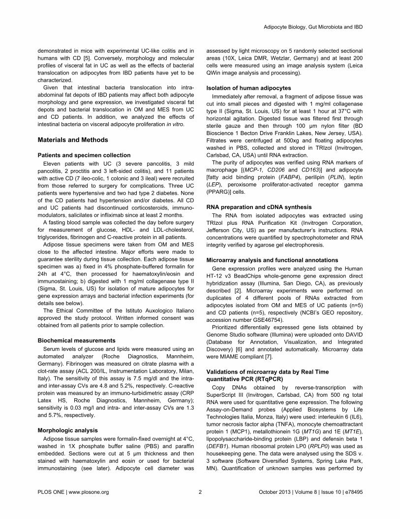
Differences in Visceral Fat and Fat Bacterial Colonization between Ulcerative Colitis and Crohn’s Disease. An In Vivo and In Vitro Study Alessandra Zulian 1 , Raffaella Cancello 2 , Chiara Ruocco 1 , Davide Gentilini 2 , Anna Maria Di Blasio 2 , Piergiorgio Danelli 3 , Giancarlo Micheletto 4 , Elisabetta Cesana 5 , Cecilia Invitti 6* 1 Diabetes Research Laboratory, Istituto Auxologico Italiano, Milan, Italy, 2 Laboratory of Molecular Biology, Istituto Auxologico Italiano, Milan, Italy, 3 Department of Clinical Sciences, Luigi Sacco Hospital, University of Milan, Milan, Italy, 4 Department of Medical and Surgical Pathophysiology and Transplants, University of Milan, Milan, Italy, 5 Microbiology Laboratory, Istituto Auxologico Italiano, Milan, Italy, 6 Department of Medical Sciences and Rehabilitation, Istituto Auxologico Italiano, Milan, Italy Abstract Crohn’s disease (CD) is notably characterized by the expansion of visceral fat with small adipocytes expressing a high proportion of anti-inflammatory genes. Conversely, visceral fat depots in ulcerative colitis (UC) patients have never been characterized. Our study aims were a) to compare adipocyte morphology and gene expression profile and bacterial translocation in omental (OM) and mesenteric (MES) adipose tissue of patients with UC and CD, and b) to investigate the effect of bacterial infection on adipocyte proliferation in vitro. Specimens of OM and MES were collected from 11 UC and 11 CD patients, processed and examined by light microscopy. Gene expression profiles were evaluated in adipocytes isolated from visceral adipose tissue using microarray and RTqPCR validations. Bacteria within adipose tissue were immuno-detected by confocal scanning laser microscopy. Adipocytes were incubated with Enterococcus faecalis and cells counted after 24h. Morphology and molecular profile of OM and MES revealed that UC adipose tissue is less inflamed than CD adipose tissue. Genes linked to inflammation, bacterial response, chemotaxis and angiogenesis were down-regulated in adipocytes from UC compared to CD, whereas genes related to metallothioneins, apoptosis pathways and growth factor binding were up-regulated. A dense perinuclear positivity for Enterococcus faecalis was detected in visceral adipocytes from CD, whereas positivity was weak in UC. In vitro bacterial infection was associated with a five-fold increase in the proliferation rate of OM preadipocytes. Compared to UC, visceral adipose tissue from CD is more inflamed and more colonized by intestinal bacteria, which increase adipocyte proliferation. The influence of bacteria stored within adipocytes on the clinical course of IBD warrants further investigations. Citation: Zulian A, Cancello R, Ruocco C, Gentilini D, Di Blasio AM, et al. (2013) Differences in Visceral Fat and Fat Bacterial Colonization between Ulcerative Colitis and Crohn’s Disease. An In Vivo and In Vitro Study. PLoS ONE 8(10): e78495. doi:10.1371/journal.pone.0078495 Editor: Markus M. Heimesaat, Charité, Campus Benjamin Franklin, Germany Received July 15, 2013; Accepted September 13, 2013; Published October 24, 2013 Copyright: © 2013 Zulian et al. This is an open-access article distributed under the terms of the Creative Commons Attribution License, which permits unrestricted use, distribution, and reproduction in any medium, provided the original author and source are credited. Funding: This study was supported by Istituto Auxologico Italiano. Istituto Auxologico Italiano, funder of this study, had no role in study design, data collection and analysis, decision to publish, or preparation of the manuscript. Competing interests: The authors have declared that no competing interests exist. * E-mail: [email protected] Introduction The two major forms of chronic inflammatory bowel disease (IBD), ulcerative colitis (UC) and Crohn’s disease (CD), are believed to result from interactions between the environment, genetic predisposition, unbalanced host-commensal microbiota and a widespread immune defect [1]. However, UC and CD present distinct pathogenic mechanisms and characteristics [1], one being the expansion of mesenteric fat surrounding inflamed intestinal tracts (“creeping fat”), which is typical for CD and absent in UC. We have recently demonstrated that, in patients with active CD, omental (OM) visceral adipose tissue shows the same inflammatory morphology and molecular profile of creeping fat (MES) [2]. In addition, we reported that OM adipocytes from CD patients are smaller than those of normal-weight subjects and express a higher proportion of anti-inflammatory genes compared to obese patients. These findings suggest that the adipocyte undergoes beneficial changes [3] and we hypothesized that the intra-abdominal fat expansion of CD is a protective phenomenon aimed at controlling the inflammatory response and preventing dissemination of intestinal bacteria. It is worth recalling that gut microbiota and/or their products have been shown to promote inflammation and determine the anti-inflammatory response of visceral adipose tissue in obese patients [4]. Further, bacterial translocation to MES has been PLOS ONE | www.plosone.org 1 October 2013 | Volume 8 | Issue 10 | e78495
-
Upload
independent -
Category
Documents
-
view
0 -
download
0
Transcript of Differences in Visceral Fat and Fat Bacterial Colonization between Ulcerative Colitis and...
Differences in Visceral Fat and Fat Bacterial Colonizationbetween Ulcerative Colitis and Crohn’s Disease. An InVivo and In Vitro StudyAlessandra Zulian1, Raffaella Cancello2, Chiara Ruocco1, Davide Gentilini2, Anna Maria Di Blasio2,Piergiorgio Danelli3, Giancarlo Micheletto4, Elisabetta Cesana5, Cecilia Invitti6*
1 Diabetes Research Laboratory, Istituto Auxologico Italiano, Milan, Italy, 2 Laboratory of Molecular Biology, Istituto Auxologico Italiano, Milan, Italy,3 Department of Clinical Sciences, Luigi Sacco Hospital, University of Milan, Milan, Italy, 4 Department of Medical and Surgical Pathophysiology andTransplants, University of Milan, Milan, Italy, 5 Microbiology Laboratory, Istituto Auxologico Italiano, Milan, Italy, 6 Department of Medical Sciences andRehabilitation, Istituto Auxologico Italiano, Milan, Italy
Abstract
Crohn’s disease (CD) is notably characterized by the expansion of visceral fat with small adipocytes expressing ahigh proportion of anti-inflammatory genes. Conversely, visceral fat depots in ulcerative colitis (UC) patients havenever been characterized. Our study aims were a) to compare adipocyte morphology and gene expression profileand bacterial translocation in omental (OM) and mesenteric (MES) adipose tissue of patients with UC and CD, and b)to investigate the effect of bacterial infection on adipocyte proliferation in vitro. Specimens of OM and MES werecollected from 11 UC and 11 CD patients, processed and examined by light microscopy. Gene expression profileswere evaluated in adipocytes isolated from visceral adipose tissue using microarray and RTqPCR validations.Bacteria within adipose tissue were immuno-detected by confocal scanning laser microscopy. Adipocytes wereincubated with Enterococcus faecalis and cells counted after 24h. Morphology and molecular profile of OM and MESrevealed that UC adipose tissue is less inflamed than CD adipose tissue. Genes linked to inflammation, bacterialresponse, chemotaxis and angiogenesis were down-regulated in adipocytes from UC compared to CD, whereasgenes related to metallothioneins, apoptosis pathways and growth factor binding were up-regulated. A denseperinuclear positivity for Enterococcus faecalis was detected in visceral adipocytes from CD, whereas positivity wasweak in UC. In vitro bacterial infection was associated with a five-fold increase in the proliferation rate of OMpreadipocytes. Compared to UC, visceral adipose tissue from CD is more inflamed and more colonized by intestinalbacteria, which increase adipocyte proliferation. The influence of bacteria stored within adipocytes on the clinicalcourse of IBD warrants further investigations.
Citation: Zulian A, Cancello R, Ruocco C, Gentilini D, Di Blasio AM, et al. (2013) Differences in Visceral Fat and Fat Bacterial Colonization betweenUlcerative Colitis and Crohn’s Disease. An In Vivo and In Vitro Study. PLoS ONE 8(10): e78495. doi:10.1371/journal.pone.0078495
Editor: Markus M. Heimesaat, Charité, Campus Benjamin Franklin, Germany
Received July 15, 2013; Accepted September 13, 2013; Published October 24, 2013
Copyright: © 2013 Zulian et al. This is an open-access article distributed under the terms of the Creative Commons Attribution License, which permitsunrestricted use, distribution, and reproduction in any medium, provided the original author and source are credited.
Funding: This study was supported by Istituto Auxologico Italiano. Istituto Auxologico Italiano, funder of this study, had no role in study design, datacollection and analysis, decision to publish, or preparation of the manuscript.
Competing interests: The authors have declared that no competing interests exist.
* E-mail: [email protected]
Introduction
The two major forms of chronic inflammatory bowel disease(IBD), ulcerative colitis (UC) and Crohn’s disease (CD), arebelieved to result from interactions between the environment,genetic predisposition, unbalanced host-commensal microbiotaand a widespread immune defect [1]. However, UC and CDpresent distinct pathogenic mechanisms and characteristics [1],one being the expansion of mesenteric fat surroundinginflamed intestinal tracts (“creeping fat”), which is typical for CDand absent in UC.
We have recently demonstrated that, in patients with activeCD, omental (OM) visceral adipose tissue shows the same
inflammatory morphology and molecular profile of creeping fat(MES) [2]. In addition, we reported that OM adipocytes fromCD patients are smaller than those of normal-weight subjectsand express a higher proportion of anti-inflammatory genescompared to obese patients. These findings suggest that theadipocyte undergoes beneficial changes [3] and wehypothesized that the intra-abdominal fat expansion of CD is aprotective phenomenon aimed at controlling the inflammatoryresponse and preventing dissemination of intestinal bacteria.
It is worth recalling that gut microbiota and/or their productshave been shown to promote inflammation and determine theanti-inflammatory response of visceral adipose tissue in obesepatients [4]. Further, bacterial translocation to MES has been
PLOS ONE | www.plosone.org 1 October 2013 | Volume 8 | Issue 10 | e78495
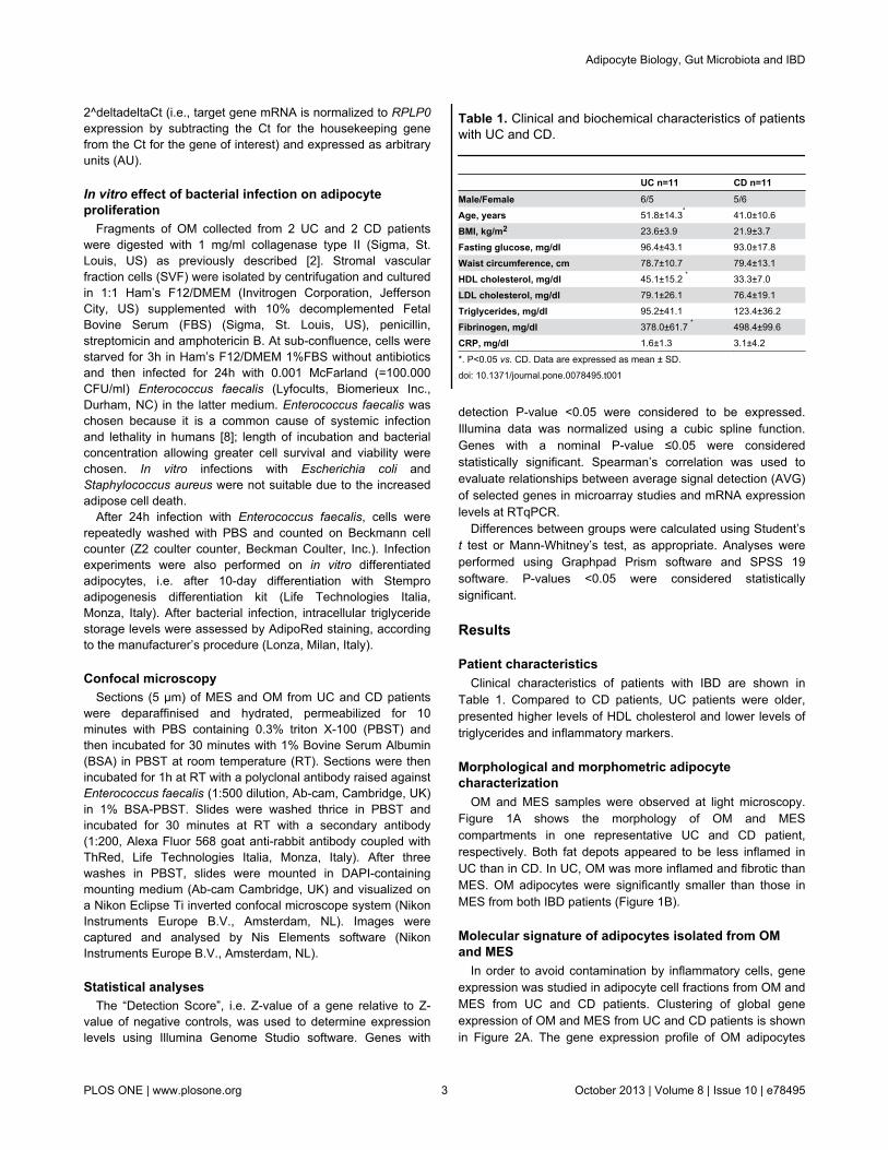
demonstrated in mice with experimental UC-like colitis and inhumans with CD [5]. Conversely, morphology and molecularprofiles of visceral fat in UC as well as the effects of bacterialtranslocation on adipocytes from IBD patients have yet to becharacterized.
Given that intestinal bacteria translocation into intra-abdominal fat depots of IBD patients may affect both adipocytemorphology and gene expression, we investigated visceral fatdepots and bacterial translocation in OM and MES from UCand CD patients. In addition, we analyzed the effects ofintestinal bacteria on visceral adipocyte proliferation in vitro.
Materials and Methods
Patients and specimen collectionEleven patients with UC (3 severe pancolitis, 3 mild
pancolitis, 2 proctitis and 3 left-sided colitis), and 11 patientswith active CD (7 ileo-colic, 1 colonic and 3 ileal) were recruitedfrom those referred to surgery for complications. Three UCpatients were hypertensive and two had type 2 diabetes. Noneof the CD patients had hypertension and/or diabetes. All CDand UC patients had discontinued corticosteroids, immuno-modulators, salicilates or infliximab since at least 2 months.
A fasting blood sample was collected the day before surgeryfor measurement of glucose, HDL- and LDL-cholesterol,triglycerides, fibrinogen and C-reactive protein in all patients.
Adipose tissue specimens were taken from OM and MESclose to the affected intestine. Major efforts were made toguarantee sterility during tissue collection. Each adipose tissuespecimen was a) fixed in 4% phosphate-buffered formalin for24h at 4°C, then processed for haematoxylin/eosin andimmunostaining; b) digested with 1 mg/ml collagenase type II(Sigma, St. Louis, US) for isolation of mature adipocytes forgene expression arrays and bacterial infection experiments (fordetails see below).
The Ethical Committee of the Istituto Auxologico Italianoapproved the study protocol. Written informed consent wasobtained from all patients prior to sample collection.
Biochemical measurementsSerum levels of glucose and lipids were measured using an
automated analyzer (Roche Diagnostics, Mannheim,Germany). Fibrinogen was measured on citrate plasma with aclot-rate assay (ACL 200/IL, Instrumentation Laboratory, Milan,Italy). The sensitivity of this assay is 7.5 mg/dl and the intra-and inter-assay CVs are 4.8 and 5.2%, respectively. C-reactiveprotein was measured by an immuno-turbidimetric assay (CRPLatex HS, Roche Diagnostics, Mannheim, Germany);sensitivity is 0.03 mg/l and intra- and inter-assay CVs are 1.3and 5.7%, respectively.
Morphologic analysisAdipose tissue samples were formalin-fixed overnight at 4°C,
washed in 1X phosphate buffer saline (PBS) and paraffinembedded. Sections were cut at 5 µm thickness and thenstained with haematoxylin and eosin or used for bacterialimmunostaining (see later). Adipocyte cell diameter was
assessed by light microscopy on 5 randomly selected sectionalareas (10X, Leica DMR, Wetzlar, Germany) and at least 200cells were measured using an image analysis system (LeicaQWin image analysis and processing).
Isolation of human adipocytesImmediately after removal, a fragment of adipose tissue was
cut into small pieces and digested with 1 mg/ml collagenasetype II (Sigma, St. Louis, US) for at least 1 hour at 37°C withhorizontal agitation. Digested tissue was filtered first throughsterile gauze and then through 100 µm nylon filter (BDBioscience 1 Becton Drive Franklin Lakes, New Jersey, USA).Filtrates were centrifuged at 500xg and floating adipocyteswashed in PBS, collected and stored in TRIzol (Invitrogen,Carlsbad, CA, USA) until RNA extraction.
The purity of adipocytes was verified using RNA markers ofmacrophage [(MCP-1, CD206 and CD163)] and adipocyte[fatty acid binding protein (FABP4), perilipin (PLIN), leptin(LEP), peroxisome proliferator-activated receptor gamma(PPARG)] cells.
RNA preparation and cDNA synthesisThe RNA from isolated adipocytes was extracted using
TRIzol plus RNA Purification Kit (Invitrogen Corporation,Jefferson City, US) as per manufacturer’s instructions. RNAconcentrations were quantified by spectrophotometer and RNAintegrity verified by agarose gel electrophoresis.
Microarray analysis and functional annotationsGene expression profiles were analyzed using the Human
HT-12 v3 BeadChips whole-genome gene expression directhybridization assay (Illumina, San Diego, CA), as previouslydescribed [2]. Microarray experiments were performed onduplicates of 4 different pools of RNAs extracted fromadipocytes isolated from OM and MES of UC patients (n=5)and CD patients (n=5), respectively (NCBI’s GEO repository,accession number GSE46754).
Prioritized differentially expressed gene lists obtained byGenome Studio software (Illumina) were uploaded onto DAVID(Database for Annotation, Visualization, and IntegratedDiscovery) [6] and annotated automatically. Microarray datawere MIAME compliant [7].
Validations of microarray data by Real Timequantitative PCR (RTqPCR)
Copy DNAs obtained by reverse-transcription withSuperScript III (Invitrogen, Carlsbad, CA) from 500 ng totalRNA were used for quantitative gene expression. The followingAssay-on-Demand probes (Applied Biosystems by LifeTechnologies Italia, Monza, Italy) were used: interleukin 6 (IL6),tumor necrosis factor alpha (TNFA), monocyte chemoattractantprotein 1 (MCP1), metallothionein 1G (MT1G) and 1E (MT1E),lipopolysaccharide-binding protein (LBP) and defensin beta 1(DEFB1). Human ribosomal protein LP0 (RPLP0) was used ashousekeeping gene. The data were analysed using the SDS v.3 software (Software Diversified Systems, Spring Lake Park,MN). Quantification of unknown samples was performed by
Adipocyte Biology, Gut Microbiota and IBD
PLOS ONE | www.plosone.org 2 October 2013 | Volume 8 | Issue 10 | e78495
2^deltadeltaCt (i.e., target gene mRNA is normalized to RPLP0expression by subtracting the Ct for the housekeeping genefrom the Ct for the gene of interest) and expressed as arbitraryunits (AU).
In vitro effect of bacterial infection on adipocyteproliferation
Fragments of OM collected from 2 UC and 2 CD patientswere digested with 1 mg/ml collagenase type II (Sigma, St.Louis, US) as previously described [2]. Stromal vascularfraction cells (SVF) were isolated by centrifugation and culturedin 1:1 Ham’s F12/DMEM (Invitrogen Corporation, JeffersonCity, US) supplemented with 10% decomplemented FetalBovine Serum (FBS) (Sigma, St. Louis, US), penicillin,streptomicin and amphotericin B. At sub-confluence, cells werestarved for 3h in Ham’s F12/DMEM 1%FBS without antibioticsand then infected for 24h with 0.001 McFarland (=100.000CFU/ml) Enterococcus faecalis (Lyfocults, Biomerieux Inc.,Durham, NC) in the latter medium. Enterococcus faecalis waschosen because it is a common cause of systemic infectionand lethality in humans [8]; length of incubation and bacterialconcentration allowing greater cell survival and viability werechosen. In vitro infections with Escherichia coli andStaphylococcus aureus were not suitable due to the increasedadipose cell death.
After 24h infection with Enterococcus faecalis, cells wererepeatedly washed with PBS and counted on Beckmann cellcounter (Z2 coulter counter, Beckman Coulter, Inc.). Infectionexperiments were also performed on in vitro differentiatedadipocytes, i.e. after 10-day differentiation with Stemproadipogenesis differentiation kit (Life Technologies Italia,Monza, Italy). After bacterial infection, intracellular triglyceridestorage levels were assessed by AdipoRed staining, accordingto the manufacturer’s procedure (Lonza, Milan, Italy).
Confocal microscopySections (5 µm) of MES and OM from UC and CD patients
were deparaffinised and hydrated, permeabilized for 10minutes with PBS containing 0.3% triton X-100 (PBST) andthen incubated for 30 minutes with 1% Bovine Serum Albumin(BSA) in PBST at room temperature (RT). Sections were thenincubated for 1h at RT with a polyclonal antibody raised againstEnterococcus faecalis (1:500 dilution, Ab-cam, Cambridge, UK)in 1% BSA-PBST. Slides were washed thrice in PBST andincubated for 30 minutes at RT with a secondary antibody(1:200, Alexa Fluor 568 goat anti-rabbit antibody coupled withThRed, Life Technologies Italia, Monza, Italy). After threewashes in PBST, slides were mounted in DAPI-containingmounting medium (Ab-cam Cambridge, UK) and visualized ona Nikon Eclipse Ti inverted confocal microscope system (NikonInstruments Europe B.V., Amsterdam, NL). Images werecaptured and analysed by Nis Elements software (NikonInstruments Europe B.V., Amsterdam, NL).
Statistical analysesThe “Detection Score”, i.e. Z-value of a gene relative to Z-
value of negative controls, was used to determine expressionlevels using Illumina Genome Studio software. Genes with
detection P-value <0.05 were considered to be expressed.Illumina data was normalized using a cubic spline function.Genes with a nominal P-value ≤0.05 were consideredstatistically significant. Spearman’s correlation was used toevaluate relationships between average signal detection (AVG)of selected genes in microarray studies and mRNA expressionlevels at RTqPCR.
Differences between groups were calculated using Student’st test or Mann-Whitney’s test, as appropriate. Analyses wereperformed using Graphpad Prism software and SPSS 19software. P-values <0.05 were considered statisticallysignificant.
Results
Patient characteristicsClinical characteristics of patients with IBD are shown in
Table 1. Compared to CD patients, UC patients were older,presented higher levels of HDL cholesterol and lower levels oftriglycerides and inflammatory markers.
Morphological and morphometric adipocytecharacterization
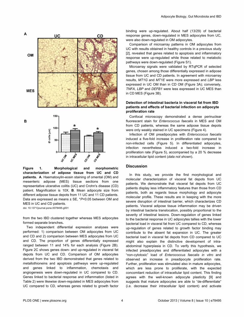
OM and MES samples were observed at light microscopy.Figure 1A shows the morphology of OM and MEScompartments in one representative UC and CD patient,respectively. Both fat depots appeared to be less inflamed inUC than in CD. In UC, OM was more inflamed and fibrotic thanMES. OM adipocytes were significantly smaller than those inMES from both IBD patients (Figure 1B).
Molecular signature of adipocytes isolated from OMand MES
In order to avoid contamination by inflammatory cells, geneexpression was studied in adipocyte cell fractions from OM andMES from UC and CD patients. Clustering of global geneexpression of OM and MES from UC and CD patients is shownin Figure 2A. The gene expression profile of OM adipocytes
Table 1. Clinical and biochemical characteristics of patientswith UC and CD.
UC n=11 CD n=11Male/Female 6/5 5/6
Age, years 51.8±14.3* 41.0±10.6
BMI, kg/m2 23.6±3.9 21.9±3.7
Fasting glucose, mg/dl 96.4±43.1 93.0±17.8
Waist circumference, cm 78.7±10.7 79.4±13.1
HDL cholesterol, mg/dl 45.1±15.2 * 33.3±7.0
LDL cholesterol, mg/dl 79.1±26.1 76.4±19.1
Triglycerides, mg/dl 95.2±41.1 123.4±36.2
Fibrinogen, mg/dl 378.0±61.7 * 498.4±99.6
CRP, mg/dl 1.6±1.3 3.1±4.2
*. P<0.05 vs. CD. Data are expressed as mean ± SD.doi: 10.1371/journal.pone.0078495.t001
Adipocyte Biology, Gut Microbiota and IBD
PLOS ONE | www.plosone.org 3 October 2013 | Volume 8 | Issue 10 | e78495
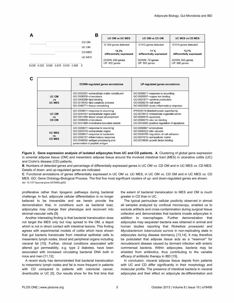
from the two IBD clustered together whereas MES adipocytesformed separate branches.
Two independent differential expression analyses wereperformed: 1) comparison between OM adipocytes from UCand CD and 2) comparison between MES adipocytes from UCand CD. The proportion of genes differentially expressedranged between 11 and 14% for each analysis (Figure 2B).Figure 2C shows genes down- and up-regulated in visceral fatdepots from UC and CD. Comparison of OM adipocytesderived from the two IBD demonstrated that genes related tometallothioneins and apoptosis pathways were up-regulatedand genes linked to inflammation, chemotaxis andangiogenesis were down-regulated in UC compared to CD.Genes linked to bacterial response and inflammation (listed inTable 2) were likewise down-regulated in MES adipocytes fromUC compared to CD, whereas genes related to growth factor
Figure 1. Morphological and morphometriccharacterization of adipose tissue from UC and CDpatients. A. Haematoxylin-eosin staining of omental (OM) andmesenteric adipose (MES) tissue sections from onerepresentative ulcerative colitis (UC) and Crohn's disease (CD)patient. Magnification is 10X. B. Mean adipocyte size fromdifferent adipose tissue depots from 11 UC and 11 CD patients.Data are expressed as means ± SE, *P<0.05 between OM andMES in UC and CD patients.doi: 10.1371/journal.pone.0078495.g001
binding were up-regulated. About half (13/29) of bacterialresponse genes, down-regulated in MES adipocytes from UC,were also down-regulated in OM adipocytes.
Comparison of microarray patterns in OM adipocytes fromUC with results obtained in healthy controls in a previous study[2], revealed that genes related to apoptosis and inflammatoryresponse were up-regulated while those related to metabolicpathways were down-regulated (Figure S1).
Microarray signals were validated by RTqPCR of selectedgenes, chosen among those differentially expressed in adiposetissue from UC and CD patients. In agreement with microarrayresults, MT1G and MT1E were more expressed and LBP lessexpressed in UC OM than in CD OM (Figure 3A); conversely,TNFA, LBP and DEFB1 were less expressed in UC MES thanin CD MES (Figure 3B).
Detection of intestinal bacteria in visceral fat from IBDpatients and effects of bacterial infection on adipocyteproliferation rate
Confocal microscopy demonstrated a dense perinuclearfluorescent stain for Enterococcus faecalis in MES and OMfrom CD patients, whereas the same adipose tissue depotswere only weakly stained in UC specimens (Figure 4).
Infection of OM preadipocytes with Enteroccocus faecalisinduced a five-fold increase in proliferation rate compared tonon-infected cells (Figure 5). In differentiated adipocytes,infection nevertheless induced a two-fold increase inproliferation rate (Figure 5), accompanied by a 20 % decreasein intracellular lipid content (data not shown).
Discussion
In this study, we provide the first morphological andmolecular characterization of visceral fat depots from UCpatients. We demonstrate that visceral fat depots from UCpatients display less inflammatory features than those from CDpatients, both as regards tissue morphology and adipocytemolecular profile. These results are in keeping with the moresevere disruption of intestinal barrier, which characterizes CDpatients. Visceral adipose tissue inflammation may be drivenby intestinal bacteria translocation, possibly proportional to theseverity of intestinal lesions. Down-regulation of genes linkedto the bacterial response in UC adipocytes tallies with the lowerbacterial load in visceral fat from UC compared to CD, whereasup-regulation of genes related to growth factor binding maycontribute to the absent fat expansion in UC. The greaterbacterial load in visceral fat depots from CD compared to UCmight also explain the distinctive development of intra-abdominal hyperplasia in CD. To verify this hypothesis, weinfected preadipocytes and differentiated adipocytes with a“non-cytotoxic” load of Enterococcus faecalis in vitro andobserved an increase in preadipocyte proliferation rate.Further, proliferation was stimulated also in mature adipocytes,which are less prone to proliferate, with the expectedconcomitant reduction of intracellular lipid content. This findingagrees with the well-known adipocyte plasticity [9] andsuggests that mature adipocytes are able to “de-differentiate”(i.e. decrease their intracellular lipid content) and activate
Adipocyte Biology, Gut Microbiota and IBD
PLOS ONE | www.plosone.org 4 October 2013 | Volume 8 | Issue 10 | e78495
proliferative rather than lipogenic pathways during bacterialchallenge. In fact, adipocyte cellular differentiation is no longerbelieved to be irreversible and we herein provide thedemonstration that, in conditions such as bacterial load,adipocytes may change their phenotype and reconvert intostromal-vascular cells [9].
Another interesting finding is that bacterial translocation doesnot target the MES only but may spread to the OM, a depotwhich is not in direct contact with intestinal lesions. This findingagrees with experimental models of colitis which have shownthat gut bacteria translocate from intestinal epithelial cells tomesenteric lymph nodes, blood and peripheral organs includingvisceral fat [10]. Further, clinical conditions associated withaltered gut permeability, e.g. type 2 diabetes, have beenassociated with increased circulating bacterial DNA both inmice and men [11,12].
A recent study has demonstrated that bacterial translocationto mesenteric lymph nodes and fat is more frequent in patientswith CD compared to patients with colorectal cancer,diverticulitis or UC [5]. Our results show for the first time that
the extent of bacterial translocation to MES and OM is muchgreater in CD than in UC.
The typical perinuclear cellular positivity observed in almostall samples analyzed by confocal microscopy, enabled us toexclude artifacts and cross-contamination during surgical tissuecollection and demonstrates that bacteria invade adipocytes inaddition to macrophages. Further demonstration thatadipocytes may sequester bacteria was obtained in animal andhuman studies reporting that Rickettsia prowazekii andMycobacterium tuberculosis survive in non-replicating state inadipocytes during disease dormancy [13,14]. It may thereforebe postulated that adipose tissue acts as a "reservoir" forrecrudescent disease caused by dormant infection with entericcommensal bacteria. Within adipocytes, bacteria may beshielded from antibiotics, thus contributing to the variableefficacy of antibiotic therapy in IBD [15].
In conclusion, visceral adipose tissue depots from patientswith UC and CD differ significantly in their morphology andmolecular profile. The presence of intestinal bacteria in visceraladipocytes and their effect on adipocyte de-differentiation and
Figure 2. Gene expression analysis of isolated adipocytes from UC and CD patients. A. Clustering of global gene expressionin omental adipose tissue (OM) and mesenteric adipose tissue around the involved intestinal tract (MES) in ulcerative colitis (UC)and Crohn's disease (CD) patients.B. Numbers of detected genes and percentage of differentially expressed genes in UC OM vs. CD OM and in UC MES vs. CD MES.Details of down- and up-regulated genes are indicated.C. Functional annotations of genes differentially expressed in UC OM vs. UC MES, in UC OM vs. CD OM and in UC MES vs. CDMES. GO: Gene Ontology-Biological Process. The first five most significant clusters of up- and down-regulated genes are shown.doi: 10.1371/journal.pone.0078495.g002
Adipocyte Biology, Gut Microbiota and IBD
PLOS ONE | www.plosone.org 5 October 2013 | Volume 8 | Issue 10 | e78495
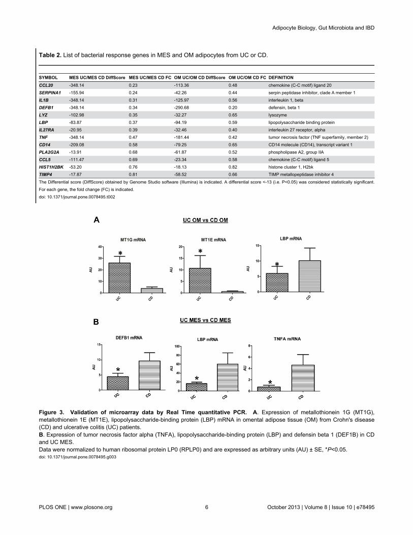
Table 2. List of bacterial response genes in MES and OM adipocytes from UC or CD.
SYMBOL MES UC/MES CD DiffScore MES UC/MES CD FC OM UC/OM CD DiffScore OM UC/OM CD FC DEFINITIONCCL20 -348.14 0.23 -113.36 0.48 chemokine (C-C motif) ligand 20
SERPINA1 -155.94 0.24 -42.26 0.44 serpin peptidase inhibitor, clade A member 1
IL1B -348.14 0.31 -125.97 0.56 interleukin 1, beta
DEFB1 -348.14 0.34 -290.68 0.20 defensin, beta 1
LYZ -102.98 0.35 -32.27 0.65 lysozyme
LBP -83.87 0.37 -94.19 0.59 lipopolysaccharide binding protein
IL27RA -20.95 0.39 -32.46 0.40 interleukin 27 receptor, alpha
TNF -348.14 0.47 -181.44 0.42 tumor necrosis factor (TNF superfamily, member 2)
CD14 -209.08 0.58 -79.25 0.65 CD14 molecule (CD14), transcript variant 1
PLA2G2A -13.91 0.68 -61.87 0.52 phospholipase A2, group IIA
CCL5 -111.47 0.69 -23.34 0.58 chemokine (C-C motif) ligand 5
HIST1H2BK -53.20 0.76 -18.13 0.82 histone cluster 1, H2bk
TIMP4 -17.87 0.81 -58.52 0.66 TIMP metallopeptidase inhibitor 4
The Differential score (DiffScore) obtained by Genome Studio software (Illumina) is indicated. A differential score <-13 (i.e. P<0.05) was considered statistically significant.For each gene, the fold change (FC) is indicated.doi: 10.1371/journal.pone.0078495.t002
Figure 3. Validation of microarray data by Real Time quantitative PCR. A. Expression of metallothionein 1G (MT1G),metallothionein 1E (MT1E), lipopolysaccharide-binding protein (LBP) mRNA in omental adipose tissue (OM) from Crohn's disease(CD) and ulcerative colitis (UC) patients.B. Expression of tumor necrosis factor alpha (TNFA), lipopolysaccharide-binding protein (LBP) and defensin beta 1 (DEF1B) in CDand UC MES.Data were normalized to human ribosomal protein LP0 (RPLP0) and are expressed as arbitrary units (AU) ± SE, *P<0.05.doi: 10.1371/journal.pone.0078495.g003
Adipocyte Biology, Gut Microbiota and IBD
PLOS ONE | www.plosone.org 6 October 2013 | Volume 8 | Issue 10 | e78495

proliferation may explain adipocyte hyperplasia in creeping fat.The role of bacteria stored into visceral fat on the clinicalcourse of IBD deserves further investigation.
Figure 4. Detection of intestinal bacteria in visceral fat from UC and CD patients. Fluorescent immunodetection ofEnterococcus faecalis (red) in omental adipose tissue (OM) and mesenteric adipose tissue (MES) from 1 representative ulcerativecolitis (UC) and 1 Crohn's disease (CD) patient by confocal microscopy. Magnification is 40X. Nuclei are counterstained in blue(DAPI, 4',6-diamidin-2-fenilindolo).doi: 10.1371/journal.pone.0078495.g004
Adipocyte Biology, Gut Microbiota and IBD
PLOS ONE | www.plosone.org 7 October 2013 | Volume 8 | Issue 10 | e78495

Figure 5. Effects of bacterial infection on adipocyteproliferation rate. Mean cell number ± SE of omentalpreadipocytes (SVF) and 10-day differentiated adipocytes(ADIPO) from 4 patients (2UC and 2CD) after 24h incubationwith (SVF EF and ADIPO EF) or without (SVF C and ADIPO C)0.001 McFarland Enterococcus faecalis. *P<0.05.doi: 10.1371/journal.pone.0078495.g005
Adipocyte Biology, Gut Microbiota and IBD
PLOS ONE | www.plosone.org 8 October 2013 | Volume 8 | Issue 10 | e78495
Supporting Information
Figure S1. UP and DOWN-regulated genes in UC versuscontrol omental adipocytes. Gene Ontology (GO) BiologicalProcess of the first five clusters of up-regulated and down-regulated genes in omental adipocytes (OM) of ulcerativecolitis patients (UC) compared to controls. Genes belonging toeach category are shown.
(TIF)
Author Contributions
Conceived and designed the experiments: AZ RC PD CI.Performed the experiments: AZ RC CR DG. Analyzed the data:AZ RC CI. Contributed reagents/materials/analysis tools:AMDB GM EC. Wrote the manuscript: AZ RC CI.
References
1. Bamias G, Pizarro T, Cominelli F (2011) New Paradigms in thePathogenesis of IBD. In: RD Cohen, Inflammatory Bowel Disease:Diagnosis and Therapeutics. pp. 41-57.
2. Zulian A, Cancello R, Micheletto G, Gentilini D, Gilardini L et al. (2012)Visceral adipocytes: old actors in obesity and new protagonists inCrohn's disease? Gut 61: 86-94. doi:10.1136/gutjnl-2012-302514b.33.PubMed: 21930728.
3. Goossens GH, Moors CC, van der Zijl NJ, Venteclef N, Alili R et al.(2012) Valsartan improves adipose tissue function in humans withimpaired glucose metabolism: a randomized placebo-controlled double-blind trial. PLOS ONE 7: e39930. PubMed: 22768174.
4. Shen J, Obin MS, Zhao L (2013) The gut microbiota, obesity andinsulin resistance. Mol Aspects Med 34: 39–58. doi:10.1016/j.mam.2012.11.001. PubMed: 23159341.
5. Peyrin-Biroulet L, Gonzalez F, Dubuquoy L, Rousseaux C, Dubuquoy Cet al. (2012) Mesenteric fat as a source of C reactive protein and as atarget for bacterial translocation in Crohn's disease. Gut 61: 78-85. doi:10.1136/gutjnl-2011-300370. PubMed: 21940721.
6. Huang da W, Sherman BT, Lempicki RA (2009) Systematic andintegrative analysis of large gene lists using DAVID bioinformaticsresources. Nat Protoc, 4: 44-57. PubMed: 19131956.
7. Brazma A (2009) Minimum Information About a Microarray Experiment(MIAME)--successes, failures, challenges. Scientific. World J 9:420-423. doi:10.1100/tsw.2009.57.
8. Goossens H (1999) The epidemiology of vancomycin-resistantenterococci. Curr Opin Infect Dis 12: 537–541. doi:10.1097/00001432-199912000-00002. PubMed: 17035818.
9. Cinti S (2012) The adipose organ at a glance. Dis Model. J Mech 5:588-594.
10. Berg RD (1999) Bacterial translocation from the gastrointestinal tract.Adv Exp Med Biol 473: 11-30. doi:10.1007/978-1-4615-4143-1_2.PubMed: 10659341.
11. Amar J, Chabo C, Waget A, Klopp P, Vachoux C et al. (2011) Intestinalmucosal adherence and translocation of commensal bacteria at theearly onset of type 2 diabetes: molecular mechanisms and probiotictreatment. EMBO. Mol Med 3: 559-572.
12. Amar J, Serino M, Lange C, Chabo C, Iacovoni J et al. (2011)Involvement of tissue bacteria in the onset of diabetes in humans:evidence for a concept. Diabetologia 54: 3055-3061. doi:10.1007/s00125-011-2329-8. PubMed: 21976140.
13. Bechah Y, Paddock CD, Capo C, Mege JL, Raoult D (2010) Adiposetissue serves as a reservoir for recrudescent Rickettsia prowazekiiinfection in a mouse model. PLOS ONE 5: e8547. doi:10.1371/journal.pone.0008547. PubMed: 20049326.
14. Neyrolles O, Hernández-Pando R, Pietri-Rouxel F, Fornès P, Tailleux Let al. (2006) Is adipose tissue a place for Mycobacterium tuberculosispersistence? PLOS ONE 1: e43. doi:10.1371/journal.pone.0000043.PubMed: 17183672.
15. Khan KJ, Ullman TA, Ford AC, Abreu MT, Abadir A et al. (2011)Antibiotic therapy in inflammatory bowel disease: a systematic reviewand meta-analysis. Am J Gastroenterol 106: 661-673. doi:10.1038/ajg.2011.72. PubMed: 21407187.
Adipocyte Biology, Gut Microbiota and IBD
PLOS ONE | www.plosone.org 9 October 2013 | Volume 8 | Issue 10 | e78495