Somatosensory extinction for meaningful objects in a patient with right hemispheric stroke

Riquelme et al. BMC Neuroscience 2014, 15:10http://www.biomedcentral.com/1471-2202/15/10
RESEARCH ARTICLE Open Access
Differences in somatosensory processing due todominant hemispheric motor impairment incerebral palsyInmaculada Riquelme1,2, Iván Padrón3, Ignasi Cifre1, Ana M González-Roldán1 and Pedro Montoya1*
Abstract
Background: Although cerebral palsy (CP) is usually defined as a group of permanent motor disorders due tonon-progressive disturbances in the developing fetal or infant brain, recent research has shown that CP individualsare also characterized by altered somatosensory perception, increased pain and abnormal activation of corticalsomatosensory areas. The present study was aimed to examine hemispheric differences on somatosensory brainprocessing in individuals with bilateral CP and lateralized motor impairments compared with healthy controls.Nine CP individuals with left-dominant motor impairments (LMI) (age range 5–28 yrs), nine CP individuals withright-dominant motor impairments (RMI) (age range 7–29 yrs), and 12 healthy controls (age range 5–30 yrs)participated in the study. Proprioception, touch and pain thresholds, as well as somatosensory evoked potentials(SEP) elicited by tactile stimulation of right and left lips and thumbs were compared.
Results: Pain sensitivity was higher, and lip stimulation elicited greater beta power and more symmetrical SEPamplitudes in individuals with CP than in healthy controls. In addition, although there was no significant differencesbetween individuals with RMI and LMI on pain or touch sensitivity, lip and thumb stimulation elicited smaller betapower and more symmetrical SEP amplitudes in individuals with LMI than with RMI.
Conclusions: Our data revealed that brain processing of somatosensory stimulation was abnormal in CPindividuals. Moreover, this processing was different depending if they presented right- or left-dominant motorimpairments, suggesting that different mechanisms of sensorimotor reorganization should be involved in CPdepending on dominant side of motor impairment.
Keywords: Somatosensory processing, Cerebral palsy, Motor impairment, Hemispheric asymmetry
BackgroundCerebral palsy (CP) is defined as a group of permanentmotor disorders that are attributed to non-progressivedisturbances in the developing fetal or infant brain.Nevertheless, it has been recently shown that CP couldbe associated with somatosensory alterations, includingabnormal perception of touch and altered pain sensi-tivity [1]. Indeed, research on somatosensory processinghas revealed that individuals with CP are characterizedby poor tactile discrimination, stereognosis and proprio-ception [2-4], as well as increased pain [5] and abnormalactivation of cortical somatosensory areas [6]. Moreover,
* Correspondence: [email protected] Institute of Health Sciences (IUNICS), University of the BalearicIslands, Carretera de Valldemossa km 7.5, Palma 07122, SpainFull list of author information is available at the end of the article
© 2014 Riquelme et al.; licensee BioMed CentCommons Attribution License (http://creativecreproduction in any medium, provided the or
somatosensory parameters have been associated withclinical measures of motor impairment in persons withCP and other neurological pathologies such as multiplesclerosis, spinal cord injuries and cerebrovascular acci-dents [7-11]. Neuroimaging studies have also providedevidence of significant alterations in white matter fibersconnecting to somatosensory cortex, suggesting that CPinjuries reflect a disruption of sensory as well as motorpathways [12-14].Although neurophysiological mechanisms involved in
altered processing of bodily information in CP are stillunknown, there is evidence of an abnormal sensori-motor integration in hemiplegic CP [15], as it occurs inother movement disorders such as Parkinson’ disease,Huntington’s disease, dystonia, and tics [16-18]. Fur-thermore, it is possible that motor reorganization in
ral Ltd. This is an open access article distributed under the terms of the Creativeommons.org/licenses/by/2.0), which permits unrestricted use, distribution, andiginal work is properly cited.
Riquelme et al. BMC Neuroscience 2014, 15:10 Page 2 of 9http://www.biomedcentral.com/1471-2202/15/10
children with congenital hemiplegic CP occurs by preser-ving motor representations of the affected arm in the in-tact hemisphere. In this sense, studies with transcranialmagnetic stimulation (TMS) have provided evidence ofipsilateral corticospinal projections from the undamagedmotor cortex to the affected hand [15,19-24]. Moreover, ithas been proposed that somatosensory deficits could bedue to secondary effects provoked by motor limitation[25]. Thus, reduced and stereotypical pattern of sponta-neous movements in patients with hemiplegic CP wouldresult in abnormal sensory feedback and altered corticalreorganization, thus leading to asymmetric somatosensoryprocessing deficits [25,26]. A case study by Ragazzoni andcolleagues (2002) [27] has further showed that somatosen-sory function of the affected (right) arm was preserved,whereas motor function was poor despite fast-conductingipsilateral cortico-motoneuronal output from primary mo-tor cortex of the intact hemisphere to the affected arm.This finding seems to suggest that different forms ofmotor and somatosensory reorganization are involved incongenital brain injury, and that fast-conducting connec-tions between primary cortex areas and ipsilateral spinalcord are not sufficient for preservation or recovery offunction.In the present study, we examined the effects of latera-
lized motor impairment on somatosensory brain pro-cessing by using somatosensory evoked potentials (SEP)in persons with bilateral CP. For this purpose, wedivided our sample into CP individuals with either right-dominant dominant side of motor impairment (RMI) orCP left-dominant side of motor impairment (LMI) totest possible differences on propioception, touch andpain sensitivity, as well as early SEP components elicitedby non-painful stimulation of lips and thumbs at eachdominant side of motor impairment. Furthermore, wecompared these CP individuals with a group of healthyvolunteers to quantify the impairments in somatosensoryprocessing due to cerebral palsy.
MethodsParticipantsThirty individuals with bilateral cerebral palsy (CP) wererecruited from educational and occupational centersestablished in the island of Majorca (Spain), and invited toparticipate in the study. Patients were classified accordingwith the criteria of the Surveillance of Cerebral Palsy inEurope (SCPE, 2000) into the following categories: bila-teral spastic (including diplegia and quadriplegia), dyski-netic and ataxic. The diagnosis of spastic hemiplegia wasspecifically excluded. The hand motor function of eachparticipant was assessed by using the House FunctionalClassification [28]. Those CP individuals with no clearasymmetric motor performance were also excluded fromthe present study. Eighteen participants showing different
asymmetric motor performance were selected on the basisof their scores for both hands in the House FunctionalClassification (Table 1). The existence of an asymmetricalbrain damage in neuroimaging explorations was only con-firmed in 33% of the cases by checking their healthrecords.Individuals with CP were classified according to the
dominant side of motor impairments into two groups:1) nine CP participants with dominant right-sided motorimpairment (RMI) (3 females; mean age: 18 y 3mo, range5-28 y), and 2) nine CP with dominant left-sided motorimpairment (LMI) (3 females; mean age: 15 y 4mo, range7-29 y). Table 1 displays clinical characteristics for eachgroup of participants. Subjects or their parents reportedtheir age and sex. Type of cerebral palsy, gestational age,cognitive level, presence of epilepsy and medication wereobtained from participant’s health records. The level ofmotor impairment was also determined by using theGross Motor Function Classification Scale (GMFCS) [29].In addition, 12 right-handed healthy volunteers (3 fe-
males; mean age: 18 y 1 mo, range 5-30 y) were recruitedfrom educational centers and took part in the study.All participants granted written informed consent
according with the Declaration of Helsinki. In the caseof children, parents were informed and their writtenconsent was obtained. The study was approved by theEthics Committee of the Regional Government of theBalearic Islands (Spain).
Somatosensory assessmentBehavioral measures of somatosensory processing andsensitivity were obtained by using following tasks:
ProprioceptionProprioceptive skills were assessed by asking parti-cipants to perform unilateral movements of the upperlimb with eyes closed and to perform the same move-ment with the contralateral limb. Each single movementwas repeated five times and the average number ofcorrect trials (defined as a movement with less than 10degrees of difference with respect to the final positionof the target limb) was used as an index of propriocep-tive skills. This procedure has been used successfully inprevious studies [2].
TouchTouch sensitivity (expressed in g/mm2) was measured bi-laterally at two body locations (lips and thumb finger) byusing von Frey monofilaments [30] with different diame-ters (0.14-1.01 mm). The test was performed by touchingthe skin in a perpendicular way with the monofilament,pressing it slowly down until it buckles, holding it steadyduring 1.5 seconds, and removing it in the same wayas it was applied. After several trials to assure the
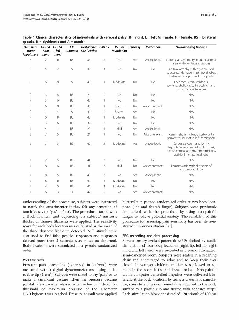
Table 1 Clinical characteristics of individuals with cerebral palsy (R = right, L = left M = male, F = female, BS = bilateralspastic, D = dyskinetic and A = ataxic)
Dominantmotor
impairment
HOUSErighthand
HOUSElefthand
CPsubgroup
Gestationalage (weeks)
GMFCS Mentalretardation
Epilepsy Medication Neuroimaging findings
R 2 6 BS 36 2 No Yes Antiepileptic Ventricular asymmetry in supratentorialarea, wide ventricular cavities
R 5 7 A 40 4 No No No Cortical atrophy with asymmetricalsubcortical damage in temporal lobes,brainstem atrophy and hypoplasia
R 6 8 A 40 1 Moderate No No Collapsed lateral ventriculi,periencephalic cavity in occipital and
posterior parietal areas
R 3 6 BS 28 2 No No No N/A
R 3 6 BS 40 1 No No No N/A
R 6 8 BS 40 1 Severe No Antidepressants N/A
R 1 4 A 40 2 Severe Yes No N/A
R 6 8 BS 40 1 Moderate No No N/A
R 3 6 BS 32 2 No No No N/A
L 4 1 BS 20 4 Mild Yes Antiepileptic N/A
L 7 5 BS 24 1 No No Musc. relaxant Asymmetry in Rolando cortex withperiventricular cyst in left hemisphere
L 7 5 BS 40 2 Moderate Yes Antiepileptic Corpus callosum and formixhypoplasia, septum pellucidum cyst,diffuse cortical atrophy, abnormal EEG
activity in left parietal lobe
L 7 5 BS 41 3 No No No N/A
L 8 6 BS 31 3 Mild No Antidepressants Leukomalacia with dilatation ofleft temporal lobe
L 8 5 BS 40 3 No Yes Antiepileptic N/A
L 8 6 BS 40 1 Moderate No No N/A
L 4 0 BS 40 3 Moderate No No N/A
L 6 3 D 42 5 No Yes Antidepressants N/A
Riquelme et al. BMC Neuroscience 2014, 15:10 Page 3 of 9http://www.biomedcentral.com/1471-2202/15/10
understanding of the procedure, subjects were instructedto notify the experimenter if they felt any sensation oftouch by saying “yes” or “no”. The procedure started witha thick filament and depending on subjects’ answers,thicker or thinner filaments were applied. The sensitivityscore for each body location was calculated as the mean ofthe three thinnest filaments detected. Null stimuli werealso used to find false positive responses and responsesdelayed more than 3 seconds were noted as abnormal.Body locations were stimulated in a pseudo-randomizedorder.
Pressure painPressure pain thresholds (expressed in kgf/cm2) weremeasured with a digital dynamometer and using a flatrubber tip (1 cm2). Subjects were asked to say ‘pain’ or tomake a significant gesture when the pressure becamepainful. Pressure was released when either pain detectionthreshold or maximum pressure of the algometer(13.0 kgf/cm2) was reached. Pressure stimuli were applied
bilaterally in pseudo-randomized order at two body loca-tions (lips and thumb finger). Subjects were previouslyfamiliarized with the procedure by using non-painfulranges to relieve potential anxiety. The reliability of thisprocedure for assessing pain sensitivity has been demon-strated in previous studies [31].
EEG recording and data processingSomatosensory evoked-potentials (SEP) elicited by tactilestimulation of four body locations (right lip, left lip, righthand and left hand) were recorded in a sound attenuated,semi-darkened room. Subjects were seated in a recliningchair and encouraged to relax and to keep their eyesclosed. In younger children, mother was allowed to re-main in the room if the child was anxious. Non-painfultactile computer-controlled impulses were delivered bila-terally at the body locations by using a pneumatic stimula-tor, consisting of a small membrane attached to the bodysurface by a plastic clip and fixated with adhesive strips.Each stimulation block consisted of 120 stimuli of 100 ms
Riquelme et al. BMC Neuroscience 2014, 15:10 Page 4 of 9http://www.biomedcentral.com/1471-2202/15/10
duration with an approximate pressure of 2 bars and avariable inter-stimulus interval of 1000 ± 50 ms. The orderof the stimulation blocks was counterbalanced across sub-jects. Similar tactile stimulation has been already used inprevious research of our lab to study somatosensory pro-cessing in persons with CP [6]. Electrical brain activitywas recorded by using a 20-channels EEG amplifier withelectrodes located according with the international 10/20system and referenced to Cz. Vertical electrooculograms(EOG) were recorded bipolarly from the outer canthi ofboth eyes. Electrode impedance was kept below 10 kOhm.Sampling rate was set at 1000 Hz and filter bandpass at0.1-40 Hz. A digital signal from the tactile stimulationdevice was used as a trigger for SEP acquisition. SEPs wereaveraged relative to a 100-ms prestimulus baseline. Eyemovement artifacts were corrected by using Gratton &Coles algorithm [32]. An artifact rejection protocol wasapplied with following criteria: 75 μV as maximal allowedvoltage step/sampling point, ±75 μV as minimum andmaximum allowed amplitudes, and 75 μV as maximumallowed absolute difference. Individual averages were ob-tained for each body location and electrode location. Onesubject of the RMI group had to be eliminated from allanalyses because their EEG recordings did not meet spe-cified criteria. In addition, three subjects from the RMIgroup and one from the LMI were eliminated from thoseanalyses involving either stimulation of thumbs (two sub-jects) or lips (two subjects) due to excessive artifacts inthose conditions.Within the first 150-ms interval, SEPs elicited by non-
painful tactile stimuli are usually characterized by aprominent positive peak around 50 ms (P50), followedby a second positive peak around 100 ms after stimulusonset (P100) [6,33,34]. Although both peaks were clearlyobservable after thumb stimulation in our grand ave-rages, peak detection was difficult in CP individual SEPaverages and, therefore, mean amplitudes were com-puted in two time-windows: 20–70 ms and 70–120 msafter stimulus onset. Moreover, event-related brain os-cillations elicited by somatosensory stimuli were ana-lyzed by computing the relative increases or decreasesof each frequency power with respect to the baselineinterval (100 ms before stimulus onset). Time-frequencyanalyses of evoked power were computed by using aMortlet wavelet (width 7 cycles) by convolution in thefrequency domain on single trials in the time-windows20–70 ms and 70–120 ms after stimulus onset. An aver-age absolute power value was calculated separately foreach electrode and following frequency bands: theta(4–8 Hz), alpha (8–12 Hz), and beta (12–20 Hz).
Statistical analysesDifferences on somatosensory processing were analyzedby comparing CP individuals, as a whole, with healthy
controls, as well as by comparing CP individualsgrouped according to the dominant side of motorimpairments (LMI vs. RMI). Group differences on pro-prioception measures were computed with non-parame-trical mean comparisons. Differences on touch and painthresholds were tested by using analyses of variance(ANOVAs) for repeated measures with a between-subject factor GROUP and a within-subject factorBODY SIDE (stimulation at left vs. right body side). SEPamplitudes and frequency power spectra of somatosen-sory-evoked oscillations over centro-parietal electrodes(C3, C4, P3, P4) were analyzed by ANOVA with thebetween-subject factor GROUP, as well as the within-subject factors BODY SIDE and HEMISPHERE (con-tralateral vs. ipsilateral to stimulation side). For allanalyses, interaction effects were assessed by usingpost-hoc mean comparison tests provided by theANOVA procedure in SPSS.
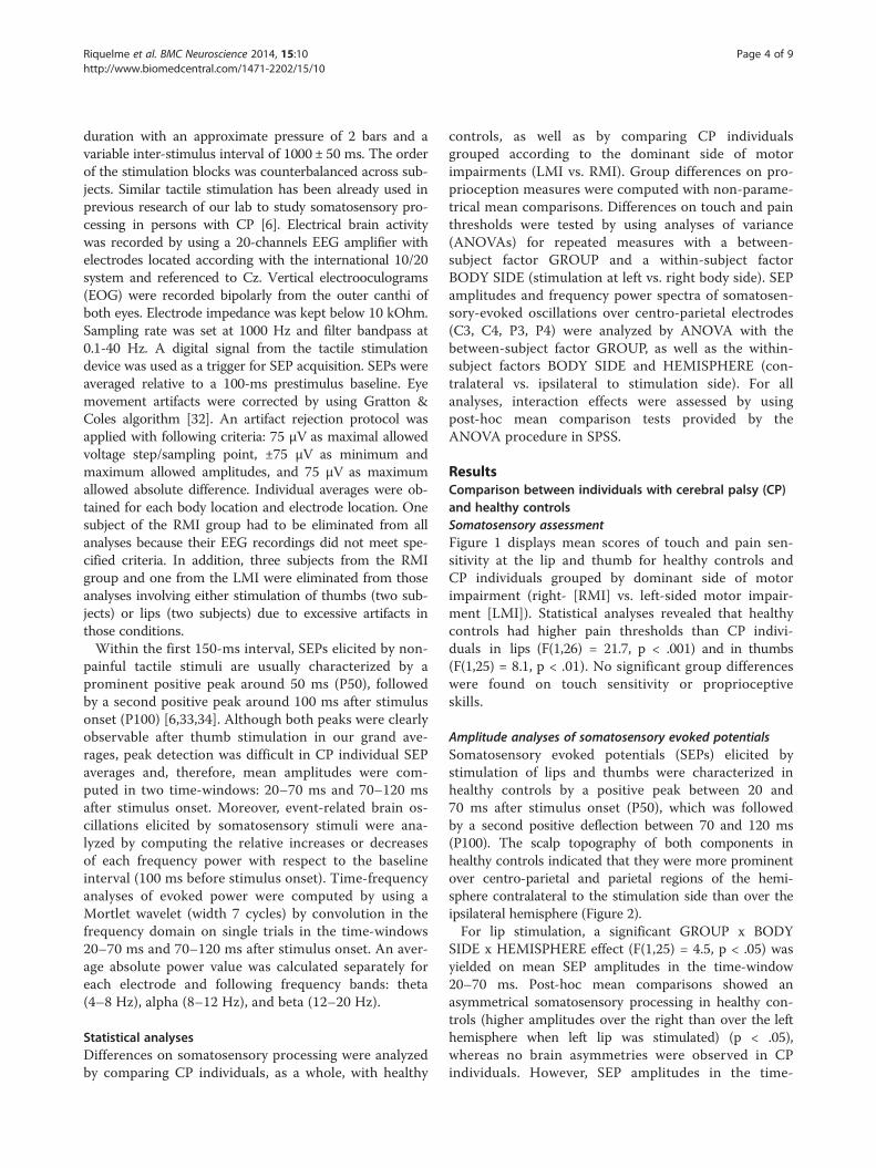
ResultsComparison between individuals with cerebral palsy (CP)and healthy controlsSomatosensory assessmentFigure 1 displays mean scores of touch and pain sen-sitivity at the lip and thumb for healthy controls andCP individuals grouped by dominant side of motorimpairment (right- [RMI] vs. left-sided motor impair-ment [LMI]). Statistical analyses revealed that healthycontrols had higher pain thresholds than CP indivi-duals in lips (F(1,26) = 21.7, p < .001) and in thumbs(F(1,25) = 8.1, p < .01). No significant group differenceswere found on touch sensitivity or proprioceptiveskills.
Amplitude analyses of somatosensory evoked potentialsSomatosensory evoked potentials (SEPs) elicited bystimulation of lips and thumbs were characterized inhealthy controls by a positive peak between 20 and70 ms after stimulus onset (P50), which was followedby a second positive deflection between 70 and 120 ms(P100). The scalp topography of both components inhealthy controls indicated that they were more prominentover centro-parietal and parietal regions of the hemi-sphere contralateral to the stimulation side than over theipsilateral hemisphere (Figure 2).For lip stimulation, a significant GROUP x BODY
SIDE x HEMISPHERE effect (F(1,25) = 4.5, p < .05) wasyielded on mean SEP amplitudes in the time-window20–70 ms. Post-hoc mean comparisons showed anasymmetrical somatosensory processing in healthy con-trols (higher amplitudes over the right than over the lefthemisphere when left lip was stimulated) (p < .05),whereas no brain asymmetries were observed in CPindividuals. However, SEP amplitudes in the time-
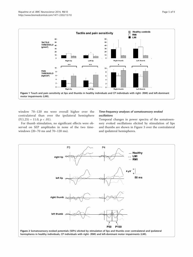
Figure 1 Touch and pain sensitivity at lips and thumbs in healthy individuals and CP individuals with right- (RMI) and left-dominantmotor impairments (LMI).
Riquelme et al. BMC Neuroscience 2014, 15:10 Page 5 of 9http://www.biomedcentral.com/1471-2202/15/10
window 70–120 ms were overall higher over thecontralateral than over the ipsilateral hemisphere(F(1,25) = 11.0, p < .01).For thumb stimulation, no significant effects were ob-
served on SEP amplitudes in none of the two time-windows (20–70 ms and 70–120 ms).
Figure 2 Somatosensory evoked potentials (SEPs) elicited by stimulathemispheres in healthy individuals, CP individuals with right- (RMI) a
Time-frequency analyses of somatosensory evokedoscillationsTemporal changes in power spectra of the somatosen-sory evoked oscillations elicited by stimulation of lipsand thumbs are shown in Figure 3 over the contralateraland ipsilateral hemispheres.
ion of lips and thumbs over contralateral and ipsilateralnd left-dominant motor impairments (LMI).
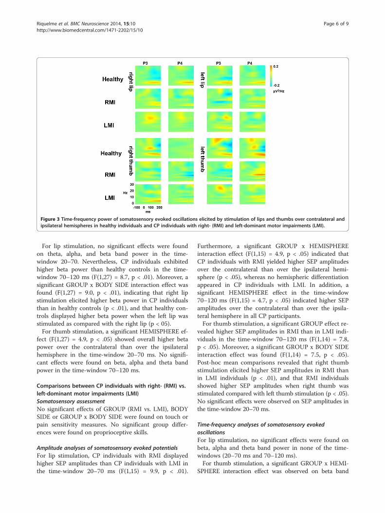
Figure 3 Time-frequency power of somatosensory evoked oscillations elicited by stimulation of lips and thumbs over contralateral andipsilateral hemispheres in healthy individuals and CP individuals with right- (RMI) and left-dominant motor impairments (LMI).
Riquelme et al. BMC Neuroscience 2014, 15:10 Page 6 of 9http://www.biomedcentral.com/1471-2202/15/10
For lip stimulation, no significant effects were foundon theta, alpha, and beta band power in the time-window 20–70. Nevertheless, CP individuals exhibitedhigher beta power than healthy controls in the time-window 70–120 ms (F(1,27) = 8.7, p < .01). Moreover, asignificant GROUP x BODY SIDE interaction effect wasfound (F(1,27) = 9.0, p < .01), indicating that right lipstimulation elicited higher beta power in CP individualsthan in healthy controls (p < .01), and that healthy con-trols displayed higher beta power when the left lip wasstimulated as compared with the right lip (p < 05).For thumb stimulation, a significant HEMISPHERE ef-
fect (F(1,27) = 4.9, p < .05) showed overall higher betapower over the contralateral than over the ipsilateralhemisphere in the time-window 20–70 ms. No signifi-cant effects were found on beta, alpha and theta bandpower in the time-window 70–120 ms.
Comparisons between CP individuals with right- (RMI) vs.left-dominant motor impairments (LMI)Somatosensory assessmentNo significant effects of GROUP (RMI vs. LMI), BODYSIDE or GROUP x BODY SIDE were found on touch orpain sensitivity measures. No significant group differ-ences were found on proprioceptive skills.
Amplitude analyses of somatosensory evoked potentialsFor lip stimulation, CP individuals with RMI displayedhigher SEP amplitudes than CP individuals with LMI inthe time-window 20–70 ms (F(1,15) = 9.9, p < .01).
Furthermore, a significant GROUP x HEMISPHEREinteraction effect (F(1,15) = 4.9, p < .05) indicated thatCP individuals with RMI yielded higher SEP amplitudesover the contralateral than over the ipsilateral hemi-sphere (p < .05), whereas no hemispheric differentiationappeared in CP individuals with LMI. In addition, asignificant HEMISPHERE effect in the time-window70–120 ms (F(1,15) = 4.7, p < .05) indicated higher SEPamplitudes over the contralateral than over the ipsila-teral hemisphere in all CP participants.For thumb stimulation, a significant GROUP effect re-
vealed higher SEP amplitudes in RMI than in LMI indi-viduals in the time-window 70–120 ms (F(1,14) = 7.8,p < .05). Moreover, a significant GROUP x BODY SIDEinteraction effect was found (F(1,14) = 7.5, p < .05).Post-hoc mean comparisons revealed that right thumbstimulation elicited higher SEP amplitudes in RMI thanin LMI individuals (p < .01), and that RMI individualsshowed higher SEP amplitudes when right thumb wasstimulated compared with left thumb stimulation (p < .05).No significant effects were observed on SEP amplitudes inthe time-window 20–70 ms.
Time-frequency analyses of somatosensory evokedoscillationsFor lip stimulation, no significant effects were found onbeta, alpha and theta band power in none of the time-windows (20–70 ms and 70–120 ms).For thumb stimulation, a significant GROUP x HEMI-
SPHERE interaction effect was observed on beta band
Riquelme et al. BMC Neuroscience 2014, 15:10 Page 7 of 9http://www.biomedcentral.com/1471-2202/15/10
power in the time-window 20–70 ms (F(1,15) = 5.6,p < .05). Post-hoc comparisons indicated higher betapower over the contralateral than over the ipsilateralhemisphere in CP individuals with RMI (p < .05), whereasno significant differences were observed in CP individualswith LMI. No significant effects were found on beta, alphaand theta band power in the time-window 70–120 ms.
DiscussionThis study was aimed to evaluate the effects of lateralizedmotor impairment on somatosensory perception andbrain processing of non-painful tactile stimulation in in-dividuals with bilateral cerebral palsy (CP). Our findingsrevealed that although individuals with CP exhibited lowerpain sensitivity than healthy controls, there were no sig-nificant differences on touch sensitivity, pain sensitivity orproprioception between CP individuals with right- (RMI)and left-dominant motor impairments (LMI). Regardingsomatosensory brain processing, we found that lip sti-mulation elicited higher beta power, but more similar SEPamplitudes over the contra- and the ipsilateral hemi-spheres in CP individuals than in healthy controls. More-over, lip and thumb stimulations elicited smaller and moresymmetrical SEP amplitudes, as well as reduced betapower (only for thumb stimulation) in CP individuals withLMI than in CP individuals with RMI.Thus, our results revealed an altered somatosensory
processing in individuals with CP, as measured by SEPamplitudes and frequency power. The pattern of brainactivation displayed by our individuals with CP afterstimulation of thumbs and lips seems to be different fromthat observed in healthy controls. Reduced beta powerand enhanced SEP amplitudes over somatosensory cor-tices have been observed in healthy controls followingsomatosensory stimulation, suggesting that these changesmight be interpreted as an activation of cortical networksinvolved in somatosensory processing [35]. In the presentstudy, healthy individuals showed a desynchronization inthe beta frequency band in response to touch stimulationof lips and thumbs. In contrast, our individuals with CPappeared to display an increased beta power over thecontralateral hemisphere, particularly in CP individualswith RMI. These findings are in agreement with previousstudies showing that, although CP is mainly characterizedby motor impairments, brain processing of incoming som-atosensory information is also significantly altered in thispathology. Thus, for instance, neuroimaging research hasfound that children with periventricular leukomalaciashow more severe injury in posterior white matter fibersconnecting the thalamus to the sensory cortex than in de-scending corticospinal tracts [14]. Moreover, it has beendemonstrated that deficits on somatosensory processing(reduced touch sensitivity, proprioception and strength)in CP could be related to injury severity of diffuse
thalamocortical projections to somatosensory and parietalcortices [26]. In this sense, our results provide furtherempirical evidence for an abnormal brain processing ofbodily information in CP individuals.Motor function seems to be often asymmetric in CP,
even in individuals with bilateral lesions [36-38]. Thisasymmetry has been reported by using physiological mea-sures such as nerve conduction velocities or dichotic lis-tening processing [39,40]. In the present study, we foundsignificant differences on the pattern of hemispheric brainactivation elicited by somatosensory stimulation depen-ding on the dominant side of motor impairment. In thissense, participants with RMI displayed enhanced res-ponses to bodily stimulation over contralateral as com-pared to ipsilateral hemispheres, whereas individuals withLMI showed no hemispheric differences. Moreover, CPsubjects with RMI showed higher SEP amplitudes whenthe affected side of the body was stimulated, while no dif-ferences on brain activation were found in LMI when theright or left body side was stimulated. In a previous studyof our lab [6], we have shown that CP children and adultselicited higher SEP amplitudes in contralateral hemispherewhen left dominant side of motor impairment was stimu-lated as compared to right dominant side of motor impair-ment. Thus, it seems that CP individuals with left- andright-dominant motor impairments differ on brain pro-cessing of lateralized bodily information. An unusual pat-tern of bilateral cortical activation and recruitment ofipsilateral tracts have been usually linked to widespreadcortical reorganization after brain lesions [41-44]. More-over, our findings are in agreement with previous studiesshowing that motor impairments are different dependingon the paretic dominant side of motor impairment afterunilateral CP lesions. Thus, for instance, Van Kampen andcolleagues [45] reported that children with left hemipa-resis had longer decision time when asked to intercept aball located 4 meters away and started their reach move-ment earlier than healthy controls and children with righthemiparesis. In addition, Craje and colleagues [46] ob-served that participants with right hemiparesis had moredifficulties in switching between different grip types thanparticipants with left hemiparesis. Our findings are also inagreement with previous data showing that bilaterally im-paired CP children with spastic diplegia displayed higherintrahemispheric coherence for delta, beta and theta EEGbands in the left than in the right hemisphere [47]. In ouropinion, all these results support the view that brain da-mage to right or to left hemisphere may have led to differ-ent plasticity mechanisms in cerebral palsy. In the presentstudy, we observed that CP individuals with RMI dis-played an asymmetrical pattern of brain activity moresimilar to that exhibited by healthy controls [48] than thatof individuals with LMI. One possible explanation forthese differences could be that damage of the left
Riquelme et al. BMC Neuroscience 2014, 15:10 Page 8 of 9http://www.biomedcentral.com/1471-2202/15/10
hemisphere releases the right hemisphere from its non-dominant role, while damage of the right hemisphere onlyemphasizes the usual role of the left hemisphere. Thus, forexample, due to the dominant role of the left somatosen-sory cortex in sensorimotor integration for complex fingermovements [49], damage of this hemisphere may havelead to a contralateral directed plasticity phenomenon inCP individuals with RMI. Nevertheless, further researchshould be necessary to elucidate the role of hemisphericdominance on somatosensory processing in CP individualsand the potential mechanisms of this differentiation.Our study has some limitations, which should be taken
into account for the interpretation of the results. Firstly,although our sample of persons with CP seems to be re-presentative of a large population in the community,sample was small and heterogeneous. Thus, it could bethat our sample size was not sufficient to reach an appro-priate statistical power and to show differences betweenindividuals with LMI and RMI on behavioral measures ofsomatosensory processing. In addition, the wide range ofage, cognitive levels and underlying brain lesions as wellas the presence of different subtypes of CP, have also li-mited the conclusions of the study. Moreover, it is possiblethat CP individuals may have been developing a left hemi-spheric dominance before their lesion. Nevertheless, theearly onset of this pathology (in most cases, with a pre-natal beginning) minimizes the influence of a possible left-hemispheric dominance development in comparison withseveral years of brain reorganization in the childhood andadolescence. On the other hand, the study should be repli-cated in hemiparetic CP individuals with clear and limitedbrain lesions. Finally, somatosensory-evoked potentialsprovide information from brain functioning arising fromsensory cortices and, therefore, the influence of subcor-tical brain structures in somatosensory processing remainsunexplored. Nevertheless, our study lays the scientificbasis for implementation of further research on a scarcelyinvestigated topic.
ConclusionOur findings suggest a different somatosensory corticalorganization in participants with CP associated with asym-metrical motor impairments. Given that activity-dependentplasticity plays a key role in the evolution of clinical signslinked to motor dysfunctions in CP [42] and that trainingand rehabilitation interventions that target these maladap-tive brain changes have already shown beneficial effects inseveral sensory and motor disorders [50], further researchmust elucidate the mechanisms of plastic changes asso-ciated to motor impairments in CP and relate them to spe-cific rehabilitation interventions for these individuals.
Competing interestsThe authors declare that they have no competing interests.
Authors’ contributionsIR carried out the recruitment of subjects, EEG recordings andsomatosensory assessment. IR and PM conceived of the study, andparticipated in its design and coordination. IC, IP and AG participated in thedesign of the study and performed the statistical analyses. PM, IR, IC and IPprepared a draft of the manuscript. All authors read and approved the finalmanuscript.
AcknowledgmentsResearch was supported by the Spanish Ministry of Science and Innovationand European Regional Development Funds (grants #SEJ2007-62312 and#PSI2010-19372).
Author details1Research Institute of Health Sciences (IUNICS), University of the BalearicIslands, Carretera de Valldemossa km 7.5, Palma 07122, Spain. 2Departmentof Nursing and Physiotherapy, University of the Balearic Islands, Palma, Spain.3Department of Developmental Psychology and Education, University of LaLaguna, La Laguna, Spain.
Received: 2 August 2012 Accepted: 31 December 2013Published: 11 January 2014
References1. Rosenbaum P, Paneth N, Leviton A, Goldstein M, Bax M, Damiano D, Dan B,
Jacobsson B: A report: the definition and classification of cerebral palsyApril 2006. Dev Med Child Neurol 2007, 109(Suppl. Feb):8–14.
2. Wingert JR, Burton H, Sinclair RJ, Brunstrom JE, Damiano DL: Joint-positionsense and kinesthesia in cerebral palsy. Arch Phys Med Rehabil 2009,90(3):447–453.
3. Sanger TD, Kukke SN: Abnormalities of tactile sensory function in childrenwith dystonic and diplegic cerebral palsy. J Child Neurol 2007,22(3):289–293.
4. Cooper J, Majnemer A, Rosenblatt B, Birnbaum R: The determination ofsensory deficits in children with hemiplegic cerebral palsy. J Child Neurol1995, 10(4):300–309.
5. Riquelme I, Cifre I, Montoya P: Age-related changes of pain experience incerebral palsy and healthy individuals. Pain Med 2011, 12(4):535–545.
6. Riquelme I, Montoya P: Developmental changes in somatosensoryprocessing in cerebral palsy and healthy individuals. Clin Neurophysiol2010, 121(8):1314–1320.
7. Nociti V, Batocchi AP, Bartalini S, Caggiula M, Patti F, Profice P, Quattrone A,Tonali P, Ulivelli M, Valentino P, Virdis D, Zappia M, Padua L: Somatosensoryevoked potentials reflect the upper limb motor performance in multiplesclerosis. J Neurol Sci 2008, 273(1–2):99–102.
8. Nardone R, Golaszewski S, Bergmann J, Venturi A, Prünster I, Bratti A,Ladurner G, Tezzon F: Motor cortex excitability changes following a lesionin the posterior columns of the cervical spinal cord. Neurosci Lett 2008,434(1):119–123.
9. Park BK, Chae J, Lee YH, Yang G, Labatia I: Median nerve somatosensoryevoked potentials and upper limb motor function in hemiparesis.Electromyogr Clin Neurophysiol 2003, 43(3):169–179.
10. Platz T, Kim IH, Pintschovius H, Winter T, Kieselbach A, Villringer K, Kurth R,Mauritz KH: Multimodal EEG analysis in man suggests impairment-specificchanges in movement-related electric brain activity after stroke. Brain 2000,123(12):2475–2490.
11. Kusoffsky A, Wadell I, Nilsson B: The relationship between sensoryimpairment and motor recovery in patients with hemiplegia.Scand J Rehabil Med 1982, 14(1):27–32.
12. Hoon AH Jr, Stashinko EE, Nagae LM, Lin DD, Keller J, Bastian A, Campbell ML,Levey E, Mori S, Johnston MV: Sensory and motor deficits in children withcerebral palsy born preterm correlate with diffusion tensor imagingabnormalities in thalamocortical pathways. Dev Med Child Neurol 2009,51(9):697–704.
13. Thomas B, Eyssen M, Peeters R, Molenaers G, Van Hecke P, De Cock P,Sunaert S: Quantitative diffusion tensor imaging in cerebral palsy due toperiventricular white matter injury. Brain 2005, 128(11):2562–2577.
14. Hoon AH Jr, Lawrie WT Jr, Melhem ER, Reinhardt EM, Van Zijl PC,Solaiyappan M, Jiang H, Johnston MV, Mori S: Diffusion tensor imaging ofperiventricular leukomalacia shows affected sensory cortex white matterpathways. Neurology 2002, 59(5):752–756.

Riquelme et al. BMC Neuroscience 2014, 15:10 Page 9 of 9http://www.biomedcentral.com/1471-2202/15/10
15. Thickbroom GW, Byrnes ML, Archer SA, Nagarajan L, Mastaglia FL:Differences in sensory and motor cortical organization following braininjury early in life. Ann Neurol 2001, 49(3):320–327.
16. Lourenço G, Meunier S, Vidailhet M, Simonetta-Moreau M: Impaired modulationof motor cortex excitability by homonymous and heteronymous muscleafferents in focal hand dystonia. Mov Disord 2007, 22(4):523–527.
17. Schmelzle-Lubiecki BM, Campbell KA, Howard RH, Franck L, Fitzgerald M:Long-term consequences of early infant injury and trauma uponsomatosensory processing. Eur J Pain 2007, 11(7):799–809.
18. Abbruzzese G, Berardelli A: Sensorimotor integration in movementdisorders. Mov Disord 2003, 18(3):231–240.
19. Eyre JA, Smith M, Dabydeen L, Clowry GJ, Petacchi E, Battini R, Guzzetta A,Cioni G: Is hemiplegic cerebral palsy equivalent to amblyopia of thecorticospinal system? Ann Neurol 2007, 62(5):493–503.
20. Cincotta M, Borgheresi A, Guidi L, Macucci M, Cosottini M, Lambruschini P,Benvenuti F, Zaccara G: Remote effects of cortical dysgenesis on theprimary motor cortex: evidence from the silent period followingtranscranial magnetic stimulation. Clin Neurophysiol 2000,111(8):1340–1345.
21. Maegaki Y, Yamamoto T, Takeshita K: Plasticity of central motor andsensory pathways in a case of unilateral extensive cortical dysplasia:investigation of magnetic resonance imaging, transcranial magneticstimulation, and short-latency somatosensory evoked potentials.Neurology 1995, 45(12):2255–2261.
22. Maegaki Y, Maeoka Y, Ishii S, Shiota M, Takeuchi A, Yoshino K, Takeshita K:Mechanisms of central motor reorganization in pediatric hemiplegicpatients. Neuropediatrics 1997, 28(3):168–174.
23. Carr LJ, Harrison LM, Evans AL, Stephens JA: Patterns of central motorreorganization in hemiplegic cerebral palsy. Brain 1993, 116(5):1223–1247.
24. Farmer SF, Harrison LM, Ingram DA, Stephens JA: Plasticity of centralmotor pathways in children with hemiplegic cerebral palsy.Neurology 1991, 41(9):1505–1510.
25. Clayton K, Fleming JM, Copley J: Behavioral responses to tactile stimuli inchildren with cerebral palsy. Phys Occup Ther Pediatr 2003, 23(1):43–62.
26. Coq JO, Strata F, Russier M, Safadi FF, Merzenich MM, Byl NN, Barbe MF:Impact of neonatal asphyxia and hind limb immobilization onmusculoskeletal tissues and S1 map organization: implications forcerebral palsy. Exp Neurol 2008, 210(1):95–108.
27. Ragazzoni A, Cincotta M, Borgheresi A, Zaccara G, Ziemann U: Congenitalhemiparesis: different functional reorganization of somatosensory andmotor pathways. Clin Neurophysiol 2002, 113(8):1273–1278.
28. House JH, Gwathmey FW, Fidler MO: House functional classification.A dynamic approach to the thumb-in-palm deformity in cerebral palsy.Evaluation and results in fifty-six patients. J Bone Joint Surg 1981,63A:216–225.
29. Palisano R, Rosenbaum P, Walter S, Russell D, Wood E, Galuppi B:Development and reliability of a system to classify gross motor functionin children with cerebral palsy. Dev Med Child Neurol 1997, 39(4):214–223.
30. Keizer D, Fael D, Wierda JM, van Wijhe M: Quantitative sensory testingwith Von Frey monofilaments in patients with allodynia: what are wequantifying? Clin J Pain 2008, 24(5):463–466.
31. Cathcart S, Pritchard D: Reliability of pain threshold measurement inyoung adults. J Headache Pain 2006, 7(1):21–26.
32. Gratton G, Coles MG, Donchin E: A new method for off-line removal ofocular artifact. Electroencephalogr Clin Neurophysiol 1983, 55(4):468–484.
33. Montoya P, Sitges C, García-Herrera M, Rodríguez-Cotes A, Izquierdo R,Truyols M, Collado D: Reduced brain habituation to somatosensorystimulation in patients with fibromyalgia. Arthritis Rheum 2006,54(6):1995–2003.
34. Montoya P, Sitges C: Affective modulation of somatosensory-evokedpotentials elicited by tactile stimulation. Brain Res 2006, 1068(1):205–212.
35. Ritter P, Moosmann M, Villringer A: Rolandic alpha and beta EEG rhythms'strengths are inversely related to fMRI-BOLD signal in primarysomatosensory and motor cortex. Hum Brain Mapp 2009, 30(4):1168–1187.
36. Bourelle S, Berge B, Gautheron V, Cottalorda J: Computerized staticposturographic assessment after treatment of equinus deformity inchildren with cerebral palsy. J Pediatr Orthop B 2010, 19(3):211–220.
37. Descatoire A, Femery V, Potdevin F, Moretto P: Step-to-step reproducibilityand asymmetry to study gait auto-optimization in healthy and cerebralpalsied subjects. Ann Phys Rehabil Med 2009, 52(4):319–329.
38. Park ES, Kim HW, Park CI, Rha DW, Park CW: Dynamic foot pressuremeasurements for assessing foot deformity in persons with spasticcerebral palsy. Arch Phys Med Rehabil 2006, 87(5):703–709.
39. Kalizhniuk ES, Fedorchuk AG: Functional asymmetry of the cerebralhemispheres in infantile cerebral palsy (according to dichotic listeningdata). Zh Nevropatol Psikhiatr Im S S Korsakova 1985, 85(10):1464–1468.
40. Lang AH, Sillanpää M, Hynninen P: Asymmetric function of peripheralnerves in children with cerebral palsy. Acta Neurol Scand 1983,67(2):108–113.
41. Krägeloh-Mann I, Cans C: Cerebral palsy update. Brain Dev 2009,31(7):537–544.
42. Eyre JA: Corticospinal tract development and its plasticity after perinatalinjury. Neurosci Biobehav Rev 2007, 31(8):1136–1149.
43. Briellmann RS, Abbott DF, Caflisch U, Archer JS, Jackson GD: Brainreorganisation in cerebral palsy: a high-field functional MRI study.Neuropediatrics 2002, 33(3):162–165.
44. Macnicol MF, Nadeem RD: Evaluation of the deformity in club foot bysomatosensory evoked potentials. J Bone Joint Surg Br 2000,82(5):731–735.
45. Van Kampen PM, Ledebt A, Deconinck FJ, Savelsbergh GJ: Visual guidanceof interceptive actions in children with spastic unilateral cerebral palsy isinfluenced by the side of the lesion. Disabil Rehabil 2010,32(18):1527–1537.
46. Crajé C, van der Kamp J, Steenbergen B: Visual information for actionplanning in left and right congenital hemiparesis. Brain Res 2009,1261:54–64.
47. Koeda T, Takeshita K: Electroencephalographic coherence abnormalities inpreterm diplegia. Pediatr Neurol 1998, 18(1):51–56.
48. Nirkko AC, Ozdoba C, Redmond SM, Bürki M, Schroth G, Hess CW,Wiesendanger M: Different ipsilateral representations for distal andproximal movements in the sensorimotor cortex: activation anddeactivation patterns. Neuroimage 2001, 13(5):825–835.
49. Okuda B, Tanaka H, Tomino Y, Kawabata K, Tachibana H, Sugita M: The roleof the left somatosensory cortex in human hand movement. Exp BrainRes 1995, 106(3):493–498.
50. Flor H: Remapping somatosensory cortex after injury. Adv Neurol 2003,93:195–204.
doi:10.1186/1471-2202-15-10Cite this article as: Riquelme et al.: Differences in somatosensoryprocessing due to dominant hemispheric motor impairment in cerebralpalsy. BMC Neuroscience 2014 15:10.
Submit your next manuscript to BioMed Centraland take full advantage of:
• Convenient online submission
• Thorough peer review
• No space constraints or color figure charges
• Immediate publication on acceptance
• Inclusion in PubMed, CAS, Scopus and Google Scholar
• Research which is freely available for redistribution
Submit your manuscript at www.biomedcentral.com/submit
Copyright © 2022 FDOKUMEN