Development of Monoclonal Antibodies against Caprine α S2 Casein and Their Potential for Detecting...
6
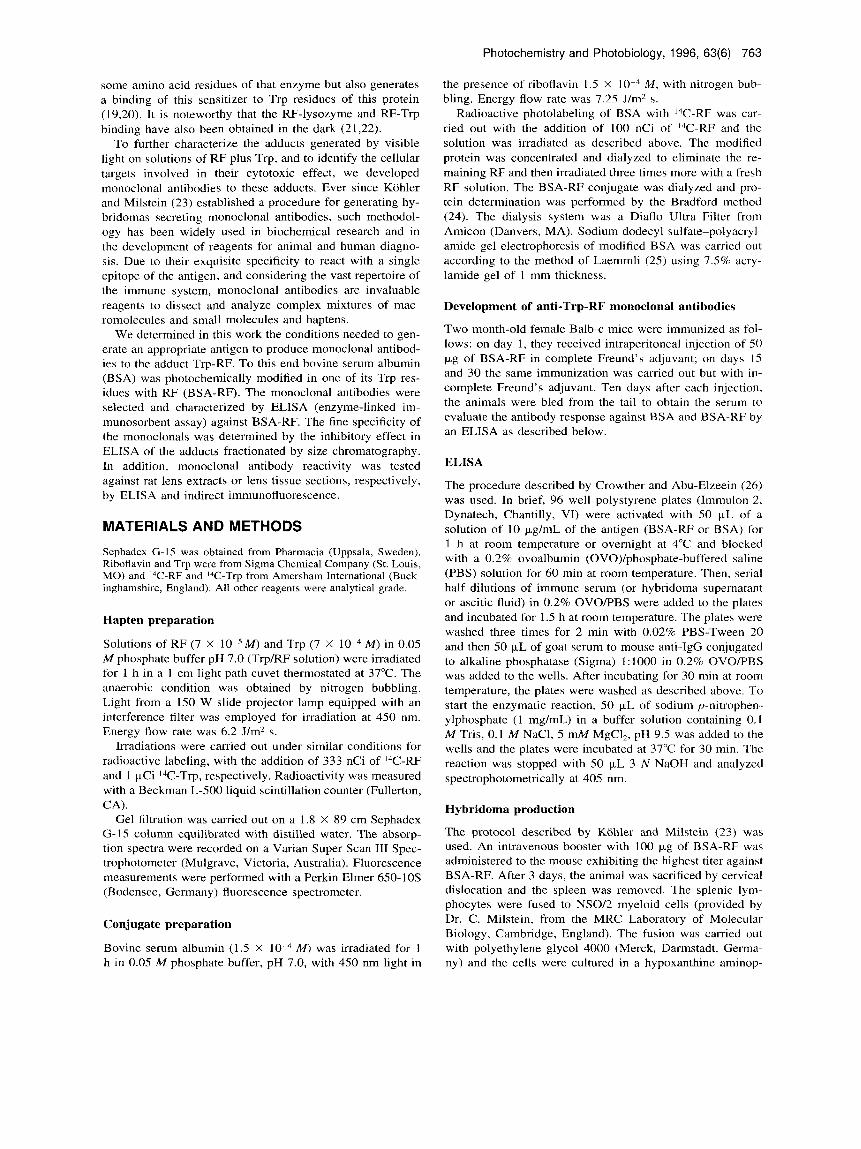
Photochemistry and Photobiology, 1996,63(6): 762-767 S ym posium-in-P r in t Development of Monoclonal Antibodies Against a Riboflavin-Tryptophan Photoinduced Adduct: Reactivity to Eye Lens Proteins* Marisol Diaz112, M. lnes Be~ker',~, Alfredo E. De loannes' and Eduardo Silvat2 'Facultad de Ciencias Biologicas and zFacultad de Quimica, P. Universidad Catolica de Chile, Santiago, Chile and "Laboratorio de Inmunologia, Bios-Chile I.G.S.A., Santiago, Chile Received 31 October 1995; accepted 8 February 1996 ABSTRACT We describe here the development of monoclonal anti- bodies to the hapten tryptophan-riboflavin, generated by irradiation of a solution of bovine serum albumin in the presence of riboflavin. The specificity of the three ob- tained monoclonal antibodies, named lE6, 5H5, 5AS all belonging to the IgGl isotype, was assessed by a com- petitive enzyme-linked immunosorbent assay in the pres- ence of an increasing concentration of the tryptophan- riboflavin adduct, obtained from an irradiated riboflavin- sensitized tryptophan solution. It was demonstrated that the tryptophan-riboflavin antibodies react with the solu- ble proteins of the eye lens; this reaction was more in- tense in the old rat lenses as compared to the young ones, and a maximum binding of the antibodies was obtained with the soluble protein fraction from the human catar- actous lens. By indirect immunofluorescence, a reactivity associated with the protein matrix, localized in the lens central zone, was observed. In the peripheral zone of the lens, where the younger cells are found, a marked im- munofluorescent emission was observed on structures preferentially localized in the nuclei. INTRODUCTION Among the sensitized photoprocesses of free and protein- bound tryptophan, those involving riboflavin (RF)* are of particular interest due to the endogenous nature of the sen- sitizer. Although Trp is an essential amino acid and RF is a vitamin, the simultaneous presence of the two compounds in living organisms has been related to hepatotoxic and cyto- *This paper is dedicated to the memory of Prof. G. Cilento. +To whom correspondence should be addressed at: Facultad de Quimica, Depto. Fisico Quimica (536), Casilla 306-Correo 22, Santiago, Chile. Fax: 56-2-5525692; e-mail: esilva@lascar,puc.cl. I.Abbreviations: BSA, bovine serum albumin; ELISA, enzyme- linked immunosorbent assay; HAT, hypoxanthine aminopterin thymidine; OVO, ovoalbumin; PBS, phosphate-buffered saline; RF, riboflavin. D 1996 American Society for Photobiology 0031-8655/96 $S.OO+O.OO toxic effects during parenteral nutrition (1,2) and in culture media (3,4), respectively, upon exposure to the action of visible light. Recently, the generation of an adduct between RF and Trp following irradiation under anaerobic conditions has been described (5) and this compound has been proposed to be one of the photoproducts that may be associated with hepatic dysfunction (6) and cytotoxicity on tumor cells and preimplantation mouse embryos in irradiated culture medi- um (7,8). The subcellular targets and the mechanism in- volved in the induction of cytotoxicity have not been deter- mined. Some tissues like the skin and the eye lens are perma- nently exposed to the action of light. Evidence has been accumulated that in the lens the photochemical insult is rec- ognized as a factor involved in the development of cataract (9-1 1). Senile nuclear cataractogenesis and aging of the hu- man eye lens have been associated with a singlet-oxygen- mediated photodynamic process promoted by endogenous sensitizers such as RF and/or formylkynurenine (1 2,13). It has also been reported recently that a short-term photochem- ically induced oxidative stress in cultured rat lenses caused by the generation of H,O,, O,..and .OH leads to rapid irre- versible damage to the epithelial cell layer (14,15). When rat lens homogenate or its soluble protein fraction are irra- diated in the presence of RF, a photoadduct is obtained be- tween this vitamin and the lens proteins (1 6). Irradiation of these proteins in the presence of RF also leads to a modifi- cation in the chromatographic elution pattern with an in- crease in the high molecular weight fraction (16). A mixed type I-type I1 mechanism has been observed when using RF as sensitizer in the photooxidation of Trp (17,18). The photodegradation of this amino acid in oxygen- saturated aqueous solution sensitized by RF is accompanied by the generation of reactive oxygen species (lo,, .OH, H,O, and Oi-) and the appearance of the following irradiation products: aggregate forms of RF, indolic products associated to flavins, indolic products of molecular weight higher than Trp, formylkynurenine and other Trp photodecomposition products (18). It has also been demonstrated that irradiation of lysozyme with visible light in the presence of RF and molecular oxygen not only produces the photooxidation of 762
-
Upload
independent -
Category
Documents
-
view
1 -
download
0
Transcript of Development of Monoclonal Antibodies against Caprine α S2 Casein and Their Potential for Detecting...

Photochemistry and Photobiology, 1996, 63(6): 762-767
S y m posi u m-i n- P r i n t
Development of Monoclonal Antibodies Against a Riboflavin-Tryptophan Photoinduced Adduct: Reactivity to Eye Lens Proteins*
Marisol Diaz112, M. lnes Be~ker ' ,~ , Alfredo E. De loannes' and Eduardo Silvat2 'Facultad de Ciencias Biologicas and zFacultad de Quimica, P. Universidad Catolica de Chile, Santiago, Chile and "Laboratorio de Inmunologia, Bios-Chile I.G.S.A., Santiago, Chile
Received 31 October 1995; accepted 8 February 1996
ABSTRACT
We describe here the development of monoclonal anti- bodies to the hapten tryptophan-riboflavin, generated by irradiation of a solution of bovine serum albumin in the presence of riboflavin. The specificity of the three ob- tained monoclonal antibodies, named lE6, 5H5, 5AS all belonging to the IgGl isotype, was assessed by a com- petitive enzyme-linked immunosorbent assay in the pres- ence of an increasing concentration of the tryptophan- riboflavin adduct, obtained from an irradiated riboflavin- sensitized tryptophan solution. It was demonstrated that the tryptophan-riboflavin antibodies react with the solu- ble proteins of the eye lens; this reaction was more in- tense in the old rat lenses as compared to the young ones, and a maximum binding of the antibodies was obtained with the soluble protein fraction from the human catar- actous lens. By indirect immunofluorescence, a reactivity associated with the protein matrix, localized in the lens central zone, was observed. In the peripheral zone of the lens, where the younger cells are found, a marked im- munofluorescent emission was observed on structures preferentially localized in the nuclei.
INTRODUCTION
Among the sensitized photoprocesses of free and protein- bound tryptophan, those involving riboflavin (RF)* are of particular interest due to the endogenous nature of the sen- sitizer. Although Trp is an essential amino acid and RF is a vitamin, the simultaneous presence of the two compounds in living organisms has been related to hepatotoxic and cyto-
*This paper is dedicated to the memory of Prof. G. Cilento. +To whom correspondence should be addressed at: Facultad de
Quimica, Depto. Fisico Quimica (536), Casilla 306-Correo 22, Santiago, Chile. Fax: 56-2-5525692; e-mail: esilva@lascar,puc.cl.
I.Abbreviations: BSA, bovine serum albumin; ELISA, enzyme- linked immunosorbent assay; HAT, hypoxanthine aminopterin thymidine; OVO, ovoalbumin; PBS, phosphate-buffered saline; RF, riboflavin.
D 1996 American Society for Photobiology 0031-8655/96 $S.OO+O.OO
toxic effects during parenteral nutrition (1,2) and in culture media (3,4), respectively, upon exposure to the action of visible light. Recently, the generation of an adduct between RF and Trp following irradiation under anaerobic conditions has been described (5) and this compound has been proposed to be one of the photoproducts that may be associated with hepatic dysfunction (6) and cytotoxicity on tumor cells and preimplantation mouse embryos in irradiated culture medi- um (7,8). The subcellular targets and the mechanism in- volved in the induction of cytotoxicity have not been deter- mined.
Some tissues like the skin and the eye lens are perma- nently exposed to the action of light. Evidence has been accumulated that in the lens the photochemical insult is rec- ognized as a factor involved in the development of cataract (9-1 1). Senile nuclear cataractogenesis and aging of the hu- man eye lens have been associated with a singlet-oxygen- mediated photodynamic process promoted by endogenous sensitizers such as RF and/or formylkynurenine (1 2,13). It has also been reported recently that a short-term photochem- ically induced oxidative stress in cultured rat lenses caused by the generation of H,O,, O,..and .OH leads to rapid irre- versible damage to the epithelial cell layer (14,15). When rat lens homogenate or its soluble protein fraction are irra- diated in the presence of RF, a photoadduct is obtained be- tween this vitamin and the lens proteins ( 1 6). Irradiation of these proteins in the presence of RF also leads to a modifi- cation in the chromatographic elution pattern with an in- crease in the high molecular weight fraction (16).
A mixed type I-type I1 mechanism has been observed when using RF as sensitizer in the photooxidation of Trp (17,18). The photodegradation of this amino acid in oxygen- saturated aqueous solution sensitized by RF is accompanied by the generation of reactive oxygen species (lo,, .OH, H,O, and O i - ) and the appearance of the following irradiation products: aggregate forms of RF, indolic products associated to flavins, indolic products of molecular weight higher than Trp, formylkynurenine and other Trp photodecomposition products (18). It has also been demonstrated that irradiation of lysozyme with visible light in the presence of RF and molecular oxygen not only produces the photooxidation of
762
Photochemistry and Photobiology, 1996, 63(6) 763
some amino acid residues of that enzyme but also generates a binding of this sensitizer to Trp residues of this protein (1 9,20). It is noteworthy that the RF-lysozyme and RF-Trp binding have also been obtained in the dark (21,22).
To further characterize the adducts generated by visible light on solutions of RF plus Trp, and to identify the cellular targets involved in their cytotoxic effect, we developed monoclonal antibodies to these adducts. Ever since Kohler and Milstein (23) established a procedure for generating hy- bridomas secreting monoclonal antibodies, such methodol- ogy has been widely used in biochemical research and in the development of reagents for animal and human diagno- sis. Due to their exquisite specificity to react with a single epitope of the antigen, and considering the vast repertoire of the immune system, monoclonal antibodies are invaluable reagents to dissect and analyze complex mixtures of mac- romolecules and small molecules and haptens.
We determined in this work the conditions needed to gen- erate an appropriate antigen to produce monoclonal antibod- ies to the adduct Trp-RF. To this end bovine serum albumin (BSA) was photochemically modified in one of its Trp res- idues with RF (BSA-RF). The monoclonal antibodies were selected and characterized by ELISA (enzyme-linked im- munosorbent assay) against BSA-RF. The fine specificity of the monoclonals was determined by the inhibitory effect in ELISA of the adducts fractionated by size chromatography. In addition, monoclonal antibody reactivity was tested against rat lens extracts or lens tissue sections, respectively, by ELISA and indirect immunofluorescence.
MATERIALS AND METHODS
Sephadex (3-1.5 was obtained from Pharmacia (Uppsala, Sweden). Riboflavin and Trp were from Sigma Chemical Company (St. Louis, MO) and I4C-RF and I4C-Trp from Amersham International (Buck- inghamshire, England). All other reagents were analytical grade.
Hapten preparation
Solutions of RF (7 X M) in 0.05 M phosphate buffer pH 7.0 (Trp/RF solution) were irradiated for 1 h in a 1 cm light path cuvet thermostated at 37°C. The anaerobic condition was obtained by nitrogen bubbling. Light from a 150 W slide projector lamp equipped with an interference filter was employed for irradiation at 450 nm. Energy flow rate was 6.2 J/m2 s.
Irradiations were carried out under similar conditions for radioactive labeling, with the addition of 333 nCi of I4C-RF and 1 pCi I4C-Trp, respectively. Radioactivity was measured with a Beckman L-500 liquid scintillation counter (Fullerton, CA).
Gel filtration was carried out on a 1.8 X 89 cm Sephadex G-15 column equilibrated with distilled water. The absorp- tion spectra were recorded on a Varian Super Scan It1 Spec- trophotometer (Mulgrave, Victoria, Australia). Fluorescence measurements were performed with a Perkin Elmer 650-10s (Bodensee, Germany) fluorescence spectrometer.
M) and Trp (7 X
Conjugate preparation
Bovine serum albumin (1.5 X M) was irradiated for 1 h in 0.05 M phosphate buffer, pH 7.0, with 450 nm light in
the presence of riboflavin 1.5 X M , with nitrogen bub- bling. Energy flow rate was 7.25 Urn2 s.
Radioactive photolabeling of BSA with 14C-RF was car- ried out with the addition of 100 nCi of I4C-RF and the solution was irradiated as described above. The modified protein was concentrated and dialyzed to eliminate the re- maining RF and then irradiated three times more with a fresh RF solution. The BSA-RF conjugate was dialyzed and pro- tein determination was performed by the Bradford method (24). The dialysis system was a Diaflo Ultra Filter from Amicon (Danvers, MA). Sodium dodecyl sulfate-polyacryl- amide gel electrophoresis of modified BSA was carried out according to the method of Laemmli (25) using 7.5% acry- lamide gel of 1 mm thickness.
Development of anti-Trp-RF monoclonal antibodies
Two month-old female Balb-c mice were immunized as fol- lows: on day 1, they received intraperitoneal injection of 50 p,g of BSA-RF in complete Freund’s adjuvant; on days 15 and 30 the same immunization was carried out but with in- complete Freund’s adjuvant. Ten days after each injection, the animals were bled from the tail to obtain the serum to evaluate the antibody response against BSA and BSA-RF by an ELISA as described below.
ELISA
The procedure described by Crowther and Abu-Elzeein (26) was used. In brief, 96 well polystyrene plates (Immulon-2, Dynatech, Chantilly, VI) were activated with 50 pL of a solution of 10 p,g/mL of the antigen (BSA-RF or BSA) for 1 h at room temperature or overnight at 4°C and blocked with a 0.2% ovoalbumin (OVO)/phosphate-buffered saline (PBS) solution for 60 min at room temperature. Then, serial half dilutions of immune serum (or hybridoma supernatant or ascitic fluid) in 0.2% OVO/PBS were added to the plates and incubated for 1.5 h at room temperature. The plates were washed three times for 2 min with 0.02% PBS-Tween 20 and then 50 p,L of goat serum to mouse anti-IgG conjugated to alkaline phosphatase (Sigma) 1:IOOO in 0.2% OVOPBS was added to the wells. After incubating for 30 min at room temperature, the plates were washed as described above. To start the enzymatic reaction, 50 p,L of sodium p-nitrophen- ylphosphate (1 mg/mL) in a buffer solution containing 0.1 M Tris, 0.1 M NaC1, 5 mM MgCI,, pH 9.5 was added to the wells and the plates were incubated at 37°C for 30 min. The reaction was stopped with 50 p,L 3 N NaOH and analyzed spectrophotometrically at 405 nm.
Hybridoma production
The protocol described by Kohler and Milstein (23) was used. An intravenous booster with 100 pg of BSA-RF was administered to the mouse exhibiting the highest titer against BSA-RF. After 3 days, the animal was sacrificed by cervical dislocation and the spleen was removed. The splenic lym- phocytes were fused to NS0/2 myeloid cells (provided by Dr. C. Milstein, from the MRC Laboratory of Molecular Biology, Cambridge, England). The fusion was carried out with polyethylene glycol 4000 (Merck, Darmstadt, Germa- ny) and the cells were cultured in a hypoxanthine aminop-
764 Marisol Diaz et a/.
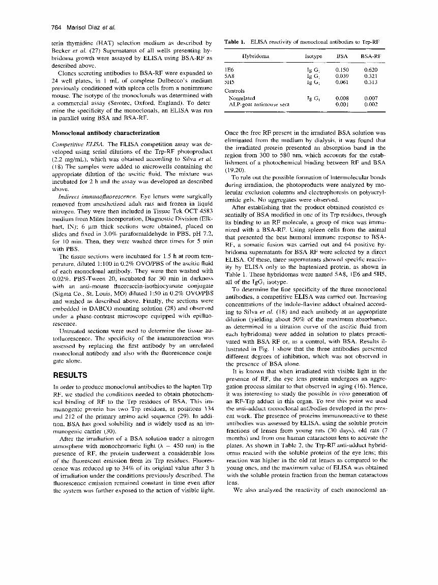
terin thymidine (HAT) selection medium as described by Becker et al. (27) Supernatants of all wells presenting hy- bridoma growth were assayed by ELISA using BSA-RF as described above.
Clones secreting antibodies to BSA-RF were expanded to 24 well plates, in 1 mL of complete Dulbecco's medium previously conditioned with spleen cells from a nonimmune mouse. The isotype of the monoclonals was determined with a commercial assay (Serotec, Oxford, England). To deter- mine the specificity of the monoclonals, an ELISA was run in parallel using BSA and BSA-RF.
Monoclonal antibody characterization
Competitive ELISA. The ELISA competition assay was de- veloped using serial dilutions of the Trp-RF photoproduct (2.2 mg/mL), which was obtained according to Silva et al. (18) The samples were added to microwells containing the appropriate dilution of the ascitic fluid. The mixture was incubated for 2 h and the assay was developed as described above.
Indirect immunojluorescence. Eye lenses were surgically removed from anesthetized adult rats and frozen in liquid nitrogen. They were then included in Tissue Tek OCT 4583 medium from Miles Incorporation, Diagnostic Division (Elk- hart, IN); 6 km thick sections were obtained, placed on slides and fixed in 3.0% paraformaldehyde in PBS, pH 7.2, for 10 min. Then, they were washed three times for 5 min with PBS.
The tissue sections were incubated for 1.5 h at room tem- perature, diluted 1:lOO in 0.2% OVO/PBS of the ascitic fluid of each monoclonal antibody. They were then washed with 0.02%. PBS-Tween 20, incubated for 30 min in darkness with an anti-mouse fluorescein-isothiocyanate conjugate (Sigma Co., St. Louis, MO) diluted 150 in 0.2% OVOPBS and washed as described above. Finally, the sections were embedded in DABCO mounting solution (28) and observed under a phase-contrast microscope equipped with epifluo- rescence.
Untreated sections were used to determine the tissue au- tofluorescence. The specificity of the immunoreaction was assessed by replacing the first antibody by an unrelated monoclonal antibody and also with the fluorescence conju- gate alone.
RESULTS In order to produce monoclonal antibodies to the hapten Trp- RF, we studied the conditions needed to obtain photochem- ical binding of RF to the Trp residues of BSA. This im- munogenic protein has two Trp residues, at positions 134 and 212 of the primary amino acid sequence (29). In addi- tion, BSA has good solubility and is widely used as an im- munogenic carrier (30).
After the irradiation of a BSA solution under a nitrogen atmosphere with monochromatic light (A = 450 nm) in the presence of RF, the protein underwent a considerable loss of the fluorescent emission from its Trp residues. Fluores- cence was reduced up to 34% of its original value after 3 h of irradiation under the conditions previously described. The fluorescence emission remained constant in time even after the system was further exposed to the action of visible light.
Table 1. ELISA reactivity of monoclonal antibodies to Trp-RF ~~ ~~
Hybridoma Isotype BSA BSA-RF
1 E6 5A8 5H5
Ig GI 0.150 0.620 Ig GI 0.039 0.321 Ig GI 0.061 0.313
Controls Nonrelated Ig G I 0.008 0.007 ALP-goat antimouse sera 0.001 0.002
Once the free RF present in the irradiated BSA solution was eliminated from the medium by dialysis, it was found that the irradiated protein presented an absorption band in the region from 300 to 580 nm, which accounts for the estab- lishment of a photochemical binding between RF and BSA (19,20).
To rule out the possible formation of intermolecular bonds during irradiation, the photoproducts were analyzed by mo- lecular exclusion columns and electrophoresis on polyacryl- amide gels. No aggregates were observed.
After establishing that the product obtained consisted es- sentially of BSA modified in one of its Trp residues, through its binding to an RF molecule, a group of mice was immu- nized with a BSA-RF. Using spleen cells from the animal that presented the best humoral immune response to BSA- RF, a somatic fusion was carried out and 64 positive hy- bridoma supernatants for BSA-RF were selected by a direct ELISA. Of these, three supernatants showed specific reactiv- ity by ELISA only to the haptenized protein, as shown in Table 1. These hybridomas were named 5A8, 1E6 and 5H5, all of the IgG, isotype.
To determine the fine specificity of the three monoclonal antibodies, a competitive ELISA was carried out. Increasing concentrations of the indole-flavine adduct obtained accord- ing to Silva et al. (18) and each antibody at an appropriate dilution (yielding about 50% of the maximum absorbance, as determined in a titration curve of the ascitic fluid from each hybridoma) were added in solution to plates preacti- vated with BSA-RF or, as a control, with BSA. Results il- lustrated in Fig. l show that the three antibodies presented different degrees of inhibition, which was not observed in the presence of BSA alone.
It is known that when irradiated with visible light in the presence of RF, the eye lens protein undergoes an aggre- gation process similar to that observed in aging (16). Hence, it was interesting to study the possible in vivo generation of an RF-Trp adduct in this organ. To test this point we used the anti-adduct monoclonal antibodies developed in the pres- ent work. The presence of proteins immunoreactive to these antibodies was assessed by ELISA, using the soluble protein fractions of lenses from young rats (30 days), old rats (7 months) and from one human cataractous lens to activate the plates. As shown in Table 2, the Trp-RF anti-adduct hybrid- omas reacted with the soluble proteins of the eye lens; this reaction was higher in the old rat lenses as compared to the young ones, and the maximum value of ELISA was obtained with the soluble protein fraction from the human cataractous lens.
We also analyzed the reactivity of each monoclonal an-

Photochemistry and Photobiology, 1996, 63(6) 765
2 0,lO s g 0 3 0
5 52 0,20
S Q
P
I I I I ( , I
0.22 I 1
0,20 -
0,18 -
0,16 -
0,14 - I 1 I I I I 1
Control 7 6 5 4 3 2 1
Trp-RF dilution(lI2exp n) Figure 1. Characterization of 1E6, 5H5 and 5A8 monoclonal anti- bodies to Trp-RF by a competitive ELISA using dilutions of pho- toinduced Trp-RF adduct.
tibody by indirect immunofluorescence in cryostat sections from rat eye lens. It was found that the three antibodies recognized structures present in the lens. Figure 2 shows a representative pattern of the immunofluorescence. The ob- served reactivity was associated with the protein matrix lo- calized in the lens central zone (Fig. 2c). In the peripheral zone of the lens, where the younger cells that have not yet lost their subcellular corpuscles are found, a marked im- inunofluorescent emission was observed on structures pref- erentially localized in the nuclei of such cells (Fig. 2d). Noteworthy, in the same region the protein matrix was less immunoreactive than toward the central region of the lens.
Table 2. with soluble eye lens proteins
ELISA reactivity of monoclonal antibodies to Trp-RF
Young lens Aged lens Cataractic lens Hybridoma (rat) (rat) (human)
1 E6 5A8 5H5
0.042 0.05 1 0.066 0.060 0.172 0.343 0.050 0.061 0.08.5
Controls Nonrelated 0.026 0.040 0.040
toinduced bonding of R F may be established with Trp resi- dues present in a protein (20). This kind of compound may also be obtained through the energy transfer from enzymat- ically generated excited species to R F (2 1,22). In the context of the primary aim of this work, i.e. to develop a tool for detecting and localizing the presence of indole-flavin adducts at the subcellular level, monoclonal antibodies offer great advantages as compared to routine analytical methods.
In the early stages of this work, we photomodified human y-globulin and hemocyanin in the presence of RF. These proteins have a large number of Trp residues in their struc- ture and, due to their molecular weight, they appeared to have a greater potential for eliciting a strong antibody re- sponse. Nonetheless, the insolubility they showed after ir- radiation prevented their use. Riboflavin excited by the ac- tion of light is able to accept an electron from the Trp res- idues, thus giving rise to the indole and the flavin radicals. These species can react with each other, generating an in- dole-flavin bond, but two indole radicals can also react to- gether to crosslink protein molecules. This type of reaction,
DISCUSSION Recent work has demonstrated that the action of visible light sensitized by R F on Trp solutions leads to the production of a series of compounds (18) among which an indole-flavin type covalently bonded product is found. Moreover, a pho-
Figure 2. Indirect immunofluorescence of 5A8 monoclonal anti- body to Trp-RF with sections of young rat eye lens. (a) Autoflu- orescence of the tissue in the absence of specific antibodies. (b) Incubation with a nonrelated antibody. (c) Central and (d) peripheral zone of the eye lens, incubated with 5A8 monoclonal antibody (Bar, 4.76 pm).

766 Marisol Diaz et a/.
as well as possible conformational changes in human y-glob- ulin and in hemocyanin, could be responsible for the ob- served insolubilization. In contrast, BSA, of lower molecular weight and with only two Trp residues in its structure, could be selectively modified in one of these and the resulting hapten was sufficiently antigenic and immunogenic to elicit antibodies to it. Taking into consideration our previous work t31), the stoichiometry of the I4C-RF incorporated into the irradiated BSA, and the fact that exposed Trp residues of proteins have quantum yields twice as high as those of the inner residues (32,33), we postulate that Trp-134, which is more exposed to the solvent in the BSA (29), could be the one involved in the photochemical bonding between RF and the protein. The fact that only one of the BSA residues is cxposed to the solvent reduces the possibility of intermolec- ular covalent crosslinking reactions of the type postulated lor hemocyanin and y-globulin.
The study of the competition between BSA-RF and Trp- RF for monoclonal antibodies to BSA-RF confirms that these monoclonal antibodies recognize an adduct between RF and i t Trp residue present in this protein. Previous studies, in which lysozyme was fractionated to its constituent peptides followed by amino acid analysis, indicated the specific bind- ing of RF to Trp residues of this enzyme (20). Using pho- tophysical methods, the interaction and photoinduced bind- ing of RF to one of the Trp residues of the BSA had also heen demonstrated (3 1). The monoclonal antibodies obtained in the present work represent a significant advance in the photochemistry of Trp sensitized by RF because for the first time, we have a highly sensitive and specific tool that allows identification of this type of bond in systems exposed to visible light, where RF or its derivatives are present together with free or protein-bound Trp.
As described in the literature, the opalescence of the oc- ular lens augments with age (34,35) and would be produced, among other things, by crosslinking of its constituent pro- teins (36). In previous work, it has been demonstrated that, upon irradiating rat lens crystallins in the presence of RF, a pattern of protein aggregation similar to that observed during normal aging of the lenses was obtained (16). Because the hinding of RF to Trp residues in model proteins (20,31) may also be accompanied by the formation of indole-indole honds (1 8) it was of interest to examine the possibility of in iivo formation of such bonds between lens proteins involv- ing their Trp residues. The presence of Trp-RF adducts would indicate that the visible light that traverses the ocular lens can excite endogenous RF present in this organ, pro- ducing indole-indole crosslinking as well. The results in Ta- ble 2 and Fig. 2 clearly show the formation of Trp-RF bonds i n ocular lens proteins, as indicated by the recognition by specific monoclonal antibodies. From these findings, it is also evident that this adduct is found more extensively in older lenses and is truly extreme in a cataractic human lens. The low molecular oxygen concentration present in this or- gan (37) makes these processes especially interesting be- cause they do not require 0,.
Immunofluorescence analysis of lens sections using the 5A8 antibody indicated that the most differentiated and aged internal zone, which receives the greatest amount of light in tivo, is precisely the most immunoreactive to the antibody. In the peripheral zone, where the younger cells that still
maintain their subcellular corpuscles are located, the mono- clonal antibody shows reactivity to nuclear structures. This suggests that adducts somehow accumulate in the nucleus. This is supported by the knowledge that Trp has high affinity for hepatocyte nuclear membranes and also for intranuclear structures (3841). There are also reports in the literature on the formation of noncovalent interactions between nitrogen- ated DNA or RNA bases and Trp or peptides that contain it (4246). On the other hand, the noncovalent bonding of fla- vins to DNA (47,48) and to nitrogenated bases (49) has been also described. All these data support the possibility that both Trp and RF can concentrate in the nuclear zone and, accordingly, facilitate the formation of adducts mediated by excitation of RF, either enzymatically (22) or by exposure to visible light (18).
Acknowledgements-The authors thank Prof. Frank Quina (Univer- sity of Sao Paulo, Brazil) and Dr. A. Yudelevich (Bios Chile S.A., Chile) for critical reading of the manuscript and A. Jamett for skillful technical assistance in the development of hybridomas. This work was supported by Fondo Nacional de Investigacidn Cientifica y Tec- noldgica (FONDECYT grants 1-93-0571, 2-93-0030 and 4-93- 0008).
REFERENCES 1.
2.
3.
4.
5 .
6.
7.
8.
9.
10.
11.
12.
13.
Farrel, M. K., W. F. Balistreri and F. J. Suchy (1982) Serum sulfated lithocholate as an indicator of cholestasis during par- enteral nutrition in infants and children. J . Purenter. Enterul Nutr. 6, 30-33. Vileisis, R. A., K. Sorensen, F. Gonzalez-Crussi and C. E. Hunt ( 1 982) Liver malignancy after parenteral nutrition. J. Pediatr. 100, 88-90. Wang, R. J. (1975) Lethal effect of “daylight” fluorescent light on human cells in tissue-culture medium. Photochem. Photobiol. 21, 373-375. Nixon, R. T. and R. J. Wang (1977) Formation of photoproducts lethal for human cells in culture by daylight fluorescent light and bilirubin light. Photochem. Photobiol. 26, 589-593. Campos Vallete, M. M., R. G. Dfaz, A. M. Edwards, S. Ken- nedy, E. Silva, J. Derouault and M. Rey-Lafon (1994) Photo- induced generation of the riboflavin-tryptophan adduct and a vibrational interpretation of its structure. Vibrut. Spectrosc. 6,
Donoso, M. N., A. Valenzuela and E. Silva (1988) Tryptophan riboflavin photo-induced adduct and hepatic dysfunction in rats. Nutr. Rep. Int. 37, 599-606. Silva, E., M. Salim-Hanna, M. I. Becker and A. E. De Ioannes ( 1988) Toxic effect of a photo-induced tryptophan-riboflavin ad- duct on F9 teratocarcinoma cells and preimplantation mouse embryos. Int. J. Vitum. Nutr. Res. 58, 394-401. Edwards, A. M., E. Silva, B. JofrC, M. I. Becker and A. E. De Ioannes (1994) Visible light effects on tumoral cells in a culture medium enriched with tryptophan and riboflavin. J . Photochem. Photobiol. B Biol. 24, 179-1 86. Spector, A,, G.-M. Wang and R.-R. Wang (1993) Photochemi- cally induced cataracts in rat lenses can be prevented by AL- 3823 A, a glutathione peroxidase mimic. Proc. Nutl. Acud. Sci.
Spector, A,, G.-M. Wang, R.-R. Wang, W. Garner and M. Moll (1993) The prevention of cataract caused by oxidative stress in cultured rat lenses. I. H202 and photochemically induced cata- ract. Curr. Eye Res. 12, 163-179. Spector, A,, G.-M. Wang and R.-R. Wang (1993) The preven- tion of cataract caused by oxidative stress in cultured rat lenses. 11. Early effects of photochemical stress and recovery. Exp. Eye Res. 57, 659-667. Zigler, J. S., Jr. and J. D. Goosey (1981) Photosensitized oxi- dati6n in the ocular lens: evidence for photsensitizers endoge- nous to the human lens. Photochem. Photobiol. 33, 869-874. Zigler, J. S., Jr. and J. D. Goosey (1981) Aging of protein mol-
173-183.
USA 90, 7485-7489.
Photochemistry and Photobiology, 1996, 63(6) 767
ecules: lens crystallins as a model system. Trends Biochem. Sci. 6, 133-136.
14. Spector, A,, G.-M. Wang, R.-R. Wang, W.-C. Li and J. R. Ku- szak (1995) A brief photochemically induced oxidative insult causes irreversible lens damage and cataract I. Transparency and epithelial cell layer. Exp. Eye Res. 60, 471481.
15. Spector, A,, G.-M. Wang, R.-R. Wang, W.-C. Li and N. J. Klei- man ( I 995) A brief photochemically induced oxidative insult causes irreversible lens damage and cataract 11. Mechamisn of action. Exp. Eye Res. 60, 483493.
16. Ugarte, R., A. M. Edwards, M. S. Diez, A. Valenzuela and E. Silva (1992) Anaerobic riboflavin photosensitized modification of rat lens proteins. A correlation with aged-related changes. J. Photochem. Photobiol. B Biol. 13, 161-1 68.
17. Yoshimura, A. and T. Ohno (1988) Lumiflavin-sensitized pho- tooxygenation of indole Photochem. Photobiol. 48, 561 -565.
18. Silva, E., R. Ugarte, A. Andrade and A. M. Edwards (1994) Riboflavin-sensitized photoprocesses of tryptophan. J . Photo- chrm. Photobiol. B Biol. 23, 4 3 4 8 .
19. Silva, E. and J. Gaule (1977) Light-induced binding of ribofla- vin to lysozyme. Radiat. Environ. Biophys. 14, 303-3 10.
20. Ferrer, I. and E. Silva (1985) Study of a photo-induced lyso- zyme-riboflavin bond. Radiat. Environ. Biophys. 24, 63-70.
21. Durh, N., M. Haun, S. M. De Toledo, G. Cilento and E. Silva (1983) Binding of riboflavin to lysozyme promoted by peroxidase- generated triplet acetone. Photochem. Photobiol. 37, 247-250.
22. Rojas, J. and E. Silva (1988) Photochemical-like behaviour of riboflavin in the dark promoted by enzyme-generated triplet ac- etone. Photochem. Photobiol. 47, 467-470.
23. Kohler, G. and C. Milstein (1975) Continuous cultures of fused cells. Nature 256, 495497.
24. Bradford, M. (1976) A rapid and sensitive method for the quan- tification of microgram quantities of protein utilizing the prin- ciple of protein-dye binding. Anal. Biochem. 86, 142-146.
25. Laemmli, U. K. (1970) Cleavage of structural proteins during the assembly of the head of bacteriophage T4. Nature 227, 680- 685.
26. Crowther, J. R. and E. M. E. Abu-Elzeein (1980) Detection of antibodies against foot- and mouth-disease virus using purified staphylococcus. A protein conjugated with alkaline phosphatase. .I. Immunol. Methods 34, 261.
27. Becker, M. I., F. Juica, A. Jamett, S. Tzichinovsky, S. Barros, J. Aguayo and A. De Ioannes (1994) Development of anti-hu- man B blood group monoclonal antibodies suitable as a blood typing reagent. Hybridoma 13, 303-3 10.
28. Krenik, K. D., G. M. Kephart, K. P. Offord, S. T. Dunett and G. J. Gleich (1989) Comparison of antifading agents used in immunofluorescence. J . Immunol. Methods 117, 91 -97.
29. Nyamaa, D., 0. Bat-Erdiene and E. A. Burstein (1985) Effect of the medium on functional and structural properties of serum albumins 111. Effect of temperature and ionic strength on the N , - F, F,-F, and F,-E transition of human serum albumin. Mol. Bid. 19, 833-840.
30. Kammeyer, A,, L. A. Oomen and S. Pave1 (1992) Preparation of monoclonal mouse antibodies against two specific eumelanin related compounds. J . Immunol. Methods 156, 61 -67.
31. Tapia, G. and E. Silva (1991) Photoinduced riboflavin binding to the tryptophan residues of bovine and human serum albumins. Radiat. Environ. Biophys. 30, 131-1 38.
32. Burstein, E. A,, N. S. Vedenkina and M. N. Ivkova ( 1 973) Flu- orescence and the location of tryptophan residsues in protein molecules. Photochem. Photobiol. 18, 263-279.
33. Edwards, A. M. and E. Silva (1986) Exposure of tryptophanyl residues in a-lactalbumin and lysozyme. Quantitative determi- nation by fluorescence quenching studies. Radiat. Environ. Bio- phys. 25, 113-122.
34. Srivastava, 0. P. (1988) Age-related increase in concentration and aggregation of degraded polypeptides in human lenses. Exp. Eye Res. 47, 525-543.
35. Srivastava, 0. P. and B. J. Ortwerth (1989) The effects of aging and cataract formation on the trypsin inhibitor activity of human lens. Exp. Eye Res. 48, 25-36.
36. Lee, C. Y. (1992) A possible biological role of the electron transfer between tyrosine and tryptophan. FEBS Lett. 299, I 19- 123.
37. Dillon, J. and A. Spector (1980) A comparison of aerobic and anaerobic photolysis of lens protein. Exp. Eye Res. 31, 591- 599.
38. Sidransky, H., E. Verney and R. Kurl (1990) Comparison of effects of L-tryptophan and a tryptophan analog, DJ+( I -na- phtyl) alanine, on processes relating to hepatic proteins synthe- sis in rats. J . Nutr. 120, 1157-1 162.
39. Sidransky, H., E. Verney, J. W. Cosgrove and A. Schwartz (1992) Studies with compounds that compete with tryptophan binding to rat hepatic nuclei. J. Nutr. 122, 1085-1095.
40. Sidransky, H., E. Verney and J. W. Cosgrove (1992) Compet- itive studies relating to tryptophan binding to hepatic nuclcar envelopes as a sensitive assay for unknown compounds. Toxi- cology 76, 89-100.
41. Sidransky, H., R. N. Kurl, S. C. Holmes and E. Verney (1995) Tryptophan binding to nuclei of rat liver and hepatoma. J . Nutr. Biochem. 6, 73-79.
42. Igo, H., H. Ueda, Y. Usami, Y . Kafuku, M. Doi, M. Inouc and T. Ishida ( 1 991) Sequence-dependent interaction of acidic ami- noacid with guanine base in tryptophan-containing dipeptides: spectroscopic studies. Chem. Pharm. Bull. 39, 2483-2486.
43. Kafuku, Y., Y. Matsui, 0. Ohtani, Y. Usami, H. Ueda, M . Doi, M. Inoue and T. Ishida (1991) Spectroscopic study on interac- tion of nucleic acid base with tryptophan-containing tripeptidcs: acetyl-Trp-X-Trp-NHCH, (X = Gly, Asn, Gln, and Glu). Chem. Pharm. Bull. 39, 2487-2490.
44. Kafuku, Y., J. Ohnishi, M. Doi, M. Inoue and T. Ishida (1993) Recognition of' nuclei acid base by tryptophan-containing pep- tides: spectroscopic study on interaction of N-acetyl Trp-(Gly),,,. TrpNHCH, (m = &2) with guanine base. Chem. Pharm. Bull. 41, 231-234.
45. Tarni, M., H. Furumura, Y. Kafuku, T. Ishida and M. Inouc (1992) Mutual assistance of hydrogen-bond pairing and aro- matic stacking interactions for molecular recognition spectro- scopic study on the interaction of guanine base with cytosine and tryptophan molecules. Biochem. Biophys. Res. Commun. 183, 577-583.
46. Ueda, H., H. Igo, M. Doi, M. Inoue and T. Ishida (1991) Co- operative stacking and hydrogen bond pairing interactions of fragment peptides in cap binding protein with mRNA cap struc- ture. Biochim. Biophys. Acta 1075, 181-186.
47. Tyrrel, R. and S. Keyse (1990) The interaction of UVA radiation with cultured cells. J. Photochenz. Photobiol. 4, 349-361.
48. Kuratomi, K. and Y . Kobayashi (1977) Studies o n the interac- tions between DNA and flavins. Biochim. Biophys. Acta 476,
49. Inoue, M., Y. Okuda, T. Ishida and M. Nakagaki (1983) On thc interaction between flavin-adenine rings and between flavin- indole rings by X-ray structural studies. Arch. Biochem. Bio- phys. 227, 52-70.
207-217.