Dentistry - The Federation of Medical Societies of Hong Kong
40
VOL.17 NO.5 MAY 2012 OFFICIAL PUBLICATION FOR THE FEDERATION OF MEDICAL SOCIETIES OF HONG KONG ISSN 1812 - 1691 Dentistry
-
Upload
khangminh22 -
Category
Documents
-
view
3 -
download
0
Transcript of Dentistry - The Federation of Medical Societies of Hong Kong

VOL.17 NO.5 MAY 2012
OFFICIAL PUBLICATION FOR THE FEDERATION OF MEDICAL SOCIETIES OF HONG KONG ISSN 1812 - 1691
Dentistry


1
VOL.17 NO.5 MAY 2012 Contents
Editorialn From Practice of MI Dentistry to Prevention of
Periodontal DiseasesDr. Albert MP LEE
2
Dental Bulletinn Psychological Characteristics of Patients with
Dentofacial Deformities and Considerations for Corrective SurgeryDr. Ka-shing SUENDr. Hannah Daile CHUAProf. Lim-kwong CHEUNG
4
n MCHK CME Programme Self-assessment Questions 6
n Biofilm and Contemporary EndodonticsDr. Danny LOW
8
n Surgical Complications in Implant Dentistry and Their ManagementDr. Raymond LK CHOW
12
n Improving Aesthetics and Smiles with Incognito® Lingual Orthodontic ApplianceDr. Wilson LEE
20
n Multi-disciplinary Treatment with Invisalign in DentistryDr. Yi-kwong YAU
24
Life Stylen Cleft Lip & Palate Surgical Ministry –
The Journey Of BlessingDr. Tak-kun CHOW
28
Dental Quizn Dental Quiz
Dr. Shiu-yin CHO17
Society News 30
Federation News 31
Medical Diary of May 33
Calendar of Events 34
Contents
The Cover Shot
Disclaimer All materials published in the Hong Kong Medical Diary represent the opinions of the authors responsible for the articles and do not reflect the official views or policy of the Federation of Medical Societies of Hong Kong, member societies or the publisher.
Publication of an advertisement in the Hong Kong Medical Diary does not constitute endorsement or approval of the product or service promoted or of any claims made by the advertisers with respect to such products or services.
The Federation of Medical Societies of Hong Kong and the Hong Kong Medical Diary assume no responsibility for any injury and/or damage to persons or property arising from any use of execution of any methods, treatments, therapy, operations, instructions, ideas contained in the printed articles. Because of rapid advances in medicine, independent verification of diagnoses, treatment method and drug dosage should be made.
CME
錦橋玉帶
Not our daily bridgeworks. The Tsing Ma Bridge (Lantau Link) showed its grandeur at twilight. A gold and blue polariser added drama to the sky.
Nikon D3100 12-24mm Auto f/8 Singh-Ray G&B CPL
Dr. Siu-fai LEUNGBDS(HK), MSc(Lond.), FRACDS, FRACDS (Endo)Specialist in Endodontics and part-time lecturer, Faculty of Dentistry, HKU

2
VOL.17 NO.5 MAY 2012Editorial
From Practice of MI Dentistry to Prevention of Periodontal Diseases
Dr. Albert MP LEE
MI Dentistry (Minimal Intervention Dentistry) is a practice philosophy incorporating minimally invasive operative procedures for management of dental caries in general dental practice. It consists of a 4-phase cycle in a patient-care concept including accurate diagnosis using the latest caries detection technology; preventing and controlling the progression of caries using chemical treatments to promote healing of early incipient lesions; restoration of advanced lesions using minimally invasive techniques and adhesive dental materials; and finally, regular recall frequency to monitor the disease development. There are growing research and clinical evidences that early carious lesions can be reversed using preventive treatments; while advanced carious lesions can be managed without complete excavation of residual caries-affected dentine and sealed under an adhesive restoration. The changing theory of caries management has a remarkable impact on operative dentistry from invasive restorative procedures to the concept of filling with less drilling.
Periodontal disease is a multi-factorial disease in the oral cavity causing destruction of gingival attachment and bony structure surrounding the tooth. Recent clinical studies and researches indicate that some oral bacteria having tissue-invading properties can gain direct access to distant organs in the body through the microvascular system in the gingivae. Therefore periodontal infection is now thought to be a risk factor associated with cardiovascular diseases including atherosclerosis, hypertension, increased intima-media thickness of arteries; cerebrovascular diseases including stroke (fatal/non-fatal); and adverse pregnancy outcomes including preterm delivery and restricted foetus growth. Despite various aetiological factors causing periodontal diseases, it is mainly a biofilm mediated chronic inflammatory disorder caused by bacteria in the periodontal pocket. Hence mechanical disruption of the biofilm in the microbial plaque formed around the tooth is a prerequisite for successful periodontal treatment.
By applying the practice philosophy of MI dentistry for patients with dental caries, patients with periodontal diseases can also be monitored in the MI care plan of a dental practice. Reinforcing oral hygiene, regular prophylaxis and scaling are all biofilm disruption procedures in patients susceptible to periodontal diseases. A routine and frequent dental recall visit is beneficial to all patients so that the periodontal health can be monitored and maintained. With such a dental care plan, the development of systemic complications that are detrimental to the patients’ general health can also be prevented.
Nowadays dentists are no longer a “dental mechanic” putting a drill on every single tooth, but rather, more likely to play a role of a “dental physician” using various preventive and chemotherapeutic means to treat the most common dental diseases in their general dental practice.
Editor
www.apro.com.hk
Dr. Albert MP LEE
Published by The Federation of Medical Societies of Hong Kong
EDITOR-IN-CHIEFDr. MOK Chun-on莫鎮安醫生
EDITORSProf. CHAN Chi-fung, Godfrey陳志峰教授 (Paediatrics)Dr. CHAN Chun-hon, Edmond陳振漢醫生 (General Practice)Dr. KING Wing-keung, Walter金永強醫生 (Plastic Surgery)
EDITORIAL BOARDDr. CHAN Chi-kuen陳志權醫生 (Gastroenterology & Hepatology) Dr. CHAN Chi-wai, Angus陳志偉醫生 (General Surgery)Dr. CHAN Chun-kwong, Jane陳真光醫生 (Respiratory Medicine)Dr. CHAN Hau-ngai, Kingsley陳厚毅醫生 (Dermatology & Venereology)Dr. CHAN, Norman陳諾醫生 (Diabetes, Endocrinology & Metabolism)Dr. CHIANG Chung-seung蔣忠想醫生 (Cardiology)Prof. CHIM Chor-sang, James詹楚生教授 (Haematology)Dr. CHONG Lai-yin莊禮賢醫生 (Dermatology & Venereology)Dr. FAN Yiu-wah范耀華醫生 (Neurosurgery)Dr. FONG To-sang, Dawson 方道生醫生 (Neurosurgery) Prof. HO Pak-leung何栢良教授 (Microbiology)Dr. KWOK Po-yin, Samuel郭寶賢醫生 (General Surgery)Dr. LAI Sik-to, Thomas黎錫滔醫生 (Gastroenterology & Hepatology)Dr. LAI Yuk-yau, Timothy賴旭佑醫生 (Ophthalmology)Dr. LAM Tat-chung, Paul林達聰醫生 (Psychiatry)Dr. LAM Wai-man, Wendy林慧文醫生 (Radiology)Dr. LEE Kin-man, Philip李健民醫生 (Oral & Maxillofacial Surgery) Dr. LEE Man-piu, Albert李文彪醫生 (Dentistry)Dr. LEUNG Kwok-yin 梁國賢醫生 (Obstetrics & Gynaecology) Dr. LO See-kit, Raymond勞思傑醫生 (Geriatric Medicine)Dr. MAN Chi-wai文志衛醫生 (Urology)Dr. MOK, Mo-yin莫慕賢醫生 (Rheumatology)Dr. SIU Wing-tai 蕭永泰醫生 (General Surgery)Dr. TSANG Wai-kay曾偉基醫生 (Nephrology)Prof. WEI I, William韋霖教授 (Otorhinolaryngology)Dr. WONG Bun-lap, Bernard黃品立醫生 (Cardiology)Dr. YU Chau-leung, Edwin余秋良醫生 (Paediatrics)
Design and Production
BDS, MSc, FRACDS, FCDSHK(Paed Dent),FHKAM(Dental Surgery)

Dental Bulletin VOL.17 NO.5 MAY 2012
4
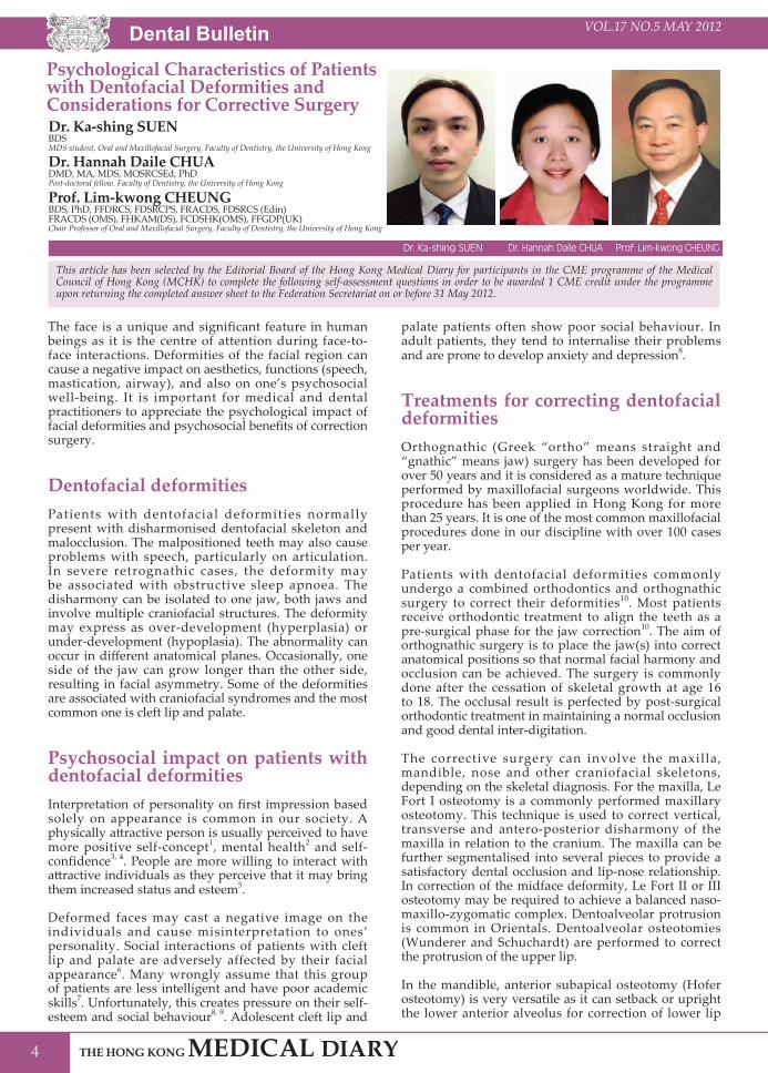
Dr. Hannah Daile CHUADr. Ka-shing SUEN Prof. Lim-kwong CHEUNG
Psychological Characteristics of Patients with Dentofacial Deformities and Considerations for Corrective SurgeryDr. Ka-shing SUEN
Dr. Hannah Daile CHUA
Prof. Lim-kwong CHEUNG
MDS student, Oral and Maxillofacial Surgery, Faculty of Dentistry, the University of Hong Kong
Post-doctoral fellow, Faculty of Dentistry, the University of Hong Kong
Chair Professor of Oral and Maxillofacial Surgery, Faculty of Dentistry, the University of Hong Kong
BDS
DMD, MA, MDS, MOSRCSEd, PhD
BDS, PhD, FFDRCS, FDSRCPS, FRACDS, FDSRCS (Edin)FRACDS (OMS), FHKAM(DS), FCDSHK(OMS), FFGDP(UK)
This article has been selected by the Editorial Board of the Hong Kong Medical Diary for participants in the CME programme of the Medical Council of Hong Kong (MCHK) to complete the following self-assessment questions in order to be awarded 1 CME credit under the programme upon returning the completed answer sheet to the Federation Secretariat on or before 31 May 2012.
The face is a unique and significant feature in human beings as it is the centre of attention during face-to-face interactions. Deformities of the facial region can cause a negative impact on aesthetics, functions (speech, mastication, airway), and also on one’s psychosocial well-being. It is important for medical and dental practitioners to appreciate the psychological impact of facial deformities and psychosocial benefits of correction surgery.
Dentofacial deformitiesPatients with dentofacial deformities normally present with disharmonised dentofacial skeleton and malocclusion. The malpositioned teeth may also cause problems with speech, particularly on articulation. In severe retrognathic cases, the deformity may be associated with obstructive sleep apnoea. The disharmony can be isolated to one jaw, both jaws and involve multiple craniofacial structures. The deformity may express as over-development (hyperplasia) or under-development (hypoplasia). The abnormality can occur in different anatomical planes. Occasionally, one side of the jaw can grow longer than the other side, resulting in facial asymmetry. Some of the deformities are associated with craniofacial syndromes and the most common one is cleft lip and palate.
Psychosocial impact on patients with dentofacial deformitiesInterpretation of personality on first impression based solely on appearance is common in our society. A physically attractive person is usually perceived to have more positive self-concept1, mental health2 and self-confidence3, 4. People are more willing to interact with attractive individuals as they perceive that it may bring them increased status and esteem5.
Deformed faces may cast a negative image on the individuals and cause misinterpretation to ones’ personality. Social interactions of patients with cleft lip and palate are adversely affected by their facial appearance6. Many wrongly assume that this group of patients are less intelligent and have poor academic skills7. Unfortunately, this creates pressure on their self-esteem and social behaviour8, 9. Adolescent cleft lip and
palate patients often show poor social behaviour. In adult patients, they tend to internalise their problems and are prone to develop anxiety and depression8.
Treatments for correcting dentofacial deformitiesOrthognathic (Greek “ortho” means straight and “gnathic” means jaw) surgery has been developed for over 50 years and it is considered as a mature technique performed by maxillofacial surgeons worldwide. This procedure has been applied in Hong Kong for more than 25 years. It is one of the most common maxillofacial procedures done in our discipline with over 100 cases per year.
Patients with dentofacial deformities commonly undergo a combined orthodontics and orthognathic surgery to correct their deformities10. Most patients receive orthodontic treatment to align the teeth as a pre-surgical phase for the jaw correction10. The aim of orthognathic surgery is to place the jaw(s) into correct anatomical positions so that normal facial harmony and occlusion can be achieved. The surgery is commonly done after the cessation of skeletal growth at age 16 to 18. The occlusal result is perfected by post-surgical orthodontic treatment in maintaining a normal occlusion and good dental inter-digitation.
The corrective surgery can involve the maxilla, mandible, nose and other craniofacial skeletons, depending on the skeletal diagnosis. For the maxilla, Le Fort I osteotomy is a commonly performed maxillary osteotomy. This technique is used to correct vertical, transverse and antero-posterior disharmony of the maxilla in relation to the cranium. The maxilla can be further segmentalised into several pieces to provide a satisfactory dental occlusion and lip-nose relationship. In correction of the midface deformity, Le Fort II or III osteotomy may be required to achieve a balanced naso-maxillo-zygomatic complex. Dentoalveolar protrusion is common in Orientals. Dentoalveolar osteotomies (Wunderer and Schuchardt) are performed to correct the protrusion of the upper lip.
In the mandible, anterior subapical osteotomy (Hofer osteotomy) is very versatile as it can setback or upright the lower anterior alveolus for correction of lower lip
Dental BulletinVOL.17 NO.5 MAY 2012
5
protrusion. Mandibular body osteotomies such as Step osteotomy can be used to close dental spaces and correct anterior open bite. Several osteotomies are available to advance or setback the whole mandible. The technique is applied on the mandibular ramus region. Sagittal split osteotomy is the commonest osteotomy used to advance the retruded mandible. To achieve mandibular setback, Vertical Subsigmoid Osteotomy is preferred as it causes less neurosensory disturbance. Other ramus osteotomy techniques such as inverted L or C osteotomy are less commonly used.
Correction of the chin deformity can be achieved by a sliding osteotomy or augmentation with an alloplastic material. Alloplastic implants may cause delayed infection and resorption of the underlying bony structure. Therefore, we prefer the sliding osteotomy for correction of chin deformities as it has less long term problems.
Psychosocial benefits of orthognathic surgeryI t has been well published that pat ients after orthognathic surgery generally have more positive mind, more vigour, less fatigue, less depression and less tension stress six months after the surgery when compared to those receiving only orthodontic treatment or no treatment11, 12, 13. In addition, patients can have better social functioning, social adjustment, self-confidence, self-concept, body image, emotional stability, self-esteem and facial attractiveness image, positive life changes and reduced anxiety14. No gender difference was found in the changes of psychological functioning before and after surgery15.
Psychological considerations for orthognathic patientsIt is of utmost importance to understand a patient’s motivation seeking for jaw correction. Many express their wish to improve aesthetics, oral functions (speech and mastication), self-esteem, social life, or prevent future dental problems (temporomandibular joint disorder and dental attrition). Patients are particularly concerned about the effects of the surgery on their facial appearance. It is prudent for the surgeons to justify whether the patients’ expectations are realistic. Unrealistic expectation can cause dissatisfaction of the final outcome, even though the result is considered as acceptable by most people. It is essential for the maxillofacial surgeons to provide informed consent regarding the surgery. It is important for the patients to understand the risks, benefits, surgical procedures, post-operative complications, treatment options, length of hospital stay and dietary adjustment before they subject themselves to surgery.
Pre-surgical psychological assessment and supportOrthognathic surgery can be considered as a potentially traumatic event which can cause post-operative
stress. Pre-existing psychological distress can be exacerbated post-operatively causing psychological problems, such as posttraumatic stress disorder. Therefore, psychological screening is recommended for orthognathic patients at the initial consultation stage as part of the work-up. This will help the surgeons in identifying any psychological risk factor and evaluate the patient’s fitness to receive orthognathic surgery. Risk factors associated with poor psychological outcomes after orthognathic surgery include personality disorders, body dysmorphic disorder, depression and anxiety16, 17. Patients having results exceeding the normal psychological threshold should be referred to clinical psychologists for a thorough evaluation and possible psychological support. Reports from the psychologists regarding a patient’s mental health status can provide important information on his/her readiness for orthognathic surgery.
Post-operative psychological supportInevitably, some orthognathic patients may express dissatisfaction and distress post-operatively. It is necessary for the surgeons to identify severity of the problems and investigate the overlying causes. Prompt management and reassurance should be given to the patients. Surgical outcomes which are considered to be satisfactory to surgeons but not to the patients, may arise due to the unmatched expectation and outcomes. Referral to psychologists for counselling and support can be beneficial for the management of these patients.
ConclusionPatients with dentofacial deformities commonly face psychological stress and may not be well accepted in the society due to their facial appearance. Orthognathic surgery has been found to provide many psychological benefits to these patients. Pre-operative psychological screening is necessary to identify patients with unrealistic expectations, who are prone to develop post-operative psychological disorders. Informed consent including risk, benefits of the corrective surgery should be discussed well before their commitment to surgery. Psychological support is useful for the management of any unexpected psychological disorders post-operatively. Close collaboration with clinical psychologists is essential in the multi-disciplinary management of patients with dentofacial deformities.
References1. Mathes, E.W.: The effects of physical attractiveness and anxiety on
heterosexual attraction over a series of five encounters. J. Marriage Family, 1975;37:769-773
2. Adams, G. R: The effect of physical attractiveness on the socialization process. In Lucker, G. W., Ribens, K. A., and McNamara. J. A., Jr (eds.): Psychological aspects of facial form. Ann Arbor, Michigan, Center for Human Growth and Development, 1981
3. Dion, K. K., and Stein. S.: Physical attractiveness and interpersonal influence. J. Exp. Soc. Psychol., 1978;14:97-108
4. Jackson, D. J., and Huston, T. L: Physical attractiveness and assertiveness. J. Soc. Psychol., 1975;96:79-84
5. Ellen Berscheid and Steve Gangestad, A.B.: The social psychological implications of facial physical attractivenss. Clinics in Plastic Surgery, 1982;9(3):289-296
6. Berk NW, Cooper ME, Liu YE, Marazita ML. Society anxiety in Chinese adults with oral-facial clefts. Cleft Palate Craniofac J. 2001;38:126-133
Dental Bulletin VOL.17 NO.5 MAY 2012
6
7. Richman LC, Eliason M. Psychological characteristics of children with cleft lip and palate: intellectual, achievement, behavioral and personality variables. Cleft Palate J. 1982;19:249-257
8. Pillemer FG, Cook KV. The psychological adjustment of pediatric craniofacial patients after surgery. Cleft Palate J. 1989;26:201-207
9. Turner SP, Rumsey N, Sandy JR. Psychological aspects of cleft lip and palate. Eur J Orthod. 1998;20:407-415
10. McCance, A.M, J.P. Moss, W.R. Wright, A.D. Linney, D.R. James. A three-dimensional soft tissue analysis of 16 skeletal class III patients following bimaxillary surgery. British Journal of Oral and Maxillofacial Surgery. 1992;30(4):221-232
11. Soon Min Hong. Reduction mandibuloplasty of the mandible in a square face: A series of cases. Journal of Oral and Maxillofacial Surgery. 2007;65(9):42.e6-e7
12. Kiyak HA, McNeill RW, West RA. The emotional impact of orthognathic surgery and conventional orthodontics. Am J Orthod 1985; 88:224-34.
13. Hannah Daile P. Chua. Cleft distraction versus orthognathic surgery-which one causes more distress? Journal of oral and maxillofacial Surgery. 2007; 65(9): 42.e7
14. Orlagh T. Hunt, Chris D. Johnston, Peter G. Hepper, Donald J. Burden. The psychosocial impact of orthognathic surgery: A systematic review. Am J Orthod Dentofacial Orthop 2001;120:490-7
15. Kiyak HA, Hohl T, Sherricl P, West RA, McNeil RW, Bucher F. Sex differences in motives for and outcomes of orthognathic surgery. J Oral Surg 1981;39:757-64
16. Bryan T. Ambro, Richard J. Wright. Depression in the cosmetic surgery patient. Facial Plast Surg 2010;26(4):333-338
17. Honigman RJ, Philips KA, Castle DJ. A review of psychosocial outcomes for patients seeking cosmetic surgery. Plast Reconstr Surg. 2004; 113:1229-37
MCHK CME Programme Self-assessment QuestionsPlease read the article entitled “Psychological Characteristics of Patients with Dentofacial Deformities and Considerations for Corrective Surgery” by Dr. Ka-shing SUEN, Dr. Hannah Daile CHUA and Prof. Lim-kwong CHEUNG and complete the following self-assessment questions. Participants in the MCHK CME Programme will be awarded CME credit under the Programme for returning completed answer sheets via fax (2865 0345) or by mail to the Federation Secretariat on or before 31 May 2012. Answers to questions will be provided in the next issue of The Hong Kong Medical Diary.
ANSWER SHEET FOR MAY 2012
Answers to April 2012 Issue
Please return the completed answer sheet to the Federation Secretariat on or before 31 May 2012 for documentation. 1 CME point will be awarded for answering the MCHK CME programme (for non-specialists) self-assessment questions.
Current Practices in the Management of Diabetic Nephropathy
1 4 82 5 93 76 10
1. T T F F T T FF T F4. 8.2. 5. 9.3. 7.6. 10.
Name (block letters):_____________________________ HKMA No.: __________________ CDSHK No.: _______________
HKID No.: __ __ - __ __ __ __ X X (X) HKDU No.: __________________ HKAM No.: ________________
Contact Tel No.:_________________________________
1. Deformities of the facial region have no impact on patients’ aesthetics and psychosocial well-being.2. Retruded jaws may be associated with obstructive sleep apnoea. 3. The public generally consider individuals with deformed faces are of normal intelligence and have better
academic skills. 4. Patients with dentofacial deformities commonly undergo a combined orthodontics and orthognathic surgery
to correct their facial deformities. 5. Patients after orthognathic surgery generally have more positive mind, more vigour, less fatigue, less depression
and less tension stress six months after the surgery when compared to those receiving no treatment. 6. Females attain more positive psychological functioning before and after surgery than males. 7. Many patients with facial deformities express their wish to improve aesthetics, oral functions, self-esteem and
social life. 8. Psychological screening is not recommended for orthognathic patients as part of the work-up. 9. Risk factors associated with poor psychological outcomes after orthognathic surgery include personality
disorders, body dysmorphic disorder, depression and anxiety.10. The perception of surgical outcomes is expected to be the same between surgeons and patients.
Questions 1-10: Please answer T (true) or F (false)
Psychological Characteristics of Patients with Dentofacial Deformities and Considerations for Corrective SurgeryDr. Ka-shing SUEN Dr. Hannah Daile CHUA Prof. Lim-kwong CHEUNG
MDS student, Oral and Maxillofacial Surgery, Faculty of Dentistry, the University of Hong Kong
Post-doctoral fellow, Faculty of Dentistry, the University of Hong Kong Chair Professor of Oral and Maxillofacial Surgery, Faculty of Dentistry,
the University of Hong Kong
BDS DMD, MA, MDS, MOSRCSEd, PhD BDS, PhD, FFDRCS, FDSRCPS, FRACDS, FDSRCS (Edin)FRACDS (OMS), FHKAM(DS), FCDSHK(OMS), FFGDP(UK)


Dental Bulletin VOL.17 NO.5 MAY 2012
8
Dr. Danny LOW
Biofilm and Contemporary EndodonticsDr. Danny LOWBDS (Syd), MScDent (Syd), MScMed (Pain Management) (Syd), AdvDipEndodont (HK), MRD RCSEd (Endo), FRACDS, FCDSHK (Endodontics), FHKAM (Dental Surgery)Specialist in Endodontics, Micro-Endodontics, Private Practice
IntroductionContemporary root canal treatment comprises of three procedural stages: mechanical instrumentation, chemical decontamination, and 3-dimensional obturation of the entire root canal system1. Despite the continuous development and invention of high-tech instruments, advanced biomaterials and effective treatment protocols, decontamination of the entire root canal system remains the key objective of contemporary root canal treatment. With complete elimination of bacteria from the root canal system, successful outcomes could be achieved. Having said that, studies have shown endodontic failures, in which persistent infection occurs even with thorough decontamination of the root canal system2,3. As a result, a new biofilm concept of endodontic infection was introduced; not just to understand the pathogenic potential of the endodontic microorganisms but also the basis for development of new strategies towards eradication of more resistant bacteria4.
Formation of BiofilmsBiofilms, by definition, are matrix-enclosed bacterial communities, in which microbes adhere to each other and/or to surfaces or interfaces5,6. This definition includes microbial aggregates and floccules and also adherent populations within the pore spaces of porous media. Without the existence of bacteria, perhaps biofilms would not be found. Thus, a prerequisite for biofilm formation is the availability of “planktonic microorganisms”; in which the free-floating microbes exist in an aqueous environment. Another important requirement would be the presence of an organic conditioning polymeric matrix (also known as conditioning film)6.
The development of biofilms could be described into 4 phases, which are (i) deposition of a conditioning film, (ii) adhesion and colonisation of planktonic microorganisms in a polysaccharide matrix with subsequent co-adhesion of other microorganisms, (iii) maturation of microbial community and (iv) detachment of biofilm microorganisms into the surrounding environment4,7.
(i) Deposition of a conditioning filmThe conditioning film is often formed prior to the arrival of planktonic microorganisms and comprised of proteins and glycoproteins from saliva and gingival crevicular fluid. Besides, the conditioning film comprises selective behaviours that determine which
type of microorganisms will attach to the matrix; hence, affects the microbial composition of the biofilm.
(ii) Adhesion, colonisation and subsequent co-adhesion of planktonic microorganismsUpon completion of the conditioning fi lm, the readily available free-floating bacteria that exist in the surrounding aqueous solution will start to attach onto the film surface and then colonisation occur with multiple micro-colonies formed. Interestingly, the initial type of bacteria that begin to colonise, seems to be crucial for the subsequent co-adhesion of other different bacteria8.
(iii) Maturation of microbial communityThe microorganisms that are embedded within the condit ioning f i lm wil l la ter mult iple and evolve to become a structurally organised mixed microbial community. During this stage the inherent characteristics of the microorganisms and the nature of the micro-environment influence growth and succession of microorganisms in the biofilm.
(iv) Detachment of microorganisms The detachment of microorganisms is a dynamic process among the biofilm formation. Indeed, it should be considered as a continuous process during development of biofilm, rather than a final stage of biofilm formation. The detachment begins at the initial microbial adhesion, in which the quantity of detachment increases with maturation of biofilm, as the gross amount of microorganisms within the biofilm multiplies. It is the detached microorganisms that contribute to transmission and colonisation at other susceptible loci.
Clinical ImplicationStudies have shown that bacterial interaction and cooperation is crucial for bacterial survival, persistence and growth in the microbial community that exists within the root canals9,10. Even though the organisation of biofilm is acknowledged, development of biofilm in endodontic infections remains mysterious4. As readily available planktonic microbes are capable to form biofilm in an aqueous medium, root canal biofilm is not uncommon in endodontic infections. If that is the case, this would be extremely important with substantial clinical significance; as biofilm-embedded microorganisms are known to show better resistance to external adverse influences such as antimicrobial treatment procedures than free-floating planktonic analogs5,6.
Dental BulletinVOL.17 NO.5 MAY 2012
9
In the context of root canal biofilm, it has been suggested that the pre-condition for root canal biofilm formation varies depending on the cause of pulpal tissue breakdown4. For example, caries exposure of pulp remains the most common cause of tissue breakdown. Carious invasion of the pulp represents bacterial infiltration into the root canal, in which the inflammatory lesion front may move successively towards the root apex. As a consequence, providing an aqueous medium by which the foreign planktonic microorganisms from the oral cavity could migrate into the root canal space and initiate biofilm formation. Nonetheless, the actual phenomenon that occurs after initial penetration of microorganisms remains unclear. There is a lack of information regarding how these microbials attach and spread further along the entire root canal space. Perhaps, a massive infiltration of polymorphonuclear cells as a result of inflammation leads to formation of by-product exudates could account for the availability of an aqueous medium for biofilm formation11.
On the other hand, there are cases of necrotic pulpal tissues, which are relatively dry and may not be fluid-filled. As such, the question remains as to whether bacterial condensations in a biofilm structure can develop or be retained in sites of the root canal system other than near the bacteria/inflammatory interface; where protein-rich immuno-substance and bacterial produced adhesive substances may provide the adequate prerequisites. Indeed, evidences regarding the pattern of root canal biofilm distribution during the progress of pulpal breakdowns are rather limited among dental literatures.
Architecturally, the mature biofilm is a highly organised filamentous structure consisting of mushroom-shaped microcolonies of bacteria bound together by a polysaccharide matrix together with surrounding open water channels that maintain homeostasis7,12,13,14. There is no doubt that the biofilm acts as a physical barrier against the action of antimicrobial substances and the host’s defence mechanisms4. Therefore, microorganisms that are embedded within the biofilm are shielded and thus often harder to be eliminated as compared with their free-floating planktonic counterpart14,15; in which they are often (up to 1000-fold) more resistant to phagocytosis, antibodies and many antibiotics5,13,15,16.
Another unique feature of the biofilm would be its constitution of a protected mode of growth that permits survival in a hostile environment5 and recovers rapidly following access to increased nutritional supply. The dense filamentous structure of biofilm might restrict the penetration of the antimicrobials into the biofilm17. That is, deep-seated microorganisms may be able to survive against antimicrobials and remain viable for remultiplication when conditions are favourable once again. For instance, an in vitro study showed that bacteria of endodontic origin were able to grow on dentin slices of extracted teeth to form biofilm that was not likely to be eradicated using various antibiotics such as ampicillin, doxycycline, clindamycin, azithromycin, or metronidazole18,19. Besides, the biofilm has been shown to be able to neutralise potent oxidising agents, making it difficult for them to penetrate and kill microorganisms20.
The proximity of individual microorganism in a biofilm also increases the chances for gene transfer, making it possible to transform previously avirulent microbes into a highly virulent pathogen that is not susceptible to antimicrobics13. Indeed, this potential for gene transfer within the biofilm is particularly significant in the case of Enterococcus faecalis (E. faecalis), because a number of its virulence factors are encoded on transmissible plasmids, which include collagenase, gelatinase, and adhesions, all with the potential to contribute to survival in and colonisation of the root canal21.
Contemporary Root Canal TreatmentIt is well known that persistent endodontic infections do not result from an individual species of microbial6. Rather, it is the entire polymicrobial entity that undergoes physiological and genetic changes triggered by ecological changes within the root canal system. Therefore, the prime objective of contemporary root canal treatment is “root canal decontamination”; that is complete eradication of free-floating bacteria as well as bacteria embedded within the biofilm from the root canal space. Furthermore, removal of the entire biofilm (i.e. anti-biofilm) along the root canal system would be equally important as this will prevent reactivation of survived nonviable bacteria22. Based on the current understanding of root canal biofilm concepts, various means for the effective control of root canal biofilm are being explored.
One important clinical procedure of conventional root canal treatment would be mechanical instrumentation with endodontic files. Unfortunately, mechanical instrumentation alone does not result good removal of biofilm23. Practically, the challenging component of biofilm removal is the not readily accessible areas such as fins and crevices of the root canal system. Such inaccessible root canal spaces would be a source of undisrupted biofilm. Thus, the use of antimicrobial agents has been recommended as an adjunct to mechanical instrumentation to eliminate root canal microorganisms23.
In contrast, intra-canal irrigation with antimicrobial solutions would be able to eliminate root canal biofilm that is impossible to be removed with mechanical instrumentation. Antimicrobial irrigation aims to assist in the removal of root canal biofilm from uninstrumented surfaces that usually account for 30 to 50% of the entire root canal wall22,24. A number of studies have examined the antimicrobial efficacy of root canal irrigants on root canal biofilm, which include sodium hypochlorite (NaOCl), EDTA, chlorhexidine and BioPure MTAD22,25-29.
As compared with other irrigants, NaOCl is probably the most effective antimicrobial and antibiofilm agent28. NaOCl is capable of both physically removing biofilm22,28 and killing bacteria22,25-29. The antimicrobial effectiveness is dose dependent, in which a higher (6%) concentration of NaOCl is sufficient to remove both bacteria and biofilm from root canals22. On the other hand, when the concentration is reduced to 1%, viable microorganisms will be able to survive. Possibly, the reduced concentration of NaOCl could be compensated if

Dental Bulletin VOL.17 NO.5 MAY 2012
10
it is replenished frequently or allowed to have additional time to exert its antimicrobial properties. Besides, the effectiveness of an irrigant is dependent on the nature of the organisms and the contact time25. The antimicrobial effect of NaOCl is likely because of removal of organic matrix of the biofilm, thus eliminating the bacterial component of the biofilm together with eradication of biofilm. Adjusting the temperature of NaOCl also affects the organic tissue dissolubility30.
Recently, chlorhexidine has been shown to have an effective antimicrobial property when used as an endodontic irrigant31. However, its effectiveness in removal of root canal biofilm as well as the dose dependent effect of chlorhexidine are not promising22. BioPure MTAD has also been examined in the same study which shows that its antimicrobial property is minimal and incapable to physically remove biofilm22,27. EDTA is generally recommended for the removal of smear layers and its antimicrobial effect remains controversial27,29,32. However, it has been suggested that EDTA facilities biofilm removal or bacteria removal from biofilms29.
Clinically, root canal irrigation often comprises a combination of the above-mentioned irrigants. Therefore, the effects of different irrigation regimens in relation to biofilm removal were also examined33. The study reported that EDTA when used as final irrigant of the root canal would lead to high susceptibility of biofilm formation; whereas, a recommended irrigation regimen of EDTA followed by NaOCl and chlorhexidine that would alter the favourable factors of biofilm formation.
Apart from irrigation regimen, other irrigation variables such as irrigant agitation on the efficacy of biofilm removal were also suggested34-36. Dynamic irrigation which consists of irrigation with the addition of intracanal push-pull manipulation of a tapered gutta-percha point closely resembling the canal dimensions enhanced the removal of root canal biofilms35. Canal enlargement of both apical dimension and canal taper also directly affects the amount of biofilm removal35. Irrigating needles designed with multiple openings would enable better biofilm removal as well as frequent replacement of irrigating solutions35. Sonic and ultrasonic irrigations are able to dislodge experimental biofilm34,37,38. A typical sonic irrigation system such as EndoActivator system was reported to be able to effectively remove biofilm37. With ultrasonic irrigation, energy is transmitted to the irrigant in the root canal by means of ultrasonic waves and subsequently induces acoustic streaming. The acoustic streaming induced is then able to cause de-agglomeration of root canal biofilm38; and hence improves efficacy of NaOCl against the microorganisms.
Recently, light activated disinfection was able to demonstrate a good result against removal of biofilm39. Although light activated disinfection alone was not effective enough to remove mature biofilm, combined procedures of chemo-mechanical instrumentation together with light activated disinfection were able to eliminate mature biofilm. This is probably because mechanical instrumentation of the root canal would have partially disrupted the underlying biofilm
structure, and thus facilitates the penetration of antimicrobial irrigants into the biofilm structure. Perhaps more long-term clinical studies are required to verify the clinical efficacy.
SummaryThe concept of biofilm plays an important role in the paradigm shift of contemporary root canal treatment. Biofilms are sessile microbial communities composed of microorganisms irreversibly attached to a substrate-rich layer and interface and/or to each other. Apparently, there is no standard clinical procedure available in terms of mechanical and antimicrobial measures that can predictably eliminate the endodontic infection in total, particularly those microorganisms lodged within the non-instrumented fins and crevices of root canal system as well as the dentinal tubules. Therefore, it would be reasonable to assume that the ideal endodontic irrigant would be the one that is antimicrobial as well as capable in physically removing both the biofilm and the associated microorganisms. Root canal treatment procedures should focus more on antimicrobial irrigation rather than mechanical instrumentation.
References1. Schilder H. Cleaning and shaping the root canal. Dent Clin North
Am 1974; 18: 269-296.2. Siren EK, Haapasalo MPP, Ranta K, Salmi P, Kerosuo ENJ.
Microbiological findings and clinical treatment procedures in endodontic cases selected for microbiological investigation. Int Endod J 1997; 30: 91-95.
3. Sundqvist G, Figdor D, Persson S, Sjögren U. Microbiologic analysis of teeth with failed endodontic treatment and the outcome of conservative re-treatment. Oral Surg Oral Med Oral Pathol 1998; 85: 86-93.
4. Svensäter G, Bergenholtz G. Biofilms in endodontic infections. Endod Topics 2004; 9: 27-36.
5. Costerton JW, Stewart PS, Greenberg EP. Bacterial biofilms: a common cause of persistent infections. Science 1999; 284: 1318-1322.
6. Chávez de Paz L. Redefining the persistent infection in root canals: possible role of biofilm communities. J Endod 2007; 33 652-662.
7. Costerton JW, Lewandowski Z, Caldwell DE, Korber DR, Lappin-Scott HM. Microbial biofilms. Annu Rev Microbiol 1995; 49: 711-745.
8. Kolenbrander PE, Andersen RN, Blehert DS, Egland PG, Foster JS, Palmer RJ Jr. Communication among oral bacteria. Microbiol Mol Biol Rev 2002; 66: 486-505.
9. Dahlén G. Microbiology and treatment of dental abscesses and periodontal-endodontic lesions. Periodontol 2000 2002; 28: 206-239.
10. Sundqvist G, Figdor D. Life as an endodontic pathogen. Etiological differences between untreated and root-filled root canals. Endod Topics 2003; 6: 3-28.
11. Nair PNR. Light and electron microscopic studies on root canal flora and periapical lesions. J Endod 1987; 13: 29-39.
12. Auschill TM, Arweiler NB, Neutuschil L, Brecx M, Reich E, Sculean A. Spatial distribution of vital and dead microorganisms in dental biofilms. Arch Oral Biol 2001; 46: 471-476.
13. Potera C. Forging a link between biofilms and disease. Science 1999; 283: 1837-1839.
14. Distel JW, Hatton JF, Gillespie MJ. Biofilm formation in medicated root canals. J Endod 2002; 28: 689-693.
15. Cook CS, Costerton JW, Lamont RJ. Biofilm formation by Porphyromonas gingivalis and Streptococcus gordonii. J Periodontal Res 1998; 33: 323-327.
16. de Paz L. Redefining the persistent infection in root canals: possible role of biofilm communities. J Endod 2007; 33: 652-662.
17. Stewart PS, Costerton JW. Antibiotic resistance of bacteria in biofilms. Lancet 2001; 14: 135-138.
18. Norrington DW, Ruby J, Beck P, Eleazer PD. Observations of biofilm growth on human dentin and potential destruction after exposure to antibiotics. Oral Surg Oral Med Oral Pathol Oral Radiol Endod 2008; 105: 526-529.
19. Larsen T. Susceptibility of Prophyromonas gingivalis in biofilms to amoxicillin, doxycycline and metronidazole. Oral Microbiol Immunol 2002; 17: 267-271.

Dental BulletinVOL.17 NO.5 MAY 2012
11
20. Stewart PS, Rayner J, Roe F, Rees WM. Biofilm penetration and disinfection efficacy of alkaline hypochlorite and chlorosulfamates. J Appl Microbiol 2001; 91: 525-532.
21. Duggan JM, Sedgley CM. Biofilm formation of oral and endodontic Enterococcus faecalis. J Endod 2007; 33: 815-818.
22. Clegg MS, Vertucci FJ, Walker C, Belanger M, Britto LR. The effect of exposure to irrigant solutions on apical dentin biofilms in vitro. J Endod 2006; 32: 434-437.
23. Byström A, Sundqvist G. Bacteriologic evaluation of the effect of 0.5 per cent sodium hypochlorite in endodontic therapy. Oral Surgery, Oral Medicine Oral Pathology 1983; 55: 307-312.
24. Peters OA, Laib A, Göhring TN, Barbakow F. Changes in root canal geometry after preparation assessed by high-resolution computed tomography. J Endod 2001; 27: 1-6.
25. Spratt DA, Pratten J, Wilson M, Gulabivala K. An in vitro evaluation of the antimicrobial efficacy of irrigants on biofilms of root canal isolates. Int Endod J 2001; 34: 300-307.
26. Chávez de Paz L, Bergenholtz G, Svensäter G. The effects of antimicrobials on endodontic biofilm bacteria. J Endod 2010; 36: 70-77.
27. Dunavant TR, Regan JD, Glickman GN, Solomon ES, Honeyman AL. Comparative evaluation of endodontic irrigants against Enterococcus faecalis biofilms. J Endod 2006; 32: 527-531
28. Bryce G, O’Donnell D, Ready D, Ng Y-L, Pratten J, Gulabivala K. Contemporary root canal irrigants are able to disrupt and eradicate single- and dual-species biofilms. J Endod 2009; 35: 1243-1248.
29. Gulabivala K, Patel B, Evans G, Ng Y-L. Effects of mechanical and chemical procedures on root canal surfaces. Endod Topics 2005; 10: 1`03-122.
30. Cunningham WT, Joseph SW. Effect of temperature on the bactericidal action of sodium hypochlorite endodontic irrigant. Oral Surgery, Oral Medicine Oral Pathology 1980; 50: 569-517.
31. Jeansonne MJ, White RR. A comparison or 2% Chlorhexidine gluconate and 5.25% sodium hypochlorite as antimicrobial endodontic irrigants. J Endod 1994; 20: 276-278.
32. Al-Bakri AG, Othman G, Bustanji Y. The assessment of the antibacterial and anti-fungal activities of aspirin, EDTA and aspirin-EDTA combination and their effectiveness as antibiofilm agents. J Appl Microbiol 2009; 107: 280-286.
33. Kishen A, Sum CP, Mathew S, Lim CT. Influence of irrigation regimens on the adherence of Enterococcus faecalis to root canal dentin. J Endod 2008; 34: 850-854.
34. Burleson A, Nusstein J, Reader A, Beck M. The in vivo evaluation of hand/rotary/ultrasound instrumentation in necrotic, human mandibular molars. J Endod 2007; 33: 782-787.
35. Huang T-Y, Gulabivala K, Ng Y-L. A bio-molecular film ex-vivo model to evaluate the influence of canal dimensions and irrigation variables on the efficacy of irrigation. Int Endod J 2008; 41: 60-71.
36. Gu LS, Kim JR, Ling J, Choi KK, Pashley DH, Tay FR. Review of contemporary irrigant agitation techniques and devices. J Endod 2009; 35: 791-804.
37. Caron G. Cleaning efficiency of the apical millimeters of curved canals using three different modalities of irrigant activation: an SEM study. Paris VII University, Paris, France: Master Thesis; 2007.
38. Joyce E, Phull SS, Lorimer JP, Mason TJ. The development and evaluation of ultrasound for the treatment of bacterial suspensions: a study of frequency, power and sonication time on cultured Bacillus species. Ultrason Sonochem 2003; 10: 315-318.
39. Lim Z, Cheng JL, Lim TW, Teo EG, Wong J, Geroge S, Kishen A. Light activated disinfection: an alternative endodontic disinfection strategy. Aust Dent J 2009; 54: 108-114.

Dental Bulletin VOL.17 NO.5 MAY 2012
12
Dr. Raymond LK CHOW
Surgical Complications in Implant Dentistry and Their ManagementDr. Raymond LK CHOW
Specialist in Oral & Maxillofacial SurgeryConsultant, Dental Implant & Maxillofacial CentrePart time Clinical Assistant Professor, Faculty of Dentistry, the University of Hong Kong
BDS(HK), MDS(HK), MOSRCS(Edinburg), FHKAM(DS), FCDSHK(OMS)
IntroductionDental implant is a predictable and safe treatment modality for partial and total edentulous patients. High success rates have been demonstrated. However, complications may occur in a small proportion of patients. These complications could occur during the placement of implants (surgical complications) or after the placement of prostheses (prosthetic complications). Complications may or may not lead to implant failure, but managing these problems could be very challenging. This article will highlight various surgical complications associated with dental implant surgery and discuss their management. Surgical complications occur in the surgical phase of dental implant treatment. It could happen intra-operatively (during the insertion of implant fixtures), or post-operatively (shortly after implant placement). These complications include:a) Haemorrhageb) Wound dehiscence/infectionc) Nerve injuryd) Displacement of implant into the maxillary sinus e) Damage to adjacent teeth
HaemorrhageThe amount of bleeding associated with a surgical procedure depends on the local and general factors. Local factors include flap or flapless procedures, the raising and extent of flap and also soft tissue management. General factors would be the patient’s general health, presence of any haemorrhagic disorders and administration of anticoagulants. Active profuse bleeding during implant surgery is very rare unless major vessels are damaged. In fact, post-operative haemorrhagic patches developed on the skin or mucous membranes were quite common (Fig. 1). Goodacre et al1 reported that 24% of his implant cases have post-operative ecchymosis. The incidence of these haemorrhagic patches can be reduced with careful soft tissue management and try to avoid vertical relieving incision in the flap. Application of pressure on the flap after suturing for few minutes can also minimise haematoma formation, and thus reduces bruises.
InfectionsThe infection rates after implant surgery were low; Powell and his colleagues2 reported the implant infection rate in his studies was around 1.14%, and Gynther et al3 found a 0.7% infection rate. Treatment of infections associated with dental implant placement was
similar to other dentoalveolar infections. Antimicrobial therapy and sometimes combined with surgical debridement can effectively resolve the infection. Strict aseptic technique during implant placement is also suggested.
Fig. 1 Ecchymosis of the infra-orbital areas after bilateral maxillary implant placement
Nerve injuryNerve injury caused by dental implant insertion is a serious complication and frequently leads to medico-legal consequences. The incidence of sensory disturbance reported in the literatures was around 10%. Inferior dental (ID) and mental nerves are frequently involved4,5. It is due to the intrusion of implant or twist drill into the inferior dental canal or mental foramen (Fig 2). Patients usually complained of paraesthesia or hypoesthesia of the ipsilateral side of the lower lip (Fig 3). However, if dysesthesia (pain elicited by low threshold stimulus) or anaesthesia (complete loss of sensation) is noted by the patient, severe nerve damage is expected. Radiograph or CT scan should be taken to ascertain the implant’s position. If it is intruded into the canal, it should either be withdrawn a couple of turns or removed completely. However, the implant may lose its primary stability after partial withdrawal, and in that case, complete removal of the implant is the only way. Because loose implants can get infected easily, this will further complicate the situation.
To prevent nerve injury, careful pre-operative determination of the nerve position (ID and mental nerves) is very important. Diagnostic imaging such as panoramic radiograph and cone beam CT should be obtained before the surgery. If panoramic radiograph is used, the appropriate magnification factor has to be taken into account. It is recommended that the dental implant should be at least 2mm away from the nerve6,7. A drill guard can be used to avoid accidental over-drilling. In addition, informed consent to the patient on the risk of nerve damage should also be mentioned.

Dental BulletinVOL.17 NO.5 MAY 2012
13
Fig. 2 Implant at 47 intruded into the inferior dental canal
Fig. 3 Numbness of the right lower lip after implant placement at 47
Once neurosensory deficit occurs, the patient should be managed promptly, rather than leaving him/her unattended, and hoping the sensation will come back by itself. Neurosensory test8 should be undertaken monthly using light touch and two-point discrimination and pin prick stimuli thresholds (Fig 4). The lack of any evidence of recovery by 3 months is recommended as an indication for surgical intervention. If there is some evidence of sensory recovery by 3 months, monitoring should be continued for a further 3 -6 months, until there is no further improvement. At this stage, an operation is considered if there is either poor recovery or dysaesthesia9. The modified protocol based on that of Robinson for the management of nerve injury is shown in figure 5.
Fig. 4 Neurosenosry test - a) Light touch b) Two point discrimination c) Pin prick
Fig. 5 Protocol for the management of nerve injury caused by dental implant placement
Displacement of implant into the maxillary sinus (Fig 6)An implant which is displaced into the maxillary sinus during insertion must be removed immediately. Otherwise, maxillary sinusitis and other serious sinus sequelae (e.g. pan-sinusitis) could happen (Fig 7). Symptoms of sinusitis include fever, para-nasal pain especially when leaning forward, yellowish discharge from the nose. Retrieval of displaced implant is accomplished by a lateral open approach (creating a lateral bone window into the sinus). In case of purulent infection of the sinus, pus has to be drained also (Fig 8). Antibiotics and nasal decongestants should be prescribed post-operatively.
Fig. 6 Displacement of implants and cover screw into the left maxillary antrum
Fig. 7 Left pan-sinusitis, note the infection spreading

Dental Bulletin VOL.17 NO.5 MAY 2012
14
Damage to adjacent teeth Apart from nerve damage, incorrect angulation or positioning of an implant can also damage the adjacent teeth. Implant impinging on an adjacent tooth can jeopardise its blood supply and result in pulp necrosis and thus periapical pathology. The damaged tooth will require root canal therapy, apicectomy or, in the worst case, extraction. Untreated periapical lesion may contaminate the implant and results in failure. Figure 9 showed a case of implant at 46 hitting the root of 45, buccal abscess developed at 45 two months after the placement. Surgical exploration revealed severe bone loss at 45 and mesial aspect of the implant and 45 required extraction (Fig 10). In this case, the implant still retained its stability, therefore, bone grafting of the defect was undertaken and a bridge was made using the 44 as abutment (Fig 11).
Fig. 8 Lateral open approach to retrieve the displaced implants and drain pus
Fig. 9 Implant hitting the root of the adjacent 45.
Fig. 10 The resulted bone defect at the mesial side of the implant
Fig. 11 Post-operative radiograph after bone grafting and bridge construction
Conclusion Dental implant treatment is safe and detrimental complication is rare. McDermott et al10 found that the overall implant complication was about 15.3% and surgical complication only accounted for 1%. Though surgical complication is rare, it could lead to medico-legal consequences. Most of these complications can be avoided by careful pre-operative assessment and planning, and through the use of accurate diagnostic tools. Last but not the least, informed consent should be given and the risks of treatment should be fully explained to the patients before the surgery.
References1. Goodacre CJ, Bernal G, Rungcharassaeng K, Kan JY. Clinical
complications with implants and implant prostheses. J Prosthet Dent 2003;90:121-132
2. Powell CA, Mealey BL, Deas DE, McDonnell HT, Moritz AJ. Post-surgical infections: Prevalence associated with various periodontal surgical procedures. J Periodontol 2005;76:329-333.
3. Gynther GW, Kondell PA, Moberg LE, Heimdahl A. Dental implant installation without antibiotic prophylaxis. Oral Surg Oral Med Oral Pathol Oral Radiol Endod 1998;85:509-511
4. Dao TT, Mellor A. Sensory disturbances associated with implant surgery. Int J Prosthodont 1998;11:462-469.
5. Bartling R, Freeman K, Kraut RA. The incidence of altered sensation of the mental nerve after mandibular implant placement. J Oral Maxillofac Surg 1999;57:1408-1412.
6. Greenstein G, Tarnow D. The mental foramen and nerve: Clinical and anatomical factors related to dental implant placement. A literature review. J Periodontol 2006;77:1933-1943.
7. Worthington P. Injury to the inferior alveolar nerve during implant placement: A formula for protection of the patient and clinician. Int J Oral Maxillofac Implants 2004;19:731-734.
8. Hegedus F, Diecidue RJ. Trigeminal nerve injuries after mandibular implant placement – Practical knowledge for clinicians. Int J Oral Maxillofac Implants 2006;21:111-116.
9. Robinson PP, Loescher AR, Yates JM, Smith KG. Current management of damage to the inferior alveolar and lingual nerves as a result of removal of third molars. Br J Oral Maxillofac Surg. 2004;42:285-292.
10. McDermott NE, Chuang SK, Woo VV, Dodson TB. Complications of dental implants: identification, frequency, and associated risk factors. Int J Oral Maxillofac Implants. 2003;18:848-855.

17
VOL.17 NO.5 MAY 2012
Dental Quiz
Dr. Shiu-yin CHOBDS(HK), MDS(Otago), FRACDS, FHKAM(Dental Surgery)Senior Dental Officer, Fanling School Dental Clinic
History:A 12-year-old boy slipped and fell at the school playground. His tooth 11 suffered horizontal crown fracture. His teacher retrieved the fractured fragment and carefully wrapped it in a piece of tissue paper and let the child to bring it home. The child sought dental care in the evening and the dentist reattached the fragment with bonding agent and composite resin. However, the reattached fragment appeared to have different colour from the remaining tooth structure.
Question:1: why the reattached fragment showed different colour from the remaining tooth structure?2: what could be done to improve the aesthetics for this tooth?
(See P.37 for answers)
Dr. Shiu-yin CHO
Dental Quiz
Venue or MeetingFacilities
Member Society(Hourly Rate HK$)
PeakHour
Non-PeakHour
All day Sats, Suns &
Public Holidays
Rental Fees of Meeting Room and Facilities at The Federation of Medical Societies of Hong Kong
Non-Member Society(Hourly Rate HK$)
Multifunction Room I (Max 15 persons)Council Chamber (Max 20 persons)Lecture Hall (Max 100 persons)
150.00
240.00
300.00
105.00
168.00
210.00
225.00
360.00
450.00
250.00
400.00
500.00
175.00
280.00
350.00
375.00
600.00
750.00
PeakHour
Non-PeakHour
All day Sats, Suns &
Public Holidays
(Effective from October 2009)
Non-Peak Hour: 9:30am - 5:30pmPeak Hour: 5:30pm - 10:30pm
LCD Projector Microphone System
500.00 per session50.00 per hour, minimum 2 hours

C
M
Y
CM
MY
CY
CMY
K
HKU_MPH2012_Medical Diary_368x257_OP.pdf 1 13/04/2012 5:51 PM
C
M
Y
CM
MY
CY
CMY
K
HKU_MPH2012_Medical Diary_368x257_OP.pdf 1 13/04/2012 5:51 PM

Dental Bulletin VOL.17 NO.5 MAY 2012
20
Dr. Wilson LEE
Improving Aesthetics and Smiles with Incognito® Lingual Orthodontic ApplianceDr. Wilson LEEFCDSHK (Orthodontics), FHKAM (Dental Surgery), MOrthRCS (Edin), MOrth (HK), AdvDipOrtho (HK), MRACDS (Ortho), BDS (HK)Specialist in Orthodontics, Private Practice; Part-time Clinical Lecturer, Faculty of Dentistry, HKU; Council member, Hong Kong Society of Orthodontists
IntroductionWhere beautiful smiles play a significant role in our world today, the number of orthodontic patients in orthodontic practices is ever increasing.1 Most patients, including adolescents, want to improve the alignment of their teeth and smiles without showing the metal braces, and prefer treatment with invisible or nearly invisible appliances.2 Despite the wide availability of clear ceramic brackets in the market, patients continue to prefer ‘invisible’ braces when they have their teeth straightened. Orthodontists often choose to use orthodontic appliance systems that can meet patient's demand and deliver clinically excellent treatment results.
‘Invisible’ Orthodontic ApplianceThere are basically two types of ‘invisible’ orthodontic appliances available in the market:1. REMOVABLE – e .g . Invisa l ign ® . Aesthet ic
orthodontic appliances such as Invisalign® have been developed in the past two decades which have met some demands by patients. As it is a removable orthodontic appliance, it has its limitations in controlling teeth movement in three dimensions. Treatment duration is also longer than conventional labial or lingual appliances. A recent US study has shown that the mean accuracy of tooth movement is 47%.3 In Hong Kong, both Specialists in Orthodontics and General Dentists can prescribe this removable appliance to their patients.
2. FIXED – e.g. Incognito® l ingual orthodontic appliance - which was the most popular lingual appliance around the world last year. This lingual appliance is only prescribed by Specialists in Orthodontics since it is very technique sensitive, and requires specialist training before certified to use this appliance. Lingual braces are not visible from the outside and there is little or no aesthetic impairment of a patient’s smile during treatment. The disadvantages would be init ial tongue discomfort and speech disturbances.4
Brief History of Lingual Orthodontic ApplianceThe original lingual orthodontic appliance was first introduced by Dr. Kinya Fujita in Japan and Dr. Craven Kurz in the USA in the late 1970’s. It has created
significant interests to Orthodontic specialists in the mid-1980’s. However, there were three major problems which have resulted in the decline in these conventional lingual orthodontics in the early 1990’s:1. Difficulty in finishing the cases in high standard
and the emergence of more aesthetic clear labial appliances which were easier to control but not invisible;
2. Rebonding problem – it is very difficult to precisely rebond the brackets after they accidentally come off from the tooth surface;
3. Speech problem – due to the larger profile and size of the conventional lingual brackets, tongue movement is more restricted during speech and the brackets causes invariable irritation during speech.
Incognito Lingual Orthodontic ApplianceDr. Dirk Wiechmann of Germany invented the Incognito lingual appliance in the late 1990’s and has created enormous interests in lingual appliances in the last decade.5-8 His company and technique were subsequently acquired by 3M Unitek® in 2009. The appliance is distinguished from other bracket systems because both the brackets and the wires are custom made for individual patients, and fabricated using state-of-the-art CAD/CAM technology and archwire bending robots.
The technological advances in the Incognito lingual appliance help in solving the three major problems in conventional lingual orthodontics:1. Improvement in patient comfort and speech
adaptation – as the appliance is custom-made and adapted to the tooth surface as flat as possible;
2. Improvement in accuracy of rebonding – as the custom-made bracket base covers most of the lingual tooth surface, this allows the ease of direct rebonding of a bracket without the need of any jig or positioning aids;
3. Improvement in ease of finishing and detailing of a case – based on the set-up models, the custom-made brackets and pre-fabricated archwires (according to the arch form) have improved accuracy in finishing the case as close to the set-up models as possible. A recent study of 94 consecutive patients in a German orthodontist’s clinic has shown that discrepancies in position and rotation between the set-up models and outcome were small for all teeth, generally less than 1mm and 4 degrees, except for the second molars, where some larger discrepancies were observed.9

Dental BulletinVOL.17 NO.5 MAY 2012
21
Appliance Manufacturing ProcessAfter the orthodontic treatment plan has been explained to the patient and a consensus is reached on the treatment objectives, an accurate polyvinyl siloxane impression and bite registration are obtained from the patient. A therapeutic set-up of the teeth model is made on a mounted articulator (Figure 1) based on Andrews’ six keys to normal occlusion10, or the orthodontist could prescribe the detailed set-up criteria (e.g. angulation and torque) to the technicians.
Figure 1: Set-up models of final prescribed occlusion mounted on an articulator.
With the use of high-resolution optical 3D scanner, the teeth set-up model is scanned into the computer. Based on the data collected from the set-up model, the shape and position of the brackets will be designed virtually. The patented state-of-the-art CAD/CAM technology will assist in design and fabrication of Incognito lingual brackets (Figure 2a-e).
Figure 2a: Individual positioning of the bracket bodies and base pads are plotted and placed on a CAD/CAM software; Figure 2b, Generation of the archwire co-ordinates; Figure 2c: Prototyping the wax brackets on a rapid prototype wax printers; Figure 2d: Individual brackets are casted and polished (a molar bracket is shown here); Figure 2e: State-of-the-art archwires bending robot. All photos above are courtesy of 3M Unitek®.
The base pad of the bracket is first plotted on the lingual surface of each tooth, with the aim of maximising the surface area of bonding surface (Figure 2a). The large size of the pad not only gives greater bond strength,
but also makes it easier to adapt to the individual tooth during bracket rebonding. Then the low profile bracket bodies are inserted, making sure the bracket profile is as thin as possible in order to maximise patient comfort during daily function. The CAD/CAM software will help select the height, angulation and torque of the brackets. As such, the individual patient’s prescriptions of the appliance are built into each bracket (Figure 2b).
Once all the brackets are designed, the information will be transferred to the production line by first creating wax patterns of each bracket using a ‘rapid prototyping’ machine (Figure 2c). It is similar to a wax printer. After adding the wax blanks to the patterns, they are placed inside a casting cylinder and coated with plaster, similar in casting a gold crown. The wax is then burnt and gold alloy is poured into the cylinder. This nickel-free gold alloy offers an alternative especially to patients who are allergic to Nickel.
After casting, all the brackets are polished to a fine surface in order to ensure patient comfort (Figure 2d). The size of the bracket slot is then checked with precision equipment to confirm their accuracy. Measuring rates show divergences of not more than 0.008mm between slots.
In the final stage of the bracket production, the brackets are bonded to the original model, and an indirect bonding transfer tray is made. In most cases, the brackets are bonded to the patients’ teeth at the same time.
Archwire productionThe 3-dimensional geometry of each archwire is calculated with the CAD/CAM software and the information is transferred to the state-of-the-art archwire bending robot (Figure 2e). All the archwires in the sequence of treatment have the same geometry to obtain the final position of the teeth in the set-up model. Although the archwires are custom made and bent, it is still up to the Orthodontists to do some final adjustments and detailing on the chair side. There are some differences in archwire design between cases, e.g. those require extraction of teeth and those don’t need extraction, and it is the Orthodontist who decides all the selection of wires. Common wires used are 0.014-inch superelastic (SE) Nickel-Titanium (NiTi) wire, 0.016-inch SE NiTi wire, 0.016x0.022-inch SE NiTi wire, 0.016x0.024-inch Stainless Steel wire, and 0.0182x0.0182-inch Titanium Molybdenum Alloy (TMA) wire.
Clinical IndicationsIncognito® is generally suitable for treatment of all malocclusions regardless of age, as long as the patient has a permanent dentition with a healthy periodontium. Three clinical cases are presented in Figures 3 to 5.
ConclusionsThe Incognito® lingual orthodontic appliance has revived interests in orthodontic patients who seek invisible lingual braces to straighten their teeth. It is

Dental Bulletin VOL.17 NO.5 MAY 2012
22
the only customised, individually fabricated, fixed and invisible lingual orthodontic appliance. It has the advantages of easier rebonding protocol with individually fitted bracket base. Wire bending is reduced with pre-fabricated archwires provided. Teeth movements and incisors torque control are also improved with Incognito® compared to the removable orthodontic aligners. Incognito® lingual orthodontic appliance is suitable for treatment of all malocclusions, and for adolescents and adults.
References1. Proffit WR, Fields HW, Sarver DM. Contemporary orthodontics. St. Louis,
Mo.: Mosby Elsevier; 2007.2. Fritz U, Diedrich P, Wiechmann D. Lingual technique--patients'
characteristics, motivation and acceptance. Interpretation of a retrospective survey. J Orofac Orthop 2002;63:227-233.
3. Kravitz ND, Kusnoto B, BeGole E, Obrez A, Agran B. How well does Invisalign work? A prospective clinical study evaluating the efficacy of tooth movement with Invisalign. Am J Orthod Dentofacial Orthop 2009;135:27-35.
4. Wu A, McGrath C, Wong RW, Wiechmann D, Rabie AB. Comparison of oral impacts experienced by patients treated with labial or customized lingual fixed orthodontic appliances. Am J Orthod Dentofacial Orthop 2010;139:784-790.
5. Wiechmann D. Lingual orthodontics (Part 4): Economic lingual treatment (ECO-lingual therapy). J Orofac Orthop 2000;61:359-370.
6. Wiechmann D. Lingual orthodontics (Part 3): Intraoral sandblasting and indirect bonding. J Orofac Orthop 2000;61:280-291.
7. Wiechmann D. Lingual orthodontics (part 2): archwire fabrication. J Orofac Orthop 1999;60:416-426.
8. Wiechmann D. Lingual orthodontics (part 1): laboratory procedure. J Orofac Orthop 1999;60:371-379.
9. Grauer D, Proffit WR. Accuracy in tooth positioning with a fully customized lingual orthodontic appliance. Am J Orthod Dentofacial Orthop 2011;140:433-443.
10. Andrews LF. The six keys to normal occlusion. Am J Orthod 1972;62:296-309.
Case Presentation
Figure 3: Case 1 - Adult female with a chief complaint of malaligned front teeth and midline shift. Treatment plan was with extraction of 3 premolars with Incognito lingual appliance. (3a) Pre-treatment; (3b) after bonding of Incognito appliance and extraction of three premolars; notice the appliance adapts closely to the teeth surface and is totally invisible from the outside; (3c) Upper arch photo: the brackets were bonded to the palatal side of the teeth; (3d) Post-treatment shows the well aligned front teeth and midline is on; (3e) Pre-treatment lateral profile: lips profile was slightly protruded; (3f) Post-treatment lateral profile: notice the reduced convexity of the lips profile.
Figure 4: Case 2 - Adult female with a chief complaint of crooked front teeth. Treatment plan was with Incognito lingual appliance without extraction of any tooth. (4a) Pre-treatment; (4b) Post-treatment; (4c) & (4d) initial bonding of Incognito appliance to upper and lower arches; (4e) & (4f) four months after treatment, notice the teeth have been aligned significantly; (4g) Pre-treatment frontal smile; (4h) Post-treatment frontal smile, notice the much improved smile.
Figure 5: Case 3 - Adult female with a chief complaint of underbite, crowded front teeth and displaced upper right canine. Treatment plan was with Incognito lingual appliance with extraction of upper right first premolar. (5a) Pre-treatment frontal smile; (5b) Pre-treatment frontal photo shows the Class III malocclusion with reverse overjet and crowding in upper arch; (5c) Post-treatment frontal smile, notice the much improved smile; (5d) Post-treatment frontal photo.


Dental Bulletin VOL.17 NO.5 MAY 2012
24
Dr. Yi-kwong YAU
Multi-disciplinary Treatment with Invisalign in DentistryDr. Yi-kwong YAUBDS( Hong Kong), DGDP(UK)General Dental Practitioner
IntroductionAfter practising as a general dental practitioner for 24 years, the author noticed that general dental practitioners nowadays perform a wide range of treatment modalities. Following the advancement in dental technology and material development, more and more general dentists have incorporated implant and orthodontics into their practices. With the introduction of a new orthodontic system, Invisalign, both general dentists and orthodontists can offer an additional treatment option to patients who cannot accept traditional braces to straighten their teeth.
Invisalign was first introduced into the dental market in 2001 in Hong Kong. By using patented CAD-CAM technology to treatment plan and to manufacture a series of custom-made aligners, over 1.6 million patients have been treated with Invisalign (Figure 1). Depending on the complexity of the teeth, most patients just need to wear the series of aligners to complete the treatment while some patients have to combine Invisalign with fixed appliances but this still significantly reduces braces wearing time. The treatment outcomes however are still not as consistent as those treated with conventional fixed appliances but getting more and more successful cases have been reported over the years following the improvement of biomechanics and research in engineering. Now more complex treatments can be treated with Invisalign and more patients in the market are also asking for this treatment approach with the appliances that are comparatively invisible, significantly more comfortable and easier for patients to maintain good oral hygiene as they can remove the aligners when they brush and floss.
Figure 1
The following 2 cases demonstrate how we can incorporate Invisalign into multidisciplinary treatments in dentistry:
Case 1 : Use of Invisalign to correct class III malocclusion and reposition of an upper right lateral incisor (#1.2) to camouflage a missing upper right central incisor (#1.1)
The patient was a 16-year-old female student who wanted to correct her crowding. She came with class III dental malocclusion with severe crowding, shift of upper midline to the right, anterior cross bite and open bite. In addition, her upper right central incisor (#1.1) was lost due to trauma she had while she was young (Figure 2).
Figure 2
The treatment plan was :1. Removal of three premolars (upper left first premolar
#2.4, lower left and right first premolars #3.4 & #4.4) and followed with Invisalign treatment to correct the crowding and the occlusion. #1.2 would be moved to the position of #1.1 for future camouflage as #1.1.
2. #1.2 would be built up with composite to camouflage #1.1 before the refinement.
3. Crown lengthening procedure of #1.2 to level the gingival margin with adjacent upper left central incisor #2.1.
4. Crowning of #1.2 to camouflage #1.1 by an all ceramic crown.
In the Invisalign software, the attachment and the staging design was based on the Best Practice Protocol of Invisalign for extraction cases. The space for the future #1.1 can be controlled much easier than using the fixed appliance, as this can be determined accurately by the computer software with a simple instruction of finishing with same size as #2.1.
The orthodontic treatment finally took 28 months to accomplish with the finishing of the upper right lateral

Dental BulletinVOL.17 NO.5 MAY 2012
25
incisor (#1.2) being moved to the position of the upper right central incisor (#1.1) (Figure 3). The movement required no other auxiliary tools except Invisalign aligners and Class III intermaxillary elastics. Mesial and distal spaces of the tooth was reserved for a future crown to camouflage the missing upper right central incisor (#1.1). The gum margin of the upper right lateral incisor (#1.2) was adjusted to level with that of the adjacent central incisor (#2.1) by crown lengthening procedure. And the treatment was all completed after a crown was placed (Figure 4).
Figure 3
Figure 4
Case 2: Use of Invisalign to reorganise spaces for implant replacement of missing upper lateral incisors (#1.2 & 2.2)
A 41-year-old female patient came with the chief concern of a dislodged bridge that was used to replace the missing upper left lateral incisor (#2.2) (Figure 5). She wanted a replacement of this bridge and another existing bridge which was to replace her missing upper right lateral incisor (#1.2) by implant supported crowns. With such, the patient could also improve the aesthetics of her front teeth. She had class I dental relationship with acceptable posterior occlusion and facial profile. There were spaces distal to the upper left and right canines (#1.3 & #2.3). The mesio-distal spaces of the upper left and right lateral incisors (#1.2 & #2.2) are too narrow for the implant insertion.
Therefore, the treatment plan was focused on the anterior teeth: 1. Orthodontic treatment by Invisalign to enlarge the
spaces for implant insertion of the upper lateral incisors (#1.2 & #2.2) and close all other residual spaces.
2. Implant to replace the upper lateral incisors (#1.2 & #2.2).
3. Crowning of the upper central incisors (#1.1 & #2.1) to improve the contours and reduce the interdental spaces.
4. Whitening of teeth before the final crowning.
Figure 5
Before the PVS impression for case submission, the dislodged Maryland bridge was cemented back, with metal wings cut away from the upper canines to allow separated movements of incisors and canines for space opening (Figure 6). During the software design, the Grid tool was used to measure the dimension of #1.2 and #2.2 implant spaces before the approval of the software (figure 7). Rectanglar attachments were used to control the root tip of their adjacent teeth.
Figure 6
Figure 7
With 10 months of treatment with Invisalign, the spaces for the upper lateral incisors (#1.2 & #2.2) were widened to 7 mm (Figure 8). Then two Replace Select implant fixtures were inserted with an immediate function protocol (Figure 9). Tooth whitening was then performed. After 3 months, an impression was taken to prepare for the Procera zirconium abutment of the upper lateral incisors (#1.2 & #2.2). Crowns were then cemented on all 4 upper incisors (#1.1, #1.2, #2.1 & #2.2) (Figure 10).

Dental Bulletin VOL.17 NO.5 MAY 2012
26
Figure 8
Figure 9
Figure 10
ConclusionA multidisciplinary approach in treating patients allows a more systematic and comprehensive treatment planning and improves patients’ dental health, function and aesthetics more efficiently and effectively. Adjunctive treatment by orthodontics is a less invasive approach to improve the teeth positions and angulations and to distribute spaces before performing restorative treatments by crowns, bridges or implants. In other words, the need for elective root canal treatment can be reduced and enamel reduction can also be minimised. Additionally, such treatment approach can increase the safety margin of implant surgery and the proportions of prostheses can be better estimated. Hence, the quality of life of Invisalign patients will also be improved due to its appliance being invisible, comfortable and removable.
Invisalign is not just for aligning teeth, its patented software called ClinCheck assists us to plan the final position of the implant osteotomy site and allow the insertion of the fixture at the early stage of orthodontic treatment with accuracy. By asking for a full size virtual pontic on the edentulous area located at the final position and not moving in the ClinCheck, we can use the Superimposition Tool to determine the position of the implant (Figure 11). While the space for the insertion
is available in the beginning or in the intermediate stage of the treatment, the aligners can be used as a template to guide the surgery (Figure 12). The aligner can also be used to fabricate temporary restorations that can serve as a useful anchor tooth to improve the control of tooth movements (Figure 13). As long as cases are selected appropriately with careful treatment planning, many patients can benefit from this new orthodontic modality.
Figure 11. use of a non-moving full size pontic at final position and superimpositon tool to determine implant position.
Figure 12. implant inserted.
Figure 13a. use aligner to make a temporary crown that aligner can be fitted on. b. button and power chain to derotate premolars combined with aligner treatment. c. rotation of premolars corrected.
References1. E. Marcuzzi, G. Galassini, O. Procopio, A. Castaldo . Invisalign
Treatment of a patient with Class III Malocclusion and Multiple Missing Teeth. JCO 2010; Vol.XLIV no. 6
2. Miller KB, McGorray SP, Womack R, Quintero JC, Perelmuter M, Gibson J, Dolan TA, Wheeler TT. A Comparison of treatment impacts between Invisalign aligner and fixed appliance therapy. AJODO 2007; Vol.131 no.3.
3. W. Womack. Case Report : Four-premolar extraction treatment with Invisalign. JCO 2006; Vol.40 no. 8.
4. Brauner K, Miethke RR, Vogt S. A Comparison of the Periodontal Health of Patients during Treatment with the Invisalign system or fixed lingual appliances. JOO 2005.
5. RL. Boyd. Improving Periodontal Health through Invisalign Treatment. Access November 2005; 24-26.
6. Taylor, McGorray, Durrett, Pavlow, Downey, Lenk, Oxford, Dolce. Effect of Invisalign Aligners on Periodontal Tissues. IADR 2003.
7. F. Garino. Basic principles and clinical applications of the Invisalign system. Mondo Orthodontico 2010; Vol.35 no.2.


28
VOL.17 NO.5 MAY 2012Life Style
Dr. Tak-kun CHOW
Cleft Lip & Palate Surgical Ministry – The Journey Of BlessingDr. Tak-kun CHOW
Specialist in Oral & Maxillofacial Surgery, Private PracticeFormerly Consultant, Maxillofacial Surgeon United Christian Hospital
BDS(HK), FRACDS, FDSRCS(Eng), FHKAM(DS), FCDSHK(OMS)
The impression of Cleft surgeries in my earlier professional life was just like a “Black Hole” that drew so many clinicians and research workers’ energy, hoping to give an answer to cure or correct this congenital deformity. Hitherto, I have already spent more than two decades of my professional life in cleft surgical management. In the recent fifteen years, I am blessed to have the opportunity to serve in mainland China as well as Indonesia as a volunteer cleft surgeon that forms my life-long ministry.
As I was asked to mention something about my cleft surgical volunteer work in the “Life Style” section, this reminds my calling to serve back in 1996 by Dr. Sik-kuen CHOW (SK), Plastic Surgeon, my primary cleft surgery mentor and currently ministry co-worker. He saw the tremendous demand in cleft lip and palate services in the Mainland and the need of a dedicated team to serve under the HIS Foundation. The HIS Foundation was established in 1992 as a charitable Christian organisation. The Chinese name of HIS Foundation ( 天鄰基金會 ) www.his-foundation.org/default_ch.asp?page=Home_ch was derived from the Chinese verses「苦難如身受,天涯若比鄰」. Its objectives are to provide disaster and poverty relief, as well as medical and education services with a view to witnessing Christian love and gospel in different parts of the world. They conduct free medical service and surgery for the patients and provide aids for the handicapped people who otherwise cannot afford the normal fees. These services rekindle life, dignity and hope for many sick people. The medical teams include medicine, orthopaedics, cleft lip and palate, ophthalmology, geriatrics and rehabilitation.
Oro-facial cleft, particularly cleft lip & palate is a major public health issue affecting 1 in every 500 to 1000 births worldwide. In China, the reported rate of occurrence ranged from 1.33 to 2.23 per 1000 live and stillbirths1. According to the UNICEF statistics, the annual number of births in China during year 2010 was 16,486 (thousands) <www.unicef.org/infobycountry/china_statistics.html>. Having such vast number of children born with cleft lip & palate deformities each year the need of treatment is enormous in the Mainland. In the past two decades, many overseas humanitarian organisations offered continuous support to the local medical facilities, hoping to relieve the surgical demands from clinical skills side to fiscal aspect in management.
My passion in surgical care of cleft patients evolved ever since I came back for posting in United Christian Hospital from the UK immediately after my maxillofacial surgical training in 1989. I was obliged to take care of at least 300 on-list cleft patients in the dental department2
referred by Dr. EH Paterson just before his retirement for subsequent staged secondary cleft reconstruction3. Furthermore, my ministry comrade Dr. SK Chow’s tireless commitment further nurtured both my clinical skills and visionary.
Figure 1. Cleft surgical team members from Hong Kong, Melbourne and Sydney
In the past fifteen years, the HIS Foundation cleft surgical team operated in a constant approach. Normally a two-surgeons’ led operating team, anaesthesiologists, operating room nurses, recovery or intensive care nurses and ward nurses (occasionally speech therapists ) are accompanied with a caring and diligent steward to compose a 10 to 12 volunteers' team (Figure 1). Very often, we may have devoted surgeons, anaesthesiologists or nurses taking a long-haul flight from the United States or Australia to join our mission. Every time we need to squeeze almost a mini-operating theatre setup into our luggage with risk of overweight penalty by the airlines. Our daily roster starts with early morning devotion followed by a busy operating day sometimes till late evening (Figure 2). So far most of our patients are kids from registered peasant poverty families though recently we diversify our service to treat orphans with cleft lip and palate. Usually, 5 operating days are planned within one mission, and about 25 patients undergo surgery after selection when particularly their health status are fit for anaesthesia. Our surgeons and anaesthesiologists work with corresponding local clinicians for skills and knowledge transfer. Occasionally we also offer surgical treatment for burn scar contracture, hypospadias, ptosis of upper eyelid, TMJ ankylosis reconstruction and microtia for total ear reconstruction. I am blessed to have the opportunity to travel within Henan Province 河南省 in my first decade of service to cities such as Luoyang 洛陽 , Ayang 安陽 , Zhumadian駐馬店, and Jiaozuo 焦作 . More recently, we have extended our serving portal to

29
VOL.17 NO.5 MAY 2012 Life Style
Yangzhou 揚州 in Jiangsu province 江蘇省 , Zaozhuang棗莊 in Shangdong province 山東 and Lampung of Indonesia by sending at least four to six surgical teams annually.
Figure 2. Simultaneous Two-operating tables team
Our team adheres to a standardised surgical protocol of Two-Flaps and post-uvular veloplasty4,5 (Dr. Paterson's modification6) technique for palate repair(Figure 3) and Tennison technique for lip repair (Figure 4) . We perform either standard 2 stages protocol or One-stage7,8 primary lip & palate repair and primary rhinoplasty for children with body weight more than 8.0 Kg. The one-stage repair could save one general anaesthetic procedure which is the most difficultly accessed service in the Mainland.
Figure 3. Post-uvular veloplasty –musculo-muosal post-uvular extension to improve anterolateral velopharyngeal competence
Figure 4. Cleft Lip Repair – pre and post-operative view
In those years, there had been a few memorable scenes that I would never forget. About a decade ago, a poor and old peasant couple adopted two abandoned children just by picking up along the road side. They sold their cattle to cover their travel expenses to bring the kids to see us for free surgical treatment. We could see their merciful and sacrificial heart to the two miserable abandoned lives. The ward environment in county hospitals is quite different from Hong Kong with the whole family crowds in one patient's bed during the hospitalisation period. Very often, a panoramic
view of the city could be seen through the windows in some old fashioned operating rooms. Once I stretched my neck when I felt tired during a very difficult palatal repair, I was amazed by the falling of snowflakes in one very chilly winter in Luoyang 洛 陽 . With the fast economic blooming of the Mainland in the past decades, we as a frequent visitor could feel and see the marked improvement in the highway facilities from the airport to the city, varieties of fast-food restaurants and of course the dressing outlook of our patients' families.
I really treasure the enjoyable fellowship with our team colleagues (Figure 5) during such a short intense partnership week though we originate from very different serving sectors. Sometimes we worked hand in hand during stormy perioperative situations, routine day and day ward rounds, lunch box sharing and hearing of team members' life testimony. We all witness the immediate surgical outcome that not only to treat the children's congenital deformity, we envisage a holistic healing both physically and spiritually to them and their families (Figure 6). Lastly, we are reluctant to say good-bye to the kids and their families, we hug each other with joyful tears after such a long week during the farewell moment. The restoration of smile and oral function after cleft repair surgeries could immediately change the life map of this child.(Figure 7). Unfortunately, we still see quite a volume of miserable adult patients with unrepaired cleft lip or palate in our Indonesia trip. It persuades me to continue the ministry in places that are deprived of cleft surgical care within my limited professional life. All over the years of service, I could see how true the bible says “It is more blessed to give than to receive (施比受更為有福)”.
Figure 5. Tea Break Time in Zaozhuang 棗莊
Figure 6. Farewell group photo with cleft and hand team patients in Ayang 安陽
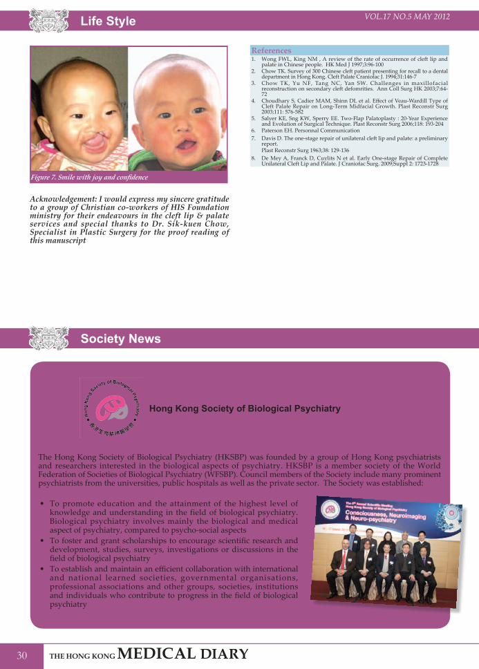
30
VOL.17 NO.5 MAY 2012Life Style
Figure 7. Smile with joy and confidence
Acknowledgement: I would express my sincere gratitude to a group of Christian co-workers of HIS Foundation ministry for their endeavours in the cleft lip & palate services and special thanks to Dr. Sik-kuen Chow, Specialist in Plastic Surgery for the proof reading of this manuscript
Hong Kong Society of Biological Psychiatry
The Hong Kong Society of Biological Psychiatry (HKSBP) was founded by a group of Hong Kong psychiatrists and researchers interested in the biological aspects of psychiatry. HKSBP is a member society of the World Federation of Societies of Biological Psychiatry (WFSBP). Council members of the Society include many prominent psychiatrists from the universities, public hospitals as well as the private sector. The Society was established: • To promote education and the attainment of the highest level of
knowledge and understanding in the field of biological psychiatry. Biological psychiatry involves mainly the biological and medical aspect of psychiatry, compared to psycho-social aspects
• To foster and grant scholarships to encourage scientific research and development, studies, surveys, investigations or discussions in the field of biological psychiatry
• To establish and maintain an efficient collaboration with international and national learned societies, governmental organisations, professional associations and other groups, societies, institutions and individuals who contribute to progress in the field of biological psychiatry
Society News
References1. Wong FWL, King NM , A review of the rate of occurrence of cleft lip and
palate in Chinese people. HK Med J 1997;3:96-1002. Chow TK. Survey of 300 Chinese cleft patient presenting for recall to a dental
department in Hong Kong. Cleft Palate Craniofac J. 1994;31:146-73. Chow TK, Yu NF, Tang NC, Yan SW. Challenges in maxillofacial
reconstruction on secondary cleft defomrities. Ann Coll Surg HK 2003;7:64-72
4. Choudhary S, Cadier MAM, Shinn DL et al. Effect of Veau-Wardill Type of Cleft Palate Repair on Long-Term Midfacial Growth. Plast Reconstr Surg 2003;111: 576-582
5. Salyer KE, Sng KW, Sperry EE. Two-Flap Palatoplasty : 20-Year Experience and Evolution of Surgical Technique. Plast Reconstr Surg 2006;118: 193-204
6. Paterson EH. Personnal Communication7. Davis D. The one-stage repair of unilateral cleft lip and palate: a preliminary
report. Plast Reconstr Surg 1963;38: 129-136
8. De Mey A, Franck D, Cuylits N et al. Early One-stage Repair of Complete Unilateral Cleft Lip and Palate. J Craniofac Surg. 2009;Suppl 2: 1723-1728

31
VOL.17 NO.5 MAY 2012 Federation News
Public Talk on Stress, Anxiety and Insomnia
Dinner Gathering with the Council Members of the Hong Kong Medical Association
On 25th March, 2012, a crowd of about 70 participants were drawn to a Public Talk held at the Federation’s Lecture Hall, sharing and discussing the topic, “Stress, Anxiety and Insomnia”.
Our Honorary Secretary, Dr. Yin-kwok NG, had the honour to invite two distinguished guest speakers, Dr. Yoki FONG and Dr. Sammy CHENG, to deliver the talk. Prior to the talk, the President of the Federation of Medical Societies of Hong Kong, Dr. Raymond SK LO, held the souvenir presentation ceremony with our distinguished guest speakers, Dr. NG and the sponsor, Sanofi.
The Talk was highly stimulating and informative. It introduced to the audience the symptoms and treatment of mental pressure, as well as ways to handle insomnia and promote mental health. The participants’ active questioning and engagement and the refreshment marked a wonderful ending of this event. We would like to thank Sanofi for the generous sponsorship and support for this talk.
On 3 April, the Executive Committee members of the Federation of the Medical Societies of Hong Kong were invited by Council Members of the Hong Kong Medical Association for a dinner gathering. The dinner was held at the Association’s Premises on 5th floor, Duke of Windsor Social Service Building, 15 Hennessy Road, Wanchai, Hong Kong.
It was a marvelous occasion of fraternity in an enjoyable and happy atmosphere. FMSHK President Dr. Raymond LO was also invited to present a prize to the winner of the HKMA medical student project competition. We are looking forward to more opportunities of gathering and exchange in the near future.

32
VOL.17 NO.5 MAY 2012Federation News
PhotogBuddy – Outing to Lion’s Nature Education Centre
New Team Members
On 7th April, 2012, 16 participants, including voluntary workers, bereaved children and their families joined the event, "PhotogBuddy". It was co-organised by the HKFMS Foundation Limited and Togetherness, as a Project-Life Moment activity.
The first part of the event was located in the Lions Nature Education Centre, in which participants, especially bereaved children and their families, could try to leave the past behind and enjoy Nature’s beauty. They did not only visit exhibition halls featuring diverse collection of nature-related themes, including Insectarium, Agriculture Hall, Fisheries Hall, Shell House, they also bought fresh, organic field crops from the situated farm.
This was then followed by an enjoying lunch of local delicacies. The second part of the event was an extra tour around the Sai Kung Town Centre, exploring and experiencing the famous Sai Kung Pier with seafood restaurants and markets. Throughout the whole event, the voluntary workers played the role of photographers to capture every memorable and joyful moment of the bereaved children and their families. Apart from the wish that they could learn to appreciate, as well as cherish, every second in their lives, it is also hoped that a positive relationship could be developed by spending a sunny, interactive afternoon with a group of active voluntary workers.
We would like to express our sincere gratitude to Ms Pearl TSE from Togetherness for her effort in making the meaningful event smooth and successful. Upcoming photo review gatherings will be held. Furthermore, a photography course is being planned in summer, aiming to further kindle the life interests and strengthen the support for the bereaved children and families. We hope to extend wider our support network, and referrals for the programme are most welcome. For information and enquiries, please contact our Admin Manager Ms Nancy CHAN on Tel 2527 8898.
Ms. Nancy CHAN Administrative Manager
FMSHK is a family of health professionals and it is with great pleasure that as of March, 2012, I became a part of this big family. I would like to extend my gratitude to the Executive Committee members of the Federation of the Medical Societies of Hong Kong (FMSHK) to entrust me to assume this new role. I am a Nutritionist and Dentist by training in the University of Ulster and Centro Escolar University respectively, as well as attained Fellow in Life Office Management Institute. Working previously as Assistant Vice President in the Sun Life Financial Hong Kong Ltd, I have equipped myself with vast experiences in customer service, marketing, accounting and projects management over the years. With the vision to put the values of the FMSHK into the practical field, it is my firm belief that the experience here will be challenging, enjoyable, and rewarding. I look forward to collaborate with every single member society in the foreseeable future.
Ms. Annie WONGExecutive Officer
I studied for a degree in Management. Prior to joining the Federation, I gained experience by carrying out administrative duties in the government. Now I was appointed to handle the Hong Kong Medical Diary and coordination of projects with commitment to providing high quality products and services.

33
VOL.17 NO.5 MAY 2012 Medical Diary of MaySunday
Monday
Tuesday
Wednesday
Thursday
Friday
Saturday
1718
19
2021
22
271314
1516
1011
129
76
23
88
211
34
5
24
2930
31
25
28
26
Join
t Pro
fess
iona
l Ba
sket
ball
Tour
nam
ent
2012
Join
t Pro
fess
iona
l Ba
sket
ball
Tour
nam
ent
2012
HK
MA
Squ
ash
Tour
nam
ent 2
012
"We
Are
Fam
ily" -
Art
G
alle
ry C
once
rt
HK
MA
Kow
loon
Wes
t C
omm
unity
Net
wor
k -
Man
agem
ent o
f Ins
omni
a an
d th
e U
se o
f Hyp
notic
M
edic
atio
ns
Hon
g K
ong
Neu
rosu
rgic
al S
ocie
ty
Mon
thly
Aca
dem
ic M
eetin
g –T
he
biom
echa
nics
of c
ervi
cal s
pine
and
po
st la
min
ecto
my
defo
rmity
HK
MA
CW
&S
Com
mun
ity N
etw
ork
– M
anag
emen
t of A
topi
c D
erm
atiti
s cu
m A
nnua
l Mee
ting
Cer
tifica
te C
ours
e on
Man
agem
ent
of D
rug
Abu
se P
atie
nts
for F
amily
D
octo
rs (K
owlo
on D
istr
icts
)
MPS
Wor
ksho
p –
Mas
terin
g Pr
ofes
sion
al
Inte
ract
ions
HK
MA
Kow
loon
City
C
omm
unity
Net
wor
k -
Cur
rent
Con
trov
ersi
es a
nd
Nov
el T
arge
t in
Ost
eopo
rosi
sFM
SHK
Offi
cers
’ M
eetin
gC
ounc
il M
eetin
g
HK
MA
Sno
oker
To
urna
men
t (Fi
nal
Roun
d)
Cer
tifica
te C
ours
e on
M
anag
emen
t of D
rug
Abu
se P
atie
nts
for F
amily
D
octo
rs (K
owlo
on
Dis
tric
ts)
HK
MA
Kow
loon
City
C
omm
unity
Net
wor
k -
BPH
& P
rost
ate
Can
cer i
n Pr
imar
y C
are
Clin
ics
HK
MA
Kow
loon
Wes
t C
omm
unity
Net
wor
k –
Clin
ical
Con
side
ratio
n of
th
e Lo
ng-te
rm T
reat
men
t of
Ost
eopo
rosi
s
HK
MA
CM
E –
An
Upd
ate
in O
steo
poro
sis
Man
agem
ent w
ith
Bisp
hosp
hona
tes
HK
MA
CW
&S
Com
mun
ity N
etw
ork
– Te
ntat
ive
HK
MA
CM
E –
Earl
y N
utri
tion
& S
ubse
quen
t M
anife
stat
ion
of A
llerg
ies
HK
MA
CW
&S
Com
mun
ity N
etw
ork
– Te
ntat
ive
MPS
Wor
ksho
p –
Mas
teri
ng A
dver
se
Out
com
es
HK
MA
Win
e D
inne
r 201
213
th R
egio
nal
Ost
eopo
rosi
s C
onfe
renc
e 20
127t
h St
anle
y D
rago
n Bo
at
War
m-U
p Ra
ces
2012
MPS
Wor
ksho
p –
Mas
teri
ng Y
our R
isk
13th
Reg
iona
l O
steo
poro
sis
Con
fere
nce
2012
HK
MA
Tab
le T
enni
s To
urna
men
t 201
2H
KM
APS
2nd
Sea
sona
l Ph
oto
Com
petit
ion
FMSH
K E
xecu
tive
Com
mitt
ee a
nd C
ounc
il M
eetin
g
Ato
pic
Der
mat
itis
MPS
Wor
ksho
p –
Mas
teri
ng D
ifficu
lt In
tera
ctio
ns w
ith P
atie
nts
MPS
Wor
ksho
p –
Mas
teri
ng Y
our R
isk
HK
MA
CM
E –
Cer
tifica
te
Cou
rse
for G
Ps 2
012
HK
MA
Sha
tin D
octo
rs
Net
wor
k –
Acn
e an
d A
cne
Scar
Man
agem
ent
MPS
Wor
ksho
p –
Mas
teri
ng A
dver
se
Out
com
es
Tai P
o C
ME
– Re
cent
A
dvan
ces
in th
e tr
eatm
ent
of d
iabe
tic n
ephr
opat
hy
MPS
Wor
ksho
p –
Mas
teri
ng D
ifficu
lt In
tera
ctio
ns w
ith P
atie
nts
HK
MA
Sha
tin D
octo
rs
Net
wor
k –
Inno
vativ
e Tr
eatm
ent o
n O
steo
poro
sis
MPS
Wor
ksho
p –
Mas
teri
ng D
ifficu
lt In
tera
ctio
ns w
ith P
atie
nts
Refr
eshe
r Cou
rse
for
Hea
lth C
are
Prov
ider
s 20
11/2
012
HK
MA
Kow
loon
Eas
t Com
mun
ity
Net
wor
k –R
eflec
t man
age
of
hype
rten
sion
HK
MA
Str
uctu
red
CM
E Pr
ogra
mm
e w
ith H
ong
Kon
g Sa
nato
rium
&
Hos
pita
l Yea
r 201
2 –
Cer
vica
l ra
dicu
lopa
thy
diag
nosi
s &
m
anag
emen
tRe
spir
ator
y C
linic
al M
eetin
g –
“Out
of
the
blue
” &
A R
are
Ans
wer
to A
C
omm
on S
cena
rio
MPS
Wor
ksho
p –
Mas
teri
ng A
dver
se
Out
com
es
MPS
Wor
ksho
p –
Mas
teri
ng P
rofe
ssio
nal
Inte
ract
ions
Join
t Sur
gica
l Sym
posi
um -
Neu
rosu
rgeo
ns' N
ight
mar
eC
ertifi
cate
Cou
rse
on M
anag
emen
t of
Dru
g A
buse
Pat
ient
s fo
r Fam
ily
Doc
tors
(Kow
loon
Dis
tric
ts)
HK
MA
Sha
tin D
octo
rs N
etw
ork
- Le
ctur
e on
Alle
rgic
Rhi
nitis
&
Com
plai
nts
to th
e M
edic
al C
ounc
il of
Hon
g K
ong
HK
MA
Yau
Tsi
m M
ong
Com
mun
ity
Net
wor
k - N
onal
coho
lic F
atty
Live
r D
isea
se -
a Re
view
of t
he
Spec
trum
of D
isea
se,
Dia
gnos
is a
nd T
hera
py
MPS
Wor
ksho
p –
Mas
teri
ng Y
our R
isk

34
VOL.17 NO.5 MAY 2012Calendar of Events
2:30 pm
SATHKMA CME DepartmentTel: 2527 84522.5 CME points5
MPS Workshop – Mastering Professional InteractionsOrganiser: The Hong Kong Medical Association, Chairman: , Speaker: Dr. Ka-lam HAU, Venue: The Hong Kong Medical Association Central Premises, Dr. Li Shu Pui Professional Education Centre, 2/F., Chinese Club Building, 21-22 Connaught Road Central
1:00 pm
TUEMiss Candice TONGTel: 2527 82851 CME point8
HKMA Kowloon West Community Network - Management of Insomnia and the Use of Hypnotic MedicationsOrganiser: HKMA Kowloon West Community Network, Chairman: Dr. Kenneth KN LEUNG, Speaker: Dr. Ka-lik KWAN, Venue: Crystal Room I-III, 30/F., Panda Hotel, Tsuen Wan, N.T.
1:00 pm Miss Candice TONGTel: 2527 8285
HKMA Kowloon City Community Network - Current Controversies and Novel Target in OsteoporosisOrganiser: HKMA Kowloon City Community Network, Chairman: Dr. Chu-wah CHIN, Speaker: Dr. Ka-kui LEE, Venue: 2/F, Site 6, Whampoa Garden, Wonderful Worlds of Whampoa, 8 Shung King Street, Hung Hom, Hung Hom
8:00 pm Ms. Nancy CHAN Tel: 2527 8898
FMSHK Officers’ MeetingOrganiser: The Federation of Medical Societies of Hong Kong, Venue: Gallop, 2/F., Hong Kong Jockey Club Club House, Shan Kwong Road, Happy Valley, Hong Kong
8:00 pm Ms. Christine WONGTel: 2527 8285
Council MeetingOrganiser: The Hong Kong Medical Association, Chairman: Dr. CHOI Kin, Venue: HKMA Head Office (5/F., Duke of Windsor Social Service Building, 15 Hennessy Road, Hong Kong)
THUMr. Alan LAWTel: 252782851 CME point10
HKMA Kowloon East Community Network –Reflect manage of hypertensionOrganiser: HKMA Kowloon East Community Network, Speaker: Dr. Wing-kwong WONG, Venue: APM,Kwun Tong
1:00 pm
WEDMiss Nadia HOTel: 2527 82852.5 CME points2
Certificate Course on Management of Drug Abuse Patients for Family Doctors (Kowloon Districts)Organiser: The Hong Kong Medical Association, Speakers: Dr. Steve WH CHAN & Dr. Fei-lung LAU, Venue: Kwun Tong Lei's Garden
6:30 pm
THUHKMA CME DepartmentTel: 2527 84522.5 CME points3
MPS Workshop – Mastering Difficult Interactions with PatientsOrganiser: The Hong Kong Medical Association, Speaker: Dr. Justin NS CHENG, Venue: The Hong Kong Medical Association Central Premises, Dr. Li Shu Pui Professional Education Centre, 2/F., Chinese Club Building, 21-22 Connaught Road Central
8:00 am
1:00 pm
1:00 pm
FRIDepartment of Surgery, Hong Kong Sanatorium & HospitalTel: 2835 86981 CME Point4
Joint Surgical Symposium - Neurosurgeons' NightmareOrganiser: Department of Surgery, The University of Hong Kong & Hong Kong Sanatorium & Hospital, Chairman: Dr. Kwan-ngai HUNG, Speakers: Dr. Wai-Man LUI & Dr. Jenny PU, Venue: Hong Kong Sanatorium & Hospital
2:00 pm
SUNMs. Dorothy KWOKTel: 2527 82856
Joint Professional Basketball Tournament 2012Organiser: The Hong Kong Medical Association, Venue: TBC
2:00 pm Ms. Dorothy KWOKTel: 2527 8285
HKMA Snooker Tournament (Final Round)Organiser: The Hong Kong Medical Association, Venue: Prat Billiard Club, 2/F, Austin Tower, 22-26 Austin Avenue, Kowloon
7:30 am
WEDDr. Gilberto LEUNGTel: 2255 33681.5 CME points9
Hong Kong Neurosurgical Society Monthly Academic Meeting –The biomechanics of cervical spine and post laminectomy deformityOrganiser: Hong Kong Neurosurgical Society, Chairman: Dr. Larry WONG, Speaker: Dr. Calvin HK MAK, Venue: Seminar Room, Ground Floor, Block A, Queen Elizabeth Hospital
1:00 pm Mr. Alan LAWTel: 252782851 CME point
HKMA CW&S Community Network – Management of Atopic Dermatitis cum Annual MeetingOrganiser: HKMA CW&S Community Network, Speaker: Dr. Kuen-kong LO, Venue: The Hong Kong Medical Association Central Premises, Dr. Li Shu Pui Professional Education Centre, 2/F., Chinese Club Building, 21-22 Connaught Road Central
1:00 pm
1:00 pm
HKMA CME DepartmentTel: 2527 84521 CME point
HKMA Structured CME Programme with Hong Kong Sanatorium & Hospital Year 2012 – Cervical radiculopathy diagnosis & managementOrganiser: The Hong Kong Medical Association, Speaker: Dr. Kan-hing MAK, Venue: The Hong Kong Medical Association Central Premises, Dr. Li Shu Pui Professional Education Centre, 2/F., Chinese Club Building, 21-22 Connaught Road Central
2:00 pm
Miss Nadia HOTel: 2527 82852.5 CME points
Certificate Course on Management of Drug Abuse Patients for Family Doctors (Kowloon Districts)Organiser: The Hong Kong Medical Association, Speakers: Dr. Gary KK AU & Ms. Esther MY CHEUNG, Venue: Kwun Tong Lei's Garden
6:30 pm HKMA CME DepartmentTel: 2527 84522.5 CME points
MPS Workshop – Mastering Professional InteractionsOrganiser: The Hong Kong Medical Association, Speaker: Dr. Andy KY CHEUNG, Venue: The Hong Kong Medical Association Central Premises, Dr. Li Shu Pui Professional Education Centre, 2/F., Chinese Club Building, 21-22 Connaught Road Central
Miss Nadia HOTel: 2527 82853 CME points
Certificate Course on Management of Drug Abuse Patients for Family Doctors (Kowloon Districts)Organiser: The Hong Kong Medical Association, Speaker: Dr. Ben KL CHEUNG, Venue: Kwun Tong Lei's Garden
Ms. Karen YIUTel: 2105 4815
HKMA Shatin Doctors Network - Lecture on Allergic Rhinitis & Complaints to the Medical Council of Hong KongOrganiser: HKMA Shatin Doctors Network, Chairman: Dr. Wing-kin MAK, Speakers: Dr. Hing-sang CHAN & Dr. Felice LIEH-MAK, Venue: President Room, Level 2, Royal Park Hotel, 8 Pak Hok Ting Street, Shatin
1:00 pm Miss Candice TONGTel: 2527 82851 CME point
HKMA Yau Tsim Mong Community Network - Nonalcoholic Fatty Liver Disease - a Review of the Spectrum of Disease, Diagnosis and TherapyOrganiser: HKMA Yau Tsim Mong Community Network, Chairman: Dr. David TY LAM, Speaker: Dr. Vincent WONG, Venue: Pearl Ballroom, Level 2, Eaton Smart, Hong Kong380 Nathan Road, Kowloon
6:30 pm HKMA CME DepartmentTel: 2527 84522.5 CME points
MPS Workshop – Mastering Your RiskOrganiser: The Hong Kong Medical Association, Speaker: Dr. Andy KY CHEUNG, Venue: The Hong Kong Medical Association Central Premises, Dr. Li Shu Pui Professional Education Centre, 2/F., Chinese Club Building, 21-22 Connaught Road Central
Date / Time Function Enquiry / Remarks
35
VOL.17 NO.5 MAY 2012 Calendar of Events
(27)
SATMs Zita BAITel: 2559 997326
13th Regional Osteoporosis Conference 2012Organiser: Osteoporosis Society of Hong Kong & Hong Kong Doctors Union, Chairman: Dr. Anita SY KAN, Venue: Hong Kong Convention and Exhibition Centre
THU10
2:00 pm
SATHKMA CME DepartmentTel: 2527 84522.5 CME points12
MPS Workshop – Mastering Difficult Interactions with PatientsOrganiser: The Hong Kong Medical Association, Speaker: Dr. Justin NS CHENG, Venue: Holiday Inn
2:30 pm HKMA CME DepartmentTel: 2527 84522 CME points
Refresher Course for Health Care Providers 2011/2012Organiser: The Hong Kong Medical Association, Speaker: Dr. John TH WONG, Venue: OLMH
6:30 pm
WEDHKMA CME DepartmentTel: 2527 84522.5 CME points16 MPS Workshop – Mastering Your Risk
Organiser: The Hong Kong Medical Association, Chairman: , Speaker: Dr. Andy KY CHEUNG, Venue: Eaton Hotel
1:45 pm
FRIMs. Wendy CHENGTel: 2824 03331.0 CME point18 HKMA Shatin Doctors Network – Acne and Acne Scar Management
Organiser: HKMA Shatin Doctors Network, Chairman: Dr. Wing-kin MAK, Speaker: Dr. Mona LS CHIU, Venue: Jasmine Room, Level 2, Royal Park Hotel, 8 Pak Hok Ting Street, Shatin
1:00 pm
WEDMr. Alan LAWTel: 2527828523 HKMA CW&S Community Network – Tentative
Organiser: HKMA CW&S Community Network, Venue: The Hong Kong Medical Association Central Premises, Dr. Li Shu Pui Professional Education Centre, 2/F., Chinese Club Building, 21-22 Connaught Road Central
7:00 pm
FRIMs. Dorothy KWOKTel: 2527 828525 HKMA Wine Dinner 2012
Organiser: The Hong Kong Medical Association, Venue: The Hong Kong Medical Association Central Premises, Dr. Li Shu Pui Professional Education Centre, 2/F., Chinese Club Building, 21-22 Connaught Road Central
6:30 pm HKMA CME DepartmentTel: 2527 84522.5 CME points
MPS Workshop – Mastering Difficult Interactions with PatientsOrganiser: The Hong Kong Medical Association, Speaker: Dr. Justin NS CHENG, Venue: The Hong Kong Medical Association Central Premises, Dr. Li Shu Pui Professional Education Centre, 2/F., Chinese Club Building, 21-22 Connaught Road Central
2:00 pm
SUNMs. Dorothy KWOKTel: 2527 828520 Joint Professional Basketball Tournament 2012
Organiser: The Hong Kong Medical Association, Venue: TBC2:00 pm Ms. Dorothy KWOK
Tel: 2527 8285HKMA Squash Tournament 2012Organiser: The Hong Kong Medical Association, Venue: KCC
8:00 pm Ms. Candy YUENTel: 2527 8285
"We Are Family" - Art Gallery ConcertOrganiser: The Hong Kong Medical Association, Venue: HK City Hall
1:00 pm
FRIMs. Mandy CHANTel: 9736 31281 CME point11
HKMA Shatin Doctors Network – Innovative Treatment on OsteoporosisOrganiser: HKMA Shatin Doctors Network, Chairman: Dr. Wing-kin MAK, Speaker: Dr. Gavin KW LEE, Venue: Tai Po Room, 2/F., Regal Riverside Hotel, 34-36 Tai Chung Kiu Road, Shatin
1:00 pm
TUEMiss Candice TONGTel: 2527 8285 15
HKMA Kowloon City Community Network - BPH & Prostate Cancer in Primary Care ClinicsOrganiser: HKMA Kowloon City Community Network, Chairman: Dr. CHIN Chu-wah, Speaker: Dr. SO Chun, Venue: 2/F, Site 6, Whampoa Garden, Wonderful Worlds of Whampoa, 8 Shung King Street, Hung Hom, Hung Hom
1:00 pm
THUHKMA CME DepartmentTel: 2527 84521.0 CME point17
HKMA CME – Certificate Course for GPs 2012Organiser: The Hong Kong Medical Association, Chairman: Dr. Gary KK AU, Speaker: Dr. Peter KM KU, Venue: TKO
2:30 pm
SATHKMA CME DepartmentTel: 2527 84522.5 CME points19
MPS Workshop – Mastering Adverse OutcomesOrganiser: The Hong Kong Medical Association, Chairman: , Speaker: Dr. Ares KL LEUNG, Venue: The Hong Kong Medical Association Central Premises, Dr. Li Shu Pui Professional Education Centre, 2/F., Chinese Club Building, 21-22 Connaught Road Central
1:00 pm
TUEMs. Sandy CHUNGTel: 3971 29401 CME point22
HKMA Kowloon West Community Network – Clinical Consideration of the Long-term Treatment of OsteoporosisOrganiser: HKMA Kowloon West Community Network, Chairman: Dr. Raymond N LAM, Speaker: Dr. Gavin KW LEE, , Venue: Crystal Room I-III, 30/F., Panda Hotel, Tsuen Wan, NT
6:30 pm
THUHKMA CME DepartmentTel: 2527 84522.5 CME points
24MPS Workshop – Mastering Adverse OutcomesOrganiser: The Hong Kong Medical Association, Speaker: Dr. Emily CW HUNG, Venue: Eaton Hotel
1:00 pm Mr. Alan LAWTel: 2527 8285
Atopic DermatitisOrganiser: HKMA Kln East Community Network, Chairman: Dr. Danny PK MA, Speaker: Dr. Hugh HC CHEUNG, Venue: East Ocean Seafood Restaurant at Tseung Kwan O
7:00 pm Ms. Nancy CHAN Tel: 2527 8898
FMSHK Executive Committee and Council MeetingOrganiser: The Federation of Medical Societies of Hong Kong, Venue: Council Chamber, 4/F, Duke of Windsor Social Service Building, 15 Hennessy Road, Wanchai, Hong Kong
1:45 pm HKMA CME DepartmentTel: 2527 84521.0 CME point
Tai Po CME – Recent Advances in the treatment of diabetic nephropathyOrganiser: The Hong Kong Medical Association, Speaker: Dr. Chung-ping HO, Venue: Tai Po
6:30 pm Dr. Fanny WS KODr. Arthur CW LAUTel: 2632 27851.5 CME points (HKCP, CSHK)2 CME points (HKCFP)
Respiratory Clinical Meeting – “Out of the blue” & A Rare Answer to A Common ScenarioOrganiser: Hong Kong Thoracic Society & American College of Chest Physicians (HK & Macau Chapter), Chairmen: Dr. Kin-keung KWONG & Dr. Chi-kai CHOW, Speakers: Dr. Yiu-cheong YEUNG & Dr. Chi-kai CHOW, Venue: LG1, Lecture Room, Ruttonjee Hospital
6:30 pm HKMA CME DepartmentTel: 2527 84522.5 CME points
MPS Workshop – Mastering Adverse OutcomesOrganiser: The Hong Kong Medical Association, Speaker: Dr. Emily CW HUNG, Venue: Eaton Hotel
Date / Time Function Enquiry / Remarks

36
VOL.17 NO.5 MAY 2012Calendar of Events
SAT26
2:00 pm
SUNMs. Dorothy KWOKTel: 2527 828527
HKMA Table Tennis Tournament 2012Organiser: The Hong Kong Medical Association, Chairman: Dr. Hilton HT KOO, Venue: Luen Wo Hui Sports Centre, 3/F, 9 Wo Mun Street, Luen Wo Hui, Fanling, New Territories
1:00 pm
WEDMr. Alan LAWTel: 2527828530
HKMA CW&S Community Network – TentativeOrganiser: HKMA CW&S Community Network, Venue: The Hong Kong Medical Association Central Premises, Dr. Li Shu Pui Professional Education Centre, 2/F., Chinese Club Building, 21-22 Connaught Road Central
2:00 pm Ms. Dorothy KWOKTel: 2527 8285
HKMAPS 2nd Seasonal Photo CompetitionOrganiser: The Hong Kong Medical Association, Venue: HKMA Head Office (5/F., Duke of Windsor Social Service Building, 15 Hennessy Road, Hong Kong)
9:00 am Ms. Dorothy KWOKTel: 2527 8285
7th Stanley Dragon Boat Warm-Up Races 2012Organiser: The Hong Kong Medical Association, Venue: Stanley Main Beach
TUE29 1:00 pm HKMA CME DepartmentTel: 2527 84521 CME point
HKMA CME – An Update in Osteoporosis Management with BisphosphonatesOrganiser: The Hong Kong Medical Association, Speaker: Prof. Mone ZAIDI, Venue: Jordan
THU317:00 pm HKMA CME Department
Tel: 2527 84521 CME point
HKMA CME – Early Nutrition & Subsequent Manifestation of AllergiesOrganiser: The Hong Kong Medical Association, Speaker: Dr. Gary WK WONG, Venue: The Hong Kong Medical Association Central Premises, Dr. Li Shu Pui Professional Education Centre, 2/F., Chinese Club Building, 21-22 Connaught Road Central
2:30 pm HKMA CME DepartmentTel: 2527 84522.5 CME points
MPS Workshop – Mastering Your RiskOrganiser: The Hong Kong Medical Association, Speaker: Dr. Danny WH LEE, Venue: The Hong Kong Medical Association Central Premises, Dr. Li Shu Pui Professional Education Centre, 2/F., Chinese Club Building, 21-22 Connaught Road Central
Date / Time Function Enquiry / Remarks
Upcoming MeetingChinese Medicine in Geriatrics, Hong Kong International Integrative Medicine Conference Organisers: Hospital Authority & Hong Kong Association for Integration of Chinese-Western Medicine (HKAIM), Venue: Hong Kong Academy of Medicine, Enquiry: Ms Toki CHAN & Ms Justin NG Tel: (852) 28718787/ 2871 8896
7-8/7/2012

37
VOL.17 NO.5 MAY 2012 Dental Quiz
Answer to Dental Quiz
Answer
1.
2.
The fractured fragment had been stored in dry condition and was desiccated, and so appeared as chalky white.
The fragment would return to its original colour and translucency after re-hydration in the oral cavity.
Dr. Shiu-yin CHOBDS(HK), MDS(Otago), FRACDS, FHKAM(Dental Surgery)
Senior Dental Officer, Fanling School Dental Clinic
The Federation of Medical Societies of Hong Kong 4/F Duke of Windsor Social Service Building, 15 Hennessy Road, Wanchai, HK Tel: 2527 8898 Fax: 2865 0345
PatronThe HonourableDonald TSANG, GBM 曾蔭權先生
PresidentDr. LO See-kit, Raymond 勞思傑醫生
1st Vice-PresidentProf. CHAN Chi-fung, Godfrey 陳志峰教授
2nd Vice-PresidentDr. CHAN Sai-kwing 陳世炯醫生
Hon. TreasurerMr. LEE Cheung-mei, Benjamin 李祥美先生
Hon. SecretaryDr. NG Yin-kwok 吳賢國醫生
Immediate Past President Dr. FONG To-sang, Dawson 方道生醫生
Executive Committee MembersDr. CHAN Chun-kwong, Jane 陳真光醫生Dr. CHAN Hau-ngai, Kingsley 陳厚毅醫生Prof. CHIM Chor-sang, James 詹楚生教授Dr. FONG Yuk-fai, Ben 方玉輝醫生Dr. HUNG Che-wai, Terry 洪致偉醫生Ms. KU Wai-yin, Ellen 顧慧賢女士Dr. LO Sze-ching, Susanna 盧時楨醫生Dr. MAN Chi-wai 文志衛醫生Dr. MOK Chun-on 莫鎮安醫生Dr. WONG Mo-lin, Maureen 黃慕蓮醫生Ms. YAP Woan-tyng, Tina 葉婉婷女士Dr. YU Chau-leung, Edwin 余秋良醫生Dr. YUEN Shi-yin, Nancy 袁淑賢醫生Dr. YUNG Shu-hang, Patrick 容樹恆醫生Dr. CHAK Wai-kwong 翟偉光醫生
Founder MembersBritish Medical Association (Hong Kong Branch)英國醫學會 ( 香港分會 )
PresidentDr. LO See-kit, Raymond 勞思傑醫生
Vice-PresidentDr. WU, Adrian 鄔揚源醫生
Hon. SecretaryDr. HUNG Che-wai, Terry 洪致偉醫生
Hon. TreasurerDr. LEUNG, Clarence 梁顯信醫生
Council RepresentativesDr. LO See-kit, Raymond 勞思傑醫生Dr. CHEUNG Tse-ming 張子明醫生Tel: 2527 8898 Fax: 2865 0345
The Hong Kong Medical Association香港醫學會
PresidentDr. CHOI Kin 蔡堅醫生
Vice- PresidentsDr. CHAN Yee-shing, Alvin 陳以誠醫生Dr. CHOW Pak-chin 周伯展醫生
Hon. SecretaryDr. LEE Fook-kay 李福基醫生
Hon. TreasurerDr. LEUNG Chi-chiu 梁子超醫生
Council RepresentativesDr. CHAN Yee-shing 陳以誠醫生Dr. CHOW Pak-chin 周伯展醫生
Chief ExecutiveMrs. LEUNG, Yvonne 梁周月美女士Tel: 2527 8285 (General Office) 2527 8324 / 2536 9388 (Club House in Wanchai / Central)Fax: 2865 0943 (Wanchai), 2536 9398 (Central)Email: [email protected] Website: http://www.hkma.org
The HKFMS Foundation Limited 香港醫學組織聯會基金 Board of DirectorsPresident
Dr. LO See-kit, Raymond 勞思傑醫生
1st Vice-PresidentProf. CHAN Chi-fung, Godfrey 陳志峰教授
2nd Vice-PresidentDr. LO Sze-ching, Susanna 盧時楨醫生
Hon. TreasurerMr. LEE Cheung-mei, Benjamin 李祥美先生
Hon. SecretaryDr. CHAN Sai-kwing 陳世炯醫生
DirectorsMr. CHAN Yan-chi, Samuel 陳恩賜先生Prof. CHIM Chor-sang, James 詹楚生教授Ms. KU Wai-yin, Ellen 顧慧賢女士Dr. WONG Mo-lin, Maureen 黃慕蓮醫生Dr. YU Chak-man, Aaron 余則文醫生