Densovirus infection in silkworm Bombyx mori and genes ...
11
ISJ 12: 118-128, 2015 ISSN 1824-307X REVIEW Densovirus infection in silkworm Bombyx mori and genes associated with disease resistance T Gupta 1 , K Kadono-Okuda 2 , K Ito 3 , K Trivedy 1 , KM Ponnuvel 1 1 Genomics Division, Seribiotech Research Laboratory, Carmelaram Post, Kodathi, Bangalore 560 035, India 2 Laboratory of Sericultural Science, Department of Science of Biological Production, Graduate School of Agriculture, Tokyo University of Agriculture and Technology, 3-5-8 Saiwai-cho, Fuchu, Tokyo, 183-8509, Japan 3 Division of lnsect Sciences, National Institute of Agrobiological Sciences, 1-2 Owashi, Tsukuba, Ibaraki 305- 8634, Japan Accepted April 13, 2015 Abstract The silkmoth Bombyx mori has been bred in captivity for around 5,000 years and it is now a completely domesticated species of the silkmoth. The larva of B. mori feeds only on Mulberry leaves so it is a monophagous insect. Silk cocoons obtained from this species are the primary source of commercial silk and this makes B. mori an economically important insect. However, the silk industry suffers significant losses due to various viral infections during the larval stages. One of the frequently affecting silkworm viruses is the B. mori Densovirus (BmDV). The BmDV is further classified into two types: B. mori Densovirus-1 (BmDV-1) and B. mori Densovirus-2 (BmDV-2). However, BmDV-2 is excluded from the family of Parvoviridae and is referred to a new family Bidnaviridae. To date, three isolates of BmDVs have been reported. This virus has been found to be a causative agent of the commonly occurring fatal silkworm disease, ‘flacherie’. BmDVs have been found to be a highly diverse group of viruses. While most of the strains of B. mori are susceptible to BmDV, few races have been found to be completely resistant to the virus. Studies have shown both dominant and recessive alleles to be responsible for the resistance. So far four genes have been reported conferring resistance against BmDV-1 and BmDV-2. These are the Nid-1 and nsd-1 genes against BmDV-1 and nsd-2 and nsd-Z genes against BmDV-2. Details about Densovirus with special reference to B. mori and the resistant genes present against it and the studies undertaken towards screening of BmDV resistant silkworm races have been discussed in this review. Key Words: Bombyx mori; densovirus resistance; nsd-2; bidnaviridae Introduction The pathological condition of Densoviruses (DVs) was studied in greater wax moth, Galleria mellonella and these studies further led to the discovery of DVs (Meynadier et al., 1979). Ultrathin sections of tissues of heavily infected larvae were examined which revealed enlarged Feulgen-positive nuclei. Further studies of these histological sections showed the presence of electron-dense viral factories leading to production of thousands of small isometric particles (Amargier et al., 1979). Virus particles of sizes 20 - 22 nm with icosahedral non- enveloped symmetry were isolated and then purified ___________________________________________________________________________ Corresponding author: Kangayam M Ponnuvel Genomics Division Seribiotech Research Laboratory Carmelaram Post, Kodathi Bangalore 560 035, India E-mail: [email protected] from the dead larvae. Chemical analysis revealed the presence of viral DNA and other proteinaceous moieties. DVs were found to have biochemical and biophysical features resembling that of vertebrate parvoviruses. The analysis of virus particles showed that the capsid was made up of four proteins and contained a single stranded linear DNA of 6 kb in size. In addition to this the DVs also shared the nuclear site of replication and the structural features with that of vertebrate parvoviruses and accordingly a new subfamily named Densovirinae was established within the family of Parvoviridae. DVs have been isolated from different insect orders including Lepidoptera, Hemiptera, Diptera, Dictyoptera and Crustacea (Bergoin and Tijssen, 2010). The Densovirinae subfamily Densoviruses (DVs) belong to the family Parvoviridae. Viruses belonging to the Parvoviridae 118
-
Upload
khangminh22 -
Category
Documents
-
view
0 -
download
0
Transcript of Densovirus infection in silkworm Bombyx mori and genes ...
ISJ 12: 118-128, 2015 ISSN 1824-307X
REVIEW Densovirus infection in silkworm Bombyx mori and genes associated with disease resistance T Gupta1, K Kadono-Okuda2, K Ito3, K Trivedy1, KM Ponnuvel1 1Genomics Division, Seribiotech Research Laboratory, Carmelaram Post, Kodathi, Bangalore 560 035, India 2Laboratory of Sericultural Science, Department of Science of Biological Production, Graduate School of Agriculture, Tokyo University of Agriculture and Technology, 3-5-8 Saiwai-cho, Fuchu, Tokyo, 183-8509, Japan 3Division of lnsect Sciences, National Institute of Agrobiological Sciences, 1-2 Owashi, Tsukuba, Ibaraki 305-8634, Japan
Accepted April 13, 2015
Abstract
The silkmoth Bombyx mori has been bred in captivity for around 5,000 years and it is now a completely domesticated species of the silkmoth. The larva of B. mori feeds only on Mulberry leaves so it is a monophagous insect. Silk cocoons obtained from this species are the primary source of commercial silk and this makes B. mori an economically important insect. However, the silk industry suffers significant losses due to various viral infections during the larval stages. One of the frequently affecting silkworm viruses is the B. mori Densovirus (BmDV). The BmDV is further classified into two types: B. mori Densovirus-1 (BmDV-1) and B. mori Densovirus-2 (BmDV-2). However, BmDV-2 is excluded from the family of Parvoviridae and is referred to a new family Bidnaviridae. To date, three isolates of BmDVs have been reported. This virus has been found to be a causative agent of the commonly occurring fatal silkworm disease, ‘flacherie’. BmDVs have been found to be a highly diverse group of viruses. While most of the strains of B. mori are susceptible to BmDV, few races have been found to be completely resistant to the virus. Studies have shown both dominant and recessive alleles to be responsible for the resistance. So far four genes have been reported conferring resistance against BmDV-1 and BmDV-2. These are the Nid-1 and nsd-1 genes against BmDV-1 and nsd-2 and nsd-Z genes against BmDV-2. Details about Densovirus with special reference to B. mori and the resistant genes present against it and the studies undertaken towards screening of BmDV resistant silkworm races have been discussed in this review. Key Words: Bombyx mori; densovirus resistance; nsd-2; bidnaviridae
Introduction
The pathological condition of Densoviruses
(DVs) was studied in greater wax moth, Galleria mellonella and these studies further led to the discovery of DVs (Meynadier et al., 1979). Ultrathin sections of tissues of heavily infected larvae were examined which revealed enlarged Feulgen-positive nuclei. Further studies of these histological sections showed the presence of electron-dense viral factories leading to production of thousands of small isometric particles (Amargier et al., 1979). Virus particles of sizes 20 - 22 nm with icosahedral non- enveloped symmetry were isolated and then purified ___________________________________________________________________________
Corresponding author: Kangayam M Ponnuvel Genomics Division Seribiotech Research Laboratory Carmelaram Post, Kodathi Bangalore 560 035, India E-mail: [email protected]
from the dead larvae. Chemical analysis revealed the presence of viral DNA and other proteinaceous moieties. DVs were found to have biochemical and biophysical features resembling that of vertebrate parvoviruses. The analysis of virus particles showed that the capsid was made up of four proteins and contained a single stranded linear DNA of 6 kb in size. In addition to this the DVs also shared the nuclear site of replication and the structural features with that of vertebrate parvoviruses and accordingly a new subfamily named Densovirinae was established within the family of Parvoviridae. DVs have been isolated from different insect orders including Lepidoptera, Hemiptera, Diptera, Dictyoptera and Crustacea (Bergoin and Tijssen, 2010).
The Densovirinae subfamily
Densoviruses (DVs) belong to the family Parvoviridae. Viruses belonging to the Parvoviridae
118

Table 1 The present classification of the family Parvoviridae (Cotmore et al., 2014)
family are known for having a wide host range. Hence this family has been divided into two subfamilies namely, Densovirinae and Parvovirinae. Viruses falling under the category of Densovirinae infect invertebrates while those under Parvovirinae infect vertebrates. The extension of taxonomy of the Parvoviridae family is under review by the International Committee on Taxonomy of Viruses (ICTV). A set of proposals have been extended to introduce new species and genera into both the subfamilies, thereby resolving misclassified species and enhancing taxonomic clarity. Consequently, the present classification of the family Parvoviridae differs significantly from that of the old one (Cotmore et al., 2014). The present classification includes affixes in the names of the genera which clarifies subfamily affiliation and reduces ambiguity that’s results from the vernacular use of parvovirus and densovirus to denote multiple taxon level. The present classification of Parvoviridae family has been shown in Table 1. Similarly, the subfamily Densovirinae has also been reclassified and comprises of five different genera. The classification of Densovirinae subfamily has been discussed in detail as under.
Classification of Densovirinae subfamily
Invertebrate densoviruses (DVs) form a distinguished subfamily (Densovirinae) within the Parvoviridae family and have recently been reclassified. All members belonging to the family of Densovirinae have arthropod hosts with high sequence identities of 85 - 95 % (Dumas et al., 1992). The present classification of Densovirinae family includes five distinguished genera namely, Ambidensovirus, Brevidensovirus, Hepandensovirus, Penstyldensovirus and Iteradensovirus as shown in detail in Table 2. These
five different genera have distinguished genomic organization with varying genomic sizes.
The genetic make-up of DVs can be classified mainly into two types: The ambisense DVs that encode open reading frames (ORFs) on both complimentary strands, while the monosense DVs that has only a single strand containing the ORFs. Overall DVs can be described as viruses having small isometric, non-enveloped capsids with a linear DNA genome (Table 3). However, viruses such as BmDV-2 do not match the description because unlike parvoviruses, its linear genomic DNA is segmented and is twice as long. Also, it codes for the enzyme DNA polymerase (Hayakawa et al., 2000). Therefore, it has been reassigned to a new family Bidnaviridae (Tissen et al., 2011). The ambisense DVs package their single strands of DNA in separate capsids so upon DNA extraction double stranded genomes are obtained under high salt conditions. The brief description of the different genera of Densovirinae subfamily is given below.
Ambidensovirus
Under the previous classification this genus was known as ‘Densovirus’. ‘Ambi’ is the new affix added to this name. This genus comprises of DVs following an ambisense mode of transcription. Five different species have been grouped under this genus so far (Table 2). The previously existing genus Pefudensovirus has been incorporated into this genus. PfDV, BgDV1, CpDV, PcDV, DsDV, GmDV, HaDV1, JCDV, MlDV, PiDV and AdDV are the viruses grouped under this genus.
Brevidensovirus
This genus has the shortest (4 kb) genome among all the other genera of DVs (Shike et al., 2000; Zhai et al., 2008). They have been found to
Genus Species Viruses or variants
Subfamily Parvovirinae-vertebrate hosts
Amdoparvovirus 2 2 Aveparvovirus 1 2 Bocaparvovirus 12 22 Copiparvovirus 2 2 Dependoparvovirus 7 23 Erythroparvovirus 6 12 Protoparvovirus 5 25 Tetraparvovirus 6 10
Subfamily Densovirinae-arthropod hosts
Ambidensovirus 6 11 Brevidensovirus 2 8 Hepandensovirus 1 7 Iteradensovirus 5 6 Penstyldensovirus 1 4
119
infect several mosquitoes (including mosquito cell lines) and shrimp species. In addition to their small size, their genome varies from the other species of DVs by the absence of ITRs. The ITRs in this case are replaced with short (130 - 150) hairpin structures at the 3’ and 5’ ends. The coding sequence of their genome is organized into three overlapping ORFs. The NS1 is encoded by a large left ORF, the NS2 is encoded by the ORF nested in the 5’ half of the ORF coding NS1 and the VP capsid proteins are encoded by a short right ORF.
Hepandensovirus The new genus Hepandensovirus was mainly
introduced to accommodate shrimp viruses under the family of Densovirinae. These are called Hepandensovirus to indicate the original name of these viruses, “Hepatopancreatic parvovirus”. The virus group comprising PmoHDV1, PchDV, PmoHDV2, PmoHDV3, PmeDV, PmoHDV4 and FchDV are included in this genus (Table 2).
Penstyldensovirus This particular genus is another newly
introduced genus under the family of Densovirinae. The name stands for a siglum for Penaeus stylirostris, the host and also the founding member of this species. PstDV1, PmoPDV1, PmoPDV2 and PstDV2 are the viruses included this genus.
Iteradensovirus
This genus was previously known as Iteravirus. The present classification has added the new affix ‘denso’ to the name. To date, these viruses have been described in five species of Lepidoptera. BmDV, CeDV, SfDV, DpDV, PpDV and HaDV2 are the viruses included in this genus. Members of the Iteradensovirus genus possess a monosense genome of only 5.1 kb in size. This genus is characterized by the presence of two overlapping ORFs in the left half encoding NS1 and NS2 polypeptides and a right half ORF encoding the four or five VP proteins. The coding sequence of this genus has 230 nt ITRs at both 3’ and 5’ end (Bergoin and Tijssen, 2010).
However, the viruses of BmDNV2 and BmDV-Z are excluded from this classification and is not included in the Parvoviridae family as the viruses exhibit a bipartite genome and also codes for the enzyme DNA polymerase. These have been put into a separate family of Bidnaviridae.
DVs can be described as small (25 nm), non-enveloped viruses with icosahedral symmetry having a single stranded un-segmented linear DNA genome size of 4 - 6 kb in length. The genome of DVs contains two set of genes encoding non-structural proteins (NS) and capsid proteins (VP). Further, some DVs have a monosense genome organization wherein the gene products are encoded in tandem from a single DNA strand while others have an ambisense genome organization wherein NS and VP coding sequences are located in the 5’ half of the complimentary strand. Most DVs cause fatal diseases in their hosts.
The members of this genus are characterized with a genome of about 6 kb in length possessing long (> 500 nt) inverted terminal repeats (ITRs) (Dumas et al., 1992; Fediere et al., 2002). Their genome has an ambisense mode of organization, with sequences encoding NS and VP proteins being located in the 5' half on the complementary strands. The two strands are encapsidated separately in equimolecular ratio during the course of virus morphogenesis. The NS polypeptides are encoded by sequences organized in three ORFs on the NS strand designated by convention as the positive strand. A stop codon (TAA) is the only codon that separates the left most ORF encoding NS3 from the main ORF encoding NS1. Further, there exists a third ORF that overlaps with the N-terminal region of NS1 and encodes NS2 in a different frame that of NS1.
The P7 promoter controls the NS genes whose TATA box and upstream promotor elements are located in the 3’ terminal sequence of the left ITR. In case of the NS genes two transcripts sharing the same transcription start have been identified. The transcripts lead to the generation of two polypeptides of about 30 and 60 kDa through the translation of the NS1/NS2 cassette by a leaky scanning mechanism. The NS1 AUG codon through leaky scanning mechanism facilitates the second (NS2) AUG codon (Bergoin and Tijssen, 2010).
A single large ORF occupying the 5’ half of the complementary strand consists of the VP gene. The P93 promoter controls the VP gene and this promoter located in the left most sequence of the right ITR. The P93 promoter transcribes a single 2.5 kb VP mRNA and four structural polypeptides VP 1- 4 through leaky scanning mechanism. The ITR sequences are highly conserved among members of the Densovirus genus (Bergoin and Tijssen, 2010).
Tissue tropism
DVs exhibit a wide range of tissue tropism. For example in G. mellonella, the virus replicates in almost all tissues except midgut epithelium. Fat body, hypodermis, muscles, tracheal cells, malpighian tubules and hemocytes are among the most severely infected tissues (Amargier et al., 1979). Infection of ovaries and the central nervous system have also been reported (Garzon and Kurstak, 1968). Tissue polytropism like this was observed in other DV-infected Lepidoptera such as Spodoptera littoralis, Agraulis vanilla, Diatarea saccharalis, Pieris rapae and Pseudoplusia includens. DVs infecting mosquitoes and crickets also show similar wide tissue tropism. Contrary to this DVs belonging to the genus Iteradensovirus replicate either exclusively (BmDV-1) or predominantly Sibine fusca DV (SfDV) and Casphalia extranea DV (CeDV) in the midgut epithelium of the hosts (Maeda and Watanabe et al., 1978; Watanabe et al., 1976; Amargier et al., 1979; Fediere, 2000). Similarly Periplaneta fuliginosa (PfDV) replicates exclusively in the hindgut epithelial cells of the host (Suto, 1979). Shrimp DVs target the tissues of ectodermal and mesodermal origin (Lightner and Redman, 1985).
120
Table 2 Taxonomy for the subfamily Densovirinae (Cotmore et al., 2014)
*BmDV-2 has been excluded from the family Densovirus because it comprises a bipartite genome, including a gene which encodes for DNA polymerase. It is now referred to a new family Bidnaviridae (Tijssen and Bergoin, 1995).
Host Range
Studies have shown that the host range of DVs vary considerably from one isolate to another. DVs such as GmDV, CeDV and AdDV have a host range apparently restricted to their original hosts (Jousset
et al., 1986; Fediere, 2000), while other DVs such as JcDV and Mythimna loreyi DV (MIDV) have a broad host range. The JcDV can replicate in Aglais urticae, B. mori, Chrysodexis chalcites, L. dispar, Mamestra brassicae, M. oleracea, Scotia ipsilon,
Genus Species Virus /virus variants Abbreviation
Blattodean ambidensovirus 1 Periplaneta fuliginosa densovirus PfDV Blattodean ambidensovirus 2 Blattella germanica densovirus 1 BgDV1 Dipteran ambidensovirus 1 Culex pipens densovirus CpDV Hemipteran ambidensovirus 1 Planococcus citri densovirus PcDV
Diatraea saccharalis densovirus DsDV Galleria mellonella densovirus GmDV Helicoverpa armigera densovirus HaDV1 Junonia coenia densovirus JcDV Mythimna loreyi densovirus MIDV
Lepidopteran ambidensovirus 1
Pseudoplusia includes densovirus PiDV
Ambidensovirus
Orthopteran ambidensovirus 1 Acheta domesticus densovirus AdDV Aedes aegypti densovirus 1 AaeDV1 Aedes albopictus densovirus 1 AalDV1 Culex pipiens pallens densovirus CppDV Anopheles gambiae densovirus AgDV
Dipteran brevidensovirus 1
Aedes aegypti densovirus 2 AaeDV2 Aedes albopictus densovirus 2 AalDV2 Aedes albopictus densovirus 3 AalDV3
Brevidensovirus
Dipteran brevidensovirus 2
Haemagogus equinus densovirus HeDV Penaeus monodon hepandensovirus 1 PmoHDV1 Penaeus chinensis hepandensovirus PchDV Penaeus monodon hepandensovirus 2 PmoHDV2 Penaeus monodon hepandensovirus 3 PmoHDV3 Penaeus merguiensis hepandensovirus PMeDV Penaeus monodon hepandensovirus 4 PmoHDV4
Hepandensovirus Decapod hepandensovirus 1
Fenneropenaeus chinensis hepandensovirus
FchDV
Lepidoteran iteradensovirus 1 Bombyx mori densovirus* BmDV
Casphalia extranea densovirus CeDV Lepidoteran iteradensovirus 2 Sibine fusca densovirus SfDV
Lepidoteran iteradensovirus 3 Dendrolimus punctatus densovirus DpDV Lepidoteran iteradensovirus 4 Papilio polyxenes densovirus PpDV
Iteradensovirus
Lepidoteran iteradensovirus 5 Helicoverpa armigera densovirus HaDV2
Penaeus stylirotris penstyldensovirus 1 PstDV1 Penaeus monodon penstyldensovirus 1 PmoPDV1 Penaeus monodon penstyldensovirus 2 PmoPDV2
Penstyldensovirus Decapod penstyldensovirus 1
Penaeus stylirostris penstyldensovirus 2 PstDV2
121
Table 3 Densoviruses infecting silkworm Bombyx mori and resistance genes
Character BmDV-1 BmDV-2/BmDV-Z
Virion size 20nm 24nm
Genome topology Linear linear
Genome molecule ssDNA Segmented ssDNA
Genome size 5.0 kb 6.0 kb, 6.5 kb
Target tissues Midgut columnar cell Midgut columnar cell
No. of genes 4 6
Symptoms Acute Chronic
Resistance genes nsd-1(L-21 identified) Nid-1(L-17)
nsd-2(L-17 identified)/ nsd-Z
Spodoptera littoralis, S. exigua but is incapable of replicating in G. mellonella. Similarly, MIDV is infectious for Chilo agamemnon, S. cretica, G. mellonella, Ostrinia nubilalis, G. mellonella, Pectinophora gossypiella, S. cretica and S. littoralis (Fediere, 2000). PfDV has its host species extended to four other species of the genus Periplaneta: P. americana, P. australasiae, P. brunnea and P. japonica (Suto, 1979). Mosquito DVs, Aedes albopictus DV (AalDV) and A. aegypti DV (AeDV) have a host range extending to different species. Larvae of A. albopictus, A. cantans, A. caspius, A.geniculatus, A. vexans, Culex pipiens, and Culiseta annulata are all susceptible to per os infection with AeDV (Lebedeva et al., 1973). In the contrary, Culex pipiens DV (CppDV) does not replicate in A. aegypti (Jousset et al., 1986). Glyphodes pyloalis, a pyralid infesting mulberry plantations of sericultural farms has been found to be the host for BmDV-1. Further, the susceptibility of silkworm to BmDV-1 varied from one strain to another and the resistant strains could be selected for betterment of sericulture industry. The mode of inheritance of the resistance to BmDV-1 has been investigated and it was established that the non-susceptibility is determined by two genes, nsd-1 (non-susceptibility to BmDV-1) and Nid-1 (non-infection to BmDV-1), located at chromosome 21 and 17, respectively (Eguchi et al., 2007). Finally it is worth mentioning that despite their high virulence for their hosts, DVs do not appear to be able to replicate in vertebrates, including humans.
Molecular Biology of DVs
Vertebrate parvoviruses consist of a small (4.5 - 5.5 kb), linear, single-stranded DNA molecule composed of two major genes i.e., non-structural (NS) and structural (VP) genes located on the same strand. The replication proteins are encoded by the NS gene occupying the left half of the coding sequence. Whereas the capsid proteins are encoded by the VP gene located on the right half of the coding sequence. Short imperfect terminal palindromes (120 - 400 nt) flank the coding
sequence. These terminal hairpins render the genome self-priming for complementary strand synthesis by host cell DNA polymerase (Cotmore and Tattersall, 2007). Alternative splicing mechanism is usually employed by vertebrate parvoviruses to regulate the expression of their overlapping genes. Like their vertebrate counterparts, and in accordance with their small size (4 - 6 kb), DV genomes are limited in their coding capacities. However, the manner their two sets of NS and VP genes are organized and transcribed as well as the structure of their non-coding 3' and 5' extremities appear much diversified (Bergoin and Tijssen, 2010). The structural criteria retained for their classification by the International Committee on Taxonomy of Viruses (ICTV) take into account the number and size of ORFs encoding NS and VP proteins, their location on the same strand (monosense) or both strands (ambisense) of the genome as well as the size, sequence and folding properties of the 5' and 3' extremities (Tijssen and Bergoin, 1995).
Gene expression strategies of DVs
Iteradensoviruses are 5-kb parvoviruses with typical J-shaped inverted terminal repeats of about 250 nucleotides and terminal hairpins of about 165 nucleotides. To date six iteradensoviruses have been reported, out of which gene expression strategies of five different Iteradensoviruses viz., BmDV, CeDV, SfDV, DpDV and PpDV have been identified (Yu and Tijssen, 2014). The small single-stranded DNA genome contains several open reading frames and the transcription maps and expression of the viruses in this genus were explored. As for brevidensoviruses, the two nonstructural (NS) genes were expressed by overlapping promoters with alternate transcription starts at both sides of the NS1 start codon. Similarly, it was found that all viral proteins (VP) had short 5’-untranslated regions and the transcripts started only 10 to 15 nucleotides upstream of the first ATG. The presence of unfavorable Kozak sequences promoted a leaky scanning mechanism
122
for VPs leading to alternate transcription phenomenon. Overall it could be concluded that both Brevdensoviruses as well as Iteradensoviruses have overlapping NS gene promoters that are responsible for different transcripts with varying length (Yu and Tijssen, 2014). Near atomic structure determined by X-ray crystallography
The structure of the recombinant BmDV-1 virus-like particle has been determined at 3.1-Å resolution using X-ray crystallography (Kauffmann et al., 2011). It is the first near-atomic-resolution structure of a virus-like particle within the genus Iteradensovirus. The particles consist of 60 copies of the 55-kDa VP3 coat protein. The capsid protein has a β-barrel "jelly roll" fold similar to that found in many diverse icosahedral viruses, including archaeal, bacterial, plant, and animal viruses, as well as other parvoviruses. Most of the surface loops have little structural resemblance to other known parvovirus capsid proteins. In contrast to vertebrate parvoviruses, the N-terminal β-strand of BmDV-1 VP3 is positioned relative to the neighboring 2-fold related subunit in a "domain-swapped" conformation, similar to findings for other invertebrate parvoviruses, suggesting domain swapping is an evolutionarily conserved structural feature of the Densovirinae (Kauffmann et al., 2011). Bombyx mori Densoviruses (BmDVs)
BmDVs are mainly classified into two types, type I and type II based on their genetic constituents. BmDVs are DNA viruses. To date three isolates of BmDV have been reported. They are: (1) BmDV-1(Ina isolate), (2) BmDV-2 (Saku isolate and Yamanashi isolate) and finally (3) BmDV-Z (Zhenjiang isolate). BmDV-1 contains a single-stranded DNA that is about 5 kb in length. On the contrary, BmDV-2 possessing a split genome comprises of two types of single stranded linear DNA molecules. BmDV-2 (Yamanashi isolate) and BmDV-Z (Zhenjiang isolate) contain two ssDNA molecules which are 6.0 and 6.5 kb in length. BmDV type I was found out to have a higher pathogenicity to silkworm than type II. As per previous classification BmDV-1, -2 and -Z were all classified into the genus named Bidensovirus. However, recently BmDV-2 and BMDV-Z have been excluded from the family of Parvoviridae due to its unique bipartite genome and the presence of a DNA polymerase motif of its own (Wang et al., 2007; Ito, 2008; Kadono-Okuda, 2010). Symptoms associated with BmDV infection
Flacherie is one of the widespread and fatal diseases occurring in silkworms, resulting in massive loss in silk yield. It is caused when the silkworms feed on virus contaminated Mulberry leaves. The midgut columnar cells of the larva are infected by BmDV. The infected cells carry hypertrophied nuclei wherein the virus multiplies Anorexia and lethargy followed by flaccidity and inhibition of molting or metamorphosis are some of the common symptoms observed in most of the Lepidoptera species. Symptoms associated with
BmDV infections start with larvae becoming whitish and progressively paralysis take place (Meynadier et al., 1964; Chao et al., 1985). Death occurs in 2 - 20 days depending on the DV isolate, larval stage, virus concentration, and the way of infection: natural (epidemic) or experimental (per os or inoculation) transmission. Body flaccidity is a major sign when silkworm larvae are infected per os with the BmDV and they die usually after seven days. All BmDVs infect only the columnar cells of midgut epithelium and they multiply only in the nuclei of columnar cells of the midgut epithelium. The infected larvae become flaccid and develop diarrhea due to loss of midgut tissue, become dark brown in color and finally die. The nucleoplasm of the infected cells can be densely stained with DNA-chromophilic methyl green or Feulgen reagent. Studies have shown that Flacherie can be caused solely by BmDV or it can also be caused by a combined infection of BmDV along with NPV. The alimental canal of the diseased larvae appears pale yellow with very little internal content. However, the virulence and the symptoms of infection are different for each of the isotypes. BmDV-2 almost exclusively infects the columnar cells of midgut epithelium while BmDV-Z can infect the columnar cells of midgut epithelium during the early stage of infection and is also able to infect the goblet cells of midgut epithelium (Wang et al., 2007). The flacherie diseased silkworms were collected from different sericulture areas of Southern India and screened for the BmDV-2 infection through qPCR analysis. Results revealed high copy numbers of BmDV-2 in the midguts of the flacherie diseased silkworms ranging from an average of 2.9x10⁵ to 6.4x10⁹ (Fig. 1). This finding confirmed the presence of viable BmDV-2 in quite high numbers in the field and its widespread association with flacherie disease in B.mori (Kadono et al., 2014). Densovirus resistance genes in B. mori
As a matter of relief, few of the silkworm strains have been found to be completely resistant to the BmDV. This finding has made way for the development of BmDV resistant parental breeds. These specific strains remain unaffected irrespective of the quantity of virus inoculation. The crosses made between the resistant and the susceptible breeds showed that resistance was controlled by both dominant and recessive genes. Nid-1 and nsd-1 are the genes discovered against BmDV-1 while nsd-2 and the nsd-Z genes are the ones reported against BmDV-2, represented in Table 2. Hence, four resistant genes against BmDV have been reported so far (Kadono-Okuda, 2010). Resistance genes against BmDV-1
Nid-1(no infection with DV-1) is a single major gene that controls the susceptibility/ non-susceptibility to BmDV-1. Also it is the only gene in which the dominant allele is responsible for BmDV-1 resistance. Nid-1 was detected quite recently. So far only five strains have been identified as Nid-1-carriers. On the contrary there are many nsd-1 and/or nsd-2-carrying strains. The lesser prevalence of the Nid-1 gene may be attributed to the fact that this particular mutation occurred quite recently and
123

Fig. 1 qPCR quantification of BmDV-2 in flacherie diseased silkworms collected from different sericultural areas of Southern India thereby is not widespread like the nsd-1 and the nsd-2 genes. Single pair back crosses carried out between resistant and susceptible strains has proved that the Nid-1 is linked to the Bm (Black moth) and bts (brown tail and head spot) mutations of chromosome 17. Its locus was determined at 31.1 cM (Eguchi et al., 2007).
In contrast to Nid-1, nsd-1 (non-susceptibility to BmDV-1) provides resistance against BmDV-1 by a recessive mutation, expressed only during the homozygous state. Its locus has been determined at 8.3 cM on chromosome 21 by Eguchi et al. (1991) which was later further confirmed by mapping five linked random amplification of polymorphic DNA (RAPD) markers (Abe et al., 1998). The mode of action of resistance still remains to be unclear but it is known that after infection of cultured embryos the resistant or the susceptible phenotype is expressed after the embryonic reversal stage, once the midgut is formed. Another interesting finding was that both nsd-1 and Nid-1 are independent genes involved separately in different aspect of the process of virus invasion and proliferation. When a cross was made between +nsd-1 and Nid-1, the dominant susceptible allele of the nsd-1 gene versus the resistant allele of the Nid-1 gene the Nid-1 was found to be epistatic. Even if a silkworm has +nsd-1/+nsd-1 on chromosome 21, it becomes resistant in the presence of a heterozygous Nid-1 gene on the chromosome 17 (Abe et al., 1987). Resistance genes against BmDV-2
Several studies carried out so far have shown that certain strains of B. mori are susceptible to BmDV-2, while, some are resistant (Ponnuvel et al., 2010, 2011; Watanabe and Maeda, 1981). A supraoptimal temperature was observed to inhibit accumulation of BmDV-2 polypeptides in the midgut (Kobayashi and Choi, 1990). Inhibition of DV-2 proliferation in the midgut of B. mori races resistant to the virus have been reported where 11 genes
were upregulated (Bao et al., 2008). In Japan, a total of 154 B. mori races have also been reported as resistant to DV-2 (Furuta, 1995). In India, 70 multivoltine and 28 productive bivoltine germplasm resources were collected and screened for BmDV-2resistant/susceptible Indian B. mori races (Ponnuvel et al., 2010). Four multivoltine races revealed the presence of BmDV-2 resistant gene.
An unlinked mutation nsd-2 was discovered to control nonsusceptibility to BmDV-2 in B. mori. Two RAPD markers were identified to be linked to the nsd-2 gene, 4.7 cM away from the gene that controls non-susceptibility to BmDV-2. Analysis of RFLP inheritance patterns using probes specific to each of the 28 linkage groups of B. mori indicated that the non-susceptibility gene was linked to linkage group 17 with a linkage map of 30.6 cM with nsd-2 mapped at 24.5 cM and three closely linked cDNA markers were identified (Abe et al., 2000; Ogoyi et al., 2003). The nsd-2, a putative transporter gene was isolated for the first time by positional cloning using Bombyx genome information which revealed that a deletion of ~6 kb and an insertion of 34 bp in the region corresponding to 9 of 12 predicted transmembrane domains conferred resistance to BmDV-2. It was suggested that the complete membrane protein functions as a receptor for BmDV-2, and the site that the virus recognizes as a target is present in the deleted portion of the membrane protein, nsd-2. Thus the full length amino acid transporter gene functions as the BmDV-2 susceptible gene and the truncated gene as the BmDV-2 resistant gene. The analysis of full length cDNA as well as corresponding genomic DNA sequences in the candidate gene revealed that, the structure of nsd-2 gene in the susceptible race had 14 exons, while, in the resistant race, there were only 5 exons. The ~6 kb deletion in the resistant race corresponded to the region from exons 5-13 in susceptible race (Ito et al., 2008).
124
a)
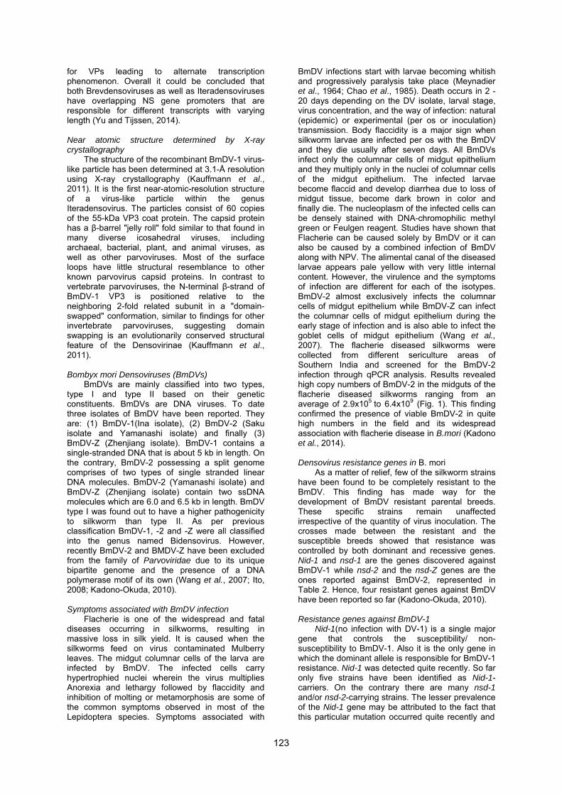
b) Fig. 2 Detection of BmDV-2 resistance through PCR amplification of genomic DNA of various multivoltine races using primers of amino acid transporter region. a) Appearance of specific band at 775 bp indicates susceptible genotype; b) Appearance of specific band at 890 bp indicates resistant genotype. Screening of Indian B. mori germplasm races for nsd-2 genes
In India, invaluable genetic silkworm B. mori stocks are maintained at the Central Sericultural Germplasm Resources Centre (CSGRC) at Hosur of Central Silk Board. Seventy multivoltine and 28 productive bivoltine silkworm germplasm resources were collected from Germplasm subjected to PCR analysis using two sets of primers viz. aa-trans1 to identify resistance to the BmDV-2 and aa-trans3 to identify susceptibility to the virus were utilized to analyze the genomic structure of nsd-2 gene (Ito, 2008). The aa-trans1 forward primer (5’-TCTACGTGCTTTCATACTACGTATC) was designed to have a binding site within exon 4 and the reverse primer (5’-TTCCTCACGTTTCTGAATTTCTCTTG) within exon 14. The aa-trans3 forward primer (5’- GGTAAGAGGTCCAACGCTGTTAAGTT) on the other hand was designed to have a binding site at exon 13, 3’ flanking region and reverse primer (5’-TTCCTCACGTTTCTGAATTTCTCTTG) within exon 14.
The results of the study revealed that, most of the multivoltine as well as bivoltine silkworm germplasm races harboured either gene for susceptibility or genes for both resistance and susceptibility to DV-2 indicating a heterozygous susceptible condition. A total of nine multivoltine races screened for the presence of resistance/susceptible genes, of which six races possessed susceptible genes, while two races (MV-76, MV-77) had resistant genes in a homozygous condition and one race (MV-73) had neither of the genes (Fig. 2). With respect to bivoltine silkworm germplasm races, the races with genes for susceptibility/resistance as well as susceptibility included those most widely reared by sericulturists like CSR2, CSR4 etc. This justifies the widespread prevalence and severity of infection by BmDV-2 in the field. Among bivoltine silkworm germplasm races, race KA revealed presence of BmDV-2 resistance gene, while, race M-III recorded absence of genes for both BmDV-2 resistance as well as susceptibility. The above studies have thus provided evidence for availability of Indian silkworm
125

germplasm resources with probable resistance to BmDV-2 (Ponnuvel et al., 2010; Murthy et al., 2014).
The recent discovery of resistant genes in some of the B. mori races has led to the initiation of screening of resistant parental races for the development of BmDV resistant transgenic breeds. In this backdrop twenty Indian bivoltine B.mori races were screened for DV-2 resistant gene (nsd-2) in the parental breeds selected for autumn specific breeding in North & North West India. Among the these bitivoltine races, APSHT-P5, BBE-198, BBE-178, APS-4, APS-9 and BBE-266 were found to possess the BmDV-2 resistant gene nsd-2. The race APSHT–P5 had a score of 100 % for the prevalence of nsd-2. The races BBE-198 and APS-4 with a score of 50 %, BBE-178 with a score of 37.50 % and APS-9, BBE-266 with a score of 25 % each were found out to be the potential candidates for the development of BmDV-2 resistant parental breeds. Thus, this study can help in identifying DV-2 resistant parental breeds which will be used for breeding programmes for developing DV-2 resistant silkworm races.
Conclusion
Densoviruses can be classified as a diverse
group of viruses having a wide host range. BmDVs among this group have proved to be the major agent for the Flacherie disease in silkworms. The molecular aspect involved in the infection mechanism is yet to be unveiled. The two categories of BmDV i.e. BmDV-1 and BmDV-2 completely differ in their genetic make-up. BmDV-1 like their other counterparts has a single monosense genome while BmDV-2 unlike the group has a bipartite genome. The nsd-1, Nid-1 and nsd-2 genes found in BmDV-1 and BmDV-2 respectively has initiated studies involving crossing a race harboring gene for resistance to BmDV-2 with an existing superior productive race widely used by sericulturists through conventional breeding followed by F1 hybrid production and backcrossing to introgress the gene. The introgression of the gene can be confirmed early through molecular biology techniques. Studies have also been undertaken for screening out Indian silkworm races bearing the resistant genes. Resistance to BmDV-2 involves only a single gene so this feature can be further used for developing BmDV-2 resistant B. mori races in a shorter period of time using molecular biology techniques. However infection pertaining to BmDV-1 will require further studies for developing markers to screen BmDV-1 resistant silkworm breeds. In the case of BmDV-2 the resistant gene itself can be used as a marker for screening BmDV-2 resistant breeds. The ultimate target of BmDV studies would be to develop disease resistant silkworm breeds which can help the sericulture farmers to have higher silk yield and thereby enhance their economic conditions.
However, the molecular aspect of virus infection is yet to be studied in detail in Indian silkworm breeds. DV infection is host specific as well as tissue specific. Studies related to the molecular mechanism of infection could further help in
differentiating the various isolates of the BmDVs. The resistance developed in certain B. mori strains against BmDV-2 is also due to deletion of exons in the amino acid transporter. Hence, further studies can be taken up in this direction to find out the specific receptors related with other viral pathogens.
Acknowledgements
This research work has been supported through collaborative DST-JSPS project jointly funded by Department of Science and Technology (DST), Ministry of Science and Technology, Government of India as well as Japan Society for the Promotion of Science (JSPS), Government of Japan (Grant No: DST/INT/JSPS/P-187/2014,16th June 2014 to 15th June 2016).
References Abe H, Sugasaki T, Kanehara M, Shimada T, Gomi
SJ, Ohbayashi F, et al. Identification and genetic mapping of RAPD markers linked to the densonucleosis refractoriness gene, nsd-2, in the silkworm, Bombyx mori. Genes Genet. Syst. 75: 93-96, 2000.
Abe H, Harada T, Kanehara M, Shimada T, Ohbayashi F, Oshiki T. Genetic mapping of RAPD markers linked to the densonucleosis refractoriness gene, nsd-1, in the silkworm, Bombyx mori. Genes Genet. Syst. 73: 237-242, 1998.
Abe H, Watanabe H. Double infection of two densonucleosis viruses in the silkworm Bombyx mori. Jpn. J. Appl. Ent. Zool. 31: 381-384, 1987.
Amargier A. Vago C, Duthoit JL, Meynadier G. Formation tumorale d'origine parvovirale chez Sibille fusca (Lep. Limacodidae). Entomophaga 24: 259-271, 1979.
Bao YY, Li MW, Zhao YP, Ge JQ, Wang CS, Huang YP, et al. Differentially expressed genes in resistant and susceptible Bombyx mori strains infected with a densonucleosis virus. Insect Biochem. Mol. Biol. 38: 853-861, 2008.
Baquerizo-Audiot E, Abd-Alla A, Jousset FX, Cousserans F, Tijssen P, Bergoin M. Structure and expression strategy of the genome of Culex pipiens densovirus, a mosquito densovirus with an ambisense organization. J. Virol. 83: 6863-6873, 2009.
Bergoin M, Tijssen P. Insect Virol. In: Asgari S, Johnson K (eds), Densoviruses: a highly diverse group of arthropod Parvoviruses, Caister Academic Press, Narfolk, UK, pp 59-82, 2010.
Bergoin M, Tijssen P. Molecular biology of Densovirinae. Contrib. Microbiol. 4: 12-32, 2000.
Broliden K, Tolfvenstam T, Norbeck O. Clinical aspects of parvovirus B19 infection. J Intern. Med. 260: 285-304, 2006.
Chao YC, Young SYI, Kim KS, Scott HA. A newly isolated densonucleosis virus from Pseudoplusia includens (Lepidoptera: Noctuidae). J. Invertebr. Pathol. 46: 70-82, 1985.
Cotmore SF, Tattersall P. Parvoviral host range and cell entry mechanisms. Adv. Virus Res. 70, 183-232, 2007.
126

Cotmore SF, Agbandje-McKenna M, Chiorini JA, Mukha DV, Pintel DJ, Qiu J, et al. The family Parvoviridae. Arch. Virol. 159: 1239-1247, 2014.
Dumas B, Jourdan M, Pascaud AM, Bergoin, M. Complete nucleotide sequence of the cloned infectious genome of Junonia coenia densovirus reveals an organization unique among parvoviruses. Virology 191: 202-222, 1992.
Eguchi R, Nagayasu K, Ninaki O, Hara W. Genetic analysis on the dominant non-susceptibility to densonucleosis virus type 1 in the silkworm, Bombyx mori. Sanshi-Kontyu Biotrech. 159-163, 2007.
Fediere G, Li Y, Zadori Z, Szelei J, Tijssen P. Genome organization of Casphalia extranea densovirus, a new iteravirus. Virology 292: 299-308, 2002.
Fediere G. Molecular biology of Densovirinae. Contrib. Microbiol. 4: 1-11, 2000.
Flegel TW. Shrimp parvoviruses. In: Kerr JR, Cotmore SF, Bloom ME, Linden RM, Parrish CR (eds), Parvoviruses, London, Hodder Arnold, pp 487-493, 2006.
Furuta Y, Susceptibility of the races of the silkworm, Bombyx mori, preserved in NISES to the nuclear polyhedrosis virus and densonucleosis viruses. Bull. Natl. Inst. Seric. Entomol. Sci. 15: 119-145, 1995.
Garzon S, Kurstak E. Infection des cellules des gonades et du systeme nerveux de Galleria mellonella par le virus de Ia densonucleose. Naturalist Can. 95: 1125, 1968.
Guo H, Zhang J, Hu Y. Complete sequence and organization of Periplaneta juliginosa densovirus genome. Acta. Virol. 44: 315-322, 2000.
Hayakawa T, Kojima K, Nonaka K, Nakagaki M, Sahara K, Asano S, et al. Analysis of proteins encoded in the bipartite genome of a new type of parvo-like virus isolated from silkworm structural protein with DNA polymerase motif. Virus Res. 66: 101-108, 2000.
Ito K, Kidokoro K, Sezutsu H, Nohata J, Yamamoto K, Kobayashi I, et al. Deletion of a gene encoding an amino acid transporter in the midgut membrane causes resistance to a Bombyx parvo-like virus. Proc. Natl. Acad. Sci. USA 105: 7523-7527, 2008.
Ito K. Positional cloning and functional analysis of the resistance genes to Bombyx mori densoviruses, Graduate School of Agricultural and Life Sciences, University of Tokyo, PhD Thesis, 2008.
Jousset FX, Compagnon B, Bergoin M. Comparison of the restriction map and infectivity of the genomes of three densoviruses, Wageningen foundation IVth Int. Coll. Invertebr. Pathol. Wageningen, Netherlands, 1986.
Kadono-Okuda K. Densovirus Resistance in Bombyx mori. In: Molecular biology and genetics of the Lepidoptera, CRC Press 337-348, pp 362, 2010.
Kadono-Okuda K, Ito K, Murthy GN, Sivaprasad V, Ponnuvel KM. Molecular mechanism of Densovirus resistance in Silkworm, Bombyx mori. Sericologia 54: 1-10, 2014.
KapelinskayaTV, Martynova EU, Korolev AL, Schal C, Mukha DV. Transcription of the German cockroach densovirus BgDV genome: alternative processing of viral RNAs. Dokl. Biochem. Biophys. 421: 176-180, 2008.
Kobayashi M, Choi H. Inhibition of the accumulation of viral polypeptides in the densonucleosis virus-infected midgut of the silkworm, Bombyx mori, reared at a supraoptimal temperature. J. Invertebr. Pathol. 56: 117-122, 1990.
Kaufmann B, El-Far M, Plevka P, Bowman VD, Li Y, Tijssen P, Rossmann MG. Structure of Bombyx mori Densovirus 1, a silkworm pathogen. J. Virol. 85: 4691-4697, 2011.
Lebedeva OP, Kuznetsova MA, Zelenko AP, Gudz-Gorban AP. Investigation of a virus disease of the densonucleosis type in a laboratory culture of Aedes aegypti. Acta Virol. 17: 253-256, 1973.
Li Y, Zadori Z, Bando H, Dubuc R, Fediere G, Szelei J, Tijssen P. Genome organization of the densovirus from Bombyx mori (BmDV-1) and enzyme activity of its capsid. J. Gen. Virol. 82: 2821-2825, 2001.
Lightner DV, Redman RM. A parvo-like virus disease of penaeid shrimp. J. lnvertebr. Pathol. 45: 47-53, 1985.
Maeda S, Watanabe H. Immunofluorescence observation of the infection of densonucleosis virus in the silkworm, Bombyx mori. Japan J. Appl. Entomol. Zool. 22: 98-101, 1978.
Meynadier G, Vago C, Plantevin G. Atger P. Virose d'un type inhabituel chez le lepidoptere Galleria mellonella L. Rev. Zool. Agric. Appl. 63: 207-208, 1964.
Mukha DV, Chumachenko AG, Dykstra MJ, Kurtti TJ, Schal C. Characterization of a new densovirus infecting the German cockroach, Blattella germanica. J. Gen. Viral. 87: 1567-1575, 2006.
Murthy GN, Ponnuvel KM, Awasthi AK, Rao CGP, Chandrasekhar Sagar BK. The Indian isolate of Densovirus-2-Impact of infection and mechanism of resistance in Bombyx mori, J. Invertebr. Pathol. 115: 48-50, 2014.
Ogoyi DO, Kadono-Okuda K, Eguchi R, Furuta Y, Hara W, Nguu EK, et al. Linkage and mapping analysis of a non-susceptibility gene to densovirus (nsd-2) in the silkworm, Bombyx mori. Insect Mol. Biol. 12: 117-124, 2003.
Ponnuvel KM, Murthy GN, Awasthi AK, Rao CGP, Vijayaprakash NB, Kamble CK. Densovirus-2 in flacherie affected silkworm Bombyx mori. Indian silk 2: 4-6, 2011.
Ponnuvel KM, Murthy GN, Triveni L, Awasthi AK, Rao CGP, Vijayaprakash NB, et al. Screening of Bombyx mori silkworm races for detection of densonucleosis virus-2 resistance genes (nsd-2). Sericologia 51: 145-156, 2010.
Ren X, Hoiczyk E, Rasgon JL. Viral paratransgenesis in the malaria vector Anopheles gambiae. PLoS Pathog. 4: e1000135, 2008.
Ryabov EV, Keane G, Naish N, Evered C, Winstanley D. Densovirus induces winged morphs in asexual clones of the rosy apple aphid, Dysaphis plantaginea. Proc. Natl. Acad. Sci. USA 106: 8465-8470, 2009.
127
Shike H, Dhar AK, Burns JC, Shimizu C, Jousset FX, Klimpel KR, Bergoin M. Infectious hypodermal and hematopoietic necrosis virus of shrimp is related to mosquito brevidensoviruses. Virology 277: 167-177, 2000.
Suto C. Characterization of a virus newly isolated from the smoky-brown cockroach, Periplaneta fuliginosa (Serville). Nagoya Med. Sci. 42: 13-25, 1979.
Tattersall P. The evolution of parvovirus taxonomy. In: Kerr JR, Cotmore SF, Bloom ME, Linden RM, Parrish CR (eds), Parvoviruses, London, Hodder Arnold, pp. 5-14, 2006.
Tijssen P, Bergoin M. Densonucleosis viruses constitute an increasingly diversified subfamily among the parvoviruses. Semin. Virol. 6: 347-355, 1995.
Tijssen P, Agbandje-McKenna M, Almendral JM, Bergoin M, Flegel TW, Hedman K, et al. In: King AMQ, M. J. Adams MJ, Carstens E, Lefkowitz EJ (eds), Virus taxonomy: classification and nomenclature of viruses: ninth report of the International Committee on Taxonomy of viruses, pp 405-425, 2011.
van Munster M, Dullemans AM, Verbeek M, van den Heuvel JF, Reinbold C, Brault V, et al. Characterization of a new densovirus infecting the green peac aphid Myzus persicae. J. Invertebr. Pathol. 84: 6-14, 2003.
Wang YJ, Yao Q, Chen KP, Wang Y, Lu J, Han X, Characterization of the genome structure of Bombyx mori Densovirus (China isolate). Virus Genes 35: 103-108, 2007.
Watanabe H, Maeda S, Matsui M, Shimizu T. Histopathology of the midgut epithelium of the silkworm, Bombyx mori, infected with a newly-isolated virus from the flacherie-diseased larvae. J. Seric. Sci. Jpn. 45: 29-34, 1976.
Watanabe H, Maeda S. Genetically determined nonsusceptibility of the silkworm, Bombyx mori, to infection with a densonucleosis virus (densovirus). J. Invertebr. Pathol. 38: 370-373, 1981.
Yamagishi J, Hu Y, Zheng J, Bando H. Genome organization and mRNA structure of Periplaneta fuliginosa densovirus imply alternative splicing involvement in viral gene expression. Arch. Virol. 144: 2111-2124, 1999.
Yang B, Dong X, Cai D, Wang X, Liu Z, Hu Z, et al. Characterization of the promoter elements and transcription profile of Periplaneta fuliginosa densovirus nonstructural genes. Virus Res. 133: 149-156, 2008.
Zhai YG, Lv XJ, Sun XH, Fu SH, Gong ZD, Fen Y, et al. Isolation and characterization of the full coding sequence of a novel densovirus from the mosquito Culex pipiens pallens. J. Gen. Virol. 89: 195-199, 2008.
128
![v11i10_ASIANMENACOUNSEL[1].pdf - MORI HAMADA ...](https://static.fdokumen.com/doc/165x107/631228f3074f092ad10d6ac9/v11i10asianmenacounsel1pdf-mori-hamada-.jpg)